Voie d'immunisation et séquence d'administration de l ... - TEL
Voie d'immunisation et séquence d'administration de l ... - TEL Voie d'immunisation et séquence d'administration de l ... - TEL
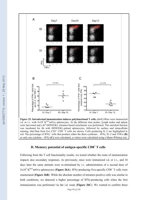
tel-00827710, version 1 - 29 May 2013 C. Quality of the T cell response Next, we were interested in characterizing the quality of the T cell response. Prior studies have indicated that cells producing high levels of IFNγ have the unique capacity to secrete multiple cytokines, leading to their being referred to as polyfunctional T cells (Seder et al., 2008). In our model, we evaluated the simultaneous production of IFNγ, IL-2 and TNFα. Mice were primed using the strategies discussed in Figure 21 and ex vivo restimulation of the tetramer-enriched fraction was performed prior to intracellular staining. As anticipated, the cells producing high levels of IFNγ also expressed TNFα and IL-2 (Figure 25A, nb. IL-2 producing cells are shown in red). The response was evaluated throughout the kinetics of T cell priming (Figure 25A), and for purposes of comparing i.d. vs i.v. immunization, we focused on the peak of the response: Day 7 for i.v. immunization; and Day 12 for i.d. immunization. The percentages of IFNγ + cells producing the 3 cytokines – IFNγ, IL-2 and TNFα – was significantly higher after i.d. immunization (Figure 25B). The converse is also true – the percentage of cells producing only IFNγ was higher following i.v. immunization (Figure 25C). Thus, we conclude that cross-priming via the i.d. route establishes a stronger, more polyfunctional response. 98
tel-00827710, version 1 - 29 May 2013 Figure 25. Intradermal immunization induces polyfunctional T cells. (A-C) Mice were immunized i.d. or i.v. with 5x10 5 K bm1 mOva splenocytes. At the different time points, lymph nodes and spleen were harvested and a K b -SIINFEKL tetramer-based enrichment was performed. The enriched fraction was incubated for 4h with SIINFEKL-pulsed splenocytes, followed by surface and intracellular staining. (A) Data from live CD3 + CD8 + T cells are shown. Cells producing IL-2 are highlighted in red. The percentage of IFNγ + cells that produce either the three cytokines – IFNγ, IL-2 and TNFα (B) or only one cytokine – IFNγ (C) were calculated. p-values were calculated using a Mann-Whitney test. D. Memory potential of antigen-specific CD8 + T cells Following from the T cell functionality results, we tested whether the route of immunization impacts also secondary responses. As previously, mice were immunized i.d. or i.v., and 34 days later the same animals were re-stimulated by i.v. administration of a second dose of 5x10 5 K bm1 mOva splenocytes (Figure 26A). IFNγ-producing Ova-specific CD8 + T cells were enumerated (Figure 26B). While the absolute number of tetramer-positive cells was similar in both conditions, we detected a higher percentage of IFNγ-producing cells when the first immunization was performed via the i.d. route (Figure 26C). We wanted to confirm these Page 99 of 256
- Page 47 and 48: tel-00827710, version 1 - 29 May 20
- Page 49 and 50: tel-00827710, version 1 - 29 May 20
- Page 51 and 52: tel-00827710, version 1 - 29 May 20
- Page 53 and 54: tel-00827710, version 1 - 29 May 20
- Page 55 and 56: tel-00827710, version 1 - 29 May 20
- Page 57 and 58: tel-00827710, version 1 - 29 May 20
- Page 59 and 60: tel-00827710, version 1 - 29 May 20
- Page 61 and 62: tel-00827710, version 1 - 29 May 20
- Page 63 and 64: tel-00827710, version 1 - 29 May 20
- Page 65 and 66: tel-00827710, version 1 - 29 May 20
- Page 67 and 68: tel-00827710, version 1 - 29 May 20
- Page 69 and 70: tel-00827710, version 1 - 29 May 20
- Page 71 and 72: tel-00827710, version 1 - 29 May 20
- Page 73 and 74: tel-00827710, version 1 - 29 May 20
- Page 75 and 76: tel-00827710, version 1 - 29 May 20
- Page 77 and 78: tel-00827710, version 1 - 29 May 20
- Page 79 and 80: tel-00827710, version 1 - 29 May 20
- Page 81 and 82: tel-00827710, version 1 - 29 May 20
- Page 83 and 84: tel-00827710, version 1 - 29 May 20
- Page 85 and 86: tel-00827710, version 1 - 29 May 20
- Page 87 and 88: tel-00827710, version 1 - 29 May 20
- Page 89 and 90: tel-00827710, version 1 - 29 May 20
- Page 91 and 92: tel-00827710, version 1 - 29 May 20
- Page 93 and 94: tel-00827710, version 1 - 29 May 20
- Page 95 and 96: tel-00827710, version 1 - 29 May 20
- Page 97: tel-00827710, version 1 - 29 May 20
- Page 101 and 102: tel-00827710, version 1 - 29 May 20
- Page 103 and 104: tel-00827710, version 1 - 29 May 20
- Page 105 and 106: tel-00827710, version 1 - 29 May 20
- Page 107 and 108: tel-00827710, version 1 - 29 May 20
- Page 109 and 110: tel-00827710, version 1 - 29 May 20
- Page 111 and 112: tel-00827710, version 1 - 29 May 20
- Page 113 and 114: tel-00827710, version 1 - 29 May 20
- Page 115 and 116: tel-00827710, version 1 - 29 May 20
- Page 117 and 118: tel-00827710, version 1 - 29 May 20
- Page 119 and 120: tel-00827710, version 1 - 29 May 20
- Page 121 and 122: tel-00827710, version 1 - 29 May 20
- Page 123 and 124: tel-00827710, version 1 - 29 May 20
- Page 125 and 126: tel-00827710, version 1 - 29 May 20
- Page 127 and 128: tel-00827710, version 1 - 29 May 20
- Page 129 and 130: tel-00827710, version 1 - 29 May 20
- Page 131 and 132: tel-00827710, version 1 - 29 May 20
- Page 133 and 134: tel-00827710, version 1 - 29 May 20
- Page 135 and 136: tel-00827710, version 1 - 29 May 20
- Page 137 and 138: tel-00827710, version 1 - 29 May 20
- Page 139 and 140: tel-00827710, version 1 - 29 May 20
- Page 141 and 142: tel-00827710, version 1 - 29 May 20
- Page 143 and 144: tel-00827710, version 1 - 29 May 20
- Page 145 and 146: tel-00827710, version 1 - 29 May 20
- Page 147 and 148: tel-00827710, version 1 - 29 May 20
tel-00827710, version 1 - 29 May 2013<br />
Figure 25. Intra<strong>de</strong>rmal immunization induces polyfunctional T cells. (A-C) Mice were immunized<br />
i.d. or i.v. with 5x10 5 K bm1 mOva splenocytes. At the different time points, lymph no<strong>de</strong>s and spleen<br />
were harvested and a K b -SIINFEKL t<strong>et</strong>ramer-based enrichment was performed. The enriched fraction<br />
was incubated for 4h with SIINFEKL-pulsed splenocytes, followed by surface and intracellular<br />
staining. (A) Data from live CD3 + CD8 + T cells are shown. Cells producing IL-2 are highlighted in<br />
red. The percentage of IFNγ + cells that produce either the three cytokines – IFNγ, IL-2 and TNFα (B)<br />
or only one cytokine – IFNγ (C) were calculated. p-values were calculated using a Mann-Whitney test.<br />
D. Memory potential of antigen-specific CD8 + T cells<br />
Following from the T cell functionality results, we tested wh<strong>et</strong>her the route of immunization<br />
impacts also secondary responses. As previously, mice were immunized i.d. or i.v., and 34<br />
days later the same animals were re-stimulated by i.v. administration of a second dose of<br />
5x10 5 K bm1 mOva splenocytes (Figure 26A). IFNγ-producing Ova-specific CD8 + T cells were<br />
enumerated (Figure 26B). While the absolute number of t<strong>et</strong>ramer-positive cells was similar in<br />
both conditions, we d<strong>et</strong>ected a higher percentage of IFNγ-producing cells when the first<br />
immunization was performed via the i.d. route (Figure 26C). We wanted to confirm these<br />
Page 99 of 256


