Voie d'immunisation et séquence d'administration de l ... - TEL
Voie d'immunisation et séquence d'administration de l ... - TEL Voie d'immunisation et séquence d'administration de l ... - TEL
tel-00827710, version 1 - 29 May 2013 spleen (den Haan et al., 2000). In contrast, the latter subset was shown to specialize in MHC- II antigen presentation (Dudziak et al., 2007). The same kind of dichotomy is true for migratory DCs. CD103 + DCs performed more antigen cross-presentation while CD11b + DCs are better for triggering CD4 help and humoral response stimulation (Bedoui et al., 2009). (b) Cooperation between different DC subsets Resident DCs in the spleen screen the blood for pathogens but also phagocytose materials that are drained directly from the periphery through the lymphatic conduits. Migratory DCs migrate to lymphoid organs upon activation where they can present antigen to specific T cells. However, once they have arrived, they can transfer antigen to resident DCs that will present antigen (Allan et al., 2006). Consequently the same antigen can be presented by different DC subsets, with different specialization for antigen presentation (Belz et al., 2004). (c) Ability to respond to different signals Interestingly, DCs subsets display different patterns of PRR expression, resulting in differential sensitivity to danger signals. In the spleen, the CD8α + DCs are the only subset to express TLR3, but not TLR7, while the CD8α - subset is characterized by the opposite phenotype (Edwards et al., 2003). These observations have important implications for modulation of the immune response stimulated by PAMPs and for considering the appropriate choice of adjuvants, which will be discussed later in this thesis. 3) Parallel between resident CD8α + DCs and migratory CD103 + DCs To study the role of CD8α + DC subset in vivo, a mouse line lacking the transcription factor Batf3 was developed. In these mice, this subset is missing and this knock-out has been correlated with the absence of a protective CD8 + T cell response upon experimental West Nile virus infection, as well as an inability to reject a syngeneic tumor (Hildner et al., 2008). These results clearly demonstrated the crucial role of CD8α + DCs in cross-presentation. Interestingly, the skin-resident CD103 + DCs are also absent in this mouse. These two subsets share several common characteristics such as the expression of DEC205 or CD24, their cross- presentation efficiency of both soluble and cell-associated antigens (den Haan et al., 2000) (del Rio et al., 2007), and their ability to respond to the TLR3 ligand poly I:C (Schulz et al., 2005; Sung et al., 2006). However, there are also differences: TLR3 was not detected in CD103 + DCs and this subset does not seem as efficient as CD8α + DCs in promoting memory CD8 + T cell responses. While they share several similarities, these 2 subsets are not identical and, therefore, it was surprising that Batf3 would be critical for both DC populations. 38
tel-00827710, version 1 - 29 May 2013 Although this question remains unclear, this result may be due to a common precursor that can give rise either to CD8α + DC or CD103 + DC depending on its tissue localization. 4) Heterogeneity of human DCs While many DC subsets have been described in the context of experimental mouse models, the same extensive identification and characterization has not yet been possible in humans, mainly due to the restricted materials available for human study – especially blood. Recently, a group studying DCs from the thymus identified different DC subsets in this organ (Vandenabeele et al., 2001). A subset of human DCs expressing Clec9A, similar to the CD8α + DC subset in the mouse, was identified: it corresponds to BDCA3 + DC subset in the blood (Crozat et al., 2010; Jongbloed et al., 2010; Poulin et al., 2010). Further investigations are required to validate that these DCs have the same functional properties as CD8α + mouse DCs. But further dissection of the different subsets of human DCs will be critical for the purpose of improving human vaccination (Caminschi et al., 2008). II. DEVELOPMENT OF A CD8 + T CELL RESPONSE In a naïve mouse, CD8 + T cells that are specific for a variety of antigens are present. They become activated upon encounter with APCs presenting the specific cognate antigen. Signals from the microenvironment are required to mount an efficient T cell response. Upon activation, T cells expand in order to clear the antigen burden and then the excess specific T cells die to maintain the homeostasis of the T cell population. Nevertheless, some antigen- specific T cells will persist as memory T cells (Figure 6). Figure 6. The different phases of a T cell response Page 39 of 256
- Page 1 and 2: tel-00827710, version 1 - 29 May 20
- Page 3 and 4: tel-00827710, version 1 - 29 May 20
- Page 5 and 6: tel-00827710, version 1 - 29 May 20
- Page 7 and 8: tel-00827710, version 1 - 29 May 20
- Page 9 and 10: tel-00827710, version 1 - 29 May 20
- Page 11 and 12: tel-00827710, version 1 - 29 May 20
- Page 13 and 14: tel-00827710, version 1 - 29 May 20
- Page 15 and 16: tel-00827710, version 1 - 29 May 20
- Page 17 and 18: tel-00827710, version 1 - 29 May 20
- Page 19 and 20: tel-00827710, version 1 - 29 May 20
- Page 21 and 22: tel-00827710, version 1 - 29 May 20
- Page 23 and 24: tel-00827710, version 1 - 29 May 20
- Page 25 and 26: tel-00827710, version 1 - 29 May 20
- Page 27 and 28: tel-00827710, version 1 - 29 May 20
- Page 29 and 30: tel-00827710, version 1 - 29 May 20
- Page 31 and 32: tel-00827710, version 1 - 29 May 20
- Page 33 and 34: tel-00827710, version 1 - 29 May 20
- Page 35 and 36: tel-00827710, version 1 - 29 May 20
- Page 37: tel-00827710, version 1 - 29 May 20
- Page 41 and 42: tel-00827710, version 1 - 29 May 20
- Page 43 and 44: tel-00827710, version 1 - 29 May 20
- Page 45 and 46: tel-00827710, version 1 - 29 May 20
- Page 47 and 48: tel-00827710, version 1 - 29 May 20
- Page 49 and 50: tel-00827710, version 1 - 29 May 20
- Page 51 and 52: tel-00827710, version 1 - 29 May 20
- Page 53 and 54: tel-00827710, version 1 - 29 May 20
- Page 55 and 56: tel-00827710, version 1 - 29 May 20
- Page 57 and 58: tel-00827710, version 1 - 29 May 20
- Page 59 and 60: tel-00827710, version 1 - 29 May 20
- Page 61 and 62: tel-00827710, version 1 - 29 May 20
- Page 63 and 64: tel-00827710, version 1 - 29 May 20
- Page 65 and 66: tel-00827710, version 1 - 29 May 20
- Page 67 and 68: tel-00827710, version 1 - 29 May 20
- Page 69 and 70: tel-00827710, version 1 - 29 May 20
- Page 71 and 72: tel-00827710, version 1 - 29 May 20
- Page 73 and 74: tel-00827710, version 1 - 29 May 20
- Page 75 and 76: tel-00827710, version 1 - 29 May 20
- Page 77 and 78: tel-00827710, version 1 - 29 May 20
- Page 79 and 80: tel-00827710, version 1 - 29 May 20
- Page 81 and 82: tel-00827710, version 1 - 29 May 20
- Page 83 and 84: tel-00827710, version 1 - 29 May 20
- Page 85 and 86: tel-00827710, version 1 - 29 May 20
- Page 87 and 88: tel-00827710, version 1 - 29 May 20
tel-00827710, version 1 - 29 May 2013<br />
Although this question remains unclear, this result may be due to a common precursor that<br />
can give rise either to CD8α + DC or CD103 + DC <strong>de</strong>pending on its tissue localization.<br />
4) H<strong>et</strong>erogeneity of human DCs<br />
While many DC subs<strong>et</strong>s have been <strong>de</strong>scribed in the context of experimental mouse mo<strong>de</strong>ls,<br />
the same extensive i<strong>de</strong>ntification and characterization has not y<strong>et</strong> been possible in humans,<br />
mainly due to the restricted materials available for human study – especially blood. Recently,<br />
a group studying DCs from the thymus i<strong>de</strong>ntified different DC subs<strong>et</strong>s in this organ<br />
(Van<strong>de</strong>nabeele <strong>et</strong> al., 2001). A subs<strong>et</strong> of human DCs expressing Clec9A, similar to the<br />
CD8α + DC subs<strong>et</strong> in the mouse, was i<strong>de</strong>ntified: it corresponds to BDCA3 + DC subs<strong>et</strong> in the<br />
blood (Crozat <strong>et</strong> al., 2010; Jongbloed <strong>et</strong> al., 2010; Poulin <strong>et</strong> al., 2010). Further investigations<br />
are required to validate that these DCs have the same functional properties as CD8α + mouse<br />
DCs. But further dissection of the different subs<strong>et</strong>s of human DCs will be critical for the<br />
purpose of improving human vaccination (Caminschi <strong>et</strong> al., 2008).<br />
II. DEVELOPMENT OF A CD8 + T CELL RESPONSE<br />
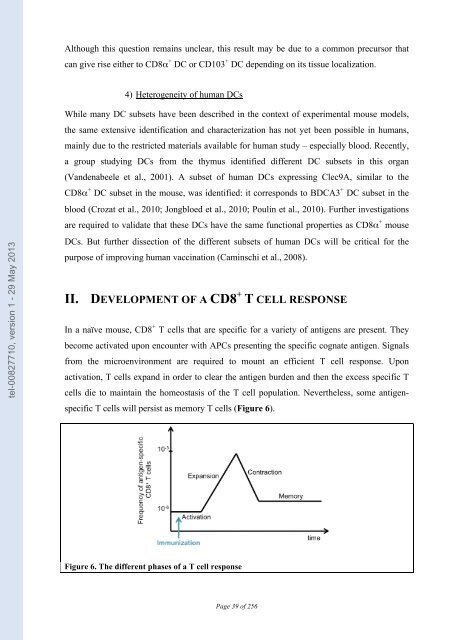
In a naïve mouse, CD8 + T cells that are specific for a vari<strong>et</strong>y of antigens are present. They<br />
become activated upon encounter with APCs presenting the specific cognate antigen. Signals<br />
from the microenvironment are required to mount an efficient T cell response. Upon<br />
activation, T cells expand in or<strong>de</strong>r to clear the antigen bur<strong>de</strong>n and then the excess specific T<br />
cells die to maintain the homeostasis of the T cell population. Nevertheless, some antigen-<br />
specific T cells will persist as memory T cells (Figure 6).<br />
Figure 6. The different phases of a T cell response<br />
Page 39 of 256


