
Create successful ePaper yourself
Turn your PDF publications into a flip-book with our unique Google optimized e-Paper software.
<strong>Planta</strong> <strong>Medica</strong><br />
Volume 77, Issue 08, May 2011<br />
de la Garza, Ana Laura; Milagro, Fermín I.; Boque, Noemí; Campión, Javier;<br />
Martínez, J. Alfredo:<br />
Natural Inhibitors of Pancreatic Lipase as New Players in Obesity Treatment<br />
Wang, Cai-Ping; Wang, Yemin; Wang, Xing; Zhang, Xian; Ye, Jian-Feng; Hu,<br />
Lin-Shui; Kong, Ling-Dong:<br />
Mulberroside A Possesses Potent Uricosuric and Nephroprotective Effects in<br />
Hyperuricemic Mice<br />
Chow, Nicholas K.; Fretz, Michael; Hamburger, Matthias; Butterweck,<br />
Veronika:<br />
Telemetry as a Tool to Measure Sedative Effects of a Valerian Root Extract<br />
and Its Single Constituents in Mice<br />
Ghosh, Tirtha; Maity, Tapan Kumar; Singh, Jagadish:<br />
Antihyperglycemic Activity of Bacosine, a Triterpene from Bacopa monnieri, in<br />
Alloxan-Induced Diabetic Rats<br />
Shao, Meng; Qu, Kai; Liu, Ke; Zhang, Yuqin; Zhang, Ling; Lian, Zeqin; Chen,<br />
Tingting; Liu, Junfeng; Wu, Aili; Tang, Yue; Zhu, Haibo:<br />
Effects of Ligustilide on Lipopolysaccharide-Induced Endotoxic Shock in<br />
Rabbits<br />
Hong, Feng; Xiao, Weilie; Ragupathi, Govind; Lau, Clara B. S.; Leung, Ping<br />
Chung; Yeung, K. Simon; George, Constantine; Cassileth, Barrie; Kennelly,<br />
Edward; Livingston, Philip O.:<br />
The Known Immunologically Active Components of Astragalus Account for<br />
Only a Small Proportion of the Immunological Adjuvant Activity When<br />
Combined with Conjugate Vaccines<br />
Venâncio, Antônio Medeiros; Onofre, Alexandre Sherlley Casimiro; Lira,<br />
Amintas Figueiredo; Alves, Péricles Barreto; Blank, Arie Fitzgerald; Antoniolli,<br />
Ângelo Roberto; Marchioro, Murilo; dos Santos Estevam, Charles; Santos de<br />
Araujo, Brancilene:<br />
Chemical Composition, Acute Toxicity, and Antinociceptive Activity of the<br />
Essential Oil of a Plant Breeding Cultivar of Basil (Ocimum basilicum L.)
Halder, Sumita; Mehta, Ashish Krishan; Kar, Rajarshi; Mustafa, Mohammad;<br />
Mediratta, Pramod Kumari; Sharma, Krishna Kishore:<br />
Clove Oil Reverses Learning and Memory Deficits in Scopolamine-Treated<br />
Mice<br />
Ettefagh, Keivan A.; Burns, Johnna T.; Junio, Hiyas A.; Kaatz, Glenn W.;<br />
Cech, Nadja B.:<br />
Goldenseal (Hydrastis canadensis L.) Extracts Synergistically Enhance the<br />
Antibacterial Activity of Berberine via Efflux Pump Inhibition<br />
Yang, Heejung; Sung, Sang Hyun; Kim, Jinwoong; Kim, Young Choong:<br />
Neuroprotective Diarylheptanoids from the Leaves and Twigs of Juglans<br />
sinensis against Glutamate-Induced Toxicity in HT22 Cells<br />
Liu, Hai; Chou, Gui-Xin; Wang, Jun-Ming; Ji, Li-Li; Wang, Zheng-Tao:<br />
Steroidal Saponins from the Rhizomes of Dioscorea bulbifera and Their<br />
Cytotoxic Activity<br />
Schmidt, Thomas J.; Kaiser, Marcel; Brun, Reto:<br />
Complete Structural Assignment of Serratol, a Cembrane-Type Diterpene<br />
from Boswellia serrata, and Evaluation of Its Antiprotozoal Activity<br />
Zhao, Jianping; Avula, Bharathi; Joshi, Vaishali C.; Techen, Natascha; Wang,<br />
Yan-Hong; Smillie, Troy J.; Khan, Ikhlas A.:<br />
NMR Fingerprinting for Analysis of Hoodia Species and Hoodia Dietary<br />
Products<br />
Turski, Michal P.; Turska, Monika; Zgrajka, Wojciech; Bartnik, Magdalena;<br />
Kocki, Tomasz; Turski, Waldemar A.:<br />
Distribution, Synthesis, and Absorption of Kynurenic Acid in Plants<br />
Yang, Shuting; Chen, Chuan; Zhao, Yunpeng; Xi, Wang; Zhou, Xiaolong;<br />
Chen, Binlong; Fu, Chengxin:<br />
Association between Chemical and Genetic Variation of Wild and Cultivated<br />
Populations of Scrophularia ningpoensis Hemsl.
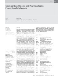
Natural Inhibitors of Pancreatic Lipase<br />
as New Players in Obesity Treatment<br />
Authors Ana Laura de la Garza, Fermín I. Milagro, Noemí Boque, Javier Campión, J. Alfredo Martínez<br />
Affiliation Department of Nutrition and Food Sciences, Physiology and Toxicology, University of Navarra, Pamplona, Spain<br />
Key words<br />
l " Orlistat<br />
l " high fat diet<br />
l " polyphenols<br />
l " saponins<br />
l " obesity<br />
l " fat digestion<br />
received Dec. 7, 2010<br />
revised February 11, 2011<br />
accepted February 21, 2011<br />
Bibliography<br />
DOI http://dx.doi.org/<br />
10.1055/s-0030-1270924<br />
Published online March 16,<br />
2011<br />
<strong>Planta</strong> Med 2011; 77: 773–785<br />
© Georg Thieme Verlag KG<br />
Stuttgart · New York ·<br />
ISSN 0032‑0943<br />
Correspondence<br />
Prof. J. Alfredo Martinez<br />
Department of Nutrition<br />
and Food Sciences,<br />
Physiology and Toxicology<br />
University of Navarra<br />
c/Irunlarrea 1<br />
31008 Pamplona<br />
Spain<br />
Phone: + 34 94842 56 00<br />
Fax: + 3494842 5649<br />
jalfmtz@unav.es<br />
Abstract<br />
!<br />
Obesity is a multifactorial disease characterized<br />
by an excessive weight for height due to an enlarged<br />
fat deposition such as adipose tissue, which<br />
is attributed to a higher calorie intake than the energy<br />
expenditure. The key strategy to combat obesity<br />
is to prevent chronic positive impairments in<br />
the energy equation. However, it is often difficult<br />
to maintain energy balance, because many available<br />
foods are high-energy yielding, which is usually<br />
accompanied by low levels of physical activity.<br />
The pharmaceutical industry has invested many<br />
efforts in producing antiobesity drugs; but only a<br />
lipid digestion inhibitor obtained from an actino-<br />
Introduction<br />
!<br />
Obesity is becoming one of the greatest threats to<br />
global health in this century, with more than 1.5<br />
billion overweight adults and at least 400 million<br />
of clinically obese subjects [1]. Due to these increasing<br />
obesity rates, the World Health Organization<br />
(WHO) has prompted to consider it as the<br />
epidemic of XXI century and to promote strategies<br />
to prevent and control its progress [2].<br />
The development of obesity is characterized by a<br />
chronic imbalance between energy intake and<br />
energy expenditure [3–5], and it is often ascribed<br />
to changing lifestyles and inadequate dietary habits<br />
[3]. Also, decreased energy expenditure is<br />
often associated with an inherited low basal metabolic<br />
rate, low energy cost of physical activity,<br />
and low capacity for fat oxidation [6]. To reduce<br />
body weight and adiposity, a change in lifestyle<br />
habits is still the crucial cornerstone [7]. Physical<br />
activity might be helpful in the prevention of obesity<br />
by elevating the average daily metabolic rate<br />
and increasing energy expenditure [3]. Unfortunately,<br />
this clinical approach is not long-term lasting,<br />
and weight regain is often seen. Drugs that<br />
Reviews<br />
bacterium is currently approved and authorized<br />
in Europe for obesity treatment. This compound<br />
inhibits the activity of pancreatic lipase, which is<br />
one of the enzymes involved in fat digestion.<br />
In a similar way, hundreds of extracts are currently<br />
being isolated from plants, fungi, algae, or<br />
bacteria and screened for their potential inhibition<br />
of pancreatic lipase activity. Among them,<br />
extracts isolated from common foodstuffs such<br />
as tea, soybean, ginseng, yerba mate, peanut, apple,<br />
or grapevine have been reported. Some of<br />
them are polyphenols and saponins with an inhibitory<br />
effect on pancreatic lipase activity, which<br />
could be applied in the management of the obesity<br />
epidemic.<br />
prevent weight regain appear necessary in obesity<br />
treatment [7]. Thus, the development of natural<br />
products for the treatment of obesity is a<br />
challenging task, which can be launched faster<br />
and cheaper than conventional single-entity<br />
pharmaceuticals [8]. Many medicinal plants may<br />
provide safe, natural, and cost-effective alternatives<br />
to synthetic drugs [9, 10]. Currently, one of<br />
the most important strategies in the treatment of<br />
obesity includes development of inhibitors of nutrient<br />
digestion and absorption. For example,<br />
acarbose is an antidiabetic drug that inhibits glycoside<br />
hydrolases, thus preventing the digestion<br />
of complex carbohydrates and decreasing postprandial<br />
hyperglycemia [11, 12]. Similar compounds<br />
with alpha-amylase inhibiting activity<br />
that can be used for diabetes control are being isolated<br />
from different plants. The list includes valoneaic<br />
acid dilactone [13], obtained from banaba<br />
(Lagerstroemia speciosa), the ethanol extract obtained<br />
from chestnut astringent skin [14], or the<br />
purified pancreatic alpha-amylase inhibitor isolated<br />
from white beans (Phaseolus vulgaris),<br />
which is able to reduce glycemia in both nondiabetic<br />
and diabetic rats [15].<br />
de la Garza AL et al. Natural Inhibitors of … <strong>Planta</strong> Med 2011; 77: 773–785<br />
773
774<br />
Reviews<br />
In this context, since dietary lipids represent the major source of<br />
unwanted calories, the inhibition of fat digestion is an interesting<br />
approach for reducing fat absorption [16]. Orlistat is the only authorized<br />
antiobesity drug in Europe and has been shown to act<br />
through inhibition of pancreatic lipase (PL), which is a key enzyme<br />
for the digestion of dietary triglycerides [17]. Orlistat is<br />
the saturated derivative of lipstatin, an inhibitor of PL isolated<br />
from the bacterium Streptomyces toxytricini [18]. This molecule<br />
exerts a modest weight lowering effect when accompanying a<br />
suitable dietary advice. Thus, in a recent meta-analysis [19], the<br />
mean BMI change with Orlistat (120 mg three times daily) was a<br />
reduction of 0.83 kg m −2 (95% CI: 0.47–1.19) compared with placebo.<br />
Accompanying this antiobesity action, Orlistat is also able<br />
to modestly reduce blood pressure, improve oral glucose tolerance<br />
and prevent the onset of type 2 diabetes [20, 21].<br />
Now, extracts from hundreds of species of medicinal plants, vegetables,<br />
and fruits [22] as well as products from microorganisms<br />
[9], fungi [23] and marine algae [24] are being screened for potential<br />
lipase inhibitory activity. Ideally, these treatments will be<br />
viewed as adjuncts to behavioral and lifestyle changes aimed at<br />
maintenance of weight loss and improved health [8].<br />
Obesity and High-Fat Diets<br />
!<br />
Epidemiological studies have shown a direct relation between<br />
the incidence of overweight/obesity and dietary fat consumption<br />
[3, 6,25].<br />
Humans are frequently exposed to fat rich foods, which are usually<br />
associated with a high-energy intake [6, 26]. Thus, those<br />
foods with a high-energy and dietary fat content are considered<br />
to promote body fat storage and weight gain in humans [8]. One<br />
explanation is that, in commercially available food items, the percentage<br />
of energy derived from fat is highly correlated with energy<br />
density. Given that fat contains 9 kcal/g compared with<br />
4 kcal/g for carbohydrates and proteins, foods rich in fat are often<br />
high in energy density. Thus, when a similar volume of food is<br />
consumed, energy intake will be higher in high-fat diets compared<br />
with low-fat diets [3].<br />
On the other hand, independently of an increased energy intake,<br />
specific dietary constituents may promote the development of<br />
obesity. This statement means that even consuming an equal<br />
amount of energy, the diet composition is important, especially<br />
the balance between nutrients [27, 28].<br />
Thus, a macronutrient profile (high-protein, high-carbohydrate,<br />
and high-lipid diets) can affect diet-induced thermogenesis, the<br />
oxidation pathway, energy intake, gene expression, or the level<br />
of some hormones [29]. Following a high-fat diet, the diet-induced<br />
thermogenesis is lower than following high-protein and<br />
carbohydrate diets, and also fat is more effectively absorbed from<br />
the gastrointestinal tract than are carbohydrates, which translates<br />
into lower energy expenditure when following a high-fat diet<br />
[26]. So, high-fat diets produce a metabolically more efficient<br />
state, at least in part because of the lower postprandial thermogenic<br />
effect of lipids in comparison with carbohydrates [30].<br />
Furthermore, the consumption of a high-fat diet has the capacity<br />
to modulate the gastrointestinal responses to ingested fat and,<br />
thereby, may lead to impairments in appetite regulation that favour<br />
the development of obesity. Dietary fat usually implies an<br />
increase in energy consumption because it has a lower potential<br />
for inducing satiety than carbohydrates and protein [6,31].<br />
de la Garza AL et al. Natural Inhibitors of… <strong>Planta</strong> Med 2011; 77: 773–785<br />
Hence, high-fat diets may play an important role in the increased<br />
prevalence of obesity and can be a triggering factor in the development<br />
of hyperglycemia and hyperinsulinemia [3, 32]. Moreover,<br />
the intake of dietary fats is usually accompanied by a higher<br />
intake of refined sweet carbohydrates (fast food, desserts), where<br />
the high intake of sucrose promotes weight gain, visceral adiposity,<br />
and the development of diseases that are related with obesity,<br />
such as diabetes and cardiovascular diseases [33]. Therefore, lowfat<br />
diets often are prescribed in the prevention and treatment of<br />
overweight and obesity because a reduction in dietary lipids<br />
without restriction of total energy intake could cause weight loss<br />
[26].<br />
Fat digestion<br />
Recent studies indicate that fat digestion is a prerequisite for the<br />
effects of fat on gastric emptying, gastrointestinal hormone secretion,<br />
appetite, and energy intake [6]. An increasing number of<br />
gastrointestinal enzymes involved in nutrient digestion are being<br />
identified and characterized, representing a rich pool of potential<br />
therapeutic targets for obesity and other metabolic disorders [9].<br />
Especially interesting are those enzymes that are related with dietary<br />
fat, which includes pre-duodenal lipases (lingual and gastric<br />
lipases), pancreatic lipase (PL), cholesterol-ester lipase, and<br />
bile-salt stimulated lipase [34].<br />
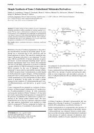
Most dietary fat is ingested as triglycerides (90–95%), and their<br />
hydrolysis starts in the mouth, then goes on through the stomach<br />
by an acid stable gastric lipase, and continues in the duodenum<br />
through the synergistic actions of gastric and colipase-dependent<br />
pancreatic lipases (PL), leading to the formation of monoglycerides<br />
and free fatty acids (FFA) (l " Fig. 1). FFA are absorbed<br />
by the enterocyte to synthesize new triglyceride molecules,<br />
which are transported to the different organs via lipoproteins, especially<br />
chylomicrons, after a meal [34].<br />
Pancreatic lipase (PL), encoded by the PNLIP gene in humans,<br />
plays a key role in the efficient digestion of triglycerides [35]. It<br />
is secreted into the duodenum through the duct system of the<br />
pancreas and is responsible for the hydrolysis of 50–70% of total<br />
dietary fats [9]. This enzyme has been widely used for the determination<br />
of the potential efficacy of natural products as antiobesity<br />
agents [36].<br />
Orlistat is currently the only clinically approved drug for obesity<br />
management in Europe. This molecule acts by inhibiting PL activity<br />
and the reduction of triglyceride absorption, and its long-term<br />
administration accompanying an energy restricted diet, results in<br />
weight loss [37]. Reduction on intestinal lipid digestion has been<br />
related to a decrease in the intra-abdominal fat content [7]. Thus,<br />
this compound is associated with a small, but statistically significant<br />
weight loss of about 3% more than diet alone in overweight<br />
and obese people [1]. In addition to losing weight, Orlistat within<br />
a prescribed diet has been shown to be safe and more effective<br />
than diet alone in modifying some of the risk of coronary artery<br />
disease and other obesity-related comorbidities. The most commonly<br />
reported adverse effects of Orlistat are a range of gastrointestinal<br />
side effects, including steathorrhea, bloating, oily spotting,<br />
fecal urgency, and fecal incontinence, as well as hepatic adverse<br />
effects [19, 38]. These adverse effects are similar to those<br />
observed for other lipase inhibitors tested in phase II studies,<br />
such as Cetilistat (ATL-962) [39].<br />
On the other hand, the inhibition of fat absorption could be accompanied<br />
by fat-soluble vitamin deficiencies, which could be<br />
prevented by the vitamin supplementation strategy, as other au-
Fig. 1 Fat metabolism in humans. Dietary fats are hydrolyzed in the gastrointestinal<br />
tract, where some lipases are involved.<br />
thors have recommended when vitamin deficiency occurs in patients<br />
undergoing Orlistat therapy [40].<br />
Hence the interest in the search for new natural substances that<br />
show potent inhibitory activity against PL and have fewer side effects<br />
than the current ones.<br />
Natural inhibitors of pancreatic lipase<br />
!<br />
In the continued search for effective antiobesity agents, several<br />
bacterial, fungal, and marine species have been screened to find<br />
new compounds with PL inhibitory activity.<br />
Many metabolic products from microorganisms, such as different<br />
kinds of Streptomyces (toxytricini, sp. NR 0619, albolongus, aburaviensis,<br />
andlavendulae) have a potent inhibitory activity of PL<br />
[9]. Lipstatin was isolated from an actinobacterium, Streptomyces<br />
toxytricini, and the catalytic hydrogenation product of lipstatin is<br />
the approved antiobesity drug tetrahydrolipstatin (Orlistat; marketed<br />
by Roche as Xenical) [18]. Panclicins, analogs of tetrahydrolipstatin<br />
isolated from Streptomyces sp. NR0619, also present<br />
strong anti-lipase activity [41]. Other compounds which also act<br />
as potent inhibitors of PL, at least in vitro, are ebelactones A and B,<br />
isolated from Streptomyces aburaviensis [42], and vibralactone,<br />
isolated from the culture broth of the polypore Boreostereum vibrans<br />
[43]. Finally, other examples of lipase inhibitors have been<br />
obtained from yeasts and fungi such as Candida antarctica, Candida<br />
rugosa, Gestrichum candidum, Humicola lanuginose, and<br />
Pseudomonas glumae, which have received special attention and<br />
are widely used in the pharmaceutical industry [44].<br />
Due to the biodiversity and unexplored resources, the fungal<br />
kingdom has been particularly searched to find new substances<br />
with lipase inhibitory activity. In a thorough screening of lipase<br />
inhibitors of fungal origin in Slovenia [23], extracts obtained from<br />
three species, Laetiporus sulphureus, Tylopilus felleus, andHygrocybe<br />
conica, exhibited very high lipase inhibitory activities (83%<br />
± 5%, 96% ± 3%, and 97% ± 5%, respectively), even higher than Orlistat.<br />
Pleurotus eryngii water extract also shows a significant in-<br />
Reviews<br />
hibitory activity against PL, preventing postprandial hyperlipidemia<br />
through low intestinal absorption of dietary fat [45]. Finally,<br />
the water and ethanol extracts from fruiting bodies of Phellinus<br />
linteus show a potent lipase inhibitory and antiobesity effect<br />
[46]. A special case is that of monascus pigments from Monascus<br />
sp., which have been used for many years as natural colorants and<br />
as a healthy food in East Asia, being employed in the production<br />
of certain fermented foods. Various monascus derivatives with<br />
incorporated unnatural amino acids show inhibitory activities<br />
against lipase [47].<br />
In the same way, marine products are an especially rich source of<br />
bioactive compounds [48]. In a milestone study, Bitou et al. [24]<br />
screened the lipase inhibitory activities of methanol and ethyl<br />
acetate extracts from 54 species of marine algae. These investigations<br />
observed a very high activity (almost 100% inhibition) in<br />
the methanol extracts from Caulerpa taxifolia and Asparagopsis<br />
tociformis, although the methanolic extracts of other Chlorophyta<br />
(i.e., Caulerpa okamurae or Codium latum), Rhodophyta (i.e.,<br />
Gloiopeltis tenax or Hypnea charoides), and Phaeophyta (i.e., Sargassum<br />
muticum, Dictyopteris latiuscula, orCutleria cylindrica),<br />
were also very promising. In this sense, Phaeophyta generally<br />
contains large amounts of polyphenols, such as tannins, with lipase-inhibiting<br />
activity. In fact, most compounds with a porphyrin<br />
structure are able to inhibit lipase activity [49]. Two algae<br />
whose extracts inhibit gastric and pancreatic lipases are Caulerpa<br />
prolifera, which may be a source of a potential antiobesity agent<br />
[50], and Caulerpa taxifolia, which synthesizes the toxin caulerpenyne<br />
[24]. On the other hand, carotenoids from Undaria pinnatifida<br />
and Sargassum fulvellum, specifically fucoxanthin that is<br />
metabolized in vivo to fucoxanthinol, suppress triglyceride absorption<br />
via the inhibition of PL in the intestinal lumen [51].<br />
Medicinal plants have been used as dietary supplements for body<br />
weight management and control in many countries. In this sense,<br />
presence of PL inhibitors has been demonstrated in different<br />
plant species (l " Table 1), although more research is needed for<br />
identifying and characterizing effective lipase inhibitors [52]. Lipase<br />
inhibitors of plant origin include certain proteins, such as<br />
those from soybean [53] and from wheat bran and germ [54].<br />
Other proteins that strongly inhibit hydrolysis of triglycerides<br />
are the basic protein protamine [55] and ε-polylysine [56], which<br />
could act, as several amphiphilic proteins like ovoalbumin and βlactoglobulin<br />
[57], by the desorption of lipase from its substrate<br />
due to a change in interfacial quality [58].<br />
Other lipase inhibitors from plant origin are basic polysaccharides,<br />
especially chitosan oligosaccharides, water-soluble chitosan<br />
(46 kDa) and polydextrose when a basic group is introduced<br />
[59, 60], phytic acid and other myoinositol phosphate esters [61],<br />
phenylboronic acid, a potent inhibitor of lipase from Oryza sativa<br />
[62], and carnosic acid, a diterpene isolated from the methanolic<br />
extract of the leaves of sage (Salvia officinalis) and rosemary [63].<br />
Korean and Chinese researchers have been very active in the<br />
search of new lipase inhibitors of herbal origin. Among the most<br />
promising compounds, there are platycodin D, isolated from the<br />
fresh roots of Platycodon grandiflorum [64, 65], dioscin from Dioscorea<br />
nipponica [66], licochalcone A from the roots of Glycyrrhiza<br />
uralensis [67], phenolic constituents from the leaves of Nelumbo<br />
nucifera [68], the aqueous ethanol extracts of Juniperus communis<br />
or common juniper (bark) and Illicium religiosum (wood)<br />
[69], the ethanol extract from stem bark and leaves from mango<br />
tree (Mangifera indica), which is able to prevent weight gain induced<br />
by feeding a high-fat diet to Wistar rats [70], a pomegranate<br />
leaf extract rich in ellagic acid and tannins [71], Rhei rhizoma<br />
de la Garza AL et al. Natural Inhibitors of … <strong>Planta</strong> Med 2011; 77: 773–785<br />
775
776<br />
Reviews<br />
Table 1 Plant extracts that showed over 40% inhibitory activity in vitro of pancreatic lipase and part of the plant from which the extract has been isolated.<br />
Family Scientific name Common Part Ref Family Scientific name Common Part Ref<br />
name<br />
of plant<br />
name of plant<br />
Aeraceae Acer pseudosiebol- Korean maple Whole [138] Lamiaceae Spirodela polyrhiza Common Whole [138]<br />
dianum<br />
duckmeat<br />
Anacardiaceae Pistacia vera Pistachio Fruits hull [52] Lamiaceae Thymus pulegoides Lemon thyme Whole [22]<br />
Apiaceae Levisticum officinale Garden lovage Whole [52] Lauraceae Cinnamomum<br />
zeylanicum<br />
Cinnamon Derm [52]<br />
Apiaceae Sanicula chinensis Bian Dou Cai Whole [138] Lauraceae Lindera glauca Grayblue<br />
spicebush<br />
Whole [138]<br />
Araliaceae Eleutherococcus Siberian ginseng Leaves [114] Liliaceae Asparagus<br />
Shiny asparagus Radix [138]<br />
senticosus<br />
cochinchinesis<br />
Aspidiaceaes Cyrtomium falcatum Japanese holly fern Whole [138] Liliaceae Scilla scilloides Chinese scilla Whole [138]<br />
Asteraceae Artemisia scoparia Redstem Whole [138] Linaceae Linum<br />
Oil flax Seed [139]<br />
wormwood<br />
usitatissimum<br />
Asteraceae Helianthus annus Common Seed [139] Lythraceae Lythrum salicaria Purple Whole [138]<br />
sunflower<br />
loosestrife<br />
Brassicaceae Brassica nigra Black mustard Radix [22] Musaeae Musa sapientum French plantain Fructus [22]<br />
Brassicaceae Brassica oleracea<br />
capitata<br />
Cabbage Folium [22] Myricaceae Myrika spp Bayberry Bark [140]<br />
Brassicaceae Raphanus sativus Radish Radix [22] Myrtaceae Myrtus communis True myrtle Leaves [52]<br />
Caprifoliaceae Lonicera japonica Japanese honeysuckle<br />
Whole [138] Myrtaceae Solanum tuberosum Potato Flowers [22]<br />
Celastraceae Euonymus<br />
sachalinensis<br />
Spindletree Whole [138] Oleaceae Olea europeae Olive Folium [22]<br />
Crassulaceae Rhodiola rosea Roseroot Whole [141] Orchida- Gastrodia elata Tien Ma Whole [138]<br />
stonecrop<br />
ceae<br />
Cucurbitaceae Cucurbita pepo Field pumpkin Whole [138] Oxalidaceae Oxalis corniculata Sleeping<br />
beauty<br />
Whole [138]<br />
Cucurbitaceae Momordica<br />
Spiny<br />
Whole [138] Poaceae Eriochloa villosa Hairy cup- Whole [138]<br />
cochinchinensis bittergourd<br />
grass<br />
Cyperaceae Bulbostylis barbata Watergrass Whole [138] Poaceae Hemarthria sibirica Weed Whole [138]<br />
Cyperaceae Carex kobomugi Japanese Whole [138] Poaceae Panicum dichotomi- Fall panic- Whole [138]<br />
sedge<br />
florumgrass<br />
Cyperaceae Cyperus amuricus Asian<br />
Whole [138] Poaceae Setaria italica Foxtail bris- Whole [138]<br />
flatsedge<br />
tlegrass<br />
Eleagnaceae Elaeagnus macro- Oleaster Whole [138] Polygala- Polygala tenuifolia Yuan Zhi Whole [138]<br />
phyllaceae<br />
Ericaceae Arctostaphylos uva-ursi Bear berry Folium [22] Polygona- Reynoutria elliptica Black bind- Whole [138]<br />
ceaeweed<br />
Ericaceae Vaccinium myrtillus Bilberry Fructus [22] Polygona- Rheum ribes Rhubarb Rhi- [52]<br />
ceaezomes<br />
Eriocaulaceae Eriocaulon siebol- Flattened Whole [138] Potamogetona- Potamogeton distinctus Pondweed Whole [138]<br />
dianum<br />
pipewort<br />
ceae<br />
Fabaceae Alhagi camelorum Camelthorn Aerial<br />
parts<br />
[52] Rosaceae Rosa damascene Damask rose Floret [52]<br />
Fabaceae Glycyrrhiza uralensis Gan Cao Whole [138] Rosaceae Rubus idaeus Raspberry Fructus [22]<br />
Fabaceae Lespedeza cuneata Chinese bush<br />
clover<br />
Whole [138] Rosaceae Malus domestica Apple Fructus [22]<br />
Fabaceae Phaseolus vulgaris Common bean Whole [22] Rubiaceae Gardenia jasmi- Cape jas- Whole [138]<br />
noidesmine<br />
Fabaceae Pisum sativum Garden pea Fructus [22] Rubiaceae Rubia akane Asian madder<br />
Whole [138]<br />
Fabaceae Pueraria thunbergiana Kudzu Whole [138] Rutaceae Citrus aurantifolium Lime Whole [138]<br />
Fabaceae Quercus infectoria Aleppo oak Galls [52] Rutaceae Murraya koeninggi Curryleaf<br />
tree<br />
Leaves [142]<br />
Juncaceae Juncus effusus Soft rush Whole [138] Rutaceae Orixa japonica Pearl frost Whole [138]<br />
Lamiaceae Agastache rugosa Purple giant Whole [138] Saxifragaceae Chrysosplenium Golden saxifrage Whole [138]<br />
hyssop<br />
grayanum<br />
Lamiaceae Origanum vulgare Oregano Herba [22] Simaroubaceae Ailanthus altissima Tree of heaven Whole [138]<br />
Lamiaceae Prunella vulgaris Common selfheal Whole [73] Tiliaceae Tilia platyphyllos Largeleaf linden Whole [22]<br />
Lamiaceae Rosmarinus officinalis Rosemary Folium [22] Urticaceae Urtica urens Dwarf nettle Aerial parts [52]<br />
Lamiaceae Salvia officinalis Salvia Folium [22] Zingiberaceae Afromomum<br />
Meleguetta Seed [143]<br />
meleguetta<br />
pepper<br />
de la Garza AL et al. Natural Inhibitors of… <strong>Planta</strong> Med 2011; 77: 773–785
Table 2 Some classes of natural compounds that have been reported to inhibit pancreatic lipase activity in vitro and species from which the compound has been<br />
obtained.<br />
Metabolites Scientific name Common name Family References<br />
Flavonoids Alpinia officinarum Lesser galangal Zingiberaceae [144,145]<br />
Flavonoids Taraxacum officinale Dandelion Asteraceae [103]<br />
Flavonoids, triterpenes Actinidia arguta Kiwi Actinidiaceae [146]<br />
Polyphenols Arachis hypogaea Peanut Fabaceae [9]<br />
Polyphenols Mangifera indica Mango Anacardiaceae [9]<br />
Polyphenols <strong>Medica</strong>go sativa Alfalfa Fabaceae [78]<br />
Polyphenols Nelumbo nucifera Sacred lotus Nelumbonaceae [9]<br />
Polyphenols Salacia reticulate Kotala himbutu Celastraceae [101]<br />
Polyphenols Salix matsudana Corkscrew willow Salicaceae [147]<br />
Polyphenols, proanthocyanidins,<br />
catechins<br />
Camellia sinensis Green, black, oolong tea Theaceae [89]<br />
Polyphenols, saponins Ilex paraguariensis Yerba mate Aquifoliaceae [99]<br />
Proanthocyanidins Cassia mimosoides Nomame herba Fabaceae [148]<br />
Proanthocyanidins Cinnamomum sieboldii Cinnamon Lauraceae [86]<br />
Proanthocyanidins Theobroma cacao Cocoa Malvaceae [86]<br />
Proanthocyanidins, saponins Vitis vinifera Grape vine Vitaceae [79,104]<br />
Saponins Aesculus hippocastanum Horse chestnut Sapindaceae [32]<br />
Saponins Aesculus turbinate Japanese horse chestnut Hippocastanaceae [110]<br />
Saponins Arctostaphylos uva-ursi Bearberry Ericaceae [32]<br />
Saponins Ardisia japonica Marlberry Myrsinaceae [152]<br />
Saponins Avena sativa Oat Poaceae [149]<br />
Saponins Coffea Arabica Coffee Rubiaceae [32]<br />
Saponins Cyclocarya paliurus Wheel wingnut Juglandaceae [9]<br />
Saponins Dioscorea nipponica Yam Dioscoreaceae [9]<br />
Saponins Eleutherococcus senticosus Siberian ginseng Araliaceae [114]<br />
Saponins Eleutherococcus sessiliflorus Sessiloside Araliaceae [9]<br />
Saponins Gardenia jasminoides Cape jasmine Rubiaceae [118]<br />
Saponins Gypsophila oldhamiana Oldhamʼs babyʼs-breath Caryophyllaceae [119]<br />
Saponins Kochia scoparia Burningbush Chenopodiaceae [150]<br />
Saponins Malus domestica Apple Rosaceae [32]<br />
Saponins Momordica charantia Balsampear Cucurbitaceae [151]<br />
Saponins Olea europeae Olive Oleaceae [32]<br />
Saponins Panax ginseng Ginseng Araliaceae [109]<br />
Saponins Panax japonicus Japanese ginseng Araliaceae [120]<br />
Saponins Panax quinquefolium American ginseng Araliaceae [122]<br />
Saponins Platycodi radix Doraji Campanulaceae [64]<br />
Saponins Platycodon grandiflorum Balloon flower Campanulaceae [103]<br />
Saponins Sapindus rarak Soapberry Sapindaceae [127]<br />
Saponins Scabiosa tschiliensis Pincushions Dipsacaceae [9]<br />
Saponins Solanum lycopersicum Tomato Solanaceae [32]<br />
Terpenes Salvia officinalis Salvia Lamiaceae [32]<br />
Triterpenes Aloe vera Aloe vera Asphodelaceae [32]<br />
Triterpenes Betula alba Birch Betulaceae [32]<br />
Triterpenes Calendula officinalis Pot marigold Asteraceae [32]<br />
Triterpenes Melissa officinalis Lemon balm Lamiaceae [32]<br />
Triterpenes Origanum vulgare Oregano Lamiaceae [32]<br />
(rhubarb) and the combinatorial drug Chunghyuldan [72], Prunella<br />
vulgaris, Rheum palmatum, and other herbs [73]. Most of<br />
the common compounds that are found in different plant species<br />
are polyphenols, saponins, and terpenes (l " Table 2).<br />
In the following chapters more information will be given out<br />
about the most thoroughly studied compounds, classified according<br />
to their biochemical structure.<br />
Polyphenols<br />
A number of studies have revealed various health benefits of<br />
plant polyphenols and their importance in foods, beverages, and<br />
natural medicine. In this context, polyphenols have some potential<br />
efficacy for preventing obesity. They inhibit enzymes related<br />
to fat metabolism including PL, lipoprotein lipase, and glycero-<br />
Reviews<br />
phosphate dehydrogenase [74]. Polyphenol extracts are able to<br />
decrease the blood levels of glucose, triglycerides, and LDL cholesterol,<br />
increase energy expenditure and fat oxidation, and reduce<br />
body weight and adiposity [75, 76]. In fact, many polyphenols,<br />
including flavones, flavonols, tannins, and chalcones, have<br />
shown an inhibitory activity of PL [9, 22].<br />
Flavonoids are a type of plant secondary metabolites that are<br />
characterized as containing two or more aromatic rings, each<br />
bearing at least one aromatic hydroxyl and connected with a carbon<br />
bridge [76]. Some of them are polymerized into large molecules,<br />
either by the plants themselves or as a result of food processing.<br />
These polymers are called tannins, and three subclasses<br />
(condensed tannins, derived tannins, and hydrolysable tannins)<br />
exhibit a variety of beneficial effects on health [76]. A flavonoid<br />
de la Garza AL et al. Natural Inhibitors of … <strong>Planta</strong> Med 2011; 77: 773–785<br />
777
778<br />
Reviews<br />
with PL inhibitory activity is hesperidin, obtained from the peels<br />
of Citrus unshiu [77].<br />
Proanthocyanidins (PA), also known as condensed tannins, are<br />
the most common group of flavonoids in the Western diet. They<br />
consist of monomeric units of flavans linked through carbon-carbon<br />
and ether linkages, which are considered the second most<br />
abundant group of natural phenolics after lignins [78]. PA can be<br />
found in such common foodstuffs as cereals, legumes, fruits, vegetables,<br />
and beverages (red wine and tea in particular) [75, 79].<br />
They have a putative role as antioxidants, showing beneficial effects<br />
on inflammatory processes, cardiovascular diseases, and<br />
other pathological conditions [80, 81]. For example, these compounds<br />
actively reduce plasma triglycerides by inhibiting the absorption<br />
of dietary lipids [79] and possess inhibitory effects on<br />
different digestive enzymes, such as trypsin, amylase, and lipase<br />
[36].<br />
Some examples of polyphenols with inhibitory action on PL are<br />
proanthocyanidins from edible herbs, such as those from Cassia<br />
mimosoides [82], and tea catechins, especially (−)-catechin gallate<br />
and (−)-gallocatechin gallate, [83]. Some of the most thoroughly<br />
studied polyphenol extracts in relation to PL inhibition are the<br />
following:<br />
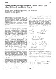
Arachis hypogaea: Peanut (Arachis hypogaea) shells (hulls, seed<br />
coats), which are by-products of the peanut industry, provide<br />
several compounds showing PL inhibitory activity in a dose dependent<br />
manner (1 mg/mL = 42% inhibitory effect) that are able<br />
to reduce body weight gain in rats fed a high-fat diet [84]. This<br />
plant contains several bioactive molecules, such as luteolin<br />
(l " Fig. 2), certain fatty acids, caffeic, ferulic, and benzoic acids,<br />
all of which are able to inhibit lipases [9]. Coumarin derivates<br />
and phenolic acids were assumed to be the major active constituents.<br />
However the authors have not examined the individual effects<br />
of each compound.<br />
Camellia sinensis: Camellia sinensis or tea plant (green tea, black<br />
tea, or oolong tea) contains over 60 polyphenols, some of them<br />
with a potent PL inhibitory activity. It is likely the plant whose<br />
extracts have been more thoroughly used for searching new PL<br />
inhibitors. The major polyphenols are catechins (l " Fig. 2), which<br />
constitute about one-third of its total dry weight. A serving of tea<br />
is moderate to high in flavonoid and/or tannin content [85–89].<br />
Nakai et al. [90] found that the polyphenols with more potent PL<br />
inhibitory effect were flavan-3-ol digallate esters isolated from<br />
oolong tea, such as (−)-epigallocatechin-3,5-digallate. Oolong<br />
tea-polymerized polyphenols reduced postprandial hypertriglyceridemia<br />
in olive oil-loaded rats and mice [91]. Also (−)-epigallocatechin,<br />
abundant in the green tea extract, is a weak inhibitor of<br />
PL and is able to decrease the postprandial hypertriglyceridemia<br />
in rodents [92].<br />
The administration of black-tea polyphenols suppressed postprandial<br />
hypertriglyceridemia in a dose-dependent manner in<br />
rats, with theaflavin-3,3′-digallate as the most effective PL inhibitor<br />
[93], whereas other authors point out to thearubigins [94].<br />
These extracts are able to prevent increases in body weight and<br />
adiposity in mice fed a high-fat diet [95]. The PL inhibitory and<br />
hypotriglyceridemic effects of tea extracts were corroborated by<br />
Tanaka et al. [96], who orally administered mixed fermented tea<br />
extracts and Loquat tea extracts to rats with a 10% soybean oil<br />
emulsion.<br />
Finally, cocoa tea extract (Camellia sinensis var. ptilophylla) is rich<br />
in polyphenols with PL inhibitory effect. A single oral administration<br />
of this extract produces an inhibition of plasma triglyceride<br />
levels in olive oil-loaded ICR mice and triolein-loaded rats [97].<br />
de la Garza AL et al. Natural Inhibitors of… <strong>Planta</strong> Med 2011; 77: 773–785<br />
Fig. 2 Selected polyphenols with PL inhibitory activity: Luteolin (1) from<br />
Arachis hypogaea, catechin (2) from Camellia sinensis, daidzein (3) from Glycine<br />
max, quercetin (4) from Ilex paraguariensis, structure of a procyanidin<br />
(5) from Vitis vinifera.<br />
Glycine max: Daidzein (l " Fig. 2) belongs to the group of isoflavones<br />
and is produced almost exclusively by the members of the<br />
Fabaceae/Leguminosae (bean) family such as soybean. In one<br />
study, Guo et al. [98] investigated the effects of daidzein on body<br />
weight, adipose tissue, blood, and liver lipid levels in obese mice<br />
fed a high-fat diet, finding that daidzein reduced body and white<br />
adipose tissue weights in obese mice and ameliorated the hyperlipidemia<br />
induced by the high-fat diet. The authors attributed<br />
this effect to the inhibition of PL activity and fat digestion.<br />
Ilex paraguariensis: Yerba mate (MT) is a plant from the subtropical<br />
region of South America that is widely consumed in Brazil,<br />
Argentina, Paraguay, and Uruguay. Yerba mate contains polyphenols,<br />
such as flavonoids (quercetin and rutin) (l " Fig. 2) and phenolic<br />
acids (chlorogenic and caffeic acids), and is also rich in caffeine<br />
and saponins [99]. These substances act on the lipid metabolism<br />
by inhibiting PL activity in a concentration value of 1.5 mg/<br />
mL [99]. Several triterpene saponins and monoterpene oligoglycosides<br />
from the leaves of yerba mate were found to exhibit potent<br />
inhibitory activity on porcine PL [100].<br />
Malus domestica: Apples (Malus domestica) belong to the Rosaceae<br />
family whose fruits contain several phenolic substances<br />
(cholorogenic acid, catechin, epicatechin, phloridzin, and procyanins).<br />
Procyanidins in apples are mainly composed of various<br />
polymerized catechins, with some of them showing a PL inhibitory<br />
activity and reducing triglyceride absorption [36]. In corn<br />
oil-loaded mice, a single oral administration of apple polyphenols<br />
reduced plasma triglyceride levels, and a test diet containing<br />
600 mg of apple polyphenols significantly inhibited triglyceride
elevation at 6 h after ingestion, indicating an inhibition of triglyceride<br />
absorption [36].<br />
Salacia reticulata: Salacia reticulata contains a high concentration<br />
of polyphenols, including catechins and condensed tannins.<br />
In hot water-soluble extract from the roots of Salacia reticulata<br />
(SRHW) the concentration is about 24% polyphenols [74]. The<br />
polyphenols from Salacia reticulata inhibit enzymes related to<br />
fat metabolism, including PL, lipoprotein lipase, and glycerophosphate<br />
dehydrogenase, and are effective in preventing obesity<br />
[101]. In fact, Salacia extract markedly improved metabolic syndrome<br />
symptoms (including body weight, adiposity, glucose intolerance,<br />
hypertension, and peripheral neuropathy) in TSOD<br />
mice [102].<br />
Taraxacum officinale: Dandelion (Taraxacum officinale) is a perennial<br />
herbaceous plant of the family Asteraceae that has been<br />
used as a phytomedicine due to its choleretic, antirhemetic, diuretic,<br />
and anti-inflammatory properties [103]. Extracts from<br />
this plant have shown hypolipidemic effects and an inhibitory activity<br />
of PL, decreasing AUC (area under curve) for the postprandial<br />
triglyceride response curve [103].<br />
Vitis vinifera: Grapevine (Vitis vinifera) has become a model plant<br />
for studying proanthocyanidin biosynthesis. Grapevine proanthocyanidins<br />
(l " Fig. 2) consist of two major flavan 3-ol monomers,<br />
catechin and epicatechin, that have inhibitory activity on<br />
PL [79, 104].<br />
Polyphenol-rich extracts from a range of berries, particularly<br />
cloudberry, are able to inhibit PL activity in vitro, which has been<br />
attributed to their content in ellagitannins and proanthocyanidins<br />
[105].<br />
Saponins<br />
Saponins are a major family of secondary metabolites that occur<br />
in a wide range of plants species [106]. These compounds have<br />
been isolated from different parts of the plants, including the<br />
roots, rhizomes, stems, bark, leaves, seeds, and fruits. Occasionally,<br />
the whole plant has been used [107].<br />
Saponins are categorized into two major classes, the triterpenoid<br />
and the steroid saponins, which are both derived from the 30 carbon<br />
atoms containing precursor oxidosqualene [107, 108]. Some<br />
of the triterpene-rich plant materials are common foodstuffs<br />
consumed in large amounts in Mediterranean countries. Therefore,<br />
the correlation of a triterpene-rich diet and the beneficial effects<br />
of consuming a Mediterranean diet should be investigated<br />
in more detail [32]. These types of plant secondary metabolites<br />
are found to inhibit PL and, thus, may represent potential effective<br />
treatments for obesity and related disorders [9, 22].<br />
Aesculus turbinata: The Japanese horse chestnut (Aesculus turbinata)<br />
is a medicinal plant widely used in East Asia. The saponin<br />
mixture extracted from the seeds is called escins and has a strong<br />
inhibitory activity on PL [110]. In mice fed a high-fat diet, total<br />
escins suppressed the increase in body weight, adiposity, and liver<br />
fat and increased triglyceride level in the feces, whereas it decreased<br />
plasma triglycerides after the oral administration of a lipid<br />
emulsion [111, 112].<br />
Dioscorea nipponica: The methanol extract of Dioscorea nipponica<br />
Makino powder has a potent inhibitory activity against porcine<br />
PL, with an IC 50 value of 5–10 µg/mL [66]. In fact, the saponin dioscin<br />
and its aglycone, diosgenin, both suppressed the increase of<br />
blood triacylglycerols when orally injected with corn oil to mice.<br />
Rats fed a high-fat diet containing 5% Dioscorea nipponica Makino<br />
gained significantly less body weight and adipose tissue than<br />
control animals [66], and a similar result has been observed after<br />
Reviews<br />
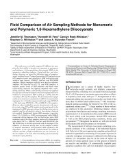
Fig. 3 Selected isoprenoids with PL inhibitory activity: Eleutheroside (6)<br />
from Eleutherococcus senticosus, geniposide (7) from Gardenia jasminoides,<br />
general structure of dammaran aglycons of ginsenosides (8)inPanax ginseng,<br />
betulin (9) from Betula alba.<br />
administering the aqueous extract of this rhizome to mice fed a<br />
high-fat diet [113].<br />
Eleutherococcus senticosus: Eleutherococcus senticosus is a shrub,<br />
belonging to the family Araliaceae, which is commonly distributed<br />
in north-eastern Asia. It is used as a traditional Chinese<br />
medicine against ischemic heart diseases, neurasthenia, hypertension,<br />
arthritis, and tumors [114]. At least fifteen triterpenoid<br />
saponins with in vitro PL inhibitory activity (l " Fig. 3) have been<br />
isolated from the fruits of Eleutherococcus senticosus [115]. The<br />
total saponin fraction obtained from the fruits of Eleutherococcus<br />
senticosus exhibits inhibitory activity on PL with an IC50 value of<br />
3.63 mg/mL [114].<br />
Eleutherococcus sessiliflorus: Different lupine-type triterpene triglycosides<br />
isolated from a hot water extract of Eleutherococcus<br />
sessiliflorus leaves are able to inhibit PL activity in vitro and to<br />
suppress the body weight gain of mice fed a high-fat diet [116].<br />
Gardenia jasminoides: Crocin is a glycosylated carotenoid extracted<br />
from the fructus of Gardenia jasminoides (l " Fig. 3). Gardeniae<br />
Fructus is used in Asian countries as a natural colorant,<br />
and in Chinese traditional medicine for its antioxidant, cytotoxic,<br />
antitumor, and neuroprotective effects. Crocin and crocetin are<br />
effective hypolipidemic agents that act by reducing the absorption<br />
of fat and cholesterol through inhibition of PL activity [117].<br />
Sheng et al. demonstrated that crocin selectively inhibited the activity<br />
of PL as a competitive inhibitor [118].<br />
Gypsophila oldhamiana: Gypsophila oldhamiana (Caryophyllaceae)<br />
is a plant distributed in the north of China whose roots have<br />
high amounts of saponins, sterols, and fatty acids. The extract<br />
from this plant shows a potent inhibitory activity of PL with an<br />
IC 50 value of 0.54 mg/ml [118,119] and different triterpenoid<br />
saponins, gypsosaponins A–C, as the more efficient compounds<br />
[119].<br />
Panax ginseng: Ginseng is one of the most popular medicinal<br />
herbs and is commonly consumed as powder, a beverage, or a<br />
food supplement. Roots of Panax ginseng contain high levels of<br />
de la Garza AL et al. Natural Inhibitors of … <strong>Planta</strong> Med 2011; 77: 773–785<br />
779
780<br />
Reviews<br />
ginsenosides (l " Fig. 3), which are steroidal saponins that show<br />
beneficial effects on lipid metabolism. Saponins from ginseng<br />
roots suppressed the expected increase in body weight and plasma<br />
triacylglycerols in mice following a high-fat diet, which was<br />
probably mediated by inhibiting PL with an IC50 value of 500 µg/<br />
mL [109].<br />
Panax japonicus: The rhizomes of Panax japonicus (Japanese ginseng)<br />
are used in folk medicine for the treatment of arteriosclerosis,<br />
hyperlipidemia, hypertension, and diabetes mellitus. Chikusetsusaponins<br />
prevented the increase in body weight and parametrial<br />
adipose tissue weight induced by a high-fat diet and inhibited<br />
the elevation of postprandial plasma triacylglycerols due<br />
to their inhibitory action of PL on dietary fat [120]. The delay in<br />
intestinal fat absorption was also behind the antiobesity effects<br />
observed for Korean white ginseng extract in high-fat diet-induced<br />
obese mice [121].<br />
Panax quinquefolium: American ginseng (Panax quinquefolium) is<br />
a native plant from North America. The saponins isolated from<br />
stems and leaves of Panax quinquefolium may prevent fat storage<br />
in adipose tissue and postprandial elevations of plasma triacylglycerols<br />
by inhibiting the intestinal absorption of dietary fat<br />
through the inhibition of PL activity [122].<br />
Platycodi grandiflorum: Platycodi radix, widely used in traditional<br />
Oriental medicines as a remedy for respiratory disorders, is<br />
rich in saponins, which are responsible for a diversity of effects<br />
including anti-inflammation, antiallergy, antitumor, and immunostimulation<br />
[64]. Given its inhibitory action on PL [123], with<br />
platycodin D as the most efficient compound [124], it ameliorated<br />
high fat-induced obesity in mice [125] and rats [64]. SK1 is<br />
an edible saponin-rich compound from Platycodi Radix that is<br />
able to reduce body weight and fat accumulation by increasing<br />
fecal lipid outputs in high-fat fed mice [126].<br />
Sapindus rarak: The methanolic extract from the pericarps of Sapindus<br />
rarak (Lerak) shows a PL inhibitory activity that is probably<br />
due to diverse saponins and sesquiterpene glycosides [127].<br />
Scabiosa tschiliensis: Different triterpenoid saponins isolated<br />
from the Mongol and Chinese traditional medicinal herb Scabiosa<br />
tschiliensis have shown strong inhibition of PL in vitro [128]. Due<br />
to the difficult task of isolating scabiosaponins and the scarceness<br />
of this type of saponin in nature, some of them have been successfully<br />
synthesized in the laboratory [129].<br />
Tea saponins: At least three kinds of tea (oolong, green, and black)<br />
have been used as healthy drinks. Tea saponins suppress the increases<br />
in body and parametrial adipose tissue weights and adipocyte<br />
diameters induced by a high-fat diet in mice by inhibiting PL<br />
and also reduce the elevation in plasma triacylglycerol levels after<br />
oral administration of a lipid emulsion. The Ki value of tea saponins<br />
was determined to be 0.25 mg/mL [85]. Thus, the crude saponin<br />
fraction from the flower buds of Chinese tea plant exhibits<br />
accelerating effects on gastrointestinal transit in mice and inhibitory<br />
effects against porcine PL, and three floratheasaponins (A–C)<br />
showed inhibitory effects on serum triglyceride elevation [130].<br />
Triterpenes<br />
Terpenes are the primary constituents of the essential oils of<br />
many types of plants and are classified by the number of terpene<br />
units in the molecule (diterpenes, triterpenes, among others).<br />
The pharmacological relevance of triterpenes has increased during<br />
the last two decades demonstrating multitarget properties<br />
such as wound healing, anti-inflammatory, antibacterial, antiviral,<br />
hepatoprotective, and antitumoral effects, combined with<br />
low toxicity [32]. Triterpene extracts are safe and provide a high<br />
de la Garza AL et al. Natural Inhibitors of… <strong>Planta</strong> Med 2011; 77: 773–785<br />
potential for further pharmaceutical and pharmacological research<br />
[131], some of them inhibiting PL activity.<br />
Betula alba: Bark of birch (Betula alba) contains pentacyclic triterpenes<br />
(l " Fig. 3). This triterpene extract is safe and provides a<br />
high potential for further pharmaceutical and pharmacological<br />
research [32, 131], displaying an inhibitory activity on PL [22].<br />
Clinical Studies about Pancreatic Lipase Inhibitors<br />
!<br />
A number of plants and natural products have been screened for<br />
their PL inhibitory activity but just some of them have gone up to<br />
clinical studies. In this line, only one product derived from natural<br />
compounds (Orlistat) is currently in clinical use, although<br />
others are under investigation. Some of them are Panax ginseng<br />
[132], Camellia sinensis [133], Eleutherococcus senticosus [134],<br />
Malus domestica [135], and Arachis hypogaea [136].<br />
In one study [132], the administration of an extract of Panax ginseng<br />
in humans for 8 weeks decreased circulating cholesterol, triglyceride,<br />
and low-density lipoprotein levels (LDL). Each subject<br />
ingested 2 g of Panax ginseng extract three times a day.<br />
Lee et al. [134] reported that healthy postmenopausal women<br />
treated for 6 months with Eleutherococcus senticosus supplementation<br />
showed significant decreases in serum LDL levels and LDL/<br />
HDL ratios.<br />
In other study, Sugiyama et al. [135] assessed six healthy male<br />
volunteers that followed a high-fat diet with 40 g of fat with 10<br />
control or 10 apple polyphenol (Malus domestica) capsules (600<br />
or 1500 mg). In this study, they demonstrated that apple polyphenols<br />
may prevent obesity in humans by a PL inhibitory mechanism.<br />
Green tea (Camellia sinensis) has been extensively studied in relation<br />
to obesity and other metabolic disorders. Thus, Chantre<br />
and Lairon [133] showed that green tea consumption may be<br />
useful to treat obesity by both, increasing thermogenesis and inhibiting<br />
PL. Thus, a green tea extract showed a direct in vitro inhibition<br />
of gastric and pancreatic lipases [133]. In moderately<br />
obese patients, green tea lowered body weight by stimulating<br />
thermogenesis and increasing energy expenditure when each<br />
subject received 2 times/d a green tea extract (2 capsules morning,<br />
2 capsules midday). Ingestion of 4 capsules containing AR25<br />
(Exolise) provided a daily total intake of 375 mg catechins, of<br />
which 270 mg was epigallocatechin gallate. Also, He et al. [137]<br />
administered daily 8 g of oolong tea for 6 weeks to 102 obese subjects.<br />
As a result, 70% of the obese subjects decreased more than<br />
1 kg in body weight. In vitro studies suggested that the effect of<br />
oolong tea on body weight could be partially attributed to the inhibition<br />
of PL [68].<br />
According to these data, a number of common herbal products<br />
that are being studied in animal (l " Table 3) and human models<br />
for obesity treatment contain different metabolites that act on<br />
lipid digestion and absorption. However, it is very difficult to establish<br />
in in vivo studies whether these antiobesity effects are only<br />
or mainly due to PL activity inhibition. The clinical implications<br />
of this therapeutic approach have yet to be determined.<br />
Conclusions<br />
!<br />
Orlistat is the only drug authorized and present in Europe for the<br />
treatment of obesity within an adequate energy intake, which<br />
acts by inhibiting the lipolytic activity of PL. With the aim of find-
Table 3 Plant extracts that showed in vivo inhibitory activity of pancreatic lipase, doses and effects. IC 50 is indicated when available.<br />
Scientific name Common name IC50 Doses Model Effects References<br />
Aesculus turbinate Japanese horse chestnut IC50 24 mg/mL 0.1–0.5% of diet DIO mice TG plasma levels and body weight gain [153]<br />
Arachis hypogaea Peanut IC50 0.029 µg/mL 1% of diet DIO rats Body weight gain [136]<br />
Camellia sinensis Green, black, oolong tea IC50 0.091 mg/mL 3% of HFD Rats Body weight gain<br />
and visceral fat<br />
[89]<br />
Cassia mimosoides Nomame herba IC50 0.1–0.71 mg/mL 1–3.5% of diet DIO rats Body weight gain [154]<br />
Coffea arabica Coffee 0.5% of standard<br />
diet<br />
Mice Body weight gain [155]<br />
Cyclocarya paliurus Wheel wingnut IC50 9.1 µg/mL 250 mg/kg; VO Mice TG plasma levels and blood glucose levels [156]<br />
Dioscorea nipponica Yam IC50 5–10 mg/mL 5% of HFD Rats TG plasma levels and body weight gain [157]<br />
Eleutherococcus senti- Siberian ginseng IC50 0.22–0.29 mM 12 mg/kg DIO rats Abdominal fat, TG in liver<br />
[158]<br />
cosus<br />
and serum and LDL in serum<br />
Eleutherococcus Sessiloside IC50 0.36–0.75 mg/mL 100–300 mg/kg; Mice TG plasma levels [159]<br />
sessiliflorus<br />
VO<br />
Gardenia jasminoides Cape jasmine IC50 2.1 mg/mL 50 mg/kg/d Mice Body weight gain [118]<br />
Humulus lupulus Common hop 0.2–1.2% (w/w)<br />
of extract<br />
Mice Body weight gain and blood glucose levels [160]<br />
Ilex paraguariensis Yerba mate 0.24% of HFD Rats Body weight gain [99]<br />
Kochia scoparia Burningbush 3% of HFD Mice Body weight gain [150]<br />
Malus domestica Apple IC50 5.6 µg/mL 200 mg/kg; VO Mice TG plasma levels [161]<br />
Myrica spp Bayberry – – TG plasma levels [140]<br />
Nelumbo nucifera Sacred lotus IC50 0.46 mg/mL 5% of diet Mice TG plasma levels and body weight gain [162]<br />
Panax ginseng Ginseng IC50 500 µg/mL 200 mg/kg with<br />
HFD<br />
Rats Body weight gain [109]<br />
Panax japonicus Japanese ginseng 1–3% of diet DIO mice Body weight gain [120]<br />
Platycodi radix Doraji Ki 0.18 mM 70 mg/kg, Sprague Body weight gain [64]<br />
with HFD Dawley rats<br />
Rhodiola rosea Roseroot stonecrop IC50 0.093 mM 150 mg/kg Mice TG plasma levels [141]<br />
Rosmarinus officinalis Rosemary 200 mg/kg HFD Mice Body weight and fat mass [163]<br />
Salacia reticulata Kotala himbutu IC50 264 mg/L 125 mg/kg;<br />
VO HFD<br />
Rats Body weight gain [101]<br />
Salix matsudana Corkscrew willow 5% of HFD Wistar rats Body weight gain [147]<br />
DIO: Diet-induced obesity; HFD: High-fat diet; VO: Via oral. (Daily food intake is approximately: rats: 20 g/day; mice: 4.5 g/day)<br />
ing new compounds more potent or with less secondary effects<br />
than Orlistat, new natural products are being identified and<br />
screened for their PL inhibitory potential. Some of these extracts<br />
are obtained from plants that are rich in polyphenols and saponins<br />
and show inhibitory effects on fat digestion, whereas other<br />
extracts come from algae, fungi, and microorganisms. Thus, natural<br />
products provide an exciting opportunity and promise for the<br />
development of new therapeutic approaches to the treatment of<br />
obesity by blocking the digestion and absorption of dietary lipids,<br />
and constitute a valuable alternative to other pharmacological<br />
agents. Some of the products reviewed in this article show potentially<br />
promising effects for weight control. In particular apple,<br />
green tea, soybean, and ginseng seem to have great potential as<br />
sources of molecules with PL inhibitory activity. For all of them<br />
more data are needed to define effects, optimal dose required,<br />
and mechanism of action, as well as their possible side or toxic<br />
effects.<br />
Thus, there is an urgent need to update the knowledge on the numerous<br />
natural sources that could act as inhibitors of PL in order<br />
to screen them as new potential therapeutic antiobesity agents<br />
with low secondary effects.<br />
Reviews<br />
Acknowledgements<br />
!<br />
The authors thank Línea Especial (LE/97) from the University of<br />
Navarra (Spain) and the CENIT PRONAOS Program (MICINN,<br />
Spain) for financial support. A. L. de la Garza and N. Boqué hold<br />
pre-doctoral grants from Ibercaja. We also acknowledge Marta<br />
Díaz Hernando for her contribution to the figures design.<br />
References<br />
1 Drew B, Dixon A, Dixon J. Obesity management: update on orlistat. Vasc<br />
Health Risk Manag 2007; 3: 817–821<br />
2 Brug J, Crawford D. The obesity pandemic. Is it bad or worse? Eur<br />
J Public Health 2009; 19: 570–571<br />
3 Schrauwen P, Westerterp KR. The role of high-fat diets and physical activity<br />
in the regulation of body weight. Br J Nutr 2000; 84: 417–427<br />
4 Voshol P, Rensen PCN, van Dijk K, Romijn J, Havekes L. Effect of plasma<br />
triglyceride metabolism on lipid storage in adipose tissue: studies using<br />
genetically engineered mouse models. Biochim Biophys Acta 2009;<br />
1791: 479–485<br />
5 Abete I, Astrup A, Martnez JA, Thorsdottir I, Zulet M. Obesity and the<br />
metabolic syndrome: role of different dietary macronutrient distribution<br />
patterns and specific nutritional components on weight loss and<br />
maintenance. Nutr Rev 2010; 68: 214–231<br />
6 Little T, Horowitz M, Feinle-Bisset C. Modulation by high-fat diets of gastrointestinal<br />
function and hormones associated with the regulation of<br />
energy intake: implications for the pathophysiology of obesity. Am<br />
J Clin Nutr 2007; 86: 531–541<br />
7 Rubio M, Gargallo M, Millán A, Moreno B. Drugs in the treatment of obesity:<br />
sibutramine, orlistat and rimonabant. Public Health Nutr 2007;<br />
10: 1200–1205<br />
de la Garza AL et al. Natural Inhibitors of … <strong>Planta</strong> Med 2011; 77: 773–785<br />
781
782<br />
Reviews<br />
8 Moreno D, Ilic N, Poulev A, Brasaemle D, Fried S, Raskin I. Inhibitory effects<br />
of grape seed extract on lipases. Nutrition 2003; 19: 876–879<br />
9 Birari R, Bhutani K. Pancreatic lipase inhibitors from natural sources:<br />
unexplored potential. Drug Discov Today 2007; 12: 879–889<br />
10 Sumantran V. Experimental approaches for studying uptake and action<br />
of herbal medicines. Phytother Res 2007; 21: 210–214<br />
11 Yamagishi S, Matsui T, Ueda S, Fukami K, Okuda S. Clinical utility of acarbose,<br />
an alpha-glucosidase inhibitor in cardiometabolic disorders. Curr<br />
Drug Metab 2009; 10: 159–163<br />
12 Raz I, Eldor R, Cernea S, Shafrir E. Diabetes: insulin resistance and derangements<br />
in lipid metabolism. Cure through intervention in fat<br />
transport and storage. Diabetes Metab Res 2005; 21: 3–14<br />
13 Hosoyama H, Sugimoto A, Suzuki Y, Sakane I, Kakuda T. [Isolation and<br />
quantitative analysis of the alpha-amylase inhibitor in Lagerstroemia<br />
speciosa (L.) Pers. (Banaba)]. Yakugaku Zasshi 2003; 123: 599–605<br />
14 Tsujita T, Takaku T, Suzuki T. Chestnut astringent skin extract, an alphaamylase<br />
inhibitor, retards carbohydrate absorption in rats and humans.<br />
J Nutr Sci Vitaminol 2008; 54: 82–88<br />
15 Tormo MA, Gil-Exojo I, de Tejada AR, Campillo JE. White bean amylase<br />
inhibitor administered orally reduces glycaemia in type 2 diabetic rats.<br />
Br J Nutr 2006; 96: 539–544<br />
16 Bray G, Ryan D. Drug treatment of the overweight patient. Gastroenterology<br />
2007; 132: 2239–2252<br />
17 McClendon K, Riche D, Uwaifo G. Orlistat: current status in clinical therapeutics.<br />
Expert Opin Drug Saf 2009; 8: 727–744<br />
18 Weibel EK, Hadvary P, Hochuli E, Kupfer E, Lengsfeld H. Lipstatin, an inhibitor<br />
of pancreatic lipase, produced by Streptomyces toxytricini.<br />
I. Producing organism, fermentation, isolation and biological activity.<br />
J Antibiot 1987; 40: 1081–1085<br />
19 Viner RM, Hsia Y, Tomsic T, Wong ICK. Efficacy and safety of anti-obesity<br />
drugs in children and adolescents: systematic review and meta-analysis.<br />
Obes Rev 2010; 11: 593–602<br />
20 Heymsfield SB, Segal KR, Hauptman J, Lucas CP, Boldrin MN, Rissanen A.<br />
Effects of weight loss with orlistat on glucose tolerance and progression<br />
to type 2 diabetes in obese adults. Arch Intern Med 2000; 160:<br />
1321–1326<br />
21 Torgerson J, Hauptman J, Boldrin M, Sjstrm L. Xenical in the prevention<br />
of diabetes in obese subjects (XENDOS) study: a randomized study of<br />
orlistat as an adjunct to lifestyle changes for the prevention of type 2<br />
diabetes in obese patients. Diabetes Care 2004; 27: 155–161<br />
22 Slanc P, Doljak B, Kreft S, Lunder M, Janes D, Strukelj B. Screening of selected<br />
food and medicinal plant extracts for pancreatic lipase inhibition.<br />
Phytother Res 2009; 23: 874–877<br />
23 Slanc P, Doljak B, Mlinaric A, Strukelj B. Screening of wood damaging<br />
fungi and macrofungi for inhibitors of pancreatic lipase. Phytother Res<br />
2004; 18: 758–762<br />
24 Bitou N, Ninomiya M, Tsujita T, Okuda H. Screening of lipase inhibitors<br />
from marine algae. Lipids 1999; 34: 441–445<br />
25 Abete I, Parra MD, Zulet MA, Martinez JA. Different dietary strategies for<br />
weight loss in obesity: role of energy and macronutrient content. Nutr<br />
Rev 2006; 19: 5–17<br />
26 Astrup A. The role of dietary fat in the prevention and treatment of<br />
obesity. Efficacy and safety of low-fat diets. Int J Obes 2001; 25: 46–50<br />
27 Lomba A, Martinez JA, Garcia-Daz D, Paternain L, Marti A, Campion J,<br />
Milagro FI. Weight gain induced by an isocaloric pair-fed high fat diet:<br />
a nutriepigenetic study on FASN and NDUFB6 gene promoters. Mol<br />
Genet Metab 2010; 101: 273–278<br />
28 Lomba A, Milagro FI, Garcia-Daz DF, Campion J, Marzo F, Martinez JA.<br />
A high-sucrose isocaloric pair-fed model induces obesity and impairs<br />
NDUFB6 gene function in rat adipose tissue. J Nutrigenet Nutrigenomics<br />
2009; 2: 267–272<br />
29 Hermsdorff HHM, Volp ACP, Bressan J. [Macronutrient profile affects<br />
diet-induced thermogenesis and energy intake]. Arch Latinoam Nutr<br />
2007; 57: 33–42<br />
30 Mobbs C, Mastaitis J, Yen K, Schwartz J, Mohan V, Poplawski M. Low-carbohydrate<br />
diets cause obesity, low-carbohydrate diets reverse obesity:<br />
a metabolic mechanism resolving the paradox. Appetite 2007; 48:<br />
135–138<br />
31 Rolls BJ. The role of energy density in the overconsumption of fat. J Nutr<br />
2000; 130: 268–271<br />
32 Jäger S, Trojan H, Kopp T, Laszczyk M, Scheffler A. Pentacyclic triterpene<br />
distribution in various plants – rich sources for a new group of multipotent<br />
plant extracts. Molecules 2009; 14: 2016–2031<br />
de la Garza AL et al. Natural Inhibitors of… <strong>Planta</strong> Med 2011; 77: 773–785<br />
33 Stevenson E, Astbury N, Simpson E, Taylor M, Macdonald I. Fat oxidation<br />
during exercise and satiety during recovery are increased following a<br />
low-glycemic index breakfast in sedentary women. J Nutr 2009; 139:<br />
890–897<br />
34 Armand M. Lipases and lipolysis in the human digestive tract: where do<br />
we stand? Curr Opin Clin Nutr Metab Care 2007; 10: 156–164<br />
35 Lowe M. The triglyceride lipases of the pancreas. J Lipid Res 2002; 43:<br />
2007–2016<br />
36 Sugiyama H, Akazome Y, Shoji T, Yamaguchi A, Yasue M, Kanda T. Oligomeric<br />
procyanidins in apple polyphenol are main active components<br />
for inhibition of pancreatic lipase and triglyceride absorption. J Agric<br />
Food Chem 2007; 55: 4604–4609<br />
37 Neovius M, Johansson K, Rssner S. Head-to-head studies evaluating efficacy<br />
of pharmaco-therapy for obesity: a systematic review and metaanalysis.<br />
Obes Rev 2008; 9: 420–427<br />
38 Filippatos T, Derdemezis C, Gazi I, Nakou E, Mikhailidis D, Elisaf M. Orlistat-associated<br />
adverse effects and drug interactions: a critical review.<br />
Drug Saf 2008; 31: 53–65<br />
39 Kopelman P, Bryson A, Hickling R, Rissanen A, Rossner S, Toubro S. Cetilistat<br />
(ATL‑962), a novel lipase inhibitor: a 12-week randomized, placebo-controlled<br />
study of weight reduction in obese patients. Int J Obes<br />
2007; 31: 494–499<br />
40 Melia AT, Koss-Twardy SG, Zhi J. The effect of orlistat, an inhibitor of<br />
dietary fat absorption, on the absorption of vitamins A and E in healthy<br />
volunteers. J Clin Pharmacol 1996; 36: 647–653<br />
41 Mutoh M, Nakada N, Matsukuma S, Ohshima S, Yoshinari K, Watanabe J.<br />
Panclicins, novel pancreatic lipase inhibitors. I. Taxonomy, fermentation,<br />
isolation and biological activity. J Antibiot 1994; 47: 1369–1375<br />
42 Nonaka Y, Ohtaki H, Ohtsuka E, Kocha T, Fukuda T, Takeuchi T. Effects of<br />
ebelactone B, a lipase inhibitor, on intestinal fat absorption in the rat.<br />
J Enzym Inhib 1996; 10: 57–63<br />
43 Liu D, Wang F, Liao T, Tang J, Steglich W, Zhu H. Vibralactone: a lipase<br />
inhibitor with an unusual fused beta-lactone produced by cultures of<br />
the basidiomycete Boreostereum vibrans. Org Lett 2006; 8: 5749–5752<br />
44 Weber HK, Zuegg J, Faber K, Pleiss J. Molecular reasons for lipase-sensitivity<br />
against acetaldehyde. J Mol Catal B 1997: 131–138<br />
45 Mizutani T, Inatomi S, Inazu A, Kawahara E. Hypolipidemic effect of<br />
Pleurotus eryngii extract in fat-loaded mice. J Nutr Sci Vitaminol 2010;<br />
56: 48–53<br />
46 Lee J, Song J, Lee J. Optimal extraction conditions of anti-obesity lipase<br />
inhibitor from Phellinus linteus and nutritional characteristics of the<br />
extracts. Mycobiology 2010; 38: 58–61<br />
47 Kim J, Kim H, Park H, Youn S, Choi D, Shin C. Development of inhibitors<br />
against lipase and alpha-glucosidase from derivatives of monascus pigment.<br />
FEMS Microbiol Lett 2007; 276: 93–98<br />
48 Jones A, Gu L, Sorrels C, Sherman D, Gerwick W. New tricks from ancient<br />
algae: natural products biosynthesis in marine cyanobacteria. Curr<br />
Opin Chem Biol 2009; 13: 216–223<br />
49 Bitou N, Ninomiya M, Tsujita T, Okuda H. Screening of lipase inhibitors<br />
from marine algae. Lipids 1999; 34: 441–445<br />
50 Ben Rebah F, Smaoui S, Frikha F, Gargouri Y, Miled N. Inhibitory effects of<br />
tunisian marine algal extracts on digestive lipases. Appl Biochem Biotechnol<br />
2008; 151: 71–79<br />
51 Matsumoto M, Hosokawa M, Matsukawa N, Hagio M, Shinoki A, Nishimukai<br />
M. Suppressive effects of the marine carotenoids, fucoxanthin<br />
and fucoxanthinol on triglyceride absorption in lymph duct-cannulated<br />
rats. Eur J Nutr 2010; 49: 243–249<br />
52 Gholamhoseinian A, Shahouzehi B, Sharifi-far F. Inhibitory effect of<br />
some plant extracts on pancreatic lipase. Int J Pharmacol 2010; 6: 18–<br />
24<br />
53 Gargouri Y, Julien R, Pieroni G, Verger R, Sarda L. Studies on the inhibition<br />
of pancreatic and microbial lipases by soybean proteins. J Lipid Res<br />
1984; 25: 1214–1221<br />
54 Lairon D, Lafont H, Vigne JL, Nalbone G, Lonardi J, Hauton JC. Effects of<br />
dietary fibers and cholestyramine on the activity of pancreatic lipase<br />
in vitro. Am J Clin Nutr 1985; 42: 629–638<br />
55 Tsujita T, Matsuura Y, Okuda H. Studies on the inhibition of pancreatic<br />
and carboxylester lipases by protamine. J Lipid Res 1996; 37: 1481–<br />
1487<br />
56 Tsujita T, Sumiyoshi M, Takaku T, Momsen W, Lowe M, Brockman H. Inhibition<br />
of lipases by epsilon-polylysine. J Lipid Res 2003; 44: 2278–<br />
2286
57 Ivanova M, Panaiotov I, Bois A, GArgouri Y, Verger R. Inhibitiion of pancreatic<br />
lipase by ovalbumin and ‑lactoglobulin A at the air-water interface.<br />
J Colloid Interface Sci 1990; 136: 363–374<br />
58 Gargouri Y, Julien R, Sugihara A, Verger R, Sarda L. Inhibition of pancreatic<br />
and microbial lipases by proteins. Biochim Biophys Acta 1984;<br />
795: 326–331<br />
59 Han LK, Kimura Y, Okuda H. Reduction in fat storage during chitin-chitosan<br />
treatment in mice fed a high-fat diet. Int J Obes 1999; 23: 174–<br />
179<br />
60 Tsujita T, Takaichi H, Takaku T, Sawai T, Yoshida N, Hiraki J. Inhibition of<br />
lipase activities by basic polysaccharide. J Lipid Res 2007; 48: 358–365<br />
61 Knuckles B. Effect of phytate and other myo-inositol phosphate esters<br />
on lipase activity. J Food Sci 1988; 53: 250–252<br />
62 Raghavendra MP, Prakash V. Phenylboronic acid–a potent inhibitor of<br />
lipase from Oryza sativa. J Agric Food Chem 2002; 50: 6037–6041<br />
63 Ninomiya K, Matsuda H, Shimoda H, Nishida N, Kasajima N, Yoshino T.<br />
Carnosic acid, a new class of lipid absorption inhibitor from sage. Bioorg<br />
Med Chem Lett 2004; 14: 1943–1946<br />
64 Zhao HL, Sim J, Shim SH, Ha YW, Kang SS, Kim YS. Antiobese and hypolipidemic<br />
effects of platycodin saponins in diet-induced obese rats:<br />
evidences for lipase inhibition and calorie intake restriction. Int J Obes<br />
2005; 29: 983–990<br />
65 Zhao H, Kim Y. Determination of the kinetic properties of platycodin D<br />
for the inhibition of pancreatic lipase using a 1,2-diglyceride-based<br />
colorimetric assay. Arch Pharm Res 2004; 27: 1048–1052<br />
66 Kwon C, Sohn H, Kim S, Kim J, Son K, Lee J. Anti-obesity effect of Dioscorea<br />
nipponica Makino with lipase-inhibitory activity in rodents. Biosci<br />
Biotechnol Biochem 2003; 67: 1451–1456<br />
67 Won S, Kim S, Kim Y, Lee P, Ryu J, Kim J. Licochalcone A: a lipase inhibitor<br />
from the roots of Glycyrrhiza uralensis. Food Res Int 2007; 40: 1046–<br />
1050<br />
68 Ono Y, Hattori E, Fukaya Y, Imai S, Ohizumi Y. Anti-obesity effect of Nelumbo<br />
nucifera leaves extract in mice and rats. J Ethnopharmacol 2006;<br />
106: 238–244<br />
69 Kim H, Kang M. Screening of Korean medicinal plants for lipase inhibitory<br />
activity. PTR. Phytother Res 2005; 19: 359–361<br />
70 Moreno D, Ripoll C, Ilic N, Poulev A, Aubin C, Raskin I. Inhibition of lipid<br />
metabolic enzymes using Mangifera indica extracts. J Food Agric Environ<br />
2006; 4: 21–26<br />
71 Lei F, Zhang XN, Wang W, Xing DM, Xie WD, Su H. Evidence of anti-obesity<br />
effects of the pomegranate leaf extract in high-fat diet induced<br />
obese mice. Int J Obes 2007; 31: 1023–1029<br />
72 Yang H, Kim Y, Bae H, Cho K, Shin J, Kim N, Kim D. Rhei Rhizoma and<br />
Chunghyuldan inhibit pancreatic lipase. Nat Prod Sci 2003; 9: 38–43<br />
73 Zheng C, Duan Y, Gao J, Ruan Z. Screening for anti-lipase properties of<br />
37 traditional Chinese medicinal herbs. J Chin Med Assoc 2010; 73:<br />
319–324<br />
74 Yoshikawa M, Shimoda H, Nishida N, Takada M, Matsuda H. Salacia reticulata<br />
and its polyphenolic constituents with lipase inhibitory and<br />
lipolytic activities have mild antiobesity effects in rats. J Nutr 2002;<br />
132: 1819–1824<br />
75 Terra X, Montagut G, Bustos M, Llopiz N, Ardvol A, Blad C. Grape-seed<br />
procyanidins prevent low-grade inflammation by modulating cytokine<br />
expression in rats fed a high-fat diet. J Nutr Biochem 2009; 20: 210–<br />
218<br />
76 García-Lafuente A, Guillamón E, Villares A, Rostagno M, Martínez J. Flavonoids<br />
as anti-inflammatory agents: implications in cancer and cardiovascular<br />
disease. Inflamm Res 2009; 58: 537–552<br />
77 Kawaguchi K, Mizuno T, Aida K, Uchino K. Hesperidin as an inhibitor of<br />
lipases from porcine pancreas and Pseudomonas. Biosci Biotechnol Biochem<br />
1997; 61: 102–104<br />
78 He F, Pan Q, Shi Y, Duan C. Biosynthesis and genetic regulation of proanthocyanidins<br />
in plants. Molecules 2008; 13: 2674–2692<br />
79 Quesada H, del Bas JM, Pajuelo D, Daz S, Fernandez-Larrea J, Pinent M.<br />
Grape seed proanthocyanidins correct dyslipidemia associated with a<br />
high-fat diet in rats and repress genes controlling lipogenesis and VLDL<br />
assembling in liver. Int J Obes 2009; 33: 1007–1012<br />
80 Lee Y, Cho E, Tanaka T, Yokozawa T. Inhibitory activities of proanthocyanidins<br />
from persimmon against oxidative stress and digestive enzymes<br />
related to diabetes. J Nutr Sci Vitaminol 2007; 53: 287–292<br />
81 de la Iglesia R, Milagro FI, Campion J, Boque N, Martinez JA. Healthy<br />
properties of proanthocyanidins. Biofactors 2010; 36: 159–168<br />
82 Yamamoto M, Shimura S, Itoh Y, Ohsaka T, Egawa M, Inoue S. Anti-obesity<br />
effects of lipase inhibitor CT‑II, an extract from edible herbs, Nomame<br />
Herba, on rats fed a high-fat diet. Int J Obes 2000; 24: 758–764<br />
Reviews<br />
83 Ikeda I, Tsuda K, Suzuki Y, Kobayashi M, Unno T, Tomoyori H. Tea catechins<br />
with a galloyl moiety suppress postprandial hypertriacylglycerolemia<br />
by delaying lymphatic transport of dietary fat in rats. J Nutr<br />
2005; 135: 155–159<br />
84 Moreno D, Ilic N, Poulev A, Raskin I. Effects of Arachis hypogaea nutshell<br />
extract on lipid metabolic enzymes and obesity parameters. Life<br />
Sci 2006; 78: 2797–2803<br />
85 Han LK, Kimura Y, Kawashima M, Takaku T, Taniyama T, Hayashi T.<br />
Anti-obesity effects in rodents of dietary teasaponin, a lipase inhibitor.<br />
Int J Obes 2001; 25: 1459–1464<br />
86 Tanaka T, Matsuo Y, Kouno I. Chemistry of secondary polyphenols<br />
produced during processing of tea and selected foods. Int J Mol Sci<br />
2009; 11: 14–34<br />
87 Koo S, Noh S. Green tea as inhibitor of the intestinal absorption of lipids:<br />
potential mechanism for its lipid-lowering effect. J Nutr Biochem<br />
2007; 18: 179–183<br />
88 Juhel C, Armand M, Pafumi Y, Rosier C, Vandermander J, Lairon D. Green<br />
tea extract (AR25) inhibits lipolysis of triglycerides in gastric and duodenal<br />
medium in vitro. J Nutr Biochem 2000; 11: 45–51<br />
89 Bose M, Lambert J, Ju J, Reuhl K, Shapses S, Yang C. The major green tea<br />
polyphenol, (−)-epigallocatechin-3-gallate, inhibits obesity, metabolic<br />
syndrome, and fatty liver disease in high-fat-fed mice. J Nutr 2008;<br />
138: 1677–1683<br />
90 Nakai M, Fukui Y, Asami S, Toyoda-Ono Y, Iwashita T, Shibata H. Inhibitory<br />
effects of oolong tea polyphenols on pancreatic lipase in vitro.<br />
J Agric Food Chem 2005; 53: 4593–4598<br />
91 Toyoda-Ono Y, Yoshimura M, Nakai M, Fukui Y, Asami S, Shibata H. Suppression<br />
of postprandial hypertriglyceridemia in rats and mice by<br />
oolong tea polymerized polyphenols. Biosci Biotechnol Biochem<br />
2007; 71: 971–976<br />
92 Ikeda I. Multifunctional effects of green tea catechins on prevention of<br />
the metabolic syndrome. Asia Pac J Clin Nutr 2008; 17: 273–274<br />
93 Kobayashi M, Ichitani M, Suzuki Y, Unno T, Sugawara T, Yamahira T.<br />
Black-tea polyphenols suppress postprandial hypertriacylglycerolemia<br />
by suppressing lymphatic transport of dietary fat in rats. J Agric<br />
Food Chem 2009; 57: 7131–7136<br />
94 Kusano R, Andou H, Fujieda M, Tanaka T, Matsuo Y, Kouno I. Polymerlike<br />
polyphenols of black tea and their lipase and amylase inhibitory<br />
activities. Chem Pharm Bull 2008; 56: 266–272<br />
95 Uchiyama S, Taniguchi Y, Saka A, Yoshida A, Yajima H. Prevention of<br />
diet-induced obesity by dietary black tea polyphenols extract in vitro<br />
and in vivo. Nutrition 2011; 27: 287–292<br />
96 Tanaka K, Tamaru S, Nishizono S, Miyata Y, Tamaya K, Matsui T. Hypotriacylglycerolemic<br />
and antiobesity properties of a new fermented tea<br />
product obtained by tea-rolling processing of third-crop green tea<br />
(Camellia sinensis) leaves and Loquat (Eriobotrya japonica) leaves.<br />
Biosci Biotechnol Biochem 2010; 74: 1606–1612<br />
97 Kurihara H, Shibata H, Fukui Y, Kiso Y, Xu J, Yao X. Evaluation of the hypolipemic<br />
property of Camellia sinensisvar. ptilophylla on postprandial<br />
hypertriglyceridemia. J Agric Food Chem 2006; 54: 4977–4981<br />
98 Guo Y, Wu G, Su X, Yang H, Zhang J. Antiobesity action of a daidzein<br />
derivative on male obese mice induced by a high-fat diet. Nutr Res<br />
2009; 29: 656–663<br />
99 Martins F, Noso T, Porto V, Curiel A, Gambero A, Bastos DHM. Maté tea<br />
inhibits in vitro pancreatic lipase activity and has hypolipidemic effect<br />
on high-fat diet-induced obese mice. Obesity 2010; 18: 42–47<br />
100 Sugimoto S, Nakamura S, Yamamoto S, Yamashita C, Oda Y, Matsuda H.<br />
Brazilian natural medicines. III. structures of triterpene oligoglycosides<br />
and lipase inhibitors from mate, leaves of Ilex paraguariensis.<br />
Chem Pharm Bull 2009; 57: 257–261<br />
101 Yoshikawa M, Shimoda H, Nishida N, Takada M, Matsuda H. Salacia reticulata<br />
and its polyphenolic constituents with lipase inhibitory and<br />
lipolytic activities have mild antiobesity effects in rats. J Nutr 2002;<br />
132: 1819–1824<br />
102 Akase T, Shimada T, Harasawa Y, Akase T, Ikeya Y, Nagai E. Preventive<br />
effects of Salacia reticulata on obesity and metabolic disorders in<br />
TSOD mice. Evid Based Complement Alternat Med, advance online<br />
publication 8 June 2009; doi: 10.1093/ecam/nep052<br />
103 Zhang J, Kang M, Kim M, Kim M, Song J, Lee Y. Pancreatic lipase inhibitory<br />
activity of Taraxacum officinale in vitro and in vivo. Nutr Res Pract<br />
2008; 2: 200–203<br />
104 Zhao J, Pang Y, Dixon R. The mysteries of proanthocyanidin transport<br />
and polymerization. Plant Physiol 2010; 153: 1–18<br />
105 McDougall GJ, Kulkarni NN, Stewart D. Berry polyphenols inhibit pancreatic<br />
lipase activity in vitro. J Food Chem 2009; 115: 193–199<br />
de la Garza AL et al. Natural Inhibitors of … <strong>Planta</strong> Med 2011; 77: 773–785<br />
783
784<br />
Reviews<br />
106 Sparg SG, Light ME, van Staden J. Biological activities and distribution<br />
of plant saponins. J Ethnopharmacol 2004; 94: 219–243<br />
107 Vincken J, Heng L, de Groot A, Gruppen H. Saponins, classification and<br />
occurrence in the plant kingdom. Phytochemistry 2007; 68: 275–297<br />
108 Haralampidis K, Trojanowska M, Osbourn A. Biosynthesis of triterpenoid<br />
saponins in plants. Adv Biochem Eng Biotechnol 2002; 75: 31<br />
109 Karu N, Reifen R, Kerem Z. Weight gain reduction in mice fed Panax<br />
ginseng saponin, a pancreatic lipase inhibitor. J Agric Food Chem<br />
2007; 55: 2824–2828<br />
110 Kimura H, Ogawa S, Jisaka M, Kimura Y, Katsube T, Yokota K. Identification<br />
of novel saponins from edible seeds of Japanese horse chestnut<br />
(Aesculus turbinata Blume) after treatment with wooden ashes and<br />
their nutraceutical activity. J Pharm Biomed Anal 2006; 41: 1657–<br />
1665<br />
111 Hu J, Zhu X, Han L, Saito M, Sun Y, Yoshikawa M. Anti-obesity effects of<br />
escins extracted from the seeds of Aesculus turbinata BLUME (Hippocastanaceae).<br />
Chem Pharm Bull 2008; 56: 12–16<br />
112 Kimura H, Ogawa S, Katsube T, Jisaka M, Yokota K. Antiobese effects of<br />
novel saponins from edible seeds of Japanese horse chestnut (Aesculus<br />
turbinata BLUME) after treatment with wood ashes. J Agric Food<br />
Chem 2008; 56: 4783–4788<br />
113 Song M, Lv N, Kim E, Kwon K, Yoo Y, Kim J. Antiobesity activity of aqueous<br />
extracts of Rhizoma Dioscoreae tokoronis on high-fat diet-induced<br />
obesity in mice. J Med Food 2009; 12: 304–309<br />
114 Li F, Li W, Fu H, Zhang Q, Koike K. Pancreatic lipase-inhibiting triterpenoid<br />
saponins from fruits of Acanthopanax senticosus. Chem Pharm<br />
Bull 2007; 55: 1087–1089<br />
115 Jiang W, Li W, Han L, Liu L, Zhang Q, Zhang S. Biologically active triterpenoid<br />
saponins from Acanthopanax senticosus. J Nat Prod 2006; 69:<br />
1577–1581<br />
116 Yoshizumi K, Hirano K, Ando H, Hirai Y, Ida Y, Tsuji T. Lupane-type saponins<br />
from leaves of Acanthopanax sessiliflorus and their inhibitory<br />
activity on pancreatic lipase. J Agric Food Chem 2006; 54: 335–341<br />
117 Lee I, Lee J, Baek N, Kim D. Antihyperlipidemic effect of crocin isolated<br />
from the fructus of Gardenia jasminoides and its metabolite Crocetin.<br />
Biol Pharm Bull 2005; 28: 2106–2110<br />
118 Sheng L, Qian Z, Zheng S, Xi L. Mechanism of hypolipidemic effect of<br />
crocin in rats: crocin inhibits pancreatic lipase. Eur J Pharmacol<br />
2006; 543: 116–122<br />
119 Zheng Q, Li W, Han L, Koike K. Pancreatic lipase-inhibiting triterpenoid<br />
saponins from Gypsophila oldhamiana. Chem Pharm Bull 2007; 55:<br />
646–650<br />
120 Han L, Zheng Y, Yoshikawa M, Okuda H, Kimura Y. Anti-obesity effects<br />
of chikusetsusaponins isolated from Panax japonicus rhizomes. BMC<br />
Complement Alternat Med 2005; 5: 9<br />
121 Lee Y, Cha B, Yamaguchi K, Choi S, Yonezawa T, Teruya T. Effects of Korean<br />
white ginseng extracts on obesity in high-fat diet-induced obese<br />
mice. Cytotechnology 2010; 62: 367–376<br />
122 Liu W, Zheng Y, Han L, Wang H, Saito M, Ling M. Saponins (Ginsenosides)<br />
from stems and leaves of Panax quinquefolium prevented<br />
high-fat diet-induced obesity in mice. Phytomedicine 2008; 15:<br />
1140–1145<br />
123 Xu B, Han L, Zheng Y, Lee J, Sung C. In vitro inhibitory effect of triterpenoidal<br />
saponins from Platycodi Radix on pancreatic lipase. Arch<br />
Pharm Res 2005; 28: 180–185<br />
124 Zhao H, Kim Y. Determination of the kinetic properties of platycodin D<br />
for the inhibition of pancreatic lipase using a 1,2-diglyceride-based<br />
colorimetric assay. Arch Pharm Res 2004; 27: 968–972<br />
125 Han L, Zheng Y, Xu B, Okuda H, Kimura Y. Saponins from platycodi radix<br />
ameliorate high fat diet-induced obesity in mice. J Nutr 2002; 132:<br />
2241–2245<br />
126 Kim J, Moon K, Seo K, Park K, Choi M, Do G. Supplementation of SK1<br />
from Platycodi radix ameliorates obesity and glucose intolerance in<br />
mice fed a high-fat diet. J Med Food 2009; 12: 629–636<br />
127 Morikawa T, Xie Y, Asao Y, Okamoto M, Yamashita C, Muraoka O. Oleanane-type<br />
triterpene oligoglycosides with pancreatic lipase inhibitory<br />
activity from the pericarps of Sapindus rarak. Phytochemistry<br />
2009; 70: 1166–1172<br />
128 Zheng Q, Koike K, Han L, Okuda H, Nikaido T. New biologically active<br />
triterpenoid saponins from Scabiosa tschiliensis. J Nat Prod 2004; 67:<br />
604–613<br />
129 Guo T, Liu Q, Wang P, Zhang L, Zhang W, Li Y. Facile synthesis of three<br />
bidesmosidic oleanolic acid saponins with strong inhibitory activity<br />
on pancreatic lipase. Carbohydr Res 2009; 344: 1167–1174<br />
de la Garza AL et al. Natural Inhibitors of… <strong>Planta</strong> Med 2011; 77: 773–785<br />
130 Yoshikawa M, Morikawa T, Yamamoto K, Kato Y, Nagatomo A, Matsuda<br />
H. Floratheasaponins A–C, acylated oleanane-type triterpene oligoglycosides<br />
with anti-hyperlipidemic activities from flowers of the<br />
tea plant (Camellia sinensis). J Nat Prod 2005; 68: 1360–1365<br />
131 Jäger S, Laszczyk M, Scheffler A. A preliminary pharmacokinetic study<br />
of betulin, the main pentacyclic triterpene from extract of outer bark<br />
of birch (Betulae alba cortex). Molecules 2008; 13: 3224–3235<br />
132 Kim S, Park K. Effects of Panax ginseng extract on lipid metabolism in<br />
humans. Pharmacol Res 2003; 48: 511–513<br />
133 Chantre P, Lairon D. Recent findings of green tea extract AR25 (Exolise)<br />
and its activity for the treatment of obesity. Phytomedicine<br />
2002; 9: 3–8<br />
134 Lee Y, Chung H, Kwak H, Yoon S. The effects of A. senticosus supplementation<br />
on serum lipid profiles, biomarkers of oxidative stress, and<br />
lymphocyte DNA damage in postmenopausal women. Biochem Biophys<br />
Res Commun 2008; 375: 44–48<br />
135 Sugiyama H, Akazome Y, Shoji T, Yamaguchi A, Yasue M, Kanda T. Oligomeric<br />
procyanidins in apple polyphenol are main active components<br />
for inhibition of pancreatic lipase and triglyceride absorption. J Agric<br />
Food Chem 2007; 55: 4604–4609<br />
136 Moreno D, Ilic N, Poulev A, Raskin I. Effects of Arachis hypogaea nutshell<br />
extract on lipid metabolic enzymes and obesity parameters. Life<br />
Sci 2006; 78: 2797–2803<br />
137 He R, Chen L, Lin B, Matsui Y, Yao X, Kurihara H. Beneficial effects of<br />
oolong tea consumption on diet-induced overweight and obese subjects.<br />
Chin J Integr Med 2009; 15: 34–41<br />
138 Sharma N, Sharma V, Seo S. Screening of some medicinal plants for<br />
anti-lipase activity. J Ethnopharmacol 2005; 97: 453–456<br />
139 Möller N, Roos N, Schrezenmeir J. Lipase inhibitory activity in alcohol<br />
extracts of worldwide occurring plants and propolis. Phytother Res<br />
2009; 23: 585–586<br />
140 Kobayashi K, Ihara S, Kobata A, Itoh K, Kusunoki N, Yoshizaki F. Inhibitory<br />
effect of Myrica bark on lipase activity in mouse plasma and gastrointestinal<br />
tract. J Med Food 2008; 11: 289–293<br />
141 Kobayashi K, Yamada K, Murata T, Hasegawa T, Takano F, Koga K. Constituents<br />
of Rhodiola rosea showing inhibitory effect on lipase activity<br />
in mouse plasma and alimentary canal. <strong>Planta</strong> Med 2008; 74: 1716–<br />
1719<br />
142 Birari R, Roy S, Singh A, Bhutani K. Pancreatic lipase inhibitory alkaloids<br />
of Murraya koenigii leaves. Nat Prod Commun 2009; 4: 1089–<br />
1092<br />
143 Ekanem AP, Wang M, Simon JE, Moreno DA. Antiobesity properties of<br />
two African plants (Afromomum meleguetta and Spilanthes acmella)<br />
by pancreatic lipase inhibition. Phytother Res 2007; 21: 1253–1255<br />
144 Shin J, Han M, Song M, Baek N, Kim D. 5-Hydroxy-7-(4′-hydroxy-3′methoxyphenyl)-1-phenyl-3-heptanone:<br />
a pancreatic lipase inhibitor<br />
isolated from Alpinia officinarum. Biol Pharm Bull 2004; 27: 138–<br />
140<br />
145 Shin J, Han M, Kim D. 3-Methylethergalangin isolated from Alpinia officinarum<br />
inhibits pancreatic lipase. Biol Pharm Bull 2003; 26: 854–<br />
857<br />
146 Jang D, Lee G, Kim J, Lee Y, Kim J, Kim Y. A new pancreatic lipase inhibitor<br />
isolated from the roots of Actinidia arguta. Arch Pharm Res 2008;<br />
31: 666–670<br />
147 Han L, Sumiyoshi M, Zhang J, Liu M, Zhang X, Zheng Y. Anti-obesity action<br />
of Salix matsudana leaves (Part 1). Anti-obesity action by polyphenols<br />
of Salix matsudana in high fat-diet treated rodent animals.<br />
Phytother Res 2003; 17: 1188–1194<br />
148 Tucci SA, Boyland EJ, Halford JC. The role of lipid and carbohydrate digestive<br />
enzyme inhibitors in the management of obesity: a review of<br />
current and emerging therapeutic agents. Diabetes Metab Syndr Obes<br />
2010; 3: 125–143<br />
149 Osbourn A. Saponins in cereals. Phytochemistry 2003; 62: 1–4<br />
150 Han L, Nose R, Li W, Gong X, Zheng Y, Yoshikawa M. Reduction of fat<br />
storage in mice fed a high-fat diet long term by treatment with saponins<br />
prepared from Kochia scoparia fruit. Phytother Res 2006; 20:<br />
877–882<br />
151 Oishi Y, Sakamoto T, Udagawa H, Taniguchi H, Kobayashi-Hattori K,<br />
Ozawa Y. Inhibition of increases in blood glucose and serum neutral<br />
fat by Momordica charantia saponin fraction. Biosci Biotechnol Biochem<br />
2007; 71: 735–740<br />
152 Chang X, Li W, Jia Z, Satou T, Fushiya S, Koike K. Biologically active triterpenoid<br />
saponins from Ardisia japonica. J Nat Prod 2007; 70: 179–<br />
187
153 Kimura H, Ogawa S, Katsube T, Jisaka M, Yokota K. Antiobese effects of<br />
novel saponins from edible seeds of Japanese horse chestnut (Aesculus<br />
turbinata BLUME) after treatment with wood ashes. J Agric Food<br />
Chem 2008; 56: 4783–4788<br />
154 Yamamoto M, Shimura S, Itoh Y, Ohsaka T, Egawa M, Inoue S. Anti-obesity<br />
effects of lipase inhibitor CT‑II, an extract from edible herbs, Nomame<br />
Herba, on rats fed a high-fat diet. Int J Obes 2000; 24: 758–764<br />
155 Shimoda H, Seki E, Aitani M. Inhibitory effect of green coffee bean extract<br />
on fat accumulation and body weight gain in mice. BMC Complement<br />
Alternat Med 2006; 6: 9<br />
156 Kurihara H, Asami S, Shibata H, Fukami H, Tanaka T. Hypolipemic effect<br />
of Cyclocarya paliurus (Batal) Iljinskaja in lipid-loaded mice. Biol<br />
Pharm Bull 2003; 26: 383–385<br />
157 Kwon C, Sohn H, Kim S, Kim J, Son K, Lee J. Anti-obesity effect of Dioscorea<br />
nipponica Makino with lipase-inhibitory activity in rodents.<br />
Biosci Biotechnol Biochem 2003; 67: 1451–1456<br />
158 Cha Y, Rhee S, Heo Y. Acanthopanax senticosus extract prepared from<br />
cultured cells decreases adiposity and obesity indices in C57BL/6J<br />
mice fed a high fat diet. J Med Food 2004; 7: 422–429<br />
Reviews<br />
159 Yoshizumi K, Murota K, Watanabe S, Tomi H, Tsuji T, Terao J. Chiisanoside<br />
is not absorbed but inhibits oil absorption in the small intestine<br />
of rodents. Biosci Biotechnol Biochem 2008; 72: 1126–1129<br />
160 Yajima H, Noguchi T, Ikeshima E, Shiraki M, Kanaya T, Tsuboyama-Kasaoka<br />
N. Prevention of diet-induced obesity by dietary isomerized<br />
hop extract containing isohumulones, in rodents. Int J Obes 2005;<br />
29: 991–997<br />
161 Sugiyama H, Akazome Y, Shoji T, Yamaguchi A, Yasue M, Kanda T. Oligomeric<br />
procyanidins in apple polyphenol are main active components<br />
for inhibition of pancreatic lipase and triglyceride absorption. J Agric<br />
Food Chem 2007; 55: 4604–4609<br />
162 Ono Y, Hattori E, Fukaya Y, Imai S, Ohizumi Y. Anti-obesity effect of<br />
Nelumbo nucifera leaves extract in mice and rats. J Ethnopharmacol<br />
2006; 106: 238–244<br />
163 Harach T, Aprikian O, Monnard I, Moulin J, Membrez M, Bolor J. Rosemary<br />
(Rosmarinus officinalis L.) leaf extract limits weight gain and liver<br />
steatosis in mice fed a high-fat diet. <strong>Planta</strong> Med 2010; 76: 566–571<br />
de la Garza AL et al. Natural Inhibitors of … <strong>Planta</strong> Med 2011; 77: 773–785<br />
785
786<br />
Original Papers<br />
Mulberroside A Possesses Potent<br />
Uricosuric and Nephroprotective Effects<br />
in Hyperuricemic Mice<br />
Authors Cai-Ping Wang 1 , Yemin Wang 1,2 , Xing Wang 1 , Xian Zhang 1 , Jian-Feng Ye 3 , Lin-Shui Hu 4 , Ling-Dong Kong 1<br />
Affiliations<br />
Key words<br />
l " mulberroside A<br />
l " hyperuricemia<br />
l " uricosuric<br />
l " nephroprotective<br />
l " organic anion transporters<br />
l " organic cation/carnitine<br />
transporters<br />
l " Morus alba L.<br />
l " Moraceae<br />
received July 23, 2010<br />
revised October 13, 2010<br />
accepted Nov. 9, 2010<br />
Bibliography<br />
DOI http://dx.doi.org/<br />
10.1055/s-0030-1250599<br />
Published online December 10,<br />
2010<br />
<strong>Planta</strong> Med 2011; 77: 786–794<br />
© Georg Thieme Verlag KG<br />
Stuttgart · New York ·<br />
ISSN 0032‑0943<br />
Correspondence<br />
Ling-Dong Kong<br />
School of Life Sciences<br />
Nanjing University<br />
No. 22, Hankou Road<br />
Nanjing 210093<br />
P. R. China<br />
Phone: + 86 2583 5946 91<br />
Fax: +862583594691<br />
kongld@nju.edu.cn<br />
1 State Key Laboratory of Pharmaceutical Biotechnology, School of Life Sciences, Nanjing University, Nanjing, P. R. China<br />
2 Division of Human Biology, Fred Hutchinson Cancer Research Center, Seattle, WA, USA<br />
3 Zhejiang Institute of Traditional Chinese Medicine & Natural Drug, Hangzhou, P.R. China<br />
4 Zhejiang Conba Pharmaceutical Co., Ltd., Lanxi, P. R. China<br />
Abstract<br />
!<br />
Mulberroside A is a major stilbene glycoside of<br />
Morus alba L. (Moraceae), which is effectively<br />
used for the treatment of hyperuricemia and gout<br />
in traditional Chinese medicine. We examined<br />
whether mulberroside A had effects on renal urate<br />
underexcretion and dysfunction in oxonateinduced<br />
hyperuricemic mice and investigated the<br />
potential uricosuric and nephroprotective mechanisms<br />
involved. Mulberroside A at 10, 20, and<br />
40 mg/kg decreased serum uric acid levels and increased<br />
urinary urate excretion and fractional excretion<br />
of uric acid in hyperuricemic mice. Simultaneously,<br />
it reduced serum levels of creatinine<br />
and urea nitrogen (10–40 mg/kg), urinary N-acetyl-β-D-glucosaminidase<br />
activity (10–40 mg/kg),<br />
β2-microglobulin (10–40 mg/kg) and albumin<br />
(20–40 mg/kg), and increased creatinine clearance<br />
(10–40 mg/kg) in hyperuricemic mice. Furthermore,<br />
mulberroside A downregulated mRNA<br />
and protein levels of renal glucose transporter 9<br />
(mGLUT9) and urate transporter 1 (mURAT1),<br />
and upregulated mRNA and protein levels of renal<br />
organic anion transporter 1 (mOAT1) and organic<br />
Introduction<br />
!<br />
The prevalence of gout is rapidly increasing<br />
worldwide [1, 2]. Hyperuricemia is the first clinical<br />
manifestation of gout and contributes to the<br />
development of renal dysfunction [2]. In the majority<br />
of patients with primary gout, hyperuricemia<br />
results from the decreased excretion of renal<br />
uric acid [1]. Recent studies have demonstrated a<br />
pivotal role of organic ion transporters of the<br />
SLC2A and SLC22A gene families in renal urate excretion<br />
and function [3–7]. In renal proximal tubules,<br />
glucose transporter 9 (GLUT9, SLC2A9) and<br />
urate transporter 1 (URAT1, SLC22A12) at the<br />
brush border membranes, and organic anion<br />
Wang C-P et al. Mulberroside A Possesses… <strong>Planta</strong> Med 2011; 77: 786–794<br />
cation and carnitine transporters (mOCT1,<br />
mOCT2, mOCTN1, and mOCTN2) in hyperuricemic<br />
mice. This is the first study demonstrating<br />
that mulberroside A exhibits uricosuric and nephroprotective<br />
effects mediated in part by cooperative<br />
attenuation of the expression alterations of<br />
renal organic ion transporters in hyperuricemic<br />
mice. These data suggest that mulberroside A<br />
may be a new drug candidate for the treatment<br />
of hyperuricemia with renal dysfunction.<br />
Abbreviations<br />
!<br />
GAPDH: glyceraldehyde-3-phosphate<br />
dehydrogenase<br />
HRP: horseradish peroxidase<br />
IgG: immunoglobulin G<br />
Scr: serum creatinine level<br />
Ucr: urinary creatinine level<br />
Supporting information available online at<br />
http://www.thieme-connect.de/ejournals/toc/<br />
plantamedica<br />
transporter 1 (OAT1, SLC22A6) at the basolateral<br />
membranes, are potential molecular components<br />
in effective reabsorptive and secretory mechanisms<br />
of renal urate excretion [5–7]. Organic cation<br />
and carnitine transporters (OCTs and OCTNs)<br />
are responsible for renal uptake and excretion of<br />
organic cations [3]. The altered expressions of<br />
the basolateral membrane OCT1 (SLC22A1) and<br />
OCT2 (SLC22A2), and of the brush border membrane<br />
OCTN1 (SLC22A4) and OCTN2 (SLC22A5)<br />
are suggested to be associated with renal dysfunction<br />
[8–11].<br />
Hypouricemic drugs including uricosurics have<br />
some side effects, which restrict their uses in patients,<br />
such as inapplicability of probenecid in pa-
tients with renal calculi and gastrointestinal problems [12].<br />
Therefore, there is an urgent need for novel hypouricemic agents.<br />
Mulberroside A is a major stilbene glycoside isolated from Morus<br />
genus plants including M. alba L. (Moraceae) [13], which is<br />
widely used in many traditional Chinese medicine prescriptions<br />
to treat gout, arthritis, and rheumatism through purging diuresis<br />
and relieving edema. However, it is not clear whether mulberroside<br />
A plays a causal role in the treatment of hyperuricemia.<br />
In the present study, we evaluated the effects of mulberroside A<br />
on serum uric acid levels and renal urate excretion in oxonate-induced<br />
hyperuricemic mice, a model used to investigate therapies<br />
of hypouricemic agents [14, 15]. We also explored the potential<br />
molecular mechanisms of its uricosuric action by determining<br />
mRNA and protein levels of renal mGLUT9, mURAT1, and mOAT1<br />
in hyperuricemic mice treated with mulberroside A. In addition,<br />
the present study was conducted to assess renal function by measuring<br />
serum levels of creatinine (Cr) and urea nitrogen (BUN),<br />
creatinine clearance (Ccr), and urinary biochemical markers of<br />
renal damage, N-acetyl-β-D-glucosaminidase (NAG) activity, β2microglobulin<br />
(β2-MG), and albumin levels. Expression levels of<br />
renal mOCT1, mOCT2, mOCTN1, and mOCTN2 were simultaneously<br />
detected to investigate the possible mechanisms underlying<br />
kidney function improvement in hyperuricemic mice with<br />
mulberroside A treatment.<br />
Materials and Methods<br />
!<br />
Reagents<br />
Mulberroside A was purified from twigs of Morus alba L., which<br />
were purchased from Medicinal Materials Co. of Jiangsu Province,<br />
R.P. China, and authenticated by Prof. J. W. Cheng, Nanjing University<br />
of Traditional Chinese Medicine. A voucher specimen<br />
(NU-20010) of the herb was deposited in the Herbarium of the<br />
Nanjing University, Nanjing. Uric acid (purity > 99%) and potassium<br />
oxonate (purity > 97%) were purchased from Sigma. Probenecid<br />
(purity > 98.5%) was purchased from SanJiang Pharmaceutical<br />
Chemical Co., Ltd. The purified mulberroside A (purity > 97%,<br />
proved by HPLC) was used. Materials for CC were silica gel (45–<br />
75 µm; Qingdao Haiyang Chemical Co., Ltd.) and Sephadex LH-<br />
20 (40–70 µm; Pharmacia). Assay kits of creatinine, albumin,<br />
BUN, and NAG were purchased from Jiancheng Biotech. ELISA assay<br />
kit of β 2-MG was purchased from R & D. All other reagents<br />
were local analytical grade products.<br />
Extraction and isolation<br />
The air-dried twigs (6500 g) were broken into small pieces and<br />
successively extracted three times with 60% ethanol (45 500 mL)<br />
under reflux, and the solvent was removed under reduced pressure<br />
at 50°C to afford ethanol extract (563.8 g). The extract was<br />
dissolved in deionized water (1500 mL) and re-extracted to obtain<br />
a soluble portion by phase separation using petroleum ether<br />
(5 × 800 mL, 65.9 g), ethyl acetate (3 × 600 mL, 99.8 g), and n-butanol<br />
(3 × 500 mL, 71.6 g). The n-butanol soluble portion (71.6 g)<br />
was subjected to separation on silica gel column (112 × 10.8 cm)<br />
and successively eluted with a solvent system of chloroform/<br />
methanol/H 2O (9 : 1:1, 8 : 2:1, 7:3:1, 6:4:1, v/v/v) with a flow<br />
rate of 8 mL/min to yield one hundred and five fractions. The fractions<br />
so obtained were recombined into fifteen portions on the<br />
basis of bioassay guided TLC fractionation. The fraction 13 was<br />
further purified by gel filtration on a Sephadex LH-20 column<br />
(100 × 2.7 cm) eluted with methanol for isolation of mulberroside<br />
Original Papers<br />
Fig. 1 Structure of<br />
mulberroside A isolated<br />
from the twigs of Morus<br />
alba L.<br />
A (6.7 g), which was identified by comparing experimental data<br />
for MS, 1 H-NMR, and 13 C-NMR spectral data with those of the authentic<br />
mulberroside A. The structure of mulberroside A is<br />
shown in l " Fig. 1.<br />
Animals<br />
Male Kun-Ming mice (20 ± 2 g) were purchased from Henan Experimental<br />
Animal Center. They were housed in plastic cages<br />
with free access to food and tap water at room temperature<br />
(22 ± 2°C) with relative humidity of 55 ± 5%, under a normal 12h/12-h<br />
light/dark schedule with the lights on at 7:00 a.m. They<br />
were allowed at least 1 week to adapt to the environment before<br />
the experiment started. All animal procedures and the experimental<br />
protocols were approved by the Institutional Animal Care<br />
Committee at the Nanjing University and the China Council on<br />
Animal Care at the Nanjing University.<br />
Experimental protocols<br />
In the present study, hyperuricemia was induced by the uricase<br />
inhibitor, potassium oxonate, according to a previous report<br />
[16]. Probenecid was used as a positive control. All doses were<br />
expressed as mg/kg body weight of the respective drugs. Food,<br />
but not water, was withdrawn from the animals 1 h prior to the<br />
administration. Sixty-four mice were divided into a normal group<br />
receiving water (normal-vehicle) and 250 mg/kg oxonate-treated<br />
groups receiving water (hyperuricemia-vehicle), 5, 10, 20, and<br />
40 mg/kg mulberroside A, 50 and 100 mg/kg probenecid, respectively.<br />
Oxonate was administered by oral gavage once daily at<br />
8:00 a. m. for 7 consecutive days. Mulberroside A and probenecid<br />
were orally initiated at 9:00 a. m. on the day after oxonate was<br />
given, respectively. The dose volume 10 mL/kg body weight was<br />
kept constant for all treatment groups.<br />
Blood, urine, and tissue sample collection: During the 6th day of<br />
treatment, normal and hyperuicemic mice were housed in metabolic<br />
cages with free access to standard chow and tap water. The<br />
24-h urine was collected, recorded and centrifuged at 2000 × g for<br />
10 min to remove particulate contaminants. Whole blood samples<br />
were collected 1 h after final administration on the 7th day<br />
by tail vein bleeding, and then centrifuged at 10000 × g for 5 min<br />
to obtain the serum. Serum and urine samples were subjected to<br />
biochemical assays on the day of collection. Simultaneously, animals<br />
were killed to collect kidney cortex tissues that were then<br />
stored at −80°C for assays.<br />
Measurement of urine and blood biospecimens: Uric acid levels in<br />
serum (Sur) and urine (Uur) were determined as described previously<br />
[17]. Excretion of urate in 24 h was calculated using the formula:<br />
volume of urine in 24 h × Uur. Creatinine levels in serum<br />
(Scr) and urine (Ucr) were determined spectrophotometrically<br />
using a standard diagnostic kit. Fractional excretion of uric acid<br />
(FEUA) was calculated using the formula: FEUA = (Uur × Scr)/<br />
(Sur × Ucr) × 100, expressed as a percentage. Creatinine clearance<br />
was calculated using the formula: Ccr = (Uur × Volume of urine in<br />
Wang C-P et al. Mulberroside A Possesses … <strong>Planta</strong> Med 2011; 77: 786–794<br />
787
788<br />
Original Papers<br />
Table 1 Antibodies used for Western blot analyses.<br />
Company Description Catalog<br />
number<br />
SaiChi Biotech Rabbit mGLUT9 antibody 001052-R<br />
Rabbit mURAT1 antibody 001046-R<br />
Rabbit mOAT1 antibody 001019-R<br />
Rabbit mOCT1 antibody 001017-R<br />
Rabbit mOCT2 antibody 001018-R<br />
Alpha Diagnostic International, Inc. Rabbit mOCTN1 antibody OCTN11-A<br />
Rabbit mOCTN2 antibody OCTN21-A<br />
KangChen BIO-TECH Mouse mGAPDH<br />
monoclonal antibody<br />
KC-5G5<br />
Cell Signaling Technology, Inc. Rabbit mNa + -K + -ATPase<br />
antibody<br />
#3010S<br />
Jingmei Biotech Goat anti-rabbit-IgG-HRP SB-200<br />
24 h)/(Scr × 24 × 60). BUN levels were determined using a standard<br />
diagnostic kit. Urine NAG was measured by the enzymatic<br />
colorimetric method. One unit (U) of NAG enzyme activity was<br />
defined as 1 µmol p-nitrophenol hydrolyzed per minute from 1 L<br />
of reaction sample with substrate at 37°C. Urine β2-MG was measured<br />
using an ELISA kit. Urine albumin was measured by the<br />
bromocresol green colorimetry method. Urinary NAG, β2-MG,<br />
and albumin levels were adjusted with creatinine into NAG/creatinine<br />
(U/g cr), β2-MG/creatinine (ng/g cr), and albumin/creatinine<br />
(g/g cr), respectively.<br />
Histological analysis: Mouse kidneys were removed and immediately<br />
fixed in carnoy fixative (ethanol:chloroform : acetic acid =<br />
6: 3: 1) for 1 day at room temperature, and preserved in 70%<br />
ethanol. Renal biopsies were dehydrated with a graded series of<br />
alcohol and embedded in paraffin. Specimens were cut in 7-µmthick<br />
sections on a rotary microtome and mounted on APEScoated<br />
glass slides. Sections were deparaffinized in xylene, rehydrated<br />
in decreasing concentrations of alcohol in water and<br />
stained with hematoxylin-eosin reagent (Sigma Chemicals Co.).<br />
The slides were mounted with neutral balsam.<br />
Western blot analysis: Mouse kidney tissue samples for Western<br />
blot analyses of mGLUT9, mURAT1, mOAT1, mOCT1, mOCT2,<br />
mOCTN1, and mOCTN2 were prepared as described previously<br />
[18]. The antibodies are listed in l " Table 1.<br />
Semiquantitative reverse transcription-polymerase chain reaction<br />
(RT-PCR): Mouse kidney tissue samples for RT-PCR analyses of<br />
mGLUT9, mURAT1, mOCT1, mOCT2, mOCTN1, and mOCTN2<br />
were prepared as described previously [18]. Sequences of primers,<br />
appropriate annealing temperature, and length of products<br />
are summarized in l " Table 2.<br />
Statistical analysis<br />
All data were expressed as mean ± standard error of the mean<br />
(SEM). Statistical analysis was performed using a one-way analysis<br />
of variance (ANOVA) followed by Dunnettʼs multiple comparison<br />
tests to determine the level of significance. A value of p < 0.05<br />
was considered statistically significant. Figures were obtained by<br />
the Statistical Analysis System (GraphPad Prism 4, GraphPad<br />
Software, Inc.).<br />
Supporting information<br />
1 13 H-NMR, C-NMR, MS, and HPLC spectra of mulberroside A are<br />
available as Supporting Information.<br />
Results<br />
!<br />
Serum uric acid levels of oxonate-induced hyperuricemic mice<br />
were significantly decreased in a dose-dependent manner by a<br />
one-week treatment with mulberroside A compared with the hyperuricemia-vehicle<br />
group (l " Fig. 2A). Twenty or 40 mg/kg mulberroside<br />
A lowered the serum uric acid levels of hyperuricemic<br />
mice to a comparable value obtained from normal-vehicle mice.<br />
Treatment of hyperuricemic mice with 100 mg/kg probenecid, a<br />
widely used uricosuric drug, dramatically reduced serum uric acid<br />
levels compared with vehicle-treated hyperuricemic mice. Simultaneously,<br />
urine volume did not differ among all experimental<br />
groups (l " Fig. 2B). Mulberroside A at 10, 20, and 40 mg/kg significantly<br />
increased urinary urate excretion in 24 h (l " Fig. 2 C),<br />
resulting in a remarkable elevation of FEUA (20–40 mg/kg)<br />
(l " Fig. 2D), and the highest dose completely reversed FEUA alteration<br />
of hyperuricemic mice to normal. Probenecid at 50 and<br />
100 mg/kg also showed uricosuric effects in hyperuricemic mice.<br />
To evaluate renal function in hyperuricemic mice treated with<br />
mulberroside A, we detected serum creatinine and BUN levels,<br />
creatinine clearance, urinary NAG activity, β2-MG and albumin<br />
levels. Oxonate induced NAG hyperactivity as well as β2-MG and<br />
albumin level elevation in mice. Mulberroside A significantly decreased<br />
serum creatinine and BUN levels (10–40 mg/kg) (l " Fig. 3A<br />
and B), urinary NAG activity (10–40 mg/kg) (l " Fig. 3 D), β2-MG<br />
(10–40 mg/kg) and albumin (20–40 mg/kg) levels (l " Fig. 3 E and<br />
F), and increased creatinine clearance (10–40 mg/kg) (l " Fig. 3 C)<br />
in hyperuricemic mice compared with the hyperuricemia-vehicle<br />
group. Moreover, 40 mg/kg mulberroside A completely reversed<br />
abnormalities of these renal function indicators in hyperuricemic<br />
mice to normal. Five mg/kg mulberroside A had no effect on these<br />
changes in this model. Probenecid also decreased serum creatinine<br />
and BUN levels (100 mg/kg) (l " Fig. 3 A and B), urinary NAG ac-<br />
Table 2 Summary of gene-specific PCR primer sequences, length of production, and the appropriate annealing temperature used in the experiments.<br />
Description Genebank Sense primer (5′ → 3′) Antisense primer (5′ → 3′) Product Tm Numbers<br />
size (bp) (°C) of thermal<br />
cycle<br />
mGLUT9 NM_001012363 CTCATTGTGGGACGGTTCA GCTACTTCGTCCTCGGTA 316 56 30<br />
mURAT1 NM_009203 GCTACCAGAATCGGCACGCT CACCGGGAAGTCCACAATCC 342 58 30<br />
mOAT1 NM_008766 ACGGGAAACAAGAAGAGGG AAGAGAGGTATGGAGGGGTAG 580 56 29<br />
mOCT1 NM_009202 ACATCCATGTTGCTCTTTCG TTGCTCCATTATCCTTACCG 315 56 29<br />
mOCT2 NM_013667 ACAGGTTTGGGCGGAAGT CACCAGAAATAGAGCAGGAAG 331 56 29<br />
mOCTN1 NM_019687 TGTTCTTCGTAGGCGTTCT TGGAATAAACCACCACAGG 392 53.3 30<br />
mOCTN2 NM_011396 TCTACGAAGCCTCAGTTGC ATTCCTTTGACCCTTAGCAT 623 53.3 30<br />
mGAPDH NM_008084 TGAGGCCGGTGCTGAGTATGT CAGTCTTCTGGGTGGCAGTGAT 299 58 35<br />
Wang C-P et al. Mulberroside A Possesses… <strong>Planta</strong> Med 2011; 77: 786–794
Fig. 2 Effects of mulberroside A and probenecid on serum uric acid levels<br />
(A), urine volume (B), urinary urate excretion in 24 h (C), and fractional excretion<br />
of uric acid (FEUA) (D) in oxonate-induced hyperuricemia in mice. Experiments<br />
were performed as described in the Materials and Methods sec-<br />
tivity, and β2-MG levels (50–100 mg/kg) (l " Fig. 3D and E), and increased<br />
creatinine clearance (50–100 mg/kg) (l " Fig. 3C) but<br />
failed to affect urinary albumin levels (l " Fig. 3F) in hyperuricemic<br />
mice compared with the hyperuricemia-vehicle group. On<br />
the other hand, vacuolar and granular degeneration could be<br />
seen in renal tubular epithelial cells of hyperuricemic mice, and<br />
tubular lumen was dilated without visible urate crystals<br />
(l " Fig. 4B). Mulberroside A reduced vacuolar and granular degeneration<br />
in the same parts of hyperuricemic mice (l " Fig. 4C–F).<br />
The degeneration almost disappeared after the highest dose treatment<br />
of mulberroside A (l " Fig. 4 F). Probenecid had similar effects<br />
in this model (l " Fig. 4G, H).<br />
To elucidate whether mulberroside A promotes renal urate excretion<br />
in hyperuricemia mediated by regulating expression of renal<br />
mGLUT9, mURAT1, and mOAT1, we examined their protein and<br />
mRNA levels. Mulberroside A at 10, 20, and 40 mg/kg significantly<br />
suppressed the elevated protein and mRNA levels of renal<br />
mGLUT9 and mURAT1 in hyperuricemic mice compared with the<br />
hyperuricemia-vehicle group (l " Fig. 5 A,B and Fig. 6 A, B). It also<br />
remarkably upregulated the expression levels of renal mOAT1<br />
protein (10, 20, and 40 mg/kg) and mRNA (20 and 40 mg/kg)<br />
(l " Fig. 5C and Fig. 6 C) in this model. One hundred mg/kg probenecid<br />
reduced protein and mRNA levels of mURAT1 and upregulated<br />
mOAT1 expression, which was in agreement with other<br />
reports that the uricosuric activity of probenecid was associated<br />
with its regulatory roles on hURAT1 in Xenopus oocyte [5] and<br />
hURAT1 and hOAT1 in human proximal tubule cells [19]. In line<br />
Original Papers<br />
tion. Data are expressed as the mean ± SEM for 8 mice; ### p < 0.001 vs. normal-vehicle<br />
animals; * p < 0.05, ** p < 0.01, and *** p < 0.001 vs. hyperuricemia-vehicle<br />
animals.<br />
with the previous study in Xenopus laevis oocytes [4], probenecid<br />
treatment did not alter mGLUT9 in hyperuricemic mice<br />
(l " Fig. 5A and Fig. 6 A).<br />
Next, we examined the effects of mulberroside A on expression<br />
profiles of renal mOCT1, mOCT2, mOCTN1, and mOCTN2 to elucidate<br />
the potential mechanisms underlying renal function improvement<br />
in hyperuricemic mice. Downregulation of renal<br />
mOCT1, mOCT2, mOCTN1, and mOCTN2 protein levels in hyperuricemic<br />
mice was attenuated significantly by the treatment with<br />
10, 20, and 40 mg/kg mulberroside A compared with the hyperuricemia-vehicle<br />
group (l " Fig. 5D–G). Mulberroside A also remarkably<br />
increased mRNA levels of renal mOCT1 (5–40 mg/kg),<br />
mOCT2 (10–40 mg/kg), mOCTN1 (20–40 mg/kg), and mOCTN2<br />
(20–40 mg/kg) in this model (l " Fig. 6 D–G). Probenecid treatment<br />
elevated protein and mRNA levels of these renal transporters in<br />
almost all cases with the exception of the 50 mg/kg concentration<br />
of probenecid which failed to alter protein and mRNA levels of renal<br />
mOCT1 and mRNA levels of renal mOCTN1 in hyperuricemic<br />
mice.<br />
Discussion<br />
!<br />
The present study showed that mulberroside A, a major stilbene<br />
glycoside of M. alba, significantly reduced serum uric acid levels,<br />
increased urinary urate excretion, resulting in a significant FEUA<br />
elevation in oxonate-induced hyperuricemic mice. Hyperurice-<br />
Wang C-P et al. Mulberroside A Possesses … <strong>Planta</strong> Med 2011; 77: 786–794<br />
789
790<br />
Original Papers<br />
Fig. 3 Effects of mulberroside A and probenecid on serum creatinine (A)<br />
and urea nitrogen (BUN) (B) levels, creatinine clearance (Ccr) (C), urinary Nacetyl-β-D-glucosaminidase<br />
(NAG) activity (D), β2-microglobulin (β2-MG) (E)<br />
and albumin (F) levels in oxonate-induced hyperuricemia in mice. Data are<br />
mia is an increased risk for the progression of renal disease [20,<br />
21]. The altered urinary enzyme activity and protein levels are<br />
generally regarded as indicators of renal injury [22,23]. The<br />
present study found that hyperuricemic mice developed an elevation<br />
of urinary NAG activity, β 2-MG, and albumin levels, accompanied<br />
with decreased creatinine clearance and increased<br />
serum creatinine and BUN levels, confirming renal function impairment<br />
in hyperuricemic mice. Mulberroside A was found to attenuate<br />
oxonate-induced renal function impairment in mice. In<br />
line with biochemical markers of renal function, mulberroside A<br />
could reduce kidney histological changes induced by oxonate in<br />
Wang C-P et al. Mulberroside A Possesses… <strong>Planta</strong> Med 2011; 77: 786–794<br />
expressed as the mean ± SEM for 8 mice; ## p < 0.01, ### p < 0.001 vs. normalvehicle<br />
animals; * p < 0.05, ** p < 0.01, and *** p < 0.001 vs. hyperuricemiavehicle<br />
animals.<br />
mice. This is the first study demonstrating that mulberroside A<br />
has uricosuric and nephroprotective effects. These biochemical<br />
data may provide evidence for the clinical application of M. alba<br />
in the treatment of gout and hyperuricemia in traditional Chinese<br />
medicine.<br />
Uric acid level is a key factor for the prevention and treatment of<br />
hyperuricemia and gout. hGLUT9 has been recently found to reduce<br />
urate reabsorption, resulting in serum uric acid reduction<br />
and FEUA elevation [24, 25]. mGLUT9 at the apical and basolateral<br />
membranes of distal convoluted tubules may be related to renal<br />
urate reabsorption [26]. URAT1, as a mediator of urate reabsorp-
tion, plays a pivotal role in uric acid homeostasis [5]. Mutations in<br />
hURAT1 are associated with the pathogenesis of primary hyperuricemia<br />
and gout [27]. Renal mURAT1 is also involved in renal<br />
urate reabsorption [28]. Renal OAT1 is associated with renal urate<br />
secretion [3, 7]. Suppression of renal rOAT1 was observed in<br />
oxonic acid and uric acid-induced hyperuricemic rats [14].<br />
OAT1-knockout mice exhibit reduced secretion of urate in the<br />
kidney [29]. In addition, patients with renal diseases show low<br />
renal OAT1 expression [30]. Reduction of renal rOCT1 and rOCT2<br />
was observed in streptozotocin-induced diabetes of rats with<br />
kidney dysfunction [8]. Down-expression of rOCT2 was also observed<br />
in rats with chronic renal failure [9] and hyperuricemia<br />
[14]. Defective mutations in hOCTN2 may impair reabsorption<br />
and secretion of organic cations [11]. In the present study, we<br />
demonstrated that oxonate-induced hyperuricemic mice developed<br />
upregulation of renal mGLUT9 and mURAT1, and downregulation<br />
of renal mOAT1, mOCT1, mOCT2, mOCTN1, and<br />
mOCTN2. It has been noted that hOCT2 mediates the basolateral<br />
membrane uptake of creatinine into HEK293 cells [31] and human<br />
tubular cell monolayers [32]. Clearance of both creatinine<br />
and urate in mutant hURAT1 carriers is significantly higher than<br />
that in healthy subjects [33]. Thus, the distorted expression and<br />
function of renal organic ion transporters may cause alterations<br />
in serum and urine creatinine levels in oxonate-treated animals,<br />
resulting in hyperuricemia and renal dysfunction.<br />
Mulberroside A may elicit its uricosuric effect by inhibiting urate<br />
reabsorption via renal mGLUT9 and mURAT1 (l " Fig. 5 A,B and<br />
Fig. 6 A,B) and promoting renal urate secretion via mOAT1<br />
(l " Fig. 5C and Fig. 6 C) in hyperuricemic mice. In addition, mulberroside<br />
A also upregulated expression levels of renal mOCT1,<br />
mOCT2, mOCTN1, and mOCTN2 (l " Fig. 5D–G and Fig. 6 D–G).<br />
These results suggest that regulation of these renal organic ion<br />
transporters by mulberroside A may be responsible for its enhancement<br />
of urate excretion, resulting in the reduction of the<br />
serum uric acid level, and the improvement of renal function in<br />
hyperuricemia. The present study was consistent with other reports<br />
that hypouricemic agents attenuate abnormality of renal<br />
organic ion transporters in oxonic acid-induced hyperuricemic<br />
rats [14] and patients with chronic renal disease [34]. Although<br />
it is unknown how mulberroside A regulates expression of these<br />
renal organic ion transporters, recent studies have provided<br />
some insights. In addition, oxyresveratrol-2-O-beta-D-glucopyranoside,<br />
oxyresveratrol-3′-O-beta-D-glucopyranoside, and oxyresveratrol,<br />
three major metabolites of mulberroside A, have recently<br />
been isolated from the small intestinal contents of rats<br />
Original Papers<br />
Fig. 4 Histological studies of the kidney in oxonate-induced<br />
hyperuricemia in mice. The kidney of<br />
normal-vehicle mice (A), hyperuricemia-vehicle<br />
mice treated with water (B), mulberroside A at 5<br />
(C), 10 (D), 20 (E), and 40 (F) mg/kg, probenecid at<br />
50 (G) and 100 (H) mg/kg; (A–H) 200 ×.<br />
[35]. Oxyresveratrol is transported to tissues much faster than<br />
mulberroside A [36]. Whether these metabolites of mulberroside<br />
A confer the uricosuric activity remains unknown. Further investigation<br />
about the precise molecular mechanism of mulberroside<br />
A regulation on these renal organic ion transporters is necessary.<br />
Of note, mGLUT9 shares 85% homology with hGLUT9 [37]. The<br />
amino acid sequence of mURAT1 has a 74% identity with that of<br />
hURAT1 [28]. hOAT1, hOCT1, and hOCT2 also exhibit a high identity<br />
to mOAT1 (82.9%) [38], mOCT1 (77–95%), and mOCT2 (80%)<br />
[39], and mOCTN1 and mOCTN2 are 84 and 85.5% identical to<br />
hOCTN1 [40] and hOCTN2 [41]. If indeed the present findings in<br />
mice can be extrapolated to humans, mulberroside A may be a<br />
potential candidate for the treatment of hyperuricemia and gout.<br />
In addition, deregulation of these renal organic ion transporters<br />
by certain drugs may cause excessive accumulation of endogenous<br />
and exogenous noxious substances in the kidney, which<br />
may further promote renal dysfunction and elicit nephrotoxicity<br />
[19]. In this regard, it should be noted that disorders of these renal<br />
organic ion transporters may elevate the bodyʼs exposure to<br />
drugs, and induce unexpected side effects of drugs during the<br />
treatment of hyperuricemia and renal dysfunction in the clinic.<br />
In conclusion, the present study showed that mulberroside A<br />
treatment enhanced renal urate excretion, resulting in a reduction<br />
of serum uric acid levels in oxonate-induced hyperuricemic<br />
mice. It also improved renal function characterized by decreasing<br />
serum creatinine and BUN levels, and urinary NAG activity, β2-<br />
MG and albumin levels, and increasing creatinine clearance in<br />
this model. The uricosuric and nephroprotective effects of mulberroside<br />
A were elicited in part by its regulation of renal<br />
mGLUT9, mURAT1, mOAT1, mOCT1, mOCT2, mOCTN1, and<br />
mOCTN2 in hyperuricemic mice. Our findings provide a scientific<br />
basis for the use of mulberroside A as a new drug candidate for<br />
the treatment of hyperuricemia with renal dysfunction.<br />
Acknowledgements<br />
!<br />
This work was co-financed by grants from NCET-06-0442, NSFC<br />
(No. 30873413 and 81025025), JSNSF (BK2010365), and 07-C-<br />
016 to Ling-Dong Kong.<br />
Wang C-P et al. Mulberroside A Possesses … <strong>Planta</strong> Med 2011; 77: 786–794<br />
791
792<br />
Original Papers<br />
Fig. 5 Effects of mulberroside A and probenecid on protein levels of renal<br />
organic ion transporters in oxonate-induced hyperuricemia in mice. Kidney<br />
cortex tissues were used for Western blot analysis of mGLUT9 (A), mOAT1(C),<br />
mOCT1 (D), and mOCT2 (E). The brush boarder membranes of kidney cortex<br />
Wang C-P et al. Mulberroside A Possesses… <strong>Planta</strong> Med 2011; 77: 786–794<br />
were prepared for Western blot analysis of mURAT1 (B), mOCTN1 (F) and<br />
mOCTN2 (G). Data are expressed as the mean ± SEM for 4 mice; ### p < 0.001<br />
versus normal-vehicle animals; * p < 0.05, ** p < 0.01, and *** p < 0.001 vs.<br />
hyperuricemia-vehicle animals.
Fig. 6 Effects of mulberroside A and probenecid on mRNA levels of renal<br />
ion transporters in oxonate-induced hyperuricemia in mice. Kidney cortex<br />
RNA was extracted for RT-PCR analysis of mGLUT9 (A), mURAT1 (B), mOAT1<br />
(C), mOCT1 (D), mOCT2 (E), mOCTN1 (F), and mOCTN2 (G). Data are ex-<br />
Original Papers<br />
pressed as the mean ± SEM for 4 mice; ## p < 0.01, ### p < 0.001 vs. normalvehicle<br />
animals; * p < 0.05, ** p < 0.01 and *** p < 0.001 vs. hyperuricemiavehicle<br />
animals.<br />
Wang C-P et al. Mulberroside A Possesses … <strong>Planta</strong> Med 2011; 77: 786–794<br />
793
794<br />
Original Papers<br />
References<br />
1 Choi HK, Mount DB, Reginato AM. Pathogenesis of gout. Ann Intern Med<br />
2005; 143: 499–516<br />
2 Weiner DE, Tighiouart H, Elsayed EF, Griffith JL, Salem DN, Levey AS. Uric<br />
acid and incident kidney disease in the community. J Am Soc Nephrol<br />
2008; 19: 1204–1211<br />
3 Imaoka T, Kusuhara H, Adachi-Akahane S, Hasegawa M, Morita N, Endou<br />
H, Sugiyama Y. The renal-specific transporter mediates facilitative<br />
transport of organic anions at the brush border membrane of mouse<br />
renal tubules. J Am Soc Nephrol 2004; 15: 2012–2022<br />
4 Caulfield MJ, Munroe PB, OʼNeill D, Witkowska K, Charchar FJ, Doblado M,<br />
Evans S, Eyheramendy S, Onipinla A, Howard P, Shaw-Hawkins S, Dobson<br />
RJ, Wallace C, Newhouse SJ, Brown M, Connell JM, Dominiczak A, Farrall M,<br />
Lathrop GM, Samani NJ, Kumari M, Marmot M, Brunner E, Chambers J, Elliott<br />
P, Kooner J, Laan M, Org E, Veldre G, Viigimaa M, Cappuccio FP, Ji C, Iacone<br />
R, Strazzullo P, Moley KH, Cheeseman C. SLC2A9 is a high-capacity<br />
urate transporter in humans. PLoS Med 2008; 5: 1509–1523<br />
5 Enomoto A, Kimura H, Chairoungdua A, Shigeta Y, Jutabha P, Cha SH, Hosoyamada<br />
M, Takeda M, Sekine T, Igarashi T, Matsuo H, Kikuchi Y, Oda T,<br />
Ichida K, Hosoya T, Shimokata K, Niwa T, Kanai Y, Endou H. Molecular<br />
identification of a renal urate-anion exchanger that regulates blood urate<br />
levels. Nature 2002; 417: 447–452<br />
6 Ichida K, Hosoyamada M, Kimura H, Takeda M, Utsunomiya Y, Hosoya T,<br />
Endou H. Urate transport via human PAH transporter hOAT1 and its<br />
gene structure. Kidney Int 2003; 63: 143–155<br />
7 Eraly SA, Vallon V, Rieg T, Gangoiti JA, Wikoff WR, Siuzdak G, Barshop BA,<br />
Nigam SK. Multiple organic anion transporters contribute to net renal<br />
excretion of uric acid. Physiol Genomics 2008; 33: 180–192<br />
8 Grover B, Buckley D, Buckley AR, Cacini W. Reduced expression of organic<br />
cation transporters rOCT1 and rOCT2 in experimental diabetes.<br />
J Pharmacol Exp Ther 2004; 308: 949–956<br />
9 Ji L, Masuda S, Saito H, Inui K. Down-regulation of rat organic cation<br />
transporter rOCT2 by 5/6 nephrectomy. Kidney Int 2002; 62: 514–524<br />
10 Tokuhiro S, Yamada R, Chang XT, Suzuki A, Kochi Y, Sawada T, Suzuki M,<br />
Nagasaki M, Ohtsuki M, Ono M, Furukawa H, Nagashima M, Yoshino S,<br />
Mabuchi A, Sekine A, Saito S, Takahashi A, Tsunoda T, Nakamura Y, Yamamoto<br />
K. An intronic SNP in a RUNX1 binding site of SLC22A4, encoding<br />
an organic cation transporter, is associated with rheumatoid arthritis.<br />
Nat Genet 2003; 35: 341–348<br />
11 Glube N, Closs E, Langguth P. OCTN2-mediated carnitine uptake in a<br />
newly discovered human proximal tubule cell line (Caki-1). Mol Pharm<br />
2007; 4: 160–168<br />
12 Harris MD, Siegel LB, Alloway JA. Gout and hyperuricemia. Am Fam<br />
Physician 1999; 59: 925–934<br />
13 Nunome S, Kondo K, Terabayashi S, Sasaki H, Xiao C, Hao X, Okada M.<br />
Studies on the quality of mulberry bark (2). Botanical source and<br />
chemical components of mulberry cortex. Nat Med 2000; 54: 33–37<br />
14 Habu Y, Yano I, Okuda M, Fukatsu A, Inui K. Restored expression and activity<br />
of organic ion transporters rOAT1, rOAT3 and rOCT2 after hyperuricemia<br />
in the rat kidney. Biochem Pharmacol 2005; 69: 993–999<br />
15 Kang DH, Nakagawa T, Feng LL, Watanabe S, Han L, Mazzali M, Truong L,<br />
Harris R, Johnson RJ. A role for uric acid in the progression of renal disease.<br />
J Am Soc Nephrol 2002; 13: 2888–2897<br />
16 Hall IH, Scoville JP, Reynolds DJ, Simlot R, Duncan P. Substituted cyclic<br />
imides as potential antigout agents. Life Sci 1990; 46: 1923–1927<br />
17 Hu QH, Wang CA, Li JM, Zhang DM, Kong LD. Allopurinol, rutin, and<br />
quercetin attenuate hyperuricemia and renal dysfunction in rats induced<br />
by fructose intake: renal organic ion transporter involvement.<br />
Am J Physiol Renal Physiol 2009; 297: F1080–F1091<br />
18 Hu QH, Jiao RQ, Wang X, Lv YZ, Kong LD. Simiao pill ameliorates urate<br />
underexcretion and renal dysfunction in hyperuricemic mice.<br />
J Ethnopharmacol 2010; 128: 685–692<br />
19 Rizwan AN, Burckhardt G. Organic anion transporters of the SLC22 family:<br />
biopharmaceutical, physiological, and pathological roles. Pharm<br />
Res 2007; 24: 450–470<br />
20 Iseki K, Oshiro S, Tozawa M, Iseki C, Ikemiya Y, Takishita S. Significance of<br />
hyperuricemia on the early detection of renal failure in a cohort of<br />
screened subjects. Hypertens Res 2001; 24: 691–697<br />
21 Chonchol M, Shlipak MG, Katz R, Sarnak MJ, Newman AB, Siscovick DS,<br />
Kestenbaum B, Carney JK, Fried LF. Relationship of uric acid with progression<br />
of kidney disease. Am J Kidney Dis 2007; 50: 239–247<br />
22 Price RG. Urinalysis to exclude and monitor nephrotoxicity. Clin Chim<br />
Acta 2000; 297: 173–182<br />
Wang C-P et al. Mulberroside A Possesses… <strong>Planta</strong> Med 2011; 77: 786–794<br />
23 Lauwerys R, Bernard A. Preclinical detection of nephrotoxicity – description<br />
of the tests and appraisal of their health significance. Toxicol<br />
Lett 1989; 46: 13–29<br />
24 Matsuo H, Chiba T, Nagamori S, Nakayama A, Domoto H, Phetdee K, Wiriyasermkul<br />
P, Kikuchi Y, Oda T, Nishiyama J, Nakamura T, Morimoto Y,<br />
Kamakura K, Sakurai Y, Nonoyama S, Kanai Y, Shinomiya N. Mutations<br />
in glucose transporter 9 gene SLC2A9 cause renal hypouricemia. Am<br />
J Hum Genet 2008; 83: 744–751<br />
25 Vitart V, Rudan I, Hayward C, Gray NK, Floyd J, Palmer CN, Knott SA, Kolcic<br />
I, Polasek O, Graessler J, Wilson JF, Marinaki A, Riches PL, Shu X, Janicijevic<br />
B, Smolej-Narancic N, Gorgoni B, Morgan J, Campbell S, Biloglav Z,<br />
Barac-Lauc L, Pericic M, Klaric IM, Zgaga L, Skaric-Juric T, Wild SH, Richardson<br />
WA, Hohenstein P, Kimber CH, Tenesa A, Donnelly LA, Fairbanks<br />
LD, Aringer M, McKeigue PM, Ralston SH, Morris AD, Rudan P, Hastie<br />
ND, Campbell H, Wright AF. SLC2A9 is a newly identified urate transporter<br />
influencing serum urate concentration, urate excretion and<br />
gout. Nat Genet 2008; 40: 437–442<br />
26 Preitner F, Bonny O, Laverriere A, Rotman S, Firsov D, Da Costa A, Metref<br />
S, Thorens B. Glut9 is a major regulator of urate homeostasis and its genetic<br />
inactivation induces hyperuricosuria and urate nephropathy.<br />
Proc Natl Acad Sci USA 2009; 106: 15 501–15 506<br />
27 Vazquez-Mellado J, Jimenez-Vaca AL, Cuevas-Covarrubias S, Alvarado-<br />
Romano V, Pozo-Molina G, Burgos-Vargas R. Molecular analysis of the<br />
SLC22A12 (URAT1) gene in patients with primary gout. Rheumatology<br />
2007; 46: 215–219<br />
28 Hosoyamada M, Ichida K, Enomoto A, Hosoya T, Endou H. Function and<br />
localization of urate transporter 1 in mouse kidney. J Am Soc Nephrol<br />
2004; 15: 261–268<br />
29 Nigam SK, Bush KT, Bhatnagar V. Drug and toxicant handling by the OAT<br />
organic anion transporters in the kidney and other tissues. Nat Clin<br />
Pract Nephrol 2007; 3: 443–448<br />
30 Sakurai Y, Motohashi H, Ueo H, Masuda S, Saito H, Okuda M, Mori N,<br />
Matsuura M, Doi T, Fukatsu A, Ogawa O, Inui K. Expression levels of renal<br />
organic anion transporters (OATs) and their correlation with<br />
anionic drug excretion in patients with renal diseases. Pharm Res<br />
2004; 21: 61–67<br />
31 Urakami Y, Kimura N, Okuda M, Inui K. Creatinine transport by basolateral<br />
organic cation transporter hOCT2 in the human kidney. Pharm Res<br />
2004; 21: 976–981<br />
32 Brown CDA, Sayer R, Windass AS, Haslam IS, De Broe ME, DʼHaese PC,<br />
Verhulst A. Characterisation of human tubular cell monolayers as a<br />
model of proximal tubular xenobiotic handling. Toxicol Appl Pharmacol<br />
2008; 233: 428–438<br />
33 Ichida K, Hosoyamada M, Hisatome I, Enomoto A, Hikita M, Endou H, Hosoya<br />
T. Clinical and molecular analysis of patients with renal hypouricemia<br />
in Japan-influence of URAT1 gene on urinary urate excretion.<br />
J Am Soc Nephrol 2004; 15: 164–173<br />
34 Siu YP, Leung KT, Tong MKH, Kwan TH. Use of allopurinol in slowing the<br />
progression of renal disease through its ability to lower serum uric acid<br />
level. Am J Kidney Dis 2006; 47: 51–59<br />
35 Zhaxi MC, Chen LX, Li XG, Komatsu K, Yao XS, Qiu F. Three major metabolites<br />
of mulberroside A in rat intestinal contents and feces. <strong>Planta</strong> Med<br />
2010; 76: 362–364<br />
36 Qiu F, Komatsu K, Saito K, Kawasaki K, Yao XS, Kano Y. Pharmacological<br />
properties of traditional medicines. XXII. Pharmacokinetic study of<br />
mulberroside A and its metabolites in rat. Biol Pharm Bull 1996; 19:<br />
1463–1467<br />
37 Keembiyehetty C, Augustin R, Carayannopoulos MO, Steer S, Manolescu<br />
A, Cheeseman CI, Moley KH. Mouse glucose transporter 9 splice variants<br />
are expressed in adult liver and kidney and are up-regulated in diabetes.<br />
Mol Endocrinol 2006; 20: 686–697<br />
38 Hosoyamada M, Sekine T, Kanai Y, Endou H. Molecular cloning and functional<br />
expression of a multispecific organic anion transporter from human<br />
kidney. Am J Physiol Renal Physiol 1999; 276: F122–F128<br />
39 Kakehi M, Koyabu N, Nakamura T, Uchiumi T, Kuwano M, Ohtani H, Sawada<br />
Y. Functional characterization of mouse cation transporter<br />
mOCT2 compared with mOCT1. Biochem Biophys Res Commun 2002;<br />
296: 644–650<br />
40 Tamai I, Yabuuchi H, Nezu J, Sai Y, Oku A, Shimane M, Tsuji A. Cloning<br />
and characterization of a novel human pH-dependent organic cation<br />
transporter, OCTN1. FEBS Lett 1997; 419: 107–111<br />
41 Lu KM, Nishimori H, Nakamura Y, Shima K, Kuwajima M. A missense<br />
mutation of mouse OCTN2, a sodium-dependent carnitine cotransporter,<br />
in the juvenile visceral steatosis mouse. Biochem Biophys Res<br />
Commun 1998; 252: 590–594
Telemetry as a Tool to Measure Sedative Effects<br />
of a Valerian Root Extract and Its Single Constituents<br />
in Mice<br />
Authors Nicholas K. Chow 1 , Michael Fretz 2 , Matthias Hamburger 2 , Veronika Butterweck 1<br />
Affiliations<br />
Key words<br />
l " Valeriana officinalis L.<br />
l " Valerianaceae<br />
l " linarin<br />
l " valerenic acid<br />
l " apigenin<br />
l " telemetry<br />
received Sept. 8, 2010<br />
revised Nov. 3, 2010<br />
accepted Nov. 5, 2010<br />
Bibliography<br />
DOI http://dx.doi.org/<br />
10.1055/s-0030-1250589<br />
Published online December 10,<br />
2010<br />
<strong>Planta</strong> Med 2011; 77: 795–803<br />
© Georg Thieme Verlag KG<br />
Stuttgart · New York ·<br />
ISSN 0032‑0943<br />
Correspondence<br />
Dr. Veronika Butterweck<br />
Department of Pharmaceutics<br />
College of Pharmacy<br />
University of Florida<br />
PO Box 100494<br />
Gainesville FL 32610<br />
USA<br />
Phone: + 13 52273 78 59<br />
Fax: + 1352273 7854<br />
butterwk@cop.ufl.edu<br />
1 Department of Pharmaceutics, College of Pharmacy, University of Florida, Gainesville, FL, USA<br />
2 Institute of Pharmaceutical Biology, Department of Pharmaceutical Sciences, University of Basel, Basel, Switzerland<br />
Abstract<br />
!<br />
Valeriana officinalis L. is a popular herbal treatment<br />
for mild sleep disorders. Clinical and nonclinical<br />
studies found contradictory results for valerian<br />
extracts and single constituents regarding<br />
the influence on sleep parameters. It was the aim<br />
of this study to investigate the sedative effects of a<br />
valerian root extract. Therefore, locomotor activity<br />
and core body temperature were recorded in<br />
male mice using radiotelemetry. A 70% ethanolic<br />
extract prepared from the roots of V. officinalis<br />
(s. l.) and some of its single constituents, valerenic<br />
acid, linarin, and apigenin, were tested for effects<br />
on locomotion and body temperature over 180<br />
minutes after oral administration. The extract<br />
was tested in a dose range of 250–1000 mg/kg,<br />
and only a dose of 1000 mg/kg valerian extract<br />
Introduction<br />
!<br />
An estimated 10 to 15% of the adult population<br />
suffers from chronic insomnia (≥ 1 month) and<br />
an additional 25 to 35% from transient or occasional<br />
insomnia (< 1 month) [1]. These high prevalence<br />
rates connected with pharmacological<br />
treatments cause significant economic and clinical<br />
costs. Traditionally, preparations obtained<br />
from the roots of Valeriana officinalis L. (Valerianaceae)<br />
are used as an herbal remedy because of<br />
their putative sleep promoting effects. In most<br />
countries, valerian preparations are a marketed<br />
over-the-counter product with remarkable success.<br />
One reason for this is that herbal products<br />
and dietary supplements are often considered as<br />
more natural and safer than prescription or other<br />
nonprescription drugs and therefore are very<br />
popular [2].<br />
Not only are extracts from Valeriana officinalis<br />
L. the best known, but also the most studied herbal<br />
treatment for insomnia. Clinical studies of its<br />
Original Papers<br />
showed a mild short-term sedative effect with reduced<br />
locomotor activity between 66–78 min<br />
minutes after administration. Paradoxically, an<br />
increased activity was observed after 150 minutes<br />
after gavage. A dose of 1 mg/kg valerenic acid<br />
produced an intermittent stimulation of activity.<br />
However, a mild short-term sedative effect was<br />
found for linarin at 12 mg/kg and apigenin at<br />
1.5 mg/kg. Considering the cumulative locomotor<br />
activity over the observation period of 180 min, it<br />
is concluded that neither the extract nor one of<br />
the compounds had considerable sedative effects.<br />
More precisely, the observed short-term changes<br />
in activity pattern indicate that valerian extract<br />
as well as the flavonoids linarin and apigenin are<br />
rather effective to reduce sleep latency than to act<br />
as a sleep-maintaining agent.<br />
sleep-inducing and sleep-promoting effects are<br />
contradictory [2–7]. Although sedative and anxiolytic<br />
effects have been observed in nonclinical<br />
studies [8, 9], literature provides insufficient evidence<br />
to imply beneficial effects on sleep.<br />
Several compounds have been isolated and identified<br />
from valerian root preparations, but it is<br />
still unclear which of them are responsible for<br />
the recorded activities. Some of the active compounds<br />
found in commonly used extracts are the<br />
sesquiterpenic acids, especially valerenic acid,<br />
which was recently identified as a GABAA receptor<br />
modulator [8, 10]. Recently, sedative or sleepenhancing<br />
properties were detected for the flavonoids:<br />
linarin (acacetin-7-O-rutinoside) [11],<br />
6-methylapigenin (4′,5,7-trihydroxy-6-methylflavone)<br />
[12,13], and hesperidin (2S(−)-7-rhamnoglucosyl-hesperetin)<br />
[12]. More recently, lignan<br />
derivatives, in particular 4′-O-beta-D-glucosyl-9-O-(6′′-deoxysaccharosyl)olivil<br />
found in V.<br />
officinalis, were shown to bind the adenosine A 1<br />
receptor, which is linked to sleep induction [14].<br />
Chow NK et al. Telemetry as a… <strong>Planta</strong> Med 2011; 77: 795–803<br />
795
796<br />
Original Papers<br />
In animal studies, sleep parameters are optimally evaluated<br />
under familiar and undisturbed conditions where usual routine<br />
is not interrupted. Therefore, the measurement of sleep-inducing<br />
effects in nonclinical studies is complicated by handling-related<br />
disturbances or unfamiliar measuring instruments. For this reason,<br />
changes in locomotor activity are used as a surrogate parameter<br />
to quantify the effect of valerian root, and experiments are<br />
conducted with methods such as open field [15], rotarod test<br />
[16], and elevated plus maze [8, 9]. But these approaches require<br />
special apparatuses, equipped with a particular measuring system<br />
(e.g., photocells and detectors, infrared beams and sensors,<br />
electromagnetic field, or video cameras). Moreover, long-term<br />
studies in animals are not possible using these methods, and<br />
therefore results are inadequate for the investigation of sleep-inducing<br />
effects (plus handling effects which cause stress). Sleepwake<br />
cycle and body temperature are strongly influenced by<br />
each other [17]. While core temperature in humans is rapidly rising<br />
in the morning and falling in the evening, activity and body<br />
temperature in mice are subject to an inversed circadian rhythm.<br />
It is known that individuals normally fall asleep when core body<br />
temperature is decreasing [18]. Thus, it is indicated to consult<br />
both, activity and core temperature, to evaluate possible effects<br />
on sleep.<br />
The present study represents an innovative, objective approach<br />
to measure sedative effects with minimized handling-related<br />
stress and remote data collection. The animals can be left undisturbed<br />
during the recording period and long-term data acquisition<br />
is possible. Most importantly, effects on locomotion can directly<br />
be quantified. For the first time, radiotelemetry was used<br />
to investigate a 70% ethanolic extract of Valeriana officinalis L. in<br />
mice. Additionally, selected constituents with reported positive<br />
pharmacological activity, namely valerenic acid, linarin, and apigenin<br />
[8, 10, 11,19], were tested. Locomotor activity and body<br />
temperature were recorded and analyzed for possible acute and<br />
medium-term sedative effects.<br />
Material and Methods<br />
!<br />
Extract and single compounds<br />
Valerian extract was manufactured by Finzelberg GmbH & Co.<br />
KG, with 70% ethanol as the solvent and a drug : extract ratio<br />
(DER) of 3–6: 1 (91% native extract, batch #: 08016674). Its content<br />
of sesquiterpenic acids, calculated as valerenic acid, was<br />
0.32% (w/w). Valerenic acid (purity ≥ 98.5%) was purchased from<br />
Phytolab GmbH & Co. Linarin (purity ≥ 98.5%) and apigenin (purity<br />
≥ 99%) were purchased from Indofine Chemical Company,<br />
Inc.<br />
Chemicals and drugs<br />
Midazolam hydrochloride 50 mg/10 mL solution was purchased<br />
from Bedford Laboratories. Zolpidem tablets (5 mg) were purchased<br />
from Henry Schein, Inc. and comminuted by mortar and<br />
pestle for preparation as a suspension for oral administration.<br />
Caffeine (purity ≥ 99%) was purchased from Sigma-Aldrich, Inc.<br />
Drug preparation<br />
All preparations for administration were made on the morning of<br />
the experimental run. Compounds for oral administration were<br />
dissolved, suspended or diluted in a vehicle of deionized water<br />
containing 0.5% propylene glycol (Fisher Scientific, Inc.). Compounds<br />
were administered by feeding needle in a volume of<br />
Chow NK et al. Telemetry as a … <strong>Planta</strong> Med 2011; 77: 795–803<br />
10 mL/kg in the following concentrations: midazolam 5 mg/kg,<br />
zolpidem 5 mg/kg, caffeine 5 mg/kg, valerian extract 250–<br />
1000 mg/kg, valerenic acid 0.5–5.0 mg/kg, linarin 2.0–12 mg/kg,<br />
and apigenin 1.5–6.0 mg/kg. The concentration of each compound<br />
was chosen based on published literature. Control animals<br />
only received vehicle of the same volume.<br />
Animals<br />
Non-fasted male C57BL/6J mice (supplied by Harlan Sprague<br />
Dawley, Inc.) weighing 22–30 g (between 8–12 weeks old) were<br />
singly caged in a temperature controlled (20 ± 2°C) quiet room<br />
and maintained on a nonreversed 12-h light/dark cycle (lights<br />
on at 6 a. m. and lights off at 6 p.m.). The animals had unlimited<br />
access to food (Harlan Teklad LM-485, standard diet) and water.<br />
From the day of the E-Mitter implantation, all mice were handled<br />
daily before the treatment in order to reduce potential handling<br />
stress. Each mouse was considered available for experimental<br />
runs after a minimum of 7 days post-transmitter implantation.<br />
Animals were randomly assigned to the different treatment<br />
groups (n = 8 per group). Experiments were conducted between<br />
9 a. m. and 1 p. m. All animal experiments were performed according<br />
to the policies and guidelines of the Institutional Animal<br />
Care and Use Committee (IACUC) of the University of Florida,<br />
Gainesville, USA (NIH publication #85-23), study protocol #<br />
200801768 (approved on 09/25/2008).<br />
G2 E-Mitter implantation<br />
The surgical implantation of the G2 E-Mitters was performed<br />
under aseptic conditions according to the University of Florida,<br />
Animal Care Services Guidelines for Telemetry Implants and<br />
Guidelines for Survival Surgery in Mice. Before surgery, meloxicam<br />
(Sigma-Aldrich, Inc.) 1 mg/kg was administered orally for analgesia,<br />
and anesthesia was maintained by 1–3% isoflurane delivered<br />
by nose cone. Twenty-four hours before implantation, the E-Mitters<br />
were sterilized with 2.4% activated glutaraldehyde (Cidex ® ,<br />
Johnson & Johnson, Inc.). The abdomen was opened by making a<br />
2-cm incision along the linea alba. The intestines and colon were<br />
gently reflected to allow insertion of the E-Mitter into the abdominal<br />
cavity along the sagittal plane, placing it in front of the<br />
caudal arteries and veins, but dorsal to the digestive organs. The<br />
positioning is important to acquire accurate temperature data. In<br />
order to prevent migration of the E-Mitter within the peritoneal<br />
cavity, transmitters were anchored to the abdominal cavity wall<br />
by the silastic suture sleeve. The peritoneal incision was closed<br />
using 4/0 caliber monofilament polypropylene nonabsorbable<br />
sutures with reverse cutting needle in a simple interrupted stitch<br />
pattern. The skin incision was closed with a series of 9-mm surgical<br />
staples which were removed 7 days after surgery. Rehydration<br />
at the end of the surgery was assured by subcutaneous administration<br />
of sterile isotonic saline (10 mL/kg body weight). Wound<br />
recovery was checked regularly.<br />
Telemetry system<br />
A data acquisition system, the VitalView ® from Mini Mitter, a<br />
Respironics Company (Series 4000), was used to acquire animal<br />
body temperature and activity data. VitalView ® is a software<br />
and hardware system specially designed for controlling data collection<br />
in laboratory monitoring of physiological parameters<br />
without the need for animal handling and prior habituation to<br />
the test environment or the presence of the experimenter. E-Mitters<br />
capture energy from the field of radio waves emitted by coils<br />
of the ER-4000 Energizer/Receiver, allowing the E-Mitter devices
to be free from battery requirements. The signal returned to the<br />
receiver consists of sequences of pulses whose period is dependent<br />
on temperature. The VitalView ® system records the average<br />
rate and the receiver converts this pulse frequency into a serial<br />
bit stream using calibration values specific to each device. For<br />
the quantification of locomotor activity, movement of the implanted<br />
E-Mitter results in fluctuations in signal strength, which<br />
are detected by the ER-4000 Receiver via telemetry and noted as<br />
activity counts. The total number of these counts during a sampling<br />
period interval is recorded. After a minimum 5-day washout<br />
period following administration of the test substance, each<br />
mouse was administered its matched control vehicle for a comparison<br />
run. At least two corresponding control values were<br />
matched with every test value. On the day of the experiment,<br />
the cages were placed on the receivers at least one hour before<br />
dosing. At 9:29 a. m. EST, dosing was started for the first four mice<br />
and at 9:35 a.m. EST for the second four mice. To assure and control<br />
constant environmental conditions, experiment runs were<br />
always accompanied by control animals.<br />
Original Papers<br />
Fig. 1 Changes in locomotor activity (A,C,E) and<br />
in body temperature (B,D, F) after oral administration<br />
of midazolam (5 mg/kg), zolpidem (5 mg/kg),<br />
caffeine (5 mg/kg), or vehicle. Values are expressed<br />
as mean ± SEM, n = 8 per group. Zero min represents<br />
time of dosing.<br />
Statistical analysis<br />
All statistical procedures were calculated with GraphPad Prism ®<br />
statistical software package, version 5.00 (GraphPad Software,<br />
Inc). Locomotor activity and body temperature data were collected<br />
in 6-minute intervals and are displayed as mean ± SEM.<br />
Comparative cumulative locomotor activity between groups was<br />
analyzed using two-way analysis of variance (ANOVA) followed<br />
by Bonferroniʼs post hoc test. Additionally, temperature and activity<br />
pattern differences between control and each treatment<br />
group were evaluated using the unpaired two-tailed t-test. A<br />
probability level of p < 0.05 was set as statistically significant.<br />
Results<br />
!<br />
l " Fig. 1A shows the depressant effect of midazolam (5 mg/kg,<br />
p.o.) and illustrates the rapid onset of action within the first minutes.<br />
Locomotor activity was significantly lower at 12 minutes<br />
(control: 104.86 ± 16.48, midazolam: 17.36 ± 4.55; p < 0.001,<br />
two-way ANOVA) and 18 minutes (control: 109.45 ± 20.85, midazolam:<br />
24.73 ± 11.33; p < 0.001, two-way ANOVA) after administration.<br />
As shown in l " Fig. 1B, body temperature showed a sig-<br />
Chow NK et al. Telemetry as a… <strong>Planta</strong> Med 2011; 77: 795–803<br />
797
798<br />
Original Papers<br />
nificant drop of 0.78 °C 12 minutes after dosing (control: 37.19 ±<br />
0.15 °C, midazolam: 36.41 ± 0.18 °C; p < 0.05, two-way ANOVA).<br />
The mean body temperature for the initial 30 minutes was significantly<br />
lower in the midazolam group compared to vehicle (control:<br />
37.13 ± 0.09 °C, midazolam: 36.66 ± 0.12 °C; p < 0.05, unpaired<br />
two-tailed t-test).<br />
Similarly to midazolam, zolpidem (5 mg/kg, p.o.) decreased the<br />
activity in the first 30 minutes (l " Fig. 1C). Mean body temperature<br />
at 12 minutes after administration was significantly lower<br />
in the zolpidem group (35.96 ± 0.13°C) than in the control group<br />
(36.43 ± 0.07 °C; p < 0.01, unpaired two-tailed t-test) (l " Fig. 1 D).<br />
The total activity for 180 minutes after administration of 5 mg/<br />
kg caffeine was increased to 126% compared to the control group<br />
(l " Fig. 1E). More precisely, locomotion was significantly stimulated<br />
only during the initial 30 minutes with a high significance<br />
at 12 minutes (control: 72.40 ± 10.86, caffeine: 176.00 ± 31.76;<br />
p < 0.001), 18 minutes (control: 42.71 ± 10.38, caffeine: 146.43 ±<br />
36.90; p < 0.001) and 24 minutes (control: 30.09 ± 7.23, caffeine:<br />
140.29 ± 32.96; p < 0.001) indicating a rapid onset of action. Body<br />
temperature was not considerably affected (l " Fig. 1F). The cumulative<br />
locomotor activity of midazolam, zolpidem, and caffeine<br />
are shown in l " Table 1.<br />
l " Fig. 2A and C show no major effect on locomotor activity after<br />
oral administration of 250 and 500 mg/kg valerian extract, re-<br />
Chow NK et al. Telemetry as a … <strong>Planta</strong> Med 2011; 77: 795–803<br />
Treatment Group Cumulative activity<br />
[0–180 min]<br />
Control 1242 ± 73<br />
Caffeine 5 mg/kg 1622 ± 97*<br />
Midazolam 5 mg/kg 829 ± 66**<br />
Zolpidem 5 mg/kg 951 ± 87*<br />
Control 1286 ± 75<br />
Valerian extract 250 mg/kg 1305 ± 112<br />
Valerian extract 500 mg/kg 1398 ± 158<br />
Valerian extract 1000 mg/kg 1367 ± 108<br />
Control 1405 ± 97<br />
Valerenic acid 0.5 mg/kg 1232 ± 175<br />
Valerenic acid 1.0 mg/kg 1221 ± 152<br />
Valerenic acid 2.0 mg/kg 1268 ± 97<br />
Valerenic acid 5.0 mg/kg 1282 ± 171<br />
Control 1303 ± 59<br />
Linarin 2.0 mg/kg 1254 ± 130<br />
Linarin 6.0 mg/kg 1335 ± 134<br />
Linarin 12.0 mg/kg 1197 ± 151<br />
Control 1173 ± 73<br />
Apigenin 1.5 mg/kg 1026 ± 194<br />
Apigenin 3.0 mg/kg 1185 ± 124<br />
Apigenin 6.0 mg/kg 1195 ± 126<br />
Values are expressed as mean ± SEM, two-way analysis of variance<br />
(ANOVA) followed by Bonferroniʼs post hoc test, * p < 0.05;<br />
** p < 0.01<br />
Table 1 Cumulativelocomotor<br />
activity<br />
after oral administration<br />
of<br />
test compounds.<br />
Fig. 2 Changes in locomotor activity (A,C,E)and<br />
in body temperature (B,D, F) after oral administration<br />
of valerian extract (250, 500, and 1000 mg/kg,<br />
respectively) or vehicle. Values are expressed as<br />
mean ± SEM, n = 8 per group. Zero min represents<br />
time of dosing.
spectively, and body temperature remained mostly unaffected in<br />
these concentrations (l " Fig. 2 B, D). In a higher concentration of<br />
1000 mg/kg,valerian extractdid not induceasignificantdifference<br />
in the overall locomotor activity over 180 minutes (l " Table 1), but<br />
altered the pattern of activity with nonsignificant short-term decreases<br />
between 60 and 90 minutes (l " Fig. 2E) and increases in<br />
the extract group between 132 and 150 minutes. Body temperature<br />
remained unchanged during the whole recording period except<br />
with a nonsignificant increase between 132 and 180 minutes<br />
(control: 35.68 ± 0.05 °C, extract: 35.96 ± 0.10 °C) (l " Fig. 2 F).<br />
No major effects were observed for valerenic acid in a low concentration<br />
(0.5 mg/kg) (l " Fig. 3 A, B). One mg/kg of valerenic acid<br />
considerably altered the locomotion pattern (l " Fig. 3C). After a<br />
prominent increase at 78 minutes, a subsequent decrease occurred.<br />
Between 138 and 180 minutes, activity was significantly<br />
lower in the valerenic acid group. Simultaneously, body temperature<br />
was significantly decreased by 0.34°C during the same period<br />
(l " Fig. 3D; control: 35.82 ± 0.09°C, valerenic acid: 35.48 ±<br />
0.10 °C; p < 0.05, unpaired two-tailed t-test). In higher concentra-<br />
Original Papers<br />
Fig. 3 Changes in locomotor activity (A, C,E, G)<br />
and in body temperature (B,D, F, H) after oral administration<br />
of valerenic acid (0.5, 1.0, 2.0, and<br />
5.0 mg/kg, respectively) or vehicle. Values are expressed<br />
as mean ± SEM, n = 8 per group. Zero min<br />
represents time of dosing.<br />
tions (2 mg/kg and 5 mg/kg, respectively), valerenic acid did not<br />
induce any statistically significant changes in locomotion<br />
(l " Fig. 3E, G) or body temperature (l " Fig. 3 F, H). Overall, valerenic<br />
acid had no effects on the cumulative activity from 0–180 min<br />
(l " Table 1).<br />
l " Fig. 4A and 4B illustrate that linarin at a concentration of 2 mg/<br />
kg did not considerably affect locomotion or body temperature.<br />
After oral administration of 6 mg/kg linarin, a significant increase<br />
in locomotor activity during the initial 30 minutes compared to<br />
vehicle was observed. l " Fig. 4C shows the significantly higher activity<br />
values at 12 minutes (control: 72.40 ± 10.86, linarin:<br />
150.20 ± 35.00; p < 0.05), 18 minutes (control: 42.71 ± 10.38, linarin:<br />
152.80 ± 41.04; p < 0.001) and 24 minutes (control: 30.09 ±<br />
7.23, linarin: 110.80 ± 40.42; p < 0.01). Body temperature was not<br />
affected over the whole observation period (l " Fig. 4B). In a dose<br />
of 12 mg/kg, linarin significantly reduced locomotor activity between<br />
60 and 132 minutes (l " Fig. 4 E) whereas no effect on body<br />
temperature was observed (l " Fig. 4 F). Considering the effects on<br />
Chow NK et al. Telemetry as a… <strong>Planta</strong> Med 2011; 77: 795–803<br />
799
800<br />
Original Papers<br />
the cumulative activity from 0–180 min, linarin had no significant<br />
effect (l " Table 1).<br />
Oral administration of 1.5 mg/kg apigenin induced a significant<br />
reduction in locomotor activity (l " Fig. 5 A). The evaluation indicates<br />
significantly lower activity between 90 and 120 minutes<br />
after gavage. The overall activity during 180 minutes did not significantly<br />
differ between the groups after oral administration of<br />
3 mg/kg apigenin (l " Fig. 5 C). However, locomotion was slightly<br />
lowered between 84 and 114 minutes. Locomotion pattern was<br />
also not considerably altered after administration of 6 mg/kg apigenin<br />
(l " Fig. 5 E). However, the total cumulative activity remained<br />
unaffected (l " Table 1). No considerable effect on body<br />
temperature was observed for all concentrations (l " Fig. 5 B, D,F).<br />
Discussion<br />
!<br />
It is generally acknowledged that the quality of physiological<br />
measurements collected from conscious unstressed animals possesses<br />
the highest validity and best reflects the normal state of<br />
the animal [20–22]. The radiotelemetry system therefore represents<br />
an adequate method and has proven its justification in<br />
modern pharmaceutical research thanks to a minimum of handling-related<br />
or restraint stress to the animal [17, 23,24]. In light<br />
of the above, the present study was interested in possible sedative<br />
effects of a 70% ethanolic valerian extract and some of its sin-<br />
Chow NK et al. Telemetry as a … <strong>Planta</strong> Med 2011; 77: 795–803<br />
Fig. 4 Changes in locomotor activity (A,C,E)and<br />
in body temperature (B,D, F) after oral administration<br />
of linarin (2.0, 6.0, and 12.0 mg/kg, respectively)<br />
or vehicle. Values are expressed as mean ±<br />
SEM, n = 8 per group. Zero min represents time of<br />
dosing.<br />
gle constituents (valerenic acid, linarin, and apigenin), which are<br />
assumed to be mainly responsible for valerianʼs activity.<br />
The outcomes of the positive (midazolam, zolpidem) and negative<br />
(caffeine) controls are supported by existing literature data<br />
and validate the used method [23, 25–29].<br />
The effects of valerian root on sleep parameters are only sparsely<br />
investigated in animals. Wagner et al. [30] observed a sedative action<br />
of a fresh valerian tincture at 1000 mg/kg p.o. in mice,<br />
whereas an aqueous extract induced a slight decrease in spontaneous<br />
motility at 10 mg/kg p. o. Similar findings are reported by<br />
Leuschner et al. [15] who found pronounced sedative properties<br />
after oral administration of 20 and 200 mg/kg of an aqueous alkaline<br />
extract. While assessing these results, it must be taken into<br />
consideration that statistical specification or significance is absent<br />
in these studies [15, 30]. Additionally, enhancing of barbiturate<br />
sleeping time is demonstrated in several studies [15, 31] and<br />
considered to be evidence for sedation. But its relevance for proving<br />
sedative properties remains unclear since barbiturates have<br />
been shown to interfere with CYP P450 enzymes [32], and therefore<br />
an increased sleeping time due to interactions of valerian<br />
with the barbiturate metabolism in mice cannot be excluded.<br />
Tokunaga et al. [33] reported significant shortening of sleep latency<br />
in rats after oral administration of 1000 mg/kg valerian extract<br />
without affecting total time of wakefulness, non-REM sleep,<br />
and REM sleep. These results are not supported by the present<br />
study, which did not find any significant effect on total cumula-
tive activity over 180 minutes. However, 1000 mg/kg altered the<br />
activity pattern of the mice and induced a nonsignificant intermittent<br />
drop in activity (66–78 min). This short-term depressant<br />
effect is not sufficient to ascribe positive effects on sleep, since a<br />
stimulating activity was observed thereafter. In contrast, 250 mg/<br />
kg and 500 mg/kg did not affect locomotor activity at all. Hattesohl<br />
et al. [9] drew a similar conclusion since they reported no effect<br />
on locomotion for several valerian extract preparations after<br />
acute and subacute (19 days) oral administration.<br />
Temperature data indicate that valerian extract produces a mild<br />
dose-dependent hyperthermic effect in higher doses. This finding<br />
is contradictory to Hendriks et al. [34] who reported a hypothermic<br />
effect for the essential oil of V. officinalis and several constituents,<br />
including valerenic acid. In contrast, Hiller and Zetler<br />
[31] did not observe any influence on body temperature after administration<br />
of 50 and 100 mg/kg ethanolic valerian extract (i. p.),<br />
whereas diazepam produced a significant hypothermic effect. No<br />
marked effects on temperature have been observed in the<br />
present study. In conclusion, considering our results on locomotor<br />
activity and body temperature obtained with telemetry, it<br />
seems unlikely that valerian has any significant sedative actions.<br />
None of the tested concentrations of valerenic acid did significantly<br />
lower the activity over 180 minutes. However, at a concentration<br />
of 1 mg/kg the activity pattern was considerably altered.<br />
A short-lasting increase was observed between 78 and 102 minutes<br />
after administration followed by a reduction in locomotion<br />
Original Papers<br />
Fig. 5 Changes in locomotor activity (A,C,E)and<br />
in body temperature (B,D, F) after oral administration<br />
of apigenin (1.5, 3.0, and 6.0 mg/kg, respectively)<br />
or vehicle. Values are expressed as mean ±<br />
SEM, n = 8 per group. Zero min represents time of<br />
dosing.<br />
between 138 and 180 minutes. These findings are in line with results<br />
from recent studies. Benke et al. [8] reported no indication<br />
for sedative activity for 10 mg/kg p.o. and 30 mg/kg i.p. (elevated<br />
plus-maze). A similar conclusion was drawn by Fernandez et al.<br />
[11]. They observed no in vivo effects in mice at low doses up to<br />
15 mg/kg i.p. (sodium thiopental-induced sleeping time and<br />
holeboard test). In contrast, Hendriks et al. [34] found valerenic<br />
acid to decrease motility of mice at a concentration of 50 mg/kg<br />
i. p. and higher. The same authors reported in a later study a decrease<br />
in rotarod performance in mice (100 mg/kg i. p.) and a<br />
dose-related increase in pentobarbital-induced sleeping time<br />
(50 and 100 mg/kg i. p.) [16]. Furthermore, a significant decrease<br />
of rectal temperature in mice at concentrations of 100, 200, and<br />
400 mg/kg was observed. Paradoxically, after administration of<br />
50 mg/kg, a mild increase in body temperature occurred [16].<br />
Overall, the authors concluded that this compound has central<br />
depressant properties. However, considering the extremely high<br />
and unphysiological doses, the relevance of these results has to<br />
be questioned.<br />
In the present study, linarin induced only at a higher concentration<br />
(12 mg/kg) a significant reduction of locomotor activity between<br />
60 and 132 minutes; however, when considering the cumulative<br />
activity, no significant changes were detected. Therefore,<br />
the findings of Fernandez et al. [11], could partly be confirmed.<br />
The authors observed depressant effects at doses of 4<br />
and 7 mg/kg i. p. in the holeboard test. Furthermore, 7 and<br />
Chow NK et al. Telemetry as a… <strong>Planta</strong> Med 2011; 77: 795–803<br />
801
802<br />
Original Papers<br />
14 mg/kg i. p. significantly prolonged the thiopental-induced<br />
sleeping time in mice. Hence it was concluded that linarin possesses<br />
sedative and sleep-enhancing activity. In contrast to the<br />
findings of Fernandez et al. [11] we found an initial stimulating<br />
effect after oral administration of 6 mg/kg linarin.<br />
While in the present study all substances were administered orally,<br />
Fernandez et al. [11] used intraperitoneal injection. It is<br />
likely that the different routes of administration caused different<br />
effects on locomotor activity. Consequentially, results obtained<br />
using different routes of administration should not be compared<br />
directly.<br />
In the present study, apigenin showed mild sedative effects at<br />
low concentrations. Locomotor activity was significantly reduced<br />
between 90 and 120 minutes (1.5 mg/kg) after gavage. The observed<br />
effect was indeed short-lasting since after 120 minutes activity<br />
began to rise to control levels. In higher doses, apigenin did<br />
not significantly reduce activity or body temperature. Viola et al.<br />
[35] suggested apigenin to act as a benzodiazepine partial agonist<br />
and to possess anxiolytic activity at low doses (3 mg/kg). In the<br />
same study, sedative effects were only observed after i. p. administration<br />
of high doses (30 and 100 mg/kg) in mice (holeboard<br />
and locomotor activity test), but not for doses up to 10 mg/kg<br />
i. p. Similarly, Avallone et al. [36] reported potent sedative properties<br />
for high doses (25 and 50 mg/kg i. p.) in rats as well. However,<br />
in most of the previously reported studies apigenin was tested for<br />
anxiolytic properties and not particularly for sedation. Therefore,<br />
the presented results reveal for the first time the potential beneficial<br />
effect of apigenin on sleep.<br />
In summary, the present findings indicate that the radiotelemetry<br />
system is suitable for the investigation of sleep-modifying effects.<br />
Considering all aspects, it is concluded that neither the extract<br />
nor one of the compounds had significant sedative activities<br />
over 180 min. However, positive short-term sedative effects were<br />
found for valerian extract 1000 mg/kg, linarin 12 mg/kg, and apigenin<br />
1.5 mg/kg whereas valerenic acid (0.5, 1, 2, and 5 mg/kg)<br />
had no significant effect on locomotor activity or body temperature.<br />
The fact that mild sedative effects of the extract and the single<br />
compounds lasted only for a very short period suggests an interesting<br />
pharmacokinetic profile probably pointing to a short halflife<br />
of all compounds. Since pharmacokinetic data about valerian<br />
compounds are only sparsely available, this assumption presently<br />
is speculative. However, pharmacokinetic data are an important<br />
issue to link data with pharmacological assays. Our findings, obtained<br />
with telemetric recordings of locomotor activity and body<br />
temperature, do not support the use of valerian root for the treatment<br />
of sleep disorders in general. However, the observed shortterm<br />
effects presented for the flavonoids linarin and apigenin indicate<br />
that these compounds could be effective to reduce sleep latency<br />
rather than to act as sleep-maintaining agents. Future studies<br />
therefore should focus on the investigation of other flavonoids<br />
from valerian as sleep-inducing agents.<br />
References<br />
1 Benca RM. Diagnosis and treatment of chronic insomnia: a review. Psychiatr<br />
Serv 2005; 56: 332–343<br />
2 Bent S, Padula A, Moore D, Patterson M, Mehling W. Valerian for sleep: a<br />
systematic review and meta-analysis. Am J Med 2006; 119: 1005–1012<br />
3 Balderer G, Borbely AA. Effect of valerian on human sleep. Psychopharmacology<br />
(Berl) 1985; 87: 406–409<br />
4 Brattstroem A. Scientific evidence for a fixed extract combination (ZE<br />
91019) from valerian and hops traditionally used as a sleep-inducing<br />
aid. Wien Med Wochenschr 2007; 157: 367–370<br />
Chow NK et al. Telemetry as a … <strong>Planta</strong> Med 2011; 77: 795–803<br />
5 Diaper A, Hindmarch I. A double-blind, placebo-controlled investigation<br />
of the effects of two doses of a valerian preparation on the sleep,<br />
cognitive and psychomotor function of sleep-disturbed older adults.<br />
Phytother Res 2004; 18: 831–836<br />
6 Leathwood PD, Chauffard F. Aqueous extract of valerian reduces latency<br />
to fall asleep in man. <strong>Planta</strong> Med 1985; 4: 144–148<br />
7 Taibi DM, Vitiello MV, Barsness S, Elmer GW, Anderson GD, Landis CA. A<br />
randomized clinical trial of valerian fails to improve self-reported,<br />
polysomnographic, and actigraphic sleep in older women with insomnia.<br />
Sleep Med 2009; 10: 319–328<br />
8 Benke D, Barberis A, Kopp S, Altmann KH, Schubiger M, Vogt KE, Rudolph<br />
U, Möhler H. GABA A receptors as in vivo substrate for the anxiolytic<br />
action of valerenic acid, a major constituent of valerian root extracts.<br />
Neuropharmacology 2009; 56: 174–181<br />
9 Hattesohl M, Feistel B, Sievers H, Lehnfeld R, Hegger M, Winterhoff H. Extracts<br />
of Valeriana officinalis L. s.l. show anxiolytic and antidepressant<br />
effects but neither sedative nor myorelaxant properties. Phytomedicine<br />
2008; 15: 2–15<br />
10 Trauner G, Khom S, Baburin I, Benedek B, Hering S, Kopp B. Modulation of<br />
GABAA receptors by valerian extracts is related to the content of valerenic<br />
acid. <strong>Planta</strong> Med 2008; 74: 19–24<br />
11 Fernandez S, Wasowski C, Paladini AC, Marder M. Sedative and sleepenhancing<br />
properties of linarin, a flavonoid-isolated from Valeriana officinalis.<br />
Pharmacol Biochem Behav 2004; 77: 399–404<br />
12 Marder M, Viola H, Wasowski C, Fernandez S, Medina JH, Paladini AC. 6methylapigenin<br />
and hesperidin: new valeriana flavonoids with activity<br />
on the CNS. Pharmacol Biochem Behav 2003; 75: 537–545<br />
13 Wasowski C, Marder M, Viola H, Medina JH, Paladini AC. Isolation and<br />
identification of 6-methylapigenin, a competitive ligand for the brain<br />
GABA(A) receptors, from Valeriana wallichii. <strong>Planta</strong> Med 2002; 68:<br />
934–936<br />
14 Schumacher B, Scholle S, Holzl J, Khudeir N, Hess S, Muller CE. Lignans<br />
isolated from valerian: identification and characterization of a new olivil<br />
derivative with partial agonistic activity at A(1) adenosine receptors.<br />
J Nat Prod 2002; 65: 1479–1485<br />
15 Leuschner J, Muller J, Rudmann M. Characterisation of the central nervous<br />
depressant activity of a commercially available valerian root extract.<br />
Arzneimittelforschung 1993; 43: 638–641<br />
16 Hendriks H, Bos R, Woerdenbag HJ, Koster AS. Central nervous depressant<br />
activity of valerenic acid in the mouse. <strong>Planta</strong> Med 1985; 51: 28–<br />
31<br />
17 Mailliet F, Galloux P, Poisson D. Comparative effects of melatonin, zolpidem<br />
and diazepam on sleep, body temperature, blood pressure and<br />
heart rate measured by radiotelemetry in wistar rats. Psychopharmacology<br />
(Berl) 2001; 156: 417–426<br />
18 Weinert D, Waterhouse J. The circadian rhythm of core temperature: effects<br />
of physical activity and aging. Physiol Behav 2007; 90: 246–256<br />
19 Campbell EL, Chebib M, Johnston GA. The dietary flavonoids apigenin<br />
and (−)-epigallocatechin gallate enhance the positive modulation by<br />
diazepam of the activation by GABA of recombinant GABA(A) receptors.<br />
Biochem Pharmacol 2004; 68: 1631–1638<br />
20 Berkey DL, Meeuwsen KW, Barney CC. Measurements of core temperature<br />
in spontaneously hypertensive rats by radiotelemetry. Am<br />
J Physiol 1990; 258: R743–R749<br />
21 Harkin A, Connor TJ, OʼDonnell JM, Kelly JP. Physiological and behavioral<br />
responses to stress: what does a rat find stressful? Lab Anim (NY)<br />
2002; 31: 42–50<br />
22 Taylor SC, Little HJ, Nutt DJ, Sellars N. A benzodiazepine agonist and contragonist<br />
have hypothermic effects in rodents. Neuropharmacology<br />
1985; 24: 69–73<br />
23 Elliot EE, White JM. The acute effects of zolpidem compared to diazepam<br />
and lorazepam using radiotelemetry. Neuropharmacology 2001;<br />
40: 717–721<br />
24 Matthew CB. Telemetry augments the validity of the rat as a model for<br />
heat acclimation. Ann NY Acad Sci 1997; 813: 233–238<br />
25 Davies MF, Onaivi ES, Chen SW, Maguire PA, Tsai NF, Loew GH. Evidence<br />
for central benzodiazepine receptor heterogeneity from behavior tests.<br />
Pharmacol Biochem Behav 1994; 49: 47–56<br />
26 Durcan MJ, Morgan PF. Hypothermic effects of alkylxanthines: evidence<br />
for a calcium-independent phosphodiesterase action. Eur J Pharmacol<br />
1991; 204: 15–20<br />
27 Pieri L. Preclinical pharmacology of midazolam. Br J Clin Pharmacol<br />
1983; 16 (Suppl. 1): 17S–27S
28 Schlosberg AJ. Temperature responses in rats after acute and chronic<br />
administrations of caffeine. Pharmacol Biochem Behav 1983; 18: 935–<br />
942<br />
29 Yang JN, Tiselius C, Dare E, Johansson B, Valen G, Fredholm BB. Sex differences<br />
in mouse heart rate and body temperature and in their regulation<br />
by adenosine A(1) receptors. Acta Physiol (Oxf) 2007; 190: 63–75<br />
30 Wagner H, Jurcic K, Schaette R. Comparative studies on the sedative action<br />
of Valeriana extracts, valepotriates and their degradation product.<br />
<strong>Planta</strong> Med 1980; 39: 358–365<br />
31 Hiller KO, Zetler G. Neuropharmacological studies on ethanol extracts<br />
of Valeriana officinalis l: behavioural and anticonvulsant properties.<br />
Phytother Res 1996; 10: 145–151<br />
32 Kakinohana M, Taira Y, Kakinohana O, Okuda Y. The inhibition of lidocaine<br />
metabolism by various barbiturates in rat hepatic microsome.<br />
Masui 1998; 47: 1302–1310<br />
Original Papers<br />
33 Tokunaga S, Takeda Y, Niimoto T, Nishida N, Kubo T, Ohno T, Matsuura Y,<br />
Kawahara Y, Shinomiya K, Kamei C. Effect of valerian extract preparation<br />
on the sleep-wake cycle in rats. Biol Pharm Bull 2007; 30: 363–366<br />
34 Hendriks H, Bos R, Allersma DP, Malingre TM, Koster AS. Pharmacological<br />
screening of valerenal and some other components of essential oil of<br />
Valeriana officinalis. <strong>Planta</strong> Med 1981; 42: 62–68<br />
35 Viola H, Wasowski C, Levi de Stein M, Wolfman C, Silveira R, Dajas F,<br />
Medina JH, Paladini AC. Apigenin, a component of Matricaria recutita<br />
flowers, is a central benzodiazepine receptors-ligand with anxiolytic<br />
effects. <strong>Planta</strong> Med 1995; 61: 213–216<br />
36 Avallone R, Zanoli P, Puia G, Kleinschnitz M, Schreier P, Baraldi M. Pharmacological<br />
profile of apigenin, a flavonoid isolated from Matricaria<br />
chamomilla. Biochem Pharmacol 2000; 59: 1387–1394<br />
Chow NK et al. Telemetry as a… <strong>Planta</strong> Med 2011; 77: 795–803<br />
803
804<br />
Original Papers<br />
Antihyperglycemic Activity of Bacosine,<br />
a Triterpene from Bacopa monnieri,<br />
in Alloxan-Induced Diabetic Rats<br />
Authors Tirtha Ghosh 1 , Tapan Kumar Maity 2 , Jagadish Singh 2<br />
Affiliations<br />
Key words<br />
l " Bacopa monnieri<br />
l " Scrophulariaceae<br />
l " bacosine<br />
l " alloxan<br />
l " antidiabetic activity<br />
l " antioxidant activity<br />
received August 15, 2010<br />
revised October 12, 2010<br />
accepted Nov. 11, 2010<br />
Bibliography<br />
DOI http://dx.doi.org/<br />
10.1055/s-0030-1250600<br />
Published online December 10,<br />
2010<br />
<strong>Planta</strong> Med 2011; 77: 804–808<br />
© Georg Thieme Verlag KG<br />
Stuttgart · New York ·<br />
ISSN 0032‑0943<br />
Correspondence<br />
Tirtha Ghosh<br />
Department of Pharmacy,<br />
Contai Polytechnic<br />
P. O.-Darua,<br />
Dist-Purba Medinipur<br />
721401 West Bengal<br />
India<br />
Phone: + 91 9475 2017 91<br />
Fax: +913220255462<br />
tghosh75@yahoo.co.in<br />
1 Department of Pharmacy, Contai Polytechnic, West Bengal, India<br />
2 Department of Pharmaceutical Technology, Jadavpur University, Kolkata, India<br />
Abstract<br />
!<br />
This article describes the antihyperglycemic activity,<br />
in vivo antioxidant potential, effect on hemoglobin<br />
glycosylation, estimation of liver glycogen<br />
content, and in vitro peripheral glucose utilization<br />
of bacosine, a triterpene isolated from<br />
the ethyl acetate fraction (EAF) of the ethanolic<br />
extract of Bacopa monnieri. Bacosine produced a<br />
significant decrease in the blood glucose level<br />
when compared with the diabetic control rats<br />
both in the single administration as well as in the<br />
multiple administration study. It was observed<br />
that the compound reversed the weight loss of<br />
the diabetic rats, returning the values to near normal.<br />
Bacosine also prevented elevation of glycosylated<br />
hemoglobin in vitro with an IC50 value of<br />
7.44 µg/mL, comparable with the one for the<br />
reference drug α-tocopherol. Administration of<br />
Introduction<br />
!<br />
Diabetes mellitus, a metabolic disorder of carbohydrate,<br />
fat, and protein metabolisms, is affecting<br />
nearly 10% of the population every year [1]. Its<br />
treatment includes the use of oral hypoglycemic<br />
agents and insulin, the former being reported to<br />
have serious side effects [2]. This leads to increasing<br />
demand for herbal products with an antidiabetic<br />
factor and little side effects. A large number<br />
of plants have been recognized to be effective in<br />
the treatment of diabetes mellitus [3].<br />
Bacopa monnieri Linn. (Scrophulariaceae) is a<br />
creeping, glabrous, succulent herb, rooting at<br />
nodes, distributed throughout India in all plain<br />
districts [4]. The plant is reported to contain tetracyclic<br />
triterpenoid saponins, bacosides A and B,<br />
hersaponin, alkaloids viz. herpestine and brahmine,<br />
and flavonoids [5]. In Ayurveda, the plant<br />
has been used in the treatment of insanity, epilepsy,<br />
and hysteria [6]. The other reported activ-<br />
Ghosh T et al. Antihyperglycemic Activity of… <strong>Planta</strong> Med 2011; 77: 804–808<br />
bacosine and glibenclamide significantly decreased<br />
the levels of malondialdehyde (MDA),<br />
and increased the levels of reduced glutathione<br />
(GSH) and the activities of superoxide dismutase<br />
(SOD) and catalase (CAT) in the liver of diabetic<br />
rats. Bacosine increased glycogen content in the<br />
liver of diabetic rats and peripheral glucose utilization<br />
in the diaphragm of diabetic rats in vitro,<br />
which is comparable with the action of insulin.<br />
Thus, bacosine might have insulin-like activity<br />
and its antihyperglycemic effect might be due to<br />
an increase in peripheral glucose consumption as<br />
well as protection against oxidative damage in alloxanized<br />
diabetes.<br />
Supporting information available online at<br />
http://www.thieme-connect.de/ejournals/toc/<br />
plantamedica<br />
ities include sedative, antiepileptic, vasoconstrictor,<br />
and anti-inflammatory properties [4]. Antidiabetic<br />
activity of the ethanolic extract of the<br />
plant has already been reported by the authors<br />
[7]. Hence, further exploitation of the ethanolic<br />
extract was done to find out the active principle<br />
responsible for the antidiabetic activity. We report<br />
herein the fractionation of the ethanol soluble<br />
extract of Bacopa monnieri, which has led to<br />
the isolation of a triterpenoid, bacosine. As triterpenoids<br />
have been shown to possess antidiabetic<br />
property [8], it was thought worthwhile to investigate<br />
the antidiabetic activity of bacosine in a scientific<br />
manner.<br />
In the present paper we report the antihyperglycemic<br />
activity, in vivo antioxidant potential, effect<br />
on hemoglobin glycosylation, liver glycogen content,<br />
and in vitro peripheral glucose utilization of<br />
bacosine, isolated from Bacopa monnieri.
Materials and Methods<br />
!<br />
Plant material<br />
The plant was identified by the taxonomists of the Botanical Survey<br />
of India, Govt. of India, Howrah, bearing reference no. CNH/1-<br />
1(99)/2005/Tech.II/1615 dated 07.12.2005. A voucher specimen<br />
is kept in our department (CP/PH/TG/02/09) for further reference.<br />
Fresh aerial parts of the young and matured plants were collected<br />
in bulk from the rural belt of Salipur, Orissa, India during early<br />
summer, washed, shade dried and then milled in to coarse powder<br />
by a mechanical grinder.<br />
Extraction and isolation<br />
The powdered plant material (2 kg) was defatted with petroleum<br />
ether (60–80°C) and then extracted with 3 L of ethanol (95%) in a<br />
soxhlet apparatus. The solvent was removed under reduced pressure,<br />
which resulted in a greenish-black sticky residue (232 g).<br />
The residue was partitioned successively in an ethyl acetatewater<br />
system and then in an n-butanol-water system (3 × 1 L).<br />
The respective solvents were removed similarly under reduced<br />
pressure, which produced an ethyl acetate fraction (EAF) (112 g)<br />
and an n-butanol fraction (57.4 g). The EAF fraction (40 g × 2<br />
times) was subjected to column chromatography over silica gel<br />
(60–120 mesh, 5 × 60 cm, flow rate 5 mL/min) using CHCl3-MeOH<br />
(gradient) as an eluent [CHCl3-MeOH = 100 : 0 (500 mL) and 95:5<br />
(1 L)]. The fractions 3–6 (500–1500 mL, 4 g) collected at<br />
CHCl3:MeOH 95: 5 were further subjected to preparative thin<br />
layer chromatography (PTLC) using silica gel G as the stationary<br />
phase (thickness of plates 0.5 mm) and C6H6 : MeOH = 8.5 : 1.5 as<br />
the mobile phase. Three spots were obtained. They were<br />
scrapped off. The spot at Rf value 0.30 yielded a product which<br />
was then recrystallized with CHCl3 and MeOH to afford a compound<br />
(120 mg) as shining white crystalline needles, which was<br />
characterized as bacosine (BS; % purity = 95%) (l " Fig. 1) onthe<br />
basis of spectroscopic (IR, 1 H, 13 C NMR, and MS) data [9].<br />
Animals used<br />
Wistar albino rats of either sex, weighing 180–250 g were used.<br />
The selected animals were housed in acrylic cages in standard environmental<br />
conditions (25–30°C). They were allowed free access<br />
to standard dry pellet diet (Hindustan Lever) and water ad<br />
libitum. The recommendations of the Institutional Animal Ethics<br />
Committee (Regn. No. 0367/01/C/CPCSEA) for the care and use of<br />
laboratory animals were strictly followed throughout the study.<br />
Drugs and chemicals used<br />
Thiobarbituric acid, nitro blue tetrazolium chloride (NBT), hemoglobin<br />
(Loba Chemie), and 5,5′-dithio bis-2-nitrobenzoic acid<br />
(DTNB) were used. Glibenclamide (% purity ≥ 99%) and α-tocopherol<br />
(% purity ≥ 96%) were purchased from Sigma-Aldrich Co.<br />
All the solvents were of analytical grade.<br />
Screening for antidiabetic activity [10]<br />
The animals were considered diabetic when the blood glucose<br />
level was raised beyond 300 mg/dL of blood after 72 h of i.p. injection<br />
of 150 mg/kg of alloxan monohydrate in normal saline.<br />
Bacosine was then evaluated for its antidiabetic activity.<br />
Single administration study: Animals were segregated into seven<br />
groups of six rats in each. Group I, II, and III rats were randomly<br />
selected from normal rats which received only distilled water<br />
and bacosine, 10 and 25 mg/kg, p.o. respectively. Group IV to<br />
Group VII animals were selected from the alloxanized rats. Group<br />
Original Papers<br />
Fig. 1 Structure of<br />
bacosine.<br />
IV animals served as diabetic controls. Groups V, VI, and VII animals<br />
were treated with bacosine, 10 and 25 mg/kg, p. o. [11] and<br />
glibenclamide, 600 µg/kg, respectively, in a similar manner. Blood<br />
was collected from the tip of the tail of each rat under mild ether<br />
anesthesia at 0 h, 1 h, 2 h, and 4 h after the administration of test<br />
samples. Estimation of blood glucose was carried out with the<br />
hemoglucostrips (Lifescan, Inc.) with the help of a Johnson &<br />
Johnson One Touch blood glucometer. The hemoglucostrips were<br />
inserted into the glucometer (code matched with that of the<br />
hemoglucostrip vial), and a drop of blood was touched with the<br />
edge of the test strips. The strips automatically draw the blood<br />
and give a reading within 5 sec.<br />
Multi-administration study: Administration of test samples was<br />
continued for 10 days, once daily. Blood samples were collected<br />
and estimated as above on the 1, 3, 7, and 10th day of the drug<br />
administration. Body weights of all the animals were recorded<br />
just prior to and on the 10th day of the study to determine the<br />
change in the body weight if any.<br />
Effect on oral glucose tolerance in rats: Animals were segregated<br />
into different groups as stated in the single administration study.<br />
After overnight fasting, a 0-min blood glucose level was estimated<br />
in different groups and without delay a glucose solution<br />
(2 g/kg) was administered. Four more blood samples were taken<br />
at 30, 60, 90, and 120 min after glucose administration [7] and estimated<br />
for blood glucose level.<br />
Determination of in vivo antioxidant activity: On the 10th day following<br />
study, the animals were deprived of food overnight and<br />
sacrificed by cervical dislocation. The livers were dissected out,<br />
washed in ice-cold saline, patted dry and weighed. A 10% w/v of<br />
homogenate was prepared in 0.15 M Tris-HCl buffer and processed<br />
for the estimation of lipid peroxidation [12], GSH [13],<br />
SOD [14], and CAT [15].<br />
Determination of in vitro glycosylation of hemoglobin: The degree<br />
of glycosylation of hemoglobin in vitro was measured colorimetrically<br />
as suggested by Fluckiger et al., 1978 [16]. Hemoglobin,<br />
5 mg/mL in 0.01 M phosphate buffer (pH 7.4) was incubated for<br />
72 h in the presence of 2 g/100 mL concentration of glucose in order<br />
to find out the best condition for hemoglobin glycosylation.<br />
The assay was performed by adding 1 mL of glucose solution,<br />
1 mL of hemoglobin solution, and 1 mL of gentamycin (20 mg/<br />
100 mL) in 0.01 M phosphate buffer (pH 7.4). The mixture was incubated<br />
in the dark at room temperature. The degree of glycosylation<br />
of hemoglobin in presence of different concentration of bacosine<br />
and its absence was measured colorimetrically at 440 nm.<br />
α-Tocopherol was used as a standard.<br />
Determination of peripheral consumption of glucose in vitro: The<br />
method of Chattopadhyay et al., 1992 [17] was followed. Peripheral<br />
glucose consumption was studied in the rat diaphragm prep-<br />
Ghosh T et al. Antihyperglycemic Activity of … <strong>Planta</strong> Med 2011; 77: 804–808<br />
805
806<br />
Original Papers<br />
aration from animals fasted for 36 h prior to the experiment. The<br />
animals were sacrificed by cervical dislocation, and the diaphragms<br />
were quickly taken out; followed by dividing each diaphragm<br />
into four pieces. The pieces of diaphragms were incubated<br />
in the nutrient solution with constant oxygenation and<br />
shaking (90 cycles/min) at 37°C for 90 min in accordance with<br />
the procedure. The nutrient solution with the diaphragms was<br />
aerated for 10 min and used immediately. Glucose was added to<br />
a final concentration of 500 mg %. Each piece of diaphragm was<br />
incubated in 2.5 mL of glucose nutrient mixture. The results were<br />
expressed as glucose consumption per 10 mg of dry diaphragm<br />
(by subtracting glucose concentration after incubation from glucose<br />
concentration before incubation). The dry weight was determined<br />
after oven-drying the diaphragm at 105 °C for 2 hours.<br />
Determination of glycogen content of liver: Glycogen estimation of<br />
the liver was carried out by the use of anthrone reagent [18].<br />
2 mL of the liver extract was added to 2 mL of 10 N KOH solution<br />
and boiled for 1 h. After cooling, 1 mL of glacial acetic acid was<br />
added, and the fluid was made up to 20 mL with water. 2 mL of<br />
this solution was added to 4 mL of anthrone reagent (0.5 g of<br />
anthrone was dissolved in 250 mL of 95% (w/v) H2SO4, shaken<br />
and boiled for 10 min for color development. The optical density<br />
was read within 2 h at 650 nm against a blank solution.<br />
Statistical analysis<br />
Statistical significance was determined by one-way analysis of<br />
variance (ANOVA) followed by Dunnetʼs t-test. P < 0.05 indicates<br />
a significant difference between group means.<br />
Supporting information<br />
Original spectra for bacosine are available as Supporting Information.<br />
Results and Discussion<br />
!<br />
The ethyl acetate fraction on further purification led to the isolation<br />
of a triterpene. The compound melted at 284–285 °C, which<br />
is characteristic of bacosine. The physical and spectral data were<br />
in complete agreement to those reported in literature for bacosine<br />
[9] (l " Fig. 1).<br />
l " Table 1 shows the blood glucose level of normal and experimental<br />
animals after oral administration of glucose (2 g/kg). Bacosine<br />
at a dose of 25 mg/kg showed a more significant decrease<br />
in the peak blood glucose level after 1 h and tended to bring the<br />
values near normal values after 2 h.<br />
The results of l " Tables 2 and 3 reveal that bacosine, when applied<br />
to normal rats, did not show any significant change in the blood<br />
glucose levels with respect to the normal control rats but showed<br />
a significant reduction (p < 0.001) of the glucose level in diabetic<br />
rats throughout the experimental period (in both single and multi-administration<br />
studies) in a dose-dependent manner. This<br />
proves that bacosine has antihyperglycemic activity rather than<br />
hypoglycemic activity.<br />
Diabetes mellitus impairs the ability to use glucose for energy<br />
which leads to increased utilization and decreased storage of<br />
protein, and this is responsible for reduction of body weight primarily<br />
by depletion of the body proteins [19]. In the present<br />
study, it was observed that bacosine, in a dose-dependent manner,<br />
reversed the weight loss of the diabetic rats and they returned<br />
to near normal values (l " Table 3).<br />
The excess glucose present in the blood during diabetes reacts<br />
with hemoglobin to form glycosylated hemoglobin, and with its<br />
improvement glycosylated hemoglobin decreases [20]. It is a<br />
well-established fact that the estimation of glycosylation of hemoglobin<br />
is a useful parameter in the management and prog-<br />
Table 1 Effect of bacosine (10 and 25 mg/kg, p.o.) on oral glucose tolerance test (OGTT) in normal and alloxan-induced diabetic rats.<br />
Treatment Blood sugar level (mg/dL)<br />
Fasting 30 min 60 min 90 min 120 min<br />
Normal 76.15 ± 0.83 157.64 ± 3.29 181.58 ± 2.37 120.44 ± 1.96 82.59 ± 1.47<br />
Normal + BS (10 mg/kg) 76.73 ± 0.87a 152.83 ± 2.76a 183.19 ± 2.64a 123.07 ± 2.39a 85.32 ± 1.86a Normal + BS (25 mg/kg) 75.82 ± 0.66a 147.35 ± 3.38a 179.26 ± 2.07a 116.40 ± 2.28a 82.43 ± 1.75a Diabetic control 257.19 ± 2.49c 315.38 ± 5.58c 380.67 ± 4.92c 325.47 ± 3.84c 312.39 ± 3.08c Diabetic + BS (10 mg/kg) 78.54 ± 0.55f 155.63 ± 3.75f 192.69 ± 3.28f 138.56 ± 3.95f 91.74 ± 1.63f Diabetic + BS (25 mg/kg) 78.38 ± 0.62f 149.24 ± 2.75f 183.48 ± 2.98f 125.17 ± 2.36f 86.54 ± 1.23f Diabetic + glibenclamide 77.26 ± 0.48f 145.04 ± 2.73f 181.59 ± 2.40f 121.32 ± 2.87f 84.16 ± 1.94f Values are mean ± SEM for n = 6; a not significant when compared to normal group; c p < 0.001 when compared to normal group; e p < 0.01 and f p < 0.001 when compared to<br />
diabetic control group<br />
Table 2 Effect of single administration treatment of bacosine (10 and 25 mg/kg, p.o.) on blood glucose level in normal and alloxan-induced diabetic rats.<br />
Treatment Blood glucose level(mg/dL)<br />
Basal value 1 h 2 h 4 h<br />
Normal 78.59 ± 0.73 78.76 ± 0.83 78.55 ± 0.73 79.14 ± 0.64<br />
Normal + BS (10 mg/kg) 79.18 ± 0.68a 79.15 ± 0.76a 78.84 ± 0.81a 78.53 ± 0.59a Normal + BS (25 mg/kg) 78.54 ± 0.76a 78.37 ± 0.66a 78.12 ± 0.83a 77.89 ± 0.74a Diabetic control 314.76 ± 3.82c 316.53 ± 3.75c 317.18 ± 4.15c 317.93 ± 3.38c Diabetic + BS (10 mg/kg) 316.13 ± 4.08d 295.48 ± 3.55e 284.53 ± 4.92f 276.83 ± 4.15f Diabetic + BS (25 mg/kg) 312.89 ± 3.57d 286.39 ± 2.88f 268.73 ± 3.15f 257.26 ± 3.18f Diabetic + glibenclamide 317.06 ± 4.19d 283.62 ± 3.17f 257.18 ± 3.84f 242.48 ± 2.86f Values are mean ± SEM for n = 6; a not significant when compared to normal group; c p < 0.001 when compared to normal group; d not significant when compared to diabetic<br />
control group; e p < 0.01 and f p < 0.001 when compared to diabetic control group<br />
Ghosh T et al. Antihyperglycemic Activity of… <strong>Planta</strong> Med 2011; 77: 804–808
Table 3 Effect of multiple administration treatment of bacosine (10 and 25 mg/kg, p.o.) on blood glucose level and change in body weight after 15 days in<br />
normal and alloxan-induced diabetic rats.<br />
Treatment Blood glucose level (mg/dL) Change in<br />
Basal value Day 1 Day 3 Day 7 Day 10<br />
body weight (g)<br />
Normal 78.59 ± 0.73 78.44 ± 0.87 79.59 ± 0.69 78.55 ± 0.54 79.23 ± 0.62 (+) 9.85 ± 1.29<br />
Normal + BS (10 mg/kg) 79.18 ± 0.68a 78.66 ± 0.65a 78.12 ± 0.91a 79.38 ± 0.88a 78.92 ± 0.59a (+) 10.06 ± 1.57a Normal + BS (25 mg/kg) 78.54 ± 0.76a 78.92 ± 0.80a 79.38 ± 0.71a 80.03 ± 0.92a 79.16 ± 0.68a (+) 10.92 ± 1.22a Diabetic control 314.76 ± 3.82c 319.25 ± 4.16c 325.54 ± 3.83c 316.39 ± 3.66c 307.85 ± 5.03c (−) 8.87 ± 1.15c Diabetic + BS (10 mg/kg) 316.13 ± 4.08d 255.85 ± 2.56f 221.38 ± 2.74f 200.41 ± 2.44f 192.38 ± 1.42f (+) 9.12 ± 1.73f Diabetic + BS (25 mg/kg) 312.89 ± 3.57d 234.19 ± 3.73* 206.13 ± 3.04f 188.57 ± 1.95f 176.65 ± 1.57f (+) 9.92 ± 1.81f Diabetic + glibenclamide 317.06 ± 4.19d 225.37 ± 3.18* 194.48 ± 2.65f 176.37 ± 1.39f 163.52 ± 1.46f (+) 10.25 ± 0.98f Values are mean ± SEM for n = 6; a not significant when compared to normal group; c p < 0.001 when compared to normal group; d not significant when compared to diabetic<br />
control group; f p < 0.001 when compared to diabetic control group<br />
Table 4 Effect of bacosine on percent inhibition of hemoglobin glycosylation<br />
in vitro.<br />
Sample Concentration % inhibition IC50 R<br />
(µg/mL) ±SD<br />
(µg/mL)<br />
Bacosine 1 2.86 ± 0.42 7.44 0.9909<br />
2.5 13.52 ± 2.06<br />
5 34.11 ± 2.69<br />
10 45.76 ± 3.58<br />
15 70.38 ± 5.10<br />
α-Tocopherol 7.74 0.9985<br />
Values are mean ± SD for n = 3; R = regression coefficient<br />
nosis of the disease [21]. The present study showed that bacosine<br />
significantly prevented glycosylation of hemoglobin in vitro, with<br />
an IC50 value of 7.44 µg/mL, which is comparable to the one of the<br />
reference drug α-tocopherol (l " Table 4). Non-enzymatic glycosylation<br />
of hemoglobin is reported to be an oxidative reaction<br />
[22]. As bacosine prevented significantly the increase of hemoglobin<br />
glycosylation, it was thus expected to possess antioxidant<br />
activity, and this fact was further substantiated by our in vivo<br />
studies.<br />
One of the characteristic features of chronic diabetes is lipid peroxidation.<br />
Alloxan gives rise to dialuric acid, which generates O2,<br />
H2O2, and OH [23] and stimulates lipid peroxidation. Administration<br />
of bacosine significantly decreased the levels of MDA in diabetic<br />
rats at a dose of 10 mg/kg (p < 0.05) and 25 mg/kg (p < 0.01)<br />
in a dose-dependent manner (l " Table 5).<br />
GSH, a tripeptide present in all the cells, is an important antioxidant<br />
[24]. Decreased GSH levels in diabetes have been considered<br />
to be an indicator of increased oxidative stress [25]. Our study<br />
showed a decrease in GSH in the liver during diabetes. Administration<br />
of bacosine and glibenclamide increased the level of GSH in<br />
the liver of diabetic rats, though no significant difference was observed<br />
in both MDA and GSH levels when bacosine was administeredtonormal<br />
ratswith respect tonormal control rats(l " Table 5).<br />
The cellular radical scavenging systems include enzymes such as<br />
SOD, which scavenges the superoxide ions by catalyzing its dismutation,<br />
and CAT, a heme enzyme which removes hydrogen<br />
peroxide [26]. Administration of bacosine increased the activity<br />
of SOD and CAT in a dose-dependent manner in diabetic rats. Bacosine<br />
did not show any significant change in SOD and CAT activity<br />
in normal rats when compared to normal control (l " Table 5).<br />
Lupeol, a triterpenoid, has been shown to protect cells from oxidative<br />
stress-induced damage by increasing the transcriptional<br />
activity of NRF 2, which induces the formation of endogenous<br />
Table 5 Effect of bacosine (10 and 25 mg/kg, p.o.) on the levels of MDA,<br />
GSH, glycogen content, and activities of SOD and CAT in the liver of normal<br />
and diabetic rats.<br />
Treatment MDA i GSH j SOD k CAT l Glycogen<br />
Normal 0.31 ± 0.02 9.56 ±<br />
0.53<br />
Normal + BS<br />
(10 mg/kg)<br />
Normal + BS<br />
(25 mg/kg)<br />
Diabetic<br />
control<br />
Diabetic + BS<br />
(10 mg/kg)<br />
Diabetic + BS<br />
(25 mg/kg)<br />
Diabetic +<br />
glibenclamide<br />
0.31 ± 0.01 a 9.88 ±<br />
0.68 a<br />
0.30 ± 0.02 a 10.52 ±<br />
0.74 a<br />
0.72 ± 0.06 d 4.73 ±<br />
0.26 d<br />
0.57 ± 0.05 f 7.94 ±<br />
0.66 g<br />
0.42 ± 0.04 g 9.08 ±<br />
0.56 h<br />
0.37 ± 0.02 h 9.43 ±<br />
0.75 h<br />
5.76 ± 0.43 75.68 ±<br />
6.77<br />
5.85 ± 0.57a 78.23 ±<br />
8.36a 5.91 ± 0.49a 79.42 ±<br />
7.52a 3.55 ± 0.28c 37.51 ±<br />
4.42d 4.83 ± 0.37f 55.41 ±<br />
6.19f 5.18 ± 0.41g 63.72 ±<br />
6.11g 5.52 ± 0.53g 68.81 ±<br />
5.06h Original Papers<br />
content m<br />
43.78 ± 1.52<br />
44.19 ± 1.06 a<br />
45.72 ± 0.98 a<br />
8.08 ± 0.47 d<br />
31.58 ± 1.52 h<br />
39.43 ± 1.65 h<br />
40.65 ± 1.02 h<br />
Values are mean ± SEM for n = 6 animals; a not significant when compared to normal<br />
group; c p < 0.01, and d p < 0.001 when compared to normal group; f p < 0.05,<br />
g h p < 0.01, and p < 0.001 when compared to diabetic control group. MDA: malondialdehyde;<br />
GSH: reduced glutathione; SOD: superoxide dismutase; CAT: catalase;<br />
i j k nmole of MDA/mg of protein; µg/mg of protein; units/mg of protein; one unit of<br />
activity means enzyme reaction responsible for 50% inhibition of NBT per min;<br />
l µ mole of H2O2 consumed/min/mg of protein; m mg/100 g of liver<br />
antioxidant enzymes like CAT, SOD, and others [27]. Bacosine,<br />
being a lupeol-type triterpene, showed an increase in SOD and<br />
CAT activity probably by the same mechanism.<br />
Alloxan causes irreversible destruction of the β-cells of the pancreas<br />
[28]. Thus, the antihyperglycemic activity might be due to<br />
extra pancreatic mechanism. Hence, the effect of bacosine on<br />
peripheral consumption of glucose was investigated. The result<br />
suggests that bacosine produces an antidiabetic effect mediated<br />
by an increase in peripheral glucose consumption in the diaphragm<br />
of diabetic rats, especially at a concentration of 50 µg/<br />
mL (l " Table 6). Insulin increased the peripheral glucose consumption<br />
in normal and diabetic rats. Thus, bacosine might have<br />
insulin-like activity, and the antihyperglycemic effect might be<br />
due to an increase in peripheral glucose consumption.<br />
The above in vitro observation was further substantiated by observing<br />
the effect of bacosine on glycogen content in the liver of<br />
diabetic rats in vivo. It has been reported that insulin is a potent<br />
activator of glycogen synthase and hexokinase, responsible for<br />
the formation of glycogen, and inhibitor of glycogen phosphorylase,<br />
responsible for glycogenolysis in the liver [29]. Insulin defi-<br />
Ghosh T et al. Antihyperglycemic Activity of … <strong>Planta</strong> Med 2011; 77: 804–808<br />
807
808<br />
Original Papers<br />
Table 6 Effect of bacosine on in vitro peripheral glucose consumption by the<br />
diaphragm of normal and diabetic rats.<br />
Glucose consumption (mg/10 mg<br />
of diaphragm dry weight)<br />
Bacosine (µg/mL) Insulin<br />
Control 25 (µg/mL) 50 (µg/mL) 5 U/mL<br />
Normal rats 0.57 ± 0.05 0.60 ± 0.07a 0.62 ± 0.08a 0.86 ± 0.06c Diabetic rats 0.62 ± 0.06 0.81 ± 0.05e 0.89 ± 0.06f 0.95 ± 0.08f Values are mean ± SEM for n = 6; a not significant when compared to normal rats;<br />
c e f p < 0.01 when compared to normal group; p < 0.05 and p < 0.01 when compared<br />
to diabetic control group<br />
ciency in diabetes results in decreased concentrations of glycogen<br />
in the liver. Bacosine caused a significant increase (p < 0.001) in<br />
glycogen content in the liver of diabetic rats (l " Table 5) and thus<br />
further proved its insulin-like activity leading to an increased uptake<br />
of glucose.<br />
Thus, from the present study it is evident that bacosine, a triterpene<br />
isolated from Bacopa monnieri, possesses significant antihyperglycemic<br />
activity against alloxan-induced diabetic rats,<br />
although it did not show any activity on normal rats. The abovementioned<br />
activity might be attributed to the protective action of<br />
bacosine on lipid peroxidation and at the same time to the enhancing<br />
effects on cellular antioxidant defense contributing to<br />
the protection against oxidative damage in alloxanized diabetes<br />
leading to an improvement of tissues and a subsequent increase<br />
in uptake and utilization of blood glucose.<br />
Acknowledgements<br />
The authors are thankful to the authorities of the Jadavpur University,<br />
Kolkata, India for providing the necessary facilities to<br />
carry out the research work.<br />
References<br />
1 Vetrichelvan T, Jagadeesan M, Uma Devi BA. Antidiabetic activity of alcohol<br />
extract of Celosia argentea Linn. seeds in rats. Biol Pharm Bull<br />
2002; 25: 526–528<br />
2 Pickup J, Williams G. Text book of diabetes. Blackwell: Oxford; 1991:<br />
467–469<br />
3 Nagarajan S, Jain HC, Aulakh GS. Indigenous plants used in the control<br />
of diabetes. New Delhi: CSIR; 1987: 586–590<br />
4 Anonymous. Indian herbal pharmacopoeia, Vol. I. Mumbai: IDMA;<br />
1998: 3037<br />
5 Kiritikar KR, Basu BD. Indian Medicinal plants, Vol. I. Dehradun: Bishen<br />
Singh Mahendrapal Singh; 1994: 1816–1817<br />
6 Chopra RN, Nayar SL, Chopra IC. Glossary of Indian medicinal plants.<br />
New Delhi, India: National Institute of Science and Communication<br />
(CSIR); 1986: 32–34<br />
7 Ghosh T, Maity TK, Sengupta P, Dash DK, Bose A. Antidiabetic and in vivo<br />
antioxidant activity of ethanolic extract of Bacopa monnieri Linn. aerial<br />
parts: a possible mechanism of action. Iranian J Pharm Res 2008; 7:<br />
61–68<br />
Ghosh T et al. Antihyperglycemic Activity of… <strong>Planta</strong> Med 2011; 77: 804–808<br />
8 Tan MJ, Ye J, Turner N, Hohnen-Behrens C, Ke CQ, Tang CP, Chen T, Weiss<br />
HC, Gesing ER, Rowland A, James DE, Ye Y. Antidiabetic activities of triterpenoids<br />
isolated from bitter melon associated with activation of the<br />
AMPK pathway. Chem Biol 2008; 15: 263–273<br />
9 Ahmed B, Rahman A. Bacosterol, a new 13,14-seco-steroid and Bacosine,<br />
a new triterpene from Bacopa monniera. Indian J Chem 2000;<br />
39B: 620–625<br />
10 Joy KL, Kuttan R. Antidiabetic activity of Picrorrhiza kurroa extract.<br />
J Ethnopharmacol 1999; 67: 143–148<br />
11 Vohra SB, Khanna T, Athar M. Analgesic activity of bacosine a new triterpene<br />
isolated from B. monniera. Fitoterapia 1997; 68: 361–365<br />
12 Fraga CG, Leibovitz BE, Toppel AL. Lipid peroxidation measured as thiobarbituric<br />
acid-reactive substances in tissue slices: characterization<br />
and comparison with homogenates and microsomes. Free Radic Biol<br />
Med 1988; 4: 155–161<br />
13 Ellman GL. Tissue sulfhydryl groups. Arch Biochem Biophys 1959; 82:<br />
70–77<br />
14 Kakkar P, Das B, Viswanathan PN. A modified spectroscopic assay of<br />
superoxide dismutase. Indian J Biochem Biophys 1984; 21: 130–132<br />
15 Maehly AC, Chance B. The assay of catalases and peroxidases. In: Glick<br />
D, editor. Methods of biochemical analysis, Vol I. New York: Interscience;<br />
1954: 357–358<br />
16 Fluckiger R, Winterhalter KH. Biochemical and clinical aspects of hemoglobin<br />
abnormalities. New York: Academic Press; 1978: 208–209<br />
17 Chattopadhyay RR, Sarkar SK, Ganguly S, Banerjee RN, Basu TK. Effect of<br />
extract of leaves of Vinca rosea Linn. on glucose utilization and glycogen<br />
deposition by isolated rat hemidiaphragm. Indian J Physiol Pharmacol<br />
1992; 36: 137–138<br />
18 Carroll NV, Longley RW, Roe JH. The determination of glycogen in liver<br />
and muscle by use of anthrone reagent. J Biol Chem 1956; 220: 583–<br />
596<br />
19 Guyton AC, Hall JE. Text book of medical physiology, 10th edition. New<br />
Delhi: Elsevier; 2000: 894–897<br />
20 Monnier VK, Cerami A. Non-enzymatic glycosylation and browning in<br />
diabetes and aging. Diabetes 1982; 31: 57–66<br />
21 Chang AT, Nobel J. Estimation of HbA1c like glycosylated proteins in<br />
kidneys of streptozotocin diabetes and controlled rats. Diabetes 1979;<br />
28: 408–415<br />
22 Yadav SB, Tripathi V, Singh RK, Pandey HP. Antioxidant activity of Cusuta<br />
reflexa stem. Indian J Pharm Sci 2000; 62: 477–478<br />
23 Winterbourn CC, Munday R. Glutathion-mediated redox cycling of<br />
alloxan. Biochem Pharmacol 1989; 38: 271–274<br />
24 Lu SC. Regulation of hepatic glutathion synthesis: current concepts and<br />
controversies. FASEB J 1999; 13: 1169–1183<br />
25 Mc Lennan SV, Heffernan S, Wright L, Rae C, Fisher E, Yue DK, Turtle JR.<br />
Changes in hepatic glutathion metabolism in diabetes. Diabetes 1991;<br />
40: 344–349<br />
26 Lee SE, Ju EM, Kim JH. Free radical scavenging and antioxidant enzyme<br />
fortifying activities of extracts from smilax china roots. Exp Mol Med<br />
2001; 34: 263–268<br />
27 Liby KT, Yore MM, Sporn MB. Triterpenoids and rexinoids as multifunctional<br />
agents for the prevention and treatment of cancer. Nat Rev Cancer<br />
2007; 7: 357–369<br />
28 Pari L, Uma Maheswari J. Hypoglycemic activity of Musa sapientum L.<br />
in alloxan-induced diabetic rats. J Ethnopharmacol 1999; 68: 321–325<br />
29 Bollen M, Keppens S, Stalmans W. Specific features of glycogen metabolism<br />
in the liver. Biochem J 1998; 336: 19–31
Effects of Ligustilide on Lipopolysaccharide-Induced<br />
Endotoxic Shock in Rabbits<br />
Authors Meng Shao 1 *, Kai Qu 2 *, Ke Liu 1 , Yuqin Zhang 2 , Ling Zhang 2 , Zeqin Lian 2 , Tingting Chen 2 ,<br />
Junfeng Liu 4 , Aili Wu 3 , Yue Tang 3 , Haibo Zhu 2<br />
Affiliations The affiliations are listed at the end of the article<br />
Key words<br />
l " ligustilide<br />
l " Angelica sinensis (Oliv.) Diels<br />
l " Umbelliferae<br />
l " septic shock<br />
l " tumor necrosis factor‑α<br />
l " interleukin‑1β<br />
received May 23, 2010<br />
revised Sept. 3, 2010<br />
accepted October 28, 2010<br />
Bibliography<br />
DOI http://dx.doi.org/<br />
10.1055/s-0030-1250573<br />
Published online November 23,<br />
2010<br />
<strong>Planta</strong> Med 2011; 77: 809–816<br />
© Georg Thieme Verlag KG<br />
Stuttgart · New York ·<br />
ISSN 0032‑0943<br />
Correspondence<br />
Professor Haibo Zhu, Ph.D.<br />
Institute of Materia <strong>Medica</strong><br />
Chinese Academy of Medicine<br />
Science & Peking Union <strong>Medica</strong>l<br />
College<br />
1 Xian Nong Tan Street<br />
Beijing 100050<br />
P. R. China<br />
Phone: + 86 1063 1881 06<br />
Fax: +861063017757<br />
zhuhaibo@imm.ac.cn<br />
Abstract<br />
!<br />
In this study, we investigated the protective effects<br />
of ligustilide against lipopolysaccharide<br />
(LPS)-induced endotoxic shock in Japanese white<br />
rabbits and attempted to elucidate the possible<br />
mechanism underlying these effects. Forty-two<br />
rabbits were randomized into 6 groups: normal<br />
group, LPS group, dexamethasone group (5 mg/<br />
kg), and 3 ligustilide groups (20, 40, and 80 mg/<br />
kg). After the rabbits had received a LPS infusion<br />
(0.3 mg/kg), dexamethasone and ligustilide were<br />
intravenously injected at the above-mentioned<br />
dosages. Heart rate (HR), mean arterial pressure<br />
(MAP), and rectal temperature (RT) were recorded<br />
throughout the experiment. Tumor necrosis<br />
factor-α (TNF-α), interleukin-1β (IL-1β),<br />
and nitric oxide (NO) levels were measured by radioimmunoassay<br />
every 30 minutes for the first<br />
hour and every 60 minutes thereafter until the<br />
end of the experiment. The serum levels of alanine<br />
transaminase (ALT), aspartate transaminase<br />
(AST), alkaline phosphatase (ALP), γ-glutamyl<br />
transpeptidase (GGT), creatinine kinase (CK), lactate<br />
dehydrogenase (LDH), total protein (TP), creatinine<br />
(Scr), blood urea nitrogen (BUN), total bilirubin<br />
(T. BIL), and counts of formed elements of<br />
Introduction<br />
!<br />
Septic shock is a serious clinical condition associated<br />
with significant morbidity and mortality [1].<br />
It is a systemic inflammatory disorder characterized<br />
by progressive failure of circulation, systemic<br />
hypotension, hyperreactivity to vasoconstrictors,<br />
and subsequent organ dysfunction, which ultimately<br />
leads to multiorgan failure. Septic shock<br />
is mostly caused by gram-negative bacterial infec-<br />
* These authors contributed equally to this work.<br />
blood were measured at 0, 120, and 300 minutes<br />
after the administration of LPS. Hemorheology<br />
was assayed 300 minutes after the LPS injection.<br />
The vital organs were collected and weighed before<br />
histopathologic examination. A comparison<br />
between the LPS group and the ligustilide groups<br />
showed that ligustilide significantly inhibited the<br />
decline in MAP and RT and decreased the levels of<br />
TNF-α, IL-1β, and NO, but had no apparent effect<br />
on HR. Ligustilide also inhibited the increase in<br />
the levels of biochemical markers, such as ALT,<br />
AST, ALP, GGT, LDH, CK, BUN, and Scr, but showed<br />
no apparent effect on T. BIL and TP. Furthermore,<br />
ligustilide partly restored the function of injured<br />
vital organs, including the heart, liver, lungs, and<br />
kidneys. These results suggest that ligustilide protected<br />
the rabbits against LPS-induced endotoxic<br />
shock.<br />
Abbreviation<br />
!<br />
TLC: thin layer chromatography<br />
Original Papers<br />
Supporting information available online at<br />
http://www.thieme-connect.de/ejournals/toc/<br />
plantamedica<br />
tion. Lipopolysaccharide (LPS) has been implicated<br />
as the major factor in the pathogenesis of<br />
septic shock caused by gram-negative bacteria<br />
[2–4]. In experimental animals, LPS challenge<br />
causes pathophysiologic changes that are similar<br />
to those observed in human patients with septic<br />
shock [5].<br />
LPS exerts most of its effects via endogenous mediators<br />
such as cytokines. Among these cytokines,<br />
tumor necrosis factor-α (TNF-α) appears to be<br />
particularly important for the endotoxic effects<br />
of LPS; studies have shown that it induces the<br />
most characteristic changes accompanying endo-<br />
Shao M et al. Effects of Ligustilide… <strong>Planta</strong> Med 2011; 77: 809–816<br />
809<br />
Downloaded by: University of British Columbia. Copyrighted material.
810<br />
Original Papers<br />
toxic shock [6, 7]. Interleukin-1β (IL-1β) has also been shown to<br />
contribute to the development of sepsis by inducing the production<br />
of other inflammatory mediators and cytokines involved in<br />
sepsis [8]. The excessive generation of nitric oxide (NO) via the<br />
induction of inducible nitric oxide synthase (iNOS) has been suggested<br />
to cause hypotension, vascular hyporeactivity, and death<br />
induced by LPS; these findings indicate that the overproduction<br />
of NO plays an important role in septic shock [9–11].<br />
Angelica sinensis (Oliv.) Diels, documented in the pharmacopoeia<br />
of China, is known to be an effective medicinal agent for the nourishment<br />
of blood, regulation of menstruation, alleviation of pain,<br />
and sliding of bowels [12]. A very recent publication showed that<br />
an ethyl acetate extract of A. sinensis significantly improved the<br />
survival of LPS-challenged mice with the reduction of TNF-α in<br />
vivo. This protective effect may be associated with the downregulation<br />
of NF-κB by ligustilide [13]. Ligustilide (l " Fig. 1), one of the<br />
major active components of the essential oil extracted from<br />
Angelica sinensis (Oliv.) Diels [14], has been identified as the inhibitor<br />
of LPS-induced TNF-α production in monocytes. This<br />
property of ligustilide is related to its inhibitory effect on the<br />
transcription of TNF-α mRNA. Furthermore, ligustilide exhibits<br />
significant suppressive effects on TNF-α-mediated nuclear factor-κB(NF-κB)<br />
activation in gene assays [15, 16].<br />
Therefore, in this study, we attempted to investigate the protective<br />
effects of ligustilide in a rabbit model of LPS-induced endotoxicity<br />
and to elucidate the mechanism underlying these protective<br />
effects.<br />
Materials and Methods<br />
!<br />
Plant material and preparation<br />
of ligustilide emulsion injection<br />
Roots of Angelica sinensis (Oliv.) Diels were collected from the<br />
northwest of China and identified by Dr. Ke Liu, Jilin University.<br />
A voucher specimen (No. 510073) was deposited at the herbarium<br />
of the College of Life Sciences, Jilin University.<br />
The dried and pulverized roots of Angelica sinensis (Oliv.) Diels<br />
were extracted with supercritical carbon dioxide twice at<br />
25 mPa and 45 °C, for 3 hours each time. Ligustilide (purity > 95%,<br />
Fig. 1S) was isolated and purified from the extract by silica gel<br />
chromatography performed according to a previously described<br />
method [17]. Considering its poor water solubility and stability,<br />
ligustilide was quickly prepared as a water-in-oil (W/O) emulsion<br />
by using a high-pressure homogenizer.<br />
Animals and reagents<br />
Japanese white rabbits (weight, 2.5–3.0 kg; age, 5–6 months; half<br />
male and half female) were purchased from Changchun Gaoxin<br />
Study Center of <strong>Medica</strong>l Animal (certificate number: SCXK-[Ji]-<br />
2003–0004). Ligustilide emulsion injection (ivory-white fluid;<br />
10 mg/mL) was obtained from Shandong Target-drug Research<br />
Co., Ltd. Dexamethasone injection (5 mg/mL) was purchased<br />
from Tianjing Pharmaceutical Group. Just before use, the abovementioned<br />
drugs were dissolved in sodium chloride solution to<br />
achieve the required concentrations. LPS (extracted from Escherichia<br />
coli serotype 0111 : B4) was obtained from Sigma-Aldrich<br />
Biotechnology. Radioimmunoassay kits for IL-1β and TNF-α were<br />
purchased from Scientific and Technological Development Center<br />
of PLA General Hospital. NO detection kit was obtained from<br />
Nanjing Jiancheng Bioengineering Institute. Assay kits for alanine<br />
aminotransferase (ALT), aspartate aminotransferase (AST), alka-<br />
Shao M et al. Effects of Ligustilide… <strong>Planta</strong> Med 2011; 77: 809–816<br />
Fig. 1 Chemical structure<br />
formula of ligustilide.<br />
line phosphatase (ALP), γ-glutamyltransferase (GGT), creatinine<br />
kinase (CK), lactate dehydrogenase (LDH), blood urea nitrogen<br />
(BUN), serum creatinine (Scr), total protein (TP), and total bilirubin<br />
(T. BIL) were purchased from Zhongsheng Beikong Bioscience<br />
and Technology, Ltd.<br />
Experimentally induced endotoxic shock<br />
All the animals were allowed to acclimatize tothe conditions atour<br />
laboratory facility for at least 7 days before being used for the<br />
study. The rabbits were housed under standard laboratory conditions<br />
(25 ± 2°C, 50–55% relative humidity, and 12-h light-dark<br />
cycle) and were provided ad libitum access to food and water. The<br />
experiments were conducted in accordance with the ethical<br />
guidelines for investigations on laboratory animals and were approved<br />
by the Ethics Committee for Animal Experimentation.<br />
The rabbits were administered intraperitoneal injections of<br />
40 mg/kg (5 mL/kg) pentobarbital and maintained under anesthesia<br />
throughout the experiment. A venous cannula linked to a<br />
3-way joint was inserted into the arteria carotis communis of<br />
the rabbits; this cannula was used for blood sampling and was<br />
connected to an 8-channel physiological recorder to measure<br />
mean arterial blood pressure (MAP) via a pressure capsule. Heparin<br />
(0.3%) was used as an anticoagulant in this closed system.<br />
Heart rate (HR) and rectal temperature (RT) were also recorded<br />
during the experiments. The rabbits in the LPS group were injected<br />
with 300 µg/kg (1 mL/kg) of LPS via the ear vein. Additionally,<br />
within 20 minutes of the LPS injection, the rabbits in the<br />
treatment groups received ligustilide injection at 80, 40, or<br />
20 mg/kg (8 mL/kg), via the ear vein; the dexamethasone group<br />
received dexamethasone (5 mg/kg, 8 mL/kg) after the administration<br />
of LPS. The normal group was not administered LPS and<br />
received only an equal volume of sodium chloride solution at<br />
the same time the other groups were injected with LPS.<br />
Assays for vital parameters<br />
The vital parameters, namely MAP level, HR, and RT, were recorded<br />
before and 30, 60, 120, 180, 240, and 300 minutes after<br />
the injection.<br />
TNF-α, IL-1β, and NO assay<br />
Blood samples (1 mL) were separately collected from the arteria<br />
carotis communis before and at 30, 60, 120, 180, 240, and 300<br />
minutes after the injection. The blood samples were then centrifuged<br />
(2500 rpm) to obtain the serum. TNF-α and IL-1β levels in<br />
the serum were measured using radioimmunoassay kits, and the<br />
NO level was determined by the nitrate reductase (NR) method,<br />
according to the manufacturerʼs instruction.<br />
Downloaded by: University of British Columbia. Copyrighted material.
ALT, AST, ALP, GGT, CK, LDH, TP, Scr, T. BIL, and BUN assays<br />
Blood samples (2 mL) were collected from the arteria carotis<br />
communis before and 120 and 300 minutes after the administration<br />
of LPS. The blood samples were then centrifuged (2500 rpm)<br />
to obtain the serum. The activities of ALT, AST, ALP, GGT, CK, and<br />
LDH in the serum were measured. The levels of TP, Scr, BUN, and<br />
T. BIL were measured using the biuret method, trinitrophenol<br />
method, 2-points dynamics method, and diazo method, respectively,<br />
according to the respective manufacturersʼ instructions.<br />
Assay for formed elements of blood<br />
Blood samples were collected from the arteria carotis communis<br />
before and 120 and 300 minutes after the administration of LPS.<br />
A blood-cell counter was used to measure the hemoglobin level<br />
(HGB), red blood cell (RBC) count, hematocrit (HCT) value, white<br />
blood cell (WBC) count, leukocyte differential count (LDC), and<br />
platelet (PLT) count.<br />
Hemorheology assay<br />
Blood samples (4 mL) from the arteria carotis communis were<br />
collected in heparinized tubes 300 minutes after the administration<br />
of LPS. The samples were used to measure the specific viscosity<br />
of whole blood (VWB), specific viscosity of hematoplasma,<br />
and blood sedimentation rate.<br />
Correlated organs weight and histopathology<br />
Rabbits were killed by bloodletting from the arteria carotis communis<br />
300 minutes after the administration of LPS. Subsequently,<br />
the vital organs, including the heart, liver, lungs, and kidneys, of<br />
the rabbits of all the 6 groups were dissected and weighed. The<br />
ponderal indices were expressed in terms of grams per kilogram<br />
body weight. These organs were fixed with 10% formaldehyde in<br />
phosphate buffer saline for more than 8 hours, dehydrated in<br />
ethanol, and embedded in paraffin. Next, 4-µm-thick sections<br />
were cut, and the sections were deparaffinized in xylene. Subsequently,<br />
the tissue sections were stained with hematoxylin and<br />
eosin (HE) for light microscopy.<br />
Statistical analysis<br />
Data shown represent the mean and standard error of the mean<br />
(SEM). Group comparison was performed using one-way analysis<br />
of variance (ANOVA) with the post hoc Tukeyʼs test. Differences<br />
were considered statistically significant if p < 0.05.<br />
Supporting information<br />
Detailed information on isolation and purification of ligustilide<br />
and microscopic sections of organs are available as Supporting<br />
Information.<br />
Results<br />
!<br />
At all time points after 60 minutes, the MAP level in the LPS group<br />
was markedly less than that in the normal group (p < 0.01;<br />
l " Fig. 2A). Treatment with dexamethasone and ligustilide led to<br />
significant inhibition of the decline in the MAP level (p < 0.01 and<br />
p < 0.05, respectively). There was no change in the HR in any<br />
group at all time points (l " Fig. 2 B). RT in the LPS group was significantly<br />
less (p < 0.01) than that in the normal group and<br />
tended to decrease continuously after the rabbits were administered<br />
LPS (l " Fig. 2 C). For all time points, compared to the LPS<br />
group, the groups treated with dexamethasone (5 mg/kg) and 40<br />
and 80 mg/kg of ligustilide showed a significant decrease in RT<br />
(p < 0.01 and p < 0.05, respectively), while there was no conspicuous<br />
effect in the group treated with 20 mg/kg of ligustilide.<br />
The levels of TNF-α, IL-1β, and NO in the LPS group at each time<br />
point after the LPS injection was significantly (p < 0.01) higher<br />
than the corresponding levels in the normal group (l " Fig. 3). The<br />
levels of TNF-α and IL-1β reached the peak value at 120 minutes,<br />
while that of NO continued to increase even after 120 minutes.<br />
After dexamethasone (5 mg/kg) administration, an obvious decrease<br />
was noted in the level of TNF-α, IL-1β, and NO (p < 0.05<br />
and p < 0.01, respectively). Compared to the LPS group, the group<br />
treated with 80 mg/kg lingustilide showed significant suppression<br />
(p < 0.05 and p < 0.01, respectively) of the levels of TNF-α,<br />
IL-1β, and NO, while 40 mg/kg lingustilide showed a marked decrease<br />
in the level of NO (p < 0.05 or p < 0.01), but no apparent effect<br />
on the levels of TNF-α and IL-1β. Moreover, ligustilide<br />
(20 mg/kg) did not have any apparent effect on the levels of the<br />
above-mentioned cytokines.<br />
After the administration of the LPS injection, the HGB level and<br />
WBC and PLT counts in the LPS group were less than those in the<br />
normal group (l " Table 1). Dexamethasone (5 mg/kg) induced a<br />
remarkable increase in the WBC and PLT counts (p < 0.05), while<br />
ligustilide had no apparent effect on the count of formed elements<br />
of blood.<br />
Compared to the normal group, the LPS group showed markedly<br />
high serum levels of ALT, AST, ALP, GGT, LDH, CK, BUN, and Scr,<br />
but no change in the levels of T. BIL and TP, after the rabbits were<br />
administered LPS. Dexamethasone and ligustilide (20, 40, and<br />
80 mg/kg) treatment had an obvious inhibitory effect on the increase<br />
in the levels of ALT, AST, ALP, GGT, LDH, CK, BUN, and Scr,<br />
which was seen in the LPS group (l " Table 2).<br />
Compared to the normal group, the LPS group showed a markedly<br />
higher value of viscosity of whole blood (VWB), but plasma viscosity<br />
and blood sedimentation rate showed no obvious change<br />
after the rabbits were administered LPS (l " Fig. 4). The value of<br />
VWB after the administration of dexamethasone (5 mg/kg) and<br />
ligustilide (80, 40 mg/kg) was markedly lower than that in the<br />
LPS group.<br />
Compared to the normal group, the LPS group showed significantly<br />
increased (p < 0.05) ponderal indices of the lungs and kidneys,<br />
and slightly increased ponderal indices of the heart and liver.<br />
There was no difference between the 4 drug-treated groups<br />
and the LPS group in terms of the ponderal indices of the abovementioned<br />
tissues (l " Fig. 5).<br />
Compared to the normal group, the LPS group showed a considerably<br />
greater extent of injury to the heart, liver, lungs, and kidneys,<br />
while dexamethasone and ligustilide treatment brought<br />
about a marked reduction in the extent of LPS-induced injury<br />
(Figs. 2S‑5S, Supporting Information).<br />
Discussion<br />
!<br />
Original Papers<br />
Septic shock, which is an acute and severe clinical condition, is a<br />
therapeutic challenge for clinicians. Septic shock leading to multiorgan<br />
failure has remained one of the main reasons for postsurgical<br />
complications and death of patients [18, 19]. LPS is known to<br />
play a central role in the pathogenesis of septic shock. Although it<br />
does not directly cause septic shock, LPS triggers a set of inflammatory<br />
responses that mediate a series of pathophysiologic<br />
changes, such as hypoplasia, fever, tissue necrosis, vascular endothelium<br />
injury, and multiorgan dysfunction, by stimulating some<br />
Shao M et al. Effects of Ligustilide… <strong>Planta</strong> Med 2011; 77: 809–816<br />
811<br />
Downloaded by: University of British Columbia. Copyrighted material.
812<br />
Original Papers<br />
endogenous mediators of inflammation, and particularly by inducing<br />
excessive secretion of cytokines [8, 20,21].<br />
In the present study, the rabbits injected with LPS showed an<br />
obvious decrease in the MAP level at all time points and a continuous<br />
decrease in RT. These changes are characteristic of severe<br />
shock. According to the published clinical statistical data, in approximately<br />
50% of the fatal cases of infectious shock, the cause<br />
of death is hypotension with hyporesponsiveness to vasoconstrictor<br />
agents and multiple organ dysfunction and failure [18]. We<br />
observed that at all doses, ligustilide could inhibit the decrease<br />
in the MAP level; notably, the effect of high-dose (80 mg/kg)<br />
ligustilide was similar to that of dexamethasone (5 mg/kg). Furthermore,<br />
treatment with ligustilide (80 and 40 mg/kg) restored<br />
the RT of the animals in the 2 therapeutic groups compared with<br />
that in the LPS group.<br />
In LPS-induced endotoxic shock, TNF-α is one of the crucial inflammatory<br />
cytokines that induces rapid production of other in-<br />
Shao M et al. Effects of Ligustilide… <strong>Planta</strong> Med 2011; 77: 809–816<br />
Fig. 2 Effect of ligustilide on mean arterial pressure,<br />
heart rate, and rectal temperature in the LPSinjected<br />
rabbits. Data were collected at the indicated<br />
time points and analyzed (A, B,andC). Data<br />
are represented as means ± SEM (n = 7 for each<br />
group). # P
mortality in septic shock [9, 30]. Therefore, we also investigated<br />
the effect of ligustilide on the production of NO. Our results<br />
showed that ligustilide can inhibit NO production. Taken together,<br />
these results indicate that the anti-endotoxic shock effect of<br />
ligustilide may be attributed to its inhibitory effect on proinflammatory<br />
mediators.<br />
Ligustilide can also regulate the alterations in serum biochemistry<br />
parameters induced by injury to the vital organs. In the LPS<br />
group, the levels of serum AST, ALT, and GGT, which are clinical<br />
marker enzymes for liver damage, showed a rapid increase, suggesting<br />
acute hepatic tissue damage (l " Table 2). However, the<br />
levels of AST, ALT, and GGT in the ligustilide treatment groups<br />
are markedly lower than those in the LPS group. Similarly, the<br />
levels of serum BUN and Scr, which are clinical marker enzymes<br />
for kidney damage, were markedly lower in the ligustilide treatment<br />
groups than in the LPS group. The serum levels of LDH and<br />
CK are often used as quantitative indices of inflammation and or-<br />
Original Papers<br />
Fig. 3 Effect of ligustilide on TNF-α,IL-1β,andNO<br />
levels in the lipopolysaccharide-injected rabbits.<br />
After the rabbits received a lipopolysaccharide injection,<br />
some of them were administered ligustilide<br />
emulsion. Serum was collected at the indicated<br />
time points, and the levels of TNF-α,IL-1β, and NO<br />
were analyzed separately (A, B,andC). Data are<br />
represented as means ± SEM (n = 7 for each group).<br />
## P < 0.01 compared to the normal group,<br />
* p < 0.05, ** p < 0.01 compared to the LPS group.<br />
gan injury, especially injury to the heart. In the rabbits of the LPS<br />
group, the levels of LDH and CK increased conspicuously. However,<br />
ligustilide treatment (80, 40, or 20 mg/kg) suppressed the<br />
increase in the levels of LDH and CK. These results are consistent<br />
with the histologic findings (Fig. 2S–5S, Supporting Information).<br />
Moreover, a decrease in the specific viscosity of whole blood and<br />
count of formed elements of blood induced by ligustilide also<br />
confirms its protective effect in the rabbit model of LPS-induced<br />
shock.<br />
In summary, the findings of the present study clearly demonstrate<br />
that ligustilide, the main active component of Angelica<br />
sinensis roots, has a potent effect on LPS-induced endotoxic shock<br />
in rabbits. The protective effects of ligustilide could partly be attributed<br />
to its ability to inhibit proinflammatory mediators.<br />
Shao M et al. Effects of Ligustilide… <strong>Planta</strong> Med 2011; 77: 809–816<br />
813<br />
Downloaded by: University of British Columbia. Copyrighted material.
814<br />
Original Papers<br />
Table 1 Effects of ligustilide on hemoglobin level and complete blood count in rabbit models of endotoxic shock.<br />
Testing Testing Normal LPS Dexamethasone Ligustilide Ligustilide Ligustilide<br />
item time<br />
5 mg/kg<br />
80 mg/kg<br />
40 mg/kg<br />
20 mg/kg<br />
HGB Before 134.9 ± 1.9 130.9 ± 2.0 130.4 ± 1.5 127.0 ± 2.2 131.9 ± 1.9 130.3 ± 1.3<br />
(g/L) 120 min 130.4 ± 2.3 117.3 ± 4.2 # 125.4 ± 3.6 112.3 ± 7.7 124.0 ± 4.6 117.4 ± 4.8<br />
300 min 122.4 ± 5.5 110.9 ± 5.5 114.6 ± 7.9 108.3 ± 8.7 115.0 ± 6.6 107.7 ± 4.8<br />
RBC Before 6.1 ± 0.1 5.9 ± 0.1 6.0 ± 0.2 5.6 ± 0.2 5.8 ± 0.2 5.8 ± 0.1<br />
(× 1012 /L) 120 min 5.8 ± 0.1 5.2 ± 0.2 5.8 ± 0.2 4.9 ± 0.4 5.6 ± 0.2 5.2 ± 0.3<br />
300 min 5.4 ± 0.2 4.9 ± 0.4 5.1 ± 0.4 4.7 ± 0.4 5.2 ± 0.3 4.7 ± 0.3<br />
HCT Before 39.2 ± 0.5 38.8 ± 0.6 39.2 ± 0.5 38.2 ± 0.7 38.5 ± 0.7 38.7 ± 0.7<br />
(%) 120 min 38.2 ± 0.5 34.6 ± 1.7 37.9 ± 1.3 33.1 ± 2.6 37.5 ± 1.4 34.6 ± 1.8<br />
300 min 35.7 ± 0.9 32.2 ± 2.2 34.0 ± 2.5 31.9 ± 2.8 34.6 ± 2.1 31.3 ± 1.5<br />
WBC Before 7.0 ± 1.0 6.3 ± 0.8 6.2 ± 0.5 5.7 ± 0.5 6.0 ± 0.5 7.1 ± 0.8<br />
(× 109 /L) 120 min 5.6 ± 1.2 2.8 ± 0.5 3.4 ± 0.5 2.9 ± 0.5 2.4 ± 0.4 2.3 ± 0.4<br />
300 min 5.2 ± 1.0 2.1 ± 0.6 # 4.2 ± 0.6* 3.0 ± 0.6 3.0 ± 0.4 3.0 ± 0.5<br />
LDC Before 31.3 ± 0.8 30.8 ± 0.7 31.2 ± 0.9 30.1 ± 0.8 30.8 ± 0.8 29.6 ± 0.6<br />
(%) 120 min 31.4 ± 1.2 31.9 ± 1.3 33.0 ± 0.4 32.8 ± 0.6 33.6 ± 1.1 33.8 ± 0.5<br />
300 min 31.5 ± 1.0 33.0 ± 1.3 31.7 ± 0.9 32.6 ± 0.5 33.9 ± 1.2 33.8 ± 1.1<br />
PLT Before 367.3 ± 13.2 374.7 ± 11.0 388.4 ± 11.1 358.0 ± 4.6 372.0 ± 9.1 381.9 ± 11.1<br />
(× 109 /L) 120 min 357.6 ± 11.2 325.0 ± 7.5 # 358.7 ± 13.1* 325.3 ± 17.5 357.1 ± 14.8 332.9 ± 18.8<br />
300 min 322.0 ± 11.0 326.4 ± 15.4 347.0 ± 14.1 316.7 ± 17.8 323.9 ± 15.6 319.1 ± 12.8<br />
Data are represented as means ± SEM (n = 7 for each group). # P < 0.05 compared to the normal group, * p < 0.05 compared to the LPS group<br />
Table 2 Effects of ligustilide on biochemical parameters in the rabbit models of endotoxic shock.<br />
Testing Testing Normal LPS Dexamethasone Ligustilide Ligustilide Ligustilide<br />
item time<br />
5 mg/kg<br />
80 mg/kg<br />
40 mg/kg<br />
20 mg/kg<br />
ALT Before 53.7 ± 6.3 56.5 ± 6.1 57.6 ± 7.4 56.8 ± 3.7 56.6 ± 6.7 60.9 ± 8.9<br />
(U/L) 120 min 55.1 ± 6.0 81.6 ± 7.1 # 61.6 ± 5.1** 62.0 ± 4.2* 58.3 ± 3.9** 64.1 ± 5.4<br />
300 min 57.9 ± 4.6 84.6 ± 6.1 ## 63.5 ± 5.2** 65.5 ± 5.1* 56.4 ± 3.8** 61.7 ± 5.7*<br />
AST Before 41.6 ± 2.5 41.5 ± 2.1 42.0 ± 2.6 41.4 ± 4.3 40.8 ± 3.3 41.0 ± 3.8<br />
(U/L) 120 min 44.8 ± 2.9 106.5 ± 7.2 ## 76.2 ± 7.5* 70.8 ± 7.8** 69.1 ± 6.7** 83.7 ± 6.7*<br />
300 min 42.1 ± 4.1 106.2 ± 8.5 ## 91.2 ± 7.1 76.2 ± 7.5* 76.2 ± 4.5** 86.6 ± 4.0<br />
ALP Before 157.1 ± 7.7 152.5 ± 7.3 155.0 ± 10.1 156.4 ± 9.0 154.2 ± 8.4 154.0 ± 9.4<br />
(U/L) 120 min 150.9 ± 10.0 207.4 ± 10.3 ## 161.0 ± 10.5** 166.7 ± 9.9* 164.3 ± 9.4** 174.5 ± 8.8*<br />
300 min 156.6 ± 6.2 249.8 ± 10.2 ## 172.1 ± 12.8** 170.3 ± 9.4** 173.6 ± 9.7** 187.2 ± 7.7**<br />
GGT Before 14.1 ± 0.4 13.4 ± 0.9 12.6 ± 0.5 12.9 ± 0.7 13.7 ± 0.6 14.0 ± 0.5<br />
(U/L) 120 min 13.6 ± 0.4 16.5 ± 0.9 # 14.2 ± 0.6* 13.9 ± 0.6* 14.9 ± 0.7 15.1 ± 0.5<br />
300 min 13.4 ± 0.5 22.0 ± 1.1 ## 15.8 ± 0.5** 14.2 ± 0.6** 17.5 ± 0.9** 17.2 ± 0.7**<br />
T. BIL Before 7.2 ± 0.3 7.0 ± 0.3 7.2 ± 0.3 7.0 ± 0.3 7.0 ± 0.4 7.2 ± 0.2<br />
(µM) 120 min 6.8 ± 0.5 6.8 ± 0.3 7.3 ± 0.4 7.4 ± 0.3 7.2 ± 0.2 6.8 ± 0.3<br />
300 min 7.4 ± 0.4 7.4 ± 0.2 6.8 ± 0.3 6.7 ± 0.3 7.0 ± 0.4 7.0 ± 0.3<br />
LDH Before 160.3 ± 10.7 165.7 ± 11.0 163.6 ± 8.8 165.6 ± 13.8 158.6 ± 10.0 161.0 ± 7.1<br />
(U/L) 120 min 166.0 ± 10.0 232.1 ± 12.9 ## 187.5 ± 10.2* 176.9 ± 14.2* 177.9 ± 8.7** 189.6 ± 7.7*<br />
300 min 171.2 ± 8.1 256.1 ± 12.8 ## 210.8 ± 12.7* 215.2 ± 11.5* 207.8 ± 13.3* 226.4 ± 13.3<br />
CK Before 2052.0 ± 63.1 2201.6 ± 189.4 1937.0 ± 312.8 2016.6 ± 239.7 2027.8 ± 89.9 1950.6 ± 240.5<br />
(U/L) 120 min 2127.3 ± 70.1 4804.7 ± 302.6 ## 3391.8 ± 395.1* 3212.5 ± 285.8** 3442.3 ± 195.2** 3758.1 ± 248.2*<br />
300 min 2131.7 ± 53.0 6294.8 ± 440.3 ## 4798.4 ± 406.3* 4146.6 ± 254.2** 4271.2 ± 232.0** 5075.5 ± 431.5<br />
TP Before 63.8 ± 3.1 61.8 ± 4.3 63.4 ± 4.2 60.5 ± 3.2 64.6 ± 3.0 62.4 ± 2.8<br />
(g/L) 120 min 61.9 ± 3.4 59.8 ± 3.5 60.8 ± 4.5 60.6 ± 4.2 60.9 ± 1.1 59.4 ± 3.4<br />
300 min 60.2 ± 3.8 57.0 ± 3.4 55.1 ± 2.7 56.1 ± 2.3 56.0 ± 5.2 57.6 ± 2.3<br />
Urea Before 7.0 ± 0.3 6.8 ± 0.4 7.2 ± 0.6 6.9 ± 0.6 7.1 ± 0.5 7.1 ± 0.6<br />
(mM) 120 min 6.9 ± 0.2 9.9 ± 0.6 ## 8.3 ± 0.8 7.3 ± 0.8* 9.0 ± 0.5 9.3 ± 0.9<br />
300 min 7.1 ± 0.2 11.8 ± 1.0 ## 8.3 ± 0.7* 8.1 ± 0.8* 4.5 ± 0.7 11.0 ± 0.8<br />
Scr Before 85.1 ± 2.6 88.4 ± 4.3 84.8 ± 6.4 87.4 ± 2.7 84.2 ± 3.4 84.8 ± 4.6<br />
(µM) 120 min 87.5 ± 2.7 124.0 ± 8.9 ## 101.3 ± 5.9 91.5 ± 3.1** 92.2 ± 5.1** 94.8 ± 3.4**<br />
300 min 86.9 ± 2.2 144.5 ± 11.1 ## 105.0 ± 5.1** 103.1 ± 7.5** 105.0 ± 6.2** 107.9 ± 2.8**<br />
Data are represented as means ± SEM (n = 7 for each group). # P < 0.05, ## p < 0.01 compared to the normal group, * p < 0.05, ** p < 0.01 compared to the LPS group<br />
Shao M et al. Effects of Ligustilide… <strong>Planta</strong> Med 2011; 77: 809–816<br />
Downloaded by: University of British Columbia. Copyrighted material.
Acknowledgements<br />
!<br />
This study was supported by grants from the National Science &<br />
Technology Major Project (2009ZX 09303-003;2009ZX09103-<br />
034), the National Natural Sciences Foundation of China<br />
(30873063; 30873455), the National 973 Fundamental Project<br />
of China (2009CB523004), the Natural Sciences Foundation of<br />
Beijing (7092068), the Key Project of Youth Foundation of Institute<br />
of Materia <strong>Medica</strong>, Chinese Academy of <strong>Medica</strong>l Sciences<br />
and Peking Union <strong>Medica</strong>l College (2006QZH01), and the Program<br />
for Changjiang Scholars and Innovative Research Team in<br />
University (PCSIRT, contract grant sponsor: IRT0514).<br />
Affiliations<br />
1 College of Life Sciences, Jilin University, Jilin, P.R. China<br />
2 Key Laboratory of Natural Drugs Biosynthesis, Ministry of Health &<br />
Key Laboratory of Bioactive Substances and Resources Utilization of Chinese<br />
Herbal Medicine, Ministry of Education; Institute of Materia <strong>Medica</strong>,<br />
Chinese Academy of <strong>Medica</strong>l Science & Peking Union <strong>Medica</strong>l College,<br />
Beijing, P.R. China<br />
3 Chinese Academy of <strong>Medica</strong>l Science & Peking Union <strong>Medica</strong>l College,<br />
Fuwai Hospital & Cardiovascular Institute, Beijing, P.R. China<br />
4 Shandong Target-Drug Research Co., Ltd., Shandong, P.R. China<br />
References<br />
1 Bone RC, Grodzin CJ, Balk RA. Sepsis: a new hypothesis for pathogenesis<br />
of the disease process. Chest 1997; 112: 235–243<br />
2 Friedman G, Silva E, Vincent JL. Has the mortality of septic shock<br />
changed with time. Crit Care Med 1998; 26: 2078–2086<br />
3 Wheeler AP, Bernard GR. Treating patients with severe sepsis. N Engl<br />
J Med 1999; 340: 207–214<br />
Original Papers<br />
Fig. 4 Effect of ligustilide on whole blood viscosity<br />
(mPa/s) at high shear (200/s), middle shear (30/s),<br />
and low shear (3/s); plasma viscosity (mPa/s); and<br />
blood sedimentation rate (Mm/H) in the LPS-injected<br />
rabbits. After the rabbits received a lipopolysaccharide<br />
injection, some of them were administered<br />
ligustilide emulsion. Data are represented as<br />
means ± SEM (n = 7 for each group). # P
816<br />
Original Papers<br />
16 Liu L, Ning ZQ, Shan S, Zhang K, Deng T, Lu XP, Cheng YY. Phthalide lactones<br />
from Ligusticum chuanxiong inhibit lipopolysaccharide-induced<br />
TNF-α production and TNF-α mediated NF-κB activation. <strong>Planta</strong> Med<br />
2005; 71: 808–813<br />
17 Ma JL, Li HY, Dai RJ, Deng YL. Isolation and purification of ligustilide in<br />
the Radix Angelicae Sinensis. Yaopin Pingjia 2005; 2: 214–215<br />
18 Rackow EC, Astiz ME. Pathophysiology and treatment of septic shock.<br />
JAMA 1991; 266: 548–554<br />
19 Bone RC. Gram-positive organisms and sepsis. Arch Intern Med 1994;<br />
154: 26–34<br />
20 Karima R, Matsumoto S. The molecular pathogenesis of endotoxic<br />
shock and organ failure. Mol Med Today 1999; 5: 123–132<br />
21 Brealey D, Brand M, Hargreaves I, Heales S, Land J, Smolenski R. Association<br />
between mitochondrial dysfunction and severity and outcome of<br />
septic shock. Lancet 2002; 360: 219–223<br />
22 Heinzel FP, Perko RM, Ling P, Hakimi J, Schoenhaut DS. Interleukin-12 is<br />
produced in vivo during endotoxemia and stimulates synthesis of interferon.<br />
Infect Immun 1994; 62: 4244–4249<br />
23 Suzuki K, Tateda K, Matsumoto T, Gondaira F, Tsujimoto S, Yamaguchi K.<br />
Effects of interaction between Escherichia coli verotoxin and lipopolysaccharide<br />
on cytokine induction and lethality in mice. J Med Microbiol<br />
2000; 49: 905–910<br />
Shao M et al. Effects of Ligustilide… <strong>Planta</strong> Med 2011; 77: 809–816<br />
24 Enomoto N, Takei Y, Hirose M, Kitamura T, Ikejima K, Sato N. Protective<br />
effect of thalidomide on endotoxin-induced liver injury. Alcohol Clin<br />
Exp Res 2003; 27: 2S–6S<br />
25 Shirakawa F, Saito K, Bonagura CA, Galson DL, Fenton MJ, Webb AC,<br />
Auron PE. The human prointerleukin 1 beta genen requires DNA sequences<br />
both proximal and distal to the transcription start site for tissue-specific<br />
induction. Mol Cell Biol 1993; 13: 1332–1344<br />
26 Dinarello CA. Proinflammatory and anti-inflammatory cytokines as<br />
mediators in the pathogenesis of septic shock. Chest 1997; 112:<br />
321S–329S<br />
27 Cobb J, Danner R. Nitric oxide and septic shock. JAMA 1996; 275: 1192–<br />
1196<br />
28 Wolkow PP. Involvement and dual effects of nitric oxide in septic shock.<br />
Inflamm Res 1998; 47: 152–166<br />
29 Llorens S, Nava E. Cardiovascular diseases and the nitric oxide pathway.<br />
Curr Vasc Pharmacol 2003; 1: 335–346<br />
30 Novelli GP. Role of free radicals in septic shock. J Physiol Pharmacol<br />
1997; 48: 517–527<br />
Downloaded by: University of British Columbia. Copyrighted material.
The Known Immunologically Active Components<br />
of Astragalus Account for Only a Small Proportion<br />
of the Immunological Adjuvant Activity When<br />
Combined with Conjugate Vaccines<br />
Authors Feng Hong 1 , Weilie Xiao 2 , Govind Ragupathi 3 , Clara B. S. Lau 4 , Ping Chung Leung 4 , K. Simon Yeung 5 ,<br />
Constantine George 1 , Barrie Cassileth 5 , Edward Kennelly 2 , Philip O. Livingston 3<br />
Affiliations The affiliations are listed at the end of the article<br />
Key words<br />
l " Astragalus membranaceus<br />
l " Leguminosae<br />
l " botanicals<br />
l " conjugate vaccine<br />
l " cancer vaccine<br />
l " astragalosides<br />
l " saponin<br />
received July 2, 2010<br />
revised October 20, 2010<br />
accepted October 28, 2010<br />
Bibliography<br />
DOI http://dx.doi.org/<br />
10.1055/s-0030-1250574<br />
Published online December 2,<br />
2010<br />
<strong>Planta</strong> Med 2011; 77: 817–824<br />
© Georg Thieme Verlag KG<br />
Stuttgart · New York ·<br />
ISSN 0032‑0943<br />
Correspondence<br />
Philip O. Livingston, MD<br />
Melanoma and Sarcoma Service<br />
Department of Medicine<br />
Memorial Sloan-Kettering<br />
Cancer Center<br />
1275 York Avenue<br />
New York, NY 10065<br />
USA<br />
Phone: + 16 46888 23 76<br />
Fax: + 1646422 0453<br />
livingsp@mskcc.org<br />
Abstract<br />
!<br />
The 95% ethanol extract of Astragalus has been<br />
demonstrated to have potent activity as an immunological<br />
adjuvant when administered with vaccines<br />
of various types. We endeavor here to identify<br />
the components of this extract that are responsible<br />
for this adjuvant activity. Mice were immunized<br />
with KLH conjugated to cancer carbohydrate<br />
antigens globo H and GD3 and cancer peptide<br />
antigen MUC1 combined with different Astragalus<br />
fractions or with commercially available<br />
Astragalus saponins and flavonoids. The antibody<br />
responses against cancer antigens and KLH were<br />
quantitated in ELISA assays, and toxicity was calculated<br />
by weight loss. Astragalosides II and IV<br />
were the most active components, but the toxicity<br />
of these two differed dramatically. Astragaloside<br />
II was the most toxic Astragalus component<br />
with 5–10% weight loss at a dose of 500 µg while<br />
astragaloside IV showed no weight loss at all at<br />
Introduction<br />
!<br />
Many widely used botanicals are claimed to have<br />
immunostimulant effects, but clear evidence that<br />
they are able to augment immunological responses<br />
against defined antigens is lacking.<br />
Screening botanicals for immunomodulatory activity<br />
after oral ingestion is difficult due to unknowns<br />
such as bioavailability (depending in part<br />
on issues such as formulation and concomitant<br />
food intake), the amount of active ingredient in<br />
the botanical selected, optimal dose, and appropriate<br />
assay. As a prelude to testing popular botanical<br />
medicines as immune modulators after<br />
oral ingestion, we have chosen to test their immunological<br />
activity as immunological adjuvants,<br />
mixed with antigens and injected subcutaneously,<br />
using resulting antibody titers as a read-out.<br />
We hypothesized that this would permit us to<br />
Original Papers<br />
this dose, suggesting that astragaloside IV might<br />
be utilized as an immunological adjuvant in future<br />
studies. Several flavonoids also had significant<br />
adjuvant activity. However, when the activities<br />
of these known immunologically active components<br />
of Astragalus (and of endotoxin) are calculated<br />
based on the extent of their presence in<br />
the 95% ethanol extract, they provide only a small<br />
proportion of the immunological activity. This<br />
raises the possibility that additional uniquely active<br />
components of Astragalus may contribute to<br />
adjuvant activity, or that the adjuvant activity of<br />
Astragalus is greater than the activity of the sum<br />
of its parts.<br />
Abbreviations<br />
!<br />
BSA: bovine serum albumin<br />
HSA: human serum albumin<br />
KLH: keyhole limpet hemocyanin<br />
identify active botanicals and botanical fractions<br />
and to identify the active ingredient(s). Once<br />
identified, these ingredients could then be administered<br />
orally in bioavailability and immunogenicity<br />
studies.<br />
When the breast cancer carbohydrate antigen<br />
globo H [1] or glycoprotein antigen MUC1, or neuroectodermal<br />
cancer ganglioside GD3 are conjugated<br />
to KLH, mixed with an immunological adjuvant<br />
and administered subcutaneously (s. c.), the<br />
magnitude of the antibody responses against globo<br />
H, MUC1, and GD3 and against KLH depend<br />
largely on the potency of the adjuvant [2]. This<br />
mix of carbohydrate autoantigens, peptide xenoantigen,<br />
and protein xenoantigen, weak, moderate,<br />
and strong immunogens, reflects the range<br />
of antigens targeted in vaccines against cancer<br />
and infectious diseases, and the range of antigens<br />
encountered in life. We have previously used this<br />
Hong F et al. The Known Immunologically… <strong>Planta</strong> Med 2011; 77: 817–824<br />
817
818<br />
Original Papers<br />
s. c. immunization model in mice to screen the immunomodulatory<br />
activity of a wide range of immunological adjuvants [2] and<br />
seven widely used botanicals selected based on prevalence of use<br />
and reports describing immunomodulatory activity [3]. The seven<br />
botanicals selected for that initial study were Astragalus [4],<br />
Coriolus [5, 6], Echinacea [6,7], H-48 (a formula consisting of ten<br />
herbs) [8, 9], maitake [7, 10], β-glucan of yeast origin [11], and<br />
turmeric [7, 12]. We found that only the 95% ethanolic extract of<br />
Astragalus (but not the water extract), Coriolus extracts, and yeast<br />
β-glucan had potent adjuvant activity. We focus here on identifying<br />
the active components in the 95% ethanolic extract of Astragalus.<br />
Yang and colleagues have tested saponins extracted from a variety<br />
of botanical sources for hemolytic effect (a surrogate for toxicity)<br />
and have constructed vaccines containing ovalbumin as an<br />
antigen to measure their potency as immunological adjuvants<br />
when mixed with ovalbumin and injected subcutaneously [21].<br />
They have identified the saponins from Astragalus as having a<br />
particularly high activity/toxicity ratio. While these reports are<br />
suggestive (they are the reason we included Astragalus in our initial<br />
screen), they do not guide us in the identification of the<br />
Astragalus components most responsible for the potent adjuvant<br />
activity identified in these or our previous study. Based on the<br />
Yang study, clearly the saponin fraction of Astragalus has adjuvant<br />
activity. Our goal here was to determine whether all of the<br />
Astragalus adjuvant activity resides in the saponin fraction and<br />
if so, which of the multiple saponins in this fraction are most active.<br />
Since activity of most adjuvants is dose related and the dose<br />
that can be administered is determined by local and systemic toxicity,<br />
our study includes toxicity determinations as well.<br />
Materials and Methods<br />
!<br />
Botanicals (see l " Table 1)<br />
The root of Astragalus membranaceus (Fisch.) Bge. (purchased<br />
from an herbal store in Mainland China) was provided by the Institute<br />
of Chinese Medicine (ICM), Chinese University of Hong Kong.<br />
A voucher specimen (no. HK 40399) of the Astragalus membranaceus<br />
studied here was deposited at the Herbarium of Agriculture,<br />
Fisheries and Conservation Department in Hong Kong. For the<br />
preparation of Astragalus extracts, the raw herb was cut into<br />
small pieces and refluxed with 95% ethanol for an hour twice.<br />
The supernatants obtained were combined and dried using a<br />
rotary evaporator to give the 95% ethanolic extract. Three subfractions<br />
of the 95% ethanolic extract of Astragalus were further<br />
prepared by passing through a D101 resin column and successively<br />
eluted with water, 30% ethanol, and 95% ethanol to give<br />
the corresponding water, 30%, and 95% ethanolic subfractions.<br />
Yeast β-glucan (SBG) was provided by Biotec Pharmacon and<br />
was > 95% pure.<br />
Analysis of botanical composition<br />
HPLC PDA and LC‑MS technologies were used for the quantitative<br />
analysis. Standards isoquercitrin, isorhamnetin-3-O-glucoside,<br />
ononin, and calycosin were purchased from ChromaDex. Standards<br />
astragalosides I, II, and IV, and formononetin were obtained<br />
from Zhongxin Innova Laboratories. The purities of the standards<br />
are listed in l " Table 1. Standard calycosin-7-O-β-D-glucoside was<br />
isolated from 95% EtOH Astragalus extract using the following<br />
procedures. Dried Radix Astragali samples (5.0 g) were extracted<br />
with 95% aqueous ethanol at room temperature (3 × 100 mL).<br />
Hong F et al. The Known Immunologically … <strong>Planta</strong> Med 2011; 77: 817–824<br />
After the EtOH was removed in vacuo, the residue (1.52 g) was<br />
separated over reversed-phase C18 eluting with MeOH−H2O<br />
(1 : 4, 2 :3, 1:1, 3:1, and 0 : 1) to give five fractions (I–V). Fraction<br />
II (250.0 mg) was further separated by Sephadex LH-20 eluting<br />
with methanol to give four subfractions (A–D). Calycosin-7-O-β-<br />
D-glucoside (1) (10.2 mg) was obtained from subfraction B by recrystallization.<br />
Its structure was determined by 1D and 2D NMR<br />
spectra, and the purity was more than 98% determined by<br />
HPLC‑PDA technology.<br />
Standard calycosin-7-O-β-D-glucoside was isolated from 95%<br />
EtOH Astragalus extract using reversed-phase chromatography<br />
as previously described [13]. Its structure was determined by 1D<br />
and 2D NMR spectra, and the purity was more than 98% determined<br />
by HPLC‑PDA technology.<br />
HPLC grade acetonitrile was from J.T. Baker; HPLC grade methanol<br />
was from E. Merck; distilled water was further purified by<br />
Milli-Q system (Millipore). Polyvinylidene difluoride (PVDF) syringe<br />
filters with a pore size of 0.45 µm were procured from National<br />
Scientific Co.<br />
Stock solutions (1 mg/mL) for the nine standards were prepared<br />
by dissolving individual standards in HPLC grade MeOH. Working<br />
standard solutions containing each of the nine compounds were<br />
prepared by diluting the stock solutions with methanol to a series<br />
of proper concentrations. The solutions were brought to<br />
room temperature, and an aliquot of 10 µL was injected into HPLC<br />
or LC‑MS for analysis. Calibration curves were obtained by plotting<br />
the peak area vs. the concentration of the standard.<br />
Analytes including 95% ethanol crude extract, water subfraction<br />
F1, 30% ethanolic subfraction F2, and 95% ethanolic subfraction<br />
F3 were dissolved in HPLC grade MeOH and prepared into<br />
20 mg/mL, which were filtered through a 0.45 µm syringe filters<br />
before injection. 10 µL of each sample was injected to HPLC and<br />
LC‑MS for quantitative analysis of flavonoids and astragalosides,<br />
respectively. The mobile phase, both flavonoids and astragalosides,<br />
consisted of water (A) and MeCN (B) with a flow rate of<br />
1 mL/min. The mobile phase composition began with 0% B, followed<br />
by a linear increase to 10% B in 5 min, 10–55% B in<br />
30 min, 55–100% B in 10 min. The column for separation is Phenomenex<br />
Hydro C 18 column (4.6 × 250 mm, 5 mm) at 25°C. The<br />
peaks for the nine standards were baseline separated. For the six<br />
flavonoids, separations were carried out on a Waters 2695 HPLC<br />
equipped with a PDA dectector. For astragalosides I, II, and IV,<br />
LC‑MS was performed on a Thermo Finnigan LCQ mass spectrometer<br />
in the positive mode with a Waters 2690 separations module.<br />
The instrument was equipped with an atmospheric pressure<br />
chemical ionization (APCI) source and controlled by Xcalibur<br />
software. The discharge current was set to 5 µA. The vaporizer<br />
and capillary temperatures were set to 450 and 200 °C, respectively.<br />
Nitrogen was used as the sheath gas and auxiliary gas at<br />
flow rates of 80 and 10 units, respectively. A mass range of 200–<br />
2000 amu was scanned.<br />
Vaccine production<br />
Globo H hexasaccharide (molecular weight 1055 Da) was synthesized<br />
and conjugated to KLH (8 × 10 6 Da) essentially as previously<br />
described [12]. Globo H/KLH molar ratios in the conjugate ranging<br />
between 500/1 and 800/1 were used in these studies. The globo<br />
H‑KLH used here was purchased from Optimer Pharmaceuticals<br />
who synthesized it under contract. It was provided as globo<br />
H ceramide for use as a target in ELISA assays and as globo H‑KLH<br />
conjugate for vaccine production. GD3 was extracted from bovine<br />
buttermilk, purchased from Matreya, Inc. and conjugated to KLH
Table 1 Botanicals tested with vaccines: source, dose, and active ingredients.<br />
as previously described [14, 15]. The GD3/KLH molar ratio in the<br />
conjugate was 950/1. MUC1 peptide containing 1 ½ tandem repeats<br />
(32 amino acids) was conjugated to KLH using the heterobifunctional<br />
linker MBS as previously described [16]. KLH for vaccine<br />
production and serological target was purchased from Sigma.<br />
OPT-821 (> 95% pure QS-21) at a dose of 20 µg and provided<br />
by Optimer Pharmaceuticals or yeast β-glucan [3] at a dose of<br />
250 µg were used as positive controls. The Astragalus fractions<br />
or purified components were used at doses between 4 µg and<br />
2 mg in individual experiments as indicated.<br />
Vaccine administration<br />
Six-week-old female C57Bl/6 mice were obtained from the Jackson<br />
Laboratory. Groups of 5 mice were immunized s. c. three<br />
times at 1-week intervals with globo H‑KLH containing 3–5µg<br />
of globo H, GD3-KLH containing 5 µg GD3, or MUC1-KLH containing<br />
1 µg MUC1 mixed with either Astragalus fractions or purified<br />
components in 0.1 mL saline, 20 µg OPT-821 as a positive control.<br />
The Memorial Sloan-Kettering Cancer Centerʼs (MSKCC) Animal<br />
Care and Use Program operates in compliance with all applicable<br />
federal, state, and local regulations. Specifically, the program<br />
complies with the Public Health Service Policy on Humane Care<br />
and Use of Laboratory Animals and has an assurance, # A3311-<br />
01, on file with the Office of Laboratory Animal Welfare. MSKCC<br />
is a registered research institution with the United States Department<br />
of Agriculture (registrant # 21-R-0072) and is fully accredited<br />
by the Association for the Accreditation of Laboratory Animal<br />
Care International.<br />
Original Papers<br />
Botanical extract Source Lot # Purity<br />
Dose Probable active<br />
by HPLC<br />
ingredients<br />
Astragalus (A. membranaceus)<br />
water crude extract<br />
ICM 500 µg No activity<br />
Astragalus (A. membranaceus)<br />
50% ethanol crude extract<br />
ICM 500 µg Saponins<br />
Astragalus (A. membranaceus)<br />
95% ethanol crude extract<br />
ICM 0.5–2 mg Saponins<br />
Astragalus 95% ethanol crude extract<br />
– water subfraction F1<br />
ICM 2 mg Saponins<br />
Astragalus 95% ethanol crude extract<br />
– 30% ethanolic subfraction F2<br />
ICM 2 mg Saponins<br />
Astragalus 95% ethanol crude extract<br />
– 95% ethanolic subfraction F3<br />
ICM 100 µg – 2 mg Saponins<br />
Standards Source Lot # Purity Dose Probable active<br />
ingredients<br />
Calycosin-7-O-β-D-glucoside Weilie Xiao 98% 20–100 µg Flavonoid<br />
Astragalosides I Chromadex 111195-(607) 99.0% 20–500 µg Saponin<br />
Astragalosides II Chromadex 11196-(607) 99.5% 4–500 µg Saponin<br />
Astragalosides IV Chromadex 11197-(203) 99.9% 20–500 µg Saponin<br />
Calycosin Chromadex 3 071-(109) 93.1% 20–100 µg Flavonoid<br />
Formononetin Chromadex 6 190-(971) 91.0% 20–500 µg Flavonoid<br />
Isoquercitrin Chromadex 9 505-(311) 91.2% 500 µg Flavonoid<br />
Isorhamnetin-3-b-glucoside Chromadex 9 535-(509) 500 µg Flavonoid<br />
Ononide ononin Chromadex 15371-(206) 99.9% 500 µg Flavonoid<br />
Yeast β-glucan Biotec Pharmacon 100 µg – 2mg β-1,3 backbone<br />
Positive controls<br />
β-glucan<br />
β-1,6 side chains<br />
OPT-821 Quillaja saponaria, fraction 21 Optimer Pharmaceuticals > 99% 20 µg Saponin<br />
Serological assays<br />
Mice were bled from the retro-orbital sinus under general anesthesia<br />
seven days after the third immunization for ELISA, and sera<br />
was frozen for future testing.<br />
ELISA: Enzyme linked immunosorbent assays were performed as<br />
described previously [14, 15]. The target antigens are globo H ceramide,<br />
GD3, MUC1 peptide, or KLH. To determine the titers of<br />
antibodies, ELISA plates were coated with antigen, generally at<br />
0.1 µg/well. Serially diluted sera in 1% HSA in PBS were added to<br />
wells of the coated plate and incubated for 1 hour at room temperature.<br />
Goat anti-mouse IgM or IgG conjugated with alkaline<br />
phosphatase (Southern Biotechnology) serve as second antibodies.<br />
The antibody titer is defined as the highest serum dilution<br />
showing an absorbance of 0.1 or greater over that of normal sera.<br />
A response is considered positive by ELISA if the titer of reactivity<br />
increased from undetectable pretreatment to at least 1 : 40 after<br />
vaccination, or in case of detectable pretreatment, by 8-fold.<br />
Endotoxin assay<br />
Endotoxin levels in the botanical samples were tested at Charles<br />
River Laboratories, using their Endosafe ® PTS system.<br />
Statistical analysis<br />
For each experimental run, results in serological assays for treated<br />
mice were compared to the no-adjuvant controls using the<br />
Mann-Whitney test or Studentʼs t-test.<br />
Hong F et al. The Known Immunologically… <strong>Planta</strong> Med 2011; 77: 817–824<br />
819
820<br />
Original Papers<br />
Results<br />
!<br />
The adjuvant activity of Astragalus fractions was determined.<br />
After vaccination with globo H‑KLH plus or minus crude Astragalus<br />
extracts, we found that the entire potency of Astragalus as immunological<br />
adjuvant with globo H‑KLH was present in the 95%<br />
ethanol crude extract portion as indicated in l " Table 2. This<br />
strongly suggested that activity was concentrated in the saponin<br />
or possibly flavonoid components. Significant augmentation of<br />
anti-KLH antibody titers using the 95% ethanol crude extract of<br />
Astragalus was confirmed in the 500 µg-2000 µg dose range in 9<br />
of 11 subsequent experiments, though augmentation of antibody<br />
titers against globo H or GD3 was not seen. This 95% ethanol<br />
crude extract of Astragalus was further fractionated into three<br />
subfractions (eluted by water, 30% ethanol, or 95% ethanol), and<br />
the individual fractions tested. Each of these 3 fractions significantly<br />
augmented antibody titers against KLH in 2 of 3 experiments<br />
but in no case were antibody titers higher than with the<br />
same dose of the initial 95% ethanol “crude” Astragalus extract.<br />
The saponin (astragaloside) and flavonoid content of these fractions<br />
was quantified using HPLC and available known astragaloside<br />
and flavonoid positive controls, as summarized in l " Table 3.<br />
While neither could be detected in subfraction F1, subfraction F3<br />
contained the astragalosides and most of the flavonoids, and subfraction<br />
F2 contained a high concentration of the flavonoid calycosin-7-O-β-D-glucoside.<br />
The commercially available purified saponin components of Astragalus<br />
are astragaloside I, astragaloside II, and astragaloside IV.<br />
Seven experiments were conducted with these astragalosides.<br />
l " Table 4 demonstrates that all 3 astragalosides are able to augment<br />
the antibody response against both MUC1 and KLH when<br />
injected with MUC1-KLH. Overall, astragaloside I significantly<br />
augmented KLH antibody titers in 2 of 4 experiments and astragaloside<br />
IV in 3 of 5 experiments. In most cases, significant reactivity<br />
was limited to the 500 µg dose level. Astragaloside II demonstrated<br />
significant elevations in titers against KLH in 6 of 6 experiments,<br />
4 of 6 experiments, and 3 of 6 experiments at the<br />
500 µg, 100 µg, and 20 µg dose levels, respectively. In 2 of 6 experiments,<br />
the anti-KLH titers induced with astragaloside II at<br />
the 500 µg dose level approached the titers of (were not significantly<br />
lower than) those obtained with 20 µg of OPT-821. Despite<br />
the high anti-KLH antibody titers induced and the consistent induction<br />
of anti-GD3 titers between 1/80 and 1/320 when 20 µg of<br />
Table 3 Quantity of astragalosides and flavonoids in dried Astragalus root extracts.<br />
Table 2 ELISA antibody titers after vaccination with globo H‑KLH plus or<br />
minus crude Astragalus extracts.<br />
Globo H‑KLH<br />
vaccine plus<br />
Globo H IgM KLH IgG<br />
No adjuvant 800(3), 1600, 400, 800(2), 1 600,<br />
3200<br />
3200<br />
Water crude extract 400, 800, 1 600(2), 0, 400, 800(2), 3200<br />
500 µg<br />
3200<br />
50% ethanol crude 400(2), 800(2), 1600, 3200(2), 6400,<br />
extract 500 µg<br />
1600<br />
12800<br />
95% ethanol crude 800, 1600(2), 25 600(3), 51 200(2)<br />
extract 500 µg<br />
3200, 6400<br />
Yeast β-glucan 250 µg 800, 1600(2), 102 400, 819 200(2),<br />
3200, 12800 1 638 400(2)<br />
* Reciprocal antibody titer. Number in parentheses is the number of mice with that<br />
titer, if more than 1. ** Groups in bold are significantly (p ≤ 0.05) higher than the<br />
no adjuvant control group<br />
OPT-821 was used as adjuvant with GD3-KLH, in none of the experiments<br />
were astragalosides able to induce detectable antibodies<br />
against GD3.<br />
The commercially available purified flavonoid components of Astragalus<br />
are isoquercitrin, isorhamnetin-3-b-glucoside, ononin,<br />
calycosin, and formononetin. The adjuvant activities of these<br />
were tested in a series of experiments with MUC1-KLH as the<br />
antigen. MUC1 was highly immunogenic, as immunogenic as<br />
KLH, and a more sensitive indicator of adjuvant activity. The results<br />
of one experiment are summarized in l " Table 5. Astragalosides<br />
II and IV at a dose of 100 µg, and Astragalus 95% EtOH extract<br />
at a dose of 500 µg or 2 mg were the most active, though<br />
the flavonoids were also active in this experiment. Overall, ononin<br />
and isorhamnetin-3-O-glucoside were tested in a single experiment<br />
at a dose of 500 µg and failed to significantly elevate<br />
antibody titers against KLH while formononetin and isoquercitrin<br />
were positive at a dose of 500 µg against KLH in 1 of 2 experiments<br />
and at a dose of 100 µg induced antibodies against<br />
MUC1, but not KLH, in 1 experiment.<br />
To address the possibility that endotoxin contamination of the<br />
various extracts was responsible for some or all reactivity, several<br />
of the relevant extracts were sent to Charles River Laboratories<br />
for determination of endotoxin levels. All levels were less than<br />
0.04 EU per dose (see l " Table 5). When the 2 EU dose of endotox-<br />
Qi, 2006 [26] Yu, 2007 [27]<br />
Astragalus 95% Ethanol crude Water subfraction 30% Ethanolic 95% Ethanolic<br />
extract (µg/mg)<br />
F1 (µg/mg)<br />
subfraction F2 subfraction F3<br />
Astragalosides<br />
I 0.61 1.15 1.86 ± 0.06* ND** ND 21.15 ± 0.36*<br />
II 0.24 0.14 0.63 ± 0.06 ND ND 7.27 ± 0.23<br />
IV<br />
Flavonoids<br />
0.14 0.042 0.44 ± 0.03 ND ND 3.76 ± 0.02<br />
Calycosin 0.09 0.03 1.92 ± 0.05 ND ND 23.11 ± 0.14<br />
Calycosin-7-O-β-D-glucoside 0.35 0.43 1.34 ± 0.02 ND 16.41 ± 0.07* 9.03 ± 0.26<br />
Formononetin 0.059 0.022 1.59 ± 0.05 ND ND 12.03 ± 0.05<br />
Isoquercitrin 0.051 ND ND ND ND ND<br />
Isorhamnetin-3-O-β-glucoside 0.13 0.042 ND ND ND ND<br />
Ononin 0.092 0.101 1.14 ± 0.01 ND ND 6.69 ± 0.17<br />
* Standard deviation of triplicates. ** ND: not detectable < 0.005 µg/mg<br />
Hong F et al. The Known Immunologically … <strong>Planta</strong> Med 2011; 77: 817–824
Table 4 Median ELISA antibody titers after vaccination with MUC1-KLH plus<br />
or minus Astragalus extract and astragalosides.<br />
GD3-KLH vaccine plus: MUC1 IgG KLH IgG<br />
Pretreatment < 40(5) < 100(5)<br />
No adjuvant < 40(3), 40(2), 100(2), 400(6),<br />
80(2), 320, 640,<br />
1280<br />
1600, 6400<br />
95% Ethanol crude extract 2 mg 640(3), 1 280(2) 1600(5)<br />
95% Ethanol crude extract 500 µg 40, 320, 640(2),<br />
1280<br />
400(3), 1600(2)<br />
Astragaloside I 500 µg 160, 1 280, 400, 1 600(2),<br />
2560(2), 5120 6400(2)<br />
Astragaloside I 100 µg 320(4), 5 120 400(2), 1600<br />
(2), 6400<br />
Astragaloside II 100 µg 1280(3), 2560, 6400, 25600(3),<br />
5120<br />
102 400<br />
Astragaloside II 20 µg 320, 640,<br />
1280(2), 5120<br />
400, 1 600(4)<br />
Astragaloside IV 100 µg 1280, 2 560(2),<br />
5120(2)<br />
1600(2), 6400(3)<br />
Astragaloside IV 20 µg 40, 80, 320(2), 400, 1 600(2),<br />
1280<br />
6400(2)<br />
OPT-821 20 µg 640, 1 280(2),<br />
2560, 5 120<br />
102, 400(5)<br />
* Reciprocal antibody titer of each mouse (number of mice at that titer). ** Groups in<br />
bold are significantly (p ≤ 0.05) higher than the 10 no adjuvant control mice<br />
in demonstrated significant adjuvant activity (l " Table 5), we performed<br />
an additional experiment to gage the impact of lower<br />
doses of endotoxin. Doses of 0.5 EU or lower had no significant<br />
adjuvant activity (l " Table 6).<br />
Weight loss after immunizations was used as a way to gauge toxicity.<br />
Significant weight loss (between 5% and 10% of body<br />
weight) was consistently seen with the 500 µg dose of astragaloside<br />
II in the 4 experiments where this was evaluated (see<br />
l " Fig. 1). The same applies to OPT-821 at the 20 µg dose level,<br />
but not to any of the other extracts tested in the experiments described<br />
here. This is an indication that astragaloside II and OPT-<br />
821 doses cannot be further escalated to gain immunogenicity.<br />
Doses of the other preparations could be further escalated safely,<br />
but doses here were limited by expense (all of the commercially<br />
available astragalosides and flavonoids) or volume (Astragalus<br />
extracts).<br />
Compared to the quantities of astragalosides and flavonoids described<br />
by Qi and Yu in whole Astragalus root preparations, the<br />
ethanol extraction procedure that we utilized did purify the astragalosides<br />
by several fold and most of the flavonoids by 4–30<br />
fold [26, 27]. Consistent adjuvant activity was demonstrated in<br />
our experiments with the 500 µg and 2 mg doses of this 95%<br />
EtOH extract of Astragalus. At the 1000 µg dose level, there are<br />
less than 2 µg of any of the major individual astragalosides or<br />
flavonoids known to be in Astragalus (see l " Table 3 under 95%<br />
EtOH extract). A mixture of the 6 astragalosides and flavonoids<br />
most highly expressed in the 95% EtOH extract of astragalus was<br />
tested at the 20 and 100 µg dose levels. Significantly increased<br />
antibody titers resulted against MUC1 at the lower mixture dose<br />
and against MUC1 and KLH at the 100 µg dose level (l " Table 5).<br />
The 100 µg dose level contained 23 µg of calycosin, formononetin,<br />
and astragaloside I, 15 µg of calycosin-7-O-β-D-glucoside, and<br />
8 µg of astragaloside II and astragaloside IV. None of these individual<br />
doses resulted in demonstrable adjuvant activity, so cer-<br />
Original Papers<br />
Table 5 Median ELISA antibody titers after vaccination with MUC1-KLH plus<br />
Astragalus extract, subfractions, astragalosides, flavonoids, or a mixture of<br />
astragalosides and flavonoids.<br />
MUC1-KLH MUC1 Mann- KLH Mann- Endotoxin<br />
vaccine plus IgG Whitney IgG Whitney EU/dose<br />
Calycosin 20 µg 320 0.214 400 0.608<br />
Calycosin 100 µg 640 0.048 1600 0.075 < 0.06 EU<br />
Calycosin-7-O-β-Dglucoside<br />
20 µg<br />
160 0.901 400 0.295<br />
Calycosin-7-O-β-Dglucoside<br />
100 µg<br />
160 0.901 400 0.366<br />
Formononetin 20 µg 320 0.138 400 0.366<br />
Formononetin<br />
100 µg<br />
1 280 0.016 1600 0.179 < 0.06 EU<br />
Astragaloside I<br />
100 µg<br />
320 0.069 1600 0.142<br />
Astragaloside I<br />
500 µg<br />
2 560 0.009 1600 0.058<br />
Astragaloside II 20 µg 1 280 0.016 1600 0.075<br />
Astragaloside II<br />
100 µg<br />
1 280 0.004 25600 0.002 0.018 EU<br />
Astragaloside IV<br />
20 µg<br />
320 0.268 1600 0.143<br />
Astragaloside IV<br />
100 µg<br />
2 560 0.003 6400 0.011 < 0.005 EU<br />
Astragalus 95% EtOH<br />
extract crude<br />
500 µg<br />
640 0.122 400 0.366 < 0.009 EU<br />
Astragalus 95% EtOH<br />
extract crude 2 mg<br />
640 0.026 1600 0.031 0.035 EU<br />
Astragalus 95% EtOH<br />
subfraction F1<br />
100 µg<br />
640 0.048 400 0.366<br />
Astragalus 95% EtOH<br />
subfraction F1<br />
500 µg<br />
320 0.048 400 0.366<br />
Astragalus 95% EtOH<br />
subfraction F2<br />
100 µg<br />
640 0.019 1600 0.075<br />
Astragalus 95% EtOH<br />
subfraction F2<br />
500 µg<br />
2 560 0.012 6400 0.011<br />
Astragalus 95% EtOH<br />
subfraction F3<br />
100 µg<br />
800 0.122 1600 0.432<br />
Astragalus 95% EtOH<br />
subfraction F3<br />
500 µg<br />
640 0.031 1600 0.020<br />
Mixture 20 µg 640 0.036 400 0.661<br />
Mixture 100 µg 1 280 0.026 1600 0.015<br />
OPT-821 20 µg 1 280 0.007 102400 0.002 < 0.005 EU<br />
Endotoxin 2 EU 2 560 0.004 6400 0.007<br />
No adjuvant<br />
(10 mice)<br />
60 400<br />
* Median antibody titer of 5 mice. ** Mixture is Calycosin, Calycosin-7-O-β-D-glucoside,<br />
formononetin, astragaloside I, astragaloside II, and astragaloside IV in weight of<br />
3, 2, 3, 3, 1, and 1<br />
tainly the 20 µg mixture dose and probably the 100 µg dose demonstrated<br />
more than additive reactivity.<br />
Hong F et al. The Known Immunologically… <strong>Planta</strong> Med 2011; 77: 817–824<br />
821
822<br />
Original Papers<br />
Table 6 Median ELISA antibody titers after vaccination with MUC1-KLH plus<br />
or minus Astragalus extract, astragaloside IV, OPT-821, and endotoxin.<br />
MUC1-KLH vaccine plus: KLH IgG Mann-Whitney<br />
Astragalus 95% EtOH crude extract 2 mg 25600 0.011<br />
Astragaloside IV 100 µg 25600 0.050<br />
OPT-821 20 µg 102400 < 0.001<br />
Endotoxin 0.5 EU 6400 0.259<br />
Endotoxin 0.1 EU 6400 0.125<br />
Endotoxin 0.02 EU 6400 0.178<br />
No adjuvant (10 mice) 6400<br />
* Median antibody titer of 5 mice<br />
Discussion<br />
!<br />
We have identified saponins and in particular the saponin fraction<br />
QS-21 and the semisynthetic saponin mix GPI-0100 as<br />
uniquely potent immunological adjuvants when mixed with conjugate<br />
vaccines containing glycolipids or peptides chemically<br />
conjugated to keyhole limpet hemocyanin (KLH) [2,17]. The maximal<br />
doses of QS-21 used in mice (20 µg) and patients (100 µg)<br />
were selected based on toxicity, acceptable weight loss in mice,<br />
and acceptable local erythema/induration and systemic flu-like<br />
symptoms in patients [18]. The quest continues for immunological<br />
adjuvants with more potent adjuvant activity and more limited<br />
local and systemic toxicities. While most studies have been<br />
focused on Quillaja saponaria saponins, there are many additional<br />
botanicals expressing other saponins. A possible case in point is<br />
Astragalus membranaceus which we have demonstrated previously<br />
[3] and demonstrate here to have significant adjuvant activity<br />
in the 95% ethanolic subfraction.<br />
A. membranaceus is a well-known traditional Chinese medicinal<br />
plant used widely for a variety of indications. The applicable part<br />
of Astragalus is the root. Astragalus root preparations contain a<br />
variety of constituents thought to be immunologically active including<br />
isoflavonoids and triterpene saponins (primarily multiple<br />
astragalosides), as well as a variety of other components including<br />
polysaccharides, multiple trace minerals, amino acids,<br />
and coumarins [19–23]. We have attempted to identify the components<br />
with greatest adjuvant activity in our model by further<br />
fractionating the 95% ethanolic Astragalus fraction into 3 fractions<br />
containing saponins, flavonoids, and the remainder, and<br />
Hong F et al. The Known Immunologically … <strong>Planta</strong> Med 2011; 77: 817–824<br />
by testing the commercially available components which include<br />
the 3 major saponins and most of the major flavonoids. We demonstrate<br />
here that the 95% ethanolic subfraction of A. membranaceus<br />
(the saponin rich fraction) augments antibody responses<br />
not only against strong xenoantigens such as KLH but also against<br />
moderately immunogenic peptides such as MUC1 and weak glycolipid<br />
autoantigens such as globo H by 4- to 20-fold in different<br />
experiments. At least 15 separate saponins have been identified<br />
among A. membranaceus saponins and their chemical structures<br />
defined [21]. Four of these (astragalosides I–IV), the most heavily<br />
expressed, are commercially available. While purifying the other<br />
11 individual saponins from extracted saponins for use as adjuvants<br />
would be difficult, the recent description of the total<br />
chemical synthesis of QS-21 [24] raises the possibility that these<br />
remaining individual A. membranaceus saponins could be synthesized.<br />
The described low hemolytic activity of these saponins<br />
and their potent adjuvant activity suggest that this might be a<br />
fruitful approach to identification of new adjuvants. It is expected<br />
that even among this family of saponins, some would have greater<br />
or lesser toxicity and that this might be distinct from their adjuvant<br />
activity.<br />
We have now conducted a quantitative analysis of the saponins<br />
and flavonoids present in the 95% ethanol fraction. Most of the<br />
major peaks have been identified and quantitated previously<br />
and are available commercially. Consequently, we were able to<br />
explore the immunological adjuvant potency of these components<br />
in the context of antibody response against our KLH conjugate<br />
vaccines. We confirmed the safety and adjuvant potency of<br />
astragalosides II and IV which are clearly the most immunologically<br />
active of the various components present in the 95% ethanol<br />
extract of Astragalus but also identified a variety of flavonoids<br />
with significant adjuvant activity. Each of these components<br />
were well tolerated at immunologically active doses with only astragaloside<br />
II at a dose of 500 µg inducing significant weight loss<br />
after each immunization.<br />
None of the flavonoids demonstrated detectable immunological<br />
activity in our model at doses of 100 µg or lower, and even astragalosides<br />
II and IV, the most active Astragalus components, demonstrated<br />
only low level activity at a dose of 20 µg and no detectable<br />
activity at a dose of 4 µg. The 500 µg dose of 95% ethanolic<br />
fraction of Astragalus contains less than 1.1 µg of astragalosides<br />
II and IV combined and less than 10 µg of the major flavonoids<br />
combined. When the eight active Astragalus components were<br />
Fig. 1 Toxicity study in C57BL/6J female mice injected<br />
s. c. with different doses of astragaloside<br />
compared to 20 µg OPT 821.
mixed together at the ratios detected in the 95% ethanolic extract<br />
and administered at different doses, activity was seen only at the<br />
100 µg total dose but not at the 20 µg dose. This represents 10fold<br />
the amount of this mixture in 500 µg of the 95% EtOH extract<br />
of Astragalus. These results suggest that the immunologic activity<br />
demonstrated by the 95% ethanolic extract of Astragalus is not<br />
determined by any one or two components but by the entire<br />
mix including components not yet detected or at least not identified<br />
here.<br />
The contribution of even low levels of contaminating endotoxin is<br />
always a concern when studying adjuvant activity. The low levels<br />
of endotoxin (0.035 and 0.018 EU) present in the Astragalus 95%<br />
EtOH and astragaloside II extracts, but not in astragaloside IV,<br />
were sufficient to contribute to adjuvant activity. While these<br />
levels of endotoxin were not high enough to explain all or even<br />
most of the adjuvant activity seen with these extracts, they may<br />
have been responsible for a small amount of the activity. Given<br />
the low levels of the known adjuvant active components in the<br />
95% ethanol Astragalus extract, it may be that there are additional<br />
active components yet to be defined. The recent report from<br />
Liu et al. [25] demonstrating that part of the immunological activity<br />
of Radix Astragali and Radix Hedysari resides in their polysaccharides<br />
is consistent with this possibility. It is also possible<br />
that the components are synergistic and more active than the<br />
sum of the immunologic activities of the component parts (including<br />
endotoxin), although this is less likely since the mixture<br />
of known components was not uniquely active.<br />
The consequences of these findings are that there will be no easy<br />
short cuts to determining batch-to-batch immunologic consistency<br />
for different batches of the same Astragalus preparations<br />
or batches obtained from different sources. It also suggests that<br />
studies aimed at determining the relevant bioavailability from<br />
an immunologic point of view of Astragalus after oral ingestion<br />
will be exceedingly difficult given the many individual components<br />
which contribute to activity. Finally, they suggest that astragaloside<br />
IV has a uniquely favorable adjuvant potency/toxicity<br />
ratio and might be an excellent immunological adjuvant for further<br />
study if it could be obtained more economically.<br />
Acknowledgements<br />
!<br />
This project was supported by Grant Number 1 P50 AT002779-01<br />
from the National Center for Complementary and Alternative<br />
Medicine (NCCAM) and the Office of Dietary Supplements<br />
(ODS). Its contents are solely the responsibility of the authors<br />
and do not necessarily represent the official views of the NCCAM,<br />
ODS, or the National Institute of Health.<br />
The authors would like to thank Professor Cao Hui (National Engineering<br />
Research Centre for Modernization of Traditional Chinese<br />
Medicine, Guangdong, China) for the morphological identification<br />
of the root of Astragalus membranaceus.<br />
Affiliations<br />
1 Department of Medicine, Laboratory of Tumor Vaccinology,<br />
Memorial Sloan-Kettering Cancer Center, New York, New York, United States<br />
2 Department of Biological Sciences, Lehman College and<br />
The Graduate Center, City University of New York, Bronx, New York,<br />
United States<br />
3 Department of Medicine, Melanoma Sarcoma Service,<br />
Memorial Sloan-Kettering Cancer Center, New York, New York, United States<br />
4 Institute of Chinese Medicine, The Chinese University of Hong Kong, Shatin,<br />
Hong Kong<br />
5 Department of Medicine, Integrative Medicine Service,<br />
Memorial Sloan-Kettering Cancer Center, New York, New York,<br />
United States<br />
Original Papers<br />
References<br />
1 Ragupathi G, Park TK, Zhang S, Kim IJ, Graeber K, Adluri S, Lloyd KO, Danishefsky<br />
SJ, Livingston PO. Immunization of mice with the synthetic<br />
hexasaccharide Globo H results in antibodies against human cancer<br />
cells. Angew Chem Int Ed Engl 1997; 36: 125–128<br />
2 Kim SK, Ragupathi G, Cappello S, Kagan E, Livingston PO. Effect of immunological<br />
adjuvant combinations on the antibody and T-cell response<br />
to vaccination with MUC1-KLH and GD3-KLH conjugates. Vaccine<br />
2000; 19: 530–537<br />
3 Ragupathi G, Yeung KS, Leung PC, Lee M, Lau CB, Vickers A, Hood C, Deng<br />
G, Cheung NK, Cassileth B, Livingston P. Evaluation of widely consumed<br />
botanicals as immunological adjuvants. Vaccine 2008; 26: 4860–4865<br />
4 Ross GD, Vetvicka V, Yan J, Xia Y, Vetvickova J. Therapeutic intervention<br />
with complement and beta-glucan in cancer. Immunopharmacology<br />
1999; 42: 61–74<br />
5 [No authors listed]. Astragalus membranaceus. Monograph. Altern Med<br />
Rev 2003; 8: 72–77<br />
6 Chu KK, Ho SS, Chow AH. Coriolus versicolor: a medicinal mushroom<br />
with promising immunotherapeutic values. J Clin Pharmacol 2002;<br />
42: 976–984<br />
7 Spellman K, Burns J, Nichols D, Winters N, Ottersberg S, Tenborg M. Modulation<br />
of cytokine expression by traditional medicines: a review of<br />
herbal immunomodulators. Altern Med Rev 2006; 11: 128–150<br />
8 Kamiyama H, Takano S, Ishikawa E, Tsuboi K, Matsumura A. Anti-angiogenic<br />
and immunomodulatory effect of the herbal medicine “Juzentaiho-to”<br />
on malignant glioma. Biol Pharm Bull 2005; 28: 2111–2116<br />
9 Matsumoto T, Sakurai MH, Kiyohara H, Yamada H. Orally administered<br />
decoction of Kampo (Japanese herbal) medicine, “Juzen-Taiho-To”<br />
modulates cytokine secretion and induces NKT cells in mouse liver. Immunopharmacology<br />
2000; 46: 149–161<br />
10 Kodama N, Komuta K, Nanba H. Can maitake MD-fraction aid cancer<br />
patients? Altern Med Rev 2002; 7: 236–239<br />
11 Smith JESR. Immunomodulatory activities of mushroom glucans and<br />
polysaccharide-protein complexes in animals and humans, Chapter 6.<br />
London: Cancer Res UK 2002; 106<br />
12 Maheshwari RK, Singh AK, Gaddipati J, Srimal RC. Multiple biological activities<br />
of curcumin: a short review. Life Sci 2006; 78: 2081–2087<br />
13 Xiao W, Wang Q, Unachukwu U, Lau CB, Jiang B, Hong F, Leung PC, Motley<br />
T, Livingston P, Cassileth B, Kennelly E. Chemical and genetic assessment<br />
of commercial Radix Astragali (Astragalus spp.) by ion trap LC‑MS<br />
and nuclear ribosomal DNA sequence analyses. J Agric Food Chem, in<br />
press<br />
14 Ragupathi G, Cappello S, Yi SS, Canter D, Spassova M, Bornmann WG,<br />
Danishefsky SJ, Livingston PO. Comparison of antibody titers after immunization<br />
with monovalent or tetravalent KLH conjugate vaccines.<br />
Vaccine 2002; 20: 1030–1038<br />
15 Ragupathi G, Meyers M, Adluri S, Howard L, Musselli C, Livingston PO. Induction<br />
of antibodies against GD3 ganglioside in melanoma patients<br />
by vaccination with GD3-lactone-KLH conjugate plus immunological<br />
adjuvant QS‑21. Int J Cancer 2000; 85: 659–666<br />
16 Gilewski T, Adluri S, Ragupathi G, Zhang S, Yao TJ, Panageas K, Moynahan<br />
M, Houghton A, Norton L, Livingston PO. Vaccination of high-risk breast<br />
cancer patients with mucin-1 (MUC1) keyhole limpet hemocyanin<br />
conjugate plus QS‑21. Clin Cancer Res 2000; 6: 1693–1701<br />
17 Kim SK, Ragupathi G, Musselli C, Choi SJ, Park YS, Livingston PO. Comparison<br />
of the effect of different immunological adjuvants on the antibody<br />
and T-cell response to immunization with MUC1-KLH and GD3-KLH<br />
conjugate cancer vaccines. Vaccine 1999; 18: 597–603<br />
18 Livingston PO, Adluri S, Helling F, Yao TJ, Kensil CR, Newman MJ, Marciani<br />
D. Phase 1 trial of immunological adjuvant QS‑21 with a GM2 ganglioside-keyhole<br />
limpet haemocyanin conjugate vaccine in patients with<br />
malignant melanoma. Vaccine 1994; 12: 1275–1280<br />
19 Chu DT, Wong WL, Mavligit GM. Immunotherapy with Chinese medicinal<br />
herbs. II. Reversal of cyclophosphamide-induced immune suppression<br />
by administration of fractionated Astragalus membranaceus in<br />
vivo. J Clin Lab Immunol 1988; 25: 125–129<br />
20 Sun HX, Qin F, Ye YP. Relationship between haemolytic and adjuvant activity<br />
and structure of protopanaxadiol-type saponins from the roots<br />
of Panax notoginseng. Vaccine 2005; 23: 5533–5542<br />
Hong F et al. The Known Immunologically… <strong>Planta</strong> Med 2011; 77: 817–824<br />
823
824<br />
Original Papers<br />
21 Yang ZG, Sun HX, Fang WH. Haemolytic activities and adjuvant effect of<br />
Astragalus membranaceus saponins (AMS) on the immune responses to<br />
ovalbumin in mice. Vaccine 2005; 23: 5196–5203<br />
22 Leung AY, Foster S. Encyclopedia of common natural ingredients used in<br />
food, drugs and cosmetics, 2nd edition. New York, NY: John Wiley &<br />
Sons; 1996<br />
23 Upton RSC. Astragalus root: Analytical, quality control, and therapeutic<br />
monograph. Scotts Valley: American Herbal Pharmacopoeia; 1999:<br />
1–25<br />
24 Kim YJ, Wang P, Navarro-Villalobos M, Rohde BD, Derryberry J, Gin DY.<br />
Synthetic studies of complex immunostimulants from Quillaja saponaria:<br />
synthesis of the potent clinical immunoadjuvant QS-21Aapi. J Am<br />
Chem Soc 2006; 128: 11 906–11 915<br />
Hong F et al. The Known Immunologically … <strong>Planta</strong> Med 2011; 77: 817–824<br />
25 Liu J, Hu X, Yang Q, Yu Z, Zhao Z, Yi T, Chen H. Comparison of the immunoregulatory<br />
function of different constituents in radix astragali and<br />
radix hedysari. J Biomed Biotechnol 2010; 2010: 479426<br />
26 Qi LW, Yu QT, Li P, Li SL, Wang YX, Sheng LH, Yi L. Quality evaluation of<br />
Radix Astragali through a simultaneous determination of six major active<br />
isoflavonoids and four main saponins by high-performance liquid<br />
chromatography coupled with diode array and evaporative light scattering<br />
detectors. J Chromatogr A 2006; 1134: 162–169<br />
27 Yu QT, Qi LW, Li P, Yi L, Zhao J, Bi Z. Determination of seventeen main<br />
flavonoids and saponins in the medicinal plant Huang-qi (Radix astragali)<br />
by HPLC‑DAD-ELSD. J Sep Sci 2007; 30: 1292–1299
Chemical Composition, Acute Toxicity,<br />
and Antinociceptive Activity of the Essential Oil<br />
of a Plant Breeding Cultivar of Basil<br />
(Ocimum basilicum L.)<br />
Authors Antônio Medeiros Venâncio 1,2 , Alexandre Sherlley Casimiro Onofre 2 , Amintas Figueiredo Lira 2 ,<br />
Péricles Barreto Alves 3 , Arie Fitzgerald Blank 4 , Ângelo Roberto Antoniolli 2 , Murilo Marchioro 2 ,<br />
Charles dos Santos Estevam 2 , Brancilene Santos de Araujo 2<br />
Affiliations The affiliations are listed at the end of the article<br />
Key words<br />
l " Ocimum basilicum L.<br />
l " Lamiaceae<br />
l " lethal dose<br />
l " central nervous system<br />
l " central antinociception<br />
l " peripheral antinociception<br />
received June 7, 2010<br />
revised Nov. 7, 2010<br />
accepted Nov. 15, 2010<br />
Bibliography<br />
DOI http://dx.doi.org/<br />
10.1055/s-0030-1250607<br />
Published online December 14,<br />
2010<br />
<strong>Planta</strong> Med 2011; 77: 825–829<br />
© Georg Thieme Verlag KG<br />
Stuttgart · New York ·<br />
ISSN 0032‑0943<br />
Correspondence<br />
Brancilene Santos de Araujo<br />
Departamento de Fisiologia<br />
Universidade Federal de Sergipe<br />
Laboratório de Bioquímica<br />
de Produtos Naturais<br />
Campus São Cristovão<br />
CEP 49100-000<br />
Aracaju, SE<br />
Brasil<br />
Phone: + 55 7921 0566 47<br />
Fax: +557921056647<br />
bsa@ufs.br<br />
Abstract<br />
!<br />
Ocimum basilicum L. is an aromatic herb used in<br />
Brazil to treat illnesses such as respiratory and<br />
rheumatic problems, vomiting, and pain. In the<br />
present study, the chemical composition, acute<br />
toxicity, and antinociceptive effects of the essential<br />
oil (EO) of the cultivar “Maria Bonita” obtained<br />
from O. basilicum L. PI 197442 genotype<br />
were evaluated in Swiss mice (20–35 g each). Lethal<br />
dose to cause 50% death (LD50) was calculated<br />
from a dose-response curve (100–5000 mg/<br />
kg body wt.; n = 6) as 532 mg/kg body wt. In the<br />
acetic acid-induced writhing test (0.6% i. p.), EO<br />
(50, 100, and 200 mg/kg body wt., n = 8, s. c.) was<br />
Introduction<br />
!<br />
According to the World Health Organization, 80%<br />
of the world population use plants to treat illnesses,<br />
such as microbial and viral diseases, inflammation,<br />
and pain [1]. In this context, culinary<br />
herbs and their essential oils bring benefits to human<br />
health and can be consumed in natura or<br />
added to improve flavor, taste, and the therapeutic<br />
potential of food. Plants can produce several<br />
medicinal effects in the organism, such as antinociception.<br />
They are active against inflammation<br />
and pain by modulating genes, receptors, and enzymes<br />
involved in the inflammatory cascade.<br />
They can be used associated with nonsteroidal<br />
anti-inflammatory drugs (NSAIDs) to attenuate<br />
side effects during the treatment of mucosal erosion<br />
and ulcers in the intestine, perforation and<br />
hemorrhage, kidney damage, and increased blood<br />
pressure [2, 3].<br />
Ocimum (Lamiaceae) is a genus that comprises<br />
more than 150 species, which are distributed<br />
from tropical to subtropical regions [4–6]. The essential<br />
oil of many Ocimum species is used to treat<br />
headaches, diarrhea, helminthes infestation, in-<br />
Original Papers<br />
effective in reducing the abdominal contractions<br />
at all doses (48–78%). In the hot-plate test, EO significantly<br />
increased the latency at 50 mg/kg body<br />
wt. at all times (37–52%, n = 8, s.c.). However, the<br />
effects of morphine and EO at 50 mg/kg were reverted<br />
in the presence of naloxone, an opioid antagonist.<br />
In the formalin test, EO significantly reduced<br />
paw licking time in the first and second<br />
phases of pain at 200 mg/kg body wt. (38 and<br />
75%, respectively, n = 8, s. c.). The results suggested<br />
that the peripheral and central antinociceptive<br />
effects of EO are related to the inhibition<br />
of the biosynthesis of pain mediators, such as<br />
prostaglandins and prostacyclins, and its ability<br />
to interact with opioid receptors.<br />
flammation, and pain [1, 7,8]. It is also used in<br />
the pharmacy, perfume, and cosmetics industries<br />
because of its odorant, bactericide, fungicide, and<br />
insect repellent properties. A chromatographic<br />
analysis of the essential oils showed that plants<br />
from this genus are rich in volatile constituents<br />
such as linalool, geraniol, citral, alcanfor, eugenol,<br />
thymol, 1,8-cineole, and neryl acetate [1, 4–6].<br />
Ocimum basilicum L. is a perennial plant with yellow,<br />
white, and violet flowers, and oval leaves<br />
which have a sharp tip. This plant is cultivated<br />
and sold in Brazil as a culinary herb [9,10]. Extracts<br />
of its aerial parts and essential oil have repellent,<br />
antispasmodic, sedative, antioxidant,<br />
antimicrobial, antiviral, and cardiotonic activities,<br />
which are attributed to the presence of apigenin,<br />
linalool, and ursolic acid. It is active against respiratory<br />
and rheumatic problems, vomiting, and<br />
stomach aches [4, 9–12]. Although it is used to<br />
treat inflammation and pain, little is known about<br />
its antinociceptive mechanisms. In addition, there<br />
is little information about the acute toxicity of the<br />
plant, mainly its essential oil, despite the fact that<br />
its leaves and stems are used in the popular cuisine<br />
as an ingredient in salads and stuffing. A<br />
Medeiros Venâncio A et al. Chemical Composition, Acute … <strong>Planta</strong> Med 2011; 77: 825–829<br />
825
826<br />
Original Papers<br />
plant breeding study with a genotype of O. basilicum was performed<br />
in the Department of Agronomical Engineering of the<br />
Federal University of Sergipe in Brazil, resulting in one cultivar,<br />
named “Maria Bonita”, which was adapted for use in the northeast<br />
of Brazil [13]. In this study, we investigated the chemical<br />
composition, toxicity, and antinociceptive activity of the essential<br />
oil extracted from the leaves of “Maria Bonita”.<br />
Materials and Methods<br />
!<br />
Drugs and reagents<br />
Morphine sulfate (Dimorf ® , 99%) and naloxone hydrochloride<br />
(Narcan ® , 99%) were purchased from Cristália do Brasil S/A. Propylene<br />
glycol, formaldehyde, and acetic acid were from Vetec. All<br />
other drugs and reagents used were of analytical grade.<br />
Plant material<br />
Voucher specimens of the cultivar used in the present study were<br />
deposited in the Herbarium of the Federal University of Sergipe<br />
under the number 13162. They resulted of a plant breeding study<br />
with the O. basilicum PI 197442 genotype obtained from the<br />
Germplasm Bank at the North Central Regional Plant Introduction<br />
Station, Iowa State University, USA [13]. Briefly, the genotype<br />
was cultivated in the Experimental Field of the Federal University<br />
of Sergipe (EF‑FUS), Brazil, and plants of the first and second generations<br />
were selected based on their essential oil yield and linalool<br />
content to be introduced in cages without pollinating<br />
agents so that they could autopollinate. “Maria Bonita” (PI<br />
197442-S3-bulk 5) was one of the cultivars resulting of the autopollination<br />
and was chosen for the present study due to its highest<br />
essential oil yield and linalool content. Fresh leaves of the cultivar<br />
were obtained from the Medicinal Plant Garden at the<br />
EF‑FUS and were dried (45 °C) for use in the present study.<br />
Essential oil extraction and analysis<br />
The essential oil (EO) of the leaves (600 g) was extracted by hydrodistillation<br />
in a Klavenger apparatus to give 28.5 mL of EO<br />
with a yield of 4.8% (v/w) and density of 0.87 g/mL. The components<br />
of EO were determined by gas chromatography coupled to<br />
mass spectrometry (GC‑MS) using a Shimadzu DC17A instrument.<br />
The GC separation was performed on a column DB-5 ms<br />
(30 m × 0.25 mm i. d., film thickness 0.25 µm) with the following<br />
oven program: (a) 80 °C, (b) increase at a rate of 3°C/min up to<br />
180 °C, and (c) increase at a rate of 10 °C/min up to 300 °C. Sample<br />
injections (2 µL) were performed in the split mode (1 : 100) at<br />
250 °C using helium as the carrier gas (1 mL/min). The mass spectrometer<br />
ionization energy was 70 eV with a scan time of 1 s and<br />
temperatures of 200 °C (ion source) and 280 °C (analyzer).<br />
Animals<br />
Swiss male and female mice (20–35 g) were used in the present<br />
study. The animals were kept in plastic boxes at standard environmental<br />
temperature (25 ± 1 °C) and fed with food and water<br />
ad libitum. Feeding was interrupted 12 h before the antinociceptive<br />
assays. All mice were treated in accordance with the guidelines<br />
approved by the Ethics Committee for Research on Animals<br />
of the Federal University of Sergipe.<br />
Medeiros Venâncio A et al. Chemical Composition, Acute … <strong>Planta</strong> Med 2011; 77: 825–829<br />
Acute toxicity<br />
The 50% lethal dose (LD50) of EO was determined to find out the<br />
acute toxicity. A prior study was performed by intraperitoneal<br />
administration of aqueous solutions of EO to mice at 1000, 3000,<br />
and 5000 mg/kg body weight (n = 6 each group). Subsequently,<br />
groups of six mice received doses of 100, 250, 500, 1000, and<br />
1500 mg/kg body weight and were surveyed. On the first days,<br />
the animals were observed every 10 min followed by 24, 48, and<br />
72 h observations for any changes in spontaneous motor activity,<br />
gate, respiration, writhing, piloerection, etc., and up to 30 days for<br />
mortality. The LD 50 was determined by logarithmic regression<br />
analysis of the death numbers using the Probit method. Concentrations<br />
used in the pharmacological tests were below the LD50 to<br />
reduce the interferences of EO in the results due to its toxicity<br />
whose signs can be confused with the ones for antinociceptive<br />
behavior.<br />
Acetic acid-induced abdominal writhings in mice<br />
The antinociceptive effect was evaluated according to a method<br />
previously described [14]. Briefly, three experimental groups received<br />
EO at 50, 100, and 200 mg/kg (s. c., diluted in the vehicle<br />
50% propylene glycol in saline), while another group received indomethacin<br />
as the positive control (10 mg/kg body weight, s. c.).<br />
All animals received intraperitoneal injections of 0.6% acetic acid<br />
(0.1 mL/10 g body weight, i. p.) to induce abdominal muscle contractions<br />
and/or stretching of the legs 30 min after the treatment.<br />
The negative control groups received the vehicle (s. c.). After<br />
10 min of the acetic acid injection, the number of writhings was<br />
registered for 20 min for the mice in each group (n = 8).<br />
Hot plate test<br />
The antinociceptive assay was performed according to the classical<br />
hot-plate technique [15]. Briefly, the animals were treated<br />
with vehicle 50% propylene glycol in saline (s. c.), morphine<br />
(10 mg/kg body weight, s. c.), morphine (10 mg/kg body weight,<br />
s. c.) plus naloxone (5 mg/kg body weight, i. p.), EO extract (50,<br />
100, and 200 mg/kg body weight, s. c.), and EO (50 mg/kg, s. c.)<br />
plus naloxone (5 mg/kg, i.p.) 30 min before the beginning of the<br />
experiment. In the groups treated with naloxone, it was administered<br />
15 min before the treatment with morphine or EO. The latency<br />
time for the mice to lick their paws in each group (n = 8)<br />
was registered during the maximum period of 30 s at intervals<br />
of 0, 15, 30, and 60 min.<br />
Formalin test<br />
Animals (n = 8) were preadapted for 10 days to the surrounding<br />
environment before the formalin test took place [16]. They were<br />
treated with vehicle 50% propylene glycol in saline (s. c.), morphine<br />
(10 mg/kg body weight, s. c.), and EO extract (50, 100, and<br />
200 mg/kg body weight, s. c.) 30 min before the injection of 1%<br />
formalin (20 µL/animal, s. c.) in the hind paw. The negative control<br />
group received only vehicle. The pain reaction observations<br />
were registered immediately after the formalin injection utilizing<br />
a stopwatch for registering the time that the animals remained<br />
paw licking or biting during the first phase (0–5 min) and the second<br />
phase (20–25 min) of the test.<br />
Statistical analysis<br />
Results were expressed as mean ± SEM. Data were analyzed by<br />
ANOVA followed by Dunnettʼs test for comparison. Differences<br />
were considered significant when p < 0.05.
Results and Discussion<br />
!<br />
“Maria Bonita”, a cultivar of O. basilicum obtained in a plant<br />
breeding study, was not characterized beyond the increase in EO<br />
yield (from 2.5 to 4.8%) and linalool content (61.6 to 69.5%) [13].<br />
Therefore, the complete chemical composition, acute toxicity,<br />
and antinociceptive potential of EO from its leaves were investigated<br />
in the present study.<br />
Twenty-seven compounds were identified out of thirty-eight<br />
peaks in the EO GC‑MS chromatograms (l " Table 1). The essential<br />
oil consisted mainly of linalool, geraniol, 1,8-cineole, neryl acetate,<br />
and α-trans-bergamotene, representing 94.3%. The components<br />
were separated into aliphatic monoterpenes, cyclic monoterpenes,<br />
bycyclic monoterpenes, oxygenated monoterpenes, cyclic<br />
sesquiterpenes, bycyclic sesquiterpenes, oxygenated sesquiterpenes,<br />
and aliphatic secondary alcohols.<br />
The EO acute toxicity was investigated before the antinociceptive<br />
study. Our results revealed no signs of toxicity or mortality under<br />
250 mg/kg, while animal mortality rates ranged from 16.6 to<br />
100% with this and other concentrations. The LD50 was 532 mg/<br />
kg and may be related to the higher content of linalool because<br />
its reported mice intraperitoneal LD50 is well documented in the<br />
literature ranging from 200 to 1200 mg/kg [17]. Geraniol and 1,8cineole,<br />
also known for their toxicity, may be contributing to the<br />
total toxicity of EO. Therefore, EO in the therapeutic doses used in<br />
our study was not toxic, since the highest dose to produce antinociception<br />
(200 mg/kg) is less than LD50, which indicates its potential<br />
as a therapeutic agent.<br />
EO was significantly active (p < 0.01) in all tested concentrations,<br />
reducing muscle contractions induced by the acetic acid in a<br />
dose-dependent manner (48–78%, l " Fig. 1) when the antinociceptive<br />
effect on the abdominal writhings was evaluated. The intraperitoneal<br />
administration of acetic acid irritates the gastric serous<br />
membrane and produces abdominal writhings due to inflammation,<br />
which is the peripheral component of pain. The inflammatory<br />
response is associated with the release of mediators<br />
such as prostaglandins. The action of anti-inflammatory substances<br />
such as indomethacin results in the inhibition of the enzyme<br />
cyclooxygenase in the arachidonic acid pathway, preventing<br />
the biosynthesis of prostaglandins and prostacyclins and reducing<br />
pain [18, 19].<br />
In the hot-plate test, the antinociceptive potential is characterized<br />
by the behavior of the animal model (biting or licking the<br />
paw) as a response to an external pain stimulus, while the latency<br />
is the time (seconds) before this response emerges [20]. The hotplate<br />
test revealed that EO at 50 mg/kg significantly increased the<br />
time (37–52%, p < 0.05) for mice response to the thermal stimulus<br />
at all analyzed time points (l " Fig. 2 A–D), although it was not<br />
comparable to morphine (up to 151%, l " Fig. 2 B), the positive<br />
control of analgesia. Naloxone was used to determine if EO mimicked<br />
the morphine mechanism of action on opioid receptors.<br />
The effects of morphine and EO at 50 mg/kg were reverted in<br />
the presence of naloxone, which suggests that opioid receptors<br />
are involved in the EO antinociceptive action [19,21, 22].<br />
The EO effect on the first and second phase of pain during the formalin<br />
test was characterized by the reduction of the paw licking<br />
time after the formalin stimulus. The results suggested that its action<br />
on the central and peripheral components of pain might be<br />
dose-dependent because both pain phases were significantly affected<br />
(p < 0.05) only at the highest dose employed, 200 mg/kg,<br />
with 38% (33.97 ± 4.38 s) and 75% (9.13 ± 2.93 s) of paw licking<br />
time reduction, respectively (l " Fig. 3A–B). At 100 mg/kg, EO had<br />
Original Papers<br />
Table 1 Chemical composition of the “Maria Bonita” cultivar essential oil determined<br />
by GC‑MS analysis.<br />
Peak Rt (min) Compound % IRRP exp. IRR lit.<br />
1 4.700 α-pinene 0.19 933 939<br />
2 5.533 sabinene 0.28 972 976<br />
3 5.592 1-octen-3ol 0.07 975 978<br />
4 5.683 β-pinene 0.70 979 980<br />
5 5.850 myrcene 0.14 987 991<br />
6 6.967 limonene 0.20 1029 1031<br />
7 7.050 1,8-cineol 7.47 1035 1033<br />
8 7.375 (Z)-β-ocimene 0.13 1043 1040<br />
9 7.767 γ-terpinene 0.04 1057 1062<br />
10 8.125 – 0.04 1069 –<br />
11 8.583 iso-terpinolene 0.06 1085 1086<br />
12 8.708 – 0.05 1089 –<br />
13 8.992 linalool 69.54 1099 1098<br />
14 10.233 (E)-miroxide 0.13 1137 1142<br />
15 11.292 – 0.12 1169 –<br />
16 11.392 – 0.06 1172 –<br />
17 11.650 terpin-4-ol 0.10 1180 1177<br />
18 12.117 α-terpineol 0.76 1194 1189<br />
19 13.075 – 0.08 1282 –<br />
20 13.542 β-citral 0.11 1236 1240<br />
21 13.933 geraniol 12.55 1249 1255<br />
22 14.542 geraranial 0.18 1265 1270<br />
23 15.158 bornyl acetate 0.29 1283 1285<br />
24 16.458 – 0.10 1321 –<br />
25 16.983 – 0.04 1337 –<br />
26 18.275 neryl acetate 3.58 1375 1365<br />
27 18.725 β-elemene 0.07 1388 1391<br />
28 19.725 β-caryophyllene 0.12 1418 1418<br />
29 20.133 α-trans-bergamotene 1.17 1431 1436<br />
30 20.917 α-humulene 0.04 1455 1454<br />
31 21.100 cis-muurola-4(14)-5-diene 0.06 1460 1460<br />
32 21.717 germacrene-D 0.29 1479 1480<br />
33 21.800 – 0.07 1482 –<br />
34 22.375 – 0.06 1499 –<br />
35 22.567 – 0.06 1506 –<br />
36 22.733 γ-cadinene 0.33 1511 1513<br />
37 25.892 – 0.07 1613 –<br />
38 26.667 epi-α-cadinol 0.65 1639 1640<br />
Fig. 1 Effect of the essential oil of “Maria Bonita” Ocimum basilicum EO<br />
and indomethacin on the abdominal writhings induced by acetic acid<br />
(Dunnettʼs test; ** p < 0.01; n = 8). Abdominal writhing inhibitions for each<br />
treatment are indicated as a percentage.<br />
Medeiros Venâncio A et al. Chemical Composition, Acute … <strong>Planta</strong> Med 2011; 77: 825–829<br />
827
828<br />
Original Papers<br />
Fig. 2 Antinociceptive effect of “Maria Bonita” Ocimum basilicum EO and<br />
morphine on the pain response induced by heat in mice during the hot-plate<br />
test at 0 (A), 15 (B), 30 (C), and 60 (D) min (Dunnettʼs test; * p < 0.05;<br />
a significant antinociceptive effect only on the second phase of<br />
pain (p < 0.05), causing a reduction of 70% (10.73 ± 3.16 s) in the<br />
paw licking time (l " Fig. 3B). The first phase of pain is neurogenic<br />
and occurs through nociceptive neuronal activation by direct action.<br />
The second phase occurs through ventral horn neuronal activation<br />
at the spinal cord level, which is characterized by inflammation<br />
and sensitivity to NSAIDs [19, 22]. Drugs that present central<br />
action, such as narcotics (morphine), inhibit the two phases<br />
of pain, while peripheral drugs only inhibit the second phase [19,<br />
23]. Morphine and EO at 200 mg/kg revealed an antinociceptive<br />
effect on both phases, which suggests that EO has central and peripheral<br />
action.<br />
It is known that (−)-linalool, the main component of EO, causes<br />
multiple effects on the central nervous system such as removing<br />
nonselectively voltage-gated currents in sensory and nonsensory<br />
neurons [24]. (−)-Linalool affects β waves of the human brain [24,<br />
25] and inhibits the neurons in the cerebral cortex of glutamatergic<br />
mice [24]. (−)-Linalool has local anesthetic, anticonvulsant<br />
activities [26, 27], sedative effect on vertebrates, including humans<br />
[24, 25], and can significantly inhibit the carrageenan-induced<br />
paw edema in mice. The anti-inflammatory, antinociceptive,<br />
and antihyperalgic properties suggest that this compound<br />
can be used to reduce or eliminate pain associated with neuronal<br />
sensitization [28]. The antinociceptive activity seems to be associated<br />
with linalool acting on K + -ATP channels, which has an important<br />
role in pain modulation [28], and the antagonistic com-<br />
Medeiros Venâncio A et al. Chemical Composition, Acute … <strong>Planta</strong> Med 2011; 77: 825–829<br />
** p < 0.01; n = 8). Percentage indicates the increase in the latency time for<br />
each time and treatment.<br />
petition for the opioid receptor N-methyl-D-aspartate (NMDA)<br />
[26, 29,30]. The NMDA receptor inhibition produces antinociception<br />
[28]. The results of the present study combined with the<br />
ones of the literature suggest that linalool may be the component<br />
of EO responsible for its antinociceptive properties.<br />
The present study demonstrated that “Maria Bonita” EO at<br />
200 mg/kg can be considered of main interest because it showed<br />
a higher inhibition of the peripheral (75–78%) and central (38%)<br />
pain, mainly in the nociception caused by a chemical stimulus<br />
(acetic acid and formalin), while the concentration of 50 mg/kg<br />
was active in the thermal stimulus. The central activity observed<br />
in the thermal stimulus seems to be associated with opioid receptors<br />
due to the inhibition of the EO effect in the presence of<br />
naloxone, a know antagonist of these nociceptive receptors. The<br />
peripheral activity may be associated with the prevention of the<br />
biosynthesis of prostaglandins and prostacyclins, which are inflammatory<br />
mediators. Therefore, the essential oil of this plant<br />
breeding cultivar showed pharmacological potential.<br />
Acknowledgements<br />
!<br />
The authors want to thank specifically Prof. Dr. Arie F. Blank for donating<br />
the botanical material, Prof. Dr. Gileno de Sá Cardoso for<br />
hosting part of the experimental studies in his laboratory, and all<br />
the people who directly and indirectly cooperated with us.
Fig. 3 Antinociceptive effect of “Maria Bonita”Ocimum basilicum EO and<br />
morphine during the first (A) and second (B) phases of the formalin test<br />
(Dunnettʼs test; * p < 0.05; ** p < 0.01; n = 8). The reduction of the paw<br />
licking or biting time is indicated by percentage.<br />
Affiliations<br />
1 Secretaria de Estado da Saúde, Centro de Investigação Toxicológica,<br />
Aracaju, SE, Brazil<br />
2 Departamento de Fisiologia, Universidade Federal de Sergipe,<br />
São Cristovão, SE, Brazil<br />
3 Departamento de Química, Universidade Federal de Sergipe,<br />
São Cristovão, SE, Brazil<br />
4 Departamento de Engenharia Agronômica,<br />
Universidade Federal de Sergipe, São Cristovão, SE, Brazil<br />
References<br />
1 Adigüzel A, Gulluce M, Sengul M, Ogutcu H, Sahin F, Karaman I. Antimicrobial<br />
effects of Ocimum basilicum (Labiatae) extract. Turkish J Biol<br />
2005; 29: 155–160<br />
2 Dog TL. A reason to season: the therapeutic benefits of spices and culinary<br />
herbs. Explore J Sci Healing 2006; 2: 446–449<br />
3 Hajhashemi A, Sajjadi SE, Heshmati M. Anti-inflammatory and analgesic<br />
properties of Heracleum persicum essential oil and hydroalcoholic<br />
extract in animal models. J Ethnopharmacol 2009; 124: 475–480<br />
4 de Paula JP, Carneiro MRG, Paumgartten FJR. Chemical composition,<br />
toxicity and mosquito repellency of Ocimum selloi oil. J Ethnopharmacol<br />
2003; 88: 253–260<br />
5 Makonnen E, Debella A, Zerihun L, Abebe D, Teka F. Antipyretic properties<br />
of the aqueous and ethanol extracts of the leaves of Ocimum suave<br />
and Ocimum lamiifolium in mice. J Ethnopharmacol 2003; 88: 85–91<br />
Original Papers<br />
6 Telci I, Bayram E, Yilmaz G, Avci B. Variability in essential oil composition<br />
of Turkish basils (Ocimum basilicum L.). Biochem Syst Ecol 2006;<br />
34: 489–497<br />
7 Pessoa LM, Morais SM, Bevilaqua CM, Luciano JHS. Anthelmintic activity<br />
of essential oil of Ocimum gratissimum Linn. and eugenol against Haemonchus<br />
contortus. Vet Parasitol 2002; 109: 59–63<br />
8 Franca CS, Menezes FS, Costa LCB, Niculau ES, Alves PB, Pinto JEB, Marçal<br />
RM. Analgesic and antidiarrheal properties of Ocimum selloi essential<br />
oil in mice. Fitoterapia 2008; 79: 569–573<br />
9 Boskabady MH, Kiani S, Haghiri B. Relaxant effects of Ocimum basilicum<br />
on guinea pig tracheal chains and its possible mechanisms. Daru 2005;<br />
13: 28–33<br />
10 Chiang LC, Ng LT, Cheng PW, Chiang W, Lin CC. Antiviral activities of extracts<br />
and selected pure constituents of Ocimum basilicum. Clin Exp<br />
Pharmacol Physiol 2005; 32: 811–816<br />
11 Muralidharan A, Dhananjayan R. Cardiac stimulant activity of Ocimum<br />
basilicum Linn extracts. Indian J Pharmacol 2004; 36: 163–166<br />
12 Carvalho Filho JL, Blank AF, Alves PB, Ehlert PAB, Melo AS, Cavalcanti SCH,<br />
Arrigoni-Blank MF, Silva-Mann R. Influence of the harvesting time,<br />
temperature and drying period on basil (Ocimum basilicum L.) essential<br />
oil. Rev Bras Farmacogn 2006; 16: 24–30<br />
13 Blank AF, Carvalho Filho JLS, Santos Neto AL, Alves PB, Arrigoni-Blank MF,<br />
Silva-Mann R, Mendonça MC. Caracterização morfológica e agronômica<br />
de acessos do manjericão e alfavaca. Hortic Bras 2004; 22: 113–116<br />
14 Koster R, Anderson M, DeBeer MJ. Acetic acid for analgesic screening.<br />
Fed Proc 1959; 18: 412–413<br />
15 Eddy NB, Leimbach D. Synthetic analgesics. II. Dithienylbutenyl and dithienylbutilamines.<br />
J Pharmacol Exp Ther 1953; 107: 385–393<br />
16 Dubuisson D, Dennis SG. The formalin test: a quantitative study of the<br />
analgesic effects of morphine, merepidine and brain stem stimulation<br />
in rats and cats. Pain 1977; 4: 161–174<br />
17 Letizia CS, Cocchiara J, Lalko J, Api AM. Fragrance material review on linalool.<br />
Food Chem Toxicol 2003; 41: 943–964<br />
18 Le Bars D, Gozariu M, Cadden SW. Animal models of nociception. Pharmacol<br />
Rev 2001; 53: 597–652<br />
19 Marchioro M, Arrigoni-Blank MF, Mourão RHV, Antoniolli AR. Anti-nociceptive<br />
activity of the aqueous extract of Erythrina velutina leaves.<br />
Fitoterapia 2005; 76: 637–642<br />
20 Woolfe G, MacDonald AL. The evaluation of the analgesic action of pethidine<br />
hydrochloride (Demerol). J Pharmacol Exp Ther 1994; 80: 300–<br />
307<br />
21 Zakaria ZA, Sulainamn MR, Gopalan HK, Ghani ZDFA, Mohd RNSR, Mat<br />
Jais AM, Abdullah F. Antinociceptive and anti-inflammatory properties<br />
of Corchorus capsularis leaves chloroform extract in experimental animal<br />
model. Yakugaku Zasshi 2007; 127: 359–365<br />
22 Smith HS. Peripherally-acting opioids. Pain Physician 2008; 11: S121–<br />
S132<br />
23 Shibata M, Ohkubo T, Takahashi H, Inoki R. Modified formalin test: characteristic<br />
biphasic pain response. Pain 1989; 38: 347–352<br />
24 Narusuye K, Kawai F, Matsuzaki K, Miyachi E. Linalool suppresses voltage-gated<br />
currents in sensory neurons and cerebellar Purkinje cells.<br />
J Neural Transm 2005; 112: 193–203<br />
25 Sugawara Y, Hara C, Tamura K, Fuji T, Nakamura K, Masujima T, Aoki T.<br />
Sedative effect on humans of inhalation of essential oil of linalool: sensory<br />
evaluation and physiological measurements using optically active<br />
linalool. Anal Chim Acta 1998; 365: 293–299<br />
26 Elisabetsky E, Brum LF, Souza DO. Anticonvulsant properties of linalool<br />
in glutamate-related seizure models. Phytomedicine 1999; 6: 107–113<br />
27 Ghelardini C, Galeotti N, Salvatore G, Mozzanti G. Local anaesthetic activity<br />
of the essential oil of Lavandula angustifolia. <strong>Planta</strong> Med 1999;<br />
65: 700–703<br />
28 Peana AT, de Montis G, Sechi S, Sircana G, DʼAquila PS, Pippia P. Effects of<br />
(−)-linalool in the acute hyperalgesia induced by carrageenan, L-glutamate<br />
and prostaglandin E2. Eur J Pharmacol 2004; 497: 279–284<br />
29 Silva-Brum LF, Emanuelli T, Souza DO, Elisabetsky E. Effects of linalool<br />
on glutamate release and uptake in mouse cortical synaptosomes.<br />
Neurochem Res 2001; 26: 191–194<br />
30 Peana AT, DʼAquila PS, Chessa ML, Moretti MDL, Serra G, Pippia P.<br />
(−)-Linalool produces antinociception in two experimental models of<br />
pain. Eur J Pharmacol 2003; 460: 37–41<br />
Medeiros Venâncio A et al. Chemical Composition, Acute … <strong>Planta</strong> Med 2011; 77: 825–829<br />
829
830<br />
Original Papers<br />
Clove Oil Reverses Learning and Memory Deficits<br />
in Scopolamine-Treated Mice<br />
Authors Sumita Halder 1 , Ashish Krishan Mehta 2 , Rajarshi Kar 3 , Mohammad Mustafa 3 ,<br />
Pramod Kumari Mediratta 1 , Krishna Kishore Sharma 1<br />
Affiliations<br />
Key words<br />
l " Eugenia caryophyllata<br />
l " Myrtaceae<br />
l " passive avoidance<br />
l " elevated plus‑maze<br />
l " step‑down latency<br />
l " transfer latency<br />
received June 8, 2010<br />
revised Nov. 10, 2010<br />
accepted Nov. 15, 2010<br />
Bibliography<br />
DOI http://dx.doi.org/<br />
10.1055/s-0030-1250605<br />
Published online December 14,<br />
2010<br />
<strong>Planta</strong> Med 2011; 77: 830–834<br />
© Georg Thieme Verlag KG<br />
Stuttgart · New York ·<br />
ISSN 0032‑0943<br />
Correspondence<br />
Prof. Pramod<br />
Kumari Mediratta<br />
Department of Pharmacology<br />
University College<br />
of <strong>Medica</strong>l Sciences<br />
(University of Delhi)<br />
Dilshad Garden<br />
Delhi 110095<br />
India<br />
Phone: + 91 9810 5248 79<br />
Fax: +911122590495<br />
drpramod_k@yahoo.com<br />
1 Department of Pharmacology, University College of <strong>Medica</strong>l Sciences, Delhi, India<br />
2 Department of Physiology, University College of <strong>Medica</strong>l Sciences, Delhi, India<br />
3 Department of Biochemistry, University College of <strong>Medica</strong>l Sciences, Delhi, India<br />
Abstract<br />
!<br />
The present study was performed to examine the<br />
effect of Eugenia caryophyllata (Myrtaceae) on<br />
learning and memory, and also evaluate whether<br />
it can modulate oxidative stress in mice. Passive<br />
avoidance step-down task and elevated plusmaze<br />
were used to assess learning and memory<br />
in scopolamine-treated mice. Oxidative stress parameters<br />
were also assessed in brain samples by<br />
estimating the malondialdehyde (MDA) and reduced<br />
glutathione (GSH) levels at the end of the<br />
study. Scopolamine (0.3 mg/kg, i. p.) produced impairment<br />
of acquisition memory as evidenced by<br />
a decrease in step-down latency and an increase<br />
in transfer latency on day 1, and also impairment<br />
of retention of memory on day 2. Pretreatment<br />
Introduction<br />
!<br />
Halder S et al. Clove Oil Reverses… <strong>Planta</strong> Med 2011; 77: 830–834<br />
Eugenia caryophyllata (Myrtaceae; syn. Syzygium<br />
aromaticum, Caryophyllus aromaticus) commonly<br />
called “clove” is a plant indigenous to Moluca Island<br />
but cultivated in many other parts of the<br />
world including India [1]. In Iran, this plant and<br />
its essential oils have been used as an antiepileptic,<br />
and recent studies have established its anticonvulsant<br />
action [2, 3]. Traditional use of clove<br />
oil includes use in dental care as an antiseptic<br />
and analgesic, the oil being rubbed on the gums<br />
to treat toothaches. Clove oil is widely used in<br />
dentistry to treat dental caries and gingivitis due<br />
to its action against oral bacteria associated with<br />
dental caries and periodontal disease [4,5]. Previous<br />
research has shown antifungal, anticarcinogenic,<br />
antiallergic and antimutagenic activity of<br />
clove oil [6–9]. Its major component is considered<br />
to be eugenol and lesser amounts of beta-caryophyllene<br />
and eugenol acetate [10–12].<br />
Eugenol is reported to activate calcium channels<br />
in dorsal root ganglion cells [13], inhibit calcium<br />
with clove oil (0.05 mL/kg and 0.1 mL/kg) for 3<br />
weeks significantly reversed the increase in acquisition<br />
latency and all the doses (0.025, 0.05,<br />
0.1 mL/kg, i. p.) reversed the increase in retention<br />
latency induced by scopolamine (0.3 mg/kg, i. p.)<br />
in elevated plus-maze. However, 0.05 mL/kg clove<br />
oil attenuated memory deficits in the passive<br />
avoidance step-down task. Brain samples showed<br />
a significant decrease in MDA levels in the group<br />
treated with clove oil (0.05 and 0.025 mL/kg).<br />
GSH levels were also increased in clove oil-treated<br />
mice though the results were not significant.<br />
Thus, it can be concluded that clove oil can reverse<br />
the short-term and long-term memory deficits<br />
induced by scopolamine (0.3 mg/kg, i.p.) and<br />
this effect can, to some extent, be attributed to<br />
decreased oxidative stress.<br />
and potassium channels in guinea pig cardiac<br />
muscles [14] and block the transmission of action<br />
potential in the frog sciatic nerve [15]. Similarly,<br />
beta-caryophyllene, another component of clove<br />
oil has been observed to inhibit calcium and potassium<br />
currents in mammalian cardiac tissues<br />
and abolish conjunctival reflex [14–16]. The role<br />
of clove oil as an antiepileptic suggests its effect<br />
on the central nervous system [3]. It still remains<br />
to be explored whether clove oil has any effect on<br />
learning and memory since many drugs which<br />
are used in the treatment of epilepsy also modulate<br />
cognitive functions [17]. Increased oxidative<br />
stress in the brain has also been suggested to play<br />
a role in memory impairment, and the role of<br />
antioxidants in preventing oxidative stress-induced<br />
memory deficits is well established [18,<br />
19]. Eugenol has been known to demonstrate<br />
neuroprotective action in some studies and to<br />
possess antioxidant properties as well [20–23].<br />
Since eugenol constitutes the major component<br />
of clove oil, it can be possible that clove oil too ex-
hibits such antioxidant potential and modulates memory and<br />
learning processes.<br />
Various animal models have been used to evaluate memory and<br />
learning in animals. The passive avoidance apparatus and the elevated<br />
plus-maze are two such protocols wherein the animal is<br />
trained, and acquisition and retention of task memory are assessed.<br />
Elevated plus-maze (EPM) is a model of anxiety but it<br />
can also be used for the assessment of learning and memory<br />
[24]. In memory assessment, the animal is placed at the end of<br />
the open arm facing outwards (away from the centre), whereas<br />
for the assessment of anxiety, the animal is placed at the centre<br />
of the maze. In both these models, acquisition latency is indicative<br />
of short-term memory, and retention latency denotes longterm<br />
memory in animals [25]. Scopolamine is a known agent<br />
causing memory impairment, and piracetam is a proven nootropic<br />
agent known to improve this scopolamine-induced memory<br />
deficit [26–29].<br />
The present study was undertaken to examine the effect of clove<br />
bud oil on learning and memory and its effects on the parameters<br />
of oxidative stress in mice.<br />
Materials and Methods<br />
!<br />
Animals<br />
Young (6 weeks old) male Swiss albino mice weighing 20–25 g<br />
were used in the study. The animals were procured from the Central<br />
Animal House, University College of <strong>Medica</strong>l Sciences, Delhi.<br />
Animals were housed in groups of 4 per cage with free access to<br />
pellet diet and water in a temperature controlled facility. All the<br />
experiments were performed at daytime between 09:30 and<br />
15:30 h. Care of animals was as per the guidelines of the Committee<br />
for the Purpose of Control and Supervision of Experiments on<br />
Animals (CPCSEA), Ministry of Environment and Forest, Govt. of<br />
India, New Delhi. The study was duly approved by the Institutional<br />
Animal Ethics Committee, University College of <strong>Medica</strong>l Sciences,<br />
Delhi (Approval No. IEC‑AR/10/2008 dated September 26, 2008).<br />
Plant material<br />
Clove bud oil was obtained from M/s. Kancor Ingredients Limited.<br />
As per the literature provided by the manufacturer, the bud oil<br />
was obtained by the steam distillation from the dried flower<br />
buds of Eugenia caryophyllata to obtain a colourless pale yellow<br />
liquid of specific gravity 1.0426 at 25°C and refractive index<br />
1.533 at 20°C (sample code no. 085407, Reference no. 1 of 730/<br />
07-08). The characterisation of a sample of clove bud oil using<br />
GC‑MS revealed a high concentration of eugenol (87.34%), eugenyl<br />
acetate (5.18%), and beta-caryophyllene (2.01%).<br />
For the purpose of the study, the clove bud oil was suspended in<br />
double distilled water along with a few drops of Tween 80 to prepare<br />
solutions of the required doses (0.025, 0.05, 0.1 mL/kg).<br />
Drugs and dosing schedule<br />
Scopolamine (Sigma Aldrich) was dissolved in double distilled<br />
water and administered at a dose of 0.3 mg/kg. Piracetam (Nootropil<br />
– M/s UCB) of 98% purity was administered at 200 mg/kg as<br />
the standard drug.<br />
Animals were divided into six groups (n = 8). Group I was chronically<br />
administered 0.9% saline (i. p. once per day × 3 weeks), and<br />
at the end of 3 weeks, the animals were challenged with saline<br />
only. Group II was also administered 0.9% saline as above and at<br />
the end of 3 weeks was challenged with scopolamine (0.3 mg/kg,<br />
Original Papers<br />
i. p.). Groups III, IV, and V were chronically treated with clove oil<br />
0.025, 0.05, and 0.1 mL/kg/day, i.p., respectively × 3 weeks and at<br />
the end of 3 weeks were challenged with scopolamine (0.3 mg/<br />
kg, i.p.). Group VI was administered piracetam (200 mg/kg,<br />
i. p. × 3 weeks) and at the end of 3 weeks was challenged with<br />
scopolamine (0.3 mg/kg, i. p.). At the end of 3 weeks, the experiments<br />
were performed and the animals were sacrificed to dissect<br />
out the brain for oxidative stress studies.<br />
Assessment of memory and learning<br />
Step-down passive avoidance task: This test was performed by the<br />
method described by Joshi et al. [30] with modifications. The passive<br />
avoidance apparatus consisted of a box (27 × 27 × 27 cm)<br />
having three walls of wood and one wall of Plexiglas, featuring a<br />
grid floor (3 mm stainless steel rods set 8 mm apart) with a<br />
wooden platform (10 × 7 × 1.7 cm) kept at the centre of the grid<br />
floor. Each mouse was placed gently onto the wooden platform<br />
placed in the middle during the training period. When the mouse<br />
stepped down from the platform and placed all its paws on the<br />
grid floor, the unscrambled intermittent electric shock (1 Hz,<br />
0.5 s, 60 V DC) was delivered over a period of 15 s, and the stepdown<br />
latency (SDL) was recorded. SDL was defined as the time<br />
taken by the mouse to step down from the wooden platform to<br />
the grid floor with all its paws on the grid floor. Animals were<br />
then given a second training which was performed 90 min after<br />
the first one. Ninety minutes after the third training on the same<br />
day (day 1), the acquisition test was performed when no electric<br />
shock was given, and SDL was noted. The retention test was performed<br />
24 h after training (on day 2), when each mouse was<br />
again placed on the platform and the SDL was measured. An<br />
upper cut-off time of 600 s was set as per the method described<br />
by Reddy and Kulkarni [31].<br />
Transfer latency (TL) in elevated plus-maze: The test was performed<br />
by the method described by Ukai et al. [32]. On the first<br />
trial (training on day 1), a mouse was placed at the end of one of<br />
the open arms facing away from the central platform, and the<br />
time it took for the mouse to move from the open arm to either<br />
of the enclosed arms (TL) was recorded. If the mouse did not enter<br />
the enclosed arm within 90 s, it was pushed gently to guide its<br />
entrance in the enclosed arm, and in such case the transfer latency<br />
was assigned to 90 s. Then, the mouse was gently taken<br />
out of the plus-maze 10 s after it entered the enclosed arm and<br />
was returned to its home cage. Twenty-four hours later (on day<br />
2), the second trial (retention test) was performed. To accomplish<br />
this, the mouse was again put into the plus-maze, and TL was recorded<br />
up to a maximum of 90 s.<br />
Estimation of oxidative stress: At the end of the study period, i. e.,<br />
after obtaining results of cognitive tests, the animals were sacrificed<br />
by deep ether anaesthesia. The brains were quickly dissected<br />
out, washed with ice-cold sodium phosphate buffer,<br />
weighed and stored over ice. The brains were further processed<br />
within 30 min of dissection, and the estimation of oxidative<br />
stress parameters were conducted on the same working day.<br />
Brain tissue was homogenised with 10 times (w/v) sodium phosphate<br />
buffer (7.4 pH, ice cold, mixture of KH 2PO4 and Na2HPO4).<br />
The homogenate was centrifuged at 3000 rpm for 15 min. The estimated<br />
parameters of oxidative stress were malondialdehyde<br />
(MDA) and reduced glutathione (GSH).<br />
Malondialdehyde (MDA) estimation: Estimation of brain lipid peroxidation<br />
was done by measuring MDA levels, as described by<br />
Ohkawa et al [33]. To 0.5 mL of the supernatant of the homogenate,<br />
acetic acid (20%, pH 3.5) 1.5 mL, thiobarbituric acid (0.8%),<br />
Halder S et al. Clove Oil Reverses… <strong>Planta</strong> Med 2011; 77: 830–834<br />
831
832<br />
Original Papers<br />
and sodium lauryl sulphate (8.1%) 0.2 mL were added and the<br />
mixture heated at 100 °C for 1 h. After cooling this mixture, 5 mL<br />
of butanol : pyridine (15 : 1 v/v) and 1 mL of distilled water were<br />
added. The mixture was vortexed and then centrifuged at<br />
4000 rpm for 10 min. Thereafter, the organic layer was separated<br />
and the absorbance measured at 532 nm using a spectrophotometer.<br />
Comparing the absorbance with that of a standard of known<br />
concentration, the concentration of MDA was determined by a<br />
linear standard curve and expressed as nmol/g wet brain tissue.<br />
Reduced glutathione (GSH) estimation: GSH was estimated by the<br />
method described by Ellman [34]. To 0.5 mL of the supernatant of<br />
the homogenate obtained above, 1 mL tricarboxylic acid (5%) was<br />
added and centrifuged to remove the proteins. To 0.1 mL of this<br />
homogenate, 4 mL of phosphate buffer (pH 8.4), 0.5 mL of DTNB,<br />
and 0.4 mL double distilled water were added. This mixture was<br />
vortexed and absorbance read at 412 nm within 15 min. Various<br />
concentrations (5–50 µg) of standard glutathione were subjected<br />
to the steps mentioned above and the absorbance plotted against<br />
the known concentrations of GSH to give a standard curve. The<br />
concentration of GSH in the samples were determined by a linear<br />
standard graph and expressed as µg/g wet brain tissue.<br />
Statistical analysis<br />
Results of the above experiments were expressed as mean ± SEM,<br />
and the difference between means was analysed by analysis of<br />
variance (ANOVA) followed by Dunnettʼs multiple comparisons<br />
test.<br />
Results<br />
!<br />
Scopolamine (0.3 mg/kg, i. p.), when administered to the salinetreated<br />
group, was found to significantly increase the acquisition<br />
latency (TL1; p < 0.05) as well as retention latency (TL2; p < 0.01)<br />
compared to the group challenged with saline. Clove oil at doses<br />
of 0.05 mL/kg and 0.1 mL/kg significantly reversed the scopolamine-induced<br />
acquisition deficit (TL1; p < 0.01 and p < 0.05, respectively),<br />
and all the doses reversed retention deficit (TL2) induced<br />
by scopolamine (0.3 mg/kg, i. p.) in elevated plus-maze<br />
(p < 0.001). Piracetam (200 mg/kg, i. p.) pretreatment also reversed<br />
the scopolamine-induced deficit in acquisition (TL1;<br />
p < 0.05) and retention latencies (TL2; p < 0.001) significantly<br />
(l " Fig. 1).<br />
Scopolamine (0.3 mg/kg, i. p.), when administered to the salinetreated<br />
group, was found to significantly decrease (p < 0.05) the<br />
SDL on day 1 (acquisition) as well as on day 2 (retention). At a<br />
dose of 0.05 mL/kg, clove oil significantly attenuated the scopola-<br />
Halder S et al. Clove Oil Reverses… <strong>Planta</strong> Med 2011; 77: 830–834<br />
mine-induced decrease in SDL on day 1 (acquisition; p < 0.01) as<br />
well as on day 2 (retention; p < 0.05). Treatment with piracetam<br />
(200 mg/kg, i.p.) also reversed the scopolamine-induced acquisition<br />
and retention deficits significantly (p < 0.01) (l " Fig. 2).<br />
Chronic administration of clove oil (0.025 and 0.05 mg/kg, i. p.)<br />
significantly decreased MDA levels as compared to the salinetreated<br />
group (p < 0.01 and p < 0.01, respectively). Piracetam also<br />
showed a significant decrease in MDA levels compared to the saline-treated<br />
group (p < 0.01). GSH levels did not show a significant<br />
increase in any of the treated groups as compared to the saline-treated<br />
group (l " Fig. 3).<br />
Discussion<br />
!<br />
Clove (Eugenia caryophyllata) is an evergreen plant grown in<br />
tropical areas of the world including India. The active medicinal<br />
agent of this botanical is an essential oil which is obtained as a<br />
colourless or light yellowish fluid, extracted from dried flower<br />
buds by steam distillation [1]. Clove oil is mainly constituted of<br />
eugenol, beta-caryophyllene, eugenol acetate, as well as alphahumulen<br />
[35] and has been demonstrated to be effective against<br />
pentylenetetrazole-induced and maximal electroshock seizures,<br />
confirming the claims of its uses in epileptic disorders in Iranian<br />
folk medicine [3]. These findings indicate that clove oil, which has<br />
eugenol as the major constituent, may exert some action on the<br />
nervous system. It could be possible that it also exercises some<br />
effect on the central nervous system influencing learning and<br />
memory processes in the brain as well.<br />
Traditional medicine claims that clove oil is effective in relieving<br />
pain, improves memory, reduces depression and acts as a mind<br />
and body stimulant, thus affecting the central nervous system,<br />
but no conclusive study has been conducted so far to prove such<br />
effects of clove oil. The present study was aimed to explore the<br />
effect of clove oil on learning and memory and evaluate its role<br />
in modulating oxidative stress in mice.<br />
In the present study, we have demonstrated that in the stepdown<br />
latency test, clove bud oil produced significant improvement<br />
in passive avoidance acquisition and retention of memory<br />
in mice. Scopolamine (0.3 mg/kg, i.p.) produced deficits in learning<br />
as well as memory retention since the animals showed a decrease<br />
in step-down latency after prior training. Clove oil was<br />
chronically administered at three doses (0.025, 0.05, 0.1 mL/kg,<br />
i. p.) for three weeks. The treatment with a dose of 0.05 mL/kg<br />
significantly attenuated the scopolamine-induced deficits in acquisition<br />
and retention. However, its administration at doses<br />
higher and lower than 0.05 mL/kg failed to reverse the scopola-<br />
Fig. 1 Effect of chronic Eugenia caryophyllata (EC)<br />
and piracetam (PIR) administration for 3 weeks on<br />
transfer latency (TL) on elevated plus-maze on day 1<br />
and 2 in scopolamine (SAL + SCO)-treated mice.<br />
* P < 0.05 as compared to the saline (SAL + SAL)<br />
group (TL1), ** p < 0.01 as compared to the SAL +<br />
SCO group (TL1), € p < 0.05 as compared to the<br />
SAL + SAL group (TL1), €€ p < 0.05 as compared to<br />
the SAL + SCO group (TL1), a p < 0.01 as compared<br />
to the SAL + SAL group (TL2), b p < 0.001 as compared<br />
to the SAL + SCO group (TL2), c p < 0.05 as<br />
compared to the SAL + SCO group (TL2).
mine-induced memory impairment in mice in the passive avoidance<br />
task.<br />
Additional evidence for the attenuation of scopolamine-induced<br />
memory deficit of clove oil was obtained from studies on the elevated<br />
plus-maze test in which mice showed a natural aversion to<br />
open and high spaces and therefore spent more time in the enclosed<br />
arms. Itoh et al. [36] suggested that TL might be shortened<br />
if the animal had previously experienced entering the arms. This<br />
shortened TL could be related to memory. In the study presented<br />
in this report, shortened TL was obtained on day 2 (retention latency)<br />
as compared to day 1 (acquisition latency) in the salinetreated<br />
group, and the effect was statistically significant. TL on<br />
days 1 and 2 was significantly increased in scopolamine-treated<br />
(0.3 mg/kg) mice compared to the saline-treated group indicating<br />
thereby a memory impairment. Chronic treatment with clove oil<br />
for three weeks prior to scopolamine injection (0.3 mg/kg) significantly<br />
reversed the effects of scopolamine-induced alterations in<br />
the TLs. Chronic administration of clove oil at higher doses (0.05<br />
and 0.1 mL/kg) reversed the scopolamine-induced increase in TL<br />
on day 1 as well as on day 2. This observation suggests that clove<br />
oil at these doses improves acquisition as well as retention of the<br />
learned task. However, a lower dose (0.025 mL/kg) of the agent<br />
showed improvement in retention but not in acquisition latencies.<br />
Piracetam, a known nootropic drug, was shown to improve<br />
acquisition as well as retention in parallel experiments.<br />
Original Papers<br />
Fig. 2 Effect of chronic Eugenia caryophyllata (EC)<br />
and piracetam (PIR) administration for 3 weeks on<br />
step-down latency (SDL) of the passive avoidance<br />
task on day 1 and 2 in scopolamine (SAL + SCO)treated<br />
mice.* P < 0.05 when compared to the saline<br />
(SAL + SAL) group (SDL1), ** p < 0.01 when compared<br />
to the SAL + SCO group (SDL1), *** p < 0.01<br />
when compared to the SAL + SCO group (SDL2),<br />
$ p < 0.05 when compared to the SAL + SCO group<br />
(SDL2), a p < 0.05 when compared to the SAL + SAL<br />
group (SDL2).<br />
Fig. 3 Effect of chronic Eugenia caryophyllata (EC)<br />
and piracetam (PIR) administration for 3 weeks on<br />
malondialdehyde (MDA) and reduced glutathione<br />
(GSH) levels in brain tissue of mice.* P < 0.05 when<br />
compared to the SAL + SAL group, ** p < 0.01 when<br />
compared to the SAL + SAL group.<br />
Recent studies have shown that cognitive disorders characterised<br />
by insidious onset of memory impairment are very often related<br />
to increased oxidative stress [18, 19] which is reflected by an increase<br />
in lipid peroxidation and a decrease in reduced glutathione<br />
[33, 34]. Evaluation of the brain samples after the chronic<br />
treatment revealed the clove oil-treated samples to have lower<br />
MDA levels compared to the saline-treated group. Clove oil at<br />
doses of 0.025 and 0.05 mL/kg demonstrated lower MDA levels<br />
which were statistically significant when compared to the saline-treated<br />
group. Further, the medium dose of 0.05 mL/kg of<br />
clove oil showed the lowest MDA levels similar to the piracetamtreated<br />
group. Brain levels of GSH were found to be relatively increased<br />
in piracetam- and clove oil-treated groups, though this<br />
difference was not statistically significant.<br />
The reduction in MDA levels at a dose of 0.05 mL/kg clove oil correlates<br />
well with our findings of changes in passive avoidance and<br />
elevated plus-maze. However, a higher dose of 0.1 mL/kg of clove<br />
oil has exhibited improvement in elevated plus-maze only, without<br />
a significant reduction in MDA or an increase in GSH levels. A<br />
lower dose of 0.025 mL/kg showed significant improvement in<br />
memory retention in elevated plus-maze only along with a significant<br />
reduction of MDA levels but no significant improvement<br />
in SDL. Thus at a higher dose (0.1 mL/kg), a reversal in effect was<br />
observed. The constituents in clove oil apart from eugenol could<br />
be responsible for this effect. Though eugenol is a known antioxidant<br />
[20–23], other constituents like beta-caryophyllene, euge-<br />
Halder S et al. Clove Oil Reverses… <strong>Planta</strong> Med 2011; 77: 830–834<br />
833
834<br />
Original Papers<br />
nol acetate, and alpha-humulen could probably exert a modulating<br />
influence at a higher dose. This finding further points to the<br />
fact that plant products have the best therapeutic effect at an optimum<br />
dose and at doses higher or lower to it, the effect can be<br />
suboptimal. The GSH levels in all the groups were not statistically<br />
significant as compared to the saline-treated group, thus suggesting<br />
the involvement of other mechanisms that are associated<br />
with reducing oxidative stress and subsequent improvement in<br />
cognitive function in clove oil-treated mice.<br />
Lesser improvement of scopolamine-induced memory deficit<br />
produced by chronic administration of a higher dose of clove oil<br />
(0.1 mL/kg) could be due to a lesser improvement in oxidative<br />
stress as observed by an insignificant decrease in the brain MDA<br />
levels at this dose. Brain MDA levels in clove oil-treated mice<br />
were found to be increased relatively at a higher dose (0.1 mL/<br />
kg) compared to those at lower doses (0.025 and 0.05 mL/kg).<br />
Treatment with 0.2 mL/kg clove oil was found to increase the<br />
MDA levels even further significantly (data not shown) in subsequent<br />
experiments going on in our laboratory. Hence, the lesser<br />
improvement of scopolamine-induced memory deficit at a relatively<br />
higher dose of clove oil (0.1 mL/kg) as observed in the passive<br />
avoidance task could be probably due to better correlation of<br />
this test to oxidative stress as compared to elevated plus-maze.<br />
Thus, the present study strongly suggests that clove oil could reverse<br />
short-term and long-term memory deficits. Reduction in<br />
lipid peroxides could be one of the mechanisms of reducing oxidative<br />
stress in clove oil-treated mice leading to subsequent improvement<br />
in learning and memory. However, further studies are<br />
needed to figure out the role of other mechanisms as well.<br />
Acknowledgements<br />
!<br />
We would like to thank Kancor Ingredients Limited, Kerala, India,<br />
for generously providing us with clove bud oil for the study.<br />
References<br />
1 Tyler VE, Brady LR, Robbers JE. Pharmacognosy. Philadelphia: Lea and<br />
Febiger; 1998: 103–135<br />
2 Avicenna A. Ghanoon dar teb. Tehran: Soroosh; 1988: 244–251<br />
3 Pourgholami MH, Kamalinejad M, Javadi M, Majzoob S, Sayyah M. Evaluation<br />
of the anticonvulsant activity of the essential oil of Eugenia<br />
caryophyllata in male mice. J Ethnopharmacol 1999; 64: 167–171<br />
4 Cai L, Wu CD. Compounds from Syzygium aromaticum possessing<br />
growth inhibitory activity against oral pathogens. J Nat Prod 1996;<br />
59: 987–990<br />
5 Hiroak N, Ryui U, Nozomik KS, Kenji K. Role of endothelium and adventitia<br />
on eugenol induced relaxation of rabbit ear artery precontracted<br />
by histamine. J Smooth Muscle Res 1998; 34: 123–137<br />
6 Chami F, Chami N, Bennis S, Bouchikhi T, Remmal A. Oregano and clove<br />
essential oils induce surface alteration of Saccharomyces cerevisiae.<br />
Phytother Res 2005; 19: 405–408<br />
7 Kim HM, Lee EH, Hong SH, Song HJ, Shin MK, Kim SH, Shin TY. Effect of<br />
Syzygium aromaticum extract on immediate hypersensitivity in rats.<br />
J Ethnopharmacol 1998; 60: 125–131<br />
8 Miyazawa M, Hisama M. Suppression of chemical mutagen induced<br />
SOS response by alkylphenols from clove (Syzygium aromaticum) in<br />
Salmonella typhymurium TA1535/pSK1002 umu test. J Agric Food<br />
Chem 2001; 49: 4019–4025<br />
9 Zheng GQ, Kenney PM, Lam LKT. Sesquiterpenes from clove (Eugenia<br />
caryophyllata). J Nat Prod 1992; 55: 999–1003<br />
10 Chaieb K, Hajlaoui H, Zmantar T, Kahla-Nakbi AB, Rouabhia M, Mahdouani<br />
K, Bakhrouf A. The chemical composition and biological activity<br />
of clove essential oil, Eugenia caryophyllata (Syzigium aromaticum L.<br />
Myrtaceae): a short review. Phytother Res 2007; 21: 501–506<br />
Halder S et al. Clove Oil Reverses… <strong>Planta</strong> Med 2011; 77: 830–834<br />
11 Pawar VC, Thakur VS. In vitro efficacy of 75 essential oils against Aspergillus<br />
niger. Mycoses 2006; 49: 316–323<br />
12 Prashar A, Locke IC, Evans CS. Cytotoxicity of clove (Syzygium aromaticum)<br />
oil and its major components to human skin cells. Cell Prolif<br />
1999; 39: 241–248<br />
13 Okhubo T, Kitamura K. Eugenol activates Ca 2+ -permeable currents in rat<br />
dorsal root ganglion cells. J Dent Res 1997; 76: 1737–1744<br />
14 Sensch O, Vierling W, Brandt W, Reiter W. Effects of inhibition of calcium<br />
and potassium currents in guinea-pig cardiac contraction: comparison<br />
of beta-caryophyllene oxide, eugenol, and nifedipine. Br J Pharmacol<br />
2000; 131: 1089–1096<br />
15 Kozam G. The effect of eugenol on nerve transmission. Oral Surg 1977;<br />
44: 799–805<br />
16 Ghelardini C, Galeotti N, Mannelli LDC, Mazzanti G, Bartolini A. Local<br />
anaesthetic activity of beta-caryophyllene. Farmaco 2001; 56: 387–<br />
389<br />
17 Bharal N, Mehta AK, Agarwal NK, Mediratta PK, Sharma KK. Melatonin<br />
protects against maximal electroshock seizures induced cognitive impairment<br />
in mice. Pharm Rev 2009; 7: 132–135<br />
18 Gupta S, Garg GR, Bharal N, Mediratta PK, Banerjee BD, Sharma KK. Reversal<br />
of propoxur-induced impairment of step-down passive avoidance,<br />
transfer latency and oxidative stress by piracetam and ascorbic<br />
acid in rats. Environ Toxicol Pharmacol 2009; 28: 403–408<br />
19 Mehta KD, Garg GR, Mehta AK, Arora T, Sharma AK, Khanna N, Tripathi<br />
AK, Sharma KK. Reversal of propoxur-induced impairment of memory<br />
and oxidative stress by 4′-chlorodiazepam in rats. Naunyn Schmiedebergs<br />
Arch Pharmacol 2010; 381: 1–10<br />
20 Jirovetz L, Buchbauer G, Stoilova I, Stoyanova A, Krastanov A, Schmidt E.<br />
Chemical composition and antioxidant properties of clove leaf essential<br />
oil. J Agric Food Chem 2006; 54: 6303–6307<br />
21 Kim B, Kim J, Kim H, Heo M. Biological screening of 100 plants for cosmetic<br />
use. II. Antioxidative activity and free radical scavenging activity.<br />
Int J Cosmet Sci 1997; 19: 299–307<br />
22 Ogata M, Hoshi M, Urano S, Endo T. Antioxidant activity of eugenol and<br />
related monomeric and dimeric compounds. Chem Pharm Bull 2000;<br />
48: 1467–1469<br />
23 Wie MB, Won MH, Lee KH, Shin JH, Lee JC, Suh HW, Song DK, Kim YH.<br />
Eugenol protects neuronal cells from excitotoxic and oxidative injury<br />
in primary cortical cultures. Neurosci Lett 1997; 225: 93–96<br />
24 Sharma AC, Kulkarni SK. Evaluation of learning and memory mechanisms<br />
employing plus maze in rats and mice. Prog Neuropsychopharmacol<br />
Biol Psychiatry 1992; 16: 117–125<br />
25 Patil RA, Jagdale SC, Kasture SB. Antihyperglycemic, antistress and<br />
nootropic activity of roots of Rubia cordifolia Linn. Indian J Exp Biol<br />
2006; 44: 987–992<br />
26 Li H, Matsumoto K, Tohda M, Yamamoto M, Watanabe H. NMDA antagonists<br />
potentiate scopolamine-induced amnesic effect. Behav Brain Res<br />
1997; 83: 225–228<br />
27 Joshi H, Parle M. Nootropic activity of calyces of Hibiscus sabdariffa<br />
Linn. Iranian J Pharmacol Ther 2006; 5: 15–20<br />
28 Joshi H, Parle M. Evaluation of nootropic potential of Ocimum santum<br />
Linn in mice. Indian J Exp Biol 2006; 44: 133–136<br />
29 De Mello N, Carobrez AP. Elevated T maze as an animal model of memory:<br />
effects of scopolamine. Behav Pharmacol 2002; 13: 139–148<br />
30 Joshi H, Kaur N, Chauhan J. Evaluation of nootropic effect of Argyreia<br />
speciosa in mice. J Health Sci 2007; 53: 382–388<br />
31 Reddy DS, Kulkarni SK. The effects of neurosteroids on acquisition and<br />
retention of a modified passive-avoidance learning task in mice. Brain<br />
Res 1998; 791: 108–116<br />
32 Ukai M, Miura M, Kameyama T. Effects of galanin on passive avoidance<br />
response, elevated plus-maze learning, and spontaneous alternation<br />
performance in mice. Peptides 1995; 16: 1283–1286<br />
33 Okhawa H, Ohishi N, Yagi K. Assay of lipid peroxides in animal tissue by<br />
thiobarbituric acid reaction. Anal Biochem 1979; 95: 351–358<br />
34 Ellman GL. Tissue sulphydryl groups. Arch Biochem Biophys 1959; 82:<br />
70–77<br />
35 Ozturk A, Ozbek H. The anti-inflammatory activity of Eugenia caryophyllata<br />
essential oil: an animal model of anti-inflammatory activity.<br />
Eur J Gen Med 2005; 2: 159–163<br />
36 Itoh J, Nabeshima T, Kameyama T. Utility of elevated plus-maze for dissociation<br />
of amnesic and behavioural effects of drugs in mice. Eur J<br />
Pharmacol 1991; 194: 71–76
Goldenseal (Hydrastis canadensis L.) Extracts<br />
Synergistically Enhance the Antibacterial Activity<br />
of Berberine via Efflux Pump Inhibition<br />
Authors Keivan A. Ettefagh 1 , Johnna T. Burns 1 , Hiyas A. Junio 1 , Glenn W. Kaatz 2 , Nadja B. Cech 1<br />
Affiliations<br />
Key words<br />
l " Hydrastis canadensis<br />
l " Ranunculaceae<br />
l " synergy<br />
l " alkaloid<br />
l " berberine<br />
l " Staphylococcus aureus<br />
received Sept. 2, 2010<br />
revised Nov. 5, 2010<br />
accepted Nov. 17, 2010<br />
Bibliography<br />
DOI http://dx.doi.org/<br />
10.1055/s-0030-1250606<br />
Published online December 14,<br />
2010<br />
<strong>Planta</strong> Med 2011; 77: 835–840<br />
© Georg Thieme Verlag KG<br />
Stuttgart · New York ·<br />
ISSN 0032‑0943<br />
Correspondence<br />
Nadja B. Cech<br />
Department<br />
of Chemistry/Biochemistry<br />
The University<br />
of North Carolina Greensboro<br />
435 Sullivan Bldg<br />
Greensboro, NC 27402<br />
USA<br />
Phone: + 13 36334 30 17<br />
Fax: + 1336334 5402<br />
nadja_cech@uncg.edu<br />
1 Department of Chemistry/Biochemistry, The University of North Carolina Greensboro, Greensboro, NC, USA<br />
2 Division of Infectious Disease, John Dingell Department of Veteran Affairs <strong>Medica</strong>l Center<br />
and Department of Internal Medicine, Wayne State University School of Medicine, Detroit, MI, USA<br />
Abstract<br />
!<br />
Goldenseal (Hydrastis canadensis L.) is used to<br />
combat inflammation and infection. Its antibacterial<br />
activity in vitro has been attributed to its alkaloids,<br />
the most abundant of which is berberine.<br />
The goal of these studies was to compare the composition,<br />
antibacterial activity, and efflux pump<br />
inhibitory activity of ethanolic extracts prepared<br />
from roots and aerial portions of H. canadensis.<br />
Ethanolic extracts were prepared separately from<br />
roots and aerial portions of six H. canadensis<br />
plants. Extracts were analyzed for alkaloid concentration<br />
using LC‑MS and tested for antimicrobial<br />
activity against Staphylococcus aureus (NCTC<br />
8325-4) and for inhibition of ethidium bromide<br />
efflux. Synergistic antibacterial activity was observed<br />
between the aerial extract (FIC 0.375) and<br />
to a lesser extent the root extract (FIC 0.750) and<br />
berberine. The aerial extract inhibited ethidium<br />
Introduction<br />
!<br />
The rise in infections from multidrug resistant<br />
bacteria is recognized worldwide as a major<br />
health crisis. According to the European Antimicrobial<br />
Resistance Surveillance System, as many<br />
as 52 million of the world population may carry<br />
multidrug resistant bacteria. One such resistant<br />
strain, methicillin-resistant Staphylococcus aureus<br />
(MRSA), is estimated to be responsible for over<br />
18000 annual deaths [1] in the US alone. Better<br />
methods to treat infections from resistant bacteria<br />
are needed. There is a long history of the use<br />
of botanical medicines to treat inflammation and<br />
infection. It is often argued that the inherent complexity<br />
of such preparations, which may lead to<br />
synergistic interactions, may make them more effective<br />
than their pharmaceutical counterparts [2,<br />
3]. Furthermore, if such botanical medicines act<br />
through multiple different pathways, it may make<br />
Original Papers<br />
bromide efflux from wild-type S. aureus but had<br />
no effect on the expulsion of this compound from<br />
an isogenic derivative deleted for norA. Our studies<br />
indicate that the roots of H. canadensis contain<br />
higher levels of alkaloids than the aerial portions,<br />
but the aerial portions synergize with berberine<br />
more significantly than the roots. Furthermore,<br />
extracts from the aerial portions of H. canadensis<br />
contain efflux pump inhibitors, while efflux<br />
pump inhibitory activity was not observed for<br />
the root extract. The three most abundant H. canadensis<br />
alkaloids, berberine, hydrastine, and canadine,<br />
are not responsible for the efflux pump inhibitory<br />
activity of the extracts from H. canadensis<br />
aerial portions.<br />
Supporting information available online at<br />
http://www.thieme-connect.de/ejournals/toc/<br />
plantamedica<br />
the development of resistance more difficult. For<br />
these reasons, further research into the use of botanical<br />
medicines to combat bacterial infections is<br />
warranted.<br />
The botanical medicine goldenseal (Hydrastis<br />
canadensis L.) is the subject of this research. Goldenseal<br />
preparations are popular in the international<br />
market [4, 5] and are among the top 20 best<br />
selling botanical dietary supplements in the US<br />
[6]. Crude extracts and isolated compounds from<br />
goldenseal have demonstrated antibacterial activity<br />
in vitro and in clinical trials [7–11]. The<br />
antibacterial activity of goldenseal has typically<br />
been attributed to alkaloids, especially berberine<br />
[11, 12], which has shown activity against various<br />
gram-positive bacteria, including MRSA [13].<br />
However, there has been some suggestion that<br />
other compounds present in complex goldenseal<br />
preparations might enhance the antimicrobial activity<br />
of berberine [7, 14]. We have observed pro-<br />
Ettefagh KA et al. Goldenseal (Hydrastis canadensis … <strong>Planta</strong> Med 2011; 77: 835–840<br />
835
836<br />
Original Papers<br />
nounced antimicrobial activity of extracts from the aerial portions<br />
of goldenseal, which could not be attributed to berberine<br />
alone. We hypothesize that these extracts contain efflux pump<br />
inhibitors that synergistically enhance the antimicrobial activity<br />
of berberine. Bacterial efflux pumps are membrane bound proteins<br />
that pump toxins out of bacterial cells [15]. Overexpression<br />
of efflux pumps contributes to the development of resistance in<br />
bacteria, including S. aureus [16]. Inhibition of efflux pumps may<br />
enhance the effectiveness of antimicrobial agents that are substrates<br />
for these pumps and decrease the minimum inhibitory<br />
concentration (MIC) for the antimicrobials [17].<br />
The goals of these studies were (1) to compare alkaloid content in<br />
extracts prepared from below ground (roots and rhizomes) and<br />
above ground (leaves and stems) portions of H. canadensis, (2) to<br />
evaluate the antibacterial activity of H. canadensis extracts in<br />
combination with the antibacterial agent berberine, and (3) to<br />
determine whether H. canadensis extracts act as inhibitors of<br />
NorA, the major chromosomal S. aureus efflux pump. Ultimately,<br />
our objective was to show whether synergists other than the major<br />
known alkaloids are present in H. canadensis, and to determine<br />
which part of the plant would have higher levels of such<br />
synergists. These questions are important in the manufacture<br />
and quality control of dietary supplements from H. canadensis.<br />
More broadly, these studies are relevant because we demonstrate<br />
an analytical approach by which synergistic activity can be investigated<br />
prospectively in crude extracts of botanical medicines.<br />
Materials and Methods<br />
!<br />
Cell lines, chemicals, and biochemicals<br />
Staphyloccus aureus NCTC 8325-4 [18] and its isogenic norA deletion<br />
mutant K1758 [19] were used. Mueller Hinton broth, carbonyl<br />
cyanide m-chloro-phenylhydrazone (CCCP, purity > 98%<br />
by TLC), berberine (purity > 98% by HPLC), (1R,9S)-(-)-β-hydrastine<br />
(purity > 98% by HPLC), and DMSO were purchased from Sigma<br />
Aldrich and canadine (tetrahydroberberine, purity > 98% by<br />
HPLC, stereochemistry unconfirmed) was obtained from Sequoia<br />
Research. Acetic acid was purchased from Fisher Chemical. Ethanol<br />
(95%), HPLC grade acetonitrile, and HPLC grade methanol<br />
were obtained from Pharmaco-AAPER. Nanopure water was prepared<br />
with a nanodiamond water purification system from Barnstead.<br />
Plant material<br />
Hydrastis canadensis L. (Ranunculaceae) was cultivated in its native<br />
habitat (a hardwood forest in Hendersonville, NC, N 35°<br />
24.277′, W 082 °20.993′, 702.4 m elevation) and harvested in September<br />
of 2008. A voucher specimen was deposited at the Herbarium<br />
of the University of North Carolina at Chapel Hill<br />
(NCU583414) and identified by Dr. Alan S. Weakly. Individual H.<br />
canadensis plants were harvested and numbered (l " Table 1).<br />
Extraction and LC‑MS<br />
The extracts (50% ethanol: 50% nanopure water) were prepared<br />
using 1 g of powdered leaves and stems or roots and rhizomes<br />
per 5 mL of solvent (1 : 5 w: v) according to the standard procedures<br />
in the US dietary supplements industry [20]. Henceforth,<br />
the root and rhizome extract will be designated “root” and the<br />
leaf and stem extract will be designated “aerial”. Plant material<br />
was blended with solvent, macerated for 24 hr and filtered under<br />
vacuum. Extracts were stored in amber bottles at room temper-<br />
Ettefagh KA et al. Goldenseal (Hydrastis canadensis … <strong>Planta</strong> Med 2011; 77: 835–840<br />
Table 1 Quantity of alkaloids in extracts from the root and rhizome or leaf<br />
and stem (aerial) portions of 6 individual H. canadensis plants and 2 “pooled<br />
samples”. Concentrations were calculated from calibration curves for standards<br />
of each alkaloid analyzed with LC‑MS in the positive ion mode. The<br />
“pooled root” and “pooled leaf” extracts were prepared from larger batches<br />
of plant material (including multiple individual plants) and used for antimicrobial<br />
testing.<br />
Extract Berberine (mM) Hydrastine (mM) Canadine (mM)<br />
Root 1 29.9 ± 0.4 2.30 ± 0.04 0.069 ± 0.011<br />
Root 2 25.3 ± 0.5 2.08 ± 0.04 0.061 ± 0.010<br />
Root 3 39.6 ± 0.5 3.34 ± 0.06 0.078 ± 0.013<br />
Root 4 70.0 ± 0.9 5.23 ± 0.10 0.056 ± 0.009<br />
Root 5 25.0 ± 0.3 3.50 ± 0.07 0.074 ± 0.011<br />
Root 6 28.9 ± 0.4 2.42 ± 0.05 0.074 ± 0.012<br />
Average root 36.5 ± 0.5 3.15 ± 0.06 0.069 ± 0.011<br />
Aerial 1 8.5 ± 0.1 1.23 ± 0.02 0.042 ± 0.007<br />
Aerial 2 8.3 ± 0.1 1.58 ± 0.03 0.046 ± 0.007<br />
Aerial 3 8.8 ± 0.1 1.13 ± 0.02 0.029 ± 0.004<br />
Aerial 4 9.2 ± 0.1 0.77 ± 0.01 0.050 ± 0.008<br />
Aerial 5 6.2 ± 0.8 1.20 ± 0.02 0.059 ± 0.009<br />
Aerial 6 5.7 ± 0.1 0.83 ± 0.02 0.044 ± 0.007<br />
Average aerial 7.8 ± 0.1 1.12 ± 0.02 0.045 ± 0.007<br />
Pooled<br />
Sample root<br />
16.6 ± 0.3 3.79 ± 0.07 0.069 ± 0.011<br />
Pooled<br />
Sample aerial<br />
8.0 ± 0.1 2.61 ± 0.05 0.054 ± 0.008<br />
ature. Biological activity and HPLC profile were retained in these<br />
storage conditions for > 1 year. An entire below ground portion<br />
(roots and rhizome) and its corresponding aerial portions (leaves<br />
and stems) of six individual plants were extracted separately. The<br />
extracts were numbered so that in each case, the number for the<br />
root and aerial extract of a single plant matched. Two larger<br />
“pooled extracts” were also prepared, one using 100 g of root/rhizome<br />
material and one using 100 g of aerial portions. All bioassay<br />
data presented here were collected using these “pooled extracts”.<br />
Analysis of alkaloid content was accomplished with liquid chromatography–mass<br />
spectrometry (LC‑MS) using an ion trap mass<br />
spectrometer with electrospray source (LCQ Advantage; Thermo)<br />
coupled to reversed phase HPLC (HP1100; Agilent). A C-18 column<br />
(50 mm × 2.1 mm, 3 µm particle size, 110 Å pore size, Prevail<br />
packing; Grace and 0.5 µm precolumn filter; MacMod Analytical)<br />
was employed for the analysis of all extracts, using a 0.2 mL/min<br />
flow rate and a 10 µL injection volume. The samples were analyzed<br />
using the following gradient, where A = 1% acetic acid in<br />
nanopure water and B = HPLC grade acetonitrile: 100–68% A from<br />
0 to 5 min; 68% − 0% A from 5 to 20 min; 0% A from 20 to 25 min,<br />
100% B from 25–40 min. Mass spectrometric detection was<br />
conducted in the positive ion mode with a scan range of 50–<br />
2000 m/z. Capillary temperature was 275 °C, sheath gas pressure<br />
was 20 (arbitrary units), and spray, capillary, and tube lens voltages<br />
were 4.5 kV, 3 V, and 60 V, respectively.<br />
Standards of the alkaloids berberine, hydrastine, and canadine<br />
were prepared at stock concentrations of 1 mg/mL in methanol.<br />
Calibration curves over a concentration range of 0.05 to 100 µM<br />
were plotted as peak area of the relevant selected ion trace versus<br />
concentration. Extracts were diluted as necessary in 50 : 50 ethanol<br />
: water to provide alkaloid concentrations within the linear<br />
range of the calibration curve. Alkaloids were identified in the extracts<br />
by matching retention time and fragmentation patterns<br />
with the relevant standards.
Checkerboard assay with Staphyloccus aureus<br />
NCTC 8325-4<br />
Extracts were dried under nitrogen and redissolved in Mueller<br />
Hinton broth containing 10% DMSO and filtered through a<br />
0.45 µm PVDF filter. Broth microdilution MIC assays were performed<br />
according to Clinical and Laboratory Standards Institute<br />
(CLSI) guidelines [21]. Extracts were standardized to berberine<br />
and tested in combination with berberine with a checkerboard<br />
assay [22] over a concentration range of 5 to 300 µg/mL (where<br />
these represent concentration of berberine either in the standard<br />
or the extract). A purified standard of berberine, which has welldocumented<br />
antimicrobial activity [23–25], served as the positive<br />
control. The assay was performed in triplicate in 96-well<br />
plates with 1 × 10 5 CFU/mL S. aureus, 2% final DMSO content,<br />
and a final well volume of 250 µL. The negative (vehicle) control<br />
consisted of 2% DMSO in broth. MIC values were determined at<br />
the point at which there was no significant difference between<br />
OD600 of the vehicle control and the treatment. OD600 for wells<br />
containing only extracts or berberine (without bacteria) was subtracted<br />
from the relevant assay wells to minimize interference.<br />
FIC indices were calculated according to equation 1, where A<br />
and B are the compounds (or extracts) tested in combination, MI-<br />
CA is defined as the minimum inhibitory concentration of A<br />
alone, MICB is defined as the minimum inhibitory concentration<br />
of B alone, and [B] is defined as the MIC of B in the presence of a A,<br />
and [A] is defined as the MIC of A in the presence of B.<br />
FIC Index ¼ FICA þ FICB ¼ A ½ Š<br />
þ<br />
MICA B ½ Š<br />
MICB<br />
(equation 1)<br />
Efflux pump assay<br />
Wild type S. aureus and its isogenic norA deletion mutant (NCTC<br />
8325-4 and K1758, respectively) were grown in Mueller Hinton<br />
broth to OD 600 = 0.740. Ethidium bromide and CCCP were added<br />
to final concentrations of 25 µM and 100 µM, respectively, and<br />
the solution was incubated at 20°C with agitation (300 RPM) for<br />
20 min. This solution was diluted to OD 600 = 0.400 with fresh<br />
Mueller Hinton broth containing 10 µg/mL ethidium bromide<br />
and 100 µM CCCP. The solution was spun down in 1 mL aliquots<br />
at 20°C for 5 min at 13000 × g in a Spectrafuge 24D centrifuge<br />
(Labnet). The supernatant was discarded and the red pellets<br />
placed on ice. At time of measurement, these pellets were thawed<br />
for 5 min and resuspended with 1 mL fresh Mueller Hinton broth<br />
containing 2% DMSO with or without inhibitor. Fluorescence<br />
measurements were made using a Spex FluoroMax-2 spectrofluorometer<br />
(Instruments S. A., Inc.) every second for 300 s, with<br />
λ ex = 530, λ emiss = 600, and slit widths of 5 mm.<br />
Original Papers<br />
Fig. 1 Structures of major alkaloid constituents of<br />
Hydrastis canadensis, berberine (1), (1R,9S)-(-)-β-hydrastine<br />
(2), and canadine (tetrahydroberberine) (3).<br />
Statistics<br />
Statistical significance of means versus control was evaluated using<br />
single factor ANOVA (calculated with Microsoft Excel version<br />
2007) with p < 0.05 considered significant.<br />
Supporting information<br />
Chromatograms and CID fragmentation patterns for berberine,<br />
hydrastine, and canadine as well as data on inhibition of ethidium<br />
bromide efflux by the individual alkaloids are available as<br />
Supporting Information.<br />
Results<br />
!<br />
The alkaloids berberine, hydrastine, and canadine (l " Fig. 1) were<br />
detected in all extracts, as confirmed based on matching retention<br />
times, molecular weights, and CID fragmentation patterns<br />
with standard compounds (Fig. 1S, Supporting Information). The<br />
configuration of hydrastine indicated in l " Fig. 1 is that reported<br />
for the standard, but the configuration of the canadine standard<br />
was not confirmed. The isolation of a ± mixture of canadine from<br />
H. canadensis has been previously reported [26], but it was not<br />
possible with the LC‑MS method employed to distinguish stereoisomers<br />
of hydrastine or canadine.<br />
Alkaloids were found in higher concentrations in root than in leaf<br />
extracts (l " Table 1). This was true for each of the individual root<br />
and leaf samples, and average values among the 6 root and 6 aerial<br />
portion samples. For berberine, the root extracts contained an<br />
average of 5 times more berberine (36.45 ± 0.47 mM in roots versus<br />
7.77 ± 0.10 mM in aerial portions, p = 0.002), 3 times more hydrastine<br />
(3.145 ± 0.058 mM in roots versus 1.123 ± 0.021 mM in<br />
aerial portions, p = 0.002), and 1.5 times more canadine (0.069 ±<br />
0.011 mM in roots versus 0.045 ± 0.007 in aerial portions,<br />
p = 0.001) than did the aerial extracts. Consistent with previous<br />
reports [27], berberine was the most abundant of the alkaloids<br />
(an average of 6.12% in roots, 1.31% in aerial portions), followed<br />
by hydrastine (0.60% in roots, 0.22% in aerial portions) and, finally,<br />
canadine (0.11% in roots, 0.008% in aerial portions).<br />
As indicated by the FIC value of 0.375 (l " Table 2) and by the convex<br />
shape of the isobologram (l " Fig. 2), the extract prepared<br />
from a pooled sample of H. canadensis leaves synergistically enhanced<br />
the antimicrobial activity of berberine. A suggestion of<br />
synergistic enhancement by the root extract was observed as<br />
well (l " Fig. 2), but the results did not comply with the stringent<br />
guidelines of an FIC < 0.5 indicating synergy [28]. For validation<br />
purposes, berberine was tested in combination with itself<br />
(l " Fig. 2), and the effect was additive (FIC of 1.00, linear isobologram).<br />
The berberine standard also served as the positive control<br />
for the antimicrobial assays, and gave an MIC of 150 µg/mL, consistent<br />
with literature values [29–32].<br />
Ettefagh KA et al. Goldenseal (Hydrastis canadensis … <strong>Planta</strong> Med 2011; 77: 835–840<br />
837
838<br />
Original Papers<br />
Table 2 MIC and FIC indices of Hydrastis canadensis extracts or isolated alkaloids<br />
in combination with berberine. MIC indicates the minimum inhibitory<br />
concentration of the compound alone, while FIC (calculated with equation 1),<br />
indicates the degree of interaction between the given compound or extract<br />
and berberine.<br />
MIC (µg/mL) FIC Index<br />
Berberine 150 1.00<br />
Hydrastine > 300 0<br />
Canadine > 300 0<br />
Root extract 150 0.750<br />
Aerial extract 75 0.375<br />
Fig. 2 Isobolograms indicating inhibition of bacteria growth by H. canadensis<br />
extracts and added berberine. Berberine alone was also included in<br />
the assay for validation and to serve as a positive control. Extracts were<br />
dissolved in 2% DMSO in Mueller Hinton broth. Assays were performed in<br />
triplicate in a 96-well plate with 5 × 10 5 CFU/mL S. aureus.<br />
Fig. 3 Percent fluorescence over time for S. aureus (NCTC 8325-4) loaded<br />
with ethidium bromide and treated with various extracts and controls.<br />
Treatments included the known efflux pump inhibitor CCCP (positive control)<br />
and extracts from the roots and aerial portions of H. canadensis. All<br />
extracts and CCCP were dissolved in Mueller Hinton broth containing 2%<br />
DMSO. Data points represent the average of three separate experiments<br />
(using 3 different pellets of S. aureus). Error bars are ± standard error.<br />
Ettefagh KA et al. Goldenseal (Hydrastis canadensis … <strong>Planta</strong> Med 2011; 77: 835–840<br />
Fig. 4 Percent fluorescence over time for the norA-deleted S. aureus<br />
(K1758). Aside from the use of a different strain of bacteria, conditions<br />
were identical to those employed for l " Fig. 3.<br />
The extract prepared from pooled aerial portions of H. canadensis<br />
was also shown to inhibit efflux of ethidium bromide from wild<br />
type S. aureus (l " Fig. 3), but not its norA-deleted derivative<br />
(l " Fig. 4). For wild-type S. aureus, the percent of ethidum bromide<br />
fluorescence after 3 minutes compared to the initial reading<br />
for bacteria in broth alone was 66.7% ± 5.5% for the root extract<br />
(50 µg/mL), 79.2% ± 1.2% for the aerial extract (50 µg/mL), and<br />
84.73% ± 1.29% for the positive control (CCCP at 20 µg/mL). The<br />
results for the CCCP and the aerial extract were significantly different<br />
from that of the negative control (ethidium bromide<br />
loaded bacteria in Mueller Hinton broth with 2% DMSO), with p<br />
values of 0.002 and 0.02, respectively. There was no significant<br />
difference in % fluorescence between the root extract and the<br />
negative control after 3 minutes. For the norA-deleted S. aureus,<br />
no significant inhibition of ethidium bromide efflux was observed<br />
for any of the treatments (H. canadensis aerial extract, H.<br />
canadensis root extract, or CCCP) after 3 min (l " Fig. 4).<br />
Discussion<br />
!<br />
Goldenseal roots (rather than aerial portions) are more commonly<br />
employed medicinally because of their higher alkaloid<br />
content [27]. Our results do show higher alkaloid content in the<br />
root extracts, but we show that when the extracts are standardized<br />
to berberine content, extracts from the aerial portions of the<br />
plants synergize the antimicrobial activity of berberine (l " Fig. 2).<br />
This finding indicates that some constituent(s) other than berberine<br />
in the extract from H. canadensis aerial portions synergistically<br />
enhance the antimicrobial activity of berberine. This synergistic<br />
enhancement is not due to hydrastine or canadine in the<br />
leaf extracts. Purified standards of these compounds exhibited no<br />
significant antimicrobial activity (MIC values of > 300 µg/mL,<br />
l " Table 2), and did not enhance the antimicrobial activity of berberine<br />
(FIC = 0, l " Table 2).<br />
On the basis of literature reporting the presence of efflux pump<br />
inhibitors in berberine containing plants [33], we hypothesized<br />
that the synergists present in the extracts of H. canadensis aerial<br />
portions might act as inhibitors of the NorA efflux pump, the ma-
jor chromosomal efflux pump expressed by S. aureus. Indeed, our<br />
data (l " Figs. 3 and 4) show that extracts from H. canadensis aerial<br />
portions do contain NorA efflux pump inhibitors. Ethidium bromide<br />
is a known substrate of NorA and fluoresces strongly (due<br />
to intercalation with DNA) when inside bacterial cells [33]. With<br />
the ethidium bromide efflux assay, cells are loaded with ethidium<br />
bromide and fluorescence is monitored over time. A decrease in<br />
fluorescence indicates efflux of ethidium bromide from the cells,<br />
and in the presence of an efflux pump inhibitor, fluorescence<br />
should decrease more slowly. This effect is observed with CCCP,<br />
a known efflux pump inhibitor, and with the extract prepared<br />
from aerial portions of H. canadensis (l " Fig. 3). If inhibition of<br />
NorA is responsible for the altered behavior of wild type<br />
S. aureus in the presence of CCCP and H. canadensis extracts, no<br />
effect should be observed with a norA-deleted strain. This, indeed,<br />
was the case (l " Fig. 4). The efflux pump inhibitory activity<br />
observed was not due to the presence of berberine, hydrastine, or<br />
canadine, as evidenced by the fact that the root extract, which<br />
had higher alkaloid concentration, did not show activity in the<br />
assay (l " Fig. 3). To verify this, the alkaloids were each tested individually<br />
and showed no significant inhibition of ethidium bromide<br />
efflux (Fig. 2S, Supporting Information).<br />
Proponents of the use of botanical dietary supplements often<br />
argue that their complexity leads to enhanced efficacy due to<br />
synergistic interactions among multiple constituents. Here we<br />
demonstrate that extracts from the aerial portions of Hydrastis<br />
canadensis contain both the known antimicrobial agent berberine<br />
and other compounds (hitherto unidentified) that synergistically<br />
increase the antimicrobial activity of berberine. We also<br />
show that this enhancement of antimicrobial activity is likely to<br />
occur through inhibition of the NorA efflux pump. This finding is<br />
particularly interesting given that Hydrastis canadensis is listed<br />
as endangered by CITES in much of its native habitat. Our results<br />
show that the aerial portions of this plant, which can be harvested<br />
sustainably, are a good source of antibacterial compounds.<br />
Furthermore, we demonstrate that berberine is effective in vitro<br />
at lower dosages when used in the complex phytochemical matrix<br />
of the plant aerial portions than in the root portions. Follow-up<br />
studies to evaluate the toxicity of extracts prepared from<br />
roots versus aerial portions of H. canadensis and to test their antibacterial<br />
activity in vivo are warranted.<br />
Acknowledgements<br />
!<br />
This publication was made possible by Grant Number 1 R15<br />
AT005005-01 from the National Center for Complementary and<br />
Alternative Medicine (NCCAM), a component of the National Institutes<br />
of Health (NIH). We thank William Burch for providing<br />
goldenseal plant material for this study, and Dr. Robert Cannon,<br />
Dr. Joseph Falkinham, and Dr. Nicholas Oberlies for helpful advice.<br />
References<br />
1 Klevens RM, Morrison MA, Nadle J, Petit S, Gershman K, Ray S, Harrison<br />
LH, Lynfield R, Dumyati G, Townes JM, Craig AS, Zell ER, Fosheim GE,<br />
McDougal LK, Carey RB, Fridkin SK. Invasive methicillin-resistant Staphylococcus<br />
aureus infections in the United States. JAMA 2007; 298:<br />
1763-1771<br />
2 Spelman K, Duke JA, Bogenschutz-Godwin MJ. The synergy principle at<br />
work with plants, pathogens, insects, herbivores and humans. In:<br />
Cseke L, Kirakosyan A, Kaufman PB, editors. Natural products from<br />
plants, 2nd edition. Boca Raton: CRC Taylor and Francis; 2006: 475–<br />
501<br />
Original Papers<br />
3 Wagner H, Ulrich-Merzenich G. Synergy research: approaching a new<br />
generation of phytopharmaceuticals. Phytomedicine 2009; 16: 97–110<br />
4 Bannerman JE. Goldenseal in world trade: pressures and potentials.<br />
Herbalgram 1997; 2: 51–53<br />
5 Sinclair A, Catling PM. Cultivating the increasingly popular medicinal<br />
plant, goldenseal: Review and update. Am J Altern Agric 2001; 16:<br />
131–140<br />
6 Blumenthal M. Herb market levels after five years of boom. Herbalgram<br />
1999; 47: 64–65<br />
7 Hwang BY, Roberts SK, Chadwick L, Wu CD, Kinghorn DA. Antimicrobial<br />
constitutents from goldenseal (the rhizomes of Hydrastis canadensis)<br />
against selected oral pathogens. <strong>Planta</strong> Med 2003; 69: 623–627<br />
8 Khosla PK, Neeraj VI, Gupta SK, Satpathy G. Berberine, a potential drug<br />
for trachoma. Rev Int Trach Pathol Ocul Trop Subtrop Sante Publique<br />
1992; 69: 147–165<br />
9 Knight SE. Goldenseal (Hydrastis canadensis) versus penicillin: a comparison<br />
of effects on Staphylococcus aureus, Streptococcus pyogenes,<br />
and Pseudomonas aeruginosa. Bios 1999; 70: 3–10<br />
10 Mahady GB, Pendland SL, Stoia A, Chadwick L. In vitro suceptibility of<br />
Helicobacter pylori to isoquinoline alkaloids from Sanguinaria canadensis<br />
and Hydrastis canadensis. Phytother Res 2003; 17: 217–221<br />
11 Scazzocchino F, Cometa MF, Tomassini L, Palmery M. Antibacterial activity<br />
of Hydrastis canadensis extract and its major isolated alkaloids.<br />
<strong>Planta</strong> Med 2001; 67: 561–564<br />
12 Chadwick LR, Wu CD, Kinghorn AD. Isolation of alkaloids from goldenseal<br />
(Hydrastis canadensis rhizomes) using pH-zone refining countercurrent<br />
chromatography. J Liq Chromatogr Relat Technol 2001; 24:<br />
2445–2453<br />
13 Villinski JR, Dumas ER, Chai HB, Pezzuto JM, Angerhofer CK, Gafner S.<br />
Antibacterial activity and alkaloid content of Berberis thunbergii, Berberis<br />
vulgaris and Hydrastis canadensis. Pharm Biol 2003; 41: 551–557<br />
14 Gentry EJ, Jampani HB, Keshavarz-Shokri A, Morton MD, Velde DV, Telikepalli<br />
H, Mitscher LA, Shawar R, Humble D, Baker W. Antitubercular<br />
natural products: Berberine from the roots of commercial Hydrastis<br />
canadensis powder. Isolation of inactive 8-oxotetrahydrothalifendine,<br />
canadine, b-hydrastine, and two new quinic acid esters, hycandinic acid<br />
esters-1 and 2. J Nat Prod 1998; 61: 1187–1193<br />
15 Nikaido H. Multiple antibiotic resistance and efflux. Curr Opin Microbiol<br />
1998; 1: 516–523<br />
16 Webber MA, Piddock LJV. The importance of efflux pumps in bacterial<br />
antibiotic resistance. J Antimicrob Chemother 2003; 51: 9–11<br />
17 Lewis K, Ausubel FM. Prospects for plant-derived antibacterials. Nat<br />
Biotechnol 2006; 24: 1504–1507<br />
18 Novick R. Properties of cryptic high-frequency transducing phage of<br />
Staphylococcus aureus. Virology 1967; 33: 155–166<br />
19 Price CT, Kaatz GW, Gustafson JE. The multidrug efflux pump NorA is<br />
not required for salicylate induced reduction in drug accumulation by<br />
Staphylococcus aureus. Int J Antimicrob Agents 2002; 20: 206–213<br />
20 Cech RA. Making plant medicine. Williams: Horizon Herbs; 2000: 276<br />
21 No author. Methods for dilution antimicrobial susceptibility tests for<br />
bacteria that grow aerobically; approved standard, Vol. 26, 7th edition.<br />
Wayne: Clinical and Laboratory Standards Institute; 2006: M7–A7<br />
22 Eliopolous GM, Moellering RC. Antimicrobial combinations. In: Lorian V,<br />
editor. Antibiotics in laboratory medicine. Baltimore, MD: Williams<br />
and Wilkins; 1996: 330–396<br />
23 Amin AH, Subbaiah TV, Abbasi KM. Berberine sulfate: antimicrobial activity,<br />
bioassay, and mode of action. Can J Microbiol 1969; 15: 1067–<br />
1076<br />
24 Yu HH, Kim KJ, Cha JD, Kim HK, Lee YE, Choi NY, You YO. Antimicrobial<br />
activity of berberine alone and in combination with ampicillin or oxacillin<br />
against methicillin-resistant Staphylococcus aureus. J Med Food<br />
2005; 8: 454–461<br />
25 Scazzocchio F, Cometa MF, Tomassini L, Palmery M. Antibacterial activity<br />
of Hydrastis canadensis extract and its major isolated alkaloids. <strong>Planta</strong><br />
Med 2001; 67: 561–564<br />
26 Monkovic I, Spenser ID. Biosynthesis of berberine and berverastine. Can<br />
J Chem 1965; 43: 2017–2026<br />
27 Upton R. Goldenseal root Hydrastis canadensis: Standards of analysis,<br />
quality control, and therapeutics. Santa Cruz: American Herbal Pharmacopoeia;<br />
2001<br />
28 Odds FC. Synergy, antagonism, and what the chequerboard puts between<br />
them. J Antimicrob Chemother 2003; 52: 1<br />
Ettefagh KA et al. Goldenseal (Hydrastis canadensis … <strong>Planta</strong> Med 2011; 77: 835–840<br />
839
840<br />
Original Papers<br />
29 Stermitz FR, Tawara-Matsuda J, Lorenz P, Mueller P, Zenewicz LA, Lewis K.<br />
5′Methoxyhydnocarpin-D and phenophorbide A: Berberis species<br />
components that potentiate berberine growth inhibition of resistant<br />
Staphylococcus aureus. J Nat Prod 2000; 63: 1146–1149<br />
30 Wang D, Yu L, Xiang H, Fan J, He L, Guo N, Feng H, Deng X. Global transcriptional<br />
profiles of Staphylococcus aureus treated with berberine<br />
chloride. FEMS Microbiol Lett 2008; 279: 217–225<br />
31 Yu H-H, Kim K-J, Cha J-D, Kim H-K, Lee Y-E, Choi N-Y, You Y-O. Antimicrobial<br />
activity of berberine alone and in combination with ampicillin or<br />
oxacillin against methicillin-resistant Staphylococcus aureus. J Med<br />
Food 2005; 8: 454–461<br />
Ettefagh KA et al. Goldenseal (Hydrastis canadensis … <strong>Planta</strong> Med 2011; 77: 835–840<br />
32 Tegos G, Stermitz FR, Lomovskaya O, Lewis K. Multidrug pump inhibitors<br />
uncover remarkable activity of plant antimicrobials. Antimicrob<br />
Agents Chemother 2002; 46: 3133–3141<br />
33 Stermitz FR, Lorenz P, Tawara JN, Zenewicz LA, Lewis K. Synergy in a medicinal<br />
plant: antimicrobial action of berberine potentiated by 5′-methoxyhydnocarpin,<br />
a multidrug pump inhibitor. Proc Nat Acad Sci<br />
2000; 97: 1433–1437
Neuroprotective Diarylheptanoids<br />
from the Leaves and Twigs<br />
of Juglans sinensis against<br />
Glutamate-Induced Toxicity<br />
in HT22 Cells<br />
Heejung Yang, Sang Hyun Sung, Jinwoong Kim,<br />
Young Choong Kim<br />
College of Pharmacy and Research Institute of Pharmaceutical<br />
Science, Seoul National University, Seoul, Korea<br />
Abstract<br />
!<br />
A new diarylheptanoid, juglanin C (1), was isolated from the 80%<br />
methanolic extract of the leaves and twigs of Juglans sinensis with<br />
three known diarylheptanoids, juglanin A (2), juglanin B (3), and<br />
(5R)-5-hydroxy-7-(4-hydroxy-3-methoxyphenyl)-1(4-hydroxyphenyl)-3-heptanone<br />
(4), using bioactivity-guided fractionation<br />
and chromatographic techniques. Among the isolated diarylheptanoids,<br />
compounds 1 and 2 significantly showed neuroprotective<br />
activities against glutamate-induced toxicity in HT22 cells.<br />
These two diarylheptanoids significantly reduced the overproduction<br />
of cellular peroxide in glutamate-injured HT22 cells.<br />
Moreover, these two diarylheptanoids significantly maintained<br />
antioxidative defense systems, including glutathione, glutathione<br />
reductase, and glutathione peroxidase, under glutamate-induced<br />
oxidative stress in HT22 cells.<br />
Key words<br />
Juglans sinensis · Juglandaceae · diarylheptanoid · HT22 hippocampal<br />
cells · antioxidant activity · neuroprotective<br />
Supporting information available online at<br />
http://www.thieme-connect.de/ejournals/toc/plantamedica<br />
Reactive oxygen species are free radicals containing oxygen<br />
atoms which are highly reactive due to the presence of unpaired<br />
valence shell electrons that causes significant damage to cell<br />
structures and platelet functions, protein denaturation, and lipid<br />
peroxidation [1, 2]. It has been reported that natural antioxidants<br />
such as terpenoids, flavonoids, and diarylheptanoids distributed<br />
in many medicinal plants could suppress the oxidative stress implicated<br />
in many neurodegenerative diseases [3–5]. Thus, it has<br />
been attempted to seek antioxidants having neuroprotective activity<br />
from natural resources [6].<br />
Glutamate induces neurotoxicity impairing the glutamate/cystine<br />
antiporter, which results in the decrease at the cellular level<br />
of free cysteine and the depletion of glutathione in HT22 cells,<br />
mouse hippocampal neuronal cells, and subsequently causes cell<br />
death attributable to oxidative stress [7]. Therefore, the glutamate-injured<br />
HT22 cells have been widely used as an in vitro assay<br />
system to elucidate the action mechanism of neuroprotective<br />
compounds [8–11].<br />
As a part of our continuing research seeking neuroprotective<br />
compounds from natural resources, we recently found that the<br />
80% methanolic extract of the leaves and twigs of Juglans sinensis<br />
Dode. (Juglandaceae), Korean walnut tree, significantly protected<br />
HT22 cells from the glutamate insult. J. sinensis is a deciduous native<br />
tree to Korea and China and has been used for the treatment<br />
of airway inflammatory diseases, leucorrhea, scabies, and elephantiasis<br />
in Korean traditional medicine [12]. It was reported<br />
that the phenolic compounds from the walnut tree leaves exerted<br />
antioxidative activities [13]. The genus Juglans contains ellagic acid,<br />
gallic acid, and flavonoids that inhibit the oxidation of human<br />
plasma and low-density lipoproteins (LDL) in vitro, along with<br />
polyphenols, diarylheptanoids, and terpenoids [14–16]. To date,<br />
only a few reports on the biological activities of this tree are<br />
available. Moreover, to the best of our knowledge, there was no<br />
report on its neuroprotective activity. In the present study, we attempted<br />
to isolate neuroprotective compounds from the leaves<br />
and twigs of J. sinensis employing bioactivity–guided fractionation<br />
techniques. We describe the isolation and identification of a<br />
new diarylheptanoid (1), along with three known diarylheptanoids<br />
(2–4), from the EtOAc fraction and their neuroprotective<br />
activities in glutamate-injured HT22 cells. The three known diarylheptanoids<br />
were identified as juglanin A (2) [15], juglanin B<br />
(3) [15], and (5R)-5-hydroxy-7-(4-hydroxy-3-methoxyphenyl)-1<br />
(4-hydroxyphenyl)-3-heptanone (4) [17]. The structures of these<br />
isolated compounds were unequivocally determined by 1D and<br />
2D NMR experiments and MS analysis, as well as in comparison<br />
with the literature (l " Fig. 1).<br />
Fig. 1 Structures of compounds 1–4.<br />
Letters<br />
Yang H et al. Neuroprotective Diarylheptanoids from… <strong>Planta</strong> Med 2011; 77: 841–845<br />
841
842<br />
Letters<br />
The molecular formula of compound 1 was determined as<br />
C20H22O4 on the basis of 13 C NMR analysis and HREIMS (m/z<br />
326.1518, calcd. for 326.1514 [M + ]). The 1 H NMR (l " Table 1) and<br />
HSQC spectra displayed the presences of two aromatic rings, a<br />
methylenedioxy group, a hydroxyl group, and six methylene<br />
groups. The 13 C NMR and DEPT spectra (l " Table 1) indicated seven<br />
quaternary carbons, six methine carbons, and seven methylene<br />
carbons in consistence with 1 H NMR and HSQC spectra analyses.<br />
The aliphatic chains from C-7 to C-13 were determined by<br />
the correlations in the 1 H- 1 H COSY analysis (l " Fig. 2). The HMBC<br />
correlations of H-7 (δH 2.85 and 2.41) with C-5 (δC 107.6) and C-19<br />
(δC 126.3), H-13 (δH 2.72) with C-15 (δC 129.5) and C-18 (δC 134.2)<br />
indicated that the aliphatic chain was attached at two aromatic<br />
rings (l " Fig. 2). Taken together, the structure of compound 1<br />
was similar to that of juglanin B (3) previously reported from Juglans<br />
spp. [15], except for the correlations of two protons (δH 5.99<br />
and 5.97) with C-3 (δC 142.4) and C-4 (δC 147.6) in HMBC analysis.<br />
These spectral data indicated the substitution of a hydroxyl group<br />
and a methoxy group in the A-ring of compound 3 by a methylenedioxy<br />
group. The absolute configuration of a hydroxyl group<br />
at C-11 was determined to be 11R by comparison of CD spectra<br />
data of compound 3 with those in the literature [18, 19] and the<br />
application of modified Mosherʼs method (l " Fig. 2) [20]. Consequently,<br />
the structure of 1 was determined as (11R)-11,17-dihydroxy-3,4-methylenedioxy-[7,0]-metacyclophane,<br />
and the compound<br />
was named juglanin C.<br />
The isolated diarylheptanoids 1–4 were evaluated for their neuroprotective<br />
activities in vitro. Among these diarylheptanoids,<br />
compounds 1 and 2 exhibited significant neuroprotective activities<br />
at the concentrations ranging from 1 to 10 µM compared to<br />
trolox, the positive control (97.8 ± 1.8% at 10 µM) (l " Table 2; the<br />
MTT assay showed a similar trend to the LDH assay; data not<br />
shown). It was noteworthy that the neuroprotective activity of<br />
compound 1 was superior to that of compound 3 in spite of the<br />
structural similarity between compounds 1 and 3. The difference<br />
in the neuroprotective activity of the two compounds may be explained<br />
by the presence of a methylenedioxy group in compound<br />
1. We attempted to elucidate the underlying mechanism of the<br />
neuroprotective activities of compounds 1 and 2 (93.7 ± 0.7%<br />
and 91.1 ± 1.3% at 10 µM for LDH assay, respectively). Since glutamate-induced<br />
toxicity is mediated by oxidative stress [21], the effects<br />
of the two compounds on the antioxidative defense system<br />
of glutamate-injured HT22 cells were determined. The effects of<br />
compounds 1 and 2 on the cellular peroxide contents were measured<br />
using the specific fluorescent dye, 2, 7-DCF‑DA (l " Table 3).<br />
If HT22 cells were exposed to glutamate, the cellular peroxide<br />
content was rapidly increased within 3 h after glutamate insult<br />
[22]. The increased cellular peroxides induced by the glutamate<br />
insult were effectively decreased by the treatment with compounds<br />
1 or 2. Glutamate-induced oxidative stress has been also<br />
known to deplete intracellular GSH and reduce the activities of<br />
antioxidant enzymes [23, 24]. We further investigated the effects<br />
of compounds 1 and 2 on GSH content and the activities of antioxidant<br />
enzymes in HT22 cells. Under our experimental conditions,<br />
the exposure of HT22 cells to glutamate resulted in a significant<br />
reduction in the activities of superoxide dismutase (SOD),<br />
glutathione reductase (GR), and glutathione peroxidase (GSH‑Px)<br />
(l " Table 4). The reduced activities of GR and GSH‑Px were significantly<br />
restored by the treatment with compounds 1 or 2 in HT22<br />
cells. However, compounds 1 and 2 did not significantly increase<br />
the SOD activity reduced by glutamate. These two compounds also<br />
effectively prevented the glutamate-induced GSH depletion.<br />
Yang H et al. Neuroprotective Diarylheptanoids from… <strong>Planta</strong> Med 2011; 77: 841–845<br />
Table 1 1 Hand 13 C NMR spectroscopic data for compounds 1 and 3 in<br />
DMSO-d 6.<br />
1 3<br />
No. δH (J in Hz) b a, b δC δH (J in Hz) b a, b δC 1 123.7, s 125.5, s<br />
2 120.5, s 126.2, s<br />
3 142.4, s 140.7, s<br />
4 147.6, s 148.3, s<br />
5 6.65 (1H, s) 107.6, d 6.69 (1H, s) 111.7, d<br />
6 131.6, s 130.3, s<br />
7 2.85 (1H, m), 30.4, t 2.88 (1H, m), 30.0, t<br />
2.41 (1H, m)<br />
2.46 (1H, m)<br />
8 1.87 (1H, m), 26.4, t 1.95 (1H, m), 26.3, t<br />
1.76 (1H, m)<br />
1.81 (1H, m)<br />
9 1.49–1.31 22.4, t 1.65 (1H, m), 22.6, t<br />
(2H, m)<br />
1.44 (1H, m)<br />
10 1.66 (1H, m), 39.4, t 1.81 (1H, m), 39.5, t<br />
1.40 (1H, m)<br />
1.53 (1H, m)<br />
11 3.70 (1H, m) 66.7, d 4 (1H, t,<br />
J = 9.8)<br />
66.7, d<br />
12 2.08 (1H, m), 34.4, t 2.22 (1H, m), 34.4, t<br />
1.49 (1H, m)<br />
1.65 (1H, m)<br />
13 2.72 (2H, m) 26.5, t 2.82 (2H, m) 26.4, t<br />
14 129.4, s 129.2, s<br />
15 6.97 (1H, m) 129.5, d 7.01 (1H,<br />
d, J = 8.2)<br />
129.9, d<br />
16 6.76 (1H, d, 115.7, d 6.78 (1H, 115.9, d<br />
J = 8.0)<br />
d, J = 8.2)<br />
17 152.2, s 151.0, s<br />
18 6.98 (1H, s) 134.2, d 7.16 (1H, s) 133.8, d<br />
19 6.50 (1H, s) 126.3, d 6.73 (1H, s) 125.1, d<br />
OCH3 3.83 (1H, s) 56.0, q<br />
OCH2O 5.99 (1H, s),<br />
5.97 (1H, s)<br />
100.4, t<br />
a 13 Multiplicity of C NMR data was determined by DEPT experiments (s = quaternary,<br />
d = methine, t = methylene, q = methyl). b1H and 13C NMR data were measured at 400<br />
and 100 MHz, respectively<br />
Fig. 2 Key 2D NMR correlations and Δδ (δS-δR) values obtained from MTPA<br />
esters for compound 1.<br />
However, compounds 3 and 4, which did not show significant<br />
neuroprotective activities in LDH assay (57.4 ± 2.0% and 41.6 ±<br />
7.1% at 10 µM, respectively), did not reduce the increased cellular<br />
peroxide content caused by the glutamate insult. The restoration<br />
of reduced intracellular GSH content and antioxidant enzymes<br />
including SOD, GR, and GSH‑Px activities also did not accompany<br />
the treatment with compounds 3 and 4 (data not shown).<br />
Taken together, compounds 1 and 2 isolated from J. sinensis significantly<br />
attenuated glutamate-induced neurotoxicity in HT22
Table 2 Protective effects of compounds 1–4 isolated from J. sinensis on the<br />
glutamate-induced toxicity in HT22 cells.<br />
Groups ED50 (µM)<br />
1 2.29 ± 0.12<br />
2 3.92 ± 0.07<br />
3 7.39 ± 0.10<br />
4 15.61 ± 1.21<br />
Trolox 3.52 ± 0.11<br />
Trolox was used as a positive control. The values are expressed as means ± SD of triplicate<br />
experiments<br />
Table 4 Protective effects of compounds 1 and 2 on the activities of antioxidant<br />
enzymes and glutathione level in the glutamate-injured HT22 cells.<br />
Groups Conc. SOD<br />
(µM) (U/mg<br />
protein)<br />
Control 27.065 ±<br />
0.738<br />
Glutamate- 18.320 ±<br />
injured<br />
1.571<br />
1 10 19.140 ±<br />
1.343<br />
5 18.585 ±<br />
0.388<br />
1 16.686 ±<br />
1.280<br />
2 10 20.641 ±<br />
0.723<br />
5 22.840 ±<br />
1.410**<br />
1 22.940 ±<br />
0.421*<br />
GR<br />
(mU/mg<br />
protein)<br />
6.240 ±<br />
0.315<br />
2.323 ±<br />
0.201<br />
3.960 ±<br />
0.056*<br />
3.816 ±<br />
0.381*<br />
3.298 ±<br />
0.174*<br />
4.243 ±<br />
0.345**<br />
4.461 ±<br />
0.255**<br />
3.239 ±<br />
0.266*<br />
GSH‑Px<br />
(mU/mg<br />
protein)<br />
0.971 ±<br />
0.011<br />
0.466 ±<br />
0.006<br />
0.580 ±<br />
0.006***<br />
0.584 ±<br />
0.005***<br />
0.513 ±<br />
0.009**<br />
0.557 ±<br />
0.012**<br />
0.578 ±<br />
0.006***<br />
0.571 ±<br />
0.010**<br />
GSSG/<br />
total<br />
GSH ratio<br />
2.768 ±<br />
0.082<br />
1.401 ±<br />
0.013<br />
1.766 ±<br />
0.016***<br />
1.612 ±<br />
0.041**<br />
1.656 ±<br />
0.022***<br />
1.722 ±<br />
0.158*<br />
1.628 ±<br />
0.027**<br />
1.595 ±<br />
0.049**<br />
HT22 cells were pretreated with compounds 1–4, respectively, for 1 h before the exposure<br />
to 5 mM glutamate and then maintained for 8–12 h. Each value represents the<br />
mean ± SD of three experiments. Results differ significantly from the glutamate-treated;<br />
* p < 0.05, ** p < 0.01, *** p < 0.001<br />
cells through a mechanism that included the scavenging of reactive<br />
oxygen species. From these results, it could be postulated<br />
that diarylheptanoids of J. sinensis have potential as a natural<br />
antioxidant resource for the treatment of neurodegenerative disorders.<br />
Further studies are ongoing to unravel the specific cellular<br />
and molecular mechanisms of diarylheptanoids from J. sinensis.<br />
Materials and Methods<br />
!<br />
The leaves and twigs of J. sinensis were collected at the Medicinal<br />
Plant Garden, College of Pharmacy, Seoul National University,<br />
Goyang, Korea, in September 2008 and air-dried. J. sinensis was<br />
identified by professor Jong Hee Park, College of Pharmacy, Pusan<br />
National University. A voucher specimen (SNUPH-0329) has been<br />
stored in the Herbarium at the Medicinal Plant Garden, College of<br />
Pharmacy, Seoul National University.<br />
Dried leaves and twigs (20 kg) of J. sinensis were extracted with<br />
80% MeOH (15 L × 3) in an ultrasonic apparatus (3 h × 3). After removal<br />
of the solvent in vacuo, the 80% MeOH extract (2.2 kg) was<br />
suspended in H2O and successively partitioned into n-hexane<br />
fraction, CHCl3 fraction, EtOAc fraction, and n-BuOH fraction, respectively.<br />
The EtOAc fraction (200 g) which showed the most<br />
Table 3 Protective effects of compounds 1 and 2 on cellular peroxidation in<br />
glutamate-induced toxicity in HT22 cells.<br />
Groups Conc. DCF‑DA Fluorescence<br />
(µM) (Arbitrary unit)<br />
Control 176.1 ± 2.7<br />
Glutamate-injured 329.5 ± 27.0<br />
1 10 239.3 ± 11.9***<br />
5 242.9 ± 3.1***<br />
1 267.9 ± 4.5*<br />
2 10 193.1 ± 13.3***<br />
5 229.3 ± 19.9***<br />
1 310.0 ± 6.0<br />
Trolox 10 184.3 ± 9.4***<br />
5 202.0 ± 14.9***<br />
1 238.1 ± 15.6**<br />
Letters<br />
HT22 cells were pretreated with compounds 1–4, respectively, for 1 h before the exposure<br />
to 5 mM glutamate and then maintained for 8–12 h. The content of intracellular<br />
peroxide was determined using the fluorescent dye 2, 7-DCF‑DA. The values are<br />
expressed as means ± SD of triplicate experiments. Results differ significantly from the<br />
glutamate-treated; * p < 0.05, ** p < 0.01, *** p < 0.001<br />
significant neuroprotective activity (63.3 ± 4.8% at 50 µg/mL and<br />
41.1 ± 5.4% at 10 µg/mL) in glutamate-injured HT22 cells was subjected<br />
to repeated open column chromatographies with a gradient<br />
elution of solvents. The EtOAc fraction was subjected to silica<br />
gel column chromatography (2 kg, 20 × 60 cm) and eluted<br />
with a mixture of CHCl 3:MeOH increasing polarity (50 : 1, 30: 1,<br />
10:1, 5:1, 3 : 1, 0:1, v/v, 20 L) to give ten fractions (HoE1-HoE10).<br />
Compound 2 (25 mg) was obtained from HoE2 by ODS silica gel<br />
column chromatography (MeOH : H2O = 7:3, 9:1, v/v, 5 × 20 cm,<br />
4 L) followed by Sephadex LH-20 (MeOH, 2 × 60 cm, 500 mL).<br />
HoE3 was subjected to ODS MPLC (5 × 20 cm) and eluted with<br />
MeOH : H 2O (6 : 4, v/v, 4 L) to yield 3 subfractions (HoE3A‑HoE3C).<br />
HoE3B was refined by Sephadex LH-20 (2 × 60 cm) with MeOH<br />
(500 mL) to give compound 1 (40 mg). Compound 3 (20 mg) was<br />
isolated from HoE3C using Sephadex LH-20 (MeOH, 2 × 60 cm,<br />
300 mL), followed by ODS MPLC (5 × 20 cm) with MeOH : H2O<br />
(7 : 3, v/v, 1 L) as an eluent. The HoE10 fraction was fractionated<br />
into 6 subfractions (HoE10A‑HoE10F) by ODS silica gel column<br />
(MeOH : H2O 8 :2, v/v, 10 × 20 cm, 4 L), and compound 4 (25 mg)<br />
was refined from HoE10F by Sephadex LH-20 (MeOH, 2 × 60 cm)<br />
and eluted with MeOH (300 mL).<br />
Isolates<br />
Juglanin C (1): colorless needle; m.p.; [α] D 25 : − 12.2 (c 1.13, MeOH);<br />
UV (MeOH): λmax (log ε) 297.9 (3.74) nm; IR: νmax 3255 (OH),<br />
1638 (CH=CH), 1509, 1406, 1258, 1222, 1051, 934 cm −1 ;CD(c<br />
0.0005, MeOH): Δε (nm) = + 25.2 (213), ‑18.2 (236), ‑8.4 (255),<br />
‑1.5 (277), ‑6.7 (298); FABMS: m/z = 327 [M + H] + ,326[M] + ;<br />
HREIMS: m/z = 326.1514 [M] + (calcd. m/z = 326.1518); 1 Hand 13 C<br />
NMR spectra data (DMSO-d6): see l " Table 1.<br />
(S)-MTPA ester (1a): 1 H NMR (CDCl3, 500 MHz): 5.69 (1H, m, H-<br />
11α), 2.99 (1H, d, J = 17.0 Hz, H-7a), 2.80 (1H, d, J = 17.6 Hz, H-<br />
13a), 2.47 (1H, m, H-7b), 2.44 (1H, m, H-13b), 2.32 (1H, m, H-<br />
8a), 2.23 (1H, t, J = 13.3 Hz, H-12a), 2.02 (1H, m, H-8b), 1.84 (1H,<br />
m, H-10a), 1.72 (1H, t, J = 11.3 Hz, H-10b), 1.61 (1H, m, H-12b),<br />
1.60 (1H, m, H-9a), 1,50 (1H, m, H-9b).<br />
(R)-MTPA ester (1b): 1 H NMR (CDCl3, 500 MHz): 5.58 (1H, m, H-<br />
11α), 2.94 (2H, m, H-7a and H-13a), 2.73 (1H, t, J = 13.9 Hz, H-<br />
13b), 2.47 (1H, t, J = 12.6 Hz, H-7b), 2.25 (2H, m, H-8a and H-<br />
Yang H et al. Neuroprotective Diarylheptanoids from… <strong>Planta</strong> Med 2011; 77: 841–845<br />
843
844<br />
Letters<br />
12a), 2.07 (1H, m, H-8b), 1.82 (1H, m, H-10a), 1.77 (1H, m, H-<br />
12b), 1.67 (1H, t, J = 11.3 Hz, H-10b), 1.58 (1H, m, H-9a), 1,48<br />
(1H, m, H-9b).<br />
An immortalized mouse hippocampal cell line, HT22 cells, was a<br />
gift from Dr. Ki-Sun Kwon at the Korea Research Institute of Bioscience<br />
& Biotechnology. HT22 cells were seeded onto 12 or 24well<br />
plates at a density of 2 × 10 5 or 1 × 10 5 cells/mL, respectively,<br />
in DMEM (Dulbeccoʼs modified Eagleʼs medium) containing 10%<br />
heat-inactivated FBS with penicillin (100 IU/mL) and streptomycin<br />
(100 µg/mL) and incubated at 37 °C in a humidified atmosphere<br />
of 95% air and 5% CO2. After 12 to 24 h incubation, cells<br />
were pretreated with each of the four diarylheptanoids at different<br />
concentrations for 1 h and then exposed to 5 mM L-glutamate.<br />
The purity of each one of the diarylheptanoids was higher<br />
than 95.0%. After 8 h, the supernatants of the cultures were assessed<br />
for glutamate-induced toxicity using LDH (lactate dehydrogenase)<br />
assay [25], and the residues of the cultures were evaluated<br />
for the neuroprotective effects using MTT (3-[4,5-dimethylthiozol-2-yl]-2,5-diphenyltetrazolium<br />
bromide) assay [26].<br />
Trolox (Sigma, > 97% purity) was used as a positive control for<br />
the bioassays. For measuring the cellular peroxide contents, the<br />
specific fluorescent dye, 2,7-DCF‑DA, was used [27]. The activities<br />
of SOD, GR, and GSH‑px were measured according to the<br />
previously reported methods [28–30]. Total GSH content in the<br />
supernatant was determined spectrophotometrically using the<br />
enzymatic cycling method [31].<br />
Supporting information<br />
Original spectral data of 1, detailed descriptions of the preparation<br />
of (S)-MTPA and (R)-MTPA ester, and bioassay protocols are<br />
available as Supporting Information.<br />
Acknowledgements<br />
!<br />
This study was supported by a grant (2010K000832) from the<br />
Brain Research Center of the 21st Century Frontier Research Program<br />
funded by the Ministry of Science and Technology, Korea.<br />
We thank Dr. Ki-Sun Kwon (the Korea Research Institute of Bioscience<br />
& Biotechnology, Daejon, South Korea) for his generous<br />
gift of HT22 cells.<br />
References<br />
1 de Diego-Otero Y, Romero-Zerbo Y, el Bekay R, Decara J, Sanchez L, Rodriguez-de<br />
Fonseca F, del Arco-Herrera I. Alpha-tocopherol protects<br />
against oxidative stress in the fragile X knockout mouse: an experimental<br />
therapeutic approach for the Fmr1 deficiency. Neuropsychopharmacology<br />
2009; 34: 1011–1026<br />
2 Kinsella JE, Frankel E, German B, Kanner J. Possible mechanisms for the<br />
protective role of antioxidants in wine and plant foods. Food Technol<br />
1993; 47: 85–89<br />
3 Dinda B, Chowdhury DR, Mohanta BC. Naturally occurring iridoids,<br />
secoiridoids and their bioactivity. An updated review, part 3. Chem<br />
Pharm Bull 2009; 57: 765–796<br />
4 Spencer JP, Vauzour D, Rendeiro C. Flavonoids and cognition: the molecular<br />
mechanisms underlying their behavioural effects. Arch Biochem<br />
Biophys 2009; 492: 1–9<br />
5 Ramassamy C. Emerging role of polyphenolic compounds in the treatment<br />
of neurodegenerative diseases: a review of their intracellular targets.<br />
Eur J Pharmacol 2006; 545: 51–64<br />
6 Zhao B. Natural antioxidants protect neurons in Alzheimerʼs disease<br />
and Parkinsonʼs disease. Neurochem Res 2009; 34: 630–638<br />
Yang H et al. Neuroprotective Diarylheptanoids from… <strong>Planta</strong> Med 2011; 77: 841–845<br />
7 Fukui M, Song JH, Choi J, Choi HJ, Zhu BT. Mechanism of glutamate-induced<br />
neurotoxicity in HT22 mouse hippocampal cells. Eur<br />
J Pharmacol 2009; 617: 1–11<br />
8 Kang TH, Baek HY, Kim YC. Protective effect of jakyak-gamcho-tang extract<br />
and its constituents against t-BHP-induced oxidative damage in<br />
HT22 cells. Am J Chin Med 2005; 33: 181–189<br />
9 Breyer A, Elstner M, Gillessen T, Weiser D, Elstner E. Glutamate-induced<br />
cell death in neuronal HT22 cells is attenuated by extracts from<br />
St. Johnʼs wort(Hypericum perforatum L.). Phytomedicine 2007; 14:<br />
250–255<br />
10 Satoh T, Izumi M. Neuroprotective effects of phenylenediamine derivatives<br />
independent of an antioxidant pathway in neuronal HT22 cells.<br />
Neurosci Lett 2007; 418: 102–105<br />
11 Jamarkattel-Pandit N, Pandit NR, Kim MY, Park SH, Kim KS, Choi H, Kim<br />
H, Bu Y. Neuroprotective effect of defatted sesame seeds extract against<br />
in vitro and in vivo ischemic neuronal damage. <strong>Planta</strong> Med 2010; 76:<br />
20–26<br />
12 Bae KH. Medicinal Plants of Korea. Seoul: Kyo-Hak Publishing Co.;<br />
2000: 49<br />
13 Pereira JA, Oliveira I, Sousa A, Valentao P, Andrade PB, Ferreira IC, Ferreres<br />
F, Bento A, Seabra R, Estevinho L. Walnut (Juglans regia L.) leaves:<br />
phenolic compounds, antibacterial activity and antioxidant potential<br />
of different cultivars. Food Chem Toxicol 2007; 45: 2287–2295<br />
14 Anderson KJ, Teuber SS, Gobeille A, Cremin P, Waterhouse AL, Steinberg<br />
FM. Walnut polyphenolics inhibit in vitro human plasma and LDL oxidation.<br />
J Nutr 2001; 131: 2837–2842<br />
15 Liu JX, Di DL, Wei XN, Han Y. Cytotoxic diarylheptanoids from the pericarps<br />
of walnuts (Juglans regia). <strong>Planta</strong> Med 2008; 74: 754–759<br />
16 Fukuda T, Ito H, Yoshida T. Antioxidative polyphenols from walnuts<br />
(Juglans regia L.). Phytochemistry 2003; 63: 795–801<br />
17 Shin D, Kinoshita K, Koyama K, Takahashi K. Antiemetic principles of<br />
Alpinia officinarum. J Nat Prod 2002; 65: 1315–1318<br />
18 Joshi BS, Pelletier SW, Newton MG, Lee D, McGaughey GB, Puar MS. Extensive<br />
1D, 2D NMR spectra of some [7.0]metacyclophanes and X‑ray<br />
analysis of (±)-myricanol. J Nat Prod 1996; 59: 759–764<br />
19 Takeda Y, Fujita T, Shingu T, Ogimi C. Studies on the bacterial gall of<br />
Myrica rubra – isolation of a new [7,0]-metacyclophan from the gall<br />
and Dl-beta-phenyllactic acid from the culture of gall-forming bacteria.<br />
Chem Pharm Bull 1987; 35: 2569–2573<br />
20 Hoye TR, Jeffrey CS, Shao F. Mosher ester analysis for the determination<br />
of absolute configuration of stereogenic (chiral) carbinol carbons. Nat<br />
Protoc 2007; 2: 2451–2458<br />
21 Fu Y, Koo MW. EGCG protects HT‑22 cells against glutamate-induced<br />
oxidative stress. Neurotox Res 2006; 10: 23–30<br />
22 Lafon-Cazal M, Pietri S, Culcasi M, Bockaert J. NMDA-dependent superoxide<br />
production and neurotoxicity. Nature 1993; 364: 535–537<br />
23 Almeida A, Heales SJ, Bolanos JP, Medina JM. Glutamate neurotoxicity is<br />
associated with nitric oxide-mediated mitochondrial dysfunction and<br />
glutathione depletion. Brain Res 1998; 790: 209–216<br />
24 Coyle JT, Puttfarcken P. Oxidative stress, glutamate, and neurodegenerative<br />
disorders. Science 1993; 262: 689–695<br />
25 Ma CJ, Kim SR, Kim J, Kim YC. Meso-dihydroguaiaretic acid and licarin A<br />
of Machilus thunbergii protect against glutamate-induced toxicity in<br />
primary cultures of a rat cortical cells. Br J Pharmacol 2005; 146: 752–<br />
759<br />
26 Lee KY, Kim SH, Jeong EJ, Park JH, Kim YC, Sung SH. New secoisolariciresinol<br />
derivatives from Lindera obtusiloba stems and their neuroprotective<br />
activities. <strong>Planta</strong> Med 2010; 76: 294–297<br />
27 Goodman Y, Mattson MP. Secreted forms of beta-amyloid precursor<br />
protein protect hippocampal neurons against amyloid beta-peptideinduced<br />
oxidative injury. Exp Neurol 1994; 128: 1–12<br />
28 McCord JM, Fridovich I. Superoxide dismutase. An enzymic function for<br />
erythrocuprein (hemocuprein). J Biol Chem 1969; 244: 6049–6055<br />
29 Carlberg I, Mannervik B. Purification and characterization of the flavoenzyme<br />
glutathione reductase from rat liver. J Biol Chem 1975; 250:<br />
5475–5480<br />
30 Flohe L, Gunzler WA. Assays of glutathione peroxidase. Methods Enzymol<br />
1984; 105: 114–121<br />
31 Tietze F. Enzymic method for quantitative determination of nanogram<br />
amounts of total and oxidized glutathione: applications to mammalian<br />
blood and other tissues. Anal Biochem 1969; 27: 502–522
eceived October 6, 2010<br />
revised November 16, 2010<br />
accepted November 18, 2010<br />
Bibliography<br />
DOI http://dx.doi.org/10.1055/s-0030-1250609<br />
Published online December 14, 2010<br />
<strong>Planta</strong> Med 2011; 77: 841–845<br />
© Georg Thieme Verlag KG Stuttgart · New York ·<br />
ISSN 0032‑0943<br />
Correspondence<br />
Prof. PhD Young Choong Kim<br />
College of Pharmacy<br />
and Research Institute<br />
of Pharmaceutical Sciences<br />
Seoul National University<br />
San 56-1, Sillim-Dong, Gwanak-Gu<br />
Seoul 151-742<br />
Republic of Korea<br />
Phone: + 82 2880 7842<br />
Fax: +8228882933<br />
youngkim@snu.ac.kr<br />
Steroidal Saponins from<br />
the Rhizomes of Dioscorea bulbifera<br />
and Their Cytotoxic Activity<br />
Hai Liu 1,2 , Gui-Xin Chou 1,2 , Jun-Ming Wang1 , Li-Li Ji1, 2 ,<br />
1, 2<br />
Zheng-Tao Wang<br />
1 The MOE Key Laboratory for Standardization of Chinese Medicines,<br />
Institute of Chinese Materia <strong>Medica</strong>, Shanghai University<br />
of Traditional Chinese Medicine, Shanghai, P. R. China<br />
2 Shanghai R & D Centre for Standardization of Chinese Medicines,<br />
Shanghai, P. R. China<br />
Letters<br />
Abstract<br />
!<br />
Two new steroidal saponins, diosbulbisides D (1)andE(2), along<br />
with five known saponins (3–7) were isolated from the rhizomes<br />
of Dioscorea bulbifera L. Their structures were elucidated on the<br />
basis of spectral data. Compounds 6 and 7, two3-O-trisaccharides<br />
of diosgenin spirostanes, showed moderate cytotoxic activity<br />
against human hepatocellular carcinoma cells, with IC50 values<br />
of 3.89 µM and 7.47 µM on SMMC7721, and 10.87 µM and<br />
19.10 µM on Bel-7402 cell lines, respectively.<br />
Key words<br />
steroidal saponin · Dioscorea bulbifera L. · Dioscoreaceae · cytotoxicity<br />
Supporting information available online at<br />
http://www.thieme.connect.de/ejournals/toc/plantamedica<br />
The rhizome of Dioscorea bulbifera L. (Dioscoreaceae) has been<br />
commonly used as a medicinal herb in China and Bangladesh for<br />
treatment of goiter, leprosy, and cancers [1,2]. Clerodane diterpenoids<br />
[3–9] and steroidal saponins [10–12] from the Dioscorea<br />
species have been reported to show potent biological activities<br />
such as antitumor, antifungal, and anti-inflammatory activities<br />
[5, 10,13]. In our continuing investigation on the chemistry of<br />
this plant [9,10], two new steroidal saponins, 1 and 2, were isolated,<br />
along with five known saponins, diosgenin-3-O-[α-<br />
L-rhamnopyranosyl-(1 → 4)-β-D-glucopyranoside] (3) [14], pennogenin-3-O-[α-L-rhamnopyranosyl-(1<br />
→ 4)-β-D-glucopyranoside]<br />
(4) [15], diosgenin-3-O-[α-L-rhamnopyranosyl-(1 → 2)-β-<br />
D-glucopyranoside] (5) [14], diosgenin-3-O-[α-L-rhamnopyranosyl-(1<br />
→ 2)-[α-L-rhamnopyranosyl-(1 → 3)]-β-D-glucopyranoside]<br />
(6) [16], and diosgenin-3-O-[α-L-rhamnopyranosyl-(1 → 2)-<br />
[α-L-rhamnopyranosyl-(1 → 4)]-β-D-glucopyranoside] (7) [14]<br />
(l " Fig. 1). Their cytotoxic activities were evaluated using in vitro<br />
assay.<br />
Compound 1 was obtained as a white powder. Its molecular formula<br />
was deduced to be C51H80O22 from the positive-ion<br />
HRFTMS (m/z 1067.5035 [M + Na] + , calcd. 1067.5038). The 1 H<br />
and 13 CNMRspectra(l " Table 1)of1 showed the presence of four<br />
sugar residues, indicated by four anomeric proton and carbon<br />
signals at δH 4.99 (J = 7.6 Hz)/δC 100.1, 5.94 (s)/102.7, 5.82 (s)/<br />
104.0, and 4.91 (J = 7.6 Hz)/104.9. Acid hydrolysis of 1 yielded<br />
glucose and rhamnose by TLC and GC analyses. In addition, three<br />
methyl singlet signals at δ H 2.06, 1.13, and 1.03, one methyl doublet<br />
signal at δH 1.08 (J = 6.7 Hz), an oxymethylene [δH 4.05 and<br />
Liu H et al. Steroidal Saponins from… <strong>Planta</strong> Med 2011; 77: 845–848<br />
845
846<br />
Letters<br />
3.69/δC 75.2 (CH2)], two carbonyl groups (δC 210.6 and 205.8), a<br />
tetrasubstituted olefinic group (δC 145.8 and 142.7), and a trisubstituted<br />
olefinic group [δH 5.38/δC 141.0, 121.5] suggested<br />
that 1 had a C27 cholestane steroidal skeleton [10]. In HMBC experiments,<br />
the correlations from H2-15 to C-16 (δC 205.8), and<br />
from Me-21, H2-23, and H2-24 to C-22 (δC 210.6) indicated the<br />
locations of the two carbonyl groups (l " Fig. 2). The double bond<br />
between C-17 (δC 142.7) and C-20 (δC 145.8) was determined by<br />
the HMBC correlations from Me-18 to C-17 and from Me-21 to δC<br />
142.7 and 145.8 (l " Fig. 2). The correlation between H-3 (δH 3.99)<br />
and H-9 (δH 1.05) in the NOESY spectrum indicated H-3 in α-orientation.<br />
The 25 R configuration was deduced based on the coupling<br />
difference between H-26a and H-26b, which showed a Δa b<br />
(δHa–δHb) of 0.36 [17]. The double bond at C-17(20) of 1 in cisconfiguration<br />
(Z-configuration) was revealed by the NOESY correlation<br />
between Me-18 and Me-21. Thus, the aglycone of 1 was<br />
identified as (25R)-3,26-dihydroxycholesta-5,17-diene-16,22-dione<br />
[10, 18]. The glycosidic and the monosaccharide residues<br />
linkages were also deduced from HMBC analyses, in which C-3<br />
(δC 77.9) correlated with H-1′, C-2′ (δC 78.5) with H-1′′, C-3′ (δC<br />
87.5) with H-1′′′, and C-26 (δC 75.2) correlated with H-1′′′′. The<br />
β-configuration of D-glucopyranosyl was determined by 1H NMR coupling constants ( 3J1, 2 > 7 Hz) for anomeric protons,<br />
while L-rhamnopyranosyl had its α-configuration deduced by<br />
comparing the 13C NMR data for the C-3 and C-5 of rhamnose<br />
[19]. Thus, the structure of 1 was established as (25R)-26-[(β-Dglucopyranosyl)oxy]-3β-[(O-α-L-rhamnopyranosyl-(1<br />
→ 3)-[α-Lrhamnopyranosyl-(1<br />
→ 2)]-β-D-glucopyranosyl)oxy]-cholesta-<br />
5,17-diene-16,22-dione and named diosbulbiside D.<br />
Compound 2 was obtained as a white powder. Its molecular formula<br />
was deduced to be C45H72O18 from the HRFTMS data (m/z<br />
923.4640 [M + Na] + , calcd. 923.4616). The 1Hand13C NMR spectra<br />
of 2 (l " Table 1) exhibited a C27 furospirostenol skeleton with<br />
one glucosyl and two rhamnopyranosyls (l " Table 1). The aglycone<br />
part of 2 was similar to that of nuatigenin [14], with the exception<br />
of an extra hydroxyl in 2. The 17-OH substitution was de-<br />
Liu H et al. Steroidal Saponins from … <strong>Planta</strong> Med 2011; 77: 845–848<br />
Fig. 1 Structures of compounds 1–7.<br />
Fig. 2 Key HMBC correlations (arrow) of compound 1.<br />
duced from the HMBC correlation of the quaternary carbon at δC<br />
88.3 to the proton signals at δH 4.64 (H-16), 1.03 (H-18), and 1.29<br />
(H-21). The HMBC correlations of H-1′ (δH 4.97) with C-3 (δC<br />
76.6), H-1′′ (δH 5.94) with C-2′ (δC 76.9), and H-1′′′ (δH 5.83) with<br />
C-3′ (δC 85.9) indicated the glycosidic and sugar sequencing linkages.<br />
Accordingly, the structure of 2 was elucidated as (22S,25S)-<br />
17α,26-dihydroxy-22,25-epoxyfurost-5-ene-3β-yl-O-α-L-rhamnopyranosyl-(1<br />
→ 3)-[α-L-rhamnopyranosyl-(1 → 2)]-β-D-glucopyranoside<br />
and was assigned to the trivial name diosbulbisin E.<br />
The cytotoxic activities of compounds 1–7 were evaluated against<br />
proliferation of the human hepatocellular carcinoma Bel-7402<br />
and SMMC7721 cell lines, following an MTT assay [20]. Among<br />
the tested compounds, 6 and 7 exhibited moderate cytotoxic activities,<br />
with IC50 values of 10.87 µM (6) and 19.10 µM (7) against<br />
the Bel-7402 cell line while for the SMMC7721 cell line, the IC50
Table 1 1 H (500 MHz) and 13 C NMR (125 MHz) data of diosbulbisides D (1)<br />
and E (2) with J values (in Hz) in parentheses.<br />
No. Diosbulbiside D Diosbulbiside E<br />
δ H δ C δ H δ C<br />
1a<br />
1b<br />
1.78 m<br />
0.99 m<br />
37.3<br />
1.80 m<br />
1.01 m<br />
36.0<br />
2a<br />
2b<br />
2.20 m<br />
1.93 m<br />
30.1<br />
2.17 m<br />
1.92 m<br />
28.5<br />
3 3.99 m 77.9 3.86 m 76.6<br />
4a<br />
4b<br />
2.82 m<br />
2.82 m<br />
38.7<br />
2.79 m<br />
2.79 m<br />
37.2<br />
5 141.0 139.2<br />
6 5.38 d (5.3) 121.5 5.35 d (5.2) 120.4<br />
7a<br />
7b<br />
1.88 m<br />
1.57 m<br />
31.8<br />
1.97 m<br />
1.68 m<br />
31.0<br />
8 1.57 m 30.9 1.53 m 30.9<br />
9 1.05 m 50.0 0.99 m 48.7<br />
10 37.2 35.6<br />
11a<br />
11b<br />
1.59 m<br />
1.59 m<br />
21.0<br />
1.65 m<br />
1.63 m<br />
19.5<br />
12a<br />
12b<br />
2.23 m<br />
1.59 m<br />
36.2<br />
2.40 m<br />
1.67 m<br />
30.3<br />
13 43.5 43.8<br />
14 1.48 m 50.6 2.13 m 51.4<br />
15a<br />
15b<br />
2.28 m<br />
2.10 m<br />
38.1<br />
2.22 m<br />
1.70 m<br />
30.7<br />
16 205.8 4.64 m 88.5<br />
17 142.7 88.3<br />
18 1.03 s 16.9 1.03 s 15.5<br />
19 1.13 s 19.4 1.15 s 18.0<br />
20 145.8 2.61 m 40.0<br />
21 2.06 s 15.9 1.29 d (7.2) 8.6<br />
22 210.6 119.0<br />
23a<br />
23b<br />
2.88 m<br />
2.88 m<br />
38.9<br />
2.21 m<br />
2.21 m<br />
32.8<br />
24a<br />
24b<br />
2.21 m<br />
1.94 m<br />
28.1<br />
2.45 m<br />
2.20 m<br />
30.9<br />
25 2.09 m 33.5 84.6<br />
26a<br />
26b<br />
4.05 m<br />
3.69 dd (9.6, 6.1)<br />
75.2<br />
3.96 m<br />
3.96 m<br />
68.6<br />
27<br />
3-Glc<br />
1.08 d (6.7) 17.6 1.41 s 22.6<br />
1′ 4.99 d (7.6) 100.1 4.97 d (7.8) 98.4<br />
2′ 4.14 m 78.5 4.14 m 76.9<br />
3′ 4.23 m 87.5 4.24 m 85.9<br />
4′ 4.13 m 70.1 4.12 m 68.4<br />
5′ 3.89 m 78.2 3.96 m 76.3<br />
6′a<br />
6′b<br />
2′-Rha<br />
4.55 m<br />
4.43 m<br />
62.4<br />
4.50 m<br />
4.40 m<br />
60.8<br />
1′′ 5.94 s 102.7 5.94 s 101.2<br />
2′′ 4.83 m 72.5 4.83 m 71.0<br />
3′′ 4.61 m 72.9 4.57 m 71.3<br />
4′ 4.40 m 73.9 4.37 m 72.4<br />
5′′ 4.94 m 70.0 4.93 m 68.4<br />
6′′<br />
3′-Rha<br />
1.84 d (6.2) 18.8 1.83 d (6.2) 17.2<br />
1′′′ 5.82 s 104.0 5.83 s 102.4<br />
2′′′ 4.58 m 72.6 4.55 m 71.0<br />
3′′′ 4.92 m 72.7 4.94 m 71.2<br />
4′′′ 4.40 m 73.7 4.36 m 72.1<br />
5′′′ 4.84 m 70.7 4.85 m 69.1<br />
6′′′ 1.73 d (6.3) 18.5 1.53 d (6.1) 17.0<br />
Table 1 1 H (500 MHz) and 13 C NMR (125 MHz) data of diosbulbisides D (1)<br />
and E (2) with J values (in Hz) in parentheses. (continued)<br />
No. Diosbulbiside D Diosbulbiside E<br />
δH δC δH δC 26-Glc<br />
1′′′′ 4.91 d (7.6) 104.9<br />
2′′′′ 4.10 m 75.3<br />
3′′′′ 4.02 m 78.6<br />
4′′′′ 4.31 m 71.8<br />
5′′′′ 4.32 m 78.7<br />
6′′′′a<br />
6′′′′b<br />
4.63 m<br />
4.47 m<br />
63.0<br />
values were 3.89 (6) and 7.47 µM (7), respectively. Structurally,<br />
both 6 and 7 are diosgenin saponins bearing a trisaccharide moiety<br />
at C-3, differently from other tested saponins.<br />
Materials and Methods<br />
!<br />
Letters<br />
Optical rotations were measured on a KRÜSS P8000-T polarimeter.<br />
Melting points were taken on a BÜCHI Melting Point B-540<br />
(uncorrected). The 1D and 2D NMR spectra were recorded on a<br />
Bruker AV500 spectrometer in pyridine-d5. The HRFTMS were<br />
determined on an Ionspec 4.7 T HisRes MALDI-FTMS. Column<br />
chromatography (CC) was performed using silica gel (200–300<br />
and 300–400 mesh; Qingdao Marine Chemical Co.), D101 macroporous<br />
resin (Tianjin Pesticide Co.), ODS‑A (50 µm; YMC), and<br />
Sephadex LH-20 (GE Healthcare) as the stationary phases. Thinlayer<br />
chromatography was performed on HSGF254 (0.2 mm;<br />
Qingdao Marine Chemical Co.) and RP-18 F254 (0.25 mm: Merck)<br />
plates. GC analysis was recorded on a Thermo Finnigan Trace DSQ<br />
gas chromatograph equipped with a mass spectrometry detector.<br />
The rhizomes of D. bulbifera L. were collected in September 2006<br />
from Qingyang, Anhui Province, China, and authenticated by Professor<br />
Shou-Jin Liu (Anhui College of Traditional Chinese Medicine,<br />
Anhui Province, China). A voucher specimen (LH0609-1)<br />
was deposited in the herbarium of the Institute of Chinese Materia<br />
<strong>Medica</strong>, Shanghai University of Traditional Chinese Medicine.<br />
The aqueous ethanol extract of the dried and pulverized rhizomes<br />
of D. bulbifera (30 kg) was partitioned with petroleum<br />
ether and ethyl acetate successively, and the residual aqueous solution<br />
was applied to D101 macroporous resin column chromatography<br />
(CC) to yield a saponin-rich fraction, which was further<br />
separated by silica gel CC, Sephadex LH-20, and reversed phase<br />
silica gel (RP-18) columns to afford seven compounds, 1–7. (Detailed<br />
information is shown in Supporting Information.)<br />
Isolates<br />
Diosbulbiside D (25R)-26-[(β-D-glucopyranosyl)oxy]-3β-[(O-α-Lrhamnopyranosyl-(1<br />
→ 3)-[α-L-rhamnopyranosyl-(1 → 2)]-β-Dglucopyranosyl)oxy]-cholesta-5,17-diene-16,22-dione<br />
(1): white<br />
powder, m.p. 182–183 °C, [α] D 25 − 74.4° (c 0.048, MeOH), 1 H‑NMR<br />
and 13 C NMR data: see l " Table 1, HRFTMS m/z: 1067.5035 [M +<br />
Na] + (calcd. for C51H80O22Na, 1067.5038).<br />
Diosbulbiside E (22S,25S)-17α,26-dihydroxy-22,25-epoxyfurost-<br />
5-en-3β-yl-O-α-L-rhamnopyranosyl-(1 → 3)-[α-L-rhamnopyranosyl-(1<br />
→ 2)]-β-D-glucopyranoside (2): white powder, m. p.<br />
Liu H et al. Steroidal Saponins from… <strong>Planta</strong> Med 2011; 77: 845–848<br />
847
848<br />
Letters<br />
290–291 °C, [α] D 25 − 94.0° (c 0.051, MeOH), 1 H‑NMR and 13 C NMR<br />
data: see l " Table 1, HRFTMSm/z: 923.4640 [M + Na] + (calcd. for<br />
C45H72O18Na, 923.4616).<br />
Acid hydrolysis and sugar analysis<br />
The hydrolysis and GC‑MS analysis of the chiral derivatives of the<br />
sugars of compounds 1 and 2 (1.5 mg, each) were achieved as<br />
previous described [10].<br />
MTT colorimetric assay<br />
The cell growth inhibition was measured by a microculture tetrazolium<br />
(MTT) assay, using two human hepatocellular carcinoma<br />
cells (Bel-7402 and SMMC7721; Cell Bank, Type Culture Collection<br />
of the Chinese Academy of Sciences) [10, 20], and 10hydroxycamptothecine<br />
(HCP; Shanghai R & D Center for Standardization<br />
of Chinese Medicines) as a positive control.<br />
Supporting information<br />
Details on extraction and isolation and copies of the spectra for<br />
compounds 1 and 2 are available as Supporting Information.<br />
Acknowledgements<br />
!<br />
Financial support from the National Natural Science Foundation<br />
of China (NSFC) to Dr. Hai Liu (No. 30701082) and National Basic<br />
Research Program of China (2006CB504704) are acknowledged.<br />
We are also grateful to Prof. Shou-Jin Liu for collecting material<br />
to this study. The authors thank Miss Jie Li and Prof. Kan Ding for<br />
the cytotoxicity assay.<br />
References<br />
1 Gao HY, Kuroyanagi M, Wu L, Kawahara N, Yasuno T, Nakamura Y. Antitumor-promoting<br />
constituents from Dioscorea bulbifera L. in JB6<br />
mouse epidermal cells. Biol Pharm Bull 2002; 25: 1241–1243<br />
2 Murray RDH, Jorge ZD, Khan NH, Shahjahan M, Quaisuddin M. Diosbulbin<br />
D and 8-epidiosbulbin-E acetate, norclerodane diterpenoid<br />
from Dioscorea bulbifera tubers. Phytochemistry 1984; 23: 623–625<br />
3 Komori T. Glycosides from Dioscorea bulbifera. Toxicon 1997; 35:<br />
1531–1535<br />
4 Teponno RB, Tapondjou AL, Abou-Mansour E, Stoeckli-Evans H, Tane P,<br />
Barboni L. Bafoudiosbulbins F and G, further clerodane diterpenoids<br />
from Dioscorea bulbifera L. var sativa and revised structure of bafoudiosbulbin<br />
B. Phytochemistry 2008; 69: 2374–2379<br />
5 Teponno RB, Tapondjou AL, Gatsing D, Djoukeng JD, Abou-Mansour E,<br />
Tabacchi R, Tane P, Stoekli-Evans H, Lontsi D. Bafoudiosbulbins A, and<br />
B, two anti-salmonellal clerodane diterpenoids from Dioscorea bulbifera<br />
L. var sativa. Phytochemistry 2006; 67: 1957–1963<br />
6 Zheng SZ, Guo Z, Shen T, Zhen XD, Shen XW. Three new apianen lactones<br />
from Dioscorea bulbifera L. Indian J Chem Sect B 2003; 42: 946–949<br />
7 Teponno RB, Tapondjou AL, Ju-Jung H, Nam JH, Tane P, Park HJ. Three<br />
new clerodane diterpenoids from the bulbils of Dioscorea bulbifera L.<br />
var sativa. Helv Chim Acta 2007; 90: 1599–1605<br />
Liu H et al. Steroidal Saponins from … <strong>Planta</strong> Med 2011; 77: 845–848<br />
8 Wang G, Liu JS, Lin BB, Wang GK, Liu JK. Two new furanoid norditerpenes<br />
from Dioscorea bulbifera. Chem Pharm Bull 2009; 57: 625–627<br />
9 Liu H, Chou GX, Guo YL, Ji LL, Wang JM, Wang ZT. Norclerodane diterpenoids<br />
from the rhizomes of Dioscorea bulbifera. Phytochemistry 2010;<br />
71: 1174–1180<br />
10 Liu H, Chou GX, Wu T, Guo YL, Wang SC, Wang CH, Wang ZT. Steroidal<br />
sapogenins and glycosides from the rhizomes of Dioscorea bulbifera.<br />
J Nat Prod 2009; 72: 1964–1968<br />
11 Teponno RB, Tapondjou AL, Djoukeng JD, Abou-Mansour E, Tabacchi R,<br />
Tane P, Lontsi D, Park HJ. Isolation and NMR assignment of a pennogenin<br />
glycoside from Dioscorea bulbifera L. var sativa. Nat Prod Sci<br />
2006; 12: 62–66<br />
12 Li SS, Deng JZ, Zhao SX. Steroids from tuber of Dioscorea bulbifera L.<br />
J Plant Resour Environ 1999; 8: 61–62<br />
13 Sautour M, Mitaine-Offer AC, Lacaille-Dubois MA. The Dioscorea genus:<br />
a review of bioactive steroid saponins. J Nat Med 2007; 61: 91–101<br />
14 Agrawal PK, Jain DC, Gupta RK, Thakur RS. Carbon-13 NMR spectroscopy<br />
of steroidal sapogenins and steroidal saponins. Phytochemistry<br />
1985; 24: 2479–2496<br />
15 Mahato SB, Sahu NP, Ganguly AN. Steroidal saponins from Dioscorea floribunda:<br />
Structures of floribundasaponins A and B. Phytochemistry<br />
1981; 20: 1943–1946<br />
16 Han XW, Yu H, Liu XM, Bao X, Yu B, Li C, Hui YZ. Complete 1 Hand 13 C<br />
NMR assignments of diosgenyl saponins. Magn Reson Chem 1999; 37:<br />
140–144<br />
17 Agrawal PK. Assigning stereodiversity of the 27-Me group of furostanetype<br />
steroidal saponins via NMR chemical shifts. Steroids 2005; 70:<br />
715–724<br />
18 Mimaki Y, Watanabe K, Sakagami H, Sashida Y. Steroidal glycosides<br />
form the leaves of Cestrum nocturnum. J Nat Prod 2002; 65: 1863–1868<br />
19 Kasai R, Okihara M, Asakawa J, Mizutani K, Tanaka O. 13 C nmr study of<br />
α-anomeric and β-anomeric pairs of d-mannopyranosides and l-rhamnopyranosides.<br />
Tetrahedron 1979; 35: 1427–1432<br />
20 Carmichael J, Degraff WG, Gazdar AF, Minna JD, Mitchell JB. Evaluation of<br />
a tetrazolium based semiautomated colorimetric assay assessment of<br />
chemosensitivity testing. Cancer Res 1987; 47: 936–942<br />
received July 4, 2010<br />
revised November 21, 2010<br />
accepted November 24, 2010<br />
Bibliography<br />
DOI http://dx.doi.org/10.1055/s-0030-1250633<br />
Published online December 16, 2010<br />
<strong>Planta</strong> Med 2011; 77: 845–848<br />
© Georg Thieme Verlag KG Stuttgart · New York ·<br />
ISSN 0032‑0943<br />
Correspondence<br />
Prof. Dr. Zheng-Tao Wang<br />
The MOE Key Laboratory<br />
for Standardization of Chinese Medicines<br />
Institute of Traditional Chinese Medicine<br />
Shanghai University<br />
of Traditional Chinese Medicine<br />
1200 Cailun Road<br />
201210 Shanghai<br />
P. R. China<br />
Phone: + 86 2151 3225 07<br />
Fax: +862151322519<br />
wangzht@hotmail.com
Complete Structural Assignment<br />
of Serratol, a Cembrane-Type<br />
Diterpene from Boswellia serrata,<br />
and Evaluation of Its<br />
Antiprotozoal Activity<br />
Thomas J. Schmidt 1 , Marcel Kaiser2,3 , Reto Brun 2,3<br />
1 Institut für Pharmazeutische Biologie und Phytochemie (IPBP),<br />
Westfälische Wilhelms-Universität Münster, Münster, Germany<br />
2 Swiss Tropical and Public Health Institute, Basel, Switzerland<br />
3 University of Basel, Basel, Switzerland<br />
Abstract<br />
!<br />
From the dichloromethane extract obtained from the gum resin<br />
of Boswellia serrata Roxb. (Burseraceae), a well-known medicinal<br />
plant resin (“Indian Olibanum”), the cembrane-type diterpene<br />
serratol was isolated in high yield. Its structure, previously reported<br />
without clear specification of double-bond geometry and<br />
without specification of stereochemistry, was reanalysed by<br />
means of spectroscopic measurements and unambiguously assigned<br />
as S(−)-cembra-3E,7E,11E‑triene-1-ol. Full assignment of<br />
all NMR data is reported for the first time. The compound was<br />
found to be identical with a cembrenol previously isolated from<br />
B. carteri. Serratol was tested for in vitro activity against four protozoan<br />
human pathogens, namely, Trypanosoma brucei rhodesiense<br />
(East African Human Trypanosomiasis, sleeping sickness),<br />
T. cruzi (Chagasʼ disease), Leishmania donovani (Kala-Azar), and<br />
Plasmodium falciparum (Tropical Malaria). It was found active<br />
against T. brucei and P. falciparum. These activities were 10- to<br />
15-fold higher than its cytotoxicity against rat skeletal myoblasts.<br />
While some reports exist on potential anti-inflammatory activity<br />
of Boswellia diterpenes, this is the first report on antiprotozoal<br />
activity of such a compound.<br />
Key words<br />
Boswellia serrata Roxb. · Burseraceae · cembrane diterpene ·<br />
serratol · antiprotozoal activity<br />
Supporting information available online at<br />
http://www.thieme-connect.de/ejournals/toc/plantamedica<br />
Pardhy and Bhattacharyya, in 1978, first reported on the isolation<br />
and structure elucidation of serratol, a cembrane diterpene, from<br />
the gum resin of Boswellia serrata Roxb. (“Indian Olibanum”; Burseraceae)<br />
and assigned its structure as cembra-4,8,12-triene-1-ol<br />
[1]. Although not explicitly stating a double-bond geometry, they<br />
depicted the structure of serratol with two Z-configured double<br />
bonds. The absolute stereochemistry at C-1 was left unassigned<br />
[1]. Klein and Obermann [2], in the same year, reported on a<br />
new cembrenol of the same constitution which they isolated<br />
from the essential oil of Olibanum, the resin of Boswellia carteri<br />
Burdw. (Syn. B. sacra Flueck.). These authors assigned the double-bond<br />
geometry of their compound as all-E on grounds of the<br />
NMR data, and the configuration at C-1 as S.<br />
We have now reisolated serratol from B. serrata and found its<br />
NMR data (see l " Table 1) identical with those of sarcophytol M,<br />
Letters<br />
Table 1 13 Cand 1 H‑NMR data (125/500 MHz, CDCl 3) of serratol. All assignments<br />
were confirmed by HSQC and HMBC spectra.<br />
Position δC (ppm) mult. δH (ppm) a mult. J (Hz)<br />
1 76.94 s<br />
2 34.86 t 2.19 m<br />
2.15 m<br />
3 121.03 d 5.234 br dd (t) 6.5, 7.5<br />
4 136.64 s<br />
5 39.59 t 2.19 m (2H)<br />
6 24.93 t 2.277 dddd (ddt)<br />
2.09 m<br />
7 126.04 d 4.879 br dd (t) 5.5, 6.5<br />
8 133.37 s<br />
9 40.02 t 2.10 m<br />
1.92 m<br />
10 23.87 t 2.06 m (2H)<br />
11 123.28 d 4.986 br dd (t) 7.0, 7.0<br />
12 135.62 s<br />
13 33.59 t 1.91 m<br />
1.73 m<br />
14 35.05 t 1.618 m (2H)<br />
15 34.66 d 1.682 m<br />
16 16.92b q 0.924 d (3H) 7.0<br />
17 16.75b q 0.931 d (3H) 7.0<br />
18 15.17 q 1.536 s (3H)<br />
19 15.28 q 1.555 s (3H)<br />
20 16.33 q 1.537 s (3H)<br />
a 1H‑NMR shift values reported with two digits were taken from HSQC cross signals.<br />
b Assignment interchangeable<br />
a cembratrienol of the soft coral Sarcophyton glaucum Quoy &<br />
Gaimard [3]. Sarcophytol M possesses the same constitution and<br />
double-bond configuration as reported for the B. carteri cembrenol<br />
[2] but opposite configuration at C-1, i.e., it is the enantiomer<br />
of the former. Based on HSQC and HMBC spectra, all proton and<br />
carbon resonances of serratol were resolved and unambiguously<br />
assigned for the first time. The specific optical rotation of our isolate<br />
([α] D 20 − 97, CHCl3) was essentially identical with the published<br />
value for serratol (− 93 [1]) and showed the same sign as<br />
reported for the cembrenol from B. carteri (− 152 [2]), while values<br />
of + 57 (CHCl3) and + 160 (EtOH) were reported for sarcophytol<br />
M [3]. Serratol from B. serrata is thus identical with the cembrenol<br />
from B. carteri (l " Fig. 1).<br />
It is noteworthy that serratol is present in the oleoresin in quite<br />
high concentrations. Calculated from our preparative yield, the<br />
content of serratol in the resin would amount to approximately<br />
1.2%.<br />
A number of reports exist on pharmacological activity of cembrane<br />
diterpenes from the Boswellia species, such as an interesting<br />
anti-inflammatory potential of incensol [4, 5]. The anti-infective<br />
potential of such compounds has not been studied so far.<br />
In the course of our search for natural compounds with antiprotozoal<br />
activity (e.g. [6, 7]), serratol was therefore tested for activity<br />
against Trypanosoma brucei rhodesiense, T. cruzi, Leishmania<br />
donovani,andPlasmodium falciparum. It was found active against<br />
T. brucei rhodesiense and P. falciparum (IC 50 1.1 µg/mL = 3.8 µM<br />
and 0.72 µg/mL = 2.5 µM, respectively). Its activity was much<br />
weaker than the positive control drugs, melarsoprol and chloroquine<br />
(0.004 and 0.07 µg/mL against T. brucei and P. falciparum,<br />
respectively) but, interestingly enough, it displayed cytotoxicity<br />
against the L6 rat skeletal myoblast cell line only at a much higher<br />
concentration (IC50 11.4 µg/mL = 39 µM) and thus showed a cer-<br />
Schmidt TJ et al. Complete Structural Assignment… <strong>Planta</strong> Med 2011; 77: 849–850<br />
849
850<br />
Letters<br />
Fig. 1 Structure of<br />
S-(−)-cembra-3E,7E,11Etriene-1-ol<br />
(serratol).<br />
tain degree of selectivity. It was essentially inactive against T.<br />
cruzi and L. donovani (IC50 = 44 and 12 µM). In spite of its relatively<br />
moderate activity, serratol might be an interesting starting<br />
point for further studies on antiprotozoal effects of cembranoid<br />
diterpenes.<br />
Material and Methods<br />
!<br />
Oleoresin of B. serrata was obtained from Pharmasan GmbH,<br />
batch No. PS0208030. A voucher sample (number PB226) was retained<br />
at the documentation file of the Institut für Pharmazeutische<br />
Biologie und Phytochemie, Münster. The material was powdered<br />
and 998 g were extracted exhaustively (Soxhlet) with dichloromethane<br />
to yield 665 g of crude extract. 100 g of the extract<br />
were separated in three portions of 33 g each by VLC on<br />
300 g silica 60 with n-hexane/EtOAc 95: 5 (1500 mL) and EtOAc<br />
(500 mL). The hexane-rich lipophilic fractions were combined<br />
(14.8 g) and further separated by CC on silica 60 (column dimensions;<br />
126 × 8 cm; 1500 g silica) using n-hexane/EtOAc 98:2<br />
(5000 mL), 95: 5 (12 000 mL), 90: 10 (5000 mL), 50: 50 (1000<br />
mL), and 0: 100 (2000 mL). Serratol was eluted in pure form (TLC<br />
control) between 12 300 and 13600 mL. The total yield was<br />
1831 mg of TLC and GC pure serratol in the form of a colourless<br />
resin.<br />
GC/EIMS analyses were carried out with an Agilent 6890N/5973<br />
system; optical rotation was measured with a Perkin Elmer Polarimeter<br />
341; CD and UV spectra were obtained with a JASCO J-815<br />
spectropolarimeter, and NMR spectra were recorded on a Varian<br />
INOVA spectrometer.<br />
Antiprotozoal tests were performed according to well-established<br />
protocols as reported previously [6].<br />
S-(−)-cembra-3E,7E,11E-triene-1-ol (serratol): GC/EIMS: m/z (rel.<br />
int. %): 290 [M] + (100); 275 [M‑CH3] + (2); 261 [M‑CHO] + (1); 247<br />
[M‑CH3CO] + (67); UV (n-hexane): λ,(logε): 190 nm (end absorb-<br />
20<br />
ance, 4.4); CD (n-hexane): λ, (Δε): 202 (−12.7); 190 (−7.6); [α] D<br />
−97° (n-hexane c 1.0); −79° (CHCl3 c 1.0). 1Hand 13C‑NMR see<br />
l " Table 1.<br />
Schmidt TJ et al. Complete Structural Assignment… <strong>Planta</strong> Med 2011; 77: 849–850<br />
Supporting information<br />
Copies of all spectra are available as Supporting Information.<br />
Acknowledgements<br />
!<br />
The authors cordially thank Mairin Lenz for excellent technical<br />
assistance.<br />
References<br />
1 Pardhy RS, Bhattacharyya S. Structure of serratol, a new diterpene<br />
cembranoid alcohol from Boswellia serrata Roxb. Indian J Chem 1977;<br />
16 B: 171–173<br />
2 Klein E, Obermann H. (S)-1-Isopropyl-4,8,12-trimethyl-cyclotetradeca-<br />
3E,7E,11E-trien-1-ol, ein neues Cembrenol aus dem ätherischen Öl von<br />
Olibanum. Tetrahedron Lett 1978; 4: 349–352<br />
3 Kobayashi M, Osabe K. Marine terpenes and terpenoids. VII. Minor<br />
cembranoid derivatives, structurally related to the potent anti-tumorpromoter<br />
sarcophytol A, from the soft coral Sarcophyton glaucum.<br />
Chem Pharm Bull 1989; 37: 631–636<br />
4 Moussaieff A, Shohami E, Kashman Y, Fride E, Schmitz ML, Renner F, Fiebich<br />
BL, Munoz E, Ben-Neriah Y, Mechoulam R. Incensole acetate, a novel<br />
anti-inflammatory compound isolated from Boswellia resin, inhibits<br />
nuclear factor-kB activation. Mol Pharmacol 2007; 72: 1657–1664<br />
5 Moussaieff A, Shein NA, Tsenter J, Grigoriadis S, Simeonidou C, Alexandrovich<br />
AG, Trembovler V, Ben-Neriah Y, Schmitz ML, Fiebich BL, Munoz<br />
E, Mechoulam R, Shohami E. Incensole acetate: a novel neuroprotective<br />
agent isolated from Boswellia carterii. J Cereb Blood Flow Metab 2008;<br />
28: 1341–1352<br />
6 Nour AMM, Khalid SA, Brun R, Kaiser M, Schmidt TJ. The antiprotozoal<br />
activity of sixteen Asteraceae species native to Sudan and bioactivityguided<br />
isolation of xanthanolides from Xanthium brasilicum. <strong>Planta</strong><br />
Med 2009; 75: 1363–1368<br />
7 Nour AMM, Khalid SA, Brun R, Kaiser M, Schmidt TJ. The antiprotozoal<br />
activity of methylated flavonoids from Ageratum conyzoides L.<br />
J Ethnopharmacol 2010; 129: 127–130<br />
received September 27, 2010<br />
revised November 17, 2010<br />
accepted November 19, 2010<br />
Bibliography<br />
DOI http://dx.doi.org/10.1055/s-0030-1250612<br />
Published online December 14, 2010<br />
<strong>Planta</strong> Med 2011; 77: 849–850<br />
© Georg Thieme Verlag KG Stuttgart · New York ·<br />
ISSN 0032‑0943<br />
Correspondence<br />
Prof. Dr. Thomas J. Schmidt<br />
Institut für Pharmazeutische Biologie<br />
und Phytochemie (IPBP)<br />
Westfälische Wilhelms-Universität Münster<br />
Hittorfstraße 56<br />
D-48149 Münster<br />
Germany<br />
Phone: + 49 2518 3333 78<br />
Fax: +492518338341<br />
thomschm@uni-muenster.de
NMR Fingerprinting for Analysis<br />
of Hoodia Species and Hoodia Dietary Products<br />
Authors Jianping Zhao 1 , Bharathi Avula 1 , Vaishali C. Joshi 1 , Natascha Techen 1 , Yan-Hong Wang 1 ,<br />
Troy J. Smillie 1 , Ikhlas A. Khan 1,2<br />
Affiliations<br />
Key words<br />
l " NMR<br />
l " fingerprinting<br />
l " multivariate analysis<br />
l " Hoodia species<br />
l " Asclepiadaceae<br />
l " botanical<br />
l " dietary supplement<br />
received July 30, 2010<br />
revised October 28, 2010<br />
accepted Nov. 2, 2010<br />
Bibliography<br />
DOI http://dx.doi.org/<br />
10.1055/s-0030-1250583<br />
Published online December 2,<br />
2010<br />
<strong>Planta</strong> Med 2011; 77: 851–857<br />
© Georg Thieme Verlag KG<br />
Stuttgart · New York ·<br />
ISSN 0032‑0943<br />
Correspondence<br />
Prof. Dr. Ikhlas A. Khan<br />
National Center<br />
for Natural Products Research<br />
Research Institute<br />
of Pharmaceutical Sciences<br />
University of Mississippi<br />
P. O. Box 1848<br />
University, MS 38677<br />
USA<br />
Phone: + 166 2915 78 21<br />
Fax: + 166 2915 7989<br />
ikhan@olemiss.edu<br />
Correspondence<br />
Dr. Jianping Zhao<br />
National Center<br />
for Natural Products Research<br />
Thad Cochran Research Center<br />
University of Mississippi<br />
P. O. Box 1848<br />
University, MS 38677<br />
USA<br />
Phone: + 166 2915 71 51<br />
Fax: + 166 2915 7989<br />
jianping@olemiss.edu<br />
1 National Center for Natural Products Research, Research Institute of Pharmaceutical Sciences, University of Mississippi,<br />
University, MS, USA<br />
2 Department of Pharmacognosy, School of Pharmacy, University of Mississippi, University, MS, USA<br />
Abstract<br />
!<br />
Hoodia gordonii, a succulent plant growing in<br />
African arid regions, is used as a botanical dietary<br />
supplement for weight loss. The increasing concerns<br />
on the quality and safety of Hoodia products<br />
call for the needs of more science-based information,<br />
as well as objective and efficient tools for inspection.<br />
In the present study, NMR fingerprinting<br />
and multivariate analysis techniques were applied<br />
for the identification, discrimination, and<br />
quality analysis of Hoodia plant materials and<br />
commercial products. Four Hoodia species,<br />
namely H. gordonii (five authenticated samples),<br />
H. currorii (one authenticated sample), H. parvi-<br />
Introduction<br />
!<br />
Hoodia gordonii (Masson) Sweet ex Decne (Asclepiadaceae),<br />
a succulent plant, grows primarily in<br />
the semi-deserts of South Africa, Botswana, Namibia,<br />
and Angola. There are about 13 species in<br />
the genus Hoodia [1]. Being fleshy succulents and<br />
growing in arid regions, H. gordonii is traditionally<br />
used as food by the indigenous people in the<br />
Kalahari Desert [2]. H. gordonii has gained popularity<br />
in recent years because of its anorectic activity.<br />
Varieties of Hoodia products prepared in<br />
powder, capsule, liquid, or tea form are sold in<br />
health food stores and on the internet as herbal<br />
weight-loss supplements. The demand for<br />
weight-loss products containing H. gordonii is increasing,<br />
especially after the Ephedra product was<br />
banned because of its side effects.<br />
H. gordonii grows slowly [3]. It takes several years<br />
to mature and grows in a limited area with extreme<br />
soil and weather conditions, making the<br />
plant vulnerable to overexploitation and short of<br />
supply. The gap between supply and demand of<br />
H. gordonii plant materials might be reflected to<br />
some extent by the existence of adulteration<br />
Original Papers<br />
flora (three authenticated samples), and H. rushii<br />
(one authenticated sample), were investigated;<br />
the chemicals and characteristic spectral signals<br />
that made most contributions for their differentiations<br />
were revealed. With the aid of NMR fingerprint<br />
analysis, ten Hoodia products sold on<br />
the dietary supplement market were assessed for<br />
their chemical composition and quality. The study<br />
demonstrated that the NMR fingerprinting approach<br />
could be a promising and efficient tool for<br />
the authentication of botanicals.<br />
Supporting information available online at<br />
http://www.thieme‑connect.de/ejournals/toc/<br />
plantamedica<br />
problems for Hoodia commercial products. Many<br />
Hoodia products on the market were found to be<br />
counterfeits, causing more concerns about their<br />
safety and quality [4, 5]. There is a crucial need to<br />
develop effective methods for authentication and<br />
quality analysis of H. gordonii plant material and<br />
commercial products.<br />
So far, H. gordonii is the only sought-after species<br />
for trade and the only source material for manufacturing<br />
Hoodia dietary supplement products.<br />
Previously, our group developed UPLC‑UV‑MS<br />
and HPTLC methods for identifying and quantifying<br />
P57, an oxypregnane glycoside purported as<br />
the active principle in H. gordonii, and several<br />
other pregnane glycosides in different species of<br />
Hoodia genus and related genera [4, 6]. Interestingly,<br />
it was found that besides H. gordonii, other<br />
species of Hoodia genus, namely H. currorii (synonym<br />
H. macranth), H. parviflora, and H. ruschii, also<br />
contained P57 and other pregnane glycosides.<br />
The finding provided the information that it<br />
might be possible to use these other Hoodia species<br />
as substitutes for H. gordonii. However, there<br />
is no sufficient knowledge about the safety and<br />
efficacy of using these related species as supple-<br />
Zhao J et al. NMR Fingerprinting for… <strong>Planta</strong> Med 2011; 77: 851–857<br />
851
852<br />
Original Papers<br />
ments, and even the information about their chemical compositions<br />
is rare. In our previous studies, the chemical fingerprint<br />
analyses of the Hoodia species by using HPLC and HPTLC were reported<br />
[6, 7]. The chromatographic methods can provide information<br />
about the content levels of different pregnane glycosides<br />
in different Hoodia species. However, it is difficult to discriminate<br />
H. gordonii from the other three Hoodia species even though at<br />
least 11 pregnane glycosides reference compounds were used as<br />
markers.<br />
Nuclear magnetic resonance (NMR) spectroscopy is one of the<br />
most powerful chemical analytical tools and widely used for<br />
characterization and structure determination of natural products.<br />
NMR spectra can provide qualitative and quantitative information<br />
regarding the components of a mixture. In principle, any<br />
components containing hydrogen can be detected by 1 H NMR.<br />
Hence, it provides unbiased detections for compounds over all organic<br />
chemical classes and allows direct quantitative comparisons<br />
[8]. NMR has therefore become one of the most useful techniques<br />
contributing to the emerging field of metabolomics [9,<br />
10]. NMR spectroscopy combined with multivariate analysis<br />
techniques have been successfully used to solve problems such<br />
as plant species and cultivar discrimination, metabolite profiling,<br />
and quality assessment of food or herbal medicines [11, 12].<br />
The aim of the present work was to study the variability of metabolite<br />
fingerprints of the Hoodia species and to develop an efficient<br />
method for identification, differentiation, and quality analysis<br />
of H. gordonii plant materials and its products by using the<br />
NMR fingerprinting approach.<br />
Materials and Methods<br />
!<br />
Chemicals<br />
Methanol, acetonitrile, water, and formic acid were of HPLC grade,<br />
purchased from Fisher Scientific. Deuterated methanol (CD3OD,<br />
99.8% D), chloroform (CDCl3, 99.9% D), and D2O (99.9% D) were obtained<br />
from Cambridge Isotope Laboratories, and sodium 3-trimethylsilyl<br />
[2,2,3,3- 2 H(4)] propanoate (TSP) was purchased from<br />
Sigma-Aldrich, Inc.<br />
The standard compounds (P57, 93.6% purity and hoodigoside L,<br />
87.0% purity) were isolated at the National Center for Natural<br />
Products Research (NCNPR). The identity and purity were confirmed<br />
by chromatographic (TLC, HPLC) methods, by the analysis<br />
of the spectroscopic data (IR, 1D- and 2D‑NMR, HR–ESI–MS), and<br />
by comparison with published spectroscopic data [13, 14].<br />
Plant materials<br />
Authenticated samples of H. gordonii were obtained from the<br />
Universiteit Van Die Vrystaat, South Africa (code numbers: 2925,<br />
2926, and 2927), the Missouri Botanical Garden (code number:<br />
2821), and the American Herbal Pharmacopoeia (AHP) (code<br />
number: 3164). The samples of H. parviflora were obtained from<br />
the AHP (code numbers: 3162 and 3163) and a domestic supplier<br />
(code number: 3230). H. ruschii (code number: 2822) and H. currorii<br />
(code number: 2823) samples were obtained from the Missouri<br />
Botanical Garden. The plant material was authenticated by<br />
Dr. Vaishali Joshi. Hoodia commercial products (sample codes:<br />
HCP-1 to HCP-10) claiming to consist solely of 100% pure H. gordonii<br />
were obtained from the market in the USA. Voucher specimens<br />
of all samples were deposited at the National Center for<br />
Natural Products Research (NCNPR), University of Mississippi,<br />
Mississippi, USA.<br />
Zhao J et al. NMR Fingerprinting for… <strong>Planta</strong> Med 2011; 77: 851–857<br />
Sample preparation<br />
The dried plant materials were ground with an electric blender<br />
(IKA Works Inc.) into powder and sieved through a 2-mm sieve.<br />
The commercial Hoodia products investigated in this study were<br />
mixed and triturated with a mortar and pestle prior to further<br />
preparation. The homogenized sample (200 mg) was weighed<br />
and ultrasonicated in 3 mL of methanol for 20 min followed by<br />
centrifugation for 15 min at 4500 rpm. The supernatant was transferred<br />
to a 15-mL vial. The extraction procedure was repeated<br />
thrice, and the respective supernatants combined. The extract<br />
was dried by removing the solvent under vacuum with a Thermo<br />
Savant SpeedVac system (Thermo Scientific, MA). Parallel triplicate<br />
experiments were conducted for each sample. The dried extract<br />
was dissolved in 0.4 mL of CD3OD containing 0.05% (w/v)<br />
TSP, then transferred into a NMR tube for NMR measurement.<br />
NMR measurements<br />
1 H NMR spectra were measured on a Bruker AVANCE DRX400<br />
NMR spectrometer operating at a proton NMR frequency of<br />
400.13 MHz under a temperature of 298 K with Bruker Topspin<br />
software (v. 1.3). A pulse program with 60 dB power level of selective<br />
irradiation for water presaturation during relaxation delay<br />
was used to suppress the residual water signal. For each sample,<br />
16 transients were collected into 64 k data points. The spectra<br />
were recorded with the following parameters: spectral width =<br />
4845 Hz, relaxation delay = 5 s, pulse width = 11.00 µs (90 °). FIDs<br />
were Fourier transformed with LB = 0.3 Hz. The spectra were<br />
manually phased and baseline corrected using Mnova (v. 5.2,<br />
Mestrelab Research S. L.), and calibrated to the signal of TSP at<br />
chemical shift δ = 0.00 ppm. 2D NMR spectra (COSY, TOCSY, HSQC,<br />
and HMBC) were recorded with the standard acquisition programs<br />
for spectral interpretation and component identification.<br />
Multivariate data analysis<br />
1 H NMR spectra were bucketed with equal bin width of 0.02 ppm<br />
over a region of δ 0.5 to 8.5 ppm after phase and baseline corrections.<br />
The spectral regions from δ 4.75 to 5.05 ppm and 3.27 to<br />
3.37 ppm were excluded to eliminate the effects of water suppression<br />
and the residual methanol signal. Binned data were normalized<br />
to the total sum and converted to ASCII format. The data<br />
sets were then imported and subjected to orthogonal partial least<br />
squares project to latent structures-discriminant analysis<br />
(OPLS‑DA), using software SIMCA‑P + (v. 12.0; Umetrics). The data<br />
sets were Pareto-scaled. The OPLS‑DA model was cross-validated<br />
through the exclusion of every 7th spectra in each cross validation<br />
round. Model quality was assessed by means of the goodness<br />
of fit parameter (R 2 ) and cross validation predictive ability [Q 2 (Y)].<br />
To identify and select the significant and reliable variables that<br />
make most contributions to the classification from the OPLS‑DA<br />
model, the VIP (variable importance in the projection) plot, S-plot<br />
[Cov(t,X) vector vs. Corr(t,X) vector], and loading plot with a jackknifed<br />
confidence interval (CIJKjk) were computed using SIMCA‑P<br />
software. Multi-criteria assessment (MCA), i.e., VIP > 1, | Corr(t,X)|<br />
> 0.58, and the span of CIJKjk excluding zero, was used selecting<br />
the most significant and reliable variables [15, 16].<br />
Supporting information<br />
Statistical validation result of the OPLS‑DA model for the samples<br />
of H. gordonii and H. parviflora, as well as VIP plot, S-plot, and<br />
loading plot with CIJKjk from the model are available as Supporting<br />
Information. The PCA scores and loadings plots for the samples<br />
of H. gordonii and H. parviflora were also provided.
Results and Discussion<br />
!<br />
The genus Hoodia belongs to the Asclepiadaceae family. It has<br />
been found that many plants in this family commonly contain<br />
steroidal glycosides as major secondary metabolites [17]. Previous<br />
phytochemical investigations conducted by our group on<br />
H. gordonii led to the isolation and identification of more than<br />
twenty pregnane glycosides, including the purported active component<br />
P57 [13, 14,18]. These pregnane glycosides are characterized<br />
by an aglycone (either hoodigogenin A or calogenin) and<br />
sugar side chains structured by different 6-deoxy- and/or 2,6-dideoxy<br />
hexopyranose moieties (l " Fig. 1). Hoodigoside L was identified<br />
as a major pregnane glycoside present in the organic extract<br />
of H. gordonii [4]. l " Fig. 2 shows a representative 1 H NMR<br />
spectrum of H. gordonii extract, as well as the spectra of pure<br />
compound P57 and hoodigoside L. The assignment of signals in<br />
the 1 H NMR spectrum of H. gordonii extract was achieved (see<br />
l " Table 1) with the aid of 13 C NMR, 2D NMR (COSY, TOCSY, HSQC,<br />
and HMBC), and spiking experiments. The broad signals with a<br />
chemical shift in the range of δ 6.88 to 7.08 ppm were assigned<br />
to the olefinic protons from the tigloyl moieties of hoodigogenin<br />
and calogenin glycosides by using COSY and HSQC analyses. The<br />
assignment was further confirmed by the HMBC experiment,<br />
which showed the correlation to the carbonyl carbon (δC 168.1)<br />
of the tiglic acid moiety. The HSQC and HMBC experiments revealed<br />
that the two methyl groups on the tigloyl moiety corresponded<br />
to the two strong signals, one singlet and one doublet,<br />
with chemical shifts at δ 1.88 and 1.83 ppm, respectively. The<br />
tigloyl moiety is a characteristic structure segment of the unique<br />
pregnane glycosides present in H. gordonii. Its NMR signals can be<br />
used as spectral markers when using the NMR fingerprinting approach<br />
for authenticating Hoodia products. The term “spectralfeature-signatures”<br />
was used for such spectral markers in this<br />
study. Another significant singlet signal with strong intensity at<br />
δ 2.19 ppm was ascribed to the methyl group (C-21) of the unique<br />
ketone moiety of hoodigogenin glycosides. The olefinic proton<br />
(H-6) on the pregnane aglycone also presented a characteristic<br />
feature in the NMR fingerprint of H. gordonii. The signals of the<br />
H-6 protons from different pregnane glycosides in the extract<br />
were observed around 5.41 ppm. The pregnane glycosides have<br />
different sugar side chains consisting of various hexopyranoses<br />
(see l " Fig. 1), and the anomeric protons of these sugar moieties<br />
gave the characteristic signals in the region from δ 4.30 to δ<br />
4.77 ppm. Their assignments were confirmed by using 2D NMR<br />
analyses including COSY, HMBC, and HSQC. In addition, the methoxyl<br />
and methyl groups on the hexopyranose side-chain-moieties<br />
of the pregnane glycosides were ascribed to the characteristic<br />
strong singlet signals in the regions of δ 3.34 to 3.72 ppm<br />
and the doublet signals in the region of δ 1.12 to 1.37 ppm, respectively.<br />
Furthermore, the characteristic singlet signals with<br />
relatively strong intensity in the region of δ 0.98 to 1.06 ppm<br />
were attributed to the C-18 and C-19 methyl groups of pregnane<br />
aglycone. All the signals mentioned above can be served as “spectral-feature-signatures”<br />
for identifying or authenticating the<br />
presence of hoodigogenin, calogenin, and their glycosides. Further<br />
spectral analyses revealed that the olefinic protons from unsaturated<br />
long chains of lipids in the extracts gave signals with<br />
chemical shifts at about δ 5.35 ppm. Sucrose, α-glucose, and βglucose<br />
were identified on the basis of 13 C, 2D NMR (COSY,<br />
TOCSY, and HMBC), and spiking experiments. Their anomeric<br />
protons showed characteristic signals at δ 5.40 (d, J = 3.8 Hz), δ<br />
5.12 (d, J = 3.7 Hz), and δ 4.48 (d, J = 8.0 Hz) ppm, respectively.<br />
Original Papers<br />
Fig. 1 Structures of pregnane glycosides and hexopyranoses previously<br />
identified in H. gordonii.<br />
l " Fig. 3 shows the 1 H NMR spectra of four different Hoodia species,<br />
H. gordonii, H. parviflora, H. currorii, andH. ruschii. Some<br />
common features in their spectra were observed for these four<br />
species. For example, all of these Hoodia species demonstrate<br />
the presence of the “spectral-feature-signatures” of pregnane<br />
glycosides in their spectra, although the intensities vary. All extracts<br />
of these species contained lipids, giving the characteristic<br />
broad signals with chemical shifts at around 1.3 and 1.6 ppm.<br />
However, the NMR spectra of H. gordonii and H. ruschii showed<br />
distinct differences from each other at a range of 2.5 to 2.8 ppm.<br />
The spectrum of H. ruschii shows significant strong multiplet signals<br />
at δ 2.57 (dd, J = 16.0, 8.0 Hz), 2.77 (dd, J = 16.0, 4.0 Hz), and<br />
4.30 (dd, J = 4.0, 8.0 Hz), but the intensity of these signals was<br />
very low in the spectrum of H. gordonii. The investigation conducted<br />
by using 2D NMR analyses attributed these resonances to<br />
malic acid. The assignment and presence of malic acid were further<br />
confirmed by a spiking experiment. Another significant difference<br />
between H. gordonii and H. ruschii revealed from their<br />
NMR fingerprints was the difference of the signal intensities for<br />
α-glucose, β-glucose, and sucrose. With calculations from the integrals<br />
of the signals relative to the internal reference TSP (0.05%,<br />
w/v), it was found that the concentrations of α-glucose, β-glu-<br />
Zhao J et al. NMR Fingerprinting for… <strong>Planta</strong> Med 2011; 77: 851–857<br />
853
854<br />
Original Papers<br />
Table 1 Characteristics of 1 H NMR signals observed in H. gordonii extract.<br />
Compound Groupa Chemical shift<br />
δ (ppm)<br />
Pregnane glycosides Tigloyl<br />
=CH (br, d)<br />
6.88–7.08<br />
-CH3 (s)<br />
1.88<br />
-CH3 (d)<br />
Pregnane aglycone<br />
1.83<br />
=CH (H-6) (m)<br />
5.41<br />
-CH3 (H-18) (s)<br />
0.98–1.01<br />
-CH3 (H-19) (s)<br />
Hoodigogin<br />
1.04–1.06<br />
-CH3 (H-21) (s)<br />
Sugar moiety<br />
2.19<br />
-CH (anomeric) (d) 4.30–4.77<br />
-CH3 (d)<br />
1.12–1.37<br />
-OCH3 (s)<br />
3.34–3.72<br />
Sucrose -CH (anomeric) (d) 5.40<br />
α-Glucose -CH (anomeric) (d) 5.12<br />
β-Glucose -CH (anomeric) (d) 4.48<br />
Lipids Fatty acid chain<br />
-CH3 (chain terminal) (t) 0.89<br />
-CH2 (m)<br />
1.28<br />
-CH2 (m)<br />
1.59<br />
=CH (br)<br />
5.35<br />
a Letters in parentheses indicate the peak multiplicity property of proton NMR signal:<br />
s, singlet; d, doublet; t, triplet; m, multiplet; br, broad<br />
cose, and sucrose in H. gordonii were approximately 3.8, 2.3, and<br />
4.7 times, respectively, higher than in H. ruschii.<br />
Significant differences between H. gordonii and H. currorii in<br />
their NMR spectra were observed in the region of δ 2.20 to<br />
2.70 ppm and at 4.30 ppm. Malic acid was identified in H. currorii<br />
also, corresponding to the signals at δ 2.57, 2.77, and 4.30 ppm.<br />
The strong multiplet signals at around 2.33 and 2.45 ppm could<br />
Zhao J et al. NMR Fingerprinting for… <strong>Planta</strong> Med 2011; 77: 851–857<br />
Fig. 2 1 HNMR spectra of H. gordonii extract, P57,<br />
and hoodigoside L. Characteristic resonances assigned<br />
to: 1, olefinic proton on tigloyl moiety;<br />
2, olefinic proton (H-6) on pregnane aglycone;<br />
3, sucrose; 4, α-glucose; 5, β-glucose; 6, methoxyl<br />
on hexopyranose of pregnane glycoside; 7, methyl<br />
(C-21) on hoodigogenin; 8, methyl on tigloyl moiety;<br />
9, methyl on hexopyranose and methylene on<br />
fatty chains; 10, methyl (C-18/C-19) on pregnane<br />
aglycon.<br />
not be assigned to a known compound (or compounds) because<br />
of the complexity of the resonances. Another significant difference<br />
between H. gordonii and H. currorii in their NMR fingerprints<br />
that could be clearly observed was the difference of sucrose<br />
signals. The signal of sucrose at δ 5.40 ppm (J =3.8Hz) in<br />
H. currorii was undetectable. Furthermore, the quantitative analyses<br />
through the integrals of the anomeric signals revealed that<br />
the concentrations of α-glucose and β-glucose in H. gordonii were<br />
approximately 2.2 and 1.4 times, respectively, higher than in H.<br />
currorii.<br />
The 1 H NMR spectra of H. gordonii and H. parviflora illustrated<br />
close similarity regarding the “spectral-feature-signatures” of<br />
pregnane glycosides, although a different feature pattern in the<br />
range of δ 3.3 to 4.2 ppm (sugar range) was observed. The discrimination<br />
of H. gordonii from H. parviflora was further conducted<br />
by analysis of spectra using chemometric methods. Chemometrics<br />
is an effective tool for visualizing and deciphering<br />
the useful information hidden within NMR spectra [19]. Mutlivariate<br />
analysis is used to describe sample clustering and to detect<br />
the chemical compounds responsible for the separation of<br />
samples. To understand the significance of variables ( 1 H NMR signals)<br />
contributing to the differentiation between H. gordonii and<br />
H. parviflora, the data sets derived from the NMR spectra of samples<br />
of these two species were subjected to OPLS‑DA. OPLS‑DA is<br />
a supervised multivariate analysis tool that focuses on analyzing<br />
variance between the sample groups (interclass variation), making<br />
the interpretation straightforward [16].<br />
l " Fig. 4 shows the scores scatter plot from the OPLS‑DA model for<br />
H. gordonii and H. parviflora samples collected from different<br />
sources. The model presented a clear separation between the<br />
two species along the predictive component (tP) dimension. Besides,<br />
discernible subgroupings were observed for the samples<br />
within the same species group along the orthogonal component<br />
(t o) dimension. Such sub-separations would be explained as the<br />
variations of metabolites in individual samples collected from
different sources. Many factors such as the growing site, age of<br />
plant, and harvest season could cause the variation of metabolites<br />
in plant materials. The model had an overall goodness of fit,<br />
R 2 (Y), of 98.4% and an overall cross-validation coefficient, Q 2 (Y),<br />
of 98.0%. To test the possibility of correlation by chance, a “yscrambling”<br />
validation was carried out to assess the risk that the<br />
model was overfitted or spurious [20]. A substantial decrease in<br />
both values of parameters R 2 (Y) and Q 2 (Y) was observed after<br />
performing 200 rounds of random permutations, and the extrapolated<br />
value of the Q 2 regression line was − 0.298 (Fig. 1S in<br />
Supporting Information). Generally, the criteria for validity are<br />
Original Papers<br />
Fig. 3 1 H NMR spectra of Hoodia species. 1, H. gordonii;<br />
2. H. parviflora; 3, H. currorii; 4, H. ruschii.<br />
Fig. 4 OPLS‑DA scores scatter plot for H. gordonii<br />
samples (Δ, 2925; ○, 2926; ◊, 2927; ∇, 2821; ▫,<br />
3164) and H. parviflora samples (▲, 3162; ♦, 3163;<br />
▪, 3230).<br />
that all Q 2 values to the left are lower than the original points to<br />
the right or the regression line of the Q 2 points intersects the vertical<br />
axis below zero. The validation results demonstrated that<br />
the constructed model was statistically valid and that the value<br />
of predictability did not arise from overfitting.<br />
By using a combination analysis of VIP statistics, S-plot, and loading<br />
plot with a jack-knifed confidence interval as a tool [15, 16],<br />
the significant and reliable variables (SRVs) that made the greatest<br />
contribution to the discrimination of H. gordonii and H. parviflora<br />
in the constructed OPLS‑DA model were further investigated.<br />
The results revealed that the NMR spectral signals corre-<br />
Zhao J et al. NMR Fingerprinting for… <strong>Planta</strong> Med 2011; 77: 851–857<br />
855
856<br />
Original Papers<br />
sponding to these SRVs were ascribed to saccharides including<br />
sucrose (δ 5.40), α-glucose (δ 5.12), and β-glucose (δ 4.48), and<br />
lipids (with characteristic broad peaks at around δ 0.89, 1.28,<br />
1.59, and 2.01 ppm for fatty chain) (Fig. 2S in the Supporting Information).<br />
The concentrations of sucrose, α-glucose, and β-glucose<br />
in extracts of H. gordonii and H. parviflora were calculated<br />
from the integrals of their signals relative to that of the internal<br />
reference TSP (0.05% w/v), and the results showed that the concentrations<br />
of sucrose, α-glucose, and β-glucose in H. gordonii<br />
were approximately 2.1, 2.3, and 2.2 times, respectively, higher<br />
than in H. parviflora.<br />
H. gordonii is the only sought-after species for trade and the only<br />
source material for manufacturing Hoodia dietary supplement<br />
products so far. The adulteration problem related to Hoodia products<br />
has raised concerns about their quality and safety [4, 5]. NMR<br />
fingerprint analysis can be used as an efficient tool to provide information<br />
on the identity and quality of Hoodia products. In the<br />
present study, we investigated ten commercial Hoodia products,<br />
which were sold on the market and claimed to consist solely of<br />
100% pure H. gordonii.<br />
l " Fig. 5 shows the 1 H NMR spectra of H. gordonii and ten commercial<br />
Hoodia products. A close examination of the spectra for<br />
the tested products revealed that the products HCP-2, HCP-3,<br />
HCP-5, HCP-6, HCP-9, and HCP-10 had significant different spectral<br />
features in comparison with that of the H. gordonii reference.<br />
There was no presence of the “spectral-feature-signatures”,characteristic<br />
signals of the NMR spectrum of H. gordonii, in the spec-<br />
Zhao J et al. NMR Fingerprinting for… <strong>Planta</strong> Med 2011; 77: 851–857<br />
Fig. 5 1 H NMR spectra of H. gordonii and Hoodia<br />
products (HCP-1 to HCP-10).<br />
tra of these products. Therefore, these samples can be easily<br />
sorted as counterfeits according to their NMR fingerprints. The<br />
“spectral-feature-signatures” of H. gordonii could be detected in<br />
the NMR fingerprints of products HCP-1, HCP-4, HCP-7, and HCP-<br />
8. The spectrum of product HCP-1 matched very well with that of<br />
the H. gordonii reference, indicating it was a true product of<br />
H. gordonii. Product HCP-7 showed a spectral pattern that agreed<br />
with the reference spectrum of H. gordonii, but the signals at δ<br />
5.40, 5.12, 4.48 ppm, arising from sucrose, α-glucose, and β-glucose,<br />
respectively, showed relatively lower intensities compared<br />
to those of the H. gordonii standard. A much better match was observed<br />
when comparing the spectrum with that of H. parviflora.<br />
Therefore, product HCP-7 might have H. parviflora as its source<br />
material. Products HCP-4 and HCP-8 show clear “spectral-feature-signatures”<br />
of H. gordonii in their NMR fingerprints. However,<br />
the additional signals, showing at δ 2.26 (broad triplet),<br />
1.65 (broad), and 0.88 (broad triplet) ppm, as well as a stronger<br />
broad peak at around δ 1.29, suggested that these two products<br />
contained additional components. All ten products were analyzed<br />
using UPLC‑UV‑MS to determine the presence of P57 [4],<br />
and the results showed that the products HCP-1, HCP-4, HCP-7,<br />
and HCP-8 did contain P57 but products HCP-2, HCP-3, HCP-5,<br />
HCP-6, HCP-9, and HCP-10 did not, which was in agreement with<br />
the result of the NMR fingerprint analysis. Yet, the NMR approach<br />
enabled us to get a holistic view about the chemical composition<br />
of the products. In addition, the method was simple, rapid, and<br />
easy to conduct.
In summary, NMR fingerprint analysis can be applied as an efficient<br />
tool for identifying and discriminating Hoodia species and<br />
commercial products. The approach offered holistic views for<br />
the chemical composition of Hoodia plant materials and commercial<br />
products without bias. Since the composition is directly<br />
associated with the source of plant material and quality of product,<br />
the method can be used as an efficient tool for the assessment<br />
of the authenticity of Hoodia plant materials and quality of<br />
commercial products. In this study, the identification and differentiation<br />
of H. gordonii from H. currorii and H. ruschii were analyzed<br />
by comparing the characteristic features of the NMR fingerprints.<br />
The application of multivariate data analysis allowed for<br />
the visualization of the separation between H. gordonii and<br />
H. parviflora and deciphering of the information on signals and<br />
metabolites that made the greatest contribution to their differentiation.<br />
The chemical markers and spectral signals that most contributed<br />
to the discrimination of H. gordonii from the other three<br />
Hoodia species were revealed. The study demonstrated that the<br />
NMR fingerprinting approach could be a promising and efficient<br />
tool for the identification, differentiation, and quality assessment<br />
of botanicals.<br />
Acknowledgements<br />
!<br />
This research is supported in part by “Science Based Authentication<br />
of Dietary Supplements” and “Botanical Dietary Supplement<br />
Research” funded by the Food and Drug Administration grant<br />
numbers 5U01FD002071-09 and 1U01FD003871-01, and the<br />
United States Department of Agriculture, Agricultural Research<br />
Service, Specific Cooperative Agreement No. 58-6408-2-0009.<br />
The authors would like to thank the Missouri Botanical Garden,<br />
Missouri, USA, and AHP, USA for providing plant samples. The authors<br />
also would like to thank Prof. Jan (JH) Van der Westhuizen<br />
and Dr. PC Zietsman for providing the H. gordonii samples.<br />
References<br />
1 Mabberly DJ. The plant-book. A portable dictionary of the vascular<br />
plants. Cambridge: Cambridge University Press; 1997: 354<br />
2 van Heerden FR, Marthinus HR, Maharaj VJ, Vleggaar R, Senabe JV, Gunning<br />
PJ. An appetite suppressant from Hoodia species. Phytochemistry<br />
2007; 68: 2545–2553<br />
3 Tibe O, Modise DM, Mogotsi KK. Potential for domestication and commercialization<br />
of Hoodia and Opuntia species in Botswana. Afr J Biotechnol<br />
2008; 7: 1199–1203<br />
Original Papers<br />
4 Avula B, Wang YH, Pawar RS, Shukla YJ, Smillie TJ, Khan IA. A rapid<br />
method for chemical fingerprint analysis of Hoodia species, related<br />
genera, and dietary supplements using UPLC‑UV‑MS. J Pharm Biomed<br />
Anal 2008; 48: 722–731<br />
5 Robb-Nicholson C. By the way, doctor. Iʼve seen a lot of Internet ads for<br />
Hoodia, a natural supplement that suppresses your appetite. What do<br />
you know about it? Does it work, is it safe? Harv Womens Health<br />
Watch 2008; 15: 8<br />
6 Rumalla CS, Avula B, Shukla YJ, Wang YH, Pawar RS, Smillie TJ, Khan IA.<br />
Chemical fingerprint of Hoodia species, dietary supplements, and related<br />
genera by using HPTLC. J Sep Sci 2008; 31: 3959–3964<br />
7 Avula B, Wang YH, Pawar RS, Shukla YJ, Khan IA. Chemical fingerprinting<br />
of Hoodia species and related genera: chemical analysis of oxypregnane<br />
glycosides using high-performance liquid chromatography with<br />
UV detection in Hoodia gordonii. J AOAC Int 2007; 90: 1526–1531<br />
8 Teng R, Junankar PR, Bubb WA, Rae C, Mercier P, Kirk K. Metabolite<br />
profiling of the intraerythrocytic malaria parasite Plasmodium falciparum<br />
by 1H NMR spectroscopy. NMR Biomed 2009; 22: 292–302<br />
9 Powers R. NMR metabolomics and drug discovery. Magn Reson Chem<br />
2009; 47: S2–S11<br />
10 Verpoorte R, Choi Y, Kim H. NMR-based metabolomics at work in phytochemistry.<br />
Phytochem Rev 2007; 6: 3–14<br />
11 van der Kooy F, Maltese F, Choi YH, Kim HK, Verpoorte R. Quality control<br />
of herbal material and phytopharmaceuticals with MS and NMR based<br />
metabolic fingerprinting. <strong>Planta</strong> Med 2009; 75: 763–775<br />
12 Ward JL, Baker JM, Beale MH. Recent applications of NMR spectroscopy<br />
in plant metabolomics. FEBS J 2007; 274: 1126–1131<br />
13 Pawar RS, Shukla YJ, Khan IA. New calogenin glycosides from Hoodia<br />
gordonii. Steroids 2007; 72: 881–891<br />
14 Pawar RS, Shukla YJ, Khan SI, Avula B, Khan IA. New oxypregnane glycosides<br />
from appetite suppressant herbal supplement Hoodia gordonii.<br />
Steroids 2007; 72: 524–534<br />
15 Son HS, Hwang GS, Kim KM, Kim EY, van den Berg F, Park WM, Lee CH,<br />
Hong YS. 1 H NMR-based metabolomic approach for understanding the<br />
fermentation behaviors of wine yeast strains. Anal Chem 2009; 81:<br />
1137–1145<br />
16 Wiklund S, Johansson E, Sjostrom L, Mellerowicz EJ, Edlund U, Shockcor<br />
JP, Gottfries J, Moritz T, Trygg J. Visualization of GC/TOF‑MS-based metabolomics<br />
data for identification of biochemically interesting compounds<br />
using OPLS class models. Anal Chem 2008; 80: 115–122<br />
17 van Heerden FR. Hoodia gordonii: a natural appetite suppressant.<br />
J Ethnopharmacol 2008; 119: 434–437<br />
18 Shukla YJ, Pawar RS, Ding Y, Li XC, Ferreira D, Khan IA. Pregnane glycosides<br />
from Hoodia gordonii. Phytochemistry 2009; 70: 675–683<br />
19 Lindon JC, Nicholson JK. Analytical technologies for metabonomics and<br />
metabolomics, and multi-omic information recovery. Trends Analyt<br />
Chem 2008; 27: 194–204<br />
20 Kang J, Choi MY, Kang S, Kwon HN, Wen H, Lee CH, Park M, Wiklund S,<br />
Kim HJ, Kwon SW, Park S. Application of a 1 H nuclear magnetic resonance<br />
(NMR) metabolomics approach combined with orthogonal projections<br />
to latent structure-discriminant analysis as an efficient tool for<br />
discriminating between Korean and Chinese herbal medicines. J Agric<br />
Food Chem 2008; 56: 11 589–11 595<br />
Zhao J et al. NMR Fingerprinting for… <strong>Planta</strong> Med 2011; 77: 851–857<br />
857
858<br />
Original Papers<br />
Distribution, Synthesis, and Absorption<br />
of Kynurenic Acid in Plants<br />
Authors Michal P. Turski 1 , Monika Turska 1 , Wojciech Zgrajka 1 , Magdalena Bartnik 2 , Tomasz Kocki 3 , Waldemar A. Turski 1,3<br />
Affiliations<br />
Key words<br />
l " kynurenic acid<br />
l " medicinal plants<br />
l " synthesis<br />
l " absorption<br />
received May 15, 2009<br />
revised Nov. 8, 2010<br />
accepted Nov. 15, 2010<br />
Bibliography<br />
DOI http://dx.doi.org/<br />
10.1055/s-0030-1250604<br />
Published online December 14,<br />
2010<br />
<strong>Planta</strong> Med 2011; 77: 858–864<br />
© Georg Thieme Verlag KG<br />
Stuttgart · New York ·<br />
ISSN 0032‑0943<br />
Correspondence<br />
Waldemar A. Turski<br />
Department of Toxicology<br />
Institute<br />
of Agricultural Medicine<br />
Jaczewskiego 2<br />
20-950 Lublin<br />
Poland<br />
Phone: + 48 81718 45 43<br />
Fax: + 4881747 8646<br />
turskiwa@op.pl<br />
1 Department of Toxicology, Institute of Agricultural Medicine, Lublin, Poland<br />
2 Department of Pharmacognosy with Medicinal Plant Laboratory, <strong>Medica</strong>l University, Lublin, Poland<br />
3 Department of Experimental and Clinical Pharmacology, <strong>Medica</strong>l University, Lublin, Poland<br />
Abstract<br />
!<br />
Kynurenic acid (KYNA) is an endogenous antagonist<br />
of the ionotropic glutamate receptors and the<br />
α7 nicotinic acetylcholine receptor as well as an<br />
agonist of the G-protein-coupled receptor<br />
GPR35. In this study, KYNA distribution and synthesis<br />
in plants as well as its absorption was researched.<br />
KYNA level was determined by means<br />
of the high-performance liquid chromatography<br />
with fluorescence detection. KYNA was found in<br />
leaves, flowers, and roots of tested medicinal<br />
herbs: dandelion (Taraxacum officinale), common<br />
nettle (Urtica dioica), and greater celandine (Chelidonium<br />
majus). The highest concentration of this<br />
compound was detected in leaves of dandelion – a<br />
Introduction<br />
!<br />
Kynurenic acid (KYNA) is an oxidative pathway<br />
metabolite of tryptophan formed along the kynurenine<br />
pathway. Its presence was first demonstrated<br />
in urine by Liebig in 1853 [1]. KYNA is an<br />
endogenous antagonist of the ionotropic glutamate<br />
receptors and the α7 nicotinic acetylcholine<br />
receptor. Moreover, it exerts an anticonvulsant<br />
and neuroprotective activity [2–6]. KYNA is an agonist<br />
of the G-protein-coupled receptor GPR35,<br />
which is predominantly expressed in the gastrointestinal<br />
tissues [7]. Based on electrophysiological<br />
studies, KYNA exerts a modulatory effect on<br />
the neurotransmission in the brain [3,8]. The possible<br />
role of KYNA in the central nervous system<br />
was recently reviewed by Wonodi and Schwarcz,<br />
2010 [9], Vamos et al., 2009 [10], and Erhardt et<br />
al., 2009 [11].<br />
In recent years, the identification of KYNA and its<br />
potential role outside the brain became popular.<br />
KYNA was found in urine, serum, amniotic and<br />
synovial fluid, where its concentration varied<br />
from 0.02–0.8 µM [12–15].<br />
Turski MP et al. Distribution, Synthesis, and … <strong>Planta</strong> Med 2011; 77: 858–864<br />
mean value of 0.49 µg/g wet weight. It was shown<br />
that KYNA can be synthesized enzymatically in<br />
plants from its precursor, L-kynurenine, or absorbed<br />
by plants from the soil. Finally, the content<br />
of KYNA was investigated in 21 herbal tablets,<br />
herbal tea, herbs in sachets, and single herbs in<br />
bags. The highest content of KYNA in a maximum<br />
daily dose of herbal medicines appeared in<br />
St. Johnʼswort– 33.75 µg (tablets) or 32.60 µg (sachets).<br />
The pharmacological properties of KYNA<br />
and its presence in high concentrations in medicinal<br />
herbs may suggest that it possesses therapeutic<br />
potential, especially in the digestive system<br />
and should be considered a new valuable dietary<br />
supplement.<br />
More recently, it was demonstrated that KYNA is<br />
a constituent of the rat intestinal fluid and that its<br />
content increases along the small intestine,<br />
reaching a concentration of 16 µM in its distal<br />
part [13, 16]. Interestingly, KYNA in a concentration<br />
found in the digestive system may interact<br />
with the glycine site of the N-methyl-D-aspartate<br />
(NMDA) glutamate receptors, the α7 nicotinic receptor,<br />
and the GPR35 receptor [16]. Furthermore,<br />
it was shown that KYNA protects against<br />
the experimentally induced gastric ulcer [17, 18],<br />
attenuates the intestinal motility in experimental<br />
colonic obstruction in dogs [19] and decreases the<br />
motility and the inflammatory activation in the<br />
early phase of acute experimental colitis in rats<br />
[20]. Taken together, these findings suggest a potential<br />
role of KYNA in the regulation of the gastrointestinal<br />
tract.<br />
In our previous study, it was proved that KYNA is<br />
present in food and honeybee products, being<br />
found in all 37 tested samples. The highest concentration<br />
of KYNA was obtained from the samples<br />
of honeybee products: propolis, multiflorous<br />
honey, and bee pollen. A high concentration of
KYNA was also found in fresh broccoli and potatoes [18]. Therefore,<br />
the aim of this study was to investigate the content and distribution<br />
of KYNA in medicinal herbs, soil, surface water, and natural<br />
fertilizer, as well as to discover whether KYNA is synthesized<br />
in plants or absorbed by them.<br />
Materials and Methods<br />
!<br />
Standard and reagents<br />
Kynurenic acid (KYNA), the standard (98.5% purity, determined<br />
by HPLC) used for the quantitative analysis of KYNA presence in<br />
the plant extracts, was purchased from Sigma. All high-performance<br />
liquid chromatography (HPLC) reagents were purchased<br />
from J.T. Baker and were of the highest available purity: zinc acetate<br />
(analytical purity 100%), sodium acetate (analytical purity<br />
100%, assay by non-aqueous titration), water (HPLC gradient<br />
grade), acetonitrile (HPLC ultra gradient grade, purity determined<br />
by GC), and isopropyl alcohol (99.5%). L-kynurenine used<br />
as a sulfate salt (purity 99%, assay by titration), triethylammonium<br />
phosphate (HPLC grade), and aminooxyacetic acid (AOAA)<br />
were obtained from Sigma. Hydrochloric acid (J.T. Baker) and trichloroacetic<br />
acid (POCH) were of an analytical grade. For purification<br />
of the extracts obtained from the plant material and the<br />
environmental sources and for extraction of KYNA, cation exchange<br />
resin Dowex 50 WX4–400, purchased from Sigma, was<br />
used.<br />
Herbal medicines<br />
The following herbal tablets were used: camomile (Matricaria<br />
chamomilla; batch 01072007), flax (Linum usitatissimum; batch<br />
803), midland hawthorn (Crataegus oxyacantha; batch 803),<br />
heartsease (Viola tricolor; batch 804), lemon balm (Melissa officinalis;<br />
batch 804), small-leaved-lime (Tilia cordata; batch<br />
01012007), common nettle (Urtica dioica; batch 803), and<br />
St. Johnʼs wort(Hypericum perforatum; batch 02042007). All of<br />
those tablets were purchased from Zakłady Farmaceutyczne Colfarm<br />
S.A. Broccoli (Brassica oleracea var. botrytis; batch AL003)<br />
purchased from Unipharm sp. z o. o. and capsules with lupulin<br />
(Lupulinum; batch 010408) purchased from Herbapol Kraków<br />
S. A. were used. The only herbal tea used, nettle root (Urticae radix),<br />
was purchased from Dary Natury Mirosław Angielczyk. The<br />
following herbs in sachets were used: lime flower (Tiliae flos)<br />
and nettle leaf (Urticae folium), both purchased from Herbapol<br />
Białystok S. A. The following single herbs in bags were used: matricaria<br />
flower (Matricariae flos), elderberry flower (Sambuci<br />
flos), common mallow flower (Malvae flos), peppermint leaf<br />
(Menthae piperitae folium), mullein flower (Verbasci flos),<br />
St. Johnʼs wort (Hyperici herba) (all supplied by “Kawon-Hurt”<br />
Nowak Sp. J.), strawflower (Helichrysi inflorescentia; Zakład<br />
Konfekcjonowania Ziół FLOS Elżbieta i Jan Głąb), and meadowsweet<br />
herb (Ulmariae herba; Herbapol Białystok S.A.).<br />
Medicinal herbs: dandelion (Taraxacum officinale), greater celandine<br />
(Chelidonium majus), common nettle (Urtica dioica), and<br />
horse-chestnut (Aesculus hippocastanum) were collected from<br />
the natural state, divided into organs: leaves, flowers, and roots<br />
and analyzed when fresh. Herbs were identified by Dr. M. Bartnik,<br />
a pharmacognosist.<br />
Original Papers<br />
Water, soil, and natural fertilizer<br />
Water samples were collected from four lakes and one river in Lublin,<br />
Opole Lubelskie and Poniatowa, Poland. Forest soil was collected<br />
in Ludwin, Poland. Peat was collected in Piaseczno, Poland.<br />
Fresh natural fertilizer – dung – was obtained from a farm in Jakubowice,<br />
Poland.<br />
Cultivation of garden cress<br />
Garden cress (Lepidium sativum) seeds (Plantico) were seeded in<br />
the artificial soil formed from cellulose pads on petri dishes<br />
(50 mm diameter) or in the natural soil (Agroplanta – universal<br />
natural soil for plants, PPHU “Atlas-<strong>Planta</strong>”) in flowerpots<br />
(62 × 62 × 62 mm) – 100 seeds in each flowerpot or on each petri<br />
dish.<br />
Adding KYNA to the artificial soil<br />
The artificial soil formed from cellulose pads was divided into 3<br />
groups and placed on petri dishes. The first group was watered<br />
with 4 mL of tap water, the second group with 4 mL of KYNA solution<br />
(pH = 7.0) in the concentration of 1 mM, and the third<br />
group with 4 mL of KYNA solution (pH = 7.0) in the concentration<br />
of 10 mM on the first day. On the following days, the artificial soil<br />
was watered with tap water only.<br />
Adding KYNA to the natural soil<br />
The natural soil was divided into 3 groups. The first group was<br />
watered with 200 mL of tap water per 1 kg of soil (control group),<br />
the second group with 200 mL of KYNA solution (pH = 7.0) in the<br />
concentration of 1 mM per 1 kg of soil, and the third group with<br />
200 mL of KYNA solution (pH = 7.0) in the concentration of<br />
10 mM per 1 kg of soil on the first day. The natural soil was then<br />
thoroughly mixed. On the following days, the natural soil was<br />
watered with tap water only.<br />
KYNA synthesis in plants<br />
Dandelion (Taraxacum officinale) was collected and cleaned from<br />
the remaining soil. Fresh plants were divided into roots, leaves,<br />
and flowers. Samples were weighed and Tris solution was added<br />
to all of them (1 : 4 w/v). Samples were then homogenized (Sonopuls<br />
2070; Bandelin Electronic GMBH & Co. KG) and centrifuged<br />
(4000 rpm, 5 min). Samples from each part of a plant – roots,<br />
leaves, and flowers separately – were placed in culture wells in<br />
the volume of 0.9 mL each and incubated in the presence of Lkynurenine<br />
in the volume of 0.1 mL for 2 hours at room temperature.<br />
The final volume of a solution was 1 mL. To stop the reaction,<br />
the plates were placed on ice. 1 mL of supernatant was collected<br />
for KYNA determination. Other samples of the homogenate<br />
of dandelion leaves were placed in culture wells in the volume<br />
of 0.9 mL and divided into 2 groups: the first group was incubated<br />
in the presence of AOAA in the concentration of 50 µM<br />
and in the volume of 0.1 mL, while the second group was incubated<br />
in the presence of both L-kynurenine and AOAA in the concentration<br />
of 50 µM and in the volume of 0.1 mL for 2 hours at<br />
room temperature. The final volume of a solution was 1 mL. To<br />
stop the reaction, the plates were placed on ice. 1 mL of supernatant<br />
was collected for KYNA determination.<br />
Preparation of samples for KYNA determination<br />
Distilled water was added to herbal tablets (1 : 5 w/v) and they<br />
were left for 30 minutes. Then they were homogenized and centrifuged<br />
(4000 rpm, 5 min) and 1 mL of supernatant was collected<br />
for further experiments. Herbal tea, herbs in sachets, and<br />
Turski MP et al. Distribution, Synthesis, and … <strong>Planta</strong> Med 2011; 77: 858–864<br />
859
860<br />
Original Papers<br />
single herbs in bags were prepared according to producersʼ<br />
guidelines. All the extracts were made using methods common<br />
in pharmacy: decoction, maceration, and infusion. Subsequently,<br />
they were centrifuged (4000 rpm, 5 min), and 1 mL of supernatant<br />
was collected for further experiments. Fresh medicinal herbs<br />
were cleaned from the remaining soil and were divided into<br />
parts. Samples were weighed, and distilled water was added to<br />
them (1 : 5 w/v). They were then homogenized and centrifuged<br />
(4000 rpm, 5 min), and 1 mL of supernatant was collected for further<br />
experiments.<br />
15 mL of water samples obtained from the lakes and the river<br />
were centrifuged (4000 rpm, 5 min), and 10 mL of supernatant<br />
was collected for further experiments.<br />
Samples of soil and dung were weighed, and distilled water was<br />
added to them (1 : 5 w/v). They were then homogenized and centrifuged<br />
(4000 rpm, 5 min), and 1 mL of supernatant was collected<br />
for further experiments.<br />
Samples of garden cress (Lepidium sativum) growing in the natural<br />
soil with or without added KYNA solution and in the artificial<br />
soil with or without added KYNA solution were cut 5 mm<br />
above the ground after 4 days. Plants were then counted,<br />
weighed and thoroughly washed. Distilled water was added to<br />
them (1 : 5 w/v). They were then homogenized and centrifuged<br />
(4000 rpm, 5 min), and 1 mL of supernatant was collected for further<br />
experiments.<br />
Measurement of KYNA<br />
KYNA was isolated and determined according to the methods described<br />
previously [5, 22]. Samples were acidified with 50% trichloroacetic<br />
acid. Denaturated proteins were removed by centrifugation<br />
(4000 rpm, 5 min) and supernatant acidified with 0.1 N<br />
HCl was applied to the columns containing cation exchange resin<br />
Dowex 50 prewashed with 0.1 N HCl. Subsequently, the columns<br />
were washed with 1 mL of 0.1 N HCl and 1 mL of water. The fraction<br />
containing KYNA was eluted with 4 mL of water. The eluate<br />
was subjected to HPLC, and KYNA was detected fluorometrically<br />
(system I – Hewlett Packard 1050 HPLC system: ESA catecholamine<br />
HR-80, 3 µm, C18 reverse-phase column, mobile phase:<br />
250 mM zinc acetate, 25 mM sodium acetate, 5% acetonitrile,<br />
pH 6.2, flow rate of 1.0 mL/min; fluorescence detector: excitation<br />
244 nm, emission 398 nm). Mobile phase was filtered through a<br />
0.2-µm membrane filter (Alltech Associates) and degassed by<br />
ultrasonic equipment for 30 min. The injection volume was<br />
100 µL. Separation was achieved at an ambient temperature. The<br />
Chromeleon software was employed to control HPLC system I<br />
and process chromatographic data.<br />
At least 10% of randomly assigned samples were subjected to another<br />
HPLC system (system II) which consisted of a Varian Pro<br />
Star pump, ESA catecholamine HR-80, 3 µm, C18 reverse-phase<br />
column, mobile phase: 250 mM zinc acetate, 25 mM sodium acetate,<br />
5% acetonitrile, pH 6.2, flow rate of 1.0 mL/min; Dionex<br />
RF2000 fluorescence detector set at excitation 350 nm and emission<br />
404 nm. Mobile phase was filtered through a 0.2-µm membrane<br />
filter (Alltech Associates) and degassed by ultrasonic<br />
equipment for 30 min. The injection volume was 100 µL. Separation<br />
was achieved at an ambient temperature. The Chromeleon<br />
software was employed to control HPLC system II and process<br />
chromatographic data.<br />
Turski MP et al. Distribution, Synthesis, and … <strong>Planta</strong> Med 2011; 77: 858–864<br />
Identification and purity determination<br />
The Dionex HPLC system (DionexA) consisted of a gradient pump<br />
P580 LPG, manual injector (7725i Rheodyne) with a 20 µL loop,<br />
ODS-2 Hypersil, 5 µm, C18 column (Thermo-Fisher Scientific),<br />
elution solvent (A) acetonitrile, elution solvent (B) 30 mM triethylammonium<br />
phosphate; elution profile: 0–3min 100–90%<br />
B; 4–15 min 90–20% B; 16–22 min 0% B; 23–25 min 0–100% B,<br />
flow rate of 1 mL/min; UVD340S diode array UV detector set at<br />
334 nm. Separation was achieved at an ambient temperature.<br />
The Dionex Chromeleon software was employed to control HPLC<br />
system and process chromatographic data. For identification purposes,<br />
the eluate corresponding to the KYNA fraction obtained<br />
from HPLC system I was used.<br />
Validation of the method of KYNA determination<br />
Linearity: KYNA standard was prepared by dissolving the authentic<br />
substance (obtained from Sigma) in water for HPLC at<br />
the following concentration levels: 0.0000945, 0.000189,<br />
0.000378, 0.0004725, 0.000945, and 0.00189 µg/100 µL in HPLC<br />
system I and 0.0000189, 0.0000378, 0.0000756, 0.0001134,<br />
0.0001512, and 0.000189 µg/100 µL in HPLC system II. The sixpoint<br />
linearity curve was prepared in three replicates. The calibration<br />
curve was obtained by plotting the quotient of the peak<br />
areas against KYNA content. The linearity of the calibration curve<br />
was evaluated by the linear regression analysis. The following r 2<br />
values were obtained: 0.9989 and 0.9939 in HPLC system I and<br />
II, respectively. The precision expressed as a relative standard deviation<br />
(RSD%) was 4.8 and 6.3 in HPLC system I and II, respectively.<br />
Limit of detection: Limit of detection (LOD) was determined on<br />
the basis of the calibration curve. LOD of KYNA was<br />
0.0000201 µg/100 µL in HPLC system I and 0.0000149 µg/100 µL<br />
in HPLC system II. LOQ was 0.0000603 µg/100 µL and<br />
0.0000447 µg/100 µL in HPLC system I and II, respectively.<br />
Recovery: The accuracy of the Dowex extraction method was<br />
evaluated from the recovery studies. Standard KYNA solutions at<br />
6 different concentrations: 0.00378, 0.00756, 0.01512, 0.0189,<br />
0.0378, and 0.0756 µg/4 mL were subjected to the Dowex procedure<br />
and determined by HPLC (as described above). The recovery<br />
for the KYNA standard was 94.77% ± 1.62.<br />
Selectivity: The selectivity of the method was determined by a<br />
comparison of the chromatograms obtained from the different<br />
parts of plants with chromatograms obtained from standards, using<br />
two different mobile phases in HPLC system I: standard mobile<br />
phase as described above and second mobile phase consisting<br />
of: 250 mM zinc acetate, 25 mM sodium acetate, 2.25% acetonitrile,<br />
isopropyl alcohol 2%, pH 6.2, flow rate of 1.0 mL/min.<br />
Moreover, randomly assigned samples (at least 10%) were subjected<br />
to HPLC system II and determined fluorimetrically at an<br />
excitation wavelength of 350 nm instead of 244 nm in HPLC system<br />
I and at an emission wavelength of 398 nm in both systems.<br />
Matrix effect and internal standard: KYNA standard prepared by<br />
dissolving the authentic substance (obtained from Sigma) in<br />
water for HPLC was added to the samples of dandelion leaves before<br />
homogenization at 6 concentration levels: 0.00378, 0.00756,<br />
0.01512, 0.01890, 0.03780, 0.07560 µg/4 mL (n = 5). Then, samples<br />
were processed in HPLC system I as described above. The calibration<br />
curve was obtained by plotting the quotient of the peak<br />
areas (after subtraction of an endogenous KYNA content) against<br />
added KYNA content. The linearity of the calibration curve was<br />
evaluated by the linear regression analysis. Calculated r 2 value<br />
was 0.9971. LOD and LOQ determined on the basis of the calibra-
tion curve were 0.0001124 µg/100 µL and 0.0003372 µg/100 µL,<br />
respectively.<br />
The calculated recovery for the KYNA standard in samples of dandelion<br />
leaves was 70.24% ± 1.22. In order to compensate the matrix<br />
effect in all further experiments, a known amount of the<br />
standard solution of an authentic KYNA was added to samples<br />
before homogenization as an internal standard. Then, samples<br />
were processed as described above. At least 3 original samples<br />
with added KYNA solution as an internal standard were analyzed<br />
for each plant material. The mean value was calculated and used<br />
to determine the content of KYNA.<br />
Statistical analysis<br />
Data were presented as a mean value and a standard error of the<br />
mean (SEM). The statistical analysis was accomplished using oneway<br />
ANOVA followed by the post hoc Tukey test. A p value of less<br />
than 0.05 was considered significant.<br />
Results<br />
!<br />
The substance extracted from herbal tablets, herbal tea, herbs in<br />
sachets, single herbs in bags, fresh medicinal herbs, soil, dung,<br />
and garden cress was compared to the authentic KYNA in two<br />
chromatographic systems. A solution of the authentic KYNA was<br />
added to the samples of herbs which were then processed as described<br />
in the Materials and Methods section. Retention times<br />
and shapes of obtained peaks were compared. In all cases, both<br />
shapes and retention times to peaks of isolated and authentic KY-<br />
NA were identical. Moreover, the substance extracted from dandelion<br />
leaves and eluted at the retention time of authentic KYNA<br />
in HPLC system I was identified by comparing retention times<br />
and diode-array spectra (wavelength range 200–595 nm) with<br />
those of the authentic reference substance. Variation between retention<br />
times which was less than 1% and spectra analysis which<br />
showed accuracy exceeding 99% confirmed the identity and purity<br />
of the peak extracted from dandelion leaves.<br />
Distribution of KYNA in plants was measured in dandelion, common<br />
nettle, greater celandine, and horse-chestnut. KYNA was<br />
found in leaves, flowers, and roots of all three medicinal herbs,<br />
as well as in leaves and flowers of horse-chestnut. A high concentration<br />
of KYNA was found in leaves: horse-chestnut leaves –<br />
0.636 µg/g, dandelion leaves – 0.489 µg/g, nettle leaves – 0.445<br />
µg/g, and greater celandine leaves – 0.283 µg/g. A lower amount<br />
of KYNA was found in flowers: horse-chestnut flowers – 0.043<br />
µg/g, dandelion flowers – 0.041 µg/g, greater celandine flowers –<br />
0.069 µg/g, and common nettle flowers – 0.243 µg/g. The content<br />
of KYNA in roots was as follows: common nettle roots – 0.063 µg/<br />
g, greater celandine roots – 0.022 µg/g, and dandelion roots –<br />
0.011 µg/g (l " Table 1).<br />
KYNA synthesis in plants was investigated in the homogenates of<br />
dandelion leaves, flowers, and roots. KYNA was synthesized in all<br />
investigated parts of dandelion. The most efficient synthesis of<br />
KYNA occurred in the presence of L-kynurenine (compared to<br />
the control group without added L-kynurenine) in the homogenate<br />
of dandelion leaves (1.208 ± 0.020 µg/g/1 h). A less effective<br />
synthesis was observed in the homogenate of dandelion flowers<br />
(0.343 ± 0.003 µg/g/1 h), and the least effective synthesis was<br />
measured in the homogenate of dandelion roots (0.141 ±<br />
0.001 µg/g/1 h) (l " Fig. 1).<br />
Influence of AOAA on KYNA synthesis in plants was measured in<br />
the homogenate of dandelion leaves. AOAA in the concentration<br />
Table 1 Content of kynurenic acid in the different parts of fresh collected<br />
plant material.<br />
Medicinal<br />
plant<br />
KYNA content [µg/g wet weight]<br />
Original Papers<br />
Leaves Flowers Roots<br />
Greater celandine 0.283 ± 0.018 0.069 ± 0.008 0.022 ± 0.001<br />
Common nettle 0.445 ± 0.025 0.243 ± 0.024 0.063 ± 0.007<br />
Dandelion 0.489 ± 0.040 0.041 ± 0.004 0.011 ± 0.001<br />
Horse-chestnut 0.636 ± 0.051 0.043 ± 0.005 not determined<br />
Data are presented as a mean value of 6 determinations and a standard error of the<br />
mean (SEM). KYNA – kynurenic acid<br />
Fig. 1 Kynurenic acid production in the different parts of dandelion. Data<br />
are presented as mean ± SEM, n = 6; one-way ANOVA followed by the post<br />
hoc Tukey test, p < 0.05 versus appropriate group.<br />
Fig. 2 Influence of aminooxyacetic acid (AOAA) upon kynurenic acid production<br />
in the homogenate of dandelion leaves. Data are presented as<br />
mean ± SEM, n = 6; one-way ANOVA followed by the post hoc Tukey test,<br />
p < 0.05 versus L-kynurenine.<br />
of 50 µM lowered KYNA synthesis from its precursor – L-kynurenine<br />
by about 60% (l " Fig. 2).<br />
Turski MP et al. Distribution, Synthesis, and … <strong>Planta</strong> Med 2011; 77: 858–864<br />
861
862<br />
Original Papers<br />
Table 2 Content of kynurenic acid in surface water, forest soil, peat, and natural<br />
fertilizer.<br />
Environmental source KYNA content<br />
Surface water 0.000024 ± 0.000002 µg/mL<br />
Forest soil 0.002365 ± 0.000213 µg/g<br />
Peat 0.005670 ± 0.000395 µg/g<br />
Natural fertilizer (dung) 0.466070 ± 0.046223 µg/g<br />
Data are presented as a mean value of 3 determinations and a standard error of the<br />
mean (SEM); KYNA: kynurenic acid<br />
Table 3 Content of kynurenic acid in a maximum daily dose of herbal tablets.<br />
Medicinal Content<br />
Maximum KYNA content<br />
plant<br />
of medicinal daily dose in a maximum<br />
plant extract [tablets/day] daily dose<br />
[g/tablet]*<br />
[µg/day]<br />
Camomile 0.15 6 0.41<br />
Flax 0.25 6 0.53<br />
Midland<br />
hawthorn<br />
0.10 6 1.19<br />
Lupulin 0.25 8 4.42<br />
Heartsease 0.15 6 5.95<br />
Broccoli 0.50 2 9.51<br />
Common<br />
nettle<br />
0.15 6 12.15<br />
Small-leavedlime<br />
0.15 6 13.55<br />
Lemon balm 0.15 6 15.98<br />
St. Johnʼs wort 0.15 6 30.38<br />
Data are presented as means. Samples were determined in duplicates. * Content of<br />
medicinal plant extract was specified by the supplier<br />
KYNA content was measured in water, forest soil, peat, and dung.<br />
KYNA was found in water obtained from the lakes and a river in a<br />
mean concentration of 0.000 024 µg/mL. KYNA was also found in<br />
the samples of forest soil (0.0024 µg/g), peat (0.0057 µg/g), and<br />
natural fertilizer (dung; 0.47 µg/g) (l " Table 2).<br />
Absorption of KYNA by plants was investigated on the basis of the<br />
cultivation of garden cress. It was found that garden cress growing<br />
in the natural soil with added KYNA solution in the concentration<br />
of 10 mM contained 0.203 µg KYNA/g, which is about 6.5<br />
times more than garden cress growing in the natural soil without<br />
added KYNA solution (which contained 0.031 µg KYNA/g). The<br />
addition of KYNA solution in the concentration of 1 mM into the<br />
natural soil had no influence on the presence of KYNA in garden<br />
cress (0.031 µg/g) (l " Fig. 3).<br />
It was found that garden cress growing in the artificial soil with<br />
added KYNA solution in concentrations of 1 mM or 10 mM contained<br />
2.750 or 20.639 µg KYNA/g, respectively, which is 254<br />
and 1910 times more than garden cress growing in the artificial<br />
soil without added KYNA solution (0.011 µg KYNA/g) (l " Fig. 4).<br />
KYNA was found in all ten tested herbal tablets available on the<br />
market (the amount of KYNA is expressed in µg KYNA in a maximum<br />
recommended daily dose): in camomile – 0.41, flax – 0.53,<br />
midland hawthorn – 1.19, lupulin – 4.42, heartsease – 5.95, broccoli<br />
– 9.51, common nettle – 12.15, small-leaved-lime – 13.55,<br />
lemon balm – 15.98, and St. Johnʼswort– 30.38 (l " Table 3).<br />
The amount of KYNA in herbal tea, herbs in sachets, and single<br />
herbs in bags was expressed in µg KYNA per a maximum daily<br />
dose of the medicinal plant extract recommended by producers.<br />
Turski MP et al. Distribution, Synthesis, and … <strong>Planta</strong> Med 2011; 77: 858–864<br />
Fig. 3 Kynurenic acid absorption by roots of garden cress from the natural<br />
soil enriched with kynurenic acid. Data are presented as mean ± SEM, n = 6;<br />
one-way ANOVA followed by the post hoc Tukey test, p < 0.05 versus appropriate<br />
group.<br />
Fig. 4 Kynurenic acid absorption by roots of garden cress from the artificial<br />
soil containing kynurenic acid. Data are presented as mean ± SEM,<br />
n = 6; one-way ANOVA followed by the post hoc Tukey test, p < 0.05 versus<br />
appropriate group.<br />
KYNA was found in a herbal tea (common nettle root – 1.08 µg),<br />
in two types of herbs in sachets (lime flower – 4.24 µg and common<br />
nettle leaf – 32.50 µg), as well as in the following single<br />
herbs in bags: mullein flower – 2.44 µg, common mallow flower<br />
– 5.98 µg, matricaria flower – 10.17 µg, strawflower – 10.74 µg,<br />
meadowsweet herb – 13.27 µg, peppermint leaf – 19.50 µg, elderberry<br />
flower – 20.74 µg, and St. Johnʼswort– 32.60 µg (l " Table 4).<br />
Discussion<br />
!<br />
To the best of our knowledge, this is the first scientific approach<br />
towards the detailed analysis of KYNA content in medicinal<br />
herbs. Previously, KYNA was mentioned as a constituent of the<br />
plants Gingko biloba [23] and Ephedra transitoria [24]. However,<br />
it was neither unequivocally identified nor had its content been<br />
measured. In our recent research, it was proved that KYNA is<br />
present in food and honeybee products [21], being found in all
Table 4 Content of kynurenic acid in a maximum daily dose of herbal medicines.<br />
Herbal<br />
Method of preparation/ KYNA content<br />
medicine<br />
temperature/time* in a maximum<br />
daily dose* [µg/day]<br />
Nettle root Infusion/90 °C/15 min 1.08<br />
Mullein flower Infusion/90 °C/15 min 2.44<br />
Lime flower Infusion/90 °C/15 min 4.24<br />
Common mallow<br />
flower<br />
Infusion/90 °C/10 min 5.98<br />
Matricaria flower Infusion/90 °C/10 min 10.17<br />
Strawflower Decoction/100 °C/5 min 10.74<br />
Meadowsweet herb Infusion/90 °C/5 min 13.27<br />
Peppermint leaf Infusion/90 °C/10 min 19.50<br />
Elderberry flower Decoction/100 °C/3 min 20.74<br />
Nettle leaf Infusion/90 °C/5 min 32.50<br />
St. Johnʼs wort Infusion/90 °C/60 min 32.60<br />
Data are presented as means. Samples were determined in duplicates.<br />
* Method of preparation and a maximum daily dose were specified by the supplier<br />
37 tested samples. The highest concentration of KYNA was obtained<br />
from the samples of honeybee products, propolis<br />
(1.63 µg/g), multiflorous honey (0.87 µg/g), and bee pollen (0.64<br />
µg/g). A high concentration of KYNA was also found in fresh broccoli<br />
(0.42 µg/g) and potatoes (0.51 µg/g) [21]. An extremely high<br />
level of KYNA in honey (23–2,114 µg/g) was recently reported by<br />
Beretta et al., 2009 [25]. Since propolis, honey, and bee pollen are<br />
commonly used in folk medicine and apitherapy, and they contain<br />
a lot of KYNA as previous studies have indicated [21, 25], it<br />
was decided to investigate the content and distribution of KYNA<br />
in herbal medicines.<br />
Experiments were carried out on four medicinal plants – dandelion,<br />
common nettle, greater celandine, and horse-chestnut. It<br />
was found that KYNA is present in all examined parts of these<br />
plants; nevertheless, its concentration expressed in µg/g wet<br />
weight varies significantly between parts. A high concentration<br />
of KYNA was found in leaves of all chosen plants. A lower concentration<br />
of KYNA was found in flowers, and the lowest concentration<br />
of KYNA was found in roots.<br />
Since achieved results proved our hypothesis that KYNA is<br />
present in medicinal herbs to be correct, it was decided to check<br />
whether KYNA might be synthesized in plants and/or absorbed<br />
by plants. KYNA is formed along the tryptophan–kynurenine<br />
pathway. Conversion from tryptophan to kynurenine is achieved<br />
by tryptophan-2,3-dioxygenase and indole-2,3-dioxygenase, and<br />
KYNA is produced irreversibly from kynurenine by kynurenine<br />
aminotransferases [26]. In order to investigate whether KYNA<br />
might be produced in plants, its precursor – L-kynurenine – was<br />
added to the homogenates of dandelion leaves, flowers, and<br />
roots, and after 2 hours of incubation at room temperature, KYNA<br />
content in all parts mentioned above was measured. It was found<br />
that KYNA is formed in such conditions, and that the rate of KY-<br />
NA synthesis is highest in leaves, followed by flowers and roots.<br />
Comparison between the amount of KYNA and the rate of KYNA<br />
synthesis in dandelion parts suggests that there is a positive correlation<br />
between both values.<br />
Due to the fact that KYNA is formed from kynurenine by the activity<br />
of kynurenine aminotransferases which are blocked by<br />
AOAA, it was decided to check whether KYNA is synthesized<br />
along this pathway in plants. In order to do this, AOAA and kyn-<br />
Original Papers<br />
urenine were added to the homogenate of dandelion leaves. AOAA<br />
significantly reduced KYNA production in such conditions, which<br />
firmly proves that KYNA is enzymatically synthesized in plants.<br />
Further experiments were aimed at checking whether KYNA<br />
might be absorbed by plants from the soil. Firstly, KYNA presence<br />
in the soil was investigated. It was proved that the highest concentration<br />
of KYNA is found in peat, and a considerably lower<br />
concentration in forest soil. Those results suggest that KYNA is<br />
present in soils of an organic origin. An additional argument for<br />
such a thesis is the huge amount of KYNA found in the natural fertilizer<br />
– dung. Moreover, a low concentration of KYNA was found<br />
in surface water. Those results interchangeably suggest that KY-<br />
NA is present in the soil, and that such soil might be a source of<br />
KYNA for plants if they are able to absorb it from the ground. Further<br />
experiments carried out on garden cress supported the assumptions<br />
that KYNA might be absorbed by plants either from a<br />
liquid basis or from the natural soil. Thus, our experiments demonstrate<br />
that KYNA found in plants might be synthesized in a<br />
plant and/or absorbed by a plant. To the best of our knowledge,<br />
this is the first report on the fate of KYNA in plants. The physiological<br />
role of KYNA in plants remains to be elucidated.<br />
Medicinal herbs are marketed in different forms. Single herbs in<br />
bags and herbal sachets are the most popular among the global<br />
society. However, herbal tablets are growing in popularity because<br />
they do not require prior time-consuming self preparation,<br />
they are handy, easy to take, and virtually tasteless. A high concentration<br />
of KYNA in honeybee products [21] and the presence<br />
of KYNA in all tested medicinal herbs prompted us to investigate<br />
the content of KYNA in herbal preparations and herbal tablets<br />
which are available in almost all pharmacies and herbal shops.<br />
Herbal teas and single herbs in bags were prepared in accordance<br />
with the producersʼ guidelines, and KYNA content was measured<br />
in µg KYNA/maximum recommended daily dose. Although KYNA<br />
was found in all tested herbal preparations and herbal tablets, its<br />
concentration varied distinctly between different products; the<br />
highest concentration was measured in St. Johnʼs wort and nettle<br />
leaf. When considering only herbal tablets, the highest concentration<br />
of KYNA was found in St. Johnʼs wort, lemon balm, smallleaved-lime,<br />
and common nettle. The majority of herbal preparations<br />
and herbal tablets containing significant amounts of KYNA,<br />
such as peppermint leaf, lemon balm, small-leaved-lime, common<br />
nettle, and St. Johnʼs wort are used to reduce the symptoms<br />
and cure the diseases of the digestive system. They stimulate secretion<br />
of gastric juices and bile and reduce smooth musclesʼ<br />
spasm. The most recent scientific reports suggest that KYNA is<br />
found in high concentrations in the intestine, bile, and pancreatic<br />
juice [27]. It was proved that this compound protects against<br />
stomach mucous membrane ulceration [17]. Moreover, it was also<br />
demonstrated that KYNA counteracts pathological reactions<br />
caused by ileus in dogs [19] and colitis in rats [20] and suggested<br />
that it plays an important role in regulating the functions of the<br />
digestive system [19,27]. Bearing in mind all those scientific reports,<br />
our results showing a high KYNA concentration in herbal<br />
preparations and herbal tablets used mainly in the diseases of<br />
the digestive system suggest that this compound might be partly<br />
responsible for their healing properties.<br />
The obtained results and recently published scientific reports enable<br />
us to make a hypothesis of an existing KYNA circulation in<br />
the biosphere. KYNA might be synthesized along the enzymatic<br />
pathway in plants and animals, absorbed from food of both vegetable<br />
and animal origin and transported to the soil in animal excrements<br />
and plant debris. Plants might absorb it from the soil.<br />
Turski MP et al. Distribution, Synthesis, and … <strong>Planta</strong> Med 2011; 77: 858–864<br />
863
864<br />
Original Papers<br />
Much less is known about KYNA metabolism. In mammals, KYNA<br />
seems to be an end-product formed along the kynurenine pathway<br />
[28]. However, microbiotics such as Pseudomonas were<br />
found to metabolize this compound to glutamic acid, alanine,<br />
and acetic acid [29]. Thus, the catabolism of KYNA protects<br />
against its accumulation in the biosphere.<br />
Summing up, pharmacological properties of KYNA and its presence<br />
in high concentrations in medicinal herbs may suggest that<br />
it possesses therapeutic potential and should be considered a<br />
new valuable dietary supplement.<br />
Acknowledgements<br />
!<br />
M. P. Turski and M. Turska are students, and volunteers in the Department<br />
of Toxicology. This study was supported in part by the<br />
Foundation for Polish Science.<br />
References<br />
1 Liebig J. Über Kynurensäure. Justus Liebigs Ann Chem 1853; 86: 125–<br />
126<br />
2 Ganong AH, Cotman CW. Kynurenic acid and quinolinic acid act at Nmethyl-D-aspartate<br />
receptors in the rat hippocampus. J Pharmacol<br />
Exp Ther 1986; 236: 293–299<br />
3 Hilmas C, Pereira EF, Alkondon M, Rassoulpour A, Schwarcz R, Albuquerque<br />
EX. The brain metabolite kynurenic acid inhibits alpha7 nicotinic<br />
receptor activity and increases non-alpha7 nicotinic receptor expression:<br />
physiopathological implications. J Neurosci 2001; 21: 7463–7473<br />
4 Perkins MN, Stone TW. An iontophoretic investigation of the actions of<br />
convulsant kynurenines and their interaction with the endogenous excitant<br />
quinolinic acid. Brain Res 1982; 247: 184–187<br />
5 Turski WA, Nakamura M, Todd WP, Carpenter BK, Whetsell Jr WO,<br />
Schwarcz R. Identification and quantification of kynurenic acid in human<br />
brain tissue. Brain Res 1988; 454: 164–169<br />
6 Schwarcz R, Pellicciari R. Manipulation of brain kynurenines: glial targets,<br />
neuronal effects, and clinical opportunities. J Pharmacol Exp Ther<br />
2002; 303: 1–10<br />
7 Wang J, Simonavicius N, Wu X, Swaminath G, Reagan J, Tian H, Ling L.<br />
Kynurenic acid as a ligand for orphan G protein-coupled receptor<br />
GPR35. J Biol Chem 2006; 281: 22 021–22 028<br />
8 Scharfman HE, Goodman JH, Schwarcz R. Electrophysiological effects of<br />
exogenous and endogenous kynurenic acid in the rat brain: studies in<br />
vivo and in vitro. Amino Acids 2000; 19: 283–297<br />
9 Wonodi I, Schwarcz R. Cortical kynurenine pathway metabolism: a novel<br />
target for cognitive enhancement in schizophrenia. Schizophr Bull<br />
2010; 36: 211–218<br />
10 Vamos E, Pardutz A, Klivenyi P, Toldi J, Vecsei L. The role of kynurenines<br />
in disorders of the central nervous system: possibilities for neuroprotection.<br />
J Neurol Sci 2009; 283: 21–27<br />
11 Erhardt S, Olsson SK, Engberg G. Pharmacological manipulation of kynurenic<br />
acid: potential in the treatment of psychiatric disorders. CNS<br />
Drugs 2009; 23: 91–101<br />
Turski MP et al. Distribution, Synthesis, and … <strong>Planta</strong> Med 2011; 77: 858–864<br />
12 Kazda H, Taylor N, Healy D, Walker D. Maternal, umbilical, and amniotic<br />
fluid concentrations of tryptophan and kynurenine after labor or cesarean<br />
section. Pediatr Res 1998; 44: 368–373<br />
13 Milart P, Sikorski R. Kynurenic acid concentration in blood and urine<br />
during normal pregnancy. Ginekol Pol 1998; 69: 968–973<br />
14 Milart P, Urbanska EM, Turski WA, Paszkowski T, Sikorski R. Intrapartum<br />
levels of endogenous glutamate antagonist – kynurenic acid in amniotic<br />
fluid, umbilical and maternal blood. Neurosci Res Commun 1999;<br />
24: 173–178<br />
15 Parada-Turska J, Rzeski W, Zgrajka W, Majdan M, Kandefer-Szerszen M,<br />
Turski W. Kynurenic acid, an endogenous constituent of rheumatoid arthritis<br />
synovial fluid, inhibits proliferation of synoviocytes in vitro.<br />
Rheumatol Int 2006; 26: 422–426<br />
16 Kuc D, Zgrajka W, Parada-Turska J, Urbanik-Sypniewska T, Turski WA.<br />
Micromolar concentration of kynurenic acid in rat small intestine.<br />
Amino Acids 2008; 35: 503–505<br />
17 Glavin GB, Pinsky C. Kynurenic acid attenuates experimental ulcer formation<br />
and basal gastric acid secretion in rats. Res Commun Chem<br />
Pathol Pharmacol 1989; 64: 111–119<br />
18 Glavin GB, Pinsky C, Bose R. Gastrointestinal effects of contaminated<br />
mussels and putative antidotes thereof. Can Dis Wkly Rep 1990; 16<br />
(Suppl. 1E): 111–115<br />
19 Kaszaki J, Palasthy Z, Erczes D, Racz A, Torday C, Varga G, Vecsei L, Boros<br />
M. Kynurenic acid inhibits intestinal hypermotility and xanthine oxidase<br />
activity during experimental colon obstruction in dogs. Neurogastroenterol<br />
Motil 2008; 21: 53–62<br />
20 Varga G, Erces D, Fazekas B, Fulop M, Kovacs T, Kaszaki J, Fulop F, Vecsei L,<br />
Boros M. N-Methyl-D-aspartate receptor antagonism decreases motility<br />
and inflammatory activation in the early phase of acute experimental<br />
colitis in the rat. Neurogastroenterol Motil 2010; 22: 217–225<br />
21 Turski MP, Turska M, Zgrajka W, Kuc D, Turski WA. Presence of kynurenic<br />
acid in food and honeybee products. Amino Acids 2009; 36:<br />
75–80<br />
22 Shibata K. Fluorimetric micro-determination of kynurenic acid, an endogenous<br />
blocker of neurotoxicity, by high-performance liquid chromatography.<br />
J Chromatogr 1988; 430: 376–380<br />
23 Drieu K. Preparation and definition of Ginkgo biloba extract. Presse<br />
Med 1986; 15: 1455–1457<br />
24 al-Khalil S, Alkofahi A, al-Eisawi D, al-Shibib A. Transtorine, a new quinoline<br />
alkaloid from Ephedra transitoria. J Nat Prod 1998; 61: 262–263<br />
25 Beretta G, Artali R, Caneva E, Orlandini S, Centini M, Facino RM. Quinoline<br />
alkaloids in honey: further analytical (HPLC‑DAD‑ESI‑MS, multidimensional<br />
diffusion-ordered NMR spectroscopy), theoretical and<br />
chemometric studies. J Pharm Biomed Anal 2009; 50: 432–439<br />
26 Nemeth H, Toldi J, Vecsei L. Role of kynurenines in the central and peripheral<br />
nervous systems. Curr Neurovasc Res 2005; 2: 249–260<br />
27 Paluszkiewicz P, Zgrajka W, Saran T, Schabowski J, Valverde Piedra JL,<br />
Fedkiv O, Rengman S, Pierzynowski SG, Turski WA. High concentration<br />
of kynurenic acid in bile and pancreatic juice. Amino Acids 2009; 37:<br />
637–641<br />
28 Turski WA, Schwarcz R. On the disposition of intrahippocampally injected<br />
kynurenic acid in the rat. Exp Brain Res 1988; 71: 563–567<br />
29 Horibata K, Taniuchi H, Tashiro M, Kuno S, Hayaishi O. The metabolism<br />
of kynurenic acid. II. Tracer experiments on the mechanism of kynurenic<br />
acid degradation and glutamic acid synthesis by Pseudomonas<br />
extracts. J Biol Chem 1961; 236: 2991–2995
Association between Chemical and Genetic<br />
Variation of Wild and Cultivated Populations<br />
of Scrophularia ningpoensis Hemsl.<br />
Authors Shuting Yang 1,2 *, Chuan Chen 1 *, Yunpeng Zhao 1 ,WangXi 1 , Xiaolong Zhou 3 , Binlong Chen 3 , Chengxin Fu 2<br />
Affiliations<br />
Key words<br />
l " Scrophularia ningpoensis<br />
l " Scrophulariaceae<br />
l " Radix Scrophulariae<br />
l " chemical diversity<br />
l " genetic diversity<br />
l " ISSR fingerprinting<br />
l " HPLC fingerprinting<br />
received April 30, 2010<br />
revised October 4, 2010<br />
accepted Nov. 11, 2010<br />
Bibliography<br />
DOI http://dx.doi.org/<br />
10.1055/s-0030-1250601<br />
Published online December 14,<br />
2010<br />
<strong>Planta</strong> Med 2011; 77: 865–871<br />
© Georg Thieme Verlag KG<br />
Stuttgart · New York ·<br />
ISSN 0032‑0943<br />
Correspondence<br />
Dr. Yunpeng Zhao<br />
Department of Biology<br />
College of Life Sciences,<br />
Zhejiang University<br />
388 Yukangtang Road<br />
Hangzhou 310058<br />
P. R. China<br />
Phone: + 86 57188 20 6463<br />
Fax: + 8657186 4322 73<br />
ypzhao@zju.edu.cn<br />
Correspondence<br />
Prof. Chengxin Fu<br />
Laboratory of Systematic<br />
and Evolutionary Botany<br />
Institute of Plant Sciences,<br />
Zhejiang University<br />
388 Yukangtang Road<br />
Hangzhou 310058<br />
P. R. China<br />
Phone: + 86 57188 20 6607<br />
Fax: + 8657186 4322 73<br />
cxfu@zju.edu.cn<br />
1 The Key Laboratory of Conservation Biology for Endangered Wildlife of the Ministry of Education,<br />
College of Life Sciences, Zhejiang University, Hangzhou, P.R. China<br />
2 Laboratory of Systematic & Evolutionary Botany and Biodiversity, Institute of Plant Sciences<br />
and Conservation Center for Gene Resources of Endangered Wildlife, Zhejiang University, Hangzhou, P. R. China<br />
3 Panʼan Institute of Chinese Materia <strong>Medica</strong>, Panʼan, P.R. China<br />
Abstract<br />
!<br />
Scrophularia ningpoensis Hemsl. is an important<br />
Chinese medicinal herb with a domestication history<br />
of more than one thousand years. Although a<br />
number of studies have focused on either chemical<br />
or genetic variation, none have dealt with<br />
their association to discuss the formation of<br />
chemical diversity. We applied HPLC fingerprinting<br />
with identification of four predominant bioactive<br />
compounds using LC‑ESI‑MS to assess chemical<br />
variation among 6 cultivated and 5 wild populations<br />
of S. ningpoensis. Significant chemical differences<br />
were revealed between wild and cultivated<br />
populations in terms of chromatographic<br />
profiles, principal component analysis (PCA)<br />
plots, and bioactive compounds contents. Compared<br />
to cultivated populations, the chemical profiles<br />
varied considerably among wild populations,<br />
of which some were remarkably similar to cultivated<br />
populations. Inter simple sequence repeats<br />
(ISSR) fingerprinting indicated a genetic differentiation<br />
pattern parallel to chemical variation. Evidence<br />
strongly supported the association be-<br />
Introduction<br />
!<br />
Phytochemical diversity, on which the pharmaceutical<br />
quality of medicinal plants relies, results<br />
from plasticity of plant secondary metabolism<br />
which has evolved to respond to stresses and interactions<br />
with continuously changing environments<br />
[1]. Genetic diversity underlies the plasticity<br />
of secondary metabolism [2]. However, domestication<br />
has led to an overall reduction of genetic<br />
diversity of crop plants, including medicinal<br />
plants, despite increased productivity [3]. The<br />
narrowing genetic variability may weaken crop<br />
* These two authors contributed to this work equally.<br />
tween chemical and genetic variation of S. ningpoensis.<br />
Based on both sets of data, suggestions<br />
are proposed for the conservation of genetic diversity,<br />
crop improvement, and good agricultural<br />
practice. The present results will also facilitate<br />
our theoretical understanding of the selective<br />
and adaptive evolutionary processes of medicinal<br />
plant species impacted by domestication and a<br />
changing environment.<br />
Abbreviations<br />
!<br />
Original Papers<br />
FST: genetic differentiation estimated by<br />
ANOVA<br />
ΦST: inter-population genetic differentiation<br />
estimated by AMOVA<br />
ΦCT: among-group genetic differentiation<br />
estimated by AMOVA<br />
Supporting information available online at<br />
http://www.thieme-connect.de/ejournals/toc/<br />
plantamedica<br />
plasticity with respect to compositional quality<br />
as well as responses to biotic and abiotic stresses<br />
[1, 4]. Therefore, wild ancestors and landraces,<br />
which are likely to bear higher genetic diversity,<br />
attract an increasing interest for their potential<br />
role in improving chemical composition and other<br />
traits in crop plants [4].<br />
Radix Scrophulariae, the root of Scrophularia<br />
ningpoensis Hemsl. (Scrophulariaceae), is a famous<br />
Chinese traditional medicinal herb with a<br />
domestication history of over 1000 years. Iridoids,<br />
like harpagoside, phenylpropanoid glycosides,<br />
like angroside C and acteoside, and cinnamic acid<br />
were revealed to be its main bioactive components<br />
with anti-inflammatory, antimicrobial, and<br />
antitumor activities [5–9]. Several methods based<br />
Yang S et al. Association between Chemical… <strong>Planta</strong> Med 2011; 77: 865–871<br />
865
866<br />
Original Papers<br />
Table 1 Sample information of 16 populations of S. ningpoensis surveyed for ISSR variation and HPLC fingerprinting.<br />
Population<br />
Locality Sample<br />
Sample<br />
Vouchers<br />
Code<br />
Cultivated populations<br />
size for ISSR size for HPLC<br />
DP Dapan, Panan County, Zhejiang Province 15 3 C. Chen, 060601-060603<br />
YC Yaochuan, Panan County, Zhejiang Province 15 3 C. Chen, 060604-060606<br />
RC Renchuan, Panan County, Zhejiang Province 15 3 C. Chen, 060607-060609<br />
HB Enshi City, Hubei Province 10 3 C. Chen, 060614-060616<br />
SX Zhenping County, Shaanxi Province 10 3 C. Chen, 060617-060619<br />
CQ<br />
Wild populations<br />
Jinfo, Chongqing Municipality 19 3 C. Chen, 060620-060622<br />
TM Tianmu Mountain, Zhejiang Province 10 3 C. Chen, 060623-060625<br />
TW Dapan Mountain, Zhejiang Province<br />
(introduced to SH)<br />
10 3 C. Chen, 060626-060628<br />
AH Tiantangzhai, Anhui Province – 4 P. Li, 091120-091122<br />
HN Pingjiang County, Hunan Province – 3 P. Li, 091131-091133<br />
JX Jiujiang County, Jiangxi Province – 3 P. Li, 091134-091136<br />
on HPLC or combined with LC‑ESI‑MS were developed to qualify<br />
and quantify all or part of the foregoing four bioactive compounds<br />
for the purposes of quality assessment on Radix Scrophulariae<br />
[10–14]. HPLC fingerprinting with no or few identified<br />
chromatographic peaks combined with multivariate analysis<br />
was also applied for quality evaluation [15, 16]. The chemical differences<br />
of Radix Scrophulariae among various production regions<br />
were demonstrated to different extents. Compared to<br />
chemical assessment, there are relatively fewer studies on the genetic<br />
variation of S. ningpoensis. Zhao et al. developed ISSR fingerprinting<br />
for this species [17] and analyzed the genetic relationships<br />
among one wild and five cultivated populations [18]. Chen<br />
et al. analyzed the genetic differentiation among three cultivars<br />
using sequence-related amplified polymorphism (SRAP) [19].<br />
Although studies have focused on either chemical or genetic<br />
analyses, no previous work has combined both sets of data to deal<br />
with the association between the chemical and genetic variation<br />
of S. ningpoensis. Therefore, in the present paper we aim (i) to estimate<br />
the chemical variation pattern of wild and cultivated Radix<br />
Scrophulariae from different regions in China using HPLC‑DAD<br />
fingerprinting and LC‑ESI‑MS, (ii) to determine genetic diversity<br />
and differentiation of these S. ningpoensis populations using ISSR<br />
Yang S et al. Association between Chemical … <strong>Planta</strong> Med 2011; 77: 865–871<br />
Fig. 1 Distribution of studied populations of<br />
S. ningpoensis.<br />
markers, and (iii) to probe the possible relationship between<br />
chemical and genetic variation of S. ningpoensis. The results will<br />
facilitate an exploration of the genetic basis of chemical variation<br />
and the development of strategies for utilization and conservation<br />
of S. ningpoensis, as well as understanding evolution of plant<br />
secondary metabolism.<br />
Materials and Methods<br />
!<br />
Sampling<br />
Young leaves of S. ningpoensis were collected from May to June of<br />
2006 and dried in silica gel (l " Table 1; l " Fig. 1). Only two wild<br />
populations (TM and TW) were subjected to ISSR analyses due<br />
to the material availability. All the materials were authenticated<br />
by Professor Chengxin Fu, and voucher specimens were deposited<br />
at the Herbarium of Zhejiang University (HZU). Samples of<br />
Radix Scrophulariae were collected in December of 2006 from<br />
the same populations from which leaf materials were sampled<br />
(l " Table 1). The remaining wild materials were collected in November<br />
of 2009. All the sampled roots were gently washed and<br />
dried at 50°C followed by pulverization and 40-mesh sieving.
HPLC fingerprinting<br />
An accurately weighed root powder (1.0 g) was ultrasonically extracted<br />
with 50 mL 70% methanol for 30 min, and the extract was<br />
diluted to a volume of 50 mL following paper-filtration. The solutions<br />
were filtered through a 0.45 µm membrane prior to HPLC<br />
analysis. Chromatograms were generated on an Agilent 1100 series<br />
HPLC system equipped with a 5 µm HC‑C18 column<br />
(250 mm × 4.6 mm) using a gradient of acetonitrile (A) and 1%<br />
aqueous acetic acid (B). The gradient program was performed as<br />
follows: 0–5min, 15% A; 5–30 min, 15–50% A; 30–50 min, 50–<br />
65% A; 50–60 min, 65% A, with a flow rate of 0.8 mL ·min −1 , column<br />
temperature 35°C, and a detection wavelength of 280 nm.<br />
One analyte was randomly selected for 5 consecutive analyses to<br />
validate precision. Stability examination was accomplished with<br />
the same analyte in 48 h (n = 5). Peak area variation was estimated<br />
in terms of relative standard deviation (RSD).<br />
Identification of four main compounds<br />
in chromatograms<br />
The four main bioactive compounds of Radix Scrophulariae, including<br />
harpagoside, angroside C, acetoeside, and cinnamic acid,<br />
were identified in terms of retention time and mass spectrum by<br />
comparison with reference compounds. The ESI MS n spectra were<br />
acquired in both positive and negative ion modes in Thermo Finnigan<br />
LCQ DECA XP system equipped with an electrospray ionization<br />
source (Thermo LC/MS Division). The mass spectrometry detector<br />
(MSD) parameters were as follows: nebulizer sheath gas,<br />
N 2 (80 uit); nebulizer auxiliary gas, N2 (20 uit); capillary temperature,<br />
350 °C; spray voltage, 4500 V in negative ion ESI mode,<br />
5000 V in positive ion ESI mode; capillary voltage, − 13 V in (−)<br />
ESI, 25 V in (+) ESI; lens voltage, 18 V in (−) ESI, − 16 V in (+) ESI;<br />
isolation width for the MS n experiments, 1.0 m/z; collision gas,<br />
He; and collision energy, 35%.<br />
HPLC fingerprinting data analyses<br />
Characteristic peaks extracted from chromatogram data in Agilent<br />
Chemstation software were used as variables for principal<br />
component analysis (PCA). Difference significance of bioactive<br />
compound contents was tested using one-way ANOVA or independent<br />
t-test. The program SPSS 13.0 was employed for both<br />
analyses.<br />
DNA extraction and ISSR amplification<br />
Total genomic DNA was extracted using the modified CTAB<br />
method [20]. ISSR‑PCR amplifications were performed in a PTC-<br />
200 thermal cycler (MJ Research) programmed for an initial 5min<br />
denaturation at 94°C, followed by 45 cycles of 1 min denaturation<br />
at 94°C, 45 s annealing at 49.4–65°C (depending on different<br />
primers), and 1.5 min elongation at 72°C, as well as a final<br />
elongation step of 10 min at 72°C. Twelve primers (UBC primer<br />
set no. 9, Biotechnology Laboratory, University of British Columbia)<br />
were screened for fingerprinting all 100 individuals. The following<br />
primers were used (annealing temperature in parentheses):<br />
UBC809 (60.5 °C), UBC810 (53 °C), UBC811 (52.7 °C),<br />
UBC812 (50.8 °C), UBC827 (60.5 °C), UBC834 (49.4 °C), UBC855<br />
(62 °C), UBC859 (57 °C), UBC874 (65 °C), UBC881 (60.5 °C),<br />
UBC887 (50.8 °C), and UBC889 (50.8 °C).<br />
ISSR data analyses<br />
The following parameters of genetic diversity were calculated using<br />
POPGENE version 1.31 [21]: (i) the percentage of polymorphic<br />
fragments (PPF); (ii) Neiʼs expected heterozygosity (HPOP); and<br />
Original Papers<br />
(iii) Neiʼs coefficient of population differentiation (GST). Considering<br />
a possible bias of HPOP resulting from the assumption of the<br />
Hardy-Weinberg equilibrium, Bayesian gene diversity (HB) was<br />
also computed using HICKORY version 1.0 [22]. A dendrogram of<br />
the unweighted pair-group method with arithmetic means<br />
(UPGMA) was generated by TFPGA version 1.3 [23], and bootstrap<br />
values were computed by resampling with replacement over loci<br />
(1000 replicates). Hierarchical structuring of genetic variation<br />
and pairwise Φ ST distances among populations were also determined<br />
by an analysis of molecular variance (AMOVA) with<br />
WINAMOVA version 1.55 [24]. Significance levels of the variance<br />
components were based on 1000 permutations. Furthermore, an<br />
estimator of F ST under a free model of population sampling, θB,<br />
and GST‑B, a Bayesian analogue of Neiʼs GST, was produced using<br />
HICKORY version 1.0. Principal coordinates analysis (PCoA) was<br />
executed using MVSP version 1.3 [25].<br />
Supporting information<br />
Information on the peak areas of all analyzed samples, the chromatogram<br />
of wild and cultivated Radix Scrophulariae, the PCA<br />
analysis of Radix Scrophulariae and the PCoA analysis of ISSR<br />
phenotypes are available as Supporting Information.<br />
Results<br />
!<br />
HPLC validation presented satisfactory precision, reproducibility,<br />
stability, and recovery with RSD of peak areas less than 5%. In total,<br />
29 characteristic peaks were extracted as fingerprint markers<br />
from all 34 analyzed samples of Radix Scrophulariae (refer to<br />
Fig. 1S for representative chromatograms in Supporting Information).<br />
Harpagoside, angroside C, acteoside, and cinnamic acid<br />
were characterized as the four predominant common peaks.<br />
Eight peaks including the four compounds mentioned above<br />
were shared by all samples (monomorphic) with varying intensities,<br />
while the other 21 compounds were polymorphic<br />
(72.41%). Most wild samples exhibited two- to three-fold greater<br />
intensities of chromatographic peaks than cultivated samples<br />
(see Table 1S in Supporting Information). Compared to wild Radix<br />
Scrophulariae, slighter differences of peak amount and peak<br />
area existed to different extents among cultivated ones.<br />
In the PCA scatter plot of all samples generated by 29 peak areas,<br />
cultivated populations approximately fell into a big cluster, while<br />
the wild populations appeared more scattered, especially the distant<br />
TW population. However, wild populations of JX, TM (except<br />
one sample), and HN distributed considerably close to the cluster<br />
of cultivated populations (l " Fig. 2 a). Further PCA excluding wild<br />
samples produced four clustered groups in coincidence with cultivated<br />
populations (l " Fig. 2 b). We ran additional PCAs using the<br />
four main bioactive compounds as variables, and the projections<br />
showed similar trends (see Fig. 2S in Supporting Information), indicating<br />
their potency as markers for quality assessment of Radix<br />
Scrophulariae. The mean contents of the two compounds, angroside<br />
C and harpagoside, of wild populations were significantly<br />
higher than those of cultivated ones (l " Table 2). Generally, one<br />
of the wild populations, TW, produced the highest contents of<br />
bioactive compounds. There were also significant differences of<br />
angroside C and cinnamic acid contents among cultivated populations.<br />
Besides these two compounds, the wild populations<br />
showed further difference of acetoside content, implying more<br />
chemical variation among wild populations.<br />
Yang S et al. Association between Chemical… <strong>Planta</strong> Med 2011; 77: 865–871<br />
867
868<br />
Original Papers<br />
Yang S et al. Association between Chemical … <strong>Planta</strong> Med 2011; 77: 865–871<br />
Fig. 2 a Scatter plot of principal component analysis<br />
(PCA) for all samples of Radix Scrophulariae.<br />
PC1 and PC2 are the first principal components using<br />
29 chromatographic peak areas as variables.<br />
Fig. 2 b Scatter plot of principal component analysis<br />
(PCA) for the cultivated samples of Radix<br />
Scrophulariae. PC1 and PC2 are the first principal<br />
components using 29 chromatographic peak areas<br />
as variables.
Table 2 Content comparison of the four bioactive compounds among different populations of S. ningpoensis (µg/mg).<br />
Population<br />
Cultivated populations<br />
Acteoside Angroside C Harpagoside Cinammic acid<br />
DP 0.104 ± 0.002c 0.152 ± 0.017cd 1.823 ± 0.613cde 0.596 ± 0.127bc YC 0.122 ± 0.033bc 0.138 ± 0.020cd 1.557 ± 0.495de 0.702 ± 0.097b RC 0.086 ± 0.029c 0.138 ± 0.019cd 1.465 ± 0.440de 0.445 ± 0.109bcd HB 0.064 ± 0.017c 0.127 ± 0.022d 0.685 ± 0.389e 0.310 ± 0.073cd SX 0.063 ± 0.008c 0.251 ± 0.055b 2.236 ± 0.822cde 0.289 ± 0.040d CQ 0.059 ± 0.008c 0.159 ± 0.025bcd 1.521 ± 0.280de 0.287 ± 0.016d Mean<br />
Wild populations<br />
0.083 ± 0.029 0.161 ± 0.049 1.548 ± 0.657 0.438 ± 0.181<br />
TM 0.178 ± 0.081ab 0.369 ± 0.128a 5.732 ± 3.739b 0.475 ± 0.104bcd TW 0.226 ± 0.061a 0.418 ± 0.049a 3.506 ± 1.119bcd 1.918 ± 0.417a JX 0.077 ± 0.020c 0.227 ± 0.062bc 4.077 ± 0.632bc 0.272 ± 0.036d HN 0.071 ± 0.004c 0.195 ± 0.030bcd 5.369 ± 1.253b 0.361 ± 0.145cd AH 0.069 ± 0.028c 0.233 ± 0.027bc 11.434 ± 0.959a 0.204 ± 0.141d Mean 0.121 ± 0.078 0.285 ± 0.106 6.362 ± 3.506 0.618 ± 0.676<br />
Lowercases represent significant differences at the level of 95% revealed by post hoc multiple comparison<br />
The survey of 100 individuals from 8 populations of S. ningpoensis<br />
with 12 ISSR primers produced a total of 124 reproducible<br />
fragments, of which 108 were polymorphic (87.10%). The highest<br />
genetic diversity was observed in the wild population TM, with<br />
PPF, HPOP, and HB up to 48.39%, 0.166, and 0.208, respectively. In<br />
contrast, the level of genetic diversity was lowest in the cultivated<br />
population DP (PPF = 12.90%, HPOP = 0.052, HB = 0.073).<br />
Considering groups of populations, those from the wild populations<br />
(TM and TW) possessed on average higher levels of diversity<br />
(PPF = 41.94%, H POP = 0.150, HB = 0.186) than those from cultivated<br />
populations (PPF = 16.80%, HPOP = 0.064, HB = 0.80) (l " Table 3).<br />
Neiʼs estimator of population substructure (GST) indicated a<br />
strong differentiation among cultivated populations (GST =<br />
0.706) while a greatly lower value among wild ones (GST =<br />
0.289). The analysis of molecular variance (AMOVA) produced a<br />
parallel result of population differentiation within both groups<br />
(Φ ST = 0.801 vs. ΦST = 0.399) (l " Table 4). As the hierarchical estimates<br />
of variance components displayed, the variance component<br />
was large and significant among groups (48.04%, p < 0.001;<br />
l " Table 4). The Bayesian estimate of FST (θB) and Neiʼs GST (GST‑B)<br />
indicated a similar result: cultivated populations harbored higher<br />
levels of variance distributed among populations compared to<br />
wild populations (l " Table 4).<br />
UPGMA clustering based on the genetic distance matrix resulted<br />
in two clades in the studied populations of S. ningpoensis, cultivated<br />
and wild clades (l " Fig. 3). In the cultivated clade, population<br />
CQ was clearly separated from the other cultivated populations,<br />
while all populations from Zhejiang (DP, RC, and YC) composed<br />
a close clade parallel to HB and SX populations. PCoA indicated<br />
four main plots approximately consistent with UPGMA<br />
clustering (see Fig. 3S in Supporting Information).<br />
Discussion<br />
!<br />
In many cases, chemical diversity has been largely attributed to a<br />
variable environment. However, recent work demonstrated that<br />
chemical diversity was correlated with genetic variation in Anemopsis<br />
californica [26, 27], Pinus ponderosa [28], and Vitex rotundifolia<br />
[29]. In our present research, PCA (l " Fig. 2) and UPGMA<br />
(l " Fig. 3) showed that chemical and genetic differentiation<br />
among S. ningpoensis populations demonstrated nearly identical<br />
Original Papers<br />
Table 3 Genetic diversity indices of the ten populations of S. ningpoensis.<br />
Population<br />
Cultivated populations<br />
PPF (%) HPOP HB DP 12.90 0.052 0.073<br />
YC 20.97 0.086 0.094<br />
RC 13.71 0.042 0.058<br />
HB 22.58 0.085 0.103<br />
SX 16.94 0.068 0.092<br />
CQ 13.71 0.048 0.060<br />
Mean<br />
Wild populations<br />
16.80 ± 4.13 0.064 ± 0.019 0.080 ± 0.019<br />
TM 48.39 0.166 0.208<br />
TW 35.48 0.134 0.164<br />
Mean 41.94 ± 9.13 0.150 ± 0.023 0.186 ± 0.031<br />
PPF, percentage of polymorphic fragments; HPOP, Neiʼsexpected heterozygosity; HB, expected Bayesian heterozygosity<br />
patterns. Both chemical and genetic diversity of wild populations<br />
were significantly higher than that of cultivated ones (l " Table 2;<br />
l " Table 3; Table 1S in Supporting Information). Regarding the<br />
currently unanalyzed wild populations of S. ningpoensis, our further<br />
ITS-based maximum parsimony (MP) tree revealed a substantial<br />
differentiation within population TM with one clade parallel<br />
to clade AH and another relatively remote clade close to cultivated<br />
populations. Populations JX and HN considerably resembled<br />
all the cultivated populations in the ITS tree (C. Chen et<br />
al., unpublished manuscript). All the above genetic relationships<br />
indicated by the ITS tree unexpectedly coincided with the chemical<br />
differentiation represented in l " Fig. 2. The correlation between<br />
chemical and genetic variation was further supported by<br />
the detection of a specific chloroplast DNA haplotype in population<br />
TW, which is phytochemically distinct, and by the existence<br />
of a shared haplotype between most cultivated populations and a<br />
wild population, JX, which is chemically the closest to the cultivated<br />
ones (C. Chen et al., unpublished manuscript). Therefore,<br />
chemical variation was strongly indicated to be associated with<br />
genetic variation in S. ningpoensis.<br />
Compared to cultivated populations, wild populations of S. ningpoensis<br />
produced a couple of unique chromatographic peaks as<br />
well as much higher contents of most metabolites, especially in<br />
Yang S et al. Association between Chemical… <strong>Planta</strong> Med 2011; 77: 865–871<br />
869
870<br />
Original Papers<br />
Table 4 Analysis of molecular variance (AMOVA) and Bayesian F ST estimators for ISSR data of wild and cultivated S. ningpoensis.<br />
AMOVA Bayesian estimators<br />
Source of variation<br />
Wild populations<br />
d.f. Variance percent Φ-statistic P value θB ±SD GST‑B ±SD<br />
Variation among populations 1 39.92% ΦST = 0.399 < 0.001 0.397 ± 0.041 0.236 ± 0.021<br />
Variation within populations<br />
Cultivated populations<br />
18 60.08%<br />
Variation among populations 5 80.13% ΦST = 0.801 < 0.001 0.720 ± 0.018 0.691 ± 0.016<br />
Variation within populations<br />
Hierarchical analysis for all studied populations<br />
70 19.87%<br />
Among groups of populations 1 48.04% ΦCT = 0.480 < 0.001<br />
Among populations within the groups 6 36.80%<br />
Within populations 88 15.16%<br />
ΦST: inter-population genetic differentiation; ΦCT: genetic differentiation among groups; θB: FST analogues under a free model of population sampling; GST‑B: Bayesian analogue of<br />
Neiʼs GST; d. f.: degree of freedom<br />
terms of angroside C and harpagoside (l " Table 2; Fig. 1S and<br />
Table 1S in Supporting Information). Secondary metabolites<br />
function in response to stresses and changing environments<br />
[1]. High genetic diversity was proposed to benefit the longterm<br />
persistence of plant species through increasing their<br />
adaptivity to ever changing environments [30]. The wild populations<br />
of S. ningpoensis (like population TW), harboring a much<br />
higher level of genetic and chemical variation, would be highly<br />
valuable for breeding programs promoting metabolite production<br />
and stress resistance. Considering that variation was largely apportioned<br />
within populations (60.08%, see l " Table 4), a sampling<br />
strategy using more individuals from one population was suggested<br />
for both breeding program and germplasm conservation.<br />
In contrast, the six cultivated populations of S. ningpoensis exhibited<br />
a remarkably low level of average ISSR diversity within populations<br />
(l " Table 3) and high differentiation (l " Table 4; l " Fig. 3).<br />
These results were consistent with the previous report of S. ningpoensis<br />
[18] as well as many other crop plants [31, 32]. This could<br />
be explained by the long-term artificial selection, the mode of<br />
clonal propagation, and limited among-population exchange of<br />
genetic materials in cultivated populations of S. ningpoensis. The<br />
cultivated populations with remarkable genetic differentiation<br />
(landraces) displaying a high among-population portion of genetic<br />
variation (80.13%, see l " Table 4) should be included for ex situ<br />
Yang S et al. Association between Chemical … <strong>Planta</strong> Med 2011; 77: 865–871<br />
Fig. 3 UPGMA dendrogram illustrating the genetic<br />
relationships among eight populations of S. ningpoensis.<br />
Numbers on branches indicate bootstrap<br />
values from 1000 replicates.<br />
conservation. Although clonally propagated in cultivated populations,<br />
S. ningpoensis reproduces sexually in wild populations.<br />
Therefore, to maintain and improve genetic diversity, sexual<br />
propagation should be encouraged to a certain extent within<br />
and among cultivated populations, even between cultivated and<br />
wild populations of S. ningpoensis.<br />
The following conclusions can be drawn from the present results.<br />
(i) There is a strong association between chemical and genetic<br />
variation of wild and cultivated populations of S. ningpoensis. (ii)<br />
The wild and genetically differentiated cultivated populations<br />
call for priority to in situ or ex situ conservation and utilization.<br />
(iii) Sexual propagation should be partly carried out in practice<br />
to maintain and improve genetic diversity. The present research<br />
considerably promotes our theoretical understanding of medicinal<br />
plant species regarding the selective and adaptive evolutionary<br />
processes under the influence of both domestication and<br />
their habitats. Our findings are also beneficial to genetic conservation,<br />
crop improvement, and quality control of S. ningpoensis.
Acknowledgements<br />
!<br />
This work was financially supported by the National Basic Research<br />
Program of China (Grant No. 2007CB411600), the National<br />
Science & Technology Pillar Program during the Eleventh Five-<br />
Year Plan Period (Grant No. 2006BAI21B07), Zhejiang Provincial<br />
Natural Science Foundation of China (Grant No. Y3080087), and<br />
the Fundamental Research Funds for the Central Universities.<br />
The authors are grateful to Pan Li for collecting part of wild populations,<br />
the local producers for their kindness of facilitating<br />
sampling, and Xinhang Jiang and Dr. Zhican Wang for LC‑MS<br />
analysis. We also thank Dr. Yingxiong Qiu for his assistance in genetic<br />
data analyses and crucial suggestions and Dr. Jimmy Triplette<br />
for manuscript improvement.<br />
References<br />
1 Fernie AR. The future of metabolic phytochemistry: larger numbers of<br />
metabolites, higher resolution, greater understanding. Phytochemistry<br />
2007; 68: 2861–2880<br />
2 Hartmann T. From waste products to ecochemicals: fifty years research<br />
of plant secondary metabolism. Phytochemistry 2007; 68: 2831–2846<br />
3 Hoisington D, Khairallah M, Reeves T, Ribaut JM, Skovmand B, Taba S,<br />
Warburton M. Plant genetic resources: what can they contribute toward<br />
increased crop productivity? Proc Natl Acad Sci USA 1999; 96:<br />
5937–5943<br />
4 Fernie AR, Tadmor Y, Zamir D. Natural genetic variation for improving<br />
crop quality. Curr Opin Plant Biol 2006; 9: 196–202<br />
5 Diaz AM, Abad MJ, Fernandez L, Silvan AM, De Santos J, Bermejo P. Phenylpropanoid<br />
glycosides from Scrophularia scorodonia: in vitro anti-inflammatory<br />
activity. Life Sci 2004; 74: 2515–2526<br />
6 Galindez JD, Lanza AMD, Matellano LF. Biologically active substances<br />
from the genus Scrophularia. Pharm Biol 2002; 40: 45–59<br />
7 Garcia D, Fernandez A, Saenz T, Ahumada C. Antiinflammatory effects of<br />
different extracts and harpagoside isolated from Scrophularia frutescens<br />
L. Farmaco 1996; 51: 443–446<br />
8 Liu L, Hudgins WR, Shack S, Yin MQ, Samid D. Cinnamic acid: a natural<br />
product with potential use in cancer intervention. Int J Cancer 1995;<br />
65: 345–350<br />
9 Miyazawa M, Okuno Y, Nakamura S, Kameoka H. Suppression of SOSinducing<br />
activity of chemical mutagens by cinnamic acid derivatives<br />
from Scrophulia ningpoensis in the Salmonella typhimurium TA1535/<br />
pSK1002 umu Test. J Agric Food Chem 1998; 46: 904–910<br />
10 Zhang HX, Song JY, Zhao SR, Zhao HQ, Gong KM. Simultaneous determination<br />
of the contents of the cinnamic acid and harpagoside in Radix<br />
Scrophulariae by RP-HPLC. J Shenyang Pharm Univ 2006; 23: 507–510<br />
11 Liu CW, Bi ZM, Zhu YF, Li P. Simultaneous determination of four kinds of<br />
bioactive components in Radix Scrophulariae by HPLC. China Pharm<br />
J 2007; 42: 1614–1616<br />
12 Li P, Zhang Y, Xiao L, Jin X, Yang K. Simultaneous determination of harpagoside<br />
and cinnamic acid in rat plasma by high-performance liquid<br />
chromatography: application to a pharmacokinetic study. Anal Bioanal<br />
Chem 2007; 389: 2259–2264<br />
13 Wang SJ, Ruan JX, Zhao YH. Simultaneous determination of harpagoside<br />
and cinnamic acid in rat plasma by liquid chromatography electrospray<br />
ionization mass spectrometry and its application to pharmacokinetic<br />
studies. Biomed Chromatogr 2008; 22: 50–57<br />
Original Papers<br />
14 Zhu YF, Bi ZM, Liu CW, Ren MT, Wu FH, Li P. Endothelial cell extraction<br />
and HPLC‑ESI/TOF MS analysis for predicting potential bioactive components<br />
of Radix Scrophulariae. J China Pharm Univ 2008; 39: 228–<br />
231<br />
15 Bai ZC. Fingerprints of Radix Scrophulariae by HPLC. Chin Mater Med<br />
2006; 29: 1295–1299<br />
16 Ma DF, Zhou N, Bai ZC, Fang LJ, Sun CJ. Identification of the habitat of<br />
Radix Scrophulariae with chromatographic fingerprinting. Lishizhen<br />
Med Mater Med Res 2009; 20: 789–793<br />
17 Zhao ZX, Liang ZS, Jiang ZM, Xue YF, Yang YZ. Establishment and optimization<br />
of ISSR reaction system for Scrophularia ningpoensis Hemsl.<br />
Pharm Biotechnol 2007; 14: 318–323<br />
18 Zhao ZX. The study on germplast resources diversity of Scrophularian<br />
ningpoensis Hemsl. [dissertation]. Yangling: Northwest Agricultural<br />
Forestry University; 2008<br />
19 Chen DX, Li LY, Peng R, Wu Y, Cai Y. Analysis of genetic difference among<br />
Scrophularia ningpoensis cultivars by SRAP. China J Chin Mater Med<br />
2009; 34: 138–142<br />
20 Doyle JJ. DNA protocols for plants – CTAB total DNA isolation. In: Hewitt<br />
GM, Johnston A, editors. Molecular techniques in taxonomy. Heidelberg:<br />
Springer-Verlag; 1991: 283–293<br />
21 Yeh FC, Yang RC, Boyle TB, Ye ZH, Mao JX. POPGENE, the user-friendly<br />
shareware for population genetic analysis. Available at http://www.<br />
ualberta.ca/~fyeh. Accessed October 3, 2009<br />
22 Holsinger KE, Lewis PO. Hickory: a package for analysis of population<br />
genetic data V1.0. Available at http://www.eeb.uconn.edu. Accessed<br />
October 3, 2009<br />
23 Miller MP. AMOVA-PREP 1.01: A program for the preparation of AMO-<br />
VA input files from dominant-markers raw data. Flagstaff: Department<br />
of Biological Sciences, Northern Arizona University; 1998<br />
24 Excoffier L, Smouse PE, Quattro JM. Analysis of molecular variance inferred<br />
from metric distances among DNA haplotypes: applications to<br />
human mitochondrial DNA restriction data. Genetics 1992; 131: 479–<br />
491<br />
25 Kovach WL. MVSP – A multivariate statistical package for windows,<br />
version 3.1. Wales: Kovach Computing Services; 1999<br />
26 Medina-Holguin AL, Holguin FO, Micheletto S, Goehle S, Simon JA, OʼConnell<br />
MA. Chemotypic variation of essential oils in the medicinal plant,<br />
Anemopsis californica. Phytochemistry 2008; 69: 919–927<br />
27 Medina-Holguin AL, Martin C, Micheletto S, Holguín FO, Rodriguez J,<br />
OʼConnel MA. Environmental influences on essential oils in roots of<br />
Anemopsis californica. Hortscience 2007; 42: 1578–1583<br />
28 Gerson EA, Kelsy RG, Clair JB. Genetic variation of piperidine alkaloids in<br />
Pinus ponderosa: a common garden study. Ann Bot 2009; 103: 447–<br />
457<br />
29 Hu Y, Zhang QY, Xin HL, Qin LP, Lu BR, Rahman K, Zheng H. Association<br />
between chemical and genetic variation of Vitex rotundifolia populations<br />
from different locations in China: its implication for quality control<br />
of medicinal plants. Biomed Chromatogr 2007; 21: 967–975<br />
30 Vrijenhoek RC. Animal population genetics and disturbance: the effects<br />
of local extinctions and recolonizations on heterozygosity and fitness.<br />
In: Pickett STA, White PS, editors. The ecology of natural disturbance<br />
and path dynamics. London, MA: Academic Press; 1985: 265–285<br />
31 Abbo S, Berger J, Turner NC. Evolution of cultivated chickpea: four bottlenecks<br />
limit diversity and constrain adaptation. Funct Plant Biol<br />
2003; 30: 1081–1087<br />
32 Doebley J. Isozymic evidence and the evolution of crop plants. In: Soltis<br />
DE, Soltis PS, editors. Isozymes in plant biology. Portland: Dioscorides<br />
Press; 1989: 165–191<br />
Yang S et al. Association between Chemical… <strong>Planta</strong> Med 2011; 77: 865–871<br />
871