View Complete Issue PDF
View Complete Issue PDF
View Complete Issue PDF

You also want an ePaper? Increase the reach of your titles
YUMPU automatically turns print PDFs into web optimized ePapers that Google loves.
The Journal of the International Society<br />
for Prosthetics and Orthotics<br />
Prosthetics and<br />
Orthotics<br />
International<br />
December 1984, Vol. 8, No. 3
OTTO BOCK Modular Arm Abduction Orthosis*<br />
The modular arm abduction orthosis combines comfort and fixation in the shoulder<br />
joint area while allowing therapeutic motion to other parts of the body.<br />
This orthosis is available with a dynamic (28A1) and a static (28A2) abduction unit.<br />
Its modular design allows ready interchangeability of the units.<br />
Additional advantages:<br />
• Infinitely variable adjustment of the abduction angle and partial release of<br />
movement<br />
• Can be individually fitted for body sizes from 160 to 190 cm, applicable for the<br />
left and right sides.<br />
• Hygienic, corrosion-resistant<br />
• Lightweight<br />
• Provides a close fit to the body - can be worn inconspicuously under clothing.<br />
Industriestraße • D-3408 Duderstadt 1<br />
DBGM 8407242.3<br />
OTTO BOCK A/ASIA, Sydney OTTO BOCK FRANCE, Les Ulis<br />
OTTO BOCK AUSTRIA, Seekirchen OTTO BOCK IBERICA, Très Cantos/Mad.<br />
OTTO BOCK AUSTRIA, Wien OTTO BOCK ITALIA, Bologna<br />
OTTO BOCK BENELUX, Son en Breugel OTTO BOCK SCANDINAVIA, Norrköping<br />
OTTO BOCK do BRASIL, D. Caxias-R.J. OTTO BOCK U. K, Egham<br />
OTTO BOCK CANADA, Winnipeg OTTO BOCK USA, Minneapolis
Prosthetics and<br />
Orthotics<br />
International<br />
Co-editors JOHN HUGHES<br />
NORMAN A. JACOBS<br />
Production Editor: RONALD G. DONOVAN<br />
Editorial Board: RONALD G. DONOVAN<br />
WILLEM H. EISMA<br />
JOHN HUGHES<br />
NORMAN A. JACOBS<br />
H. RICHARD LEHNEIS<br />
GERTRUDE MENSCH<br />
SEISHI SAWAMURA<br />
Prosthetics and Orthotics International is published three times yearly by the International Society for<br />
Prosthetics and Orthotics (ISPO), Borgervaenget 5, 2100 Copenhagen 0, Denmark, (Tel. (01) 20 72<br />
60). Subscription rate is $45 (U.S.) per annum. The journal is provided free to Members of ISPO. The<br />
subscription rate for Associate Members is $22.50 (U.S.) per annum. Remittances should be made<br />
payable to ISPO.<br />
Editorial correspondence, advertisement bookings and enquiries should be directed to Prosthetics and<br />
Orthotics International, National Centre for Training and Education in Prosthetics and Orthotics,<br />
University of Strathclyde, Curran Building, 131 St. James' Road, Glasgow G4 0LS^ Scotland (Tel:<br />
041-552 4049).<br />
ISSN 0309-3646<br />
Produced by the National Centre for Training and Education in Prosthetics and Orthotics, Glasgow<br />
Printed by David J. Clark Limited, Glasgow
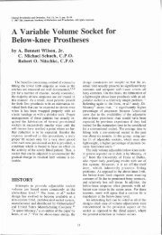
The High Technology Prosthesis<br />
The ENDOLITE system provides a lightweight<br />
endoskeletal prosthesis for above<br />
and below knee amputees. The above<br />
knee version weighs 2 kg and the below<br />
knee 1 kg in their finished forms.<br />
Using the latest technology, advanced<br />
materials such as carbon fibre reinforced<br />
plastics these weight limits have been<br />
achieved without detracting from international<br />
strength requirements for all<br />
categories of patients including the most<br />
active amputee.<br />
Within the continuous cosmesis, the<br />
system provides a full range of limb function<br />
including stance and swing phase control<br />
of knee flexion. A built-in alignment<br />
facility is also included.<br />
The new multiflex ankle/foot module<br />
has a full range of movement allowing controlled<br />
inversion and eversion in addition to<br />
plantar and dorsiflexion movements. Heel<br />
height adjustment is also provided.<br />
Please write or telephone for further<br />
information.<br />
Head Office and Main Factory:<br />
CHAS. A. B LATCH FORD & SONS LTD<br />
Lister Road Basingstoke Hampshire RG22 4AH England<br />
Telephone: 0256 65771
The Journal of the International Society<br />
for Prosthetics and Orthotics<br />
December 1984, Vol 8, No,3<br />
Contents<br />
Editorial 123<br />
Biomechanical significance of the correct length of lower limb prostheses: a clinical and<br />
radiological study 124<br />
O. FRIBERG<br />
Spatial parameters of gait related to the position of the foot on the ground 130<br />
C. RIGAS<br />
The effect of oxygen inhalation and intravenous Naftidrofuryl on the transcutaneous<br />
partial oxygen pressure in ischaemic lower limbs 135<br />
N. M. MUSTAPHA, S. K. JAIN, P. DUDLEY and R. G. REDHEAD<br />
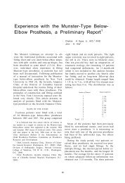
"Zero-position" functional shoulder orthosis 139<br />
J. OZAKI and I. KAWAMURA<br />
Cold injury amputees—a psychosocial problem? 143<br />
J. HUNTER and F. R. I. MIDDLETON<br />
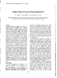
Biomechanical evaluation of S ACH and uniaxial feet 147<br />
J. C. H. GOH, S. E. SOLOMONIDIS, W. D. SPENCE and J. P. PAUL<br />
A 24 year survey of amputees in Hong Kong 155<br />
K. M. CHAN, D. CHEUNG, A. SHER, P. C LEUNG, K. T. FU and J. LEE<br />
Foot loading in amputee stance 159<br />
M. LORD and D. M. SMITH<br />
Technical note—Orthosis for barefoot walking 165<br />
A. BALAKRISHNAN<br />
Book review—Cash's textbook of orthopaedics and rheumatology for physiotherapists 166<br />
Calendar of events 167<br />
Index to Volume 8 173<br />
iii
Prosthetics and Orthotics International, 1984, 8<br />
ISPO<br />
Elected Members of Executive Board:<br />
E. Marquardt (President) FRG<br />
J. Hughes (President-Elect) UK<br />
E. Lyquist (Vice-President<br />
and Hon. Treasurer) Denmark<br />
S. Fishman (Vice-President) USA<br />
W. H. Eisma Netherlands<br />
H. R. Lehneis USA<br />
G. Mensch Canada<br />
S. Sawamura Japan<br />
G. Murdoch (Ex-Officio) UK<br />
N. A. Jacobs (Hon. Secretary) UK<br />
Standing Committee Chairmen and Task Officers<br />
J. Kjølbye (Finance) Denmark<br />
E. Lyquist (Protocol) Denmark<br />
J. Hughes (Membership) UK<br />
G. Murdoch/S. Fishman (Education) UK/USA<br />
J. Hughes/J. Kjølbye (Congress) UK/Denmark<br />
G. Martel (Standards) Canada<br />
A. B. Wilson (Evaluation) USA<br />
S. Heim (Design and Layout) FRG<br />
J. Fischer/S. Heim (Prosthetics Manual) Denmark/FRG<br />
W. H. Eisma (Editorial) Netherlands<br />
Consultants to Executive Board<br />
H. C. Chadderton (Consumer) Canada<br />
C. Dunham (Consumer) UK<br />
P. Dollfus (RI/ICTA) France<br />
International Consultants to Executive Board<br />
S. Heim Africa<br />
E. K. Jensen South America<br />
T. Keokarn Thailand<br />
N. Kondrashin USSR<br />
H. Schmidl Italy<br />
Chairmen of National Member Societies<br />
Australia<br />
Belgium<br />
Canada<br />
Denmark<br />
Egypt<br />
Hong Kong<br />
Israel<br />
Japan<br />
Netherlands<br />
Norway<br />
Sweden<br />
Switzerland<br />
UK<br />
USA<br />
Secretary<br />
Aase Larsson<br />
Past Presidents<br />
K. Jansen (1974-1977)<br />
G. Murdoch (1977-1980)<br />
A. Staros (1980-1982)<br />
E. Lyquist (1982-1983)<br />
iv<br />
V. Angliss<br />
M. Stehman<br />
G. Martel<br />
J. S. Jensen<br />
M. Fouda<br />
T. Steinbach<br />
K. Tsuchiya<br />
W. G. Hazelaar<br />
G. Veres<br />
A. Jernberger<br />
R. Baumgartner<br />
M. E. Ellis<br />
F. Golbranson<br />
Denmark<br />
Denmark<br />
UK<br />
USA<br />
Denmark
Prosthetics and Orthotics International, 1984, 8<br />
Editorial<br />
Many of you will only recently have received a copy of the Society's Constitution. No doubt it will have<br />
received more or less attention depending on the volume of correspondence already in your "In Tray".<br />
Of course, this document contains much information which is solely concerned with the organization<br />
and operation of our Society—the classes of membership with their obligations and privileges, the<br />
committee structures, the officers and their duties, the national organizations—information which for<br />
the most part is simply available for guidance and reference.<br />
The Preamble along with Article 1, however, running as it does to a single page, identifies the whole<br />
reason for the existence of the Society. The Preamble states that the Society is formed, "In order to<br />
promote high quality orthotic and prosthetic care of all people with neuromuscular and skeletal<br />
disabilities." Article 1 goes on to identify the way the Society hopes to achieve that universal goal,<br />
outlining in seven simple paragraphs a plan for international coordination, guidance and action.<br />
Detailed examination of Article 1 reveals that the Society is active in most of the seven areas of<br />
activity. Continuing and useful dialogues are being developed with other agencies in this field; scientific<br />
exchange is being effected through international and national conferences and not least through this<br />
Journal; the Society already has a record of achievement in the international coordination of research as<br />
exemplified by the programme of "Amputee Performance Measurement"; our efforts in education<br />
have resulted in two international meetings this year, with one planned for next year leading to ever<br />
improving standards; our's is the strongest input to the International Standards Organization's<br />
development of standards in this field. There is no reason for complacency. Much remains to be done<br />
but our efforts and effectiveness are on the increase.<br />
Perhaps our greatest success has been in the development, as defined in our Constitution, of our<br />
National Member Societies. To take a typical example, the United Kingdom had no interdisciplinary<br />
forum for the professionals in this field before the establishment of the UK National Member Society.<br />
Now a thriving society of some 240 members fulfils that role. There is an annual Scientific Meeting held<br />
in different parts of the Kingdom, regular regional meetings in three different areas: Glasgow,<br />
Newcastle and London, a Newsletter, prizes for various activities such as papers presented at meetings,<br />
fellowships offered by the British Limbless Ex-servicemen's Association and commercial companies for<br />
attendance at international meetings and, perhaps most important of all, continuing contact with one's<br />
peers.<br />
Of course, the only credit the International Society can take for this is in identifying the professionals,<br />
putting them in contact and encouraging them to work. And work they do! All of these activities require<br />
hard, dedicated, continuing work by officers and members of the National Member Society involved.<br />
One of the side-effects associated with national success however, is that the national activity becomes<br />
foremost in our minds and we lose sight of our international aspirations. The most immediate benefit to<br />
us as members may be these very national activities but we do believe that we have joined an<br />
international society which will not only bring benefit to our patients and ourselves but will widen the<br />
availability of these benefits to all.<br />
The remainder of the Constitution is concerned with the modus operandi of our International Society<br />
and it clearly provides a functional model. Our Society is indeed working at both national and<br />
international level. The Executive Board is concerned, however, that our international collaboration,<br />
either as individual members or as national societies must be further fostered. Communication,<br />
involvement, activity at all levels must be improved if we are to achieve the goals which we have set<br />
ourselves. We would welcome the ideas and comments of the membership at large.<br />
123<br />
John Hughes<br />
President Elect
Biomechanical significance of the correct length of<br />
lower limb prostheses: a clinical and radiological study.<br />
O. FRIBERG<br />
Institute of Military Medical Research, Central Military Hospital, Helsinki, Finland.<br />
Abstract<br />
The length of the lower limb prosthesis was<br />
compared with the length of the contralateral<br />
lower extremity in 113 Finnish war-disabled<br />
amputees by a radiological weight bearing<br />
method developed by the author. Considering a<br />
shortening of 10 mm for above-knee prostheses<br />
and of 5 mm for below-knee prostheses as<br />
tolerance limits, the length of the prosthesis was<br />
acceptable only in 17 cases (15% of the total<br />
group). In 79 cases (70%) the prosthesis was up<br />
to 47 mm too short and in 17 cases (15%) up to 40<br />
mm too long. Chronic pain symptoms of low<br />
back, hip and knee correlated significantly with<br />
the lateral asymmetry caused by incorrect length<br />
of the prosthesis. Independently of the side of<br />
amputation, the unilateral sciatica and chronic<br />
hip pain occurred mainly on the long leg side.<br />
Physical activity of the lower limb amputees<br />
seemed to correlate with the suitability of the<br />
length of the prosthesis, and was unrelated to the<br />
length of the amputation stump.<br />
Introduction<br />
Being a weight bearing substitute for a lost<br />
part of the lower extremity, the prosthetic limb<br />
should be of correct length to fulfil its<br />
biomechanical task. To avoid lateral imbalance<br />
in standing, walking, and running, the length of<br />
both exo and endoprosthesis ought to be<br />
adjusted to the length of the contralateral lower<br />
extremity.<br />
In spite of the technical development in<br />
prosthetics, assessment of the length of the<br />
prosthesis still takes place with conventional<br />
clinical methods e.g. by direct tape<br />
measurement or indirectly by estimating the<br />
heights of the iliac crests or other bony<br />
prominences in the upright position. Devices<br />
have been developed e.g. by Hirschberg and<br />
Robertson (1972) for determining the level of<br />
pelvis.<br />
Clinical methods for measuring leg length<br />
inequality, however, contrary to general belief,<br />
have proved to be inaccurate and even<br />
misleading with observer error of ±10 mm or<br />
even more (Nichols and Bailey, 1955; Clarke,<br />
1972; Morscher, 1977) as compared with the<br />
results of significantly more accurate<br />
radiographic measurements. The errors in<br />
clinical measurements are partly due to<br />
difficulties in locating the exact bony points for<br />
measurement through layers of soft tissues, and<br />
partly because of the prevalence of iliac<br />
asymmetries (Ingelmark and Lindström, 1963).<br />
The amputee's subjective opinion about the<br />
correct length of the prosthetic limb may also be<br />
misleading, particularly if the previous<br />
prosthesis has been of incorrect length. A rather<br />
common belief has been that walking with a<br />
short prosthesis should be easier than with one<br />
of equal length to the contralateral lower<br />
extremity.<br />
On the other hand, a pelvic tilt caused by leg<br />
length inequality of even less than 10 mm is<br />
nearly invariably compensated with a functional<br />
scoliosis and associated with a varus position of<br />
the hip joint on the long leg side (Krakovits,<br />
1967; Gofton and Trueman, 1971; Clarke,<br />
1972). These mechanisms evidently have a<br />
predisposing role in the aetiology of chronic low<br />
back and hip pain symptoms and in the<br />
development of degenerative hip disease<br />
(Gofton and Trueman, 1971; Clarke, 1972;<br />
Heufelder, 1979; Friberg, 1983). The criteria for<br />
the optimal length of lower limb prostheses<br />
generally differ for below-knee and above-knee<br />
prostheses. A shortening up to 2 cm (Krämer et<br />
al, 1979) of above-knee prosthesis is generally<br />
allowed for ground clearance in the swing phase<br />
of walking. However, a shortening of not more<br />
than 1 cm, and for suction socket prostheses with<br />
minimal piston action only 6 mm or even less,<br />
has been suggested by Duthie and Bentley<br />
(1983). Recommendations to make a belowknee<br />
prosthesis full length and to avoid
prostheses longer than the contralateral lower<br />
extremity are probably in accordance with the<br />
current general opinion.<br />
The aim of this study was to find out how the<br />
above mentioned criteria for the length of lower<br />
limb prostheses are in fact satisfied, and to study<br />
the correlation of correct and incorrect<br />
prosthesis length with the incidence and severity<br />
of chronic low back, hip and knee pain<br />
symptoms in lower limb amputees.<br />
Material and methods<br />
This study comprises a series of 113 Finnish<br />
war-disabled lower limb amputees of whom 84<br />
subjects had a below-knee prosthesis and 29 an<br />
above-knee prosthesis. The amputation was<br />
unilateral in all cases but one who had<br />
undergone an above-knee amputation and a<br />
contralateral below-knee amputation. In four<br />
cases a total hip replacement had been<br />
performed, the endoprosthesis being on the<br />
amputated side in one case and on the nonamputated<br />
side in three cases. The primary<br />
amputations were made during the wars 1939-40<br />
and 1941-44, 39 to 45 years before this study.<br />
To record the complaints associated with the<br />
amputation and the prosthesis, the patients were<br />
interviewed with a questionnaire and by<br />
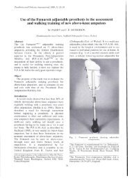
Fig. 1. Equipment and positioning of the amputee for<br />
measurement of the length of the prosthetic limb as<br />
compared with the length of the contralateral lower<br />
extremity ( = the heights of the femoral heads).<br />
personal inquiry. Special attention was drawn to<br />
symptoms of low back, hip and knee joints and<br />
to their laterality.<br />
The clinical and radiological examinations<br />
were performed in 1983-84 at the Central<br />
Military Hospital, Helsinki, at the Tampere<br />
Radiological Center, Tampere, and at the<br />
Military Hospital 3, Kouvola. The majority of<br />
the amputees came to examination from the<br />
Kaskisaari Rehabilitation Center of the<br />
Fraternity Association of War Invalids.<br />
Radiological assessment of the length of<br />
prostheses<br />
The length discrepancy between the lower<br />
limb prosthesis and the contralateral lower<br />
extremity was measured with a weight-bearing<br />
radiographic method developed by the author<br />
(Friberg, 1983). In this method, the patient<br />
stands in front of a chest X-ray stand with<br />
straight knees and the weight equally distributed<br />
between both legs. A 15 cm broad block<br />
between the heels keeps the loading axes of the<br />
legs parallel and the positioning of the patient<br />
reproducible. A gonad shield supplied with an<br />
O-shaped plastic tube partly filled with mercury<br />
is strapped to the patient (Figs. 1 and 2). To<br />
avoid swaying the patient is advised to lean<br />
Fig. 2. Weight bearing radiographic measurement of<br />
the discrepancy between the length of lower limb<br />
prosthesis and contralateral lower extremity.
Fig. 3. A radiograph taken for measurement of the length of lower limb prosthesis. In this case there is 30 mm<br />
shortening of the prosthetic limb (right).<br />
gently against the cassette holder with both<br />
buttocks. The central X-ray beam is focused on<br />
the pubic symphysis, and an A-P radiograph of<br />
both hip joints is taken. A horizontal reference<br />
line is drawn on the radiograph through the tops<br />
of the roentgen positive mercury pillars, from<br />
which the distances of the highest articular<br />
points of both femoral heads are measured (Fig.<br />
3). The difference of the heights of femoral<br />
heads indicates the inequality of the weightbearing<br />
lower extremities.<br />
As the distance of the X-ray tube is constant<br />
and long enough, the magnification error is<br />
insignificant and can be ignored. The main<br />
source of error, unequal extension of the knees,<br />
is easily avoided by proper positioning of the<br />
patient.<br />
Clinical examination<br />
Special attention was paid to the pelvic tilt,<br />
lumbar scoliosis and other static body<br />
asymmetries in erect posture, which, if<br />
necessary, were radiologically documented. A<br />
block of equal thickness to the leg length<br />
inequality measured, was then placed under the<br />
foot of the shorter limb to reveal the subjective,<br />
clinical and radiological response of the body to<br />
the equalization of the pelvis. If the response<br />
was positive, i.e. the patient experienced the lift<br />
as comfortable and balancing, and the scoliotic<br />
curve straightened, the patients were advised to<br />
try a fixed lift under the shoe of the short leg<br />
before making a decision to have a permanent<br />
change in the length of the prosthesis.<br />
Results<br />
The mean age of the patients was 65.1 years<br />
(range 54 to 80 years), the mean length 173.5 cm<br />
(range 158 to 189 cm) and the mean body mass<br />
80.9 kg (range 51 to 102 kg). The mean length of<br />
the above-knee stumps was 19.8 cm (range 8 to<br />
30 cm) and of below-knee stumps 22.4 cm (range<br />
12 to 28 cm).<br />
Length of the prostheses<br />
Though a majority (78.8%) of the amputees<br />
were of the belief that their prostheses were of<br />
equal length with the contralateral lower<br />
extremity, the length discrepancies were<br />
extremely prevalent and significant, ranging<br />
from a shortening of 47 mm to a lengthening of<br />
40 mm (Table 1). Of the total group, the length<br />
discrepancy was more than 10 mm in 66.4% and<br />
more than 20 mm in 33.6% of the cases (Table<br />
2). For comparison, in a group of 359 symptomfree<br />
Finnish Army conscripts aged 17 to 24 years<br />
(Friberg, 1983), leg length inequality of less than<br />
10 mm occurred in 84.5% of the cases, and more<br />
than 20 mm in one case only.<br />
In below-knee prostheses, the length was<br />
appropriate (length discrepancy less than 5 mm)<br />
in only 10 (11.9%) out of 84 patients, and in<br />
Table 1. Subjective opinion of amputees about the<br />
length of the prosthesis and the ranges of measured<br />
length discrepancies.<br />
(" + " signs lengthening and "—" shortening of<br />
prosthetic limb)
Table 2. Leg length inequality (difference of the height<br />
of femoral heads) in bipedal standing of 113 amputees<br />
with lower limb prostheses.<br />
above-knee prostheses (discrepancy less than 10<br />
mm) in only 7 (24%) out of 29 cases. In 17 cases<br />
(15%) out of the total the prosthesis was on<br />
average 14.4 mm longer than the contralateral<br />
lower extremity (Table 3).<br />
Accuracy of radiological measurements<br />
In a previous study with non-amputees, the<br />
mean error between two or more subsequent<br />
measurements was 0.6 mm (range 0 to 2 mm)<br />
Friberg, 1983). In the present study, a reexamination<br />
was made in 10 cases and in a<br />
further 10 cases after correction of the incorrect<br />
length of the prosthesis or by insertion of a lift<br />
equivalent to the leg length inequality under the<br />
foot of the short leg. the mean error between<br />
these measurements was 1.2 mm (range 0 to 4<br />
mm).<br />
Low back pain<br />
Chronic low back pain symptoms were<br />
prevalent in the present series of lower limb<br />
amputees. Only 6 patients (5.3% of the total<br />
series) were completely free from low back pain.<br />
In altogether 25 patients, however, the low back<br />
symptoms were occasional and fairly mild. In<br />
Table 3. Suitability of the length in lower limb prostheses<br />
these patients the mean leg length discrepancy<br />
was 6.1 mm. The mean discrepancy in 32<br />
amputees with frequent or constant and severe<br />
low back pain was 21.7 mm. The difference<br />
between the mean leg length discrepancies in<br />
these two groups was statistically highly<br />
significant (p
length inequality was 12.8 mm. The complaint<br />
occurred on the amputated side in 25% of the<br />
cases and on the non-amputated side in 75% of<br />
the cases.<br />
Sporting activity of the amputees<br />
Physical activities like jogging, skiing or<br />
gymnastics were carried out daily by 43 belowknee<br />
amputees. The mean leg length inequality<br />
in these amputees was 9.7 mm. In 25 below-knee<br />
subjects without daily physical activity, the<br />
mean leg length inequality was 18.5 mm. The<br />
difference between these means proved to be<br />
statistically significant (p
Fig. 5. A case, with an above-knee prosthesis on the<br />
right side which was 40 mm too short, suffering from<br />
constant low back and left hip pain since amputation in<br />
1944. Note: diminution of weight-bearing articular<br />
surface of the longer lower limb as illustrated by<br />
Wiberg's angle, and the functional scoliosis with<br />
wedge-shaped intervertebral spaces and axial rotation<br />
of the vertebrae (radiographs taken in a standing<br />
posture). The response to the correction of the<br />
prosthesis length was excellent.<br />
The varus position of the long leg hip as<br />
illustrated by Wiberg's angle (Krakovits, 1967;<br />
Morscher, 1977) results in a diminution of<br />
weight-bearing articular area of the joint, thus<br />
speeding the degenerating breakdown in the<br />
hip. Concurrent occurrence of symptoms from<br />
lumbar spine and hip, the hip-spine syndrome<br />
(Offierski and Macnab, 1983), was common<br />
among lower limb amputees, evidently because<br />
of the high prevalence of lateral asymmetries<br />
due to incorrect length of the prostheses.<br />
In the light of present results, conventional<br />
assessment of the length of lower limb<br />
prostheses with clinical methods seems to be<br />
inaccurate and unreliable. The range of errors in<br />
the length of the lower limb prostheses was as<br />
high as 87 mm (47 mm too short to 40 mm too<br />
long). The weight-bearing radiographic method<br />
for measurement of leg length inequality offers<br />
an accurate, reliable and simple tool to adjust<br />
the correct length for lower limb prostheses. The<br />
costs of radiography are minimal when<br />
compared with the expense of manufacturing a<br />
prosthetic limb. Paricularly in adjusting the first<br />
prosthesis for a recently amputated patient, the<br />
assessment of the correct length of the prosthesis<br />
appears imperative to allow the subject to adapt<br />
from the beginning to symmetry of the lower<br />
extremities, to train him to walk without a limp,<br />
and to avoid malpositions and development of<br />
degenerative changes in hip, knee and lumbar<br />
spine.<br />
REFERENCES<br />
CLARKE, G. R. (1972). Unequal leg length: an<br />
accurate method for detection and some clinical<br />
results. Rheum. Phys. Med. 11, 385-390.<br />
Duthie, R., BENTLEY, G. (1983). Mercer's<br />
orthopaedic surgery. 8th ed. London: E. Arnold,<br />
P1122.<br />
FRIBERG, O. (1983). Clinical symptoms and<br />
biomechanics of lumbar spine and hip joint in leg<br />
length inequality. Spine. 8, 643-651.<br />
GOFTON, J. P., TRUEMAN, G. E,, (1971). Studies in<br />
osteoarthrosis of hip. 2. Osteoarthritis of the hip and<br />
leg length disparity. Can. Med. Assoc. J. 104,<br />
791-799.<br />
HEUFELDER, P. (1979). Die Beinlängendifferenz aus<br />
der Sicht des Allgemeinarztes. Z. Orthop. 117,<br />
345-354.<br />
HIRSCHBERG, G. C, ROBERTSON, K. B. (1972).<br />
Device for determining difference in leg length.<br />
Arch. Phys. Med. Rehabil. 53, 45-46.<br />
INGELMARK, B. E., LINDSTRÖM, J. (1963).<br />
Asymmetries of the lower extremities and pelvis and<br />
their relations to lumber scoliosis: a radiographic<br />
study. Acta. Morphol. Neerl. Scand. 5, 221-234.<br />
KRAKOVITS, G. (1961). Uber die Auswirkung einer<br />
Beinverkürzung auf die Statik und Dynamik des<br />
Hüftgelenkes. Z. Orthop. 102, 418-423.<br />
KRÄMER, J., HEISEL, J., ULLRICH, C. (1979).<br />
Spätschäden am Bewegungsapparat bei<br />
Oberschenkelamputierten und deren<br />
Begutachtung. Z. Orthop. 117, 801-807.<br />
MORSCHER, E. (1977). Etiology and pathogenesis in<br />
leg length discrepancies. Progr. Orthop. Surg. 1,<br />
9-19.<br />
NICHOLS, P. J. R., BAILEY, N. T. J. (1955). The<br />
accuracy of measuring leg length differences: an<br />
"observer error" experiment. Br. Med. J. 2,<br />
1247-1248.<br />
OFFIERSKI, C. M., MACNAB, I. (1983). Hip-spine<br />
syndrome. Spine, 8, 316-321.
Spatial parameters of gait<br />
related to the position of the foot on the ground<br />
C. RIGAS<br />
Department of Medical Physics, University of Ioannina, Greece<br />
Abstract<br />
A number of parameters related to the position<br />
of the foot on the ground during normal level<br />
walking were analysed for a group of young and<br />
a group of old subjects, divided in two<br />
sub-groups each, according to sex.<br />
The analysis has shown asymmetries between<br />
the left and the right side of a number of<br />
subjects, differences between sexes and<br />
differences between age groups. Changes in the<br />
parameters of gait for the old subjects served the<br />
task of providing a larger base of support and a<br />
smaller loading of the hip musculature.<br />
Introduction<br />
Recent work on the biomechanics of human<br />
gait has been directed mostly to dynamic and<br />
energetic aspects, while kinematic studies<br />
appear more and more rarely in literature.<br />
Following the work of the California group<br />
(Eberhart, 1947) kinematic data was provided<br />
by Levens et al (1948), Ryker (1952), Murray et<br />
al (1964, 1966, 1969, 1970), Lamoreux (1970,<br />
1971) and more recently by Dainis (1980),<br />
Hershler and Milner (1980), Bajd and Kralj<br />
(1980), Durie and Farley (1980), Cappozzo<br />
(1981), Mena et al (1981). A small part of this<br />
work is devoted to the kinematics of the foot.<br />
Aspects of the position of the foot on the<br />
ground during walking attracted interest many<br />
years ago. Dougan (1924) measured the angle of<br />
gait, that is the angle of the long axis of the shoe<br />
and the line of progression, for young males.<br />
The angle of gait was also studied by Morton<br />
(1932) and by Barnett (1956). The most<br />
comprehensive data on the position of the foot<br />
on the ground was provided by Murray et al<br />
Fig. 1. The parameters analysed in the present study.<br />
(1964, 1966, 1970), who reported a series of<br />
studies of a large number of spatial and temporal<br />
parameters of free and fast speed walking for<br />
normal men and women of different ages.<br />
This study aims at the analysis and<br />
interpretation of a number of spatial parameters<br />
related to the position of the foot on the ground<br />
during level normal walking of young and old<br />
subjects of both sexes. These parameters,<br />
defined as shown in Fig. 1, are the following:<br />
1. Foot angle, theta<br />
2. Step length, L<br />
3. Stride width, b.<br />
4. Mid-line, y.<br />
Apart from reporting our results on<br />
parameters that have been studied before by<br />
other investigators, this work examines the<br />
variability of these parameters from step to step<br />
of the same subject during a certain trial. It also<br />
provides comparisons between left and right side<br />
of each subject. Furthermore, values for step<br />
length and stride width are expressed not only in<br />
absolute terms but also in percent of the<br />
subject's stature (Defined as "relative"<br />
quantities by Grieve, 1968).<br />
Method<br />
Two groups, one of young subjects and<br />
another of old ones were tested. The group of<br />
young subjects consisted of 35 (25 male and 10<br />
female) students, aged 17 to 24 years (Mean=19<br />
years, SD=2.1 yrs). The group of old subjects<br />
consisted of 24 (14 male and 10 female) boarders<br />
at a home for the aged. Their ages ranged
etween 65 and 90 years (Mean=78 yrs, SD=6.9<br />
yrs). Cases described as pathological were not<br />
considered.<br />
After a few trials of familiarization with the<br />
walking area, each subject was asked to walk<br />
freely at his/her natural speed along a corridor,<br />
part of which was covered by paper laid over<br />
carbon paper. The points of contact of the toes<br />
and the heel were clearly marked on the paper<br />
by small pins set at the corresponding points on<br />
the long axis of the shoe. The first and last steps<br />
of each subject on the paper walkpath were not<br />
taken into account; a tendency to make the last<br />
step shorter was evident in many cases.<br />
The number of footprints on each side that<br />
were analysed was six for each young subject and<br />
seven for each old subject. The sample size of<br />
each of the parameters studied is shown in the<br />
corresponding table.<br />
Mean values of a certain parameter were<br />
considered different when their difference was<br />
statistically significant (p
(p
Table 5. Variability of gait parameters. The variability is measured by the SD.<br />
Relative quantities are expressed in percent of the subject's stature.<br />
Six steps on either side of each young subject (25 males and 10 females) and<br />
six steps on either side of each old subject (14 males and 10 females) were analysed.<br />
Discussion<br />
Analysis of the foot angle and step length<br />
revealed statistically significant asymmetries<br />
between the left and the right side of a number of<br />
subjects considered normal. It is thought that<br />
these asymmetries have an important<br />
contribution in forming the characteristic gait of<br />
a subject. Whether these asymmetries<br />
correspond to skeletal, muscular or other<br />
asymmetries cannot be inferred from this study.<br />
This will require a larger number of parameters<br />
to be analysed and also a larger number of<br />
subjects to be tested.<br />
Among the parameters analysed, the step<br />
length showed the least relative variability<br />
between steps of the same subject, or between<br />
subjects of the same sub-group. It seems<br />
therefore likely that this parameter may<br />
constitute a useful differentiator between<br />
normal and pathological gait.<br />
The SD is inversely related to the sample size<br />
which would be required to define a mean value<br />
not differing from the true mean by a certain<br />
amount. For example, when it is required that a<br />
95 percent confidence interval on the mean step<br />
length of any one normal young man will be ± 2<br />
percent of his stature, then a sample size of n=4<br />
steps is needed. When it is required that the<br />
same interval will be ± 1 percent of the subject's<br />
stature, then a sample size of n=16 steps is<br />
needed. The number of steps or of subjects<br />
required for any particular gait analysis depends<br />
on the variability of the parameter of interest.<br />
The variability of sequential steps of the same<br />
subject was considerably higher for old than for<br />
young subjects. This fact is in agreement with<br />
Spielberg's (1940) theory that the motor pattern<br />
of walking goes through several stages of<br />
disintegration. At the third stage the uniformity<br />
of sequential steps is increasingly disturbed. It is<br />
interesting to note though, that such decrease of<br />
uniformity is not observed for the rest of the gait<br />
parameters analysed in this study.<br />
Step length was relatively smaller for old than<br />
for young subjects. This feature that has been<br />
described by Spielberg (1940), has also been<br />
reported in an extensive study by Drillis (1961),<br />
and later by Murray et al (1969). It is attributed<br />
to the fact that smaller step lengths result in<br />
smaller moments about the joints of the lower<br />
limb and therefore smaller effort is required to<br />
be exercised by the weakened musculature of<br />
old subjects.<br />
Mean values for the foot angle are reasonably<br />
close to those reported by Dougan (1924), by<br />
Morton (1932) and by Murray et al (1964,1966,<br />
1969, 1970).
Comparisons of foot angle, 8, and stride<br />
width, b, between young and old subjects<br />
showed a significant increase in the case of the<br />
latter. Foot angle and stride width, taken<br />
together, define the overall width of the<br />
supporting base for the walking subject. Old<br />
people who suffer more instability, due to<br />
weaker hip abductors and poorer physical<br />
condition, require a larger base of support<br />
during their gait. This is achieved by increasing<br />
both the foot angle and the stride width. The<br />
increase in stride width is particularly stressed in<br />
the case of old women. This may be due to the<br />
fact that the female pelvis is relatively wider than<br />
the male one. Murray et al (1964) observe a<br />
significant increase in the mean foot angle with<br />
age, but their analysis reveals no increase of<br />
stride width with age. They, therefore, conclude<br />
that wider base of support is achieved in old age<br />
by out-toeing and not by both out-toeing and<br />
greater stride width, as this study reveals.<br />
It was felt likely that instability of the elderly<br />
might result in greater side-ways sway (mean<br />
path lateral deviation), than that shown by<br />
young subjects. This hypothesis was not justified<br />
by the analysis of the mid-line deviation,<br />
indicating that instability is adequately counterbalanced<br />
by the increase in the size of the<br />
supporting base. It should, nevertheless, be<br />
pointed out, that data on the mid-line deviation<br />
might be biased, as the subjects were more or<br />
less "guided" to walk along a straight line by the<br />
paper walkpath itself. To reach final conclusions<br />
about this parameter a much wider paper<br />
walkpath will be required.<br />
REFERENCES<br />
BAJD, T., KRAU, A. (1980). Simple kinematic gait<br />
measurements. J. Biomed. Eng., 2, 129-132.<br />
BARNETT, C. H. (1956). The phases of human gait.<br />
Lancet, ii, 617-621.<br />
CAPPOZZO, A. (1981). Analysis of the linear<br />
displacement of the head and trunk during walking<br />
at different speeds. J. Biomech. 14, 411-425.<br />
DAINIS, A. (1980). Whole body and segment center of<br />
mass determination from kinematic data. J.<br />
Biomech. 13, 647-651.<br />
DOUG AN, S. (1924). The angle of gait. Am. J. Phys.<br />
Anthropol., 7, 275-279.<br />
Drillis, R. J. (1961). The influence of ageing on the<br />
kinematics of gait. In: The geriatric amputee. A<br />
report on a conference sponsored by the National<br />
Research Council, Committee on Prosthetics<br />
Research and Development.—Washington, DC:<br />
National Academy of Sciences—National Research<br />
Council, pl34-145. (NAS-NRC. Publication 919).<br />
DURIE, N. D., FARLEY, R. L. (1980). An apparatus<br />
for step length measurement. J. Biomed. Eng., 2,<br />
38-40.<br />
EBERHART, H. D. (1947). Fundamental studies of<br />
human locomotion and other information relating<br />
to design of artificial limbs. Berkeley, CA:<br />
University of California. Prosthetic Devices<br />
Research Project.<br />
GRIEVE, D. W. (1968). Gait patterns and the speed of<br />
walking. Biomed. Eng. 3, 119-122.<br />
HERSHLER, C, MILNER, M. (1980). Angle-angle<br />
diagrams in the assessment of locomotion; Am. J.<br />
Phys. Med., 59, 109-125.<br />
LAMOREUX, L. W. (1970). Experimental kinematics of<br />
human walking. Ph.D. Thesis, University of<br />
California, Berkeley.<br />
LAMOREUX, L. W. (1971). Kinematic measurements<br />
in the study of human walking. Bull. Prosthet. Res.<br />
10-15, 3-84.<br />
LEVENS, A. S., INMAN, V.T., BLOSSER, J. A. (1948).<br />
Transverse rotation of the segments of the lower<br />
extremity in locomotion. J. Bone Joint Surg. 30, A,<br />
859-872.<br />
MENA, D., MANSOUR, J. M., SIMON, S. R. (1981).<br />
Analysis and synthesis of human swing leg motion<br />
during gait and its clinical applications. J. Biomech.,<br />
14, 823-832.<br />
MORTON, D. J. (1932). The angle of gait: a study based<br />
upon examination of the feet of Central African<br />
natives. J. Bone Joint Surg., 14, 741-754.<br />
MURRAY, M. P., DROUGHT, A. B., KORY, R. C.<br />
(1964). Walking patterns of normal men. J. Bone<br />
Joint Surg. 46-A, 335-360.<br />
MURRAY, M. P., KORY, R. C, CLARKSTON, B. H.,<br />
SEPIC, S. B. (1966). A comparison of free and fast<br />
speed walking patterns of normal men. Am. J. Phys.<br />
Med. 45, 8-24.<br />
MURRAY, M. P., KORY, R. C, CLARKSTON, B. H.<br />
(1969). Walking patterns of healthy old men. J.<br />
Gerontol., 24, 169-178.<br />
MURRAY, M. P., KORY, R. C, SEPIC, S. B. (1970).<br />
Walking patterns of normal women. Arch. Phys.<br />
Med. Rehabil. 51, 637-650.<br />
RYKER, N. J. (1952). Glass walkway studies of normal<br />
subjects during normal level walking. Berkely, CA:<br />
University of California. Prosthetic Devices<br />
Research Project. (Series II, issue 20).<br />
SPIELBERG, P. I. (1940). Walking patterns of old<br />
people: cyclographic analysis. In: N.A. Bernstein.<br />
Investigations on the biodynamics of walking,<br />
running and jumping: part 2. Moscow: Central<br />
Scientific Institute of Physical Culture, p72-76.
The effect of oxygen inhalation and intravenous Naftidrofuryl<br />
on the transcutaneous partial oxygen pressure<br />
in ischaemic lower limbs<br />
N. M. MUSTAPHA, S. K. JAIN,* P. DUDLEY** and R. G. REDHEAD<br />
Limb Fitting Centre, Roehampton Lane, London,<br />
* Artificial Limb Centre, Pune 41101, India,<br />
** University Department of Surgery, General Infirmary, Leeds.<br />
Abstract<br />
The effect of oxygen inhalation at atmospheric<br />
pressure and Naftidrofuryl infusion (N) on the<br />
TCpO2 is shown.<br />
At the central control site—5 cm below the<br />
midclavicular line—oxygen inhalation produced<br />
a significant increase in TCpO 2, whereas there<br />
was no change after Naftidrofuryl infusion. At<br />
the 10 cm below-knee site, there were significant<br />
rises after oxygen inhalation alone,<br />
Naftidrofuryl alone and both combined. The<br />
study was conducted on 20 patients (23 legs).<br />
It is suggested that this study can form the<br />
basis for a regimen to improve the viability of<br />
ischaemic limbs showing borderline TCpO2<br />
readings, and increase the chances of a<br />
successful below-knee amputation.<br />
Introduction<br />
The value of transcutaneous partial oxygen<br />
pressure (TCpO0 2) recording in assessing skin<br />
viability has been the subject of recent<br />
publications (Achauer et al, 1980; Dowd et al,<br />
1983). In a previous communication we have<br />
shown average TCpO 2 in a group of young<br />
healthy volunteers, elderly healthy volunteers,<br />
and a group of patients with lower limb<br />
ischaemia in varying degrees of severity<br />
(Mustapha et al, 1983).<br />
Our findings relate closely to other<br />
researchers' results. A TCpO 2 recording of 40<br />
mm Hg or over indicates adequate perfusion of<br />
the skin, and a recording below 30 mm Hg is<br />
clear indication of inadequate perfusion<br />
(Holstein et al, 1979).<br />
In the management of the ischaemic lower<br />
limb and especially in situations where the<br />
TCpO2 recording shows values in the borderline<br />
range of 30-40 mm Hg it is desirable to enhance<br />
the viability of the limb in general and the skin in<br />
particular, either to avoid surgery (especially<br />
amputation) or to improve the chances of<br />
success of a distal (i.e. below-knee) amputation.<br />
This is a study of the effect of oxygen<br />
inhalation and the effect of a drug Naftidrofuryl<br />
(Praxilene LIPHA) that has been shown to<br />
enhance oxidative metabolism at cellular level<br />
(Meynaud et al, 1973).<br />
Patients, materials and method<br />
In this and the previous studies (Mustapha et<br />
al, 1983; Jain 1982), the TCM1 (Radiometer,<br />
Copenhagen) TCpO2 monitor is used.<br />
Two site readings are recorded: a control site 5<br />
cm below the midclavicular point (either side as<br />
convenient) and a site 10 cm below the knee<br />
anteriorly. This latter site marks the critical<br />
perfusion level especially in relation to the<br />
anterior incision line of the commonly<br />
performed below-knee amputation technique.<br />
Oxygen inhalation is administered as a 24%<br />
mixture in air (2 litre flow per minute) through a<br />
face mask or nasal spectacle. A higher flow rate<br />
was found unnecessary as it produced only<br />
marginally higher TCpO2 recordings.<br />
Naftidrofuryl (N) is given in a dose of 400 mg six<br />
hourly mixed in 5% Dextrose in water and<br />
administered in a continuous infusion.<br />
Oxygen inhalation was initially given to two<br />
groups of healthy volunteers: six young<br />
volunteers aged 17-25 years (average 19.5<br />
years), and six elderly volunteers aged 54-64<br />
years (average 59 years) with no signs of
Fig. 1. TCpO2 recordings in relation to therapy.<br />
peripheral vascular insufficiency or central<br />
cardiopulmonary deficit.<br />
The study was then extended to dysvascular<br />
patients, and in addition to the oxygen<br />
inhalation, the effects of Naftidrofuryl were<br />
noted according to the following regimen (Fig.<br />
1):<br />
A baseline TCpO2 recording over the two<br />
sites is followed by oxygen inhalation for 15<br />
minutes. The TCpO2 is recorded again.<br />
A period of six hours follows when no<br />
therapy is given, and then Naftidrofuryl 400<br />
mg six hourly is administered for 48 hours.<br />
Soon after the end of the infusion, oxygen is<br />
administered for another period of 15<br />
minutes. The TCpO2 is recorded immediately<br />
before and immediately after this second<br />
oxygen inhalation.<br />
This regimen was settled upon as the most<br />
convenient for the medical nursing staff<br />
workload as well as the clinical condition under<br />
study. It is, however, recognised that greater<br />
accuracy of assessment can be obtained if more<br />
frequent readings are taken, or the period of<br />
therapy and monitoring is extended.<br />
Results<br />
The group of young healthy volunteers (Table<br />
1 and Fig. 2) showed an average TCpO2 level of<br />
Fig. 2. Effect of oxygen inhalation on healthy<br />
volunteers.<br />
80 mm Hg (S.D. 9.0) at the control site before<br />
oxygen inhalation, rising to 119 mm Hg (S.D.<br />
14.1) after the 15 minutes of 24% oxygen<br />
mixture inhalation. This represents an average<br />
increase of 48% (S.D. 16.9) (p
Table 2. TCpO2 changes in response to O 2 inhalation in 6 elderly volunteers. Age range 54-64 years.<br />
Table 3. TCpO2 changes in response to O 2 inhalation and Naftidrofuryl (Praxilene)<br />
In the group of dysvascular patients studied,<br />
20 patients, 23 legs (Table 3 and Fig. 3)<br />
recordings over the control site showed a<br />
significant rise following oxygen inhalation from<br />
an average of 62.39 mm Hg (S.D. 15.50) to an<br />
average of 87.71 (S.D. 18.33) (p
T he<br />
Fig. 4. TCpO2 shown as an index (10 cm BK)/(Control site):<br />
effect of O2 inhalation alone, Praxilene alone and both<br />
combined.<br />
but did contribute significantly to the TCpO2 in<br />
the ischaemic part of the lower limb. This is in<br />
keeping with a previous observation that<br />
Praxilene effects ischaemic parts selectively<br />
(Gaylarde et al, 1980; Elert et al, 1976).<br />
This fact is reflected again in the significant<br />
difference Praxilene produces in the Control<br />
TCpO2 index (Fig. 4). This idea (Healing index)<br />
(Jain, 1982) may not at this stage offer a reliable<br />
indication as to the severity of ischaemia, but its<br />
significance is under consideration and longer<br />
studies are required to establish its usefulness or<br />
otherwise.<br />
No complications have been noted in this<br />
study from the use of oxygen inhalation or the<br />
above mentioned dosage regimen of Praxilene.<br />
Conclusion<br />
This study is an extension to the previous<br />
studies (Jain, 1982; Dowd et al, 1983; Mustapha<br />
et al, 1983;) in which the value of TCpO2 was<br />
shown to be a reliable indicator of the state of<br />
perfusion in the tissues at large and the<br />
ischaemic lower limb in particular. Further<br />
studies are needed to establish whether such a<br />
regimen can assist the clinician in dealing with<br />
borderline ischaemic conditions before or<br />
immediately following an amputation. The<br />
regimen described is presented only as a guide<br />
and varying clinical conditions will inevitably<br />
call for a flexible application. A further<br />
comparative study is contemplated to establish<br />
the regimen more firmly.<br />
Acknowledgements<br />
The authors are grateful to Mr. K. P.<br />
Robinson MS, FRCS, FRCS Ed, Consultant<br />
Surgeon and Director of the Limb Surgery Unit<br />
in Queen Mary's Hospital, Roehampton, SW15<br />
for his help and for allowing the study on his<br />
patients.<br />
Thanks are also due to Messrs Lipha<br />
Pharmaceuticals for the loan of the TCM1<br />
monitor.<br />
Finally the authors wish to thank Mrs. D.<br />
Browning for her secretarial help.<br />
REFERENCES<br />
ACHAUER, B. M., BLACK, K. S., LITKE, D. K. (1980).<br />
Transcutaneous pO2 in flaps: a new method of<br />
survival prediction. Plast. Reconstr. Surg. 65,<br />
738-745.<br />
DOWD, G. S., LINGE, K., BENTLEY, G. (1983).<br />
Measurement of transcutaneous oxygen pressure in<br />
normal and ischaemic skin. J. Bone Joint Surg.<br />
65-B, 79-83.<br />
ELERT, C, NIEBEL, W., KARUSE, E., SATTER, P.<br />
(1976). The effect of Naftidrofuryl on energy<br />
metabolism in the musculature of limbs with<br />
impaired blood flow. Therapiewoche. 23, 3947-50.<br />
GAYLARDE, P. M., TAN, O. T., SARKANY, I. (1980).<br />
Blood flow changes with Naftidrofuryl in systemic<br />
sclerosis and Raynaud's phenomenon Dermatol.<br />
102, 7-10.<br />
HOLSTEIN, P., SAGER., P., LASSEN, N. A. (1979).<br />
Wound healing in below-knee amputations in<br />
relation to the skin perfusion pressure. Acta.<br />
Orthop. Scand. 50, 49-58.<br />
JAIN, S. K. (1982). Transcutaneous oxygen tension<br />
measurement in assessment of peripheral ischaemia<br />
in the lower limbs. Thesis for Degree of Master of<br />
Philosophy, University of Surrey, 185-186; 201-<br />
207.<br />
MEYNAUD, A., GRAND, M., FONTAINE, L. (1973).<br />
Effect of Naftidrofuryl upon energy metabolism of<br />
the brain. Arzneimittelforsch, 23, 1431-1436.<br />
MUSTAPHA, N. M., REDHEAD, R. G., JAIN, S. K.,<br />
Wielogorski, J. W. (1983). Transcutaneous partial<br />
oxygen pressure assessment of the ischaemic lower<br />
limb. Surg. Gynecol. Obstet. 156, 582-584.
"Zero-position" functional shoulder orthosis<br />
J. OZAKI and I. KAWAMURA*<br />
Department of Orthopaedic Surgery, Nara Medical University, *Kawamura Orthopaedic Appliance, Co. Ltd.<br />
Abstract<br />
The "zero-position" of the shoulder joint<br />
described by Saha (1961) is recognized as a<br />
mechanical position between scapula and<br />
humerus, and the "scapular plane" is widely<br />
accepted as a mechanical plane at the shoulder<br />
joint. On the basis of biomechanical concepts of<br />
the "zero-position" and the "scapular plane",<br />
the authors designed the "zero-position"<br />
functional shoulder orthosis. This orthosis has<br />
been successfully fitted to more than 75 patients<br />
for the postoperative management of rotator<br />
cuff injuries, and to 3 patients for the treatment<br />
of scapular neck fractures. It is introduced here,<br />
together with biomechanical considerations,<br />
structure, functional and clinical results.<br />
Introduction<br />
A plaster cast has commonly been used for<br />
immobilization in the so-called optimal position<br />
of the shoulder joint after operation for rotator<br />
cuff injuries. However, it sometimes causes<br />
contracture of the shoulder joint and muscle<br />
imbalance around the shoulder girdle because,<br />
in such cast immobilization, patients cannot<br />
exercise their shoulder postoperatively. From<br />
this point of view, the authors initially used a<br />
"zero-position" plaster cast in the postoperative<br />
management of rotator cuff injuries (Ozaki and<br />
Nobuhara, 1978). On the other hand, however,<br />
some difficulties were found after the removal of<br />
the plaster cast in that almost all the patients<br />
complained of pain and could not continue with<br />
their exercises. This resulted in prolongation of<br />
the healing period. It was thought that if a<br />
functional shoulder orthosis were adopted it<br />
would enable adjustments to be made to the<br />
elevation angle from the "zero-position" step by<br />
step, thus producing less pain. This is the reason<br />
for the design of this functional shoulder<br />
orthosis.<br />
Biomechanical considerations<br />
The shoulder joint consists of the small and<br />
shallow scapular glenoid fossa, the large<br />
humeral head, and abundant muscles. These<br />
structures give the shoulder joint the widest<br />
range of motion and the most varied movements<br />
of any joint in the human body. Therefore the<br />
shoulder mechanism has been an enigma to the<br />
anatomist. Codman (1934), and others<br />
demonstrated that whether the arm is elevated<br />
to the vertical in the saggital, the coronal, or in<br />
any other plane, the end result is always the<br />
same, and when the arm is elevated to the<br />
vertical in the coronal plane, it undergoes an<br />
appreciable degree of lateral rotation during the<br />
process. According to Johnston (1937), when<br />
the arm is raised to the vertical, no matter how<br />
many planes it may move through in the process,<br />
at the end of the movement the humerus must lie<br />
in the "plane of the scapula". He stated that the<br />
"plane of the scapula" is not easy to define, but<br />
may be regarded as a plane drawn at right angles<br />
to the glenoid cavity through its greatest vertical<br />
diameter. On the other hand, Codman (1934)<br />
pointed out a very natural position for the<br />
human arm when the body is recumbent. In this<br />
position, the axis of the humerus is in line with<br />
the axis of the spine of the scapula, and the head<br />
and neck of the humerus is in the same plane. He<br />
called this point a subordinate pivotal position in<br />
which the deltoid, the supraspinatus and the<br />
infraspinatus are relaxed. It is the most<br />
favourable position to encourage physical repair<br />
of lesions in and about the tuberosity. However,<br />
he never actually used this position for<br />
treatment. Saha (1961) has designated this point
as the "zero-position", because muscular<br />
rotatory forces acting upon the humerus at this<br />
position are almost zero. He stated that this<br />
position of stability can be seen in fast-moving<br />
quadrupeds. According to Saha's observations,<br />
the "zero-position" is at about 155° of elevation<br />
from the anatomical position, with the axis of the<br />
humerus about 45° anterior to the coronal plane.<br />
He used this position in the reduction of<br />
fractures of the humerus and dislocations of the<br />
shoulder. However, this position had never been<br />
used for the postoperative treatment of rotator<br />
cuff injuries. In order to define the "plane of the<br />
scapula", Ozaki (1980) performed<br />
cineradiography and radiographic studies on<br />
normal and disordered shoulders. He<br />
demonstrated that the "scapular plane" should<br />
be inclined forward at an angle of 30° to 45° to<br />
the frontal plane (Fig. 1), and that in the<br />
"zero-position", the humerus must be elevated<br />
to 150° in the "scapular plane" with individual<br />
variations (Fig. 2). On the basis of these<br />
biomechanical concepts, the functional shoulder<br />
orthosis, introduced in this paper, was designed.<br />
The design of the orthosis<br />
The "zero-position" functional shoulder<br />
orthosis is shown in Figure 3. It consists of a<br />
pelvic girdle, an upright bar, a transverse cross<br />
bar, and an arm. The pelvic girdle is made of<br />
4mm Subortholen plastic sheet.<br />
The length of the upright bar can be changed<br />
since the distance from axilla to iliac crest varies<br />
according to the angles of elevation of the arm,<br />
and can be rotated in the horizontal plane and<br />
fixed in any position using two mechanical<br />
joints. The distance from axilla to elbow joint<br />
and from palm to forearm can be changed as<br />
Fig. 1. The "scapular plane".<br />
Fig. 2. Radiograph showing the normal shoulder joint<br />
in the "zero-position". The "zero-position" is at about<br />
150° of elevation in the "scapular plane", and the axis<br />
of the humerus is in line with the axis of the scapular<br />
spine.<br />
required. Furthermore, the angle of rotation of<br />
the humeral axis and the forearm can be changed<br />
easily with the two mechanical joints.<br />
Clinical applications<br />
Postoperative management of rotator cuff<br />
injuries<br />
Immediately after the successful repair of a<br />
rotator cuff injury, the "zero-position" of the<br />
shoulder should be maintained by skin traction<br />
while the patient rests in bed (Fig. 4). After<br />
three days, a functional shoulder orthosis, which<br />
has been made to order preoperatively, is<br />
applied to maintain the "zero-position" in the<br />
scapular plane. At the beginning of the third<br />
postoperative week, the upper limb in the<br />
orthosis is extended at 100° abduction in the<br />
"scapular plane" and the patient is allowed to<br />
start gradual active-assisted abduction exercises<br />
of the arm. From the fourth to the sixth week,<br />
when the patient is able to perform active<br />
elevation in the range of 60° to 150°, the<br />
abduction angle of the orthosis can be decreased
gradually to 30°. Mass movement exercise<br />
involving circular motion is indicated. Two to<br />
three months after surgery, the orthosis is<br />
removed. At this point the patient is able to use a<br />
full range of elevation, and after three to six<br />
Fig. 4. Postoperative management of rotator cuff<br />
injuries using a "zero-position" functional shoulder<br />
orthosis.<br />
Fig. 3. "Zero-position" functional shoulder orthosis.<br />
months, the patient will have made maximum<br />
recovery.<br />
For the postoperative management of rotator<br />
cuff injuries, this orthosis has been fitted to more<br />
than 75 patients. As a result of being able<br />
gradually to decrease the elevation angle from<br />
the "zero-position", pain was reduced compared<br />
to that suffered by patients of the plaster cast<br />
group in the "zero-position". Early healing and<br />
excellent results occurred in almost all cases.<br />
Fractures of the scapular neck<br />
Almost all cases of scapular fracture tend to be<br />
neglected because of their complications such as<br />
haemothorax, pneumothorax, and other<br />
associated multiple fractures. Moreover,<br />
immobilization of the fracture of the scapular<br />
neck has been done with a sling or a Velpeau<br />
bandage, but this cannot reduce the displaced<br />
fracture successfully, and sometimes causes<br />
contracture of the shoulder joint. Therefore, on<br />
the basis of the scapular mooring muscle<br />
network, the authors have been using the<br />
"zero-position" in the reduction of scapular neck
Fig. 5. Top, anteroposterior view shows a fracture of<br />
the scapular neck in a 50 year old man. Note the<br />
marked angulation of the scapular neck. Bottom, the<br />
reduction after 10 weeks in "zero-position" traction<br />
and fixation by a functional shoulder orthosis. The<br />
result was excellent.<br />
fractures to prevent the consequent shoulder<br />
contracture and mooring muscular imbalance.<br />
After the injury, traction in the "zeroposition"<br />
should be maintained whenever severe<br />
complications exist. After relief from the<br />
complications, a functional shoulder orthosis is<br />
applied to maintain the "zero-position" in the<br />
"scapular plane". From four to six weeks after<br />
injury, the elevation angle of the upper limb in<br />
the orthosis in the "scapular plane" can be<br />
decreased gradually from the "zero-position",<br />
and the patient is allowed to start active<br />
abduction exercise of the arm. Six to ten weeks<br />
after injury, the orthosis is removed, and<br />
circular motion exercise is indicated. Three<br />
patients have been fitted with this orthosis for<br />
the treatment of scapular neck fractures, and<br />
their clinical results were excellent. Radiographs<br />
of a representative case are presented in Figure<br />
5.<br />
Conclusion<br />
The "zero-position" functional shoulder<br />
orthosis has been successfully applied for the<br />
postoperative management of rotator cuff<br />
injuries arid for the treatment of scapular neck<br />
fractures. Clinical trials of this orthosis can also<br />
be extended to the postoperative management<br />
of other shoulder conditions. In the cases of<br />
recurrent shoulder dislocation, post-traumatic<br />
shoulder dislocation, and inferior and<br />
multidirectional instability of the shoulder, this<br />
orthosis should not be used to immobilize at the<br />
"zero-position". In these cases glenohumeral<br />
dislocation may frequently be encountered due<br />
to their original glenohumeral instability as in<br />
Bankart, or Hill-Sacks lesions, and glenoid<br />
dysplasia.<br />
REFERENCES<br />
CODMAN, E. A. (1934). The shoulder. Boston, T.<br />
Todd.<br />
JOHNSTON, T. B. (1937). Movements of the shoulder<br />
joint: a plea for the use the "plane of the scapula" as<br />
the plane of reference for movements occuring at<br />
the humero-scapular joint. Br. J. Surg., 25,<br />
252-260.<br />
OZAKI, J., NOBUHARA, K. (1978). Postoperative<br />
management of rotator cuff injuries. Seikeigeka<br />
Mook, 4, 109-116, (Japanese).<br />
OZAKI, J. (1980). Shoulder mechanism: a study based<br />
on cineradiography and radiography with the arm<br />
elevated. Nippon Seikeigeka Gakkai Zasshi, 54,<br />
1679-1692,(Japanese).<br />
SAHA, A. K. (1961). Theory of shoulder mechanism:<br />
descriptive and applied. Springfield, C. C. Thomas.
Cold injury amputees—a psychosocial problem?<br />
J. HUNTER and F. R. I. MIDDLETON*<br />
Rehabilitation Studies Unit, Princess Margaret Rose Hospital, Edinburgh<br />
Abstract<br />
The rehabilitation of 8 cold injury lower limb<br />
amputees is described, 7 of whom were alcoholic<br />
and had significant personality disorders.<br />
Delayed wound healing was the only common<br />
physical problem but the psychosocial<br />
difficulties were substantial and were the<br />
principal determinant of outcome following<br />
rehabilitation.<br />
Introduction<br />
Most of the literature on cold injury describes<br />
the immediate management and indications for<br />
different surgical procedures, little attention<br />
being paid to the problems associated with the<br />
rehabilitation of amputees resulting from this<br />
type of injury. We report here our experience of<br />
the management of 8 patients who have<br />
undergone amputation of part or parts of their<br />
lower limbs as a result of cold injury in the last 5<br />
years. The popular image of the cold injury<br />
amputee is of the intrepid explorer or climber<br />
caught in a blizzard. Such cases undoubtedly<br />
occur but our experience has been that this<br />
condition is associated with less positive<br />
psychological attributes. The outcome of<br />
rehabilitation of these patients is determined by<br />
psychological factors rather than the physical<br />
disability.<br />
Clinical features<br />
Presentation<br />
Seven of the eight patients (Table 1) were<br />
frostbitten in Scotland and 1 in Northern<br />
Sweden; 3 had hypothermia on admission to<br />
hospital. Four patients had been "sleeping<br />
rough" (3 of them sustaining cold injury in<br />
winter and 1 in summer) while the other 3<br />
Scottish patients were living in their own<br />
unheated homes during the winter. The<br />
immediate management of all the patients was<br />
conservative and involved appropriate<br />
resuscitative measures including rapid warming.<br />
Surgical intervention took place between 48<br />
hours and 3 months after cold injury.<br />
Physical factors in rehabilitation<br />
These are summarized in Table 2. The level of<br />
amputation of the lower limbs varied and the<br />
mobility achieved by the patients after limb<br />
fitting was in keeping with that expected of the<br />
patients in view of their ages, associated physical<br />
impairments and the level of amputation. Only<br />
one patient became wheelchair-bound and he<br />
was an older bilateral above-knee amputee.<br />
Delayed wound healing was common (50%), as<br />
might be expected from the conservative nature<br />
of the surgery performed, but affected the<br />
duration of stay in hospital in only one patient.<br />
The patient whose fingers were also affected had<br />
some difficulty in performing activities of daily<br />
living and required a number of aids.<br />
Psychological factors<br />
Seven patients were alcoholic (Table 3), the<br />
duration of their dependency varying from 2 to<br />
over 20 years. Patient 3 emphasized the wide<br />
ranging nature of the personality factors which<br />
may manifest themselves as alcohol<br />
dependency. As part of an apparently successful<br />
alcohol treatment programme, some time prior<br />
to the episode of cold injury, he had found a job<br />
*Now at the Medical Rehabilitation Centre, 152/6<br />
Camden Road, London NW1 9HL.
as quality control tester for a pharmaceutical<br />
firm which makes heroin. It was not long before<br />
he was testing the quality of the heroin by<br />
consuming it as well as by testing it chemically!<br />
In 2 patients the episode of cold injury was<br />
clearly a suicide attempt and in 4 patients<br />
depression played a prominent role. Two<br />
patients required treatment for delirium<br />
tremens and 4 had anti-depressant therapy.<br />
Seven patients had personality disorders. One<br />
of them (6) had such a severe schizoid<br />
personality that he required long-term<br />
Table 1 — Features at initial presentation<br />
Table 2 — Physical factors in rehabilitation<br />
psychiatric care. Patient 1 also had a gross<br />
personality disorder. He had a poor family<br />
history—his parents were divorced, his mother<br />
was schizophrenic, his uncle shot himself, his<br />
sister took a fatal overdose and his aunt, to<br />
whom he was sent for safe-keeping was<br />
subsequently found to be an alcoholic. He<br />
suffered from recurrent depressive illnesses and<br />
had taken 2 overdoses prior to the episode of<br />
cold injury. He had been discharged from the<br />
Royal Navy on psychiatric grounds but later<br />
found work in the City. After a disagreement
with one of the partners of the stockbroking firm<br />
for which he worked, he absconded to the<br />
Canary Islands with £23,000 of the firm's money.<br />
A bout of depression prevented him from<br />
enjoying this and in a state of great remorse, he<br />
travelled to Sweden where he took the train as<br />
far north as he could go and then set out on foot<br />
in the snow to try to commit suicide. He was,<br />
however, found and taken back to the local<br />
hospital where surgery was performed.<br />
Behavioural problems were the main<br />
difficulty encountered during in-patient<br />
management.<br />
Social problems<br />
All patients had social difficulties (Table 4).<br />
None of the patients had close relationships and<br />
all were living isolated existences; 3 were of no<br />
fixed abode. Two patients (2 and 4) had<br />
developed serious drinking problems after being<br />
widowed.<br />
Resettlement and after care<br />
One patient required long-term psychiatric<br />
Table 3 — Psychological aspects of care<br />
Table 4 — Social aspects of rehabilitation<br />
care. The other 7 were resettled within the<br />
community but 4 of them needed continuing<br />
psychiatric help after discharge. In spite of this 3<br />
have resumed drinking although not as heavily<br />
as before. On the positive side, 2 patients (1 and<br />
5) resumed contact with their families as a result<br />
of their illnesses and all 3 patients who were of<br />
no fixed abode at the time of their admission<br />
were successfully re-established in satisfactory<br />
accommodation within the community. It took<br />
some time, however, to arrange this.<br />
Discussion<br />
Patients free of psychological or social<br />
problems may suffer cold injury and subsequent<br />
amputation but we feel that our experience is<br />
typical of the patients presenting with this<br />
uncommon cause of amputation in Britain<br />
today. These amputees formed 2% of amputees<br />
going through our unit, a figure in agreement<br />
with that quoted by Bevan (1972). Most of the<br />
amputees seen by the rehabilitation services in<br />
Edinburgh have, on average, 2 other significant
physical problems (Middleton and Stephen,<br />
1981) but serious psychiatric problems are<br />
uncommon and the social problems are those of<br />
any elderly population. The cold injury<br />
amputees formed a distinctive group. From a<br />
physical point of view, they were relatively fit<br />
and were younger than average. Four of the 8<br />
patients did have delayed wound healing but this<br />
influenced the duration of the patient's hospital<br />
stay in only one case. Their average duration of<br />
stay in hospital was 8 months (compared to 3<br />
months for elderly atherosclerotic or diabetic<br />
amputees in our unit). The principal reason for<br />
this prolonged admission was their social<br />
resettlement which was determined, by and<br />
large, by their previous lifestyle and personality<br />
problems.<br />
The high incidence of alcohol problems and<br />
personality disorders in cold injury amputees<br />
has been noted previously but there are<br />
variations in incidence between different series<br />
depending, as might be expected, on<br />
geographical factors. Miller and Chasmar (1980)<br />
reviewing 110 patients from Saskatoon collected<br />
over a ten year period, found alcohol<br />
consumption as a contributing factor in 39<br />
patients and motor vehicle accident or<br />
breakdown in 33 others. Kyosola (1974) on the<br />
other hand, reporting 110 consecutive patients<br />
treated over a 2 year period in Helsinki, Finland,<br />
noted that "most" of them had suffered cold<br />
injury under alcoholic intoxication, being<br />
"skidrow" type alcoholics. Even Barat, et al<br />
(1978) with their extensive experience of 837<br />
cases which occurred in a 2 week period of<br />
military manoeuvres in Kashmir in 1971, found<br />
that the relevant aetiological factors included<br />
not only accidents and disasters but "personal<br />
lapses". These and other papers have simply<br />
noted the existence of alcohol and personality<br />
problems and their relevance to the patients'<br />
acute management; our experience emphasizes<br />
their considerable importance in the patients'<br />
rehabilitation and resettlement.<br />
It is apparent to all who treat these patients<br />
that their management requires an awareness of<br />
the psychological and social dimensions as well<br />
as of the physical problems. In this need for<br />
comprehensive management (i.e. rehabilitation),<br />
they do share common ground<br />
with our other amputees, although for different<br />
reasons.<br />
REFERENCES<br />
BARAT, A. K., PURI, H. C, RAY, N. (1978). Cold<br />
injury in Kashmir, December 1971. Ann. R. Coll.<br />
Surg. Engl. 60, 332-335.<br />
BEVAN, P. G.(1972). The management of lower limb<br />
ischaemia. Ann. R. Coll. Surg. Engl. 51, 103-117.<br />
KYOSOLA, K. (1974). Clinical experiences in the<br />
management of cold injuries — a study of 110 cases.<br />
J. Trauma. 14, 32-36.<br />
MIDDLETON, F. R. I., STEPHEN, P. J. (1981).<br />
Management of patients following bilateral<br />
amputation. Br. Med. J. 283, 1184-1185.<br />
MILLER, B. J., CHASMAR, L. R. (1980). Frostbite in<br />
Saskatoon: a review often winters. Can. J. Surg. 23,<br />
423-426.
Biomechanical evaluation of SACH and uniaxial feet<br />
J. C. H. GOH, S. E. SOLOMONIDIS, W. D. SPENCE and J. P. PAUL<br />
Abstract<br />
A review of prosthetic prescription practice<br />
reveals that in the United Kingdom about 85%<br />
of below and above-knee amputees are fitted<br />
with uniaxial feet, whereas in the United States<br />
about 80% are fitted with SACH feet. An<br />
evaluation method was developed to assess the<br />
performance of these two different types of feet.<br />
This included a subjective assessment procedure<br />
and a biomechanical evaluation of the function<br />
of the two feet and their effects on whole body<br />
gait kinematics and lower limb kinetics.<br />
Data were acquired by three Bolex H16 cine<br />
cameras and two Kistler force plates. This set-up<br />
allowed three-dimensional analysis on the<br />
prosthetic and contralateral sides of the subject.<br />
Investigations were undertaken in which an<br />
experimental prosthesis permitted the<br />
interchange of the ankle/foot while keeping the<br />
rest of the components the same. Altogether, six<br />
below knee and five above knee amputees were<br />
tested. No clear trend for preference for either<br />
type of foot was evident from the subjective<br />
survey; in general the patients showed a<br />
preference for the foot that they were<br />
accustomed to. Kinematic and kinetic analysis<br />
showed some differences in the function<br />
between the two prosthetic feet. It is the purpose<br />
of this paper to discuss these differences and<br />
their significance.<br />
Introduction<br />
Many prosthetic feet and ankle mechanisms<br />
have been designed to date, some incorporating<br />
ingenious mechanisms capable of imitating<br />
functions and movements of the normal foot and<br />
ankle complex (Wagner and Catranis, 1954).<br />
However, the complexity of most of these<br />
designs, their excessive maintenance<br />
Bioengineering Unit, University of Strathclyde, Glasgow.<br />
requirements and unacceptably high mass have<br />
prevented their wide use. The two most common<br />
prosthetic feet used nowadays are the uniaxial<br />
(single-axis) type and the solid ankle cushion<br />
heel (SACH) foot.<br />
The uniaxial foot in its present form<br />
originated in 1861, when J. E. Hanger (U.S.A.)<br />
replaced the cords in the "American Leg" by<br />
rubber bumpers about the ankle joint<br />
(American Academy of Orthopaedic Surgeons<br />
1960). Since then, the device has undergone<br />
many modifications. However, its basic<br />
principle of operation still remains the same.<br />
There is an articulating ankle joint with a<br />
horizontal shaft located in plain bushes or ball<br />
bearings. Two bumpers (one in front and the<br />
other at the rear of the axis) provide the<br />
restraining and restoring moments about the<br />
ankle joint for plantar and dorsiflexion<br />
movements, simulating the normal foot and<br />
ankle flexion function during level walking.<br />
The SACH foot was developed at the<br />
University of California, Berkeley, in the early<br />
1950's. (Radcliffe and Foort, 1961). It consists of<br />
a wedge of cushioning material built into the<br />
heel and an internal keel, shaped at the ball of<br />
the foot to provide a smooth rolling action. The<br />
heel wedge cushions the impact at heel strike<br />
and compresses to give the effect, albeit limited,<br />
of plantar flexion. The foot has no articulating<br />
ankle joint thus eliminating the need for<br />
maintenance of moving mechanical parts.<br />
Prescription criteria for prosthetic feet have<br />
been derived from experience gained by the<br />
clinician and prosthetist. Over the years several<br />
studies have been conducted to supply statisical<br />
data on prescription patterns of prosthetic<br />
components, notably: Litt and Nattress (1961),<br />
Davies et al (1970) and Fishman et al (1975), in<br />
the United States. In the United Kingdom, the<br />
Department of Health and Social Security<br />
(DHSS) has published annual statistics for<br />
England, Wales and Northern Ireland since<br />
1957.
Table 1. Prescription patterns for prosthetic feet.<br />
A review of the above studies reveals that the<br />
most common prosthetic foot prescribed in<br />
North America for above and below-knee<br />
amputees during the 1970s was the SACH foot.<br />
On the contrary, in the United Kingdom the<br />
most common prosthetic foot prescribed was the<br />
uniaxial foot (Table 1) while the SACH foot was<br />
rarely prescribed. Recent statistics, however,<br />
indicate a slight increase in its usage (DHSS,<br />
1980).<br />
Review of published work<br />
In 1955, New York University was contracted<br />
by the Veterans Administration to determine<br />
the performance of the SACH foot against that<br />
of the single axis (with toe-break type) wooden<br />
foot. (Fishman et al, 1955). Three below-knee<br />
and three above-knee "active" unilateral<br />
amputees participated in the programme. A<br />
high level of acceptance of the SACH foot was<br />
indicated in the clinical analysis. The<br />
engineering evaluation using force plate data<br />
showed that the above-knee amputees had a<br />
smoother transition from heel to toe with the<br />
SACH foot. This effect was not detected in the<br />
below-knee amputees. Following on in the<br />
1960s, clinical experience in North America<br />
indicated a high preference for the SACH foot.<br />
(Gordon and Ardizzone, 1960; Wilson, 1962).<br />
In the United Kingdom, there was no report<br />
of studies carried out to indicate the high<br />
preference for the uniaxial foot, although it had<br />
been suggested by experienced prosthetists that<br />
better knee stability could be achieved with the<br />
uniaxial foot during early stance.<br />
Recently a study by Doane and Holt (1983) on<br />
the comparison of the SACH and single-axis<br />
foot in the gait of eight unilateral below-knee<br />
Table 2. Distribution by level of amputation.<br />
amputees was carried out. They found no<br />
significant difference in most of the kinematic<br />
data of the lower extremity and the temporal<br />
parameters during gait. The only significant<br />
difference was in the ankle angle at the<br />
beginning of the foot flat period where the<br />
single-axis foot displayed a plantar flexion angle<br />
6.5° greater than that of the SACH foot. They<br />
concluded that the interchanging of the two<br />
prosthetic feet in a prosthesis does not affect the<br />
gait pattern of the amputees.<br />
Subjects and materials<br />
Surveys of amputation levels show that over<br />
80% of the population are either below-knee or<br />
above-knee (Table 2). This is true for the United<br />
States as well as the United Kingdom. These two<br />
groups of amputees are the main users of<br />
prosthetic feet and were therefore considered in<br />
this evaluation programme. Altogether six<br />
below-knee and five above-knee male unilateral<br />
amputees were selected to participate in the<br />
trials. The average age of the below-knee and<br />
above-knee amputees was 53 (standard<br />
deviation SD=9) and 48 (SD=11) respectively.<br />
To quantify the activity level of each amputee,<br />
the assessment method proposed by Day (1981)<br />
was used. The average score for the below-knee<br />
amputees was 33, while that for the above-knee<br />
amputees was 37. These values represented an<br />
active group of amputees.<br />
In order to minimize the variables which<br />
might influence the results, it was necessary to<br />
provide each amputee with an experimental<br />
prosthesis, which was sufficiently adaptable to<br />
accommodate either the SACH or uniaxial foot.<br />
The Otto Bock modular system was found to be<br />
suitable for this purpose. All the below-knee<br />
amputees were fitted with a standard PTB with<br />
suprapatellar cuff suspension prosthesis, while<br />
all the above-knee amputees were fitted with a<br />
quadrilateral (total contact) suction socket and a<br />
single-axis knee mechanism prosthesis. Each<br />
prosthesis was provided with an additional<br />
shank tube to accommodate the height
Fig. 1. Prosthetic feet evaluated (a) Uniaxial foot<br />
(b) SACH foot.<br />
discrepancy between the SACH and uniaxial<br />
feet. Figure 1 shows the Otto Bock SACH and<br />
uniaxial feet used in this study.<br />
Gait measuring system<br />
The laboratory consisted of a walkpath<br />
approximately 20 metres long. Two Kistler force<br />
plates and three Paillard Bolex H-16 cine<br />
cameras were arranged as shown in Figure 2.<br />
This arrangement allowed three-dimensional<br />
analysis of both sides of the human body.<br />
Each camera was driven by a synchronous<br />
motor at mains frequency (i.e. 50 Hz) through a<br />
gear ratio of 8: 1. The internal gear ratio of the<br />
camera is 1: 8, therefore a shutter frequency of<br />
50 cycles/s was achieved. The sampling rate for<br />
the force plate signals was also set at 50 Hz. A<br />
PDP/12 mini computer was used to store data<br />
from both force plates. The three cine cameras<br />
and two force plates were synchronized during<br />
the walking trials by a single external event.<br />
Methodology<br />
Dynamic alignment of the experimental<br />
prosthesis (either with SACH or uniaxial foot)<br />
was performed by an experienced prosthetist<br />
prior to the walking trials. This involved rotating<br />
and tilting of the various prosthetic components,<br />
and selecting the most suitable density of heel<br />
wedge for the SACH foot or the plantar rubber<br />
bumper for the uniaxial foot. When the<br />
"optimum" alignment was achieved, the<br />
amputee was allowed to become accustomed to<br />
the prosthesis. Meanwhile, the prosthetist<br />
recorded a final assessment of the amputee's gait<br />
on a given form.<br />
Lightweight, spherical (10 mm diameter)<br />
body markers were positioned and taped on<br />
to the anatomical landmarks (Fig. 3). The<br />
Fig. 2. Gait measuring system. Fig. 3. Locations of body markers.
amputee was then instructed to walk along the<br />
walkpath towards the front camera. Five<br />
successful runs were recorded; successful<br />
meaning when contact was made with the two<br />
force plates, with the left foot on one and the<br />
right foot on the other. The amputee and the<br />
prosthetist were then asked to comment on the<br />
performance of the prosthetic foot, in respect of:<br />
function, cosmesis, comfort and effects of the<br />
foot on other components in the prosthesis.<br />
The procedure was repeated after<br />
interchanging the prosthetic foot and shank<br />
tube. After the walking trials, the amputee and<br />
the prosthetist were asked to comment on the<br />
overall performance of each foot.<br />
The alignment of the prosthesis was measured<br />
and the mass and mass moment of inertia were<br />
determined. Both prosthetic feet were also<br />
subjected to static tests on a tensile testing<br />
machine, according to the Veteran's<br />
Administration standard (Veterans<br />
Administration Prosthetics Center, 1973).<br />
Data reduction<br />
The cine films were processed and the<br />
positions of markers digitized for transfer to an<br />
ICL 1904S mainframe computer for analysis.<br />
The force plate data were sorted on the PDP/12<br />
and subsequently also transferred to the ICL<br />
1904S.<br />
A suite of programs, written in Fortran, was<br />
used in the analysis of both the cine and force<br />
plate data. Initial data processing included<br />
calibration, parallax correction and digital<br />
filtering of raw displacement data. Further<br />
processing gave the three-dimensional<br />
kinematic data of the whole body as well as the<br />
forces and moments at the hip, knee and ankle of<br />
the prosthetic and contralateral limbs.<br />
Results and discussion<br />
The plantar rubber bumper supplied with the<br />
Otto Bock modular uniaxial foot (mouldedtype)<br />
was too soft for most of the amputees.<br />
Consequently, plantar rubber bumpers of four<br />
different stiffnesses had to be specially moulded<br />
in the Bioengineering Unit. These allowed the<br />
selection of bumpers that were most acceptable<br />
to the amputees and prosthetists.<br />
The proposed guide for the selection of the<br />
heel stiffness of the SACH foot by Radcliffe and<br />
Foort (1961) was found to be inadequate. For<br />
example, the medium grade SACH foot was<br />
preferred even by amputees whose body weight<br />
exceeded that recommended. Furthermore the<br />
guide does not make provision for amputees<br />
who require a soft grade SACH foot. A revised<br />
guide should be established in which the basis of<br />
selection of heel stiffness should not only include<br />
the level of amputation and body weight but also<br />
the level of activity of the amputee. There is no<br />
existing guide for the selection of rubber bumper<br />
stiffness for the uniaxial foot.<br />
The Veterans Administration standard<br />
appears to be valid for the SACH feet only<br />
(Veterans Administration Prosthetics Center,<br />
1973). It was observed that for a particular<br />
amputee, the heel stiffness preferred for the<br />
uniaxial foot was always more stiff than that of<br />
the preferred SACH foot according to the load<br />
versus deformation curve (Fig. 4). This is due to<br />
the different load bearing characteristic of the<br />
SACH and uniaxial foot. It is suggested that a<br />
standard be compiled for the articulating ankle<br />
joint type of prosthetic foot, in which the ankle<br />
moment versus angluar displacement is used as a<br />
means of classification.<br />
The subjective assessment showed that of the<br />
six below-knee amputees tested, three preferred<br />
the uniaxial foot, two preferred the SACH foot<br />
and one had no specific preference. Of the five<br />
above-knee amputees, three preferred the<br />
uniaxial foot while the remaining two preferred<br />
the SACH foot. No clear trend of preference for<br />
Fig. 4. Static plantar flexion testing. From VA<br />
Standards and Specifications for Prosthetic Foot/<br />
Ankle Assemblies (1973).
either type of foot was evident. In general the<br />
amputees showed a preference for the foot to<br />
which they were accustomed (Table 3).<br />
When the prosthetic foot and shank tube were<br />
interchanged it was necessary to re-align the<br />
prosthesis. The difference in alignment was due<br />
to the height discrepancy of approximately 1.5<br />
cm and the antero-posterior difference in<br />
attachment point of the foot bolt relative to the<br />
heel of the foot of approximately 1.5 cm.<br />
Furthermore, the heel stiffness of the prosthetic<br />
foot also influences the alignment of the<br />
prosthesis. Therefore in this evaluation,<br />
alignment of the prosthesis had to be considered<br />
as an integral part of the fitting of the respective<br />
foot. The alignment of the prosthesis, whether<br />
with the SACH or uniaxial foot, was found to be<br />
within the "optimum" alignment range<br />
established with the SACH foot in an on-going<br />
project in the Bioengineering Unit. No<br />
differences in gait were observed by the<br />
prosthetist resulting from the difference in<br />
alignment of the prosthesis with SACH and<br />
uniaxial foot.<br />
Table 4 shows the averaged temporal<br />
parameters of the below-knee amputees. A two<br />
sample, two-tailed t-test at 5% level was used to<br />
determine whether the means were significantly<br />
Table 3. Subjective assessment of amputee's preference.<br />
different. The averaged preferred speed of<br />
walking was found to be the same whether the<br />
SACH or uniaxial foot was used. The stance<br />
phase of the sound limb is longer than that of the<br />
prosthetic side and consequently the swing of the<br />
sound limb is shorter than that of the prosthetic<br />
side. This is consistent with the findings reported<br />
by Molen et al (1973) and Breakey (1976). The<br />
percentage of the walking cycle occupied by<br />
stance phase however, for both the sound and<br />
prosthetic sides was shorter than that reported.<br />
This could be due to the faster speed of walking.<br />
The difference in prosthetic stance phase for the<br />
SACH and uniaxial foot was found to be<br />
insignificant.<br />
The most significant difference was found in<br />
the temporal components of the prosthetic<br />
stance phase (Table 4). The uniaxial foot<br />
demonstrated similar characteristics to the<br />
normal foot. This is because the compression of<br />
the rear bumper controls the foot plantar flexion<br />
about the hinge joint, in a similar manner to the<br />
pre-tibial muscles in the normal subject. With<br />
the SACH foot, the period of heel-strike to<br />
foot-flat took twice as long as that of the normal<br />
foot; the rigid ankle prevents pivoting about the<br />
ankle axis. Consequently, foot flat position has<br />
to occur by compression of the heel cushion and<br />
forward movement of the knee joint. These<br />
Table 4. Temporal parameters of below-knee amputees. Percentage of stance phase.
Table 5. Temporal parameters of above-knee amputees. Percentage of stance phase.<br />
patterns were also evident in the above-knee<br />
amputees (Table 5).<br />
The average speed of walking for the aboveknee<br />
amputees was the same whether the SACH<br />
or uniaxial foot was used. This value is slightly<br />
higher than that reported by Murray et al (1980).<br />
No other significant differences in the temporal<br />
parameters were evident between the SACH<br />
and uniaxial foot.<br />
In order to assess the smoothness and<br />
aesthetic quality of gait, the trajectories of the<br />
joint centres of the lower limb and shoulders<br />
were examined. It was thought that the hip and<br />
shoulder trajectories might provide useful<br />
evaluation criteria. However, comparison of the<br />
trajectories obtained with SACH and uniaxial<br />
feet showed 10 significant differences. This was<br />
true for both the below-knee and the aboveknee<br />
amputees.<br />
The ankle angle was found to be significantly<br />
different between the SACH and uniaxial foot<br />
during early >tance phase i.e. the plantar flexion<br />
angle of the foot. Doane and Holt (1983) also<br />
reported this difference in below-knee<br />
amputees. The below and above-knee amputees<br />
showed an average difference of 7.5° and 5°<br />
respectively. The uniaxial foot in this respect<br />
functions in a manner closer to the normal foot<br />
than does the SACH foot. Those amputees who<br />
preferred the uniaxial foot expressed a liking for<br />
this plantar flexion action. One below-knee<br />
amputee commented that the uniaxial foot was<br />
"much m o r e lively and natural". The average<br />
knee flexion angle was also found to be slightly<br />
greater during the stance phase for the uniaxial<br />
foot. However the difference was insignificant.<br />
The ground reaction forces for below-knee<br />
amputees did not show any significant difference<br />
in magnitude or pattern when comparison was<br />
made between SACH and uniaxial feet,<br />
although, as expected, differences between the<br />
prosthetic and sound limbs were evident. From<br />
the force vector diagrams, shown in Figure 5<br />
(top), the uniaxial foot displayed a slightly more<br />
rapid forward change in its point of force<br />
application during the mid-stance period than<br />
the SACH foot. This occurred during the<br />
transitional phase, when the plantar flexion<br />
bumper was relieved and initial loading of the<br />
dorsiflexion stop took place. The active belowknee<br />
amputees were able to use their intact knee<br />
to control this event to minimize any abrupt<br />
change in load action that might otherwise have<br />
occurred. Thus, the forces and moments at the<br />
hip, knee and ankle of the prosthetic and<br />
contralateral limbs showed no significant<br />
difference, whether the prosthesis incorporated<br />
the SACH or the uniaxial foot.<br />
The vertical ground reaction force on the<br />
prosthetic side for the above-knee amputee<br />
showed differences in its loading pattern<br />
between the SACH and uniaxial foot. Figure 5,<br />
(bottom) shows the typical force vector diagrams.<br />
The SACH foot has a two peak loading pattern<br />
although the span of forces applied over the<br />
second peak is extended. This reflects the<br />
rocker-shaped design of the wooden keel in the<br />
SACH foot and also the inability of the<br />
prosthetic knee to control this event, whereby<br />
the amputee rolls over the ball of the foot with<br />
the prosthetic knee locked. The smooth "rollover"<br />
can be characterized by the fairly constant<br />
forward change in the point of force application.<br />
The uniaxial foot displayed a three-peak<br />
loading pattern. The second peak is the result of<br />
the rapid forward change of the point of force<br />
application during the transitional phase already<br />
mentioned above. The prosthetic limb is unable<br />
to control this phase well; this causes a rapid<br />
increase in the upward acceleration of the body.
Fig. 5. Top, typical force vector diagrams of below-knee amputee. Bottom, typical force vector diagrams of<br />
above-knee amputee.<br />
The third peak represents the initiation of<br />
toe-off. The design of the distal section of the<br />
uniaxial foot could also have influenced the<br />
smoothness of the "roll-over".<br />
These differences in the ground to foot<br />
loading pattern and smoothness of "roll-over"<br />
obtained between the two designs of feet for<br />
above knee amputees were also reported by<br />
Fishman et al (1955). Apparently, however,<br />
these differences were compensated for by the<br />
amputees so that the moments at the hip, knee<br />
and ankle showed no significant difference.<br />
Conclusions<br />
Subjective assessment presented no clear<br />
trend of preference for either type of prosthetic<br />
foot. In general, the amputees showed a<br />
preference for the foot to which they were<br />
accustomed.<br />
The kinematic analysis showed that<br />
considering foot action alone, the uniaxial foot<br />
resembles more closely the normal foot in<br />
providing plantar flexion in early stance. On the<br />
other hand, examination of the ground force<br />
actions showed that the SACH foot gives a
smoother transition from heel-strike to toe-off.<br />
These differences nevertheless did not produce<br />
any significant differences in the whole body<br />
kinetics.<br />
It can be concluded that with proper selection of<br />
heel stiffness and alignment of the prosthesis,<br />
both types of prosthetic feet can be made to<br />
function so that similar whole body kinetic<br />
patterns can be obtained. In this respect, if costs<br />
and maintenance requirements are included as<br />
factors in prosthetic foot selection, then the<br />
SACH foot would be the first choice for the<br />
unilateral active below and above-knee<br />
amputees.<br />
Amputees, who are "inactive" (i.e. geriatric<br />
or enfeebled), might benefit from having early<br />
foot flat to provide better support and stability.<br />
In this case, the uniaxial foot might be a better<br />
choice.<br />
Acknowledgement<br />
This work is part of a PhD thesis submitted to the<br />
University of Strathclyde (Goh, 1982). The<br />
authors wish to acknowledge the support of a<br />
Strathclyde Open Postgraduate Studentship<br />
Award throughout this study.<br />
REFERENCES<br />
AMERICAN ACADEMY OF ORTHOPAEDIC SURGEONS<br />
(1960). Orthopaedic appliances atlas. Vol. 2<br />
Artificial limbs. Ann Arbor, Mi: J. W. Edwards.<br />
BREAKEY, J. (1976). Gait of unilateral below-knee<br />
amputees. Orthot. and Prosthet. 30 (3), 17-24.<br />
DAVIES, E. J., FRIZ, B. R., CLIPPINGER, F. W. (1970).<br />
Amputees and their prostheses. Artif. Limbs, 14(2),<br />
1948.<br />
DAY, H. J. B. (1981). The assessment and description<br />
of amputee activity. Prosthet. Orthot. Int. 5, 23-28.<br />
DEPARTMENT OF HEALTH AND SOCIAL SECURITY<br />
STATISTICS AND RESEARCH DIVISION, (1976 and<br />
1980). Amputation Statistics for England, Wales<br />
and N. Ireland, Blackpool: DHSS.<br />
DOANE, N. E., HOLT, L. E. (1983). A comparison of<br />
the SACH and single axis foot in the gait of<br />
unilateral below-knee amputees. Prosthet. Orthot.<br />
Int. 7, 33-36.<br />
FISHMAN, S., CONTINI, R., WORK H. K. (1955).<br />
Evaluation of the solid-ankle cushion-heel foot.<br />
New York: New York University. Prosthetic<br />
Devices Study (Report no. 115.19).<br />
FISHMAN, S., BERGER, N. WATKINS, D. (1975). A<br />
survey of prosthetics practice 1973-1974. Orthot.<br />
Prosthet. 29 (3), 15-20.<br />
GOH, J. C. H. (1982). Biomechanical evaluation of<br />
prosthetic feet. PhD thesis. Glasgow: University of<br />
Strathclyde.<br />
GORDON, E. J. ARDIZZONE, J. (1960). Clinical<br />
experiences with the SACH foot prosthesis. J. Bone<br />
Joint Surg. 42-A, 226-234.<br />
GLATTLY, H. W. (1964). A statistical study of 12,000<br />
new amputees. South Med. J., 57, 1373-1378.<br />
KAY, H. W., NEWMAN, J. D. (1975). Relative<br />
incidences of new amputations: statistical<br />
comparisons of 6000 new amputees. Orthot.<br />
Prosthet. 29 (2), 3-16.<br />
LITT, B. D. NATTRESS, L. W. (Jr) (1961). Prosthetic<br />
services, USA, 1961: report 1. Washington D.C.:<br />
American Orthotics and Prosthetics Association.<br />
MOLEN, N. H. (1973). Problems on the evaluation of<br />
gait with special reference to the comparison of<br />
normal subjects with below-knee amputees.<br />
Amsterdam: Free University.<br />
MURRAY, M. P., SEPIC, S. B., GARDNER, G. M.,<br />
MOLLINGER, L. A. (1980). Gait patterns of above<br />
knee amputees using constant-friction knee<br />
components. Bull. Prosthet. Res., 10-34, 35-45.<br />
RADCLIFFE, C. W., FOORT, J. (1961). The patellartednon-bearing<br />
below-knee prosthesis. Berkeley<br />
C.A.: University of California, Biomechanics<br />
Laboratory.<br />
VETERANS ADMINISTRATION PROSTHETICS CENTER<br />
(1973). Standard and specifications for prosthetic<br />
foot/ankle assemblies. Washington D.C.: Veterans<br />
Administration (VAPC-L-7007-2).<br />
WAGNER, E. M., CATRANIS, J. G. (1954). New<br />
developments in lower-extremity prostheses. In:<br />
Klopsteg, P. E. and Wilson, P. D. (eds) Human<br />
limbs and their substitutes. New York: Hafner,<br />
p481-514.<br />
WILSON, A. B. (JR.) (1962). A report on the SACH<br />
foot. Prostheses, Braces and Technical Aids. 11,<br />
5-8.
A 24 year survey of amputees in Hong Kong<br />
K. M. CHAN, D. CHEUNG, A. SHER, P. C. LEUNG, K. T. FU * and J. LEE **<br />
Department of Orthopaedic and Traumatic Surgery, The Chinese University of Hong Kong,<br />
* Queen Elizabeth Hospital, Hong Kong,<br />
** Prosthetic and Orthotic Unit, Kowloon Rehabilitation Centre, Hong Kong.<br />
Abstract<br />
A 24-year retrospective study of amputees was<br />
conducted at the Prosthetic and Orthotic Unit of<br />
the Kowloon Rehabilitation Centre, the first and<br />
largest rehabilitation centre in Hong Kong. A<br />
review was made of 1821 patients and a rising<br />
trend of amputee population was demonstrated<br />
probably related to the population growth. The<br />
ratio of lower limb to upper limb amputees was<br />
1.83 to 1. The mean age of the amputees was 39<br />
years. The commonest cause of upper limb<br />
amputation was trauma (89%) and of lower limb<br />
amputation was infection (35%). Vascular<br />
diseases were not as common in Chinese as in<br />
Caucasian communities. These patterns of<br />
amputee population indicate the demand for<br />
prosthetic service and provide guide-lines for<br />
future development.<br />
Introduction<br />
The total care of an amputee should start<br />
before the amputation. There have been<br />
extensive studies on the rehabilitation of<br />
amputees. The medico-social aspect of the care<br />
should be based on accurate epidemiological<br />
data of the community, which will provide<br />
guide-lines for the planning and implementation<br />
of the prosthetic service. Hughes (1983) has<br />
emphatically indicated the urgent requirement<br />
for a survey of the needs of the prosthetic service<br />
in Hong Kong. This survey has been conducted<br />
with the intention of serving future planning in<br />
the local prosthetic service.<br />
Material and method<br />
A retrospective study was carried out of all the<br />
records of the Prosthetic and Orthotic Unit at<br />
the Kowloon Rehabilitation Centre. The period<br />
of this study was 24 years, from January 1, 1960<br />
to October 30, 1983. Each amputee was counted<br />
Table 1. Number of amputees attending the KRC<br />
Prosthetic Centre in different periods.<br />
once at his/her first registration at the centre.<br />
The following data were collected and analysed:<br />
Total number of amputees, age, sex, cause of<br />
amputation, site and type of amputation. Five<br />
periods (one of 4 years and four of 5 years) were<br />
considered in the statistical analysis.<br />
Results<br />
(1) Number of amputees (Table 1)<br />
There had been a steady increase in the total<br />
number of amputees over the first 18 years. The<br />
growth rate had been moderate (
Table 3. Distribution of age groups.<br />
period to 44.02 in the "79-83" period. This trend<br />
was more remarkable among the female<br />
amputees who had a mean age of 53.20 in the<br />
"79-83" period, compared with only 31.75 in the<br />
"60-63" period.<br />
When the ages were broken down into 4<br />
specific groups (Table 3), it was evident that the<br />
largest number of amputees fell within the 21-40<br />
age group (34.7%) while those between 41-60<br />
formed the second majority group (29.3%). The<br />
distribution did, however, change over the<br />
period of the survey.<br />
(3) Sex<br />
Among the 1821 amputees, there was a strong<br />
male preponderance: 1363 male (74.9%),<br />
compared with 452 females (24.8%). The<br />
overall male to female ratio was 3:1 (ranging<br />
from 2.30:1 to 3.83:1 in different periods).<br />
When the specific age groups were analysed<br />
with respect to sex distribution, there were<br />
several interesting features (Table 4). Among<br />
the younger amputees of 21-40, the male to<br />
female ratio was almost doubled to 6:1<br />
compared to the overall pattern. On the other<br />
hand, there appeared to be a steady increase in<br />
the number of female amputees over the age of<br />
61, so much so that among the oldest amputees<br />
of "79—83" period, there were more females<br />
than males.<br />
Table 4. Male to female ratio in each age group<br />
(4) Site and type of amputation<br />
There were 1,178 (65%) lower limb amputees<br />
and 641 (35%) upper limb amputees, the ratio<br />
being 1.83:1.<br />
The types of amputation are shown in Table 5,<br />
the commonest being below-knee amputation<br />
(37%), followed by above-knee (16%), belowelbow<br />
(15%) and hand amputation (13%).<br />
Table 5. Level of amputation.<br />
The age distribution of 543 of the upper limb<br />
amputees was analysed (Table 6) and 248<br />
patients (45.7%) were found to fall within the<br />
21—40 age group. The commonest varieties were<br />
hand amputations and below-elbow<br />
amputations.<br />
Table 6. Age distribution of upper limb amputees.<br />
The age distribution of 923 lower limb<br />
amputees was analysed (Table 7) and a different<br />
picture was revealed. The commonest age group<br />
was 41-60 (32.1%), followed by >60 (28.5%) and<br />
then 21-40 (24.6%). Below-knee amputation<br />
was the commonest variety among the lower<br />
Table 7. Age distribution of lower limb amputees.
Table 8. Side of involvement.<br />
limb amputee, the incidence ranged from 48% in<br />
the 21-40 age group to 69% in the >60 age group.<br />
Among the lower limb amputees, there were<br />
about equal numbers of left and right<br />
involvements. In the upper limb group,<br />
however, the right sided variety exceeded<br />
slightly that of left side but the difference was<br />
statistically insignificant (Table 8).<br />
Altogether 63 bilateral amputees were<br />
registered, 3% of the total population in this<br />
study. There was an overwhelming male<br />
preponderance (81%) within this group of<br />
patients, 67% of whom were lower limb and<br />
32% upper limb amputees. Only 1 patient had<br />
one upper and one lower limb amputated<br />
simultaneously. The common age groups of the<br />
bilateral amputees were "21-40" (35%) and<br />
"41-60" (22%).<br />
(5) Causes of amputation (Table 9)<br />
Trauma was the commonest cause of<br />
amputation (42.4%) for the combined upper<br />
limb and lower limb groups. The next common<br />
varieties were infection (26.4%), and vascular<br />
disease and diabetes mellitus 22.8%. In these 2<br />
latter types, there was an overwhelming<br />
predilection for the lower limbs (>95%) while<br />
trauma was the leading cause of upper limb<br />
amputation (89%). Amputation due to vascular<br />
disease and diabetes have been increasing<br />
steadily over the last 15 years. In the same period<br />
there has been a significant increase in the<br />
number of older amputees.<br />
Table 9. Cause of amputation.<br />
When the causes of amputation were<br />
tabulated against the 4 different age groups,<br />
specific correlations were obvious (Table 10),<br />
78% of congenital amputees were found among<br />
the
of facilities and man-power be made<br />
meaningful.<br />
The overall ratio of lower limb to upper limb<br />
amputees is 1.83:1. Among the upper limb<br />
amputees, trauma is the single commonest cause<br />
(89%) (Table 9). A medico-social study of<br />
occupational hand injuries in Hong Kong<br />
(Leung and Chan, 1981) indicated that the<br />
majority of these injuries were related to<br />
occupational hazards. It is therefore not<br />
surprising to find in this study that the majority<br />
of upper limb amputees are relatively young,<br />
73% of them have ages of below 40, representing<br />
the major working force in industry (Table 6).<br />
The same reasons might explain the male and<br />
right-handed predominance (Table 8).<br />
Among the lower limb amputees, there is a<br />
spread of pathological causes and age groups.<br />
Infection is the commonest group (35%),<br />
followed by vascular disease and diabetes<br />
mellitus (31%) and trauma (26%). There is also<br />
a relatively even distribution in age groups, 32%<br />
for those in the 41-60 group, 28% for the >60 and<br />
25% for those in the 21-40 group (Table 7). In<br />
the early 60's, leprosy was not uncommon in<br />
Hong Kong. This might account for a relatively<br />
high incidence of infection as a cause of<br />
amputation among the lower limb amputees.<br />
The problem of diabetes and peripheral vascular<br />
disease among the Chinese population seems to<br />
be less overwhelming than in most Caucasian<br />
communities, which commonly report a 60-70%<br />
incidence of vascular disease among lower limb<br />
amputees. In the same reports, a greater<br />
proportion of lower limb amputees are above 60<br />
years of age (50% to 60%) Kerstein et al, 1975).<br />
In the last 5 years in Hong Kong, there has been<br />
an observable increase in the number of elderly<br />
lower limb amputees, particularly among the<br />
females (Table 2). If this pattern continues in the<br />
next decade or so, the pattern of amputation<br />
among the local Chinese population may<br />
resemble more closely that of the Western<br />
communities.<br />
Whether this is due to changing life styles of<br />
the local population or other reasons may be<br />
revealed by further studies. Whatever the cause,<br />
the demand for prosthetic service is expected to<br />
follow closely the rising incidence of<br />
amputation.<br />
REFERENCES<br />
HONG KONG CENSUS AND STATISTIC DEPARTMENT.<br />
(1976). Hong Kong by-census.<br />
HONG KONG COMMISSION FOR LABOUR DEPARTMENT<br />
(1976). Annual report 1974-1976.<br />
HUGHES J. (1983). The prosthetics/orthotics service<br />
and training in Hong Kong. Report on a British<br />
Council sponsored specialist visit.<br />
KERSTEIN, M. D., ZIMMER H, DUGDALE, F. E.,<br />
LERNER, E. (1975). What influence does age have<br />
on rehabilitation of amputees? Geriatrics, 30 (12),<br />
67-71.<br />
LEUNG P. C, CHAN W. T. (1981). Occupational hand<br />
injuries—a medico-social research, 1979-1980.<br />
(O.H.I. Research Report No. I—Profile Study,<br />
March 1981.)
Abstract<br />
A survey has been carried out to establish<br />
information on foot loading in amputee stance.<br />
The parameters measured are percentage body<br />
weight and the positions of the centres of<br />
pressure under each foot.<br />
The data was collected in a clinical<br />
environment by the use of a Double Video<br />
Forceplate (DVF), a tool developed at the<br />
Bioengineering Centre. The objective of the<br />
survey is to provide background information for<br />
the use of the DVF in static alignment of<br />
lower-limb prostheses.<br />
Results are presented from a number of<br />
patients attending the Roehampton Walking<br />
Training School, and for a small group of<br />
patients attending the Bioengineering Centre<br />
for delivery of an experimental below-knee<br />
prosthesis. Scattergrams and averaged results<br />
provide normative data which can assist in<br />
interpretation of DVF displays during individual<br />
alignment.<br />
Introduction<br />
The pattern of foot loading under a lower limb<br />
prosthesis will in most cases reflect features of<br />
the alignment of the limb components. Based on<br />
this premise, a clinical tool has been designed to<br />
aid static alignment by rapid collection and<br />
display of foot loading parameters during stance<br />
at the time of fitting of a prosthesis (Smith et al,<br />
1983; Smith and Lord, in press). A background<br />
of comparative data is required to interpret the<br />
significance of individual loading patterns seen<br />
in clinical assessments, and it is to provide such<br />
information that this study was initiated.<br />
In symmetrical rest stance of a normal subject,<br />
the loads on each foot are dictated by anatomy,<br />
posture and balance. The loading under the feet<br />
Foot loading in amputee stance<br />
M. LORD and D. M. SMITH<br />
Bioengineering Centre, University College London.<br />
of a lower-limb amputee is determined also by<br />
constraints imposed by amputation and fitting of<br />
a prosthesis. There has been to date little study<br />
of foot loading of the amputee in stance and its<br />
dependence on prosthetic considerations.<br />
Ground loading can be expressed in terms of<br />
the foot-to-floor reaction force occurring at the<br />
support surface. For the whole body, the origin<br />
of the reaction force at the support surface is<br />
commonly defined as the Centre of Foot<br />
Pressure (CFP). The CFP corresponds to the<br />
projection of the body's Centre of Gravity onto<br />
the support surface averaged over a period of<br />
several seconds standing. The location of the<br />
CFP with respect to the foot positioning has<br />
been the subject of limited study. Carlsoo (1972)<br />
describes the location of the CFP by reference to<br />
a vertical line passing through the body's centre<br />
of gravity, stating that this "line of gravity passes<br />
2-5 cm ahead of the movement axis of the<br />
talo-crural joint, which itself passes through the<br />
lower tip of the lateral malleolus". Hellebrandt<br />
et al, (1938) found the average location of the<br />
line of gravity to fall 50.8 mm in front of the<br />
ankle joint, but noted a wide range in this<br />
parameter.<br />
In prosthetics, the location of the centre of<br />
pressure for an individual foot is of relevance,<br />
indicating the line of the limb loading at the foot.<br />
This line may be strongly influenced by the<br />
alignment of the prosthetic limb components.<br />
No studies have been identified that quantify the<br />
location of the centre of pressure for each foot<br />
individually (referred to hereafter as the ICFP).<br />
If it is reasonable to assume that a normal<br />
subject can stand symmetrically on request, the<br />
position of the ICFP under the left foot will be a<br />
mirror image of that for the right foot; then it can<br />
be deduced that the anteroposterior location of<br />
both ICFPs are the same as that of the CFP.<br />
However for amputees or other subjects with<br />
disabilities of the spine or lower limbs, the stance<br />
cannot be assumed to be symmetrical.
There have been many studies of the small<br />
movements of the CFP occurring in the support<br />
plane during postural sway (Thomas and<br />
Whitney, 1959, De Wit, 1972) which have been<br />
analysed as indicators of disturbed postural<br />
balance (Njiokiktjien et al, 1978). It is widely<br />
reported that for normal adult subjects standing<br />
still for periods of less than a few minutes, sway<br />
movements of the CFP are confined within a few<br />
tens of millimetres of the mean position. The<br />
majority of the power is in the bandwidth of 0.1<br />
to 10Hz, and therefore a mean location of the<br />
CFP can be described by averaging over ten<br />
seconds or more.<br />
In the amputee population, it is anticipated<br />
that a greater variation in the anteroposterior<br />
location of the ICFP will be seen, dependent on<br />
the type of limb, level of amputation, hip<br />
complications, residual anatomy and muscle<br />
function. It might be anticipated, for example,<br />
that the meean location of the ICFP under a<br />
prosthesis with a patellar-tendon-bearing socket<br />
will be anterior to that expected in the normal<br />
subject, reflecting the anterior placement of the<br />
load at the knee. Conversely, the ICFP may be<br />
abnormally posterior under the prosthetic limb<br />
on an ischially-seated above-knee amputee. The<br />
ICFP on a remaining natural limb may be shifted<br />
in a compensatory response. However these<br />
variations should correspond to physical factors<br />
within a consistent framework of alignment and<br />
prosthetic fitting.<br />
For amputees the magnitude of the limb load<br />
on the amputated side may not reach 50%. It is<br />
predictable that the natural limb should be<br />
favoured in the majority of cases where there are<br />
no severe complications of the remaining natural<br />
limb. However, failure to place a reasonable<br />
load through the prosthetic limb may reflect an<br />
undue lack of confidence, a short prosthetic<br />
limb, pain (stump, knee or hip) or some<br />
alignment factor. Data on magnitude of load<br />
may establish criteria for satisfactory<br />
weightbearing on the prosthesis.<br />
In this study, the locations of the CFP, ICFPs<br />
and magnitude of load were recorded. The range<br />
and variability of loading parameters for the<br />
lower limb amputee are investigated, with an<br />
attempt to seek consistent trends.<br />
Methods<br />
Technique<br />
A research prototype of a DVF (Double<br />
Video Forceplate) was used to assess groups of<br />
Fig. 1. The DVF equipment. A facility to project a<br />
laser line against the lateral aspect of the limb (shown<br />
in the foregound) was not used in this study.<br />
amputee patients and non-amputee subjects.<br />
The DVF consists of a pedestal with a separate<br />
forceplate for each foot, an output display for<br />
monitoring, a printer and a controlling<br />
microprocessor (Fig. 1). An additional facility of<br />
this device permits the projection of the reaction<br />
force line against the limb in the lateral view,<br />
although this was not explored in this study.<br />
Each patient was asked to stand in his customary<br />
footwear on the pedestal, which places the feet<br />
in a known orientation by reference to medial<br />
and rear guides (Fig. 2). The guides place the<br />
subject's feet with heel midpoints approximately<br />
Fig. 2. Foot placement markings on the DVF pedestal.
150 mm apart and with 30° included angle<br />
between their midlines (bisectors of the angle<br />
between the medial and lateral tangents to the<br />
shoe outline). The foot outline corresponding<br />
most closely to the patient's shoe was selected by<br />
its letter code, and this size of outline is used<br />
subsequently for display and printout.<br />
The patient was asked to stand as evenly as<br />
possible for 15 seconds, after which a printout of<br />
the time-averaged loading was produced (Fig.<br />
3). During the recording period, the patient<br />
could not see the screen display of loading. Care<br />
was taken to ensure that the patient remained<br />
facing squarely forward without visual<br />
distractions.<br />
The DVF is a live interactive device and on<br />
occasions the patients were asked to transfer<br />
load fore and aft or from the inside to outside<br />
border of the foot with reference to the screen<br />
display. This enabled the range of motion of the<br />
ICFPs to be observed. The ICFP positions can<br />
be determined from the DVF printouts to an<br />
accuracy of ±7, mm, and the weight distribution<br />
determined to within 1% for adult subjects.<br />
Patients<br />
Two groups of patients have been studied.<br />
One group comprised all A/K and B/K patients<br />
attending the Walking Training School at the<br />
Roehampton Limb Fitting Centre during a fixed<br />
period, who did not have prosthetic or medical<br />
complications. Data was collected by a<br />
physiotherapist collaborating on this project.<br />
The age range of the patients is not available.<br />
Fig. 3. A typical printout from the DVF, showing the<br />
loading pattern averaged over 15 seconds. Crosses<br />
indicate ICFPs and CFP, and the figures indicate the<br />
percentage weightbearing on the left and right foot<br />
respectively. The dimensions within arrows indicate<br />
the dimensions of the box drawn around the foot<br />
outlines. The DVF screen display is similar to the<br />
printout, but gives instantanous results.<br />
The second group consisted of patients<br />
attending the Bioengineering Centre for fitting<br />
of an experimental vacuum formed<br />
thermoplastic socket PTB limb (Davies and<br />
Russell, 1979). These limbs were all similar, and<br />
fitted by the same prosthetist. The data was<br />
recorded at final fitting prior to delivery, either<br />
by the prosthetist or researcher. The age range<br />
of the patients was 20-78: mean age 47.<br />
Non-amputee comparison<br />
A group of non-amputee asymptomatic<br />
subjects was also studied. This data was<br />
collected in the same location, by the same<br />
researcher following the same procedure as with<br />
the second group of amputees. (Age range<br />
16-72: mean age 42).<br />
Analysis<br />
From the DVF printouts, the position of the<br />
ICFP under each foot was measured in terms of<br />
its percentage length and width in the<br />
anteroposterior and mediolateral directions<br />
respectively (Fig. 4). A statistical investigation<br />
of CFP and ICFP positions indicated that<br />
representation of the range of distribution of the<br />
group averages by a standard deviation would<br />
not be meaningful because of the apparent<br />
non-Gaussian nature of the distribution and<br />
small sample sizes. Therefore the results for<br />
sample groups are presented visually in the<br />
Fig. 4. Conversion of ICFP position to percentage<br />
length and width points. Construction for the right<br />
foot is a mirror image of that shown for the left foot.
unprocessed form of scattergrams. Each<br />
scattergram comprises the overlaid printouts for<br />
the individuals in the group, normalized for foot<br />
size. The numerical data represents group<br />
averages of mean ICFP positions and magnitude<br />
of limb load.<br />
Results<br />
Reproducibility of results from any one<br />
individual on repeated testing is obviously an<br />
issue to consider. In previous tests, 14 healthy<br />
young men were recorded on the DVF<br />
sequentially in shoes, barefoot, barefoot and in<br />
shoes (Smith and Lord, in press). The difference<br />
in position of the ICFPs between the first and last<br />
shod test was less than 20 mm in the<br />
anteroposterior direction in all cases. This<br />
represents approximately 7% of the average<br />
male shoe length. Preliminary tests repeated<br />
during a session with a few volunteer amputees<br />
indicated that a similar reproducibility of<br />
anteroposterior ICFP location could be<br />
Fig. 5. Scattergrams of 42 non-amputee subjects, split<br />
randomly into two groups for clarity. For scattergrams<br />
the individual data is first normalized as shown in<br />
Figure 4, and then superimposed on a standard size<br />
footprint. The figures in the lower left and right hand<br />
corners are the average percentage positions of the left<br />
and right foot ICFPs.<br />
expected. The other measures were similarly<br />
repeatable to better than 7%.<br />
Scattergrams of the normal population are<br />
shown in Figure 5. On average, the weight<br />
bearing is close to the expected 50% on each<br />
limb. ICFPs lie in the range of 32% to 51% of<br />
shoe length in the anteroposterior direction and<br />
34% to 63% of shoe width in the mediolateral<br />
direction. In contrast, the loading of the<br />
above-knee amputees (Fig. 6, top) is<br />
approximately 40% of body weight on the<br />
prosthesis, and the range of locations of the<br />
ICFPs under the prosthetic foot are from 23% to<br />
71% anteroposteriorly, and 37% to 58%<br />
mediolaterally. Results of below-knee amputees<br />
from the second group of patients are also<br />
presented (Fig. 6, bottom), showing a similar<br />
pattern of decreased loading and increased<br />
scatter of ICFP locations under the prosthetic<br />
foot.<br />
The locations of the ICFPs averaged by group<br />
are shown in Table 1, further grouped by<br />
Fig. 6. Top, scattergram from 11 right above-knee<br />
amputees, wearing a variety of different limbs.<br />
Bottom, scattergram from 9 left below-knee<br />
amputees, wearing PTB prostheses with<br />
polypropylene sockets.
Table 1. Group averages of ICFP positions, expressed as a percentage of foot length and width. For amputees, the<br />
figures are given for left amputees, with those for right amputees in brackets. For the non- amputees the figures are<br />
averages for right and left natural limbs.<br />
amputation level and side where relevant.<br />
Overall the measures vary considerably between<br />
the groups and for left and right amputees. Two<br />
points worthy of note are the anterior placement<br />
of the ICFP on the prosthetic side compared to<br />
the remaining natural limb for all B/K patients;<br />
and the medial location of the ICFP under all the<br />
prostheses with a lateral shift out on the natural<br />
side, compared to the non-amputee group.<br />
Discussion<br />
The study revealed some trends in the loading<br />
patterns of amputees although the results show a<br />
great deal of variability. The latter could be a<br />
reflection of the variability of the physiological<br />
condition of the patients, and the range of<br />
different prostheses worn by the various<br />
patients. However, it may reflect the fact that<br />
static alignment is not well standardized.<br />
A constraint imposed by the DVF is the<br />
placement of the feet in standardized locations.<br />
The orientation shown in Figure 2 was tested for<br />
suitability for amputee use at the Walking<br />
Training School at the Roehampton Limb<br />
Fitting Centre. The stance was sometimes not<br />
the one that the amputee would normally<br />
assume in the rest position but was reported to<br />
be comfortable, except for bilateral amputees<br />
who needed a wider base of stance.<br />
Standardization of foot position has the<br />
advantage of removing one of the variables<br />
affecting standing balance, and aids<br />
comparisons between repeated measures on the<br />
same patient or between the individual and<br />
normative data.<br />
The screen display and printouts refer to a<br />
standard foot outline, which is only matched in<br />
length to the patient's footwear. The position of<br />
the foot within the shoe is not estimated in any<br />
way, and variability in the toe space in different<br />
styles of shoes undoubtedly leads to ambiguity in<br />
the relation of the foot anatomy to the shoe<br />
outline. This is especially true in the case of highheeled<br />
shoes, but these were not too common in<br />
the amputee population.<br />
Repeated measures on a sample of both<br />
amputee and normal subjects have shown that<br />
reasonable repeatability of results can be<br />
expected provided that posture was controlled.<br />
Important points were voluntary maintenance of<br />
a constant pelvic orientation and head forward<br />
position.<br />
Inspection of the individual results did not<br />
show any obvious correlations between the<br />
positions of the ICFPs and the type of limb worn.<br />
For example, above-knee patients with ischial<br />
seating sockets produced both the most anterior<br />
and posterior of the ICFP locations. It was<br />
suspected, but could not be proved within this<br />
study, that the anteriorly placed ICFPs were to<br />
be found on patients who made little use of the<br />
ischial seating. Further study on larger groups of<br />
patients is needed to correlate ICFP positions<br />
with such factors of socket fit, alignment, stump<br />
flexion etc.<br />
It was found useful to express the location of<br />
the ICFP in terms of the percentage of foot<br />
length and width, in order to facilitate<br />
comparisons of patients with different foot sizes.<br />
As a result of the system adopted (Fig. 4) and the<br />
foot placement in toe-out, a saggital movement<br />
of the ICFP would lead to changes in both the<br />
percentage of foot length and width. Voluntary<br />
gross forward sway was typically accompanied
y a reduction in percentage foot width position<br />
of the ICFP i.e. the ICFP moved medially.<br />
Conclusion<br />
Normative data on foot loading has been<br />
collected from amputees and normal subjects,<br />
and this has allowed differences between the two<br />
groups to be established. The chief findings are<br />
that amputees favour their natural limb with<br />
approximately 60% of bodyweight, and that the<br />
load is placed medially on the prosthesis with a<br />
compensatory lateral shift on the natural foot.<br />
Of particular significance is the large scatter in<br />
the anteroposterior location of ICFPs for<br />
amputees, which did not correlate clearly with<br />
the type of prosthesis and socket. It is possible<br />
that this variability was due in part to variations<br />
in the alignment of the limbs.<br />
Whether static alignment expressed in terms<br />
of these foot loading parameters is of clinical<br />
significance is a question beyond the scope of<br />
this normative study, and remains to be<br />
established.<br />
Acknowledgement<br />
The Physiotherapy staff of the Roehampton<br />
Walking Training School, under the supervision<br />
of C. Van De Venn, collected patient data and<br />
participated in discussions of protocol and<br />
interpretation of the results. This collaboration<br />
is financially supported by a DHSS contract<br />
awarded to Queen Mary's Hospital.<br />
REFERENCES<br />
CARLSOO S. (1972). How man moves. London:<br />
Heinemann, p 59-61.<br />
DAVIES R. M., RUSSELL, D. (1979). Vacuum formed<br />
thermoplastic sockets for prostheses. In: Disability:<br />
proceedings 1978. Edited by R. M. Kenedi, J. P.<br />
Paul, J. Hughes. London: Macmillan. p 385-390.<br />
DE WIT G. (1972). Analysis of the stabilographic<br />
curves. Aggressologie 13C, 79-80.<br />
HELLEBRANDT F. A., TEPPER R. H., BRAUN G. L.,<br />
ELLIOTT M. C. (1938). The location of the cardinal<br />
anatomic and orientation planes passing through the<br />
center of weight in young adult women. Am. J.<br />
Physio. 121 465-470.<br />
NHOKIKTJIEN, C, DE RUKE, W., DIEKER. VAN<br />
OPHEM A., VOORHOEVE-COEBERGH O. (1978). A<br />
possible contribution of stabilography to the<br />
differential diagnosis of cerebellar processes.<br />
Agressologie 19B, 87-88.<br />
SMITH D. M., LORD M, KINNEAR E. M. L. (1983). A<br />
video aid to assessment and retraining of standing<br />
balance. In: High technology aids for the disabled.<br />
Edited by W. J. Perkins. London: Butterworths,<br />
p 42-55.<br />
SMITH D. M., AND LORD M. (In press). A tool for<br />
standing load line assessment. In: Biomechanical<br />
measurements in orthopaedic practice, Edited by<br />
D. Harris and M. Whittle, Oxford: Oxford<br />
University Press.<br />
THOMAS D. P., WHITNEY R. J. (1959). Postural<br />
movements during normal standing in man. J. Anat.<br />
93, 524-539.
Technical note<br />
Orthosis for barefoot walking<br />
A. BALAKRISHNAN<br />
National Institute of Prosthetic and Orthotic Training, Orissa, India<br />
Introduction<br />
Most lower limb orthoses cannot be used<br />
without footwear. Occasionally, barefoot<br />
walking by patients fitted with orthoses may be<br />
necessary due to economic circumstances, to<br />
satisfy the dictates of religion, or for other<br />
reasons, such as the discomfort of conventional<br />
footwear in hot and humid climates.<br />
Invariably a boot or shoe is the foundation for<br />
an orthosis, with a stirrup riveted directly to the<br />
sole of the shoe under the anterior section of the<br />
heel. If the shoe with the stirrup is detached from<br />
the orthosis, the biomechanical function of the<br />
brace ankle joint is lost and relative movement<br />
between the brace and body takes place. Hence<br />
if a patient fitted with a lower limb orthosis<br />
desires to walk barefooted, certain<br />
modifications are necessary in the ankle-foot<br />
section of the orthosis.<br />
In the system described below the sole of the<br />
foot on the orthotic side is bare under the heel<br />
and forefoot with a support only under the arch.<br />
This ensures a level pelvis in barefoot standing.<br />
The orthosis<br />
Instead of a shoe, a moulded arch support<br />
covering the longitudinal and transverse arches<br />
of the foot of the patient is substituted as a<br />
foundation. This support can be fabricated out<br />
of polyproplyene or epoxy/polyester resin<br />
fibreglass laminate. The vertical pieces of a split<br />
stirrup are incorporated in this arch support and<br />
the free ends of the stirrup are connected to the<br />
ankle joint of the orthosis. The stirrup is selected<br />
to match the ankle joint for the biomechanical<br />
function required. It is aligned to coincide with<br />
the anatomical ankle joint and obliquely set to<br />
the apex of the longitudinal arch on the medial<br />
side. The lateral stirrup piece is set parallel to the<br />
medial piece and both pieces are fixed on the<br />
arch support by riveting. The arch support is<br />
attached to the foot by D rings and Velcro<br />
fasteners.<br />
The orthosis with the arch support system can<br />
be used with or without footwear (Fig. 1, left) or<br />
this system can be detached and replaced by a<br />
shoe and stirrup system as an interchangeable<br />
plan (Fig. 1, centre).<br />
For patients with drop foot, a monobar<br />
ankle-foot-orthosis with an arch support<br />
substitute system can be used (Fig. 1, right).<br />
Fig. 1 Left, arch support system. Centre, arch support<br />
may be detached and replaced by shoe and stirrup<br />
system. Right, monobar AFO with arch support for<br />
drop foot.<br />
Results<br />
Paraplegics fitted with modified orthosis gave<br />
the following feedback on the advantages of the<br />
device.<br />
(a) It is light and comfortable when compared<br />
to the shoe stirrup system.<br />
(b) The device is easy to put on and take off<br />
(Donning and taking off shoes is more<br />
cumbersome).<br />
(c) It is easier to walk because of the weight<br />
reduction.
Prosthetics and Orthotics International, 1984, 8, 166<br />
Cash's Textbook of Orthopaedics and<br />
Rheumatology for Physiotherapists.<br />
Patricia A. Downie, editor.<br />
Faber and Faber Limited, 3 Queen Square,<br />
London WON 3AÜ.<br />
638p. £7.95.<br />
Two of the most respected comprehensive<br />
textbooks for physiotherapists were Cash's<br />
Textbook of Medical Conditions for<br />
Physiotherapists, published in 1951 and Cash's<br />
Textbook of Physiotherapy in Some Surgical<br />
Conditions, published in 1955. This new book<br />
brings together in revised form the orthopaedic,<br />
fracture and rheumatology sections of the<br />
original volumes and consists of twenty eight<br />
chapters contributed by nineteen authors. Brian<br />
T. O'Connor, Robert Jones Professor of<br />
Orthopaedics at the Robert Jones and Agnes<br />
Hunt Orthopaedic Hospital, Oswestry, provides<br />
a brief introduction.<br />
Of particular interest is the discussion of the<br />
mechanics of lower limb orthoses by engineer<br />
John Stallard. Orthopaedic surgeon, Gordon<br />
Rose, reviews gait biomechanics, but includes<br />
neither normal angles in the kinematic section<br />
nor the phasic action of specific muscles in the<br />
kinetics discussion. P. B. Butler, a<br />
physiotherapist, recommends video recordings<br />
to aid observational gait analysis. In his footwear<br />
chapter, Mr. Rose shows representative intrinsic<br />
and extrinsic modifications. He and therapist J.<br />
A. Bentley review functional anatomy basic to<br />
foot pathology and prescribe for juvenile and<br />
adult flat foot.<br />
Orthopaedic surgeon, G. A. Evans and<br />
therapist V. Draycott advise the Pavlik hip<br />
harness and the Scottish Rite Perthes' brace.<br />
Therapist P. M. Wood recommends that<br />
therapists supply temporary collars and corsets.<br />
Corsets receive brief mention by Miss Wood and<br />
rheumatologist A. G. Mowat in their survey of<br />
degenerative spinal arthritis and intervertebral<br />
disc disease and by therapist E. Goss in the<br />
chapter on spinal surgery. Orthopaedic surgeon,<br />
J. P. O'Brien and Mrs. Draycott outline scoliosis<br />
management with the Milwaukee brace,<br />
including exercises within it. Following surgery<br />
Book Review<br />
166<br />
to correct kyphosis, they advocate a well-fitting,<br />
but undefined, brace. Postoperative<br />
spondylolisthesis care involves a plaster jacket.<br />
Plaster splintage to prevent arthritic<br />
deformities and corrective serial splints is noted<br />
by rheumatologist D.J. Ward and therapist M.<br />
E. Tidswell. They prescribe orthoses, especially<br />
for haemophilia and following hand surgery, but<br />
discourage supports for ankylosing spondylitis<br />
and omit shoe modifications for gout. Although<br />
warning that the arthritic balance rest with<br />
exercise, they neglect orthoses, except a shoe<br />
raise, cane and the telescopic knee support. Of<br />
various procedures described by orthopaedic<br />
surgeon G. T. Benke, the Girdlestone<br />
arthroplasty requires either a weight-relieving<br />
orthosis or a shoe raise. Physiotherapy following<br />
joint replacement, discussed by Miss Bentley,<br />
lacks orthoses, except slings and appliances after<br />
silastic metacarpophalangeal implant.<br />
E. R. S. Ross, an orthopaedic surgeon,<br />
provides an especially pertinent overview of<br />
fracture management, including thermoplastic<br />
functional bracing. Mr. Ross and therapist C. E.<br />
Apperley chart common fractures, showing<br />
usual age of occurrence, how injury occurs,<br />
when movement is begun, complications and<br />
results. Therapist J. A. Fowler claims that<br />
splinting benefits severe injuries, bursitis and<br />
tenosynovitis. Concluding the text, therapist S.<br />
H. McLaren outlines advanced rehabilitation<br />
following trauma and describes "maxercises"<br />
(maximum physical and mental exercise<br />
combined with an element of fun). End features<br />
are a brief random bibliography, a list of<br />
organizations and an index.<br />
While directed at physiotherapists, others in<br />
rehabilitation will find Cash's Textbook useful,<br />
even though orthoses receive scant emphasis.<br />
However, the generously illustrated,<br />
encyclopaedic coverage of skeletal and articular<br />
disorders makes this a volume to be<br />
recommended.<br />
Joan E. Edelstein,<br />
Senior Research Scientist.<br />
New York University,<br />
Post-Graduate Medical School.
Prosthetics and Orthotics International, 1984, 8, 167-172<br />
Calendar of events<br />
National Centre for Training and Education in Prosthetics and Orthotics<br />
Short-Term Courses and Seminars 1984<br />
Seminars<br />
NC709 Amputation Surgery and Prosthetics—for Vascular Surgeons; 1 February, 1985.<br />
NC706 Wheelchairs; 18-19 March, 1985.<br />
NC707 Clothing for the Disabled (In conjunction with SCD) Dates: To be announced.<br />
NC708 Footwear for the Disabled (In conjunction with SCD); 11 February, 1985.<br />
NC710 Incontinence; 3 May, 1985.<br />
Courses for Physicians, Surgeons and Therapists<br />
NC505 Lower Limb Prosthetics; 14-18 January, 1985.<br />
NC502 Upper Limb Prosthetics and Orthotics; 21-25 January, 1985.<br />
NC507 Amputation Surgery and Pre- and Post-Operative Care for Physicians and Surgeons; 11-12<br />
February, 1985.<br />
NC509 Spinal Orthotics; 4-5 March, 1985.<br />
NC501 Functional Electrical Stimulation (Peroneal Brace); 11-14 March, 1985.<br />
NC506 Fracture Bracing; 1-4 April, 1985.<br />
Course for Prosthetists<br />
NC205 Above-Knee Prosthetics; 18 February—1 March, 1985.<br />
Course for Orthotists<br />
NC207 Spinal Orthotics, 28 January-8 February, 1985.<br />
Course for Orthotics Technicians<br />
NC604 7 January—5 April, 1985.<br />
Module 1 Footwear; 7-25 January, 1985.<br />
Module 2 Conventional Metal/Leather Orthoses; 28 January—1 March, 1985.<br />
Module 3 Plastic Orthoses; 4 March—5 April, 1985.<br />
Further information may be obtained by contacting Prof. J. Hughes, Director, National Centre for<br />
Training and Education in Prosthetics and Orthotics, University of Strathclyde, Curran Building, 131<br />
St. James' Road, Glasgow G4 0LS, Scotland. Tel: 041-552 4400 ext. 3298.<br />
North Western University Medical School<br />
Short Term Courses<br />
Courses for Physicians, Surgeons and Therapists<br />
602-D, 603-D Lower and Upper-Limb Prosthetics; 4-8 March, 1985.<br />
702-B, 703-B Spinal, Lower and Upper-Limb Orthotics; 22-26 April, 1985.<br />
Courses for Physicians and Surgeons<br />
603-C Lower and Upper Limb Prosthetics; 10-14 December, 1984.<br />
603-E Lower and Upper-Limb Prosthetics; 8-12 April, 1985.<br />
613-A Lower and Upper-Limb Prosthetics for the Adult and Juvenile Amputee.<br />
167
168 Calendar of events<br />
Courses for Therapists<br />
622-A Lower-Limb Prosthetics; 14-18 January, 1985.<br />
652 Upper-Limb Prosthetics and Orthotics; 11-15 February, 1985.<br />
622-B Lower-Limb Prosthetics; 29 April-3 May, 1985.<br />
Courses for Prosthetists<br />
651-A The Scandinavian Flexible Socket; 29-30 May, 1985.<br />
612-A "The Wu Technique" for Pre-Prosthetic Management of the Below-Knee Amputee, 29 May-1<br />
June, 1985.<br />
Course for Rehabilitation Personnel<br />
640-B Orientation in Prosthetics and Orthotics; 18-20 March.<br />
Course for Pedorthists and Orthodists<br />
801-A Pedorthic Management of the Foot; 3-7 June, 1985.<br />
Applications for admission and requests for further information should be addressed to Charles M.<br />
Fryer, Director Prosthetic-Orthotic Center, Northwestern University Medical School, 345 East<br />
Superior Street, Room 1723, Chicago, Illinois 60611.<br />
New York University Medical School<br />
Short Term Courses<br />
Courses for Physicians and Surgeons<br />
741-B Lower-Limb Prosthetics; 4-8 March, 1985.<br />
741-C Lower-Limb Prosthetics; 6-10 May, 1985.<br />
744-A Upper-Limb Prosthetics and Orthotics; 3-7 June, 1985.<br />
751-B Lower-Limb and Spinal Orthotics; 18-22 March, 1985.<br />
751-C Lower-Limb and Spinal Orthotics; 20-24 May, 1985.<br />
759-B Prosthetics and Orthotics Update; 1-2 April, 1985.<br />
754-B Foot Orthotics; 18-19 April, 1985.<br />
Courses for Therapists<br />
742-B Lower-Limb Prosthetics; 25 February-1 March, 1985.<br />
742-C Lower-Limb Prosthetics; 29 April-3 May, 1985.<br />
745-A Upper-Limb Prosthetics and Orthotics; 3-7 June, 1985.<br />
752-C Lower-Limb and Spinal Orthotics; 20-24 May, 1985.<br />
759-A Prosthetics and Orthotics Update; 13-14 December, 1984.<br />
759-B Prosthetics and Orthotics Update; 1-2 April, 1985.<br />
754-A Foot Orthotics; 10-11 December, 1984.<br />
754-B Foot Orthotics; 18-19 April, 1985.<br />
Courses for Prosthetists and Orthotists<br />
759-B Prosthetics and Orthotics Update; 1-2 April, 1985.<br />
7431 B The ISNY Flexible Socket System; 10-11 January, 1985.<br />
743 1C The ISNY Flexible Socket System; 13-14 June, 1985.<br />
Requests for further information should be addressed to Professor Sidney Fishman, Prosthetics and<br />
Orthotics, New York University Post-Graduate Medical School, 317 East 34th Street, New York, N. Y.<br />
10016.
Calendar of events 169<br />
1985<br />
"REHA-Aids for the Handicapped, 3rd International Fair and Forum", Düsseldorf, postponed from<br />
1984.<br />
Information: NOWEA, Düsseldorfer Messegesellschaft mbH. Postfach 320203 Stockumer Kirchstrabe<br />
61 4000 Düsseldorf 30, Germany.<br />
24-29 January, 1985<br />
American Academy of Orthopaedic Surgeons Annual Meeting, Las Vegas, NV.<br />
Information: AAOS, 444 N. Michigan Avenue, Chicago, IL 60611, USA.<br />
30 January-3 February, 1985<br />
Academy Annual Meeting and Scientific Seminar, San Francisco, California.<br />
Information: The American Orthotic and Prosthetic Association, 717 Pendleton St., Alexandria.<br />
21-23 February, 1985<br />
Common Operative Problems in Children's Orthopaedics, Santa Monica, CA.<br />
Information: AAOS, 444 N. Michigan Avenue, Chicago, IL 60611.<br />
March, 1985<br />
Sports—Special Olympics International, Utah.<br />
Information: Special Olympics, Inc., 1701 K. Street, N.W., Washington, 20005, U.S.A.<br />
28-29 March, 1985<br />
International Society for Prosthetics and Orthotics, United Kingdom, Scientific Meeting, University of<br />
Warwick.<br />
Information: Mr. R. L. Nelham, Secretary ISPO, Scientific Sub-Committee, Rehabilitation<br />
Engineering Unit, Chailey Heritage Hospital, Lewes, Sussex BN8 4EF.<br />
15-19 AprU, 1985<br />
AOTA's Annual Conference Atlanta, Georgia.<br />
Information: The American Occupational Therapy Association, Inc., 1383 Piccard Drive, Rockville,<br />
Maryland 20850.<br />
17-19 April, 1985<br />
British Orthopaedic Association: Spring Meeting, Llandudno, Wales.<br />
Information: Honorary Secretary, 35-43 Lincoln's Inn Fields, London WC2A 3PN.<br />
18-20 April, 1985<br />
AOPA Region IV Annual Meeting, Wilmington, North Carolina.<br />
Information: Academy National Headquarters, AAOP, 717 Pendleton Street, Alexandria, VA 22314.<br />
2-4 May, 1985<br />
AOPA Region V Annual Meeting, Cleveland, Ohio.<br />
Information: Academy National Headquarters, AAOP, 717 Pendleton Street, Alexandria, VA 22314.<br />
8-11 May, 1985<br />
3rd International Exhibition for Technical Aids for Rehabilitation with Workshop-Seminars<br />
"Rehabilitation stage '85", Germany.<br />
Information: Borgmann GmbH, Hohe Strasse 39, D-4600 Dortmund 1, Fed. Rep. of Germany.
170 Calendar of events<br />
10-12 May, 1985<br />
Rehabilitation Gazette's third international post-polio conference.<br />
Information: Gini Laurie, Gazette International Networking Institute, 4502 Maryland Avenue, St.<br />
Louis, Missouri 63108.<br />
15-18 May, 1985<br />
Paediatric Orthopaedic Society Annual meeting, San Antonio, TX.<br />
Information: 7950 Floyd Curl Drive, San Antonio, TX 78229,<br />
19-25 May, 1985<br />
XVI International Congress of Rheumatology, Sydney, Australia.<br />
Information: The Secretariat, XVI International Congress of Rheumatology, GPO Box 2609, Sydney,<br />
NSW, Australia 2001.<br />
4-8 June, 1985<br />
Trade Fair and Congress—Orthopädie & Reha-Technik' 85 International.<br />
Information: COC Kangressorganisation GmbH, Kongress-Zentrale, Wilhelm Syborg, Büro<br />
Rhein-Main, Postfach 696, Berliner Strasse 175, D-6050 Offenbach.<br />
7-9 June, 1985<br />
AOPA Region IX, COPA, and the California Chapters of the Academy Combined Annual Meeting.<br />
Information: Academy National Headquarters, AAOP, 717 Pendleton Street, Alexandria, VA 22314.<br />
9-13 June, 1985<br />
American Orthopaedic Association, San Diego, CA.<br />
Information: AO A, 444 N. Michigan Avenue, Chicago, IL 60611.<br />
15-20 June, 1985<br />
X International Congress of Biomechanics, Umea, Sweden.<br />
Information: Professor Roland Örtengren, Division of Industrial Ergonomics, Linköping University,<br />
S-581 83 Linköping, Sweden.<br />
24-28 June, 1985<br />
RESNA '85, 8th Annual Conference on Rehabilitation Technology, Tennessee.<br />
Information: RESNA, Suite 402, 4405 East West Highway, Bethesda, Maryland 20814 U.S.A.<br />
30 June-5 July, 1985<br />
International Symposium on Youth and Disability, Israel.<br />
Information: The Secretariat, Youth and Disability Symposium, P. O. Box 394, Tel. Aviv 61003, Israel.<br />
1-5 July, 1985<br />
International Conference and Advanced Course on Amputation Surgery and Lower Limb Prosthetics,<br />
Dundee.<br />
Information: Secretariat Dundee '85, Tayside Rehabilitation Engineering Service, Dundee Limb<br />
Fitting Centre, 133 Queen Street, Broughty Ferry, Dundee DD5 1AG, Scotland.<br />
7-13 July, 1985<br />
14th International Conference on Medical and Biological Engineering, Finland.<br />
Information: Finnish Society for Medical Physics and Medical Engineering, P.O. Box 27 33 231,<br />
Tampere 23, Finland.
August, 1985<br />
Health Care Expo '85<br />
Calendar of events 171<br />
Information: Steven K. Herlitz, Inc., 404 Park Avenue South, New York, New York 10016.<br />
11-16 August, 1985<br />
XIV International Conference on Medical and Biological Engineering and VII International<br />
Conference on Medical Physics, Helsinki.<br />
Information: Mr. Hannu Seitsonen, XIVICMBE/VIII CMP. Secretary General, P.O. Box 105,00251<br />
Helsinki, Finland.<br />
September or October, 1985<br />
5th World Congress of the International Rehabilitation Medicine Association, Sydney, Australia.<br />
Information: Prof. G. G. Burniston, Australian Association of Physical Rehabilitation Medicine,<br />
Prince Henry Hospital, Little Bay, 2036 Australia.<br />
September, 1985<br />
Scoliosis Research Society, San Diego, CA.<br />
Information: SRS 444 N. Michigan Avenue, Chicago, IL 60611.<br />
11-13 September, 1985<br />
Fifth Annual Advanced Course in Lower Extremity Prosthetics, New York.<br />
Information : Lawrence W. Friedman, M. D. Chairman, Dept. of Physical Medicine and Rehabilitation,<br />
Nassau County Medical Center, 2201 Hempstead Turnpike, East Meadow, New York.<br />
16-18 September, 1985<br />
8th International Congress of the Mediterranean and Middle Eastern Orthopaedic Surgery and<br />
Traumatology Society, France.<br />
Information: Prof. J. G. Pous, C. H. U. Saint Charles, F-34059 Montpellier Cedex (France).<br />
18-20 September, 1985<br />
British Orthopaedic Association: Autumn Meeting, Leeds/Harrogate.<br />
Information: Honorary Secretary, 35-43 Lincoln's Inn Fields, London WC2A 3PN.<br />
2-6 October, 1985<br />
Limb salvage in musculoskeletal oncology Orlando, Florida, U.S.A.<br />
Information: W. F. Enneking, M. D., Box J-246, University of Florida, Health Centre, Gainesville,<br />
Florida 32610, U.S.A.<br />
8-10 October, 1985<br />
Melecon '85. Mediterranean Electrotechnical Conference, Madrid.<br />
Information: Prof. A. Luque, Melecon '85, Instituto de Energia Solar, E.T.S.I. Telecomunicaciön,<br />
UPM, Ciudad Universitaria, Madrid-3, Spain.<br />
15-21 October, 1985<br />
AOPA International Assembly, Miami Beach, Florida.<br />
Information: AOPA National Assembly, 717 Pendleton Street, Alexandria, Virginia 22314 U.S.A.<br />
24-29 November, 1985<br />
The 8th Congress Western Pacific Orthopaedic Association, Bangkok.<br />
Information: Organizing Committee, G.P.O. Box 2708, Bangkok 10500, Thailand.
172 Calendar of events<br />
1986<br />
Rehabilitation International 8th Asia and Pacific Regional Conference, New Delhi, India.<br />
Information: Rehabilitation Coordination-India, P.O. Box 14%, Bombay, 400 001 India.<br />
25 February, 1986<br />
American Academy of Orthopaedic Surgeons Annual Meeting New Orleans, L.A.<br />
Information: AAOS, 444 N. Michigan Avenue, Chicago, IL 60611, USA.<br />
23-26 June, 1986<br />
American Orthopaedic Association Annual Meeting Hot Springs, Va.<br />
Information: AAOS, 444 N. Michigan Avenue, Chicago, IL 60611, USA.<br />
29 June-5 July, 1986<br />
ISPO 5th World Congress, Copenhagen.<br />
Information: ISPO Secretariat, Borgervaengt 5,2100 Copenhagen 0 Denmark.<br />
September, 1986<br />
Scoliosis Research Society Annual Meeting, Bermuda, West Indies.<br />
Information: SRS, 444 N. Michigan Avenue, Chicago, IL 60611, USA.<br />
1988<br />
16th World Congress of Rehabilitation International, Tokyo.<br />
Information: Secretary General, 16th World Congress of Rehabilitation International, The Japanese<br />
Society for Rehabilitation of the Disabled, 13-15, 3-chome Higashi, Ikebukuro, Toshima-ku, Tokyo<br />
170, Japan.
Prosthetics and Orthotics International, 1984, 8, 173-175<br />
Author Index<br />
Prosthetics and Orthotics International<br />
Index to Volume 8, 1984<br />
Balakrishnan, A. Technical note—Orthosis for barefoot walking. 165<br />
Banerji, B. and Banerji, J. B. A preliminary report on the use of cane and bamboo as basic<br />
construction materials for orthotic and prosthetic appliances. 91<br />
Banerji, J. B. and Banerji, B. (see Banerji, B.)<br />
Barbanel, J. C. Book review 58<br />
Baumann, J. U. Clinical experience of gait analysis in the management of cerebral palsy. 29<br />
Butler, P. B., Major R. E. and Patrick, J. H. The technique of reciprocal walking using the hip<br />
guidance orthosis (hgo) with crutches. 33<br />
Butterworth, R. H,, Pratt, D. J. and Rees, P. H. (see Pratt, D. J.)<br />
Chan, K. M., Cheung, D., Sher, A., Leung, P. C, Fu, K. T. and Lee, J. A 24 year survey of<br />
amputees in Hong Kong. 155<br />
Cheung, D., Chan, K. M., Sher, A., Leung, P. C, Fu, K. T. and Lee, J. (see Chan, K. M.)<br />
Convery, P., Jones, D., Hughes, J. and Whitefield, G. Potential problems of manufacture and<br />
fitting of polypropylene ultralightweight below-knee prostheses. 21<br />
Dakpa, R. Technical note—Otto Bock powered wrist rotator incorporated with the<br />
Systemteknik child size electric hand. 103<br />
Dudley, P., Mustapha, N. M., Jain, S. K. and Redhead, R. G. (see Mustapha, N.M.)<br />
Edelstein, J. Book review 166<br />
Fernie, G. R., Halsall, A. P. and Ruder, K. Shape sensing as an educational aid for student<br />
prosthetists. 87<br />
Friberg, O. Biomechanical significance of the correct length of lower limb prostheses; a clinical<br />
and radiological study. 124<br />
Fu, K. T., Chan, K. M., Cheung, D., Sher, A., Leung, P. C. and Lee, J. (see Chan, K. M.)<br />
Goh, J. C. H., Solomonidis, S. E., Spence W. D. and Paul, J. P. Biomechanical evaluation of<br />
S ACH and uniaxial feet. 147<br />
Halsall, A. P., Fernie, G. R. and Ruder, K. (see Fernie, G. R.)<br />
Hughes, J., Convery, P., Jones, D. and Whitefield, G. (see Convery, P.)<br />
Hughes, J. Editorial 123<br />
Hunter, J. and Middleton, F.R.I. Cold injury amputees—a psychosocial problem? 143<br />
Inami, Y., Morinaka, Y., Matsuo, Y., Nojima, M. and Nojima, K. (see, Morinaka, Y.)<br />
Jain, S. K., Mustapha, N. M., Dudley, P. and Redhead, R. G. (see Mustapha, N. M.)<br />
James, W. V. and Orr, J. F. Technical note—Upper limb weakness in children with Duchenne<br />
muscular dystrophy—a neglected problem. Ill<br />
Jape, V. S., Narang, I. C, Mathur, B. P. and Singh, P. (see Narang, I. C.)<br />
Jarrett, M. O., Kerr, G. and Saleh, M. (see Kerr, G.)<br />
Johnson, G. R. and Scothern, R. P. (see Scothern R. P.)<br />
Jones, D., Convery, P., Hughes, J. and Whitefield, G. (see Convery, P.)<br />
173
174 Prosthetics and Orthotics International, 1984, 8,<br />
Kawamura, I. and Ozaki, J. (see Ozaki, J.)<br />
Kerr, G., Saleh, M. and Jarrett, M. O. Technical note—An angular alignment protractor for<br />
use in the alignment of below-knee prostheses.<br />
Kristen, H., Lukeschitsch, G., Plattner, F., Sigmund, R. and Resch, P. Thermography as a<br />
means for quantitative assessment of stump and phantom pains.<br />
Lee, J. Chan, K. M., Cheung, D., Sher, A., Leung, P. C, and Fu, K. T. (see Chan, K. M.)<br />
Leung, P. C, Chan, K. M., Cheung, D., Sher, A., Fu, K. T. and Lee, J. (see Chan, K. M.)<br />
Lord, M. and Smith, D. M. Foot loading in amputee stance.<br />
Lukeschitsch, G., Kristen, H., Plattner, F., Sigmund, R. and Resch, P. (see Kristen, H.)<br />
Lyquist, E. Editorial<br />
Major, R. E., Butler, P. B. and Patrick, J. H. (see Butler, P. B.)<br />
Mathur, B. P., Narang, I. C, Singh, P. and Jape, V. S. (see Narang, I. C.)<br />
Matsuo, Y., Morinaka, Y., Nojima, M., Inami, Y. and Nojima, K. (see Morinaka, Y.)<br />
McCollum, P. T., Spence, V. A., Walker, W. F. and Murdoch, G. (see Spence, V. A.)<br />
Middleton, F. R. I. and Hunter, J. (see Hunter, J.)<br />
Milewski, P. T., Pokora, M. B. and Ober, J. (see Pokora, M. B.)<br />
Moriai, N., Nakamura, R. and Sajiki, N. (see Nakamura, R.)<br />
Morinaka, Y., Matsuo, Y., Nojima, M., Inami, Y. and Nojima, K. Biomechanical study of a<br />
knee-ankle-foot orthosis for hémiplégie patients.<br />
Murdoch, G. Editorial<br />
Murdoch, G., Spence, V. A., McCollum, P. T. and Walker, W. F. (see Spence, V. A.)<br />
Murdoch, G. The Knud Jansen Lecture—Amputation revisited.<br />
Mustapha, N. M., Jain, S. K., Dudley, P. and Redhead, R. G. The effect of oxygen inhalation<br />
and intravenous Naftidrofuryl on the transcutaneous partial oxygen pressure in ischaemic<br />
lower limbs.<br />
Nakamura, R., Moriai, N. and Sajiki, N. Reaction times of normal subjects and amputees with<br />
below-knee and above-knee prostheses during stepping.<br />
Narang, I. C. Mathur, B. P., Singh, P. and Jape, V. S. Functional capabilities of lower limb<br />
amputees.<br />
Nojima, K., Morinaka, Y., Matsuo, Y., Nojima, M. and Inami, Y. (see Morinaka, Y.)<br />
Nojima, M., Morinaka, Y., Matsuo, Y., Inami, Y., and Nojima, K. (see Morinaka, Y.)<br />
Ober, J., Pokora, M. B. and Milewski, P. T. (see Pokora, M. B.)<br />
Ollayos, C. W. and Stern, P. H. (see Stern, P. H.)<br />
Orr, J. F. and James, W. V. (see James, W.V.)<br />
Ovesen, J. and Støckel, M. Measurement of skin perfusion pressure by photoelectric<br />
technique-an aid to amputation level selection in arteriosclerotic disease.<br />
Ozaki, J. and Kawamura, I. "Zero-position" functional shoulder orthosis.<br />
Paul, J. P., Goh, J. C. H., Solomonidis, S. E. and Spence, W. D. (see Goh, J. C. H.)<br />
Patrick, J. H., Butler, P. B. and Major, R. E. (see Butler, P. B.)<br />
Plattner, F., Kristen, H., Lukeschitsch, G., Sigmund, R. and Resch, P. (see Kristen, H.)<br />
Pokora, M. B., Ober, J. and Milewski, P. T. Technical note—Lower extremity telescopic<br />
orthosis—LETOR.<br />
Pratt, D. J., Rees, P. H. and Butterworth, R. H. Technical note—R.T.V. silicone insoles.<br />
Redhead, R. G., Mustapha, N. M. Jain, S. K. and Dudley, P. (see Mustapha, N. M.)<br />
Rees, P. H., Pratt, D. J. and Butterworth, R. H. (see Pratt, D. J.)<br />
Resch, P., Kristen, H. Lukeschitsch, G., Plattner, F. and Sigmund, R. (see Kristen, H.)<br />
Rigas, C. Spatial parameters of gait related to the position of the foot on the ground.<br />
Ruder, K., Fernie, G. R. and Halsall, A. P. (see Ferme, G. R.)
Prosthetics and Orthotics International, 1984, 8, 175<br />
Sajiki, N., Nakamura, R. and Moriai, N. (see Nakamura, R.)<br />
Saleh, M., Kerr, G. and Jarrett, M. O. (see Kerr, G.)<br />
Scothern, R. P. and Johnson, G. R. A method for determining the mechanical characteristics of<br />
orthotic knee joints. 16<br />
Sher, A., Chan, K. M., Cheung, D., Leung, P. C, Fu, K. T. and Lee, J. (see Chan, K. M.)<br />
Sigmund, R., Kristen, H., Lukeschitsch, G., Plattner, F. and Resch, P. (see Kristen, H.)<br />
Singh, P., Narang, I. C, Mathur, B. P. and Jape, V. S. (see Narang, I. C.)<br />
Smith, D. M. and Lord, M. (see Lord, M.)<br />
Solomonidis, S. E., Goh, J. C. H., Spence, W. D. and Paul, J. P. (see Goh, J. C. H.)<br />
Spence, V. A., McCollum, P. T., Walker, W. F. and Murdoch, G. The assessment of tissue<br />
viability in relation to the selection of amputation level. 67<br />
Spence, W. D., Goh, J. C. H., Solomonidis, S. E. and Paul, J. P. (see Goh, J. C. H.)<br />
Stern, P. H. and OUayos, C. W. Personal computer based programs for the handicapped. 82<br />
Støckel, M. and Ovesen, J. (see Ovesen, J.)<br />
Taylor, A. G. Technical note—The use of flexible polyurethane foam in orthotics. 108<br />
Troup, I. M. Book review. 116<br />
Walker, W. F., Spence, V. A., McCollum, P. T. and Murdoch, G. (see Spence, V. A.)<br />
Whitefield, G., Convery, P., Jones, D. and Hughes, J. (see Convery, P.)<br />
Young, N. J. Technical Note—A method for socket duplication using Silcoset R105. 52
176 Prosthetics and Orthotics International, 1984, 8,<br />
Subject Index<br />
Amputation<br />
Assessment of tissue viability in relation to the selection of amputation level. V. A. Spence,<br />
P. T. McCollum, W. F. Walker and G. Murdoch. 67<br />
A 24 year survey of amputees in Hong Kong. K. M. Chan, D. Cheung, A. Sher, P.C. Leung,<br />
K. T. Fu and J. Lee. 155<br />
Cold injury amputees—a psychosocial problem? J. Hunter and F. R. I. Middleton. 143<br />
Measurement of skin perfusion pressure by photoelectric technique—an aid to amputation<br />
level selection in arteriosclerotic disease. J. Ovesen and M. Støckel. 39<br />
The effect of oxygen inhalation and intravenous Naftidrofuryl on the transcutaneous partial<br />
oxygen pressure in ischaemic lower limbs. N.M. Mustapha, S. K. Jain, P. Dudley and R. G.<br />
Redhead. 135<br />
The Knud Jansen Lecture—Amputation revisited. G. Murdoch. 8<br />
Thermography as a means for quantitative assessment of stump and phantom pains. H.<br />
Kristen, G. Lukeschitsch, F. Plattner, R. Sigmund and P. Resch. 76<br />
Biomechanics<br />
Biomechanical evaluation of SACH and uniaxial feet. J. C. H. Goh, S. E. Solomonidis, W.<br />
D. Spence and J. P. Paul. 147<br />
Biomechanical significance of the correct length of lower limb prostheses: a clinical and<br />
radiological study. O. Friberg. 124<br />
Biomechanical study of a knee-ankle-foot orthosis for hémiplégie patients. Y. Morinaka, Y.<br />
Matsuo, M. Nojima, Y. Inami and K. Nojima. 97<br />
Foot loading in amputee stance. M. Lord and D. M. Smith. 159<br />
Book reviews 58,116,166<br />
Calendar of events 59,118,167<br />
Cerebral palsy<br />
Clinical experience of gait analysis in the management of cerebral palsy, J. U. Baumann. 29<br />
Computers<br />
Personal computer based programs for the handicapped. P. H. Stern and C. W. Ollayos. 82<br />
Shape sensing as an educational aid for student prosthetists. G. R. Fernie, A. P. Halsall and<br />
K. Ruder. 87<br />
Editorial<br />
J. Hughes 123<br />
E. Lyquist 1<br />
G. Murdoch 65<br />
Education<br />
Shape sensing as an educational aid for student prosthetists. G. R. Fernie, A. P. Halsall and<br />
K. Ruder. 87
Prosthetics and Orthotics International, 1984, 8,<br />
Gait<br />
Clinical experience of gait analysis in the management of cerebral palsy. J. U. Baumann. 29<br />
Reaction times of normal subjects and amputees with below-knee and above-knee<br />
prostheses during stepping. R. Nakamura, N. Moriai and N. Sajiki. 100<br />
Spatial parameters of gait related to the position of the foot on the ground. C. Rigas. 130<br />
Hemiplegia<br />
Biomechanical study of a knee-ankle-foot orthosis for hémiplégie patients. Y. Morinaka, Y.<br />
Matsuo, M. Nojima, Y. Inami and K. Nojima. 97<br />
Instrumentation<br />
Foot loading in amputee stance. M. Lord and D. M. Smith. 159<br />
ISPO<br />
Accounts 6<br />
Award for the President 3<br />
National Member Society Secretaries 117<br />
Publications 63<br />
Muscular dystrophy<br />
Technical note—Upper limb weakness in children with Duchenne muscular dystrophy—a<br />
neglected problem. W. V. James and J. F. Orr. Ill<br />
Obituary<br />
D. S. McKenzie.<br />
Orthotics<br />
A preliminary report on the use of cane and bamboo as basic construction materials for<br />
orthotic and prosthetic appliances. B. Banerji and J. B. Banerji. 91<br />
Technical note—The use of flexible polyurethane foam in orthotics. A. G. Taylor. 108<br />
Lower-limb<br />
Biomechanical study of a knee-ankle-foot orthosis for hémiplégie patients. Y. Morinaka, Y.<br />
Matsuo, M. Nojima, Y. Inami and K. Nojima. 97<br />
A method for determining the mechanical characteristics of orthotic knee joints. R. P.<br />
Scothern and G. R. Johnson. 16<br />
Technical note—Lower extremity telescopic orthosis—LETOR. M. B. Pokora, J. Ober and<br />
P. T. Milewski. 114<br />
Technical note—Orthosis for barefoot walking. A. Balakrishnan. 165<br />
Technical note—R. T. V. silicone insoles. D. J. Pratt, P. H. Rees and R. H. Butterworth. 54<br />
The technique of reciprocal walking using the hip guidance orthosis (hgo) with crutches. P.<br />
B. Butler, R. E. Major and J. H. Patrick. 33<br />
Upper limb<br />
"Zero-position" functional shoulder orthosis. J. Ozaki and I. Kawamura. 139<br />
111
178 Prosthetics and Orthotics International, 1984, 8,<br />
Prosthetics<br />
A preliminary report on the use of cane and bamboo as basic construction materials for<br />
orthotic and prosthetic appliances. J. Banerji and J. B. Banerji. 91<br />
Shape sensing as an educational aid for student prosthetists. G. R. Fernie, A. P. Halsall and<br />
K. Ruder. 87<br />
Lower limb<br />
Biomechanical evaluation of SACH and uniaxial feet. J. C. H. Goh, S. E. Solomonidis, W.<br />
D. Spence and J. P. Paul. 147<br />
Biomechanical significance of the correct length of lower limb prostheses: a clinical and<br />
radiological study. O. Friberg. 124<br />
Foot loading in amputee stance. M. Lord and D. M. Smith. 159<br />
Potential problems of manufacture and fitting of polypropylene ultralightweight below-knee<br />
prostheses. P. Convery, D. Jones, J. Hughes and G. Whitefield. 21<br />
Reaction times of normal subjects and amputees with below-knee and above-knee<br />
prostheses during stepping. R. Nakamura, N. Moriai and N. Sajiki. 100<br />
Technical note—A method for socket duplication using Silcoset R105. N. J. Young. 52<br />
Technical note—An angular alignment protractor for use in the alignment of below-knee<br />
prostheses. G. Kerr, M. Saleh and M. O. Jarrett. 56<br />
Upper limb<br />
Technical note—Otto Bock powered wrist rotator incorporated with the Systemteknik child<br />
size electric hand. R. Dakpa. 103<br />
Rehabilitation<br />
Cold injury amputees—a psychosocial problem? J. Hunter and F. R. I. Middleton. 143<br />
Functional capabilities of lower limb amputees. I. C. Narang, B. P. Mathur, P. Singh and V.<br />
S. Jape. 43<br />
Personal computer based programs for the handicapped. P. H. Stern and C. W. Ollayos. 82<br />
Technical note—Upper limb weakness in children with Duchenne muscular dystrophy—a<br />
neglected problem. W. V. James and J. F. Orr. Ill<br />
Thermography<br />
Assessment of tissue viability in relation to the selection of amputation level. V. A. Spence,<br />
P. T. McCollum, W. F. Walker and G. Murdoch. 67<br />
Thermography as a means for quantitative assessment of stump and phantom pains. H.<br />
Kristen, G. Lukeschitsch, F. Plattner, R. Sigmund and P. Resch. 76<br />
Vascular disease<br />
Assessment of tissue viability in relation to the selection of amputation level. V. A. Spence,<br />
P. T. McCollum, W. F. Walker and G. Murdoch. 67<br />
Measurement of skin perfusion pressure by photoelectric technique—an aid to amputation<br />
level selection in arteriosclerotic disease. J. Ovesen and M. Støckel. 39<br />
The effect of oxygen inhalation and intravenous Naftidrofuryl on the transcutaneous partial<br />
oxygen pressure in ischaemic lower limbs. N. M. Mustapha, S.K. Jain, P. Dudley and R. G.<br />
Redhead. 135
SPONSORING MEMBERS<br />
We are pleased to announce that the following companies<br />
were Sponsoring Members of ISPO for the year 1984.<br />
Lindemann Orthopedic, Camp Scandinavia AS,<br />
Faaborgvej 41A, Kastanie Alle 22,<br />
5250 Odense SV, 2720 København V,<br />
Denmark. Denmark.<br />
U.S. NATIONAL MEMBER SOCIETY, I.S.P.O.<br />
SEMINAR<br />
ADVANCES IN FOOT ORTHOTICS AND PROSTHETICS<br />
Biomechanics of the foot and ankle — Pathomechanics of foot and ankle disorders<br />
Clinical management of diabetes — Computer-aided design and manufacture of footwear<br />
Faculty John Bowker, MD<br />
Joan Edelstein, RPT<br />
Frank Golbranson, MD<br />
H. R. Lehneis, PhD, CPO, Chairman<br />
Saturday, February 2, 1985 Cathedral Hill Hotel<br />
2 to 5 pm Van Ness and Geary<br />
San Francisco, California 94109<br />
(415) 776-8200<br />
Immediately following the scientific meeting of the American Academy of Orthotists and Prosthetists<br />
Fees $20 for ISPO members<br />
$45 for non-ISPO members<br />
Deduct $5 if reservation is postmarked before January 1, 1985<br />
Send check payable to US ISPO to:<br />
Secretary-Treasurer Joan Edelstein<br />
US ISPO<br />
317 East 34th Street<br />
New York, New York, 10016<br />
For seminar and membership information, contact:<br />
H. R. Lehneis: 212-340-6852<br />
Joan Edelstein: 212-340-6683<br />
AAOP Continuing Education Credits to be awarded<br />
V
"DUNDEE '85"<br />
International Conference and Advanced Course on<br />
Amputation Surgery and Lower Limb Prosthetics<br />
Bonar Hall, University of Dundee<br />
lst-5th July, 1985<br />
Sunday, 30th June, 1985<br />
PROGRAMME<br />
14.00-20.00 REGISTRATION IN WEST PARK HALL OF RESIDENCE<br />
Monday 1st July, 1985<br />
8.30<br />
10.00 Introduction<br />
Epidemiology of amputation, causal<br />
conditions, levels and limiting factors<br />
Level assessment<br />
Pre-operative and post-operative care,<br />
stump environment<br />
Principles of prosthetic fitting including<br />
biomechanics, socket alignment,<br />
components<br />
The Below-Knee Amputation<br />
Surgery, including levels, alternative<br />
techniques, growth period<br />
Biomechanics, socket design,<br />
suspension and variants<br />
Ankle/foot devices<br />
Prescription principles<br />
including "early" and temporary<br />
prostheses<br />
Physiotherapy and the amputee including<br />
walking training<br />
REGISTRATION IN BONAR HALL<br />
J. J. REID, Chief Medical Officer, Scottish<br />
Home & Health Department.<br />
B.EBSKOV,<br />
Charlottenlund, Denmark.<br />
V. SPENCE, Dundee, U.K.<br />
I. M. TROUP, Dundee, U.K.<br />
C. H. PRITHAM, Louisville, Kentucky,<br />
U.S.A.<br />
E. M. BURGESS,<br />
Seattle, Washington, U.S.A.<br />
J. HUGHES, Glasgow U.K.<br />
J. S. TAYLOR, Glasgow U.K.<br />
D. N. CONDIE, Dundee, U.K.<br />
J.C.ANGEL,<br />
Stanmore, U.K.<br />
M. E. CONDIE,<br />
Glasgow, U.K.<br />
Tuesday, 2nd July, 1985<br />
WELCOME RECEPTION<br />
Syme's Amputation<br />
9.00 Surgery with variants including growth<br />
period<br />
Biomechanics, socket design, feet<br />
The Partial Foot Amputations<br />
Surgery and level considerations<br />
Biomechanics, prosthetic and orthotic<br />
solutions<br />
Prosthetic Technology<br />
Shape, measurement and replication<br />
Materials and manufacturing processes<br />
Alignment and gait optimization<br />
Prosthetic technology; the patient,<br />
industry and government<br />
Cosmesis<br />
Role of the consumer in prosthetics<br />
Cultural considerations<br />
Standards in Prosthetics<br />
Physical testing<br />
Terminology and classification<br />
Reporting the surgical experience<br />
Role of government in prosthetic supply<br />
Evaluation<br />
vi<br />
S. SAWAMURA, Kobe, Japan.<br />
C. H. PRITHAM.<br />
R. BAUMGARTNER, Zurich, Switzerland.<br />
D. N. CONDIE<br />
M. L. STILLS, Dallas, Texas, U.S.A.<br />
J. FOORT, Vancouver, Canada.<br />
R. M. DAVIES, London, U.K.<br />
M. SALEH, Sheffield, U.K.<br />
B. KLASSON, Stockholm, Sweden.<br />
R. M. KENEDI, Glasgow, U.K.<br />
P. H. DIXON, Chadwell Heath, U.K.<br />
S. HEIM, Moshi, Tanzania.<br />
J. J. SHORTER, Basingstoke, U.K.<br />
D. N. CONDIE<br />
M. WALL, Uppsala, Sweden.<br />
G. ROBERTSON, Edinburgh, U.K.<br />
A. B. WILSON, Jr., Charlottesville,<br />
Virginia, U.S.A.
Education<br />
Education and training in prosthetics<br />
Education and training in developing<br />
countries<br />
Wednesday, 3rd July, 1985<br />
Congenital Limb Deficiency<br />
9.00 Classification<br />
Role of surgery; the leg<br />
The Van Ess procedure<br />
Role of surgery, the arm<br />
Prosthetics in congenital limb deficiency<br />
Total management<br />
Thursday, 4th July, 1985<br />
The Above-Knee Amputation<br />
9.00 Surgery<br />
Biomechanics<br />
Socket design<br />
Flexible socket<br />
Components<br />
Prescription criteria, fitting, check-out<br />
procedures and walking training<br />
The Through-Knee Amputation<br />
Surgery, including transcondylar and<br />
supracondylar procedures<br />
Biomechanics, socket design, alignment<br />
requirements<br />
The Knee Unit Dilemma<br />
Biological Mechanisms as Potential Sources<br />
of Feedback, Control and their possible Applications<br />
CIVIC RECEPTION<br />
AVIEMORE SAFARI<br />
S. FISHM AN, New York ,U.S.A.<br />
S. HEIM.<br />
H. J. B. DAY, Manchester, U.K.<br />
L. M. KRUGER, Springfield, Massachusetts,<br />
U.S.A.<br />
R. GILLESPIE, Toronto, Canada.<br />
D. W. LAMB, Edinburgh, U.K.<br />
D.S.CHILDRESS,<br />
Chicago, Illinois, U.S.A.<br />
E. MARQUARDT, Heidelberg, F.R.G.<br />
G. NEFF,Tubingen, F.R.G.<br />
N. A. JACOBS, Glasgow, U.K.<br />
H. PFAU, Berlin<br />
O. KRISTINSSON, Reykjavik, Iceland.<br />
K. OBERG, Jonkoping, Sweden<br />
R. G. REDHEAD, London, U.K.<br />
J. STEEN JENSEN, Copenhagen, Denmark.<br />
P. BOTTA, Biel-Bienne, Switzerland.<br />
E. LYQUIST, Copenhagen, Denmark.<br />
D. S. CHILDRESS<br />
CONFERENCE DINNER<br />
Friday, 5th July, 1985<br />
The Hindquarter Amputation and Hip Disarticulation<br />
9.00 Tumour pathology, chemotherapy,<br />
radiotherapy and surgical options<br />
D. L. HAMBLEN, Glasgow, U.K.<br />
Factors in the planning of wide excision<br />
of tumours<br />
R. S. SNEATH, Birmingham, U.K.<br />
An experience with interferon<br />
U. NILSONNE, Stockholm, Sweden.<br />
Biomechanical considerations following<br />
excision of malignant tumours in hip and<br />
pelvis<br />
J. P. PAUL, Glasgow, U.K.<br />
Design of prosthetic implants<br />
J. T. SCALES, Stanmore, U.K.<br />
Biomechanics, socket design, available<br />
components and external prosthetic<br />
design<br />
A. W. McQUIRK, London, U.K.<br />
Gait training<br />
M. E. CONDIE<br />
Experiences in prosthetic fitting following<br />
hip disarticulation, hemipelvectomy<br />
and hemicorporectomy<br />
H. SCHMIDL, Bologna, Italy.<br />
The Dundee Experience 1965-1985<br />
G. MURDOCH, Dundee, Scotland.<br />
Some 40% of the time available will be allotted to discussion.<br />
Further enquiries to Dr. I. M. Troup, Secretary General, Dundee '85,<br />
Limb Fitting Centre, 133 Queen Street, Broughty Ferry, Dundee DD5 1AG.<br />
vii
Mechanical or<br />
electrical?<br />
Steeper's unique choice<br />
of prosthetic hands for children<br />
V<br />
For further information about Steeper's range of prosthetics<br />
contact Hugh Steeper (Roehampton) Limited<br />
237-239 Roehampton Lane, London SW15 4LB England Tel: 01 - 788 8165 Telex: 261787
easy to<br />
assemble<br />
The Hanger RoeLite system is ideal for patients<br />
requiring a really lightweight limb. Based on a strong<br />
structural tube, and incorporating the proven Hanger 4-Bar<br />
linkage knee, it is compatible with all systems from other<br />
manufacturers. The innovative use of the Hanger friction<br />
alignment device fitted to the socket fixing unit, and knee<br />
and ankle units, makes adjustment at trial fittings simple<br />
and easy to control.<br />
The system meets all appropriate international standards<br />
and can be used for an amputation on any site including<br />
knee disarticulation and Symes amputation. And it can be<br />
fitted with SACH or articulated feet<br />
If you would like full information on this system please<br />
write to us at the address below.<br />
Hanger — first in the field<br />
of rehabilitation<br />
Hanger<br />
J.E Hanger & Co. Limited,<br />
Roehampton Lane, Roehampton,<br />
London SW15 5PL.<br />
Telephone: 01-789 6565<br />
Telex: 896983<br />
I Address<br />
The Hip Module (60 x 110 x 45mm) (350g)<br />
incorporates the hip joint with adjustable<br />
buffer stop and stride limiter.<br />
Socket alignment device is fully adjustable.<br />
Name<br />
Position....<br />
The Knee Module (40 x 100 x 60mm )<br />
(300g) is basically a polycentric mechanism<br />
giving good knee stability at heel contact without<br />
influencing the swing phase mode of control, a<br />
simple adjustable non linear friction damper<br />
controls the latter. A friction alignment device<br />
can also be incorporated.<br />
The Socket Attachment Module<br />
(45 x 40 x 45mm) (150g), which will only be<br />
required for below knee amputations and short<br />
to medium above knee cases, consists of a<br />
simple casting fitted with the friction<br />
alignment device. Several fixtures are available<br />
to enable many types of sockets to be fitted<br />
including vacuum formed or laminated plastic,<br />
leather, metal and wood.<br />
The Ankle Module (45 x 40 x 70mm)<br />
(250g) (SACH 80g) incorporates the<br />
well proven design of the Hanger<br />
uniaxial joints and is also fitted with a<br />
friction alignment device. This<br />
comprises a Steel Investment casing<br />
with concentric spherical surfaces and<br />
extended jacking lugs, the attachment<br />
casting and a clamp ring, all held<br />
together with clamp screws.<br />
Please send me your new catalogue with details<br />
l'lease seπα me your π<<br />
of the modular system.
lE,n Lin Li/lRDUli riöüK rKU5111EMd-IlialAN<br />
Weight :2Kg<br />
(1) Knee Rotator<br />
(2) Hydraulic Cushioning Apparatus<br />
(3) Heel Height Adjusting Switch<br />
(4) Single & Multi Axis Ankle<br />
Also Available :<br />
Carbon Fiber Hydraulic Prosthesis : 1.8 Kg<br />
Carbon Fiber Manual Lock Prosthesis: 1.7Kg<br />
/^2K TEH LIN PROSTHETIC & ORTHOPAEDIC INC.<br />
C» AM) ) NO.236-238, SEC.3,HOPING W.RD.,P.0.B0X 79-78,TAIPEI,TAIWAN, R. 0. C.<br />
X j ^ ' TELEX: 191 99 TLC0 CABLE:"TLC0" TAIPEI TEL:3023071 ~6 ( 6 Lines)
Introducing:<br />
the
Regular Suction<br />
Socket Valve<br />
for Laminating<br />
Everything seems to be getting smaller. Computers,<br />
cars and Suction Socket Valves from United<br />
States Manufacturing Company.<br />
Regular size valves will always have a place in<br />
prosthetic fabrication. But as you use the new Small<br />
Suction Socket Valves we think you'll find more and<br />
more applications for them.<br />
Take, for example, suction sockets for aboveelbow<br />
amputations. Or for small above-knee or<br />
below-knee stumps. Or suction sockets for children.<br />
They're all excellent applications for a valve that will<br />
give you more latitude in valve positioning and placement.<br />
And for a valve that gives you a better seal<br />
with both an inner gasket and outer "0" ring.<br />
All plastic, corrosion resistant Small Suction<br />
Socket Valves are available for both thermoplastic<br />
and laminated sockets and may be ordered as either<br />
a pull type, push type with adjustable leak rate or<br />
push type with non-adjustable leak rate. And once a<br />
housing is installed the valves are completely<br />
interchangeable.<br />
For added versatility, you may want to install the<br />
thermoplastic model valve into a plastic laminated<br />
socket. Now with two flat sides, the thermoplastic<br />
valve is extremely easy to install, remove and reinstall<br />
in either thermoplastic or plastic laminated<br />
sockets.*<br />
*NOTE: A 7 8"diameter hole saw with pilot is recommended for<br />
boring when this procedure is used.<br />
difference.<br />
PRODUCT NUMBERS<br />
Small Suction<br />
Socket Valves<br />
IKCaminating.<br />
SMALL SUCTION SOCKET VALVES FOR<br />
THERMOPLASTICS<br />
Pull Type—P19-000-0001<br />
Push Type with Adjustable Leak Rate—P19-000-0002<br />
Push Type with Non-adjustable Leak Rate—P19-000-0003<br />
SMALL SUCTION SOCKET VALVES FOR<br />
LAMINATING<br />
Pull Type—P19-000-0004<br />
Push Type with Adjustable Leak Rate—P19-000-0005<br />
Push Type with Non-adjustable Leak Rate—P19-000-0006<br />
TOOLS<br />
Forming Tool—T13-000-0001<br />
Valve Key—T15-600-0001<br />
United States Manufacturing Company<br />
180 N. San Gabriel Blvd., P.O. Box 5030, Pasadena, California 91107 U.S.A.<br />
(818) 796-0477, Cable: LIMBRACE, TWX No.: 910-588-1973, Telex: 466-302<br />
ce, 1984, United States Manufacturing Company<br />
Regular Suction<br />
Socket Valve<br />
for Thermoplastic
Elastically<br />
Fixing<br />
Orthosis<br />
for the hip joint<br />
according to<br />
Prof. H. THOM MD<br />
alleviates pain<br />
caused by<br />
activated<br />
coxarthroses<br />
4&fo> Wilh. Jul. Teufel<br />
^HP.O. BOX 1357 - Neckarstraße 189-191 - D-7000 Stuttgart 1<br />
Orthopedic products - Bandages - Bandaging Material - Sanitary Underwear<br />
KNEE BRACES<br />
the perfect combination of orthopaedic<br />
effectiveness and ease of movement.<br />
The JUZO-knee braces are made according to the<br />
anatomic shape of the knee of a special<br />
two-way-stretch fabric, stretchable into the length<br />
and circumference. That is why they offer an<br />
outstanding fit.<br />
A variety of models with different trimmings allows<br />
an application exactly according to the medical<br />
indication and the respective disease of the knee joint<br />
for the benefit of the patient.<br />
Julius Zom GmbH 8890 Aichach/West Germany<br />
P. 0. Box 1280 Tel. 082 51/901111 Telex 5 39405<br />
Erlanger<br />
Orthosis<br />
according to<br />
Prof. D. HOHMANN<br />
M. D., and<br />
R. UHLIG<br />
Indications:<br />
coxarthroses,<br />
painful loosening<br />
of implantation,<br />
unilateral or bilateral<br />
THR removal.
Prosthetics and Orthotics International, 1984, 8<br />
List of advertisers<br />
Charles A. Blatchford and Sons Ltd. ii<br />
Otto Bock Orthopädische IFC<br />
Durr-Fillauer Medical Inc. xiii<br />
J. E. Hanger & Co. Ltd. ix<br />
Kistler Instrumente AG xi<br />
Knit-Rite Inc. IBC<br />
Hugh Steeper (Roehampton) Ltd. viii<br />
Teh Lin Prosthetic and Orthopaedic Inc. x<br />
Wilh. Jul. Teufel xiv<br />
United States Manufacturing Co. OBC and xii<br />
Julius Zorn GmbH xiv<br />
XV
Prosthetics and Orthotics International, 1984, 8<br />
Information for Contributors<br />
Contributions should be sent to Prosthetics and Orthotics International, National Centre for Training<br />
and Education in Prosthetics and Orthotics, University of Strathclyde, Curran Building, 131 St. James'<br />
Road, Glasgow G4 0LS, Scotland. In the meantime considerable difficulty and delay is entailed in<br />
processing contributions in languages other than English. Authors are asked to provide three copies of<br />
text, tables and figures. Papers are accepted on the understanding that they may be subject to editorial<br />
revision and that no substantial part has been, or will be published elsewhere. Subsequent permission to<br />
reproduce articles must be obtained from the publishers. Manuscripts should be typewritten in double<br />
line spacing on one side of paper only with margins of 25 mm. Papers must commence with an abstract<br />
not exceeding 250 words. On a separate sheet must be:<br />
(1) Title and short title. The short title should appear at the head of each page and should not<br />
exceed forty-five characters including spaces.<br />
(2) Authors' names, initials and titles. The present address of any author if different from the<br />
place where the work was done, may be shown as a footnote.<br />
(3) Departments) in which the work was done.<br />
(4) The name and full postal address of the author to whom correspondence and requests for<br />
reprints should be directed. This will appear as a footnote.<br />
Illustrations<br />
All illustrative material should be lightly marked on the back in pencil with the figure number in arabic<br />
numerals, title of paper, authors' names and a clear indication of the top of the figure. The approximate<br />
location in the text should be marked. Figure captions should be typed on a separate sheet. Tables<br />
should be used only when necessary to clarify important points. Each table should be typed on a separate<br />
sheet and numbered consecutively in arabic numerals.<br />
References<br />
References in the text should follow the author/date system, for example Peizer (1971). If there are more<br />
than two authors et ai. should be used as in Solomonidis et ai (1974). References cited in the text should<br />
be listed on a separate sheet alphabetically by first author. All authors should be listed and journal titles<br />
given in full.<br />
Reference to an article in a journal<br />
Should include Author(s); Year of publication; Article title; Journal title; Volume number; First and<br />
last page numbers.<br />
Newcombe, J. F., Marcuson, R. W. (1972). Through-knee amputation. British Journal of Surgery,<br />
59, 260-266.<br />
Reference to a contribution in a book<br />
Should include Author(s) of contribution; Year of publication; Title of contribution (followed by 'In:');<br />
Author(s), Editor(s) of book; Book title; Edition; Place of publication; Publisher; Volume number;<br />
First and last page numbers.<br />
Cruickshank, C. N. D. (1976). The microanatomy of the epidermis in relation to trauma. In: Kenedi,<br />
R. M. and Cowden, J. M. (eds). Bedsore biomechanics, London, Macmillan Press Ltd, p. 39-46.<br />
Reprints<br />
Ten reprints will be supplied free of charge to the first named author. Additional reprints may be ordered<br />
from the publishers at time of acceptance.<br />
xvi
We Have A Lot To Offer...<br />
SUPER-SOCK<br />
PROSTHETIC<br />
SOCKS<br />
100%<br />
Washable<br />
Wool<br />
CAST-RITE<br />
TORSO<br />
INTERFACE<br />
For wear<br />
under body<br />
jackets,<br />
girdles,<br />
plaster<br />
casts, etc.<br />
Compressogrip<br />
STUMP SHRINKERS<br />
Tubular elastic compression material<br />
for control or prevention of<br />
edema following amputation<br />
surgery.<br />
Compressogrip<br />
TUBULAR ELASTIC<br />
BANDAGE<br />
NEW STYLE<br />
SHEATHS<br />
Sheath sizes<br />
correspond<br />
to prosthetic<br />
sock sizes<br />
CAST-RITE<br />
FRACTURE<br />
CAST SOCKS<br />
CAST-RITE<br />
FRACTURE<br />
BRACE SOCKS<br />
With Over 8,000 Items In Our Line!<br />
Prosthetic Socks, Fracture Cast and Bracing Supplies, Knees,<br />
Ankle Joints, Stockinette, Cosmetic Underhose, Arm Sleeves,<br />
Feet, Orthotic Supplies, and Much More.<br />
WRITE FOR THE 492 PAGE KNIT-RITE CATALOG TODAY<br />
KNIT-RITE, INC.<br />
2020 Grand Avenue • P.O. Box 208 • Kansas City, MO 64141<br />
Phone: (816) 221-5200<br />
TWX #9107710513 Cable Code: KNIT-RITE
You're looking at all the cervical orthoses you'll need<br />
to correctly fit all of your patients.<br />
One size fits all.<br />
The S.O.M.I.® Jr., in addition<br />
to being completely adjustable,<br />
is a lightweight cervical orthosis<br />
designed to immobilize the cervical<br />
spine by controlling rotation, flexion<br />
and extension.<br />
It is especially effective when<br />
used as part of a total rehabilitation<br />
program after removal of the more<br />
rigid regular S.O.M.I.® Orthosis.<br />
United States Manufacturing Company<br />
180 N. San Gabriel Blvd., P.O. Box 5030, Pasadena, California 91107 U.S.A.<br />
(818) 796-0477, Cable: LIMBRACE, TWX No.: 910-588-1973, Telex: 466-302<br />
Patent Pending<br />
©1984, United States Manufacturing Company.<br />
A variety of Velcro closure<br />
adjustment points control orthosis<br />
circumference and height while<br />
modifications to orthosis shape may<br />
be easily made with the application<br />
of heat.<br />
A lightweight plastazote lining<br />
makes the S.O.M.I.® Jr. extremely<br />
comfortable and cosmetic in<br />
appearance. It is completely washable<br />
and may be applied right off<br />
the shelf in any clinical setting.<br />
Product Number: A17-800-0000