Suppurative Inflammation With Microabscess ... - DermpathMD.com
Suppurative Inflammation With Microabscess ... - DermpathMD.com
Suppurative Inflammation With Microabscess ... - DermpathMD.com

You also want an ePaper? Increase the reach of your titles
YUMPU automatically turns print PDFs into web optimized ePapers that Google loves.
Anatomic Pathology / RAPID-GROWING MYCOBACTERIUM SPECIES<br />
<strong>Suppurative</strong> <strong>Inflammation</strong> <strong>With</strong> <strong>Microabscess</strong> and<br />
Pseudocyst Formation Is a Characteristic Histologic<br />
Manifestation of Cutaneous Infections <strong>With</strong> Rapid-<br />
Growing Mvcobacterium Species<br />
Ashley D_ Gable, MD, Derek K. Marsee, MD, PhD, Dan A. Milner, MD, and Scott R. Granter, MD<br />
Key Words: Rapid-growing Mycobacterium; Mycobacterium abscess us; Mycobacterium chelonae; Suppuration; Abscess<br />
DOl: 10.1309/DPCLAwWONTB74JNB<br />
Upon <strong>com</strong>pletion of this activity you will be able to:<br />
• describe the classical findings of cutaneous involvement by classical<br />
Mycobacterium tuberculosis.<br />
• list histologic features in skin biopsies that should raise suspicion of<br />
rapidly growing atypical mycobacterial infections.<br />
• list the special stains that are indicated in the workup of inflammatory<br />
conditions of the skin to assist in defining possible mycobacterial<br />
infections.<br />
Abstract<br />
Mycobacterial irifections of the skin classically<br />
cause a granulomatous tissue reaction. We have<br />
observed a suppurative pattern of inflammation<br />
associated with infections by rapid-growing<br />
Mycobacterium species in immuno<strong>com</strong>promised<br />
patients. We report 6 cases in skin and soft tissue with<br />
an unusual but consistent lack of a predominance of<br />
granulomatous inflammation. Of the 6 cases, 4 had<br />
predominantly (~75%)suppurative inflammation, 1<br />
case predominantly demonstrated (~75%)a mix of<br />
acute and chronic inflammation, and 1 case showed<br />
an approximately equal contribution of suppurative<br />
and granulomatous inflammation. All 6 cases showed<br />
abscess formation and numerous acid-fast bacilli<br />
(AFB) on AFB stain and were confirmed by tissue<br />
culture. Of these 6 cases, 2 had microabscesses with<br />
central pseudocysts harboring microorganisms.<br />
Five patients were taking oral prednisone, and 1<br />
had an uncharacterized immunodeficiency These<br />
cases highlight the need for awareness of this<br />
unusual manifestation of infection with rapidgrowing<br />
Mycobacterium species, particularly in<br />
immuno<strong>com</strong>promised patients.<br />
514 AmJClinPathoI2008;130514-517<br />
DOl: 10 1309/DPCLAWWONTB74JNB<br />
The ASCP is accredited by the Accreditation Council for Continuing<br />
Medical Education to provide continuing medical education for physicians.<br />
The ASCP designates this educational activity for a maximum of 1 AMA PRA<br />
Category 1 Credit 1M per article.<br />
The authors of this article and the planning <strong>com</strong>mittee members and staff<br />
have no relevant financial relationships with <strong>com</strong>mercial interests to disclose .<br />
Questions appear on p 642. Exam is located at www.ascp.org/ajcpcme.<br />
Mycobacterial infections in the skin, as in other parts<br />
of the body, classically cause a granulomatous tissue reaction.<br />
Pathologists readily order acid-fast bacilli (AFB) stains,<br />
such as Kinyoun or Fite stain, along with fungal stains in the<br />
context of granulomatous inflammation. However, infection<br />
with AFB is often not considered in the differential diagnosis<br />
of prominent acute inflammation and abscess formation in<br />
skin and soft tissue. As a consequence, only Gram and fungal<br />
stains are often ordered in these cases. Herein, we describe<br />
suppurative response to infection with rapid-growing species<br />
of mycobacteria in immuno<strong>com</strong>promised patients.<br />
Materials and Methods<br />
We studied 6 cases of infection with rapid-growing<br />
Mycobacterium species in skin and soft tissue, 5 with<br />
Mycobacterium abscessus and 1 with Mycobacterium chelonae.<br />
Of the 6 patients, 5 were taking oral prednisone<br />
for various chronic illnesses and 1 had an uncharacterized<br />
immunodeficiency with a CD4 cell count of 25/IlL in the<br />
face of multiple negative HIV serologic test results. This<br />
patient also had chronic hepatitis B and hepatitis C virus<br />
infections lTable 11. Of the 6 patients, 4 had multiple nodules<br />
on the bilateral lower extremities only, 1 had multiple<br />
papules and nodules on all 4 extremities and 1 abscess on<br />
the lower extremity, and 1 had a single abscess and sinus<br />
tract on the chest wall. Clinical suspicion for cutaneous<br />
mycobacterial infection was present in only 2 of the 6 cases.<br />
Additional details about the clinical manifestations for each<br />
patient are given in lTable 21.<br />
© American Societyfor Clinical Pathology
parts<br />
reacrains,<br />
in the<br />
one<br />
erized<br />
in the<br />
This<br />
virus<br />
nod-<br />
tiple<br />
son<br />
sinus<br />
b.eous<br />
ases.<br />
each<br />
ITable 11<br />
Demographic and Clinical Data for Patients <strong>With</strong> Infection by Rapid-Growing Mycobacterium Species<br />
Case No.!<br />
Sex/Age (y) Immunosuppression Underlying Disease<br />
l/FnO<br />
2/M/45<br />
3/M/51<br />
4/M/77<br />
5/M/54<br />
6/M/45<br />
Oral prednisone<br />
Oral prednisone<br />
Oral prednisone<br />
Oral prednisone<br />
Oral prednisone<br />
Immunodeficiency of unknown<br />
etiology (HIV-; CD4 count, 25/IJU<br />
Anatomic Pathology / ORIGINAL ARTICLE<br />
IPF<br />
PAN<br />
Bilateral lung transplantation for CF<br />
cu.<br />
Sarcoidosis of liver<br />
HBV and HCV infection; uncharacterized immunodeficiency<br />
CF, cystic fibrosis; CLL, chronic lymphocytic leukemia; HBV, hepatitis B virus; HCV, hepatitis C virus; IPF, idiopathic pulmonary fibrosis; PAN, polyarteritis nodosa.<br />
lTable 21<br />
Data on Manifestations of Infection <strong>With</strong> Rapid-Growing Mycobacterium Species<br />
Initial Clinical Manifestation<br />
Case No. of Cutaneous Lesions Systemic Involvement<br />
1<br />
2<br />
3<br />
4<br />
5<br />
6<br />
Multiple nodules, bilateral lE only<br />
Multiple nodules, bilateral lE only<br />
Abscess and sinus tract, chest wall<br />
Multiple nodules, bilateral lE only<br />
Multiple nodules, bilateral lE only<br />
Multiple nodules, all 4 extremities<br />
and single abscess on 1 lE<br />
LE, lower extremity or extremities.<br />
The mycobacteria were cultured using liquid and solid<br />
media for growth. The specimens were inoculated into a bacT/<br />
alert bottle (biolvlerieux, Durham, NC), which has a liquid<br />
medium containing Middlebrook 7H 9 broth, oleic acid, and<br />
a mixture of antibiotic and antifungal agents (polymyxin B,<br />
amphotericin B, nalidixic acid, trimethoprim, and azlocillin).<br />
Once growth was detected by an automated system, a smear<br />
with Kinyoun stain was made to confirm that the culture<br />
grew AFB. Once confirmation of AFB was made, species<br />
identification was attempted with genetic probes (Gen-Probe<br />
technology [Gen-Probe, San Diego, CA] with probes for<br />
Mycobacterium tuberculosis <strong>com</strong>plex, Mycobacterium avium,<br />
Mycobacterium intracellulare, Mycobacterium gordonae, and<br />
Mycobacterium kansasii). In our cases, the AFB were not<br />
identified with any of our probes, so the samples were submitted<br />
to the Massachusetts State Laboratory. At that facility, the<br />
mycobacteria species were identified by using growth characteristics<br />
and standard biochemical techniques.<br />
Results<br />
Ofthe 6 cases, 4 (67%) histologically showed greater than<br />
75% suppurative inflammation and less than 25% granulomatous<br />
inflammation; 1 case (17%) showed a greater than 75%<br />
mixture of acute and chronic inflammation with less than 25%<br />
© AmericanSocietyfor ClinicalPathology<br />
None<br />
None<br />
None<br />
"Suspicious" lung opacities; patient died before<br />
lung biopsy performed; no autopsy done<br />
None<br />
None<br />
Duration of Cutaneous Size of Cutaneous<br />
Lesions (mo) Lesions (em)<br />
1-2 Not known<br />
1 1-2<br />
4 Not known<br />
1 Not known<br />
6+<br />
Not known<br />
4 2-4<br />
granulomatous inflammation; and 1 case (17%) showed 50%<br />
suppurative inflammation and 50% granulomatous inflammation.<br />
All 6 cases (100%) showed frank dermal abscess or<br />
microabscess formation. When present, the granulomatous<br />
inflammation was nonnecrotizing and was intimately admixed<br />
with the suppurative inflammation. In 3 cases (50%), the<br />
inflammatory response was centered in the panniculus, and in<br />
2 cases (33%), the response was centered in mid and deep levels<br />
ofthe dermis with less prominent involvement of the superficial<br />
subcutis. In 1 case (17%), the inflammatory response was<br />
present only in the superficial and mid dermis. Ofthe 6 cases, 2<br />
(33%) cases, both with greater than 75% suppuration, showed<br />
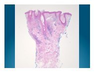
formation of small pseudocysts within the abscesses. These<br />
pseudocysts were clear or filled with amorphous "fluffy" bluegray<br />
material that on AFB staining represented AFB IImage 11<br />
and IImage 21. In all 6 cases, AFB staining revealed numerous<br />
microorganisms, sometimes in areas of the biopsy with mini-<br />
mal to no inflammation IImage 31 and IImage 41. Of note, 1 of<br />
the 6 cases was initially misdiagnosed by a surgical pathologist<br />
as necrotizing fasciitis.<br />
Discussion<br />
Mycobacterial infections classically cause a granulomatous<br />
tissue reaction in most body sites, including the skin.<br />
Therefore, AFB stains are routinely performed along with<br />
Am J Ctin Pathot 2008;130:514-517 515<br />
DOl: 10 1309/DPCLAWWQNTB74JNB
Gable et al R.;piDGRO\J;'L'1G MYCOBACTERIUM SPECIES<br />
'Image 11 Pseudocyst within a dermal abscess (H&E, x400).<br />
'Image 3' Corresponding area for Image 4 of area with<br />
minimal inflammation away from the dermal abscess (H&E,<br />
x200).<br />
fungal stains in the context of granulomatous inflammation.<br />
However, other histologic patterns have been reported<br />
in association with cutaneous mycobacterial infections,<br />
including classical "tuberculoid" caseating granulomas, 1,2<br />
well-formed noncaseating ("sarcoidal") granulomas.l-? suppurative<br />
granulomas.l' dermal abscess formation.!" diffuse<br />
histiocytic infiltration,' panniculitis, 1 lichenoid dermatitis<br />
mixed with granulomata.l and nonspecific chronic inflammati<br />
on. 1 Despite these reports, infection with AFB is often<br />
not considered in the differential diagnosis in skin and soft<br />
tissue biopsy specimens with prominent acute inflammation<br />
and abscess formation.<br />
516 Am J Clin P;;7k::<br />
DOl: io.: c. =-:!.._2<br />
IImage 2' The pseudocyst is filled with acid-fast organisms<br />
(acid-fast bacilli stain, x400).<br />
'Image 4' Acid-fast bacilli (AFB) are readily seen away from<br />
the inflammation (AFB stain, x400).<br />
The rapid-growing Mycobacterium species, including<br />
M abscessus, M chelonae, and Mycobacterium fortuitum, are<br />
separated from other nontuberculous mycobacteria because<br />
of their tendency to grow out in culture in 3 to 5 days rather<br />
than the more typical 2 to 4 weeks of other Mycobacterium<br />
species. These organisms are ubiquitous and rarely cause<br />
significant clinical infection in immuno<strong>com</strong>petent hosts.<br />
However, in the past 2 decades with the emergence of AIDS<br />
and the increased use of immunosuppression in transplant<br />
recipients and other chronically ill patients, the recogni-<br />
tion of infections caused by rapid-growing mycobacteria<br />
has increased.f These infections often are disseminated in<br />
© American Society for Clinical Pathology
a<br />
in<br />
JVgy<br />
nno<strong>com</strong>promised hosts but can also be localized to the<br />
and soft tissue. Immuno<strong>com</strong>promised patients in whom<br />
berculous mycobacterial infections develop have been<br />
ed to have an increased number of cutaneous lesions<br />
ared with immuno<strong>com</strong>petent patients with similar infec-<br />
.1 The immuno<strong>com</strong>promised patients tend to have more<br />
. ent ulceration of and abscess formation within the<br />
eous lesionsJ,8 Histologically, irnmuno<strong>com</strong>promised<br />
show more involvement of the subcutaneous tissue with<br />
re prominent microabscess formation."?<br />
We report a characteristic histologic reaction pattern in<br />
eous rapid-growing Mycobacterium infections in a series<br />
i: immuno<strong>com</strong>promised patients. A high index of suspicion<br />
AFB should be present when faced with a skin or soft<br />
::5sue sample with predominantly suppurative inflammation,<br />
especially in immuno<strong>com</strong>promised patients. In some cases,<br />
all pseudocysts within the abscesses were found to be filled<br />
ith acid-fast bacteria. We re<strong>com</strong>mend routine use of an AFB<br />
stain, in addition to Gram and fungal stains, for the histologic<br />
aluation of suppurative inflammations of skin and soft tissue<br />
infections.<br />
From the Department of Pathology, Brigham and Women's<br />
Hospital, Harvard Medical School, Boston, MA.<br />
Address reprint requests to Dr Granter: Dept of Pathology,<br />
Brigham and Women's Hospital, 75 Francis St, Boston, MA<br />
02115.<br />
© American Society for Clinical Pathology<br />
References<br />
Anatomic Pathology / ORIGINAL ARTICLE<br />
1. Santa Cruz DJ, Strayer DS. The histologic spectrum of the<br />
cutaneous mycobacterioses. Hum Patho!. 1982; 13:485-495.<br />
2. Street ML, Urnbert-Millet 1], Roberts GD, et al. Nontuberculous<br />
mycobacterial infections of the skin: report of fourteen cases and<br />
review of the literarure.J Am Acad Dermato!' 1991;24:208-215 .<br />
3. Escalonilla P, Esteban J, Soriano ML, et aL Cutaneous<br />
manifestations of infection by nontuberculous mycobacteria.<br />
Clin Exp Dennato!' 1998;23:214-221.<br />
4. High WA, Evans CC, Hoang MP. Cutaneous miliary<br />
tuberculosis in two patients with HIY infection. ] Am Acad<br />
Dermatol. 2004;50(5 suppl):SllO-S113.<br />
5. Breza S, Magro CM. Lichenoid and granulomatous dermatitis<br />
associated with atypical mycobacterium infections. ] Cutan<br />
Pathol. 2006;33:512-515.<br />
6. Wolinsky E. Mycobacterial diseases other than tuberculosis.<br />
Clin Infect Dis. 1992;15:1-10.<br />
7. Bartralot R, Garcia-Patos Y, Rodriguez-Cano L, et al. Clinical<br />
patrerns of cutaneous nontuberculous mycobacterial infections.<br />
Br] Dennatol. 2005;152:727-734.<br />
8. Bartralot R, Pujol RM, Garcia-Pates Y, et aL Cutaneous<br />
infections due to nontuberculous mycobacteria: histopathological<br />
review of 28 cases: <strong>com</strong>parative study between lesions observed<br />
in immunosuppressed patients and normal hosts. ] Cutan<br />
Pathol. 2000;27:124-129.<br />
9. McFarlane JR, Plumb SJ, Rhim EW, et aL Leg ulcers in a<br />
38-year-old female. Am] Dermatopathol. 2007;29:586-587.<br />
AmJClinPathoI2008;130:514-517 517<br />
DOl: 10.1309/DPCLAWWQNTB74JNB