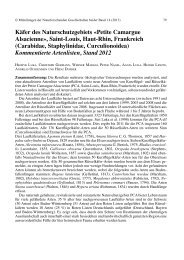
Mallinella thaleri Dankittipakul & Schwendinger
Mallinella thaleri Dankittipakul & Schwendinger
Mallinella thaleri Dankittipakul & Schwendinger

You also want an ePaper? Increase the reach of your titles
YUMPU automatically turns print PDFs into web optimized ePapers that Google loves.
5<br />
11–12), but it can be distinguished by the posteriorly originating embolus<br />
(Fig. 9) (originating more retrolaterally in M. irrorata), by the embolus with a<br />
short branch at mid-length (Figs. 13, 15), and by the unique arrangement of<br />
white patches on the dorsal side of the opisthosoma (Figs. 1–2). In M. <strong>thaleri</strong><br />
sp. nov. the coiled part of the internal epigynal ducts is short (Figs. 17–19),<br />
whereas in M. irrorata it is more elongate and globular, with at least two coils<br />
visible in anterior view.<br />
Description: Male (holotype). Colour in alcohol: Carapace reddish brown;<br />
chelicerae, labium and sternum brown. Leg articles brown except for yellowish<br />
brown tibiae, metatarsi and tarsi. Dorsum of opisthosoma dark sepia, anteriorly<br />
with a pair of large pale patches followed by series of irregular pale stripes;<br />
venter purple, mottled with numerous irregularly arranged white spots. See<br />
Figs. 1, 3.<br />
Prosoma ovate, widest between leg coxae II and III. Carapace coarsely<br />
granular, with a fine white pubescence on pars cephalica. Legs and other<br />
sclerotized areas of body set with numerous hinged hairs on round sockets.<br />
Opisthosoma elongate oval, covered with black hairs; dorsal scutum triangular<br />
(not clearly visible in Fig. 1), relatively broad in front, with straight anterior<br />
margin, gradually tapering towards posterior end, occupying about half of<br />
opisthosoma length. Spines in front of spinnerets relatively thick and short,<br />
situated in a weakly sclerotized area (Fig. 7).<br />
Palp (Figs. 9–11, 13–15) with retrolateral tibial apophysis (RTA) broad at<br />
base, bent ectad, then turning slightly anteroventrad (visible only in ventral<br />
384 Pakawin <strong>Dankittipakul</strong> & Peter J. <strong>Schwendinger</strong><br />
6<br />
7 8<br />
Figs. 5–8. <strong>Mallinella</strong><br />
<strong>thaleri</strong> sp. nov., male<br />
holotype (5, 7) and<br />
female paratype (6, 8).<br />
Prosoma, frontal view<br />
(5–6). Posterior part of<br />
opisthosoma showing<br />
arrangement of spines<br />
in front of spinnerets,<br />
ventral view (7–8).