An Ultrastructural Study on the Desert Locust (Schistocerca gregaria ...
An Ultrastructural Study on the Desert Locust (Schistocerca gregaria ...
An Ultrastructural Study on the Desert Locust (Schistocerca gregaria ...

Create successful ePaper yourself
Turn your PDF publications into a flip-book with our unique Google optimized e-Paper software.
INTERNATIONAL JOURNAL OF AGRICULTURE & BIOLOGY<br />
ISSN Print: 1560–8530; ISSN Online: 1814–9596<br />
07–404/SAE/2008/10–5–475–480<br />
http://www.fspublishers.org<br />
Full Length Article<br />
<str<strong>on</strong>g>An</str<strong>on</strong>g> <str<strong>on</strong>g>Ultrastructural</str<strong>on</strong>g> <str<strong>on</strong>g>Study</str<strong>on</strong>g> <strong>on</strong> <strong>the</strong> <strong>Desert</strong> <strong>Locust</strong> (<strong>Schistocerca</strong><br />
<strong>gregaria</strong>) as Affected by Tebufenozide (RH-5992)<br />
K.S. GHONEIM 1 , R.F. BAKR†, M.A. TANANI, A.G. AL DALI AND A.S. BREAM<br />
Department of Zoology, Faculty of Science, Al-Azhar University, Cairo, Egypt<br />
†Department of Entomology, Faculty of Science, Ain Shams University, Cairo, Egypt<br />
1 Corresp<strong>on</strong>ding author’s e-mail: kar_gh<strong>on</strong>eim@yahoo.com<br />
ABSTRACT<br />
Tebufenozide exhibited some histopathological effects <strong>on</strong> <strong>the</strong> integument of last instar nymphs of desert locust (<strong>Schistocerca</strong><br />
<strong>gregaria</strong> Forsk) such as: detachment of <strong>the</strong> cuticle from epidermis, undistinguishable epicuticle. Tebufenozide treatments<br />
affected <strong>the</strong> ultrastructural c<strong>on</strong>figurati<strong>on</strong> of thoracic muscles such as disrupted organizati<strong>on</strong> of A, I and H bands and <strong>the</strong><br />
degenerati<strong>on</strong> of Z disc varied from <strong>on</strong>e half of <strong>the</strong> disc to small central or peripheral degenerated areas. Tebufenozide<br />
treatments resulted in <strong>the</strong> loss of normal architecture of <strong>the</strong> majority of mid-gut epi<strong>the</strong>lial cells, which had dwarf and deformed<br />
microvilli. The most important ultrastructural changes in <strong>the</strong> intracellular organelles by <strong>the</strong> acti<strong>on</strong> of Tebufenozide, as shown<br />
in <strong>the</strong> electr<strong>on</strong> micrographs, were el<strong>on</strong>gati<strong>on</strong> of mitoch<strong>on</strong>dria, which appeared with prominent cristae, <strong>the</strong> lysosomes were<br />
supposedly autophagic, Golgi bodies had a bullet shape and <strong>the</strong> cytoplasm c<strong>on</strong>tained scattered granules.<br />
Kew Words: <strong>Schistocerca</strong> <strong>gregaria</strong>; Tebufenozide; Nymphal instar; Ultrastructure; Histopathology; Integument; Muscles<br />
INTRODUCTION<br />
Development and reproducti<strong>on</strong> in insects are affected<br />
by a number of horm<strong>on</strong>es, including juvenile horm<strong>on</strong>e and<br />
ecdys<strong>on</strong>e. Insect growth regulators (IGRs) are syn<strong>the</strong>tic<br />
analogues, which mimic <strong>the</strong> naturally occurring horm<strong>on</strong>es<br />
for affecting <strong>the</strong> physiological processes of insects and are<br />
generally classified as juvenile horm<strong>on</strong>e analogues and<br />
ecdys<strong>on</strong>e ag<strong>on</strong>ists (Sehnal, 1983; Mohandass et al., 2006).<br />
The ecdys<strong>on</strong>e ag<strong>on</strong>ists, or ecdysteroids, are syn<strong>the</strong>tic<br />
products interfering with <strong>the</strong> natural insect moulting<br />
horm<strong>on</strong>e (20-hydroxyecdys<strong>on</strong>e), which c<strong>on</strong>trols several<br />
physiological and biochemical processes of growth and<br />
development. They have attracted <strong>the</strong> attenti<strong>on</strong> of many<br />
researchers all over <strong>the</strong> world (Hagedr<strong>on</strong>, 1985; Silhacek et<br />
al., 1990; Gh<strong>on</strong>eim et al., 1991 & 98; Smagghe et al., 1996;<br />
Cadogan et al., 1997). Tebufenozide (RH-5992) represents<br />
a novel class of IGRs, which directly stimulate <strong>the</strong><br />
ecdysteroid receptors as <strong>the</strong> molecular level initiating <strong>the</strong><br />
moulting process by gene regulati<strong>on</strong> especially in larval<br />
Lepidoptera (Wing et al., 1988).<br />
Although c<strong>on</strong>siderable interest has been shown in <strong>the</strong><br />
use of horm<strong>on</strong>e analogues as agents for insect pest c<strong>on</strong>trol,<br />
studies have tended to c<strong>on</strong>centrate <strong>on</strong> morphometric<br />
changes and <strong>on</strong> assessing pesticidal activity. Hence,<br />
informati<strong>on</strong> c<strong>on</strong>cerning <strong>the</strong> effects of such compounds at <strong>the</strong><br />
cellular or subcellular level is somewhat limited. In <strong>the</strong><br />
present work, <strong>the</strong> histopathological effects of Tebufenozide,<br />
as well as <strong>the</strong> ultrastructural changes in <strong>the</strong> desert locust<br />
(<strong>Schistocerca</strong> <strong>gregaria</strong> Forsk) were investigated aiming to<br />
shed some light <strong>on</strong> its roles at <strong>the</strong> cellular and subcellular<br />
levels.<br />
MATERIALS AND METHODS<br />
Experimental insect. A gregarious stock culture of desert<br />
locust [<strong>Schistocerca</strong> <strong>gregaria</strong> Forsk. (Orthoptera:<br />
Acrididae)] was raised by a sample from <strong>the</strong> established<br />
culture of <strong>Locust</strong> and Grasshopper Res. Divisi<strong>on</strong>, Agric.<br />
Res. Center, Giza, Egypt. The insects were reared under<br />
crowded breeding c<strong>on</strong>diti<strong>on</strong>s outlined by Hunter-J<strong>on</strong>es<br />
(1961) and Hassanein (1965). Newly hatched hoppers were<br />
kept in wooden cages with wire-gauze sides (40 x 40 x 60<br />
cm) and small door in <strong>the</strong> upper-side to allow <strong>the</strong> daily<br />
feeding and cleaning routine. The bottom was covered with<br />
20 cm layer of sterilized sand. Cages were equipped<br />
internally with 60 W electric bulb for lightening (17:7 LD)<br />
and warming (32±2 o C.). Successive generati<strong>on</strong>s were raised<br />
before obtaining <strong>the</strong> nymphs for <strong>the</strong> present experimental<br />
work. Fresh food plant was clover Medicago sativa al<strong>on</strong>g<br />
<strong>the</strong> period of study except few weeks every year, because of<br />
<strong>the</strong> absence of this plant species. During <strong>the</strong>se weeks,<br />
insects were fed <strong>on</strong> Sesbania egyptiaca. All experiments<br />
were c<strong>on</strong>ducted with Medicago sativa <strong>on</strong>ly.<br />
Administrati<strong>on</strong> of <strong>the</strong> horm<strong>on</strong>e analogues. A technical<br />
grade of <strong>the</strong> n<strong>on</strong>-steroidal ecdys<strong>on</strong>e ag<strong>on</strong>ist Tebufenozide<br />
To cite this paper: Gh<strong>on</strong>eim, K.S., R.F. Bakr, M.A. Tanani, A.G. Al Dali and A.S. Bream, 2008. <str<strong>on</strong>g>An</str<strong>on</strong>g> ultrastructural study <strong>on</strong> <strong>the</strong> desert locust (<strong>Schistocerca</strong><br />
<strong>gregaria</strong>) as affected by Tebufenozide (RH-5992). Int. J. Agri. Biol., 10: 479–80
(RH-5992) was used. Its chemical name is 1-N-t-butyl-1 (3,<br />
5-dimethyl benzoyl)-2-(4-ethylbenzoyl) hydrazine (Rohm &<br />
Haas Company, Philadelphia, PA). Fresh clean clover<br />
leaves (M. sativa) were dipped for 3 min in 1000 mg kg -1<br />
Tebufenozide and introduced to <strong>on</strong>e-day penultimate instar<br />
nymphs. Three replicates (10 nymphs per replicate) were<br />
carried out for each treatment and c<strong>on</strong>trols. Each individual<br />
nymph was kept in a suitable glass vial whose bottom<br />
covered with a thin layer of sterilized sand. The nymphs,<br />
treated or c<strong>on</strong>trol, were kept individually.<br />
Histopathological and ultrastuctural techniques. The<br />
histopathological and ultrastuctural effects were examined<br />
in <strong>the</strong> cuticle, muscles, mid-gut of <strong>on</strong>e-day old last instar<br />
nymphs. A representative nymph of each group was<br />
dissected and transected, fixed as so<strong>on</strong> as possible in 3%<br />
phosphate buffered glutaraldehyde (pH 7.3) for 2 h. After<br />
two rinses in <strong>the</strong> buffer (for a period of 4 h.) <strong>the</strong> specimens<br />
were fixed in 1% buffered osmium tetraoxide for 1 h at 4°C<br />
(Brissan et al., 1996).<br />
The tissue pieces were washed twice in a buffer for 30<br />
min. The specimens were <strong>the</strong>n dehydrated in grades of<br />
ethanol; 50, 70, 80, 90 and 100%. The specimens were<br />
cleared in toluene for 10 min and <strong>the</strong>n embedded in <strong>the</strong> resin<br />
of choic Ep<strong>on</strong>. Semi<strong>the</strong>n secti<strong>on</strong>s are cut from <strong>the</strong>se blocks<br />
(stained with toluidine blue) and examined by <strong>the</strong> light<br />
microscope (Spnrr, 1969).<br />
Ultrathin secti<strong>on</strong>s obtained from selected blocks were<br />
mounted <strong>on</strong> copper grids stained with uranyl acetate and<br />
lead citrate and <strong>the</strong>n examined with Jol 1010 transmissi<strong>on</strong><br />
electr<strong>on</strong> microscope (Reynolds, 1963). This technique was<br />
carried out at Regi<strong>on</strong>al Center for Mycology and<br />
Biotechnology, Al-Azhar University, Madenit Nasr, Cairo.<br />
RESULTS<br />
The histopathological and ultrastructural investigati<strong>on</strong>s<br />
were carried out for <strong>on</strong>e-day old last instar nymphs of desert<br />
locust after feeding <strong>the</strong> <strong>on</strong>e-day old penultimate instar<br />
nymphs <strong>on</strong> a fresh food plant treated with 1000 mg kg -1 of<br />
Tebufenozide, as given above.<br />
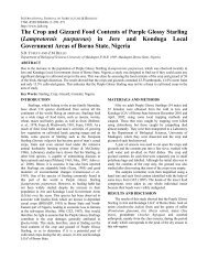
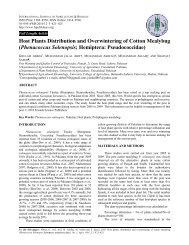
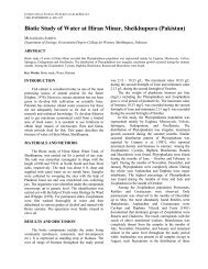
Histopathology and ultrastucture of integument. The<br />
basic structure of c<strong>on</strong>trol integument of <strong>the</strong> last instar<br />
nymphs is shown in (Fig. 1a & b). The available electr<strong>on</strong><br />
micrograph clearly shows several changes in <strong>the</strong> integument<br />
of Tebufenozide treated last instar nymphs (Fig. 2).<br />
Tebufenozide treatments resulted in detachment of <strong>the</strong><br />
cuticle from <strong>the</strong> hypodermis, undistinguishable appearance<br />
of epicuticle and exocuticle.<br />
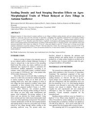
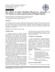
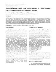
Histopathology and ultrastructure of thoracic muscles.<br />
At least, two kinds of filaments exist in <strong>the</strong> fibrils:<br />
prominent thicker filaments running al<strong>on</strong>g <strong>the</strong> A band and<br />
represent <strong>the</strong> protein, myosin; and less obvious filamentous<br />
thinner filaments extending from Z line to <strong>the</strong> edge of <strong>the</strong> H<br />
band, which represent <strong>the</strong> actin (Fig. 3).<br />
Feeding of <strong>on</strong>e-day old penultimate instar nymphs <strong>on</strong><br />
1000 mg L -1 Tebufenozide treated fresh food caused many<br />
GHONEIM et al. / Int. J. Agri. Biol., Vol. 10, No. 5, 2008<br />
476<br />
Fig. 1. Electr<strong>on</strong> micrographs of transverse secti<strong>on</strong>s<br />
through two sites (a & b) of <strong>the</strong> integument of c<strong>on</strong>trol<br />
1- day old last instar nymphs of desert locust. Ep:<br />
epicuticle, Ex: exocuticle, En: endocuticle, Mt:<br />
mitoch<strong>on</strong>dria, Cr: mitoch<strong>on</strong>drial cristae<br />
Fig. 2. Electr<strong>on</strong> micrographs of transverse secti<strong>on</strong>s<br />
through two sites (a, b) of <strong>the</strong> integument of<br />
Tebufenozide treated last instar nymphs of desert<br />
locust. Tebufenozide treatments resulted in:<br />
detachment of <strong>the</strong> cuticle from <strong>the</strong> hypodermis,<br />
undistinguishable appearance of epicuticle and<br />
exocuticle. Abbreviati<strong>on</strong>s: Ep: epicuticle, Ex:<br />
exocuticle, En: endocuticle, Hy: epidermis (or<br />
hypodermis)<br />
histopathological and ultrastuctural disorders in <strong>the</strong> thoracic<br />
muscles of <strong>the</strong> last instar nymphs as dem<strong>on</strong>strated in <strong>the</strong><br />
electr<strong>on</strong> micrograph (Fig. 4). The degenerati<strong>on</strong> of Z-disc<br />
varied from <strong>on</strong>e half of <strong>the</strong> disc to small central or<br />
peripheral degenerated areas. The micrograph also shows a<br />
disrupted organizati<strong>on</strong> of A, I and H bands.<br />
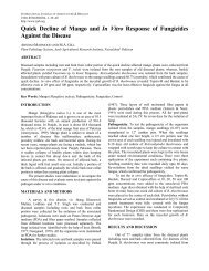
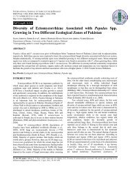
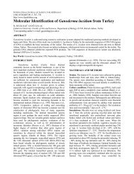
Histopathology and ultrastructure of mid-gut. As seen in<br />
(Fig. 5), <strong>the</strong> c<strong>on</strong>trol (normal) mid-gut of desert locust<br />
appears with luminal surface of <strong>the</strong> epi<strong>the</strong>lium, which is<br />
provided with a striated border c<strong>on</strong>stituting l<strong>on</strong>g microvilli.<br />
Such microvilli protrude inwards into <strong>the</strong> lumen to increase<br />
<strong>the</strong> absorpti<strong>on</strong> surface of <strong>the</strong> cells, as well as, <strong>the</strong> spaces<br />
between <strong>the</strong>m act as a kind of sieve.<br />
The ecdys<strong>on</strong>e ag<strong>on</strong>ist Tebufenozide exerted several<br />
histopathological effects <strong>on</strong> <strong>the</strong> mid-gut of last instar<br />
hoppers. The majority of epi<strong>the</strong>lial cells lost <strong>the</strong>ir normal
ULTRASTRUCTURAL STUDIES ON DESERT LOCUST INSTAR NYMPHS / Int. J. Agri. Biol., Vol. 10, No. 4, 2008<br />
Fig. 3. Electr<strong>on</strong> micrograph of a l<strong>on</strong>gitudinal secti<strong>on</strong> of<br />
thoracic muscles of c<strong>on</strong>trol <strong>on</strong>e day old last instar<br />
nymphs of desert locust. Abbreviati<strong>on</strong>s: I: isotropic<br />
band, A: anisotropic band, M: middle band, H: narrow<br />
light band, Z: marked limits of sarcomere<br />
Fig. 4. Electr<strong>on</strong> micrograph of a l<strong>on</strong>gitudinal secti<strong>on</strong> of<br />
thoracic muscles of Tebufenozide treated last instar<br />
nymphs of desert locust. The degenerati<strong>on</strong> of Z-disc<br />
varied from <strong>on</strong>e half of <strong>the</strong> disc to small central or<br />
peripheral degenerated areas. Also, disrupted<br />
organizati<strong>on</strong> of A, I and H bands can be observed.<br />
Abbreviati<strong>on</strong>s: I: Isotropic band, A: anisotropic band,<br />
H: narrow light band, Z: marked limits of sarcomere<br />
architecture, because <strong>the</strong> microvilli appeared dwarf and had<br />
been severely deformed. Also, autopathogenic vacuoles and<br />
residual bodies, as well as vacuolizati<strong>on</strong> and ruptured cell<br />
wall were obtained.<br />
Changes in <strong>the</strong> organelles. The orientati<strong>on</strong> of <strong>the</strong><br />
mitoch<strong>on</strong>dria is said to depend <strong>on</strong> <strong>the</strong> nature of <strong>the</strong><br />
cytoplasmic matrix, vacuolar system and <strong>the</strong> directi<strong>on</strong> of <strong>the</strong><br />
diffusi<strong>on</strong> currents of <strong>the</strong> cell. Also, <strong>the</strong> number of<br />
mitoch<strong>on</strong>dria in a cell depends <strong>on</strong> <strong>the</strong> type and functi<strong>on</strong>al<br />
state of <strong>the</strong> cell. They vary from cell to cell and from o<strong>the</strong>r<br />
to species. The mitoch<strong>on</strong>dria may be filamentous or<br />
granular in <strong>the</strong> shape and may change from <strong>on</strong>e form to<br />
477<br />
Fig. 5. Electr<strong>on</strong> micrographs of transverse secti<strong>on</strong>s<br />
through two sites (a & b) of <strong>the</strong> mid gut of c<strong>on</strong>trol <strong>on</strong>e<br />
day old last instar nymphs of desert locust.<br />
Abbreviati<strong>on</strong>s: L: lumen cavity, Gb: Golgi bodies, N:<br />
nucleus, Ly: lysosome, Mv: microvilli<br />
Fig. 6. Electr<strong>on</strong> micrograph of mitoch<strong>on</strong>dria in c<strong>on</strong>trol<br />
<strong>on</strong>e day old last instar nymphs of desert locust.<br />
Abbreviati<strong>on</strong>: Mt: mitoch<strong>on</strong>dria, Cr: Cristae<br />
ano<strong>the</strong>r depending up<strong>on</strong> <strong>the</strong> physiological c<strong>on</strong>diti<strong>on</strong> of <strong>the</strong><br />
cells. Thus <strong>the</strong>y may be of club, racket, vesicular, ring or<br />
round-shape. The size of <strong>the</strong> mitoch<strong>on</strong>dria ranges from 0.2<br />
to 2.0 um and <strong>the</strong> length may range from 0.3 to 40 µm.<br />
In additi<strong>on</strong>, <strong>the</strong> mitoch<strong>on</strong>dria are bounded by a double<br />
membrane envelope, which provides good tensile strength,<br />
stability and flexibility to <strong>the</strong>m. The outer and inner<br />
mitoch<strong>on</strong>drial membranes are 0.6-0.7 µm thick. Each of<br />
<strong>the</strong>m c<strong>on</strong>sisted of outer and inner dense osmiophilic layers<br />
composed of protein molecules of 200 to 250 nm<br />
thicknesses. Also, <strong>the</strong>re was a middle (250 nm thick)<br />
biomolecular layer composed of lipids (Fig. 6). Existence of<br />
rounded bodies, lysosomes, was noted in <strong>the</strong> form of saclike<br />
structures, each surrounded by a single thin lipoprotein<br />
membrane, which c<strong>on</strong>tained <strong>the</strong> broken cellular materials<br />
such as protein, nucleic acid and polysaccharides. The
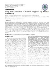
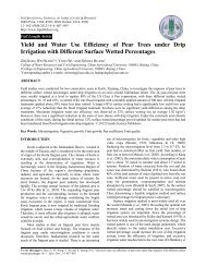
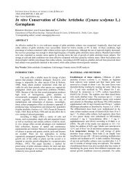
Fig. 7. Electr<strong>on</strong> micrograph of mitoch<strong>on</strong>dria in<br />
Tebufenozide treated last instar nymphs of desert<br />
locust. Some mitoch<strong>on</strong>dria were el<strong>on</strong>gated with<br />
prominent cristae. Abbreviati<strong>on</strong>s: Mt: mitoch<strong>on</strong>dria,<br />
Cr: cristae<br />
Fig. 8. Electr<strong>on</strong> micrographs of transverse secti<strong>on</strong>s<br />
through two sites (a & b) of <strong>the</strong> mid gut of<br />
Tebufenozide treated last instar nymphs of desert<br />
locust. The microvilli appeared as dwarf and severely<br />
deformed. Vacuolizati<strong>on</strong> and ruptured cell wall were<br />
obtained. Also, <strong>the</strong>re are proposingly autophagic<br />
lysosomes. Golgi bodies appear in a bullet shape. The<br />
cytoplasm c<strong>on</strong>tains scattered granules. Abbreviati<strong>on</strong>s:<br />
dMv: destructi<strong>on</strong> of microvilli, Gb: Golgi bodies, L:<br />
lumen cavity, N: nucleus, Ch: nuclear chromatin, Lc:<br />
lysed cytoplasm, MvB: multivesicular bodies<br />
dictyosomes appear as flattened oval sac toge<strong>the</strong>r with<br />
cluster of small vesicular bodies at <strong>the</strong>ir edges (Fig. 7). The<br />
ultrastructural examinati<strong>on</strong>, in <strong>the</strong> present study, revealed<br />
several effects of <strong>the</strong> ecdysteroid Tebufenozide <strong>on</strong> <strong>the</strong> last<br />
instar nymphs of desert locust. Some mitoch<strong>on</strong>dria were<br />
el<strong>on</strong>gated with prominent cristae (Fig. 7). There were<br />
autophagic lysosomes. The dictyosomes appeared in a bullet<br />
shape and <strong>the</strong> cytoplasm c<strong>on</strong>tained scattered granules (Fig. 8).<br />
GHONEIM et al. / Int. J. Agri. Biol., Vol. 10, No. 5, 2008<br />
478<br />
DISCUSSION<br />
Histopathological Changes in <strong>the</strong> Body Tissues<br />
The integument. In <strong>the</strong> present study, <strong>on</strong>e-day old<br />
penultimate instar nymphs of desert locust after feeding <strong>on</strong><br />
Tebufenozide-treated food resulted in several<br />
histopathological changes in <strong>the</strong> integument of last instar<br />
nymphs. The most important changes were detachment of<br />
<strong>the</strong> cuticle from <strong>the</strong> epidermal layer, <strong>the</strong> epicuticle could not<br />
be distinguished from <strong>the</strong> exocuticle. Wing et al. (1988)<br />
obtained precocious and lethal moults in various insect<br />
larvae by <strong>the</strong> ecdysteroid Tebufenozide. Disturbance in<br />
some steps of chitin depositi<strong>on</strong> and histopathological<br />
changes in <strong>the</strong> epidermal cells were also reported by several<br />
authors using different horm<strong>on</strong>e analogues (Gnatzy &<br />
Romer, 1984; Retnakaran & Oberlander, 1993).<br />
Electrophoretic investigati<strong>on</strong> revealed <strong>the</strong> effect of<br />
Tebufenozide <strong>on</strong> <strong>the</strong> producti<strong>on</strong> of cuticular proteins in<br />
larvae of Spodoptera littoralis and Spodoptera exigua<br />
(Smagghe & Degheele, 1996). In additi<strong>on</strong>, <strong>the</strong> inhibiti<strong>on</strong> of<br />
normal endocuticle syn<strong>the</strong>sis in tomato looper Chrysodeixis<br />
chalcites may have been caused by residual levels of<br />
Tebufenozide and this in turn may have prevented<br />
formati<strong>on</strong> of a functi<strong>on</strong>al cuticle and may explain <strong>the</strong><br />
fragility of <strong>the</strong> incompletely formed new cuticle (Smagghe<br />
et al., 1997).<br />
The thoracic muscles. All <strong>the</strong> insect muscles are built <strong>on</strong> a<br />
similar plan with el<strong>on</strong>gate cells housing <strong>the</strong> c<strong>on</strong>tractile<br />
elements and in many cases, inserted into <strong>the</strong> integument at<br />
ei<strong>the</strong>r end. Each muscle is made up of a number of l<strong>on</strong>g<br />
fibers. Multinucleate cells usually run al<strong>on</strong>g <strong>the</strong> whole<br />
length of <strong>the</strong> muscle. The sarcolemma, myofibrils,<br />
sarcoplasm, molecular filaments, striati<strong>on</strong> pattern (including<br />
discs & bands) and o<strong>the</strong>r structures are detailed in Elder<br />
(1975). In desert locust, in <strong>the</strong> present study, a complete set<br />
of adult muscles was present in <strong>the</strong> nymphal form. Most<br />
muscles were in use during nymphal stage. Flight muscles,<br />
however, remained small and functi<strong>on</strong>less until <strong>the</strong> last<br />
larval instar and developed rapidly just before and after <strong>the</strong><br />
imaginal moult (Novicki, 1989; Shiga et al., 2002).<br />
Excisi<strong>on</strong> of <strong>the</strong> corpora allata (CA) producing juvenile<br />
horm<strong>on</strong>es (JH) from reproductive beetles causes muscle<br />
degenerati<strong>on</strong>. Reimplantati<strong>on</strong> of <strong>the</strong> active CA causes <strong>the</strong>m<br />
to regenerate (Stegwee et al., 1963). In c<strong>on</strong>trast,<br />
implantati<strong>on</strong> of <strong>the</strong> CA from reproductive females into<br />
diapausing <strong>on</strong>es induces degenerati<strong>on</strong> of flight muscles in<br />
<strong>the</strong> plant bug Dysdercus intermedius (Edwards, 1970).<br />
These studies have shown a close associati<strong>on</strong> between <strong>the</strong><br />
endocrine system and flight muscle degenerati<strong>on</strong>. In<br />
Modicogryllus c<strong>on</strong>firmatus and Gryllus firmus, exogenous<br />
applicati<strong>on</strong>s of JH or a JH analog c<strong>on</strong>firmed its role<br />
(Tanaka, 1994; Zera & Cisper, 2001). Topically treated 5 th<br />
instar nymphs of <strong>Locust</strong>a migratoria with <strong>the</strong> juvenoid<br />
methoprene caused profound changes in <strong>the</strong> muscle fiber<br />
fine structure (Cott<strong>on</strong> & <str<strong>on</strong>g>An</str<strong>on</strong>g>stee, 1990).<br />
The mid gut. Tebufenozide treatments of <strong>the</strong> tomato looper
ULTRASTRUCTURAL STUDIES ON DESERT LOCUST INSTAR NYMPHS / Int. J. Agri. Biol., Vol. 10, No. 4, 2008<br />
C. chalcites larvae resulted in cell degenerati<strong>on</strong> of <strong>the</strong> fore<br />
gut (Smagghe et al., 1997). In <strong>the</strong> present study, <strong>the</strong><br />
Tebufenozide treatments to <strong>on</strong>e-day old penultimate instar<br />
nymphs of desert locust resulted in several serious<br />
histopathological changes in <strong>the</strong> mid gut of <strong>on</strong>e-day old last<br />
instar nymphs. Majority of epi<strong>the</strong>lial cells lost normal<br />
architecture and had dwarf and deformed microvilli. Also,<br />
obvious autophagic vacuoles scattered in <strong>the</strong> cytoplasm<br />
c<strong>on</strong>taining many vesicles. These results are in accordance<br />
with those of various o<strong>the</strong>r insect species (Williams, 1968;<br />
Birkenbeil, 1983; Gnatzy & Romer, 1984; Locke, 1984 &<br />
85; Hant<strong>on</strong> et al., 1993).<br />
Influence of horm<strong>on</strong>al analogues <strong>on</strong> intracellular<br />
ultrastructure. Mitoch<strong>on</strong>dria are c<strong>on</strong>sidered as <strong>the</strong> 'power<br />
house' of <strong>the</strong> living cell, which supply energy for many<br />
chemical reacti<strong>on</strong>s and transport mechanisms in <strong>the</strong> cell.<br />
Orientati<strong>on</strong> of mitoch<strong>on</strong>dria depends <strong>on</strong> <strong>the</strong> nature of <strong>the</strong><br />
cytoplasmic matrix, vascular system and <strong>the</strong> directi<strong>on</strong> of<br />
diffusi<strong>on</strong> of currents in <strong>the</strong> cell. Their number and shape<br />
depend <strong>on</strong> <strong>the</strong> cell type and its physiological state varying<br />
from insect species to ano<strong>the</strong>r. Each mitoch<strong>on</strong>drium is<br />
bounded by a double membrane envelope, which provides<br />
its tensile strength, stability and flexibility (Bakr, 1986).<br />
Smagghe et al. (1997) observed a dramatic increase in<br />
endoplasmic reticulum, scattered throughout <strong>the</strong> whole<br />
epidermal cell cytoplasm, hypertrophy of Golgi complexes,<br />
increase in <strong>the</strong> volume of <strong>the</strong> nuclei, and <strong>the</strong> presence of<br />
numerous oval and el<strong>on</strong>gated mitoch<strong>on</strong>dria of C. chalcites<br />
larvae. Also, <strong>the</strong> epidermal cell cytoplasm of treated larvae<br />
c<strong>on</strong>tained numerous vacuoles and vesicles, myelin figures<br />
and glycogen clusters. On <strong>the</strong> o<strong>the</strong>r hand, Bakr et al. (1997)<br />
examined various ultrastructural effects of some IGRs <strong>on</strong><br />
<strong>the</strong> larvae and pupae of Culex pipiens such as <strong>the</strong> loss of<br />
cristae in some mitoch<strong>on</strong>dria, swelling of o<strong>the</strong>rs with<br />
prominent cristae and loss of demarcati<strong>on</strong> of <strong>the</strong><br />
mitoch<strong>on</strong>dria membranes with obvious increase in <strong>the</strong><br />
mitoch<strong>on</strong>dria granules.<br />
Tebufenozide, in <strong>the</strong> present study, remarkably affected<br />
<strong>the</strong> ultrastructure of <strong>the</strong> last instar nymphs of desert locust<br />
such as: el<strong>on</strong>gati<strong>on</strong> of mitoch<strong>on</strong>dria with prominent cristae,<br />
appearance of lysosomes supposedly autophagic, bulletshaped<br />
Golgi bodies and scattered granules in <strong>the</strong> cytoplasm.<br />
REFERENCES<br />
Bakr, R.F.A., 1986. Morphogenic and physiological aberrati<strong>on</strong> induced by<br />
certain IGRs in <strong>the</strong> house fly Musca domestica. Unpublished Ph.D.<br />
Thesis, Ain Shams University, Cairo, Egypt<br />
Bakr, R.F.A., A.M. Isa, M.S. Gabry and A.M. Guneidy, 1997.<br />
Histopathological changes in Culex pipiens (Diptera: Culicidae)<br />
induced by juvenile horm<strong>on</strong>e mimics. J. Egypt Ger. Soc. Zool., 22:<br />
27–45<br />
Birkenbeil, H., 1983. <str<strong>on</strong>g>Ultrastructural</str<strong>on</strong>g> and immunocytochemical investigati<strong>on</strong><br />
of ecdysteroid secreti<strong>on</strong> by <strong>the</strong> prothoracic gland of <strong>the</strong> waxmoth<br />
Galleria mell<strong>on</strong>ella. Cell Tissue Res., 229: 433–41<br />
Brissan, A., S. Gharibian, R. Agen, D.F. Leclerc and C. Breuil, 1996.<br />
Localizati<strong>on</strong> and characterizati<strong>on</strong> of <strong>the</strong> melanin granules produced<br />
by <strong>the</strong> sap-staining fungus Ophiostoma piceae. Material and<br />
Organismen, 30: 23–32<br />
479<br />
Cadogan, B.L., A. Retnakaran and J.H. Meating, 1997. Efficacy of RH-<br />
5992, a new insect growth regulator against spruce budworm<br />
(Lepidoptera : Tortricidae) in a Boreal forest. J. Ec<strong>on</strong>. Entomol., 90:<br />
551–9<br />
Cott<strong>on</strong>, G. and J.H. <str<strong>on</strong>g>An</str<strong>on</strong>g>stee, 1990. A structural and biochemical study <strong>on</strong> <strong>the</strong><br />
effects of methoprene <strong>on</strong> flight muscle development in <strong>Locust</strong>a<br />
migratoria L. J. Insect Physiol., 36: 959–69<br />
Edwards, F.J., 1970. Endocrine c<strong>on</strong>trol of flight muscle histolysis in<br />
Dysdercus intermedius. J. Insect Physiol., 16: 2027–31<br />
Elder, H.Y., 1975. Muscle structure. In: Usherwood, P.N.R. (ed.), "Insect<br />
Muscles", pp: 1–74. Academic Press, L<strong>on</strong>d<strong>on</strong><br />
Gh<strong>on</strong>eim, K.S., M.A. Fouda and A.S. Bream, 1991. Effectiveness of <strong>the</strong><br />
n<strong>on</strong>-steroidal ecdys<strong>on</strong>e mimic, RH-5849 for <strong>the</strong> c<strong>on</strong>trol of Musca<br />
domestica vicina (L). J. Egyptian Soc. Parasitol., 21: 723–33<br />
Gh<strong>on</strong>eim, K.S., A.S. Bream and H.A. Mohamed, 1998. Bioactivity of <strong>the</strong><br />
ecdysteroid against Tebufenozide (RH-5992) <strong>on</strong> <strong>the</strong> Egyptian cott<strong>on</strong><br />
leafworm, Spodoptera littoralis (Boisd) (Lepidoptera: Noctuidae).<br />
Al-Azhar Bull. Sci., 9: 947–63<br />
Gnatzy, W. and F. Romer, 1984. Cuticle: formati<strong>on</strong>, moulting and c<strong>on</strong>trol.<br />
In: Bereiter-Hahn, J., A.G. Matoltsy and K.S. Richards (eds.),<br />
"Biology of <strong>the</strong> Integument, Invertebrates", pp: 638–84. Springer-<br />
Verlag<br />
Hagedr<strong>on</strong>, H.H., 1985. The role of ecdysteroides in reproducti<strong>on</strong>. In: Kekut,<br />
G.K. and L.I. Gilbert (eds.), “Comprehensive Insect Physiology,<br />
Biochemistry and Pharmacology”, Vol. 8, pp: 205–62. Pergam<strong>on</strong>,<br />
Oxford<br />
Hant<strong>on</strong>, W.K., R.D. Wats<strong>on</strong>, and W.E. Bollenbacher, 1993. Ultrastructure<br />
of prothoracic glands during larval-pupal development of <strong>the</strong><br />
tobacco hornworm Manduca sexta: a reappraisal. J. Morphol., 216:<br />
95–112<br />
Hassanein, M.S., 1965. Laboratory and Outdoor Cultures and Breeding of<br />
<strong>Locust</strong>s and Grasshoppers, p: 10. FAO Publicati<strong>on</strong> 5/31901<br />
Hunter-J<strong>on</strong>es, P., 1961. Rearing and Breeding <strong>Locust</strong>s in Laboratory, p: 12.<br />
Bulletin <str<strong>on</strong>g>An</str<strong>on</strong>g>ti-locust Research Centre L<strong>on</strong>d<strong>on</strong><br />
Locke, M., 1984. Epidermal cells. In: Bereiter-Hahn, J., A.G. Matoltsy and<br />
K.S. Richards (eds.), "Biology of <strong>the</strong> Integument. 1. Invertebrates",<br />
pp: 502-522. Springer-Verlag<br />
Locke, M., 1985. A structural analysis of post-embry<strong>on</strong>ic development. In:<br />
Kerkut, G.A. and L.I. Gilbert (eds.), "Comprehensive Insect<br />
Physiology, Biochemistry and Pharmachology", Vol. 2, pp: 87–149.<br />
Pergam<strong>on</strong> Press, Oxford<br />
Mohandass, S.M., F.H. Arthur, K.Y. Zhu and J.E. Thr<strong>on</strong>e, 2006.<br />
Hydroprene: mode of acti<strong>on</strong>, current status in stored product pest<br />
management, insect resistance and future prospects. Crop Protec.,<br />
25: 902–9<br />
Novicki, A., 1989. C<strong>on</strong>trol of growth and ultrastructural maturati<strong>on</strong> of a<br />
cricket flight muscle. J. Exp. Zool., 250: 263–72<br />
Retnakaran, A. and H. Oberlander, 1993. C<strong>on</strong>trol of chitin syn<strong>the</strong>sis in<br />
insects. In: Muzzarelli, R.A.A. (ed.), "Chitin Enzymology", pp: 89-<br />
99. Ac<strong>on</strong>a: European Chitin Society<br />
Reynolds, S.E., 1963. The use of lead citrate at high pH as an electr<strong>on</strong><br />
opaque stain in electr<strong>on</strong> microscopy. J. Cell Biol., 1: 208<br />
Sehnal, F., 1983. Juvenile horm<strong>on</strong>e analogues. In: Downer, R.G.H. and<br />
Laufer, H. (eds.), "Endocrinology of Insects", pp: 657–72. Alan R.<br />
Liss, Inc., NY<br />
Shiga, S., K. Yauyama, N. Okamura and T. Yamaguchi, 2002. Natural and<br />
endocrine c<strong>on</strong>trol of flight muscle degenerati<strong>on</strong> <strong>on</strong> <strong>the</strong> adult cricket,<br />
Gryllus bimaculatus. J. Insect Physiol., 48: 15–24<br />
Silhacek, D.L., H. Oberlander and P. Porcher<strong>on</strong>, 1990. Acti<strong>on</strong> of RH-5849,<br />
a n<strong>on</strong>-steroidal ecdysteroid mimic, <strong>on</strong> Plodia interpunctella<br />
(Hubner) in vivo. Arch. Insect Biochem. Physiol., 15: 201–12<br />
Smagghe, G. and D. Degheele, 1996. Electrophoretic relevance of <strong>the</strong><br />
ecdysteroid-like acti<strong>on</strong> of Tebufenozide. Med. Fac. Landbouww<br />
Univ. Gent, 61: 899–901<br />
Smagghe, G., E. Vinuela, F. Budia and D. Degheele, 1997. Effects of <strong>the</strong><br />
n<strong>on</strong>-steroidal ecdysteroid mimic Tebufenozide <strong>on</strong> <strong>the</strong> tomato looper<br />
Chrysodeixis chalcites (Lepidoptera: Nocutidae): <str<strong>on</strong>g>An</str<strong>on</strong>g> ultrastructural<br />
analysis. Arch. Insect Biochem. Physiol., 35: 179–90<br />
Spnrr, A.R., 1969. A low viscosity epoxy resin embdding medium for<br />
electr<strong>on</strong> microscopy. J. Ultrastr. Res., 26: 31–43
Stegwee, D., E.C. Kimmel, J.A. De Boer and S. Henstra, 1963. Horm<strong>on</strong>al<br />
c<strong>on</strong>trol of reversible degenerati<strong>on</strong> of flight muscle in <strong>the</strong> Colorado<br />
potato beetle, Leptinotarsa decemlineata Say (Coleoptera). J. Cell<br />
Biol., 19: 519–27<br />
Tanaka, S., 1994. Endocrine c<strong>on</strong>trol of ovarian development and flight<br />
muscle histolysis in a wing dimorphic cricket, Modicogryllus<br />
c<strong>on</strong>firmatus. J. Insect Physiol., 40: 483–90<br />
Williams, C.M., 1968. Ecdys<strong>on</strong>es and ecdyst<strong>on</strong>e analogues. Their assay and acti<strong>on</strong><br />
<strong>on</strong> diapausing pupae of <strong>the</strong> Cynthia silkworm. Biol. Bull., 134: 344–55<br />
GHONEIM et al. / Int. J. Agri. Biol., Vol. 10, No. 5, 2008<br />
480<br />
Wing, D.D., R.A. Slawecki and G.R. Carls<strong>on</strong>, 1988. RH-5849, a<br />
n<strong>on</strong>steroidal ecdys<strong>on</strong>e ag<strong>on</strong>ist: effects <strong>on</strong> larval Lepidoptera. Sci.,<br />
241: 470–2<br />
Zera, A.J. and G. Cisper, 2001. Genetic and diurnal variati<strong>on</strong> in <strong>the</strong> juvenile<br />
horm<strong>on</strong>e titer in a wing polymorphic cricket: implicati<strong>on</strong>s for <strong>the</strong><br />
evaluati<strong>on</strong> of life histories and dispersal. Physiol. Biochem. Zool., 74:<br />
293–306<br />
(Received 19 November 2007; Accepted 27 May 2008)