Philips Achieva 3T T1 Map FA2
Philips Achieva 3T T1 Map FA2 Philips Achieva 3T T1 Map FA2
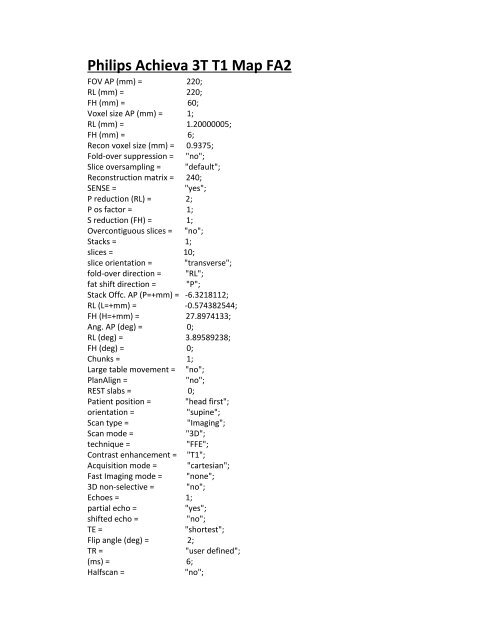
Philips Achieva 3T T1 Map FA2 FOV AP (mm) = 220; RL (mm) = 220; FH (mm) = 60; Voxel size AP (mm) = 1; RL (mm) = 1.20000005; FH (mm) = 6; Recon voxel size (mm) = 0.9375; Fold‐over suppression = "no"; Slice oversampling = "default"; Reconstruction matrix = 240; SENSE = "yes"; P reduction (RL) = 2; P os factor = 1; S reduction (FH) = 1; Overcontiguous slices = "no"; Stacks = 1; slices = 10; slice orientation = "transverse"; fold‐over direction = "RL"; fat shift direction = "P"; Stack Offc. AP (P=+mm) = ‐6.3218112; RL (L=+mm) = ‐0.574382544; FH (H=+mm) = 27.8974133; Ang. AP (deg) = 0; RL (deg) = 3.89589238; FH (deg) = 0; Chunks = 1; Large table movement = "no"; PlanAlign = "no"; REST slabs = 0; Patient position = "head first"; orientation = "supine"; Scan type = "Imaging"; Scan mode = "3D"; technique = "FFE"; Contrast enhancement = "T1"; Acquisition mode = "cartesian"; Fast Imaging mode = "none"; 3D non‐selective = "no"; Echoes = 1; partial echo = "yes"; shifted echo = "no"; TE = "shortest"; Flip angle (deg) = 2; TR = "user defined"; (ms) = 6; Halfscan = "no";
- Page 2 and 3: Water‐fat shift = "maximum"; Shim
- Page 4 and 5: Fast Imaging mode = "none"; 3D non
- Page 6 and 7: Large table movement = "no"; PlanAl
- Page 8 and 9: fat shift direction = "P"; Stack Of
- Page 10: Full flow comp. = "yes"; TSE es / s
<strong>Philips</strong> <strong>Achieva</strong> <strong>3T</strong> <strong>T1</strong> <strong>Map</strong> <strong>FA2</strong><br />
FOV AP (mm) = 220;<br />
RL (mm) = 220;<br />
FH (mm) = 60;<br />
Voxel size AP (mm) = 1;<br />
RL (mm) = 1.20000005;<br />
FH (mm) = 6;<br />
Recon voxel size (mm) = 0.9375;<br />
Fold‐over suppression = "no";<br />
Slice oversampling = "default";<br />
Reconstruction matrix = 240;<br />
SENSE = "yes";<br />
P reduction (RL) = 2;<br />
P os factor = 1;<br />
S reduction (FH) = 1;<br />
Overcontiguous slices = "no";<br />
Stacks = 1;<br />
slices = 10;<br />
slice orientation = "transverse";<br />
fold‐over direction = "RL";<br />
fat shift direction = "P";<br />
Stack Offc. AP (P=+mm) = ‐6.3218112;<br />
RL (L=+mm) = ‐0.574382544;<br />
FH (H=+mm) = 27.8974133;<br />
Ang. AP (deg) = 0;<br />
RL (deg) = 3.89589238;<br />
FH (deg) = 0;<br />
Chunks = 1;<br />
Large table movement = "no";<br />
PlanAlign = "no";<br />
REST slabs = 0;<br />
Patient position = "head first";<br />
orientation = "supine";<br />
Scan type = "Imaging";<br />
Scan mode = "3D";<br />
technique = "FFE";<br />
Contrast enhancement = "<strong>T1</strong>";<br />
Acquisition mode = "cartesian";<br />
Fast Imaging mode = "none";<br />
3D non‐selective = "no";<br />
Echoes = 1;<br />
partial echo = "yes";<br />
shifted echo = "no";<br />
TE = "shortest";<br />
Flip angle (deg) = 2;<br />
TR = "user defined";<br />
(ms) = 6;<br />
Halfscan = "no";
Water‐fat shift = "maximum";<br />
Shim = "auto";<br />
Fat suppression = "no";<br />
Water suppression = "no";<br />
MTC = "no";<br />
Research prepulse = "no";<br />
Diffusion mode = "no";<br />
Dixon mode = "no";<br />
SAR mode = "high";<br />
B1 mode = "default";<br />
PNS mode = "low";<br />
Gradient mode = "default";<br />
SofTone mode = "no";<br />
Cardiac synchronization = "no";<br />
Respiratory compensation = "no";<br />
Navigator respiratory comp = "no";<br />
Flow compensation = "yes";<br />
fMRI echo stabilisation = "no";<br />
NSA = 3;<br />
SMART = "no";<br />
Angio / Contrast enh. = "no";<br />
Quantitative flow = "no";<br />
Manual start = "no";<br />
Dynamic study = "no";<br />
Arterial Spin labeling = "no";<br />
Preparation phases = "auto";<br />
Manual Offset Freq. = "no";<br />
SmartPlan survey = "no";<br />
B0 field map = "no";<br />
B1 field map = "no";<br />
MIP/MPR = "no";<br />
Images = "M", (3) " no";<br />
Autoview image = "M";<br />
Calculated images = (4) "no";<br />
Reference tissue = "Grey matter";<br />
Preset window contrast = "soft";<br />
Reconstruction mode = "real time";<br />
Save raw data = "no";<br />
Hardcopy protocol = "no";<br />
Ringing filtering = "rectangular";<br />
Geometry correction = "default";<br />
Elliptical k‐space shutter = "default";<br />
IF_info_seperator = 1634755923;<br />
Total scan duration = "00:15.6";<br />
Rel. signal level (%) = 100;<br />
Act. TR/TE (ms) = "6.0 / 1.95";<br />
ACQ matrix M x P = "220 x 183";<br />
ACQ voxel MPS (mm) = "1.00 / 1.20 / 6.00";<br />
REC voxel MPS (mm) = "0.92 / 0.92 / 6.00";
Scan percentage (%) = 83.1858444;<br />
Act. WFS (pix) / BW (Hz) = "2.271 / 191.2";<br />
Min. WFS (pix) / Max. BW (Hz) = "0.543 / 799.1";<br />
Min. TR/TE (ms) = "5.8 / 1.95";<br />
SAR / head = "< 1 % / 0.0 W/kg";<br />
Whole body / level = "0.0 W/kg / normal";<br />
B1 rms [uT] = 0.174392194;<br />
PNS / level = "60 % / normal";<br />
Sound Pressure Level (dB) = 21.5244675;<br />
<strong>Philips</strong> <strong>Achieva</strong> <strong>3T</strong> <strong>T1</strong> <strong>Map</strong> FA 5<br />
FOV AP (mm) = 220;<br />
RL (mm) = 220;<br />
FH (mm) = 60;<br />
Voxel size AP (mm) = 1;<br />
RL (mm) = 1.20000005;<br />
FH (mm) = 6;<br />
Recon voxel size (mm) = 0.9375;<br />
Fold‐over suppression = "no";<br />
Slice oversampling = "default";<br />
Reconstruction matrix = 240;<br />
SENSE = "yes";<br />
P reduction (RL) = 2;<br />
P os factor = 1;<br />
S reduction (FH) = 1;<br />
Overcontiguous slices = "no";<br />
Stacks = 1;<br />
slices = 10;<br />
slice orientation = "transverse";<br />
fold‐over direction = "RL";<br />
fat shift direction = "P";<br />
Stack Offc. AP (P=+mm) = ‐6.3218112;<br />
RL (L=+mm) = ‐0.574382544;<br />
FH (H=+mm) = 27.8974133;<br />
Ang. AP (deg) = 0;<br />
RL (deg) = 3.89589238;<br />
FH (deg) = 0;<br />
Chunks = 1;<br />
Large table movement = "no";<br />
PlanAlign = "no";<br />
REST slabs = 0;<br />
Patient position = "head first";<br />
orientation = "supine";<br />
Scan type = "Imaging";<br />
Scan mode = "3D";<br />
technique = "FFE";<br />
Contrast enhancement = "<strong>T1</strong>";<br />
Acquisition mode = "cartesian";
Fast Imaging mode = "none";<br />
3D non‐selective = "no";<br />
Echoes = 1;<br />
partial echo = "yes";<br />
shifted echo = "no";<br />
TE = "shortest";<br />
Flip angle (deg) = 5;<br />
TR = "user defined";<br />
(ms) = 6;<br />
Halfscan = "no";<br />
Water‐fat shift = "maximum";<br />
Shim = "auto";<br />
Fat suppression = "no";<br />
Water suppression = "no";<br />
MTC = "no";<br />
Research prepulse = "no";<br />
Diffusion mode = "no";<br />
Dixon mode = "no";<br />
SAR mode = "high";<br />
B1 mode = "default";<br />
PNS mode = "low";<br />
Gradient mode = "default";<br />
SofTone mode = "no";<br />
Cardiac synchronization = "no";<br />
Respiratory compensation = "no";<br />
Navigator respiratory comp = "no";<br />
Flow compensation = "yes";<br />
fMRI echo stabilisation = "no";<br />
NSA = 3;<br />
SMART = "no";<br />
Angio / Contrast enh. = "no";<br />
Quantitative flow = "no";<br />
Manual start = "no";<br />
Dynamic study = "no";<br />
Arterial Spin labeling = "no";<br />
Preparation phases = "auto";<br />
Manual Offset Freq. = "no";<br />
SmartPlan survey = "no";<br />
B0 field map = "no";<br />
B1 field map = "no";<br />
MIP/MPR = "no";<br />
Images = "M", (3) "no";<br />
Autoview image = "M";<br />
Calculated images = (4) "no";<br />
Reference tissue = "Grey matter";<br />
Preset window contrast = "soft";<br />
Reconstruction mode = "real time";<br />
Save raw data = "no";<br />
Hardcopy protocol = "no";
Ringing filtering = "rectangular";<br />
Geometry correction = "default";<br />
Elliptical k‐space shutter = "default";<br />
IF_info_seperator = 1634755923;<br />
Total scan duration = "00:15.6";<br />
Rel. signal level (%) = 100;<br />
Act. TR/TE (ms) = "6.0 / 1.95";<br />
ACQ matrix M x P = "220 x 183";<br />
ACQ voxel MPS (mm) = "1.00 / 1.20 / 6.00";<br />
REC voxel MPS (mm) = "0.92 / 0.92 / 6.00";<br />
Scan percentage (%) = 83.1858444;<br />
Act. WFS (pix) / BW (Hz) = "2.271 / 191.2";<br />
Min. WFS (pix) / Max. BW (Hz) = "0.543 / 799.1";<br />
Min. TR/TE (ms) = "5.8 / 1.95";<br />
SAR / head = "< 3 % / 0.1 W/kg";<br />
Whole body / level = "0.0 W/kg / normal";<br />
B1 rms [uT] = 0.435980469;<br />
PNS / level = "60 % / normal";<br />
Sound Pressure Level (dB) = 21.5244675;<br />
<strong>Philips</strong> <strong>Achieva</strong> <strong>3T</strong> <strong>T1</strong> <strong>Map</strong> FA 10<br />
FOV AP (mm) = 220;<br />
RL (mm) = 220;<br />
FH (mm) = 60;<br />
Voxel size AP (mm) = 1;<br />
RL (mm) = 1.20000005;<br />
FH (mm) = 6;<br />
Recon voxel size (mm) = 0.9375;<br />
Fold‐over suppression = "no";<br />
Slice oversampling = "default";<br />
Reconstruction matrix = 240;<br />
SENSE = "yes";<br />
P reduction (RL) = 2;<br />
P os factor = 1;<br />
S reduction (FH) = 1;<br />
Overcontiguous slices = "no";<br />
Stacks = 1;<br />
slices = 10;<br />
slice orientation = "transverse";<br />
fold‐over direction = "RL";<br />
fat shift direction = "P";<br />
Stack Offc. AP (P=+mm) = ‐6.3218112;<br />
RL (L=+mm) = ‐0.574382544;<br />
FH (H=+mm) = 27.8974133;<br />
Ang. AP (deg) = 0;<br />
RL (deg) = 3.89589238;<br />
FH (deg) = 0;<br />
Chunks = 1;
Large table movement = "no";<br />
PlanAlign = "no";<br />
REST slabs = 0;<br />
Patient position = "head first";<br />
orientation = "supine";<br />
Scan type = "Imaging";<br />
Scan mode = "3D";<br />
technique = "FFE";<br />
Contrast enhancement = "<strong>T1</strong>";<br />
Acquisition mode = "cartesian";<br />
Fast Imaging mode = "none";<br />
3D non‐selective = "no";<br />
Echoes = 1;<br />
partial echo = "yes";<br />
shifted echo = "no";<br />
TE = "shortest";<br />
Flip angle (deg) = 10;<br />
TR = "user defined";<br />
(ms) = 6;<br />
Halfscan = "no";<br />
Water‐fat shift = "maximum";<br />
Shim = "auto";<br />
Fat suppression = "no";<br />
Water suppression = "no";<br />
MTC = "no";<br />
Research prepulse = "no";<br />
Diffusion mode = "no";<br />
Dixon mode = "no";<br />
SAR mode = "high";<br />
B1 mode = "default";<br />
PNS mode = "low";<br />
Gradient mode = "default";<br />
SofTone mode = "no";<br />
Cardiac synchronization = "no";<br />
Respiratory compensation = "no";<br />
Navigator respiratory comp = "no";<br />
Flow compensation = "yes";<br />
fMRI echo stabilisation = "no";<br />
NSA = 3;<br />
SMART = "no";<br />
Angio / Contrast enh. = "no";<br />
Quantitative flow = "no";<br />
Manual start = "no";<br />
Dynamic study = "no";<br />
Arterial Spin labeling = "no";<br />
Preparation phases = "auto";<br />
Manual Offset Freq. = "no";<br />
SmartPlan survey = "no";<br />
B0 field map = "no";
B1 field map = "no";<br />
MIP/MPR = "no";<br />
Images = "M", (3) "no";<br />
Autoview image = "M";<br />
Calculated images = (4) "no";<br />
Reference tissue = "Grey matter";<br />
Preset window contrast = "soft";<br />
Reconstruction mode = "real time";<br />
Save raw data = "no";<br />
Hardcopy protocol = "no";<br />
Ringing filtering = "rectangular";<br />
Geometry correction = "default";<br />
Elliptical k‐space shutter = "default";<br />
IF_info_seperator = 1634755923;<br />
Total scan duration = "00:15.6";<br />
Rel. signal level (%) = 100;<br />
Act. TR/TE (ms) = "6.0 / 1.95";<br />
ACQ matrix M x P = "220 x 183";<br />
ACQ voxel MPS (mm) = "1.00 / 1.20 / 6.00";<br />
REC voxel MPS (mm) = "0.92 / 0.92 / 6.00";<br />
Scan percentage (%) = 83.1858444;<br />
Act. WFS (pix) / BW (Hz) = "2.271 / 191.2";<br />
Min. WFS (pix) / Max. BW (Hz) = "0.543 / 799.1";<br />
Min. TR/TE (ms) = "5.8 / 1.95";<br />
SAR / head = "< 14 % / 0.4 W/kg";<br />
Whole body / level = "0.0 W/kg / normal";<br />
B1 rms [uT] = 0.871960938;<br />
PNS / level = "60 % / normal";<br />
Sound Pressure Level (dB) = 21.5244675;<br />
<strong>Philips</strong> <strong>Achieva</strong> <strong>3T</strong> DCE 50 Phases<br />
FOV AP (mm) = 220;<br />
RL (mm) = 178.095245;<br />
FH (mm) = 179;<br />
Voxel size AP (mm) = 0.653409004;<br />
RL (mm) = 0.873015881;<br />
Slice thickness (mm) = 5;<br />
Recon voxel size (mm) = 0.446428567;<br />
Fold‐over suppression = "no";<br />
Reconstruction matrix = 512;<br />
SENSE = "no";<br />
Stacks = 1;<br />
type = "parallel";<br />
slices = 30;<br />
slice gap = "user defined";<br />
gap (mm) = 1;<br />
slice orientation = "transverse";<br />
fold‐over direction = "RL";
fat shift direction = "P";<br />
Stack Offc. AP (P=+mm) = ‐12.0656366;<br />
RL (L=+mm) = ‐0.574382544;<br />
FH (H=+mm) = 21.0048237;<br />
Ang. AP (deg) = 0;<br />
RL (deg) = 3.89589238;<br />
FH (deg) = 0;<br />
Minimum number of packages = 4;<br />
Slice scan order = "default";<br />
Large table movement = "no";<br />
PlanAlign = "no";<br />
REST slabs = 1;<br />
type = "parallel";<br />
thickness (mm) = 60;<br />
position = "feet";<br />
gap = "default";<br />
power = "1";<br />
Patient position = "head first";<br />
orientation = "supine";<br />
Scan type = "Imaging";<br />
Scan mode = "MS";<br />
technique = "IR";<br />
Acquisition mode = "cartesian";<br />
Fast Imaging mode = "TSE";<br />
shot mode = "multishot";<br />
TSE factor = 31;<br />
startup echoes = 0;<br />
profile order = "linear";<br />
DRIVE = "no";<br />
ultrashort = "yes";<br />
shift = 0;<br />
Echoes = 1;<br />
partial echo = "no";<br />
TE = "user defined";<br />
(ms) = 125;<br />
Refocusing control = "yes";<br />
angle (deg) = 120;<br />
echo enhancement = "no";<br />
bright fat reduction = "no";<br />
TR = "user defined";<br />
(ms) = 11000;<br />
Halfscan = "no";<br />
Water‐fat shift = "maximum";<br />
IR delay (ms) = 2800;<br />
acquire during delay = "yes";<br />
dual = "no";<br />
Shim = "default";<br />
Fat suppression = "no";<br />
Water suppression = "no";
Grad. rev. offres. supp. = "no";<br />
MTC = "no";<br />
T2prep = "no";<br />
Research prepulse = "no";<br />
Diffusion mode = "no";<br />
Dixon mode = "no";<br />
SAR mode = "high";<br />
B1 mode = "default";<br />
PNS mode = "high";<br />
Gradient mode = "default";<br />
SofTone mode = "no";<br />
Cardiac synchronization = "no";<br />
Respiratory compensation = "no";<br />
Navigator respiratory comp = "no";<br />
Flow compensation = "yes";<br />
Motion smoothing = "no";<br />
NSA = 1;<br />
Manual start = "no";<br />
Dynamic study = "no";<br />
Arterial Spin labeling = "no";<br />
Preparation phases = "auto";<br />
Manual Offset Freq. = "no";<br />
SmartPlan survey = "no";<br />
B0 field map = "no";<br />
B1 field map = "no";<br />
MIP/MPR = "no";<br />
Images = "M", (3) "no";<br />
Autoview image = "M";<br />
Reference tissue = "Grey matter";<br />
Preset window contrast = "soft";<br />
Reconstruction mode = "real time";<br />
Save raw data = "no";<br />
Hardcopy protocol = "no";<br />
Ringing filtering = "rectangular";<br />
Geometry correction = "default";<br />
IF_info_seperator = 1634755923;<br />
Total scan duration = "05:08.0";<br />
Rel. signal level (%) = 100;<br />
Act. TR/TI (ms) = "11000 / 2800";<br />
Act. TE (ms) = "125";<br />
ACQ matrix M x P = "336 x 186";<br />
ACQ voxel MPS (mm) = "0.65 / 0.96 / 5.00";<br />
REC voxel MPS (mm) = "0.43 / 0.43 / 5.00";<br />
Scan percentage (%) = 68.3823547;<br />
Packages = 4;<br />
Min. slice gap (mm) = 5;<br />
Optimal slices = 16;<br />
Max. slices = 32;<br />
WFS (pix) / BW (Hz) = "1.438 / 301.9";
Full flow comp. = "yes";<br />
TSE es / shot (ms) = "7.8 / 242";<br />
TE k=0 / plateau / equiv (ms) = "125 / 0 / 111";<br />
Min. TR/TI (ms) = "6390 / 50";<br />
SAR / head = "< 34 % / 1.1 W/kg";<br />
Whole body / level = "< 0.1 W/kg / normal";<br />
B1 rms [uT] = 1.36912632;<br />
PNS / level = "68 % / normal";<br />
Sound Pressure Level (dB) = 20.721714;


