PROGRESS IN PROTOZOOLOGY
PROGRESS IN PROTOZOOLOGY PROGRESS IN PROTOZOOLOGY
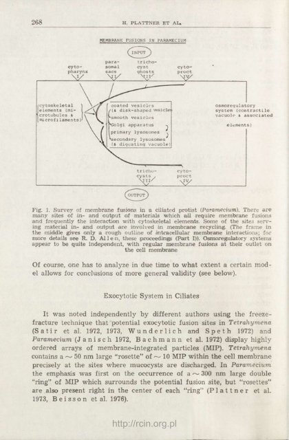
268 H. PLATTNER ET AL» cy topharynx V/ cytoskeletal elements (microtubules & microfi laments) MEMBRANE FUSIONS IN PARAMECIUM parasomal sacs trichocyst ghosts Mil/ coated vesicles {(&. disk-shaped vesicles smooth vesicles *Golgi apparatus ^ primary lysosomes ^ secondary lysosomes (& digesting vacuole) ^OUTPUT trichocys Ls , ' a 11/ cy toproct —\ cy toproct My osmoregulatory system (contractile vacuolo & associated elements) Fig. 1. Survey of membrane fusions in a ciliated protist (Paramecium). There are many sites of in- and output of materials which all require membrane fusions and frequently the interaction with cytoskeletal elements. Some of the sites serving material in- and output are involved in membrane recycling. (The frame in the middle gives only a rough outline of intracellular membrane interactions; for moore details see R. D. Al 1 e n, these proceedings (Part I)). Osmoregulatory systems appear to be quite independent, with regular membrane fusions at their outlet on the cell membrane Of course, one has to analyze in due time to what extent a certain model allows for conclusions of more general validity (see below). Exocytotic System in Ciliates It was noted independently by different authors using the freezefracture technique that potential exocytotic fusion sites in Tetrahymena (Satir et al. 1972, 1973, Wunderlich and Speth 1972) and Paramecium (J a n i s c h 1972, B a c h m a n n et al. 1972) display highly ordered arrays of membrane-integrated particles (MIP). Tetrahymena contains a ~ 50 nm large "rosette" of ~ 10 MIP within the cell membrane precisely at the sites where mucocysts are discharged. In Paramecium the emphasis was first on the occurrence of a~ 300 nm large double "ring" of MIP which surrounds the potential fusion site, but "rosettes" are also present right in the center of each "ring" (P 1 a 11 n e r et al. 1973, B ei sso n et al. 1976). http://rcin.org.pl
MEMBRANE FUSIONS IN PROTOZOA 269 It is difficult to derive a time sequence or — even more — any possible functional implications of static snap shots one gets from membranes by cryofixation and freeze-fracturing. This holds even more if any such attempt is done after previous chemical pretreatments (aldehyde fixation, antifreeze impregnation). In retrospect it appears, as summarized by Plattner (1981), that much of the controversies (see below) between different groups, working with protozoa, and also other groups, working with metazoan systems, came from such preparative difficulties. Both Tetrahymena and Paramecium display "rosettes" already in the resting stage, i.e., without triggering. After exocytosis triggering by a Ca 2+ -ionophore, "rosettes" are no longer present, whereas the "rings" persist and collapse (Plattner 1974). This indicates that only "rosette" but not "ring" structures are directly involved in exocytosis performance; the "rings" merely delineate the potential fusion zones without participating in the fusion process. Even stronger evidence for this comes from freeze-fracture work with mutant strains of Paramecium tetraurelia (Beisson et al. 1976). A brief survey is given in Figs. 2 and 3. Some mutations make no (tl) or grossly defective {ftA, tam 38) trichocysts which cannot be attached to the cell membrane. Other mutations (nd) allow the attachment but not the subsequent exocytotic discharge. Beisson et al. (1976) established that the discharge capacity is coupled with the presence of "rosettes". This has later been extended to further mutations (Beisson et al. 1980, Lefort-Tran et al. 1981). synthesis — packing transport attachment trigger } membrane fusion ) discharge membrane resealing membrane detachment trichless tam 8. tam 38, ftb A nd 9 (27 °C) 7S, K 401, kin 241 ( nd 6, nd 7 (all temperatures) I nd 9 {18 °C) = 'permissive' non-permissive Fig. 2. Sequence of events leading to exocytotic membrane fusion (left) and genetic dsturbances at different levels in various mutant strains of Paramecium tetraurelia. 7S is the wild type strain (K401, kin 241 are non-exocytotic mutations); for nd 9 see text. From Plattner et al. (1980) http://rcin.org.pl
- Page 43 and 44: PHYLOGENETIC RELATIONSHIPS AMONG PR
- Page 45 and 46: PROGRESS IN PROTOZOOLOGY Proceeding
- Page 47 and 48: THE TAXONOMIC POSITION OF EVGLENIDA
- Page 49 and 50: PROGRESS IN PROTOZOOLOGY Proceeding
- Page 51 and 52: 225 brate cycle in vitro. Further c
- Page 53 and 54: 227 could support the development o
- Page 55 and 56: 229 for the development of methods
- Page 57 and 58: IN VITRO CULTIVATION OF PARASITIC P
- Page 59 and 60: Malaria IN VITRO CULTIVATION OF PAR
- Page 61 and 62: IN VITRO CULTIVATION OF PARASITIC P
- Page 63 and 64: 237 Rai Choudhuri A. N.. Chowdhuri
- Page 65 and 66: PROGRESS IN PROTOZOOLOGY Proceeding
- Page 67 and 68: 241 Only those with 9-type 1 fronto
- Page 69 and 70: PROGRESS IN PROTOZOOLOGY Proceeding
- Page 71 and 72: THE MOLECULAR DIVERSITY OF TETRAHYM
- Page 73 and 74: 247 in eukaryotes. It is about the
- Page 75 and 76: Table 2 Mating species of Tetrahyme
- Page 77 and 78: THE MOLECULAR DIVERSITY OF TETRAHYM
- Page 79 and 80: THE MOLECULAR DIVERSITY OF TETRAHYM
- Page 81 and 82: THE MOLECULAR DIVERSITY OF TETRAHYM
- Page 83 and 84: Table 8 Percentage of DNA reanneali
- Page 85 and 86: 259 pattern; they do confirm our ju
- Page 87 and 88: THE MOLECULAR DIVERSITY OF TETRAHYM
- Page 89 and 90: Table 13 Two-dimensional electropho
- Page 91 and 92: 265 Cuny M., Milet M. and Hayes D.
- Page 93: PROGRESS IN PROTOZOOLOGY Proceeding
- Page 97 and 98: Table 1 Characterization of exocyto
- Page 99 and 100: Fig. 3. Freeze fractured trichocyst
- Page 101 and 102: * 1 ^MBBHBBHBBBBI jgf'- Fig. 5. Tan
- Page 103 and 104: 273 the preliminary designation "co
- Page 105 and 106: MEMBRANE FUSIONS IN PROTOZOA 275 ba
- Page 107 and 108: 277 B i 1 i n s k i M., Plattner H.
- Page 109 and 110: LIST OF SCIENTIFIC REPORTS presente
- Page 111 and 112: 281 structure and cytochemistry of
- Page 113 and 114: 283 LIST OF SCIENTIFIC REPORTS A. P
- Page 115 and 116: 285 LIST OF SCIENTIFIC REPORTS R. G
- Page 117 and 118: 287 LIST OF SCIENTIFIC REPORTS K. G
- Page 119 and 120: LIST OF SCIENTIFIC REPORTS 289 R. A
- Page 121 and 122: 291 LIST OF SCIENTIFIC REPORTS magn
- Page 123 and 124: 293 Co-Chairman: Jan Kućera, Czech
- Page 125 and 126: 295 LIST OF SCIENTIFIC REPORTS for
- Page 127 and 128: PROGRESS IN PROTOZOOLOGY Proceeding
- Page 129 and 130: INTERNATIONAL COLLABORATION AMONG P
- Page 131 and 132: INTERNATIONAL COLLABORATION AMONG P
- Page 133 and 134: INTERNATIONAL COLLABORATION AMONG P
- Page 135 and 136: INTERNATIONAL COLLABORATION AMONG P
- Page 137 and 138: INTERNATIONAL COLLABORATION AMONG P
- Page 139 and 140: http://rcin.org.pl
- Page 141 and 142: LONDON 1965 http://rcin.org.pl
- Page 143 and 144: LENINGRAD 1969 http://rcin.org.pl
268 H. PLATTNER ET AL»<br />
cy topharynx<br />
V/<br />
cytoskeletal<br />
elements (microtubules<br />
&<br />
microfi laments)<br />
MEMBRANE FUSIONS <strong>IN</strong> PARAMECIUM<br />
parasomal<br />
sacs<br />
trichocyst<br />
ghosts<br />
Mil/<br />
coated vesicles<br />
{(&. disk-shaped vesicles<br />
smooth vesicles<br />
*Golgi apparatus ^<br />
primary lysosomes ^<br />
secondary lysosomes<br />
(& digesting vacuole)<br />
^OUTPUT<br />
trichocys<br />
Ls ,<br />
' a 11/<br />
cy toproct<br />
—\<br />
cy toproct<br />
My<br />
osmoregulatory<br />
system (contractile<br />
vacuolo & associated<br />
elements)<br />
Fig. 1. Survey of membrane fusions in a ciliated protist (Paramecium). There are<br />
many sites of in- and output of materials which all require membrane fusions<br />
and frequently the interaction with cytoskeletal elements. Some of the sites serving<br />
material in- and output are involved in membrane recycling. (The frame in<br />
the middle gives only a rough outline of intracellular membrane interactions; for<br />
moore details see R. D. Al 1 e n, these proceedings (Part I)). Osmoregulatory systems<br />
appear to be quite independent, with regular membrane fusions at their outlet on<br />
the cell membrane<br />
Of course, one has to analyze in due time to what extent a certain model<br />
allows for conclusions of more general validity (see below).<br />
Exocytotic System in Ciliates<br />
It was noted independently by different authors using the freezefracture<br />
technique that potential exocytotic fusion sites in Tetrahymena<br />
(Satir et al. 1972, 1973, Wunderlich and Speth 1972) and<br />
Paramecium (J a n i s c h 1972, B a c h m a n n et al. 1972) display highly<br />
ordered arrays of membrane-integrated particles (MIP). Tetrahymena<br />
contains a ~ 50 nm large "rosette" of ~ 10 MIP within the cell membrane<br />
precisely at the sites where mucocysts are discharged. In Paramecium<br />
the emphasis was first on the occurrence of a~ 300 nm large double<br />
"ring" of MIP which surrounds the potential fusion site, but "rosettes"<br />
are also present right in the center of each "ring" (P 1 a 11 n e r et al.<br />
1973, B ei sso n et al. 1976).<br />
http://rcin.org.pl


