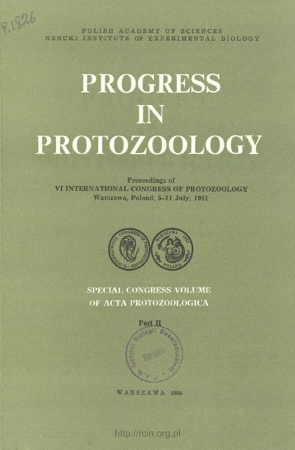
PROGRESS IN PROTOZOOLOGY
PROGRESS IN PROTOZOOLOGY
PROGRESS IN PROTOZOOLOGY

You also want an ePaper? Increase the reach of your titles
YUMPU automatically turns print PDFs into web optimized ePapers that Google loves.
POLISH ACADEMY OF SCIENCES<br />
NENCKI <strong>IN</strong>STITUTE OF EXPERIMENTAL BIOLOGY<br />
<strong>PROGRESS</strong><br />
<strong>IN</strong><br />
<strong>PROTOZOOLOGY</strong><br />
Proceedings of<br />
VI <strong>IN</strong>TERNATIONAL CONGRESS OF <strong>PROTOZOOLOGY</strong><br />
Warszawa, Poland, 5-11 July, 1981<br />
SPECIAL CONGRESS VOLUME<br />
OF ACTA PROTOZOOLOGICA<br />
Part U<br />
WARSZAWA 1984<br />
http://rcin.org.pl
POLISH ACADEMY OF SCIENCES<br />
NENCKI <strong>IN</strong>STITUTE OF EXPERIMENTAL BIOLOGY<br />
ACTA PROTOZOOLOGICA<br />
International Journal of Protozoology<br />
Editors<br />
Stanisław DRYL and Stanisław L. KAZUBSKI<br />
Stanisław DRYL<br />
Vassil GOLEMANSKY<br />
Witold KASPRZAK<br />
Stanisław L. KAZUBSKI<br />
Editorial Board<br />
Chairman: Leszek KUŹNICKI<br />
Vice-chairman: Andrzej GRĘBECKI<br />
Members<br />
Jiri LOM<br />
Georg Ivanovië POLJANSKY<br />
Igor Borysovic RAIKOV<br />
Ksenia Mironovna SUKHANOVA<br />
Managing Editor and Editorial Board Secretary<br />
Julitta PŁOSZAJ<br />
© COPYRIGHT BY PAŃSTWOWE WYDAWNICTWO NAUKOWE<br />
WARSZAWA 1984<br />
Printed in Poland<br />
ISBN 83-01-04786-0<br />
http://rcin.org.pl
<strong>PROGRESS</strong> <strong>IN</strong> <strong>PROTOZOOLOGY</strong><br />
Proceedings of<br />
VI <strong>IN</strong>TERNATIONAL CONGRESS OF <strong>PROTOZOOLOGY</strong><br />
Warszawa, Poland, 5-11 July 1981<br />
PART II<br />
Editors:<br />
S. DRYL, S. L. KAZUBSKI, L. KUZNICKI and J. PLOSZAJ<br />
http://rcin.org.pl
SIXTH <strong>IN</strong>TERNATIONAL CONGRESS OF <strong>PROTOZOOLOGY</strong><br />
5-11 JULY 1981, WARSZAWA, POLAND<br />
organized and sponsored by the Nencki Institute of Experimental Biology<br />
in Warsaw, with the assistance of the Committee on Cell Biology,<br />
Polish Academy of Sciences (Member of ECBO) and the Protozoological<br />
Section of the Polish Zoological Society on behalf of the International<br />
Commission of Protozoology<br />
OFFICERS<br />
OF THE ORGANIZ<strong>IN</strong>G COMMITTEE<br />
OF THE CONGRESS<br />
President<br />
S. DRYL, Warszawa<br />
Vi cep resident and Chairman of Scientific<br />
Sessions<br />
L. KUŻNICKI, Warszawa<br />
Vicepresidents<br />
A. CZAPIK, Kraków<br />
A. GRĘBECKI, Warszawa<br />
M. JERKA-DZIADOSZ, Warszawa<br />
W. KASPRZAK, Poznań<br />
Secretary General<br />
S. L. KAZUBSKI, Warszawa<br />
Executive Secretary<br />
E. WYROBA, Warszawa<br />
Assistant Secretary General '<br />
H. REBANDEL, Warszawa<br />
Treasurers<br />
B. SKOCZYLAS, Warszawa<br />
I. WITA, Warszawa<br />
Editors of Proceedings<br />
S. DRYL<br />
S. L. KAZUBSKI<br />
J. PŁOSZAJ<br />
http://rcin.org.pl<br />
<strong>IN</strong>TERNATIONAL COMMISSION<br />
OF <strong>PROTOZOOLOGY</strong><br />
S. DRYL, Warszawa, Poland<br />
P. C. C. GARNHAM, Ascot, England<br />
K. G. GRELL, Tübingen, G.F.R<br />
B. M. HONIGBERG, Amherst, U.SA.<br />
S. <strong>IN</strong>OKI, Osaka, Japan<br />
J. JAD<strong>IN</strong>, Antwerpen, Belgium<br />
S. L. KAZUBSKI, Warszawa, Poland<br />
L. KUZNICKI, Warszawa, Poland<br />
J. LEE, New York, U.SA.<br />
J. LOM, Prague, Czechoslovakia<br />
R. B. McGHEE, Athens, U.S.A.<br />
B. A. NEWTON, Cambridge, England<br />
J. R. NILSSON, Copenhagen, Denmark<br />
R. NOBILI, Pisa, Italy<br />
I. J. POLJANSKY, Leningrad, U.S.S.R.<br />
P. de PUYTORAC, Aubiere, France<br />
I. B. RAIKOV, Leningrad, U.S.S.R.<br />
B. R. SESHACHAR, Bangalore, India<br />
D. T. SPIRA, Jerusalem, Israel<br />
J. H. TERAS, Tallin, U.S.S.R.<br />
W. TRAGER, New York, U.S.A.<br />
E. VIVIER, Villeneuve, d'Ascq., France
<strong>IN</strong>TRODUCTORY REMARKS<br />
This Part II of the Post-Congress Volume "Progress in Protozoology"<br />
of Vlth International Congress of Protozoology (5-11 July, 1981,<br />
Warsaw, Poland) appears approximately more than one year later than<br />
Part I. The main reason for publishing it as two separate parts was<br />
a long delay in supplying us with the manuscripts containing the chairpersons<br />
summaries of Round-Table Discussion on "Phylogenetic Relationships<br />
among Protozoa" and of Symposia which were supplemented<br />
with "List of Scientific Reports Presented at Symposia, Contributed<br />
Paper and Poster Sessions". Part II of "Progress in Protozoology" contains<br />
those summaries which were received by Editors before March<br />
1983; included is also an additional chapter entitled: "International Collaboration<br />
Among Protozoologists During the Years 1961 to 1981 (by<br />
L. Kuznicki and B. M. Honigberg), illustrated with numerous photographs<br />
carried out during Congresses. %<br />
I must state with great regret that not all expected summaries arrived<br />
and in consequence the Editors decided in these cases to publish<br />
only short notes about the subject of meeting, supplying only information<br />
about names of the chairpersons and speakers, who took part in the<br />
discussion.<br />
http://rcin.org.pl<br />
Stanislaw DRYL<br />
President, Vlth International<br />
Congress of Protozoology
http://rcin.org.pl
<strong>PROGRESS</strong> <strong>IN</strong> <strong>PROTOZOOLOGY</strong><br />
Proceedings of VI International Congress of Protozoology<br />
Special Congress Volume of ACTA PROTOZOOLOGICA<br />
Part II, pp. 181-218, 1984<br />
Phylogenetic Relationships among Protozoa<br />
B. M. HONIGBERG<br />
(Chairman and Rapporteur)<br />
Round Table Discussion<br />
Department of Zoology, University of Massachusetts, Amherst, MA 01003, USA<br />
The "Round-Table Discussion" which occupied the entire morning<br />
of 15 July, 1981, the last day of the Sixth International Congress of<br />
Protozoology, involved a large panel of experts and attracted a very<br />
large audience. The primary interests of some of the members of the<br />
audience were far removed from the subjects under consideration, yet<br />
they seemed to be sufficiently attracted by what was being said that they<br />
remained in the lecture hall for a long time. Judging from the amount<br />
of discussion among the members of the panel and from the expressions<br />
of disappointment from many members of the audience who, because of<br />
time limitations, were unable actively to participate in the discussion,<br />
it is apparent that similar meetings ought to be planned for the future<br />
Congresses. More time, perhaps even an entire day, should be set aside<br />
for the consideration of the Phylogeny of Protozoa. In light of the lively<br />
discussion that took place during the "Round-Table" conference and in<br />
that of the conclusions reached at the meeting, it seems that the publication<br />
of "The Newly Revised Classification of the Protozoa" (L e v i n e<br />
et al. 1980) might have been somewhat premature. With regard to future<br />
schemes, a consensus can best be achieved after much discussion at<br />
national and international meetings and after an active exchange of<br />
communications among experts from various lands. Such constant exchanges<br />
should, I feel, be initiated and encouraged by the Chairman<br />
and Members of the standing Committee on Systematics and Evolution<br />
of the Society of Protozoologists.<br />
Session took place on July 11, 1981 at VI International Congress of Protozoology,<br />
Warsaw, Poland, 5-11 July 1981.<br />
http://rcin.org.pl
182 B. M. HONIGBERG<br />
In presiding over the deliberations of the panel of experts, the<br />
Chairman was aided by three Honorary Co-Chairmen, Professors<br />
P.C.C. Garnham (U.K.), W. Michajlow (Poland), and G.I. Poljansky<br />
(U.S.S.R.).<br />
Each higher taxon, depending upon the group being discussed,<br />
a PHYLUM, SUBPHYLUM, or SUPERCLASS as defined in the most<br />
recent scheme published by the Society of Protozoologists (L e v i n e<br />
et al. 1980), was commented upon briefly by one or two speakers and<br />
several officially appointed and voluntary discussants from among the<br />
members of the panel. Taxa down to, and including SUBORDERS were<br />
those primarily considered. In addition, the final part of the Discussion<br />
dealt with the question as to whether the Protozoa constitute a "Natural<br />
Subkingdom."<br />
Not all taxa were discussed in equal depth, either because not equal<br />
bodies of information are available about all of them, or because some<br />
of the originally invited speakers and discussants were unable to attend<br />
the Congress.<br />
Some speakers submitted complete bibliographies with their contributions;<br />
others did not. Because of the great differences among the presentations,<br />
no attempt can or will be made to keep the style uniform<br />
throughout this report.<br />
Subphylum MASTIGOPHORA Diesing, 1866<br />
Speakers Discussants<br />
Prof. J. Mignot (France) Prof. B. M. Honigberg (U.S.A.)<br />
Prof. K. Vickerman (U.K.) Prof. W. Michajlow (Poland)<br />
Prof. F. J. R. Taylor (Canada)<br />
Class 1. PHYTOMASTIGOPHOREA Calkins, 1909<br />
On the basis of the ideas expressed by numerous investigators (actual<br />
references not submitted), including B. B. B o u c k, P. Bourrely,<br />
G. Brugerolle, J. D. Dodge, H. E 111, P. Gayral and collaborators,<br />
S. Gibbs, J. C. Green, B. F o d t, D. H i b b e r t, G. F.<br />
Lee dale, A. R. Loeblich, III, I. M a n t o n, Q. M o e s t r u p, J. D.<br />
Pickett-Heaps, F. Schnepf, F. J. R. Taylor, Van Valkenb<br />
u r g, and Wujek, Prof. MIGNOT proposed certain important modifications<br />
of the class PHYTOMASTIGOPHOREA as recommended by<br />
Le vine's Committee (1980). Although in many instances the taxa are<br />
the same as those included in L e v i n e et al. (1980), the diagnoses differ,<br />
reflecting the approach of Prof. Mignot to the classification.<br />
http://rcin.org.pl
183<br />
In his abstract Mignot (1981) stated that in the recent classification<br />
of the Society of Protozoologists (L e v i n e et al. 1980) the<br />
entire assemblage of PHYTOMASTIGOPHOREA occupies about one<br />
page, of which nearly one-half is devoted to EUGLENIDA and only<br />
a few lines to D<strong>IN</strong>OFLAGELLIDA. It seems, therefore, that there is<br />
an inbalanee in treatment of the class with regard to space devoted to<br />
the several orders; the degree of attention centered upon certain assemblages<br />
is disproportionate to the diversity and numbers of species<br />
they contain. Other workers present at the Congress and in subsequent<br />
correspondence leveled similar critiques against the Society's scheme.<br />
Prof. Mignot thought the inbalanee in the space and attention devoted<br />
to the ordinal taxa of MASTIGOPHORA was even greater with<br />
regard to PHYTOMASTIGOPHOREA vs ZOOMASTIGOPHOREA. Actually,<br />
many participants of the Panel, and of the Congress in general,<br />
felt that there was no justification in separation of the flagellate protozoa<br />
into the two classes, and this view will be discussed later in this<br />
report.<br />
Another objection of Prof. Mignot (and of some of the other<br />
members of the Panel) was 'the omission of some or many structural<br />
and physiological characteristics from the diagnoses of the phytomastigophorean<br />
taxa included in the Society's scheme. Among the morphological<br />
attributes, he would include the fine structure of the cytoskeleton,<br />
and among the physiological ones, the chemical composition of<br />
the pigments. All such characters could provide useful criteria for<br />
evolutionary considerations.<br />
Much work remains to be done, and meaningful results cannot be<br />
achieved without a close collaboration between phycologists and proctologists<br />
which has not been realized to date. To provide a basis for<br />
a discussion, Prof. Mignot proposed the following scheme for PHY-<br />
TOMASTIGOPHOREA. The class was to be divided into 11 orders:<br />
1. CHRYSOMONADIDA, 2. PRYMNESIIDA, 3. SILICOFLAGELLIDA,<br />
4. HETEROCHLORIDA, 5. CHLOROMONADIDA, 6. CRYPTOMONA-<br />
DIDA, 7. D<strong>IN</strong>OFLAGELLIDA, 8. SYND<strong>IN</strong>IIDA, 9. EUGLENIDA, 10.<br />
PRAS<strong>IN</strong>OMONADIDA, 11. VOLVOCIDA. In turn, the first five orders<br />
could be brought together in one class, CHROMOMASTIGOPHOREA<br />
Mignot, 1981, or superorder, CHROMOMASTIGOPHORIDEA Mignot,<br />
1981. If this course were followed, CRYPTOMONADIDA, D<strong>IN</strong>OFLA-<br />
GELLIDA, SYND<strong>IN</strong>IIDA, and EUGLENIDA would be elevated to the<br />
rank of classes or subclasses. The basis for this elevation is to be found<br />
in the structural complexity of the representatives of the assemblages<br />
which cannot be considered primitive and which differ profoundly<br />
from one another in many attributes. The last class, CHLOROMASTI-<br />
GOPHOREA Mignot, 1981, with clear plant affinities, would include<br />
http://rcin.org.pl
184<br />
PRAS<strong>IN</strong>OMONADIDA and VOLVOCIDA. In addition, Prof. Mignot<br />
(1981) proposed the division of PRYMNESIIDA into three suborders:<br />
PRYMNES<strong>IN</strong><strong>IN</strong>A, PAVLOVAN<strong>IN</strong>A, ISOCHRYS<strong>IN</strong><strong>IN</strong>A. The order DI-<br />
NOFLAGELLIDA was divided into as many as 15 suborders, which<br />
evidently should be discussed in detail before they are accepted; in<br />
fact, the author provided no diagnoses for these taxa, referring merely<br />
to the authorities of each of them. As far as the class EUGLENOMAS-<br />
TIGOPHOREA (or super-order EUGLENOMASTIGOPHORIDEA) is concerned,<br />
it was to include a single order EUGLENIDA Biitschli, 1884<br />
and four suborders: EUTREPTI<strong>IN</strong>A Leedale, 1967; EUGLEN<strong>IN</strong>A Biitschli,<br />
1884; RHABDOMONAD<strong>IN</strong>A Leedale, 1967; and TERONEMAT<strong>IN</strong>A<br />
Leedale, 1967. It should be noted that Prof. Mignot found unacceptable<br />
the separation between EUTREPTI<strong>IN</strong>A from EUGLENAMORPH<strong>IN</strong>A<br />
Leedale, 1967 and RHABDOMONAD<strong>IN</strong>A from SPHENOMONAD<strong>IN</strong>A<br />
Leedale, 1967.<br />
A summary such as this is not an appropriate place to publish<br />
a new classification scheme. Yet to show the differences between the<br />
kind of diagnosis proposed in many instances by Prof. Mignot and<br />
that employed by the Committee (L e v i n e et al. 1980), I shall cite<br />
below the diagnoses of CHRYSOMONADIDA given by him and by the<br />
Committee.<br />
Committee of the Society of Protozoologists (Levine<br />
et al. 1980), CHRYSOMONADIDA Engler, 1898. Two unequal<br />
flagella, one directed anteriorly and bearing 2 opposite rows of mastigonemes,<br />
other trailing and smooth; chloroplast golden-brown or absent;<br />
storage products chrysolaminarin and fat; cell naked, with richly<br />
patterned silicified scales, or with lorica; sexual reproduction present.<br />
Dinobryon; Ochromonast Synura<br />
Mignot 1981 (presentation at the Congress), CHRYSOMONADIDA<br />
Engler, 1898 emend. Hibberd, 1976. — Two flagella, one directed<br />
anteriorly, bearing 2 opposite rows of tubular complex mastigonemes;<br />
zone of transition helicoidal; flagellum short, inflated at its base; cytoskeleton<br />
consisting mainly of 2 microtubular cortical systems, 1 formed<br />
by microtubules radiating from a fibrillar MTOC, situated at the dorsal<br />
edge, and by a rhizoplast associated with the nucleus; yellow-brown<br />
chloroplasts contain chlorophylls a and c, (3-carotene, lutein, fucoxanthin,<br />
didinoxanthin, violaxanthin; plastid lamellae formed by 3 thylokoids<br />
of which the peripheral ones are circular; stigma in plastid or<br />
leucoplast; single dictyosome surrounding nucleus; polysaccharide reserve<br />
in vacuoles is chrysolaminarin; cells naked or covered with silicified<br />
scales of specific complex structure; or in lorica of organic material<br />
(cellulose?); formation of scales in intracellular silicalemma originating<br />
in Golgi complex associated with endoplasmic reticulum, fre-<br />
http://rcin.org.pl
PHYLOGENETIC RELATIONSHIPS AMONG PROTOZOA 185<br />
quently periplastidial; encystment by endogenous formation of silicified<br />
cyst wall with specific structure, pore, and aperture; very marked<br />
polymorphism with development of microfibrillar part of cytoskeleton<br />
into rhizopodial forms.<br />
Ochromonas, Dinobryon, Synura<br />
It should be added here that in the diagnosis of the class or superorder<br />
CHROMOMASTIGOPHOREA (IDEA) Prof. Mignot specified<br />
also that the mitochondria have ampouliform (inflated) or tubular<br />
cristae.<br />
Opinion has been expressed by several members of the Panel that<br />
the foregoing, and some of the other diagnoses given by Prof. M i gn<br />
o t tend to be too long and detailed. It cannot be denied, however,<br />
that they are very complete.<br />
There were certain praises as well as criticisms voiced during the<br />
session with regard to the Society's scheme of classification (L e v i n e<br />
et al. 1980) as well as with regard to that of Prof. Mignot. It was<br />
pointed out by Prof. Taylor that the Mignot scheme represents<br />
a beginning toward recognition of natural relationships among the<br />
flagellates. He considered CHROMOMASTIGOPHOREA as a good, natural<br />
grouping. On the other hand, he felt that separation of VOLVO-<br />
CIDA from their very close relatives Chlorococcales was unnatural.<br />
Prof. Taylor not only further emphasized the need for a closer collaboration<br />
between phycologists and protozoologists, but felt that such<br />
a collaboration must include also the mycologists.<br />
Class 2. ZOOMASTIGOPHOREA Calkins, 1909<br />
As far as the separation of PHYTOMASTIGOPHOREA and ZOO-<br />
MASTIGOPHOREA is concerned, several members of the Panel, including<br />
Prof. Taylor, felt that it is unnatural and should be eliminated.<br />
The proposed elimination of these two classes while undoubtedly<br />
sound — it would not separate what appear to be related assemblages<br />
at a class level — must be the subject of further discussion. It has been<br />
suggested, for example, that a new scheme should involve elevating<br />
some groups from the taxonomic levels which they now occupy. Prof.<br />
Taylor went so far as to suggest that the dinoflagellates, with<br />
a diversity and distinctness resembling that of CILIOPHORA, might<br />
actually deserve the rank of a subphylum. A number of questions will<br />
have to be answered, as the result of ongoing discussions, before we<br />
can make major changes in the admittedly artificial class ZOOMASTI-<br />
GOPHOREA. Among these are the following: 1. Are there to be no<br />
http://rcin.org.pl
12 B. M. HONIGBERG<br />
superorders among the nonpigmented flagellates, save PARABASALI-<br />
DEA? 2. Is there enough evidence in support of the kinships suggested<br />
by Brugerolle (1977) for RETORTAMONADIDA, DIPLOMONA-<br />
DIDA, and OXYMONADIDA to allow the establishment of superorder<br />
(class?) for these orders? 3. Where actually do CHOANOFLAGELLIDA<br />
and BICOSOECIDA belong in the general classification scheme; could<br />
they be placed in a higher taxon? 4. Is there any justification for<br />
dividing the nonpigmented flagellates into the somewhat informal superordinal<br />
groupings recommended by Grasse (1952)?<br />
Although for convenience's sake (we still have difficulty with avoiding<br />
this pragmatic concept), we shall discuss here the orders of ZOOMASTI-<br />
GOPHOREA, as was done by Prof. VICKERMAN, we ought not to lose<br />
sight of the need for reconsidering the validity of this class as well<br />
as of PHYTOMASTIGOPHOREA. As I suggested during the Round-<br />
Table Discussion, these taxa may well be soon put to rest.<br />
Professor Vickerman concentrated on the changes in the orders<br />
of ZOOMASTIGOPHOREA between the first (H o n i g b e r g et al.<br />
1964) and second (L e v i n e et al. 1980) Systems of Classification published<br />
by the Society of Protozoologists. He noted that these changes<br />
were more noticeable among the "lower" mostly free-living nonpigmented<br />
flagellates than among the structurally more complex and almost<br />
exclusively parasitic forms. He felt that the problems of classification<br />
and evolution of K<strong>IN</strong>ETOPLASTIDA Honigberg, 1963, DIPLOMONA-<br />
DIDA Wenyon, 1926 emend. Brugerolle, 1975, and TRICHOMONADIDA<br />
Kirby, 1947, emend. Honigberg, in Camp, Mattern and Honigberg, 1974,<br />
which were discussed at the Fourth International Congress of Parasitology<br />
in Warsaw, 1979 (Honigberg et al. 1982), could be left out of<br />
the present consideration. He also did not discuss RETORTAMONADIDA,<br />
analyzed in some detail by Kulda and Nohynkova (1978), and<br />
PROTEROMONADIDA Grasse, 1952 emend. Vickerman, 1976, which he<br />
as well as Kulda and Nohymkova (1978) considered as quite<br />
separate from K<strong>IN</strong>ETOPLASTIDA. Professor Vickerman felt, however,<br />
that more ultrastructural details are needed to enable us to obtain<br />
an understanding (of OXYMONADIDA and HYPERMASTIGIDA) comparable<br />
to that we have now of kinetoplastids, proteromonads, retortamonads,<br />
diplomonads, and trichomonads. It is a pity that Dr. Brugerolle<br />
who did much work on the fine structure of the last two<br />
groups could not be present, for he could have discussed the relationships<br />
among RETORT AMONADIDA, DIPLOMONADIDA, and OXYMO-<br />
NADIDA as suggested in his 1977 publication.<br />
Among the problems discussed by Prof. Vickerman was that<br />
of CHOANOFLAGELLIDA. These organisms, with a single flagellum and<br />
a collar made up of a ring of tentacles, were at one time considered as<br />
http://rcin.org.pl
187<br />
algae by phycologists (Bourelly 1968, Christen sen 1962, 1966).<br />
Subsequently, however, on the basis of electron-microscopic studies<br />
(H i b b e r d 1975, Leadbetter 1972, Leadbetter and M a n t o w<br />
1974), they were removed from among the plants and by implication<br />
from PHYTOMASTIGOPHOREA. Although the choanoflagellates share<br />
certain structural characteristics with the choanocytes of sponges and<br />
of other metazoa (Norrevang and Wingstrand 1970), their<br />
retention in a separate protozoan order appears entirely justified. No one<br />
took issue with this assertion.<br />
The question of BICOSOECIDA Grasse et Deflandre, 1952, appears<br />
more complex. Hall (1953), among others, placed these organisms<br />
among the chrysomonads, and more recently Mi g not (1974) assigned<br />
Bicoeca to this assemblage of pigmented flagellates. He thought that this<br />
group of unpigmented heterokont, lorica-dwelling flagellates might be<br />
appended to the Chry sophy ceae, but, according to Vickerman,<br />
a number of features sets Bicoeca apart from these algae. Among the<br />
structures in question is the long tongue-like appendage (languette)<br />
which arises alongside the two flagella and is supported by a row of<br />
microtubules. Although the languette is somewhat reminiscent of the<br />
heptonema found in Prymnesiophyceae, in Bicoeca it is involved in food<br />
capture. Although the mastigonemes on the anterior coiling flagellum<br />
of BICOSOECIDA are tubular and similar to those found in CHRYSO-<br />
MONADIDA, the anchoring recurrent flagellum, having a fibrillar sheet<br />
and attachment cone is distinctive. It must be also remembered that the<br />
carbohydrate storage product of the bicosoecids is glycogen and not leucosin.<br />
In a subsequent personal communication to me, Prof. Vickerman<br />
stated with regard to BICOSOECIDA: "I do not think they should<br />
be in a separate order if Silicoflagellida (in Honigberg et al. 1964;<br />
Levine et al. 1980) and Ebriida (in Honigberg et al. 1964) and<br />
other chrysomo.nad appendages should. There are certainly enough ultrastructural<br />
features to set them apart."<br />
The unnatural and clearly polyphylectic nature of the order RHIZO-<br />
MASTIGIDA Doflein, 1916 was pointed out by the committee of the<br />
Society of Protozoologists which was responsible for the 1964 classification<br />
scheme. At that time Honigberg and B a 1 a m u t h stated in<br />
a footnote: "The presence of simple mastigonts in trophic stages is the<br />
basis for alignment with Mastigophora rather than with Sarcodina,<br />
while at the same time the concomitant occurrence of flagella and pseudopodia<br />
strikingly illustrates the basic affinities .of generalized flagellates<br />
and amebae (as originally suggested by Pascher 1918). The tendency<br />
for secondary reduction of flagella is observed in numerous representatives<br />
(of the order). The polyphylectic nature of the group is indicated<br />
by the presence of at least two distinct kinds of pseudopodial organiza-<br />
http://rcin.org.pl
188 B. M. HONIGBERG<br />
tion among different members. One pattern (exemplified by Naegleria)<br />
is characterized by the lobose pseudopodia encountered in amebae<br />
of the subclass Lobosia. Another pattern (exemplified by Dimorpha<br />
and other helioflagellates) is marked by typical axopodia terminating<br />
internally in a central granule or centroplast, as in heliozoa<br />
of the order Centrohelida. Future studies can be expected to lead to<br />
a more natural regrouping of the forms placed here for convenience.""<br />
As might have been expected, the order RHIZOMASTIGIDA was omitted<br />
from the more recent scheme published by L e v i n e et al. (1980). Actually,<br />
the constitution of this "order" has varied among the standard<br />
protozoology texts; e.g., Hall (1953) included the following genera<br />
among RHIZOMASTIGIDA: Histomonas, Heliobodo, Mastigamoeba, Mastigella,<br />
Mastigina and Rhizomastix. Amon other workers, he felt also<br />
that Pteridomonas was a chrysomonad and that Actinomonas and Dimorpha<br />
had helioflagellate affinities. Grasse (1952) thought that Pteridomonas<br />
was also a helioflagellate. Many authors (e.g., Kudo 1966)<br />
included Tetramitus and Naegleria in RHIZOMASTIGIDA. Certain protozoologists<br />
assigned at least some of the rhizomastigid genera to many<br />
flagellate and rhizopod assemblages. Vickerman, who until recently<br />
(Vickerman 1976) considered Cercomonas and Heteromita as BO-<br />
DON<strong>IN</strong>A incertae sedis, included these two genera in his discussion of<br />
zooflagellates at the 1981 session in Warsaw. According to him (V i ck<br />
e r m a n, personal communication): Cercomonas and Heteromita are<br />
clearly related to each other and sufficiently distinct from both chrysomonads<br />
and sarcodines to merit a separate small order. Accordingly,<br />
using the characteristics presented by him at the 1981 Congress, I propose<br />
to create, with Vickerman as the sole authority, a new<br />
order among MASTIGOPHORA.<br />
Order CERCOMONADIDA Vickerman<br />
Two heterodynamic acronematic flagella whose kinetosomes cap cone<br />
of microtubules closely enveloping drawn out anterior part of nucleus;<br />
cone apparently attached to nucleus; microtubules, some lying along<br />
nucleus, not all subpellicular (as in Kinetoplastida)-, single, membranebounded,<br />
postnuclear organelle of unknown function loosely capping<br />
posterior part of nucleus; extrusomes resembling haptocysts of heliozoans<br />
often associated with surface microtubule tracks; several mitochondria<br />
with tubular cristae; food ingestion by pseudopodia; contractile vacuole<br />
usually postnuclear, may be adbasal (as in Kinetoplastida).<br />
Cercomonas, Heteromita<br />
Some of the former members of RHIZOMASTIGIDA can be now<br />
assigned to known flagellate and rhizopod taxa, and this is reflected in<br />
http://rcin.org.pl
PHYLOGENETIC RELATIONSHIPS AMONG PROTOZOA 189<br />
the 1980 classification of Levine's Committee. Thus Histomonas and<br />
Dientamoeba (see Honigberg and Bennett 1971, Honigberg<br />
and Kuldova 1969, Honigberg in Camp et al. 1974) are considered<br />
as modified trichomonad genera (order TRICHOMONADIDA);<br />
Tetramitus and Naegleria have been included in the rhizopod order<br />
SCHIZOPYRENIDA Singh, 1952.<br />
According to V i c k e r m a n, in certain respects Mastigamoeba resembles<br />
the cercomonads. It possesses the prenuclear cone of microtubules,<br />
pseudopodia, and similar mitochondria, but it lacks the postnuclear<br />
organelle and the extrusomes; furthermore, there is no recurrent<br />
flagellum but only a barren kinetosome corresponding to the basal body<br />
of this flagellum. Evidently, Mastigamoeba has structural resemblance<br />
to the myxamebae of some EUMYCETOZOEA. In a personal communication<br />
to me, Vickerman wrote: "Mastigamoeba bears such a close<br />
superficial resemblance to slime-mold flagellates that I cultured one of<br />
the latter believing it to be Mastigamoeba for several years (as cyst,<br />
amoeba and flagellate) — till one day it produced beautiful plasmodia."<br />
Vickerman wondered if, indeed, the affinities of mastigamoebae lie with<br />
mycetozoan sarcodines rather than with any known flagellate group.<br />
He stated, however, in the course of the discussion at the Warsaw Congress<br />
that a post-nuclear organelle was found in Rhipidodendron and<br />
Spongomonas, both sedentary colorless flagellates with homodynamic<br />
flagella (H i b b e r d 1976). "Is it possible (he asked) that heterodynamism<br />
of the two flagella has been lost as a result of adoption of the<br />
sedentary habits?"<br />
With regard to another genus, Mastigella, which used to be included<br />
in RHIZOMASTIGIDA, Vickerman stated that two species of the<br />
genus, Mastigella vitraea and Mastigella hylae, appear quite different<br />
from the species of Mastigamoeba. Since nothing is known about the<br />
fine structure of Mastigella vitraea, its assignment to any existing or to<br />
a new taxon is not possible at this time. On the other hand, it is known<br />
that Mastigella hylae is multinucleate, lacks mitochondria, and has cones<br />
of microtubules associated with its flagellar kinetosomes. According to<br />
unpublished studies of B r u g e r o 11 e, there are several noncontractile<br />
flagella, each associated with one of the nuclei. In view of this, V i ck<br />
e r m a n believes that Mastigella is a "higher" zooflagellate, but appears<br />
to have no obvious affinities to any particular group. Perhaps,, after<br />
more is known about other members of this genus, a new order might<br />
have to be established for them.<br />
Since nothing is known about the fine-structural details of many<br />
of the other former members of the order RHIZOMASTIGIDA, e.g.,<br />
Heliobodo, Actinomonas, Dimorpha and Pteridomonas, neither V i ck<br />
e r m a n nor I is ready to place them in any known protozoan as-<br />
http://rcin.org.pl
16 B. M. HONIGBERG<br />
semblage or establish new ones for these genera. Thus there still remains<br />
a group of former rhizomastigotes which will have to be considered for<br />
some time as species incertae sedis.<br />
It has been pointed out by Vickerman that one of the problems<br />
in placing "lower" zooflagellates in an order is how minimal a definition<br />
of the order is acceptable. A good example of this problem quoted by<br />
him is the assignment by Hollande (1980) of Perkinsiella, the endobiont<br />
of the parasitic amebae Paramoeba and Janickina to K<strong>IN</strong>ETO-<br />
PLASTIDA. Perkinsiella has, indeed, a single mitochondrion rich in<br />
DNA fibrils which, however, are isotropic in their arrangement. On the<br />
basis of the available data, Vickerman concluded: "In the absence<br />
of any sign of a flagellar apparatus or pellicular microtubules and of<br />
information on nuclear division, the writer would hesitate to assign this<br />
organism to K<strong>IN</strong>ETOPLASTIDA, though it is possible that adaptation<br />
of Perkinsiella to existence as an organelle of its amoeba host may have<br />
robbed it of most of its kinetoplastid characteristics." Although Hollande<br />
(1980) claimed that: "... les microtubules situés à la périphérie<br />
du cinétoplaste pourraient être homologues de ceux qui, dans une cellule<br />
de Cryptobia sont sous-jacents à la pellicule;..." one would tend to<br />
concur with Vickerman that the evidence for the presence of subpellicular<br />
microtubules is not compelling. In general, although an admirer<br />
of Prof. Hollande's cytologic researches, I cannot help but feel<br />
that assignments of flagellates or any other protozoa to given taxa<br />
ought to be based on stronger evidence than that provided by him for<br />
Perkinsiella.<br />
It is evident from the foregoing discussion that although much is<br />
known about both the pigmented and nonpigmented flagellate groups,<br />
which constitute the basic assemblages of the greatest importance to<br />
evolution of eukaryotes, there remain many important problems still to<br />
be solved. Clearly the solution of some of these problems will depend<br />
upon mutual understanding and effective collaboration among protozoologists,<br />
phycologists, and mycologists.<br />
Appendix to MASTIGOPHORA<br />
Prof. W. Michajlow presented his views on the taxonomy and<br />
taxonomic criteria that ought to be employed for the group of parasitic<br />
euglenoids "Euglenida parasitica," which he has been studying for many<br />
years. Since we are concerned in the present discussion primarily with<br />
higher taxa, we cannot consider details of taxa below suborders. It suffices<br />
to say that in classifying the parasitic euglenids, Michajlow wishes<br />
to employ the following criteria: 1. structure of parasitic and free-living<br />
forms (present in the life cycle); 2. details of the developmental cycles;<br />
http://rcin.org.pl
PHYLOGENETIC RELATIONSHIPS AMONG PROTOZOA 191<br />
3. the type of movement characteristic of the parasitic and free-living<br />
forms; 4. host specificity based upon statistical data; 5. geographic distribution.<br />
The editors of the Post-Congress Publication may wish to include<br />
the entire presentation in an Appendix to the present discussion (see<br />
p. 219).<br />
Superclass RHIZOPODA von Siebold, 1845<br />
Speaker<br />
Dr. T. K. Sawyer (U.S.A.)<br />
Dr. Sawyer did not transmit to us the full text of his remarks<br />
and there was relatively little discussion of the very important superclass<br />
RHIZOPODA, probably because several of the experts concerned<br />
with this group were unable to attend the Congress. Consequently, the<br />
reader is referred to the abstract published by Dr. Sawyer in Progress<br />
in Protozoology, the Congress Proceedings, which were<br />
distributed among the participants at the registration desk in Warsaw.<br />
The only person who made extensive remarks in connection with<br />
RHIZOPODA was Prof. J. B. J a d i n. His most interesting presentation<br />
was entitled "Relation entre protozoaires, bactéries et virus."<br />
Superclass ACT<strong>IN</strong>OPODA Calkins, 1909<br />
Discussants<br />
Prof. J. B. Jadin (Belgium)-<br />
Prof. J. J. Lee (U.S.A.)<br />
Speaker Discussants<br />
Prof. C. Bardele (Fed. Prof. J. J. Lee (U.S.A.)<br />
Rep. Germany) Dr. D. J. Patterson (U.K.><br />
Prof. Bardele expressed his full agreement with the views of the<br />
absent Dr. Merinfeld (see Merinfeld 1981) that ACT<strong>IN</strong>OPODA<br />
do not represent a natural assemblage, but rather have to be regarded<br />
as an artificial taxon. According to Prof. Bardele, only those species<br />
presently available for cytologic analysis by modern methods should be<br />
employed in all future considerations of the organisms now included<br />
among ACT<strong>IN</strong>OPODA. The species that have not been located and examined<br />
since the turn of the century ought to be excluded from such<br />
considerations.<br />
The speaker listed a series of characteristics he considers useful in<br />
the analysis of phylogenetic relationships among the actinopods: (a) asexual<br />
reproduction — binary or multiple fission; (b) location of the division<br />
spindle and the type of polar bodies present during mitosis; (c) sex-<br />
http://rcin.org.pl
192 B. M. HONIGBERG<br />
ual reproduction — present or absent; (d) flagellated stages — present or<br />
absent; (e) flagellum naked or with mastigonemes; (f) uni-us multinucleate<br />
condition; (g) pattern of axonemal microtubules — absent, triangular,<br />
hexagonal, hexagonal and triangular, double spiralled, dodecahedral with<br />
or without peripheral spirals; (h) MIOC associated with a nuclear envelope,<br />
an axoplast, or a centroplast with a tripartite disc; (i) motility<br />
mechanisms; (j) type of extrusomes, muciferous bodies, or kinetocysts;<br />
(k) mitochondria entering or not entering the axopodia; (1) skeleton consisting<br />
of one to many pieces; (m) skeleton — intra- or extracellular;<br />
(n) material of which the skeleton is made — organic, silica, strontium<br />
sulfate, calcium carbonate; (o) capsular membrane location — intra- vs<br />
extracellular; (p) capsular membrane — material of which it is made<br />
and symmetry; (q) life cycle with or without metamorphosis; (r) habitat<br />
— marine, brackish, fresh-water; (s) mode of life — planctonic, benthic,<br />
attached to substrate (stalked).<br />
If the aforementioned attributes, a part of which can be observed<br />
only by electron microscopy, are considered, it becomes evident that<br />
neither ACT<strong>IN</strong>OPODA nor the various sub-groups of this superclass,<br />
e.g., the former RADIOLARIA Müller 1858, as listed in Honigberg<br />
et al. (1964) (present POLYCYST<strong>IN</strong>EA Ehrenberg, 1838 and PHAEO-<br />
DAREA Haeckel, 1879 as listed in L e v i n e et al. (1980)), HELIOZOEA<br />
Haeckel, 1866, or CENTROHELIDA Kühn, 1926, are monophylectic.- All<br />
these designations, irrespective of their taxonomic level, could be maintained<br />
for teaching purposes. It might even be advisable to style these<br />
names in lower case, thus avoiding any taxonomic implications. If, however,<br />
as is the usual custom, the names such as ACT<strong>IN</strong>OPODA or<br />
HELIOZOEA are employed for designation of definite taxa, this should<br />
be done with the clear understanding (stated in writing) that these<br />
groups appear to be polyphyletic and are the reflection of the present<br />
imperfect knowledge of many species.<br />
Since only a few species representative of the 13 orders belonging<br />
to the three classes of "radiolarians," ACANTAREA, POLYCYST<strong>IN</strong>EA,<br />
and PHAEODAREA, listed in the recent classification system of the<br />
Society of Protozoologists (L e v i n e et al. 1980), have been studied sufficiently<br />
at the fine-structural level, Prof. B a r d e 1 e limited his subsequent<br />
comments to the heliozoa.<br />
While M e r infeld (1981) divides the heliozoa into six natural<br />
"ultrastructural clusters," Bardele (in Cachon and B a 1 a m u t h<br />
1977) recognizes seven groups at the family level, i.e., Ciliophryidae,<br />
Clathrulinidae, Actinophryidae, Taxopodidae, Acanthocystidae, Heterophyidae,<br />
and Gymnosphaeridae. Grouping these families into orders is<br />
still debatable. For example, Davidson (1974) considers Ciliophryidae<br />
(the helioflagellates) as closely related to chrysomonads. On the<br />
other hand, Febre-Chevalier (personal communication) has created a new<br />
http://rcin.org.pl
PHYLOGENETIC RELATIONSHIPS AMONG PROTOZOA 193<br />
heliozoan order CILIOPHRYIDA for the same organisms. The phylogenetic<br />
relationships of Clathrulidae, Actinophryidae, and especially of<br />
Taxopodidae to the remaining heliozoan families is unknown. In view<br />
of this, it might be advisable to elevate these families to the ordinal<br />
rank. While Acanthocystidae and Heterophryidae are closely related to<br />
each other, they appear to have little if any kinships with Actinophryidae.<br />
According to Bar dele (1977), the former two groups, together<br />
with the third centrohelidian family Gymnosphaeridae, might be more<br />
closely related to the radiolarian assemblages than to the other heliozoa.<br />
One of the discussants, Dr. Patterson, agreed in general with<br />
the views expressed by the speaker. Furthermore, he emphasized the<br />
need for detailed studies of the flagellated stages wherever they occur<br />
in the life cycles of the actinopod species. Patterson suggested that<br />
there might have been two evolutionary lines among the aetinopods:<br />
1. lobose amebae filose amebae -> heliozoa -> other aetinopods<br />
and<br />
2. chrysomonads —pedinellids helioflagellates heliozoa.<br />
All other problems notwithstanding, the actinopod groups clearly<br />
belong among SARCOD<strong>IN</strong>A.<br />
Phylum APICOMPLEXA<br />
Speakers<br />
Prof. S. Desser (Canada)<br />
Prof. E. Scholtyseck (Fed.<br />
Rep. Germany)<br />
Discussants<br />
Dr. Tamara Beyer (U.S.S.R.)<br />
Prof. P. C. C. Garnham (U.K.)<br />
Prof. M. V. Krylov (U.S.S.R.)<br />
Prof. E. Vivier (France)<br />
The lively discussion of APICOMPLEXA occupied much time. Since<br />
the speakers discussed the entire phylum and did not limit themselves<br />
to specific groups, I prefer to deal with each presentation separately<br />
rather than to combine them.<br />
Prof. SCHOLTYSECK initiated the discussion on APICOMPLEXA.<br />
According to him, the principles to be used in systematics of Protozoa<br />
should be concerned primarily with morphological aspects, including<br />
fine-structure. The latter constituted the basis for the creation of the<br />
phylum APICOMPLEXA by Levine (1970). The speaker illustrated<br />
the basic structural characteristics by a schematic diagram of a coccidian<br />
merozoite (Fig. 1). It includes: the trilaminar pellicle (PE), polar rings (P),<br />
micropore (MP), conoid (C), rhoptries (RH), and micronemes (MN). All<br />
these structures are located mainly in the apical region of the motile<br />
stages. In light of the validity of the aforementioned morphological<br />
characteristics in the differentiation of the members of the phylum<br />
from all the remaining protozoan assemblages, the relatively recent<br />
name APICOMPLEXA has been accepted by most protozoologists.<br />
http://rcin.org.pl
20 B. M. HONIGBERG<br />
On phylogenetic grounds, Prof. Scholtyseck suggested a divison<br />
of the phylum into two subphyla, SPOROZOA and PIRO-<br />
PLASMA.<br />
Fig. 1. Schematic diagram of a coccidian merozoite showing the diagnostic finestructural<br />
characteristics, (a) longitudinal section of the organism, (b) longitudinal<br />
and transverse sections of a micropore, (c) a diagram of a conoid. A — amylopectin,<br />
C — conoid, DRH — ductule of rhoptry, ER — endoplasmic reticulum, FI —<br />
fibril, GO — Golgi apparatus, IM — inner membrane complex, MI — mitochondrion,<br />
MN — micronemes, MP — micropore, MT — miirotuibules, N — nucleus, NE —<br />
nuclear envelope, NP — nuclear pore, O — opening, OM — outer membrane,<br />
P — polar ring, PE — pellicle, PP — posterior polar ring, Ri, R2 — preconoidal<br />
rings, RH — rhqptry<br />
In their life cycle the organisms belonging to the former subphylum<br />
have an encysted zygote, sporocysts in the gregarines and oocysts in<br />
the coccidia. Meiosis and sporogony occur in these stages. Therefore,<br />
it would be useful to preserve the historical name SPOROZOA Leuckart,<br />
1879 for this subphylum.<br />
Since to date the occurrence of gamogony among piroplasms has not<br />
been unequivocally proved, this group should be separated from SPO-<br />
ROZOA and placed in a new subphylum PIROPLASMA.<br />
http://rcin.org.pl
195<br />
The foregoing subdivisions of APICOMPLEXA reflect Prof. S c h o 1t<br />
y s e c k's belief that asexual reproduction, i.e., schizogony (or merogony),<br />
represents a secondary development in the course of evolution<br />
of the phylum found only in the more advanced intracellular SPORO-<br />
ZOA. The more primitive gregarines and coccidia have only the sexual<br />
reproductive phase.<br />
According to the speaker, if one follows his approach to the taxonomy<br />
of APICOMPLEXA, the genus Perkinsus Levine, 1978, the sole<br />
member of the class PERK<strong>IN</strong>SEA Levine, 1978, is to be considered not<br />
as sporozoan, but as an apicomplexan. On the other hand, the members<br />
of the order AGAMOCOCCIDIIDA Levine, 1979 do not belong among<br />
COCCIDIA Leuckart, 1879.<br />
A different approach to APICOMPLEXA was presented by the second<br />
speaker, Prof. DESSER.<br />
It was his understanding that the name APICOMPLEXA was<br />
created by Levine (1970) to replace the older and generally employed<br />
name SPOROZOA which included only the gregarines, coccidia, and<br />
piroplasms. In addition to possessing the apical complex, as revealed<br />
by electron microscopy, the majority of the species belonging to API-<br />
COMPLEXA have life cycles that involve gamogony and sporogony;<br />
in most instances they include also merogony.<br />
The inclusion of the class PERK<strong>IN</strong>SEA Levine, 1978 among API-<br />
COMPLEXA (with the single genus Perkinsus Levine, 1978) in the most<br />
recent scheme published by the Society of Protozoologists (Levine<br />
et al., 1980) is not justified. Admittedly, the zoospores of this peculiar<br />
organism possess certain features which superficially resemble the apical<br />
complex. These features might have resulted, however, from convergent<br />
evolution of organisms of different ancestry and function in a similar<br />
manner to enable the parasites to assume their intracellular location.<br />
Moreover, these parasites do not undergo sexual reproduction, sporogony,<br />
or merogony. Finally, the zoospores of Perkinsus sp. (which are<br />
not sexual stages) are biflagellate (Perkins 1976). For those reasons,<br />
he felt that the class Perkinsea should be removed from the APICOM-<br />
PLEXA and that the original concept of APICOMPLEXA as the equivalent<br />
of SPOROZOA, be retained.<br />
Prof. Desser felt that not enough information is available at this<br />
time to justify definitive decisions with regard to the affinities of the<br />
piroplasms. One must agree with him that much evidence was provided<br />
during the Sixth Congress in support of this opinion. One of the<br />
lines of evidence was the demonstration by Prof. V i v i e r that Dactylosoma<br />
ranarum is an eimeriid coccidian rather than a piroplasm. Intraerythrocytic<br />
merozoites of these species have a trilaminate pellicle, po-<br />
http://rcin.org.pl
22<br />
B. M. HONIGBERG<br />
lar rings, micronemes, and a conoid; also, centrioles were seen associated<br />
with dividing nuclei.<br />
Sexual "behavior" is an important characteristic in ascertaining the<br />
taxonomic position of an organism in the subclass COCCIDIA. Therefore,<br />
the elucidation of a presumed sexual phase in the life cycle(s) of<br />
piroplasms is of major importance. Me hi horn et al. (1980) indicated<br />
that the piroplasms have many characteristics in common with the<br />
members of HAEMOSPOR<strong>IN</strong>A and that the two assemblages should be<br />
considered as "sister groups." According to M a 11 m a n (at the present<br />
Congress), however his fine-structural observations of a species of<br />
Babesia cultivated in vitro revealed apparent syngamy of gametes,<br />
a process unlike that typical of HAEMOSPOR<strong>IN</strong>A, but resembling<br />
fertilization described from several ADEL<strong>IN</strong>A. Furthermore, according<br />
to Prof. D e s s e r, the kinetes of the species belonging to the<br />
genera Babesia and Theileria are as similar to the sporokinetes of Karyolysus<br />
spp. as to the ookinetes of HAMEOSPOR<strong>IN</strong>A. The picture is<br />
further confused by the study of Theileria annulata (S c h e i n et al.<br />
1975) in which the sexual reproduction is said to involve anisogamy,<br />
with filariform microgametes. Some workers still doubt the occurrence<br />
of sexual reproduction among the piroplasms.<br />
In light of the available data, Prof. Desser concluded that a definite<br />
commitment on the taxonomic position of the piroplasms among<br />
the APICOMPLEXA should be postponed until the problems outlined<br />
in his presentation have been resolved.<br />
Remarks of the Discussants<br />
Dr. Tamara Beyer and Profs. G a r n h a m and K r y 1 o v made<br />
remarks about APICOMPLEXA.<br />
The following statement was made by Prof. GARNHAM. The two<br />
classification schemes published by the Society of Protozoologists (H on<br />
i g b e r g et al. 1964; L e v i n e et al. 1980) were concerned only with<br />
the higher taxa, down to suborders, but the interrelationships can be<br />
more precisely visualized in the taxa below suborders — the former represents<br />
concepts, the latter actual organisms. In view of this, he proposed<br />
that a special Round Table Discussion be arranged at some future<br />
time, for example at the next Congress.<br />
The "phylum" APICOMPLEXA, divided into three "sub-classes,"<br />
seemed to him natural and fairly neatly to fit into the old term SPO-<br />
ROZOA which we reluctantly abandoned; but electron microscopy has<br />
revolutionized the territory and has necessitated a new look. APICOM-<br />
PLEXA are a unique group in: (a) being obligatorily parasitic and (b)<br />
possessing ultramicroscopic structures at the anterior end and a micro-<br />
http://rcin.org.pl
PHYLOGENETIC RELATIONSHIPS AMONG PROTOZOA 197<br />
pore near the center. These and other structures regress at different<br />
stages of development and in the degree of juvenility in phylogeny.<br />
They are best seen in sporozoites, the product of the sexual phase, and<br />
function particularly in the invasion of a host cell, which is an important<br />
feature of parasitism.<br />
Three factors are of importance in the phylogeny of APICOMPLE-<br />
XA: 1. The Stage: characteristic organelles may be present at one<br />
stage but absent in another (e.g., tubular mitochondria, cf. Vivier);<br />
2. The Host: It is essential to consider the phylogeny of both vertebrate<br />
and invertebrate hosts in heteroxenous examples, as this does<br />
not necessarily run parallel with the phylogeny of the parasite — unfortunately<br />
there are few indications as to the date when the parasite<br />
became established; 3. Zoogeography. The presence or absence<br />
of certain parasites in various vertebrate groups in different regions of<br />
the world may be linked with major geological events such as continental<br />
drift in more remote ages and glaciation in more recent. Factors<br />
2 and 3 are not directly related to classification, but undoubtedly<br />
shed light on phylogeny. The intervention of a biting arthropod<br />
has long been suggested as the path taken in the phylogeny of<br />
haemosporidia from coccidia and of certain haemogregarines from<br />
adeleids.<br />
According to Prof. G a r n h a m, the question of free-living ancestors<br />
of APICOMPLEXA has assumed new importance in light of the recent<br />
observations on Acanthamoeba and Naegleria, which have illustrated<br />
how the host barrier in parasitism can be surmounted. However,<br />
ideas about their origin remain highly speculative. We look for three<br />
primary characters — a degree of "amoebicity," a flagellate stage, and<br />
some indication of sexuality. The members of the suborder Bodonina<br />
Hollande, 1952 emend. Vickerman, 1976 have been suggested as the<br />
possible ancestors of APICOMPLEXA. However, Prof. Garnham's<br />
preference is for RHIZOPODA von Siebold, 1875, some members of<br />
which possess at least two of these characters, e.g., Naegleria in the order<br />
SCHIZOPYRENIDA. The "monopodial" cylinder found in some members<br />
of this group resembles the structure of the motile zygote (ookinete).<br />
The best secondary clues are offered by the sexual stages, e.g., the<br />
microgamete and the zygote, which certainly are of immense significance<br />
in the more recent phylogeny. Prof. G a r n h a m was still uncertain<br />
as to how much help will be obtained through the employment<br />
of biochemical features, e.g., isoenzymes and nucleic acid series.<br />
Dr. BEYER was the second discussant. She found no justification<br />
for the doubts expressed by some workers with regard to the validity<br />
of the name APICOMPLEXA, and adduced the following arguments in<br />
support of her viewpoint:<br />
http://rcin.org.pl
198 B. M. HONIGBERG<br />
1. Morphological, especially fine-structural, features are, as indicated<br />
by Prof. Scholtyseck, the principal bases for separation of<br />
the higher protozoan taxa. It is the presence of the apical complex<br />
that helps in placing a newly discovered organism in the phylum API-<br />
COMPLEXA. This structural characteristic can be recognized rather<br />
quickly. On the other hand, finding all stages of a life cycle might take<br />
months, years, or, indeed a lifetime of an investigator;<br />
2. It is now generally accepted that the main, or even the only<br />
structural pattern found among the members of the phylum APICOM-<br />
PLEXA is the one characteristic of the class SPOROZOA Leuckart,<br />
1879, or even of the suborder EIMERI<strong>IN</strong>A Léger, 1911. This limited<br />
view can be seen in the diagrams of the apical complex included in the<br />
majority of the more recent reports. The presence or absence of the<br />
•conoid or the number and position of rhoptries and of subpellicular<br />
microtubules are the features often used for separation of the members<br />
of the phylum under consideration. However, this approach is not rational.<br />
The structural organization may vary far more, e.g., Dermocystidium<br />
marinum or Spiromonas. Yet the presence of a trilaminar pellicle,<br />
or a micropore, or rhoptries etc. can be considered sufficient for inclusion<br />
of an organism among APICOMPLEXA, even though any of these<br />
structures by itself may not constitute sufficient grounds for assigning<br />
a protozoon to SPOROZOA;<br />
3. Dissimilarity in life cycles of organisms having similar structure<br />
does not preclude their assignment to APICOMPLEXA. For example,<br />
until the late 1960's or early 1970's the life cycles of genera such as<br />
Toxoplasma, Sarcocystis, or Besnoitia were considered as distinct from<br />
those typical of SPOROZOA. The more recent fine-structural findings<br />
indicated the need for inclusion of these genera in APICOMPLEXA,<br />
albeit not in SPOROZOA. Subsequent studies of their life cycles showed<br />
that Toxoplasma and the related genera belonged together with Eimeria,<br />
Isospora, and Plasmodium in the order EUCOCCIDIIDA Léger et Dubosq,<br />
1910; they are considered now as members of the suborder EIMERI<strong>IN</strong>A<br />
Léger, 1911.<br />
According to Dr. Beyer, the situation cited by her for Toxoplasma<br />
may apply also to Dermocystidium (= Perkinsus) marinum, the life cycle<br />
of which is incompletely understood. Thus, it is impossible to include<br />
the latter genus in SPOROZOA. However, the presence of apical structures<br />
in Perkinsus, although strikingly different from those of "typical"<br />
coccidia, provides a sufficient reason for placing this organism in API-<br />
COMPLEXA. Further studies are needed before a definite position in<br />
this phylum is found for the heretofore neglected genus Perkinsus. The<br />
discussant expressed no opinion about placing this genus in L e v i n e's<br />
(1978) class PERK<strong>IN</strong>SEA. (It should be noted here that several of the<br />
http://rcin.org.pl
PHYLOGENETIC RELATIONSHIPS AMONG PROTOZOA 199<br />
panel members supported Prof. D e s s e r's view-point with regard to<br />
Levine's class. They agreed with the former that there are problems<br />
even with placing Perkinsus in APICOMPLEXA.)<br />
As far as the piroplasmids are concerned, Dr. Beyer, like the<br />
previous speakers, emphasized the fact that until now there has been<br />
found much more similarity between the piroplasmids and haemosporidians<br />
in their ultrastructure than in their life cycles. She also supported<br />
Prof. Scholtyseck in separating the piroplasms from SPO-<br />
ROZOA.<br />
The last discussant was Prof. KRYLOV who submitted the following<br />
remarks on, and scheme of classification of the phylum SPORO-<br />
ZOA Leuckart, 1879 emend. Krylov et Dobrovolsky, 1980 (he does not<br />
accept the name APICOMPLEXA Levine, 1970). His presentation was<br />
taken from the chapter which he and A. A. Dobrovolsky contributed<br />
to the book on higher taxa of Protozoa (see below).<br />
According to the discussant, SPOROZOA have the following structural<br />
characters: (a) trilaminar pellicle in "migrating stages" (merozoites<br />
or sporozoites); (b) ultracytostome (micropore); (c) polar rings; (d) subpellicular<br />
microtubules; (e) conoid; (f) rhoptries and micronemes. According<br />
to the principles formulated by Cuvier as early as in 1817,<br />
SPOROZOA, with a common plan of organization, should be placed<br />
in a separate animal phylum. Within the limits of this phylum Prof.<br />
Krylov recognizes three large groups:<br />
1. Class PERK<strong>IN</strong>SEMORPHA Levine, 1978 emend. Krylov, 1980.<br />
Orders with features characteristic of all sporozoans; with flagellated<br />
vegetative stages;<br />
2. Class GREGAR<strong>IN</strong>OMORPHA Dufour, 1928 emend. Krylov et Dobrovolsky,<br />
1980.<br />
Gametogenesis similar in both sexes;<br />
3. Class COCCIDIOMORPHA Doflein, 1901 emend. Krylov, 1980<br />
Gametogenesis usually different in male and female gametes.<br />
The discussant believes that the class COCCIDIOMORPHA includes<br />
two phylogenetically different groups of orders. These groups are placed<br />
in two subclasses:<br />
1. Subclass COCCIDIOMORPH<strong>IN</strong>A Doflein, 1901 emend. Krylov,<br />
1980<br />
— Order 1. AGAMOCOCCIDIIDA Levine, 1979<br />
— Order 2. PROTOCOCCIDIIDA Kheisin, 1956<br />
— Order 3. COCCIDIIDA Labb
200 B. M. HONIGBERG<br />
— Order 1. HAEMOSPORIDIA Danilevsky, 1881 emend. Krylov,<br />
1980<br />
— Order 2. PIROPLASMIDA Wenyon, 1926 emend. Krylov, 1980.<br />
The works of Brugerolle and Mi g not (1979), which showed<br />
a morphological relationship between Spiromonas perforans and SPO-<br />
ROZOA, suggest that SPOROZOA and Spiromonas had common ancestors.<br />
More detailed information on the aforementioned subjects can be<br />
found in Prof. Krylov's chapter written with Dobrovolsky in<br />
the book "Principles of Construction of a Macrosystem of Unicellular<br />
Organisms" ("Printsipy Postroeniia Makrosistemy Odnokletochnikh Zhivothykh"),<br />
edited by M. V. Krylov and la. I. Starobogatov which was<br />
published in 1980 as Volume 94 of Trudy Zoologicheskovo Instituta,<br />
Akademii Nauk SSSR.<br />
Phylum MICROSPORA<br />
Speaker Discussant<br />
Dr. Elizabeth Canning (U.K.) Dr. J. Vavra (Czechoslovakia)<br />
The discussion presented by Dr. CANN<strong>IN</strong>G was divided into two<br />
parts: (1) Relationship of MICROSPORA with other Protista, and (2)<br />
Phylogeny within the MICROSPORA.<br />
(1) Relationship of MICROSPORA with Other<br />
Protista<br />
According to Dr. Canning, no obvious relationships appear to<br />
exist between MICROSPORA and any of the known groups of protozoa.<br />
However, two special structural and biological features of the<br />
phylum in question may be significant in speculating on their phylogeny.<br />
MICROSPORA have 70 S ribosomes, precisely comparable with those<br />
of prokaryotes. This is surprising in view of their complex structure<br />
and development, but may either reflect the level of dependence on<br />
their host cells (in common with the absence of mitochondria) o r indicate<br />
their early branching from the prokaryote/eukaryote stocks.<br />
The second feature of note is the presence, in many genera, of<br />
nuclei in diplokaryon arrangement, i.e., a pair of nuclei in close apposition,<br />
which divide synchronously. In some genera, both nuclei of the<br />
diplokaryon undergo meiosis in preparation for sporogony. Thus, the<br />
concept of diploidy, haploidy, and, by inference, sexuality have been<br />
established for the phylum. Diplokaryon nuclei and comparable sexual<br />
http://rcin.org.pl
PHYLOGENETIC RELATIONSHIPS AMONG PROTOZOA 201<br />
cycles are otherwise known only in certain groups of fungi, but other<br />
biological features common to MICROSPORA and fungi are lacking.<br />
(2) Phylogeny within MICROSPORA<br />
In his new classification of MICROSPORA, Sprague (1977) made<br />
use of a range of biological characters in an attempt to reflect evolutionary<br />
relationships of the genera. This system is now widely used<br />
and is a considerable advance over previous systems.<br />
Dr. Canning limited her comments to the order MICROSPORIDA<br />
Balbiani, 1882, which includes all the typical forms. Sprague used the<br />
concept of primary dichotomy based on the presence or absence of<br />
a pansporoblast membrane around the stages of sporulation (T u z e t<br />
et al. 1971). The microsporida belonging to the suborder PANSPORO-<br />
BLAST<strong>IN</strong>A (Tuzet et al. 1971) sporulate within a pansporoblast<br />
membrane and produce spores in groups of 4, 8, 16, 32... n. Those<br />
belonging to the suborder APANSPOROBLAST<strong>IN</strong>A (Tuzet et al. 1971)<br />
sporulate free in the host cell.<br />
Unfortunately, just as Sprague (1977) published his scheme, the<br />
relationship between dimorphism and meiosis in some genera became<br />
established and this appears to send the classification of these organisms<br />
back to the melting pot.<br />
The dimorphic microsporida have one sporulation sequence, involving<br />
meiosis in sporont nuclei (usually in diplokaryon form), giving rise to<br />
haploid, uninucleate spores in a pansporoblast. Another sporulation sequence,<br />
without meiosis gives rise to diploid (or probably tetraploid)<br />
binucleate free spores. The second sequence can be induced by physical<br />
stimuli such as temperature or by other biological conditions, e.g., the<br />
host tissue in which development takes place, or sex of host. Thus,<br />
since pansporoblastic and apansporoblastic development can be induced<br />
in the life cycle of one parasite, the separate suborders PANSPORO-<br />
BLASTIC and APANSPOROBLAST<strong>IN</strong>A are no longer tenable in phylogenetic<br />
terms.<br />
In contrast, nuclear cycles may prove useful as an index of affinity.<br />
Those microsporida which have a pansporoblastic sporulation sequence<br />
involving meiosis (usually with diplokarya) might constitute a natural<br />
group, and it is likely that the well known diplokaryotic genus Nosema<br />
belongs here, although meiosis has not yet been demonstrated in this<br />
genus. Other pansporoblastic microsporida, with isolated nuclei throughout<br />
their development, must be separated from the dimorphic pansporoblastic<br />
forms. An example of the effect of these ideas is the proposal<br />
by Canning and Hazard (1981) that the pansporoblastic genus<br />
Pleistophora be subdivided into three genera to be placed in two fami-<br />
http://rcin.org.pl
202<br />
lies: one genus has a meiotic division, the other two have not. To add<br />
further confusion, evidence is emerging that the genus Glugea, previously<br />
considered as apansporoblastic, may indeed be pansporoblastic (C a nn<br />
i n g, unpublished results).<br />
In conclusion, Dr. Canning suggested that at this time a reappraisal<br />
of the classification may be justified at ordinal and familial levels<br />
to see whether nuclear cycles can give clearer indications of the<br />
phylogenetic relationships of the genera.<br />
The speaker expressed her gratitude to her colleagues, particularly<br />
to Drs. E. I. Hazard and J. Vavra (the official Discussant), for<br />
discussions from which her ideas were formulated.<br />
(There was little discussion of the Phylum MICROSPORA, beyond<br />
the remarks made by Dr. Vavra. Evidently, only relatively few<br />
groups of protozoologists are well acquainted with this unique protozoan<br />
group.)<br />
Phylum MYXOZOA<br />
Speaker<br />
Dr. J. Lom (Czechoslovakia)<br />
Discussant<br />
Dr. L. Mitchell (U.S.A.)<br />
Dr. LOM remarked briefly about the phylum MYXOZOA, which<br />
is unfamiliar to many protozoologists:<br />
1. The phylum is clearly distinct from all the other protozoan groups.<br />
Paramyxidae can no longer be considered as related to MYXOZOA (see<br />
Desportes and Lom 1981);<br />
2. Several characters have been used in support of the relationship<br />
between the myxozoans and coelenterates. These included similarities<br />
between larval development of narcomedusae and the myxozoans, the<br />
existence of parasitic coelenterates, and the close resemblance of morphogenesis<br />
of the polar capsules and of the coelenterate nematocysts. Of<br />
these, the last feature is the most important; it can hardly represent<br />
convergence. It seems that either Coelenterata originated from MYXO-<br />
ZOA or might have undergone parallel evolution;<br />
3. The present-day classification of the class MYXOSPOREA is artificial,<br />
being based primarily on the spore structure. Shulman's<br />
classification (1966), useful in diagnoses, should, however, be retained<br />
for practical reasons. Until more natural criteria can be established, only<br />
relatively small modifications, including the elimination of the family<br />
Myxosomatidae, ought to be introduced into this scheme. The natural<br />
criteria should be sought among the life-cycle stages (e.g., origin of the<br />
http://rcin.org.pl
PHYLOGENETIC RELATIONSHIPS AMONG PROTOZOA 203<br />
sporoblast), in the fine structure of trophozoites, and in function of<br />
cells within a Plasmodium.<br />
Since the Discussant failed to supply me with a synopsis of his statement<br />
and because there was virtually no discussion on the part of<br />
the members of the Panel, nothing further can be reported on MYXO-<br />
ZOA. More time ought to be devoted to this phylum during the next<br />
Congress.<br />
Phylum CILIOPHORA<br />
Speaker Discussant<br />
Dr. Dennis Lynn (Canada) Prof. J. O. Corliss (U.S.A.)<br />
Many persons, i.e., Professors A. C. B o r r o r (U.S.A.), M. F. C an<br />
e 11 a (Italy), J. Dragesco (Africa), A. W. Jankowski (U.S.S.R.),<br />
and P. de Puytorac (France) were also invited to participate, in<br />
the capacity of speakers or discussants, in considering this large and<br />
throughly investigated protozoan group. Actually Dr. L y n n's presentation<br />
was based to a large extent on the questions raised by Prof,<br />
de Puytorac in his abstract printed in the Congress Proceedings.<br />
We hoped for a lively exchange, indeed, for some controversy (there are<br />
major disagreements among the experts to be found in their publications).<br />
For various reasons, the aforementioned workers were unable<br />
to join us in Warsaw. The organizers of the Congress and I, as chairman<br />
of the Round Table Discussion, are particularly grateful to our young<br />
colleague, Dr. Lynn, who with the aid of Prof. Corliss, the Dean<br />
of the U.S. "ciliatolo,gists," made an excellent scholarly contribution<br />
to our deliberations. (B.M.H.)<br />
According to Dr. LYNN, progress has been made over the past<br />
two decades in the systematics of ciliated protozoa. Succeeding the<br />
earlier schemes (Corliss 1961, H o n i g b e r g et al. 1964), the more<br />
recent schemes (de Puytorac et al. 1974, Corliss 1974, 1979,<br />
L e v i n e et al. 1980) have incorporated, among other, new ideas on<br />
the evolution of nuclear dualism (R a i k o v 1969), on the relationship<br />
between blepharocorythid and entodiniomorphid ciliates (W o 1 s k a<br />
1971), and on the similarities in buccal structures of various groups (de<br />
Puytorac and Grain 1976).<br />
Small and Lynn (1981) have suggested that the present systems<br />
do not recognize monophyletic assemblages if ultrastructure of the cortex<br />
is considered. Lynn (1981) has argued that the structural conservatism<br />
of biological organization should allow us to place greater weight<br />
http://rcin.org.pl
204 B. M. HONIGBERG<br />
on features at the lower levels of biological organization. Hence, Small<br />
and Lynn (1981) have relied heavily on ultrastructural features of<br />
the somatic cortex to reassess relationships. However, as Patterson<br />
and Dr yd en (1981) have pointed out during the present Congress,<br />
structural conservatism is a useful generalization but does not attain<br />
the certainty of a law.<br />
In his abstract included in the Proceedings of this Congress, Prof,<br />
de Puytorac raised the following important questions, some of<br />
which can be addressed from the perspectives that were outlined above:<br />
1. Are somatic cortical features, as used by Gerassimova and<br />
Sera vin (1976) to define the K<strong>IN</strong>ETODESMATOPHORA and POST-<br />
CILIODESMATOPHORA, more important than oral cortical features?<br />
Oral features are often found to be more variable than the somatic ones<br />
irrespective of whether one examines the cortical fibrillar structures<br />
(Lynn 1981) or the membrane particle arrays of the cilia (B a r d e 1 e<br />
1981 a, b). Membership in a group should, therefore, be based first on<br />
similarities in somatic ultrastructure features, when present, and then<br />
on oral ultrastructure, aspects of morphogenesis, and nuclear characteristics;<br />
2. With respect to oral characteristics, is it of major importance<br />
what kind of cytopharyngeal ribbon supports the cytopharynx? Small<br />
(1976) has defined two subphyla, the RHABDOPHORA and CYRTO-<br />
PHORA, on the basis of this feature. Small and Lynn (1981) supported<br />
this division and suggested that, at a low level of cortical organization,<br />
these structures are reliable indicators of common ancestry;<br />
3. Should SUCTORIA be elevated, as Jankowski (1975) has<br />
done, to a rank equivalent to his CILIOSTOMATA and MEMBRANEL-<br />
LOPHORA? One ought to answer in the negative, as the cortical ultrastructures<br />
of the swarmers of several suctorian species bear strong resemblances<br />
to those of phyllopharyngids and chonotrichs (Lynn 1981);<br />
4. Do KARYOLICTIDA represent a homogeneous assemblage when<br />
genera such as Loxodes and Trachelocerca differ so much in oral structure?<br />
If somatic cortical features are truly good indicators of shared<br />
common descent, then the karyorelictid ciliates share somatic cortical<br />
and nuclear features and are a natural assemblage;<br />
5. Should the colpodid ciliates be elevated to the same rank as<br />
VESTBULIFERA? Small and Lynn (1981), on the basis of the<br />
uniting features of cortical ultrastructures and stomatogenesis, have<br />
separated the colpodids from the vestibuliferans;<br />
6. Should the rhynchodids be united more closely with other phyllopharyngids?<br />
On the basis of similarities in the somatic cortical ultrastructure,<br />
Small and Lynn (1981) have suggested that this is a reasonable<br />
decision to take;<br />
http://rcin.org.pl
PHYLOGENETIC RELATIONSHIPS AMONG PROTOZOA 205<br />
7. Should the nassulids be considered as more closely related to the<br />
tetrahymenids, since their oral structures are similar? One is inclined<br />
to argue against this because oral structures have a higher probability<br />
of being convergent and because the somatic cortical ultrastruetures of<br />
nassulids place them more closely to the peniculines and microthoracines<br />
than to the tetrahymenines (Lynn 1981, Small and Lynn<br />
1981);<br />
8. Should the apostomes be removed from K<strong>IN</strong>ETOFRAGM<strong>IN</strong>OPHO-<br />
REA? Small and Lynn (1981) have argued that this class is not<br />
a natural assemblage and should, therefore, be abandoned. Moreover,<br />
they related the apostomes to the oligohymenophoreans, because of the<br />
resemblance of their somatic cortical ultrastructure;<br />
9. Are toxicysts a phylogenetically important character, since Protocruzia<br />
apparently has toxicysts? Toxicysts cannot be used as an important<br />
diagnostic feature, but they do probably suggest a general common<br />
ancestry of larger assemblages of ciliates.<br />
Dr. Lynn has attempted to reply to as many of the questions<br />
raised by Prof, de Puytorac as our knowledge at present allows.<br />
Answers to questions pertaining to the morphogenesis of budding in<br />
suctorians as indicating true diversity, the retention of stichotrichine<br />
and sporado-trichine hypotrich groups, or the maintenance of a firm<br />
division between pleuronematine and philasterine scuticociliates must<br />
await more detailed comparative studies of all these groups.<br />
Prof, de Puytorac has left a most stimulating question to the<br />
end. Should the pattern of stomatogenesis always have priority over the<br />
pattern of the differentiated oral structures? The speaker would tentatively<br />
say yes, although a definite answer requires further, more detailed<br />
treatment. Small (1976) has used this approach in establishing<br />
the two subphyla; he considered the pattern of differentiation or dedifferentiation<br />
of the cytopharynx to be extremely significant. Clearly,<br />
this is an area where future discussion is warranted.<br />
The discussant, Prof. CORLISS made the following comments. Dr.<br />
Lynn has summarized well most of the principal problems facing ciliatologists<br />
today who' have an overall interest in the phylogenetics and<br />
evolution of the major groups comprising the phylum CILIOPHORA.<br />
He has nicely included the controversial questions posed by Prof, de<br />
Puytorac, who is sorely missed, in his printed abstract. In fact,<br />
based mainly on the recent ideas and conclusions of E. B. Small and<br />
Lynn, some still unpublished, Dr. Lynn has offered a new scheme<br />
of classification of the ciliates that is, at the very least, stimulatingly<br />
provocative.<br />
We have come a long way from the times when the structural diversity<br />
and topological distribution of the external ciliature, viewed solely<br />
http://rcin.org.pl
206 B. M. HONIGBERG<br />
by light microscopy, were considered sufficient for proposing interrelationships<br />
among the suprafamilial taxa of ciliates. The advent of electron<br />
microscopy in particular (see history in Corliss 1974) has allowed<br />
a revolution not only in taxonomy and classification but also in<br />
our ideas concerning affinities and evolutionary lines among these protozoa.<br />
New methodologies of data analysis are now available (phenetics,<br />
cladistics, etc.) as well as the new technological and cytological approaches:<br />
outstanding examples at this Congress would include Dr. B a r d e-<br />
1 e's precise and patient use of the freeze-fracture technique and Dr.<br />
L y n n's application of his own "Structural Conservatism Hypothesis."<br />
Others have emphasized what the discussant likes to call the "Constellation<br />
of Characters Hypothesis;" Drs. Bar dele and Lynn also<br />
subscribe to this latter approach.<br />
The intriguing case of Stephanopogon can be used as a striking<br />
example of the value of ultrastructural studies and of treating numerous<br />
data by computer analysis. Most of the data Dr. Corliss mentioned<br />
— and the half-dozen slides he showed were results of a study now<br />
being concluded at the University of Maryland by Ms. Diana Lipscomb.<br />
Stephanopogon has, for a whole century, been recognized and classified<br />
as a "relatively simple marine benthic gymnostome ciliate". In<br />
a recent book (Corliss 1979), a new order, PRIMOCILIATIDA, was<br />
even erected for it. Electron microscopical studies, however, reveal that<br />
it not only does not show such major and essentially unique ciliate<br />
characters as pellicular alveoli, parasomal sacs, kinetodesmata, and<br />
transverse and posticiliary ribbons of microtubules (universally associated<br />
with ciliate kinetosomes) but does show such "lower" flagellate<br />
features as mitochondrial cristae that are discoidal, a single kind of<br />
nucleus with single large central endosome or nucleolus (with "promitotic"<br />
type of division), a symmetrogenic mode of fission, a desmose<br />
running between adjacent basal bodies, and a subpellicular sheet of<br />
microtubules. Stephanopogon possesses also some characters found in<br />
both ciliate and flagellate groups, as well as several totally unique<br />
features of its own. A cladistic analysis of 136 characters, as found<br />
(present or absent) in some 34 taxa of flagellated (or ciliated) high-level<br />
protozoan groups, reveals that Stephanopogon belongs in some supraordinal<br />
taxon that also includes both the trypanosomatids (former "lower<br />
zooflagellates") and the euglenids (former "green algae"). (Ms. Lipscomb<br />
and the discussant will be publishing details soon, elsewhere,<br />
including proposal of a unique order for this "ciliate-turned-flagellate.")<br />
http://rcin.org.pl
PHYLOGENETIC RELATIONSHIPS AMONG PROTOZOA 207<br />
DO PROTOZOA CONSTITUTE A "NATURAL" SUBK<strong>IN</strong>GDOM?<br />
Speakers<br />
Prof. J. O. Corliss (U.S.A.)<br />
Prof. G. I. Poljansky<br />
(U.S.S.R.)<br />
Discussants<br />
Prof. P. C. C. Garnham (U.K.)<br />
Prof. B. M. Hoinigberg (U.S.A.)<br />
Prof. W. Michajlow (Poland)<br />
Prof. F. J. R. Taylor (Canada)<br />
The final section of the Round Table Discussion was devoted to the<br />
much discussed and controversial question of the subkingdom PROTO-<br />
ZOA. Two of the most eminent students of protozoa were invited to<br />
present arguments for and against the premise that the "unicellular"<br />
organisms constitute a "natural" subkingdom of the kingdom ANIMA-<br />
LIA. The discussants were also senior scientists who have previously<br />
published on this or related subjects.<br />
The following remarks were made by Profs. CORLISS and POL-<br />
JANSKY, which they summarized in the papers they transmitted to<br />
me for inclusion in this report (B.M.H.).<br />
The following text of Prof. CORLISS is given with only minor editorial<br />
changes.<br />
We've heard and seen examples throughout papers given at this<br />
Congress and especially by the "group-experts" who have participated<br />
in this Round-Table Discussion, of what is happening with respect<br />
to the systematic arrangements and evolutionary interrelationships<br />
among various major protozoan taxa. But no one has yet pinpointed the<br />
causes for what may seem to be "pure chaos" to the "conservative<br />
majority" among practicing protozoologists.<br />
The number one point, certainly, is that we're in the middle of<br />
a continuing flood of new and exciting data on the protozoa, much of<br />
it being of an ultrastruetural or biochemical-molecular nature. We have<br />
improved methodologies for analysis of numerous comparative data. We<br />
are facing up — or need to do so — to the reality, even the practicality,<br />
of treating protozoan groups within the larger context of the kingdom<br />
PROTISTA, a major taxon of the biotic world containing numerous<br />
other (than protozoan) unicellular eukaryotic forms classifiable into<br />
separate classes or phyla.<br />
Every taxonomic protozoologist, although a specialist on his or her<br />
own group, should today be aware of the prokaryote-eukaryote story<br />
(eukaryogenesis), the serial endosymbiosis theory of Dr. Lynn M a rg<br />
u 1 i s, and the value of recognizing the PROTISTA as separate from<br />
the multicellular eukaryotes [kingdoms PLANTAE, MYCETAE (fungi),<br />
and ANIMALIA].<br />
http://rcin.org.pl
208 B. M. HONIGBERG<br />
Thus, we should view the present ferment or "unrest" as a happy<br />
sign of progress, not as undirected chaos.<br />
I wish, as a generalist in protistology, to present briefly an<br />
overview of what I have just stated and to answer strongly in the<br />
negative the question posed in the title of this final section of our<br />
Round Table. I hope to show supportable reasons for rejection of the<br />
"PROTOZOA," with a capital "P," as the name for any taxon at<br />
phylum, subkingdom, or kingdom levels. To get ahead of my story, there<br />
are simply too many intergradations, too many interrelationships of<br />
various degrees of closeness of affinity, with other currently (at least)<br />
n o n - protozoan groups of PROTISTA to permit setting up a unique<br />
taxonomic barrier between protozoan species of various kinds and<br />
members of these other unicellular groups. This works in both (all)<br />
directions: the taxa concerned are inextricably commingled, and no longer<br />
should we attempt to draw a closed circle around one group, i.e., the<br />
former "PROTOZOA." I say this, fully aware of the didactic value of<br />
uniting the protozoa taxonomically. Yet it is, as Dr. F. J. R. Taylor<br />
has pointed out, the (better) students themselves who are demanding<br />
revision of outmoded current classification schemes.<br />
Historically, note the progression from a two- (plants plus animals)<br />
to a three-kingdom world (by adding the "PROTISTA," as proposed by<br />
H a e c k e 1 in 1866). Then note the "regression" to two again, with —<br />
mostly by chance, originally — treatment of "algae" and "fungi" (and<br />
bacteria) as plants and "protozoa" as ("first") animals.<br />
Using primarily historical groupings (and thus admittedly sometimes<br />
quite "unnatural" phylogenetically) of mainly microscopic unicellular<br />
organisms above the level of the prokaryotic (super) kingdom Monera<br />
(see Corliss, in press in BioSystems), I propose that we recognize<br />
the eukaryotic kingdom PROTISTA as being comprised of five major<br />
groups. The names I shall use are purposely in the vernacular — they<br />
are descriptive of a novel way of packaging some three dozen phyla of<br />
protista; and the arrangement should help throw new light on some<br />
long-persisting problems, especially with regard to mixed algal-protozoan<br />
groupings. It is to be kept in mind, however, that these five groups<br />
are not mutually exclusive, that they are not completely "natural"<br />
within themselves, and that some are definitely polyphyletic. Thus they<br />
serve, in part, to underline my theme that "protists are (simply) protists"<br />
and that it is, therefore, unwise and unjustifiable to separate<br />
"protozoa" out as a single, monophyletic, unified, definable group, the<br />
so-called "PROTOZOA." [Whether the number of "phyla" involved be<br />
10 or 25 (or limited to the 7) of the recent report of the Society (L e-<br />
http://rcin.org.pl
209<br />
vine et al. 1980), there is no unique set of supraphylum or subkingdom<br />
characteristics that will allow clear-cut taxonomic or evolutionary<br />
separation of the protozoa from various other protists.]<br />
Details will appear elsewhere (Corliss, in press, loc. cit.), but my<br />
five major groupings — containing some 34 phyla, all told — may be<br />
listed as follows:<br />
I. The Protozoan Group (with 10 taxa: the "conventional" PROTO-<br />
ZOA but excluding the "phytoflagellates" and certain "slime-mold"<br />
groups);<br />
II. The Protozoalgal Group (with 11 taxa: mostly pigmented groups<br />
conventionally considered phyla or divisions by phycologists but orders,<br />
or lower by protozoologists);<br />
III. The Algal Group (with six taxa: the "usual" algae, including<br />
the controversial red and brown algae but excluding forms found above,<br />
under II, that would be here in phycological-botanical schemes);<br />
IV. The Protozofungal Group (with four taxa, generally claimed by<br />
both protozoologists and mycologists);<br />
V. The Fungal Group (with three taxa, called "lower fungi" by<br />
mycologists and to which they wolud add Group IV).<br />
The areas of greatest controversy — with respect to attempts to delimit<br />
a single taxon called the "PROTOZOA" — are, of course, within<br />
the groupings I've labeled as numbers II and IV. But even with grouping<br />
I, the problem of the "multicellular" Myxozoa (formerly Myxospora)<br />
arises. In grouping II, the dinoflagellates have long been mistreated.<br />
And the separation of certain forms into separate phyla in groupings<br />
II and III — forms which ought to be united under a single phylum<br />
(e.g., the "phytomonads" of II and the "chlorophytes" of III) — is unconscionable.<br />
The kingdom PROTISTA itself is difficult to define, at least in a way<br />
satisfactory to all. But to attempt to segregate a number of its three<br />
dozen phyla into a unique group of "PROTOZOA," (with a capital "P"),<br />
is no longer justifiable on the basis of data that have for some time<br />
been available to us and that I have attempted to summarize here.<br />
The following remarks were presented by the second speaker, Prof.<br />
POLJANSKY. With some editorial changes, they are included here<br />
in their entirety.<br />
How should we interpret the question "Do the Protozoa constitute<br />
a 'natural' group?" As I understand it, in the conventional sense, i.e.,<br />
according to Darwin, "a natural group" is an assemblage of organisms<br />
related phylogenetically because of their common origin.<br />
Were I asked this question at the end of the 19th or in the early<br />
20th century, my answer would have been simple and unequivocal. At<br />
3<br />
http://rcin.org.pl
210<br />
those times PROTOZOA were regarded as unicellular eukaryotic organisms<br />
which gave rise to METAZOA. The origin of the multicellular<br />
animals was explained differently by various authors, starting with<br />
the gastrea theory of E. H a e c k e 1. However, the most common of<br />
the theories proposed through the years was that METAZOA originated<br />
from the unicellular eukaryotes, PROTOZOA.<br />
Origins of the protozoan taxa were considered first in the pioneering<br />
works of Butschli (1884, 1885, 1887, 1889). The idea of a monophyletic<br />
origin of the PROTOZOA was the basis of all theories on their<br />
evolution. Either the naked amebae or the flagellates were considered<br />
by different workers as the ancestors of all other eukaryotic groups,<br />
including PROTOZOA.<br />
During the last two decades, the originally simple scheme has undergone<br />
numerous drastic changes. The great progress in biology, especially<br />
in the fields of electron microscopy, molecular biology, and biochemistry,<br />
necessitated major revisions of theories previously believed<br />
to be unassailable. Among the questions which have arisen as a result<br />
of recent developments is whether the PROTOZOA constitute a natural<br />
group. This question is associated with the concept of the origin of<br />
eukaryotes and with that of their mono- vs polyphyletic evolution.<br />
The elucidation of these problems in a manner which would take<br />
into account the vast body of information available in published works<br />
would necessitate writing an entire book. Such an approach is, of course,<br />
beyond the scope of my short presentation. This presentation is intended<br />
merely to open a discussion. Because of time limitations, some<br />
of the opinions I shall express cannot be illustrated by supporting factual<br />
evidence; therefore, my assertions may sound somewhat dogmatic.<br />
The first problem I shall address pertains to the kinds of relationships<br />
existing between Prokaryota and Eukaryota. The question is whether<br />
these two main forms of organisms evolved independently of each<br />
other or if the latter found their origin in the former.<br />
Both of the aforementioned views find adherents among scientists;<br />
I am inclined to support the concept of prokaryote ancestry of the eukaryotes.<br />
Indeed, although there exist obvious differences between the<br />
two groups of organisms, their most important vital processes are essentially<br />
the same. Among the processes shared by the pro- and eukaryotes<br />
are: storage, replication, transcription, and translation of hereditary<br />
information by DNA and RNA. The common origin of Pro- and<br />
Eukaryota is supported also by their similar batteries of enzymes mediating<br />
various metabolic processes. The results of genetic engineering can<br />
be used as additional evidence in support of the common origin of the<br />
two kinds or organisms, e.g., it is known that some eukaryotic genes<br />
http://rcin.org.pl
PHYLOGENETIC RELATIONSHIPS AMONG PROTOZOA 211<br />
maintain their normal activity when transferred into a prokaryotic cell.<br />
The second problem pertains to the ways in which transition from<br />
Pro- to Eukaryota was accomplished over two billion years ago.<br />
Two main groups of hypotheses have been concerned with this<br />
problem.<br />
According to the first theory, the eukaryotic cells originated by symbiosis<br />
with prokaryotes, the latter ultimately becoming organelles of<br />
the former. The theory of symbiotic origin of the eukaryotes, first formulated<br />
by Russian biologists Merezkhovsky (1905) and Famintsyn<br />
[1907 (1915)], was subsequently totally forgotten. In recent years<br />
it has been revived, mainly due to the publications of Lynn M a rg<br />
u 1 i s and her colleagues. Her book, showing the great talent of its<br />
author, which appeared in 1970, contains a brilliant and well documented<br />
presentation of the symbiosis theory which is considered in terms<br />
of modern biology. I shall not go into details of this theory for I am<br />
certain that my audience is familiar with the book. I would like, however,<br />
to discuss some of the consequences of the concept represented by<br />
the symbiosis theory.<br />
The theory allows that eukaryotic cells may have originated from<br />
their prokaryotic ancestors on many separate occasions, thus providing<br />
conditions for an almost unlimited polyphyly. According to Margulis<br />
and her coworkers, the majority of photoautotrophic and heterotrophic<br />
flagellates may have originated by symbiogenesis. She placed all the<br />
organisms in five kingdoms: Monera (Prokaryota), Protists, Fungi, Plants,<br />
and Animals. Among the protists, in the broader sense, she recognized<br />
31 phyla that include both the protozoa and algae. MASTIGOPHORA<br />
are included in nine of the phyla. Clearly, Margulis does not regard<br />
PROTOZOA as a natural group. Indeed, her concept of PROTOZOA<br />
appears valid only with regard to a certain level of organization of<br />
unicellular eukaryotes belonging to a high but uncertain number of<br />
independently evolved groups.<br />
The symbiosis theory is not accepted by all, and its opponents raise<br />
serious objections to it. I believe that the transition from Prokaryota<br />
to Eukaryota occurred not by symbiogenesis, but by a progressive increase<br />
in the complexity of organization of the prokaryotic cell. Polymerization,<br />
as defined by Dogiel (1929, 1954) and his colleagues, i.e.,<br />
an increase in number of cell components, appears to have played<br />
a leading role in this complexity increase.<br />
I shall discuss here only some of the possible objections to the symbiosis<br />
theory of eukaryote evolution. As yet, no cases of symbiosis between<br />
two prokaryotic cells have been reported. Still, such a relationship<br />
is postulated by the proponents of this theory. Most illustrations<br />
of transformation of prokaryotic to eukaryotic cells are unconvincing,<br />
http://rcin.org.pl
212 B. M. HONIGBERG<br />
e.g., the flagella of MASTIGOPHORA have a complex molecular organization<br />
and a typical structure of these organelles (9 + 2 microtubules<br />
in the axoneme) is revealed by electron microscopy. This organization,<br />
however, has nothing in common with that of either bacterial flagella<br />
or with that of spirochetes, which, according to the proponents of the<br />
symbiosis theory, have given rise to protozoan flagella. Thus, we are<br />
dealing here with superficial analogy rather than with homology.<br />
It is difficult to conceive the kind of symbiont that could give rise<br />
to the nucleus of a eukaryotic cell, with its complex organization. However,<br />
recent studies of mesokaryotic cells of dinoflagellates (Dodge<br />
1971, Ris and Kubai 1974, Soyer 1972) provide the means for outlining<br />
and admittedly only tentative scheme, wherein the prokaryotic<br />
nucleoids gave rise first to meso-, and then to eukaryotic nuclei. Of<br />
course, I do not mean to imply that modern D<strong>IN</strong>OFLAGELLATA are<br />
the ancestors of eukaryotes; actually, I believe this not tq be the case.<br />
Certain contemporary workers adhere to the idea of polyphyletic<br />
origin of the mastigophorean groups, assuming that the autophototrophs<br />
were the primary forms giving rise to the heterotrophs which, therefore,<br />
must represent a secondary development (Taylor 1976, 1978). As<br />
an all-inclusive generalization, this statement does not appear to be<br />
valid. Without a doubt, many groups of autophototrophic MASTIGO-<br />
PHORA gave origin to the nonpigmented (colorless) ones. There are<br />
many examples of this process, e.g., among the euglenids. This does<br />
mean, however, that all nonpigmented heterotrophic flagellates had autophototrophic<br />
ancestors. Indeed, according to O p a r i n's theory of the<br />
origin of life, certain heterotrophic forms preceded the autophototrophs.<br />
It will be remembered also that metabolism varies greatly among<br />
MASTIGOPHORA, different types of metabolism having been observed<br />
in closely related organisms. Thus, there are no valid reasons to subdivide<br />
the flagellates into several phyla, as was done by Margulis,<br />
among others, or even into kingdoms as suggested by Leedale (1974).<br />
On the contrary, despite their great diversity, MASTIGOPHORA have<br />
many common features of organization, e.g., the structure of flagella,<br />
kinetosomes, membranes etc. These common characters enable us to<br />
consider the flagellates as one group, with presumably a common origin,<br />
which appeared at a certain level of evolution of the eukaryotes. From<br />
this viewpoint, the division of MASTIGOPHORA into PHYTOMASTI-<br />
GOPHOREA and ZOOMASTIGOPHOREA does not appear to be natural.<br />
Among the unsolved questions about the phylogeny of PROTOZOA,<br />
there is the relationship between MASTIGOPHORA and SARCOD<strong>IN</strong>A.<br />
Our present knowledge of the main protozoan groups allows us to recognize<br />
the phylogenetic kinships between them. There is insufficient<br />
http://rcin.org.pl
213<br />
evidence for the assumption of polyphyletic origins of some of the large<br />
assemblages of PROTOZOA. This lack of evidence provides good reasons<br />
to regard the foregoing assumption with suspicion.<br />
At present, the correct principles lie in the "Synthetic Theory of<br />
Evolution," the modern version of Darwinism, which has been espoused<br />
by a number of outstanding scientists (Huxley, M a y r, Dobzans<br />
k y, and Schmallhausen, among others).<br />
Among the principal lines of evolution of the main protozoan groups,<br />
the central position is occupied by MASTIGOPHORA. Their assumption<br />
of parasitic existence has resulted in the evolution of the large phylum<br />
APICOMPLEXA, among which SPOROZOA are the most important.<br />
Equally certain is the relationship between the flagellates and the most<br />
advanced protozoan group, CILIOPHORA. The close relationship between<br />
MASTIGOPHORA and SARCOD<strong>IN</strong>A (above all AMOEB<strong>IN</strong>A) is<br />
beyond a doubt. The question of which of the two assemblages originated<br />
first has been answered differently by various authors; I do not<br />
propose to consider the details of this controversy.<br />
From all the foregoing, it follows that the question of whether the<br />
PROTOZOA constitute a "natural" group should be answered in the<br />
affirmative.<br />
The PROTOZOA represent a step in the evolution of living organisms<br />
characterized by unicellulairity and common origin. This statement is not<br />
to imply that all PROTOZOA originated from a single "Archiprotozoon"<br />
which appeared a very long time ago on our planet. Evolutionary events<br />
concerned populations rather than a single organism. It is likely that<br />
from the very beginning of life on our planet, evolution was directed<br />
toward biocoenoses and to their component populations.<br />
Although we may admit the reality and phylogenetic unity of the<br />
subkingdom PROTOZOA, we cannot express a definite opinion as to the<br />
evolutionary relationships among all the large protozoan taxa.<br />
CNIDOSPORIDIA may have originated from metazoan ancestors in<br />
the course of adaptation to tissue parasitism (L o m, among others). If<br />
this hypothesis is valid, the inclusion of the cnidosporidians among<br />
PROTOZOA is artificial. It is also unsafe to include MICROSPORIDIA,<br />
with their aberrant cell structure, in this subkingdom. It is likely that<br />
other smaller protozoan groups also have no phylogenetic relationships<br />
with PROTOZOA. None of these facts alter my view that this subkingdom<br />
constitutes a natural group which appeared in the course of organic<br />
evolution on earth.<br />
Among the discussants of the last section of the Round-Table Discussion,<br />
Professor MICHAJLOW used species of the genera Euglena and<br />
Astasia to show that formal divisions among various protozoan groups,<br />
http://rcin.org.pl
214 B. M. HONIGBERG<br />
e.g., between pigmented and nonpigmented flagellates, are always more<br />
or less artificial. He also adduced some examples from the group of<br />
parasitic euglenids in the hope that information derived from studies<br />
of these organisms will aid in the discussion of the larger problem of<br />
evolution of PROTOZOA and PROTISTA.<br />
Although, because of the late hour, not much discussion was elicited<br />
by the highly interesting contributions of Professors Corliss and<br />
P o 1 j a n s k y, it should be mentioned that some of the basic ideas<br />
enlarged upon and presented very precisely by Prof. Corliss have<br />
been expressed in the 1940's by the late Prof. Harold Kir by (unpublished),<br />
by E. N. K o z 1 o f f, in his presentation at the Twelfth Meeting<br />
of the Society of Protozoologists in 1960 (unfortunately Prof. K o z-<br />
1 o f f's remarks were not published either in an abstract or in a more<br />
complete paper), and by Honigberg (1967). The last author based<br />
many of his views on those of Prof. Kirby. Honigberg (1967)<br />
ended his paper as follows: "Protozoologists have been concerned with<br />
organisms constituting the whole or parts of (some of) the several divisions<br />
as considered here [i.e., of RHODOPHYTA, EUGLENOPHYTA,<br />
CHLOROPHYTA, PYRROPHYTA, CHRYSOPHYTA, MONADARIA,<br />
SARCOD<strong>IN</strong>A, CNIDOSPORA (=MICROSPORA and MYXOZOA), SPO-<br />
ROZOA ( = APICOMPLEXA), and CILIOPHORA]. Unquestionably they<br />
will continue to do so irrespective of the taxonomic treatment of these<br />
organisms. Practical considerations such as teaching protozoology on the<br />
undergraduate level and aiding the cytologist, the biochemist, the parasitologist,<br />
and other nonspecialists, may necessitate, for a time at least,<br />
the retention of some of the available and the possible creation of new<br />
unified systems of classification of the protozoa. With more information<br />
based on light- and electron-microscopic, as well as on biochemical and<br />
physiological studies of the various protozoan forms, the kinships within<br />
and among the great divisions of living organisms may lead to a more<br />
natural arrangement of the groups which are included in these divisions.<br />
The probably salutary tendency to elevate the various groups that<br />
include protozoa to progressively higher taxonomic levels, clearly apparent<br />
in the systems proposed (for example) by Grasse (1952, 1953),<br />
Honigberg et al. (1964), Kozloff (unpublished) and Levine et<br />
al. (1980) will undoubtedly continue until the generally accepted scheme<br />
or schemes may present living things in a manner (similar to that)<br />
suggested here." The scheme recommended by Prof. Corliss represents<br />
a commendable attempt in this direction.<br />
All the foregoing considerations notwithstanding/ the stimulating<br />
presentation of Prof. P o 1 j a n s k y, and especially certain parts of it<br />
should give us all food for thought.<br />
http://rcin.org.pl
REFERENCES<br />
215<br />
Bardele C. F. 1977: Comparative study of axopodial microtubule patterns and<br />
possible mechanisms of pattern control in the centrohelidian Heliozoa, Acanthocystis,<br />
Raphidiophrys and Heterophrys. J. Cell Sei., 25, 205-232.<br />
Bardele C. F. 1977: Ca c hon M. and Bala mut h W. In: Round Table 2,<br />
The Sarcodina. (Ed. Hutner S. H.). In: Protozoological Actualities, Post-<br />
Congress Publication, 5th Int. Cong. Protozool., New York City, June-July,<br />
1977, pp. 190-196.<br />
Bardele C. F. 1981 a: Functional and phylogenetic aspects of the ciliary membrane.<br />
A comparative freeze-fracture study. BioSystems 14, 403-421.<br />
Bardele C. F. 1981 b: Freeze-fracture technique as a new tool in studying ciliate<br />
phytogeny. Progress in Protozoology, Abstr. Sixth Int. Congr. Protozool.,<br />
Warsaw, Poland, July, 1981, 18.<br />
Bourrelly P. 1968: Les Algues d'Eau Douce, Tome 2, Les Algues Jaunes et<br />
Brunes, Boubee et Cie., Paris.<br />
Brugerolle G. 1977: Ultrastructure du genre Retortamonas Grassi, 1879 (Zoomastigophorea,<br />
Retortamonadida Wenrich 1931). Protistologica, 13, 233-240.<br />
Brugerolle G. and Mignot J. P. 1979: Observations soir le cycle l'ultrastructure<br />
et la position systématique de Spiromonas per j or ans (Bodo perforans<br />
Hollande 1938), flagellé parasite de Chilomonas Paramecium: ses relations<br />
avec les Dinoflagellés et Spcxrozoaiires. ProUsto-logica, 15, 183-196.<br />
Bütschli O. 1884: Protozoa, 2 Abt.: Mastigophora. Bronn's Klass. Ordnung. Thier-<br />
Reichs, 1, 785-864.<br />
Bütschli O. 1885: Protozoa, 2 Abt.: Mastigophora. Bronn's Klass. Ordnung.<br />
Thier-Reichs, 1, 865-1088.<br />
Bütschli O., 1887: Protozoa, 3 Abt.: Infusoria und System der Radiolaria, Bronn's<br />
Klass. Ordnung. Thier-Reichs, 1, 1098-1280.<br />
Bütschli O. 1889: Protozoa, 3 Abt.: Infusoria und System der Radiolaria. Bronn's<br />
Klass. Ordnung. Thier-Reichs, 1, 1585-2035.<br />
Canning E. U. and Hazard E. I. 1981: Genus Pleistophora Gurley, 1893; an<br />
assemblage of at least three genera. J. Protozool. 29, 39-49.<br />
Christensen T. 1962: Alger (eds. Bö cher T. W., Lange M. and Serensen<br />
T.). In: Botanik, 1st ed., Munskgaard, Copenhagen, 2, 1-178.<br />
Christensen T. 1966: Alger (eds. Bö cher T. W., Lange M. and Serensen<br />
T.). In: Botanik, 2nd ed., Munksgaard, Copenhagen, 2, 1-180.<br />
Corliss J. O. 1961: The Ciliated Protozoa: Characterization, Classification, and<br />
Guide to the Literature, Pergamon Press, London and New York.<br />
Corliss J. O. 1974: The changing world of ciliate systematics: Historical analysis<br />
of past efforts and a newly proposed phylogenetic scheme of classification<br />
for the protistan phylum Ciliophora. Syst. Zool., 23, 91-138.<br />
Corliss J. O. 1979: The Ciliated Protozoa: Characterization, Classification, and<br />
Guide to the Literature, 2nd ed., Pergamon Press, London and New York.<br />
Davidson L. 1974: An outline of evolution and origin of actinopods. J. Protozool.,<br />
21, 426.<br />
Desportes I. and Lom J. 1981: Affinités de Paramyxa paradoxa Chatton 1911,<br />
parasite de Poeciochaetus serpens (Annelidé Polychéte) avec les Marteiliidae<br />
Sprague, parasites d'Huîtres et du Crustacé Orchestia gammarellus. C. R.<br />
Äcad. Sei., Paris, 292, 627-632.<br />
Dodge J. D. 1971: Fine structure of the Pyrrophyta. Bot. Rev., 37, 481-508.<br />
Dogiel V. A. 1929: Polymerization als ein Prinzip der progressiven Entwicklung<br />
bei Protozoen. Biol. Zbl., 49, 451-469.<br />
Dogiel A. V. 1954: Oligomerizatziia Gomologichnykh Organov (Oligomerization<br />
of Homologous Organs. In Russian.) Izd. Leningr. Gos. Univ.<br />
Famintsyn (Faminizyn) A. 1915: La symbiose et l'évolution des organismes.<br />
C. R. Soc. Biol., Paris, 78, 297-298.<br />
Gerassimova Z. P. and S er a vin L. N. 1976: Ectoplasmic fibrillar system<br />
of Infusoria and its role for the understanding of their phylogeny. Zool. Zh.,<br />
55, 645-656.<br />
http://rcin.org.pl
216 B. M. HONIGBERG<br />
Grasse P.-P. 1952: Classe des Zooflagellés, Zoojlagellata ou Zoomastigina — Généralités.<br />
(ed. Grasse P.-P.). In: Traité de Zoologie, Mass on et Gie.,<br />
Paris, 1, 1, 574-578.<br />
Grasse P.-P. 1953: Sous-embranchement des Sporozoaires (Sporozoa Leuckart,<br />
1879: Rabdogeniae Delage et Hérouard, 1896: Telosporidia Schaudinn, 1900).<br />
(ed. Grasse, P.-P.). In: Traité de Zoologie, Masson et Cie., Paris, 1, 2,<br />
545-975.<br />
Haeckel E. 1866: Generelle Morphologie der Organismen. I. G. Reimer, Berlin.<br />
Hall R. P. 1953: Protozoology, Prentice-Hall, New York.<br />
Hibberd D. J. 1975: Observations on the ultrastructure of the choanoflagellate<br />
Codosiga botrytis (Ehr.) Saville Kent with special reference to the flagellar<br />
apparatus. J. Cell Sci., 17, 191-213.<br />
Hibberd J. 1976: The fine structure of the colonial colourless flagellates Rhipidodendron<br />
splendidum Stein and Spongomonas uvella Stein with special reference<br />
to the flagellar apparatus. J. Protozool., 23, 374-385.<br />
Hollande A. 1980: Identification du pa raso me (Nebenkern) de Janickina pigmentifera<br />
a urn isymbionte (Perkinsiella amoebae «nov. gen. — nov. sp.) apparenté<br />
aux flagellés kinetoplastidiés. Protistologica, 16, 613-625.<br />
Honigberg B. M. 1967: Problems in classification of Protozoa. Bull. Nat. Inst.<br />
Sci. India, 34, 13-19.<br />
Honigberg B. M. 1974: II. Taxonomic position and revision of the genus Dientamoeba<br />
Jepps et Dobell (pp. 79-80). (Camp R. R., Mat tern C. F. T. and<br />
Honigberg B. M.). Study of Dientamoeba fragilis Jepps et Dobell. I. Electron-microscopic<br />
observations of binucleate stages. II. Taxonomic position<br />
and revision of the genus. J. Protozool., 21, 69-82.<br />
Honigberg B. M. and Bennett C. J. 1971: Lightmicroscopic observations on<br />
structure and division of Histomonas meleagridis (Smith). J. Protozool., 18,<br />
687-697.<br />
Honigberg B. M. and Kul do va J. 1969: Structure of a non-pathogenic histomonad<br />
from the cecum of galliform birds and revision of the trichomonad<br />
family Monocercomonadidae Kirby. J. Protozool., 16, 526-535.<br />
Honigberg B. M. (Chm.), Bałamut h W., Bovee E. C., Corliss J. O., G o jdics<br />
M., Hall R. P., Kudo R. R., Le vi ne N. D., Loeblich Jr. A. R.,<br />
W e i s e r J. and W e n r i c h D. H. (Comm. on Taxonomy and Taxonomic<br />
Problems, Soc. Protozool.). 1964: A revised classification of the phylum Protozoa.<br />
J. Protozool., 11, 7-20.<br />
Honigberg B. M., Vickerman K., Kulda J. and Brugerolle G. 1982:<br />
Cytology and taxonomy of parasitic flagellates, (ed. Ślusarski W.). In: Review<br />
of Advances in Parasitology, Polish Scientific Publishers, Warsaw, pp. 205-<br />
227.<br />
Jankowski A. W. 1975: (A conspectus of the new system of subphylum Ciliophora<br />
Doflein, 1901.) (ed. Balashov U. S.). (Account of Scientific Sessions<br />
on Results of Scientific Work, Year 1974: Abstracts of Reports), Akad. Nauk<br />
SSSR, Zool. Inst. Leningrad, pp. 26-27. (In Russian).<br />
Kudo R. R. 1966: Protozoology, 5th ed., Charles C. Thomas, Springfield, Illinois.<br />
Kulda J. and Nohynkovâ E. 1978: Flagellates of the human intestine and of<br />
intestines of other species, (ed. K r e i e r J. P.). In: Parasitic Protozoa, Academic<br />
Press, New York 2, 1-138.<br />
Leadbeater B. S. C. 1972: Fine structural observations on some marine choanoflagellates<br />
from the coast of Norway. J. Mar. Biol. Ass. U. K., 52, 67-79.<br />
Leadbeater B. S. C. and Man ton I. 1974: Preliminary observations on the<br />
chemistry and biology of the lorica of a collared flagellate (Stephanoeca diplocostata<br />
Ellis). J. Mar. Biol. Ass. U. K., 54, 269-276.<br />
Lee da le G. F. 1974: How many are the kingdoms of organisms. Taxon, 23, 261-<br />
270.<br />
Le vine N. D. 1970: Taxonomy of the sporozoa. J. Paras it oL, 56, 4, Sect. H,<br />
Pt. 1, 208-209.<br />
Le vine N. D. 1978: Perkinsus gen. n. and other new taxa in the protozoan phylum<br />
Apicomplexa. J. Parasitol., 64, 549.<br />
Le vi ne N. D. (Chm.), Corliss J. O., Cox F. E. G., De roux G., Grain J.,<br />
Honigberg B. M., Lee da le G. F., Loeblich A. R. Ill, Lorn J.,<br />
Lynn D., Merinfeld E. G., Page F. C., Poljansky G., Sprague<br />
V., Vâvra J. and Wallace F. G. (Comm. on Systematics and Evolution,<br />
http://rcin.org.pl
PHYLOGENETIC RELATIONSHIPS AMONG PROTOZOA 217<br />
SO'C. Protozool.) 1980: A newly revised classification of the Protozoa. J. Protozool.,<br />
27, 37-58.<br />
Lynn D. H. 1981: The organization and evolution of microtubular organelles inciliated<br />
protozoa. Biol. Rev., 56, (in press).<br />
Margu lis L. 1970: Origin of Eukaryotic Cells. Yale Univ. Press, New Haven and<br />
London.<br />
Mehlhorn H., Peters N. and Haberkorn A. 1980: The formation of kinetes<br />
and oocysts in Plasmodium gallinaceum (Haemosporidia) and considerations<br />
on phylogenetic relationships between Haemosporidia, Piroplasmida and<br />
other Coccidia. Protistologica, 16, 135-154.<br />
Meres chkovsky K. C. 1905: La plant considéré comme un complex symbiotique.<br />
Bull. Soc. Nat. Sei., Ouest, 6, 17-98.<br />
Merinfeld E. G. 1981: Phylogenetic relationships among Actinopoda. Progress,<br />
in Protozoology, Abstr. Sixth Int. Congr. Protozool., Warsaw, Poland, July,<br />
1981, 244.<br />
Micha jlow W. 1981: The position of euglenid parasites of Copepoda in the order<br />
Euglenida Btitschli, 1884. Progress in Protozoology, Abstr. Sixth Int. Congr.<br />
Protozool., Warsaw, Poland, July, 1981, 247.<br />
Mignot J.-P. 1974: Etude ultrastructurale des Bicoeca, protistes flagellés. Protistologica,<br />
10, 543-565.<br />
Mignot J.-P. 1981: Sur la nécessité d'une révision de la classification des Phytornastigophorea.<br />
Progress in Protozoology, Abstr. Sixth Int. Congr. Protozool.,<br />
Warsaw, Poland, July, 1981, 252.<br />
Norrevang A. and Wingstrand K. G. 1970: On the occurrence and structure<br />
of choanocyte-like cells in some echinoderms. Acta Zool., Stockholm,.<br />
51, 249-270.<br />
Oparin A. I. 1936: Origin of Life (English translation, 1953, Dover Publications,<br />
Inc., New York).<br />
Pascher A. 1918: Flagellaten und Rhizopoden und ihre gegenseitigen Beziehungen.<br />
Arch. Protistenk., 38, 1-88.<br />
Patterson D. J. and Dr y den R. C. 1981: Comparative ultrastructure of species<br />
of Paramecium: a test of the structural conservatism hypothesis. Progress<br />
in Protozoology, Abstr. Sixth Int. Congr. Protozool., Warsaw, Poland,<br />
July, 1981, 287.<br />
Perkins F. O. 1976: Zoospores of the oyster pathogen, Dermocystidium marinum.<br />
1. Fine structure of the conoid and other sporozoan-like organelles. J. Parasitol.,<br />
62, 959-974.<br />
Puytorac, P. de et Grain J. 1976: Ultrastructure du cortex buccal et évolution<br />
chez les ciliés. Protistologica, 12, 49-67.<br />
Puytorac P. de, Bâtisse A., Bohatier J., Corliss J. O., Deroux G.,<br />
Didiér P., Dr ages co J., F r y d-V e r s a v e 1 G., Grain J., Grolière<br />
C-A., Hovasse R., If to de F., Laval M., Roque M., Savoie A. and<br />
Tuffrau M. 1974: Proposition d'une clasisfication du phylum Ciliophora<br />
Doflein, 1901. C. R. Acad. Sri., Paris, 278, 2799-2802.<br />
Raikov I. B. 1969: The macronucleus of ciliates. (ed. Chen T.-T.). In: Research<br />
in Protozoology, Pergamon Press, London and New York, 3, 1-128.<br />
Ris H. and Kubai D. F. 1974: An unusual mitotic mechanism in the parasitic<br />
protozoan Syndinium sp. J. Cell Biol., 60, 702-720.<br />
Schein E., Büsch er G. and Fried hoff K. T. 1975: Lichtmikroskopische<br />
Untersuchungen über die Entwicklung von Theileria annulata (Dschunkowsky<br />
und Lühs, 1904) in Hyalomma anatolicum excavatum (Koch, 1844). 1.<br />
Die Entwicklung im Darm vollgesogener Nymphen. Z. Parasitenkd., 48, 123-<br />
136.<br />
Shulman S. S. 1966: Mikrosporidii Fanny SSR. (Microsporidia of the Fauna of<br />
the U.S.S.R. In Russian, English Summary.) Nauka, Moscow and Leningrad.<br />
Small E. B. 1976: A proposed subphyletic division of the phylum Ciliophora Doflein,<br />
1901. Trans. Am. Micros. Soc., 95, 739-751.<br />
Small E .B. and Lynn D. H. 1981: A new macrosystem for the phylum Ciliophora<br />
Doflein, 1901. BioSystems 14, 387-401.<br />
Soy er M.-O. 1972: Les ultrastructures nucléaires de la Noctiluque (dinoflagelle<br />
libre) au cours de la sporogénesis. Chromosoma, 39, 419-441.<br />
Sprague V. 1977: Classification and phylogeny of the Microsporida. (eds. Bulla<br />
L. A. and Cheng T. C.). In: Comparative Pathobiology. 2. Systematica<br />
of the Microsporidia, Plenum Press, New York and London, 1-30.<br />
http://rcin.org.pl
218 B. M. HONIGBERG<br />
Taylor F. J. R. 1976: Flagellate phytogeny: a study (of comilicts. J. Protozool.,<br />
23, 28-40.<br />
Taylor F. J. R. 1978: Problems in the development of an explicit phylogeny of<br />
lower eukaryotes. BioSystems, 10, 67-89.<br />
Taylor F. J. R. 1980: On dimoflagedlate evolution. BioSystems 13, 65-108.<br />
Tuzet O., Maurand J., Fize A., Michel R. and Fen wick B. 1971: Proposition<br />
d'un nouveau cadre systématique pour les genres de Microsporidies.<br />
C. R. Acad. Sci., Paris, 272, 1268-1271.<br />
Vickerman K. 1976: The diversity of the kinetoplastid flagellates, (eds. L u m sden<br />
W. H. R. and Evans D. A.). In: Biology of the Kinetoplastida, Academic<br />
Press, New York and London, 1, 1-34.<br />
Wo 1 ska M. 1971: Studies on the family Blepharocorythidae Hsiung. VI. Phylogenesis<br />
of the family and the description of the new genus Circodinium<br />
gen. n. with the species C. minimum (Gassovsky, 1918). Acta Protozool., 9,<br />
171-194.<br />
http://rcin.org.pl
<strong>PROGRESS</strong> <strong>IN</strong> <strong>PROTOZOOLOGY</strong><br />
Proceedings of VI International Congress of Protozoology<br />
Special Congress Volume of ACTA PROTOZOOLOGICA<br />
part II, pp. 219-221, 1984<br />
Round Table Discussion<br />
Appendix<br />
The Taxonomic Position of Euglenida parasitica<br />
W. MICHAJLOW<br />
Institute of Parasitology, Polish Academy of Sciences, Pasteura 3, 02-093 Warszawa,<br />
Poland<br />
I should like to make some remarks, on the taxonomic and systematic<br />
problems of flagellates. I am concerned specifically with euglenids<br />
parasitic in copepods, which I have studied since 1955. This group,<br />
to which I refer under the designation of "Euglenida parasitica", contains<br />
now 128 species. It can be assumed that in the future there will<br />
be described additional species, the total number being thus doubled<br />
or tripled.<br />
Euglenida parasitica is a biologic rather than a systematic grouping.<br />
Species, genera, and even families of these euglenids can belong to various<br />
taxonomic units within the order Euglenida. This indicates that<br />
Euglenida parasitica is a polyphyletic assemblage.<br />
First of all I would like to join Prof. Poljansky in his opinion,<br />
presented in his excellent paper, that there exists a possibility and, indeed,<br />
a necessity for determination of species as basic biologic units<br />
also among protozoa which undergo exclusively asexual reproduction.<br />
In determination of the Euglenida parasitica species the employment<br />
of exclusively morphological criteria does not suffice, elthough the significance<br />
of these criteria cannot be overlooked. I am certain that many<br />
euglenid species described, for example by Skuja or Skovortzova<br />
as well as by other authors who based their determinations exclusively<br />
on morphologic attributes of flagellated forms swimming in water,<br />
will be identified as parasitic organisms after more complete observations<br />
of their developmental cycles.<br />
Paper presented at Round Table Discussion on July 11 at VI International<br />
Congress of Protozoology, Warsaw, Poland 5-11 July 1981.<br />
http://rcin.org.pl
220 W. MICHAJLOW<br />
Continuous observation of individual cultures is most important in<br />
all investigations, because there have been found many euglenids parasitic<br />
in copepods which can have several free-living generations in<br />
the aquatic environment; yet these euglenids remain obligate parasites<br />
of copepods.<br />
I cannot consider here the detailed procedures necessary for species<br />
determination; they are described in numerous papers written by me<br />
and my coworkers.<br />
The general criteria to be employed in determining parasitic euglenids<br />
are as follows: (a) the structure of parasitic and free-living forms<br />
(in a life cycle); (b) details of the developmental cycle; (c) typical movement<br />
of the parasitic and free-living forms; (d) host specificity based<br />
on statistical data; (e) geographic distribution.<br />
Naturally, each of the foregoing categories entails more detailed<br />
characteristics. For example, as far as structure is concerned, one can<br />
differentiate 15 characteristics of the parasitic forms and an equal number<br />
of characteristics limited to the free-living species. Among others,<br />
the important characteristics include the type of the mastigont or of paramylon<br />
(number, shape, etc. of the paramylon inclusions).<br />
In the course of the developmental cycle, one should consider the<br />
method of entry into the host and the site within it (e.g., intestine, body<br />
cavity, ovum), the method of leaving the host, the ways of multiplication,<br />
and the period of each phase of the developmental cycle, etc. — a<br />
total of 25 characteristics. The characteristic movement of the parasitic<br />
and free-living forms is difficult to describe; it can be best demonstrated<br />
by cinemicrography. One can differentiate several degrees of hostparasite<br />
specifity, ranging from very wide to quite narrow. The differences<br />
in the degree of specificity depend upon geographical, ecological,<br />
and even phenological factors.<br />
Some other criterions of distinguishing the taxonomic units of<br />
Euglenida, e.g., biochemical and enzymatic properties, which undoubtedly<br />
will be taken into consideration in the future. Naturally, the aforementioned<br />
characteristics have varying values in differentiating families,<br />
genera, and species. For example, the general structure of the<br />
mastigont is important in differentiation of suborders and paramylon<br />
in that of species.<br />
The parasitic Dinema (Heteronematina) have different developmental<br />
cycles. In this instance parasitism has been the main factor in speciation<br />
as well as in micro- and macroevolution. In this suborder we<br />
find genera, e.g., Dinemula, Dinemina, Paradinemula, and Mononema,<br />
containing only parasitic species.<br />
In the suborder Euglenina there is the family Parastasiellidae consisting<br />
of 11 exclusively parasitic species.<br />
http://rcin.org.pl
THE TAXONOMIC POSITION OF EVGLENIDA PARASITICA 221<br />
What is the placement of various taxa of Euglenida parasitica in the<br />
new System of Classification proposed by L e v i n e et al. (1980)? This<br />
question was the subject of my contribution presented at this Congress<br />
(M i c h a j 1 o w 1981). Eighty-three species belonging to partly or entirely<br />
parasitic genera can be placed in the suborders Euglenina, Eutreptiina,<br />
and Heteronematina. Forty-five species of the family Embryocoelidae<br />
have no common characteristics with any of the aforementioned<br />
or three other suborders. Therefore, I suggested the creation of a new<br />
exclusively parasitic suborder Embryocolina, comprising genera with<br />
certain morphological characteristics, especially with a mastigont containing<br />
an asymmetrically located flagellum which has a characteristic<br />
movement as a unit. If this recommendation is accepted we shall be<br />
able to assert that (also in this instance) parasitism causing speciation<br />
leads toward parasitic megaevolution.<br />
http://rcin.org.pl
http://rcin.org.pl
<strong>PROGRESS</strong> <strong>IN</strong> <strong>PROTOZOOLOGY</strong><br />
Proceedings of VI International Congress of Protozoology,<br />
Special Congress Volume of ACTA PROTOZOOLOGICA,<br />
part II, pp. 223-237, 1984<br />
Symposium B: In vitro Cultivation of Parasitic Protozoa<br />
Isabel CUNN<strong>IN</strong>GHAM<br />
Center for Parasitology, Department of Zoology, University of Massachusetts,<br />
Amherst, MA 01003-0027, USA<br />
Chairman: Isabel Cunningham, University of Edinburgh, Edinburgh, Scotland, U. K.<br />
Co-Chairman: Louis S. Diamond, Laboratory of Parasitic Diseases, National Institute<br />
of Health, Bethesda, USA<br />
Convenor: Witold Kasprzak, Academy of Medicine, Poznań, Poland<br />
Invited Speakers<br />
C. G. D. Brown L. D. Hendrics<br />
R. Brun I. Roitman<br />
L. S. Diamond W. Trager<br />
The Symposium consisted of a series of papers concerned with in<br />
vitro cultivation of various protozoan parasites which have a severe impact<br />
on the health of man and domestic animals. Since the last Congress<br />
of Protozoology in New York in 1977, considerable progress has been<br />
made in the cultivation of the various developmental forms found in<br />
the vector and in the mammalian host.<br />
Theileria and Babesia<br />
C. G. D. Brown traced the history of cultivation of these two<br />
closely related species of the order Piroplasmida. The marked difference<br />
in their life cycle in the vertebrate host is reflected in their course<br />
of development of in vitro cultivation. Theileria sspp. undergo exoery-<br />
Session took place on July 11, 1981 at VI International Congress of Protozoology,<br />
Warsaw, Poland, 5-11 July 1981.<br />
http://rcin.org.pl
2.32 I. CUNN<strong>IN</strong>GHAM<br />
throcytic schizogony in cells of the lymphoid system; Babesia multiply<br />
exclusively in their intraerythrocytic piroplasm form.<br />
The cultivation of Theileria spp. dates from 1945 when Tchernom<br />
o r e t z reported the growth of Koch bodies (schizonts) of T. annulata<br />
in explants of infected calf spleen placed in drops of calf plasma clots<br />
in chick embryo extract and Tyrode's salt solution enriched with various<br />
growth-promoting compounds. About twenty years lapsed before the<br />
next step of major significance in Theilerial cultivation was recorded.<br />
Using Theileria-iniected cells growing on monolayers of baby hamster<br />
kidney cells (B.H.K.), Hulliger et al. (1964) observed the mode of<br />
multiplication of the macroschizont in the host cell and demonstrated<br />
that parasite and host cell division was interdependent. A few years<br />
later successful cultivation of T. parva which causes East Coast Fever<br />
of cattle in East Africa, was described by Malmquist et al. (1970).<br />
Using the techniques developed for the cultivation of T. annulata<br />
and T. parva, it is now possible to isolate and grow lymphoblastoid cell<br />
lines infected with most of the Theilerial species in which schizonts<br />
have been described. These include T. lawrenci grown in the cells of<br />
buffalo and cattle, T. taurotragi in eland and bovine lymphoid cells and<br />
T. hirci in ovine lymphocytes. Cultures of these parasites can be initiated<br />
from either schizont-infected lymphoid cells or by in vitro infection<br />
and transformation of normal lymphocytes by sporozoites of<br />
Theileria obtained from infected ticks. Establishment of successful cultures<br />
in which 90-100% of the lymphoid cells contain theilerial macroschizonts<br />
provides an excellent model of parasite-host cell interdependence.<br />
These cultures are handled in a manner similar to that used for<br />
human lymphoblastoid cell lines isolated from patients suffering from<br />
benign or malignant lymphoproliferative disorders.<br />
Methods for the cultivation of Babesia, on the other hand, confined<br />
to the piroplasm stage within the erythrocyte have only recently been<br />
developed. The efforts were undoubtedly stimulated by the reports<br />
of the successful cultivation of the erythrocytic stages of human malaria<br />
(T r a g e r and Jensen 1976). More recently significant progress<br />
has been achieved in the cultivation of Babesia bovis. In two distinct,<br />
yet related approaches, piroplasms of this important parasite have been<br />
grown in bovine erythrocytes in spinner flasks (Erp et al. 1978) and in<br />
microaerophilous stationary culture (Levy and Ristic 1980).<br />
Although it is 35 years since the first isolation of Theileria in culture,<br />
only one stage in its complex life cycle can be grown predictably<br />
in vitro. It is hoped that the impetus provided by the cultivation of<br />
malaria and Babesia will produce a system able to support the propagation<br />
of the intraerythrocytic piroplasm forms. This perhaps might lead<br />
to the production of gametocytes and thus the completion of the verte-<br />
http://rcin.org.pl
225<br />
brate cycle in vitro. Further concentration on the cultivation of the<br />
vector stages of Babesia and Theileria in arthropod tissue culture would<br />
result in the in vitro growth of sporozoites useful for possible immunization<br />
of mammalian hosts.<br />
3<br />
Salivarian Trypanosomes<br />
During the past five years there has been remarkable progress in<br />
the development of culture systems to support the growth of Trypanosoma<br />
brucei sspp. and T. congolense forms infective to mammalian hosts.<br />
Previously only the noninfective procyclic stages could be cultivated<br />
with certainty. In his review of these various methods, Dr. Reto B r u n<br />
also reported in some detail his most significant advances in the cultivation<br />
of bloodstream forms of all three Trypanozoon subspecies and<br />
their relevance to immunization.<br />
(1) Cultivation of Trypanosoma brucei sspp.<br />
(a) Metacyclic Stages<br />
Hitherto when bloodstream forms were added to the commonly used<br />
blood-enriched media and incubated at 28°C or 37°C, they transformed<br />
into procyclic stages corresponding to those in the tsetse fly midgut<br />
and were not infective to mammalian hosts. However, Cunningham<br />
and Honigberg (1977) demonstrated that metacyclic forms infective<br />
to mice, developed in cultures of procyclics of T. b. brucei grown at<br />
28°C in a liquid medium (Cunningham 1977) containing tsetse fly<br />
head-salivary gland explants. This culture system, extended and successfully<br />
applied to many different stocks of T. b. brucei (Cunningham<br />
and Taylor 1979) and several stocks of T. b. gambiense (Jones et<br />
al. 1981), generated metacyclic trypanosomes morphologically (Gardiner<br />
et al. 1980 a) and antigenically (Gardiner et al. 1980 b) similar<br />
to those transmitted by tsetse flies infected with the same stock. In<br />
a series of papers, Nyindo and his co-workers (1978, 1979, 1980) described<br />
the cultivation of metacyclic T. b. brucei in cultures of bovine<br />
cell feeder layers grown in mammalian cell culture media.<br />
(b) Bloodstream Forms<br />
A major achievement in the cultivation of the bloodstream trypanosomes<br />
was reported by Hirumi et al. (1977). He devised a system<br />
consisting of a feeder layer of bovine fibroblast-like cells grown in<br />
http://rcin.org.pl
2.32 I. CUNN<strong>IN</strong>GHAM<br />
RPMI 1640 with 20% foetal bovine serum for the growth of monomorphic<br />
forms of T. brucei. A similar technique of growing bloodstream<br />
forms in co-cultivation with mammalian cells had been attempted 10<br />
years earlier by Le Page (1967) but gave limited results. Hirumi<br />
et al.'s success, however, stimulated further efforts which resulted in<br />
the establishment of similar cultures of other T. brucei stocks (Hill et<br />
al. 1978, B r u n et al. 1979, JenniandBrun 1981).<br />
The following results were obtained by B r u n and his collaborators<br />
in Basel. Several cell lines and primary cell cultures grown in<br />
Eagles MEM supplemented with sera from various animal sources were<br />
tested for their ability to maintain the growth of pleomorphic stocks<br />
of T. b. brucei, T. b. rhodesiense, and T. b. gambiense. The mountain<br />
vole (Microtus montanus) embryo fibroblast-like cells (MEF) and rabbit<br />
embryo fibroblasts grown in medium with rabbit or human sera were<br />
most successful for supporting long term cultivation of blood-stream<br />
forms. The cultures could be initiated with either trypanosomes from<br />
infected mouse blood or from metacyclic forms from infected Glossina.<br />
The "cultivated" bloodstream trypanosomes were (a) pleomorphic, (b)<br />
infective to mice, and (c) transformed into procyclic forms when added<br />
to medium SDM-79 (B r u n and Schonenberger 1979) and incubated<br />
at 28°C. Of interest was the finding that tsetse fly metacyclic forms<br />
transformed rapidly into bloodstream forms in the in vitro systems,<br />
but without the expression of new variable antigenic types (VATs) during<br />
the first 50 h in culture. During this period metacyclic antigens<br />
had increased 10-20 fold. At 50 h the trypanosomes were collected,<br />
irradiated, and used to imunize mice. These mice were completely protected<br />
from infection by tsetse flies' bites containing the homologous<br />
T. brucei stock.<br />
(2) Trypanosoma congolense<br />
Media used by the early workers for the cultivation of T. congolense<br />
were variations of a liquid medium prepared by combining R i ng<br />
e r's solution with citrated blood (for references, see Tobie 1958).<br />
These media and the diphasic blood agar medium of Tobie (1958) were<br />
suitable for growing procyclic noninfective trypanosomes transformed<br />
from bloodstream forms from mammalian hosts. Trager (1959) observed<br />
the growth of procyclic forms in Glossina organ cultures using<br />
a liquid medium enriched with tsetse pupal extract. The liquid medium<br />
of Cunningham (1977) was suitable for the cultivation of procyclics,<br />
which had been transformed from bloodstream forms in the medium<br />
containing tsetse fly alimentary tract tissues. The culture system of<br />
Steiger et al. (1977) consisting of a feeder layer of tsetse fly cells<br />
http://rcin.org.pl
227<br />
could support the development of a few epimastigote forms in the procyclic<br />
populations.<br />
A cell-free culture system that allows direct adaptation for bloodstream<br />
forms into procyclics has been devised in Dr. B r u n's laboratory.<br />
The liquid medium contains Eagle's MEM with Earle's salts, hemin,<br />
and foetal bovine serum. The procyclics reached a maximum concentration<br />
of 4-5 X 10 7 /ml.<br />
So far, continuous cultivation of mammalian infective stages of<br />
T. congolense has not been possible using the methods applied to T. brucei<br />
sspp. However, the following important advances have provided optimism<br />
in this field. The observations of Luckins and Gray (1978)<br />
of the close association of T. congolense with collagen fibrils in the local<br />
reactions ait the sites of bites of infected tsetse flies suggested that<br />
this material might serve as a useful source of trypanosomes for in<br />
vitro studies. Indeed, T. congolense in cultures prepared from bovine<br />
dermal explants taken from local reactions and incubated at 37°C retained<br />
their infectivity for up to 21 days (Gray et al. 1979). More<br />
satisfactory results were obtained using two unrelated stocks of T. congolense<br />
established in cultures at 28°C with trypanosomes from proboscides<br />
of infected tsetse flies placed beside bovine dermal collagen explants<br />
(Gray et al. 1981). The trypanosomes multiplied and formed<br />
an adherent layer in the culture flasks. They were subpassaged in medium<br />
without dermal explants and retained their infectivity for several<br />
months. Infectivity titrations indicated an output of over 10 6 infective<br />
organisms per 25 cm 2 culture flask in a 2-day period. The trypanosomes<br />
resembled those in the hypopharynx and labrum of a tsetse<br />
fly-<br />
(3) Trypanosoma vivax<br />
The relatively little attention given to the cultivation of T. vivax<br />
may be due to the problems of working with this species outside Africa.<br />
The first successful attempts to cultivate infective forms were reported<br />
by Trag er (1959). He found that bloodstream forms from infected<br />
sheep added to cultures of organs of Glossina palpalis transformed into<br />
the various stages found in the vector. Some samples of two culture<br />
lines grown for several weeks, and exposed to an elevated temperature<br />
of 38 c C, developed infections in sheep. During a subsequent visit to<br />
the laboratory in Nigeria, Trager (1975) obtained the development and<br />
multiplication of epimastigote forms in similar tsetse fly tissue cultures<br />
but using a different medium. Older cultures were reported to contain<br />
transitional forms almost resembling metacyclic trypanosomes.<br />
During a recent visit to ILRAD in Kenya, Dr. Brun, in collabora-<br />
http://rcin.org.pl
2.32<br />
I. CUNN<strong>IN</strong>GHAM<br />
tion with Dr. S. K. M o 1 o o, was able to etablish cultures of bloodstream<br />
forms of a West African stock of T. vivax in feeder layers of goat embryo<br />
fibroblast-like cells grown in Eagle's MEM with Earle's salts supplemented<br />
with inactivated goat serum. The cultures were maintained<br />
for several weeks and were infective to mice. The cultures yielded up to<br />
3 to 8 X 10 5 organisms/ml and the trypanosomes possessed a surface coat.<br />
These methods for the cultivation of infective trypanosomes offer<br />
possibilities of studying processes such as antigenic variation, pleomorphism<br />
among bloodstream populations or cell differentiation in the absence<br />
of the mammalian host. They might also be useful in development<br />
of vaccines and drug testing. It would, however, be desirable if the<br />
infective forms could be grown in larger quantities in cell- or tissue-free<br />
systems.<br />
Entamoeba and Trichomonadidae<br />
Dr. Diamond reviewed the types of media and techniques currently<br />
employed in the initiation and establishment of axenic cultures of<br />
these protozoa. There are many similarities in the development of axenic<br />
culture systems for enteric amoebae and trichomonads. Further<br />
refinements and modifications of the available media might still improve<br />
the yields of parasites, but satisfactory levels of growth have been<br />
obtained for most investigations. Cloning procedures were discussed<br />
in their role for providing uniform populations of organisms and as<br />
a means of more critically assessing their viability. Relatively few Entamoeba<br />
or Trichomonadidae have been cultivated axenically. The<br />
availability of these culture systems would be a useful source of material<br />
for studies on biochemistry, physiology and immunology of these<br />
organisms. Our knowledge of the metabolism and nutritional requirements<br />
of these protozoa awaits the formulation of completely defined<br />
media. Furthermore, such media would facilitate investigations of the<br />
physical and chemical nature of their biomembranes, a subject of intense<br />
interest at present. With few exceptions, the interval between the<br />
isolation of these protozoa and their adaptation to axenic cultivation is<br />
exceedingly long. Therefore, it is possible that selection and/or mutation<br />
could have occurred during this period. There is a need to reduce this<br />
period in order to conserve the characteristics of the freshly isolated<br />
population. With the exception of Entamoeba invadens, a reptilian parasite,<br />
the cystic stage of Entamoeba does not form spontaneously, and<br />
furthermore, encystation cannot be induced in axenic cultures. Thus,<br />
this important life cycle stage cannot be studied in other Entamoeba<br />
under axenic conditions. Knowledge of the biology of cysts is required<br />
http://rcin.org.pl
229<br />
for the development of methods to control the transmission of these<br />
amoebae, especially the human pathogen, E. histolytica. It is important<br />
to develop methods to induce encystation under the unique conditions<br />
in axenic cultures.<br />
Leishmania<br />
Dr. B. M. Honigberg presented the paper on the cultivation of<br />
Leishmania submitted by Dr. Larry Hendricks who was unable<br />
to attend the Congress. The cultivation methods of the two developmental<br />
stages of this parasite were dealt with separately.<br />
(1) Promastigotes<br />
Methods to cultivate promastigote forms morphologically similar to<br />
those found in the sandfly have existed for many years. This extracellular,<br />
flagellated organism grows optimally at 30°C. The early workers<br />
used monophasic or biphasic blood-containing media which have<br />
been superceded by the semi-defined or defined liquid media more<br />
suitable for assessing the condition and growth of the parasites and<br />
for immunological or biochemical investigations.<br />
Commercially available insect media of Grace; Mitsuhashi and Maramorosch,<br />
and Schneider (Hendricks et al. 1978, Chi Ids et al.<br />
1978) and the mammalian cell media, e.g., Eagle's MEM and TC 199<br />
(B e r e n s et al. 1976) can generate satisfactory yields of promastigotes<br />
of a wide variety of Leishmania sspp. These media available commercially,<br />
and prepackaged lyophilized with 30% foetal bovine serum in<br />
serum vials, have been useful for the diagnosis of both cutaneous and<br />
visceral leishmaniasis from both the old and new world. The insect media<br />
can support the large scale production of promastigotes for metabolic<br />
studies. Several culture systems used for this purpose include the<br />
chemostat (S c h a e f e r et al. 1970) and the fermenter (E n d e r s<br />
et al. 1977). A typical bulk culture system consisted of promastigotes<br />
grown in 9-liter quantities of Schneider's Drosophila medium supplemented<br />
with 30% foetal bovine serum. Yields of up to 5 X 10 7 organisms/ml<br />
produced 16 ml of packed cells of several strains of L. braziliensis<br />
and L. mexicana. The tsetse fly-trypanosome medium SM (C u nn<br />
i n g h a m 1977) mixed 1 : 1 with RPMI 1640 and enriched with 30%<br />
foetal bovine serum has been used routinely for large scale production<br />
of promastigotes (5—10 ml of packed cells) of strains of L. donovani from<br />
East Africa, L. chagasi from Central America and cutaneous leishmania<br />
from Central and South America and the Middle East.<br />
http://rcin.org.pl
2.32<br />
I. CUNN<strong>IN</strong>GHAM<br />
Chemically defined media are essential for metabolic studies. In<br />
1957, Dr. Trager devised a medium for the in vitro cultivation of<br />
L. tarentolae, a parasite of lizards. Steiger and colleagues (1976,<br />
1977) designed a medium which could support the growth of large<br />
populations of L. donovani and L. braziliensis. Another defined medium<br />
described by Be r ens and his co-workers (1978) was based on their<br />
earlier HO-MEM mixture.<br />
(2) Amastigotes<br />
These are round to oval, intracellular organisms which occur in the<br />
vertebrate host and can only be grown in vitro at host body temperature.<br />
Until recently, amastigotes were difficult to collect or cultivate in<br />
sufficient quantities for physiological, biochemical or immunological<br />
investigations. Limited studies of morphology and 02 uptake during<br />
transformation of amastigotes into promastigotes have been reported<br />
by Simpson (1968) and Brun and Krassner (1976).<br />
Mammalian cell cultures can support the growth of amastigote stages<br />
with varying degrees of success. The culture system of Walton<br />
et al. (1972) could provide enough amastigotes grown in monolayers of<br />
VERO (African green monkey kidney) cells for the immunofluorescent<br />
antibody test (IFAT), biochemical and metabolic studies. Cells derived<br />
from dog sarcoma and hamster peritoneum and infected by promastigotes<br />
of L. tropica, L. mexicana and L. donovani can be used in drug<br />
assays (Mattock and Peters 1975). A more authentic system for<br />
testing drug activity was described by Berman et al. (1979) in which<br />
amastigotes became established and multiplied in human peripheral<br />
macrophage cultures: the parasites reside in the macrophages in an<br />
infected human host. Promastigotes of Leishmania isolated from patients<br />
in Schneider's medium for less than a few weeks were infective for human<br />
macrophage cells in vitro. Using this macrophage-parasite in vitro<br />
system, it has been shown that patients with a clinical resistance to<br />
antimony harbor parasites that are drug resistant in vitro. This method<br />
was applied to visceral Leishmania from Kenya and cutaneous leishmaniasis<br />
from Panama.<br />
A technique has been devised for the routine in vitro transformation<br />
of promastigotes into amastigote-like forms of L. braziliensis panamensis<br />
and L. mexicana. Promastigotes which have entered the log phase of<br />
growth in Schneider's medium at 24°C were collected by centrifugation<br />
and transferred to medium TC 199 supplemented with 30°/o foetal bovine<br />
serum and incubated at 37°C. Within 48-96 h approximately 95%<br />
http://rcin.org.pl
<strong>IN</strong> VITRO CULTIVATION OF PARASITIC PROTOZOA 231<br />
of the organisms had rounded up and resembled amastigotes. When these<br />
forms were placed in Schneider's medium and incubated at 24°C,<br />
they reverted to promastigote forms. The axenically grown amastigotes<br />
were slightly larger than those found within the host cell, but their<br />
ultrastructure was identical. This source of extracellular amastigotes<br />
has been useful for further studies on drug action and determination<br />
of biochemical and metabolic properties of the promastigotes, axenically<br />
cultivated and host cell-derived amastigotes. Results of preliminary investigations<br />
of the infectivity of these stages of L. b. panamensis have<br />
indicated the axenic and the tissue derived-amastigotes produced cutaneous<br />
lesions in African white tailed rats at about the same time (10<br />
days) postinoculation, but much sooner than the promastigotes.<br />
One of the most significant areas to be studied will be the use of the<br />
host cell-free amastigotes as antigen in the production of species specific<br />
monoclonal antibodies.<br />
Trypanosoma cruzi<br />
This parasite causes Chagas' disease in man. The parasite is widely<br />
distributed in Central and South America and extends into the southern<br />
United States. The disease in man occurs where dwellings are infested<br />
with bugs. Man may contract the disease from bugs which had fed previously<br />
on an infected animal.<br />
Dr. R o i t m a n described some of the methods most commonly used<br />
to cultivate the different developmental stages of this parasite. The early<br />
blood-containing media could support substantial yields of epimastigote<br />
and occasional development of trypomastigote forms in cultures incubated<br />
at 25-28°C (Taylor and Baker 1968).<br />
Several semi-defined liquid media have been described. In the medium<br />
of Citri and Grossowitz (1955) the blood was replaced<br />
by haemin, crystalline serum albumin and a series of growth factors.<br />
No change in the growth pattern or the yields of T. cruzi flagellates<br />
occurred during prolonged cultivation in this medium. It was demonstrated<br />
by Bon6 and Parent (1963) that the main function of serum<br />
in their medium was as a source of stearic acid, essential for growth<br />
of T. cruzi epimastigotes. These workers devised a heat-sterilizable medium,<br />
composed of peptone and known compounds; it produced maximum<br />
populations of 7 X 10 7 organisms/ml. A macromolecule-free medium,<br />
containing in its defined part 3 salts, glucose, haemin, 21 amino<br />
acids, 3 lipids and some undefined components obtained by dialysis of<br />
liver infusion, was developed by Yoshida (1975) for cultivation of<br />
T. cruzi at 28°C. The medium supported prolonged cultivation with<br />
http://rcin.org.pl
2.32 I. CUNN<strong>IN</strong>GHAM<br />
growth of the organisms comparable to that obtained in more complex<br />
media containing serum.<br />
The complexity of the blood-enriched and semi-defined media had<br />
prevented studies on the nutritional requirements of the in vitro grown<br />
trypanosomes. This was overcome by the development of the defined<br />
medium HX-25 of Cross and Manning (1973) originally designed<br />
for the cultivation of procyclic forms of T. brucei. When T. cruzi was<br />
cultivated at 28°C in HX-25, the population increased about 10-fold<br />
in 10 days, but inocula somewhat larger than those used for T. brucei<br />
were necessary for good growth. Preliminary data on amino acid utilization<br />
indicated threonine uptake and glycine excretion (Cross et al.<br />
1975). Medium HX-25 was also successful in the hands of An d e r s o n and<br />
Krassner (1975) who were able to adapt T. cruzi orginally grown in<br />
a beef heart infusion (BHI) to HX-25 in cultures incubated at 25-26°C.<br />
The omission from medium HX-25 of HEPES; KH2P04; NaHC03; Coenzyme<br />
Q6 and Ql0 and linoleic acid, and the addition of beta-glycerophosphate<br />
Na salt; Na stearate; KC1 and Na2P04 • 12HaO was reported by<br />
Azevedo and Roitman (1977) to improve the cultures which<br />
attained yields up to 1.6 X 10 7 organisms/ml incubated at 28°C. The defined<br />
medium of Avila et al. (1979) contained bovine liver catalase,<br />
horseradish peroxidase, lactoperoxidase and bovine haemoglobin maintained<br />
continuous propagation of cultures grown at 27°C. About 95°/o of<br />
the populations consisted of epimastigotes.<br />
The forms which occur in the mammalian host can be grown in<br />
association with various cell cultures derived from mammals or man.<br />
Of all the cell culture systems tested, human heart cells were considered<br />
to be best at supporting good growth of intracellular amastigotes<br />
(Brener 1973).<br />
Using two liquid media (F-29 and F-32) Pan (1971) developed<br />
a system of cultivation of axenic T. cruzi amastigote forms in the absence<br />
of host cells. Medium F-29 consisted of TC 199, trypticase, haemin<br />
and foetal bovine serum. Medium F-32 was F-29 enriched with chicken<br />
plasma. The inocula for serial cultivation in both media contained over<br />
99% epimastigote forms. The proportion of the structural types remained<br />
the same during the first 10 passages at 29°C. However, at second<br />
passages in medium F-29 at 35.5°C, most of the organisms became amastigotes,<br />
and after several more subcultures, they comprised 92% of the<br />
population. During the original passage in medium F-32 at 35.5°C over<br />
90% of the parasites were trypomastigotes with a terminal kinetoplast.<br />
After further passages, and regardless of incubation temperatures<br />
(29.5-35.5°C), over 94% became amastigotes. In both F-29 and F-32<br />
media a seven-fold increase of amastigotes could be achieved after<br />
4 days at 35.5°C.<br />
http://rcin.org.pl
Malaria<br />
<strong>IN</strong> VITRO CULTIVATION OF PARASITIC PROTOZOA 233<br />
Dr. T r a g e r outlined the most important advances made in the<br />
cultivation of Plasmodium spp. since the first report of continuous culture<br />
of the erythrocytic stage of Plasmodium falciparum in human erythrocytes<br />
maintained under appropriate conditions (T r a g e r and Jensen<br />
1976). Subsequently the erythrocytic stages of four species of<br />
malarial parasites of rhesus monkeys have been placed in continuous<br />
culture using similar methods: P. knowlesi (Chen et al. 1980, W i cklam<br />
et al. 1980) with a 24-hour cycle; P. fragile (Chin et al. 1979),<br />
a falciparum-like organism; P. inui (N g u y e n - D i n h et al. 1978)<br />
with a 72-hour cycle; and P. cynomolgi (N g u y e n - D i n h et al. 1980),<br />
a vivax-like parasite. A culture of P. malariae mixed with P. falciparum<br />
in human red cells has been reported by R a i C h o w d h u r i et al.<br />
(1979). The second most important human malarial parasite, P. vivax<br />
has been cultivated by Larrouy et al. (1981) who used methods<br />
similar to those applied to P. falciparum but with enhanced glucose<br />
levels and a change of medium three times daily instead of only once<br />
a day. All the culture systems have involved the use of various modifications<br />
of the petri dish method or the continuous flow technique<br />
originally designed for P. falciparum. The most suitable medium seems<br />
to be a mixture of RPMI 1640 and human serum which, for some<br />
purposes, can be replaced by rabbit serum (Sax and R i e c k m a n<br />
1980) or calf serum with protease peptone (I f e d i b a and V a n d e nberg<br />
1980). Most studies are concentrated on P. falciparum and several<br />
culture lines of this species have been established. There is no indication<br />
of loss of infectivity resulting from prolonged cultivation in vitro.<br />
A morphological change, however, was observed in some culture lines<br />
after more than 18 months in continuous culture (L a n g r e t h et al.<br />
1979). There was a tendency for the loss of the "knobs," — structures<br />
which appear on the membranes of infected erythrocytes when the parasite<br />
becomes a trophozoite.<br />
A further advance was the establishment of cloned populations prepared<br />
by the dilution method or by microscopic selection of samples<br />
with a single organism. These clones seemed to differ with regard to<br />
the appearance of knobs o i the membrane of the host cell containing<br />
trophozoites or late stages (K i 1 e j i a n 1979). Preliminary studies using<br />
clones of a line from Gambia indicated that the knobbed (K + ) clone was<br />
infective and pathogenic in Aotus, whereas the knobless (K~) clone produced<br />
a barely detectable infection. The K+ and K~~ clones differed also<br />
in their in vitro response to chloroquine. It is most probable that the<br />
culture method, combined with cloning offers good material for studies<br />
on the occurrence and nature of drug-resistant strains.<br />
http://rcin.org.pl
2.32 I. CUNN<strong>IN</strong>GHAM<br />
Large scale cultures grown by the method of T r a g e r and Jensen<br />
(1980) can produce sufficient yields of parasites for biochemical<br />
and immunological studies. Microcultures, on the other hand, have been<br />
used to screen for drug sensitivity and to test for growth inhibiting<br />
antibodies.<br />
The asexual erythrocytic forms of malarial parasites cannot transmit<br />
the infection in nature; when ingested by a mosquito they are killed<br />
and digested. However, certain merozoites can develop within erythrocytes<br />
into male and female gametocytes. On ingestion by an appropriate<br />
mosquito, they form male and female gametes which unite and initiate<br />
the sporogenic cycle of development in the vector. Remarkable progress<br />
has been made in the in vitro production of gametocytes infective to<br />
mosquitoes (T r a g e r and Jensen 1978, Jensen 1979) making<br />
available for experimental use the sporozoites and the entire sporogenic<br />
cycle of P. falciparum.<br />
In mammalian malaria, the sporozoites ingested by the mosquito give<br />
rise to a single cycle of development in hepatic cells of the liver. The<br />
merozoites formed in this pre-erythrocytic cycle seem to be capable only<br />
of initiating the erythrocytic cycle. Recently the pre-erythrocytic cycle<br />
of rodent malaria, P. berghei, has been obtained in vitro by inoculation<br />
of sporozoites into various types of tissue culture (Strorae et al.<br />
1979). The most successful cultures were obtained in a human embryonic<br />
lung cell line (H o 11 i n g d a 1 e et al. 1981) in which large schizonts<br />
formed 48 h postinoculation with sporozoites, and the merozoites infective<br />
to mice were present at 72 h.<br />
Conclusions<br />
The presentations at this Symposium have demonstrated the continued<br />
progress in the cultivation of parasitic protozoa. Methods are<br />
now available to cultivate most of the forms found in the mammalian<br />
hosts and vectors, but gaps remain to be filled with regard to culture<br />
methods for some stages and species of Malaria, Piroplasmida and salivarian<br />
trypanosomes. It is likely that before the next Congress this goal<br />
will be achieved and thus lead to a better understanding of the parasites.<br />
REFERENCES<br />
Anderson S. J. and Krassner S. M. 1975: Axenic culture of Trypanosoma<br />
cruzi in a chemically defined medium. Exp, Parasitol., 61, 144-145.<br />
Aila J. L., Bretana A., Casanova M. A., Avila A. and Rodriguez F.<br />
1979: Trypanosoma cruzi: Defined medium for continuous cultivation of<br />
virulent parasites. Exp. Parasitol., 48, 27-35.<br />
http://rcin.org.pl
<strong>IN</strong> VITRO CULTIVATION OF PARASITIC PROTOZOA 235<br />
Azevedo de H. P. and Roitman I. 1977: Growth of the Y strain Trypanosoma<br />
cruzi in an HX25-modified defined medium. J. Parasitol., 63, 485.<br />
Be r ens R. L., Brun R. and Krassner S. M. 1976: A simple monophasic<br />
medium for axenic culture of hemoflagellates. J. Parasitol., 62, 360-365.<br />
Berens R. L. and Marr J. J. 1978: An easily prepared defined medium for<br />
cultivation of Leishmania donovani promastigotes. J. Parasitol., 64, 160.<br />
Berman J. D., Dwyer D. M. and Wyler D. J. 1979: Multiplication of<br />
Leishmania in human macrophages in vitro. Infect. Immun., 26, 375-379.<br />
Bone G. J. and Parent G. 1963: Stearic acid, an essential growth factor for<br />
Trypanosoma cruzi. J. Gen. Microbiol., 31, 261-266.<br />
Brener Z. 1973: Biology of Trypanosoma cruzi. Ann. Rev. Microbiol., 27, 347-382.<br />
Brun R. and Krassner S. M. 1976: Quantitative ultrastructural investigations<br />
of mitochondrial development in Leishmania donovani during transformation.<br />
J. Protozoal., 23, 493-497.<br />
Brun R., Jenni L., Tanner M., Schonenberger M. and Schell K. F.<br />
1979: Cultivation of vertebrate infective forms derived from metacyclic forms<br />
of pleomorphic Trypanosoma brucei stocks. Acta Trop., 36, 387-390.<br />
Brun R. and Schonenberger M. 1979: Cultivation and in vitro cloning of<br />
procyclic culture forms of Trypanosoma brucei in a semi-defined medium.<br />
Acta Trop., 36, 289-292.<br />
Chen Z. R., Geo M. X., Li Y. H., Han S. M. and Zhang N. L. 1980: Studies<br />
on the cultivation of erythrocytic stage Plasmodium in vitro. Chinese Med. J.,<br />
93, 31-35.<br />
Chi Ids G. E., Foster K. A. and McRoberts M. J. 1978: Insect cell culture<br />
media for cultivation of New World Leishmania. Int. J. Parasitol., 8, 255-258.<br />
Chin W., Moss De L. and Collins W. E. 1979: The continuous cultivation of<br />
Plasmodium fragile by the method of Trager-Jensen. Am. J. Trop. Med.<br />
Hyg., 28, 591-592.<br />
Citri N. and Crossowitz N. 1955: A partially defined culture medium for<br />
Trypanosoma cruzi and some other haemoflagellates. J. Gen. Microbiol., 13,<br />
273-278.<br />
Cross G. A. M. and Manning J. C. 1973: Cultivation of Trypanosoma brucei<br />
sspp. in semi-defined and defined media. Parasitology 67, 315-333.<br />
Cross G. A. M„ Klein R. A. and Baker J. R. 1975: Trypanosoma cruzi:<br />
growth, amino acid utilization and drug action in a defined medium. Ann.<br />
Trop. Med. Parasitol., 69, 513-514.<br />
Cunningham I. 1977: New culture medium for the maintenance of tsetse<br />
tissues and growth of trypanosomatids. J. Protozoal., 24, 325-329.<br />
Cunningham I. and Honigberg B. M. 1977: Infectivity reacquisition by<br />
Trypanosoma brucei brucei cultivated with tsetse salivary glands. Science<br />
197, 1279-1282.<br />
Cunningham I. and Taylor A. M. 1979: Infectivity of Trypanosoma brucei<br />
cultivated at 28°C with tsetse fly salivary glands. J. Protozool., 26, 428-432.<br />
Enders B., Brauns F. and Zwisler O. 1977: Biochemical nad technical<br />
considerations regarding the mass production of parasitic protozoa. Bull.<br />
Wld. Hlth. Org., 55, 393-402.<br />
Erp E. E., Gravely S. M., Smith R. D., Ristic M., Osorno B. M. and<br />
Carson C. A. 1978: Growth of Babesia bovis in bovine erythrocyte cultures.<br />
Am. J. Trop. Med. Hyg., 27, 1061-1064.<br />
Gardiner P. R., Lament L. C., Jones T. W. and Cunningham I. 1980 a:<br />
The separation and structure of infective trypanosomes from cultures of<br />
Trypanosoma brucei grown in association with tsetse fly salivary glands.<br />
J. Protozool., 27, 182-185.<br />
Gardiner P. R., Jones T. W. and Cunningham I. 1980 b: Antigenic analysis<br />
by immunofluorescence of in uitro-produced metacyclics of Trypanosoma<br />
brucei and their infections in mice. J. Protozool., 27, 316-320.<br />
Gray M. A., Brown C. G. D., Luckins A. G. and Gray A. R. 1979: Maintenance<br />
of infectivity of Trypanosoma congolense in vitro with explants of<br />
infected skin at 37°C. Trans. Roy. Soc. Trop. Med. Hyg., 73, 406-408.<br />
Gray M. A., Cunningham I., Gardiner P. R., Taylor A. M. and Luckins<br />
A. G. 1981: Cultivation of infective forms of Trypanosoma congolense<br />
from trypanosomes in the proboscis of Glossina morsitans. Parasitology, 82,<br />
81-95.<br />
http://rcin.org.pl
2.32<br />
I. CUNN<strong>IN</strong>GHAM<br />
Heind ricks L. D., Wood D. E. and Hajduk M. E. 1978: Haemoflagellates:<br />
Commercially available liquid media for rapid cultivation. Parasitology. 76,<br />
309-316.<br />
Hill G. C., Shimer S., Caughey B. and Sauer S. 1978: Growth of infective<br />
forms of Trypanosoma (T) brucei in buffalo lung and Chinese hamster lung<br />
tissue culture cells. Acta Trop., 35, 201-207.<br />
Hirumi H., Doyle J. J. and Hirumi K. 1977: African trypanosomes: cultivation<br />
of animal infective Trypanosoma brucei in vitro. Science, 196, 992-994.<br />
Hollingdale M. R., Leef J. L., McCullough M. and Beaudouin R. L.<br />
1981: In vitro cultivation of the exoerythrocytic stage of Plasmodium berghei<br />
from sporozoites. Science, 213, 1021-1022.<br />
Hulliger L., Wilde J. K. H., Brown C. G. D. and Turner L. 1964: Mode of<br />
multiplication of Theileria in cultures of bovine lymphocyte cells. Nature,<br />
203, 728-730.<br />
I f e d i b a T. and Vandenberg J. P. 1980: Peptones and calf serum as a replacement<br />
for human serum in the cultivation of Plasmodium falciparum.<br />
J. Parasitol., 66, 236-239.<br />
Jenni L. and Brun R. 1981: In vitro cultivation of pleomorphic Trypanosoma<br />
brucei stocks: a possible source of variable antigens for immunization studies.<br />
Trans. Roy. Soc. Trop. Med. Hyg., 75, 150-151.<br />
Jensen J. B. 1979: Observations on gametocytogenesis in Plasmodium falciparum<br />
from continuous culture. J. Protozool., 26, 129-132.<br />
Jones T. W., Cunningham I., Taylor A. M. and Gray A. R. 1981: The<br />
use of culture-derived metacyclic trypanosomes in studies on the serological<br />
relationships of stocks of Trypanosoma brucei gambiense. Trans. Roy. Soc.<br />
Trop. Med. Hyg., 75, 560-565.<br />
Kilejian A. 1979: Characterization of a protein correlated with the production<br />
of knob-like protrusions on membranes of erythrocytes infected with Plasmodium<br />
falciparum. Proc. Natl. Acad. Sci. U.S.A. 76, 4650-4653.<br />
Le Page R. W. F. 1967: Short term cultivation of Trypanosoma brucei in vitro<br />
at 37°C. Nature, 216, 1141-1142.<br />
Langreth S. G„ Reese R. T., Motyl M. R. G. and Trager W. 1979:<br />
Plasmodium falciparum: Loss of knobs on the infected erythrocyte surface<br />
after longterm cultivation. Exp. Parasitol., 48, 213-219.<br />
Larrouy G., Magnaval J-F. and Moro F. 1981: A propos de l'obtention par<br />
culture in vitro de formes intra-erythrocytaires de Plasmodium vivax. C. R.<br />
Acad. Sci. Paris, 292, 929-930.<br />
Levy M. G. and Ristic M. 1980: Babesia bovis: Continuous cultivation in a microaerophilous<br />
stationary phase culture. Science, 207, 1218-1220.<br />
Luckins A. G. and Gray A. R. 1978: An extravascular site of development<br />
of Trypanosoma congolense. Nature, 272, 613-614.<br />
Malmquist W. A., Nyindo M. B. A. and Brown C. G. D. 1970: East Coast<br />
Fever: Cultivation in vitro of bovine spleen cell lines infected and transformed<br />
by Theileria parva. Trop. Animal Health Prod., 2, 139-145.<br />
Mattock N. M. and Peters W. 1975: The experimental chemotherapy of<br />
leishmaniasis. I. Techniques for the study of drug action in tissue culture.<br />
Ann. Trop. Med. Parasitol., 69, 349-358.<br />
Nguyen-Dinh P. and Trager W. 1978: Chloroquine resistance produced in<br />
vitro in an African strain of human malaria. Science, 200, 1397-1398.<br />
Nguyen-Dinh P. and Trager W. 1980: Plasmodium falciparum in vitro.<br />
Determination of chloroquine sensitivity of three new strains by a modified<br />
48-hour test. Am. J. Trap. Med. Hyg., 29, 339-342.<br />
Nyindo M., Chimtawi M., O w o r J., K a m i n j o 1 o J. S., P a t e 1 N. and<br />
Dar ji N. 1978: Sleeping sickness. In vitro cultivation of Trypanosoma brucei<br />
from the salivary glands of Clossina morsitans. J. Parasitol., 64, 1039-1043.<br />
Nyindo M., Patel N., Dar ji N. and Gol der T. K. 1979: Trypanosoma brucei:<br />
in vitro propagation of metacyclic forms derived from the salivary glands<br />
of Glossina morsitans. J. Parasitol., 65, 751-755.<br />
Nyindo M., Chimtawi M. and Owor J. 1980: Fine structure of metacyclic<br />
forms of Trypanosoma brucei grown in continuous culture. Am. J. Trop<br />
Med. Hyg., 29, 774-778.<br />
Pan C-T. 1971: Cultivation and morphogenesis of Trypanosoma cruzi in improved<br />
liquid media. J. Protozool., 18, 556-560.<br />
http://rcin.org.pl
237<br />
Rai Choudhuri A. N.. Chowdhuri D. S. and Regis M. L. 1979: Simultaneous<br />
propagation of Plasmodium malariae and P. falciparum in a continuous<br />
culture. Indian J. Med. Res., 70 (Suppl), 72-78.<br />
Sax L. J. and Rieckmann K. H. 1980: Use of rabbit serum in the cultivation<br />
of Plasmodium falciparum. J. Parasitol., 66, 621-624.<br />
Schaefer F. W. Ill Bell E. J. and Etger F. J. 1970: Leishmania tropica:<br />
Chemostatic cultivation. Exp. Parasitol., 28, 465-472.<br />
Simpson L. 1968: The leishmania-leptomonad transformation of Leishmania donovani.<br />
Nutritional requirements, respiration changes and antigenic changes.<br />
J. Protozool., 15, 201-207.<br />
Steiger R. F. and Steiger E. 1976: A defined medium for cultivating Leishmania<br />
donovani and L. braziliensis. J. Parasitol., 62, 1010-1011.<br />
Steiger R. F. and Steiger E. 1977: Cultivation of Leishmania donovani and<br />
Leishmania braziliensis in defined media: nutritional requirements. J. Protozool.,<br />
24, 437-443.<br />
Steiger R. F., Steiger E., Trager W. and Schneider I. 1977: Trypanosoma<br />
congolense: partial cyclic development in a Glossina cell system and<br />
oxygen consumption. J. Parasitol., 63, 861-867.<br />
Strome C. P. A., De Santis P. L. and Beaudouin R. L. 1979: Cultivation<br />
of exo-erythrocytic stages of Plasmodium berghei from sporozoite to merozoite.<br />
In Vitro, 15, 531-536.<br />
Taylor A. E. R. and Baker J. R. 1968: Cultivation of Parasites in vitro. Blackwell<br />
Scientific Publications, Oxford.<br />
Tchernomoretz I. 1945: Multiplication in vitro of Koch bodies of Theileria<br />
annulata. Nature, 156, 391.<br />
Tobie E. J. 1958: The cultivation of Trypanosoma congolense in vitro. J. Parasitol.,<br />
44, 241-242.<br />
Trager W. 1957: Nutrition of a hemoflagellate (Leishmania tarentolae) having<br />
an interchangeable requirement for choline or pyridoxal. J. Protozool., 4,<br />
269-276.<br />
Trager W. 1959: Tsetse fly tissue culture and the development of trypanosomes<br />
to the infective stage. Ann. Trop. Med. Parasitol., 53, 173^191.<br />
Trager W. 1975: On the cultivation of Trypanosoma vivax: a tale of two visits<br />
in Nigeria. J. Parasitol., 61, 3-11.<br />
Trager W. and Jensen J. B. 1976: Human malaria parasites in continuous<br />
culture. Science, 193, 673-675.<br />
Trager W. and Jensen J. B. 1978: Cultivation of malarial parasites. Nature,<br />
(London) 273, 621-622.<br />
Trager W. and Jensen J. B. 1980: Cultivation of erythrocytic and exoerythrocytic<br />
stages of Plasmodia. In: Malaria, vol. 2 (ed. Kreier J. P.) Academic<br />
Press, New York, pp. 271-319.<br />
Walton B. C., Brooks W. H. and Arjona I. 1972: Serodiagnosis of American<br />
leishmaniasis by indirect fluorescent antibody test. Am. J. Trop. Med.<br />
Hvg. 21, 296—299.<br />
Wick lam j'. M., Dennis E. D. and Mitchell G. B. 1980: Long term cultivation<br />
of a simian malaria parasite (Plasmodium knowlesi) in a semi-automated<br />
apparatus. Trans. Roy. Soc. Trop. Med. Hyg., 74, 789-792.<br />
Yoshida N. 1975: A macromolecule-free partially defined medium for Trypanosoma<br />
cruzi. J. Protozool., 22, 128-130.<br />
http://rcin.org.pl
http://rcin.org.pl
<strong>PROGRESS</strong> <strong>IN</strong> <strong>PROTOZOOLOGY</strong><br />
Proceedings of VI International Congress of Protozoology<br />
Special Congress Volume of ACTA PROTOZOOLOGICA<br />
part II, pp. 239-242, 1984<br />
Symposium F: Mutualistic (Symbiotic) Relationships<br />
Anthony T. SOLDO<br />
Research Laboratories, Veterans Administration Medical Center,<br />
Miami, FL 33125, USA<br />
Chairman: Anthony T. Soldo, Veterans Administration Medical Center, Miami. USA<br />
Co-Chairman: Klaus Heckmann, University of Munster, Munster, FRG<br />
Convenor: Witold Kasprzak, Academy of Medicine, Poznań, Poland.<br />
This section of the program brought together workers whose research<br />
interests focused on symbiotic (mutualistic) relationships among<br />
protozoa. The program included a Symposium and a Poster Session;<br />
thirteen papers were presented, four in the Symposium and eleven in<br />
the Poster Session. Topics included algal, bacterial and viral symbionts<br />
associated with host protozoa ranging from foraminifera and flagellates<br />
to ciliates, both fresh water and marine in origin. For the sake of clarity,<br />
this summary is arranged with respect to categories of symbionts<br />
discussed.<br />
Algal Symbionts<br />
J. J. Lee and M. E. McEnery found that a number of families<br />
of large benthic foraminifera served as hosts for a wide variety of<br />
endosymbiotic algae including dinoflagellates, chlorophytes, diatoms and<br />
unicellular red algae. As a consequence of protracted and continuous<br />
association with these giant protozoa, the outer cell walls of the symbionts,<br />
through adaptive mechanisms involving host participation, were<br />
repressed within the host and re-formed when released (experimentally)<br />
from the host. In most cases, the symbionts, which appeared to be<br />
regionally separated from those upon which the protozoans fed, pro-<br />
Session took place on July 6, 1981 at VI International Congress of Protozoology v<br />
Warsaw, Poland, 5-11 July, 1981.<br />
http://rcin.org.pl
240 A. T. SOLDO<br />
vided the host with about 10% of their nutritional needs photosynthetically.<br />
Foraminifera containing diatom symbionts were best adapted<br />
for photosynthetic activity under conditions where they received 1-10%<br />
of the incident light and were capable of effectively recycling nutrients<br />
in an otherwise nutrient poor environment. At least one species, Heterostegina<br />
depressa appeared to be totally dependent on its symbionts<br />
to meet its nutritional needs.<br />
D. S. W e i s studied the infectious process of algal symbionts of<br />
Paramecium bursaria and found that infectivity was associated with<br />
the ability of the algae to excrete maltose. He observed that, despite<br />
a Poisson analysis that indicated a one-hit phenomenon, an average<br />
of 1000 algae were required to infect a single ciliate. Because infection<br />
frequency was greatly increased when ciliates were exposed to algae<br />
intermittantly, rather than in a single dose, he proposed that susceptibility<br />
to infection involved an inducible synthesis of receptors located<br />
on the surface of the phagosomal membrane and that the inducer was<br />
maltose, itself. In support of this hypothesis, the herbicide DCMU which<br />
blocks algal maltose release, was found to prevent infection even when<br />
present during an initial 30 min period of algal-ciliate contact. The frequency<br />
of infection was also reduced when algae were exposed to the<br />
lectin concanavalin A. W. R e i s s e r and K. Meier concurred with<br />
Weis' finding that the algae excreted maltose and further suggested<br />
that the sugar was digested by the ciliates and resulted in an increased<br />
growth rate. These workers studied the uptake of algae during infection<br />
and found them to be enclosed in perialgal vacuoles with closely attached<br />
vacuolar membranes which facilitated nutrient exchange between<br />
the partners. It was suggested that host-symbiont specificity may reside<br />
in the ability of the host to "recognize" the symbionts. "Unsuitable"<br />
algal led to the formation of food vacuoles around them and were<br />
presumably digested by the protozoan. Freeze-fracture studies suggested<br />
that perialgal and food vacuole membranes differed from each other<br />
morphologically.<br />
Bacterial Symbionts<br />
K. Heckmann and R. den Hagen reported on their studies<br />
with omikron, an infectious gram-negative bacterial endosymbiont found<br />
in the fresh water ciliate Euplotes aediculatus. Destruction of the symbionts<br />
by treatment with penicillin caused the ciliates to die unless<br />
reinfected with omikron. All Euplotes with a 9-type 1 fronto-ventral<br />
cirrus pattern were found to contain and be dependent upon symbionts<br />
for cell division. E. crenosus and E. palustris, both characterized by<br />
10 fronto-ventral cirri and E. musicola (9-type 2 pattern) did not contain<br />
symbionts. Over 40 stocks representing 7 species were examined.<br />
http://rcin.org.pl
241<br />
Only those with 9-type 1 fronto-ventral cirri contained symbionts.<br />
J. A. Kloetzel exposed omi/cron-containing Euplotes aediculatus to<br />
gamma irradiation in an attempt to produce amieronucleate strains and<br />
found the greatest effect to be on the symbionts. Animals exposed to<br />
40 Krad usually died by day 17 whereas cells exposed to 23 Krad<br />
survived and divided after a lag of several days. Survival was attributed<br />
to the re-establishment of normal-appearing symbiont populations,<br />
a result consistent with Heckmann's finding that omikron was<br />
essential for the growth and division of E. aediculatus. A. T. Soldo<br />
studied bacterial-like symbionts, termed xenosomes, of the marine ciliate<br />
Parauronema acutum. Xenosomes, found in 12 strains of the protozoan,<br />
were gram-negative, contained RNA, DNA and protein, divided<br />
in synchronism with the host and were susceptible to the action of<br />
a number of antibiotics. Unlike omikron particles, xenosomes were not<br />
essential for the growth of the protozoan. Rather, they appeared to be<br />
dependent upon the protozoans for growth. Xenosomes were found to<br />
infect homologous as well as heterologous Parauronema cells. Some<br />
strains were capable of killing certain species of the genus Uronema.<br />
The structure and size of chromosomal xenosomal DNA was unusual for<br />
a bacterium. There were 8 copies of a circularly permuted DNA molecule<br />
of MW = 0.34 X 10 9 ; most free-living bacteria contain one (or at<br />
most two) copies of the genome and are much larger with respect to<br />
molecular weight. H-D. G o r t z described a symbiotic bacterium, Holospora<br />
elegans that multiplied exclusively in the micronucleus of Paramecium<br />
caudatum. After being taken up in the food vacuole together<br />
with prey organisms, the infectious form of the symbiont underwent<br />
a series of morphological changes and was carried in a "transport vessel"<br />
to the micronucleus. The tip of the symbiont, which may be seen with<br />
thin fibrils projecting from it, entered the micronucleus and established<br />
the infection. In other work with P. caudatum, J. Dieckmann<br />
described gram-negative bacterium-like symbionts, 4-10 |im long and<br />
1-2.5 |iim wide, which were present in the cytoplasm. The symbionts<br />
contained refractile inclusion bodies that differed in appearance from<br />
the R-bodies of kappa and were motile, propelled by flagella, when released<br />
from the ciliate. The symbionts were capable of infecting syngens<br />
1, 3, 12 and 13 of P. caudatum but not other ciliate species. Unless<br />
the paramecia were grown at 2-3 fissions per day the symbionts multiplied<br />
and killed the host. A. I. Radchenko reported on the presence<br />
of gram-negative bacterial symbionts in the nucleus of the euglenoid<br />
flagellate Peranema trichophorum. The symbionts were 1-2 jjm<br />
long and 0-3 fim wide. The cell wall was 20-30 nm thick. Nucleoids<br />
resembling DNA of bacteria were observed; ribosomes, 17 nm in diameter,<br />
were also present. The symbionts were non-motile and did not<br />
s<br />
http://rcin.org.pl
242<br />
possess flagella. Bacterial symbionts occurring in dinoflagellates were<br />
studied by autoradiographic techniques and by fluorescent microscopy<br />
by S. Franca and E. S. S i 1 v a. These workers concluded that the<br />
symbionts gained entrance into the dinoflagellates early in its life cycle.<br />
Viral Symbionts<br />
J. T e r a s and L. K e s a studied protozoan-viral interactions using<br />
Tetrahymena pyriformis, myxovirus and picornaviruses. Previous studies<br />
showed that whereas Tetrahymena served as host for picorna-viruses,<br />
myxovirus was rapidly inactivated by the protozoan. However,<br />
previous exposure of Tetrahymena to myxovirus rendered the protozoan<br />
incapable of harboring picornavirus, thus establishing for the first time,<br />
viral interference in a protozoan-virus system. In studies of the interaction<br />
of Giardia duodenalis and picornaviruses in vitro, G. Takhonia,<br />
J. Teras and L. K e s a found that the picornaviruses were<br />
not only taken up by the protozoans but were capable of exerting a cytopathogenic<br />
effect on new-born white mice even after several passages<br />
of the infected protozoan in virus-free medium. Further, the virus could<br />
be re-isolated from the protozoans even after several subcultures in<br />
virus-free medium. These results suggested that the cytopathic effects<br />
of the virus remained unchanged as a consequence of protracted residence<br />
in the protozoan host. S. Perez-Prieto, M. A. Jareno<br />
and A. Garcia Cancedo studied the uptake of vaccinia virus in<br />
ciliates. In experiments carried out at 25°C, extracellular viruses disappeared<br />
progressively, although low levels of viral activity remained for<br />
some time. In similar experiments with Onycchodromus acuminatus,<br />
viral liters decreased to non-detectable levels. Future experiments were<br />
planned with T. pyriformis strains adapted to 37°C.<br />
http://rcin.org.pl
<strong>PROGRESS</strong> <strong>IN</strong> <strong>PROTOZOOLOGY</strong><br />
Proceedings of VI International Congress of Protozoology<br />
Special Congress Volume of ACTA PROTOZOOLOGICA<br />
part II, pp. 243-266, 1984<br />
The Molecular Diversity and Evolutionary Antiquity<br />
of the Tetrahymenci pyriformis<br />
Species Complex<br />
D. L. N ANNE Y 1<br />
Department of Genetics and Development, University of Illinois,<br />
Urbana, II. 61801, USA<br />
Symposium A<br />
(1) Evolutionary Events Can be Reconstructed from the Comparative<br />
Anatomy of Molecules<br />
In the 19th Century a major tool for reconstructing evolutionary<br />
events was comparative morphology, and this tool continues to be an<br />
essential instrument of the evolutionist. Improved technologies, however,<br />
enable us to compare structures of ever smaller dimensions; comparative<br />
ultra-structural analysis is becoming a powerful means of probing<br />
protozoan phylogenies. The ultimate comparative anatomy is, however,<br />
the comparative anatomy of molecules. We are now able to detect sequence<br />
differences in the components (nucleotides or amino acids) of<br />
the major informational macromolecules, and we are beginning to acquire<br />
the ability to compare secondary and tertiary configurations also. A major<br />
advantage of comparative biochemistry is that it reveals relationships<br />
much deeper into the past, even beyond the records of the rocks<br />
into the remote times of life's beginnings.<br />
A recent splendid example of the uses of comparative molecular<br />
biology is provided by the work of Carl W o e s e and his colleagues<br />
(W o e s e 1981) on the nucleic components of the ribosomal apparatus.<br />
Paper presented at Symposium A on July 9 at VI International Congress of<br />
Protozoology, Warsaw, Poland, 5-11 July 1981.<br />
1 Work partially supported by grants (GM-07779, AG-00010) from the U.S. Department<br />
of Health and Human Services.<br />
http://rcin.org.pl
244 D. L. NANNEY<br />
Basing their analysis on the primary structure of the ribosomal ribonucleic<br />
acids, these workers have stretched our understanding of evolutionary<br />
events back almost to the beginning of life on earth, to the origin<br />
of the ancestor of modern molecular information-processing systems.<br />
All life forms use essentially the same nitrogenous bases in their DNA<br />
and RNA, the same amino acids in their proteins, the same ribosomal<br />
apparatus and the same genetic code to translate molecular information.<br />
No stronger argument is available for the unity of origin and universality<br />
of design of all forms of life than is found in the commonalities of the<br />
ribosomal apparatus.<br />
But the ribosome, although conservative, is not invariant. Subtle<br />
differences can be found among the ribosomal molecules of different<br />
organisms. Organisms of similar appearance, showing close relationships<br />
by more conventional criteria and through the fossil record, also show<br />
few differences in their RNA sequences. Organisms judged to be of<br />
more remote common ancestry have more sequence differences. If one<br />
assumes that sequence differences are a measure of evolutionary distance,<br />
even beyond the limits of conventional taxonomic criteria and paleontological<br />
records, one can reconstruct the previously hidden first<br />
half of the history of life.<br />
This analysis stretches back to the "progenote", this first (or most)<br />
successful self-perpetuating information-processing machine, that managed<br />
to become the granddaddy of us all. The time of origin of the ribosome,<br />
and its bearer, is still uncertain, but geochemists are progressively<br />
moving back their estimate of the time when life began to leave<br />
chemical traces in the earth and the atmosphere. Some now estimate<br />
that life was present in quantity some 3 1/2 or 4 billions of years ago,<br />
within 1/2-1 billion years after the origin of the earth. The ribosome is<br />
arguably as ancient as that. The ribosomal analysis also shows an unexpected<br />
evolutionary cleavage among the organisms previously lumped<br />
together as bacteria, or prokaryotes. The W o e s e group shows two<br />
distinctive phylogenies — that of the common eubacteria and the much<br />
rarer archaebacteria, which may have been common in earlier times,<br />
but which are now driven into relict habitats, in salt marshes and ocean<br />
depths. The ribosomal apparatus of eukaryotic cells apparently came<br />
from the same ancient source, and its sequences also drifted slowly<br />
through the long eras before the appearance of modern eukaryotes.<br />
This molecular reconstruction of early evolutionary history, though<br />
as yet skimpy in detail, reveals the power of the technique. It also<br />
leads to new perspectives on the origins of the eukaryotes, and renders<br />
obsolete many of the perennial systematic arguments corncerning the<br />
eukaryotic protists. We cannot yet answer all the questions concern-<br />
http://rcin.org.pl
THE MOLECULAR DIVERSITY OF TETRAHYMENA PYRIFORMIS 245<br />
ing eukaryotic beginnings and protistan relationships, but the means<br />
are at hand for definitive solutions and they will soon be ours.<br />
The firsl; modern eukaryote probably appeared on the evolutionary<br />
scene something like 1.8-2.0 billions of years ago, about half the time<br />
since the appearance of the progenote. It likely appeared as the result<br />
of a single improbable collusion — a genetic conspiracy involving two<br />
or more evolutionary lineages that had been separated for some 2 billions<br />
of years. The saltation (or punctuation) created in some as yet uncertain<br />
way a "community" organization with vastly improved capabilities.<br />
This organization involved at least three major improvements: a plasma<br />
membrane capable of phagocytosis, pinocytosis, excitability and enhanced<br />
sensitivity; a microtubular system capable of forming the 9 + 2 external<br />
motility organelles, and the corresponding centrioles and associated<br />
elements responsible for moving internal structures; the nucleosomic<br />
apparatus, consisting of the 5 classes of basic proteins called histones,<br />
which make chromosomes possible and through them the storage and<br />
manipulation of orders of magnitude more nucleic information than<br />
could be managed by more primitive organisms.<br />
The important thing for present considerations is that the new eukaryote<br />
inventions, like the earlier ribosome, were quickly brought to<br />
a state of efficiency incapable of improvement without radical redesign.<br />
The 9 + 2 structure of cilia and flagella does not vary significantly<br />
between Chlamydomonas and Homo. The excitable membrane of Paramecium<br />
has properties remarkably similar to those in the nervous<br />
system of the squid. The nucleosomes of Tetrahymena are scarcely distinguishable<br />
from those of Drosophila or maize.<br />
These fundamental eukaryotic structures provide, therefore, conservative<br />
molecules whose variations can be used to measure large<br />
evolutionary distances. Not all molecules are equally useful in different<br />
kinds of evolutionary enquiry. Variations between certain histone molecules<br />
are essentially useless in studying vertebrate relations, for example;<br />
the vertebrates are of far too recent origin to have accumulated<br />
a statistically useful number of histone differences. On the other hand,<br />
evolutionarily labile molecules such as esterases may be useful in discriminating<br />
among insect subspecies, but may be of no value in examining<br />
more ancient evolutionary separations; they are so variable<br />
as to supply only meaningless noise when the organisms being compared<br />
are of more distant relationships.<br />
I apologize (insincerely) for taking so much time to display my<br />
prejudices concerning the principles of molecular evolution, and also<br />
for not documenting more fully the evidences for them. Time does not<br />
permit a full explanation, however, and a summary is necessary for<br />
http://rcin.org.pl
246 D. L. NANNEY<br />
background against which to examine the molecular variety of the<br />
Tetrahymena pyriformis complex.<br />
(2) Tetrahymena Arose Very Early after the Eukaryotic Saltation<br />
In principle the techniques of molecular biology are capable of unrevelling<br />
the tangled history of life in whatever detail may be desired.<br />
In practice their application is constrained. Some of the techniques<br />
require high technology, equipment and expertise not available to everyone.<br />
An even more severe restriction is the need, in most cases, to<br />
have at least semi-domesticated organisms for study. The organisms need<br />
to be capable of growth under controlled conditions which permit the<br />
introduction of appropriate labels into their molecules. The happenstance<br />
of Tetrahymena's domesticity, first demonstrated by Lwoff (1923)<br />
over half a century ago, is responsible for the abundance of information<br />
about its molecular characteristics. Tetrahymena has been a favorite<br />
object of biochemical analysis in dozens of laboratories for dozens<br />
of years. Unfortunately, this work has not been systematic; it has not<br />
always been applied to properly identified strains, and it has not always<br />
been viewed in appropriate evolutionary perspective. The useful information<br />
is fragmentary.<br />
I will briefly summarize here some of the studies on Tetrahymena<br />
that place it in the context of the broad evolutionary tapestry I displayed<br />
earlier. I wish I could show you the 16S ribosomal RNA from<br />
Tetrahymena, but this is not yet available. Perhaps the next best thing<br />
is the 5S ribosomal RNA recently reported by L u e h r s e n et al.<br />
(1980). The Tetrahymena 5S RNA is without question a true eukaryotic<br />
5S RNA, but it has some features which are distinctive. It contains, for<br />
example, a sequence CGAAC beginning at position 40 that had previously<br />
been found in all eubacterial 5S molecules but in no eukaryotic 5S<br />
molecules. This molecule from Tetrahymena is, with the exception of<br />
that from yeast, the most atypical eukaryotic molecule that has been<br />
examined. The force of this statement is weakened by the realization<br />
that relatively few eukaryotic 5S RNA molecules have been sequenced.<br />
Tetrahymena thermophila is the only Tetrahymena, the only ciliate,<br />
the only protozoan according to a restrictive definition, for which we<br />
have 5S RNA data. Nevertheless, the data indicate that Tetrahymena<br />
branched off the eukaryotic stem early; precisely how early is still in<br />
question.<br />
A second reputedly conservative molecule for which we have some<br />
Tetrahymena data is histone H4. On superficial analysis, this chromosomal<br />
constituent seems to be very mudh like all other H4 molecules<br />
http://rcin.org.pl
247<br />
in eukaryotes. It is about the same size as the others, and has about<br />
the same amino acid composition. The Tetrahymena molecule seemed<br />
to confirm the reputation of H4 as the most conservative protein molecule<br />
known. Thus far, three mammalian H4 molecules have been sequenced;<br />
no differences were found among the pig, the calf and the<br />
rat. The sea urchin differs from the mammals in one amino acid substitution;<br />
the pea plant differs in two amino acid replacements. But when<br />
Glover and Gorovsky (1979) began to sequence the Tetrahymena<br />
H4 molecule, the conservatism was shattered. The molecule has<br />
been only partially sequenced, but it already differs from mammalian<br />
H4 in at least 15 respects, including a deletion, an insertion, and many<br />
changes of amino-acid type. Tetrahymena has the most aberrant H4<br />
molecule thus far sequenced.<br />
Once again, this one item of information does not tell us very much<br />
about the relations among the ciliates or between them and other protists.<br />
It is consistent with a very early separation of the ciliates from<br />
the main root of the eukaryotes.<br />
Another protein very important in evolutionary considerations is<br />
cytochrome c. The evolutionary history of this protein is fascinating,<br />
because the protein appears in both eukaryotes and in eubacteria. Cytochrome<br />
c is a mitochondrial enzyme and is widely believed to have come<br />
into the eukaryotes along with the captured mitochondrial genetic<br />
system at the time the modern eukaryotes appeared. In any case, the<br />
ubiquity of the protein and the substantial comparative information<br />
concerning it make it a useful phylogenetic marker. G. E. Tarr (cited<br />
as personal communication in Rag an and Chapman 1978) has<br />
determined the amino acid sequence of cytochrome c from Tetrahymena,<br />
and has concluded that it is the most atypical eukaryotic cytochrome<br />
c thus far sequenced. In this case the evidence for a unique phylogenetic<br />
position is somewhat stronger than in the case of 5S RNA and histone<br />
H4, because more comparative data are available. At least Crithidia,<br />
Euglena and Physarum seem to be of later phylogenetic origin.<br />
One final conservative molecule, in this case a conservative protein<br />
of the plasma membrane, supports the evidence of the molecules previously<br />
considered. Nozawa and Nagao (1981) have reported at<br />
these meetings the primary structure of a Tetrahymena calmodulin.<br />
They review the evidence for homologies of the molecules compared<br />
and show that the Tetrahymena protein is the most distinctive of the<br />
calmodulins thus far studied.<br />
Inferences based on any o.ne of these molecules are of uncertain<br />
reliability, particularly because of the lack of an adequate comparative<br />
base. In conjunction, however, the four molecules allow a strong supposition<br />
that Tetrahymena, and coincidentally the ciliates generally, were<br />
http://rcin.org.pl
248 D. L. NANNEY<br />
among the earliest of the major eukaryotic protists, at least the earliest<br />
represented by extant forms. Perhaps the discovery of nuclear compounding<br />
provided the first effective means of increasing greatly the<br />
size and motility of the eukaryotic organisms. And perhaps these new<br />
capabilities permitted an important early radiation. That radiation could<br />
conceivably have occurred as early as 1.5 to 1.8 billion years ago, but<br />
the time cannot be fixed until more satisfactory data are available for<br />
several well-calibrated presumably chronometrie molecules.<br />
(3) Tetrahymena pyrijormis is a Complex of Species Remarkably<br />
Similar in Certain Features<br />
Following Sonneborn's (1937) discovery that Paramecium aurelia<br />
is a cluster of sibling species, many named ciliate species have<br />
been shown to be species complexes. The first systematic study of the<br />
population structure of Tetrahymena pyrijormis was carried out by<br />
Gruchy (1955), who found eight genetically isolated species in collections<br />
from North America. Elliott and his collaborators continued<br />
Species Name<br />
Table 1<br />
Some mating groups in the T. pyriformis complex<br />
T. thermophila<br />
I II III IV V VI VII<br />
T. pigmentosa<br />
I II III<br />
T. canadensis<br />
I II III IV V<br />
T. thermophila I - + + + + + + _ _ _ ______<br />
(mating group 1) II h + + + + - - -<br />
m - + + + + — — —<br />
iv - + + + — - —<br />
V - + + — - —<br />
VI - + — - —<br />
VII<br />
T. pigmentosa i - + +<br />
(mating groups 6, 8) II - +<br />
HI —<br />
T. canadensis i - + + + +<br />
(mating group 7) u - + + +<br />
in - + +<br />
TV - +<br />
v —<br />
these explorations, making collections in Central America, the Pacific<br />
Islands, Australia and Europe (see Elliott 1973). The general situation<br />
is illustrated in Table 1. Each species, once referred to as a "variety"<br />
http://rcin.org.pl
Table 2<br />
Mating species of Tetrahymena pyriformis complex<br />
. _ Syngen (Mating Number of Mode of Mating<br />
Spec.es Name Group) Number Collector Mating Types Type Inheritance _<br />
T. thermophila 1 Elliott and Hays (1953) 7 karyonidal<br />
T. americanis 2 Gruchy (1955) 9 synclonal<br />
T. borealis 3 Gruchy (1955) 7 karyonidal<br />
T. hegewischi 5 Gruchy (1955), Nyberg (1981) 8 unknown<br />
T. canadensis 7 Gruchy (1955) 5 karyonidal<br />
T. pigmentosa 6,8 Gruchy (1955) 3 synclonal<br />
T. tropicalis 9 Elliott and Hays (1955) 5 unknown<br />
T. hyperangularis 10 Elliott et al. (1962) 4 synclonal<br />
T. australis 11 Elliott et al. (1966) 3 unknown<br />
T. capricornis 12 Elliott et al.(1966) 4 unknown<br />
T. sonneborni 13 Nyberg (1981) 3 unknown<br />
T. nipissingi 14 Nyberg (1981) 3 unknown<br />
T. malaccensis 16 Simon (1981, unpublished) i 6 karyonidal<br />
http://rcin.org.pl
250 D. L. NANNEY<br />
or "syngen" but now given a Latin binomial (Nanney and McCoy<br />
1976), contains two or more different mating types which mate in all<br />
heterologous combinations within a species. The 13 currently recognized<br />
breeding species (Table 2) include three recently named by N y b e r g<br />
(1981) and one discovered by Simon (personal communication) in<br />
Malaysia.<br />
Not all strains conforming to the type T. pyriformis (see Corliss<br />
1952, 1979) are represented in the list of breeding species. A considerable<br />
fraction isolated from nature self in unmixed cultures and cannot<br />
therefore be assigned to species by mating reactions. An even larger<br />
number of isolates are amicronucleate, and generally these amicronucleate<br />
strains cannot be induced to interact with mating straijis. Relatively<br />
little attention has been paid to the selfing and amicronucleate<br />
strains, but several amieronucleates have found their way into research<br />
laboratories. Although they cannot be classified by mating reactions,<br />
amicronucleates can be readily discriminated by molecular analysis<br />
(Borden et al. 1973 a) and four groups have been assigned Latin<br />
binomials (Nanney and McCoy 1976). The amicronucleate "species"<br />
seem to have few distinctive characteristics in common. None has<br />
been convincingly associated with any particular micronueleate species.<br />
The genus Tetrahymena contains at least 8 "morphological" species<br />
in addition to the T. pyriformis group (Corliss 1979). Some of these<br />
are certainly species complexes also, but in the absence of adequate<br />
breeding and systematic biochemical analysis, the extent of species crypticity<br />
in them is uncertain. Our present understanding of the complexity<br />
of the genus is summarized in Table 3.<br />
The genetic isolation of the cryptic species of T. pyriformis has provoked<br />
several efforts to find some useful biological correlates of their<br />
evolutionary disjunction. Generally speaking, studies of supramolecular<br />
characteristics have not been very helpful in this respect. I will not<br />
discuss all these inconclusive studies, but will summarize briefly some<br />
representative ones.<br />
First, let us consider size. The size of a ciliate varies by a factor of<br />
two in each replication cycle, and it is also sensitive to growth conditions.<br />
To assess size accurately, one must control the cultural conditions<br />
and examine cells at a fixed time in the cell cycle. One measure of<br />
size (surface area) is provided by an assessment of the number of<br />
ciliary units (basal bodies) on the cell surface. It can be converted to<br />
a measure of mass and length, but can also be left as a surface measure.<br />
In a comparative study (Nanney et al. 1978) the number of basal<br />
bodies in the posterior cell at division was found to vary from a low of<br />
234 in the amicronucleate T. furgasoni to a high of 481 in T. capricornis.<br />
Assuming equivalent sizes for cortical units in the species, the surfaces<br />
vary by a maximum factor of about two. Again making plausible but<br />
http://rcin.org.pl
THE MOLECULAR DIVERSITY OF TETRAHYMENA PYRIFORMIS 251<br />
Table 3<br />
The genius Tetrahymena. The taxa on the left are defined on conventional morphological<br />
criteria; those on the right employ genetic and biochemical distinctions<br />
Pyriformis complex Amicronucleate species (Phenosets)<br />
(a) T. pyriformis (sensu stricto) (A)<br />
1. X. pyriformis (b) T. elliotti (B)<br />
(c) T. furgasoni (C)<br />
2. T. setosa (d) T. Iwoffi (E)<br />
3. T. chironomi<br />
Rostrota complex<br />
n<br />
1. T. rostrata<br />
Mating species — Micronucleate (Syngens)<br />
2. T. limacis (a) T. thermophila (1)<br />
(b) T. américains (2)<br />
3. T. cor/issi (c) T. borealis (3)<br />
(d) T. hegewischi (5)<br />
Patula complex (e) T. canadensis (7)<br />
(f) T. pigmentosa (6, 8)<br />
1. T. patula (g) T. tropicalis (9)<br />
(h) T. hyperangularis (10)<br />
2. T. vorax (i) T. australis (11)<br />
(j) T. capricornis (12)<br />
3. T. paravorax (k) T. sonneborni 13)<br />
(1) T. nipissingi (14)<br />
(m) T. malaccensis (16)<br />
n<br />
non-rigorous assumptions, the mass of the smallest species is about<br />
1/4 that of the largest, its length is about 68°/o. The mean sizes (Table 4)<br />
of the species form a continuous and highly constrained series. Size is<br />
not a useful diagnostic characteristic.<br />
A second characteristic that has received considerable attention is<br />
the Tetrahymena cytoarchitecture (N a n n e y et al. 1980). For illustrative<br />
purposes, I draw attention to the location of the contractile vacuole<br />
pores (CVP), approximately 1/4 of the circumference of the cell to the<br />
right of the stomatogenic meridian. This position does vary significantly<br />
among the species. In T. thermophila the mean position is 21.3% of the<br />
circumference; in T. hyperangularis it is 29.8%. All the other means<br />
(Table 5) are intermediate, and tightly clustered at about 25%. The CVP<br />
position is a representative value indicative of the form of the cell,<br />
its basic geometrical organization. It, like size, varies continuously within<br />
very narrow limits, and is not useful in discriminating species.<br />
http://rcin.org.pl
252 D. L. NANNEY<br />
Table 4<br />
Measures of the size of presumptive opisthe Tetrahymenas from fast exponential growth. Only<br />
the basal bodies were enumerated; the volume and length estimates are based on geometrical extrapolations<br />
Species<br />
Surface Mean<br />
Basal Bodies<br />
Volume<br />
Arbitrary Units<br />
Length<br />
Arbitrary Units<br />
Amicronucleate species<br />
T. pyriformis (A) 362 131 19<br />
T. elliotti (B)<br />
T. furgasoni (C)<br />
317<br />
234*<br />
100<br />
«<br />
18<br />
15<br />
T. Iwoffi (E)<br />
Micronucleate species<br />
282 80 17<br />
T. thermophila (1) 369 136 19<br />
T. americanis (2) 434 188 21<br />
T. borealis (3) 400 160 20<br />
T. hegewischi (5) 352 124 19<br />
T. canadensis (7) 447 200 21<br />
(6)<br />
T. pigmentosa ^<br />
440<br />
418<br />
194<br />
175<br />
T. tropicalis (9) 381 145 20<br />
T. hyperangularis (10) 365 133 19<br />
T. austral is (11) 447 200 21<br />
T. capricornis (12) 481* 231 22<br />
T. sonneborni (13) NA — —<br />
T. nipissingi (14) 391 153 20<br />
N a n n e y et al. (1978).<br />
* indicate extreme values.<br />
I will document the constrained variability of only one additional<br />
gross cellular characteristic, that provided by a recent study of Doerder<br />
et al. (1981) on the DNA content of the macronucleus (Table 6).<br />
This study was not so complete with respect to the species of the<br />
T. pyriformis complex; Doerder's group examined only three amicronucleate<br />
species and five mating species. But they also examined<br />
strains of four other species within the genus. With respect to the variation<br />
within the T. pyriformis complex, we note about the same kind<br />
and degree of quantitative variation observed with basal body numbers.<br />
The smallest species is again T. furgasoni, but the largest species measured<br />
was one of the T. pigmentosa subspecies. The smallest species has<br />
about 1/3-1/4 the DNA content of the largest species.<br />
Even this variability, though not large, may nevertheless be exaggerated.<br />
The macronucleus divides "amitotically", and the products are<br />
only approximately equal (Cleffmann 1974). The dispersion of DNA<br />
content within the clone becomes large, as can be seen from the coeffi-<br />
http://rcin.org.pl<br />
21<br />
20
THE MOLECULAR DIVERSITY OF TETRAHYMENA PYRIFORMIS 253<br />
Table 5<br />
The locations of the contractile vacuole pores on the cell surface,<br />
measured as the fraction of the circumference of the cell from the<br />
oral kinety<br />
Species<br />
Mean Location of CVP's<br />
% of Cell Circumference<br />
Amicronucleate species<br />
T. pyriformis (A) 22.5<br />
T. elliotti (B) 23.5<br />
T. furgasoni (C) 24.5<br />
T. Iwoffi (E) 24.8<br />
Micronucleate species<br />
T. thermophila (1) 21.3*<br />
T. americcinis (2) 24.9<br />
T. borealis (3) 22.0<br />
T. hegewischi (5) 24.5<br />
T. canadensis (7) 22.5<br />
^ • ( 6 > 23.9<br />
T. pigmentosa ^<br />
23.8<br />
T. tropicalis (9) 24.5<br />
T. hyperangnlaris (10) 29.8*<br />
T. austral is (11) 24.3<br />
T. capricornis (12) 24.8<br />
T. sonneborni (13) 24.6<br />
T. nipissingi (14) 29.7<br />
N a n n e y et al. (1980).<br />
* indicate extreme values.<br />
cients of variation. Moreover, the DNA content is slightly heritable in<br />
vegetative growth; a cell with low content tends to produce daughters<br />
with low content. Rectification occurs episodically, by means of an added<br />
round of DNA synthesis or an additional cell division without an intervening<br />
S phase. Nevertheless, the subline variability in DNA content<br />
is large enough to cast doubt on the significance of the differences in<br />
the means. DNA content is not a useful diagnostic character.<br />
The measurements of DNA content in other species show that the<br />
entire genus is similar in scale, though differences in other characteristics<br />
are well established. The high coefficients of variation in the other<br />
species indicate that DNA content is regulated in much the same imprecise<br />
way as in the T. pyrijormis species cluster.<br />
Generally, attempts to distinguish among the species of this cluster<br />
using gross organismic characters have had limited success. Either the<br />
species are tightly clustered according to some numerical measure, or<br />
they show no variation at all. Charles Ray Jr. (1956), for example,<br />
studied the chromosomes in the micronuclei of several species (T. thermo-<br />
http://rcin.org.pl
254 D. L. NANNEY<br />
Table 6<br />
Estimates of the mean DNA contents of Tetrahymena strains in programs<br />
DNA per G2 macronucleus<br />
Species Mean<br />
Coefficient of<br />
Variation (%)<br />
T. pyriformis complex<br />
Amicronucleate species<br />
T. pyriformis (A) 30 25<br />
T. elliotti (B) 15 17<br />
T. furgasoni (C)<br />
Micronucleate species<br />
13* 16<br />
T. thermophila (1) 20 20<br />
T. americanis (2) 30 20<br />
T. canadensis (7) 20 20<br />
(6)<br />
T. pigmentosa ^<br />
25<br />
36*<br />
25<br />
20<br />
T. hyperangularis (10)<br />
T. rostrata complex<br />
35 20<br />
T. rostrata 40 25<br />
T. corlissi<br />
T. patula complex<br />
50 25<br />
T. patula 40 12<br />
T. vorax 30 30<br />
D o e r d e r et al. (1981).<br />
* indicate extreme values.<br />
philci, T. americanis, T. hegewischi, T. pigmentosa and T. tropicalis) and<br />
concluded that the five pairs of chromosomes observed in each were<br />
indistinguishable. Similarly, Elliott (1973) notes the "remarkable<br />
homogeneity" of the species he surveyed for nutritional requirements.<br />
Although occasional deviant strains have been reported, and mutations<br />
have been induced, the standard nutritional pattern of the 10 species<br />
studied is to require 11 amino acids, 7B vitamins, a purine and a pyrimidine.<br />
In all the gross organismic characteristics — size, shape, karyotype,<br />
biosynthetic processes — the T. pyriformis complex is monolithic. It is<br />
against this organismic homogeneity that we have to consider the<br />
question of molecular diversity.<br />
(4) The T. pyriformis Complex is Enormously Diversified in its<br />
Molecular Characteristics<br />
The impression of phenotypic invariance, or at least of tightly constrained<br />
phenetic variability, is sharply in conflict with essentially every<br />
http://rcin.org.pl
THE MOLECULAR DIVERSITY OF TETRAHYMENA PYRIFORMIS 265<br />
investigation of the molecular or genetic diversity of the T. pyriformis<br />
complex. Once again I must select examples rather than catalog exhaustively,<br />
and will choose preferentially studies of supposedly conservative<br />
molecules.<br />
The first molecular characteristic is chosen in part for historical<br />
reasons. S u e o k a's (1961) study of the base composition of several<br />
strains of Tetrahymena provided the first indication of vast genetic<br />
distances among these species. All organismic DNA's contain of course<br />
the same nitrogenous bases: guanine and cytosine, thymine and adenine.<br />
And they contain these in certain fixed proportions, e.g., the molar<br />
amounts of thymine and adenine are equal. The relative amounts of<br />
G and C relative to the amounts of A + T are, however, permitted to<br />
vary over a considerable range in different organisms. Within a species<br />
the average base composition is a highly conservative characteristic.<br />
Closely related species usually have identical base compositions. Among<br />
all the vertebrates, the base composition varies only about 4°/o, from<br />
40°/o G + C to 44°/o.<br />
Yet, all the workers who have studied several Tetrahymena strains<br />
have reported readily detected differences in base composition. The 12<br />
strains studied by S u e o k a (Table 7) had G + C values ranging from<br />
Table 7<br />
Percent C+G/total bases in total DNA from different Tetrahymena strains<br />
Species<br />
Sueoka Suyama<br />
Investigators<br />
Allen,<br />
Gibson<br />
Flavell,<br />
Jones<br />
T. pyriformis complex<br />
Amicronucleate species<br />
GL (species unknown) 30 28 26<br />
E (species unknown) 31<br />
ST (species unknown) 33<br />
W (species unknown)<br />
Micronucleate species<br />
33<br />
T. thermophila (1) 25 25 24, 25, 26 25<br />
T. americanis (2) 25<br />
T. borealis (3) 27<br />
T. hegewischi (5) 25<br />
T. canadensis (7) 27 25, 26<br />
T. pigmentosa ^<br />
25<br />
25<br />
25<br />
T. tropicalis (9) 28 30 28<br />
T. hyperangularis (10) 31<br />
T. australis (11) 30<br />
T. capricornis (12) 25<br />
T. rostrata 23<br />
http://rcin.org.pl
256 D. L. NANNEY<br />
23-33%. Later workers (S u y a m a 1966, Allen and Gibson 1971,<br />
Flavell and Jones 1970), reported values different from Sueoka's<br />
for some particular strains, in some cases because of the use of<br />
different techniques, in some cases because of the use of strains of<br />
questionable provenance (Borden et al. 1973b). But all workers,<br />
regardless of the techniques used, found large strain differences in the<br />
DNA composition.<br />
The variation in the G + C°/o in T. pyriformis strains is over twice<br />
that found in the vertebrates. If, to take an utterly simplistic view, the<br />
base-ratio variation in a group of organisms were directly proportional<br />
to the time since they had a common ancestor, we could use the vertebrates,<br />
whose age we know, to calibrate the instrument and measure the<br />
age of the T. pyriformis complex. The vertebrates with 4°/o variation<br />
have a common ancestor about 500 million years ago; an 8°/o variation<br />
in Tetrahymena would signify a common ancestor over 1000 million<br />
years ago, over halfway back to the origin of the eukaryotes.<br />
The G + C ratio, however, is a crude measure of genetic relationships,<br />
and susceptible to unknown perturbances. One needs a more refined<br />
measure of molecular similarities. Such a measure, though still<br />
with its complications, is provided by DNA-DNA hybridizations. In this<br />
procedure the double-standed DNA is "melted" and then allowed to<br />
reanneal with DNA from another source. If a foreign DNA is identical<br />
to that used as the reference, cross^annealing should be equivalent to<br />
reannealing. As differences accumulate, affinities decline and the fraction<br />
of reannealing gradually drops. The interpretation of nucleic hybridization<br />
data requires some brief discussion. We need to note, for example,<br />
that the conditions of reannealing influence the specificity of the<br />
results. Moreover repetitive and unique sequences have different hybridization<br />
kinetics, and they may have different evolutionary stabilities.<br />
The main comparative hybridization study of total, cellular (largely<br />
macronuclear) DNA in Tetrahymena is that of Allen and Li (1974).<br />
They used T. thermophila as the reference species and prepared DNA<br />
from the other mating species available at the time (Table 8). They<br />
separated the repeated from the unique sequences, and these are separately<br />
considered. The unique sequences were hybridized first under<br />
relatively non-stringent conditions, at 50°C. Obviously, none of the<br />
species is close to T. thermophila, but they show a gradation; T. pigmentosa<br />
is relatively closer than the others, even though it shows only<br />
about 30% reannealing. This percentage drops sharply when hybridization<br />
is carried out at 65°C, when more complete complementarities are<br />
required, and the only approximately identical structures fall<br />
apart. All the species studied show only about 5% reannealing at 65°.<br />
http://rcin.org.pl
Table 8<br />
Percentage of DNA reannealing (relative to the control) of various Tetrahymena DNA<br />
preparations to DNA of T. thermophila<br />
Percent of Control Reannealing<br />
Species Repeated Sequences Unique Sequences<br />
50°C<br />
50°C 65°C<br />
T. thermophila (1) 100 100 100<br />
T. americanis (2) 23, 23 15, 16<br />
T. borealis (3) 29 21<br />
T. hegewischi (5) 24 18<br />
T. canadensis (7) 25 12<br />
^ • ( 6 ) 26, 27 29, 29 4.5, 4.7<br />
T. pigmentosa ^<br />
26 36 6.1<br />
T. tropicalis (9) 25 17<br />
T. hyperangularis (10) 28 18<br />
T. australis (11) 22 10 4.2<br />
T. capricornis (12) 21 9 3.1<br />
Allen and L i (1974).<br />
257<br />
The repeated sequences tell a somewhat different tale. Hybridizations<br />
were carried out only at 50°C, and the results are similar for all combinations;<br />
all combinations show about 25% reannealing. The degree of<br />
complementarity shown by unique sequences is apparently greater (at<br />
50°C) than that shown by the unique sequences. This unexpected result<br />
needs further study. Its explanation may lie in highly conserved repeated<br />
sequences used in the organization of the macronuclear chromatin.<br />
Such a sequence, (CCCCAA)n, has been reported by Blackburn and<br />
Gall (1978), and has been used as a probe of macronuclear organization<br />
by Yao and Gall (1979). Regardless of precisely how one interprets<br />
the limited data available, they unquestionably show that T. thermophila<br />
has undergone innumerable nucleotide changes since it shared a common<br />
ancestor with the other species examined. The simplest explanation is<br />
that the species have coexisted for a very long time.<br />
Studies involving bulk DNA have the advantage of assessing the<br />
average changes in many molecules. They have the disadvantage of being<br />
possibly perturbed by the behavior of atypical or unimportant fractions<br />
of the DNA. For this reason one also wishes to examine the molecular<br />
divergence of particular molecules of known function. Two such studies<br />
are available, on particular DNA molecules of very different evolutionary<br />
stability. The first of these is the ribosomal DNA, which specifies<br />
the ultraconservative rRNA molecules (excluding the 5S RNA component).<br />
The rRNA cistron in Tetrahymena occupies a single chromosomal<br />
locus in the micronucleus (Yao and Gall 1976). In development of<br />
http://rcin.org.pl
258<br />
the macronucleus, the rDNA is released from the chromosome, transformed<br />
into a palindrome, trimmed and patched and amplified to a level<br />
a couple of orders of magnitude greater than that of other known macronuclear<br />
components.<br />
The rDNA's of several Tetrahymena species have been analyzed by<br />
Din and Engberg (1979). The technique used in this case was restriction<br />
enzyme cleavage, which is capable of yielding molecular maps.<br />
When several different endonucleases with different sites of action are<br />
employed, a reasonably detailed map can be developed, and the maps<br />
for molecules of different origin can be compared. The maps now at<br />
hand show numerous differences in the rDNA's of the species compared,<br />
and show some possibly significant distinctions among the species.<br />
Particularly, T. pyriformis (sensu stricto) and T. thermophila are very<br />
different from each other and from the other species examined. The<br />
latter, though distinguishable, are more similar. Thus, even these very<br />
conservative molecules provide evidence of considerable evolutionary distance<br />
between the species of the complex even though we are not able<br />
to calibrate those distances very precisely.<br />
i A last DNA molecule for which we have some comparative data is<br />
notoriously labile. The mitochondrial DNA's of the amicronucleate species<br />
have been studied by Goldbach et al. (1977), using DNA-DNA<br />
hybridization to test for affinities. The results, summarized in Table 9,<br />
include comparisons in all combinations, and not just against a single<br />
Table 9<br />
Comparisons of the mitochondrial DNAs of four amicronucleate species of the T. pyriformis<br />
complex<br />
Species<br />
Reannealing Percentage at 65°C Molecular<br />
T. pyriformis T. elliotti T. furgasoni T. Iwoffi Weight x 10- 6<br />
T. pyriformis 100 4 9 4 28.4<br />
(sensu stricto)<br />
T. elliotti 6 100 7 9 31.0<br />
T. furgasoni 11 8 100 5 25.8<br />
T. Iwoffi 4 8 5 100 31.0<br />
Goldbach et al. (1977).<br />
reference strain as in the case of the study of Allen and L i. As<br />
might be expected for such labile molecules, the cross-annealing values<br />
are all very low, in the range of 5-10%. The residual hybridizations<br />
probably reflect the conservatism of a portion of the otherwise labile<br />
structures — particularly the portion responsible for the ribosomal apparatus<br />
of the mitochondrial synthetic system. The mitochondrial data<br />
do not help us to order the Tetrahymena species in any phylogenetic<br />
http://rcin.org.pl
259<br />
pattern; they do confirm our judgement that all the species studied<br />
are very far from a common ancestor.<br />
As we have seen, some nucleic molecules are very stable, while<br />
others are relatively labile. The same observation can be made for<br />
proteins. An example of a very unstable protein, at least unstable for<br />
the attribute examined, is the immobilization antigen. Tetrahymenas are<br />
readily immobilized by antisera prepared against them (Margolin<br />
et al. 1959), and the sensitive immobilization test has facilitated considerable<br />
work on the genetics and regulation of molecules involved<br />
(Doerder 1979). Thus far, antisera prepared against one species of<br />
the T. pyriformis complex have never been found to immobilize strains<br />
of another species, even at much higher concentrations (N a n n e y,<br />
unpublished, Grass, unpublished). Moreover, antisera prepared against<br />
whole cells usually form no sharp bands in heterologous agar diffusion<br />
tests. The antigenic properties of the immobilization antigens, and of<br />
most other major antigenic molecules, are too unstable to provide estimates<br />
of evolutionary distances within the complex.<br />
Very much the same sense of large but unmeasurable molecular distances<br />
comes from the electrophoretic analysis of certain enzymes. The<br />
esterase enzymes, like the mitochondrial DNAs, are (notoriously variable<br />
in most organisms and useful chiefly in discriminating among closely<br />
related species. Allen and Weremiuk (1971) carried out a survey<br />
of the esterases in a number of species of the T. pyriformis complex,<br />
attempting to describe not only their electrophoretic properties but<br />
their specificities. Starting with T. thermophila, they defined three classes<br />
of esterases (Table 10) on the basis of their substrates and inhibitors.<br />
Table 10<br />
Classes of esterases demonstrable in Tetrahymena species . Enzymes noted in parentheses<br />
not observed in all strains examined<br />
Species Esterase-1 Esterase-2 Esterase-3 Summary<br />
T. thermophila (1) + + + + + +<br />
T. americanis (2) + + + + +<br />
T. borealis (3) + - + + - +<br />
T. hegewischi (5) - - + 4-<br />
T. canadensis (7) + + + + + +<br />
^ • < 6 — — —<br />
><br />
T. pigmentosa ^ — — —<br />
T. tropicalis (9) + + + + + +<br />
T. hyperangularis (10) - - —<br />
T. australis (11) - + + - +<br />
T. capricornis (12) - -f + - + +<br />
Allen and Weremiuk (1971).<br />
http://rcin.org.pl
260 D. L. NANNEY<br />
Esterase-1 is a propionyl esterase, activated by sodium taurocholate and<br />
inhibited by eserine sulfate. Esterase-2 is a butyryl esterase activated by<br />
p-chloromercuribenzoate and insensitive to eserine. Finally esterase-3<br />
acts on both a-naphthyl propionate and butyrate; it is activated by both<br />
sodium taurocholate and p-chloromercuribenzoate and is insensitive to<br />
eserine. These specifications seemed suitable for identifying the arrays<br />
of isozymes in the various species.<br />
However, the esterases differ not only in their electrophoretic mobilities<br />
but also in their other enzymatic characteristics. Three species<br />
manifested isozymes of all three classes, but in the other species one or<br />
more of the classes is missing entirely. T. pigmentosa and T. hyperangularis<br />
have no enzymes that can be unequivocally associated with<br />
esterases-1, -2, or -3. These observations do not prove that homologous<br />
proteins are missing from the other species, or even that the homologous<br />
proteins are no longer esterases. They do demonstrate that some of the<br />
functional characteristics of these enzymes are labile within the complex,<br />
despite the superficial physiological and nutritional similarities of the<br />
organisms.<br />
Simply to show that the variability of the esterases is no isolated<br />
example, consider for a moment the results of a recently published<br />
(Nanney et al. 1981) starch gel electrophoretic analysis of the NADPisocitrate<br />
dehydrogenase enzymes. T. pigmentosa (Table 11) manifests<br />
three isozymes — a proximal, a middle and a distal band. Other species<br />
show one, two, or three bands, and their positions within the general<br />
region in which they appear are variable. We can arbitrarily classify<br />
a species as to the isozymes present (or absent) within each region of<br />
the gel, and as to their relative mobility (fast or slow). Although this<br />
procedure provides "characters" that can be used in sorting the species,<br />
we cannot be confident of the genetic relationships of the molecules<br />
being compared. This enzyme may be somewhat more evolutionarily<br />
constrained than are the esterases, but not much more. The enzyme's<br />
lability is too great for it to be used as a reliable measure of evolutionary<br />
distance in species so distantly removed from a common ancestor. This<br />
same conclusion is probably appropriate for isozymic studies on several<br />
other enzymes (Borden et al. 1977); in many cases one simply cannot<br />
compare the electrophoretic mobility of enzymes for two species, because<br />
the enzymes are too different to be visualized under the same circumstances<br />
on a gel. Such data may be helpful for taxonomic purposes, in<br />
the identification of a species; they are probably useless in defining<br />
phylogenetic relationships.<br />
Primarily because the form of Tetrahymena is so perfectly preserved<br />
in all the species of the complex, one might suppose that structural<br />
proteins would be more conservative in this group than are many of the<br />
http://rcin.org.pl
THE MOLECULAR DIVERSITY OF TETRAHYMENA PYRIFORMIS 261<br />
Table 11<br />
Relative electrophoretic mobilities of isocitrate dehydrogenase (IDH) isozymes in Tetrahymena<br />
species. Symbols in parentheses indicate weak and irregularly observed bands. F refers to fast<br />
migrating bands, S to slowly migrating bands<br />
Species<br />
Proximal Band Middle Band Distal Band<br />
Sum-<br />
5-10 11-17 45-55 56-68 99-110 111-123 mary<br />
Amicronucleate species<br />
T. pyriformis (A)<br />
T. elliotti (B) _L<br />
+<br />
+<br />
+ OFF<br />
OSS<br />
T. jurgasoni (C) OSO<br />
T. Iwoffi (E)<br />
Micronucleate species<br />
(+) OSO<br />
T. thermophila (1)<br />
T. americanis (2) +<br />
+ +<br />
+<br />
OSS<br />
SOS<br />
T. borealis (3)<br />
T. hegewischi (5)<br />
T. canadensis (7)<br />
(6)<br />
T. pigmentosa<br />
W<br />
T. tropicalis (9)<br />
T. hyperangutaris (10)<br />
T. austra/is (11)<br />
T. capricornis (12)<br />
T. sonneborni (13)<br />
T. nipissingi (14)<br />
+<br />
(+)<br />
T<br />
(+)<br />
(+)<br />
(+)<br />
+<br />
+<br />
J.<br />
+<br />
+<br />
(+)<br />
(+)<br />
(+)<br />
+<br />
(+)<br />
+<br />
+<br />
+<br />
+<br />
+<br />
+<br />
+<br />
+<br />
-j-<br />
+<br />
+<br />
SOS<br />
FFS<br />
SFF<br />
SFF<br />
FFF<br />
SFS<br />
SOS<br />
SOS<br />
OOF<br />
FSS<br />
FFS<br />
N a n n e y et al. (1981).<br />
catalytic proteins. Indeed some structural proteins are very conservative,<br />
at least in some respects. Vaudaux et al. (1977) extracted the<br />
major cortical proteins of a number of species of Tetrahymena and separated<br />
them according to molecular weight (Table 12). All the species,<br />
including two of the T. patula complex, had a protein of high molecular<br />
weight of about 250 000 daltons. All species also had one or two smaller<br />
proteins, falling into a limited number of molecular weight classes. The<br />
patterns yield to no simple evolutionary scheme, primarily because of<br />
the similar patterns expressed within and outside the species complex.<br />
Obviously the system of proteins is highly constrained with respect to<br />
molecular weight classes, but the architectural rationale or the evolutionary<br />
basis of the constraint is obscure. Species with the same patterns<br />
are not necessarily more closely related.<br />
Another study of structural proteins also gives evidence of some<br />
unexpected molecular conservatism in the face of large scale variability.<br />
Seyfert and Willis (1981) extracted the proteins of the cilia of<br />
five species of the T. pyriformis complex and studied their molecular<br />
weights by SDS-polyacrylamide gel electrophoresis. Over 30 polypeptides<br />
http://rcin.org.pl
262 D. L. NANNEY<br />
Table 12<br />
Comparison of major structural proteins of the Tetrahymena cortex<br />
Species<br />
Molecular Weight x 10 -3<br />
Pattern<br />
122 140 145 155 174 250 Class<br />
T. pyriformis complex<br />
Amicronucleate species<br />
T. pyriformis (A) - + - + - + I<br />
T. elliotti (B) — — + — — + IV<br />
T. furgasoni (C) + — — - + + II<br />
Micronucleate species<br />
T. thermophila (1) + - + - - + V<br />
T. americanis (2) — — — + — + III<br />
T. borealis (3) + - - — + + II<br />
T. hegewischi (5) — — - + — + III<br />
T. canadensis (7) + - - - + + II<br />
T. pigmentosa ^<br />
+<br />
— — +<br />
T. tropicalis (9) + - — - + + II<br />
T. hyperangularis (10) — — — + — + III<br />
T. australis (11) — + — + — + I<br />
T. capricornis (12) — — — + — + III<br />
T. patula complex<br />
T. patula (L-FF) — — — + — + III<br />
T. vorax (Tur) — + — + — + I<br />
(V2S) - - + - - + IV<br />
V a u d a u x et al. (1977).<br />
were examined and only two had identical molecular weights across the<br />
Jew species examined. In comparison of any two species the percentage<br />
of bands with identical molecular weights was less than 65'°/n. The authors<br />
suggest that one of the constant proteins is probably tubulin, but they<br />
point out that similarity of molecular weight by no means implies identity<br />
of primary structures in this or any other molecule. In ain interesting<br />
•cognate study these authors prepared antisera against ciliary preparations,<br />
and were unable to obtain interspecific cross reactions in agar<br />
•diffusion systems.<br />
I will mention only one additional class of structural proteins, one<br />
which should under any view be especially conserved. The ribosomal<br />
proteins, like the ribosomal RNA's, trace their ancestry back to the progenote,<br />
and they help conserve the rigid grammatical rules of information<br />
transfer. Recently C uny et al. (1979) carried out a two-dimensional<br />
electrophoretic comparison of two Tetrahymena species, T. pyriformis<br />
and T. thermophila, and detected many differences in their ribosomal<br />
proteins (Table 13). Only nineteen of the 34 or 35 basic proteins of<br />
the large subunit were indistinguishable. Only 7 of the 9 or 11 acidic<br />
http://rcin.org.pl<br />
+<br />
+<br />
III<br />
II
Table 13<br />
Two-dimensional electrophoretic comparison of ribosomal proteins of two<br />
Tetrahymena species<br />
Basic Proteins<br />
Species<br />
Small Subunit Large Subunit<br />
Acidic Proteins<br />
T. pyriformis 35 40 11<br />
T. thermophila 34 38 9<br />
Comigrating 19 21 7<br />
C u n y et al. (1979).<br />
263<br />
proteins were alike. At least 34 of the ribosomal proteins in the two<br />
species were different in either charge or molecular weights. Differences<br />
in sequence would undoubtedly be even more widespread.<br />
(5) The Combination of Phenetic Similarity and Genetic Diversity<br />
in the T. pyriformis Complex Provides a Powerful Tool<br />
in Comparative Biology<br />
The simplest explanation for the observations summarized here is<br />
that the T. pyriformis complex is very ancient; not only was the ancestral<br />
ciliate derived very early from the eukaryotic root, but different<br />
species of the T. pyriformis complex diverged from each other genetically<br />
at some very ancient but still imperfectly measured time. The<br />
phenetic constancy of the complex suggests that certain of its features<br />
have been stringently selected so as to maintain them unchanged, or<br />
within narrow limits, since the remote time of their common origin.<br />
The "organismic design" of the Tetrahymena's shares some evolutionary<br />
similarities to other complex organic designs, as in the ribosome, the<br />
cilium and the nucleosome, that were brought to a kind of perfection<br />
long ago and maintained thereafter through stabilizing selection. An<br />
interesting feature in all these cases is the suggestion that perfect<br />
molecular conservation is not required for the preservation of the essential<br />
features of complex organic constructs. Many kinds of molecular<br />
changes are consistent with the perfectly adequate functioning of complex<br />
organelles.<br />
An opportunity for comparative biology lies in the array of multiple<br />
evolutionary replicates of a common design. We are permitted to ask<br />
which features of a design are truly essential and which are merely<br />
circumstantial. Who would have thought that the unusual palindromic<br />
structure of Tetrahymena's ribosomal DNA would be preserved, even<br />
though the nucleotide sequences and the lengths of the cistrons vary<br />
http://rcin.org.pl
264 D. L. NANNEY<br />
considerably among the species? Who would guess in advance that the<br />
atypical linearity of the mitochondrial DNA would be found in all the<br />
Tetrahymena species, even when their sequences are scrambled beyond<br />
recognition? Why is the molecular weight of tubulin critical to the<br />
structure of the cilium? How can one account for the invariance of the<br />
molecular weight of the large cortical protein? What is so special about<br />
the karyotype of Tetrahymena, that its five pairs are preserved even<br />
when the base ratios of DNA shift by 8-10°/o? What is so significant<br />
about the size of T. pyriformis? Why can't tetrahymenas grow as large<br />
as paramecia? Why does the contractile vacuole pore open asymmetrically<br />
on the side of the cell? on the right side? at 25% of the distance<br />
around the cell? How does one explain the constancy of nutritional<br />
requirements in all the species?<br />
Our claim is, in essence, that Tetrahymena is a kind of living fossil,<br />
an organism frozen in its organismic characteristics for a long, long time.<br />
Organisms fixed in their attributes have a peculiar fascination, and the<br />
longer they have persisted in their invariant adaptations the more<br />
fascinating and useful they become. The opossum, Didelphus, seems<br />
organismically unchanged for 75 million years, the coelocanth, Latimeria,<br />
for 100 million, the horseshoe crab Limulus is 200 million years old.<br />
Conceivably, Tetrahymena is 5 or even 10 times as old as that. And<br />
we have available not one surviving species, but many, natural replicates<br />
of evolutionary experiments in stabilizing selection.<br />
REFERENCES<br />
Allen S. L. and Gibson I. 1971: The purification of DNA from the genomes<br />
of Paramecium aurelia and Tetrahymena pyriformis. J. Protozool., 18, 518-525.<br />
Allen S. L. and Li C. I. 1974: Nucleotide sequence divergence among DNA<br />
fractions of different syngens of Tetrahymena pyriformis. Bioch. Genetics,<br />
12, 213-233.<br />
Allen S. L. and Weremiuk S. L. 1971: Interspecific variations in the esterases<br />
and acid phosphatases of Tetrahymena pyriformis. Biochem. Genetics, 5,<br />
119-133.<br />
Blackburn E. H. and Gall J. G. 1978: A tandemly repeated sequence of the<br />
termini of the extrachromosomal ribosomal RNA genes in Tetrahymena.<br />
J. Mol. Biol., 120, 33-53<br />
Borden D., Miller E. T., Whitt G. S. and Nanney D. L. 1977: Electrophoretic<br />
analysis of evolutionary relationships in Tetrahymena. Evolution, 31,<br />
91-102.<br />
Borden D., Whitt G. S. and Nanney D. L. 1973 a: Electrophoretic characterization'<br />
of classical Tetrahymena pyriformis strains. J. Protozool., 20, 693-700.<br />
Borden D., Whitt G. S. and Nanney D. L. 1973 b: Isozymic heterogeneity in<br />
Tetrahymena strains. Science, 181, 279-280.<br />
Cleffmann G. 1974: The Cell Cycle. Actualités Protozoologiques 4th Intern.<br />
Congress Clermont-Ferrand. 1, 315-325.<br />
Corliss J. O. 1952: Systematic status of the pure culture ciliate known as<br />
"Tetrahymena gelei" and "Glaucoma piriformis". Science, 116, 188-191.<br />
Corliss J. O. 1979: The Ciliated Protozoa. Pergamon Press, Oxford, pp. 455.<br />
http://rcin.org.pl
265<br />
Cuny M., Milet M. and Hayes D. H. 1979: Strain-dependent variation of the<br />
protein composition of Tetrahymena ribosomes FEBS Letters, 101, 77-84.<br />
D i n N. and Engberg J. 1979: Extrachromosomal ribosomal RNA genes in Tetrahymena:<br />
Structure and evolution. J. Mol. Biol., 134, 555-574.<br />
Doerder F. P. 1979: Differential expression of immobilization antigen genes in<br />
Tetrahymena thermophila. I. Genetic and epigenetic relationships among recessive<br />
mutations which alter normal expression of i-antigens. Immunogenetics,<br />
9, 551-562.<br />
Doerder F. P., Ditaranto J. and DeBault L. E. 1981: Evolutionary constraints<br />
on quantitative variation and regulation of macronuclear DNA content<br />
in the genus Tetrahymena. J. Cell Science, 49, 177-193.<br />
Elliott A. M. 1973: Life cycle and distribution of Tetrahymena. In: Biology of<br />
Tetrahymena (A. M. Elliott, ed.) pp. 259-286. Dowden, Hutchinson and<br />
Ross. Stroudsburg, PA. U.S.A.<br />
Elliott A. M., Addison M. A. and Carey S. E. 1962: Distribution of Tetrahymena<br />
pyriformis in Europe. J. Protozool., 9, 135-141.<br />
Elliott A. M. and Hayes R. E. 1953: Mating types in Tetrahymena. Biol. Bull.,<br />
105, 269-284.<br />
Elliott A. M. and Hayes R. E. 1955: Tetrahymena from Mexico, Panama and<br />
Columbia with special reference to sexuality. J. Protozool., 2, 75-80.<br />
Elliott A. M., Studier M. A. and Work J. A. 1966: Tetrahymena pyriformis<br />
from several Pacific Islands and Australia. J. Protozool., 11, 370-378.<br />
Flavell R. A. and Jones I. G. 1970: Mitochondrial deoxyribonucleic acid from<br />
Tetrahymena pyriformis and its kinetic complexity. Biochem. J., 116, 811-817.<br />
Glover C. V. C. and Gorovsky M. A. 1979: Amino-acid sequence of Tetrahymena<br />
histone H4 differs from that of higher eukaryotes. Proc. Nat'l. Acad.<br />
Sci. U.S.A., 76, 585-589.<br />
Goldbach R. W., Arnberg A. C., Van Bruggen E. F. J., Defize J. and<br />
Borst P. 1977: The structure of Tetrahymena pyriformis mitochondrial<br />
DNA. I Strain differences and occurrence of inverted repetitions. Biochim.<br />
et Biophys. Acta, 477, 37-50.<br />
Gruchy D. F. 1955: The breeding system and distribution of Tetrahymena pyriformis.<br />
J. Protozool., 2, 178-185.<br />
Luehrsen K. R., Fox G. E. and Woese C. R. 1980: The sequence of Tetrahymena<br />
thermophila 5S ribosomal ribonucleic acid. Current Microbiol., 4,<br />
123-126.<br />
Lwoff A. 1923: Sur la nutrition des infusoires. C. R. Acad. Sci. Paris, 176,<br />
928-930.<br />
Margolin P., Loefer J. B. and Owen R. D. 1959: Immobilizing antigens of<br />
Tetrahymena pyriformis. J. Protozool., 6, 207-215.<br />
Nanney D. L., Chen S. S. and Meyer E. B. 1978: Scalar constraints in<br />
Tetrahymena evolution: quantitative basal body variations within and between<br />
species. J. Cell Biol., 79, 727-736.<br />
Nanney D. L., Cooper L. E., Simon E. M. and Whitt G. S. 1981: Isozymic<br />
characterization of three mating groups of the Tetrahymena pyriformis complex.<br />
J. Protozoology, 27, 451-^159.<br />
Nanney D. L. and McCoy. J. W. 1976: Characterization of the species of the<br />
Tetrahymena pyriformis complex. Trans. Amer. Mioros. Soc., 95, 664-682.<br />
Nanney D. L., Nyberg D., Chen S. S. and Meyer E. B. 1980: Cytogeometric<br />
constraints in Tetrahymena evolution: contactile vacuole positions in 19<br />
species of the T. pyriformis complex. Amer. Nat., 115, 705-717.<br />
Nozawa Y. and Nagao S. 1981: Regulation by calmodulin of membrane-bound<br />
guanylate cyclase in Tetrahymena. Progress in Protozoology, Abstracts,<br />
p. 274.<br />
Nyberg D. 1981: Three new "biological" species of Tetrahymena (T. hegewischi<br />
n. sp., T. sonneborni n. sp., T. nipissingi n. sp.) and temperature tolerance<br />
of members of the "pyriformis" complex. J. Protozool., 28, 65-69.<br />
Ragan M. A. and Chapman D. J. 1978: A Biochemical Phylogeny of the Protists.<br />
Academic Press., New York, p. 317.<br />
Ray C. Jr. 1956: Meiosis and nuclear behavior in Tetrahymena pyriformis. J. Protozool.,<br />
3, 88-96.<br />
Sonneborn T. M. 1937: Sex, sex inheritance and sex determination in Paramecium<br />
aurelia. Proc. Nat'l. Acad. Sci. U.S.A., 23, 378-395.<br />
http://rcin.org.pl
266 D. L. NANNEY<br />
Seyfert H-M. and Willis J. H. 1981: Molecular polymorphism of ciliary proteins<br />
from different species of the ciliate Tetrahymena. Biochemical Genetics, 19,<br />
385-396.<br />
Sue oka N. 1961: Compositional correlation between deoxyribonucleic acid and protein.<br />
Cold Spring Harbor Symp. Quant. Biol., 26, 35-43.<br />
Suyama Y. 1966: Mitochondrial deoxyribonucleic acid of Tetrahymena. Its partial<br />
physical characterization. Biochem., 5, 2214-2221.<br />
Vaudaux P. E., Williams N. E., Frankel J. and Vaudaux C. 1977: Interstrain<br />
variability of structural proteins in Tetrahymena. J. Protozol., 24, 453-<br />
458.<br />
Woese C. R. 1981: Archaebacteria. Sei. American., 244, 98-122.<br />
Yao M. C. and Gall J. G. 1977: A single integrated gene for ribosomal RNA in<br />
a eukaryote, Tetrahymena pyriformis. Cell., 12, 121-132.<br />
Tao M, C. and G a 11 J. G. 1979: Alteration of the Tetrahymena genome during nuclear<br />
differentiation. J. Protozool., 26, 10-13.<br />
http://rcin.org.pl
<strong>PROGRESS</strong> <strong>IN</strong> <strong>PROTOZOOLOGY</strong><br />
Proceedings of VI International Congress of Protozoology<br />
Special Congress Volume of ACTA PROTOZOOLOGICA<br />
part II, pp. 267-278, 1984<br />
Membrane Fusions in Protozoa.<br />
A Survey Lecture with Special Emphasis on<br />
Exocytosis in Ciliates<br />
Helmut PLATTNER, Christel WESTPHAL,<br />
Rolf TIGGEMANN and Heinrich MATT<br />
Symposium C<br />
Faculty of Biology, University of Konstanz, P.O.B. 5560, D-7750 Konstanz, FRG<br />
General Remarks<br />
As in metazoan cells, membrane fusion events are involved in a variety<br />
of processes also in protist cells (see Fig. 1, see also Allen 1978 a).<br />
Among them one can list the discharge (exocytosis) or uptake (endocytosis)<br />
of molecules or bulk materials (pino- and phagocytosis), the<br />
insertion and recycling of membrane components, fusion and fission processes<br />
between certain intracellular compartments (e.g., lysosomes and<br />
phagosomes etc.), certain osmoregulatory functions and the fusion of<br />
cells (mating reactions). Some protozoa have at their disposition a highly<br />
efficient and sophisticated "machinery" for membrane fusion-fission processes.<br />
Such structures may be permanently "installed" either at operationally<br />
favourable and, eventually in high redundancy, at precisely<br />
predictable sites. This is in conitrast to most (not all) metazoan cells and<br />
should facilitate correlated structural and functional analyses. This is<br />
also the main reason why ciliated protozoa, notably Tetrahymena and<br />
Paramecium, became important model systems for the situdy of membrane<br />
fision events, particularly for the study of exocytosis; another<br />
reason is the unique occurrence of exocytotic mutations in paramecia.<br />
Paper presented at Symposium C on July 9 at VI International Congress of<br />
Protozoology, Warsaw, Poland, 5-11 July, 1981.<br />
http://rcin.org.pl
268 H. PLATTNER ET AL»<br />
cy topharynx<br />
V/<br />
cytoskeletal<br />
elements (microtubules<br />
&<br />
microfi laments)<br />
MEMBRANE FUSIONS <strong>IN</strong> PARAMECIUM<br />
parasomal<br />
sacs<br />
trichocyst<br />
ghosts<br />
Mil/<br />
coated vesicles<br />
{(&. disk-shaped vesicles<br />
smooth vesicles<br />
*Golgi apparatus ^<br />
primary lysosomes ^<br />
secondary lysosomes<br />
(& digesting vacuole)<br />
^OUTPUT<br />
trichocys<br />
Ls ,<br />
' a 11/<br />
cy toproct<br />
—\<br />
cy toproct<br />
My<br />
osmoregulatory<br />
system (contractile<br />
vacuolo & associated<br />
elements)<br />
Fig. 1. Survey of membrane fusions in a ciliated protist (Paramecium). There are<br />
many sites of in- and output of materials which all require membrane fusions<br />
and frequently the interaction with cytoskeletal elements. Some of the sites serving<br />
material in- and output are involved in membrane recycling. (The frame in<br />
the middle gives only a rough outline of intracellular membrane interactions; for<br />
moore details see R. D. Al 1 e n, these proceedings (Part I)). Osmoregulatory systems<br />
appear to be quite independent, with regular membrane fusions at their outlet on<br />
the cell membrane<br />
Of course, one has to analyze in due time to what extent a certain model<br />
allows for conclusions of more general validity (see below).<br />
Exocytotic System in Ciliates<br />
It was noted independently by different authors using the freezefracture<br />
technique that potential exocytotic fusion sites in Tetrahymena<br />
(Satir et al. 1972, 1973, Wunderlich and Speth 1972) and<br />
Paramecium (J a n i s c h 1972, B a c h m a n n et al. 1972) display highly<br />
ordered arrays of membrane-integrated particles (MIP). Tetrahymena<br />
contains a ~ 50 nm large "rosette" of ~ 10 MIP within the cell membrane<br />
precisely at the sites where mucocysts are discharged. In Paramecium<br />
the emphasis was first on the occurrence of a~ 300 nm large double<br />
"ring" of MIP which surrounds the potential fusion site, but "rosettes"<br />
are also present right in the center of each "ring" (P 1 a 11 n e r et al.<br />
1973, B ei sso n et al. 1976).<br />
http://rcin.org.pl
MEMBRANE FUSIONS <strong>IN</strong> PROTOZOA 269<br />
It is difficult to derive a time sequence or — even more — any possible<br />
functional implications of static snap shots one gets from membranes<br />
by cryofixation and freeze-fracturing. This holds even more if any such<br />
attempt is done after previous chemical pretreatments (aldehyde fixation,<br />
antifreeze impregnation). In retrospect it appears, as summarized<br />
by Plattner (1981), that much of the controversies (see below)<br />
between different groups, working with protozoa, and also other groups,<br />
working with metazoan systems, came from such preparative difficulties.<br />
Both Tetrahymena and Paramecium display "rosettes" already in<br />
the resting stage, i.e., without triggering. After exocytosis triggering<br />
by a Ca 2+ -ionophore, "rosettes" are no longer present, whereas the<br />
"rings" persist and collapse (Plattner 1974). This indicates that only<br />
"rosette" but not "ring" structures are directly involved in exocytosis<br />
performance; the "rings" merely delineate the potential fusion zones<br />
without participating in the fusion process. Even stronger evidence for<br />
this comes from freeze-fracture work with mutant strains of Paramecium<br />
tetraurelia (Beisson et al. 1976). A brief survey is given in Figs.<br />
2 and 3. Some mutations make no (tl) or grossly defective {ftA, tam 38)<br />
trichocysts which cannot be attached to the cell membrane. Other<br />
mutations (nd) allow the attachment but not the subsequent exocytotic<br />
discharge. Beisson et al. (1976) established that the discharge capacity<br />
is coupled with the presence of "rosettes". This has later been<br />
extended to further mutations (Beisson et al. 1980, Lefort-Tran<br />
et al. 1981).<br />
synthesis —<br />
packing<br />
transport<br />
attachment<br />
trigger }<br />
membrane fusion )<br />
discharge<br />
membrane resealing<br />
membrane detachment<br />
trichless<br />
tam 8. tam 38, ftb A<br />
nd 9 (27 °C)<br />
7S, K 401, kin 241<br />
( nd 6, nd 7 (all temperatures) I<br />
nd 9 {18 °C) = 'permissive'<br />
non-permissive<br />
Fig. 2. Sequence of events leading to exocytotic membrane fusion (left) and genetic<br />
dsturbances at different levels in various mutant strains of Paramecium<br />
tetraurelia. 7S is the wild type strain (K401, kin 241 are non-exocytotic mutations);<br />
for nd 9 see text. From Plattner et al. (1980)<br />
http://rcin.org.pl
270 H. PLATTNER ET AL»<br />
Some time ago we have documenlted the occurrence of some ill<br />
defined, electron dense "connecting material" between trichocyst and<br />
cell membrane (Plattner et al. 1975, 1977). This material has now<br />
been further characterized by group- and charge-specific electron stains<br />
in conjunction with enzymatic extraction experiments (W e s t p h a 1<br />
and Plattner 1981 a, b; see Fig 4); it represents protein(s) with<br />
a surplus of negative charges. Similar analyses with nd 9 cells grown<br />
under permissive or non-permissive conditions, revealed that the simultaneous<br />
presence (or absence) of "connecting material" (and of "rosettes";<br />
see above) is correlated with the (un-) capability for exocytosis performance<br />
(Beisson et al. 1980, Matt et al. 1980, Plattner et al.<br />
1980). We have chosen the term "connecting material" because of our<br />
ignorance on its precise nature and function (for possible speculations<br />
see below) and to indicate that it connects physically the trichocyst and<br />
cell membrane (selectively in those strains which are capable of membrane<br />
fusion: Plattner et al. 1980); this holds for a variety of<br />
exocytosis trigger procedures (Matt et al. 1980). Furthermore, microinjection<br />
studies using exocytotically normal and defective Paramecium<br />
tetraurelia strains as donors and/or receptors gave independent support<br />
for the functional importance of a kind of "connecting material"<br />
(Beisson et al. 1980).<br />
Table 1 summarizes some of the features relevant for exocvtotic<br />
membrane fusion in Paramecium tetraurelia.<br />
Not only the "connecting material" between trichocyst and cell membrane<br />
is sensitive selectively to proteolytic enzymes (W e s t p h a 1 and<br />
Plattner, 1981 a, b; Fig. 4) but also the "rosette" (and the "ring")<br />
MIP when analyzed by freeze-fracturing (V i 1 m a r t and P la 11 n e r<br />
1983).<br />
In conclusion, the potential exocytotic fusion sites in paramecia contain<br />
both membrane-integrated and membrane-associated proteins. This<br />
is quite opposite to what was almost generally believed to be essential<br />
for membrane fusion in metazoan cells (for MIP c.f. Orci and Perrelet<br />
1978; for membrane associated material c.f. Laws on and<br />
Raff 1979). Studies with model systems (liposomes) had also come<br />
to the conclusion of a primary role of membrane lipids for the determination<br />
of fusion capacity (c.f. Papahadjopoulos 1978). How<br />
can these discrepancies be reconciled? Some points were recently discussed<br />
within a broader frame (Plattner 1981) and ithe most crucial<br />
aspects pertinent to membrane fusions in protozoa will be discussed<br />
in more detail in the following.<br />
What might be the functional role of (this assembly of membraneintegrated<br />
and membrane-attached proteins for membrane fusion? With<br />
certain expectations in mind we localized a Ca 2 +-stimulated ATPase<br />
http://rcin.org.pl
Table 1<br />
Characterization of exocytotic membrane fusion in Paramecium tetraurelia strains*<br />
Ciliary<br />
reversal<br />
upon<br />
triggering<br />
Matrix<br />
stretching<br />
in<br />
vitro<br />
(/3<br />
•115<br />
E w +'<br />
» a«<br />
.2<br />
OQ<br />
Ô<br />
c/3<br />
y,<br />
«.- X g<br />
><br />
§ « § »<br />
g a ^<br />
Ultra thin sect<br />
Freeze-fra cture<br />
2 +<br />
Ca<br />
ATPase<br />
"connecting<br />
material"<br />
"rings"<br />
"rosettes"<br />
Exocytosis<br />
capacity<br />
Trichocyst<br />
attachment<br />
to cell<br />
membrane<br />
Strain<br />
+ + +<br />
+ + 1<br />
higher<br />
lower<br />
+ 1<br />
+ 1<br />
lower<br />
i<br />
1 |<br />
1<br />
+ 1 u<br />
+ +<br />
*<br />
•<br />
+<br />
+ 1 1<br />
+ 1 1<br />
+ + 1<br />
7 S, K401<br />
nd 9 (18°C)<br />
nd 6, nd 1<br />
nd 9 (28°C)<br />
tarn 38, tl<br />
(trichless)<br />
http://rcin.org.pl<br />
I<br />
* Data pooled from B e i s s o n et al. (1976, 1980), B i 1 i n s k i et al. (1981), Lefort-Tran et al. (1980), Plattner et al. (1973<br />
1975, 1977, 1980), Matt et al. (1980), Westphal and Plattner (in press) and Beisson et al. (unpublished results).<br />
** In form of "parentheses".
272 H. PLATTNER ET AL»<br />
activity (Fig. 5) at the preformed exocytotic fusion sites of paramecia<br />
(P 1 a 11 n e r et al. 1978). This activity was absent from exocytotically<br />
inactive mutations devoid of "rosettes" and simultaneously of "connecting<br />
material" (P 1 a 11 n e r et al. 1980); see Table 1. Some differences<br />
were also found biochemically, when pellicles were isolated from exocytosis-capable<br />
and -incapable strains and assayed by spectral photometric<br />
tests for their Ca 2+ -ATPase activity (B i 1 i n s k i et al. 1981 b).<br />
All this opens up several possibilities: The ATPase could represent<br />
a Ca 2 +-pump (keeping the local Ca 2+ concentration low and thus avoiding<br />
membrane fusion in the untriggered state; see also B e i s s o n<br />
et al. 1980), a site of protein phosphorylation (as in other exocytotic<br />
systems, like synaptosomes), a Ca 2+ -channel (if combined with a pump,<br />
as in other systems, like sarcoplasmic reticulum) or it could indicate<br />
the presence of contractile elements of the actomyosin type. Since exocytosis<br />
occurs in response to ATPase inhibitors (Matt et al. 1980),<br />
the pump function appears possible, although we have no definite proof<br />
for this assumption. Protein phosphorylation would also be compatible<br />
with our cytochemical findings (Plattner et al. 1977, 1980) but this<br />
interpretation would still require biochemical confirmation. A Ca 2+ channel<br />
function was inferred by Satir and Oberg (1978) but it was<br />
shown later, that an alternative interpretation of their results would<br />
be possible (Matt et al. 1980), although their concept could be correct.<br />
As to contractile elements, it was shown by immunofluorescence (rabbitanti<br />
(Paramecium) actin IgG) and affinity fluorescence (DNase I, heavy<br />
meromyosin) labeling (Tiggemann and Plattner 1981) as well<br />
as by immuno-electron microscopic methods (Tiggemann et al.<br />
1981) that the potential fusion sites proper, i.e., the "connecting material"<br />
regions, are devoid of actin (Plattner et al. 1982; see Fig. 6).<br />
This questions any direct interference of actin (or microfilaments,<br />
respectively) in the membrane fusion process — in opposition to earlier<br />
assumptions along these lines (Poste and Allisson 1973).<br />
In conclusion we assume that our cytochemical findings could indicate<br />
most likely the local presence of a Ca 2+ -dependent ATP-splitting<br />
activity, possibly combined with a Ca 2 +-transport function (pump and<br />
channel?). "Rosettes" and "connecting material" occur always together<br />
and only in exocytosis-capable Paramecium strains. This could be seen<br />
as being relevant for the structural assembly of the "rosette" MIP (which<br />
would be held together by the apposed "connecting material") and probably<br />
as being also functionally relevant, e.g., as a combined enzymeactivator<br />
type system (see discussion in Plattner et al. 1980). As<br />
to the latter possibility it would be rewarding to test for the presence<br />
of calmodulin or related proteins within the structural component with<br />
http://rcin.org.pl
Fig. 3. Freeze fractured trichocyst attachment sites<br />
(cell membrane) of Paramecium tetraurelia cells,<br />
strain nd 9. (a, b): nd 9 grown at the permissive<br />
temperature of 18°C, a — representing the plasmatic<br />
fracture face (P-face), b — showing the exoplasmic<br />
fracture face (E-face). Note the occurrence of a<br />
"ring" and of a "rosette" of MIPs in one of the<br />
fracture faces (P-face). (a', b'): nd 9 cells grown at<br />
the non-permissive temperature of 27°C are devoid<br />
of a "rosette" on both fracture faces (a' — P-face,<br />
b' — E-face). 100 000X. From Beisson et al. (1976)<br />
with kind permission of the author<br />
http://rcin.org.pl
Fig. 4. Trichocyst attachment site of an exocytotically normal<br />
Paramecium tetraurelia cell (strain K401), as seen on an ultrathin<br />
section after tannic acid staining, a — Normal situation<br />
showing the presence of "connecting material" (arrow) between<br />
trichocyst and cell membrane, "co" designates the collar, "ep"<br />
the epiplasm, b — In a pronase treated aliquot all these structures<br />
are digested. 220 000 X<br />
http://rcin.org.pl
* 1<br />
^MBBHBBHBBBBI jgf'-<br />
Fig. 5. Tangentional sectio n through the surface of an exocytotically normal<br />
Paramecium tetraurelia cell (strain 7S) after performance of a Ca 2 +-ATPase<br />
reaction. Only the trichocyst attachment sites are positive; numbers 1-7 indicate<br />
different sectioning levels (6 and 7 being negative because of being cut at too<br />
low a level). 39 000X. From Plattner et al. (1980)<br />
http://rcin.org.pl
Fig. 6. Ultrathin section through a trichocyst attachment site<br />
of a Paramecium tetraurelia (K 401) cell after immunocytochemical<br />
staining for actin, using rabbit anti-(P aramecium) actin IgG<br />
coupled to horseradish peroxidase (type VII) for visualization<br />
by the diaminobenzidine technique. Only the microfilaments (mf)<br />
which surround the trichocyst tip are reactive; the trichocyst<br />
attachment site on the cell membrane (arrow) is negative. (The<br />
"inner lamellar sheath" within the trichocyst tip displays some<br />
endogenous electron density). 100 000 X. From Plattner et aL<br />
(1982)<br />
http://rcin.org.pl
273<br />
the preliminary designation "connecting material". Such attempts have<br />
been started just recently.<br />
It deserves to be mentioned that the uppermost trichocyst tip (adjacent<br />
to the cell membrane) is always devoid of MIP; the "annulus" MIPs<br />
surround the fusion site proper somewhat below, so that this structure<br />
(like the "rings" within the cell membrane) seems to be situated outside<br />
the fusion zone proper.<br />
Exocytotic Systems in Other Protists and in Metazoa<br />
"Rosette" type MIP clusters (without a "ring") have been observed<br />
also over extrusomes of heliozoa (Davidson 1976) and sporozoa<br />
(Eivieria: Dubremetz and T o r p i e r 1978, Toxoplasma: Porchet<br />
and Tor pier 1977: Plasmodium: Dubremetz et al. 1979, Bannister,<br />
personal communication). In the case of sporozoa exocytotic<br />
events are believed to (participate in the host cell penetration (Eimeria:<br />
J e n s e n and Edgar 1976; Toxoplasma: Nichols and O'Connor<br />
1981). During conjugation of the ciliate Euplotes the contents of ampullae<br />
are released by exocytosis and again "rosettes" were found at these sites<br />
(L u p o r i n i, personal communication). Nothing alike was reported for<br />
amoebae (for Entamoeba see Henley et al. 1976). There are so far<br />
no data available on the occurrence of "connecting material" in these<br />
cases.<br />
Among metazoa, exocytotic sites are characterized by conspicuous<br />
MIP aggregates only in motor endplates (c.f. Meldolesi et al. 1978).<br />
For many other systems it was claimed that MIPs would be removed<br />
from the fusion sites on a large scale before fusion (O r c i and Perrelet<br />
1978). This view has recently been challenged for methodical<br />
reasons (Plattner 1981). Careful freeze-fracture experiments with<br />
various gland cells also showed that exocytotic fusion events in several<br />
mammalian gland cells might occur without any or with only a small<br />
MIP shift (Tanaka et al. 1980), so that MIPs might here also be<br />
located quite closely to the fusion zone. We arrive to the following conclusion.<br />
If some membrane (integrated and/or associated) proteins would<br />
stay close to the fusion site, they could exert a positive modulatory<br />
effect, e.g., by exerting a Ca 2 +-channel function as mentioned above or<br />
by changing the fusogenic properties of the tightly apposed membranes<br />
(to mention just two possibilities). Supporting evidence along these lines<br />
came recently from work with liposomes which fuse in the presence of<br />
physiological Ca 2 + concentrations only when supplemented with certain<br />
proteins (Z i m m e r b e r g et al. 1980), whereas previous work on lipo-<br />
7<br />
http://rcin.org.pl
274 H. PLATTNER ET AL»<br />
some fusion concentrated entirely on the physical effects on lipids (c.f.<br />
Papahadjopoulos 1978). It deserves also considerable interest<br />
now that membrane associated proteins were found more and more<br />
also on the contact sites of secretory organelles and plasmalemma in<br />
metazoan systems (for review see Plattner 1981).<br />
All this would imply that proto- and metazoan systems would not<br />
be principally different with regard to membrane fusion processes, but<br />
that protozoa would contain the same elements in an amplified form.<br />
One could further speculate, that regular MIP arrays and apposed "connecting<br />
material" would have to be present, wherever a membrane<br />
fusion event would have to take place precisely at a certain site. This<br />
assumption would hold not only for the above mentioned cases in<br />
protozoa but again also for some metazoan systems (e.g., neuromuscular<br />
junctions, where transmitter molecules are released rapidly and precisely<br />
over underlying junctional folds; c.f. Meldolesi et al. 1978).<br />
Further examples and arguments along these lines were presented<br />
elsewhere (Plattner 1981). Another example for the simultaneous<br />
occurrence of MIP arrays and membrane-attached materials in fusogenic<br />
zones of protist cells would be the mating structures of Chlamydomonas<br />
(see below).<br />
Other Fusion Sites in Protists<br />
The theory proposed above could also account for the occurrence of<br />
MIP aggregates in mating structures (Chlamydomonas: Weiss et al.<br />
1977 a; Euplotes: Luporini, personal communication). Regular MIP<br />
arrays were reported also from the Paramecium cytoproct and contractile<br />
vacuole outlet (Allen 1978 b), where they were thought, however,<br />
to reflect the pattern of underlying microtubules rather than being involved<br />
in membrane fusion events. MIP aggregates are also found<br />
around the contractile vacuole outlet of Chlamydomonas (Weiss et.<br />
al. 1977 b). For other membrane fusion events in protists see also the<br />
review articles by R. D. Allen (1978 a and these proceedings, Part I).<br />
Possible Changes During Fusion<br />
The actual fusion process is now assumed to be a rather focal event<br />
(Plattner 1981, see Fig. 7); quite a similar fusion model was recently<br />
put forward also for artificial lipid membranes (Hui et al. 1981). Also<br />
for this reason large intramembraneous MIP shifts before fusion would<br />
not be required, although it appears likely that the actual fusion site<br />
would be merely lipidic. It could be represented by a small focal pertur-<br />
http://rcin.org.pl
MEMBRANE FUSIONS <strong>IN</strong> PROTOZOA 275<br />
bation of lipids which then would be reorientated to make adjacent<br />
membranes continuous. MIPs could then secondarily spread in lateral<br />
direction around the expanding exocytotic canal. Data from II a u s-<br />
Fig. 7. "Focal membrane fusion model" according<br />
to Plattner (1981). It indicates that membraneintegrated<br />
and membrane-associated proteins<br />
remain closely associated with the fusion site<br />
(so that they can exert a modulating effect). This<br />
model also assumes that lipids fuse by rearrangement<br />
within a very small focus and that the<br />
exocytotic canal expands later on. From Plattner<br />
(1981)<br />
mann and Allen (1976) indicate that this would involve only little<br />
— if any — intermixing of components from both membranes involved.<br />
It also appears relevant in this context that the membrane fusion events<br />
involved in the discharge of trichocysts appear quite independent from<br />
the events in the matrix space; the trichocyst matrix is decondensed<br />
http://rcin.org.pl
276 H. PLATTNER ET AL»<br />
(stretched and shot out) by exogenous Ca 2+ , once Ca 2+ can come in via<br />
the already formed exocytotic canal (B i 1 i n s k i et al. 1981a).<br />
Conclusions<br />
Mebrane-integrated and membrane-associated proteins may exert<br />
a modulating effect on membrane fusion in ciliates, other protozoa and<br />
even in metazoan cells. At least some of these systems display conspicuous<br />
ultrastructural details which could be interpreted along these<br />
lines. The actual membrane fusion appears to be a focally restricted<br />
event. In retrospect it appears now that research on membrane fusion<br />
in protozoa has provided important clues for a general understanding of<br />
membrane fusion processes. Such research appears also important with<br />
regard to the interaction of protozoan parasites and host cells.<br />
ACKNOWLEDGEMENTS<br />
We thank Prof. J. Beisson for providing us generously some of her mutations,<br />
for allowing us to reproduce the freeze-fracture micrographs of Fig. 3 and for giving<br />
us valuable suggestions and criticism to this article. We also gratefully acknowledge<br />
the skilful technical (assistance of Miss C. Braun, Mrs. R. Hildebramd and<br />
Mrs. R. Sturzl as well as the secretarial help of Mrs. U. Remensperger. Supported<br />
by SFB 138.<br />
b<br />
REFERENCES<br />
Allen R. D. 1978 a: Membranes of ciliates: ultrastructure, biochemistry and fusion.<br />
In: Membrane Fusion (eds. G. P o s t e and G. L. N i c o 1 s o n), North-<br />
Holland Publishing Company (Amsterdam, New York, Oxford), 657-763.<br />
Allen R. D. 1978 b: Particle arrays in the surface membrane of Paramecium:<br />
Junctional and possible sensory sites. J. Ultrastruct. Res., 63, 64^78.<br />
Bachmann L., Schmitt W. W. and Plattner H. 1972: Improved cryofixatian:<br />
Demonstrated on free.ze-etched solutions, cell fractions and unicellular<br />
organisms. In: Proc. Fifth Eur. Conf. Electron Microsc. (ed. V. E. C o s s 1 e 11),<br />
The Institute of Physics (London, Bristol), 244-245.<br />
Beisson J., Cohen J., Lefor t-T ran M., P o u p h i 1 e M. and R o s s i g n o 1<br />
M. 1980: Control of membrane fusion in exocytosis: Physiological studies on<br />
a Paramecium mutant blocked in the final step of the trichocyst extrusion<br />
process. J. Cell Biol., 85, 213-227.<br />
Beisson J., Lefort-Tran M., P o u p hi 1 e M., Rossignol M. and Satir B.<br />
1976: Genetic analysis of membrane differentiation in Paramecium. Freezefracture<br />
study of the trichocyst cycle in wild-type and mutant strains. J. Cell<br />
Biol., 69, 126-143.<br />
Bilinski M., Plattner H. and Matt H. 1981: Secretory protein decondensation<br />
as a distinct, Ca 2 +-mediated event during the final steps of exocytosis<br />
in Paramecium cells. J. Cell Biol., 88, 179-188.<br />
http://rcin.org.pl
277<br />
B i 1 i n s k i M., Plattner H. and Tiggeraamn R. 1981: Isolation of surface<br />
membranes from normal and exocytotic mutant strains of Paramecium tetraurelia.<br />
Ultrastructural and biochemical characterization. Eur. J. Cell Biol.,<br />
24, 108-115.<br />
Davidson L. A. 1976: infrastructure of the membrane attachment sites of the<br />
extrusomes of Ciliophrys marina and Heterophrys marina (Actinopoda). Cell<br />
Tiss. Res., 170, 353-365.<br />
Dubremetz J. F. and Tor pier G. 1978: Freeze fracture study of the pellicle of<br />
an eimerian sporozoite (protozoa, coccidia). J. Ultrastruct. Res., 62, 94-109.<br />
Dubremetz J. F., Torpier G., Maurois P., Prensier G. and S i n d e n R.<br />
1979: Structure de la pellicule du sporozoite de Plasmodium yoelii: etude par<br />
cryofracture. C. R. Acad. Sci. Paris, 288, 623-626.<br />
H ausmann K. and Allen R. D. 1976: Membrane behavior of exocytic vesicles.<br />
II. Fate of the trichocyst membranes in Paramecium after induced trichocyst<br />
discharge. J. Cell Biol., 69, 313-326.<br />
Henley G. L., Lee C. M. and Takeuchi A. 1976: Freeze-etching observations<br />
of trophozoites of pathogenic Entamoeba histolytica. Z. Parasitenk., 48, 181-<br />
190.<br />
Hui S. W., Stewart T. P., Boni L. T. and Yeagle P. L. 1981: Membrane<br />
fusion through point defects in bilayers. Science, 212, 921-923.<br />
Janisch R. 1972: Pellicle of Paramecium caudatum as revealed by freeze etching.<br />
J. Protozool., 19, 470-472.<br />
Jensen J. B. and Edgar S. A. 1976: Possible secretory function of rhoptries<br />
of Eimeria magna during penetration of cultured cells. J. Parasitol., 62,<br />
988-992.<br />
Lawson D. and Raff M. C. 1979: Some membrane events occurring during<br />
fusion and exocytosis on rat peritoneal mast cells. In: Secretory Mechanisms<br />
(eds. C. R. Hopkins and C. J. Duncan), Cambridge University Press<br />
(Cambridge), 337-347.<br />
Lef ort-Tran M., Aufderheide K., Pouphile M., Rossignol M. and<br />
Beisson J. 1981: Control of exocytotic processes: Cytological and physiological<br />
studies of trichocyst mutants in Paramecium tetraurelia. J. Cell Biol.,<br />
88, 301-311.<br />
Matt H., Plattner H., Reichel K., Lef ort-Tran M. and Beisson J.<br />
1980: Genetic dissection of the final exocytosis steps in Paramecium tetraurelia<br />
cells: Trigger analyses. J. Cell Sea., 46, 41-60.<br />
Mel dole si J., Borgese N., DeCamilli P. and Ceccarelli B. 1978: Cytoplasmic<br />
membranes and the secretory process. In: Membrane Fusion (eds.<br />
G. Poste and G. L. Ni cols on), North-Holland Publishing Company (Amsterdam,<br />
New York, Oxford), 509-627.<br />
Nichols B. A. and O'Connor G. R. 1981: Penetration of mouse peritoneal<br />
macrophages by the protozoan Toxoplasma gondii: New evidence for active<br />
invasion and phagocytosis. Lab. Invest., 44, 324-335.<br />
Orci L. and Perrelet A. 1978: Ultrastructural aspects of exocytotic membrane<br />
fusion. In: Membrane Fusion (eds. G. Poste and G. L. Nicolson), North-<br />
Holland Publishing Company (Amsterdam, New York, Oxford), 629-656.<br />
Papahadjopoulos D. 1978: Calcium-induced phase changes and fusion in<br />
natural and model membranes. In: Membrane Fusion (eds. G. Poste and<br />
G. L. Nicolson), North-Holland Publishing Company (Amsterdam. New<br />
York, Oxford), 765-790.<br />
Plattner H. 1974: Intramembraneous changes on oationophoire-triggered exocytosis<br />
in Paramecium. Nature, 252, 722-724.<br />
Plattner H. 1981: Membrane behaviour during exocytosis. Cell Biol. Int. Rep.,<br />
5, 435-459.<br />
Plattner H.. Miller F. and Bachmann L. 1973: Membrane specializations<br />
in the form of regular membrane-to-membrane attachment sites in Paramecium.<br />
A correlated freeze-etching and ultrathin-sectioning analysis. J. Cell<br />
Sci., 13, 687-719.<br />
Plattner H., Wolfram D., Bachmann L. and Wachter E. 1975: Tracer<br />
and freeze-etching analysis of intra-cellular membrane-junctions in Paramecium.<br />
With a note on a new heme-nonapeptide tracer. Histochemistry, 45,<br />
1-21.<br />
http://rcin.org.pl
278 H. PLATTNER ET AL»<br />
Plattner H., Reichel K. and Matt H. 1977: Bivalent cation-stimulated<br />
ATPase activity art preformed exocytosis sites in Paramecium coincides with<br />
membrane-intercalated particle aggregates. Nature, 267, 702-704.<br />
Plattner H., Reichel K., Matt H., Beisson J., Lefort-Tran M. and<br />
Pouphile M. 1980: Genetic dissection of the final exocytosis steps in<br />
Paramecium tetraurelia cells: Cytochemieal localization of Ca 8 +-ATPase<br />
activity over preformed exocytosis sites. J. Cell Sci., 46, 17-40.<br />
Plattner H., Westphal C. and Tiggemann R. 1982: Cytoskeleton-secretory<br />
vesicle interactions during the docking of secretory vesicles to the cell<br />
membrane in Paramecium tetraurelia cells. J. Cell. Biol., 92, 368-377.<br />
Porchet E. and Tor pier G. 1977: Etude du germe infectieux de Sarcocystis<br />
tenella et Toxoplasma gondii par la technique de cryodecapage. Z. Parasitenk.,<br />
54, 101-124.<br />
Poste G. and Allison A. C. 1973: Membrane fusion. Biochim. Biophys. Acta,<br />
300. 421-465.<br />
Satir B. H. and Oberg S. G. 1978: Paramecium fusion rosettes: Possible function<br />
as Ca 2 + gates. Science, 199. 536-538.<br />
Satir B., S c h o o 1 e y C. and Satir P. 1972: Membrane reorganization during<br />
secretion in Tetrahymena. Nature, 235, 53-54.<br />
Satir B., Schooley C. and Satir P. 1973: Membrane fusion in a model<br />
system. Mucocyst secretion in Tetrahymena. J. Cell Biol., 56, 153-176.<br />
Tanaka Y., De Camilla P. and Meldolesi J. 1980: Membrane interactions<br />
between secretion granules and plasmalemma in three exocrine glands.<br />
J. Cell Biol., 84, 438-453.<br />
Tiggemann R. and Plattner H. 1981: Localization of actin in the cortex<br />
of Paramecium tetraurelia cells by immuno- and affinity fluorescence microscopy.<br />
Eur. J. Cell Biol., 24, 184-190.<br />
Tiggemann R., Plattner H., Rasched I., Baeuerle P. and Wac liter<br />
E. 1981: Quantitative data on peroxidatic markers for electron microscopy.<br />
With a note on actin identification in Paramecium cells. J. Histochem.<br />
Cytochem., 29, 1387-1396.<br />
Weiss R. L., Goodenough D. A. and Goodenough U. W. 1977 a: Membrane<br />
differentiations at sites specialized for cell fusion. J. Cell Biol., 72,<br />
144-160.<br />
Weiss R. L., Goodenough D. A. and Goodenough U. W. 1977 b: Membrane<br />
particle arrays associated with the basal body and with contractile<br />
vacuole secretion in Chlamydomonas. J. Cell Biol., 72, 133-143.<br />
Westphal C. and Plattner H. 1981 a: Ultrastructural analysis of the cell<br />
membrane — secretary organelle interaction zone in Paramecium tetraurelia<br />
cells. I. In situ characteirizatioin by electron "staining" and enzymatic<br />
digestion. Biol. Cell., 42, 125-140.<br />
Westphal C. and Plattner H. 1981 b: Ultrastructural analysis of the cell<br />
membrane — secretory organelle interaction zone in Paramecium tetraurelia<br />
cells. II. Changes involved in the attachment of secretory organelles. Biol.<br />
Cell., 42, 141-146.<br />
Wunderlich F. and Speth V. 1972: Membranes in Tetrahymena. I. The cortical<br />
pattern. J. Ultrastruct. Res., 41, 258-269.<br />
Zimmerberg J., Cohen F. S. and Finkelstein A. 1980: Micromolar Ca 2 +<br />
stimulates fusion of lipid vesicles with planar bilayers containing a calcium-binding<br />
(protein. Science, 210, 906-908.<br />
http://rcin.org.pl
LIST OF SCIENTIFIC REPORTS<br />
presented at Symposia, Contributed Paper and Poster Sessions<br />
The aim of the editors of Post-Congress Volume of "Proceedings of<br />
VI International Congress of Protozoology" was to prepare extensive<br />
summaries of all Symposia and Contributed Paper Sessions, which<br />
were organized during VI International Congress of Protozoology in<br />
Warsaw (July 5-11, 1981). Unfortunately, only few summaries were<br />
submitted by contributors and under these conditions we would publish<br />
only the list of Scientific Reports presented at Symposia Contributed<br />
Paper and Poster Sessions with information concerning designation of<br />
session, the number of abstract etc. as indicated in Pre-Congress Volume<br />
(Progress in Protozoology — Abstracts of papers submitted to VI International<br />
Congress of Protozoology) and Post-Congress Volumes (Progress<br />
in Protozoology, Proceedings of VI International Congress of Protozoology,<br />
Special Congress Volume of ACTA PROTOZOOLOGICA, Part I and<br />
Part II).<br />
Symposium A and Contributed Paper Session, Section I: Variation, Life<br />
Cycles, Systematics and Phylogeny of Protozoa<br />
Chairman: Jean Gêner m ant, Université Paris-S-ud, Paris, France<br />
Co-Chairmen: Emil Vivier, Université de Lille, Lille, France<br />
Kristian Bardele, University of Tübingen, Tübingen, FRG<br />
Convener: Anna Czapik, Jagiellonian University, Krakow, Poland<br />
D. L. N a n n e y (USA), Intraspecific and interspecific variation within<br />
the Tetrahymena pyriformis complex<br />
abstr. 266 and proc. II 243-266<br />
Contributed papers<br />
D. K. Chatterjee (India), Life-cycle of Trypanosoma avium bakeri<br />
and its host-specificity abstr. 54<br />
V. Golemansky (Bulgarie), Origine, relations philogenetiques et<br />
évolution des Thécamoebiens interstitiels du supralittoral marin<br />
abstr. 117<br />
E. Vivier (France), Examen des critères taxonomiques utilisables<br />
chez les Sporozoa-Apicomplexa et propositions systématiques<br />
abstr. 382<br />
Z. Cerna (Czechoslovakia), Further studies on the life cycle of sarcosporidians<br />
in the intermediate host abstr. 49<br />
http://rcin.org.pl
110 LIST OF SCIENTIFIC REPORTS<br />
P. J. O'Donoghue, E. G o e b e 1 (FRG), The schizogenous proliferation<br />
and tissue cyst development in sheep of Sarcocystis sp. from<br />
dogs; as revealed by light and electron microscopy abstr. 276<br />
J. Lorn, I. Dykovâ (Czechoslovakia), Biological and morphological<br />
features of two myxozoans of the genera Sphaerospora and Mitraspora<br />
infecting carps abstr. 222<br />
H. K. Hinaidy (Austria), Taxonomie und Morphologie von Babesia<br />
diver gens abstr. 138<br />
U. G. Mo It m a nn, H. Mehlhorn, E. S c h e i n, W. P. Voigt<br />
(FRG), Fine structure of supposed sexual stages of two Babesia<br />
species (Piroplasmia) after in vitro development abstr. 256<br />
J. P. M i g n o t (France), Evolution des systèmes microtubulaires au<br />
cours du cycle de reproduction chez l'Heliozoaire Actinophrys sol<br />
abstr. 251<br />
D. J. Patterson, R. C. Dryden (GB), Comparative ultrastructure<br />
of species of Paramecium: a test of the structural conservatism<br />
hypothesis abstr. 287<br />
D. H. Lynn, B. R. Oakley (USA), Mesodinium rubrum: ultrastructure<br />
and systematic position abstr. 226<br />
S. L. Kazubski (Poland), Interpopulational variation in trichodinas<br />
(Ciliata, Peritrichida) abstr. 180<br />
C. D e m a r (France), Intrageneric acid-phosphatases variability in<br />
Euplotes (Ciliata, Hypotrichida) abstr. 73<br />
Posters<br />
D. Holberton, R. Crossley, J. Marshall (GB), Morphogenesis<br />
of the disc cytoskeleton in Giardia. Studies of microribbon assembly<br />
in vitro abstr. 142<br />
M. Costa s, A. J. Griffiths (GB), The characterization of 7L strains<br />
of Acanthamoeba using starch gel electrophoresis of isoenzymes<br />
abstr. 63<br />
C. Woffendin, A. J. Griffiths (GB), Synchronous cultures of<br />
Dictyostelium discoideum abstr. 394<br />
A. Choudhury, A. Chatterjee (India), Life cycle study of<br />
a Schizocystis sp. (Schizogregarina: Sporozoa) from an oligochaete<br />
worm Eutypheus masoni Bourne abstr. 56<br />
D. Mandai, A. Choudhury (India), Studies on the coccidian parasites<br />
of wild gaur Bos gaurus (Indian "bison") of Betla Forest,<br />
Palamuo Tiger Reserve, India abstr. 229<br />
L. H. Bannister, G. H. Mitchell (GB), Ultrastructural changes<br />
in membranes during the asexual erythrocytic cycle of Plasmodium<br />
knowlesi and P. falciparum abstr. 17<br />
J. K. Listebarger, L. G. Mitchell (USA), Plasmodia! ultra-<br />
http://rcin.org.pl
281<br />
structure and cytochemistry of the myxozoan, Chloromyxurn<br />
trijugum a'bstr. 219<br />
J. F. F a h r n i (Switzerland), Scanning microscopy and protargol<br />
staining of budding and metamorphosis in the sessile ciliate Spirochona<br />
gemmipara (Ciliophora, Chonotrichida) abstr. 92<br />
D. P. H a 1 d a r, M. Mukherjee (India), Occurrence of Trichodina<br />
nigra (Peritrichia, Mobilina) from Indian fishes abstr. 129<br />
Y. A. Jamadar, A. C h o u d h u r y (India), Morphological variability<br />
in ciliates Scyphidia (Gerda) ubiquita Hirshfield, from mangrove<br />
gastropods of Sunderbans, West Bengal, India abstr. 152<br />
M. T u 11 i (Poland), Some remarks on the Uronema sp. ciliate from<br />
Antarctica (n.sp.?) abstr. 372<br />
Shi Xin-Bai (China), Stomatogenesis in Paramecium and its phylogenetic<br />
implications abstr. 332<br />
P. Luporini,H. M. Seyfert (FRG), Variations in the total protein<br />
patterns of Euplotes species with a single type dargyrome abstr. 225<br />
Symposium B*: In vitro Cultivation of Parasitic Protozoa<br />
Chairman: Isabel Cunningham, University of Edinburgh, Edinburgh, GB<br />
Co-Chairman: Louis Diamond, National Institutes of Health, Bethesda, USA<br />
Convener: Witold Kasprzak, Academy of Medicine, Poznań, Poland<br />
L. S. Diamond (USA), Axenic cultivation of Entamoeba and Trichomonadidae:<br />
the state of the art abstr. 74<br />
W. T r a g e r (USA), Cultivation of malaria abstr. 370<br />
C. G. D. B r o w n (GB), Theileria<br />
R. Brun (Switzerland), In vitro cultivation of Trypanosoma (T.) brucei<br />
abstr. 41<br />
L. D. Hendricks (USA), Cultivation of Leishmania in vitro: promastigotes<br />
and amastigotes abstr. 135<br />
I. Roitman (Brasil), In vitro cultivation of Trypanosoma cruzi<br />
abstr. 316<br />
Contributed Papers<br />
E. B r u z u a 1, L. A r c a y, F. T e j e r o (Venezuela), Influence of chemotrypsin<br />
in the in vitro growth of Leishmania mexicana amazonesis<br />
in mouse peritoneal macrophages and in vivo in albino mice<br />
abstr. 42<br />
* Summary of Symposium see p. 223-237.<br />
http://rcin.org.pl
282 LIST OF SCIENTIFIC REPORTS<br />
L. C e r v a (Czechoslovakia), Differences in the growth requirements<br />
of virulent and nonvirulent strains of Naegleria fowleri abstr. 50<br />
Symposium C: Cellular Membranes: Structure and Function<br />
Chairman: Richard D. Allen, University of Hawaii at Manoa, Honolulu, USA<br />
Co-Chairman: John E. Thompson, University of Waterloo, Waterloo, Canada<br />
Convener: Aleksandra Przeîçcka, Nencki Institute of Experimental Biology, Warsaw,<br />
Poland<br />
R. D. A 11 e n (USA), Introduction<br />
J. E. Thompson, P. Pauls (Canada), Physical and biochemical<br />
properties of Amoeba membranes abstr. 366<br />
Y. Nozawa, S. Nagao (Japan), Regulation by calmodulin of membrane-bound<br />
guanylate cyclase in Tetrahymena abstr. 274<br />
H. Plattner (FRG), Membrane fusion in ciliates and other protozoa,<br />
with special emphasis on exocytosis<br />
abstr. 294 and proc. II 267-278<br />
R. D. A11 e n, A. K. F o k (USA), Membrane recycling in ciliates<br />
abstr. 11<br />
K. Hiwatashi, A. Kitamura (Japan), Ciliary membranes in the<br />
mating reactions of Paramecium abstr. 140<br />
Contributed Papers<br />
G. Metenier (France), Sur les protéines corticales du Cilié Tetrahymena<br />
paravorax abstr. 245<br />
E. W y r o b a (Poland), G. Battiroli, P. Giordano (Italy), Fluorescent<br />
labelling of surface membrane in living Paramecium aurelia<br />
abstr. 396<br />
G. Battiroli (Italy), E. W y r o b a (Poland), P. Giordano (Italy),<br />
Enhancement of the fluorescence intensity of dansyl labelled Paramecium<br />
evoked by removal of surface components abstr. 36<br />
H. Fukushima, S. Umeki, Y. Nozawa (Japan), Alterations in<br />
Tetrahymena microsomal desaturase activity and the level of electron<br />
transport components during temperature adaptation abstr. 107<br />
R. S. Fritsch, L. Wollweber (DDR), M. Brutkowska,<br />
A. Przelçcka, A. Sobota (Poland), Effects of membrane-stabilizing<br />
compounds on phagocytosis and calcium binding to the<br />
plasma membrane of protozoae abstr. 104<br />
A. Sobota, A. Przelçcka, D. D. Ban (Poland), Cation affinity<br />
of the sub-plasmalemmal compartment of Acanthamoeba castellanii<br />
trophozoites abstr. 342<br />
http://rcin.org.pl
283 LIST OF SCIENTIFIC REPORTS<br />
A. Przel^cka, A. Sobota (Poland), Structural changes of plasma<br />
membrane in differentiating trophozoites of Acanthamoeba castellanii<br />
abstr. 297<br />
Posters<br />
B. F. Dickens, C. S. R a m e s h a, G. A. Thompson, Jr. (USA),<br />
The role of lipids in the adaptation of Tetrahymena to low<br />
temperature abstr. 75<br />
N. Hag a, K. Hiwatashi (Japan), A soluble gene product controlling<br />
membrane excitability in Paramecium caudatum abstr. 128<br />
E. T. Hitchen (England), Microtubule and membrane recycling during<br />
tentacle resorption in the Suctorian Rhyncheta cyclopum Zenker<br />
Zenker abstr. 139<br />
P. Kovacs, G. Csaba (Hungary), Demostration of specific hormone<br />
receptors by lectins on the membrane of Tetrahymena -pyriformis<br />
abstr. 194<br />
A. L. Knupfer, D. Coral, J. Gruenberg, J. Deshusses<br />
(Switzerland), Physiological effects of liposome-trypanosome interactions<br />
abstr. 189<br />
C. F. B a r d e 1 e (FRG), Freeze-fracture technique as a new tool in<br />
studying ciliate phylogeny abstr. 18<br />
Symposium D: Cytoplasmic Organelles (e.g., Mitochondria, Kinetoplasts,<br />
Hydrogenosome, Glycosome)<br />
Chairman: Miklos Müller, Rockefeller University, New York, USA<br />
Co-Chairman: Jan Michejda, Mickiewicz University, Poznań, Poland<br />
Convener: Maria Jerka-Dziadosz, Nencki Institute of Experimental Biology, Warsaw,<br />
Poland<br />
M. Müller (USA), Introduction<br />
J. Michejda (Poland), Protozoan mitochondria abstr. 249<br />
F. J. R. Taylor (Canada), Mitochondrial cristal morphogeny and<br />
the origin of the Metazoa<br />
J. R. N i 1 s s o n (Denmark), The dynamics of cell organelles in Tetrahymena<br />
pyriformis abstr. 270<br />
F. R. Opperdoes (Belgium), The glycosomes of trypanosomatids<br />
abstr. 280<br />
A. Cerkasova, J. Cerkasov, J. Kulda (Czechoslovakia), Structure<br />
and function of hydrogenosomes; a review abstr. 48<br />
D. L 1 o y d, S. W. E d w a r d s (GB), The cellular clock in Acanthamoeba<br />
castellanii abstr. 221<br />
http://rcin.org.pl
114 LIST OF SCIENTIFIC REPORTS<br />
Symposium E: Ultrastructural and Molecular Background of Motility<br />
Chairman: Karl E. Wohlfarth-Bottermann, University of Bonn, Bonn, FRG<br />
Co-Chairman: D. Lansing Taylor, Harvard University, Cambridge, USA<br />
Convener: Andrzej Gr^becki, Nencki Institute of Experimental Biology, Warsaw,<br />
Poland<br />
K. E. Wohlfart h-B ottermann (FRG), Ultrastructure and molecular<br />
background of motility. Introduction proc. I 113-116<br />
A. Gr^becki (Poland), Supramolecular aspects of amoeboid movement<br />
proc. I 117-130<br />
P. S a t i r (USA), Tubulin-based motility in Protozoa<br />
abstr. 327 and proc. I 131-140<br />
M. A. S 1 e i g h and D. I. Barlow (UK), The use of cilia by Protozoa<br />
abstr. 371 and proc. I 141-147<br />
Contributed Papers<br />
W. Klopocka, A. Grejbecki (Poland), An attempt to standardize<br />
the shape of Amoeba proteus abstr. 188<br />
C. King, T. Preston (GB), Cell-substrate interactions during the<br />
amoeboid locomotion of Naegleria gruberi abstr. 186<br />
Posters<br />
R. D. Butler, C. M. Hackney (GB), Tentacle contraction in<br />
Discophrya collini abstr. 45<br />
F. M. C h i 1 d (USA), The lengths of cilia and flagella may be controlled<br />
by cytoskeletal links between the plasma membrane shell and<br />
the doublet microtubules abstr. 55<br />
T. Maruyama (Japan), Retractile motion of the longitudinal flagellum<br />
in a marine dinoflagellate, Ceratium tripos abstr. 236<br />
C. G i c q u a d, M. P a u 1 i n-L evasseur (Canada), Electron microscopy<br />
of the contractile structures in cytoplasmic preparations of<br />
Amoeba proteus abstr. 113<br />
L. Gr^becka (Poland), Formation of new fronts between two contraction<br />
sites in Amoeba proteus abstr. 123<br />
A. Wasik, J. Sikora (Poland), Acceleration of cytoplasmic streaming<br />
velocity by external stimuli in Paramecium bursaria abstr. 387<br />
J. Sikora (Poland), Organization of cytoplasmic streaming in<br />
Paramecium abstr. 337<br />
B. Hrebenda (Poland), Ultrastructure of the frontal part of Physarum<br />
polycephalum abstr. 147<br />
K. Pozaryska (Poland), E. V o i g t (FRG), An unique mode of<br />
attachment of foraminifers to substratum abstr. 296<br />
http://rcin.org.pl
285 LIST OF SCIENTIFIC REPORTS<br />
R. Gil, F. G. H i 1 d a g o (Spain), Electron microscopic observations<br />
on Frontonia leucas: Nematodesmata abstr. 114<br />
Symposium F;* Mutualistic (Symbiotic) Relationship<br />
Chairman: Anthony T. Soldo, Veterans Administration, Medical Center, Miami,<br />
USA<br />
Co-Chairman: Klaus Heckmann, University of Munster, Miinsiter, FRG<br />
Convener: Witold Kasprzak, Academy of Medicine, Poznan, Poland<br />
J. J. Lee, M. E. M c E n e r y (USA), Algal endosymbiosis in foraminifera<br />
abstr. 215<br />
D. S. W e i s (USA), The process of infection of aposymbiotic Paramecium<br />
bursaria by algae abstr. 388<br />
H. Heckmann, R. ten H a g e n (FRG), Do all fresh water Euplotes<br />
species depend upon endosymbionts? abstr. 134<br />
A. T. S o 1 d o (USA), Endosymbiosis in marine protozoa abstr. 343<br />
Posters<br />
W. Reisser, R. Meier (FRG), Common principles of endosymbiotic<br />
interaction between ciliates and chlorophyceaen algae abstr. 311<br />
J. Dieckmann (FRG), An infectious bacterium in the cytoplasm<br />
of Paramecium caudatum abstr. 77<br />
H. D. G o r t z (FRG), Interaction of Paramecium caudatum with the<br />
infectious form of the micronucleus-specific Holospora elegans<br />
abstr. 122<br />
S. Franca, E. SousaSilva (Portugal), Intracellular bacteria in<br />
dinoflagellates; further observations on their morphological relationships<br />
abstr. 99<br />
J. A. Kloetzel (USA), Gamma-radiation-induced loss of endosymbionts<br />
(omikron) in Euplotes abstr. 187<br />
Contributed Paper Session, Section II: Genetics and Morphogenesis.<br />
In Memory of Professor Tracy M. Sonneborn<br />
Chairman: David L. Nanney, University of Illinois, Urbana, USA<br />
Co-Chairmen: Gerard de Haller, Université de Geneve, Geneve, Switzerland<br />
Joseph F r a n k e 1, University of Iowa, Iowa City, USA<br />
Renzo N o b i 1 i, Istituto di Zoologia, Pisa, Italy<br />
Convener: Maria Jerka-Dziadosz, Nencki Institute of Experimental Biology, Warsaw,<br />
Poland<br />
* Summary of Symposium see p. 239-242.<br />
http://rcin.org.pl
286 LIST OF SCIENTIFIC REPORTS<br />
D. L. N a n n e y (USA), T. M. Sonneborn: reluctant protozoologist<br />
proc. I 165-175<br />
S. Inoki, H. Osaki, M. Furuya (Japan), Microfluorometry of<br />
whole kinetoplast DNA and nuclear DNA in a single Trypanosoma<br />
cell abstr. 149<br />
I. B. Raikov (USSR), Fine structure of the nuclear apparatus of<br />
the ciliate Galeia orbis (Koryorelictida) abstr. 302<br />
G. S a n t a n g e 1 o, R. N o b i 1 i (Italy), On the meiosis induction in<br />
Blepharisma abstr. 323<br />
F. Fried 1, A. Miyake, K. Hec ik mann (FRG), Requirement of<br />
protein synthesis for the progress of meiosis in Blepharisma japonicum<br />
abstr. 103<br />
M. U. D e 1 m o n t e-C o r r a d o, P. R a m o i n o, E. M a r g a 11 o,<br />
T. C r i p p a-F ranceschi (Italy), Variability of a quantitative<br />
character and macronuclear DNA content in exautogamous clones<br />
of Paramecium primaurelia abstr. 72<br />
S. F. Ng (Hong Kong), Nuclear transplantation in Paramecium tetraurelia<br />
abstr. 268<br />
H. Kościuszko (Poland), S. A. Koizumi (Japan), Induction of<br />
autogamy by transfer of macronuclear chromatin in Paramecium<br />
aurelia abstr. 193<br />
P. Luporini, R. Dallai (Italy), Ultrastructural analysis of mating<br />
Euplotes abstr. 224<br />
N. R i c c i (Italy), The behaviour of sexually competent Oxytricha<br />
bifaria (Ciliata, Hypotrichida) abstr. 312<br />
J. Wolfe (USA), Interaction induced change in gene expression in<br />
Tetrahymena abstr. 395<br />
A. Kaczanowski (Poland), Cell interaction during conjugation of<br />
cycloheximide resistant and cycloheximide sensitive cells in Tetrahymena<br />
thermopila abstr. 165<br />
P. Ramoino, M. U. D e 1 m o n it e-C orrado, C. Calv i-P a r i s e tt<br />
i, T. C r i p p a-F ranceschi (Italy), A study on clonal variability<br />
in Paramecium primaurelia abstr. 304<br />
G. Cleffman, E. Schafer (FRG), Control of cell growth and cell<br />
division: analysis of the mutant conical of Tetrahymena the~mophila<br />
abstr. 58<br />
M. S u h a m a (Japan), The location of the dividing micronucleus in<br />
binary fission of Glaucoma scintillans abstr. 353<br />
J. Frankel, E. M. Nelsen (USA), How the janus mutant ger.e of<br />
Tetrahymena thermophila comes to expression abstr. 100<br />
J. Bąkowska (Poland), E. M. Nelsen, N. E. Williams (USA),<br />
Starvation-induced alterations in size, pattern and ciliation of membranelles<br />
in Tetrahymena thermophila abstr. 16<br />
http://rcin.org.pl
287 LIST OF SCIENTIFIC REPORTS<br />
K. Go lińska (Poland), Regulation of ciliary pattern in Dileptus.<br />
II. Formation of sensory cilia during posttraumatic regeneration<br />
abstr. 118<br />
S. Ferraro, M. G. Chess a, T. Crippa Franceschi (Italy),<br />
Variability of oral structures and stomatogenic patterns in cell<br />
division of Colpoda cucullus O.F.M. abstr. 94<br />
M. E. K a w u 1 o k, R. D. A 11 e n (USA), Morphogenesis of the cytostome-cytoproct<br />
complex in Paramecium caudatum abstr. 178<br />
C. J. Shinondo (Zambia), In vitro effects of colchicine and vinblastine<br />
on the morphogenesis of Trypanosoma corvi abstr. 333<br />
Posters<br />
E. F r ą c z e k-M olik, W. Kasprzak (Poland), L. Ć e r v a (Czechoslovakia),<br />
J. W i n i e c k a (Poland), Study on thermophilic strains<br />
of Naegleria abstr. 98<br />
M. Gołembiewska, E. Pleszczyńska (Poland), On monotone dependence<br />
of generation times of sibiling cells in Chilodonella steini<br />
abstr. 119<br />
B. Pelvat, D. Duborgel, G. de Haller (Switzerland), Proteins<br />
synthesis during regeneration in Stentor coeruleus abstr. 289<br />
M. Jerk a-D ziado sz (Poland), Involvement of kinetosomal pairs in<br />
patterning of complex ciliary organelles in a hypotricha ciliate<br />
Paraurostyla weissei abstr. 157<br />
K. Muszyńska, L. Wiernick a, M. Jerk a-D ziadosz (Poland),<br />
The fine structure of some surface organelles in janus mutant of<br />
Tetrahymena thermophila abstr. 263<br />
T c h a n g T s o-r u n, Dong J i-1 o n g, G u F u-k a n g, Huang Ming<br />
(China), Experimental study of macronucleation and macronuclei<br />
displacement on artificial dorsi-conjugant Stylonychia mytilus and<br />
Oxytricha jallax abstr. 361<br />
Pang Y a n-b in, Z o u Shi-fa, S h e n X i-q i, H o u L i n g-s h e n g<br />
(China), A study on morphogenesis and experimental regeneration<br />
of Urostyla cristata abstr. 286<br />
I. T o t w e n-N owakowska (Poland), Cell division rate in single<br />
and double forms of Stylonychia mytilus (O.F.M.) abstr. 368<br />
M. Fujishima (FRG), Pre-meiotic DNA synthesis in Paramecium<br />
caudatum abstr. 106<br />
Y. Suganuma, C. Shimode (Japan), Conjugation in Tetrahymena:<br />
formation of a special contact region for conjugation during the<br />
co-sftimulation period abstr. 352<br />
G. Rosati, F. Verni (Italy), Conjugation in Euplotes crassus: ultrastructural<br />
and cytochemical investigations abstr. 317<br />
http://rcin.org.pl
288 LIST OF SCIENTIFIC REPORTS<br />
C. M i c e 1 i, P. Luporini (Italy), Singlet X doublet conjugation in<br />
Euplotes crassus produces heterokaryon doublets abstr. 246<br />
R. Nobili, R. Banchetti, N. Ricci, H. M. Seyfert (Italy,<br />
FRG), Paraconjugation in Oxytricha hymenostoma (Stokes): macronuclear<br />
DNA content abstr. 271<br />
W. W. L u e k e n (FRG), Mating type specific induction of con A binding<br />
sites on the cell surface as a prerequisite of conjugation in<br />
Euplotes vannus (Ciliophora, Hypotrichida) abstr. 223<br />
J. C. G u t i e r r e z, J. P e r e z-S i 1 v a (Spain), Quantitative cytochemistry<br />
study of chromatin and histones on the isolated macronuclear<br />
masses from the resting cysts of Gastrostyla steinii abstr. 126<br />
S. Radzikowski (Poland), Variable DNA contents in the macronucleus<br />
of the heteromerous type of Chilodonella steini Blochmann,<br />
1895 abstr. 301<br />
J. Kaczanowska, B. Dubielecka (Poland), Regulation of the<br />
length and positioning of the cytoproct in Paramecium tetraurelia<br />
abstr. 164<br />
M. Kiersnowska (Poland), The effect of permanent presence of<br />
a cycloheximide on the fission rate of Euplotes minuta abstr. 185<br />
L. Szablewski (Poland), The effect of colistin on stomatogenesis<br />
in Tetrahymena pyriformis GL abstr. 357<br />
T. C r i p p a-F ranceschi (Italy), Clonal development and a kind<br />
of phenotypic change in Paramecium primaurelia abstr. 65<br />
P. L. Walne, D. A. Gerard, J. R. D u n 1 a p, L. K. West (USA),<br />
Lorica development and elemental composition in the unicellular<br />
flagellates Trachelomonas (Euglenoidina) and Pteromonas (Phytomonadina)<br />
abstr. 385<br />
R. Codreanu, D. Codrean u-B a 1 c e s c u (Romania), On the fine<br />
structure of a microsporidian belonging to Thelohania, muscular<br />
parasite of a freshwater gammaridean abstr. 59<br />
D. Codrean u-B ale esc u, R. Codreanu (Romania), On the ultrastructure<br />
of a parenteral microsporidian cf. Nosema serbica Weiser,<br />
1963, parasitic in Lymantria dispar abstr. 60<br />
Contributed Paper Session, Section III: Antigenic Analysis<br />
and Immunogenecity<br />
Chairman: Juri Teras, Estonian Academy of Sciences, Tallinn, USSR<br />
Co-Chairman: Richard Adekundle Joshua, Nigerian Institute for Trypanosomiasis,<br />
Vom Plateau, Nigeria<br />
Convener: Tadeusz Dzbenski, National Institute of Hygiene, Warsaw, Poland<br />
http://rcin.org.pl
LIST OF SCIENTIFIC REPORTS 289<br />
R. A. Joshua, W. J. Herbert, R. G. White (GB), Observations<br />
on the virulence of clone derived variable antigen types of Trypanosoma<br />
brucei ssp. in laboratory mice abstr. 162<br />
R. K. G. As sok u (Ghana), Studies in African trypanosomiasis: the<br />
identification and nature of the B-cell mitogen of Trypanosoma<br />
congolense abstr. 15<br />
T. H. Dzbeński, E. Bitkowska, M. Szadziewska, Z. Wegner<br />
(Poland), Suppression of cellular defence reaction in the Bug,<br />
Triatoma injestans, during infection with a protozoan, Trypanosoma<br />
cruzi abstr. 86<br />
S. Stępkowski, S. Klimo nt (Poland), Certain antigenic properties<br />
of Histomonas meleagridis abstr. 349<br />
I. Kazakova, E. Röigas, J. Teras, H. Sardis (USSR), On the<br />
specific antibodies in blood sera of patients with trichomonads in<br />
respiratory /tract abstr. 179<br />
L. K e s a, J. T e r a s (USSR), On the pathogenicity and antigenic properties<br />
of Tetrahymena pyriformis induced with picornaviruses<br />
abstr. 181<br />
E. Kochanowska, P. Myjak (Poland), Antigenicity of Entamoeba<br />
histolytica cell lysate abstr. 190<br />
R. B. M c G h e e (USA), Antigenic changes related to avirulence and<br />
immunogenicity of Plasmodium gallinaceum adapted to existence<br />
in chicken embryos abstr. 242<br />
R. J. M. W i 1 s o n (GB), Serotyping Plasmodium falciparum abstr. 393<br />
Posters<br />
A. M. Johnson, P. J. McDonald, S. H. Neoh (Australia), Analysis<br />
of the major polypeptides of Toxoplasma gondii abstr. 160<br />
J. Chalupsky (Czechoslovakia), The India ink immunoreaction for<br />
the diagnosis of toxoplasmosis abstr. 51<br />
J. W i e s n e r (GDR), The role of biliary IgA in immunity to coccidia<br />
in the chicken abstr. 391<br />
Contributed Paper Session, Section IV: Action of External Agents<br />
and Drugs<br />
»Chairman: Lucilla Arcay, Universidad Central de Venezuela, Caracas, Venezuela<br />
Co-Chairman: Pilar Alonso, Instituto de Immunologia y Biologia, Microbiana,<br />
Madrid, Spain<br />
Convener: Andrzej Grqbçcki, Nencki Institute of Experimental Biology, Warsaw,<br />
Poland<br />
8<br />
http://rcin.org.pl
290 LIST OF SCIENTIFIC REPORTS<br />
P. Alonso, D. Rodriguez (Spain), Effect of some phenolic compounds<br />
on the growth of Tetrahymena pyriformis W. abstr. 12<br />
M a del M a r G a r c i a, F. L u n d, R. G i 1 (Spain), The effects of benzoic<br />
acid and phenol on Paramecium aurelia and Frontonia leucas<br />
abstr. 108<br />
G. O c k e r t (GDR), Influences of desinfectious solutions on cysts of<br />
limax amebae abstr. 275<br />
L. A r c a y (Venezuela), Non-specific action of polyacrilic acid and<br />
endotoxin in experimental infections produced by Leishmania mexicana<br />
amazonesis abstr. 14<br />
V. S. L a 11 e r (GB), The radiometric antimalarial in vitro test: modifications<br />
and applications abstr. 214<br />
E. E. O k o 1 i e (Nigeria), Some properties of Plasmodium yeolii poly<br />
(ADP-ribose) polymerase abstr. 277<br />
D. M. S ax en a, M. K. K. Pillai, S. Shivati (India), Mode of action<br />
of alkylating agents using a ciliate protozoan as a model system:<br />
effect of metepa on the syntheses of macromolecules in Tetrahymena<br />
pyriformis (Ciliata) abstr. 416<br />
Posters<br />
B. L. B e r g q u i s t, R. Fleming, R. E. Simmons, C. S. Woo<br />
(USA), Effects of selected polynuclear aromatic hydrocarbons upon<br />
Tetrahymena pyriformis in culture abstr. 26<br />
J. Kulda, J. Cerkasov, A. Cerkasova (Czechoslovakia), Trichomonas<br />
foetus resistant to metronidazole: development of resistance<br />
in vitro abstr. 203<br />
M. R. H o 11 i n g d a 1 e, J. L. L e e f, P. J. L e 1 a n d (USA), Inhibitory<br />
effect of primaquine and chloroquine to in vitro cultured exoerythrocytic<br />
stage of Plasmodium berghei abstr. 143<br />
L. Ubornyak, G. Csaba (Hungary), Effect of hormones on the<br />
RNA-synthesis of Tetrahymena pyriformis GL-strain abstr. 374<br />
Zs. D a r v a s, G. Csaba (Hungary), The effect of histamine and its<br />
antagonists on the glucose uptake and utilization of Tetrahymena<br />
pyriformis GL strain abstr. 68<br />
E. Piccinni, O. Coppellotti, V. Albergoni (Italy), Trace<br />
metals regulation in Protozoa abstr. 292<br />
J. Larsen, J. R. Nilsson (Denmark), Effects of nickel on Tetrahymena<br />
and determination of the cell content of the ion abstr. 213<br />
T. Crippa Franceschi, C. Buzio (Italy), Temperature effects<br />
on some cortical characteristics of Colpoda cucullus O.F.M.<br />
abstr. 406<br />
H. Rebandel (Poland), Effect of pulsed high peak power electro-<br />
http://rcin.org.pl
291 LIST OF SCIENTIFIC REPORTS<br />
magnetic energy on Paramecium tetraurelia and Tetrahymena pyriformis<br />
abstr. 310<br />
N. Sukhareva, V. U. Vasilevskaya, L. S. Udalova (USSR),<br />
Ways of raising of Crithidia oncopelti sensitivity to antibiotic<br />
olivomycin abstr. 355<br />
Contributed Paper Session, Section V: Motility and Behaviour.<br />
In Memory of Professor Theodore L. Jahn<br />
Chairman: Cecily Chapman-Andresen, University of Copenhagen, Copenhagen,<br />
Denmark<br />
Convener: Stanislaw Dryl, Nencki Institute of Experimental Biology, Warsaw, Poland<br />
Cecily Chapma n-A n d r e s e n, (Denmark), Introduction<br />
J. W. D e i t m e r (FRG), Voltage-clamp analysis of a mechanoreceptor<br />
response in the hypotriche ciliate Stylonychia mytilus abstr. 71<br />
T. Ristori, C. Ascoli, R. B a n c h e 11 i, P. P a r r i n i, D. P et<br />
r a c c h i (Italy), Localization of photoreceptor and active membrane<br />
in the green alga Haematococcus pluvialis abstr. 314<br />
G. Colombetti, F. Lenci, V. Passarelli, M. Quaglia,<br />
G. R i n a 1 d i-F o r n a c a (Italy), Are there two separate photosensory<br />
transduction chains in Euglena? abstr. 61<br />
L. Kuznicki (Poland), Comparison between the ciliary reversal of<br />
Paramecium and the rapid reorientation in flagellum position of<br />
Euglena abstr. 208<br />
Posters<br />
J. Janiszewski, J. Kurbybacha (Poland), Effects of various<br />
levels of external pH and pCa on barium induced excitation and<br />
motor response of Paramecium caudatum abstr. 153<br />
S. Fabczak (Poland), Electrophysiological characteristics of the cell<br />
membrane of Stentor abstr. 91<br />
S. Dryl, A. Kubalski, I. Totwe n-N owakowska (Poland),<br />
Galvanotactic response of Stylonychia mytilus doublets abstr. 84<br />
Contributed Paper Session, Section VI: Ecology of Free-living Protozoa<br />
Chairman: Gotram Uhlig, Helgoland Meeresstation, Helgoland, FRG<br />
Convener: Witold Kasprzak, Academy of Medicine, Poznan, Poland<br />
http://rcin.org.pl
292 LIST OF SCIENTIFIC REPORTS<br />
W. Foissner (Austria), Terrestrial Protozoa as bioindicators: investigations<br />
at a graded ski trail at the Schlossalm near Bad Hofgastein<br />
abstr. 96<br />
P. M a d o n i (Italy), The effect of the horizontal zonation in water<br />
courses on the ciliated Protozoa distribution abstr. 227<br />
G. A. L a n g 1 o i s (USA), The effects of pollutant stress on marine<br />
benthic ciliates abstr. 211<br />
M. L e g n e r (Czechoslovakia) Growth pate and population structure<br />
of free-living ciliates abstr. 216<br />
M. Cs. Bereczky, N. O e r t e 1, J. N. N o s e k (Hungary), Examination-series<br />
of Protozoa colonization on artificial substrates at three<br />
layers of river Danube abstr. 24<br />
W. Schonborn (GDR), Annual production and population dynamics<br />
of Testacea in soils abstr. 329<br />
M. M. Couteaux (France), Relations trophiquez chez les Protozaires<br />
du sol abstr. 64<br />
E. H a 1 i c z, Z. R e i s s (Israel), Foraminifera in an extreme marine<br />
environment — the Gulf of Aqaba (Elat), Red Sea abstr. 130<br />
N. W i 11 u m s e n (Denmark), Marine amoebae (Gymnamoebia) cultured<br />
from benthic habits of the Danish Belt Sea abstr. 392<br />
G. U h 1 i g (FRG), Isolation ar.d analysis of a pigment mutant of Keronopsis<br />
rubra, var. albino abstr. 375<br />
Posters<br />
M. R. Goddard (GB), Temporal variations in the population dynamics<br />
of ciliate protozoa in slow sand filter beds abs:r. 115<br />
L. B o w i k-D y 1 i ri s k a (Poland), Phosphorus and nitrogen excretion<br />
rate by free living planctonic ciliates abstr. 38<br />
J. F. McClellan, J. F r e y, M. K. Campion, D. C. Coleman<br />
(USA), Protozoan mineralization roles in soil ecosystems abstr. 241<br />
H. F. L a r s e n (Denmark), Ecological and light microscopic studies<br />
of Blepharisma lateritium Kahl, 1932 (Trichoda ignita O. F. Miiller,<br />
1778) abs:r. 212<br />
K. S c h 1 o t t-l\ 1 (Austria), The ecology of some pelagic ciliates of<br />
Piburger See (Tyrol, Austria) abs:r. 328<br />
M. Cigada Leonardi (Italy), Speciation in fresh water amoebae<br />
abstr. 57<br />
Contributed Paper Session, Section VII: Ecology of Parasitic Protozoa<br />
and Host Parasite Relationships<br />
Chairman: Warren Jonas, Wallaceville Animal Research Center, Upper Hutt, New<br />
Zealand<br />
http://rcin.org.pl
293<br />
Co-Chairman: Jan Kućera, Czechoslovak Academy of Sciences, Prague, Czechoslovakia<br />
Convener: Witold Kasprzak, Academy of Medicine, Poznań, Poland<br />
S. Urdanet a-M orales (Venezuela), In vitro interaction of Trypanosoma<br />
cruzi with poikilotherm and homoiotherm cells abstr. 376<br />
F. A. Ghaffar, E. Scholtyseck (FRG), Ultrastructural studies<br />
on the host-parasite relationships and pathological changes induced<br />
by Eimeria falciformis (Coccidia, Sporozoa) infection abstr. 410<br />
E. U. Canning (GB), The parasite/host cell interface in microsporidian<br />
infections abstr. 47<br />
T. V. Beyer, J. S. Grikenene, N. V. S i d o r e n k o (USSR), Sarcocystis<br />
ovifelis (Eimeriidae, Sporozoa, Apicomplexa): modes of asexual<br />
reproduction in the cyst stages abstr. 27<br />
W. Kasprzak, A. Majewska (Poland), Relationship between in<br />
vitro excystation and invasiveness of Giardia cysts abstr. 175<br />
E. Roigas, J. Teras, I. Kazakova, H. Sardis (USSR), On the role of<br />
Trichomonas tenax in genesis of pulmonary inflammations abstr. 315<br />
M. R. H i d a 1 g o-A rguello, M. Cor der o del Campillo (Spain),<br />
Kinetics of the elimination of oocysts of Eimeria spp. in ovines in<br />
rough pasture abstr. 137<br />
W. A. Kro to ski, D. M. Kro t oski (USA), P. C. C. Garnham,<br />
R. S. Bray, R. Killick-Kendrick, C. C. Draper, G. A. T.<br />
Target, M. W. Guy (GB), R. W. G w a d z, L. C. K o o n t z (USA),<br />
Observations on early post-sporozoite events and on the hypnozoite<br />
of a relapsing primate malaria parasite abstr. 200<br />
K. Vickerman, L. Tetley (GB), Differentiation of the metacyclic<br />
stage in the life cycle of Trypanosoma brucei abstr. 381<br />
W. Jonas (New Zealand), Evidence for a cat-sheep-cat Sarcocystis<br />
lifecycle abstr. 161<br />
J. Kućera (Czechoslovakia), Some remarks to the distribution and<br />
the ecology of avian trypanosomes in central Europe abstr. 202<br />
L. Dykovâ, J. Lom (Czechoslovakia), G. Grupcheva (Bulgaria),<br />
Fish gills at the site of sporogony of an Eimeria species abstr. 85<br />
K. Migała (Poland), From researches on parasitic ciliata on salmonids<br />
in pond culture , abstr. 250<br />
K. P u r r i n i (FRG), Protozoa of soil fauna in beech forests, mixed<br />
coniferous forests and mixed deciduous forests abstr. 298<br />
Posters<br />
R. P e r e z-R eyes, M. M. C a s t i 11 a, M. L. C a 1 v o-M e n d e z,<br />
G. Castanon, F. Martine z-G o m e z (Mexico), Morphology of<br />
blood trypomastigotes and virulence of Mexican strains of Trypanosoma<br />
(Schizotrypanum) cruzi abstr. 291<br />
http://rcin.org.pl
294 LIST OF SCIENTIFIC REPORTS<br />
Z. J. Stelmaszyk (Poland), Lambliasis in young men's population<br />
abstr. 378<br />
R. L. Beaudoin, N. D. P a c h e c o, R. J. Moon, R. A. V r a 11 e,<br />
R. D. Leunck (USA), Establishment of Plasmodium berghein<br />
mice with special reference to the liver abstr 23<br />
J. M. Ramsey, R. L. Beaudoin, M. R. Ho lling dale (USA),<br />
Observations on the attachment and entry of Plasmodium berjhei<br />
sporozoites into tissue culture cells abstr. 305<br />
P. C. S e n, D. P. A c h a r y a, M. R. S e n (India), Effect of 5-hydrcxytryptamine<br />
on the pathogenicity of Entamoeba histolytica abstr. 330<br />
R. E. S i n d e n, J. E. S m i t h (GB), Factors involved in the uptake of<br />
malarial sporozoites by isolated perfused rat liver abstr. 407<br />
A. M. T o w 1 e (GB), The significance of Eimeria in European agrbulture<br />
as defined by the literature abstr. 369<br />
A. Marot (Yugoslavien), Die Möglichkeit für die Diagnostizierung der<br />
Sarkosporidien in den Muskelfasern, ohne dass die Mischerschen<br />
Schläuche sichtbar Sind abstr. 233<br />
Z. 2 i z k a (Czechoslovakia), Ultrastructural study on tissues of larval<br />
Galleria mellonella infected experimentally with the neogregarine<br />
Mattesia dispora (host — parasite relationship) abstr. 401<br />
Z. Hostounsky (Czechoslovakia), A method prolonging the period<br />
of release of microsporidian spores with the faeces abstr. 146<br />
Contributed Paper Session, Section VIII: Cytoplasmic Organelles<br />
and Metabolism<br />
•Chairman: Miklos Müller, Rockefeller University, New York, USA<br />
Co-Chairman: Jan Michejda, Mickiewicz University, Poznań, Poland<br />
Convener: Andrzej Grębecki, Nemcki Institute of Experimental Biology, Warsaw,<br />
Poland<br />
C. C h a p m a n-A n d r e s e n (Denmark), Peroxidase activity in large<br />
freshwater amoebae abstr. 53<br />
N. Y a r 1 e 11, D. Lloyd, A. G. Williams (Scotland), Hydrcgenosomes<br />
of the rumen protozoon Dasytricha ruminatium Schaberg<br />
abstr. 397<br />
P. M. P a e s t e, A. K. F o k, R. D. A 11 e n (USA), Lysosomal en:ymes<br />
of Paramecium abstr. 284<br />
A. T i e d t k e (FRG), Secretion of hexosaminidase and acid phosphatase<br />
associated with mucocyst discharge in Tetrahymena thermophila<br />
abstr. 367<br />
P. C. Bradbury, G. D e r o u x, A. Campillo (France), Evidence<br />
http://rcin.org.pl
295 LIST OF SCIENTIFIC REPORTS<br />
for a unique method of feeding: the fine structure of the early<br />
feeding stage of the ciliate Ascophrys rodor Campillo and Deroux,<br />
1974 abstr. 39<br />
D. F i s c h e r-D e f o y (FRG), Ultrastructural and cytochemical studies<br />
of the oral apparatus and digestion in the ciliate Climacostomum<br />
virens abstr. 95<br />
K. Hausmann (FRG), D. J. Petterson (GB), The feeding activity<br />
of the heliozoon, Actinophrys sol abstr. 133<br />
D. Linstead, M. A. Cranshaw (GB), Arginine catabolism in<br />
Trichomonas vaginalis abstr. 218<br />
P. G. Heyworth, R. Czok (Austria), The transport of purines in<br />
Trichomonas vaginalis abstr. 136<br />
R. A. A k i n s, T. J. B y e r s (USA), 32 P-labelled compounds released<br />
during encystment by Acanthamoeba castellanii: correlations with<br />
appearance of encystment enchancing activity abstr. 6<br />
E. Sousa Silva, I. Sousa (Portugal), Experimental work on<br />
the toxins production in Dinoflagellates abstr. 345<br />
Posters<br />
W. A. Eckert, W. Kaffenberger (FRG), Regulation of ribosomal<br />
RNA metabolism in Tetrahymena pyriformis during a nutritional<br />
shift-up abstr. 87<br />
M. V. Krylov, A. E. Khovanskikh, Y. M. Krylov (USSR),<br />
Biosynthesis of pyrimidine bases in ribonucleic acid of sporozoites<br />
Eimeria tenella (Coccidiida, Sporozoa) abstr. 201<br />
K. Mueller, M. Freiburg, G. Hover (FRG), DNA-dependent<br />
RNA polymerase from Tetrahymena pyriformis abstr. 259<br />
M. Freiburg, S. Rommel (FRG), Effect of cycloheximide on the<br />
activity of RNA polymerase I in the maeronucleus of Tetrahymena<br />
pyriformis abstr. 101<br />
G. Steinbriick (FRG), Age-dependent amplification of individual<br />
genes in the macronucleus genome of Stylonychia mytilus abstr. 347<br />
C. Birago, A. Bucc i-O rfei, E. Dore, C. Frontal i, P. Zen obi<br />
(Italy), Repetitive DNA content in Plasmodium berghei: its relevance<br />
in gametogenesis abstr. 31<br />
B. Skoczylas (Poland), Development of Paramecium aurelia grown<br />
in the presence of 4,6-diamidine-2-phenylindole (DAPI) — a DNA<br />
ligand abstr. 340<br />
W. Krawczyńska (Poland), Prolongation of Tetrahymena cilia regeneration<br />
under the influence of Diamidine Phenylindole (DAPI)<br />
abstr. 197<br />
Y. Nozawa, H. 11 d a, T. 11 z u k a, Y. Ishimura (Japan), T. K im<br />
u r a (USA), Changes in hemoprotein content in Tetrahymena<br />
http://rcin.org.pl
296<br />
pyriformis as studied by low temperature spectroscopy abstr. 273<br />
A. W a r t o ń (Poland), The employment of 3,3'-diaminobenzidine for<br />
demonstration of catalase and cytochrome oxidase activity in some<br />
Protozoa by electron microscopy abstr. 386<br />
A. D o m k a-P o p e k, J. Michejda (Poland), Transmembrane potential<br />
and calcium uptake in mitochondria of Acanthamoeba castellani<br />
Neff abstr. 82<br />
M. Budzińska, J. Michejda (Poland), Activity of ATPase in mitochondria<br />
of Acanthamoeba castellani Neff abstr. 43<br />
M. Wicińska, W. Drożański (Poland), Ultrastructure of digestive<br />
vacuole of Acanthamoeba castellani fed on bacterial parasite<br />
abstr. 390<br />
J. Moraczewski, J. Kaczanowska (Poland), Endocytosis-exocytosis<br />
cycle and autopsy in Chilodonella steini abstr. 257<br />
M. Kaneda, T. Suzaki, Y. Shigenaka (Japan), Structure and<br />
function of the contractile vacuole complex of a heliozoan, Echinosphaerium<br />
nucleofilum abstr. 170<br />
A. Marko s, A. Blahuskova (Czechoslovakia), Trypanosoma brucei:<br />
subcellular distribution of phospholipids in the bloodstream and<br />
procyclic forms abstr. 232<br />
G. A. Thompson, Jr., R. E. Gundersen, S. G. N a n d i n i-<br />
Kishore (USA), On the nature and function of catecholamines<br />
in cyclic nucleotide metabolism by Tetrahymena abstr. 365<br />
T. Michałowski (Poland), G. Breves, H. Holler (FRG), Thiamin<br />
(Vit. Bj) in suspensions of Protozoa isolated from the sheep<br />
rumen abstr. 248<br />
W. Drożański (Poland) Vitamin Bi2 in the metabolism of Acanthamoeba<br />
castellanii abstr. 83<br />
B. Czeczuga (Poland), The presence of carotenoids in free living<br />
and parasitic flagellata abstr. 66<br />
J. Bochenek, P. Muszyński, T. Michałowski (Poland),<br />
Non protein nitrogen as a factor influencing of the population density<br />
of rumen ciliates in vitro abstr. 32<br />
P. Muszyński, J. Bochenek (Poland), Some factors affecting<br />
the protein utilization by rumen Protozoa abstr. 264<br />
http://rcin.org.pl
<strong>PROGRESS</strong> <strong>IN</strong> <strong>PROTOZOOLOGY</strong><br />
Proceedings of VI International Congress of Protozoology-<br />
Special Congress Volume of ACTA PROTOZOOLOGICA<br />
part II, pp. 297-307, 1984<br />
International Collaboration Among Protozoologists<br />
During the Years 1961 to 1981 1<br />
Leszek KUŹNICKI and Bronisław M. HONIGBERG<br />
Nencki Institute of Experimental Biology, Pasteura 3, 02-093 Warszawa, Poland<br />
and Department of Zoology, University of Massachusetts, Amherst, Massachusetts<br />
01003, USA<br />
The Sixth International Congress of Protozoology in Warsaw (5-11<br />
July, 1981) marked the end of the first 20 years of organized collaboration<br />
of world protozoologists. This anniversary appeared to be a suitable<br />
occasion for analysis and evaluation of not only the evolution of the<br />
Congresses and of the forms of international collaboration, but also,<br />
of the subject matter under investigation.<br />
1. International Organizations of Protozoologists<br />
1.1. Society of Protozoologists<br />
International collaboration of protozoologists started only after the<br />
second world war. The initiative in this area was undertaken by American<br />
scientists. In 1947 there was organized the American Society of<br />
Protozoologists which, from that time, started to hold annual meetings<br />
in the United States. From its inception, the Society, despite its name,<br />
included in its ranks investigators from other countries. At its Third<br />
Annual Meeting, in 1951, the members of the Society adopted the first<br />
Constitution and By-Laws. In its desire to assume and proclaim its<br />
international, rather than national, character, the Society changed its<br />
name from American Society of Protozoologists to Society of Protozoologists.<br />
This change appeared on the cover of Volume 3 of the Proceedings<br />
of the Society's Fourth Annual Meeting. Until 1954, these Proceedings<br />
constituted the only publications of the Society. However, in February,<br />
This article was prepared for Post-Congress Volume as a special contribution<br />
to celebration of twentieth anniversary of Congresses.<br />
1 Reprint requests may be sent to either of the authors at their respective<br />
addresses.<br />
http://rcin.org.pl
298 l. kuznicki and b. m. honigberg<br />
1954, Vol. 1, No. 1 of the "Journal of Protozoology" appeared. In that<br />
number, Professor D. H. W e n r i c h, in his President's Address, reviewed<br />
the history of the young Society. From its very beginning, the<br />
Journal assumed an international character by including on its Editorial<br />
Board members residing outside the United States (Profs. Fauré-<br />
F r e m i e t from France, and Pringsheim from the Federal Republic<br />
of Germany), and by announcing its readiness to publish in French and<br />
German, in addition to English; in fact, one of the contributions to the<br />
first issue was written in French by Professor F a u r é-F r e m i e t.<br />
The traditions of publishing in languages other than English and having<br />
protozoologists from outside the United States as members of the Editorial<br />
Board (more recently called Board of Reviewers), have been preserved<br />
to date.<br />
In 1961, during the First International Congress of Protozoology, in<br />
Prague, the Society had 600 members. The organizers of the Congress<br />
proposed originally that the First Congress would also be the Thirteenth<br />
Annual Meeting of The Society of Protozoologists. Subsequently, however,<br />
this idea was abandoned. There was, however, the precedent established<br />
that during this and future Congresses of Protozoology, there<br />
would take place the Business Meeting and the Executive Committee<br />
meeting of The Society of Protozoologists; thereafter, starting with the<br />
Third Congress, the Past President's Address of the Society were delivered<br />
during the Congresses. The international character of the Society<br />
was further emphasized in the 1960's, when the British and Israeli<br />
Sections were organized.<br />
In the middle of the 1960's the protozoological organization under the<br />
name "Groupement des Protozoologues de la Langue Français" was formed,<br />
which questioned the status of The Society of Protozoologists as<br />
an organization representing all protozoologists. During the Second International<br />
Congress of Protozoology in London, 1965, the representatives<br />
of the "Groupement" proposed formation of the International Union<br />
of Protozoological Societies, in which The Society of Protozoologists<br />
would represent only one national organization; however, the proposal<br />
was never implemented. On the other hand, there was formed, in the<br />
course of the London Congress the International Commission on Protozoology,<br />
which was to establish connections with international organizations<br />
such as IUBS, UNESCO, WHO, and F AO, and actively to<br />
participate in the preparation of the subsequent International Congresses<br />
of Protozoology. Since the London Congress, more national or supranational<br />
protozoological societies were formed. Still, The Society of Protozoologists,<br />
which retained its international status, prospered; in addition<br />
to the already existing British and Israeli Section there were formed<br />
within its framework also Scandinavian, Italian, Brazilian, Argentinian,<br />
and German (Federal Republic of Germany) Sections.<br />
http://rcin.org.pl
<strong>IN</strong>TERNATIONAL COLLABORATION AMONG PROTOZOOLOGISTS 299-<br />
During the Fifth International Congress in New York, in 1977, it was<br />
resolved that various groups of protozoologists could affiliate themselves<br />
with The Society of Protozoologists as sections or as national or supranational<br />
organizations. Such affiliated organizations, when prepared (and<br />
able) to cover the cost of publication, could have summaries of their<br />
national meetings printed in the supplement to "The Journal of Protozoology"<br />
without the individual members of the organizations having to<br />
pay dues to the Society. The Section of Protozoologists of the Polish<br />
Zoological Society is one of these national organizations, affiliated with<br />
The Society of Protozoologists.<br />
Following tradition, during the Sixth International Congress in Warsaw,<br />
10 July, 1981, there took place in the Hotel Europejski an Annual<br />
Business Meeting of The Society of Protozoologists, with the President,<br />
the late Professor R. Barclay McGhee, in the chair. His short presentation<br />
was very optimistic about the future of protozoology and protozoologists.<br />
He mentioned also the formation of The Society fo Protozoologists<br />
of the People's Republic of China.<br />
The Society of Protozoologists is growing in number of members as<br />
well as in the establishment of new sections. In June of 1981 the Society<br />
itself had a total of 996 members; during the next year, 82 new members<br />
joined. Continued interest in the field is reflected in the still significant<br />
number of requests for the little booklet published by the Society under<br />
the title "So You Want To Be a Protozoologist?"<br />
The optimism expressed by President McGhee was confirmed by<br />
the reports of the Secretary (Professor Jerome J. P a u 1 i n) and Treasurer<br />
(Professor Brower R. B u r c h i 11). In the period between 30 June<br />
1980 and 31 May 1981, there was a significant improvement in the<br />
Society's finances; the excess of income over expenditures increased<br />
from $ 18,000 to over $ 32,000.<br />
1.2. International Commission on Protozoology<br />
On 5 September 1965, during the closing session of the Second International<br />
Congress of Protozoology in London, the International Commission<br />
on Protozoology was established. Both its structure and scope of<br />
activities were defined. It was resolved that the Commission would consist<br />
of delegates of national and/or supranational Sections or Societies<br />
of Protozoologists; the President and Secretary-General of the previous<br />
Congress; and the President and Secretary-General of the next Congress,<br />
who were to be simultaneously President and Secretary of the Commission<br />
for the interim four-year period. It was decided also that the Third<br />
International Congress of Protozoology was to be held in Leningrad in<br />
1969.<br />
http://rcin.org.pl
300 L. KUZNICKI AND B. M. HONIGBERG<br />
Starting with the London Congress, the International Commission<br />
became the most important international organization of Protozoologsts.<br />
The Commission selects the places and approves the programs of the<br />
Congresses and the slate of invited guests.<br />
During the Third International Congress of Protozoology in Leningrad<br />
(1969), the International Commission formalized its relationship vith<br />
IUBS and established the rules governing the constitution of its membership.<br />
According to these rules, national or supranational societies or<br />
sections consisting of fewer than 100 members have one representative<br />
each; two representatives are allowed such units that have 100-300<br />
members; and units with more than 300 members have three representatives.<br />
During the periods between Congresses, the International Commission<br />
promotes the development of protozoology and facilitates personal<br />
contacts among all protozoologists.<br />
During the 1970's the International Commission undertook a series<br />
of steps aimed at improving the research methods and increasing the<br />
quantity and improving the quality of publications. The Commission<br />
resolved to meet once every two years. The meetings between Congresses<br />
are to be used for discussion and ultimate choice of the scientific<br />
subjects to be discussed at the next Congress; the members are also<br />
expected to concern themselves with certain organizational details of<br />
such Congresses. Starting with the Fourth International Congress of<br />
Protozoology, 1973, in Clermont-Ferrand, beyond publishing summaries<br />
of contributed papers, the organizers have been asked to publish volumes<br />
containing the various special lectures and summaries of the deliberations<br />
of round-table discussions and of symposia.<br />
At the Fifth International Congress in New York (1977), farther<br />
organizational changes in the Commission were introduced. In an attempt<br />
to improve international coordination during the periods between Congresses,<br />
it was resolved that the functions of the coordinator would be<br />
fulfilled during the succeeding four years by the Secretary-General of<br />
the previous Congress. His primary tasks would include distribution of<br />
information about national protozoological meetings and symposia, and<br />
about new publications.<br />
The general program format of the Fifth International Congress in<br />
New York was adopted by the Commission meeting in Warsaw in 1979,<br />
although with some important modifications, for the Sixth International<br />
Congress. The most important modification was the change of workshops<br />
to real Symposia, and a clear separation of the latter from contributed<br />
paper sessions. In addition, a major round-table discussion on "Phylogenetic<br />
Relationships Among Protozoa" was scheduled for the entire<br />
morning of the last day of the meetings. The foremost experts in the<br />
field participated in the discussion, which is printed in this volume.<br />
http://rcin.org.pl
<strong>IN</strong>TERNATIONAL COLLABORATION AMONG PROTOZOOLOGISTS 301-<br />
It is hoped that similar discussions on other topics will be held at future<br />
Congresses. The most recent meeting of the International Commission<br />
took place during the Sixth Congress, July 1981, in Warsaw. There was<br />
much discussion about the site of the Seventh Congress, which is scheduled<br />
for 1985. Ultimately, two countries presented formal invitations to<br />
the Commission — Japan (through Professor I n o k i) and Kenya<br />
(through Dr. M u t i n g a). As a result of a vote, Kenya was selected<br />
as the site of the Seventh Congress. Dr. M u t i n g a expressed his<br />
thanks to the members of the Commission. The selection of a developing<br />
country, outside Europe and North America, is a sign of a new<br />
period of growth in protozoology.<br />
2. The Congresses of Protozoology<br />
An especially effective form of international collaboration in studies<br />
of protozoa is represented by the International Congresses. Between 1959<br />
and 1960 Czech, Soviet, and Polish protozoologists initiated attempts<br />
to organize European meetings. This was the seed for the First International<br />
Congress in Prague, the organization of which was undertaken<br />
by Professor Jirovec with his closest collaborators (J. L u d v i k,<br />
J. L o m, J. V a v r a, and J. W e i s e r). In the course of preparing<br />
for the Congress, The Society of Protozoologists, acting through Professors<br />
J. O. Corliss, N. D. L e v i n e, and W. T r a g e r, offered<br />
its help in the organization of the meetings. The Organizing Committee<br />
also included two Soviet investigators, G. I. Poljansky and E. M.<br />
K h e i s s i n.<br />
The First Congress met for 10 days (22-31 August, 1961). A total of<br />
260 (including accompanying persons) participated, representing 23 countries.<br />
The most numerous were the groups from the United States and<br />
Czechoslovakia. The sessions of the Congress (the official name was The<br />
First International Conference of Protozoology) were all plenary (with<br />
the exception of one on Toxoplasma) and all of them took place in the<br />
International Hotel. The subjects considered by the Congress were divided<br />
into the following groups: systematics; genetics; biochemistry; biophysics;<br />
cytology; ecology; electron microscopy; parasitic protozoa; and<br />
Toxoplasma. There also were two sessions during which scientific films<br />
were shown. Three lectures had a more general character.<br />
The symbol of the conference was Giardia lamblia (Lamblia intestinalis),<br />
a parasitic flagellate found in the small intestine of man which<br />
was discovered in 1895 by a Czech physician, Wilem D. L a m b 1; it has<br />
remained the symbol of all subsequent Congresses. The Second International<br />
Conference on Protozoology took place in London between<br />
http://rcin.org.pl
302 L. KUZNICKI AND B. M. HONIGBERG<br />
25 July and 5 August, in 1965. It included about 450 participants. Plenary<br />
sessions were held in the mornings and in the afternoons simultaneous<br />
contributed paper sessions. The main responsibility for organizing<br />
and running the meetings rested upon the shoulders of the President,<br />
Professor P. C. C. G a r n h a m and the Secretary-General,<br />
Dr. R. S. Bray. The opening sessions consisted of six lectures. The<br />
following days were divided into 15 sessions, plus one for scientific<br />
films.<br />
During the Third International Congress of Protozoology, 2-10 July,<br />
1969, in Leningrad, over 300 protozoologists from the Soviet Union tx)k<br />
part, which brought the total number of participants to 700. The President<br />
of the Congress was Professor G. I. P o 1 j a n s k y, and the<br />
Secretary-General was Dr. I. B. Raikov. The format did not differ<br />
significantly from that of the Second Congress in London. In Leningrad<br />
the topics under discussion were dominated by parasitology, especially<br />
in the area of ecology and adaptation of parasitic protozoa. There were<br />
fewer contributions on morphogenesis and life cycles, but a significant<br />
number in the area of morphology, especially the ultrastructure of ciliates.<br />
The sections devoted to biochemistry and physiology were rataer<br />
heterogeneous in nature. As in Prague and London, there were separate<br />
film sessions.<br />
The Fourth Congress, which took place in Clermont-Ferrand, France,,<br />
included about 570 participants. Major changes occurred in the structure<br />
of this gathering, in comparison with the preceding ones. In addition<br />
to plenary and contributed paper sessions, there were 26 Round-Table<br />
Discussions (or workshops). Each discussion dealt with a rather narrow topic;<br />
the chairmen and vice-chairmen of each were asked to present summaries<br />
of the deliberations. The following topics were taken up during the<br />
round-table discussions: purine and pyrimidine metabolism; extranuclear<br />
DNA; extranuelear inheritance; regulation of morphogenesis;<br />
conjugation and genetics of free-living protozoa; nuclei of ciliates; nuclear<br />
division; and stomatogenesis. In addition, several sessions dealt<br />
with the phenomena of movement, including ameboid, ciliary, and flagellar<br />
movements, and the processes of contractions, as well as with the<br />
developmental cycles of protozoa, their ecology, and species found in<br />
salt water, fresh water, and in the soil. Questions of parasitism were<br />
also discussed.<br />
The Fifth Congress, held in New York in 1977, attracted a large number<br />
of participants, the total number, including accompanying persons,<br />
being 900. In addition to plenary session lectures, symposia, round-table<br />
discussions, and contributed paper sessions, the poster session was introduced.<br />
In the course of the Congress, two specialized symposia took place,<br />
one on Paleoprotistology and the second on Chagas Disease. As be-<br />
http://rcin.org.pl
<strong>IN</strong>TERNATIONAL COLLABORATION AMONG PROTOZOOLOGISTS 303-<br />
fore, parasitology was well represented, with emphasis upon the problems<br />
of parasitism and symbiosis — about 50°/o of all contributions. According<br />
to tradition, strong emphasis was also placed upon morphogenesis and<br />
morphology; taxonomy; and evolution. The biochemical and physiological<br />
sections had, like those in the preceding Congress, a rather heterogeneous<br />
character. Among them was a round-table discussion of thebehavioral,<br />
physiological, and molecular aspects of movement.<br />
The Sixth Congress, held in Warsaw in 1981, was not as well attended<br />
as the previous ones, with 392 participants from 34 countries. On<br />
the basis of preregistration, the organizers had anticipated about 500.<br />
Nonetheless, and despite numerous difficulties, the meeting in Warsaw<br />
was a great success, characterized by a very friendly atmosphere. Theexperience<br />
gained from the previous meetings or Congresses was utilized,,<br />
and there was an attempt to balance several types of presentation<br />
for the exchange of information. Of special importance during the deliberations<br />
in Warsaw was the round-table discussion in which panel<br />
members attempted to reconcile numerous questions in the realm of<br />
systematics and phylogeny of protozoa.<br />
In analyzing the past Congresses, it is easy to note the changes of<br />
direction in protozoology which occurred between 1961 and 1981. Certain<br />
subjects predominated during the first two Congresses, to which lesstime<br />
was given in the later ones, reflecting these changes. From the<br />
Prague Congress to the Warsaw Congress, there was a steady improvement<br />
in organization, continually aiming at more suitable and varied<br />
forms of presentation. The Congresses of Protozoology never became<br />
gigantic, and this renders them especially conducive to preserving old<br />
and establishing new contacts among protozoologists, providing a very<br />
successful forum for the free exchange of ideas.<br />
The accompanying Table 1 contains the numbers of authors and the<br />
numbers of their summaries or abstracts that were printed in the Pro-<br />
Table l<br />
The numbers of abstracts and the numbers of authors of the abstracts<br />
published in the Proceedings ("Progress in Protozoology") of the<br />
several Congresses held to date<br />
International Congress of Protozoology<br />
No. of<br />
abstracts<br />
No. of<br />
authors<br />
I Prague (1961) 192 230<br />
II London (1965) 374 467<br />
III Leningrad (1969) 492 623<br />
IV Clermont-Ferrand (1973) 479 525<br />
V New York (1977) 478 745<br />
VI Warsaw (1981) 418 866<br />
http://rcin.org.pl
l. kuZnicki and b. m. honigberg<br />
ceedings (published as "Progress in Protozoology") of the several Congresses.<br />
This listing reveals the continual drop in the number of singleauthored<br />
publications; co-authorship has become the general rule, a development<br />
which highlights the need for collaboration among various<br />
specialists.<br />
3. International Periodicals and Publications<br />
Connected with the Congresses<br />
Protozoa are utilized as model organisms in basic investigations in<br />
the areas of biochemistry, physiology, genetics, ecology, among others.<br />
A significant number of important pathogenic species renders them also<br />
important for human and veterinary medicine. In light of the foregoing,<br />
reports of investigations on protozoa appear in a variety of periodicals,<br />
while the number of specialized international journals devoted exclusively<br />
to this group of organisms has remained rather small.<br />
At the time of the First International Congress (Conference) of Protozoology<br />
in Prague, there were only two international periodicals devoted<br />
to protozoa: "Archiv fur Protistenkunde" (published since 1902)<br />
and "The Journal of Protozoology", established in 1954 under the auspices<br />
of The Society of Protozoologists. The increased activity of protozoological<br />
research, stimulated in part by the International Congresses,<br />
resulted in the establishment of two additional international periodicals—<br />
"Acta Protozoologica" (published in Warsaw since 1963) and "Protistologica"<br />
(published since 1965 in Paris by the Centre National de la<br />
Recherche Scientifique).<br />
During the past 20 years, an important role in exchange of information<br />
has been played by the publications connected with the International<br />
Congresses. Two years after the Congress in 1961, the Czechoslovak<br />
Academy of Sciences published a volume entitled "Progress in<br />
Protozoology", Proceedings of the First International Congress, held in<br />
Prague, which included all the contributions presented at the Congress.<br />
The British, following the precedent of the Czechs, published a booklet<br />
under the same title, which was distributed at registration. This booklet<br />
contained summaries of the communications submitted for presentation<br />
at the Congress. The publication prepared for the London Congress<br />
fulfilled two functions: it served as proceedings and also as a post-Congress<br />
publication. The two publications prepared before the Leningrad<br />
Congress (one version in Russian and the other in the three oth-er official<br />
languages of the Congress, English, French, and German) contained summaries<br />
of all the contributions. Both versions were entitled "Progress<br />
in Protozoology" ("Uspiechy Protozoologii," in Russian).<br />
http://rcin.org.pl
<strong>IN</strong>TERNATIONAL COLLABORATION AMONG PROTOZOOLOGISTS 305<br />
Further modifications in the area of publications were introduced at<br />
the Fourth Congress, held at Clermont-Ferrand. The participants<br />
received a volume at registration containing summaries of the plenary<br />
lectures and contributed papers. This volume, prepared by photo-offset,<br />
carried the traditional title of "Progress in Protozoology". In addition,<br />
the organizers of the Congress published, in 1974, "Actualités Protozoologiques,"<br />
containing the summaries prepared by the chairmen of<br />
individual round-table discussions. The Fifth Congress, which was held<br />
in New York in 1977, followed the French pattern, i.e., pre- and post-<br />
Congress publications were offered to the participants.<br />
In Warsaw, participants in the Sixth Congress were given a volume<br />
("Progress in Protozoology") containing summaries of the various presentations.<br />
Two volumes of post-Congress publications were also planned.<br />
The first one appeared at the end of 1982, and the second is to appear<br />
during 1984.<br />
4. Final Remarks<br />
Protozoology, which can be traced from the work of Anthony van<br />
Leeuwenhoek, did not become a separate discipline of biology before<br />
the latter part of the 19th century, but it became the subject of international<br />
collaboration much later. This collaboration started on a large<br />
scale only after the second world war, and it assumed a more organized<br />
form during the last 20 years. From this viewpoint, one can observe some<br />
analogy between biophysics and protozoology. The former also came into<br />
its own in the 19th century, and the first Congress devoted to it took<br />
place in 1961. There are, however, real differences between the two<br />
disciplines. These pertain not only to the number of investigators, but<br />
also to their very structure. Protozoology is a science whose unifying<br />
factors are the organisms investigated (i.e., protozoa) while in biophysics<br />
the unifying factors lie in methodology. It is evident that either group<br />
of factors is strong enough to stimulate the development of the two<br />
disparate disciplines.<br />
A critical analysis of developments in protozoology during the past<br />
20 years reveals a continuous change in direction of this discipline. Also<br />
evident is the steady improvement of international collaboration among<br />
world protozoologists, as fostered by the quadrennial International<br />
Congresses and by the activities of the International Commission on<br />
Protozoology.<br />
9<br />
http://rcin.org.pl
306<br />
EXPLANATION OF PHOTOGRAPHS<br />
I International Congress of Protozoology<br />
Prague, 1961<br />
Phot. 1. Participants of the Congress before the Hotel International (photo, Progress<br />
in Protozoology, Prague 1961)<br />
Phot. 2. During session at the Hotel International (photo, J. Lom)<br />
Phot. 3. E. M. Cheissin and R. Yagiu (photo, S. L. Kazubski)<br />
Phot. 4. W. Balamuth, D. R. Pitelka and W. A. Siddiqui (photo, S. L. Kazubski)<br />
II International Congress of Protozoology<br />
London, 1965<br />
Phot. 5. J. O. Corliss, J. Dobrzahska-Kaczanowska and E. Faure-Fremiet<br />
Phot. 6. A. Okajima, T. H. Abe and R. Yagiu (photo, S. L. Kazubski)<br />
III International Congress of Protozoology<br />
Leningrad, 1969<br />
Phot. 7. The group of participants of the Congress (photo, organizers of the Congress)<br />
Phot. 8. The opening session of the Congress (photo, organizers of the Congress)<br />
Phot. 9. M. A. Peshkov and R. D. Allen (photo, S. L. Kazubski)<br />
Phot. 10. At the coffee break (photo, S. L. Kazubski)<br />
IV International Congress of Protozoology<br />
Clermont-Ferrand, 1973<br />
Phot. 11. J. Sikora before the Department of the Medicine and Pharmacy, University<br />
of Clermont (photo, L. Kuznicki)<br />
Phot. 12. The main entry to the Congress (photo, J. Sikora)<br />
Phot. 13. Participants of the Congress on the way to the group photography (photo,<br />
S. L. Kazubski)<br />
Phot. 14. T. M. Sonneborn (photo, S. L. Kazubski)<br />
Phot. 15. T. L. Jahn and K. E. Wohlfarth-Bottermann (standing back to the camera)<br />
V International Congress of Protozoology<br />
New York, 1977<br />
Phot. 16. Participants of the Congress before the Fordham University at Lincoln<br />
Center (photo, organizers of the Congress)<br />
VI International Congress of Protozoology<br />
Warsaw, 1981<br />
Phot. 17. The opening session of the Congress. From the left: S. Dryl, M. Jerka-Dziadosz,<br />
S. L. Kazubski, J. Lom, B. M. Honigberg, J. H. Teras, A. Gieysztor,<br />
J. Ploszaj, A. Urbanek, L. Kuznicki, R. Nobili, R. B. McGhee, J. Jadin,<br />
W. Kasprzak, P. C. C. Garnham, E. Vivier, J. I. Poljansky, J. Lee, J. R. Nilsson,<br />
S. Inoki and W. Trager (photo, K. Krawczyk)<br />
http://rcin.org.pl
<strong>IN</strong>TERNATIONAL COLLABORATION AMONG PROTOZOOLOGISTS 307-<br />
Phot. 18. Participants of the Congress in Concert Hall at the Fryderyk Chopin<br />
Academy of Music (photo, S. Baluk)<br />
Phot. 19. Meeting and banquet of the Protozoological Society at the Europejski<br />
Hotel. Standing: P. C. C. Garnham, R. B. McGhee, J. I. Poljansky, sitting:<br />
R. D. Allen, Mrs. Allen, S. L. Kazubski, J. Mann and J. Wolfe (photo, S. Baluk)<br />
Phot. 20. W. Trager's toast to the success of the Congress and further collaboration<br />
among pro to zoologists. To the left: Mrs. Trager, I. Filipowicz, to<br />
the right: E. Wyroba and R. Nobili (photo, S. Baluk)<br />
I<br />
http://rcin.org.pl
http://rcin.org.pl
http://rcin.org.pl
PRAGUE 1961<br />
http://rcin.org.pl
LONDON 1965<br />
http://rcin.org.pl
http://rcin.org.pl
LEN<strong>IN</strong>GRAD 1969<br />
http://rcin.org.pl
http://rcin.org.pl
CLERMONT-FERRAND 1973<br />
http://rcin.org.pl
http://rcin.org.pl
WARSAW 1981<br />
http://rcin.org.pl
WARSAW 1981<br />
http://rcin.org.pl
CONTENTS<br />
Introductory Remarks 179<br />
B. M. HONIGBERG: Phylogenetic Relationships among Protozoa (A Round-<br />
Table Discussion) 181<br />
W. MICHAJŁOW: The Taxonomic Position of Euglenida parasitica (A Round-<br />
Table Discussion — Appendix) 219<br />
I. CUNN<strong>IN</strong>GHAM: Symposium B: In vitro Cultivation of Parasitic Protozoa 223<br />
A. T. SOLDO: Symposium F: Mutualistic (Symbiotic) Relationships . . . 239<br />
D. L. NANNEY: The Molecular Diversity and Evolutionary Antiquity of<br />
the Tetrahymena pyriformis. Species Complex (Symposium A) . 243<br />
H. PLATTNER, Ch. WESTPHAL, R. TIGGEMANN and H. MATT: Membrane<br />
Fusions in Protozoa, A Survey Lecture with Special Emphasis on<br />
Exocytosis in Ciliates 267<br />
LIST OF SCIENTIFIC REPORTS presented at Symposia, Contributed Paper<br />
and Poster Sessions 279<br />
L. KUZNICKI and B. M. HONIGBERG: International Collaboration among<br />
Protozoologists During the Years 1961 to 1981 297<br />
Państwowe Wydawnictwo Naukowe — Oddział we Wrocławiu<br />
Wrocławska Drukarnia Naukowa<br />
http://rcin.org.pl<br />
ISBN 83-01-04786-0