

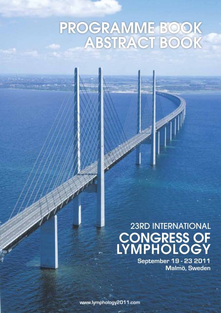
PROGRAMME BOOK ABSTRACT BOOK - Lymphology 2011
PROGRAMME BOOK ABSTRACT BOOK - Lymphology 2011
PROGRAMME BOOK ABSTRACT BOOK - Lymphology 2011

You also want an ePaper? Increase the reach of your titles
YUMPU automatically turns print PDFs into web optimized ePapers that Google loves.
<strong>PROGRAMME</strong> <strong>BOOK</strong><br />
<strong>ABSTRACT</strong> <strong>BOOK</strong><br />
www.lymphology<strong>2011</strong>.com<br />
23RD INTERNATIONAL<br />
CONGRESS OF<br />
LYMPHOLOGY<br />
September 19 - 23 <strong>2011</strong><br />
Malmö, Sweden
Congress<br />
Hall<br />
VENUE MAP<br />
East<br />
Room<br />
Room F<br />
Room G<br />
Room E<br />
Speakers<br />
Service<br />
Center<br />
Basement<br />
Room A+B<br />
Room C<br />
Room D<br />
Registration / Info<br />
Main Entrance<br />
Toilets<br />
Wardrobe<br />
Lunch<br />
Exhibition<br />
Posters<br />
Coffee Breaks<br />
www.stadionmassan.se
Dear Colleagues and Lymphologists!<br />
The Department of Plastic and Reconstructive Surgery at<br />
Malmö University Hospital, Sweden, is honored to organize the<br />
23rd International Congress of <strong>Lymphology</strong> in cooperation with<br />
the International Society of <strong>Lymphology</strong>, the Swedish <strong>Lymphology</strong><br />
Association and the Faculty of Medicine at Lund University.<br />
Problems related to the lymphatic system are central issues for<br />
us, and one of our main focuses is the development of surgical<br />
techniques related to lymphology. Olof Rudbeck (1630-1702),<br />
a Swedish scientist, published his first treatise De Circulatione<br />
Sanguinis in 1652, at the age of 22 years only, and he actually<br />
became the first one to describe the delineation and function<br />
of the lymphatic system in Nova Exercitatio Anatomica, which<br />
he published one year later. With this historical perspective in<br />
mind, we are enthusiastic about hosting the 23rd International<br />
Congress of <strong>Lymphology</strong> in Sweden. We are also proud of being<br />
entrusted with the task of arranging the prestigious congress in<br />
the city of Malmö. In fact, Malmö connects on to another pioneering<br />
scientific work in the field of lymphology performed by<br />
Thomas Bartholin (1616-1680), who was active in the nearby<br />
capital of Denmark, Copenhagen.<br />
In <strong>2011</strong>, from September 19 to 23, the most renowned scientists<br />
from all over the world will gather in Malmö to present and debate<br />
their front line knowledge and experiences in the various<br />
fields of lymphology. This will assure for an interdisciplinary and<br />
all-round illumination of the lymphatic system, its pathophysiology,<br />
and the state-of-the art of different treatment regimes.<br />
Moreover, at the end of the summer but before fall, September<br />
is an excellent time of the year to visit Sweden.<br />
We look forward to seeing you all in Malmö on this very special<br />
occasion. Please contact us for any additional information or<br />
suggestions that can make your stay even more pleasant in our<br />
dynamic and beautiful city.<br />
On behalf of the Organizing Committee<br />
Welcome to Sweden and the 23rd<br />
International Congress of <strong>Lymphology</strong>!<br />
Håkan Brorson, MD, PhD, Associate Professor<br />
Congress President<br />
23rd International<br />
Congress of<br />
<strong>Lymphology</strong><br />
Dear Therapist Colleagues!<br />
Come and join us at the 23rd International Congress of <strong>Lymphology</strong>,<br />
September 19-23, <strong>2011</strong>. Make friends from all over the<br />
world and expand your personal network, as I have enjoyed doing<br />
the past ten years. It is now a network of great knowledge<br />
and wonderful personalities and I am really looking forward to<br />
a reunion in Malmö.<br />
The congress program is now developing from the abstracts<br />
that have been submitted. Those of you who are working with<br />
cancer patients and have special interest in PREVENTION will<br />
find a session with that topic.<br />
If you are involved in TREATMENT of patients with lymphedema,<br />
primary as well as secondary, arms as well as legs, breast or<br />
genitals, you name it, there will be presentations that will interest<br />
you, including different approaches, new as well as already<br />
established. CHILDREN will get a special session.<br />
To get reliable result from our treatments we need good MEAS-<br />
UREMENT METHODS and I am very pleased that not only one<br />
session, but two, will evaluate old methods and introduce new<br />
ones, most of them very useful in clinical work.<br />
What about PHYSICAL ACTIVITY? Can lymphedema patients do<br />
exercises without deterioration of the edema? Do they actually<br />
benefit from exercise? Yes, yes, and yes! Make sure you get all<br />
the latest information on this important topic.<br />
Whenever a lymphedema is diagnosed it will influence QUAL-<br />
ITY OF LIFE for the patient and this issue is very important to<br />
recognize for all care providers. This will be discussed in one<br />
session.<br />
Do different approaches and treatments have promise and<br />
where do they fit in context? The session BEST PRACTICE will<br />
tell.<br />
I think that if this program cannot tickle your lymphedema<br />
senses then nothing can.<br />
Love to see you all in Malmö in September.<br />
Best regards<br />
Karin Johansson, PT, PhD<br />
Congress Vice President<br />
September 19-23, <strong>2011</strong><br />
Malmö, Sweden www.lymphology<strong>2011</strong>.com
The Organizing committee expresses its thanks and appreciation<br />
to all those who are generously contributing to the success of the<br />
23 rd International Congress of <strong>Lymphology</strong>, September 19 - 23,<br />
Malmö, Sweden<br />
Platinum Sponsor<br />
Gold Sponsors<br />
Apodan Nordic Healthcare A/S<br />
Bauerfeind AG<br />
Bodystat Ltd<br />
Carl Zeiss AB<br />
Delfin Technologies Ltd<br />
Haddenham Healthcare<br />
Höjmed Medical AB<br />
Intramedic AB<br />
Rolf Davidsen Helseagenturer AS/Jovipak<br />
Juzo GmbH<br />
Lymed Oy<br />
Solaris<br />
Technovital
Content<br />
Committees 5<br />
General Information 7<br />
Overall Congress Evaluation 13<br />
Programme at a Glance 17<br />
Additional Meetings 19<br />
Scientific Programme 21<br />
Keynote Lectures 22<br />
Round Table Sessions 29<br />
Programme Sessions 01-30 39<br />
Industry Sponsored Sessions 54<br />
Social Programme 55<br />
Oral Presentation Abstracts 57<br />
Poster Presentation Abstracts 171<br />
Author Index 193<br />
Exhibition 203<br />
Certificate of Attendance 207
JOBST ® Lymphoedema Therapy<br />
Compression therapy is widely used to control lymphoedema and oedema in<br />
the upper limb. Depending on the severity of the swelling, shape distortion and<br />
ability to manage and tolerate compression, Jobst ® offers a wide variety of<br />
Upper Extremity Garments to help maintain the limb size of these patients.<br />
the comfortable and robust garments provide:<br />
• Efficient compression for all patients<br />
• Increased level of comfort due to air-permeable and breathable material<br />
• High quality design to provide a precise anatomical fit<br />
• Ideal for maintenance therapy following CDT*<br />
* Complex Decongestive Therapy<br />
60435 RN © 2009 BSN medical Inc. Printed in (Country) REV 08/09<br />
JOBST - a brand of<br />
www.bsnmedical.com<br />
www.jobst.com<br />
Elvarex ®<br />
Elvarex ® Soft<br />
Elvarex ® Soft<br />
Seamless<br />
Comfort, Health and Style!
ORGANIZING COMMITTEE<br />
SCIENTIFIC COMMITTEE<br />
Håkan Brorson, MD, PhD<br />
Karin Johansson, PT, PhD<br />
Leif Perbeck, MD, PhD<br />
ORGANIZING COMMITTEE<br />
Honorary Presidents<br />
Sten Jacobsson, MD, PhD (Malmö)<br />
Torsten Landberg, MD, PhD (Malmö)<br />
Iwona Swedborg, MD, PhD (Stockholm)<br />
Magnus Åberg, MD, PhD (Malmö)<br />
Malmö-Lund<br />
Henry Svensson, MD, PhD<br />
Professor and Head<br />
Department of Plastic and Reconstructive Surgery<br />
Malmö University Hospital<br />
Håkan Brorson, MD, PhD<br />
Carolin Freccero, MD, PhD<br />
Karin Johansson, PT, LT, PhD<br />
Karin Ohlin, OT<br />
Barbro Svensson, PT, LT<br />
Ingrid Tengrup, MD, PhD<br />
Stockholm<br />
Elizabeth Johansson, PT, LT<br />
Leif Perbeck, MD, PhD<br />
Ulla Steen-Zupanc, MD<br />
SCANDINAVIAN GROUP<br />
Denmark<br />
Tony Karlmark, MD, PhD<br />
Susan Nörregaard, RN<br />
Susanne Birkballe, MD<br />
Norway<br />
Kristin Ruder, PT<br />
Hilde Osnes, PT<br />
23 rd ICL Secretariat<br />
Destination Öresund<br />
Fersens väg 18<br />
SE-211 42 Malmö, Sweden<br />
Tel: +46 40 300 665<br />
Fax: +46 40 918 952<br />
lymphology<strong>2011</strong>@destinationoresund.com<br />
Contact person: Congress Manager Lars Rudbert<br />
5
European Union of Medical Specialists<br />
EACCME - European Accreditation Council for Continuing Medical Education<br />
Institution of the UEMS<br />
Avenue de la Couronne 20, B-1050, Brussels<br />
T: +32 2 649 5164 | F: +32 2 640 37 30 | E: accreditation@uems.net<br />
Department of Clinical Sciences Malmö, Lund University, Plastic and Reconstructive Surgery,<br />
Malmö University Hospital<br />
Entrance 75<br />
SE-205 02 Malmö<br />
SWEDEN<br />
SUBJECT: EACCME accreditation granted EACCME-6047-G<br />
We are pleased to inform you that your application for European accreditation for:<br />
23rd International Congress of <strong>Lymphology</strong><br />
Venue: Malmö, Sweden (19.–23.09.<strong>2011</strong>)<br />
Event code: 6047<br />
was granted 24 European CME credits (ECMEC) by the European Accreditation Council for Continuing<br />
Medical Education (EACCME).<br />
European Accreditation<br />
European Accreditation is granted by the EACCME in order to allow participants who attend the above-mentioned activity to validate their<br />
credits in their own country.<br />
Accreditation Statement<br />
Accreditation by the EACCME confers the right to place the following statement in all communication materials including the registration<br />
website, the event programme and the certificate of attendance. The following statements must be used without revision:<br />
»The 'Department of Clinical Sciences Malmö, Lund University, Plastic and Reconstructive Surgery, Malmö University Hospital' (or) '23rd<br />
International Congress of <strong>Lymphology</strong>' is accredited by the European Accreditation Council for Continuing Medical Education (EACCME) to<br />
provide the following CME activity for medical specialists. The EACCME is an institution of the European Union of Medical Specialists (UEMS),<br />
www.uems.net.«<br />
»The '23rd International Congress of <strong>Lymphology</strong>' is designated for a maximum of (or 'for up to') 24 hours of European external CME credits.<br />
Each medical specialist should claim only those hours of credit that he/she actually spent in the educational activity.«<br />
»EACCME credits are recognized by the American Medical Association towards the Physician's Recognition Award (PRA). To convert EACCME<br />
credit to AMA PRA category 1 credit, contact the AMA.«<br />
EACCME credits<br />
Each medical specialist should claim only those hours of credit that he/she actually spent in the educational activity. The EACCME credit system<br />
is based on 1 ECMEC per hour with a maximum of 3 ECMECs for half a day and 6 ECMECs for a full-day event.<br />
Logo<br />
The UEMS – EACCME logo is a service mark of the European Union of Medical Specialists – European Accreditation Council for CME. This<br />
service mark may be used publicly only with the permission of the UEMS – EACCME. The logo may only be used in conjunction with, and in<br />
proximity to, the EACCME accreditation statement. The logo cannot be used in notices, advertising, or promotion of activities other than in<br />
association with the EACCME accreditation statement.<br />
Feedback report<br />
The EACCME requires you to provide a feedback report of the event within four weeks of its completion together with a copy of the list of<br />
participants and the results of the individual feedback assessments by participants.<br />
Brussels, 13. 7. <strong>2011</strong> The UEMS – EACCME Secretariat<br />
UEMS - Union européenne des médecins spécialistes | Avenue de la Couronne 20, B-1050, Bruxelles<br />
IBAN BE28 0001 3283 3820 | BIC (SWIFT) code: BPOTBEB1<br />
The European Accreditation Council for Continuing Medical Education (EACCME) was set up by the UEMS for the purpose of ensuring the international mutual<br />
recognition of quality assesments of CME-CPD activities organised all over the world for the benefit of European physicians
GENERAL<br />
INFORMATION
Banking<br />
Banks are open between 10.00 and 16.00 on weekdays.<br />
Some banks in central Malmö are open 09.00-17.00,<br />
and a few also on Saturdays. Cash machines are often<br />
located along the main streets next to a bank. Credit<br />
cards are accepted everywhere. Some supermarkets in<br />
Malmö are post office agents. Opening hours are usually<br />
08.00-22.00 on weekdays.<br />
Bicycles<br />
Some hotels have bicycles for lending, free of charge.<br />
Ask at the hotel reception.<br />
Certificate of attendance<br />
A certificate of attendance will be issued in the programme<br />
book.<br />
CME information<br />
European Accreditation is granted by the EACCME<br />
(European Accreditation Council for Continuing<br />
Medical Education) in order to allow participants who<br />
attend the congress to validate their credits in their<br />
own country. The EACCME is an institution of the<br />
European Union of Medical Specialists (UEMS), www.<br />
uems.net.<br />
23rd ICL is designated for a maximum of, or up to 24<br />
European CME credits (ECMECs).<br />
Participants who have indicated that they would like<br />
to apply for CME credits will receive a form before<br />
the congress. The form can be submitted to the staff<br />
www.medi.de<br />
at the Congress Information Desk or emailed/faxed to<br />
louise@destinationoresund.com Fax: +46 40-918 952.<br />
A CME certificate will follow by email after the congress.<br />
Coat and luggage<br />
The cloakroom is located at entrance level and will be<br />
open during the congress.<br />
Congress venue<br />
Stadionmässan<br />
Stadiongatan 25, Malmö<br />
Website: www.stadionmassan.se<br />
Congress evaluation<br />
Please fill out the form on page 13 and hand it in<br />
to the staff at the Information Desk.<br />
We kindly ask you to fill the form out, as it is required<br />
by the EACCME as well as future organisers<br />
of the ICL Congresses.<br />
Currency<br />
All official congress prices are indicated in SEK (Swedish<br />
Krona). Exchange rates are approximately:<br />
100 SEK= 15 USD<br />
100 SEK= 11 EUR<br />
All major credit cards are accepted in most hotels, restaurants<br />
and shops.<br />
Lymph therapy<br />
made easy with mediven<br />
flat-knit models<br />
medi Symposium<br />
Symposium<br />
„Compression „Compression therapy therapy<br />
in in lymphoedema“<br />
lymphoedema“<br />
20 20 September, September, 13-13:30h<br />
13-13:30h<br />
You are are kindly invited! invited!<br />
mediven mondi<br />
The The gentle and and effective product<br />
line line for for oedema up up to to stage II II<br />
mediven 550 550 arm arm<br />
Your Your specialist with with maximum<br />
compression for for all all forms of of arm arm<br />
oedema<br />
medi. I feel I feel better.<br />
00000_pmaz_195x140_lymphtherapie_GB_1107.indd 1 1 28.07.11 17:00 17:00
Delegate badge<br />
Each participant will receive a name badge when checking<br />
in on-site. For security reasons all participants are<br />
requested to wear their badge during all the Congress<br />
activities and social events. The cost of replacing a lost<br />
or mislaid badge is 200 SEK.<br />
Delegate bag<br />
The delegate bag, which includes the programme book,<br />
pad and pen etc. can be collected at the entrance upon<br />
checking in at the registration desks.<br />
Disclaimer & insurance<br />
The Organizing Committee, Destination Öresund and<br />
Stadionmässan accept no liability for any injuries/<br />
losses incurred by participants and/or accompanying<br />
persons, nor loss or damage to any luggage and/or personal<br />
belongings. Participants are advised to take their<br />
own personal insurance. The 23 rd ICL and the organisers<br />
take no responsibility for cancellations or delays for<br />
any causes beyond our reasonable control.<br />
Emergency/First aid<br />
For any emergency or first aid services inside the congress<br />
venue please contact the staff. The emergency<br />
number in Sweden is 112.<br />
Exhibition opening hours<br />
Monday, 19 September 17.30<br />
Tuesday, 20 September 10.00 - 16.30<br />
Wednesday, 21 September 10.00 - 16.30<br />
Thursday, 22 September 10.00 - 16.30<br />
Friday, 23 September 10.00 - 14.00<br />
Guided tours<br />
Participants and accompanying persons should have<br />
registered for the tours by 30 th August. Last minute<br />
registration can also be done online. For detailed info<br />
please refer to: www.hagelborntravel.se/travel/<br />
Accompanying persons are automatically registered<br />
for the tour on Tuesday 20 September.<br />
Information<br />
The Congress Information Desk is located by the entrance.<br />
Our staff will be happy to assist delegates with<br />
their queries. Tel: +46 768 397 936. E-mail: louise@<br />
destinationoresund.com.<br />
Internet spots and wireless internet<br />
We are pleased to provide wireless internet access at<br />
the congress venue. The relevant network name and<br />
password can be found at the message board by the<br />
entrance.<br />
Lost and found<br />
Any lost and found items will be held at the Congress<br />
Information Desk during the congress.<br />
Messages<br />
There will be a message board by the entrance.<br />
Meals and refreshments<br />
Coffee breaks and lunches are included in the delegate<br />
registration fee. For times of service, please refer to the<br />
detailed programme section in this book.<br />
Mobile phones<br />
All delegates are requested to switch off their mobile<br />
phones in the lecture halls and rooms during sessions.<br />
Non-smoking policy<br />
The entire venue is a non-smoking area. Smoking is<br />
only permitted outdoors.<br />
Official congress language<br />
The official congress language is English. No simultaneous<br />
translation will be provided.<br />
Official social events<br />
Welcome reception<br />
Stadionmässan Exhibition Hall, Monday September 19<br />
at 17.30.<br />
Congress dinner:<br />
Malmö Opera, Thursday September 22 at 19.00.<br />
Oral presentations<br />
Please refer to the program in this book.<br />
Photos, filming and recording<br />
Taking photos, filming and audio recording of the scientific<br />
program as well as of sponsor satellite symposia,<br />
partly or in their entirety, is not allowed without<br />
a written approval by the Organising Committee. The<br />
presenters have the intellectual rights and copyrights<br />
of their presentations.<br />
Poster area<br />
The poster area will be located along the left side of the<br />
Exhibition Hall.<br />
Poster presentation<br />
Please see the overview programme in this book.<br />
Public transport<br />
Please consult www.skanetrafiken.se for public transport<br />
in Malmö (click on the British flag at the top for<br />
English), including trains for Copenhagen and other<br />
destinations. Tickets cannot be purchased on the bus/<br />
train.<br />
You can buy your ticket at customer service centers or<br />
ticket machines by the train stations. If you plan to take<br />
the bus/train more than once, purchasing a Jojo discount<br />
card might be advisable.<br />
For a bus map, please see the website above. The information<br />
desk by the entrance can also assist you with<br />
queries regarding buses to and from the congress venue.<br />
9
Registration hours<br />
Monday, 19 Sept 10.00-20.00<br />
Tuesday, 20 Sept 08.00-18.00<br />
Wednesday, 21 Sep 08.00-18.00<br />
Thursday, 22 Sep 08 00-18.00<br />
Friday, 23 Sep 08.00-15.00<br />
Responsibility<br />
The delegate acknowledges that he/she has no right to<br />
lodge damage claims against the organisers, should the<br />
holding of the congress be hindered or prevented by<br />
unexpected political or economical events or generally<br />
by force majeure, or should the non-appearance of<br />
speaker or other reasons necessitate program changes.<br />
With his/her registration, the delegate accepted this<br />
proviso.<br />
3M Coban2, 2 Layer Compression System<br />
Lymphoedema Intensive Therapy<br />
Maintain Mobility<br />
While Reducing Oedema<br />
A Breakthrough for Patients and Clinicians!<br />
• Clinically effective volume reduction without the bulk of traditional reusable bandages<br />
• Unparalleled comfort, mobility and function enabling patients to carry on with everyday life<br />
• New application techniques that make wrapping sessions less taxing for clinicians and patients<br />
3M Health Care<br />
Skin and Wound Care Division<br />
Europe, CEE, MEA, Carl-Schurz-Str. 1, D - 41453 Neuss<br />
Tel. +49 (0)2131-14 3000, Fax: +49 (0)2131-14 4443<br />
www.3m.com<br />
3M and Coban are trademarks of the 3M Company.<br />
© 3M <strong>2011</strong>. All rights reserved.<br />
Taxi<br />
The price setting of taxi fares is free in Malmö and we<br />
strongly advise you to use these companies:<br />
Taxi 97, Phone: +46 (0)40 97 97 97<br />
Taxi Skåne, Phone: +46 (0)40 330 330<br />
Taxi Kurir, Phone: +46 (0)40 70 000<br />
You don´t need to take the first taxi in the rank in<br />
Malmö!<br />
Train<br />
The trip from Copenhagen Airport to Station Triangeln/Malmö<br />
Central Station takes approximately 20<br />
minutes and the single fare is 105 SEK (about 11 Euro<br />
or 15 USD, July <strong>2011</strong>) if you buy the ticket at the airport.<br />
Ms G, breast cancer survivor,<br />
lymphoedema patient, demonstrates<br />
the flexibility and<br />
function of 3M Coban 2<br />
Compression System.
Turning<br />
Torso<br />
Stora Varvsg<br />
23rd International Congress of <strong>Lymphology</strong>, Malmö<br />
Malmöhus slott<br />
Malmöhus Castle N. Vallg<br />
Kungsparken<br />
Slottsg<br />
Slottsparken<br />
Stadsbiblioteket<br />
City Library<br />
Regementsg<br />
Congress venue<br />
Stadionmässan<br />
Europaporten<br />
8<br />
Fersens v.<br />
Skeppsbron<br />
Lilla torg<br />
Stadiong<br />
Jörgen Kocksg<br />
Pildammsv<br />
Carlsg<br />
Rådmansg<br />
Stortorget<br />
Malmö Central Station<br />
F<br />
S. Förstadsg<br />
Rådhuset/Town Hall<br />
Baltzarsg<br />
Stora Nyg<br />
Carl Gustafsv<br />
Skånes Universitetssjukhus<br />
University<br />
Hospital<br />
John Ericssons v<br />
Drottn.g<br />
P<br />
Föreningsg<br />
P<br />
Österg<br />
S. Förstadsg<br />
S. Förstadsg<br />
Amiralsg<br />
Dalaplan<br />
Köpenhamn Copenhagen<br />
Köpenhamn Copenhagen<br />
➔<br />
2<br />
Gustav Adolfs torg<br />
1<br />
2<br />
3<br />
Renaissance Malmo Hotel<br />
Hotel Noble House<br />
Scandic S:t Jörgen<br />
6<br />
Triangeln<br />
5<br />
●F<br />
4 Scandic Malmö City<br />
7<br />
5<br />
6<br />
Hotel Plaza<br />
Teaterhotellet<br />
Malmö<br />
Opera Station Triangeln<br />
S:t Johannesg.<br />
7 Hilton Malmö City<br />
8 Ibis Hotel<br />
Pildammsparken<br />
Taxi<br />
F Flygbussar<br />
Airport Coaches<br />
Malmö airport<br />
Walk<br />
Bus<br />
Station<br />
Triangeln<br />
Smedjeg.<br />
Isstadion<br />
34<br />
▲<br />
~2,5 km<br />
➞<br />
1<br />
●<br />
3<br />
➞<br />
4<br />
●F<br />
●F<br />
34<br />
➔<br />
Gågata<br />
Pedestrian street<br />
●F<br />
●F<br />
Bergsg<br />
●<br />
F<br />
Södervärn<br />
N<br />
500 m<br />
Amiralsg<br />
Nobelv<br />
©Destination Öresund<br />
11
Overall Congress Evaluation<br />
Please fill out this form (3 pages) and hand it to the staff at the Information Desk.<br />
1. Registration ID Number (required): _________________<br />
CME credit applicants must complete this.<br />
For others, you may enter a "0" if you want complete anonymity<br />
If you don't remember your registration numbe, ask at the information desk.<br />
2. Your area of specialty (click all that apply)<br />
2a. Physician<br />
2b. Physiotherapist<br />
2c. Occupational therapist<br />
2d. Nurse<br />
2e. Other, please specify: __________________________<br />
3. Please rate the Congress (if applicable)<br />
3a. Overall Opinion of the Congress<br />
Excellent Very Good Good Poor Don’t know<br />
3b. Material Supplied (Final Programme, CD-Rom, Website Content)<br />
Excellent Very Good Good Poor Don’t know<br />
3c. Opportunity for Discussion<br />
Excellent Very Good Good Poor Don’t know<br />
3d. Congress content<br />
Excellent Very Good Good Poor Don’t know<br />
4. Rate the Quality of:<br />
4a. Planning and Organization<br />
Excellent Very Good Good Poor Don’t know<br />
4b.Meeting Rooms<br />
Excellent Very Good Good Poor Don’t know<br />
4c. Registration/Hospitality Desk<br />
Excellent Very Good Good Poor Don’t know<br />
4d. Quality of Catering<br />
Excellent Very Good Good Poor Don’t know<br />
5. How did you hear about the Congress?<br />
I am a past ICL Congress attendee<br />
Email<br />
Website search<br />
From a colleague or friend<br />
Printed material or advertisement<br />
13
14<br />
6. Was this event free of commercial bias? Registration ID: ________________<br />
Yes No<br />
7. Distribution of topics in parallel sessions: How often did you feel that it was urgent for you<br />
to attend more than one parallel session.<br />
Very often Often Seldom Almost never Don’t know<br />
7. Please use the space below to comment on the aspects of the Congress that you enjoyed and<br />
have helped you learn<br />
8. Please use the space below for recommendations or suggestions for improving any aspect<br />
of the Congress
9. General comments or suggestions for the next Congress<br />
Registration ID: ______________<br />
10. What other initiatives would you like the ICL Congress to incorporate in the future?<br />
11. Are you planning to attend the 2013 ICL Congress in Rome?<br />
Yes No Don’t know<br />
15
<strong>PROGRAMME</strong> AT A GLANCE
18<br />
Congress Hall<br />
Room A+B<br />
East Room<br />
* Congress Hall<br />
Monday 19 Sept Tuesday 20 Sept Wednesday 21 Sept Thursday 22 Sept Friday 23 Sept<br />
Breakfast Workshop - Bauerfeind<br />
07:30 - 08.15<br />
Registration Starts 08:30 - 09.00<br />
Kari Alitalo *<br />
Molecular Mechanisms in Lymphangiongenesis<br />
Keynote<br />
Coffee<br />
10:00<br />
Anne Kärki *<br />
Do We Need MLD? Lymphedema Therapy:<br />
Current Evidence of Best Practise<br />
Keynote<br />
Pierre Bourgeois *<br />
Imaging the Lymphatic System in <strong>2011</strong><br />
Keynote<br />
Terence Ryan *<br />
Adipose Tissue, Fibrosis and Lymphoedema, Mechanisms of Formation and Removal,<br />
What is the Evidence?<br />
Keynote<br />
Session 29<br />
Decongestive Therapy I<br />
Chair: Isabel Forner-Cordero,<br />
Giovanni Moneta,<br />
Jean-Paul Belgrado<br />
Session 27<br />
New Frontiers in<br />
Lymphatic Research I<br />
Keynote<br />
Stan Rockson<br />
Chair: Stan Rockson,<br />
Moirya Ohkuma,<br />
Anatoliy Gashev<br />
Session 24<br />
Genital Lymphedema<br />
Chair: Alexandre Pissas,<br />
Wichai Ekataksin<br />
Session 21<br />
Phlebolymphology<br />
Session 09<br />
Measuring Methods I<br />
09:00 - 10.30<br />
Opening Ceremony<br />
12:00<br />
Chair: Francesco<br />
Boccardo,<br />
Evangelos<br />
Dimakakos<br />
Session 18<br />
Lymphatic Imaging I<br />
Chair: Pierre Bourgeois,<br />
Leif Perbeck,<br />
Pernilla Peterson<br />
Session 15<br />
Lipedema<br />
Chair: Wilfried Schmeller,<br />
Gyozo Szolnoky,<br />
Sandro Michelini<br />
Session 12<br />
New Approaches<br />
- Alternative Therapies<br />
Chair: Miguel Amore,<br />
Wichai Ekataksin,<br />
Alberto Gersman<br />
Round Table<br />
Leigh Ward,<br />
Jane Armer,<br />
Harvey Mayrowitz<br />
Session 06<br />
Prevention<br />
Chair: Neil Piller,<br />
Alex Munnoch,<br />
Ethel Földi<br />
Session 03<br />
Pathophysiology<br />
Chair: Terence Ryan,<br />
Moriya Ohkuma,<br />
Andrzej Szuba<br />
Vice-chancellor Per Eriksson<br />
Repr Skåne Regional Council<br />
City Councillor Kent Andersson<br />
President Håkan Brorson<br />
Vice President Karin Johansson<br />
ISL President Rüdiger Baumeister<br />
Secretary-General Marlys Witte<br />
Prof Emirita Iwona Swedborg<br />
Coffee Break Coffee Break Coffee Break Coffee Break<br />
10:00 - 11.00<br />
Refreshment Break<br />
13:00<br />
Session 30<br />
Decongestive Therapy II<br />
Chair: Ulla Steen-Zupanc,<br />
Waldemar Olszewski,<br />
Elizabeth Johansson<br />
Session 28<br />
New Frontiers in<br />
Lymphatic Research II<br />
Session 10<br />
Measuring Methods II<br />
11:00 - 12.30<br />
Keynotes<br />
Marlys Witte<br />
<strong>Lymphology</strong> and the ISL in the Real, Virtual and<br />
Imagined World of the Future<br />
13:45<br />
Chair: Stan Rockson,<br />
David Zawieja,<br />
Marlys Witte<br />
Session 25<br />
Compression Treatment I:<br />
Pitfalls and Dangers of<br />
Compression Therapy<br />
Round Table<br />
Hugo Partsch,<br />
Robert Damstra,<br />
Vaughan Keeley<br />
Session 22<br />
Lymphatic Imaging II<br />
Chair: Pierre Bourgeois,<br />
Ningfei Liu,<br />
Emily Iker<br />
Session 19<br />
Surgery I<br />
Chair: Alex Munnoch,<br />
Jaume Masia,<br />
Martin Wald<br />
Session 16<br />
Best Practice for the<br />
Management of<br />
Lymphedema I<br />
Chair: Corradino Campisi,<br />
Jane Armer,<br />
Susanne Birkballe<br />
Session 13<br />
Filariasis and Lymphedema<br />
Keynote<br />
Gurusamy Manokaran<br />
Chair: Gurusamy Manokaran,<br />
Isabel Forner-Cordero<br />
Chair: Leif Perbeck,<br />
Michael Bernas,<br />
Neil Piller<br />
Session 04<br />
Session 07<br />
Pediatric I:<br />
Basic Science<br />
Lymphedema in Children Chair: Mauro Andrade,<br />
Keynote<br />
Gyozo Szolnoky<br />
Cristóbal Papendieck<br />
Keynote<br />
Fiona Connell<br />
Chair: Cristóbal Papendieck,<br />
Fiona Connell,<br />
Carolin Freccero<br />
Katie Schmitz<br />
Balancing Lymphedema Risks: Exercise Versus<br />
Deconditioning<br />
Stanley Leong<br />
Impact of Tumor Burden in the Sentinel Lymph<br />
Nodes on the Outcomes of Cancer Patients<br />
Lunch<br />
Lunch<br />
Lunch<br />
12:30 - 14.00<br />
Peter Mortimer<br />
Breast Cancer Related Lymphedema<br />
Lunch<br />
medi Symposium (13.00 - 13.30) *<br />
Jobst Symposium (13.00 - 13.45) *<br />
3M Symposium (13.00 - 13.30) *<br />
Coffee Break<br />
13.15<br />
Consensus Document<br />
Session 26<br />
Compression Treatment II<br />
Session 20<br />
Surgery II<br />
Chair: Harry Voesten,<br />
Erkki Suominen,<br />
Corrado Campisi<br />
Session 11<br />
Clinic on Lymphedema<br />
14:00 - 15.30<br />
15:30<br />
16:00<br />
14.00<br />
Close of Congress<br />
• Congress President Håkan Brorson<br />
• ISL President Rüdiger Baumeister<br />
• ISL Secretary-General Marlys Witte<br />
• Presentation of 2013 Congress Outline,<br />
Sandro Michelini, San Giovanni Battista<br />
Hospital, Rome, Italy<br />
Chair: Hugo Partsch,<br />
Robert Damstra,<br />
Karin Ohlin<br />
Session 23<br />
Physiology of the<br />
Lymphatic System<br />
Chair: Waldemar Olszewski,<br />
Christen Krag,<br />
Anatoliy Gashev<br />
Session 17<br />
Best Practice for the<br />
Management of<br />
Lymphedema II<br />
Chair: Ethel Földi,<br />
Alexandre Pissas,<br />
Barbro Svensson<br />
Session 14<br />
Cancer and Lymphedema<br />
Chair: Torsten Landberg,<br />
Stanley Leong,<br />
Francesco Boccardo<br />
Chair: Henry Svensson,<br />
Martin Wald,<br />
Marlys Witte<br />
Session 08<br />
Quality of Life<br />
Chair: Jane Armer,<br />
Pia Klenäs,<br />
Sandy Hayes<br />
Session 05<br />
Pediatric II:<br />
Lymphatic Malformations<br />
Keynote<br />
Arin Greene<br />
Session 02<br />
Anatomy of the<br />
Lymphatic System<br />
Keynote<br />
Miguel Amore<br />
Session 01<br />
Exercise and<br />
Lymphedema<br />
Chair: Karin Johansson,<br />
Kathryn Schmitz,<br />
Ethel Földi<br />
Chair: Arin Greene,<br />
Magnus Åberg,<br />
Carolin Freccero<br />
Chair: Miguel Amore,<br />
Eikichi Okada<br />
Welcome Reception Hosted by City of Malmö<br />
17.30<br />
14.30<br />
General Assembly<br />
Coffee Break Coffee Break Coffee Break<br />
15:30 - 16.00<br />
ISL Executive Committee Meeting<br />
Round Table *<br />
Basic Science and the Way to Treatment<br />
Rüdiger Baumeister, Neil Piller,<br />
Anne Saaristo, Håkan Brorson<br />
16:00 - 17.30<br />
Round Table *<br />
Surgical and Non-surgical Treatment: Against Each Other or Together?<br />
Rüdiger Baumeister, Corradino Campisi, Håkan Brorson, Anne Kärki<br />
Round Table *<br />
Lipedema: Diagnostic Tools and Treatment Necessities?<br />
Peter Mortimer, Gyozo Szolnoky, Ilka Meier-Vollrath, Wilfried Schmeller<br />
17.30<br />
Tivoli Copenhagen (optional) Free Evening Congress Dinner (19.00)<br />
* Congress Hall<br />
Poster session Thursday<br />
13.30 – 14.00<br />
P-13.07 – P-29.15<br />
Poster session Tuesday<br />
13.30 – 14.00<br />
P-01.08 – P-11.13
Additional meetings<br />
Swedish <strong>Lymphology</strong> Association<br />
Date: Thursday, September 22<br />
Time: 12.30-13.00<br />
Room: Congress Venue, Room D (combined with<br />
lunch)<br />
ISL Executive Committee Meeting<br />
Date: Sunday, September 18<br />
Time: 18.00<br />
Room: Meeting Room, Department of Plastic Surgery,<br />
Skåne University Hospital, Entrance 75, 3rd floor<br />
Date: Wednesday, September 21<br />
Time: 12.30-13.00<br />
Room: Congress Venue, Room D (combined with<br />
lunch)<br />
Date: Friday, September 23<br />
Time: afternoon, after General Assembly<br />
Room: Congress Venue, Room D<br />
19
SCIENTIFIC <strong>PROGRAMME</strong><br />
KEYNOTE LECTURES<br />
ROUND TABLE SESSIONS<br />
<strong>PROGRAMME</strong> SESSIONS 01-30<br />
INDUSTRY SPONSORED SESSIONS
22<br />
KEYNOTE LECTURE MONDAY 13.45-14.15 CONGRESS HALL<br />
KN-01<br />
LYMPHOLOGY AND THE ISL IN THE REAL, VIRTUAL AND IMAGINED WORLD OF THE FUTURE<br />
M H Witte, Department of Surgery, University of Arizona College of Medicine, Tucson, UNITED STATES<br />
Since before the official founding of the International Society of <strong>Lymphology</strong> (ISL), lymphology has transcended the barriers<br />
of medical specialization, language, and geography. In 1966, the First International Congress of <strong>Lymphology</strong> in Zurich<br />
brought together hundreds of basic scientists (largely anatomists and physiologists) and clinicians (prominent radiologists/<br />
lymphographers and cancer/vascular surgeons) who envisioned the lymphatic system as more than “lymph nodes held<br />
together by strings.” Forty-five years later, we in the “real world” of lymphology still “stand on the shoulders of these giants,”<br />
revisiting neglected threads from earlier times and applying advances in molecular biology, technology, and public health to<br />
bring new understanding to structure-function relationships in the normal and diseased lymphatic system, higher resolution<br />
in multimodal dynamic lymphatic imaging, and improved care to patients afflicted with myriad disorders of the “lymphatics,<br />
lymph, lymph nodes and lymphocytes.” Many key unanswered questions and unquestioned answers persist (true “medical<br />
ignorance”), and many more will doubtless arise de novo in the “imagined world” of bewildering genomes, proteomes,<br />
transcriptomes, metabolomes, organomes, functionomes, diseasomes, phenomes, etc. As we move from “genes to man” – to<br />
translate discoveries into personalized medicine in lymphology – new and better ways to collaborate globally across basic<br />
and clinical disciplines will incorporate sophisticated communication/data networks and remote outreach. Here, the “virtual<br />
world” of our web-based Virtual Clinical Research Center/Questionarium should be a powerful tool to advance molecular,<br />
cellular, systemic, and clinical lymphology and provide a welcome roadmap to our “unimagined” future.<br />
Declaration of interest<br />
None declared<br />
KEYNOTE LECTURE MONDAY 14.15-14.40 CONGRESS HALL<br />
KN-02<br />
BALANCING LYMPHEDEMA RISKS: EXERCISE VERSUS DECONDITIONING<br />
K Schmitz, University of Pennsylvania, Philadelphia, UNITED STATES<br />
Background: Lymphedema is a common and feared adverse clinical sequelae of breast cancer treatment. Our current ability<br />
to predict who will develop lymphedema or who will progress to advanced stages of the condition are poor, at best. Given this<br />
scenario, the clinical advice to women with and at risk for lymphedema has been risk averse, including advice to protect the<br />
affected limb and avoid lifting. Unfortunately, this rational response to the unknown may lead to the very outcome patients<br />
seek to avoid (onset or worsening of lymphedema) as a side effect of the deconditioning resulting from protecting the limb.<br />
Objectives: The objective of this presentation is to present evidence and theory regarding physiologic effects of exercise on the<br />
lymphatic system structure and function, as well as to review the recently published evidence regarding the clinical effects of<br />
exercise on lymphedema outcomes in breast cancer survivors.<br />
Methods: A systematic literature review of the evidence regarding physiologic and clinical effects of exercise on outcomes<br />
related to lymphedema in breast cancer survivors.<br />
Results: Evidence from lymphoscintigraphic studies indicate increased lymphatic clearance during exercise than at rest among<br />
women with lymphedema. Decades of exercise science support the usefulness of site specific exercise to improve circulatory<br />
function, suggesting exercise may improve the ability of the affected limb to respond to infection, inflammation, and trauma.<br />
Exercise training also improves the functional capacity, reducing the relative intensity of work. This may result in avoiding<br />
the overuse often thought to be the cause of lymphedema onset and progression. Results of clinical trials on exercise strongly<br />
support the use of exercise as part of a program to reduce lymphedema onset or progression among breast cancer survivors.<br />
Conclusions: The scientific evidence base regarding the value of exercise for prevention and control of lymphedema in breast<br />
cancer survivors is as compelling as for any therapeutic modality. Further imaging studies will assist with cementing the<br />
mechanisms through which these effects occur. However, it is now clear that rehabilitative weight training exercise should be<br />
standard of care for breast cancer survivors.<br />
Declaration of interest<br />
none declared
KEYNOTE LECTURE MONDAY 14.40-15.05 CONGRESS HALL<br />
KN-03<br />
IMPACT OF TUMOR BURDEN IN THE SENTINEL LYMPH NODES ON THE OUTCOMES OF CANCER<br />
PATIENTS<br />
S Leong, Department of Surgery, California Pacific Medical Center, San Francisco, UNITED STATES<br />
Background: The concept that cancer of a specific anatomical site spreads to its corresponding sentinel lymph nodes (SLNs) in<br />
the regional nodal basin has been extensively validated in melanoma and breast cancer.<br />
Objective: To evaluate the clinical significance of micrometastasis in the SLNs.<br />
Methods: Literature review.<br />
Results: In general, micrometastasis in the SLNs of solid cancers predicts a worse clinical outcome. Cancer metastasis from the<br />
primary site to the SLNs (incubator hypothesis) and then to the non-SLNs prior to systemic spread in melanoma and breast<br />
cancer is consistent with the spectrum theory that cancer metastasis is progressive. However, in about 20% of the time, cancer<br />
cells may spread through the SLNs and vascular system simultaneously (marker hypothesis) or independently to the distant<br />
sites via the vascular system. If the SLNs are negative, a more morbid regional lymph node dissection may be spared in melanoma.<br />
For breast cancer, removal of SLNs with metastasis may be effective during the ‘‘incubator’’ phase, but adjuvant therapy<br />
should be given for patients with microscopic disease in the distant sites. In penile carcinoma, SLN mapping will guide the<br />
appropriate location of the lymph node to avoid a morbid bilateral radical ilioinguinal lymph node dissection. For head and<br />
neck, colorectal, upper GI, other GU and gynecological cancers, the lymphatic pathways are, in general, more complicated and<br />
unpredictable. For head and neck as well as gynecological cancers, the goal is to establish a reliable SLN mapping method to<br />
minimize the degree of lymph node dissection. For colorectal and upper GI cancers, while the extent of lymphadenectomy may<br />
not be altered significantly, the identification of SLNs will increase the accuracy of staging the nodal basins, especially in view<br />
of the fact that the number of lymph nodes being resected for colorectal and gastroesophageal cancer is a significant predictor<br />
of survival.<br />
Conclusion: In conclusion, SLN is the gateway for cancer metastasis in the majority of the time. Further molecular and genomic<br />
studies may define the mechanisms of cancer spread through the lymphovascular system more precisely, thus, allowing us to<br />
develop more rational therapy.<br />
Declaration of interest<br />
None declared<br />
KEYNOTE LECTURE MONDAY 15.05-15.30 CONGRESS HALL<br />
KN-04<br />
BREAST CANCER RELATED LYMPHOEDEMA (BCRL)<br />
P Mortimer, Division of Clinical Science, St George’s, University of London, London, UNITED KINGDOM<br />
Axillary surgery for breast cancer may be followed, weeks to years later, by chronic arm lymphoedema (BCRL). A simple<br />
obstructive “stopcock” mechanism (reduced lymph drainage from the entire limb) does not explain many clinical aspects,<br />
including the delayed onset and selective sparing of some regions eg hand. In a prospective study that investigated patients<br />
following breast cancer treatment, but before the onset of lymphoedema, lymph flow was found to be significantly higher<br />
in the arms of those women who subsequently developed arm swelling.(1) Lymph flow was found significantly higher in<br />
both subcutis and sub-fascial forearm muscle.(1) Furthermore lymph flow was also found to be higher in the contralateral<br />
arm which was unaffected by surgery.(1) BCRL therefore developed in women with higher peripheral lymph flows. These<br />
results indicate that BCRL is not simply an obstructive lymphoedema. We propose that women have a defined, constitutive<br />
predisposition to BCRL by developing high fluid filtration rates in response to cancer and/or its treatment. Higher fluid<br />
filtration overloads lymph drainage which, over time and in the face of increased outflow lymphatic resistance, leads to<br />
lymphatic collector pump failure and the onset of lymphoedema. (2,3)<br />
1. Stanton AW, Modi S, Bennett Britton et al, Lymphatic drainage in the muscle and subcutis of the arm after breast cancer<br />
treatment. Breast Cancer Res Treat. 2009;117:549-57<br />
2. Modi S, Stanton AW, Svensson WE et al. Human lymphatic pumping measured in healthy and lymphoedematous arms by<br />
lymphatic congestion lymphoscintigraphy. J Physiol. 2007;583:271-85<br />
3. Stanton AW, Modi S, Mellor RH et al. Recent advances in breast cancer-related lymphoedema of the arm: lymphatic pump<br />
failure and predisposing factors. Lymphat Res Biology 2009;7:29-45<br />
Declaration of interest<br />
None declared<br />
23
24<br />
KEYNOTE LECTURE MONDAY 16.00 - 16.30 ROOM A+B<br />
O-02.01<br />
REVIEW THE LYMPHATIC ANATOMY IN THE SENTINEL NODE ERA<br />
M Amore, Buenos Aires University, Buenos Aires, ARGENTINA<br />
OBJECTIVE:The introduction of the sentinel lymph node biopsy has renewed the interest in regional lymph nodes outside<br />
some organs as a potential site of regional lymph nodes metastases. Detailed gross anatomical information about the lymphatic<br />
system is essential for predicting accurate distant metastatic sites in cancer. The purpose of this study is to carry out a detailed<br />
description of the lymphatic drainage of different organs (mammary gland, colon, stomach, esophagus, thyroid gland, genital<br />
organs and skin, and translation this finding to the clinical experience in lymphatic mapping and sentinel node biopsy.<br />
MATERIAL AND METHODS: Our material comprised 300 specimens 283 of which where from human fetus and 17 from<br />
fresh adult cadavers between 50 to 76 years old of which 226 where women and 74 men. The injection was done with the<br />
modified Gerota’s mass. Dissection is carried out after fixation of the specimen in 40% formaldehyde for 6 days, and then<br />
immersed in an 100 volume hydrogen peroxide solution for 24 hours (Prof. Caplan’s bleaching technique). In 90 fetus<br />
specimens we used the Spalteholz technique for diafanization. Research was carried out at the Lymphatic Research Laboratory<br />
of the III Chair of Anatomy at the University of Buenos Aires.<br />
RESULT: Sentinel lymph node dissections have been shown to be sensitive for the evaluation of nodal basins for metastatic<br />
disease and are associated with decreased short-term and long-term morbidity when compared with complete lymph node<br />
dissection. This study addresses the lymphatic anatomy of some organs in relation of the incidence of cancer. Our finding may<br />
explain the clinical experience in lymphatic mapping and sentinel node biopsy, and also the persistence of a false negative rate<br />
irrespective of experience of the surgeon.<br />
Declaration of interest<br />
NONE DECLARED<br />
KEYNOTE LECTURE TUESDAY 08.30 - 09.00 CONGRESS HALL<br />
KN-05<br />
MOLECULAR MECHANISMS IN LYMPHANGIOGENESIS<br />
K Alitalo, Molecular/Cancer Biology Laboratory, Haartman Institute and Finnish Institute for Molecular Medicine, Helsinki,<br />
FINLAND<br />
Vascular endothelial growth factor (VEGF) stimulates angiogenesis and permeability of blood vessels via its two receptors<br />
VEGFR-1 and VEGFR-2, but it has only little lymphangiogenic activity. The third receptor, VEGFR-3, does not bind VEGF<br />
and its expression becomes restricted mainly to lymphatic endothelia during development. Homozygous VEGFR-3 targeted<br />
mice die around midgestation due to failure of cardiovascular development, whereas transgenic mice expressing the VEGFR-3<br />
ligand VEGF-Cor VEGF-D show evidence of lymphangiogenesis and VEGF-C knockout-mice have defective lymphatic<br />
vessels. VEGF-C overexpression induces lymphangiogenesis and growth of the draining lymphatic vessels, intralymphatic<br />
tumor growth, lymph node lymphangiogenesis and metastasis. Furthermore, soluble VEGFR-3 and antibodies blocking<br />
VEGFR-3 inhibited embryonic and tumor lymphangiogenesis and lymphatic metastasis. These results have indicated that<br />
paracrine signal transduction between tumor cells and the lymphatic endothelium is involved in lymphatic metastasis. We<br />
have recently found that VEGF-C and VEGFR-3 provide also new targets to complement current anti-angiogenic therapies.<br />
Because of their ability to attenuate angiogenic sprouting and inhibit lymphatic metastasis, VEGFR-3 blocking antibodies are<br />
now being tested in phase I clinical (safety) trials. - Furthermore, our studies indicate that antibody combinations may be<br />
used for increased efficacy of inhibition of angiogenic signal transduction pathways.<br />
Preclinical studies of lymphedema treatment have shown that VEGF-C stimulates the formation of new lymphatic capillaries<br />
and, after an initial increase in lymph extravasation, reduces edema. When the growth factor therapy was applied to damaged<br />
collecting lymph vessels, lymphatic capillary growth was followed by intrinsic remodeling, differentiation, and maturation<br />
into functional vessels with normal zipper-like endothelial cell-cell junctions, intraluminal valves and SMC coverage. A<br />
combination of VEGF-C therapy with lymphnode transplantation showed that the growth factor-transduced lymph nodes<br />
formed both afferent and efferent connections with the pre-existing lymphatic vessel network, and could even trap metastatic<br />
tumor cells. In pigs, VEGF-C or VEGF-D therapy proved effective in restoring functional lymphatic vasculature to the site<br />
of surgical damage and greatly increased the survival and functionality of transferred lymph nodes. These studies have<br />
provided a basis for clinical trials in lymphedema patients with non-malignant disease that, after further safety trials, should<br />
be applicable also to cancer patients.<br />
Declaration of interest<br />
None declared
KEYNOTE LECTURES TUESDAY 11.00 - 11.25 CONGRESS HALL<br />
O-04.01<br />
LYMPHEDEMA IN PEDIATRICS. DIALOGUE BETWEEN SCIENCE AND PRIMARY CARE. PROPOSAL<br />
FOR A CONSENSUS DOCUMENT.<br />
C Papendieck, Instituto de Diagnóstico y Tratamiento, Buenos Aires, ARGENTINA, R Martinez, Hospital de Quemados, Buenos<br />
Aires, ARGENTINA, L Barbosa, Angiopediatría, Buenos Aires, ARGENTINA, M A Amore, Hospital Militar Centra, Buenos Aires,<br />
ARGENTINA, E Paltrinieri, Centro Vodder, Buenos Aires, ARGENTINA, D Braun, Angiopediatria, Buenos Aires, ARGENTINA<br />
Lymphedema diagnosis in pediatric patients include as less 140 syndromes or diseases, including the whole universe of primary<br />
lymphedema (PL). There is no reached consensus on its definition. We support the opinion that PL is a sign, and their causes<br />
congenital, and provoke a dysfunction of the lymphatic system. Initially, there is some endothelial, interstitial, precapillary or<br />
capillary disability, or some canalicular or nodal dysplasia (LAD I, LAD II respectively). This dysplasia expresses itself as a deficit<br />
in interstitial fluid protein reabsorption, and other components of lymphatic load too, or in lymph transportation mechanical<br />
obstruction in the way to the venous angle. There are 18 well-known anatomical anomalies in lymph vessels and lymph nodes<br />
design. Stem cells and as less as 12 lymphangiogenic growth factors and their receptors, are involved in the morphogenesis of<br />
lymphatic system and are endothelial function regulators, five of them in the interstice. Interstitial volume/space increased is<br />
constant with or without hypertension in the segments of the circuit. The protocols of international consensus, MEP, ISL and<br />
others, provided the basic tools for rehabilitation. There is no cure. That means a future without specific therapies, for the rest of<br />
life. There is no diagnostic by evidence.<br />
We propose the basis for a Consensus Document on Lymphedema in Pediatric Patients for diagnosis in genetic, molecular,<br />
anatomopathology and image, as well as therapeutic indications considering, when possible, its etiological diagnosis.<br />
A little baby cannot wait for the whole life, it is not satisfactory to propose to use bandage forever, as our unique therapeutic offer.<br />
The ISL Consensus Document completely avoids any mention to pediatric patients. Children are not little adults, their physiology,<br />
anatomy, immune system are peculiar and put the frame for diagnostic and therapeutic interventions.<br />
In a typical secondary lymphedema standard treatment, as established in consensus documents, is indicated. Details for<br />
specific diagnostic and therapeutic procedures must be discussed to guide clinical practice until evidence-based practice<br />
could be well established. Proposals for a consensus document in primary and secondary pediatric lymphedema will be<br />
discussed.<br />
Declaration of interest<br />
None declared<br />
KEYNOTE LECTURES TUESDAY 11.25-11.42 CONGRESS HALL<br />
O-04.02<br />
RECENT ADVANCES IN THE GENETICS OF PRIMARY LYMPHOEDEMA<br />
F Connell, Guy’s and St Thomas’ NHS Trust London, London, UNITED KINGDOM, S Mansour, St George’s University of London,<br />
London, UNITED KINGDOM, P Ostergaard, St George’s University of London, London, UNITED KINGDOM, G Brice, St<br />
George’s University of London, London, UNITED KINGDOM, M Simpson, King’s College London, London, UNITED KINGDOM,<br />
R Trembath, King’s College Hospital, London, UNITED KINGDOM, P Mortimer, St George’s University of London, London,<br />
UNITED KINGDOM, S Jeffery, St George’s University of London, London, UNITED KINGDOM<br />
Background: Primary lymphoedema is a chronic oedema caused by a developmental abnormality of the lymphatic system. In<br />
recent years there has been considerable progress made in understanding the molecular pathways underlying lymphangiogenesis<br />
but knowledge of the genetic causes of human lymphatic disease was limited to VEGFR3, FOXC2 and SOX18. There are many<br />
different phenotypes of primary lymphoedema and with the use of conventional molecular analysis techniques and new next<br />
generation sequencing we have identified two further genes that cause different primary lymphoedema phenotypes; CCBE1 and<br />
GJC2.<br />
Objectives: The aim of our work has been to improve phenotyping of primary lymphoedema patients in order to facilitate the<br />
identification of well defined patient groups for molecular studies that would lead to the discovery of genetic causes of primary<br />
lymphoedema in humans.<br />
Methods: Patients were ascertained from the joint Lymphoedema/Genetics Clinic at St George’s Hospital, London.<br />
Linkage and sequence analysis was carried out to identify the genetic cause of recessively inherited generalised lymphatic<br />
dysplasia/Hennekam syndrome. A large, non-consanguineous family with three affected siblings with generalised lymphatic<br />
dysplasia was studied. Linkage analysis was used to determine a locus in a large multigenerational pedigree in which four-limb<br />
lymphoedema segregates in an autosomal dominant manner. Exome sequencing was employed to look for causative variants<br />
within the predetermined locus.<br />
Results: A homozygous change in CCBE1 was identified as the causative mutation for autosomal recessive generalised lymphatic<br />
dsyplasia/Hennekam syndrome. Mutations in GJC2 were identified to cause autosomal dominantly inherited four-limb/bilateral<br />
lower limb lymphoedema.<br />
Conclusion: Notable advances in the understanding of the genetics of primary lymphoedema have been achieved and the<br />
implications of these discoveries will be discussed. The role of CCBE1 and GJC2 in lymphatic disease has been reported by<br />
Alders et al 2009 and Ferrell et al 2010 respectively, and our work adds supportive evidence to these studies. Understanding the<br />
molecular mechanisms that result in lymphatic disease will hopefully ultimately translate into improved therapies for patients.<br />
Declaration of interest<br />
None declared<br />
25
26<br />
KEYNOTE LECTURE TUESDAY 14.00 - 14.30 CONGRESS HALL<br />
O-05.01<br />
DIFFERENTIAL DIAGNOSIS OF LOWER EXTREMITY ENLARGEMENT IN PEDIATRIC PATIENTS<br />
REFERRED WITH A DIAGNOSIS OF “LYMPHEDEMA”<br />
A Greene, Children’s Hospital Boston/Harvard Medical School, Boston, UNITED STATES<br />
Background: There are many causes for a large lower limb in the pediatric age group. These children are often mislabeled as<br />
having “lymphedema”, and incorrect diagnosis can lead to improper treatment. The purpose of this study was to determine<br />
the differential diagnosis in pediatric patients referred for lower extremity “lymphedema” and to clarify management.<br />
Methods: Our Vascular Anomalies Center database was reviewed between 1999 - 2010 for patients referred with a diagnosis<br />
of “lymphedema” of the lower extremity. Records were studied to determine the correct etiology for the enlarged extremity.<br />
Alternative diagnoses, gender, age-of-onset, and imaging studies also were analyzed.<br />
Results: A referral diagnosis of lower extremity “lymphedema” was given to 170 children; however, the condition was<br />
confirmed in only 72.9% of patients. Forty-six children (27.1%) had another disorder: micro/macrocystic lymphatic<br />
malformation (19.6%), non-eponymous combined vascular malformation (13.0%), capillary malformation (10.9%), Klippel-<br />
Trenaunay syndrome (10.9%), hemi-hypertrophy (8.7%), post-traumatic swelling (8.7%), Parkes Weber syndrome (6.5%),<br />
lipedema (6.5%), venous malformation (4.3%), rheumatologic disorder (4.3%), infantile hemangioma (2.2%), kaposiform<br />
hemangioendothelioma (2.2%), or lipofibromatosis (2.2%). Age-of-onset in children with lymphedema was older than<br />
patients with another diagnosis (p = 0.027).<br />
Conclusion: “Lymphedema” is not a generic term. Approximately one-fourth of pediatric patients with a large lower extremity<br />
are misdiagnosed as having “lymphedema”; the most commonly confused etiologies are other types of vascular anomalies.<br />
History, physical examination, and often radiological studies are required to differentiate lymphedema from other conditions<br />
to ensure the child is managed appropriately.<br />
Declaration of interest<br />
None declared<br />
KEYNOTE LECTURE WEDNESDAY 08.30 - 09.00 CONGRESS HALL<br />
KN-06<br />
ADIPOSE TISSUE, FIBROSIS AND LYMPHOEDEMA, MECHANISMS OF FORMATION AND REMOVAL,<br />
WHAT IS THE EVIDENCE?<br />
T Ryan, Oxford University, Marlborough, UNITED KINGDOM<br />
This paper will describe some of the grossest hypertrophy of the tissues observed in elephantiasis Oedema is a small component<br />
creating mechanical tension and triggering hypertrophy of the collagen component. This is mostly in the dermis and there is<br />
loss of the more elastic tissue component which is elastin.<br />
The dermis affected by lympoedema does acquire new fat containing cells believed to support the locally threatened immunosurveillance<br />
system and secondarily dissipating mechanical forces.<br />
The subcutaneous tissues are normally host to adipose tissue and usually show less hypertrophy when the dermis and epidermis<br />
is most affected by lymphoedema. When the subcutaneous tissues do show gross hypertrophy, the overlying epidermis<br />
and dermis are usually less grossly affected<br />
Reversal of the hypertrophy by therapists is often achieved by simple technologies influencing the lymphatic system such as<br />
washing and massage. How fibrosis is reversed raises many questions. Scandinavia is host to the most impressive effects of<br />
liposuction acting on the subcutaneous issues but sparing the upper dermis in particular. Exactly what mechanisms reverse<br />
elephantiasis requires a review of both<br />
1) the transduction of biochemical signals by mechanical forces and the protective role of elastin, and<br />
2) emphasis on the function of the adipocyte and the effects of its removal from the subcutaneous or lower dermal tissues.<br />
In some respects the deep collecting lymphatic system and the superficial initial lymphatic network are two independent<br />
systems . While the superficial system prefers to exit through the collecting system I regard it as mostly independent and<br />
relying on elastin for support of its function. The collecting system and lymphnodes rely on adipose tissue for some functions,<br />
but when injured the lymph drainage system possibly does better without it. This presentation will discuss the evidence in so<br />
far as it exists in the obscure nomenclature of dermatological disease.<br />
Declaration of interest<br />
None declared
KEYNOTE LECTURE WEDNESDAY 11.00 - 11.30 EAST ROOM<br />
O-13.01<br />
“NEW HOPE FOR LYMPHOEDEMA PATIENTS”<br />
G Manokaran, Apollo Hospitals, Chennai, INDIA<br />
As you all know patients with lymphoedema are so unfortunate that they never get cured irrespective of the etiology, only<br />
lymphatic filarasis in Stage I and Stage II lymphoedema are curable and reverable. Lymphoedema progresses due to secondary<br />
infection, cellulitis, lymphangitis due to injury, intertrigo (fungal infection) and focal sepsis like carries teeth in the patients.<br />
Controlling or eliminating these problems with simple foot hygiene, washing with soap and water, using antifungal cream<br />
or powder, eliminating the focal sepsis by dentist along with Manual Lymph Drainage (MLD) and bandaging with regular<br />
physiotherapy helps in keeping this problem under control.<br />
Only very few congenital, post-surgical lymphoedema patients needs micro-vascular surgery, lymphatico lymphatic anastomosis,<br />
free lymph node or muscle transfers to address this problem, though not cured completely. Liposuction is found to be<br />
very useful in post-mastectomy lymphoedema.<br />
LF lymphadema which loses its shape needs multiple stage reduction surgery followed by physiological operations or MLD.<br />
Our policy of treating these patients by multi-modality therapy will be discussed as an ideal recommendation for these<br />
patients through a power-point presentation, for roughly twenty minutes.<br />
Declaration of interest<br />
None declared<br />
KEYNOTE LECTURE THURSDAY 08.30 - 09.00 CONGRESS HALL<br />
KN-07<br />
IMAGING THE LYMPHATIC SYSTEM IN <strong>2011</strong><br />
P BOURGEOIS, Université Libre de Bruxelles, Institut Jules Bordet, Brussels, BELGIUM<br />
The lymphatic system is a complex network of lymph vessels, lymphatic organs and lymph nodes. Conventional oil-contrast<br />
lymphography has long been the mainstay for lymphatic imaging but is now an all-but-extinct method. Imaging of the lymphatic<br />
system are based -particularly in oncology- on conventional anatomical imaging methods like computed tomography (CT)<br />
and magnetic resonance imaging (MRI), whereby enlargement of lymph nodes is considered the primary diagnostic criterion<br />
for disease. Now, positron emission tomography (PET) can also provide a metabolic assessment of node status. With regard to<br />
lymphatic vessels, heavily T2-weighted MRI has great sensitivity and the Magnetic Resonance Lymphangiographic image has<br />
high legibility for detecting the pathologically modified lymphatic vessels and accompanying complications non-invasively.<br />
However, none of these techniques is capable of detecting flow within the lymphatics. With the intracutaneous injection of<br />
contrast agents such as gadolinium-diethylene-triamine-pentaacetic-acid, MR lymphangiography can provide information<br />
concerning the functional status of lymph flow transport in these lymphatic vessels and lymph nodes. However, MRI methods<br />
are relatively expansive and the reported experiences are relatively limited.<br />
Despite its relatively poor resolution, lymphoscintigraphy is now recognized and widely used as the first investigational step<br />
in the management of most of the lymphatic disorders. It permits (in addition to its use for the sentinel node detections in<br />
oncology) easier imaging of peripheral lymphatic vessels and provides insight into lymph flow dynamics. It is the only method<br />
which allows quantifications and the current “hybrid” equipments (SPECT-CT) allow to obtain three-dimensional imagings<br />
of the lymphatic system-and of its anomalies fused with X ray computed tomographic data. Recently, a new technique was<br />
introduced using indocyanine green and a handheld near-infrared camera to visualize and evaluate lymphatic channels in<br />
real time. The method is very attractive but, in our opinion, presents a major limitation: it allows to study only the very<br />
superficial lymphatic structures. To conclude, the use of these techniques, alone, combined or in an orderly fashion, will have<br />
to be considered taking into account the clinical situations and the (diagnostic and/or therapeutic) questions the clinicians<br />
ask.<br />
Declaration of interest<br />
None declared<br />
27
28<br />
KEYNOTE LECTURE FRIDAY 08.30 - 09.00 CONGRESS HALL<br />
KN-08<br />
DO WE NEED MLD? LYMPHEDEMA THERAPY: CURRENT EVIDENCE OF THE BEST PRACTICE<br />
A Kärki, Satakunta University of Applied Sciences, Pori, FINLAND<br />
Systematic review published 2007 (1) by Finnish Office of Health Technology Assessment and reported 2009 (2) in Acta<br />
Oncologica ended up to conclusions that research results did not support the use of MLD. Literature research yield: 13<br />
systematic reviews, 14 RCTs were included to the final analysis. Two RCTs had moderate and the others high risk of bias.<br />
There was moderate evidence that compression bandages decreased lymphoedema, and that pneumatic pumps had no effect<br />
on lymphoedema.<br />
The available evidence suggests that compression bandages are likely to reduce upper limb lymphedema in breast cancer<br />
patients. Limited or no evidence was found of the effects of other physiotherapy methods and their combinations on<br />
lymphoedema or any other outcome. (1,2)<br />
Recent systematic review by McNeely et al. <strong>2011</strong> (3) concerning conservative interventions for cancer-related lymphedema<br />
concluded that a large clinical benefit was found that supports the use of compression garments and multi-layered compression<br />
bandaging. They pointed out that only a small benefit was found that supports the use of additional MLD. This review also<br />
suggests, that the primary volume reduction benefit from intensive treatment programs, such as DLT, is the results of the<br />
effect of compression therapy component.<br />
Just this moment numerous ongoing clinical trials ( www.controlled-trials.com) are aiming to find out effects of several<br />
treatment options and also manual lymph therapy is studied. Some of the clinical trials are aiming to find out the best<br />
compression treatment for developing more effective and user-friendly methods.<br />
1. Anttila H., Kärki A., Rautakorpi U-M. 2007. LYMPHEDEMA THERAPY IN BREAST CANCER PATIENTS.<br />
EFFECTIVENESS, CURRENT PRACTICES AND COSTS. Finnish Office of Health Technology Assessment, report 30/2007.<br />
2. Kärki A., Anttila H., Tasmuth T., Rautakorpi U-M. 2009. Lymphoedema therapy in breast cancer patients - a systematic<br />
review on effectiveness and a survey of current practices and costs in Finland. Acta Oncologica 2009;48.<br />
3. McNeely M., Peddle C., Yurick J., Dayes I., Mackey J. <strong>2011</strong>. Conservative and dietary interventions for cancer-related<br />
lymphedema. Systematic review and meta-analysis. Cancer Mar.15, 117 (6): 1136-48.<br />
Declaration of interest<br />
None declared<br />
KEYNOTE LECTURE FRIDAY 09.00 - 09.30 CONGRESS HALL<br />
O-27.01<br />
NEW FRONTIERS IN LYMPHATIC RESEARCH<br />
S Rockson, Stanford Center for Lymphatic and Venous Disorders, Stanford University School of Medicine, Stanford, UNITED<br />
STATES<br />
The last decade has witnessed an unprecedented explosion of new information that has been accrued about lymphatic vascular<br />
development and genetics, its contractile biology, and the processes of normal, abnormal and therapeutic lymphangionesis.<br />
An overview of these developments will be undertaken, with an eye toward the implications for the diagnosis and treatment<br />
of lymphatic diseases.<br />
Declaration of interest<br />
None declared
ROUND TABLE SESSION TUESDAY 16.00 - 17.30 CONGRESS HALL<br />
BASIC SCIENCE AND THE WAY TO TREATMENT<br />
RT-01.01<br />
TREATMENT OPTIONS, PATHOPHYSIOLOGY AND BASIC SCIENCE<br />
R Baumeister, University of Munich, Munich, GERMANY<br />
Treatment can be based on personal experience, speculation and believes. Treatment options should be based however<br />
on respecting the pathophysiology of the underlying disease and the results given by the basic science. Furthermore they<br />
should take advantage of the latest actual technical possibilities and finally, the results should be controlled by independent<br />
investigators. As an example, the development and scientific follow up protocols of microsurgical reconstructive procedures<br />
is described and future aspects with respect to basic science discussed.<br />
The ISL conferences combine traditionally the different aspects from basic science to treatment in the field of <strong>Lymphology</strong>.<br />
To intensify the combined efforts, a round table session at this early stage of the congress with basic scientists and therapeutic<br />
practitioners including the opinions of the panel may be the optimal nucleus for further progress on one side and may help<br />
the auditors to form the own opinion of different procedures presented in the treatment sessions at the end of the week on<br />
the other side.<br />
Declaration of interest<br />
None declared<br />
RT-01.02<br />
CONSERVATIVE THERAPY FOR LYMPHOEDEMA: ARE WE DOING IT RIGHT?<br />
N Piller, Lymphoedema Assessment Clinic, Department of Surgery, Flinders Medical Centre, Bedford Park, AUSTRALIA<br />
We do not seem to be gaining significantly better outcomes for our patients despite the plethora of older and new conservative<br />
treatments and patient based management programs. Why is this so? Is this real, or is it because we are not using appropriate<br />
measurement techniques to determine changes related to treatment, or are we not using the right treatment at the right time,<br />
ignoring what we know of anatomy, physiology and patho-physiological changes as lymphoedema progresses.<br />
There are a range of well-evidenced tools available to assess total, limb and local fluid levels using Bio-impedance spectroscopy<br />
and di-electric constants and yet we still do not use them despite the fact that they require less time than circumference<br />
measurement, are more representative of the key changes we are interested in (fluids) and can be easily logged as a measurement<br />
series.<br />
When a health professional does not see a patient frequently the use of circumference and/or plethysmography can be an<br />
invalid indicator ofthe progression of lymphoedema since these measure total limb volume change, which may be increased<br />
with weight gain or musclemass or visaversa. We should concentrate on fluids in the limb or area since these are the best<br />
measure of the status of the lymphatic system.<br />
We also do not use other simple tools such as the tissue tonometer or Indurometer to objectively measure the build up of<br />
fibrotic induration in the lymphatic territories and yet knowing about how its changing is crucial to good lymphatic drainage<br />
and knowing where it is may help better targeting and sequencing of lymphoedema treatment.<br />
This talk will focus on the issues of recognising and responding to structural and functional changes in the tissues as<br />
lymphoedema progresses in terms of selecting the “right” treatment at the “right” time, and what is best or “right” for the<br />
patient, as well as our ability to measure these changes and respond to them with targeted and sequenced conservative<br />
treatment. I hope we can also discuss some of the common errors of assumption that we can make when assessing the<br />
progress of conservative treatment.<br />
Declaration of interest<br />
None declared<br />
29
RT-01.03<br />
LYMPH NODE TRANSFER AND GROWTH FACTOR THERAPY FOR LYMPHEDEMA PATIENTS<br />
A Saaristo, Turku University Central Hospital, Turku, FINLAND, T Tervala, Turku University Central Hospital, Turku, Finland,<br />
K Honkonen, University of Eastern Finland, Kuopio, Finland, K Alitalo, Helsinki University, Helsinki, Finland, E Suominen,<br />
Turku University Central Hospital, Turku, Finland<br />
Background and Objectives: Lymphedema after surgery, infection or radiation therapy is a common and often incurable<br />
problem. Previously application of lymphangiogenic growth factors has been shown to induce lymphangiogenesis and<br />
to reduce tissue edema. The objective of our translational research group has been to develop an effective treatment for<br />
lymphedema, which could be applied in conjunction with surgery.<br />
Methods: We have evaluated the therapeutic effect of autologous lymph node transfer with or without lymphatic growth factors<br />
in lymphedema porcine model. Lymph node grafts and adenovirally delivered vascular endothelial growth factor-C or<br />
VEGF-D were used to reconstruct the lymphatic network in the inguinal area; AdLacZ served as a control.<br />
Results: Results from the porcine lymphedema model demonstrate that lymphatic vasculature does have a tremendous capacity<br />
for spontaneous regeneration. Interestingly we also found that human lymphnodes express endogeneous lymphatic<br />
vessel growth factors. However, without additional growth factor support (AdVEGF-C/D) the incorporation of the transferred<br />
lymph nodes may fail resulting into scarring and fibrosis of the nodes. Both growth factors induced robust growth of<br />
new lymphatic vessels in the defect area, and postoperative lymphatic drainage was significantly improved in the VEGF-C/D<br />
treated pigs as compared to the controls. Interestingly, VEGF-D transiently increased accumulation of seroma fluid in the<br />
operated inguinal region postoperatively, whereas VEGF-C did not have this side effect.<br />
Conclusion: Lymph node transfer technique is still considered experimental surgery. In the future our goal is to combine<br />
lymphatic growth factor therapy (AdVEGF-C) with the lymph node transfer to gain more efficacy for the lymphedema<br />
treatment. By combining the lymphatic vascular growth factor therapy with the lymph node transfer we can fully restore the<br />
normal lymphatic vascular anatomy.<br />
Declaration of interest<br />
Dr Anne Saaristo is a member of the board of directors of OY LX Therapies Ltd.<br />
RT-01.04<br />
FROM LYMPH TO FAT: CONFIRMATORY ANALYSES<br />
H Brorson, Lund University, Dept of Plastic and Reconstructive Surgery, Skåne University Hospital, Malmö, SWEDEN<br />
The occurrence of adipose tissee hypertrophy has been evaluated in various papers. In 1987 we noted an excess of adipose<br />
tissue in the lymphedematous tissues and recommended liposuction in order to remove the excess volume. New ideas and<br />
concepts of treatment, however, require evaluation and confirmation before they are accepted and become the standard of<br />
care.<br />
1. Consecutive analyzes of the content of the aspirate removed under bloodless conditions, using a tourniquet, showed a very<br />
high content of adipose tissue in 44 women (mean 90%, range: 58-100) was found.<br />
2. Analyses with DXA in 18 women with postmastectomy arm lymphedema showed a significant increase of adipose tissue<br />
in the non-pitting swollen arm before surgery. Postoperative analyses showed normalization at 3 months. This effect was seen<br />
also at 12 months. These results paralleled the complete reduction of the excess volume (“edema volume”).<br />
3. Investigation with VR-CT (Volume Rendering Computer Tomography) in 8 patients also showed a significant preoperative<br />
increase of adipose tissue in the swollen arm, followed by a normalization at 3 months paralleling the complete reduction of<br />
the excess volume.<br />
4. The findings of increased adipose tissue in intestinal segments in patients with Crohn’s disease, known as “fat wrapping”,<br />
have clearly shown that inflammation plays an important role.<br />
5. In Graves´ ophthalmopathy a major problem is an increase in the intraorbital adipose tissue volume leading to exopthalmus.<br />
Adipocyte related IEGs (immediate early genes) are overexpressed in active ophthalmopathy and CYR61 (cysteine-rich,<br />
angiogenic inducer, 61) may have a role in both orbital inflammation and adipogenesis.<br />
We now know that patients with chronic, non-pitting, lymphedema develop large amounts of newly formed subcutaneous<br />
adipose tissue, which precludes complete limb reduction utilizing microsurgical reconstruction or conservative treatment.<br />
Although incompletely understood, this adipocyte proliferation has important pathophysiologic and therapeutic implications.<br />
Liposuction can be performed in patients who fail to respond to conservative management because the hypertrophy of the<br />
subcutaneous adipose tissue cannot be removed or reduced by the other techniques available.<br />
Declaration of interest<br />
None declared<br />
30
ROUND TABLE SESSION WEDNESDAY 09.00 - 10.30 CONGRESS HALL<br />
MEASURING METHODS I<br />
O-09.01<br />
BIOELECTRICAL IMPEDANCE SPECTROSCOPY FOR THE EARLY DETECTION OF LYMPHEDEMA.<br />
L C Ward, The University of Queensland, Brisbane, AUSTRALIA<br />
It is now three decades since the measurement of electrical impedance was first proposed for the measurement of edema<br />
(Watanabe et al., 1981) although it was not until 10 years later that it was demonstrated clinically as being diagnostically<br />
informative (Ward et al., 1992). Since then, methods of impedance measurement have been standardised and impedance<br />
instrumentation specifically designed for lymphedema assessment have been produced. Yet the method has still to gain wide<br />
acceptance in a clinical setting (Mohler and Mondry, <strong>2011</strong>). The reasons for this are unclear but may relate to the technical<br />
complexity of the method, equipment costs or simply wide-spread acceptance of long-standing use of tape measurements of<br />
limb volume. This instructional session will provide a simple overview of the principles of the technique, review how it may<br />
be used in clinical practice and evaluate the advantages of BIS vis-á-vis other commonly used assessment tools.<br />
Watanabe R. et al. (1981)Trial of application of electrical impedance for measuring leg edema. In Oshima and Kanai (Eds.)<br />
Proc. Vth Int. Conf. on Electrical Bioimpedance, Tokyo, suppl. p. 109.<br />
Ward LC et al. (1992) Multi-frequency bioelectrical impedance augments the diagnosis and management of lymphoedema in<br />
post-mastectomy patients. Eur J Clin Invest. 22: 751-4.<br />
Mohler ER & Mondry TE (<strong>2011</strong>) Lymphedema: etiology, clinical manifestations and diagnosis. UpToDate http://www.<br />
uptodate.com/contents/lymphedema-etiology-clinical-manifestations-and-diagnosis Accessed 16th April <strong>2011</strong>.<br />
Declaration of interest<br />
None declared<br />
O-09.02<br />
CIRCUMFERENCES, PEROMETRY, AND SYMPTOM REPORT IN THE ASSESSMENT OF<br />
LYMPHEDEMA<br />
J Armer, University of Missouri, Sinclair School of Nursing, Columbia, MO, UNITED STATES<br />
A historical overview of assessment of lymphedema by circumference, perometry, and symptom assessment will be provided,<br />
with comparison to the ‘gold standard’ of water displacement. Indications for use, contradictions, advantages and disadvantages,<br />
procedures for measurement, and data generated by circumferences, perometry, and symptom report will be reviewed (1-4).<br />
Comparisons of findings with single and multi-frequency impedance will be reviewed. The summary will include comparisons of<br />
findings among methods, including comparison of advantages and disadvantages, and how to choose the method best suited for<br />
a practice or research setting. National and international clinical guidelines in measurement and assessment will be reviewed (5).<br />
Selected references:<br />
1. Armer, J.M. (2005). The problem of post-breast cancer lymphedema: Impact and measurement issues. Cancer Investigation, 1,<br />
76-83.<br />
2. Armer, J.M., Stewart, B.R. (2005). A Comparison of Four Diagnostic Criteria for Lymphedema in a Post-Breast Cancer Population.<br />
Lymphatic Research and Biology, 3(4).<br />
3. Ridner, S.H., Montgomery, L.D., Armer, J.M., Hepworth, J., & Stewart, B.R. (2007). Comparison of upper limb volume<br />
measurement techniques between healthy volunteers and individuals with known lymphedema. <strong>Lymphology</strong>, 40, 35-46..<br />
Also: Ridner, S.H., Montgomery, L.D., Hepworth, J.T., Stewart, B.R., & Armer, J.M. (2007). Comparison of upper limb volume<br />
measurement techniques and arm symptoms between healthy volunteers and individuals with known lymphedema. Phlebology<br />
Digest, 21, 23-25. (Invited abstract)<br />
4. Bernas, M., Askew, R., Armer, J.M., & Cormier, J. (2010). Lymphedema: How do we diagnose and reduce the risk of this dreaded<br />
complication of breast cancer treatment?, Current Breast Cancer Reports, Vol.2. 53-58.<br />
5. National Lymphedema Network (<strong>2011</strong>). THE DIAGNOSIS AND TREATMENT OF LYMPHEDEMA. Position paper of<br />
the National Lymphedema Network Medical Advisory Committee. Updated February <strong>2011</strong>. Accessed April 18, <strong>2011</strong> at: http://<br />
lymphnet.org/pdfDocs/nlntreatment.pdf<br />
Objectives<br />
1. Participants can state the protocol, indications, implications, and contraindications for use of circumferences.<br />
2. Participants can state the protocol, indications, implications, and contraindications for use of perometry.<br />
3. Participants can state the protocol, indications, implications, and contraindications for use of symptom assessment.<br />
4. Participants can state the protocol, indications, implications, and contraindications for use of electrical impedance.<br />
5. Participants can identify national and international guidelines for lymphedema assessment and measurement.<br />
Declaration of interest<br />
None Declared<br />
31
O-09.03<br />
SKIN TISSUE DIELECTRIC CONSTANT VALUES IN WOMEN WITH BREAST CANCER:PRE-SURGERY<br />
AND ONE YEAR POST-SURGERY<br />
H MAYROVITZ, COLLEGE OF MEDICAL SCIENCES, FT. LAUDERDALE, UNITED STATES<br />
Background:In breast cancer treatment-related lymphedema (BCRL), tissue dielectric constant (TDC) values, reflecting local<br />
skin water, are greater in affected than contralateral arms. However, the magnitude of side-to-side differences before and<br />
changes after surgery is unclear. Pre-surgery evaluations with follow-ups are the best approach for early BCRL detection, but<br />
pre-surgery values may not be taken. So, it is useful to know if breast cancer or natural variations between affected (at-risk)<br />
and contralateral body sides alter parameter values possibly useful for detecting BCRL.<br />
Objectives:To determine body side differences in lymphedema assessment parameters before and one year after surgery.<br />
Methods: With IRB approval, 71 newly diagnosed breast cancer patients were evaluated before surgery and 32 reevaluated<br />
12 months post-surgery. Bilateral TDC values (Delfin Moisture-Meter-D, 2.5 mm depth probe) were measured at forearm,<br />
biceps, axilla and lateral thorax. Arm volumes and bioimpedance values (Impedimed-DF-50) were also determined and<br />
[affected-side/contralateral-side] ratios determined.<br />
Results: Pre-surgery values (mean±SD) for all (n=71) affected and contralateral side values and [ratios] were as follows;<br />
Arm Volumes (ml): 2263±669 vs. 2289±666 [0.988±0.049]; TDCforearm: 25.2±3.7 vs. 24.9±3.7 [1.015±0.117], TDCbiceps:<br />
22.0±3.2 vs. 21.9±3.7 [1.019±0.138], TDCaxilla: 35.0±7.0 vs. 34.9±8.1 [1.034±0.203], TDCthorax: 26.4±4.6 vs. 26.7±5.2<br />
[0.995±0.107]. Arm bioimpedance values (Ohms) were 290.9±41.6 vs. 290.4±42.8 [1.004±0.052]. All pre-surgery side-to-side<br />
differences were statistically insignificant for the total group and for the 32 patient sub-group. Twelve months after surgery a<br />
significant increase only in thorax TDC values was detected (26.6±4.9 vs. 30.7±6.0, p
ROUND TABLE SESSION WEDNESDAY 16.00 - 17.30 CONGRESS HALL<br />
LIPEDEMA: DIAGNOSTIC TOOLS AND TREATMENT NECESSITIES?<br />
RT-02.01<br />
LIPOEDEMA – AN INHERITED CONDITION<br />
P Mortimer, Division of Clinical Science, St George’s, University of London, London, UNITED KINGDOM<br />
Lipoedema is an under recognised condition either mis-diagnosed as lymphoedema or dismissed as obesity. A positive<br />
family history is not unusual and the publication of a series of pedigrees suggests that lipoedema is a genetic condition [Child<br />
AH, Gordon KD, Sharpe P et al. Lipedema: an inherited condition. Am J Med Genet 2010;152:970-7]. The pure phenotype is<br />
as originally described in 1940 by Allen & Hines namely a regional lipohypertrophy (lipomatosis, lipodystrophy) associated<br />
with pain, tenderness and easy bruising. Pain can be a disabling feature and may locate to joints eg knees, as well as affected<br />
subcutaneous tissues. The inheritance appears to be either x linked dominant or autosomal dominant with sex limitation<br />
hence the almost exclusive involvement with females. Lipoedema is presumably oestrogen-requiring as it usually manifests<br />
at puberty or at times of hormonal change (pregnancy). Male involvement only seems to occur with hormonal abnormalities<br />
[Bano G, Mansour S, Brice G et al. Pit-1 mutation and lipoedema in a family. Exp Clin Endocrinol Diabetes 2010;118:377-80]<br />
Declaration of interest<br />
None declared<br />
RT-02.02<br />
LIPEDEMA: PATHOPHYSIOLOGICAL FEATURES<br />
G Szolnoky, Department of Dermatology and Allergology, University of Szeged, Szeged, HUNGARY, A Nemes, 2nd Department<br />
of Medicine, University of Szeged, Szeged, HUNGARY, É Dósa - Rácz, Department of Dermatology and Allergology, University<br />
of Szeged, Szeged, HUNGARY, M Varga, Department of Dermatology and Allergology, University of Szeged, Szeged, HUNGARY,<br />
E Varga, Department of Dermatology and Allergology, University of Szeged, Szeged, HUNGARY, L Kemény, Department of<br />
Dermatology and Allergology, University of Szeged, Szeged, L Kemény, Department of Dermatology and Allergology, University<br />
of Szeged, Szeged, HUNGARY<br />
Background and Objectives: Most prominent lipedema hallmarks (capillary fragility (CF) and pain) or general perception<br />
(GP) and their response to complex decongestive physiotherapy (CDP) or compression were investigated. We also aimed to<br />
measure large blood vessel characteristics of lipedema patients.<br />
Methods: CDP comprised once daily manual lymph drainage (MLD), intermittent pneumatic compression (IPC)<br />
at 30 Hgmm pressure and multilayered and multicomponent short-stretch bandaging for 5 days. Volumetry was<br />
performed in accordance with Kuhnke’s disc model or optoelectronic measurement, CF was assessed using vacuum<br />
suction method (VSM) and pain was measured with a 10-item questionnaire, Wong Baker Faces and visual<br />
analogue scale (VAS) prior and subsequent to therapy cycles. GP was compared with chronic venous insufficiency<br />
questionnaire (CVIQ) between groups using made-to-measure compression class 2 or 3 stockings. Aortic stiffness<br />
(β) was calculated upon echocardiography.<br />
Results: CDP resulted in a significant reduction of limb volumes in both CDP and CDP+IPC groups, of the number<br />
of petechiae and pain severity (p
RT-02.03<br />
LOWER LEGS IN PATIENTS WITH LIPOEDEMA – A CHALLENGE FOR LIPOSUCTION.<br />
I Meier - Vollrath, Hanse-Klinik, Lübeck, GERMANY, W Schmeller, Hanse-Klinik, Lübeck, GERMANY<br />
Lipoedema, a chronic and progressive disease in women, mainly affects thighs and lower legs. In many cases spontaneous<br />
pain, pain due to pressure and feeling of tension are more pronounced in the calves due to stronger oedema in this area. And<br />
in addition the cosmetic impairment is more obvious.<br />
While complex decongestive therapy (CDT) is able to temporarily reduce oedema and pain,the removal of fat of the lower<br />
legs leads to long lasting effects and is therefore crucial. Liposuction is demanding for the surgeon due to the small subcutaneous<br />
layer of the calf and ankle area. Hence there is an increased risk of causing skin irregularities and the surgical procedure<br />
itself can be more painful. The healing period is prolonged compared to the thighs due to a longer period of postoperative<br />
oedema and scar formation and is therefore also a bigger strain for the patients.<br />
Critical evaluation of 112 of our lipoedema patients treated with liposuction in pure tumescent local anesthesia and with<br />
vibrating microcannulas showed good results. There was an improvement of the disproportion with a mean reduction of the<br />
circumference of the calves of 4 (1-11)cm. Beside the amelioration of shape, complaints like swelling and pain were reduced<br />
significantly (p
ROUND TABLE SESSION THURSDAY 11.00 - 12.30 ROOM A+B<br />
COMPRESSION TREATMENT I: PITFALLS AND DANGERS OF COMPRESSION THERAPY<br />
O-25.01<br />
COMPRESSION THERAPY IN MIXED ULCERS: SEARCH FOR AN EFFECTIVE PRESSURE RANGE NOT<br />
AFFECTING ARTERIAL PERFUSION.<br />
H Partsch, Medical University of Vienna, Vienna, AUSTRIA, G Mosti, Medical University of Vienna, Vienna, AUSTRIA,<br />
Objectives: To define bandage pressures which are safe and effective in treating leg ulcers of mixed, arterial-venous, aetiology.<br />
Methods: In 25 patients with mixed aetiology leg ulcers receiving inelastic bandages applied with pressures between 20-30,<br />
31-40 and 41-50 mmHg, the following measurements were performed before and after bandage application to ensure patient<br />
safety throughout the investigation: Laser Doppler Fluxmetry (LDF) close to the ulcer under the bandage and at the great toe,<br />
Transcutaneous Oxygen Pressure (TcPO2) on the dorsum of the foot and toe pressure as safety parameters. Ejection Fraction<br />
(EF) of the venous pump was performed to assess efficacy on venous haemodynamics.<br />
Results: LDF values under the bandages increased by 33% (95% CI 17-48, p < .01), 28% (95% CI 12-45, p
O-25.03<br />
SCREENING FOR PERIPHERAL VASCULAR DISEASE IN CHRONIC OEDEMA. ARE ARTERIAL<br />
DOPPLER WAVE FORMS USEFUL?<br />
V Keeley, Royal Derby Hospital, Derby, UNITED KINGDOM, K Riches, Royal Derby Hospital, Derby, UNITED KINGDOM<br />
Introduction: Peripheral arterial disease is considered a contra-indication to full compression treatment for chronic oedema.<br />
In the UK, in leg ulcer clinics, the ankle brachial pressure index (ABPI) is a commonly used screening tool for peripheral<br />
vascular disease. However, this can be difficult to measure in patients with chronic oedema and indeed the results obtained<br />
may be inaccurate. Recording Doppler wave forms (DWF) in peripheral arteries may be a more practical and reliable tool in<br />
this group of patients.<br />
Objectives: To review the prevalence of abnormal DWF in patients attending a chronic oedema clinic with leg oedema and<br />
to determine the causes of these.<br />
Methods; Doppler wave forms are recorded routinely in new patients using a “vascular Assist” (Huntleigh, UK). Biphasic or<br />
triphasic wave forms in the dorsalis pedis and the posterior tibial arteries are considered to be normal, whereas monophasic<br />
wave forms suggest arterial disease and merit further investigation before compression can be considered.<br />
Results: In 2010 DWF were recorded in 320 new patients with chronic leg oedema in our clinic. Of these, 28 patients (9%)<br />
had at least one monophasic wave form on 44 limbs. 17 (Y%) had two or more monophasic wave forms.<br />
Further investigation, initially with arterial duplex scans revealed significant peripheral vascular disease in the vast majority<br />
of these patients. A number of patients were referred to vascular surgeons and further studies such as MR angiography was<br />
performed.<br />
Conclusions:In this preliminary study, patients with monophasic DWF do seem to have arterial pathology, which may be<br />
asymptomatic. These results have affected our management of patients: in some, where the cause has been reversible e.g. with<br />
angioplasty or stenting, full compression has been possible subsequently. In others, modified (reduced pressure) compression<br />
has been possible. In others with significant irreversible vascular disease, compression has been contra-indicated. Further<br />
studies are required to validate this technique formally in patients with chronic oedema.<br />
Declaration of interest<br />
None Declared<br />
36
ROUND TABLE SESSION THURSDAY 16.00 - 17.30 CONGRESS HALL<br />
SURGICAL AND NON-SURGICAL TREATMENT: AGAINST EACH OTHER OR TOGETHER?<br />
RT-03.01<br />
INTERCONNECTED TREATMENT OPTIONS – A BENEFIT FOR THE PATIENT<br />
R Baumeister, University of Munich, Munich, GERMANY<br />
There is a bunch of suggestions for successful treatment of lymphedema with a wide range starting from simple elevation ,<br />
pharmacological treatment , physical manual drainage , specific exercises, mechanical compression, compression stockings<br />
towards different types of surgical interventions.<br />
Each of these methods may have its specific value. Often however there is a tendency to claim the exclusive usefulness for one<br />
or the other method.<br />
In the view of the patient there is a definite order of desires: cure as the most important one, less invasive treatment , freedom<br />
of continuing treatment, invasive treatment as secondary option with the sequence: reconstruction if possible and resection<br />
if necessary.<br />
With respect to the wishes of the patients each method proposed should be discussed in this order.<br />
To elucidate this, open discussion between the therapist and the patient is necessary and helpful.<br />
This will lead to transitions from one treatment option to the other and possibly to a combination of treatment protocols<br />
supervised jointly by the different specialists.<br />
As an example the procedure is shown using the experiences with conservative treatment, reconstructive microsurgical<br />
options and resection treatment in Germany.<br />
A round table discussion following the presentation of the different methods at the final stage of the congress is the right<br />
occasion to initiate this procedure as a routine in daily praxis.<br />
Declaration of interest<br />
None declared<br />
RT-03.02<br />
LONG-TERM OUTCOME AFTER LYMPHATIC MICROSURGERY FOR PERIPHERAL LYMPHEDEMA.<br />
C Campisi, University of Genoa - University Hospital , Genoa, Italy, P Santi, University of Genoa, Genoa, Italy, C C Campisi,<br />
University of Genoa, Genoa, Italy, R Lavagno, University of Genoa, Genoa, Italy, C S Campisi, University of Genoa, Genoa, Italy,<br />
F Boccardo, University of Genoa, Genoa, Italy<br />
Background: Authors’ wide surgical play in the therapy of both primary and secondary peripheral lymphedema by<br />
microsurgical procedures is mentioned as performed at the Centre of Lymphatic Surgery and Microsurgery of the University<br />
of Genoa, Italy.<br />
Objectives: To report the over 30 years vast clinical experience and the consistent long-lasting outcome concerning the treatment<br />
of peripheral lymphedema by advanced derivative and reconstructive microsurgical techniques.<br />
Methods: Between 1973 and <strong>2011</strong> over 1900 patients underwent microsurgery including derivative lymphatic-venous anastomoses<br />
(LVA technique) and lymphatic reconstruction by interpositioned vein grafted shunts (LVLA technique) with an<br />
average follow-up of more than 10 years. Objective assessment was undertaken by volumetry, venous duplex scan and lymphoscintigraphy.<br />
The outcome obtained in treating lymphedemas at different stages was analyzed in terms of volume reduction,<br />
stability of results with time, reduction of dermatolymphangioadenitis (DLA) attacks, necessity of wearing elastic supports<br />
and use conservative measures post-operatively.<br />
Results: Results were objectively assessed by volumetry and lymphoscintigraphy. Volume changes showed a significant improvement,<br />
till over 84% volume reduction comparing pre-operative conditions. Among patients with lymphedema at earlier<br />
stages (stage I and stage II A), over 86% could progressively give up the use of conservative measures and elastic supports,<br />
and 42% of patients with late stage lymphedema (stage II B and stage III) could decrease the use of physical therapies. Most<br />
considerably, considering lymphedema tertiary prevention in later stage disease, the significant volume reduction diminished<br />
DLA attacks of about 91%. Histological findings showed minor lymphatic and lymph nodal tissue changes in early stage<br />
lymphedemas, whilst significant fibrotic lesions have been demonstrated in late stage lymphedemas.<br />
Conclusion: Microsurgical lymphatic derivative and reconstructive techniques allow to bring about positive results in the<br />
treatment of both primary and secondary peripheral lymphedema, above all in early stages when tissue changes are slight and<br />
allow almost a complete restore of lymphatic drainage.<br />
Declaration of interest<br />
None declared<br />
37
RT-03.03<br />
WHY PERFORM LIPOSUCTION IN PATIENTS WITH CHRONIC NON-PITTING LYMPHEDEMA?<br />
H Brorson, Lund University, Dept of Plastic and Reconstructive Surgery, Skåne University Hospital, Malmö, SWEDEN<br />
The occurrence of adipose tissue hypertrophy in patients with chronic non-pitting lymphedema has been shown in several<br />
papers. We recommend liposuction in patients with chronic non-pitting lymphedema in order to remove the excess volume.<br />
New ideas and concepts of treatment, however, require evaluation and confirmation before they are accepted and become the<br />
standard of care.<br />
Liposuction can thus be performed in patients who fail to respond to conservative management or microsurgical reconstructive<br />
procedures because the hypertrophy of the subcutaneous adipose tissue cannot be removed or reduced by these methods.<br />
A combination of liposuction and reconstructive microsurgery, in order to avoid the wearing of garments, may be an<br />
interesting option but seems not feasible in chronic late stage lymphedema since the delicate anatomy and intrinsic transport<br />
capacity of the lymph vessels is destroyed.<br />
Declaration of interest<br />
None declared<br />
RT-03.04<br />
NEXT STEP – HOW TO ENHANCE THE BEST TREATMENT OPTION OF THE CLIENT?<br />
A Kärki, Satakunta University of Applied Sciences, Pori, FINLAND<br />
Systematic reviews of the clinical trials concerning conservative lymphedema treatments systematically support the use of<br />
compression in different forms. The therapist should be able to critically read existing studies, but the basic education has not<br />
enough supported this scientific reading skills. The lack of awareness of the existing evidence of best practice raises some<br />
concerns. If the therapists are not aware of the evidence base for the treatments they use, they will not be able to explain<br />
and discuss the treatment options with their patients. The lymph therapists should seriously think about increasing their<br />
awareness and also take their own steps to build the evidence base through rigorous and well-designed research settings.<br />
If the evidence do not support the methods used – the methods should be developed. The professional practice of therapist<br />
should be evolving by the study results. It is not enough anymore to rely on old regimes if the study results do not support<br />
their use. The next step in therapeutic practices should be user-centered studies of enhancing best practices among altered<br />
user groups.<br />
Declaration of interest<br />
None declared<br />
38
<strong>PROGRAMME</strong><br />
Programme Oral Presentations<br />
THE 23RD INTERNATIONAL CONGRESS OF LYMPHOLOGY 19-23 SEPTEMBER <strong>2011</strong>,<br />
MALMÖ, SWEDEN<br />
TIME NUMBER TITLE ROOM<br />
<strong>2011</strong>-09-19 MONDAY<br />
SESSION 00 PLENAR<br />
13.45-14.15 KN-01 LYMPHOLOGY AND THE ISL IN THE REAL, VIRTUAL AND IMAGINED<br />
WORLD OF THE FUTURE<br />
M H Witte<br />
14.15-14.40 KN-02 BALANCING LYMPHEDEMA RISKS: EXERCISE VERSUS DECONDITIONING<br />
K Schmitz<br />
14.40-15.05 KN-03 IMPACT OF TUMOR BURDEN IN THE SENTINEL LYMPH NODES ON THE<br />
OUTCOMES OF CANCER PATIENTS<br />
S Leong<br />
15.05-15.30 KN-04 BREAST CANCER RELATED LYMPHOEDEMA (BCRL)<br />
P Mortimer<br />
SESSION 01 EXERCISE AND LYMPHEDEMA<br />
16.00-16.12 O-01.01 EFFECT OF WEIGHT LIFTING ON BREAST CANCER-RELATED<br />
LYMPHEDEMA – DOES EFFECT DIFFER BY DIAGNOSTIC METHOD?<br />
S Hayes, R Speck, K Schmitz<br />
16.12-16.24 O-01.02 THE USE OF CLINIMETRIC INSTRUMENTS ACCORDING TO THE<br />
INTERNATIONAL CLASSIFICATION OF FUNCTIONING, DISABILITY AND<br />
HEALTH<br />
A Hendrickx, R J Damstra<br />
16.24-16.36 O-01.03 USING WEIGHT-LIFTING TO CONTROL LYMPHEDEMA ONSET OR FLARE-<br />
UPS: THE PAL TRIAL<br />
K Schmitz<br />
16.36-16.48 O-01.04 IMPACT OF SPORTING COMPRESSION CLOTHING ON LYMPH FLOW<br />
DURING EXERCISE; MIGHT IT PREVENT EARLY LYMPHOEDEMA?<br />
K Sierakowski, N Piller<br />
16.48-17.00 O-01.05 RESISTANCE TRAINING IN THE SUB-ACUTE POST-OPERATIVE PERIOD<br />
DOES NOT CAUSE LYMPHOEDEMA: A RANDOMISED CONTROL TRIAL<br />
L Ward, J Beith, K Refshauge, M J Lee, S Kilbreath<br />
17.00-17.12 O-01.06 POLE WALKING FOR WOMEN WITH BREAST CANCER RELATED ARM<br />
LYMPHEDEMA<br />
C Jönsson, K Johansson<br />
17.12-17.24 O-01.07 A RANDOMIZED STUDY OF THE EFFECT OF SWIMMING AND WATER<br />
AEROBIC EXERCISE ON BREAST-CANCER-RELATED ARMLYMPHEDEMA.<br />
K Johansson, S Hayes, R Speck, K Schmitz<br />
SESSION 02 ANATOMY OF THE LYMPHATIC SYSTEM<br />
16.00-16.30 O-02.01 REVIEW THE LYMPHATIC ANATOMY IN THE SENTINEL NODE ERA<br />
M Amore<br />
16.30-16.42 O-02.02 SIGNIFICANCE OF D2-40 IMMUNOHISTOCHEMISTRY IN EVALUATION OF<br />
LYMPHATIC INVASION BY CANCER CELLS IN GASTRIC<br />
ADENOCARCINOMA<br />
E OKADA<br />
16.42-16.54 O-02.03 EFFECT OF NEGATIVE PRESSURE IN PHARYNGO-ORAL CAVITY ON<br />
LYMPHATIC CIRCULATION OF EYES<br />
C Guoling, W Luwan<br />
16.54-17.06 O-02.04 OLOF RUDBECK AND THE DISCOVERY OF THE LYMPHATIC SYSTEM<br />
E Hansson, H Svensson, H Brorson<br />
17.06-17.18 O-02.05 LYMPHATIC DRAINAGE OF MAMMARY GLAND: TRANSLATING FROM<br />
ANATOMY TO SURGERY TO MICROSURGERY.<br />
C Campisi, M Amore<br />
17.18-17.30 O-02.06 VERSATILITY OF A CANINE FORELIMB MODEL FOR INVESTIGATING THE<br />
LYMPHATIC SYSTEM<br />
H Suami, D Chang<br />
<strong>2011</strong>-09-20 TUESDAY<br />
SESSION 01 KEYNOTE<br />
08.30-09.00 KN-05 MOLECULAR MECHANISMS IN LYMPHANGIOGENESIS<br />
K Alitalo<br />
SESSION 03 PATHOPHYSIOLOGY<br />
Oral presentations<br />
CONGRESS HALL<br />
CONGRESS HALL<br />
ROOM A+B<br />
CONGRESS HALL<br />
CONGRESS HALL<br />
39
40<br />
<strong>PROGRAMME</strong><br />
Oral presentations<br />
THE 23RD INTERNATIONAL CONGRESS OF LYMPHOLOGY 19-23 SEPTEMBER <strong>2011</strong>,<br />
MALMÖ, SWEDEN<br />
TIME NUMBER TITLE ROOM<br />
09.00-09.15 O-03.01 OVERGROWTH AND ITS REVERSAL IN WOUND HEALING AND<br />
LYMPHOEDEMA.<br />
T J Ryan<br />
09.15-09.30 O-03.02 THE MECHANISM HOW LYMPHEDEMA GETS WORSE.<br />
M Ohkuma<br />
09.30-09.40 O-03.03 INTERLEUKIN-6 IN THE SECONDARY LYMPHEDEMA AND IN NORMAL<br />
VOLUNTEERS<br />
M Ohkuma<br />
09.40-09.50 O-03.04 STUDY OF INTERLEUKIN1, INTERLEUKIN6 AND TNFΑ IN LYMPH<br />
COMPOSITION IN POST-SURGICAL LYMPHOCEL.<br />
S Michelini, m Cardone, a Failla, g Moneta, m Todini, r Todisco, a Fiorentino, f Cappelino<br />
09.50-10.00 O-03.05 DEVELOPMENT OF UPPER EXTREMITY LYMPHEDEMA AFTER AXILLARY<br />
LYMPHNODE DISSECTION. PROSPECTIVE LYMPHOSCINTIGRAPHIC<br />
EVALUATION.<br />
A Szuba, A Chachaj, M Koba - Wszedybyl, R Hawro, R Jasinski, R Tarkowski, K Szewczyk,<br />
A Jodkowska, U Pilch, M Wozniewski<br />
10.00-10.10 O-03.06 BLOCKADING MESENTERIC LYMPH RETURN CAN PROMOTE THE<br />
VASCULAR REACTIVITY AND CALCIUM SENSITIVITY AFTER<br />
HEMORRHAGIC SHOCK<br />
Z Zhao, C Niu, Y Wei, J Zhang, Y Zhang<br />
10.10-10.20 O-03.07 THE RELATIONSHIP BETWEEN MESOTHELIAL CELLS AND LYMPHATIC<br />
ENDOTHELIAL CELLS IN AN ADJUVANT-INDUCED LYMPHANGIOMA<br />
T Ezaki, K Shimizu, S Morikawa, S Kitahara, J Desaki<br />
10.20-10.30 O-03.08 ROLE OF NITRIC OXIDE ON ISOLATED LYMPHATIC CONTRACTILE<br />
ACTIVITY IN HEMORRHAGIC SHOCK RATS<br />
C Niu, Z Zhao, L Qin, J Zhang, Y Zhang<br />
SESSION 04 PEDIATRIC I - LYMPHEDEMA IN CHILDREN<br />
11.00-11.25 O-04.01 LYMPHEDEMA IN PEDIATRICS. DIALOGUE BETWEEN SCIENCE AND<br />
PRIMARY CARE. PROPOSAL FOR A CONSENSUS DOCUMENT.<br />
C Papendieck, R Martinez, L Barbosa, M A Amore, E Paltrinieri, D Braun<br />
11.25-11.42 O-04.02 RECENT ADVANCES IN THE GENETICS OF PRIMARY LYMPHOEDEMA<br />
F Connell, S Mansour, P Ostergaard, G Brice, M Simpson, R Trembath, P Mortimer, S<br />
Jeffery<br />
11.42-11.54 O-04.03 MANAGEMENT OF LYMPHEDEMA IN CHILDREN AND ADOLESCENTS.<br />
A Failla, S Michelini, M Cardone, F Cappelino, L Michelotti, M Haag o Agga<br />
11.54-12.06 O-04.04 THE CHALLENGE OF PAEDIATRIC LYMPHOEDEMA AND VASCULAR<br />
ANOMALIES<br />
J Phillips<br />
12.06-12.18 O-04.05 CASEREPORT.ADJUSTMENT IN SEATING POSTURE OF A YOUNG WOMAN<br />
WITH HYPERTROFIC TISSUE IN LOWER LIMB<br />
Å Gruvsved Andersson<br />
12.18-12.30 O-04.06 EXPERIENCE FROM REHABILITATION OF YOUTHS WITH LYMPHOEDEMA<br />
AT THE RED CROSS HOSPITAL IN STOCKHOLM<br />
Å Gruvsved Andersson<br />
SESSION 05 PEDIATRIC II – LYMPHATIC MALFORMATIONS<br />
14.00-14.30 O-05.01 DIFFERENTIAL DIAGNOSIS OF LOWER EXTREMITY ENLARGEMENT IN<br />
PEDIATRIC PATIENTS REFERRED WITH A DIAGNOSIS OF “LYMPHEDEMA”<br />
A Greene<br />
14.30-14.42 O-05.02 DIFFUSE LIPOFIBROMATOSIS OF THE LOWER EXTREMITY<br />
MASQUERADING AS A VASCULAR ANOMALY<br />
A Greene<br />
14.42-14.54 O-05.03 LYMPHATIC MALFORMATIONS IN KLIPPEL-TRENAUNAY SYNDROME<br />
N Liu<br />
14.54-15.06 O-05.04 LYMPHEDEMA AND MALFORMATIONS: PERSONAL EXPERIENCE.<br />
S Michelini, A Failla, G Moneta, M Cardone, F Cappelino, C Salusri<br />
15.06-15.18 O-05.05 THE ROLE OF C-ARM CT IN THE TREATMENT OF COMPLEX HEAD AND<br />
NECK LYMPHATIC MALFORMATIONS<br />
D Ballah, X Zhu, J Edgar, A Cahill<br />
15.18-15.30 O-05.06 CONGENITAL LARGE MICROCYSTIC LYMPHATIC MALFORMATION – A<br />
CASE REPORT<br />
M Åberg, M Becker, K Ivanchev<br />
SESSION 06 PREVENTION<br />
CONGRESS HALL<br />
CONGRESS HALL<br />
ROOM A+B
<strong>PROGRAMME</strong><br />
Oral presentations<br />
THE 23RD INTERNATIONAL CONGRESS OF LYMPHOLOGY 19-23 SEPTEMBER <strong>2011</strong>,<br />
MALMÖ, SWEDEN<br />
TIME NUMBER TITLE ROOM<br />
09.00-09.13 O-06.01 INCREASED FLUID ACCUMULATION (OEDEMA/LYMPHOEDEMA) AND<br />
HARDENING IN THE BREASTS ASSOCIATED WITH POORLY FITTED BRAS<br />
N Piller, B Heidenreich, J Douglass, J Smith, J Rice, S Birrell, A Moseley<br />
09.13-09.24 O-06.02 INCIDENCE OF UPPER LIMB LYMPHOEDEMA FOLLOWING FREE FLAP<br />
BREAST RECONSTRUCTION<br />
M Schaverien, A Munnoch<br />
09.24-09.35 O-06.03 EARLY DIAGNOSTICS OF LYMPHEDEMA AND SELF MANAGEMENT AFTER<br />
ONCOLOGICAL SURGERY<br />
E Brouwer, R J Damstra<br />
09.35-09.46 O-06.04 LYMPHEDEMA RISK FACTORS IN BREAST CANCER PATIENTS<br />
S Haghighat, A Akbari, M Ansari, F Homaei, M Najafi, M Ebrahimi, M Yunesian, HR Mirzaei,<br />
M E Akbari<br />
09.46-09.57 O-06.05 EFFECTIVENESS OF MANUAL LYMPH DRAINAGE ON THE PREVENTION<br />
OF BREAST CANCER- RELATED ARM LYMPHOEDEMA<br />
N Devoogdt, M R Christiaens, I Geraerts, S Truijen, A Smeets, K Leunen, P Neven, M Van<br />
Kampen<br />
09.57-10.08 O-06.06 LY.M.P.H.A.: A NEW STRATEGIC APPROACH TO PREVENT BREAST<br />
CANCER RELATED LYMPHEDEMA.<br />
C C Campisi, F Casabona, D Friedman, M Puglisi, F De Cian, C S Campisi, M Adami, P<br />
Santi, C Campisi, F Boccardo<br />
10.08-10.19 O-06.07 PREVENTIVE INTERVENTION FOR LOWER- LIMB LYMPHEDEMA AT EARLY<br />
POSTOPERATIVE PERIOD IN GYNECOLOGICAL CANCER<br />
N Kobayashi, T Fujino, N Sakuragi<br />
10.19-10.30 O-06.08 THE USE OF PRE-OPERATIVE MEASUREMENTS IN THE DETECTION OF<br />
LYMPHOEDEMA FOLLOWING TREATMENT FOR BREAST CANCER.<br />
K Riches, V Keeley<br />
SESSION 07 BASIC SCIENCE<br />
11.00-11.15 O-07.01 ADIPOSE TISSUE EXPRESSES SPECIFIC LYMPHANGIOGENESIS<br />
MEMBRANE RECEPTORS<br />
M ANDRADE, A L JACOMO, F E AKAMATSU, A Q SILVA, D A MARIA<br />
11.15-11.30 O-07.02 HEALING OF ACUTE WOUNDS: LYMPHATICS MATTER<br />
G Szolnoky, G Erős, K Szentner, I B Németh, L Kemény<br />
11.30-11.45 O-07.03 DIFFERENTIATION OF LYMPHATIC ENDOTHELIAL CELLS FROM BONE<br />
MARROW MESENCHYMAL STEM CELLS WITH VEGF<br />
W Luwan, L Yanli<br />
11.45-12.00 O-07.04 THE MEDICAL MECHANISM OF SUCTION BASED NEGATIVE PRESSURE<br />
ON VITAL POWER ELEMENTS<br />
L Zhiyu, D Zhaoxi<br />
12.00-12.15 O-07.05 EXPRESSION AND SIGNIFICANCE OF CHEMOKINE RECEPTOR CCR7 IN<br />
THYROID PAPILLARY CARCINOMA<br />
H Tian, H Tian, H Zhang<br />
12.15-12.30 O-07.06 GRAIN MEASURES IN THE EXTRACELLULAR MATRIX<br />
F Passariello<br />
SESSION 08 QUALITY OF LIFE<br />
14.00-14.15 O-08.01 ASSESSMENT OF QUALITY OF LIFE IN LYMPHEDEMA PATIENTS IN<br />
SWEDEN<br />
P Klernäs, K Johansson, L Kristjanson<br />
14.15-14.30 O-08.02 ROLE OF PROBLEM SOLVING IN COPING SUCCESSFULLY WITH<br />
LYMPHEDEMA<br />
J Armer, B Stewart, P Heppner, A Wanchai<br />
14.30-14.45 O-08.03 QUALITY OF LIFE OF WOMEN WITH LOWER-LIMB LYMPHOEDEMA<br />
FOLLOWING GYNAECOLOGICAL CANCER<br />
A Finnane, M Janda, S C Hayes, A Obermair<br />
14.45-15.00 O-08.04 RELATIONSHIP BETWEEN PHYSICAL ACTIVITY AND LYMPHOEDEMA IN<br />
THE CONTEXT OF EVERYDAY LIFE<br />
J Meiklejohn, S Hayes<br />
15.00-15.15 O-08.05 YOUNG WOMEN’S EXPERIENCES OF LYMPHOEDEMA<br />
P Sanderson<br />
15.15-15.30 O-08.06 QUALITY OF LIFE IN PATIENTS WHO SELF-REPORT LOWER LIMB<br />
SWELLING AFTER TREATMENT FOR GYNAECOLOGICAL CANCER<br />
S Hayes, M Janda, H Reul - Hirche, L Ward, A Obermair<br />
S ROUND TABLE: BASIC SCIENCE AND THE WAY TO TREATMENT<br />
ROOM A+B<br />
ROOM A+B<br />
CONGRESS HALL<br />
41
42<br />
<strong>PROGRAMME</strong><br />
Oral presentations<br />
THE 23RD INTERNATIONAL CONGRESS OF LYMPHOLOGY 19-23 SEPTEMBER <strong>2011</strong>,<br />
MALMÖ, SWEDEN<br />
TIME NUMBER TITLE ROOM<br />
16.00-17.30 RT-01.01 TREATMENT OPTIONS, PATHOPHYSIOLOGY AND BASIC SCIENCE<br />
R Baumeister<br />
16.00-17.30 RT-01.02 CONSERVATIVE THERAPY FOR LYMPHOEDEMA: ARE WE DOING IT<br />
RIGHT?<br />
N Piller<br />
16.00-17.30 RT-01.03 LYMPH NODE TRANSFER AND GROWTH FACTOR THERAPY FOR<br />
LYMPHEDEMA PATIENTS<br />
A Saaristo, T Tervala, K Honkonen, K Alitalo, E Suominen<br />
16.00-17.30 RT-01.04 FROM LYMPH TO FAT: CONFIRMATORY ANALYSES<br />
H Brorson<br />
<strong>2011</strong>-09-21 WEDNESDAY<br />
SESSION 02 KEYNOTE<br />
08.30-09.00 KN-06 ADIPOSE TISSUE, FIBROSIS AND LYMPHOEDEMA, MECHANISMS OF<br />
FORMATION AND REMOVAL, WHAT IS THE EVIDENCE?<br />
T Ryan<br />
SESSION 09 MEASURING METHODS I<br />
09.00-09.30 O-09.01 BIOELECTRICAL IMPEDANCE SPECTROSCOPY FOR THE EARLY<br />
DETECTION OF LYMPHEDEMA.<br />
L C Ward<br />
09.30-10.00 O-09.02 CIRCUMFERENCES, PEROMETRY, AND SYMPTOM REPORT IN THE<br />
ASSESSMENT OF LYMPHEDEMA<br />
J Armer<br />
10.00-10.30 O-09.03 SKIN TISSUE DIELECTRIC CONSTANT VALUES IN WOMEN WITH BREAST<br />
CANCER:PRE-SURGERY AND ONE YEAR POST-SURGERY<br />
H MAYROVITZ<br />
SESSION 10 MEASURING METHODS II<br />
11.00-11.12 O-10.01 INFRARED OPTOELECTRONIC VOLUMETER (PEROMETER®)AS A<br />
STANDARD MEASUREMENT TOOL FOR BREAST CANCER RELATED<br />
LYMPHEDEMA.<br />
N Adriaenssens, R Buyl, P Lievens, J Lamote<br />
11.12-11.24 O-10.02 THE USE OF ICF CORE SETS FOR LYMPHEDEMA IN DAILY CLINICAL<br />
PRACTICE<br />
P Viehoff, Y Heerkens, D van Ravensberg, M Martino<br />
11.24-11.36 O-10.03 TRAINING OF A NOVICE LYMPHOEDEMA CLINICIAN IN UPPER LIMB<br />
CIRCUMFERENTIAL MEASUREMENT<br />
M Matthews, S Gordon<br />
11.36-11.48 O-10.04 LIMB VOLUMETRY: COMPARISON OF MAGNETIC RESONANCE IMAGING<br />
WITH OPTOELECTRONIC MEASUREMENT<br />
G Szolnoky, A Palkó, M Varga, E Varga, L Kemény<br />
11.48-12.00 O-10.05 LOWER LIMB LYMPHEDEMA FOLLOWING TREATMENT FOR<br />
GYNAECOLOGICAL CANCER<br />
T Lahtinen, J Pyykönen, M Komulainen, J Nuutinen, M Anttila, M Tuppurainen<br />
12.00-12.12 O-10.06 A NON-INVASIVE, LOW COST, PORTABLE PHOTOGRAPHIC APPLICATION<br />
FOR LIMB VOLUME CALCULATIONS IN LYMPHOEDEMA<br />
L Mc Fetridge, O J Pallotta, N B Piller<br />
12.12-12.24 O-10.07 A NEW TOOL, THE INDUROMETER, FOR ASSESSING SUPERFICIAL<br />
CHANGES AS LYMPHOEDEMA PROGRESSES<br />
N Piller, M McEwen, O Pallotta, S Tilley, T Wonders, M Waters<br />
12.24-12.30 O-10.08 POST-OPERATIVE LYMPHOEDEMA: THE EFFECT OF LIMB DOMINANCE<br />
L Ward, K Refshauge, J Beith, L Koelmeyer, J Lee, J French, O Ung, S Kilbreath<br />
SESSION 11 CLINIC ON LYMPHEDEMA<br />
14.00-14.10 O-11.01 CORRECT SEMANTICS IN LYMPHOLOGY AND CONSEQUENT CORRECT<br />
GUIDELINES<br />
S Michelini, A Failla, G Moneta, M Cardone, F Cappelino, A Fiorentino<br />
14.10-14.20 O-11.02 LYMPHEDEMA ASSOCIATED SKIN MANIFESTATIONS: A CLINICAL<br />
OVERVIEW<br />
R Damstra<br />
14.20-14.30 O-11.03<br />
USING MULTISPIRAL COMPUTED TOMOGRAPHY IN DIAGNOSIS OF<br />
LYMPHEDEMA OF LOWER EXTREMITIES.<br />
T V Apkhanova, V A Badtieva, V G Bardakov<br />
CONGRESS HALL<br />
CONGRESS HALL<br />
CONGRESS HALL<br />
CONGRESS HALL
<strong>PROGRAMME</strong><br />
Oral presentations<br />
THE 23RD INTERNATIONAL CONGRESS OF LYMPHOLOGY 19-23 SEPTEMBER <strong>2011</strong>,<br />
MALMÖ, SWEDEN<br />
TIME NUMBER TITLE ROOM<br />
14.30-14.40 O-11.04 POST-MASTECTOMY SYNDROME AND ITS RELATIONSHIP TO<br />
LYMPHOEDEMA.<br />
M Wald, J Adámek, H Váchová<br />
14.40-14.50 O-11.05 DIAGNOSIS AND MANAGEMENT OF SECONDARY LYMPHEDEMA DUE TO<br />
METASTASIS<br />
K F van Duinen, C C W Theunissen, R J Damstra<br />
14.50-15.00 O-11.06 DEFINING A THRESHOLD FOR INTERVENTION IN BREAST CANCER-<br />
RELATED LYMPHEDEMA: IS 3% VOLUME CHANGE TOO LOW?<br />
A Taghian, M N Skolny, C L MIller, T A Russell, J O' Toole,, M Ancukiewicz, M Specht, S J<br />
Isakoff, B L Smith<br />
15.00-15.10 O-11.07 ARM MORBIDITY AFTER BREAST CONSERVING THERAPY FOR BREAST<br />
CANCER. OBJECTIVE FINDINGS AND SELF-ESTEEMED SYMTOMS<br />
I Tengrup, L Tennvall - Nittby, I Christiansson<br />
15.10-15.20 O-11.08 CHARACTERISTIC INDOCYANINE GREEN (ICG)-ENHANCED<br />
LYMPHOGRAPHY FINDINGS: A NOVEL LOWER EXTREMITY LYMPHEDEMA<br />
SEVERITY STAGING SYSTEM<br />
T Yamamoto, M Narushima, N Yamamoto, K Doi, A Oshima, M Mihara, M Gerhard, T Iida, I<br />
Koshima<br />
15.20-15.30 O-11.09 THAILAND LYMPHEDEMA DAY CARE CENTER: PROPOSING A FORMAT<br />
FOR MANAGING LYMPHEDEMA UNDER LIMITED RESOURCES<br />
W Ekataksin, W Ekataksin, N Chanwimalueang, P Suebtrakul, P Piyaman, P Wongwat, W<br />
Khajornsaksumeth, B K Hanboon<br />
SESSION 12 NEW APPROACHES – ALTERNATIVE THERAPIES<br />
09.00-09.10 O-12.01 THE EFFECT OF LYMPH TAPING ON SEROMA FORMATION AFTER<br />
BREAST CANCER SURGERY<br />
J Bosman, N B Piller<br />
09.10-09.20 O-12.02 EFFECT OF ORIENTAL DRUGS ON CALCITONIN GENE-RELATED PEPTIDE<br />
IN LYMPHEDEMA TREATED BY PHYSIOTHERAPY<br />
H Hasegawa<br />
09.20-09.30 O-12.03 A PRIMARY LYMPHODEMA OF UPER LIMB WAS CURED BY COMBINATION<br />
OF TCM WITH WESTERN MEDICINE<br />
L Zhiyu, D Zhaoxi<br />
09.30-09.40 O-12.04 LYMPHOTROPIC THERAPY OF ACUTE INFECTIOUS PULMONARY<br />
DISEASES<br />
I Kurnikova, Y Levin, F Kurieva<br />
09.40-09.50 O-12.05 FOOD AGGRAVATION: EFFECT OF DIETARY HABITS STUDIED IN 2300<br />
PATIENTS WITH LYMPHEDEMA AND THE LIKE<br />
W Ekataksin, N Chanwimalueang, M Teerachaisakul<br />
09.50-10.00 O-12.06 LYMPHEDEMA, PAIN AND THE USE OF TAPING<br />
M Amore, P Yanes Chandia, J L Ciucci, L Marcovecchio, A Mendoza<br />
10.00-10.10 O-12.07 AN INNOVATIVE APPROACH TO TREATING LYMPHATIC ASSOCIATED<br />
PATHOLOGIES USING OSTEOPATHIC SPINAL MANIPULATIONS<br />
S Merrett, N Piller<br />
10.10-10.20 O-12.08 A YOGA INTERVENTION FOR BREAST CANCER-RELATED LYMPHOEDEMA<br />
J Douglass, M Immink, N Piller<br />
10.20-10.30 O-12.09 LYMPHEDEMA PROFUNDA: A NEW CLINICAL ENTITY, A NEW ROLE<br />
ADDED FOR LYMPHOLOGISTS<br />
W Ekataksin<br />
SESSION 13 FILARIASIS AND LYMPHEDEMA<br />
11.00-11.30 O-13.01 "NEW HOPE FOR LYMPHOEDEMA PATIENTS"<br />
G Manokaran<br />
11.30-11.42 O-13.02 "GENITAL MANIFESTATION OF LYMPHATIC FILARASIS"<br />
G Manokaran<br />
11.42-11.54 O-13.03 PRECLINICAL LYMPHOEDEMA - A LYMPHOSCINTIGRAPHIC<br />
CORRELATION<br />
G Manokaran<br />
11.54-12.06 O-13.04 LYMPHATIC FILARIASIS: A METHOD TO IDENTIFY SUBCLINICAL LOWER<br />
LIMB CHANGE IN PNG ADOLESCENTS<br />
S Gordon, W Melrose, J Warner, P Buttner, L Ward<br />
12.06-12.18 O-13.05<br />
SURGERY OF ELEPHANTIASIS NOSTRAS VERRUCOSA OF THE FOOT IN<br />
CHRONIC LYMPHEDEMA OF THE LEG<br />
D A. A. Lamprou, P. Klinkert, O. R M. Wikkeling, R.J. Damstra, H. G. J. Voesten<br />
EAST ROOM<br />
EAST ROOM<br />
43
44<br />
<strong>PROGRAMME</strong><br />
Oral presentations<br />
THE 23RD INTERNATIONAL CONGRESS OF LYMPHOLOGY 19-23 SEPTEMBER <strong>2011</strong>,<br />
MALMÖ, SWEDEN<br />
TIME NUMBER TITLE ROOM<br />
12.18-12.30 O-13.06 PODOCONIOSIS IN ETHIOPIA. A PILOT STUDY TO IMPROVE THE<br />
MANAGEMENT OF LYMPHEDEMA.<br />
M LOPEZ - AGUSTIN, M GUTIERREZ - DELGADO, I FORNER - CORDERO<br />
SESSION 14 CANCER AND LYMPHEDEMA<br />
14.00-14.10 O-14.01 MICROSURGERY IN PREVENTING AND TREATING LYMPHEDEMA AFTER<br />
SKIN MELANOMA TREATMENT<br />
F Boccardo, C Campisi, C Campisi, R Lavagno, S Accogli, F De Cian, P L Santi, C Campisi<br />
14.10-14.20 O-14.02 SHOULDER-ARM MORBIDITY IN EARLY BREAST CANCER PATIENTS<br />
TREATED WITH TWO DIFFERENT RADIATION TECHNIQUES<br />
N Adriaenssens, H Van Parijs, V Vinh - Hung, T Reynders, M De Ridder, J Lamote, P<br />
Lievens<br />
14.20-14.30 O-14.03 LYMPHEDEMA OF THE BREAST IN BREAST CANCER PATIENTS<br />
FOLLOWING BREAST CONSERVING SURGERY WITH RADIATION<br />
THERAPY<br />
N Adriaenssens, H Verbelen, J Lamote, P Lievens<br />
14.30-14.40 O-14.04 ARM LYMPHOEDEMA AND IMPAIRED SHOULDER MOBILITY AFTER<br />
TREATMENT FOR PRIMARY BREAST CANCER BY ELDERLY WOMEN<br />
I Christiansson<br />
14.40-14.50 O-14.05 PROSPECTIVE ASSESSMENT OF LYMPHEDEMA FOLLOWING LYMPH<br />
NODE SURGERY FOR MELANOMA<br />
J Cormier, Y J Chiang, J Hyngstrom, Y Xing, K Mungovan, S Taylor, J Lee, J Gershenwald,<br />
J Armer, M Ross<br />
14.50-15.00 O-14.06 IS THERE A LINK BETWEEN LYMPHOEDEMA TREATMENT AND BREAST<br />
CANCER REOCCURENCE?<br />
R Dawson, D de Vries, N Piller, J Rice<br />
15.00-15.10 O-14.07 AXILLARY LYMPH NODES BEFORE AND AFTER COMPLETE AXILLARY<br />
CLEARANCE IN WOMEN UNDERGOING BREAST CANCER SURGERY<br />
A Szuba, A Chachaj, M Koba - Wszędybył, R Hawro, R Jasiński, R Tarkowski, K Szewczyk,<br />
A Jodkowska, U Pilch, M Woźniewski<br />
15.10-15.20 O-14.08 CORDING IN THE PRESENCE OF BREAST CANCER RELATED<br />
LYMPHEDEMA: A PROSPECTIVE STUDY ON NATURAL HISTORY<br />
J O' Toole, M N Skolny, C L MIller, T A Russell, M Specht, M Ancukiewicz, R Schainfeld, S J<br />
Isakoff, B L Smith, A G Taghian, A G Taghian<br />
SESSION 15 LIPEDEMA<br />
09.00-09.15 O-15.01 THE LIPO-LYMPHEDEMA: DEFINITION OF DISABILITY BY INTERNATIONAL<br />
CLASSIFICATION OF FUNCTIONING.<br />
M Cardone, S Michelini, A Failla, G Moneta, F Cappelino, A Fiorentino<br />
09.15-09.30 O-15.03 LONG-TERM OUTCOME AFTER SURGICAL TREATMENT OF LIPEDEMA<br />
H Brorson, A Warren Peled, S Slavin<br />
09.30-09.45 O-15.02 COMPLEX DECONGESTIVE THERAPY IN LIPEDEMA<br />
G Szolnoky, S Diana, L Kemény<br />
09.45-10.00 O-15.04 PAIN IN LIPOEDEMA<br />
I Meier - Vollrath<br />
10.00-10.15 O-15.05 LIPOEDEMA AND ASSOCIATED COMPLAINTS (OBESITY, ORTHOPAEDIC<br />
PROBLEMS)<br />
W Schmeller<br />
10.15-10.30 O-15.06 LIPEDEMA AND QUALITY OF LIFE<br />
S Michelini, M Cardone, A Failla, G Moneta, F Cappellino, A Fiorentino<br />
SESSION 16 BEST PRACTICE FOR THE MANAGEMENT OF LYMPHEDEMA I<br />
11.00-11.18 O-16.01 ASSOCIATION BETWEEN MEASUREMENT DATA AND LYMPHEDEMA<br />
PATIENT INFORMATION USING TEMPORAL MINING TECHNIQUE<br />
S Xu, C Shyu, J Armer<br />
11.18-11.30 O-16.02 DEVELOPMENT OF A MINIMUM DATA SET TO ASSIST IN INTERNATIONAL<br />
COLLABORATIVE LYMPHEDEMA STUDIES<br />
J Reneker, J Armer, B Stewart, C Shyu<br />
11.30-11.42 O-16.03 ALFP THERAPISTS SURVEY: PATIENT CHARACTERISTICS AND<br />
TREATMENT OPTIONS FOR THERAPISTS MANAGING LYMPHEDEMA IN<br />
UNITED STATES<br />
J Armer, J Feldman, J Cormier, M Austin, B Stewart<br />
11.42-11.54 O-16.04<br />
DISCOVERING DISTRIBUTIONS OF TRAINED LYMPHEDEMA THERAPISTS,<br />
TREATMENT CENTERS, AND LYMPHEDEMA PATIENTS USING<br />
GEOGRAPHIC INFORMATION SYSTEM<br />
S Xu, C Shyu, B Stewart, J Armer<br />
EAST ROOM<br />
ROOM A+B<br />
ROOM A+B
<strong>PROGRAMME</strong><br />
Oral presentations<br />
THE 23RD INTERNATIONAL CONGRESS OF LYMPHOLOGY 19-23 SEPTEMBER <strong>2011</strong>,<br />
MALMÖ, SWEDEN<br />
TIME NUMBER TITLE ROOM<br />
11.54-12.06 O-16.05 DEVELOPMENT OF ICF CORE SETS FOR LYMPHEDEMA: LITERATURE<br />
REVIEW<br />
P Viehoff, Y Heerkens, D van Ravensberg, J Hidding, M Martino<br />
12.06-12.18 O-16.06 NEW MULTIDISCIPLINARY CENTRE FOR MANAGEMENT OF CHRONIC<br />
OEDEMA WITH EXPERT FUNCTION OF DIAGNOSES AND TREATMENT<br />
S Birkballe, T Karlsmark, S Nørregaard, F Gottrup<br />
12.18-12.30 O-16.07 CANCER-RELATED LYMPHEDEMA: AN EDUCATIONAL INTERVENTION<br />
FOR GENITOURINARY AND GYNECOLOGIC ONCOLOGY OUTPATIENT<br />
NURSES<br />
K Smalky, H Saez, R Martin, J Leflor, R Askew, M Kallen, R Tintner, J Cormier<br />
SESSION 17 BEST PRACTICE FOR THE MANAGEMENT OF LYMPHEDEMA II<br />
14.00-14.15 O-17.01 THE INCIDENCE OF LYMPHOEDEMA IN PARTIAL VERSUS TOTAL<br />
MASTECTOMIES<br />
H Saeed, M Samarin, N Piller<br />
14.15-14.30 O-17.02 A RISK FACTOR-BASED CLINICAL DECISION TOOL TO IDENTIFY<br />
LYMPHEDEMA IN WOMEN WITH EARLY BREAST CANCER<br />
N Stout, L Pfalzer, E Levy, C Mc Garvey, B Springer, L Gerber, P Soballe<br />
14.30-14.45 O-17.03 BREAST CANCER RELATED LYMPHEDEMA: COST OF PROSPECTIVE<br />
SURVEILLANCE COMPARED TO STANDARD CARE<br />
N Stout, L Pfalzer, B Springer, E Levy, C McGarvey, J Danoff, L Gerber, P Soballe<br />
14.45-15.00 O-17.04 LONG-TERM FOLLOW UP PROCEDURES FOR LYMPHEDEMA PATIENTS<br />
FOLLOWING LIPOSUCTION OF ARM AND/OR LEG LYMPHEDEMA.<br />
B Svensson, K Ohlin, C Freccero, H Brorson<br />
15.00-15.15 O-17.05 THE EFFECT OF SEASONAL VARIATION ON UPPER LIMB SIZE AND<br />
VOLUME: AN AUSTRALIAN STUDY<br />
M Matthews, S Gordon<br />
15.15-15.30 O-17.06 DIET AND LYPHOEDEMA: FACTS AND FALLACIES IN THE GREY AND<br />
POPULAR INTERNET LITERATURE<br />
R Dawson, N Piller<br />
ROOM A+B<br />
S ROUND TABLE: LIPEDEMA - DIAGNOSTIC TOOLS AND TREATMENT NECESSITIES? CONGRESS HALL<br />
16.00-17.30 RT-02.01 LIPOEDEMA – AN INHERITED CONDITION<br />
P Mortimer<br />
16.00-17.30 RT-02.02 LIPEDEMA: PATHOPHYSIOLOGICAL FEATURES<br />
G Szolnoky, A Nemes, É Dósa - Rácz, M Varga, E Varga, L Kemény, L Kemény<br />
16.00-17.30 RT-02.03 LOWER LEGS IN PATIENTS WITH LIPOEDEMA – A CHALLENGE FOR<br />
LIPOSUCTION.<br />
I Meier - Vollrath, W Schmeller<br />
16.00-17.30 RT-02.04 LONG-TERM RESULTS FOLLOWING LIPOSUCTION IN LIPOEDEMA<br />
W Schmeller, I Meier - Vollrath<br />
<strong>2011</strong>-09-22 THURSDAY<br />
SESSION 03 KEYNOTE<br />
08.30-09.00 KN-07 IMAGING THE LYMPHATIC SYSTEM IN <strong>2011</strong><br />
P BOURGEOIS<br />
SESSION 18 LYMPHATIC IMAGING I<br />
09.00-09.10 O-18.01 THE ROLE OF HIGH-RESOLUTION ULTRASOUND IN THE DIAGNOSICS,<br />
THERAPY-PLANNING AND MONITORING OF LYMPHEDEMA.<br />
K Martin, E Föeldi<br />
09.10-09.20 O-18.02 NEAR-INFRARED FLUORESCENCE IMAGING OF TUMOR-INDUCED<br />
LYMPHANGIOGENESIS IN MELANOMA PATIENTS<br />
J Rasmussen, I Tan, M Marshall, B Zhu, J Cormier, E Sevick - Muraca<br />
09.20-09.30 O-18.03 USE OF INDOCYANINE GREEN FLUORESCENT LYMPHOGRARPHY FOR<br />
DEMONSTRATING DYNAMIC LYMPH FLOW<br />
H Suami, D Chang, K Yamada, Y Kimata<br />
09.30-09.40 O-18.04 FLUORESCENT LYMPHOGRAPHY IN VISUALIZATION OF LYMPHATIC<br />
SYSTEM<br />
O. V. Danilevskaya, V. I. Polsachev, B. M. Urtaev, A. I. Marchenko, N. Y u. Mushnikova, N.<br />
A. Khananyan, A. R. Tsarapkina, V. V. Yakubson, I. V. Yarema<br />
09.40-09.50 O-18.05<br />
MANUAL LYMPHATIC DRAINAGE VISUALIZED BY LYMPHO-<br />
FLUOROSCOPY<br />
J P Belgrado<br />
CONGRESS HALL<br />
CONGRESS HALL<br />
45
46<br />
<strong>PROGRAMME</strong><br />
Oral presentations<br />
THE 23RD INTERNATIONAL CONGRESS OF LYMPHOLOGY 19-23 SEPTEMBER <strong>2011</strong>,<br />
MALMÖ, SWEDEN<br />
TIME NUMBER TITLE ROOM<br />
09.50-10.00 O-18.06 INVESTIGATION OF SUBFASCIAL FAT IN ARM LYMPHEDEMA USING MRI-<br />
BASED FAT QUANTIFICATION<br />
P Peterson, H Brorson, S Månsson<br />
10.00-10.10 O-18.07 DIAGNOSING LYMPHEDEMA WITH MRI: A REDEFINITION FOR<br />
THERAPEUTIC PURPOSES<br />
W Ekataksin, N Chanwimalueang, W Nitiwarangkul<br />
10.10-10.20 O-18.08 LYMPH DRAINAGE STUDIED BY LYMPHOSCINTIGRAPHY IN THE BREAST<br />
AFTER REDUCTION MAMMAPLASTY AFTER 5 YEARS FOLLOW-UP<br />
L Perbeck, R Axelsson, L Svensson<br />
SESSION 19 SURGERY I<br />
11.00-11.10 O-19.01 LYMPHATICOVENOUS SIDE TO END ANASTOMOSIS FOR TREATMENT OF<br />
PERIPHERAL LYMPHEDEMA<br />
J Maegawa, H Tomoeda, M Hosono<br />
11.10-11.20 O-19.02 SURGICAL TREATMENT OF SECONDARY LYMPHOEDEMAS BY<br />
MICROSURGICAL LYMPH VESSEL TRANSPLANTATION<br />
D Tobbia, G Felmerer<br />
11.20-11.30 O-19.03 SURGICAL TREATMENT FOR LYMPHOEDEMA AFTER BREAST CANCER: 4<br />
YEARS OF EXPERIENCE<br />
J MASIA, G PONS<br />
11.30-11.40 O-19.04 5 YEAR EXPERIENCE OF LIPOSUCTION FOR CHRONIC LYMPHOEDEMA<br />
OF THE UPPER LIMB IN DUNDEE, SCOTLAND<br />
N Kandamany, K Munro, D Munnoch<br />
11.40-11.50 O-19.05 DOES LIPOSUCTION FOR UPER-EXTREMITY LYMPHOEDEMA HAVE A<br />
LONG-LASTING EFFECT?<br />
M Wald, D Tomášek, H Houdová, J Adámek, J Hoch<br />
11.50-12.00 O-19.06 END-STAGE BREAST CANCER RELATED ARM LYMPHEDEMA SURGICAL<br />
TREATMENT: A PROSPECTIVE STUDY OF 100 PATIENTS<br />
P. Klinkert, O. R. M. Wikkeling, D A. A. Lamprou, R. J. Damstra, H. G. J. Voesten<br />
12.00-12.10 O-19.07 LIPOSUCTION IN THE MANAGEMENT OF LEG LYMPHOEDEMA – THE<br />
DUNDEE EXPERIENCE.<br />
P Baker, A Munnoch<br />
12.10-12.20 O-19.08 SEVENTEEN YEARS’ EXPERIENCE OF COMPLETE REDUCTION OF ARM<br />
LYMPHEDEMA FOLLOWING BREAST CANCER<br />
H Brorson, C Freccero, K Ohlin, B Svensson, M Åberg, H Svensson<br />
12.20-12.30 O-19.09 THE SIMULTANEOUS TWO-PHASE DEBULKING PROCEDURE IN PATIENTS<br />
WITH LATE STAGE OF BREAST CANCER RELATED LYMPHEDEMA<br />
V Nimaev, M Shumkov, M Soluyanov, E Kombantsev, D Habarov, M Lubarsky<br />
SESSION 20 SURGERY II<br />
14.00-14.10 O-20.01 PROSPECTIVE RANDOMIZED TRIAL ON THE EFFECTS OF PERIPHERAL<br />
LYMPHEDEMA PHYSICAL-MICROSURGICAL TREATMENT VS. COMBINED<br />
PHYSICAL THERAPY.<br />
C C Campisi, F Boccardo, M Adami, C S Campisi, R Lavagno, C Campisi, P Santi<br />
14.10-14.20 O-20.02 LONG TERM RESULTS AFTER RECONSTRUCTIVE MICROSURGERY USING<br />
LYMPHATIC AUTOGRAFTS, PROVED BY RADIOLOGY AND NUCLEAR<br />
MEDICINE<br />
R Baumeister, M Notohamiprodjo, M Weiss, J Wallmichrath, S Springer, A Frick<br />
14.20-14.30 O-20.03 MICROVASCULAR BREAST RECONSTRUCTION AND LYMPH NODE<br />
TRANSFER FOR POSTMASTECTOMY LYMPHEDEMA PATIENTS<br />
E Suominen, A Saarikko, T Niemi, T Viitanen, T Tervala, K Alitalo<br />
14.30-14.40 O-20.04 EARLY AUSTRALIAN EXPERIENCE OF LIPOSUCTION TREATMENT FOR<br />
NON-PITTING CHRONIC LYMPHOEDEMA.<br />
J Tiong, H Mackie, K Shanley, K Poon<br />
14.40-14.50 O-20.05 LIPOSUCTION NORMALIZES ELEPHANTIASIS OF THE LEG – A<br />
PROSPECTIVE STUDY WITH AN EIGHT-YEAR FOLLOW-UP<br />
H Brorson, C Freccero, K Ohlin, B Svensson, M Åberg, H Svensson<br />
14.50-15.00 O-20.06 LIPOSUCTION WITH COMPRESSIVE THERAPY FOR TREATMENT OF<br />
LOWER EXTREMITY LYMPHEDEMA AND LIPOLYMPHEDEMA<br />
M Topalan, Y Demirtas<br />
15.00-15.10 O-20.07<br />
CIRCUMFERENTIAL SUCTION-ASSISTED LIPECTOMY IN END STAGE LEG<br />
LYMPHEDEMA: A PROSPECTIVE STUDY IN 60 PATIENTS<br />
H. G. J. Voesten, D A. A. Lamprou, O. R. M. Wikkeling, R. J. Damstra, P. Klinkert<br />
CONGRESS HALL<br />
CONGRESS HALL
<strong>PROGRAMME</strong><br />
Oral presentations<br />
THE 23RD INTERNATIONAL CONGRESS OF LYMPHOLOGY 19-23 SEPTEMBER <strong>2011</strong>,<br />
MALMÖ, SWEDEN<br />
TIME NUMBER TITLE ROOM<br />
15.10-15.20 O-20.08 TREATMENT OF LOWER LIMB ELEPHANTIASIS WITH COMBINATION OF<br />
CONSERVATIVE TREATMENT, SURGICAL EXCISION AND LIPOSUCTION<br />
K Dalen, M Veske, M Johansen, W van de Veen, H Brorson<br />
15.20-15.30 O-20.09 DIFERENT RESULTS OF LIPOSUCTION FOR PRIMARY AND SECONDARY<br />
LOWER-EXTREMITY LYMPHOEDEMA.<br />
M Wald, J Adámek<br />
SESSION 21 PHLEBOLYMPHOLOGY<br />
09.00-09.15 O-21.01 PREVENTION AND TREATMENT OF LYMPHATIC COMPLICATIONS DURING<br />
VENOUS SURGERY<br />
F Boccardo, C Campisi, S Accogli, C Campisi, R Lavagno, P L Santi, C Campisi<br />
09.15-09.30 O-21.02 MANAGEMENT OF HIGH EXUDATION OF THE ULCERS IN PATIENTS WITH<br />
LYMPHEDEMA (FINAL RESULTS)<br />
E. Dimakakos, J. Kalemikerakis, Z. Vardaki, G. Fouka, K. Syrigos<br />
09.30-09.45 O-21.03 INSUFFICIENT LYMPH DRAINAGE CAUSES ABNORMAL LIPID<br />
ACCUMULATION AND DEGENERATES VEIN WALL.<br />
H Tanaka, N Yamamoto, M Suzuki, Y Mano, M Sano, T Saito, N Zaima, H Konno, M Setou,<br />
N Unno<br />
09.45-10.00 O-21.04 TREATMENT OF PHLEBEDEMA OF THE FACE : 14 CASES<br />
A Hamadé, C Krieger, T Samkharadzé, P Michel, G Obringer, J C Stoessel, M Lehn - Hogg<br />
10.00-10.15 O-21.05 ESTIMATION OF BIOMECHANICAL ABNORMALITIES OF PATIENTS WITH<br />
CHRONIC LYMPHOVENOUS INSUFFICIENCY OF LOWER EXTREMITIES.<br />
S Katorkin, G Yarovenko<br />
10.15-10.30 O-21.06 REVISITING "VENOUS OBSTRUCTION IN THE ETIOLOGY OF<br />
LYMPHEDEMA PRAECOX"<br />
W Hsu, T Chang, T Tsai<br />
SESSION 22 LYMPHATIC IMAGING II<br />
11.00-11.10 O-22.01 MR LYMPHAMGIOGRAPHY: WHAT IS THE VALUE IN DIAGNOSIS OF<br />
PERIPHERAL LYMPHATIC SYSTEM DISORDERS<br />
N Liu<br />
11.10-11.20 O-22.02 THE LYMPHATIC DRAINAGES OF MAMMARY EDEMAS AND/OR AFTER<br />
MAMMARY RECONSTRUCTION DEMONSTRATED BY<br />
LYMPHOSCINTIGRAPHY<br />
P BOURGEOIS, J P BELGRADO, J DEWILDE, N KINDT, P BRACALE, M HARDY, F<br />
URBAIN<br />
11.20-11.30 O-22.03 ROLE OF LYMPHOSCINTIGRAPHY OF LOWER EXTREMITIES IN PATIENTS<br />
WITH INCREASED BODY MASS<br />
E Iker, E lGlass<br />
11.30-11.40 O-22.04 CLINICAL ASPECTS OF LYMPHEDEMA AND LYMPHOSCINTIGRAPHY<br />
M Cardone, S Michelini, A Failla, F Cappelino, F Romaldini, D Puglisi, A Semprebene<br />
11.40-11.50 O-22.05 LYMPHEDEMA OF THE PROSTHESIS-RECONSTRUCTED BREAST AFTER<br />
TRAUMA OR EXERCISE.<br />
L Vandermeeren, P Bourgeois, G Andry, F C Urbain<br />
11.50-12.00 O-22.06 LYMPHATIC DRAINAGE IN DIEP FLAPS AFTER BREAST<br />
RECONSTRUCTION<br />
L Vandermeeren, P Bourgeois, G Andry, F C Urbain<br />
12.00-12.10 O-22.07 A COMPARISON OF PHYSICAL AND FUNCTIONAL ASSESSMENT USING<br />
LYMPHOSCINTIGRAPHY IN PRIMARY AND SECONDARY LYMPHEDEMA<br />
M Hosono, J Maegawa, H Tomoeda<br />
12.10-12.20 O-22.08 LYMPHOSCINTIGRAPHY IN VIZUALIZING PATHWAYS OF LYMPH AND<br />
TISSUE FLUID FLOW DURING PNEUMATIC COMPRESSION THERAPY<br />
W L Olszewski, M Zaleska, A Domaszewska - Szostek, J Cwikla<br />
SESSION 23 PHYSIOLOGY OF THE LYMPHATIC SYSTEM<br />
14.00-14.12 O-23.01 AGING AND LYMPH FLOW: STATUS, RESERVES, MECHANISMS<br />
A Gashev, S Thangaswamy, T Nagai, T Akl, V Chatterjee, G Cote<br />
14.12-14.24 O-23.02 EXTRACELLULAR FLUID MOVEMENT IN ELEVATED LYMPHOEDEMATOUS<br />
AND HEALTHY UPPER LIMBS: A STUDY USING BIOIMPEDANCE<br />
SPECTROSCOPY.<br />
O Meddings - Blaskett, C Galbraith, N Piller<br />
14.24-14.36 O-23.03<br />
MITOGENIC EFFECT OF HUMAN TISSUE FLUID/ LYMPH ON<br />
KERATINOCYTE PROLIFERATION<br />
A Domaszewska - Szostek, M Zaleska, W L Olszewski<br />
EAST ROOM<br />
EAST ROOM<br />
EAST ROOM<br />
47
48<br />
<strong>PROGRAMME</strong><br />
Oral presentations<br />
THE 23RD INTERNATIONAL CONGRESS OF LYMPHOLOGY 19-23 SEPTEMBER <strong>2011</strong>,<br />
MALMÖ, SWEDEN<br />
TIME NUMBER TITLE ROOM<br />
14.36-14.48 O-23.04 PHYSIOLOGICAL PARAMETERS FOR EFFECTIVE COMPRESSION<br />
THERAPY OF SWOLLEN LOWER LIMBS<br />
W Olszewski, M Zaleska, M Cakala, M Durlik<br />
14.48-15.00 O-23.05 QUANTIFYING THE MOLECULAR MECHANISMS IN VITRO OF LYMPHATIC<br />
UPTAKE OF LIPOPROTEINS FROM THE INTESTINE<br />
J Dixon, M Faulkner<br />
15.00-15.12 O-23.06 THOMAS BARTHOLIN AND THE DISCOVERY OF THE LYMPHATIC SYSTEM<br />
C KRAG<br />
15.12-15.24 O-23.07 EFFECT OF AGING ON LYMPHATIC PUMPING IN HUMAN LOWER<br />
EXTREMITIES<br />
N Unno, M Suzuki, H Tanaka, N Yamamoto, M Nishiyama, Y Mano, M Sano, T Saito, H<br />
Konno<br />
SESSION 24 GENITAL LYMPHEDEMA<br />
09.00-09.15 O-24.01 A NEW CLASSIFICATION OF GENITAL LYMPHOEDEMA AND ITS<br />
SURGICAL TREATMENT<br />
G Felmerer, D Tobbia, M Zvonik<br />
09.15-09.30 O-24.02 LYMPHEDEMA WITH GENITAL COMPLICATIONS, SO FAR DESPERATE<br />
AND EMBARRASSING, BUT NOW PREVENTABLE: FOUR CASE REPORTS<br />
W Ekataksin, N Chanwimalueang, P Suebtrakul, P Piyaman<br />
09.30-09.45 O-24.03 CASEREPORT:TREATMENT WITH URIDOME FOR PENILE LYMPHOEDEMA<br />
M Dahl, K Johansson<br />
09.45-10.00 O-24.04 THREE CASES OF PENO-SCROTAL LYMPHEDEMA OPERATED BY<br />
PLASTIC PROCEDURE<br />
A PISSAS, L SOUSTELLE, A GEVORGYAN<br />
10.00-10.15 O-24.05 SURGERY FOR EXTERNAL GENITALIA LYMPHOEDEMA<br />
M Wald, L Jarolím, J Adánek<br />
10.15-10.30 O-24.06 SURGERY OF MALE GENITAL LYMPHEDEMA<br />
W Olszewski, S Gogia, P Jain, M Zaleska, M Durlik<br />
SESSION 25 COMPRESSION TREATMENT I: PITFALLS AND DANGERS OF<br />
11.00-11.25 O-25.01 COMPRESSION THERAPY IN MIXED ULCERS: SEARCH FOR AN<br />
EFFECTIVE PRESSURE RANGE NOT AFFECTING ARTERIAL PERFUSION.<br />
G Mosti, H Partsch<br />
11.25-11.50 O-25.02 CUTANEOUS WOUNDS, EDEMA AND COMPRESSION: “CONDITIONING OF<br />
THE SKIN”<br />
R J Damstra<br />
11.50-12.00 O-25.03 SCREENING FOR PERIPHERAL VASCULAR DISEASE IN CHRONIC<br />
OEDEMA. ARE ARTERIAL DOPPLER WAVE FORMS USEFUL?<br />
V Keeley, K Riches<br />
12.00-12.10 O-25.04 EVALUATION OF A 2-LAYER COMPRESSION SYSTEM IN TREATMENT OF<br />
A LYMPHOEDEMA PATIENT WITH ARTERIAL STENOSIS<br />
T Zee<br />
12.10-12.20 O-25.05 USE OF 2-LAYER COMPRESSION SYSTEM IN TREATMENT OF A PATIENT<br />
WITH FONTAINE STAGE IV<br />
M Lauret - Roemers<br />
12.20-12.30 O-25.06 COMPRESSION MAY CAUSE PROBLEMS IN PATIENTS WITH CARDIAC<br />
OEDEMA<br />
K Riches<br />
SESSION 26 COMPRESSION TREATMENT II<br />
14.00-14.12 O-26.01 PROSPECTIVE TRIAL COMPARING A NEW 2 LAYER COMPRESSION<br />
VERSUS INELASTIC MULTICOMPONENT BANDAGING IN LEG<br />
LYMPHEDEMA<br />
R J Damstra, D A A Lamprou, H Partsch<br />
14.12-14.24 O-26.02 A MODEL APPROACH FOR ASSESSING OPTIMUM COMPRESSION FOR AN<br />
IRREVERSIBLE LOWER LIMB LYMPHOEDEMA<br />
S THEYS, J C Schoevaerdts, J F r Thirot, T h Deltombe<br />
14.24-14.36 O-26.03 INTERNATIONAL COMPRESSION CLUB- PRACTICAL IMPLICATIONS<br />
H Partsch<br />
14.36-14.48 O-26.04<br />
SEARCH FOR AN OPTIMAL COMPRESSION PRESSURE TO REDUCE<br />
EXTREMITY OEDEMA<br />
H Partsch, R Damstra, H Mosti<br />
ROOM A+B<br />
ROOM A+B<br />
ROOM A+B
<strong>PROGRAMME</strong><br />
Oral presentations<br />
THE 23RD INTERNATIONAL CONGRESS OF LYMPHOLOGY 19-23 SEPTEMBER <strong>2011</strong>,<br />
MALMÖ, SWEDEN<br />
TIME NUMBER TITLE ROOM<br />
14.48-15.00 O-26.05 CONTROLLED COMPRESSION THERAPY AFTER LIPOSUCTION OF LEG<br />
LYMPHEDEMA – HOW TO KEEP CONTROL OVER TIME<br />
K Ohlin, B Svensson, C Freccero, H Brorson<br />
15.00-15.12 O-26.06 STANDARIZATION OF A PRESSURE-MEASURING DEVICE FOR<br />
OPTIMIZING LYMPHEDEMA TREATMENT WITH COMPRESSION<br />
GARMENTS<br />
C Freccero, E Jense, K Ohlin, B Svensson, H Brorson<br />
15.12-15.24 O-26.07 PROSPECTIVE TRIAL COMPARING THE EFFECT OF JUXTA-FIT VERSUS<br />
TRICO BANDAGES IN TREATING LEG LYMPHOEDEMA<br />
R Damstra, D- A A Lamprou, H Partsch<br />
S ROUND TABLE: SURGICAL AND NON-SURGICAL TREATMENT<br />
16.00-17.30 RT-03.01 INTERCONNECTED TREATMENT OPTIONS – A BENEFIT FOR THE PATIENT<br />
R Baumeister<br />
16.00-17.30 RT-03.02 LONG-TERM OUTCOME AFTER LYMPHATIC MICROSURGERY FOR<br />
PERIPHERAL LYMPHEDEMA.<br />
C Campisi, P Santi, C C Campisi, R Lavagno, C S Campisi, F Boccardo<br />
16.00-17.30 RT-03.03 WHY PERFORM LIPOSUCTION IN PATIENTS WITH CHRONIC NON-PITTING<br />
LYMPHEDEMA?<br />
H Brorson<br />
16.00-17.30 RT-03.04 NEXT STEP – HOW TO ENHANCE THE BEST TREATMENT OPTION OF THE<br />
CLIENT?<br />
A Kärki<br />
<strong>2011</strong>-09-23 FRIDAY<br />
SESSION 04 KEYNOTE<br />
08.30-09.00 KN-08 DO WE NEED MLD? LYMPHEDEMA THERAPY: CURRENT EVIDENCE OF<br />
THE BEST PRACTICE<br />
A Kärki<br />
SESSION 27 NEW FRONTIERS IN LYMPHATIC RESEARCH I<br />
09.00-09.30 O-27.01 NEW FRONTIERS IN LYMPHATIC RESEARCH<br />
S Rockson<br />
09.30-09.45 O-27.02 PDGF (PLATELET DERICVED GROWTH FACTOR) IN THE PRIMARY<br />
LYMPHEDEMA.<br />
M Ohkuma<br />
09.45-10.00 O-27.03 CLINICAL AND GENETIC STUDY OF ITALIAN FAMILIES WITH PRIMARY<br />
LYMPHEDEMA<br />
S Michelini, M Bertelli, M Cardone, S Cecchin, L Pinelli, F Cappelino, A Guerrini, F Agostini,<br />
A Fiorentino<br />
10.00-10.15 O-27.04 SOME CASES OF FAMILIAL LOW-LIMB LYMPHEDEMA IN SIBERIAN<br />
REGION (PRELIMINARY REPORT)<br />
V Nimaev, M Gubina, M Lubarsky, I Kulikov, A Povestchenko, V Maksimov, V Konenkov<br />
10.15-10.30 O-27.05 A RAT MODEL FOR EXPERIMENTAL SUPERMICROSURGICAL<br />
LYMPHATICOVENULER ANASTOMOSIS<br />
Y Demirtas, M Topalan<br />
SESSION 28 NEW FRONTIERS IN LYMPHATIC RESEARCH II<br />
11.00-11.15 O-28.01 TREATMENT OF SECONDARY LYMPHOEDEMA WITH MICROSURGICAL<br />
LYMPH NODE AUTOTRANSPLANTATON<br />
D Tobbia, M Johnston, J Semple<br />
11.15-11.30 O-28.02 MICROSURGICAL TECHNIQUE AND FUNCTION OF LYMPHO-<br />
LYMPHONODULAR ANASTOMOSES IN THE RAT MODEL.<br />
J Wallmichrath, A Frick, R G H Baumeister<br />
11.30-11.45 O-28.03 MONOCYTIC RESOURCE OF LYMPHATIC ENDOTHELIAL LIKE CELLS<br />
Z Zou, Z Zou, T Tian<br />
11.45-12.00 O-28.04 ANTIGEN-PRESENTING CELL FUNCTION WITHIN RAT MUSCULAR<br />
LYMPHATICS<br />
D Zawieja, W Wang, E Childs, E Bridenbaugh<br />
12.00-12.15 O-28.05 CONFERRING TO WATER THE CHARACTERISTICS OF STRENGTHENING<br />
LYMPH OUTFLOW FROM ORGANS AND TISSUES.<br />
M PIKALOV, Y LEVIN<br />
12.15-12.30 O-28.06 THE REGULATION OF TISSUE HUMORAL TRANSPORT,<br />
LYMPHATICDRAINAGE AND LYMPHATIC SYSTEM FUNCTIONS IN<br />
GENERAL PATHOLOGY<br />
Y Levin, O Rodionova, E Artamonova, F Kurieva, I Panova, V Milov, I Kurnikova, G<br />
Kukushkin, Y Sharikov, K Syzdykova<br />
SESSION 29 DECONGESTIVE THERAPY I<br />
CONGRESS HALL<br />
CONGRESS HALL<br />
CONGRESS HALL<br />
CONGRESS HALL<br />
ROOM A+B<br />
49
50<br />
<strong>PROGRAMME</strong><br />
Oral presentations<br />
THE 23RD INTERNATIONAL CONGRESS OF LYMPHOLOGY 19-23 SEPTEMBER <strong>2011</strong>,<br />
MALMÖ, SWEDEN<br />
TIME NUMBER TITLE ROOM<br />
09.00-09.12 O-29.01 COMPARISON OF THREE DRAINAGE OPTIONS IN IRREVERSIBLE LOWER<br />
LIMB LYMPHOEDEMA<br />
S Theys, J F r Thirot, A Genette, J C Schoevaerdts, T h Deltombe<br />
09.12-09.24 O-29.02 PREDICTIVE FACTORS OF RESPONSE TO COMPLETE DECONGESTIVE<br />
THERAPY IN UPPER EXTREMITY LYMPHEDEMA FOLLOWING BREAST<br />
CARCINOMA<br />
S Haghighat, M Lotfi - Tokaldany, A A Khadem Maboudi, M Karami, A Bahadori, J Weiss<br />
09.24-09.36 O-29.03 INTEGRATED TREATMENT APPROACHES IN LYMPHEDEMA.<br />
G Moneta, S Michelini, A Failla, M Cardone, F Cappelino, V Zinicola<br />
09.36-09.48 O-29.04 PREVENTING POSTOPERATIVE LIMB OEDEMA WITH FARROWWRAP®<br />
COMPRESSION.<br />
A Munnoch, J Wigg<br />
09.48-10.00 O-29.05 INFECTION RECURRENCE IN POSTMASTECTOMY LYMPHEDEMA<br />
PATIENTS TREATED BY COMPLETE DECONGESTIVE TREATMENT<br />
S Haghighat, F Haji Mollahoseini, F Zayeri, M Habibi<br />
10.00-10.12 O-29.06 EFFECTIVENESS OF TWISTING TOURNIQUET TECHNIQUE® FOR<br />
LYMPHEDEMA TREATMENT<br />
N Chanwimalueang, W Ekataksin, P Piyaman<br />
10.12-10.24 O-29.07 COMPRESSION THERAPY OF SWOLLEN LOWER LIMBS- TISSUE FLUID<br />
HYDRAULICS, CLINICAL EFFECTS<br />
M Zaleska, W Olszewski, M Cakala, P Jain<br />
SESSION 30 DECONGESTIVE THERAPY II<br />
11.00-11.10 O-30.01 PRACTICAL LYMPHEDEMA SELF-MANAGEMENT: AN ASSESSMENT OF<br />
PATIENT SATISFACTION AND PERCEIVED EFFECTIVENESS OF<br />
TREATMENT MODALITIES<br />
K Ashforth, J Cosentino<br />
11.10-11.20 O-30.02 COMPARING TWO TREATMENT METHODS FOR POST MASTECTOMY<br />
LYMPHEDEMA: CDT ALONE AND IN COMBINATION WITH IPC<br />
S Haghighat, M Lotfi - Tokaldany, M Yunesian, M E Akbari, F Nazemi, J Weiss<br />
11.20-11.30 O-30.03 A NEW TREATMENT OF LYMPHEDEMA BY SEQUENTIAL COMPRESSION<br />
COMBINED WITH THE OTHER PHYSIOTHERAPY<br />
M Ohkuma<br />
11.30-11.40 O-30.04 PHYSICAL THERAPIES IN THE DECONGESTIVE TREATMENT OF<br />
LYMPHEDEMA: A PHASE III, MULTICENTER, RANDOMIZED, CONTROLLED<br />
STUDY.<br />
I FORNER - CORDERO, J MUNOZ - LANGA, J M DEMIGUEL - JIMENO, P REL - MONZO<br />
11.40-11.50 O-30.05 PRINCIPLES OF PNEUMATIC COMPRESSION THERAPY<br />
W Olszewski, P Jain, M Zaleska, M Cakala<br />
11.50-12.00 O-30.06 FORMATION OF TISSUE FLUID CHANNELS IN LYMPHEDEMATOUS<br />
SUBCUTANEOUS TISSUE DURING INTERMITTENT PNEUMATIC<br />
COMPRESSION THERAPY<br />
W Olszewski, M Zaleska, M Cakala, P Jain<br />
12.00-12.10 O-30.07 MANUAL AND PNEUMATIC MASSAGE - TISSUE FLUID AND LYMPH<br />
TRANSFER TO THE NON-LYMPHEDEMATOUS TISSUES<br />
W Olszewski, P Jain, M Zaleska, M Cakala, E Stelmach<br />
12.10-12.20 O-30.08<br />
THE GIANTS. TREATMENT OF BIG LYMPHEDEMAS WITH CDT.<br />
A GERSMAN, A GERSMAN<br />
ROOM A+B
THE 23RD INTERNATIONAL CONGRESS OF LYMPHOLOGY 19-23 SEPTEMBER <strong>2011</strong>,<br />
MALMÖ, SWEDEN<br />
TIME NUMBER TITLE ROOM<br />
<strong>PROGRAMME</strong><br />
Poster presentations<br />
Page 1<br />
THE 23RD INTERNATIONAL CONGRESS OF LYMPHOLOGY 19-23 SEPTEMBER <strong>2011</strong>,<br />
MALMÖ, SESSION SWEDEN<br />
01 EXERCISE AND LYMPHEDEMA<br />
TIME NUMBER<br />
TITLE ROOM<br />
P-01.08 MUSCLE ACTIVITY AND MECHANICAL LOAD IN THE SHOULDERS OF<br />
<strong>PROGRAMME</strong><br />
Poster presentations<br />
WOMEN WITH BREAST CANCER RELATED LYMPHEDEMA.<br />
Page 1<br />
THE 23RD INTERNATIONAL K Johansson, CONGRESS J Linnell, A Sandsborg, OF LYMPHOLOGY E Horneij 19-23 SEPTEMBER <strong>2011</strong>,<br />
MALMÖ, SESSION SWEDEN 01 02 01 EXERCISE ANATOMY EXERCISE OF AND THE LYMPHEDEMA<br />
LYMPHATIC SYSTEM<br />
TIME NUMBER TITLE ROOM<br />
P-01.08 P-02.07 P-01.08 MUSCLE MICROARCHITECTURE MUSCLE ACTIVITY AND OF MECHANICAL PRELYMPHATIC LOAD SYSTEM IN THE SHOULDERS IN RAT LIVER OF<br />
AND<br />
WOMEN INTERFACE WOMEN WITH TO BREAST LYMPHATICS CANCER REVEALED RELATED BY LYMPHEDEMA.<br />
THREE-DIMENSIONAL<br />
K RECONSTRUCTION<br />
K Johansson, J Linnell, A Sandsborg, E Horneij<br />
P Piyaman, W Ekataksin, K Kaneda<br />
SESSION SESSION 02 01 02 ANATOMY EXERCISE ANATOMY OF AND THE LYMPHEDEMA<br />
LYMPHATIC SYSTEM<br />
SESSION 03 PATHOPHYSIOLOGY<br />
P-02.07<br />
P-01.08 MICROARCHITECTURE MUSCLE ACTIVITY AND OF MECHANICAL PRELYMPHATIC LOAD SYSTEM IN THE SHOULDERS IN RAT LIVER OF AND<br />
P-03.09 INTERFACE THE INTERFACE WOMEN LYMPHATIC WITH TO BREAST LYMPHATICS HYPO-REACTIVITY CANCER REVEALED RELATED AND BY CALCIUM LYMPHEDEMA.<br />
THREE-DIMENSIONAL<br />
DESENSITIZATION<br />
RECONSTRUCTION<br />
FOLLOWING RECONSTRUCTION<br />
K Johansson, J HEMORRHAGIC Linnell, A Sandsborg, SHOCK E Horneij<br />
P C P Piyaman, Niu, Z Zhao, W Ekataksin, Y Zhang, Z K Liu, Kaneda<br />
J Zhang<br />
SESSION 02 ANATOMY OF THE LYMPHATIC SYSTEM<br />
P-03.10<br />
SESSION 03 PATHOPHYSIOLOGY<br />
ROLE OF NITRIC OXIDE ON ISOLATED LYMPHATIC REACTIVITY DURING<br />
P-02.07 HEMORRHAGIC MICROARCHITECTURE SHOCKOF<br />
PRELYMPHATIC SYSTEM IN RAT LIVER AND<br />
P-03.09<br />
THE Z THE INTERFACE Zhao, LYMPHATIC C Niu, TO L Qin, LYMPHATICS HYPO-REACTIVITY Y Zhang, Y Si, REVEALED L Zhang, AND J Zhang BY CALCIUM THREE-DIMENSIONAL<br />
DESENSITIZATION<br />
P-03.11 MESENTERIC FOLLOWING RECONSTRUCTION HEMORRHAGIC LYMPH DUCT LIGATION SHOCK<br />
IMPROVES HYPO-VISCOSAEMIA<br />
AND C P Piyaman, Niu, ABNORMAL Z Zhao, W Ekataksin, Y Zhang, ERYTHROCYTE Z K Liu, Kaneda J Zhang<br />
RHEOLOGY IN ACUTE BLOOD LOSSING<br />
P-03.10<br />
ROLE RATS<br />
SESSION 03 PATHOPHYSIOLOGY<br />
ROLE OF NITRIC OXIDE ON ISOLATED LYMPHATIC REACTIVITY DURING<br />
HEMORRHAGIC C HEMORRHAGIC Niu, Z Zhang SHOCK<br />
P-03.12 P-03.09 HUMAN Z THE Zhao, LYMPHATIC C SKIN Niu, L TISSUE Qin, HYPO-REACTIVITY Y Zhang, FLUID/LYMPH Y Si, L Zhang, CYTOKINES AND J Zhang<br />
CALCIUM AND DESENSITIZATION<br />
GROWTH FACTORS<br />
P-03.11<br />
MESENTERIC - MESENTERIC FOLLOWING THEIR ROLE HEMORRHAGIC LYMPH IN LYMPHEDEMA DUCT LIGATION SHOCK SKIN CHANGES IMPROVES HYPO-VISCOSAEMIA<br />
AND M AND C Niu, Zaleska, ABNORMAL Z Zhao, A Domaszewska Y Zhang, ERYTHROCYTE Z Liu, - Szostek, J ZhangM<br />
RHEOLOGY Cakala, W Olszewski, IN ACUTE M Durlik, BLOOD M Zaleska, LOSSING<br />
A<br />
RATS Domaszewska RATS - Szostek, M Cakala, W Olszewski, M Durlik<br />
P-03.10 ROLE OF NITRIC OXIDE ON ISOLATED LYMPHATIC REACTIVITY DURING<br />
C Niu, Z Zhang<br />
SESSION 07 PEDIATRIC HEMORRHAGIC I - LYMPHEDEMA SHOCK IN CHILDREN<br />
P-03.12<br />
HUMAN Z Zhao, C SKIN Niu, L TISSUE Qin, Y Zhang, FLUID/LYMPH Y Si, L Zhang, CYTOKINES J Zhang AND GROWTH FACTORS<br />
P-07.07<br />
P-03.11 - FLUORESCENCE-BASED - MESENTERIC THEIR ROLE LYMPH IN LYMPHEDEMA DUCT<br />
MEASUREMENTS<br />
LIGATION SKIN CHANGES<br />
IMPROVES<br />
OF NITRIC<br />
HYPO-VISCOSAEMIA<br />
OXIDE IN ISOLATED<br />
M LYMPHATICS<br />
AND M Zaleska, ABNORMAL A Domaszewska ERYTHROCYTE - Szostek, M RHEOLOGY Cakala, W Olszewski, IN ACUTE M Durlik, BLOOD M Zaleska, LOSSING A<br />
Domaszewska O RATS Domaszewska Gasheva, J Wilson, - Szostek, E Rahbar, M Cakala, J Moore, W Olszewski, H G Bohlen, M Durlik<br />
D Zawieja<br />
C Niu, Z Zhang<br />
SESSION 07 06 07 PEDIATRIC PREVENTION<br />
PEDIATRIC I - LYMPHEDEMA IN CHILDREN<br />
P-03.12 HUMAN SKIN TISSUE FLUID/LYMPH CYTOKINES AND GROWTH FACTORS<br />
P-07.07 P-06.09 P-07.07 FLUORESCENCE-BASED WHETHER - FLUORESCENCE-BASED THEIR ROLE OR IN NOT LYMPHEDEMA PUNCTURING MEASUREMENTS SKIN THE CHANGES IPSILATERAL OF NITRIC ARM OXIDE AFTER IN ISOLATED<br />
LYMPHATICS<br />
AXILLARY LYMPHATICS<br />
M Zaleska, A NODE Domaszewska DISSECTION. - Szostek, A M REVIEW. Cakala, W Olszewski, M Durlik, M Zaleska, A<br />
O P O Domaszewska van Gasheva, Gulick J - Wilson, Gielink, - Szostek, E J Rahbar, Hidding M Cakala, J Moore, W Olszewski, H G Bohlen, M Durlik D Zawieja<br />
Programme Poster Presentations<br />
SESSION SESSION 06 08 06 07 PREVENTION<br />
QUALITY PREVENTION<br />
PEDIATRIC OF I - LIFE LYMPHEDEMA IN CHILDREN<br />
P-06.09 P-08.07 P-06.09 P-07.07<br />
WHETHER THE WHETHER FLUORESCENCE-BASED USE OF OR PATIENT NOT PUNCTURING INFORMATION MEASUREMENTS THE TO IPSILATERAL PREVENT OF NITRIC LATE ARM OXIDE AND AFTER<br />
IN LONG-TERM ISOLATED<br />
AXILLARY IMPAIRMENTS AXILLARY LYMPHATICS NODE AFTER DISSECTION. BREAST A CANCER REVIEW.<br />
TREATMENT<br />
P A P O van Skördåker, Gasheva, Gulick J - U Wilson, Gielink, Steen E - J Zupanc, Rahbar, Hidding<br />
L J Pettersson, Moore, H G A Bohlen, Jonsson, D Zawieja K Johansson<br />
SESSION SESSION 09 08 06 08 MEASURING QUALITY PREVENTION<br />
QUALITY OF LIFE<br />
METHODS I<br />
P-08.07 P-09.04 P-08.07 P-06.09 THE MEASUREMENT THE WHETHER USE OF OR PATIENT NOT OF PUNCTURING RESISTANCE INFORMATION TO THE TO COMPRESSION TO IPSILATERAL PREVENT LATE ARM USING AND AFTER LONG-TERM<br />
IMPAIRMENTS ULTRASONOGRAPHY IMPAIRMENTS AXILLARY NODE AFTER DISSECTION. BREAST WITH BREAST REAL-TIME A CANCER REVIEW. PRESSURE TREATMENT<br />
MONITORING<br />
A J A P Park, van Skördåker, Gulick K S Seo - U Gielink, Steen - J Zupanc, HiddingL<br />
L Pettersson, A Jonsson, K Johansson<br />
SESSION SESSION<br />
P-09.05<br />
09 08 MEASURING QUALITY<br />
PREDICTION<br />
OF LIFE METHODS OF TREATMENT<br />
I<br />
OUTCOME WITH BIOIMPEDANCE MEASURE<br />
IN BREAST CANCER RELATED LYMPHEDEMA(BCRL) PATIENTS<br />
P-09.04<br />
P-08.07 MEASUREMENT G MEASUREMENT THE J Yun, USE S J OF Lee, PATIENT Y OF J OF Sim, RESISTANCE J INFORMATION Y Jeon TO COMPRESSION TO COMPRESSION PREVENT LATE USING<br />
AND LONG-TERM<br />
P-09.06 THE ULTRASONOGRAPHY IMPAIRMENTS ULTRASOUND AFTER INTERNAL WITH BREAST WITH REAL-TIME VOLUMETRY CANCER PRESSURE TREATMENT IN THE ASSESSMENT MONITORING<br />
OF<br />
LYMPHEDEMA<br />
J A Park, Skördåker, K S Seo<br />
U Steen - Zupanc, L Pettersson, A Jonsson, K Johansson<br />
P-09.05<br />
PREDICTION F PREDICTION Passariello<br />
SESSION 09 MEASURING METHODS<br />
OF TREATMENT I<br />
OUTCOME WITH BIOIMPEDANCE MEASURE<br />
P-09.07 IN MEASUREMENTS IN BREAST CANCER OF RELATED LOWER EXTREMITY LYMPHEDEMA(BCRL) LOCAL TISSUE PATIENTS<br />
WATER IN<br />
P-09.04 HEALTHY G MEASUREMENT J Yun, S J WOMEN Lee, Y J OF J Sim, VOLUNTEERS RESISTANCE J Y Jeon<br />
– TO VALIDATION COMPRESSION AND REPRODUCIBILITY.<br />
USING<br />
P-09.06<br />
THE M THE ULTRASONOGRAPHY Radmer ULTRASOUND Jensen, S Birkballe, INTERNAL WITH S Nørregaard, REAL-TIME VOLUMETRY T Karlsmark PRESSURE IN THE ASSESSMENT MONITORING OF<br />
P-09.08 LYMPHEDEMA<br />
PREDICTION LYMPHEDEMA<br />
J Park, K S Seo<br />
OF TREATMENT OUTCOME WITH BIOIMPEDANCE<br />
P-09.05 MEASUREMENT F PREDICTION F Passariello<br />
OF IN TREATMENT LOWER-EXTREMITY OUTCOME LYMPHEDEMA WITH BIOIMPEDANCE PATIENTS MEASURE<br />
P-09.07<br />
MEASUREMENTS G MEASUREMENTS IN J BREAST Yun, S J Lee, CANCER J Y OF Jeong, RELATED LOWER Y J Sim EXTREMITY LYMPHEDEMA(BCRL) LOCAL TISSUE PATIENTS WATER IN<br />
HEALTHY G J Yun, S J WOMEN Lee, Y J Sim, VOLUNTEERS J Y Jeon – VALIDATION AND REPRODUCIBILITY.<br />
SESSION 10 MEASURING M Radmer METHODS Jensen, S Birkballe, II<br />
P-09.06 THE ULTRASOUND INTERNAL S Nørregaard, VOLUMETRY T Karlsmark<br />
IN THE ASSESSMENT OF<br />
P-09.08 P-10.09 P-09.08 PREDICTION AN PREDICTION LYMPHEDEMA<br />
INNOVATIVE OF APPROACH TREATMENT TO OUTCOME EXAMINING WITH LYMPHEDEMA<br />
BIOIMPEDANCE<br />
MEASUREMENT OCCURRENCE: MEASUREMENT F Passariello TRAJECTORIES IN LOWER-EXTREMITY AND AREA LYMPHEDEMA UNDER THE PATIENTS<br />
CURVE<br />
P-09.07 G J MEASUREMENTS G Armer, J Yun, S B S Stewart J Lee, J Y OF Jeong, LOWER Y J Sim<br />
EXTREMITY LOCAL TISSUE WATER IN<br />
HEALTHY WOMEN VOLUNTEERS – VALIDATION AND REPRODUCIBILITY.<br />
SESSION 10 11 10 MEASURING CLINIC MEASURING M ON Radmer LYMPHEDEMA<br />
METHODS Jensen, S Birkballe, II<br />
S Nørregaard, T Karlsmark<br />
P-10.09<br />
P-09.08 AN PREDICTION AN INNOVATIVE OF APPROACH TREATMENT TO OUTCOME EXAMINING WITH LYMPHEDEMA<br />
BIOIMPEDANCE<br />
OCCURRENCE: MEASUREMENT OCCURRENCE: TRAJECTORIES IN LOWER-EXTREMITY AND AREA LYMPHEDEMA UNDER THE PATIENTS CURVE<br />
J G Armer, J Yun, B S Stewart<br />
J Lee, J Y Jeong, Y J Sim<br />
SESSION SESSION 11 10 CLINIC MEASURING ON LYMPHEDEMA<br />
METHODS II<br />
P-10.09<br />
AN INNOVATIVE APPROACH TO EXAMINING LYMPHEDEMA<br />
OCCURRENCE: TRAJECTORIES AND AREA UNDER THE CURVE<br />
J Armer, B Stewart<br />
SESSION 11 CLINIC ON LYMPHEDEMA<br />
51
52<br />
<strong>PROGRAMME</strong><br />
Poster presentations<br />
Page 2<br />
THE 23RD INTERNATIONAL CONGRESS OF LYMPHOLOGY 19-23 SEPTEMBER <strong>2011</strong>,<br />
MALMÖ, SWEDEN<br />
TIME NUMBER TITLE ROOM<br />
P-11.10<br />
P-11.11<br />
P-11.12<br />
P-11.13<br />
CHRONIC PERIPHERAL LYMPHEDEMA: CLINICAL, ETIOPATHOGENETIC,<br />
AND DIAGNOSTIC ANALYSES A SINGLE-CENTER EXPERIENCE<br />
Y Akcali, M Kula, E Mavili<br />
DEVELOPMENT OF LIPODERMATOSCLEROSIS OBSERVED IN<br />
LYMPHEDEMA PATIENTS IS ACTUALLY A SLAENGH TO PORK<br />
W Ekataksin<br />
SHOULD BREAST CANCER-RELATED LYMPHEDEMA BE DIAGNOSED<br />
BASED ON ABSOLUTE OR RELATIVE (PERCENT) ARM VOLUME CHANGE?<br />
A G Taghian, C L Miller, M N Skolny, L E Warren, T A Russell, J O' Toole, M Ancukiewicz<br />
THE INCIDENCE OF LYMPHEDEMA RELATED TO BREAST CANCER THAT<br />
RESOLVES WITHOUT INTERVENTION<br />
J O' Toole, T A Russell, M N Skolny, C L MIller, M Specht, M Ancukiewicz, R A Raad, S J<br />
Isakoff, B L Smith, A G Taghian<br />
SESSION 13 FILARIASIS AND LYMPHEDEMA<br />
P-13.07<br />
NONMEDICINAL, NONSURGICAL MANAGEMENT OF CUTANEOUS<br />
COMPLICATIONS IN PATIENTS WITH LYMPHEDEMA: FOUR CASE<br />
REPORTS<br />
K Oyama, M Mitsuda, N Chanwimalueang, M Ohkuma, W Ekataksin<br />
SESSION 14 CANCER AND LYMPHEDEMA<br />
P-14.09<br />
P-14.10<br />
P-14.11<br />
P-14.12<br />
P-14.13<br />
P-14.14<br />
SESSION 19 SURGERY I<br />
P-19.10<br />
SESSION 20 SURGERY II<br />
P-20.10<br />
THE EFFECT OF A THORAX BANDAGE ON SEROMA FORMATION<br />
POSTOPERATIVE IN A BREAST CANCER PATIENT<br />
A T M Nieuwenhuijsen<br />
EFFECT OF MAGNETIC STIMULATION IN SPINAL CORD ON LIMB<br />
ANGIOGENESIS AND IMPLICATION FOR LYMPHEDEMA<br />
J Beom, B M Oh, K S Seo<br />
THE EFFECT OF COMPLEX DECONGESTIVE PHYSIOTHERAPY FOR<br />
MALIGNANT LYMPHEDEMA : PILOT STUDY<br />
Y J Sim, B H Kim, E H Kong<br />
MALIGNANT PLEURAL EFFUSION AND ASCITES OCCURRING IN<br />
CONJUNCTION WITH CANCER METASTASIS TO THE LYMPHATIC<br />
STOMATA<br />
H Oshiro, M Miura, O Ohtani, A Kudo, Y Shimazu, T Aoba, K Okudela, K Nagahama, Y<br />
Inayama, T Nagao<br />
DIFFERENTIAL DIAGNOSIS OF LYMPHEDEMA IN A PATIENT WITH A LUNG<br />
CANCER AND A HUMERAL FRACTURE.<br />
I FORNER - CORDERO, C RUBIO - MAICAS, E DUARTE - ALFONSO, R GARCIA - SAEZ<br />
FOXC2 DEFICIENCY AND MELANOMA SPREAD IN A MOUSE MODEL<br />
S Daley, E Bastidas, J Washington, M Bernas, M Witte<br />
MICROSURGICAL LYMPHOVENOUS ANASTOMOSES AFTER 45 YEARS-<br />
INDICATIONS, TECHNIQUES AND FOLLOW-UP EVALUATION METHODS<br />
W L Olszewski<br />
SURGICAL DEBULKING PROCEDURES IN VERY ADVANCED<br />
LYMPHEDEMA OF LOWER LIMBS<br />
W Olszewski, P Jain, J Victor<br />
SESSION 24 GENITAL LYMPHEDEMA<br />
P-24.07<br />
EFFECTS OF REDUCTION OPERATION WITH GENITAL LYMPHOEDEMA ON<br />
THE FREQUENCY OF ERYSIPELAS AND LIFE QUALITY<br />
G Felmerer, E Földi,<br />
SESSION 26 COMPRESSION TREATMENT II<br />
P-26.08<br />
EFFECT OF SHORT-STRETCH MULTILAYER BANDAGING WITH PADDING<br />
IN BREAST CANCER-RELATED LYMPHEDEMA PATIENTS<br />
S J Lee, G J Yun, H J Kim, Y J Sim, J Y Jeon<br />
SESSION 27 NEW FRONTIERS IN LYMPHATIC RESEARCH I<br />
P-27.06<br />
POSSIBLE ROLES OF PODOPLANIN POSITIVE CELLS IN LYMPHATIC<br />
VESSEL REGENERATION DURING THE WOUND HEALING<br />
K Shimizu, T Ezaki<br />
SESSION 29 DECONGESTIVE THERAPY I
<strong>PROGRAMME</strong><br />
Poster presentations<br />
Page 3<br />
THE 23RD INTERNATIONAL CONGRESS OF LYMPHOLOGY 19-23 SEPTEMBER <strong>2011</strong>,<br />
MALMÖ, SWEDEN<br />
TIME NUMBER TITLE ROOM<br />
P-29.08<br />
P-29.09<br />
P-29.10<br />
P-29.11<br />
P-29.12<br />
P-29.13<br />
P-29.14<br />
P-29.15<br />
LONGER TERM CHANGES IN BIOIMPEDENCE FOLLOWING LIPOSUCTION<br />
FOR CHRONIC ARM LYMPHOEDEMA RELATED ADIPOSITY<br />
H Mackie, J Tiong, K Shanley<br />
COMPARATIVE STUDY OF THE EFFICIENCY OF METHODS CRYOTHERAPY<br />
OF LYMPHEDEMA OF LOWER EXTREMITIES<br />
T Apkhanova<br />
STATISTICAL EXAMINATION OF LYMPHATICOVENOUS ANASTOMOSIS<br />
AND PREOPERATIVE COMPLEX DECONGESTIVE PHYSIOTHERAPY FOR<br />
TREATMENT OF PERIPHERAL LYMPHEDEMA<br />
J Maegawa, H Tomoeda, M Hosono, A Tosaki<br />
ENDOLYMPHATIC PHARMACOKINETICS OF MEROPENEM AT LOCAL<br />
PYOINFLAMMATORY PROCESS OF ABDOMINAL CAVITY –<br />
EXPERIMENTAL RESEARCHES RESULTS<br />
I. V. Yarema, B. M. Urtaev, A. I. Marchenko, A. A. Akopyan, R. A. Simanin, O. V.<br />
Danilevskaya, R. I. Yarema, P. S. Neklyudova<br />
SUBSTANTIATION OF ENDOLYMPHATIC ANTIBIOTIC THERAPY OF<br />
POLYNEURITIS<br />
A. I. Marchenko, I. D. Stulin, E. Y u. Demidova, R. I. Yarema, E. Y u. Ponomareva, O. V.<br />
Danilevskaya, N. D. Postriganova, A. R. Tsarapkina<br />
LYMPHOEDEMA AND HEALTH RELATED QUALITY OF LIFE BY EARLY<br />
TREATMENT IN LONGTERM SURVIVORS OF BREASTCANCER.<br />
K Karlsson, I Wallenius, L Nilsson Wikmar, H Lindman, B Johansson<br />
MANUAL LYMPHATIC DRAINAGE INFLUENCE IN THE EVOLUTION OF<br />
POST-SURGERY IN PATIENTS SUBMITTED TO ABDOMINAL<br />
LIPOASPIRATION<br />
M Lacerda<br />
SUCCESSFUL EVIDENCE FROM TWISTING TOURNIQUET TECHNIQUE®<br />
FOR LYMPHEDEMA TREATMENT<br />
N Chanwimalueang, W Ekataksin, P Piyaman<br />
53
Industry Sponsored Sessions<br />
3M Satellite Symposium<br />
Date: Tuesday Sept 20.<br />
Time: 13:00-13:30<br />
Room: Congress Hall<br />
Chair: Bart Maene, Scientific Affairs & Education<br />
Specialist - Western Europe<br />
3M Skin & Wound Care Division<br />
Less is More in Intensive Compression Therapy?<br />
13:00-13:15 Bandage Frequency, Efficacy and<br />
Quality of Life?<br />
Speaker: Bart Maene<br />
13:15-13:20 Bandaging Frequency and Health<br />
Economics?<br />
Speaker: Bart Maene<br />
13:20-13:30 Case Histories<br />
Speaker: TBD<br />
Bauerfeind Breakfast session<br />
Date: Wednesday Sept 21.<br />
Time: 07.30-08.15<br />
Room: Room D<br />
Chair: Bart Maene<br />
VOLUME MEASUREMENT FOR CONTROLLING<br />
LYMPHEDEMA<br />
1. Limb Volume Assessments Based on<br />
Circumference<br />
Measurements:<br />
Possibilities and Limitations<br />
Speaker: Harvey Mayrovitz<br />
2. Inverse Water Plethysmography for Arm Volume<br />
Measurements"<br />
Speaker: Robert Damstra<br />
3. Photogrammetry for Treatment-Controlling of<br />
Edema<br />
Speakers: HJ Thomä, A Basson<br />
4. Volumetry by Computer Tomography<br />
Speaker: JF Uhl<br />
5. Volumetry by MRI<br />
Speaker: Gyozo Szolnoky<br />
6. Discussion<br />
54<br />
Jobst Satellite Symposium<br />
Date: Wednesday Sept 21.<br />
Time: 13:00-13:45<br />
Room: Congress Hall<br />
Effects of compression therapy on lymphedema:<br />
pressure and temperature<br />
Speaker: Ethel Földi<br />
medi Satellite Symposium<br />
Date: Thursday Sept. 22<br />
Time: 13:00-13:30<br />
Room: Congress Hall<br />
Chair: Hugo Partsch<br />
Compression in decongestive lymphatic therapy.<br />
Speaker: Franz Schingale<br />
Compression after liposuction.<br />
Robert Damstra
SOCIAL <strong>PROGRAMME</strong>
Photo: Charlotte Strömwall<br />
Social Programme<br />
Welcome Reception<br />
Monday September 19, 17.30 at Stadionmässan<br />
(Exhibition Hall)<br />
The Welcome Reception evening is hosted<br />
by the City of Malmö.<br />
Meet the exhibitors and talk about the latest<br />
news, make new friends, mingle with<br />
colleagues and enjoy a buffet dinner with<br />
drinks. During the evening there will be entertainment.<br />
Dress code: Business-casual<br />
NB! You must register for the Welcome<br />
Reception (free of charge) to obtain a ticket.<br />
Otherwise you will not have access.<br />
Malmö Opera House<br />
56<br />
Congress Dinner<br />
Thursday September 22, 19.00 at Malmö<br />
Opera<br />
Situated in the city centre, Malmö Opera is<br />
southern Sweden’s own opera house.<br />
The Opera House is one of the largest auditoriums<br />
in Scandinavia with 1508 seats,<br />
created in the form of an enclosed amphitheatre<br />
to allow for the greatest viewing<br />
possibility.<br />
Join us at Malmö Opera House and enjoy<br />
a three course dinner with wine, entertainment,<br />
and a dance floor that will open after<br />
the dinner.<br />
Dress code: Business-casual<br />
NB! You must register for the Congress Dinner<br />
(free of charge) to obtain a ticket. Otherwise<br />
you will not have access.<br />
Please bring your ticket!<br />
Photo: Jimmy Wahlstedt
ORAL PRESENTATION<br />
<strong>ABSTRACT</strong>S<br />
SESSIONS 01-30
KN-01<br />
LYMPHOLOGY AND THE ISL IN THE REAL, VIRTUAL AND IMAGINED WORLD OF THE FUTURE<br />
M H Witte, Department of Surgery, University of Arizona College of Medicine, Tucson, UNITED STATES<br />
Since before the official founding of the International Society of <strong>Lymphology</strong> (ISL), lymphology has transcended the barriers<br />
of medical specialization, language, and geography. In 1966, the First International Congress of <strong>Lymphology</strong> in Zurich<br />
brought together hundreds of basic scientists (largely anatomists and physiologists) and clinicians (prominent radiologists/<br />
lymphographers and cancer/vascular surgeons) who envisioned the lymphatic system as more than “lymph nodes held<br />
together by strings.” Forty-five years later, we in the “real world” of lymphology still “stand on the shoulders of these giants,”<br />
revisiting neglected threads from earlier times and applying advances in molecular biology, technology, and public health to<br />
bring new understanding to structure-function relationships in the normal and diseased lymphatic system, higher resolution<br />
in multimodal dynamic lymphatic imaging, and improved care to patients afflicted with myriad disorders of the “lymphatics,<br />
lymph, lymph nodes and lymphocytes.” Many key unanswered questions and unquestioned answers persist (true “medical<br />
ignorance”), and many more will doubtless arise de novo in the “imagined world” of bewildering genomes, proteomes,<br />
transcriptomes, metabolomes, organomes, functionomes, diseasomes, phenomes, etc. As we move from “genes to man” – to<br />
translate discoveries into personalized medicine in lymphology – new and better ways to collaborate globally across basic<br />
and clinical disciplines will incorporate sophisticated communication/data networks and remote outreach. Here, the “virtual<br />
world” of our web-based Virtual Clinical Research Center/Questionarium should be a powerful tool to advance molecular,<br />
cellular, systemic, and clinical lymphology and provide a welcome roadmap to our “unimagined” future.<br />
Declaration of interest<br />
None declared<br />
KN-02<br />
BALANCING LYMPHEDEMA RISKS: EXERCISE VERSUS DECONDITIONING<br />
K Schmitz, University of Pennsylvania, Philadelphia, UNITED STATES<br />
Background: Lymphedema is a common and feared adverse clinical sequelae of breast cancer treatment. Our current ability<br />
to predict who will develop lymphedema or who will progress to advanced stages of the condition are poor, at best. Given this<br />
scenario, the clinical advice to women with and at risk for lymphedema has been risk averse, including advice to protect the<br />
affected limb and avoid lifting. Unfortunately, this rational response to the unknown may lead to the very outcome patients<br />
seek to avoid (onset or worsening of lymphedema) as a side effect of the deconditioning resulting from protecting the limb.<br />
Objectives: The objective of this presentation is to present evidence and theory regarding physiologic effects of exercise on the<br />
lymphatic system structure and function, as well as to review the recently published evidence regarding the clinical effects of<br />
exercise on lymphedema outcomes in breast cancer survivors.<br />
Methods: A systematic literature review of the evidence regarding physiologic and clinical effects of exercise on outcomes<br />
related to lymphedema in breast cancer survivors.<br />
Results: Evidence from lymphoscintigraphic studies indicate increased lymphatic clearance during exercise than at rest among<br />
women with lymphedema. Decades of exercise science support the usefulness of site specific exercise to improve circulatory<br />
function, suggesting exercise may improve the ability of the affected limb to respond to infection, inflammation, and trauma.<br />
Exercise training also improves the functional capacity, reducing the relative intensity of work. This may result in avoiding<br />
the overuse often thought to be the cause of lymphedema onset and progression. Results of clinical trials on exercise strongly<br />
support the use of exercise as part of a program to reduce lymphedema onset or progression among breast cancer survivors.<br />
Conclusions: The scientific evidence base regarding the value of exercise for prevention and control of lymphedema in breast<br />
cancer survivors is as compelling as for any therapeutic modality. Further imaging studies will assist with cementing the<br />
mechanisms through which these effects occur. However, it is now clear that rehabilitative weight training exercise should be<br />
standard of care for breast cancer survivors.<br />
Declaration of interest<br />
None declared<br />
58
KN-03<br />
IMPACT OF TUMOR BURDEN IN THE SENTINEL LYMPH NODES ON THE OUTCOMES OF CANCER<br />
PATIENTS<br />
S Leong, Department of Surgery, California Pacific Medical Center, San Francisco, UNITED STATES<br />
Background: The concept that cancer of a specific anatomical site spreads to its corresponding sentinel lymph nodes (SLNs)<br />
in the regional nodal basin has been extensively validated in melanoma and breast cancer.<br />
Objective: To evaluate the clinical significance of micrometastasis in the SLNs.<br />
Methods: Literature review.<br />
Results: In general, micrometastasis in the SLNs of solid cancers predicts a worse clinical outcome. Cancer metastasis from<br />
the primary site to the SLNs (incubator hypothesis) and then to the non-SLNs prior to systemic spread in melanoma and<br />
breast cancer is consistent with the spectrum theory that cancer metastasis is progressive. However, in about 20% of the<br />
time, cancer cells may spread through the SLNs and vascular system simultaneously (marker hypothesis) or independently<br />
to the distant sites via the vascular system. If the SLNs are negative, a more morbid regional lymph node dissection may be<br />
spared in melanoma. For breast cancer, removal of SLNs with metastasis may be effective during the ‘‘incubator’’ phase, but<br />
adjuvant therapy should be given for patients with microscopic disease in the distant sites. In penile carcinoma, SLN mapping<br />
will guide the appropriate location of the lymph node to avoid a morbid bilateral radical ilioinguinal lymph node dissection.<br />
For head and neck, colorectal, upper GI, other GU and gynecological cancers, the lymphatic pathways are, in general, more<br />
complicated and unpredictable. For head and neck as well as gynecological cancers, the goal is to establish a reliable SLN<br />
mapping method to minimize the degree of lymph node dissection. For colorectal and upper GI cancers, while the extent of<br />
lymphadenectomy may not be altered significantly, the identification of SLNs will increase the accuracy of staging the nodal<br />
basins, especially in view of the fact that the number of lymph nodes being resected for colorectal and gastroesophageal<br />
cancer is a significant predictor of survival.<br />
Conclusion: In conclusion, SLN is the gateway for cancer metastasis in the majority of the time. Further molecular and<br />
genomic studies may define the mechanisms of cancer spread through the lymphovascular system more precisely, thus,<br />
allowing us to develop more rational therapy.<br />
Declaration of interest<br />
None declared<br />
KN-04<br />
BREAST CANCER RELATED LYMPHOEDEMA (BCRL)<br />
P Mortimer, Division of Clinical Science, St George’s, University of London, London, UNITED KINGDOM<br />
Axillary surgery for breast cancer may be followed, weeks to years later, by chronic arm lymphoedema (BCRL). A simple<br />
obstructive “stopcock” mechanism (reduced lymph drainage from the entire limb) does not explain many clinical aspects,<br />
including the delayed onset and selective sparing of some regions eg hand. In a prospective study that investigated patients<br />
following breast cancer treatment, but before the onset of lymphoedema, lymph flow was found to be significantly higher<br />
in the arms of those women who subsequently developed arm swelling.(1) Lymph flow was found significantly higher in<br />
both subcutis and sub-fascial forearm muscle.(1) Furthermore lymph flow was also found to be higher in the contralateral<br />
arm which was unaffected by surgery.(1) BCRL therefore developed in women with higher peripheral lymph flows. These<br />
results indicate that BCRL is not simply an obstructive lymphoedema. We propose that women have a defined, constitutive<br />
predisposition to BCRL by developing high fluid filtration rates in response to cancer and/or its treatment. Higher fluid<br />
filtration overloads lymph drainage which, over time and in the face of increased outflow lymphatic resistance, leads to<br />
lymphatic collector pump failure and the onset of lymphoedema. (2,3)<br />
1. Stanton AW, Modi S, Bennett Britton et al, Lymphatic drainage in the muscle and subcutis of the arm after breast cancer<br />
treatment. Breast Cancer Res Treat. 2009;117:549-57<br />
2. Modi S, Stanton AW, Svensson WE et al. Human lymphatic pumping measured in healthy and lymphoedematous arms by<br />
lymphatic congestion lymphoscintigraphy. J Physiol. 2007;583:271-85<br />
3. Stanton AW, Modi S, Mellor RH et al. Recent advances in breast cancer-related lymphoedema of the arm: lymphatic pump<br />
failure and predisposing factors. Lymphat Res Biology 2009;7:29-45<br />
Declaration of interest<br />
None declared<br />
59
O-01.01<br />
EFFECT OF WEIGHT LIFTING ON BREAST CANCER-RELATED LYMPHEDEMA – DOES EFFECT<br />
DIFFER BY DIAGNOSTIC METHOD?<br />
S Hayes, Queensland University of Technology, Brisbane, AUSTRALIA, R Speck, University of Pennsylvania, Philadelphia,<br />
United States of America, K Schmitz, University of Pennslyvania, Philadelphia, United States of America<br />
Background: Several objective and self-report methods for assessing lymphedema exist, and it is well-established that the<br />
diagnostic method used in observational or intervention studies may influence results found.<br />
Objectives: The purposes of this work were to compare baseline lymphedema prevalence in the Physical Activity and<br />
Lymphedema (PAL) trial cohort according to three standard diagnostic methods, and to subsequently compare the effect of<br />
the weight lifting intervention on lymphedema.<br />
Methods: The PAL trial was a randomized, controlled intervention study, involving 295 women who had previously been<br />
treated for breast cancer, and evaluated the effect of 12 months of weight lifting on lymphedema status. Three diagnostic<br />
methods were used to evaluate lymphedema outcomes: interlimb volume difference via water displacement, interlimb<br />
impedance ratio using bioimpedance spectroscopy and a validated self-report survey.<br />
Results: Of the 295 women who participated in the PAL trial, between 26-47% were considered to have lymphedema at<br />
baseline according to the three diagnostic criteria used. No between group differences were noted in the proportion of women<br />
who had a change in interlimb swelling, interlimb ratio, or survey score of >5% (Cumulative incidence ratio, CIR, 95% CI: 0.8,<br />
0.4-1.4), >10% (CIR, 95%: 0.8, 0.2, 2.9) and 1 unit (CIR, 95% CI: 0.6, 0.2-1.9), respectively.<br />
Conclusion: The variation in proportions of women within the PAL trial considered to have lymphedema at baseline highlights<br />
the potential impact of the diagnostic criteria on population surveillance regarding prevalence of this common morbidity of<br />
treatment. Importantly though, progressive weight lifting was shown to be safe for women following breast cancer, even for<br />
those at risk- or with lymphedema, irrespective of the diagnostic criteria used.<br />
Declaration of interest<br />
None declared<br />
O-01.02<br />
THE USE OF CLINIMETRIC INSTRUMENTS ACCORDING TO THE INTERNATIONAL<br />
CLASSIFICATION OF FUNCTIONING, DISABILITY AND HEALTH<br />
A Hendrickx, Nij Smellinghe hospital, Drachten, NETHERLANDS, R J Damstra, Nij Smellinghe hospital, Drachten,<br />
NETHERLANDS<br />
Background: Patients with lymphedema suffer from varying degrees swelling, limited range of motion, pain, loss<br />
of muscle strength and fatigue. Related to these problems, activities of daily living are limited, e.g. personal care,<br />
walking, housekeeping, sports activities and working. Subsequently, the overall quality of life for people with<br />
lymphedema is often significantly affected.<br />
With the utilization of the ICF, based on the bio-psycho-social model, influences upon a patient’s functioning, including body<br />
functions and structures, activities and participation in relation to personal and environmental factors, can be described.<br />
In the management of lymphedema monitoring of activity of disease parameters as well as results of treatment and follow up<br />
is mandatory. Health care professionals and the patient perform monitoring. Such checks require validated measurements, in<br />
a protocolled schedule on all domains of the ICF.<br />
Objective: Present an overview clinical measurements and reasoning on all domains of the ICF in the expertise centre for<br />
lymphedema Drachten, The Netherlands.<br />
Methods / Results: We use several clinical instruments for all the phases of treatment for the patient with or at risk for<br />
developing lymphedema. The phase after oncological surgery, when the lymph system is impaired, secondary prevention<br />
consists of volumetry and Body Mass Index and is regular measured during oncological follow-up.<br />
When a patient has been diagnosed with lymphedema and the treatment has started, the initial measurements are aimed at<br />
the edema itself, but also at the presence of risk factors, pain, loss of joint mobility, strength, physical capacity and emotional<br />
distress. In this phase, the frequency of measurement is high.<br />
In the maintenance phase the desired level of activity and participation are leading for the therapy itself, but also for the<br />
instruments and the frequency of measuring. For example, the DASH (Disabilities of the Arm, Shoulder and Hand) and<br />
objective questionnaires regarding Health Related Quality of Life are utilized.<br />
The frequency of measuring decreases and the role of self-monitoring becomes more important.<br />
Conclusion: Clinimatric instruments according the ICF provide tools for objective measuring the various domains of func- functioning<br />
in relation to prevention, treatment and follow-up of lymphedema.<br />
Declaration of interest<br />
None declared<br />
60
O-01.03<br />
USING WEIGHT-LIFTING TO CONTROL LYMPHEDEMA ONSET OR FLARE-UPS: THE PAL TRIAL<br />
K Schmitz, University of Pennsylvania, Philadelphia, UNITED STATES<br />
Background: Lymph node dissection forever alters the area served by those nodes with regard to response to injury, infection,<br />
inflammation and trauma. Exercise improves body responses to these challenges.<br />
Objectives: To determine the effect of one year of twice-weekly strength training on lymphedema outcomes among breast<br />
cancer survivors with and at risk for lymphedema.<br />
Methods: Breast cancer survivors (N=295) were recruited, including 141 with and 154 at risk for lymphedema. Randomization<br />
was stratified by baseline lymphedema status, number of nodes removed, age, time since breast cancer diagnosis, body mass<br />
index (BMI), and history of radiation therapy. Treatment group participants were provided with 13 weeks of supervised twice<br />
weekly strength training instruction, followed by 9 months of unsupervised strength training intervention. Measurements at<br />
baseline and 12 months included water volume, arm circumferences, need for therapist delivered treatment (onset or flareup),<br />
BMI, body composition (DEXA scans), muscle strength and body image.<br />
Results: No differences were noted across treatment versus control group participants with regard to changes in arm swelling,<br />
with the exception of 70% reduction of the likelihood of increased interlimb water volume differences in the treatment versus<br />
control group women who had five or more lymph nodes removed (p=0.05). The likelihood of needing therapist delivered<br />
treatment among women who entered the study with lymphedema was reduced by 53% among treatment versus control<br />
participants (p=0.03). No significant changes were noted in BMI or body composition. Muscle strength and body image<br />
both improved significantly more among treatment than control group participants, regardless of baseline lymphedema<br />
status (p
O-01.05<br />
RESISTANCE TRAINING IN THE SUB-ACUTE POST-OPERATIVE PERIOD DOES NOT CAUSE<br />
LYMPHOEDEMA: A RANDOMISED CONTROL TRIAL<br />
L Ward, The University of Queensland, Brisbane, AUSTRALIA, J Beith, Sydney Cancer Centre, Sydney, Australia, K Refshauge,<br />
University of Sydney, Sydney, Australia, M J Lee, University of Sydney, Sydney, Australia, S Kilbreath, University of Sydney,<br />
Sydney, Australia<br />
Introduction: As part of a single-blinded randomised trial, we investigated whether progressive resistance training,<br />
commencing 4-6 weeks post-breast cancer surgery was associated with a higher incidence of lymphoedema (LE).<br />
Methods: Women (n=160) who had undergone either axillary node dissection or sentinel node biopsy in combination with<br />
breast surgery within the previous 4 – 6 weeks for unilateral breast cancer were stratified for axillary surgery, and randomised<br />
to an exercise (EX: 79) or control group (CONTROL: n=77). EX received progressive resistance training using free weights<br />
and Thera-Band®. This program targeted the shoulder flexors, abductors, external rotators and horizontal flexors in the<br />
range of 90 degrees and above-shoulder elevation. Exercise progression was monitored weekly. The median commencing<br />
weight for each exercise on the affected side was 1 to 1.5kg. However, by 8 weeks, the median weight was 3 kg for shoulder<br />
abduction and flexion, and 4 kg for horizontal flexion and extension. CONTROL received advice regarding active stretching.<br />
Presence of swelling in both groups was assessed by self-report, by arm circumferential measurements and by bioimpedance<br />
spectroscopy.<br />
Results: Following the 8-week program, EX did not have greater incidence of LE or changes suggestive of LE than CONTROL.<br />
At 6 m follow-up, 13% of women in CONTROL and 8% in EX exceeded the published boimpedance thresholds indicative of<br />
lymphoedema, and 85% of both groups were within 2 cm differences at all 5 inter-limb arm circumference comparisons. BIS<br />
identified more women as having LE than either circumference or volume measures but not necessarily the same individuals.<br />
This discrepancy was particularly noticeable when the non-dominant limb was identified as having LE with BIS.<br />
Conclusion: Resistance training commencing within 4-6 weeks of surgery for breast cancer did not result in higher incidence<br />
of LE compared to women receiving usual care. Resistance training to address post-operative shoulder weakness can<br />
commence early as 4 weeks post-operatively. For early detection of lymphoedema, BIS is the preferred tool as it has greater<br />
sensitivity and specificity than measures related to arm volume.<br />
Declaration of interest<br />
Author Ward consults to Impedimed Ltd.<br />
Other authors state no conflict of interests.<br />
O-01.06<br />
POLE WALKING FOR WOMEN WITH BREAST CANCER RELATED ARM LYMPHEDEMA<br />
C Jönsson, University hospital in Lund, Lund, SWEDEN, K Johansson, University hospital, Lund, Sweden<br />
Background and objectives: The positive effects of exercise are well documented for breast cancer treated women. However,<br />
there are few studies of the effects of exercise in breast cancer treated women with arm lymphedema. The purpose of this<br />
study was to determine the effects of pole walking as a cardiovascular exercise on arm lymphedema, fitness, body weight and<br />
subjective assessments in women with arm lymphedema after breast cancer treatment.<br />
Methods: Twenty-three women with unilateral arm lymphedema took part in an eight week intervention study consisting<br />
of pole walking for at least 30 minutes, 3 to 5 times per week, at 70-80% of maximum heart rate, preceded by a two week<br />
control period. Measurements of arm lymphedema (water displacement method), body weight, fitness (submaximal bicycle<br />
ergometer test) and subjective assessments (the disability of arm, shoulder and hand questionnaire (DASH), rating of<br />
heaviness and tightness in the edema arm on the VAS and general well-being) were performed before control period, before<br />
and after exercise intervention.<br />
Results: No augmentation in arm lymphedema was caused by the intervention. On the contrary we found a significant<br />
reduction in total arm volume in the edema arm (p=.001), in lymphedema absolute volume (p=.014) and in lymphedema<br />
relative volume (p=.015) after exercise intervention compared to before intervention. After intervention we also found a<br />
significant decrease in heart rate (p=.004), in DASH score (p=.04) and in rating of tightness in the edema arm (p=.027)<br />
compare to before. No changes in body weight or in rating of heaviness were found after intervention. Both positive and<br />
negative influences on well-being were reported by the women.<br />
Conclusion:Pole walking can be performed as a cardiovascular exercise in women with breast cancer related arm lymphedema<br />
without increasing the arm lymphedema. However, there is a need for more and larger prospective studies on the effects of<br />
pole walking.<br />
Declaration of interest<br />
None declared<br />
62
O-01.07<br />
A RANDOMIZED STUDY OF THE EFFECT OF SWIMMING AND WATER AEROBIC EXERCISE ON<br />
BREAST-CANCER-RELATED ARMLYMPHEDEMA.<br />
K Johansson, Lund University, Lund, SWEDEN, S Hayes, Queensland University, Brisbane, AUSTARLIA, R Speck, Pennsylvania<br />
University, Philadelphia, USA, K Schmitz, Pennsylvania University, Philadelphia, USA<br />
Background: Swimming and water aerobic exercises are often used in breast cancer rehabilitation.<br />
Objectives: The purpose of this study was to determine the impact of swimming and water aerobic exercise on changes in arm<br />
volume and shoulder range of motion and perceptions of body image in women with breast cancer related arm lymphedema.<br />
Patients and methods: Twenty-nine women with unilateral breast cancer and arm lymphedema clinically verified for at least<br />
six months have been included in the study. They were randomized either to a control group with no intervention or an<br />
intervention group performing swimming and water aerobic exercise for 30 minutes 3 times per week for 8 weeks. The<br />
participants were free to choose whether they wanted to swim or do exercises or both but register in a diary. Arm volume<br />
were measured by perometer, arm fluids by bioimpedance spectroscopy (BIS), local tissue water by Tissue dielectric constant<br />
(TDC) and shoulder range of motion by goniometer<br />
Results: Preliminary results show no differences between the groups, however, a significant increase of outward rotation was<br />
found in the intervention group.<br />
Declaration of interest<br />
None declared<br />
O-02.01<br />
REVIEW THE LYMPHATIC ANATOMY IN THE SENTINEL NODE ERA<br />
M Amore, Buenos Aires University, Buenos Aires, ARGENTINA<br />
OBJECTIVE: The introduction of the sentinel lymph node biopsy has renewed the interest in regional lymph nodes outside<br />
some organs as a potential site of regional lymph nodes metastases. Detailed gross anatomical information about the lymphatic<br />
system is essential for predicting accurate distant metastatic sites in cancer. The purpose of this study is to carry out a detailed<br />
description of the lymphatic drainage of different organs (mammary gland, colon, stomach, esophagus, thyroid gland, genital<br />
organs and skin, and translation this finding to the clinical experience in lymphatic mapping and sentinel node biopsy.<br />
MATERIAL AND METHODS: Our material comprised 300 specimens 283 of which where from human fetus and 17 from<br />
fresh adult cadavers between 50 to 76 years old of which 226 where women and 74 men. The injection was done with the<br />
modified Gerota’s mass. Dissection is carried out after fixation of the specimen in 40% formaldehyde for 6 days, and then<br />
immersed in an 100 volume hydrogen peroxide solution for 24 hours (Prof. Caplan’s bleaching technique). In 90 fetus<br />
specimens we used the Spalteholz technique for diafanization. Research was carried out at the Lymphatic Research Laboratory<br />
of the III Chair of Anatomy at the University of Buenos Aires.<br />
RESULT: Sentinel lymph node dissections have been shown to be sensitive for the evaluation of nodal basins for metastatic<br />
disease and are associated with decreased short-term and long-term morbidity when compared with complete lymph node<br />
dissection. This study addresses the lymphatic anatomy of some organs in relation of the incidence of cancer. Our finding may<br />
explain the clinical experience in lymphatic mapping and sentinel node biopsy, and also the persistence of a false negative rate<br />
irrespective of experience of the surgeon.<br />
Declaration of interest<br />
NONE DECLARED<br />
63
O-02.02<br />
SIGNIFICANCE OF D2-40 IMMUNOHISTOCHEMISTRY IN EVALUATION OF LYMPHATIC INVASION<br />
BY CANCER CELLS IN GASTRIC ADENOCARCINOMA<br />
E OKADA, TAKAOKA CITY HOSPITAL, TAKAOKA, JAPAN<br />
Background: Although lymphatic invasion by cancer cells has been expected to be an important prognostic factor, the accurate<br />
evaluation of the lymphatic invasion has been difficult. By advent of D2-40 immunohistochemistry, correct identification of<br />
the lymphatic vessels in histological sections became possible.<br />
Objectives: To demonstrate significance of D2-40 immunohistochemistry in evaluation of the lymphatic invasion<br />
Methods: An investigation with surgically resected stomachs and their regional lymph nodes from cases of primary gastric<br />
adenocarcinoma was performed. The histological sections of the stomachs were subjected to immunohistochemistry using<br />
monoclonal antibody of D2-40 (DakoCytomation M3619) to detect lymphatic invasion by the tumor cells. The relations<br />
between extent of lymphatic invasion, extent of lymph node metastasis, and life prognosis was analyzed.<br />
Results: With D2-40 immunohistochemistry, lymphatic endothelial cells always gave clear positive signals along its contours,<br />
and endothelial cells of the blood vessels never revealed positive signals with D2-40 antiserum. So that, we could make<br />
accurate assessment of extent of lymphatic invasion by the tumor cells. Lymphatic invasion by the tumor cells were principally<br />
observed in pre-existent submucosal lymphatic vessels and lymphatic vessels in adjacent to cancer nests. By nonparametric<br />
multifactorial test, we analyzed whether the extent of lymphatic invasion is an independent prognostic factor.<br />
Declaration of interest<br />
None declared<br />
O-02.03<br />
EFFECT OF NEGATIVE PRESSURE IN PHARYNGO-ORAL CAVITY ON LYMPHATIC CIRCULATION<br />
OF EYES<br />
C Guoling, the Second Hospital of Shandong University, Jinan, CHINA, W Luwan, Shandong University School of Medicine,<br />
Jinan, CHINA<br />
Background: Altered aqueous humor flow from the eye can lead to high intraocular pressure (IOP) and irreversible blindness<br />
from glaucoma. Aqueous humor is drained from the eye via two pathways: conventional outflow via trabecular meshwork,<br />
and unconventional outflow via the ciliary body.Previous studies have indicated that the presence of distinct lymphatic<br />
channels in the human ciliary body and that fluid and solutes flow at least partially through this system. It had been reported<br />
that negative pressure in pharyngo-oral cavity was helpful in the treatment of lymphedema and related disorders.<br />
Objective: To investigate the signification and effect of negative pressure in pharyngo-oral cavity on lymphatic circulation<br />
of eyes in rabbits and the dynamics influence of aqueous humor circulation in patients who were suffering from glaucoma.<br />
Methods: Part I, 0.2ml of 10% Patent Blue V was injected into the anterior chamber of eyes in 24 New-Zealand rabbits,12<br />
rabbits were treated with negative pressure in pharyngo-oral cavity, and other 12 rabbits were not received this treatment. After<br />
that, the time of lymphatic circulation were counted. ParII, 32 patients suffering from glaucoma were randomly divided into<br />
control group (n=16) and treatment group (n=16) according to whether they were received negative pressure of pharyngooral<br />
cavity or not, the IOP and visual function of patients were observed.<br />
Results: After treatment with negative pressure of pharyngo-oral cavity in rabbits, times of lymphatic circulation (from<br />
anterior chamber to preauricular lymph nodes) of eyes were less than those without this therapy (P
O-02.04<br />
OLOF RUDBECK AND THE DISCOVERY OF THE LYMPHATIC SYSTEM<br />
E Hansson, Lund University/Skåne University Hospital, Malmö, SWEDEN, H Svensson, Lund University/Skåne University<br />
Hospital, Malmö, SWEDEN, H Brorson, Lund University/Skåne University Hospital, Malmö, Sweden,<br />
In 1653 Olof Rudbeck (1630-1702) presented a thesis, “Nova exercitatio anatomica”, in which the lymphatic system for the<br />
first time was described as a functional unit of the human body, in analogy with the venous circulation. Later the same year,<br />
the Danish anatomist Thomas Bartholin (1616-1680) published an anatomical description of the lymphatic system in “Vasa<br />
lymphatica”. He claimed that the lymphatic vessels drain chyle to the liver for the production of blood. This was in contrast<br />
to Rudbeck findings. The discovery of the lymphatic system resulted in a battle between Rudbeck and Bartholin, regarding<br />
who actually made the discovery. In brief, Bartholin published his results about one month before Rudbeck, but Rudbeck had<br />
described the thoracic duct and noted that the lymphatic vessels contain serous fluid that drain into cisterna chyli already in<br />
1650. Furthermore, Rudbeck had performed a public vivisection of a dog for Queen Kristina, in 1952. During the vivisection<br />
he called the lymphatic vessels vasa serosa. The Queen’s physician, Pierre Bourdelot, wrote to Bartholin about the vivisection,<br />
but did not state who had performed it. Bartholin wrote that the vessels have been named as vasa serosa “by others” in<br />
his work “Vasa lymphatica”, thus he used the same terminology as Rudbeck had in his thesis. In summary, both Rudbeck<br />
and Bartholin deserve praise for their discoveries but Rudbeck has to be considered the discoverer of the function of the<br />
lymphatic system.<br />
Declaration of interest<br />
None of the authors have any conflict of interest including affiliations with sponsoring companies<br />
O-02.05<br />
LYMPHATIC DRAINAGE OF MAMMARY GLAND: TRANSLATING FROM ANATOMY TO SURGERY<br />
TO MICROSURGERY.<br />
C Campisi, University of Genoa - University Hospital , Genoa, Italy, M Amore, University of Buenos Aires, Buenos Aires,<br />
Argentina<br />
Background: The predominant breast lymphatic drainage pathway is towards the axilla. The incidence of secondary arm<br />
lymphedema varies from 7 to 77 % in patients following axillary lymph node dissection (ALND). On the other hand, the<br />
incidence of arm lymphedema after sentinel lymph node biopsy (SLNB) varies from 0 to 13 %.<br />
Objectives: To carry out a detailed description of the breast area lymphatic drainage remarking the importance of upper limb<br />
derivative lymphatic pathways. Translating this anatomical findings into current surgical practice, microsurgical primary<br />
prevention of secondary arm lymphedema acquires a leading role in the advanced management of breast cancer patients.<br />
Methods: In this study, 350 mammary glands and upper limbs together with 80 sections of anterior pectoral skin of deceased<br />
fetuses and of 20 adults were injected. The injection had been performed with the modified Gerota’s mass. Dissection had<br />
been carried out after appropriate fixation of the specimens in 40% formaldehyde for 6 days, and then immersed in a 100<br />
volume hydrogen peroxide solution for 24 hours. In 90 fetus specimens we used the Spalteholz technique for diafanization.<br />
Results: Breast lymph flows through the perilobular lymphatics and the interlobar spaces that initiate the lymphatic capillaries,<br />
which, on the other hand, give origin to secondary pedicles. These lymphatic vessels exit the mammary gland at specific sites<br />
(external, internal and posterior), thus constituting the following draining pedicles: external or axillary pedicle (95,33 %),<br />
internal or mediastinal pedicle (36,6 %) and posterior or retromammary pedicle (17,1%). Regarding the skin lymphatic<br />
drainage of breast area, there are two main lymphatic pathways, the homolateral and the contralateral. In addition, we can<br />
observe three different derivative lymphatic pathways of the upper limb: anterior external superficial pathway, posterior<br />
external superficial way and anterior internal deep pathway.<br />
Conclusion: Microsurgery has a key role regarding primary prevention of secondary arm lymphedema at the same time<br />
of ALND/SLNB. Planning breast cancer surgery, patients should undergo an appropriate clinical assessment together with<br />
lymphoscintigraphy in order to evaluate their lymphedema low-moderate-high risk.<br />
Declaration of interest<br />
None declared<br />
65
O-02.06<br />
VERSATILITY OF A CANINE FORELIMB MODEL FOR INVESTIGATING THE LYMPHATIC SYSTEM<br />
H Suami, The University of Texas M. D. Anderson Cancer Center, Houston, UNITED STATES, D Chang, The University of Texas<br />
M. D. Anderson Cancer Center, Houston, UNITED STATES<br />
Introduction: Despite a critical need for better understanding of lymphedema and evaluating efficacy of various surgical<br />
treatments for lymphedema, there is no accepted animal model for investigating the lymphatic system.<br />
Objectives: The aim of this study is to investigate the lymphatic system of the forelimb in canine and to compare with our<br />
previous studies of human lymphatic system in the upper extremity.<br />
Methods: Six upper extremities from three mongrel dogs were investigated. After animals were euthanized, our novel<br />
microsurgical injection technique was applied for demonstrating the lymphatic vessels. Hydrogen peroxide (3 %) was injected<br />
into the dermis and subcutaneous tissue in the searching area. The lymphatic vessels were inflated by fine oxygen bubbles and<br />
identified under the microscope. Individual lymph channels were cannulated with 30 gauge needle or 24 gauge cannula and<br />
filled with a radio-opaque lead oxide suspension. Each injected vessel was dissected and traced antegradely to find its course<br />
until the vessel reached the corresponding lymph nodes. Each vessel was then traced retrogradely to define the lymphatic<br />
territory of each lymph node.<br />
Results: Using our injection technique, the superficial and deep lymphatic vessels could be demonstrated. The calibers of<br />
canine lymphatic vessels varied from 0.3 mm to 1.2 mm. These were similar to human lymphatic vessels and were sizable for<br />
demonstrating on radiographs. The radiographs revealed that major difference between human and canine lymphatic system<br />
was the size of the supraclavicular lymph node territory. The territory in canine was much larger than that in human and most<br />
of the lymphatic vessels from the lateral aspect connected to the node.<br />
Conclusions: We succeeded to map the lymphatic vessels in the canine forelimb. Our studies revealed that the dog forelimb<br />
contains three lymphatic pathways: the lateral, medial, and deep lymphatic pathways. We concluded that the canine model is<br />
versatile enough to mimic the human lymphatic system because of composition of the superficial and deep lymphatic system<br />
and readiness for manipulating the lymphatic vessels.<br />
Declaration of interest<br />
None declared<br />
KN-05<br />
MOLECULAR MECHANISMS IN LYMPHANGIOGENESIS<br />
K Alitalo, Molecular/Cancer Biology Laboratory, Haartman Institute and Finnish Institute for Molecular Medicine, Helsinki,<br />
FINLAND<br />
Vascular endothelial growth factor (VEGF) stimulates angiogenesis and permeability of blood vessels via its two receptors<br />
VEGFR-1 and VEGFR-2, but it has only little lymphangiogenic activity. The third receptor, VEGFR-3, does not bind VEGF<br />
and its expression becomes restricted mainly to lymphatic endothelia during development. Homozygous VEGFR-3 targeted<br />
mice die around midgestation due to failure of cardiovascular development, whereas transgenic mice expressing the VEGFR-3<br />
ligand VEGF-Cor VEGF-D show evidence of lymphangiogenesis and VEGF-C knockout-mice have defective lymphatic<br />
vessels. VEGF-C overexpression induces lymphangiogenesis and growth of the draining lymphatic vessels, intralymphatic<br />
tumor growth, lymph node lymphangiogenesis and metastasis. Furthermore, soluble VEGFR-3 and antibodies blocking<br />
VEGFR-3 inhibited embryonic and tumor lymphangiogenesis and lymphatic metastasis. These results have indicated that<br />
paracrine signal transduction between tumor cells and the lymphatic endothelium is involved in lymphatic metastasis. We<br />
have recently found that VEGF-C and VEGFR-3 provide also new targets to complement current anti-angiogenic therapies.<br />
Because of their ability to attenuate angiogenic sprouting and inhibit lymphatic metastasis, VEGFR-3 blocking antibodies are<br />
now being tested in phase I clinical (safety) trials. - Furthermore, our studies indicate that antibody combinations may be<br />
used for increased efficacy of inhibition of angiogenic signal transduction pathways.<br />
Preclinical studies of lymphedema treatment have shown that VEGF-C stimulates the formation of new lymphatic capillaries<br />
and, after an initial increase in lymph extravasation, reduces edema. When the growth factor therapy was applied to damaged<br />
collecting lymph vessels, lymphatic capillary growth was followed by intrinsic remodeling, differentiation, and maturation<br />
into functional vessels with normal zipper-like endothelial cell-cell junctions, intraluminal valves and SMC coverage. A<br />
combination of VEGF-C therapy with lymphnode transplantation showed that the growth factor-transduced lymph nodes<br />
formed both afferent and efferent connections with the pre-existing lymphatic vessel network, and could even trap metastatic<br />
tumor cells. In pigs, VEGF-C or VEGF-D therapy proved effective in restoring functional lymphatic vasculature to the site<br />
of surgical damage and greatly increased the survival and functionality of transferred lymph nodes. These studies have<br />
provided a basis for clinical trials in lymphedema patients with non-malignant disease that, after further safety trials, should<br />
be applicable also to cancer patients.<br />
Declaration of interest<br />
None declared<br />
66
O-03.01<br />
OVERGROWTH AND ITS REVERSAL IN WOUND HEALING AND LYMPHOEDEMA.<br />
T J Ryan, Oxford University, Oxford, UNITED KINGDOM<br />
Collagen in lymphoedema is greatly increased while water accumulation is only slightly so.<br />
In common with wound healing,the later phases are inflammation and organisation which is inclusive of fibrosis. Hebra and<br />
Kaposi in the 19th century emphasised that fibrosis was stimulated by repair of damage done by venous disease .<br />
The reticular elastin is inextricably interwoven with the superficial lymphatic system. Replacement by an endogenous stocking<br />
of fibrosis limits swelling and may be hypertrophic ,even keloidal though unevenly distributed.<br />
A little oedema stretches the fibroblast which responds by laying down collagen to different degrees at different stages of life.<br />
Experiments in our laboratory on fibroblasts of different ages concerned protease inhibition when subjected to centrifugal<br />
forces.<br />
Tangential elastin fibres help the epidermis to respond to the mechanical forces of expansion and they dampen mechanical<br />
signals to the epidermis. What happens when collagen fibresreplace elastin ?<br />
Fat cells also increase in lymphoedema. When subjected to mechanical stress they dissipate such signals and dampen the<br />
response.<br />
Reversibility of overgrowth in lymphatic filariasis is demonstable using Ayurveda and Yoga and in Podoconiosis using<br />
footwear and washing.. The mechanism of such effects by breathing includes suppressing sympathetic vasoconstriction and<br />
dispersal of even a little tissue fluid which thereby reduces tension on collagen.<br />
The initial lymphatic drainage system is superficial at the interface of the body with the environment . The collecting<br />
lymphatics are deep and genetically different . When the deep system is damaged the superficial system usually continues<br />
to work very well,unless damaged by inflammation when fibrosis replaces elastin .<br />
In the majority of cases in which the deep system is damaged there is an increase in adiposity and dispersing oedema<br />
becomes more difficult . Liposuction of the subcutaneous tissues spares the upper dermis, increasing its responsiveness to<br />
tissue movement.<br />
Declaration of interest<br />
none declared<br />
O-03.02<br />
THE MECHANISM HOW LYMPHEDEMA GETS WORSE.<br />
M Ohkuma, Univ. Kinki, Sakai, Kazo, Saitama, JAPAN, H. Hasegawa and Moriya Ohkuma#, Department of Nephrology and<br />
# of Dermatology, Sakai Hospital, Kinki University, School of Medicine, Osaka, Japan<br />
Background:It has often been said lymphedema is an uncurable disease. Is it true? It is well known that after acute inflammatory<br />
complication, lymphedema gets worse with extremity’s volume increased. On the contrary some patients gets worse even<br />
without such a complication. How does it come?<br />
Methods:Lymphedema with Lymphedema-related acute dermatitis(Ohkuma,2001)(acute cellulitis, erysipelas or acute<br />
dermato- lymphangioadenitis) and lymphedema appearantly free from this complication are compared after examinations<br />
of clinical symptoms, laboratory & histological findings.<br />
Results:The inflammations of lymphedema-related acute dermatitis are very similar to those of lymphedema without clinical<br />
signs of inflammation in laboratory data as well as histological findings. However clinical symptoms are a little bit different in<br />
two groups with the less extent in the lymphedema without recognized inflammatory complications.<br />
Discussion and Conclusion: If lymphedema shows inflammatory reactions even without clinically recognized dermatitis, it<br />
causes fibrosis and acanthosis. Thus proliferation, anastomosis and function of the lymphatics are impaired. This is the one<br />
which makes the lymphedema worse. If the patients have become edema free after the treatment and are kept away from this<br />
inflammatory complication, they are completely healed. A new treatment of lymphedema should be carried out from this<br />
point of view.<br />
Declaration of interest<br />
None declared if there is no conflict to declare.<br />
67
O-03.03<br />
INTERLEUKIN-6 IN THE SECONDARY LYMPHEDEMA AND IN NORMAL VOLUNTEERS<br />
M Ohkuma, Univ. Kinki, Sakai, Sakai, Osaka, JAPAN, H.Hasegawa and M. Ohkuma*, Department of Nephrology and *<br />
Dermatology, Sakai Hospital, Kinki University, School of Medicine, Osaka, Japan<br />
Background:In the past the authors have shown Il-6 is high in the lymphedema patients during lymphedema-related acute<br />
dermatitis, a serious inflammatory complication. It is well known that after the complication the edema of the patients become<br />
worse. Even though the patients do not show any such an inflammation, the edema gets worse and worse.<br />
Objectives:In order to explain how it happens, this investigation has been performed.<br />
Materials and Methods: Il-6 in sera of each 5 patients of secondary lymphedema and of 5 normal volunteers are evaluated<br />
for interleukin-6 by means of ELISA.<br />
Results:the secondary lymphedema shows higher value of Il-6 than the normal volunteers.<br />
Discussion: Il-6 is one of the inflammatory cytokins. This inflammation increases acanthosis and fibrosis which make<br />
lymphedematous tissue rigid.The lymphedema patients show inflammatory reaction even though they are free from<br />
clinical complication.It may explain how lympheema patients get worse and worse even though they are clinically free from<br />
complication. A good treatment and management of lymphedema must be done from this point of view.<br />
Conclusion:The secondary lymphedema patients show elevated Il-6 even though they are free from clinical complication.<br />
Declaration of interest<br />
None declared if there is no conflict to declare.<br />
O-03.04<br />
STUDY OF INTERLEUKIN1, INTERLEUKIN6 AND TNFΑ IN LYMPH COMPOSITION IN POST-<br />
SURGICAL LYMPHOCEL.<br />
s Michelini, San Giovanni Battista Hospital, Roma, ITALY, m Cardone, San Giovanni Battista Hospital, Roma, ITALY, a Failla,<br />
San Giovanni Battista Hospital, Roma, ITALY, g Moneta, San Giovanni Battista Hospital, Roma, ITALY, m Todini, San Giovanni<br />
Battista Hospital, Roma, ITALY, r Todisco, San Giovanni Battista Hospital, Roma, ITALY, a Fiorentino, San Giovanni Battista<br />
Hospital, Roma, ITALY, f Cappelino, San Giovanni Battista Hospital, Roma, ITALY<br />
INTRODUCTION: The post-operative lymphocele can be considered a source of informations about the lymph composition.<br />
The lymphocele is lasting (from 2 weeks to more than 1 year) and is supplied by the lymph collectors coming from the natural<br />
corresponding anatomical area. The lymph carried by the collectors is the same produced by the tissues and collected by the<br />
initial lymphatic vessels at microvasculotissutal level.<br />
AIMS: Studying the behavior of interleukins in the lymph produced in the postoperative period in both subjects with<br />
Secondary lymphedema and in subjects with non edematous limb.<br />
METHODS: The authors studied 18 patients undergoing lymphadenectomy at the root of the limb for cancer with secondary<br />
lymphocele. In particolar were researched at the time 0, after 1 week (T1), after 2 weeks (T2) and after 3 weeks (T3) the<br />
following components of the lymph (of subjects with and without associated lymphedema) :<br />
- Interleukin 1<br />
- Interleukin 6<br />
- TNF α<br />
RESULTS:The Authors observed:<br />
A medium concentration so composed : Interleukin 1 (with lymphoedema 89 pg/ml and without 65 pg/ml), Interleukin 6<br />
(with lymphoedema 78pg/ml and without 46 pg/ml), TNF α (with 31 pg/ml and without 22 pg/ml). After three weeks of<br />
combined physical treatment the most important modifications were the medium decrease of the Interleukin 1 (21 and 12)<br />
and of Interleukin 6 (19 and 10). There weren’t significant modifications of the concentrations of TNF α.<br />
CONCLUSIONS: This preliminary study demonstrates that the lymph concentration of interleukin 1 and Interleukin 6<br />
is caracterized by low concentration in health limb than in the affected limbs. In particular the precox tissular fibrosis is<br />
proportional to the Interleukin 1 initial concentration. The TNFα is constant and non significant. The composition can<br />
change in function of the pharmachological and/or physical treatment.<br />
Declaration of interest<br />
None declared<br />
68
O-03.05<br />
DEVELOPMENT OF UPPER EXTREMITY LYMPHEDEMA AFTER AXILLARY LYMPHNODE<br />
DISSECTION. PROSPECTIVE LYMPHOSCINTIGRAPHIC EVALUATION.<br />
A Szuba, Wroclaw Medical University, Wroclaw, POLAND, A Chachaj, Wroclaw Medical University, Wroclaw, Poland,<br />
M Koba - Wszedybyl, Wroclaw Medical University, Wroclaw, Poland, R Hawro, School of Physical Education, Wroclaw, Poland,<br />
R Jasinski, School of Physical Education, Wroclaw, Poland, R Tarkowski, Wroclaw Medical University, Wroclaw, Poland,<br />
K Szewczyk, Wroclaw Medical University, Wroclaw, Poland, A Jodkowska, Wroclaw Medical University, Wroclaw, Poland,<br />
U Pilch, School of Physical Education, Wroclaw, Poland, M Wozniewski, School of Physical Education, Wroclaw, Poland<br />
Background: Upper limb lymphedema is one of the most common complications after breast cancer surgery, considerably<br />
decreasing a quality of life in affected women. The pathology of it remains still unclear. The aim of this study was to compare<br />
lymphoscyntygraphy patterns (LSG) in breast cancer women before and after surgical breast treatment and to determine<br />
characteristic changes of LSG patterns connected with the appearance of lymphedema.<br />
Methods: We have prospectively investigated 32 women with breast cancer who underwent breast cancer surgery with<br />
auxiliary lymph nodes dissection (ALND). The study protocol included physical examination, measurements of of both<br />
upper limbs circumferences in 4 cm intervals and LSG before surgery and repeated after 1-2 months (32 women), 1 year (32<br />
women) and 2 years (9 women). The upper limb edema was defined as at least 2,0 cm circumference difference in at least 2<br />
arm levels.<br />
Results: Upper extremity lymphedema was diagnosed in 10 of 32 women. Characteristic lymphoscintigraphic features<br />
included: disappearance of previously functional lymphnodes (2 of 10 women with lymphedema) and appearance of dermal<br />
backflow (4 of 10 women with lymphedema). In women who did not developed lymphedema 6 of 22 had no change in<br />
visualized axillary lymphnodes comparing to preoperative status.<br />
Conclusion: Destruction of lymphatic pathways spared during ALND may represent an important mechanism of lymphedema<br />
development. We will present complete analysis of arm volume changes and quantitative and qualitative lymphoscintigraphy<br />
analysis in the studied group of women.<br />
Declaration of interest<br />
None declared<br />
O-03.06<br />
BLOCKADING MESENTERIC LYMPH RETURN CAN PROMOTE THE VASCULAR REACTIVITY AND<br />
CALCIUM SENSITIVITY AFTER HEMORRHAGIC SHOCK<br />
Z Zhao, Hebei North University, Zhangjiakou, CHINA, C Niu, Hebei North University, Zhangjiakou, CHINA, Y Wei, Hebei<br />
North University, Zhangjiakou, CHINA, J Zhang, Hebei North University, Zhangjiakou, CHINA, Y Zhang, Hebei North<br />
University, Zhangjiakou, CHINA<br />
Background: Vascular hyporeactivity is an important mechanism of irreversible shock, and the intestinal lymph pathway play<br />
an important role on multiple organ injury after severe hemorrhagic shock. Objectives: To observe the effects of mesenteric<br />
lymph duct ligation (MLDL) and mesenteric lymph drainage (MLD) on vascular reactivity and calcium sensitivity in<br />
hemorrhagic shock rats. Methods: Seventy-two male Wistar rats were randomly divided into Sham, Shock, Shock+MLDL,<br />
Shock+MLD groups. The changes of mean artery pressure (MAP) after norepinephrine (NE, 3μg/kg) at different time points<br />
were recorded. After the hypotension (40mmHg) for three hours, the superior mesenteric artery (SMA) vascular ring was<br />
made for assaying the vascular reactivity and calcium sensitivity by observing the contraction initiated by NE and Ca2+ under<br />
depolarizing conditions (120 mmol/L K+) with isolated organ perfusion system. RESULTS: Compared with Sham group, the<br />
∆MAP of Shock group was increased at shock 0h and 0.5h and decreased at shock 1.5h, 2h, 2.5h and 3h, that of Shock+MLDL<br />
and Shock+MLD groups were increased at shock 0h, 0.5h, 1h, and decreased at shock 2.5h and 3h, respectively; but the<br />
∆MAP of Shock+MLDL and Shock+MLD groups were higher than Shock group at multiple time points after shock 0.5h.<br />
The SMA reactivity to NE and sensibility to Ca2+ of Shock, Shock+MLDL and Shock+MLD groups were lower markedly<br />
than that of Sham group, and that of Shock+MLDL and Shock+MLD groups were higher compared with Shock group.<br />
Meanwhile, the vascular reactivity and calcium sensitivity of shocked SMA were signficantly increased after incubating<br />
with calcium sensitizer angiotensin II, that of Shock+MLDL and Shock+MLD groups were decreased after incubating with<br />
calcium sensitivity inhibitor insulin. Conclusion: Blockading mesenteric lymph return with the methods of MLDL and MLD<br />
could promote the vascular reactivity of rats after HS, and its mechanism was related to improving the calcium sensitivity<br />
(This work is supported by National Natural Science Foundation of China No. 30971203).<br />
Declaration of interest<br />
None declared<br />
69
O-03.07<br />
THE RELATIONSHIP BETWEEN MESOTHELIAL CELLS AND LYMPHATIC ENDOTHELIAL CELLS IN<br />
AN ADJUVANT-INDUCED LYMPHANGIOMA<br />
T Ezaki, Tokyo Women's Medical University, Tokyo, JAPAN, K Shimizu, Tokyo Women's Medical University, Tokyo, Japan,<br />
S Morikawa, Tokyo Women's Medical University, Tokyo, Japan, S Kitahara, Tokyo Women's Medical University, Tokyo, Japan,<br />
J Desaki, Ehime University, Matsuyama, Japan<br />
[Background] Besides sharing the same mesenchymal origin, both endothelial cells and mesothelial cells have several<br />
similarities in their structure and function. Furthermore, the two cell types have some ability to transform into various<br />
interstitial cells under pathological conditions or in vitro. In vivo, however, there has been no clear evidence to show the<br />
interrelationship between the two cell types.<br />
[Objectives] The aim of this study is, therefore, to investigate the relationship between mesothelial cells of the abdominal<br />
cavity and lymphatic endothelial cells under the pathological condition using an adjuvant-induced benign lymphangioma<br />
model.<br />
[Methods] Benign lymphangiomas were induced in C57BL/6 mice by the intraperitoneal injection(s) of Freund’s incomplete<br />
adjuvant (FIA). Lymphatic vessels were identified with specific markers, such as LYVE-1, podoplanin and LA102 (Ezaki et<br />
al., 2006). Blood vessels were visualized either by the immunostaining of CD31 or by perfusing fluorescent tomato lectin<br />
after the intravenous injection. The animals were perfused with 2% paraformaldehyde in PBS for 5 min and washed with<br />
PBS. Cryosections of tissue samples were made and immunostained with various antibodies. Three-dimensional images were<br />
obtained using a Leica TCS-SL confocal laser-scanning microscope. Some tissues were prepared for both transmission and<br />
scanning electron microscopy.<br />
[Results & Conclusions] One to 2 weeks after FIA injection, we found peritoneal mesothelial cells became tall in height and<br />
lost their polarity, and gradually formed thick stratified cell masses all over the peritoneal membranes. Some mesothelial cells,<br />
interstitial cells including fat-stored cells expressed early pregnancy factor (EPF or HSP-10). At 1~2 months, they formed<br />
typical honeycomb-like lymphangiomas consisted of various sizes of fat storing cells. At 3 months or longer periods, the<br />
fat storing tumor cells fused with each other and gradually formed tubular structures like lymphatic vessels. Although the<br />
tumors were podoplanin-positive, only the typical lymphatic vessel-like structures expressed LYVE-1 and LA102. The results<br />
may suggest a sequential changes from mesothelial cells to lymphatic endothelial cells via fat-storing lymphangioma cells<br />
after FIA stimulations. The phenomena may be interpreted as one of biological defense mechanisms to drain the extrinsic<br />
FIA out of the peritoneal cavity.<br />
Declaration of interest<br />
None declared.<br />
O-03.08<br />
ROLE OF NITRIC OXIDE ON ISOLATED LYMPHATIC CONTRACTILE ACTIVITY IN HEMORRHAGIC<br />
SHOCK RATS<br />
C Niu, Hebei North University, Zhangjiakou , CHINA, Z Zhao, Hebei North University, Zhangjiakou , CHINA, L Qin, Hebei<br />
North University, Zhangjiakou , CHINA, J Zhang, Hebei North University, Zhangjiakou , CHINA, Y Zhang, Hebei North<br />
University, Zhangjiakou , CHINA<br />
Background: The lymphatic contraction is the dynamical foundation of lymph circulation, which play important roles in<br />
circulation system homeostasis, lymphatic contraction disturbance play an important role on progress of severe shock.<br />
Objective: To observe the changes of isolated lymphatic contractile activity during hemorrhagic shock, and probe the role<br />
of nitric oxide (NO). Methods: To evaluate the lymphatic contractile activity, we determined contraction frequency (CF),<br />
end systolic diameter, end diastolic diameter and passive diameter of isolated lymphatics in different transmural pressure<br />
(1, 3, 5, 7, 9 cmH2O) during hemorrhagic shock (0h, 0.5h, 1h, 2h, 3h) using pressure myograph system, and calculated the<br />
contraction amplitude (CA), tonic index (TI) and fractional pump flow (FPF). At transmural pressure of 3 cmH2O, the<br />
lymphatics of shock 0.5h and shock 2h were incubated with the different drugs of NO/nitric oxide synthase, respectively.<br />
Results: The results showed that in several transmural pressure, lymphatic CF, TI, FPF were significantly higher in shock<br />
0h and shock 0.5h groups than control group, but with the development of shock, the CF, TI, FPF decreased significantly in<br />
shock 2h and shock 3h groups. Meanwhile, the NO donor L-Arg reduced the CF, TI and FPF of shock 0.5h lymphatics to the<br />
control levels, the ODQ which is soluble guanylate cyclase inhibitor suppressed the effect of L-Arg. Moreover, NOS inhibitor<br />
L-NAME elevated the CF, TI and FPF of 2h- shocked lymphatics to the control levels, and the phosphodiesterase inhibitor<br />
aminophylline suppressed the effect of L-NAME. Conclusion: The results suggested that the lymphatics contractile activity<br />
appeared a biphasic change during hemorrhagic shock: the lymphatic contractility is increased in early phase and declined<br />
in later stage. And NO plays a major regulating role in the biphasic change of shocked lymphatic contraction. (This work is<br />
supported by National Natural Science Foundation of China No. 30770845, 30971203).<br />
Declaration of interest<br />
None declared<br />
70
O-04.01<br />
LYMPHEDEMA IN PEDIATRICS. DIALOGUE BETWEEN SCIENCE AND PRIMARY CARE. PROPOSAL<br />
FOR A CONSENSUS DOCUMENT.<br />
C Papendieck, Instituto de Diagnóstico y Tratamiento, Buenos Aires, ARGENTINA, R Martinez, Hospital de Quemados, Buenos<br />
Aires, ARGENTINA, L Barbosa, Angiopediatría, Buenos Aires, ARGENTINA, M A Amore, Hospital Militar Centra, Buenos<br />
Aires, ARGENTINA, E Paltrinieri, Centro Vodder, Buenos Aires, ARGENTINA, D Braun, Angiopediatria, Buenos Aires,<br />
ARGENTINA<br />
Lymphedema diagnosis in pediatric patients include as less 140 syndromes or diseases, including the whole universe of<br />
primary lymphedema (PL). There is no reached consensus on its definition.<br />
We support the opinion that PL is a sign, and their causes congenital, and provoke a dysfunction of the lymphatic system.<br />
Initially, there is some endothelial, interstitial, precapillary or capillary disability, or some canalicular or nodal dysplasia<br />
(LAD I, LAD II respectively). This dysplasia expresses itself as a deficit in interstitial fluid protein reabsorption, and other<br />
components of lymphatic load too, or in lymph transportation mechanical obstruction in the way to the venous angle.<br />
There are 18 well-known anatomical anomalies in lymph vessels and lymph nodes design. Stem cells and as less as 12<br />
lymphangiogenic growth factors and their receptors, are involved in the morphogenesis of lymphatic system and are<br />
endothelial function regulators, five of them in the interstice.<br />
Interstitial volume/space increased is constant with or without hypertension in the segments of the circuit. The protocols of<br />
international consensus, MEP, ISL and others, provided the basic tools for rehabilitation. There is no cure. That means a future<br />
without specific therapies, for the rest of life.<br />
There is no diagnostic by evidence.<br />
We propose the basis for a Consensus Document on Lymphedema in Pediatric Patients for diagnosis in genetic, molecular,<br />
anatomopathology and image, as well as therapeutic indications considering, when possible, its etiological diagnosis.<br />
A little baby cannot wait for the whole life, it is not satisfactory to propose to use bandage forever, as our unique therapeutic<br />
offer.<br />
The ISL Consensus Document completely avoids any mention to pediatric patients. Children are not little adults, their<br />
physiology, anatomy, immune system are peculiar and put the frame for diagnostic and therapeutic interventions.<br />
In a typical secondary lymphedema standard treatment, as established in consensus documents, is indicated. Details for<br />
specific diagnostic and therapeutic procedures must be discussed to guide clinical practice until evidence-based practice<br />
could be well established.<br />
Proposals for a consensus document in primary and secondary pediatric lymphedema will be discussed.<br />
Declaration of interest<br />
None declared<br />
O-04.02<br />
RECENT ADVANCES IN THE GENETICS OF PRIMARY LYMPHOEDEMA<br />
F Connell, Guy's and St Thomas' NHS Trust London, London, UNITED KINGDOM, S Mansour, St George's University of<br />
London, London, UNITED KINGDOM, P Ostergaard, St George's University of London, London, UNITED KINGDOM, G<br />
Brice, St George's University of London, London, UNITED KINGDOM, M Simpson, King's College London, London, UNITED<br />
KINGDOM, R Trembath, King's College Hospital, London, UNITED KINGDOM, P Mortimer, St George's University of London,<br />
London, UNITED KINGDOM, S Jeffery, St George's University of London, London, UNITED KINGDOM<br />
Background: Primary lymphoedema is a chronic oedema caused by a developmental abnormality of the lymphatic<br />
system. In recent years there has been considerable progress made in understanding the molecular pathways underlying<br />
lymphangiogenesis but knowledge of the genetic causes of human lymphatic disease was limited to VEGFR3, FOXC2<br />
and SOX18. There are many different phenotypes of primary lymphoedema and with the use of conventional molecular<br />
analysis techniques and new next generation sequencing we have identified two further genes that cause different primary<br />
lymphoedema phenotypes; CCBE1 and GJC2.<br />
Objectives: The aim of our work has been to improve phenotyping of primary lymphoedema patients in order to facilitate<br />
the identification of well defined patient groups for molecular studies that would lead to the discovery of genetic causes of<br />
primary lymphoedema in humans.<br />
Methods: Patients were ascertained from the joint Lymphoedema/Genetics Clinic at St George’s Hospital, London.<br />
Linkage and sequence analysis was carried out to identify the genetic cause of recessively inherited generalised lymphatic<br />
dysplasia/Hennekam syndrome. A large, non-consanguineous family with three affected siblings with generalised lymphatic<br />
dysplasia was studied. Linkage analysis was used to determine a locus in a large multigenerational pedigree in which fourlimb<br />
lymphoedema segregates in an autosomal dominant manner. Exome sequencing was employed to look for causative<br />
variants within the predetermined locus.<br />
Results: A homozygous change in CCBE1 was identified as the causative mutation for autosomal recessive generalised<br />
lymphatic dsyplasia/Hennekam syndrome. Mutations in GJC2 were identified to cause autosomal dominantly inherited fourlimb/bilateral<br />
lower limb lymphoedema.<br />
Conclusion: Notable advances in the understanding of the genetics of primary lymphoedema have been achieved and the<br />
implications of these discoveries will be discussed. The role of CCBE1 and GJC2 in lymphatic disease has been reported by<br />
Alders et al 2009 and Ferrell et al 2010 respectively, and our work adds supportive evidence to these studies. Understanding<br />
the molecular mechanisms that result in lymphatic disease will hopefully ultimately translate into improved therapies for<br />
patients.<br />
Declaration of interest<br />
None declared<br />
71
O-04.03<br />
MANAGEMENT OF LYMPHEDEMA IN CHILDREN AND ADOLESCENTS.<br />
A Failla, San Giovanni Battista Hospital, Roma, ITALY, S Michelini, San Giovanni Battista Hospital, Roma, ITALY, M Cardone,<br />
San Giovanni Battista Hospital, Roma, ITALY, F Cappelino, San Giovanni Battista Hospital, Roma, ITALY, L Michelotti, San<br />
Giovanni Battista Hospital, Roma, ITALY, M Haag o Agga, San Giovanni Battista Hospital, Roma, ITALY<br />
INTRODUCTION: The primary connatal lymphedema is a pathology of difficult therapeutic approach also due to the<br />
involvement of the family of the young patient who is often unprepared to deal with chronic care needs that the clinical case<br />
requires.<br />
AIMS: The authors have examined the problems most frequently encountered in clinical practice in the management of<br />
patients with connatal lymphedema.<br />
METHODS: We studied 65 patients with primary connatal lymphedema (42 females and 23 males) aged between 0 and 16<br />
years. All patients showed no associated malformations. 31 of them had a history of familiarity of the condition. We have<br />
been studied for a period of 12 months, in collaboration with the psychology service, the factors that opposed the continuous<br />
monitoring of the disease and the management of it as a whole by involving the young patient and the family.<br />
RESULTS: The study helped to identify the strengths and weaknesses affecting results. Among the first: better economic<br />
conditions of the household; best level of medium culture, proximity to specialized centers; strength of the marital relationship<br />
of parents and availability of time in relation to work commitments. Among the second: poor compliance to elastic garement<br />
or ortho-elastic shoes; prescribing errors in 'tailored' garments or neglect in replacement (with more rapid turnover than in<br />
adults), school performance problems or relationships with same aged; treatment of chronic treatment.<br />
CONCLUSIONS: The study demonstrates the importance of a number of economic, relationships and social factors that<br />
influence the therapeutic management of lymphedema in children and adolescents, considering the role of the patient, but<br />
(or above) of the family to which he belongs.<br />
Declaration of interest<br />
None declared<br />
O-04.04<br />
THE CHALLENGE OF PAEDIATRIC LYMPHOEDEMA AND VASCULAR ANOMALIES<br />
J Phillips, Mercy Health Lymphoedema Clinic, Melbourne , AUSTRALIA<br />
A joint clinic in the management of paediatric lymphoedema has been set up between an adult lymphoedema service<br />
and a paediatric hospital, in Melbourne, Australia. This collaborative care clinic, set up to recognise and manage primary<br />
lymphoedema in childhood, has seen a significant proportion of children with vascular anomalies present for management of<br />
swelling. Deformities associated with primary lymphoedema may present with orthopaedic distortion and may be managed<br />
by surgery. Deformities presented by vascular anomalies are frequently managed by plastic surgery. There are significant<br />
challenges for the successful outcome of surgery against the background of oedema impeding healing. Lack of awareness of<br />
best practice for lymphoedema management may lead to variable management post-operatively and consequently, mixed<br />
outcomes of surgery. The impact on the child and family is considerable as they attempt to navigate a health system with<br />
complex and inconsistent messages, whilst they deal with different hospital departments.<br />
A summary of some of the challenges for good functional outcome will be presented.<br />
Declaration of interest<br />
None declared<br />
72
O-04.05<br />
CASEREPORT.ADJUSTMENT IN SEATING POSTURE OF A YOUNG WOMAN WITH HYPERTROFIC<br />
TISSUE IN LOWER LIMB<br />
Å Gruvsved Andersson, Red Cross Hospital Stockholm, Solna, SWEDEN<br />
Klippel Trénaunay syndrome (KTS) is a congenital disorder of varicose or malformed veins, cutaneous capillary malformations<br />
of veins(portwinestains) and lymphatics, and bony or soft tissue hyperplasia of usually one lower limb. Symptoms of KTS<br />
effects activities of everyday life in various degrees. This case report presents an analysis of seating, guided by body pressure<br />
measuring, and describes adjustment in the seating posture of a young woman with KTS including hypertrophic tissue of the<br />
leg and pelvic girdle.<br />
Declaration of interest<br />
None declared<br />
O-04.06<br />
EXPERIENCE FROM REHABILITATION OF YOUTHS WITH LYMPHOEDEMA AT THE RED CROSS<br />
HOSPITAL IN STOCKHOLM<br />
Å Gruvsved Andersson, Red Cross Hospital, Solna, SWEDEN<br />
Rehabilitation and complete decongestive therapy in a multi professional setting, for children and young adults with<br />
lymhpoedema during specific youth weeks is provided for at the Red Cross Hospital in Stockholm. A summary of 6 years<br />
experience of these youth weeks will be presented. The participants have been in ages 2 – 24 years. Diagnosis besides<br />
lymphoedema has been Turner or Noonan syndrome, Klippel Trénaunay syndrome, juvenile arthritis and others. Focus<br />
of the program is to strengthen coping skills in our young patients by teaching them more about their own diagnose and<br />
treatment options, sharing experiences of living with lymphoedema with other patients and giving their families a chance<br />
to meet others in a similar situation. The program strives to broaden the conception of what physical exercise might be and<br />
encourage engagement in new activities. Individual, sometimes diagnose specific, interventions are made. Cooperation with<br />
the lymphoedema patient organization is valuable and has made some of the activities possible<br />
Declaration of interest<br />
None declared<br />
73
O-05.01<br />
DIFFERENTIAL DIAGNOSIS OF LOWER EXTREMITY ENLARGEMENT IN PEDIATRIC PATIENTS<br />
REFERRED WITH A DIAGNOSIS OF “LYMPHEDEMA”<br />
A Greene, Children's Hospital Boston/Harvard Medical School, Boston, UNITED STATES<br />
Background: There are many causes for a large lower limb in the pediatric age group. These children are often mislabeled as<br />
having “lymphedema”, and incorrect diagnosis can lead to improper treatment. The purpose of this study was to determine<br />
the differential diagnosis in pediatric patients referred for lower extremity “lymphedema” and to clarify management.<br />
Methods: Our Vascular Anomalies Center database was reviewed between 1999 - 2010 for patients referred with a diagnosis<br />
of “lymphedema” of the lower extremity. Records were studied to determine the correct etiology for the enlarged extremity.<br />
Alternative diagnoses, gender, age-of-onset, and imaging studies also were analyzed.<br />
Results: A referral diagnosis of lower extremity “lymphedema” was given to 170 children; however, the condition was<br />
confirmed in only 72.9% of patients. Forty-six children (27.1%) had another disorder: micro/macrocystic lymphatic<br />
malformation (19.6%), non-eponymous combined vascular malformation (13.0%), capillary malformation (10.9%), Klippel-<br />
Trenaunay syndrome (10.9%), hemi-hypertrophy (8.7%), post-traumatic swelling (8.7%), Parkes Weber syndrome (6.5%),<br />
lipedema (6.5%), venous malformation (4.3%), rheumatologic disorder (4.3%), infantile hemangioma (2.2%), kaposiform<br />
hemangioendothelioma (2.2%), or lipofibromatosis (2.2%). Age-of-onset in children with lymphedema was older than<br />
patients with another diagnosis (p = 0.027).<br />
Conclusion: “Lymphedema” is not a generic term. Approximately one-fourth of pediatric patients with a large lower extremity<br />
are misdiagnosed as having “lymphedema”; the most commonly confused etiologies are other types of vascular anomalies.<br />
History, physical examination, and often radiological studies are required to differentiate lymphedema from other conditions<br />
to ensure the child is managed appropriately.<br />
Declaration of interest<br />
None declared<br />
O-05.02<br />
DIFFUSE LIPOFIBROMATOSIS OF THE LOWER EXTREMITY MASQUERADING AS A VASCULAR<br />
ANOMALY<br />
A Greene, Children's Hospital Boston/Harvard Medical School, Boston, UNITED STATES<br />
Lipofibromatosis is a slow-growing, childhood soft-tissue neoplasm that is often confused with other conditions. We report<br />
a patient with lipofibromatosis causing extremity enlargement at birth. The lesion initially was thought to be a vascular<br />
anomaly or lipedema on clinical and MRI examination. When involving the lower extremity, diffuse lipofibromatosis must<br />
be differentiated from more common causes of lower limb enlargement in children: lymphatic malformation, lymphedema,<br />
or lipedema. Compared to these more frequent conditions, lipofibromatosis usually has less morbidity. Management of the<br />
tumor includes observation or excision. Because complete extirpation of the lesion is often difficult, recurrence after excision<br />
is common.<br />
Declaration of interest<br />
None declared<br />
74
O-05.03<br />
LYMPHATIC MALFORMATIONS IN KLIPPEL-TRENAUNAY SYNDROME<br />
N Liu, Shanghai 9th People's Hospital, Shanghai Jiao Tong University School of Medicine, Shanghai , CHINA<br />
Objectives: Fewer previous studies have focused on the involvement of the lymphatic system in KTS, although some evidence<br />
suggests that lymphatic abnormalities are associated with the disease. The aim of the study was to investigate the involvement<br />
of lymphatic system in Klippel-Trenaunay syndrome (KTS).<br />
Methods: Magnetic resonant lymphangiography (MRL) with use of Gadobenate Dimeglumine as the contrast was performed<br />
on 51 patients with KTS involving the extremities, to evaluate lymphatic vessels, lymph nodes and veins.<br />
Results Forty-eight of 51 patients exhibited lymphatic vessel and /or lymph node anomalies, including hyperplasia (18/48),<br />
hypoplasia (22/48) or aplasia (4/48) of lymphatic vessels and lymphedema (48/48) of the affected limbs. Twenty-seven patients<br />
showed asymmetry of inguinal node as increase or decrease in number and size or absence. Venous dysplasia was found in 49<br />
patients in superficial and/or deep veins. The results showed a high concomitance of malformations of the lymphatic system<br />
and veins in the affected limbs of patients with KTS.<br />
Conclusions: Lymphatic system abnormalities as examined with MRL are commonly associated with KTS and are likely to<br />
play a significant role in the disorder.<br />
Declaration of interest<br />
None declared<br />
O-05.04<br />
LYMPHEDEMA AND MALFORMATIONS: PERSONAL EXPERIENCE.<br />
S Michelini, San Giovanni Battista Hospital, Roma, ITALY, A Failla, San Giovanni Battista Hospital, Roma, ITALY, G Moneta,<br />
San Giovanni Battista Hospital, Roma, ITALY, M Cardone, San Giovanni Battista Hospital, Roma, ITALY, F Cappelino, San<br />
Giovanni Battista Hospital, Roma, ITALY, C Salusri, San Giovanni Battista Hospital, Roma, ITALY<br />
INTRODUCTION: Lymphedema is associated with many complex malformative diseases occurring at birth or in childhood<br />
and consists, in this sense, in a diagnostic and therapeutic aspect of the overall clinical picture.<br />
AIMS: This study evaluated the importance of specific diagnosis and of therapeutic approach in malformative diseases that<br />
also involve the lymphatic system.<br />
METHODS: We studied 35 patients with rare malformative diseases (19 Prader Willi Syndrome, 7 Noonan Syndrome,<br />
4 Klippel Trenaunay, 2 Maffucci Weber Syndrome, 2 Petreus Syndrome, 1 Gorham Stout Syndrome). All subjects had<br />
Lymphedema of the lower limbs in developmental stages ranging from I to III, evaluated with high-resolution ultrasound<br />
examination and lymphoscintigraphy. They were treated with physical decongestive treatment which includes manual and<br />
mechanical drainage and multilayer bandaging.<br />
RESULTS: The most advanced clinical stages of lymphedema were found in Prader Willi Syndrome (which is associated with<br />
early obesity). All subjects obtained clinical improvement with the treatment (found with the parameters of the International<br />
Classification of Functioning) and maintained the results through the use of standard or 'tailored' elastic garments. Recurrence<br />
(27) were determined either by infectious complications (14 cases) or poor patient compliance (often linked to the cognitive<br />
conditions of the same).<br />
CONCLUSIONS: The study shows that the lymphedema is associated with variable clinical entity in various malformative<br />
diseases involving children and adolescents. Continuous monitoring of the disease and of lymphology aspects are essential to<br />
control the lymphedema and for the prevention of secondary complications.<br />
Declaration of interest<br />
None declared<br />
75
O-05.05<br />
THE ROLE OF C-ARM CT IN THE TREATMENT OF COMPLEX HEAD AND NECK LYMPHATIC<br />
MALFORMATIONS<br />
D Ballah, Children's Hospital of Philadelphia/University of Pennsylvania, Philadelphia, PA , UNITED STATES, X Zhu, Children's<br />
Hospital of Philadelphia/University of Pennsylvania, Philadelphia, PA , UNITED STATES, J Edgar, Children's Hospital of<br />
Philadelphia/University of Pennsylvania, Philadelphia, PA , UNITED STATES, A Cahill, Children's Hospital of Philadelphia/<br />
University of Pennsylvania, Philadelphia, PA , UNITED STATES<br />
Objective: This study evaluates the efficacy of C-Arm CT for the assessment of sclerotherapy agent distribution within<br />
lymphatic malformations (LM) and to note its clinical implications.<br />
Methods: Over a 2 year period, 22 children (11M, 11F, mean age 6.0 Years) with LM underwent 30 sclerotherapy procedures<br />
with additional novel low dose C-Arm CT imaging. Imaging and clinical records were reviewed for each patient. Pretreatment<br />
imaging with MRI or CT were compared to intra-procedural C-Arm CT imaging in three dimensions by two<br />
reader consensus, to assess the volume of lesion treated. Percent of lesion coverage was calculated.<br />
Results: 30 C-Arm CT imaging studies were analyzed. The median lesion volume treated was 79%, (range 5.2%-388%). In<br />
five procedures, lesions measured greater than 100% due to interval growth. In nine procedures, less than 50% of the lesion<br />
was treated due to elective partial lesion treatment secondary to lesion extent.<br />
Periprocedural management was altered with 10 procedures necessitating intubation (mean 6.9 days, range 4-21days) due to<br />
lesion sclerotherapy agent proximity to airway noted on C-Arm CT. One patient with sclerotherapy agent
O-06.01<br />
INCREASED FLUID ACCUMULATION (OEDEMA/LYMPHOEDEMA) AND HARDENING IN THE<br />
BREASTS ASSOCIATED WITH POORLY FITTED BRAS<br />
N Piller, Flinders Medical Centre, Bedford Park , SOUTH AUSTRALIA, B Heidenreich, Flinders Medical Centre, Bedford Park<br />
, SOUTH AUSTRALIA, J Douglass, Flinders Medical Centre, Bedford Park , SOUTH AUSTRALIA, J Smith, Flinders Medical<br />
Centre, Bedford Park , SOUTH AUSTRALIA, J Rice, Flinders Medical Centre, Bedford Park , SOUTH AUSTRALIA, S Birrell,<br />
Flinders Medical Centre, Bedford Park , SOUTH AUSTRALIA, A Moseley, Flinders Medical Centre, Bedford Park , SOUTH<br />
AUSTRALIA<br />
Background: The exact incidence and prevalence of breast oedema and lymphoedema is still a relative unknown, however, it<br />
may be as high as or higher than arm lymphoedema. Current evidence suggests lymphatic pumping ability is poor in those<br />
with lymphoedema thus we should take better care of the pressure exerted by a bra on the lymphatic drainage pathways of<br />
the arm, chest and breast.<br />
Objectives: To discover the impact of the type of bra on operated and normal breast fluids and fibrotic induration.<br />
Methods: Tonometry (tissue hardness), bio-impedance (tissue fluids) and pressure sensors (external pressures)changes were<br />
followed over a 12 month period in patients wearing either an off the shelf or Made to Measure bra.<br />
Results: The level of breast induration fluctuated in both groups over the course of the trial not only in the surgical breast but<br />
the un-operated breast. Those who wore off the shelf bras displayed increased tissue hardness in all four breast quadrants.<br />
Hardening was statistically significant in all 4 quadrants, in the post op to new bra time period, and 3 and 9 months. The bra's<br />
increased pressures on lymphatic territories was highest under the shoulder strap in both groups. Pressures were higher in off<br />
the shelf group. Breast fluid levels showed increases in both groups with the made to measure group showing a statistically<br />
significant improvement in the lower outer quadrant at 6mo. The made to measure bra group showed either no change or<br />
an improvement in symptoms and like the off the shelf group, the perceived hardness of the breast tissue reduced, reaching<br />
statistical significance at 6 months. The 12 month follow-up showed a trend to reduced fluids in the made to measure group<br />
suggesting better lymphatic drainage. This was associated with reduced fibrotic induration.<br />
Conclusion: A made to measure bra is better than an off the shelf one for comfort and minimising changes to the breast tissue<br />
over the 12 month period after breast cancer surgery.<br />
Declaration of interest<br />
This trial was funded by the National Breast Cancer Foundation and Flinders Medical Centre Foundation<br />
O-06.02<br />
INCIDENCE OF UPPER LIMB LYMPHOEDEMA FOLLOWING FREE FLAP BREAST<br />
RECONSTRUCTION<br />
M Schaverien, Ninewells Hospital, Dundee, UNITED KINGDOM, A Munnoch, Ninewells Hospital, Dundee, UNITED<br />
KINGDOM<br />
Background: Lymphoedema of the upper extremity is a well-recognised complication of axillary lymph node surgery for breast<br />
cancer and leads to significant physical and psychological sequelae. Recent reports have suggested that breast reconstruction<br />
following mastectomy may lead to decreased rates of lymphoedema.<br />
Objectives: This prospective study investigated the incidence of lymphoedema up to 24 months following mastectomy,<br />
axillary node surgery, and immediate free flap breast reconstruction using four measures.<br />
Methods: Patients with preoperative circumferential measurements of both upper limbs were included in the study. Patients<br />
with evidence of lymphoedema preoperatively were excluded. Demographic information was recorded and at each follow-up<br />
appointment measurements were repeated. Evidence of lymphoedema included patient reported symptoms of swelling or<br />
heaviness, 2cm circumferential difference from the contralateral measurement at any measurement point, 200ml calculated<br />
volume difference, and 10% difference in calculated volume difference.<br />
Results: 184 patients underwent immediate breast reconstruction following mastectomy and axillary surgery within the study<br />
period. 54 patients underwent free flap reconstruction and had complete measurement data. 8 patients were excluded for<br />
meeting any criteria for preoperative lymphoedema (14.8%). 13 patients met at least one criterion for lymphoedema on at<br />
least one occasion during the study period (28.3%). There was no relationship with time from surgery, type of axillary surgery<br />
(lymph node clearance or sample), or chemo or radiotherapy, or both. Depending on the method used, the mean incidence<br />
of lymphoedema ranged from 21.7% (patient reported symptoms) to 8.7% (10% volume difference)<br />
Conclusions: This study provides preliminary evidence that immediate free flap surgery following mastectomy and axillary<br />
surgery is associated with a low rate of upper limb lymphoedema.<br />
Declaration of interest<br />
None declared<br />
77
O-06.03<br />
EARLY DIAGNOSTICS OF LYMPHEDEMA AND SELF MANAGEMENT AFTER ONCOLOGICAL<br />
SURGERY<br />
E Brouwer, Nij Smellinghe Hospital, Drachten, NETHERLANDS, R J Damstra, Nij Smellinghe Hospital, Drachten,<br />
NETHERLANDS<br />
Breast cancer patients in general,those having an axillary lymph dissection and patients receiving an inguinal/abdominal<br />
dissection in particular, have a legitimate concern for developing LE in arm, leg or midline. Healthcare providers should<br />
keep in mind that LE involves more than just a swollen limb, associated symptoms such as psychological distress, fatigue, and<br />
altered arm/leg sensations should also be assessed and treated when needed. Until true prevention strategies are developed or<br />
a cure is found, early identification of swelling and prompt referral for treatment remain the best hope for achieving optimal<br />
patient outcomes. In this process the patients play an important role themselves .<br />
In this process two stages can be distinguished: primary prevention in those who don’t have LE yet but are at risk and secondary<br />
prevention in those who already experiencing LE or just found a small swelling. In this second group self-assessment and<br />
self-treatment is very important.<br />
Many articles have been published about patient independence, encompassing the concepts of the expert patient and selfmanagement<br />
in which the nurse-patient relationship is based on partnership . Bogan et al described 7 patients with noncancer<br />
related LE who where instructed during an inpatient treatment in self-exercise, self-management and advices about<br />
awareness. These results meet the conclusions from Brouwer et al about 50 courses of self-management training in patients<br />
with BCRL and LE on the legs<br />
Well designed, quality pretreatment lymphedema education in a manner that is tolerable to patients must be offered to breast<br />
cancer survivor’s to reduce dissatisfaction with educational information about LE. It may also serve as the foundation from<br />
which coping decisions and strategies are generated should LE develop later . This awareness education exists of knowledge<br />
and practical skills, that is designed to stimulate lymphatic flow. The psychological issues on LE and cancer are being discusses<br />
in the course, but are not the main topic, because it is all about prevention and awareness. When possible, family members or<br />
friends are included in the training in order to help encourage compliance with this part of the home program .<br />
Declaration of interest<br />
None declared<br />
O-06.04<br />
LYMPHEDEMA RISK FACTORS IN BREAST CANCER PATIENTS<br />
S Haghighat, ACECR/ICBC, Tehran, IRAN (ISLAMIC REPUBLIC OF), A Akbari, SBMU/CRC, Tehran, IRAN (ISLAMIC<br />
REPUBLIC OF), M Ansari, ACECR/ICBC, Tehran, IRAN (ISLAMIC REPUBLIC OF), F Homaei, MUMS, Mashad, IRAN<br />
(ISLAMIC REPUBLIC OF), M Najafi, ACECR/ICBC, Tehran, IRAN (ISLAMIC REPUBLIC OF), M Ebrahimi, ACECR/ICBC,<br />
Tehran, IRAN (ISLAMIC REPUBLIC OF), M Yunesian, TUMS, Tehran, IRAN (ISLAMIC REPUBLIC OF), HR Mirzaei,<br />
SBMU/CRC, Tehran, IRAN (ISLAMIC REPUBLIC OF), M E Akbari, SBMU/CRC, Tehran, IRAN (ISLAMIC REPUBLIC OF)<br />
Background: lymphedema secondry to breast cancer treatment is a common and serious problem for breast cancer survivors.<br />
The objective of the current study was to identify the risk factors for secondary lymphedema after breast carcinoma treatment.<br />
Materials & Methods: The breast cancer patients who were followed up in three centers in Tehran and Mashad in 2008 were<br />
included in the study. The circumference measurement was used for defining lymphedema.<br />
Results: Among 410 breast cancer patients, 123 cases (30%) developed lymphedema. Variables such as low educational level,<br />
Body Mass Index (BMI), higher stage of disease, number of involved lymph node, co-morbid diseases, trauma, infection and<br />
the time after surgery showed significant correlation with the development of lymphedema. In logistic regression analysis,<br />
increase of 1 kg/m2 in BMI (OR = 1.09; 95%CI 1.05-1.15), each number increase in lymph node involvement (OR = 1.15;<br />
95%CI 1.08-1.21) and the increase of every 1 year after surgery (OR = 1.01; 95%CI 1.01-1.02) significantly increased the risk<br />
of lymphedema.<br />
Conclusion: The results of this study demonstrated that preserving a fitted BMI, emphasis on self-care and educating<br />
preventive activities may have important roles in decreasing the lymphedema incidence and improving the patients’ quality<br />
of life.<br />
Declaration of interest<br />
Academic Center for Education, Culture and Research (ACECR),Grant research support<br />
Shahid Beheshti Medical University (SBMU),Grant research support<br />
78
O-06.05<br />
EFFECTIVENESS OF MANUAL LYMPH DRAINAGE ON THE PREVENTION OF BREAST CANCER-<br />
RELATED ARM LYMPHOEDEMA<br />
N Devoogdt, Katholieke Universiteit Leuven, Heverlee, BELGIUM, M R Christiaens, University Hospitals Leuven, Leuven,<br />
BELGIUM, I Geraerts, University Hospitals Leuven, Leuven, BELGIUM, S Truijen, Artesis University College Antwerp,<br />
Antwerp, BELGIUM, A Smeets, University Hospitals Leuven, Leuven, BELGIUM, K Leunen, University Hospitals Leuven,<br />
Leuven, BELGIUM, P Neven, University Hospitals Leuven, Leuven, BELGIUM, M Van Kampen, Katholieke Universiteit Leuven,<br />
Leuven, BELGIUM<br />
Background: The combination of guidelines about the prevention of lymphoedema, exercise therapy and manual lymph<br />
drainage (MLD) is more effective to prevent arm lymphoedema than alone guidelines. The preventive effect of MLD on the<br />
development of lymphoedema has never been investigated in a peer-reviewed randomised controlled trial.<br />
Objective: This single-blinded randomised controlled trial aimed to compare the effect of a treatment programme consisting<br />
of guidelines, exercise therapy and MLD (experimental group) and the same programme without MLD (control group) on<br />
the development of breast cancer- related lymphoedema.<br />
Methods: We included 160 breast cancer patients with a unilateral axillary dissection. All patients were randomised into an<br />
experimental (N=79) or control group (N=81). Treatment started five weeks after the axillary dissection on average. Patients<br />
were treated during 20 weeks. The experimental group received 29 exercise therapy sessions and 34 MLD sessions and the<br />
control group received 28 exercise therapy sessions. The assessors were blinded for the allocation to the groups. Primary<br />
endpoint was incidence of arm lymphoedema at 6 and 12 months, defined as an increase of 200 ml or more of the pre-surgical<br />
value. Secondary outcome parameters were time to develop lymphoedema, lymphoedema volume and patient’s quality life at<br />
3, 6 and 12 months post-surgery.<br />
Results: Four patients of the experimental group and 2 patients of the control group were lost to follow-up. Overall incidence<br />
rates for lymphoedema were comparable between the experimental and control group both at 6 months (14% vs 15%, odds<br />
ratio 0.9 [95% CI 0.4 to 2.3]; p=0.93) and at 12 months (24% vs 19%, odds ratio 1.3 [95% CI 0.6 to 2.9]; p=0.45). Time to<br />
develop lymphoedema was comparable in the experimental and control group (log rank p>0.05). Both groups had 3, 6 and<br />
12 months post-surgery, a similar increase of the arm volume compared to the pre-surgical level (p>0.05) and a comparable<br />
mental and physical health-related quality of life (p>0.05).<br />
Conclusion: MLD additional to guidelines and exercise therapy is unlikely to have a medium to large effect in the reduction<br />
on the number of breast cancer- related arm lymphoedema at short-term.<br />
Declaration of interest<br />
None declared<br />
O-06.06<br />
LY.M.P.H.A.: A NEW STRATEGIC APPROACH TO PREVENT BREAST CANCER RELATED<br />
LYMPHEDEMA.<br />
C C Campisi, University of Genoa - University Hospital , Genoa, Italy, F Casabona, University of Genoa, Genoa, Italy,<br />
D Friedman, University of Genoa, Genoa, Italy, M Puglisi, University of Genoa, Genoa, Italy F De Cian, University of Genoa,<br />
Genoa, Italy, C S Campisi, University of Genoa, Genoa, Italy, M Adami, University of Genoa, Genoa, Italy, P Santi, University<br />
of Genoa, Genoa, Italy, C Campisi, University of Genoa, Genoa, Italy, F Boccardo, University of Genoa, Genoa, Italy<br />
Background: The prevention of lymphatic complications caused by breast cancer treatment is a central topic concerning the<br />
high incidence of secondary lymphedema. Following sentinel lymph node (SLN) biopsy, lymphedema incidence varies from<br />
0% to 13% and after axillary lymph node dissection incidence range varies from 7 to 77%. This wide variability is due to the<br />
definition and method to determine lymphedema, the different length of follow-up, the different number of positive lymph<br />
nodes, postoperative irradiation and body habitus.<br />
Objectives: The purpose of this manuscript is to prospectively assess the efficacy of LYMPHA (Lymphatic Microsurgical<br />
Preventive Healing Approach) to prevent secondary lymphedema following axillary dissection (AD) for breast cancer<br />
treatment.<br />
Methods: Among 49 consecutive women from March 2008 to September 2009 addressed to complete AD, 46 were randomly<br />
divided in two groups. Twenty-three patients underwent LYMPHA technique for the prevention of arm lymphedema<br />
(LYMPHA group – LG). The other 23 patients had no preventive surgical approach (control group – CG). LYMPHA procedure<br />
consisted in performing lymphatic-venous anastomoses (LVA) at the same time of AD. All patients underwent pre-operative<br />
lymphoscintigraphy (LS). Patients were followed up clinically at 1, 3, 6, 12 and 18 months by volumetry. Lymphedema was<br />
diagnosed when a difference in the excess volume was of at least 100 ml. Post-operatively LS was performed after 18 months<br />
in 41 patients (21 LG and 20 CG). Arm volume and LS alterations were assessed.<br />
Results: Lymphedema appeared in 1 patient in the LG after 6 months from the operation (4,34 %). In the CG lymphedema<br />
occurred in 7 patients (30,43 %). No significant difference in the arm volume were observed in LG during follow-up, while<br />
the arm volume in CG showed a significant increase after 1, 3 and 6 months from operation. There was significant difference<br />
between the two groups in the volume changes with respect to baseline after 1, 3, 6, 12 and 18 months from surgery (every<br />
timing p-value
O-06.07<br />
PREVENTIVE INTERVENTION FOR LOWER- LIMB LYMPHEDEMA AT EARLY POSTOPERATIVE<br />
PERIOD IN GYNECOLOGICAL CANCER<br />
N Kobayashi, Hokkaido University Hospital, Sapporo, JAPAN, T Fujino, Teine Keijinkai Hospital, Sapporo, JAPAN, N Sakuragi,<br />
Hokkaido University Hospital, Sapporo, JAPAN<br />
Background: The treatment of the lower-limb lymphedema (LLL) is not easy because of various factors such as the age, the<br />
complication, the patient background, and so on. Therefore, the management of LLL at the early period after gynecological<br />
surgery is very important for patients to improve the quality of life.<br />
Objectives: The aim of this study was to examine the effect of early preventive intervention for the LLL after lymphadenectomy<br />
in patients with gynecological cancer.<br />
Methods: From June, 2002, 623 LLL patients after lymphadenectomy for gynecological cancer were diagnosed lymphedema<br />
stage according to the criteria of the International Society of <strong>Lymphology</strong>. Self-management techniques based on Complete<br />
decongestive physiotherapy (manual lymphatic drainage, compression, exercises, and skin care) were guided for the<br />
postoperative patients with gynecological cancer in hospital<br />
Results: 1) Ninety of 623 patients (14.4%) were diagnosed stage I of lymphedema, and 533 (85.6%) were more than stage II.<br />
Although 55.7% was aware of the edema within 1 year after the operation, patients took 5.0 ± 6.7 years (M ± SD) to a diagnosis<br />
of LLL. 2) In stage I, 81 of 90 patients (90.0%) didn’t turn worse of LLL (non-progress group), while 9 of 90 patients (10.0%)<br />
had a progression to stage II from stage I (progress group) in treatment follow up period 4.3±2.1 years.3) The occurence of<br />
cellulitis were 6/9 (33.3%), 5/81 (6.2%) in the progress group and non- progress group, respectively. In the progress group,<br />
cellulitis occurred significantly higher than the non- progress group (p10% may be considered to represent lymphoedema, although this definition is debated.<br />
An absolute “L-dex” >10 or a change in “L-dex” of >10 indicates lymphoedema using bioimpedance.<br />
Results:To date 413 patients have undergone pre-treatment assessments (Uptake rate =100%). 14 (3%) have declined follow<br />
up assessment. 101 (24%) patients had an axillary node clearance (ANC), 287 (69%) had a sentinel node biopsy (SNB) and<br />
25 (6%) had other surgery.<br />
85% of ANC patients and 46% of SNB/other patients have been reviewed and re-measured at least once.<br />
Abnormal results have been demonstrated in 19 (5%) patients pre or post treatment. 10 patients have been referred to the<br />
lymphoedema service. 3 patients had undergone a SNB and 7 ANC.<br />
Conclusions: Pre and post-treatment assessments seem to be acceptable to women.<br />
Some abnormal results have already been detected.<br />
A more detailed evaluation of these methods and the results will be undertaken.<br />
These assessment techniques are being used in a randomised trial of early intervention / treatment with the aim of reducing<br />
the development of long term symptomatic lymphoedem<br />
Declaration of interest<br />
none declared<br />
80
O-07.01<br />
ADIPOSE TISSUE EXPRESSES SPECIFIC LYMPHANGIOGENESIS MEMBRANE RECEPTORS<br />
M ANDRADE, UNIVERSITY OF SÃO PAULO, SÃO PAULO, BRAZIL, A L JACOMO, UNIVERSITY OF SÃO PAULO, SÃO<br />
PAULO, BRAZIL, F E AKAMATSU, UNIVERSITY OF SÃO PAULO, SÃO PAULO, BRAZIL, A Q SILVA, UNIVERSITY OF<br />
SÃO PAULO, SÃO PAULO, BRAZIL, D A MARIA, BUTANTAN INSTITUTE, SÃO PAULO, BRAZIL<br />
BACKGROUND: Lymphedema results from impaired lymphatic drainage and subcutaneous tissue modifications have long<br />
been recognized.<br />
One of the most striking features in affected limbs is fat tissue hypertrophy and the mechanisms by which lymph stasis<br />
promotes fat overgrowth are still unclear. Increase in blood levels of VEGF-D, a specific growth factor to lymphatic endothelial<br />
cells, has been reported in patients with lymphedema and interpreted as a compensatory mechanism secondary to lymph<br />
stasis.<br />
OBJECTIVES: Our aim was to identify any possible target in the subcutaneous fat tissue which could be secondarily activated<br />
by lymphatic growth factors besides lymphatic endothelial cells in the tissue.<br />
METHODS: Normal subcutaneous fat was obtained under sterile conditions from human surgical specimens and immersed<br />
and washed in a phosphate buffer and then submitted to enzymatic digestion in a Dulbecco´s Modified Eagle Medium<br />
(DMEM), 2 mg/ml type A collagenase, 20 mg/ml bovine serum albumin, penicillin, gentamycin, and streptomycin. The cell<br />
suspension was centrifuged and the pellet was transferred and cultured in a medium containing DMEM, 10% Fetal Bovine<br />
Serum, penicillin, gentamycin and streptomycin.<br />
The cells in culture were tested for different markers using flow citometry and their characteristics are those of multi potential<br />
mesenchymal stem cells.<br />
Cells were then submitted to indirect immunofluorescence. Flt-4 (C-20): SC-321 (Santa Cruz Biotechnology, Inc.), a purified<br />
rabbit polyclonal antibody against a peptide mapping at the C-terminus of human VEGFR-3, was used as the primary<br />
antibody. Diluted Alexa fluoride 488 was employed as secondary antibody.<br />
RESULTS: Cells stained with immunofluorescent marked antibodies against VEGFR-3 receptors.<br />
DISCUSSION: In lymphedema, current therapeutic approaches focus on removal of stagnant lymph in order to keep epifascial<br />
tissues from progressive degenerative changes or surgical debulking to improve limb format and function. Though further<br />
studies to ascertain the real role of those multi potential mesenchymal cells under local and systemic lymphangiogenic stimuli<br />
are needed, it seems to be a promising possibility to explain one possible causative mechanism for fat hyperplasia in lymph<br />
stasis conditions.<br />
Declaration of interest<br />
none declared<br />
O-07.02<br />
HEALING OF ACUTE WOUNDS: LYMPHATICS MATTER<br />
G Szolnoky, Department of Dermatology and Allergology, University of Szeged, Szeged, HUNGARY, G Erős, Institute of Surgical<br />
Research, University of Szeged, Szeged, HUNGARY, K Szentner, Department of Dermatology and Allergology, University of<br />
Szeged, Szeged, HUNGARY, I B Németh, Department of Dermatology and Allergology, University of Szeged, Szeged, HUNGARY,<br />
L Kemény, Department of Dermatology and Allergology, University of Szeged, Szeged, HUNGARY<br />
Background: Wound healing is assumed to be predominantly determined by blood vasculature development. Several clinical<br />
observations and histological examinations suggest that lymph stasis may also be responsible for the maintenance of chronic<br />
wounds. The role of lymphatics in acute wound healing is likewise poorly studied.<br />
Objectives: We aimed to measure acute wound healing of mice using PBS as control and lymph- or blood angiogenesis<br />
inhibitors.<br />
Methods: 6 SKH-1 hairless mice were included into each group receiving periwound intracutaneous injections with sterile<br />
phosphate buffer saline (PBS) as control and vascular endothelial growth factor receptor-2 (VEGFR-2) or -3 (VEGFR-3)<br />
neutralizing antibodies and wound size measurements with photodocumentation were carried out every second day for<br />
20 days. Intravital microscopy was performed every 4th day. By the end of study period animals were sacrificed and biopsy<br />
specimen were taken from the wound area.<br />
Results: Between days 8 and 18 mean wound surfaces, between days 4 and 18 relative vascular densities and between days<br />
8 and 18 vascular diameters of each group significantly (p
O-07.03<br />
DIFFERENTIATION OF LYMPHATIC ENDOTHELIAL CELLS FROM BONE MARROW MESENCHYMAL<br />
STEM CELLS WITH VEGF<br />
W Luwan, Shandong University School of Medicine of China, Jinan, CHINA, L Yanli, Shandong University School of Medicine<br />
of China, Jinan, CHINA<br />
Background: Although secondary lymphedema is a common clinical condition, treatment for this disabling condition remains<br />
limited and largely ineffective. Although many researches have demonstrated that bone marrow-derived mesenchymal stem<br />
cells (MSCs) have the potential to differentiate into mesenchymal tissues like osteocytes, chondrocytes, and adipocytes in<br />
vivo and in vitro, little information is available regarding its potential to differentiate into lymphatic endothelial cell. Thus, we<br />
investigated the differentiation of MSCs into cells of the lymphatic endothelial lineage.<br />
Objective: in order to study the differentiation of lymphatic endothelial cells from bone marrow mesenchymal stem cells with<br />
VEGF.<br />
Methods: Rat MSCs were isolated from bone marrow aspirate of Sprague-Dawley rats, and flow cytometry to detect the<br />
surface markers: CD31, CD29 and CD90. Purified MSCs were then plated onto dishes at cell density of 1 to 1.5×10⁴ cells/<br />
cm2 in the presence of VEGF (20 ng/mL) and / or VEGF-C (50 ng/mL) cultured in differentiation medium for 10 days to<br />
media containing 2% FBS. The immunocytochemistry, RT-PCR, Western Blot demonstrated that compared to control group,<br />
the groups of VEGF-A, VEGF-C and VEGF-A +VEGF-C express prox-1 and LYVE-1. Our results demonstrated that in<br />
differentiation medium, MSCs treating with VEGF-A, VEGF-C and VEGF-A + VEGF-C individually promote lymphatic<br />
endothelial cell formation in vitro, and express the marks of Prox-1and LYVE-1.<br />
Results: According to the Immunohistochemical staining, the result of RT-PCR and Western blot analysis were similar with<br />
Immunohistochemical staining that the prox-1 and LYVE-1 was chosen for the basal characterization of lymphatic endotheliallike<br />
cells and the results demonstrated: after 10 days of cultivation the prox-1 and LYVE-1 were expressed in differentiated<br />
MSCs, not only in endothelial cell cuture medium with VEGF-C, but also with VEGF-A and VEGF-A/ VEGF–C,<br />
Conclusion: Our study shows for the first time that rat bone marrow-derived MSCs are capable of differentiating into<br />
lymphatic endothelial like cells in vitro, which make them attractive candidates for the development of autologous tissue<br />
grafts.<br />
Declaration of interest<br />
None declared<br />
O-07.04<br />
THE MEDICAL MECHANISM OF SUCTION BASED NEGATIVE PRESSURE ON VITAL POWER<br />
ELEMENTS<br />
L Zhiyu, Shandong University School of Medicine of China, Jinan, CHINA, D Zhaoxi, Shandong University School of Medicine<br />
of China, Jinan, CHINA<br />
Background: The 21st century is the biotic century. It is the new era of returning to nature, green environmental protection,<br />
health caring, emphasizing prevention and the revival of the health medicine. This article presents a new theory introducing<br />
present chemical and physical theory to medical field. It describes the theory of meridian-blood-lymphology, Traditional<br />
Chinese Medicine (TCM) meridian theory and Qi-blood body fluid and its metabolism by studying the vital power elements<br />
and the complex ion which is soluble in water. We establish the negative pressure in oral cavity to activate the medicine,<br />
hemolymph circulation and form the pressure gradient in coelom, which can significantly increase the oxygen concentration<br />
in blood and distance of oxygen diffusion to activate the human meridian channel. The method is simple, reliable and has no<br />
side effects through repeated clinical tests.<br />
Objective. inorder to explore the medical mechanism of suction based negative pressure on vital power elements. Methods:<br />
Establish the negative pressure in oral cavity to activate the function of the meridian-hemolymph circulation and increase<br />
the microcirculation. Results: According to the new theory of “the lymphatic system is the self-caring system, which has<br />
three functions of immune defence, depuratory and reparation, we form the gradient in intracoelomic cavity system by<br />
establishing negative pressure to increase the gradient pressure between the two ends of tissue channel. That will be helpful<br />
to wash away the metabolic deposit in vivo and activate the meridian-blood-lymph microcirculation. Diseases caused by<br />
stagnation of qi and blood will be treated with smoothened channel of meridian. This kind of medical equipment has great<br />
effect especially for prevention and recovery of illness. It provides a convenient method to health caring and prlonging life.<br />
Declaration of interest<br />
None declared<br />
82
O-07.05<br />
EXPRESSION AND SIGNIFICANCE OF CHEMOKINE RECEPTOR CCR7 IN THYROID PAPILLARY<br />
CARCINOMA<br />
H Tian, Shandong University, Jinan, CHINA, H Tian, Shandong University, Jinan, CHINA, H Zhang, Shandong University,<br />
Jinan, CHINA<br />
Background: Chemokine receptor 7 (CCR7) has been detected in tumor cells and reported to play a role in the process of<br />
lymphatic metastasis. Objective: To investigate the expression of chemokine receptor CCR7 in thyroid papillary carcinoma<br />
and discuss its relationship with lymphatic metastasis. Methods: The expression of CCR7 in thyroid papillary carcinoma<br />
tissues from 60 patients undergoing curative surgery was examined by immunohistochemistry. Results: CCR7 was positively<br />
expressed in 56.7% (34/60) of the patients. The expression rate of CCR7 in lymph node metastasis and non-lymph node<br />
metastasis was 92.3% (24/26) and 29.4% ( 10/34) respectively. The expression of CCR7 was not proportional to age, gender or<br />
tumor size(P>0.05). Conclusions: The expression of CCR7 is closely related to the lymph node metastasis of capillary thyroid<br />
cancer(P= 10 MHz<br />
linear probe is used, with a “small parts” configuration and a near focalization.Definitions are given of axes in the limb and<br />
angles between them.<br />
In addition, lengths can be related to the height and standardised according to the weight and to the body mass index (BMI)<br />
or to the standard length of limb segments.<br />
The clinical assessment is effected using the CEAP-L classification, using an extension of venous CEAP.<br />
The Ultrasound Internal Volumetry (UIV) measures the volumes of the coutaneous, subcoutaneous and musculo-skeletal<br />
compartments. Standard limb reference points are used to get more stable measures.<br />
Results: Grain calibre in the EM and the angle grain/segment axis correlate with the clinical severity score and with the UIV<br />
measures.<br />
Discussion: Axes and grain measures are useful in the assessment of lymph diseases.<br />
Declaration of interest<br />
None declared<br />
83
O-08.01<br />
ASSESSMENT OF QUALITY OF LIFE IN LYMPHEDEMA PATIENTS IN SWEDEN<br />
P Klernäs, Red Cross Hospital, Solna, SWEDEN, K Johansson, Lund University, Lund, SWEDEN, L Kristjanson, Curtin<br />
University, Perth, Western Australia<br />
Background: The purpose of this study was to measure the quality of life in patients with different types of lymphedema.<br />
Material and Methods: An instrument for assessment of quality of life has been developed in Australia, the Lymphedema<br />
Quality of Life Inventory (LQOLI) and has been adapted to Swedish conditions. The Swedish version of the LQOLI (SLQOLI)<br />
consists of 61 items and was found valid with a moderate reliability (kappa rang 0.25-0.83) in the physical, emotional, social<br />
and practical dimensions. It can be used in the clinic to describe quality of life in patients with lymphedema. The responses in<br />
the reliability test, from 58 patients with lymphedema, were analyzed and are presented in descriptive statistics.<br />
Results: The mean score in the physical dimension was 62.1, in the emotional dimension 56.2, in the social dimension 49.0 and<br />
in the practical dimension 50.7. The highest scored items were found in the physical dimension concerning swelling (score<br />
103) and heaviness (score 94) followed by the emotional dimension concerning anxiety of whether or not the lymphedema<br />
would get worse.<br />
Conclusion: The quality of life in lymphedema patients are more influenced in the physical and emotional dimensions than<br />
in the social and practical dimensions.<br />
Declaration of interest<br />
None declared<br />
O-08.02<br />
ROLE OF PROBLEM SOLVING IN COPING SUCCESSFULLY WITH LYMPHEDEMA<br />
J Armer, University of Missouri, Columbia, MO, UNITED STATES, B Stewart, University of Missouri, Columbia, MO, United<br />
States, P Heppner, University of Missouri, Columbia, MO, United States, A Wanchai, University of Missouri, Columbia, MO,<br />
United States<br />
Background: Breast cancer (BC) survivors (BCS) are faced with numerous life-changing issues including a lifetime risk for<br />
developing lymphedema (LE), a chronic condition that impacts quality of life.<br />
Objectives: The research goal was to examine the role of problem solving in coping successfully with LE one year after<br />
diagnosis and initial BC surgery.<br />
Methods: Participants were enrolled following diagnosis of BC but before surgical treatment and followed every 3 months up<br />
to 12 months (n = 54). Psychosocial data were collected at baseline and 12 months later, using the Problem Solving Inventory<br />
(PSI), Coping Efficacy with Lymphedema (CEL), the Psychological Adjustment to Illness Scale Self-Report (PAIS-SR), and<br />
Global Severity Index (GSI) of the Brief Symptom Inventory (BSI).<br />
Results and Conclusions: Preliminary analysis indicated that the CEL was significantly predictive of the GSI but not the PAIS<br />
at 12 months. Moreover, the PSI was significantly predictive of the CEL as well as psychological adjustment (both PAIS and<br />
GSI) 12 months later. As in an earlier study, the Personal Control factor of the PSI was the most predictive of psychological<br />
adjustment.<br />
These preliminary findings provide evidence that women’s self-reported efficacy to cope with LE was predictive of some<br />
dimension of psychological adjustment. Moreover, BC survivors’ problem solving was predictive of their perceived ability<br />
to cope with the difficult LE symptoms as early as 12 months after BC intervention. Thus, BC survivors’ problem solving is<br />
an important predictor of overall psychological adjustment over time, and also a good predictor of women’s perceived ability<br />
to cope with LE 12 months after diagnosis. These results underscore the importance of the need for more focus on problemsolving<br />
interventions with BCS.<br />
Declaration of interest<br />
None declared<br />
84
O-08.03<br />
QUALITY OF LIFE OF WOMEN WITH LOWER-LIMB LYMPHOEDEMA FOLLOWING<br />
GYNAECOLOGICAL CANCER<br />
A Finnane, Queensland University of Technology, Brisbane, AUSTRALIA, M Janda, Queensland University of Technology,<br />
Brisbane, AUSTRALIA, S C Hayes, Queensland University of Technology, Brisbane, AUSTRALIA, A Obermair, Royal Brisbane<br />
and Women's Hospital, Brisbane, AUSTRALIA<br />
Background: Secondary lymphoedema has been associated with impairments in physical functioning and reduced quality of<br />
life (QoL). However, much of what is understood about the impact of lymphoedema following cancer is derived from studies<br />
involving women with upper-limb lymphoedema following breast cancer.<br />
Objectives: The aim of this review was to summarise results of studies assessing the impact of lower-limb lymphoedema<br />
(LLL) on QoL of women with gynaecological cancer, evaluate their methodologies, and discuss limitations and priorities for<br />
future research.<br />
Methods: Electronic databases were searched for articles published between 2001 and <strong>2011</strong>. Studies were included if they<br />
involved women with LLL following treatment for gynaecological cancers. To be eligible, studies needed to evaluate the<br />
relationship between lymphoedema and any aspect of QoL. QoL could be measured using validated tools or qualitatively<br />
explored. Only articles published in English in peer-reviewed journals were eligible for inclusion.<br />
Results: Of the ten eligible papers, seven were descriptive papers, of which four (including one case study) used qualitative<br />
research methods, and three (two using data from the same patient population) used quantitative methods. The remaining<br />
three papers reported the results from intervention trials. Despite differences in methodology and study samples, findings<br />
are largely consistent and indicate patients with LLL following treatment for gynaecological cancers experience reductions<br />
in physical, psychological and emotional functioning, and significant reductions in QoL. Women’s ability to perform daily<br />
tasks (e.g., grocery shopping, exercise, social activities) was adversely affected and they faced additional difficulties in finding<br />
suitable clothing and completing self-management tasks required for treatment of their LLL. Living with LLL affected social,<br />
work and personal relationships and severe sexual difficulties and the loss of intimacy were also reported.<br />
Conclusion: Despite limited research involving women with LLL, it is clear that the condition adversely impacts all aspects of<br />
life. Future research investigating strategies to minimise the impact of lymphoedema on quality of life is needed.<br />
Declaration of interest<br />
None declared<br />
O-08.04<br />
RELATIONSHIP BETWEEN PHYSICAL ACTIVITY AND LYMPHOEDEMA IN THE CONTEXT OF<br />
EVERYDAY LIFE<br />
J Meiklejohn, Queensland University of Technology, Brisbane, AUSTRALIA, S Hayes, Queensland University of Technology,<br />
Brisbane, AUSTRALIA<br />
Background: Studies of exercise interventions among women with breast cancer, with or without lymphoedema, clearly<br />
demonstrate that exercise during and following treatment optimizes function and quality of life. While exercise does not<br />
increase risk nor exacerbate existing lymphoedema, reductions in incidence of lymphoedema exacerbations and associated<br />
symptoms have been observed in those participating in regular weight lifting. Despite these benefits, lymphoedema prevention<br />
and management advice cautions women against ‘repetitive use’ or ‘overuse’ of their affected arm. It is plausible that this<br />
advice creates a barrier to exercise participation, however, little is known about the relationship between physical activity and<br />
lymphoedema from the perspective of women following breast cancer treatment.<br />
Objectives: To explore the relationship between lymphoedema and physical activity among women following treatment for<br />
breast cancer.<br />
Methods: Four focus groups, involving a total of thirteen women, and eight individual telephone interviews (women n=20),<br />
recruited using purposive sampling techniques, have been conducted. Qualitative data analysis has been undertaken using a<br />
social constructionist grounded theory approach.<br />
Results: Participants were aged 40-75 years and were experiencing mild to severe lymphoedema following treatment for<br />
breast cancer. Differences in how people perceived physical activity were found and confusion about acceptable forms and<br />
levels of physical activity were described. Significant impacts on one’s ability to be physically active were reported, however<br />
some participants undertook physical activity to cope with lymphoedema and to regain a sense of normality.<br />
Conclusions: There is a clear need for lymphoedema guidelines to reflect evidence derived from well-designed studies regarding<br />
the benefits of physical activity and to better define and clarify the role of physical activity with respect to lymphoedema<br />
prevention and/or management.<br />
Declaration of interest<br />
None declared<br />
85
O-08.05<br />
YOUNG WOMEN’S EXPERIENCES OF LYMPHOEDEMA<br />
P Sanderson, Mercy Health Lymphoedema Clinic, East Melbourne, AUSTRALIA<br />
Background: Young women diagnosed with lymphoedema in their adolescent years or younger, often struggle to make<br />
sense of the impact it has on their lives. Life issues including education, employment, social acceptance and identity are of<br />
paramount importance during these years, and the way they negotiate these issues, either successfully or not, is predictive of<br />
their future wellness and integration into social and vocational networks. The skills required for them to approach care of<br />
lymphoedema seem elusive, and are often not implemented until they are approaching their twenties.<br />
Objectives: The objective of the study was to identify the range of issues for young women with lymphoedema and to define<br />
the barriers that exist for them in embracing self care. The research was aimed at identifying the successful strategies that<br />
have been adopted to improve self management and what influenced an improvement in the capacity to show resilience.<br />
Methods: A small group of young women ranging in age from 18 - 28, who are current clients of the lymphoedema clinic,<br />
participated in both semi structured interviews and a focus group exploring their experiences of lymphoedema.<br />
Results: Key themes will be identified for these women in terms of the challenges they encountered in dealing with the<br />
diagnosis and treatment, and the impact this had and continues to have on their lifestyle. This will provide valuable information<br />
in developing and improving the young people’s programs currently available which can enhance capacity to manage the<br />
physical and emotional aspects of lymphoedema.<br />
Conclusion: Psycho-social support and program development can be targeted more effectively by understanding the factors<br />
which improve young women’s capacity to engage with treatment and self management programs.<br />
Declaration of interest<br />
None Declared<br />
O-08.06<br />
QUALITY OF LIFE IN PATIENTS WHO SELF-REPORT LOWER LIMB SWELLING AFTER TREATMENT<br />
FOR GYNAECOLOGICAL CANCER<br />
S Hayes, Queensland University of Technology, Brisbane, AUSTRALIA, M Janda, Queensland University of Technology, Brisbane,<br />
Australia, H Reul - Hirche, Royal Brisbane and Women's Hospital, Brisbane, Australia, L Ward, University of Queensland,<br />
Brisbane, Australia, A Obermair, Royal Brisbane and Women's Hospital, Queensland Centre of Gynaecological Research,<br />
Brisbane, Australia<br />
Background and objectives: Lower limb lymphoedema is a serious and feared sequelae after treatment for gynaecological<br />
cancer. Given the limited prospective data on incidence of and risk factors for lymphoedema after treatment for gynaecological<br />
cancer we initiated a prospective cohort study in 2008.<br />
Methods: Overall, 378 women were assessed before treatment and at regular intervals after treatment for two years. Followup<br />
visits were grouped into time periods of 6 week - 3 months (time 1), 6 months – 12 months (time 2), and 15 months - 24<br />
months (time 3). Preliminary data analysis was undertaken using generalised estimating equations to model the repeated<br />
measures data of Functional Assessment of Cancer Therapy-General (FACT-G) quality of life (QoL) scores and self-reported<br />
swelling at each follow-up visit period (best-fitting covariance structure).<br />
Results: The QOL of those with self-reported swelling was clinically lower at all time periods, compared with those who did<br />
not have swelling (Mean (SD) FACTG scores at time 1, 2 and 3 were 83 (15), 82 (17) and 85 (15), respectively, for those with<br />
swelling and 88 (15), 93 (13), 91 (15), respectively, for those without swelling, p
RT-01.01<br />
TREATMENT OPTIONS, PATHOPHYSIOLOGY AND BASIC SCIENCE<br />
R Baumeister, University of Munich, Munich, GERMANY<br />
Treatment can be based on personal experience, speculation and believes.<br />
Treatment options should be based however on respecting the pathophysiology of the underlying disease and the results<br />
given by the basic science.<br />
Furthermore they should take advantage of the latest actual technical possibilities and finally, the results should be controlled<br />
by independent investigators.<br />
As an example, the development and scientific follow up protocols of microsurgical reconstructive procedures is described<br />
and future aspects with respect to basic science discussed.<br />
The ISL conferences combine traditionally the different aspects from basic science to treatment in the field of <strong>Lymphology</strong>.<br />
To intensify the combined efforts, a round table session at this early stage of the congress with basic scientists and therapeutic<br />
practitioners including the opinions of the panel may be the optimal nucleus for further progress on one side and may help<br />
the auditors to form the own opinion of different procedures presented in the treatment sessions at the end of the week on<br />
the other side.<br />
Declaration of interest<br />
None declared<br />
RT-01.02<br />
CONSERVATIVE THERAPY FOR LYMPHOEDEMA: ARE WE DOING IT RIGHT?<br />
N Piller, Lymphoedema Assessment Clinic, Department of Surgery, Flinders Medical Centre, Bedford Park, AUSTRALIA<br />
We do not seem to be gaining significantly better outcomes for our patients despite the plethora of older and new conservative<br />
treatments and patient based management programs. Why is this so? Is this real, or is it because we are not using appropriate<br />
measurement techniques to determine changes related to treatment, or are we not using the right treatment at the right time,<br />
ignoring what we know of anatomy, physiology and patho-physiological changes as lymphoedema progresses.<br />
There are a range of well-evidenced tools available to assess total, limb and local fluid levels using Bio-impedance<br />
spectroscopy and di-electric constants and yet we still do not use them despite the fact that they require less time than<br />
circumference measurement, are more representative of the key changes we are interested in (fluids) and can be easily logged<br />
as a measurement series.<br />
When a health professional does not see a patient frequently the use of circumference and/or plethysmography can be an<br />
invalid indicator ofthe progression of lymphoedema since these measure total limb volume change, which may be increased<br />
with weight gain or musclemass or visaversa. We should concentrate on fluids in the limb or area since these are the best<br />
measure of the status of the lymphatic system.<br />
We also do not use other simple tools such as the tissue tonometer or Indurometer to objectively measure the build up of<br />
fibrotic induration in the lymphatic territories and yet knowing about how its changing is crucial to good lymphatic drainage<br />
and knowing where it is may help better targeting and sequencing of lymphoedema treatment.<br />
This talk will focus on the issues of recognising and responding to structural and functional changes in the tissues as<br />
lymphoedema progresses in terms of selecting the “right” treatment at the “right” time, and what is best or “right” for the<br />
patient, as well as our ability to measure these changes and respond to them with targeted and sequenced conservative<br />
treatment. I hope we can also discuss some of the common errors of assumption that we can make when assessing the<br />
progress of conservative treatment.<br />
Declaration of interest<br />
None declared<br />
87
RT-01.03<br />
LYMPH NODE TRANSFER AND GROWTH FACTOR THERAPY FOR LYMPHEDEMA PATIENTS<br />
A Saaristo, Turku University Central Hospital, Turku, FINLAND, T Tervala, Turku University Central Hospital, Turku, Finland,<br />
K Honkonen, University of Eastern Finland, Kuopio, Finland, K Alitalo, Helsinki University, Helsinki, Finland, E Suominen,<br />
Turku University Central Hospital, Turku, Finland<br />
Background and Objectives: Lymphedema after surgery, infection or radiation therapy is a common and often incurable<br />
problem. Previously application of lymphangiogenic growth factors has been shown to induce lymphangiogenesis and<br />
to reduce tissue edema. The objective of our translational research group has been to develop an effective treatment for<br />
lymphedema, which could be applied in conjunction with surgery.<br />
Methods: We have evaluated the therapeutic effect of autologous lymph node transfer with or without lymphatic growth<br />
factors in lymphedema porcine model. Lymph node grafts and adenovirally delivered vascular endothelial growth factor-C<br />
or VEGF-D were used to reconstruct the lymphatic network in the inguinal area; AdLacZ served as a control.<br />
Results: Results from the porcine lymphedema model demonstrate that lymphatic vasculature does have a tremendous<br />
capacity for spontaneous regeneration. Interestingly we also found that human lymphnodes express endogeneous lymphatic<br />
vessel growth factors. However, without additional growth factor support (AdVEGF-C/D) the incorporation of the transferred<br />
lymph nodes may fail resulting into scarring and fibrosis of the nodes. Both growth factors induced robust growth of new<br />
lymphatic vessels in the defect area, and postoperative lymphatic drainage was significantly improved in the VEGF-C/D<br />
treated pigs as compared to the controls. Interestingly, VEGF-D transiently increased accumulation of seroma fluid in the<br />
operated inguinal region postoperatively, whereas VEGF-C did not have this side effect.<br />
Conclusion: Lymph node transfer technique is still considered experimental surgery. In the future our goal is to combine<br />
lymphatic growth factor therapy (AdVEGF-C) with the lymph node transfer to gain more efficacy for the lymphedema<br />
treatment. By combining the lymphatic vascular growth factor therapy with the lymph node transfer we can fully restore the<br />
normal lymphatic vascular anatomy.<br />
Declaration of interest<br />
Dr Anne Saaristo is a member of the board of directors of OY LX Therapies Ltd.<br />
RT-01.04<br />
FROM LYMPH TO FAT: CONFIRMATORY ANALYSES<br />
H Brorson, Lund University, Dept of Plastic and Reconstructive Surgery, Skåne University Hospital, Malmö, SWEDEN<br />
The occurrence of adipose tissee hypertrophy has been evaluated in various papers. In 1987 we noted an excess of adipose<br />
tissue in the lymphedematous tissues and recommended liposuction in order to remove the excess volume. New ideas and<br />
concepts of treatment, however, require evaluation and confirmation before they are accepted and become the standard of<br />
care.<br />
1. Consecutive analyzes of the content of the aspirate removed under bloodless conditions, using a tourniquet, showed a<br />
very high content of adipose tissue in 44 women (mean 90%, range: 58-100) was found.<br />
2. Analyses with DXA in 18 women with postmastectomy arm lymphedema showed a significant increase of adipose tissue<br />
in the non-pitting swollen arm before surgery. Postoperative analyses showed normalization at 3 months. This effect was seen<br />
also at 12 months. These results paralleled the complete reduction of the excess volume (“edema volume”).<br />
3. Investigation with VR-CT (Volume Rendering Computer Tomography) in 8 patients also showed a significant preoperative<br />
increase of adipose tissue in the swollen arm, followed by a normalization at 3 months paralleling the complete reduction of<br />
the excess volume.<br />
4. The findings of increased adipose tissue in intestinal segments in patients with Crohn’s disease, known as “fat wrapping”,<br />
have clearly shown that inflammation plays an important role.<br />
5. In Graves´ ophthalmopathy a major problem is an increase in the intraorbital adipose tissue volume leading to<br />
exopthalmus. Adipocyte related IEGs (immediate early genes) are overexpressed in active ophthalmopathy and CYR61<br />
(cysteine-rich, angiogenic inducer, 61) may have a role in both orbital inflammation and adipogenesis.<br />
We now know that patients with chronic, non-pitting, lymphedema develop large amounts of newly formed subcutaneous<br />
adipose tissue, which precludes complete limb reduction utilizing microsurgical reconstruction or conservative treatment.<br />
Although incompletely understood, this adipocyte proliferation has important pathophysiologic and therapeutic implications.<br />
Liposuction can be performed in patients who fail to respond to conservative management because the hypertrophy of the<br />
subcutaneous adipose tissue cannot be removed or reduced by the other techniques available.<br />
Declaration of interest<br />
None declared<br />
88
O-09.01<br />
BIOELECTRICAL IMPEDANCE SPECTROSCOPY FOR THE EARLY DETECTION OF LYMPHEDEMA.<br />
L C Ward, The University of Queensland, Brisbane, AUSTRALIA<br />
It is now three decades since the measurement of electrical impedance was first proposed for the measurement of edema<br />
(Watanabe et al., 1981) although it was not until 10 years later that it was demonstrated clinically as being diagnostically<br />
informative (Ward et al., 1992). Since then, methods of impedance measurement have been standardised and impedance<br />
instrumentation specifically designed for lymphedema assessment have been produced. Yet the method has still to gain wide<br />
acceptance in a clinical setting (Mohler and Mondry, <strong>2011</strong>). The reasons for this are unclear but may relate to the technical<br />
complexity of the method, equipment costs or simply wide-spread acceptance of long-standing use of tape measurements of<br />
limb volume. This instructional session will provide a simple overview of the principles of the technique, review how it may<br />
be used in clinical practice and evaluate the advantages of BIS vis-á-vis other commonly used assessment tools.<br />
Watanabe R. et al. (1981)Trial of application of electrical impedance for measuring leg edema. In Oshima and Kanai (Eds.)<br />
Proc. Vth Int. Conf. on Electrical Bioimpedance, Tokyo, suppl. p. 109.<br />
Ward LC et al. (1992) Multi-frequency bioelectrical impedance augments the diagnosis and management of lymphoedema in<br />
post-mastectomy patients. Eur J Clin Invest. 22: 751-4.<br />
Mohler ER & Mondry TE (<strong>2011</strong>) Lymphedema: etiology, clinical manifestations and diagnosis. UpToDate http://www.<br />
uptodate.com/contents/lymphedema-etiology-clinical-manifestations-and-diagnosis Accessed 16th April <strong>2011</strong>.<br />
Declaration of interest<br />
None declared<br />
O-09.02<br />
CIRCUMFERENCES, PEROMETRY, AND SYMPTOM REPORT IN THE ASSESSMENT OF<br />
LYMPHEDEMA<br />
J Armer, University of Missouri, Sinclair School of Nursing, Columbia, MO, UNITED STATES<br />
A historical overview of assessment of lymphedema by circumference, perometry, and symptom assessment will be<br />
provided, with comparison to the ‘gold standard’ of water displacement. Indications for use, contradictions, advantages and<br />
disadvantages, procedures for measurement, and data generated by circumferences, perometry, and symptom report will be<br />
reviewed (1-4). Comparisons of findings with single and multi-frequency impedance will be reviewed. The summary will<br />
include comparisons of findings among methods, including comparison of advantages and disadvantages, and how to choose<br />
the method best suited for a practice or research setting. National and international clinical guidelines in measurement and<br />
assessment will be reviewed (5).<br />
Selected references:<br />
1. Armer, J.M. (2005). The problem of post-breast cancer lymphedema: Impact and measurement issues. Cancer Investigation, 1, 76-83.<br />
2. Armer, J.M., Stewart, B.R. (2005). A Comparison of Four Diagnostic Criteria for Lymphedema in a Post-Breast Cancer Population. Lymphatic Research<br />
and Biology, 3(4).<br />
3. Ridner, S.H., Montgomery, L.D., Armer, J.M., Hepworth, J., & Stewart, B.R. (2007). Comparison of upper limb volume measurement techniques between<br />
healthy volunteers and individuals with known lymphedema. <strong>Lymphology</strong>, 40, 35-46..Also: Ridner, S.H., Montgomery, L.D., Hepworth, J.T., Stewart, B.R., &<br />
Armer, J.M. (2007). Comparison of upper limb volume measurement techniques and arm symptoms between healthy volunteers and individuals with known<br />
lymphedema. Phlebology Digest, 21, 23-25. (Invited abstract)<br />
4. Bernas, M., Askew, R., Armer, J.M., & Cormier, J. (2010). Lymphedema: How do we diagnose and reduce the risk of this dreaded complication of breast<br />
cancer treatment?, Current Breast Cancer Reports, Vol.2. 53-58.<br />
5. National Lymphedema Network (<strong>2011</strong>). THE DIAGNOSIS AND TREATMENT OF LYMPHEDEMA. Position paper of the National Lymphedema<br />
Network Medical Advisory Committee. Updated February <strong>2011</strong>. Accessed April 18, <strong>2011</strong> at: http://lymphnet.org/pdfDocs/nlntreatment.pdf<br />
Objectives<br />
1. Participants can state the protocol, indications, implications, and contraindications for use of circumferences.<br />
2. Participants can state the protocol, indications, implications, and contraindications for use of perometry.<br />
3. Participants can state the protocol, indications, implications, and contraindications for use of symptom assessment.<br />
4. Participants can state the protocol, indications, implications, and contraindications for use of electrical impedance.<br />
5. Participants can identify national and international guidelines for lymphedema assessment and measurement.<br />
Declaration of interest<br />
None Declared<br />
89
O-09.03<br />
SKIN TISSUE DIELECTRIC CONSTANT VALUES IN WOMEN WITH BREAST CANCER:PRE-SURGERY<br />
AND ONE YEAR POST-SURGERY<br />
H MAYROVITZ, COLLEGE OF MEDICAL SCIENCES, FT. LAUDERDALE, UNITED STATES<br />
Background:In breast cancer treatment-related lymphedema (BCRL), tissue dielectric constant (TDC) values, reflecting local<br />
skin water, are greater in affected than contralateral arms. However, the magnitude of side-to-side differences before and<br />
changes after surgery is unclear. Pre-surgery evaluations with follow-ups are the best approach for early BCRL detection, but<br />
pre-surgery values may not be taken. So, it is useful to know if breast cancer or natural variations between affected (at-risk)<br />
and contralateral body sides alter parameter values possibly useful for detecting BCRL.<br />
Objectives:To determine body side differences in lymphedema assessment parameters before and one year after surgery.<br />
Methods: With IRB approval, 71 newly diagnosed breast cancer patients were evaluated before surgery and 32 reevaluated<br />
12 months post-surgery. Bilateral TDC values (Delfin Moisture-Meter-D, 2.5 mm depth probe) were measured at forearm,<br />
biceps, axilla and lateral thorax. Arm volumes and bioimpedance values (Impedimed-DF-50) were also determined and<br />
[affected-side/contralateral-side] ratios determined.<br />
Results: Pre-surgery values (mean±SD) for all (n=71) affected and contralateral side values and [ratios] were as follows;<br />
Arm Volumes (ml): 2263±669 vs. 2289±666 [0.988±0.049]; TDCforearm: 25.2±3.7 vs. 24.9±3.7 [1.015±0.117], TDCbiceps:<br />
22.0±3.2 vs. 21.9±3.7 [1.019±0.138], TDCaxilla: 35.0±7.0 vs. 34.9±8.1 [1.034±0.203], TDCthorax: 26.4±4.6 vs. 26.7±5.2<br />
[0.995±0.107]. Arm bioimpedance values (Ohms) were 290.9±41.6 vs. 290.4±42.8 [1.004±0.052]. All pre-surgery side-to-side<br />
differences were statistically insignificant for the total group and for the 32 patient sub-group. Twelve months after surgery a<br />
significant increase only in thorax TDC values was detected (26.6±4.9 vs. 30.7±6.0, p
O-10.02<br />
THE USE OF ICF CORE SETS FOR LYMPHEDEMA IN DAILY CLINICAL PRACTICE<br />
P Viehoff, Erasmus Medical Centre, Rotterdam, NETHERLANDS<br />
Y Heerkens, Dutch Institute of Allied Health Care, Amersfoort, NETHERLANDS<br />
D van Ravensberg, Dutch Institute of Allied Health Care, Amersfoort, NETHERLANDS<br />
M Martino, Erasmus Medical Centre, Rotterdam, NETHERLANDS<br />
BACKGROUND The International Classification of Functioning, Disability and Health (ICF) offers a system to describe<br />
the functioning of the patient complementary to the medical problems described with the ICD-10. Since the ICF is too<br />
comprehensive for daily practice, Core Sets can be composed for easier use.<br />
OBJECTIVES The purpose of the presentation is to make the audience more familiar with the practical use of the ICF Core<br />
Sets for Lymphedema and to make clear the benefits of its use.<br />
With ICF Core Sets for lymphedema the health care professional can work faster (no need to describe the patient in words)<br />
and gets a better overview of the patient with lymphedema.<br />
METHODS An oral presentation will be given with explications and examples of the use of Core Sets for patients with<br />
lymphedema.<br />
RESULTS Not applicable<br />
CONCLUSION The Core sets can give direction to treatment goals. The codes of the ICF can also be used to formulate<br />
outcome measures. Once there are ICF Core Sets digital registration in terms of the ICF of the patient with lymphedema can<br />
be faster and more compact. Registration generates data which can be used for research (getting to know more about the<br />
patients) and policy making (e.g. insurance companies, governmental).<br />
Declaration of interest<br />
None declared<br />
O-10.03<br />
TRAINING OF A NOVICE LYMPHOEDEMA CLINICIAN IN UPPER LIMB CIRCUMFERENTIAL<br />
MEASUREMENT<br />
M Matthews, James Cook University, Townsville, AUSTRALIA<br />
S Gordon, James Cook Uni, Townsville, AUSTRALIA<br />
Background: Skills of taking upper limb circumferential measurements as an outcome measure to diagnose and quantify limb<br />
swelling and to monitor changes in limb size are usually acquired through post-graduate education. Novice lymphoedema<br />
clinicians, newly graduated health professionals, or those who have no experience in the field of lymphoedema, require a<br />
protocol for training in circumferential measurement that ensures high intra-measurer reliability. Anatomical and incremental<br />
landmarks are used in clinical and research setting, however there is no literature regarding the training required to achieve<br />
reliable upper limb circumferential measurements.<br />
Objective: To determine the effect of training and standardization of upper limb circumferential measurements, on intrameasurer<br />
reliability of a novice clinician.<br />
Method: An Intra-measurer, test-retest reliability study for upper limb circumferential measurements investigated ten women,<br />
with no history of a condition or injury likely to affect the size, movement and function of their upper limb. Circumferential<br />
measurements of both upper limbs of each participant were performed at anatomical landmarks and at 10cm increments<br />
from the ulna styloid by a novice clinican. After five minutes, the arm was remarked and remeasured. Intraclass correlation<br />
coefficients (ICCs) were then calculated to determine measurement test-retest reliability.<br />
Results: The provision of a 30 minute supervised training session, followed by four weeks of independent practice (16 hours),<br />
and a skills revision session, is sufficient for a novice clinician to produce upper limb circumferential measures of good<br />
reliability. Circumferences taken at 10cm increments from the ulna styloid were more reliable than those performed at<br />
anatomical landmarks.<br />
Conclusions: This training and standardised measurement protocol can be confidently used to train novice practitioners in<br />
reliable circumferential measurements for clinical and research activities. Further research is required to determine if the<br />
training time is optimal or could be decreased.<br />
Declaration of interest<br />
None declared<br />
91
O-10.04<br />
LIMB VOLUMETRY: COMPARISON OF MAGNETIC RESONANCE IMAGING WITH<br />
OPTOELECTRONIC MEASUREMENT<br />
G Szolnoky, University of Szeged, Szeged, HUNGARY<br />
A Palkó, Department of Radiology, University of Szeged, Szeged, HUNGARY<br />
M Varga, Department of Dermatology and Allergology, University of Szeged, Szeged, HUNGARY<br />
E Varga, Department of Dermatology and Allergology, University of Szeged, Szeged, HUNGARY<br />
L Kemény, Department of Dermatology and Allergology, University of Szeged, Szeged, HUNGARY<br />
Background: Volume measurement is an essential outcome parameter in the assessment of decongestive therapy. The<br />
optoelectronic device, Perometer has already been validated using tape measurement and water dispacement method and<br />
was found to be a reliable and convenient tool. Limb volume determination with magnetic resonance imaging (MRI) is an<br />
advanced method that can also precisely display tissue and fluid composition.<br />
Objective: We aimed to compare MRI with Perometer in leg volume measurement and to determine correlation coefficient<br />
between these two volumetric methods before and after decongestion.<br />
Methods: 20 legs of 10 patients (mean age: 56.65 ys) with phlebolymphedema were measured with MRI and Perometer prior<br />
and subsequent to complex decongestive physiotherapy (CDP) consisting of manual lymph drainage (MLD), intermittent<br />
pneumatic compression (IPC) and multilayered compression bandaging. Limb volumes were compared to calculate<br />
correlation coefficients.<br />
Results: Pre-and posttreatment limb volumes strongly correlated in the two groups (r>0.9), therefore both methods appeared<br />
to be adequate in the measurement of limb volumes and therapy-related changes.<br />
Conclusion: Optoelectronic measurement and MRI nearly equally assess leg volumes in lymphoedema patients.<br />
Declaration of interest<br />
This lecture is going to be a part of Bauerfeind sponsored block.<br />
O-10.05<br />
LOWER LIMB LYMPHEDEMA FOLLOWING TREATMENT FOR GYNAECOLOGICAL CANCER<br />
T Lahtinen, Kuopio University Hospital, Kuopio, FINLAND<br />
J Pyykönen, Kuopio University Hospital, Kuopio, FINLAND<br />
M Komulainen, Kuopio University Hospital, Kuopio, FINLAND<br />
J Nuutinen, Delfin Technologies Ltd, Kuopio, FINLAND<br />
M Anttila, Kuopio University Hospital, Kuopio, FINLAND<br />
M Tuppurainen, Kuopio University Hospital, Kuopio, FINLAND<br />
Background Lymph node dissection is an integral part of gynecological cancer treatment and surgical staging. This procedure<br />
has been associated with the development of lower limb lymphedema (LLL). Mayrovitz et al (<strong>Lymphology</strong> 41;87,2008) have<br />
demonstrated that the tissue dielectric constant (TDC) is a useful indicator for the assessment of lymphedema in patients<br />
with unilateral postmastectomy lymphedema.<br />
Objectives To compare the sensitivity of TDC (MoistureMeterD, Delfin Technologies Ltd) and limb circumference<br />
measurement in the assessment of post-therapy LLL in lower limbs among gynaecological cancer patients in a gross-sectional<br />
setting.<br />
Methods Bilateral TDC measurements have been performed for 106 cancer patients from thigh and calf. Limb circumference<br />
from the same sites was measured with a tape measure and visual evaluation of the limb was performed.<br />
Results The mean age of the patients was 66.7 + 10.7 years (range 31.1 - 90.9 years). There were 52 endometrial, 40 ovarian/<br />
tubal, 8 cervical and 6 vulvar cancers. The mean time from surgery to the measurements was 2.9 + 2.2 years. Right limb<br />
pitting was seen in 11.7 % while left pitting in 17.5 % of the patients. The right leg teleangiectacy occurred in 43.7% vs. left<br />
51.5% of the patients. With left leg pitting the calf TDC value was 35.9 + 10.4 and respective leg circumference 24.9 ± 2.9 cm.<br />
The respective TDC values for non-pitting patients were 27.2 ± 7.5 and leg circumference 23.5 ± 2.7 cm. The difference was<br />
32 % between pitting and non-pitting edema when evaluated by the TDC measurement and 6.0 % when evaluated by the<br />
circumferential measurement.<br />
Conclusions We conclude that the TDC measurement is 5.3 times more sensitive to indicate changes of LLL in a lower leg<br />
than the circumferential measurement. This coincides well with the previous estimates of 4-5 (Nuutinen et al 2004, Mayrovitz<br />
et al 2008).<br />
Declaration of interest<br />
J Nuutinen, represents manufacturer of the MoistureMeterD<br />
92
O-10.06<br />
A NON-INVASIVE, LOW COST, PORTABLE PHOTOGRAPHIC APPLICATION FOR LIMB VOLUME<br />
CALCULATIONS IN LYMPHOEDEMA<br />
L Mc Fetridge, Flinders University & Flinders Medical Centre, Adelaide, AUSTRALIA<br />
O J Pallotta, Flinders University & Flinders Medical Centre, Adelaide, AUSTRALIA<br />
N B Piller, Flinders University & Flinders Medical Centre, Adelaide, AUSTRALIA<br />
Background: Measuring and monitoring limb volume is an effective method for assessing the efficacy of treatments for<br />
lymphoedema. The ideal tool for limb volume measurement should be non-invasive, inexpensive and portable, while also<br />
providing accurate and repeatable results. Current methods, including fluid displacement, limb circumference measurement,<br />
perometry, computer tomography and laser scanning, do not meet all of these requirements.<br />
The technique proposed utilises automated computer aided imaging techniques (Photogrammetry) to calculate limb volumes<br />
and track the changes over time. Imaging through photography has the advantage of being non-invasive, low-cost, portable,<br />
highly accurate and repeatable. In addition, photography provides both the clinician and patient with a visual record of their<br />
treatment progression.<br />
Objectives: To demonstrate the benefits of limb volume measurement and limb volume changes based on photographic<br />
imaging techniques with the aim to produce an automated, low-cost, portable, accurate and repeatable measurement tool for<br />
rural and remote areas.<br />
Methods: The volume of a range of artificial limbs as well as lymphoedema limbs will be measured with imaging techniques<br />
and compared to the results from the traditional measures, such as perometry, water displacement and circumference<br />
measurement.<br />
Results: The final results will be available at the meeting but the accuracy of photogrammetry seems dependent on several<br />
interrelated factors, the resolution of the camera, the size of the object of interest, the number of photographic images taken,<br />
and the geometric layout of the pictures relative to the limb. These issues are all manageable.<br />
Given a camera resolution of at least three mega-pixels, photogrammetry is capable of a measurement resolution of less than<br />
1 mm. This is suitable for the measurement of the expected volume changes in Lymphoedema limbs between clinic visits.<br />
Conclusions: Photographic imaging can provide provide quantitative and qualitative indications which allow us to monitor<br />
volume and shape changes in lymphoedema over time. The incorporation of this technique into a smart phone or a tablet<br />
could be easily implemented, creating a platform for an inexpensive, portable measurement system usable in rural and remote<br />
areas.<br />
Declaration of interest<br />
None declared<br />
O-10.07<br />
A NEW TOOL, THE INDUROMETER, FOR ASSESSING SUPERFICIAL CHANGES AS LYMPHOEDEMA<br />
PROGRESSES<br />
N Piller, Flinders University, Adelaide, Australia, M McEwen, Flinders Medical Centre, Adelaide, Australia, O Pallotta, Flinders<br />
Medical Centre, Adelaide, Australia, S Tilley, Lymphodema and Laser Therapy, Adelaide, Australia, T Wonders, Flinders<br />
University, Adelaide, Australia, M Waters, Flinders University, Adelaide, Australia<br />
Background: Lymphoedema is a failure of the lymphatic system to remove fluids from the tissue. The initial stages are<br />
characterised by a predominance of fluid. As it progresses, the epifascial tissue has increasing deposits of fat and concomitant<br />
fibrotic induration. Over time, affected limbs vary in compliance and composition. Targeted treatment requires an accurate<br />
diagnosis of these changes.<br />
Tonometry is used to measure the tissue’s resistance to compression, providing an objective assessment of lymphoedema,<br />
tissue changes and treatment efficacy. However the Tonometer has a hard to read display, the results are not stored and had<br />
to be read while held upright, in place on the limb. A new tool, the Indurometer, has been designed to replicate the function<br />
of the Tonometer, and overcome its shortcomings.<br />
Objectives: This study investigates the use of an Indurometer on secondary arm lymphoedema and compares its performance<br />
with the older Tonometers to determine if it is an appropriate replacement.<br />
Methods: Both the Tonometer and Indurometer were used to assess tissue compliance in 22 subjects from the Lymphoedema<br />
Assessment Clinic at Flinders Medical Centre and the Lymphoedema and Laser Therapy Clinic in Adelaide. Measurements<br />
were taken on bilaterally at the anterior surface of: mid-lower arm, mid-upper arm, chest (between the second and third ribs).<br />
Three operators participated in the study and their feedback on using both devices was recorded.<br />
Results: The Indurometer and Tonometer had similar repeatability. The Tonometer had consistent repeatability at all<br />
measurement sites, while the Indurometer’s deteriorated for measurements at the anterior chest. Both devices had variable<br />
repeatability between operators. All operators rated the Indurometer easier to use than the Mechanical Tonometer.<br />
Conclusion: While the Indurometer and Tonometer had similar repeatability, the Indurometer overcome many of the<br />
Tonometers shortcomings. Both devices had repeatability performance dependent on the operator, which needs to be<br />
addressed. A tracking Indurometer has been designed to monitor the user’s technique during the measurement. This will<br />
allow the collection of a large data set of operator techniques, which may lead to a redesigned instrument with minimal interoperator<br />
variability.<br />
Declaration of interest<br />
Two of the authors of this article have received renumeration from Flinders Biomedical Enterprises Pty Ltd. If an indurometer can be successfully developed and offered for sale, it will<br />
be sold through Flinders Biomedical Enterprises Pty Ltd.<br />
93
O-10.08<br />
POST-OPERATIVE LYMPHOEDEMA: THE EFFECT OF LIMB DOMINANCE<br />
L Ward, The University of Queensland, Brisbane, Australia<br />
K Refshauge, University of Sydney, Sydney, Australia<br />
J Beith, Sydney Cancer Centre, Sydney, Australia<br />
L Koelmeyer, Westmead Breast Cancer Institute, Sydney, Australia<br />
J Lee, University of Sydney, Sydney, Australia<br />
J French, Westmead Breast Cancer Institute, Sydney, Australia<br />
O Ung, Royal Brisbane and Women's Hospital , Brisbane, Australia<br />
S Kilbreath, University of Sydney, Sydney, Australia<br />
Background: Bioimpedance spectroscopy (BIS) is a highly specific and sensitive tool for detection of unilateral breast cancerrelated<br />
arm lymphoedema. Detection with BIS is based on established reference ranges determined in a control healthy<br />
population. We have compared post-operative with pre-operative BIS measurements to determine the proportion of women<br />
with impedance ratios that exceed the normal ranges at each of these time points. The aim of the study was to investigate the<br />
impact of limb dominance on these proportions.<br />
Methods: As part of a longitudinal study, women diagnosed with early breast cancer (n = 328) were recruited preoperatively<br />
and followed post-operatively. Bioimpedance measurements were obtained pre-operatively and 1 to 3 weeks<br />
post-operatively to determine the inter-limb bioimpedance ratio. All women underwent breast and axilla surgery, with 41%<br />
undergoing mastectomy, and 55% undergoing sentinel node biopsy. The inter-limb bioimpedance ratio was determined from<br />
measurements of each arm with electrode placement using the equipotential principle.<br />
Results: Pre-operatively, 1 out of 153 women, in whom the dominant limb was at risk, exceeded the BIS threshold compared<br />
to 8 out of 179 in whom the non dominant limb was at risk. Post-operatively, 11 women in whom the dominant limb was at<br />
risk and 25 women in whom the non-dominant limb was at risk exhibited ratios greater than the thresholds. Both of these<br />
proportions were significantly greater than that expected based upon normal distribution in the control population (z-test).<br />
Overall, the change in range post-operatively was 0.22 ± 0.066, with 59.9% exhibiting an increase in the ratio post-operatively.<br />
For those that increased their BIS inter-limb ratio postoperatively, the mean increase was 0.066 ± 0.056.<br />
Conclusion: Notably, 2.5 times as many women in whom surgery was on the non-dominant side compared to those in whom<br />
surgery was on the dominant side presented with BIS ratios post-operatively indicative of lymphoedema. Evidence from<br />
other studies suggests that, contrary to popular belief, limb usage is protective for lymphoedema. We speculate that effect of<br />
limb dominance may reflect preferential limb use during the acute postoperative period.<br />
Declaration of interest<br />
Auithor Ward consults to ImpediMed Ltd.<br />
O-11.01<br />
CORRECT SEMANTICS IN LYMPHOLOGY AND CONSEQUENT CORRECT GUIDELINES<br />
S Michelini, San Giovanni Battista Hospital, Roma, ITALY<br />
A Failla, San Giovanni Battista Hospital, Roma, ITALY<br />
G Moneta, San Giovanni Battista Hospital, Roma, ITALY<br />
M Cardone, San Giovanni Battista Hospital, Roma, ITALY<br />
F Cappelino, San Giovanni Battista Hospital, Roma, ITALY<br />
A Fiorentino, San Giovanni Battista Hospital, Roma, ITALY<br />
INTRODUCTION: The diagnostic and therapeutic protocols in lymphedema too often in the world today are assigned to<br />
empirical criteria and are not supported from evidence-based medicine. The guidelines provide a valuable corporate help to<br />
clarify in this area.<br />
AIMS: It’s very important that International Society of <strong>Lymphology</strong> Consensus Document should be clear and containing<br />
right and appropriate terms; that's why some statements that do not provide sufficient information to be followed by operators<br />
should be better explained . METHODS It was rated the level of recommendations of the main statements contained in the<br />
Consensus Document of the International Society of <strong>Lymphology</strong> on the following aspects: definition, lymphoscintigraphy,<br />
physical therapy (manual lymph drainage, pressotherapy, bandaging, kinesiology), Drugs, Surgery, Liposuction, Prevention.<br />
RESULTS: Starting from the definition of lymphedema (a clinical condition and not a disease) all the other aspects studied<br />
showed a very low average level of recommendation (b, c). For some diagnostic principles (lymphoscintigraphy) and<br />
therapeutic (drugs, lymphatic drainage, pressothearpy) there is little evidence of standardization of methods or assays.<br />
CONCLUSIONS: The study shows the need to promote within the International Soiety multicenter studies that lead to<br />
the standardization and validation of the statements reported by the Consensus Document, in the light of evidence based<br />
medicine.<br />
Declaration of interest<br />
None declared<br />
94
O-11.02<br />
LYMPHEDEMA ASSOCIATED SKIN MANIFESTATIONS: A CLINICAL OVERVIEW<br />
R Damstra, Nij Smellinghe hospital, drachten, NETHERLANDS<br />
Introduction: Lymphedema is a disabling clinical condition caused by impairment of the lymphatic system, which leads to<br />
swelling of subcutaneous soft tissue. As a result, accumulation of protein-rich interstitial fluid and lymph stasis additional<br />
swelling, fibrosis and adipose tissue hypertrophy occurs. Because of the close relation between the lymphatics in the<br />
subcutaneous tissue and the skin, dermatological manifestations are often present and cause additional lost of quality of life<br />
for the patient who is already suffering from lymphedema.<br />
Objectives: Understanding of lymphedema associated skin manifestations or its early recognition can often lead to prevention<br />
of more severe complications, aggravation of initial lymphedema or can consequently initiate new anti cancer treatment.<br />
Method and results: Three groups of skin diseases will be presented:<br />
1. Skin manifestations resulting from lymphedema. They can be related to the lymphatic vessels, e.g. peau d’orange,<br />
lymphectasia, micro- and macro lymph cysts and lymph fistulae or due to (epi)dermal changes as chronic inflammation ,<br />
fibrosis and papillomatosis.<br />
2. Skin manifestations resulting from immunodeficiency due to lymphatic impairment. These can be either infectious<br />
(erysipelas) or secondary tumours as Kaposi's sarcoma, lymphoma, melanoma and non-melanoma skin cance<br />
3. Manifestations of recurrence of the primary tumor in cancer related lymphedema. Early recognition is crucial in order to<br />
re-evaluate oncological treatment. Examples are metastasis cutis and ‘malignant lymphedema’.<br />
4. Concomitant skin diseases without a relation to lymphedema or lymphatic impairment. These patients need a proper<br />
dermatological treatment in addition to to the lymphedema treatment.<br />
Conclusion: Besides knowledge of diagnosis and treatment of lymphatic diseases, a general knowledge of dermatology can be<br />
of big help when confronted with lymphedema.<br />
Often a proper dermatological diagnosis will influence the treatment program of the patient. Especially in patients previously<br />
treated for cancer, the physician should always consider if the origin of lymphoedema is malignancy recurrence, and therefore<br />
close dermatological examination should be performed. It is highly recommendable to consult a dermatologist when discrete<br />
skin changes are seen in patients with lymphedema in general and with a history of cancer in particular.<br />
Declaration of interest<br />
none declared<br />
O-11.03<br />
USING MULTISPIRAL COMPUTED TOMOGRAPHY IN DIAGNOSIS OF LYMPHEDEMA OF LOWER<br />
EXTREMITIES.<br />
T V Apkhanova, Federal State Institution «Russian Scientific Center of Rehabilitation Medicine and Health Resort Science of the<br />
Ministry of Health and Social Development», Moscow, RUSSIAN FEDERATION<br />
V A Badtieva, Federal State Institution «Russian Scientific Center of Rehabilitation Medicine and Health Resort Science of the<br />
Ministry of Health and Social Development, Moscow, Russian Federation<br />
V G Bardakov, Federal State Institution «National Medical and Surgery Center bay name NI Pirogov of the Ministry of Health and<br />
Social Development of the Russian Federation, Moscow, Russian Federation<br />
In the diagnosis of lymphedema of the lower extremities is dominated by non-invasive technology: lymphoscintigraphy and<br />
computed tomography.<br />
Objective: To study the CT scans of patients with lymphedema of the lower extremities with certain patterns of pathological<br />
changes and their structure depending on the form and stage of disease.<br />
Materials and Methods: We examined the CT scans of extremities of 15 patients with lower extremity lymphedema I-III<br />
stages, including 12 women and 3 men, aged 18 to 73 years (mean age 43 years). The disease duration ranged from 2 to 48 years.<br />
Primary lymphedema has suffered in 6 patients, secondary - 9 patients. CT was conducted in the modern 64-spiral CT "PHILIPS<br />
BRILLIANS 64 ", slice thickness -5 mm.<br />
Results: In all cases, patients with lymphedema (regardless of etiology) revealed thickening of the skin, more pronounced in the<br />
III stages. Increasing the thickness of the subcutaneous space was also observed in all cases, more pronounced in stage III-IV.<br />
In 13 out of 15 cases, typical changes of subcutaneous tissue in the form of "honeycomb" structure formed by the overgrowth<br />
of fibrous filaments oriented sections in the form of parallel and transverse striation.The patient with stage III lymphedema<br />
showed a sharp increase in density due to the expressed fibrosis of the subcutaneous fatty tissue, thickness fascia. CT in patients<br />
with flebolymphedema showed increased subcutaneous space, subfascial space and thickening of the skin, the characteristic<br />
changes in the subcutaneous space in the form of "honeycomb" structure was not detected in any of these patients. CT at<br />
lipolymphedema revealed increased subcutaneous space through expansion of adipose tissue, but not observed thickening of<br />
the skin and increase subfascial space.<br />
Conclusion: In general, the distinguishing features of primary from secondary lymphedema, according to CT, have not been<br />
identified. CT changes largely depended on the stage of disease, severity of asymmetry, growths of fibrous tissue and increase the<br />
thickness of skin and subcutaneous tissue. Changes in the subcutaneous space in the form of "honeycomb" structure, observed<br />
in the classical lymphedema, are not typical for flebolymphedema and lipolymphedema.<br />
Declaration of interest<br />
None declared<br />
95
O-11.04<br />
POST-MASTECTOMY SYNDROME AND ITS RELATIONSHIP TO LYMPHOEDEMA.<br />
M Wald, Charles University Prague, Prague, CZECH REPUBLIC<br />
J Adámek, Charles University , Prague, CZECH REPUBLIC<br />
H Váchová, Charles University , Prague, CZECH REPUBLIC<br />
Background and Objectives: Post-mastectomy pain syndrome (PMPS) is a condition affecting 22% to 72% of the women who<br />
have had surgery for breast cancer. Its symptoms include: burning, achy feeling around the breasts and chest, “frozen” shoulder<br />
that restricts range of motion at the shoulder and arms, tenderness around the area, and pain and tingling in scar tissue.<br />
PMPS is believed to be primarily caused by a neuropathic disorder associated with phantom breast pain, intercostobrachial<br />
neuralgia and neuroma pain.<br />
Methods: Based on an 11-year follow-up involving 119 patients who underwent onco-surgical treatment of breast cancer,<br />
the authors demonstrate a statistically significant association of PMPS with limb and non-limb lymphoedema, axillary WEB<br />
syndrome and subsequent disorders affecting the ipsilateral shoulder girdle and arm. These may result in the development<br />
of craniocervical and/or craniobrachial syndromes with both somatic and vegetative manifestations. The authors present<br />
evidence that an early diagnosis and treatment of lymphoedema, followed by a physical therapy of the musculoskeletal system<br />
when indicated, can eliminate PMPS almost completely.<br />
Results and Conclusion: In the authors’ own group of patients who had advanced upper-limb asymmetry in both volume and<br />
weight, vertebrogenic pain syndrome and restricted shoulder motion, the results show that liposuction is effective as a causal<br />
therapy for pathological conditions associated with upper-extremity lymphoedema.<br />
Acknowledgement: This research was partly supported by the Internal Grant Agency of the Ministry of Health, Czech<br />
Republic, project no. NS9906-4/2008<br />
Declaration of interest<br />
None declared<br />
O-11.05<br />
DIAGNOSIS AND MANAGEMENT OF SECONDARY LYMPHEDEMA DUE TO METASTASIS<br />
K F van Duinen, Nij Smellinghe hospital, Drachten, NETHERLANDS<br />
C C W Theunissen, Nij Smellinghe hospital, Drachten, NETHERLANDS<br />
R J Damstra, Nij Smellinghe hospital, Drachten, NETHERLANDS<br />
Background: Lymphedema is a complication frequently encountered in patients treated for cancer, especially after<br />
lymphadenoectomy and/ or radiotherapy. Although lymphatic impairment is sometimes caused by obstructive solid<br />
metastasis, there are cases presenting secondary lymphedema with minor dermatological features without detectable solid<br />
metastasis. Managing secondary lymphedema due to metastasis needs a special approach.<br />
Objectives: To recognise and manage a patient with secondary lymphedema due to metastasis<br />
Methods: Description of clinical presentations of malignant lymphedema (recognition of clinical symptoms, diagnostic<br />
approach and treatment) and review of literature.<br />
Results: Secondary lymphedema caused by infiltration of the superficial dermal lymphatics is rare and resembles dermatosis.<br />
Various forms of metastatic skin lesions are described. Among cancer patient with skin infiltration, skin involvement<br />
was most commonly seen with breast cancer. Secondary lymphedema du to malignant infiltration should be considered<br />
particularly when lymphedema develops quickly, is constantly present, dilated veins are visible and patients are complaining<br />
of intolerable, intense pain because of infiltration to the nervous system. Early recognition of disease etiology of local swelling<br />
in combination of minor skin changes is important tot start a anti-cancer treatment combined with lymphological treatment<br />
to improve survival and quality of life.<br />
Conclusion: Lymphedema caused by malignant infiltration is a chronic progressive disease with poor prognosis.<br />
Close dermatological examination of minor skin abnormalities in patients with a history of cancer and lymphedema is<br />
recommendable.<br />
Declaration of interest<br />
none declared<br />
96
O-11.06<br />
DEFINING A THRESHOLD FOR INTERVENTION IN BREAST CANCER-RELATED LYMPHEDEMA<br />
IS 3% VOLUME CHANGE TOO LOW?<br />
A Taghian, Massachusetts General Hospital, Boston, UNITED STATES, M N Skolny, Massachusetts General Hospital, Boston,<br />
UNITED STATES, C L MIller, Massachusetts General Hospital, Boston, UNITED STATES, T A Russell, Massachusetts General<br />
Hospital, Boston, UNITED STATES, J O' Toole, Massachusetts General Hospital, Boston, UNITED STATES, M Ancukiewicz,<br />
Massachusetts General Hospital, Boston, UNITED STATES, M Specht, Massachusetts General Hospital, Boston, UNITED<br />
STATES, S J Isakoff, Massachusetts General Hospital, Boston, UNITED STATES, B L Smith, Massachusetts General Hospital,<br />
Boston, UNITED STATES<br />
BACKGROUND: There is no established threshold that indicates when intervention for breast cancer-related lymphedema<br />
(BCRL) should be initiated. A previous study by Stout et al (Cancer 2008) suggested that a 3% change in limb volume is an<br />
appropriate threshold warranting the need for therapy with little evidence to support that this volume change will progress<br />
without treatment.<br />
OBJECTIVES: The intention of this study is to determine the appropriate point of intervention in patients with BCRL through<br />
analysis of the natural history of lymphedema progression.<br />
METHODS: Patients diagnosed with unilateral breast cancer were prospectively screened pre-operatively and throughout<br />
follow-up with perometry to assess bilateral changes in arm volume. At each time point (tx), relative volume change (RVC) of the<br />
treated arm was calculated using volume ratios of treated (T) to non-treated (N) side compared to baseline (t1) (RVC=(Ttx/Ntx)/<br />
(Tt1/Nt1)-1) (Ancukiewicz et al, 2010). The thresholds of ≥3%, ≥ 5% or ≥ 10% RVC were evaluated by review of measurement<br />
data. Patients were classified according to the threshold initially crossed. Baseline and pre-clinical RVC were defined as 0-3% or<br />
3-4 months, respectively.<br />
RESULTS: Among 2240 patients with baseline measurements, 777 met the criteria for this analysis with a median of 5<br />
measurements and 24.3 months follow-up. Respectively, 22.8% (n=177), 15.4% (120), and 1.8% (14) of patients reached the 3%,<br />
5% and 10% thresholds. After reaching 3%, 5% and 10% RVC: 85.3%, 70.8% and 42.9% returned to baseline or pre-clinical RVC;<br />
4.5%, 17.5% and 0% exhibited Mild edema, and 10.2%, 11.7% and 57.1% progressed to Moderate edema by the end of follow-up,<br />
respectively.<br />
CONCLUSIONS: Our analysis revealed that 85.3% of patients that crossed the 3% threshold never progressed to mild or<br />
moderate lymphedema. This suggests that 3% may be too low of a threshold and intervention may be more burdensome than<br />
beneficial. Our data demonstrates that a 5% threshold may be more appropriate for intervention given that 29.2% of patients<br />
progressed, and a 10% threshold clearly demonstrates the need for intervention.<br />
Declaration of interest<br />
None Declared<br />
O-11.07<br />
ARM MORBIDITY AFTER BREAST CONSERVING THERAPY FOR BREAST CANCER. OBJECTIVE<br />
FINDINGS AND SELF-ESTEEMED SYMTOMS<br />
I Tengrup, Lund University, Skåne University Hospital, Malmö, SWEDEN<br />
L Tennvall - Nittby, Lund University, Skåne University Hospital, Malmö, SWEDEN<br />
I Christiansson, Lund University, Skåne University Hospital, Malmö, SWEDEN<br />
Background: Swelling of the arm and impaired shoulder mobility are well-known consequences of treatment of breast cancer.<br />
The incidence of these problems varies considerably in different studies.<br />
Objectives: This prospective study report of incidence of lymphoedema, impaired shoulder mobility and the incidence of arm<br />
symptoms registered in patients treated with breast-conserving surgery and axillary dissection level I and II with or without<br />
postoperative radiotherapy.<br />
Methods: 110 consecutive patients were examined yearly for 5 years for arm volumes and arm mobility. 75 patients had<br />
postoperative radiotherapy to the remaining breast and 35 were randomised to the control group without postoperative<br />
radiotherapy. The axilla was not included in the radiation field.<br />
Results: 22% in the radiotherapy group developed lymphoedema and 57 % impaired shoulder mobility. Corresponding figures<br />
in the control group were 12% and 30% respectively. The patients answered a questionnaire where they stated perception of<br />
swelling and impaired mobility or not. 25% with registered lymphoedema did not state problems with arm swelling and 7<br />
patients stated problems with arm swelling where no swelling was registered. Medical records showed that only 50% of the<br />
patients with lymphoedema had a record of swelling and were referred to a physiotherapist. Impaired shoulder mobility was<br />
in most cases registered during the first two years after surgery. Around 20 % of the patients with registered impaired mobility<br />
stated impaired mobility and almost none of them had any medical records of the impaired mobility.<br />
Conclusions: The incidence of lymphoedema after breast conserving surgery with postoperative radiotherapy to the remaining<br />
breast tissue is twice as high as after breast conserving surgery without radiotherapy. The incidence of impaired shoulder<br />
mobility is twice as high in the radiotherapy group compared with the control group. 25% of the patients with registered<br />
lymphoedema do not state any problem with arm swelling. In 50% of the patients with lymphoedema no medical records<br />
mentioned this and the patients had no referral to a physiotherapist. Around 50% of the patients with registered impaired<br />
shoulder mobility perceived impaired mobility according to the questionnaire and in their medical records no mentioning<br />
of the problems.<br />
Declaration of interest<br />
None declared<br />
97
O-11.08<br />
CHARACTERISTIC INDOCYANINE GREEN (ICG)-ENHANCED LYMPHOGRAPHY FINDINGS: A<br />
NOVEL LOWER EXTREMITY LYMPHEDEMA SEVERITY STAGING SYSTEM<br />
T Yamamoto, The University of Tokyo, Tokyo, JAPAN<br />
M Narushima, The University of Tokyo, Tokyo, JAPAN<br />
N Yamamoto, The University of Tokyo, Tokyo, JAPAN<br />
K Doi, The University of Tokyo, Tokyo, JAPAN<br />
A Oshima, The University of Tokyo, Tokyo, JAPAN<br />
M Mihara, The University of Tokyo, Tokyo, JAPAN<br />
M Gerhard, Johns Hopkins Hospital, Baltimore, United States<br />
T Iida, The University of Tokyo, Tokyo, JAPAN<br />
I Koshima, The University of Tokyo, Tokyo, JAPAN<br />
Background: Indocyanine green (ICG) lymphography has been a useful modality in the clinical examination of patients with<br />
lymphedema. However, no formal classification system of ICG lymphography findings according to the lymphedema severity<br />
exists.<br />
Objectives: The aim of this study was to describe, analyze, and classify characteristic ICG lymphography findings in order to<br />
uniformly guide surgical treatment of lymphedema using this modality.<br />
Methods: Seventy eight limbs of 45 patients with lower extremity lymphedema were assessed by ICG lymphography. All<br />
lymphography images were recorded in photographs and movies. Images were reviewed and analyzed to classify characteristic<br />
findings according to clinical severity as determined by Campisi clinical lymphedema staging.<br />
Results: Lymphography findings were classifiable into two patterns. Mild cases of lymphedema were characterized by a linear<br />
lymphatic channel pattern (linear pattern). In more severe cases, lymphatic channels demonstrated retrograde lymphatic flow<br />
(dermal backflow pattern) and diminution or absence of linear channel patterning. Three dermal backflow patterns, splash,<br />
stardust, and diffuse, were identified, and correlated with progression of lymphedema severity. These findings supported the<br />
generation of a novel anatomical lymphedema severity staging system, the dermal backflow staging system.<br />
Conclusions: ICG lymphography is a safe, minimally invasive, and useful method for evaluation of lower extremity<br />
lymphedema. Characteristic ICG lymphography patterns are consistent and correlate with clinical severity. The dermal<br />
backflow staging system can facilitate patient stratification, discussion between referring parties, and surgical planning.<br />
Declaration of interest<br />
None declared<br />
O-11.09<br />
THAILAND LYMPHEDEMA DAY CARE CENTER: PROPOSING A FORMAT FOR MANAGING<br />
LYMPHEDEMA UNDER LIMITED RESOURCES<br />
W Ekataksin, Mahidol University, Bangkok, THAILAND<br />
W Ekataksin, Mahidol University, Bangkok, THAILAND<br />
N Chanwimalueang, Mahidol University, Bangkok, THAILAND<br />
P Suebtrakul, Mahidol University, Bangkok, THAILAND<br />
P Piyaman, Mahidol University, Bangkok, THAILAND<br />
P Wongwat, Mahidol University, Bangkok, THAILAND<br />
W Khajornsaksumeth, Mahidol University, Bangkok, THAILAND<br />
B K Hanboon, Mahidol University, Bangkok, THAILAND<br />
Background In Asia, Thailand is the sixth after Japan, India, China, Taiwan, and Korea, where lymphedema care is available.<br />
With a dedicated MD and a number of locally trained staff, we have successfully established a clinic that gathered more<br />
than 2300 patients domestic and overseas from 31 countries in five years. Objectives To propose a praticeable yet sizeable<br />
unit for lymphedema, and to identify essentials for clinical need. Methods and Results Under a day care protocol, one<br />
physician covered 10-20 in-patients on a weekly basis with 6-12 attending therapists from 0900 to 1600 h. Everyday 3-5<br />
OPD appointments were digested to feed the beds of coming weeks. After taking anamnestic history, physical examination,<br />
and photographic record, an MRI was assigned, if required; hospital-routine laboratory was not done. The enrolled patients<br />
received 1) compression therapy, 2) cryotherapy, and 3) nutritional therapy. Compression therapy comprised a series of<br />
bandaging combined with twisting tourniquet technique (see a separate abstract in this meeting). Pneumatic intermittent<br />
compression pump and vibration device were adjuvant; manual lymphatic drainage was not adopted. Only urea cream was<br />
used, no other drug prescription, except for occasional diuretics. Lunch was provided as a vegan diet with sodium restricted<br />
(equivalent to 6-8 g/d). While doing treatment in a non-partitioned single hall, patients socialized to form a patient circle.<br />
The course included a social event involving relatives, taking a group picture of arm-and-leg contest, and an MRI-reading<br />
class where everybody could learn how to read own films compared with others, and ask questions. Evaluation was done<br />
daily before and after, by measuring limb circumferences at 7 segments. At discharge day 5, most patients were satisfied<br />
with the average 50% swelling reduction. A home program was assigned individually, using a combination of tourniquet set,<br />
bandaging, compression garment, etc., to match with first follow-up in 1-2 m; subsequent follow-ups set at 3-6 m interval<br />
upon a good show-up. Conclusions Under a simple setting, using a self-manageable simple device, yielding a rapid result, we<br />
simply propose with confidence the day care model for practice elsewhere from mega city to hilltop.<br />
Declaration of interest<br />
None declared<br />
98
O-12.01<br />
THE EFFECT OF LYMPH TAPING ON SEROMA FORMATION AFTER BREAST CANCER SURGERY<br />
J Bosman, Medisch Centrum Zuid, Eelde, NETHERLANDS<br />
N B Piller, Flinders Medical Centre, Adelaide, AUSTRALIA<br />
Background: The most common complication of breast cancer treatment is seroma formation. Persistent seromas have<br />
traditionally been treated with repeated aspirations, however, the use of aspiration can be problematic and may produce<br />
additional inflammation and oedema. Lymph taping has the potential to prevent or reduce seroma formation, but currently<br />
its potential benefits have not been fully investigated. Objectives: To investigate the potential of lymph taping to combat<br />
seroma formation. Methods: Nine women treated for breast cancer were recruited to this randomised clinical trial; four<br />
developed seromas requiring aspiration. Bio-impedance spectroscopy of the breast was used to assess intra- and extra cellular<br />
fluid levels in each of the four quadrants of the breast. Participants also filled out a quality of life (QoL) questionnaire.<br />
From day one postoperatively, lymph taping was applied over the watershed between skin territories on the posterior thorax<br />
between the spine and axilla on those allocated to the treatment group. Measurements were repeated at five, nine and sixteen<br />
days. Results: The extra cellular fluid value at t16 was 0.1037 ± 0.0324 (15.3% decrease) over t1 in the lymph taping group and<br />
0.1066 ± 0.0227 (4.6% decrease) in the current best practice group (n=4 in each group). After 16 days of treatment, substantial<br />
changes were found in burning sensations, tightness and heaviness in favour of the lymph taping group. In particular, pain<br />
perception in the lymph taping group improved. Conclusions: This study has demonstrated that lymph taping has the ability<br />
to reduce extra cellular fluid accumulation and improve a range of quality of life measures. Lymph Taping has the potential to<br />
become a non invasive method to manage seroma.<br />
Declaration of interest<br />
The CureTape® for this study was funded by FysioTape B.V. the Netherlands.<br />
O-12.02<br />
EFFECT OF ORIENTAL DRUGS ON CALCITONIN GENE-RELATED PEPTIDE IN LYMPHEDEMA<br />
TREATED BY PHYSIOTHERAPY<br />
H Hasegawa, Kinki Univ. Hospital, Sakai, Sakai, Osaka, JAPAN<br />
H. Hasegawa and M. Ohkuma*, Department of Nephrology and *Dermatology, Sakai Hospital, Kinki University, School of<br />
Medicine, Osaka, Japan<br />
Introduction:The authors have already shown CGRP and substance P are raised after physiotherapy by magnetic fields,<br />
vibration and hyperthermia. This may explain why this physiotherapy is effective because these neuro-transmitting substances<br />
intensify the contraction of lymphatic smooth muscle cells.<br />
Objectives: This trials have been performed to show these Oriental drugs stimulate more elevation of CGRP after the<br />
physiotherapy.<br />
Materials and Methods:Serum CGRP is evaluated in the physiotherapy treated and oritental drugs given 5 cases of lymphedema<br />
patients. The controls are done without giving drugs.The physiotherapy is performed by pulse magnetic<br />
fields, vibration and hyperthermia.<br />
Results:CGRP is not especially elevated even though the physiotherapy is perfomed with oral Oriental drugs.<br />
Discussion:Tj. 100 contains sanshool and Tj. 25 contains shinamon. The both are known to stimulate vanilloid receptors.<br />
This physiotherapy itself brings a perfect healing of lymphedema(Ohkuma, 22rd International Congress of <strong>Lymphology</strong>,<br />
2009). The reason why these Oriental drugs have not helped to stimulate vanilloid receptors more is not known. It is still<br />
worth while to give this combined treatment because it reduces the volume of the thigh more efficiently than the physiotherapy<br />
alone. We should look for some substances to stimulate the receptors more efficiently.<br />
Conclusion: Oral Oriental drugs, Tj. 100 & Tj. 25 which have been given during the physiotherapy have not increased CGRP<br />
more than the control, the physiotherapy alone.<br />
Declaration of interest<br />
None declared if there is no conflict to declare.<br />
99
O-12.03<br />
A PRIMARY LYMPHODEMA OF UPER LIMB WAS CURED BY COMBINATION OF TCM WITH<br />
WESTERN MEDICINE<br />
L Zhiyu, Shandong University School of Medicine, Jinan, CHINA<br />
D Zhaoxi, Shandong University School of Medicine, Jinan, CHINA<br />
Backgrond: A boy suffering from primary lymphodema of uper limb after the birth. He see the doctors everywhere in china<br />
but his edema is became Severity. Upto he was seven years old the edema part was some tissue necrosis and his mather ask to<br />
cut of the limb. We treat it with combination of TCM with Western medicine.<br />
Material and method: ointment of chinese herbs, bandage, massage, chinese medicine for oral use, oral negtive pressure and<br />
transplantation of lymphatic endotheial stem cells were employied.<br />
Results: After treatment for 3 years it became better and better. Now he has basicly recovered normal level include structure<br />
and function of uper limb and hand.<br />
Declaration of interest<br />
None declared<br />
O-12.04<br />
LYMPHOTROPIC THERAPY OF ACUTE INFECTIOUS PULMONARY DISEASES<br />
I Kurnikova, Izhevsk's Federal Medical Academy, Izhevsk, RUSSIAN FEDERATION<br />
Y Levin, Russian Peoples' Friendship University, Moscow, RUSSIAN FEDERATION<br />
F Kurieva, Russian Peoples' Friendship University, Moscow, RUSSIAN FEDERATION<br />
Aim. To assess the methods of retrosternal lymphotropic administration of antibacterial preparations in the treatment of<br />
infectious pulmonary destructions.<br />
138 patients aged from 28 to 57 with acute infectious destructive pulmonary diseases were examined and treated, among<br />
them 18 patients with gangrenous abscess and lung gangrene, 29 with double pulmonary lesion and multiple abscesses and 92<br />
patients with a single purulent abscess. The basic group consisted of 46 patients, whose therapeutic complex in acute period<br />
included the course of lymphotropic retrosternal dioxidine administration. The other group of 92 patients (comparison<br />
one) received antibacterial agents parenterally. The technology according to Levin envisages slow ( 15 drops per minute)<br />
injection of 10 ml 0,5% dioxidine solution diluted on 200 ml 0,9% of NaCL to retrosternal fat. 0,5% novocain was used as<br />
a conductor. Apart from physical examination, laboratory findings characterizing acute phase of inflammation (CRP, α₂globulins,<br />
fibrinogen, sialic acids), transaminases, protein metabolism, roentgenography, immunological tests of 1 and 2<br />
levels were assessed.<br />
Results. In the basic group the intoxication period was 20-30% shorter than that in the comparison one. T⁰ fall was registered<br />
at 2-nd-3d day from the therapy onset. Dynamics of laboratory values of acute phase proteins showed their significant<br />
reduction in the basic group at the 5th-7th day from the therapy onset. Considerable part of immunogram shifts in the<br />
observation period did not occur, but when estimating remote results it was found out that in patients of the basic group the<br />
signs of secondary immunodeficiency were observed in 17% of cases as compared to 80% of patients in the compared group,<br />
5 of whom died of infections following chest operations for residual lung cavities. Full and clinical (residual dry cavity in the<br />
lung)recovery were registered in 45 patients (95%). In the comparison group full recovery was observed in 17 patients (%),<br />
clinical one in 35 patients (%). The others demonstrated chronicity of the process.<br />
Conclusion. Using retrosternal lymphotropic administration of antibacterial preparations in infectious pulmonary diseases<br />
allows to reduce acute period of the disease and improve prognosis.<br />
Declaration of interest<br />
None declared<br />
100
O-12.05<br />
FOOD AGGRAVATION: EFFECT OF DIETARY HABITS STUDIED IN 2300 PATIENTS WITH<br />
LYMPHEDEMA AND THE LIKE<br />
W Ekataksin, Mahidol University, Bangkok, THAILAND<br />
N Chanwimalueang, Mahidol University, Bangkok, THAILAND<br />
M Teerachaisakul, Chulalongkorn University, Bangkok, THAILAND<br />
Background It is known to Thai that daylily consumed food can induce aggravation of symptomatology, called slaengh.<br />
An individual is said to have a bad lymph, when unidentified eczema or insect bites repeatedly result in multiple papular<br />
or macular rashes with prominent pigmentation with lasting scar in limbs. Objectives and Methods To determine if food<br />
modifies clinical course, we at Thailand Lymphedema Day Care Center, examined 2300 patients from 31 countries. Emphasis<br />
was placed on medical history to correlate food preferences, for poultry, pork, meat, seafood, egg, and milk, with onset of<br />
cellulitis, skin lesions, leg complaints, weakness, tenderness, pain hypersensitivity, and fever. Many patients recalled their acute<br />
attack in association with a fatty/proteinaceous meal. MRI was investigated to visualize pathologic dilatation and proliferation<br />
of peripheral lymphatics. Results About 70% of patients had a positive history, of which four groups were recognized. In<br />
group I with skin manifestations, lymphedema was rather minor, but lymphadenitis evident. Eruptions readily receded when<br />
omitted slaengh food, and recurred when returned to such consumption as of pork, chicken, and shrimp. Group II with either<br />
primary or secondary lymphedema, was typically characterized by cellulitic episodes. All patients admitted they remained<br />
amazingly free of fever as long as they ingested no animal products. Patients of group III, constituting 60% population,<br />
presented with lymphedema profunda (see a separate abstract in this meeting). Their leg complaints, chronic fatigue, and<br />
general tenderness, including muscle ache, bodily discomfort, migraine, and dizziness, literally diminished or vanished after<br />
treatment, but could be triggered to resume on exposure to slaengh diet as of chicken, or pork or diary products. Group<br />
IV patients with recurrent leg ulceration, had more or less striking swelling; their venous profile abnormality was clearly<br />
demonstrated on MRI. Ulcer spontaneously healed in 2-3 months of abstinence from slaengh. Lipodermatosclerosis also<br />
improved significantly. Conclusions With little doubt, dietary habit modify clinical course of lymphedema. Although little<br />
is known of its molecular mechanism, our observation with vegan diet strongly suggests that lymphangitis, lymphadenitis,<br />
cellulitis, panniculitis, arthritis, synovitis, and folliculitis, which aggravate the symptoms and complications, can be avoided.<br />
Declaration of interest<br />
None declared<br />
O-12.06<br />
LYMPHEDEMA, PAIN AND THE USE OF TAPING<br />
M Amore, Hospital Militar, Buenos Aires, ARGENTINA<br />
P Yanes Chandia, Fundacion Favaloro, Buenos Aires, ARGENTINA<br />
J L Ciucci, Hospital Militar, Buenos Aires, ARGENTINA<br />
L Marcovecchio, Hospital Militar, Buenos Aires, ARGENTINA<br />
A Mendoza, Hospital Militar, Buenos Aires, ARGENTINA<br />
Background: For some years the neuromuscular taping is a technique used by doctors and physiotherapists in addition to all<br />
kinds of muscle pains, ligament injuries, fibrous scars and edema.<br />
Objectives: Demonstrate the usefulness of neuromuscular taping in the treatment of pain in patients with lymphedema.<br />
Methods: For this study, we work with 20 patients with lymphedema, of which 12 were of upper limb and 8 lower limb . All<br />
patients underwent myolymphokinetic exercises after the taping. Was used to quantify the pain visual analogue scale.<br />
Results: The use of neuromuscular taping decreases pain in muscles and joints in patients with lymphoedema, allowing them<br />
to make a complete routine of myolymphokinetic exercises.<br />
Conclusion: Being a new technique in our country, they have to wait to assess the long-term results. The most important<br />
objective is the reduction of muscle pain and shoulder joint in cases of upper limb lymphedema or back in lymphedema of<br />
both members from 24 hours of application.<br />
Declaration of interest<br />
none declared<br />
101
O-12.07<br />
AN INNOVATIVE APPROACH TO TREATING LYMPHATIC ASSOCIATED PATHOLOGIES USING<br />
OSTEOPATHIC SPINAL MANIPULATIONS<br />
S Merrett, Flinders, Adelaide, AUSTRALIA<br />
N Piller, Flinders, Adelaide, AUSTRALIA<br />
The manual lymphatic pump technique is acknowledged to be effective in the treatment of lymphoedema. The question is<br />
raised as to whether other manual techniques (such as 'spinal manipulations') can be used to achieve changes in lymphatic<br />
dynamics.<br />
Various manual therapists utilise techniques that are classified as 'manipulative'. Although the fine details vary between the<br />
professions (for example Osteopaths use "High Velocity Low Amplitude" (HVLA) thrusts) the general premise is to use an<br />
operator directed thrust technique to achieve a cavitation (audible popping noise) in the prescribed joint of the patient. These<br />
manipulative techniques are associated with improved joint ranges of motion, altered biomechanics, neurological changes<br />
and the release of chemical messengers. As the cysterna chyli is primarily innervated via the 11th thoracic ganglion, it is<br />
hypothesised that a manipulation in this region of the thoracic spine may result in localised increases in sympathetic tone,<br />
causing enhanced pumping of the lymphatic tissues and resulting in an increase in local and systemic lymphatic flow.<br />
The current work therefore aims to establish whether an Osteopathic HVLA thrust technique to the thoracic region has<br />
any influence on the lymphatic flow through the cysterna chyli. For the initial study a cohort of healthy volunteers are to be<br />
recruited. Each person will be assessed pre and post intervention to establish if any changes in their physiological parameters<br />
are demonstrated as a result of the Osteopathic intervention. The physiological parameters and body composition will be<br />
measured using the INBody 3.0 and the Impedimed LU400 in the Department of Surgery, Lymphoedema Assessment Unit,<br />
Flinders Medical Centre, Australia.<br />
The paper will present the results from the study as well as exploring the potential mechanisms of action for the intervention.<br />
Based on the results it will be possible to establish whether an Osteopathic HVLA thrust technique to the thoracic region<br />
provides an innovative adjunct and/or alternative treatment modality for enhancing lymphatic flow through the cysterna<br />
chyli. If the provisional study demonstrates suitable outcomes in the healthy volunteers then it will be repeated in those with<br />
clinically manifest lymphoedema of the legs and lower body.<br />
Declaration of interest<br />
none declared<br />
O-12.08<br />
A YOGA INTERVENTION FOR BREAST CANCER-RELATED LYMPHOEDEMA<br />
J Douglass, Flinders University, Adelaide, AUSTRALIA<br />
M Immink, UniSA, Adelaide, Australia<br />
N Piller, Flinders Universoty, Adelaide, Australia<br />
BACKGROUND: Breast cancer related lymphoedema (BCRL) is a common sequelae of breast cancer treatment with reported<br />
incidence from 6 - 83%. Established BCRL will persist for the remaining lifetime potentially impacting almost every aspect of<br />
life. Historically best practice advice was to restrict using the arm, however recent research indicates that exercise may reduce<br />
BCRL-related symptoms. Given the importance of exercise for breast cancer survivors generally it is important to identify<br />
physical activities that are safe and beneficial for women who have, or are at risk of BCRL. Yoga practice has been shown to<br />
benefit women recovering from breast cancer treatment; however no research has been conducted into the effects of a yoga<br />
program in women with BCRL.<br />
OBJECTIVE: A preliminary investigation into the effectiveness of a yoga-based program; which includes adapted exercise,<br />
relaxation, and meditation. Primary outcomes were the reduction in objective measures of lymphoedema and secondary<br />
outcomes to improve BCRL symptoms and quality of life.<br />
METHODS: A randomised-control trial was conducted n=24 women with unilateral BCRL. All participants received<br />
education in self-management and risk reduction. An intervention group attending a four-week yoga-based program<br />
involving weekly classes and daily home practice. A wait-listed control group received self-management and risk reduction<br />
education alone. Objective measurements were taken pre and post test using bioimpedance spectroscopy (BIS), perometry<br />
and tissue tonometry. Questionnaires were used to record self-reported symptoms, quality of life, and hope. All participants<br />
kept a journal of their daily activities and the intervention group also recorded frequency of their home practice.<br />
RESULTS: Overall there were no statistically significant changes in lymphoedema between the intervention and control<br />
groups. There were some clinically relevant changes for individual participants but this did not show a strong trend for either<br />
an increase or decrease in objective measures. Similarly self reported symptoms and QOL scores did not show any significant<br />
differences between the two groups.<br />
CONCLUSION: BCRL status and symptoms were neither improved nor exacerbated as a result of participation in an adapted<br />
yoga program. Further larger trials are warranted to examine the potential benefits of yoga for the management or prevention<br />
of BCRL.<br />
Declaration of interest<br />
None declared<br />
102
O-12.09<br />
LYMPHEDEMA PROFUNDA: A NEW CLINICAL ENTITY, A NEW ROLE ADDED FOR<br />
LYMPHOLOGISTS<br />
W Ekataksin, Mahidol University, Bangkok, THAILAND<br />
Background Some patients have desperately sought solution as to their sickness, “swelling from inside,” a subjective sense that<br />
so far unverified objectively. As a result, the emerging treatment at Thailand Lymphedema Day Care Center has been consulted;<br />
such sufferers has mounted to above one thousand during the last two years. Objectives To document clinical entity under the<br />
discipline of lymphology and to define what lymphologists have to offer. Methods When primary or secondary lymphedema<br />
could not be diagnosed, patients were subject to a “pinpoint pain, palmpress painless” (PPP/PPP) test. In positive cases, if<br />
the quadriceps were finger-pressed laterally or medially along the femur, or the paratibial lines done likely, tenderness was<br />
elicited; pain disappeared promptly if palm-pressed instead. Calves, popliteal fossa, and gluteal areas were likewise elicitable for<br />
tenderness. These were lymphedema profunda (LEP). Still, a quarter of patients had tender areas extended into body, armpits,<br />
neck, head, face, back, breast, and upper limbs; they were referred to as lymphedema profunda with body involvement (LEP/<br />
BI). All were investigated with MRI half or whole body; reconstruction of T2W/fat suppression was essential for diagnosis.<br />
Results LEP patients came from all classes of BMI, 30 kg/m2, more of the overweight and obese. With a<br />
wide range of 12-90 years of age, LEP were 90% female, most abundant in middle decades, usually with lipoedema. On MRI,<br />
lymphatic dilatation and saccular formation was found corresponding to the tender areas, i.e. paratibial, myofascial, and deep<br />
subcutis. In LEP/BI lymphatic fluid characteristically spread in breast tissue, often associated with axillary lymphadenitis;<br />
sometimes lymph pooled along flank lines down to hips. Chief complaints as leg fatigue, joint pain, night cramp, hip and low<br />
back pain, disappeared after our prescribed compression therapy. Surprisingly breast pain vanished as immediately as the<br />
arms regained maximum strength. Conclusions LEP/BI patients had been suspected/diagnosed variously, such as myositis,<br />
fibromyalgia, neuritis, carpal tunnel syndrome, disk hernia, seronegative spondyloarthritis, rheumatoid arthritis, lupus,<br />
gastroesophageal reflux disease, fibrocystic change of mammary gland, postmenopausal syndrome, migraine, true vertigo,<br />
schizophrenia, etc. They came to accomplish a better quality of life via management by a lymphologist.<br />
Declaration of interest<br />
None declared<br />
O-13.01<br />
"NEW HOPE FOR LYMPHOEDEMA PATIENTS"<br />
G Manokaran, Apollo Hospitals, Chennai, INDIA<br />
As you all know patients with lymphoedema are so unfortunate that they never get cured irrespective of the etiology, only<br />
lymphatic filarasis in Stage I and Stage II lymphoedema are curable and reverable. Lymphoedema progresses due to secondary<br />
infection, cellulitis, lymphangitis due to injury, intertrigo (fungal infection) and focal sepsis like carries teeth in the patients.<br />
Controlling or eliminating these problems with simple foot hygiene, washing with soap and water, using antifungal cream<br />
or powder, eliminating the focal sepsis by dentist along with Manual Lymph Drainage (MLD) and bandaging with regular<br />
physiotherapy helps in keeping this problem under control.<br />
Only very few congenital, post-surgical lymphoedema patients needs micro-vascular surgery, lymphatico lymphatic<br />
anastomosis, free lymph node or muscle transfers to address this problem, though not cured completely. Liposuction is found<br />
to be very useful in post-mastectomy lymphoedema.<br />
LF lymphadema which loses its shape needs multiple stage reduction surgery followed by physiological operations or MLD.<br />
Our policy of treating these patients by multi-modality therapy will be discussed as an ideal recommendation for these<br />
patients through a power-point presentation, for roughly twenty minutes.<br />
Declaration of interest<br />
None declared<br />
103
O-13.02<br />
"GENITAL MANIFESTATION OF LYMPHATIC FILARASIS"<br />
G Manokaran, Apollo Hospitals, Chennai, INDIA<br />
Lymphatic filarasis has affected more than seventy million people world over, out of which forty million has been affected<br />
with genital manifestations. Male are more commonly affected than female. The most common manifestations among male<br />
being hydrocile, filarial scrotum, rhamphorns penis and among female being genital warty lesions, vesicles, lymphorea, etc.<br />
As this being most psychological disturbing, emotionally upsetting and embrassing situation for these unfortunate patients,<br />
they seek medical help most of the time at the very last stage of the disease.<br />
As this affects their sexual function and bring down their self-esteem, they all need some immediate long-term relief. For the<br />
past thirty years, we have treated various types of genital manifestations of lymphatic filarasis with various techniques, both<br />
in the male and female.<br />
Declaration of interest<br />
None declared<br />
O-13.03<br />
PRECLINICAL LYMPHOEDEMA - A LYMPHOSCINTIGRAPHIC CORRELATION<br />
G Manokaran, Dept of Plastic Surgery, Apollo Hospitals, Chennai, INDIA<br />
In an endemic country like India there are patients who has been referred to us for diagnosis of vague symptom like multiple<br />
joint pain, which are fleeting in nature, but comes periodically in spite of seeing orthopaedician, rheumatologist. We<br />
exemine the patients clinically, they may not have any clinical signs. Lymphoscintigraphy shows changes with dilatation of<br />
lymphatics in the affected limb. After administratin of antifilarial DEC for 3 weeks the symptoms disappear. They are treated<br />
prophylactically every month in a small dose for one year. Follow-up show no clinical symptoms and the lymphoscintigraphic<br />
changes are reversed. We call this preclinical or atypical lympatic filariasis, which can only be diagnosed by LAS.<br />
Declaration of interest<br />
None declared<br />
104
O-13.04<br />
LYMPHATIC FILARIASIS: A METHOD TO IDENTIFY SUBCLINICAL LOWER LIMB CHANGE IN PNG<br />
ADOLESCENTS<br />
S Gordon, James Cook University, Douglas, AUSTRALIA<br />
W Melrose, James Cook University, Douglas, AUSTRALIA<br />
J Warner, James Cook University, Douglas, AUSTRALIA<br />
P Buttner, James Cook University, Douglas, AUSTRALIA<br />
L Ward, University of Queensland, St Lucia, AUSTRALIA<br />
Background: The mosquito-borne parasitic disease lymphatic filariasis (LF) is endemic in around 81 tropical countries, has<br />
a global burden of around 120 million cases, and is classified by the World Health Organization as the second most common<br />
cause of long term disability after mental illness. The effects of LF on the lymphatic system often become apparent during<br />
adolescence when the lower limb swells due to lymphedema and males develop hydrocele.<br />
Objective: To establish a non-invasive, mobile field method to identify early, sub-clinical changes of lymphedema secondary<br />
to LF in the lower limbs of adolescents in Papua New Guinea (PNG).<br />
Methods: Ethical approval for this study was provided by the PNG Medical Research Council. The study was undertaken in<br />
the village of Opau, Central Province, PNG. Fifty-three adolescents, 25 LF infected and 28 LF non-infected, in age and sexmatched<br />
groups, using the Binax ICT rapid card test for filarial antigen were recruited to the study. None of the participants<br />
had overt signs of lymphedema. Lymphedema assessment measures were used to assess lower limb tissue compressibility<br />
(tonometry), limb circumference (tape measure), intra- and extra-cellular fluid distribution (bioimpedance) and joint range<br />
of motion (goniometry).<br />
Results: Thigh circumference and posterior thigh tissue compressibility (tonometry)were significantly greater in the LF<br />
infected adolescents. ROC curve analysis to define optimal cut-off of the tonometry measurements indicated that at 3.5,<br />
sensitivity of this potential screening test is 100% (95%-CI = 86.3%, 100%) and specificity is 21.4% (95%-CI = 8.3%, 41.0%).<br />
It is proposed that this cut-off can be used to indicate early tissue change characteristic of LF in an at-risk population of PNG<br />
adolescents.<br />
Conclusions: Further longitudinal research is required to establish if all those with tissue change subsequently develop<br />
lymphedema. However, thigh tonometry to identify early tissue change in LF positive adolescents may enable early<br />
intervention to minimize progression of lymphedema and prioritization of limited resources to those at greatest risk of<br />
developing lifetime morbidity.<br />
Declaration of interest<br />
None declared.<br />
O-13.05<br />
SURGERY OF ELEPHANTIASIS NOSTRAS VERRUCOSA OF THE FOOT IN CHRONIC LYMPHEDEMA<br />
OF THE LEG<br />
D A. A. Lamprou, Nij Smellinghe Hospital, Drachten, NETHERLANDS<br />
P. Klinkert, Tjongerschans Hospital, Heerenveen, Netherlands<br />
O. R M. Wikkeling, Nij Smellinghe Hospital, Drachten, Netherlands<br />
R.J. Damstra, Nij Smellinghe Hospital, Drachten, Netherlands<br />
H. G. J. Voesten, Nij Smellinghe Hospital, Drachten, Netherlands<br />
Background: In chronic (untreated) lymphedema of the legs, Elephantiasis Nostras Verrucosa (ENV) is a well known but<br />
rare complication . It appears as a severe deformity of the skin (mostly of the toes and forefoot) as an end result caused<br />
by chronic stasis of lymphe fluid. This so called Papillomatosis (pseudotumour-like proliferation of the skin) may develop<br />
when there is epidermal acanthosis, hyperkeratosis and proliferation of the connective tissue. Under the microscope these<br />
papillomata contain a very small number of dilated lymphatics. Papillomatosis not only is a port d’entree for bacterial<br />
infection and subsequent erysipelas but also causes impairment of locomotion and therefore negatively influences lymphe<br />
transport capacity of the leg.<br />
Objectives: To demonstrate that the ‘shaving’ technique combined with postoperative compression is a safe and effective<br />
treatment for Elephantiasis Nostras Verrucosa.<br />
Methods: This presentation will show the results of the treatment in 15 patients with Elephantiasis Nostras Verrucosa of the<br />
feet. After initial conservative therapy , patients were treated by surgery: excessive lobular hyperkeratotic tissue was initially<br />
removed with sharp excision followed by contour correction by means of shaving by a one-bladed razor until pinpoint<br />
bleeding could be demonstrated. Compression was resumed directly after surgery with ointment and non adhesive dressing.<br />
After completion of wound healing garments with toe-caps were prescribed. None of the patients had an infection.<br />
Results: During follow-up there was no recurrence of the Elephantiasis Nostras Verrucosa.<br />
Conclusion: ‘Shaving’ technique combined with a multi-disciplinary approach including pre- and postoperative compression<br />
therapy and lifelong wearing of compression garments is effective in the treatment of Elephantiasis Nostras Verrucosa.<br />
Declaration of interest<br />
None declared<br />
105
O-13.06<br />
PODOCONIOSIS IN ETHIOPIA. A PILOT STUDY TO IMPROVE THE MANAGEMENT OF<br />
LYMPHEDEMA.<br />
M LOPEZ - AGUSTIN, Fontilles&fundacion Francisco Lorente, VALENCIA, SPAIN, M GUTIERREZ - DELGADO, University<br />
Hospital La Fe, VALENCIA, SPAIN, I FORNER - CORDERO, University Hospital La Fe, VALENCIA, SPAIN<br />
Background: Podoconiosis is a non-infectious cause of lymphedema, frequent in some African countries. It provokes not<br />
only medical problems of elephantiasis and repetitive dermatolymphangitis, but also a severe disability and a social stigma.<br />
Objectives: This pilot study aims to improve /to implement the management of lymphedema secondary to podoconiosis in<br />
a rural hospital in Southern Ethiopia.<br />
Methods: A call for donation of compression garments was done in Spain, to give any used and retired garment, even if<br />
deteriorated, in different associations of lymphedema patients (i.e. ACVEL) and in the Forum of Lymphedema in the internet<br />
(http://www.med-foren.de/ ). The collected garments, principally stockings were sent to the Rural Hospital in Gambo,<br />
Southern Ethiopia.<br />
A physiatrist M Lopez-Agustin worked there for 6 months, between November 2010 to April <strong>2011</strong>, sponsored by Fontilles<br />
and Fundacion Francisco Lorente.<br />
The working plan was:<br />
- To train the staff in the lymphatic system, the causes of lymphedema, prevention strategies, skin care, intensive treatment<br />
and maintenance.<br />
- Decongestive lymphatic treatment of the patients<br />
- Adaptation of the donated garments for maintenance phase<br />
Results: Fifteen patients with lower limb lymphedema secondary to podoconiosis have received DLT. The reduction of the<br />
lymphedema volume was significant with 10 sessions of treatment. The maintenance phase was made possible to perform by<br />
means of the donated garments.<br />
This improvement in the management of this neglected disease has generated:<br />
- The appointment of a physiotherapist trained in lymphedema in the Rural Hospital of Gambo.<br />
- An increase in the number of the patients coming to the hospital looking for attendance.<br />
- A start to reduction of the social isolation of patients.<br />
Conclusions: This pilot study has had good results in helping lymphedema management in Southern Ethiopia. The donation<br />
of used garments by chronic patients can be a feasible and effective step to improve the maintenance phase of the patients with<br />
lymphedema in places with limited resources.<br />
Declaration of interest<br />
None declared.<br />
O-14.01<br />
MICROSURGERY IN PREVENTING AND TREATING LYMPHEDEMA AFTER SKIN MELANOMA<br />
TREATMENT<br />
F Boccardo, University of Genoa, Italy, Genoa, ITALY<br />
C Campisi, University of Genoa, Genoa, ITALY<br />
C Campisi, University of Genoa, Genoa, ITALY<br />
R Lavagno, University of Genoa, Genoa, ITALY<br />
S Accogli, University of Genoa, Genoa, ITALY<br />
F De Cian, University of Genoa, Genoa, ITALY<br />
P L Santi, University of Genoa, Genoa, ITALY<br />
C Campisi, University of Genoa, Genoa, ITALY<br />
Background: The appearance of lymphedema after inguinal and axillary lymph nodal dissection due to melanoma treatment<br />
is a crucial problem that highly jeopardizes one’s quality of life.<br />
Objectives: To evaluate the efficacy of microsurgical procedures in the prevention and management of lymphedema after skin<br />
malignant melanoma treatment.<br />
Methods: The study includes 37 patients who underwent complete iliac and groin dissection and axillary lymphnodal<br />
dissection for the treatment of skin malignant melanoma. In 16 patients with melanoma at the trunk, microsurgical<br />
lymphatic-venous anastomoses were performed at the groin (10) and at the axilla (6) simultaneously with lymphnodal<br />
dissection. In other 21 patients with melanoma at the extremities (13 lower and 8 upper limbs), lymphedema was treated<br />
by microsurgery at later stages after proper time from melanoma treatment and an accurate oncologic assessment. They all<br />
underwent lymphoscintigraphy to assess superficial and deep lymphatic circulation of the limb and Duplex Scan to rule out<br />
the presence of any venous disorder. All patients were assessed with volume measurements at short and long term follow-up<br />
and, post-operatively, lymphoscintigraphy was performed in 11 patients.<br />
Results: Microsurgical preventive approach allowed to avoid the occurrence of secondary lymphedema in all patients<br />
treated with lymphatic-venous anastomoses performed simultaneously with lymphnodal dissection. Those patients operated<br />
on afterwards, based on a proper oncological follow-up, had significant (about 80% averagely) reduction of edema. Postoperative<br />
lymphoscintigraphy demonstrated the patency of anastomoses at over 5 years from operation.<br />
Conclusions. Proper preventive and microsurgical therapeutic strategies of secondary peripheral lymphedema, that include<br />
lymphoscintigraphy and reconstructive microsurgical procedures, proved to reduce the incidence of lymphedema after<br />
melanoma treatment and to successfully manage already stabilized lymphedema.<br />
Declaration of interest<br />
None declared<br />
106
O-14.02<br />
SHOULDER-ARM MORBIDITY IN EARLY BREAST CANCER PATIENTS TREATED WITH TWO<br />
DIFFERENT RADIATION TECHNIQUES<br />
N Adriaenssens, VUB/UZB, Jette (Brussels), BELGIUM, H Van Parijs, UZB, Jette, BELGIUM, V Vinh - Hung, UZB, Jette,<br />
BELGIUM, T Reynders, UZB, Jette, BELGIUM, M De Ridder, UZB, Jette, BELGIUM, J Lamote, UZB, Jette, BELGIUM, P<br />
Lievens, VUB, Jette, BELGIUM<br />
BACKGROUND: Adjuvant radiation therapy (RT) after breast cancer surgery, especially to the axilla, is a risk factor for the<br />
development of shoulder-arm morbidity, but it reduces the risk of local recurrence by approximately 33% and increases survival<br />
rates. With the introduction of modern radiation technologies, long term irradiation sequelae have decreased.<br />
Hypofractionation shortens the treatment schedule and is an acceptable alternative to the conventional RT in terms of curation.<br />
TomoTherapy ® lowers the delivered irradiation dose to vital organs.<br />
Minor changes in fractionation and dose distribution are associated with a large variation in the risk of developing shoulderarm<br />
morbidity. Since hypofractionation delivers a bigger size of dose per fraction, a higher incidence of shoulder-arm morbidity<br />
could be expected.<br />
OBJECTIVES: The main objective is to determine whether the incidence of shoulder-arm morbidity in early breast cancer<br />
patients treated with hypofractionated RT with TomoTherapy® is higher than in patients treated with conventional post-surgery<br />
RT.<br />
METHODS. Breast Cancer Related Lymphedema of the arm (BCRL), shoulder mobility and scapula positioning have been<br />
evaluated before the intervention and one to three months after finishing the intervention.<br />
Circumferential tape measurements have been performed to diagnose BCRL, through the ≥ 2 cm and ≥ 10% diagnostic criteria,<br />
together with self-reported subjective BCRL symptoms. Shoulder mobility has been assessed by a goniometer and scapula<br />
positioning has been determined by inspection, palpation and the lateral scapular slide test.<br />
RESULTS. The principal results of 93 pts show that there is no significant difference (p < .05) in BCRL incidence, shoulder<br />
mobility impairment and altered scapula positioning between the control and the intervention group, before and after the<br />
intervention.<br />
CONCLUSIONS: Hypofractionated RT with TomoTherapy ® does not cause higher BCRL incidence, impaired shoulder mobility<br />
and altered scapula positioning compared with conventional post-surgery RT, one to three months after finishing the RT.<br />
Our research group is preparing the results of the follow-up of one, two and three years after finishing the RT, in the required<br />
118 patients, including vital organ toxicity, quality of life and tonometry as main outcome measures, which will be presented at<br />
the congress.<br />
Declaration of interest<br />
None declared<br />
O-14.03<br />
LYMPHEDEMA OF THE BREAST IN BREAST CANCER PATIENTS FOLLOWING BREAST<br />
CONSERVING SURGERY WITH RADIATION THERAPY<br />
N Adriaenssens, VUB/UZB, Jette (Brussels), BELGIUM<br />
H Verbelen, VUB, Jette, BELGIUM<br />
J Lamote, UZB, Jette, BELGIUM<br />
P Lievens, VUB, Jette, BELGIUM<br />
BACKGROUND: The National Institutes of Health Consensus Development Conference on Treatment of Early-Stage Breast<br />
Cancer indicated, in 1990, that breast conserving surgery with radiation therapy (RT) is the primary therapy for the majority<br />
of women with early stage breast cancer. It is preferable to total mastectomy, because it provides survival equivalence while<br />
preserving the breast, but several patients develop lymphedema in the operated breast (BE).<br />
OBJECTIVE: The main objective of the study is to determine the incidence and degree of BE in female breast cancer patients<br />
following breast conserving surgery with RT, less than five years post surgery. Determination of risk factors, influence on<br />
quality of life and pattern of bra use are secondary objectives.<br />
METHODS: Subjective symptoms of the operated breast, like swelling, redness, peau d’orange and sensibility disorders<br />
have been scored on a scale between 0 (symptom not present) and 10 (symptom is intolerable). The EORTC QLQ-BR23<br />
questionnaire has been completed to assess the quality of life.<br />
RESULTS: In the 131 included patients, the incidence of BE is 75.6 %, but the mean degree of BE is only 13.8%. There is a<br />
significant negative correlation (p < 0.05) between the degree of BE and the time since surgery, and a significant positive<br />
correlation (p < 0.05) between the degree of BE and BMI. No relation is found between the degree of BE and axillary dissection,<br />
location of the tumor, preoperative bra cup size or hand dominance.<br />
There is a significant negative correlation (p < 0.001) between the degree of BE and body image/future perspective. A<br />
significant positive correlation (p < 0.001) is found between the degree of BE and side effects of systemic therapy/breast<br />
symptoms/arm symptoms/upset with hair loss (p = 0.019).<br />
CONCLUSION: Despite the benefits of breast conserving surgery, BE is a common complication, negatively influencing<br />
quality of life.<br />
Our research group is developing an objective diagnostic method for BE, because incidence and degree vary widely according<br />
to the subjective criteria that are used today. This will be presented at the congress with the results of the influence of BE on<br />
pattern of bra use.<br />
Declaration of interest<br />
None declared<br />
107
O-14.04<br />
ARM LYMPHOEDEMA AND IMPAIRED SHOULDER MOBILITY AFTER TREATMENT FOR PRIMARY<br />
BREAST CANCER BY ELDERLY WOMEN<br />
I Christiansson, SUS Malmö, Malmö, SWEDEN<br />
Background: The aim was to study the incidence of arm lymph oedema and impairment of shoulder mobility after breast<br />
cancer surgery in combination with or without postoperative radiotherapy.<br />
Objectives: Women aged 70 -79 years.<br />
Methods: Forty-six women aged 70-79 years with breast cancer who were treated with two different surgical methods were<br />
studied prospectively regarding lymph oedema in the arm and reduction of the mobility in the shoulder. Follow up was done<br />
after one and two years. All patients had axillary clearance. Thirty were treated with modified radical mastectomy, sixteen<br />
with partial mastectomy. Nineteen patients had radiotherapy with two different methods. After modified radical mastectomy<br />
the thoracic wall, glandulary fields in the axillary and fossa supraclavicaris were radiated. After partial mastectomy only the<br />
operated breast was radiated.<br />
Results: After modified radical mastectomy and radiotherapy 6/9 patients had arm lymph oedema and impaired shoulder<br />
mobility. After partial mastectomy with radiotherapy 2/10 had lymph oedema. If radiation therapy was not given 2/27 had<br />
lymph oedema. Impaired shoulder mobility showed the same pattern. More advanced tumour stage correlated positively with<br />
increased frequency of lymph oedema. Five of the seven patients who developed lymph oedema during the first year also<br />
had impaired mobility (p10%. Following inguinofemoral surgery, LVC of 5-10% and >10% was noted<br />
in 21% and 25%, respectively. LVC was more common in those with body mass index >30kg/m2, LND, inguinal vs. axillary<br />
LND, and radiation therapy. The median (range) change in symptom scores from baseline for axillary LND patients with<br />
LVC >10% was 9 (1,11) compared to 1 (-1,7) for patients with 5% LVC was<br />
1.97 (95% CI:1.08-3.60) for inguinal LND compared to axillary LND.<br />
Conclusions: Using a prospective approach and objective criteria, LVC >5% is a common occurrence in melanoma patients.<br />
Symptom scores were significantly increased for patients with LVC >10%, except for those undergoing inguinal LND as they<br />
had a high number of symptoms regardless of LVC. Inguinal LND is associated with a 2-fold increase in LVC compared to<br />
axillary LND. Informed consent for melanoma patients undergoing lymph node surgery should include a discussion of the<br />
risks of postoperative lymphedema.<br />
Declaration of interest<br />
None declared<br />
108
O-14.06<br />
IS THERE A LINK BETWEEN LYMPHOEDEMA TREATMENT AND BREAST CANCER<br />
REOCCURENCE?<br />
R Dawson, Flinders University, Bedford Park, AUSTRALIA<br />
D de Vries, Flinders University, Bedford Park, AUSTRALIA<br />
N Piller, Flinders University, Bedford Park, AUSTRALIA<br />
J Rice, Flinders Medical Centre, Bedford Park, AUSTRALIA<br />
Background: The treatment of lymphoedema encompasses many modalities. Manual lymphatic drainage, compression devices<br />
and low level laser are known to increase lymph flow from and through the affected extremity. However there are occasional<br />
rumours that these lymph flow enhancing treatments can spread cancer cells and contribute to disease progression.<br />
Objectives: To determine if there is any relationship between low level laser therapy and manual lymphatic drainage, and the<br />
re-occurence of cancer.<br />
Methods: Ethics permission from Southern Adelaide Health Service/Flinders University human research ethics committee<br />
was obtained to conduct the audit. The primary data was collected as medical records for patients of the Flinders Breast Cancer<br />
Unit and Lymphoedema Assessment Clinic between 1994 and 2010. This data consisted of 1536 patients and contained all<br />
necessary bio-data. Records for breast cancer treatment were kept for all patients. A breast nurse kept a diary of cancer reoccurrence<br />
from 2000-2010. From this data, records of treatment for breast cancer between 2000 and 2008 for 1298 individual<br />
patients, of whom 52 had treatment for lymphoedema were extracted, as well as the records of cancer re-occurrence dated<br />
between 2000 and 2010. All data were aggregated and summarised using MS Access 2007.<br />
Results: To test whether there was a significant difference between the proportion of patients who underwent lymphoedema<br />
treatment against those who did not, a two-tailed statistical test with a confidence level of 99% was made. The results show<br />
there is no significant difference between the proportions of cancer re-occurrence between patients who received treatment<br />
for lymphoedema and those who did not.<br />
Conclusions: Treatment of lymphoedema consisting primarily of low level laser and manual lymphatic drainage does not<br />
impact on cancer re-occurrence rates.<br />
Declaration of interest<br />
None declared<br />
O-14.07<br />
AXILLARY LYMPH NODES BEFORE AND AFTER COMPLETE AXILLARY CLEARANCE IN WOMEN<br />
UNDERGOING BREAST CANCER SURGERY<br />
A Szuba, Wroclaw Medical University, Wroclaw, POLAND, A Chachaj, Wroclaw Medical University, Wroclaw, POLAND,<br />
M Koba - Wszędybył, Wroclaw Medical University, Wroclaw, POLAND, R Hawro, University School of Physical Education,<br />
Wroclaw, POLAND, R Jasiński, University School of Physical Education, Wroclaw, POLAND, R Tarkowski, Wroclaw Medical<br />
University, Wroclaw, POLAND, K Szewczyk, Wroclaw Medical University, Wroclaw, POLAND, A Jodkowska, Wroclaw Medical<br />
University, Wroclaw, POLAND, U Pilch, University School of Physical Education, Wroclaw, POLAND, M Woźniewski, University<br />
School of Physical Education, Wroclaw, POLAND<br />
BACKGROUND: There are only few studies on effects of axillary lymph node dissection (ALND) on upper extremity<br />
lymphatic drainage evaluated by lymphoscintigraphy. Preoperative evaluation of patients before ALND may provide further<br />
clues to better understanding of pathogenesis of breast cancer related lymphedema. OBJECTIVES: The study was designed<br />
to evaluate changes in upper extremity lymphatic drainage after ALND in comparison to the preoperative status using<br />
lymphoscintigraphy.<br />
METHODS: 30 women (mean age: 55,97; range: 29–80 years) with a new diagnosis of unilateral invasive breast carcinoma<br />
who had surgery that included ALND were examined. Standard procedure that encompasses removal of levels I, II and<br />
III nodal tissue in one bloc was performed in every case, regardless to the clinical axillary lymph nodes (ALNs) status. All<br />
women underwent lymphoscintigraphy of upper extremities preoperatively and 1-6 weeks after surgery. In 22 women upper<br />
extremity venous system was also evaluated pre- and postoperatively with photoplethysmography.<br />
RESULTS: Analysis of lymphoscintigrams revealed that after surgery ALNs were observed in 26 of 30 examined women. In<br />
comparison to the preoperative status they were visualized in the same location (12 women), in the same and additionally in<br />
different location (9 women), or only in different location (4 women). No lymph nodes was noticed in 1 woman and presence<br />
of lymphocoele in 4 women. Lymphatic transport from the upper extremities estimated by qualitative lymphoscintigraphy<br />
was not affected by the surgery: differences between lymphoscytygraphic axillary ratio and tracer disappearance rate before<br />
and after ALND were not statistically relevant. Also venous function examined by photoplethysmography was not affected by<br />
surgery: venous pump index and venous refill time did not differ before and after surgery.<br />
CONCLUSIONS: ALNs can be detected in majority of women who underwent ALND. Current surgical technique of level<br />
I+II+III ALND does not allow to dissect all the ALNs in majority of operated women. Visualization of ALNs in different<br />
locations in comparison to the preoperative status in almost half of the women indicates that collateral lymphatic pathways<br />
were recruited and may represent a compensatory mechanism after impairment of axillary lymphatic circulation.<br />
Declaration of interest<br />
None declared<br />
109
O-14.08<br />
CORDING IN THE PRESENCE OF BREAST CANCER RELATED LYMPHEDEMA: A PROSPECTIVE<br />
STUDY ON NATURAL HISTORY<br />
J O' Toole, Massachusetts General Hospital, Boston, UNITED STATES, M N Skolny, Massachusetts General Hospital, Boston,<br />
UNITED STATES, C L MIller, Massachusetts General Hospital, Boston, UNITED STATES, T A Russell, Massachusetts General<br />
Hospital, Boston, UNITED STATES, M Specht, Massachusetts General Hospital, Boston, UNITED STATES, M Ancukiewicz,<br />
Massachusetts General Hospital, Boston, UNITED STATES, R Schainfeld, Massachusetts General Hospital, Boston, UNITED<br />
STATES, S J Isakoff, Massachusetts General Hospital, Boston, UNITED STATES, B L Smith, Massachusetts General Hospital,<br />
Boston, UNITED STATES, A G Taghian, Massachusetts General Hospital, Boston, UNITED STATES<br />
BACKGROUND: Cording following treatment for breast cancer(BC) is a painful condition that has been reported as self-limiting<br />
and occurring only in the post operative period. To date, no large cohort has been monitored for this problem in a prospective and<br />
ongoing manner, nor has the association of lymphedema with cording been studied.<br />
OBJECTIVES: Our purpose was to assess the incidence and time course of cording following treatment for BC and determine if it<br />
is associated with upper extremity lymphedema.<br />
METHODS: From August 2009 to December 2010, 135 women with unilateral BC were evaluated as part of a prospective trial<br />
screening for BC related lymphedema. Participants were assessed with bilateral volumetric arm measurements with perometery<br />
and by completing a survey of upper quarter symptoms, cording, physical function, and quality of life. Edema was quantified as<br />
the relative volume change (RVC) compared to baseline. Lymphedema was defined as an RVC≥5% above baseline. Per protocol,<br />
measurements occurred pre- and post-operatively, following chemotherapy and/or radiation, and at 4-7 month intervals. Groups<br />
were compared utilizing the Fisher exact test.<br />
RESULTS: Post-operative cording was reported in 37.8% (51/135) of patients at the first visit following surgery, with mean<br />
time to follow up of 3.6 weeks. 15.7% (8/51) of these patients also had lymphdema. 86 patients were measured at a second time<br />
postoperatively, with a mean time to follow of 4.3 months. 34.9% (30/86) continued to report cording at that time. 26.7% (8/30) of<br />
these cases reported cording for the first time. Of patients who reported cording at this point, 60.0% (18/30) had an axillary lymph<br />
node dissection and 43.3% (13/30) had a mastectomy. Cording at this point was associated with axillary node dissection (P5%. The presence of cording at this time was strongly associated with<br />
lymphedema (P=0.007).<br />
CONCLUSION: The results demonstrate that cording following treatment does occur beyond the early post operative period. The<br />
data indicates that cording that persisted for several months following surgery was strongly associated with lymphedema supporting<br />
the need for continued study of the natural history of cording.<br />
Declaration of interest<br />
None declared<br />
O-15.01<br />
THE LIPO-LYMPHEDEMA: DEFINITION OF DISABILITY BY INTERNATIONAL CLASSIFICATION OF<br />
FUNCTIONING.<br />
M Cardone, San Giovanni Battista Hospital, Roma, ITALY<br />
S Michelini, San Giovanni Battista Hospital, Roma, ITALY<br />
A Failla, San Giovanni Battista Hospital, Roma, ITALY<br />
G Moneta, San Giovanni Battista Hospital, Roma, ITALY<br />
F Cappelino, San Giovanni Battista Hospital, Roma, ITALY<br />
A Fiorentino, San Giovanni Battista Hospital, Roma, ITALY<br />
INTRODUCTION: The lipedema affects about 10% of the female population and is characterized by the presence of pain,<br />
swelling, mechanical and overall functional impairment and a tendency to easy bruising and hematomas. In advanced stages<br />
is associated with lymphostasis. The disease, to date, is not included in the International Classification of Diseases<br />
AIMS: The purpose of this study is defining the disability caused by the disease for treatment, prevention and definitive<br />
international framework in a nosological pont of view.<br />
METHODS: 64 patients with lipedema were studied in the three evolutionary stages (all females aged between 7 and 81<br />
years). The clinical features were described with the International Classification of Functioning before and after physical<br />
decongestive treatment. All patients underwent lymph drainage, non elastic multilayer bandage, vacuumtherapy.<br />
RESULTS: It was found a direct proportionality between the clinical stage of the disease and the functional commitment<br />
of the patients, evidenced particularly in the domains D (activities and performances) and E (influence of environmental<br />
factors) of the Classification. In particular, in stage I the qualifiers found coincided with slight physical effort (1); in stage II<br />
it was highlighted the presence of at least one qualifier expression of medium commitment in each domain (2). In stage III,<br />
there was at least one qualifier between the two domains, which demonstrate a major commitment (3).<br />
CONCLUSIONS: The study shows the severity of the disease developed in the late stages, which translates into a major<br />
handicap for the patient. This should help the recognition of lipedema as a real 'disease'.<br />
Declaration of interest<br />
None declared<br />
110
O-15.02<br />
COMPLEX DECONGESTIVE THERAPY IN LIPEDEMA<br />
G Szolnoky, Department of Dermatology and Allergology, University of Szeged, Szeged, HUNGARY<br />
S Diana, Department of Dermatology and Allergology, University of Szeged, Szeged, HUNGARY<br />
L Kemény, Department of Dermatology and Allergology istället för Department, Szeged, HUNGARY<br />
According to general view, fat tissue cannot be compressed therefore its volume is barely reducible. The available two<br />
studies seem to disagree with this concept. However both studies found significant volume decrease due to compression<br />
supplemented with physiotherapy, these values are insufficient to have a considerable impact on limb shape, cosmetological<br />
appearance and mobility.<br />
Anecdotical reports agree that high compression pressure is useless in lipedema because patients tolerate only low pressure.<br />
In the clinical practice we prescribe high compression pressure stockings to the majority of our lipedema patients therefore<br />
own observations discouraged this opinion.<br />
We were interested to elucidate this controversy therefore recently conducted a retrospective cohort study to compare the<br />
wearability of pantyhoses with 23-32 mmHg (lower) and 34-46 mmHg (higher) original pressures.<br />
17 patients constantly wearing lower and 22 patients with higher pressure pantyhoses were included. Interface pressure<br />
measurement was performed at B1 in each limb and subjective perceptions were assessed with chronic venous insufficiency<br />
questionnaire (CIVIQ).<br />
There was a significant difference between mean interface pressure values. None of the questions of CIVIQ made a distinction<br />
between lower and higher pressure stockings.<br />
This clinical trial supports our experience that lipedema patients can tolerate lower and higher pressures nearly equally.<br />
Declaration of interest<br />
None declared<br />
O-15.03<br />
LONG-TERM OUTCOME AFTER SURGICAL TREATMENT OF LIPEDEMA<br />
H Brorson, Lund University, Dept of Plastic and Reconstructive Surgery, Skåne University Hospital, Malmö, SWEDEN<br />
A Warren Peled, Beth Israel Deaconess Medical Center, Harvard Medical School, Boston, USA<br />
S Slavin, Beth Israel Deaconess Medical Center, Harvard Medical School, Boston, USA<br />
Background: Lipedema is a condition characterized by abnormal deposition of adipose tissue in the lower extremities leading<br />
to circumferential bilateral lower extremity enlargement typically seen extending from the hips to the ankles.<br />
Methods: Diagnosis of the condition is often challenging, and patients frequently undergo a variety of unsuccessful therapies<br />
before receiving the proper diagnosis and appropriate management. Patients may experience pain and aching in the lower<br />
extremity in addition to distress from the cosmetic appearance of their legs and the resistance of the fatty changes to diet and<br />
exercise.<br />
Results and Conclusion: We report a case of a patient with lipedema who was treated with suction-assisted lipectomy and use<br />
of compression garments, with successful treatment of the lipodystrophy and maintenance of improved aesthetic results at 4<br />
years’ postoperative follow-up.<br />
Declaration of interest<br />
None declared<br />
111
O-15.04<br />
PAIN IN LIPOEDEMA<br />
I Meier - Vollrath, Hanse-Klinik, Lübeck, GERMANY<br />
Introduction: In the scientific literature complaints in patients with lipoedema are mainly described as pain due to pressure<br />
or as tenderness.<br />
Objective: To better classify the quality of pain in lipoedema and to determine the extent of complaints.<br />
Methods: 50 patients with lipoedema stage II received a questionnaire with 30 items containing adjectives of sensory and<br />
affective pain qualities. For these items they could choose between ”fits exactly”, “fits fairly well”, “fits little”, or “does not fit”. In<br />
addition, patients were asked to describe their complaints with their own words.<br />
Results: In most cases the items “pressing” and “nagging” were chosen. Many patients used the characterizations “heavy”,<br />
“tearing”, “distressing”, “enervating”, “fierce”, “unbearable”, exhausting”, and “pinching”. The median values of all patients´<br />
sensory and affective scores showed – compared to people with chronic pain – “average” values. However, by using their own<br />
words, the complaints were often described as extremely burdensome.<br />
Conclusion: For describing their pain only few items were used by lipoedema patients. There were great interindividual<br />
variations. Beside objective clinical aspects also subjective aspects of pain should be considered in the characterization and<br />
the grading of lipoedema.<br />
Declaration of interest<br />
None declared<br />
O-15.05<br />
LIPOEDEMA AND ASSOCIATED COMPLAINTS (OBESITY, ORTHOPAEDIC PROBLEMS)<br />
W Schmeller, Hanse-Klinik, Lübeck, GERMANY<br />
In Western society the percentage of people with a body mass index (BMI) > 30 kg/m2 is said to be between 10 and 20%<br />
(Kunz et al, 2010, Dt. Aerzteblatt). In a study of 112 women with lipoedema, who underwent liposuction in our clinic within<br />
the last 7 years, this percentage was 46%. Even higher numbers (85%) are reported in a different study (Child et al. 2010, Am<br />
J Med Genet).<br />
It is unclear, whether obesity is a result of wrong eating habits or a symptom of the disease itself. Following liposuction, about<br />
30% of the patients report an – often small, sometimes huge - weight increase. In these cases, the increase of volume is mainly<br />
in the trunk area, but not in the operated extremities. One might speculate, that, though often seen, adiposity is not a specific<br />
feature of the disease itself.<br />
Orthopaedic problems are often found in patients with lipoedema. They mainly manifest in the knee and sometimes in the<br />
ankle region. This is the result of an abduction in the leg axis due to the increased volume of the thighs or of circumscribed fat<br />
bulges in the knee area, causing a wrong position during standing and an unnatural gait during walking. So there is a strain<br />
on the leg joints, often resulting in a knock knee.<br />
Following liposuction of the thighs, the leg axis improves and some patients report, that their joint pain improves or even<br />
disappears after some time.<br />
So liposuction in lipoedema can reduce knee and ankle pain; early treatment may even prevent these often associated<br />
orthopaedic problems.<br />
Declaration of interest<br />
None declared<br />
112
O-15.06<br />
LIPEDEMA AND QUALITY OF LIFE<br />
S Michelini, San Giovanni Battista Hospital, Rome, ITALY<br />
M Cardone, San Giovanni Battista Hospital, Rome, ITALY<br />
A Failla, San Giovanni Battista Hospital, Rome, ITALY<br />
G Moneta, San Giovanni Battista Hospital, Rome, ITALY<br />
F Cappellino, San Giovanni Battista Hospital, Rome, ITALY<br />
A Fiorentino, San Giovanni Battista Hospital, Rome, ITALY<br />
The list of illnesses recognized by the World Health Organization does not include the lipedema. In many medical reality<br />
health care, public and private, and in medical-legal fees, this condition is considered a cosmetic problem and not a real<br />
disease.<br />
Following the three-stage trials in which the disease evolves with time seriated follow-up, 115 patients suffering from<br />
lipoedema there has been a progressive deterioration of motor and the subject's autonomy in activities of daily living.<br />
A questionnaire administered to patients to highlight what were the main concerns related to subjective health showed the<br />
following results: first place economic concerns to address the problem (29%), followed by problems in the family (22%) and<br />
in 'scope of work (including the fear of losing their jobs) (18%), followed by problems related to sexuality (13%), relationship<br />
problems (9%), the expectation of care (5% ) and the expectation of healing (4%).<br />
In all subjects a deep sense of frustration emerged related to the awareness of the substantial ineffectiveness of medical and<br />
physical treatment and difficulties in being able to deal with the liposuction surgery, in many cases (especially in advanced<br />
clinical stages) performed several times, often with unsustainable costs . The study demonstrates the considerable negative<br />
impact determined by lipoedema on the quality of life of patients affected by it and testifies the need for the disease to be more<br />
appropriately considered by health care systems, public and private.<br />
Declaration of interest<br />
None declared<br />
O-16.01<br />
ASSOCIATION BETWEEN MEASUREMENT DATA AND LYMPHEDEMA PATIENT INFORMATION<br />
USING TEMPORAL MINING TECHNIQUE<br />
S Xu, University of Missouri, Columbia, UNITED STATES<br />
C Shyu, University of Missouri, Columbia, United States<br />
J Armer, University of Missouri, Columbia, United States<br />
Background: Though most lymphedema (LE) clinical settings and research organizations have measurement data for LE<br />
patients, these measurement data are mainly used to track patients limb volume change (LVC) over time. Few practitioners<br />
try to analyze trends of measurement data and connect these trends with patient information. There is no evidence-based<br />
recommendation regarding LVC trends from published best practice guidelines.<br />
Objectives: To develop data mining framework for temporal knowledge discovery from lymphedema patient data, which<br />
aims to improve lymphedema management for the LE community.<br />
Methods: We use perometry to collect LE patients’ measurement data by research nurses at every lab visit during the<br />
30-months period starting from pre-surgery. We use ±5% as the boundary of LVC by comparing each patient’s limb volume<br />
with pre-operative limb volume which is considered as a baseline measurement. After preprocessing data, we fed the data<br />
into our temporal mining framework to discover temporal patterns of the measurement data. We used 10% LVC as the LE<br />
diagnostic criteria.<br />
Results and Conclusions: There are 69 patients meeting LE diagnostic criteria among the total number of patients (n=216) in<br />
our dataset. We first found associations between LVC patterns and LE diagnosis – there are 43% patients having stable LVC<br />
at from post-op to 6 months, who have a 20.4% chance to meet the LE diagnostic criteria, while there are 11% having stable<br />
LVC at post-op, 3rd month, and increased LVC at the 6th month, who would have a 67% chance to meet the LE diagnostic<br />
criteria. This could provide an evidence-based recommendation for surveillance during the six months after surgery, even<br />
though patients might have stable LVC at post-op and the 3rd month post-op. We also correlated measurement data with<br />
patient symptom data to examine association between trends of LVC and symptom data, where swelling was the early sign<br />
of developing LE. We can also correlate measurement data with other patient demographic data and treatment data for<br />
our future work. This temporal mining framework is expected to provide evidence-based and relevant clinical temporal<br />
knowledge for the LE community to improve lymphedema management.<br />
Declaration of interest<br />
None declared.<br />
113
O-16.02<br />
DEVELOPMENT OF A MINIMUM DATA SET TO ASSIST IN INTERNATIONAL COLLABORATIVE<br />
LYMPHEDEMA STUDIES<br />
J Reneker, University of Missouri, Columbia, UNITED STATES<br />
J Armer, University of Missouri, Columbia, United States<br />
B Stewart, University of Missouri, Columbia, United States<br />
C Shyu, University of Missouri, Columbia, United States<br />
Background: The American Lymphedema Framework Project (ALFP) is teaming up with various sites in the United States<br />
and abroad to collect lymphedema patient data. This collective information is used to define a Minimum Data Set (MDS),<br />
which includes common and relevant attributes from the heterogeneous data sources.<br />
Objectives Aggregated data in the MDS will be available for viewing and querying by eligible researchers according to<br />
established protocols. Research findings from the MDS will be available for all levels of stakeholders and provide evidencebased<br />
content for the Best Practice Document. Citings of findings will be reported in aggregate and will not have individual<br />
affiliations attached.<br />
Methods: Patient information incorporated into the MDS includes both objective and subjective measurements. This<br />
information is collected at different sites throughout the world in different ways but will be presented to all MDS stakeholders in<br />
a unified format. For instance, objective measurements in the MDS include, among other attributes, limb volume calculations<br />
which can be perometer readings, circumference measurements, water displacement, etc. The MDS will accommodate and<br />
normalize these differences for end users, which will greatly facilitate cross-site studies of limb volumes by the research<br />
community worldwide. Also, quality-of-life data that pertains to a patient’s management of the disease will be a part of the<br />
MDS. For instance, some sites may ask participants to rate various issues common to lymphedema patients such as their<br />
ability to maintain daily activities and/or their feelings of depression or stress, etc. Different sites may ask slightly different<br />
questions pertaining to the same issue and, therefore, receive slightly different answers. In these cases, stakeholders will be<br />
able to aggregate similar information in the MDS to subsequently query. Individual stakeholders are encouraged to request<br />
the development and incorporation of desired queries for all stakeholders to use.<br />
Results and Conclusions: The MDS data is currently being pooled across multiple institutions and will be available for<br />
informatics tools, such as data mining, text mining and decision support systems to provide up-to-date and dynamic findings<br />
that are true evidence-based for all stakeholders.<br />
Declaration of interest<br />
None declared<br />
O-16.03<br />
ALFP THERAPISTS SURVEY: PATIENT CHARACTERISTICS AND TREATMENT OPTIONS FOR<br />
THERAPISTS MANAGING LYMPHEDEMA IN UNITED STATES<br />
J Armer, University of Missouri, Columbia, MO, UNITED STATES<br />
J Feldman, NorthShore University Health System, Evanstonb, IL, United States<br />
J Cormier, University of Texas M D Anderson Cancer Center, Houston, TX, United States<br />
M Austin, University of Missouri, Columbia, MO, United States<br />
B Stewart, University of Missouri, Columbia, MO, United States<br />
Background: Persons with an impaired lymphatic system are at a lifetime risk for developing lymphedema (LE), a chronic<br />
condition requiring careful management to maintain the health and quality of life of the patient. Symptom management<br />
by a trained LE therapist is critical to improving quality of life during cancer survivorship. However, precise information<br />
regarding these management techniques and related treatment patterns is not well understood.<br />
Objectives: The goal of this survey was to collect information about the management of LE and treatment options in the US<br />
as reported by therapists.<br />
Methods: Under the guidance of the American Lymphedema Framework Project research and steering committees, an<br />
online survey was developed between January and October 2009, with survey administration occurring over two weeks in<br />
November 2009. Invitations to complete the survey were sent to an existing database of therapists, LE training organizations,<br />
and members of industry for further dissemination.<br />
Results and Conclusions: The analysis included responses from 419 therapists from 46 states. Respondents typically had<br />
a background in physical (50%) or occupational therapy (34%). Nearly all (93%) reported having completed a 135-hour<br />
training course on LE treatment and management, and the majority had advanced training beyond 135 hours (60%). The<br />
practice settings included hospital-based outpatient clinic (65%), private practice (26%), and hospital-based inpatient<br />
service (14%). The treatment options most often reported were Comprehensive Decongestive Therapy (CDT), exercise/<br />
movement, risk-reduction education, and skin care. Patients of respondents with oncology-related LE received treatments<br />
on the upper extremities (59%), followed by lower extremities (32%), trunk (20%), head and neck (7%), and genitals (6%).<br />
These preliminary findings provide evidence that a trained group of therapists provide care for patients with LE in a variety<br />
of practice settings. Treatment with CDT is available in almost all clinical settings, while other options such as exercise/<br />
movement and risk reduction education are also used.<br />
Declaration of interest<br />
None declared.<br />
114
O-16.04<br />
DISCOVERING DISTRIBUTIONS OF TRAINED LYMPHEDEMA THERAPISTS, TREATMENT<br />
CENTERS, AND LYMPHEDEMA PATIENTS USING GEOGRAPHIC INFORMATION SYSTEM<br />
S Xu, University of Missouri, Columbia, UNITED STATES<br />
C Shyu, University of Missouri, Columbia, United States<br />
B Stewart, University of Missouri, Columbia, United States<br />
J Armer, University of Missouri, Columbia, United States<br />
Background: The Geographic Information System (GIS) has been widely used in different fields, such as business, education,<br />
and health care. It could be especially beneficial for the lymphedema (LE) community by identifying LE therapists and<br />
treatment centers, because many LE patients don’t know what LE is or where to get proper LE treatment. Due to this lack of<br />
awareness, it is hard to detect LE at an early stage which is the key to successful control and management of LE.<br />
Objectives: Our objective was to improve awareness and accessibility for LE patients by using GIS to identify available LE<br />
resources not only in the US, but also internationally.<br />
Methods: As a pilot study, we first collected geographical information on breast cancer survivors and LE therapists in Missouri<br />
from different data bases related to our research. Later on, we fed the geographical data into the GIS system and examined<br />
associations between distributions of LE therapists and breast cancer survivors. We also used geographical information from<br />
a database of LANA-certified therapists to examine the distribution of LE therapists nationally. In order to compare different<br />
population distributions, we cross-mapped the population at risk for breast cancer with location of LE therapists.<br />
Results and Conclusions: Findings from our GIS tool could provide information on availability of different LE resources<br />
and identify regions that need LE resources. Those findings could also help policy-makers in arranging and planning LE<br />
resources. Even though we only examined the Missouri breast cancer population specifically, there are potential opportunities<br />
for other national and international organizations and patients to examine geographical availability of LE resources and the<br />
pool of potential LE patients, which could improve accessibility for the LE community and help policy-makers in balancing<br />
LE resources based on needs.<br />
Declaration of interest<br />
None declared<br />
O-16.05<br />
DEVELOPMENT OF ICF CORE SETS FOR LYMPHEDEMA: LITERATURE REVIEW<br />
P Viehoff, Erasmus Medical Centre, Rotterdam, NETHERLANDS<br />
Y Heerkens, Dutch Institute of Allied Health Care, Amersfoort, NETHERLANDS<br />
D van Ravensberg, Dutch Institute of Allied Health Care, Amersfoort, NETHERLANDS<br />
J Hidding, Dutch Association of Physical Therapists in <strong>Lymphology</strong>, Zutphen, NETHERLANDS<br />
M Martino, Erasmus Medical Centre, Rotterdam, NETHERLANDS<br />
BACKGROUND The International Classification of Functioning, Disability and Health (ICF) offers a system to describe the<br />
functioning of the patient. Since the ICF is too comprehensive for daily practice, Core Sets can be composed for easier use.<br />
OBJECTIVES The review is part of the development of ICF Core Sets for lymphedema. The purpose of the review is to get<br />
clear the researchers point of view concerning meaningful concepts which can be classified by the ICF.<br />
METHODS Databases were searched and then two researchers selected the articles. These were read and meaningful concepts<br />
were classified according the ICF. This was also done by two researchers who had to come to consensus.<br />
RESULTS A total of 149 articles were selected according to the in- and exclusion criteria.<br />
The research is not yet concluded. At the time of the congress provisional data can be delivered.<br />
CONCLUSION With ICF Core Sets for lymphedema the health care professional can work faster (no need to describe the<br />
patient in words) and gets a better overview of the patient with lymphedema. The Core Sets can give direction to treatment<br />
goals. The codes of the ICF can also be used to formulate outcome measures. Once there are ICF Core Sets digital registration<br />
in terms of the ICF of the patient with lymphedema can be faster and more compact. Registration generates data which can<br />
be used for research (getting to know more about the patients) and policy making (e.g. insurance companies, governmental).<br />
Declaration of interest<br />
None declared<br />
115
O-16.06<br />
NEW MULTIDISCIPLINARY CENTRE FOR MANAGEMENT OF CHRONIC OEDEMA WITH EXPERT<br />
FUNCTION OF DIAGNOSES AND TREATMENT<br />
S Birkballe, Bispebjerg University Hospital, Copenhagen NV, DENMARK<br />
T Karlsmark, Bispebjerg University Hospital, Copenhagen NV, DENMARK<br />
S Nørregaard, Bispebjerg University Hospital, Copenhagen NV, DENMARK<br />
F Gottrup, Bispebjerg University Hospital, Copenhagen NV, DENMARK<br />
Background: Hypothesis: An independent, multidisciplinary centre for management of chronic lymphoedema with an<br />
accepted national expert function focusing on all types of chronic oedemas (primary and secondary) is the optimal way<br />
to improve diagnoses, treatment and prophylaxis of chronic oedema and sequelae. Design: A clinical perspective analysis.<br />
Setting: An independent, multidisciplinary centre focusing on all types of chronic oedemas, organized as a university hospital<br />
department, and integrated as a national expert function in the health care organization of Denmark.<br />
Patients and Methods: Patients with all types of chronic oedema referred to and treated in the centre during the first years of<br />
its existence provided a model for a new multidisciplinary structure for diagnoses and treatment of chronic oedema involving<br />
specialist physicians with expert knowledge of lymphoedema, dermatologists, surgeons specialized in wound treatment,<br />
medical educators, specialist nursing staff, podiatry staff, dieticians, physiotherapists, orthotists and clinical physiologists.<br />
Results: During the first 4½ years of the fully functioning centre for management of chronic oedema, a total of 8000 patient<br />
consultations were preformed in the outpatient clinic, and 150 patients with severe oedema was hospitalised in the inpatient<br />
ward. The concept of the centre has resulted in improved diagnoses, treatment and prophylaxis of patients with chronic<br />
oedema and has decreased rates of long-term sequelae. The structure has provided excellent opportunities for basic and<br />
clinical research as well as establishing expert education for all types of health care personnel. The centres structure has<br />
been the background for establishing an expert function in management of chronic oedema, allowing this area to be fully<br />
integrated in the Danish National Health Care System. Overall, the concept of the centre has enhanced the awareness and<br />
knowledge about patients with chronic oedema and increased the status of this group of patients and their care.<br />
Conclusions: Establishing multidisciplinary centres for management of chronic oedema integrated as an accepted national<br />
expert function is an optimal way to improve the diagnoses, treatment and prophylaxis of all types of chronic oedemas. This<br />
model, with minor adjustments, may be applicable for both industrialized and developing countries.<br />
Declaration of interest<br />
None declared<br />
O-16.07<br />
CANCER-RELATED LYMPHEDEMA: AN EDUCATIONAL INTERVENTION FOR GENITOURINARY<br />
AND GYNECOLOGIC ONCOLOGY OUTPATIENT NURSES<br />
K Smalky, MD Anderson Cancer Center, Houston, UNITED STATES, H Saez, MD Anderson Cancer Center, Houston, UNITED<br />
STATES, R Martin, MD Anderson Cancer Center, Houston, UNITED STATES, J Leflor, MD Anderson Cancer Center, Houston,<br />
UNITED STATES, R Askew, MD Anderson Cancer Center, Houston, UNITED STATES, M Kallen, MD Anderson Cancer<br />
Center, Houston, UNITED STATES, R Tintner, The Methodist Neurological Institute, Houston, UNITED STATES, J Cormier,<br />
MD Anderson Cancer Center, Houston, UNITED STATES<br />
Background: Cancer-related lymphedema (CRLE) is a chronic, progressive, incurable condition which negatively impacts<br />
patient quality of life. Although CRLE is often associated with breast cancer treatment, it can occur following surgical<br />
treatments for a number of malignancies. The published literature acknowledges a lack of understanding of CRLE among<br />
healthcare professionals.<br />
Objectives: The aim of this study was to assess knowledge of CRLE before and after an educational intervention designed for<br />
nurses working in an oncology outpatient clinic.<br />
Methods: Nurses staffing the genitourinary (GU) and gynecologic (GYN) oncology outpatient clinics were invited to an<br />
educational program on CRLE highlighting risk factors, natural history, modalities assessment, and treatment. Knowledge<br />
assessments and practice surveys were administered before and after the program.<br />
Results: 32 attendees averaged 10.1 years of experience in clinical cancer care, 9.8 of which were spent at the host institution.<br />
Attendees reported an average of 7 patients voicing concern or being observed with swollen legs within the previous 6 months;<br />
more than half were thought to be attributable to CRLE. If patients expressed concern about swelling, more than half (56.7%) of<br />
respondents would recommend additional evaluation. Dramatic improvements were noted in nurse understanding of CRLE<br />
with averages of 61.2 % of assessment items correct before the presentation and 93.2% correct following the presentation.<br />
The greatest areas of improvement were in understanding risk for developing lymphedema (from 37% to 85% correct) and in<br />
treatment and management of lymphedema (from 45% to 95% correct).<br />
Conclusions: Experienced oncology nurses have knowledge deficits related to CRLE, particularly with respect to risk for<br />
developing CRLE and for treatment and management of CRLE. A 45 minute educational session was effective in improving<br />
nurse understanding with respect to both of these issues, however, follow-up assessment will be needed to determine if this<br />
increase is sustained over time.<br />
Declaration of interest<br />
None<br />
116
O-17.01<br />
THE INCIDENCE OF LYMPHOEDEMA IN PARTIAL VERSUS TOTAL MASTECTOMIES<br />
H Saeed, Flinders University, Adelaide, AUSTRALIA<br />
M Samarin, Flinders University, Adelaide, AUSTRALIA<br />
N Piller, Flinders University, Adelaide, AUSTRALIA<br />
Background: Lymphoedema rates secondary to surgery have been suggested to be as high as 40%. The 2 million women<br />
per year in the USA who get breast cancer are a significant social group and the burden of lymphoedema can be quite<br />
severe. Identifying the relationship between partial and total mastectomies and lymphoedema could alter the current surgical<br />
protocols.<br />
Objectives: To identify the factors that influence lymphoedema in partial versus total mastectomy axillary clearance surgery.<br />
Methods: A retrospective investigation of ~980 patients of the Flinders Medical Centre Breast Clinic from 1994-2009 was<br />
undertaken comparing the incidence of lymphoedema in partial and total mastectomy axillary clearance patients with and<br />
without radiotherapy<br />
Results: On the basis of previous reports in the literature we anticipate there will be a statistical difference between incidence<br />
of lymphoedema in patients receiving partial versus total mastectomy surgery.<br />
Conclusions: If there is a positive correlation between partial and total mastectomies and lymphoedema this demonstrates<br />
that total mastectomies cause lymphodema. Active targeted post-surgical patient education and the managent of any early<br />
(latent) stage lymphoedema , before the onset of clinically manifest lymphoedema must then be initiated to reduce incidence,<br />
as it is clear from the literature that early detection and reaction can reduce lymphoedema incidence and severity.<br />
Declaration of interest<br />
None declared<br />
O-17.02<br />
A RISK FACTOR-BASED CLINICAL DECISION TOOL TO IDENTIFY LYMPHEDEMA IN WOMEN<br />
WITH EARLY BREAST CANCER<br />
N Stout, National Naval Medical Center, Bethesda, UNITED STATES, L Pfalzer, University of Michigan - Flint, Flint, United<br />
States, E Levy, National Institute of Health, Bethesda, United States, C Mc Garvey, CLM Consulting, Rockville, United States,<br />
B Springer, Office of the surgeon general, Falls Church, United States, L Gerber, George Mason University, Vienna, United States,<br />
P Soballe, Naval Hospital , San Diego, United States<br />
Background: Risk factor identification is important in detection of breast cancer related lymphedema (BCRL). A clinical<br />
decision tool is needed to provide a patient-specific assessment of risk factors related to lymphedema development. This<br />
could promote risk stratification and contribute to early identification and treatment of lymphedema.<br />
Objective: To outline the interrelationship of objective and subjective clinical risk factors for lymphedema in the context of a<br />
clinical decision making rule.<br />
Methods: LE was identified in sixty-three of 166 women in an IRB approved prospective, breast cancer morbidity trial. A<br />
clinical upper quarter screen including volume assessment was conducted pre-operatively, one month and 3 month intervals<br />
for one year following surgery. Social, demographic, disease specific variables and subjective reports were documented.<br />
Lymphedema was defined as > 3 % increase in limb volume from pre-op and controlled for weight change. Classification and<br />
regression tree (CART) analysis was used to recognize relationship patterns among the clinical variables associated with the<br />
development of BCRL. CART analysis enables a non-parametric analysis of numeric data using unique algorithms to search<br />
all possible independent variable relationships associated with the development of BCRL.<br />
Results: The first CART split suggests that number of positive lymph nodes is the variable most associated with lymphedema.<br />
BCRL development was seen in 100% of women with > 2 positive lymph nodes who also had shoulder external rotation ROM<br />
< 80 degrees at one month post operatively. In women with < 2 positive lymph nodes BCRL was seen in 100 % of those who<br />
were < 65 inches in height who also reported chest wall pain and had post-operative seromas.<br />
Conclusions: This novel analysis involves pattern recognition among clinical variables associated with the development of<br />
BCRL. The analysis produces a hierarchical, aggregate combination of contributing risk factors and becomes the derivation<br />
of a clinical prediction rule for BCRL. Clinical prediction rules aid in classifying patients into clinically-important risk<br />
categories and have applicability in lymphedema early identification and treatment. The risk profile presented here should be<br />
prospectively assessed to validate a prediction rule in a clinical setting.<br />
Declaration of interest<br />
None declared<br />
117
O-17.03<br />
BREAST CANCER RELATED LYMPHEDEMA: COST OF PROSPECTIVE SURVEILLANCE COMPARED<br />
TO STANDARD CARE<br />
N Stout, National Naval medical center, Bethesda, UNITED STATES, L Pfalzer, University of Michigan-Flint, Flint, united states<br />
B Springer, Office of the Surgeon General, Falls Church, united states, E Levy, National Institutes of Health, Bethesda, united<br />
states, C McGarvey, CLM consulting, Rockville, united states, J Danoff, The George Washington University, Washington, united<br />
states, L Gerber, George mason university, Vienna, united states, P Soballe, naval hospital, San Diego, united states<br />
Background: Breast cancer related lymphedema (BCRL) is a costly disease sequela. Secondary prevention through prospective<br />
surveillance aids in early identification and treatment of BCRL potentially reducing intensive rehabilitation and may be cost<br />
saving. Prospective surveillance entails pre-operative assessment and ongoing interval surveillance using repeated measures<br />
to detect change in limb volume over time.<br />
Methods: We constructed cost models examine direct treatment costs associated with 2 groups: 1. PSM) All women with BC<br />
receiving interval prospective surveillance assuming that 1/3 will develop early BCRL and require early intervention. 2. TM)<br />
women referred for BCRL treatment using a traditional model of referral and treatment of clinically apparent lymphedema.<br />
A sensitivity analysis was conducted for varied incidence rates associated with each protocol to adjust for uncertainty in rates<br />
of onset of BCRL. We then extrapolated, using Excel what-if analyses, to identify cost associated with BCRL progression. All<br />
estimates are in US dollars.<br />
Results: Direct cost to treat early stage BCRL per patient per year using a PSM is $ 636.19. A TM cost is $ 3124.92 per patient<br />
per year to treat advanced BCRL. Based on incidence rate of 33.5%, the cost of a PSM for 100 women over one year is $<br />
38,272.83. This includes the cost of surveillance. The TM cost is $ 104,684.82 held to the same assumptions and without the<br />
cost of surveillance monitoring. Sensitivity analysis varying BCRL incidence rates (7% to 48%) yielded cost ranges from: PSM<br />
$29,315.50 - $43,799.20 and TM $ 32,811.66 - $149,996.16. Model comparison demonstrates that 27 / 33.5 anticipated cases<br />
(80.5%) diagnosed through the PSM would have to progress to advanced stage lymphedema in order for the cost to eclipse<br />
the base case cost of the TM based on our assumptions.<br />
Conclusions: Prospective surveillance in the absence of impairment is clinically effective for early detection and treatment of<br />
BCRL. This approach has the potential to be a cost saving mechanism as well. Further analysis of indirect costs and utility are<br />
necessary to assess cost effectiveness. A shift in the paradigm of physical therapy towards a PSM may be warranted.<br />
Declaration of interest<br />
none declared<br />
O-17.04<br />
LONG-TERM FOLLOW UP PROCEDURES FOR LYMPHEDEMA PATIENTS FOLLOWING<br />
LIPOSUCTION OF ARM AND/OR LEG LYMPHEDEMA.<br />
B Svensson, Malmö University Hospital, Malmö, SWEDEN<br />
K Ohlin, Malmö University Hospital, Malmö, SWEDEN<br />
C Freccero, Malmö University Hospital, Malmö, SWEDEN<br />
H Brorson, Malmö University Hospital, Malmö, SWEDEN<br />
Introduction: Relevant and repeated control measures are important to get stable and successful results in the long run for<br />
patients with chronic non-pitting lymphedema who have been treated with liposuction due to adipose tissue hypertrophy.<br />
Methods: Measuring of excess volume: the difference between the edematous and non-edematous extremity is measured<br />
with water plethysmography. The shape of the extremity is measured at fixed anatomic landmarks using a tape measure.<br />
The range of motion of relevant joints are assessed. Measurements are taken to order compression garments. The goal is<br />
complete reduction to obtain equal size of the extremities. Quality of life parameters are measured with SF-36, and a VASscale<br />
measures subjective pain, and difficulties with activities of daily living (ADL). Photos are taken at all follow-up visits at<br />
0,5, 1, 3, 6, (9) and 12 months, and then once a year.<br />
Results: On average, complete reduction of the excess volume is achieved within 6-12 months. At the 12-months follow-up<br />
visit, compression garments for the following year are ordered and then only yearly follow-up visits are needed without<br />
additional treatment like CDT.<br />
Conclusion: A multi-disciplinary team approach towards lymphedema patients treated with liposuction surgery has shown to<br />
be successful. The idea is to create a "mental contract" between the patient and the team. The patient is aware of and accepts<br />
our postoperative compression recommendations, and is at the same time encouraged by direct visual feed back from volume<br />
measurements and circumferences at check-up visits.<br />
There are no conflicts of interest.<br />
Declaration of interest<br />
"None declared".<br />
118
O-17.05<br />
THE EFFECT OF SEASONAL VARIATION ON UPPER LIMB SIZE AND VOLUME: AN AUSTRALIAN<br />
STUDY<br />
M Matthews, James Cook University, Townsville, AUSTRALIA<br />
S Gordon, James Cook Uni, Townsville, AUSTRALIA<br />
Background: Currently, there is limited information regarding variation of upper limb size, volume and fluid distribution in<br />
a normal population with respect to heat and humidity. Understanding normal variation can assist in the provision of best<br />
management for those with lymphoedema, living in hot, humid areas.<br />
Objectives: To determine if variations in upper limb arm size, volume or fluid distribution occur in response to seasonal<br />
variation in a healthy population.<br />
Methods: This longitudinal cohort study was undertaken in Australia. Data collection was based on seasonal variation<br />
as determined by monthly, average maximum temperatures and relative humidity levels and undertaken October, 2009<br />
(Spring); February, 2010 (Summer) and June, 2010 (Winter). Participant questionnaires established arm dominance and<br />
lifestyle factors. Women over 40 years of age, with no history of breast cancer or lymphoedema were included.<br />
Gross upper limb size was measured using circumferential measures and calculated using the Sum of the Anatomical<br />
Circumferences (SOAC) and volume was calculated from circumferential measures using the frustum calculation and<br />
assumption for arm shape. Fluid distribution was determined using Bioelectrical Impedance Analysis (BIA). An impedance<br />
ratio was used to determine the amount of extra-cellular fluid within the upper limbs.<br />
Results: Sixty-three women participated in the initial data collection, with seven lost to follow-up. A significant relationship<br />
was identified between seasonal variation and limb size as calculated by SOAC, with significant decreases in summer and<br />
winter when compared to spring. Further analysis showed that circumferential measures taken at anatomical landmarks<br />
during Spring were significantly larger than those taken at other times. Climate data for the Spring measurement were of high<br />
temperatures and moderate humidity levels. There was no relationship between seasonal variation and limb volume or fluid<br />
distribution.<br />
Conclusions: The significantly larger limb size identified in Spring may be linked to high average maximum temperature and<br />
low levels of humidity, and may indicate that SOAC is more sensitive at detecting changes in the limb than the other methods.<br />
Research is now required examining other age and gender groups.<br />
Declaration of interest<br />
None declared<br />
O-17.06<br />
DIET AND LYPHOEDEMA: FACTS AND FALLACIES IN THE GREY AND POPULAR INTERNET<br />
LITERATURE<br />
R Dawson, Flinders University, Bedford Park, AUSTRALIA<br />
N Piller, Flinders University, Bedford Park, AUSTRALIA<br />
Background: A range of medically related internet sites provide patients with numerous options for the management of<br />
lymphoedema, much of which is not peer reviewed or evidence based. Patients, unless well educated about their lymphoedema<br />
may not be critical consumers. The great concern is that many patients may lack the ability to critically appraise and separate<br />
the facts from the fallacies.<br />
Objectives: To provide practitioners and patients with a report of conservative lymphoedema therapies (particularly focusing<br />
on diet and exercise) currently recommended on the internet.<br />
Through being informed, encourage better communication between patient and practitioner through the provision of more<br />
credible advice to patients who approach them with information they have gathered from the internet.<br />
Methods: Search Strategies: Google was used to find websites and the first 20 valid results from each search were investigated<br />
for claims about lymphoedema strategies targeting diet/exercise. E-books were purchased from some sites. Search Terms:<br />
lymphoedema, lymphedema, lymphatic dysfunction, diet, exercise, cure, management. Search Results: Eight topics/themes<br />
were identified: Diet to achieve and maintain a healthy weight, Low carbohydrate diet, Low protein diet, Alkalising diet,<br />
Low salt diet, Drinking distilled water, Abstanance from alcohol and caffeine, Oil pulling. Each was then investigated in the<br />
peer reviewed literature for evidence that supported or refuted the claims made. The topics/themes were categorised into the<br />
following: Safe and effective, no harm but no evidence and no evidence and potentially dangerous.<br />
Results: Although some sites contained inaccurate information, this often accompanied accurate information, making<br />
elucidating right from wrong even more difficult for an uninformed patient.<br />
Conclusion: This search of grey literature on the internet reveals some concerning recommendations that are made to<br />
lymphoedema patients. Patients should not be discouraged from taking initiative in their own health. It is however, crucial<br />
that they are aware of the importance of being critical of any information they may gain from the internet, and that they discuss<br />
any potential treatment strategies with their aware health professional or doctor before commencing a new intervention.<br />
Declaration of interest<br />
The Cancer Council of South Australia provided a Summer Research Scholarship<br />
119
RT-02.01<br />
LIPOEDEMA – AN INHERITED CONDITION<br />
P Mortimer, Division of Clinical Science, St George’s, University of London, London, UNITED KINGDOM<br />
Lipoedema is an under recognised condition either mis-diagnosed as lymphoedema or dismissed as obesity. A positive<br />
family history is not unusual and the publication of a series of pedigrees suggests that lipoedema is a genetic condition [Child<br />
AH, Gordon KD, Sharpe P et al. Lipedema: an inherited condition. Am J Med Genet 2010;152:970-7]. The pure phenotype is<br />
as originally described in 1940 by Allen & Hines namely a regional lipohypertrophy (lipomatosis, lipodystrophy) associated<br />
with pain, tenderness and easy bruising. Pain can be a disabling feature and may locate to joints eg knees, as well as affected<br />
subcutaneous tissues. The inheritance appears to be either x linked dominant or autosomal dominant with sex limitation<br />
hence the almost exclusive involvement with females. Lipoedema is presumably oestrogen-requiring as it usually manifests<br />
at puberty or at times of hormonal change (pregnancy). Male involvement only seems to occur with hormonal abnormalities<br />
[Bano G, Mansour S, Brice G et al. Pit-1 mutation and lipoedema in a family. Exp Clin Endocrinol Diabetes 2010;118:377-80]<br />
Declaration of interest<br />
None declared<br />
RT-02.02<br />
LIPEDEMA: PATHOPHYSIOLOGICAL FEATURES<br />
G Szolnoky, Department of Dermatology and Allergology, University of Szeged, Szeged, HUNGARY<br />
A Nemes, 2nd Department of Medicine, University of Szeged, Szeged, HUNGARY<br />
É Dósa - Rácz, Department of Dermatology and Allergology, University of Szeged, Szeged, HUNGARY<br />
M Varga, Department of Dermatology and Allergology, University of Szeged, Szeged, HUNGARY<br />
E Varga, Department of Dermatology and Allergology, University of Szeged, Szeged, HUNGARY<br />
L Kemény, Department of Dermatology and Allergology, University of Szeged, Szeged,<br />
L Kemény, Department of Dermatology and Allergology, University of Szeged, Szeged, HUNGARY<br />
Background and Objectives: Most prominent lipedema hallmarks (capillary fragility (CF) and pain) or general perception<br />
(GP) and their response to complex decongestive physiotherapy (CDP) or compression were investigated. We also aimed to<br />
measure large blood vessel characteristics of lipedema patients.<br />
Methods: CDP comprised once daily manual lymph drainage (MLD), intermittent pneumatic compression (IPC) at 30 Hgmm<br />
pressure and multilayered and multicomponent short-stretch bandaging for 5 days. Volumetry was performed in accordance<br />
with Kuhnke’s disc model or optoelectronic measurement, CF was assessed using vacuum suction method (VSM) and pain<br />
was measured with a 10-item questionnaire, Wong Baker Faces and visual analogue scale (VAS) prior and subsequent to<br />
therapy cycles. GP was compared with chronic venous insufficiency questionnaire (CVIQ) between groups using made-tomeasure<br />
compression class 2 or 3 stockings. Aortic stiffness (β) was calculated upon echocardiography.<br />
Results: CDP resulted in a significant reduction of limb volumes in both CDP and CDP+IPC groups, of the number of<br />
petechiae and pain severity (p
RT-02.03<br />
LOWER LEGS IN PATIENTS WITH LIPOEDEMA – A CHALLENGE FOR LIPOSUCTION.<br />
I Meier - Vollrath, Hanse-Klinik, Lübeck, GERMANY<br />
W Schmeller, Hanse-Klinik, Lübeck, GERMANY<br />
Lipoedema, a chronic and progressive disease in women, mainly affects thighs and lower legs. In many cases spontaneous<br />
pain, pain due to pressure and feeling of tension are more pronounced in the calves due to stronger oedema in this area. And<br />
in addition the cosmetic impairment is more obvious.<br />
While complex decongestive therapy (CDT) is able to temporarily reduce oedema and pain,the removal of fat of the lower legs<br />
leads to long lasting effects and is therefore crucial. Liposuction is demanding for the surgeon due to the small subcutaneous<br />
layer of the calf and ankle area. Hence there is an increased risk of causing skin irregularities and the surgical procedure itself<br />
can be more painful. The healing period is prolonged compared to the thighs due to a longer period of postoperative oedema<br />
and scar formation and is therefore also a bigger strain for the patients.<br />
Critical evaluation of 112 of our lipoedema patients treated with liposuction in pure tumescent local anesthesia and with<br />
vibrating microcannulas showed good results. There was an improvement of the disproportion with a mean reduction of the<br />
circumference of the calves of 4 (1-11)cm. Beside the amelioration of shape, complaints like swelling and pain were reduced<br />
significantly (p
O-18.01<br />
THE ROLE OF HIGH-RESOLUTION ULTRASOUND IN THE DIAGNOSICS, THERAPY-PLANNING<br />
AND MONITORING OF LYMPHEDEMA.<br />
K Martin, Foeldiclinic, Hinterzarten, GERMANY<br />
E Föeldi, Foeldiclinic, ,<br />
Background: Ultrasound has found increasing use in the apparative diagnostics of lymphedema due to the ever-increasing<br />
improvements in ultrasound scanners, particularly at the high-end level. Ultrasound machines with 17 MHz probes allow<br />
differentiated examination of the skin and deeper soft-tissue regions.<br />
Patients and material: Therapy-relevant possibilities for differential diagnostics in primary and secondary lymphedema of the<br />
arm are presented using examples from the lymphological patient population at the Földi clinic. Furthermore, a sonographic<br />
examination of cutis and subcutis thickness and volumetric change due to intensive complex physical decongestive therapy in<br />
a defined group of patients with unilateral lymphedema of the arm will be presented. We used a Phillips iU 22 with a 17-MHz<br />
linear probe, sensitive colour-doppler and harmonic imaging and optional panorama imaging as well as sono-CT.<br />
Results: High-resolution soft-tissue ultrasound diagnostics should be used as the screening examination for locally therapyresistant<br />
primary lymphedema of the hand and arm. With this method lymphangiomas can be diagnosed and their extent<br />
and relevance for the volumetric progression can be assessed (e.g. in the back of the hand). Based on publications on skin-<br />
thickness in lymphedema, relevant sonographic examinations were performed at the outset and conclusion of therapy. A<br />
considerable, significant reduction in skin- thickness at 5 different measuring points on the affected arms was found. A<br />
significant reduction in the thickness of the subcutis at the measuring points was also found. The arm volume, calculated<br />
using the truncated- cone- method (Prof. Kuhnke) was reduced by 63% of the original edema volume on average over the<br />
course of the intensive 3-4 week complex physical decongestive therapy.<br />
Conclusions: High-resonance Ultrasound has further improved the diagnostic possibilities in <strong>Lymphology</strong>. Due to the<br />
absence of side-effects this examination can be used repeatedly and also in children. Lymphangiomas existing within the<br />
context of primary lymphedema can also be detected. Monitoring of the therapeutic results is possible, from volumetric<br />
determination through to the measuring of the thickness of the skin and subcutaneous tissue. Significant alterations of these<br />
measurements can be detected already after 3 - 4 weeks of intensive CDT.<br />
Declaration of interest<br />
Disclosure:The authors declare to have no financial relations to the mentioned companies<br />
O-18.02<br />
NEAR-INFRARED FLUORESCENCE IMAGING OF TUMOR-INDUCED LYMPHANGIOGENESIS IN<br />
MELANOMA PATIENTS<br />
J Rasmussen, University of Texas Health Science Center at Houston, Houston, UNITED STATES, I Tan, University of Texas<br />
Health Science Center at Houston, Houston, UNITED STATES, M Marshall, University of Texas Health Science Center at<br />
Houston, Houston, UNITED STATES, B Zhu, University of Texas Health Science Center at Houston, Houston, UNITED STATES<br />
J Cormier, University of Texas M.D. Anderson Cancer Center, Houston, UNITED STATES, E Sevick - Muraca, University of<br />
Texas Health Science Center at Houston, Houston, UNITED STATES<br />
Background: Metastasis to the regional nodal basin is a primary prognostic indicator of patient survival in many cancers<br />
including melanoma. Preclinical studies indicate that tumor-induced lymphangiogenesis may occur before metastasis while<br />
histological studies indicate that the density of the lymphatics in the peri-tumoral tissue may correlate with the metastatic<br />
state of the sentinel node. Indeed several pharmaceutical companies are actively developing therapeutics aimed at inhibiting<br />
lymphangiogenesis as a means to prevent metastasis. Until now, however, tumor-induced lymphangiogenesis has not been<br />
imaged in vivo in humans. In the past several years, developments in near-infrared fluorescence imaging have enabled its use to<br />
non-invasively assess lymphatic structure and to quantitate lymphatic contractile function.<br />
Objectives: In this study we seek to non-invasively image melanoma-induced lymphangiogenesis in humans and ultimately to<br />
correlate lymphatic structure and function with the metastatic state of the sentinel lymph node to determine if near-infrared<br />
fluorescence imaging may provide prognostic information on nodal metastasis.<br />
Methods: Under an ongoing FDA and IRB approved protocol, multiple injections of 25µg of indocyanine green (total dose<br />
≤400µg) were administered intradermally around the primary tumor and in corresponding locations on the contralateral side.<br />
To image the movement of the dye through the lymphatics from the injection sites to the regional nodal basin, the tumoral area<br />
was illuminated with diffuse 785nm excitation light and the resulting fluorescent signal was captured using an intensified charge<br />
coupled device (ICCD) camera. The structural and functional differences between the tumor-draining lymphatics and the<br />
corresponding contralateral lymphatics were compared and correlated with the metastatic status of the sentinel lymph node as<br />
determined by standard pathological assessment.<br />
Results and Conclusions: While to date we are still enrolling subjects, we have successfully imaged, for the first time, melanomadraining<br />
lymphatics in humans. The images present distinct structural differences in the tumor-draining lymphatics, including<br />
increased numbers and tortuosity of the lymphatic vessels, as compared to the corresponding contralateral lymphatics. These<br />
results indicate that near-infrared fluorescence imaging can visualize tumor-induced lymphangiogenesis and as we continue<br />
enrolling subjects may prove to be associated with metastatic nodal tumor burden.<br />
Declaration of interest<br />
None declared<br />
122
O-18.03<br />
USE OF INDOCYANINE GREEN FLUORESCENT LYMPHOGRARPHY FOR DEMONSTRATING<br />
DYNAMIC LYMPH FLOW<br />
H Suami, The University of Texas M. D. Anderson Cancer Center, Houston, UNITED STATES<br />
D Chang, The University of Texas M. D. Anderson Cancer Center, Houston, UNITED STATES<br />
K Yamada, Okayama University, Okayama, JAPAN<br />
Y Kimata, Okayama University, Okayama, JAPAN<br />
Background: Visualization of the lymphatic system is a challenging problem. Recently, indocyanine green (ICG) fluorescent<br />
lymphography system was developed for visualizing the lymphatic vessels. ICG is a water-soluble compound and it has<br />
been widely used for assessing cardiac output, hepatic function, and ophthalmic angiography. ICG emits energy in the nearinfrared<br />
region between 840 and 850 nm when it is bound to protein in the tissue and excited by 750 nm light emitted diode.<br />
Objectives: The lymphatic anatomy in the upper extremity was investigated using ICG fluorescent lymphography in three<br />
healthy volunteers and fifteen patients with breast cancer-related lymphedema prior to a lymphaticovenular shunt operation.<br />
Methods: ICG (0.01 - 0.02 ml) was injected into each finger web and at 3 locations at the volar side of wrist intradermally.<br />
Lymph flow was observed and recorded for 20 minutes after the injections using the system.<br />
Results: In healthy volunteers, fluorescent images of lymphatic vessels emerged at the dorsal hand and ran longitudinally<br />
towards the proximal arm. They ascended in the posterior forearm and then gradually changed direction towards the medial<br />
side of the upper arm en route to the axilla.<br />
In patients with lymphedema, the lymphatic vessels could be identified at the dorsal hand, but a patchy reticular starburst-like<br />
finding could be seen in the forearm instead of the linear structure seen in the healthy volunteers. Skin incisions (~2-3 cm)<br />
were made at these identified sites, and prominent and patent lymphatic vessels were found. Surgical findings and image data<br />
were concurrent with high accuracy.<br />
Using the ICG fluorescent lymphography, we were able to demonstrate the differences in the anatomy of the lymphatic system<br />
between healthy upper limbs and lymphedema limbs. We observed the reticular structures only in the lymphedema limbs.<br />
These findings are similar to the "dermal backflow" sign observed in standard lymphangiography and lymphoscintigraphy.<br />
Conclusions: The ICG fluorescent lymphography, allows for the prompt and accurate identification of the functional lymphatic<br />
vessels and may have the potential to significantly improve the outcomes of lymphovenous shunt operations.<br />
Declaration of interest<br />
None declared<br />
O-18.04<br />
FLUORESCENT LYMPHOGRAPHY IN VISUALIZATION OF LYMPHATIC SYSTEM<br />
O. V. Danilevskaya, Moscow State University of Medicine and Dentistry, Moscow, RUSSIAN FEDERATION, V. I. Polsachev,<br />
Moscow State University of Medicine and Dentistry, Moscow, RUSSIAN FEDERATION, B. M. Urtaev, Moscow State University of<br />
Medicine and Dentistry, Moscow, RUSSIAN FEDERATION, A. I. Marchenko, Moscow State University of Medicine and Dentistry,<br />
Moscow, RUSSIAN FEDERATION, N. Y u. Mushnikova, Moscow State University of Medicine and Dentistry, Moscow, RUSSIAN<br />
FEDERATION, N.A. Khananyan, Moscow State University of Medicine and Dentistry, Moscow, RUSSIAN FEDERATION, A. R.<br />
Tsarapkina, Moscow State University of Medicine and Dentistry, Moscow, RUSSIAN FEDERATION, V. V. Yakubson, Moscow State<br />
University of Medicine and Dentistry, Moscow, RUSSIAN FEDERATION, I. V. Yarema, Moscow State University of Medicine and<br />
Dentistry, Moscow, RUSSIAN FEDERATION<br />
Background. Today lymphoscintigraphy is widely used in different cases for diagnostic and medical purposes. This method makes<br />
it possible to visualize lymph flow in order to make a diagnosis in case of assumption of lymphatic system affect in malignant<br />
tumours, to reveal venous and lymph flow disturbance and to control the treatment efficiency. However labour intensity of the<br />
method, insufficient contrasting, potential teratogenicity as well as the duration and high cost considerably restrict its range of<br />
application. We have used the new technique of lymph flow visualization offered by the Japanese scientists in 2008 for the first<br />
time with the application of green indocyanine.<br />
Methods. We have offered, substantiated and used the new method of the lymphatic system visualization with the help of<br />
fluorescence using 10% sodium fluorenate solution as a contrast substance. These methods have been used on volunteers without<br />
any specific pathology. Volunteer lying on his back has injected without anaesthetics 0.5 ml of 10% sodium fluorenate solution<br />
into the first interdigital space of dorslim of foot.<br />
Results. Fluorescence has observed visually with the help of illuminator with light source of wave length 480 nm. The obtained<br />
images have been registered with the digital camera within 70, 80 and 90 minutes after the injection of contrast substance. The<br />
results of the research confirm the possibility of applying fluorescent lymphography for the lymphatic system visualization which<br />
can be used for both diagnostic and medical purposes. In comparison with other ray methods it is more preferable due to its<br />
simplicity, availability, safety, minimal invasiveness and economic benefits.<br />
As fluorescent lymphography is a new technique it is necessary to investigate it further, to determine indications and<br />
contraindications to its application, to select a certain contingent of patients.<br />
Conclusions. Thus we are planning to continue the developing this new technology and carrying out investigations of patients<br />
with lymphatic system pathology, oncology patients and those with acute surgical pathology. Applying new methods will extend<br />
the indications for the further investigations, make prerequisites for studies in future and introduction into daily practice.<br />
Declaration of interest<br />
None declared<br />
123
O-18.05<br />
MANUAL LYMPHATIC DRAINAGE VISUALIZED BY LYMPHO-FLUOROSCOPY<br />
J P Belgrado, Université Libre de Bruxelles, Bruxelles, BELGIUM<br />
Belgrado JP ; Giacalone G; Bourgeois P; Bracale P; N.Röh , JJ.Moraine<br />
Corresponding author:<br />
JP Belgrado, PT Dhrs - Université Libre de Bruxelles - Faculté des Sciences de la Motricité – <strong>Lymphology</strong> Research Unit - CP 640<br />
Campus Erasme - 808, route de Lennik - 1070 Bruxelles – Belgium<br />
e-mail belgrado@ulb.ac.be<br />
Background: Lymphoscintigraphy supports scientists to study the efficiency of manual lymphatic drainage and persists being<br />
the reference imaging for lymphatics, but we can’t ignore its minimal ionizing effects on patients and physicians.<br />
Subcutaneous injection of Indocyanine green and the observation of its diffusion under the skin by a specific camera, seems<br />
to be an interesting new way of imaging in the study of lymphoedema and their treatment.<br />
Objectives: To verify the efficiency of the “A.Leduc technique of manual lymphatic drainage” on resorption of oedema and<br />
acceleration of the lymphatic flow inside the collectors.<br />
To verify the possibility of mapping the functional lymphatic superficial network of patients with lymphoedema in order to<br />
indicate the best way to treat.<br />
Method: Lympho-fluoroscopies applied on 22 volountary patients presenting an unilateral secondary lymphedema of the<br />
upper limb after adenectomy for breast cancer.<br />
- Injection of a diluted solution of Indocyanine green in the second interdigital space. - “passive” period of 10 minutes :<br />
observation of the progression of the tracer in the subcutaneous area.<br />
- 20 minutes protocol of manual lymphatic drainage and permanent observation of the tracer’s progression under the skin.<br />
Results: During the passive stage, we have repeatedly observed contractions of lymphangions and a slow diffusion of the<br />
tracer inside of lymphatic collectors.<br />
During the manual lymphatic drainage, we visualized systematically an increase of the lymph flow mapping progressively the<br />
functional lymphatic network of the edema, even areas of dermal backflow.<br />
Conclusions: Dynamic lymphofluoroscopy completes the arsenal of imaging tools in lymphology. It might help to verify the<br />
efficiency of manual lymphatic drainage. It allows a mapping of the functional superficial lymphatic network.<br />
Declaration of interest<br />
None declared<br />
O-18.06<br />
INVESTIGATION OF SUBFASCIAL FAT IN ARM LYMPHEDEMA USING MRI-BASED FAT<br />
QUANTIFICATION<br />
P Peterson, Lund University, Malmö, SWEDEN<br />
H Brorson, Lund University, Malmö, SWEDEN<br />
S Månsson, Lund University, Malmö, SWEDEN<br />
Background: Lymphedema is a common complication after breast cancer treatment. The condition soon leads to excess<br />
adipose tissue in the epifascial compartment. However, it is not known whether there is an excess of fat also in the subfascial<br />
compartment. Multi-echo imaging is a magnetic resonance imaging (MRI) based fat quantification technique, which provides<br />
both quantitative and spatial information on fat accumulation.<br />
Objectives: To compare the volume of fat in the subfascial compartment in forearms of patients with arm lymphedema,<br />
before, and at four days and one month after liposuction using multi-echo imaging.<br />
Methods: Seven women with arm lymphedema (median excess volume 1230 ml, range 685-1820 ml, median duration 2 years,<br />
range 0-27) were examined with MRI before, and at four days and one month after liposuction. Three slices centered 10 cm<br />
distally of the humeral epicondyle of each arm were acquired using a multi gradient echo sequence with eight echo times. The<br />
voxel size was 1.6x1.6x5 mm3. From the acquired images, volume fat fraction maps were reconstructed using a linear least<br />
squares algorithm. Regions of interest were drawn covering the subfascial compartment, excluding bone. Within the ROIs,<br />
the volume of fat in fatty voxels, defined as voxels containing more than 20 % fat, was calculated. The values for the edematous<br />
arm were compared to the values in the healthy arm using the Wilcoxon signed-rank test.<br />
Results: The edematous arm showed significantly (p < 0.05) larger subfascial fat volume than the healthy arm at all time<br />
points. The median differences in fat volume between the arms at the three time points were: 0.61, 0.37 and 0.75 ml. In the<br />
healthy arm, the corresponding median fat volumes/total ROI volumes were: 1.41/37.73, 1.43/38.68 and 1.34/36.69 ml.<br />
Conclusions: Fat quantification with multi-echo imaging makes the detection and quantification of localized fat accumulation<br />
in and between organs possible. The increased fat volume was localized in the septal fat that is normally found between the<br />
separate muscles. This finding indicates that there is also an increase in the subfascial fat volume in arm lymphedema. To our<br />
knowledge, this has not been reported before.<br />
Declaration of interest<br />
None declared<br />
124
O-18.07<br />
DIAGNOSING LYMPHEDEMA WITH MRI: A REDEFINITION FOR THERAPEUTIC PURPOSES<br />
W Ekataksin, Mahidol University, Bangkok, THAILAND<br />
N Chanwimalueang, Mahidol University, Bangkok, THAILAND<br />
W Nitiwarangkul, Police General Hospital, Bangkok, THAILAND<br />
Background: Lymphedema classically is defined with an obstruction of lymphatic pathway, primary and secondary, or in<br />
congenital cases, is diagnosed with an aplasia or hypoplasia of lymphatics. Seemingly correct as it appears, but on the other<br />
hand is misleading as a matter of fact. It is consequently deemed untreatable. Lymphologists simply disagree.<br />
Objectives: To evaluate the lymphedema by state-of-the-art technology MRI in its simplest modality, and to redefine the<br />
disease for therapeutic purposes.<br />
Methods: Of 2300 patients at Thailand Lymphedema Day Care Center during last five years, 60% were subject to MRI<br />
examination (Siemens Magnetom Avanto 1.5 T) without contrast media. Basic modes included T1W and T2W/STIR fat<br />
suppression, coronal, sagittal, and axial, conducted two stations for upper limbs, three for lower, and four for body involvement.<br />
Serial images were reconstructed to yield results that were anatomically intuitive.<br />
Results: Regardless of etiology, lymphedema under MRI appears unmistakably similar. There were two elements, a dilatation<br />
and a proliferation of peripheral lymphatics in subcutaneous adipose layer; the more chronic the disease, the more thickness<br />
the layer increases. Obese patients tended to have more pronounced dilatation and proliferation. Likewise lymphedma<br />
congenita were noticed with abundance of lymphatic spaces that corresponded to fine tributaries; the findings contradicted<br />
against their early childhood diagnosis from biopsy as lymphatic aplasia/hypoplasia. Sagittal T2W images provided a clear<br />
view for genital lymphedema even in early stage. Spreading to contralateral limb was found via gluteolumbar and transpubic<br />
communications. Coronal T2W was used to judge the proximal limit of leg swelling which sometimes involved waist and<br />
flank. For arm, lymphedema that spread over the external chest wall or complicated with pleural effusion was evidently<br />
visualized.<br />
Conclusions: There is little doubt that plain MRI is helpful in guiding the targets for therapists, without which could be<br />
overlooked by physicians. The redefinition of lymphedema, a dilatation and proliferation of peripheral lymphatics adapts<br />
very well to modern treatment regime; dilated then make it collapsed, proliferated then make it atrophic, the two principles<br />
of compression therapy. The existence of numerous collaterals overturns any who argues for the obstruction of lymph<br />
passageway.<br />
Declaration of interest<br />
None declared<br />
O-18.08<br />
LYMPH DRAINAGE STUDIED BY LYMPHOSCINTIGRAPHY IN THE BREAST AFTER REDUCTION<br />
MAMMAPLASTY AFTER 5 YEARS FOLLOW-UP<br />
L Perbeck, Karolinska Institutet, Dept of Molecula Medicine and Surgery, Karolinska University Hospital Solna, Stockholm,<br />
SWEDEN<br />
R Axelsson, Karolinska Institutet, Dept of Clinical Science, Intervention and Technology, Karolinska University Hopsital<br />
Huddinge, Stockholm, SWEDEN<br />
L Svensson, Medical Physics, Karolinska University Hospital Huddinge, Stockholm , SWEDEN<br />
Background: Theoretically reduction mammaplasty might result in a change in lymph drainage, which e.g. implies that the<br />
sentinel node biopsy technique in breast cancer can not be used routinely after reduction mammaplasty.<br />
Objectives: The aim of the study was to measure the lymph drainage by lymphoscintigraphy in different areas in the breast<br />
before the operation and one month postoperatively when the breast still were swollen and long-term after 5 years using two<br />
types of isotopes.<br />
Methods: Nine patients (median age 41.6 year, range 26.7-52.7) who were operated by reduction mammplasty by the evelation<br />
of the nipple-areola complex by a medial horizontal pedicle, were measured by lymphoscintigraphy with two types of isotopes,<br />
99m Tc-nanocolloid and 99m Tc-dextran. Measurement were performed in four areas during 3 hours and expressed in<br />
percent of the amount isotope injected. One month postoperative the measurements were repeated and also after 5 years.<br />
Results: A median of the four areas were calculated. After 2 hours 93 percent of the injected of 99m Tc-nanocolloid remained<br />
in the tissue measured preoperatively, 92 percent postoperatively and 90 percent after 5 years. Corresponding results using<br />
99m Tc-dextran showed 91 percent, 87 percent and 95 percent, respectively. No major differences were found between the<br />
different areas.<br />
Conclusions: The results implies that one month postoperatively and after 5 years after a reduction mammaplasty there<br />
was no difference in lymph drainage studied by lymphoscintigraphy using either 99m Tc-nanocolloid or 99m Tc-dextran<br />
compared with the preoperative values.<br />
Declaration of interest<br />
None declared.<br />
125
O-19.01<br />
LYMPHATICOVENOUS SIDE TO END ANASTOMOSIS FOR TREATMENT OF PERIPHERAL<br />
LYMPHEDEMA<br />
J Maegawa, Yokohama City University Hospital, Yokohama, JAPAN<br />
H Tomoeda, Yokohama City University Hospital, Yokohama, JAPAN<br />
M Hosono, Yokohama City University Hospital, Yokohama, JAPAN<br />
Efficacy of lympaticovenous anastomosis (LVA) has been controversial because of lack of evidence in late patency after<br />
clinical applications. We have been applied lymphaticovenous side-to-end anastomosis (LVSEA) to patients with peripheral<br />
lymphedema since 1998 because this type of anastomosis should be ideal compared with a fashion of end to end anastomosis.<br />
From the view point of lymph flow in the lymphatics anastomosed, original lymph flow in LVSEA is theoretically preserved.<br />
We present an evidence of late patency in LVSEA by using ICG fluorescence lymphography in patients with peripheral<br />
lymphedema. 169 anastomoses were performed in the 32 patients. Out of the 169 anastomoses 60 anastomoses in the ankle<br />
and dorsum of the foot could be evaluated and the remaining 70 anastomoses could not be evaluated in the other areas<br />
because subcutaneous layer is too thick to be able to detect lymph vessels by ICG fluorescence lymphography in all the<br />
patients. Late patency of the anastomoses could be observed in 25 anastomoses in the dorsum of the foot and the ankle areas<br />
in 18 patients out of the 32 patients. Late patency could not be detected in the remaining 14 patients. There were several<br />
patterns in ICG fluorescence lymphography could be observed at the patent anastomosed sites according to the anastomosed<br />
vein; a dendritic, straight, and L shaped pattern. We present those patterns by video.<br />
Declaration of interest<br />
None declared<br />
O-19.02<br />
SURGICAL TREATMENT OF SECONDARY LYMPHOEDEMAS BY MICROSURGICAL LYMPH VESSEL<br />
TRANSPLANTATION<br />
D Tobbia, University Medicine Göttingen, Göttingen, GERMANY<br />
G Felmerer, University Medicine Göttingen, Göttingen, GERMANY<br />
Background: Chronic lymphoedema is a debilitating complication of cancer diagnosis and therapy and poses many challenges<br />
for health care professionals. It remains a poorly understood condition that has the potential to occur after any intervention<br />
affecting lymph node drainage mechanism. Microsurgical lymph vessel transplantation is increasingly recognized as a<br />
promising method for bypassing the obstructed lymph pathways and promoting long-term reduction of oedema in the<br />
affected limb.<br />
Objectives: The purpose of the present study is to report our experience in alleviating secondary lymphoedema including<br />
upper limb, lower limb, genital and facial oedemas.<br />
Methods: A review of 14 patients with post-operative lymphoedema treated with autologous lymph vessel transplantation<br />
between October 2005 and November 2009. 2 to 4 lymphatic vessel grafts were harvested from the inner aspect of the thigh<br />
and used for bypassing areas of obstructed lymph flow. The grafts are anastomosed to the ascending lymphatics in the affected<br />
limb and proximally to lymphatics in the neck. Patients underwent pre- and post-operative limb circumference measurements<br />
and bioelectrical impedance analysis. In some cases magnetic resonance lymphangiography was also used to visualize the<br />
lymphatic pathways.<br />
Results: 7 of the patients had unilateral upper limb oedema, all showed clinical reduction of limb oedema as early as 2-3<br />
days after surgery, this ranged from 20% to 60% (average 39.8%) decrease in oedema. This improvement continued steadily<br />
over a follow up period of 3 years. The benefit of lymph vessel transplantation in the 5 patients that had unilateral lower limb<br />
lymphoedema was less pronounced than the upper limb, this ranged from 10% to 33% (average 20%). One patient received<br />
lymph vessel transplantation for isolated penis shaft oedema; he had striking resolution of lymphoedema. The patient with<br />
unilateral facial edema, showed diminished swelling and more symmetry of facial appearance.<br />
Conclusion: Lymph vessel transplantation enhanced lymphatic drainage in patients with secondary lymphoedema. Immediate<br />
improvement of lymph flow was demonstrated with postoperative bioelectrical impedance measurements as compared<br />
with baseline values. We were able to establish long-term patency of the lymph vessel anastomosis by magnetic resonance<br />
lymphangiography.<br />
Declaration of interest<br />
None declared<br />
126
O-19.03<br />
SURGICAL TREATMENT FOR LYMPHOEDEMA AFTER BREAST CANCER: 4 YEARS OF EXPERIENCE<br />
J MASIA, SANT PAU UNIVERSITY HOSPITAL (UNIVERSITAT AUTONOMA DE BARCELONA), BARCELONA, SPAIN<br />
G PONS, SANT PAU UNIVERSITY HOSPITAL, BARCELONA, SPAIN<br />
Introduction: Lymphedema is one of the most feared complications of breast cancer therapy and its treatment is a challenging<br />
problem for plastic surgeons. Although more conservative surgery has been introduced, it continues to be a prevalent<br />
iatrogenic problem that affects quality of life. In an attempt to provide breast cancer patients with an integral treatment we<br />
initiate lymphedema treatment using two surgical techniques: lymph node transplant and lymho-venous anastomosis.<br />
Method: Sixty-two patients with lymphoedema for more than two years underwent lymphatic venous anastomosis, in<br />
eighteen of them we also did a lymph node transplantation as a complementary treatment.<br />
Results: We clinically assessed the quality of skin tissue and the reduction of the circumference of the affected limb. After a<br />
follow up of 6-46 months, we observed the circumference of the arm decreased from 0,9 to 6,1 cm (average 3,25 cm). The rate<br />
of preoperative versus postoperative excess circumference decreased in range from 12 to 66,7% (average 38,45%).<br />
Conclusion: Results were most satisfactory with the combination of the two techniques. In a first step, lymph node transplant<br />
restores axillary lymph tissue and lympho-venous anastomosis then permits localized intervention in regions with resistant<br />
and invasive lymphedema. Nevertheless, use of either technique alone also improved lymphedema. Treatment must thus be<br />
individualized for each patient in order to achieve optimal results<br />
Declaration of interest<br />
None declared<br />
O-19.04<br />
5 YEAR EXPERIENCE OF LIPOSUCTION FOR CHRONIC LYMPHOEDEMA OF THE UPPER LIMB IN<br />
DUNDEE, SCOTLAND<br />
N Kandamany, Ninewells Hospital, Dundee, UNITED KINGDOM<br />
K Munro, Ninewells Hospital, Dundee, United Kingdom<br />
D Munnoch, Ninewells Hospital, Dundee, United Kingdom<br />
Introduction: Chronic lymphoedema of the upper limb is a common complication of axillary surgery for breast cancer.<br />
Effective treatment by liposuction has previously been demonstrated and we present our experience of treatment over the<br />
last 5 years.<br />
Methods: A prospective analysis of 12 patients who underwent liposuction followed by compressive therapy as described by<br />
Dr. Brorson was carried out. 11 patients developed chronic unilateral upper limb lymphoedema following axillary surgery for<br />
breast cancer and 1 patient following thyroidectomy.<br />
Results: The mean preoperative arm volume difference was 1391mls (637-2428) and the mean ratio of the swollen arm to the<br />
unaffected arm was 1.48 (1.3-1.7). A mean of 1712mls (925-2600) of fat was removed.<br />
Mean percentage reductions following liposuction were 101% a 1 year (n=10), 123% at 3 years (n=9) and 136% at 5 years (n=2)<br />
with a mean ratio of operated to non-operated arm of 0.89. All patients have been delighted with the results of treatment.<br />
Conclusion: This prospective study has shown that liposuction for chronic upper limb lymphoedema is effective, safe and<br />
long-lasting, comparable with results previously demonstrated by Dr. Brorson. A dedicated team, careful patient selection and<br />
compliance with lifelong pressure therapy are essential to maintain the results and we will continue to develop an integrated<br />
lymphoedema service in Scotland.<br />
Declaration of interest<br />
None declared<br />
127
O-19.05<br />
DOES LIPOSUCTION FOR UPER-EXTREMITY LYMPHOEDEMA HAVE A LONG-LASTING EFFECT?<br />
M Wald, Charles University Prague, Prague , CZECH REPUBLIC<br />
D Tomášek, Charles University , Prague , CZECH REPUBLIC<br />
H Houdová, Centre for lymphoedema treatment, Pardubice, CZECH REPUBLIC<br />
J Adámek, Charles University , Prague, CZECH REPUBLIC<br />
J Hoch, Charles University , Prague, CZECH REPUBLIC<br />
Background and Objectives: The main reason for liposuction is limb asymmetry due to non-pitting lipohypertrophy in the<br />
epifascial region. Volume and weight asymmetry may result in vertebrogenic pain syndrome and restrict shoulder and upper<br />
extremity motion.<br />
Material and Methods: In the last six years the authors performed liposuction in 50 patients. The average time of bloodless<br />
operation was 90 minutes. Immediately after surgery a compressive garment was applied. To improve microcirculation in the<br />
operated limb, proteolytic enzymes were given orally. The patients were followed up at 3 and 12 months after surgery, and<br />
then once every year.<br />
Results: Before surgery, the biggest asymmetry, as assessed by the lymphoedema volume of the affected limb related to the<br />
contralateral unaffected limb, was 3,279 ml and the smallest asymmetry was 598 ml, with the average value being 1,400 ml.<br />
The average amount of aspirated adipose tissue was 1,460 ml, with the range of 3,200 ml to 800 ml. During follow-up of the<br />
patients the average post-operative volume difference, as related to the contralateral arm, was 84 ml.<br />
No immediate post-operative complications were observed. One patient developed erysipelas of the operated extremity<br />
two weeks after surgery but she had had it many times before surgery. Generally, however, the occurrence of erysipelas<br />
in the patients suffering from it before surgery dramatically decreased after liposuction. One patient developed deep vein<br />
thrombosis of the lower extremity one week after surgery; this soon resolved with adequate treatment.<br />
Conclusion: The major positive effects of liposuction in lymphoedema include improvement in upper-extremity motor<br />
function, lower risk of recurrent vertebrogenic complaints caused by asymmetrical weight distribution. In addition,<br />
liposuction reduces adipose tissue volume and thus prevents excessive fluid absorption.<br />
All patients were fully satisfied with the results of treatment and considered their post-operative condition very good. The<br />
most frequent co-morbidities caused by limb asymmetry in chronic lymphoedema associated with lipohypertrophy were<br />
significantly reduced.<br />
Acknowledgement: This research was partly supported by the Internal Grant Agency of the Ministry of Health, Czech<br />
Republic, project No. NS9906-4/2008<br />
Declaration of interest<br />
None declared<br />
O-19.06<br />
END-STAGE BREAST CANCER RELATED ARM LYMPHEDEMA SURGICAL TREATMENT: A<br />
PROSPECTIVE STUDY OF 100 PATIENTS<br />
P. Klinkert, Hospital de Tjongerschans, Heerenveen, Netherlands<br />
O. R. M. Wikkeling, Nij Smellinghe Hospital, Drachten, Netherlands<br />
D A. A. Lamprou, Nij Smellinghe Hospital, Drachten, Netherlands<br />
R. J. Damstra, Nij Smellinghe Hospital, Drachten, Netherlands<br />
H. G. J. Voesten, Nij Smellinghe Hospital, Drachten, Netherlands<br />
Background: The incidence of breast cancer related lymphedema varies between 7 to 40 % depending on the combination of<br />
treatment modalities. Although most patients in earlier stages can and will be treated conservatively, end-stage non-pitting<br />
lymphedema is non-responsive to compression as chronic stasis of high protein lymph fluid causes the start of a chain<br />
reaction ending in hypertrophy of connective and adipose tissue.<br />
Objectives: To demonstrate the long term results of Circumferential Suction-Assisted Lipectomy (CSAL) in end-stage<br />
irreversible breast cancer related arm lymphedema (BCRL) in combination with flat –knit garments worn 24 hrs a day.<br />
Methods: This was a prospective study of 100 patients in end-stage irreversible breast cancer related arm lymphedema.<br />
After initial conservative treatment CSAL was used to reduce excess volume. Compression was resumed directly at the<br />
end of the surgical procedure with short-stretch bandages, followed by flat-knit compression garments as soon as suction<br />
cannula entrance wounds were healed. Arm volumes were measured pre- and postoperatively, after 1, 3, 6 and 12 months (<br />
Lymphometer.)<br />
Results: The mean preoperative volume difference was 1413 ml (IQR: 2956; 734 ml). The mean aspirate volume using CSAL<br />
was 2340 ml (IQR 4350; 945 ml) . After 1 month the mean reduction in lymphedema volume difference was 106 per cent;<br />
after twelve months 117 per cent.<br />
Conclusion: Circumferential suction-assisted lipectomy combined with a multi-disciplinary approach including pre- and<br />
postoperative compression therapy and lifelong wearing of compression garments is an effective technique in the treatment<br />
of irreversible lymphedema of arm.<br />
Declaration of interest<br />
None declared<br />
128
O-19.07<br />
LIPOSUCTION IN THE MANAGEMENT OF LEG LYMPHOEDEMA – THE DUNDEE EXPERIENCE.<br />
P Baker, Ninewells Hospital, Dundee, UNITED KINGDOM<br />
A Munnoch, Ninewells Hospital, Dundee, UNITED KINGDOM<br />
Background: Chronic lymphoedema of the upper limb is a common complication of axillary surgery for breast cancer.<br />
Effective treatment by liposuction has previously been demonstrated and we present our experience of treatment over the<br />
last 5 years.<br />
Methods: A prospective analysis of 12 patients who underwent liposuction followed by compressive therapy as described by<br />
Dr. Brorson was carried out. 11 patients developed chronic unilateral upper limb lymphoedema following axillary surgery for<br />
breast cancer and 1 patient following thyroidectomy.<br />
Results: The mean preoperative arm volume difference was 1391mls (637-2428) and the mean ratio of the swollen arm to the<br />
unaffected arm was 1.48 (1.3-1.7). A mean of 1712mls (925-2600) of fat was removed.<br />
Mean percentage reductions following liposuction were 101% a 1 year (n=10), 123% at 3 years (n=9) and 136% at 5 years (n=2)<br />
with a mean ratio of operated to non-operated arm of 0.89. All patients have been delighted with the results of treatment.<br />
Conclusion: This prospective study has shown that liposuction for chronic upper limb lymphoedema is effective, safe and<br />
long-lasting, comparable with results previously demonstrated by Dr. Brorson. A dedicated team, careful patient selection and<br />
compliance with lifelong pressure therapy are essential to maintain the results and we will continue to develop an integrated<br />
lymphoedema service in Scotland.<br />
Declaration of interest<br />
None declared<br />
O-19.08<br />
SEVENTEEN YEARS’ EXPERIENCE OF COMPLETE REDUCTION OF ARM LYMPHEDEMA<br />
FOLLOWING BREAST CANCER<br />
H Brorson, Lund University, Dept of Plastic and Reconstructive Surgery, Skåne University Hospital, Malmö, SWEDEN<br />
C Freccero, Lund University, Dept of Plastic and Reconstructive Surgery, Skåne University Hospital, Malmö, SWEDEN<br />
K Ohlin, Lund University, Dept of Plastic and Reconstructive Surgery, Skåne University Hospital, Malmö, SWEDEN<br />
B Svensson, Lund University, Dept of Plastic and Reconstructive Surgery, Skåne University Hospital, Malmö, SWEDEN<br />
M Åberg, Lund University, Dept of Plastic and Reconstructive Surgery, Skåne University Hospital, Malmö, SWEDEN<br />
H Svensson, Lund University, Dept of Plastic and Reconstructive Surgery, Skåne University Hospital, Malmö, SWEDEN<br />
Background: Breast cancer is the most common disease in women, and up to 38% develop lymphedema of the arm following<br />
mastectomy, standard axillary node dissection and postoperative irradiation. Limb reductions have been reported utilizing<br />
various conservative therapies such as manual lymph and compression therapies. Patients with longstanding, pronounced,<br />
non-pitting lymphedema do not respond to conservative treatment because diminished lymph flow and inflammation result<br />
in the formation of excess subcutaneous adipose tissue. Total excision with skin grafting or reduction surgery seldom achieved<br />
acceptable cosmetic and functional results. Microsurgical reconstruction involving lymphovenous shunts or transplantation<br />
of lymph vessels/lymph nodes, although attractive as a physiological concept, cannot provide complete reduction in chronic<br />
non-pitting lymphedema because it does not eliminate newly formed, subcutaneous adipose tissue collections.<br />
Methods: 120 women with non-pitting edema, a mean age of 64 years and a mean duration of arm swelling of 9 years<br />
underwent liposuction. Mean age at breast cancer operation, mean interval between breast cancer operation and duration of<br />
lymphedema start were 52 years and 3 years respectively. Aspirate and arm volumes were recorded.<br />
Results: Aspirate mean volume was 1865 ml with an adipose tissue concentration of 94%. Preoperative mean excess volume<br />
was 1636 ml. Postoperative mean reduction was 102% at 3 months and more than 100% during 17 years’ follow-up, i.e. the<br />
lymphedematous arm was somewhat smaller than the healthy arm. The preoperative mean ratio between the volumes of the<br />
edematous and healthy arms was 1.5, rapidly declining to 1.0 at 3 months, and less than 1 after one year.<br />
Conclusion: These long-term results demonstrate that liposuction is an effective method for treatment of chronic, nonpitting<br />
arm lymphedema in patients who have failed conservative treatment. Because of adipose tissue hypertrophy, it is<br />
the only known method that completely reduces excess volume. The removal of hypertrophied adipose tissue, induced by<br />
inflammation and slow or absent lymph flow is a prerequisite to complete reduction. The newly reduced volume is maintained<br />
through constant (24-hour) use of compression garments postoperatively.<br />
Declaration of interest<br />
None declared<br />
129
O-19.09<br />
THE SIMULTANEOUS TWO-PHASE DEBULKING PROCEDURE IN PATIENTS WITH LATE STAGE OF<br />
BREAST CANCER RELATED LYMPHEDEMA<br />
V Nimaev, Institute of Clinical and Experimental Lympholgy SB RAMS, Novosibirsk, RUSSIAN FEDERATION<br />
M Shumkov, Institute of Clinical and Experimental Lympholgy SB RAMS, Novosibirsk, RUSSIAN FEDERATION<br />
M Soluyanov, Institute of Clinical and Experimental Lympholgy SB RAMS, Novosibirsk, RUSSIAN FEDERATION<br />
E Kombantsev, Institute of Clinical and Experimental Lympholgy SB RAMS, Novosibirsk, RUSSIAN FEDERATION<br />
D Habarov, Institute of Clinical and Experimental Lympholgy SB RAMS, Novosibirsk, RUSSIAN FEDERATION<br />
M Lubarsky, Institute of Clinical and Experimental Lympholgy SB RAMS, Novosibirsk, RUSSIAN FEDERATION<br />
BACKGROUND. The debulking surgery of patients with the late stage of breast cancer related lymphedema (BCRL) has<br />
complications both during and after the operation. It is connected with huge mass of changed tissue and troubles with<br />
bleeding.<br />
OBJECTIVES. To modify debulking surgery procedure for diminishing the complication rate.<br />
METHODS. We have observed 170 patients with BCRL. Patients with late degree averaged 9,4% (16). Fifteen patients<br />
underwent debulking procedures (8 – usual resection procedure and 7 - simultaneous two-stages procedures (STSP). STSP<br />
consists of liposuction (first stage) before resection of skin-fat flap (second stage).<br />
RESULTS. The average duration of usual procedures has made 150 + 43 minutes. The<br />
intraoperative haemorrhage averaged 658,3+453 ml and demanded hemotransfusions in 4 patients (57,1 %). Average duration<br />
of STSP has made 102,5+15 minutes. In one patient out of 7 (14,3 %) the haemorrhage was observed at the stage following<br />
after removal of the arterial tourniquet which without need of haemotransfusion. Bleeding was not marked in other patients<br />
due the accurate differentiation of bleeding vessels and their ligation or electrocoagulations at a stage of realization of a<br />
haemostasis. Thus, the average volume of the changed tissue being removed during operation was about 2075+1100 ml.<br />
CONCLUSIONS. Liposuction as the preliminary phase before resection of lymphedematous tissues allows to carry out more<br />
precision hemostasis due to accurate control of vascular structures. As a result, the decrease of quantity of post-surgical<br />
complications amounted 4,4 times, whereas the duration of surgical intervention 1,5 times.<br />
Declaration of interest<br />
None declared<br />
O-20.01<br />
PROSPECTIVE RANDOMIZED TRIAL ON THE EFFECTS OF PERIPHERAL LYMPHEDEMA<br />
PHYSICAL-MICROSURGICAL TREATMENT VS. COMBINED PHYSICAL THERAPY.<br />
C C Campisi, University of Genoa, Genoa, Italy, F Boccardo, University of Genoa, Genoa, Italy, M Adami, University of Genoa,<br />
Genoa, Italy, C S Campisi, University of Genoa, Genoa, Italy, R Lavagno, University of Genoa, Genoa, Italy, C Campisi,<br />
University of Genoa, Genoa, Italy, P Santi, University of Genoa, Genoa, Italy<br />
Background: Primary and secondary lymphedema of both upper and lower limbs may be successfully managed by a correctly<br />
timed synergistic physical-microsurgical approach.<br />
Objectives: To report the wide clinical experience and the translational research studies in the microsurgical treatment of<br />
peripheral lymphedema, underlying the role of a combined physical-microsurgical early treatment to obtain the best longterm<br />
results.<br />
Methods: From January 2009 to January 2010, 100 consecutive patients affected by peripheral lymphedema of both upper and<br />
lower extremities were randomly divided in two groups. Fifty patients underwent a three steps approach (LVA group - LG)<br />
defined by two weeks of pre-operative combined physical therapy (CPT), microsurgical derivative multiple lymphatic-venous<br />
anastomoses (LVA) with the end-to-end telescopic technique and two weeks of post-operative rehabilitative treatment. The<br />
other 50 patients (control group - CG) were treated with CPT alone during the same period of the LG. Patients were followed<br />
up clinically at 1, 3, 6, 12 months by volumetry.<br />
Results: Statistically significant (p-value < 0.01) difference between the two groups was evident in the volume changes with<br />
respect to baseline. In the LG volume changes showed an excellent improvement in 84%, with an average reduction of 76%<br />
of the excess volume. Of those patients, 36% were able to discontinue the use of conservative measures after one-year followup,<br />
according to the early lymphedema stage. In the LG there was also reported an 89% reduction in the incidence of acute<br />
lymphangitis. In the CG volume changes showed improvement in 52%, with an average reduction of 32% of the excess<br />
volume and a 23% reduction in the incidence of acute lymphangitis.<br />
Conclusions: Microsurgical multiple lymphatic-venous anastomoses have a key role in the treatment of peripheral lymphedema.<br />
Microsurgical derivative techniques need an adequate learning curve but are easily reproducible as demonstrated also by<br />
experimental researches. A combined physical-microsurgical approach should be the treatment of choice in patients not<br />
sufficiently and successfully responsive to CPT alone. “Restitutio ad integrum” can be expected with CPT-microsurgery<br />
performed earlier at the very first stages of the disease.<br />
Declaration of interest<br />
None declared<br />
130
O-20.02<br />
LONG TERM RESULTS AFTER RECONSTRUCTIVE MICROSURGERY USING LYMPHATIC<br />
AUTOGRAFTS, PROVED BY RADIOLOGY AND NUCLEAR MEDICINE<br />
R Baumeister, University of Munich, Dept. of Surgery, Munich, GERMANY<br />
M Notohamiprodjo, University of Munich, Inst. of Radiology, Munich, Germany<br />
M Weiss, University of Munich, Clinic of Nuclearmedicine, Munich, Germany<br />
J Wallmichrath, University of Munich, Dept. of Surgery, Munich, Germany<br />
S Springer, University of Munich, Dept. of Surgery, Munich, Germany<br />
A Frick, University of Munich, Dept. of Surgery, Munich, Germany<br />
The results for lymphedema treatment protocols are mostly based on circumferential and volume measurements.<br />
Additionally, after microsurgical reconstruction of interrupted lymphatic channels using lymphatic autografts, there exists<br />
the possibility to get the patency and function proved by independent investigators from the department of radiology and<br />
nuclear medicine.<br />
There is another important aspect , the estimation of the quality of life after or during the course of the treatment.<br />
We report on our experiences on 352 patients. 199 suffered from lymphedemas of upper limbs, 143 of lower limbs and 10 of<br />
penis and scrotum.<br />
Significant reductions in volume where seen in our series in arm edemas also after 10 years follow up The mean values of<br />
volume were reduced from 2911±146 cm³ to 2209±134 cm³ . In comparison, the unaffected side showed a volume of 1943±<br />
cm³.<br />
Also in leg edemas there was a significant reduction after a follow up period of more than 4 years. The mean values of volume<br />
were reduced from 12153±522 cm³ to 10158±382 cm³. In comparison, the unaffected side showed a volume of 8868±367 cm³.<br />
Additionally a marked reduction after 8 years was seen.<br />
The patients have been investigated by lymphoscintiscans prior to and after surgery. These showed a significant improvement<br />
indicating restoration of lymphatic outflow up to normalization.<br />
With the help of Indirect Lymphography and MRI Lymphography, the patency of lymphatic autografts could be demonstrated<br />
also after follow up period of more than 10 years.<br />
Finally the Quality of life was significantly improved .<br />
Declaration of interest<br />
None declared<br />
O-20.03<br />
MICROVASCULAR BREAST RECONSTRUCTION AND LYMPH NODE TRANSFER FOR<br />
POSTMASTECTOMY LYMPHEDEMA PATIENTS<br />
E Suominen, Turku University Hospital, Turku, Finland<br />
A Saarikko, Turku University Hospital, Turku, Finland<br />
T Niemi, Turku University Hospital, Turku, Finland<br />
T Viitanen, Turku University Hospital, Turku, Finland<br />
T Tervala, Turku University Hospital, Turku, Finland<br />
K Alitalo, Biomedicum MCBL, Helsinki, Finland<br />
Background and Objective: Postoperative lymphedema after breast cancer surgery is a challenging problem. Recently, a novel<br />
microvascular lymph node transfer technique, provided new hope for lymphedema patients. We wanted to combine this new<br />
method with the standard breast reconstruction.<br />
Methods: During 2008-2010 we performed free lower abdominal flap breast reconstruction on 87 patients. For all patients<br />
with lymphedema symptoms (n=10) we used a modified lower abdominal reconstruction flap containing lymph nodes<br />
and lymphatic vessels surrounding the superficial circumflex vessel pedicle. Operation time, donor site morbidity and<br />
postoperative recovery between the two groups (lymphedema breast reconstruction - breast reconstruction) were compared.<br />
The effect on the postoperative lymphatic vessel function was examined.<br />
Results: The average operation time was 426 minutes in the lymphedema breast reconstruction group and 391 minutes in<br />
the breast reconstruction group. The postoperative abdominal seroma formation was increased in lymphedema patients.<br />
Postoperative lymphoscintigraphy demonstrated at least some improvement in lymphatic vessel function in five (5/6)<br />
lymphedema patients. The upper limb perimeter decreased in seven (8/10) patients. Physiotherapy and compression was no<br />
longer needed in three (4/9) patients. Importantly, we found that human lymph nodes express high levels of endogenous<br />
lymphatic vessel growth factors. Transfer of the lymph nodes and the resulting endogeneous growth factor expression may<br />
thereby induce the re-growth of lymphatic network in the axilla. No edema problems were detected in the lymph node donor<br />
area.<br />
Conclusion: Simultaneous breast and lymphatic reconstruction is an ideal option for patients that suffer from lymphedema<br />
after mastectomy and axillary dissection.<br />
Declaration of interest<br />
ES,TN, TV, PH, TT none declared<br />
AS is a member of the board of LXtherapies<br />
131
O-20.04<br />
EARLY AUSTRALIAN EXPERIENCE OF LIPOSUCTION TREATMENT FOR NON-PITTING CHRONIC<br />
LYMPHOEDEMA.<br />
J Tiong, Medica Centre,The Surgery Centre, Sydney, AUSTRALIA<br />
H Mackie, Mt Walga Hospital, Sydney, Australia<br />
K Shanley, 187, Macquarie St, Sydney, Australia<br />
K Poon, Medica Centre,31 Dora St, Hurstville, Australia<br />
Introduction: Since 2008,eight patients with chronic lymphoedema (3 legs and 5 arms) have been treated with liposuction to<br />
reduce excess volume. All patients had previously been treated with complete decongective therapy and showed no pitting<br />
but further reduction of the excess volume could not be achieved.All patients were compliant with wearing of compression<br />
garments.<br />
Method: All the patients underwent liposuction according to the selection criteria ,operative approach and postoperative<br />
management as described by Brorson's et al at Skane University Hospital, Malmo,Sweden.<br />
Result: The post operative follow-up will be presented and discussed.<br />
Conclusion: The longitudinal follow-up of 8 patients demonstrate that liposuction for none pitting lymphoedema in reducing<br />
the excess limb volume is successful.<br />
Declaration of interest<br />
None declared.<br />
O-20.05<br />
LIPOSUCTION NORMALIZES ELEPHANTIASIS OF THE LEG – A PROSPECTIVE STUDY WITH AN<br />
EIGHT-YEAR FOLLOW-UP<br />
H Brorson, Lund University, Dept of Plastic and Reconstructive Surgery, Skåne University Hospital, Malmö, SWEDEN<br />
C Freccero, Lund University, Dept of Plastic and Reconstructive Surgery, Skåne University Hospital, Malmö, SWEDEN<br />
K Ohlin, Lund University, Dept of Plastic and Reconstructive Surgery, Skåne University Hospital, Malmö, SWEDEN<br />
B Svensson, Lund University, Dept of Plastic and Reconstructive Surgery, Skåne University Hospital, Malmö, SWEDEN<br />
M Åberg, Lund University, Dept of Plastic and Reconstructive Surgery, Skåne University Hospital, Malmö, SWEDEN<br />
H Svensson, Lund University, Dept of Plastic and Reconstructive Surgery, Skåne University Hospital, Malmö, SWEDEN<br />
Background: Patients with long-standing pronounced non-pitting lymphedema do not respond to conservative treatment<br />
or microsurgical procedures (such as lympho-venous shunts or transplantation) because slow or absent lymph flow, as well<br />
as chronic inflammation, cause the formation of excess subcutaneous adipose tissue which cannot be removed by these<br />
methods. Previous surgical techniques utilizing either total excision with skin grafting or reduction seldom achieved<br />
acceptable, cosmetic and functional results. The swelling of chronic non-pitting arm lymphedema following breast cancer,<br />
can be completely reduced by liposuction and has not recurred during more than ten years’ follow-up. Encouraged by this<br />
experience, we decided to test the effectiveness of liposuction on leg lymphedema.<br />
Methods: 41 patients with an age of 52 years and a duration of leg swelling of 15 years underwent liposuction due to nonpitting,<br />
chronic lymphedema. There were 19 primary (PL), and 22 secondary lymphedemas (SL) following cancer therapy.<br />
Age at cancer treatment and interval between cancer treatment and lymphedema start were 42 years, and 3 years respectively.<br />
Age at onset of PL was 29 years. All patients had received conservative treatment before surgery without further reduction.<br />
All were wearing compression garments before surgery. Aspirate and leg volumes were recorded.<br />
Results: Aspirate volume was 4116 ml with an adipose tissue concentration of 94%. Preoperative excess volume was 4195 ml.<br />
Postoperative mean reduction was 84% at 3 months and 105% at 1 year, and more than 100% during 8 years’ follow-up, i.e.<br />
the lymphedematous leg was somewhat smaller than the healthy one. The preoperative mean ratio between the volumes of<br />
the edematous and healthy legs was 1.4, rapidly declining to 1.0 at 6 months, and less than 1 after one year.<br />
Conclusion: Liposuction is an effective method for treatment of chronic, non-pitting leg lymphedema in patients who<br />
have failed conservative treatment. It is the only known method that completely reduces excess volume. The removal of<br />
hypertrophied adipose tissue is a prerequisite to complete reduction. The reduced volume is maintained through constant use<br />
of compression garments postoperatively.<br />
Declaration of interest<br />
None declared<br />
132
O-20.06<br />
LIPOSUCTION WITH COMPRESSIVE THERAPY FOR TREATMENT OF LOWER EXTREMITY<br />
LYMPHEDEMA AND LIPOLYMPHEDEMA<br />
M Topalan, Istanbul, Istanbul, TURKEY<br />
Y Demirtas, Ondokuz Mayis, Samsun, TURKEY<br />
Introduction: Excisional procedures are generally the only surgical option for patients with advanced lower extremity<br />
lymphedema. These procedures are associated with several complications including recurrence, infection and extensive<br />
cutaneous scarring. Reported here is the preliminary experience with application of liposuction to lower extremity<br />
lymphedema and lipolymphedema cases.<br />
Material and Methods: During the last three years, 28 lower extremities of 14 lipolymphedema patients and 35 lower<br />
extremities of 25 lymphedema patients were treated with liposuction. The procedures were performed under tourniquet and<br />
with the “dry technique.” Made-to-measure stockings in high compression classes were immediately dressed at the operation<br />
table. The patients were informed to use the garments continuously and renewal was needed after 4 to 6 months.<br />
Results: Twenty-six of the patients were female and 13 were male. Mean age of the patients was 32.9 years and mean follow<br />
up was 22 months. Mean volume of the aspirate was 5.2 (range, 2.8-16) liters. Wound infection was observed in one patient,<br />
seroma formation in another patient and a local skin necrosis occurred in another one. Among 28 patients who told that they<br />
used the garments according to instructions, the postoperative volume of the limbs was preserved during follow up in 21<br />
(13 lipolymphedema and 8 lymphedema), but the limbs began to enlarge in seven patients. The edema recurred in all of 11<br />
patients who failed to use the garments properly.<br />
Conclusion: Liposuction, when used in conjunction with compression garments, has proved to be a safe and effective way of<br />
reducing upper extremity lymphedema. Though, its use for lower extremity is under debate. According to results of this study,<br />
liposuction seems to be more efficient for treatment of lipolymphedema. Concerning the lymphedema, it should be reserved<br />
for selected patients who prove their compliance with rigorous use of the garments preoperatively.<br />
Declaration of interest<br />
None declared<br />
O-20.07<br />
CIRCUMFERENTIAL SUCTION-ASSISTED LIPECTOMY IN END STAGE LEG LYMPHEDEMA: A<br />
PROSPECTIVE STUDY IN 60 PATIENTS<br />
H. G. J. Voesten, Nij Smellinghe Hospital, Drachten, Netherlands<br />
D A. A. Lamprou, Nij Smellinghe Hospital, Drachten, Netherlands<br />
O. R. M. Wikkeling, Nij Smellinghe Hospital, Drachten, Netherlands<br />
R. J. Damstra, Nij Smellinghe Hospital, Drachten, Netherlands<br />
P. Klinkert, Tjongerschans Hospital, Heerenveen, Netherlands<br />
Background: The incidence of secondary lower limb lymphedema ranges from 40 to 60% in patients who underwent radical<br />
inguinal or para-iliac lymphe node dissection with or without radiation therapy. In addition to this , primary lymphedema<br />
of the leg has a prevalence ranging from 1/100.000 to 8/100. 000. Although conventional compression therapy is used in all<br />
early cases , end-stage irreversible lymphedema is non-responsive to compression therapy.<br />
Objectives: To demonstrate the long term results of Circumferential Suction-Assisted Lipectomy (CSAL) in end-stage (<br />
primary or secondary) irreversible lymphedema of the leg.<br />
Methods: This was a prospective study of 60 patients with chronic irreversible lymphedema of the leg. After initial conservative<br />
treatment CSAL was used to reduce excess volume. Compression was resumed directly after surgery with short-stretch<br />
bandages, followed by flat-knit compression garments. Leg volumes were measured pre- and postoperatively, after 1, 3, 6 and<br />
12 months ( perometer)<br />
Results: The mean preoperative volume difference was 4166 ml (IQR: 2863; 6820 ml). The mean aspirate volume using CSAL<br />
was 6760 ml containing 74 per cent adipose tissue. After 12 months, the mean reduction in lymphedema volume difference<br />
was 93 per cent.<br />
Conclusion: Circumferential suction-assisted lipectomy combined with a multi-disciplinary approach including pre- and<br />
postoperative compression therapy and lifelong wearing of compression garments is an effective technique in the treatment<br />
of irreversible lymphedema of the leg.<br />
Declaration of interest<br />
None declared<br />
133
O-20.08<br />
TREATMENT OF LOWER LIMB ELEPHANTIASIS WITH COMBINATION OF CONSERVATIVE<br />
TREATMENT, SURGICAL EXCISION AND LIPOSUCTION<br />
K Dalen, Levanger Hospital, Levanger, Norway<br />
M Veske, Levanger Hospital, Levanger, Norway<br />
M Johansen, Levanger Hospital, Levanger, Norway<br />
W van de Veen, St Olavs Hospital, Trondheim, Norway<br />
H Brorson, Skåne University Hospital, Malmø, Sweden<br />
Background: Deposition of inflammation-induced adipose tissue has been recognized as an important factor in some patients<br />
with lymphedema. Recently, there have been described surgical techniques for liposuction of this excessive adipose tissue in<br />
combination with compression therapy.<br />
Objectives: We present a case of a 39 year old woman with primary lymphedema of the left lower limb. Due to obesity<br />
(maximal weight 165 kg) she was treated with dietary weight reduction and then gastric bypass surgery in 2007. We highlight<br />
a treatment of a combination of decongestive lymphatic therapy (DLT) including compression (bandaging) and lymphatic<br />
drainage and surgical interventions. Due to excessive fibrous tissue, probably induced by obesity and concomitant venous<br />
hypertension, a two stage surgical procedure was performed.<br />
Methods: Limbs circumferences were measured every 4 cm from the malleoli to the groin to calculate the excess volume (EV).<br />
Before liposuction and lateral surgical excision of fibrous tissue (November 2009) the patient underwent one year of DLT<br />
to reduce the EV. Before liposuction and medial excision (March 2010) an additional four months of DLT was performed.<br />
Prior to the both surgical procedures intensive DLT was performed daily during two weeks. After surgery the patient was<br />
instructed to wear compression garments continuously.<br />
Results: Prior to treatment in hospital DLT reduced maximal circumference of left leg from 102 to 71 cm, accompanied by<br />
dietary weight loss from 107 to 88 kg. She was then treated by DLT at our outpatient clinic with reduction of the EV of the<br />
left lower limb from 9376 to 5539 ml before the first surgical procedure. Before the second procedure the preoperative EV<br />
was 2601 ml. Follow-up after one year showed maintained result, but the patient claimed to sometimes forgets to use her<br />
compression garments due to the successful outcome.<br />
Conclusion: We present a case of successful treatment of leg elephantiasis with a combination of conservative treatment,<br />
surgical excision of fibrous tissue and circumferential liposuction.<br />
Declaration of interest<br />
None declared<br />
O-20.09<br />
DIFERENT RESULTS OF LIPOSUCTION FOR PRIMARY AND SECONDARY LOWER-EXTREMITY<br />
LYMPHOEDEMA.<br />
M Wald, Charles University Prague, Prague, CZECH REPUBLIC<br />
J Adámek, Charles University , Prague, CZECH REPUBLIC<br />
Background and Objectives: After using liposuction successfully as a method for upper- extremity lymphoedema treatment<br />
in 2005, we started in 2007 with the same method in the treatment of lower-extremity lymphoedema. Here we present our<br />
results with an emphasis on different long-term outcomes in primary and secondary lymphoedema.<br />
Material and Methods: Liposuction was carried out in 21 lower extremities. Thirteen extremities were affected with primary<br />
lymphoedema (PL group) and eight extremities had secondary lymphoedema (SL group). The average time of bloodless<br />
operation was 120 minutes. Immediately after surgery a compressive stocking was applied. Proteolytic enzymes were given<br />
orally to improve microcirculation of the operated limb. The patients were followed up at 3 and 12 months after surgery, and<br />
then once every year.<br />
Results: In the PL group the biggest volume asymmetryolume between the affected and unaffected lower extremities, was<br />
9,673 ml and the smallest asymmetry was 939 ml. In the SL group, these values were 7,106 ml and 2,344 ml. The average<br />
difference in lymphoedema volume between the PL and SL groups before surgery was only 400 ml (4,000 ml vs. 4,400). The<br />
average amounts of aspirated adipose tissue were 2,300ml and 3,250ml in PL and SL groups, respectively. The largest aspirated<br />
amounts were 9,673ml in the PL group and 7,106 ml in the SL group. The lowest aspirated amounts were 500 ml (in two<br />
patients) in the PL group and 2,000 ml in the SL group. During follow-up of the patients the average post-operative volume<br />
difference between the affected and unaffected extremities reached 2,750 ml in the PL group and 1,140 ml in the SL group.<br />
Neither early nor late post-operative complications were observed.<br />
Conclusion: All SL group patients were fully satisfied with the results of surgery and rated their post-operative condition<br />
(symmetry of volume and weight) as very positive. In the PL group only 60% of patients showed objective or subjective<br />
improvement.<br />
Acknowledgement: This research was partly supported by the Internal Grant Agency of the Ministry of Health, Czech<br />
Republic, project no. NS9906-4/<br />
Declaration of interest<br />
None declared<br />
134
O-21.01<br />
PREVENTION AND TREATMENT OF LYMPHATIC COMPLICATIONS DURING VENOUS SURGERY<br />
F Boccardo, University of Genoa, Italy, Genoa, ITALY<br />
C Campisi, University of Genoa, Genoa, ITALY<br />
S Accogli, University of Genoa, Genoa, ITALY<br />
C Campisi, University of Genoa, Genoa, ITALY<br />
R Lavagno, University of Genoa, Genoa, ITALY<br />
P L Santi, University of Genoa, Genoa, ITALY<br />
C Campisi, University of Genoa, Genoa, ITALY<br />
Background: Lymphatic complications not rarely occur in great and small saphenous vein surgery and their treatment is<br />
sometimes difficult due to the significant lymphatic impairment caused by venous surgery.<br />
Objectives: To evaluate the possibility to prevent and treat lymphatic complications in venous surgery by accurate diagnostics,<br />
proper surgical technique and repairing microsurgical procedures.<br />
Methods: 135 patients, affected from venous insufficiency and varices with history and clinical signs of associated lymphatic<br />
impairment at lower limbs, addressed to great saphenous vein surgery, were studied retrospectively and divided in two<br />
groups. In one group (65 patients, PG – preventive group) a protocol for prevention of lymphatic injuries was followed, in<br />
the other group no lymphatic prevention was considered (67 patients, SG – standard group). Three patients did not conclude<br />
the follow-up and therefore were excluded from the study. Patients were followed-up for 3 years. The preventive protocol<br />
included pre-operative lymphoscintigraphy (LS) and the use of the blue dye injected during venous surgery to visualize<br />
and prevent injuries to lymphatics and lymphnodes nearby venous structures. Microsurgical lymphatic-venous shunts were<br />
performed after repairing saphenous valvular function (by an external valvuloplasty) in 21 patients of the PG group. Venous<br />
Duplex Scan (VDS) was used in all patients. Follow-up consisted in limb volumetry compared to pre-operative conditions<br />
and number of episodes of dermatolymphagioadenitis (DLA).<br />
Results: Four patients of the SG had lymphedema (excess volume >or= 100 ml compared to pre-op conditions) and 5 patients<br />
had one or more episodes of DLA. No patient in the PG had any lymphatic complication. VDS demonstrated great saphenous<br />
vein valvular insufficiency in all patients. LS showed different patterns of lymphatic impairment and allowed to address PG<br />
patients either to blue dye injection alone (44) or associated with microsurgical lymphatic reconstruction (21).<br />
Conclusions: Accurate diagnostic investigation and proper surgical technique associated, when necessary, with microsurgical<br />
procedures demonstrated to be of paramount importance in avoiding lymphatic complications during venous surgery.<br />
Declaration of interest<br />
None declared<br />
O-21.02<br />
MANAGEMENT OF HIGH EXUDATION OF THE ULCERS IN PATIENTS WITH LYMPHEDEMA (FINAL<br />
RESULTS)<br />
E. Dimakakos, Vascular Unit of Oncology Deparment of 3rd Internal Clinic of the University of Athens “Sotiria” Hospital, Greece<br />
, Maroussi, GREECE<br />
J. Kalemikerakis, TEI of Athens, Greece, , Greece<br />
Z. Vardaki, TEI of Athens, Greece, , Greece<br />
G. Fouka, TEI of Athens, Greece, , Greece<br />
K. Syrigos, Vascular Unit of Oncology Deparment of 3rd Internal Clinic of the University of Athens “Sotiria” Hospital, Greece ,<br />
, Greece<br />
Background: Exudation of ulcers and consequently the leaking and the wet of the lower extremities is a very common and big<br />
problem in patients with lymphedema which change dramatically the quality of their life and delay the healing of the ulcers.<br />
Objectives: The aim of this work is to study the safe and the effectiveness of the dressing wound foam in the management of<br />
the exudation and the wet of the bandage in patients with lymphedema<br />
Methods: We studied 36 patients with ulcers who were randomized into two groups. Group (A) included 18 patients (13<br />
women and 5 man) who were treated with a simple foam dressing (Baitain) for four weeks. Group (B) included 18 patients<br />
(14 women and 4 men) who were treated with Silver foam dressing (Baitain Ag) for four weeks. The inclusion criteria were<br />
patient with lymphedema, ulcers without signs of inflammation, ulcers > 10 cm diameter and superficial
O-21.03<br />
INSUFFICIENT LYMPH DRAINAGE CAUSES ABNORMAL LIPID ACCUMULATION AND<br />
DEGENERATES VEIN WALL.<br />
H Tanaka, Hamamatsu University School of Medicine, Hamamatsu, JAPAN, N Yamamoto, Hamamatsu University School<br />
of Medicine, Hamamatsu, JAPAN, M Suzuki, Hamamatsu University School of Medicine, Hamamatsu, JAPAN, Y Mano,<br />
Hamamatsu University School of Medicine, Hamamatsu, JAPAN, M Sano, Hamamatsu University School of Medicine,<br />
Hamamatsu, JAPAN, T Saito, Hamamatsu University School of Medicine, Hamamatsu, JAPAN, N Zaima, Hamamatsu<br />
University School of Medicine, Hamamatsu, JAPAN, H Konno, Hamamatsu University School of Medicine, Hamamatsu, JAPAN<br />
M Setou, Hamamatsu University School of Medicine, Hamamatsu, JAPAN, N Unno, Hamamatsu University School of Medicine,<br />
Hamamatsu, JAPAN<br />
Background: In patients with varicose vein, we have previously reported that the number of lymphatic vessels located around<br />
incompetent great saphenous vein (GSV) was significantly decreased compare to that of competent GSV, which suggested the<br />
pathological interaction between venous and lymphatic insufficiency.<br />
Objectives: In this study, we recreated an animal model of lymphatic drainage insufficiency in perivenous tissue to investigate<br />
the influence on the vein wall degeneration.<br />
Methods: Rats’ lymphatic collecting vessels surrounding the femoral vein were ligated, which caused local retention of<br />
lymphatic fluid in the perivascular tissue (model group). Histological study was performed in both the femoral vein and the<br />
perivascular tissue. Imaging mass spectrometry (IMS) was performed to assess the distribution of lipid molecules in those<br />
tissue. IMS can identify the accumulated lipid molecules as well as its distribution.<br />
Results: The diameter of lymphatic vessels at 7 days after the treatment was significantly larger than that of lymphatic vessels<br />
without the treatment (control group), and the number of the lymphatic vessels in the model group was significantly decreased<br />
compare to that in the control group. IMS revealed that abnormal lipid molecules such as lysophosphatidylcholine (LPC)<br />
(1-acyl 16:0), phosphatidylcholine (PC) (16:0/20:4), and triglyceride (52:3) were markedly accumulated in the perivascular<br />
tissue in the model group. Adipocytes markedly increased in that region with positively stained with tumor necrosis factor<br />
alpha. Furthermore, the femoral vein wall in the model group was thicker than that in the control group. Immunohistological<br />
study identified that cleaved caspase 3, which is an apoptotic marker, was positively stained in the dilated lymphatic vessels<br />
specifically in the model group.<br />
Conclusion: These results indicated that the accumulation of lymphatic fluid due to the insufficient lymph drainage could be<br />
associated with inflammation and degenerate the adjacent vein wall structure.<br />
Declaration of interest<br />
None declared<br />
O-21.04<br />
TREATMENT OF PHLEBEDEMA OF THE FACE : 14 CASES<br />
A Hamadé, Emile Muller Hospital, mulhouse, FRANCE<br />
C Krieger, Emile Muller Hospital, mulhouse, FRANCE<br />
T Samkharadzé, Emile Muller Hospital, mulhouse, FRANCE<br />
P Michel, Cabinet de Phlebology, Wingen-sur-Moder, FRANCE<br />
G Obringer, Emile Muller Hospital, mulhouse, FRANCE<br />
J C Stoessel, Emile Muller Hospital, mulhouse, FRANCE<br />
M Lehn - Hogg, Emile Muller Hospital, mulhouse, FRANCE<br />
Background.Compression or thrombosis of vena cava superior (VCS) provoke important edema of the face.Superior vena<br />
cava syndrome (SVCS) requires an urgent management.<br />
Methods.We report the case of 15 patients , 9 women and 6 men, aged from 32 to 75 years who presented malignant SVCS<br />
with important dyspnea and facial edema. 9 patients presented compression of VCS by mediastinal tumor and in 5 patients<br />
CT showed thrombosis of this vein. The first group (compression of VCS ) had been treated by percutaneaous stenting of<br />
VSC and the second group ( thrombosis of VSC ) : 5 patients had been treated by thrombolysis and one patient by heparin<br />
and corticosteoids.<br />
Result. No major complication was reported in short and long-term follow up in the 2 groups.There was non recurrence of<br />
the syptoms and the median survival after the stentig was 15 months.<br />
Conclusion. Vascular stenting for malignant SVCS allows a rapid improvement of the symptoms ( dyspnea and edema ) .<br />
Some cases require thrombolysis.<br />
Declaration of interest<br />
I disclose any conflict of interest including affiliations with<br />
sponsoring companies<br />
136
O-21.05<br />
ESTIMATION OF BIOMECHANICAL ABNORMALITIES OF PATIENTS WITH CHRONIC<br />
LYMPHOVENOUS INSUFFICIENCY OF LOWER EXTREMITIES.<br />
S Katorkin, Samara State Medical University, Samara, RUSSIAN FEDERATION<br />
G Yarovenko, Samara State Medical University, Samara, Russian Federation<br />
Pathology of venous blood circulation leads to second functional and morphological changes of lymph outflow. Evident<br />
vascular pathology of low extremities with edema, pain syndrome, dystrophic changes of muscles and trophic disorders leads<br />
to functional and stadodinamical insufficiency of lower extremities.<br />
Purpose: To improve results of treatment of chronic lymphovenous insufficiency of lower extremities by use of biomechanical<br />
methods of functional diagnostic.<br />
Methods: With the aim of complex functional diagnostic of venous, lymphatic and musculoskeletal system conditions were<br />
used:lymphoscintigraphy, podometry, goniometry, plantography and optical projecting competer investigation of bearing,<br />
functional electromyography.<br />
It was examined 284 pathients with chronic lymphovenous insufficiency C4b-C6. 105 of them (37%) are men and 179(63,%)<br />
are women. The reason of chronic lymphovenous insufficiency of 116 patients (40,8%) was varicosity, 168 patients (59,2%)<br />
– posttrombophlebitis of lower extremities. For stimulation musclovenous pomp of crus, improvement of locomotion<br />
and reduction of edema in treatment were used intermittent pneumocompression and biomechanical pneumovibration<br />
stimulation of lower extremities.<br />
Results: The results of examination of patients (C4b-C6) showed that 56% have scoliosis, 28% - osteohondrosis, 89% -<br />
dysfunction of foot configuration, 45% - osteoarthritis. From our point of view, concomitant pathology of musculoskeletal<br />
system and absence of adequate treatment is the main reason of life quality decline. Most evident biomechanical disorders<br />
have patients with considerable edema and active trophic changes of soft tissues of lower extremities. Were noted pathological<br />
changes of internal temporary structure of step cycle and lowering locomotion, especially in ankle joint. In C4b-C6 – there<br />
is an overwork of affected extremity.<br />
First developing an evident insufficiency of muscular system of crus and then of the hip. There is an increase of amplitude<br />
of repulsion reaction foothold and speed of walk. Functional insufficiency of all lower extremity is developing.The use of<br />
biomechanical pneumovibration stimulation leaded to improvement of statodinamical function of lower extremities,<br />
locomotion in joints and developing of walk pattern.<br />
Conclusion: Rehabilitation programs should be directed on edema liquidation, muscle system stimulation, amplitude<br />
locomotion reconstruction, pathological walk liquidation and work on feedback principle.<br />
Declaration of interest<br />
None declare<br />
O-21.06<br />
REVISITING "VENOUS OBSTRUCTION IN THE ETIOLOGY OF LYMPHEDEMA PRAECOX"<br />
W Hsu, Taipei Medical University-Wan Fang Hospital, Taipei, Taiwan<br />
T Chang, Taipei Medical University-Wan Fang Hospital, Taipei, Taiwan<br />
T Tsai, Taipei Medical University-Wan Fang Hospital, Taipei, Taiwan<br />
Objectives: In 1964 Calnan et al reported that 66 % of lymphedem praecox is due to venous obstruction, which were treated<br />
with open surgery for venous decompression in some patients. A retrospective study of primary lymphedema of lower<br />
extremity that occurred before the age of 35 was conducted in our institute intending to identify the possible existence<br />
of venous pathology in the pelvis. Since the beginning of 2007 we developed an algorithm for study of lower extremity<br />
lymphedema focusing in the lymphedema praecox.<br />
Materials and Methods: Between Oct. 2001 and Dec. 2010, 54 cases of primary lymphedema with onset age before 35 were<br />
collected for study. Their median age of onset is 18 years old ranging from 12 to 35. Female to male ratio is 5 to 1. All patients<br />
followed the study protocol and underwent noninvasive vascular test and catheter-directed iliac venogram with MDCT<br />
(multi-detector computerized tomography) synchronously. The advantage of performing this imaging study is able to depict<br />
both the arterial and venous morphology in the pelvis, anatomical variation and the inter-vascular relationship.<br />
Results of study and management:<br />
Among the 54 study cases, 48 patients (88.8 %) revealed pronounced left iliac vein compression with numerous collaterals and<br />
other anatomical variation. Only 6 patients revealed no significant iliac vein compression at left iliocaval junction, however<br />
stenosis at different level of external iliac vein was demonstrated. In stead of open surgery for venous decompression, we<br />
performed intravenous angioplastic balloon dilatation with placement of metallic stents in patients with pronounced iliac<br />
vein compression. The technical success rate is 100%. No surgical mortality was reported. The morbidity was minimal and<br />
acceptable. The post-stenting follow-up showed that the patency rate at 3 months, 6 months and 12 months are 97.9%, 95.83%<br />
and 89.58 % respectively. All patients with in-stent thrombosis were identified to have thrombophilia and were treated with<br />
in-stent thrombolysis.<br />
Conclusion: The underlying venous pathology of lymphedema praecox is further verified by the more advanced imaging<br />
study and the treatment has been innovated to the endovascular technique.<br />
Declaration of interest<br />
None declared<br />
137
O-22.01<br />
MR LYMPHAMGIOGRAPHY: WHAT IS THE VALUE IN DIAGNOSIS OF PERIPHERAL LYMPHATIC<br />
SYSTEM DISORDERS<br />
N Liu, Shanghai 9th People's Hospital, Shanghai Jiao Tong University School of Medicine, Shanghai , CHINA<br />
Peripheral lymphostatic diseases may be primary or secondary origin and caused by lymphatic system dysplasia, dysfunction,<br />
or obliteration. It is essential to have sensitive and accurate diagnostic modality which can provide comprehensive information<br />
of individual patient for both the treatment and etiological research purpose. The image results of more than 500 cases in<br />
author’s clinic showed that MR lymphangiography is superior to the previously used image modality for providing high<br />
quality imaging to outline the detailed structural and anatomical abnormalities of both lymphatic vessels and draining<br />
lymph nodes. In the meant time MR lymphagiography can make real-time monitoring of lymph flow in the lymphatics and<br />
nodes and make quantitative judgment of the functional state of the lymph system. So far MR lymphangiography is the only<br />
image modality that is able to localize the edema fluid in the tissue and visualize the lymphatic system anomalies and assess<br />
its functional state in a single acquisition. It is also helpful in differential diagnosis of lymphedema and venous edema or<br />
lipedema. The comprehensive information provided by contrast MR lymphangiography is useful in staging and classification<br />
of primary lymphostatic diseases and promote the development of rational therapeutics.<br />
Declaration of interest<br />
None declared<br />
O-22.02<br />
THE LYMPHATIC DRAINAGES OF MAMMARY EDEMAS AND/OR AFTER MAMMARY<br />
RECONSTRUCTION DEMONSTRATED BY LYMPHOSCINTIGRAPHY<br />
P BOURGEOIS, Université Libre de Bruxelles, Institut Jules Bordet, Brussels, BELGIUM, J P BELGRADO, Université Libre de<br />
Bruxelles, Faculté des Sciences de la Motricité, Brussels, BELGIUM, J DEWILDE, Université Libre de Bruxelles, Institut Jules<br />
Bordet, Brussels, BELGIUM, N KINDT, Université Libre de Bruxelles, Institut Jules Bordet, Brussels, BELGIUM, P BRACALE,<br />
Université Libre de Bruxelles, Faculté des Sciences de la Motricité, Brussels, BELGIUM, M HARDY, Université Libre de Bruxelles,<br />
Institut Jules Bordet, Brussels, BELGIUM, F URBAIN, Université Libre de Bruxelles, Institut Jules Bordet, Brussels, BELGIUM<br />
Background: Edema of (one or more quadrant of) the breast (BE) after complete axillary (Ax) lymph node (LN) dissection for<br />
breast cancer is sometimes seen associated to upper limb edemas (ULE) but also after mammary reconstruction procedures<br />
(MRP) such as Deep Inferior Epigastric Perforator flap (DIEP). Few data also exist about the lymphatic drainages of the skins<br />
of such flaps.<br />
Objectives: To analyze and report the lymphatic drainage(s) of such situations.<br />
Methods: The lymphoscintigraphic investigations of 14 patients with ULE who also presented BE (Group 1) were<br />
(retrospectively) reviewed as well as the exams of 16 patients referred (prospectively in 15) after MRP (Group 2: 8 with DIEP,<br />
5 with Latissimus Dorsi flap and prothesis (in 4) and 3 with “only prothesis”) among whose 6 had ULE and 7 presented BE<br />
(6 after DIEP, with BE in normal “original” skin in 1). Drainages of the normal and/or edematous part(s) of the breasts and/<br />
or of the cutaneous flaps were studied after intradermal injection(s) of 99mTc-labeled HSA nanosized colloids in these areas.<br />
Results: BE could be related to lymphatic leakage from the axilla in one patient with ULE and with Latissimus Dorsi flap and<br />
drained towards homolateral (H) internal mammary (IM) LN.<br />
In Group 1, lymphatic superficial collateralisation towards the mid line was observed in 2 patients but without LN. Among<br />
other 12 patients, lymphatic drainages were observed, either “isolated” (in 6), or “combined”, towards HAxLN in 7, HIMLN<br />
in 3, contralateral (C) IMLN in 2 and CAxLN in 7.<br />
Among the 16 patients of Group 2, lymphatic drainages were observed, either “isolated” (in 6 but in a single patient with BE<br />
after DIEP), or “combined”, towards HAxLN in 11 (4 with BE after DIEP), HIMLN in 5 (3 with BE after DIEP), CIMLN in 3<br />
(1 with BE after DIEP) and CAxLN in 10 (3 with BE after DIEP).<br />
Conclusions: In “simple” BE or after MRP, lymphatic drainages of the normal and/or edematous mammary skins appear to be<br />
highly variable from patient to patient. Lymphoscintigraphy represents thus the best way to optimize the Manual Lymphatic<br />
Drainage treatments in such patients.<br />
Declaration of interest<br />
None declared.<br />
138
O-22.03<br />
ROLE OF LYMPHOSCINTIGRAPHY OF LOWER EXTREMITIES IN PATIENTS WITH INCREASED<br />
BODY MASS<br />
E Iker, Lymphedema Center, Santa Monica, CA, UNITED STATES<br />
E lGlass, Medical Imaging of Southern , Santa Monica, CA, UNITED STATES<br />
Background: Patients with enlarged legs and increased body mass index (BMI) are at risk for complications caused by, or<br />
resulting in, swelling of the legs. Improper diagnoses in these individuals are associated recurrent infections, progressive<br />
disease, increased surgical risk, and the risk of inappropriate treatments.<br />
Objectives: We investigated the use of whole body lymphoscintigraphy (WBLS) to evaluate leg enlargement in these<br />
individuals.<br />
Methods: WBLS consists of sterile skin prep, local dorsal pedal anesthesia, intradermal-subcutaneous Tc99m colloid (~15<br />
MBq), mild limb exercise, and imaging, at ~15-20 min., at ~2-3 hr., with ambulation. Normals demonstrate prompt flow to<br />
inguinal nodes by 15–20 min. in discrete, nontortuous lymphatics. Normal images at 2-3 hr. demonstrate no dermal retention<br />
and good inguinal nodal uptake.<br />
Results: We evaluated 69 consecutive individuals with increased BMI ( > 25 kg/sq.m.) (55 female, 14 male, ages 13- 84)<br />
with WBLS. Intact lymphatic systems and findings consistent with lipedema were demonstrated in 22. The remaining 47<br />
demonstrated: primary lymphedema (LE) in 31, secondary LE in 12, phleboedema in 3, and obesity alone in 1. 47 of 69<br />
demonstrated lymphatic dysfunction including deficient or accelerated lymph flow, deficient or no nodes, abnormal dermal<br />
retention (in 31), or other abnormalities, often mixed. Dermal backflow was demonstrated in 75% with secondary LE<br />
compared to 45% in primary LE and 36% in lipedema.<br />
WBLS was useful in all cases for assessing etiology of leg enlargement, recurrent infections, and determining relative<br />
contributions of adiposity, lipedema, and primary versus secondary LE. WBLS identifies patients at risk for progressive LE<br />
and other complications after lymphadenectomy or other surgery, including sentinel node.<br />
Conclusions: Patients with increased BMI commonly suffer with lymphatic disorders, in whom diagnosis can be difficult.<br />
WBLS is a valuable tool for evaluation and classification of these individuals.<br />
Declaration of interest<br />
None for either author<br />
O-22.04<br />
CLINICAL ASPECTS OF LYMPHEDEMA AND LYMPHOSCINTIGRAPHY<br />
M Cardone, San Giovanni Battista Hospital, Roma, ITALY<br />
S Michelini, San Giovanni Battista Hospital, Roma, ITALY<br />
A Failla, San Giovanni Battista Hospital, Roma, ITALY<br />
F Cappelino, San Giovanni Battista Hospital, Roma, ITALY<br />
F Romaldini, San Giovanni Battista Hospital, Roma, ITALY<br />
D Puglisi, San Giovanni Battista Hospital, Roma, ITALY<br />
A Semprebene, San Camillo Forlanini, Roma, ITALY<br />
INTRODUCTION: Lymphoscintigraphy is the 'glod-standard' of diagnostic imaging in both primary and secondary<br />
lymphedema. In all the current guidelines, however, the method is not yet standardized in carrying out, reducing in part the<br />
specificity of the examination.<br />
AIMS: The purpose of this study was to evaluate the clinical utility of lymphoscintigraphy in lymphedema, both for the<br />
confirmation and better diagnostic definition and of indications for treatment and for prognosis.<br />
METHODS: We studied 278 patients with lymphedema of the limbs (101 primary, 177 secondary). All the subjects<br />
performed lymphoscintigraphy with subcutaneous injection of nano colloids in the plantar and palmar interdigital spaces.<br />
The measurements were performed with gamma camera at 5 ', 30', 60 ' after the inoculation. The following parameters were<br />
considered: dermal back flow, time of appearance of the stations of the limb-root, presence of lymph node sub-stations,<br />
lymphatics alternative pathways. The examination was always performed bilaterally.<br />
RESULTS: We found: distal dermal back flow in primary forms (96% of cases) and proximal in secondary ones secondary<br />
(89%), variable time appearance at the root of the limb, presence of lymph nodes along the limb normally not displayed<br />
(64% ), variable lower visualization of the stations at the root of the limb in primary forms, absent in the secondary ones,<br />
presence of alternative lymphatic pathways (supra-pubic and supra-thoracic) in 59% of cases. In some cases it was observed<br />
discrepancy between the lymphoscintigraphy (apparently the most affected) and clinical picture (less important).<br />
CONCLUSIONS: Lymphoscintigraphy proves to be a very useful definition examination of lymphedema, in directing<br />
therapeutical and surgical treaments and in prognosis.<br />
Declaration of interest<br />
None declared<br />
139
O-22.05<br />
LYMPHEDEMA OF THE PROSTHESIS-RECONSTRUCTED BREAST AFTER TRAUMA OR EXERCISE.<br />
L Vandermeeren, Bordet Cancer Institute, Brussels, BELGIUM<br />
P Bourgeois, Bordet Cancer Institute, Brussels, BELGIUM<br />
G Andry, Bordet Cancer Institute, Brussels, BELGIUM<br />
F C Urbain, Bordet Cancer Institute, Brussels, BELGIUM<br />
Lymphedema of the prosthesis-reconstructed breast after trauma or exercise.<br />
L. Vandermeeren° MD, P. Bourgeois* MD PhD, G. Andry’ MD, FC. Urbain° MD, Bordet Cancer Institute, Brussels, Belgium.<br />
° Department of Plastic Surgery<br />
* Department of Nuclear Medicine<br />
‘ Head of the Department of Surgical Oncology.<br />
Background: Breast reconstruction using implants after mastectomy and axillary lymph node dissection remains a very<br />
popular technique despite the morbidity involved, such as infection. Late postoperative infections can be confused with<br />
another, often overlooked, clinical entity : the late postoperative periprosthetic seroma.<br />
Methods: 6 patients with late postoperative seroma after implant breast reconstruction were described and a dynamic lymphoscintigraphy,<br />
using 99mTc-HSA-Nanocolloids, of the arms and breasts was performed.<br />
The images were analysed by a dedicated nuclearist.<br />
Results: Massive stagnation of lymph in the operative field is observed with poor axillary uptake and unusual lymph flow.<br />
Conclusion: A periprosthetic seroma of the prosthesis reconstructed breast has its origin in a reverse lymphatic flow from the<br />
arm caused by a reduced axillary uptake capacity.<br />
Declaration of interest<br />
None declared<br />
O-22.06<br />
LYMPHATIC DRAINAGE IN DIEP FLAPS AFTER BREAST RECONSTRUCTION<br />
L Vandermeeren, Bordet Cancer Institute, Brussels, BELGIUM<br />
P Bourgeois, Bordet Cancer Institute, Brussels, BELGIUM<br />
G Andry, Bordet Cancer Institute, Brussels, BELGIUM<br />
F C Urbain, Bordet Cancer Institute, Brussels, BELGIUM<br />
LYMPHATIC DRAINAGE IN DIEP FLAPS AFTER BREAST RECONSTRUCTION<br />
L. Vandermeeren° MD, P. Bourgeois* MD PhD, G. Andry’ MD, F.C.° Urbain MD, Bordet Cancer Institute, Brussels, Belgium.<br />
° Department of Plastic Surgery<br />
* Department of Nuclear Medicine<br />
‘ Head of the Department of Surgical Oncology.<br />
Introduction: Little is known about the development of new lymphatics in the transplanted tissue after free flap transfer.<br />
Material and Methods: In a series of 20 breast cancer patients, lymphatic drainage of DIEP flaps was studied with dynamic<br />
lymphoscintigraphy, using 99mTc-HSA-Nanocolloids injected intradermally in the reconstructed breast, at different times<br />
postoperatively. The obtained images were analysed by a dedicated nuclearist.<br />
The patient group was heterogenous in function of the administration of postmastectomy radiotherapy and extent of axillary<br />
lymph node dissection.<br />
Results: Our findings show a predictive pattern of lymphatic neoformation and drainage, that already develops in the early<br />
postoperative phase. Comparison of data at different postoperative stages, demonstrates an increase of drainage capacity<br />
in function of time. Postmastectomy radiotherapy has proven to be a major factor of influence. Volume changes of the<br />
reconstructed breast in the early postoperative period can be interpreted as adaptation in the drainage capacity of the<br />
mastecomy site following an increased local lymph formation.<br />
Declaration of interest<br />
None declared<br />
140
O-22.07<br />
A COMPARISON OF PHYSICAL AND FUNCTIONAL ASSESSMENT USING LYMPHOSCINTIGRAPHY<br />
IN PRIMARY AND SECONDARY LYMPHEDEMA<br />
M Hosono, Yokohama City University Hospital, Yokohama, JAPAN<br />
J Maegawa, Yokohama City University Hospital , Yokohama, JAPAN<br />
H Tomoeda, Yokohama City University Hospital, Yokohama, JAPAN<br />
Background: Lower limbs lymphedema is classified as primary lymphedema or secondary lymphedema. Secondary<br />
lymphedema causes infection, trauma and malignancy, but etiology of primary lymphedema is still unclear.<br />
Objectives: Objectives of this report is to assess differentiations between primary and secondary lower limbs lymphedema in<br />
types of lymphoscintigraphy we proposed and the staging of the Internatinal Society of <strong>Lymphology</strong>.<br />
Methods: Subjects were 35 patients of primary lympedema and 156 patients of secondary lymphedema who were seen by this<br />
department from May 1992-May 2010. All patients underwent lymphoscintigraphy. The images obtained were classified into<br />
5 types according to the classification of Maegawa. At the same time, physical severity was assessed using the ISL stage. In<br />
patients who underwent lymphoscintigraphy 2 or more times, we assess changes in images.<br />
The incidence of edema in the right and left limbs was studied using a χ2 test. The association with the ISL stage and the type<br />
of lymphoscintigraphic images was examined using a Yates corrected m×n χ2 test.<br />
Results: The χ2 test revealed a significant difference in the incidence of secondary lymphedema in the right and left limbs but<br />
no significant difference in primary lymphedema.<br />
In lymphoscintigraphy, Type 1 and Type 4 images were often obtained in primary and secondary lymphedema patients,<br />
respectively. Type 1 images were mostly obtained in patients of primary and secondary lymphedema with healthy limbs.<br />
About relationships between the types of lymphoscintigraphy and the ISL stage, there was a significant difference in primary<br />
and secondary lymphedema.<br />
Changes in lymphoscintigraphy images obtained 47% in secondary lymphedema and 33% in primary lymphedema.<br />
Conclusions: Primary lymphedema and secondary lymphedema are different in the incidence of edema in the right and left<br />
limbs and changes in images. The type of lymphoscintigraphy we proposed may be applicable to both secondary and primary<br />
lymphedema.<br />
Declaration of interest<br />
None declared<br />
O-22.08<br />
LYMPHOSCINTIGRAPHY IN VIZUALIZING PATHWAYS OF LYMPH AND TISSUE FLUID FLOW<br />
DURING PNEUMATIC COMPRESSION THERAPY<br />
W L Olszewski, Medical Research Center, Polish Academy of Sciences, Warsaw, POLAND<br />
M Zaleska, Medical Research Center, Warsaw, POLAND<br />
A Domaszewska - Szostek, Medical Research Center, Warsaw, POLAND<br />
J Cwikla, Central Clinical Hospital, Ministry of Internal Affairs, Warsaw, POLAND<br />
Introduction. Lymphedema of limbs is treated by massage squeezing the mobile edema fluid toward the root of the extremity.<br />
In the calf and thigh the natural fluid channels form along large blood vessels, as the saphenous, popliteal and femoral veins<br />
and also around small unnamed vessels, leading to the groin region. There they end up at the inguinal crease where skin is<br />
connected with the inguinal ligament and external oblique muscle by natural elastic fibers. Aim.The question arises whether<br />
the accumulated tissue fluid can form natural subcutaneous channels crossing the inguinal crease to the hypogastrium. This<br />
would facilitate absorption of fluid in normal hypogastrium tissues where connections with normal lymphatics could be<br />
formed. Such newly created flow pathways would justify the common practice of treating the core (truncal) lymphatics as a<br />
major therapy component before limb massage.<br />
Methods. We investigated with use of lymphoscintigraphy the pathways of lymph and mobile tissue fluid flow: a) across the<br />
inguinal and gluteal regions to the healthy non-swollen tissues of hypogastrium and b) in the hypogastrium to the lateral<br />
and upper abdominal quadrants, during pneumatic massage of the limb. To prove that there was effective fluid flow during<br />
pneumatic massage, the plethysmographic flow measurements were carried out.<br />
Results. We showed that: (i) pneumatic compression pushed isotope in lymph in the still remaining functioning lymphatics<br />
and tissue fluid in the interstitial space toward the inguinal region and femoral channel, (ii) there was no isotope crossing<br />
the inguinal crease or flowing to the gluteal area, and (iii) isotope injected intradermally in the hypogastrium did not spread<br />
during manual massage to the upper and contralateral abdominal quadrants.<br />
Conclusions. Intermittent pneumatic compression is effective in pushing mobile tissue fluid and relocating large fluid volumes<br />
toward the groin. However, it does not cross the inguinal crease. This challenges the commonly accepted notion of preparing<br />
the hypogastrium prior to massage for receiving thigh lymph.<br />
Declaration of interest<br />
None declared<br />
141
O-23.01<br />
AGING AND LYMPH FLOW: STATUS, RESERVES, MECHANISMS<br />
A Gashev, Texas A&M Health Science Center College of Medicine, Temple, TX, UNITED STATES<br />
S Thangaswamy, Texas A&M Health Science Center College of Medicine, Temple, TX, UNITED STATES<br />
T Nagai, Texas A&M Health Science Center College of Medicine, Temple, TX, UNITED STATES<br />
T Akl, Texas A&M University, College Station, TX, UNITED STATES<br />
V Chatterjee, Texas A&M Health Science Center College of Medicine, Temple, TX, UNITED STATES<br />
G Cote, Texas A&M University, College Station, TX, UNITED STATES<br />
Background. Effective lymph flow is crucial for fluid and macromolecules homeostasis, fat absorption, and immunity. Aging<br />
alters all of these functions, however even status of contractility and flow in aged lymph vessels is not well characterized. The<br />
role of the oxidative stress and chronic inflammation as risk factors for aging of lymph vessels is unknown.<br />
Objectives. We investigated the aging-related changes in rat mesenteric lymph vessels (MLV) in reference to their NOdependent<br />
modulation, level of the oxidative stress and status of the anti-oxidant defense mechanisms.<br />
Methods. 9-mo and 24-mo old Fischer-344 rats were used. Diameters/pump indices of isolated/pressurized segments of MLV<br />
were determined. High-speed cell microscopic video monitoring was used to calculate lymphatic diameters, contraction<br />
frequency, flow velocity, and wall shear stress in vivo. Application of the non-selective nitric oxide synthase inhibitor -<br />
L-NAME was implemented. Immunohistochemical labeling of iNOS in aged MLV and surrounding mesenteric tissues was<br />
performed. Protein expression of three isoforms of superoxide dismutase (SOD) and 3–nitrotyrosine has been determined,<br />
their immunohistochemical labeling has performed. The SOD activity and levels of lipid peroxidation were evaluated. Live<br />
vessel fluorescent imaging with dihydroethedine (DHE) for whole MLV, and with Mitotracker Red – for mitochondria,<br />
allowed to compare the levels of superoxide anions in adult and aged MLV.<br />
Results. We found weakening of the lymphatic pump in isolated aged MLV including diminished tone, contraction amplitude,<br />
frequency with corresponding decrease in their minute productivity. Application of L-NAME is able to enhance contractility<br />
of aged MLV. Observations in situ revealed a greater degree of the contractile inhibition of aged MLV related to excessive NO<br />
in natural tissue environment. Data demonstrate diminished ability of the aged MLV to resist to oxidative stress: decreased<br />
SOD activity referred to diminished expression of its Cu/Zn isoform. High levels of superoxide anions and increased levels of<br />
lipid peroxidation confirmed in aged MLV.<br />
Conclusions. Cumulatively, these findings demonstrate links between age-related oxidative stress, inflammation and<br />
functional disturbances in aged MLV. Results of in vivo studies demonstrate the potential to maintain normal lymphatic<br />
transport function in elderly by correction of aged-induced environmental changes in tissues.<br />
Declaration of interest<br />
None declared<br />
O-23.02<br />
EXTRACELLULAR FLUID MOVEMENT IN ELEVATED LYMPHOEDEMATOUS AND HEALTHY UPPER<br />
LIMBS: A STUDY USING BIOIMPEDANCE SPECTROSCOPY.<br />
O Meddings - Blaskett, Flinders University, Adelaide, AUSTRALIA<br />
C Galbraith, Flinders University, Adelaide, AUSTRALIA<br />
N Piller, Flinders University, Adelaide, AUSTRALIA<br />
Background: Elevation and use of compression garments are common methods for reducing extracellular fluid volume,<br />
however our understanding of resulting fluid movement is limited. Normal anatomical variation between right and left<br />
complicates the fluid movement picture. Differences in microvasculature, lymphatic distribution and muscle mass all appear<br />
to result in asymmetrical fluid flux. Furthermore, arm dominance alters the left/right fluid balance, resulting in an overall<br />
fluid movement picture that is difficult to interpret. We anticipate that enhancing understanding of fluid fluxes in healthy and<br />
lymphoedematous limbs will help guide management of lymphoedema.<br />
Objectives: The present study aimed to establish the differences in extracellular fluid movement between left and right arms;<br />
between lymphoedematous and healthy arms, and between arms with a compression garment, versus those without.<br />
Methods: Upper limb electrical resistance was measured via BioImpedance using an ImpediMed SFB7 machine. Three<br />
measurements were taken every minute during the initial rest phase, and every thirty seconds during the elevation and final<br />
rest phases. Alternate participants had compression garments fitted. Parameters such as handedness, surgical procedures,<br />
medical treatments, and other relevant medical conditions were recorded. Participants were grouped into ‘garments’ or ‘no<br />
garments’, lymphoedema ‘affected’ or ‘unaffected’ and ‘left’ or ‘right’.<br />
Results: The present study showed that in phases of elevation and rest, right arm resistance increases more than left arm<br />
resistance, in all groups. ‘Garments’ increase resistance in the final rest phase more than ‘no garments’. There was no difference<br />
between ‘affected’ and ‘unaffected’ groups.<br />
Conclusions: Fluid volume is inversely proportional to bioimpedance, so increases in resistance are attributed to fluid efflux<br />
from the limb, and vice versa. We observed that right limbs drain fluid more effectively than left, likely due to a combination<br />
of handedness and anatomical variation. Studies with more left-dominant subjects may clarify this. We also observed that<br />
compression garments increased fluid drainage in all limbs, supporting the therapeutic use of compression. Interestingly, we<br />
found no difference between affected and unaffected limbs, and suggest that segmental bioimpedance may better reflect fluid<br />
fluxes within more localised areas.<br />
Declaration of interest<br />
None declared.<br />
142
O-23.03<br />
MITOGENIC EFFECT OF HUMAN TISSUE FLUID/ LYMPH ON KERATINOCYTE PROLIFERATION<br />
A Domaszewska - Szostek, Medical Research Center, Polish Academy of Sciences, Warsaw, POLAND<br />
M Zaleska, Medical Research Center, Polish Academy of Sciences, Warsaw, POLAND<br />
W L Olszewski, Medical Research Center, Polish Academy of Sciences, Warsaw, POLAND<br />
Background. Cultured keratinocytes (KC) are needed for covering large burn wounds and ulcers. They can be cultured in<br />
artificial media, however, the yield is always low and viability is limited. In our previous studies we found that human skin<br />
tissue fluid and lymph (TF/L) contain high levels of growth factors and cytokines. The aim was to study the effect of human<br />
TF/L containing IL-1β, IL-6, TNF-α, KGF, TGF-β on cultured human KC of human lower extremity skin and to show which<br />
cytokines and growth factors of human skin TF/L have influence on KC: proliferation, differentiation and expression of<br />
markers characteristic for epidermal stem cells<br />
Material and methods. KC were isolated from lower limb skin and were cultured for 1 to 14 days in TF/L and culture in<br />
standard medium served as a control. Neutralization of IL-1β, IL-6, TNF-α, KGF, TGF-β in TF/L and blocking their receptors<br />
on KC helped to estimate which cytokine could stimulated KC proliferation and differentiation.<br />
Results: KC cultured in TF/L showed higher percentage of dividing and cells from basal layer as well as lower percentage<br />
of differentiated cells from upper layers vs control. Higher percentage of p63(48 vs 8), CD29(52.4 vs 41,4), Ki67(57 vs 23,8),<br />
PCNA(63 vs 38), CK6(15,5 vs 4,4), CK17(10,6 vs 5,5), CK16(26,4 vs 15,3) and decrease in percentage of CK 10(52 vs 77,5),<br />
filaggrin(19,6 vs 48,5) and involucrin(18,8 vs 45,3) positive KC was observed vs control. Neutralization of IL-1β, IL-6, TNF-α,<br />
KGF and blocking their receptors on KC caused decrease in percentage of mitotic cells. Quantitative growth of KC revealed<br />
higher proliferative ratio after KC culture in TF/L vs control. Neutralization of selected cytokines and growth factors except<br />
TGF-β revealed lower total number of KC.<br />
Conclusion. The investigated cytokines have a stimulating effect on proliferation of basal KC but not on their differentiation.<br />
Declaration of interest<br />
None declared<br />
O-23.04<br />
PHYSIOLOGICAL PARAMETERS FOR EFFECTIVE COMPRESSION THERAPY OF SWOLLEN LOWER<br />
LIMBS<br />
W Olszewski, Medical Research Center, Polish Academy of Sciences, Warsaw, POLAND<br />
M Zaleska, Medical Research Center, Polish Academy of Sciences, Warsaw, POLAND<br />
M Cakala, Medical Research Center, Polish Academy of Sciences, Warsaw, POLAND<br />
M Durlik, Medical Research Center, Polish Academy of Sciences, Warsaw, POLAND<br />
Introduction. Removal of excess of tissue fluid (TF) from injured tissue is indispensable for slowing down the progress of<br />
lymphedema with hyperkeratosis, fibrosis and recurrent infections. Aim. Mechanical compression is at present the most<br />
effective conservative method enabling tissue fluid to overcome tissue resistance and flow to the non-swollen regions.<br />
Methods. Intermittent compression.We studied hydraulics of tissue fluid in swollen lower limbs (lymphedema, venous<br />
ulcers, posttraumatic hematoma) using intermittent pneumatic compression. Twenty five patients with lymphedema stage<br />
II/III of lower limbs were investigated. An 8-chamber sequential pneumatic device was used for compression therapy. The<br />
parameters of compression were: inflation pressure 120-100mHg, sequentially from chamber 1 to 8, inflation time of each<br />
chamber 50’’, daily for 1 h and for a period of 12 months. Skin tonometry was done in each patient before and after each<br />
compression cycle. TF pressure was measured in the calf and thigh with use of subcutaneously placed pressure sensor.<br />
Changes in circumference of compressed limb were measured continuosly using a plethysmograph. Flow was calculated<br />
from changes in circumference. Results. TF pressure generated by massage was dependent on skin rigidity. In advanced cases<br />
of lymphedema, pressures in the sleeve had to be raised as high as 150mmHg to obtain the transmural (TF) pressure of 40<br />
mmHg. This was the minimum pressure necessary for TF flow. Tonometry. Tonometer piston was pressed against swollen<br />
tissue to a depth of 10mm and applied force was read off on the scale. Simultaneously, TF pressure was measured under<br />
tonometer. Applied force plotted against pressure gave hints how high massage pressure would be required to move TF.<br />
Tonometer force of 1000g/sq.cm generated average TF pressures of 30mmHg, of 2000g/sq.cm 50mmHg, above 2000g/sq.cm<br />
70mmHg. Massage pressures had to be set accordingly. Strain gauge applied around calf showed increase in circumferences<br />
depending on the applied sleeve pressures. TF flow calculated from circumference increase ranged from 1 to 20 ml per<br />
inflation cycle. Conclusions. Pneumatic compression in order to be effective should be based on prior skin tonometry and TF<br />
pressure/flow measurements.<br />
Declaration of interest<br />
None declared<br />
143
O-23.05<br />
QUANTIFYING THE MOLECULAR MECHANISMS IN VITRO OF LYMPHATIC UPTAKE OF<br />
LIPOPROTEINS FROM THE INTESTINE<br />
J Dixon, Georgia Institute of Technology, Atlanta, UNITED STATES<br />
M Faulkner, Georgia Institute of Technology, Atlanta, UNITED STATES<br />
Background: It has long been appreciated that lymphatics play an essential role in transporting chylomicrons from the intestine<br />
to the circulation (1). However recent work by numerous labs suggests that they play a more active role than previously<br />
anticipated, and that lymphatic dysfunction in lipid transport in particular is detrimental to disease progression. For example<br />
lipid often accumulates in the effected limb of lymphedema patients, making lipectomy an effective method of treatment;<br />
and various mouse models with leaky lymphatic develop obesity or other lipid-related problems (refer to recent review (2)).<br />
Objectives: Thus we have sought to develop a tissue engineered model of the lacteal to probe the molecular mechanisms<br />
involved in lymphatic uptake of lipid and have used this model to demonstrate transcytosis of lipid through the lymphatic<br />
endothelial cell (LEC) in vesicular compartments (3).<br />
Methods: By culturing LECs with Caco-2 cells (a model enterocyte line) on opposing sides of a permeable transwell, we can<br />
add various lipids to the apical side of the intestinal cells, along with a fluorescently-labeled long-chain fatty acid, and induce<br />
these cells to secrete lipoproteins with the fluorescent probe now incorporated into the particle. This allows for quantitative<br />
measurements of lipid uptake, flux and effective permeability into the LEC and across the LEC monolayer.<br />
Results: We have found that this transport requires ATP, involves the molecular motors dynein and kinesin, and is transported<br />
through the cell on microtubule tracks. Blocking any of these pathways with various inhibitors results in a decrease in LEC<br />
effective permeability to lipid, but not to dextran, and an accumulation of lipid within the LEC.<br />
Conclusions: Through these approaches it is becoming apparent that the lymphatics rely on active processes to take up lipid,<br />
making this an important consideration when treating lymphatic disease.<br />
1. J. B. Dixon, Ann N Y Acad Sci 1207 Suppl 1, E52-57 (2010).<br />
2. J. B. Dixon, Trends Endocrin Met 21, 480-487 (2010).<br />
3. J. B. Dixon, S. Raghunathan, M. A. Swartz, Biotechnol Bioeng 103, 1224-1235 (2009).<br />
Declaration of interest<br />
None declared<br />
O-23.06<br />
THOMAS BARTHOLIN AND THE DISCOVERY OF THE LYMPHATIC SYSTEM<br />
C KRAG, University of Copenhagen, Herlev Hospital, Copenhagen, DENMARK<br />
Thomas Bartholin (1616-1680) was a danish physician, naturalist and anatomist and as such credited with the first<br />
description of the thoracic duct in man (1652) (few years earlier described in animals by Jean Pecquet) although contested<br />
by Oluf Rudbeck (1630-1702). Bartholin´s greatest contribution to physiology was his discovery that the lymphatic vessels<br />
constituted an entirely separate vascular system. At first he did explain the mesenteric lymphatic vessels as providing the liver<br />
with chyle for the production of blood. Later, working with prosector Michael Lyser (1626-1659), Bartholin, in 1652, reached<br />
the conclusion that the lymph vessels formed a hitherto unrecognised physiological system first published and described<br />
in animals (Vasa lymphatica, nuper Hafniae in animantibus inventa, et hepatis exsequiae, 1653) and later in humans (Vasa<br />
lymphatica, in homine nuper inventa, 1654).<br />
Declaration of interest<br />
None declared<br />
144
O-23.07<br />
EFFECT OF AGING ON LYMPHATIC PUMPING IN HUMAN LOWER EXTREMITIES<br />
N Unno, Hamamatsu University School of Medicine, Hamamatsu, JAPAN<br />
M Suzuki, Hamamatsu University School of Medicine, Hamamatsu, JAPAN<br />
H Tanaka, Hamamatsu University School of Medicine, Hamamatsu, JAPAN<br />
N Yamamoto, Hamamatsu University School of Medicine, Hamamatsu, JAPAN<br />
M Nishiyama, Hamamatsu University School of Medicine, Hamamatsu, JAPAN<br />
Y Mano, Hamamatsu University School of Medicine, Hamamatsu, JAPAN<br />
M Sano, Hamamatsu University School of Medicine, Hamamatsu, JAPAN<br />
T Saito, Hamamatsu University School of Medicine, Hamamatsu, JAPAN<br />
H Konno, Hamamatsu University School of Medicine, Hamamatsu, JAPAN<br />
Background: The lymphatic system possesses numerous active pumps to propel lymph to the central lymphatic systems.<br />
Decreased pumping activity may be associated with lymphedema, and aging may affect the pumping activity. However, there<br />
is no data regarding the difference in human lymphatic pumping by generations. Recently, we have reported a novel method<br />
of measuring human lymphatic pumping using indocyanine green (ICG) fluorescence lymphography.<br />
Objectives: In this study, we measured the lymphatic pumping of the lower extremities in various generations to assess the<br />
effect of aging on the lymphatic pumping.<br />
Methods: Lymphatic pumping was measured as follows. ICG fluorescence lymphography was performed by subcutaneously<br />
injecting 0.3 ml of ICG (0.5%) into the dorsum of the foot. Real-time fluorescence images of lymph propulsion were obtained<br />
with an infrared-light camera system (PDE: Hamamatsu Photonics K.K., Hamamatsu, Japan) in a sitting position. A custommade<br />
transparent sphygmomanometer cuff was wrapped around the lower leg and connected to a standard mercury<br />
sphygmomanometer. The cuff was inflated to 70 mm Hg, then gradually deflated at 5-min intervals to lower the pressure by 5<br />
mm Hg steps until the fluorescence dye exceeded the upper border of the cuff (indicating that the lymphatic contraction had<br />
overcome the cuff pressure). Lymph pumping pressure (Ppump) was defined as the value of the cuff pressure when the dye<br />
exceeded the upper border of the cuff.<br />
Results: A total of 159 healthy volunteers (318 legs, 93 males, 66 females) participated in this study. Ppump in each generation<br />
was 42±16, 32.5±11, 27±16, 27±13, 27±14, 23±13, 21±12 mmHg (mean ± SD) in groups of 20-29, 30-39, 40-49, 50-59, 60-69,<br />
70-79, >80 yrs old, respectively.<br />
Conclusion: We have applied our novel technique of measuring lymphatic pumping in human lower extremities. The results<br />
identified that aging affected the lymphatic pumping force, possibly by decreased contraction of the lymphatic vessels.<br />
Declaration of interest<br />
None declared<br />
O-24.01<br />
A NEW CLASSIFICATION OF GENITAL LYMPHOEDEMA AND ITS SURGICAL TREATMENT<br />
G Felmerer, University Medicine, Göttingen, Göttingen, GERMANY, D Tobbia, University Medicine, Göttingen, Göttingen,<br />
GERMANY, M Zvonik, niversity Medicine, Göttingen, Göttingen, GERMANY<br />
Background: Genital lymphoedema is a debilitating condition which leads to functional and aestetic impairment. Patients<br />
experience repeated bouts of cellulitis often requiring antibiotics. The oedemastous genitals can be so enlarged that dailly<br />
mobility is significantly impeded. Quality of life is negatively affected and conservative treatment options are very limited.<br />
Objectives: We present a new classification of genital oedema to optimise patient selection for plastic surgical reconstruction<br />
of genital lymphoedema<br />
Methods: From 2003 to <strong>2011</strong> we operated on 65 patients with genital lymphoedema according to the mentioned classification.<br />
Male<br />
Type I: Edema of foreskin with redundant foreskin tissue<br />
Type IIA Redundant penile skin<br />
Type IIB Redundant scrotal skin<br />
Type IIC Redundant penile and scrotal skin<br />
Type III Buried penis<br />
Female<br />
Type I Lymph cysts without redundant skin<br />
Type IIA Redundant skin of internal labia<br />
Type IIB Redundant skin of internal and external labia<br />
Type IIC Features of both A and B<br />
Type III Redundant skin in labia and mons pubis<br />
Treatment-male<br />
Type I Circumcision with or without removal of lymphcysts<br />
Type IIA Wedge excision of penile skin and reconstruciotion<br />
with dorsal based skin flap<br />
Type IIB Subtotal excision of redundant scrotal skin and<br />
reconstruction by mobilisation of non-oedematous skin flaps<br />
laterally.<br />
Type IIC Combination of type IIA and IIB<br />
Type III Combination of IIB and lower apronectomy with<br />
subtotal resection of mons pubis, and penille shaft coverage<br />
with the internal layer of the prepuce.<br />
Treatment-female<br />
Type I Excision of lymph cysts<br />
Type IIA Wedge-resection of redundant skin<br />
Type IIB Sicle shaped excision of redundant skin<br />
Type IIC Combination procedure of A and B<br />
Type III Combined reduction plasty of mons pubis and of<br />
the external labia with or without resection of minor labiae.<br />
Results<br />
Complications: 5 haematomas requiring sugical revision, 4 cases of minor skin necrosis, 10 cases of wound dehiscence with<br />
porolonged healing course. All other patients healed uneventfully.<br />
Conclusions This new classification of genital lymphoedema helps in the optimal selection of patients for different resection<br />
procedures. Overall hight patient satisfaction and improved quality of life. Less frequent bouts of cellulitis.<br />
Declaration of interest<br />
None declared<br />
145
O-24.02<br />
LYMPHEDEMA WITH GENITAL COMPLICATIONS, SO FAR DESPERATE AND EMBARRASSING, BUT<br />
NOW PREVENTABLE: FOUR CASE REPORTS<br />
W Ekataksin, Mahidol University, Bangkok, THAILAND<br />
N Chanwimalueang, Mahidol University, Bangkok, THAILAND<br />
P Suebtrakul, Mahidol University, Bangkok, THAILAND<br />
P Piyaman, Mahidol University, Bangkok, THAILAND<br />
Background About 5% of patients at Thailand Lymphedema Day Care Center are presented with genital swelling, some of which<br />
are manageable, if lymphologist is knowledgeable with slaengh, aggravating factors. Objectives To correct genitocutaneous<br />
problems in a self-disciplined, no-medicine, no-surgery fashion, and to testify the concept of slaengh. Methods and Results<br />
With Informed Consent signed, we report here two compliants and two non-compliants. Case I: 15 y/o female, lymphedema<br />
congenita of lower extremities. Vaginal chylous lymphorrhea had been observed since age 3; more profuse when fried chicken<br />
was ingested. The whitish vaginorrhea ceased immediately after twisting tourniquet technique, as the swollen legs reduced<br />
progressively and came close to normal at fourth visit in less than a year. At year 3 follow-up, she surreptitiously but modestly<br />
consumed such slaengh as chicken, pork, beef, and seafood, and gradually developed nucleation-laden papillomatosis in<br />
labia majora/minora with milky discharge. When she switched to vegan food, the genital complications vanished. Case II: 19<br />
y/o male, lymphedema congenita of left lower limb. By first treatment, the leg almost equaled. So far studded with pearl-like<br />
lymph cysts, his scrotal skin also normalized. These lesions reappeared and disappeared every time he ate and quitted fish,<br />
respectively. Case III: 28 y/o female, tuberculous lymphadenitis. Lymphedema of lower limbs responded well to treatment<br />
scheme. She confessed pork as favorite, but from time to time when our diet regime was violated, her limbs flared up and<br />
genital swelling worsened. Within two years, the labia majora prominently protruded, the mottled pigmentation darkened,<br />
spreading widely over the perineum with papillomatosis and condyloma acuminatum. Case IV: 18 y/o male, primary<br />
lymphedema praecox of lower extremities. Limbs were under control by our therapy, but his 20 cm-diameter scrotum wax<br />
and wane. We managed to reduce it down to 10 cm, despite the numerous mushroom-like outgrowths on scrotal skin that<br />
steadily enlarged, some ulcerated. With most slaengh to pork, chicken, and crabs, his scrotal sack lately grew to the knee<br />
level. The patient was convinced of his slaengh, but confessed could hardly change dietary habit. Conclusions Vegan diet is<br />
indispensable to avoid exacerbation of genital lymphedma.<br />
Declaration of interest<br />
None declared<br />
O-24.03<br />
CASEREPORT:TREATMENT WITH URIDOME FOR PENILE LYMPHOEDEMA<br />
M Dahl, Skånes Universitetssjukhus Lund, LUND, SWEDEN<br />
K Johansson, Skånes Universitet, LUND, SWEDEN<br />
Two cases regarding genital lymphoedema in two male patients will be presented.<br />
Male born 1944<br />
Oedema with unknown genesis. In the summer of 2007 the patient noticed oedema in the penis that soon after spread to<br />
the scrotum and in the fall of 2007 to the lungs. The oedema has since 2007 also after spread to the hips, back and to the<br />
legs. The patient has undergone extensive medical exams and x-rays and no malignancy has been found. The patient has also<br />
undergone lymphscintigrafy which shows a normal uptake in the inguinal lymphnodes but a somewhat slow uptake in the<br />
abdominal nodes as well as a late and weak dermal backflow in the legs which according to the doctors could be perceived as<br />
a secondary lymphoedema.<br />
Male born 1949<br />
Oedema with unknown genesis. In 2002 the patient underwent surgery for abdominal aorta aneurysm. At a trip abroad<br />
during spring of 2009 he noticed penile oedema after intercourse. No sign of infection due to tropical causes was found.<br />
MR- and CT-scans showed no apparent causes. After 2009 the patient has undergone surgery for an iliac aneurysm which<br />
according to his medical journal did not affect the oedema.<br />
Uridome as compression for penile oedema was mentioned and suggested at the <strong>Lymphology</strong> Congress in Shanghai 2007.<br />
Both patients have been treated with uridome as compression with varied results and success.<br />
Declaration of interest<br />
None declared<br />
146
O-24.04<br />
THREE CASES OF PENO-SCROTAL LYMPHEDEMA OPERATED BY PLASTIC PROCEDURE<br />
A PISSAS, FACULTY MEDICINE GENERAL HOSPITAL, BAGNOLS SUR CEZE , FRANCE<br />
L SOUSTELLE, FACULTY MEDICINE GENERAL HOSPITAL, BAGNOLS SUR CEZE , FRANCE<br />
A GEVORGYAN, FACULTY OF MEDICINE GENERAL HOSPITAL, BAGNOLS SUR CEZE , FRANCE<br />
The authors report three cases of adult caucasian patients operated for peno-scrotal lymphedema with simple post -operative<br />
course.They insist upon the precious collaboration between urologists , plastic surgeons and lymphologists.Penoscrotal<br />
lymphedema is relatively rare in north hemisphere without filariosis.Those three cases represent probably a real secondary<br />
lymphedema due to lymphatic reflux interesting the lower limbs and the scrotal region.Of course the evolution and the<br />
aggravation were linked to attacks of dermato-lymhangio-adenitis.The origin was inflammatory and not malignant.The authors<br />
insist upon the preoperative management with treatment of lymphedema; peroperatve disposition with antibioprophylaxy<br />
and the postoperative involvement in physical treatment.For , in addition , the most important complication of this surgery is<br />
the aggravation of the lymphedema of the lower limbs some weeks or months after plastic surgery of scrotal region.<br />
Declaration of interest<br />
none declared<br />
O-24.05<br />
SURGERY FOR EXTERNAL GENITALIA LYMPHOEDEMA<br />
M Wald, Charles University Prague, Prague, CZECH REPUBLIC<br />
L Jarolím, Charles University, Prague, CZECH REPUBLIC<br />
J Adánek, Charles University, Prague, CZECH REPUBLIC<br />
The incidence of secondary lymphoedema of external male and female genital organs is much lower than is the incidence<br />
of lymphoedema of extremities. In Europe it has been reported as a result of lymphadenectomy and /or radiotherapy of the<br />
inguinal or pelvic lymphatics in association with therapy of several malignancies or as an iatrogenic disorder. It develops<br />
through the same stages and shows similar complications as lymphoedema of the extremities. In many cases lymphoedema<br />
may progress into lipohypertrophy and elephantiasis. External genitalia lymphoedema interferes with the quality of patient’s<br />
life because of problems with motion, personal hygiene, verrusocis lymphostatica and lymphorrhea. The authors present the<br />
results of surgical treatment in 11 patients with this disorder, with emphasis given to indications for surgery, particular steps<br />
of the surgical procedure, post-operative care and complications.<br />
Declaration of interest<br />
None declared<br />
147
O-24.06<br />
SURGERY OF MALE GENITAL LYMPHEDEMA<br />
W Olszewski, Medical Research Center, Polish Academy of Sciences, Warsaw, POLAND<br />
S Gogia, Society for Administration of Telemedicine and Health Care Informatics and Indian Association for Medical Informatics,<br />
New Delhi, INDIA<br />
P Jain, Department of Plastic Surgery, Benares Hindu University, Varanasi, INDIA<br />
M Zaleska, Medical Research Center, Polish Academy of Sciences, Warsaw, POLAND<br />
M Durlik, Medical Research Center, Polish Academy of Sciences, Warsaw, POLAND<br />
Introduction. Scrotal and penis lymphedema is not a frequent but extremely embarassing pathological condition. It eliminates<br />
patients from sexual life, it is burdened by recurrent infections with septic symptoms and in advanced stages urination using<br />
penis becomes impossible. The etiology is in 99% of cases of infective origin. On lymphoscintigraphy there is no flow from<br />
the swollen organs to the inguinal lymph nodes. Moreover, in most cases lymphedema of hypogastrium subcutaneous tissue<br />
develops. Fortunately enough genital lymphedema doesn’t affect testes and only the superficial lymph drainage is impaired.<br />
Aim. To work out a fast surgical method of debulking scrotum and penis. Material. 48 male patients with obstructive<br />
lymphedema of scrotum and penis were operated upon. In 32 patients penis was hidden in the swollen scrotum. In 5 cases<br />
there was a saxophone penis with minor scrotal edema. Method. Patients were receiving amoxicillin or ciprofloxacin fro at<br />
least 1 month before operation. Scrotum. Testes were temporarily exteriorized. A small anterior flap was made, bulk of scrotal<br />
tissue was removed, large posterior flap was constructed. Testes were placed between skin flaps and their margins were<br />
stitched together. Penis was freed from the swollen scrotum and circumcision was made. In cases with lax skin penis was<br />
wrapped with a pedunculated flap. The saxophone penis had excess of skin removed from a longitudinal incision. Results.<br />
Healing of surgical wounds was uneventful in all cases within 3-4 weeks. There was no dehiscence of wounds. The followup<br />
in cases under observation for 3 years revealed slow increase in scrotal volume by approximately 20%. There was scar<br />
formation in some penises with slight disfigurement. In all cases the sexual activities became possible.<br />
Declaration of interest<br />
None declared<br />
O-25.01<br />
COMPRESSION THERAPY IN MIXED ULCERS: SEARCH FOR AN EFFECTIVE PRESSURE RANGE NOT<br />
AFFECTING ARTERIAL PERFUSION.<br />
G Mosti, Medical University of Vienna, Vienna, AUSTRIA<br />
H Partsch, Medical University of Vienna, Vienna, AUSTRIA<br />
Objectives: To define bandage pressures which are safe and effective in treating leg ulcers of mixed, arterial-venous, aetiology.<br />
Methods: In 25 patients with mixed aetiology leg ulcers receiving inelastic bandages applied with pressures between 20-30,<br />
31-40 and 41-50 mmHg, the following measurements were performed before and after bandage application to ensure patient<br />
safety throughout the investigation: Laser Doppler Fluxmetry (LDF) close to the ulcer under the bandage and at the great toe,<br />
Transcutaneous Oxygen Pressure (TcPO2) on the dorsum of the foot and toe pressure as safety parameters. Ejection Fraction<br />
(EF) of the venous pump was performed to assess efficacy on venous haemodynamics.<br />
Results: LDF values under the bandages increased by 33% (95% CI 17-48, p < .01), 28% (95% CI 12-45, p
O-25.02<br />
CUTANEOUS WOUNDS, EDEMA AND COMPRESSION: “CONDITIONING OF THE SKIN”<br />
R J Damstra, Department of Dermatology, Phlebology and <strong>Lymphology</strong>, Nij Smellinghe Hospital, Drachten, NL<br />
Background: In general, cutaneous wound healing is a complex system with three phases: a physiological exudative phase<br />
with edema formation, a proliferative phase with granulation formation and a reparative phase with scar formation and<br />
tissue remodelling. Disturbance in the fine balance of wound healing by edema formation switches physiological acute<br />
wound healing in chronic wound healing. The phenomena of impairment of wound healing by edema formation is well<br />
known in venous ulcers, because edema is more pronounced as a result of advanced chronic venous insufficiency and venous<br />
hypertension. Compression bandaging remains the cornerstone in the management of venous and lymphatic disease.<br />
Aim: To describe the effect of edema formation on the wound healing of non-venous cutaneous wounds by short stretch<br />
bandage and compression garments. The method is called conditioning of the skin.<br />
Material and methods: The results are presented of patients with delayed wound healing treated in an out patient clinic.<br />
Results: All wounds were initially treated with wound dressings without adequate compression during 3-6 weeks. When multi<br />
layer inelastic bandaging was started, all wounds healed within 1-4 weeks. Because the reparative phase in wound healing last<br />
for about 3 months, compression garments were wore during this time after wound closure<br />
Conclusion: In all cutaneous wound-healing processes, edema formation is present and leads to impairment of healing.<br />
Similar as in the treatment of chronic venous insufficiency with ulcer formation, compression therapy is very effective to<br />
minimize edema formation and to initiate wound healing. In conclusion, compression therapy during the various stages of<br />
wound repair stimulated wound closure and improves the quality of the skin. Therefore we call this “conditioning of the skin”.<br />
Declaration of interest<br />
None Declared<br />
O-25.03<br />
SCREENING FOR PERIPHERAL VASCULAR DISEASE IN CHRONIC OEDEMA. ARE ARTERIAL<br />
DOPPLER WAVE FORMS USEFUL?<br />
V Keeley, Royal Derby Hospital, Derby, UNITED KINGDOM<br />
K Riches, Royal Derby Hospital, Derby, UNITED KINGDOM<br />
Introduction: Peripheral arterial disease is considered a contra-indication to full compression treatment for chronic oedema.<br />
In the UK, in leg ulcer clinics, the ankle brachial pressure index (ABPI) is a commonly used screening tool for peripheral<br />
vascular disease. However, this can be difficult to measure in patients with chronic oedema and indeed the results obtained<br />
may be inaccurate. Recording Doppler wave forms (DWF) in peripheral arteries may be a more practical and reliable tool in<br />
this group of patients.<br />
Objectives: To review the prevalence of abnormal DWF in patients attending a chronic oedema clinic with leg oedema and<br />
to determine the causes of these.<br />
Methods; Doppler wave forms are recorded routinely in new patients using a “vascular Assist” (Huntleigh, UK). Biphasic or<br />
triphasic wave forms in the dorsalis pedis and the posterior tibial arteries are considered to be normal, whereas monophasic<br />
wave forms suggest arterial disease and merit further investigation before compression can be considered.<br />
Results: In 2010 DWF were recorded in 320 new patients with chronic leg oedema in our clinic. Of these, 28 patients (9%)<br />
had at least one monophasic wave form on 44 limbs. 17 (Y%) had two or more monophasic wave forms.<br />
Further investigation, initially with arterial duplex scans revealed significant peripheral vascular disease in the vast majority<br />
of these patients. A number of patients were referred to vascular surgeons and further studies such as MR angiography was<br />
performed.<br />
Conclusions:In this preliminary study, patients with monophasic DWF do seem to have arterial pathology, which may be<br />
asymptomatic. These results have affected our management of patients: in some, where the cause has been reversible e.g. with<br />
angioplasty or stenting, full compression has been possible subsequently. In others, modified (reduced pressure) compression<br />
has been possible. In others with significant irreversible vascular disease, compression has been contra-indicated. Further<br />
studies are required to validate this technique formally in patients with chronic oedema.<br />
Declaration of interest<br />
None Declared<br />
149
O-25.04<br />
EVALUATION OF A 2-LAYER COMPRESSION SYSTEM IN TREATMENT OF A LYMPHOEDEMA<br />
PATIENT WITH ARTERIAL STENOSIS<br />
T Zee, De Behandelbank, Arnhem, NETHERLANDS<br />
Background: Evidence of the effect of compression therapy for patients with lower leg lymphedema is well established in <strong>2011</strong>.<br />
Compression therapy is considered as contra-indicated in patients with arterial stenosis. Patients with both lymphedema and<br />
arterial disorders in the same extremity place the therapist for a challenging dilemma. Arterial stenosis as a contraindication<br />
for compression therapy has limited evidence. In this N=1 design study a new cohesive 2 layer compression is used in the<br />
treatment of a patient diagnosed with arterial stenosis and lymphedema.<br />
Objective: In this study we reconfirm the effect of compression therapy in lymphedema. Complications due to ischemia<br />
caused by lack of arterial inflow are monitored and evaluated.<br />
Method: A 66 y.o woman was diagnosed with mild primary lymphoedema of the left leg in her youth. In 2009 an arterial<br />
reconstruction was preformed after several years of complaints due to arterial sclerosis. The small saphenous vein was<br />
harvested in the right leg and used to make an arterial bypass in the femoral artery in the groin. Severe swelling occurred<br />
after this operation.<br />
Treatment consisted of CDT with the use of a new cohesieve 2 layer compression system applied to the whole leg. Pressure<br />
of the bandage was monitored by using a Picopress device. Volume of the leg was estimated by circumference measurement<br />
every 4 cm and calculated using the formula of the truncated cone.<br />
Results and conclusions: The bandage showed high pressures immediate after application (53-78mmHg). After three day<br />
there was a pressure reduction of 50%. After 10 days a significant reduction in volume was measured. A compression garment<br />
was ordered. Therapy (manual lymphatic drainage en IPC) was continued without bandaging the leg, waiting for the ordered<br />
garment. There was an immediate recurrence of swelling of the leg, suggesting that the bandage was an essential component<br />
of treatment. There were no complications due to ischemia despite the arterial inflow restrictions.<br />
Declaration of interest<br />
none declared<br />
O-25.05<br />
USE OF 2-LAYER COMPRESSION SYSTEM IN TREATMENT OF A PATIENT WITH FONTAINE STAGE<br />
IV<br />
M Lauret - Roemers, Fysiotherapie Van Diepen, UTRECHT, NETHERLANDS<br />
Background Evidence of the effect of compression therapy for patients with lower leg lymphedema was well established in <strong>2011</strong>.<br />
Compression therapy is considered as contra-indicated in patients with arterial problems. Patients with both lymphedema<br />
and arterial disorders in the same extremity place the therapist with a challenging dilemma. Arterial disorder, as a contraindication<br />
for compression therapy, has limited evidence. This old lady walking with the help of a rollator, was advised to<br />
walk because of the Fontaine IV and must therefore fit in her shoes. She also suffers from arterial ischaematic ulcera and<br />
lymph-leakage. In this N=1 design study a 2 layer compression is used in treatment of a patient with diagnosed Fontaine IV<br />
and lymphedema in the left leg. Objective: In this study, we re-confirm the effect of compression therapy in lymphedema,<br />
by using only a 2 layer system without any further padding. Complications due to ischemia caused by lack of arterial inflow<br />
are monitored and evaluated. Method A 86 y.o woman was diagnosed with arterial sclerosis obliterans (Fontaine IV) of the<br />
left leg, co-morbity of heart failure and severe gonartrosis in the left knee. In December 2010 an arterial reconstruction was<br />
performed: arterial bypass PTFE left leg , the AFS was partially closed. Severe swelling occurred after this operation in the<br />
left leg and foot. Treatment consisted of CDT with the use of the 2 layer compression-system on the left foot and leg. Volume<br />
of the leg was measured by a circumference measurement every 4 cm and calculated using the formula of the truncated cone.<br />
Results and conclusions The bandage was very convenient because she was still able to fit in her shoe with this bandage. After<br />
21 days a significant reduction of 1861,16 ml volume was measured in the left leg. This means a 41,5 % reduction in 2 weeks.<br />
(See the graphics and pictures in the poster) A compression garment was then ordered whilst the lymph-leakage stopped and<br />
manual lymphatic drainage and exercises) was continued. There were no complications due to arterial problems.<br />
Declaration of interest<br />
none declared<br />
150
O-25.06<br />
COMPRESSION MAY CAUSE PROBLEMS IN PATIENTS WITH CARDIAC OEDEMA<br />
K Riches, Derby Hospitals NHS Trust, Royal Derby Hospital, Derby, UNITED KINGDOM<br />
Introduction: In practice patients may present to lymphoedema clinics with chronic leg oedema where heart failure may<br />
be the major cause or a significant contributory factor. The treatment of lymphoedema utilises compression therapy. In<br />
patients with uncontrolled heart failure compression therapy is contraindicated. Therefore, a simple blood test is useful in<br />
determining the appropriate management of this group of patients.<br />
Brain natriuretic peptide (BNP) is a biologically active peptide released from the cardiac ventricles in response to stretching<br />
of the chamber. BNP has vasodilator, natriuretic, diuretic and renin inhibition properties. This peptide has been shown to<br />
be elevated in patients with congestive heart failure. Elevation of plasma BNP is increasingly being used in the diagnosis and<br />
treatment of congestive heart failure.<br />
United Kingdom guidance on the management of heart failure recommends measuring plasma BNP alongside<br />
electrocardiograms as screening tools in the diagnosis of heart failure. Plasma levels of BNP fall after effective pharmacological<br />
treatments of heart failure. Therefore, measuring BNP concentrations can be used to monitor the efficacy of the treatment of<br />
heart failure in addition to being a diagnostic tool.<br />
Aims of audit: The audit was developed to confirm that measurement of BNP aids clinical diagnosis and the appropriate<br />
management of patients with chronic oedema of the lower limbs.<br />
Results: Since January 2006 approximately 850 patients with leg oedema have been referred to the service. BNP has been<br />
reviewed in 69 (8%) patients attending the clinic. 32/45 BNP measurements undertaken in the clinic and 8/24 undertaken<br />
previously were abnormal.<br />
Of all of the patients with an abnormal BNP, 25 patients received compression therapy but this was modified for the majority<br />
of this group, i.e. at reduced pressure. One patient was treated for heart failure and did not require lymphoedema treatment.<br />
Discussion: Undertaking BNP measurement in selected patients can aid diagnosis and management planning. This audit has<br />
demonstrated that some patients require modification of compression in light of the BNP resultst.<br />
Conclusion: This audit suggests that BNP measurement may play a useful part in the assessment and management of patients<br />
with chronic oedema.<br />
Declaration of interest<br />
None declared<br />
O-26.01<br />
PROSPECTIVE TRIAL COMPARING A NEW 2 LAYER COMPRESSION VERSUS INELASTIC<br />
MULTICOMPONENT BANDAGING IN LEG LYMPHEDEMA<br />
R J Damstra, Nij Smellinghe hospital, Drachten, NETHERLANDS<br />
D A A Lamprou, Nij Smellinghe hospital, Drachten, NETHERLANDS<br />
H Partsch, Private pratice, Vienna, Austria<br />
Background: Inelastic multicomponent bandages are the standard of care in managing lymphoedema.<br />
Aim: to compare the efficacy of such bandages with that of a new, less bulky two-component system.<br />
Material and methods: Thirty hospitalized patients suffering from moderate to severe unilateral lymphedema (stage 2-3) of<br />
the leg were randomized in 2 groups of 15 patients each. One group received the new 2 Layer Compression System (2LC), the<br />
other was treated by conventional Inelastic Multilayer Compression bandages (IMC). Bandages were removed and reapplied<br />
after 2 and after 24 hours.<br />
Bilateral lower leg volume was measured by means of classic water displacement volumetry before bandage application, after<br />
2 hours and after 24 hours. Sub-bandage pressures (interface pressure) were measured after bandage application, before<br />
removal, after reapplication two hours later and before removal after 24 hours.<br />
The main outcome parameters were volume reduction of the affected leg and interface pressure loss after two and 24 hours.<br />
Results: Overall volume reduction after 24 hours was 8·4% in the 2LC group compared to 4·4% in the IMC bandages group<br />
(P
O-26.02<br />
A MODEL APPROACH FOR ASSESSING OPTIMUM COMPRESSION FOR AN IRREVERSIBLE LOWER<br />
LIMB LYMPHOEDEMA<br />
S THEYS, Clin Univ Godinne, YVOIR, BELGIUM<br />
J C Schoevaerdts, Clin Univ Godinne, YVOIR, BELGIUM<br />
J F r Thirot, HE Europe-Charleroi, Montignies/Sambre, BELGIUM<br />
T h Deltombe, Clin Univ Godinne, YVOIR, BELGIUM<br />
The main problem optimizing the conservative treatment of an irreversible lower limb lymphoedema (ILLL)is the choice of<br />
the effective compression (P). The purpose was to investigate whether interstitial compliance influences both the rate of leg<br />
swelling reduction and the optimum value of compression. To this end, variation in volume (Vol) of 12 ILLL was measured<br />
plethysmographicaly during different compression. Results showed a strong interaction between Vol and P. The data allow<br />
drawing a hyperbola with a left convexity, turned to the axis of the Vol. The initial part of the curve is steep: for a little<br />
variation of P, there is a great variation of Vol. But with growing ILLL, the curve slope slowly down. The interstitial distension,<br />
the residual capacitance is reduced. Beyond this swivel, it becomes necessary to raise, more and more, the compression to<br />
mobilize fewer oedema. Finally, the curve becomes a horizontal: the expansion of oedema is stopped. In other words, in<br />
highly compliant tissue, as in old ILLL, the elastic tissue is overstretched by chronic oedema and the elastic recoil is poor. So,<br />
with low compression or hydrostatic pressure it is extreme difficult reducing ILLL but there is a high potential in recurrence.<br />
The results could give further support to the concept that the oedema decongestion yield varies according with the Vol-P<br />
ratio that is to say to the consistence. So, the optimum value of compression may be low (30 mm Hg) with low compliance but<br />
becomes high (150 mm Hg in our study)as oedema has a high compliance. In conclusion, the determination of the specific<br />
Vol-P ratio of any given case of ILLL, at a given time, would lead to more optimize its decongestive treatment.<br />
Declaration of interest<br />
None declared<br />
O-26.03<br />
INTERNATIONAL COMPRESSION CLUB- PRACTICAL IMPLICATIONS<br />
H Partsch, Medical University of Vienna, Vienna, AUSTRIA<br />
Background: The mission of the ICC is to increase the scientific level concerning compression therapy by creating a platform<br />
for exchanging ideas and initiatives between interested medical experts and producers of compression products.<br />
Previous activities<br />
Since 2005 several consensus meetings have been organized followed by publications which can be found under www.icccompressionclub.com.<br />
Practical implications<br />
Several proposals to achieve higher scientific standards in planning future trials have been worked out:<br />
1. Valid clinical methods to measure compression pressure which corresponds to the dosage in drug trials (Dermatol<br />
Surg 2006;32:224)<br />
2. Guidelines how to test compression materials (Eur J Vasc Endovasc Surg 2008;35: 494)<br />
3. Practical aspects how to classify compression bandages (Dermatol Surg 2008;34:600)<br />
4. Indications for compression therapy: Areas of lacking evidence ( Int Angiol 2008;27:193)<br />
5. Methods for measuring the effects of compression therapy (several articles in Int Angiol. 2010; October-issue)<br />
6. Optimizing compression therapy in breast cancer related lymphoedema of the upper extremity (Int Angiol<br />
2010;29:442)<br />
7. Compression therapy in chronic oedema of the lower extremity (Int Angiol submitted).<br />
8. A discussion on several dogmas in compression therapy should also help to stimulate research ( ICC-Meeting in<br />
Brussels, May <strong>2011</strong>)<br />
Conclusion: Up to now compression therapy which is the cornerstone in the conservative management of chronic oedema<br />
including lymphoedema is mainly based on experience. Some proposals from the ICC could help to stimulate research and<br />
to achieve stronger scientific insights into this underestimated area.<br />
No conflict of interests<br />
Declaration of interest<br />
None declared<br />
152
O-26.04<br />
SEARCH FOR AN OPTIMAL COMPRESSION PRESSURE TO REDUCE EXTREMITY OEDEMA<br />
H Partsch, Medical University, Vienna, AUSTRIA<br />
R Damstra, Nij Smellinghe, Drachten, NETHERLANDS<br />
H Mosti, Casa Barbantini, Lucca, ITALY<br />
Background: Standard care in the initial treatment phase of patients with chronic oedema and lymphoedema starts usually<br />
with the application of inelastic bandages.<br />
Objectives: to measure volume reduction of a swollen extremity depending on the amount of pressure exerted by inelastic<br />
material during the first week of treatment.<br />
Methods: 36 patients with moderate to severe unilateral breast cancer related arm lymphoedema were investigated in a lymph<br />
clinic in the Netherlands, 42 legs of 30 patients with chronic oedema of the lower extremities (excluding severe lymphoedema)<br />
were examined in a phlebological centre in Italy. The arm- patients were randomized to receive inelastic arm bandages with a<br />
pressure between 20-30 mm Hg or 44-68 mm Hg. The leg patients were either treated with compression stockings (23-32 mm<br />
Hg) or with inelastic bandages (pressure 53-88 mm Hg). Water-displacement volumetry was used to measure the volume<br />
changes of the treated extremities, after 2 and 24 hours in the arm-patients and after 2 and 7 days in the leg-patients.<br />
Results: In the arm patients low pressure achieved a higher degree of volume reduction than high pressure , which was -2,5%<br />
vs -1,4% after 2 hours and -5,2% vs. -4,2% after 24 hours ( n.s.).<br />
In patients with leg oedema compression stockings after 7 days attained a volume reduction (-10, 1%) which was in the same<br />
range of what could be achieved by bandages after 2 days (-11, 2%). While stockings in the range between 20 and 40 mm Hg<br />
showed a good correlation between exerted pressure and volume reduction, bandages applied with a resting pressure of more<br />
than 60 mm Hg resulted in a decreasing volume reduction.<br />
Conclusions: There is obviously an upper pressure limit beyond which further increase of pressure seems contra-productive.<br />
For inelastic bandages this upper limit is around 30-40 mm Hg on the upper and around 50-60 mm Hg on the lower extremity.<br />
A possible explanation is the different action of compression on Starling’s equilibrium on one hand and on the lymphatic<br />
drainage on the other.<br />
No conflict of interests<br />
Declaration of interest<br />
None declared<br />
O-26.05<br />
CONTROLLED COMPRESSION THERAPY AFTER LIPOSUCTION OF LEG LYMPHEDEMA – HOW TO<br />
KEEP CONTROL OVER TIME<br />
K Ohlin, Department of Plastic and Reconstructive Surgery, Skåne University Hospital, Malmö, SWEDEN<br />
B Svensson, Department of Plastic and Reconstructive Surgery, Skåne University Hospital, Malmö, SWEDEN<br />
C Freccero, Department of Plastic and Reconstructive Surgery, Skåne University Hospital, Department of Medical Sciences<br />
Malmö, Lund University, Malmö, SWEDEN<br />
H Brorson, Department of Plastic and Reconstructive Surgery, Skåne University Hospital, Department of Medical Sciences<br />
Malmö, Lund University, Malmö, SWEDEN<br />
Background: Lymphedema can successfully be treated with liposuction and Controlled Compression Therapy (CCT). The<br />
aim of CCT is to increase compression until the volume of the lymphedematous leg is smaller or equal to the healthy one and<br />
to maintain the outcome.<br />
Objectives: This study presents how CCT works in practice, leading to complete reduction, and how this works over time.<br />
The need of compression garments is discussed in relation to the patients’ activity levels and the severity of the lymphedema.<br />
Methods: Four patients, one male and three female, aged between 18 and 69 years were investigated. Two patients had<br />
primary and two secondary lymphedema. The excess volumes were measured preoperatively, and at 0,5, 1, 3, 6, 9,12 and<br />
18 months postoperatively, then annually. Extra check-ups were planned when needed. At the check-up, the outcome was<br />
evaluated and complementary measures were added, if necessary. At each occasion the treatment strategy was identified.<br />
Results: The treatment strategies used in CCT are: decrease circumferential measurements of compression garments, increase<br />
compressions class, use of several compression garments (multilayer), increase the amount of garments prescribed at the<br />
same time, and taking in existing garments. The choice of strategy depends on where increased compression is needed on<br />
the whole leg or part of it. It also depends on the patients’ abilities to put on the compression garment and the patients’<br />
preferences and motivation.<br />
Conclusions: Varying strategies can be used and combined to increase compression until complete reduction is achieved. This<br />
compression then needs to be maintained and evaluated at regular check-ups to keep a good result over time.<br />
References:<br />
Brorson H et al. J Lymphoedema 2008; 1: 38-47. <strong>Lymphology</strong> 2008; 41: 52-63.<br />
Declaration of interest<br />
None declared<br />
153
O-26.06<br />
STANDARIZATION OF A PRESSURE-MEASURING DEVICE FOR OPTIMIZING LYMPHEDEMA<br />
TREATMENT WITH COMPRESSION GARMENTS<br />
C Freccero, Skane University Hospital, Malmö, SWEDEN<br />
E Jense, Skane University Hospital, Malmö, Sverige<br />
K Ohlin, Skane University Hospital, Malmö, Sverige<br />
B Svensson, Skane University Hospital, Malmö, Sverige<br />
H Brorson, Skane University Hospital, Malmö, Sverige<br />
Background: The use of compression garments in treating lymphedema following breast cancer is a well-established method. Though<br />
compression garments are classified in theoretically calculated compression classes, little is known about the actual subgarment<br />
pressure provided.<br />
Objectives<br />
1. To establish a mehod of measuring subgarment pressure using the I-scan® (Tekscan Inc.) pressure measuring equipment along<br />
the extremity.<br />
2. To analyze initial subgarment pressure along the extremity of 5 similar compression garments from three various manufacturers.<br />
3. To analyze subgarment pressure of three manufacturers’ garments over time after standardized wear and tear.<br />
Method and material: Five compression garments from 3 different manufacturers were custom-ordered to fit plastic legs. Initial<br />
readings were made using the I-scan® pressure-measuring tool from Tekscan. The garments were then washed and put on the plastic<br />
legs every day to simulate for wear and tear. Pressure measurements were obtained once a week during 4 weeks to assess changes in<br />
subgarment pressure.<br />
Results: Successful measurements of subgarment pressure were obtained using the Tekscan system. The results show a satisfying<br />
homogeneity within garments from the same manufacturer. There were significant initial differences between some of the<br />
manufacturers. Garments were then subjected to 4 weeks of washing. No significant decrease of subgarment pressure was observed<br />
during the observation period for any of the manufacturers. On the other hand, there were inconsistent significant differences<br />
between the manufacturers during the observation period.<br />
Conclusion: This in vitro study showed that Tekscan pressure-measuring equipment could measure subgarment pressure in vitro.<br />
There were variable differences between the different manufacturers as regards to exerted pressure, but no change for each separate<br />
manufacturer. To assess the life span of compression garments and decrease in subgarment pressure, using simulated wear and tear,<br />
garments must be observed for much longer time, probably 3-6 months. To simplify measurements in vivo, a sensor for subgarment<br />
pressure measurement needs to be developed.<br />
Declaration of interest<br />
None declared.<br />
O-26.07<br />
PROSPECTIVE TRIAL COMPARING THE EFFECT OF JUXTA-FIT VERSUS TRICO BANDAGES IN<br />
TREATING LEG LYMPHOEDEMA<br />
R Damstra, Nij Smellinghe hospital, Drachten, NETHERLANDS<br />
D- A A Lamprou, Nij Smellinghe hospital, Drachten, NETHERLANDS<br />
H Partsch, Private pratice, Vienna, Austria<br />
Background: Inelastic multicomponent bandages are the standard of compression in the initial treatment phase of<br />
lymphoedema. The inelastic Juxta fit device (JFD), which can be easily adjusted by the patient to the circumference of the<br />
limb and was originally developed to maintain the effect of a lymphedema treatment but not for initial treatment.<br />
Aim: To compare the efficacy of JFD with classic short stretch bandages concerning volume reduction and interface pressure<br />
loss in the initial treatment phase<br />
Material and methods: Thirty hospitalized patients suffering from moderate to severe lymphedema (stage 2-3) of the leg<br />
were randomized in 2 groups of 15 patients each. One group received the new Juxta fit device. These patients were trained<br />
by an experienced nurse on how to adjust the compression by themself as soon as the patients sensed the therapeutical<br />
pressure became ineffective. The control group was treated with conventional Inelastic Multilayer Compression bandages<br />
(IMC) applied by an experienced nurse. Bandages were removed and reapplied after 2 and after 24 hours.<br />
Bilateral lower leg volume was measured by means of classic water displacement volumetry before bandage application,<br />
after 2 hours and after 24 hours. Sub-bandage pressures (interface pressures) were measured at the B1 location after bandage<br />
application, before removal, after reapplication, after 2 hours and before removal 24 hours later.<br />
Primary outcome parameters were volume reduction of the affected leg and interface pressure loss after 2 and 24 hours.<br />
Results: A preliminary evaluation of 13 patients in each group showed a mean volume reduction after 24 hours of 11.3% in<br />
the JFD group compared to 7.3% in the IMC bandages group (n.s.). The interface pressure dropped significantly after two and<br />
24 hours in the IMC group, but much less in the JFD group due to self-adjustment.<br />
Conclusion: The JFD is very effective in the initial treatment phase compared to IMC bandages in the conventional treatment<br />
of moderate to severe lymphedema. The possibility of self-adjustment of JFD enhances the efficacy of the clinical outcome.<br />
Declaration of interest<br />
none declared<br />
154
RT-03.01<br />
INTERCONNECTED TREATMENT OPTIONS – A BENEFIT FOR THE PATIENT<br />
R Baumeister, University of Munich, Munich, GERMANY<br />
There is a bunch of suggestions for successful treatment of lymphedema with a wide range starting from simple elevation ,<br />
pharmacological treatment , physical manual drainage , specific exercises, mechanical compression, compression stockings<br />
towards different types of surgical interventions.<br />
Each of these methods may have its specific value. Often however there is a tendency to claim the exclusive usefulness for one<br />
or the other method.<br />
In the view of the patient there is a definite order of desires: cure as the most important one, less invasive treatment , freedom<br />
of continuing treatment, invasive treatment as secondary option with the sequence: reconstruction if possible and resection<br />
if necessary.<br />
With respect to the wishes of the patients each method proposed should be discussed in this order.<br />
To elucidate this, open discussion between the therapist and the patient is necessary and helpful.<br />
This will lead to transitions from one treatment option to the other and possibly to a combination of treatment protocols<br />
supervised jointly by the different specialists.<br />
As an example the procedure is shown using the experiences with conservative treatment, reconstructive microsurgical<br />
options and resection treatment in Germany.<br />
A round table discussion following the presentation of the different methods at the final stage of the congress is the right<br />
occasion to initiate this procedure as a routine in daily praxis.<br />
Declaration of interest<br />
None declared<br />
RT-03.02<br />
LONG-TERM OUTCOME AFTER LYMPHATIC MICROSURGERY FOR PERIPHERAL LYMPHEDEMA.<br />
C Campisi, University of Genoa - University Hospital , Genoa, Italy<br />
P Santi, University of Genoa, Genoa, Italy<br />
C C Campisi, University of Genoa, Genoa, Italy<br />
R Lavagno, University of Genoa, Genoa, Italy<br />
C S Campisi, University of Genoa, Genoa, Italy<br />
F Boccardo, University of Genoa, Genoa, Italy<br />
Background: Authors’ wide surgical play in the therapy of both primary and secondary peripheral lymphedema by<br />
microsurgical procedures is mentioned as performed at the Centre of Lymphatic Surgery and Microsurgery of the University<br />
of Genoa, Italy.<br />
Objectives: To report the over 30 years vast clinical experience and the consistent long-lasting outcome concerning the<br />
treatment of peripheral lymphedema by advanced derivative and reconstructive microsurgical techniques.<br />
Methods: Between 1973 and <strong>2011</strong> over 1900 patients underwent microsurgery including derivative lymphatic-venous<br />
anastomoses (LVA technique) and lymphatic reconstruction by interpositioned vein grafted shunts (LVLA technique) with<br />
an average follow-up of more than 10 years. Objective assessment was undertaken by volumetry, venous duplex scan and<br />
lymphoscintigraphy. The outcome obtained in treating lymphedemas at different stages was analyzed in terms of volume<br />
reduction, stability of results with time, reduction of dermatolymphangioadenitis (DLA) attacks, necessity of wearing elastic<br />
supports and use conservative measures post-operatively.<br />
Results: Results were objectively assessed by volumetry and lymphoscintigraphy. Volume changes showed a significant<br />
improvement, till over 84% volume reduction comparing pre-operative conditions. Among patients with lymphedema at<br />
earlier stages (stage I and stage II A), over 86% could progressively give up the use of conservative measures and elastic<br />
supports, and 42% of patients with late stage lymphedema (stage II B and stage III) could decrease the use of physical<br />
therapies. Most considerably, considering lymphedema tertiary prevention in later stage disease, the significant volume<br />
reduction diminished DLA attacks of about 91%. Histological findings showed minor lymphatic and lymph nodal tissue<br />
changes in early stage lymphedemas, whilst significant fibrotic lesions have been demonstrated in late stage lymphedemas.<br />
Conclusion: Microsurgical lymphatic derivative and reconstructive techniques allow to bring about positive results in the<br />
treatment of both primary and secondary peripheral lymphedema, above all in early stages when tissue changes are slight and<br />
allow almost a complete restore of lymphatic drainage.<br />
Declaration of interest<br />
None declared<br />
155
RT-03.03<br />
WHY PERFORM LIPOSUCTION IN PATIENTS WITH CHRONIC NON-PITTING LYMPHEDEMA?<br />
H Brorson, Lund University, Dept of Plastic and Reconstructive Surgery, Skåne University Hospital, Malmö, SWEDEN<br />
The occurrence of adipose tissue hypertrophy in patients with chronic non-pitting lymphedema has been shown in several<br />
papers. We recommend liposuction in patients with chronic non-pitting lymphedema in order to remove the excess volume.<br />
New ideas and concepts of treatment, however, require evaluation and confirmation before they are accepted and become the<br />
standard of care.<br />
Liposuction can thus be performed in patients who fail to respond to conservative management or microsurgical reconstructive<br />
procedures because the hypertrophy of the subcutaneous adipose tissue cannot be removed or reduced by these methods.<br />
A combination of liposuction and reconstructive microsurgery, in order to avoid the wearing of garments, may be an<br />
interesting option but seems not feasible in chronic late stage lymphedema since the delicate anatomy and intrinsic transport<br />
capacity of the lymph vessels is destroyed.<br />
Declaration of interest<br />
None declared<br />
RT-03.04<br />
NEXT STEP – HOW TO ENHANCE THE BEST TREATMENT OPTION OF THE CLIENT?<br />
A Kärki, Satakunta University of Applied Sciences, Pori, FINLAND<br />
Systematic reviews of the clinical trials concerning conservative lymphedema treatments systematically support the use of<br />
compression in different forms. The therapist should be able to critically read existing studies, but the basic education has not<br />
enough supported this scientific reading skills. The lack of awareness of the existing evidence of best practice raises some<br />
concerns. If the therapists are not aware of the evidence base for the treatments they use, they will not be able to explain<br />
and discuss the treatment options with their patients. The lymph therapists should seriously think about increasing their<br />
awareness and also take their own steps to build the evidence base through rigorous and well-designed research settings.<br />
If the evidence do not support the methods used – the methods should be developed. The professional practice of therapist<br />
should be evolving by the study results. It is not enough anymore to rely on old regimes if the study results do not support<br />
their use. The next step in therapeutic practices should be user-centered studies of enhancing best practices among altered<br />
user groups.<br />
Declaration of interest<br />
None declared<br />
156
O-27.01<br />
NEW FRONTIERS IN LYMPHATIC RESEARCH<br />
S Rockson, Stanford Center for Lymphatic and Venous Disorders, Stanford University School of Medicine, Stanford, UNITED<br />
STATES<br />
The last decade has witnessed an unprecedented explosion of new information that has been accrued about lymphatic vascular<br />
development and genetics, its contractile biology, and the processes of normal, abnormal and therapeutic lymphangionesis.<br />
An overview of these developments will be undertaken, with an eye toward the implications for the diagnosis and treatment<br />
of lymphatic diseases.<br />
Declaration of interest<br />
None declaed<br />
O-27.02<br />
PDGF (PLATELET DERICVED GROWTH FACTOR) IN THE PRIMARY LYMPHEDEMA.<br />
M Ohkuma, Kinki Univ. Hospital, Sakai, Kazo, Saitama, JAPAN<br />
PDGF(platelet derived growth factor) in the Primary lymphedema<br />
Hasegawa F and Ohkuma M*<br />
Department of Nephrology and * of Dermatology, Sakai Hospital, Kinki University, School of Medicine, Osaka, Japan<br />
Introduction and Objectives:One of the differences between the primary and secondary lymphedema is disorder in VEGFR3<br />
in the former. How about the PDGF? In European Congress of <strong>Lymphology</strong>, Warsaw, Poland, <strong>2011</strong> The authors have<br />
demonstrated PDGF-BB is low in the secondary lymphedema*.<br />
Material and Method:sera were tqken from 6 cases of primary lymphedema and compared with those of 5 normal volunteers.<br />
The data were evalauated for PDGF-BB by ELISA.<br />
Result: PDGF-BB in the serum of 6 cases of primary lymphedema is high if compared with those of normal volunteers.<br />
Discussion: From the result of scintigraphic findings of primary and secondary lymphedema lymphatic proliferation and<br />
distribution is insufficient in the former.This accerelated PDGF value in the primary lymphedema may be due to a positive<br />
feed back due to poor prolifration ability.On the<br />
contrary potential ablity for proliferation is restored in the secondary lymphedema. It is said that lymphatics of the old patients<br />
in secondary lymphedema are difficult to grow or proliferate. In the effective treatment for the two kinds of lymphedema<br />
should be different. To stimulate the lymphatic proleferation and foming a new anastomosis may be a good treatment for the<br />
primary lymphedema<br />
Conclusion:PDGF is high in primary lymphedema as compared with the normal volunteers.<br />
* The authos have demonstrated to the contrary in 21st international congress of lymphology, Shanghai, 2007.<br />
Declaration of interest<br />
None declared if there is no conflict to declare.<br />
157
O-27.03<br />
CLINICAL AND GENETIC STUDY OF ITALIAN FAMILIES WITH PRIMARY LYMPHEDEMA<br />
S Michelini, San Giovanni Battista Hospital, Roma, ITALY<br />
M Bertelli, MAGI non profit Human Medical Genetics Insitute, Rovereto, ITALY<br />
M Cardone, San Giovanni Battista Hospital, Roma, ITALY<br />
S Cecchin, MAGI non profit Human Medical Genetics Insitute, Rovereto, ITALY<br />
L Pinelli, MAGI non profit Human Medical Genetics Insitute, Rovereto, ITALY<br />
F Cappelino, San Giovanni Battista Hospital, Roma, ITALY<br />
A Guerrini, MAGI non profit Human Medical Genetics Insitute, Rovereto, ITALY<br />
F Agostini, MAGI non profit Human Medical Genetics Insitute, Rovereto, ITALY<br />
A Fiorentino, San Giovanni Battista Hospital, Roma, ITALY<br />
INTRODUCTION Primary Lymphedema usually arises at puberty (but in some cases at birth or later) and may be associated<br />
with dystichiasis (double row of eyelashes). This syndrome may be transmitted as autosomal dominant with variable penetrance.<br />
The genes involved are mainly the one coding for the transcription factor FOXC2 and the one coding for receptor 3 of vascular<br />
endothelial growth factor.<br />
AIMS To conduct a clinical study in patients from 11 italian selected families in order to analyse the genotype-phenotype<br />
correlation and shed light on prevalence of hereditary lymphoedema associated with FOXC2 and VEGFR3 mutations.<br />
METHODS Patients were selected by a network of medical specialists in <strong>Lymphology</strong> located in 11 Italians regions. The<br />
diagnostic criteria considered in this study include assessment of Lymphedema by Lymphoscintigraphy, exclusion of secondary<br />
causes, enrolment of patients whose family had at least 2 cases of Lymphoedema and disease onset earlier than 25 years. Genetic<br />
analysis was carried out after obtaining informed consent. Test involved the extraction of DNA by Salting-out from 100 blood<br />
samples (1ml for each) which was followed by PCR amplification of the entire coding region and thus sequencing.<br />
RESULTS Genetic screening is still ongoing but a novel mutation (p.Ile926Thr), a mutation already described in literature<br />
(Gly854Ser)and several changes in intron sequence (c. 3686+83 ins G, c.3001+80 _ +81ins TAGGGTAACC) not yet published<br />
were detected.<br />
CONCLUSIONS To our knowledge, this is the first italian study in which FOXC2 and VEGFR3 mutations were sought among<br />
a wide range of clinically selected patients with Primary Lymphedema. The results achieved so far show evidence of novel<br />
mutations and sequence variations in intronic regions. Compared to the American series, our show a significantly lower rate<br />
of mutation, suggesting the existence of a new gene playing a major role in the etiology of primary Lymphoedema in Italian<br />
Patients.<br />
Declaration of interest<br />
None declared<br />
O-27.04<br />
SOME CASES OF FAMILIAL LOW-LIMB LYMPHEDEMA IN SIBERIAN REGION (PRELIMINARY<br />
REPORT)<br />
V Nimaev, Institute of Clinical and Experimental Lympholgy SB RAMS, Novosibirsk, RUSSIAN FEDERATION<br />
M Gubina, Institute of Cytology and Genetics, Novosibirsk, RUSSIAN FEDERATION<br />
M Lubarsky, Institute of Clinical and Experimental Lympholgy SB RAMS, Novosibirsk, RUSSIAN FEDERATION<br />
I Kulikov, Institute of Cytology and Genetics Research Institute of Therapy, Novosibirsk, RUSSIAN FEDERATION<br />
A Povestchenko, Institute of Clinical and Experimental Lympholgy SB RAMS, Novosibirsk, RUSSIAN FEDERATION<br />
V Maksimov, Research Institute of Therapy, Novosibirsk, RUSSIAN FEDERATION<br />
V Konenkov, Institute of Clinical and Experimental Lympholgy SB RAMS, Novosibirsk, RUSSIAN FEDERATION<br />
BACKGROUND. Milroy disease is caused by mutation in the FLT4 gene which encodes the vascular endothelial growth<br />
factor receptor-3 (VEGF-R3). Approximately 85%-90% of individuals who have a mutation in FLT4 develop lower-limb<br />
lymphedema by the of age of three years; conversely, 10%-15% of individuals with an FLT4 mutation are clinically unaffected.<br />
Opinions about mutation in any genes in patients with Meige disease are controversial yet. We know nothing about<br />
investigations of these rare diseases in Russian population.<br />
OBJECTIVE: Identification of genes responsible for predisposition to certain forms of lymphedema. Fil the questionnaire<br />
form to compile genealogies for family analysis. For all examinees DNA samples for molecular genetic studies will be provided<br />
METHODS: Analysis of the questionnaire and pedigree analysis of clinical manifestations. Separation of DNA. The<br />
conditions for PCR and sequencing of exons 22-23 and 22 intron FLT4 located at 5q35.3 have been matched.<br />
RESULTS: We have been determined that the only family have clinical features of Milroy disease. Two another families have<br />
presence of low-limbs swelling in puberty and characterized as Meige disease. All of three families have no any syndrome<br />
associated with lymphedema. Preliminary analysis revealed a mutation of the gene FLT4 22 intron (37896G / T) (GenBank<br />
accession No NG011536) and 23 of exon C / T (at position 3198). The mutation that we identified in exon 23 previously had<br />
been described in the patients with lymphedema from Japan.<br />
CONCLUSION: Thus, we have identified mutations in the gene FLT4 for both Milroy and Meige desease. The survey of all<br />
families with family lymphedema, including those without clinical manifestations, mutations in the genes FLT4 and FOXS2<br />
may bring new information about the cause of hereditary lymphedema in the Siberian region, which differs from those in<br />
other populations.<br />
Declaration of interest<br />
None declared<br />
158
O-27.05<br />
A RAT MODEL FOR EXPERIMENTAL SUPERMICROSURGICAL LYMPHATICOVENULER<br />
ANASTOMOSIS<br />
Y Demirtas, Ondokuz Mayis, Samsun, TURKEY<br />
M Topalan, Istanbul, Istanbul, TURKEY<br />
Introduction: Although supermicrosurgical lymphaticovenuler anastomosis has been used for the treatment of lymphedema<br />
with encouraging results in the last decade, it lacks a thorough experimental basis. Because, a relevant model to assess the<br />
longterm patency has not been revealed yet. This study aimed to develop a reliable small animal model for objective evaluation<br />
of the technique.<br />
Material and Methods: Subcutaneous tissue of the ears and the lower extremities of five rabbits were dissected to visualize the<br />
lymphatic trunks following “Patent Blue Violet” dye injection. None of the explored trunks were suitable for lymphaticovenuler<br />
anastomosis for being smaller than 0.2 mm. Then, lumbosacral trunks of the same rabbits were exposed at the retroperitoneal<br />
region. Relatively larger diameters of these lymphatic trunks inspired the idea of developing the experimental model in rats.<br />
The anatomy of the lumbosacral lymphatic trunks and the neighboring veins was investigated via midline laparotomy in five<br />
rats, as well as the possibility of supermicrosurgical lymphaticovenuler anastomosis.<br />
Results: In opposition to the former acquiescence that the lumbosacral lymphatic trunks were located posterior to the renal<br />
veins, we elicited that these lymphatics traversed the renal veins anteriorly constituting the easiest anatomic location to<br />
identify them as staining was inconsistent. The diameters of the trunks ranged between 0.2-0.4 mm and it was possible to<br />
anastomose them to neighboring internal genital veins with supermicrosurgical technique: six to eight stitches using 12-0<br />
sutures with 40-50 micron needles.<br />
Conclusion: The lumbosacral lymphatic trunks of the rat have consistent anatomy, are easily explored at the retroperitoneum<br />
and are critically large enough to allow supermicrosurgical lymphaticovenular anastomosis. This model could be used to<br />
validate the experimental basis of the technique. Though, routine microsurgical instruments and laboratory microscopes are<br />
inadequate to perform this operation; specialized instruments, thinner microsutures and a superior microscope are needed.<br />
Declaration of interest<br />
None declared<br />
O-28.01<br />
TREATMENT OF SECONDARY LYMPHOEDEMA WITH MICROSURGICAL LYMPH NODE<br />
AUTOTRANSPLANTATON<br />
D Tobbia, University Medicine Göttingen, Göttingen, GERMANY<br />
M Johnston, Sunnybrook Health Sciences Centre, University of Toronto, Toronto, Canada<br />
J Semple, Women’s College Hospital, University of Toronto, , Toronto, Canada<br />
Background: Breast cancer related lymphoedema continues to represent a distressing consequence of axillary lymph node<br />
dissection. We propose the novel concept that the lymph node is the crucial element in maintaining fluid balance and its<br />
removal rather than damage to the lymphatic vessels could be the key to triggering lymphoedema.<br />
Objectives: The purpose of this study was to develop an experimental approach to quantify lymph transport rate after lymph<br />
node excision in sheep. We examined whether the re-implantation of a vascularized lymph node would prevent oedema and<br />
restore lymphatic function.<br />
Methods: Lymph transport was quantitated in the popliteal lymphatic system of sheep by injecting radioactive Human Serum<br />
Albumin (HSA) into one of the prenodal lymph vessels. The recovery of the tracer in plasma was measured at 8, 12 and 16<br />
weeks after nodal excision. These values were compared with similar time points in intact limbs (control) and limbs that<br />
received a vascularized lymph node transplant.<br />
Results: The control limbs became progressively more oedematous in the first few days following lymphnodectomy (33.8%<br />
increase relative to pre-surgical measurements). This oedema improved over time but did not resolve completely in the<br />
majority of animals even at 16 weeks after surgery. Assessment of the mass transport of the HSA showed recoveries at 8, 12<br />
and 16 weeks (% injected/hr) of 10.6 ± 1.5 (n=7), 14.4 ± 1.0 (n=7) and 13.9 ± 1.0 (n=6) respectively compared with 19.3 ±<br />
2.2 in node-intact limbs (n=8). Following autologous lymph node transplantation, the preliminary results were variable with<br />
recoveries of 18.2, 15.3 and 16.1 at 8 weeks and 20.7 and 12.6 at 12 weeks. Oedema in two of the animals at 8 and one of the<br />
animals at 12 weeks post microvascular node transplant resolved completely, incidentally these were also the animals that<br />
showed mass transport rates similar to the control group.<br />
Conclusion: This is a new method to assess the impact of lymph node excision in a sheep lymphoedema model.<br />
Microsurgical transfer of an autologous lymph node following nodectomy may have the potential to prevent the formation of<br />
chronic oedema and help restore lymphatic transport function.<br />
Declaration of interest<br />
None declared<br />
159
O-28.02<br />
MICROSURGICAL TECHNIQUE AND FUNCTION OF LYMPHO-LYMPHONODULAR ANASTOMOSES<br />
IN THE RAT MODEL.<br />
J Wallmichrath, University Hospital of the Ludwig-Maximilians University , Munich, GERMANY<br />
A Frick, UniversSurgical Clinic and Policlinic, University Hospital of the Ludwig-Maximilians University, Munich, GERMANY<br />
R G H Baumeister, Surgical Clinic and Policlinic, University Hospital of the Ludwig-Maximilians University , Munich, GERMANY<br />
BACKGROUND: The continuity of lymphatic vessels in secondary lymphedemas can be restored by microsurgical lymphatic<br />
vessel transplantation. Sometimes, the connection of the transplant is difficult because of a lack of appropriate lymphatic<br />
recipient vessels.<br />
OBJECTIVE: Our study focuses on the microsurgical completion and functional testing of lympho-lymphonodular<br />
anastomoses.<br />
METHODS: In 36 Sprague-Dawley-rats the retroperitoneal lymphatic structures were prepared after staining with patent<br />
blue V dye. In group A (n=12), the left lumbar trunk was cut cranially and its distal part was turned over to the right lumbar<br />
lymph node where a microsurgical lympho-lymphonodular anastomosis was performed in a single stitch technque. In group<br />
B (n=12), treatment was similar but without anastomosing. Group C (n=12) consisted of animals with a sole transection of<br />
the left lumbar trunk. The lymphatic drainage was examined using patent blue after 8 to 16 weeks.<br />
RESULTS: All animals of group A showed patent transposed lymph vessels and anastomoses with blue staining of the right<br />
lumbar lymph node. Only one animal of group B and two animals of group C showed a blue staining of the right lumbar<br />
lymph node.<br />
CONCLUSIONS: The microsurgical fabrication of lympho-lymphonodular anastomoses leads to a safe and durable lymphatic<br />
connection. The known plasticity of lymphatic vessels with the potential of spontaneous anastomosing seems to be higher<br />
between lymphatic vessels than between lymphatic vessels and a lymph node.<br />
Declaration of interest<br />
None declared.<br />
O-28.03<br />
MONOCYTIC RESOURCE OF LYMPHATIC ENDOTHELIAL LIKE CELLS<br />
Z Zou, Shandong University, Jinan, CHINA<br />
Z Zou, Shandong University, Jinan, CHINA<br />
T Tian, Shandong University, Jinan, CHINA<br />
Background: Monocytes have been highlighted due to their potency as endothelial progenitor cells. However, whether<br />
monocytes can transdifferentiate into lymphatic endothelial cells is scarcely studied. Objectives: To induce monocytes to<br />
transdifferentiate into lymphatic endothelial cells. Methods: Monocytes were isolated by density gradient centrifugation and<br />
purified by an adhesion process. Cultured in EGM-2 in fibronectin coated flasks for 7 d, they were detected of the expression<br />
of both antigens specific for lymphatic endothelial cells (Prox-1, LYVE-1, Podoplanin and VEGFR-3), and antigens shared by<br />
vascular endothelium and lymphatic endothelium (vWF and VEGFR-2) by flow cytometry, PCR and immunocytochemistry.<br />
Results: After 7 d of induction, the monocytes transformed into spindle or polygon in shape. By immunoflurorescence,<br />
the cells turned out to express Prox-1, LYVE-1, Podoplanin, VEGFR-3 and vWF, but the expression of VEGFR-2 was weak<br />
and unstable. The PCR and flow cytometry results conformed to the immunoflurorescent outcome. Conclusion: In spite<br />
of the absence of VEGFR-2 expression, the result suggests a potentiality of monocytes to transdifferentiate into lymphatic<br />
endothelial like cells in vitro under appropriate conditions.<br />
Declaration of interest<br />
Supported by Shandong Provincial Scientific Funds (ZR2009CZ014).<br />
160
O-28.04<br />
ANTIGEN-PRESENTING CELL FUNCTION WITHIN RAT MUSCULAR LYMPHATICS<br />
D Zawieja, Texas A&M Health Science Center, Temple, UNITED STATES<br />
W Wang, Texas A&M Health Science Center, Temple, UNITED STATES<br />
E Childs, Texas A&M Health Science Center, Temple, UNITED STATES<br />
E Bridenbaugh, Texas A&M Health Science Center, Temple, UNITED STATES<br />
Many immune cells are classically thought to traffic through the lymphatics on their route to the nodes as part of the<br />
normal innate and learned immune response. The architecture of the mesenteric lymphatic network provides a unique site<br />
for entrance of macromolecules and cells absorbed from the intestine to the lymph stream. These can be transported, via<br />
mesenteric pre-nodal lymphatics (PreNL), to the nodes in which innate and learned immune response are initiated. Agents<br />
that leave the node in lymph are then carried by post-nodal collecting lymphatics (PstNL), and thoracic duct (TD) to the<br />
blood. Antigen-presenting cells (APCs) are specialized cells that probe, recognize, phagocytose & display antigen complex<br />
with major histocompatibility complex II (MHCII) on its surface, to start the immune response. They are classically thought<br />
to sense antigens in the tissues, home to and enter initial lymphatics en route to the node via lymph flow. We provide evidence<br />
of a resident population of APC in the PreNL that are uniquely positioned to sense and present antigens directly from<br />
lymph. We measured the distribution and morphology of the APCs in rat mesenteric PreNL, PstNL and TD by fluorescence<br />
immunohistochemistry. We demonstrate that: 1. MHCII-positive APCs reside within the muscularized lymphatic wall that<br />
have pseudopods that extend to the endothelium subendothelium. 2. These APCs rapidly (
O-28.06<br />
THE REGULATION OF TISSUE HUMORAL TRANSPORT, LYMPHATICDRAINAGE AND LYMPHATIC<br />
SYSTEM FUNCTIONS IN GENERAL PATHOLOGY<br />
Y Levin, O Rodionova, E Artamonova, F Kurieva, I Panova, V Milov,, I Kurnikova, G Kukushkin, Y Sharikov, K Syzdykova.<br />
Russian Peoples' Friendship University, Moscow, RUSSIAN FEDERATION<br />
1. Literature data analysis and experiments made (1965–1982) evidenced that interstitial, tissue and lymphatic humoral<br />
transport (T&LHT) are involved in all pathological processes regardless of their etiology. Emerging disturbances affect a<br />
diseases pathogenesis and outcome. Said above allowed to formulate the task of developing clinical methods of regulating<br />
T&LHT, interstitium and lymphatic system functions in general pathology.<br />
In experimental researches (Y.Levin and coauthors 1965-1972) it was found out that some pharmacological and homeopathic<br />
preparations, some herbs and their combination, some mineral and potable waters exert the necessary effect. A nontraumatic<br />
method of saturating lymphatic system with medical preparations was developed.<br />
2. Clinical medicine has acquired the possibility of:<br />
-regulating tissue fluid formation, tissue lymphatic drainage, lymph transport;<br />
-optimizing lymphatic system functions by affecting lymph (its composition, rheology,<br />
metabolism);<br />
-sanitating cells environment and cells themselves;<br />
-creating detoxicative system at cellular-organismal level (ERL);<br />
-developing lymphotropic therapy that allows to reduce toxic effect of medical preparation (MP), to create high MP<br />
concentration in the sites of pathological foci.<br />
3. Long term clinical observations (1972–2002) showed that such methods facilitate the treatment of many diseases (myocardial<br />
infarction, atherosclerosis, hypertension, bronchial asthma, disturbancies of immunity, hormonal and other systems of the<br />
organism). Evident positive effect was obtained in inflammatory pathology (pneumonia, bronchitis, pancreatitis and so on).<br />
Special significance they acquired in sanitary medicine. Into practical medicine the methods created have been introduced<br />
under the terms: “All-clinical lymphology” and “Endoecological medicine”.<br />
4. The methods developed have been recommended for wide application by Russian and International Congresses (Russia,<br />
1982, Argentina, 1992, Greece, 2002, Cyprus, 2007). The legal basis is represented by a Patent, authorship certificates, Orders,<br />
Instructive and Methodical Letters of USSR and RF Health Ministries (1987–2005), Resolution of Russian academy of medical<br />
sciences (2005). Social and economic effect was confirmed by RF Counting Chamber, 2005.<br />
Conclusion: the developed methods make considerable contribution to therapeutic and<br />
sanitary medicine and require wide application.<br />
Declaration of interest<br />
None declared<br />
O-29.01<br />
COMPARISON OF THREE DRAINAGE OPTIONS IN IRREVERSIBLE LOWER LIMB LYMPHOEDEMA<br />
S Theys, Cliniques Universitaires Godinne, Yvoir, BELGIUM<br />
J F r Thirot, HE Europe-Charleroi, Montignies/Sambre, BELGIUM<br />
A Genette, HE Europe-Charleroi, Montignies/Sambre, BELGIUM<br />
J C Schoevaerdts, Cliniques Universitaires Godinne, Yvoir, BELGIUM<br />
T h Deltombe, Cliniques Universitaires Godinne, Yvoir, BELGIUM<br />
Our aim was to compare the effect of three drainage options on irreversible lower limb lymphoedema (ILLL). Therefore,<br />
12 consecutive patients (45.5 years old) with massive (147%) and old (18 years) ILLL (7 primary, 5 secondary) received<br />
by random assignement three sessions of retrograde drainage: a manual drainage (MD), two 7-cells pneumatic drainage<br />
(PD) (QIM914.2)(30 or 90 mm Hg). Each 16 min session was spaced in time by 15 min of rest.The order of execution<br />
was randomly permuted after each case. Relative volume changes of the calf were continuously (120 min) recorded with a<br />
Hg-plethysmograph (SeriMedPL12)gauge fitted 10 cm below the knee. The results show that volumetric calf decrease was<br />
progressive and reaches 5 ml/100ml oedema manually, 6.5 ml/100ml oed by means of a 30 mm Hg PD and 12 ml/100ml oed<br />
by means of a 90 mm Hg PD. It has been noted no significant difference between the low (30 mm Hg) M or PD. But, there was<br />
a clear difference (p
O-29.02<br />
PREDICTIVE FACTORS OF RESPONSE TO COMPLETE DECONGESTIVE THERAPY IN UPPER<br />
EXTREMITY LYMPHEDEMA FOLLOWING BREAST CARCINOMA<br />
S Haghighat, ICBC/SBMU, Tehran, IRAN (ISLAMIC REPUBLIC OF)<br />
M Lotfi - Tokaldany, Tehran Heart Center , Tehran, IRAN (ISLAMIC REPUBLIC OF)<br />
A A Khadem Maboudi, SBMU, Tehran, IRAN (ISLAMIC REPUBLIC OF)<br />
M Karami, SBMU, Tehran, IRAN (ISLAMIC REPUBLIC OF)<br />
A Bahadori, SBMU, Tehran, IRAN (ISLAMIC REPUBLIC OF)<br />
J Weiss, 5., Cox Health, Mo, USA<br />
Background: Post mastectomy chronic lymphedema as a complication of breast cancer treatment is now managed with<br />
Complete Decongestive Therapy (CDT).<br />
Objectives: Research was performed to study the factors correlating with the result of CDT in controlling the upper extremity<br />
lymphedema following breast cancer.<br />
Methods: The study population consisted of patients with lymphedema referred to the Lymphedema Clinic of the Iranian<br />
Center for Breast Cancer for control of arm edema. After recording the demographic and clinical data, patients were treated<br />
with CDT for 2 – 3 weeks.<br />
Results: One hundred and thirty seven patients (mean age ±SD; 53.5 ± 10 years) were studied. In 48.7% of patients, the<br />
affected arm was the dominant limb. Fifty percent of patients experienced lymphedema during the first year after surgery.<br />
The mean duration of lymphedema was 35 ± 43 months. Mean volume reduction was 43% ± 14.87% (p = 0.03). There was<br />
significant relationship between the percent of volume reduction and start lymphedema volume (p=0.003) and duration of<br />
lymphedema (p=0.002).<br />
Conclusion: This study concluded that CDT is a very effective treatment for post mastectomy lymphedema, especially if it is<br />
provided in earlier stages of disease. It has also an important role in reducing clinical symptoms and improving limb function.<br />
It seems that it is essential to use and develop CDT as an effective method of controlling post mastectomy lymphedema.<br />
Declaration of interest<br />
None declared.<br />
O-29.03<br />
INTEGRATED TREATMENT APPROACHES IN LYMPHEDEMA.<br />
G Moneta, San Giovanni Battista Hospital, Roma, ITALY<br />
S Michelini, San Giovanni Battista Hospital, Roma, ITALY<br />
A Failla, San Giovanni Battista Hospital, Roma, ITALY<br />
M Cardone, San Giovanni Battista Hospital, Roma, ITALY<br />
F Cappelino, San Giovanni Battista Hospital, Roma, ITALY<br />
V Zinicola, San Giovanni Battista Hospital, Roma, ITALY<br />
INTRODUCTION: Lymphedema as main treatment recognizes the decongestive complex physical treatment, and customized<br />
in each case. The therapeutic approach depends on the needs of individual patients and clinical stage. The best and most<br />
durable results are heavily dependent on a precise evaluation of the 'compliance' of the patient towards the treatment. AIMS<br />
The purpose of this study was to identify the most effective treatment pathways in patients with primary or secondary<br />
lymphedema, possibly pointing out the opportunities provided by each mechanical and manual methods.<br />
METHODS: We studied 362 patients with lymphedema (129 primary and 243 secondary aged between 0 and 78 years). All<br />
subjects performed multi-layer bandaging and manual lymph drainage combined with isotonic exercises. The sequential<br />
pressotherapy was performed only in patients who were susceptible to prevailing passive physical therapy. Shock waves were<br />
excluded in children under 5 years and to all those who experienced non-acceptance of the method (for these ultrasounds<br />
were performed on the fibrotic areas). Occupational therapy was given to secondary forms of lymphedema. To all subjects it<br />
was prescribed the final elastic garment at the end of the acute phase. RESULTS<br />
After treatment lasting 3 weeks (average 15 sessions) 321 showed a marked clinical improvement, 23 patients showed mild<br />
clinical improvement or stationary (pitting -test weakly positive at baseline). 18 patients abandoned the protocol for various<br />
reasons (generally poor compliance to the bandage).<br />
CONCLUSIONS: A careful study of compliance for each treatment by the subject can provide in patients with primary<br />
or secondary lymphedema the best and most stable clinical results. The elastic garment contributes significantly to the<br />
maintenance of the results. For these reasons, the AA. before taking charge of patients, require the same acceptance of the<br />
wearing of the garment at the end of intensive phase therapy.<br />
Declaration of interest<br />
None declared<br />
163
O-29.04<br />
PREVENTING POSTOPERATIVE LIMB OEDEMA WITH FARROWWRAP® COMPRESSION.<br />
A Munnoch, Ninewells, Dundee, Dundee, UNITED KINGDOM<br />
J Wigg, Leduc UK, Staffordshire<br />
Background: Isolated limb hypertrophy and lipodystrophy can pose unique problems for both patient and surgeon. The excess<br />
fat can be removed by liposuction, but requires postoperative compression to prevent haematoma and oedema formation. The<br />
size and shape of the limb can make compression garment fitting difficult. FarrowWrap® provides an effective and functional<br />
alternative form of compression which is easy to apply. We present 2 cases recently treated with liposuction.<br />
Case 1<br />
A 42 year old woman with Proteus-type syndrome underwent staged right upper limb reduction to correct a 10 litre volume<br />
excess. An excisional procedure was undertaken to reduced upper arm circumference at 1st stage. For the 2nd stage, 2.5 litres<br />
of fat was removed from the forearm and compression garments applied. This proved problematic, with the garment constantly<br />
slipping, with daily bandaging required as an alternative, a timely and painful procedure. At the 3rd stage operation to remove<br />
4 litres of fat from the upper arm, a FarrowWrap® garment was applied to the upper arm immediately postoperatively – this<br />
was comfortable, easy to apply, replace and reposition. The product was easily washed and was well received by patient and<br />
staff.<br />
Case 2<br />
A 40 year old man with congenital analbuminaemia had significant lypodystrophy and fatty excess on both thighs, with<br />
mild lymphoedema of the lower legs. Liposuction of the left thigh to remove 4 litres of fat was undertaken and a full length<br />
FarrowWrap® garment applied. This was easy to apply and ensured that adequate compression was appropriately applied to<br />
the leg. The fit and compression was maintained and easily adjusted on reduction due to the flexibility of the FarrowWrap®<br />
using Velcro fastening.<br />
Discussion: Following limb liposuction, appropriate compression is required to minimise haematoma and oedema formation,<br />
and to help with soft-tissue redraping. With unknown muscle bulk and bilateral disease, measuring for compression garments<br />
pre operatively can be a lottery, with a risk that they are too loose/tight. This may adversely affect outcome. These problems<br />
have been solved with the FarrowWrap® system, especially the ‘trim to fit’ making it an appropriate, effective postoperative<br />
dressing.<br />
Declaration of interest<br />
Jane Wigg is employed by Haddenham Healthcare Ltd, who supply Farrow products in the UK<br />
O-29.05<br />
INFECTION RECURRENCE IN POSTMASTECTOMY LYMPHEDEMA PATIENTS TREATED BY<br />
COMPLETE DECONGESTIVE TREATMENT<br />
S Haghighat, Iranian Center for Breast Cancer, Tehran, IRAN (ISLAMIC REPUBLIC OF)<br />
F Haji Mollahoseini, Iranian Center for Breast Cancer, Tehran, IRAN (ISLAMIC REPUBLIC OF)<br />
F Zayeri, Shahid Beheshti University, Tehran, RAN (ISLAMIC REPUBLIC OF)<br />
M Habibi, Iranian Canter for Breast Cancer, Tehran, RAN (ISLAMIC REPUBLIC OF)<br />
Background: Infection is one of the side effects of lymphedema due to impairment of macrophages and lymphocytes<br />
circulation in lymphedematous organ. Reduction of lymphedema volume can reduce the amount of stagnant fluid in the<br />
tissues, thereby potentially preventing or eliminating infections.<br />
Objectives: The aim of this study was determining reduction rate of infection episodes in treated lymphedema by CDT and<br />
ascertaining its predicting factors.<br />
Methods: In this study, 58 patients of postmastectomy lymphedema who had the history of arm infection and been treated by<br />
CDT were interviewed by telephone. The history of recurrent cellulitis were asked and the impact of demographic and clinical<br />
factors, maintenance phase advices such as self massage, bandaging at night, doing exercise, using arm sleeves during day and<br />
doing protective behaviors on recurrence of infection were assessed.<br />
Results: The frequencies of infection were 1-17 and 0-4 episodes, respectively before and after the CDT. The mean percent<br />
reduction of infection attacks was 85.54% ±36.74 during 2-3 years after treatment. In spite of the protective effect of most<br />
maintenance phase activities, in Poisson regression analysis, using arm sleeve in day (p=0.035) and end volume of edema after<br />
CDT treatment (p
O-29.06<br />
EFFECTIVENESS OF TWISTING TOURNIQUET TECHNIQUE® FOR LYMPHEDEMA TREATMENT<br />
N Chanwimalueang, Mahidol University, Bangkok, THAILAND<br />
W Ekataksin, Mahidol University, Bangkok, THAILAND<br />
P Piyaman, Mahidol University, Bangkok, THAILAND<br />
Background Twisting Tourniquet Technique® (TTT) is a series of simple maneuver for reducing the swelling of extremities.<br />
TTT is designed as a hand-operated progressive tightening device based on the mechanism that changes a spirally rotating<br />
twist into a circumferentially constricting force to compress around an edematous limb. Duration of use is a few days for<br />
a swift reduction phase and as long as a few months for a maintenance and a consolidation phase, depending on severity<br />
and grading. Each patient is required to get trained to properly use this technique by enrolling to a 5-day treatment course<br />
at Thailand Lymphedema Day Care Center, and further practicing at home. Objective To evaluate the effectiveness of TTT<br />
in patients with a unilateral lymphedema. Methods From 2,310 patients that came to our Lymphedema Day Care Center<br />
during 2006-<strong>2011</strong>, 530 subjects, mean age 55 years, range 6-85 years, were included to the present study. The volume of the<br />
affected extremity was compared before and after the 5-day treatment course. Results For 242 patients with upper extremity<br />
involvement, 2.5% primary, and 97.5% secondary lymphedema, the average reduction in limb volume was 434 mL, range 66-<br />
1,845 mL, and calculated rate of swelling reduction was 48.9%, range 16.5-106.8%. For 288 patients with a lower extremity<br />
involved, 28.1% primary and 71.9% secondary lymphedema, the average reduction of limb volume was 1,805 mL, range, 195-<br />
9,191 mL, and the calculated rate of swelling reduction was 54.1%, range 17.8-113.5%. Conclusions With the striking effective<br />
reduction in the swollen limbs, in literally 5 days, we confidently yet humbly attempt to expand its use as a new clinical tool<br />
for treating lymphedema as well as swelling of other etiologies (see separate abstracts in this meeting).<br />
Declaration of interest<br />
None declared<br />
O-29.07<br />
COMPRESSION THERAPY OF SWOLLEN LOWER LIMBS- TISSUE FLUID HYDRAULICS, CLINICAL<br />
EFFECTS<br />
M Zaleska, Medical Research Center, Polish Academy of Sciences, Warsaw, POLAND<br />
W Olszewski, Medical Research Center, Polish Academy of Sciences, Warsaw, POLAND<br />
M Cakala, Medical Research Center, Polish Academy of Sciences, Warsaw, POLAND<br />
P Jain, Department of Plastic Surgery, Benares Hindu University, Varanasi, INDIAIntroduction. Removal of edema tissue<br />
fluid (TF) from swollen tissues is indispensable for prevention of limb volume increase, development of fibrosis and dermatolymphangioadenitis.<br />
Aim. To apply mechanical compression enabling TF flow to overcome tissue resistance and create flow<br />
to non-swollen regions. Methods. Hydraulics of tissue fluid in swollen lower limb (lymphedema, venous insufficiency with<br />
ulcers, posttraumatic hematoma) were studied using sequential pump at various pressures with no deflation of distal segments<br />
and measuring subcutaneous tissue fluid pressure with wick-in-needle method, tissue fluid movement with plethysmography<br />
and tissue compliance with tonometry. Results. Minimum TF pressures enabling fluid flow ranged between 25 and 30 mmHg.<br />
Depending on the stage of lymphedema, to reach this pressure level, sleeve pressures had to be raised from 80 to above 120<br />
mmHg. Much the same was necessary in edema in the postthrombotic syndrome with fibrotic skin. Tonometry measuring<br />
skin and subcutis rigidity was found indispensable for choosing proper inflation pressures. Tonometer force of 120mmH. Continuous recording of circumference changes at<br />
6 levels allowed to calculate centripetal tissue fluid movement. It ranged 13-120 ml at each sleeve inflation. Conclusions. In<br />
10 advanced lymphedema cases stage III/IV treated with inflation pressure of 120mmHg, no distal chambers deflation, for<br />
12months 1 hour a day, a decrease in calf girth by 5-7 cm was obtained and no debulking surgery was needed.<br />
Declaration of interest<br />
None declared<br />
165
O-30.01<br />
PRACTICAL LYMPHEDEMA SELF-MANAGEMENT: AN ASSESSMENT OF PATIENT SATISFACTION<br />
AND PERCEIVED EFFECTIVENESS OF TREATMENT MODALITIES<br />
K Ashforth, Dominican Hospital, Santa Cruz, UNITED STATES<br />
J Cosentino, Dominican Hospital, Santa Cruz, UNITED STATES<br />
Background: Home management of lymphedema is an important consideration for the clinician. Many patients have<br />
limited access to traditional CDT which includes manual drainage of the lymphatics (MLD) and bandaging. Home or<br />
clinic treatment by a professional therapist or trained family member may be limited by lack of availability, lack of financial<br />
resources, insurance limitations and inability to reach treatment centers. Self-performance can be ineffective as a result of<br />
patient limitations including: range of motion, strength, endurance or motivation. Therefore, consistency, compliance and<br />
effectiveness of home treatment can vary.<br />
Objectives: The purpose of this study was to examine home treatment modalities for patients with lymphedema and determine<br />
which were utilized most frequently and successfully.<br />
Methods: We conducted a survey of 30 patients treated at our clinic for lymphedema of upper and lower extremities, ranging<br />
in ages from 45 to 79. All patients studied had a history of cancer and developed lymphedema as a result of surgery with<br />
lymph node excision. Patients were interviewed for satisfaction and compliance in their home program modalities. All<br />
patients were treated initially with conservative therapy consisting of compression, skin care, elevation, exercise, and MLD.<br />
Patients who did not show significant response to this therapy received in-clinic treatment with pneumatic compression<br />
pumps. Patients were discharged after individualized training in self MLD, use of bandaging and/or elastic, quilted and rigid<br />
compression garments and use of the pneumatic compression pump if provided by insurance.<br />
Results: Patient responses were analyzed as a whole and also subgrouped based on diagnostic category and by access to<br />
modalities. Patients who had access to all modalities (based on insurance coverage) rated pneumatic compression most<br />
effective (100%) followed by quilted compression (72.7%) and MLD and elastic compression (each 63.6). (Of the total group,<br />
the treatment modalities judged by patients to work most effectively were quilted garments (70%), pneumatic compression<br />
(63.3%), and MLD (50%.)<br />
Conclusions: This survey indicated that pneumatic compression and quilted compression garments were ranked highest in<br />
effectiveness for home treatment by this group of patients. Additional studies are warranted to assess comparative efficacy.<br />
Declaration of interest<br />
K Ashforth: BioHorizon Medical,Consultant; Lympha Press USA, Consultant; Prairie Medical, Consultant<br />
O-30.02<br />
COMPARING TWO TREATMENT METHODS FOR POST MASTECTOMY LYMPHEDEMA: CDT ALONE<br />
AND IN COMBINATION WITH IPC<br />
S Haghighat, ACECR/ICBC, Tehran, IRAN (ISLAMIC REPUBLIC OF)<br />
M Lotfi - Tokaldany, Tehran Heart Center, Tehran, IRAN (ISLAMIC REPUBLIC OF)<br />
M Yunesian, TUMS, Tehran, IRAN (ISLAMIC REPUBLIC OF)<br />
M E Akbari, SBMU/CRC, Tehran, IRAN (ISLAMIC REPUBLIC OF)<br />
F Nazemi, ACECR/ICBC, Tehran, IRAN (ISLAMIC REPUBLIC OF)<br />
J Weiss, 5., Cox Health, Mo, USA<br />
Background: Varying methods for controlling breast cancer related lymphedema have been prescribed but there is no cure<br />
for it.<br />
Objective: This study was conducted to compare two treatment methods for postmastectomy lymphedema: Complex<br />
Decongestive Therapy (CDT) and Modified CDT (MCDT) combined with Intermittent Pneumatic Compression (IPC).<br />
Materials & Methods: One hundred and twelve patients referred to the Lymphedema Clinic of the Iranian Center for Breast<br />
Cancer in 2008, were included in a randomized clinical trial. They were randomly allocated into two equal groups receiving<br />
daily CDT alone or in combination with IPC. The volume reduction of the upper limb was measured by water displacement<br />
volumetry.<br />
Results: No statistically significant differences in demographic and clinical variables between the two groups were observed.<br />
During the intensive phase (phase I) of treatment, CDT alone yielded a significantly higher mean volume reduction than the<br />
combination modality (43.1% vs. 37.5%; p = 0.036). Limb volume measured three months following treatment, showed 16.9%<br />
volume reduction by CDT alone, and 7.5% reduction by MCDT plus IPC.<br />
Conclusion: This study demonstrated that the use of CDT alone, or in combination with IPC significantly reduced limb<br />
volume in patients with post mastectomy lymphedema. CDT alone provided better results in both treatment phases. Further<br />
studies will help to define the role of multidisciplinary approaches in the management of postmastectomy lymphedema.<br />
Declaration of interest<br />
This research has been undertaken with a grant support from Academic Center for Education, Culture and Research (ACECR).<br />
166
O-30.03<br />
A NEW TREATMENT OF LYMPHEDEMA BY SEQUENTIAL COMPRESSION COMBINED WITH THE<br />
OTHER PHYSIOTHERAPY<br />
M Ohkuma, Kinki Univ. Hospital, Sakai, Sakai, Osaka, JAPAN<br />
A New Treatment of Lymphedema by Sequential Compression Combined with Physiotherarpy by Magnetic Fields,Vibration<br />
and Hyperthermia<br />
M. Ohkuma and H. Hasegawa*<br />
Department of Dermatology, *Nephrology, Sakai Hospital, Kinki University, School of Medicine, Osaka, Japan<br />
Introduction:W. Ekataksin has reported squeezing tourniquet method for lymphedema treatment (2009) and W. Olszewski,<br />
sequential compression method (2010). They are good method to decrease the edema in a short time. But edema regurgitates<br />
easily.<br />
Material and Method: I. five patients are treated by sequential compression* for 2 weeks, II. Eight patients, by sequential<br />
compression for 2 weeks added by physiotherapy**for 2 weeks, III. 14 cases are treated in the same way as II. but for 4 weeks<br />
respectively, IV. Only physiotherapy** are given for 4 weeks. All patients are secondary lymphedema of the lower extremity.<br />
* Sequential compression:The extremity is divided into 4 segments with compression by means of 4 sphygmomanometers<br />
starting from the periphery by the initial pressure of 100 mmHg with a gradual decrease by10mmHg each after progressing<br />
to the proximal parts. They are compressed for 5 minutes each being repeated after 5 minutes intermission. ** physiotherapy<br />
is performed by pulse magnetic fields(10mT), vibration(100-160/s) and hyperthermia.<br />
Results:III method is the most effective followed by IV. IV is better than II.<br />
Discussion: If the tissue has edema, it contains less oxygen, inflammation becomes more intensive inducing more<br />
collagenesis & acanthosis, immunity is impaired and lymphangiogenesis is more disturbed. Sequential compression helps<br />
lymphedematous tissue to get rid of edema. However to decrease edema alone(ex. by manual massage) is not enough to treat<br />
lymphedema. Physiotherapy undertaken here does something good from the above point of view(Ohkuma, 2009). That is<br />
why this combination treatment is the best.<br />
Conclusion: To treat secondary lymphedema of the lower extremity by sequential compression and physiotherapy by magnetic<br />
fields, vibration & hyperthermia for 4 weeks respectively is very effective.<br />
Declaration of interest<br />
None declared if there is no conflict to declare.<br />
O-30.04<br />
PHYSICAL THERAPIES IN THE DECONGESTIVE TREATMENT OF LYMPHEDEMA: A PHASE III,<br />
MULTICENTER, RANDOMIZED, CONTROLLED STUDY.<br />
I FORNER - CORDERO, University Hospital La Fe, VALENCIA, SPAIN, J MUNOZ - LANGA, University Hospital Dr. Peset,<br />
VALENCIA, SPAIN, J M DEMIGUEL - JIMENO, Hospital San Pedro , LOGRONO, SPAIN, P REL - MONZO, University<br />
Hospital La Fe, VALENCIA, SPAIN<br />
OBJECTIVES: to assess the efficacy of MLD, pneumatic massage (PM) and Intermittent Pneumatic Compression therapy (IPC)<br />
followed by multilayered bandages (MB) in the treatment of lymphedema.<br />
METHODS: we conducted a multicenter, randomized, controlled study in patients with primary or secondary lymphedema, stage<br />
II-IV, affecting upper (UL) or lower limb (LL), when Excess volume (EV)>10%.<br />
Patients were stratified within UL and LL and then randomized to receive 20 sessions of the following regimens:<br />
- A (control group): MLD, IPC (multi-compartmental pump) +MB;<br />
- B: PM +IPC +MB;<br />
- C: IPC +MB.<br />
The end-point “Percentage reduction in EV (PREV)” was assessed at the end of treatment and at 1st month. A blinding was<br />
performed for both, the evaluator and the statistician.<br />
The “Percentage reduction in volume (PRV)”, adverse events and predictive factors of response were analysed.<br />
A sample size (n=59 per group) was calculated for an equivalence analysis, to detect a +/-10% variability in PREV with a power<br />
of 90% and an assumed drop-out rate of 10%. Between 2003-2009, 194 patients were randomised (129 UL, 65 LL) and 182 were<br />
treated. Lymphedema was secondary in 71.6%, stage III-IV 84.5% and 7.7 years of chronicity. Baseline EV was 899 ml (95%CI:810-<br />
989) in UL and 2192 ml (95%CI:1556-2829) in LL. Demographic and clinical characteristics were well balanced between the 3 groups.<br />
RESULTS: Global mean PREV was 71.9% (95%CI:64.6-79.1), without significant differences between groups: 76.1% (95%CI:58.9-<br />
93.3) in group A, 67.8% (95%CI:56.7-78.9) in B and 71.0% (95%CI:63.0-79.0) in C (p=0.784). Global mean PRV was 15.4%<br />
(95%CI:14.5-16.3) without differences between groups: 15.1% (95%CI:13.5-16.7) in group A, 15.3% (95%CI:13.5-17.0) in B and<br />
15.8% (95%CI:14.6-16.9) in C (p=0.800). LL patients showed a better PREV (103.5%; 95%CI:74.1-132.9) than UL (66.1; 95%CI:59.7-<br />
72.6) (p
O-30.05<br />
PRINCIPLES OF PNEUMATIC COMPRESSION THERAPY<br />
W Olszewski, Medical Research Center, Polish Academy of Sciences, Warsaw, POLAND<br />
P Jain, Department of Plastic Surgery, Benares Hindu University, Varanasi, INDIA<br />
M Zaleska, Medical Research Center, Polish Academy of Sciences, Warsaw, POLAND<br />
M Cakala, Medical Research Center, Polish Academy of Sciences, Warsaw, POLAND<br />
Two questions connected with pneumatic massage are: how high should sleeve pressures be in order to generate tissue<br />
fluid (TF) propelling pressures and flow and how to secure unidirectional flow of TF toward non-swollen groin and hip<br />
or arm and prevent back-flow. Methods. Twenty patients with lymphedema of lower limbs stage I-IV were studied. Soft<br />
tissue tonometry (TN) was performed before massage. TF pressures (TFP) were measured at 6 levels of lower limb during<br />
sequential pneumatic 8 chamber massage, using wick-in-needle technique. Strain gauge plethysmography (SGP) recording<br />
girth changes was performed at the same levels. Parameters of compression: inflation pressure 50-120mHg sequentially from<br />
chamber 1 to 8, inflation time of each chamber 55’’, for 1 h. Results. Tonometry. Compression force 0.2kg in stage I to 2.5kg<br />
in stage IV was needed to create a tissue depression of 10mm. Pump pressures. Depending on TN results different pump<br />
pressures were set up. In stage I with TN 0.2, sleeve pressure of 50mmHg created TF pressures of 25-40mmHg, in stage IV<br />
with TN 1.5kg, pressure 120mmHg generated TF pressures of 70-100mmHg. TF flow. SGP calculated from girth changes<br />
was high in stage I and low in IV. It ranged from 12-120ml to 0 ml for each chamber, respectively. There was no correlation<br />
between TF pressure and TF flow. Conclusions. Knowledge of physiological parameters of tissue and TF (tissue compliance,<br />
pressure/flow) allows to set pneumatic massage device at proper levels. There is usually a high pressure gradient across skin<br />
due to its rigidity (fibrosis).<br />
Declaration of interest<br />
None declared<br />
O-30.06<br />
FORMATION OF TISSUE FLUID CHANNELS IN LYMPHEDEMATOUS SUBCUTANEOUS TISSUE<br />
DURING INTERMITTENT PNEUMATIC COMPRESSION THERAPY<br />
W Olszewski, Medical Research Center, Polish Academy of Sciences, Warsaw, POLAND<br />
M Zaleska, Research Center, Polish Academy of Sciences, Warsaw, POLAND<br />
M Cakala, Research Center, Polish Academy of Sciences, Warsaw, POLAND<br />
P Jain, Department of Plastic Surgery, Benares Hindu University, Varansi, INDIA<br />
Introduction. In advanced lymphedema of lower limbs of postinflammatory, posttraumatic or postsurgical etiology the<br />
collecting lymphatics are obstructed. Lymph flow is practically nil. Tissue fluid accumulates in soft tissues spaces. Pressures<br />
generated by muscular contractions and massage move fluid through spontaneously formed tissue channels. These irregular<br />
shape channels are seen along small vein tributaries, in the loose connective tissue and along collagen bundles. We tried to<br />
enhance formation of these channels by high pressure long-term pneumatic massaging. Aim. To observe formation of tissue<br />
channels during high pressure pneumatic therapy using lymphoscintigraphic and biopsy histochemical methods. Material.<br />
Ten patients with lymphedema stage II/III of lower limbs were investigated. An 8-chamber sequential pneumatic device<br />
was used for compression therapy. The parameters of compression were: inflation pressure 120-100mHg, sequentially from<br />
chamber 1 to 8, inflation time of each chamber 50’’, daily for 1 h and for a period of 12 months. Lymphoscintigraphy with<br />
Nanocoll was performed before, after 6 and 12 months of treatment. Skin and subcutaneous tissue biopsies were taken before<br />
and after treatment. Specimens were injected with Paris Blue in chloroform and made translucent to visualize spaces filled<br />
with mobile tissue fluid and subepidermal lymphatics. Results. Lymphoscintigraphic imaging. After one year of massaging<br />
multiple wide channels filled with tracer could be seen in the subcutis on the internal aspect of thigh and along large blood<br />
vessels running to the groin. There were no channels around the hip, in the hypogastrium and buttocks. Immunohistochemistry<br />
of biopsies revealed presence in subcutis and around veins of open spaces not lined by cells, negative on staining with LYVE1<br />
specific for lymphatic endothelial cells. These spaces were stained with Paris Blue and were of irregular shape with many<br />
interconnections. Conclusions. Long term high pressure pneumatic compression brings about formation of multiple fluid<br />
channels running to the groin and femoral channel but not to the lateral parts of the limb. These channels are not lined with<br />
endothelial cells.<br />
Declaration of interest<br />
None declared<br />
168
O-30.07<br />
MANUAL AND PNEUMATIC MASSAGE - TISSUE FLUID AND LYMPH TRANSFER TO THE NON-<br />
LYMPHEDEMATOUS TISSUES<br />
W Olszewski, Medical Research Center, Polish Academy of Sciences, Warsaw, POLAND<br />
P Jain, Department of Plastic Surgery, Benares Hindu University, Varanasi, INDIA<br />
M Zaleska, Medical Research Center, Polish Academy of Sciences, Warsaw, POLAND<br />
M Cakala, Medical Research Center, Polish Academy of Sciences, Warsaw, POLAND<br />
E Stelmach, Medical Research Center, Polish Academy of Sciences, Warsaw, POLAND<br />
Background. As lymphatics became obstructed and are not anymore a fluid conduit, the accumulating tissue fluid (TF)<br />
should be mechanically transferred to the non-swollen tissue through the tissue spaces. Two main problems connected<br />
with manual massage are: how high should the external pressures generated by the massaging hand be and how to secure<br />
unidirectional flow of TF to the proximal parts of the limb and prevent back-flow. Methods. We measured TF pressures, using<br />
the wick-in-needle technique, in limbs with different stages of lymphedema during manual and pneumatic massage. Results.<br />
In order to manually move TF (pressing of skin deep to fascia level) external pressures of 60 to 100 mmHg had to be applied<br />
for 30-60 seconds. After discontinuation of external pressure, the indentation filled up within 60-90 seconds. Unidirectional<br />
(centripetal) translocation of TF flow by hand massage was difficult to obtain because of lateral- and back-flow. Sequential<br />
pumping had advantage of pushing fluid proximally with no back-flow because of high pressure in inflated distal sleeve<br />
chambers. Moreover, pressures applied circumferentially by inflated sleeve prevented lateral and back-flow of TF. Effective<br />
pressures moving forward fluid depended on skin compliance. In cases with fibrotic skin effective sleeve pressures had to be<br />
raised above 100 mmHg to reach only 40mmHg in subcutis. Conclusions. Both manual and pneumatic massaging may be<br />
effective depending on evaluation of skin and subcutis rigidity and subsequently establishing proper massage pressures. The<br />
disfigured parts of limb (above ankle) can successfully be treated by manual but less by pneumatic massage.<br />
Declaration of interest<br />
None declared<br />
O-30.08<br />
THE GIANTS. TREATMENT OF BIG LYMPHEDEMAS WITH CDT.<br />
A GERSMAN, HOSPITAL EVA PERON. GRANADERO BAIGORRIA- Prov. of SANTA FE, ROSARIO, ARGENTINA<br />
A GERSMAN, HOSPITAL EVA PERON, ROSARIO, ARGENTINA<br />
The author presents in this paper a brief explanation of fat phisiology, and his experience treating big elephantiasic lymphedema<br />
patients, applying complex decongestive treatment (CDT).<br />
Declaration of interest<br />
None declare<br />
169
POSTER PRESENTATION<br />
<strong>ABSTRACT</strong>S
P-01.08<br />
MUSCLE ACTIVITY AND MECHANICAL LOAD IN THE SHOULDERS OF WOMEN WITH BREAST<br />
CANCER RELATED LYMPHEDEMA.<br />
K Johansson, Skåne University Hospital, Lund, SWEDEN<br />
J Linnell, Lund University, Lund, SWEDEN<br />
A Sandsborg, Lund University, Lund, SWEDEN<br />
E Horneij, Lund University, Lund, SWEDEN<br />
Background: Breast cancer related arm lymphedema may affect the function of the upper limb. Studies are lacking regarding<br />
whether the muscle activity and the mechanical load in the shoulder are affected. The neuromuscular activity occurring<br />
during a muscle contraction can be recorded with electromyography (EMG). EMG is a valid method to evaluate muscle<br />
activity.<br />
Aim: The aim of the study was to describe and analyze the difference in muscle activity and mechanical load between the<br />
healthy and the affected shoulder among patients with breast cancer related arm lymphedema Method: Explorative study.<br />
Nine patients with a mean lymphedema relative volume of 33,0±6,7 (mean ± SD) were included. Muscle activity was registered<br />
in m. trapezius pars descendens and the anterior part of the deltoid muscle. The arm was elevated in the scapular plane and<br />
three tests of voluntary maximal contraction and a 30 second isometric work was performed. EMG data was recorded and<br />
analyzed in the program MegaWin 3.1. The arm was weighed in a relaxed position and photographed during elevation in the<br />
scapular plane to receive information for biomechanical calculations.<br />
Result: Work as a percentage of maximum voluntary contraction was in m. trapezius pars descendens in the affected shoulder<br />
47.0±19.4percent and in the healthy shoulder 33.2±18.77percent (p=0.021). The corresponding values in the anterior part of<br />
the deltoid muscle were 60.2±10.8 percent and 53.4±11.1percent. Significant difference of torque in relation to arm extesion<br />
measured in Newton meters was found between the affected and the healthy shoulder (p = 0.008).<br />
Conclusion: The study indicates that lymphedema following breast cancer treatment may cause an increased mechanical load<br />
and increased muscle activity in m. trapezius pars descendens of the affected shoulder in relation to the healthy shoulder.<br />
Declaration of interest<br />
None declared<br />
P-02.07<br />
MICROARCHITECTURE OF PRELYMPHATIC SYSTEM IN RAT LIVER AND INTERFACE TO<br />
LYMPHATICS REVEALED BY THREE-DIMENSIONAL RECONSTRUCTION<br />
P Piyaman, Mahidol University, Bangkok , THAILAND<br />
W Ekataksin, Mahidol University, Bangkok , THAILAND<br />
K Kaneda, Osaka City University Medical School, Osaka, JAPAN<br />
Background: Liver contributes 25 – 50 % of the lymph flow in thoracic duct. Nevertheless, microanatomy of the path that<br />
leads the lymph from perisinusoidal space to initial lymphatics in portal tract is still unclear.<br />
Objectives: To show the microanatomy of prelymphatic pathway in rat liver.<br />
Methods: Livers of male Wistar rats were investigated under portal tract edema in endothelin-1 infusion portal hypertension<br />
(Kaneda et al, 1998). The specimens were subjected for 1) light microscope of toluidine blue semithin serial sections followed<br />
by three-dimensional reconstruction, and 2) transmission electron microscopy.<br />
Results: In control liver, it was difficult to identify structures in portal tract interstitium. While in endothelin-1 treated,<br />
the components could be delineated separately upon edematous background. Fibroblasts were most numerous forming a<br />
network subdividing portal tract interstitium into polygonal interstices, resembling honeycomb architecture (Ekataksin et<br />
al, 2000). These interstices were measured 10-15 microns averagely, 20-40 microns near larger portal veins, 5 – 10 micron<br />
surround bile ducts and arterioles. Each interstice had interruption in wall allowing for transportation. Periphery, the<br />
fibroblastic network casted anchors on (1) basal laminae of cholangiocyte, (2) basal laminae of portal/arterial smooth muscle<br />
cells, (3) hepatocytes, (4) lymphatic endothelial cells. Three-dimensional reconstruction showed that the network almost<br />
completely surrounded distal branches of initial lymphatics. Some segments of lymphatics sent cytoplasmic projection to<br />
join the fibroblastic network. Such segment were defined as “incipient lymphatics” lined by cells with hybrid morphology;<br />
endotheliofibroblast. The latter featured endothelial portion lining lumen and fibroblastic portion adjoining with fibroblast<br />
counterpart.<br />
Conclusion: Path from perisinusoidal space to lymphatics is a journey through series of interstices constructed by<br />
fibroblasts, not endothelium, regarded as “prelymphatic channel” (Foeldi, 1968; Casley-Smith, 1976; Ohtani et al, 2003).<br />
Endotheliofibroblast is nominated for a hybrid cell located at the interface of prelymphatic-lymphatic system indicating<br />
transitional stage of fibroblast becoming lymphatic endothelium.<br />
Declaration of interest<br />
None declared<br />
172
P-03.09<br />
THE LYMPHATIC HYPO-REACTIVITY AND CALCIUM DESENSITIZATION FOLLOWING<br />
HEMORRHAGIC SHOCK<br />
C Niu, Hebei North University, Zhangjiakou, CHINA<br />
Z Zhao, Hebei North University, Zhangjiakou, CHINA<br />
Y Zhang, Hebei North University, Zhangjiakou, CHINA<br />
Z Liu, Hebei North University, Zhangjiakou, CHINA<br />
J Zhang, Hebei North University, Zhangjiakou, CHINA<br />
Background: Lymph circulation is an important component of circulation system and is strongly associated with the<br />
development of severe shock, lymphatic contraction disturbance also presented in the development of severe shock and<br />
further promoted the progress of the critical condition.<br />
Objectives: To observe the changes of the lymphatic reactivity on norepinephrine (NE) in hemorrhagic shock rats, and<br />
explore the calcium sensitivity mechanism.<br />
Methods: Thirty-two Wistar rats were randomly divided into sham group and shock group, the changes of lymphatic pressure<br />
(LP) and contractility of mesenteric lymphatic (ML) to NE at different times in shock were observed, and the indices such as<br />
∆LP, ∆F, ∆Index I, ∆Index II, ∆LD-Index were used. The other 49 rats were divided into sham group, shock group (shock 1h<br />
and shock 2h subgroups) for mading thoracic duct ring, and 48 activated isolated lymphatics in each groups were used for<br />
assaying the lymphatic reactivity to NE and calcium sensitivity with isolated vessels perfusion system.<br />
Results: The response of LP to NE was decreased from shock 0.5h and the hypo-reactivity was maintained until shock 3h.<br />
The reactivity of ML contractility indices to NE in shock group were significantly lower than that of sham group and preshock<br />
at shock 1h, 1.5h and 2h. The NE concentration-response curves of lymphatic ring in shock 1h and 2h groups and<br />
calcium concentration-response curves in shock 2h group were obviously right shifted, the reactivity to NE and contraction<br />
to calcium, Emax, pD2 were markedly reduced. In shock 2h group, the lymphatic reactivity to NE and calcium sensitivity<br />
were signficantly increased but reduced with sham group after incubating with calcium sensitizer Ang II, and it was decreased<br />
after incubating with calcium sensitivity inhibitor Ins.<br />
CONCLUSION: Lymphatic reactivity is progressive decline during the process of hemorrhagic shock, its mechanism is<br />
related to calcium desensitization. The results suggested the lymphatic hypo-reactivity is the one of mechanisms of lymphatic<br />
hypo-contraction in shock rats.<br />
Declaration of interest<br />
None declared<br />
P-03.10<br />
ROLE OF NITRIC OXIDE ON ISOLATED LYMPHATIC REACTIVITY DURING HEMORRHAGIC<br />
SHOCK<br />
Z Zhao, Hebei North University, Zhangjiakou , CHINA<br />
C Niu, Hebei North University, Zhangjiakou , CHINA<br />
L Qin, Hebei North University, Zhangjiakou , CHINA<br />
Y Zhang, Hebei North University, Zhangjiakou , CHINA<br />
Y Si, Hebei North University, Zhangjiakou , CHINA<br />
L Zhang, Hebei North University, Zhangjiakou , CHINA<br />
J Zhang, Hebei North University, Zhangjiakou , CHINA<br />
Background: The spontaneous contractions of lymphatics are the major propel power of lymph circulation, the lymphatic<br />
contractile activity playing an important role in the pathogenesis of severe shock and its mechanisms is related to lymphatic<br />
reactivity. The nitric oxide (NO) plays an important role in the development of shock and involves in the vascular hyporeactivity<br />
and organ damage. Therefore, whether NO involves in the regulation of lymphatic reactivity after shock? Objective:<br />
In order to research the changes of lymphatic reactivity to substance P (SP) and investigate the probable role of NO during<br />
the process of hemorrhagic shock. Methods: We determined contraction frequency (CF), end systolic diameter, end diastolic<br />
diameter and passive diameter of isolated lymphatics during hemorrhagic shock (0h, 0.5h, 1h, 2h, 3h) at transmural pressure<br />
of 3 cmH2O, and then stimulated with gradient SP using pressure myograph system, and calculated the tonic index (TI)<br />
and fractional pump flow (FPF). The different values of CF, TI and FPF between pre- and post- administration of SP were<br />
calculated and expressed as ∆CF, ∆TI and ∆FPF to further assess the lymphatic reactivity. Moreover, to observe the effects of<br />
NO, the 0.5h-shocked lymphatics were incubated by L-Arg (NO donor), L-Arg+ODQ (soluble guanylate cyclase inhibitor),<br />
the 2h-shocked lymphatics were incubated by L-NAME (nitric oxide synthase inhibitor), L-NAME+aminophylline<br />
(phosphodiesterase inhibitor), with 5 min, respectively. Results: After SP incubation, the ∆CF, ∆TI and ∆FPF of 0h- and<br />
0.5h-shocked lymphatics were increased, that of 2h- and 3h-shocked lymphatics were lowered significantly compared with<br />
control on one or several concentrations. After shocked lymphatics incubated by tool agents, at various concentration<br />
conditions of SP, L-Arg reduced this indices of 0.5h-shocked lymphatics, and this effect was suppressed by ODQ obviously;<br />
L-NAME elevated this indices of 2h-shocked lymphatics and the manifestation of lymphatics exceeded the values of control<br />
levels, the effect was supressed by aminophylline significantly. Conclusion: The lymphatic reactivity presents a biphasic<br />
change during hemorrhatic shock, which is manifested by an increase at early shock and decrease at late shock. NO involves<br />
in the biphasic modulation of shocked lymphatics and its effect might be achieved by cGMP.<br />
Declaration of interest<br />
None declared<br />
173
P-03.11<br />
MESENTERIC LYMPH DUCT LIGATION IMPROVES HYPO-VISCOSAEMIA AND ABNORMAL<br />
ERYTHROCYTE RHEOLOGY IN ACUTE BLOOD LOSSING RATS<br />
C Niu, Hebei North University, Zhangjiakou, CHINA<br />
Z Zhang, Hebei North University, Zhangjiakou, CHINA<br />
Background: The hemorrheologic events in the pathogenesis of hemorrhagic shock subjected to acute blood lossing (ABL)<br />
becomes a key target of shock theropy. Objective: To observe the effects of mesenteric lymph duct ligation (MLDL) on<br />
blood viscosity and erythrocyte rheology in ABL rats, and investigate the role of intestinal lymphatic pathway during ABL.<br />
Methods: Twenty Male Wistar rats were radomly divided into ABL and ABL+MLDL group. Blood (one fourth of whole<br />
blood volume) was withdrawn by automatic withdrawal-infusion machine through right common carotid artery (CCA)<br />
after rats were anesthetized. In ABL+MLDL group, the mesenteric lymph duct (MLD) was ligated after ABL, and in ABL<br />
group, only threading under the MLD. The survival situation of 24h was recorded. After 24h, survival rats were anesthetized<br />
again, 6ml blood was withdrawn through lift CCA rapidly. The indices of blood viscosity and erythrocyte rheology were<br />
determined before and after experiment. Results: There were 6 rats alive in ABL group, and 9 rats alive in ABL+MLDL<br />
group, the ABL+MLDL group was slightly better than ABL group. The whole blood viscosity, whole blood relative viscosity,<br />
hematocrit, erythrocyte deformability, electrophoresis lengh and erythrocyte migration were lower, the whole blood relative<br />
viscosity, plasma viscosity, erythrocyte sedimentation rate (ESR), K value of edquation, K value of emendation, electrophoresis<br />
time, erythrocytes aggregation index were raised or longer than that of before hemorrhage significantly in ABL group,<br />
respectively. In the ABL+MLDL group, the changes of those indices such as the whole blood viscosity, whole blood relative<br />
viscosity, hematocrit, erythrocyte deformability, etc. were increased accordance with ABL group; But compared with ABL<br />
group after 24h, the plasma viscosity, ESR, K value of edquation, K value of emendation, erythrocytes aggregation index<br />
and electrophoresis time in ABL+MLDL group were lower, the whole blood viscosity, whole blood relative viscosity and<br />
hematocrit, electrophoresis lengh, erythrocyte migration and deformability were rasing, significantly. Conclusion: The whole<br />
blood viscosity, electrophoresis function and erythrocyte deformability were reduced and plasma viscosity and erythrocyte<br />
aggregation ability were rising in ABL rats; and the MLDL could markedly improve the hypo-viscosaemia and abnormal<br />
erythrocyte rheology in ABL rats.<br />
Declaration of interest<br />
None declared<br />
P-03.12<br />
HUMAN SKIN TISSUE FLUID/LYMPH CYTOKINES AND GROWTH FACTORS - THEIR ROLE IN<br />
LYMPHEDEMA SKIN CHANGES<br />
M Zaleska, Medical Research Center, Polish Academy of Sciences, Warsaw, POLAND<br />
A Domaszewska - Szostek, Medical Research Center, Polish Academy of Sciences, Warsaw, POLAND<br />
M Cakala, Medical Research Center, Polish Academy of Sciences, Warsaw, POLAND<br />
W Olszewski, Medical Research Center, Polish Academy of Sciences, Warsaw, POLAND<br />
M Durlik, Medical Research Center, Polish Academy of Sciences, Warsaw, POLAND<br />
Objective. Tissue fluid/lymph (TF) contains cytokines, chemokines, growth factors originating from blood, parenchymatous<br />
and infiltrating cells. These proteins regulate immune processes but also influence cellular events in skin and lymph nodes<br />
draining lymphedematous inflammatory tissues. Stimulation of keratinocytes and fibroblasts results in hyperkeratosis and<br />
fibrosis of tissues. Cytokines regulate this process. Aim. To measure concentration of pro- and anti-inflammatory cytokines<br />
and chemokines in human lower limb skin tissue fluid/lymph in normal subjects, patients with obstructive lymphedema<br />
without and with bacterial dermatitis and their effect on keratinocytes.<br />
Methods. TF was collected from lymphatics in lower leg from cannulated lymphatics or puncture of the superficial dilated<br />
plezus. Cytokines measured with ELISA. Results. 1) pro- and anti-inflammatory (IL1β, TNFα, IL1Rα, MIP1α, MCP1, IL6,<br />
IL12, TGF β), 2) regulating epidermal and dermal cellular ( KGF, MMP9, TIMP 1 and 2, PDGF BB) and 3) lymphatic structure<br />
(VEGF, VEGF C, CCL21 and 27) were measured in patients : A) without any dermal conditions( N), B) lymphedema without<br />
dermatitis (LD), C) lymphedema complicated by dermatitis (L). 1) In all patients groups it was higher than in N. IL10 and<br />
12 levels were low. 2) KGF, MMP9 and TIMPS concentration was significantly higher than in N in all groups, 3) VEGFs and<br />
CCL21 and 27 were much elevated in lymphedema. Conclusions. Concentration of cytokines in tissue fluid/lymph varies<br />
depending on the type of processes in the skin. Most of cyto- and chemokines are produced locally and their level exceeds<br />
that of serum. Measuring humoral factors in TF gives insight into tissue events that is not possible with measuring serum<br />
concentrations.<br />
Declaration of interest<br />
None declared<br />
174
P-06.09<br />
WHETHER OR NOT PUNCTURING THE IPSILATERAL ARM AFTER AXILLARY NODE DISSECTION.<br />
A REVIEW.<br />
P van Gulick - Gielink, NVFL, Amersfoort, NETHERLANDS<br />
J Hidding, NVFL, Amersfoort, NETHERLANDS<br />
Background. After axillary node dissection (AD), breast cancer patients are often advised to be careful with the corresponding<br />
arm to lower the risk of developing lymphedema. Therefore these patients try to avoid intravenous punctures (IVP), blood<br />
pressure measurements and other skin puncturing in the ipsilateral arm. However, medical personnel tend to question the<br />
necessity of this advice. This leads to discussion and confusion with the patients.<br />
Objective. To aggregate existing evidence, on the risk of developing lymphedema following IVP or other skin punctures in<br />
breast cancer patients after AD.<br />
Methods. Pubmed was searched on “lymphedema and (intravenous procedure or skin puncture)”.<br />
Results. Four relevant articles were found with different study designs. A prospective study (n=188) found that IVP/ skin<br />
puncture, versus none, had a relative risk of 2.44 (95%CI 1.33–4.47) on development of lymphedema in a group of 18<br />
ipsilateral punctured patients. Other studies (n= 14 and n=311) were retrospective and found, respectively, no or very small<br />
risk (2%). One of these studies described a hospital (Royal Marsden) protocol on puncturing procedures after lymph node<br />
removal. A case-control study (n=101) did not find IVP as a risk factor for lymphedema.<br />
Conclusions. The four studies were of moderate quality and could not be compared. The results of the prospective study<br />
and the other studies are contradictory. Therefore it remains difficult to advise whether it is safe to puncture or not in the<br />
ipsilateral arm after AD. More research is urgently needed. The described Royal Marsden protocol might be recommended<br />
until further evidence is found.<br />
The authors indicate no potential conflicts of interest.<br />
Declaration of interest<br />
None declared<br />
P-07.07<br />
FLUORESCENCE-BASED MEASUREMENTS OF NITRIC OXIDE IN ISOLATED LYMPHATICS<br />
O Gasheva, Texas A&M Health Science Center College of Medicine, Temple, TX, UNITED STATES<br />
J Wilson, Texas A&M University, College Station, TX, UNITED STATES<br />
E Rahbar, Texas A&M University, College Station, TX, UNITED STATES<br />
J Moore, Texas A&M University, College Station, TX, UNITED STATES<br />
H G Bohlen, Indiana University , Indianapolis, IN, UNITED STATES<br />
D Zawieja, Texas A&M Health Science Center College of Medicine, Temple, TX, UNITED STATES<br />
Background. Nitric oxide (NO) plays a principal role in the endothelium/shear-dependent regulation of contractility in rat<br />
thoracic duct (TD) and mesenteric lymphatics (ML). Studying the role of NO in lymphatic function requires the development<br />
of novel tools to determine its concentrations in lymphatics with reference to their contractile status.<br />
Objectives. To develop and implement a fluorescent assay that regionally maps [NO] in isolated lymphatics to dynamically<br />
monitor [NO] under changes in shear stress – an important modulator of lymphatic contractility.<br />
Methods. Isolated, cannulated & pressurized ML were used in this study. Vessels were loaded with the NO-sensitive dye –<br />
DAF-FM and exposed to flow from axial pressure gradients of 0 to 1, 3, 5 and 7 cm H2O. At the end of each experiment, ML<br />
were treated with the NO donor – SNAP. DAF fluorescence from the ML was imaged using confocal microscopy. In separate<br />
experiments, direct measurements by NO-sensitive electrodes were performed in situ.<br />
Results. Using DAF-FM fluorescence, we found that increases in the rate of change of fluorescence intensity (ROCFI) were<br />
dependent on the imposed flow/shear stress applied to the ML. Treatment by the NO donor demonstrated the validity of<br />
the approach and ability of DAF-FM to respond effectively. Increases in DAF-FM ROCFI during elevations of imposed flow<br />
in ML correlates with data obtained with direct measurements of ML wall [NO] in situ with electrodes during the increased<br />
lymph flow that accompanies hypotonic volume expansion.<br />
Conclusions. Our findings suggest that DAF-FM may be effectively used to detect and spatially map the production of NO<br />
in the lymphatic wall, and to measure and map NO production in normal and pathological states. Thus, the use of a specific<br />
fluorescent NO indicator - DAF-FM could provide a potentially powerful tool to evaluate the status of lymphatic function.<br />
Declaration of interest<br />
None declared<br />
175
P-08.07<br />
THE USE OF PATIENT INFORMATION TO PREVENT LATE AND LONG-TERM IMPAIRMENTS AFTER<br />
BREAST CANCER TREATMENT<br />
A Skördåker, Red Cross Hospital, Solna, SWEDEN<br />
U Steen - Zupanc, Red Cross Hospital, Solna, SWEDEN<br />
L Pettersson, Red Cross Hospital, Solna , SWEDEN<br />
A Jonsson, Karolinska University Hospital, Solna, SWEDEN<br />
K Johansson, Lund University Hospital, Lund, SWEDEN<br />
Up to 74% of women treated for breast cancer suffer from later impairments occuring in the operated area or arm/shoulder.<br />
These impairments include lymphoedema, pain, sensory problems, restricted movements, muscle weakness and phantom<br />
sensations. Preventive patient information is often insufficient. The project aimed to improve knowledge among breast cancer<br />
patients to prevent later impairments.<br />
At present, the study includes 62 patients treated for breast cancer at Sophiahemmet Hospital in Sweden. Forty study<br />
group patients have received the preventive measures and twenty-two control group patients have been given the usual<br />
care information. The study group patients provided measurements of arm volume (Kuhnke 1976), shoulder movement<br />
(goniometer), pain index (VAS 1976), BMI and estimation of quality of life using questionnaires (LiSat-11 and EORTC<br />
QLQ-C30). These measurements were taken pre-operative and then annually over a four-year period. Study group patients<br />
also received preventive information 10 weeks after the operation. Physiotherapy was provided as required. A questionnaire<br />
was completed by study group patients at the end of the project. Fifty-six % of the questionnaires were answered and eightyfour<br />
% totally agreed that they had benefit from participating in the project.<br />
Results: the incidence of lymphoedema (defined as an increase in volume of 5 % compared to the non-operated arm) was<br />
15% in the study group and 45% in the control group. Result of arm volume and end-point 3-4 years after completed cancer<br />
treatment: volume difference in ml and % in study group was approximately 0 compared with the control group where the<br />
volume difference was 66 ml or 3, 4 %, with a significant difference between the groups.<br />
Result from quality of-life instrument LiSat-11 in study group, statistically significant improvements for the variables: life as a<br />
whole, financial situation and leisure situation were seen between inclusion and end-point. Conclusion: preventive measures<br />
are of significant importance for the prevention of lymphoedema among breast cancer treated patents.<br />
Reamining data from quality of-life instruments will be analyzed and presented att 23´rd International Congress of<br />
<strong>Lymphology</strong> in Malmö in September <strong>2011</strong>.<br />
Declaration of interest<br />
None declared.<br />
P-09.04<br />
MEASUREMENT OF RESISTANCE TO COMPRESSION USING ULTRASONOGRAPHY WITH REAL-<br />
TIME PRESSURE MONITORING<br />
J Park, Seoul National University College of Medicine, Seoul, KOREA, REPUBLIC OF<br />
K S Seo, Seoul National University College of Medicine, Seoul, KOREA, REPUBLIC OF<br />
Background: Lymphedema is one of the most common complications in breast cancer patient. The physical properties of<br />
lymphedema tissue such as hardness and tightness of the arm affect the quality of life. Previously, the evaluation of resistance<br />
to compression (RC) using ultrasonography was introduced as a useful tool for the measurement of hardness. However, the<br />
pressure cannot be controlled precisely in this method.<br />
Objectives : In this study, we aimed to evaluate the pattern of tissue thickness with control of precise pressure and to estimate<br />
the reliability of the RC parameters using ultrasonography with real-time pressure monitoring.<br />
Methods: Sixteen healthy volunteers participated in this study. The ultrasonographic images with compression were obtained<br />
with real-time pressure monitoring device. Two examiners conducted these procedures and analyzed the RC independently.<br />
Results: The intra-rater reliabilities of measuring skin and subcutis thickness were excellent (>0.80), and the inter-rater<br />
reliabilities were good (0.60-0.80) at skin and excellent (>0.90) at subcutis. The intra-rater reliabilities of measuring skin and<br />
subcutis RC were good (0.70-0.80) at skin and excellent (>0.80) at subcutis, and the inter-rater reliabilites were fair (0.50-0.60)<br />
at skin and excellent (>0.80) at subcutis.<br />
Conclusions: These results suggest that measuring the thickness and RC using the ultrasonography with real-time pressure<br />
monitoring might be more reliable tool for evaluating the hardness of soft tissue in the upper extremities.<br />
Declaration of interest<br />
None declared<br />
176
P-09.05<br />
PREDICTION OF TREATMENT OUTCOME WITH BIOIMPEDANCE MEASURE IN BREAST CANCER<br />
RELATED LYMPHEDEMA(BCRL) PATIENTS<br />
G J Yun, University of Ulsan College of Medicine, Seoul, KOREA, REPUBLIC OF<br />
S J Lee, University of Ulsan College of Medicine, Seoul, KOREA, REPUBLIC OF<br />
Y J Sim, University of Kosin College of Medicine, Seoul, KOREA, REPUBLIC OF<br />
J Y Jeon, University of Ulsan College of Medicine, Seoul, KOREA, REPUBLIC OF<br />
Objective: Lymphedema is a common problem in breast cancer patients. Bioimpedance measure is known to be useful in<br />
early detection of lymphedma and assessing treatment outcome. The lower bioimpedance value reflects that there is more<br />
volume of fluid in the tissue. When there is more fluid than fibrotic or fatty component in the tissue, the treatment outcome<br />
will be better. In this study, we investigated if bioimpedance measure can predict the treatment outcome in BCRL patients.<br />
Method: Unilateral upper extremity BCRL patients after surgery were included. These patients received complex decongestive<br />
therapy for two weeks (five days per week), and we measured bioimpedance at a 5kHz using Inbody 7200®, arm circumferences<br />
at 10cm above and below from the elbow in both upper extremties. We evaluated the correlation of the bioimpedance ratio of<br />
uninvolved arm to involved arm and the difference of arm circumferences before and after the treatment.<br />
Results: Seventy five patients were enrolled in this study. The higher bioimpedance ratio was significantly correlated with<br />
the higher reduction of arm circumference at 10cm below the elbow after treatment, but the bioimpedance ratio was not<br />
significantly correlated with the difference of arm circumference at 10cm above the elbow.<br />
Conclusion: The composition of the tissue influence the treatment outcome of lymphedema patients. So, it is important to<br />
know the local tissue composition with measuring bioimpedance. These results show bioimpedance measure before treatment<br />
is a useful method in predicting the outcome of patients with lymphedema, especially in below elbow level. We concluded<br />
that bioimpedance measure can be used as routine screening tool in lymphedema patients to predict the outcome.<br />
Declaration of interest<br />
“None declared”<br />
No commercial party having a direct financial interest in the results of the research supporting this article has or will confer a benefit upon the authors or upon any organization with<br />
which the authors are associated.<br />
P-09.06<br />
THE ULTRASOUND INTERNAL VOLUMETRY IN THE ASSESSMENT OF LYMPHEDEMA<br />
F Passariello, Centro Diagnostico Aquarius, NAPOLI, ITALY<br />
Introduction: The Indirect Tape Volumetry (ITV) is an easy procedure, which however isn't able to evaluate the involvement<br />
of the tissue layers in lymphedema.<br />
The Ultrasound Internal Volumetry (UIV) is an indirect measurement method, which computes the volumes of limb<br />
compartments.<br />
Materials and Methods: A tape meter, a dermographic pen and an echo device are needed. A high resolution >= 10 MHz<br />
linear probe is used, with a “small parts” configuration and a near focalization.<br />
The Ultrasound Internal Volumetry (UIV) measures the volumes of the coutaneous, subcoutaneous and musculo-skeletal<br />
compartments. Standard limb reference points are used to get more stable measures. The computation follows the cone trunk<br />
formula (frustum metod), modified and extended to the computation of the internal volumes. In addition, lengths can be<br />
related to the height and standardised according to the weight and to the body mass index (BMI) or to the standard length<br />
of limb segments. Measured data are managed by the UIV Lymph (Aquarius srl <strong>2011</strong>) software, which provides computation<br />
and data store facilities.<br />
Results:Our UIV experience dates to 1 year, mainly with upper limbs examinations of patients who underwent breast surgery<br />
owing to mammalian onchological pathology, with/without associated chemiotherapy and radiant therapy.<br />
Lower limb pathology, less frequent, was too an important occasion of clinical application.<br />
UIV, though still in a qualitative observational phase, allows us to detect localised and still subclinic disfunctions often with<br />
initial synthoms.<br />
Discussion:Thw UIV allows a low-cost lymphedema monitoring, the detection of the involvement of tissue layers, the<br />
localization of very initial and subclinic changes. Finally, the reconstruction of an useful pathway for the flow in zones where<br />
the EM is unchanged.<br />
Declaration of interest<br />
None declared<br />
177
P-09.07<br />
MEASUREMENTS OF LOWER EXTREMITY LOCAL TISSUE WATER IN HEALTHY WOMEN<br />
VOLUNTEERS – VALIDATION AND REPRODUCIBILITY.<br />
M Radmer Jensen, University of Copenhagen / Bispebjerg Hospital, Copenhagen NV, DENMARK<br />
S Birkballe, University of Copenhagen / Bispebjerg Hospital, Copenhagen NV, DENMARK<br />
S Nørregaard, University of Copenhagen / Bispebjerg Hospital, Copenhagen NV, DENMARK<br />
T Karlsmark, University of Copenhagen / Bispebjerg Hospital, Copenhagen NV, DENMARK<br />
Background: MoistureMeter D measures Tissue Dielectric Constant (TDC), which is a unitless physical quantity proportional<br />
to tissue water content. MoistureMeter D has successfully been applied in several studies on upper extremity lymphoedema<br />
and may become an important tool in the diagnosis and treatment of lower extremity swelling; however, it has been shown<br />
that multiple factors affect the measured value. To validate its application on the lower extremity this methodological study<br />
has been performed.<br />
Objectives: 1) To assess the influence of anatomical location, hair growth, moisturizer use, subject age and BMI on lower leg<br />
TDC. 2) To investigate the interobserver variability of TDC-measurements. 3) To obtain a normal reference value for the<br />
lower leg.<br />
Methods: 34 healthy women volunteered for the study. Age, BMI, moisturizer use and hair removal was recorded.<br />
Measurements were performed by 3 blinded investigators in a randomized sequence on clearly marked locations on the foot,<br />
ankle and lower leg. Each investigator measured all locations once. The applied probe (M25) measured TDC to an effective<br />
depth of 2.5 mm.<br />
Results: Age was 42.7±10.5 years (mean±SD) and BMI was 24.6±4.1 kilograms/m2. TDC was 37.5±6.4 on the foot, 29.0±3.1<br />
on the ankle and 30.5±3.9 on the lower leg. The foot TDC was significantly higher than both ankle and crus TDC (p < 0.000,<br />
one-way ANOVA). There was no significant difference between ankle and crus TDC (p=0.349). Neither age, BMI, hair removal<br />
or moisturizer use had any significant effect on measured lower leg TDC (p > 0.05, UNIANOVA). Intraclass Correlation<br />
Coefficients for the 3 investigators was 0,765 for the foot, 0,935 for the ankle and 0,938 for the lower leg respectively.<br />
Conclusion: Feet measurements are significantly higher compared to ankle and crus values and are similar to published<br />
lymphoedema values. The interobserver agreement was excellent on crus and ankle measurements but poorer in the foot<br />
measurements. Thus foot TDC values should be interpreted with great care. Neither age, BMI, hair removal or moisturizer<br />
use had a significant effect on TDC in this setup. An upper normal limit of 38 (mean+2SD) for lower leg TDC is suggested.<br />
Declaration of interest<br />
None declared<br />
P-09.08<br />
PREDICTION OF TREATMENT OUTCOME WITH BIOIMPEDANCE MEASUREMENT IN LOWER-<br />
EXTREMITY LYMPHEDEMA PATIENTS<br />
G J Yun, University of Ulsan College of Medicine, Seoul, KOREA, REPUBLIC OF<br />
S J Lee, University of Ulsan College of Medicine, seoul, KOREA, REPUBLIC OF<br />
J Y Jeong, University of Ulsan College of Medicine, seoul, KOREA, REPUBLIC OF<br />
Y J Sim, University of Ulsan College of Medicine, seoul, KOREA, REPUBLIC OF<br />
In a previous study, bioimpedance measure before treatment was a useful method in predicting the outcome of patients with<br />
upper limb lymphedema. So, we investigated if bioimpedance measure can predict the treatment outcome in patients with<br />
lower leg lymphedema<br />
Method: Unilateral lower extremity lymphedema patients after gynecologic cancer surgery were included. These patients<br />
received complex decongestive therapy for two weeks (five days per week), and we measured bioimpedance at a 5kHz using<br />
Inbody 7200®, and leg circumferences at knee, ankle, foot dorsum, 10 cm above and below from the knee in both lower<br />
extremities. We evaluated the correlation of the bioimpedance ratio of uninvolved leg to involved leg and the difference of leg<br />
circumferences before and after the treatment.<br />
Results: Twenty seven patients were enrolled in this study. The bioimpedance ratio was not significantly correlated with the<br />
reduction of leg circumference in all sites, but the bioimpedance ratio was significantly correlated with the duration of disease.<br />
Conclusion: In contrast to upper limb lymphedema, bioimpedance measure did not show the treatment outcome of lower leg<br />
lymphedema patients. It may be contributed to many factors influence leg edema. But as the disease duration was longer, the<br />
edema was severe. So it may be necessary to treat lower leg lymphedema early.<br />
Declaration of interest<br />
None declared<br />
178
P-10.09<br />
AN INNOVATIVE APPROACH TO EXAMINING LYMPHEDEMA OCCURRENCE: TRAJECTORIES AND<br />
AREA UNDER THE CURVE<br />
J Armer, University of Missouri, Columbia, MO, UNITED STATES<br />
B Stewart, University of Missouri, Columbia, MO, United States<br />
Baackground: The experience of lymphedema (LE) is variable as related to the pattern of LE emergence, with characteristics<br />
related to time since diagnosis, duration of the LE episode, frequency of limb volume change (LVC) events, and overall time<br />
spent in the state defined as LE. Recognition of these characteristics, examination of trajectories, and application of the ability<br />
to quantify LE state through calculation of Area Under the Curve provide an opportunity to further examine the association<br />
of LVC with psychosocial and functional outcomes of interest.<br />
Objectives: Our objective was to explore new ways to quantify LE occurrence to better examine impact of LE on survivorship<br />
outcomes. Further, participants will understand the potential variations in lymphedema experience, including those in<br />
patterns of emergence, duration of each episode, frequency of events, and time spent in the lymphedema state.<br />
Methods: Participants were enrolled following BC diagnosis and followed every 6 months through 60 months. The criterion<br />
of 10% LVC (LePCT) by perometry was chosen to demonstrate use of trajectories and utility of calculating Area Under<br />
the Curve (AUC). We examined times when a subject met the criterion as a means of following the trajectory of LE and<br />
quantified the proportion of (calendar) time that a subject appeared to have LE by looking at the AUC determined by the<br />
individual’s graph. As study time differed, rather than look at AUC alone, we looked at the percent of time that a subject met<br />
the LE criterion by dividing AUC by total study time.<br />
Results and Conclusions: Over the 60-month time-stamps of data collection points, we examined trajectories or patterns of<br />
events where participants met the LE criterion of interest. An individual may have a single LVC “spike” or may have multiple<br />
“spikes.” These cases may be acute, transient, or chronic. Further, individuals may experience the LVC early or late in the<br />
survivorship trajectory. These variations contribute to varying AUC and percent of the (calendar) time that a subject meets<br />
the LE criterion. These approaches provide an opportunity to further examine the association of LVC with psychosocial and<br />
functional outcomes of interest in cancer survivorship<br />
Declaration of interest<br />
None declared<br />
P-11.10<br />
CHRONIC PERIPHERAL LYMPHEDEMA: CLINICAL, ETIOPATHOGENETIC, AND DIAGNOSTIC<br />
ANALYSES A SINGLE-CENTER EXPERIENCE<br />
Y Akcali, Erciyes University, Kayseri, TURKEY<br />
M Kula, Erciyes University, Kayseri, TURKEY<br />
E Mavili, Erciyes University, Kayseri, TURKEY<br />
Background. Lymphedema (LE) is excessive accumulation of interstitiel fluid as a consequence of impaired lymphatic<br />
drainage due to congenital or acquired causes.<br />
Objectives. We aimed to analyze our cases with lymphedema using more new diagnostic, descriptive classifications.<br />
Patients and methods. In the last five years, we reviewed 267 patients with chronic limb LE, who were managed in our<br />
department. More than half of the patients (54.6%) were excluded because of data absence in study parameters, scintigraphic<br />
standardization etc. Median age was 35 years (range, 1 month to 84 years). Most of the patients were women (85%). The ratio<br />
of involvement of upper/lower extremity LE was 1/35 for primary LE. They had usually unilateral LE (85,7%); bilateral lower<br />
limb were involved in 17 patients. Diagnostic methods were patient’s history, physical exam, and ultrasonography (Level<br />
I), lymphoscintigraphy (Level II), and CT-san, MRI, magnetic resonance lymphangiography (MRLA) (level III). Modified<br />
CEAP-L classification, and Lee’s four-staged lymphoscintigraphic classification was used.<br />
Results. Swolling was presenting symptom in all patients. ‘Buffalo hump’ was more frequent finding in the patients with<br />
congenital LE (62.5%). Fixed edema (C2) was frequent (89,0%). ‘Peau d’orange’ was exist in 14% of the patients. Onychmycosis<br />
was frequent concomitant finding (31,8%). Positive Stemmer’s sign was frequent (69%). Exudation (S1, 13,9%, and S2, 7,8%),<br />
lymphangitis (L1, 8,4%, and L2, 10,3%), lymphostatic ulcer (C3, 1,9%, and C4, 3,8%), and elephantiasis (6,6%) were less<br />
frequent clinical presentations. The majority of the patients in our study had primary LE (I-III) (64%), and lymphedema<br />
praecox (II) was predominant (51,8%). Klippel-Trénaunay syndrome (4,8%), yellow-nail syndrome, congenital telangiectasia,<br />
and neurofibromatosis associated with LE. Lipolymphedema (17,2%) and obesity (26,6%) was important. Lymphatic<br />
obstruction (Po) was frequent pathophysiologic finding (76%). Stage II (55.8%) was frequent in lymphoscintigraphic staging.<br />
Conclusion. Lymphedema is a challenging problem with protean manifestations. Universal clinical, etiopathogenetic, and<br />
diagnostic approaches will help to solve this enigmatic disorder.<br />
Declaration of interest<br />
None declared<br />
179
P-11.11<br />
DEVELOPMENT OF LIPODERMATOSCLEROSIS OBSERVED IN LYMPHEDEMA PATIENTS IS<br />
ACTUALLY A SLAENGH TO PORK<br />
W Ekataksin, Mahidol University, Bangkok, THAILAND<br />
Background Taking advantage of a large population of patients enrolled for treatment at Thailand Lymphedema Day Care<br />
Center, it was possible to classify lymphedema based characteristically on categories of slaengh, food-related aggravating<br />
fators. Pork and its derivatives are among the most common food found to be associated with cellulitic attack, acute leg<br />
pain, habitual induration, chronic inflammation, and ulceration, an end stage of which can result in a lipodermatosclerosis.<br />
Objectives To analyze the slaengh effect of pork on clinical pictures of lymphedematous legs, and to provide a new option<br />
for management. Methods and Results Among 2,300 patients diagnosed as lymphedema, more than 250 were found as<br />
slaengh to pork and pork-originated food/products. Manifestations were as various as 20 subtypes. Among them there were<br />
folliculitis, keratosis pilaris, arthritis, tendinitis, ankle lipomatosis, papular dermatitis, panniculitis, vasculitis, cellulitis, and<br />
erysipelas. Many patients remembered a sharply localized leg pain; some episodes were short hours after ingesting pork. Some<br />
unintentionally, yet knowingly, consumed pork and reproduced an aching calf. Some were accompanied with a stubborn<br />
itching sensation, the scratch thereafter caused a wound likely to ulcerate; the latter healed but sometimes took weeks or<br />
months or even years to get healed. Many developed increased pigmentation of lower legs; some also found pronouncedly in<br />
perineum. Unpleasant coloration of shins and calves often was a major concern for seeking medical care. Swelling could be<br />
mild or moderate in some cases, but severe in others; the lymphedema usually involved leg segment plus ankle and dorsum<br />
of foot and toes, leaving the thigh normally unaffected. Complications with varicosis and inguinal lymphadenopathy were<br />
obvious in some individuals. Following treatment protocol at our Day Care Center, clinical pictures improved significantly;<br />
swelling reduced, pain vanished, pigmentation diminished, ulcer healed, varicosed veins receded, hardened skin gradually<br />
softened, walking difficulties and night cramps disappeared. They testified similarly that leg complaints were no longer a<br />
problem. Conclusions Observations were clear that pathologic conditions in legs of lymphedema who are slaengh to pork can<br />
be managed. Although histopathological process was not identified, the effects the patients benefited guarantee a therapeutic<br />
result lymphologists can expect.<br />
Declaration of interest<br />
None declared<br />
P-11.12<br />
SHOULD BREAST CANCER-RELATED LYMPHEDEMA BE DIAGNOSED BASED ON ABSOLUTE OR<br />
RELATIVE (PERCENT) ARM VOLUME CHANGE?<br />
A G Taghian, Massachusetts General Hospital, Boston, UNITED STATES<br />
C L Miller, Massachusetts General Hospital, Boston, UNITED STATES<br />
M N Skolny, Massachusetts General Hospital, Boston, UNITED STATES<br />
L E Warren, Massachusetts General Hospital, Boston, UNITED STATES<br />
T A Russell, Massachusetts General Hospital, Boston, UNITED STATES<br />
J O' Toole, Massachusetts General Hospital, Boston, UNITED STATES<br />
M Ancukiewicz, Massachusetts General Hospital, Boston, UNITED STATES<br />
BACKGROUND: Objective evaluation of breast cancer-related lymphedema is commonly performed by taking circumference<br />
or volumetric measurements and by comparing the affected arm vs opposite arm and/or earlier measurements. In published<br />
clinical studies, the diagnosis of lymphedema has been based on a variety of criteria. Some of popular criteria involve absolute<br />
changes such as the difference of arms circumferences by 2cm or of arm volumes by 200mL. Other set of criteria involve<br />
relative (percent) changes, such as change of arm volume by 10%.<br />
OBJECTIVES: To evaluate absolute vs relative criterion of arm volume change for diagnosis of lymphedema following<br />
treatment of unilateral breast cancer.<br />
METHODS: We used bilateral perometer measurements of arm volumes of 677 patients screened for lymphedema before and<br />
following treatment of breast cancer between 2005 and 2008. We evaluated statistical distribution of arm volume, calculated<br />
the effect of 2cm circumference change or 200ml arm volume change for various patients, finally evaluated temporal variation<br />
of absolute vs relative sequential arm volume changes in a subset of 124 patients with 6 to 13 sequential measurements.<br />
RESULTS: Baseline arm volumes exhibited an approximately log-normal distribution with median 2596 and range from 1270<br />
to 7405mL. The change by 200mL corresponds to 15.4%, and 3.3%, respectively, for volumes of 1300 and 6000mL; a local 2cm<br />
circumference increase corresponds to volume change of 6.0% for 1799mL and 9.8%, for 4376mL. The temporal variation of<br />
absolute changes in the volume of opposite arm correlated with initial arm volume, patient weight, and BMI (P
P-11.13<br />
THE INCIDENCE OF LYMPHEDEMA RELATED TO BREAST CANCER THAT RESOLVES WITHOUT<br />
INTERVENTION<br />
J O' Toole, Massachusetts General Hospital, Boston, UNITED STATES, T A Russell, Massachusetts General Hospital, Boston,<br />
UNITED STATES, M N Skolny, Massachusetts General Hospital, Boston, UNITED STATES, C L MIller, Massachusetts General<br />
Hospital, Boston, UNITED STATES, M Specht, Massachusetts General Hospital, Boston, UNITED STATES M Ancukiewicz,<br />
Massachusetts General Hospital, Boston, UNITED STATES, R A Raad, Massachusetts General Hospital, Boston, UNITED<br />
STATES, S J Isakoff, Massachusetts General Hospital, Boston, UNITED STATES B L Smith, Massachusetts General Hospital,<br />
Boston, UNITED STATES, A G Taghian, Massachusetts General Hospital, Boston, UNITED STATES<br />
BACKGROUND. Timely and accurate identification of breast cancer related lymphedema is considered to be the cornerstone<br />
of successful management. Despite some suggestion in the literature that not all lymphedema is persistent, to date this<br />
phenomenon has not been supported with data.<br />
OBJECTIVES: Our primary aim was to assess the incidence of breast cancer related lymphedema that resolves (transient),<br />
versus the lymphedema that persists and requires intervention. The secondary objective of this study was to evaluate the<br />
treatment factors associated with transient and persistent lymphedema.<br />
METHODS: Women treated for unilateral breast cancer were prospectively measured with perometry prior to surgery, and<br />
followed for serial measurements after surgery. For this study patients with at least 3 measurements and 6 months follow up<br />
were evaluated. Lymphedema was defined as a relative volume change (RVC) of ≥ 5% above the baseline measurement. If<br />
edema persisted > 4months it was classified as persistent. If edema returned to
P-14.09<br />
THE EFFECT OF A THORAX BANDAGE ON SEROMA FORMATION POSTOPERATIVE IN A BREAST<br />
CANCER PATIENT<br />
A T M Nieuwenhuijsen, St. Antonius Ziekenhuis, Nieuwegein, NETHERLANDS<br />
Background: Seroma formation is a common complication after breast cancer surgery and delays the recovery. Excessive<br />
seroma formation puts pressure on the wound which is painful for the patient, gives limitations on daily activities and delays<br />
further treatments.<br />
Objective: To investigate if the use of a thorax bandage postoperative reduces seroma formation in a breast cancer patient.<br />
The practical aspects of wearing the thorax bandage will be described.<br />
Methods: In this report the case of a 62 year old woman will be presented after surgery for breast cancer both sides: modified<br />
radical mastectomy (MRM) on the right and an axillary lymph node dissection (ALND) on the left. The selection criteria<br />
were: status after MRM and at least one seroma aspiration . After the surgery the axilla on the left got infected and the patient<br />
had to be hospitalized five days for treatment with antibiotics. The research design is a n=1 single subject design, where<br />
a baseline situation is followed by an intervention phase (four weeks physical therapy treatments and the use of a thorax<br />
bandage on the surgical site on the right) Measurements were taken at baseline and follow-up: score on the visual analog<br />
scale (VAS) and the Disability of the Arm, Shoulder and Hand Questionnaire Dutch language version (DASH-DLV).<br />
Results: After the start of the physical therapy interventions there were no more seroma aspirations needed on the right.<br />
The patient found the thorax bandage comfortable. “It makes me feel safe”. Three times the pain score is measured and a<br />
DASH-DLV is completed. Over a period of four weeks the VAS score decreased with 26 mm and de DASH-DLV score with<br />
16 points. Both scores are clinical relevant for the patient.<br />
Conclusion: In this case report the application of a thorax bandage seemed clinical successful for the patient. Further studies<br />
are necessary to investigate the effect of a thorax bandage on seroma formation. At least the experience of the patient was<br />
positive.<br />
Declaration of interest<br />
none declared<br />
P-14.10<br />
EFFECT OF MAGNETIC STIMULATION IN SPINAL CORD ON LIMB ANGIOGENESIS AND<br />
IMPLICATION FOR LYMPHEDEMA<br />
J Beom, Seoul National University College of Medicine, Seoul, KOREA, REPUBLIC OF<br />
B M Oh, Seoul National University College of Medicine, Seoul, KOREA, REPUBLIC OF<br />
K S Seo, Seoul National University College of Medicine, Seoul, KOREA, REPUBLIC OF<br />
Background: Lymphedema caused by breast, cervical or endometrial cancer operation has high prevalence, but is nearly<br />
intractable to treat. It needs an innovative method such as induction of lymphangiogenesis. Recently, a few animal studies<br />
suggested the beneficial effect of repetitive transcranial magnetic stimulation (rTMS) in angiogenesis of the brain tissue.<br />
However, compared with the growing number of researches about rTMS of the brain, there is no study on the effect of<br />
repetitive magnetic stimulation (rMS) of the spinal cord on the limb angiogenesis closely linked to lymphangiogenesis.<br />
Objectives: To investigate the effect of repetitive magnetic stimulation of the spinal cord on limb angiogenesis in healthy rats<br />
and explore its implication for the treatment of lymphedema.<br />
Methods: Twelve adult male Sprague-Dawley rats were divided into four groups as follows: sham rMS followed by tissue<br />
harvest 5 minutes later (group 1, n=2), 1Hz rMS and tissue harvest 5 minutes later (group 2, n=3), 20Hz rMS and tissue<br />
harvest 5 minutes later (group 3, n=3), 20Hz rMS and tissue harvest 30 minutes later (group 4, n=4). Animals were treated<br />
with 20-minute rMS with 120% of the motor threshold on their left side of upper lumbar spinal cord. Expression of angiogenic<br />
factors, that is, Akt, phospho-Akt, endothelial nitric oxide synthase (eNOS), phospho-eNOS were measured by western blot.<br />
Bilateral hindlimb muscles (quadriceps and gastrocnemius) were harvested.<br />
Results: Expression of Akt in left quadriceps increased in group 4 compared with group 2 and 3 (3.4 and 5.3-fold each,<br />
p=0.026). Expression of eNOS in left plus right quadriceps markedly increased in group 3 and 4 compared with group 1 and<br />
2 (p=0.007). Expressions of eNOS and Akt in gastrocnemius were not comparable between four groups (p>0.05).<br />
Conclusion: Repetitive magnetic stimulation of the spinal cord may exert an angiogenic effect closely linked to<br />
lymphangiogenesis. It has clinical implication for the possible therapy of lymphedema caused by cancer operation. Future<br />
studies with the specific lymphatic endothelial cell markers are required to confirm the effect of repetitive magnetic stimulation<br />
on lymphangiogenesis.<br />
Declaration of interest<br />
None declared<br />
182
P-14.11<br />
THE EFFECT OF COMPLEX DECONGESTIVE PHYSIOTHERAPY FOR MALIGNANT LYMPHEDEMA :<br />
PILOT STUDY<br />
Y J Sim, Kosin University College of Medicine, Busan, KOREA, REPUBLIC OF<br />
B H Kim, Kosin University College of Medicine, Busan, KOREA, REPUBLIC OF<br />
E H Kong, Kosin University College of Medicine, Busan, KOREA, REPUBLIC OF<br />
Background: Complex decongestive physiotherapy (CDPT) is the most commonly used and standard treatment for<br />
lymphedema. However, there were no literatures that showed the effect of CDPT for malignant lymphedema.<br />
Objective : To investigate the effects of CDPT for the treatment of malignant lymphedema in upper or lower limb.<br />
Methods : The patietns (N=22) who diagnosed as having malignant lymphedema in breast cancer and gynecological cancer<br />
were assigned to this study. CDPT except manual lymphatic drainage (MLD) was done in all patietns for 2 weeks. The<br />
main outcome measurements were the circumference of the limb (proximal, distal and total) for the volume changes, visual<br />
analogue scale (VAS)for pain and the short form-36 version 2 questionnaire (SF-36) for the quality of life (QOL) and posttreatment<br />
for each patient.<br />
Results : There was a stastistically significant difference of volume between pre and post-treatment as 8.02% (distal : 7.00%,<br />
proximal: 8.62%). There were improvements of the VAS as 1.5 points after CDPT. And physical and mental component<br />
summary of SF-36 (PCS and MCS) showed statistical improvements.<br />
Conclusion : Our results suggenst the CDPT program except MLD is helpful for patients with malignant lymphedema in<br />
upper or lower limb and that have significantly positive effects of QOL.<br />
Declaration of interest<br />
none declared<br />
P-14.12<br />
MALIGNANT PLEURAL EFFUSION AND ASCITES OCCURRING IN CONJUNCTION WITH CANCER<br />
METASTASIS TO THE LYMPHATIC STOMATA<br />
H Oshiro, Tokyo Medical University, Tokyo, Japan, M Miura, Oita University, Oita, Japan, O Ohtani, University of Toyama,<br />
Toyama, Japan, A Kudo, Nippon Dental University, Tokyo, Japan, Y Shimazu, Nippon Dental University, Tokyo, Japan, T Aoba,<br />
Nippon Dental University, Tokyo, Japan, K Okudela, Yokohama City University Hospital, Yokohama, Japan, K Nagahama,<br />
Yokohama City University Hospital, Yokohama, Japan, Y Inayama, Yokohama City University Hospital, Yokohama, Japan, T<br />
Nagao, Tokyo Medical University, Tokyo , Japan<br />
Background: Little is known about pathophysiology of human lymphatic stomata.<br />
Objectives: The aim of this study was to elucidate anatomicopathological significance of human lymphatic stomata in the<br />
pathogenesis of malignant pleural effusion and ascites.<br />
Methods: 58 autopsy cases were investigated. These cases consisted of 37 men and 21 women; mean age of 67.5 years old; 9<br />
gastric, 8 colorectal, 7 biliary tract, 7 pancreatic, 4 hepatic, 7 genitourinary, 8 pulmonary, 3 esophageal, and 5 other organ<br />
cancers; 44 adenocarcinomas, 7 squamous cell carcinomas, and 7 other histological subtypes; and 36 well or moderately<br />
differentiated types and 22 poorly or undifferentiated types.<br />
Results: (1) The best anatomical regions to observe lymphatic stomata were the pulmonary ligament in the pleural cavity and<br />
the diaphragmatic peritoneum in the peritoneal cavity. (2) 25 percent of left malignant pleural effusion cases, 30 percent of<br />
right malignant pleural effusion cases, and 12 percent of malignant ascites cases demonstrated neither serosal direct invasion<br />
nor serosal dissemination of cancer. (3) Univariate analysis demonstrated that pleural dissemination, pulmonary hilar lymph<br />
node metastasis, venous angle lymph node metastasis, and metastasis to the lymphatic stomata on the pulmonary ligament<br />
were significantly high risk factors of malignant pleural effusion; however, multiple logistic regression analysis demonstrated<br />
that only the metastasis to the lymphatic stomata on the pulmonary ligament was a significantly independent risk factor<br />
of malignant pleural effusion in each pleural cavity. (4) Univariate analysis demonstrated that peritoneal direct invasion,<br />
peritoneal dissemination, upper para-aortic lymph node metastasis, primary biliary tract cancer, adenocarcinoma histology,<br />
and metastasis to the lymphatic stomata on the diaphragmatic peritoneum were significantly high risk factors of malignant<br />
ascites; however, multiple logistic regression analysis demonstrated that only the metastasis to the lymphatic stomata on the<br />
diaphragmatic peritoneum was a significantly independent risk factor of malignant ascites.<br />
Conclusions: These results suggest that cancer metastasis to the lymphatic stomata on a certain region is a significant predictor<br />
rather than serosal direct invasion or serosal dissemination of cancer, and that cancer metastasis to the lymphatic stomata<br />
plays a role in the pathogenesis of malignant pleural effusion and ascites.<br />
Declaration of interest<br />
None declared.<br />
183
P-14.13<br />
DIFFERENTIAL DIAGNOSIS OF LYMPHEDEMA IN A PATIENT WITH A LUNG CANCER AND A<br />
HUMERAL FRACTURE.<br />
I FORNER - CORDERO, C RUBIO - MAICAS, E DUARTE - ALFONSO, R GARCIA - SAEZ, University Hospital La Fe,<br />
VALENCIA, SPAIN<br />
A 65 years-old woman, with a history of hypertension and lung carcinoma treated with concomitant chemo-radiotherapy,<br />
was referred to the Rehabilitation clinic from the orthopedic surgeon after a right humeral fracture. The fracture occurred<br />
three months before and was treated surgically with an internal fixation.<br />
The clinical examination showed:<br />
- An important limitation of active and passive shoulder<br />
mobility.<br />
- Severe swelling of the upper limb, from hand to arm with<br />
pitting sign suggestive of lymphedema.<br />
- The upper limb is extremely painful, erythematous<br />
with stiffness in the elbow, wrist and hand, allodynia and<br />
diaphoresis.<br />
At the Lymphedema Unit, the following diagnoses were<br />
considered:<br />
- Complex regional pain syndrome type I (CRPS)<br />
- Venous thrombosis secondary to fracture and lung cancer<br />
- Lymphedema secondary to: radiation therapy, trauma or<br />
tumour lymphatic invasion.<br />
- Infection of the fixation implant<br />
- Superior vena cava syndrome due to tumour compression.<br />
Some additional test and consultation to various specialties<br />
were performed to reach a diagnosis:<br />
P-14.14<br />
FOXC2 DEFICIENCY AND MELANOMA SPREAD IN A MOUSE MODEL<br />
S Daley, University of Arizona, Tucson, UNITED STATES<br />
E Bastidas, University of Arizona, Tucson, United States<br />
J Washington, University of Arizona, Tucson, United States<br />
M Bernas, University of Arizona, Tucson, United States<br />
M Witte, University of Arizona, Tucson, United States<br />
Background: The lymphatic system has been implicated in the growth, development, and dissemination of many cancers and<br />
lymphatic involvement often guides clinical evaluation, prognosis, and therapeutic approaches for most solid organ cancers.<br />
The availability of mouse models with lymphatic dysfunction offers an opportunity to delineate the role the lymphatic system<br />
plays in cancer development and spread.<br />
Objectives: This preliminary study examines melanoma growth and adjacent and distant tumor spread in a haploinsufficient<br />
mouse model (Foxc2 +/-) that exhibits generalized lymphatic hyperplasia, occasional hindlimb lymphedema, and bilateral<br />
distichiasis. We evaluated the presence and size differences in primary, secondary, and tertiary tumors in both Foxc2 +/- and<br />
wildtype (Foxc2 +/+) mice.<br />
Methods: Groups of adult C57/B16 mice with a targeted disruption of Foxc2 expression (Foxc2 +/-) were compared to<br />
wildtype mice. Mice were injected in the left dorsal ear with 200,000 B16-F10 murine melanoma cells and observed for<br />
approximately 23 days at which time they were sacrificed, primary, secondary, and tertiary tumors evaluated for incidence<br />
and volume, metastasis and reflux assessed and graded, and tissues harvested for histological analysis. Data analysis was<br />
conducted using t-test and Mann-Whitney rank sum tests.<br />
Results: Tumor take was 100% in Foxc2 +/+ mice (n=20) compared to 92% in Foxc2 +/- mice (n=24) (n.s.). Primary tumor<br />
incidence was significantly decreased in the Foxc2+/- model (71%) compared to +/+ mice (95%) (p
P-19.10<br />
MICROSURGICAL LYMPHOVENOUS ANASTOMOSES AFTER 45 YEARS- INDICATIONS,<br />
TECHNIQUES AND FOLLOW-UP EVALUATION METHODS<br />
W L Olszewski, Medical Research Center, Polish Academy of Sciences, Warsaw, POLAND<br />
Over the last 42 above 1000 microsurgical lympho-venous shunts (lymphnode-vein, lymphatics-vein) were performed in our<br />
center in patients with lymphedema of lower limbs. The follow-up has been from 5 to 40 years. The indications for shunts were:<br />
postsurgical (after hysterectomy), postinflammatory, hyperplastic and idiopathic lymphedema. The 5-year follow-up results<br />
were dependent on the type of lymphedema. In postsurgical group 80% (in the survivals), 40-50% in postinflammatory, above<br />
80% in hyperplastic, and 5-10% in idiopathic group showed decrease of swelling and DLA (dermato-lymphangio-adenitis)<br />
rate. Basing on the accumulated experience the main problems have been formulated: (i) establishing proper indications,<br />
(ii) postoperative evaluation of shunt patency, (iii) reasons for shunt obliteration, (iv) discrimination of effectiveness of<br />
shunts fromthat of elastic support, massage and antibiotic therapy. 0ur present remmendations are as follows: (i) indications:<br />
lymphoscintigraphy of superficial and deep systems delineating at least one lymphatic and fragment of inguinal lymph node<br />
within 3h in all patients with obstructive edema, fast growing edema after hysterectomy or groin dissection not controlled<br />
by elastic support, hyperplastic lymphedema in children and teenagers, decompression of thigh lymph stasis before lower<br />
leg debulking.(ii)contraindications: lack of even rudimentary lymphatics on lymphoscintigraphy, inflammatory changes in<br />
skin and lymphatics, idiopathic lymphedema (previously hypoplastic, precox and tarda), (iii) immediate postoperative low<br />
molecular heparin for 6 weeks, long-lasting penicillin for one year or longer, elastic stockings, intensive walking and muscular<br />
exercises, foot hygiene (iv) postoperative evaluation: lymphoscintigraphy with liver scanning (time of appearance of tracer<br />
in blood circulation), subsidence of DLA attacks, lack of increase of circumference or volume. Conclusions. The 5-year<br />
follow-up of patients operated in the 1960- and 70-ties without additional therapeutic modalities showed evident efficacy of<br />
microsurgical shunting. Today combined microsurgery, long-lasting penicillin and elastic support with intensive muscular<br />
exercise do not allow to evaluate the independent effect of lympho-venous shunting.<br />
Declaration of interest<br />
None declared<br />
P-20.10<br />
SURGICAL DEBULKING PROCEDURES IN VERY ADVANCED LYMPHEDEMA OF LOWER LIMBS<br />
W Olszewski, Medical Research Center, Polish Academy of Sciences, Warsaw, POLAND<br />
P Jain, Indian <strong>Lymphology</strong> Centers in Varanasi and Thanjavur, Varanasi, INDIA<br />
J Victor, Indian <strong>Lymphology</strong> Centers in Varanasi and Thanjavur, Thanjavur, INDIA<br />
Very advanced stage IV lymphedema of lower limbs cases are not common in the Western hemisphere. They account for less<br />
than 1 percent of all lymphedema patients. However, they are frequent in the Asian and African countries. The number of<br />
advanced cases is estimated at a level of 10-15 million. The historical surgical procedures comprised total denuding of limb<br />
down to fascia and covering with epidermal grafts. The results were unsatisfactory because of non-healing, acute infections<br />
in the non-removed skin, epidermal ulcerations and plasma leakage from the uncovered surfaces and increasing edema of<br />
the dorsum of foot. Our studies revealed presence of microorganism in the calf and foot skin biopsies. The question arouse<br />
whether these microbes may not be responsible for non-healing and increasing foot edema. Basing on the contemporary<br />
knowledge of type and localization of infection as well as visualization of lymphatic pathways allowed to redesign the surgical<br />
procedures. The by us designed new approach with preoperative preparation and surgical protocol include: 1. Antibiotic<br />
preparation (ciprofloxacin 1.0 g daily for 3 months, 2. Daily disinfection of skin with antimicrobial soap containing phenol<br />
or similar chemical for 14 days before operation, 3. Two-week limb elevation in bed, 4. Limb surgery divided into 2 stages: a.<br />
removal of fibrotic inguinal lymph nodes and vessels, b. 3-4 weeks later formation of long anterior and posterior flaps below<br />
the knee and above the ankle, without denuding the entire calf down to the fascia as in the classic Kondoleon’s operation.5.<br />
bed -confined limb and continuation of 0.5 g cipro for another month. Upper and lower flap underwent marginal necrosis<br />
healed later on by granulation. Occasionally epidermal grafts were laid on 5. elastic support (pressure grade III). This modified<br />
approach allowed patients to become ambulant, prevented further destruction of joints and frequent life-endangering<br />
septicemias. It also allowed males normal sexual contacts.<br />
Declaration of interest<br />
None declared<br />
185
P-24.07<br />
EFFECTS OF REDUCTION OPERATION WITH GENITAL LYMPHOEDEMA ON THE FREQUENCY OF<br />
ERYSIPELAS AND LIFE QUALITY<br />
G Felmerer, University Hospital Goettingen, Göttingen, GERMANY<br />
E Földi, Földiklinik, Hinterzarten, GERMANY<br />
Background: Genital lymphoedema represents a severe strain for the patients affected. The erysipelatous infection is the most<br />
frequent complication of the lymphoedema, also in the genital region.<br />
Objectives: The objectives of this study were: to investigate the frequency of erysipelas in patients with genital lymphoedema<br />
and genital lymphatic cysts, to observe the influence the resection operation on the frequency of erysipelas, and to indicate<br />
changes in the quality of life due to the resection operation.<br />
Methods: The data for 93 patients undergoing treatment within the scope of integrated care as a result of a genital lymphoedema<br />
in the Földiklinik, Hinterzarten and the Department of Plastic and Hand Surgery of the University Hospital Freiburg during<br />
the period 1997-2007 were investigated.<br />
Results and Conclusions: The results of the study indicated that lymphatic cysts were the most important risk-aggravating<br />
factor for recurrent erysipelas with lymphorrhea in the genital region (P
P-27.06<br />
POSSIBLE ROLES OF PODOPLANIN POSITIVE CELLS IN LYMPHATIC VESSEL REGENERATION<br />
DURING THE WOUND HEALING<br />
K Shimizu, Tokyo Women's Medical University, Tokyo, JAPAN<br />
T Ezaki, Tokyo Women's Medical University, Tokyo, JAPAN<br />
[Aim] Regeneration of the microcirculation including lymphatic vessels (LVs) is very important for the remodeling of the<br />
tissue organizations. However, the understanding of LV regeneration has been hampered by the lack of the specific markers<br />
for LVs. In this study, we investigated the LV regeneration in a wound-healing model of the mouse tongue using with specific<br />
markers.<br />
[Materials and Methods] We made a 1mm deep-laceration on the tongue in C57BL/6 mice under anesthesia. The mice<br />
were perfusion-fixed with 4% paraformaldehyde after the injury and cryosections of the tongues were made. Some samples<br />
were stained for either podoplanin or LYVE-1 as specific lymphatic markers and examined by a confocal laser-scanning<br />
microscope. The total RNA was extracted by the lacerated tongues. These RNA were examined for podoplanin and CCL21<br />
by RT-PCR amplification.<br />
[Results and Discussion] By day5 after injury, the epithelium completely healed and the granulation was formed in the<br />
subcutis where many active fibroblast-like cells were populated and formed fine meshworks without any signs of LV. However,<br />
these fibroblast-like cells expressed podoplanin, but not LYVE-1. The RT-PCR revealed that CCL21 and podoplanin mRNAs<br />
increased as early as on day1 after injury. These results may suggest that the podoplanin positive fibroblast-like cells form an<br />
extravascular pathway in the wound tissue like the podoplanin positive meshworks in the spleen as a prelymphatic routes as<br />
previously reported (Shimizu et al., 2009).<br />
Declaration of interest<br />
None declared<br />
P-29.08<br />
LONGER TERM CHANGES IN BIOIMPEDENCE FOLLOWING LIPOSUCTION FOR CHRONIC ARM<br />
LYMPHOEDEMA RELATED ADIPOSITY<br />
H Mackie, Mt Wilga Private Hospital, Sydney, AUSTRALIA<br />
J Tiong, Sydney, Australia<br />
K Shanley, Sydney, Australia<br />
Introduction: Sustained reductions in limb volume have been reported following Liposuction and continued compression<br />
garmenting.<br />
Objective: This study looked at changes in extra cellular fluid measurement following this procedure.<br />
Method: Liposuction was performed on four patients with chronic arm lymphoedema adiposity according to selection<br />
criteria and protocols developed by Dr Brorson.<br />
Bioimpedence spectroscopy (BIS) recordings used an Impedimed Lymphometer were performed in months prior to and<br />
immediately before surgery and in the days, months and years after surgery at the same time as circumferential and water<br />
displacement measurements were undertaken.<br />
Results: Prior to surgery circumferential measurements and BIS had stabilised. There was a greater than 1000ml measured<br />
volume difference between affected and unaffected arms. Pre-surgical BIS varied from L-Dex less than 10 to over 40.<br />
Immediately after Liposuction with removal of about 1000ml of fat there was a dramatic reduction in circumferential and<br />
water displacement measurements to near or below the unaffected arm measurement.<br />
There was an increase of extra cellular fluid as measured by BIS which remained above immediate pre-operative levels up to<br />
3 months post operative. At six months post-liposuction BIS recording was below pre-operative level and with longer term<br />
follow up BIS continued to fall significantly lower than the levels achieved by pre-liposuction compression management.<br />
Conclusion: Following liposuction for lymphoedema adiposity compression garments on the affected arm are measured<br />
using the unaffected arm measurement. This compression maintains the excellent surgical volume reduction and manages<br />
operative swelling. The effectiveness of the compression on the liposuction volume reduced arm results in “improvement” in<br />
lymphoedema as assessed by BIS.<br />
Declaration of interest<br />
None declared<br />
187
P-29.09<br />
COMPARATIVE STUDY OF THE EFFICIENCY OF METHODS CRYOTHERAPY OF LYMPHEDEMA OF<br />
LOWER EXTREMITIES<br />
T Apkhanova, Federal State Institution «Russian Scientific Center of Rehabilitation Medicine and Health Resort Science of the<br />
Ministry of Health and Social Development», Moscow, RUSSIAN FEDERATION<br />
In Clinic Center developed methods of cryotherapy of lymphedema of the lower extremities: cryomassage,<br />
cryoelectrostimulation feet with using a cold temperatures to-20º C.<br />
We have treated 142 patients with lymphedema of the lower limbs I-III stage. All patients randomized were divided into<br />
the following groups: group 1 received a course of manual massage feet on a reflex-segmental method, group 2 -course of<br />
cryomassage feet, for which we used cryopack with an operating temperature ̶ 18 º C, group 3 - a course of electrostimulation<br />
lumbar region and the region of the calf muscle, group 4 - cryoelectrostimulation with using the cold applicators temperature<br />
-4 º C in the shin area.<br />
Regression of edema of lower extremities after a course of manual massage on a reflex-segmental method was 11,4%, after<br />
a course of cryomassage 44, 06%, after a course of electrostimulation -40, 4%, after a course of cryoelectrostimulation -62,<br />
8%. In all patients, according to the Laser Doppler Flowmetry were observed: the stagnant type of microcirculation, decrease<br />
in basal blood flow, indicating that spasm of the arterioles, increase in the vascular tone. After the course of a manual<br />
massage decreased the amplitude of the respiratory oscillations, indicating a decrease in venous stasis in the venules, while<br />
maintaining high tone arterioles. After the course of cryomassage legs decreased stasis in venules, the increased tone of<br />
arterioles decreased. After a course of electrostimulation increased of basal blood flow. This resulted in insufficient venous<br />
drainage to the preservation of venous stasis in the microcirculatory bed and the increased vascular tone of arterioles. This<br />
resulted in insufficient venous drainage to the preservation of venous stasis in the microcirculatory bed and the increased<br />
vascular tone of arterioles. In contrast to electrostimulation, cryoelectrostimulation has a positive impact on all links of<br />
microcirculation: decreased tone of arterioles, decreased venous stasis at the level of the venules, improves basal blood flow,<br />
and consequently tissue perfusion.<br />
Thus, we have found that the use of cryotherapy, significantly increases the effectiveness of physiotherapy treatment of<br />
lymphedema of the lower extremities.<br />
Declaration of interest<br />
None declared<br />
P-29.10<br />
STATISTICAL EXAMINATION OF LYMPHATICOVENOUS ANASTOMOSIS AND PREOPERATIVE<br />
COMPLEX DECONGESTIVE PHYSIOTHERAPY FOR TREATMENT OF PERIPHERAL LYMPHEDEMA<br />
J Maegawa, Yokohama City University Hospital, Yokohama, JAPAN<br />
H Tomoeda, Yokohama City University Hospital, Yokohama, JAPAN<br />
M Hosono, Yokohama City University Hospital, Yokohama, JAPAN<br />
A Tosaki, HIgashikanagawa Tosaki puncture clinic, Yokohama, JAPAN<br />
Although there are reports on complex decongestive physiotherapy (CDP) and surgical treatment for peripheral lympedema,<br />
only a few detailed descriptions on treatment protocols that combine CDP and surgery. Several facilities have been recently<br />
established for lymphaticovenous anastomosis. However, the statistical analysis of the direct effects of surgery on edema<br />
has not been thorough. We performed CDP before and after surgery at the same facility, and then implemented a statistical<br />
examination of changes in the circumference of affected limbs on which lymphaticovenous anastomosis had been performed<br />
using the same patients. Further, we examined the differences in edema improvements using classifications based on the<br />
preoperative lymphoscintigraphy. The subjects were 55 patients with chronic lower limb lymphedema; 39 had secondary<br />
lymphedema and 16 primary lymphedema. We calculated the mean circumference of the lower limb during the initial<br />
examination as well as 100 days before and after surgery. We performed a statistical comparison of the mean lower limb<br />
circumference using the Types I, II, and III that we proposed on the basis of preoperative lymphoscintigraphy as the SA cohort<br />
(N = 15), Types IV and V as the SB cohort (N = 24), and the primary lymphedema cases as the P cohort (N = 16). The results<br />
identified a significant difference between in the mean values during the initial examination and 100 days before surgery in<br />
both secondary cohorts. A significant difference was observed in the mean values 100 days before and after surgery in the<br />
SA, but no significant difference in the SB. In the P, a significant difference was observed in the mean values 100 days before<br />
and after surgery, and in the mean values during the initial examinations and 100 days before surgery in nearly all measured<br />
parts. These results show that CDP before surgery has significant effects on primary and secondary lymphedema and that<br />
surgery is effective in primary lymphedema and the SA. Reduced edema in lymphaticovenous anastomosis was identified in<br />
the SA, in lymphatic functions were presumably preserved in comparison to others. Thus we believe that anastomosis needs<br />
to be performed before lymphatic functions worsen.<br />
Declaration of interest<br />
None declared<br />
188
P-29.11<br />
ENDOLYMPHATIC PHARMACOKINETICS OF MEROPENEM AT LOCAL PYOINFLAMMATORY<br />
PROCESS OF ABDOMINAL CAVITY – EXPERIMENTAL RESEARCHES RESULTS<br />
I. V. Yarema, B. M. Urtaev, A. I. Marchenko, A. A. Akopyan, R. A. Simanin, O. V. Danilevskaya, R. I. Yarema, P. S. Neklyudova,<br />
Moscow State University of Medicine and Dentistry, Moscow, RUSSIAN FEDERATION<br />
Background. The over-all mortality at pyoinflammatory process of abdominal cavity is equal to 20.7–58.2%. Despite its high<br />
efficiency antibacterial therapy frequently does not give the desirable results.<br />
Objectives. The comparative analysis of pharmacokinetics of Meropenem after its intravenous and endolymphatic intradaction<br />
has been carried out in experiment.<br />
Methods. The concentration of Meropenem in blood and abscess cavity within 24 hours has been compared among 17<br />
patients with appendiceal abscesses. Endolymphatic injection has performed by catheterization of peripheral lymphatic<br />
vessels on foot.<br />
Results: It has been find out, that minimal inhibitory concentration of the antibiotic in blood (18,7±1,94 mcg/ml) after<br />
endolymphatic injection has occurred in an hour after the injection and remained stable up to 24 hours. By the end of the 24th<br />
hour after injection the concentration of Meropenem in blood has been 5.5±0.33 mcg/ml. Mean maximum concentration<br />
(Cmax) has made 35.9±2.93 mcg/ml, Tmax has been equal to 6 hours, T½ has been equal to 9 hours.<br />
Abscesses content analysis has shown that average concentration of Meropenem in abscess cavity in 6 hours after a single<br />
endolymphatic injection has made 8.2±0.76 mcg/ml. The MIC90 of antibiotic has been stable up to 18th hour (4.3±0.29 mcg/<br />
ml).<br />
At analysis of the results of intravenous administration of Meropenem its average concentration in blood in 1 hour after the<br />
intravenous injection has been equal to 47.3±3.24 mcg/ml, but MIC90 has been not detected in blood already in 6 hours. The<br />
average Cmax has been 47.3±3.24 mcg/ml, Tmax has been equal 1 hour, T½ has been the same.<br />
The concentration of Meropenem in abscess cavity in 1 hour after the injection has been 5.7±0.47 mcg/ml. In 6 hours after<br />
the injection Meropenem hasn’t been detected in abscess content.<br />
The average AUC24 in the group with endolymphatic injections has been 4,61 mcg*ml/h, and the AUC24 in the group with<br />
intravenous injections has made 6.48 mcg*ml/h. The ratio AUC24 endolymph./AUC24 intraven. has made 71.14%.<br />
Conclusions. The investigation has proved that the concentration of the antibiotic in tissues of patients’ organism, particularly<br />
in abscess cavity, persists longer at endolymphatic injection.<br />
Declaration of interest<br />
None declared<br />
P-29.12<br />
SUBSTANTIATION OF ENDOLYMPHATIC ANTIBIOTIC THERAPY OF POLYNEURITIS<br />
A. I. Marchenko, I. D. Stulin, E. Y u. Demidova, R. I. Yarema, E. Y u. Ponomareva, O. V. Danilevskaya, N. D. Postriganova,<br />
A. R. Tsarapkina, Moscow State University of Medicine and Dentistry, Moscow, RUSSIAN FEDERATION<br />
Background. Treatment of neuritis is a great problem of practical medicine because of difficulty to reach high and continuously<br />
retainable antibiotic concentration in nerve fibers. At traditional antibiotic therapy there is no accumulation of antibiotics in<br />
nerve fibers.<br />
Objectives: to prove the existence of the link between cerebrospinal fluid and lymph at the level of perineural spaces<br />
(endoneurium), where elementary links of lymphatic system are located. The lymphatic system provides a drainage function<br />
and proves the necessity of endolymphatic antibiotic therapy of acute inflammation of nerve fibers.<br />
Methods. In experiment on 21 animals antibiotic Kefzol has been injected in the dose of 15 mg/ml into peripheral lymphatic<br />
vessel. In order to determine the concentration of antibiotics samples of serum, lymph and cerebrospinal fluid have been<br />
taken, consequently every: 1st, 3rd, 6th, 12th, 18th, 24th, 36th, 48th, 60th, 72nd hour after the injection of Kefzol.<br />
Results. After the 1st, 12th, 24th and 48th hours the concentration of Kefzol has been 689, 607, 321 mcg/ml in lymph, and<br />
31.4, 149.6, 31.7, 11.8, 5.4 mcg/ml in blood respectively. Inguinal, pelvic lymphatic nodes, sciatic and tibial nerves have been<br />
removed from each dog sequentially every 6th, 12th, 18th, 24th and 48th hours after Kefzol injecting in order to determine<br />
its concentration. The concentration in pelvic lymphatic nodes after 6th, 12th, 24th hours has been 46.1, 45.3, 35.5 mcg/ml,<br />
and in sciatic nerves it has been 21.3, 17.4, 7.3 mcg/ml respectively. Thus there has been established smooth and prolonged<br />
reduction of Kefzol concentration during 24 hours in lymph and serum at endolymphatic injection. Lymphatic nodes and<br />
nerve trunks held the concentration at the high levels longer, in cerebrospinial fluid it has stayed at therapeutic level up to<br />
36–48 hours.<br />
Conclusions. Endolymphic therapy allows reaching high and continuously retainable therapeutic antibiotic concentration in<br />
nerve fibers at single drug injection. Such concentration is much higher than the minimal inhibitory concentration which<br />
suppresses the growth of pathogenic flora. This experimental research allows quite objectively presume that the links between<br />
lymph – interstitium – perineural spaces and liquor exists. This fact opens wide perspectives in clinical practice.<br />
Declaration of interest<br />
None declared<br />
189
P-29.13<br />
LYMPHOEDEMA AND HEALTH RELATED QUALITY OF LIFE BY EARLY TREATMENT IN<br />
LONGTERM SURVIVORS OF BREASTCANCER.<br />
K Karlsson, Division of physiotherapy, Karolinska University Hospital, Stockholm, SWEDEN<br />
I Wallenius, Department of Oncology, Academical Hospital, Uppsala, Sweden<br />
L Nilsson Wikmar, Department of neurobiology, Care sciences and society,Karolinska Institutet, Stockholm, Sweden<br />
H Lindman, Department of oncology, Academical Hospital, Uppsala, Sweden<br />
B Johansson, Department of oncology, Academical Hospital, Uppsala, Sweden<br />
Background: According to the literature lymphoedema is regarded as a chronic disease which will progress without treatment.<br />
Earlier studies show that women with lymphoedema will experience lower quality of life.<br />
Objectives: This is a comparative retrospective study and the purpose was to compare medical background data,the<br />
progression/regression of arm lymphoedema and health related quality of life for women who no longer stay in contact with<br />
the lymphoedema clinic (project group), to women who did have treatment and regular visits at the clinic (treatment group).<br />
Material: The project group (n=72) have significantly less visits at the clinic (p
P-29.15<br />
SUCCESSFUL EVIDENCE FROM TWISTING TOURNIQUET TECHNIQUE® FOR LYMPHEDEMA<br />
TREATMENT<br />
N Chanwimalueang, Mahidol University, Bangkok, THAILAND<br />
W Ekataksin, Mahidol University, Bangkok, THAILAND<br />
P Piyaman, Mahidol University, Bangkok, THAILAND<br />
Background We have developed a series of simple tools for decreasing the sizes of swelling limbs, patented and referred to<br />
as a Twisting Tourniquet Technique® (TTT). These progressive tightening devices are based on the principle of transforming<br />
the act of twisting, screw-like rotation, into a compression force, constricting circumferentially around an axis. With an ease<br />
of use and a self-manageable design, patients are required to enroll to a 5-day therapeutic program at Thailand Lymphedema<br />
Day Care Center, a reduction phase, get trained, and continue using it at home, a maintenance phase. Regular follow-ups<br />
with a lymphologist will ensure a consolidation phase and a complete cure. Objective To compare the differences, the visually<br />
perceptible results of lymphedematous extremities before and after treatment by TTT. Methods During 2006-<strong>2011</strong>, more<br />
than 2,300 patients, appointment, walk-in, and referral, consulted our Day Care Center for medical treatment. Successive<br />
photography was recorded while their swollen limbs improved. Underlying conditions consisted of a wide spectrum,<br />
malignancies, congenital abnormalities, Klippel Trenaunay syndrome, infections, injury, trauma, tumor, tuberculosis,<br />
neurofibromatosis, varicosis, venous insufficiency, deep vein thrombosis, cardiopulmonary disorders, renal failure, liver<br />
disease, lymphoma, lipomatosis, lipoedema, obesity, fibromyalgia, myofascial pain, chronic fatigue syndrome, rheumatoid<br />
arthritis, SLE, HIV, and lymphatic filariasis. Results With most of the patients complying with the treatment protocol,<br />
compression therapy, cryotherapy, and nutritional therapy (vegan diet with sodium restriction), we were able to maximize<br />
the effectiveness of the regime, rendering a striking outcome rapidly. To some individuals 10 to 50 years of suffering was<br />
abolished right away in months, changing their attitude of once being a disable, and amazingly increased quality of life.<br />
Conclusions Visual evidence was self-explanatory. Social recognition is being accumulated so that we have changed the<br />
manner physicians and oncologists used to handle their patients, and the rest of patients’ life. With successfully treated<br />
patients, a living testimony, we propose to establish a TTT unit for a routine adoption to a hospital.<br />
Declaration of interest<br />
None declared<br />
191
AUTHOR INDEX
AUTHOR INDEX<br />
NAME NUMBER<br />
194<br />
A<br />
Accogli,S..........................................................O-14.01, O-21.01<br />
Adámek, J .........................................O-11.04, O-19.05, O-20.09<br />
Adami, M ........................................................................ O-20.01<br />
Adami, M ....................................................................... O-06.06<br />
Adánek, J ........................................................................ O-24.05<br />
Adriaenssens, N ................................O-10.01, O-14.02, O-14.03<br />
Agostini,F....................................................................... O-27.03<br />
Akamatsu, F E ................................................................ O-07.01<br />
Akbari, A .........................................................................O-06.04<br />
Akbari, M E .....................................................O-06.04, O-30.02<br />
Akcali, Y .......................................................................... P-11.10<br />
Akl, T ...............................................................................O-23.01<br />
Akopyan, A. A. ................................................................ P-29.11<br />
Alitalo, K ........................................................O-20.03, RT-01.03<br />
Alitalo , K .......................................................................... KN-05<br />
Amore, M ..........................................O-02.01, O-02.05, O-12.06<br />
Amore, M A .................................................................... O-04.01<br />
Ancukiewicz, M ..................O-11.06, O-14.08, P-11.12, P-11.13<br />
Andrade, M .....................................................................O-07.01<br />
Andry,G.......................................................... O-22.05, O-22.06<br />
Ansari, M ........................................................................ O-06.04<br />
Anttila, M ........................................................................O-10.05<br />
Aoba, T .............................................................................P-14.12<br />
Apkhanova,T..................................................................P-29.09<br />
Apkhanova, T V ............................................................. O-11.03<br />
Armer, J ........ O-08.02, O-09.02, O-14.05, O-16.01, O-16.02, O<br />
-16.03, O-16.04, P-10.09<br />
Artamonova, E ................................................................O-28.06<br />
Ashforth, K ..................................................................... O-30.01<br />
Askew, R ..........................................................................O-16.07<br />
Austin, M .........................................................................O-16.03<br />
Axelsson, R ......................................................................O-18.08<br />
B<br />
Badtieva, V A ..................................................................O-11.03<br />
Bahadori, A .....................................................................O-29.02<br />
Baker, P ...........................................................................O-19.07<br />
Ballah, D ..........................................................................O-05.05<br />
Barbosa, L .......................................................................O-04.01<br />
Bardakov, V G ................................................................O-11.03<br />
Bastidas, E ....................................................................... P-14.14<br />
Baumeister, R .............................. O-20.02, RT-01.01, RT-03.01<br />
Baumeister, R G H ......................................................... O-28.02<br />
Becker, M ........................................................................O-05.06<br />
Beith, J ............................................................. O-01.05, O-10.08<br />
Belgrado, J P ....................................................O-18.05, O-22.02<br />
Beom, J .............................................................................P-14.10<br />
NAME NUMBER<br />
Bernas, M .........................................................................P-14.14<br />
Bertelli, M ....................................................................... O-27.03<br />
Birkballe, S .......................................................O-16.06, P-09.07<br />
Birrell, S .......................................................................... O-06.01<br />
Boccardo, F ......O-06.06, O-14.01, O-20.01, O-21.01, RT-03.02<br />
Bohlen, H G ..................................................................... P-04.07<br />
Bosman, J ........................................................................O-12.01<br />
Bourgeois,P.........................KN-07, O-22.02, O-22.05, O-22.06<br />
Bracale, P ........................................................................ O-22.02<br />
Braun, D ..........................................................................O-04.01<br />
Brice, G ............................................................................O-04.02<br />
Bridenbaugh,E...............................................................O-28.04<br />
Brorson, H .....O-02.04, O-15.03, O-17.04, O-18.06, O-19.08, O<br />
-20.05, O-20.08, O-26.05, O-26.06, RT-01.04, RT-03.03<br />
Brouwer, E ......................................................................O-06.03<br />
Buttner, P ........................................................................O-13.04<br />
Buyl,R.............................................................................O-10.01<br />
C<br />
Cahill, A .......................................................................... O-05.05<br />
Cakala, M .......O-23.04, O-29.07, O-30.05, O-30.06, O-30.07, P<br />
-03.12, P-03.12<br />
Campisi, C .....O-02.05, O-06.06, O-14.01, O-14.01, O-14.01, O<br />
-20.01, O-21.01, O-21.01, O-21.01, RT-03.02<br />
Campisi, C C .................................. O-06.06, O-20.01, RT-03.02<br />
Campisi, C S ...................................O-06.06, O-20.01, RT-03.02<br />
Cappelino, F ..O-03.04, O-04.03, O-05.04, O-11.01, O-15.01, O<br />
-22.04, O-27.03, O-29.03<br />
Cappellino,F...................................................................O-15.06<br />
Cardone, M ... O-03.04, O-04.03, O-05.04, O-11.01, O-15.01, O<br />
-15.06, O-22.04, O-27.03, O-29.03<br />
Casabona, F .................................................................... O-06.06<br />
Cecchin, S ........................................................................O-27.03<br />
Chachaj,A....................................................... O-03.05, O-14.07<br />
Chang,D.......................................................... O-02.06, O-18.03<br />
Chang,T .........................................................................O-21.06<br />
Chanwimalueang,N........................................ O-29.06, P-29.15<br />
Chanwimalueang , N ....O-11.09, O-12.05, O-18.07, O-24.02, P<br />
-13.07<br />
Chatterjee,V...................................................................O-23.01<br />
Chiang, Y J ..................................................................... O-14.05<br />
Childs, E ..........................................................................O-28.04<br />
Christiaens, M R .............................................................O-06.05<br />
Christiansson, I ...............................................................O-14.04<br />
Christiansson , I ..............................................................O-11.07<br />
Ciucci, J L ....................................................................... O-12.06<br />
Connell, F ........................................................................O-04.02<br />
Cormier, J ......................... O-14.05, O-16.03, O-16.07, O-18.02<br />
Cosentino, J .....................................................................O-30.01
AUTHOR INDEX<br />
NAME NUMBER<br />
Cote, G .............................................................................O-23.01<br />
Cwikla, J ......................................................................... O-22.08<br />
D<br />
Dahl , M ...........................................................................O-24.03<br />
Dalen, K ...........................................................................O-20.08<br />
Daley,S............................................................................ P-14.14<br />
Damstra, R ........................................O-11.02, O-26.04, O-26.07<br />
Damstra, R J ...... O-01.02, O-06.03, O-11.05, O-25.02, O-26.01<br />
Damstra, R. J. ..................................................O-19.06, O-20.07<br />
Damstra, R.J. ..................................................................O-13.05<br />
Danilevskaya, O. V. ...........................O-18.04, P-29.11, P-29.12<br />
Danoff, J ..........................................................................O-17.03<br />
Dawson, R ........................................................ O-14.06, O-17.06<br />
De Cian, F ........................................................ O-06.06, O-14.01<br />
De Ridder, M .................................................................. O-14.02<br />
De Vries, D ...................................................................... O-14.06<br />
Deltombe, T H ................................................. O-26.02, O-29.01<br />
Demidova, E. Y U. ...........................................................P-29.12<br />
Demiguel - Jimeno, J M .................................................O-30.04<br />
Demirtas, Y ......................................................O-20.06, O-27.05<br />
Desaki, J .......................................................................... O-03.07<br />
Dewilde, J ........................................................................O-22.02<br />
Devoogdt,N.....................................................................O-06.05<br />
Diana, S ........................................................................... O-15.02<br />
Dimakakos, E. .................................................................O-21.02<br />
Dixon, J ........................................................................... O-23.05<br />
Doi, K ...............................................................................O-11.08<br />
Domaszewska - Szostek, A ............................................ P-03.12<br />
Domaszewska - Szostek, A ...............O-22.08, O-23.03, P-03.12<br />
Dósa - Rácz, É ...............................................................RT-02.02<br />
Douglass,J....................................................... O-06.01, O-12.08<br />
Duarte - Alfonso, E ......................................................... P-14.13<br />
Durlik, M ............................ O-23.04, O-24.06, P-03.12, P-03.12<br />
E<br />
Ebrahimi, M ....................................................................O-06.04<br />
Edgar,J........................................................................... O-05.05<br />
Ekataksin, W .O-11.09, O-11.09, O-12.05, O-12.09, O-18.07, O<br />
-24.02, O-29.06, P-02.07, P-11.11, P-13.07, P-29.15<br />
Erős, G .............................................................................O-07.02<br />
Ezaki, T .............................................................O-03.07, P-27.06<br />
NAME NUMBER<br />
F<br />
Failla, A ......... O-03.04, O-04.03, O-05.04, O-11.01, O-15.01, O<br />
-15.06, O-22.04, O-29.03<br />
Faulkner, M .................................................................... O-23.05<br />
Feldman, J .......................................................................O-16.03<br />
Felmerer, G ....................................... O-19.02, O-24.01, P-24.07<br />
Finnane, A .......................................................................O-08.03<br />
Fiorentino, A ......O-03.04, O-11.01, O-15.01, O-15.06, O-27.03<br />
Forner - Cordero, I ...........................O-13.06, O-30.04, P-14.13<br />
Fouka, G. .........................................................................O-21.02<br />
Freccero, C ........................ O-17.04, O-19.08, O-20.05, O-26.06<br />
Freccero, C .....................................................................O-26.05<br />
French, J ..........................................................................O-10.08<br />
Frick, A ............................................................ O-20.02, O-28.02<br />
Friedman, D ....................................................................O-06.06<br />
Fujino,T..........................................................................O-06.07<br />
Föeldi, E .......................................................................... O-18.01<br />
Földi, E .............................................................................P-24.07<br />
G<br />
Galbraith, C ....................................................................O-23.02<br />
Garcia - Saez, R ...............................................................P-14.13<br />
Gashev, A ........................................................................ O-23.01<br />
Gasheva, O .......................................................................P-04.07<br />
Genette, A ........................................................................O-29.01<br />
Geraerts, I ....................................................................... O-06.05<br />
Gerber, L ......................................................... O-17.02, O-17.03<br />
Gerhard, M ..................................................................... O-11.08<br />
Gershenwald, J ............................................................... O-14.05<br />
Gersman, A ......................................................O-30.08, O-30.08<br />
Gersman, A ......................................................O-30.08, O-30.08<br />
Gevorgyan,A..................................................................O-24.04<br />
Gogia,S........................................................................... O-24.06<br />
Gordon, S ..........................................O-10.03, O-13.04, O-17.05<br />
Gottrup,F....................................................................... O-16.06<br />
Greene, A ......................................................... O-05.01, O-05.02<br />
Gruvsved Andersson, Å ..................................O-04.05, O-04.06<br />
Gubina, M ....................................................................... O-27.04<br />
Guerrini, A ......................................................................O-27.03<br />
Guoling,C....................................................................... O-02.03<br />
Gutierrez - Delgado,M..................................................O-13.06<br />
195
AUTHOR INDEX<br />
NAME NUMBER<br />
196<br />
H<br />
Haag O Agga,M.............................................................O-04.03<br />
Habarov, D ......................................................................O-19.09<br />
Habibi, M ........................................................................ O-29.05<br />
Haghighat,S......................O-06.04, O-29.02, O-29.05, O-30.02<br />
Haji Mollahoseini,F.......................................................O-29.05<br />
Hamadé, A ...................................................................... O-21.04<br />
Hanboon, B K ................................................................. O-11.09<br />
Hansson, E ...................................................................... O-02.04<br />
Hardy,M.........................................................................O-22.02<br />
Hasegawa,H................................................................... O-12.02<br />
Hawro, R ..........................................................O-03.05, O-14.07<br />
Hayes,S............................. O-01.01, O-01.07, O-08.04, O-08.06<br />
Hayes, S C ....................................................................... O-08.03<br />
Heerkens, Y ..................................................... O-10.02, O-16.05<br />
Heidenreich, B ............................................................... O-06.01<br />
Hendrickx, A ...................................................................O-01.02<br />
Heppner,P...................................................................... O-08.02<br />
Hidding,J......................................................... O-16.05, P-06.09<br />
Hoch, J .............................................................................O-19.05<br />
Homaei , F .......................................................................O-06.04<br />
Honkonen, K .................................................................RT-01.03<br />
Horneij,E........................................................................ P-01.08<br />
Hosono, M ......................................... O-19.01, O-22.07, P-29.10<br />
Houdová, H ..................................................................... O-19.05<br />
Hsu, W .............................................................................O-21.06<br />
Hyngstrom,J.................................................................. O-14.05<br />
Iida, T .............................................................................. O-11.08<br />
Iker, E ..............................................................................O-22.03<br />
Immink, M ...................................................................... O-12.08<br />
Inayama,Y...................................................................... P-14.12<br />
Isakoff, S J .........................................O-11.06, O-14.08, P-11.13<br />
Ivanchev, K ..................................................................... O-05.06<br />
Jacomo, A L .................................................................... O-07.01<br />
Jain, P ...O-24.06, O-29.07, O-30.05, O-30.06, O-30.07, P-20.10<br />
Janda, M ......................................................................... O-08.03<br />
Janda, M ........................................................................ O-08.06<br />
Jarolím, L ........................................................................O-24.05<br />
Jasinski, R ........................................................O-03.05, O-14.07<br />
Jeffery,S..........................................................................O-04.02<br />
Jense, E ............................................................................O-26.06<br />
Jeon, J Y ............................................................P-09.05, P-26.08<br />
Jeong, J Y ........................................................................ P-09.08<br />
Jodkowska, A .................................................. O-03.05, O-14.07<br />
I<br />
J<br />
NAME NUMBER<br />
Johansen, M ....................................................................O-20.08<br />
Johansson, B ....................................................................P-29.13<br />
Johansson, K ....... O-01.06, O-01.07, O-08.01, P-01.08, P-08.07<br />
Johansson , K ..................................................................O-24.03<br />
Johnston, M .................................................................... O-28.01<br />
Jonsson, A ........................................................................P-08.07<br />
Jönsson, C ....................................................................... O-01.06<br />
K<br />
Kalemikerakis, J. ............................................................O-21.02<br />
Kallen, M .........................................................................O-16.07<br />
Kandamany,N................................................................O-19.04<br />
Kaneda, K ........................................................................P-02.07<br />
Karami , M ......................................................................O-29.02<br />
Karlsmark, T ....................................................O-16.06, P-09.07<br />
Karlsson, K ......................................................................P-29.13<br />
Katorkin, S ......................................................................O-21.05<br />
Keeley,V.......................................................... O-06.08, O-25.03<br />
Kemény,L......O-07.02, O-10.04, O-15.02, RT-02.02, RT-02.02<br />
Khadem Maboudi, A A ..................................................O-29.02<br />
Khajornsaksumeth,W...................................................O-11.09<br />
Khananyan, N.A. ............................................................O-18.04<br />
Kilbreath, S ......................................................O-01.05, O-10.08<br />
Kim, B H .......................................................................... P-14.11<br />
Kim, H J ...........................................................................P-26.08<br />
Kimata, Y ........................................................................O-18.03<br />
Kindt, N ...........................................................................O-22.02<br />
Kitahara, S ......................................................................O-03.07<br />
Klernäs, P ........................................................................O-08.01<br />
Klinkert, P. ....................................... O-13.05, O-19.06, O-20.07<br />
Koba - Wszedybyl,M.....................................................O-03.05<br />
Koba - Wszędybył,M.....................................................O-14.07<br />
Kobayashi,N...................................................................O-06.07<br />
Koelmeyer,L.................................................................. O-10.08<br />
Kombantsev, E ............................................................... O-19.09<br />
Komulainen, M ...............................................................O-10.05<br />
Konenkov, V ................................................................... O-27.04<br />
Kong, E H ........................................................................ P-14.11<br />
Konno, H ..........................................................O-21.03, O-23.07<br />
Koshima, I .......................................................................O-11.08<br />
Krag,C............................................................................O-23.06<br />
Krieger,C........................................................................O-21.04<br />
Kristjanson,L.................................................................O-08.01<br />
Kudo, A ............................................................................P-14.12<br />
Kukushkin, G ................................................................. O-28.06<br />
Kula, M ............................................................................ P-11.10<br />
Kulikov, I ........................................................................ O-27.04<br />
Kurieva, F ........................................................ O-12.04, O-28.06
AUTHOR INDEX<br />
NAME NUMBER<br />
Kurnikova, I .................................................... O-12.04, O-28.06<br />
Kärki, A ........................................................... KN-08, RT-03.04<br />
L<br />
Lacerda, M ...................................................................... P-29.14<br />
Lahtinen, T ..................................................................... O-10.05<br />
Lamote, J .......................................... O-10.01, O-14.02, O-14.03<br />
Lamprou, D A A .............................................................O-26.01<br />
Lamprou, D A. A. ............................ O-13.05, O-19.06, O-20.07<br />
Lamprou, D- A A ............................................................O-26.07<br />
Lauret - Roemers, M ......................................................O-25.05<br />
Lavagno,R...................... O-14.01, O-20.01, O-21.01, RT-03.02<br />
Lee, J ................................................................ O-10.08, O-14.05<br />
Lee, M J ...........................................................................O-01.05<br />
Lee, S J ............................................................................P-09.08<br />
Lee, S J .............................................................. P-09.05, P-26.08<br />
Leflor, J ........................................................................... O-16.07<br />
Lehn - Hogg,M...............................................................O-21.04<br />
Leong,S.............................................................................KN-03<br />
Leunen, K ........................................................................O-06.05<br />
Levin, Y .............................................O-12.04, O-28.05, O-28.06<br />
Levy,E............................................................. O-17.02, O-17.03<br />
Lglass,E.......................................................................... O-22.03<br />
Lievens, P ..........................................O-10.01, O-14.02, O-14.03<br />
Lindman, H ..................................................................... P-29.13<br />
Linnell, J .......................................................................... P-01.08<br />
Liu, N ................................................................O-05.03, O-22.01<br />
Liu, Z ................................................................................P-03.09<br />
Lopez - Agustin,M.........................................................O-13.06<br />
Lotfi - Tokaldany ,M..................................... O-29.02, O-30.02<br />
Lubarsky,M.................................................... O-19.09, O-27.04<br />
Luwan, W ........................................................ O-02.03, O-07.03<br />
M<br />
Mackie, H .........................................................................P-29.08<br />
Mackie, H .......................................................................O-20.04<br />
Maegawa,J........................................O-19.01, O-22.07, P-29.10<br />
Maksimov, V ...................................................................O-27.04<br />
Mano, Y ............................................................O-21.03, O-23.07<br />
Manokaran, G ..................................O-13.01, O-13.02, O-13.03<br />
Mansour, S ......................................................................O-04.02<br />
Marchenko, A. I. ..............................................O-18.04, P-29.12<br />
Marchenko , A. I. ............................................................ P-29.11<br />
Marcovecchio, L ............................................................. O-12.06<br />
Maria, D A ...................................................................... O-07.01<br />
Marshall, M .................................................................... O-18.02<br />
Martin, K ........................................................................ O-18.01<br />
Martin, R .........................................................................O-16.07<br />
NAME NUMBER<br />
Martinez , R .................................................................... O-04.01<br />
Martino, M .......................................................O-10.02, O-16.05<br />
Masia, J ........................................................................... O-19.03<br />
Matthews, M ....................................................O-10.03, O-17.05<br />
Mavili, E ...........................................................................P-11.10<br />
Mayrovitz,H...................................................................O-09.03<br />
Mc Fetridge,L................................................................ O-10.06<br />
Mc Garvey,C..................................................................O-17.02<br />
Mcewen, M ......................................................................O-10.07<br />
Mcgarvey,C....................................................................O-17.03<br />
Meddings - Blaskett,O.................................................. O-23.02<br />
Meier - Vollrath, I ....................... O-15.04, RT-02.03, RT-02.04<br />
Meiklejohn,J.................................................................. O-08.04<br />
Melrose, W .....................................................................O-13.04<br />
Mendoza, A ..................................................................... O-12.06<br />
Merrett, S ........................................................................O-12.07<br />
Michel, P ..........................................................................O-21.04<br />
Michelini, S ....O-03.04, O-04.03, O-05.04, O-11.01, O-15.01, O<br />
-15.06, O-22.04, O-27.03, O-29.03<br />
Michelotti, L ....................................................................O-04.03<br />
Mihara, M ....................................................................... O-11.08<br />
Miller, C L .......................... O-11.06, O-14.08, P-11.12, P-11.13<br />
Milov, V ...........................................................................O-28.06<br />
Mirzaei, HR .................................................................... O-06.04<br />
Mitsuda, M ...................................................................... P-13.07<br />
Miura, M ..........................................................................P-14.12<br />
Moneta, G ......O-03.04, O-05.04, O-11.01, O-15.01, O-15.06, O<br />
-29.03<br />
Moore, J ........................................................................... P-04.07<br />
Morikawa, S ....................................................................O-03.07<br />
Mortimer, P ......................................KN-04, O-04.02, RT-02.01<br />
Moseley,A.......................................................................O-06.01<br />
Mosti, G ...........................................................................O-25.01<br />
Mosti, H ..........................................................................O-26.04<br />
Mungovan,K.................................................................. O-14.05<br />
Munnoch, A ......................................O-06.02, O-19.07, O-29.04<br />
Munnoch, D .................................................................... O-19.04<br />
Munoz - Langa,J........................................................... O-30.04<br />
Munro, K .........................................................................O-19.04<br />
Mushnikova, N. Y U. ......................................................O-18.04<br />
Månsson, S ...................................................................... O-18.06<br />
197
AUTHOR INDEX<br />
NAME NUMBER<br />
198<br />
N<br />
Nagahama,K...................................................................P-14.12<br />
Nagai,T........................................................................... O-23.01<br />
Nagao,T...........................................................................P-14.12<br />
Najafi,M......................................................................... O-06.04<br />
Narushima, M .................................................................O-11.08<br />
Nazemi, F ........................................................................ O-30.02<br />
Neklyudova, P. S. ............................................................ P-29.11<br />
Nemes, A ....................................................................... RT-02.02<br />
Németh, I B ..................................................................... O-07.02<br />
Neven, P ...........................................................................O-06.05<br />
Niemi, T ...........................................................................O-20.03<br />
Nieuwenhuijsen, A T M ..................................................P-14.09<br />
Nilsson Wikmar, L ..........................................................P-29.13<br />
Nimaev, V .........................................................O-19.09, O-27.04<br />
Nishiyama,M..................................................................O-23.07<br />
Nitiwarangkul,W...........................................................O-18.07<br />
Niu, C ....................O-03.06, O-03.08, P-03.09, P-03.10, P-03.11<br />
Notohamiprodjo,M........................................................O-20.02<br />
Nuutinen, J ......................................................................O-10.05<br />
Nørregaard,S...................................................O-16.06, P-09.07<br />
O<br />
O' Toole, J ........................................................................P-11.12<br />
O' Toole , J ....................................................... O-14.08, P-11.13<br />
O' Toole,, J ......................................................................O-11.06<br />
Obermair, A .....................................................O-08.03, O-08.06<br />
Obringer,G.....................................................................O-21.04<br />
Oh, B M ............................................................................P-14.10<br />
Ohkuma, M .........O-03.02, O-03.03, O-27.02, O-30.03, P-13.07<br />
Ohlin, K ..............O-17.04, O-19.08, O-20.05, O-26.05, O-26.06<br />
Ohtani, O ......................................................................... P-14.12<br />
Okada, E ......................................................................... O-02.02<br />
Okudela, K .......................................................................P-14.12<br />
Olszewski, W .O-23.04, O-24.06, O-29.07, O-30.05, O-30.06, O<br />
-30.07, P-03.12, P-03.12, P-20.10<br />
Olszewski, W L ................................. O-22.08, O-23.03, P-19.10<br />
Oshima, A ....................................................................... O-11.08<br />
Oshiro, H ......................................................................... P-14.12<br />
Ostergaard,P..................................................................O-04.02<br />
Oyama,K.........................................................................P-13.07<br />
NAME NUMBER<br />
P<br />
Palkó, A ........................................................................... O-10.04<br />
Pallotta, O ....................................................................... O-10.07<br />
Pallotta, O J .................................................................... O-10.06<br />
Paltrinieri, E ................................................................... O-04.01<br />
Panova, I ......................................................................... O-28.06<br />
Papendieck,C................................................................. O-04.01<br />
Park, J ..............................................................................P-09.04<br />
Partsch, H ...........O-25.01, O-26.01, O-26.03, O-26.04, O-26.07<br />
Passariello, F .................................................... O-07.06, P-09.06<br />
Perbeck, L ....................................................................... O-18.08<br />
Peterson, P ...................................................................... O-18.06<br />
Pettersson, L ....................................................................P-08.07<br />
Pfalzer, L ..........................................................O-17.02, O-17.03<br />
Phillips,J.........................................................................O-04.04<br />
Pikalov, M ....................................................................... O-28.05<br />
Pilch, U .............................................................O-03.05, O-14.07<br />
Piller, N ..........O-01.04, O-06.01, O-10.07, O-12.07, O-12.08, O<br />
-14.06, O-17.01, O-17.06, O-23.02, RT-01.02<br />
Piller, N B .........................................................O-10.06, O-12.01<br />
Pinelli, L .......................................................................... O-27.03<br />
Pissas, A ...........................................................................O-24.04<br />
Piyaman,P...........O-11.09, O-24.02, O-29.06, P-02.07, P-29.15<br />
Polsachev , V. I. .............................................................. O-18.04<br />
Ponomareva, E. Y U. ...................................................... P-29.12<br />
Pons, G ............................................................................ O-19.03<br />
Poon, K ............................................................................O-20.04<br />
Postriganova, N. D. ......................................................... P-29.12<br />
Povestchenko, A ..............................................................O-27.04<br />
Puglisi,D......................................................................... O-22.04<br />
Puglisi,M........................................................................ O-06.06<br />
Pyykönen,J.....................................................................O-10.05<br />
Q<br />
Qin, L ................................................................ O-03.08, P-03.10<br />
R<br />
Raad, R A ........................................................................ P-11.13<br />
Radmer Jensen, M .......................................................... P-09.07<br />
Rahbar, E .........................................................................P-04.07<br />
Rasmussen, J ...................................................................O-18.02<br />
Refshauge,K....................................................O-01.05, O-10.08<br />
Rel - Monzo, P ................................................................ O-30.04<br />
Reneker, J ....................................................................... O-16.02<br />
Reul - Hirche, H ..............................................................O-08.06<br />
Reynders,T.....................................................................O-14.02<br />
Rice, J ...............................................................O-06.01, O-14.06<br />
Riches, K ...........................................O-06.08, O-25.03, O-25.06
AUTHOR INDEX<br />
NAME NUMBER<br />
Rockson , S ......................................................................O-27.01<br />
Rodionova, O .................................................................. O-28.06<br />
Romaldini, F ................................................................... O-22.04<br />
Ross, M ............................................................................O-14.05<br />
Rubio - Maicas, C ........................................................... P-14.13<br />
Russell, T A .........................O-11.06, O-14.08, P-11.12, P-11.13<br />
Ryan,T.............................................................................. KN-06<br />
Ryan, T J .........................................................................O-03.01<br />
Saarikko , A .................................................................... O-20.03<br />
Saaristo, A .....................................................................RT-01.03<br />
Saeed, H ...........................................................................O-17.01<br />
Saez, H .............................................................................O-16.07<br />
Saito, T ............................................................. O-21.03, O-23.07<br />
Sakuragi,N..................................................................... O-06.07<br />
Salusri, C .........................................................................O-05.04<br />
Samarin, M ..................................................................... O-17.01<br />
Samkharadzé, T ............................................................. O-21.04<br />
Sanderson, P ................................................................... O-08.05<br />
Sandsborg,A................................................................... P-01.08<br />
Sano, M ............................................................ O-21.03, O-23.07<br />
Santi, P ............................................O-06.06, O-20.01, RT-03.02<br />
Santi, P L ......................................................... O-14.01, O-21.01<br />
Schainfeld , R ..................................................................O-14.08<br />
Schaverien, M ................................................................. O-06.02<br />
Schmeller, W ................................O-15.05, RT-02.03, RT-02.04<br />
Schmitz, K ........................... KN-02, O-01.01, O-01.03, O-01.07<br />
Schoevaerdts, J C ............................................O-26.02, O-29.01<br />
Semple,J......................................................................... O-28.01<br />
Semprebene,A................................................................O-22.04<br />
Seo, K S ............................................................. P-09.04, P-14.10<br />
Setou, M .......................................................................... O-21.03<br />
Sevick - Muraca, E ......................................................... O-18.02<br />
Shanley,K........................................................ O-20.04, P-29.08<br />
Sharikov, Y ..................................................................... O-28.06<br />
Shimazu, Y .......................................................................P-14.12<br />
Shimizu, K ........................................................ O-03.07, P-27.06<br />
Shumkov, M ....................................................................O-19.09<br />
Shyu,C..............................................O-16.01, O-16.02, O-16.04<br />
Si, Y .................................................................................. P-03.10<br />
Sierakowski, K ................................................................O-01.04<br />
Silva, A Q ........................................................................ O-07.01<br />
Sim, Y J ................................ P-09.05, P-09.08, P-14.11, P-26.08<br />
Simanin, R. A. ................................................................. P-29.11<br />
Simpson,M..................................................................... O-04.02<br />
Skolny, M N ........................O-11.06, O-14.08, P-11.12, P-11.13<br />
Skördåker, A ................................................................... P-08.07<br />
S<br />
NAME NUMBER<br />
Slavin, S ...........................................................................O-15.03<br />
Smalky,K........................................................................O-16.07<br />
Smeets, A .........................................................................O-06.05<br />
Smith, B L ......................................... O-11.06, O-14.08, P-11.13<br />
Smith, J ........................................................................... O-06.01<br />
Soballe, P .........................................................................O-17.03<br />
Soballe, P ........................................................................O-17.02<br />
Soluyanov,M.................................................................. O-19.09<br />
Soustelle, L ......................................................................O-24.04<br />
Specht,M...........................................O-11.06, O-14.08, P-11.13<br />
Speck,R........................................................... O-01.01, O-01.07<br />
Springer,B.......................................................O-17.02, O-17.03<br />
Springer,S...................................................................... O-20.02<br />
Steen - Zupanc,U............................................................P-08.07<br />
Stelmach, E ..................................................................... O-30.07<br />
Stewart, B .......................... O-08.02, O-16.02, O-16.03, O-16.04<br />
Stewart, B ....................................................................... P-10.09<br />
Stoessel, J C .....................................................................O-21.04<br />
Stout, N .............................................................O-17.02, O-17.03<br />
Stulin , I. D. ......................................................................P-29.12<br />
Suami, H .......................................................... O-02.06, O-18.03<br />
Suebtrakul, P ...................................................O-11.09, O-24.02<br />
Suominen, E .................................................. O-20.03, RT-01.03<br />
Suzuki, M .........................................................O-21.03, O-23.07<br />
Svensson, B ........................O-17.04, O-19.08, O-20.05, O-26.06<br />
Svensson, B .................................................................... O-26.05<br />
Svensson, H .......................................O-02.04, O-19.08, O-20.05<br />
Svensson, L .................................................................... O-18.08<br />
Syrigos, K. ......................................................................O-21.02<br />
Syzdykova,K.................................................................. O-28.06<br />
Szentner, K ......................................................................O-07.02<br />
Szewczyk,K.....................................................O-03.05, O-14.07<br />
Szolnoky,G..................... O-07.02, O-10.04, O-15.02, RT-02.02<br />
Szuba, A ........................................................... O-03.05, O-14.07<br />
T<br />
Taghian, A G ...................... O-14.08, O-14.08, P-11.12, P-11.13<br />
Taghian ,A......................................................................O-11.06<br />
Tan, I ............................................................................... O-18.02<br />
Tanaka, H ........................................................ O-21.03, O-23.07<br />
Tarkowski, R ................................................... O-03.05, O-14.07<br />
Taylor,S..........................................................................O-14.05<br />
Teerachaisakul, M ..........................................................O-12.05<br />
Tengrup,I....................................................................... O-11.07<br />
Tennvall - Nittby,L........................................................O-11.07<br />
Tervala, T ...................................................... O-20.03, RT-01.03<br />
Thangaswamy,S.............................................................O-23.01<br />
Theunissen, C C W .........................................................O-11.05<br />
199
AUTHOR INDEX<br />
NAME NUMBER<br />
Theys,S............................................................ O-26.02, O-29.01<br />
Thirot , J F R .................................................. O-26.02, O-29.01<br />
Tian, H ............................................................. O-07.05, O-07.05<br />
Tian, H ............................................................. O-07.05, O-07.05<br />
Tian, T ............................................................................. O-28.03<br />
Tilley,S............................................................................O-10.07<br />
Tintner, R ........................................................................O-16.07<br />
Tiong,J............................................................. O-20.04, P-29.08<br />
Tobbia, D .......................................... O-19.02, O-24.01, O-28.01<br />
Todini, M .........................................................................O-03.04<br />
Todisco, R ....................................................................... O-03.04<br />
Tomášek, D ..................................................................... O-19.05<br />
Tomoeda, H .......................................O-19.01, O-22.07, P-29.10<br />
Topalan,M...................................................... O-20.06, O-27.05<br />
Tosaki, A ..........................................................................P-29.10<br />
Trembath, R ....................................................................O-04.02<br />
Truijen,S........................................................................ O-06.05<br />
Tsai, T ..............................................................................O-21.06<br />
Tsarapkina, A. R. .............................................O-18.04, P-29.12<br />
Tuppurainen,M............................................................. O-10.05<br />
200<br />
U<br />
Ung,O............................................................................. O-10.08<br />
Unno, N ............................................................ O-21.03, O-23.07<br />
Urbain, F ......................................................................... O-22.02<br />
Urbain, F C ......................................................O-22.05, O-22.06<br />
Urtaev, B. M. ....................................................O-18.04, P-29.11<br />
V<br />
Váchová, H ......................................................................O-11.04<br />
Van De Veen, W ............................................................. O-20.08<br />
Van Duinen, K F .............................................................O-11.05<br />
Van Gulick - Gielink, P .................................................. P-06.09<br />
Van Kampen,M............................................................. O-06.05<br />
Van Parijs,H.................................................................. O-14.02<br />
Van Ravensberg,D......................................... O-10.02, O-16.05<br />
Vandermeeren, L ............................................ O-22.05, O-22.06<br />
Vardaki, Z. ......................................................................O-21.02<br />
Varga,E.........................................................O-10.04, RT-02.02<br />
Varga,M........................................................O-10.04, RT-02.02<br />
Verbelen, H ..................................................................... O-14.03<br />
Veske, M ..........................................................................O-20.08<br />
Victor, J ............................................................................P-20.10<br />
Viehoff, P ......................................................... O-10.02, O-16.05<br />
Viitanen, T ...................................................................... O-20.03<br />
Vinh - Hung,V............................................................... O-14.02<br />
Voesten, H. G. J. ...............................O-13.05, O-19.06, O-20.07<br />
NAME NUMBER<br />
W<br />
Wald, M .............................O-11.04, O-19.05, O-20.09, O-24.05<br />
Wallenius, I ......................................................................P-29.13<br />
Wallmichrath, J ...............................................O-20.02, O-28.02<br />
Wanchai, A ..................................................................... O-08.02<br />
Wang,W......................................................................... O-28.04<br />
Ward, L ............................. O-01.05, O-08.06, O-10.08, O-13.04<br />
Ward, L C ....................................................................... O-09.01<br />
Warner, J ........................................................................ O-13.04<br />
Warren, L E .....................................................................P-11.12<br />
Warren Peled, A .............................................................O-15.03<br />
Washington,J..................................................................P-14.14<br />
Waters, M ........................................................................O-10.07<br />
Wei, Y ..............................................................................O-03.06<br />
Weiss, J .............................................................O-29.02, O-30.02<br />
Weiss, M ..........................................................................O-20.02<br />
Wigg,J.............................................................................O-29.04<br />
Wikkeling, O. R M. ........................................................ O-13.05<br />
Wikkeling, O. R. M. ........................................O-19.06, O-20.07<br />
Wilson, J ......................................................................... P-04.07<br />
Witte, M ........................................................................... P-14.14<br />
Witte, M H ........................................................................ KN-01<br />
Wonders, T ..................................................................... O-10.07<br />
Wongwat,P.....................................................................O-11.09<br />
Wozniewski, M ................................................ O-03.05, O-14.07<br />
X<br />
Xing,Y.............................................................................O-14.05<br />
Xu, S ................................................................. O-16.01, O-16.04<br />
Y<br />
Yakubson, V. V. ..............................................................O-18.04<br />
Yamada, K ...................................................................... O-18.03<br />
Yamamoto, N ....................................O-11.08, O-21.03, O-23.07<br />
Yamamoto, T .................................................................. O-11.08<br />
Yanes Chandia, P ........................................................... O-12.06<br />
Yanli, L ............................................................................O-07.03<br />
Yarema, I. V. ....................................................O-18.04, P-29.11<br />
Yarema , R. I. ....................................................P-29.11, P-29.12<br />
Yarovenko, G ..................................................................O-21.05<br />
Yun, G J ............................................................ P-09.05, P-26.08<br />
Yun , G J ..........................................................................P-09.08<br />
Yunesian, M ....................................................................O-06.04<br />
Yunesian , M ...................................................................O-30.02
AUTHOR INDEX<br />
NAME NUMBER<br />
Z<br />
Zaima, N ..........................................................................O-21.03<br />
Zaleska, M .....O-22.08, O-23.03, O-23.04, O-24.06, O-29.07, O<br />
-30.05, O-30.06, O-30.07, P-03.12, P-03.12<br />
Zawieja,D.........................................................O-28.04, P-04.07<br />
Zayeri,F..........................................................................O-29.05<br />
Zee, T ...............................................................................O-25.04<br />
Zhang,H......................................................................... O-07.05<br />
Zhang,J.............................. O-03.06, O-03.08, P-03.09, P-03.10<br />
Zhang,L...........................................................................P-03.10<br />
Zhang,Y............................. O-03.06, O-03.08, P-03.09, P-03.10<br />
Zhang,Z...........................................................................P-03.11<br />
Zhao, Z ................................O-03.06, O-03.08, P-03.09, P-03.10<br />
Zhaoxi, D ..........................................................O-07.04, O-12.03<br />
Zhiyu,L............................................................O-07.04, O-12.03<br />
Zhu, B ..............................................................................O-18.02<br />
Zhu, X ..............................................................................O-05.05<br />
Zinicola, V .......................................................................O-29.03<br />
Zou, Z ...............................................................O-28.03, O-28.03<br />
Zou, Z ...............................................................O-28.03, O-28.03<br />
Zvonik, M ........................................................................O-24.01<br />
Å<br />
Åberg,M...........................................O-05.06, O-19.08, O-20.05<br />
NAME NUMBER<br />
201
EXHIBITION
List of exhibitors<br />
BSN medical / JOBST<br />
Mr Rune Westheim<br />
Uggledalsvägen 13<br />
427 40 Billdal<br />
Sweden<br />
T +46 31 727 98 04<br />
F +46 31 727 98 10<br />
E rune.westheim@bsnmedical.com<br />
BSN Medical manufacture JOBST compression garment<br />
for lymphatic and venous diseases. We offer Custom Made<br />
garment, RTW garments and compression bandages.<br />
medi GmbH & Co. KG<br />
Mr Christian Lacher<br />
Medicusstr. 1<br />
95448 Bayreuth<br />
Germany<br />
T +49 921 912-0<br />
F +49 921 912-781<br />
E export@medi.de<br />
medi GmbH & Co. KG in Germany is one of the world’s<br />
leading manufacturers of compression hosiery. medi<br />
supplies the following products:<br />
• medical compression stockings for treatment of venous<br />
disorders (i.e. varicose veins, DVT, etc.)<br />
• anti-embolism stockings for thrombosis prophylaxis in<br />
hospitals<br />
• lymphoedema sleeves and stockings<br />
• elastic supports<br />
• orthotic rigid braces for knee, hip, shoulder and back<br />
• prosthetic products<br />
• compression sport socks<br />
3M Svenska AB<br />
Ms Christina Erviken Eile<br />
Bollstanäsvägen 3<br />
191 89 Sollentuna<br />
Sweden<br />
T +46 (0)8 92 25 18<br />
F +46 (0)8 92 22 89<br />
E christina.erviken@mmm.com<br />
In 3M, we have worked with compression for leg ulcers for<br />
years. 3M has now developed a treatment for lymphoedema,<br />
where there is considerable focus on treatment effectiveness<br />
and quality of life of patients.<br />
After extensive research, design and testing, 3M's<br />
researchers developed 3 M Coban compression systems<br />
- a breakthrough compression bandage, which may change<br />
the procedure for intensive lymphoedema treatment.<br />
ApodanNordic HealthCare A/S<br />
Ms Pernille Krogh<br />
Lergravsvej 63<br />
Copenhagen S 2300<br />
Danmark<br />
T +45 3297 1555<br />
F +45 3297 2101<br />
E pk@apodan.dk<br />
Apodan Nordic distributes in Denmark medical compression<br />
products under the brands: CircAid® and Lympha Press®<br />
Varitex NV manufactures and distributes in Europe medical<br />
compression products under the brands: Varitex®, CircAid®<br />
Medical Products, Lympha Press® and Eureka®<br />
204<br />
Bauerfeind AG<br />
Ms. Isabell Weber<br />
Triebeser Str. 16<br />
07937 Zeulenroda-Triebes<br />
Germany<br />
T +49 (0)36628 – 66 1000<br />
F +49 (0)36628 – 66 1999<br />
E info@bauerfeind.com<br />
Bauerfeind AG is one of the leading manufacturers of<br />
medical aids such as compression stockings, supports,<br />
orthoses and orthopedic insoles and shoes. The high-quality<br />
products with the respected „Made in Germany“ label make<br />
an important contribution to maintaining and restoring<br />
health.<br />
Bodystat Ltd<br />
Mrs Louise Whitelegg<br />
PO Box 50<br />
Douglas<br />
Isle of Man (British Isles)<br />
T +44 (0) 1624 629 571<br />
F +44 (0) 1624 611 544<br />
E info@bodystat.com / louise@bodystat.com<br />
QuadScan 4000 Two devices in one! Firstly, assess fluid shifts<br />
in limbs with the Impedance Ratio, instantly displayed on<br />
this portable device, identifying Intra / Extra cellular fluid<br />
shifts. Secondly, manage the whole body, with measures<br />
including body fat, fat-free mass, lean mass, total body<br />
water, extra-cellular water, and more.<br />
Carl Zeiss AB<br />
Mr Peter Wallin<br />
Box 27324<br />
102 54 Stockholm<br />
Sweden<br />
T +46 (0)8-459 25 00<br />
F +46 (0)8-660 29 35<br />
E med@zeiss.se<br />
Vario 700 is a visualization system that enlarges anatomical<br />
structures and displays them with good illumination. It<br />
enables surgeons to recognize minute details and work<br />
with precision. Magnification is primarily important in<br />
minimally invasive surgery.<br />
Delfin Technologies Ltd<br />
Mr. Aki Immonen<br />
P.O. Box 1199, Microkatu 1<br />
Kuopio, 70211<br />
Finland<br />
T +358 50 911 1199<br />
F +358 17 222 2343<br />
E info@delfintech.com<br />
Delfin produces scientific instruments for skin and oedema<br />
measurements. Used worldwide, these are accurate, reliable<br />
and user-friendly. The MoistureMeterD and MoistureMeterD<br />
Compact are water-specific instruments for the local and<br />
non-invasive measurement of tissue dielectric constant<br />
(TDC) used in lymphoedema research, early detection of<br />
lymphoedema and assessment of treatment efficacy.
Haddenham Healthcare Ltd<br />
International Sales<br />
Crendon House<br />
Long Crendon, Bucks, HP18 9BB<br />
United Kingdom<br />
T +44 (0)1844 208842<br />
F +44 (0)1844 208843<br />
E sales@hadhealth.com<br />
Haddenham Healthcare is an independently owned<br />
company specializing in the treatment of lymphoedema.<br />
Haddenham is a market leader in UK, and is now rapidly<br />
developing partnerships in international markets with its<br />
own operation in Australia, and partnership / distribution<br />
arrangements in Europe, North America and Asia.<br />
Höjmed Medical AB<br />
Mr David Erlandsson<br />
Box 24053<br />
104 51 Stockholm<br />
Sweden<br />
T +46 (0)8 41070600, +46 (0)733-424752<br />
E info@hojmed.com<br />
Manufacturer and distributor of medical devices in the field<br />
of plastic, burns, reconstructive and trauma surgery.<br />
Intramedic AB<br />
Ms Christina Lundbäck<br />
Vänersborgsvägen 5<br />
746 34 Bålsta<br />
T +46(0 171 468383<br />
E Christina.lundback@intramedic.se<br />
Rolf Davidsen Helseagenturer AS/Jovipak<br />
Mr ROLF DAVIDSEN<br />
POSTBOX 238<br />
4291 KOPERVIK<br />
NORWAY<br />
T 0047 52844500<br />
F 0047 52844509<br />
E post@rdh.as<br />
JOVIPAK Quality you can feel, products you can trust, for<br />
managing lymphedema. RDH AS is a company that provides<br />
a complete range of products for lymphedema treatment<br />
with JoviPak, Lympha Press and Mediven compression<br />
garments.<br />
Juzo GmbH<br />
Ms Petra Jakob<br />
Juliusplatz 1<br />
86551 Aichach<br />
Germany<br />
T +49 163 3901014<br />
F +49 8251 90177151<br />
E petra.jakob@juzo.de<br />
Juzo is one of the leading manufacturers for compression<br />
garments worldwide. With its production in Germany, Juzo<br />
is celebrating its 100. anniversary in 2012 and therefore has a<br />
precious variety of experience in compression therapy. From<br />
tip to toe in the field of <strong>Lymphology</strong>, Phlebology and Scar<br />
Treatment.<br />
We are looking forward welcoming you at our booth!<br />
Lymed Oy<br />
Ms Marisa Palonen<br />
Pyhäjärvenkatu 5 A<br />
Tampere, 33200<br />
Finland<br />
T +358 20 779 2233<br />
F +358 20 779 2230<br />
E info@lymed.fi<br />
Lymed Oy is a Finnish manufacturer of high quality<br />
custom-made medical compression garments with 20 years’<br />
experience. Unique, exclusively developed materials are<br />
used in manufacturing, enabling us to design garments that<br />
are comfortable to wear, look good and give the best possible<br />
result in the therapy.<br />
Solaris<br />
Mr Dave Lischka<br />
6737 West Washington Street, Suite 3260<br />
West Allis, WI 53214<br />
United States of America<br />
T +1 (414) 918-9180<br />
F +1 (414) 918-9189<br />
E info@solarismed.com<br />
With over a decade of manufacturing experience, Solaris<br />
has become a leader in nighttime lymphedema management<br />
and is quickly developing a reputation in venous and wound<br />
care treatment. Solaris prides itself on its products, customer<br />
service, and involvement in their local and lymphedema<br />
communities. For more information, visit www.solarismed.<br />
com.<br />
Technovital AB<br />
Mr Johan Wessel<br />
Gyllenstiernsgatan 16<br />
115 26 Stockholm<br />
T +46 8 7656860<br />
E info@technovital.se<br />
“LPG ENDERMOLOGIE” is the science of cell-stimulation<br />
proven by more than 85 scientic studies. It reactivates<br />
dormant cellular activity by mechanically stimulating<br />
the skin. (mechano-stimulation) Treatment of diverse<br />
lymphologie patalogies are particulary efficient as well as<br />
non-invasive and pain-free.<br />
205
206
EXHIBITION MAP<br />
Carl Zeiss, Technovital,<br />
3x2 3x2<br />
Jovipak<br />
3x2<br />
Posters<br />
Posters<br />
Intramedic<br />
3x2<br />
Solaris<br />
3x2<br />
Coffee<br />
Haddenham<br />
5x2<br />
Coffee<br />
Bodystat<br />
3x2<br />
Delfin<br />
3x2<br />
Lymed<br />
3x2<br />
Höjmed<br />
3x2<br />
3M<br />
4x2,5<br />
Lunch Area<br />
medi<br />
6x6<br />
Coffee<br />
Coffee<br />
Coffee<br />
Coffee<br />
Entrance from<br />
lecture halls and<br />
registration area.<br />
Juzo<br />
4x2<br />
Apodan<br />
6x2<br />
Bauerfeind<br />
4x2,5<br />
BSN Medical<br />
12x4,5
23rd International<br />
Congress of<br />
<strong>Lymphology</strong><br />
September 19-23, <strong>2011</strong><br />
Malmö, Sweden www.lymphology<strong>2011</strong>.com<br />
Congress and Exhibition Office<br />
Destination Öresund AB<br />
Fersens väg 18 • 211 42 Malmö, Sweden<br />
Tel +46 (0)40-300 665 • Fax +46 (0)40-918 952<br />
lymphology<strong>2011</strong>@destinationoresund.com<br />
www.lymphology<strong>2011</strong>.com
