SYNAPTIC PLASTICITY IN THE VM NUCLEUS 1984; Ma<strong>de</strong>ira and Paula-Barbosa, 1993). For this purpose, electron micrographs of corresponding fields of the neuropil were taken at a primary magnification of 5,400 and enlarged photographically to a final magnification of 16,200. Two separate counting fields were photographed per block. Areas of the neuropil occupied by potentially interfering structures, such as large blood vessels, glial cells, and myelin, were intentionally avoi<strong>de</strong>d. Disectors were ma<strong>de</strong> from micrographs obtained from pairs of adjacent sections (Figs. 2, 3). Because each section was used in turn as the reference section, nine disectors were ma<strong>de</strong> per block, which provi<strong>de</strong>d a total of 36 disectors per animal. A transparency with an unbiased counting frame was superimposed onto the reference section micrograph. A synapse was counted whenever its postsynaptic <strong>de</strong>nsity (the counting unit) was seen in the reference section, but not in the look-up section, entirely or partly within the counting frame without intersecting the forbid<strong>de</strong>n lines and their extensions. Synapses were i<strong>de</strong>ntified by the presence of synaptic <strong>de</strong>nsities, at least three synaptic vesicles at the presynaptic site and a synaptic cleft (Gray and Guillery, 1966; Colonnier, 1968). Because the number of symmetrical synapses received by the <strong>de</strong>ndritic trees of VMNvl neurons is very low (Nishizuka and Pfaff, 1989), for the purpose of the estimations herein performed no distinction was ma<strong>de</strong> between symmetrical and asymmetrical synapses. However, the estimates were performed in<strong>de</strong>pen<strong>de</strong>ntly for axospinous and axo<strong>de</strong>ndritic synapses (Figs. 2, 3). Dendritic shafts were i<strong>de</strong>ntified by the presence of characteristic organelles, such as mitochondria and microtubules (Fig. 2), and <strong>de</strong>ndritic spines by the absence of these organelles and/or by the presence of spine cysternal structures (Fig. 3). When two or more postsynaptic <strong>de</strong>nsities were visible on the same spine they were consi<strong>de</strong>red a single synaptic junction. The mean thickness of the ultrathin sections, estimated using the minimal fold technique (Small, 1968), was 70 nm. On average, 100 axospinous and 130 axo<strong>de</strong>ndritic synapses were counted per animal; the coefficient of error of the estimates was 0.050 and 0.045, respectively. The numerical <strong>de</strong>nsity (N v) of axosomatic synapses (Figs. 2, 4) was estimated as the number of synapses per unit volume of neuronal perikaryon. For this purpose, four perikaryal profiles and the surrounding neuropil, where the terminals establishing synapses with the cell bodies are located, were photographed per block. Thus, a total of 16 neuronal cell bodies, photographed from four alternate ultrathin sections, that is, 140 nm apart, were analyzed per animal. The photographs of each profile, taken at primary magnification of 5,400 and enlarged to a final magnification of 16,200, were used to estimate the N v of the somatic synapses by applying the physical disector method (Sterio, 1984; Ma<strong>de</strong>ira and Paula-Barbosa, 1993). The reference volume, i.e., the volume of the disector, was the area of the neuronal perikarya multiplied by the height of the disector; that is, the distance between the upper surface of the reference and look-up sections. The area of the neuronal perikarya was estimated by pointcounting techniques by using an appropriate system of test points (Gun<strong>de</strong>rsen and Jensen, 1987). Although both symmetrical (Fig. 4) and asymmetrical (Fig. 2) synapses occur on the soma of VMNvl neurons (Milhouse, 1978; Nishizuka and Pfaff, 1989) the former predominate, and thus in this study we did not subdivi<strong>de</strong> axosomatic syn- 30 apses according to the morphology of the synaptic junction and synaptic vesicles. Neuronal <strong>de</strong>nsity and neuronal volume. The numerical <strong>de</strong>nsity (N v) of the neurons located in the VMNvl was estimated from series of semithin sections, obtained as <strong>de</strong>scribed above, by applying the physical disector method (Sterio, 1984; Ma<strong>de</strong>ira and Paula-Barbosa, 1993). Sets of four alternate semithin sections were selected per block, which provi<strong>de</strong>d a total of 16 semithin sections per animal. Because each section was used in turn as the reference section, 24 disectors were performed on average per animal. The sections were analyzed using a modified Olympus BH-2 microscope interfaced with a color vi<strong>de</strong>o camera and equipped with a Hei<strong>de</strong>nhain ND 281 microcator (Traunreut, Germany), a computerized stage, and an object rotator (Olympus, Albertslund, Denmark). A computer fitted with a framegrabber (Screen Machine II, FAST Multimedia, Germany) was connected to the monitor. By using the C.A.S.T. – Grid system software (Olympus), two counting frames equivalent in shape and with an area of 1580 m 2 each were superimposed onto the tissue images on the screen. One of the images was frozen on the left half of the screen (the look-up section). In the right half of the screen, the software displayed a live vi<strong>de</strong>o image of the same area of the VMNvl, but obtained from the next sampled section (the reference section). Neurons were counted at a final magnification of 800, when their nuclei (the counting unit) were visible in the reference section (the live image), but not in the look-up section (the frozen image), within the counting frame without being intersected by the exclusion edges or their extensions. Cells that were obviously microglia or oligo<strong>de</strong>ndrocytes (Ling et al., 1973) were not inclu<strong>de</strong>d in the estimations. On average, 150 neurons were counted per animal; the coefficient of error of the estimates was 0.055. Estimates of perikaryon volumes were obtained with the nucleator method implemented in isotropic, uniform random sections (Gun<strong>de</strong>rsen, 1988). Neurons were selected for measurements with physical disectors using the nucleolus as the sampling unit. Then the distance from the nucleolus to the cell boun<strong>da</strong>ry was measured in four different directions. The average number of neurons measured per animal was 70. Number of synapses per neuron. The number of axospinous and axo<strong>de</strong>ndritic synapses per neuron was estimated by dividing the numerical <strong>de</strong>nsity of each type of synaptic contact by the numerical <strong>de</strong>nsity of VMNvl neurons. The number of axosomatic synapses per neuron was estimated by multiplying the numerical <strong>de</strong>nsity of these synapses by the volume of the neuronal cell bodies. Surface area of synapses. The surface area (S A)of the postsynaptic <strong>de</strong>nsities of individual axo<strong>de</strong>ndritic and axospinous synapses was <strong>de</strong>termined by dividing the surface <strong>de</strong>nsity (S v) of the postsynaptic <strong>de</strong>nsities by the numerical <strong>de</strong>nsity (N v) of the respective synapses. The S v was estimated, in all photographs used for the estimation of the numerical <strong>de</strong>nsity of the synapses, by counting the total number of intersections of the cycloid arcs of a “staggered” cycloid test system (Bad<strong>de</strong>ley et al., 1986) with the postsynaptic <strong>de</strong>nsities. The total surface area per neuron of the postsynaptic <strong>de</strong>nsities was estimated by dividing the respective S v by the neuronal numerical <strong>de</strong>nsity. The S A of the plasmalemma of neuronal perikarya (see below) and of the postsynaptic <strong>de</strong>nsities of axosomatic synapses was estimated using the same method. The S A of 71
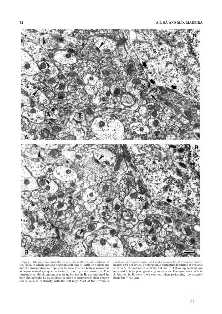
72 S.I. SÁ AND M.D. MADEIRA Fig. 2. Electron micrographs of two consecutive serial sections of the VMN, in which part of a neuronal cell body (s) with its nucleus (n) and the surrounding neuropil can be seen. The cell body is connected at asymmetrical synaptic contacts (arrows) by axon terminals. The terminals establishing synapses in A, but not in B, are indicated in both photographs by an asterisk. A spine or excrescence (long arrow) can be seen in continuity with the cell body. Most of the terminals contain clear round vesicles and make asymmetrical synapses (arrowheads) with <strong>de</strong>ndrites. The terminals contacting <strong>de</strong>ndrites at synaptic sites in A (the reference section), but not in B (look-up section), are indicated in both photographs by an asterisk. The synapses visible in A, but not in B, were those counted when performing the disector. Scale bar 0.5 m. 31
- Page 1: Susana Isabel Ferreira da Silva de
- Page 5: Esta investigação foi realizada n
- Page 9 and 10: Corpo Catedrático da Faculdade de
- Page 11: Doutor Manuel Machado Rodrigues Gom
- Page 15 and 16: AGRADECIMENTOS Em 2001, por sugest
- Page 17: ÍNDICE ITRODUÇÃO 1 TRABALHOS 15
- Page 21 and 22: Acção dos estrogénios no sistema
- Page 23 and 24: O núcleo ventromedial do hipotála
- Page 25 and 26: ventral e sistema mesencefálico de
- Page 27 and 28: 1991). São, no entanto, menos clar
- Page 29: o objectivo de aquilatar se as dose
- Page 33: TRABALHOS 15
- Page 37 and 38: Neuroscience 133 (2005) 919-924 NEU
- Page 39 and 40: enclosed by the Golgi complex (Fig.
- Page 41 and 42: Fig. 4. Graphic representation of t
- Page 43: Estrogen modulates the sexually dim
- Page 46 and 47: SYNAPTIC PLASTICITY IN THE VM NUCLE
- Page 50 and 51: SYNAPTIC PLASTICITY IN THE VM NUCLE
- Page 52 and 53: SYNAPTIC PLASTICITY IN THE VM NUCLE
- Page 54 and 55: SYNAPTIC PLASTICITY IN THE VM NUCLE
- Page 56 and 57: SYNAPTIC PLASTICITY IN THE VM NUCLE
- Page 59 and 60: Neuroscience 162 (2009) 307-316 EFF
- Page 61 and 62: ml) and chloral hydrate (40 mg/ml)
- Page 63 and 64: Table 1. Serum estradiol and proges
- Page 65 and 66: (Frankfurt and McEwen, 1991a; Segar
- Page 67 and 68: Canteras NS, Simerly RB, Swanson LW
- Page 69: Role of neural afferents as mediato
- Page 72 and 73: 2 1989; Krettek and Price, 1978), b
- Page 74 and 75: 4 rats. As expected, treatment of o
- Page 76 and 77: 6 estrogen-induced synapse formatio
- Page 78 and 79: 8 4.3. Hormonal determinations Bloo
- Page 80 and 81: 10 estrogen treatment. Brain Res. 2
- Page 83: DISCUSSÃO GERAL 65
- Page 86 and 87: Apesar de nas fêmeas os neurónios
- Page 88 and 89: influências excitatórias que sobr
- Page 90 and 91: estrogénios promovam a formação
- Page 93: CONCLUSÕES 75
- Page 97: REFERÊNCIAS 79
- Page 100 and 101:
Brown TJ, Clark AS, MacLusky NJ. Re
- Page 102 and 103:
Griffin GD, Flanagan-Cato LM. Estra
- Page 104 and 105:
Luiten PG, ter Horst GJ, Steffens A
- Page 106 and 107:
Ribeiro AC, Sawa E, Carren-LeSauter
- Page 108 and 109:
Voskuhl RR. Hormone-based therapies
- Page 111:
Com os resultados constantes desta
- Page 115:
The studies incorporated in the pre