

Susana Isabel Ferreira da Silva de Sá ESTROGÉNIOS E ...
Susana Isabel Ferreira da Silva de Sá ESTROGÉNIOS E ...
Susana Isabel Ferreira da Silva de Sá ESTROGÉNIOS E ...

You also want an ePaper? Increase the reach of your titles
YUMPU automatically turns print PDFs into web optimized ePapers that Google loves.
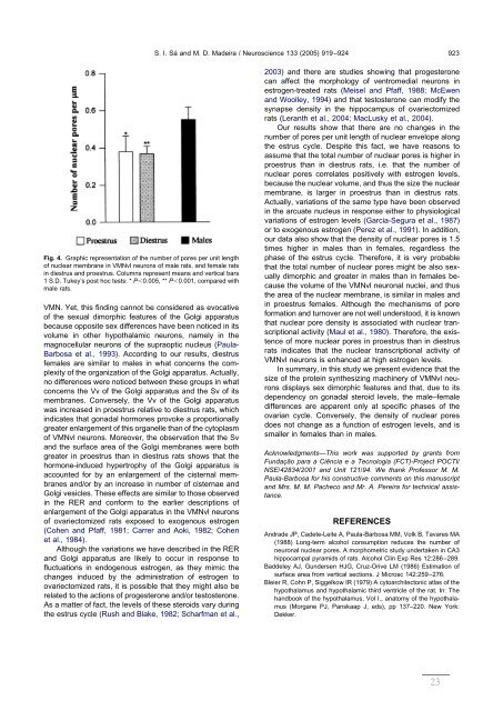
Fig. 4. Graphic representation of the number of pores per unit length<br />
of nuclear membrane in VMNvl neurons of male rats, and female rats<br />
in diestrus and proestrus. Columns represent means and vertical bars<br />
1 S.D. Tukey’s post hoc tests: * P0.005, ** P0.001, compared with<br />
male rats.<br />
VMN. Yet, this finding cannot be consi<strong>de</strong>red as evocative<br />
of the sexual dimorphic features of the Golgi apparatus<br />
because opposite sex differences have been noticed in its<br />
volume in other hypothalamic neurons, namely in the<br />
magnocellular neurons of the supraoptic nucleus (Paula-<br />
Barbosa et al., 1993). According to our results, diestrus<br />
females are similar to males in what concerns the complexity<br />
of the organization of the Golgi apparatus. Actually,<br />
no differences were noticed between these groups in what<br />
concerns the Vv of the Golgi apparatus and the Sv of its<br />
membranes. Conversely, the Vv of the Golgi apparatus<br />
was increased in proestrus relative to diestrus rats, which<br />
indicates that gona<strong>da</strong>l hormones provoke a proportionally<br />
greater enlargement of this organelle than of the cytoplasm<br />
of VMNvl neurons. Moreover, the observation that the Sv<br />
and the surface area of the Golgi membranes were both<br />
greater in proestrus than in diestrus rats shows that the<br />
hormone-induced hypertrophy of the Golgi apparatus is<br />
accounted for by an enlargement of the cisternal membranes<br />
and/or by an increase in number of cisternae and<br />
Golgi vesicles. These effects are similar to those observed<br />
in the RER and conform to the earlier <strong>de</strong>scriptions of<br />
enlargement of the Golgi apparatus in the VMNvl neurons<br />
of ovariectomized rats exposed to exogenous estrogen<br />
(Cohen and Pfaff, 1981; Carrer and Aoki, 1982; Cohen<br />
et al., 1984).<br />
Although the variations we have <strong>de</strong>scribed in the RER<br />
and Golgi apparatus are likely to occur in response to<br />
fluctuations in endogenous estrogen, as they mimic the<br />
changes induced by the administration of estrogen to<br />
ovariectomized rats, it is possible that they might also be<br />
related to the actions of progesterone and/or testosterone.<br />
As a matter of fact, the levels of these steroids vary during<br />
the estrus cycle (Rush and Blake, 1982; Scharfman et al.,<br />
S. I. <strong>Sá</strong> and M. D. Ma<strong>de</strong>ira / Neuroscience 133 (2005) 919–924 923<br />
2003) and there are studies showing that progesterone<br />
can affect the morphology of ventromedial neurons in<br />
estrogen-treated rats (Meisel and Pfaff, 1988; McEwen<br />
and Woolley, 1994) and that testosterone can modify the<br />
synapse <strong>de</strong>nsity in the hippocampus of ovariectomized<br />
rats (Leranth et al., 2004; MacLusky et al., 2004).<br />
Our results show that there are no changes in the<br />
number of pores per unit length of nuclear envelope along<br />
the estrus cycle. Despite this fact, we have reasons to<br />
assume that the total number of nuclear pores is higher in<br />
proestrus than in diestrus rats, i.e. that the number of<br />
nuclear pores correlates positively with estrogen levels,<br />
because the nuclear volume, and thus the size the nuclear<br />
membrane, is larger in proestrus than in diestrus rats.<br />
Actually, variations of the same type have been observed<br />
in the arcuate nucleus in response either to physiological<br />
variations of estrogen levels (Garcia-Segura et al., 1987)<br />
or to exogenous estrogen (Perez et al., 1991). In addition,<br />
our <strong>da</strong>ta also show that the <strong>de</strong>nsity of nuclear pores is 1.5<br />
times higher in males than in females, regardless the<br />
phase of the estrus cycle. Therefore, it is very probable<br />
that the total number of nuclear pores might be also sexually<br />
dimorphic and greater in males than in females because<br />
the volume of the VMNvl neuronal nuclei, and thus<br />
the area of the nuclear membrane, is similar in males and<br />
in proestrus females. Although the mechanisms of pore<br />
formation and turnover are not well un<strong>de</strong>rstood, it is known<br />
that nuclear pore <strong>de</strong>nsity is associated with nuclear transcriptional<br />
activity (Maul et al., 1980). Therefore, the existence<br />
of more nuclear pores in proestrus than in diestrus<br />
rats indicates that the nuclear transcriptional activity of<br />
VMNvl neurons is enhanced at high estrogen levels.<br />
In summary, in this study we present evi<strong>de</strong>nce that the<br />
size of the protein synthesizing machinery of VMNvl neurons<br />
displays sex dimorphic features and that, due to its<br />
<strong>de</strong>pen<strong>de</strong>ncy on gona<strong>da</strong>l steroid levels, the male–female<br />
differences are apparent only at specific phases of the<br />
ovarian cycle. Conversely, the <strong>de</strong>nsity of nuclear pores<br />
does not change as a function of estrogen levels, and is<br />
smaller in females than in males.<br />
Acknowledgments—This work was supported by grants from<br />
Fun<strong>da</strong>ção para a Ciência e a Tecnologia (FCT)-Project POCTI/<br />
NSE/42834/2001 and Unit 121/94. We thank Professor M. M.<br />
Paula-Barbosa for his constructive comments on this manuscript<br />
and Mrs. M. M. Pacheco and Mr. A. Pereira for technical assistance.<br />
REFERENCES<br />
Andra<strong>de</strong> JP, Ca<strong>de</strong>te-Leite A, Paula-Barbosa MM, Volk B, Tavares MA<br />
(1988) Long-term alcohol consumption reduces the number of<br />
neuronal nuclear pores. A morphometric study un<strong>de</strong>rtaken in CA3<br />
hippocampal pyramids of rats. Alcohol Clin Exp Res 12:286–289.<br />
Bad<strong>de</strong>ley AJ, Gun<strong>de</strong>rsen HJG, Cruz-Orive LM (1986) Estimation of<br />
surface area from vertical sections. J Microsc 142:259–276.<br />
Bleier R, Cohn P, Siggelkow IR (1979) A cytoarchitectonic atlas of the<br />
hypothalamus and hypothalamic third ventricle of the rat. In: The<br />
handbook of the hypothalamus, Vol I., anatomy of the hypothalamus<br />
(Morgane PJ, Panskaap J, eds), pp 137–220. New York:<br />
Dekker.<br />
23


