Unconventional dissolution methodologies - Wiley Online Library
Unconventional dissolution methodologies - Wiley Online Library
Unconventional dissolution methodologies - Wiley Online Library

Create successful ePaper yourself
Turn your PDF publications into a flip-book with our unique Google optimized e-Paper software.
MINIREVIEW<br />
<strong>Unconventional</strong> Dissolution Methodologies<br />
VINESS PILLAY AND REZA FASSIHI*<br />
Contribution from Temple University, School of Pharmacy, Department of Pharmaceutical Sciences, 3307 North Broad Street,<br />
Philadelphia, Pennsylvania 19140.<br />
Received April 28, 1999. Final revised manuscript received June 30, 1999.<br />
Accepted for publication July 1, 1999.<br />
Introduction<br />
In line with the key focus of recent publications 1-3<br />
emerging from the labs of Dressman, Amidon, and Shah,<br />
and in conjunction with the aims of both the FDA and US<br />
Pharmacopoeial Convention to improve and possibly develop<br />
alternative <strong>dissolution</strong> testing procedures as well as<br />
techniques for data analysis, this work considers an<br />
overview of the constantly changing areas of in vitro<br />
<strong>dissolution</strong> research in the evaluation of novel oral drug<br />
delivery systems. Over the years, <strong>dissolution</strong> testing has<br />
been employed as a quality control procedure in pharmaceutical<br />
production, in product development to assist in<br />
selection of a candidate formulation, in research to detect<br />
the influence of critical manufacturing variables such as<br />
binder effect, 4 mixing effect, 5,6 granulation procedure, 7<br />
coating parameters, 8,9 excipient type, 10 and/or in comparative<br />
studies of different formulations, 11 in in vitro-in vivo<br />
correlations, 12-15 and possibly as an in vivo surrogate under<br />
strictly defined conditions. 16,17 It therefore becomes apparent<br />
that sensitive and reproducible <strong>dissolution</strong> data derived<br />
from physicochemically and hydrodynamically defined<br />
conditions are necessary in order to compare various in<br />
vitro <strong>dissolution</strong> data and be able to use such results as a<br />
surrogate for possible in vivo bioavailability, bioequivalence<br />
testing, and in vitro-in vivo correlations (IVIVC). However,<br />
the influence of technological differences and process<br />
variables involved during manufacturing on <strong>dissolution</strong><br />
rate often complicates the decision making process in<br />
selection of the appropriate <strong>dissolution</strong> method and subsequent<br />
data interpretation technique. Moreover, Skoug<br />
and co-workers 18 stressed that this consequence is the<br />
* Corresponding author. Tel. 215-707-7670. Fax 215-707-3678.<br />
e-mail. afassihi@vm.temple.edu.<br />
September 1999<br />
Volume 88, Number 9<br />
reason <strong>dissolution</strong> studies and the defined specifications<br />
so often generate strong interest during regulatory review<br />
of solid oral dosage forms. As a result, the Center for Drug<br />
Evaluation and Research (CDER) at the Food and Drug<br />
Administration (FDA) has recently released guidelines<br />
called Scale-Up and Post Approval Changes, commonly<br />
referred to as SUPAC 19 and Extended Release Solid Oral<br />
Dosage Forms: Development, Evaluation and Application<br />
of In vitro/In vivo Correlations, commonly known as<br />
IVIVC, 20 to be used by the pharmaceutical sponsor in<br />
quality assurance and specific postapproval changes and<br />
to demonstrate that the “<strong>dissolution</strong> profiles of prechange<br />
product and postchange product are similar”. The impact<br />
of the process and establishment of an in vitro and in vivo<br />
performance (IVIVR) as a critical stage in development of<br />
oral controlled release products has been further highlighted<br />
in the recent work of Devane and Butler. 21<br />
The current sophistication in formulation of new modified<br />
release drug delivery systems and associated diversity<br />
in dosage form design necessitates the development of new<br />
procedures or appropriate modification to the existing<br />
apparatus as an alternative for <strong>dissolution</strong> measurements.<br />
22,23 More recently, it has been shown that the<br />
complex hydrodynamics and three-dimensional fluid flow<br />
pattern produced by the USP paddle 24 within different<br />
regions of the <strong>dissolution</strong> vessel varies significantly with<br />
a relatively more stagnant region at the bottom portion of<br />
the vessel. 25,26 Consequently, to mimic and more closely<br />
reflect the possible in vivo dosage form surface exposure,<br />
have reliable <strong>dissolution</strong> data, and be able to discriminate<br />
between release behavior of various modified release<br />
formulations, it becomes apparent that a better understanding<br />
of the role of hydrodynamics in relation to delivery<br />
© 1999, American Chemical Society and 10.1021/js990139b CCC: $18.00 Journal of Pharmaceutical Sciences / 843<br />
American Pharmaceutical Association Published on Web 07/31/1999<br />
Vol. 88, No. 9, September 1999
system and release mechanisms are necessary for the<br />
development of alternative <strong>dissolution</strong> methods. 22,23<br />
In general, the design of alternative <strong>dissolution</strong> methods<br />
may be approached in one of two ways or a combination.<br />
First, the method might consider the establishment of in<br />
vitro testing conditions similar to an actual in vivo setting.<br />
This approach may encompass instrumental developments<br />
mimicking gastrointestinal peristaltic motion with combination<br />
of flow-through methods for ensuring sink condition<br />
maintenance. All media used in testing of dosage forms<br />
should also be very similar to fluids comprising the<br />
gastrointestinal compartment particularly with respect to<br />
composition. Furthermore, other technical considerations<br />
may incorporate fabrication materials that are in contact<br />
with the dosage to possibly behave as pliable, flexible<br />
gastrointestinal tissue. Irrespective of the extent to which<br />
the ideal testing conditions are achieved, this approach<br />
becomes a mammoth task flawed by numerous inconsistencies.<br />
The second and more amenable approach is to<br />
establish in vitro <strong>dissolution</strong> conditions that may provide<br />
drug release profiles very similar to deconvoluted (i.e.,<br />
fraction absorbed) blood plasma profiles through different<br />
levels of correlation as described in the USP. This entails<br />
an establishment of in vitro-in vivo correlation through<br />
manipulation of process variables such as selection of<br />
appropriate <strong>dissolution</strong> media systems taking into consideration<br />
sink condition maintenance and/or manipulation<br />
of fluid flow hydrodynamics by use of additional devices<br />
within the standard <strong>dissolution</strong> vessel. A useful example<br />
of the latter suggestion is the reported ring/mesh assembly<br />
used for the in vitro enhancement of dosage form positioning<br />
and surface area exposure in order to simulate hydrodynamically<br />
comparable conditions to that of in vivo. 27,28<br />
Generally reviews, theoretical papers, and research<br />
publications on the subject of <strong>dissolution</strong> have focused on<br />
standardizing, expanding, and developing useful mathematical<br />
and physical models principally due to apparent<br />
unresolved mechanistic complexities in the thermodynamic<br />
sense. 29-34 On the basis of such past in-depth analyses,<br />
researchers may presently use those concepts in experimental<br />
design of alternative <strong>dissolution</strong> methods. Therefore,<br />
to consolidate the principles governing the <strong>dissolution</strong><br />
process, this article will attempt to provide a brief theoretical<br />
perspective of <strong>dissolution</strong> theory and associated concepts<br />
used in drug release from oral dosage forms. In<br />
keeping with the significant research activities with hydrophilic<br />
swellable matrixes, priority will be given to the<br />
optimization of <strong>dissolution</strong> studies pertaining to modified/<br />
controlled release drug delivery from such systems. A<br />
summary of attempts to improve the currently recommended<br />
USP, 23 <strong>dissolution</strong> <strong>methodologies</strong> for swellable<br />
sticking and swellable floatable delivery systems is provided,<br />
and the approaches recently used to overcome the<br />
associated difficulties are referred to in the text. 27 In<br />
addition, aspects relating to the lack of an official in vitro<br />
<strong>dissolution</strong> test method for lipid-filled capsules and the<br />
strategy used to solve this dilemma is discussed. 28 A critical<br />
review on the advantages and limitations of commonly used<br />
mathematical and statistical parameters for comparison<br />
of <strong>dissolution</strong> data, including the newly developed FDArecommended<br />
f2 similarity factor and f1 difference factor,<br />
also follows. The use of “chemical stabilizers” in <strong>dissolution</strong><br />
testing of drugs (such as ascorbic acid), normally susceptible<br />
to rapid decomposition in solution, is discussed for a<br />
gel-based controlled release product. We also briefly examine<br />
the problems associated with non-UV responsive<br />
drugs and the implications of colorimetric adaptation for<br />
the evaluation of release characteristics of both soluble and<br />
insoluble active substances.<br />
844 / Journal of Pharmaceutical Sciences<br />
Vol. 88, No. 9, September 1999<br />
Fundamental Dissolution Theories<br />
Dissolution of a solute is a multistep process involving<br />
heterogeneous reactions/interactions between the phases<br />
of the solute-solute, solute-solvent, solvent-solvent, and<br />
at the solute-solvent interface. As one of the most commonly<br />
known mass transfer rate processes, the component<br />
heterogeneous reactions may broadly be categorized into<br />
(i) diffusion or convective transport of the solute from the<br />
interface to the bulk phase; and (ii) the rate of solute<br />
liberation and transport from and across the interfacial<br />
boundaries.<br />
Various researchers in the field have developed theories<br />
to define the <strong>dissolution</strong> process and these have been<br />
comprehensively reported. 35-37 As three of the pioneering<br />
theories in the field, this review will not be complete<br />
without a brief description of the diffusion layer model,<br />
surface renewal theory, and limited solvation theory.<br />
Table 1 concisely depicts the principal mathematical<br />
equations associated with the theories and highlights key<br />
points regarding the theory. Selected information is derived<br />
from the text of Abdou 35 for diffusion layer and surface<br />
renewal theories. The limited solvation theory is presented<br />
from the original work of Goldberg and co-workers. 38<br />
In the diffusion layer theory, the simplest model used<br />
to describe <strong>dissolution</strong> makes use of a single crystal in a<br />
nonreactive environment. The initial step in solution of the<br />
solid (solute or crystal) at the interface is usually very rapid<br />
and results in the formation of a saturated stagnant layer<br />
around the particle. This is contrasted by the second<br />
diffusion step that is slow and becomes the rate-limiting<br />
step in the <strong>dissolution</strong> process. In particular, the Noyes-<br />
Whitney equation (eq 3) illustrates that one of the main<br />
factors determining the rate of <strong>dissolution</strong> is drug solubility.<br />
37 From this it is understood that in vivo the <strong>dissolution</strong><br />
process may become the rate-limiting step if the rate of<br />
solution is much slower than the rate of absorption. This<br />
may be the case when the drug in question has a very low<br />
solubility at both gastric and intestinal pH.<br />
The surface renewal theory assumes an equilibrium at<br />
the solute-solution interface is attained and that the ratelimiting<br />
step in the <strong>dissolution</strong> process is mass transport.<br />
The model is thought of as being continually exposed to<br />
fresh <strong>dissolution</strong> medium. The agitating medium consists<br />
of numerous eddies or packets into which the solute<br />
diffuses and is carried to the bulk medium. Due to the<br />
turbulence at the surface of the solute, there is no boundary<br />
layer and therefore no stagnant film layer. In other words<br />
the surface is continually being replaced with fresh medium.<br />
The limited solvation theory 38 predicts that a crystal<br />
undergoes <strong>dissolution</strong> through an interfacial process in the<br />
dissolving medium. The true surface area of the crystal<br />
must be considered since each face of the crystal may have<br />
a different interfacial barrier. Hence each surface may<br />
provide a different contribution to the <strong>dissolution</strong> process.<br />
Basic Theories of Dissolution Profile Analysis<br />
Table 2 in summary depicts four prominent theories used<br />
in <strong>dissolution</strong> profile analysis, namely Wagner’s, 39<br />
Kitazawa’s, 40-42 El-Yazigi’s, 43 and Carstensen’s. 44<br />
Wagner’s theory 39 for the interpretation of percent<br />
dissolved-time plots of tablets and capsules relates the<br />
apparent first-order kinetics under sink conditions to the<br />
fact that a percent dissolved value at a certain time may<br />
be equivalent to the percent surface area generated at the<br />
same time. Kitazawa’s theory 40-42 showed that the biphasic<br />
straight lines were obtained from plots of ln cs/(cs - c) vst.<br />
The first segment was due to tablet disintegration or
Table 1sSummary of Fundamental Dissolution Theories a<br />
theory b equations associated characteristics<br />
diffusion layer 35<br />
Fick’s First Law Jix ) −Di (∂ci/∂x) (1) Considers diffusion only under steady-state conditions.<br />
Fick’s Second Law ∂c/∂t ) D (∂ 2 c/∂x 2 ) (2) Used when drug concentration decreases with time; hence,<br />
considers non-steady state conditions.<br />
Noyes and Whitney dc/dt ) K (cs − ct) (3) Description of drug <strong>dissolution</strong> based on constant surface area.<br />
Brunner and Tolloczko dc/dt ) kS (cs − ct) (4) Manipulation of Noyes−Whitney’s eq 3 by incorporation of surface area<br />
term S. Proposed the formation of a stagnant layer around the dissolving<br />
particle, a layer through which solute diffuses through into the bulk.<br />
Nernst -<br />
Brunner dc/dt ) kDS/vh (cs − ct)<br />
If ct , cs (i.e.
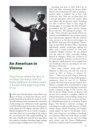
Figure 1sSchematic of drug delivery system positioning within a <strong>dissolution</strong><br />
vessel: (A) floatable system close to the paddle shaft; (B) floatable system<br />
under the ring/mesh assembly; (C) sticking system adhering to bottom of<br />
<strong>dissolution</strong> vessel; (D) sticking system placed over the ring/mesh assembly.<br />
(Modified from Pillay and Fassihi 27 ).<br />
progress and expansion of “generic” industries. The application<br />
of new polymeric materials to enhance drug<br />
delivery, particularly in certain aspects of controlled release,<br />
has from our experience led to the recognition of<br />
limitations in the versatility of the currently recommended<br />
USP-23 <strong>dissolution</strong> methods. 24 This aspect has been adequately<br />
demonstrated in the recent publications showing<br />
the benefits of alternative <strong>dissolution</strong> approaches to the<br />
currently recommended USP-23 methods applied to swellable<br />
sticking and swellable floatable delivery systems, the<br />
summary of which is presented in the following sections.<br />
Alternative Dissolution Methods and Examples<br />
(i) Application of Ring/Mesh Assembly for Determination<br />
of Release Profiles from Swellable Lowand<br />
High-Density MatricessAs pointed out above, new<br />
modified release formulation technologies and diversity in<br />
dosage form design necessitates the development of new<br />
procedures or appropriate modification to the existing<br />
apparatus as alternative <strong>dissolution</strong> measurement methods.<br />
22,23,27,28 For example, in <strong>dissolution</strong> studies of lowdensity<br />
swellable, floatable controlled release drug delivery<br />
systems, often position of the dosage form appears to be<br />
close to the paddle shaft and liquid surface as illustrated<br />
in Figure 1A (i.e., in schematic). On the other hand, when<br />
a sinker such as the USP-recommended 24 “wire helix” is<br />
wound around the delivery system, position of the dosage<br />
form will vary within the vessel (inconsistent hydrodynamics),<br />
and its free three-dimensional swelling process would<br />
be adversely affected and difficult to control. 45 Furthermore,<br />
and contrary to floatable dosage forms, many drug<br />
delivery systems having high density tend to adhere (stick)<br />
to the bottom of the <strong>dissolution</strong> vessel as illustrated in<br />
Figure 1C. This problem of sticking is accentuated with<br />
the use of swellable polymers such as hydroxypropylmethylcellulose,<br />
hydroxypropylcellulose, and poly(ethylene oxide).<br />
Under these conditions, the lower surface of the dosage<br />
form is not exposed to the <strong>dissolution</strong> medium, and drug<br />
release is limited to the exposed surfaces only. Similar<br />
phenomena are unlikely to occur in the human gastrointes-<br />
846 / Journal of Pharmaceutical Sciences<br />
Vol. 88, No. 9, September 1999<br />
tinal tract. Furthermore, it may be anticipated that the<br />
USP 23 Apparatus 1 (rotating basket method) may be used<br />
to surmount this problem by allowing complete immersion<br />
of the dosage form and full surface area exposure. However,<br />
the early work of Withey and Bowker 46 on fluid flow<br />
dynamics clearly show that the rotating basket produces<br />
nonreproducible flow patterns with least fluid flow in the<br />
axial plane directly above and below the basket as well as<br />
within the basket. In addition, our experience has shown<br />
that some swellable delivery systems tend to expand<br />
greater than the diameter of the basket and often float<br />
against the flat base of the rotating shaft. These events<br />
will result in restriction of swelling and erosion processes,<br />
as well as limited surface exposure to <strong>dissolution</strong> medium.<br />
More recently, it has been shown that the complex hydrodynamics<br />
and three-dimensional fluid flow pattern produced<br />
by the USP paddle within different regions of the<br />
<strong>dissolution</strong> vessel varies significantly with a relatively more<br />
stagnant region at the bottom portion of the vessel. 25,26<br />
Consequently, to mimic and more closely reflect the possible<br />
in vivo dosage form surface exposure, have reliable<br />
<strong>dissolution</strong> data, and be able to discriminate between<br />
release behavior of various modified release formulations,<br />
a better understanding of the role of hydrodynamics,<br />
delivery system, and release mechanisms together with the<br />
development of alternative <strong>dissolution</strong> methods is apparent.<br />
22,23<br />
Recently Pillay and Fassihi 27 have used a new device<br />
(ring/mesh assembly) in conjunction with the paddle method<br />
to study the influence of the position of various dosage<br />
forms on release behavior and evaluated the release<br />
profiles obtained with such modification with those derived<br />
under standard <strong>dissolution</strong> conditions including the USP<br />
23- recommended helical wire sinker used for swellable<br />
floatable delivery systems (see schematic Figure 1). Model<br />
drugs used included theophylline (0.85% water soluble at<br />
25 °C) and diltiazem hydrochloride (>50% water soluble<br />
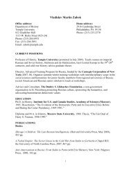
at 25 °C). It was shown that for a low water-soluble drug<br />
such as theophylline, full surface exposure was necessary<br />
in order to accomplish complete drug release from the<br />
delivery system (Figure 2a). This was accomplished by<br />
placing the delivery system over the ring/mesh assembly<br />
as depicted in Figure 1. This surface area exposure<br />
phenomenon was also applicable to a floatable theophylline<br />
system. Application of the USP-recommended helical wire<br />
sinker to the swellable floatable theophylline delivery<br />
system appeared to inhibit the three-dimensional swelling<br />
process of the dosage form and consequently suppressed<br />
drug release from the formulation (Figure 2b). Such a<br />
limitation was alleviated by positioning the delivery system<br />
below the ring/mesh assembly (Figure 1). In the case of<br />
diltiazem hydrochloride (solubility in water >50% at 25 °C)<br />
similar release differences as in the case of theophylline<br />
(p < 0.05) were also observed when the sticking delivery<br />
system was placed either in the vessel as recommended<br />
by the USP 23 standard method or when it was positioned<br />
over the ring/mesh assembly (Figure 2c). However, in the<br />
case of a swellable floatable system containing the highly<br />
soluble drug diltiazem hydrochloride, no differences in<br />
release were found by employing the helical wire sinker,<br />
placing the dosage form in the vessel as such or when the<br />
delivery system was fully submerged under the ring/mesh<br />
assembly (see Figure 2d). Hence, the nature of drug release<br />
behavior from swellable floatable systems depended both<br />
on full surface exposure and unhindered swelling as well<br />
as drug solubility.<br />
(ii) Evaluation of Drug Release from Lipid-Filled<br />
Hardshell or Softgel CapsulessConsiderable interest<br />
has been shown in the formulation of lipid-filled capsules<br />
for the enhancement of either in vivo <strong>dissolution</strong> rates or
Figure 2s(a) Theophylline release from a swellable sticking drug delivery<br />
system. Key: b, delivery system placed over the ring/mesh assembly for full<br />
surface exposure to the <strong>dissolution</strong> medium; O, delivery system dropped into<br />
the vessel with one surface sticking to the bottom of the vessel. (b) Theophylline<br />
release from a swellable floatable drug delivery system. Key: O, delivery<br />
system placed under the ring/mesh assembly to prevent flotation to the surface<br />
of the <strong>dissolution</strong> medium; b, delivery system dropped into the vessel and<br />
allowed to float at the surface of the <strong>dissolution</strong> medium; 1, delivery system<br />
enclosed within a helical wire sinker to prevent flotation to the surface of the<br />
<strong>dissolution</strong> medium. (c) Diltiazem hydrochloride release from a swellable sticking<br />
drug delivery system. Key: b, delivery system placed over the ring/mesh<br />
assembly for full surface exposure to the <strong>dissolution</strong> medium; O, delivery<br />
system dropped into the vessel with one surface sticking to the bottom of the<br />
vessel. (d) Diltiazem hydrochloride release from a swellable floatable drug<br />
delivery system. Key: O, delivery system placed under the ring/mesh assembly<br />
to prevent flotation to the surface of the <strong>dissolution</strong> medium; b, delivery system<br />
dropped into the vessel and allowed to float at the surface of the <strong>dissolution</strong><br />
medium; 1, delivery system enclosed within a helical wire sinker to prevent<br />
flotation to the surface of the <strong>dissolution</strong> medium. (N ) 3 in all of the above<br />
cases; standard deviations are not shown because they are smaller than the<br />
symbol size; modified from Pillay and Fassihi 27 ).<br />
bioavailability of bioactive agents. 28,47,48 To ascertain that<br />
drug is completely delivered from its formulation over an<br />
appropriate time period and is able to reach and cross the<br />
gut wall, an aqueous environment similar to the gut<br />
luminal fluid and a sink resembling the lipoidal nature of<br />
the gastrointestinal mucosa becomes a necessary condition<br />
for the development of a prognostic in vitro test method.<br />
Both softgel and hard shell capsules filled with vehicles<br />
which are capable of self-emulsification (due to their ability<br />
to form fine oil-in-water emulsions) offer great potential<br />
for the oral delivery of insoluble hydrophobic and poorly<br />
absorbable drugs. However, in vitro evaluation of such<br />
dosage forms have thus far been problematic, since no<br />
official <strong>dissolution</strong> method for lipid-based formulations as<br />
yet has been established. This may be due to the relative<br />
difficulties associated with the evaluation methodology of<br />
lipid-based formulations. A greater challenge is presented<br />
when poorly soluble drugs in a lipid-based vehicle are<br />
presented as lipid-filled capsules for enhancement of<br />
solubility. Such matrixes, however, are not soluble in<br />
commonly used aqueous <strong>dissolution</strong> media. With some<br />
conventional <strong>dissolution</strong> methods, the use of surfactants 48-50<br />
or hydro-alcoholic media 47,50 have been recommended.<br />
However, it is speculated that exposure of the gelatin shell<br />
to such media may induce physical and/or chemical changes,<br />
arising either through complex formation or cross-linking<br />
reactions. Typically, sodium lauryl sulfate (SLS), an anionic<br />
surfactant, is often employed in <strong>dissolution</strong> media; however,<br />
many researchers fail to recognize that SLS will bind<br />
to cationic charges of gelatin at pH values equivalent to<br />
Figure 3sA comparative illustration of the four <strong>dissolution</strong> designs employed<br />
for the induction of different hydrodynamic conditions. Left panel: Key: I )<br />
Position of either rotating basket or paddle with hydrodynamic arrangements<br />
as follows. Design A: Centrally positioned in aqueous phase between<br />
boundaries of organic phase and bottom of vessel. Design B: Halfway at<br />
air/organic phase interface. Design C: Halfway at organic/aqueous phase<br />
interface. Design D: Centrally positioned in aqueous phase between boundaries<br />
of organic phase and ring/mesh assembly. Stirring rate of 75 rpm was used<br />
in all designs with exception of design D where in addition 100 rpm was also<br />
tested. II ) organic phase, i.e., 100 mL 1-octanol. III ) aqueous phase i.e.,<br />
phosphate buffer: 400 mL for design A, 200 mL for designs B and C, 300<br />
mL for design D. Note that 400 and 300 mL of phosphate buffer were employed<br />
in designs A and D to ensure that basket and paddle are fully immersed in<br />
aqueous phase. IV ) ring/mesh assembly. V ) Position of capsule either<br />
within basket or below ring/mesh assembly. Right panel: Transfer profile of<br />
lipid-based nifedipine capsule preparation derived under different hydrodynamic<br />
conditions and designs as described above (N ) 3). (a) Profile obtained using<br />
the USP 23 rotating basket method at 75 rpm (<strong>dissolution</strong> design A). (b) Profile<br />
obtained using paddle over ring/mesh assembly halfway at air/organic interface<br />
at 75 rpm (<strong>dissolution</strong> design B). (c) Profile obtained using paddle over ring/<br />
mesh assembly halfway at organic/aqueous interface at 75 rpm (<strong>dissolution</strong><br />
design C). (d) Profile obtained using paddle over ring/mesh assembly in<br />
aqueous phase at 100 rpm (<strong>dissolution</strong> design D). (Modified from Pillay and<br />
Fassihi 28 ).<br />
Journal of Pharmaceutical Sciences / 847<br />
Vol. 88, No. 9, September 1999
gastric pH. These interactions may influence the solubility<br />
and disintegration time of the shell and/ or true release<br />
potential of the product. Therefore, difficulties that may<br />
be experienced include, but are not limited to, exposure of<br />
gelatin shell to the organic phase, separation of poorly<br />
soluble drugs as metastable liquid crystals, lack of reproducibility<br />
in <strong>dissolution</strong> data, dosage form and lipid flotation<br />
in the <strong>dissolution</strong> vessel, etc.<br />
In a recent report 28,51 a method which encompasses the<br />
development, design, and use of a modified two-phase<br />
<strong>dissolution</strong> media system by a novel approach for testing<br />
of either soft or hard shell lipid-filled gelatin capsules was<br />
proposed. Nifedipine was chosen as the model compound<br />
due to its water-insoluble nature (
al. in their recent work on <strong>dissolution</strong> profile analysis. 2<br />
Over the years, scientists have given much consideration<br />
to use of the Weibull function, 58,59 a model-dependent<br />
approach, as depicted in eq 18:<br />
m ) 1 - exp[-(t - T 1 ) b/a ] (18)<br />
where m is the percent dissolved at time t, a is the time<br />
scale parameter, b is the shape factor, and T1 is the location<br />
parameter. The shape factor, b, qualitatively defines the<br />
curve, i.e., when b ) 1, the curve becomes a simple firstorder<br />
exponential. If b > 1, the drug release rate is slow<br />
initially followed by an increase in release rate. The shape<br />
factor also provides qualitative information on diffusion and<br />
disintegration processes. The effective surface area for<br />
<strong>dissolution</strong> will be maximum after a certain time at the<br />
outset when b > 1, while when b e 1 no disintegration<br />
occurs at all, and the rate of <strong>dissolution</strong> will decrease<br />
steadily. The scale factor, a, provides a quantitative evaluation<br />
by differentiating the curves along the time axis. As<br />
pointed out by Polli and co-workers, 60 the Weibull model<br />
becomes fraught with an element of subjectivity because<br />
the judgment of the researcher is used in devising criteria<br />
for an adequate model fit. This further introduces a lack<br />
of metric sensitivity since as with all model-dependent<br />
approaches, no acceptance limits have been set as standard.<br />
In addition, the success of this approach relies on linearizing<br />
the <strong>dissolution</strong> data. However, a considerable curvature<br />
may be found in the upper region of the plot if the<br />
accumulated fraction of drug dissolved is not 1.0. In<br />
addition, the location parameter, which represents the lag<br />
time before the actual onset of the <strong>dissolution</strong> process, has<br />
to be estimated indirectly by a least-squares analysis or a<br />
graphical trial and error technique.<br />
Therefore, it may be useful to consider a second category<br />
of analyses, i.e., model-independent treatment of <strong>dissolution</strong><br />
data in order to determine the release profile similarity<br />
and concomitant dissimilarity where applicable. In this<br />
work we will focus on two classes of model-independency,<br />
namely time point or ratio test approaches and pairwise<br />
models. Model-independency, previously described by Rescigno,<br />
61 in general would generate results for which the<br />
values do not depend on the selection of the specific<br />
parameter for fitting the data, but are dependent on the<br />
sampling times t1, t2,..., tn and on an appropriate coefficient<br />
wj representing the weight that the sampling time tj has<br />
in the determination of the specific fitted functions.<br />
In the time point/ratio test approach the t50%, t70%, and<br />
t90% values as well as the mean <strong>dissolution</strong> times (MDT50%,<br />
MDT70%, MDT90%) are calculated for each formulation in<br />
each of the replicate <strong>dissolution</strong> measurements. Application<br />
of MDT provides more accurate drug release rate as<br />
compared to the tx% approach and is determined as the sum<br />
of the individual periods of time during which a specific<br />
fraction of the total dose is released. 62<br />
The following equation (eq 19) may be used to calculate<br />
the MDT for each percentage point:<br />
MDT ) ∑ i)1<br />
n Mt tˆ i<br />
M∞<br />
(19)<br />
where Mt is the fraction of dose released in time tˆi ) (ti +<br />
ti - 1)/2, and M∞ corresponds to the loading dose.<br />
In the pairwise approach, determination of a “difference<br />
factor, f1” 63 and “similarity factor, f2” 19,20,63 (as outlined in<br />
the SUPAC and IVIVC guidelines) using the mean percentage<br />
released values can be performed by using eqs 20<br />
and 21. To validate the acceptance of the f1 and f2 fit factors,<br />
calculations should be performed on the individual dis-<br />
solution data of each formulation, which should reflect no<br />
statistical difference (p > 0.05) to the mean <strong>dissolution</strong><br />
values.<br />
The recent guidelines by the CDER at the FDA 20<br />
describes the necessary criteria for granting biowaivers for<br />
specific changes in drug product manufacturing such as<br />
formulation changes or even changes in manufacturing<br />
site. To this end, the guidelines and specific published<br />
work 63 on extended release solid oral dosage forms describe<br />
the mathematical treatment of <strong>dissolution</strong> data derived<br />
from the pre- and postapproval changes by comparing their<br />
release profiles using the “similarity factor, f2” which may<br />
be defined as follows:<br />
1<br />
f2 ) 50 log{[<br />
1 +<br />
n<br />
n ∑ t)1<br />
wt (Rt - Ti ) 2]<br />
-0.5<br />
× 100} (20)<br />
where n is the number of <strong>dissolution</strong> time points, wt is an<br />
optional weight factor, Rt is the reference assay at time<br />
point t, and Tt is the test assay at time point t. Note that<br />
the “reference” and “test” products may be identical formulations.<br />
Optimization of release profiles may be achieved<br />
by the appropriate adoption of standard or alternative<br />
<strong>dissolution</strong> methods. The f2 value between 50 and 100<br />
suggests that the <strong>dissolution</strong> profiles are similar. The f2<br />
value of 100 suggests that the test and reference release<br />
profiles are identical, and as the value becomes smaller,<br />
the dissimilarity between release profiles increases. Equation<br />
20 is a logarithmic transformation of the sum of<br />
squared error. It takes the average sums of squares of the<br />
difference between test and reference profiles and fits the<br />
result between 0 and 100. It is important to note that eq<br />
20 is for the comparison of <strong>dissolution</strong> curves in which the<br />
average difference between Rt and Tt is 1. If all values are<br />
treated equally, then wt ) 1.0.<br />
In addition, Moore and Flanner63 in their recent work<br />
also describe an f1 fit factor or “difference factor” as follows:<br />
}<br />
n<br />
{∑|Rt - Tt |<br />
t)1<br />
f1 ) × 100% (21)<br />
n<br />
∑Rt t)1<br />
where f1 describes the relative error between two <strong>dissolution</strong><br />
profiles. “It approximates the percent error between<br />
two curves. The percent error is zero when the test and<br />
reference profiles are identical and increases proportionally<br />
with the dissimilarity between the two profiles”.<br />
Advantages and Limitations Associated with the<br />
Time Point/Ratio Test and Pairwise Approaches<br />
Used in Dissolution Data Treatment<br />
The time point approach (tx%) for the interpretation of<br />
<strong>dissolution</strong> data appears to be inadequate for complete<br />
characterization of the profiles, since comparison of profiles<br />
not following a single path or void of crossover are not<br />
uncommon. Consequently, the choice of single data points<br />
for the calculation of meaningful <strong>dissolution</strong> values are<br />
questionable in the case of such issues revolving around<br />
product bioequivalence. Similarly, the choice of MDT50%,<br />
MDT70%, and MDT90% may not always provide accurate<br />
information when profile crossover is too close. In the case<br />
of immediate release products such crossover in drug<br />
Journal of Pharmaceutical Sciences / 849<br />
Vol. 88, No. 9, September 1999
elease profiles may not present a major problem since the<br />
time scale of the release event is very short, often in the<br />
range of a few minutes to hours. On the contrary, such<br />
occurrences with controlled release products may have a<br />
significant impact on both quality assurance during product<br />
development and establishment of in vitro-in vivo correlations.<br />
Therefore, in the characterization of such <strong>dissolution</strong><br />
profiles, a more in-depth analysis of data could<br />
provide a better description of the overall release profile.<br />
Polli and co-workers 60 recently undertook an extensive<br />
study to mathematically and statistically evaluate various<br />
methods for the comparison of <strong>dissolution</strong> profiles of<br />
conventional metoprolol tartrate dosage forms for the<br />
demonstration of IVIVC. One of the selected methods<br />
included the application of the “similarity factor, f2”. In this<br />
work 60 as well as in studies from our lab, 27,28 it is shown<br />
that the similarity factor, f2” is useful in providing an<br />
overall basis for <strong>dissolution</strong> profile comparisons. In addition,<br />
the fit factors evaluate curves that cross without a<br />
canceling effect. This effect may be unavoidable when the<br />
tx% and MDTx% models are used. While the method appears<br />
accurate, one of the main difficulties experienced is the<br />
“dependence of metric value on length of <strong>dissolution</strong><br />
profile”. When the “similarity-difference factor approach”<br />
is employed in data treatment (pairwise procedure), it<br />
becomes apparent that the selection and determination of<br />
the number of <strong>dissolution</strong> time points play a critical role<br />
in the calculation of the similarity factor value and the<br />
subsequent decision as to whether the test and reference<br />
profiles resemble each other or not. This observation is in<br />
agreement with the latest addition to the CDER document<br />
on the <strong>dissolution</strong> guidance for immediate release products.<br />
64 However, it should be noted that as yet no limit on<br />
the selection of the <strong>dissolution</strong> time points has been<br />
released in the case of modified release dosage forms. For<br />
example, in the case of the high-density sticking formulation<br />
of theophylline (Figure 2a), f2 values of 49.85 and 51.30<br />
are obtained when time points (i.e., the n value) up to 30.5<br />
and 35 h are respectively selected. This is also the case for<br />
the high-density sticking system of diltiazem hydrochloride<br />
(Figure 2c), i.e., f2 values of 47.57 and 52.09 are obtained<br />
when time points up to 15 and 25 h are selected. Therefore,<br />
marginal differences observed in the comparison of <strong>dissolution</strong><br />
data between the “test” and “reference” products<br />
may result in rejection of the test product as it is currently<br />
stipulated in the guidelines.<br />
Conclusions<br />
Historically, the theories applied to <strong>dissolution</strong> have<br />
remained unchanged, though to date their application and<br />
basic understanding is essential for design and development<br />
of sound alternative <strong>dissolution</strong> <strong>methodologies</strong> as well<br />
as for deriving complementary statistical and mathematical<br />
techniques for unbiased <strong>dissolution</strong> profile comparison. The<br />
various approaches described in this review, including<br />
intervention with the ring/mesh assembly, application of<br />
two-phase <strong>dissolution</strong> media systems, use of reverse binding<br />
technique, chemical stabilization via constant nitrogen<br />
gas purge into aqueous <strong>dissolution</strong> media, and chemical<br />
complexation/interaction outside the <strong>dissolution</strong> vessel as<br />
a colorimetric tool for analytical measurements, emphasize<br />
the potential of new or alternative methods for both<br />
qualitative and quantitative in vitro <strong>dissolution</strong> analysis.<br />
In particular, and as defined by <strong>dissolution</strong> theories, strict<br />
control of sink conditions by possibly mimicking the role<br />
played by the lipoidal nature of the gastrointestinal tissue<br />
in drug <strong>dissolution</strong> and absorption is primarily an absolute<br />
necessity prior to validating any in vitro-in vivo comparison.<br />
Various model-dependent and independent techniques<br />
850 / Journal of Pharmaceutical Sciences<br />
Vol. 88, No. 9, September 1999<br />
have been used to characterize <strong>dissolution</strong> profiles for the<br />
primary purpose of comparison. With the advent of international<br />
harmonization of scientific protocols and implementation<br />
of SUPAC guidelines including site-to-site manufacturing<br />
conditions, such process comparisons have<br />
important regulatory implications. Although not infallible,<br />
the most statistically viable approach at this stage appears<br />
to be the use of f2 similarity factor and f1 difference factor.<br />
As outlined earlier, one of the distinct features of these two<br />
model-independent statistical measures surpassing all<br />
other techniques for profile comparison is their unique<br />
ability for complete profile characterization. However, more<br />
data on their utility in conjunction with similarity of in<br />
vivo drug absorption profiles will provide the ultimate<br />
measure of their discerning potential.<br />
References and Notes<br />
1. Dressman, J. B.; Amidon, G. L.; Reppas, C.; Shah, V. P.<br />
Dissolution Testing as a Prognostic Tool for Oral Drug<br />
Absorption: Immediate Release Dosage Forms. Pharm. Res.<br />
1998, 15, 11-22.<br />
2. Shah, V. P.; Tsong, Y.; Sathe, P.; Liu, J.-P. In Vitro Dissolution<br />
Profile Comparison - Statistics and Analysis of the<br />
Similarity Factor, f2. Pharm. Res. 1998, 15, 889-896.<br />
3. Galia, E.; Nicolaides, E.; Horter, D.; Lobenberg, R.; Reppas,<br />
C.; Dressman, J. B. Evaluation of Various Dissolution Media<br />
Performance of Class I and II Drugs. Pharm. Res. 1998, 15,<br />
698-705.<br />
4. Omelczuk, M. O.; McGinity, J. W. The Influence of Thermal<br />
Treatment on the Physical-Mechanical and Dissolution<br />
Properties of Tablets Containing Poly (DL-lactic acid). Pharm.<br />
Res. 1993, 10, 542-548.<br />
5. Wang, Z.; Horikawa, T.; Hirayama, F.; Uekama, K. Design<br />
and In vitro Evaluation of a Modified Release Oral Dosage<br />
Form of Nifedipine by Hybridization of Hydroxypropyl-betacyclodextrin<br />
and Hydroxypropylcellulose. J. Pharm. Pharmacol.<br />
1993, 45, 942-946.<br />
6. De Villiers, M. M.; Van der Watt, J. G. The Measurement of<br />
Mixture Homogeneity and Dissolution to Predict the Degree<br />
of Drug Agglomerate Breakdown Achieved through Powder-<br />
Mixing. Pharm. Res. 1994, 11, 1557-1561.<br />
7. Gordon, M. S.; Rudraraju, V. S.; Dani, K.; Chowan, Z. T.<br />
Effect of the Mode of Super Disintegrant Incorporation on<br />
Dissolution in Wet Granulated Tablets. J. Pharm. Sci. 1993,<br />
82, 220-226.<br />
8. Rekhi, G. S.; Jambhekar, S. S. Bioavailability and In vitro/<br />
In vivo Correlation for Propranolol Hydrochloride Extended<br />
Release Bead Products Prepared Using Aqueous Polymeric<br />
Dispersions. J. Pharm. Pharmacol. 1996, 48, 1276-1284.<br />
9. Fassihi, R. A.; Munday, D. L. Dissolution of Theophylline<br />
from Film-coated Slow Release Mini-tablets in Various<br />
Dissolution Media. J. Pharm. Pharmacol. 1989, 41, 369-<br />
372.<br />
10. Gouldson, M. P.; Deasy, P. B. Use of Cellulose Ether<br />
Containing Excipients withMicrocrystalline Cellulose for the<br />
Production of Pellets Containing Metformin Hydrochloride<br />
bythe Process of Extrusion-spheronization. J. Microencaps.<br />
1997, 14, 137-153.<br />
11. Naylor, L. J.; Bakatselou, V.; Dressman, J. B. Comparison<br />
of the Mechanism of Dissolution ofHydrocortisone in Simple<br />
and Mixed Micelle Systems. Pharm. Res. 1993, 10, 865-870.<br />
12. Williams, R. L.; Upton, R. A.; Ball, L.; Braun, R. L. Lin, E.<br />
T.; Liang-Gee, W.; Leeson, L. J. Development of a New<br />
Controlled Release Formulation of Chlorpheniramine Maleate<br />
Using In vitro-In vivo Correlations. J. Pharm. Sci. 1991,<br />
80, 22-25.<br />
13. Fassihi, R. A.; Ritschel, W. A. Multiple-Layer, Direct-<br />
Compression, Controlled Release System: In vitro and In<br />
vivo Evaluation. J. Pharm. Sci. 1993, 82, 750-754.<br />
14. Munday, D. L.; Fassihi, R. A. In vitro/In vivo Correlation<br />
Studies on Novel Controlled ReleaseTheophylline Delivery<br />
System and on Theo-Dur Tablets. Int. J. Pharm. 1995, 118,<br />
251-255.<br />
15. Grundy, J. S.; Anderson, K. E.; Rogers, J. A.; Foster, R. T.<br />
Studies on Dissolution Testing of the Nifedipine Gastrointestinal<br />
Therapeutic System. II Improved In vitro-In vivo<br />
Correlation Using a Two-Phase Dissolution Test. J. Controlled<br />
Release 1997, 48, 9-17.<br />
16. Yu, Z.; Schwartz, J. B.; Sugita, E. T. Theophylline Controlled<br />
Release Formulations: In vitro-In vivo Correlations. Biopharm.<br />
Drug Dispos. 1996, 17, 259-272.
17. Elkoshi, Z. Dissolution Specifications Based on Release Rates.<br />
J. Pharm. Sci. 1999, 88, 434-444.<br />
18. Skoug, J. W.; Halstead, G. W.; Theis, D. L.; Freeman, J. E.;<br />
Fagan, D. T.; Rohrs, B. R. Strategy for the Development and<br />
Validation of Dissolution Tests for Solid Oral Dosage Forms.<br />
Pharm. Tech. 1996, 20, 59-72.<br />
19. SUPAC; Center for Drug Evaluation and Research (CDER)<br />
at the Food and Drug Administration (FDA): Washington,<br />
D.C., 1995.<br />
20. IVIVC; Center for Drug Evaluation and Research (CDER)<br />
at the Food and Drug Administration (FDA): Washington,<br />
D.C., 1996.<br />
21. Devane, J.; Butler, J. The Impact of In vitro-In vivo<br />
Relationships on Product Development. Pharm Tech. 1997,<br />
21, 146-159.<br />
22. Cohen, J. L.; Hubert, B. B.; Leeson, L. J.; Rhodes, C. T.;<br />
Robinson, J. R.; Roseman, J. T.; Shefter, E. The Development<br />
of USP Dissolution and Drug Release Standards. Pharm. Res.<br />
1990, 7, 983-987.<br />
23. AAPS/USP Workshop on Dissolution Calibration and Testing:<br />
Workshop Report Pharm. Res. 1996, 13, 6-9.<br />
24. USP 23-NF18; United States Pharmacopoeial Convention,<br />
Inc.: Rockville, MD, 1995.<br />
25. Bocanegra, L. M.; Morris, G. J.; Jurewicz, J. T.; Mauger, J.<br />
W. Fluid and Particle Laser Doppler Velocity Measurements<br />
and Mass Transfer Predictions for the USP Paddle Method<br />
Dissolution Apparatus. Drug Dev. Ind. Pharm. 1990, 16,<br />
1441-1464.<br />
26. Khoury, N.; Mauger, J. W.; Stephen, H. Dissolution Rate<br />
Studies from a Stationary Disk/Rotating Fluid System.<br />
Pharm. Res. 1988, 5, 495-500.<br />
27. Pillay, V.; Fassihi, R. Evaluation and Comparison of Dissolution<br />
Data Derived from Different Modified Release<br />
Dosage Forms: An Alternative Methodol. J. Controlled<br />
Release 1998, 55, 45-55.<br />
28. Pillay, V.; Fassihi, R. A New Method for Dissolution Studies<br />
of Lipid-filled Capsules Employing Nifedipine as the Model<br />
Drug. Pharm. Res. 1999, 15, 333-337.<br />
29. Nelson, K. G.; Shah, A. C. Convective Diffusion Model for a<br />
Transport-Controlled Dissolution Rate Process. J. Pharm.<br />
Sci. 1975, 64, 610-614.<br />
30. Shah, A. C.; Nelson, K. G. Evaluation of a Convective<br />
Diffusion Drug Dissolution Rate Model. J. Pharm. Sci. 1975,<br />
64, 1518-1520.<br />
31. Patel, M.; Carstensen, J. T. Nonsink Dissolution Rate<br />
Equations. J. Pharm. Sci. 1975, 64, 1651-1656.<br />
32. Kitamori, N.; Iga, K. Dissolution Profiles of Drugs from<br />
Tablets. J. Pharm. Sci. 1978, 67, 1436-1439.<br />
33. Melia, C. D.; Davis, S. S. Review Article: Mechanisms of<br />
Drug Release from Tablets and Capsules. I Disintegration.<br />
Alim. Pharmacol. Ther. 1989, 3, 223-232.<br />
34. Melia, C. D.; Davis, S. S. Review Article: Mechanisms of<br />
Drug Release from Tablets and Capsules. I Dissolution. Alim.<br />
Pharmacol. Ther. 1989, 3, 513-525.<br />
35. Abdou, H. M. Theory of <strong>dissolution</strong> and Theoretical concepts<br />
for the release of a drug from a dosage form. In Dissolution,<br />
Bioavailability and Bioequivalence, Gennaro, A., Migdalof,<br />
B., Hassert, G. L., Medwick, T., Eds.; Mack Publishing<br />
Company: Easton, PA, 1989; pp 11-52.<br />
36. Gibaldi, M. Gastrointestinal absorption - Physicochemical<br />
considerations. In Biopharmaceutics and Clinical Pharmacokinetics,<br />
4th ed.; Gibaldi, M., Lea and Febiger: Malvern,<br />
PA, 1991; pp 40-60.<br />
37. Florence, A. T.; Attwood, D. Properties of the solid state. In<br />
Physicochemical Principles of Pharmacy, 2nd ed.; Florence,<br />
A. T., Attwood, D., Eds.; Macmillan Press: Basingstoke,<br />
Hants., England, 1988; pp 21-46.<br />
38. Goldberg, A. H.; Higuchi, W. I.; Ho., N. F.; Zographi, G.<br />
Mechanisms of interphase transport. I. Theoretical considerations<br />
of diffusion and interfacial barriers in transport of<br />
solubilized systems. J. Pharm. Sci. 1967, 56, 1432-1437.<br />
39. Wagner, J. G. Interpretation of percent dissolved-time plots<br />
derived from in vitro testing of conventional tablets and<br />
capsules. J. Pharm. Sci. 1969, 58, 1253-1257.<br />
40. Kitazawa, S.; Sakai, K.; Murosaki, H. Effect of pharmaceutical<br />
adjuvant on absorption of drugs. Effect of Magnesium<br />
aluminosilicate on absorption of aspirin in man (authors<br />
translation form Japanese). Yakugaku Zassihi 1974, 94,<br />
1353-1357.<br />
41. Kitazawa, S.; Johno, I.; Ito, Y.; Teramura, S.; Okada, J.<br />
Effects of hardness on the disintegration time and the<br />
<strong>dissolution</strong> rate of uncoated caffeine tablets. J. Pharm.<br />
Pharmacol. 1975, 27, 765-770.<br />
42. Kitazawa, S.; Johno, I.; Minouchi, T.; Okada, J. Interpretation<br />
of <strong>dissolution</strong> rate data from in vitro testing of compressed<br />
tablets. J. Pharm. Pharmacol. 1977, 29, 453-459.<br />
43. El-Yazigi, A. Disintegration-<strong>dissolution</strong> analysis of percent<br />
dissolved-time data. J. Pharm. Sci. 1981, 70, 535-537.<br />
44. Carstensen, J. T.; Wright, J. L.; Blessel, K. W.; Sheridan, J.<br />
J. Pharm. Sci. 1978, 67, 48-50.<br />
45. Soltero, R. A.; Hoover, J. M.; Jones, T. F.; Standish, M. Effects<br />
of Sinker Shapes on Dissolution Profiles. J. Pharm. Sci. 1989,<br />
78, 35-40.<br />
46. Withey, R. J.; Bowker, A. J. J. Pharm. Pharmacol. 1972, 24,<br />
345-351.<br />
47. Serajuddin, A. T. M.; Sheen, P.-C.; Mufson, D.; Bernstein D.<br />
F.; Augustine, M. A. Effect of Vehicle Amphiphilicity on the<br />
Dissolution and Bioavailability of a Poorly Water Soluble<br />
Drug from Solid Dispersions. J. Pharm. Sci. 1988, 77, 414-<br />
417.<br />
48. Sheen, P.-C.; Kim, S.-I.; Petillo J. J.; Serajuddin, A. T. M.<br />
Bioavailability of a Poorly Water Soluble Drug from Tablet<br />
and Solid Dispersion in Humans. J. Pharm. Sci. 1991, 80,<br />
712-714.<br />
49. Shah, N. H.; Carjaval, M. T.; Patel, C. I.; Infeld M. H.; Malick,<br />
A. W. Self-Emulsifying Drug Delivery Systems (SEDDS) for<br />
Improving In Vitro Dissolution and Oral Absorption of<br />
Lipophilic Drugs. Bull. Technol. Gattefosse. 1992-1993, 85,<br />
45-54.<br />
50. Crison, J. R.; Weiner N. D.; Amidon, G. L. Dissolution Media<br />
for In Vitro Testing of Water Soluble Drugs: Effect of<br />
Surfactant Purity on In Vitro Dissolution of Carbamazepine<br />
in Aqueous Solutions of Sodium Lauryl Sulfate. J. Pharm.<br />
Sci. 1997, 87, 384-388.<br />
51. Grundy, J. S.; Anderson, K. E.; Rogers J. A.; Foster, R. T.<br />
Studies on Dissolution Testing of the Nifedipine Gastrointestinal<br />
Therapeutic System. I Description of a Two-Phase In<br />
Vitro Dissolution Test. J. Controlled Release 1997, 48, 1-8.<br />
52. Gibaldi, M.; Feldman, S. Establishment of Sink Conditions<br />
in Dissolution Rate Determinations. J. Pharm. Sci. 1967, 56,<br />
1238-1242.<br />
53. Wu, L.-S.; McCormick, T. J.; Chang, R.-K.; Pang, J.; Mc-<br />
Cummings, T.; Ramos, M.; Hussain, M. A. Development of<br />
an <strong>Unconventional</strong> In Vitro Drug Release Test Method for a<br />
Bile Acid Sequestrant, DMP 504, Tablet. Pharm. Res. 1999,<br />
in press.<br />
54. Shah, V. P.; Yamamoto, L. A.; Shuirmann, D.; Elkins, J.;<br />
Skelly, J. P. Analysis of In Vitro Dissolution of Whole Versus<br />
Half Controlled Release Tablets. Pharm. Res. 1987, 4, 416-<br />
419.<br />
55. Chow, S. C.; Ki, F. Y. C. Statistical Comparison between<br />
Dissolution Profiles of Drug Products. J. Biopharm. Stat.<br />
1997, 7, 241-258.<br />
56. Sathe, P.; Tsong, Y.; Shah, V. P. In Vitro Dissolution Profile<br />
Comparison: Statistics and Analysis, Model Dependent<br />
Approach. Pharm. Res. 1996, 13, 1799-1803.<br />
57. Tsong, Y.; Hammerstrom, T.; Sathe, P.; Shah, V. P. Statistical<br />
Assessment of Mean Difference between Two Dissolution<br />
Data Sets. Drug Inf. J. 1996, 30, 1105-1112.<br />
58. Altaf, S. A.; Yu, K.; Parasrampuria, J.; Friend, D. R. Guar<br />
Gum-Based Sustained Release Diltiazem. Pharm. Res. 1998,<br />
15, 1196-1201.<br />
59. Langenbucher, F. Parametric Representation of Dissolution-<br />
Rate Curves by the RRBSW Distribution. Pharm. Ind. 1976,<br />
38, 472-477.<br />
60. Polli, J. E.; Rekhi, G. S.; Augsburger, L. L.; Shah, V. P.<br />
Methods to Compare Dissolution Profiles and Rationale for<br />
Wide Dissolution Specifications for Metoprolol Tartrate<br />
Tablets. J. Pharm Sci. 1997, 86, 690-700.<br />
61. Rescigno, A. Bioequivalence. Pharm Res. 1992, 9, 925-928.<br />
62. Linder, W. D.; Lippold, B. C. Drug Release from Hydrocolloid<br />
Embeddings with High or Low Susceptibility to Hydrodynamic<br />
Stress. Pharm. Res. 1995, 12, 1781-1785.<br />
63. Moore, J. W.; Flanner, H. H. Mathematical Comparison of<br />
Dissolution Profiles. Pharm. Tech. 1996, 20, 64-74.<br />
64. Guidance for Industry: Dissolution Testing of Immediate<br />
Release Solid Oral Dosage Forms; U.S. Department of Health<br />
and Human Services, Food and Drug Administration, Center<br />
for Drug Evaluation and Research: August, 1997.<br />
Acknowledgments<br />
The National Research Foundation (South Africa) is acknowledged<br />
for awarding the Doctoral Fellowship to Viness Pillay. The<br />
authors wish to thank Dr. Munir Hussain (DuPont Pharmaceutical<br />
Company, DE) for reviewing this article. References have been<br />
made in the text with regard to work of Lei-Shu Wu et al. (DuPont<br />
Pharmaceutical Company, DE) on reverse binding methodology.<br />
Mr. William D. ST. John (Nutraceutix, Inc., Redmond, WA) is<br />
acknowledged for supporting projects on vitamin C and glucosamine<br />
products.<br />
JS990139B<br />
Journal of Pharmaceutical Sciences / 851<br />
Vol. 88, No. 9, September 1999