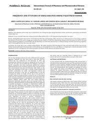
formulation and evaluation of pulsatile drug delivery system of ...
formulation and evaluation of pulsatile drug delivery system of ...
formulation and evaluation of pulsatile drug delivery system of ...

You also want an ePaper? Increase the reach of your titles
YUMPU automatically turns print PDFs into web optimized ePapers that Google loves.
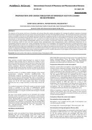
The surface topography <strong>of</strong> optimized <strong>formulation</strong> (A3) <strong>of</strong><br />
Acecl<strong>of</strong>enac loaded Eudragit S100 – L100 combination microspheres<br />
(here after called Eudragit microspheres) was investigated by<br />
scanning electron microscopy (SEM) <strong>and</strong> the photomicrographs are<br />
shown in Fig 1 (I) <strong>and</strong> (II). The SEM photographs revealed that, the<br />
prepared microspheres were spherical <strong>and</strong> uniform with a narrow<br />
size distribution. Fig 1 (II) showed that the surface <strong>of</strong> microsphere<br />
was little rough, porous, free from cracks <strong>and</strong> completely covered<br />
with coat material. The rough surface can be attributed to the rapid<br />
solvent diffusion <strong>and</strong> quick precipitation during the formation <strong>of</strong><br />
O/O emulsion. According to the SEM analysis the size <strong>of</strong> the<br />
microspheres was found to be 397.50 µm.<br />
Chauhan et al.<br />
Size Analysis<br />
Int J Pharm Pharm Sci, Vol 4, Issue 3, 507-512<br />
The size distribution <strong>of</strong> the microspheres was found to be narrow in<br />
all the batches (A1-A4) with an arithmetic mean size <strong>of</strong> 425 µm. The<br />
average size <strong>of</strong> the major fraction <strong>of</strong> microspheres obtained was in<br />
the size range <strong>of</strong> 376-403 µm. The size <strong>of</strong> the polymeric<br />
microspheres was enhanced by increase in the level <strong>of</strong> core: coat<br />
ratio. This could be due to greater amounts <strong>of</strong> coat material<br />
contained in a same volume <strong>of</strong> liquid droplet. This can be ascribed to<br />
an increase in the viscosity resulting in enhanced interfacial tension<br />
<strong>and</strong> demised shearing efficiency 18. A more viscous solution would<br />
require higher shear force for emulsification <strong>and</strong> lead to larger<br />
droplets which would result in larger particles 19.<br />
Table 3: Effect <strong>of</strong> Core: Coat Ratio <strong>of</strong> Acecl<strong>of</strong>enac Loaded Microspheres on Yield, Microspheres Particle Size, Drug content, Encapsulation<br />
Efficiency, T 50% <strong>and</strong> T 90%<br />
Code Core: Coat Yield* Particle size* Davg Drug content** (mg) Encapsulation T 50% T 90%<br />
(%)<br />
(µm)<br />
Theoretical Practical Efficiency*<br />
(%)<br />
A1 1:0.5 78.65±0.82 338.04±0.09 10 8.18±0.678 81.81±0.47 5.19±0.11 10.90±0.07<br />
A2 1:1 82.79±0.78 343.32±0.21 10 8.45±0.696 84.5±0.66 5.43±0.24 11.52±0.12<br />
A3 1:1.5 85.21±0.50 350.04±0.13 10 8.79±0.660 87.96±0.89 5.78±0.31 11.84±0.17<br />
A4 1:2 89.55±0.67 358.84±0.05 10 9.02±0.649 90.2±0.81 6.30±0.19 ----<br />
All values are expressed as mean ± SD, *n=3, **Average <strong>of</strong> 3 determinants.<br />
Microspheres Yield <strong>and</strong> Drug Content<br />
The yield <strong>of</strong> the microspheres was high ranging from 78.65 to 89.55 %.<br />
Little increase in the yield <strong>of</strong> microspheres was observed with<br />
proportionate increase in the core: coat ratio. Similarly encapsulation<br />
efficiency was also increased with the proportionate increase in the core:<br />
coat ratio. The <strong>drug</strong> content estimated in microspheres equivalent to 10<br />
mg <strong>of</strong> Acecl<strong>of</strong>enac was found to be uniform <strong>and</strong> reproducible in each<br />
batch <strong>and</strong> is shown in Table 3. The encapsulation efficiency <strong>of</strong> different<br />
batches <strong>of</strong> microspheres (A1-A4) was in the range <strong>of</strong> 81.81 to 90.2%.<br />
The high encapsulation efficiency could be due to polymer loss by<br />
adherence to the container as a result <strong>of</strong> viscous nature <strong>of</strong> slurry 13.<br />
Interestingly, encapsulation efficiency was found to be directly<br />
proportional to the percentage yield <strong>of</strong> the microsphere preparation 20.<br />
Evaluation <strong>of</strong> Formaldehyde Treated Empty Capsule Bodies<br />
Formalin treatment has been employed to modify the solubility <strong>of</strong><br />
gelatin capsules. Exposure to formalin vapors results in an<br />
unpredictable decrease in solubility <strong>of</strong> gelatin owing to the crosslinkage<br />
<strong>of</strong> the amino groups in the gelatin molecular chain with<br />
aldehyde groups <strong>of</strong> formaldehyde by Schiff’s base condensation 13.<br />
The formaldehyde treated empty capsule bodies were evaluated for<br />
visual appearance, dimension changes, solubility studies <strong>and</strong><br />
qualitative chemical test for free formaldehyde.<br />
The capsule bodies after formaldehyde treatment slightly reduced in<br />
their size <strong>and</strong> apart from this no other visual defects were observed.<br />
The formaldehyde treatment <strong>of</strong> the capsule bodies significantly<br />
altered their solubility compared to the untreated cap <strong>of</strong> the capsule.<br />
The untreated caps were dissolved within 12 minutes <strong>and</strong> the<br />
treated bodies remained intact over a period <strong>of</strong> 24 hrs <strong>and</strong> thus<br />
indicating the suitability for the colonic <strong>delivery</strong>. The qualitative<br />
chemical test for the free formaldehyde was carried out. The sample<br />
solution was not intensely colored than the st<strong>and</strong>ard solution,<br />
inferring that less than 20μg <strong>of</strong> free formaldehyde was present in 25<br />
capsules used for the test.<br />
Fig. 2: In-vitro Release <strong>of</strong> Acecl<strong>of</strong>enac from Eudragit Microspheres in pH 1.2 <strong>and</strong> 6.8.<br />
In-vitro Dissolution Studies<br />
Microspheres<br />
The release <strong>of</strong> Acecl<strong>of</strong>enac from the different batches (A1, A2, A3<br />
<strong>and</strong> A4) <strong>of</strong> Eudragit microspheres was studied in pH 1.2 for 2 hrs<br />
<strong>and</strong> pH 6.8 for 10 hrs using USP-XXIV dissolution testing apparatus.<br />
All four batches <strong>of</strong> Eudragit microspheres showed a strong <strong>drug</strong><br />
release control in acidic medium <strong>of</strong> pH 1.2. The release pattern <strong>of</strong><br />
Acecl<strong>of</strong>enac from Eudragit microspheres <strong>of</strong> A1, A2, A3 <strong>and</strong> A4<br />
batches were found 96.54, 93.71, 91.20 <strong>and</strong> 88.29% respectively at<br />
510