Report on the larger Ascomycete fungi (PDF File, 2.9 MB)
Report on the larger Ascomycete fungi (PDF File, 2.9 MB)
Report on the larger Ascomycete fungi (PDF File, 2.9 MB)

Create successful ePaper yourself
Turn your PDF publications into a flip-book with our unique Google optimized e-Paper software.
<strong>Ascomycete</strong> <strong>fungi</strong>, Cook Islands<br />
P.R. Johnst<strong>on</strong>, Landcare Research, 2011<br />
The <strong>fungi</strong> treated here are those collected from Rarot<strong>on</strong>ga during two vists in 2005, supported by<br />
<strong>the</strong> Cook Island Natural Heritage Commissi<strong>on</strong> and Landcare Research. Brief descripti<strong>on</strong>s and notes<br />
are provided for <strong>fungi</strong> associated with leaf spotting symptoms and those <strong>on</strong> fallen wood with<br />
macroscopically obvious fruiting bodies. Collecti<strong>on</strong>s of smaller <strong>fungi</strong>, such as <strong>the</strong> leaf inhabiting cup<br />
<strong>fungi</strong>, of which <strong>the</strong>re were perhaps 19 species collected during <strong>the</strong> visits in 2005, remain to be<br />
treated. A few o<strong>the</strong>r <strong>fungi</strong> were collected, including Hypocreales (Nectria-like species), and<br />
endophytes cultured from <strong>the</strong> living leaves of rata.<br />
<strong>Ascomycete</strong> leaf spotting <strong>fungi</strong><br />
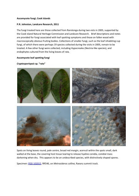
Cryptosporiopsis sp. “rata”<br />
Spots <strong>on</strong> living leaves round, pale centre, broad red margin, acervuli within <strong>the</strong> spots small, dark<br />
walled at <strong>the</strong> base, <strong>the</strong> covering host tissue tearing to release hyaline c<strong>on</strong>idia, c<strong>on</strong>idial mass<br />
darkening when dry. This appears to be an undescribed species, with distinctively shaped spores.<br />
Specimen: PDD 102015, RR546, <strong>on</strong> Metrosideros collina, Raearu summit track.
Meliola sp. “rata”<br />
Forms black col<strong>on</strong>ies about 1.5-4 mm diam. <strong>on</strong> living leaves, flat plate of black hyphae against cuticle<br />
with black setae and superficial, black, globose peri<strong>the</strong>cia; ascospores dark brown, 4-septate,<br />
c<strong>on</strong>stricted at septa, about 50-55 x 15-20 µm. This is probably an undescribed species, no species of<br />
Meliola have been reported from Metrosideros.<br />
Specimen examined: PDD 102035, RR638, <strong>on</strong> Metrosideros collina, Ikurangi.<br />
Meliolina cookii S. Hughes, Mycological Papers166: 59 (1993).<br />
Spots <strong>on</strong> upper surface of living leaves with dense black hyphal mat, with small, black, globose<br />
periethicia in some of <strong>the</strong> spots, lower surface of leaf chlorotic leaf below <strong>the</strong> hypae. Endemic to <strong>the</strong><br />
Cook Islands.<br />
Specimens examined: PDD 102036, RR303, <strong>on</strong> Metrosideros collina, Ikurangi. PDD 102037, RR342, <strong>on</strong><br />
Metrosideros collina, Raemaru. PDD xxxxx, RR359, <strong>on</strong> Metrosideros collina, Te Kou. PDD 82312,<br />
RR573, <strong>on</strong> Metrosideros collina, Te Rua Manga. PDD 102039, RR574, <strong>on</strong> Metrosideros collina, Te Rua<br />
Manga. PDD 102014, RR637, <strong>on</strong> Metrosideros collina, Ikurangi.
Pseudocercospora melastomobia (W. Yamam.) Deight<strong>on</strong>, Transacti<strong>on</strong>s of <strong>the</strong> British Mycological<br />
Society 88: 388 (1987).<br />
Spots <strong>on</strong> living leaves, smallish pale brown with red edge, tiny black fruiting bodies within <strong>the</strong> pale<br />
parts of <strong>the</strong> spots.<br />
Specimens examined: PDD 102044, RR364, <strong>on</strong> Melastoma denticulatum, Te Kou summit track. PDD<br />
102045, RR396, <strong>on</strong> Melastoma denticulatum, Te Manga track.<br />
Pseudocercospora metrosideri U. Braun, Fungal Diversity 8: 44 (2001).<br />
First described from New Zealand from Metrosideros excelsa and M. parkins<strong>on</strong>ii. The two Cook<br />
Island collecti<strong>on</strong>s referred to this species both have typical c<strong>on</strong>idia for this fungus, but are associated<br />
with ra<strong>the</strong>r different symptoms. Although <strong>the</strong> symptom of distinct, round, pale spots does not match<br />
<strong>the</strong> descripti<strong>on</strong> of this fungus well, <strong>the</strong>re are specimens from New Zealand identified by Braun as P.<br />
metrosideri that look very similar.<br />
Specimens examined: PDD 102046, RR304, <strong>on</strong> Metrosideros collina, Ikurangi. PDD 102047, RR305, <strong>on</strong><br />
Metrosideros collina, Ikurangi.<br />
<strong>Ascomycete</strong> <strong>fungi</strong> <strong>on</strong> fallen wood
Xylariaceae<br />
The majority of <strong>the</strong> macroscopically obvious species bel<strong>on</strong>g in <strong>the</strong> family Xylariaceae, most of which<br />
are found fruiting <strong>on</strong> fallen wood. Most genera have hard, dark stromatic fruiting bodies that c<strong>on</strong>tain<br />
numerous individual peri<strong>the</strong>cia, <strong>the</strong> openings of <strong>the</strong>se visible as tiny dots <strong>on</strong> <strong>the</strong> surface of <strong>the</strong> fruiting<br />
body. A few genera such as Rosellinia and some Nemania spp. have uniperi<strong>the</strong>ciate fruiting bodies<br />
but with <strong>the</strong>se often develop in close groups. Most species have brown to dark brown ascospores with<br />
a germ slit. Genera are distinguished by differences in <strong>the</strong> macroscopic fruit body appearance and by<br />
microscopic features associated with <strong>the</strong> anamtomy of <strong>the</strong> fruiting body, ascus apex structure, and<br />
ascospores.<br />
Annulohypoxyl<strong>on</strong><br />
Annulohypoxyl<strong>on</strong> species have thin, extensive, crust-like fruiting bodies c<strong>on</strong>taining of a single layer<br />
of numerous peri<strong>the</strong>cia, <strong>the</strong> individual peri<strong>the</strong>cia often visible as small lumps across <strong>the</strong> surface of <strong>the</strong><br />
fruiting body. The ostioles are characteristically surrounded by a small flattened area (<strong>the</strong> annulate<br />
disc).<br />
Annulohypoxyl<strong>on</strong> ? moriforme (Henn.) Y.M. Ju, J.D. Rogers & H.M. Hsieh, Mycologia 97: 859<br />
(2005).<br />
A. moriforme is widespread in tropical regi<strong>on</strong>s. The fruiting bodies form extensive, thin sheets across<br />
<strong>the</strong> surface of <strong>the</strong> host substrate, <strong>the</strong> tissue between <strong>the</strong> peri<strong>the</strong>cial mounds reddish or red-brown.<br />
Ascospores about 7-9.5 x 3-3.5 µm, flattened <strong>on</strong> <strong>on</strong>e side, germ slit <strong>on</strong> curved side of spore. The<br />
Cook Island specimen matches A. moriforme in most aspects, but has blackish pigment diffusing into<br />
KOH whereas H. moriforme has been described as having dark green diffusible pigments.<br />
Specimen examined: PDD 102004, RR445, cross island walk, nor<strong>the</strong>rn end.
Annulohypoxyl<strong>on</strong> stygium (Lév.) Y.M. Ju, J.D. Rogers & H.M. Hsieh, Mycologia 97: 861 (2005).<br />
A. stygium is a comm<strong>on</strong> and widespread tropical species. Macroscopically similar to A. moriforme,<br />
ascospores are smaller (5-6.5 x 2-2.5 µm) and have <strong>the</strong> germ slit <strong>on</strong> <strong>the</strong> flattened side of <strong>the</strong> spore.<br />
Specimens examined: PDD 102010, RR538, <strong>on</strong> Albizzia sp., track to Raemaru. PDD 102009, RR344,<br />
<strong>on</strong> Albizzia sp., track to Raemaru. PDD 102008, RR309, near start of track to Ikurangi.<br />
Biscogniauxia uniapiculata (Penz. & Sacc.) Whalley & Læssøe, in Whalley, Laessøe & Kile,<br />
Mycological Research 94: 239 (1990).<br />
Biscogniauxia species characteristically have extensive, flat fruiting bodies of this genus become<br />
erumpent from beneath host bark as <strong>the</strong>y mature, and some host tissue remains overlapping <strong>the</strong><br />
edges of <strong>the</strong> mature fruiting body. The fruiting bodies have a single layer of numerous peri<strong>the</strong>cia<br />
opening through small, round ostioles, and almost no internal sterile tissue. These species often fruit<br />
<strong>on</strong> recently fallen wood, where <strong>the</strong>y are likely to have been living as endophytes within <strong>the</strong> bark of <strong>the</strong><br />
living tree. B. uniapiculata has ascospores with a small, hyaline cell at <strong>on</strong>e end of <strong>the</strong> o<strong>the</strong>rwise dark<br />
brown spore, <strong>the</strong> pale of <strong>the</strong> spore sometimes lost and <strong>the</strong>n spore appearing truncate at <strong>on</strong>e end. Its<br />
is comm<strong>on</strong> and widespread in tropical regi<strong>on</strong>s.<br />
Specimen examined: PDD 102011, RR413, Turangi Stream track.
Collodiscula sp. “coc<strong>on</strong>ut”<br />
Collodiscula has uniper<strong>the</strong>ciate fruiting bodies, <strong>the</strong> large, dark walled, globose peri<strong>the</strong>cium erumpent<br />
from deep within <strong>the</strong> host, with <strong>the</strong> covering host tissue folding back to remain as a star-like margin.<br />
Unusual for Xylariaceae, it has 2-celled spores with both cells being of similar size and dark brown. A<br />
single species from bamboo has been described in <strong>the</strong> genus. The Cook Island specimens from<br />
coc<strong>on</strong>ut fr<strong>on</strong>ds probably represent an undescribed species, with spores <strong>larger</strong> than that described for<br />
<strong>the</strong> bamboo inhabiting species, C. jap<strong>on</strong>ica.<br />
Specimens examined: PDD 102013, RR322, <strong>on</strong> fallen coc<strong>on</strong>ut fr<strong>on</strong>d, sou<strong>the</strong>rn start to cross island<br />
walk. PDD 102014, RR549, <strong>on</strong> fallen coc<strong>on</strong>ut fr<strong>on</strong>d, track to Raemaru.<br />
Hypoxyl<strong>on</strong><br />
Hypoxyl<strong>on</strong> species have thin, extensive, crust-like fruiting bodies c<strong>on</strong>taining of a single layer of<br />
numerous peri<strong>the</strong>cia, <strong>the</strong> individual peri<strong>the</strong>cia often visible as small lumps across <strong>the</strong> surface of <strong>the</strong><br />
fruiting body.<br />
Hypoxyl<strong>on</strong> cinnabarinum (Henn.) Y.M. Ju & J.D. Rogers, Mycological Memoirs 20: 99 (1996).<br />
Comm<strong>on</strong> in tropical regi<strong>on</strong>s, <strong>the</strong> fruiting bodies are distinctively apricot-coloured. Ascospores 11-13.5<br />
x 5-6.5 µm.<br />
Specimen examined: PDD 102017, RR343, track to Raemaru.
Hypoxyl<strong>on</strong> haematostroma M<strong>on</strong>t., in Sagra, Historia física, polirica y nayturál de la islea de Cuba 9:<br />
344 (1845).<br />
Comm<strong>on</strong> in tropical regi<strong>on</strong>s, <strong>the</strong> surface of <strong>the</strong> fruiting bodies are bright rusty-brown in colour.<br />
Ascospores <strong>larger</strong> <strong>the</strong>n H. cinnabarinum, 14.5-16.5 x 7.5-8 µm.<br />
Specimen examined: PDD 102018, RR555, track to Raemaru.<br />
Hypoxyl<strong>on</strong> — unidentified species<br />
Specimens that appear to represent four morphologically distinct Hypoxyl<strong>on</strong> spp. were collected, n<strong>on</strong>e<br />
of which could be reliably matched to a species using available keys.<br />
Hypoxyl<strong>on</strong> sp. PDD 102021, RR542, track to Raemaru.<br />
Fruiting body red-brown, no pigments in KOH; ascospores about 10-11 x 4.5-5.5 µm, pale brown,<br />
symmetrical, ends broadly rounded, spore-length germ slit; amyloid pore in ascus about 1 µm high.
Hypoxyl<strong>on</strong> sp. PDD 102020, RR545, track to Raemaru<br />
Fruiting body purplish, deep yellow-brown pigments in KOH; ascospores about 7.5-8.5 x 4-4.5 µm,<br />
more or less symmetrical to slightly flattened <strong>on</strong>e side, taper to narrow rounded ends, germ slit sporelength,<br />
straight.<br />
Hypoxyl<strong>on</strong> sp. PDD 102019, RR346, track to Raemaru<br />
. Fruiting body comprising small groups of peri<strong>the</strong>cia, grey-brown when immature, str<strong>on</strong>g yellowbrown<br />
pigment in KOH; ascospores 10.5-13 x 4.5-6 µm, dark brown, flat <strong>on</strong>e side, slightly curved,<br />
taper to small rounded ends, germ-lit straight, a little less than spore length; amyloid ring at ascus<br />
apex less than 1 µm high.<br />
.
Hypoxyl<strong>on</strong> sp. PDD 102022, RR575, Te Rua Manga.<br />
Fruiting body comprising small groups of peri<strong>the</strong>cia, no pigment in KOH; ascospores about 12.5-14 x<br />
4.5-5.5 µm, pale brown, distinctly more tapered to <strong>on</strong>e end than <strong>the</strong> o<strong>the</strong>r, germ slit striaght, slitghtly<br />
less than spore length; amyloid ring at ascus apex 3 µm high.<br />
Kretzschmaria<br />
Four species of Kretzschmaria were found. The genus is characterised by <strong>the</strong> fruiting bodies being<br />
hollow when mature. In early stages <strong>the</strong> internal tissue, if present, is white. Xylaria also has white<br />
tissue internally, but in that genus <strong>the</strong> tissue is much more dense in structure and is persistent. There<br />
are two macroscopically distinct groups within Kretzschmaria, <strong>on</strong>e with fruiting bodies made up of<br />
large numbers of small, gregarious stromata <strong>on</strong> short stalk-like bases, <strong>the</strong> o<strong>the</strong>r with large, spreading<br />
fruiting bodies often with a very irregular surface.<br />
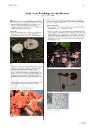
Kretzschmaria clavus (Fr.) Sacc., Sylloge Fungorum 2: XXIX (1883).<br />
A comm<strong>on</strong>, widespread tropical species. Large numbers of small, broadly stalked stromata ga<strong>the</strong>red<br />
into large crusts. The right hand image shows <strong>the</strong> hollow inside, with individual peri<strong>the</strong>cia hanging<br />
down into <strong>the</strong> space from <strong>the</strong> stromatal crust. Ascospores about 26-40 x 7.5-10.5 µm with a straight,<br />
spore-length germ slit.<br />
Specimens examined: PDD 102023, RR535, Raemaru. PDD 102024, RR608, Te Rua Manga.
Kretzschmaria pavimentosa (Ces.) P.M.D. Martin, Journal of South African Botany 42: 74 (1976).<br />
A comm<strong>on</strong>, widespread tropical species with broad, spreading fruiting body, very uneven <strong>on</strong> <strong>the</strong><br />
surface. Ascospores about 35-50 x 7.5-12 µm with a short, straight germ slit.<br />
Specimens examined: PDD 102029, RR583, Te Rua Manga. PDD 102025, RR60, Takitumu<br />
C<strong>on</strong>servati<strong>on</strong> Area. PDD 102026, RR72, Takitumu C<strong>on</strong>servati<strong>on</strong> Area. PDD 102027, RR398, Te Rua<br />
Manga.<br />
Kretzschmaria sp. “helico”<br />
Kretzschmarioid fruiting body about 1.5-3 mm diam., c<strong>on</strong>taining a small number of peri<strong>the</strong>cia, surface<br />
distinctively cracked. Developing in gregarious groups <strong>on</strong> surafce of slightly blackened wood, and<br />
sometimes forming extensive crusts. Internally initially with some white tissue, this perhaps lost with<br />
age. Spores with a distinctive helical germ slit. Could not be identified from available keys to<br />
Kretzschmaria.<br />
Specimens examined: PDD 102031, RR579, Te Manga Rua. PDD 102030, RR607, Te Manga Rua.
Kretzschmaria sp. “small spore”<br />
Fruiting bodies 5-8 mm diam, raised 3 mm above surface of wood. Internally <strong>the</strong> fruiting body has a<br />
small amount of loose, white tissue but is mostly hollow. The spores are very small for Kretzschmaria,<br />
10.5-12 x 6-7 µm. The species could not be identified using available keys.<br />
Specimens examined: PDD 102032, RR347, Raemaru. PDD 102033, RR412, Turangi Stream. PDD<br />
102034, RR478, Avana water intake.<br />
Rosellinia<br />
Fruiting bodies comprising a single peri<strong>the</strong>cium, but often closely crowded toge<strong>the</strong>r and when young<br />
often collectively surrounded by fungal mycelium. Three species were found, but a lack of a modern<br />
m<strong>on</strong>ograph of tropical species meant n<strong>on</strong>e were identified to species level.<br />
Rosellinia sp. “red-brown”<br />
Characteristically with deep red-brown pigments in <strong>the</strong> lower part of <strong>the</strong> peri<strong>the</strong>cia. All specimens<br />
overmature, with very few spores seen.<br />
Specimens examined: PDD 102049, RR404, Te Manga. PDD 102050, RR652, start of track to Te<br />
Manga. PDD 102048, RR55, Takitumu C<strong>on</strong>servati<strong>on</strong> Area.
Rosellinia sp. “white”<br />
Peri<strong>the</strong>cia about 0.8 mm diam., becoming slightly wider towards <strong>the</strong> base, base partly immersed in<br />
host substrate, sparse patches of white hyphae at <strong>the</strong> base of some peri<strong>the</strong>cia, ascospores about<br />
11.5-14 x 6.5-7.5 µm, dark brown, more or less symmetrical, taper to narrow rounded ends germ slit<br />
straight, spore-length; amyloid ring at ascus apex 2-2.5 µm high.<br />
Specimens examined: PDD 102054, RR581, <strong>on</strong> Hibiscus wood, nor<strong>the</strong>rn end of cross island walk.<br />
PDD 102052, RR360. track to Te Kou. PDD 102051, RR302, near start of track to Ikurangi. PDD<br />
102053, RR434, Barringt<strong>on</strong>ia remnant, <strong>on</strong> coast between Avarua and Matavera.<br />
Rosellinia sp. “yellow”<br />
Peri<strong>the</strong>cia about 0.7 mm diam., surrounded by bright yellow, often dense hyphae, as peri<strong>the</strong>cia age<br />
hyphae becomes less dense and starts to lose yellow pigment. Ascospores 8.5-10.5 x 4-5.5 µm,<br />
symmetrical, ends rounded, germ slit straight, spore-length; amyloid ring at ascus apex 1µm high.<br />
Specimens examined: PDD 102056, RR580, Te Rua Manga. PDD 101075, RR432, Barringt<strong>on</strong>ia<br />
remnant, <strong>on</strong> coast between Avarua and Matavera.
Stilbohypoxyl<strong>on</strong> moelleri Henn., Hedwigia 41: 16 (1902).<br />
Stilbohypoxyl<strong>on</strong> is characterised by <strong>the</strong> anamorph forming large, pointed, spine-like structures <strong>on</strong> <strong>the</strong><br />
outside of <strong>the</strong> solitary peri<strong>the</strong>cia. S. moelleri has peri<strong>the</strong>cia about 0.8-1 mm diam., comm<strong>on</strong> in tropical<br />
America.<br />
Specimen examined: PDD 102057, RR605, <strong>on</strong> coc<strong>on</strong>ut fr<strong>on</strong>d, nor<strong>the</strong>rn end of <strong>the</strong> cross island walk.<br />
Xylaria<br />
Xylaria species have upright fruiting bodies with multiple peri<strong>the</strong>cia <strong>on</strong> <strong>the</strong> outer margin, <strong>the</strong> centre of<br />
<strong>the</strong> fruiting body has copious, persistent white sterile tissue. Species are distinguished by <strong>the</strong><br />
macroscopic shape of <strong>the</strong> fruiting body, <strong>the</strong> appearance of <strong>the</strong> surface of <strong>the</strong> fruiting body (whe<strong>the</strong>r<br />
cracked or not), ascospore shape and size, and shape of <strong>the</strong> germ slit <strong>on</strong> <strong>the</strong> ascospores.<br />
Xylaria cf. apiculata<br />
Xylaria apiculata has narrow-cylindric stromata with a characteristic short, pointed sterile tip. There<br />
are several macroscopically similar species that differ slightly in ascospore size. Two X. apiculata-like<br />
species were collected in <strong>the</strong> Cook Islands, nei<strong>the</strong>r of which could be c<strong>on</strong>fidently matched to a<br />
species.<br />
Xylaria sp. “apiculata leopard”<br />
Fruit bodies about 15 mm tall, with small pale patches scattered across <strong>the</strong> o<strong>the</strong>rwise black fruiting<br />
body; ascospores 9-10.5 x 3.5-4.5 µm, flat <strong>on</strong> <strong>on</strong>e side, slightly curved, narrow rounded ends, germ<br />
slit <strong>on</strong> flat side of spore, straight, slightly less than spore length.
Specimen examined: PDD 102075, RR547, track to Raemaru.<br />
Xylaria sp. “apiculata spiral”<br />
Fruiting bodies about 15 mm tall, uniformly blackish, <strong>the</strong> sterile tips often branched; ascospores 19.5-<br />
23 x 6-7.5 µm, flat <strong>on</strong>e side, slightly curved, short germ slit somwhat less than spore length, oblique to<br />
spiralled.<br />
Specimens examined: PDD 102077, RR307, track to Ikurangi. PDD 102076, RR89, Takitumu<br />
C<strong>on</strong>servati<strong>on</strong> Area. PDD 102078, RR372, track to Te Kou.<br />
Xylaria cubensis (M<strong>on</strong>t.) Fr., Nova Acta R. Soc. Scient. upsal., Ser. 3 1: 126 (1851).<br />
A comm<strong>on</strong> and widespread tropical species characterised in part by <strong>the</strong> fruiting bodies of <strong>the</strong> asexual<br />
state being highly and irregularly branched, white, up to about 8 mm high. The mature form of <strong>the</strong><br />
sexual fruiting body is regularly cylindrical, about 20-40 x 4-8 mm. In <strong>the</strong> Cook Islands both forms<br />
were often found toge<strong>the</strong>r <strong>on</strong> <strong>the</strong> same piece of wood. The ascospores about 8.5-10 x 4-4.5 µm,<br />
with no obvious germ slit.<br />
Specimens examined: PDD 102061, RR408, Turangi Stream track. PDD 102062, RR410, Turangi<br />
Stream track. PDD 102063, RR425, Turangi Stream track. RR595, Te Manga. PDD 102064, RR451,<br />
nor<strong>the</strong>rn end of cross island walk.
Xylaria cf. filiformis<br />
Xylaria filiformis has very narrow, hair-like fruiting bodies, <strong>the</strong> peri<strong>the</strong>cia forming small lumps in <strong>the</strong><br />
fertile parts. The fruiting bodies typically develop <strong>on</strong> fallen leaves, ra<strong>the</strong>r than wood. There are<br />
several species with similar shaped fruiting bodies, but <strong>the</strong> group is poorly understood<br />
tax<strong>on</strong>omically. The single Cook Island specimen was immature — peri<strong>the</strong>cia had formed but<br />
c<strong>on</strong>tained no mature ascospores.<br />
Specimen examined: PDD 102060, RR624, Ikurangi.<br />
Xylaria schweinitzii Berk. & M.A. Curtis, Journal of <strong>the</strong> Academy of natural Sciences Philadelphia<br />
2: 284 (1853).<br />
A comm<strong>on</strong> and widespread tropical species with a stroma extremely variable in shape. Often a wide<br />
variety of shapes is present within a single collecti<strong>on</strong>. The ascospores characteristically have a short,<br />
oblique or somewhat coiling germ slit.<br />
Specimens examined: PDD 102071, RR453, nor<strong>the</strong>rn end of cross island walk. RR156, sou<strong>the</strong>rn end<br />
of cross island walk. PDD 102070, RR362 and RR363, start of track to Te Kou. PDD 102072, RR456<br />
and RR457, Avana water intake. PDD 102080, RR6, RR11, RR21, RR50, and RR51, Takitumu<br />
C<strong>on</strong>servati<strong>on</strong> Area.
Xylaria sp. “leopard”<br />
Fruit body about 15-30 x 7-10 mm, broad-cylindric, surface with distinct small pale patches;<br />
ascospores 9-10.5 x 3.5-4.5 µm, flat <strong>on</strong>e side, slightly curved, narrow rounded ends, germ slit <strong>on</strong><br />
flattened side, somewhat less than spore length; amyloid pore at ascus apex 2 µm high.<br />
Microscopically similar to X. sp. “apiculata leopard” but fruit bodies much <strong>larger</strong>.<br />
Specimen examined: PDD 102079, RR539, near start of Raemaru track.<br />
Xylariaceae sp.<br />
A single specimen that cannot be c<strong>on</strong>fidently placed in a genus. The large, cylindric fruit body, up<br />
to 6 cm tall, is hollow inside or with loose, dark sterile tissue, numerous, small, round peri<strong>the</strong>cia at<br />
margins. Ascospores about 11-12.5 x 4-4.5 µm, flat <strong>on</strong>e side, slightly curved, germ slit sporelength,<br />
straight, <strong>on</strong> flattened side od spore. Asci not seen.<br />
Specimen examined: PDD 102080, RR6, Takitumu C<strong>on</strong>servati<strong>on</strong> Area.
N<strong>on</strong>-xylariaceous wood inhabiting species<br />
Cytosphaera sp.<br />
An asexual coelomycete fungus. The fruiting body about 1-1.5 mm diam., sometimes in c<strong>on</strong>fluent<br />
groups. The globose spores are about 13-15 µm diam., hyaline and thick-walled. The species remains<br />
unidentified in this tax<strong>on</strong>omically poorly understood genus.<br />
Specimen examined: PDD 102016, RR566, <strong>on</strong> fallen Barringt<strong>on</strong>ia twigs, Barringt<strong>on</strong>ia remnant, <strong>on</strong><br />
coast between Avarua and Matavera.<br />
Valsa sp.<br />
Peri<strong>the</strong>cia deeply immersed in woody host , <strong>the</strong> ostioles with about 1 mm l<strong>on</strong>g, thin, erumpent necks;<br />
ascospores about 4.5-5.5 x 1.5 µm, pale brown, sausage-shaped. The genus is poorly understood<br />
tax<strong>on</strong>omically, and lack of a modern m<strong>on</strong>ograph meant <strong>the</strong> species was not able to be identified.<br />
Specimen examined: PDD 102058, RR560, <strong>on</strong> fallen Hibiscus wood, track to Raemaru.