KVÄLLSSYMPOSIUM 2008 Vaccinering av hund och katt
KVÄLLSSYMPOSIUM 2008 Vaccinering av hund och katt KVÄLLSSYMPOSIUM 2008 Vaccinering av hund och katt
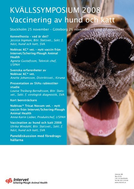
KVÄLLSSYMPOSIUM 2008 Vaccinering av hund och katt Stockholm 25 november – Göteborg 26 november – Lund 27 november Kennelhosta – vad är det? Jessica Ingman, Bitr. Statsvet., Sekt. f. häst, hund och katt, SVA Nobivac KC® vet. – nytt vaccin från Intervet/Schering-Plough Animal Health Agneta Gustafsson, Teknisk chef, I/SPAH Svenska erfarenheter av Nobivac KC® vet. Anette Johansson, Distriktsvet., Kiruna Presentation av SVAs rabiestiterstudie Louise Treiberg-Berndtsson, Bitr. Statsvet., Sekt. f. virologisk diagnostik, SVA Kort bensträckare Nobivac® Tricat Novum vet. – nytt vaccin från Intervet/Schering-Plough Animal Health Anna-Karin Lieber, Produktchef, I/SPAH Vaccination av hund och katt 2008 Ulrika Windahl, Bitr. Statsvet., Sekt. f. häst, hund och katt, SVA Paneldiskussion med föredragshållarna Intervet AB Box 6103 102 33 Stockholm Tel 08–522 216 60 www.intervet.se
- Page 3 and 4: Kennelhosta - vad är det? Bitr. st
- Page 5: Sammanfattningsvis om kennelhosta
- Page 8 and 9: 356 C. Buonavoglia, V. Martella com
- Page 10 and 11: 358 C. Buonavoglia, V. Martella of
- Page 12 and 13: 360 C. Buonavoglia, V. Martella oth
- Page 14 and 15: 362 C. Buonavoglia, V. Martella inf
- Page 16 and 17: 364 C. Buonavoglia, V. Martella 2 c
- Page 18 and 19: 366 C. Buonavoglia, V. Martella and
- Page 20 and 21: 368 C. Buonavoglia, V. Martella J.M
- Page 22 and 23: 370 C. Buonavoglia, V. Martella reo
- Page 24 and 25: 372 C. Buonavoglia, V. Martella gly
- Page 27 and 28: N o b i v a c ® KC KC - bredare oc
- Page 29 and 30: N o b i v a c ® K C Intranasala va
- Page 31 and 32: Excellent säkerhetsprofil N o b i
- Page 33 and 34: Svenska erfarenheter av vaccination
- Page 35: Varför skillnad i resultat? Vaccin
- Page 38 and 39: 270 S.M. Moore et al. / Biologicals
- Page 40 and 41: 272 S.M. Moore et al. / Biologicals
- Page 42 and 43: 274 S.M. Moore et al. / Biologicals
- Page 44 and 45: 276 S.M. Moore et al. / Biologicals
- Page 46 and 47: Varför levande vaccin? • Stimule
- Page 49 and 50: Vaccination av hund och katt 2008 B
- Page 51 and 52: Vaccinationsintervall = hur länge
<strong>KVÄLLSSYMPOSIUM</strong> <strong>2008</strong><br />
<strong>Vaccinering</strong> <strong>av</strong> <strong>hund</strong> <strong>och</strong> <strong>katt</strong><br />
Stockholm 25 november – Göteborg 26 november – Lund 27 november<br />
Kennelhosta – vad är det?<br />
Jessica Ingman, Bitr. Statsvet., Sekt. f.<br />
häst, <strong>hund</strong> <strong>och</strong> <strong>katt</strong>, SVA<br />
Nobivac KC® vet. – nytt vaccin från<br />
Intervet/Schering-Plough Animal<br />
Health<br />
Agneta Gustafsson, Teknisk chef,<br />
I/SPAH<br />
Svenska erfarenheter <strong>av</strong><br />
Nobivac KC® vet.<br />
Anette Johansson, Distriktsvet., Kiruna<br />
Presentation <strong>av</strong> SVAs rabiestiterstudie<br />
Louise Treiberg-Berndtsson, Bitr. Statsvet.,<br />
Sekt. f. virologisk diagnostik, SVA<br />
Kort bensträckare<br />
Nobivac® Tricat Novum vet. – nytt<br />
vaccin från Intervet/Schering-Plough<br />
Animal Health<br />
Anna-Karin Lieber, Produktchef, I/SPAH<br />
Vaccination <strong>av</strong> <strong>hund</strong> <strong>och</strong> <strong>katt</strong> <strong>2008</strong><br />
Ulrika Windahl, Bitr. Statsvet., Sekt. f.<br />
häst, <strong>hund</strong> <strong>och</strong> <strong>katt</strong>, SVA<br />
Paneldiskussion med föredragshållarna<br />
Intervet AB<br />
Box 6103<br />
102 33 Stockholm<br />
Tel 08–522 216 60<br />
www.intervet.se
Kennelhosta – vad är det?<br />
Bitr. statsveterinär Jessica Ingman<br />
Enheten för djurhälsa <strong>och</strong> antibiotikafrågor, Statens veterinärmedicinska anstalt<br />
Kennelhosta<br />
• Är ett samlingsbegrepp för kikhosteliknande symtom hos <strong>hund</strong>.<br />
• Akut, mycket smittsam luftvägsinfektion som karakteriseras <strong>av</strong> akut insättande<br />
hostattacker (kväljningar/slem/nosflöde).<br />
• Multifaktoriell etiologi.<br />
• Graden <strong>av</strong> symtom kan variera beroende på vilket eller vilka agens som är<br />
inblandade.<br />
• Infektion med enbart ett agens ger oftast lindrigare symtom.<br />
• Prognosen god vid okomplicerade infektioner, men immuniteten kan vara<br />
kortvarig.<br />
Smittspridning<br />
• Infektion sprids snabbt <strong>och</strong> effektivt bland <strong>hund</strong>ar som hålls tillsammans i<br />
större grupper.<br />
• Smitta direkt nos-nos-kontakt, hosta/nysningar, indirekt via händer/föremål.<br />
• Kliniska symtom uppträder vanligtvis inom 3-5 dagar efter infektion, även om<br />
inkubationstid för kennelhosta anges till 3-10 dagar.<br />
• För de vanligaste virala agens kan utsöndring <strong>av</strong> virus ske upptill två veckor<br />
efter infektion.<br />
• För B. bronchiseptica kan dock utsöndring pågå betydligt längre tid.<br />
Etiologi<br />
• Virus<br />
- Hundens parainfluens<strong>av</strong>irus typ 2 (CPiV-2)<br />
- Hundens adenovirus typ 2 (CAV-2)<br />
- Hundens herpesvirus typ 1 (CHV-1)<br />
- Canine respiratory corona virus (CRCoV)<br />
- (valpsjuka…)<br />
• Bakteriella agens<br />
- Bordetella bronchiseptica<br />
- Mykoplasma-arter, streptokocker, pasteurella-arter, koliforma bakterier <strong>och</strong><br />
pseudomonas-bakterier har också påvisats hos hostande <strong>hund</strong>ar, men då<br />
främst som sekundärinfektion eller eventuellt bifynd.<br />
• Hundens parainfluens<strong>av</strong>irus typ 2 <strong>och</strong> Bordetella bronchiseptica är de agens som<br />
enligt litteraturen oftast isoleras från <strong>hund</strong>ar med infektiös trakeobronkit, men<br />
flera andra virus <strong>och</strong> bakterier kan påverka symtombild <strong>och</strong> sjukdomsförlopp.
Hundens parainfluens<strong>av</strong>irus typ 2<br />
• CPiV-2, canine parainfluenza virus<br />
• RNA-virus, familjen Paramyxoviridae<br />
• Det vanligaste virus som isoleras från luftvägarna hos <strong>hund</strong> i samband med<br />
kennelhostesymtom.<br />
• Orsakar vid ensam infektion vanligen en relativt lindrig infektion i övre<br />
luftvägarna med kortvarig, övergående hosta <strong>och</strong> minimal allmänpåverkan.<br />
• (Katter kan bli subkliniskt infekterade <strong>och</strong> utsöndra virus – okänt dock om<br />
betydelse för smittspridning hos <strong>hund</strong>.)<br />
Hundens adenovirus typ 2<br />
• Virus som är nära släkt med det virus (CAV-1) som orsakar HCC, men CAV-2<br />
angriper huvudsakligen luftvägarna.<br />
• Infektion orsakar vanligen inga påtagliga kliniska symtom.<br />
• De flesta <strong>hund</strong>ar är idag vaccinerade mot CAV-2 (ger skydd mot CAV-1 som<br />
orsakar HCC).<br />
Hundens herpesvirus typ 1<br />
• Är ett virus som också angriper de övre luftvägarna hos <strong>hund</strong>, men som<br />
vanligtvis inte orsakar några tydliga kliniska symtom. Anses inte längre vara<br />
någon väsentlig orsak till kennelhosta.<br />
• (Har betydelse vid dödlig infektion hos nyfödda valpar.)<br />
CRCoV<br />
• Canine respiratory corona virus<br />
• Relativt nyupptäckt virus som isolerats från <strong>hund</strong>ar med kennelhostesymtom.<br />
• Är olikt från CCoV – den enteriska formen<br />
• Har oftast associerats med lindriga kliniska symtom.<br />
Valpsjukevirus<br />
• CDV, canine distemper virus<br />
• Kan ge respiratoriska symtom <strong>och</strong> har därför tidigare beskrivits i samband med<br />
kennelhostekomplexet, men har idag mindre betydelse pga. utbredd vaccination.<br />
B. bronchiseptica<br />
• Gramnegativ, aerob bakterie.<br />
• Kan förekomma i luftvägarna även hos friska individer.<br />
• Hundratals olika isolat påvisade med olika virulens <strong>och</strong> patogenicitet.<br />
• I experimentella studier orsakar infektion med enbart B. bronchiseptica typiska<br />
symtom på kennelhosta framför allt hos unga <strong>hund</strong>ar.<br />
• Bakterien kan utsöndras från övre luftvägarna i 3-4 månader efter<br />
genomgången infektion.
Sammanfattningsvis om kennelhosta<br />
• Kennelhosta är ett komplext komplex - flera möjliga agens, multifaktoriell<br />
etiologi.<br />
• Patogena agens kan vara olika vid olika utbrott.<br />
• En <strong>hund</strong> som haft kennelhosta är inte skyddad mot att få sjukdomen igen vid<br />
ett senare tillfälle.
Vet. Res. 38 (2007) 355–373 355<br />
c○ INRA, EDP Sciences, 2007<br />
DOI: 10.1051/vetres:2006058<br />
Review article<br />
Canine respiratory viruses<br />
Canio Buon<strong>av</strong>oglia*, Vito Martella<br />
Department of Animal Health and Wellbeing, Faculty of Veterinary Medicine of Bari, Italy<br />
(Received 27 April 2006; accepted 28 August 2006)<br />
Abstract – Acute contagious respiratory disease (kennel cough) is commonly described in dogs<br />
worldwide. The disease appears to be multifactorial and a number of viral and bacterial pathogens<br />
h<strong>av</strong>e been reported as potential aetiological agents, including canine parainfluenza virus, canine<br />
adenovirus and Bordetella bronchiseptica, as well as mycoplasmas, Streptococcus equi subsp.<br />
zooepidemicus, canine herpesvirus and reovirus-1,-2 and -3. Enhancement of pathogenicity by multiple<br />
infections can result in more severe clinical forms. In addition, acute respiratory diseases<br />
associated with infection by influenza A virus, and group I and II coron<strong>av</strong>iruses, h<strong>av</strong>e been described<br />
recently in dogs. Host species shifts and tropism changes are likely responsible for the<br />
onset of these new pathogens. The importance of the viral agents in the kennel cough complex is<br />
discussed.<br />
kennel cough / respiratory disease / dogs / viruses<br />
1.<br />
Table of contents<br />
Introduction ......................................................................................................355<br />
2. Canineadenovirustype2 .....................................................................................356<br />
3. Canineherpesvirus .............................................................................................357<br />
4. Canineinfluenz<strong>av</strong>irus ..........................................................................................359<br />
5. Canineparainfluenz<strong>av</strong>irus ....................................................................................360<br />
6. Caninereovirus..................................................................................................362 7. Caninerespiratorycoron<strong>av</strong>irus ..............................................................................363<br />
8. Pantropiccaninecoron<strong>av</strong>irus.................................................................................364 9. Conclusions ......................................................................................................365<br />
1. INTRODUCTION<br />
Infectious tracheobronchitis (ITB) or<br />
kennel cough is the term used by veterinarians<br />
to describe an acute, highly contagious<br />
respiratory disease in dogs affecting the<br />
larynx, trachea, bronchi, and occasionally<br />
the nasal mucosa and the lower respiratory<br />
tract [6].<br />
* Corresponding author:<br />
c.buon<strong>av</strong>oglia@veterinaria.uniba.it<br />
Mild to severe episodes of cough and<br />
respiratory distress are characteristic clinical<br />
features recognized in affected dogs.<br />
ITB has worldwide distribution and is recognized<br />
as one of the most prevalent infectious<br />
diseases of dogs. The disease is frequently<br />
described in dogs housed in groups<br />
in rehoming centers and boarding or training<br />
kennels.<br />
Two clinical forms of ITB h<strong>av</strong>e been described.<br />
The uncomplicated form is most<br />
Article <strong>av</strong>ailable at http://www.edpsciences.org/vetres or http://dx.doi.org/10.1051/vetres:2006058
356 C. Buon<strong>av</strong>oglia, V. Martella<br />
common and is characterized as a dry,<br />
hacking cough, often in association with<br />
gagging and retching beh<strong>av</strong>ior. The dogs<br />
are affected by a self-limiting, primarily<br />
viral infection of the trachea and bronchi.<br />
A complicated form of ITB is described<br />
in puppies or immuno-compromised dogs.<br />
In the complicated forms, secondary bacterial<br />
infections and involvement of pulmonary<br />
tissue overlaps the viral process.<br />
The cough is associated with mucoid discharges.<br />
The condition may progress to<br />
bronchopneumonia and, in the most severe<br />
instances, to death [6].<br />
Multiple agents, bacterial and viral, are<br />
implicated in the aetiology of ITB. Coisolation<br />
of viral and bacterial pathogens<br />
is frequent, while experimental infections<br />
with single pathogens may result in subclinical<br />
or mild forms of disease, suggesting<br />
a multi-factorial pathogenesis. Many<br />
agents likely play a role in ITB, such<br />
as canine parainfluenza virus [2], canine<br />
adenovirus [55], Bordetella bronchiseptica<br />
[18], and mycoplasmas [36, 128].<br />
Streptococcus equi subsp. zooepidemicus,<br />
has been associated with severe to fatal<br />
respiratory forms in dogs alone or in<br />
mixed infections [36, 61, 173]. Recently,<br />
outbreaks of influenza A virus, initially<br />
misdiagnosed as ITB, h<strong>av</strong>e been reported<br />
in the USA [45,173]. In addition, novel canine<br />
coron<strong>av</strong>iruses, a pantropic variant of<br />
CCoV type II [29] and the canine respiratory<br />
coron<strong>av</strong>irus virus, CRCoV [60], h<strong>av</strong>e<br />
been detected from the respiratory tract<br />
of either symptomatic or asymptomatic<br />
dogs. Canine herpesvirus and reovirus-1,-<br />
2,-3 h<strong>av</strong>e rarely been reported from dogs<br />
with kennel cough but are not thought<br />
to play a major role in the disease complex<br />
[86,97]. Vaccines are <strong>av</strong>ailable against<br />
some of these infectious agents but regular<br />
vaccination in kennels often fails to prevent<br />
ITB.<br />
An overview of the viral agents that<br />
h<strong>av</strong>e been associated with ITB in dogs,<br />
with particular regards to newly described<br />
viruses, is reported.<br />
2. CANINE ADENOVIRUS TYPE 2<br />
Canine adenovirus type 2 (CAV-2) determines<br />
unapparent to mild infection of<br />
the respiratory tract and is regarded as one<br />
of the causes of the common widespread<br />
ITB [154]. CAV-2 has also been implicated<br />
in episodes of enteritis [76, 98] and has<br />
been detected in the brain of dogs with<br />
neurological signs [19].<br />
The virus was first detected in 1961,<br />
in Canada, from dogs affected by laryngotracheitis<br />
[55]. The isolate, strain Toronto<br />
A26/61, was characterized as an adenovirus,<br />
and was initially considered to be<br />
an attenuated strain of canine adenovirus<br />
type 1 (CAV-1). Subsequently, structural<br />
and antigenic differences were observed<br />
and strain A26/61 was proposed as the<br />
prototype of a distinct canine adenovirus,<br />
designated as type 2 (CAV-2) [65, 75, 104,<br />
107, 152, 172]. CAV-1 and CAV-2 were<br />
found to be genetically different by restriction<br />
endonuclease analysis [10, 75] and by<br />
DNA hybridization [106]. The complete<br />
sequence analysis of both the CAV-1 and<br />
CAV-2 genome has revealed about 75%<br />
nucleotide identity [49, 114]. Although<br />
CAV-1 and CAV-2 are related genetically<br />
and antigenically [109,168], they h<strong>av</strong>e different<br />
tissue tropism. Vascular endothelial<br />
cells and hepatic and renal parenchymal<br />
cells are the main targets of CAV-1, while<br />
the respiratory tract epithelium and, to a<br />
limited degree, the intestinal epithelium,<br />
are the targets of CAV-2 [3, 140, 153]. In<br />
addition, the two types display different<br />
hemagglutination patterns [105].<br />
Infection with CAV-2 appears to be<br />
widespread in dogs that are not immune to<br />
CAV-1 or CAV-2. CAV-2 was isolated from<br />
34 out of 221 throat swabs of pups with and<br />
without respiratory signs that were taken<br />
to a veterinarian for vaccination [4]. Likewise,<br />
pups in pet shops and in laboratory
animal colonies were found to carry CAV-2<br />
in the respiratory tract [6,20]. By converse,<br />
CAV-2 was not detectable in dogs vaccinated<br />
in a rehoming center [61].<br />
The host range of CAV-2 includes<br />
a broad number of mammalian species.<br />
Wild-life animals may be a source of infection<br />
for domestic dogs. The overall<br />
prevalence of antibodies to canine adenoviruses<br />
in European red foxes (Vulpes<br />
vulpes) in Australia was 23.2% with<br />
marked geographical, seasonal and age differences<br />
[134], while the prevalence of antibodies<br />
was 97% in Island foxes (Urocyon<br />
littoralis) in the Channel Islands, California<br />
[70]. Antibodies to CAV-2 were also<br />
detected in free-ranging terrestrial carnivores<br />
and in marine mammals in Alaska<br />
and Canada, including black bears (Ursus<br />
americanus), fishers (Martes pennanti),<br />
polar bears (Ursus maritimus), wolves<br />
(Canis lupus), walruses (Odobenus rosmarus)<br />
and Steller sea lions (Eumetopias<br />
jubatus) [30, 120, 147].<br />
The route of infection of CAV-2 is oronasal.<br />
The virus replicates in non-ciliated<br />
bronchiolar epithelial cells, in surface cells<br />
of the nasal mucosa, pharynx, tonsillar<br />
crypts, mucous cells in the trachea and<br />
bronchi in peribronchial glands and type<br />
2 alveolar epithelial cells. In addition to<br />
these tissues, the virus can be isolated<br />
from retropharyngeal and bronchial lymph<br />
nodes as well as from the stomach and<br />
the intestine. The peak of replication is<br />
reached by 3–6 days post infection. Subsequently,<br />
virus loads rapidly decline, in<br />
relation to the production of antibodies,<br />
and CAV-2 usually can not be isolated by<br />
9 days post infection. Respiratory signs<br />
are consistent with damage of bronchial<br />
epithelial cells. There may be evidence<br />
of narcotising bronchitis or bronchiolitis<br />
and of bronchiolitis obliterans. Infection of<br />
type 2 alveolar cells is associated with interstitial<br />
pneumonia [5, 6, 9, 35, 47, 90].<br />
Dogs exposed to CAV-2 alone rarely<br />
show spontaneous disease signs, although<br />
Canine respiratory viruses 357<br />
lung lesions can be extensive. When additional<br />
bacterial or viral agents are involved,<br />
the ITB complex can be observed [5, 6].<br />
Antibodies to CAV-2 antigens h<strong>av</strong>e<br />
been demonstrated by hemagglutinationinhibition,<br />
agar gel diffusion, virus precipitation,<br />
complement fixation and by neutralization<br />
[90]. Protection appears to correlate<br />
with the neutralizing antibody levels [3,8].<br />
Nasal or throat swabs appear to be suitable<br />
for virus isolation. Primary dog kidney<br />
cells h<strong>av</strong>e been used successfully for<br />
isolation and cultivation of CAV-2 [55].<br />
However, a variety of cell lines are similarly<br />
susceptible to CAV-2 and to CAV-1<br />
[171]. Demonstration of CAV-2 antigen by<br />
immunofluorescence in acetone-fixed lung<br />
sections or tissue imprints is used for diagnosis<br />
of CAV-2. A polymerase chain<br />
reaction (PCR) assay has been developed<br />
to detect canine adenoviruses and to distinguish<br />
between CAV-1 and CAV-2 [82].<br />
Modified live CAV-2 vaccines proved<br />
to be highly effective in reducing the circulation<br />
of CAV-2 in canine populations.<br />
Dogs vaccinated with CAV-2 develop immunity<br />
to both CAV-1 and CAV-2 [3, 8].<br />
In a similar fashion, dogs vaccinated with<br />
CAV-1 develop immunity to both CAV-1<br />
and CAV-2 [43]. However, the use of<br />
CAV-2 for immunization of pups against<br />
both canine adenovirus types has eliminated<br />
safety side-effects encountered with<br />
CAV-1 vaccines, i.e. the occurrence of ocular<br />
lesions [24,48,90]. Maternally-derived<br />
antibodies in pups may prevent active immunization<br />
after vaccine administration up<br />
to the age of 12-16 weeks [8]. Vaccine<br />
administration by the intranasal route has<br />
been proposed to overcome the interference<br />
of maternal antibodies [8], but products<br />
for intranasal vaccination are not marketed.<br />
3. CANINE HERPESVIRUS<br />
Canine herpesvirus (CHV) is a memberoftheAlphaherpesvirinae<br />
subfamily
358 C. Buon<strong>av</strong>oglia, V. Martella<br />
of the Herpesviridae [159]. CHV was first<br />
described in the mid 1960s from a fatal<br />
septicemic disease of puppies [32]. Infection<br />
of susceptible puppies of less than<br />
two weeks of age may result in fatal generalized<br />
necrotizing and hemorrhagic disease,<br />
while pups older than two weeks and<br />
adult dogs often do not show any clinical<br />
signs [32]. Infection in older dogs appears<br />
to be restricted to the upper respiratory<br />
tract [7]. CHV is also transmitted transplacentally,<br />
resulting in fetal death [77].<br />
Serological surveys h<strong>av</strong>e shown a relatively<br />
high prevalence of CHV in household<br />
and colony-bred dogs. The prevalence<br />
of antibodies in dogs was 88% in England,<br />
45.8% in Belgium, and 39.3% in<br />
the Netherlands [129, 133, 135]. Serological<br />
studies in Italy h<strong>av</strong>e revealed a similar<br />
prevalence in kennelled dogs (27.9%),<br />
while the prevalence was lower in pets<br />
(3.1%) [142].<br />
The host range of CHV is restricted to<br />
dogs [91]. However, antibodies to CHV<br />
h<strong>av</strong>e been detected in the sera of European<br />
red foxes (Vulpes vulpes) in Australia [134]<br />
and Germany [156] and in sera of North<br />
American river otters (Lontra canadensis)<br />
from New York State [88], while a CHVlike<br />
virus has been isolated from captive<br />
coyote pups [64].<br />
CHV appears to be a monotypic virus,<br />
as defined by antigenic comparison of various<br />
isolates [32, 122]. The gene structure<br />
of CHV has yet to be determined, since<br />
no CHV strain has been completely sequenced<br />
and only a few genes h<strong>av</strong>e been<br />
identified [73, 96, 132]. Restriction mapping,<br />
southern blot hybridization and sequence<br />
analysis h<strong>av</strong>e shown that the overall<br />
structure of CHV resembles those of<br />
other alphaherpesviruses and that CHV is<br />
genetically related to feline herpesvirus<br />
(FHV-1), phocid herpesvirus 1 and to the<br />
equid herpesviruses 1 and 4 [103,130,138,<br />
169].<br />
Like other herpesviruses, attachment of<br />
CHV to permissive cells (MDCK) ap-<br />
pears to be mediated by heparan sulfate,<br />
as observed for FHV and for other herpesviruses<br />
[99, 116].<br />
After both symptomatic and asymptomatic<br />
infections, dogs remain latently infected<br />
and virus may be excreted at unpredictable<br />
intervals over periods of several<br />
months, or years. Reactivation of latent<br />
virus may be provoked by stress (movement<br />
to new quarters, introduction of new<br />
dogs) or, experimentally, by immunosuppressive<br />
drugs (corticosteroids) or antilymphocyte<br />
serum. Latent virus, demonstrated<br />
by the polymerase chain reaction, persists<br />
in the trigeminal ganglia, but other sites<br />
such as the lumbo-sacral ganglia, tonsils,<br />
and parotid salivary gland h<strong>av</strong>e been identified<br />
[31, 33, 112, 119].<br />
Canine herpesvirus has been detected in<br />
dogs with ITB [22] but its role remains<br />
controversial. Experimental infection has<br />
been shown to cause mild clinical symptoms<br />
of rhinitis and pharyngitis [7] or to<br />
result in ITB-related disease [85]. Experimental<br />
infection by the intr<strong>av</strong>enous route<br />
in adult foxes results in fever, lethargy and<br />
respiratory signs, while peroral infection<br />
does not [131].<br />
A long-term survey in a population<br />
of dogs in a rehoming center has evidenced<br />
CHV in 9.6% of lung and 12.8%<br />
of tracheal samples. CHV infections occurred<br />
later than other viral infections. In<br />
contrast to CRCoV and CPIV, that were detected<br />
more frequently within the first and<br />
second week, respectively, CHV was detected<br />
more frequently at weeks 3 and 4<br />
after dog introduction in the kennel. Interestingly,<br />
CHV infection was apparently related<br />
to more-severe respiratory signs [61].<br />
Whether the presence of CHV is responsible<br />
for increased disease severity or viceversa<br />
is not clear. In a 1-year study in<br />
training centers for working dogs, seroconversion<br />
to CHV appeared to be more<br />
frequent in dogs infected by CRCoV [62],<br />
a finding that is more consistent with virus<br />
reactivation after disease-induced stress.
An inactivated, subunit vaccine has<br />
been <strong>av</strong>ailable in Europe since 2003. The<br />
vaccine is specifically indicated for bitches<br />
during pregnancy. The vaccine was shown<br />
to provide good immunity to newborn pups<br />
after two injections had been administered<br />
to their dams.<br />
4. CANINE INFLUENZAVIRUS<br />
Influenza is globally the most economically<br />
important respiratory disease in<br />
humans, pigs, horses, and in the <strong>av</strong>ian<br />
species. Influenza A viruses h<strong>av</strong>e enveloped<br />
virions of 80 to 120 nm in diameter,<br />
with about 500 spikes of 10 to<br />
14 nm in length radiating outward from the<br />
lipid envelope [170]. The genome is composed<br />
of eight segments of single-stranded<br />
RNA that segregate independently. The<br />
spike proteins, HA (hemagglutinin) and<br />
NA (neuraminidase), elicit neutralizing antibody<br />
response and provide the basis for a<br />
dual classification system by H (1 to 16)<br />
and N (1 to 9) subtypes [67, 170].<br />
Distribution of the various subtypes is<br />
species-restricted but interspecies transmissions<br />
may occur, notably between<br />
<strong>av</strong>ians and mammalians [170]. H3N2,<br />
H2N2 and H1N1 strains h<strong>av</strong>e been responsible<br />
for influenza-associated disease<br />
and mortality in the last decades in humans,<br />
while fatal infections by the highly<br />
pathogenic H7N7 and H5N1 <strong>av</strong>ian strains<br />
h<strong>av</strong>e occurred sporadically [41, 66, 151].<br />
In pigs, influenza A infection is caused by<br />
H1N1 and H3N2 subtypes [27]. Two different<br />
subtypes of equine influenza virus,<br />
H7N7 and H3N8, h<strong>av</strong>e been associated<br />
with disease in horses [149, 162, 163]. Virtually<br />
all the H and N subtypes h<strong>av</strong>e been<br />
signaled in the <strong>av</strong>ian species that act as a<br />
vast, continual reservoir for mammalians<br />
[164, 170].<br />
Until recently, dogs were regarded<br />
unanimously as non-susceptible hosts to<br />
influenza virus A. In the 1980s, antibodies<br />
to human influenza A viruses were<br />
Canine respiratory viruses 359<br />
detected in the sera of dogs by hemagglutination<br />
inhibition and serum neutralization<br />
assays, suggesting a possible exposure of<br />
dogs to human influenza viruses [28].<br />
The first documented evidence of influenza<br />
A in dogs was obtained in 2004 in<br />
the USA, where outbreaks of severe respiratory<br />
disease were reported in Florida<br />
racing greyhounds [45]. Additional outbreaks<br />
of respiratory disease were reported<br />
in 2004 in 6 states and 2005 in 11 states<br />
throughout the USA and the infection was<br />
also confirmed in pet dogs. These cases<br />
occurred in animal shelters, humane societies,<br />
rescue groups, pet stores, boarding<br />
kennels, and veterinary clinics [45, 173].<br />
The viruses were found to agglutinate<br />
chicken erythrocytes and two<br />
strains were isolated in Madin-Darby<br />
canine kidney cells (MDCK) from<br />
lung and bronchioalveolar l<strong>av</strong>age<br />
fluid [45, 173], A/ca/Florida/43/2004<br />
and A/ca/Iowa/13628/2005. Retrospective<br />
serological investigation demonstrated<br />
that the virus was present before 2004,<br />
but not before 1998, and a strain,<br />
A/eq/Florida/242/03, was isolated from<br />
archival tissues of a greyhound that had<br />
died from hemorrhagic bronchopneumonia<br />
in 2003 [45].<br />
Molecular and antigenic analyses of<br />
influenza viruses isolated from the various<br />
influenza outbreaks in racing greyhounds<br />
revealed that the canine strains are<br />
closely related to H3N8 equine influenza<br />
viruses [45, 173]. The HA and NA genes<br />
of the canine isolates are genetically close<br />
(96%–98% nucleotide identity) to the HA<br />
and NA genes of recent H3N8 equine<br />
influenza viruses. Sequence and phylogenetic<br />
analysis of all the 8 genome segments<br />
indicated that the canine influenza viruses<br />
form a monophyletic group, a finding that<br />
is consistent with a single interspecies<br />
virus transfer, and that the virus likely got<br />
adapted to the canine host by accumulation<br />
of point mutations rather than by exchange<br />
of genome segments via reassortment with
360 C. Buon<strong>av</strong>oglia, V. Martella<br />
other influenza virus A strains, since all the<br />
genome segments are of equine origin [45].<br />
Two distinct clinical forms h<strong>av</strong>e been<br />
described in dogs infected with influenza<br />
virus with illness rates being nearly 100%.<br />
A milder illness is described in most dogs,<br />
which is characterized by initial fever and<br />
then cough for 10 to 14 days, followed<br />
by recovery. The cough is usually moist,<br />
but in some dogs can be dry and resemble<br />
the ITB complex. A thick nasal discharge<br />
may be described, which is usually caused<br />
by a secondary bacterial infection. A peracute<br />
death associated with haemorrhage<br />
in the respiratory tract has been observed<br />
in about 5% of the dogs. The severe form<br />
is accompanied by rapid respiration and<br />
high fever (40–41 ◦ C). Post-mortem examination<br />
of dogs dead after the peracute<br />
form revealed extensive haemorrhage in<br />
the lungs, mediastinum and pleural c<strong>av</strong>ity.<br />
The lungs exhibited extensive red to<br />
red-black discoloration with moderate to<br />
marked palpable firmness. Mild fibrinous<br />
pleuritis was also noted. Histological examination<br />
revealed tracheitis, bronchitis,<br />
bronchiolitis and suppurative bronchopneumonia.<br />
Lung sections were characterized<br />
by severe hemorrhagic interstitial or<br />
bronchointerstitial pneumonia. Patchy interstitial<br />
change with alveolar septal thickening,<br />
coagula of debris in the alveoli, and<br />
associated atelectasis were evident, along<br />
with foci of pyogranulomatous bronchointerstitial<br />
pneumonia and dilatation of airways<br />
by degenerate cells and debris. Scattered<br />
vasculitis and vascular thrombi were<br />
also observed [45, 173]. The disease has<br />
been reproduced by experimental inoculation<br />
of the virus [45].<br />
Therapeutic administration of broadspectrum<br />
antimicrobial drugs reduces the<br />
severity but can not control the disease<br />
[173]. In the milder forms, a thick<br />
green nasal discharge, which most likely<br />
represents a secondary bacterial infection,<br />
usually resolves quickly after treatment<br />
with a broad-spectrum bactericidal antimi-<br />
crobial. In the more severe forms, pneumonia<br />
is likely caused by bacterial superinfection,<br />
and responds best to hydration<br />
and broad-spectrum bactericidal antimicrobials.<br />
No vaccine is <strong>av</strong>ailable to protect<br />
dogs against canine influenza. Vaccination<br />
against other pathogens causing respiratory<br />
disease, however, may help prevent<br />
more common respiratory pathogens<br />
from becoming secondary infections in<br />
a respiratory tract already compromised<br />
by influenza infection. The canine influenza<br />
virus appears to be easily inactivated<br />
by common disinfectants (e.g., quaternary<br />
ammonium compounds and bleach<br />
solutions). Protocols should be established<br />
for thoroughly cleaning and disinfecting<br />
cages, bowls, and other surfaces between<br />
use, as well as for disinfections of personnel<br />
before and after handling of animals.<br />
There is no rapid test for direct diagnosis<br />
of acute canine influenza virus infection.<br />
Serological assays may detect antibodies<br />
to canine influenza virus as early<br />
as 7 days after onset of clinical signs. In<br />
equipped laboratories, viral isolation on<br />
tissue cultures and reverse transcription<br />
(RT)-PCR or real time PCR analysis may<br />
be applied to fresh lung and tracheal tissues<br />
of dogs that h<strong>av</strong>e died from pneumonia and<br />
to respiratory secretion specimens from ill<br />
animals.<br />
5. CANINE PARAINFLUENZAVIRUS<br />
Canine parainfluenz<strong>av</strong>irus (CPIV) was<br />
first reported in the late 1960s from laboratory<br />
dogs with respiratory disease [2] and<br />
from a sentry dog with respiratory disease<br />
of the upper tract [44]. Subsequent studies<br />
revealed that the virus was frequent in dogs<br />
with respiratory disease [11, 22, 42, 110,<br />
137, 158], suggesting a key role, along<br />
with Bordetella bronchiseptica, in the aetiology<br />
of ITB.<br />
Parainfluenza viruses include important<br />
pathogens of the respiratory tract of mammals<br />
and birds. The term parainfluenza
was originally adopted after the influenzalike<br />
symptoms observed in infected patients<br />
and after the influenza-like hemagglutination<br />
and neuraminidase activities<br />
exhibited by the virus particles. Parainfluenza<br />
viruses are classified in the family<br />
Paramyxoviridae, subfamily Paramyxovirinae.<br />
CPIV is antigenically similar to<br />
the simian virus 5 (SV5) and to porcine,<br />
bovine, ovine and feline parainfluenza<br />
viruses [1, 127]. Sequence analysis of the<br />
fusion protein-encoding gene has revealed<br />
that CPIV has 99.3% nucleotide similarity<br />
to porcine parainfluenza virus, 98.5%<br />
to SV5 and 59.5% nt to human parainfluenza<br />
virus 2 [111]. Accordingly, CPIV<br />
is regarded as a host variant of SV5, within<br />
the genus Rubul<strong>av</strong>irus and has been tentatively<br />
proposed as parainfluenza virus 5<br />
(PI-5) [39]. Viruses genetically similar to<br />
SV5 h<strong>av</strong>e been detected in humans in more<br />
occasions although the relationship to any<br />
human disease remains contentious [39].<br />
The virus is composed of a single<br />
stranded RNA genome of negative polarity<br />
and is surrounded by a lipid envelope<br />
of host cell origin. The genome<br />
of SV5 contains seven genes that encode<br />
eight proteins: the nucleoprotein<br />
(NP), V/phosphoprotein (V/P), matrix<br />
(M), fusion (F), small hydrophobic (SH),<br />
hemagglutinin–neuraminidase (HN), and<br />
large (L) genes [92]. The HN protein<br />
is involved in cell attachment to initiate<br />
virus infection and mediates hemagglutination<br />
[100]. In addition, HN has neuraminidase<br />
activity. The F protein mediates<br />
fusion of the viral envelope with the cell<br />
membrane [143]. The V protein blocks interferon<br />
(IFN) signaling and inhibits IFN<br />
synthesis. Interaction of the virus with the<br />
IFN system is regarded as a critical factor<br />
in the outcome of the infection [23,54,121,<br />
146].<br />
Parainfluenza is highly contagious and<br />
the prevalence of infection appears to be<br />
related to the density of the dog population.<br />
CPIV is excreted from the respiratory tract<br />
Canine respiratory viruses 361<br />
of infected animals for 8-10 days after infection<br />
and is usually transmitted by direct<br />
contact with infected aerosol [6]. The virus<br />
may spread rapidly in kennels or shelters<br />
where a large number of dogs are kept together.<br />
The virus was detected in 19.4%<br />
of tracheal and 9.6% of lung samples of<br />
dogs in a rehoming centre where ITB was<br />
endemic and persisted, in spite of regular<br />
vaccination against canine adenovirus<br />
type-2, distemper and parainfluenza [61].<br />
There is evidence that cats, hamsters<br />
and guinea pigs may naturally be infected<br />
with CPIV/SV5 or a very closely<br />
related virus [81, 144]. In addition, a<br />
CPIV/SV5-like strain, termed SER, was<br />
recently isolated from the lung of a fetus<br />
of a breeding sow with porcine respiratory<br />
and reproductive syndrome [79, 155].<br />
Antibodies to CPVI h<strong>av</strong>e been demonstrated<br />
in 20 of 44 wildlife species in eight<br />
African countries [74]. Also, antibodies to<br />
CPVI h<strong>av</strong>e been detected in non-captive<br />
black bears (Ursus americanus) and fishers<br />
(Martes pennanti) in Canada [120], suggesting<br />
circulation of CPIV-like viruses in<br />
wildlife animals. Even more interestingly,<br />
CPIV/SV5-like viruses may infect humans<br />
and non-human primates [39].<br />
CPIV infection is usually restricted to<br />
the upper respiratory tract in dogs of<br />
two weeks of age or older [6]. Although<br />
viremia is considered an uncommon event,<br />
CPIV has been recovered from the lungs,<br />
spleen, kidneys and liver of laboratory<br />
dogs with mixed infections [22]. After experimental<br />
infection of dogs, CPIV replicates<br />
in cells of the nasal mucosa, pharynx,<br />
trachea and bronchi. Small amounts<br />
of virus can be recovered from the local<br />
lymph nodes, but not from other lymphatic<br />
tissues. In naturally infected dogs, simultaneous<br />
infections with other viral and bacterial<br />
agents are quite common and clinical<br />
signs may be more severe [2, 22, 137].<br />
Symptoms generally occur 2–8 days after<br />
infection. CPIV produces mild symptoms<br />
lasting less than six days, but
362 C. Buon<strong>av</strong>oglia, V. Martella<br />
infection is usually complicated by other<br />
pathogens. In the non-complicated forms,<br />
clinical signs include low-grade rise in<br />
temperature, deep sounding dry cough, watery<br />
nasal discharge, pharyngitis and tonsillitis<br />
[6]. Most dogs appear healthy and<br />
active. In the complicated forms, described<br />
mostly in immunocompromised animals<br />
or young unvaccinated puppies, the symptoms<br />
may progress and include lethargy,<br />
fever, inappetance, and pneumonia.<br />
CPIV has also been isolated from a dog<br />
with temporary posterior paralysis [63]<br />
and this isolate, termed CPI+, caused<br />
acute encephalitis when injected intracranially<br />
into gnotobiotic dogs [13]. From one<br />
such experimentally infected dog, a variant,<br />
termed CPI2, was isolated that had<br />
phenotypic and genotypic differences from<br />
CPI+. CPI2isattenuatedinferretsandit<br />
more readily establishes persistent infections<br />
in tissue culture cells. The biological<br />
changes and the ability to block IFN<br />
signaling h<strong>av</strong>e been mapped to the P/V-<br />
N-terminal common domain of the V protein<br />
[14–17, 38, 148].<br />
In experimentally infected dogs petechial<br />
hemorrhages h<strong>av</strong>e been described<br />
in lung lobes between 3 and 8 days<br />
post infection [2, 26]. Histological examination<br />
has revealed catarrhal rhinitis<br />
and tracheitis with mono- and polymorphonuclear<br />
cell infiltrates in the mucosa<br />
and submucosa. Bronchi and bronchioli<br />
may contain leukocytes and cellular<br />
debris.<br />
Laboratory diagnosis may rely on viral<br />
isolation from nasopharyngeal or laryngeal<br />
swabs, using primary cells or cell lines<br />
derived from dog kidneys. A wide range<br />
of canine, feline, bovine, simian, and human<br />
cells are permissive for CIPV and<br />
monkey kidney cells h<strong>av</strong>e also been used<br />
successfully [6]. In the first passage, the<br />
virus usually does not induce cytopathic<br />
effects and virus antigens may be demonstrated<br />
by hemadsorption or immunofluorescence<br />
[2, 44]. RT-PCR may also be<br />
applied to respiratory secretions, nasopharyngeal/laryngeal<br />
swabs and tracheal/lung<br />
tissues [61]. Serological investigations by<br />
hemagglutination inhibition and the virus<br />
neutralization test may be useful to screen<br />
animals for the presence of specific antibodies.<br />
Attenuated vaccines h<strong>av</strong>e been developed<br />
against CPIV. A parenteral CPIV<br />
vaccine is <strong>av</strong>ailable in combination with<br />
other antigens. These vaccines alone rarely<br />
provide protection against contracting the<br />
infection, although they help to reduce the<br />
severity of the disease. Vaccination of all<br />
animals, notably of puppies, is indicated<br />
in kennels or in pet shops. Strict hygiene<br />
with thorough cleaning and disinfection<br />
of cages and food and water containers,<br />
good ventilation and adequate population<br />
density are essential for controlling virus<br />
spread.<br />
6. CANINE REOVIRUS<br />
Mammalian orthoreoviruses (MRV) are<br />
non-enveloped, double-stranded (ds) RNA<br />
viruses included in the genus Orthoreovirus<br />
within the family Reoviridae. MRV<br />
are responsible for either symptomatic or<br />
asymptomatic infections in mammals and<br />
possess a broad host range [157].<br />
The reovirus genome contains ten<br />
dsRNA segments, which are designed as<br />
large (L, three segments), medium (M,<br />
three segments), or small (S , four segments)<br />
on the basis of the electrophoretic<br />
mobility [118]. Three MRV serotypes h<strong>av</strong>e<br />
been recognized by cross-evaluation with<br />
specific sera in neutralization and inhibition<br />
of haemagglutination assays [136,<br />
141]. Neutralization and HA activities are<br />
restricted to a single reovirus gene segment,<br />
S 1 [165], that encodes for the proteins<br />
σ1 andσ1s. The σ1 protein, a fibrous<br />
trimer located on the outer capsid<br />
of the virion [68, 69], is responsible for<br />
viral attachment on cellular receptors [95,<br />
167], serotype-specific neutralization [12],<br />
and hemagglutination [166]. Analysis of
the S 1 gene of MRV belonging to different<br />
serotypes has shown a strict correlation<br />
between sequence similarity and<br />
viral serotype [34, 56, 117]. Conversely,<br />
the other genome segments do not display<br />
any correlation to viral serotype, suggesting<br />
that MRV h<strong>av</strong>e evolved independently<br />
of serotype in the various species [25, 37,<br />
71, 87, 94].<br />
MRV h<strong>av</strong>e a wide geographic distribution<br />
and can virtually infect all mammals,<br />
including humans [157]. In carnivores,<br />
MRV infections h<strong>av</strong>e been reported<br />
sporadically, although all three serotypes<br />
h<strong>av</strong>e been isolated from dogs and cats [21,<br />
46, 51, 57, 89, 97, 102, 108, 113, 145].<br />
As in other mammalians, the aetiological<br />
role of MRV in respiratory diseases of<br />
dogs is still unclear. MRV-1 strains h<strong>av</strong>e<br />
been recovered from dogs with pneumonia<br />
[97] or enteritis [4], in association with<br />
either canine distemper virus or canine parvovirus<br />
type 2. MRV-2 and MRV-3 h<strong>av</strong>e<br />
been isolated from dogs with disease of the<br />
upper respiratory tract [21] and with diarrhea<br />
[51, 89], respectively. Only an MRV-3<br />
enteric strain has been characterized at the<br />
molecular level in the S1andL1segments.<br />
The highest nucleotide identity was found<br />
to a murine strain in the S 1 segment (93%)<br />
and to human and bovine strains in the<br />
L1 segments (90%), revealing the lack of<br />
species-specific patterns [51]. By PCR, reovirus<br />
RNA was detected in 50/192 rectal<br />
swabs from dogs with diarrhea. Also, reovirus<br />
RNA was detected in 9/12 ocular<br />
swabs, in 10/19 nasal swabs of dogs with<br />
ocular/nasal discharge, whereas it was not<br />
detected in the oro-pharynx [57]. These<br />
data suggest that reoviruses are common<br />
in dogs, both in the enteric or in the respiratory<br />
tract, although the viruses are shed<br />
in low amounts. Experimental infections<br />
with MRV in germ- and disease-free dogs<br />
failed to give conclusive results [4, 80].<br />
Accordingly, it appears that MRV do not<br />
exert direct pathogenic activity and, more<br />
likely, act in synergism with other respira-<br />
Canine respiratory viruses 363<br />
tory pathogens, aggr<strong>av</strong>ating the course of<br />
concomitant infections [4].<br />
Diagnosis of reovirus infection is usually<br />
based on virus isolation on cell<br />
cultures, electron microscopy and polyacrilamide<br />
gel electrophoresis (PAGE).<br />
These methods proved to be poorly sensitive<br />
[115] and likely underestimate the<br />
presence of MRV in animals and humans.<br />
RT PCR protocols h<strong>av</strong>e been developed for<br />
detection of MRV and for prediction of the<br />
MRV serotype [51, 94, 115].<br />
7. CANINE RESPIRATORY<br />
CORONAVIRUS<br />
Members of the Coron<strong>av</strong>iridae family<br />
are enveloped viruses, 80–160 nm<br />
in diameter, containing a linear positivestranded<br />
RNA genome. Coron<strong>av</strong>iruses<br />
are currently classified into four distinct<br />
groups based on sequence analysis and<br />
genome structure and on the antigenic<br />
relationships [101, 139, 150]. The coron<strong>av</strong>irus<br />
structural proteins include the<br />
spike glycoprotein, the membrane glycoprotein<br />
and the nucleocapsid protein.<br />
The hemagglutinin-esterase glycoprotein<br />
is found only on the surface of group 2<br />
coron<strong>av</strong>iruses [60].<br />
Three different coron<strong>av</strong>iruses h<strong>av</strong>e been<br />
identified in dogs thus far [60, 125]. The<br />
enteric canine coron<strong>av</strong>iruses (CCoV) are<br />
distinguished into two genotypes, I and II,<br />
and are included in group 1 coron<strong>av</strong>iruses<br />
along with feline coron<strong>av</strong>iruses (FCoV)<br />
type I and type II, transmissible gastroenteritis<br />
virus of swine (TGEV), porcine<br />
respiratory coron<strong>av</strong>irus (PRCoV), porcine<br />
epidemic diarrhea virus (PEDV) and human<br />
coron<strong>av</strong>irus 229E [59]. The evolution<br />
of CCoV is tightly intermingled with that<br />
of FCoV I and II [125]. Canine respiratory<br />
coron<strong>av</strong>irus (CRCoV) was first detected<br />
in the United Kingdom in 2003 from trachea<br />
and lung tissues of dogs [60]. By<br />
phylogenetic analysis of the polymerase,<br />
CRCoV was found to segregate with group
364 C. Buon<strong>av</strong>oglia, V. Martella<br />
2 coron<strong>av</strong>iruses, along with bovine coron<strong>av</strong>iruses<br />
(BCoV) and human coron<strong>av</strong>irus<br />
strain OC43 (HCV-OC43) [60]. Sequence<br />
analysis of the S protein-encoding gene<br />
revealed a high genetic similarity to the<br />
bovine strain BCoV and to the human<br />
strain OC43 (96.9 and 97.1% at the nucleotide<br />
level and 96.0 and 95.2% at<br />
the aa level, respectively) [60], suggesting<br />
a recent common ancestor for the<br />
three viruses and demonstrating the occurrence<br />
of repeated host-species shifts [161].<br />
Conversely, CRCoV was found to be genetically<br />
and antigenically different from<br />
the enteric canine coron<strong>av</strong>iruses (less than<br />
21.2% aa in the S gene).<br />
By RT-PCR, CRCoV RNA has been detected<br />
both in asymptomatic and in symptomatic<br />
dogs, that suffered from mild or<br />
moderate respiratory disease [60]. Analysis<br />
of archival samples has identified CR-<br />
CoV in 2 out of 126 dogs affected by<br />
respiratory diseases in Canada [58].<br />
Taking advantage of the close genetic<br />
relatedness between CCRoV and BCoV,<br />
ELISA assays h<strong>av</strong>e been set up using<br />
BCoV antigen and h<strong>av</strong>e been used to<br />
screen canine sera, revealing the presence<br />
of specific antibodies in 30.1% of dogs at<br />
the time of entry in a rehoming kennel [60].<br />
In a further study, antibodies were detected<br />
in 22.2% and 54.2% of dogs on the<br />
day of entry into working kennels in London<br />
and Warwickshire, respectively [62].<br />
In larger sero-epidemiological surveys, the<br />
prevalence of CRCoV was demonstrated<br />
to be 54.7% in North America, 36.6%<br />
in the United Kingdom [126], 17.8% in<br />
Japan [84] and 32% in Italy [53] while<br />
there was no evidence for CRCoV-specific<br />
antibodies in cats [84]. By examining<br />
the relationship between the age of dogs<br />
and the presence of CRCoV antibodies, a<br />
steady increase in the seropositivity rates<br />
was observed, with the highest prevalence<br />
among dogs of 7–8 years (68.4%) [126].<br />
Attempts to isolate CRCoV from tissue<br />
of the respiratory tract, using canine<br />
lung fibroblasts, MDCK, HRT-18G and<br />
fewf-4 cells were unsuccessful [60,84] and<br />
this has hampered, thus far, the evaluation<br />
of CRCoV patho-biological properties and<br />
the CRCoV role in canine respiratory diseases.<br />
The role of CRCoV in ITB is not<br />
clear. Sero-conversion was observed in<br />
immunologically-naïve dogs after introduction<br />
in a kennel where infected dogs<br />
were housed, revealing a highly contagious<br />
nature [60]. Dogs sero-negative to CRCoV<br />
were statistically more prone to develop<br />
respiratory disease than dogs with antibodies<br />
to CRCoV, providing indirect evidence<br />
for a pathogenic role of CRCoV [60]. It<br />
is likely that CRCoV alone may induce<br />
only subclinical or mild respiratory symptoms.<br />
However, coron<strong>av</strong>irus replication<br />
can damage the respiratory epithelium<br />
and lead to bacterial superinfections. The<br />
human respiratory coron<strong>av</strong>irus 229E can<br />
disrupt the respiratory epithelium and<br />
cause ciliary dyskinesia [40]. Accordingly,<br />
virus-induced alterations of the respiratory<br />
epithelium would trigger the replication of<br />
other pathogens, causing respiratory diseases<br />
resembling the ITB complex.<br />
8. PANTROPIC CANINE<br />
CORONAVIRUS<br />
Enteric CCoV usually cause mild to<br />
severe diarrhea in pups, whereas fatal<br />
infections h<strong>av</strong>e been associated mainly<br />
with concurrent infections by canine parvovirus,<br />
canine adenovirus type 1 or canine<br />
distemper virus [50, 123, 124].<br />
Thus far, two genotypes of enteric<br />
CCoV h<strong>av</strong>e been described, namely CCoV<br />
type I and CCoV type II [125]. Molecular<br />
methods able to distinguish between the<br />
two genotypes h<strong>av</strong>e revealed that mixed infections<br />
by both genotypes occur at high<br />
frequency in dogs [52].<br />
Recently, a fatal, systemic disease<br />
caused by a highly virulent CCoV strain
was reported, which was characterized<br />
by severe gastrointestinal and respiratory<br />
symptoms [29]. The disease occurred in<br />
seven dogs housed in a pet shop in the<br />
Apulia region, Italy. The dogs displayed<br />
fever (39.5–40 ◦ C), lethargy, inappetance,<br />
respiratory distress, vomiting, hemorrhagic<br />
diarrhea, and neurological signs (ataxia,<br />
seizures) followed by death within 2 days<br />
after the onset of the symptoms. A marked<br />
leukopenia, with total WBC counts below<br />
50% of the baseline values, was also<br />
reported. Necropsy examination revealed<br />
severe gross lesions in the tonsils, lungs,<br />
liver, spleen and kidneys. Extensive lobar<br />
subacute bronchopneumonia was evidenced<br />
both in the cranial and caudal<br />
lobes, along with effusions in the thoracic<br />
c<strong>av</strong>ity.<br />
By genotype-specific real-time RT-PCR<br />
assays, CCoV type II RNA was detected in<br />
the intestinal content and parenchymatous<br />
organs, including the lungs, and a coron<strong>av</strong>irus<br />
strain was successfully isolated on<br />
cell cultures from lungs and other tissues.<br />
Sequence analysis of the 3’ end of the<br />
viral genome showed a point mutation in<br />
the S protein and a truncated form of the<br />
nonstructural protein 3b, due to the presence<br />
of a 38-nt deletion and to a frame shift<br />
in the sequence downstream of the deletion<br />
that introduced an early stop codon<br />
in ORF3b. Either point mutations or deletions<br />
in the structural spike glycoprotein<br />
and in the nonstructural proteins h<strong>av</strong>e been<br />
associated to changes in tropism and virulence<br />
of coron<strong>av</strong>iruses [72, 78, 83, 93,<br />
160]. The porcine respiratory coron<strong>av</strong>irus<br />
(PRCoV), a spike (S) gene deletion mutant<br />
of transmissible gastroenteritis virus<br />
(TGEV), causes mild or subclinical respiratory<br />
infections in pigs [93].<br />
Experimental infection of dogs with the<br />
virus isolate resulted in a severe systemic<br />
disease that mimicked the clinical signs<br />
observed in the outbreak. However, older<br />
puppies were able to recover from the<br />
infection. The pathogenic CCoV variants<br />
Canine respiratory viruses 365<br />
should be suspected when unexplainable<br />
episodes of severe to fatal disease occur in<br />
pups. Epidemiological studies are required<br />
to determine whether the pantropic CCoV<br />
strain is a new coron<strong>av</strong>irus variant emerging<br />
in the canine population or if it is a<br />
wide-spread infectious agent of dogs that<br />
usually goes undetected. Vaccination trials<br />
are necessary to determine whether the<br />
CCoV vaccines currently <strong>av</strong>ailable are effective<br />
against the highly virulent CCoV<br />
strain.<br />
9. CONCLUSIONS<br />
The development of new diagnostic<br />
techniques and the extensive use of molecular<br />
analysis are quickly providing an<br />
increasing amount of information on the<br />
epidemiology of respiratory viruses, on the<br />
molecular basis of pathogenicity and on<br />
the mechanisms that drive virus evolution.<br />
In the last decades, evidence has been collected<br />
for the emergence of novel viruses<br />
by host species shift or by change of tissue<br />
tropism due to genome mutations. Prophylaxis<br />
of the ITB complex relies on the use<br />
of vaccines based on selected pathogens<br />
(CAV-2, CPIV and Bordetella bonchiseptica)<br />
and those vaccines are not always<br />
effective in preventing ITB, suggesting that<br />
other pathogens may also play a role in<br />
respiratory diseases of dogs. Whether the<br />
detection of new respiratory pathogens requires<br />
the development of novel prophylaxis<br />
tools is an issue that surely deserves<br />
more attention. At the same time, intensification<br />
of surveillance activity is paramount<br />
to monitor the emergence and spread of<br />
novel pathogens, to investigate their epidemiology<br />
and plan adequate measures of<br />
control.<br />
REFERENCES<br />
[1] Ajiki M., Takamura K., Hiramatsu K.,<br />
Nakai M., Sasaki N., Konishi S., Isolation
366 C. Buon<strong>av</strong>oglia, V. Martella<br />
and characterization of parainfluenza 5<br />
virus from a dog, Nippon Juigaku Zasshi<br />
(1982) 44:607–618.<br />
[2] Appel M., Percy D.H., SV5-like parainfluenza<br />
virus in dogs, J. Am. Vet. Med.<br />
Assoc. (1970) 156:1778–1781.<br />
[3] Appel M., Bistner S.I., Menegus M., Albert<br />
D.A., Carmichael L.E., Pathogenicity of<br />
low-virulence strains of two canine adenoviruses,<br />
Am. J. Vet. Res. (1973) 34:543–<br />
550.<br />
[4] Appel M., Reovirus, in: Appel M.J. (Ed.),<br />
Virus infections of carnivores, Elsevier<br />
Science Publisher, Amsterdam, 1987, pp.<br />
95–96.<br />
[5] Appel M., Canine adenovirus type 2<br />
(Infectious Laryngotracheitis Virus), in:<br />
Appel M.J. (Ed.), Virus Infections of<br />
Carnivores, Elsevier Science Publisher,<br />
Amsterdam, 1987, pp. 45–51.<br />
[6] Appel M., Binn L.N., Canine infectious<br />
tracheobronchitis. Short review: kennel<br />
cough, in: Appel M.J. (Ed.), Virus infections<br />
of carnivores, Elsevier Science<br />
Publisher, Amsterdam, 1987, pp. 201–211.<br />
[7] Appel M.J., Menegus M., Parsonson I.M.,<br />
Carmichael L.E., Pathogenesis of canine<br />
herpesvirus in specific-pathogen-free dogs:<br />
5- to 12-week-old pups, Am. J. Vet. Res.<br />
(1969) 30:2067–2073.<br />
[8] Appel M.J.G., Carmichael L.E., Robson<br />
D.S., Canine adenovirus type 2-induced immunity<br />
to two canine adenoviruses in pups<br />
with maternal antibody, Am. J. Vet. Res.<br />
(1975) 36:1199–1202.<br />
[9] Appel M.J., Canine infectious tracheobronchitis<br />
(kennel cough): a status report,<br />
Compend. Contin. Educ. Pract. Vet. (1981)<br />
3:70–79.<br />
[10] Assaf R., Marsolais G., Yelle J., Hamelin<br />
C., Unambiguous typing of canine adenovirus<br />
isolates by deoxyribonucleic acid<br />
restriction-endonuclease analysis, Can. J.<br />
Comp. Med. (1983) 47:460–463.<br />
[11] Azetaka M., Konishi S., Kennel cough<br />
complex: confirmation and analysis of the<br />
outbreak in Japan, Nippon Juigaku Zasshi<br />
(1988) 50:851–858.<br />
[12] Bassel-Duby R., Spriggs D.R., Tyler K.L.,<br />
Fields B.N., Identification of attenuating<br />
mutations on the reovirus type 3<br />
S1 double-stranded RNA segment with a<br />
rapid sequencing technique, J. Virol. (1986)<br />
60:64–67.<br />
[13] Baumgärtner W.K., Metzler A.E.,<br />
Krakowka S., Koestner A., In vitro<br />
identification and characterization of a<br />
virus isolated from a dog with neurological<br />
dysfunction, Infect. Immun. (1981)<br />
31:1177–1183.<br />
[14] Baumgärtner W.K., Krakowka S., Koestner<br />
A., Evermann J., Acute encephalitis and<br />
hydrocephalus in dogs caused by canine<br />
parainfluenza virus, Vet. Pathol. (1982)<br />
19:79–92.<br />
[15] Baumgärtner W., Krakowka S., Blakeslee<br />
J., Evolution of in vitro persistence of two<br />
strains of canine parainfluenza virus. Brief<br />
report, Arch. Virol. (1987) 93:147–154.<br />
[16] Baumgärtner W., Krakowka S., Blakeslee<br />
J.R., Persistent infection of Vero cells<br />
by paramyxoviruses. A morphological and<br />
immunoelectron microscopic investigation,<br />
Intervirology (1987) 27:218–223.<br />
[17] Baumgärtner W., Krakowka S., Durchfeld<br />
B., In vitro cytopathogenicity and in vivo<br />
virulence of two strains of canine parainfluenza<br />
virus, Vet. Pathol. (1991) 28:324–<br />
331.<br />
[18] Bemis D.A., Carmichael L.E., Appel<br />
M.J.G., Naturally occurring respiratory disease<br />
in a kennel caused by Bordetella bronchiseptica,<br />
Cornell Vet. (1977) 67:282–293.<br />
[19] Benetka V., Weissenbock H., Kudielka I.,<br />
Pallan C., Rothmuller G., Mostl K., Canine<br />
adenovirus type 2 infection in four puppies<br />
with neurological signs, Vet. Rec. (2006)<br />
158:91–94.<br />
[20] Binn L.N., Lazar E.C., Rogul M., Shepler<br />
V.M., Swango L.J., Claypoole T., Hubbard<br />
D.W., Asbill S.G., Alexander A.D., Upper<br />
respiratory disease in mility dogs: bacterial<br />
mycoplasma and viral studies, Am. J. Vet.<br />
Res. (1968) 29:1809–1815.<br />
[21] Binn L.N., Marchwicki R.H., Keenan K.P.,<br />
Strano A.J., Engler W.R., Recovery of reovirus<br />
type 2 from an immature dog with<br />
respiratory tract disease, Am. J. Vet. Res.<br />
(1977) 38:927–929.<br />
[22] Binn L.N., Alford J.P., Marchwicki R.H.,<br />
Keefe T.J., Beattie R.J., Wall H.G., Studies<br />
of respiratory disease in random-source<br />
laboratory dogs: viral infections in unconditioned<br />
dogs, Lab. Anim. Sci. (1979)<br />
29:48–52.<br />
[23] Biron C.A., Sen G.C., Interferons and<br />
other cytokines, in: Knipe D.M., Howley<br />
P.M. (Eds.), Fields Virology, 4th edition,<br />
Lippincott Williams & Wilkins,<br />
Philadelphia, 2001, pp. 321–351.<br />
[24] Bittle J.L., Grant W.A., Scott F.W., Canine<br />
and feline immunization guidelines in
1982, J. Am. Vet. Med. Assoc. (1982)<br />
181:332–335.<br />
[25] Breun L.A., Broering T.J., McCutcheon<br />
A.M., Harrison S.J., Luongo C.L., Nibert<br />
M.L., Mammalian reovirus L2 gene and l2<br />
core spike protein sequences and wholegenome<br />
comparison of reoviruses type 1<br />
Lang, type 2 Jones, and type 3 Dearing,<br />
Virology (2001) 287:333–348.<br />
[26] Brown A.L., Bihr J.G., Vitamvas J.A.,<br />
Miers L., An alternative method for evaluating<br />
potency of modified live canine parainfluenza<br />
virus vaccine, J. Biol. Stand. (1978)<br />
6:271–281.<br />
[27] Brown I.H., The epidemiology and evolution<br />
of influenza viruses in pigs, Vet.<br />
Microbiol. (2000) 74:29–46.<br />
[28] Buon<strong>av</strong>oglia C., Sala V., Indagine sierologica<br />
nei cani sulla presenza di anticorpi<br />
verso ceppi di virus influenzali umani tipo<br />
A, Clin. Vet. (1983) 106:81–83.<br />
[29] Buon<strong>av</strong>oglia C., Decaro N., Martella V.,<br />
Elia G., Campolo M., Desario C., Tempesta<br />
M., Canine coron<strong>av</strong>irus highly pathogenic<br />
for dogs, Emerg. Infect. Dis. (2006)<br />
12:492–494.<br />
[30] Burek K.A., Gulland F.M., Sheffield G.,<br />
Beckmen K.B., Keyes E., Spraker T.R.,<br />
Smith A.W., Skilling D.E., Evermann<br />
J.F., Stott J.L., Saliki J.T., Trites A.W.,<br />
Infectious disease and the decline of Steller<br />
sea lions (Eumetopias jubatus) inAlaska,<br />
USA: insights from serologic data, J. Wildl.<br />
Dis. (2005) 41:512–524.<br />
[31] Burr P.D., Campbell M.E., Nicolson L.,<br />
Onions D.E., Detection of canine herpesvirus<br />
1 in a wide range of tissues using<br />
the polymerase chain reaction, Vet.<br />
Microbiol. (1996) 53:227–237.<br />
[32] Carmichael L.E., Squire R.A., Krook L.,<br />
Clinical and pathologic features of a fatal<br />
viral disease of newborn pups, Am. J. Vet.<br />
Res. (1965) 26:803–814.<br />
[33] Carmichael L.E., Greene C.E., Canine herpesvirus<br />
infection, in: Greene C.E. (Ed.),<br />
Infectious diseases of the dog and cat,<br />
WB Saunders Co, Philadelphia, 1998,<br />
pp. 28–32.<br />
[34] Cashdollar L.W., Chmelo R.A., Wiener<br />
J.R., Joklik W.K., The sequence of the<br />
S1 genes of the three serotypes of reovirus,<br />
Proc. Natl. Acad. Sci. USA (1985)<br />
82:24–28.<br />
[35] Castleman W.L., Bronchiolitis obliterans<br />
and pneumonia-induced in young dogs by<br />
Canine respiratory viruses 367<br />
experimental adenovirus infection, Am. J.<br />
Pathol. (1985) 119:495–504.<br />
[36] Chalker V.J., Brooks H.W., Brownlie J.,<br />
The association of Streptococcus equi<br />
subsp. zooepidemicus with canine infectious<br />
respiratory disease, Vet. Microbiol.<br />
(2003) 95:149–156.<br />
[37] Chappel J.D., Goral M.I., Rodgers S.E., de-<br />
Pamphilis C.W., Dermody T.S., Sequence<br />
diversity within the reovirus S2 gene: reovirus<br />
genes reassort in nature, and their<br />
termini are predicted to form a panhandle<br />
motif, J. Virol. (1994) 68:750–756.<br />
[38] Chatziandreou N., Young D., Andrejeva J.,<br />
Goodbourn S., Randall R.E., Differences in<br />
interferon sensitivity and biological properties<br />
of two related isolates of simian virus<br />
5: a model for virus persistence, Virology<br />
(2002) 293:234–242.<br />
[39] Chatziandreou N., Stock N., Young D.,<br />
Andrejeva J., Hagmaier K., McGe<strong>och</strong> D.J.,<br />
Randall R.E., Relationships and host range<br />
of human, canine, simian and porcine isolates<br />
of simian virus 5 (parainfluenza virus<br />
5), J. Gen. Virol. (2004) 85:3007–3016.<br />
[40] Chilvers M.A., McKean M., Rutman A.,<br />
Myint B.S., Silverman M., O’Callaghan C.,<br />
The effects of coron<strong>av</strong>irus on human nasal<br />
ciliated respiratory epithelium, Eur. Respir.<br />
J. (2001) 18:965–970.<br />
[41] Claas E.C., Osterhaus A.D., van Beek R.,<br />
De Jong J.C., Rimmelzwaan G.F., Senne<br />
D.A., Krauss S., Shortridge K.F., Webster<br />
R.G., Human influenza A H5N1 virus related<br />
to a highly pathogenic <strong>av</strong>ian influenza<br />
virus, Lancet (1998) 351:472–477.<br />
[42] Cornwell H.J., McCandlish I.A., Thompson<br />
H., Laird H.M., Wright N.G., Isolation<br />
of parainfluenza virus SV5 from dogs<br />
with respiratory disease, Vet. Rec. (1976)<br />
98:301–302.<br />
[43] Cornwell H.J., Koptopoulos G., Thompson<br />
H., McCandlish I.A., Wright N.G.,<br />
Immunity to canine adenovirus respiratory<br />
disease: a comparison of attenuated CAV-1<br />
and CAV-2 vaccines, Vet. Rec. (1982)<br />
110:27–32.<br />
[44] Crandell R.A., Brumlow W.B., D<strong>av</strong>ison<br />
V.E., Isolation of a parainfluenza virus from<br />
sentry dogs with upper respiratory disease,<br />
Am. J. Vet. Res. (1968) 29:2141–2147.<br />
[45] Crawford P.C., Dubovi E.J., Castleman<br />
W.L., Stephenson I., Gibbs E.P., Chen L.,<br />
Smith C., Hill R.C., Ferro P., Pompey J.,<br />
Bright R.A., Medina M.J., Johnson C.M.,<br />
Olsen C.W., Cox N.J., Klimov A.I., Katz
368 C. Buon<strong>av</strong>oglia, V. Martella<br />
J.M., Donis R.O., Transmission of equine<br />
influenza virus to dogs, Science (2005)<br />
310:482–485.<br />
[46] Csiza C.K., Characterization and serotyping<br />
of three feline reovirus isolates, Infect.<br />
Immun. (1974) 9:159–166.<br />
[47] Curtis R., Jemmet J.E., Furminger I.G.S.,<br />
The pathogenicity of an attenuated strain<br />
of canine adenovirus type 2 (CAV-2), Vet.<br />
Rec. (1978) 103:380-381.<br />
[48] Curtis R., Barnett K.C., The ’blue eye’ phenomenon,<br />
Vet. Rec. (1983) 112:347–353.<br />
[49] D<strong>av</strong>ison A.J., Benko M., Harrach B.,<br />
Genetic content and evolution of adenoviruses,<br />
J. Gen. Virol. (2003) 84:2895–<br />
2908.<br />
[50] Decaro N., Camero M., Greco G., Zizzo<br />
N., Elia G., Campolo M., Pratelli A.,<br />
Buon<strong>av</strong>oglia C., Canine distemper and related<br />
diseases: report of a severe outbreak<br />
in a kennel, New Microbiol. (2004) 27:177–<br />
181.<br />
[51] Decaro N., Campolo M., Desario C., Ricci<br />
D., Camero M., Lorusso E., Elia G.,<br />
L<strong>av</strong>azza A., Martella V., Buon<strong>av</strong>oglia C.,<br />
Virological and molecular characterization<br />
of a Mammalian orthoreovirus type 3 strain<br />
isolated from a dog in Italy, Vet. Microbiol.<br />
(2005) 109:19–27.<br />
[52] Decaro N., Martella V., Ricci D., Elia G.,<br />
Desario C., Campolo M., C<strong>av</strong>aliere N., Di<br />
Trani L., Tempesta M., Buon<strong>av</strong>oglia C.,<br />
Genotype-specific fluorogenic RT-PCR assays<br />
for the detection and quantitation of<br />
canine coron<strong>av</strong>irus type I and type II RNA<br />
in faecal samples of dogs, J. Virol. Methods<br />
(2005) 130:72–78.<br />
[53] Decaro N., Desario C., Elia G., Mari V.,<br />
Lucente M.S., Cordioli P., Colaianni M.L.<br />
Martella V., Buon<strong>av</strong>oglia C., Serological<br />
and molecular evidence that canine respiratory<br />
coron<strong>av</strong>irus is circulating in Italy, Vet.<br />
Microbiol. (2006) (in press).<br />
[54] Didcock L., Young D.F., Goodbourn S.,<br />
Randall R.E., Sendai virus and simian virus<br />
5 block activation of interferon-responsive<br />
genes: importance for virus pathogenesis, J.<br />
Virol. (1999) 73:3125–3133.<br />
[55] Ditchfield J., MacPherson L.W., Zbitnew<br />
A., Association of a canine adenovirus<br />
(Toronto A26/61) with an outbreak of<br />
laryngotracheitis (kennel cough). A preliminary<br />
report, Can. Vet. J. (1962) 3:238–247.<br />
[56] Duncan R., Horne D., Cashdollar L.W.,<br />
Joklik W.K., Lee P.W.K., Identification of<br />
conserved domains in the cell attachment<br />
proteins of the three serotypes of reovirus,<br />
Virology (1990) 174:339–409.<br />
[57] Elia G., Lucente M.S., Bellacicco A.L.,<br />
Camero M., C<strong>av</strong>aliere N., Decaro N.,<br />
Martella V., Assessment of reovirus epidemiology<br />
in dogs, in: Proc. 16th Eur.<br />
Congress of Clinical Microbiology and<br />
Infectious Diseases, Nice, 2006, p. 1674.<br />
[58] Ellis J.A., McLean N., Hupaelo R., Haines<br />
D.M., Detection of coron<strong>av</strong>irus in cases<br />
of tracheobronchitis in dogs: a retrospective<br />
study from 1971 to 2003, Can. Vet. J.<br />
(2005) 46:447–448.<br />
[59] Enjuanes L., Brian D., C<strong>av</strong>anagh D.,<br />
Holmes K., Lai M.M.C., Laude H., Masters<br />
P., Rottier P., Siddell S., Spaan W.J.M.,<br />
Taguchi F., Talbot P., Coron<strong>av</strong>iridae, in:<br />
van Regenmortel M.H.V., Fauquet C.M.,<br />
Bishop D.H.L., Carstens E.B., Estes M.K.,<br />
Lemon S.M., et al. (Eds.), Virus Taxonomy,<br />
Classification and Nomenclature of<br />
Viruses, Academic Press, New York, 2000,<br />
pp. 835–849.<br />
[60] Erles K., Toomey C., Brooks H.W.,<br />
Brownlie J., Detection of a group 2 coron<strong>av</strong>irus<br />
in dogs with canine infectious respiratory<br />
disease, Virology (2003) 310:216–<br />
223.<br />
[61] Erles K., Dubovi E.J., Brooks H.W.,<br />
Brownlie J., Longitudinal study of viruses<br />
associated with canine infectious respiratory<br />
disease, J. Clin. Microbiol. (2004)<br />
42:4524–4529.<br />
[62] Erles K., Brownlie J., Investigation into<br />
the causes of canine infectious respiratory<br />
disease: antibody responses to canine respiratory<br />
coron<strong>av</strong>irus and canine herpesvirus<br />
in two kennelled dog populations, Arch.<br />
Virol. (2005) 150:1493–1504.<br />
[63] Evermann J.F., Lincoln J.D., McKiernan<br />
A.J., Isolation of a paramyxovirus from the<br />
cerebrospinal fluid of a dog with posterior<br />
paresis, J. Am. Vet. Med. Assoc. (1980)<br />
177:1132–1134.<br />
[64] Evermann J.F., LeaMaster B.R., McElwain<br />
T.F., Potter K.A., McKeirnan A.J., Green<br />
J.S., Natural infection of captive coyote<br />
pups with a herpesvirus antigenically related<br />
to canine herpesvirus, J. Am. Vet.<br />
Med. Assoc. (1984) 185:1288–1290.<br />
[65] Fairchild G.A., Cohen D., Serologic study<br />
of canine adenovirus (Toronto A26/61) infection<br />
in dogs, Am. J. Vet. Res. (1969)<br />
30:923–928.<br />
[66] Fouchier R.A., Schneeberger P.M.,<br />
Rozendaal F.W., Broekman J.M., Kemink
S.A., Munster V., Kuiken T., Rimmelzwaan<br />
G.F., Schutten M., Van Doornum G.J.,<br />
K<strong>och</strong> G., Bosman A., Koopmans M.,<br />
Osterhaus A.D., Avian influenza A virus<br />
(H7N7) associated with human conjunctivitis<br />
and a fatal case of acute respiratory<br />
distress syndrome, Proc. Natl. Acad. Sci.<br />
USA (2004) 101:1356–1361.<br />
[67] Fouchier R.A., Munster V., Wallensten<br />
A., Bestebroer T.M., Herfst S., Smith D.,<br />
Rimmelzwaan G.F., Olsen B., Osterhaus<br />
A.D., Characterization of a novel influenza<br />
A virus hemagglutinin subtype (H16) obtained<br />
from black-headed gulls, J. Virol.<br />
(2005) 79:2814–2822.<br />
[68] Fraser R.D., Furlong D.B., Trus B.L.,<br />
Nibert M.L., Fields B.N., Steven<br />
A.C., Molecular structure of the cellattachment<br />
protein of reovirus: correlation<br />
of computer-processed electron micrographs<br />
with sequence-based predictions, J.<br />
Virol. (1990) 64:2990-3000.<br />
[69] Furlong D.B., Nibert M.L., Fields B.N.,<br />
Sigma 1 protein of mammalian reoviruses<br />
extends from the surfaces of viral particles,<br />
J. Virol. (1988) 62:246-256.<br />
[70] Garcelon D.K., Wayne R.K., Gonzales<br />
B.J., A serologic survey of the island fox<br />
(Urocyon littoralis) on the Channel Islands,<br />
California, J. Wildl. Dis. (1992) 28:223–<br />
229.<br />
[71] Goral M.I., M<strong>och</strong>ow-Grundy M., Dermody<br />
T.S., Sequence diversity within the reovirus<br />
S3 gene: reoviruses evolve independently<br />
of host species, geographic locale, and date<br />
of isolation, Virology (1996) 216:265–271.<br />
[72] Guan Y., Zheng B.J., He Y.Q., Liu X.L.,<br />
Zhuang Z.X., Cheung C.L., et al., Isolation<br />
and characterization of viruses related to<br />
the SARS coron<strong>av</strong>irus from animals in<br />
southern China, Science (2003) 302:276–<br />
278.<br />
[73] Haanes E.J., Tomlinson C.C., Genomic organization<br />
of the canine herpesvirus US<br />
region, Virus Res. (1998) 53:151–162.<br />
[74] Hamblin C., Hedger R.S., Neutralising antibodies<br />
to parainfluenza 3 virus in African<br />
wildlife, with special reference to the Cape<br />
buffalo (Syncerus caffer), J. Wildl. Dis.<br />
(1978) 14:378-388.<br />
[75] Hamelin C., Marsolais G., Assaf R.,<br />
Interspecific differences between the DNA<br />
restriction profiles of canine adenoviruses,<br />
Experientia (1984) 40:482.<br />
[76] Hamelin C., Jouvenne P., Assaf R.,<br />
Association of a type-2 canine adenovirus<br />
with an outbreak of diarrhoeal<br />
Canine respiratory viruses 369<br />
disease among a large dog congregation, J.<br />
Diarrhoeal Dis. Res. (1985) 3:84–87.<br />
[77] Hashimoto A., Hirai K., Yamaguchi T.,<br />
Fujimoto Y., Experimental transplacental<br />
infection of pregnant dogs with canine herpesvirus,<br />
Am. J. Vet. Res. (1982) 43:844–<br />
850.<br />
[78] Haspel M.V., Lampert P.W., Oldstone M.B.,<br />
Temperature-sensitive mutants of mouse<br />
hepatitis virus produce a high incidence of<br />
demyelination, Proc. Natl. Acad. Sci. USA<br />
(1978) 75:4033–4036.<br />
[79] Heinen E., Herbst W., Schmeer N.,<br />
Isolation of a cytopathogenic virus from a<br />
case of porcine reproductive and respiratory<br />
syndrome (PRRS) and its characterization<br />
as parainfluenza virus type 2, Arch. Virol.<br />
(1998) 143:2233–2239.<br />
[80] Holzinger E.A., Griesemer R.A., Effects of<br />
reovirus type 1 on germfree and diseasefree<br />
dogs, Am. J. Epidemiol. (1966)<br />
84:426–430.<br />
[81] Hsiung G.D., Parainfluenza-5 virus.<br />
Infection of man and animal, Prog. Med.<br />
Virol. (1972) 14:241–274.<br />
[82] Hu R.L., Huang G., Qiu W., Zhong Z.H.,<br />
Xia X.Z., Yin Z., Detection and differentiation<br />
of CAV-1 and CAV-2 by polymerase<br />
chain reaction, Vet. Res. Commun. (2001)<br />
25:77–84.<br />
[83] Jonassen C.M., Kofstad T., Larsen I.L.,<br />
Lovland A., Handeland K., Follestad A.,<br />
et al., Molecular identification and characterization<br />
of novel coron<strong>av</strong>iruses infecting<br />
graylag geese (Anser anser), feral<br />
pigeons (Columbia livia) and mallards<br />
(Anas platyrhynchos), J. Gen. Virol. (2005)<br />
86:1597–1607.<br />
[84] Kaneshima T., Hohdatsu T., Satoh K.,<br />
Takano T., Motokawa K., Koyama H., The<br />
prevalence of a group 2 coron<strong>av</strong>irus in dogs<br />
in Japan, J. Vet. Med. Sci. (2006) 68:21–25.<br />
[85] Karpas A., Garcia F.G., Calvo F., Cross<br />
R.E. Experimental production of canine tracheobronchitis<br />
(kennel cough) with canine<br />
herpesvirus isolated from naturally infected<br />
dogs, Am. J. Vet. Res. (1968) 29:1251–<br />
1257.<br />
[86] Karpas A., King N.W., Garcia F.G., Calvo<br />
F., Cross R.E., Canine tracheobronchitis;<br />
Isolation and characterization of the agent<br />
with experimental reproduction of the disease,<br />
Proc. Soc. Exp. Biol. Med. (1968)<br />
127:45–52.<br />
[87] Kedl R., Schmechel S., Schiff L.,<br />
Comparative sequence analysis of the
370 C. Buon<strong>av</strong>oglia, V. Martella<br />
reovirus S4 genes from 13 serotype 1 and<br />
serotype 3 field isolates, J. Virol. (1995)<br />
69:552–559.<br />
[88] Kimber K.R., Kollias G.V., Dubovi E.J.,<br />
Serologic survey of selected viral agents<br />
in recently captured wild North American<br />
river otters (Lontra canadensis), J. Zoo<br />
Wildl. Med. (2000) 31:168–175.<br />
[89] Kokubu T., Takahashi T., Takamura K.,<br />
Yasuda H., Hiramatsu K., Nakai M.,<br />
Isolation of a reovirus type 3 from dogs<br />
with diarrhea, J. Vet. Med. Sci. (1993)<br />
55:453–454.<br />
[90] Koptopoulos G., Cornwell H.J.C., Canine<br />
adenoviruses: a review, Vet. Bull. (1981)<br />
51:135–142.<br />
[91] Krakowka S., Canine herpesvirus-1, in:<br />
Olsen R.G., Krakowka S., Blakeslee J.R. Jr.<br />
(Eds.), Comparative pathobiology of viral<br />
disease, CRC Press, Boca Raton, 1985, pp.<br />
137–144.<br />
[92] Lamb R.A., Kolakofsky D.,<br />
Paramyxoviridae: The viruses and their<br />
replication, in: Knipe D.M., Howley<br />
P.M. (Eds.), Fields virology, 4th edition,<br />
Lippincott Williams & Wilkins,<br />
Philadelphia, 2001, pp. 1305–1340.<br />
[93] Laude H., Van Reeth K., Pensaert M.,<br />
Porcine respiratory coron<strong>av</strong>irus: molecular<br />
features and virus-host interactions, Vet.<br />
Res. (1993) 24:125–150.<br />
[94] Leary P.L., Erker J.C., Chalmers M.L.,<br />
Cruz A.T., Wetzel J.D., Desai S.M.,<br />
Mushahwar I.K., Dermody T.S., Detection<br />
of mammalian reovirus RNA by using reverse<br />
transcription-PCR: sequence diversity<br />
within the l3-encoding L1 gene, J. Clin.<br />
Microbiol. (2002) 40:1368–1375.<br />
[95] Lee P.W., Hayes E.C., Joklik W.K., Protein<br />
s1 is the reovirus cell attachment protein,<br />
Virology (1981) 108:156–163.<br />
[96] Limbach K.J., Limbach M.P., Conte D.,<br />
Paoletti E., Nucleotide sequenze of the<br />
genes encoding the canine herpesvirus gB,<br />
gC and gD homologues, J. Gen. Virol.<br />
(1994) 75:2029–2039.<br />
[97] Lou T.Y., Wenner H.A., Natural and experimental<br />
infection of dogs with reovirus type<br />
1: Pathogenecity of the strain for other animals,<br />
Am. J. Hyg. (1963) 77:293–304.<br />
[98] Macartney L., C<strong>av</strong>anagh H.M., Spibey N.,<br />
Isolation of canine adenovirus-2 from the<br />
faeces of dogs with enteric disease and<br />
its unambiguous typing by restriction endonuclease<br />
mapping, Res. Vet. Sci. (1988)<br />
44:9–14.<br />
[99] Maeda K., Yokoyama N., Fujita K.,<br />
Maejima M., Mikami T., Heparin-binding<br />
activity of feline herpesvirus type 1 glycoproteins,<br />
Virus Res. (1997) 52:169–176.<br />
[100] Markwell M., New frontiers opened by<br />
the exploration of host cell receptor, in:<br />
Kingsbury D. (Ed.), The Paramyxoviruses,<br />
Plenum Press, New York, 1991, pp. 407-<br />
426.<br />
[101] Marra M.A., Jones S.J., Astell C.R., Holt<br />
R.A., et al., The Genome sequence of<br />
the SARS-associated coron<strong>av</strong>irus, Science<br />
(2003) 300:1399–1404.<br />
[102] Marshall J.A., Kennett M.L., Rodger S.M.,<br />
Studdert M.J., Thompson W.L., Gust I.D.,<br />
Virus and virus-like particles in the faeces<br />
of cats with and without diarrhea, Aust. Vet.<br />
J. (1987) 64:100–105.<br />
[103] Martina B.E., Harder T.C., Osterhaus A.D.,<br />
Genetic characterization of the unique short<br />
segment of phocid herpesvirus type 1 reveals<br />
close relationships among alphaherpesviruses<br />
of hosts of the order Carnivora,<br />
J. Gen. Virol. (2003) 84:1427–1430.<br />
[104] Marusyk R.G., Norrby E., Lundqvist U.,<br />
Biophysical comparison of two canine adenoviruses,<br />
J. Virol. (1970) 5:507–512.<br />
[105] Marusyk R.G., Yamamoto T.,<br />
Characterization of canine adenoviruse<br />
hemagglutinin, Can. J. Microbiol. (1971)<br />
17:151–155.<br />
[106] Marusyk R.G., Hammarskjold M.L., The<br />
genetic relationship of two canine adenoviruses<br />
as determined by nucleic acid hybridization,<br />
Microbios (1972) 5:259–264.<br />
[107] Marusyk R.G., Comparison of the immunological<br />
properties of two canine adenoviruses,<br />
Can. J. Microbiol. (1972) 18:817–<br />
823.<br />
[108] Massie E.L., Shaw E.D., Reovirus type 1<br />
in laboratory dogs, Am. J. Vet. Res. (1966)<br />
27:783–787.<br />
[109] Matthews R.E.F., Classification and<br />
nomenclature of viruses, Fourth Report of<br />
the International Committee on Taxonomy<br />
of Viruses, Intervirology (1982) 17:4–199.<br />
[110] McCandlish I.A., Thompson H., Cornwell<br />
H.J., Wright N.G., A study of dogs with<br />
kennel cough, Vet. Rec. (1978) 102:293–<br />
301.<br />
[111] Meng Q., Qiao J., Guo X., Cloning<br />
and sequence analysis of fusion protein<br />
gene of canine parainfluenza<br />
virus wildtype strain, in: Gene bank,<br />
http://www.ncbi.nlm.nih.gov, 2003.
[112] Miyoshi M., Ishii Y., Takiguchi M., Takada<br />
A., Yasuda J., Hashimoto A., Okazaki K.,<br />
Kida H., Detection of canine herpesvirus<br />
DNA in the ganglionic neurons and the<br />
lymph node lymphocytes of latently infected<br />
dogs, J. Vet. Med. Sci. (1999)<br />
61:375–379.<br />
[113] M<strong>och</strong>izuki M., Uchizono S., Experimental<br />
infections of feline reovirus serotype 2 isolates,<br />
J. Vet. Med. Sci. (1993) 55:469–470.<br />
[114] Morrison M.D., Onions D.E., Nicolson L.,<br />
Complete DNA sequence of canine adenovirus<br />
type 1, J. Gen. Virol. (1997) 78:873–<br />
878.<br />
[115] Muscillo M., La Rosa G., Marianelli C.,<br />
Zaniratti S., Capobianchi M.R., Cantiani L.,<br />
Carducci A., A new RT-PCR method for<br />
the identification of reoviruses in seawater<br />
samples, Water Res. (2001) 35:548–556.<br />
[116] Nakamichi K., Ohara K., Matsumoto Y.,<br />
Otsuka H., Attachment and penetration<br />
of canine herpesvirus 1 in non-permissive<br />
cells, J. Vet. Med. Sci. (2000) 62:965–970.<br />
[117] Nibert M.L., Dermody T.S., Fields B.N.,<br />
Structure of the reovirus cell-attachment<br />
protein: a model for the domain organization<br />
of s1, J. Virol. (1990) 64:2976–2989.<br />
[118] Nibert M.L., Schiff L.A., Reoviruses and<br />
their replication, in: Knipe D.M., Howley<br />
P.M. (Eds.), Fields Virology, 4th edition,<br />
Lippincott Williams & Wilkins,<br />
Philadelphia, 2001, pp. 1679–1728.<br />
[119] Okuda Y., Ishida K., Hashimoto A.,<br />
Yamaguchi T., Fukushi H., Hirai K.,<br />
Carmichael L.E., Virus reactivation in<br />
bitches with a medical history of herpesvirus<br />
infection, Am. J. Vet. Res. (1993)<br />
54:551–554.<br />
[120] Philippa J.D.W., Leighton P.J., Nielsen O.,<br />
Pagliarulo M., Schwantje H., Shury T.,<br />
Van Herwijnen R., Martina B., Kuiken T.,<br />
Van de Bildt M.W.G., Osterhaus A.D.M.E.,<br />
Antibodies to selected pathogens in freeranging<br />
terrestrial carnivores and marine<br />
mammals in Canada, Vet. Rec. (2004)<br />
155:135–140.<br />
[121] Poole E., He B., Lamb R.A., Randall R.E.,<br />
Goodbourn S., The V proteins of simian<br />
virus 5 and other paramyxoviruses inhibit<br />
induction of interferon-β, Virology (2002)<br />
303:33–46.<br />
[122] Poste G., Lecatsas G., Apostolov K.,<br />
Electron microscope study of the morphogenesis<br />
of a new canine herpesvirus in dog<br />
kidney cells, Arch. Gesamte. Virusforsch.<br />
(1972) 39:317–329.<br />
Canine respiratory viruses 371<br />
[123] Pratelli A., Tempesta M., Roperto F.P.,<br />
Sagazio P., Carmichael L.E., Buon<strong>av</strong>oglia<br />
C., Fatal coron<strong>av</strong>irus infection in puppies<br />
following canine parvovirus 2b infection, J.<br />
Vet. Diagn. Invest. (1999) 11:550–553.<br />
[124] Pratelli A., Martella V., Elia G., Tempesta<br />
M., Guarda F., Capucchio M.T., Carmichael<br />
L.E., Buon<strong>av</strong>oglia C., Severe enteric disease<br />
in an animal shelter associated with<br />
dual infection by canine adenovirus type<br />
1 and canine coron<strong>av</strong>irus, J. Vet. Med.<br />
B Infect. Dis. Vet. Public Health (2001)<br />
48:385–392.<br />
[125] Pratelli A., Martella V., Decaro N., Tinelli<br />
A., Camero M., Cirone F., Elia G., C<strong>av</strong>alli<br />
A., Corrente M., Greco G., Buon<strong>av</strong>oglia D.,<br />
Gentile M., Tempesta M., Buon<strong>av</strong>oglia C.,<br />
Genetic diversity of a canine coron<strong>av</strong>irus<br />
detected in pups with diarrhoea in Italy, J.<br />
Virol. Methods (2003) 10:9–17.<br />
[126] Priestnall S.L., Brownlie J., Dubovi E.J.,<br />
Erles K., Serological prevalence of canine<br />
respiratory coron<strong>av</strong>irus, Vet. Microbiol.<br />
(2006) 115:43–53.<br />
[127] Randall R.E., Young D.F., Goswami K.K.,<br />
Russell W.C., Isolation and characterization<br />
of monoclonal antibodies to simian virus 5<br />
and their use in revealing antigenic differences<br />
between human, canine and simian<br />
isolates, J. Gen. Virol. (1987) 68:2769–<br />
2780.<br />
[128] Randolph J.F., Moise N.S., Scarlett J.M.,<br />
Shin S.J., Blue J.T., Bookbinder P.R.,<br />
Prevalence of mycoplasmal and ureaplasmal<br />
recovery from tracheobronchial<br />
l<strong>av</strong>ages and of mycoplasmal recovery from<br />
pharyngeal swab specimens in cats with<br />
or without pulmonary disease, Am. J. Vet.<br />
Res. (1993) 24:897–900.<br />
[129] Reading M.J., Field H.J., Detection of high<br />
levels of canine herpes virus-1 neutralising<br />
antibody in kennel dogs using a novel<br />
serum neutralisation test, Res. Vet. Sci.<br />
(1999) 66:273-275.<br />
[130] Rémond M., Sheldrick P., Lebreton F.,<br />
Nardeux P., Foulon T., Gene organization<br />
in the UL region and inverted repeats of the<br />
canine herpesvirus genome, J. Gen. Virol.<br />
(1996) 77:37–48.<br />
[131] Reubel G.H., Pekin J., Venables D., Wright<br />
J., Zabar S., Leslie K., Rothwell T.L., Hinds<br />
L.A., Braid A., Experimental infection of<br />
European red foxes (Vulpes vulpes) with<br />
canine herpesvirus, Vet. Microbiol. (2001)<br />
83:217–233.<br />
[132] Reubel G.H., Pekin J., Webb-Wagg K.,<br />
Hardy C.M., Nucleotide sequence of
372 C. Buon<strong>av</strong>oglia, V. Martella<br />
glycoprotein genes B, C, D, G, H and I,<br />
the thymidine kinase and protein kinase<br />
genes and gene homologue UL24 of an<br />
Australian isolate of canine herpesvirus,<br />
Virus Genes (2002) 25:195–200.<br />
[133] Rijsewijk F.A., Luiten E.J., Daus F.J.,<br />
van der Heijden R.W., van Oirschot J.T.,<br />
Prevalence of antibodies against canine herpesvirus<br />
1 in dogs in The Netherlands in<br />
1997-1998, Vet. Microbiol. (1999) 65:1–7.<br />
[134] Robinson A.J., Crerar S.K., Waight Sharma<br />
N., Muller W.J., Bradley M.P., Prevalence<br />
of serum antibodies to canine adenovirus<br />
and canine herpesvirus in the European red<br />
fox (Vulpes vulpes) in Australia, Aust. Vet.<br />
J. (2005) 83:356–361.<br />
[135] Ronsse V., Verstegen J., Onclin K., Guiot<br />
A.L., Aeberle C., Nauwynck H.J. et al.,<br />
Seroprevalence of canine herpesvirus-1<br />
in the Belgian dog population in 2000,<br />
Reprod. Domest. Anim. (2002) 37:299–<br />
304.<br />
[136] Rosen L., Serologic groupings of reovirus<br />
by hemagglutination inhibition, Am. J.<br />
Hyg. (1960) 71:242–249.<br />
[137] Rosenberg F.J., Lief F.S., Todd J.D., Reif<br />
J.F., Studies on canine respiratory viruses.<br />
I. Experimental infection of dogs with an<br />
SV5-like canine parainfluenza agent, Am.<br />
J. Epidemiol. (1971) 94:147–165.<br />
[138] Rota P.A., Maes R.K., Homology between<br />
feline herpesvirus-1 and canine herpesvirus,<br />
Arch. Virol. (1990) 115:139–145.<br />
[139] Rota P.A., Oberste M.S., Monroe S.S., et<br />
al., Characterization of a novel coron<strong>av</strong>irus<br />
associated with severe acute respiratory<br />
syndrome, Science (2003) 300:1394–1399.<br />
[140] Rubarth S., An acute virus disease with<br />
liver lesion in dogs (hepatitis contagiosa canis),<br />
Acta Pathol. Microbiol. Scand. (1947)<br />
Suppl. 69:1–207.<br />
[141] Sabin A.B., Reoviruses, Science (1959)<br />
130:1397–1389.<br />
[142] Sagazio P., Cirone F., Pratelli A., Tempesta<br />
M., Buon<strong>av</strong>oglia D., Sasanelli M., Rubino<br />
G., Infezione da herpesvirus del cane: diffusione<br />
sierologica in Puglia, Obiettivi e<br />
Documenti Veterinari (1998) 5:63–67.<br />
[143] Sanderson C.M., McQueen N.L., Nayak<br />
D.P., Sendai virus assembly: M protein<br />
binds to viral glycoproteins in transit<br />
through the secretory pathway, J. Virol.<br />
(1993) 67:651–663.<br />
[144] Saona-Black L., Lee K.M., Infection<br />
of dogs and cats with canine parainfluenza<br />
virus and the application of a<br />
conglutinating-complement-absorption test<br />
on cat serums, Cornell Vet. (1970) 60:120–<br />
134.<br />
[145] Scott F.W., Kahn D.E., Gillespie J.H.,<br />
Feline reovirus: isolation, characterization,<br />
and pathogenicity of a feline reovirus, Am.<br />
J. Vet. Res. (1970) 31:11–20.<br />
[146] Sen G.C., Viruses and interferons, Annu.<br />
Rev. Microbiol. (2001) 55:255–281.<br />
[147] Skilling D.E., Evermann J.F., Stott J.L.,<br />
Saliki J.T., Trites A.W., Infectious disease<br />
and the decline of Steller sea lions<br />
(Eumetopias jubatus) in Alaska, USA: insights<br />
from serologic data, J. Wildl. Dis.<br />
(2005) 41:512–524.<br />
[148] Southern J.A., Young D.F., Heaney<br />
F., Baumgärtner W.K., Randall R.E.,<br />
Identification of an epitope on the P and V<br />
proteins of simian virus 5 that distinguishes<br />
between two isolates with different biological<br />
characteristics, J. Gen. Virol. (1991)<br />
72:1551–1557.<br />
[149] Sovinova O., Tumova B., Pouska F., Nemec<br />
J., Isolation of a virus causing respiratory<br />
disease in horses, Acta Virol. (1958) 2:52–<br />
61.<br />
[150] Stephensen C.B., Casebolt D.B.,<br />
Gangopadhyay N.N., Phylogenetic analysis<br />
of a highly conserved region of the polymerase<br />
gene from 11 coron<strong>av</strong>iruses and<br />
development of a consensus polymerase<br />
chain reaction assay, Virus Res. (1999)<br />
60:181–189.<br />
[151] Subbarao K., Klimov A., Katz J., Regnery<br />
H., Lim W., Hall H., Perdue M., Swayne D.,<br />
Bender C., Huang J., Hemphill M., Rowe<br />
T., Shaw M., Xu X., Fukuda K., Cox N.,<br />
Characterization of an <strong>av</strong>ian influenza A<br />
(H5N1) virus isolated from a child with<br />
a fatal respiratory illness, Science (1998)<br />
279:93–96.<br />
[152] Swango L.J., Eddy G.A., Binn L.N.,<br />
Serologic comparisons of infectious canine<br />
hepatitis and Toronto A26/61 canine adenoviruses,<br />
Am. J. Vet. Res. (1969) 30:1381-<br />
1387.<br />
[153] Swango L.J., Wooding W.L., Binn L.N., A<br />
comparison of the pathogenesis and antigenicity<br />
of infectious canine hepatitis virus<br />
and the A26/61 virus strain (Toronto), J.<br />
Am. Vet. Med. Assoc. (1970) 156:1687–<br />
1696.<br />
[154] Tham K.M., Horner G.W., Hunter R.,<br />
Isolation and identification of canine adenovirus<br />
type-2 from the upper respiratory<br />
tract of a dog, N. Z. Vet. J. (1998) 46:102–<br />
105.
[155] Tong S., Li M., Vincent A., Compans R.W.,<br />
Fritsch E., Beier R., Klenk C., Ohuchi M.,<br />
Klenk H.-D., Regulation of fusion activity<br />
by the cytoplasmic domain of a paramyxovirus<br />
F protein, Virology (2002) 301:322–<br />
333.<br />
[156] Truyen U., Muller T., Heidrich R.,<br />
Tackmann K., Carmichael L.E., Survey on<br />
viral pathogens in wild red foxes (Vulpes<br />
vulpes) in Germany with emphasis on parvoviruses<br />
and analysis of a DNA sequence<br />
from a red fox parvovirus, Epidemiol.<br />
Infect. (1998) 121:433-440.<br />
[157] Tyler K.L., Mammalian reoviruses, in:<br />
Knipe D.M., Howley P.M. (Eds.), Fields<br />
Virology, 4th edition, Lippincott Williams<br />
& Wilkins, Philadelphia, 2001, pp. 1729–<br />
1745.<br />
[158] Ueland K., Serological, bacteriological<br />
and clinical observations on an outbreak<br />
of canine infectious tracheobronchitis in<br />
Norway, Vet. Rec. (1990) 126:481–483.<br />
[159] Van Regenmortel M.H.V., Fauquet C.M.,<br />
Bishop D.H.L., Carstens E.B., Estes M.K.,<br />
Lemon S.M., Maniloff J., Mayo M.A.,<br />
McGe<strong>och</strong> D.J., Pringle C.R., Wickner<br />
B.R.B., Virus Taxonomy, Seventh Report of<br />
the International Committee on Taxonomy<br />
of Viruses, Academic Press, New York, NY,<br />
2000.<br />
[160] Vennema H., Poland A., Foley J., Pedersen<br />
N.C., Feline infectious peritonitis viruses<br />
arise by mutation from endemic feline<br />
enteric coron<strong>av</strong>iruses, Virology (1998)<br />
243:150–157.<br />
[161] Vijgen L., Keyaerts E., Moes E., Thoelen<br />
I., Wollants E., Lemey P., Vandamme A.M.,<br />
Van Ranst M., Complete genomic sequence<br />
of human coron<strong>av</strong>irus OC43: molecular<br />
clock analysis suggests a relatively recent<br />
zoonotic coron<strong>av</strong>irus transmission event, J.<br />
Virol. 79 (2005) 1595–1604.<br />
[162] Waddell G.H., Teigland M.B., Sigel M.M.,<br />
A new influenza virus associated with<br />
equine respiratory disease, J. Am. Vet.<br />
Med. Assoc. (1963) 143:587–590<br />
[163] Webster R.G., Are equine 1 influenza<br />
viruses still present in horses? Equine Vet.<br />
J. (1993) 25:537–538.<br />
Canine respiratory viruses 373<br />
[164] Webster R.G., The importance of animal influenza<br />
for human disease, Vaccine (2002)<br />
20:16–20.<br />
[165] Weiner H.L., Fields B.N., Neutralization<br />
of reovirus: the gene responsible for the<br />
neutralization antigen, J. Exp. Med. (1977)<br />
146:1305–1310.<br />
[166] Weiner H.L., Raming R.F., Mustoe T.A.,<br />
Fields B.N., Identification of the gene<br />
coding for the hemagglutinin of reovirus,<br />
Virology (1978) 86:581–584.<br />
[167] Weiner H.L., Ault K.A., Fields B.N.,<br />
Interaction of reovirus with cell surface<br />
receptors. I. Murine and human lymphocytes<br />
h<strong>av</strong>e a receptor for the emagglutinin<br />
of reovirus type 3, J. Immunol. (1980)<br />
124:2143–2148.<br />
[168] Wigand R., Bartha A., Dreizin R.S.,<br />
Esche H., Ginsberg H.S., Green M.,<br />
Hierholzer J.C., Kalter S.S., McFerran J.B.,<br />
Pettersson U., Russel W.C., Wadell G.,<br />
Adenoviridae: second report, Intervirology<br />
(1982) 18:169–176.<br />
[169] Willoughby K., Bennett M., McCracken<br />
C.M., Gaskell R.M., Molecular phylogenetic<br />
analysis of felid herpesvirus 1, Vet.<br />
Microbiol. (1999) 69:93–97.<br />
[170] Wright P.F., Webster R.G., Orthomyxoviruses,<br />
in: Knipe D.M., Howley P.M.<br />
(Eds.), Fields virology, Lippincott Williams<br />
& Wilkins, Philadelphia, 2001, pp. 1533–<br />
1579.<br />
[171] Yamamoto T., Some physical and growth<br />
characteristics of a canine adenovirus isolated<br />
from dogs with laryngotracheitis,<br />
Can. J. Microbiol. (1966) 12:303–311.<br />
[172] Yamamoto R., Marusyk R.G., Morphological<br />
studies of a canine adenovirus, J.<br />
Gen. Virol. (1968) 2:191–194.<br />
[173] Yoon K.-J., Cooper V.L., Schwartz<br />
K.J., Harmon K.M., Kim W.I., Janke<br />
B.H., Strohbehn J., Butts D., Troutman<br />
J., Influenza virus infection in racing<br />
greyhounds, Emerg. Infect. Dis. (2005)<br />
11:1974–1975.
N o b i v a c ® KC<br />
KC – bredare <strong>och</strong> snabbare skydd<br />
N o b i v a c ® KC<br />
KC – bredare <strong>och</strong> snabbare skydd<br />
Nobivac ® KC – produktprofil<br />
N o b i v a c ® K C<br />
• Försvagat levande vaccin för intranasalt<br />
bruk:<br />
• >108.3 cfu B. bronchiseptica stam B-C2<br />
• 103.8 TCID50 CPi<br />
• Dos: 0.4 ml<br />
• Ges i ena näsborren<br />
• Immunitet efter 72 tim
Intranasala vacciner – fördelar<br />
• Inducerar lokal <strong>och</strong> systemisk<br />
immunitet<br />
• Snabb immunitetsstart<br />
• Fullt skydd efter en enda vaccination<br />
• Kan ges till MDA-positiva valpar*<br />
* Jacobs et al. WSAVA 2006<br />
Lumen Bb<br />
Vaccin Bb<br />
Celler<br />
Plasmacell<br />
Systemisk Immunitet - IgG<br />
IgG<br />
N o b i v a c ® K C<br />
N o b i v a c ® K C<br />
Efter injektions- / intranasal<br />
vaccination:<br />
Vid smitta - Inhalerade Bb<br />
• Fäster till cilierna<br />
• Orsakar infektion<br />
• Inflammationen frisätter IgG<br />
till ytan <strong>och</strong> cilierna<br />
• Infektion begränsas men<br />
förhindras inte<br />
Lokal immunitet - IgA<br />
IgA<br />
N o b i v a c ® K C<br />
Bb Intranasalt vaccin<br />
administreras på mukosan<br />
som vid naturlig infektion:<br />
• Submukösa plasmaceller<br />
stimuleras till IgA-produktion<br />
• IgA frisätts i mukosan<br />
• Vid smitta - IgA binder<br />
inhalerade Bordetella<br />
bronchiseptica
N o b i v a c ® K C<br />
Intranasala vacciner – nackdelar<br />
• Ibland svårt att administrera<br />
• Kan förekomma nysningar efter<br />
vaccination<br />
• Vaccinerade <strong>hund</strong>ar kan utskilja Bb<br />
(apatogen)<br />
Effektivietsstudie<br />
1 års immunitet<br />
• 12 <strong>hund</strong>ar vacc vid 3 v ålder<br />
• + 6 kontroller samma ålder<br />
N o b i v a c ® K C<br />
• Challenge med Bb <strong>och</strong> CPi efter 56 veckor<br />
• + 6 10v valpkontroller<br />
Jacobs et al. 2005 Vet Rec, 157, 19-23.<br />
• Provtagna <strong>och</strong> observerade i 3 veckor<br />
Effektivietsstudie<br />
1 års immunitet – resultat<br />
Efter vacc - signifikant lägre:<br />
- virusisolering (p=0,05)<br />
- mv kliniska poäng (61%, p=0,009)<br />
N o b i v a c ® K C<br />
- mv kliniska poäng valpar (90%, p=0,001)<br />
Jacobs et al. 2005 Vet Rec, 157, 19-23.
Effektivitetsstudie<br />
skydd på 72 tim<br />
• Valpar vacc. vid 8 v<br />
– 20 vacc. (10 + 10) + 10 kontroller<br />
• Challenge med Bb 108,4 CFU/ml i<br />
2ml aerosol efter 48 eller 72 tim<br />
• Bedömning <strong>av</strong> kliniska symptom<br />
Effektivitetsstudie<br />
skydd på 72 tim – resultat<br />
25<br />
20<br />
15<br />
10<br />
5<br />
0<br />
4<br />
N o b i v a c ® K C<br />
Gore et al. (2005) Vet Rec 156, 482-483<br />
Kliniska poäng efter Bb Challenge - mv<br />
18<br />
Vaccinates (n=10) Controls (n=10)<br />
N o b i v a c ® K C<br />
Kliniska symptom minskade med 73%, signifikant (p=0.002)<br />
Excellent säkerhetsprofil<br />
• Experimentella säkerhetsstudier:<br />
2-veckors valpar x 1<br />
2-veckors valpar x 2<br />
2-veckors valpar x 10<br />
Dräktiga i alla tre trimestrar<br />
• Resultat:<br />
Inget onormalt!<br />
N o b i v a c ® K C
Excellent säkerhetsprofil<br />
N o b i v a c ® K C<br />
• Fältstudier:<br />
- 1289 <strong>hund</strong>ar varierande ålder, ras <strong>och</strong> kön<br />
(var<strong>av</strong> 104 st dräktiga)<br />
- observerades i 2 veckor efter vaccinering<br />
• Resultat:<br />
0,1 % visade<br />
milda, övergående<br />
symptom<br />
N o b i v a c ® K C<br />
Nobivac ® KC – sammanfattning<br />
• Bivalent skydd mot kennelhosta<br />
• Bra skydd vid Bordetella-challenge<br />
• Sgs ingen virusutsöndring efter CPi-challenge<br />
• Endast EN dos<br />
• Mycket snabb immunitetsstart Bb – 72h!<br />
• Duration minst 12 mån<br />
• Kan användas ihop med andra Nobivac-vacc*<br />
• Excellent säkerhetsprofil – 2v valpar, dräktiga<br />
* Jacobs et al. Vet Rec (2007) Jan 13, 160, 41-45.<br />
N o b i v a c ® K C<br />
Vilka <strong>hund</strong>ar ska vaccineras mot<br />
kennelhosta?<br />
• Både SVS/SVAs <strong>och</strong> WSAVAs<br />
expertgrupper rek. vaccination till <strong>hund</strong>ar<br />
som vistas i <strong>hund</strong>rika miljöer<br />
• SKK rek. Vaccination inför utställningar,<br />
prov <strong>och</strong> tävlingar
N o b i v a c ® K C<br />
Nobivac ® KC – vaccinationsteknik<br />
• Låt ägaren stå framför <strong>hund</strong>en<br />
• Stå själv bakom <strong>hund</strong>en<br />
• Håll om nosen med ena handen<br />
• Droppa i dropparna, inte spraya<br />
Förpackningar <strong>och</strong> priser<br />
Apotek IntervetDirekt<br />
5 doser 310:- -<br />
25 doser 1385:- 1299:-<br />
N o b i v a c ® K C<br />
N o b i v a c ® K C<br />
KC – bredare <strong>och</strong> snabbare skydd<br />
Anv Använd Anv nd Nobivac ® KC till<br />
<strong>hund</strong>ar i alla åldrar ldrar när ett<br />
bredare <strong>och</strong> snabbare skydd<br />
mot kennelhosta önskas nskas nskas!
Svenska erfarenheter <strong>av</strong> vaccination med intranasalt kennelhostevaccin<br />
Anette Johansson, distriktsveterinär i Kiruna<br />
Det är femtonde året i rad som vi vaccinerar våra <strong>hund</strong>ar samt en del andra släd<strong>hund</strong>ar med<br />
intranasalt kennelhostevaccin.<br />
Första gången var 1994 <strong>och</strong> då fick <strong>hund</strong>arna två vaccinationer med en månads intervall. Efter<br />
varje vaccination fick de vila en vecka. Under de första åren vaccinerade vi två gånger. De<br />
fick en första vaccination tidigt på hösten <strong>och</strong> en andra i december i god tid innan<br />
tävlingssäsongen drog igång på allvar.<br />
Under de följande tio åren har de vaccinerats bara en gång per säsong <strong>och</strong> det har gett ett<br />
tillräckligt skydd. Vi vaccinerar under perioden oktober till december. Eftersom <strong>hund</strong>arna får<br />
vila några dagar (oftast 5 dagar) har vi vaccinerat när det har blivit isigt före <strong>och</strong> omöjligt att<br />
träna eller om vi har varit bortresta.<br />
Från början var det ett vaccin <strong>av</strong> annat fabrikat men nu har vi använt Nobivac KC i flera år.<br />
Det intranasala vaccinet ger ett mycket bra skydd. Sedan vi började använda intranasalt<br />
vaccin har våra vaccinerade <strong>hund</strong>ar inte drabbats <strong>av</strong> kennelhosta trots att smittan har grasserat<br />
i omgivningarna. Vi har oftast inte vaccinerat valpar <strong>och</strong> så sent som i vintras smittades<br />
valparna när grannarnas <strong>hund</strong>ar hade hosta. Men ingen <strong>av</strong> de vuxna <strong>hund</strong>arna fick hosta.<br />
Risken för biverkningar lär vara högre med intranasalt vaccin <strong>och</strong> det är därför vi bara har<br />
vaccinerat de vuxna <strong>hund</strong>arna. Det är också anledningen till att <strong>hund</strong>arna får vila från<br />
träningen veckan efter vaccination. Det är nog också viktigt att <strong>hund</strong>arna är i god kondition<br />
<strong>och</strong> friska för övrigt.<br />
Genom åren har någon enstaka <strong>hund</strong> hostat efter vaccinationen. Det har varit <strong>hund</strong>ar som inte<br />
tidigare har varit vaccinerade med intranasalt vaccin. Det är enda biverkningarna vi har sett på<br />
våra <strong>hund</strong>ar.<br />
Däremot har jag ett par fall <strong>av</strong> allvarligare biverkningar på två andra kennlar.<br />
Det första var ett spann som skulle tävla på medeldistanstävlingen Alpirod som gick i<br />
Alperna. Det var redan 1992 <strong>och</strong> första gången jag sökte licens på intranasalt vaccin.<br />
Hundarna vaccinerades en tid innan de skulle åka på tävlingen. De blev riktigt sjuka med<br />
hosta <strong>och</strong> andra symtom från luftvägarna. En del fick pneumoni. Förmodligen var inte<br />
<strong>hund</strong>arna i tillräckligt god kondition eller kanske rent<strong>av</strong> nerkörda <strong>av</strong> för mycket träning. Det<br />
var ingen mönsterkennel vad gäller utfodring <strong>och</strong> kennelmiljö.<br />
En annan kennel med ca 20 <strong>hund</strong>ar vaccinerades i januari månad för ca 10 år sedan.<br />
Hundhållningen var bra <strong>och</strong> träningen seriös. Ändå blev flertalet <strong>av</strong> <strong>hund</strong>arna sjuka med hosta<br />
<strong>och</strong> luftvägssymtom. Även då fick någon <strong>hund</strong> lunginflammation. Kennelhosta grasserade<br />
redan när <strong>hund</strong>arna vaccinerades. Sannolikt var de redan smittade <strong>och</strong> vaccinationen gjorde<br />
utbrottet allvarligare.<br />
Jag har mycket goda erfarenheter <strong>av</strong> det intranasala vaccinet. Jag vet inte om risken för<br />
biverkningar är mindre idag än tidigare men jag rekommenderar att man vaccinerar i god tid<br />
innan säsongen börjar på allvar, att <strong>hund</strong>arna är i god kondition <strong>och</strong> att man erbjuder dem en<br />
viloperiod efter vaccinationen.
Jämförande studie mellan vaccinationsantikroppar mot rabies.<br />
Louise Treiberg Berndtsson, leg.vet, enheten för virologi, immunbiologi <strong>och</strong> parasitologi, SVA.<br />
Bakgrund.<br />
I <strong>och</strong> med Sveriges inträde i EU fick <strong>hund</strong>ar <strong>och</strong> <strong>katt</strong>er möjlighet att utan karantän föras in i Sverige.<br />
Djuren måste dock vara vaccinerade mot rabies <strong>och</strong> dessutom kontrollerade att de erhållit en skyddande titer<br />
på minst 0,5 IE/ml serum 120 dagar efter senaste vaccination.<br />
SVA har sedan detta blev möjligt tagit emot blodprov/serum för analys. Analysen görs i enlighet med OIEs<br />
<strong>och</strong> EUs community reference laboratory, CRL instruktioner, med deltagande i årliga ringtester.<br />
SVA testar årligen flera tusen prover för rabies antikroppar. De första åren fanns i Sverige bara ett godkänt<br />
vaccin, Rabisin Vet, men sedan 1998 finns 2 st godkända vaccin då även Nobivac Rabies Vet är inregistrerat i<br />
Sverige.<br />
Vi tyckte oss märka efter en tid att det var fler <strong>hund</strong>ar som inte blev godkända om de var vaccinerade med<br />
Nobivac Rabies Vet än de som var vaccinerade med Rabisin vet. Vi fick också indikationer från veterinär <strong>och</strong><br />
kliniker att fler <strong>hund</strong>ar än tidigare inte blivit godkända.<br />
Vi visste sedan tidigare att det skilde sig om djuret var vaccinerat en eller två gånger samt om vaccinet även<br />
innehöll komponenten Leptospiros.<br />
Med hjälp <strong>av</strong> medel från Intervet <strong>och</strong> Merial beslöts att en undersökning <strong>av</strong> prov tagna under ett år skulle<br />
undersökas huruvida det var någon skillnad mellan vaccinerna. Vi beslöt också att samtidigt undersöka om<br />
ålder, kön, ras samt om djuret är vaccinerat en eller två gånger har betydelse.<br />
Material<br />
Serum från 6.881 <strong>hund</strong>ar, provtagna i Sverige analyserade år 2005 på SVA, <strong>av</strong>delningen för virologi. Prov har<br />
skickats in från hela landet <strong>av</strong> olika veterinärer <strong>och</strong> kliniker. Av de 6.881 <strong>hund</strong>arna som ingår i studien var<br />
3.584 st (52,08%) vaccinerade med Nobivac Rabies Vet <strong>och</strong> 3.297 st (47,91%) vaccinerade med Rabisin vet.<br />
Analysmetod<br />
Serumproverna analyserades med FAVN-testen, en modifierad serumneutralisations test utarbetad <strong>av</strong><br />
AFSSA, Nancy, OIEs referenslab <strong>och</strong> EUs CRL-lab. (Cliquet et al, 1998).<br />
Resultat<br />
Vid studien kunde några faktorer påvisas som har stor betydelse för om <strong>hund</strong>en ska klara gränsen på<br />
0,5IE/ml eller ej.<br />
1. En eller två immuniseringar.<br />
Dubbla vaccineringar har stor betydelse för om <strong>hund</strong>en ska klara gränsen eller ej om <strong>hund</strong>en är<br />
vaccinerad med Nobivac ® Rabies Vet.<br />
2. Hundens ålder<br />
Hundar under ett års ålder klarade sig något sämre än äldre <strong>hund</strong>ar om de vaccineras med Nobivac<br />
Rabies Vet.<br />
3. Skillnader mellan de 2 olika vaccinerna<br />
Av de 3.584 st <strong>hund</strong>arna vaccinerade med Nobivac Rabies Vet klarade 87,28% gränsen 0,5 IE/ml<br />
Av de 3.297 st <strong>hund</strong>arna vaccinerade med Rabisin Vet klarade 97,15% gränsen 0,5 IE/ml<br />
Kön på <strong>hund</strong>en spelade ingen roll.
Varför skillnad i resultat?<br />
Vaccinerna är framställda <strong>av</strong> olika virusstammar. Nobivac ® Rabies vet innehåller RIV från Pasteur Institutet<br />
<strong>och</strong> Rabisin® Vet innehåller Wistar G 57 också från Pasteur Institutet. I analysmetoden används CVS-11<br />
virus som kan vara närmare besläktat med Wistar G 57 (likheter i G-proteinet) <strong>och</strong> därmed lättare fångar upp<br />
antikroppar riktade mot detta virus.
Abstract<br />
The influence of homologous vs. heterologous challenge<br />
virus strains on the serological test results of rabies<br />
virus neutralizing assays<br />
Susan M. Moore*, Teri A. Ricke, Rolan D. D<strong>av</strong>is, Deborah J. Briggs<br />
Department of Diagnostic Medicine, College of Veterinary Medicine, Kansas State University, Manhattan, KS 66506, USA<br />
Received 19 May 2005; accepted 12 June 2005<br />
The effect that the relatedness of the viral seed strain used to produce rabies vaccines has to the strain of challenge virus used to<br />
measure rabies virus neutralizing antibodies after vaccination was evaluated. Serum samples from 173 subjects vaccinated with<br />
either purified Vero cell rabies vaccine (PVRV), produced from the Pittman Moore (PM) seed strain of rabies virus, or purified chick<br />
embryo cell rabies vaccine (PCECV), produced from the Flury low egg passage (Flury-LEP) seed strain of rabies virus, were tested<br />
in parallel assays by RFFIT using a homologous and a heterologous testing system. In the homologous system, CVS-11 was used as<br />
the challenge virus in the assay to evaluate the humoral immune response in subjects vaccinated with PVRV and Flury-LEP was<br />
used for subjects vaccinated with PCECV. In the heterologous system, CVS-11 was used as the challenge virus in the assay to<br />
evaluate subjects vaccinated with PCECV and Flury-LEP was used for subjects vaccinated with PVRV. Although the difference in G<br />
protein homology between the CVS-11 and Flury-LEP rabies virus strains has been reported to be only 5.8%, the use of<br />
a homologous testing system resulted in approximately 30% higher titers for nearly two-thirds of the samples from both vaccine<br />
groups compared to a heterologous testing system. The evaluation of equivalence of the immune response after vaccination with the<br />
two different vaccines was dependent upon the type of testing system, homologous or heterologous, used to evaluate the level of<br />
rabies virus neutralizing antibodies. Equivalence between the vaccines was achieved when a homologous testing system was used but<br />
not when a heterologous testing system was used. The results of this study indicate that the strain of virus used in the biological<br />
assays to measure the level of rabies virus neutralizing antibodies after vaccination could profoundly influence the evaluation of<br />
rabies vaccines.<br />
Ó 2005 The International Association for Biologicals. Published by Elsevier Ltd. All rights reserved.<br />
Keywords: CVS-11; Flury-LEP; RFFIT; Serological testing; Rabies vaccine<br />
1. Introduction<br />
Biologicals 33 (2005) 269e276<br />
The immune response to rabies vaccination involves<br />
activation of rabies virus-specific B cells which differentiate<br />
into plasma cells producing antibody and memory<br />
B cells. Although antibodies specific for the rabies virus<br />
glycoprotein (G) and nucleoprotein (N) (as well as other<br />
* Corresponding author. Tel.: C1 785 532 5650; fax: C1 785 532 4474.<br />
E-mail address: smoore@vet.ksu.edu (S.M. Moore).<br />
rabies viral proteins) are produced after vaccination,<br />
published reports indicate that it is the antibodies<br />
specifically directed against antigenic components of<br />
the G protein that neutralize the rabies virus [1]. Rabies<br />
virus-specific CD4C T cells, primarily induced by the<br />
rabies virus N protein, assist in B cell immunoglobulin<br />
class switching and immunoglobulin production. Due to<br />
the lack of a well-established practical method to<br />
measure the cellular immune response against rabies<br />
virus and because rabies virus neutralizing antibodies<br />
1045-1056/05/$30.00 Ó 2005 The International Association for Biologicals. Published by Elsevier Ltd. All rights reserved.<br />
doi:10.1016/j.biologicals.2005.06.005<br />
www.elsevier.com/locate/biologicals
270 S.M. Moore et al. / Biologicals 33 (2005) 269e276<br />
(RVNA) are critical for protection against rabies<br />
infection, the standard method to verify that an immune<br />
response has occurred after rabies vaccination is to<br />
evaluate the level of RVNA in sera. The World Health<br />
Organization (WHO) recognizes two RVNA assays to<br />
assess the humoral immune response after rabies<br />
vaccination: the Rapid Fluorescent Focus Inhibition<br />
Test (RFFIT) and the Fluorescent Antibody Virus<br />
Neutralization Test (FAVN). Both assays utilize the<br />
Challenge Virus Standard (CVS-11) strain of rabies<br />
virus as the challenge virus to quantitate the neutralization<br />
activity of RVNA produced in response to<br />
a rabies vaccine [2,3]. Previous studies h<strong>av</strong>e demonstrated<br />
the significant influence that the strain of<br />
challenge virus used in an assay has on the measurement<br />
of vaccine potency [4,5]. Indeed, published reports<br />
indicate that higher vaccine potency values are achieved<br />
when a homologous challenge virus is used for potency<br />
testing as compared to when a heterologous challenge<br />
virus strain is used. A similar effect has been demonstrated<br />
in the serological test results from serum samples<br />
assayed for the presence of specific antibody against<br />
different genotypes of lyss<strong>av</strong>iruses including rabies virus.<br />
For example, higher RVNA titer values were obtained<br />
against rabies virus, (lyss<strong>av</strong>irus genotype 1) as opposed<br />
to lyss<strong>av</strong>irus genotypes 2e7 when the source of the<br />
antibody that was evaluated was pooled sera from<br />
persons vaccinated against rabies virus (lyss<strong>av</strong>irus<br />
genotype 1) [6]. Additionally, another study reported<br />
variations in RVNA titer values when two different CVS<br />
strains were used as the challenge virus [7].<br />
There are several cell culture rabies vaccines licensed<br />
for use throughout the world. Many of these vaccines<br />
are produced from different rabies virus seed strains<br />
including: Pittman Moore (PM) rabies virus strain used<br />
to produce human diploid cell rabies vaccine (HDCV),<br />
purified Vero cell rabies vaccine (PVRV) and purified<br />
duck embryo cell rabies vaccine (PDEV); Flury high egg<br />
passage (Flury-HEP) or Flury low egg passage (Flury-<br />
LEP) rabies virus strain used to produce two different<br />
types of purified chick embryo cell rabies vaccine<br />
(PCECV); and Kissling rabies virus strain of Challenge<br />
Virus Standard used to produce rabies vaccine adsorbed<br />
(RVA). The PM and Kissling rabies virus strains<br />
originated from the brain of a rabid cow in France in<br />
1882 and the Flury-LEP strain originated from a human<br />
patient in the USA who died of rabies in 1939.<br />
Investigation of the phylogenetic trees of the G and N<br />
rabies virus proteins originating from different vaccine<br />
seed strains indicates a much closer relationship exists<br />
between the PM and CVS strains of rabies virus than<br />
between the Flury-LEP and the CVS strains (Fig. 1).<br />
Published reports also indicate areas of differences exist<br />
between the amino acid sequence of the G protein of<br />
CVS and PM and the G protein of CVS and Flury-LEP<br />
rabies virus strains (Fig. 2). It is important to note that<br />
there are no amino acid sequence differences in the<br />
known, mapped antigenic sites [8]. However, six of the<br />
eight known antigenic sites (epitopes) of the G protein<br />
are conformational and any amino acid changes in close<br />
proximity to these epitopes could potentially affect the<br />
folding of the protein [8]. Additionally, the transmembrane<br />
region has been reported to affect folding of the<br />
ectodomain resulting in subtle conformational changes<br />
of the antigenic sites [9]. The production of RVNA<br />
involves a process of fine-tuning of specificity resulting in<br />
the selection of B cell clones with the highest <strong>av</strong>idity to<br />
a specific antigen. The potential differences in the<br />
G protein antigenic sites of the original seed virus strains<br />
used in the production of the different rabies vaccines<br />
could result in the preferential production of antibodies<br />
with the highest affinity for antigenic sites resembling the<br />
vaccine seed virus strain. Thus, the strain of challenge<br />
virus used in an RVNA assay and the type of vaccine that<br />
a person was vaccinated with could profoundly influence<br />
the serological test results after vaccination. If this is<br />
correct, RVNA assays using homologous testing systems<br />
(wherein the strain of challenge virus used in the testing<br />
assay is very closely related to the seed virus strain used<br />
to produce the vaccine that a subject received) would<br />
report higher titer values than heterologous testing<br />
systems (wherein the strain of challenge virus used in<br />
the testing system is less closely related to the seed virus<br />
strain used to produce the vaccine that a subject<br />
received). The following study was conducted to determine<br />
the influence that the strain of rabies virus<br />
(homologous vs. heterologous) used as the challenge<br />
virus in a serological assay and the strain of seed virus<br />
used in the production of the rabies vaccine that a subject<br />
received has on the quantitative evaluation of RVNA.<br />
2. Materials and methods<br />
2.1. Challenge virus<br />
Two strains of rabies virus were evaluated as the<br />
challenge virus in the RFFIT assays used to quantitate<br />
the amount of RVNA present in serum samples. The<br />
CVS-11 strain was obtained from the Centers for<br />
Disease Control and Prevention (Atlanta, GA). Seed<br />
virus of the CVS-11 was grown on BHK cells to produce<br />
stock virus. The Flury-LEP strain was obtained from<br />
Chiron Vaccines (Marburg, Germany); stock virus was<br />
grown in primary chicken fibroblasts. Stock virus<br />
preparations were titered to obtain a working dilution<br />
of 50 TCID50.<br />
2.2. Serum samples<br />
Serum samples used in the analyses were obtained<br />
from subjects who had received the same simulated
post-exposure vaccination regimen with either PCECV<br />
(n Z 86) or PVRV (n Z 87). Subjects did not receive<br />
rabies immune globulin (RIG). Serum samples that were<br />
collected on day 14 and day 90 after initial vaccination<br />
were included in the study. Serum samples were<br />
randomly placed into five testing groups (Groups 1<br />
through 5). Each group contained from 60 to 120 serum<br />
samples including samples from subjects vaccinated with<br />
PCECV as well as subjects vaccinated with PVRV. All<br />
serum samples were coded to ensure that testing was<br />
conducted blindly and unsorted by vaccine group.<br />
2.3. Equilibration of working dilution<br />
of challenge virus<br />
The working dilution of the challenge virus was<br />
equilibrated to 50 TCID50 for both the CVS-11 and the<br />
Flury-LEP rabies virus strains. The titer of the challenge<br />
virus was calculated for each test set of serological<br />
samples in order to assure equivalence in testing criteria.<br />
For all test runs, the titer of the challenge virus was<br />
maintained within one standard deviation of the <strong>av</strong>erage<br />
S.M. Moore et al. / Biologicals 33 (2005) 269e276<br />
Fig. 1. Phylogenetic relationship of rabies virus strains (courtesy of Dr Iris Stalkamp, Institut fu¨r Virologie, Giessen, Germany).<br />
calculation (41.1 TCID50) for virus titer throughout the<br />
entire evaluation.<br />
2.4. Serological testing<br />
The RFFIT, using CVS-11 and Flury-LEP as the<br />
challenge virus strains in parallel, was used to assay all<br />
serum samples, as previously described [10]. Briefly,<br />
100 mL of each serum sample, in duplicate, was diluted<br />
in serial fivefold dilutions and loaded into 8-well Labtek<br />
chamber slides after which 100 mL of the challenge<br />
virus, at a concentration of 50 TCID50, was added.<br />
Slides were incubated at 37 C for 90 min after which<br />
200 mL of a suspension of 5 ! 10 5 BHK cells was added<br />
to each well. Slides were placed in a 5% CO2 incubator<br />
at 37 C for 24 h. After incubation the slides were<br />
washed and fixed in 80% cold acetone, dried and stained<br />
with FITC conjugated anti-rabies antibody (Chemicon,<br />
Temecula, CA). Twenty fields/well were examined under<br />
160! magnification using a fluorescence microscope for<br />
the presence of rabies virus and RVNA titers were<br />
calculated using the Reed and Muench method. Reciprocal<br />
titers were used in the evaluations in order to<br />
271
272 S.M. Moore et al. / Biologicals 33 (2005) 269e276<br />
Fig. 2. Amino acid alignment of the rabies glycoprotein from Flury, CVS and PM strains (courtesy of Dr Iris Stalkamp, Institut fu¨r Virologie,<br />
Giessen, Germany). There are fewer amino acid sequence changes from CVS to PM (filled arrows) than CVS to Flury (open arrows). The changes are<br />
not in areas of the mapped antigenic sites of the rabies glycoprotein (shaded triangles). The transmembrane sequence is indicated by the boxed area.<br />
eliminate the need to calculate international units using<br />
titer results obtained from an international rabies<br />
reference serum that originated from subjects only<br />
vaccinated with a rabies vaccine produced from a PM<br />
seed strain of rabies virus.<br />
2.5. Statistical analyses<br />
After all serum samples were tested separately with<br />
both the CVS-11 and the Flury-LEP rabies challenge<br />
virus strains, the identification of the two vaccination<br />
groups (PVRV and PCECV) was unblinded and the<br />
RVNA titers were statistically analyzed to determine the<br />
effect of serological testing by means of a homologous<br />
vs. heterologous test. To determine whether any straindependent<br />
difference in neutralizing antibody was<br />
magnified at higher titers, the titer results (both day 14<br />
and day 90) were sorted into response groups, the<br />
geometric mean titer (GMT) of the groups was<br />
calculated, and the GMT by challenge virus was<br />
compared. Additionally, to determine whether maturation<br />
of the antibody response amplified the differences in<br />
GMT, the titer responses by day of serum drawn were<br />
sorted and the GMT of the groups was calculated and<br />
compared by challenge virus.<br />
3. Results<br />
The virus titer of CVS-11 and Flury-LEP, used as the<br />
challenge virus in each of the five serological testing<br />
groups, remained consistently equivalent throughout the<br />
testing period (Table 1). There was a similar wide range<br />
of RVNA titers obtained for each vaccination group,<br />
independent of whether CVS-11 or Flury-LEP was used<br />
as the challenge virus strain (Table 2). There were two<br />
outlier reciprocal titer values in the PVRV vaccination<br />
group, 9500 and 19 700, exhibited by the same subject
Table 1<br />
Titer of Challenge Virus Standard (CVS-11) and Flury low egg passage<br />
(Flury-LEP), the two rabies virus strains used as the challenge viruses<br />
for each serological testing group<br />
Serological testing group Titer of CVS-11 Titer of Flury-LEP<br />
1 42.1 41.1<br />
2 40.0 41.2<br />
3 41.0 40.0<br />
4 41.4 42.3<br />
5 41.1 40.9<br />
Geometric mean 41.1 41.1<br />
Virus titer is expressed in TCID50. on days 14 and 90, respectively. The GMT for each<br />
group indicates higher RVNA titers were reported when<br />
a homologous challenge virus strain was used in the<br />
serological assay. The RVNA test results of individual<br />
serum samples indicated that there was a clear trend to<br />
report higher titers when a homologous testing system<br />
(CVS-11 used as the challenge virus in the RFFIT for<br />
testing sera from subjects vaccinated with PVRV and<br />
Flury-LEP used as the challenge virus in the RFFIT for<br />
testing sera from subjects vaccinated with PCECV)<br />
rather than a heterologous testing system (Flury-LEP<br />
used as the challenge virus in the RFFIT for testing sera<br />
from subjects vaccinated with PVRV and CVS-11 used<br />
as the challenge virus in the RFFIT for testing sera from<br />
subjects vaccinated with PCECV) was used (Fig. 3).<br />
The RVNA values in both the PCECV and the<br />
PVRV vaccine groups included titers in the low, medium<br />
and high range, regardless of which challenge virus was<br />
used in the assay. Low, medium, and high ranges were<br />
designated for this set of results to determine possible<br />
trends associated with the strength of the antibody<br />
response. Nearly two-thirds of the samples from each<br />
Table 2<br />
Rabies virus neutralizing antibody (RVNA) titers from the Rapid<br />
Fluorescent Focus Inhibition Test (RFFIT) using a homologous<br />
challenge virus testing system and a heterologous challenge virus<br />
testing system<br />
Vaccine RFFIT testing GMT (range)<br />
administered system<br />
Challenge virus Day 14 Day 90<br />
PCECV Homologous 1855 265<br />
Flury-LEP (320e6300) (45e1500)<br />
Heterologous 1275 183<br />
CVS-11 (145e5400) (45e1100)<br />
PVRV Homologous 2364 274<br />
CVS-11 (360e8500) (45e9500)<br />
Heterologous 1448 188<br />
Flury-LEP (70e8500) (45e19700)<br />
Serum samples were obtained from subjects vaccinated with purified<br />
chick embryo cell rabies vaccine (PCECV) or purified Vero cell rabies<br />
vaccine (PVRV) and were assayed using CVS-11 and Flury-LEP as the<br />
challenge viruses in the RFFIT.<br />
S.M. Moore et al. / Biologicals 33 (2005) 269e276<br />
titers (Flury) log dil<br />
4.00<br />
3.50<br />
3.00<br />
2.50<br />
2.00<br />
1.50<br />
1.50 2.00 2.50 3.00 3.50 4.00<br />
vaccine group reported higher titers when a homologous<br />
challenge virus strain was used for the RFFIT assay,<br />
63% for PCECV and 65% for PVRV. Approximately<br />
30% of the serum samples tested in each vaccine group<br />
reported titers that were the same value or similar<br />
(within one standard deviation) regardless of whether<br />
they were assayed using a homologous or a heterologous<br />
challenge virus strain.<br />
The percent reduction of reported RVNA titers,<br />
when switching from a homologous testing system to<br />
a heterologous testing system was 23%, 47%, and 33%,<br />
respectively, for the low, medium, and high response<br />
groups in the PVRV vaccination group and 27%, 25%,<br />
and 40% in the PCECV vaccination group (Fig. 4).<br />
Thus, there was no clear trend of higher or lower RVNA<br />
titers related to the type of testing system, and whether<br />
the serum tested belonged to the low, medium, or high<br />
response group. The overall reduction in RVNA titer<br />
values when switching from a homologous challenge<br />
virus assay to a heterologous challenge virus assay was<br />
33% for the PVRV vaccination group, and a 31%<br />
reduction for the PCECV vaccination group.<br />
On both day 14 (data not shown) and day 90 the<br />
GMTs were higher when a homologous challenge virus<br />
system was used for the PVRV and PCECV vaccination<br />
groups (Fig. 5).<br />
4. Discussion<br />
PCECV Subjects PVRV Subjects<br />
titers (CVS) log dil<br />
Fig. 3. Serum samples from day 90 after administration of purified<br />
chick embryo cell rabies vaccine (PCECV) or purified Vero cell rabies<br />
vaccine (PVRV) given in a post-exposure prophylaxis regimen were<br />
analyzed twice by RFFIT. In one assay CVS-11 was utilized as the<br />
challenge virus in the RFFIT and in the second RFFIT, Flury-LEP<br />
was utilized as the challenge virus. The rabies virus neutralization titer<br />
(RVNA) result obtained for each serum sample was plotted according<br />
to the challenge virus used in the RFFIT. The line of unity represents<br />
expected RFFIT values that would be equivalent regardless of whether<br />
CVS-11 or Flury-LEP rabies virus was used as the challenge virus for<br />
patients vaccinated with PCECV or PVRV. Similar results were seen<br />
with the day 14 results (data not shown).<br />
Neutralizing antibodies play a critical role in immune<br />
protection against rabies infection. Therefore, it is<br />
273
274 S.M. Moore et al. / Biologicals 33 (2005) 269e276<br />
GMT<br />
260<br />
240<br />
220<br />
200<br />
180<br />
160<br />
140<br />
120<br />
100<br />
80<br />
60<br />
PCECV homologous<br />
PCECV heterologous<br />
Low<br />
PVRV homologous<br />
PVRV heterologous<br />
1200<br />
1100<br />
1000<br />
900<br />
800<br />
700<br />
600<br />
appropriate to utilize RVNA assays to measure the<br />
immune response after rabies vaccination rather than<br />
relying on antigen-binding assays, which do not measure<br />
the function of the antibodies produced. Indeed,<br />
currently the most accepted method for measuring the<br />
immune response to rabies antigen is to measure<br />
the amount of RVNA in serum. In the United States,<br />
the Advisory Committee on Immunization Practices<br />
(ACIP) recommends that RVNA testing should be<br />
performed using a virus neutralization assay; those<br />
persons at risk of contracting rabies should h<strong>av</strong>e their<br />
RVNA levels measured periodically; and a booster<br />
should be administered to persons at risk of contracting<br />
rabies when their RVNA titer falls below complete<br />
neutralization of a specific quality of rabies virus at a 1:5<br />
500<br />
400<br />
300<br />
200<br />
PCECV homologous<br />
PCECV heterologous<br />
Medium<br />
PVRV homologous<br />
PVRV heterologous<br />
6000<br />
5500<br />
5000<br />
4500<br />
4000<br />
3500<br />
3000<br />
2500<br />
2000<br />
1500<br />
1000<br />
PCECV homologous<br />
PCECV heterologous<br />
High<br />
PVRV homologous<br />
PVRV heterologous<br />
Fig. 4. Depicted are the geometric mean titers (GMT) of serum samples analyzed by the Rapid Fluorescent Focus Inhibition Test (RFFIT) and<br />
separated into high, medium and low titer results. Serum samples were collected from subjects vaccinated with purified Vero cell rabies vaccine<br />
(PVRV) or purified chick embryo cell rabies vaccine (PCECV), and tested with RFFIT in either a homologous or heterologous testing system. A<br />
homologous testing system included sera from subjects vaccinated with PCECV and analyzed by RFFIT using the Flury-LEP as the challenge and<br />
sera from subjects vaccinated with PVRV and analyzed by RFFIT using the CVS-11 as the challenge virus. A heterologous testing system included<br />
sera from subjects vaccinated with PCECV and analyzed by RFFIT using the CVS-11 challenge virus and sera from subjects vaccinated with PVRV<br />
and analyzed by RFFIT using the Flury-LEP as the challenge virus.<br />
GMT (reciprocal titers)<br />
1000<br />
100<br />
10<br />
Challenge Virus Strain:<br />
GMR<br />
(PCECV/PVRV)<br />
PCECV heterologous<br />
PCECV homologous<br />
serum dilution by the RFFIT (the World Health<br />
Organization recognizes this level to be 0.5 IU/mL)<br />
[11,12]. The evaluation of serological levels of RVNA is<br />
also appropriate for patients who may h<strong>av</strong>e a questionable<br />
immune response after post-exposure prophylaxis,<br />
i.e. when vaccine was administered or stored inappropriately,<br />
when a patient may be immunosuppressed, or<br />
when a patient may h<strong>av</strong>e had a severe adverse reaction<br />
to the vaccine. Finally, new rabies vaccines are<br />
evaluated, licensed and approved for use partly by<br />
assessing the level of RVNA produced after vaccination<br />
in human subjects enrolled in clinical trials.<br />
As mentioned earlier, the CVS-11 strain of rabies virus,<br />
generally used as the challenge virus in the RFFIT assays<br />
that are used to measure RVNA, differs in how closely it is<br />
PVRV heterologous<br />
PVRV homologous<br />
CVS-11 Flury LEP CVS-11 Flury LEP<br />
0.69 (0.58 - 0.84)<br />
1.48 (1.23 - 1.80)<br />
1.00 (0.83 - 1.21)<br />
Fig. 5. Serum samples from day 90 after administration of purified chick embryo cell rabies vaccine (PCECV) or purified Vero cell rabies vaccine<br />
(PVRV) given in a post-exposure prophylaxis regimen were analyzed by the Rapid Fluorescent Focus Inhibition Test (RFFIT) for rabies virus<br />
neutralizing antibodies, using different challenge virus strains. Depicted are geometric means of reciprocal titers (GMT), error bars represent 90%<br />
confidence intervals. Geometric mean ratios (PCECV/PVRV) of the different challenge strain comparison groups were calculated (90% confidence<br />
intervals in parentheses), resulting in equivalent titers when using the homologous challenge strain Flury-LEP for PCECV and CVS-11 for PVRV.
elated to the PM and Flury strains of rabies viruses that<br />
are used in the production of human rabies vaccines<br />
(Figs. 1 and 2). These differences are in some instances<br />
located in areas that are in close enough proximity to the<br />
antigenic sites (and also in the transmembrane region) to<br />
potentially affect the conformation of the antigenic sites.<br />
It is possible that the differences between the strains of<br />
seed virus used in the production of rabies vaccines are<br />
enough to cause slight conformational changes in the<br />
antigen-binding site of the antibodies that are induced<br />
after vaccination. These slight differences in the antigenbinding<br />
site could cause the antibody to h<strong>av</strong>e a higher<br />
affinity for a challenge virus used in an in vitro assay<br />
that more closely resembles the antigen that caused its<br />
production in the first place. The results of our study<br />
indicate that the degree of homology between the strain of<br />
challenge virus used in the RFFIT to measure the immune<br />
response after vaccination and the strain of seed<br />
virus used to produce the vaccine that subjects received<br />
profoundly affects the reported RVNA values. Indeed,<br />
the use of challenge virus strains with equivalent titers in<br />
RFFIT assays resulted in approximately 30% higher<br />
RVNA values in two-thirds of the serum samples we<br />
analyzed when a homologous testing system was used. In<br />
addition, the level of the RVNA titer (high, medium or<br />
low) had no obvious or consistent effect on the percentage<br />
of titer difference reported between the testing systems.<br />
In most cases the choice of the challenge virus strain<br />
used in a rabies virus neutralization assay does not play<br />
a critical role in the evaluation of RVNA titers; for<br />
example, periodic titer evaluations and the determination<br />
of an immune response after post-exposure prophylaxis<br />
where the exact titer level is less important<br />
than the actual detection of neutralizing antibody. In<br />
addition, the strain of rabies virus used in a rabies virus<br />
neutralization assay is unlikely to be a determining<br />
factor in the measurement of RVNA titers in persons<br />
whose pre-exposure series may be from one vaccine<br />
source and subsequent booster(s) from another source.<br />
Similarly, persons who h<strong>av</strong>e had a rabies exposure will<br />
h<strong>av</strong>e an immune response to the rabies antigens in the<br />
exposure strain and to the vaccine strain confounding<br />
the mix of antibodies produced. For all of the above<br />
mentioned reasons it would provide little benefit to<br />
routinely measure the RVNA response using separate<br />
rabies virus challenge strains. In contrast, the measurement<br />
of the humoral immune response after vaccination<br />
for the specific purpose of evaluating a rabies vaccine<br />
makes the choice of the challenge virus used in a rabies<br />
virus neutralizing assay extremely important. When the<br />
RVNA levels produced against vaccines made from two<br />
different parent strains are compared using an assay that<br />
employs a particular challenge virus strain in the testing<br />
system, the combined effect of the quantity, functionality<br />
and specificity of the respective antibody response<br />
is measured. As demonstrated by this study, if the<br />
S.M. Moore et al. / Biologicals 33 (2005) 269e276<br />
challenge virus used in the assay is more closely related<br />
to one parent virus strain than to the other, the titer<br />
results obtained will be biased toward the homologous<br />
vaccine. Most importantly, the evaluation methods used<br />
to confirm an absence of significant difference between<br />
the immune response produced by two vaccines involve<br />
statistical comparisons of the GMT by the geometric<br />
mean ratio (GMR). The Food and Drug Administration<br />
(FDA) defines bio-equivalence as ‘‘pharmaceutical<br />
equivalents whose rate and extent of absorption are<br />
not statistically different when administered to patients<br />
or subjects at the same molar dose under similar<br />
experimental conditions’’ [13]. In comparing the statistical<br />
evaluation of each vaccine, the confidence intervals<br />
(CI) of the GMR are examined. When the lower limit of<br />
the 95% CI is greater than 50% and the interval<br />
includes 100%, ‘‘non-inferiority’’ is achieved. To determine<br />
the stricter standard of ‘‘bio-equivalence’’, 90%<br />
CI of the GMR must lie within 80e125%. If this<br />
equivalence test is applied for the day 90 results in our<br />
study, the GMTs obtained for PCECV are inferior to<br />
PVRV when serum samples from subjects vaccinated<br />
with PCECV are tested in a heterologous testing system<br />
using the CVS-11 strain of challenge virus. Conversely,<br />
the GMTs obtained for PVRV are inferior to PCECV<br />
when serum samples from subjects vaccinated with<br />
PVRV are tested in a heterologous testing system using<br />
the Flury-LEP strain of challenge virus (Fig. 5). However,<br />
when a homologous testing system is used to test<br />
the serum samples for subjects in each vaccination group,<br />
not only are the two vaccines non-inferior, they are<br />
equivalent.<br />
This report ascertains that the choice of challenge<br />
virus strain used in rabies virus neutralization assays to<br />
evaluate the production of RVNA titers after vaccination<br />
should be taken into consideration when the titer<br />
values will be used for the evaluation of new or existing<br />
vaccines. Clearly, if quantifying the immune response to<br />
the vaccine is the objective, then using a homologous<br />
rabies virus strain in the testing would most appropriately<br />
reflect this goal. Finally, it is important to<br />
remember that modern cell culture rabies vaccines are<br />
highly effective and cross-protection between strains has<br />
been demonstrated [14,15]. The use of a heterologous or<br />
homologous testing system to evaluate the level of<br />
RVNA as a measure of complete ‘protection’ against<br />
rabies infection is incorrect. To date, the level of RVNA<br />
required to be ‘protective’ against infection in humans<br />
is not known for an obvious reason: it is unethical to<br />
conduct challenge experiments in humans to determine<br />
the level of RVNA required for protection. On the<br />
other hand, the use of rabies virus neutralizing antibody<br />
testing systems to measure the immune response to<br />
specific rabies antigens and the response to rabies<br />
vaccines should not only be accurate and precise, but<br />
also meaningful.<br />
275
276 S.M. Moore et al. / Biologicals 33 (2005) 269e276<br />
Acknowledgments<br />
The authors would like to thank the Centers for<br />
Disease Control and Prevention and Chiron Vaccines<br />
for donating the challenge viruses used in this study. In<br />
addition, our appreciation is extended to Dr Iris<br />
Stalkamp for providing phylogenetic data and sequence<br />
comparison information and to Dr Tony Stamp and<br />
Mal Hoover for their valuable assistance in preparing<br />
some of the figures used in this paper.<br />
References<br />
[1] Lafon M, Edelman L, Bouvet JP, Lafage M, Martchatre E.<br />
Human monoclonal antibodies specific for the rabies virus<br />
glycoprotein and N protein. J Gen Virol 1990;71:1689e96.<br />
[2] Smith JS, Yager PA, Baer GM. A rapid reproducible test for<br />
determining rabies neutralizing antibody. Bull World Health<br />
Organ 1973;48:535e41.<br />
[3] Cliquet F, Aubert M, Sagné L. Development of a fluorescent<br />
antibody virus neutralization test (FAVN test) for the quantitation<br />
of rabies-neutralising antibody. J Immunol Methods<br />
1998;212:79e87.<br />
[4] Blancou J, Aubert MF, Cain E, Selve M, Thraenhart O,<br />
Bruckner L. Effect of strain differences on the potency testing of<br />
rabies vaccines in mice. J Biol Stand 1989;17(3):259e66.<br />
[5] Ferguson M, Wachmann B, Needy C, Fitzgerald EA. The effect<br />
of strain differences on the assay of rabies virus glycoprotein by<br />
single radial immunodiffusion. J Biol Stand 1987;15:73e7.<br />
[6] Smith JS. Molecular epidemiology. In: Jackson AC, Wunner WH,<br />
editors. Rabies. 1st ed. San Diego, CA, USA: Academic Press;<br />
2002. p. 79e111.<br />
[7] Smith JS. Rabies serology. In: Baer GM, editor. The natural<br />
history of rabies. 2nd ed. Boca Raton, FL, USA: CRC Press;<br />
1991. p. 235e52.<br />
[8] Tordo N. Characteristics and molecular biology of the rabies<br />
virus. In: Meslin FX, Kaplan MM, Koproski H, editors.<br />
Laboratory techniques in rabies. 4th ed. Geneva, Switzerland:<br />
World Health Organization; 1996. p. 28e51.<br />
[9] Maillard A, Gaudin Y. Rabies virus glycoprotein can fold in two<br />
alternative, antigenically distinct conformations depending on<br />
membrane-anchor type. J Gen Virol 2002;83:1465e76.<br />
[10] Smith JS, Yager PA, Baer GM. A rapid fluorescent focus<br />
inhibition test (RFFIT) for determining rabies virus-neutralizing<br />
antibody. In: Meslin FX, Kaplan MM, Koproski H, editors.<br />
Laboratory techniques in rabies. 4th ed. Geneva, Switzerland:<br />
World Health Organization; 1996. p. 181e92.<br />
[11] Centers for Disease Control and Prevention. Human rabies<br />
prevention-United States, 1999: recommendations of the Advisory<br />
Committee on Immunization Practices (ACIP). MMWR<br />
Morb Mortal Wkly Rep 1999;RR-1:1e21.<br />
[12] WHO Expert Committee on Rabies. 8th Report. Geneva,<br />
Switzerland: World Health Organization; 1992 [WHO Technical<br />
Report Series, No. 824].<br />
[13] Code of Federal Regulations Title 21 Subpart A 320.1 Definitions.<br />
Food and Drug Administration, Health and Human<br />
Services.<br />
[14] Briggs DJ. Public health management of humans at risk. In:<br />
Jackson AC, Wunner WH, editors. Rabies. 1st ed. San Diego,<br />
CA, USA: Academic Press; 2002. p. 401e28.<br />
[15] Lodmell DL, Smith JS, Esposito JJ, Ewalt LC. Cross-protection<br />
of mice against a global spectrum of rabies virus variants. J Virol<br />
1995;69:4957e62.
Nobivac ® Tricat Novum<br />
• Felint panleucopenivirus (stam MW-1)<br />
– 3 års duration<br />
• Felint herpesvirus (stam G2620A)<br />
– Årlig vaccination<br />
• Felint calicivirus (F9)<br />
– Årlig vaccination<br />
Continuum ® Feline HCP<br />
• Intervets <strong>katt</strong>vaccin i USA<br />
• 3 års DOI* indikation på alla komponenter<br />
• Samma som Nobivac ® Tricat<br />
*Gore et al. Vet Ther 2006<br />
Nobivac ® <strong>katt</strong>vacciner<br />
Levande attenuerat vaccin<br />
För maximal effekt<br />
11/20/<strong>2008</strong>Nov <strong>2008</strong><br />
Anna-Karin Lieber<br />
Kvällssymposium: <strong>Vaccinering</strong> <strong>av</strong> <strong>hund</strong> <strong>och</strong> <strong>katt</strong> 11/20/<strong>2008</strong>25–27<br />
Nov <strong>2008</strong><br />
2<br />
Kvällssymposium: <strong>Vaccinering</strong> <strong>av</strong> <strong>hund</strong> <strong>och</strong> <strong>katt</strong> 11/20/<strong>2008</strong>25–27<br />
Nov <strong>2008</strong><br />
3<br />
1
Varför levande vaccin?<br />
• Stimulerar till cellmedierad immunitet<br />
– Nödvändigt för eliminering <strong>av</strong> infekterade celler<br />
– Ger längre immunitetsduration<br />
• Effektivare vid närvaro <strong>av</strong> maternala antikroppar<br />
• Snabbare immunitetsinsättande<br />
– ABCD, WSAWA rekommenderar MLV för <strong>katt</strong>hem<br />
• Replikerar i värdcellen, ger bredare antigenuttryck<br />
– Sannolikt skydd mot fler stammar än med inaktiverat vaccin<br />
Korsneutralisationsstudie (UK)<br />
40 slumpmässigt utvalda isolat testades mot<br />
vaccinstammarna F9 <strong>och</strong> 255<br />
Ref: Porter et al. (<strong>2008</strong>) Feline Med Surg 10 (1): 32-40<br />
Korsneutralisationsstudie (UK)<br />
Samma isolat användes för att jämföra<br />
vaccinstammarna F9 <strong>och</strong> 431/G1<br />
Ref. Lin F et al. (2007) Proceedings at the Voorjaarsdagen 40: 188<br />
Kvällssymposium: <strong>Vaccinering</strong> <strong>av</strong> <strong>hund</strong> <strong>och</strong> <strong>katt</strong> 11/20/<strong>2008</strong>25–27<br />
Nov <strong>2008</strong><br />
4<br />
Kvällssymposium: <strong>Vaccinering</strong> <strong>av</strong> <strong>hund</strong> <strong>och</strong> <strong>katt</strong> 11/20/<strong>2008</strong>25–27<br />
Nov <strong>2008</strong><br />
5<br />
Kvällssymposium: <strong>Vaccinering</strong> <strong>av</strong> <strong>hund</strong> <strong>och</strong> <strong>katt</strong> 11/20/<strong>2008</strong>25–27<br />
Nov <strong>2008</strong><br />
6<br />
2
Nobivac ® <strong>katt</strong>vacciner<br />
flexibla vacciner med dokumenterat skydd<br />
• Nobivac ® Ducat<br />
– Skydd mot herpes <strong>och</strong> calici<br />
• Nobivac ® Tricat Novum<br />
– Skydd mot herpes, calici <strong>och</strong> panleucopeni<br />
• Nobivac ® Forcat<br />
– Skydd mot herpes, calici, panleucopeni <strong>och</strong> klamydia<br />
Vaccinationsrekommendation<br />
8-9 v<br />
12 v<br />
År 1 booster<br />
År 2 booster<br />
År 3 booster<br />
År 4 booster<br />
Kvällssymposium: <strong>Vaccinering</strong> <strong>av</strong> <strong>hund</strong> <strong>och</strong> <strong>katt</strong> 11/20/<strong>2008</strong>25–27<br />
Nov <strong>2008</strong><br />
7<br />
Tricat ® Novum (Forcat)<br />
Tricat ® Novum (Forcat)<br />
Tricat ® Novum (Forcat)<br />
Ducat ® (Forcat)<br />
Ducat ® (Forcat)<br />
Tricat ® Novum (Forcat)<br />
Kvällssymposium: <strong>Vaccinering</strong> <strong>av</strong> <strong>hund</strong> <strong>och</strong> <strong>katt</strong> 11/20/<strong>2008</strong>25–27<br />
Nov <strong>2008</strong><br />
8<br />
3
Vaccination <strong>av</strong> <strong>hund</strong> <strong>och</strong> <strong>katt</strong> <strong>2008</strong><br />
Bitr. stats veterinär Ulrika Windahl<br />
SVA, Enheten för djurhälsa <strong>och</strong> antibiotikafrågor<br />
Bra vacciner är när de används korrekt kostnadseffektiva läkemedel som förebygger<br />
sjukdom <strong>och</strong> lidande.<br />
• <strong>Vaccinering</strong> ingår i en god djurhållning <strong>av</strong> <strong>hund</strong> <strong>och</strong> <strong>katt</strong><br />
• Syfte med vaccination: förbättra djurskyddet.<br />
• Kan dock inte kompensera en i övrigt undermålig djurhållning<br />
• En hög andel korrekt vaccinerade individer i en population: minskat generellt smittryck, dvs.<br />
bidrar till att skydda även<br />
o individer som inte svarat på vaccinering<br />
o ännu inte har vaccinerats<br />
o vaccineringen misslyckats<br />
o är dåligt skyddade mot sjukdom (t ex valpar, <strong>katt</strong>ungar)<br />
• ”Ett nej till vaccination mot allvarliga sjukdomar är detsamma som ett ja till kommande<br />
sjukdomsutbrott”<br />
Risk: allvarliga infektioner ”glöms bort” tack vare effektiv vaccinering.<br />
Biverkningsriskerna överdrivs<br />
.<br />
• Ex: parvovirus, HCC tros vara utrotad i Sverige<br />
• Grupper arbetar aktivt (internet) för att motverka vaccinering<br />
• Misstroende mot veterinärer, humansjukvård<br />
• Vetenskapligt underlag för rekommendationer<br />
• Rapportera misstänkta fall <strong>av</strong> biverkningar!<br />
Vaccination <strong>av</strong> <strong>hund</strong> <strong>och</strong> <strong>katt</strong> <strong>2008</strong><br />
Ulrika Windahl, SVA
<strong>Vaccinering</strong> skall liksom annan medicinsk behandling vara individbaserad, baseras<br />
på vetenskap <strong>och</strong> djurägaren skall vara informerad <strong>och</strong> involverad<br />
• Vaccin skall användas till rätt djur vid rätt tillfälle = endast vid behov<br />
• Onödiga vaccinationer:<br />
o Mot lindriga infektioner<br />
o Som inte har någon positiv effekt mot infektionsrisk <strong>och</strong> utveckling <strong>av</strong> sjukdom<br />
o Infektioner det vaccinerade djuret löper mycket liten risk att drabbas <strong>av</strong><br />
o Eller där risken för allvarlig biverkning <strong>av</strong> vaccinet är större än risken för allvarlig<br />
sjukdom hos det ovaccinerade djuret.<br />
Kunskap krävs om<br />
• Förekomsten <strong>av</strong> ett smittämne i det land/område djuret vistas i<br />
• Risken för klinisk sjukdom kopplad till det aktuella smittämnet.<br />
• Hur djuret hålls<br />
• Resor<br />
• Krävs att djurägaren är delaktig <strong>och</strong> tillför nödvändig information.<br />
Djurägaren har också behov <strong>av</strong> information från veterinär i samband med<br />
vaccinering.<br />
• Djuret har normalt inte har ett gott skydd omedelbart efter förstagångsvaccinationen<br />
• Vilka vacciner som inte skyddar (helt) mot infektion <strong>och</strong> smittspridning<br />
• Vilken effekt vaccinet förväntas ge; varför det är indicerat ge vaccinet<br />
• Djur som vaccinerats intranasalt kan utsöndra vaccinstammen en kortare tid<br />
• Korrekt information om biverkningar<br />
• Normalt med viss reaktion, jämför barn (<strong>och</strong> häst)<br />
• Tillägg till besöket: skriftlig information<br />
Vaccination <strong>av</strong> <strong>hund</strong> <strong>och</strong> <strong>katt</strong> <strong>2008</strong><br />
Ulrika Windahl, SVA
Vaccinationsintervall = hur länge ger en vaccination ett immunologiskt<br />
tillfredställande skydd<br />
• Texten i FassVet = en information från vaccintillverkaren.<br />
o De vaccinationsintervallen = de är de företaget dokumenterat effekten <strong>av</strong> vid registreringen<br />
<strong>av</strong> vaccinet.<br />
o Företagen kan inte marknadsföra förhållanden som skiljer sig från de godkända<br />
produktresuméerna.<br />
• Andra studier kan visa på en annan (t.ex. längre varaktighet) <strong>av</strong> skyddet<br />
• …..<strong>och</strong> annan effekt<br />
• Veterinära Ansvarsnämnden 2003:<br />
” ……VA i sin bedömning <strong>av</strong> anmälda fall beaktar så väl vetenskap som beprövad<br />
erfarenhet, varför vaccinationsintervall som bygger på sådan grund inte löper risk<br />
att ifrågasättas på grund <strong>av</strong> <strong>av</strong>vikelse från rekommenderade intervall i FassVet.”<br />
• OBS: om en enskild veterinärs vaccinationsrutiner väsentligt <strong>av</strong>viker från tillverkarens<br />
rekommendationer ökar kr<strong>av</strong>et på aktsamhet, <strong>och</strong> att veterinären håller sin vetenskapliga kunskap<br />
uppdaterad inom detta område. (Norska vaccinrapp -03)<br />
Bristande möjlighet vaccinera kan leda till<br />
- Dödsfall hos människa<br />
- Dödsfall vilda djur<br />
- Djurlidande, dödsfall husdjur<br />
- Svält…<br />
Generell risk I-länder: för få djur vaccineras – men med onödigt många vacciner.<br />
• Basvaccin (core vaccine) ”till alla”<br />
• Tilläggsvaccin (non-core vaccine) ”Till en del, i en del situationer”<br />
• ”Rekommenderas ej -vacciner” = i ett visst land/generellt<br />
• Vaccination enligt lagstiftning<br />
www.sva.se Djurhälsa – <strong>hund</strong>, eller <strong>katt</strong><br />
SVS-SVA:s vaccinationsrapport 2003, arbete pågår med revidering = uppdatering<br />
ABCD-vets.org<br />
AAFP<br />
AAHA<br />
WSAVA<br />
Vaccination <strong>av</strong> <strong>hund</strong> <strong>och</strong> <strong>katt</strong> <strong>2008</strong><br />
Ulrika Windahl, SVA
Basvaccin - <strong>katt</strong><br />
Basvaccin - <strong>hund</strong><br />
Kattungar bas/core vaccine<br />
Tilläggs vaccin ‐ <strong>katt</strong><br />
Tilläggsvaccin - <strong>hund</strong><br />
Vuxna <strong>katt</strong>er bas/core vaccine<br />
Rekommenderas ej<br />
Rekommenderas ej<br />
Vaccination <strong>av</strong> <strong>hund</strong> <strong>och</strong> <strong>katt</strong> <strong>2008</strong><br />
Ulrika Windahl, SVA
Valpar bas/core vaccine<br />
Vuxna <strong>hund</strong>ar - bas/core vaccine


