Handbook of Biomineralization: Biological Aspects and Structure ...
Handbook of Biomineralization: Biological Aspects and Structure ... Handbook of Biomineralization: Biological Aspects and Structure ...
- Page 2: Handbook of Biomineralization. Edit
- Page 5 and 6: The Editor Prof. Dr. Edmund Bäuerl
- Page 7 and 8: This book is dedicated to my ‘‘
- Page 9 and 10: VIII Foreword: The Enigma of Morpho
- Page 11 and 12: X Foreword: The Enigma of Morphogen
- Page 13 and 14: XII Contents 1.5 Bacterial Filament
- Page 15 and 16: XIV Contents 5 Interactions between
- Page 17 and 18: XVI Contents 10 Physical and Chemic
- Page 19 and 20: XVIII Contents 14.3 The Proportion
- Page 21 and 22: XX Contents 18.5.1 Ovoglycan and Ov
- Page 23 and 24: XXII Contents 23 Nanoscale Mechanis
- Page 25 and 26: XXIV Preface allowed me to prepare
- Page 27 and 28: XXVI List of Contributors Alexandra
- Page 29 and 30: XXVIII List of Contributors Karlhei
- Page 31 and 32: XXX List of Contributors Sylvie Tam
- Page 33 and 34: 2 1 Growth and Form: What is the Ai
- Page 35 and 36: 4 1 Growth and Form: What is the Ai
- Page 37 and 38: 6 1 Growth and Form: What is the Ai
- Page 39 and 40: 8 1 Growth and Form: What is the Ai
- Page 41 and 42: 10 1 Growth and Form: What is the A
- Page 43 and 44: 12 1 Growth and Form: What is the A
- Page 45 and 46: 14 1 Growth and Form: What is the A
- Page 47 and 48: 16 1 Growth and Form: What is the A
- Page 49 and 50: 18 1 Growth and Form: What is the A
- Page 51 and 52: 20 1 Growth and Form: What is the A
<strong>H<strong>and</strong>book</strong> <strong>of</strong> <strong>Biomineralization</strong>. Edited by E. Bäuerlein<br />
Copyright 8 2007 WILEY-VCH Verlag GmbH & Co. KGaA, Weinheim<br />
ISBN: 978-3-527-31804-9<br />
<strong>H<strong>and</strong>book</strong> <strong>of</strong><br />
<strong>Biomineralization</strong><br />
Edited by<br />
Edmund Bäuerlein
<strong>H<strong>and</strong>book</strong> <strong>of</strong> <strong>Biomineralization</strong><br />
<strong>Biological</strong> <strong>Aspects</strong> <strong>and</strong> <strong>Structure</strong> Formation<br />
Edited by<br />
Edmund Bäuerlein<br />
Foreword by Jeremy D. Pickett-Heaps
The Editor<br />
Pr<strong>of</strong>. Dr. Edmund Bäuerlein<br />
Max-Planck-Institute for Biochemistry<br />
Department <strong>of</strong> Membrane Biochemistry<br />
Am Klopferspitz 18 A<br />
82152 Planegg<br />
Germany<br />
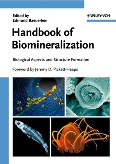
Cover Illustration (Top left designed by Jürgen<br />
Plitzko)<br />
(Top right, Bottom left <strong>and</strong> Bottom right<br />
designed by Felix Baeuerlein)<br />
Top left: 3-D-visualization <strong>of</strong> a part <strong>of</strong><br />
Magnetospirillum gryphiswaldense cell with the<br />
cytoplasmic membrane (blue), empty vesicles<br />
(yellow), growing <strong>and</strong> mature magnetite crystals<br />
(red), <strong>and</strong> the magnetosome cytoskeletal<br />
filament (green). Cryo-electron tomography <strong>and</strong><br />
visualization by M. Gruska <strong>and</strong> J. M. Plitzko,<br />
MPI Martinsried.<br />
(C. Jogler, D. Schüler, Chap. 9, Fig. 9.4 b<br />
modified)<br />
Top right: A skeletal spicule <strong>of</strong> a sea urchin<br />
embryo, isolated, broken, etched <strong>and</strong> carbon<br />
coated, visualized in the SEM by Lindsay<br />
Croker.<br />
(F.H. Wilt, C.A. Ettensohn, Chap. 11, Fig. 11.3<br />
modified)<br />
Bottom left: Skeleton <strong>of</strong> an adult Zebrafish<br />
(Danio rerio) by Synchrotron Radiation Micro<br />
Computer Tomography (SRmCT)<br />
(F. Neues et al, Chap. 21, Fig. 21.4)<br />
Bottom right: Photograph <strong>of</strong> the Medusa<br />
Periphylla periphylla, remarkable for its<br />
statoliths <strong>of</strong> CaSO4 hemihydrate in gravity<br />
sensing.<br />
(F. Boßelmann, et al, Chap. 15, Fig. 15.1)<br />
<strong>H<strong>and</strong>book</strong> <strong>of</strong> <strong>Biomineralization</strong><br />
<strong>Biological</strong> <strong>Aspects</strong> <strong>and</strong> <strong>Structure</strong> Formation:<br />
ISBN 978-3-527-31804-9<br />
Biomimetic <strong>and</strong> Bioinspired Chemistry:<br />
ISBN 978-3-527-31805-6<br />
Medical <strong>and</strong> Clinical <strong>Aspects</strong>:<br />
ISBN 978-3-527-31806-3<br />
Set (3 volumes):<br />
ISBN 978-3-527-31641-0<br />
9 All books published by Wiley-VCH are<br />
carefully produced. Nevertheless, authors,<br />
editors, <strong>and</strong> publisher do not warrant the<br />
information contained in these books,<br />
including this book, to be free <strong>of</strong> errors.<br />
Readers are advised to keep in mind that<br />
statements, data, illustrations, procedural<br />
details or other items may inadvertently be<br />
inaccurate.<br />
Library <strong>of</strong> Congress Card No.: applied for<br />
British Library Cataloguing-in-Publication Data<br />
A catalogue record for this book is available<br />
from the British Library.<br />
Bibliographic information published by<br />
the Deutsche Nationalbibliothek<br />
Die Deutsche Nationalbibliothek lists this<br />
publication in the Deutsche Nationalbibliografie;<br />
detailed bibliographic data are<br />
available in the Internet at hhttp://dnb.d-nb.dei.<br />
8 2007 WILEY-VCH Verlag GmbH & Co.<br />
KGaA, Weinheim<br />
All rights reserved (including those <strong>of</strong><br />
translation into other languages). No part <strong>of</strong> this<br />
book may be reproduced in any form – by<br />
photoprinting, micr<strong>of</strong>ilm, or any other means –<br />
nor transmitted or translated into a machine<br />
language without written permission from the<br />
publishers. Registered names, trademarks, etc.<br />
used in this book, even when not specifically<br />
marked as such, are not to be considered<br />
unprotected by law.<br />
Printed in the Federal Republic <strong>of</strong> Germany<br />
Printed on acid-free paper<br />
Typesetting Asco Typesetters, Hong Kong<br />
Printing betz-druck GmbH, Darmstadt<br />
Binding Litges & Dopf GmbH, Heppenheim<br />
Wiley Bicentennial Logo Richard J. Pacifico<br />
ISBN 978-3-527-31804-9
Further Reading<br />
B€auerlein, E. (Ed.)<br />
<strong>Biomineralization</strong><br />
Progress in Biology, Molecular Biology, <strong>and</strong> Application<br />
Second, Completely Revised <strong>and</strong> Extended Edition<br />
2004<br />
Hardcover<br />
ISBN 978-3-527-31065-4<br />
Konigsberger, E.<br />
<strong>Biomineralization</strong> – Medical <strong>Aspects</strong> <strong>of</strong> Solubility<br />
2006<br />
Hardcover<br />
ISBN 978-0-470-09209-5<br />
Kumar, C. S. S. R. (Ed.)<br />
Nanotechnologies for the Life Sciences<br />
10 volumes<br />
2007<br />
Hardcover<br />
ISBN 978-3-527-31301-3<br />
Willner, I., Katz, E. (Eds.)<br />
Bioelectronics<br />
From Theory to Applications<br />
2005<br />
Hardcover<br />
ISBN 978-3-527-30690-9<br />
Niemeyer, C. M., Mirkin, C. A. (Eds.)<br />
Nanobiotechnology<br />
Concepts, Applications <strong>and</strong> Perspectives<br />
2004<br />
Hardcover<br />
ISBN 978-3-527-30658-9
This book is dedicated to<br />
my ‘‘biomineralization team’’<br />
my wife Cornelia, my daughter Henrike, <strong>and</strong> my son Felix<br />
who accomplished together the pre-editorial management <strong>and</strong> the cover design<br />
<strong>of</strong> the three volumes on biomineralization<br />
<strong>and</strong> to<br />
Pr<strong>of</strong>. Dr. Dieter Oesterhelt<br />
who has <strong>of</strong>fered me also, for this new three-volumes edition,<br />
his support<br />
I am very grateful to all<br />
V
Foreword: The Enigma <strong>of</strong> Morphogenesis –<br />
A Personal View<br />
When asked to write a foreword to this volume, I decided to share my personal<br />
experiences <strong>of</strong> a frustrating contradiction: how much we now underst<strong>and</strong> about<br />
cell structure <strong>and</strong> function; <strong>and</strong> how little we know about morphogenesis at<br />
the cellular level. My interest in morphogenesis stemmed from looking at cells<br />
in a plant root tip. During the 1960s, the electron microscope promised great advances<br />
in our ability to relate cell structure <strong>and</strong> function with the morphogenetic<br />
processes that create such cellular organization. Getting <strong>of</strong>f to an encouraging<br />
start in graduate research, I discovered the ‘‘preprophase b<strong>and</strong>’’ <strong>of</strong> microtubules<br />
[1] which mass along the cell wall before division in a position that, with uncanny<br />
accuracy, predicts the eventual plane <strong>of</strong> this division. This correlation was most<br />
provocative – just the sort that should provide a deeper insight into the mechanisms<br />
by which cells control division <strong>and</strong> morphogenesis. However, after a sobering<br />
40 years <strong>of</strong> investigation in many laboratories, we still do not know the function<br />
or significance <strong>of</strong> the preprophase b<strong>and</strong>.<br />
I ab<strong>and</strong>oned higher plants in favor <strong>of</strong> single-celled algae, where the emergence<br />
<strong>of</strong> form is quick, clean, <strong>and</strong> deceptively simple. Morphogenesis occupies a brief<br />
period <strong>of</strong> the cell cycle, after division; it is precise <strong>and</strong> predictable in outcome,<br />
<strong>and</strong> without the complexities <strong>of</strong> cellular interactions. A group <strong>of</strong> green algae with<br />
thous<strong>and</strong>s <strong>of</strong> different species, the desmids, <strong>of</strong>fered one tractable model system.<br />
When these beautifully symmetrical cells divide, they are bisected by the cleavage<br />
furrow. They restore their symmetry by each older half-cell generating a new halfcell<br />
(‘‘semicell’’) that is the mirror image <strong>of</strong> its parent. This remarkable feat is accomplished<br />
in 1 to 4 hours by the osmotically driven enlargement <strong>of</strong> a wall that<br />
starts <strong>of</strong>f as a round balloon. Very soon in expansion, the plasticity <strong>of</strong> the s<strong>of</strong>t wall<br />
becomes subtly altered in a pattern which is precisely programmed in time <strong>and</strong><br />
space. Thus, the enlarging balloon is magically transformed into a perfect new<br />
semicell.<br />
I confidently explored the ultrastructure <strong>and</strong> cytoskeleton in growing semicells,<br />
but eventually had to accept that nothing in their organization indicated how this<br />
spatial differentiation is generated. Equally enigmatic were living cells. The cytoplasm<br />
in semicells streams vigorously but, as far as I could determine, in an entirely<br />
chaotic fashion. Exquisite form emerges from chaos! Again, after some 30<br />
<strong>H<strong>and</strong>book</strong> <strong>of</strong> <strong>Biomineralization</strong>. Edited by E. Bäuerlein<br />
Copyright 8 2007 WILEY-VCH Verlag GmbH & Co. KGaA, Weinheim<br />
ISBN: 978-3-527-31804-9<br />
VII
VIII Foreword: The Enigma <strong>of</strong> Morphogenesis – A Personal View<br />
years <strong>of</strong> effort, we have not the slightest idea how pattern control is brought about<br />
by the cell [2].<br />
Among the protists, the diatoms inevitably comm<strong>and</strong> the attention <strong>of</strong> anyone<br />
interested in biological form. Their beautifully sculptured cell walls are not only<br />
differentiated to an extraordinary degree, but there are also hundreds <strong>of</strong> thous<strong>and</strong>s<br />
<strong>of</strong> species. Thus, the morphogenetic systems in these cells must satisfy<br />
two contradictory dem<strong>and</strong>s: first, that they reproduce a given complex shape with<br />
absolute, unwavering fidelity; <strong>and</strong> second, that they be capable <strong>of</strong> almost infinite<br />
variability. And <strong>of</strong> course, to express form using one <strong>of</strong> the most refractory materials<br />
known, pure silica.<br />
Electron microscopy <strong>of</strong> these cells was both exciting <strong>and</strong> mystifying. Morphogenesis<br />
in diatoms is obviously completely different to that in desmids. In diatoms,<br />
the wall is secreted in discrete segments (the valves <strong>and</strong> girdle b<strong>and</strong>s),<br />
each formed within the special membrane compartment defined by the ‘‘silica deposition<br />
vesicle’’ (SDV). This is where the complex chemistry <strong>of</strong> silica concentration<br />
<strong>and</strong> precipitation takes place – the area <strong>of</strong> intense research interest, as reflected<br />
in this volume, <strong>and</strong> one for which the details appears increasingly close<br />
to resolution. Morphogenesis is brought about by the growing SDV being precisely<br />
molded in time <strong>and</strong> space. The actual molding process turned out to be a<br />
surprise, as there is no one or two central systems involved. Instead, an extraordinary<br />
range <strong>of</strong> cytoplasmic organelles (e.g., microtubules, actin filaments, endoplasmic<br />
reticulum, mitochondria, vesicles <strong>of</strong> various types) <strong>and</strong> cytoplasmic processes<br />
(e.g., plasmolysis, protoplast movements <strong>and</strong> twisting, localized adhesion<br />
to the valves, mucilage secretion, interaction <strong>of</strong> daughter cells) are combined in<br />
various combinations by different species to mold the growing SDV [3]. Thus, diatoms<br />
to me are the ‘‘Maestros <strong>of</strong> Morphogenesis’’, orchestrating complex interactions<br />
<strong>of</strong> many players to remarkable effect. So perhaps it is not surprising that<br />
there are hundreds <strong>of</strong> thous<strong>and</strong>s <strong>of</strong> different species.<br />
A specific example <strong>of</strong> this morphogenetic flexibility is seen in the extension <strong>of</strong><br />
very fine, needle-like spines <strong>of</strong> silica in four genera: the setae <strong>of</strong> Chaetoceros [4, 5],<br />
the labiate process <strong>of</strong> Ditylum [6] <strong>and</strong> Rhizosolenia [7], <strong>and</strong> the simpler spine <strong>of</strong><br />
Proboscia [8]. Not only do these genera employ different cytoplasmic systems to<br />
create <strong>and</strong> shape the spine, but there are also distinct differences between the<br />
two species <strong>of</strong> Chaetoceros. In addition, the labiate process <strong>of</strong> Rhizsolenia adds an<br />
extraordinary oscillation <strong>of</strong> the spine in vivo during its extension. So, in this tiny<br />
sample <strong>of</strong> the generation <strong>of</strong> one small feature, the cytoplast shows great variation<br />
in the morphogenetic systems that it uses. One wonders how many other ways<br />
diatoms form spines. Summarizing: it seems that in desmids, no organelles appear<br />
correlated with spatial morphogenesis, but diatoms many are. And we have<br />
not considered the morphogenetic systems <strong>of</strong> other unicellular protists, for example,<br />
those that make delicately sculptured, species-specific scales <strong>and</strong> spines from<br />
calcium carbonate <strong>and</strong> silica.<br />
It is axiomatic that the information which controls these morphogenetic processes<br />
resides in the cell’s genetic system, its DNA. What we entirely lack is any<br />
scenario to explain how molecular information in DNA, RNA <strong>and</strong> proteins, can
e expressed on a vastly greater stage <strong>of</strong> macromolecular structure with such precision.<br />
We seem bound to the old reductionist hope that somehow the answer<br />
will drop out if we just get enough information on the bits <strong>and</strong> pieces involved.<br />
There is little reason for optimism that this will happen, <strong>and</strong> a metaphor might<br />
be useful – one, I believe, originally put forward by E.O. Wilson. Consider an ant.<br />
Today, we can study this creature in many ways. We can analyze the structure <strong>and</strong><br />
behavior <strong>of</strong> an ant; its anatomy, muscles <strong>and</strong> sensory abilities; its organs <strong>and</strong><br />
their function; its biochemistry <strong>and</strong> genetics; <strong>and</strong> <strong>of</strong> course, we can sequence its<br />
genome. When we put a million <strong>of</strong> these ants together, miraculous events transpire.<br />
They create social structures <strong>and</strong> build (for them) enormous, speciesspecific<br />
ant castles. None <strong>of</strong> the information we collect about our isolated ant<br />
provides any indication <strong>of</strong> these abilities <strong>and</strong> worse, none has any prospect <strong>of</strong> so<br />
doing. Where does the organizational information reside that confers on them<br />
such abilities? The same enigma is surely true <strong>of</strong> cells. We are swamped by information<br />
about cells <strong>and</strong> their organelles. This information is increasingly reductionistic<br />
due to the burgeoning success <strong>of</strong> molecular biology. Yet none <strong>of</strong> this information<br />
promises any underst<strong>and</strong>ing <strong>of</strong> how, even in principle, information<br />
stored in genes can initiate <strong>and</strong> control processes such as morphogenesis on a<br />
vastly greater scale, like the ant <strong>and</strong> its castle. We appear, yet again, to be confronted<br />
with the limits <strong>of</strong> reductionism because the cell is so much more than<br />
the sum <strong>of</strong> its parts. Where instead to look?<br />
After 40 years’ preoccupation with these phenomena, my conclusion is that the<br />
morphogenetic properties <strong>of</strong> the cytoplasm are an expression <strong>of</strong> its dynamics as<br />
much as its constituents. Like the relationship between the ant <strong>and</strong> the ant-castle,<br />
as soon as we stop the dynamics – whether to undertake ultrastructural or molecular<br />
analysis – the principles <strong>of</strong> morphogenesis we seek to investigate are lost to<br />
us. We may need entirely new models, approaches <strong>and</strong> axioms to progress toward<br />
underst<strong>and</strong>ing one <strong>of</strong> the most interesting <strong>and</strong> intractable mysteries <strong>of</strong> life.<br />
References<br />
1 J.D. Pickett-Heaps, D.H. Northcote.<br />
Cell division in the formation <strong>of</strong> the<br />
stomatal complex in the young leaves<br />
<strong>of</strong> wheat. J. Cell Sci. 1966, 1, 121–128.<br />
Pr<strong>of</strong>. Dr. J. D. Pickett-Heaps (retired)<br />
School <strong>of</strong> Botanic<br />
University <strong>of</strong> Melbourne<br />
Parkville, Australia<br />
New Address:<br />
Cytographics, Online Store<br />
www.cytographics.com<br />
2 J.D. Pickett-Heaps. Morphogenesis<br />
in Desmids: Our current state <strong>of</strong><br />
ignorance, in: J.R. McIntosh (Ed.),<br />
Spatial Organization <strong>of</strong> Eucaryotic<br />
References IX
X Foreword: The Enigma <strong>of</strong> Morphogenesis – A Personal View<br />
Cells. Alan R. Liss, Inc., New York.<br />
1983, Vol. II, pp. 241–258.<br />
3 J.D. Pickett-Heaps, A.M. Schmid, L.A.<br />
Edgar. The cell biology <strong>of</strong> diatom<br />
valve formation. Prog. Phycological<br />
Res. 1990, 7, 1–168.<br />
4 J.D. Pickett-Heaps, J. Carpenter,<br />
A. Koutilis. Valve <strong>and</strong> seta (spine)<br />
morphogenesis in the centric diatom<br />
Chaetoceros peruvianus. Protoplasma<br />
1994, 181, 269–282.<br />
5 J.D. Pickett-Heaps. Cell division <strong>and</strong><br />
morphogenesis <strong>of</strong> the centric diatom<br />
Chaetoceros decipiens: II. Electron<br />
microscopy <strong>and</strong> a new paradigm for<br />
tip growth. J. Phycol. 1998, 34, 995–<br />
1004.<br />
6 J.D. Pickett-Heaps, R. Wetherbee,<br />
D.R.A. Hill. Cell division <strong>and</strong><br />
morphogenesis <strong>of</strong> the labiate process<br />
in the centric diatom Ditylum<br />
brightwellii. Protoplasma 1988, 143,<br />
139–149.<br />
7 A.M.L. Van de Meene, J.D. Pickett-<br />
Heaps. Valve morphogenesis in the<br />
centric diatom Rhizosolenia segitera<br />
(Bacillariophyceae, Centrales) <strong>and</strong> its<br />
taxonomic implications. Eur. J. Phycol.<br />
2004, 39, 93–104.<br />
8 A.M.L. Van de Meene, J.D. Pickett-<br />
Heaps. Valve morphogenesis in the<br />
centric diatom Proboscia alata Sundstom<br />
<strong>and</strong> its taxonomic implications.<br />
J. Phycol. 2002, 38, 351–363.
Contents<br />
Foreword: The Enigma <strong>of</strong> Morphogenesis – A Personal View VII<br />
Preface XXIII<br />
List <strong>of</strong> Contributors XXV<br />
1 Growth <strong>and</strong> Form: What is the Aim <strong>of</strong> <strong>Biomineralization</strong>? 1<br />
Edmund Bäuerlein<br />
Abstract 1<br />
1.1 Introduction 1<br />
1.2 Notions <strong>of</strong> D’Arcy Thompson on Deposition <strong>of</strong> Inorganic Material in<br />
Cells 2<br />
1.3 Close to the Beginning <strong>of</strong> <strong>Biomineralization</strong> 3<br />
1.3.1 Prebiotic Synthesis <strong>of</strong> Peptides 3<br />
1.3.2 Selected Binding <strong>of</strong> Phage(Virus)-Displayed Peptides to Inorganic<br />
Materials 3<br />
1.3.3 Synthesis <strong>of</strong> Inorganic Material by Selected Peptides 3<br />
1.3.4 Selected Sequences with Various Functional Groups 6<br />
1.3.5 Summary <strong>and</strong> Conclusion 7<br />
1.4 Nucleation <strong>of</strong> Inorganic Crystals <strong>and</strong> Inorganic Amorphous/Porous<br />
Forms on Peptides 7<br />
1.4.1 Porous Silica Spheres Synthesized by 12-Amino Acid Peptides<br />
Targets 7<br />
1.4.2 An Amorphous CaCO3 Core <strong>and</strong> a Crystalline CaCO3 Envelope,<br />
Separated by an Organic Layer, Coexist on an Ascidian Skeleton 8<br />
1.4.3 The Proteomic Analysis <strong>of</strong> the Chicken Calcified Eggshell Layer 8<br />
1.4.4 Synthesis <strong>of</strong> Nanocrystalline Hydroxyapatite with a Crystalline Core<br />
<strong>and</strong> a Disordered Surface Region 9<br />
1.4.5 One Iron Atom in Archaeal Ferritin Crystals as Seed for an Iron-Oxide<br />
Cluster 9<br />
1.4.6 Directional Freezing <strong>of</strong> Aqueous Ceramic Suspensions to Shape<br />
Complex Composites 9<br />
1.4.7 Ways to Porous <strong>Structure</strong>s 10<br />
<strong>H<strong>and</strong>book</strong> <strong>of</strong> <strong>Biomineralization</strong>. Edited by E. Bäuerlein<br />
Copyright 8 2007 WILEY-VCH Verlag GmbH & Co. KGaA, Weinheim<br />
ISBN: 978-3-527-31804-9<br />
XI
XII Contents<br />
1.5 Bacterial Filaments in the Advent <strong>of</strong> <strong>Biomineralization</strong>: Cytoskeleton-<br />
Like Proteins <strong>and</strong> Exopolysaccharides 11<br />
1.5.1 Proteins Responsible for the Alignment <strong>of</strong> Magnetosomes in<br />
Magnetotactic Bacteria 11<br />
1.5.1.1 Actin-Like Filaments in Magnetotactic Bacteria 11<br />
1.5.1.2 Actin Filaments in Morphogenesis <strong>of</strong> Diatoms, Eukaryotic Unicellular<br />
Organisms 12<br />
1.5.1.3 Renaissance <strong>of</strong> the ‘‘Gr<strong>and</strong> Unified Theory?’’ 12<br />
1.5.2 Filaments <strong>of</strong> Bacterial Acidic Polysaccharides as Matrices for Iron<br />
Oxide Crystals 13<br />
1.5.2.1 Bacterial Iron Oxide Precipitations 13<br />
1.5.2.2 Bacterial Core Str<strong>and</strong>s <strong>of</strong> Acidic Exopolysaccharides Template Assembly<br />
<strong>of</strong> FeOOH Nanocrystal Fibers 13<br />
1.5.2.3 Acidic Polysaccharides Mediating Formation <strong>of</strong> Complex Calcite<br />
(CaCO3) Crystals in Pleurochrysis carterae, a Unicellular, Eukaryotic<br />
Organism 14<br />
1.5.2.4 Polysaccharides or Peptides: Is There a ‘‘Unified Theory’’? 14<br />
1.6 Proteins <strong>of</strong> Similar Function <strong>and</strong>/or <strong>Structure</strong>, but Low Sequence<br />
Homology: Typical in <strong>Biomineralization</strong> 15<br />
1.6.1 The Avian Eggshell Protein Ovocleidin-17, <strong>and</strong> Human Pancreatic<br />
Stone Protein 15<br />
1.6.2 The Starmaker Protein <strong>of</strong> Zebrafish <strong>and</strong> Human Dentin<br />
Sialophosphoprotein (DSPP) 16<br />
1.7 Composites: Inorganic–Organic Hybrid Materials 16<br />
1.8 Finite Element Analysis <strong>and</strong> Conclusion 18<br />
References 19<br />
I Silica-Hydrated Polysilicondioxide 21<br />
2 Collagen: A Huge Matrix in Glass Sponge Flexible Spicules <strong>of</strong> the<br />
Meter-Long Hyalonema sieboldi 23<br />
Hermann Ehrlich <strong>and</strong> Hartmut Worch<br />
Abstract 23<br />
2.1 Introduction 23<br />
2.2 A Modern Approach to Desilicification <strong>of</strong> Spicules in Glass<br />
Sponges 25<br />
2.3 Glass Sponge Collagen 26<br />
2.3.1 Chemical Etching <strong>of</strong> Spicules <strong>and</strong> Extraction <strong>of</strong> Collagen 26<br />
2.3.2 Collagen Identification 27<br />
2.3.3 Nanoimagery <strong>of</strong> Fibrillar Organic Matrix 29<br />
2.4 Collagen as a Unified Template for <strong>Biomineralization</strong> 30<br />
2.4.1 Evolutionary <strong>Aspects</strong> 30<br />
2.4.2 Twisted Plywood Architecture <strong>of</strong> Collagen Fibrils in Basal Spicules <strong>of</strong><br />
H. sieboldi 32
2.4.3 A New View on the Possible Role <strong>of</strong> Silica in Bone Mineralization 33<br />
2.5 Collagen–Silica-Based Biomaterials 35<br />
2.5.1 Bioactive Glass Composites 35<br />
2.5.2 Collagen–Silica-Based Biohybrids 36<br />
2.6 Open Questions 37<br />
References 38<br />
3 Biochemistry <strong>and</strong> Molecular Genetics <strong>of</strong> Silica <strong>Biomineralization</strong> in<br />
Diatoms 43<br />
Nils Kröger <strong>and</strong> Nicole Poulsen<br />
Abstract 43<br />
3.1 Introduction 43<br />
3.2 The Cell Biology <strong>of</strong> Diatom Silica Formation 45<br />
3.3 Thalassiosira pseudonana as a Model Organism 47<br />
3.3.1 Genome Analysis 47<br />
3.3.2 Silaffins <strong>and</strong> Long-Chain Polyamines from T. pseudonana 48<br />
3.3.2.1 Silaffins 49<br />
3.3.2.2 LCPAs 51<br />
3.3.3 Silica Formation by Silaffins <strong>and</strong> LCPAs 52<br />
3.3.4 Molecular Genetic Manipulation 54<br />
References 57<br />
4 Formation <strong>of</strong> Siliceous Spicules in Demosponges: Example Suberites<br />
domuncula 59<br />
Werner E. G. Müller, Xiaohong Wang, Sergey I. Belikov, Wolfgang Tremel,<br />
Ute Schloßmacher, Antonino Natoli, David Br<strong>and</strong>t, Alex<strong>and</strong>ra Boreiko,<br />
Muhammad Nawaz Tahir, Isabel M. Müller, <strong>and</strong> Heinz C. Schröder<br />
Abstract 59<br />
4.1 Introduction 59<br />
4.2 Early Descriptions 63<br />
4.3 Structural Features <strong>of</strong> the Sponge Body Plan 64<br />
4.4 Cells Involved in Spicule Formation 65<br />
4.5 Anabolic Enzyme for the Synthesis <strong>of</strong> Silica: Silicatein 67<br />
4.6 Silicatein-Associated Proteins 72<br />
4.7 Catabolic Enzyme: Silicase 73<br />
4.8 Morphology <strong>and</strong> Synthesis <strong>of</strong> Spicules in S. domuncula 73<br />
4.9 Formation <strong>of</strong> Spicule Morphology 74<br />
4.10 Phases <strong>of</strong> Silica Deposition during Spicule Formation 76<br />
4.10.1 The Intracellular Phase in the Sclerocytes 76<br />
4.10.2 The Extracellular Phase: Appositional Growth 76<br />
4.10.3 The Extracellular Phase: Shaping 78<br />
4.11 Final Remarks 79<br />
References 80<br />
Contents XIII
XIV Contents<br />
5 Interactions between <strong>Biomineralization</strong> <strong>and</strong> Function <strong>of</strong> Diatom<br />
Frustules 83<br />
Christian Hamm<br />
Abstract 83<br />
5.1 Introduction 83<br />
5.2 Approaches to Study Biominerals 85<br />
5.3 Evolution <strong>and</strong> Diatom Shells 89<br />
5.4 Biomechanics <strong>and</strong> Diatoms 90<br />
5.5 The Effect <strong>of</strong> Evolutionary Feedback on <strong>Biomineralization</strong> 91<br />
5.6 Conclusions 92<br />
References 93<br />
6 The Evolution <strong>of</strong> the Diatoms 95<br />
Wiebe H. C. F. Kooistra<br />
Abstract 95<br />
6.1 Introduction 95<br />
6.2 The Silica Cell Walls <strong>of</strong> the Diatoms 96<br />
6.2.1 The Frustule 96<br />
6.2.2 Frustule Construction 99<br />
6.2.3 Sexual Reproduction <strong>and</strong> Auxospore Formation 100<br />
6.2.4 Resting Stages 102<br />
6.3 Phylogenies 102<br />
6.3.1 The Heterokont Relatives <strong>of</strong> the Diatoms 102<br />
6.3.2 The Phylogeny <strong>of</strong> the Diatoms 103<br />
6.4 The Diatom Fossil Record 105<br />
6.5 The Origin <strong>and</strong> Evolution <strong>of</strong> the Diatom Frustule 107<br />
6.6 Paleo-Ecology <strong>and</strong> Diatom Evolution 108<br />
References 109<br />
7 Uptake <strong>of</strong> Silicon in Different Plant Species 113<br />
Jian Feng Ma<br />
Abstract 113<br />
7.1 Silicon in Plants 113<br />
7.2 Beneficial Effects <strong>of</strong> Silicon on Plant Growth 115<br />
7.2.1 Disease Control 115<br />
7.2.2 Alleviation <strong>of</strong> Stress 115<br />
7.2.3 Plant Growth 116<br />
7.3 Uptake Systems <strong>of</strong> Si in Different Plant Species 116<br />
7.4 Genes Involved in Si Uptake 120<br />
References 123
II Iron Sulfides <strong>and</strong> Oxides 125<br />
8 Magnetic Microstructure <strong>of</strong> Magnetotactic Bacteria 127<br />
Richard B. Frankel, Rafal E. Dunin-Borkowski, Mihály Pósfai,<br />
<strong>and</strong> Dennis A. Bazylinski<br />
Abstract 127<br />
8.1 Introduction 127<br />
8.1.1 Magnetotactic Bacteria 127<br />
8.1.2 Magnetosomes 128<br />
8.1.3 Magnetite Magnetosomes 129<br />
8.1.4 Greigite Magnetosomes 131<br />
8.1.5 Magnetic Properties <strong>of</strong> Magnetosomes 132<br />
8.1.6 Cellular Magnetic Dipole 133<br />
8.2 Experimental Measurements <strong>of</strong> the Magnetic Microstructure <strong>of</strong><br />
Magnetosomes 133<br />
8.2.1 Off-Axis Electron Holography: An Overview 134<br />
8.2.2 Off-Axis Electron Holography <strong>of</strong> Magnetite Magnetosome Chains 135<br />
8.2.3 Off-Axis Electron Holography <strong>of</strong> Greigite Magnetosome Chains 138<br />
8.3 Conclusions 141<br />
References 142<br />
9 Genetic <strong>and</strong> Biochemical Analysis <strong>of</strong> Magnetosome Formation in<br />
Magnetospirillum gryphiswaldense 145<br />
Christian Jogler <strong>and</strong> Dirk Schüler<br />
Contents XV<br />
Abstract 145<br />
9.1 Introduction 145<br />
9.2 Genetics <strong>of</strong> Magnetosome Formation 146<br />
9.2.1 Genomic Organization <strong>of</strong> Magnetosome Genes 146<br />
9.2.2 Genes Encoding Magnetosome-Associated Proteins are Co-Transcribed<br />
within the mam- <strong>and</strong> mms-Operons 150<br />
9.2.3 The Magnetosome Isl<strong>and</strong> is a Highly Unstable Genomic Region <strong>and</strong><br />
Undergoes Spontaneous Rearrangements 150<br />
9.3 Magnetosome-Associated Proteins 151<br />
9.3.1 Biochemical Characterization <strong>of</strong> the Magnetosome Membrane 151<br />
9.3.1.1 TPR Proteins (MamA) 153<br />
9.3.1.2 CDF Proteins 153<br />
9.3.1.3 HtrA-like Serine Proteases 156<br />
9.3.1.4 MMPs with Unknown Function 156<br />
9.3.2 MamJ <strong>and</strong> MamK Control Subcellular Organization <strong>and</strong> Assembly <strong>of</strong><br />
Magnetosomes Chains 157<br />
9.4 Mechanism <strong>of</strong> Magnetosome Formation <strong>and</strong> Magnetite<br />
<strong>Biomineralization</strong> 158<br />
References 160
XVI Contents<br />
10 Physical <strong>and</strong> Chemical Principles <strong>of</strong> Magnetosensation in Biology 163<br />
Michael Winklh<strong>of</strong>er <strong>and</strong> Thorsten Ritz<br />
Abstract 163<br />
10.1 Introduction 163<br />
10.2 A Biochemical Compass Mechanism 164<br />
10.2.1 Magnetic Field Effects on Radical-Pair Reactions 164<br />
10.2.2 A Hypothetical Radical-Pair Based Compass 165<br />
10.2.3 Evidence for a Radical-Pair Mechanism in Migratory Birds 166<br />
10.3 Biogenic Magnetite as a Basis <strong>of</strong> Magnetoreception 167<br />
10.3.1 Pitfalls with the Magnetite Hypothesis 168<br />
10.3.2 Magnetite-Based Magnetoreceptors 169<br />
10.3.3 Hypothetical Transduction Mechanisms 171<br />
10.3.4 Testing the Magnetite Hypothesis with Pulse Experiments 172<br />
10.3.5 <strong>Biomineralization</strong> <strong>of</strong> Magnetite in Vertebrates 173<br />
10.3.6 Non-Destructive Techniques Used to Detect Magnetite in Tissue 174<br />
10.3.6.1 SQUID Measurements 174<br />
10.3.6.2 X-Ray Fluorescence (XRF) <strong>and</strong> X-Ray Absorption Spectroscopy<br />
(XAS) 175<br />
10.3.6.3 Ferromagnetic Resonance (FMR) Spectroscopy 175<br />
10.3.6.4 Nuclear Magnetic Resonance (NMR) Relaxometry 176<br />
10.4 Conclusions 176<br />
References 177<br />
III Calcium Carbonates <strong>and</strong> Sulfates 181<br />
11 The Morphogenesis <strong>and</strong> <strong>Biomineralization</strong> <strong>of</strong> the Sea Urchin Larval<br />
Skeleton 183<br />
Fred H. Wilt <strong>and</strong> Charles A. Ettensohn<br />
Abstract 183<br />
11.1 Introduction 183<br />
11.2 Developmental <strong>Aspects</strong> <strong>of</strong> Sea Urchin <strong>Biomineralization</strong> 184<br />
11.2.1 A General Description <strong>of</strong> Skeletogenesis 184<br />
11.2.2 PMC Specification 189<br />
11.2.2.1 Embryological Studies 189<br />
11.2.2.2 The Micromere-PMC Gene Regulatory Network 189<br />
11.2.3 Regulation <strong>of</strong> Skeletal Patterning in the Embryo 190<br />
11.2.4 Cell Interactions <strong>and</strong> Skeletogenesis 192<br />
11.2.4.1 Cell Interactions <strong>and</strong> PMC Specification 192<br />
11.2.4.2 Cell Interactions <strong>and</strong> Skeletal Morphogenesis 192<br />
11.3 The Composition <strong>and</strong> Formation <strong>of</strong> the Skeletal Spicule 194<br />
11.3.1 Sources <strong>of</strong> Calcium, its Precipitation, <strong>and</strong> Secretion 194<br />
11.3.2 The Spicule Compartment 195<br />
11.3.3 Growth <strong>of</strong> the Spicule 196<br />
11.3.4 Integral Matrix Proteins <strong>of</strong> the Spicule 197
11.3.5 Mineral–Matrix Relationships 200<br />
11.3.6 Functions <strong>of</strong> Matrix Proteins 202<br />
11.3.7 Adult Mineralized <strong>Structure</strong>s 202<br />
11.3.8 Function <strong>of</strong> Non-Matrix Proteins 204<br />
11.4 Generalizations about <strong>Biomineralization</strong> <strong>of</strong> Calcium Carbonates 205<br />
References 207<br />
12 Regulation <strong>of</strong> Coccolith Calcification in Pleurochrysis carterae 211<br />
Mary E. Marsh<br />
Abstract 211<br />
12.1 Introduction 211<br />
12.2 Pleurochrysis Coccolith <strong>Structure</strong> 213<br />
12.3 Pleurochrysis Coccolith Formation 214<br />
12.3.1 Ion Accumulation 215<br />
12.3.2 Calcite Nucleation 217<br />
12.3.3 Crystal Growth 218<br />
12.3.4 Growth Termination 219<br />
12.4 Identifying Other Regulatory Elements in Coccolith<br />
Mineralization 219<br />
12.5 The Non-Mineralizing Phases <strong>of</strong> Pleurochrysis <strong>and</strong> Other<br />
Coccolithophores 221<br />
12.6 Coccolith Calcification <strong>and</strong> the Ocean Carbon Cycle 223<br />
References 224<br />
13 Molecular Approaches to Emiliana huxleyi Coccolith Formation 227<br />
Betsy A. Read <strong>and</strong> Thomas M. Wahlund<br />
Abstract 227<br />
13.1 Introduction 227<br />
13.2 Cellular Physiology <strong>of</strong> <strong>Biomineralization</strong> 228<br />
13.3 Traditional Biochemical Approaches 229<br />
13.4 Genomics 231<br />
13.5 Functional Genomics 232<br />
13.5.1 Suppressive Subtractive Hybridization 233<br />
13.5.2 Microarray 234<br />
13.5.3 Real-Time RT-PCR 235<br />
13.6 Future Directions <strong>and</strong> Approaches 239<br />
References 240<br />
14 Organic Matrix <strong>and</strong> <strong>Biomineralization</strong> <strong>of</strong> Scleractinian Corals 243<br />
Sylvie Tambutté, Eric Tambutté, Didier Zoccola, <strong>and</strong> Denis Allem<strong>and</strong><br />
Abstract 243<br />
14.1 Introduction 243<br />
14.2 Coral Anatomy <strong>and</strong> Histology 245<br />
Contents XVII
XVIII Contents<br />
14.3 The Proportion <strong>of</strong> the Organic Matrix in the Skeleton 247<br />
14.4 The Relationship between the Organic Matrix <strong>and</strong> Calcification 248<br />
14.5 The Composition <strong>of</strong> the Organic Matrix 249<br />
14.6 Localization <strong>of</strong> Organic Matrix Synthesis 249<br />
14.7 The Role <strong>of</strong> Zooxanthellae <strong>and</strong> Heterotrophic Feeding in Organic<br />
Matrix Synthesis 251<br />
14.8 Characterization <strong>of</strong> Organic Matrix Proteins 252<br />
14.9 Comparative Studies between Organic Matrix Proteins from Different<br />
Organisms 253<br />
14.10 Organic Matrix <strong>and</strong> Skeleton Microarchitecture 254<br />
14.11 Organic Matrix <strong>and</strong> Its Implications for Paleo-/Geo-Chemistry <strong>and</strong><br />
Diagenesis 255<br />
14.12 Conclusions 256<br />
References 257<br />
15 Statoliths <strong>of</strong> Calcium Sulfate Hemihydrate are used for Gravity Sensing in<br />
Rhopaliophoran Medusae (Cnidaria) 261<br />
Fabienne Boßelmann, Matthias Epple, Ilka Sötje, <strong>and</strong> Henry Tiemann<br />
Abstract 261<br />
15.1 Diversity <strong>of</strong> Alkaline Earth Sulfates in Organisms <strong>and</strong> Nature 261<br />
15.2 Morphology <strong>of</strong> Rhopalia, Statoliths, <strong>and</strong> their Function 262<br />
15.3 Examination <strong>of</strong> Statoliths 264<br />
15.4 Formation <strong>and</strong> Growth <strong>of</strong> Statoliths 266<br />
15.5 Occurrence <strong>of</strong> Calcium Sulfate Hemihydrate in the Different Taxa with<br />
Phylogenetic <strong>Aspects</strong> 269<br />
References 271<br />
16 Unusually Acidic Proteins in <strong>Biomineralization</strong> 273<br />
Frédéric Marin <strong>and</strong> Gilles Luquet<br />
Abstract 273<br />
16.1 Introduction: Unusually Acidic Proteins <strong>and</strong> the History <strong>of</strong> their<br />
Discovery 273<br />
16.2 What Makes a Protein Unusually Acidic? 275<br />
16.3 Biochemical Techniques for Studying Unusually Acidic Proteins 277<br />
16.4 Interactions <strong>of</strong> Acidic Proteins with Calcium Carbonate Crystals <strong>and</strong><br />
Organo-Mineral Models 279<br />
16.5 Occurrence <strong>of</strong> Unusually Acidic Proteins in Selected Metazoan CaCO3-<br />
Mineralizing Phyla 282<br />
16.6 Concluding Remarks 285<br />
References 286
17 Fish Otolith Calcification in Relation to Endolymph Chemistry 291<br />
Denis Allem<strong>and</strong>, Nicole Mayer-Gostan, Hélène de Pontual, Gilles Boeuf,<br />
<strong>and</strong> Patrick Payan<br />
Abstract 291<br />
17.1 Introduction 291<br />
17.2 Basic Calcification Principles as Applied to Fish Otoliths 293<br />
17.2.1 Basic Equations 293<br />
17.2.2 Difference between a Chemical Crystal <strong>and</strong> a Biocrystal 293<br />
17.2.3 The Players Involved in Calcification 295<br />
17.2.4 The Case <strong>of</strong> Fish Otoliths 296<br />
17.3 The Fish Endolymph: a Complex Heterogeneous Medium 296<br />
17.3.1 The St<strong>and</strong>ard View 296<br />
17.3.2 Spatial Heterogeneity <strong>of</strong> Endolymph Composition 297<br />
17.3.3 Complexity <strong>of</strong> the Saccular Epithelium 298<br />
17.3.4 Dynamics <strong>of</strong> the Components <strong>of</strong> the Endolymph 299<br />
17.4 Are Levels <strong>of</strong> Calcifying Parameters in Endolymph Associated with<br />
Otolith Growth? 300<br />
17.4.1 The Nychthemeral Cycle 300<br />
17.4.1.1 Plasma Calcium Levels 300<br />
17.4.1.2 Incorporation <strong>of</strong> Precursors in the Otolith 300<br />
17.4.1.3 Acid–Base Balance 300<br />
17.4.1.4 Organic Compounds 301<br />
17.4.2 Environmental Factors 301<br />
17.4.3 Conclusion 302<br />
17.5 Questions <strong>and</strong> Future Research Directions 302<br />
17.5.1 Daily Variations in Endolymph Protein Concentrations 302<br />
17.5.2 [Ca 2þ ] <strong>and</strong> [HCO3 ] in the Endolymph 302<br />
17.5.3 Physico-Chemical Originalities <strong>of</strong> the Distal Endolymph 303<br />
17.5.4 Difficulties in the Analysis <strong>of</strong> the OM 304<br />
17.5.5 Weak Analogy between the Organic Components <strong>of</strong> OM <strong>and</strong><br />
Endolymph 305<br />
17.5.6 Comparative Study <strong>of</strong> the OM <strong>of</strong> Carbonated Biominerals 306<br />
17.5.7 Organic Chemistry <strong>of</strong> the Endolymph 306<br />
References 307<br />
18 Eggshell Growth <strong>and</strong> Matrix Macromolecules 309<br />
José Luis Arias, Karlheinz Mann, Yves Nys, Juan Manuel Garcia Ruiz,<br />
<strong>and</strong> Maria Soledad Fernández<br />
Abstract 309<br />
18.1 Introduction 309<br />
18.2 Eggshell <strong>Structure</strong> <strong>and</strong> Formation 310<br />
18.3 Crystalline <strong>Structure</strong> <strong>of</strong> the Eggshell 311<br />
18.4 Eggshell Organic Matrix Components <strong>and</strong> Their Localization 312<br />
18.5 The Unique Eggshell Organic Components 314<br />
Contents XIX
XX Contents<br />
18.5.1 Ovoglycan <strong>and</strong> Ovocleidin-116 314<br />
18.5.2 C-Type Lectin-Like Proteins <strong>of</strong> the Avian Eggshell 314<br />
18.5.3 Ovocalyxins 319<br />
18.6 A Proteomic Inventory <strong>of</strong> the Chicken Calcified Eggshell Matrix 319<br />
18.7 Role <strong>of</strong> the Organic Components in Eggshell Mineralization 323<br />
References 324<br />
IV Calcium Phosphates 329<br />
19 Genetic Basis for the Evolution <strong>of</strong> Vertebrate Mineralized Tissue 331<br />
Kazuhiko Kawasaki <strong>and</strong> Kenneth M. Weiss<br />
Abstract 331<br />
19.1 Introduction 331<br />
19.2 Dental Tissue Mineralization 332<br />
19.3 Matrix Proteins <strong>of</strong> Dental Tissues 333<br />
19.4 Mammalian SCPP Genes 334<br />
19.5 Chicken <strong>and</strong> Frog SCPP Genes 339<br />
19.6 Teleost SCPP Genes 340<br />
19.7 The Origin <strong>of</strong> the SCPP Family 341<br />
19.8 The Function <strong>of</strong> SCPPs <strong>and</strong> Intrinsic Disorder 342<br />
19.9 Conclusions 343<br />
References 344<br />
20 Skeletogenesis in Zebrafish Embryos (Danio rerio) 349<br />
Shao-Jun Du<br />
Abstract 349<br />
20.1 Introduction 349<br />
20.2 Crani<strong>of</strong>acial Skeleton 350<br />
20.2.1 Anatomy <strong>and</strong> Development <strong>of</strong> Zebrafish Crani<strong>of</strong>acial Skeleton 350<br />
20.2.2 Molecular Regulation <strong>of</strong> Crani<strong>of</strong>acial Skeleton Development <strong>and</strong><br />
Patterning 352<br />
20.2.3 Mutational Analyses <strong>of</strong> Crani<strong>of</strong>acial Skeletons 353<br />
20.3 The Axial Skeleton 354<br />
20.3.1 Anatomy <strong>and</strong> Development <strong>of</strong> the Axial Skeleton 354<br />
20.3.2 Development <strong>of</strong> the Intervertebral Disc 356<br />
20.3.3 The Notochord Plays Key Roles in Vertebral Column<br />
Development 358<br />
20.3.4 Retinoic Acid <strong>and</strong> Hedgehog are Involved in Notochord Segmentation<br />
<strong>and</strong> IVD Formation 358<br />
20.3.5 Genetic Screening for Vertebral Mutants 360<br />
20.4 Fin Skeleton 361<br />
20.4.1 Development <strong>of</strong> Median Fins 361<br />
20.4.2 Development <strong>of</strong> Paired Fins 361<br />
20.4.3 Molecular Regulation <strong>of</strong> Fin Formation <strong>and</strong> Growth 362
20.5 Summary 363<br />
References 364<br />
21 The Application <strong>of</strong> Synchrotron Radiation-Based Micro-Computer<br />
Tomography in <strong>Biomineralization</strong> 369<br />
Frank Neues, Felix Beckmann, Andreas Ziegler, <strong>and</strong> Matthias Epple<br />
Abstract 369<br />
21.1 Synchrotron Radiation-Based Micro-Computer Tomography<br />
(SRmCT) 369<br />
21.2 SRmCT applied to Bones <strong>and</strong> Teeth <strong>of</strong> the Zebrafish (Danio<br />
rerio) 371<br />
21.2.1 Overview <strong>of</strong> the Skeleton 372<br />
21.2.2 The Teeth 372<br />
21.2.3 The Vertebral Column 374<br />
21.3 SRmCT applied to the Cuticle <strong>of</strong> P. scaber 375<br />
21.3.1 Overview <strong>of</strong> the Mineralized Exoskeleton 375<br />
21.3.2 Molting <strong>and</strong> Sternal Deposits 376<br />
21.4 Summary 378<br />
References 379<br />
22 Mechanical <strong>and</strong> Structural Properties <strong>of</strong> Skeletal Bone in Wild-Type <strong>and</strong><br />
Mutant Zebrafish (Danio rerio) 381<br />
Fuzhai Cui <strong>and</strong> Xiumei Wang<br />
Abstract 381<br />
22.1 Introduction 381<br />
22.2 The Potential <strong>of</strong> Zebrafish as a Model for Bone Mineralization 382<br />
22.2.1 Hierarchical <strong>Structure</strong>s <strong>of</strong> Zebrafish Skeleton Bone 382<br />
22.2.2 Microstructural Characteristics <strong>and</strong> Nanomechanical Properties across<br />
the Thickness <strong>of</strong> Zebrafish Skeletal Bone 383<br />
22.2.3 Surface Mineralization <strong>of</strong> Collagen Fibrils in Zebrafish Skeleton<br />
Bone 386<br />
22.2.4 Conclusion 390<br />
22.3 Hierarchical Structural Comparisons <strong>of</strong> Bones from Wild-Type <strong>and</strong><br />
liliput dtc232 (lil) Gene-Mutated Zebrafish 390<br />
22.3.1 Alteration <strong>of</strong> Vertebrae Development 390<br />
22.3.2 Fracture Topography <strong>and</strong> Fibrils Array Patterns 390<br />
22.3.3 Mineralized Collagen Fibrils 391<br />
22.3.4 Type I Collagen Fibrils 391<br />
22.3.5 The Hydoxyapatite Minerals 391<br />
22.4 Variation <strong>of</strong> Nanomechanical Properties <strong>of</strong> Bone by Gene Mutation in<br />
the Zebrafish 392<br />
22.5 Conclusion 395<br />
References 395<br />
Contents XXI
XXII Contents<br />
23 Nanoscale Mechanisms <strong>of</strong> Bone Deformation <strong>and</strong> Fracture 397<br />
Peter Fratzl <strong>and</strong> Himadri S. Gupta<br />
Abstract 397<br />
23.1 The Hierarchical <strong>Structure</strong> <strong>of</strong> Bone 397<br />
23.2 Structural Design <strong>of</strong> Bone at the Nanoscale 400<br />
23.3 The Lamellar Organization <strong>of</strong> Bone 404<br />
23.4 Bone Deformation at the Nanoscale 408<br />
References 412<br />
24 Formation <strong>and</strong> <strong>Structure</strong> <strong>of</strong> Calciprotein Particles: The Calcium Phosphate–<br />
Ahsg/Fetuin-A Interface 415<br />
Alex<strong>and</strong>er Heiss <strong>and</strong> Dietmar Schwahn<br />
Abstract 415<br />
24.1 The Protein–Mineral Interface 415<br />
24.1.1 Mineral Formation 415<br />
24.1.2 Fetuin-A 418<br />
24.2 Small-Angle Neutron-Scattering Studies 419<br />
24.2.1 Instrumental Set-Up 419<br />
24.2.2 Theoretical Background 420<br />
24.3 Calciprotein Particle Formation <strong>and</strong> Transformation 422<br />
24.3.1 Fetuin-A 422<br />
24.3.2 Inhibition <strong>of</strong> Mineral Sedimentation Effected by the Serum Proteins<br />
Fetuin-A <strong>and</strong> Albumin 423<br />
24.3.3 Calciprotein Particle Formation 424<br />
24.3.4 CPP <strong>Structure</strong> 426<br />
24.4 Conclusions 428<br />
References 430<br />
Index 433
Preface<br />
Currently, biomineralization is evolving from its heyday <strong>of</strong> structural research<br />
into one <strong>of</strong> the most exciting fields <strong>of</strong> molecular biology. In order to bring these<br />
new developments to the notice <strong>of</strong> its chemists <strong>and</strong> physicists, I had edited two<br />
multi-author books on ‘‘<strong>Biomineralization</strong>’’, in November 2000 <strong>and</strong> October<br />
2004. In a competent <strong>and</strong> detailed review <strong>of</strong> the second <strong>of</strong> these books, S. Weiner<br />
(Angew. Chem. Int. Ed. 2005, 44, 4833–4834) noted that he had missed the presence<br />
<strong>of</strong> a general introduction <strong>and</strong>, in addition to that, a more comprehensive<br />
view ‘‘. . . to cover the waterfront <strong>of</strong> this vast field’’.<br />
It was the exemplary intuition <strong>of</strong> the outst<strong>and</strong>ing publisher, Dr. Gudrun Walter,<br />
the Program Director <strong>of</strong> Wiley-VCH, that encouraged her to outline biomineralization<br />
as a fundamental interaction between the organic <strong>and</strong> inorganic spheres.<br />
Consequently, she devised three volumes on biomineralization, covering biomimetic<br />
chemistry, materials science, <strong>and</strong> the life sciences. Moreover, she entrusted<br />
me again with this new project which exceeds by far, both in content <strong>and</strong> extent,<br />
the two preceding books.<br />
The first draft three volumes <strong>of</strong> ‘‘<strong>Biomineralization</strong>’’ were appended with the<br />
subtitle: Biology, Biomimetic <strong>and</strong> Bio-Inspired-Chemistry, Medicine. In addition,<br />
because each volume was planned to consist <strong>of</strong> 25 to 30 chapters, I invited Pr<strong>of</strong>.<br />
Peter Behrens, a well-known expert in biomimetic chemistry <strong>and</strong> coordinator <strong>of</strong><br />
the DFG-Priority Program ‘‘Principles <strong>of</strong> <strong>Biomineralization</strong>’’, to co-edit with me<br />
the chemistry volume. Pr<strong>of</strong>. Behrens convinced me that biomimetic chemistry is<br />
an indispensable approach to study the developments <strong>of</strong> inorganic/organic hybrids<br />
in addition to molecular biology, which, before our co-edition, was in my<br />
mind the only method. The Spring Meeting 2005 <strong>of</strong> the Materials Research Society<br />
(MRS) was characterized by a broad <strong>of</strong>fer <strong>of</strong> bio-inspired synthesis <strong>and</strong> techniques,<br />
<strong>and</strong> this allowed the selection <strong>of</strong> several new topics. Pr<strong>of</strong>. Matthias Epple,<br />
an extraordinary inorganic chemist, whose interests cover the isolation <strong>and</strong> analysis<br />
<strong>of</strong> biominerals, the synthesis <strong>of</strong> biomaterials, <strong>and</strong> the development <strong>of</strong> new<br />
physical methods in materials science <strong>and</strong> cooperation with medical institutes<br />
<strong>and</strong> cliniques, agreed – to my great pleasure – to organize the medical volume<br />
essentially by himself.<br />
As all manuscripts for the book arrived at Munich for the pre-editorial management,<br />
I had the chance to take a general view <strong>of</strong> each <strong>of</strong> the 68 chapters, <strong>and</strong> this<br />
<strong>H<strong>and</strong>book</strong> <strong>of</strong> <strong>Biomineralization</strong>. Edited by E. Bäuerlein<br />
Copyright 8 2007 WILEY-VCH Verlag GmbH & Co. KGaA, Weinheim<br />
ISBN: 978-3-527-31804-9<br />
XXIII
XXIV Preface<br />
allowed me to prepare a first draft in Chapter 1 <strong>of</strong> what might be common in biomineralization<br />
reactions but characteristically different from mainstream biochemistry.<br />
It is possible that although this draft was too specialized for an introduction,<br />
its complexity was simply a reflection <strong>of</strong> the current state <strong>of</strong> the art <strong>of</strong><br />
biomineralization.<br />
January 2007 Edmund Bäuerlein<br />
Munich/Martinsried<br />
Germany
List <strong>of</strong> Contributors<br />
Denis Allem<strong>and</strong><br />
Centre Scientifique de Monaco<br />
Ave. Saint-Martin<br />
MC-98000 Monaco<br />
Principality <strong>of</strong> Monaco<br />
<strong>and</strong><br />
Université de Nice-Sophia<br />
Antipolis<br />
Faculté des Sciences<br />
Parc Valrose, 28 Ave. Valrose<br />
06108 Nice Cedex 02<br />
France<br />
José-Luis Arias<br />
Department <strong>of</strong> Animal Biology<br />
Faculty <strong>of</strong> Veterinary <strong>and</strong> Animal<br />
Sciences<br />
University <strong>of</strong> Chile <strong>and</strong> CIMAT<br />
Santa Rosa 11735<br />
La Pintana<br />
Santiago<br />
Chile<br />
Edmund Bäuerlein<br />
Max-Planck-Institute for<br />
Biochemistry<br />
Dept. <strong>of</strong> Membrane Biochemistry<br />
Am Klopferspitz 18A<br />
82152 Munich-Martinsried<br />
Germany<br />
<strong>H<strong>and</strong>book</strong> <strong>of</strong> <strong>Biomineralization</strong>. Edited by E. Bäuerlein<br />
Copyright 8 2007 WILEY-VCH Verlag GmbH & Co. KGaA, Weinheim<br />
ISBN: 978-3-527-31804-9<br />
Dennis A. Bazylinski<br />
Dept. <strong>of</strong> <strong>Biological</strong> Sciences<br />
University <strong>of</strong> Nevada Las Vegas<br />
4505 Maryl<strong>and</strong> Parkway<br />
Las Vegas, NV 89154<br />
USA<br />
Felix Beckmann<br />
Institute for Materials Research<br />
GKSS-Research Center<br />
Max-Planck-Str. 1<br />
21502 Geesthacht<br />
Germany<br />
Sergey I. Belikov<br />
Limnological Institute <strong>of</strong> the Siberian<br />
Branch <strong>of</strong> Russian Academy <strong>of</strong><br />
Sciences<br />
Ulan-Batorskaya 3<br />
664033<br />
Irkutsk<br />
Russia<br />
Gilles Boeuf<br />
UMR 7628 University Pierre et Marie<br />
Curie-Paris 6/CNRS<br />
Laboratoire Arago<br />
BP 44<br />
66651 Banyuls-sur-mer Cedex<br />
France<br />
XXV
XXVI List <strong>of</strong> Contributors<br />
Alex<strong>and</strong>ra Boreiko<br />
Institute for Physiological<br />
Chemistry<br />
Department <strong>of</strong> Applied Molecular<br />
Biology<br />
University Mainz<br />
Duesbergweg 6<br />
55099 Mainz<br />
Germany<br />
Fabienne Boßelmann<br />
Institute <strong>of</strong> Inorganic Chemistry<br />
University <strong>of</strong> Duisburg-Essen<br />
Universitätsstraße 5–7<br />
45117 Essen<br />
Germany<br />
David Br<strong>and</strong>t<br />
Institute for Physiological<br />
Chemistry<br />
Department <strong>of</strong> Applied Molecular<br />
Biology<br />
University Mainz<br />
Duesbergweg 6<br />
55099 Mainz<br />
Germany<br />
Fuzhai Cui<br />
Department <strong>of</strong> Materials Science<br />
<strong>and</strong> Engineering<br />
Tsinghua University<br />
Yifu Technology <strong>and</strong> Scientific<br />
Building<br />
Beijing, 100084<br />
P.R. China<br />
Shao-Jun Du<br />
Center for Marine Biotechnology<br />
University <strong>of</strong> Maryl<strong>and</strong><br />
Biotechnology Institute<br />
Columbus Center<br />
701 East Pratt Street<br />
Baltimore<br />
Madison, MD 21202<br />
USA<br />
Rafal E. Dunin-Borkowski<br />
Dept. <strong>of</strong> Materials Science<br />
<strong>and</strong> Metallurgy<br />
University <strong>of</strong> Cambridge<br />
Pembroke Street<br />
Cambridge CB2 3QZ<br />
UK<br />
Hermann Ehrlich<br />
Max Bergmann Center <strong>of</strong> Biomaterials<br />
<strong>and</strong> Institute <strong>of</strong> Material Science<br />
Dresden University <strong>of</strong> Technology<br />
Budapester Straße 27<br />
01069 Dresden<br />
Germany<br />
Matthias Epple<br />
Institute for Inorganic Chemistry<br />
University <strong>of</strong> Duisburg-Essen<br />
Universitätsstraße 5–7<br />
45117 Essen<br />
Germany<br />
Charles A. Ettensohn<br />
Department <strong>of</strong> <strong>Biological</strong> Sciences<br />
Carnegie Mellon University<br />
4400 Fifth Avenue<br />
Pittsburg, PA 15213<br />
USA<br />
Maria Soledad Fernández<br />
Department <strong>of</strong> Animal Biology<br />
Faculty <strong>of</strong> Veterinary <strong>and</strong> Animal<br />
Sciences<br />
University <strong>of</strong> Chile <strong>and</strong> CIMAT<br />
Santa Rosa 11735<br />
La Pintana<br />
Santiago<br />
Chile<br />
Richard B. Frankel<br />
Department <strong>of</strong> Physics<br />
California Polytechnic State University<br />
San Louis Obispo, CA 93407<br />
USA
Peter Fratzl<br />
Dept. <strong>of</strong> Biomaterials<br />
Max-Planck-Institute <strong>of</strong> Colloids<br />
<strong>and</strong> Interfaces<br />
Am Mühlenberg 1<br />
14476 Potsdam<br />
Germany<br />
Juan-Manuel García-Ruiz<br />
Laboratorio de Estudios<br />
Cristalográficos CSIC<br />
Avenida de la Innovacion 1<br />
P.T. Ciencias de la Salud<br />
18100 Armilla<br />
Granada<br />
Spain<br />
Himadri S. Gupta<br />
Dept. <strong>of</strong> Biomaterials<br />
Max-Planck-Institute <strong>of</strong> Colloids<br />
<strong>and</strong> Interfaces<br />
Am Mühlenberg 1<br />
14476 Potsdam<br />
Germany<br />
Christian Hamm<br />
Alfred Wegener Institute for<br />
Polar <strong>and</strong> Marine Research<br />
Plankton Biomechanics Pelagic<br />
Ecosystems/<strong>Biological</strong><br />
Oceanography<br />
Am H<strong>and</strong>elshafen 12/Co9<br />
27570 Bremerhaven<br />
Germany<br />
Alex<strong>and</strong>er Heiss<br />
Dept. <strong>of</strong> Biomedical Engineering<br />
<strong>and</strong> IZKF BIOMAT RWTH<br />
Aachen<br />
University Hospital<br />
Pauwelsstraße 30<br />
52074 Aachen<br />
Germany<br />
Christian Jogler<br />
Department <strong>of</strong> Microbiology<br />
Max-Planck-Institute for Marine<br />
Microbiology<br />
Celsiusstrasse 1<br />
28359 Bremen<br />
Germany<br />
Kazuhiko Kawasaki<br />
Department <strong>of</strong> Anthropology<br />
Pennsylvania State University<br />
409 Carpenter Building<br />
University Park, PA 16802-3404<br />
USA<br />
Wiebe H. C. F. Kooistra<br />
Stazione Zoologica Anton Dohrn<br />
Department <strong>of</strong> Marine Botany<br />
Villa Communale<br />
80121 Napoli<br />
Italy<br />
Nils Kröger<br />
School <strong>of</strong> Chemistry <strong>and</strong> Biochemistry<br />
<strong>and</strong> School <strong>of</strong> Materials Science<br />
Georgia Institute <strong>of</strong> Technology<br />
901 Atlantic Dr.<br />
Atlanta, GA 30332<br />
USA<br />
Gilles Luquet<br />
UMR CNRS 5561 Biogéoscience<br />
Université de Bourgogne<br />
6, Bd. Gabriel<br />
21000 Dijon<br />
France<br />
List <strong>of</strong> Contributors XXVII<br />
Jian Feng Ma<br />
Research Institute for Bioresources<br />
Okayama University<br />
Chuo 2-20-1<br />
Kurashiki, 710-0046<br />
Japan
XXVIII List <strong>of</strong> Contributors<br />
Karlheinz Mann<br />
Department <strong>of</strong> Proteomics <strong>and</strong><br />
Signal Transduction<br />
Max-Planck-Institute for<br />
Biochemistry<br />
Am Klopferspitz 18A<br />
82152 Martinsried<br />
Germany<br />
Frédéric Marin<br />
UMR CNRS 5561 Biogéoscience<br />
Université de Bourgogne<br />
6, Bd. Gabriel<br />
21000 Dijon<br />
France<br />
Mary E. Marsh<br />
Department <strong>of</strong> Basic Science<br />
University <strong>of</strong> Texas Health<br />
Science Center<br />
6516 M. D. Anderson Blvd.<br />
Houston, TX 77030<br />
USA<br />
Nicole Mayer-Gostan<br />
UMR UNSA-INRA 112<br />
University <strong>of</strong> Nice-Sophia<br />
Antipolis<br />
Faculty <strong>of</strong> Science<br />
Parc Valrose, 28 Ave. Valrose<br />
06108 Nice Cedex 2<br />
France<br />
Isabel M. Müller<br />
Institute for Physiological<br />
Chemistry<br />
Department <strong>of</strong> Applied Molecular<br />
Biology<br />
University Mainz<br />
Duesbergweg 6<br />
55099 Mainz<br />
Germany<br />
Werner E. G. Müller<br />
Institute for Physiological Chemistry<br />
Department <strong>of</strong> Applied Molecular<br />
Biology<br />
University Mainz<br />
Duesbergweg 6<br />
55099 Mainz<br />
Germany<br />
Antonio Natoli<br />
Institute for Physiological Chemistry<br />
Department <strong>of</strong> Applied Molecular<br />
Biology<br />
University Mainz<br />
Duesbergweg 6<br />
55099 Mainz<br />
Germany<br />
Frank Neues<br />
Institute for Inorganic Chemistry<br />
University <strong>of</strong> Duisburg-Essen<br />
Universitätsstr. 5–7<br />
45117 Essen<br />
Germany<br />
Yves Nys<br />
Institut National de la Recherche<br />
Agronomique<br />
Unité de Recherches Avicoles<br />
Centre de Tours<br />
37380 Nouzilly<br />
France<br />
Patrick Payan<br />
UMR UNSA-INRA 112<br />
University <strong>of</strong> Nice-Sophia Antipolis<br />
Faculty <strong>of</strong> Science<br />
Parc Valrose, 28 Ave. Valrose<br />
06108 Nice Cedex 2<br />
France
Hélène de Pontual<br />
IFREMER, Centre de Brest<br />
STH/LASAA<br />
BP 70<br />
29280 Plouzane<br />
France<br />
Mihály Pósfai<br />
Dept. <strong>of</strong> Earth <strong>and</strong> Environmental<br />
Sciences<br />
University <strong>of</strong> Veszprem<br />
Egyetem Street 10<br />
8200 Veszprem<br />
Hungary<br />
Nicole Poulsen<br />
School <strong>of</strong> Chemistry <strong>and</strong><br />
Biochemistry<br />
Georgia Institute <strong>of</strong> Technology<br />
901 Atlantic Dr.<br />
Atlanta, GA 30332<br />
USA<br />
Betsy A. Read<br />
California State University<br />
San Marcos<br />
3335 Twin Oaks Valley Rd.<br />
San Marcos, CA 92096-0001<br />
USA<br />
Thorsten Ritz<br />
Dept. <strong>of</strong> Physics <strong>and</strong> Astronomy<br />
University <strong>of</strong> California<br />
4129 Frederic Reines Hall<br />
Irvine, CA 92697-4575<br />
USA<br />
Ute Schloßmacher<br />
Institute for Physiological<br />
Chemistry<br />
Department <strong>of</strong> Applied Molecular<br />
Biology<br />
University Mainz<br />
Duesbergweg 6<br />
55099 Mainz<br />
Germany<br />
Heinz C. Schröder<br />
Institute for Physiological Chemistry<br />
Department <strong>of</strong> Applied Molecular<br />
Biology<br />
University Mainz<br />
Duesbergweg 6<br />
55099 Mainz<br />
Germany<br />
Dirk Schüler<br />
Institute for Biology I<br />
Department <strong>of</strong> Microbiology<br />
LMU München<br />
Maria-Ward-Str. 1a<br />
80638 Munich<br />
Germany<br />
Dietmar Schwahn<br />
Institute for Solid State Research<br />
Research Center Jülich<br />
Wilhelm-Juhnen-Str.<br />
52425 Jülich<br />
Germany<br />
Ilka Sötje<br />
Biocenter Grindel<br />
University <strong>of</strong> Hamburg<br />
Martin-Luther-King-Platz 3<br />
20146 Hamburg<br />
Germany<br />
Muhammad Nawaz Tahir<br />
Institute for Inorganic <strong>and</strong> Analytical<br />
Chemistry<br />
University Mainz<br />
Duesbergweg 10–14<br />
55099 Mainz<br />
Germany<br />
Eric Tambutté<br />
Centre Scientifique de Monaco<br />
Ave. Saint Martin<br />
MC-98000<br />
Monaco<br />
List <strong>of</strong> Contributors XXIX
XXX List <strong>of</strong> Contributors<br />
Sylvie Tambutté<br />
Centre Scientifique de Monaco<br />
Ave. Saint Martin<br />
MC-98000<br />
Monaco<br />
Henry Tieman<br />
Biocenter Grindel<br />
University <strong>of</strong> Hamburg<br />
Martin-Luther-King-Platz 3<br />
20146 Hamburg<br />
Germany<br />
Wolfgang Tremel<br />
Institute for Inorganic <strong>and</strong><br />
Analytical Chemistry<br />
University Mainz<br />
Duesbergweg 10–14<br />
55099 Mainz<br />
Germany<br />
Thomas M. Wahlund<br />
California State University<br />
San Marcos<br />
3335 Twin Oaks Valley Rd.<br />
San Marcos, CA 92096-0001<br />
USA<br />
Xiaohong Wang<br />
National Research Center for<br />
Geoanalysis<br />
26 Baiwanzhuang Dajie<br />
Beijing 100037<br />
China<br />
Xiumei Wang<br />
Center for Biomedical<br />
Engineering<br />
Massachusetts Institute <strong>of</strong><br />
Technology<br />
500 Technology Square<br />
Cambridge, MA 02139<br />
USA<br />
Kenneth M. Weiss<br />
Department <strong>of</strong> Anthropology<br />
Pennsylvania State University<br />
409 Carpenter Building<br />
University Park, PA 16802-3404<br />
USA<br />
Fred H. Wilt<br />
Department <strong>of</strong> Molecular Cell Biology<br />
University <strong>of</strong> California<br />
142 Life Science Annex Bldg.<br />
Berkeley, CA 94720-3200<br />
USA<br />
Michael Winklh<strong>of</strong>er<br />
Department <strong>of</strong> Earth <strong>and</strong><br />
Environmental Science<br />
Geophysics<br />
Ludwig-Maximilians-University <strong>of</strong><br />
Munich<br />
Theresienstraße 41/IV<br />
80333 Munich<br />
Germany<br />
Hartmut Worch<br />
Max Bergmann Center <strong>of</strong> Biomaterials<br />
<strong>and</strong> Institute <strong>of</strong> Material Science<br />
Dresden University <strong>of</strong> Technology<br />
Budapester Straße 27<br />
01069 Dresden<br />
Germany<br />
Andreas Ziegler<br />
Center for Electron Microscopy<br />
University <strong>of</strong> Ulm<br />
Albert Einstein-Allee 11<br />
89069 Ulm<br />
Germany<br />
Didier Zoccola<br />
Centre Scientifique de Monaco<br />
Ave. Saint Martin<br />
MC-98000<br />
Monaco
1<br />
Growth <strong>and</strong> Form: What is the Aim<br />
<strong>of</strong> <strong>Biomineralization</strong>?<br />
Edmund Bäuerlein<br />
Abstract<br />
12-Amino-acid peptides with binding-selected sequences <strong>and</strong> r<strong>and</strong>om functional<br />
groups synthesize the same inorganic material. Bacteria use structured polymers<br />
as filaments <strong>of</strong> cytoskeletal protein <strong>and</strong> <strong>of</strong> polysaccharides in templating biominerals.<br />
Nucleation appears to be involved not only in crystal formation, but also in<br />
the formation <strong>of</strong> porous or amorphous inorganic material. A seed <strong>of</strong> one iron ion<br />
for an iron-oxide cluster could be obtained in the low-iron-state <strong>of</strong> an archeal ferritin<br />
crystal. Porous inorganic materials were described to grow out in huge channeled<br />
complexes <strong>of</strong> organic compounds or crystallized seawater. The evolutionary<br />
progress <strong>of</strong> biomineralization runs apparently in the development <strong>of</strong> complex,<br />
inorganic–organic hybrid materials, composites, which are <strong>of</strong>ten structured hierarchically.<br />
The mechanical properties <strong>of</strong> a mineral undergo a change by presence<br />
<strong>and</strong> partition <strong>of</strong> the organic material, the most prominent example being the<br />
human skeleton. In order to describe such a complex system, a mathematical<br />
procedure – the finite element analysis – was introduced with great success.<br />
This analysis is governed amazingly by only one physical quantity, force, together<br />
with the modulus <strong>of</strong> elasticity. The overwhelming extent <strong>of</strong> research into the mechanical<br />
properties <strong>of</strong> skeletons has resulted in the conclusion that the aim <strong>of</strong> biomineralization<br />
appears to be stability.<br />
Key words: peptides, induced structures, low homologies, nucleation, filaments,<br />
pores, polysaccharides, unified theory, composites, finite element analysis,<br />
stability.<br />
1.1<br />
Introduction<br />
A shortened title <strong>of</strong> an extraordinary book written by D’Arcy Wentworth Thompson,<br />
On Growth <strong>and</strong> Form [1], published in 1917, opens this first volume <strong>of</strong> the<br />
<strong>H<strong>and</strong>book</strong> <strong>of</strong> <strong>Biomineralization</strong>. Edited by E. Bäuerlein<br />
Copyright 8 2007 WILEY-VCH Verlag GmbH & Co. KGaA, Weinheim<br />
ISBN: 978-3-527-31804-9<br />
1
2 1 Growth <strong>and</strong> Form: What is the Aim <strong>of</strong> <strong>Biomineralization</strong>?<br />
<strong>H<strong>and</strong>book</strong> <strong>of</strong> <strong>Biomineralization</strong>. It refers to an ‘‘Abridged Edition’’ <strong>of</strong> the illustrious<br />
biologist John Tyler Bonner (Princetown) by the same publishing house in 1961<br />
[2]. Bonner’s fundamental experimental research on the height <strong>and</strong> life cycle <strong>of</strong><br />
slime molds favored him with the competence to shorten <strong>and</strong> comment on this<br />
important work [3].<br />
Whereas D’Arcy Thompson initiated the development <strong>of</strong> a general relationship<br />
between the growth <strong>and</strong> form <strong>of</strong> cells <strong>and</strong> tissues, this chapter focuses on the<br />
interaction between inorganic materials <strong>and</strong> organic (including prebiotic) compounds<br />
up to organisms, which are able to synthesize <strong>and</strong> shape biominerals.<br />
1.2<br />
Notions <strong>of</strong> D’Arcy Thompson on Deposition <strong>of</strong> Inorganic Material in Cells<br />
The outst<strong>and</strong>ing merit <strong>of</strong> Thompson’s book is the introduction <strong>of</strong> mathematical<br />
<strong>and</strong> physical methods into the biology <strong>of</strong> growth <strong>and</strong> form in 1917. This broad<br />
field <strong>of</strong> research is covered today by biophysics, bioinformatics <strong>and</strong> quantitative<br />
system biology to such an extent that could not have been expected previously.<br />
Although the center <strong>of</strong> his interest was the development <strong>and</strong> shaping <strong>of</strong> cells,<br />
D’Arcy Thompson did not ignore biomineralization. Within his book <strong>of</strong> nine<br />
chapters, he attended one <strong>of</strong> those to ‘‘On Spicules <strong>and</strong> Spicular Skeletons’’. This<br />
is based on the view that ‘‘. . . deposition <strong>of</strong> inorganic material in living cells is a<br />
frequent phenomenon’’. Later, J.T. Bonner summarized precisely the fixed opinion<br />
<strong>of</strong> the author [2a]: ‘‘In this chapter <strong>and</strong> the following ones D’Arcy Thompson<br />
is struggling against the notion that all form can simply be explained by heredity,<br />
<strong>and</strong> that therefore changes in form inevitably map out phylogenetic relations. Instead<br />
he repeatedly suggests that physical forces (such as those which produce<br />
the variations <strong>of</strong> shapes <strong>of</strong> snowflakes) are <strong>of</strong> prime importance <strong>and</strong> relationships<br />
<strong>of</strong> shape may not justify any family tree or sequence in time, but simply show<br />
mathematical kinship.’’<br />
Indeed, the excellent structural research <strong>of</strong> biominerals within the past 20 to<br />
30 years appears to be strongly affected by these physical <strong>and</strong> mathematical<br />
approaches.<br />
Nevertheless, in 2000 J. Kirschvink risked outlining ‘‘A Gr<strong>and</strong> Unified<br />
Theory on <strong>Biomineralization</strong>’’ [4]. This draft was based mainly on morphological<br />
changes, <strong>and</strong> began with the earliest known biosynthesis <strong>of</strong> a mineral, speciesspecific<br />
magnetite nanoparticles in magnetotactic bacteria. This idea fascinated<br />
many <strong>of</strong> us that magnetite biomineralization might be an ‘‘evolutionary procedure’’<br />
for most biominerals during evolution.<br />
One year later, in 2001, the new combined method <strong>of</strong> molecular biology <strong>and</strong><br />
genome sequencing invaded biomineralization. This era began with the almostcomplete<br />
genome sequences <strong>of</strong> two magnetotactic bacteria, which were generously<br />
<strong>of</strong>fered to the scientific public in January 2001 by the Joint Genome Institutes,<br />
DOE, US Government (thanks to Pr<strong>of</strong>. Ken Nealson). By using sequence<br />
comparison, no genes could be found with high similarities to those involved in<br />
magnetite biomineralization (see Chapter 9 in this volume).
1.3<br />
Close to the Beginning <strong>of</strong> <strong>Biomineralization</strong><br />
1.3.1<br />
Prebiotic Synthesis <strong>of</strong> Peptides<br />
Today, carbon compounds such as amino acids, sugars <strong>and</strong> heterocycles, which<br />
are described as organic compounds from the time after the origin <strong>of</strong> life, may<br />
be synthesized in laboratories under a variety <strong>of</strong> prebiotic reaction conditions [5].<br />
The prebiotic origin <strong>of</strong> peptides is <strong>of</strong> special interest to the first phenomena <strong>of</strong><br />
biomineralization. Basalt, a volcanic rock composed <strong>of</strong> various silicates, was used<br />
as catalyst in prebiotic mode reaction, with a mixture <strong>of</strong> all amino acids reacting<br />
in the presence <strong>of</strong> basalt at 180 C to peptides, via the loss <strong>of</strong> water [6]. On the<br />
basis <strong>of</strong> these preliminary experiments it could not have been foreseen that the<br />
tight binding <strong>of</strong> the products – the peptides – might represent an important step<br />
in the interaction <strong>of</strong> prebiotic ‘‘organic’’ compounds <strong>and</strong> minerals in the development<br />
<strong>of</strong> the first cells [7].<br />
1.3.2<br />
Selected Binding <strong>of</strong> Phage(Virus)-Displayed Peptides to Inorganic Materials<br />
It was the unique intuition <strong>of</strong> A. Belcher which made her seek for specific interaction,<br />
the goal being to identify families <strong>of</strong> amino acid sequences that could bind<br />
differentially to specific faces <strong>and</strong> polymorphs <strong>of</strong> the substrate <strong>of</strong> interest [8a].<br />
At this time, ‘‘Combinatorial Phage Display Peptide Libraries’’ first became<br />
commercially available [9]. These peptide libraries were the result <strong>of</strong> a gigantic<br />
r<strong>and</strong>om peptide synthesis initiative, based on molecular biology. For example,<br />
about 3 10 9 distinct r<strong>and</strong>om sequences are present as combinations <strong>of</strong> 12 r<strong>and</strong>om<br />
amino acids. Each <strong>of</strong> these 12-amino-acid peptides is expressed as a fusion<br />
with a small surface displayed protein (called III) <strong>of</strong> the bacteriophage M13 (a<br />
virus) which infects only bacteria. In addition, each virus molecule has placed<br />
five copies <strong>of</strong> its protein III onto the surface, <strong>and</strong> provides there consequently<br />
five identical peptides. Each 12-amino-acid peptide sequence is amplified to yield<br />
about 55 copies per volume, as used by Belcher in the selection steps with electronic<br />
material. These were gallium arsenide, indium phosphide <strong>and</strong> zinc sulfide,<br />
as well as crystals <strong>of</strong> biominerals such as calcite <strong>and</strong> aragonite – two common<br />
polymorphs <strong>of</strong> calcium carbonate found in many organisms.<br />
1.3.3<br />
Synthesis <strong>of</strong> Inorganic Material by Selected Peptides<br />
1.3 Close to the Beginning <strong>of</strong> <strong>Biomineralization</strong> 3<br />
M.O. Stone did not use this method only to select for peptides that bind specifically<br />
to inorganic materials. Rather, his brilliant idea was to determine whether<br />
such phage-displayed peptides could be capable <strong>of</strong> generating those inorganic<br />
structures to which they had bound. The success <strong>of</strong> this extended analysis was<br />
extraordinary in a variety <strong>of</strong> directions. In the first experiment, Stone’s group ex-
4 1 Growth <strong>and</strong> Form: What is the Aim <strong>of</strong> <strong>Biomineralization</strong>?<br />
Fig. 1.1 Schematic illustration <strong>of</strong> regular <strong>and</strong> polymerase chain reaction<br />
(PCR)-driven phage panning for peptides that bind to inorganic<br />
nanoparticles. (According to [10c]; courtesy <strong>of</strong> M.O. Stone.)<br />
amined those peptides which were selected by their binding capacity on silica particles<br />
[10a] for the formation <strong>of</strong> these particles by orthosilicic acid. This peptidemediated<br />
material synthesis resulted in the greatest quantities <strong>of</strong> silica when<br />
amino acids containing hydroxyl-, imidazole- <strong>and</strong> high cationic groups were present<br />
in the 12-amino-acid peptides. This population <strong>of</strong> phage-displayed peptides<br />
was obtained by a st<strong>and</strong>ard panning procedure (Fig. 1.1), which included several<br />
amplification steps by infections <strong>of</strong> Escherichia coli host cells with the selected<br />
phage-displayed peptides [10a]. They appeared to act enzyme-like by definite residues<br />
in binding as well as in material production [11]. Although such similarity<br />
was found, it was questioned at the same time by the results <strong>of</strong> peptide-mediated<br />
synthesis <strong>and</strong> the patterning <strong>of</strong> silver nanoparticles [10b]. Three silver-binding<br />
peptides indicated a preferential enrichment <strong>of</strong> proline <strong>and</strong> hydroxyl-residues<br />
containing amino acids, but not <strong>of</strong> any sulfur-containing amino acids such as<br />
cysteine or methionine. Two <strong>of</strong> these three phage-displayed peptides precipitated<br />
silver nanocrystals.<br />
This fascinating study reached its climax, when a new method – polymerase<br />
chain reaction (PCR)-driven panning – was used instead <strong>of</strong> a regular phage panning<br />
(Fig. 1.1). The results with silver will not be discussed in detail here, in favor<br />
<strong>of</strong> more extended experiments with cobalt nanoparticles, but they are listed in<br />
Table 1.1.<br />
The question <strong>of</strong> whether some <strong>of</strong> these peptides remained bound following a<br />
low-pH incubation step was answered when three sets <strong>of</strong> cobalt-binding phagedisplayed<br />
peptides were obtained. Both methods – ‘‘regular panning’’ <strong>and</strong> PCRdriven<br />
– are likely to be characterized very shortly. ‘‘Regular panning’’ is carried<br />
out by incubating the library <strong>of</strong> phage-displayed peptides with a target (in this
Table 1.1 Silver-binding peptides. a<br />
PCR method Regular panning<br />
AG-P1 KFLQFVCLGVGP AG3<br />
AYSSGAPPMPPF<br />
AG-P2 AVLMQKYHQLGP AG4<br />
NPSSLFRYLPSD<br />
AG-P6 QQNVPASGTCSI<br />
AG-P10 NAMPGMVAWLCR<br />
AG-P11 HNTSPSPIILTP<br />
AG-P12 ASQTLLLPVPPL<br />
AG-P14 YNKDRYEMQAPP<br />
AG-P18 TLLLLAFVHTRH<br />
AG-P27 PWATAVSGCFAP<br />
AG-P28 SPLLYATTSNQS<br />
AG-P35 WSWRSPTPHWT<br />
a According to Ref. [10c]; courtesy <strong>of</strong> M.O. Stone.<br />
1.3 Close to the Beginning <strong>of</strong> <strong>Biomineralization</strong> 5<br />
AG3 AYSSGAPPMPPF<br />
AG4 NPSSLFRYLPSD<br />
AG5 SLATQPPRTPPV<br />
case cobalt nanoparticles), washing away unbound/non-specific phages, <strong>and</strong> eluting<br />
the bound phages using a low-pH incubation step (see Fig. 1.1). The eluted<br />
phages are amplified <strong>and</strong> subjected to additional ‘‘panning’’ rounds in order to<br />
obtain highly enriched binding sequences. A high number <strong>of</strong> phage-displayed<br />
peptides remained strongly bound to the cobalt nanoparticles, even after an extended<br />
acid elution step. These were identified using a PCR-driven method,<br />
which is much more rapidly performed than ‘‘regular panning’’. In both methods,<br />
the first steps before or after acid solution were essentially the same, after<br />
which the corresponding cobalt nanoparticles were incubated in lysis buffer for<br />
10 min at 95 C to disrupt the phage coat; this resulted in phage DNA release.<br />
The PCR reaction, when mixed with a specific set <strong>of</strong> DNA primers [12], was<br />
added directly to the cobalt nanoparticles.<br />
Following the PCR reaction, the samples were separated on an agarose gel. The<br />
DNA fragments obtained were cloned into a plasmid, <strong>and</strong> DNA sequencing was<br />
performed to derive the amino acid sequences <strong>of</strong> the phage-displayed peptides.<br />
The amino acid sequences <strong>of</strong> these cobalt-binding peptides are listed in Table<br />
1.2 (no discussion will be made at this point as to why Set II does not represent<br />
all sequences <strong>of</strong> Set I plus those <strong>of</strong> Set III). Moreover, Set II included seven sequences<br />
which did not belong to either Set I or Set III, which probably indicated<br />
that insufficient clones had been sequenced, this being an extremely laborintensive<br />
project <strong>and</strong> not the aim <strong>of</strong> this publication.
6 1 Growth <strong>and</strong> Form: What is the Aim <strong>of</strong> <strong>Biomineralization</strong>?<br />
Table 1.2 Cobalt-binding peptides. a<br />
Eluted cobalt nanoparticles<br />
(Set I)<br />
Uneluted cobalt nanoparticles<br />
(Set II)<br />
1.3.4<br />
Selected Sequences with Various Functional Groups<br />
Regular panning (Set III)<br />
Co1-P1 HSVRWLLPGAHP Co2-P4 HSVRWLLPGAHP Co3-P12 GTSTFNSVPVRD<br />
Co1-P2 HETNPPATIMPH Co2-P3 SAPNLNALSAAS Co3-P13 SAPNLNALSAAS<br />
Co1-P3 WASAAWLVHSTI Co2-P5 SVSVGMKPSPRP Co3-P1 SVSVGMKPSPRP<br />
Co1-P4 SPLQVLPYQGYV Co2-P8 SPLQVLPYQGYV Co3-P16 VPTNVQLQTPRS<br />
Co1-P5 ESIPALAGLSDK Co2-P1 SLTQTVTPWAFY<br />
Co1-P6 GVLNAAQTWALS Co2-P7 TNLDDSYPLHHL<br />
Co1-P8 TPNSDALLTPAL Co2-P6 TPNSDALLTPAL<br />
Co1-P10 HYPTLPLGSSTY Co2-P12 HYPTLPLGSSTY<br />
Co1-P13 HAMRPQVHPNYA Co2-P9 TQQTDSRPPVLL<br />
Co1-P15 QYKHHPQKAAHI Co2-P14 QYKHHPQKAAHI<br />
Co1-P16 YGNQTPYWYPHR Co2-P11 TFPSHLATSTQP<br />
Co1-P17 HPPTDGMVPSPP Co2-P13 QNFLQVIRNAPR<br />
Co1-P18 TWQPFGMRPSDP Co2-P2 KLHSSPHTPLVQ<br />
Co1-P21 TGDVSNNPNVTL Co2-P17 QLLPLTPSLLQA<br />
a According to Ref. [10c]; courtesy <strong>of</strong> M.O. Stone.<br />
The st<strong>and</strong>ard procedure <strong>of</strong> binding, washing <strong>and</strong> acid-eluting followed by amplification<br />
in E. coli host cells – which is referred to as regular panning (Fig. 1.1) –<br />
was used initially in the isolation <strong>of</strong> four selected phage-displayed peptides with<br />
some functional groups on their 12-amino-acid sequences. Among these four<br />
peptides, neither histidine (H) nor glutamic acid (E) (Table 1.1) – both <strong>of</strong> which<br />
are known to bind specifically to cobalt [13] – were found.<br />
Moreover, the PCR method (Fig. 1.1) yielded at least 25 phage-displayed peptides<br />
(Table 1.2) which had resisted high-stringency washing conditions. Between<br />
one <strong>and</strong> three histidines (H) were present in nine <strong>of</strong> these 25 peptide sequences,<br />
whereas glutamic acid was identified in only one peptide sequence.<br />
Three peptides <strong>of</strong> Set I (Co1-P1, Co1-P10 <strong>and</strong> Co1-P15) <strong>and</strong> one peptide <strong>of</strong> Set<br />
II were synthesized chemically [14] (Table 1.3) in order to exclude possible interactions<br />
<strong>of</strong>, or precipitation on, the phage (virus) surface during mineral synthesis.<br />
This study was not conducted to obtain knowledge <strong>of</strong> these peptides as templates<br />
for cobalt nanoparticles, but rather for the synthesis <strong>of</strong> cobalt–platinum nanoparticles.<br />
These nanoparticles are <strong>of</strong> major interest in materials chemistry because <strong>of</strong><br />
their enhanced magnetic anisotropy <strong>and</strong> other positive properties [15]. Again, the<br />
lack <strong>of</strong> similarities among the four 12-amino-acid peptide sequences was obvious<br />
(Table 1.3), <strong>and</strong> all four resulted in the precipitation <strong>of</strong> the metal alloy CoaPt, albeit<br />
with various crystal sizes [10c].
1.4 Nucleation <strong>of</strong> Inorganic Crystals <strong>and</strong> Inorganic Amorphous/Porous Forms on Peptides 7<br />
Table 1.3 Peptides synthesized chemically for the experiments.<br />
1.3.5<br />
Summary <strong>and</strong> Conclusion<br />
Amino acid sequence Isoelectric pH (pI) Panning method<br />
Silver-binding peptides<br />
AG-P3 IRPAIHIIPISH 9.7 PCR<br />
AG-P4 NVIRASPPDTSY 5.8 PCR<br />
AG-P5 LAMPNTQADAPF 3.8 PCR<br />
AG-P28 SPLLYATTSNQS 5.2 PCR<br />
AG-P35 WSWRSPTPHVVT 9.7 PCR<br />
AG-4 NPSSLFRYLPSD 6.1 Regular<br />
Cobalt-binding peptides<br />
Co1-P1 HSVRWLLPGAHP 9.7 PCR<br />
Co2-P2 KLHSSPHTLPVQ 8.6 PCR<br />
Co1-P10 HYPTLPLGSSTT 6.7 PCR<br />
Co1-P15 QYKHHPQKAAHI 9.7 PCR<br />
a According to Ref. [10c]; courtesy <strong>of</strong> M.O. Stone.<br />
12-Amino-acid peptides bind very tightly to inorganic materials <strong>and</strong> produce this<br />
inorganic material, not by the action <strong>of</strong> any specific functional group but rather<br />
by a variety <strong>of</strong> selected sequences with various other functional groups. Whether<br />
each <strong>of</strong> these sequences or various induced secondary structures may be responsible<br />
for both interactions, binding to <strong>and</strong> producing <strong>of</strong> this corresponding inorganic<br />
material, remains an open question.<br />
Some indications in favor <strong>of</strong> induced secondary structures were obtained from<br />
studies with poly-l-lysine (PLL), the molecular weight <strong>of</strong> which affected the morphology<br />
<strong>of</strong> silica precipitated in presence <strong>of</strong> silicic acid. A higher molecularweight<br />
PLL produced hexagonal silica platelets, whereas spherical silica particles<br />
were obtained using low-molecular-weight PLL. The polypeptide secondary structure<br />
transitions were determined from circular dichroism spectra [16].<br />
1.4<br />
Nucleation <strong>of</strong> Inorganic Crystals <strong>and</strong> Inorganic Amorphous/Porous Forms<br />
on Peptides<br />
1.4.1<br />
Porous Silica Spheres Synthesized by 12-Amino Acid Peptides Targets<br />
12-Amino-acid peptides, when bound to silica nanoparticles, provide the simplest<br />
system for synthesizing the starting product, porous silica spheres (see Section
8 1 Growth <strong>and</strong> Form: What is the Aim <strong>of</strong> <strong>Biomineralization</strong>?<br />
1.3.3) [10a]. Formation <strong>of</strong> the alloy cobalt–platinum <strong>and</strong> <strong>of</strong> silver by using the<br />
same method created nano- <strong>and</strong> microcrystals. Thus, the question arises <strong>of</strong><br />
whether porous or amorphous materials begin their formation <strong>and</strong> their shaping<br />
also by nucleation. Again, both mechanisms (see Section 1.3.4) impose the question<br />
<strong>of</strong> whether enzyme-like specific functional group(s) or selected sequences<br />
(either hydrophobic or hydrophilic) might be responsible for the origin <strong>of</strong> crystals<br />
or porous/amorphous forms, respectively. Initially, this question does not make<br />
sense, because in methodical terms it appears that no access exists for studying<br />
early clusters in terms <strong>of</strong> their preorder or disorder as a prerequisite for crystals<br />
or porous/amorphous particles.<br />
1.4.2<br />
An Amorphous CaCO3 Core <strong>and</strong> a Crystalline CaCO3 Envelope, Separated by<br />
an Organic Layer, Coexist on an Ascidian Skeleton<br />
An important report on ascidian skeletons by Aizenberg [17] suggested the possibility<br />
that the formation <strong>of</strong> skeletal crystalline calcium carbonate may begin with<br />
nucleation <strong>of</strong> an amorphous crystalline core (ACC). A detailed study revealed that<br />
each mineral phase contained characteristic proteins. Typically, on the one h<strong>and</strong><br />
glycoproteins were extracted from the ACC, which was rich in glutamic acid <strong>and</strong><br />
hydroxyamino acids, <strong>and</strong> on the other h<strong>and</strong> from the crystalline phase, which was<br />
rich in aspartic acid. Based on the results <strong>of</strong> in-vitro experiments, the ACC was<br />
stabilized by its acidic glycoprotein, which in addition inhibited the crystallization<br />
<strong>of</strong> calcite; otherwise, the aspartate-rich glycoprotein induced the formation <strong>of</strong><br />
calcite crystals. The two calcium carbonate phases were separated in well-defined<br />
domains by an organic sheet. This highly organized system points to a very interesting<br />
inorganic/organic hybrid – namely, a composite with specific materials<br />
properties – rather than to an initial calcite crystallization by an amorphous<br />
phase.<br />
1.4.3<br />
The Proteomic Analysis <strong>of</strong> the Chicken Calcified Eggshell Layer<br />
Very recently, Mann reported the details <strong>of</strong> a proteomic analysis <strong>of</strong> an acid-soluble<br />
matrix <strong>of</strong> chicken calcified eggshell layer [18]. Here, knowledge <strong>of</strong> 90% <strong>of</strong> the<br />
chicken genome sequence was combined with Edman degradation sequencing<br />
<strong>of</strong> chromatographic separation, coupled to modern liquid chromatography (LC)mass<br />
spectrometry (MS)/MS method following protein cleavage.<br />
Among 520 proteins, a group <strong>of</strong> 32 highly abundant proteins was described<br />
which contained ten previously characterized molecules. As yet, only a<br />
few <strong>of</strong> the mostly uncharacterized proteins have been characterized as acidic (for<br />
a discussion <strong>of</strong> this subject, see Volume 1, Chapters 16 <strong>and</strong> 18; see also Table<br />
18.1).
1.4 Nucleation <strong>of</strong> Inorganic Crystals <strong>and</strong> Inorganic Amorphous/Porous Forms on Peptides 9<br />
1.4.4<br />
Synthesis <strong>of</strong> Nanocrystalline Hydroxyapatite with a Crystalline Core <strong>and</strong><br />
a Disordered Surface Region<br />
In synthesizing nanocrystalline hydroxyapatite, Epple succeeded in presenting a<br />
reverse order <strong>of</strong> nucleation [19] compared to the development <strong>of</strong> a complex composite<br />
ACC/calcite tissue in the ascidian skeleton study (see Section 1.4.2) [17].<br />
Using transmission electron microscopy (TEM) <strong>and</strong> solid-state nuclear magnetic<br />
resonance (NMR), the nanocrystals were shown to consist <strong>of</strong> a crystalline core <strong>and</strong><br />
a disordered surface region. The crystalline core was seen to be hydroxyapatite<br />
(HAp), <strong>and</strong> the surface region to be dominated by hydrogen phosphate anions,<br />
with no hydroxyapatite-like structural motif <strong>and</strong> structural water.<br />
1.4.5<br />
One Iron Atom in Archaeal Ferritin Crystals as Seed for an Iron-Oxide Cluster<br />
The Dps-like ferritin protein DpsA from the halophile Halobacterium salinarum is<br />
member <strong>of</strong> a second subfamily <strong>of</strong> ferritin. This differs from the ferritins by having<br />
24 identical subunits, each <strong>of</strong> which shows outer <strong>and</strong> inner diameters <strong>of</strong> 12<br />
<strong>and</strong> 8 nm, respectively. By comparison, members <strong>of</strong> the Dps subfamily assemble<br />
only to protein shells <strong>of</strong> 12 identical subunits, with outer <strong>and</strong> inner diameters <strong>of</strong> 9<br />
<strong>and</strong> 4.5 nm, respectively. These ferritins therefore store only up to 110 iron<br />
atoms, compared to about 500 iron atoms in the case <strong>of</strong> eukaryotic <strong>and</strong> eubacterial<br />
ferritins.<br />
One major advantage <strong>of</strong> the structural analysis <strong>of</strong> the DpsA protein was that<br />
three high-resolution structures could be determined at successively increasing<br />
iron contents [20]. For the low-iron state <strong>of</strong> DpsA with 1.6 A˚ resolution, a single<br />
iron atom provides a structural seed for iron-oxide cluster formation at the nucleation<br />
center, NI. The Fe 3þ <strong>of</strong> this subsite is coordinated prismatically to three<br />
symmetry-related glutamic acid residues <strong>and</strong> three symmetry-related water molecules.<br />
At this subsite, 13 water molecules form an unusual three-layered crown<br />
around the central Fe 3þ ion. The next structure to be detected is an [4Fe-3O] cluster<br />
in the NII nucleation center; this is the first structural characterization <strong>of</strong> a<br />
nucleation event by one ion (atom) in which succeeded Vesterhelt.<br />
1.4.6<br />
Directional Freezing <strong>of</strong> Aqueous Ceramic Suspensions to Shape Complex<br />
Composites<br />
Those <strong>of</strong> us not familiar with the physics <strong>of</strong> water will be excited for several reasons<br />
by the approach <strong>of</strong> Deville. Diatoms, coccolithophores <strong>and</strong> other impurities<br />
are concentrated within channels between the ice crystals <strong>of</strong> sea-water, because<br />
pure hexagonal ice platelets with r<strong>and</strong>omly oriented horizontal crystallographic<br />
axis are formed [21]. Moreover, it is a fundamental finding that a critical particle
10 1 Growth <strong>and</strong> Form: What is the Aim <strong>of</strong> <strong>Biomineralization</strong>?<br />
size exists [22], above which the suspended particles will be trapped by the moving<br />
water–ice front. The application <strong>of</strong> both these experiences to produce complex<br />
composites is aided considerably by the fact that the hexagonal ice crystals exhibit<br />
strong anisotropic growth kinetics. The ice thus formed will have a lamellar<br />
microstructure, with the lamellar thickness depending mainly on the speed <strong>of</strong> the<br />
freezing front. The authors succeeded in building homogeneous, layered, porous<br />
scaffolds by freezing concentrated suspensions <strong>of</strong> ceramic particles above the critical<br />
particle size [23]. (For details, see Volume 2, Chapters 5 <strong>and</strong> 10.)<br />
1.4.7<br />
Ways to Porous <strong>Structure</strong>s<br />
A first analogy to these porous scaffolds presents itself in the formation <strong>of</strong> the<br />
porous silica-based cell walls <strong>of</strong> diatoms. A supramolecular assembly, termed<br />
the silaffin matrix, is formed spontaneously when the main organic compounds<br />
<strong>of</strong> these cell walls – long-chain polyamines (LCPA) <strong>and</strong> silaffins – are added together.<br />
LCPA are N-methylated derivatives <strong>of</strong> the oligo-propyleneimine chains,<br />
whereas silaffins (e.g., natSil2) are highly phosphorylated polypeptides. These<br />
matrices present two principles – a negative principle due to silaffins, <strong>and</strong> a positive<br />
principle due to LCPA – <strong>and</strong> are protonated at the acidic pH inside diatoms,<br />
in the silica deposition vesicles (SDV) [24]. (For details, see Volume 1, Chapter 3<br />
<strong>and</strong> Volume 2, Chapter 2).<br />
The huge, developing matrix remains invisible in water <strong>and</strong> may be isolated<br />
by centrifugation. It also exhibits silica precipitation activity by the addition <strong>of</strong><br />
monomeric silicic acid. This matrix may be considered analogous to the ice<br />
matrix, <strong>and</strong> is assumed to be a network where, in the free space, the channels<br />
are filled by polymerizing silicic acid. Removal <strong>of</strong> these organic compounds may<br />
lead to an opening <strong>of</strong> the labyrinth <strong>of</strong> pores.<br />
However, the discovery <strong>of</strong> three new silaffins in the model organism Thalassiosira<br />
pseudonana revealed that each silaffin, when mixed with LCPA, exhibits strikingly<br />
different effects on silica morphogenesis in vitro, either as:<br />
the formation <strong>of</strong> spherical silica particles, or<br />
porous sheets <strong>of</strong> silica, or<br />
the deposition <strong>of</strong> plates made from densely packed but<br />
extremely small silica particles, together with spherical silica<br />
particles.<br />
This packing <strong>of</strong> silica particles traces the formation <strong>of</strong> porous silica back to the<br />
freezing technique (see Section 1.4.6; see also Volume 2, Chapter 10). The most<br />
important observation is that a critical particle size exists above which segregation<br />
– that is, the formation <strong>of</strong> multilayered porous ceramic structures – occurs. With<br />
increasing particle size, the space between the particles exp<strong>and</strong>s, leading most<br />
likely to a second type <strong>of</strong> pore which constitutes its own a labyrinth; this is an<br />
‘‘open pore’’. When the channels are subsequently filled with another phase, the<br />
now dense complex composite may still possess pores <strong>of</strong> the second type.
1.5 Bacterial Filaments in the Advent <strong>of</strong> <strong>Biomineralization</strong> 11<br />
Very recently, a quantitative evaluation <strong>of</strong> porous materials in bones <strong>and</strong> bonesubstitute<br />
materials was introduced by Epple, who used microcomputed tomography<br />
followed by an algorithmic procedure to perform the analysis. More precise,<br />
quantitative data were obtained on the extent <strong>and</strong> type <strong>of</strong> porosity, <strong>and</strong> on their<br />
inner surface. The presence <strong>of</strong> an interconnecting pore system (<strong>of</strong> an ‘‘open<br />
pore’’) was shown convincingly for bone <strong>and</strong> all <strong>of</strong> the bone-substitute materials<br />
examined [25].<br />
1.5<br />
Bacterial Filaments in the Advent <strong>of</strong> <strong>Biomineralization</strong>: Cytoskeleton-Like Proteins<br />
<strong>and</strong> Exopolysaccharides<br />
1.5.1<br />
Proteins Responsible for the Alignment <strong>of</strong> Magnetosomes in Magnetotactic Bacteria<br />
Bacteria are regarded as the simplest <strong>of</strong> all known unicellular organisms capable<br />
<strong>of</strong> synthesizing biominerals [26]. However, this superlative position has been<br />
questioned with the recent detection <strong>of</strong> a bacterial cytoskeleton containing tubulin<br />
<strong>and</strong> actin homologues [27a,b]; this was similar to eukaryotic actin <strong>and</strong> tubulin<br />
in terms <strong>of</strong> its three-dimensional (3-D) structure <strong>and</strong> biochemical properties.<br />
1.5.1.1 Actin-Like Filaments in Magnetotactic Bacteria<br />
Magnetotactic bacteria have roused special interest because they are able to utilize<br />
the Earth’s magnetic field to achieve optimal life conditions. Single nanocrystals<br />
<strong>of</strong> magnetite (Fe3O4), a magnetic iron oxide, when enveloped by a phospholipid<br />
membrane, are referred to as magnetosomes. These are aligned in the cytoplasm<br />
to form a chain which behaves like a compass needle. The construction <strong>of</strong> such a<br />
stable magnet <strong>of</strong> about 30 to 60 magnetosomes is the center <strong>of</strong> this biomineralization,<br />
most details <strong>of</strong> which were elucidated by Schüler when introducing modern<br />
molecular-genetic <strong>and</strong> protein chemical methods into biomineralization research<br />
(see Volume 1, Chapter 9).<br />
A large genomic isl<strong>and</strong>, which encodes most <strong>of</strong> the specific, magnetosomeassociated<br />
proteins, was identified <strong>and</strong> designated as a ‘‘magnetosome isl<strong>and</strong>’’<br />
[28], due mainly to its striking similarities to the ‘‘pathogenicity isl<strong>and</strong>s’’ identified<br />
in other bacteria [29].<br />
The product <strong>of</strong> the mamJ gene is MamJ, an acidic protein which appears to interact<br />
with the magnetosome surface, as well as with an actin-like filament structure<br />
that directs the assembly <strong>and</strong> localization <strong>of</strong> the magnetosomes (Fig. 1.2).<br />
This cytoskeletal structure is very similar to that <strong>of</strong> the actin-like MreB proteins<br />
<strong>of</strong> other bacteria [27a,b], <strong>and</strong> is assumed to be formed by the mamK gene product<br />
[30]. Its deletion resulted in an absence <strong>of</strong> the magnetic filament(s) [31].<br />
A major breakthrough in the analysis <strong>of</strong> detailed structure within intact shockfrozen<br />
cells was reported by Plitzko, who used cryoelectron tomography (as developed<br />
by its pioneer, Baumeister) [32] to display the cytoskeletal structures <strong>of</strong> wild-
12 1 Growth <strong>and</strong> Form: What is the Aim <strong>of</strong> <strong>Biomineralization</strong>?<br />
Fig. 1.2 A section <strong>of</strong> Magnetospirillum gryphiswaldense showing the<br />
cytoskeletal magnetosome filament. Magnetosome vesicles, which are<br />
empty or contain growing magnetite crystals, are attached to the<br />
filament. (Micrographs <strong>and</strong> visualization by J.M. Plitzko; see Volume 1,<br />
Chapter 9.)<br />
type <strong>and</strong> mutant bacteria that were just a few nanometers in size (see Volume 1,<br />
Chapter 9; see also the front/back book cover, top left).<br />
1.5.1.2 Actin Filaments in Morphogenesis <strong>of</strong> Diatoms, Eukaryotic Unicellular<br />
Organisms<br />
Until today, a deep discrepancy has existed between those involved in research<br />
utilizing morphogenesis <strong>and</strong> those using molecular biology methods. Nevertheless,<br />
several studies <strong>of</strong> the morphogenesis <strong>of</strong> diatoms have shown that flagellinlike<br />
filaments appear to be involved in the formation <strong>of</strong> diatom shells (frustules).<br />
Thus, Kröger <strong>and</strong> Poulson (see Volume 1, Chapter 3), as leading proponents <strong>of</strong><br />
molecular biology studies, point to the important review <strong>of</strong> Pickett-Heaps <strong>and</strong> colleagues<br />
on ‘‘Cell biology <strong>of</strong> diatom valve formation’’ [33], in which the following<br />
is described (see p. 109):<br />
‘‘In some scale-forming organisms, too, there is evidence that actin<br />
filaments are involved in morphogenesis. Micr<strong>of</strong>ilaments<br />
associated with the silica deposition vesicles (SDV) <strong>of</strong> scale<br />
forming vesicles in synura appear in a transient horse-shoe<br />
pattern <strong>of</strong> parallel, cross-linked fibres, attached directly to the<br />
membrane. They contain actin <strong>and</strong> may function in shaping the<br />
SDV, since they disappear once the shape <strong>of</strong> the scale has been<br />
established.’’ [34].<br />
1.5.1.3 Renaissance <strong>of</strong> the ‘‘Gr<strong>and</strong> Unified Theory?’’<br />
In Section 1.3, some doubt was expressed as to whether a sequence comparison<br />
would confuse rather than confirm the evolution <strong>of</strong> biominerals from magnetite<br />
crystals <strong>of</strong> bacteria to hydroxyapatite in the human skeleton. A first draft <strong>of</strong> a unified<br />
theory <strong>of</strong> biomineralization, based mainly on morphogenesis, was published<br />
by Kirschvink <strong>and</strong> Hagedorn in ‘‘<strong>Biomineralization</strong> 2000’’ [4]. Although, initially<br />
this highly logical theory was accepted with great interest, major doubts were soon
1.5 Bacterial Filaments in the Advent <strong>of</strong> <strong>Biomineralization</strong> 13<br />
expressed when the Human Genome Project was completed by the Joint Genome<br />
Institutes, DOE, US Government. There followed by an extensive project <strong>of</strong> genome<br />
sequencing <strong>of</strong> 15 bacteria, including those <strong>of</strong> two magnetotactic species.<br />
A surprising discovery was made in 2004 by Löwe [27], who reported that bacteria<br />
possess a cytoskeleton which consists <strong>of</strong> actin-like <strong>and</strong> tubulin-like proteins,<br />
with the bacteria showing low homologies in their DNA sequence to those <strong>of</strong> eukaryotic<br />
cells. Such experience suggests that genes <strong>and</strong> gene products, when combined<br />
with a knowledge <strong>of</strong> likely supposition <strong>of</strong> function, may (or will) allow the<br />
creation <strong>of</strong> a new unified theory. Today, however, more importance seems to be<br />
attached to the structure <strong>and</strong> function <strong>of</strong> proteins in biomineralization than to<br />
genes or to the derived amino acid sequences (see Sections 1.5.2.4 <strong>and</strong> 1.6).<br />
1.5.2<br />
Filaments <strong>of</strong> Bacterial Acidic Polysaccharides as Matrices for Iron Oxide Crystals<br />
1.5.2.1 Bacterial Iron Oxide Precipitations<br />
An extraordinary number <strong>of</strong> publications have described external iron oxide precipitates<br />
(for a review, see [35a,b]) compared to those relating to internal crystal<br />
formation (see Volume 1, Chapter 9). However, to date only one report exists describing<br />
polysaccharide filaments that are located outside, but still attached to, the<br />
bacterium, as templates for such minerals [36].<br />
1.5.2.2 Bacterial Core Str<strong>and</strong>s <strong>of</strong> Acidic Exopolysaccharides Template Assembly<br />
<strong>of</strong> FeOOH Nanocrystal Fibers<br />
An organic polymer other than a polypeptide was identified by Banfield as a new<br />
templating element <strong>of</strong> bacterial biomineralization. In extended studies <strong>of</strong> iron<br />
oxyhydroxide-encrusted bi<strong>of</strong>ilms, <strong>and</strong> <strong>of</strong> minerals suspended in the surrounding<br />
groundwater, mineralized filaments were isolated <strong>and</strong> characterized using physical<br />
methods such as high-resolution transmission electron micrsocopy (HRTEM),<br />
energy-dispersive X-ray spectroscopy (EDX), scanning electron microscopy (SEM)<br />
<strong>and</strong> X-ray photo-emission electron microscopy (X-PEEM). The bacteria <strong>of</strong> the<br />
bi<strong>of</strong>ilm extruded polymer str<strong>and</strong>s such that the precipitation reaction <strong>of</strong> the<br />
minerals was located on the outside. Each <strong>of</strong> these filaments contained a thin,<br />
pseudo-single crystal core (Fig. 1.3), surrounded by a porous layer <strong>of</strong> nanocrystal-<br />
Fig. 1.3 FeOOH-mineralized filaments from the bi<strong>of</strong>ilm. A filament,<br />
which shows overall structure <strong>of</strong> thin alkageneite (b-FeOOH) core,<br />
surrounded by amorphous <strong>and</strong> crystallites <strong>of</strong> iron oxyhydroxide. Upper<br />
right: Alkageneite at the filament core. The 0.75-nm lattice fringes are<br />
parallel to the filament length. (Modified from Ref. [36]; courtesy <strong>of</strong><br />
J. Banfield.)
14 1 Growth <strong>and</strong> Form: What is the Aim <strong>of</strong> <strong>Biomineralization</strong>?<br />
line two-line ferrihydrite (Fig. 1.3d). These pseudo-single crystals, composed <strong>of</strong><br />
akaganeite (b-FeOOH), were located at the core <strong>and</strong> were 2 to 3 nm in diameter,<br />
thous<strong>and</strong>s <strong>of</strong> nanometers in length, <strong>and</strong> formed a tunnel.<br />
The organic polymer within this core <strong>of</strong> iron oxide filaments was also analyzed<br />
using physical methods such as X-PEEM combined with X-ray absorption nearedge<br />
structure spectroscopy (XANES). All <strong>of</strong> the data obtained indicated the presence<br />
<strong>of</strong> an acidic polysaccharide (most likely alginate). It was suggested that the<br />
polysaccharides might constitute a significant portion <strong>of</strong> the organics in each filament,<br />
<strong>and</strong> served as the mineral structure-directing components.<br />
1.5.2.3 Acidic Polysaccharides Mediating Formation <strong>of</strong> Complex Calcite (CaCO3)<br />
Crystals in Pleurochrysis carterae, a Unicellular, Eukaryotic Organism<br />
The coccolithophore Pleurochrysis carterae, an alga <strong>of</strong> the marine plankton, was<br />
made prominent by the extensive studies conducted by Marsh (see Volume 1,<br />
Chapter 12), as was the similar coccolith, Emiliana huxleyi (see Volume 1, Chapter<br />
13). In these organisms, 24 interlocking calcite crystals are assembled on the distal<br />
rim <strong>of</strong> an oval organic baseplate (see Volume 1, Fig. 12.2), <strong>and</strong> this construct is<br />
designated a coccolith. The initial crystallites are tethered on the coccolith ribbon,<br />
which consists most likely <strong>of</strong> polysaccharides. This ribbon is not a filament, but<br />
rather acts as a more two-dimensional b<strong>and</strong>, <strong>and</strong> appears to be the site <strong>of</strong> calcite<br />
nucleation (see Volume 1, Fig. 12.4b, insert).<br />
Three acidic polysaccharides, termed PS1, PS2, <strong>and</strong> PS3, were isolated <strong>and</strong><br />
characterized, <strong>and</strong> their functions were analyzed by a series <strong>of</strong> immunolocalization<br />
studies <strong>and</strong> mutant analyses. PS1 <strong>and</strong> PS2 were found to be involved in ion<br />
accumulation, <strong>and</strong> PS3 in crystal growth <strong>and</strong> shaping. When examined in greater<br />
detail, the sulfated galacturonomannan PS3 (see Volume 1, Table 12.1) was found<br />
to be localized at the interface between the first crystallites <strong>and</strong> the membrane <strong>of</strong><br />
the mineralizing vesicle, <strong>and</strong> to be responsible for acquiring anvil-like crystals by<br />
growth <strong>of</strong> the first crystallites. On completion <strong>of</strong> growth, all three polysaccharides<br />
were seen to be localized on the crystal coats <strong>and</strong> remained bound on the coccolith<br />
after extrusion to the surface <strong>of</strong> the cell.<br />
1.5.2.4 Polysaccharides or Peptides: Is There a ‘‘Unified Theory’’?<br />
Although, at present, only single examples <strong>of</strong> polysaccharides have been<br />
recognized as being involved in biomineralization, recently developed glycomics<br />
research methods may now provide rapid <strong>of</strong> access to the analysis <strong>of</strong> these materials.<br />
Analytical electrophoretic separation techniques, including agarose-gel,<br />
high-performance capillary electrophoresis (HPCE) <strong>and</strong> fluorophore-assisted carbohydrate<br />
electrophoresis (FACE), have been introduced into the areas <strong>of</strong> structural<br />
analysis <strong>and</strong> quantification <strong>of</strong> complex polysaccharides, <strong>and</strong> notably <strong>of</strong> the<br />
acidic bacterial polysaccharides [37]. In this respect, considering that PS3 represents<br />
only 3% <strong>of</strong> the total acidic polysaccharides (PS1, 2 <strong>and</strong> 3), many new polysaccharides<br />
involved in the biomineralization remain to be discovered. Recently,<br />
the analytical possibilities in this area have been exp<strong>and</strong>ed by the introduction <strong>of</strong>
1.6 Proteins <strong>of</strong> Similar Function <strong>and</strong>/or <strong>Structure</strong>, but Low Sequence Homology 15<br />
mass spectrometry, while bioinformatics platforms have been developed to support<br />
the findings <strong>of</strong> glycomics <strong>and</strong> glycobiology research [38, 39].<br />
In an important pioneering study, Arias showed that eggshell mineralization<br />
begins with the nucleation <strong>of</strong> calcite crystals on a discrete organic aggregation<br />
termed ‘‘mammillan’’, a proteoglycan which contains oversulfated keratin sulfate<br />
[40]. Of note, the polysaccharide region <strong>of</strong> mammillan was seen to resemble PS3<br />
(see above), which is also a sulfated polysaccharide, but without a protein core.<br />
1.6<br />
Proteins <strong>of</strong> Similar Function <strong>and</strong>/or <strong>Structure</strong>, but Low Sequence Homology:<br />
Typical in <strong>Biomineralization</strong><br />
In Section 1.5.1, the suggestion was made that low homologies among the amino<br />
acid sequences <strong>of</strong> proteins involved in biomineralization might be common, an<br />
example being that <strong>of</strong> bacterial <strong>and</strong> eukaryotic actins, <strong>and</strong> therefore similarities<br />
in function <strong>and</strong> structure become crucial when identifying the corresponding<br />
genes. Some examples have been selected from elsewhere in Volume 1, <strong>and</strong> these<br />
appear to follow this distinctive feature.<br />
1.6.1<br />
The Avian Eggshell Protein Ovocleidin-17, <strong>and</strong> Human Pancreatic Stone Protein<br />
The avian eggshell protein, ovocleidin-17 (OC17), occurs in the matrix <strong>of</strong> calcified<br />
layers <strong>of</strong> the eggshell (see Volume 1, Chapter 18), yet the ‘‘most similar’’ amino<br />
acid sequence found among the protein databases was for the human pancreatic<br />
stone protein (PSP). Although these two proteins have only 32% similarity <strong>of</strong> residues<br />
(see Volume 1, Sections 18.5.2 <strong>and</strong> 18.6), their 3-D structures are remarkably<br />
similar.<br />
PSP, which is found in human pancreatic stones (as pathological calcite biominerals)<br />
was originally known as ‘‘lithostatine’’, because it was assumed by<br />
some to inhibit calcium carbonate precipitation in pancreatic fluid. Fetuin A has<br />
a similar role with regards to calcium phosphates in human extracellular fluids,<br />
including blood, lymph, <strong>and</strong> cerebrospinal fluid (see Volume 1, Chapter 24 <strong>and</strong><br />
Volume 3, Chapter 22) [42]. Both, avian eggshell gl<strong>and</strong> fluid <strong>and</strong> human pancreatic<br />
fluid are supersaturated with calcium carbonate, whereas human blood is<br />
rich in calcium phosphates <strong>and</strong> hydroxyapatite.<br />
In chronic pancreatitis, PSP was shown to form large fibrils as a scaffold for<br />
stone formation, following the cleavage <strong>of</strong> an undecapaptide by activated trypsin.<br />
The proteins <strong>and</strong> proteoglucans [40] involved in the scaffold construction <strong>of</strong> the<br />
eggshell palisade <strong>and</strong> mammillary layers have been the subjects <strong>of</strong> intense investigation,<br />
with an initial proteomic analysis <strong>of</strong> the chicken eggshell having been<br />
reported in March 2006 [41] by Mann.
16 1 Growth <strong>and</strong> Form: What is the Aim <strong>of</strong> <strong>Biomineralization</strong>?<br />
1.6.2<br />
The Starmaker Protein <strong>of</strong> Zebrafish <strong>and</strong> Human Dentin Sialophosphoprotein (DSPP)<br />
These two proteins exemplify the possibility <strong>of</strong> detecting genes (e.g., starmaker) by<br />
similarities in function, despite considerable differences in DNA sequences <strong>and</strong><br />
even overall structure [43, 44].<br />
Dentinogenesis imperfecta, which sometimes is also associated with hearing loss,<br />
manifests in humans in which the DSPP gene has undergone mutation. DSSP is<br />
not only responsible for the mineralization <strong>of</strong> human teeth by calcium phosphates<br />
(hydroxyapatite) (see Volume 3, Chapter 13), but is also expressed in the<br />
ears, where mineralization by calcium carbonate appears as the ‘‘ear stones’’ <strong>of</strong><br />
octonia [44].<br />
On the basis <strong>of</strong> extensive studies conducted by Nüsslein-Volhard [45], the zebrafish<br />
has today become a favored model organism for studying developmental<br />
<strong>and</strong> biological processes, <strong>and</strong> proved especially valuable in investigating the biomineralization<br />
<strong>of</strong> ‘‘ear stones’’ (which in the zebrafish are termed ‘‘otoliths’’). Otoliths<br />
contain aragonite (a polymorph <strong>of</strong> calcium carbonate) <strong>and</strong> the Starmaker<br />
protein, <strong>and</strong> are excellent examples <strong>of</strong> natural composites (see Section 1.7).<br />
1.7<br />
Composites: Inorganic–Organic Hybrid Materials<br />
Nature has invented amazingly very special materials that are known as ‘‘hybrids’’<br />
or ‘‘composites’’, <strong>and</strong> which are assembled from at least an organic material<br />
<strong>and</strong> a mineral, <strong>of</strong>ten in complex morphologies. They are also <strong>of</strong>ten structured<br />
hierarchically, with the properties <strong>of</strong> the mineral undergoing change due to the<br />
presence <strong>and</strong> partition <strong>of</strong> the organic material.<br />
Human inspiration <strong>and</strong> invention led to the development <strong>of</strong> a number <strong>of</strong><br />
chemical approaches to these phenomena, <strong>and</strong> these are summarized in Volume<br />
2, entitled Biomimetic <strong>and</strong> Bio-Inspired Chemistry. It was not until recently, however<br />
[44], that the first example <strong>of</strong> a bio-composite could be studied in otoliths,<br />
by using molecular biology methods, whereby the phased biosynthesis <strong>and</strong><br />
phased decomposition <strong>of</strong> the successive shells – which consist <strong>of</strong> small crystallites<br />
<strong>of</strong> aragonite as well as mainly <strong>of</strong> the Starmaker protein – were analyzed.<br />
The seeding particles, which form the core <strong>of</strong> the developing otoliths, consist <strong>of</strong><br />
glycogen <strong>and</strong> bind to the extensions (the ‘‘kinocilia’’) <strong>of</strong> two special cells. These<br />
kinocilia correspond to filaments (as discussed above) as templates for the formation<br />
<strong>of</strong> otoliths. When mature, their core is surrounded by succeeding layers <strong>of</strong> an<br />
organic matrix, <strong>and</strong> layers <strong>of</strong> inorganic frames; the organic matrix represents a<br />
complete network <strong>of</strong> proteins, which is rich in acidic macromolecules, the Starmaker<br />
protein being the most abundant [46]. Crystallites <strong>of</strong> aragonite (CaCO3)<br />
constitute the inorganic frames (for a review, see Volume 1, Chapter 17, especially<br />
Fig. 17.1).
1.7 Composites: Inorganic–Organic Hybrid Materials 17<br />
The Starmaker protein was the first to be shown capable <strong>of</strong> genetically controlling<br />
an inorganic process, namely crystal <strong>and</strong> shape formation. A reverse genetic<br />
approach was used for gene knock-down mediated by morpholino oligonucleotides<br />
(MO) [45] to special targets on the mRNA. The major advantage <strong>of</strong> this<br />
method was that, in subsequent experiments, the level <strong>of</strong> Starmaker protein<br />
could be regulated down in stepwise fashion. Thus, depending upon the MO concentration,<br />
a dramatic stepwise change was seen to occur in the otoliths, as: (i)<br />
they lost their ovoid form; (ii) they formed star-shaped otoliths together with another<br />
arrangement <strong>of</strong> the protein matrix; <strong>and</strong> (iii) the calcium carbonate polymorph<br />
aragonite was completely transformed into its polymorph, calcite.<br />
Although otolith formation in the zebrafish (Danio rerio) is an exceptional example<br />
<strong>of</strong> biomineralization, the reason why this complex, inorganic–organic hybrid<br />
material is synthesized remains an open question. Likewise, its physical <strong>and</strong><br />
chemical properties remain something <strong>of</strong> a mystery. However, it has been suggested<br />
that ‘‘. . . the mass load <strong>of</strong> the otoliths increased the sensitivity <strong>of</strong> the detection<br />
system . . .’’, which referred to the sensory epithelium <strong>of</strong> the sensory hair<br />
cells <strong>and</strong> supporting cells [44, 47].<br />
As described previously, it is possible to analyze otoliths by reducing their content<br />
<strong>of</strong> Starmaker protein within the layers <strong>of</strong> the organic matrix, <strong>and</strong> in this<br />
respect an excellent micromechanical method was developed [48] to measure<br />
the forces required to destroy silica shells (frustules) (see Volume 1, Chapter 5).<br />
However, were this method to be applied to the above-described otoliths, it would<br />
surely fail to provide the expected data, because the original otoliths would be<br />
changed to star-shaped <strong>and</strong> ultimately calcite-crystal-like otoliths; in other words,<br />
they adopt other geometric forms.<br />
Due to its transparent <strong>and</strong> easily accessible embryos, D. rerio has recently<br />
become an important model organism in the study <strong>of</strong> skeletogenesis, both by developmental<br />
analysis <strong>and</strong> powerful genetic screening (for a review, see Volume 1,<br />
Chapter 20) [49].<br />
Moreover, innovative studies (using atomic force microscopy <strong>and</strong> transmission<br />
electron microscopy) on the mechanical properties <strong>of</strong> wild-type <strong>and</strong> mutant<br />
zebrafish have been carried out in order to determine similarities between the zebrafish<br />
skeleton <strong>and</strong> human bone, at the nanomechanical level (see Volume 1,<br />
Chapter 22; Table 22.1). The most important <strong>and</strong> long-serving pioneer <strong>of</strong> biomineralization,<br />
S. Weiner, has described seven levels <strong>of</strong> hierarchical organization in<br />
the human long bones [50]. Nonetheless, although the zebrafish is only 4 cm in<br />
length, Cui <strong>and</strong> Wang observed a similar hierarchical structure <strong>of</strong> skeletal bone<br />
within this early vertebrate (see Volume 1, Chapter 22; Fig. 22.1).<br />
Such high complexity <strong>and</strong> hierarchy – even <strong>of</strong> ‘‘lower’’ organisms – might discourage<br />
those <strong>of</strong> us intending to analyze these inorganic–organic hybrid materials<br />
in order to explain their mechanical properties by chemical <strong>and</strong> physical–<br />
chemical methods. The very basis <strong>of</strong> this approach relies on interactions between<br />
the surfaces <strong>of</strong> inorganic <strong>and</strong> organic matter, <strong>and</strong> the way in which a material is<br />
created with altered mechanical properties.
18 1 Growth <strong>and</strong> Form: What is the Aim <strong>of</strong> <strong>Biomineralization</strong>?<br />
In this respect, DiMasi succeeded in a surprising – but convincing – first step<br />
for the interaction between structured proteins, which had self-assembled into fiber<br />
network, <strong>and</strong> calcium carbonate in its early stages <strong>of</strong> biomimetic mineralization.<br />
Compared with its neighboring unstructured proteins, calcium carbonate<br />
stiffens the protein fibers selectively before crystal formation, <strong>and</strong> crystallizes<br />
only on such fibers (see Volume 2, Chapter 7). These results support the notion<br />
that proteins, when structured into filaments <strong>of</strong> fibers, are involved fundamentally<br />
in the precrystalline stages <strong>of</strong> biomineralization. These early-stage small<br />
quantities <strong>of</strong> calcium carbonate, by binding to filaments, generate an alteration<br />
in mechanical properties, namely an increased stiffness <strong>of</strong> their target, the<br />
filament.<br />
These development point the way to broad developments in the basic reaction,<br />
in which the binding <strong>of</strong> inorganic ions causes a change in the mechanical properties<br />
<strong>of</strong> organic polymers. However, we are far from being able to reconstruct a<br />
skeleton based on this molecular interaction <strong>of</strong> the first reaction partners in a hierarchical<br />
process.<br />
Consequently, in order to describe these highly complex constructs, such as a<br />
skeleton, mathematics suddenly became the center <strong>of</strong> attention, reviving the original<br />
ideas <strong>of</strong> D’Arcy Thompson.<br />
1.8<br />
Finite Element Analysis <strong>and</strong> Conclusion<br />
The topic <strong>of</strong> biomineralization begins with a fundamental question regarding the<br />
interaction <strong>of</strong> inorganic <strong>and</strong> organic materials – an interaction which seems to<br />
have followed a clear direction since the origins <strong>of</strong> life. During the early stages<br />
<strong>of</strong> evolution, a specialized mode <strong>of</strong> biomaterial formation arose in bacteria utilizing<br />
ordered proteins <strong>and</strong> filaments for biosynthetic processes. Shortly afterwards<br />
(in evolutionary terms!), the development <strong>of</strong> biomaterials led to the creation <strong>of</strong><br />
inorganic–organic hybrid materials which, until today, have eluded complete<br />
chemical <strong>and</strong> physical analyses aimed at determining their mechanical properties.<br />
In fact, most biominerals are characterized by a hierarchical order, which<br />
culminates in the skeletons <strong>of</strong> vertebrates.<br />
In order to describe highly complex systems such as skeletons, Finite Element<br />
Analysis was introduced, <strong>and</strong> achieved great success (for a review, see Volume 3,<br />
Chapter 3). In this analysis, a skeleton would be covered by a mesh <strong>of</strong> points,<br />
termed ‘‘nodes’’, with the mesh having been programmed to include all material<br />
<strong>and</strong> structural data. Today, by using powerful computers it is possible to study the<br />
actions <strong>of</strong> varying forces (or loads) <strong>and</strong>, from a simplified point <strong>of</strong> view, Finite Element<br />
Analysis is able to answer the same questions as did the micromechanical<br />
method <strong>of</strong> Hamm (see Volume 1, Chapter 5), namely under what force an element<br />
would be destroyed. It is indeed amazing that the Finite Element Analysis<br />
<strong>of</strong> a skeleton is governed by only one physical quantity – the Force – together with
the modulus <strong>of</strong> elasticity. Hence, the conclusion must be drawn that the major<br />
aim <strong>of</strong> biomineralization is STABILITY.<br />
References<br />
1 D’Arcy Thompson (Ed.), On Growth<br />
<strong>and</strong> Form. Cambridge University<br />
Press, Cambridge 1917.<br />
2 (a) D’Arcy Thompson, On Growth <strong>and</strong><br />
Form. Abridged edition, J.T. Bonner<br />
(Ed.), Cambridge University Press,<br />
Cambridge, 1961; (b) German edition<br />
(Transl. E.M. Fountain, M. Neff ).<br />
Suhrkamp TB, Wissenschaft 410,<br />
1983.<br />
3 J.T. Bonner, The Cellular Slime Molds.<br />
Princeton University Press, 2nd edn.,<br />
1967.<br />
4 J.L. Kirschvink, J.W. Hagadorn, in:<br />
E. Baeuerlein (Ed.), <strong>Biomineralization</strong>.<br />
From Biology to Biotechnology <strong>and</strong><br />
Medical Application. Wiley-VCH,<br />
Weinheim, 2000, pp. 139–149.<br />
5 H. Rauchfuß, Chemische Evolution<br />
und der Ursprung des Lebens. Springer-<br />
Verlag, Berlin, 2005 (in German<br />
only).<br />
6 (a) S.W. Fox, K. Dose (Eds.), Molecular<br />
Evolution <strong>and</strong> the Origin <strong>of</strong> Life.<br />
Revised edition. Marcel Dekker, New<br />
York, 1977; (b) W.W. Leach, D.W.<br />
Nooner, J. Oro, Origin <strong>of</strong> Life 1978, 8,<br />
113–122.<br />
7 E. Baeuerlein, in: E. Baeuerlein (Ed.),<br />
<strong>Biomineralization</strong>. Progress in Biology,<br />
Molecular Biology <strong>and</strong> Application.<br />
Wiley-VCH, Weinheim, 2004, pp. 1–<br />
14.<br />
8 (a) A.M. Belcher, in: E. Baeuerlein<br />
(Ed.), <strong>Biomineralization</strong>. From Biology<br />
to Biotechnology <strong>and</strong> Medical<br />
Application. Wiley-VCH, Weinheim,<br />
2000, pp. 244–248; (b) S.R. Whaley,<br />
D.S. English, E.L. Hu, P.F. Barbara,<br />
A.M. Belcher, Nature 2000, 405, 665–<br />
668.<br />
9 New Engl<strong>and</strong> Biolabs, Beverly, MA.<br />
10 (a) R.R. Naik, L.L. Brott, S.J. Clarson,<br />
M.O. Stone, J. Nanosci. Nanotechnol.<br />
2000, 2, 95–100; (b) R.R. Naik, S.J.<br />
Stringer, G. Agrarwal, S.E. Jones,<br />
References 19<br />
M.O. Stone, Nat. Mater. 2002, 1, 169–<br />
172; (c) R.R. Naik, S.E. Jones, C.J.<br />
Murray, J.C. McAuliff, R.A. Vaia,<br />
M.O. Stone, Adv. Funct. Mater. 2004,<br />
14, 25–30.<br />
11 (a) J.N. Cha, K. Shimizu, Y. Zhou,<br />
S.C. Christiansen, B.F. Chmelka, T.J.<br />
Deming, G.D. Stucky, D.E. Morse,<br />
Proc. Natl. Acad. Sci. USA 1999, 96,<br />
361–365; (b) Y. Zhou, K. Shimizu,<br />
I.N. Cha, G.D. Stucky, D.E. Morse,<br />
Angew. Chem. Int. Ed. 1999, 38, 779–<br />
782.<br />
12 D.A. Estell, C.J. Murray, P. Tijerina,<br />
Y. Chen, WO Patent 01/79479 A2,<br />
2001.<br />
13 (a) L.C. Gruen, Biochim. Biophys. Acta<br />
1975, 386, 270; (b) M.J. Stillmann,<br />
C.F. Shaw, K.T. Suzuki (Eds.),<br />
Metallothioneins. VCH, New York,<br />
1992; (c) D. Bar-Or, G. Curtis, N. Rao,<br />
N. Bampos, E. Lau, Eur. J. Biochem.<br />
2001, 268, 42.<br />
14 New Engl<strong>and</strong> Peptide Inc.<br />
15 T. Hyeon, Chem. Commun. 2003, 1,<br />
927.<br />
16 M.M. Tomczak, D.D. Glawe, L.F.<br />
Drummy, C.G. Lawrence, M.O.<br />
Stone, C.C. Perry, D.J. Pochan, T.J.<br />
Deming, R.R. Naik, J. Am. Chem. Soc.<br />
2005, 127, 125577–125582.<br />
17 J. Aizenberg, G. Lambert, S. Weiner,<br />
L. Addadi, J. Am. Chem. Soc. 2002,<br />
124, 32–39.<br />
18 K. Mann, B. Macek, J.V. Olsen,<br />
Proteomics 2006, 6, 3801–3810.<br />
19 C. Jäger, T. Wenzel, W. Meyer-Zaika,<br />
M. Epple, Magn. Reson. Chem. 2006,<br />
44, 573–580.<br />
20 K. Zeth, S. Offermann, L.-O. Essen,<br />
D. Oesterhelt, Proc. Natl. Acad. Sci.<br />
USA 2004, 101, 13780–13785.<br />
21 M.G. Wurster, J.S. Wettlaufer, J. Phys.<br />
Chem. B 1997, 101, 6132–6136.<br />
22 H. Ishiguro, B. Rubinsky, Cryobiology<br />
1994, 31, 483–500.
20 1 Growth <strong>and</strong> Form: What is the Aim <strong>of</strong> <strong>Biomineralization</strong>?<br />
23 S. Deville, E. Saiz, R.K. Nalla, A.P.<br />
Tomsia, Science 2006, 311, 515–518.<br />
24 N. Poulsen, N. Kröger, J. Biol. Chem.<br />
2004, 279, 42993–42999.<br />
25 S. Vanis, O. Rheinbach, A. Klwonn,<br />
O. Prymak, M. Epple, Mat. Wiss.<br />
Werkst<strong>of</strong>ftechn. (Engl.) 2006, 37, 469–<br />
473.<br />
26 E. Bäuerlein, Angew. Chem. Int. Ed.<br />
Engl. 2003, 42, 614–640.<br />
27 (a) A. Löwe, F. van den Ent, L.A.<br />
Amos, Annu. Rev. Biophys. Biomol.<br />
Struct. 2004, 33, 177–198; (b) K.A.<br />
Michil, J. Löwe, Annu. Rev. Biochem.<br />
2006, 75, 467–492.<br />
28 S. Ullrich, M. Kube, S. Schübbe,<br />
R. Reinhardt, D. Schüler, J. Bacteriol.<br />
2005, 187, 7176–7184.<br />
29 U. Dobrindt, O. Hochhut,<br />
U. Hentschel, J. Hacker, Nat. Rev.<br />
Microbiol. 2004, 2, 414–424.<br />
30 D. Schüler, Arch. Microbiol. 2004, 181,<br />
1–7.<br />
31 A. Komeili, Z. Li, D.K. Newman, G.J.<br />
Jensen, Science 2006, 311, 242–245.<br />
32 A. Scheffel, M. Gruska, D. Faivre,<br />
A. Linaroudis, J.M. Plitzko,<br />
D. Schüler, Nature 2006, 440, 110–<br />
114.<br />
33 J. Pickett-Heaps, A.M.M. Schmid,<br />
L.A. Edgar, in: F.E. Round, D.J.<br />
Chapman (Eds.), Progress in Phycological<br />
Research, Volume 7. Biopress,<br />
Bristol, 1990, pp. 1–169.<br />
34 G. Bugerolle, G. Bricheux,<br />
Protoplasma 1984, 123, 203–212.<br />
35 (a) D. Fortin, S. Langley, Earth-Science<br />
Reviews 2005, 72, 1–19; (b) D. Fortin,<br />
T.J. Beveridge, in: E. Baeuerlein (Ed.),<br />
<strong>Biomineralization</strong>. From Biology to<br />
Biotechnology <strong>and</strong> Medical Application.<br />
Wiley-VCH, Weinheim, 2000, pp. 7–<br />
24.<br />
36 C.S. Chan, G. De Stasio, S.A. Welch,<br />
M. Girasole, B.H. Frazer, M.V.<br />
Nesterova, S. Fakra, J.F. Banfield,<br />
Science 2004, 303, 1656–1658.<br />
37 N. Volpi, E. Maccari, J. Chromatogr. B<br />
Analyt. Technol. Biomed. Life Sci. 2006,<br />
834, 1–13.<br />
38 T. Lütteke, A. Bohn-Lang, A. Loss,<br />
T. Goetz, M. Frank, C.W. von der<br />
Lieth, Glycobiology 2006, 16, 71R–<br />
81R.<br />
39 R. Raman, S. Raguram,<br />
G. Venkataraman, J.C. Paulson,<br />
R. Sasiekharan, Nat. Methods 2005, 2,<br />
817–824.<br />
40 J.L. Arias, A. Neira-Carillo, J.I. Arias,<br />
C. Escobar, M. Bodero, M. David,<br />
M.S. Fern<strong>and</strong>ez, J. Mater. Chem.<br />
2004, 14, 2154–2160.<br />
41 W. Jahnen-Dechent, in: E. Baeuerlein<br />
(Ed.), <strong>Biomineralization</strong>. Progress in<br />
Biology, Molecular Biology <strong>and</strong><br />
Application. 2nd edn. Wiley-VCH,<br />
Weinheim, 2004, pp. 245–267.<br />
42 K. Mann, B. Macrk, J.V. Olsen,<br />
Proteomics 2006, 6, 3801–3810.<br />
43 C. Söllner, M. Burghammer, E. Busch-<br />
Nentwich, J. Berger, H. Schwarz,<br />
C. Riekel, T. Nicolson, Science 2003,<br />
302, 282–286.<br />
44 C. Söllner, T. Nicolson, in: E. Baeuerlein<br />
(Ed.), <strong>Biomineralization</strong>. Progress<br />
in Biology, Molecular Biology <strong>and</strong><br />
Application. 2nd edn. Wiley-VCH,<br />
Weinheim, 2004, pp. 229–242.<br />
45 R. Dahm, R. Geisler, C. Nüsslein-<br />
Volhard, in: R.A. Meyers (Ed.),<br />
Encyclopedia <strong>of</strong> Molecular Cell Biology<br />
<strong>and</strong> Molecular Medicine. 2nd edn.,<br />
Volume 15. Wiley-VCH, Weinheim,<br />
2005, pp. 593–626.<br />
46 G. Borelli, N. Mayer-Gostan, H. De<br />
Pontual, G. Boeuf, P. Payan, Calcif.<br />
Tissue Int. 2001, 69, 356–364.<br />
47 A.J. Hudspeth, P.G. Gillespie, Neuron<br />
1994, 12, 1–9.<br />
48 C.E. Hamm, R. Merkel, O. Springer,<br />
P. Jurkoje, C. Maier, K. Prechtel,<br />
V. Smetacek, Nature 2003, 421, 841–<br />
843.<br />
49 S.J. Du, Y. Haga, in: E. Bauerlein<br />
(Ed.), <strong>Biomineralization</strong>. Progress in<br />
Biology, Molecular Biology <strong>and</strong><br />
Application. 2nd edn. Wiley-VCH,<br />
Weinheim, 2004, pp. 283–304.<br />
50 S. Weiner, H.D. Wagner, Annu. Rev.<br />
Mater. Sci. 1998, 28, 271–298.
Part I<br />
Silica-Hydrated Polysilicondioxide<br />
<strong>H<strong>and</strong>book</strong> <strong>of</strong> <strong>Biomineralization</strong>. Edited by E. Bäuerlein<br />
Copyright 8 2007 WILEY-VCH Verlag GmbH & Co. KGaA, Weinheim<br />
ISBN: 978-3-527-31804-9
2<br />
Collagen: A Huge Matrix in Glass Sponge<br />
Flexible Spicules <strong>of</strong> the Meter-Long<br />
Hyalonema sieboldi<br />
Hermann Ehrlich <strong>and</strong> Hartmut Worch<br />
Abstract<br />
The internal skeletons <strong>of</strong> sponges <strong>of</strong> the class Hexactinellida comprise a<br />
meshwork <strong>of</strong> six-rayed spicules <strong>of</strong> ca. 1 mm diameter made from solid silica<br />
(SiO2 nH2O). Their hierarchical construction from the nanometer to the centimeter<br />
scale has been elucidated, but as yet the nature <strong>of</strong> the organic template on<br />
which silica is deposited from dissolved silicic acid Si(OH)4 has eluded identification.<br />
In order to investigate the structure <strong>of</strong> the organic matrix associated with silica,<br />
we studied spicules from the stalk <strong>of</strong> the glass rope sponge (Hyalonema sieboldi).<br />
These anchoring spicules are remarkable for their size, durability, high<br />
flexibility <strong>and</strong> their exceptional fiber-optic properties which together render them<br />
<strong>of</strong> interest as a novel natural material. Among these investigations, we present a<br />
study confirming our hypothesis that an organic matrix <strong>of</strong> collagenous nature<br />
within the H. sieboldi spicules is responsible for their extraordinary mechanical<br />
properties. Hexactinellida sponges are organisms that date back to the Cambrium<br />
period (600 million years ago); consequently, it can be assumed that the evolutionary<br />
history <strong>of</strong> collagen is at least equally long. Furthermore, collagen also<br />
serves as a template for calcium phosphate <strong>and</strong> carbonate deposition in bone,<br />
suggesting that the evolution <strong>of</strong> silica <strong>and</strong> bone skeletons share a common origin<br />
with respect to collagen as a unified template for biomineralization.<br />
Key words: biosilicification, biomineralization, collagen, glass sponge, collagensilica,<br />
biocomposites, biomaterials, biomimetic.<br />
2.1<br />
Introduction<br />
Glass sponges are fascinating research objects because <strong>of</strong> the hierarchical organization<br />
<strong>of</strong> their spicules from the nanoscale to the macroscale [1]. Species <strong>of</strong> the<br />
<strong>H<strong>and</strong>book</strong> <strong>of</strong> <strong>Biomineralization</strong>. Edited by E. Bäuerlein<br />
Copyright 8 2007 WILEY-VCH Verlag GmbH & Co. KGaA, Weinheim<br />
ISBN: 978-3-527-31804-9<br />
23
24 2 Collagen: A Huge Matrix in Glass Sponge Flexible Spicules <strong>of</strong> the Meter-Long Hyalonema sieboldi<br />
class Hexactinellida are distributed in all oceans, <strong>and</strong> live at depths ranging from<br />
30 to 6235 m [2]. The glass sponges <strong>of</strong> the families Hyalonematidae, Monorhaphidae,<br />
Pheronematidae <strong>and</strong>, partially, Euplectellidae inhabit loose muddy substrates.<br />
One <strong>of</strong> the strategies <strong>of</strong> survival under such conditions is the formation<br />
<strong>of</strong> root-like structures that prevent the body <strong>of</strong> the animal from sinking into the<br />
ground [3, 4]. The members <strong>of</strong> these families, including the genus Hyalonema<br />
(Hyalonematidae), anchor themselves on the substrate by means <strong>of</strong> a stalk <strong>of</strong> variable<br />
length constructed from a bundle <strong>of</strong> megascleres. The latter represent hexactinal<br />
or pentactinal spicules with a hypertrophied proximal ray <strong>and</strong> other, smaller<br />
rays. The height <strong>of</strong> such stalks can reach 1 m <strong>and</strong> more [5], <strong>and</strong> the stalks can<br />
become twisted along the axis under the effects <strong>of</strong> near-bottom currents (Fig.<br />
2.1). The r<strong>and</strong>om distribution <strong>of</strong> Hyalonema species’ stalks across the sea floor<br />
suggests that the s<strong>of</strong>t sediment habitat is homogeneous for Hyalonema [6]. These<br />
siliceous stalks are likely to be long-term features <strong>of</strong> the sea floor because they remain<br />
long after the sponge dies, <strong>and</strong> are not expected to change in density over a<br />
time period <strong>of</strong> several years [5, 6]. The objects <strong>of</strong> the present study – Hyalonema<br />
sieboldi Gray, 1835 – are typical representatives <strong>of</strong> the glass sponges [7]. Hyalonema<br />
sieboldi are also known as ‘‘glass rope sponges’’ because <strong>of</strong> the twisted column<br />
<strong>of</strong> their basal spicules. Recently, it was suggested that the H. sieboldi spicule<br />
Fig. 2.1 Marine glass sponge Hyalonema sieboldi, a typical member<br />
<strong>of</strong> the Hyalonematidae family. The diameter <strong>of</strong> the sponge head is<br />
approximately 10 cm; the height <strong>of</strong> the stalks ranges from 70 to<br />
100 cm. (Illustration courtesy Kenneth L. Smith, Scripps Institution<br />
<strong>of</strong> Oceanography.)
2.2 A Modern Approach to Desilicification <strong>of</strong> Spicules in Glass Sponges 25<br />
is an example <strong>of</strong> a biocomposite containing a silicificated collagen matrix, <strong>and</strong> that<br />
high collagen content is the origin <strong>of</strong> the high mechanical flexibility <strong>of</strong> the spicules<br />
[8–11].<br />
2.2<br />
A Modern Approach to Desilicification <strong>of</strong> Spicules in Glass Sponges<br />
It was generally accepted that the skeletons <strong>of</strong> Hexactinellida are composed <strong>of</strong><br />
amorphous hydrated silica [12] deposited around a proteinaceous axial filament<br />
[13]. However, the nanolocalization <strong>of</strong> the proteinaceous component <strong>of</strong> the spicule<br />
was not investigated in detail because <strong>of</strong> the lack <strong>of</strong> a demineralization<br />
method which preserved the organic matrix during desilicification. Until now,<br />
the common technique for the desilicification <strong>of</strong> sponge spicules was based on<br />
hydrogen fluoride (HF) solutions. Kölliker, in 1864, was the first to describe the<br />
use <strong>of</strong> HF solution for the demineralization <strong>of</strong> Hyalonema spicules [14]. In 1888,<br />
Sollas reported on sponge spicule desilicification methods based not only on HF,<br />
but also on boiling solutions <strong>of</strong> KOH [15]. A HF-etching procedure developed for<br />
microscopic investigations <strong>of</strong> the structure <strong>of</strong> sponge siliceous spicules was described<br />
by Vosmaer <strong>and</strong> Wijsmann in 1902 [16], <strong>and</strong> this is still used today.<br />
A similar HF-based desilicification method was used by Schmidt in 1926 for a<br />
comparative study <strong>of</strong> organic <strong>and</strong> inorganic substances within the spicules <strong>of</strong><br />
Hyalonema <strong>and</strong> Monorhaphis species [17]. HF dissolution <strong>of</strong> silica was used<br />
more recently to visualize the sponges’ axial filaments, but was satisfactory only<br />
for determining their gross morphology; unfortunately, it also had the drawback<br />
<strong>of</strong> partially masking the filament fine detail [18]. The removal <strong>of</strong> silica from the<br />
face <strong>of</strong> the block by soaking in HF circumvents some problems, but results in the<br />
loss <strong>of</strong> the freed filaments which are no longer supported by a surrounding matrix<br />
to hold them in the block [19]. Also, cathepsin-like proteins called ‘‘silicateins’’<br />
were isolated from siliceous spicules <strong>of</strong> Tethya aurantia by dissolving the<br />
silica in HF/NH4F solutions [20]. However, the fibrillar organic matrix was described<br />
for Euplectella sp. spicules that had been desilicated using HF gas rather<br />
than solution [21]. Although the HF-based silica dissolution procedure was<br />
criticized for the first time by Bütschli as early as 1901 [22], this type <strong>of</strong> desilicification<br />
was not reported again as a rather aggressive chemical procedure which<br />
could drastically change the structure <strong>of</strong> proteins until 2003, this time by Croce<br />
et al. [23] <strong>and</strong> more recently by Schröder et al. [24]. Another biogenic silica digestion<br />
method [25], based on Na2CO3 or 0.5 M NaOH with subsequent heating at<br />
85 C, is also not acceptable for these purposes, as it leads to denaturation <strong>of</strong> the<br />
proteins. In order to overcome this obstacle, we developed novel, slow-etching<br />
methods, which use solutions <strong>of</strong> 2.5 M NaOH (or at least 1% sodium dodecylsulfate,<br />
SDS, or 1% rhamnolipid biosurfactant) at 37 C, <strong>and</strong> take 14 days to complete<br />
[11].
26 2 Collagen: A Huge Matrix in Glass Sponge Flexible Spicules <strong>of</strong> the Meter-Long Hyalonema sieboldi<br />
The main principle <strong>of</strong> this type <strong>of</strong> demineralization is based on alkali–silica reactions.<br />
The basis <strong>of</strong> the reaction is that the hydroxyl ions attack the stronger siloxane<br />
bridge (SiaOaSi) near the surface <strong>of</strong> the reactive siliceous component <strong>and</strong><br />
break it down. The negative charge created by this breakdown is balanced by the<br />
positively charged alkali ions:<br />
SiaOaSi þ 2 NaOH ! 2SiaOaNa þ þ H2O<br />
As the reaction proceeds, alkali hydroxide penetrates into the siliceous particle,<br />
thus loosening the lattice structure. This type <strong>of</strong> biosilica dissolution is not applicable<br />
to well-crystallized silica (quartz), but is efficient for amorphous silica because<br />
<strong>of</strong> the increased surface area <strong>and</strong> the disordered, open structure [26].<br />
2.3<br />
Glass Sponge Collagen<br />
2.3.1<br />
Chemical Etching <strong>of</strong> Spicules <strong>and</strong> Extraction <strong>of</strong> Collagen<br />
Hyalonema sieboldi was collected in the Pacific Ocean (12 N, 137 E; water depth<br />
5000 m). Dried fragments <strong>of</strong> H. sieboldi basal spicules (length 50 cm, diameter<br />
1.0–1.5 mm) were washed three times in distilled water, cut into 2- to 5-cm-long<br />
pieces <strong>and</strong> placed in a solution containing purified Clostridium histolyticum collagenase<br />
(Sigma) to digest any possible collagen contamination <strong>of</strong> exogenous nature.<br />
After incubation for 24 h at 15 C, the pieces <strong>of</strong> spicules were again washed<br />
three times in distilled water, dried, <strong>and</strong> placed in a 10-mL plastic vessel containing<br />
5 mL <strong>of</strong> 2.5 M NaOH solution. The vessel was covered, placed under thermostatted<br />
conditions at 37 C, <strong>and</strong> shaken slowly for 14 days.<br />
The effectiveness <strong>of</strong> the slow alkali etching was monitored using scanning electron<br />
microscopy (SEM) (Fig. 2.2) <strong>and</strong> laser scanning microscopy (LSM) at different<br />
locations along the spicules’ length <strong>and</strong> within the cross-sectional area [9–11].<br />
The alkali extracts <strong>of</strong> Hyalonema spicules containing fibrillar protein were<br />
dialyzed against deionized water <strong>and</strong> the dialyzed material was dried under vacuum<br />
conditions. The lyophylizate was fractionated using high-performance liquid<br />
chromatography (HPLC) [9, 11]. To elucidate the nature <strong>of</strong> proteins isolated from<br />
glass sponge spicules, corresponding electrophoretic gels stained with Coomassie<br />
were used to determine the amino acid sequence by a mass spectrometric sequencing<br />
technique (MALDI; Finnigan LTQ), as described previously [27]. Structural<br />
analysis <strong>of</strong> the glass sponge basal spicules <strong>and</strong> corresponding extracted<br />
proteinaceous components was performed using SEM, transmission electron<br />
microscopy (TEM), atomic force microscopy (AFM), LSM, <strong>and</strong> X-ray diffraction<br />
(XRD) [8–11].
Fig. 2.2 Schematic view <strong>of</strong> strategy for desilicification <strong>and</strong> biochemical<br />
analysis <strong>of</strong> Hyalonema sieboldi spicules.<br />
2.3.2<br />
Collagen Identification<br />
2.3 Glass Sponge Collagen 27<br />
Only one main peak was obtained by HPLC separation <strong>of</strong> the dialyzed <strong>and</strong> lyophilized<br />
H. sieboldi fibrillar proteins contained in the extracts. The results <strong>of</strong> the<br />
amino acid analysis <strong>of</strong> this HPLC fraction showed an amino acid content typical<br />
for collagens isolated from several sources listed in Table 2.1 [28–34]. In addition,<br />
the results <strong>of</strong> Edman degradation confirmed the collagenous origin <strong>of</strong> the fibrillar
28 2 Collagen: A Huge Matrix in Glass Sponge Flexible Spicules <strong>of</strong> the Meter-Long Hyalonema sieboldi<br />
Table 2.1 Amino acid composition <strong>of</strong> fibrillar protein, isolated from basal spicules <strong>of</strong> H. sieboldi in comparison to data from<br />
invertebrate <strong>and</strong> vertebrate collagens.<br />
Collagen source Amino acid [%] Reference<br />
Asp Thr Ser Glx Pro HyPr Gly Ala Val Met Ile Leu Tyr Phe His HyLys Lys Arg<br />
H. sieboldi 10.7 5.6 4.7 9.3 6.5 6.9 24.5 6.2 4.1 0.4 3.8 4.3 1.2 2.2 1.6 1.5 2.0 4.8<br />
Chondrosia reniformis 9.5 4.7 4.2 10.3 5.4 2.4 18.9 7.7 5.5 1.4 5.4 6.2 2.1 3.2 1.1 4.3 2.8 4.9 [28]<br />
Holothuria forskali 9.3 5.1 5.5 16.0 8.1 5.4 30.8 11.4 2.1 0.7 1.3 2.8 1.1 1.1 0.5 0.5 1.2 4.6 [29]<br />
Sturgeon swim-bladder 6.9 3.8 5.8 11.4 12.8 11.8 27.7 11.6 2.3 1.4 1.7 2.6 0.5 2.5 0.8 1.9 3.5 10.0 [30]<br />
Shark 6.4 2.4 3.3 11.0 13.3 8.8 25.4 11.4 2.7 1.8 2.7 2.6 7.2 2.1 1.7 0.9 3.7 8.6 [31]<br />
Femur <strong>of</strong> ox 4.3 1.9 3.5 6.5 11.8 10.3 31.7 10.8 2.4 0.5 1.3 3.0 0.7 1.9 0.6 0.8 3.0 5.0 [32]<br />
Porcine skin 5.1 1.7 3.5 11.4 13.3 12.6 22.1 8.7 2.1 0.5 1.1 2.9 0.5 1.9 0.7 – 3.5 8.4 [33]<br />
Human tendon 3.9 1.5 3.0 9.5 10.3 7.5 26.4 9.0 2.1 0.5 0.9 2.1 0.3 1.2 1.4 1.5 3.5 16.0 [34]<br />
Human bone 3.8 1.5 2.9 9.0 10.1 8.2 26.2 9.3 1.9 0.4 1.1 2.1 0.4 1.2 1.4 0.6 4.6 15.4 [34]
protein isolated from basal spicules <strong>of</strong> H. sieboldi (R. Deutzmann, personal communication).<br />
Although the advent <strong>of</strong> large-scale genomic sequencing has greatly simplified<br />
the task <strong>of</strong> determining the primary structures <strong>of</strong> peptides <strong>and</strong> proteins, the<br />
genomic sequences <strong>of</strong> many organisms – including glass sponges – are still<br />
unknown. Thus, complete characterization <strong>of</strong> the protein primary structure <strong>of</strong>ten<br />
requires determination <strong>of</strong> the protein sequence by mass spectrometry with minimal<br />
assistance from genomic data, via de-novo protein sequencing [35]. Here,<br />
four electrophoretic b<strong>and</strong>s were excised <strong>and</strong> the protein material digested in-gel<br />
with trypsin to obtain tryptic peptide mixtures for further analysis using LTQ<br />
<strong>and</strong> MALDI peptide finger printing. A comparison to the MSDB protein database<br />
[36] led to the identification <strong>of</strong> collagen alpha-1 <strong>and</strong> -2 in three out <strong>of</strong> four<br />
high-molecular weight (MW) b<strong>and</strong>s. These matched to dog collagen alpha-1<br />
(AAD34619, MW 139.70 kDa), to rat collagen alpha-1 (CAB01633, MW 138.83<br />
kDa), <strong>and</strong> to bovine collagen alpha-2 (BAA25171, MW 129.50 kDa) [9].<br />
In contrast to the ‘‘finding <strong>of</strong> collagen’’ in spicules <strong>of</strong> Euplectella sp. reported<br />
previously by Travis et al. [21], where collagen was suspected to be a contaminant<br />
<strong>of</strong> exogenous nature [37], we have isolated this protein from H. sieboldi in<br />
amounts <strong>of</strong> between 300 <strong>and</strong> 450 mg <strong>of</strong> collagen per gram <strong>of</strong> glassy spicule. The<br />
presence <strong>of</strong> collagen fibrils in alkali solution is therefore not surprising. Hattori<br />
et al. [38, 39] investigated the resistance <strong>of</strong> collagen to alkali treatment at a concentration<br />
range <strong>of</strong> between 3 <strong>and</strong> 4% NaOH at 37 C in vitro. The results obtained<br />
indicated that the triple-helical conformation <strong>and</strong> the helicity <strong>of</strong> the collagen<br />
molecule were maintained throughout the period <strong>of</strong> the alkaline treatment.<br />
2.3.3<br />
Nanoimagery <strong>of</strong> Fibrillar Organic Matrix<br />
Scanning electron microscopy <strong>of</strong> the slow etching procedures reveals that a fibrillar<br />
organic matrix is the template for silica mineralization (Fig. 2.3). Typical fibril-<br />
Fig. 2.3 Scanning electron microscopy images <strong>of</strong> H. sieboldi spicules<br />
treated with alkali solution provide strong evidence that the multifibrillar<br />
organic matrix is the template for silica mineralization.<br />
2.3 Glass Sponge Collagen 29
30 2 Collagen: A Huge Matrix in Glass Sponge Flexible Spicules <strong>of</strong> the Meter-Long Hyalonema sieboldi<br />
Fig. 2.4 High-resolution transmission electron microscopy image <strong>of</strong> the<br />
fragment <strong>of</strong> H. sieboldi collagen micr<strong>of</strong>ibril (top right); the arrows<br />
indicate the presence <strong>of</strong> nan<strong>of</strong>ibrillar structures the diameter <strong>of</strong> which<br />
corresponds to that <strong>of</strong> collagen triple helices (1.5 nm). (Image courtesy<br />
P. Simon.)<br />
lar formations were observed within the tubular silica structures in all layers,<br />
starting from the inner axial channel up to the outermost surface layer <strong>of</strong> the spicules<br />
[11].<br />
The micr<strong>of</strong>ibrils with a diameter <strong>of</strong> approximately 10 to 25 nm are organized in<br />
bundles with a thickness <strong>of</strong> between 1 <strong>and</strong> 5 mm, which can easily be identified by<br />
TEM, SEM, LSM, <strong>and</strong> AFM, as recently reported [9–11]. The morphology <strong>of</strong> observed<br />
H. sieboldi micr<strong>of</strong>ibrils (Fig. 2.4) is very similar to non-striated collagen fibrils<br />
isolated previously not only from invertebrates [40], but also from chick embryos<br />
[41], chick cornea <strong>and</strong> tibia [42], mouse calvaria [43], calf skin [44], or from<br />
human tibialis posterior tendon [45], human cartilage [46, 47], human cartilagenous<br />
nasal septa [48], human omnion [49] <strong>and</strong> human dental papilla [50], examined<br />
using electron microscopy.<br />
2.4<br />
Collagen as a Unified Template for <strong>Biomineralization</strong><br />
2.4.1<br />
Evolutionary <strong>Aspects</strong><br />
Silicon is one <strong>of</strong> the most abundant elements (27.7%) in the Earth’s crust after<br />
oxygen, the abundance <strong>of</strong> which is 46.6% on a weight/weight basis [51]. The<br />
ubiquitous abundance <strong>of</strong> silicon may have been a useful characteristic in the evolutionary<br />
process, <strong>and</strong> in the dispersal <strong>of</strong> genetic information–silicon complexes
2.4 Collagen as a Unified Template for <strong>Biomineralization</strong> 31<br />
on Earth during early evolution [52]. Silicon also has other useful characteristics,<br />
such as buffering in soil <strong>and</strong> oceans, as well as a net electronegative charge which<br />
allows it to act as a reservoir for trapping cations <strong>and</strong> organic molecules. In particular,<br />
amorphous silica is a dominant component in marine surface sediment,<br />
most <strong>of</strong> which is considered to be generated by the activity <strong>of</strong> living organisms<br />
[53]. It was suggested, that silicon might have been a player in the evolutionary<br />
process from inorganic to some combination <strong>of</strong> inorganic-organic, <strong>and</strong> finally to<br />
C-based organisms [52]. In particular, biogenic silica exhibits diversity in structure,<br />
density <strong>and</strong> composition (a condition referred to as ‘‘adaptive morphology’’),<br />
<strong>and</strong> can exist in several structural forms, including sheet-like, globular, fibrillar,<br />
helical, tubular, <strong>and</strong> folded sheets [54]. Adaptive morphology may have been an<br />
important feature in the molecular evolution <strong>of</strong> nucleic acids <strong>and</strong> proteins that<br />
assisted these central biomolecules in evolving to their present-day conformations<br />
[52].<br />
Evolution was triggered by environmental changes, among which was an increase<br />
in oxygen in the late Proteozomic period [55]. Oxygen also plays an important<br />
role in the formation <strong>of</strong> the SiaOaSi bond, which is known to be highly stable,<br />
<strong>and</strong> <strong>of</strong> SiaO bonds which are very strong <strong>and</strong> resistant to acidic conditions<br />
[52]. Increased oxygen became a key factor in the synthesis <strong>of</strong> collagen through<br />
its involvement in the post-translational hydroxylation <strong>of</strong> proline <strong>and</strong> lysine residues<br />
[55].<br />
The origin <strong>of</strong> a collagen-related structural motif (CSM) containing glycine-X-Y<br />
repeats found in numerous proteins in prokaryotic <strong>and</strong> eukaryotic organisms is<br />
still unknown. The bacterial proteins containing a CSM that could be functionally<br />
annotated were either surface structures or spore components, whereas the viral<br />
proteins generally could be annotated as structural components <strong>of</strong> the viral particle<br />
[56]. The limited occurrence <strong>of</strong> CSMs in eubacteria <strong>and</strong> lower eukaryotes, <strong>and</strong><br />
the absence <strong>of</strong> CSMs in archebacteria, suggest that DNA encoding CSMs has been<br />
transferred horizontally, possibly from multicellular organisms to bacteria. However,<br />
the presence <strong>of</strong> CSMs in prokaryotes underlines the importance <strong>of</strong> collagen<br />
as a structural motif in nature. To the present authors’ best knowledge, there is<br />
no information available about the involvement <strong>of</strong> bacterial CSMs in biosilicification<br />
phenomena.<br />
A recent study <strong>of</strong> silica deposition on the surface <strong>of</strong> extreme thermophilic bacteria<br />
within the genera Thermus <strong>and</strong> Hydrogenobacter revealed that one insoluble<br />
protein <strong>of</strong> approximately 35 kDa – called ‘‘silica-induced protein’’ – was induced<br />
by the presence <strong>of</strong> polymeric silica, <strong>and</strong> its expression corresponded with biomediated<br />
silica precipitation [57].<br />
The evolution <strong>of</strong> collagens can be considered as a hallmark <strong>of</strong> metazoans [58].<br />
From the first multicellular animal (the glass sponge) to humans, only two collagen<br />
types appear to have been conserved – that is, the fibrillar <strong>and</strong> the basement<br />
membrane collagens. The latter have been identified in only one group <strong>of</strong> sponges<br />
(Homoscleromorpha). Sponges are anatomically simple, with a body surrounded<br />
by syncytial tissue (Hexactinellida) or by a layer <strong>of</strong> pinacoderm cells <strong>and</strong> internal<br />
tissue, the mesohyl, consisting <strong>of</strong> cells <strong>and</strong> a structured matrix rich in collagen
32 2 Collagen: A Huge Matrix in Glass Sponge Flexible Spicules <strong>of</strong> the Meter-Long Hyalonema sieboldi<br />
fibrils (Demospongiae, Calcarea). Hence, in comparison with bacterial agglomerates,<br />
sponges not only have cells in contact with the environment, but also have<br />
cells embedded in a structured extracellular matrix [59]. Furthermore, in contrast<br />
to the Demospongiae, where the silica spicules are ‘‘glued’’ together by ‘‘collagenous<br />
cement’’ made from micr<strong>of</strong>ibrils [60], the presence <strong>of</strong> fibrillar collagen in<br />
the older Hexactinellida [61] highlights its fundamental role in the evolution <strong>of</strong><br />
the earliest metazoans <strong>and</strong> their skeletons. Thus, it can be deduced that living organisms<br />
at the earliest stages <strong>of</strong> metazoan evolution can form highly structured<br />
silica networks that are controlled by collagen.<br />
2.4.2<br />
Twisted Plywood Architecture <strong>of</strong> Collagen Fibrils in Basal Spicules <strong>of</strong> H. sieboldi<br />
The results <strong>of</strong> SEM observations <strong>of</strong> the desilicified spicular layers show strong<br />
evidence that the orientation collagen fibrils within spicules possesses a twisted<br />
plywood architecture (Figs. 2.3, 2.5, <strong>and</strong> 2.6b,c). The twisted plywood or helicoidal<br />
structure <strong>of</strong> collagen fibrils is well described by Giraud-Guille [62] for both<br />
in-vivo [63] <strong>and</strong> in-vitro [64] systems. Spiral twisting <strong>of</strong> the collagen fibril orientation<br />
was found in several biological tissues, <strong>and</strong> has been described for different<br />
organisms including cuticular collagens <strong>of</strong> Polychaete [65], Vestimentifera [66],<br />
scale collagens <strong>of</strong> primitive [67] <strong>and</strong> bony fishes [68], <strong>and</strong> finally collagen fibers<br />
inside bone [69]. A plywood-like structure composed <strong>of</strong> five different arrays <strong>of</strong><br />
parallel collagen fibrils has been described in lamellar bone by Weiner et al. [70].<br />
In many lamellae, the fibrils are in small domains about 30 to 100 mm wide within<br />
which the fibril orientation is constant, though in any one lamella the orientation<br />
changes from one domain to the next. The collagen in lamellar bone forms<br />
branching bundles that are 2 to 3 mm in diameter <strong>and</strong> much thicker than those in<br />
woven bone [71].<br />
According to the model proposed by Giraud-Guille, adjacent lamellae have different<br />
orientations; either longitudinal (with the collagen fibers along the long<br />
axis <strong>of</strong> the lamellar sheet), or transverse (with the collagen fibers perpendicular<br />
to the long axis) [72]. The osteonal lamellae are wrapped around a central canal,<br />
<strong>and</strong> sequential concentric lamellae have fiber orientations alternating with each<br />
other, spiraling around the central canal. From a mechanical point <strong>of</strong> view, helicoidal<br />
structures have certain advantages in resisting mechanical loads compared<br />
to orthogonal plywood structures as the twisted orientation enables a higher extensibility<br />
in tension <strong>and</strong> compression [69]. Giraud-Guille’s model predicts a collagen<br />
matrix with elastic anisotropy (anisotropy ratio > 1). The data <strong>of</strong> Takano<br />
et al. [73] indicate that the collagen matrix <strong>of</strong> bone does indeed have elastic anisotropy,<br />
<strong>and</strong> thus a predominant collagen orientation. The twisted plywood architecture<br />
<strong>of</strong> collagen fibrils within basal spicules <strong>of</strong> H. sieboldi visible after alkali treatment<br />
(Fig. 2.5) is very similar to that reported for lamellar bone, <strong>and</strong> thus could<br />
also confirm the Giraud-Guille model in the case <strong>of</strong> biosilicification in vivo. Correspondingly,<br />
this type <strong>of</strong> collagen fibril orientation explains why sponge spicules<br />
exhibit remarkable flexibility <strong>and</strong> can be bent even to a full circle (Fig. 2.6).
2.4 Collagen as a Unified Template for <strong>Biomineralization</strong> 33<br />
Fig. 2.5 The twisted plywood orientation <strong>of</strong> collagen micr<strong>of</strong>ibrils within<br />
(a) <strong>and</strong> on the surface (b; arrows) <strong>of</strong> the partially demineralized<br />
spicular layers can be well identified using SEM.<br />
Fig. 2.6 Unique flexibility <strong>of</strong> basal spicules <strong>of</strong> H. sieboldi (a) is proposed<br />
to be possible due to plywood organization <strong>of</strong> collagen fibrils within<br />
these natural collagen-silica composites (b, c).<br />
2.4.3<br />
A New View on the Possible Role <strong>of</strong> Silica in Bone Mineralization<br />
Due to the pioneering studies <strong>of</strong> Carlisle, which were mainly conducted during<br />
the 1970s [74–77], the importance <strong>of</strong> silicon for bone growth <strong>and</strong> mineralization<br />
is currently an intensively investigated topic [78–81]. Originally, Carlisle identified<br />
a close relationship between Si concentration <strong>and</strong> the degree <strong>of</strong> mineralization<br />
<strong>of</strong> young bone. Silicon deficiency was also associated with a reduction in the<br />
number <strong>of</strong> osteoblasts in the bone matrix <strong>of</strong> chickens [74]. Based on information<br />
reported to date, the effects <strong>of</strong> silicon in the form <strong>of</strong> physiologically active orthosilicic<br />
acid on the formation <strong>of</strong> bone <strong>and</strong> connective tissues can be summarized as<br />
follows:
34 2 Collagen: A Huge Matrix in Glass Sponge Flexible Spicules <strong>of</strong> the Meter-Long Hyalonema sieboldi<br />
Si may be associated with Ca in the early stage <strong>of</strong> bone<br />
calcification, because as mineralization progresses, the Si<br />
<strong>and</strong> Ca contents rise congruently in osteoid tissue; in the<br />
advanced stages <strong>of</strong> mineralization Si concentrations fall<br />
markedly, whereas Ca concentrations approach proportions<br />
found in bone apatite [76].<br />
Si is required for the normal formation <strong>of</strong> glycosaminoglycans<br />
in bone <strong>and</strong> cartilage, <strong>and</strong> is essential for the development<br />
<strong>of</strong> mesenchymal embryonic tissue [82].<br />
Si could be involved in the formation <strong>of</strong> cross-links between<br />
collagen <strong>and</strong> proteoglycans, thus resulting in the stabilization<br />
<strong>of</strong> bone matrix molecules <strong>and</strong> preventing their degradation<br />
[83].<br />
Si-containing sodium zeolite A stimulates DNA synthesis in<br />
osteoblasts <strong>and</strong> inhibits osteoclast-mediated bone resorption<br />
in vitro [84].<br />
Deprivation <strong>of</strong> Si in rats results in altered bone mineral<br />
composition <strong>and</strong> decreased activity <strong>of</strong> bone enzymes such as<br />
femur alkaline phosphatase [85].<br />
Si-deprivation reduced the collagen content in skull <strong>and</strong> long<br />
bone [86].<br />
Si stimulated the activity <strong>of</strong> prolyl hydroxylase in frontal<br />
bones <strong>of</strong> chick embryos in vitro [87].<br />
Orthosilicic acid stimulated collagen I synthesis by human<br />
osteoblast-like cells in vitro <strong>and</strong> enhances osteoblastic<br />
differentiation [78].<br />
Regardless <strong>of</strong> the mechanism, silicon has a biochemical function that can affect<br />
circulating <strong>and</strong> bone concentrations <strong>of</strong> some extracellular matrix proteins, the<br />
activity <strong>of</strong> some enzymes associated with bone formation, <strong>and</strong> the excretion <strong>of</strong><br />
collagen breakdown products [79]. These properties <strong>of</strong> Si did not markedly affect<br />
bone calcium content <strong>and</strong> bone strength, although Si did enhance bone elasticity.<br />
This indicates that Si does not have a major effect on bone crystal formation, but<br />
rather has an effect on bone growth processes prior to the start <strong>of</strong> mineralization.<br />
Thus, we can speculate about the possible role <strong>of</strong> silica as silica–collagen composite<br />
in the earliest stages <strong>of</strong> bone formation.<br />
We suggest that the higher structural organization <strong>of</strong> multicellular organisms,<br />
including the presence <strong>of</strong> fibrillar collagen (in contrast to prokaryotic cells), may<br />
have stimulated the formation <strong>and</strong> organization <strong>of</strong> silica–collagen-based skeletons.<br />
However, natural silica–collagen composites are no longer the desired end<br />
stage in metazoans, since silica–gel matrices have been superseded by mineralized<br />
collagen matrices based on calcium carbonate <strong>and</strong> calcium phosphate during<br />
the course <strong>of</strong> animal evolution. On the other h<strong>and</strong>, the twisted plywood principle<br />
<strong>of</strong> collagen fibril orientation which we discovered recently within silica layers in
asal spicules <strong>of</strong> H. sieboldi, <strong>and</strong> has been investigated extensively in lamellar<br />
bone [69–73], seems to be an example <strong>of</strong> a conserved feature.<br />
From this point <strong>of</strong> view, silica–collagen composites may be an example <strong>of</strong> a preliminary<br />
phase in bone development which has been conserved from the time<br />
when silica–collagen composites were necessary for the construction <strong>of</strong> the first<br />
metazoan skeletons. That is, they may be synthesized <strong>and</strong> play a critical role only<br />
during an early phase <strong>of</strong> the embryonal development <strong>of</strong> vertebrates. This would<br />
be in accordance with Haeckel’s Biogenetic Law [87], which states that the rapid<br />
<strong>and</strong> brief ontogeny is a summary <strong>of</strong> the slow <strong>and</strong> long phylogeny; in other words,<br />
the reproduction <strong>of</strong> ancestral patterns <strong>of</strong> development in descendant ontogenies<br />
is a key aspect <strong>of</strong> biological systems [88], including skeletogenesis. The role <strong>of</strong><br />
silica–collagen composites in bone development is therefore open to speculation<br />
<strong>and</strong> should serve as an impetus for further research in this new direction.<br />
2.5<br />
Collagen–Silica-Based Biomaterials<br />
Recently, interest in the biomaterial properties <strong>of</strong> silica-containing structures has<br />
grown. In order to exploit the mechanisms for the synthesis <strong>of</strong> advanced materials<br />
<strong>and</strong> devices [89], an investigation into the nanoscopic structure <strong>of</strong> the threedimensional<br />
(3-D) networks <strong>of</strong> these remarkable materials must be conducted<br />
[90].<br />
Silicon is known to be a major contributor to bone growth <strong>and</strong> early stages<br />
<strong>of</strong> mineralization, <strong>and</strong> this has led to the substitution <strong>of</strong> silicon for calcium in<br />
synthetic hydroxyapatite. It has also led to the development <strong>of</strong> different silicabased<br />
biomaterials, including bioglasses [91], silica-containing cements [92],<br />
cyclosilicate-based silica-calcium phosphate nanocomposites [93], nano-silicafused<br />
whiskers [94], silicon-substituted calcium phosphate powders [95], calcium<br />
phosphate hydroxyapatite nanoparticles within organized silica structures [96],<br />
silica gel [97] <strong>and</strong> xerogel [98] – containing composites, silicon nanowire-based<br />
materials [99] <strong>and</strong> silica–collagen hydrogel composites [100].<br />
2.5.1<br />
Bioactive Glass Composites<br />
2.5 Collagen–Silica-Based Biomaterials 35<br />
Bioactive glass composites have become increasingly important in tissue engineering<br />
<strong>and</strong> clinical applications since the first investigations conducted by<br />
Hench during the early 1960s. Bioactive glasses are amorphous silica-based materials<br />
that are biocompatible, bioactive, osteoconductive, <strong>and</strong> osteoproductive [101].<br />
Their bone-bonding ability has been attributed to the formation <strong>of</strong> a hydroxycarbonate<br />
apatite layer on the surface <strong>of</strong> the glass on contact with body fluid. The<br />
composition <strong>and</strong> structure <strong>of</strong> this layer is similar to that <strong>of</strong> bone mineral, <strong>and</strong> a<br />
strong bond may form [102]. However, some <strong>of</strong> the currently available bioglass
36 2 Collagen: A Huge Matrix in Glass Sponge Flexible Spicules <strong>of</strong> the Meter-Long Hyalonema sieboldi<br />
products have inherent limitations [103]: for example, bioactive glass is a stiff <strong>and</strong><br />
brittle material, is difficult to form into complex shapes, <strong>and</strong> prone to catastrophic<br />
fracture under loads. Bioglasses are also granular in nature <strong>and</strong> cannot be relied<br />
upon as space-making devices; bioglasses are also not osteoinductive <strong>and</strong> are incapable<br />
<strong>of</strong> forming bone at ectopic sites.<br />
2.5.2<br />
Collagen–Silica-Based Biohybrids<br />
In contrast to bioglasses, the development <strong>of</strong> collagen–silica-based biohybrids<br />
seems to be closer to the real situation in Nature. Historically, Ono et al. [104]<br />
showed for the first time that collagen type I fibers could be used as templates<br />
for the preparation <strong>of</strong> hollow-fiber silica, but not for collagen–silica biomaterials.<br />
A novel synthetic pathway to obtain collagen–silica biohybrids using a co-gelation<br />
process was described recently by Coradin et al. [105, 106]. Likewise, Brasack et al.<br />
[107] investigated biocomposite sols <strong>of</strong> silica with embedded scleroproteins, including<br />
collagen <strong>and</strong> gelatin. Biocomposites are stable <strong>and</strong> form high-quality<br />
Fig. 2.7 (a) Rod-like collagen–silica-based biomaterial derived in vitro.<br />
SEM images: nanoparticles <strong>of</strong> amorphous silica deposited in vitro<br />
from silicic acid solution on collagen fibrils isolated from spicules<br />
(b) replicate the nanostructure <strong>of</strong> glass sponge spicules (c).
layers with excellent mechanical properties, even without annealing. Moreover,<br />
the coatings are not cytotoxic <strong>and</strong> show good biocompatibility.<br />
In order to verify whether the collagen scaffold shapes the morphology <strong>of</strong> the<br />
glassy sponge spicules, a number <strong>of</strong> in-vitro experiments was carried out in which<br />
collagen isolated from the spicule <strong>of</strong> H. sieboldi was exposed to silicic acid solution<br />
[8]. As a consequence, rod-like structures <strong>of</strong> several millimeters diameter<br />
<strong>and</strong> <strong>of</strong> similar shape to the sponge spicules were obtained. The ultrastructural<br />
analysis <strong>of</strong> these self-assembled, non-enzymatically derived collagen–silica composites<br />
demonstrates that amorphous silica is deposited on the surface <strong>of</strong> collagen<br />
fibrils in the form <strong>of</strong> nano-pearl necklets, closely resembling the nanoparticulate<br />
structure <strong>of</strong> natural spicules (Fig. 2.7).<br />
A true underst<strong>and</strong>ing <strong>of</strong> Hexactinellida’s collagen templating activity for silica<br />
mineralization might represent the key to the development <strong>of</strong> new biomimetically<br />
fabricated, collagen-based silica materials for applications in material<br />
science, biomedicine, <strong>and</strong> other modern technologies.<br />
2.6<br />
Open Questions<br />
2.6 Open Questions 37<br />
With regard to the subject <strong>of</strong> sponge collagens, many fascinating questions remain<br />
to be answered, including:<br />
Sponge collagen is highly insoluble, <strong>and</strong> attempts to purify<br />
it have been largely unsuccessful; hence, no sequence data<br />
are currently available for these proteins [108]. The type <strong>of</strong><br />
collagen present in the micr<strong>of</strong>ibrils described in these<br />
investigations is debatable, <strong>and</strong> in particular how well some<br />
characteristics <strong>of</strong> Hyalonema collagen might be conserved<br />
from sponges to humans. Experiments to determine the full<br />
sequence <strong>of</strong> H. sieboldi spicular collagen are currently in<br />
progress.<br />
It was reported that, in the case <strong>of</strong> Demospongiae, the genes<br />
coding for silicateins <strong>and</strong> for collagen are up-regulated in<br />
the presence <strong>of</strong> silicate [109]. Recently, it was suggested that<br />
silicateins might be associated with collagen [9], <strong>and</strong> this was<br />
subsequently partially confirmed by Schröeder et al. [24].<br />
Silicateins, as reported previously [110], govern the enzymatic<br />
<strong>and</strong> structurally controlled synthesis <strong>of</strong> silica in marine<br />
demosponges. At the same time, silicateins resemble<br />
cathepsins, which are known to be collagenolytic <strong>and</strong> capable<br />
<strong>of</strong> attacking the triple helix <strong>of</strong> fibrillar collagens [111, 112].<br />
It is not unreasonable to hypothesize that silicateins are<br />
proteins responsible for the reconstruction <strong>of</strong> collagen to<br />
form templates necessary for the subsequent silicification.<br />
Whilst no silicateins were found in extracts obtained from
38 2 Collagen: A Huge Matrix in Glass Sponge Flexible Spicules <strong>of</strong> the Meter-Long Hyalonema sieboldi<br />
H. sieboldi spicules after demineralization using these<br />
techniques, the question persists with regard to the presence<br />
<strong>of</strong> silicateins in Hexactinellida, in the context <strong>of</strong> collagen.<br />
The results <strong>of</strong> these studies also suggest that collagen might be identified in other<br />
representatives <strong>of</strong> glass sponges, including fossil species where collagen fibrils<br />
could be conserved due to silicification, <strong>and</strong> remain intact. Clearly, the future elucidation<br />
<strong>of</strong> the structure <strong>and</strong> function <strong>of</strong> glass sponge collagens represents a scientific<br />
task <strong>of</strong> major interest.<br />
Acknowledgments<br />
The authors thank J.-Y. Exposito, V. Smetacek, H. Reiswig, A. Pisera <strong>and</strong> A.<br />
Ereskovsky for helpful suggestions in the preparation <strong>of</strong> this chapter.<br />
References<br />
1 J. Aizenberg, J.C. Weaver, M.S.<br />
Thanawala, V.C. Sundar, D.E.<br />
Morse, P. Fratzl, Science 2005, 309,<br />
275–278.<br />
2 H. Reiswig, Boll. Mus. Ist. biol. Univ.<br />
Genova 2004, 68, 71–84.<br />
3 D. Barthel, O.S. Tendal, Koeltz<br />
Scientific Books 1994, 6, 1–154.<br />
4 K.R. Tabachnik, in: Fossil <strong>and</strong> recent<br />
sponges, J. Reitner, H. Keupp, Berlin,<br />
1991, p. 378.<br />
5 S.E. Beaulieu, Mar. Biol. 2001, 138,<br />
803–817.<br />
6 S.E. Beaulieu, Deep-Sea Research I<br />
2001, 48, 1121–1137.<br />
7 M. Schultze. Die Hyalonemen. Ein<br />
Beitrag zur Naturgeschichte der<br />
Spongien. Adolph Marcus, Bonn,<br />
1860.<br />
8 H. Ehrlich, H. Worch, C. Eckert,<br />
T. Hanke, S. Heinemann,<br />
H. Meissner, DE-BP Application<br />
No. 10 2005 041 414.1, 2005.<br />
9 H. Ehrlich, T. Hanke, P. Simon,<br />
C. Goebel, S. Heinemann, R. Born,<br />
H. Worch, BIOmaterialien 2005, 6(4),<br />
297–302.<br />
10 H. Ehrlich, T. Hanke, H. Meissner,<br />
G. Richter, R. Born, S. Heinemann,<br />
A. Ereskovsky, D. Krylova, H. Worch,<br />
VDI Berichte 2005, 1920, 163–166.<br />
11 H. Ehrlich, A. Ereskovsky, A.L.<br />
Drozdov, D.D. Krylova, T. Hanke,<br />
H. Meissner, S. Heinemann,<br />
H. Worch. Russ. J. Mar. Biol. 2006, 32,<br />
186–193.<br />
12 M.J. Uriz, X. Turon, M.A. Becerro,<br />
G. Agelli, Microsc. Res. Tech. 2003, 62,<br />
279–299.<br />
13 J.C. Weaver, D.E. Morse, Microsc. Res.<br />
Tech. 2003, 62, 356–367.<br />
14 A. Kölliker, Der feinere Bau der<br />
Protozoen. Wilhelm Engelmann,<br />
Leipzig, 1864.<br />
15 W.J. Sollas, in: Report on the scientific<br />
results <strong>of</strong> the voyage <strong>of</strong> H.M.S.<br />
Challenger during the years 1873–76.<br />
Published by order <strong>of</strong> Her Majesty’s<br />
Government: XLVII-L., (Ed. C.W.<br />
Thomson, J. Murray), London, 1888.<br />
16 G.S.J. Vosmajer, H.P. Wijsman,<br />
Koninklije Akademie van<br />
Wettenschappen 1902, 13, 15–28.<br />
17 W.J. Schmidt, Zoologische Jahrbücher.<br />
Abteilung für Anatomie und Ontogenie<br />
der Tiere 1926, 48, 311–364.<br />
18 T.L. Simpson, P.F. Langenbruch,<br />
L. Scalera-Liaci, Zoomorphology 1985,<br />
105, 375–382.<br />
19 P. Willenz, <strong>Aspects</strong> cinetiques<br />
quantitatifs et ultrastructuraux de<br />
l’endocytose, la digestion, et l’exocytose
chez les eponges. Thesis, Université<br />
Libre de Bruxelles, Bruxelles, 1983.<br />
20 J.N. Cha, K. Shimizu, Y. Zhou, S.C.<br />
Christiansen, B.F. Chmelka, G.D.<br />
Stucky, D.E. Morse, Proc. Natl Acad.<br />
Sci. USA 1999, 96, 361–365.<br />
21 D.F. Travis, C.J. Francois, L.C. Bonar,<br />
M.J. Glimcher, J. Ultrastruct. Res.<br />
1967, 18, 519–550.<br />
22 O. Bütschli, Z. wiss. Zool. 1901, 59,<br />
235–286.<br />
23 G. Croce, A. Frache, M. Milanesio,<br />
L. Marchese, M. Causa, D. Viterbo,<br />
A. Barbaglia, V. Bolis, G. Bavestrello,<br />
C. Cerrano, U. Benfatti, M. Pazzolini,<br />
M. Giovine, M. Armenisch, Biophys.<br />
J. 2003, 86, 526–534.<br />
24 H.C. Schröder, A. Boreiko,<br />
M. Korzhev, M.N. Tahir, W. Tremel,<br />
C. Eckert, H. Ushijima, I.M. Müller,<br />
W.E.G. Müller, J. Biol. Chem. 2006,<br />
281, 12001–12009.<br />
25 D.J. Conley, Mar. Chem. 1998, 63, 39–<br />
48.<br />
26 D.K. Smith, Powder Diffraction 1998,<br />
13, 2–19.<br />
27 A. Shevchenko, M. Wilm, O. Vorm,<br />
M. Mann, Anal. Chem. 1996, 68, 850–<br />
858.<br />
28 D. Swatschek, W. Schatton,<br />
J. Kellermann, W.E.G. Müller,<br />
J. Kreuter, Eur. J. Pharm. Biopharm.<br />
2002, 53, 107–113.<br />
29 M.R. Watson, N.R. Silvester, Biochem.<br />
J. 1958, 71, 578–584.<br />
30 J.E. Eastoe, Biochem. J. 1956, 65, 363–<br />
368.<br />
31 M. Damodaran, C. Sivaraman, R.S.<br />
Dhavalikar, Biochem. J. 1956, 62, 621–<br />
625.<br />
32 S. Marinow, A. Diedrich, M. Natow,<br />
S. Wassileva, Angew. Macromol. Chem.<br />
1999, 266, 18–23.<br />
33 T.Y. Ho, Proc. Natl. Acad. Sci. USA<br />
1965, 54, 26–31.<br />
34 J.E. Eastoe, Biochem. J. 1955, 61, 589–<br />
600.<br />
35 K.G. St<strong>and</strong>ing, Curr. Opin. Struct.<br />
Biol. 2003, 13, 595–601.<br />
36 MSDB Database, Imperial College<br />
London (http://www.matrixscience<br />
.com)<br />
37 Simpson, T.L. The Cell Biology <strong>of</strong><br />
Sponges, Springer, New York, 1984.<br />
References 39<br />
38 S. Hattori, E. Adachi, T. Ebihara,<br />
T. Shirai, I. Someki, S. Irie,<br />
J. Biochem. 1999, 125, 676–684.<br />
39 Y. Suzuki, I. Someki, E. Adashi,<br />
S. Irie, S. Hattori, J. Biochem. 1999,<br />
126, 54–67.<br />
40 F. Gail, FASEB J. 1993, 7, 558–565.<br />
41 R.R. Minor, J. Cell Biol. 1973, 56, 27–<br />
50.<br />
42 M.J. Hendrix, E.D. Hay, K. von der<br />
Mark, T.F. Linsenmayer, Invest.<br />
Ophthalmol. Vis. Sci. 1982, 22, 359–<br />
375.<br />
43 M. Hiramatsu, M. Kumegawa,<br />
K. Hatakeyama, T. Yajima, N. Minami,<br />
H. Kodama, Endocrinology 1982, 111,<br />
1810–1816.<br />
44 G. Na, L.J. Butz, R.J. Carrol, J. Biol.<br />
Chem. 1986, 261, 12290–12299.<br />
45 W. Petersen, G. Hohmann, Foot Ankle<br />
Int. 2001, 22, 126–132.<br />
46 A. Cimpean, L. Moldovan,<br />
L. Constantinescu, M. Bunea,<br />
Roum. Biotechnol. Lett. 2001, 6,<br />
315–324.<br />
47 D.R. Eyre, J.-J. Wu, R.J. Fern<strong>and</strong>es,<br />
T.A. Pietka, M.A. Weis, Biochem. Soc.<br />
Trans. 2002, 30, 894–899.<br />
48 F. Ortolani, M. Giordano,<br />
M. Manchini, Biopolymers 2000, 54,<br />
448–463.<br />
49 A. Modesti, T. Kalebic, S. Scvarpa,<br />
S. Togo, G. Grotendorst, L.A. Liotta,<br />
T.J. Triche, Eur. J. Cell Biol. 1984, 35,<br />
246–255.<br />
50 A. Steyer, D. Gazit, I. Bindman, I.A.<br />
Bab, In Vitro Cell Dev. Biol. 1987, 23,<br />
15–20.<br />
51 H.L. Ehrlich, Geomicrobiology, Marcel<br />
Dekker, Inc., New York, 1990.<br />
52 J.T. Trevors, Antonie van Leeuwenhoek<br />
1997, 71, 271–276.<br />
53 M. Kastner, In: Emiliani, C. (ed.), The<br />
Sea, Vol. 7, Wiley, New York, 1981,<br />
p. 915.<br />
54 D.E. Canfield, A. Teske, Nature 1996,<br />
382, 127–132.<br />
55 S. Mann, C.C. Perry, in: Silicon<br />
Biochemistry CIBA Foundation<br />
Symposium 121, John Wiley & Sons,<br />
Sussex, UK, 1986.<br />
56 M. Rasmussen, M. Jacobsson,<br />
L. Björck, J. Biol. Chem. 2003, 278,<br />
32313–32316.
40 2 Collagen: A Huge Matrix in Glass Sponge Flexible Spicules <strong>of</strong> the Meter-Long Hyalonema sieboldi<br />
57 F. Inagaki, Y. Motomura, S. Ogata,<br />
Appl. Microbiol. Biotechnol. 2003, 60,<br />
605–611.<br />
58 J.Y. Exposito, C. Cluzel, R. Garrone,<br />
C. Lethias, Anat. Rec. 2002, 268, 302–<br />
316.<br />
59 R. Garrone, Prog. Mol. Subcell. Biol.<br />
1999, 21, 119–139.<br />
60 R. Garrone, Phylogenesis <strong>of</strong> Connective<br />
Tissue, Karger Press, Basel, Switzerl<strong>and</strong>,<br />
1978.<br />
61 J.P. Botting, N.J. Batterfield, Proc.<br />
Natl. Acad. Sci. USA 2005, 102, 1554–<br />
1559.<br />
62 M.M. Giraud-Guille, Calcif. Tissue Int.<br />
1988, 42, 167–180.<br />
63 M.M. Giraud-Guille, Microsc. Res.<br />
Tech. 1994, 27, 420–428.<br />
64 M.M. Giraud-Guille, G. Mosser,<br />
C. Helary, D. Eglin, Micron 2005, 36,<br />
602–608.<br />
65 L. Lepescheux, Biology <strong>of</strong> the Cell 1988,<br />
62, 17–31.<br />
66 F. Gail, D. Herbage, L. Lepescheux,<br />
Matrix 1991, 11, 197–205.<br />
67 F.J. Meunier, Amer. Zool. 1984, 24,<br />
953–964.<br />
68 A. Bigi, M. Burghammer, R. Falconi,<br />
M.H.J. Koch, S. Panzavolta, C. Riekel,<br />
J. Struct. Biol. 2001, 136, 137–143.<br />
69 W. Wagermaier, H.S. Gupta,<br />
A. Gourrier, M. Burghammer,<br />
P. Roschger, P. Fratzl, Biointerphases<br />
2006, 1, 1–5.<br />
70 S. Weiner, T. Arad, W. Traub, FEBS<br />
Lett. 1991, 285, 49–54.<br />
71 J.D. Currey, Proc. Instn. Mech. Engrs.<br />
1998, 212, 399–411.<br />
72 J.Y. Rho, L. Kuhn-Spearing,<br />
P. Zioupos, Med. Eng. Phys. 1998, 20,<br />
92–102.<br />
73 Y. Takano, C.H. Turner, I. Owan, R. B.<br />
Martin, S.T. Lau, M.R. Forwood, D.B.<br />
Burr, J. Orthop. Res. 1999, 17, 59–66.<br />
74 E.M. Carlisle, Science 1970, 167, 279–<br />
280.<br />
75 E.M. Carlisle, Science 1972, 178, 619–<br />
621.<br />
76 E.M. Carlisle, Fed Proc, 1974, 33,<br />
1758–1766.<br />
77 E.M. Carlisle, in: D. Evered,<br />
M. O’Conner (Eds.), Silicon<br />
Biochemistry. John Wiley & Sons,<br />
New York, 1986, p. 123.<br />
78 D.M. Reffitt, N. Ogston, R. Jugdaohsingh,<br />
H.F.J. Cheung, B.A.J. Evans,<br />
R.P.H. Thompson, J.J. Powell, G.N.<br />
Hampson, Bone 2003, 32, 127–135.<br />
79 F.H. Nielsen, R. Poellot, J. Trace Elem.<br />
Exp. Med. 2004, 17, 137–149.<br />
80 S. Srypaniyakom, R. Jugdaohsingh,<br />
R.P.H. Thompson, J.J. Powell,<br />
Nutrition Bulletin 2005, 30, 222–230.<br />
81 M. Calomme, P. Geusens,<br />
N. Demeester, G.J. Behets,<br />
P. D’Haese, J.B. Sindambiwe, V. Van<br />
Ho<strong>of</strong>, D. Van den Berghe, Calcif.<br />
Tissue Int. 2006, 78, 227–232.<br />
82 E.M. Carlisle, J. Nutr. 1976, 106, 478–<br />
484.<br />
83 K. Schwarz, Proc. Natl. Acad. Sci.<br />
USA 1973, 70, 1608–1612.<br />
84 C.D. Seaborn, F.H. Nielsen, Biol.<br />
Trace Elem. Res. 2002, 89, 239–250.<br />
85 C.D. Seaborn, F.H. Nielsen, J. Trace<br />
Elem. Exp. Med. 1994, 7, 11–18.<br />
86 E.M. Carlisle, J. Nutr. 1980, 110, 352–<br />
359.<br />
87 M.K. Richardson, G. Keuck, Biol. Rev.<br />
2002, 77, 495–528.<br />
88 A.S. Rautian, Zhurnal Obshchei<br />
Biologii 1993, 54, 131–148.<br />
89 C. Wong Po Foo, J. Huang, D.L.<br />
Kaplan, Trends Biotechnol. 2004, 22,<br />
577–585.<br />
90 P.J. Lopez, C. Gautier, J. Livage,<br />
T. Coradin, Curr. Nanosci. 2005, 1,<br />
73–78.<br />
91 L.L. Hench, J.K. West, Life Chemistry<br />
Reports 1996, 13, 187–241.<br />
92 Q. Fu, N. Zhou, W. Huang, D. Wang.<br />
L. Zhang, H. Li, J. Mater. Sci.-Mater.<br />
Med. 2004, 10, 1–5.<br />
93 G. Gupta, A. Zbib, A. El-Ghannam,<br />
M. Khraisheh, H. Zbib, Composite<br />
<strong>Structure</strong>s 2005, 71, 423–428.<br />
94 H.H.K. Xu, D.T. Smith, C.G. Simon,<br />
Biomaterials 2004, 25, 4615–4626.<br />
95 J.W. Reid, L. Tuck, M. Sayer, K. Fargo,<br />
J.A. Hendry, Biomaterials 2006, 27,<br />
2916–2925.<br />
96 A. Diaz, T. Lopez, J. Manjarrez,<br />
E. Basaldella, J.M. Martinez-Blanes,<br />
J.A. Odriozola, Acta Biomaterialia<br />
2006, 2, 173–179.<br />
97 J. Korventausta, M. Jokinen,<br />
A. Rosling, T. Peltola, A. Yli-Urpo,<br />
Biomaterials 2003, 24, 5173–5182.
98 S. Radin, G. El-Bassyouni, E.J.<br />
Vresilovic, E. Schepers, P. Ducheyne.<br />
Biomaterials 2005, 26, 1043–1052.<br />
99 K.C. Popat, R.H. Daniels, R.S.<br />
Dubrow, V. Hardev, T.A. Desai,<br />
J. Orthop. Res. 2006, 619–627.<br />
100 D. Eglin, S. Maalheem, J. Livage,<br />
T. Coradin, J. Mater. Sci.-Mater. Med.<br />
2005, 16, 1–7.<br />
101 L.L. Hench, Curr. Opin. Sol. St. M.<br />
1997, 2, 604–610.<br />
102 J.R. Jones, L.L. Hench, Curr. Opin.<br />
Solid. St. M. 2003, 7, 301–307.<br />
103 M.V. Thomas, D.A. Puleo, M. Al-<br />
Sabbagh, J. Long. Term. Eff. Med.<br />
Implants 2005, 15, 585–597.<br />
104 Y. Ono, Y. Kanekiyo, K. Inoue, J. Hojo,<br />
M. Nango, S. Shinkai, Chem. Lett.<br />
1999, 6, 475–476.<br />
105 T. Coradin, M.M. Giraud-Guille,<br />
C. Helary, C. Sanchez, J. Livage.<br />
References 41<br />
Mater. Res. Soc. Symp. Proc. 2002, 726,<br />
Q5.2.1–Q5.2.5.<br />
106 D. Eglin, G. Mosser, M.M. Giraud-<br />
Guille, J. Livage, T. Coradin, S<strong>of</strong>t<br />
Matter 2005, 1, 129–131.<br />
107 I. Brasack, H. Böttcher, U. Hempel,<br />
J. Sol-Gel Technol. 2000, 19, 479–482.<br />
108 J.Y. Exposito, R. Ouazana, R. Garrone,<br />
Eur. J. Biochem. 1990, 190, 401–406.<br />
109 A. Krasko, B. Lorenz, R. Batel, H.C.<br />
Schröder, I.M. Müller, W.E.G. Müller,<br />
Eur. J. Biochem. 2000, 267, 4878–<br />
4887.<br />
110 M.M. Murr, D.E. Morse, Proc. Natl.<br />
Acad. Sci. USA 2005, 102, 11657–<br />
11662.<br />
111 P.G. Scott, H. Pearson, Eur. J.<br />
Biochem. 1981, 114, 59–62.<br />
112 U. Felbor, L. Dreier, R.A.R. Bryant,<br />
H.L. Ploegh, B.R. Olsen, W. Mothes,<br />
EMBO J. 2000, 19, 1187–1194.
3<br />
Biochemistry <strong>and</strong> Molecular Genetics <strong>of</strong> Silica<br />
<strong>Biomineralization</strong> in Diatoms<br />
Nils Kröger <strong>and</strong> Nicole Poulsen<br />
Abstract<br />
Amorphous silica is the second most abundant biomineral on Earth, <strong>and</strong> is<br />
mainly produced by diatoms, a large class <strong>of</strong> unicellular, eukaryotic algae. Diatoms<br />
are able to build up an enormous variety <strong>of</strong> genetically controlled threedimensional<br />
(3-D) silica shapes <strong>and</strong> nanopatterns. Studying the molecular mechanism<br />
<strong>of</strong> diatom silica morphogenesis will not only provide important clues to<br />
the mechanism <strong>of</strong> biological silica formation, but is also expected to lead to novel,<br />
environmentally benign pathways for the production <strong>of</strong> silica-based nanomaterials<br />
with controlled 3-D architectures. The recent determination <strong>of</strong> the genome sequence<br />
<strong>of</strong> the diatom Thalassiosira pseudonana, the establishment <strong>of</strong> methods for<br />
its molecular genetic manipulation, <strong>and</strong> the identification <strong>of</strong> T. pseudonana biomolecules<br />
involved in silica formation, have established this organism as the<br />
model system for silica biomineralization research. In this chapter we will summarize<br />
these new data <strong>and</strong> evaluate their significance for underst<strong>and</strong>ing the molecular<br />
mechanism <strong>of</strong> diatom silica morphogenesis.<br />
Key words: silaffins, long-chain polyamines (LCPAs), silica deposition vesicle<br />
(SDV), posttranslational modifications, silica morphogenesis, nourseothricin, inducible<br />
gene expression, green fluorescent protein (GFP).<br />
3.1<br />
Introduction<br />
Diatoms are single-celled eukaryotic algae that are found throughout the world’s<br />
oceans <strong>and</strong> freshwater habitats [1]. They are <strong>of</strong> enormous ecological importance,<br />
being responsible for about 40% <strong>of</strong> the primary biological production in the<br />
oceans [2, 3]. Diatoms have fascinated scientists for around 200 years, because<br />
<strong>of</strong> their unique cell shapes <strong>and</strong> intricately patterned cell walls. Diatom cell walls<br />
are made <strong>of</strong> amorphous silica (SiO2) that exhibits species-specific, mostly porous,<br />
<strong>H<strong>and</strong>book</strong> <strong>of</strong> <strong>Biomineralization</strong>. Edited by E. Bäuerlein<br />
Copyright 8 2007 WILEY-VCH Verlag GmbH & Co. KGaA, Weinheim<br />
ISBN: 978-3-527-31804-9<br />
43
44 3 Biochemistry <strong>and</strong> Molecular Genetics <strong>of</strong> Silica <strong>Biomineralization</strong> in Diatoms<br />
Fig. 3.1 <strong>Structure</strong>s <strong>of</strong> diatom silica<br />
biominerals. (A) Thalassiosira sp. (B) Paralia<br />
sulcata. (C) Amphitetras sp. (D) Arachnoidiscus<br />
sp. (E) Cymatoneis sp. (F) Sheshukovia sp.<br />
(G) Dictyoneis sp. (H) Hydrosera sp. (I–M)<br />
Thalassiosira pseudonana. (I) Intact cell wall<br />
from a single cell. (K) Detail <strong>of</strong> the rim area<br />
<strong>of</strong> the valve. The asterisks indicate two tubelike<br />
structures (rimoportulae). Each valve<br />
contains about nine to 12 rimoportulae that<br />
are regularly spaced along the rim. (L) Detail<br />
<strong>of</strong> the valve surface close to the center. The<br />
valves exhibit irregularly arranged perfectly<br />
circular nanopores (diameter 20 nm).<br />
(M) Detail from the girdle b<strong>and</strong> region. The<br />
girdle b<strong>and</strong> pores also exhibit uniform sizes<br />
<strong>of</strong> 20 nm diameter. (Images A–H are<br />
reprinted from Ref. [1], with permission<br />
from the publisher.)<br />
patterns in the nanomater to micrometer range (Fig. 3.1). The shapes <strong>and</strong> patterns<br />
<strong>of</strong> the silica are characteristic for each diatom species, which clearly demonstrates<br />
that the formation <strong>of</strong> diatom silica is under the control <strong>of</strong> a genetic program<br />
[1]. Therefore, investigations into the silica-forming machinery <strong>of</strong> diatoms<br />
will shed light on a central biological question: How are the chemical <strong>and</strong> physical<br />
properties <strong>of</strong> biomolecules translated into the complex three-dimensional<br />
(3-D) architectures <strong>of</strong> (sub)cellular structures?<br />
Besides the biological motivation, there is strong interest from materials engineers<br />
in underst<strong>and</strong>ing the mechanism <strong>of</strong> diatom silica biomineralization. The<br />
ability <strong>of</strong> diatoms to generate complex 3-D mineral shapes with periodic nanopatterns<br />
at ambient conditions by far exceeds the capabilities <strong>of</strong> present-day materials<br />
engineering. Furthermore, this process is environmentally benign <strong>and</strong> sustainable,<br />
because it requires only sunlight, mineral salts (including carbonate),<br />
<strong>and</strong> water. An underst<strong>and</strong>ing <strong>of</strong> the mechanism <strong>of</strong> diatom silica formation, <strong>and</strong>
establishing tools for mimicking <strong>and</strong> tailoring it, is therefore expected to enable<br />
novel routes for the production <strong>of</strong> functional nanopatterned materials [4–6].<br />
First insights into the molecular mechanism <strong>of</strong> silica morphogenesis in diatoms<br />
have been obtained by the characterization <strong>of</strong> organic molecules isolated<br />
from diatom silica (silaffins, long-chain polyamines, LCPAs), that were shown<br />
to strongly influence silica formation in vitro [7, 8]. Additionally, the first diatom<br />
genome sequence has recently become available [9], <strong>and</strong> methods for the genetic<br />
manipulation <strong>of</strong> several diatom species have been established [10]. These recent<br />
advances have provided unprecedented tools, which may eventually enable the<br />
identification <strong>and</strong> underst<strong>and</strong>ing <strong>of</strong> the machinery that enables diatoms to generate<br />
such amazing silica morphologies. In this chapter we will describe the current<br />
knowledge regarding the structure <strong>and</strong> properties <strong>of</strong> silaffins <strong>and</strong> LCPAs, as well<br />
as the existing tools for the molecular genetic manipulation <strong>of</strong> diatoms.<br />
3.2<br />
The Cell Biology <strong>of</strong> Diatom Silica Formation<br />
3.2 The Cell Biology <strong>of</strong> Diatom Silica Formation 45<br />
Investigations into the cellular mechanism <strong>of</strong> silica biomineralization in diatoms<br />
were pioneered by electron microscopists during the early 1960s. These studies<br />
were performed using transmission electron microscopy (TEM) <strong>of</strong> ultrathin sections<br />
<strong>of</strong> chemically fixed <strong>and</strong> embedded diatom cells. Drum <strong>and</strong> Pankratz showed<br />
that silica formation is an intracellular process that takes place within membranelimited<br />
compartments, which they termed silica deposition vesicles (SDVs) [11].<br />
Subsequent studies by Pickett-Heaps, Volcani, Schmid <strong>and</strong> others using electron<br />
<strong>and</strong> fluorescence microscopy have led to detailed descriptions <strong>of</strong> the intricate sequence<br />
<strong>of</strong> events that take place during silica morphogenesis in different diatom<br />
species [12, 13]. Furthermore, evidence was presented that actin filaments <strong>and</strong><br />
microtubules are involved in shaping the silica-deposition vesicle [13–15]. A coherent<br />
picture regarding the coupling <strong>of</strong> silica formation <strong>and</strong> the cell cycle has<br />
emerged, which is described in the following paragraph.<br />
The protoplast <strong>of</strong> a diatom cell is tightly enclosed by the silica-based cell wall,<br />
which is constructed from two half-shells, with one half-shell (epitheca) overlapping<br />
a slightly smaller half-shell (hypotheca). Each theca is made up <strong>of</strong> a valve <strong>and</strong><br />
several girdle b<strong>and</strong>s that span the circumference <strong>of</strong> the cell (Fig. 3.2). This cell<br />
wall architecture <strong>and</strong> the inherent rigidity <strong>of</strong> silica dem<strong>and</strong> a unique mechanism<br />
for cell division <strong>and</strong> growth that is outlined in Figure 3.2. Valves <strong>and</strong> girdle b<strong>and</strong>s<br />
are formed in separate SDVs at different stages <strong>of</strong> the cell cycle – that is, cell division<br />
<strong>and</strong> interphase, respectively. Following the completion <strong>of</strong> silica formation<br />
inside the SDV, the silica is transported to the cell surface by the process <strong>of</strong> exocytosis.<br />
The mechanism <strong>of</strong> SDV exocytosis is not well understood [13], but it is<br />
reasonable to assume that it operates analogously to vesicle exocytosis in other<br />
eukaryotic cells. Accordingly, it is expected that the SDV membrane fuses only<br />
temporarily with the plasma membrane, <strong>and</strong> is immediately recycled to the endo-
46 3 Biochemistry <strong>and</strong> Molecular Genetics <strong>of</strong> Silica <strong>Biomineralization</strong> in Diatoms<br />
Fig. 3.2 The diatom cell cycle. The silica cell<br />
wall is composed <strong>of</strong> two half-shells (epitheca<br />
<strong>and</strong> hypotheca) which together fully enclose<br />
the protoplast. Each theca consists <strong>of</strong> a valve<br />
<strong>and</strong> several girdle b<strong>and</strong>s, which run laterally<br />
along the outline <strong>of</strong> the cell. The terminal<br />
girdle b<strong>and</strong>s <strong>of</strong> each theca constitute the<br />
overlap region, in which the slightly larger<br />
epitheca overlaps the hypotheca. (1) During<br />
cell division the protoplast undergoes<br />
cytokinesis, yielding two daughter<br />
protoplasts, each containing a silicadeposition<br />
vesicle (SDV). (2) The SDVs<br />
rapidly exp<strong>and</strong> as increasing amounts <strong>of</strong><br />
silica are deposited inside the vesicle. (3)<br />
Finally, each SDV contains a fully formed<br />
valve (i.e., the silica morphogenesis process<br />
is complete), which is then deposited on the<br />
cell surface by exocytosis. At this stage, each<br />
daughter protoplast contains a functional cell<br />
wall consisting <strong>of</strong> a theca from the mother<br />
cell wall <strong>and</strong> the newly formed valve. (4)<br />
Subsequently, the two daughter cells separate<br />
<strong>and</strong> develop independently <strong>of</strong> each other.<br />
(5) To prepare for the next cell division the<br />
protoplast needs to grow which, due to the<br />
rigid nature <strong>of</strong> the silica cell wall, can only be<br />
accomplished by increasing the distance<br />
between the epitheca <strong>and</strong> the hypotheca.<br />
Synchronously with cell expansion, girdle<br />
b<strong>and</strong>s are produced in a sequential manner<br />
(each is made in a separate SDV) <strong>and</strong> added<br />
on to the hypotheca.<br />
membrane pool by compensatory endocytosis [16–18]. Therefore, it is likely that<br />
the components <strong>of</strong> the SDV membrane will hardly mix – or not mix at all – with<br />
the plasma membrane during exocytosis. This would be reasonable from a biological<br />
point <strong>of</strong> view, because stable incorporation <strong>of</strong> large amounts <strong>of</strong> SDV membrane<br />
would drastically alter the properties <strong>and</strong> impair the function <strong>of</strong> the plasma<br />
membrane.
To date, isolation <strong>of</strong> the SDV has not been accomplished, <strong>and</strong> therefore its molecular<br />
composition is unknown [19]. Analysis by TEM indicated that the SDV is<br />
bound by a typical lipid bilayer membrane [13]. Accumulation <strong>of</strong> the marker dyes<br />
DAMP <strong>and</strong> PDMPO in the SDV lumen suggested that the SDV is an acidic compartment<br />
[20, 21], thus resembling the vacuoles <strong>and</strong> lysosomes <strong>of</strong> yeast <strong>and</strong> animal<br />
cells [22]. Vacuoles <strong>and</strong> lysosomes are connected to the Golgi apparatus by<br />
transport vesicles that deliver membrane <strong>and</strong> protein material for organelle biogenesis<br />
<strong>and</strong> maintenance [23]. Therefore, it seems likely that the formation <strong>and</strong><br />
expansion <strong>of</strong> the SDV is also accomplished by the fusion <strong>of</strong> Golgi-derived vesicles.<br />
Indeed, a close association <strong>of</strong> small vesicles (
48 3 Biochemistry <strong>and</strong> Molecular Genetics <strong>of</strong> Silica <strong>Biomineralization</strong> in Diatoms<br />
chondrial DNA-encoded proteins), <strong>of</strong> which 5326 (47%) are unique – that is, they<br />
do not exhibit significant homology to other proteins in the databases [9]. One <strong>of</strong> the<br />
most interesting findings from the genome analysis was the presence <strong>of</strong> an elaborate<br />
nitrogen metabolism. T. pseudonana is the only photoautotrophic organism<br />
that encodes the full set <strong>of</strong> enzymes for a complete urea cycle [9]. Presumably, the<br />
urea cycle is employed for fuelling other metabolic pathways with nitrogenbearing<br />
compounds rather than for nitrogen excretion because, like many other<br />
diatom species, T. pseudonana is able to grow on urea as the sole nitrogen source<br />
[36]. Ornithine is an intermediate in the urea cycle <strong>and</strong> is also the biosynthetic<br />
precursor for the synthesis <strong>of</strong> spermine <strong>and</strong> spermidine, which are polyamines<br />
required for diverse functions in all eukaryotes [37]. Interestingly, the T. pseudonana<br />
genome contains 21 putative spermine/spermidine synthase genes, which<br />
is at least fourfold more than in any other genome sequenced to date [9]. It is reasonable<br />
to speculate, that some or even most <strong>of</strong> these genes encode enzymes involved<br />
in biosynthesis <strong>of</strong> the diatom-specific LCPAs, as their molecular structures<br />
are closely related to the structures <strong>of</strong> spermine <strong>and</strong> spermidine (see below).<br />
Silicon biochemistry is largely uncharacterized in any organism. Therefore, a<br />
large number <strong>of</strong> the proteins involved in silica biomineralization will be encoded<br />
by the 47% <strong>of</strong> T. pseudonana genes that appear to be unique to this organism. In<br />
this regard, the comparative genome analysis <strong>of</strong> additional diatom species (the genome<br />
project for the diatom Phaeodactylum tricornutum is nearing completion)<br />
<strong>and</strong> other silicifying organisms (e.g., synurophytes, silica sponges) would be extremely<br />
helpful for identifying genes involved in silicon metabolism. By using<br />
the SIT sequences from C. fusiformis, Hildebr<strong>and</strong> <strong>and</strong> co-workers have identified<br />
three silicic acid transporter genes in T. pseudonana [34]. Surprisingly, no clear<br />
matches to the silaffin gene sil1 from C. fusiformis were found in the T. pseudonana<br />
genome [9], which indicated that the primary structures <strong>of</strong> the silica-forming<br />
proteins are quite different in both diatom species. There is also no homologue<br />
to silicatein-a (i.e., silica-forming protein from sponges [38, 39]) encoded<br />
in the T. pseudonana genome. Therefore, in order to search for the silica-forming<br />
components <strong>of</strong> T. pseudonana, a biochemical analysis <strong>of</strong> the silica-associated biomolecules<br />
has been performed [32]. This has led to new insights into the mechanism<br />
<strong>of</strong> diatom silica morphogenesis, as will be outlined below.<br />
3.3.2<br />
Silaffins <strong>and</strong> Long-Chain Polyamines from T. pseudonana<br />
Silaffins (proteins with high silica affinity) <strong>and</strong> LCPAs are tightly associated with<br />
diatom silica. They cannot be extracted from diatom silica even when treated with<br />
hot solutions (95 C) <strong>of</strong> sodium dodecylsulfate (SDS), but become readily solubilized<br />
by complete dissolution <strong>of</strong> the silica. This can be accomplished either by<br />
treatment with anhydrous hydrogen fluoride (HF) (which leads to dephosphorylation<br />
<strong>and</strong> deglycosylation <strong>of</strong> proteins) or, more gently, by incubation in a concentrated<br />
solution <strong>of</strong> NH4F under mildly acidic conditions (pH 4–5) [28–30]. As the<br />
purification, structures <strong>and</strong> properties <strong>of</strong> the silaffins <strong>and</strong> LCPAs from C. fusifor-
3.3 Thalassiosira pseudonana as a Model Organism 49<br />
mis have been extensively summarized elsewhere [7, 8], they will not be discussed<br />
here.<br />
3.3.2.1 Silaffins<br />
As the T. pseudonana genome sequence analysis failed to identify homologues <strong>of</strong><br />
the C. fusiformis silaffin genes (see above), a biochemical search for silaffins has<br />
been pursued in an analogous manner as described for C. fusiformis. Three protein<br />
components (termed tpSil1/2L, tpSil1/2H, <strong>and</strong> tpSil3) were isolated from the<br />
NH4F extract <strong>of</strong> T. pseudonana silica. Treatment <strong>of</strong> the isolated components with<br />
anhydrous HF resulted in a decrease in the apparent molecular masses (Fig.<br />
3.3A), indicating that the components contained HF-sensitive post-translational<br />
modifications. Indeed, chemical analyses confirmed that all three components<br />
are phosphorylated, sulfated, <strong>and</strong> glycosylated (Fig. 3.3B). Furthermore, >80% <strong>of</strong><br />
their lysine residues carry HF-resistant modifications. The chemical structures<br />
<strong>of</strong> these modifications have not yet been elucidated, but presumably involve<br />
short polyamine-chains [32]. With regard to their chemical characteristics (glycosylation,<br />
phosphorylation, sulfation, acidic pI), these components clearly resemble<br />
silaffin natSil2 from C. fusiformis, <strong>of</strong> which the amino acid sequence has not<br />
yet been determined [30, 32]. HF-treatment <strong>of</strong> tpSil1/2H <strong>and</strong> tpSil1/2L, respectively,<br />
revealed two b<strong>and</strong>s on SDS-PAGE (Fig. 3.3A), indicating that each <strong>of</strong> these<br />
silaffin components represents mixtures <strong>of</strong> two closely related proteins (tpSil1L,<br />
tpSil2L <strong>and</strong> tpSil1H, tpSil2H). Thus, T. pseudonana contains at least five silaffins.<br />
Sequencing <strong>of</strong> the N-termini <strong>and</strong> tryptic peptide fragments <strong>of</strong> the HF-treated T.<br />
pseudonana silaffins enabled identification <strong>of</strong> their corresponding genes in the genome<br />
database. Surprisingly, only two genes were identified (sil-A, sil-B), <strong>and</strong><br />
these did not account for all <strong>of</strong> the peptide sequences obtained from peptide mapping<br />
<strong>of</strong> the silaffins. Therefore, extensive RT-PCR analyses were performed, leading<br />
to the identification <strong>of</strong> three cDNAs (tpsil1, tpsil2, tpsil3) that covered all obtained<br />
peptide sequences. The cDNA sequences <strong>of</strong> tpsil1 <strong>and</strong> tpsil2 are almost<br />
identical (97% sequence identity), <strong>and</strong> comparison with the genome database revealed<br />
that silaffin gene sil-A represented a combination <strong>of</strong> the tpsil1 <strong>and</strong> tpsil2 sequences.<br />
In contrast, sil-B perfectly matched the tpsil3 cDNA sequence. Therefore,<br />
it was concluded that the sil-A sequence in the genome database is an artifact<br />
created during assembly <strong>of</strong> the genome sequencing data due to the extremely<br />
high sequence homology between the tpsil1 <strong>and</strong> tpsil2 encoding genes [32]. The<br />
deduced polypeptide sequences that are encoded by the three silaffin cDNAs<br />
(tpSil1p, tpSil2p, tpSil3p) are shown in Figure 3.4C. Intracellular processing <strong>of</strong><br />
these three silaffin precursor polypeptides generates all five mature silaffin species,<br />
as described below.<br />
Each silaffin precursor polypeptides contains a classical N-terminal signal<br />
peptide required for import into the endoplasmic reticulum (ER). After cotranslational<br />
removal <strong>of</strong> the signal peptide, the N-terminus <strong>of</strong> each silaffin becomes<br />
further shortened. In case <strong>of</strong> the precursor tpSil3p, nine additional amino<br />
acids are removed to yield the mature silaffin tpSil3 (Fig. 3.3C). The silaffin precursors<br />
tpSil1p <strong>and</strong> tpSil2p are processed in a more complex manner, being
50 3 Biochemistry <strong>and</strong> Molecular Genetics <strong>of</strong> Silica <strong>Biomineralization</strong> in Diatoms
subjected to alternative proteolytic processing. The first processing route (Fig.<br />
3.3D, arrow A) involves the removal <strong>of</strong> large N-terminal domains that contain<br />
more than 220 amino acids to yield silaffins tpSil1H <strong>and</strong> tpSil2H (collectively<br />
termed tpSil1/2H). These mature silaffins contain two chemically very different<br />
domains: (i) a highly glycosylated proline-rich domain, in which most (@80%) <strong>of</strong><br />
the proline residues are dihydroxyproline; <strong>and</strong> (ii) a weakly glycosylated, lysinerich<br />
domain, in which most <strong>of</strong> the lysine residues (>80%) are alkylated (includes<br />
dimethyl-lysine <strong>and</strong> unknown lysine derivatives). Both domains are sulfated <strong>and</strong><br />
phosphorylated. The second processing route (Fig. 3.3D, arrow B) involves complete<br />
removal <strong>of</strong> the proline-rich domains <strong>of</strong> the precursor polypeptides, <strong>and</strong> thus<br />
silaffins tpSil1L <strong>and</strong> tpSil2L (collectively termed tpSil1/2L) are comprised only <strong>of</strong><br />
the post-translationally modified lysine-rich domain.<br />
3.3.2.2 LCPAs<br />
Previously, the structures <strong>of</strong> LCPAs have been investigated in seven diatom species,<br />
<strong>and</strong> were interpreted as N-methylated derivatives <strong>of</strong> oligo-propyleneimine<br />
chains attached to a terminal putrescine molecule or putrescine derivative (Fig.<br />
3.4A). Each diatom species was found to contain a population <strong>of</strong> LCPA molecules<br />
that exhibits a characteristic distribution <strong>of</strong> chain lengths <strong>and</strong> variations in the<br />
degree <strong>of</strong> N-methylation [32, 40–44]. Recently, detailed analyses by Sumper <strong>and</strong><br />
coworkers have revealed that the assumption <strong>of</strong> a general LCPA structure was<br />
rather simplistic. Several new LCPA structures have been identified that are<br />
composed <strong>of</strong> linear oligo-propyleneimine chains, but these are attached to a propylamine,<br />
spermine or spermidine basis (Fig. 3.4B–D) [43]. Furthermore, a subpopulation<br />
<strong>of</strong> T. pseudonana LCPA molecules carries a terminal trimethylammonium<br />
group (Fig. 3.4B) [44]. It is currently unknown how the different structural<br />
parameters affect the properties <strong>of</strong> silaffins. However, syntheses have been devel-<br />
Fig. 3.3 <strong>Structure</strong>s <strong>of</strong> T. pseudonana silaffins<br />
[42]. (A) SDS–PAGE analysis <strong>of</strong> purified<br />
silaffins before ( ) <strong>and</strong> after (þ) treatment<br />
with anhydrous HF. (B) Chemical<br />
characteristics <strong>of</strong> T. pseudonana silaffins.<br />
GlcUA ¼ glucuronic acid; Rhm ¼ rhamnose;<br />
Man ¼ mannose; Gal ¼ galactose;<br />
Glc ¼ glucose; GlcN ¼ glucosamine;<br />
Fuc ¼ fucose; Xyl ¼ xylose. The numbers next<br />
to the monosaccharides indicate their relative<br />
abundance. (C) Amino acid sequences <strong>of</strong><br />
silaffin precursor polypeptides. Predicted<br />
N-terminal signal peptides are shown in<br />
italics. Serine, proline <strong>and</strong> threonine residues<br />
are depicted in blue; lysine, arginine <strong>and</strong><br />
histidine residues are shown in red. The<br />
N-terminal amino acid <strong>of</strong> tpSil3 is marked by<br />
a red arrowhead. The propeptide sequence<br />
3.3 Thalassiosira pseudonana as a Model Organism 51<br />
H ________________________________________________________________________________<br />
located between the signal peptide <strong>and</strong> the<br />
N-terminus <strong>of</strong> tpSil3 is depicted in green.<br />
Identical amino acids in tpSil1p <strong>and</strong> tpSil2p<br />
are denoted by asterisks in the tpSil2p<br />
sequence. The N-terminal amino acid<br />
residues <strong>of</strong> tpSil1L <strong>and</strong> tpSil2L are marked by<br />
red arrowheads. (D) Alternative proteolytic<br />
processing <strong>of</strong> tpSil1p <strong>and</strong> tpSil2p. The<br />
domain structures are depicted schematically.<br />
Proline-rich regions contain >20% proline,<br />
which is mostly present as di-hydroxyproline.<br />
The arrows labeled A <strong>and</strong> B indicate the<br />
positions <strong>of</strong> the alternative proteolytic<br />
cleavage sites. The black hexagons indicate<br />
carbohydrate residues, circles with minus<br />
signs denote sulfate <strong>and</strong> phosphate residues.<br />
CRR ¼ cysteine-rich region.
52 3 Biochemistry <strong>and</strong> Molecular Genetics <strong>of</strong> Silica <strong>Biomineralization</strong> in Diatoms<br />
Fig. 3.4 <strong>Structure</strong>s <strong>of</strong> long-chain polyamines<br />
(LCPA). (A) Originally proposed structure<br />
for LCPA [43]. The putrescine basis is<br />
highlighted in red. (B) LCPA isolated from<br />
T. pseudonana. The terminal spermidine<br />
(top) <strong>and</strong> propylamine (middle) basis elements<br />
are shown in red. (C) LCPA isolated<br />
oped to generate LCPA <strong>of</strong> defined length <strong>and</strong> degree <strong>of</strong> methylation, <strong>and</strong> preliminary<br />
experiments have shown that the synthetic LCPAs catalyze silicic acid polycondensation<br />
<strong>and</strong> deposition [45, 46]. The ability to synthesize LCPAs will enable<br />
structure–property correlations to be determined in future studies.<br />
3.3.3<br />
Silica Formation by Silaffins <strong>and</strong> LCPAs<br />
from the diatom Stephanopyxis turris (top:<br />
0–8 ¼ m < n, m ¼ n ¼ 6–18; bottom:<br />
n ¼ 7–18). The spermine (top <strong>and</strong> bottom)<br />
<strong>and</strong> the propylamine (middle) basis<br />
molecules are highlighted in red. (D) LCPA<br />
isolated from Cylindrotheca fusiformis. The<br />
spermine basis molecule is shown in red.<br />
Silaffin natSil1A was the first component from a diatom that was shown to accelerate<br />
silica formation from silicic acid solutions in vitro. The silica-forming activ-
ity <strong>of</strong> natSil1A is dependent on both the presence <strong>of</strong> oligo-propyleneimine chains<br />
attached to specific lysine residues, <strong>and</strong> the phosphate-ester groups [28, 29]. This<br />
is consistent with the observation that LCPAs are only able to induce rapid silica<br />
formation if inorganic phosphate or other polyvalent anions are added to the silicic<br />
acid solution [42].<br />
The C. fusiformis silaffin natSil2 <strong>and</strong> the T. pseudonana silaffins lack inherent<br />
silica precipitation activity. However, together with LCPAs, each <strong>of</strong> these silaffins<br />
spontaneously forms a supramolecular assembly (termed silaffin matrix) that exhibits<br />
silica precipitation activity [30, 32]. Except for the genus Coscinodiscus,<br />
which appears to lack silaffins [41], all diatom species investigated so far contain<br />
both silaffins <strong>and</strong> LCPAs [32, 40, 47]. Therefore, it is tempting to assume that the<br />
highly negatively charged silaffins may be the polyanions required for LCPAmediated<br />
silica formation inside the SDV. Interestingly, tpSil1/2L, tpSil1/2H <strong>and</strong><br />
tpSil3 have been shown to exhibit strikingly different effects on silica morphogenesis<br />
in vitro. Mixtures <strong>of</strong> tpSil1/2L <strong>and</strong> LCPAs directed the formation <strong>of</strong> spherical<br />
silica particles (Fig. 3.5A), tpSi1/2H-LCPA mixtures produced porous sheets <strong>of</strong><br />
silica (Fig. 3.5B), <strong>and</strong> tpSil3-LCPA mixtures induced mainly the deposition <strong>of</strong><br />
plates made <strong>of</strong> densely packed extremely small silica particles as well as spherical<br />
silica particles (Fig. 3.5C,D). When phosphate, instead <strong>of</strong> silaffin was added to the<br />
Fig. 3.5 Control <strong>of</strong> silica morphogenesis by<br />
silaffins. Silica structures formed from<br />
silicic acid solutions in vitro by mixtures <strong>of</strong><br />
T. pseudonana LCPA with (A) tpSil1/2L,<br />
(B) tpSil1/2H, <strong>and</strong> (C–D) tpSil3. (E) Model<br />
for silica morphogenesis within the SDV<br />
(‘‘silaffin-matrix hypothesis’’). LCPA<br />
molecules are assumed to be generic<br />
3.3 Thalassiosira pseudonana as a Model Organism 53<br />
components <strong>of</strong> the SDV. Delivery <strong>of</strong> a specific<br />
silaffin (or set <strong>of</strong> silaffins) to the SDV leads<br />
to self-assembly <strong>of</strong> a nanostructured organic<br />
matrix (silaffin matrix), which accelerates<br />
silica formation <strong>and</strong> acts as a template for<br />
silica morphogenesis. The nanostructure <strong>of</strong><br />
the organic matrix depends on the type(s) <strong>of</strong><br />
silaffin molecule(s) present within the SDV.
54 3 Biochemistry <strong>and</strong> Molecular Genetics <strong>of</strong> Silica <strong>Biomineralization</strong> in Diatoms<br />
LCPA solution, only silica spheres were generated [32]. These experiments demonstrated<br />
that tpSil1/2H <strong>and</strong> tpSil3 – but not tpSil1L – are able to overrule the<br />
inherent tendency <strong>of</strong> LCPAs to direct the formation <strong>of</strong> spherical silica particles.<br />
We hypothesize that, in vivo, the silica nanostructure is also mainly determined<br />
by silaffins rather than LCPAs. The different silica structures <strong>of</strong> diatom species<br />
would then result from the presence <strong>of</strong> species-specific silaffins inside the SDV.<br />
We expect that silaffins <strong>and</strong> LCPAs constitute a silaffin-matrix within the SDV<br />
lumen, which acts as a template <strong>and</strong> accelerator for silica deposition (Fig. 3.5E).<br />
This hypothesis is in agreement with the observation that different diatom species<br />
contain distinct sets <strong>of</strong> silaffins with different silica-forming properties (Fig.<br />
3.5A–D) [30, 32, 40]. The silaffin-matrix hypothesis has the following implications<br />
that can be experimentally tested:<br />
1. The structure <strong>of</strong> the silaffin-matrix reflects the corresponding<br />
diatom silica structure.<br />
2. Different types <strong>of</strong> silaffins are associated with<br />
morphologically distinct regions <strong>of</strong> the silica cell wall (e.g.,<br />
girdle b<strong>and</strong>s, valves).<br />
3. The silica structure <strong>of</strong> a diatom species will become altered<br />
upon expression <strong>of</strong> a foreign silaffin or ectopic expression <strong>of</strong><br />
an endogenous silaffin.<br />
The best approach to analyze prediction (1) is not obvious, but X-ray scattering<br />
techniques (e.g., SAXS, WAXS) may enable information to be obtained regarding<br />
the structures <strong>of</strong> LCPA-silaffin assemblies in vitro. Prediction (2) can be tested by<br />
immunolocalization experiments or by generating transgenic diatoms that express<br />
silaffin–green fluorescent protein (GFP) fusion proteins. Analysis <strong>of</strong> prediction<br />
(3) requires techniques for molecular genetic manipulation <strong>of</strong> diatoms that<br />
allow controlling the timing <strong>of</strong> gene expression <strong>and</strong> the targeting <strong>of</strong> silaffins.<br />
Although genetic transformation systems for several diatom species have previously<br />
been established, the available tools for molecular genetic manipulation <strong>of</strong><br />
diatoms are still rather limited [35, 48–50]. Due to the importance <strong>of</strong> genetic engineering<br />
<strong>of</strong> diatoms for silica biomineralization research, recent accomplishments<br />
in this area <strong>of</strong> research are summarized in the following paragraph.<br />
3.3.4<br />
Molecular Genetic Manipulation<br />
In order fully to exploit the T. pseudonana genome data for molecular research on<br />
diatom metabolism in general, <strong>and</strong> on silica morphogenesis in particular, it is<br />
highly desirable to establish molecular cell biological techniques for this organism<br />
that have already become st<strong>and</strong>ard for many other model systems. Examples<br />
include Escherichia coli, Saccharomyces cerevisiae, Drosophila melanogaster, <strong>and</strong> Caenorhabditis<br />
elegans. These methods are, for example, gene knock-out, gene knockdown<br />
(antisense RNA), complementation <strong>of</strong> mutants, inducible gene expression,
3.3 Thalassiosira pseudonana as a Model Organism 55<br />
<strong>and</strong> targeting <strong>of</strong> proteins to specific subcellular locations. However, only recently<br />
Poulsen et al. have established a molecular genetic transformation method for<br />
T. pseudonana [35]. In this method foreign DNA is introduced into the cells by<br />
microparticle bombardment. It has been shown that T. pseudonana is readily<br />
susceptible to co-transformation, allowing for the simultaneous introduction <strong>of</strong> a<br />
non-selective gene together with the selection marker gene (nat gene conferring<br />
resistance to nourseothricin). Both introduced genes were incorporated into the<br />
genome <strong>and</strong> stably inherited, even in the absence <strong>of</strong> the antibiotic selection pressure<br />
[35].<br />
T. pseudonana specific expression cassettes have been developed that are able to<br />
drive constitutive expression (controlled by promoter Pfcp2 <strong>and</strong> terminator Tfcp2)<br />
<strong>and</strong> inducible expression (controlled by promoter Pnr2 <strong>and</strong> terminator Tnr2) <strong>of</strong><br />
introduced genes. The cassette Pfcp2/Tfcp2 was derived from the gene LHCF9<br />
that encodes a fucoxanthin-chlorophyll a/c binding protein, <strong>and</strong> cassette Pnr2/<br />
Tnr2 stemmed from the nitrate reductase gene [35]. Genes under the control <strong>of</strong><br />
Pnr2/Tnr cassette are not expressed when the cells are grown in medium containing<br />
ammonium as the sole nitrogen source, but expression is switched on when<br />
cells are transferred into nitrate medium (Fig. 3.6A,B). Interestingly, the Pnr2/Tnr<br />
cassette enables the decoupling <strong>of</strong> mRNA <strong>and</strong> protein biosynthesis: gene expression<br />
is switched on when cells are grown in a nitrogen-free medium, but the resulting<br />
mRNA does not become translated until nitrate is added [51]. The same<br />
effect has been observed with the nitrate reductase promoter/terminator cassette<br />
in transgenic C. fusiformis [52]. The decoupling <strong>of</strong> mRNA <strong>and</strong> protein expression<br />
allows for an accelerated onset <strong>of</strong> nitrate-induced protein biosynthesis, which has<br />
been demonstrated with a transformant that expressed GFP under the control <strong>of</strong><br />
the Pnr2/Tnr2 cassette (Fig. 3.6C).<br />
The observed lag-phase <strong>of</strong> about 2 h for Pnr2/Tnr2 controlled protein expression<br />
(Fig. 3.6C) most likely is an underestimate <strong>of</strong> the speed <strong>of</strong> induction,<br />
because de-novo formation <strong>of</strong> the GFP chromophore is a very slow process<br />
(t 1=2 ¼ 84.3 min) [53]. Therefore, it is expected that nitrate-induced expression <strong>of</strong><br />
transgenes will allow functional analysis <strong>of</strong> diatom proteins in relatively shortlived<br />
cellular processes such as valve <strong>and</strong> girdle b<strong>and</strong> formation, which are completed<br />
in less than 1 h. In combination with the method for synchronization <strong>of</strong><br />
T. pseudonana cells [54] it will be possible to over-express a protein <strong>of</strong> unknown<br />
function only at specific stages <strong>of</strong> the cell cycle (e.g., cell division, interphase).<br />
Phenotypes that are generated by ill-timed expression <strong>of</strong> the protein <strong>of</strong> interest<br />
would aid in the identification <strong>of</strong> the protein’s function. Furthermore, the expression<br />
<strong>of</strong> a mutated form <strong>of</strong> the protein <strong>of</strong> interest has the potential to generate a<br />
loss-<strong>of</strong> function phenotype, provided that the mutated form is able to successfully<br />
compete with the wild-type protein in binding with interaction partners in the<br />
cell. Such experiments may enable analysis <strong>of</strong> the influence <strong>of</strong> silaffins on silica<br />
morphogenesis in vivo. The existing molecular genetic tools for diatom research<br />
have the potential to enable, for the first time, the in-vivo analysis <strong>of</strong> the role <strong>of</strong><br />
specific proteins in silica biomineralization. Nevertheless, establishing methods
56 3 Biochemistry <strong>and</strong> Molecular Genetics <strong>of</strong> Silica <strong>Biomineralization</strong> in Diatoms<br />
Fig. 3.6 Control <strong>of</strong> gene expression in<br />
T. pseudonana. (A) RT-PCR analysis on the<br />
influence <strong>of</strong> the nitrogen source on the<br />
expression <strong>of</strong> the nitrate reductase gene in<br />
wild-type cells. NR: primers specific for the<br />
tpNR mRNA were used; fcp: primers specific<br />
for the constitutive LHCF9 mRNA were<br />
used. (B) Fluorescence microscopy <strong>of</strong><br />
T. pseudonana cells. Wt/NO3 : wild-type<br />
cells grown in nitrate medium; Tf/NO3<br />
transformant carrying GFP under control<br />
<strong>of</strong> Pnr2 promoter grown in nitrate medium;<br />
Tf/NH4 þ same transformant grown in<br />
ammonium medium. The green color is<br />
for gene knock-out/knock-down using homologous recombination or RNAi<br />
remains highly desirable.<br />
Acknowledgments<br />
indicative <strong>of</strong> GFP, the red color is caused by<br />
chloroplast aut<strong>of</strong>luorescence. Scale bars ¼ 2<br />
mm. (C) Kinetics <strong>of</strong> GFP protein expression<br />
under control <strong>of</strong> the Pnr2 promoter. Wild-type<br />
cells <strong>and</strong> cells <strong>of</strong> a transformant carrying gfp<br />
under control <strong>of</strong> Pnr2 were grown in an ammonium<br />
medium <strong>and</strong> then transferred to a<br />
nitrate medium (wild-type ¼ blue diamonds;<br />
transformant ¼ green circles). In a parallel<br />
experiment the transformant cells grown in<br />
an ammonium medium were incubated<br />
for 24 h in a nitrogen-free medium before<br />
nitrate was added (red triangles). The x-axis<br />
indicates the time after addition <strong>of</strong> nitrate.<br />
The studies described in this chapter were supported by the Deutsche<br />
Forschungsgemeinschaft (SFB521/A2), the Office for Naval Research, the Defense<br />
Advanced Research Projects Agency, the Georgia Tech Research Corporation,<br />
<strong>and</strong> the Colleges <strong>of</strong> Science <strong>and</strong> Engineering at the Georgia Institute <strong>of</strong><br />
Technology.
References<br />
1 F.E. Round, D.G. Mann, R.M.<br />
Crawford, The Diatoms: Biology <strong>and</strong><br />
morphology <strong>of</strong> the genera. Cambridge<br />
University Press, Cambridge, 1990.<br />
2 D. Nelson, P. Treguer, M. Brzezinski,<br />
A. Leynaert, B. Queguiner, Global<br />
Biogeochem. Cycles 1995, 9, 359–372.<br />
3 P.G. Falkowski, R.T. Barber, V.V.<br />
Smetacek, Science 1998, 281, 200–207.<br />
4 J. Parkinson, R. Gordon, Trends<br />
Biotechnol. 1999, 17, 190–196.<br />
5 R.W. Drum, R. Gordon, Trends<br />
Biotechnol 2003, 21, 325–328.<br />
6 R.R. Naik, M.O. Stone, Materials<br />
Today 2005, 8, 18–26.<br />
7 N. Kröger, M. Sumper, in: E.<br />
Baeuerlein (Ed.), <strong>Biomineralization</strong>,<br />
2nd edn. Wiley-VCH, Weinheim,<br />
2004, pp. 137–158.<br />
8 M. Sumper, N. Kröger, J. Mater.<br />
Chem. 2004, 14, 2059–2065.<br />
9 E.V. Armbrust, J.A. Berges, C. Bowler,<br />
B.R. Green, D. Martinez, et al. Science<br />
2004, 306, 79–86.<br />
10 A. Grossman, Plant Physiol. 2005,<br />
137, 410–427.<br />
11 R.W. Drum, H.S. Pankratz, J.<br />
Ultrastruct. Res. 1964, 10, 217–223.<br />
12 B.E. Volcani, in: T.L. Simpson, B.E.<br />
Volcani (Eds.), Silicon <strong>and</strong> Siliceous<br />
<strong>Structure</strong>s in <strong>Biological</strong> Systems.<br />
Springer, New York, 1981, pp. 157–<br />
200.<br />
13 J. Pickett-Heaps, A.M.M. Schmid,<br />
L.A. Edgar, in: F.E. Round, D.J.<br />
Chapman (Eds.), Progress in<br />
Phycological Research, Vol. 7. Biopress,<br />
Bristol, 1990, pp. 1–169.<br />
14 J.D. Pickett-Heaps, J. Phycol. 1998, 34,<br />
995–1004.<br />
15 A.M.L van de Meene, J.D. Pickett-<br />
Heaps, J. Phycol. 2002, 38, 351–363.<br />
16 E.D. Gundelfinger, M.M. Kessels, B.<br />
Qualmann, Nat. Rev. Mol. Cell Biol.<br />
2003, 4, 127–139.<br />
17 T. Whalley, M. Terasaki, M. Cho, S.S.<br />
Vogel, J. Cell Biol. 1995, 131, 1183–<br />
1192.<br />
18 R.M. Smith, B. Baibakov, Y. Ikebuchi,<br />
B.H. White, N.A. Lambert, L.K.<br />
References 57<br />
Kaczmarek, S.S. Vogel, J. Cell Biol.<br />
2000, 148, 755–767.<br />
19 M. Hildebr<strong>and</strong>, J. Nanosci. Nanotech.<br />
2005, 5, 158–166.<br />
20 E.G. Vrieling, W.W. Gieskes, T.P.M.<br />
Beelen, J. Phycol. 1999, 35, 548–559.<br />
21 K. Shimizu, Y. Del Amo, M.A.<br />
Brzezinski, G.D. Stucky, D.E. Morse,<br />
Chemistry Biology 2001, 8, 1051–1060.<br />
22 H. Lodish, A. Berk, P. Matsudeira,<br />
C.A. Kaiser, M. Krieger, M.P. Scott,<br />
S.L. Zipursky, J. Darnell. Molecular<br />
Cell Biology, 5th edn. W.H. Freeman,<br />
New York, 2003.<br />
23 R.E. Dalbey, G. von Heijne, Protein<br />
Targeting <strong>and</strong> Translocation. Academic<br />
Press, London, 2002.<br />
24 A.M.M. Schmid, D. Schulz,<br />
Protoplasma 1979, 100, 267–288.<br />
25 M. Hildebr<strong>and</strong>, B.E. Volcani, W.<br />
Gassmann, J.I. Schroeder, Nature<br />
1997, 385, 688–689.<br />
26 N. Kröger, C. Bergsdorf, M. Sumper,<br />
EMBO J. 1994, 13, 4676–4683.<br />
27 N. Kröger, G. Lehmann, R. Rachel,<br />
M. Sumper, Eur. J. Biochem. 1997,<br />
250, 99–105.<br />
28 N. Kröger, R. Deutzmann, M. Sumper,<br />
Science 1999, 286, 1129–1132.<br />
29 N. Kröger, S. Lorenz, E. Brunner, M.<br />
Sumper, Science 2002, 298, 584–586.<br />
30 N. Poulsen, M. Sumper, N. Kröger,<br />
Proc. Natl. Acad. Sci. USA 2003, 100,<br />
12075–12080.<br />
31 B.E. Reimann, J.C. Lewin, B.E.<br />
Volcani, J. Cell Biol. 1965, 24, 39–55.<br />
32 N. Poulsen, N. Kröger, J. Biol. Chem.<br />
2004, 279, 42993–42999.<br />
33 M. Sumper, E. Brunner, G. Lehmann,<br />
FEBS Lett. 2005, 579, 3765–3769.<br />
34 K. Thamatrakoln, A.J. Alverson, M.<br />
Hildebr<strong>and</strong>, J. Phycol. 2006, 42, 822–<br />
834.<br />
35 N. Poulsen, P.M. Chesley, N. Kröger,<br />
J. Phycol. 2006, 42, 1059–1065.<br />
36 N.A.D. Waser, P.J. Harrison,<br />
B. Nielsen, S.E. Calvert, Limnol.<br />
Oceanog. 1998, 43, 215–224.<br />
37 D.M.L. Morgan, in: D.M.L. Morgan<br />
(Ed.), Methods in Molecular Biology,
58 3 Biochemistry <strong>and</strong> Molecular Genetics <strong>of</strong> Silica <strong>Biomineralization</strong> in Diatoms<br />
Vol. 79: Polyamine Protocols. Humana<br />
Press Inc., Totowa, NJ, 1998, pp. 3–<br />
30.<br />
38 K. Shimizu, J. Cha, G.D. Stucky, D.E.<br />
Morse, Proc. Natl. Acad. Sci. USA<br />
1998, 95, 6234–6238.<br />
39 A. Krasko, B. Lorenz, R. Batel, H.C.<br />
Schröder, I.M. Müller, W.E.G. Müller,<br />
Eur. J. Biochem. 2000, 267, 4878–<br />
4887.<br />
40 N. Kröger, R. Deutzmann, C.<br />
Bergsdorf, M. Sumper, Proc. Natl.<br />
Acad. Sci. USA 2000, 97, 14133–<br />
14138.<br />
41 M. Sumper, Science 2002, 295, 2430–<br />
2433.<br />
42 M. Sumper, S. Lorenz, E. Brunner,<br />
Angew. Chem. Int. Ed. 2003, 42, 5192–<br />
5195.<br />
43 M. Sumper, E. Brunner, Adv. Funct.<br />
Mater. 2006, 16, 17–26.<br />
44 M. Sumper, E. Brunner, G. Lehmann,<br />
FEBS Lett. 2005, 579, 3765–3769.<br />
45 H. Menzl, S. Horstmann, P. Behrens,<br />
P. Bärnreuther, I. Krueger, M.<br />
Jahns, Chem. Commun. 2003, 24,<br />
2994–2995.<br />
46 V.V. Annenkov, S.V. Patwardhan, D.<br />
Belton, E.N. Danilovtseva, C.C. Perry,<br />
Chem. Commun. 2006, 14, 1521–1523.<br />
47 S. Wenzl, R. Deutzmann, R. Hett, E.<br />
Hochmuth, M. Sumper, Angew.<br />
Chem. Int. Ed. 2004, 43, 5933–5936.<br />
48 T.G. Dunahay, E.E. Jarvis, P.G.<br />
Roessler, J. Phycol. 1995, 31, 1004–<br />
1012.<br />
49 K.E. Apt, P.G. Kroth-Pancic, A.R.<br />
Grossman, Mol. Gen. Genet. 1996,<br />
252, 572–579.<br />
50 H. Fischer, I. Robl, M. Sumper, N.<br />
Kröger, J. Phycol. 1999, 35, 113–120.<br />
51 N. Poulsen, N. Kröger, unpublished<br />
observation.<br />
52 N. Poulsen, N. Kröger, FEBS J. 2005,<br />
272, 3413–3423.<br />
53 B.G. Reid, G.C. Flynn, Biochemistry<br />
1997, 36, 6786–6791.<br />
54 L.G. Frigeri, T.R. Radabaugh, P.A.<br />
Haynes, M. Hildebr<strong>and</strong>, Mol. Cell<br />
Proteomics 2006, 5, 182–193.
4<br />
Formation <strong>of</strong> Siliceous Spicules in Demosponges:<br />
Example Suberites domuncula<br />
Werner E. G. Müller, Xiaohong Wang, Sergey I. Belikov, Wolfgang<br />
Tremel, Ute Schloßmacher, Antonino Natoli, David Br<strong>and</strong>t, Alex<strong>and</strong>ra Boreiko,<br />
Muhammad Nawaz Tahir, Isabel M. Müller, <strong>and</strong> Heinz C. Schröder<br />
Abstract<br />
Silica is a major constituent <strong>of</strong> sponge spicules in the classes Demospongiae <strong>and</strong><br />
Hexactinellida. The spicules <strong>of</strong> these sponges are composed <strong>of</strong> hydrated, amorphous,<br />
non-crystalline silica. In the case <strong>of</strong> the marine demosponge Suberites domuncula,<br />
the initial secretion <strong>of</strong> spicules has been shown to occur in specialized<br />
cells, the sclerocytes, where silica is deposited around an organic filament. Subsequently,<br />
the spicules are extruded <strong>and</strong> completed extracellularly within a galectin/<br />
collagen lattice/scaffold. A major step in elucidating the formation <strong>of</strong> siliceous<br />
spicules on the molecular level was the finding that the ‘‘axial organic filament’’<br />
<strong>of</strong> siliceous spicules is an enzyme, silicatein, which mediates the apposition <strong>of</strong><br />
amorphous silica <strong>and</strong> hence the formation <strong>of</strong> spicules. The formation <strong>of</strong> siliceous<br />
spicules is certainly genetically controlled; this process initiates the morphogenesis<br />
phase <strong>and</strong> involves, in addition to silicatein, galectin <strong>and</strong> collagen, other<br />
molecules. The aim <strong>of</strong> this chapter is to provide an underst<strong>and</strong>ing <strong>of</strong> spicule formation<br />
<strong>and</strong> to outline the application <strong>of</strong> the basic biological strategies <strong>of</strong> the controlled<br />
mineralization for nanobiotechnology.<br />
Key words: sponges, Porifera, Suberites domuncula, spicules, biosilica, silica<br />
formation, biotechnology, nanobiotechnology.<br />
4.1<br />
Introduction<br />
Since the times <strong>of</strong> Aristotle (384–322 bc) [1], sponges have occupied a distinguished<br />
position among the animals because <strong>of</strong> their biomedical potential [2],<br />
their beauty [3], <strong>and</strong> their enigmatic evolutionary origin [4–6]. Difficulties in their<br />
systematic positioning <strong>and</strong> elucidation <strong>of</strong> their relationship to other multicellular<br />
organisms have resulted in their designation as ‘‘Zoophytes’’ or ‘‘Plant-animals’’<br />
(a taxon placed between plants <strong>and</strong> animals), until they were finally recognized<br />
<strong>H<strong>and</strong>book</strong> <strong>of</strong> <strong>Biomineralization</strong>. Edited by E. Bäuerlein<br />
Copyright 8 2007 WILEY-VCH Verlag GmbH & Co. KGaA, Weinheim<br />
ISBN: 978-3-527-31804-9<br />
59
60 4 Formation <strong>of</strong> Siliceous Spicules in Demosponges: Example Suberites domuncula<br />
as genuine metazoans, which diverged first from the animal ancestor, the Urmetazoa<br />
[7]. By then it had become clear that sponges are not ‘‘simple blobs’’ but<br />
rather contain <strong>and</strong> express a variety <strong>of</strong> metazoan-like transcription factors, <strong>and</strong><br />
in turn form sophisticated tissue assemblies [8]. The sponges (phylum Porifera)<br />
were originally grouped into siliceous sponges <strong>and</strong> calcareous sponges [9], but<br />
after the discovery <strong>and</strong>/or appreciation <strong>of</strong> the glass sponges [10] they were reallocated<br />
to three classes: Demospongiae, <strong>and</strong> Hexactinellida (both have a siliceous<br />
skeleton), <strong>and</strong> Calcarea (a calcareous skeleton) [11].<br />
Examples are provided in Figure 4.1 <strong>of</strong> a hexactinellid (Hyalonema sieboldi; Fig.<br />
4.1A), a demosponge (Suberites domuncula; Fig. 4.1F), <strong>and</strong> a calcarean sponge<br />
Fig. 4.1 The three different classes <strong>of</strong><br />
Porifera. (A) Hexactinellida: Hyalonema<br />
sieboldi. Lateral view <strong>of</strong> a specimen showing<br />
the body <strong>and</strong> the stalk (size: 500 mm).<br />
Specimens <strong>of</strong> this hexactinellid can be<br />
divided morphologically into the cylindrical<br />
upper body which is attached to 30 cm-long<br />
basal stalk spicules that fix the specimens<br />
to the substratum. (B) Earliest sponge in<br />
body preservation, Solactiniella plumata<br />
(Lowermost Cambrian Sansha section<br />
[Hunan, China]) (size: 40 mm). (C) A section<br />
through the demosponge Spongites plicatus<br />
from the Jurassic (size: 30 mm). (D) The<br />
fossil freshwater sponge Spongilla gutenbergiana<br />
<strong>of</strong> the Middle Eocene (Lutetian)<br />
near Messel (Darmstadt, Germany); size:<br />
3 mm. (E) The calcareous sponge Clathrina<br />
coriacea; size: 10 mm. (F) Demospongiae:<br />
Suberites domuncula. This specimen lives<br />
on the hermit crab Pagurites oculatus, which<br />
resides in shells <strong>of</strong> the mollusk Trunculariopsis<br />
trunculus; size: 30 mm.
Fig. 4.2 Phylogenetic position <strong>of</strong> the Porifera<br />
between the Urmetazoa <strong>and</strong> the Urbilateria.<br />
The major evolutionary novelties which must<br />
be attributed to the Urmetazoa are those<br />
molecules which mediate apoptosis <strong>and</strong><br />
control morphogenesis, the immune<br />
molecules, <strong>and</strong> primarily the cell adhesion<br />
molecules. The siliceous sponges with the<br />
two classes Hexactinellida <strong>and</strong> Demospongiae<br />
emerged first, <strong>and</strong> finally the<br />
Calcarea, which possess a calcareous<br />
skeleton, appeared. The Archaeocyatha,<br />
which are sponge-related animals with a<br />
calcareous skeleton, became extinct. The<br />
Calcarea are very likely a sister group <strong>of</strong> the<br />
Cnidaria. From the latter phylum the<br />
Ctenophora evolved which comprise not only<br />
an oral/aboral polarity but also a biradial<br />
4.1 Introduction 61<br />
symmetry. Finally, the Urbilateria emerged<br />
from which the Protostomia <strong>and</strong> the<br />
Deuterostomia originated. It is very likely that<br />
the Urmetazoa emerged between the two<br />
major ‘‘snowball earth events’’, the Sturtian<br />
glaciation (710–680 MYA) <strong>and</strong> the Varanger-<br />
Marinoan ice ages (605–585 MYA). In the<br />
two poriferan classes, Hexactinellida <strong>and</strong><br />
Demospongiae, the skeleton is composed <strong>of</strong><br />
amorphous <strong>and</strong> hydrated silica, while the<br />
spicules <strong>of</strong> Calcarea are composed <strong>of</strong> calcium<br />
carbonate. The latter biomineral is also<br />
prevalent in Protostomia <strong>and</strong> also in<br />
Deuterostomia. In vertebrates, the bones are<br />
composed <strong>of</strong> calcium phosphate (apatite).<br />
The autapomorphic character <strong>of</strong> the<br />
Demospongiae is the spicule-synthesizing<br />
enzyme, silicatein.
62 4 Formation <strong>of</strong> Siliceous Spicules in Demosponges: Example Suberites domuncula<br />
Fig. 4.3 Siliceous spicules. (A) The first<br />
description <strong>of</strong> spicules from a species<br />
belonging to genus Geodia [21]. Highlighted<br />
are the spheres (asters [between t <strong>and</strong> c])<br />
<strong>and</strong> also the spines (protriaenes <strong>and</strong> oxeas<br />
[between c <strong>and</strong> g]). (B) The first description<br />
<strong>of</strong> siliceous spicule formation in sponges<br />
in the freshwater sponge Spongilla fluviatilis<br />
[22]; this starts intracellularly, within the<br />
‘‘Schw€armsporen’’. (C) Other cells have also<br />
been suggested to form spicules, especially<br />
those which have a large nucleus <strong>and</strong><br />
nucleolus [22]. (D) Schematic outline <strong>of</strong> the<br />
spicule formation, based on studies with the<br />
hexactinellids Hyalonema <strong>and</strong> Monorhaphis<br />
[23]. In the center <strong>of</strong> the spicules the axial<br />
filament (A) exists around, which the first<br />
layer <strong>of</strong> siphons (silica) is formed. The next<br />
organic layers, termed ‘‘spiculin lamellae’’<br />
(Sp), are located beside this layer. The<br />
spicule-forming sclerocytes, which were<br />
originally termed ‘‘epithelium-like cells’’ (E),<br />
surround the spicule. (E) Section through an<br />
entire demosponge (Craniella schmidtii),<br />
showing embryos within the parent [27]. This<br />
small specimen with a diameter <strong>of</strong> 5 mm is<br />
traversed by 0.5 mm-long protriaenes<br />
(spicules; sp), while the embryo (em)<br />
comprises only small oxeas (not shown).<br />
(F) Euplectella marshalli, showing the mature<br />
floricome, complex spicules, which are<br />
surrounded by scleroblasts [61]; the spicules<br />
may be up to 300 mm in size. (G) Fossil<br />
spicules from S. plumata; the spicules are 0.5<br />
to 5 mm in length.
(Clathrina coriacea; Fig. 4.1E). Sponges were united to the phylum Porifera, for<br />
the characteristic <strong>and</strong> distinct pores on the surface <strong>of</strong> the animals [12, 13].<br />
Sponges represent the evolutionarily oldest, still extant taxon – which testifies<br />
to the developmental level <strong>of</strong> animals living in the Neo-Proterozoic Eon (some<br />
1000 to 520 million years ago; MYA); hence they can be termed ‘‘living fossils’’<br />
[14]. Based on the sequence data <strong>of</strong> informative genes, which code for structural<br />
<strong>and</strong> functional proteins, it had been calculated that the sponges diverged from the<br />
common metazoan ancestor approximately 650 MYA [15]. This calculation is<br />
in close accordance with fossil records, <strong>and</strong> implies that the sponges evolved<br />
between the two glaciations, Sturtian <strong>and</strong> Varanger-Marinoan (Fig. 4.2). The primordial<br />
Earth’s surface comprised initially insoluble silicates, carbonates, <strong>and</strong><br />
also phosphates. During the cycle <strong>of</strong> silicate weathering <strong>and</strong> carbonate precipitation,<br />
which occurred prior to or simultaneously with the glaciations, a dissolution<br />
<strong>of</strong> surface rocks composed <strong>of</strong> insoluble silicates [CaSiO3] resulted in the formation<br />
<strong>of</strong> soluble calcium carbonate [CaCO3] <strong>and</strong> soluble silica [SiO2], under the<br />
consumption <strong>of</strong> atmospheric CO2 [16, 17].<br />
The Hexactinellida are the oldest group <strong>of</strong> sponges in the fossil records <strong>of</strong> the<br />
Sansha section in Hunan (Early Cambrian; China) [18], where more or less completely<br />
preserved sponge fossils, such as Solactiniella plumata (Fig. 4.1B) have<br />
been found. This fossil is especially noteworthy as it shows, besides the unusual<br />
body preservation, also very intact siliceous spicules (skeletal elements). An approximately<br />
40-mm-sized specimen comprises spicules that are 0.5 to 5 mm<br />
long <strong>and</strong> have a diameter <strong>of</strong> 0.1 mm (Fig. 4.3G); some <strong>of</strong> these spicules may be<br />
broken <strong>and</strong> present the open axial canals. A section through a fossil demosponge<br />
Spongites plicatus (Jurassic) found near Oppeln (Pol<strong>and</strong>) [19] shows the characteristic<br />
complex aquiferous canal system by which the sponges pump <strong>and</strong> filter large<br />
volumes <strong>of</strong> water through their tissues (Fig. 4.1C). Very rich fauna <strong>of</strong> fossil freshwater<br />
sponges have been discovered in the oil-shales <strong>of</strong> the Messel pit, near<br />
Darmstadt (Germany). There, Spongilla gutenbergiana from the Middle Eocene<br />
(Lutetian; some 50 MYA) has been found (Fig. 4.1D) [20]; most <strong>of</strong> this sponge’s<br />
spicules are between 160 <strong>and</strong> 230 mm in length.<br />
4.2<br />
Early Descriptions<br />
4.2 Early Descriptions 63<br />
The first reports <strong>of</strong> sponge spicules were published in 1753 by Donati [21], who<br />
isolated them from a species belonging to the genus Geodia (Alcyonium) (Fig.<br />
4.3A). Distinct cytological studies on spicule formation were first published in<br />
1856 by Lieberkühn [22], who used the freshwater sponge Spongilla fluviatilis (Demospongiae)<br />
as a model. These studies provided the earliest detailed analyses <strong>of</strong><br />
the development <strong>and</strong> differentiation <strong>of</strong> fertilized eggs, together with the differentiation<br />
stages <strong>of</strong> somatic sponge cells from the ‘‘Schwärmsporen’’ to the spiculeforming<br />
sclerocytes (Fig. 4.3B,C). Precise descriptions <strong>of</strong> the structure <strong>and</strong> also
64 4 Formation <strong>of</strong> Siliceous Spicules in Demosponges: Example Suberites domuncula<br />
the growth <strong>of</strong> spicules <strong>of</strong> the glass sponges Hyalonema <strong>and</strong> Monorhaphis were<br />
later provided by Schulze (1904) [23]. The spicules <strong>of</strong> Monorhaphis can reach<br />
lengths <strong>of</strong> 70 cm, <strong>and</strong> may be up to 8 mm thick. Schulze proposed that the<br />
growth started from axial organic filaments <strong>and</strong> suggested that thickening <strong>of</strong> the<br />
spicules proceeded by fairly regular apposition <strong>of</strong> lamellar silica layers under<br />
the formation <strong>of</strong> centric rings (siphons). Schulze also described the presence <strong>of</strong><br />
organic layers located between the concentric lamellae (Fig. 4.3D); during a later<br />
phase <strong>of</strong> growth the spicules become surrounded by sclerocytes, which are the<br />
spicule-forming cells.<br />
The spicules function as the main stabilizing inorganic elements in the body in<br />
demosponges (Fig. 4.3E) <strong>and</strong> hexactinellids (Fig. 4.3F). The inorganic material<br />
which constitutes the spicules <strong>of</strong> the Demospongiae <strong>and</strong> also the Hexactinellida<br />
is silica. The first report on the composition <strong>of</strong> the spicules dates back to 1789,<br />
when Bindheim reported that these skeletal elements are formed from ‘‘Kieselerde’’<br />
(siliceous earth) [24]. Later, Gray (1826) mentioned that the ‘‘. . . fusiformes<br />
transparent spiculae are composed <strong>of</strong> glass, which are cemented together by a<br />
cartilage-like material’’ [25]. In 1864, Bowerbank noted also organic material besides<br />
silica within the spicules [26]. Detailed analyses <strong>of</strong> siliceous spicules were<br />
reported later, <strong>and</strong> revealed that silicic acid contributes more than 92% <strong>of</strong> the<br />
mass <strong>of</strong> a spicule [27, 28]. Sollas, in 1888, presented especially comprehensive results<br />
on the physical <strong>and</strong> chemical properties <strong>of</strong> the siliceous spicules [27]; for example,<br />
he reported that they have a refractive index <strong>of</strong> 1.5 <strong>and</strong> that colloidal silica<br />
contains about 10% <strong>of</strong> water <strong>and</strong> has the general formula <strong>of</strong> (SiO2)2a5 H2O.<br />
4.3<br />
Structural Features <strong>of</strong> the Sponge Body Plan<br />
Sponges – which are sessile filter-feeding organisms – form the oldest yet still extant<br />
metazoan phylum. They are characterized by a simple body plan [6, 14], their<br />
body being composed <strong>of</strong> an epithelial layer which surrounds a mesohyl compartment<br />
that is reticulated in a highly organized manner by a canal system. The<br />
main structural <strong>and</strong> functional novelties, which evolved during the major evolutionary<br />
transitions to the Porifera <strong>and</strong> Cnidaria, are summarized in Figure 4.2.<br />
The autapomorphic character <strong>of</strong> the sponges, the spicules, represents an amazing<br />
assortment <strong>of</strong> highly complex <strong>and</strong> morphologically diverse <strong>and</strong> filigree skeletal<br />
elements.<br />
In 1907, Wilson introduced the sponges as a biological system to experimental<br />
biology, <strong>and</strong> later this became a traditional model to study both cell–cell as well as<br />
cell–matrix adhesion [29]. A completely new approach to a further underst<strong>and</strong>ing<br />
<strong>of</strong> the body plan organization in sponges was opened following the application <strong>of</strong><br />
molecular biological techniques, mainly by cloning <strong>of</strong> their cDNAs. Our studies<br />
have focused especially on molecules (genes <strong>and</strong> proteins) which have been identified<br />
in the marine demosponges S. domuncula <strong>and</strong> Geodia cydonium, as well
as the freshwater sponge Lubomirskia baicalensis, an endemic sponge <strong>of</strong> the Lake<br />
Baikal. With the isolation <strong>of</strong> a galectin sequence as the first cell–cell adhesion<br />
molecule, <strong>and</strong> the integrin sequence as the first cell–matrix adhesion receptor<br />
from sponges, it became clear that sponges contain molecules closely related<br />
to those known to promote adhesion also in Protostomia <strong>and</strong> Deuterostomia. Collagen<br />
is the dominant molecule present in the extracellular matrix <strong>of</strong> sponges,<br />
which functions as a cell–matrix adhesion molecule. The corresponding<br />
gene was cloned from the freshwater sponge Ephydatia muelleri <strong>and</strong> from the<br />
marine sponge S. domuncula. Cell surface-spanning receptors – in particular<br />
the single-pass as well as the seven-pass transmembrane receptor proteins – serve<br />
as receivers for extracellular signaling molecules. The first identified sponge<br />
1-transmembrane receptor which represents an autapomorphic character for<br />
Metazoa, was the receptor tyrosine kinase. In G. cydonium the metabotropic<br />
glutamate/GABA-like receptor was characterized as a 7-transmembrane receptor.<br />
Besides cell surface-associated molecules, receptors <strong>and</strong> their interacting lig<strong>and</strong>s,<br />
molecules involved in morphogenesis are <strong>of</strong> prime interest for the underst<strong>and</strong>ing<br />
<strong>of</strong> body plan formation. During the development <strong>of</strong> animals, a set <strong>of</strong><br />
genes – most <strong>of</strong> which are transcription factors <strong>and</strong> responsible for cell fate <strong>and</strong><br />
pattern determination – are expressed.<br />
4.4<br />
Cells Involved in Spicule Formation<br />
4.4 Cells Involved in Spicule Formation 65<br />
Until recently it has not been possible to define cell types in sponges in a strict<br />
manner. Elucidation <strong>of</strong> the details <strong>of</strong> the molecular markers also allows the<br />
distinction <strong>of</strong> some steps during the differentiation <strong>of</strong> stem cells to the sclerocyte<br />
lineage – the cells that are involved in the formation <strong>of</strong> spicules. As with any<br />
other metazoan, fertilized eggs also develop in sponges during a series <strong>of</strong> cell<br />
divisions to morulae, blastulae, <strong>and</strong> perhaps even to gastrulae [30]. Recently, the<br />
first report was made <strong>of</strong> using molecular markers to determine the restriction <strong>of</strong><br />
gene expression during embryogenesis in a sponge [31]. The results indicated<br />
that, in oocytes, morulae <strong>and</strong> blastulae/larvae from S. domuncula, distinct genes<br />
are expressed, among them a sponge-specific receptor tyrosine kinase (RTKvs).<br />
In addition, the sex-determining protein FEM1 <strong>and</strong> the sperm-associated<br />
antigen-related protein are highly expressed; in adult animals the expression<br />
levels <strong>of</strong> these genes are very low (Fig. 4.4).<br />
At present, studies on embryonic stem cells in sponges are restricted by the<br />
lack <strong>of</strong> techniques to induce mass production <strong>of</strong> embryos under controlled conditions.<br />
As a substitution, a three-dimensional (3-D) cell culture system has been<br />
established for S. domuncula [32] whereby, under suitable conditions, dissociated<br />
single cells form special types <strong>of</strong> cell aggregates, known as ‘‘primmorphs’’. These<br />
contain cells <strong>of</strong> high proliferation <strong>and</strong> differentiation capacity that can be consid-
66 4 Formation <strong>of</strong> Siliceous Spicules in Demosponges: Example Suberites domuncula<br />
Fig. 4.4 Sequential expression <strong>of</strong> stem cell<br />
marker genes in S. domuncula. In primmorphs,<br />
as well as in germ cells, high expression<br />
<strong>of</strong> two genes – the sponge-specific<br />
receptor tyrosine kinase (RTKvs) <strong>and</strong> the<br />
embryonic development protein (EED) –<br />
can be identified. These genes could be<br />
considered as markers for totipotent stem<br />
cells. On exposure <strong>of</strong> the primmorphs to a<br />
water current, the transcription factor Iroquois<br />
is expressed, primarily in epithelial cells.<br />
Subsequently, the gene for Noggin <strong>and</strong> later<br />
those genes for silicatein <strong>and</strong> collagen, are<br />
translated. Early drawings <strong>of</strong> a larva <strong>of</strong><br />
Aplysilla sulfurea (above; Delage 1892) [5] <strong>and</strong><br />
an electron micrograph <strong>of</strong> a spicule from<br />
G. cydonium (a sterraster) are given as an<br />
underlay <strong>of</strong> the bars.<br />
ered operationally as embryonic cells (archaeocytes), as they express the gene encoding<br />
the sponge-specific RTKvs (Fig. 4.4). In a second step, a homeobox gene,<br />
Iroquois, is up-regulated in primmorphs that are cultivated in a stronger water<br />
current [33]. This transcription factor is expressed in the cells which form the canal<br />
system. The further phase to differentiation to sclerocytes is induced by addition<br />
<strong>of</strong> the morphogenetic inorganic elements silicon <strong>and</strong> ferric iron [34]. First,<br />
the mesenchymal stem cell-like protein <strong>and</strong> noggin are expressed [35], <strong>and</strong> subsequently<br />
the key enzymes <strong>and</strong> functional proteins, silicatein <strong>and</strong> collagen (see<br />
Section 4.5) can be identified in the primmorphs (Fig. 4.4). The formation <strong>of</strong> spicules<br />
then commences [34, 36]. Generally, spicule formation is a rapid process;
for example, in the freshwater sponge E. fluviatilis the 100 to 300 mm-long spicules<br />
are synthesized under optimal conditions within 40 h [37].<br />
4.5<br />
Anabolic Enzyme for the Synthesis <strong>of</strong> Silica: Silicatein<br />
4.5 Anabolic Enzyme for the Synthesis <strong>of</strong> Silica: Silicatein 67<br />
A major breakthrough in our underst<strong>and</strong>ing <strong>of</strong> spicule formation occurred following<br />
the discovery <strong>of</strong> the key enzyme involved in spiculogenesis. The research<br />
group led by Morse discovered that the organic filament in the central canal <strong>of</strong><br />
spicules is composed <strong>of</strong> a cathepsin L-related enzyme, which they termed silicatein<br />
[38, 39]. The group then cloned two <strong>of</strong> the proposed three is<strong>of</strong>orms <strong>of</strong> silicateins,<br />
the a- <strong>and</strong> b-forms, from the marine demosponge Tethya aurantium [39].<br />
In subsequent years these molecules were also cloned from other sponges, including<br />
the marine sponge S. domuncula <strong>and</strong> the freshwater sponge L. baicalensis<br />
[34, 36, 40, 41, 42].<br />
Silicatein exists in the axial canal (diameter ca. 1 mm) <strong>of</strong> spicules as an axial filament.<br />
This can be freed from the silica mantel with hydr<strong>of</strong>luoric acid (HF), <strong>and</strong><br />
subsequently visualized by staining with Coomassie brilliant blue [43]. The silica<br />
material around the axial filament <strong>of</strong> the spicules is in some geographic regions<br />
(e.g., on the bottom <strong>of</strong> Lake Baikal) amazingly resistant to dissolution, <strong>and</strong> protects<br />
the proteinaceous axial filament against disintegration. Sediments obtained<br />
from the Baikal Drilling Project <strong>and</strong> known to be between 2 <strong>and</strong> 3 million years<br />
old were extensively studied <strong>and</strong> found to contain fossil spicules from the ancestral<br />
species <strong>of</strong> L. baicalensis [44]. From these fossil spicules, organic filaments<br />
dating back 2 million years can be released by HF treatment <strong>and</strong> stained with<br />
Coomassie brilliant blue (Fig. 4.5A). All spicules from recent specimens <strong>of</strong> L.<br />
baicalensis also contain an axial filament consisting <strong>of</strong> silicatein (Fig. 4.5B <strong>and</strong> C).<br />
The S. domuncula cDNA encoding silicatein has been isolated <strong>and</strong> characterized<br />
[36]; the 1169 bp-long sequence has an open reading frame <strong>of</strong> 993 nucleotides<br />
(nt), <strong>and</strong> the predicted translation product <strong>of</strong> 330 amino acids (Fig. 4.6) has<br />
a calculated Mr <strong>of</strong> 36 306. As described above, silicatein is a new member <strong>of</strong> the<br />
cathepsin subfamily [35, 36, 38]. The three amino acids Cys, His <strong>and</strong> Asn – which<br />
form the catalytic triad <strong>of</strong> cysteine proteases – are present in the sponge cathepsin<br />
at the characteristic sites: Cys125, His164 <strong>and</strong> Asn184. Furthermore, the silicatein<br />
sequence comprises one cluster <strong>of</strong> characteristic hydroxy amino acids (serine)<br />
(Fig. 4.6).<br />
Phylogenetic analysis was performed with the silicateins from the demosponges<br />
L. baicalensis, Tethya aurantium, S. domuncula, S. lacustris <strong>and</strong> E. fluviatilis,<br />
as well as with the cathepsins L from these sponges <strong>and</strong> the glass sponge<br />
Aphrocallistes vastus; analyses were also conducted with the papain cysteine peptidase<br />
from the plant Arabidopsis thaliana as an outgroup. The polypeptides were<br />
aligned <strong>and</strong> a slanted tree was calculated which showed that the cathepsin L sequences<br />
form the basic branches from which the silicatein sequences originate.
68 4 Formation <strong>of</strong> Siliceous Spicules in Demosponges: Example Suberites domuncula<br />
Fig. 4.5 Dissolution <strong>of</strong> the polymerized silica<br />
<strong>of</strong> the spicules with HF in time-lapse images.<br />
For this series <strong>of</strong> experiments the freshwater<br />
sponge Lubomirskia baicalensis was used.<br />
The spicules were treated with HF to dissolve<br />
the siliceous material <strong>of</strong> the spicules. Within<br />
2 min, optical time-lapse micrographs were<br />
taken. Initially, the surfaces <strong>of</strong> the partially<br />
broken spicule samples still show spines.<br />
With time, the silica is dissolved, showing<br />
first a perforation <strong>of</strong> the silica mantel at the<br />
sites where spines had protruded. Finally, the<br />
silica is completely dissolved leaving behind<br />
only the organic axial filament (af ), which<br />
was stained with Coomassie brilliant blue.<br />
(A: a–f ) Fossil spicules (1 MYA) were treated<br />
with HF. The release <strong>of</strong> one axial filament<br />
(af ) is shown. In addition to the siliceous<br />
spicule, the siliceous diatoms can also be<br />
seen at the start <strong>of</strong> the dissolution process.<br />
(B: a–f ) spicules from recent specimens<br />
(L. baicalensis) were likewise processed to<br />
demonstrate the presence <strong>of</strong> the axial<br />
filament. (C: a–d) Limited dissolution <strong>of</strong><br />
spicules from Baikalospongia bacillifera.<br />
Dissolution <strong>of</strong> the tips <strong>of</strong> the megascleres<br />
(strongyles) was achieved with HF. The<br />
successive release <strong>of</strong> the axial filament<br />
(af ) is seen; also striking is the fragmentary<br />
dissolution <strong>of</strong> the biosilica at the tips <strong>of</strong> the<br />
spicules, leaving bubble-like holes behind.<br />
(D) Limited dissolution <strong>of</strong> spicules from<br />
S. domuncula show that, initially, the axial<br />
filament is surrounded by a proteinaceous<br />
coat, which becomes stained with Coomassie<br />
brilliant blue. Scale bars: (A,B) ¼ 50 mm;<br />
(C,D) ¼ 10 mm.<br />
This suggests that the silicateins derive from a common ancestor <strong>of</strong> the cathepsin<br />
L sequences from the marine Hexactinellida Aphrocallistes vastus <strong>and</strong> the marine<br />
demosponges, here from S. domuncula. The tree indicates also that the silicateins<br />
from the cosmopolitan species S. lacustris <strong>and</strong> E. fluviatilis form the basal branch<br />
from which the polypeptides <strong>of</strong> the endemic Lake Baikal sponge, L. baicalensis,<br />
emerge (Fig. 4.6). This analysis underscores also that the freshwater sponges
4.5 Anabolic Enzyme for the Synthesis <strong>of</strong> Silica: Silicatein 69
70 4 Formation <strong>of</strong> Siliceous Spicules in Demosponges: Example Suberites domuncula<br />
evolved later in evolution than the marine sponges; in the case <strong>of</strong> the Lake Baikal<br />
sponge this was approximately 3 MYA [45].<br />
We were able to demonstrate that silicatein occurs in the axial filament in several<br />
is<strong>of</strong>orms [41]. By using two-dimensional (2-D) gel electrophoretic analysis <strong>of</strong><br />
the axial filament (¼ silicatein), it was possible to show that in the axial filament<br />
silicatein undergoes stepwise phosphorylation (Fig. 4.7B). Five phospho-is<strong>of</strong>orms<br />
with pI values <strong>of</strong> 5.5, 4.8, 4.6, 4.5, <strong>and</strong> 4.3 have been identified [46]. The sizes<br />
<strong>of</strong> these phosphorylated proteins are approximately 25 kDa, which is compatible<br />
with the predicted mature form <strong>of</strong> the silicatein. Interestingly, the silicatein proteins<br />
(ca. 25 kDa) appeared as two (perhaps also three) spots. In order to clarify<br />
whether they represent three different posttranslational modifications, the two<br />
spots were subjected to electrospray ionization (ESI)-mass spectrometry (MS).<br />
The use <strong>of</strong> this approach led to the identification <strong>of</strong> 11 amino acid-long peptides;<br />
in spot B, the deduced peptide sequence (F:Y:Y[Tyr]:S:G:V:Y:D:S:S:R9) was identified.<br />
This stretch matched with the amino acids (aa) from aa259 to aa26 in the<br />
H ________________________________________________________________________________<br />
Fig. 4.6 Grouping <strong>of</strong> silicatein-deduced<br />
protein sequences within the cathepsin/<br />
cysteine protease family. Upper panel:<br />
The deduced proteins <strong>of</strong> silicatein-a from<br />
L. baicalensis (SILICAa_LUBAI) <strong>and</strong> S.<br />
domuncula (SILICAa_SUBDO; CAC03737.1)<br />
were aligned with the cathepsin L proteins<br />
from L. baicalensis (CATL_LUBAI) <strong>and</strong><br />
S. domuncula (CATL_SUBDO; AJ784224).<br />
Residues conserved (similar or related with<br />
respect to their physico-chemical properties)<br />
in all sequences are shown in white on black,<br />
<strong>and</strong> those in at least two sequences in black<br />
on gray. The characteristic sites in the<br />
sequences are marked; the catalytic triad<br />
(CT) amino acids, Ser (#) in silicateins <strong>and</strong><br />
Cys in cathepsin, as well as His (#) <strong>and</strong><br />
Asn (#), <strong>and</strong> the processing site for the<br />
conversion <strong>of</strong> the proenzyme to the mature<br />
enzyme ([ ] PRO). Finally, the serine cluster<br />
([¼Ser¼]) <strong>and</strong> cleavage site <strong>of</strong> the signal<br />
peptide are marked ({ }). The putative<br />
protein kinase C phosphorylation sites are<br />
indicated (P). The region within silicatein-a<br />
which has been post-translationally modified<br />
F:Y:Y:S:G:V:Y:D:S:S:R is marked. Lower panel:<br />
Phylogenetic relationship <strong>of</strong> the silicateins,<br />
the enzyme which catalyzes the polymerization<br />
process <strong>of</strong> biosilica in the sponge<br />
spicules. Four deduced silicatein sequences<br />
<strong>of</strong> the is<strong>of</strong>orm silicatein-a [a-1, a-2, a-3 <strong>and</strong><br />
a-4] from L. baicalensis (SILICAa1_LUBAI, accession<br />
number AJ872183; SILICAa2_LUBAI,<br />
AJ968945; SILICAa3_LUBAI, AJ968946;<br />
SILICAa4_LUBAI, AJ968947) <strong>and</strong> the two<br />
cathepsin L sequences (CATL1_LUBAI,<br />
AJ96849; CATL2_LUBAI, AJ968951) were<br />
aligned with silicatein-a from S. domuncula<br />
(SILICAa_SUBDO; CAC03737.1) from Tethya<br />
aurantium] (SILICAa_TETHYA, AAD23951)<br />
<strong>and</strong> with the b-isoenzymes from S. domuncula<br />
(SILICAb_SUBDO, AJ547635.1) <strong>and</strong> T. aurantium<br />
(SILICAb_TETYHA, AF098670), as well<br />
as with the cathepsin L sequences from<br />
sponges S. domuncula (CATL_SUBDO,<br />
AJ784224), G. cydonium (CATL_GEOCY,<br />
Y10527) <strong>and</strong> Aphrocallistes vastus<br />
(CATL_APHRVAS, AJ968951) <strong>and</strong> the related<br />
papain-like cysteine peptidase XBCP3 from<br />
Arabidopsis thaliana (PAPAIN_ARATH,<br />
AAK71314) [outgroup]. Additionally, the<br />
deduced silicateins from the cosmopolitan<br />
freshwater sponges E. fluviatilis<br />
(SILCA1_EPHYDAT <strong>and</strong> SILCA2_EPHYDAT)<br />
<strong>and</strong> S. lacustris (SILCA_SPONGILLA) are<br />
included in this analysis. The numbers at<br />
the nodes are an indication <strong>of</strong> the level <strong>of</strong><br />
confidence for the branches as determined by<br />
bootstrap analysis (1000 bootstrap replicates)<br />
[45].
Fig. 4.7 Analysis <strong>of</strong> silicatein in the axial<br />
canals <strong>of</strong> spicules from S. domuncula. (A)<br />
Isolation <strong>of</strong> proteins under mild conditions,<br />
<strong>and</strong> not with HF. Spicules were prepared,<br />
pulverized <strong>and</strong> extracted with urea [47]. After<br />
extraction, the samples were centrifuged <strong>and</strong><br />
the supernatants subjected to SDS–PAGE.<br />
Lane b: proteins from spicule extracts<br />
were separated <strong>and</strong> the gels stained with<br />
Coomassie brilliant blue. The proteins were<br />
blot transferred <strong>and</strong> reacted with PoAb-aSILIC<br />
(lane c; 24 kDa b<strong>and</strong>) or PoAb-aGALEC2<br />
(lane d; 35 kDa b<strong>and</strong>); the immunocomplexes<br />
were identified with labeled secondary<br />
antibodies. Lane a: protein pattern after<br />
SDS–PAGE <strong>of</strong> primmorphs extracted with the<br />
urea-lysis buffer. The region around which<br />
4.5 Anabolic Enzyme for the Synthesis <strong>of</strong> Silica: Silicatein 71<br />
collagen had been identified is marked with<br />
an arrowhead. (B) Analysis <strong>of</strong> the proteins in<br />
the axial filament by two-dimensional gel<br />
electrophoresis (first isoelectric focusing,<br />
then size separation). A protein sample<br />
obtained from the axial filament <strong>of</strong> the<br />
spicules was analyzed. The arrowheads mark<br />
the position <strong>of</strong> the monomers (25–28 kDa),<br />
dimers (55 kDa) <strong>and</strong> trimers <strong>of</strong> silicatein<br />
(75 kDa). In addition, the different phosphorylated<br />
is<strong>of</strong>orms <strong>of</strong> silicatein (#1 to #5),<br />
as well as the two spots (#A <strong>and</strong> #B) <strong>of</strong> the<br />
25- to 28-kDa silicatein which had been<br />
analyzed by ESI-MS are highlighted. Spot B<br />
comprises the unmodified tyrosine at<br />
position aa261, while in spot A this amino<br />
acid is replaced by phenylalanine.<br />
complete deduced sequence <strong>of</strong> silicatein-a from S. domuncula (see Fig. 4.6). However,<br />
parallel determinations with the protein <strong>of</strong> spot A revealed a modified sequence<br />
<strong>of</strong> F:Y:F[Phe]:S:G:V:Y:D:S:S:R, suggesting that this peptide had undergone<br />
a posttranslational modification, namely a de-hydroxylation from tyrosine to<br />
phenylalanine. Computer modeling <strong>of</strong> silicatein-a with the human cathepsin S<br />
(accession number NP_004070) as a reference using the Swissprot approach<br />
(http://swissmodel.expasy.org/SM_Blast.html) revealed that this modified amino<br />
acid (Tyr/Phe) is localized on the surface <strong>of</strong> the silicatein molecule (Fig. 4.8A).<br />
In a further approach to model the silicatein dimer, the program Hex V4.5 (obtained<br />
from D.W. Ritchie; http://www.csd.abdn.ac.uk/~dritchie/bioinformatics<br />
.html) was applied. This s<strong>of</strong>tware compares the shapes <strong>of</strong> two molecules by applying<br />
a fast Fourier transformation (FFT)-based algorithm; in addition, the surface<br />
charge is implemented. This model shows that the cleft <strong>of</strong> the active centers <strong>of</strong><br />
the two molecules is directed to the surrounding milieu (Fig. 4.8B).
72 4 Formation <strong>of</strong> Siliceous Spicules in Demosponges: Example Suberites domuncula<br />
Fig. 4.8 Computer modeling <strong>of</strong> silicatein-a.<br />
(A) The silicatein-a had been subjected to<br />
three-dimensional modeling using the<br />
Swissprot approach <strong>and</strong> the cathepsin S<br />
sequence as a reference. The active center <strong>of</strong><br />
the enzyme (ac), comprising Ser, His <strong>and</strong><br />
Asn (in red), the disulfide bridges (in blue),<br />
<strong>and</strong> the localization <strong>of</strong> the Ser-stretches are<br />
marked in green. In addition, the tyrosine<br />
4.6<br />
Silicatein-Associated Proteins<br />
residue which undergoes de-hydroxylation to<br />
phenylalanine is marked (Tyr). (B) To explain<br />
the dimer formation, two silicatein-a<br />
molecules (colored in blue [back] <strong>and</strong> brown<br />
[front]) have been docked together, showing<br />
the cleft <strong>of</strong> the active centers <strong>of</strong> the two<br />
molecules in one row. Again, the Serstretches<br />
are marked.<br />
In our initial studies on the formation <strong>of</strong> spicules in the primmorph system [46]<br />
we were able to demonstrate that the axial canal not only contains a homogeneous<br />
material but is also structured <strong>and</strong> comprises membrane elements. Therefore,<br />
we analyzed the protein composition <strong>of</strong> the spicules by using a novel extraction<br />
procedure. Instead <strong>of</strong> dissolving the spicules with HF, the purified spicules<br />
were pulverized <strong>and</strong> extracted with a lysis-buffer (1 Tris-buffered saline; pH 7.5,<br />
1 mM EDTA, 1% Nonidet-P40, protease inhibitor cocktail), supplemented with<br />
4 M urea [47]. These extracts contained as the dominant protein the 24-kDa silicatein(s),<br />
as indicated by sodium dodecylsulfate–polyacrylamide gel electrophoresis<br />
(SDS–PAGE) (Fig. 4.7A, lane b) followed by Western blotting with silicatein<br />
antibodies (lane c). For comparison, the protein pattern <strong>of</strong> crude extracts from<br />
primmorphs obtained with this urea-lysis buffer is shown after SDS–PAGE (Fig.<br />
4.7, lane a). The second major b<strong>and</strong> in the extracts from spicules, corresponding<br />
to 35 kDa, was identified as galectin by SDS–PAGE (lane b) <strong>and</strong> by Western blotting<br />
(lane d). In addition, a strong protein b<strong>and</strong> <strong>of</strong> size approximately 250 kDa<br />
could be identified, which represents collagen (lane b). Finally, a further 14-kDa<br />
protein was identified in the spicules, which displayed sequence similarity to selenoprotein<br />
M [48]. From these data it must be concluded, that the formation <strong>of</strong>
iosilica in spicules requires other proteins in addition to silicatein. At present it<br />
is assumed that these proteins are active in the structural <strong>and</strong> morphological organization<br />
<strong>of</strong> the silicatein-mediated biosilica synthesis.<br />
4.7<br />
Catabolic Enzyme: Silicase<br />
During further elucidation <strong>of</strong> the metabolism <strong>of</strong> siliceous spicules, another<br />
enzyme – silicase (which is able to depolymerize amorphous silica) – was identified<br />
in the marine sponge S. domuncula. The cDNA was isolated <strong>and</strong> the deduced<br />
polypeptide was seen to be related to carbonic anhydrases. In addition to a carbonic<br />
anhydrase activity, recombinant silicase possesses the ability to dissolve<br />
amorphous silica <strong>and</strong> to form free silicic acid [49]. Hence, this enzyme will surely<br />
have a wide application in nanobiotechnology, for example in lithography [50].<br />
4.8<br />
Morphology <strong>and</strong> Synthesis <strong>of</strong> Spicules in S. domuncula<br />
4.8 Morphology <strong>and</strong> Synthesis <strong>of</strong> Spicules in S. domuncula 73<br />
The skeleton <strong>of</strong> S. domuncula is composed <strong>of</strong> only two types <strong>of</strong> megascleres –<br />
monactinal tylostyles, <strong>and</strong> a smaller fraction <strong>of</strong> diactinal oxeas [51]. The spicules<br />
reach lengths <strong>of</strong> up to 450 mm in length, <strong>and</strong> diameters <strong>of</strong> 5 to 7 mm (Fig. 4.9A),<br />
<strong>and</strong> grow through the apposition <strong>of</strong> lamellar silica layers (Fig. 4.9B). While the<br />
two ends <strong>of</strong> the oxeas are pointed, the tylostyles have one pointed end <strong>and</strong> one<br />
swollen knob-like end. Analysis with transmission electron microscopy (TEM)<br />
showed that all spicules have an axial canal which is 0.3 to 1.6 mm wide in their<br />
center (Fig. 4.9). By applying the primmorph system it became possible to follow<br />
the different steps <strong>of</strong> spicule formation [46]. These studies established unequivocally<br />
that the initial steps <strong>of</strong> spicule formation occur intracellularly in the sclerocytes<br />
(Fig. 4.9C). The large (15 mm) sclerocytes produce between one <strong>and</strong> three<br />
spicules, each <strong>of</strong> which is up to 6 mm in length.<br />
The formation <strong>of</strong> (at least) the first silica layer around the axial filament starts<br />
within the sclerocytes. During the primordial stage, spicule growth begins around<br />
the axial filament (Fig. 4.9D), whereupon lumps with highly electron-dense material<br />
that represent the first deposits <strong>of</strong> spicules become visible. During growth in<br />
the extracellular space, the spicules grow up to 450 mm in length, with a diameter<br />
<strong>of</strong> 5 mm. Initially, the 1.6 mm-wide axial canal is filled with the axial filament <strong>and</strong><br />
additional membrane structures (Fig. 4.9E), whereas in the final stage it is filled<br />
almost completely with the axial filament, which is homogeneous (Fig. 4.9F) <strong>and</strong><br />
displays the characteristic triangular axial form.<br />
In an effort to achieve a better underst<strong>and</strong>ing <strong>of</strong> spicule synthesis, immunogold<br />
labeling/TEM studies were performed using antibodies against silicatein<br />
[46]. The immune serum showed a dense accumulation <strong>of</strong> gold granules in the<br />
sclerocytes <strong>and</strong> the extracellular space. Fine structure analysis revealed that, ini-
74 4 Formation <strong>of</strong> Siliceous Spicules in Demosponges: Example Suberites domuncula<br />
Fig. 4.9 Spicules <strong>and</strong> their formation in<br />
sclerocytes from S. domuncula. (A) SEM<br />
image <strong>of</strong> megascleres from S. domuncula.<br />
(B) Cross-sections through one tylostyle with<br />
its axial canal (ac). (C) TEM analysis <strong>of</strong> a<br />
primmorph showing a cell (sclerocytes)<br />
producing spicules (sp). (D–F) Maturation<br />
<strong>of</strong> the axial filament in spicule from<br />
primmorphs; TEM analysis. (D) Initial stage<br />
<strong>of</strong> spicule growth (sp) showing the electrondense,<br />
homogeneous axial filament (af ).<br />
(E) At a later stage the spicules are filled<br />
with membrane structures <strong>and</strong> a small axial<br />
filament (af ). (F) Finally, the triangular axial<br />
tially, concentric rings which are 0.2 to 0.5 mm apart are visible around the forming<br />
spicules (Fig. 4.9G). Subsequently, the inner rings fuse <strong>and</strong> electron-dense<br />
lumps become visible. Later, during maturation, the number <strong>of</strong> concentric rings<br />
increases from two to ten, with a total diameter 4 to 6 mm (Fig. 4.9H <strong>and</strong> I). These<br />
data support the view that the spicules grow appositionally in both directions,<br />
both in width <strong>and</strong> in length [46, 51].<br />
4.9<br />
Formation <strong>of</strong> Spicule Morphology<br />
filament appears as a homogeneous core that<br />
does not completely fill the canal. (G–I)<br />
Arrangement <strong>of</strong> the silicatein/immunogold<br />
strings in the bulky extracellular space<br />
(mesohyl) in tissue from adult specimens;<br />
the analysis is performed by immunogold<br />
electron microscopical analysis. (G) The<br />
silicatein/immunogold concentric rings (ri)<br />
are highly abundant in those areas <strong>of</strong> high<br />
spicule synthesis. (H, I) The b<strong>and</strong>-like rings<br />
are regularly arranged around the surface <strong>of</strong><br />
the spicules/axial canals. Scale bars: (A) ¼<br />
50 mm; (B–I) ¼ 1 mm.<br />
To date, only preliminary experimental data are available on the underlying cellular<br />
mechanism(s) involved in formation <strong>of</strong> the filigree architecture <strong>of</strong> spicules <strong>and</strong><br />
their arrangement within the sponge body. This situation exists despite the fact
Fig. 4.10 Morphogenesis <strong>of</strong> spicules. (A) As<br />
early as 1901, Maas demonstrated that in<br />
Tethya lyncurium spicule synthesis begins<br />
intracellularly [52]. (B) The spicules are<br />
completed by an ordered arrangement <strong>of</strong><br />
cells in the extracellular space, the mesohyl<br />
[52]. (C) After treatment with HF, the<br />
surfaces <strong>of</strong> almost-dissolved spicules are<br />
covered by an ordered arrangement <strong>of</strong> cells,<br />
which – according to Maas’ illustrations –<br />
surround the spicules. The ordered<br />
4.9 Formation <strong>of</strong> Spicule Morphology 75<br />
arrangement <strong>of</strong> the spicules can even be<br />
traced after dissolution with HF [52]. (D)<br />
SEM analysis through tissue <strong>of</strong> S. domuncula.<br />
The collagen fibers surround the spicules as<br />
an organized mesh. Around the tips <strong>of</strong> the<br />
tylostyles, a mesh-like arrangement <strong>of</strong> the<br />
collagen fibers (col) is seen. Cell-like structures<br />
which are framed by collagen fibers<br />
(>
76 4 Formation <strong>of</strong> Siliceous Spicules in Demosponges: Example Suberites domuncula<br />
brils surrounding the spicules (Fig. 4.10D). At the tips <strong>of</strong> the spicules, where the<br />
knobs <strong>of</strong> the tylostyles are formed, a mesh <strong>of</strong> collagen fibers is visible, which suggests<br />
that during spiculogenesis the enzyme silicatein (which mediates biosilica<br />
deposition) is matrix-guided first by galectin <strong>and</strong> subsequently by collagen [47,<br />
53]. Future experiments will address the question <strong>of</strong> the mechanism involved<br />
here, <strong>and</strong> the specificity <strong>of</strong> interactions <strong>of</strong> these three molecules. Such investigations<br />
will also attempt to elucidate the controlling genetic machinery which is<br />
‘‘switched on’’ during spicule formation. It should be noted here that, on the surfaces<br />
<strong>of</strong> the spicules, clusters <strong>of</strong> cell fragments, surrounded by collagen, are very<br />
frequently seen (Fig. 4.10D) which are very reminiscent <strong>of</strong> the cell-like structures<br />
first reported by Maas in 1901 (Fig. 4.10C).<br />
4.10<br />
Phases <strong>of</strong> Silica Deposition during Spicule Formation<br />
Based on the results <strong>of</strong> the above-described studies, it is clear that the process <strong>of</strong><br />
spicule formation can be divided into the initial intracellular steps <strong>and</strong> a final, extracellular<br />
<strong>and</strong> shaping phase.<br />
4.10.1<br />
The Intracellular Phase in the Sclerocytes<br />
Silica is taken up actively by a Na þ /HCO3 [Si(OH)4] co-transporter [54]. In the<br />
first steps, silicatein is synthesized as a pro-enzyme (signal peptide-propeptidemature<br />
enzyme: 36.3 kDa) <strong>and</strong> processed via the 34.7 kDa form (propeptidemature<br />
enzyme) to the 23 kDa mature enzyme. It is very likely that, during<br />
transport through the endoplasmic reticulum <strong>and</strong> the Golgi complex, silicatein<br />
undergoes phosphorylation <strong>and</strong> is transported into vesicles where it forms rods,<br />
the axial filaments (Fig. 4.11A). Following assembly to filaments, the first layer(s)<br />
<strong>of</strong> silica is (are) formed. Silica deposition occurs in two directions: (i) from the<br />
axial canal to the surface (centrifugal orientation); <strong>and</strong> (ii) from the mesohyl to<br />
the surface <strong>of</strong> the spicule (centripedal). Finally, the spicules are released into<br />
the extracellular space where they grow in length <strong>and</strong> diameter by appositional<br />
growth.<br />
4.10.2<br />
The Extracellular Phase: Appositional Growth<br />
Silicatein is known to be present in the extracellular space where, surprisingly, its<br />
molecules are organized into larger entities. Immunogold electron microscopy<br />
studies have shown the silicatein molecules to be arranged along strings, which<br />
in turn are organized parallel to the surfaces <strong>of</strong> the spicules [47]. In the presence<br />
<strong>of</strong> Ca 2þ , silicatein associates with galectin <strong>and</strong> allows appositional growth <strong>of</strong> the<br />
spicules (Fig. 4.11B). As the surface <strong>of</strong> a new siliceous spicule is also covered with
Fig. 4.11 Schematic outline <strong>of</strong> spicule<br />
formation in S. domuncula. (A) The<br />
initial steps <strong>of</strong> spicule synthesis occur<br />
intracellularly, where the silicatein is<br />
processed to the mature form. During<br />
this process the silicatein undergoes<br />
phosphorylation. It is likely that with the help<br />
<strong>of</strong> other proteins the silicatein molecules<br />
assemble to a rod, the axial filament. The<br />
first deposition <strong>of</strong> silica proceeds around this<br />
filament. (B) Process <strong>of</strong> appositional growth<br />
<strong>of</strong> spicules in the extracellular space<br />
(mesohyl). Here, galectin molecules<br />
associate in the presence <strong>of</strong> Ca 2þ to strings<br />
(nets) that allow the binding <strong>of</strong> silicatein<br />
molecules. Collagen fibers orient the<br />
silicatein–galectin strings concentrically<br />
4.10 Phases <strong>of</strong> Silica Deposition during Spicule Formation 77<br />
around the growing spicules. In the final<br />
step, biosilica deposition is mediated in<br />
two directions, originating both from the<br />
silicatein–galectin strings <strong>and</strong> from the<br />
surface <strong>of</strong> the spicules (centripetally <strong>and</strong><br />
centrifugally). Finally, an additional further<br />
biosilica lamella (bs) is formed which is<br />
layered onto the previous two lamellae. The<br />
initial biosilica layer is formed around the<br />
silicatein rod, existing in the axial canal <strong>of</strong><br />
the spicules. (C) Spicule growth in the axial<br />
direction occurs, likewise, by appositional<br />
deposition <strong>of</strong> biosilica. It is proposed that the<br />
axial filament (af ), which resides in the axial<br />
canal, elongates through internalization <strong>of</strong><br />
the galectin–silicatein complexes.
78 4 Formation <strong>of</strong> Siliceous Spicules in Demosponges: Example Suberites domuncula<br />
silicatein, the appositional growth/thickening <strong>of</strong> a spicule proceeds in two directions,<br />
centrifugally <strong>and</strong> centripetally.<br />
4.10.3<br />
The Extracellular Phase: Shaping<br />
In the next step, the galectin-containing strings are organized by collagen fibers<br />
to form net-like structures. It is very likely that collagen, which is released by<br />
specialized cells – the collenocytes – provides the organized platform for spicule<br />
morphogenesis (Fig. 4.11B <strong>and</strong> C). The longitudinal growth <strong>of</strong> spicules can be explained<br />
by the assumption that, at their tips, the galectin/silicatein complexes are<br />
incorporated into deposited biosilica under formation <strong>and</strong> elongation <strong>of</strong> the axial<br />
filament (Fig. 4.11C).<br />
Fig. 4.12 Nature as a blueprint for nanobiotechnological<br />
applications. (A) The biological<br />
blueprint: In sponges, biosilica is<br />
formed through silicatein. When linked to<br />
galectin <strong>and</strong> guided by collagen, biosilica<br />
deposition forms an organized pattern. (B)<br />
Nanobiotechnological application: Histidine-<br />
tagged silicatein is linked to a nitrilotriacetic<br />
acid lig<strong>and</strong> which chemisorbs to an activated<br />
matrix, formed from a reactive polymer onto<br />
a gold matrix through cysteamine activation.<br />
In this state, silicatein catalyzes the formation<br />
<strong>of</strong> bio-titania <strong>and</strong> bio-zirconia from<br />
monomeric precursors.
Elucidation <strong>of</strong> the different phases <strong>of</strong> spiculogenesis, <strong>and</strong> the molecules involved<br />
in the process, allows a wide application <strong>of</strong> silicatein in the field <strong>of</strong> nanobiotechnology.<br />
In sponges, silica deposition is a biologically controlled process, the<br />
key enzyme being silicatein which is linked non-covalently to galectin [47]. The<br />
organized process <strong>of</strong> biosilica deposition is guided by an organic matrix, collagen<br />
(Fig. 4.12A), <strong>and</strong> this principle has been used, on a gold surface (matrix), to synthesize<br />
biosilica from monomeric precursors [55, 56]. The matrix used had been<br />
activated by cysteamine to allow binding to a reactive polymer; in turn, this polymer<br />
is able to chemisorb the nitrilotriacetic acid lig<strong>and</strong>. Finally, this architecture<br />
allowed the binding <strong>of</strong> histidine-tagged silicatein (Fig. 4.12B). Surprisingly, silicatein<br />
immobilized onto this matrix has the capacity to catalyze the formation <strong>of</strong><br />
bio-titania <strong>and</strong> bio-zirconia from their monomeric metal oxide precursors, which<br />
is another striking example <strong>of</strong> how Nature may provide a biological blueprint for<br />
nanobiotechnological applications. Moreover, this technology introduces a new<br />
concept, namely the synthesis <strong>of</strong> inorganic polymers <strong>and</strong> metal oxides via an<br />
organic molecule (silicatein). In contrast, in 1828 Wöhler succeeded with his<br />
epochal experiments on the synthesis <strong>of</strong> urea, an organic compound from inorganic<br />
basic materials [‘‘die künstliche Erzeugung eines organischen, und zwar<br />
animalischen, St<strong>of</strong>fes aus unorganischen St<strong>of</strong>fen’’] [57].<br />
4.11<br />
Final Remarks<br />
4.11 Final Remarks 79<br />
To date, silicon biotechnology represents one <strong>of</strong> the fastest growing areas for<br />
the generation <strong>of</strong> innovative biomaterials. Moreover, this technology is ‘‘bioinspired’’,<br />
with Nature being used as a model in the utilization <strong>of</strong> silicon – the<br />
second most abundant element in the Earth’s crust – in combination with oxygen<br />
to form polymeric silica. Although at present the mechanism by which sponges<br />
form spicular biosilica is poorly understood, this has stimulated recent major<br />
advances in biomedicine (e.g., bone replacement or immuno-masking), in electronics<br />
(semiconductor technologies), in optics (light transmission), <strong>and</strong> in<br />
lithography. In time, <strong>and</strong> in principle, the application <strong>of</strong> silicatein <strong>and</strong> silicase<br />
enzymes might lead to a combination <strong>of</strong> 3-D architectures with silica structures<br />
<strong>and</strong> the formation <strong>of</strong> 3-D electronic biosilica chips.<br />
It is perhaps <strong>of</strong> interest at this point to appreciate the findings <strong>of</strong> Haeckel<br />
(1872) <strong>and</strong> Thompson (1942), who maintained that ‘‘. . . the form <strong>of</strong> the spongespicules<br />
is all the more important <strong>and</strong> all the more interesting because it has<br />
been discussed time <strong>and</strong> again in biology . . .’’, <strong>and</strong> that ‘‘. . . the sponge-spicule is<br />
a typical illustration <strong>of</strong> the theory <strong>of</strong> ‘bio-crystallisation’ to form ‘biocrystals’ ein<br />
Mittelding between an inorganic crystal <strong>and</strong> an organic secretion’’. [9, 58]. Clearly,<br />
what these authors did not anticipate is that biosilica is likely to become a prime<br />
raw material <strong>of</strong> the present millennium [59], with an annual global market for<br />
silica estimated at approximately US$2 billion [60].
80 4 Formation <strong>of</strong> Siliceous Spicules in Demosponges: Example Suberites domuncula<br />
Acknowledgments<br />
The authors thank Mr. Carsten Eckert (Museum für Naturkunde, Berlin) for providing<br />
SEM images <strong>of</strong> collagen bundles, surrounding the S. domuncula spicules.<br />
These studies were supported by grants from the European Commission, the<br />
Deutsche Forschungsgemeinschaft, the Bundesministerium für Bildung und Forschung<br />
Germany [project: Center <strong>of</strong> Excellence BIOTECmarin], the National Natural<br />
Science Foundation <strong>of</strong> China (No. 50402023) <strong>and</strong> the International Human<br />
Frontier Science Program.<br />
References<br />
1 M. Camus, Histoire des Animaux<br />
d’Aristote. Paris: Desaint, 1783.<br />
2 W. Arndt, Schwämme, in: F. Pax,<br />
W. Arndt (Eds.), Die Rohst<strong>of</strong>fe des<br />
Tierreiches. Georg Borntraeger, Berlin,<br />
1937.<br />
3 L.G. Campbell, Log Letters from ‘‘The<br />
Challenger’’. MacMillan, London,<br />
1876.<br />
4 J. Spix, Geschichte und Beurtheilung<br />
aller Systems in der Zoologie.<br />
Schrag’sche Buchh<strong>and</strong>lung,<br />
Nürnberg, 1811.<br />
5 Y. DeLage, Arch. De Zool. Exp. 1892<br />
(sér 2)10, 345–498.<br />
6 W.E.G. Müller, M. Wiens, T. Adell,<br />
V. Gamulin, H.C. Schröder, I.M.<br />
Müller, Int. Rev. Cytol. 2004, 235, 53–<br />
92; this review provides a more<br />
detailed literature reference.<br />
7 W.E.G. Müller, Comp. Biochem.<br />
Physiol. A. 2001, 129, 433–460.<br />
8 H. Pilcher, Nature, 2005, 435, 1022–<br />
1023.<br />
9 E. Haeckel, Biologie der Kalkschwämme,<br />
vol. I. Georg Reimer, Berlin,<br />
1872.<br />
10 F.E. Schulze, Report on the<br />
Hexactinellida, in: J. Murray (Ed.),<br />
Report on the Scientific Results <strong>of</strong> the<br />
Voyage <strong>of</strong> the HMS Challenger During<br />
the years 1873–1876. Majesty’s<br />
Stationary Office, London, Vol. 21,<br />
1887, 513.<br />
11 J.N.A. Hooper, Sponge Guide.<br />
Queensl<strong>and</strong> Museum, 1997.<br />
12 R.E. Grant, Porifera, in: H. Bailliere<br />
(Ed.), Outlines <strong>of</strong> Comparative<br />
Anatomy. London 1835, 1, 1–656.<br />
13 N. Lieberkühn, Arch. Anat. Physiol.<br />
1859, 515–529.<br />
14 W.E.G. Müller, Naturwissenschaften<br />
1998, 85, 11–25.<br />
15 H. Schäcke, I.M. Müller, W.E.G.<br />
Müller, in: W.E.G. Müller (Ed.), Use<br />
<strong>of</strong> Aquatic Invertebrates as Tools for<br />
Monitoring <strong>of</strong> Environmental Hazards.<br />
Gustav Fischer Verlag, Stuttgart, New<br />
York, 1994, pp. 201–211.<br />
16 P.F. H<strong>of</strong>fman, A.J. Kaufman, G.P.<br />
Halverson, D.P. Schrag, Science 1998,<br />
281, 1342–1346.<br />
17 G. Walker, Snowball Earth: The Story<br />
<strong>of</strong> the Great Global Catastrophe that<br />
Spawned Life as we know it. New York,<br />
Crown Publishers, 2003.<br />
18 M. Steiner, D. Mehl, J. Reitner, B.D.<br />
Erdtmann, Berliner Geowiss. Abh. (E)<br />
1993, 9, 293–329.<br />
19 F.A. Quenstedt, Atlas zu den<br />
Schwämmen. Fues’s Verlag, Leipzig,<br />
1878.<br />
20 W.E.G. Müller, R.K. Zahn, A. Maidh<strong>of</strong>,<br />
Senckenbergiana lethaea 1982, 63,<br />
465–472.<br />
21 V. Donati, Auszug seiner Natur-<br />
Geschichte des Adriatischen Meers.<br />
Halle: CP Franckens, 1753.<br />
22 N. Lieberkühn, Arch. Anat. Physiol.<br />
1856, 399–414.<br />
23 F.E. Schulze, Hexactinellida, in:<br />
C. Chun (Ed.), Wissenschaftliche<br />
Ergebnisse der Deutschen Tiefsee-<br />
Expedition auf dem Dampfer ‘‘Valdivia’’<br />
1898–1899. Gustav Fischer, Jena,<br />
1904.<br />
24 J.J. Bindheim, Abh<strong>and</strong>lung über den<br />
Flußschwamm (Spongia fluviatilis)
nebst chemischen Bemerkungen darüber.<br />
Moskow, 1789.<br />
25 J.E. Gray, Notizen aus dem Gebiete der<br />
Natur- und Heilkunde. Lossius, Erfurt,<br />
1826, Vol. 12, p. 202.<br />
26 J.S. Bowerbank, A Monograph <strong>of</strong> the<br />
British Spongiadae. Ray Society,<br />
London, 1864.<br />
27 W.J. Sollas, Report on the Tetractinellida.<br />
Schulze FE (1887) Report on<br />
the Hexactinellida, in: J. Murray (Ed.),<br />
Report on the Scientific Results <strong>of</strong> the<br />
Voyage <strong>of</strong> the HMS Challenger During<br />
the years 1873–1876. Majesty’s Stationary<br />
Office, London, Vol. 25, 1888,<br />
pp. 458.<br />
28 O. Bütschli, Zeitschr. Wissenschftl.<br />
Zoologie 1901, 64, 235–286.<br />
29 H.V. Wilson, J. Exptl. Zool. 1907, 5,<br />
245–258.<br />
30 T.L. Simpson, The Cell Biology <strong>of</strong><br />
Sponges. Springer-Verlag, New York,<br />
1984.<br />
31 S. Perović-Ottstadt, H. Ćetković,<br />
V. Gamulin, H.C. Schröder, K. Kropf,<br />
C. Moss, M. Korzhev, B. Diehl-Seifert,<br />
I.M. Müller, W.E.G. Müller, Int. J.<br />
Dev. Biol. 2004, 48, 293–305.<br />
32 W.E.G. Müller, M. Wiens, R. Batel,<br />
R. Steffen, R. Borojevic, R.M.<br />
Custodio, Marine Ecol. Progr. Ser.<br />
1999, 178, 205–219.<br />
33 S. Perović, H.C. Schröder, S. Sudek,<br />
V.A. Grebenjuk, R. Batel, M. S ˇ tifanić,<br />
I.M. Müller, W.E.G. Müller, Evol.<br />
Develop. 2003, 5, 240–250.<br />
34 A. Krasko, H.C., Schröder R. Batel,<br />
V.A. Grebenjuk, R. Steffen, I.M.<br />
Müller, W.E.G. Müller, DNA Cell Biol.<br />
2002, 21, 67–80.<br />
35 W.E.G. Müller, A. Krasko, G. Le<br />
Pennec, R. Steffen, M.S.A. Ammar,<br />
M. Wiens, I.M. Müller, H.C.<br />
Schröder, Progress Molec. Subcell. Biol.<br />
2003, 33, 195–222.<br />
36 A. Krasko, R. Batel, H.C. Schröder,<br />
I.M. Müller, W.E.G. Müller, Eur.<br />
J. Biochem. 2000, 267, 4878–<br />
4887.<br />
37 N. Weissenfels, Biologie und<br />
Mikroskopische Anatomie der<br />
Süßwasserschwämme (Spongillidae).<br />
Gustav Fischer Verlag, Stuttgart,<br />
1989.<br />
References 81<br />
38 K. Shimizu, J. Cha, G.D. Stucky, D.E.<br />
Morse, Proc. Natl. Acad. Sci. USA<br />
1998, 95, 6234–6238.<br />
39 J.N. Cha, K. Shimizu, Y. Zhou, S.C.<br />
Christianssen, B.F. Chmelka, G.D.<br />
Stucky, D.E. Morse, Proc. Natl. Acad.<br />
Sci. USA 1999, 96, 361–365.<br />
40 W.E.G. Müller, H.C. Schröder,<br />
B. Lorenz, A. Krasko, European<br />
Patent No. 1320624, 2004.<br />
41 H.C. Schröder, S. Perović-Ottstadt,<br />
M. Wiens, R. Batel, I.M. Müller,<br />
W.E.G. Müller, Cell Tissue Res. 2004,<br />
316, 271–280.<br />
42 M. Wiens, S.I. Belikov, O.V.<br />
Kaluzhnaya, A. Krasko, H.C.<br />
Schröder, S. Perovic-Ottstadt, W.E.G.<br />
Müller, Develop. Genes Evol. 2006, 216,<br />
229–242.<br />
43 W.E.G. Müller, O.V. Kaluzhnaya, S.I.<br />
Belikov, M. Rothenberger, H.C.<br />
Schröder, A. Reiber, J.A. Ka<strong>and</strong>orp,<br />
B. Manz, D. Mietchen, F. Volke,<br />
J. Struct. Biol. 2006, 153, 31–41.<br />
44 E. Weinberg, I. Weinberg,<br />
S. Efremova, A. Tanichev, Y. Masuda,<br />
Late Pliocene spongial fauna in Lake<br />
Baikal (from material from the<br />
deep drilling core BDP-96-1), in:<br />
K. Kashiwaya (Ed.), Long Continental<br />
Records from Lake Baikal. Springer<br />
Verlag, Tokyo, 2003, pp. 283–293.<br />
45 W.E.G. Müller, H.C. Schröder,<br />
P. Wrede, O.V. Kaluzhnaya, S.I.<br />
Belikov, J. Zool. Syst. Evol. Res. 2006,<br />
44, 105–117.<br />
46 W.E.G. Müller, M. Rothenberger,<br />
A. Boreiko, W. Tremel, A. Reiber,<br />
H.C. Schröder, Cell Tissue Res. 2005,<br />
321, 285–297.<br />
47 H.C. Schröder, A. Boreiko,<br />
M. Korzhev, M.N. Tahir, W. Tremel,<br />
C. Eckert, H. Ushijima, I.M. Müller,<br />
W.E.G. Müller, J. Biol. Chem. 2006,<br />
281, 12001–12009.<br />
48 W.E.G. Müller, A. Boreiko, D. Br<strong>and</strong>t,<br />
R. Osinga, H. Ushijima, B. Hamer,<br />
A. Krasko, C. Xupeng, I.M. Müller,<br />
H.C. Schröder, FEBS J. 2005, 272,<br />
3838–3852.<br />
49 H.C. Schröder, A. Krasko, G. Le<br />
Pennec, T. Adell, H. Hassanein, I.M.<br />
Müller, W.E.G. Müller, Progress Molec.<br />
Subcell. Biol. 2003, 33, 249–268.
82 4 Formation <strong>of</strong> Siliceous Spicules in Demosponges: Example Suberites domuncula<br />
50 W.E.G. Müller, H.C. Schröder,<br />
A. Krakso, German Patent No.<br />
10246186, 2005.<br />
51 W.E.G. Müller, S.I. Belikov, W. Tremel,<br />
C.C. Perry, W.W.C. Gieskes,<br />
A. Boreiko, H.C. Schröder, Micron<br />
2006, 37, 107–120.<br />
52 O. Maas, Sitzungsberichte der<br />
mathematisch-physikalischen Classe<br />
der Akademie der Wissenschaften zu<br />
München. 1901, pp. 553–569.<br />
53 C. Eckert, H.C. Schröder, D. Br<strong>and</strong>t,<br />
S. Perovic-Ottstadt, W.E.G. Müller,<br />
J. Histochem. Cytochem. 2006, 54,<br />
1031–1040.<br />
54 H.C. Schröder, S. Perović-Ottstadt,<br />
M. Rothenberger, M. Wiens,<br />
H. Schwertner, R. Batel, M. Korzhev,<br />
I.M. Müller, W.E.G. Müller, Biochem.<br />
J. 2004, 381, 665–673.<br />
55 M.N. Tahir, P. Théato, W.E.G. Müller,<br />
H.C. Schröder, A. Boreiko, S. Faiß,<br />
A. Jansh<strong>of</strong>f, J. Huth, W. Tremel,<br />
Chem. Commun. 2005, 44, 5533–<br />
5535.<br />
56 M.N. Tahir, P. Théato, W.E.G. Müller,<br />
H.C. Schröder, A. Jansh<strong>of</strong>f, J. Zhang,<br />
J. Huth, W. Tremel, Chem. Commun.<br />
2004, 24, 2848–2849.<br />
57 F. Wöhler, Ueber künstliche<br />
Bildung des Harnst<strong>of</strong>fs. Annalen<br />
der Physik und Chemie 1828, 12, 253–<br />
256.<br />
58 D’Arcy W. Thompson, On Growth <strong>and</strong><br />
Form. Cambridge University Press,<br />
Cambridge, 1942.<br />
59 W.E.G. Müller, S.I. Belikov, H.C.<br />
Schröder, Science-First H<strong>and</strong> 2006, 6,<br />
26–35.<br />
60 T. Kendall, Written in S<strong>and</strong> – The<br />
world <strong>of</strong> Specialty Silicas: Industrial<br />
Minerals, 2000, 390, pp. 49–59.<br />
61 I. Ijima, J. Coll. Sci. Imp. Univ. Tokyo<br />
1904, 15, 1–299.
5<br />
Interactions between <strong>Biomineralization</strong><br />
<strong>and</strong> Function <strong>of</strong> Diatom Frustules<br />
Christian Hamm<br />
Abstract<br />
The very complex, biomineralized silica shells ( frustules) <strong>of</strong> diatoms – autotrophic<br />
protists belonging to the heterokontophyta – are currently subject to investigations<br />
concerning the molecular mechanisms which precipitate silica <strong>and</strong> influence<br />
its morphogenesis. The formation <strong>of</strong> biominerals is fascinating because it<br />
occurs at ambient temperature, <strong>and</strong> because organisms are capable <strong>of</strong> efficiently<br />
manipulating mineral growth into forms <strong>and</strong> composite organizations that inorganic<br />
processes would never achieve. Recent results have shown that the growth<br />
<strong>of</strong> biominerals, including the silica-forming diatom shells, can be strongly influenced<br />
by the presence <strong>of</strong> diverse species-specific organic components. The geometry<br />
<strong>of</strong> biominerals <strong>and</strong> their interpenetration with macromolecules are defined<br />
by a production process which seems to be optimized for efficiency. However,<br />
it is not only the efficiency <strong>of</strong> this process, but also the quality <strong>of</strong> the product –<br />
the biomineralized structure – <strong>and</strong> its adaptive success which finally controls the<br />
details <strong>of</strong> biomineralization. Thus, it is argued here that it would be helpful to assess<br />
the function <strong>of</strong> biomineralized structures in order to underst<strong>and</strong> the speciesspecific<br />
characteristics in the process <strong>of</strong> diatom biomineralization. Likewise, it is<br />
<strong>of</strong> interest to assess the physical properties <strong>of</strong> specific biominerals to transfer<br />
knowledge on biomineralization into diverse technological fields.<br />
Key words: diatoms, arms race, evolution, functional morphology, biomechanics.<br />
5.1<br />
Introduction<br />
The perception <strong>of</strong> biominerals has a long, changeful history. Early cultures used<br />
biominerals because <strong>of</strong> their mechanical, optical, <strong>and</strong> esthetic qualities (e.g., bone<br />
needles, reflecting fish hooks made <strong>of</strong> nacre [1], cauri shells for embellishment<br />
<strong>and</strong> money [2]). The discovery <strong>of</strong> intricate biomineralized structures in the proto-<br />
<strong>H<strong>and</strong>book</strong> <strong>of</strong> <strong>Biomineralization</strong>. Edited by E. Bäuerlein<br />
Copyright 8 2007 WILEY-VCH Verlag GmbH & Co. KGaA, Weinheim<br />
ISBN: 978-3-527-31804-9<br />
83
84 5 Interactions between <strong>Biomineralization</strong> <strong>and</strong> Function <strong>of</strong> Diatom Frustules<br />
zoan realm which, for example was realized in diatoms <strong>and</strong> radiolarians, led –<br />
especially during the 19th century – to an intuitive appraisal <strong>of</strong> their function,<br />
which influenced the early naming <strong>of</strong> these organisms. For instance, Ehrenberg<br />
called them ‘‘Panzerthierchen’’ (i.e., armored little animals) [3]. However, the<br />
overwhelming diversity <strong>of</strong> forms among these taxa, combined with little knowledge<br />
regarding the environmental factors crucial to them, soon supported the<br />
view that their beautiful biomineralized shells were largely non-functional <strong>and</strong><br />
ornamental at best [4]. During the 1980s, a group <strong>of</strong> interdisciplinary scientists<br />
comprising engineers, botanists <strong>and</strong> architects, mainly studied diatom <strong>and</strong> radiolarian<br />
shells with regard to their geometries <strong>and</strong> potential building mechanisms<br />
[5], but not their function. However, during the past few decades until the present,<br />
the focus <strong>of</strong> research has been on biomineral precipitation <strong>and</strong> morphogenesis<br />
rather than functional morphology [6–10].<br />
Recently, it has been shown that diatom shells most likely provide a very efficient<br />
first line <strong>of</strong> defense against abundant pathogens <strong>and</strong> predators in aquatic<br />
environments [11]. However, this significant ecological function, which is crucial<br />
for the survival <strong>of</strong> diatom species <strong>and</strong> thus strongly controlled by the process <strong>of</strong><br />
natural selection, is still underestimated. Most likely, this is related to the small<br />
size <strong>and</strong> the puzzling morphological diversity <strong>of</strong> these organisms (Fig. 5.1). In<br />
order to link structural features <strong>of</strong> an organism to a function, knowledge <strong>of</strong> the<br />
physical <strong>and</strong> ecological context, <strong>and</strong> thus <strong>of</strong> the factors that cause significant<br />
selection pressure, is <strong>of</strong> utmost importance. Attacking organisms range from<br />
viruses to fish, <strong>and</strong> thus diatoms are exposed to very diverse forms <strong>of</strong> mechanical<br />
attack by pathogens, parasites <strong>and</strong> ingestors. Mechanical defenses in turn will<br />
Fig. 5.1 Diverse forms <strong>of</strong> diatom frustules. (Photograph courtesy<br />
F. Hinz, Hustedt-collection at the AWI, Bremerhaven, collected by<br />
Hans Hass, Insel Tower, Galapagos, arranged by Ralf N€otzel.)
ange from tough cell walls that withst<strong>and</strong> puncturing to long spines that deter<br />
ingestion. Thereby, armor evolved in response to specific forms <strong>of</strong> attack.<br />
It is now generally accepted, that serious mechanical challenges exist for diatoms<br />
in their environments, <strong>and</strong> that these are mostly generated by diverse predators<br />
[12, 13]. These challenges dem<strong>and</strong> that diatoms must be stable <strong>and</strong> light,<br />
<strong>and</strong> indeed criteria typical for stable lightweight constructions are realized in<br />
many aspects <strong>of</strong> diatom shells. The co-evolution <strong>of</strong> mechanical attack <strong>and</strong> defense<br />
systems, which is encapsulated in the term ‘‘evolutionary arms race’’ [14], is an<br />
acknowledged driving force in the natural selection <strong>of</strong> terrestrial plants, leading<br />
to speciation <strong>and</strong> ultimately shaping the structure <strong>and</strong> function <strong>of</strong> ecosystems. Although<br />
the biomineralized shells <strong>of</strong> diatoms are clearly shaped by the same processes,<br />
the interactions between predators <strong>and</strong> their biomineralized prey are still<br />
poorly understood.<br />
5.2<br />
Approaches to Study Biominerals<br />
5.2 Approaches to Study Biominerals 85<br />
The proportion <strong>of</strong> organisms has been always an issue for artists, architects <strong>and</strong><br />
scientists alike. Examples are the Roman architect Vitruvius (‘‘Symmetry arises<br />
from proportion, which the Greeks call analogia . . . they are as necessary to the<br />
beauty <strong>of</strong> a building as to that <strong>of</strong> a well-formed human figure . . .’’ [15]) <strong>and</strong><br />
his famous illustrator, Leonardo Da Vinci. The first scientific approaches to the<br />
morphologies <strong>of</strong> (non-human) organisms have been typically linked to Systematics<br />
[16]. Linnaeus used patterns, colors <strong>and</strong> geometric features to categorize<br />
organisms as well as crystals. The philosopher Kant was interested in providing<br />
explanations <strong>of</strong> the nature <strong>and</strong> origin <strong>of</strong> organisms, which is still central to an account<br />
<strong>of</strong> biology. Thereby, he emphasized the importance <strong>of</strong> quantitative aspects:<br />
‘‘. . . in any special doctrine <strong>of</strong> nature there can be only as much proper science as<br />
there is mathematics therein . . . in explaining how mathematics can be applied to<br />
bodies’’. Kant asserted that ‘‘principles for the construction <strong>of</strong> the concepts that belong<br />
to the possibility <strong>of</strong> matter in general’’ must first be introduced. This means<br />
that the concept <strong>of</strong> matter must be determined according to the Critique <strong>of</strong> Pure<br />
Reason’s categories <strong>of</strong> quantity, quality, relation, <strong>and</strong> modality. On the other h<strong>and</strong>,<br />
he thought it ‘‘. . . absurd for human beings . . . to hope that there may yet arise a<br />
Newton who could make conceivable even so much as the production <strong>of</strong> a blade<br />
<strong>of</strong> grass according to natural laws which no intention has ordered [17].’’<br />
In spite <strong>of</strong> this pessimistic view, D’Arcy Wentworth Thompson, in his work On<br />
Growth <strong>and</strong> Form [18], came close to a mechanistic explanation <strong>of</strong> how morphogenesis<br />
happens in accordance to the laws <strong>of</strong> physics. He postulated that physical<br />
factors are the driving forces <strong>of</strong> morphogenesis, <strong>and</strong> thus the determined final<br />
forms. Consequently, his many examples explain the morphologies <strong>of</strong> organisms,<br />
including many biomineralized parts, from the perspective that certain physical<br />
factors are crucial to the final form that biomineralized structures might have. A<br />
striking example is his comparison between the septa within the coiled shells <strong>of</strong>
86 5 Interactions between <strong>Biomineralization</strong> <strong>and</strong> Function <strong>of</strong> Diatom Frustules<br />
foraminifera (marine protists) <strong>and</strong> the cephalopod nautilus. While septa between<br />
individual chambers are concave outwards in Nautilus, they are convex outside in<br />
forminiferans. Thompson related this phenomenon purely to morphogenesis. He<br />
claimed that the curvature <strong>of</strong> the septa were determined while still flexible, thus<br />
having mechanical properties <strong>of</strong> an elastic membrane. Since in Nautilus, the organism<br />
is situated outside the septum which is to be built, while foraminiferans<br />
form it from the inside, he saw the curvature only as a result <strong>of</strong> the building<br />
mechanisms. Thompson explicitly excluded aspects <strong>of</strong> functional morphology –<br />
mainly mechanical strength – which had been put forward by Rhumbler, who<br />
was an expert in research <strong>of</strong> foraminifera at the beginning <strong>of</strong> the 20th century<br />
[19].<br />
Although the connection between building process <strong>and</strong> convex or concave septa<br />
<strong>of</strong> Foraminifera <strong>and</strong> Nautilus, respectively, is plausible, functional aspects are logically<br />
also consistent. For practical reasons, any organism will prefer to live on the<br />
concave side <strong>of</strong> a dividing wall; otherwise, the partition where it lives would need<br />
more – <strong>and</strong> thus waste – material <strong>and</strong> energy to be built. In addition, especially<br />
when possessing multiple chambers, foraminiferan shells do not necessarily follow<br />
the growth pattern described above – the species belonging to the Fusulinida<br />
have more or less straight chamber walls, although the building mechanism<br />
must have been the same. However, creating the last <strong>of</strong> many inhabited chambers<br />
concave at the outside does not give the organism the same advantage it<br />
does if only few chambers are present, <strong>and</strong> the last one is much larger than is<br />
the one before.<br />
Thus, form is probably less governed by morphogenesis than it is by function.<br />
Indeed, nowhere has the principle <strong>of</strong> ‘‘form follows function’’, which is attributed<br />
to the American Architect Louis Henry Sullivan (1856–1924), been more strikingly<br />
realized than in Nature, <strong>and</strong> this is exceptionally visible in biominerals.<br />
Organisms are clearly able to manipulate recalcitrant crystalline materials into<br />
forms they obviously need, <strong>and</strong> which would never form under purely abiotic<br />
conditions. This manipulation, which is – from an energetics view – not favorable,<br />
must therefore be caused by selection pressure on the organisms, which<br />
use specific biomineralized structures to increase their survival rates. Although<br />
the amorphous silica used by diatoms is cheap <strong>and</strong> much more docile, obvious<br />
adaptations <strong>of</strong> overall form can also be seen in diatom frustules (Fig. 5.2): Motile<br />
forms, such as the pennate genera Navicula <strong>and</strong> Nitzschia shown here, always<br />
have elongate forms with a bilateral symmetry, in contrast to planktonic or epiphytic<br />
forms, which <strong>of</strong>ten show radial symmetry (see Fig. 5.1).<br />
Interestingly, the historic antithesis to Sullivan’s principle in architecture,<br />
‘‘form follows precedent’’, is also strongly connected to natural systems: the genetic<br />
heritage <strong>of</strong> an organism ensures, that radical modifications in the biochemical<br />
machinery <strong>of</strong> mineralization cannot occur. Organisms <strong>and</strong> their <strong>of</strong>fspring<br />
are, for many generations, stuck with a set <strong>of</strong> tools – that is, the enzymes, organic<br />
components <strong>and</strong> mechanisms <strong>of</strong> self-organization, which are largely inherited.<br />
Adapting biomineralized structures to changing environmental conditions seems<br />
therefore to be strongly restricted by the limited possibility to exchange these
Fig. 5.2 Motile pennate diatoms (Navicula spp. <strong>and</strong> Nitzschia sp.) from<br />
a muddy environment, displaying bilateral symmetry.<br />
5.2 Approaches to Study Biominerals 87<br />
tools. However, the complex interactions that occur between transcription <strong>and</strong><br />
morphogenesis allow for considerable changes in the final traits <strong>of</strong> organisms,<br />
even if genomes <strong>and</strong> protein composition are very similar. Consequently, many<br />
traits <strong>of</strong> an organism can be attributed to certain genes <strong>and</strong> proteins, which were<br />
originally used for completely different functions. For instance, marine sponges<br />
carry genes (e.g., Bar/Bsh-Hb) that in other animals control the growth <strong>of</strong> eyes,<br />
brains <strong>and</strong> the central nervous system [20]. Such biological functions <strong>of</strong> a trait<br />
(e.g., the defensive capacity <strong>of</strong> siliceous shells) – if they originally evolved as adaptations<br />
to other problems than those for which their current characteristics are<br />
designed for – are <strong>of</strong>ten called ‘‘exaptations’’ [21]. The discovery that genes<br />
responsible for the development <strong>of</strong> highly developed traits in organisms with a<br />
complex bauplan can be regularly found in ‘‘primitive’’ simple organisms suggests<br />
that exaptations are not exceptions, but rather are the rule among evolutionary<br />
adaptations.<br />
The most easily modified trait <strong>of</strong> a biomineral is probably its geometry. The geometric<br />
variations <strong>of</strong> diatom shells are remarkable, <strong>and</strong> major differences occur<br />
even between closely related species [22]. This may be related to the specific<br />
building process within biomembrane-surrounded silica deposition vesicles, the<br />
forms <strong>of</strong> which appear to be strongly controlled by the cytoskeleton [23], probably<br />
by mechanisms as described by Sheez [24] or Ingber [25]. Much more difficult is<br />
a change <strong>of</strong> the biomineral chosen as the main structural component – in diatoms,<br />
silica has been the only mineral used for at least 110 (<strong>and</strong> probably 190)<br />
million years [26, 27], while another prominent group <strong>of</strong> algae, the coccolithophores,<br />
have relied on calcite for a similar period. A greater (but still very limited)<br />
flexibility in the choice <strong>of</strong> materials for external shells can be seen in the phylum<br />
Radiolaria, where the class Acantharia builds skeletal structures using strontium<br />
sulfate, while the Polycystinea have a silicaceous skeleton. But even this is an exception,<br />
as in most cases the variation <strong>of</strong> minerals only concerns polymorphs; for<br />
example, the minerals aragonite, calcite (<strong>and</strong> seldom vaterite) for calcium carbo-
88 5 Interactions between <strong>Biomineralization</strong> <strong>and</strong> Function <strong>of</strong> Diatom Frustules<br />
nate in mollusk shells [28], while sometimes (e.g., in Foraminiferans) agglutinated<br />
sediment is taken as an complementary material to calcite or aragonite to<br />
build shells [29].<br />
The precipitation <strong>and</strong> modification <strong>of</strong> minerals are currently the focal points <strong>of</strong><br />
many studies on biomineralization [30, 31]. While much scientific progress has<br />
been made during the past decade regarding biomineralization as a process, morphogenesis<br />
remains an open issue <strong>and</strong> our underst<strong>and</strong>ing <strong>of</strong> the exact physical<br />
properties <strong>of</strong> biomineralized structures <strong>and</strong> their ecological significance is poorly<br />
developed.<br />
The morphogenesis <strong>of</strong> diatoms has been described in detail <strong>and</strong> extensively<br />
[32, 33], <strong>and</strong> diverse potential mechanisms <strong>of</strong> how the complex patterns might<br />
develop have been proposed [10, 34]. Whilst these descriptions <strong>and</strong> are very valuable,<br />
the true nature <strong>of</strong> frustule formation process is much easier to study by directly<br />
observing it or by using video sequences [35]. In essence, such observations<br />
show that the formation <strong>of</strong> diatom frustules occurs during highly dynamic processes,<br />
many <strong>of</strong> which – <strong>and</strong> especially those concerning spine formations – do<br />
not fit into the models mentioned above, as they involve rotation <strong>and</strong> counterrotation<br />
<strong>and</strong> longitudinal growth. In addition, scanning electron microscopy<br />
(SEM) images <strong>of</strong> many diatoms (e.g., those <strong>of</strong> Asteromphalus depicted in Fig. 5.3)<br />
show that very different structural elements occur at different size levels. This indicates<br />
that frustule formation is under continuous biological control rather than<br />
existing as a detached self-organizing process.<br />
Besides efficient biomineralization <strong>and</strong> morphogenesis, the performance <strong>of</strong> the<br />
finished product – the biomineralized structure – is essential for the survival <strong>of</strong> a<br />
given species. Thus, the organism is both producer <strong>and</strong> customer: it has a certain<br />
limited spectrum <strong>of</strong> tools <strong>and</strong> is responsible for process optimization, but it has<br />
Fig. 5.3 Centric diatom Asteromphalus sp. (A) General overview,<br />
showing the inside (left) <strong>and</strong> outside (right) <strong>of</strong> the frustule. (B) Detailed<br />
view, showing the remarkable control <strong>of</strong> diverse forms at different<br />
hierarchical levels.
also urgent dem<strong>and</strong>s for the properties <strong>of</strong> the final product. The lack <strong>of</strong> exact<br />
knowledge concerning these dem<strong>and</strong>s is probably rooted in methodology: whilst<br />
studying biomineralization is challenging (but possible) in the laboratory, it is almost<br />
impossible to assess the exact nature <strong>and</strong> importance <strong>of</strong> the factors which<br />
shape the ultimate form <strong>and</strong> nanostructure <strong>of</strong> biominerals in situ.<br />
In diatoms, the factors involved in biomineralization <strong>and</strong> morphogenesis are<br />
very sensitive to mutations <strong>and</strong> other perturbations. The polypeptides that precipitate<br />
silica within the silica deposition vesicles (SDV) are in diatoms speciesspecific,<br />
<strong>and</strong> cause different types <strong>of</strong> precipitate [6]. Adding the element germanium<br />
(Ge) or heavy metals to the culture medium causes strong deformations <strong>of</strong><br />
the diatom shells [36]. As the morphologies <strong>of</strong> diatom shells are defined by the<br />
shape <strong>of</strong> the SDVs, which in turn are controlled by the cytoskeleton, drugs such<br />
as colchicine may cause considerable aberrations in valve morphologies [37].<br />
5.3<br />
Evolution <strong>and</strong> Diatom Shells<br />
5.3 Evolution <strong>and</strong> Diatom Shells 89<br />
The original use <strong>of</strong> biominerals can be traced back to the Palaeozoic period, when<br />
microorganisms were dominating biomineralization, <strong>and</strong> formed reef-like structures<br />
(stromatolithes). Indeed, it was the unusual geometry <strong>of</strong> the earliest stromatolithes<br />
[38], some 3430 million years ago, which served as an indication for the<br />
presence <strong>of</strong> organic life. In this context, <strong>and</strong> considering the success <strong>of</strong> diatoms<br />
in the ‘‘recent’’ world, it seems difficult to explain that unicellular algae bearing<br />
silica frustules, such as the diatoms, evolved only some 190 million years ago, <strong>and</strong><br />
well-preserved diatom frustules exist only from the Cretaceous (110 million years)<br />
onwards [26]. Hexactinellida (glass sponges) were widely abundant much earlier<br />
– throughout the Palaeozoic – indicating that dissolved silica was available in concentrations<br />
sufficient for biomineralization.<br />
The details <strong>of</strong> diatom evolution are provide in Chapter 8 <strong>of</strong> this volume, <strong>and</strong> in<br />
essence they show an amazing rise <strong>of</strong> diatoms to the globally most important<br />
group <strong>of</strong> microalgae – both in terms <strong>of</strong> primary production <strong>and</strong> species diversity.<br />
There is reason to believe that the key to this success is a very favorable cost/<br />
performance ratio <strong>of</strong> the diatom frustules. A major argument is that there are virtually<br />
no diatoms without frustules, although the organisms have (in contrast to a<br />
common belief that the protoplast cannot survive without silica) a fully functional<br />
metabolism while they are in their reproductive stage, when the cells are free <strong>of</strong><br />
biomineralized parts. The synchronous reproduction <strong>of</strong> diatoms [39] indicates<br />
that they are extremely vulnerable to all sorts <strong>of</strong> attack during this stage. This<br />
phenomenon can be compared to many other synchronized reproductive efforts<br />
<strong>of</strong> biomineralized organisms, for example, those <strong>of</strong> corals [40].<br />
There is increasing knowledge <strong>and</strong> enthusiasm regarding the complex <strong>and</strong><br />
functional nature <strong>of</strong> graded composite biominerals. The structural parts <strong>of</strong> the<br />
organisms, the functional properties <strong>of</strong> which have been underestimated compared<br />
to those involved in metabolism, have been studied with new, highly devel-
90 5 Interactions between <strong>Biomineralization</strong> <strong>and</strong> Function <strong>of</strong> Diatom Frustules<br />
oped methods such as atomic force microscopy (AFM), nanoindentation, X-ray<br />
diffraction, <strong>and</strong> infra-red (IR) spectroscopy. Biogenic materials for which intricate<br />
structures have been unraveled include enamel, nacre, bone, <strong>and</strong> silica. Clearly, an<br />
originally provocative sentence by the naval <strong>and</strong> aeroplane engineer Jim Gordon –<br />
‘‘There can be no reason to assume that, while nature uses methods <strong>of</strong> infinite<br />
subtlety in her chemistry <strong>and</strong> her control mechanisms, her structural approach<br />
should be a crude one.’’ [41] – has meanwhile become common knowledge<br />
among natural scientists. However, the physical properties, which result from<br />
the complex structure <strong>of</strong> biominerals, are still <strong>of</strong>ten difficult to assess. As such<br />
properties will have a crucial influence on the survival <strong>of</strong> a species, it is likely<br />
that they are adapted to a certain environment <strong>and</strong> fulfill a certain function. In<br />
order to appreciate the function <strong>of</strong> any structure built by an organism, it is important<br />
first to underst<strong>and</strong> the environmental factors which play significant roles for<br />
its survival <strong>and</strong> reproduction.<br />
Among the diverse explanations for the presence <strong>and</strong> form <strong>of</strong> diatom shells –<br />
they are thought to have optical, catalytic, or mechanical functions – the mechanical<br />
approach (lightweight armor) appears to be the most plausible, as it can explain<br />
both their material properties <strong>and</strong> the complex structure patterns [11, 13].<br />
In addition, a wide variety <strong>of</strong> predators involved in an arms race with defensive<br />
diatom frustules create many ecological niches in a seemingly monotonous environment,<br />
such as the open ocean. This could explain the seemingly paradoxical<br />
diversity <strong>of</strong> forms in plankton [42].<br />
5.4<br />
Biomechanics <strong>and</strong> Diatoms<br />
Several approaches exist that describe the mechanical properties <strong>of</strong> diatom<br />
frustules, using very diverse methods. Although long suspected, direct indications<br />
that diatom frustules are mechanically very strong have only recently been<br />
described [11]. The forces which are necessary to crush diatom frustules (cells<br />
with intact shells have been shown to pass the guts <strong>of</strong> krill unharmed) are high<br />
enough to surpass those that many potential predators can generate.<br />
Almqvist et al. [43], have mapped the e-moduli <strong>and</strong> moduli <strong>of</strong> hardness using<br />
AFM in combination with Sneddon mechanics. Although this approach was described<br />
by the authors as semi-quantitative, it has provided the first information<br />
about significant differences in material properties in different regions <strong>of</strong> the diatom<br />
frustule, thereby supporting the view that the material is more sophisticated<br />
than ‘‘just opal’’. Further investigations along this line, perhaps using atomic<br />
force acoustic microscopy (AFAM), may reveal such information.<br />
While it is necessary to study the mechanical <strong>and</strong> material properties <strong>of</strong> diatom<br />
shells directly, additional helpful information might be obtained by studying the<br />
feeding tools <strong>of</strong> the organisms which feed on them, in order to assess the load<br />
cases (defined by forces, contact areas, <strong>and</strong> directions), which pose the most important<br />
selection pressures. Planktonic diatoms, which are adapted to resist the<br />
silica-laced m<strong>and</strong>ibles <strong>of</strong> copepods [44], will need shells with different mechanical
properties than epiphytic species adapted to withst<strong>and</strong> the radulae (file-like feeding<br />
tools) <strong>of</strong> gastropods [45].<br />
Further information could be obtained using finite-element analyses. Here,<br />
the geometry <strong>of</strong> the diatom frustule must be transferred into an exact threedimensional<br />
(3-D) model, using either SEM or confocal laser scanning microscopy<br />
(CLSM) in combination with 3-D reconstruction s<strong>of</strong>tware. The resultant<br />
3-D model <strong>of</strong> the frustules can be used to visualize the stress values <strong>and</strong> directions<br />
within the structure as a function <strong>of</strong> certain load cases, <strong>and</strong> help to assess<br />
parameters such as the ultimate strength <strong>of</strong> the biominerals. This approach could<br />
be extended to the nanostructure <strong>of</strong> the silica, as soon as detailed information on<br />
its composite structure is available. Ideally, these approaches may even provide<br />
indications as to which mechanical challenges a specific diatom shell – with its<br />
entire structural component, ranging from internal nanostructures to the overall<br />
shape <strong>of</strong> the shells – is adapted.<br />
5.5<br />
The Effect <strong>of</strong> Evolutionary Feedback on <strong>Biomineralization</strong><br />
5.5 The Effect <strong>of</strong> Evolutionary Feedback on <strong>Biomineralization</strong> 91<br />
In order to underst<strong>and</strong> the mechanism <strong>of</strong> biomineralization <strong>of</strong> diatoms, it may<br />
not be helpful to regard the process <strong>of</strong> biomineralization <strong>and</strong> the thereby generated<br />
biomineral structures as a simple ‘‘cause-<strong>and</strong>-effect chain’’, because they both<br />
interact strongly through evolutionary processes which select – between genetically<br />
varied individuals – for certain traits which maximize reproduction <strong>and</strong> survival<br />
(Fig. 5.4).<br />
Fig. 5.4 The evolutionary feedback loop. The performance <strong>of</strong> the<br />
biomineralized structures in context with the challenges <strong>of</strong> the<br />
environment has a crucial influence on the mechanisms <strong>of</strong><br />
biomineralization. Together with a need for the efficient production <strong>of</strong><br />
biomineralized structures, this leads to a high degree <strong>of</strong> system<br />
integration.
92 5 Interactions between <strong>Biomineralization</strong> <strong>and</strong> Function <strong>of</strong> Diatom Frustules<br />
The benefits <strong>of</strong> this integrative mechanism are clearly visible in biomineralized<br />
structures other than those <strong>of</strong> diatoms. In nacre <strong>and</strong> bone, for example, organics<br />
with sophisticated mechanical properties such as peptides, collagen <strong>and</strong> chitin<br />
support biomineralization [46], yet at the same time they play a major role as<br />
structural, reinforcing components within the hierarchical framework <strong>of</strong> biomineral<br />
composites [47]. In other words, the process <strong>of</strong> biomineralization must be<br />
tuned in a way which at the same time promotes the efficiency <strong>of</strong> biomineral creation<br />
<strong>and</strong> the usefulness <strong>of</strong> the finished structures, namely in terms <strong>of</strong> geometry<br />
<strong>and</strong> material properties.<br />
Several investigations concerning diatom frustules have suggested that this is<br />
also the case in the minute, yet complex, diatom frustules. In contrast to the typical<br />
material properties <strong>of</strong> ‘‘opal’’, finite element calculations predict anisotropic<br />
material properties <strong>of</strong> diatom silica. As stress values are strongly directional, it<br />
follows that if the structure should be optimized for mechanical strength, then<br />
the silica <strong>of</strong> the diatom frustule should be anisotropic – that is, it is stronger in<br />
the direction <strong>of</strong> the highest stress values. Such anisotropic properties should be<br />
reflected in the silica nanostructure <strong>of</strong> diatoms.<br />
Although reports exist which show that diatom silica consists essentially <strong>of</strong><br />
minute spherical silica particles, <strong>and</strong> consequently would have isotropic material<br />
properties [48], there is also evidence to suggest that diatom silica can have a<br />
layered or even fibrous nanostructure. This, together with the polypeptides that<br />
support the precipitation <strong>of</strong> silica [6, 7], would also provide the potential to form<br />
composites which, in contrast to nacre, should be adapted to the more complex<br />
lightweight geometry <strong>of</strong> diatom frustules. A further potential function has been<br />
proposed by Sumper <strong>and</strong> colleagues [8] who showed that, based on their physical<br />
properties, these polypeptides might also play a role in the morphogenesis <strong>of</strong> the<br />
fractal pore structures (loculate areolae) <strong>of</strong> the diatom Coscinodiscus concinnus. A<br />
similar function has been proposed by Poulsen et al. [49], who found that diatoms<br />
possess diverse proteins <strong>and</strong> polypeptides, which either induce or inhibit<br />
the precipitation <strong>of</strong> silica, <strong>and</strong> thus may also be responsible for the creation <strong>of</strong><br />
nanopatterns in diatom frustules. Thus, it is likely that the polypeptides isolated<br />
from purified diatom silica serve several functions in the morphogenesis <strong>and</strong><br />
functionality (i.e., mechanical strength <strong>and</strong> toughness) <strong>of</strong> diatom frustules. These<br />
multiple functions <strong>of</strong> biogenic components are indeed the rule in biological<br />
systems – for example, the role <strong>of</strong> collagen for the formation <strong>and</strong> function <strong>of</strong><br />
the endoskeleton <strong>of</strong> vertebrates. Therefore, it is necessary to assess such diverse<br />
themes as genomics, proteomics, morphogenesis, function <strong>of</strong> the shell, ecology<br />
<strong>and</strong> evolution <strong>of</strong> diatoms in an effort to underst<strong>and</strong> why biomineralization functions<br />
in diatoms in the way that it does.<br />
5.6<br />
Conclusions<br />
In order to appreciate the intricacies <strong>of</strong> biomineralization <strong>of</strong> diatom frustules, it is<br />
first necessary to underst<strong>and</strong> their function, their physical properties, <strong>and</strong> their
genetic heritage. It is clear that such integrative approaches to build structures<br />
are also attractive to improve the efficiencies <strong>of</strong> macroscopic technological production<br />
processes <strong>and</strong> the properties <strong>of</strong> the thus-produced components. Although<br />
this has been attempted to a limited extent in the field <strong>of</strong> materials science (e.g.,<br />
for casting fiber-reinforced plastics), a process where the mold for a complex, optimized<br />
structure is still forming while casting is already in progress, <strong>and</strong> where<br />
certain compounds <strong>of</strong> a composite adapted to this structure turn dissolved substances<br />
into solids, has still to be invented in the technical world.<br />
It is very likely, that many concepts to use diatoms in nanotechnology would<br />
also pr<strong>of</strong>it heavily from a deeper insight into the physical properties <strong>of</strong> diatom<br />
frustules, <strong>and</strong> their connection to the ecology <strong>and</strong> to the evolutionary background<br />
<strong>of</strong> these small, but successful, organisms. Thus, studies on the functional morphology<br />
<strong>and</strong> functional materials science <strong>of</strong> diatoms would not only improve our<br />
underst<strong>and</strong>ing <strong>of</strong> how biomineralization works, but also support new, efficient<br />
technological applications in the small, as well as the macroscopic, world.<br />
Acknowledgments<br />
The author thanks E. Bäuerlein for the kind invitation to contribute to this volume,<br />
<strong>and</strong> Friedel Hinz for providing scanning electron micrographs <strong>and</strong> light<br />
microscopy photographs <strong>of</strong> diatoms. Helpful ideas <strong>and</strong> inspiration were provided<br />
by Victor Smtacek.<br />
References<br />
1 T. Bowrey, The Countries Round the<br />
Bay <strong>of</strong> Bengal 1669–1679. R.C.<br />
Temple (Ed.), Cambridge, 1905, 218.<br />
2 A. Mitchell, Proc Soc. Antiq. Scot.,<br />
1896, 30, 369–374.<br />
3 C.G. Ehrenberg, Die Infusionsthierchen<br />
als vollkommene Organismen. Ein Blick<br />
in das tiefere organische Leben der<br />
Natur, 1838. Leopold Voss, Leipzig.<br />
4 Haeckel, E., Die Radiolarien<br />
(Rhizopoda Radiaria), 1862, Berlin.<br />
5 B. Burkhardt, K. Bach, Diatoms 1 –<br />
shells in nature <strong>and</strong> technics. Cramer,<br />
Braunschweig, 1984.<br />
6 M.L. Chiappino, B.E. Volcani,<br />
Protoplasma 1977, 93, 205–221.<br />
7 A.M.M. Schmid, D. Schulz,<br />
Protoplasma 1979, 100, 267–288.<br />
8 N. Kröger, R. Deutzmann, M. Sumper,<br />
Science 1999, 286, 1129–1132.<br />
9 N. Kröger, S. Lorenz, E. Brunner,<br />
M. Sumper, Science 2002, 298, 584–<br />
586.<br />
References 93<br />
10 M. Sumper, Science 2002, 295, 2430–<br />
2433.<br />
11 C.E. Hamm, R. Merkel, O. Springer,<br />
P. Jurkojc, C. Maier, K. Prechtel,<br />
V. Smetacek, Nature 2003, 421, 841–<br />
843.<br />
12 C.E. Hamm, J. Nanosci. Nanotechnol.<br />
2005, 5(1), 108–119.<br />
13 C.E. Hamm, V. Smetacek, in:<br />
P. Falkowski, A. Knoll (Eds.), The<br />
Evolution <strong>of</strong> Aquatic Photoautotrophs.<br />
Elsevier, in press.<br />
14 V. Smetacek, Nature 2001, 411,<br />
745.<br />
15 M.P. Vitruvius, De Architectura/The<br />
Architecture <strong>of</strong> Marcus Vitruvius Pollio,<br />
translated by Joseph Gwilt, London:<br />
Priestley <strong>and</strong> Weale, 28 b.C./1826.<br />
16 C. Linnaeus, Systema Naturae.<br />
Amsterdam, 1735.<br />
17 I. Kant Metaphysische Anfangsgründe<br />
der Naturwissenschaft. J.F. Hartknoch,<br />
Riga, 1786, 158 pp.
94 5 Interactions between <strong>Biomineralization</strong> <strong>and</strong> Function <strong>of</strong> Diatom Frustules<br />
18 D’Arcy W. Thompson, On Growth <strong>and</strong><br />
Form. Cambridge University Press,<br />
Cambridge, UK, 1917. Second<br />
edition, 1942.<br />
19 L. Rhumbler, in: V. Hensen (Ed.),<br />
Ergebnisse der Plankton-Expedition der<br />
Humboldt-Stiftung B<strong>and</strong> III L.c.<br />
Lipsius & Tischler, Kiel <strong>and</strong> Leipzig,<br />
1909.<br />
20 A. Hill, J. Tetrault, M. Hill, Dev. Gene<br />
Evol. 2004, 214, 515–523.<br />
21 S.J. Gould, E.S. Vrba, Palaeobiology<br />
1982, 8, 4–15.<br />
22 F.E. Round, R.M. Crawford, D.G.<br />
Mann, The Diatoms. Biology <strong>and</strong><br />
morphology <strong>of</strong> the genera. Cambridge<br />
University Press, Cambridge, 1990.<br />
23 L.A. Edgar, J.D. Pickett-Heaps.<br />
J. Phycol. 1984, 20, 47–61.<br />
24 M.P. Sheetz, Nat. Rev. Molec. Cell<br />
Biol. 2001, 2, 392–396.<br />
25 D.E. Ingber, Proc. Natl. Acad. Sci.<br />
USA 2005, 102, 11571–11572.<br />
26 R. Gersonde, D.M. Harwood, Proc.<br />
ODP, Sci. Results 1990, 113, 365–402.<br />
27 W. Kooistra: see Chapter 8, this<br />
volume.<br />
28 G. Falini, S. Albeck, S. Weiner,<br />
L. Addadi, Science 1996, 271, 67–69.<br />
29 M.A. Bertram, J.P. Cowen, Aquat.<br />
Geochem. 1998, 4, 455–468.<br />
30 P. Ball, The Self-Made Tapestry: Pattern<br />
formation in nature. Oxford University<br />
Press, New York, 1999, 287 pp.<br />
31 E. Dujardin, S. Mann, Adv. Mater.<br />
2002, 14, 1–14.<br />
32 J.D. Pickett-Heaps, A.M.M. Schmid,<br />
L.A. Edgar, Prog. Phycol. Res. 1990, 7,<br />
1–163.<br />
33 J. Parkinson, Y. Brechet, R. Gordon,<br />
Biochim. Biophys. Acta 1999, 1452,<br />
89–102.<br />
34 K. Bentley, E.J. Cox, P.J. Bentley,<br />
J. Nanosci. Nanotechnol. 2005, 5(1),<br />
25–34.<br />
35 J.D. Pickett-Heaps, J. Pickett-Heaps,<br />
Life in Glass Houses. DVD,<br />
Cytographics, 2003.<br />
36 M.L. Chiappino, B.E. Volcani,<br />
Protoplasma 1977, 93, 191–204.<br />
37 A.M.M. Schmid, Nova Hedvigia 1980,<br />
33, 811–847.<br />
38 A.C. Allwood, M.R. Walter, B.S.<br />
Kamber, C.P. Marshall, I.W. Burch,<br />
Nature 2006, 441, 714–718.<br />
39 R.M. Crawford, Limnology<br />
Oceanography 1995, 40, 200–204.<br />
40 R.C. Babcock, G.D. Bull, P.L.<br />
Harrison, A.J. Heyward, J.K. Oliver,<br />
C.C. Wallace, B.L. Willis, Mar. Biol.<br />
1986, 90, 379–394.<br />
41 J.E. Gordon, <strong>Structure</strong>s or why things<br />
don’t fall down. Pelican, Gretna, La.,<br />
1978.<br />
42 G.E. Hutchinson, American Naturalist<br />
1961, 95, 137–145.<br />
43 N. Almqvist, Y. DelAmo, B.L. Smith,<br />
N.H. Thomson, A ˚ . Bartholdsson,<br />
R. Lal, M. Brzezinski, P.K. Hansma.<br />
J. Microscop. 2001, 202, 518–532.<br />
44 K.V. Beklemishev, Dokl. Akad. Nauk.<br />
SSSR 1954, 97, 543–545.<br />
45 H. Takami, T. Kawamura, JARQ 2003,<br />
37, 89–98.<br />
46 S. Weiner, Amer. Zool. 1984, 24, 945–<br />
951.<br />
47 C. Sanchez, H. Arribart, M.M. Guille,<br />
Nat. Mater. 2005, 4, 277–288.<br />
48 S.A. Crawford, M.J. Higgins,<br />
P. Mulvaney, R. Wetherbee, R.,<br />
J. Phycol. 2001, 37, 543–554.<br />
49 N. Poulsen, M. Sumper, N. Kroeger,<br />
Proc. Natl. Acad. Sci. USA 2003, 100,<br />
12075–12080.
6<br />
The Evolution <strong>of</strong> the Diatoms<br />
Wiebe H. C. F. Kooistra<br />
Abstract<br />
Diatoms constitute the single-most diverse group <strong>of</strong> microalgae. Their hallmark<br />
is the compound silica cell wall, the frustule, which encases the cell completely<br />
but, nevertheless, permits growth <strong>and</strong> cell division. Silica cell wall elements are<br />
encountered also in their resting stages <strong>and</strong> during the brief sexual phase in the<br />
diatom life cycle, namely during the formation <strong>of</strong> a specialized zygote, the auxospore.<br />
Frustule shape <strong>and</strong> ultrastructural details vary considerably across the diversity<br />
<strong>and</strong> are at the foundation <strong>of</strong> taxonomic treatment. Generally, diatoms are<br />
grouped into radial centrics, polar centrics, <strong>and</strong> pennates. Molecular phylogenies<br />
reveal that the former two categories constitute ancestral groups (grades), whereas<br />
the latter forms a clade. Fossil evidence confirms these findings. Diatoms are<br />
ubiquitous in marine, freshwater <strong>and</strong> temporarily humid terrestrial habitats, <strong>and</strong><br />
they dominate the plankton in many coastal <strong>and</strong> oceanic regions. It is therefore<br />
no surprise that diatoms constitute major players in the geochemical cycles <strong>of</strong><br />
carbon <strong>and</strong> silica – a role they have played since at least the Lower Cretaceous,<br />
as their fossil record reveals. Several explanations can be given for the apparent<br />
success <strong>of</strong> the diatoms, but their remarkable morphological <strong>and</strong> ecological diversity<br />
is a key aspect. For example, the planktonic lifestyle developed from benthic<br />
ancestry several times, independently.<br />
Key words: auxospore, centrics, diatom, fossil record, frustule, ortho-silicic acid,<br />
pennates, phylogeny, silica cycle, Thalassiosira.<br />
6.1<br />
Introduction<br />
Diatoms are unicellular algae that usually range between 10 <strong>and</strong> 200 mm in<br />
size. They constitute one <strong>of</strong> the major lineages <strong>of</strong> photosynthetic eukaryotes, <strong>and</strong><br />
<strong>H<strong>and</strong>book</strong> <strong>of</strong> <strong>Biomineralization</strong>. Edited by E. Bäuerlein<br />
Copyright 8 2007 WILEY-VCH Verlag GmbH & Co. KGaA, Weinheim<br />
ISBN: 978-3-527-31804-9<br />
95
96 6 The Evolution <strong>of</strong> the Diatoms<br />
there are probably well over 10 5 species [1]. Diatoms abound in the plankton <strong>and</strong><br />
benthos <strong>of</strong> marine <strong>and</strong> freshwater habitats, as well as in damp terrestrial environments.<br />
Their contribution to productivity <strong>and</strong> nutrient cycling in the world’s<br />
oceans is huge. Just a few hundred species <strong>of</strong> planktonic diatoms are responsible<br />
for around one-fifth <strong>of</strong> all the photosynthetic fixation <strong>of</strong> carbon on Earth, <strong>and</strong> they<br />
are also the most important biological participants in the silica cycle [2].<br />
Three categories <strong>of</strong> siliceous cell wall elements are encountered in diatoms.<br />
The best known <strong>of</strong> these elements are those that form the compound cell wall,<br />
termed a frustule (see Sections 6.2.1 <strong>and</strong> 6.2.2). A second category <strong>of</strong> silica elements,<br />
consisting <strong>of</strong> small discs or series <strong>of</strong> b<strong>and</strong>s, is formed during sexual reproduction<br />
(see Section 6.2.3), while a third category consists <strong>of</strong> more robust elements<br />
produced during the formation <strong>of</strong> resting cells (see e.g. [3] <strong>and</strong> Section<br />
6.2.4).<br />
Insights into the evolution <strong>of</strong> the diatoms can be gleaned from phylogenies<br />
inferred from sequence differences among particular DNA marker regions <strong>of</strong> a<br />
series <strong>of</strong> taxa. A widely used marker is the small subunit <strong>of</strong> the nuclear ribosomal<br />
DNA (SSU rDNA; e.g., [4, 5]). The evolutionary history <strong>of</strong> the diatoms can also be<br />
studied using their extensive fossil record, as frustule elements <strong>of</strong>ten are preserved<br />
in all their ultrastructural detail over millions <strong>of</strong> years (e.g., [6]).<br />
Diatoms silica is <strong>of</strong> considerable economic importance. Fossil diatoms form extensive<br />
deposits, termed diatomite, which constitute an indispensable ingredient<br />
<strong>of</strong> a wide range <strong>of</strong> products [7]. In addition, diatoms provide materials scientists<br />
with models for the synthesis <strong>of</strong> silica-based materials at ambient temperatures<br />
<strong>and</strong> pressures (see Chapters 3 <strong>and</strong> 5).<br />
In this chapter, attention is focused on the evolution <strong>of</strong> the diatom frustule.<br />
First, the ultrastructure <strong>and</strong> construction process <strong>of</strong> the frustule is described, after<br />
which an overview is provided <strong>of</strong> the taxonomy, phylogeny, <strong>and</strong> fossil record <strong>of</strong> the<br />
diatoms. Subsequently, the possible origin <strong>and</strong> evolution <strong>of</strong> silica sequestering<br />
<strong>and</strong> cell wall construction are discussed. Finally, the rise <strong>of</strong> the diatoms is compared<br />
with the waxing <strong>and</strong> waning <strong>of</strong> other groups in the modern phytoplankton,<br />
<strong>and</strong> explanations are provided as to why the diatoms with their silica cell walls<br />
rose to dominance.<br />
6.2<br />
The Silica Cell Walls <strong>of</strong> the Diatoms<br />
6.2.1<br />
The Frustule<br />
The diatom frustule consists <strong>of</strong> two valves, each with a series <strong>of</strong> girdle b<strong>and</strong>s<br />
(Fig. 6.1). A typical valve resembles a Petri dish with a flat area, called the valve<br />
face, <strong>and</strong> a rim, the mantle. The girdle b<strong>and</strong>s underlap the mantle. One valve <strong>and</strong><br />
its girdle b<strong>and</strong>s are together called the epitheca, <strong>and</strong> these overlap a similar but
Fig. 6.1 Schematic representation <strong>of</strong> a diatom frustule <strong>and</strong> its<br />
components in tilted girdle view. ET, epitheca; HT, hypotheca; EV,<br />
epivalve; HV, hypovalve; VF, valve face; VM, valve mantle; GB, girdle<br />
b<strong>and</strong>. The girdle b<strong>and</strong>s have been depicted as closed (ring-shaped)<br />
(marked C), but in many species these structures are open at one end<br />
(marked O), or they consist <strong>of</strong> diamond-shaped segments.<br />
6.2 The Silica Cell Walls <strong>of</strong> the Diatoms 97<br />
slightly narrower set <strong>of</strong> elements, the hypotheca. The frustule elements are stiff<br />
<strong>and</strong> enclose the protoplasm completely, yet the composite nature <strong>of</strong> the frustule<br />
permits growth by expansion along the central axis [3].<br />
The frustule elements are interspersed with pores that are usually organized in<br />
rows, called striae; silica bars between these striae are called interstriae. The valves<br />
are the most elaborate elements, showing a remarkable diversity in shape <strong>and</strong> ornamentation<br />
[3] (see Fig. 6.2 for illustration <strong>of</strong> terms). Many species have pore<br />
fields, which exude polysaccharides to form mucilage pads for attachment to sister<br />
cells or to the substratum. Most diatoms possess processes (tubes) through<br />
their valve face. Labiate processes (rimoportulae) are the most widespread <strong>of</strong> these,<br />
<strong>and</strong> are probably involved in mucilage excretion <strong>and</strong> uptake <strong>of</strong> organic matter [8].<br />
The name refers to their lip-like compression on the inside <strong>of</strong> the valve. Another<br />
type <strong>of</strong> process – the strutted process ( fultoportula) – is unique for a group <strong>of</strong>
98 6 The Evolution <strong>of</strong> the Diatoms<br />
Fig. 6.2 Stylized shape <strong>and</strong> ornamentation <strong>of</strong><br />
a radial centric diatom in (A) valve view, <strong>and</strong><br />
(B) girdle view. (C) A polar centric diatom in<br />
valve view; (D) polar centric diatom in girdle<br />
view. (E) An araphid pennate diatom in valve<br />
<strong>and</strong> girdle view. (F) A raphid pennate diatom<br />
in valve <strong>and</strong> girdle view. AN, annulus (not<br />
always visible); LP, labiate process; ALP,<br />
apically oriented LP; CLP, central or pericentral<br />
LP; GB, girdle b<strong>and</strong>s; IS, interstria<br />
(plural -striae), <strong>and</strong> S, stria; MR, midrib; PF,<br />
pore field (in pennates, apical pore field; in<br />
polar centrics, polar pore field, also called<br />
ocellus or ocellulimbus depending on the<br />
taxonomic group).<br />
planktonic diatoms, the Thalassiosirales, <strong>and</strong> is involved in the excretion <strong>of</strong> bchitin<br />
fibrils [9]. A third type <strong>of</strong> process, the raphe, consists <strong>of</strong> two V-shaped slits<br />
located on one side <strong>of</strong> the midrib. The raphe allows the cells to slide actively over<br />
the substratum by means <strong>of</strong> traction [3].<br />
Valve shape <strong>and</strong> ornamentation form the basis <strong>of</strong> the diatom taxonomy. Three<br />
major groups have now been recognized: radial centrics, bipolar centrics, <strong>and</strong><br />
pennates [5] (Fig. 6.2). Radial centrics possess valves resembling Petri dishes,<br />
which are ornamented with a central annulus (ring) <strong>and</strong> striae radiating from it.
Labiate processes, if present, are found along the valve perimeter or near the annulus.<br />
Polar centrics have bi-, tri-, or multi-polar valves with a deformed annulus<br />
<strong>and</strong> a radial pattern <strong>of</strong> striae. Labiate processes are located in or near the annulus,<br />
<strong>and</strong> apical pore fields (if present) are located at the valve poles (apices). Pennates<br />
possess boat-shaped valves ornamented with a midrib (the sternum) <strong>and</strong> striae<br />
perpendicular upon it [3]. Pennate diatoms can be separated in araphid pennates<br />
<strong>and</strong> raphid pennates. The former possess apical labiate processes <strong>and</strong> apical pore<br />
fields in their valves, whereas the latter, instead, possess the raphe [3].<br />
Although, in general, the valves are mono-layered, several species possess<br />
valves in which a second siliceous layer is deposited underneath or on top <strong>of</strong> the<br />
primary one. In many centric diatoms this secondary layer takes the form <strong>of</strong><br />
honeycomb-like chambers, whereas in some pennates the chambers arise from<br />
secondary reinforcement <strong>of</strong> the interstriae [3]. The loculate structure enhances<br />
stiffness <strong>of</strong> the valve whilst economizing on silica [10] (see also Chapter 5).<br />
6.2.2<br />
Frustule Construction<br />
6.2 The Silica Cell Walls <strong>of</strong> the Diatoms 99<br />
Vegetative cell division involves the assembly <strong>of</strong> two new thecae inside the<br />
confines <strong>of</strong> the old ones. Valve construction follows immediately upon mitotic<br />
nuclear division, <strong>and</strong> girdle b<strong>and</strong>s are formed either during brief periods in the<br />
interphase (e.g., Navicula pelliculosa) or more or less continuously throughout interphase<br />
(e.g., Cylindrotheca fusiformis). The construction <strong>of</strong> elements takes place<br />
in silica deposition vesicles (SDVs) under mildly acidic conditions <strong>and</strong> lasts for<br />
only a few hours. Ortho-silicic acid Si(OH)4 is taken up through the plasmalemma<br />
by means <strong>of</strong> silicon transporter proteins (SITs), which have been identified<br />
in C. fusiformis <strong>and</strong> other diatoms [11] (see Chapter 3). Within the SDV, the<br />
silicic acid encounters an organic matrix <strong>of</strong> silaffins <strong>and</strong> long-chain polyamines,<br />
which have been implicated in accelerating silica deposition <strong>and</strong> controlling the<br />
silica nanostructure [12] (see Chapter 3).<br />
Silica deposition generally commences along the annulus or midrib, <strong>and</strong> then<br />
proceeds along the interstriae to the edges [3]. When silica precipitation is completed,<br />
frustule elements are coated with organic macromolecules including<br />
calcium-binding glycoproteins (called frustulins) to glue the elements together<br />
upon their exocytosis, <strong>and</strong> to avoid subsequent dissolution. In C. fusiformis a third<br />
category <strong>of</strong> frustule-related proteins, called pleuralins, has been discovered. Pleuralins<br />
are located in the zone where the ultimate girdle b<strong>and</strong>s <strong>of</strong> the epitheca overlap<br />
the hypotheca. Their function is currently unknown.<br />
The advantages <strong>of</strong> a modular cell wall system composed <strong>of</strong> valves <strong>and</strong> girdle<br />
b<strong>and</strong>s are manifold. Vesicles permit orchestration <strong>of</strong> the construction process;<br />
overlapping modules can slide alongside one another permitting cell growth,<br />
<strong>and</strong> daughter cells can inherit their share <strong>of</strong> the parental modules while maintaining<br />
their defenses throughout the vegetative cell cycle. However, there are<br />
also some evolutionary constraints associated with the frustule. New thecae
100 6 The Evolution <strong>of</strong> the Diatoms<br />
must be constructed back-to-back, which permits the diatoms to exist only as<br />
solitary cells or as chains; tissue differentiation is impossible [3]. Moreover, new<br />
valves must be constructed within the confines <strong>of</strong> the parental frustule. The<br />
daughter cell inheriting the parental hypotheca must now use it as the epitheca,<br />
<strong>and</strong> produce a new hypotheca that fits inside the epitheca. Thus, with ongoing<br />
vegetative growth the cells, on average, grow smaller <strong>and</strong> their variance in size<br />
increases [3]. In order to escape from this miniaturization trap, the diatoms must<br />
re-establish their original cell size, <strong>and</strong> virtually the only way do this by means <strong>of</strong><br />
sexual reproduction.<br />
6.2.3<br />
Sexual Reproduction <strong>and</strong> Auxospore Formation<br />
Diatoms have a diplontic lifecycle with a prolonged vegetative diploid phase <strong>and</strong><br />
a brief sexual phase <strong>of</strong> gamete formation <strong>and</strong> fusion (Fig. 6.3). Centric diatoms<br />
possess flagellated microgametes <strong>and</strong> non-motile macrogametes, whereas pennates<br />
reproduce usually by means <strong>of</strong> amoeboid isogametes [3, 13]. The gametes<br />
constitute the only, albeit brief, stage in the diatom life cycle in which cell<br />
walls are lacking. Immediately after gamete fusion follows the formation <strong>of</strong> a specialized<br />
zygote, the auxospore, which exp<strong>and</strong>s in a matter <strong>of</strong> hours to a few days.<br />
In radial centrics, the exp<strong>and</strong>ed cell surrounds itself with an organic wall, <strong>and</strong><br />
the new frustule is formed inside this wall (Fig. 6.3A). Hence, the initial valves<br />
<strong>of</strong> the hatching cell are dome-shaped. The hypovalves <strong>of</strong> daughter cells formed<br />
during the subsequent vegetative divisions have a flat valve face because they develop<br />
against one another in the equatorial plane <strong>of</strong> the dividing cell. In some<br />
centric diatoms the organic wall contains small silica discs ornamented with a<br />
central annulus <strong>and</strong> a radial pattern <strong>of</strong> striae [3]. In polar centric diatoms with<br />
a non-circular valve outline, the auxospore exp<strong>and</strong>s anisometrically by means<br />
<strong>of</strong> a system <strong>of</strong> silica hoops <strong>and</strong> b<strong>and</strong>s, called a properizonium (Fig. 6.3B). These<br />
elements are formed <strong>and</strong> excreted one after another whilst the cell exp<strong>and</strong>s.<br />
When the expansion is completed, the new frustule is formed inside the properizonium<br />
[3]. In pennates, a similar system <strong>of</strong> hoops, the perizonium, is laid<br />
down (Fig. 6.3C). The primary tube-like layer <strong>of</strong> hoops, when sited perpendicularly<br />
upon the longitudinal axis, creates the elongated shape typical <strong>of</strong> pennate<br />
diatoms. In some species, a second layer <strong>of</strong> b<strong>and</strong>s (the longitudinal perizonium)<br />
is formed underneath these hoops <strong>and</strong> perpendicular upon the hoops (see inset<br />
<strong>of</strong> Fig. 6.3C) [13, 14]. Subsequently, frustule formation proceeds as in the centric<br />
diatoms.<br />
Formation <strong>of</strong> the first two thecae in the auxospore occurs in synchrony with two<br />
nuclear divisions, without any cell division; the superfluous nuclei disappear subsequently<br />
[15]. Apparently, nuclear division is linked so intricately with silica uptake<br />
<strong>and</strong> theca formation during normal mitosis, that even within the auxospore,<br />
two – apparently useless – nuclear divisions must accompany frustule formation.<br />
These processes seem to be governed by the same master regulator genes.
Fig. 6.3 Life cycles <strong>of</strong> (A) radial centrics,<br />
(B) polar centrics, <strong>and</strong> (C) pennates. Aux,<br />
auxospore; Iso, isogamete, Ma, macrogamete;<br />
Mi, microgamete; Per, perizonium<br />
composed <strong>of</strong> perizonial b<strong>and</strong>s; ProPer,<br />
properizonium composed <strong>of</strong> properizonial<br />
b<strong>and</strong>s; Z, zygore; þ, cell <strong>of</strong> plus-mating typestrain;<br />
, cell <strong>of</strong> minus-mating type-strain.<br />
Perizonial <strong>and</strong> properizonial b<strong>and</strong>s are<br />
formed inside a primary organic cell wall.<br />
Many polar centrics form small silica discs<br />
6.2 The Silica Cell Walls <strong>of</strong> the Diatoms 101<br />
in this primary layer (not drawn). Inset: fully<br />
developed pennate auxospore in top view<br />
<strong>and</strong> side view, but with the primary layer <strong>of</strong><br />
transverse (vertically oriented) perizonial<br />
b<strong>and</strong>s partially removed to show secondary<br />
layer <strong>of</strong> longitudinal (horizontally oriented)<br />
perizonial b<strong>and</strong>s. The initial cell forms<br />
inside the confines <strong>of</strong> the perizonium. The<br />
first (epi-) valve forms in the lower side, the<br />
second underneath the longitudinal perizonial<br />
b<strong>and</strong>s.
102 6 The Evolution <strong>of</strong> the Diatoms<br />
6.2.4<br />
Resting Stages<br />
Auxospores are not resting stages. Auxospore cell walls are flimsy, serving solely<br />
to guide the shape <strong>of</strong> the new cell. The only known exception here is Leptocylindrus<br />
danicus, in which the auxospore generates a resting spore. Vegetative cells can<br />
develop into resting stages which, in most species, resemble vegetative cells.<br />
However, some centrics produce spores, which are resting cells with morphologically<br />
distinct, <strong>and</strong> heavily silicified cell walls [16]. The resting stages <strong>of</strong>ten become<br />
buried in the sediment <strong>and</strong> can fossilize (e.g., [17]); thus, they constitute an important<br />
sink in the oceanic silica budget [2].<br />
6.3<br />
Phylogenies<br />
6.3.1<br />
The Heterokont Relatives <strong>of</strong> the Diatoms<br />
Diatoms belong to the heterokontophytes [18] – that is, the golden-brown microalgae<br />
<strong>and</strong> brown macroalgae (Fig. 6.4). These photoautotrophic organisms form a<br />
Fig. 6.4 Cladogram showing relationships among heterokont organisms<br />
based on nuclear SSU rDNA sequences. Tree topology redrawn from<br />
phylogram in Ref. [18]. The presence <strong>of</strong> silica hard-parts in the groups<br />
is indicated, <strong>and</strong> the most parsimonious way <strong>of</strong> character acquisition is<br />
indicated over the tree.
lineage within a much larger group <strong>of</strong> heterotrophic heterokont lineages, including<br />
bicoecids, thraustochytrids, labyrinthulids, <strong>and</strong> oomycetes. All together, they<br />
constitute the heterokonts. Their principal character is the heterodynamic flagellar<br />
apparatus, with a forward swimming long flagellum lined with two rows <strong>of</strong><br />
tripartite glycoproteinaceous hairs (mastigonemes), <strong>and</strong> a backwards swimming,<br />
shorter, naked flagellum. Within the heterokontophytes, there seems to be a basal<br />
divergence between a clade with diatoms <strong>and</strong> bolidomonads [19] <strong>and</strong> a clade with<br />
the remaining golden-brown algae [18]. The latter clade includes eustigmatophytes,<br />
chrysophytes, <strong>and</strong> raphidophytes, with both freshwater <strong>and</strong> coastal<br />
marine taxa, the freshwater synurophytes, <strong>and</strong> the marine pedinellids, pelagomonads,<br />
silic<strong>of</strong>lagellates (dictyochophytes), rhizochromulinids, tribophytes, xanthophytes,<br />
<strong>and</strong> brown macroalgae [18, 20]. Diatoms lack flagella – at least during<br />
their vegetative stage – but the microgametes <strong>of</strong> the centrics reveal the heterokont<br />
ancestry; they have one flagellum, the one with mastigonemes.<br />
6.3.2<br />
The Phylogeny <strong>of</strong> the Diatoms<br />
Recent phylogenies <strong>of</strong> the diatoms [4, 5, 21] include many species <strong>and</strong> provide a<br />
good coverage <strong>of</strong> the phylogenetic diversity (see Fig. 6.5 for an overview). Tree to-<br />
Fig. 6.5 Cladogram showing relationships<br />
among groups <strong>of</strong> diatoms based on SSU<br />
rDNA sequences. The numbers at nodes are<br />
bootstrap values. Values above 95 indicate<br />
a high level <strong>of</strong> confidence; lesser values<br />
indicate lower confidence. Gray blocks<br />
indicate groupings; a block emerging from<br />
6.3 Phylogenies 103<br />
a branch (horizontal line) indicates a clade<br />
(natural- or monophyletic group), whereas a<br />
block emerging directly from a vertical line<br />
(node) indicates an unresolved polytomy (as<br />
teeth on a rake). (This figure is an abridged<br />
version <strong>of</strong> the phylogram in Ref. [4].)
104 6 The Evolution <strong>of</strong> the Diatoms<br />
pologies generally show regions where the topology is well resolved, interspersed<br />
with a few regions <strong>of</strong> densely packed ramifications that essentially collapse into<br />
polytomies. The polytomies may result from the fact that most diatom phylogenies<br />
have been inferred from single gene regions, generally the nuclear-encoded<br />
large subunit ribosomal DNA (nuclear SSU rDNA, or 18S rDNA), which may<br />
simply not contain enough phylogenetic signal to resolve such rapid ramifications.<br />
Yet, the taxa in these polytomies <strong>of</strong>ten share morphological novelties.<br />
Therefore, the apparent lack <strong>of</strong> structure may result from relatively brief periods<br />
<strong>of</strong> adaptive radiation on the basis <strong>of</strong> these morphological novelties.<br />
The radial centrics constitute the most ancient group. Phylogenetically <strong>and</strong><br />
morphologically they are highly diverse, but species numbers are relatively low<br />
in comparison to the other groups. In several trees (e.g., [4]), radial centric Ellerbeckia<br />
(¼ Paralia) sol is sister to a clade with all other diatoms. Ellerbeckia has robust,<br />
massive frustules that possess elaborately chambered valve walls, <strong>and</strong> linking<br />
structures between adjacent valve faces, consisting <strong>of</strong> interlocking, radial<br />
ridges [3] (Fig. 6.6).<br />
The remainder <strong>of</strong> the radial centrics shows ill-resolved basal ramifications<br />
from which emerge a few well-supported lineages [4, 5, 21]. One <strong>of</strong> these clades<br />
contains the chain-forming Leptocylindrus; another one includes predominantly<br />
solitary planktonic cells with lightly silicified, chambered valves <strong>and</strong> radially arranged<br />
labiate processes (Actinoptychus, Actinocyclus, Stellarima, Coscinodiscus); a<br />
Fig. 6.6 Electron micrographs showing<br />
frustule <strong>of</strong> Ellerbeckia sol. (A) Frustule with<br />
broken valve face permitting internal view <strong>of</strong><br />
a chain <strong>of</strong> cells. The partially broken valve in<br />
the front is a terminal one because its valve<br />
face lacks any linking structures, thus<br />
allowing a chain to split into two daughter<br />
chains without damage. Radial ribs <strong>and</strong><br />
grooves visible on the intercalary valves (see<br />
the ornaments on the broken walls in the<br />
interior) link sister valves solidly together. (B)<br />
Internal view showing in the upper right, a<br />
part <strong>of</strong> a valve in valve-view. In the lower left,<br />
from top to bottom, a girdle b<strong>and</strong>, a valve,<br />
<strong>and</strong> connected with it, the adjacent valve <strong>of</strong><br />
the sister cell, all in girdle view, <strong>and</strong> partially<br />
broken, showing the robust walls. Pores in<br />
the striae <strong>of</strong> the valve mantle are larger than<br />
those in the girdle b<strong>and</strong>. Scale bars ¼ 10 mm.<br />
(Illustrations provided courtesy <strong>of</strong> Richard<br />
Crawford.)
third clade has cells with an elongated pervalvar axis with many girdle b<strong>and</strong>s, <strong>and</strong><br />
its members <strong>of</strong>ten form chains (Corethron, Rhizosolenia, <strong>and</strong> Guinardia). A fourth<br />
clade contains more heavily silicified diatoms, forming chains with interlocking<br />
spines (Aulacoseira, Stephanopyxis) or with central mucilage pads (Melosira, Podosira,<br />
Hyalodiscus). Many <strong>of</strong> these genera possess labiate processes, either in a ring<br />
near the valve perimeter or clustered near the central area.<br />
The most diverse lineage emerging from the radial centric diversity contains all<br />
<strong>of</strong> the polar centrics <strong>and</strong> the pennates [4, 5, 21]. The last common ancestor <strong>of</strong> this<br />
lineage must have acquired the properizonial b<strong>and</strong>s permitting formation <strong>of</strong><br />
shapes deviating from a circular outline (see Fig. 6.3B). Apically oriented pore<br />
fields constitute another novelty, giving rise to ribbon-like or zig-zag-like chains,<br />
<strong>and</strong> permitting exposure <strong>of</strong> most <strong>of</strong> the valve face to the environment. Within this<br />
lineage the polar centrics generally form a grade or an ill-resolved topology from<br />
which emerge several well-supported lineages. Three <strong>of</strong> these lineages are particularly<br />
diverse. One <strong>of</strong> these contains the planktonic genera Chaetoceros <strong>and</strong> Bacteriastrum,<br />
which possess setae, spine-like structures emerging from the apical regions<br />
in the valves <strong>and</strong> extending outwards in telescopic fashion [22]. Another<br />
lineage includes the Thalassiosirales [23], <strong>and</strong> has valves with a circular outline<br />
as in radial centrics, though this must be a reversal because the nearest neighbors<br />
are polar centrics [4, 5, 21]. Cells form chains by means <strong>of</strong> b-chitin fibrils exuded<br />
through the strutted processes (see Section 6.2.1).<br />
The single-most diverse lineage emerging from the polar centric diversity comprises<br />
the pennates. Here, amongst the several innovations, are the sternum for<br />
structural reinforcement, the amoeboid isogametes, <strong>and</strong> the double layer <strong>of</strong> perizonial<br />
b<strong>and</strong>s (see Fig. 6.3C). The pennates show a basal dichotomy between a<br />
well-supported but small clade <strong>of</strong> araphids <strong>and</strong> a diverse clade <strong>of</strong> araphids <strong>and</strong><br />
all <strong>of</strong> the raphids. The small araphid clade (see e.g., [4, 21]) contains, amongst<br />
others, two genera, Rhaphoneis <strong>and</strong> Talaroneis, which are representatives <strong>of</strong> relatively<br />
species-poor lineages in the extant diversity but with a rich fossil record.<br />
The diverse clade shows a badly resolved basal topology (indicated as a polytomy<br />
in Fig. 6.5) from which several well-supported clades emerge. One <strong>of</strong> these includes<br />
all <strong>of</strong> the raphid pennates.<br />
Raphid pennates constitute a clade, <strong>and</strong> therefore the raphe has been acquired<br />
only once, probably through elongation <strong>of</strong> apical labiate processes in an ancestral<br />
araphid pennate [3, 24]. The raphe permitted diatoms to move actively, enabling a<br />
rapid <strong>and</strong> massive radiation in many new niches. Raphid pennates now constitute<br />
the largest group among the diatoms, in terms <strong>of</strong> genera <strong>and</strong> species, even<br />
though it is the youngest <strong>of</strong> the major lineages.<br />
6.4<br />
The Diatom Fossil Record<br />
6.4 The Diatom Fossil Record 105<br />
Silica frustules preserve well, <strong>and</strong> <strong>of</strong>ten with all their ultrastructural details intact.<br />
Strata consisting virtually entirely <strong>of</strong> fossil frustules are referred to as diatomite.
106 6 The Evolution <strong>of</strong> the Diatoms<br />
Reliable diatom fossils <strong>of</strong> Pre-Cretaceous age are scarce [25], possibly because diatoms<br />
were rare or lived in habitats adverse to fossilization. In coastal habitats,<br />
diatom frustules have a high probability <strong>of</strong> being destroyed or diagenetically<br />
transformed into chert [26] or minerals, such as zeolites. Diatom frustules<br />
preserve much better in the sediment <strong>of</strong> the ocean floor because <strong>of</strong> the slow<br />
sedimentation pace, but even these sites are not permanent because seafloorspreading<br />
reworks the ocean crust on average every 110 Ma, rendering the<br />
chances <strong>of</strong> finding Pre-Cretaceous fossils low.<br />
The first rich diatom assemblages are <strong>of</strong> Early Cretaceous age at 115 Ma [27]<br />
<strong>and</strong> 110 Ma B.P. [6, 17]. All valves in these records have a centric type <strong>of</strong> organization,<br />
but none <strong>of</strong> these could be assigned to any modern diatom genus or family.<br />
The valves show a similar ultrastructure, revealing a principal layer with radially<br />
organized striae composed <strong>of</strong> simple pores <strong>and</strong>, in most specimens, a second,<br />
external, heavily silicified layer consisting <strong>of</strong> a honeycomb pattern. A valve <strong>of</strong><br />
Amblypyrgus campanellus, a typical representative <strong>of</strong> the 110 Ma record [6, 17], is<br />
shown in Figure 6.7. These diatoms probably formed chains by means <strong>of</strong> the<br />
large spines fitting in the slots in the valve <strong>of</strong> the adjacent sister cell. Virtually all<br />
valves in the 110 Ma record possess a radial centric organization, but a few – for<br />
example, Bilingua rossii – have bipolar valves, indicating that polar centrics were<br />
already present.<br />
Between 110 <strong>and</strong> 90 Ma, diatoms have been poorly preserved [28]. However,<br />
well-preserved fossil diatoms reappear at ca. 90 Ma, in the Late Cretaceous [29,<br />
Fig. 6.7 Electron micrograph <strong>of</strong> a perfectly preserved valve belonging to<br />
the 110 Ma B.P. fossil radial centric diatom Amblypyrgus campanellus<br />
[6]. Note the honeycomb-pattern <strong>of</strong> the heavily silicified secondary outer<br />
layer. The inner valve layer with radially organized pores is visible<br />
through the openings in the secondary layer. Note also the spines <strong>and</strong><br />
slots in which spines on the valve <strong>of</strong> the sister cell fitted. Scale bar ¼<br />
10 mm. (Illustration provided courtesy <strong>of</strong> Rainer Gersonde.)
30], <strong>and</strong> provide the next clear window into the evolving diatom diversity. This<br />
reveals the persistence <strong>of</strong> species from the 110 Ma fossil record, but many other<br />
fossils show similarities to modern radial or polar centric genera.<br />
Araphid pennates appear only after 90 Ma in the fossil record, <strong>and</strong> these fossil<br />
pennates show only a simple midrib (e.g., [29]). In particular, diatoms in the<br />
small araphid clade (Rhaphoneis <strong>and</strong> relatives) are well represented in these early<br />
pennate fossil records. Raphid pennates are not known before the Palaeocene<br />
[31], but from that time onwards they diversify rapidly. The Cretaceous-Tertiary<br />
(K/T) boundary coincided with the extinction <strong>of</strong> some diatom taxa, but overall<br />
diversity rebounded quickly. Changes in the Tertiary diatom flora, as well as the<br />
events that shaped this flora, have been reviewed extensively [28]. In any case,<br />
the order in which the major diatom groups arose, as inferred from the stratigraphy<br />
<strong>of</strong> the fossil record – that is, radial centrics first, then also polar centrics, then<br />
araphid pennates, then finally, raphid pennates – agrees with the evidence from<br />
the molecular phylogenies [4, 28].<br />
6.5<br />
The Origin <strong>and</strong> Evolution <strong>of</strong> the Diatom Frustule<br />
6.5 The Origin <strong>and</strong> Evolution <strong>of</strong> the Diatom Frustule 107<br />
Silica is not only used for cell wall construction, but is also a necessary compound<br />
in many cellular processes throughout the Eukaryotes [32]. Therefore, silica uptake<br />
<strong>and</strong> processing must be a feature <strong>of</strong> all eukaryotes. Silica cannot be hoarded<br />
in the cytoplasm, <strong>and</strong> in case <strong>of</strong> overabundance it must be removed, either directly<br />
through the plasmalemma or into vesicles that can then, if needed, discard<br />
their content by means <strong>of</strong> fusion with the plasmalemma. The SDV <strong>of</strong> the diatoms<br />
probably evolved from such vesicles, or from vesicles producing organic body<br />
scales, like those <strong>of</strong> pedinellids (e.g., Apedinella, Pteridomonas).<br />
The ability to produce silica structures in a modular fashion was possibly acquired<br />
in the last common ancestor <strong>of</strong> the heterokontophytes, because the modular<br />
construction <strong>of</strong> silica elements is encountered throughout the group [18, 33]<br />
(see Fig. 6.4). Silic<strong>of</strong>lagellates, synurophytes, chrysophytes, parmophytes, <strong>and</strong><br />
xanthophytes all produce silica structures [20, 34, 35], though the bolidomonads<br />
– the sister group <strong>of</strong> the diatoms – do not [19]. None <strong>of</strong> the heterotrophic heterokont<br />
sister lineages possesses any silica structures. Actinophryid heliozoans (e.g.,<br />
Actinopryis, Echinosphaerium) are heterotrophic heterokonts adorned with silica<br />
spines, but they are derived pedinellids that have lost the ability to photosynthesize<br />
secondarily [36].<br />
The heterokontophytes may have acquired the genetic toolbox involved in<br />
silica metabolism themselves, but there are alternatives. Mayama <strong>and</strong> Kuriyama<br />
[35] hypothesize that early heterokontophyta obtained some <strong>of</strong> the genes via horizontal<br />
transfer from polycystineans. Extant polycystineans (e.g., Thalassicolla,<br />
Collozoum) produce siliceous elements, <strong>and</strong> usually have endosymbiotic algae,<br />
amongst them pennates [37]. Likewise, ancient polycystineans may have accommodated<br />
early heterokontophytes. In each <strong>of</strong> the heterokontophytan groups that
108 6 The Evolution <strong>of</strong> the Diatoms<br />
possess silica structures, these structures are unique for that group. Again, some<br />
unique features may have been acquired via horizontal gene transfer. The hypothesis<br />
can be tested by comparing species phylogenies with those <strong>of</strong> genes involved<br />
in silica metabolism.<br />
Several heterokontophytan groups, such as chrysophytes, synurophytes, <strong>and</strong><br />
pedinellids, possess relatively simple isomorphic scales. A key innovation <strong>of</strong> the<br />
diatom ancestor must have been the reshaping <strong>of</strong> such scales into two valves <strong>and</strong><br />
accompanying girdle b<strong>and</strong>s. Parmophytes have developed a similar cell wall organization<br />
<strong>of</strong> interlocking silica elements <strong>of</strong> different shapes [20, 34], but whether<br />
these organisms are related to diatoms remains to be determined.<br />
6.6<br />
Paleo-Ecology <strong>and</strong> Diatom Evolution<br />
In order to underst<strong>and</strong> how, <strong>and</strong> why, the diatoms rose to their present importance,<br />
their success must be reflected against the waxing <strong>and</strong> waning <strong>of</strong> other autotrophic<br />
protistan groups through geological time. Following the Permo-Triassic<br />
extinction period (250 Ma B.P.) [38], the first planktonic organisms to (re)-appear<br />
were the din<strong>of</strong>lagellates <strong>and</strong> the prasinophytes in the Early Triassic, followed by<br />
the appearance <strong>of</strong> the coccolithophorids in the Late Triassic [39]. These groups<br />
became highly diverse in the Jurassic <strong>and</strong> Cretaceous, but from the Early Tertiary<br />
onwards, their diversity declined steadily [39].<br />
The waxing <strong>and</strong> waning <strong>of</strong> these groups is correlated with a cyclic process <strong>of</strong><br />
continental rifting <strong>and</strong> re-assembly [40]. The rifting <strong>of</strong> Pangaea resulted in higher<br />
sea levels, larger areas covered by shallow epicontinental seas, increased availability<br />
<strong>of</strong> nutrients (especially calcium) <strong>and</strong>, consequently, high phytoplankton biodiversity.<br />
Continental reassembly since the early Tertiary has had the opposite effects;<br />
sea levels dropped, shallow seas became dry l<strong>and</strong> again, <strong>and</strong> nutrients were<br />
locked up in the arid interior <strong>of</strong> large continents. Hence, phytoplankton diversity<br />
declined [39].<br />
Diatoms did not follow this trend <strong>of</strong> waxing <strong>and</strong> waning; they appeared relatively<br />
late (see Section 6.4), but have kept on diversifying ever since. There are<br />
several advantages associated with the use <strong>of</strong> silica for cell wall construction above<br />
that <strong>of</strong>, for example, calcium carbonate or organic material, <strong>and</strong> these advantages<br />
may have added to the success <strong>of</strong> the diatoms. Silica reinforced with organic material<br />
has high tension <strong>and</strong> compression strength (see Chapter 5), <strong>and</strong> it is also<br />
less heavy than calcium carbonate. Silica is generally in plentiful supply in both<br />
seawater <strong>and</strong> freshwater (unless, <strong>of</strong> course, the diatoms themselves have depleted<br />
it), <strong>and</strong> it is energetically less costly to sequester than calcium carbonate <strong>and</strong> organic<br />
material [41]. In addition, silica structures dissolve less easily <strong>and</strong> can exist<br />
across a wider pH-range than calcium carbonate. Silica itself is unaffected by enzymatic<br />
decomposition, but the dissolution <strong>of</strong> frustule elements is enhanced by<br />
bacteria decomposing the organic coating <strong>of</strong> these elements [42].
Diatom diversity may have been affected by the evolution <strong>of</strong> grasses, because<br />
dissolved silica became more readily available with the flourishing <strong>of</strong> grass on<br />
l<strong>and</strong> [39, 43, 44]. Grass sequesters silica from the soil <strong>and</strong> deposits it as microscopically<br />
small flakes, called phytoliths. The phytoliths dissolve relatively quickly,<br />
<strong>and</strong> the dissolved silica is washed via the run<strong>of</strong>f into coastal marine habitats.<br />
Nevertheless, diatom diversity did not increase only because <strong>of</strong> these external<br />
<strong>and</strong> biophysical factors; it also shared the biochemical, physiological, ultrastructural,<br />
biological <strong>and</strong> ecological characteristics that have provided diatoms with a<br />
cutting edge in the competition for resources with other microalgal groups [45].<br />
With regard to silica usage, the cutting edge aspect <strong>of</strong> the diatoms is their frustule,<br />
which protects them throughout the vegetative phase, <strong>and</strong> their system <strong>of</strong><br />
(pro)-perizonial b<strong>and</strong>s deployed during the sexual phase. These structures have<br />
permitted the emergence <strong>of</strong> a wide range <strong>of</strong> cell shapes distinct from a Petri<br />
dish, <strong>and</strong> allowed the diatoms to occupy an array <strong>of</strong> niches unparalleled in most<br />
other microalgal groups. At present, diatoms dominate the oceanic <strong>and</strong> coastal<br />
phytoplankton, <strong>and</strong> are ubiquitous in almost every other watery or humid community<br />
thinkable on Earth. Relatives <strong>of</strong> the diatoms – the silic<strong>of</strong>lagellates –<br />
possess a siliceous exoskeleton composed <strong>of</strong> a polygonal frame adorned with silica<br />
spines. The skeleton does not impose structure onto the protoplasm because it<br />
does not cover the cell, <strong>and</strong> therefore it restricts the exploration <strong>of</strong> cell shapes. Notably,<br />
silic<strong>of</strong>lagellates emerged at about the same period as the diatoms, <strong>and</strong> also<br />
they kept on diversifying when din<strong>of</strong>lagellate- <strong>and</strong> coccolithophorid diversity declined.<br />
During the Late Tertiary, however, their diversity also waned, so that all<br />
that is left <strong>of</strong> them today is a rich fossil record <strong>and</strong> a few extant species in cold<br />
oceanic waters [46].<br />
Acknowledgments<br />
The author acknowledges Rainer Gersonde, David Harwood, Nils Kröger, David<br />
Mann, Linda Medlin, Marina Montresor <strong>and</strong> Maurizio Ribera for suggestions, inspiration,<br />
<strong>and</strong> ideas. Rainer Gersonde <strong>and</strong> Richard Crawford kindly provided the<br />
electron micrographs <strong>of</strong> Amblypyrgus <strong>and</strong> Ellerbeckia, respectively.<br />
References<br />
1 D.G. Mann, S.J.M. Droop, Hydrobiologia<br />
1996, 336, 19–32.<br />
2 G.H. Rau, U. Riebesell, D.A. Wolf-<br />
Gladrow, Global Biogeochem. Cycles<br />
1997, 11, 267; P.G. Falkowski, R.T.<br />
Barber, V. Smetacek, Science 1998,<br />
281, 200–206; V. Smetacek, Protist<br />
1999, 150, 25–32.<br />
References 109<br />
3 F.E. Round, R.M. Crawford, D.G.<br />
Mann, The Diatoms. Biology <strong>and</strong><br />
morphology <strong>of</strong> the genera. Cambridge<br />
University Press, Cambridge, 1990.<br />
4 W.H.C.F. Kooistra, V. Chepurnov, L.K.<br />
Medlin, M. De Stefano, K. Sabbe,<br />
D.G. Mann, in: A.K. Sharma,<br />
A. Sharma (Eds.), Plant genome:
110 6 The Evolution <strong>of</strong> the Diatoms<br />
biodiversity <strong>and</strong> evolution. Science<br />
Publishers, Enfield, NH, 2006, pp.<br />
117–178.<br />
5 L.K. Medlin, I. Kaczmarska,<br />
Phycologia 2004, 43, 245–270.<br />
6 R. Gersonde, D.M. Harwood, Proc.<br />
ODP, Sci. Results 1990, 113, 365–<br />
402.<br />
7 E.F. Stoermer, J.P. Smol, The<br />
Diatoms. Applications for the<br />
environmental <strong>and</strong> earth sciences,<br />
Cambridge University Press,<br />
Cambridge, UK, 1999.<br />
8 L.K. Medlin, R.M. Crawford, R.A.<br />
Andersen, Br. Phycol. J. 1986, 21,<br />
297–301; D.J. Pickett-Heaps, D.R.A.<br />
Hill, R. Wetherbee, J. Phycol. 1986,<br />
22, 334–339; S.F. Kühn, C. Brownlee,<br />
Bot. Mar. 2005, 48, 297–305.<br />
9 A.E. Walsby, A. Xypolyta, Br. Phycol. J.<br />
1977, 12, 215–223.<br />
10 C.E. Hamm, R. Merkel, O. Springer,<br />
P. Jurkojc, C. Maier, K. Prechtel, V.<br />
Smetacek, Nature 2003, 421, 841–843.<br />
11 M. Hildebr<strong>and</strong>, K. Dahlin, B.E.<br />
Volcani, Mol. Gen. Genet. 1998, 260,<br />
480–486; V. Martin-Jezequel,<br />
M. Hildebr<strong>and</strong>, M.A. Brzezinski,<br />
J. Phycol. 2000, 36, 821–840.<br />
12 M. Sumper, N. Kroger, J. Mat. Chem.<br />
2004, 14, 2059–2065.<br />
13 I. Kaczmarska, J.M. Ehrman, S.S.<br />
Bates, in: A. Economou-Amilli (Ed.),<br />
Proceedings <strong>of</strong> the 16th International<br />
Diatom Symposium, University <strong>of</strong><br />
Athens, Greece, 2001, pp. 153–168;<br />
V.A. Chepurnov, D.G. Mann,<br />
K. Sabbe, W. Vyverman, Int. Rev.<br />
Cytol. 2004, 237, 91–154; A. Amato,<br />
L. Orsini, D. D’Alelio, M. Montresor.<br />
J. Phycol. 2005, 41, 542–556.<br />
14 I. Kaczmarska, S.S. Bates, J.M.<br />
Ehrman, C. Leger, Nova Hedwigia<br />
2000, 71, 337–357.<br />
15 W.M. Darley, B.E. Volcani, Exp. Cell<br />
Res. 1969, 58, 334–342.<br />
16 F.W. French, P.E. Hargraves, J. Phycol.<br />
1985, 21, 477–483; M.R. McQuoid,<br />
L.A. Hobson, J. Phycol. 1996, 32, 889–<br />
902; M. Montresor, J. Lewis, in: D.V.<br />
Subba Rao (Ed.), Algal cultures,<br />
analogues <strong>of</strong> blooms <strong>and</strong> applications.<br />
Vol. 1. Science Publishers, Enfield,<br />
NH, 2005, pp. 91–130.<br />
17 D.M. Harwood, R. Gersonde, Proc.<br />
ODP, Sci. Results 1990, 113, 403–<br />
425.<br />
18 T. Cavalier-Smith, E.E. Chao, J. Mol.<br />
Evol. 1996, 43, 551–562.<br />
19 L. Guillou, M.J. Chrétiennot-Dinet,<br />
L.K. Medlin, H. Claustre, S. Loiseauxde<br />
Goër, D. Vaulot, J. Phycol. 1999,<br />
35, 368–381.<br />
20 L.E. Graham, L.W. Wilcox, Algae.<br />
Prentice-Hall, Upper Saddle River,<br />
NJ, 2000.<br />
21 J.S. Sinninghe-Damsté, G. Muyzer,<br />
B. Abbas, S.W. Rampen, G. Masse,<br />
W.G. Allard, S.T. Belt, J.-M. Robert,<br />
S.J. Rowl<strong>and</strong>, J.M. Moldowan, S.M.<br />
Barbanti, F.J. Fago, P. Denisevich,<br />
J. Dahl, L.A.F. Trindade, S. Schouten,<br />
Science 2004, 304, 584–587; U.<br />
Sörhannus, Cladistics 2004, 20, 487–<br />
497; A.J. Alverson, J.J. Cannone, R.R.<br />
Gutell, E.C. Theriot, J. Phycol. 2006,<br />
42, 655–668.<br />
22 D.J. Pickett-Heaps, J. Phycol. 1998, 34,<br />
989–994; D.J. Pickett-Heaps, J. Phycol.<br />
1998, 34, 995–1004.<br />
23 I. Kaczmarska, M. Beaton, A.C.<br />
Benoit, L.K. Medlin, J. Phycol. 2006,<br />
42, 121–138.<br />
24 G.R. Hasle, Nova Hedwigia, Beih.<br />
1974, 45, 167–194.<br />
25 A. Rothpletz, Z. Deutsch. Geolog.<br />
Gesell. 1896, 48, 854–915; D.M.<br />
Harwood, K.H. Chang, V.A. Nikolaev,<br />
Abstracts, 18th International Diatom<br />
Symposium, Miedzyzdroje, Pol<strong>and</strong><br />
2004, p. 81.<br />
26 U. Von Rad, V. Riech, H. Rosch,<br />
Initial Reports <strong>of</strong> the Deep Sea Drilling<br />
Project. Washington, U.S. Government<br />
Printing Office, 1977, 41, 879–<br />
905.<br />
27 A. Forti, P. Schulz, P, Beih. Bot.<br />
Zentralbl. 1932, 50, 241–246; V.A.<br />
Nikolaev, D.M. Harwood, Diat. Res.<br />
1997, 12, 43–53.<br />
28 P.A. Sims, D.G. Mann, L.K. Medlin,<br />
Phycologia 2006, 45, 361–402.<br />
29 N.I. Strel’nikova, Diatomei pozdnego<br />
mela (zapadnaya Sibir’) Diatoms <strong>of</strong><br />
the late Cretaceous (western Siberia).<br />
Izdatel’stvo Nauka, Moscow, 1974.<br />
30 N.I. Strel’nikova, Nova Hedwigia Beih.<br />
1975, 53, 311–321; P.M. Tapia, D.M.
Harwood, Micropaleontology 2002, 48,<br />
303–342.<br />
31 N.I. Strel’nikova, Paleogenovye<br />
diatomovye vodorosli Paleogene diatom<br />
algae., St. Petersburg University. St.<br />
Petersburg, Russian Republic, 1992.<br />
32 L.K. Medlin, Diat. Res. 2002, 17, 453–<br />
459.<br />
33 M. Kawachi, I. Inouye, D. Honda,<br />
C.J. O’Kelly, J.C. Bailey, R.R. Bidigare,<br />
R.A. Andersen, Phycol. Res. 2002, 50,<br />
31–47.<br />
34 C. Van den Hoek, D.G. Mann, H.M.<br />
Jahns, Algae: an introduction to<br />
phycology. Cambridge University<br />
Press, Cambridge, UK, 1995.<br />
35 S. Mayama, A. Kuriyama, J. Plant.<br />
Res. 2000, 115, 289–295.<br />
36 K.A. Mikrjukov, D.J. Patterson,<br />
Acta Protozool. 2001, 40, 3–25; S.I.<br />
Nikolaev, C. Berney, J.F. Fahrni,<br />
I. Bolivar, S. Polet, A.P. Mylnikov,<br />
V.V. Aleshin, N.B. Petrov, J. Pawlowski,<br />
Proc. Natl. Acad. Sci. USA. 2004,<br />
101, 8066–8071.<br />
37 O.R. Anderson, Radiolaria. Springer,<br />
New York 1983; S. Polet, C. Berney,<br />
J. Fahrni, J. Pawlowski, Protist 2004,<br />
155, 53–63; O.R. Anderson, in:<br />
W. Reisser (Ed.), Algae <strong>and</strong> symbioses.<br />
References 111<br />
Biopress, Bristol, UK, 1992, pp. 92–<br />
102.<br />
38 D.M. Raup, J.J. Sepkoski, Jr., Science<br />
1982, 215, 1501–1503.<br />
39 P.G. Falkowski, M.E. Katz, A. Knoll,<br />
A. Quigg, J.A. Raven, O. Sch<strong>of</strong>ield,<br />
F.J.R. Taylor, Science 2004, 305,<br />
354–360; M.E. Katz, Z.V. Finkel,<br />
D. Grzebyk, A.H. Knoll, P.G.<br />
Falkowski, Annu. Rev. Ecol. Syst. 2005,<br />
35, 523–556.<br />
40 J.T. Wilson, Nature 1966, 211, 676–<br />
681; T.R. Worsley, R.D. Nance, J.B.<br />
Moody, Paleoceanography 1986, 1,<br />
233–263.<br />
41 J.A. Raven, Biol. Rev. 1983, 58, 179–<br />
207.<br />
42 K.D. Biddle, F. Azam, Nature 1999,<br />
397, 508–512.<br />
43 G.J. Retallack, Palaios 2001, 12, 380–<br />
390.<br />
44 D.P. Piperno, H.-D. Sues, Science<br />
2005, 310, 1126–1128.<br />
45 W.H.C.F. Kooistra, R. Gersonde,<br />
L.K. Medlin, D.G. Mann, in: P.G.<br />
Falkowski, A.H. Knoll (Eds.), The<br />
evolution <strong>of</strong> aquatic photoautotrophs.<br />
Academic Press, New York (in press).<br />
46 T.V. Desikachary, P. Prema,<br />
Bibliotheca Phycologica, 1996, 100.
7<br />
Uptake <strong>of</strong> Silicon in Different Plant Species<br />
Jian Feng Ma<br />
Abstract<br />
Silicon (Si) is the second most abundant element in soil, <strong>and</strong> has a wide array <strong>of</strong><br />
functions in the growth <strong>and</strong> development <strong>of</strong> plants. Silicon is able to alleviate various<br />
stresses including diseases, pests, lodging, drought, <strong>and</strong> nutrient imbalance.<br />
Although all plants contain some Si in their tissues, the concentrations <strong>of</strong> Si in<br />
the shoots differ greatly with plant species, <strong>and</strong> this difference is attributed to the<br />
capacity <strong>of</strong> the roots to take up Si. At least two steps are involved in Si uptake,<br />
including radial transport from external solution to the root cells, <strong>and</strong> subsequent<br />
release from the root cells to the xylem. Currently, the latter process seems more<br />
important for high Si accumulation. The first gene encoding Si transporter has<br />
recently been identified in rice, a typical Si-accumulating plant. The transporter<br />
encoded by this gene shows a high specificity for Si, <strong>and</strong> is localized at the distal<br />
side <strong>of</strong> both exodermis <strong>and</strong> endodermis. The future cloning <strong>of</strong> more genes will<br />
help in our underst<strong>and</strong>ing <strong>of</strong> the molecular mechanisms <strong>of</strong> Si uptake in different<br />
plant species.<br />
Key words: plant species, radial transport, silicon, transporter, xylem loading.<br />
7.1<br />
Silicon in Plants<br />
Silicon (Si) is the second most abundant element in the Earth’s crust. In fact, silicon<br />
dioxide comprises about 60% <strong>of</strong> the Earth’s crust mass, <strong>and</strong> more than 50%<br />
<strong>of</strong> the soil mass. The Si concentration in soil solution is normally between 3.5<br />
<strong>and</strong> 40 mg L 1 , in the form <strong>of</strong> silicic acid [1]. Although all plants grown in soil<br />
will contain some Si in their tissues, the Si concentration in the shoots varies considerably<br />
among plant species, ranging from 0.1% to 10% Si <strong>of</strong> the tissue dry<br />
weight [2]. Takahashi <strong>and</strong> coworkers have conducted an extensive survey on Si<br />
concentrations in almost 500 plant species from Bryophyta to Angiospermae,<br />
<strong>H<strong>and</strong>book</strong> <strong>of</strong> <strong>Biomineralization</strong>. Edited by E. Bäuerlein<br />
Copyright 8 2007 WILEY-VCH Verlag GmbH & Co. KGaA, Weinheim<br />
ISBN: 978-3-527-31804-9<br />
113
114 7 Uptake <strong>of</strong> Silicon in Different Plant Species<br />
Fig. 7.1 Distribution <strong>of</strong> Si-accumulating plants in plant kingdom. (Adapted from Ref. [2].)<br />
grown under similar soil conditions (for a summary, see Ref. [2]). The results<br />
showed that there is a characteristic distribution <strong>of</strong> Si accumulation in the plant<br />
kingdom (Fig. 7.1). For example, plants belonging to the Bryophyta, Lycopsida,<br />
<strong>and</strong> Equisetopsida <strong>of</strong> Pteridophyta show high Si accumulation, whereas those belonging<br />
to the Filicopsida in Pteridophyta, Gymnospermae, <strong>and</strong> Angiospermae<br />
show low Si accumulation. In higher plants, only plants in the Gramineae <strong>and</strong><br />
Cyperacea show high Si accumulation. Recently, it was reported that plants in<br />
the Balsaminaceae family also accumulate Si [3]. Plants in the Cucurbitales, Urticales,<br />
<strong>and</strong> Commelinaceae show intermediate Si accumulation, whereas most<br />
other plants species show low Si accumulation.<br />
Within a family, the degree <strong>of</strong> Si accumulation differs among subfamilies.<br />
For example, the degree <strong>of</strong> Si accumulation in Gramineae follows the order<br />
<strong>of</strong> Bambusoideae > Pooideae > Panicoideae > Eragrostoideae [2]. Furthermore,<br />
there also are genotypical variations in Si concentration; for example, a variation<br />
from 1.53% to 2.71% Si was reported in the hull <strong>of</strong> barley cultivars [4].
7.2<br />
Beneficial Effects <strong>of</strong> Silicon on Plant Growth<br />
Numerous beneficial effects <strong>of</strong> silicon on plant growth <strong>and</strong> development have<br />
been observed in both Si-accumulating <strong>and</strong> non-Si-accumulating plants, including<br />
barley, cucumber, rice, <strong>and</strong> strawberry. The effects are especially characterized<br />
by the alleviation <strong>of</strong> multiple stresses, both biotic <strong>and</strong> abiotic in nature [5].<br />
7.2.1<br />
Disease Control<br />
Silicon is effective in controlling (biotic) diseases caused by both fungi <strong>and</strong> bacteria<br />
in different plant species. For example, Si increases the resistance <strong>of</strong> rice to<br />
leaf <strong>and</strong> neck blast, sheath blight, brown spot, leaf scald, <strong>and</strong> stem rot [6]. Silicon<br />
also decreases the incidence <strong>of</strong> powdery mildew in barley, cucumber, strawberry,<br />
<strong>and</strong> wheat, ring spot in sugarcane; rust in cowpea; leaf spot in Bermuda grass<br />
<strong>and</strong> gray leaf spot in St. Augustine grass <strong>and</strong> perennial ryegrass [7]. Silicon also<br />
enhances the resistance <strong>of</strong> plants to insect pest such as stem borer <strong>and</strong> planthopper<br />
[8].<br />
7.2.2<br />
Alleviation <strong>of</strong> Stress<br />
7.2 Beneficial Effects <strong>of</strong> Silicon on Plant Growth 115<br />
Silicon also alleviates many abiotic stresses, including chemical <strong>and</strong> physical<br />
stress [5]. Typically, Si alleviates water stress by reducing transpiration – by up to<br />
30% in the case <strong>of</strong> rice, which has a thin cuticle [2]. Under water-stressed conditions<br />
(low humidity), the effect <strong>of</strong> Si on rice growth was more pronounced than<br />
on rice that was cultivated under non-stressed conditions (high humidity) [2].<br />
When rice leaves were exposed to a solution containing polyethylene<br />
glycol (PEG), electrolyte leakage (which is an indicator <strong>of</strong> membrane lesion)<br />
from the leaf tissues was decreased as the level <strong>of</strong> Si in the leaves increased [9].<br />
The level <strong>of</strong> polysaccharides in the cell wall was higher in leaves containing Si<br />
than in those lacking Si. These results suggest that, in rice leaves, the Si is involved<br />
in cell water relationships such as mechanical properties <strong>and</strong> water permeability.<br />
Silicon application in rice is also effective in alleviating the damage caused by<br />
climatic stress such as typhoons, low temperature <strong>and</strong> insufficient sunshine during<br />
the summer season [2]. A typhoon attack usually causes lodging <strong>and</strong> sterility<br />
in rice, with a resultant considerable reduction in rice yield. A high deposition <strong>of</strong><br />
Si in rice enhances the resistance to lodging.<br />
The beneficial effects <strong>of</strong> Si under phosphorus (P)-deficiency stress have been<br />
observed in many plants, including barley <strong>and</strong> rice. In an experiment using a nutrient<br />
solution, Si supply resulted in a larger increase in the dry weight <strong>of</strong> a rice<br />
shoot at a low P level (14 mM P) than at a medium level (210 mM) [10]. The larger<br />
beneficial effect <strong>of</strong> Si on plant growth under P-deficiency stress may be attributed
116 7 Uptake <strong>of</strong> Silicon in Different Plant Species<br />
to the enhanced availability <strong>of</strong> internal P through reductions in excess Fe <strong>and</strong> Mn<br />
uptake. Silicon can also alleviate the damage caused by P excess by decreasing the<br />
excessive uptake <strong>of</strong> P, which results in a decreased internal inorganic P concentration.<br />
An alleviative function <strong>of</strong> Si on Mn toxicity has been observed in hydroponically<br />
cultured barley, bean, pumpkin, <strong>and</strong> rice [2].<br />
The beneficial effect <strong>of</strong> Si under salt stress has been observed in barley, rice,<br />
<strong>and</strong> wheat [2]. In rice, both shoot <strong>and</strong> root growth were inhibited by 60% in the<br />
presence <strong>of</strong> 100 mM NaCl for three weeks, but Si addition significantly alleviated<br />
salt-induced injury [11]. The Na concentration in the shoot was reduced by 50%<br />
by Si addition. Alleviative effects <strong>of</strong> Si on Al toxicity have been observed in barley,<br />
maize, rice, sorghum, soybean, <strong>and</strong> teosinte [2].<br />
7.2.3<br />
Plant Growth<br />
In addition to Si alleviating a variety <strong>of</strong> stresses, it also improves light interception<br />
by keeping leaves erect, thereby stimulating canopy photosynthesis in rice [2].<br />
Silicon also promotes cell elongation but not cell division, probably due to Sienhanced<br />
extensibility <strong>of</strong> the cell wall in rice [12]. Recently, Si was found to<br />
increase the extensibility <strong>of</strong> the cell wall in the growing zone, <strong>and</strong> to reduce cellwall<br />
extensibility in the basal zone <strong>of</strong> isolated stellar tissues covered by endodermal<br />
inner tangential walls in the roots <strong>of</strong> sorghum; this implied a role for Si in<br />
enhancing root elongation <strong>and</strong> in protecting the stele as a mechanical barrier by<br />
hardening the cell wall <strong>of</strong> the stele <strong>and</strong> endodermal tissues [13].<br />
Most <strong>of</strong> the beneficial effects <strong>of</strong> Si are expressed through its deposition in the<br />
cell walls <strong>of</strong> the epidermal surfaces <strong>of</strong> leaves, stems, <strong>and</strong> hulls. Silicon is deposited<br />
in cell wall material as a polymer <strong>of</strong> hydrated, amorphous silica, forming<br />
silica-cuticle double layers <strong>and</strong> silica-cellulose double layers [2]. Silicon is also deposited<br />
in the bulliform cells, dumbbell cells, <strong>and</strong> in the long <strong>and</strong> short cells<br />
on the surface <strong>of</strong> leaves <strong>and</strong> hulls. Therefore, the beneficial effects <strong>of</strong> Si are quantitatively<br />
related to the amount <strong>of</strong> Si accumulated in the shoots. Silicon is also<br />
thought to induce an active defense system in the suppression <strong>of</strong> plant disease;<br />
an example <strong>of</strong> this is the enhancement by Si <strong>of</strong> phytolexin production in cucumber<br />
<strong>and</strong> rice [14].<br />
7.3<br />
Uptake Systems <strong>of</strong> Si in Different Plant Species<br />
The differences in Si accumulation described above have been attributed to the<br />
ability <strong>of</strong> the roots to take up Si [15]. Silicon is taken up in the form <strong>of</strong> an uncharged<br />
molecule, silicic acid [16]. Three different modes <strong>of</strong> Si uptake have been<br />
proposed for plants with different degrees <strong>of</strong> Si accumulation, namely active, passive,<br />
<strong>and</strong> rejective uptake [2]. Plants which employ an active mode take up Si<br />
faster than water uptake, <strong>and</strong> this results in a depletion <strong>of</strong> Si in the uptake solu-
7.3 Uptake Systems <strong>of</strong> Si in Different Plant Species 117<br />
tion. Plants which employ a passive mode take up Si at a rate that is similar to<br />
water uptake, <strong>and</strong> thus no significant changes in Si concentration are observed<br />
in the uptake solution. In contrast, plants which employ a rejective mode <strong>of</strong> uptake<br />
tend to exclude Si, <strong>and</strong> this is demonstrated by an increasing concentration<br />
<strong>of</strong> Si in the uptake solution. These phenomena suggest that different mechanisms<br />
are involved in the Si uptake by different plant species.<br />
The uptake <strong>of</strong> Si involves at least two processes: (i) the radial transport <strong>of</strong> Si<br />
from the external solution to cortical cells; <strong>and</strong> (ii) the release <strong>of</strong> Si from cortical<br />
cells into the xylem (xylem loading). These processes have been examined in rice,<br />
cucumber, <strong>and</strong> tomato, which represent high, medium, <strong>and</strong> low accumulations,<br />
respectively [17]. The results <strong>of</strong> a time-course experiment showed that whilst the<br />
Si concentration in the root-cell symplast increased with time in all species, the Si<br />
concentration in the symplast was much higher in rice, followed by cucumber<br />
<strong>and</strong> tomato; this occurred despite the Si concentration in the symplast in all species<br />
being higher than in the external solution. A kinetic study showed that Si<br />
concentrations in the root-cell symplast increased with increasing external Si concentration,<br />
but was saturated at a higher concentration, although the Si level in<br />
the root-cell symplast differed widely between the three species (Fig. 7.2). Based<br />
on these curves, the Km values were estimated as 0.16, 0.15, <strong>and</strong> 0.16 mM for<br />
rice, cucumber, <strong>and</strong> tomato, respectively. In the same study, the values <strong>of</strong> Vmax<br />
were 34.5, 26.9 <strong>and</strong> 13.3 ng Si root 1 8h 1 , respectively, for rice, cucumber, <strong>and</strong><br />
tomato. These results suggested that the radial transport <strong>of</strong> Si from the external<br />
solution to the cortical cells in the three plant species is mediated by a transporter<br />
which shows a similar affinity to silicic acid. However, the differences in Vmax<br />
suggested that the density <strong>of</strong> the Si transporter on the root cell membranes differs<br />
among plant species, following the order <strong>of</strong> rice > cucumber > tomato.<br />
A higher Si concentration in the root-cell symplast than in the external solution<br />
suggests that silicic acid is transported against a concentration gradient from the<br />
external solution to the cortical cells, <strong>and</strong> that this process would be energydependent.<br />
This is supported by the finding that presence <strong>of</strong> a metabolic inhibitor<br />
(2,4-dinitrophenol; 2,4-DNP) or exposure to low temperature resulted in a decreased<br />
radial transport <strong>of</strong> Si [17]. Furthermore, the Si concentration in the rootcell<br />
symplast was reduced to a level similar to that in the apoplast <strong>and</strong> the external<br />
solution in the presence <strong>of</strong> 2,4-DNP <strong>and</strong> under low temperature. These<br />
results further suggest that the radial transport <strong>of</strong> Si involves two components:<br />
first, a transporter-mediated component (as described above); <strong>and</strong> second, passive<br />
transport by diffusion.<br />
The subsequent process – that is, the release <strong>of</strong> Si from the cortical cells to the<br />
xylem (xylem loading) – was also compared among rice, cucumber, <strong>and</strong> tomato<br />
[17]. When the roots were exposed to a nutrient solution containing 0.5 mM Si,<br />
the concentration <strong>of</strong> Si in the xylem sap <strong>of</strong> rice reached 6.0 mM Si in 30 min, <strong>and</strong><br />
18 mM in 8.5 h. This suggested that Si release to the xylem is a very rapid process,<br />
<strong>and</strong> that in rice, Si is loaded against a concentration gradient. In contrast, Si<br />
concentrations in the xylem sap <strong>of</strong> cucumber <strong>and</strong> tomato were much lower: in<br />
cucumber, the concentration was only 0.6 mM after 30 min, <strong>and</strong> remained stable
118 7 Uptake <strong>of</strong> Silicon in Different Plant Species<br />
Fig. 7.2 Kinetic study <strong>of</strong> radial transport <strong>of</strong> Si in rice (A), cucumber (B), <strong>and</strong> tomato (C).
7.3 Uptake Systems <strong>of</strong> Si in Different Plant Species 119<br />
during the experiment period, whilst in tomato it was lower than that <strong>of</strong> the external<br />
solution.<br />
A kinetics study <strong>of</strong> xylem loading <strong>of</strong> Si showed that, in rice, the process is mediated<br />
by a transporter (Fig. 7.3) [17], whereas in cucumber <strong>and</strong> tomato it is mediated<br />
by passive diffusion. Si concentrations in the xylem sap increased gradually<br />
Fig. 7.3 Kinetic study <strong>of</strong> xylem loading <strong>of</strong> Si in rice (A), cucumber (B), <strong>and</strong> tomato (C).
120 7 Uptake <strong>of</strong> Silicon in Different Plant Species<br />
with increasing Si concentration in the external solution in both cucumber <strong>and</strong><br />
tomato (Fig. 7.3), though the concentration was lower than that in the external<br />
solution. These results indicated that xylem loading is a very important determinant<br />
<strong>of</strong> high-level Si accumulation in the shoots <strong>of</strong> rice. In contrast, the much<br />
lower accumulation <strong>of</strong> Si in cucumber <strong>and</strong> tomato might be due to a lower density<br />
<strong>of</strong> the transporter from the external solution to the cortical cells, <strong>and</strong> a defective<br />
(or even absent) transporter from the cortical cells to the xylem.<br />
Recently, a study using different plant species confirmed the coexistence <strong>of</strong> both<br />
passive <strong>and</strong> active Si-uptake components, <strong>and</strong> that their relative contributions<br />
were dependent on the plant species <strong>and</strong> external Si concentration [18]. Whilst<br />
the active component serves as the major mechanism in rice <strong>and</strong> maize (see<br />
above), passive uptake prevails in sunflower <strong>and</strong> wax gourd at high Si concentrations,<br />
though active uptake becomes important at low concentrations.<br />
7.4<br />
Genes Involved in Si Uptake<br />
Although a range <strong>of</strong> physiological studies have shown that Si transport is mediated<br />
by transporters in Si-accumulating plants (see above), the role <strong>of</strong> the genes<br />
encoding these transporters is not well understood. Recently, a gene responsible<br />
for Si uptake was identified in rice [19], <strong>and</strong> cloned by utilizing a rice mutant<br />
which is defective in Si uptake [20]. The mutant was isolated from sodium<br />
azide-treated M2 seeds <strong>of</strong> rice using Ge tolerance as a selection parameter [20].<br />
The mutant had a plant type similar to wild-type, except that the leaf blade <strong>of</strong><br />
lsi1 remained droopy when Si was supplied in solution culture. The Si concentration<br />
<strong>of</strong> the shoot tissue was found to be much lower in the mutant than in the<br />
wild-type, but concentrations in the roots <strong>of</strong> each plant were similar. A relatively<br />
long-term uptake experiment showed Si uptake by the mutant to be significantly<br />
lower than that <strong>of</strong> the wild-type, whereas there was no difference in the uptake <strong>of</strong><br />
other nutrients such as P <strong>and</strong> K. The Si concentration in xylem sap <strong>of</strong> the wildtype<br />
rice was also much higher than that <strong>of</strong> lsi1 [21].<br />
The gene was cloned by a map-based cloning technique using a mapping population<br />
derived from a cross between the mutant <strong>and</strong> an Indica cultivar, Kasalath.<br />
The gene Lsi1 is localized on chromosome 2, <strong>and</strong> consists <strong>of</strong> five exons <strong>and</strong> four<br />
introns [19]. The cDNA <strong>of</strong> this gene is 1409 base pairs in length, <strong>and</strong> the deduced<br />
protein consisted <strong>of</strong> 298 amino acids. The gene is predicted to encode a membrane<br />
protein similar to water channel proteins, aquaporins. The predicted amino<br />
acid sequence has six transmembrane domains <strong>and</strong> two Asn-Pro-Ala (NPA) motifs,<br />
which are well conserved in typical aquaporins. Blast search <strong>and</strong> ClustalW<br />
analyses revealed that Lsi1 belongs to a Nod26-like major intrinsic protein (NIP)<br />
subfamily. A single nucleotide substitution (G ! A) occurred in the lsi1 mutant,<br />
<strong>and</strong> this resulted in an amino acid change from alanine in the wild-type to threonine<br />
in the mutant, at a position <strong>of</strong> 132 aa. The substitution <strong>of</strong> Thr132 for Ala132<br />
provoked severe steric interactions with Val55 <strong>and</strong> Val59 in helix 1 (H1), facilitating<br />
a movement <strong>of</strong> H1. This unfavorable interaction would affect the conforma-
tion <strong>of</strong> Asn108, the pore-forming residue in the P-loop according to computer<br />
modeling, <strong>and</strong> thus change the conformation <strong>of</strong> the transporter.<br />
When the expression <strong>of</strong> Lsi1 was suppressed by RNA interference (RNAi), Si<br />
uptake was significantly reduced [19]. Furthermore, when the cRNA encoding<br />
Lsi1 was injected into Xenopus laevis oocytes, an increased transport activity for<br />
silicic acid was observed [19], which supported the idea that Lsi1 is a major transporter<br />
for Si into rice roots. The transporter also shows high specificity for Si,<br />
whereas a non-charged molecule, glycerol, is not transported by Lsi1. A kinetic<br />
study also showed that Si transport activity increased with increasing Si concentrations<br />
in the external solution.<br />
Lsi1 is mainly expressed in the roots, <strong>and</strong> the expression is constitutive. However,<br />
the expression is regulated by Si level, <strong>and</strong> will decrease by 25% with a continuous<br />
supply <strong>of</strong> Si [19]. The results <strong>of</strong> in-situ hybridization showed that the<br />
mRNA <strong>of</strong> Lsi1 was localized at the exodermis <strong>and</strong> endodermis. Interestingly, Lsi1<br />
is expressed in the main roots <strong>and</strong> lateral roots, but not in root hairs (Fig. 7.4).<br />
Fig. 7.4 Localization <strong>of</strong> the Si transporter in rice. (a,b) Fluorescence <strong>of</strong><br />
the Lsi1-GFP fusion protein in transgenic plants. (a) Distribution <strong>of</strong> Lsi1<br />
in the intact roots; (b) localization <strong>of</strong> Lsi1 in the longitudinal section;<br />
(c) cellular localization <strong>of</strong> Lsi1, detected by immunolocalization staining<br />
with antibody <strong>of</strong> Lsi1; (d) computer modeling <strong>of</strong> Lsi1; the molecule at<br />
the center shows silicic acid.<br />
7.4 Genes Involved in Si Uptake 121
122 7 Uptake <strong>of</strong> Silicon in Different Plant Species<br />
This is consistent with the results <strong>of</strong> a previous physiological study which showed<br />
that root hairs do not play any demonstrable part in Si uptake, but that lateral<br />
roots contribute significantly to Si uptake [22]. Further investigations showed<br />
that the transport protein is localized on the plasma membrane <strong>of</strong> the distal side<br />
<strong>of</strong> both the exodermis <strong>and</strong> endodermis cells (Fig. 7.4), where the Casparian strips<br />
exist [19]. As solutes are unable to pass through the Casparian strips freely, transporters<br />
are needed to reach the stele for translocation from the roots to the shoot.<br />
There are three close Lsi1 homologues in maize, namely ZmNIP 2-1, 2-2, <strong>and</strong><br />
2-3 m, with identity <strong>of</strong> 77 to 83%, <strong>and</strong> one homologue in rice (Os06g12310,<br />
named Lsi6) with identity <strong>of</strong> 77% (Fig. 7.5). Maize also can accumulate Si, which<br />
suggests that ZmNIP 2-1 to 2-3 might also be involved in Si uptake. It is interesting<br />
to notice that genes close to Lsi1 are also found in zucchini <strong>and</strong> chickpea<br />
Fig. 7.5 The phylogenetic relationship <strong>of</strong> Lsi1, a rice Si transporter, in<br />
rice, maize, Arabidopsis, <strong>and</strong> other plant species. Os, rice genes; At,<br />
Arabidopsis gene; Zm, maize genes. The scale bar indicates the genetic<br />
distance.
Fig. 7.6 Schematic representation <strong>of</strong> Si uptake in rice roots. The yellow<br />
<strong>and</strong> red circles indicate possible transporters involved in Si uptake.<br />
(both <strong>of</strong> which are dicotyledonous), <strong>and</strong> the function <strong>of</strong> these genes remains to be<br />
examined in relation to Si accumulation in these plants.<br />
In the marine diatom which requires Si as an essential element, a gene family<br />
encoding Si transporters has been identified from Cylindrotheca fusiformis [23].<br />
However, these genes do not have any similarity to the genes identified from<br />
rice, indicating that the Si uptake system is different between diatoms <strong>and</strong> higher<br />
plants.<br />
A model <strong>of</strong> Si uptake in rice roots is illustrated in Figure 7.6. Many genes are<br />
thought to be involved in Si uptake in rice, <strong>and</strong> Lsi1 appears to be responsible for<br />
the influx <strong>of</strong> Si from the external solution to the root cells. Genes for efflux <strong>and</strong><br />
distribution <strong>of</strong> Si in rice remain to be identified, <strong>and</strong> homologues <strong>of</strong> these genes<br />
must also be examined in different plant species. Furthermore, factors regulating<br />
Si uptake need also to be identified in order to provide a better underst<strong>and</strong> <strong>of</strong> the<br />
molecular mechanisms <strong>of</strong> Si uptake in different plant species.<br />
Acknowledgments<br />
The author thanks Drs. Lawrence Datn<strong>of</strong>f <strong>and</strong> Naoki Yamaji for their critical reading<br />
<strong>of</strong> this manuscript.<br />
References<br />
1 (a) H. Marschner, Mineral Nutrition <strong>of</strong><br />
Higher Plants. Academic Press, San<br />
Diego, USA, 1995; (b) E. Epstein,<br />
Proc. Natl. Acad. Sci. USA 1994, 91,<br />
11–17.<br />
2 J.F. Ma, E. Takahashi, Soil, Fertilizer,<br />
<strong>and</strong> Plant Silicon Research in Japan.<br />
Elsevier, Amsterdam, The Netherl<strong>and</strong>s,<br />
2002.<br />
References 123<br />
3 M.J. Hodson, P.J. White, A. Mead,<br />
M.R. Broadley, Ann. Bot. 2005, 96,<br />
1027–1046.<br />
4 J.F. Ma, A. Higashitani, K. Sato,<br />
K. Tateda, Plant Soil 2003, 249, 383–<br />
387.<br />
5 (a) J.F. Ma, Soil Sci. Plant Nut. 2004,<br />
50, 11–18; (b) E. Epstein, Annu. Rev.<br />
Plant Physiol. Plant Mol. Biol. 1999,
124 7 Uptake <strong>of</strong> Silicon in Different Plant Species<br />
50, 641–664; (c) J.F. Ma, N. Yamaji,<br />
Trends Plant Sci. 2006, 11, 392–397.<br />
6 L.E. Datn<strong>of</strong>f, F.Á. Rodrigues, Online,<br />
APSnet Feature. American Phytopathological<br />
Society, St. Paul, MN, 2005,<br />
pp. 1–15.<br />
7 F. Fauteux, W. Remus-Borel, J.G.<br />
Menzies, R.R. Belanger, FEMS<br />
Microbiol. Lett. 2005, 249, 1–6.<br />
8 N.K. Savant, G.H. Snyder, L.E.<br />
Datn<strong>of</strong>f, Adv. Agron. 1997, 58, 151–<br />
199.<br />
9 S. Agarie, N. Hanaoka, O. Ueno,<br />
A. Miyazaki, F. Kubota, W. Agata,<br />
P.B. Kaufman, Plant Prod. Sci. 1998,<br />
1, 96–103.<br />
10 (a) J.F. Ma, E. Takahashi, Plant Soil<br />
1990, 126, 115–119; (b) J.F. Ma,<br />
E. Takahashi, Plant Soil 1990, 126,<br />
121–125; (c) J.F. Ma, E. Takahashi,<br />
Plant Soil 1991, 133, 151–155.<br />
11 T. Matoh, P. Kairusmee, E. Takahashi,<br />
Soil Sci. Plant Nutr. 1986, 32, 295–<br />
304.<br />
12 M.T. Hossain, R. Mori, K. Soga,<br />
K. Wakabayashi, S. Kamisaka, S. Fujii,<br />
R. Yamamoto, T. Hoson, J. Plant Res.<br />
2002, 115, 23–27.<br />
13 T. Hattori, S. Inanaga, E. Tanimoto,<br />
A. Lux, M. Luxová, Y. Sugimoto, Plant<br />
Cell Physiol. 2003, 44, 743–749.<br />
14 (a) M. Cherif, A. Asselin, R. Belanger,<br />
Phytopathology 1994, 84, 236–242;<br />
(b) R.R. Belanger, N. Benhamou,<br />
J.G. Menzies, Phytopathology 2003,<br />
93, 402–412; (c) F.A. Rodrigues,<br />
D.J. McNally, L.E. Datn<strong>of</strong>f, J.B.<br />
Jones, C. Labbe, N. Benhamou,<br />
J.G. Menzies, R.R. Belanger,<br />
Phytopathology 2004, 94, 177–183.<br />
15 E. Takahashi, J.F. Ma, Y. Miyake,<br />
Comments Agric Food Chem. 1990, 2,<br />
99–122.<br />
16 E. Takahashi, K. Hino, J. Sci. Soil<br />
Manure Jpn. 1978, 49, 357–360.<br />
17 N. Mitani, J.F. Ma, J. Exp. Bot. 2005,<br />
56, 1255–1261.<br />
18 Y. Liang, H. Hua, Y.G. Zhu, J. Zhang,<br />
C. Cheng, V. Romheld, New Phytol.<br />
2006, 172, 63–72.<br />
19 J.F. Ma, K. Tamai, N. Yamaji,<br />
N. Mitani, S. Konishi, M. Katsuhara,<br />
M. Ishiguro, Y. Murata, M. Yano,<br />
Nature 2006, 440, 688–691.<br />
20 J.F. Ma, K. Tamai, M. Ichii, K. Wu,<br />
Plant Physiol. 2002, 130, 2111–2117.<br />
21 J.F. Ma, N. Mitani, S. Nagao,<br />
S. Konishi, K. Tamai, T. Iwashita,<br />
M. Yano, Plant Physiol. 2004, 136,<br />
3284–3289.<br />
22 J.F. Ma, S. Goto, K. Tamai, M.<br />
Ichii, Plant Physiol. 2001, 127, 1773–<br />
1780.<br />
23 (a) M. Hildebr<strong>and</strong>, D.R. Higgins,<br />
K. Busser, B.E. Volcani, Gene 1993,<br />
132, 213–218; (b) M. Hildebr<strong>and</strong>, B.E.<br />
Volcani, W. Gassmann, J.I. Schroeder,<br />
Nature 1997, 385, 688–689.
Part II<br />
Iron Sulfides <strong>and</strong> Oxides<br />
<strong>H<strong>and</strong>book</strong> <strong>of</strong> <strong>Biomineralization</strong>. Edited by E. Bäuerlein<br />
Copyright 8 2007 WILEY-VCH Verlag GmbH & Co. KGaA, Weinheim<br />
ISBN: 978-3-527-31804-9
8<br />
Magnetic Microstructure <strong>of</strong> Magnetotactic<br />
Bacteria<br />
Richard B. Frankel, Rafal E. Dunin-Borkowski, Mihály Pósfai,<br />
<strong>and</strong> Dennis A. Bazylinski<br />
Abstract<br />
Magnetotactic bacteria are permanent magnetic dipoles due to magnetosomes,<br />
which are intracytoplasmic, nanometer-sized, crystals <strong>of</strong> magnetite (Fe3O4) or<br />
greigite (Fe3S4) in membrane vesicles. Different strains <strong>of</strong> bacteria have different<br />
crystal sizes, shapes, <strong>and</strong> arrangements. Here, we review the use <strong>of</strong> <strong>of</strong>f-axis electron<br />
holography to determine the magnetic microstructure associated with the<br />
magnetosome crystals in a number <strong>of</strong> different magnetotactic bacteria. The results<br />
allow the quantitative determination <strong>of</strong> parameters such as magnetic moments<br />
<strong>and</strong> coercive fields for individual magnetosomes <strong>and</strong> entire magnetosome<br />
chains.<br />
Key words: magnetotactic bacteria, magnetosome, magnetite, greigite, <strong>of</strong>f-axis<br />
electron holography, magnetic microstructure.<br />
8.1<br />
Introduction<br />
8.1.1<br />
Magnetotactic Bacteria<br />
Magnetotactic bacteria are a morphologically <strong>and</strong> physiologically diverse group <strong>of</strong><br />
motile, Gram-negative prokaryotes, that are ubiquitous in aquatic environments,<br />
<strong>and</strong> able both to orient <strong>and</strong> migrate along magnetic field lines [1]. They include<br />
coccoid, rod-shaped, vibrioid, spirilloid (helical), <strong>and</strong> multicellular morphotypes.<br />
Physiological types include denitrifiers that are facultative anaerobes, obligate microaerophiles,<br />
<strong>and</strong> anaerobic sulfate reducers [2]. There are likely many species <strong>of</strong><br />
magnetotactic bacteria [3], although only several have been isolated in pure cul-<br />
<strong>H<strong>and</strong>book</strong> <strong>of</strong> <strong>Biomineralization</strong>. Edited by E. Bäuerlein<br />
Copyright 8 2007 WILEY-VCH Verlag GmbH & Co. KGaA, Weinheim<br />
ISBN: 978-3-527-31804-9<br />
127
128 8 Magnetic Microstructure <strong>of</strong> Magnetotactic Bacteria<br />
ture [4]. Iron can constitute as much as 3% <strong>of</strong> the dry weight <strong>of</strong> the bacterial cells<br />
[5, 6]. Magnetotactic bacteria are found in freshwater <strong>and</strong> marine or brackish<br />
aquatic habitats. Their highest numbers are found in the microaerobic zone,<br />
also known as the oxic-anoxic interface (OAI) [7–9]. In some brackish-to-marine<br />
chemically stratified habitats, the OAI is found – or is seasonally located – within<br />
the water column. Hydrogen sulfide, which is produced by sulfate-reducing bacteria<br />
in the anaerobic zone <strong>and</strong> sediment, diffuses upwards, while oxygen diffuses<br />
downwards from the surface; this results in a double, vertical, chemical concentration<br />
gradient that also constitutes a redox gradient.<br />
8.1.2<br />
Magnetosomes<br />
All magnetotactic bacteria contain magnetosomes, which are intracellular structures<br />
comprising magnetic iron mineral crystals enveloped by a phospholipid bilayer<br />
membrane [10, 11]. The magnetosome membrane is anchored to a network<br />
<strong>of</strong> cytoskeletal filaments, adjacent to the cell wall, <strong>and</strong> is the locus <strong>of</strong> biological<br />
control over the nucleation <strong>and</strong> growth <strong>of</strong> the mineral crystals [12–14]. Each magnetosome<br />
crystal consists either <strong>of</strong> magnetite, Fe3O4 [15], or <strong>of</strong> greigite, Fe3S4<br />
[16], with crystals ranging typically from 35 to 120 nm in length [17], which is<br />
within the single-magnetic-domain size range for both magnetite <strong>and</strong> greigite<br />
[18–20]. On occasion, however, magnetite crystals with lengths <strong>of</strong> up to 250 nm<br />
have been reported [21–23]. In the majority <strong>of</strong> magnetotactic bacteria, the magnetosomes<br />
are organized into one or more chains <strong>of</strong> various lengths, which lie parallel<br />
to the axis <strong>of</strong> motility <strong>of</strong> the cell. Clusters <strong>of</strong> separate magnetosomes occur<br />
in some species, usually on the side <strong>of</strong> the cell on which the flagella are inserted<br />
[24, 25]. Statistical analyses <strong>of</strong> magnetosome magnetite crystals show narrow,<br />
asymmetric size distributions with a sharp high-end cut<strong>of</strong>f, <strong>and</strong> consistent<br />
width-to-length ratios within individual species. In contrast, the size distributions<br />
<strong>of</strong> synthetic magnetite crystals <strong>and</strong> biologically induced magnetite crystals with a<br />
similar mean size have log-normal size distributions that extend to relatively large<br />
crystals [17, 26, 27].<br />
All known freshwater magnetotactic bacteria, as well as some marine, estuarine<br />
<strong>and</strong> salt marsh strains, contain magnetite magnetosomes. Other strains in the<br />
latter habitats contain greigite magnetosomes. While none <strong>of</strong> the greigitecontaining<br />
strains is available in pure culture, recognized greigite-bearing magnetotactic<br />
bacteria include the many-celled magnetotactic prokaryote (MMP) [16],<br />
<strong>and</strong> a variety <strong>of</strong> relatively large, rod-shaped bacteria [28, 29]. The magnetosome<br />
greigite crystals are thought to form from non-magnetic precursors, including<br />
mackinawite (tetragonal FeS) <strong>and</strong> possibly sphalerite-type cubic FeS [29, 30].<br />
Some magnetotactic bacteria contain both magnetite <strong>and</strong> greigite magnetosomes,<br />
that are co-organized within the same magnetosome chains but have distinct<br />
morphologies for each mineral [31, 32].
8.1.3<br />
Magnetite Magnetosomes<br />
8.1 Introduction 129<br />
A series <strong>of</strong> electron micrographs <strong>of</strong> magnetite magnetosomes from magnetotactic<br />
bacteria are illustrated in Figure 8.1. The projected shapes <strong>of</strong> the magnetite crystals<br />
are generally consistent within each species or strain [17, 33, 34], although<br />
some variations in shape <strong>and</strong> size can occur within a given magnetosome chain<br />
[35]. The presence <strong>of</strong> smaller <strong>and</strong> more rounded crystals, which are common at<br />
one or both ends <strong>of</strong> the chains <strong>and</strong> are usually interpreted as ‘‘immature’’ crystals,<br />
has been used as evidence that the magnetosome chain increases in length<br />
by the formation <strong>of</strong> new magnetosomes at both ends <strong>of</strong> the chain following cell<br />
division [36]. In addition to the roughly equidimensional magnetite crystal shapes<br />
that are seen in, for example, Magnetospirillum magnetotacticum <strong>and</strong> other freshwater<br />
magnetotactic spirilla (Fig. 8.1a,d), non-equidimensional shapes have been<br />
described in other species or strains [36, 37]. These shapes include elongated<br />
pseudo-prismatic (Fig. 8.1b,e) <strong>and</strong> tooth, bullet, or arrowhead morphologies (Fig.<br />
8.1c,f ) [38–40]. Many crystals <strong>of</strong> these types have been studied with highresolution<br />
transmission electron microscopy (HRTEM) <strong>and</strong>/or selected area electron<br />
diffraction (SAED). As a consequence, idealized crystal habits based on low<br />
index forms – that is, on combinations <strong>of</strong> symmetry-related crystal faces – have<br />
been proposed [17, 36, 39–41].<br />
Magnetite <strong>and</strong> greigite both crystallize in the Fd3m space group. Macroscopic<br />
crystals <strong>of</strong> magnetite display habits <strong>of</strong> the octahedron {111} <strong>and</strong>, more rarely, dodecahedron<br />
{110} or cube {100} forms [42]. The idealized habit <strong>of</strong> the crystals in<br />
M. magnetotacticum is the cuboctahedron [43], with equidimensional development<br />
<strong>of</strong> six symmetry-related faces <strong>of</strong> the {100} form <strong>and</strong> eight symmetry-related<br />
faces <strong>of</strong> the {111} form. A number <strong>of</strong> strains such as MV-1 [34, 44] <strong>and</strong> MC-1 [33]<br />
have pseudo-prismatic habits that are elongated along a [111] axis, with six large<br />
(110)-type faces parallel to the elongation axis capped by (111) planes <strong>and</strong> corner<br />
facets (see Fig. 8.2d). This elongated morphology appears to be common in coccoid<br />
magnetotactic bacteria [23].<br />
The relative orientations <strong>of</strong> the magnetosome crystals in a single chain can be<br />
obtained from either zone-axis SAED patterns or HRTEM images <strong>of</strong> individual<br />
crystals.<br />
Figure 8.2a shows a bright-field image <strong>of</strong> a double chain <strong>of</strong> elongated magnetite<br />
crystals in an unidentified freshwater magnetotactic coccus, in which each <strong>of</strong> the<br />
numbered magnetosome crystals was aligned to a zone-axis orientation <strong>and</strong> their<br />
relative orientations were determined. The white arrows indicate the [111] crystallographic<br />
axis <strong>of</strong> elongation <strong>of</strong> each crystal [45]. The mutual alignment <strong>of</strong> the<br />
arrows along the chain shows that [111] is nearly parallel to the chain axis in all<br />
<strong>of</strong> the magnetosomes, except for the smallest crystal (marked 1) at the end <strong>of</strong><br />
the chain. In contrast, biological control over the orientations <strong>of</strong> the crystals<br />
perpendicular to [111] appears to be r<strong>and</strong>om [45]. From a magnetic perspective,<br />
the alignment <strong>of</strong> the crystals ensures that their magnetocrystalline easy axes are
130 8 Magnetic Microstructure <strong>of</strong> Magnetotactic Bacteria<br />
Fig. 8.1 (a) Near-perfect octahedral<br />
magnetite magnetosomes in a cell from<br />
a freshwater pond in Bremen, Germany.<br />
(b) Highly elongated, pseudo-prismatic<br />
magnetite magnetosomes in cells from<br />
Woods Hole, Massachusetts, USA. (c) A<br />
chain <strong>of</strong> arrowhead-shaped magnetite<br />
magnetosomes in a rod-shaped cell from<br />
Lake Ely, Pennsylvania, USA. (d) Highresolution<br />
transmission electron microscopy<br />
(HRTEM) image <strong>and</strong> SAED pattern <strong>of</strong> a<br />
cuboctahedral magnetite magnetosome from<br />
Magnetospirillum gryphiswaldense. (e) HRTEM<br />
image <strong>and</strong> selected area electron diffraction<br />
(SAED) pattern <strong>of</strong> an elongated<br />
magnetosome similar to those shown in (b).<br />
(f ) HRTEM image <strong>and</strong> SAED pattern <strong>of</strong> a<br />
magnetite magnetosome similar to those<br />
shown in (c). The crystal is elongated along<br />
[001].<br />
closely parallel to the chain axis. Figures 8.2b <strong>and</strong> 8.2c show a HRTEM image <strong>and</strong><br />
a SAED pattern, respectively, acquired from crystal 5 in Figure 8.2a. The HRTEM<br />
image is consistent with the idealized habit shown in Figure 8.2d. However, in<br />
the absence <strong>of</strong> specimen thickness information, crystal habits cannot be deter-
Fig. 8.2 (a) Bright-field TEM image <strong>of</strong> a<br />
double chain <strong>of</strong> magnetite magnetosomes<br />
from a single cell <strong>of</strong> a freshwater<br />
magnetotactic coccus [45]. The numbered<br />
crystals were found to have their [111]<br />
directions parallel to the magnetosome chain<br />
axis to within 3 (except for crystal 1 at the<br />
mined precisely from two-dimensional (2-D) images alone, because the edges <strong>of</strong><br />
crystal planes cannot be distinguished from the intersections <strong>of</strong> planes [46].<br />
8.1.4<br />
Greigite Magnetosomes<br />
8.1 Introduction 131<br />
end <strong>of</strong> the chain, which was misaligned with<br />
respect to the chain axis). (b) HRTEM image<br />
<strong>and</strong> (c) SAED pattern <strong>of</strong> crystal 5 from (a),<br />
viewed along [–110]. (d) Model for the<br />
assumed morphology <strong>of</strong> the magnetite<br />
crystals in (a). (Figure adapted from Ref.<br />
[45].)<br />
Although all freshwater magnetotactic bacteria have been found to mineralize<br />
magnetite magnetosomes, many marine, estuarine, <strong>and</strong> salt marsh species produce<br />
magnetosomes that contain iron sulfide mineral crystals [8, 16, 47]. Bacteria<br />
that contain iron sulfide magnetosomes include the MMPs [48–50] <strong>and</strong> a variety<br />
<strong>of</strong> relatively large rod-shaped bacteria [8, 28, 30]. The iron sulfide minerals are either<br />
greigite [8, 16, 29, 30] or a mixture <strong>of</strong> greigite <strong>and</strong> transient non-magnetic<br />
iron sulfide phases that likely represent mineral precursors to greigite. These<br />
minerals include non-magnetic mackinawite <strong>and</strong> likely sphalerite-type cubic FeS<br />
[29, 30]. Based on transmission electron microscopy (TEM) observations, electron<br />
diffraction, <strong>and</strong> known iron sulfide chemistry [51], the reaction scheme for greigite<br />
formation in magnetotactic bacteria appears to be: cubic FeS ! mackinawite
132 8 Magnetic Microstructure <strong>of</strong> Magnetotactic Bacteria<br />
(tetragonal FeS) ! greigite (Fe3S4) [29, 30]. Orientation relationships between<br />
mackinawite <strong>and</strong> greigite minerals suggest that the cubic-close-packed S substructure<br />
remains unchanged during the transformation; only the Fe atoms rearrange.<br />
Planar defects typically occur along the close-packed layers <strong>of</strong> the resulting<br />
greigite crystals. Such defects are probably remnants <strong>of</strong> precursor structures, <strong>and</strong><br />
could indicate that all greigite crystals formed by solid-state transformation from<br />
mackinawite or cubic FeS. Neither mackinawite nor cubic FeS are magnetic,<br />
yet the crystals are aligned in chains. It is perhaps kinetically easier to nucleate<br />
<strong>and</strong> to grow mackinawite or cubic FeS, <strong>and</strong> then subsequently to allow them<br />
to convert to greigite. A similar process occurs abiotically [52, 53]. The size range<br />
<strong>of</strong> greigite crystals in magnetotactic bacteria is similar to that observed for<br />
magnetite.<br />
Just as for magnetite, several greigite crystal morphologies, exhibiting primarily<br />
{111} <strong>and</strong> {100} forms, have been observed [41]. These morphologies include cuboctahedral,<br />
pseudo-rectangular prismatic, <strong>and</strong> tooth-shaped. In contrast to their<br />
magnetite counterparts, the morphologies <strong>of</strong> greigite particles are much more irregular,<br />
<strong>and</strong> the crystal size distribution <strong>of</strong> greigite from MMPs produces a Gaussian,<br />
rather than an asymmetric, curve [54]. The MMPs contain greigite crystals<br />
that have a range <strong>of</strong> morphologies, including pleiomorphic, pseudo-rectangular<br />
prismatic, tooth-shaped, <strong>and</strong> cuboctahedral [30].<br />
8.1.5<br />
Magnetic Properties <strong>of</strong> Magnetosomes<br />
In uniformly magnetized grains <strong>of</strong> ferrimagnetic materials such as magnetite,<br />
uncompensated surface magnetic poles are sources <strong>of</strong> magnetostatic energy.<br />
Magnetic domains (regions <strong>of</strong> uniform magnetization with varying direction) separated<br />
by narrow transition regions that are referred to as domain walls, can form<br />
in a grain to minimize the magnetostatic energy [19]. However, for grains with<br />
dimensions that are comparable to or below the domain wall width (ca. 100 nm<br />
for magnetite), domains cannot form <strong>and</strong> the grains remain uniformly magnetized,<br />
single-magnetic domains (SD). For very small grains, the magnetization, although<br />
uniform, spontaneously changes direction because <strong>of</strong> thermal energy. This<br />
behavior, which is referred to as superparamagnetism (SPM), results in a timeaveraged<br />
remanent magnetization <strong>of</strong> zero. Thus, multi-domain <strong>and</strong> SPM grains<br />
have lower remanent magnetization than SD grains [19]. Almost all magnetite<br />
(<strong>and</strong> greigite) magnetosomes that have been characterized by TEM fall within<br />
the SD size range [18].<br />
During the past decade, computer simulations <strong>of</strong> magnetic dipole arrays (referred<br />
to as micromagnetic calculations) have been used to predict that a variety<br />
<strong>of</strong> other low-energy microstates (LEMs) can occur in cubic grains <strong>of</strong> materials with<br />
low anisotropy, such as magnetite [19]. One important LEM is the metastable SD<br />
state, in which a near-uniformly magnetized SD state (a ‘‘flower’’ state) can persist<br />
in grains that are larger than SD. The lowest energy states <strong>of</strong> these grains are<br />
predicted to be non-SD, so-called ‘‘vortex’’ states.
In summary, magnetite <strong>and</strong> greigite magnetosomes in the size range <strong>of</strong><br />
approximately 35 to 120 nm are predicted to be stable SD. In contrast, isolated<br />
grains that are larger than 120 nm may be non-uniformly magnetized because<br />
<strong>of</strong> formation <strong>of</strong> flower states, vortex states, or domain walls.<br />
8.1.6<br />
Cellular Magnetic Dipole<br />
8.2 Experimental Measurements <strong>of</strong> the Magnetic Microstructure <strong>of</strong> Magnetosomes 133<br />
The arrangement <strong>of</strong> single-magnetic-domain magnetosomes in a chain maximizes<br />
the dipole moment <strong>of</strong> a cell because magnetic interactions between<br />
the magnetosomes cause each magnetosome moment to orient spontaneously<br />
parallel to the others <strong>and</strong> to the chain axis, minimizing their combined magnetostatic<br />
energy [18]. The total magnetic moment <strong>of</strong> the chain is therefore approximately<br />
the algebraic sum <strong>of</strong> the moments <strong>of</strong> the individual magnetosomes in the<br />
chain. For organisms such as M. magnetotacticum, the remanent moment is the<br />
maximum possible moment <strong>of</strong> the chain [55]. As the chain <strong>of</strong> magnetosomes is<br />
fixed within the cell, the entire cell can be oriented in an external magnetic field<br />
by the torque exerted on its magnetic moment, causing the cell to migrate along<br />
the direction <strong>of</strong> the applied magnetic field as it swims [56].<br />
Reported magnetic moments m <strong>of</strong> cells <strong>of</strong> M. magnetotacticum <strong>and</strong> strain MV-1<br />
are @1 10 15 A m 2 [57, 58]. The orientational potential energy <strong>of</strong> the magnetic<br />
moment in the geomagnetic field B scales as mB, which is 5 10 20 J for B ¼ 50<br />
mT. This value <strong>of</strong> mB is about ten times greater than the thermal energy kBT at<br />
room temperature, 4:1 10 21 J (where kB is Boltzmann’s constant). The average<br />
orientation <strong>of</strong> a cell with respect to B as it swims is related to the average projection<br />
<strong>of</strong> m onto B, which depends on the ratio <strong>of</strong> the magnetic to the thermal energy,<br />
(mB kBT 1 ) [56]. For a ratio <strong>of</strong> 10, the average orientation <strong>of</strong> the cell is 0.9,<br />
which means that the cell migrates along the magnetic field at 90% <strong>of</strong> its forward<br />
speed. Thus, a magnetotactic bacterium is, in effect, a self-propelled magnetic<br />
compass needle. Magnetotaxis occurs when the oriented cell migrates along the<br />
local magnetic field lines. According to the magneto-aerotaxis hypothesis, the direction<br />
<strong>of</strong> migration is determined by the sense (clockwise or counterclockwise)<br />
<strong>of</strong> flagellar rotation, which is determined, in turn, by the aerotactic response <strong>of</strong><br />
the cell [59]. The presumed utility <strong>of</strong> magneto-aerotaxis is an increased efficiency<br />
in finding <strong>and</strong> remaining at a preferred oxygen concentration at the OAI in a vertically<br />
stratified aquatic habitat.<br />
8.2<br />
Experimental Measurements <strong>of</strong> the Magnetic Microstructure <strong>of</strong> Magnetosomes<br />
The magnetic properties <strong>of</strong> individual magnetotactic bacteria <strong>and</strong> magnetosome<br />
chains have been studied using several techniques, which have been used to confirm<br />
the SD nature <strong>of</strong> the magnetosomes <strong>and</strong> the permanent magnetic structure<br />
<strong>of</strong> the magnetosome chains [55, 57, 60, 61]. Of these techniques, <strong>of</strong>f-axis electron
134 8 Magnetic Microstructure <strong>of</strong> Magnetotactic Bacteria<br />
holography (EH) in the TEM is unique in allowing the direct visualization <strong>of</strong> the<br />
magnetic microstructure associated with the magnetosomes [57, 58, 62]. Here,<br />
we outline the basis <strong>of</strong> the method <strong>of</strong> <strong>of</strong>f-axis EH <strong>and</strong> review several relevant applications<br />
<strong>of</strong> the technique.<br />
8.2.1<br />
Off-Axis Electron Holography: An Overview<br />
The technique <strong>of</strong> <strong>of</strong>f-axis EH requires the use <strong>of</strong> a field emission gun (FEG) TEM.<br />
A schematic diagram <strong>of</strong> the electron-optical set-up is shown in Figure 8.3a. The<br />
sample is illuminated coherently, with the region <strong>of</strong> interest positioned so that it<br />
intercepts approximately half <strong>of</strong> the incident electron wave. An electron biprism<br />
is used to overlap the electron wave that passes through the sample with the reference<br />
wave that passes only through vacuum, or alternatively, through a thin region<br />
<strong>of</strong> support film. Interference <strong>of</strong> the two parts <strong>of</strong> the electron wave results in<br />
the formation <strong>of</strong> holographic fringes (Fig. 8.3b), which record both the amplitude<br />
<strong>and</strong> the phase shift <strong>of</strong> the electron wave that has passed through the sample. A<br />
‘‘Lorentz’’ lens allows holograms <strong>of</strong> magnetic samples to be acquired at high<br />
Fig. 8.3 (a) Schematic ray diagram illustrating<br />
the components <strong>of</strong> a transmission electron<br />
microscope that are required for <strong>of</strong>f-axis<br />
electron holography (EH) <strong>of</strong> nanoscale magnetic<br />
materials. See text for details. (b–d)<br />
Data analysis in <strong>of</strong>f-axis EH <strong>of</strong> a chain <strong>of</strong><br />
magnetosomes. (b) Holographic interference<br />
fringes. (c) Thickness contours derived from<br />
the MIP contribution to the holographic<br />
phase. (d) Magnetic induction map derived<br />
from the magnetic contribution to the holographic<br />
phase. (Figure adapted from Ref. [62].)
8.2 Experimental Measurements <strong>of</strong> the Magnetic Microstructure <strong>of</strong> Magnetosomes 135<br />
magnification, with the conventional microscope objective lens switched <strong>of</strong>f, <strong>and</strong><br />
with the sample in magnetic-field-free conditions. The microscope objective lens<br />
can be used to apply an external field to the specimen, parallel to the optic axis <strong>of</strong><br />
the microscope.<br />
The sample can be tilted to an angle <strong>of</strong> @80 to provide a component <strong>of</strong> this<br />
external field in the plane <strong>of</strong> the sample. Details <strong>of</strong> the procedure used to record<br />
electron holograms, <strong>and</strong> to extract phase information from them, are described<br />
elsewhere [58, 62].<br />
The mean inner (electrostatic) potential (MIP) <strong>and</strong> magnetic (MAG) contributions<br />
to the phase shift <strong>of</strong> the electron wave are sensitive to the mean inner potential<br />
<strong>of</strong> the sample, V0, <strong>and</strong> the magnetic induction B, respectively. These contributions<br />
to the phase shift are given by the equations:<br />
ð<br />
fMIP ¼ CE V0 dz ð1Þ<br />
ðð<br />
fMAG ¼ 2pe=h B dx dz: ð2Þ<br />
where the constant CE is given by the expression<br />
CE ¼ 2p ½ðE þÞ=ðEðE þ 2E0ÞÞŠ ð3Þ<br />
In Eqns. (1) to (3), E <strong>and</strong> E0 are the kinetic <strong>and</strong> rest-mass energies <strong>of</strong> the<br />
incident electron wave, respectively, l is the (relativistic) electron wavelength,<br />
<strong>and</strong> z <strong>and</strong> x are directions parallel <strong>and</strong> perpendicular to the incident electron<br />
beam, respectively. By magnetization <strong>of</strong> the sample in opposite directions in situ<br />
in the electron microscope, acquisition <strong>of</strong> electron holograms <strong>of</strong> the oppositely<br />
magnetized specimen, <strong>and</strong> finally subtraction <strong>of</strong> the resulting phase images<br />
from each other during processing, the MIP contribution to the phase shift can<br />
be eliminated, leaving only the desired MAG contribution. Contours can then be<br />
generated from this magnetic phase image to produce a map <strong>of</strong> the in-plane component<br />
<strong>of</strong> the magnetic induction, as shown in Figure 8.3d [57]. The MIP contribution<br />
to the phase shift can be used to generate thickness contours (Fig. 8.3c)<br />
which, together with HRTEM images <strong>and</strong>/or SAED patterns, can be used to provide<br />
information about the habits <strong>of</strong> the magnetosome crystals [46].<br />
8.2.2<br />
Off-Axis Electron Holography <strong>of</strong> Magnetite Magnetosome Chains<br />
The superposition <strong>of</strong> the measured magnetic phase contours onto the MIP contribution<br />
allows the magnetic microstructure to be correlated with the positions<br />
<strong>of</strong> the magnetosomes. Magnetic phase contours <strong>of</strong> spacing 0.064 radians are overlaid<br />
onto an image showing the positions <strong>of</strong> the magnetosomes in Figure 8.4 for<br />
a cell <strong>of</strong> the magnetotactic bacterium M. magnetotacticum [57, 58]. The contours
136 8 Magnetic Microstructure <strong>of</strong> Magnetotactic Bacteria<br />
Fig. 8.4 Magnetic induction map <strong>of</strong> a chain <strong>of</strong> magnetite<br />
magnetosomes in a cell <strong>of</strong> Magnetospirillum magnetotacticum strain<br />
MS-1, recorded using electron holography. The mean inner potential<br />
contribution to the phase shift has been removed from the measured<br />
signal before creating the contour map. The white arrow marks a defect<br />
in the chain that consists <strong>of</strong> two small crystals. The spacing <strong>of</strong> the<br />
contours is 0.064 radians. (Figure taken from Ref. [57].)<br />
provide a semi-quantitative map <strong>of</strong> the magnetic field in the sample; the direction<br />
<strong>of</strong> the measured in-plane induction at each point, integrated in the electron beam<br />
direction, is tangential to the contours. All <strong>of</strong> the magnetosomes in M. magnetotacticum<br />
are approximately single magnetic domains that are magnetized parallel<br />
to the axis <strong>of</strong> the magnetosome chain, as predicted (see Section 8.1.5). For the<br />
magnetosomes that are at the ends <strong>of</strong> the chain, the contours ‘‘fan out’’ as a result<br />
<strong>of</strong> the presence <strong>of</strong> the demagnetizing field. At one point in the chain, there is<br />
a ‘‘defect’’ (arrowed), where two small magnetosomes, which have been mineralized<br />
in place <strong>of</strong> a larger one, result in slightly poorer confinement <strong>of</strong> the magnetic<br />
field at that point. Smaller crystals at the right-h<strong>and</strong> end <strong>of</strong> the chain are in the<br />
SPM size range, but they are also magnetized parallel to the chain axis, presumably<br />
as a result <strong>of</strong> interactions with the magnetic field <strong>of</strong> the larger crystals in the<br />
chain. The magnetic moment <strong>of</strong> the entire magnetosome chain can be obtained<br />
directly from the magnetic contribution to the phase shift [57, 58], giving a value<br />
<strong>of</strong> 5 10 16 A m 2 (5 10 13 emu), which is consistent with the value that would<br />
be predicted for a chain <strong>of</strong> 22, 45 nm-diameter spheres <strong>of</strong> magnetite, using the<br />
bulk value for the magnetization <strong>of</strong> magnetite <strong>of</strong> 480 kA m 1 .<br />
Similar measurements have been obtained from large (up to 250 nm) magnetosomes<br />
with roughly square projected shapes in uncultured bacteria (Itaipu-1) [22].<br />
This sample also contained bacteria with smaller, more elongated magnetosomes<br />
(Itaipu-3). Preparation <strong>of</strong> the TEM grids resulted in mixing <strong>of</strong> the two magnetosome<br />
types. Figure 8.5a shows that, although a significant amount <strong>of</strong> magnetic<br />
flux can be seen emerging from the sides <strong>of</strong> the larger magnetosomes, the concentration<br />
<strong>of</strong> flux lines within the crystals indicates that the Itaipu-1 magnetosomes<br />
in the chain configuration are in metastable SD states [22]. A fortuitous<br />
arrangement <strong>of</strong> magnetosomes comprising three Itaipu-1 crystals forming a<br />
right-angle <strong>and</strong> three Itaipu-3 crystals (Fig. 8.5b) showed containment <strong>of</strong> the mag-
8.2 Experimental Measurements <strong>of</strong> the Magnetic Microstructure <strong>of</strong> Magnetosomes 137<br />
Fig. 8.5 Magnetic induction maps from<br />
electron holography <strong>of</strong> large magnetite<br />
magnetosomes from cells collected at Itaipu,<br />
Brazil. (a) When the large magnetosomes<br />
are in a chain configuration, magnetic<br />
interactions result in parallel magnetic<br />
contour lines within the crystals. Smaller<br />
magnetosomes from other types <strong>of</strong> cell<br />
cling to the larger particles. (b) The large<br />
magnetosomes are no longer in a single<br />
domain state in broken chains, <strong>and</strong> exhibit a<br />
more complicated magnetic microstructure.<br />
The double-headed arrow labeled Ha indicates<br />
the direction <strong>of</strong> the applied magnetic<br />
field before recording the remanent magnetic<br />
state. The contour spacing is 0.5 radians.<br />
(Figure adapted from Ref. [22].)<br />
netic flux lines parallel to the long axes <strong>of</strong> the elongated Itaipu-3 magnetosomes,<br />
which suggested that these latter crystals were in SD states. In contrast, the magnetic<br />
field pattern in the larger crystals suggests that they contain domain walls or<br />
vortex states, <strong>and</strong> are therefore not in SD states in the non-chain configuration.<br />
Thus, it appears that the larger Itaipu-1 magnetosomes are in SD states only in<br />
the chain configuration, in which their magnetization directions are stabilized<br />
by the presence <strong>of</strong> the neighboring crystals [22]. The elongated Itaipu-3 magnetosomes<br />
lie in the SD region, in agreement with the numerical results discussed<br />
above. The larger Itaipu-1 magnetosomes are predicted to lie in the metastable<br />
SD region, into which the SD size range is extended for materials <strong>of</strong> low anisotropy<br />
[63]. The curvature <strong>of</strong> the field lines emerging from the sides <strong>of</strong> the larger<br />
crystals in Figure 8.5a is reminiscent <strong>of</strong> the ‘‘flower-like’’ state predicted by micromagnetic<br />
models [63], although it may result primarily from the presence <strong>of</strong> the<br />
demagnetizing fields <strong>of</strong> the crystals. This study illustrates the power <strong>of</strong> EH to determine<br />
the magnetic microstructures <strong>of</strong> crystals, the sizes <strong>of</strong> which are close to<br />
the critical size for the formation <strong>of</strong> metastable SD states, for comparison with<br />
theoretical calculations.<br />
Magnetite undergoes a phase change from the cubic to a monoclinic structure<br />
at the Verwey transition (@119 K) [64]. The effect <strong>of</strong> the Verwey transition on the<br />
magnetic microstructure <strong>of</strong> linear chains <strong>of</strong> magnetite nanocrystals was studied<br />
by acquiring electron holograms from magnetosomes, both at room temperature<br />
<strong>and</strong> with the specimen cooled to liquid nitrogen temperature [45]. For this study,<br />
a freshwater magnetotactic coccus that contained two double chains <strong>of</strong> magnetite<br />
magnetosomes (see Fig. 8.2) were chosen. At room temperature, both the magnetically<br />
easy [111] direction <strong>and</strong> the elongation axis <strong>of</strong> each magnetite crystal<br />
are parallel to the chain axis. As a result, the magnetic contours are highly con-
138 8 Magnetic Microstructure <strong>of</strong> Magnetotactic Bacteria<br />
Fig. 8.6 (a) Magnetic induction map<br />
recorded using electron holography from a<br />
double chain <strong>of</strong> magnetite magnetosomes in<br />
a cell similar to that shown in Figure 8.2.<br />
Magnetic induction maps (b) at room<br />
temperature <strong>and</strong> (c) at 119 K in a double<br />
chain, showing the effect <strong>of</strong> the Verwey<br />
strained to be parallel to each other within the crystals <strong>and</strong> to follow the chain<br />
axis (Fig. 8.6a,b). Although there is some transfer <strong>of</strong> magnetic flux between adjacent<br />
chains, each chain behaves relatively independently. In contrast, in the lowtemperature<br />
induction map, the directions <strong>of</strong> magnetic contours are far more<br />
variable (Fig. 8.6c), with some crystals now showing S-shaped magnetic configurations.<br />
The irregularities in the magnetic contours at low temperature likely result<br />
from a greater variation in the magnetization directions in each crystal,<br />
which therefore behave magnetically in a less collective manner.<br />
The change in structure from cubic to monoclinic at the Verwey transition is<br />
associated with a change in the easy axis <strong>of</strong> magnetization to an [001] direction<br />
<strong>of</strong> the original cubic structure [64]. Although inter-particle interactions <strong>and</strong> shape<br />
anisotropy invariably result in the preservation <strong>of</strong> the overall magnetic induction<br />
direction along the chain axis at low temperature, it is likely that the undulation<br />
<strong>of</strong> the contours in the low-temperature induction map in Figure 8.6c results from<br />
a competition between the effects <strong>of</strong> magnetocrystalline anisotropy, shape anisotropy<br />
<strong>and</strong> interactions, which are only mutually favorable at room temperature.<br />
8.2.3<br />
Off-Axis Electron Holography <strong>of</strong> Greigite Magnetosome Chains<br />
transition in magnetite on the magnetic<br />
microstructure. The contour lines are straight<br />
in (b), but undulate along the chain in (c) as<br />
a result <strong>of</strong> the change in the magnetic easy<br />
axis <strong>of</strong> magnetite from [111] to [100]. The<br />
spacing <strong>of</strong> the contours is 0.3 radians.<br />
(Figure adapted from Ref. [45].)<br />
The first application <strong>of</strong> <strong>of</strong>f-axis EH to the determination <strong>of</strong> magnetic microstructures<br />
in greigite magnetosomes involved the characterization <strong>of</strong> magnetotactic<br />
bacteria from the Morro Bay, in the California estuary [32]. Most <strong>of</strong> the magnetotactic<br />
cells in the Morro Bay sample were rod-shaped, had a single polar flagellum,<br />
<strong>and</strong> contained multiple chains <strong>of</strong> iron sulfide magnetosomes (Fig. 8.7a).
8.2 Experimental Measurements <strong>of</strong> the Magnetic Microstructure <strong>of</strong> Magnetosomes 139<br />
Fig. 8.7 (a) Bright-field TEM image <strong>of</strong> a rodshaped<br />
cell that contains a double chain <strong>of</strong><br />
iron sulfide magnetosomes. (b) Magnetic<br />
induction map <strong>of</strong> the double magnetosome<br />
chain in (a), recorded using electron holography<br />
(EH). The contour spacing is 0.075<br />
radians. The properties <strong>of</strong> the numbered<br />
crystals were studied in detail. (c) SAED<br />
pattern <strong>and</strong> HRTEM image <strong>of</strong> the greigite<br />
magnetosome marked 1 in (b). Arrows<br />
mark a planar defect. The inhomogeneous<br />
contrast in (c) is likely caused by variations<br />
in the thickness <strong>of</strong> the crystal. (d) Magnetic<br />
induction map recorded using EH from a<br />
dividing cell. The boxed area is enlarged in<br />
the inset. The arrowed crystals were identified<br />
as greigite. The magnetic phase contours<br />
(spacing 0.098 radians) are wavy, <strong>and</strong> their<br />
density within individual magnetosomes is<br />
highly variable. (Figure adapted from Ref.<br />
[32].)
140 8 Magnetic Microstructure <strong>of</strong> Magnetotactic Bacteria<br />
The chains were not as well organized as is usual in magnetite-bearing bacteria,<br />
<strong>and</strong> many cells contained additional crystals that were <strong>of</strong>fset from the chains, apparently<br />
scattered throughout the cytoplasm. In some cells, the chains were not<br />
recognizable, <strong>and</strong> the magnetosomes formed disordered clusters.<br />
The structures <strong>of</strong> the magnetosomes marked 1 to 7 in Figure 8.7b were identified<br />
as greigite using SAED patterns <strong>and</strong> HRTEM images (Fig. 8.7c). No reflections<br />
corresponding to mackinawite or any other iron sulfide mineral appeared<br />
in the patterns.<br />
However, extended defects such as stacking faults <strong>and</strong> probable dislocations<br />
were present, <strong>and</strong> the crystals typically exhibited patchy contrast in HRTEM images<br />
(Fig. 8.7c). Similar heterogeneous contrast was observed previously in other<br />
iron-sulfide-containing cells, <strong>and</strong> attributed to strain resulting from the presence<br />
<strong>of</strong> defects [29, 30]; the defects themselves were interpreted as resulting from the<br />
mackinawite to greigite solid-state conversion process. Other factors – primarily<br />
thickness variations – may also contribute to the uneven contrast. HRTEM images<br />
obtained from greigite magnetosomes in another cell, as well as threedimensional<br />
reconstruction <strong>of</strong> crystal positions <strong>and</strong> morphologies using electron<br />
tomography (not shown), confirmed that the magnetosomes had highly irregular<br />
<strong>and</strong> variable morphologies [32]. Instead <strong>of</strong> having well-defined, distinct facets,<br />
their surfaces were <strong>of</strong>ten rounded <strong>and</strong> irregular. The crystallographic orientations<br />
<strong>of</strong> the greigite magnetosomes are as r<strong>and</strong>om as are their morphologies. Whereas<br />
crystals in magnetite-bearing bacteria are typically elongated along [111], with<br />
their axis <strong>of</strong> elongation aligned parallel to the magnetosome chain, greigite crystals<br />
in several cells were observed to have variable orientations. None <strong>of</strong> the major<br />
crystallographic directions was found to be preferred as the axis <strong>of</strong> elongation,<br />
<strong>and</strong> the alignment <strong>of</strong> the crystals with respect to the chain axis also varied<br />
r<strong>and</strong>omly.<br />
Just as for the magnetite-containing cells described above, the measured magnetic<br />
microstructure was displayed by adding contours to the magnetic contribution<br />
to the measured holographic phase shift. The magnetosome chain in the cell<br />
shown in Figure 8.7a contained magnetic phase contours that were generally parallel<br />
to the axis <strong>of</strong> the chain (Fig. 8.7b). However, there was considerable variation<br />
in the local direction <strong>of</strong> the contours, including within individual particles. The<br />
lower chain appeared to be more uniformly magnetized than the upper chain,<br />
which contained three short magnetized segments, between which the contours<br />
either turned back on themselves or joined those in the bottom chain. The magnetization<br />
<strong>of</strong> the crystals appeared to be weaker towards both ends <strong>of</strong> the chains.<br />
Some crystals – in particular those at the ends <strong>of</strong> the chains <strong>and</strong> at their sides –<br />
appeared to be non-magnetic (without any contours inside them).<br />
A greigite-bearing, dividing cell, similar to that shown in Figure 8.7a, was also<br />
studied in detail [32]. The structures <strong>and</strong> orientations <strong>of</strong> the arrowed crystals in<br />
Figure 8.7d were determined, <strong>and</strong> quantitative measurements <strong>of</strong> the magnetic induction<br />
in individual magnetosomes were performed on all <strong>of</strong> the crystals shown<br />
in the inset to Figure 8.7d. In order to calculate <strong>and</strong> to interpret the magnetic induction<br />
from the electron holographic phase images, the volumes <strong>of</strong> the crystals
were determined by measuring the widths <strong>and</strong> lengths <strong>of</strong> the crystals in highangle<br />
annular dark-field images, <strong>and</strong> by assuming either spherical or cylindrical<br />
crystal shapes. The results <strong>of</strong> these calculations showed that the measured magnetic<br />
induction in the crystals appeared to be related to their shapes <strong>and</strong> orientations.<br />
The induction values <strong>of</strong> only two <strong>of</strong> the arrowed greigite magnetosomes<br />
(Fig. 8.7d) were consistent with data from the literature for bulk greigite (0.16 T);<br />
the elongation axes <strong>of</strong> these crystals were roughly parallel to the chain axis. The<br />
other crystals had measured induction values <strong>of</strong> less than 0.16 T, even though<br />
SAED patterns <strong>and</strong> compositional data confirmed that these crystals were also<br />
greigite. The apparent lack <strong>of</strong> a magnetic moment in these crystals could be explained<br />
by the dominant out-<strong>of</strong>-plane nature <strong>of</strong> their magnetic induction, which<br />
is not recorded using EH, as well as by the presence <strong>of</strong> demagnetizing fields.<br />
We suggest that crystal morphology <strong>and</strong> orientation are responsible for the apparent<br />
variation <strong>of</strong> the magnetic induction in the greigite crystals shown in Figure<br />
8.7. The shapes <strong>of</strong> the greigite crystals are irregular; most are slightly elongated,<br />
but almost spherical crystals also occur. In general, the more elongated<br />
<strong>and</strong> closely spaced the crystals, the more strongly magnetic they appear to be, particularly<br />
if the elongation <strong>and</strong> chain axes are parallel to each other. However, the<br />
long axes <strong>of</strong> many <strong>of</strong> the crystals are not parallel to the chain axis, particularly in<br />
the outer regions <strong>of</strong> the chains; such crystals appear to be non-magnetic as their<br />
induction direction is parallel to the elongation direction <strong>of</strong> the crystal, <strong>and</strong> not<br />
that <strong>of</strong> the chain. The crystallographic orientations <strong>of</strong> the greigite crystals are<br />
themselves r<strong>and</strong>om.<br />
Based on these observations, some general conclusions can be drawn about the<br />
magnetic behavior <strong>of</strong> greigite magnetosomes. As for magnetite crystals, among<br />
the effects that control the magnetic properties <strong>of</strong> isolated greigite crystals <strong>of</strong> this<br />
size, shape anisotropy is the most important. If, however, the crystals are closely<br />
spaced, then interparticle interactions dominate. Magnetocrystalline anisotropy is<br />
the least important point. The apparently non-magnetic greigite crystals are probably<br />
magnetized with the major component <strong>of</strong> their magnetization pointing out<br />
<strong>of</strong> the plane <strong>of</strong> the flat cell. Their magnetic signal therefore remains undetected<br />
in electron holograms.<br />
It is interesting to note that, although the r<strong>and</strong>om shapes <strong>and</strong> orientations<br />
<strong>of</strong> the greigite magnetosomes result in a variable, winding magnetic induction<br />
along the chain, the chain shown in Figure 8.7b consists <strong>of</strong> a large number <strong>of</strong><br />
magnetosomes that collectively result in a magnetic moment <strong>of</strong> 9 10 16 A m 2<br />
parallel to the long axis <strong>of</strong> the cell, which is sufficient for migration along the direction<br />
<strong>of</strong> the magnetic field.<br />
8.3<br />
Conclusions<br />
8.3 Conclusions 141<br />
When the magnetic properties <strong>of</strong> greigite magnetosomes are compared with<br />
those <strong>of</strong> magnetite magnetosomes in other types <strong>of</strong> bacteria, the differences are
142 8 Magnetic Microstructure <strong>of</strong> Magnetotactic Bacteria<br />
striking. In most magnetite-bearing strains that have been studied using EH, the<br />
magnetite crystals are organized into relatively straight chains <strong>and</strong> each particle is<br />
aligned with the [111] direction parallel to the chain axis; this results in fairly uniform<br />
magnetic induction along the entire chain, in terms <strong>of</strong> both magnitude <strong>and</strong><br />
direction. It appears that greigite magnetosomes are formed <strong>and</strong> assembled into<br />
chains by biomineralization processes that are less strict than those in magnetitebearing<br />
bacteria. Nevertheless, these magnetosome chains are functionally adequate<br />
in magnetotaxis. The biological lesson here may be that Nature need not<br />
be perfect, just good enough.<br />
Acknowledgments<br />
The authors are grateful to Takeshi Kasama, Ed Simpson, Dirk Schüler, Anna<br />
Finlayson, Ryan Chong, Peter Buseck, Molly McCartney, Ulysses Lins, Marcos<br />
Farina, <strong>and</strong> Bruce Moskowitz for contributions to these studies, ongoing collaborations<br />
<strong>and</strong> discussions. R.D.B. thanks the Royal Society for support. M.P.<br />
acknowledges support from the Hungarian Science Fund (OTKA-T0301860).<br />
D.A.B. is supported by US National Science Foundation Biogeosciences grant<br />
EAR-0311950. R.B.F. is supported by the Frankel Foundation.<br />
References<br />
1 R.P. Blakemore, Annu. Rev. Microbiol.<br />
1982, 36, 217–238.<br />
2 D.A. Bazylinski, R.B. Frankel, Nature<br />
Rev. Microbiol. 2004, 2, 217–230.<br />
3 S. Spring, R. Amann, W. Ludwig,<br />
K.-H. Schleifer, N. Petersen, Syst.<br />
Appl. Microbiol. 1992, 15, 116–122.<br />
4 D.A. Bazylinski, ASM News 1995, 61,<br />
337–343.<br />
5 R.P. Blakemore, D. Maratea, R.S.<br />
Wolfe, J. Bacteriol. 1979, 140, 720–<br />
729.<br />
6 D. Schüler, E. Baeuerlein, Arch.<br />
Microbiol. 1996, 166, 301–307.<br />
7 S. Spring, R. Amann, W. Ludwig,<br />
K.-H. Schleifer, H. van Gemerden,<br />
N. Petersen, Appl. Environ. Microbiol.<br />
1993, 59, 2397–2403.<br />
8 S.L. Simmons, S.M. Sievert, R.B.<br />
Frankel, D.A. Bazylinski, K.J.<br />
Edwards, Appl. Environ. Microbiol.<br />
2004, 70, 6230–6239.<br />
9 D.A. Bazylinski, B.M. Moskowitz, in:<br />
J.F. Banfield, K.H. Nealson (Eds.),<br />
Geomicrobiology: Interactions Between<br />
Microbes <strong>and</strong> Minerals. Mineralogical<br />
Society <strong>of</strong> America, Washington,<br />
D.C., 1997, pp. 181–223.<br />
10 Y.A. Gorby, T.J. Beveridge, R.P.<br />
Blakemore, J. Bacteriol. 1988, 170,<br />
834–841.<br />
11 A. Komeili, H. Vali, T.J. Beveridge,<br />
D.K. Newman, Proc. Natl. Acad. Sci.<br />
USA 2004, 101, 3839–3844.<br />
12 A. Komeili, Z. Li, D.K. Newman, G.J.<br />
Jensen, Science 2006, 311, 242–245.<br />
13 A. Scheffel, M. Grushka, D. Faivre,<br />
A. Linaroudis, J.M. Plitzko,<br />
D. Schüler, Nature 2006, 440, 110–<br />
114.<br />
14 D. Schüler, Arch. Microbiol. 2004, 181,<br />
1–7.<br />
15 R.B. Frankel, R.P. Blakemore, R.S.<br />
Wolfe, Science 1979, 203, 1355–1356.<br />
16 S. Mann, N.H.C. Sparks, R.B.<br />
Frankel, D.A. Bazylinski, H.W.<br />
Jannasch, Nature 1990, 343, 258–260.<br />
17 B. Devouard, M. Pósfai, X. Hua, D.A.<br />
Bazylinski, R.B. Frankel, P.R. Buseck,<br />
Amer. Mineral. 1998, 83, 1387–1398.
18 B.M. Moskowitz, Rev. Geophys. Suppl.<br />
1995, 123–128.<br />
19 D.J. Dunlop, Ö. Özdemir, Rock<br />
Magnetism: Fundamentals <strong>and</strong> Frontiers.<br />
Cambridge University Press, 1997.<br />
20 J.C. Diaz-Ricci, J.L. Kirschvink,<br />
J. Geophys. Res. 1992, 97, 17309.<br />
21 S. Spring, U. Lins, R. Amann, K.-H.<br />
Schleifer, L.C.S. Ferreira, D.M.S.<br />
Esquivel, M. Farina, Arch. Microbiol.<br />
1998, 169, 136–147.<br />
22 M.R. McCartney, U. Lins, M. Farina,<br />
P.R. Buseck, R.B. Frankel, Eur. J.<br />
Mineral. 2001, 13, 685–689.<br />
23 U. Lins, M.R. McCartney, M. Farina,<br />
R.B. Frankel, P.R. Buseck, Appl.<br />
Environ. Microbiol. 2005, 71, 4902–<br />
4905.<br />
24 K.M. Towe, T.T. Moench, Earth Planet.<br />
Sci. Lett. 1981, 52, 213–220.<br />
25 R.P. Blakemore, R.B. Franke, Sci.<br />
Amer. 1981, 245(v. 6), 58–65.<br />
26 D.D. Eberl, R.B. Frankel, Annual<br />
Meeting <strong>of</strong> the Geological Society <strong>of</strong><br />
America, Abstracts, 1999, 31, A379.<br />
27 B. Arató, Z. Szányi, C. Flies, D.<br />
Schuler, R.B. Frankel, P.R. Buseck,<br />
M. Pósfai, Amer. Mineral. 2005, 90,<br />
1233–1241.<br />
28 B.R. Heywood, D.A. Bazylinski, A.J.<br />
Garrett-Reed, S. Mann, R.B. Frankel,<br />
Naturwissenschaften 1990, 77, 536–<br />
538.<br />
29 M. Pósfai, P.R. Buseck, D.A.<br />
Bazylinski, R.B. Frankel, Science 1998,<br />
280, 880–883.<br />
30 M. Pósfai, P.R. Buseck, D.A.<br />
Bazylinski, R.B. Frankel, Amer.<br />
Mineral. 1998, 83, 1469–1481.<br />
31 D.A. Bazylinski, R.B. Frankel, B.R.<br />
Heywood, S. Mann, J.W. King, P.L.<br />
Donaghay, A.K. Hanson, Appl.<br />
Environ. Microbiol. 1995, 61, 3232–<br />
3239.<br />
32 T. Kasama, M. Pósfai, R.K.K. Chong,<br />
A.P. Finlayson, P.R. Buseck, R.B.<br />
Frankel, R.E. Dunin-Borkowski, Am.<br />
Miner. 2006, 91, 1216–1229.<br />
33 F.C. Meldrum, B.R. Heywood,<br />
S. Mann, R.B. Frankel, D.A.<br />
Bazylinski, Proc. R. Soc. Lond. B 1993,<br />
251, 231–236.<br />
34 F.C. Meldrum, B.R. Heywood,<br />
S. Mann, R.B. Frankel, D.A.<br />
References 143<br />
Bazylinski, Proc. R. Soc. Lond. B<br />
1993, 251, 237–242.<br />
35 D.A. Bazylinski, A.J. Garratt-Reed,<br />
R.B. Frankel, Microsc. Res. Tech. 1994,<br />
27, 389–401.<br />
36 S. Mann, R.B. Frankel, in: S. Mann,<br />
J. Webb, R.J.P. Williams (Eds.),<br />
<strong>Biomineralization</strong>: Chemical <strong>and</strong><br />
Biochemical Perspectives. VCH,<br />
Weinheim, 1989, pp. 389–426.<br />
37 T. Matsuda, J. Endo, N. Osakabe,<br />
A. Tonomura, T. Arii, Nature 1983,<br />
302, 411–412.<br />
38 R.P. Blakemore, R.B. Frankel,<br />
A.J. Kalmijn, Nature 1980, 236,<br />
384–385.<br />
39 S. Mann, N.H.C. Sparks, R.P.<br />
Blakemore, Proc. R. Soc. Lond. B<br />
1987, 231, 469–476.<br />
40 S. Mann, N.H.C. Sparks, R.P.<br />
Blakemore, Proc. R. Soc. Lond. B<br />
1987, 231, 477–487.<br />
41 B.R. Heywood, S. Mann, R.B.<br />
Frankel, in: M. Alpert, P. Calvert, R.B.<br />
Frankel, P. Rieke, D. Tirrell (Eds.),<br />
Materials Synthesis Based on <strong>Biological</strong><br />
Processes. Materials Research Society,<br />
Pittsburgh, 1991, pp. 93–108.<br />
42 C. Palache, H. Berman, C. Frondel,<br />
Dana’s System <strong>of</strong> Mineralogy. Wiley,<br />
New York, 1944.<br />
43 S. Mann, R.B. Frankel, R.P. Blakemore,<br />
Nature 1984, 310, 405–407.<br />
44 K.L. Thomas-Keprta, D.A. Bazylinski,<br />
S.J. Clemett, J.L. Kirschvink, D.S.<br />
McKay, S.J. Wentworth, H. Vali, E.K.<br />
Gibson, Jr., M.F. McKay, C.S.<br />
Romanek. Proc. Natl. Acad. Sci. USA<br />
2001, 98, 2164–2169.<br />
45 E.T. Simpson, T. Kasama, M. Pósfai,<br />
P.R. Buseck, R.J. Harrison, R.E.<br />
Dunin-Borkowski, J. Phys. Conf. Ser.<br />
2005, 17, 108–121.<br />
46 P.R. Buseck, R.E. Dunin-Borkowski,<br />
B. Devouard, R.B. Frankel, M.R.<br />
McCartney, P.A. Midgley, M. Pósfai,<br />
M. Weyl<strong>and</strong>, Proc. Natl. Acad. Sci.<br />
USA 2001, 98, 13490–13495.<br />
47 M. Farina, D.M.S. Esquivel, H.G.P.<br />
Lins de Barros, Nature 1990, 343,<br />
256–258.<br />
48 M. Farina, H. Lins de Barros, D.M.S.<br />
Esquivel, J. Danon, Biol. Cell 1983, 48,<br />
85–88.
144 8 Magnetic Microstructure <strong>of</strong> Magnetotactic Bacteria<br />
49 F.G. Rogers, R.P. Blakemore, N.A.<br />
Blakemore, R.B. Frankel, D.A.<br />
Bazylinski, D. Maratea, C. Rogers,<br />
Arch. Microbiol. 1990, 154, 18–22.<br />
50 E.F. DeLong, R.B. Frankel, D.A.<br />
Bazylinski, Science 1993, 259, 803–<br />
806.<br />
51 R.A. Berner, Am. J. Sci. 1967, 265,<br />
773–785.<br />
52 A.R. Lennie, S.A.T. Redfern, P.E.<br />
Champness, C.P. Stoddart, F.F.<br />
Sch<strong>of</strong>ield, D.J. Vaughan, Amer.<br />
Mineral. 1997, 82, 302–309.<br />
53 D. Rickard, J.W. Morse, Marine<br />
Chemistry 2005, 97, 141–197.<br />
54 M. Pósfai, K. Cziner, E. Márton,<br />
P. Márton, P.R. Buseck, R.B. Frankel,<br />
D.A. Bazylinski, Eur. J. Mineral. 2001,<br />
13, 691–703.<br />
55 I. Penninga, H. deWaard, B.M.<br />
Moskowitz, D.A. Bazylinski, R.B.<br />
Frankel, J. Magn. Magn. Mater. 1995,<br />
149, 279–286.<br />
56 R.B. Frankel, Annu. Rev. Biophys.<br />
Bioeng. 1984, 13, 85–103.<br />
57 R.E. Dunin-Borkowski, M.R.<br />
McCartney, R.B. Frankel, D.A.<br />
Bazylinski, M. Posfai, P.R. Buseck,<br />
Science 1998, 282, 2868–1870.<br />
58 R.E. Dunin-Borkowski, M.R.<br />
McCartney, M. Pósfai, R.B. Frankel,<br />
D.A. Bazylinski, P.R. Buseck, Eur. J.<br />
Mineral. 2001, 13, 671–684.<br />
59 R.B. Frankel, D.A. Bazylinski,<br />
M. Johnson, B.L. Taylor, Biophys. J.<br />
1997, 73, 994–1000.<br />
60 R. Proksch, B.M. Moskowitz, E.D.<br />
Dahlberg, D.A. Bazylinski, R.B.<br />
Frankel, Appl. Phys. Lett. 1995, 66,<br />
2582–2584.<br />
61 M. Hanzlik, M. Winklh<strong>of</strong>er,<br />
N. Petersen, J. Magn. Magn. Mater.<br />
2002, 248, 258–267.<br />
62 R.E. Dunin-Borkowski, M.R.<br />
McCartney, D.J. Smith, in: H.S.<br />
Nalwa (Ed.), Encyclopedia <strong>of</strong> Nanoscience<br />
<strong>and</strong> Nanotechnology, Vol. 3.<br />
American Scientific Publishers,<br />
2004, pp. 41–100.<br />
63 K. Fabian, A. Kirchner, W. Williams,<br />
F. Heider, T. Leibl, A. Huber, Geophys.<br />
J. Int. 1996, 124, 89–104.<br />
64 A.R. Muxworthy, E. McClell<strong>and</strong>,<br />
Geophys. J. Int. 2000, 140, 101–114.
9<br />
Genetic <strong>and</strong> Biochemical Analysis<br />
<strong>of</strong> Magnetosome Formation<br />
in Magnetospirillum gryphiswaldense<br />
Christian Jogler <strong>and</strong> Dirk Schüler<br />
Abstract<br />
Magnetite crystals produced by magnetotactic bacteria (MTB) have speciesspecific<br />
morphologies <strong>and</strong> sizes, which are mostly unknown from inorganic systems.<br />
This indicates that biomineralization in magnetosomes is a process under<br />
strict genetic control with regard to iron accumulation, to the deposition <strong>of</strong> magnetic<br />
crystals within a specific organelle, <strong>and</strong> to the intracellular assembly <strong>and</strong><br />
alignment <strong>of</strong> such crystals into chain-like structures. During the past few years,<br />
our underst<strong>and</strong>ing <strong>of</strong> the molecular <strong>and</strong> genetic basis <strong>of</strong> magnetosome formation<br />
has improved substantially due to the increasing amount <strong>of</strong> genomic sequence<br />
data obtained from various MTB, <strong>and</strong> the development <strong>of</strong> advanced genetic techniques<br />
for their study. In this chapter we summarize our current knowledge <strong>of</strong><br />
the genes <strong>and</strong> proteins that control magnetosome biomineralization in Magnetospirillum<br />
gryphiswaldense, <strong>and</strong> provide an overview <strong>of</strong> the genomic analysis <strong>of</strong> MTB,<br />
with emphasis on the structure <strong>and</strong> organization <strong>of</strong> the genomic ‘‘magnetosome<br />
isl<strong>and</strong>’’ (MAI) identified in M. gryphiswaldense <strong>and</strong> other MTB. In addition, a<br />
model <strong>of</strong> magnetosome formation <strong>and</strong> magnetite biomineralization is described.<br />
Key words: magnetotactic, magnetosome membrane, magnetite biomineralization,<br />
magnetosome isl<strong>and</strong> (MAI), Magnetospirillum gryphiswaldense, magnetosome<br />
membrane protein (MMP), magnetosome filament, magnetosome chain.<br />
9.1<br />
Introduction<br />
The most intriguing feature <strong>of</strong> magnetotactic bacteria (MTB) is their capability to<br />
synthesize perfectly shaped, sized <strong>and</strong> arranged nano-sized crystals <strong>of</strong> a magnetic<br />
mineral which, in many MTB, consist <strong>of</strong> magnetite (Fe3O4). Magnetite biomineralization<br />
takes place within intracellular organelles bounded by the magnetosome<br />
<strong>H<strong>and</strong>book</strong> <strong>of</strong> <strong>Biomineralization</strong>. Edited by E. Bäuerlein<br />
Copyright 8 2007 WILEY-VCH Verlag GmbH & Co. KGaA, Weinheim<br />
ISBN: 978-3-527-31804-9<br />
145
146 9 Genetic <strong>and</strong> Biochemical Analysis <strong>of</strong> Magnetosome Formation<br />
membrane, which provides spatial <strong>and</strong> chemical control <strong>and</strong> functions as a nanoreactor<br />
for the growth <strong>of</strong> an iron oxide crystal. In addition, the bacterial magnetosome<br />
chain represents one <strong>of</strong> the most complex subcellular structures found in a<br />
prokaryotic cell. Bacterial magnetosome biomineralization has attracted a broad<br />
interdisciplinary interest which goes beyond microbiology <strong>and</strong> biochemistry, <strong>and</strong><br />
is derived from fields such as geobiology, palaeomagnetism, <strong>and</strong> even nanobiotechnology<br />
[1–3]. The formation <strong>of</strong> a variety <strong>of</strong> crystals in diverse MTB is under<br />
strict genetic control, yet our current underst<strong>and</strong>ing <strong>of</strong> magnetosome formation<br />
at biochemical <strong>and</strong> genetic levels remains poor. The reasons for this are manifold,<br />
<strong>and</strong> include a lack <strong>of</strong> appropriate bacterial strains available in pure culture, their<br />
fastidiousness, <strong>and</strong> their inaccessibility to genetic manipulation. During the past<br />
few years, however, our underst<strong>and</strong>ing <strong>of</strong> magnetosome formation has greatly<br />
benefited from the combined application <strong>of</strong> genetic <strong>and</strong> biochemical techniques.<br />
Initially, proteomic analysis <strong>of</strong> the magnetosome membrane led to the cloning<br />
<strong>and</strong> identification <strong>of</strong> key genes encoding major magnetosome proteins in a reverse<br />
genetic approach. Recently, this has been complemented by the genomewide<br />
analysis <strong>of</strong> various MTB, as well as mutagenesis studies <strong>of</strong> individual gene<br />
functions, the details <strong>of</strong> which are described in the following sections.<br />
9.2<br />
Genetics <strong>of</strong> Magnetosome Formation<br />
9.2.1<br />
Genomic Organization <strong>of</strong> Magnetosome Genes<br />
In 2000, the DOE Joint genome institute (USA) commenced an effort to sequence<br />
the genomes <strong>of</strong> the magnetotactic coccus MC-1 <strong>and</strong> the strain Magnetospirillum<br />
magnetotacticum MS-1. Although, as yet, only incomplete draft versions<br />
<strong>of</strong> these two genomes are available, the findings have already provided important<br />
insights, <strong>and</strong> allowed the genome-wide identification <strong>of</strong> magnetosome genes [4].<br />
Sequence determination <strong>of</strong> the first complete genome from a MTB was recently<br />
accomplished for M. magneticum strain AMB-1, which has a genome size <strong>of</strong><br />
4.9 Mb. A near-complete draft version <strong>of</strong> the M. gryphiswaldense MSR-1 genome<br />
sequence is currently available (M. Richter et al., unpublished results).<br />
________________________________________________________________________________ G<br />
Fig. 9.1 (A) Schematic representation <strong>of</strong> the<br />
482-kb whole genome shotgun (WGS)<br />
genomic region. Magnetosome genes (mms<br />
<strong>and</strong> mam), hypothetical genes, tRNA genes,<br />
genes with other assigned functions, <strong>and</strong><br />
transposase genes are indicated by different<br />
colors (see key). The distribution <strong>of</strong> the G–C<br />
content is shown in the upper panel (the<br />
average value for the 482-kb region is<br />
62.59%). Selected direct repeats are indicated<br />
as small arrows. The extent <strong>of</strong> the<br />
conspicuous 130-kb magnetosome isl<strong>and</strong><br />
(MAI) described in the text is indicated by a<br />
box. (Modified from Ref. [6].)
9.2 Genetics <strong>of</strong> Magnetosome Formation 147
148 9 Genetic <strong>and</strong> Biochemical Analysis <strong>of</strong> Magnetosome Formation<br />
The identification <strong>of</strong> genes encoding various magnetosome proteins by a<br />
reverse genetic approach led to the discovery that magnetosome genes are not<br />
scattered throughout the chromosome, but apparently are confined to a single genomic<br />
region [4]. This was later confirmed by the analysis <strong>of</strong> a spontaneous nonmagnetic<br />
mutant <strong>of</strong> M. gryphiswaldense termed strain MSR-1B [5]. Hybridization<br />
experiments in combination with restriction analysis <strong>and</strong> pulsed-field gel electrophoresis<br />
revealed a large deletion <strong>of</strong> 40 kb in MSR-1B encompassing all previously<br />
described magnetosome genes. Sequence analysis <strong>of</strong> a 482-kb genomic<br />
fragment from M. gryphiswaldense MSR-1 proved the existence <strong>of</strong> a conspicuous<br />
region harboring the magnetosome genes extending over 130 kb [6]. This 130-kb<br />
region displays the following characteristics:<br />
it harbors all identified magnetosome (mam/mms) genes that<br />
encode magnetosome membrane proteins (MMPs), as<br />
described later in this chapter;<br />
it contains a high proportion <strong>of</strong> transposase genes (>20% <strong>of</strong><br />
the coding region); <strong>and</strong><br />
it contains many hypothetical genes. The 130-kb region<br />
displays numerous direct <strong>and</strong> inverted repeats, which mostly<br />
correspond to similar copies <strong>of</strong> transposase genes (Fig. 9.1).<br />
The GþC content <strong>of</strong> this genomic fragment is slightly distinct from that <strong>of</strong><br />
the remainder <strong>of</strong> the genome, <strong>and</strong> displays a more heterogeneous distribution.<br />
Three tRNA genes are present within the region, which is also bounded by a putative<br />
integrase gene. In summary, all <strong>of</strong> these features are strongly reminiscent<br />
<strong>of</strong> those described for genome isl<strong>and</strong>s, such as ‘‘pathogenicity isl<strong>and</strong>s’’ in other<br />
bacteria [7]. By analogy, the 130-kb region encoding magnetosome genes thus<br />
represents a large genomic ‘‘magnetosome isl<strong>and</strong>’’ (MAI), which may have been<br />
acquired by horizontal gene transfer (HTG). Recently, a MAI was identified in<br />
M. magneticum AMB-1. Although its predicted size <strong>of</strong> 100 kb was slightly smaller,<br />
its molecular structure, gene content <strong>and</strong> operon organization were very similar<br />
to those <strong>of</strong> the MAI from MSR-1 (Fig. 9.2) [8–10].<br />
________________________________________________________________________________ G<br />
Fig. 9.2 Molecular organization <strong>of</strong> MAI genes<br />
encoding known magnetosome proteins in<br />
M. gryphiswaldense <strong>and</strong> their homologous<br />
regions in the genome <strong>of</strong> M. magneticum<br />
AMB-1, M. magnetotacticum MS-1, <strong>and</strong> the<br />
magnetic coccus strain MC-1. Genes <strong>of</strong><br />
related functional categories which are<br />
shared between diverse MTB strains are<br />
indicated by identical colors or hatching (see<br />
key), equivalent genes are indicated by<br />
identical denominations. Genes without<br />
functionally characterized paralogs in nonmagnetotactic<br />
organisms are indicated in<br />
gray. Colors <strong>of</strong> genes correspond to their<br />
suggested functions, as indicated in Fig. 9.5.<br />
Although further homologues <strong>of</strong> mam <strong>and</strong><br />
mms genes can be identified in the genome<br />
assembly <strong>of</strong> the magnetic coccus MC-1, only<br />
the mamAB operon is shown, as the genomic<br />
sequence information for other parts is<br />
incomplete.
9.2 Genetics <strong>of</strong> Magnetosome Formation 149
150 9 Genetic <strong>and</strong> Biochemical Analysis <strong>of</strong> Magnetosome Formation<br />
9.2.2<br />
Genes Encoding Magnetosome-Associated Proteins are Co-Transcribed within the<br />
mam- <strong>and</strong> mms-Operons<br />
With the exception <strong>of</strong> MamW, all identified MMPs are encoded within the mam-<br />
(magnetosome membrane) <strong>and</strong> mms- (magnetic particle membrane-specific) operons,<br />
which are located within less than 35 kb <strong>of</strong> the MAI [4, 5]. Co-transcription<br />
<strong>of</strong> genes within the mamAB, mamDC <strong>and</strong> mms6 operons from their respective<br />
single promoters was demonstrated, indicating the presence <strong>of</strong> long polycistronic<br />
transcripts <strong>of</strong> magnetosome genes, along with several additional genes <strong>of</strong> unknown<br />
function [11]. In M. gryphiswaldense MSR-1, the mamAB operon encompasses<br />
17 collinear genes extending over 16.4 kb <strong>of</strong> DNA (Fig. 9.1). The 2.1 kb<br />
mamGFDC operon is located 15 kb upstream <strong>of</strong> the mamAB operon <strong>and</strong> comprises<br />
four genes. The 3.6-kb mms6 operon is located 368 bp upstream <strong>of</strong> the<br />
mamGFDC operon <strong>and</strong> contains five genes. The transcription starting points <strong>of</strong><br />
the mamAB, mamDC, <strong>and</strong> mms6 operons were mapped closely upstream <strong>of</strong> the<br />
first genes in the operons, respectively. The presence <strong>of</strong> transcripts was independent<br />
from growth phase, but expression <strong>of</strong> mam <strong>and</strong> mms genes was slightly upregulated<br />
under magnetite-forming conditions – that is, during microaerobiosis<br />
<strong>and</strong> in the presence <strong>of</strong> iron [11, 12].<br />
The operon organization <strong>of</strong> magnetosome genes is conserved among different<br />
Magnetospirillum strains, <strong>and</strong> to a lesser extent, also strain MC-1 (Fig. 9.2). Whereas<br />
the overall similarity at protein <strong>and</strong> DNA level is very high between various<br />
magnetospirilla, the transcriptional direction <strong>of</strong> mms6- <strong>and</strong> mamGFDC-operons<br />
is inversed in M. gryphiswaldense MSR-1 compared to M. magneticum AMB-1 <strong>and</strong><br />
M. magnetotacticum MS-1. Both sequence <strong>and</strong> organization <strong>of</strong> magnetosome<br />
genes are less conserved in strain MC-1 (Fig. 9.2).<br />
9.2.3<br />
The Magnetosome Isl<strong>and</strong> is a Highly Unstable Genomic Region <strong>and</strong> Undergoes<br />
Spontaneous Rearrangements<br />
Substantial sequence polymorphism within the MAI was observed between<br />
clones isolated from different subcultures <strong>of</strong> M. gryphiswaldense MSR-1 [6], indicating<br />
that this region undergoes frequent rearrangements during serial subcultivation<br />
in the laboratory. Spontaneous magnetosome mutants accumulate in<br />
bacterial cultures after prolonged storage <strong>of</strong> cells at 4 C or exposure to oxidative<br />
stress at a frequency <strong>of</strong> up to 10 2 [6]. All tested non-magnetic mutants exhibited<br />
extended <strong>and</strong> multiple deletions within the MAI, <strong>and</strong> had lost either parts or the<br />
entire mms- <strong>and</strong> mam-operons encoding magnetosome proteins. Mutations are<br />
polymorphic with respect to position <strong>and</strong> extent <strong>of</strong> deletions, but are all associated<br />
with the loss <strong>of</strong> various copies <strong>of</strong> IS elements. These observations indicate that<br />
the genomic MAI has undergone frequent transposition events, which lead to<br />
subsequent deletions by homologous recombination under conditions <strong>of</strong> physiological<br />
stress [5, 6]. This could be interpreted in terms <strong>of</strong> adaptation to physiolog-
ical stress, <strong>and</strong> might contribute to the genetic plasticity <strong>and</strong> mobilization <strong>of</strong> the<br />
MAI.<br />
Spontaneous loss <strong>of</strong> the ability to synthesize magnetosomes has been reported<br />
for different MTB species [5, 6, 13]. For example, spontaneous non-magnetic<br />
mutants were repeatedly observed in the marine magnetotactic vibrio MV-1. All<br />
mutants failed to express a major copper-containing periplasmic protein (ChpA)<br />
which presumably was involved in iron uptake [13]. Although mutants were genetically<br />
heterogeneous, they all exhibited two point mutations (transversions)<br />
at identical wobble positions within chpA, which apparently prevented the translation<br />
<strong>of</strong> its transcript by an unknown mechanism. This finding suggests that<br />
different mechanisms are responsible for the appearance <strong>of</strong> spontaneous nonmagnetic<br />
mutants in different MTB.<br />
Genomic isl<strong>and</strong>s are known to be frequently flanked by direct repeats [7],<br />
which can participate in different RecA-dependent or RecA-independent rearrangements<br />
that typically lead to the duplication or deletion <strong>of</strong> covered sequences<br />
[14]. Likewise, the direct repeats within the MAI <strong>of</strong> AMB-1 might be responsible<br />
for genetic instability <strong>of</strong> the genome isl<strong>and</strong> in this organism. Fukuda et al. [9]<br />
suggested a direct repeat-dependent mechanism <strong>of</strong> integrase-mediated excision<br />
<strong>of</strong> the MAI followed by transient circularization. Interestingly, the MAI <strong>of</strong> AMB-<br />
1 exhibits much fewer direct repeats than the corresponding region <strong>of</strong> M. gryphiswaldense<br />
MSR-1, which might be due either to subsequent duplication events in<br />
MSR-1, or to the reduction <strong>of</strong> complexity in AMB-1 by gradual loss by recombination<br />
during repeated subcultivation [6].<br />
The discovery <strong>of</strong> the MAI might also explain the widespread occurrence <strong>of</strong><br />
magnetic phenotypes among various Proteobacteria <strong>and</strong> the phylum Nitrospira<br />
[15]. It can be speculated that magnetosome genes have evolved only once, <strong>and</strong><br />
different bacteria may have acquired magnetosome genes via HTG [5], which<br />
would have been facilitated by clustering <strong>of</strong> all required genes within a compact<br />
MAI [10].<br />
9.3<br />
Magnetosome-Associated Proteins<br />
9.3.1<br />
Biochemical Characterization <strong>of</strong> the Magnetosome Membrane<br />
9.3 Magnetosome-Associated Proteins 151<br />
Magnetic crystals synthesized in Magnetospirillum species are enveloped by a lipid<br />
bilayer described as the ‘‘magnetosome membrane’’ (MM) [16]. There is growing<br />
evidence that the presence <strong>of</strong> a MM seems to be common to many (if not all)<br />
magnetite-forming MTB. The membrane can be solubilized by detergents after<br />
magnetosome isolation, resulting in the agglomeration <strong>of</strong> the particles (Fig. 9.3).<br />
Biochemical analysis <strong>of</strong> isolated magnetosomes revealed that phosphatidylethanolamine<br />
<strong>and</strong> phosphatidylglycerol are the most abundant lipids in the MM <strong>of</strong><br />
M. gryphiswaldense MSR-1 <strong>and</strong> other cell membranes, whereas amide-linked fatty
152 9 Genetic <strong>and</strong> Biochemical Analysis <strong>of</strong> Magnetosome Formation<br />
Fig. 9.3 (A) Isolated magnetosome particles with intact magnetosome<br />
membranes (MMs) spontaneously tend to form chains. (B) Membraneenclosed<br />
particles frequently reorganize in the form <strong>of</strong> flux-closed rings,<br />
which are interconnected by organic material <strong>of</strong> the MM forming<br />
junctions between crystals (arrow). (C) Removal <strong>of</strong> the MM by SDS<br />
treatment results in the instant agglomeration <strong>of</strong> membrane-free<br />
magnetite crystals.<br />
acids, which are common in the outer membranes <strong>of</strong> Gram-negative bacteria,<br />
were not detected in the MM [17]. This similarity in lipid composition between<br />
the MM <strong>and</strong> the cytoplasmic membrane (CM), <strong>and</strong> the observed localization adherent<br />
to the CM, seems to point towards a common cellular origin. Recently, Komeili<br />
<strong>and</strong> co-workers [18] presented direct evidence, using cryoelectron microscopy,<br />
that magnetosome vesicles in fact derive from invaginations <strong>of</strong> the inner<br />
membrane [18], which suggests that the MM <strong>and</strong> CM are at least transiently contiguous.<br />
The first suggestions for the presence <strong>of</strong> magnetosome-specific proteins<br />
were derived from a study conducted by Gorby <strong>and</strong> co-workers, who reported two<br />
polypeptide spots unique to the MM in two-dimensional (2-D) gels <strong>of</strong> protein extracts<br />
from isolated magnetosome particles <strong>of</strong> M. magnetotacticum MS-1 [16]. Although<br />
the protein patterns <strong>of</strong> MM preparations from different MTB appear to be<br />
dissimilar in one-dimensional (1-D) <strong>and</strong> 2-D analyses, there is growing evidence<br />
that homologous MM constituents are present in other related MTB [4, 17]. Much<br />
<strong>of</strong> the knowledge about the biochemical composition <strong>of</strong> the MM is derived from<br />
analyses <strong>of</strong> the M. gryphiswaldense MSR-1 magnetosome subproteome. A number<br />
<strong>of</strong> specific magnetosome-associated proteins (MMPs) were identified using various<br />
proteomic techniques in SDS-solubilized extracts <strong>of</strong> the MM <strong>and</strong> tryptic<br />
digests <strong>of</strong> isolated magnetosomes [17]. Several <strong>of</strong> the polypeptides identified<br />
in MM preparations represent oligomers or post-translational modifications <strong>of</strong><br />
the same protein, as for instance by proteolytic processing, or covalently bound<br />
c-type heme as revealed by peroxidase staining.<br />
In addition to a set <strong>of</strong> major specific MMPs, a number <strong>of</strong> minor constituents<br />
were occasionally found bound to isolated magnetosomes [8, 17]. Because these<br />
were identified in small amounts, or they represent highly abundant cellular proteins<br />
with different, well-defined functions, they may result from contaminations<br />
by other subcellular compartments. For example, Mms16 which was detected
9.3 Magnetosome-Associated Proteins 153<br />
in MM preparations from different Magnetospirillum strains was originally suggested<br />
to be involved in the formation <strong>of</strong> MM vesicles in Magnetospirillum strain<br />
AMB-1 [19]. However, it was demonstrated that Mms16 is not associated with<br />
magnetosomes <strong>of</strong> M. gryphiswaldense in vivo, <strong>and</strong> its function seems entirely<br />
unrelated to magnetosome formation, but is involved in the formation <strong>of</strong> poly-bhydroxybutyrate<br />
(PHB) granules. Thus, Mms16 most likely represents a contamination,<br />
which becomes attached to the magnetosomes during cell disruption [20,<br />
21].<br />
In MSR-1, 18 specific bona fide MMPs have been identified so far, based on<br />
their specific <strong>and</strong> abundant occurrence in preparations <strong>of</strong> the MM, <strong>and</strong> the colocalization<br />
<strong>of</strong> their genes. Their characteristics are shown in Table 9.1. Further<br />
MMPs might have escaped detection due their low abundance, tight mineral association,<br />
or poor solubilization. Different MMPs display different binding characteristics.<br />
For example, while pronase <strong>and</strong> proteinase K treatment caused total<br />
degradation <strong>of</strong> MM-associated proteins, resulting in the agglomeration <strong>of</strong> particles,<br />
MamC, MamF <strong>and</strong> Mms6 remained associated with the MM after limited<br />
tryptic digestion. The incomplete digestion <strong>of</strong> these proteins might be due to their<br />
integral membrane localization <strong>and</strong> protection against proteolytic degradation.<br />
Based on sequence analysis, MMPs can be assigned to various protein families,<br />
which are briefly summarized in the following sections.<br />
9.3.1.1 TPR Proteins (MamA)<br />
MamA described in M. gryphiswaldense MSR-1 has also been identified in the MM<br />
<strong>of</strong> several Magnetospirillum species [17]. It corresponds to mms24 which was<br />
found in M. magneticum AMB-1 [22], <strong>and</strong> to Mam22 identified in M. magnetotacticum<br />
MS-1 [4, 23, 24]. MamA is an abundant protein <strong>of</strong> the MM <strong>and</strong> contains<br />
four to five copies <strong>of</strong> the tetratricopeptide repeat (TPR) motif. These motifs have<br />
been identified in a growing number <strong>of</strong> proteins with diverse functions, <strong>and</strong> are<br />
known to mediate protein–protein interactions [25]. It therefore has been speculated<br />
that MamA is a receptor in the MM that interacts with cytoplasmic proteins,<br />
or is involved in the assembly <strong>of</strong> multiprotein complexes within the MM. Data<br />
supporting this notion were obtained via heterologous expression <strong>of</strong> mam22 in<br />
E. coli [23, 24]. However, MamA-deficient mutants <strong>of</strong> AMB-1 were not affected in<br />
the formation <strong>of</strong> magnetosome vesicles, but rather produced magnetite crystals<br />
identical in shape <strong>and</strong> alignment to those in the wild-type, albeit in reduced numbers.<br />
These results indicate that MamA is not essential for magnetosome biomineralization,<br />
but has a so-far uncharacterized function, which might be in the<br />
‘‘activation’’ <strong>of</strong> magnetosome vesicles as suggested by Komeili <strong>and</strong> co-workers<br />
[26].<br />
9.3.1.2 CDF Proteins<br />
Both MamB <strong>and</strong> MamM were identified as members <strong>of</strong> the cation diffusion facilitator<br />
(CDF) family <strong>of</strong> metal transporters, which consists <strong>of</strong> proteins that function<br />
as efflux pumps <strong>of</strong> toxic divalent cations, such as zinc, cadmium, cobalt, <strong>and</strong><br />
other heavy-metal ions. More specifically, MamB <strong>and</strong> MamM have greatest simi-
154 9 Genetic <strong>and</strong> Biochemical Analysis <strong>of</strong> Magnetosome Formation<br />
mms6<br />
mamGFDC<br />
mamAB<br />
8<br />
><<br />
Table 9.1 Characteristics <strong>of</strong> identified magnetosome membrane proteins<br />
(MMPs) from M. gryphiswaldense within the mms6-, mamGFDC-, <strong>and</strong><br />
mamAB-operons.<br />
Protein Length BlastP-homologues<br />
present AMB1/<br />
MS1/MC1 [e-value]<br />
BlastP-homologues<br />
other organisms<br />
[e-value]<br />
Characteristics<br />
or function<br />
MamW 138 AA 5e 45 /3e 45 /0.92 mox J motive (1.7) Membrane<br />
protein<br />
moxJ-like<br />
MgI457 449 AA 1e 136 /1e 133 /0.007 Rhodospirillum rubrum<br />
(2e 23 )<br />
MgI458 347 AA 2e 86 /4e 79 /3.0 Sinorhizobium meliloti<br />
(3e 05 )<br />
MgI459 125 AA 2e 44 /5e 37 /3e 17 Thermococcus<br />
kodakaraensis (0.032)<br />
TPR motive<br />
Hypothetical<br />
transmembrane<br />
protein<br />
Hypothetical<br />
membrane<br />
protein<br />
>:<br />
Mms6 136 AA 7e 17 /1e 12 MgI462 90 AA<br />
/1.5<br />
–/–/–<br />
–<br />
–<br />
Iron binding<br />
Unknown<br />
8<br />
MamG 84 AA –/–/– – Unknown<br />
><<br />
>:<br />
8<br />
><<br />
>:<br />
MamF 111 AA 1e 42 /2e 42 /2e 17 – Unknown<br />
MamD 314 AA 2e 89 /8e 90 /2e 30 Alphavirus;<br />
glycoprotein J (1.9)<br />
Unknown<br />
MamC 125 AA 5e 21 /7e 21 /2e 07 – Unknown<br />
MamH 428 AA 0.0/1e 171 /1e 116 Chlorobium tepidum;<br />
putative transporter<br />
(2e 34 )<br />
Major<br />
Facilitator<br />
Superfamily<br />
MamI 77 AA 4e 15 /7e 15 /2e 05 – Unknown<br />
MamE 772 AA 0.0/0.0/3e 57 Rhodopirellula baltica;<br />
serine protease do-like<br />
DEGP (1e 37 )<br />
MamJ 426 AA 3e 47 /2e 52 /– Streptomyces avermitilis;<br />
hypothetical protein<br />
(0.003)<br />
MamK 360 AA 0.0/3e 172 /1e 99 Delta proteobacterium<br />
MLMS-1; Actin-like<br />
(2e 85 )<br />
PDZ domains;<br />
putative Serin<br />
protease<br />
Magnetosome<br />
chain assembly<br />
Cytoskeleton<br />
filament; MreBlike;<br />
chain<br />
formation
mamAB<br />
8<br />
><<br />
>:<br />
Table 9.1 (continued)<br />
Protein Length BlastP-homologues<br />
present AMB1/<br />
MS1/MC1 [e-value]<br />
BlastP-homologues<br />
other organisms<br />
[e-value]<br />
MamL 123 AA 5e 32 /8e 27 /0.42 Takifugu rubripes; claudin<br />
32a (0.12)<br />
MamM 318 AA 3e 173 /2e 159 /3e 79 Geobacter metallireducens;<br />
predicted Co/Zn/Cd<br />
cation transporters (4e 34 )<br />
MamN 437 AA 0.0/0.0/0.002 Rubrobacter xylanophilus;<br />
Naþ/Hþ antiporter<br />
NhaD <strong>and</strong> related<br />
arsenite permeases<br />
(3e 31 )<br />
MamO 632 AA 0.0/0.0/5e 79 Caulobacter crescentus;<br />
serine protease (1e 14 )<br />
MamP 270 AA 1e 108 /1e 108 /1e 34 Legionella pneumophila;<br />
Protease (4e 05 )<br />
MamA 217 AA 5e 113 /7e 113 /2e 37 Methanosarcina mazei;<br />
O-linked N-acetylglucosamine<br />
transferase<br />
(6e 15 )<br />
MamQ 272 AA 6e 112 /4e 106 /1e 16 Thermotoga maritima;<br />
lemA protein (6e 16 )<br />
MamR 73 AA 3e 31 /3e 31 /1.9 Thermococcus<br />
kodakaraensis; Predicted<br />
site-specific integraseresolvase<br />
(0.015)<br />
MamB 297 AA 1e 159 /2e 159 /1e 78 Geobacter sulfurreducens;<br />
cation efflux family<br />
protein (1e 39 )<br />
MamS 180 AA 5e 60 /7e 55 /3e 13 Irpex lacteus; Exocellulase<br />
(3.6)<br />
MamT 174 AA 3e 87 /4e 83 /7e 27 Caenorhabditis briggsae;<br />
Hypothetical protein<br />
(0.66)<br />
MamU 297 AA 5e 114 /4e 116 /2.5 Mesorhizobium sp.;<br />
Sphingosine kinase <strong>and</strong><br />
enzymes related to<br />
eukaryotic diacylglycerol<br />
kinase (2e 28 )<br />
AA ¼ amino acid.<br />
9.3 Magnetosome-Associated Proteins 155<br />
Characteristics<br />
or function<br />
Unknown<br />
Iron<br />
transporter<br />
Unknown<br />
PDZ domain;<br />
putative Serin<br />
protease<br />
Unknown<br />
TPR-motifs<br />
‘‘activation’’ <strong>of</strong><br />
magnetosomes<br />
Putative<br />
membrane<br />
protein<br />
Hydrophilic<br />
protein<br />
Iron transport<br />
Putative<br />
membrane<br />
protein<br />
Putative CytC<br />
binding<br />
Unknown
156 9 Genetic <strong>and</strong> Biochemical Analysis <strong>of</strong> Magnetosome Formation<br />
larity to the CDF3 subfamily, which was postulated to comprise putative iron<br />
transporters [27]. It has been speculated that MamB <strong>and</strong> MamM are involved in<br />
the magnetosome-directed uptake <strong>of</strong> iron [4], <strong>and</strong> preliminary evidence obtained<br />
from mutant analysis supports this assumption (Jung et al., unpublished results).<br />
A further CDF gene paralogous to mamB <strong>and</strong> mamM, named mamV, is<br />
present in the mamAB operon <strong>of</strong> AMB-1 <strong>and</strong> MS-1, which however is missing in<br />
the genomes <strong>of</strong> other MTB. Intriguingly, FieF, a CDF transporter identified in<br />
E. coli with remarkable sequence similarity to the MamB <strong>and</strong> MamM proteins,<br />
was recently demonstrated to export iron <strong>and</strong> zinc over the cytoplasm membrane<br />
[28], which lends further support to the notion that CDF3 proteins are involved<br />
in the transport <strong>of</strong> iron.<br />
9.3.1.3 HtrA-like Serine Proteases<br />
MamE <strong>and</strong> MamO display sequence similarity to HtrA-like serine proteases, although<br />
they share only relatively weak (31%) sequence similarity to each other.<br />
The mamP gene, encoding a further putative serine protease, is co-located with<br />
mamE <strong>and</strong> mamO within the same operon, but the gene product <strong>of</strong> mamP has<br />
not been identified in the MM. HtrA-like proteins share a conserved trypsin-like<br />
protease domain <strong>and</strong> one or two PDZ domains. They act as molecular chaperones<br />
<strong>and</strong> heat-shock-induced proteases, which degrade misfolded proteins in the periplasm<br />
[29]. It has been suggested that MamE <strong>and</strong> MamO are involved in magnetosome<br />
formation, perhaps by the processing, maturation, <strong>and</strong> targeting <strong>of</strong> MMPs<br />
during MM assembly [4].<br />
9.3.1.4 MMPs with Unknown Function<br />
Most <strong>of</strong> the identified MMPs – including, for example, the most abundant<br />
MM-associated proteins MamC <strong>and</strong> MamF – have no known homologues in organisms<br />
other than MTB, <strong>and</strong> thus represent members <strong>of</strong> unique, MTB-specific<br />
protein families [17]. MamC (‘‘Msm13’’) was also identified in isolated magnetosomes<br />
<strong>of</strong> M. magneticum AMB-1 [22]. Other investigators confirmed the specific<br />
localization <strong>of</strong> MamC within the MM <strong>of</strong> M. magnetotacticum using immunogold<br />
staining techniques [30]. Both MamC <strong>and</strong> MamF are highly hydrophobic proteins<br />
with several predicted transmembrane helices.<br />
MamD <strong>and</strong> MamG are partially identical <strong>and</strong> share a hydrophobic sequence<br />
motive with Mms6 [17], bearing similarity to LG-rich motives found in selfaggregating<br />
proteins <strong>of</strong> biomineralization systems <strong>of</strong> animal origin [31–33].<br />
Mms5, -7, <strong>and</strong> -8 were also hypothesized to play a role in the magnetite crystallization<br />
process [34]. The small Mms6 protein was described recently in Magnetospirillum<br />
strain AMB-1 as a tightly bound constituent <strong>of</strong> the MM that exhibited<br />
iron-binding activity <strong>and</strong> had an effect on the morphology <strong>of</strong> growing magnetite<br />
crystals in vitro [22].<br />
MamN exhibits similarity to transporters including several H þ -translocating<br />
proteins, which makes it tempting to speculate that the MamN protein might<br />
mediate H þ efflux from the magnetosome compartment. This may contribute<br />
to efficient buffering <strong>of</strong> the protons released during magnetite precipitation,
9.3 Magnetosome-Associated Proteins 157<br />
which is required for the biomineralization process [35, 36]. MamT contains<br />
two conserved putative cytochrome c heme binding sites [17], <strong>and</strong> thus might<br />
represent a redox-active protein, which could be responsible for redox mediation<br />
<strong>of</strong> iron within the MM compartment (see Fig. 9.5 later). Further MMPs, including<br />
MamQ, MamR <strong>and</strong> MamS, display no or only weak similarity with wellcharacterized<br />
proteins in organisms other than MTB [17] which, in the absence<br />
<strong>of</strong> further experimental data, precludes any functional prediction.<br />
9.3.2<br />
MamJ <strong>and</strong> MamK Control Subcellular Organization <strong>and</strong> Assembly <strong>of</strong> Magnetosomes<br />
Chains<br />
The magnetosome-associated MamJ protein is particularly rich (18.7%) in repeats<br />
<strong>of</strong> the acidic amino acid residues glutamate <strong>and</strong> aspartate. Its central domain contains<br />
two identical, highly acidic Glu-rich stretches, in which acidic amino acid<br />
residues account for 32.4% <strong>of</strong> the sequence [37]. MamJ was initially assumed to<br />
be involved in magnetite crystallization [38], but recent data obtained from a<br />
mamJ deletion mutant <strong>of</strong> M. gryphiswaldense MSR-1 have revealed that MamJ is<br />
involved in the assembly <strong>of</strong> magnetosome chains by interaction with the cytoskeletal<br />
‘‘magnetosome filament’’ [37]. Specifically, mutant cells <strong>of</strong> MSR-1 lacking<br />
MamJ display a reduced magnetic orientation, although they contain magnetosome<br />
crystals identical in number, size, <strong>and</strong> morphology to those <strong>of</strong> the wildtype.<br />
However, in the MamJ-deficient strain magnetosome crystals are no longer<br />
aligned in a straight chain as in the wild-type, but instead arranged in compact<br />
clusters.<br />
Electron tomograms <strong>of</strong> vitrified M. gryphiswaldense cells clearly show a network<br />
<strong>of</strong> filamentous structures traversing the cell closely adjacent to the cytoplasmic<br />
membrane (Fig. 9.4). In wild-type cells, most magnetosomes were found arranged<br />
intimately along this filamentous structure, which is suggested to be part<br />
<strong>of</strong> the cytoskeleton [37].<br />
The close connection <strong>of</strong> the magnetosome chain with a cytoskeletal ‘‘magnetosome<br />
filament’’ is likely to stabilize the magnetosome chains, <strong>and</strong> facilitates<br />
transfer <strong>of</strong> the magnetic torque to the cell body. Empty magnetosome vesicles<br />
were found mostly scattered throughout the cytoplasm. They were widely spaced<br />
from each other <strong>and</strong> dissociated from the magnetosome filament, as revealed by<br />
cryoelectron tomography. This indicates that magnetosomes are attached to the<br />
magnetosome filament by MamJ in wild-type cells, whereas in cells lacking<br />
MamJ mature magnetosome crystals are free to agglomerate once they are in<br />
close proximity, thereby lowering the magnetostatic energy <strong>of</strong> their alignment<br />
[37].<br />
Because <strong>of</strong> its striking sequence similarity to actin-like MreB proteins [39, 40],<br />
it was speculated that the mamK gene product might be involved in the formation<br />
<strong>of</strong> a cytoskeletal structure associated with magnetosome chains [38]. Consistent<br />
with this notion, the deletion <strong>of</strong> mamK in M. magneticum AMB-1 resulted in a<br />
perturbation <strong>of</strong> chain formation <strong>and</strong> an absence <strong>of</strong> the ‘‘magnetosome filament’’.
158 9 Genetic <strong>and</strong> Biochemical Analysis <strong>of</strong> Magnetosome Formation<br />
Fig. 9.4 Reconstruction <strong>of</strong> a M. gryphiswaldense<br />
cell by cryoelectron tomography<br />
(modified from Ref. [37]). (A) A section<br />
showing empty magnetosome membrane<br />
(MM) vesicles, <strong>and</strong> those which are partially<br />
filled with growing, immature magnetite<br />
crystals (arrow), are closely attached to<br />
the cytoskeletal magnetosome filament.<br />
(B) Three-dimensional visualization <strong>of</strong> a part<br />
<strong>of</strong> a M. gryphiswaldense cell with the cytoplasmic<br />
membrane (blue), empty vesicles<br />
(yellow), growing <strong>and</strong> mature magnetite<br />
crystals (red), <strong>and</strong> the magnetosome filament<br />
(green). (Micrographs <strong>and</strong> visualization<br />
by M. Gruska <strong>and</strong> J. M. Plitzko, MPI<br />
Martinsried.)<br />
This led to the conclusion that MamK itself is forming this filamentous network<br />
[18], thereby providing the scaffold to which magnetosomes are bound.<br />
9.4<br />
Mechanism <strong>of</strong> Magnetosome Formation <strong>and</strong> Magnetite <strong>Biomineralization</strong><br />
Although magnetosome formation is far from being fully understood in detail, a<br />
model can be postulated based on available physiological, molecular genetic <strong>and</strong><br />
ultrastructural data (Fig. 9.5). According to this speculative model, magnetosome<br />
biomineralization is preceded by vesicle formation, which originates from invaginations<br />
<strong>of</strong> the cytoplasmic membrane (shown as ‘A’ in Fig. 9.5). The formation <strong>of</strong><br />
magnetite is coupled to the uptake <strong>of</strong> large amounts <strong>of</strong> extracellular ferrous or ferric<br />
iron, which is transported into the cell by an energy-dependent process via an<br />
unknown uptake system. The assimilation <strong>of</strong> iron for magnetite synthesis occurs<br />
very efficiently from relatively low environmental concentrations. In M. gryphiswaldense,<br />
ferric iron is incorporated by a high-affinity uptake system, which is saturated<br />
at extracellular iron concentrations <strong>of</strong> 15–20 mM Fe [41]. Beside micromolar<br />
amounts <strong>of</strong> iron, micro-oxic conditions are required for magnetite formation.<br />
Cells <strong>of</strong> M. gryphiswaldense are non-magnetic when grown under aerobic condi-
9.4 Mechanism <strong>of</strong> Magnetosome Formation <strong>and</strong> Magnetite <strong>Biomineralization</strong> 159<br />
Fig. 9.5 (A) Model for magnetosome formation <strong>and</strong> magnetite<br />
biomineralization in Magnetospirillum gryphiswaldense, based on<br />
available ultrastructural, genetic, <strong>and</strong> physiological data. For an<br />
explanation, see the text.<br />
tions, but resume Fe3O4 synthesis when the oxygen concentration is shifted<br />
below a threshold value <strong>of</strong> 20 mbar [42, 43]. There is a clear correlation <strong>of</strong> the increase<br />
in magnetosome content with decreasing extracellular oxygen concentration,<br />
with maximum magnetite synthesis occurring at 0.25 mbar oxygen. Supersaturating<br />
amounts <strong>of</strong> ferrous iron are transported by the MamB/MamM-proteins<br />
from the cytoplasm into the magnetosome vesicles, which are activated for magnetite<br />
formation by the MamA protein. Alternatively, iron transport may proceed<br />
via MamB/MamM directly from the periplasmic space into the MM vesicles. Intravesicular<br />
iron is then thought to be partially reoxidized, possibly by MamT, to<br />
form a highly reactive Fe(III) oxide, which may react with dissolved Fe 2þ to form<br />
magnetite by a via-solution process [35]. Within the MM, iron is thought to be<br />
bound by the Mms6 protein, which promotes nucleation <strong>and</strong> growth <strong>of</strong> magnetite<br />
crystals. As protons are released in stoichiometric amounts during magnetite precipitation,<br />
the solution within the MM compartment must be sufficiently buffered<br />
to ensure that the solubility product <strong>of</strong> magnetite is always exceeded [35,<br />
36]. This could be accomplished by the active export <strong>of</strong> protons, possibly by the<br />
MamN protein. (shown as ‘B’ in Fig. 9.5) Mature MM vesicles, which are attached<br />
to the magnetosome filament through the MamJ protein, align themselves along<br />
this cytoskeletal structure, probably interconnected <strong>and</strong> stabilized by the magnetosome<br />
membrane (‘C’ in Fig. 9.5).
160 9 Genetic <strong>and</strong> Biochemical Analysis <strong>of</strong> Magnetosome Formation<br />
Acknowledgments<br />
The authors thank their students, colleagues, <strong>and</strong> numerous collaborators. Research<br />
in the authors’ laboratory is supported by the Max Planck Society, the<br />
Deutsche Forschungsgemeinschaft, <strong>and</strong> the German BMBF.<br />
References<br />
1 D.A. Bazylinski, R.B. Frankel, 2004,<br />
2, 217.<br />
2 R.B. Frankel, R.P. Blakemore,<br />
Bioelectromagnetics 1989, 10, 223.<br />
3 D. Schüler, Magnetoreception <strong>and</strong><br />
magnetosomes in bacteria, Vol. 3,<br />
Springer, Heidelberg, 2006.<br />
4 K. Grünberg, C. Wawer, B.M. Tebo,<br />
D. Schüler, Appl. Environ. Microbiol.<br />
2001, 67, 4573.<br />
5 S. Schübbe, M. Kube, A. Scheffel,<br />
C. Wawer, U. Heyen, A. Meyerdierks,<br />
M.H. Madkour, F. Mayer,<br />
R. Reinhardt, D. Schüler, J. Bacteriol.<br />
2003, 185, 5779.<br />
6 S. Ullrich, M. Kube, S. Schubbe,<br />
R. Reinhardt, D. Schuler, J. Bacteriol.<br />
2005, 187, 7176.<br />
7 U. Dobrindt, B. Hochhut, U. Hentschel,<br />
J. Hacker, Nat. Rev. Microbiol.<br />
2004, 2, 414.<br />
8 T. Matsunaga, Y. Okamura, Y.<br />
Fukuda, A.T. Wahyudi, Y. Murase,<br />
H. Takeyama, DNA Res. 2005, 12,<br />
157.<br />
9 Y. Fukuda, Y. Okamura, H. Takeyama,<br />
T. Matsunaga, FEBS Lett. 2006, 580,<br />
801.<br />
10 C. Jogler, D. Schüler, in: D. Schüler<br />
(Ed.), Magnetoreception <strong>and</strong><br />
magnetosomes in bacteria, Vol. 3,<br />
Springer, Heidelberg, 2006.<br />
11 S. Schübbe, C. Würdemann,<br />
U. Heyen, C. Wawer, J. Peplies, F.O.<br />
Glöckner, D. Schüler, Appl. Environ.<br />
Microbiol. 2006, 72, 5757–5765.<br />
12 C. Würdemann, J. Peplies, S.<br />
Schubbe, A. Ellrott, D. Schuler, F.O.<br />
Glockner, Syst. Appl. Microbiol. 2006,<br />
29, 349.<br />
13 B.L. Dubbels, A.A. DiSpirito, J.D.<br />
Morton, J.D. Semrau, J.N. Neto, D.A.<br />
Bazylinski, Microbiology 2004, 150,<br />
2931.<br />
14 M. Bzymek, S.T. Lovett, Proc. Natl.<br />
Acad. Sci. USA 2001, 98, 8319.<br />
15 R. Amann, J. Peplies, D. Schüler, in:<br />
D. Schüler (Ed.), Magnetoreception <strong>and</strong><br />
magnetosomes in bacteria, Vol. 3,<br />
Springer, Heidelberg, 2006.<br />
16 Y.A. Gorby, T.J. Beveridge, R.P.<br />
Blakemore, J. Bacteriol. 1988, 170,<br />
834.<br />
17 K. Grünberg, E.C. Muller, A. Otto,<br />
R. Reszka, D. Linder, M. Kube,<br />
R. Reinhardt, D. Schüler, Appl.<br />
Environ. Microbiol. 2004, 70,<br />
1040.<br />
18 A. Komeili, Z. Li, D.K. Newman,<br />
G.J. Jensen, Science 2006, 311, 242.<br />
19 Y. Okamura, H. Takeyama, T.<br />
Matsunaga, J. Biol. Chem. 2001, 276,<br />
48183.<br />
20 D. Schultheiss, R. H<strong>and</strong>rick,<br />
D. Jendrossek, M. Hanzlik, D.<br />
Schüler, J. Bacteriol. 2005, 187,<br />
2416.<br />
21 R. H<strong>and</strong>rick, S. Reinhardt, D.<br />
Schultheiss, T. Reichart, D. Schüler,<br />
V. Jendrossek, D. Jendrossek,<br />
J. Bacteriol. 2004, 186, 2466.<br />
22 A. Arakaki, J. Webb, T. Matsunaga,<br />
J. Biol. Chem. 2003, 278, 8745.<br />
23 Y. Okuda, K. Denda, Y. Fukumori,<br />
Gene 1996, 171, 99.<br />
24 Y. Okuda, Y. Fukumori, FEBS Lett.<br />
2001, 491, 169.<br />
25 G.L. Blatch, M. Lassle, BioEssays<br />
1999, 21, 932.<br />
26 A. Komeili, H. Vali, T.J. Beveridge,<br />
D.K. Newman, Proc. Natl. Acad. Sci.<br />
USA 2004, 101, 3839.<br />
27 D.H. Nies, S. Silver, J. Ind. Microbiol.<br />
1995, 14, 186.
28 G. Grass, M. Otto, B. Fricke, C.J.<br />
Haney, C. Rensing, D.H. Nies,<br />
D. Munkelt, Arch. Microbiol. 2005,<br />
183, 9.<br />
29 T. Clausen, C. Southan, M. Ehrmann,<br />
Mol. Cell 2002, 10, 443.<br />
30 A. Taoka, R. Asada, H. Sasaki,<br />
K. Anzawa, L.F. Wu, Y. Fukumori,<br />
J. Bacteriol. 2006, 188, 3805.<br />
31 B. Bochicchio, A. Pepe, A.M.<br />
Tamburro, Matrix Biol. 2001, 20,<br />
243.<br />
32 S. Sudo, T. Fujikawa, T. Nagakura,<br />
T. Ohkubo, K. Sakaguchi, M. Tanaka,<br />
K. Nakashima, T. Takahashi, Nature<br />
1997, 387, 563.<br />
33 M. Zurovec, F. Sehnal, J. Biol. Chem.<br />
2002, 277, 22639.<br />
34 T. Matsunaga, Y. Okamura, Trends<br />
Microbiol. 2003, 11, 536.<br />
35 R.M. Cornell, U. Schwertmann, The<br />
Iron Oxides (<strong>Structure</strong>, properties,<br />
References 161<br />
reactions, occurrences <strong>and</strong> uses), 2nd<br />
edn. Wiley-VCH, Weinheim, 2003.<br />
36 D. Faivre, P. Agrinier, N. Menguy,<br />
P. Zuddas, K. Pachana, A. Gloter, J.-Y.<br />
Laval, F. Guyot, Geochim. Cosmochim.<br />
Acta 2004, 68, 4395.<br />
37 A. Scheffel, M. Gruska, D. Faivre,<br />
A. Linaroudis, P.L. Graumann, J.M.<br />
Plitzko, D. Schuler, Nature 2006, 440,<br />
110.<br />
38 D. Schüler, Arch. Microbiol. 2004, 181,<br />
1.<br />
39 R. Carballido-Lopez, J. Errington,<br />
Trends Cell Biol. 2003, 13, 577.<br />
40 J. Errington, Nat. Cell Biol. 2003, 5,<br />
175–178.<br />
41 D. Schüler, E. Baeuerlein, Arch.<br />
Microbiol. 1996, 166, 301.<br />
42 D. Schüler, E. Baeuerlein, J. Bacteriol.<br />
1998, 180, 159.<br />
43 U. Heyen, D. Schüler, Appl. Microbiol.<br />
Biotechnol. 2003, 61, 536.
10<br />
Physical <strong>and</strong> Chemical Principles <strong>of</strong><br />
Magnetosensation in Biology<br />
Michael Winklh<strong>of</strong>er <strong>and</strong> Thorsten Ritz<br />
Abstract<br />
This chapter provides an overview <strong>of</strong> the recent progress in elucidating the<br />
physical basis <strong>of</strong> geomagnetic-field sensitivity in higher organisms. Following a<br />
brief introduction to the behavioral evidence, two hypotheses on the magnetoreception<br />
mechanism are discussed, both <strong>of</strong> which are physically plausible <strong>and</strong> in<br />
agreement with behavioral experiments. Under the radical-pair hypothesis, an external<br />
magnetic field will interfere with the dynamics <strong>of</strong> singlet-triplet interconversion<br />
in a spin-correlated radical pair (RP) <strong>and</strong>, hence, change the ratio between<br />
singlet <strong>and</strong> triplet products created from the RP (biochemical compass). The magnetite<br />
hypothesis, on the other h<strong>and</strong>, posits that biogenic magnetite found in nerve<br />
tissue converts the external magnetic field into a mechanical stimulus. A brief<br />
overview is also provided <strong>of</strong> non-destructive techniques suitable to screen tissue<br />
samples for the presence <strong>of</strong> magnetite or other iron mineral phases, which usually<br />
is the first step in identifying magnetoreceptors <strong>and</strong> studying magnetite<br />
biomineralization.<br />
Key words: magnetite, magnetic measurements, magnetoreceptor, behavioral<br />
experiments, radical pair mechanism, ferritin.<br />
10.1<br />
Introduction<br />
It is known that the Earth has had a global magnetic field since at least 3.5 billion<br />
years. Evolution has therefore taken place in the presence <strong>of</strong> the geomagnetic<br />
field, <strong>and</strong> – not surprisingly – many species across all major animal groups have<br />
developed the ability to use the magnetic field as a major cue for oriented short<strong>and</strong><br />
long-range migration. The so-called magnetic compass sense has now been<br />
demonstrated in invertebrates (mollusks [1], crustaceans [2], insects [3]) <strong>and</strong><br />
vertebrates (fish [4], amphibians [5], reptiles [6], birds [7], mammals [8]), with<br />
<strong>H<strong>and</strong>book</strong> <strong>of</strong> <strong>Biomineralization</strong>. Edited by E. Bäuerlein<br />
Copyright 8 2007 WILEY-VCH Verlag GmbH & Co. KGaA, Weinheim<br />
ISBN: 978-3-527-31804-9<br />
163
164 10 Physical <strong>and</strong> Chemical Principles <strong>of</strong> Magnetosensation in Biology<br />
migratory birds being the most thoroughly investigated group (for a complete reference<br />
until 1994, see Ref. [9]). Whilst the geomagnetic main field varies<br />
smoothly over the globe, thereby acting as a reliable source <strong>of</strong> directional information,<br />
the Earth’s crust with its heterogeneous magnetization structure gives<br />
rise to regional <strong>and</strong> local magnetic anomalies. Many regions have their distinct<br />
‘‘magnetic fingerprint’’, <strong>and</strong> so can provide animals with navigational information.<br />
Homing pigeons, for example, appear to be able to pinpoint their location<br />
on the basis <strong>of</strong> local magnetic anomalies, <strong>and</strong> there is evidence that homing pigeons<br />
may detect spatial field variations as low as 50 nT [10] relative to the background<br />
field <strong>of</strong> 50 000 nT (0.5 Oe).<br />
Despite a pr<strong>of</strong>usion <strong>of</strong> behavioral data, astonishingly little is known about the<br />
nature <strong>of</strong> the underlying magnetic sense <strong>and</strong> the primary physical mechanisms<br />
by which the magnetic field is transduced into a nerve signal. As humans have<br />
no reliable magnetic sense, we need to deduce all our knowledge from animal<br />
behavior in magnetic fields. However, behavioral responses are a complex sum<br />
<strong>of</strong> neuronal processes, genetic disposition, individual experience, <strong>and</strong> motivation,<br />
which tends to obscure the primary processes in the receptors. What adds to the<br />
complexity <strong>of</strong> the challenge is that the magnetic cue may serve as the input for<br />
more than one sensory system <strong>and</strong> mediate more than one type <strong>of</strong> behavioral response.<br />
There are many ways by which organic matter can interact with magnetic<br />
fields but, fortunately, most <strong>of</strong> the associated effects are negligible in the comparatively<br />
weak geomagnetic field (50 mT). In the following sections, we discuss the<br />
two most plausible hypothetical effects <strong>and</strong> promising structural c<strong>and</strong>idates for<br />
magnetoreceptors. For recent review articles, see also Ref. [11].<br />
10.2<br />
A Biochemical Compass Mechanism<br />
Here, we describe a magnetoreception mechanism, by which the magnetic field<br />
stimulus is converted directly into a chemical signal, through affecting a certain<br />
type <strong>of</strong> chemical reaction.<br />
10.2.1<br />
Magnetic Field Effects on Radical-Pair Reactions<br />
Magnetic fields can affect electronic states <strong>of</strong> atoms <strong>and</strong> molecules, in particular<br />
their spin states. The magnetic fields required to overcome thermal fluctuations<br />
are usually a thous<strong>and</strong> or more times stronger than the geomagnetic field, such<br />
as the fields used in electron <strong>and</strong> nuclear spin resonance experiments. However,<br />
in chemical reactions that involve an intermediate step with unpaired electron<br />
spins – so-called radical-pair intermediates – much weaker magnetic fields can<br />
change spin states. Today, the physics <strong>of</strong> magnetic field effects on radical pair reactions<br />
is well understood [12]. Radical pair intermediates can be created through
a variety <strong>of</strong> reactions, such as light-induced electron transfer or the breaking <strong>of</strong> a<br />
chemical bond. A radical pair contains two unpaired electrons with an unpaired<br />
electron spin on each radical, the relative alignment <strong>of</strong> which influences the<br />
chemistry <strong>of</strong> the radical pair <strong>and</strong>, hence, the reaction rates <strong>and</strong> yields. The different<br />
alignments <strong>of</strong> electron spins correspond either to singlet or triplet states. Due<br />
to the effect <strong>of</strong> internal magnetic fields created by magnetic nuclei (hyperfine interactions),<br />
the alignment <strong>of</strong> electron spins changes, thus converting singlet into<br />
triplet states, <strong>and</strong> back. An external magnetic field will change the dynamics <strong>of</strong><br />
singlet-triplet interconversion <strong>and</strong>, hence, change the ratio between singlet <strong>and</strong><br />
triplet products created from the radical pair. The radical-pair mechanism is currently<br />
the only mechanism for which magnetic field effects <strong>of</strong> geomagnetic field<br />
strength have been measured experimentally on chemical reactions [13].<br />
The formation <strong>of</strong> radical pairs is quite common in biological systems, notably<br />
among reactions involving photopigments, which can respond to light in fundamentally<br />
different ways. Photopigments such as retinal in the opsin-family <strong>of</strong><br />
photoreceptors undergo a conformational change upon light absorption, triggering<br />
the reaction cascade underlying normal visual photoreception. Other photopigments,<br />
such as chlorophylls or flavins, use light energy to transfer electrons<br />
to nearby molecules, thereby generating radical pairs. Radical pairs have been<br />
found for both chlorophyll-based reactions, such as in the primary charge separation<br />
reaction in photosynthesis, as well as for flavin-based reactions, such as<br />
photoactivation <strong>of</strong> cryptochrome photoreceptors containing flavin dehydrogenase<br />
(FAD) as active pigment [14].<br />
10.2.2<br />
A Hypothetical Radical-Pair Based Compass<br />
10.2 A Biochemical Compass Mechanism 165<br />
The hypothesis that organisms can harness the magnetic field sensitivity <strong>of</strong><br />
radical-pair reactions to detect the direction <strong>of</strong> the geomagnetic field, was suggested<br />
originally by Schulten [15], <strong>and</strong> later refined [16]. As hyperfine interactions<br />
<strong>of</strong> many nuclei are anisotropic, the effects <strong>of</strong> an external magnetic field on<br />
a single radical-pair reaction depend on the strength as well as on the direction <strong>of</strong><br />
the external magnetic field with respect to the molecular frame <strong>of</strong> reference. The<br />
exact dependence on the external magnetic field depends on the chemical details<br />
<strong>of</strong> the radical pair, but for many radical pairs it will approximate a cos 2f dependence<br />
[17]. Strong magnetic field effects are expected only for sufficiently longlived<br />
radical pairs [18]. A viable physiological compass system based on radical<br />
pairs then requires that the following crucial conditions are met:<br />
photopigments exist in birds that can form long-lived radical<br />
pairs;<br />
a sufficient number <strong>of</strong> photopigments are fixed in the same<br />
orientation to provide directional information; <strong>and</strong><br />
a link <strong>of</strong> photopigments exists to a signal transduction<br />
system (e.g., the visual system).
166 10 Physical <strong>and</strong> Chemical Principles <strong>of</strong> Magnetosensation in Biology<br />
Unlike other sensory receptor mechanisms – where a stimulus creates a signal –<br />
in the radical-pair mechanism a magnetic stimulus modulates an already existing<br />
signal by changing the balance between two intermediate reaction products. Magnetic<br />
fields can, in this way, be detected by their indirect influence on another signal<br />
transduction pathway. For a variety <strong>of</strong> reasons, it appears most plausible that<br />
magnetic fields affect photoreception. The absorption <strong>of</strong> light by a photopigment<br />
with appropriate photochemistry, can trigger a radical pair reaction <strong>and</strong> thus<br />
modulate the spin states <strong>of</strong> the photopigment. If we assume that only the singlet<br />
state, but not the triplet state, <strong>of</strong> the photopigment can initiate subsequent signal<br />
transduction steps, the change <strong>of</strong> singlet yields by the magnetic field would result<br />
in proportional changes in the photoreceptor signal intensity.<br />
Assuming a fixed orientation <strong>of</strong> the radical-forming photopigments inside the<br />
retinal cells <strong>of</strong> the eye, the magnetic field would modulate the radical-pair reaction,<br />
<strong>and</strong> thereby affect light signaling differently in different parts <strong>of</strong> the retina,<br />
leading to perception <strong>of</strong> the magnetic field as visual modulation patterns [16]. Expectations<br />
from radical pair-mediated magnetoreception are consistent with previous<br />
experimental observations – for example, the inclination-based nature <strong>of</strong> the<br />
songbird magnetic compass <strong>and</strong> the observation <strong>of</strong> a narrow functional window<br />
<strong>of</strong> the magnetic compass <strong>of</strong> European robins [7, 19].<br />
10.2.3<br />
Evidence for a Radical-Pair Mechanism in Migratory Birds<br />
Initial support for the radical-pair hypothesis was derived from studies demonstrating<br />
that the compass <strong>of</strong> birds is influenced by the availability <strong>of</strong> light with<br />
specific wavelengths [20]. Furthermore, birds with their pineal gl<strong>and</strong> removed<br />
can still orient magnetically [21], suggesting that the eyes are the most likely site<br />
<strong>of</strong> processing light information in relation to the magnetic compass. Subsequent<br />
studies have resulted in a considerably more complicated picture <strong>of</strong> light effects<br />
[22] that cannot be easily related to the responses <strong>of</strong> any one photoreceptor. Recently,<br />
it was shown that magnetic compass responses <strong>of</strong> European robins appear<br />
to be strongly lateralized, as covering up the right eye <strong>of</strong> migratory birds seemed<br />
to destroy their magnetic compass orientation capabilities [23]. However, none <strong>of</strong><br />
these studies rules out the possibility that a change in light conditions affects<br />
behavioral responses due to a change in motivation or other reasons unrelated to<br />
the magnetic sensing mechanism. Thus, the effects <strong>of</strong> light alone should not be<br />
taken as evidence for the existence <strong>of</strong> a radical-pair mechanism.<br />
In principle, spectroscopy with oscillating magnetic fields can reveal the chemical<br />
nature <strong>of</strong> the molecules involved in a radical-pair reaction. Such so-called<br />
electron spin resonance (ESR) spectroscopy is only possible in vitro as the fluorescence<br />
<strong>of</strong> product yields is too small to be detectable in vivo. However, a form <strong>of</strong> invivo<br />
spectroscopy can be attempted by detecting the effect <strong>of</strong> oscillating magnetic<br />
fields on the behavior <strong>of</strong> the animal. Oscillating fields interfere with the ability <strong>of</strong><br />
birds to detect the geomagnetic field through a radical-pair mechanism, <strong>and</strong> sufficiently<br />
strong oscillating fields will therefore result in disorientation. The appli-
cation <strong>of</strong> oscillating magnetic fields indeed resulted in bird responses consistent<br />
with expectations from the radical-pair mechanism. Magnetic fields <strong>of</strong> 1.3 <strong>and</strong> 7<br />
MHz, at a strength <strong>of</strong> 470 nT, resulted in a disorientation in magnetic compass<br />
responses unless they were presented parallel to the static (i.e., the geomagnetic<br />
field), in which case no effect <strong>of</strong> the oscillating fields was discernible [24]. Nonmagnetic<br />
compass responses <strong>of</strong> birds [25] <strong>and</strong> responses <strong>of</strong> mole rats with a compass<br />
presumably based on magnetite [26], were unaffected by the oscillating<br />
fields. Taken together, the results <strong>of</strong> these studies provide strong evidence that<br />
the magnetic compass <strong>of</strong> bird involves a radical-pair mechanism, but they provide<br />
no information about the chemical nature <strong>of</strong> the radical pair.<br />
Cryptochromes, with their active flavin group, have been suggested [16] as possible<br />
magnetoreceptor c<strong>and</strong>idates, as they have the ability to form long-lived radical<br />
pairs [18] <strong>and</strong> are also found in the eye <strong>of</strong> birds [27]. In plants, cryptochromes<br />
mediate several blue light-dependent responses. Recently, it was shown that<br />
a change in the intensity <strong>of</strong> the ambient magnetic field from 33–44 mT to<br />
500 mT enhanced growth inhibition in Arabidopsis thaliana under blue light,<br />
when cryptochromes are the mediating photoreceptor, but not under red light,<br />
when the mediating receptors are phytochromes, or in total darkness. Additional<br />
cryptochrome-dependent responses, such as blue light-dependent anthocyanin<br />
accumulation <strong>and</strong> blue light-dependent degradation <strong>of</strong> CRY2 protein, were also<br />
enhanced at the higher magnetic intensity [28]. These findings demonstrate a<br />
link between light sensing through cryptochromes <strong>and</strong> magnetic field effects,<br />
<strong>and</strong> pave the way towards identification <strong>of</strong> the signal transduction mechanism<br />
through for molecular biological <strong>and</strong> genetic studies in the Arabidopsis model<br />
system.<br />
In birds, several studies have employed genetic markers that are expressed only<br />
in active cells showing neuronal activity, <strong>and</strong> can therefore be used to investigate<br />
which brain areas are involved in a particular sensory or behavioral task; this<br />
approach is referred to as ‘‘behavioral molecular mapping’’ [29]. Mouritsen et al.<br />
[30] used this technique to show that a specific cluster <strong>of</strong> regions named Cluster<br />
N in the forebrain <strong>of</strong> migratory birds is active at night during magnetic orientation,<br />
<strong>and</strong> that this activity disappears when the birds’ eyes are covered. Activity in<br />
the same brain cluster is not observed in non-migratory zebra finches. These<br />
findings further support the suggestion that magnetic compass signals are lightdependent,<br />
originate in the retina, <strong>and</strong> enter the brain as visual information.<br />
10.3<br />
Biogenic Magnetite as a Basis <strong>of</strong> Magnetoreception<br />
10.3 Biogenic Magnetite as a Basis <strong>of</strong> Magnetoreception 167<br />
The magnetite hypothesis assumes that some specialized sensory cells contain accumulations<br />
<strong>of</strong> ferrimagnetic material, such as biogenic magnetite (Fe3O4),<br />
through which the external magnetic field is coupled into the nerve system. In<br />
its simplest realization, the ferromagnetic material would act as a compass needle,<br />
being rotated into the direction <strong>of</strong> the magnetic field, thereby exerting a tor-
168 10 Physical <strong>and</strong> Chemical Principles <strong>of</strong> Magnetosensation in Biology<br />
que on the neighboring tissue. A torque produces mechanical deformation which,<br />
in combination with a mechanoreceptor such as a Pacinian corpuscle, can create<br />
a receptor potential <strong>and</strong> hence trigger a nerve signal. Such a torque mechanism is<br />
theoretically well understood [31] <strong>and</strong> <strong>of</strong> course plausible, as it is already realized<br />
in magnetotactic bacteria [32], which can be considered as microscopic compass<br />
needles, propelling along magnetic field lines. The great attraction for the magnetite<br />
hypothesis is that magnetic single-domain (SD) crystals <strong>of</strong> magnetite have<br />
indeed been found in animal tissue, <strong>and</strong> closely resemble bacterial magnetite<br />
crystals. The most impressive example in this context are magnetite crystals extracted<br />
from ethmoid tissue <strong>of</strong> the sockeye salmon, Oncorhynchus nerka, with<br />
grain-sizes <strong>of</strong> between 25 <strong>and</strong> 60 nm (mean 48 nm); the crystal morphology was<br />
described as cubo-octahedral [33]. Such a narrow grain-size range is typical <strong>of</strong><br />
biologically controlled mineralization processes. Due to the extraction procedure,<br />
however, there was no information left on the in-situ disposition <strong>of</strong> crystals <strong>and</strong><br />
their histological context. Although chains <strong>of</strong> magnetite crystals were observed<br />
in the magnetic extract [33], it cannot be ruled out that these were artifacts from<br />
the magnetic extraction procedure. Likewise, it is not known if the ethmoid tissue<br />
is involved in the magnetic sense <strong>of</strong> sockeye salmon. So far, however, chains <strong>of</strong><br />
magnetite crystals have not been identified in situ with certainty.<br />
10.3.1<br />
Pitfalls with the Magnetite Hypothesis<br />
The presence <strong>of</strong> SD magnetite in tissue does not automatically make a case for<br />
magnetite-based magnetoreception. Single-domain magnetite has been identified<br />
in radula teeth in recent chitons (Mollusca, Polyplacophora), where it is hardening<br />
the denticle cappings [34]. Apart from being magnetic, magnetite is also quite<br />
hard (between calcite <strong>and</strong> quartz), thus allowing chitons to scrape encrusting algae<br />
from intertidal rocks without abrading their tooth cusps. As an ore mineral,<br />
magnetite has a high density (5 g cm 3 ), a factor which can be utilized to design<br />
more sensitive gravity receptors than on the basis <strong>of</strong> calcium carbonate (3 g cm 3 ),<br />
from which the otoconia in otolithic organs are normally composed. Indeed, magnetite<br />
particles in s<strong>and</strong> ingested by rays (guitarfish) were incorporated as otoconia<br />
in the vestibular organ, alongside calcitic otoconia [35]. Even though the magnetite<br />
particles are exogenous, they may well interact with the geomagnetic field <strong>and</strong><br />
produce a mechanical torque on the sensory cells in the otolithic organs [36].<br />
There may be additional physiological or metabolic functions <strong>of</strong> endogenously<br />
mineralized magnetite that have not yet been unearthed. Magnetite has been<br />
identified in the human brain [37], the meninx <strong>and</strong> hippocampus [38], <strong>and</strong> also<br />
in heart, spleen, <strong>and</strong> liver [39]. Magnetite may therefore serve as an iron dump<br />
for the body, or as a byproduct from iron metabolism, or may even be diagnostic<br />
for uncontrolled metabolism in tumor cells. In this respect, magnetic remanence<br />
measurements on two mouse tumors have revealed large concentrations <strong>of</strong> ferromagnetic<br />
mineral, presumably magnetite [40]. The lessons to be learned from<br />
these findings are that:
the detection <strong>of</strong> magnetite using bulk measurements can<br />
only be taken as a first hint where to search more closely for<br />
a magnetoreceptor; <strong>and</strong><br />
ultrastructure analysis is required to clarify the cellular<br />
context <strong>of</strong> the magnetite crystals <strong>and</strong> the type <strong>of</strong> neurons<br />
associated with it.<br />
10.3.2<br />
Magnetite-Based Magnetoreceptors<br />
10.3 Biogenic Magnetite as a Basis <strong>of</strong> Magnetoreception 169<br />
A magnetite-based magnetoreceptor cell has been identified in trout (Oncorhynchus<br />
mykiss) [41], whereby single neurons were identified in the superficial ophthalmic<br />
branch (ros V) <strong>of</strong> the trigeminal nerve; these neurons responded to<br />
changes in the intensity, but not to the direction <strong>of</strong> an imposed magnetic field.<br />
The ros V innervates the nose <strong>of</strong> the trout, <strong>and</strong> on order to localize the magnetoreceptor<br />
in this region the authors used a staining technique to trace the magnetically<br />
responsive nerves back to the endings <strong>of</strong> the individual nerve cells. The magnetoreceptor<br />
cells were eventually detected in the epithelium <strong>of</strong> the olfactory<br />
rosette. Using confocal laser scanning microscopy (CLSM), electron microscopy<br />
<strong>and</strong> atomic force microscopy, a clump <strong>of</strong> iron-rich crystals (grain size 50 nm)<br />
was found in the magnetoreceptor cell, which by means <strong>of</strong> magnetic force microscopy<br />
(MFM) were shown to have a permanent magnetism with magnetic<br />
properties similar to those <strong>of</strong> SD magnetite [42]. It is possible that the crystals<br />
were originally arranged in the form <strong>of</strong> a chain, which however collapsed during<br />
the preparation, since a cluster <strong>of</strong> crystals is an energetically more stable configuration<br />
than a chain. What remains to be clarified therefore is the in-situ arrangement<br />
<strong>of</strong> magnetic crystals in the magnetoreceptor, <strong>and</strong> their physical connection<br />
to the cytoskeleton or to the cell membrane.<br />
In homing pigeons, magnetite-containing nerve endings (FNE) were consistently<br />
located at six sites in the upper-beak skin (Fig. 10.1), in all nine individuals<br />
investigated [43]. This excellent reproducibility strongly suggests a biological<br />
function <strong>of</strong> the magnetite-containing structures. Significantly, not all FNE contained<br />
magnetite, although the magnetite was found exclusively in FNE, <strong>and</strong> not<br />
in the surrounding tissues. This finding points to a highly specialized function <strong>of</strong><br />
the magnetite-containing FNE in the upper-beak skin. The magnetite crystals in<br />
the FNE have surprisingly small grain sizes (between 2 <strong>and</strong> 5 nm) [44, 45], <strong>and</strong><br />
form clusters that are 1 to 2 mm in size. Some 20 magnetite clusters can be found<br />
in one FNE. The selected-area electron diffraction (SAED) pattern shows a uniform<br />
angular distribution, implying that the crystallographic axes <strong>of</strong> the magnetite<br />
crystals are r<strong>and</strong>omly distributed in a cluster. This is very different to the situation<br />
in magnetic bacteria, which grow magnetite crystals with their [100] axes<br />
aligned along the magnetosome chain axis. It should be noted that the SAED pattern<br />
cannot be used to distinguish unequivocally between magnetite <strong>and</strong> maghemite<br />
(g-Fe2O3; the fully oxidized form <strong>of</strong> magnetite with practically identical lattice<br />
parameter), while the low-temperature magnetic properties are diagnostic for
170 10 Physical <strong>and</strong> Chemical Principles <strong>of</strong> Magnetosensation in Biology<br />
Fig. 10.1 Clusters <strong>of</strong> ultrafine-grained<br />
magnetite in the upper beak <strong>of</strong> homing<br />
pigeons [43–45, 49]. (A) Light microscopic<br />
view <strong>of</strong> a tissue section stained with Prussian<br />
blue (PB), specific for Fe(III). The PB reaction<br />
products, black granules between 1 <strong>and</strong> 2 mm<br />
in size, occur within free nerve endings.<br />
(B) Bright-field TEM image <strong>of</strong> a Fe(III)-rich<br />
granule. The electron-opaque granule is partly<br />
disrupted from sectioning. The arrow points<br />
to fibrous material, possibly encapsulating<br />
the granule. (C) Dark-field TEM image <strong>of</strong> the<br />
opaque granule in (b) at higher magnification.<br />
The bright dots are ultrafine-grained<br />
particles with grain sizes between 2 <strong>and</strong> 5 nm.<br />
(D) Selected area diffraction pattern (SAED)<br />
<strong>of</strong> the granule in (b), showing r<strong>and</strong>om orientation<br />
<strong>of</strong> the crystal axes. (E) Inside <strong>of</strong> the<br />
upper beak. The white dots indicate the six<br />
sites where c<strong>and</strong>idate magnetoreceptors were<br />
found. Scale bars: (A) ¼ 10 mm; (B) ¼ 1 mm;<br />
(C) ¼ 0:1 mm; (E) ¼ 2 mm.<br />
magnetite as indicated by the peak in induced magnetization between 35 <strong>and</strong><br />
50 K (Fig. 10.2). Whilst such a distinction is crucial when investigating possible<br />
pathways <strong>of</strong> biomineralization, it has little relevance for the magnetic properties<br />
at physiological temperatures, where nanocrystalline maghemite <strong>and</strong> magnetite<br />
are very similar in their magnetic properties.<br />
Interestingly, the magnetite clusters are not the only conspicuous feature <strong>of</strong><br />
the putative magnetoreceptor. A vacuole <strong>and</strong>, even more surprisingly, numerous<br />
platelets composed <strong>of</strong> an amorphous material rich in iron <strong>and</strong> phosphorus were<br />
found alongside the magnetite clusters [43]. The platelets did not appear to be<br />
magnetic in MFM measurements, which would be expected if the material in<br />
question was a s<strong>of</strong>t-magnetic FePX alloy. It is therefore more likely that the platelets<br />
are made from iron phosphate. Amorphous iron phosphate has been reported<br />
in a number <strong>of</strong> invertebrates [46], though its role in the putative magnetoreceptor<br />
cells remains mysterious.
Although the magnetite-containing FNE in the beak skin have not yet been<br />
shown to be involved directly in magnetoreception, later behavioral studies in<br />
homing pigeons with severed nerves [47] have provided more evidence to support<br />
this conjecture. The ophthalmic branch <strong>of</strong> the trigeminal nerve (not the olfactory<br />
nerve, as in trout) was shown to be involved in magnetic field reception in homing<br />
pigeons [47]. Moreover, it is the ophthalmic branch that conveys the sensory<br />
input from the FNE in the upper-beak skin to the brain [43].<br />
10.3.3<br />
Hypothetical Transduction Mechanisms<br />
10.3 Biogenic Magnetite as a Basis <strong>of</strong> Magnetoreception 171<br />
Fig. 10.2 Low-temperature magnetic measurement on the beak skin <strong>of</strong><br />
homing pigeon. The sample was first cooled in a zero-field (ZFC curve),<br />
after which the induced magnetization was measured during warming<br />
in a field <strong>of</strong> 150 Oe (15 mT). The measurement was repeated, but now<br />
after cooling in 150 Oe (FC). The remanence blocked in during field<br />
cooling is the difference FC-ZFC. Note the conspicuous anomaly<br />
between 35 <strong>and</strong> 50 K, which is diagnostic for magnetite.<br />
Three main ingredients are needed to establish a quantitative biophysical model<br />
<strong>of</strong> magnetic-field transduction: (i) the ultrastructure <strong>of</strong> the magnetoreceptor; (ii)<br />
its physical properties (magnetic, electric, rheological); <strong>and</strong> (iii) the type <strong>of</strong> nerve<br />
cell (slow/fast, tonic/phasic). In the case <strong>of</strong> homing pigeons, the ultrastructure is<br />
known <strong>and</strong> the magnetic properties can be deduced from micrographs. A crucial<br />
observation here is that the magnetite clusters are located in the FNE, which are<br />
bare dendrites ramifying from myelinated nerve fibers (axons) into the epidermis.<br />
Being unmyelinated <strong>and</strong> having no structural specialization, these FNE detect<br />
temperature, pain, <strong>and</strong> – more importantly – touch; that is, they are sensitive to<br />
mechanical stimulation. Interestingly, the FNE that contain the magnetite clusters<br />
are located in the subcutis (below the epidermis <strong>and</strong> dermis) <strong>and</strong> so are not<br />
likely to sense surficial stimulations such as tickle or light touch. Likewise, chem-
172 10 Physical <strong>and</strong> Chemical Principles <strong>of</strong> Magnetosensation in Biology<br />
ical stimulation (itch) is too surficial to be relevant in the subcutis. What remains<br />
is heavy external touch, <strong>and</strong> also light internal mechanical deformation, as would be<br />
produced by magnetic particles interacting with the magnetic field.<br />
At this point, it is necessary to examine more closely the behavior <strong>of</strong> a cluster <strong>of</strong><br />
nanocrystalline magnetite particles under a magnetic field. Due to their small<br />
grain size, the magnetic nanocrystals found in the pigeon beak cannot act as<br />
microscopic compass needles (as would a magnetosome chain), but will be constantly<br />
buffeted by thermal fluctuations. While the individual crystals can therefore<br />
not carry a stable direction <strong>of</strong> magnetization, a cluster <strong>of</strong> crystals will do so<br />
in the presence <strong>of</strong> an external magnetic field. Such a collective <strong>of</strong> crystals has a<br />
surprisingly high magnetic susceptibility; that is, even a field <strong>of</strong> low intensity can<br />
induce a large magnetization. Well-understood examples <strong>of</strong> superparamagnetic<br />
(SP) systems are technical ferr<strong>of</strong>luids (i.e., magnetic nanocrystals suspended in<br />
a carrier liquid). Such a suspension is both magnetic <strong>and</strong> fluid, <strong>and</strong> deforms in<br />
a magnetic field. Applied to the magnetite clusters in the FNE, this means that<br />
magnetic field-induced deformation can stimulate FNE to produce a nerve impulse<br />
[48]. Magnetic field-induced deformation is not restricted to SP clusters<br />
with particles suspended in a liquid medium, but will also occur in SP clusters<br />
with particles attached to the cytoskeleton. It should be noted that the SP clusters<br />
do not occur isolated in a FNE, but rather form elongated groups <strong>of</strong> some<br />
20 clusters, with the spacing between two adjacent clusters being roughly twice<br />
their diameter [49]. Thus, the clusters will interact magnetically, thereby attracting<br />
or repelling each other, depending upon the direction <strong>of</strong> the magnetic field.<br />
In that way, mechanical forces arise that can stimulate the FNE. Theoretical calculations<br />
show that the mechanism is viable in terms <strong>of</strong> sensing the magnetic<br />
field direction – a finding which is also in agreement with results from behavioral<br />
experiments (see Section 10.3.4) [50].<br />
Based on the results <strong>of</strong> behavioral studies, it has been inferred that pigeons can<br />
detect magnetic-field variations as small as 0.1% [10]. It is not clear if mechanical<br />
forces induced by small field variations are strong enough to stimulate FNE according<br />
to the theoretical models. A quantitative answer to this question requires<br />
numerical values <strong>of</strong> physical model parameters such as magnetic susceptibility <strong>of</strong><br />
the SP clusters, as well as viscosity <strong>and</strong> shear modulus <strong>of</strong> the medium containing<br />
the nanocrystals. Likewise, the threshold sensitivity <strong>of</strong> these FNE to mechanical<br />
stimuli remains to be determined. Nonetheless, the FNE containing SP clusters<br />
are excellent structural c<strong>and</strong>idates for a magnetoreceptor.<br />
10.3.4<br />
Testing the Magnetite Hypothesis with Pulse Experiments<br />
In several behavioral experiments test birds were subjected to a brief, but strong,<br />
magnetic pulse (0.5 T for a few milliseconds) to test specifically if ferromagnetic<br />
material is involved in magnetoreception [51]. The pulse intensity chosen was<br />
high enough to remagnetize stable SD magnetite, <strong>and</strong> the minimum duration <strong>of</strong><br />
the pulse was dictated by the coil geometry used in the experiments. A duration
<strong>of</strong> less than 5 ms was widely considered short enough to solely remagnetize ferromagnetic<br />
material, without affecting SP particles <strong>and</strong> without triggering any<br />
unwanted side effects such as electrondynamically induced nerve pulses. A positive<br />
test (i.e., misorientation after pulse treatment) was prematurely taken as evidence<br />
<strong>of</strong> SD magnetite being involved in magnetoreception. It transpires that SP<br />
clusters will be also affected by a pulse, albeit differently than a chain <strong>of</strong> SD magnetites<br />
would be. If applied at an angle f > 45 with respect to the chain axis, a<br />
strong magnetic pulse will disrupt the chain, causing temporal impairment <strong>of</strong> the<br />
magnetoreceptor mechanism <strong>and</strong> misorientation [50]. Importantly, the chain will<br />
re-assemble on a time scale <strong>of</strong> hours to days after the pulse treatment [50], which<br />
is in good agreement with behavioral experiments which show that normal orientation<br />
behavior is recovered after 4 to 10 days.<br />
Interestingly, the pulse applied in behavioral experiments affected experienced<br />
birds, but not young birds before their first migration. Compass orientation in<br />
young birds on the other h<strong>and</strong> is affected by oscillating magnetic fields with frequencies<br />
in the MHz range (see Section 10.2.3). Likewise, the compass orientation<br />
<strong>of</strong> experienced birds was not affected by anesthetization <strong>of</strong> the ophthalmic<br />
nerve carrying magnetic information from the magnetite magnetoreceptor to the<br />
brain. This led to the paradigm that compass orientation is based on a radical-pair<br />
mechanism, while magnetite forms the basis <strong>of</strong> a magnetic-intensity sensor,<br />
which is likely to be used by experienced birds in a so-called magnetic map sense<br />
for determining geographic position by means <strong>of</strong> local magnetic field variations<br />
(see Section 10.1).<br />
10.3.5<br />
<strong>Biomineralization</strong> <strong>of</strong> Magnetite in Vertebrates<br />
10.3 Biogenic Magnetite as a Basis <strong>of</strong> Magnetoreception 173<br />
One question unanswered thus far concerns magnetite biomineralization in<br />
vertebrates. In the radula teeth <strong>of</strong> chitons (mollusks/invertebrates), the ironoxyhydroxide<br />
ferrihydrite (5Fe2O3 9H2O) was identified as a precursor mineral<br />
to magnetite [52]. Using laser Raman spectroscopy among other techniques,<br />
Brooker et al. [53] were able to show that ferrihydrite is the single main precursor<br />
to magnetite formation in chiton teeth, <strong>and</strong> that iron mineralization occurs simultaneously<br />
on two fronts.<br />
Ferritin may be the key protein in magnetite biomineralization in vertebrates.<br />
Ferritins comprise a class <strong>of</strong> iron-storage molecules that are ubiquitous among<br />
living systems. Each molecule <strong>of</strong> ferritin consists <strong>of</strong> 24 subunits which are assembled<br />
to form an approximately spherical cage-like structure <strong>of</strong> external diameter<br />
12 nm; the cavity has a diameter <strong>of</strong> 8 nm [54]. Ferritin transforms highly toxic<br />
Fe(II) into less toxic Fe(III) iron, to be sequestered in the cavity in the form <strong>of</strong><br />
an iron mineral similar to ferrihydrite with varying amounts <strong>of</strong> phosphate incorporated.<br />
Interestingly, the SP magnetite particles in homing pigeons have grain<br />
sizes below 8 nm, <strong>and</strong> therefore may well have originated in ferritin, either as a<br />
direct precipitate or with ferrihydrite as a precursory phase. The first hypothesis<br />
is nourished by the fact that magnetite can precipitate in empty ferritin mole-
174 10 Physical <strong>and</strong> Chemical Principles <strong>of</strong> Magnetosensation in Biology<br />
cules, provided that the pH/Eh conditions are such that magnetite is stable [55].<br />
In case ferrihydrite precipitates first, it may dehydrate <strong>and</strong> recrystallize into magnetite,<br />
or dissolve <strong>and</strong> reprecipitate as magnetite. The magnetoreceptor cell must<br />
either be able to control the pH/Eh conditions so that magnetite is formed, or<br />
have a modified ferritin molecule. It should be pointed out here that magnetite<br />
instead <strong>of</strong> ferrihydrite is commonly found in ferritin from neurodegenerative<br />
brain tissue (e.g., Alzheimer’s disease) [56]. The reason for the uncontrolled mineralization<br />
<strong>of</strong> magnetite is not clear, but there may be a connection to the controlled<br />
biomineralization <strong>of</strong> magnetite in homing pigeons.<br />
10.3.6<br />
Non-Destructive Techniques Used to Detect Magnetite in Tissue<br />
Currently, several methods are available to identify magnetite-based magnetoreceptor<br />
cells, with their suitability depending largely on the dimensions <strong>of</strong> the tissue<br />
sample <strong>and</strong> on the volume concentration <strong>of</strong> magnetic material. When no histological<br />
information on the exact location <strong>of</strong> the magnetoreceptors is available,<br />
the search for magnetite usually begins with the analysis <strong>of</strong> larger samples (volume<br />
<strong>of</strong> a few cm 3 ), here referred to as ‘‘bulk’’ samples. Once a positive sample<br />
has been found, ultrastructural analysis <strong>and</strong> staining techniques can be applied<br />
to thin sections to clarify the nature <strong>and</strong> histological context <strong>of</strong> the magnetite<br />
crystals. The following techniques have proven successful in the detection <strong>of</strong> minute<br />
amounts <strong>of</strong> magnetite in bulk samples.<br />
10.3.6.1 SQUID Measurements<br />
Modern superconducting quantum interference device (SQUID) magnetometers<br />
may reach a sensitivity <strong>of</strong> 10 8 emu (magnetic dipole moment), which translates<br />
into 10 7 mg <strong>of</strong> magnetite, or 10 5 magnetosomes with 50-nm grain size. To put<br />
this figure into perspective, it should be noted that the amount required for X-ray<br />
diffractometry is at least 1 mg, with a minimum 1% (by weight) concentration <strong>of</strong><br />
magnetite. For Mossbauer instruments a few milligrams <strong>of</strong> sample may be insufficient,<br />
with 10 mg (<strong>and</strong> even 100 mg) <strong>of</strong>ten being required. Nonetheless, this<br />
technique is very effective at identifying different iron compounds.<br />
Magnetic measurements are suitable not only for the rapid screening <strong>of</strong> tissue<br />
samples to determine the presence <strong>of</strong> magnetic material, but they also allow us<br />
to identify magnetic mineral phases from characteristic phase transitions at low<br />
temperature. As magnetic properties depend on grain size, magnetic measurements<br />
can be used further to infer the grain size <strong>of</strong> the magnetic phases. The<br />
most diagnostic feature <strong>of</strong> magnetite is the Verwey transition; this is a first-order<br />
phase transition that is ideal for stoichiometric samples <strong>and</strong> occurs at Tv ¼ 125 K<br />
[57]. Single-domain <strong>and</strong> multi-domain magnetite can easily be identified by a pronounced<br />
drop in the magnetization around Tv. Magnetic bacteria, in which SD<br />
magnetite crystals are arranged in the form <strong>of</strong> one or several chains, show a<br />
much larger drop in magnetic remanence when cooled in a strong magnetic field
10.3 Biogenic Magnetite as a Basis <strong>of</strong> Magnetoreception 175<br />
compared to when cooled in zero field. This phenomenon does not occur in clusters<br />
<strong>of</strong> SD magnetite crystals, <strong>and</strong> therefore can be used as a diagnostic criterion<br />
for the arrangement <strong>of</strong> crystals in chains [58]. In superparamagnetic magnetite<br />
particles the Verwey transition is obscured. Nevertheless, the complex electronic<br />
behavior <strong>of</strong> magnetite gives rise to another conspicuous magnetic anomaly between<br />
35 K <strong>and</strong> 50 K [59], which was also observed in beak samples <strong>of</strong> homing<br />
pigeon (see Fig. 10.2), despite the small crystal size (between 2 <strong>and</strong> 5 nm) [44].<br />
By using a scanning SQUID microscope [60], it is possible to map the<br />
magnetic-field distribution over a sample with a magnetic-field sensitivity <strong>of</strong> the<br />
order <strong>of</strong> 1 pT (10 8 Gauss). The spacing between the superconducting pick-up<br />
coil <strong>of</strong> the scanning SQUID <strong>and</strong> the sample surface is about 100 mm, which<br />
makes it possible to detect the signal <strong>of</strong> a magnetic bacterium. The spatial resolution<br />
is largely determined by the dimensions <strong>of</strong> the pick-up coil (100–500 mm),<br />
<strong>and</strong> limited by its distance to the magnetic source in the sample. By using deconvolution<br />
techniques [61] it is possible to undo the smoothing effect <strong>of</strong> the pick-up<br />
coil <strong>and</strong> so to improve the effective resolution below 100 mm.<br />
10.3.6.2 X-Ray Fluorescence (XRF) <strong>and</strong> X-Ray Absorption Spectroscopy (XAS)<br />
These synchroton-based measurements are described in detail in Chapter 21.<br />
Here, we focus on how the technique can be used to detect <strong>and</strong> identify iron<br />
minerals in tissue. The sample is first scanned under a micr<strong>of</strong>ocused X-ray<br />
beam to produce two fluorescence maps – one for incoming radiation below the<br />
K absorption edge <strong>of</strong> iron (7112 eV; background), <strong>and</strong> the other for X-ray energies<br />
above the absorption edge. The spatial distribution <strong>of</strong> iron is obtained by subtracting<br />
the two X-ray fluorescence maps. The method is so sensitive that a single<br />
nanometer-scale particle is detectable in a 500 mm spot [62]. Regions <strong>of</strong> high Fe<br />
concentrations can then be further investigated using energy-dependent modulations<br />
<strong>of</strong> photoelectron scattering (X-ray absorption spectroscopy; XAS). Each material<br />
has a characteristic absorption spectrum above the edge, where two spectral<br />
windows are distinguished, the X-ray absorption near-edge structure (XANES, 0–<br />
70 eV above edge) <strong>and</strong> the extended X-ray absorption fine structure (EXAFS,<br />
above @70 eV). By comparing the obtained spectra with those <strong>of</strong> st<strong>and</strong>ard materials,<br />
one can identify the compound in tissue. By using XANES, it was possible to<br />
show that regions in the pigeon brain with anomalously high Fe concentrations<br />
consisted primarily <strong>of</strong> magnetite [62].<br />
10.3.6.3 Ferromagnetic Resonance (FMR) Spectroscopy<br />
This technique is based on microwave absorption in ferromagnetic crystals. For a<br />
given microwave frequency (e.g., 10 GHz), an absorption spectrum is measured<br />
as a function <strong>of</strong> the intensity <strong>of</strong> an externally applied field. The resonance field<br />
depends on magnetic material parameters (saturation magnetization, magnetocrystalline<br />
anisotropy), particle shape, <strong>and</strong> the arrangement <strong>of</strong> particles. By using<br />
FMR, isolated magnetite nanoparticles were detected in the abdomen <strong>of</strong> honey<br />
bee (Apis mellifera), besides isolated Fe 3þ ions <strong>and</strong> amorphous FeOOH [63].
176 10 Physical <strong>and</strong> Chemical Principles <strong>of</strong> Magnetosensation in Biology<br />
10.3.6.4 Nuclear Magnetic Resonance (NMR) Relaxometry<br />
Nuclear magnetic resonance spectroscopy is an indispensable tool in chemistry<br />
for the analysis <strong>of</strong> functional groups, molecular conformations, bonding, etc.<br />
When magnetic material is present, however, long-range dipolar fields lead to<br />
substantial line broadening, <strong>and</strong> as a result NMR spectroscopy loses much <strong>of</strong> its<br />
discriminative power. Fortunately, the effect that causes line broadening also<br />
changes the relaxation times <strong>of</strong> protons, <strong>and</strong> so can be exploited in NMR relaxometry.<br />
By using this technique, Gossuin et al. [64] were able to determine an upper<br />
limit <strong>of</strong>
tion in fish [65], sets the stage for a systematic analysis <strong>of</strong> magnetoreception using<br />
the power <strong>of</strong> zebrafish genetics <strong>and</strong> experimental manipulation.<br />
References<br />
1 K.J. Lohmann, A.O.D. Willows,<br />
Science 1987, 235, 331.<br />
2 L.C. Boles, K.J. Lohmann, Nature<br />
2003, 421, 60.<br />
3 M. Rickli, R.H. Leuthold, Ethology<br />
1988, 77, 209.<br />
4 P.B. Taylor, J. Fish Biol. 1986, 28, 607.<br />
5 J.B. Phillips, J. Comp. Physiol. A 1986,<br />
158, 103.<br />
6 K.J. Lohmann, S.D. Cain, S.A. Dodge,<br />
C.M.F. Lohmann, Science 2001, 294,<br />
364.<br />
7 W. Wiltschko, R. Wiltschko, Science<br />
1972, 176, 62.<br />
8 S. Marhold, W. Wiltschko, H. Burda,<br />
Naturwissenschaften 1997, 84, 421.<br />
9 R. Wiltschko, W. Wiltschko, Magnetic<br />
Orientation in Animals. Springer,<br />
Heidelberg, 1995.<br />
10 W.T. Keeton, T.S. Larkin, D.M.<br />
Windsor, J. Comp. Physiol. 1974, 95,<br />
95.<br />
11 H. Mouritsen, T. Ritz, Curr. Opin.<br />
Neurobiol. 2005, 15, 406; S. Johnsen,<br />
K.J. Lohmann, Nat. Rev. Neurosci.<br />
2005, 6, 703.<br />
12 C.R. Timmel, U. Till, B. Brocklehurst,<br />
K.A. McLauchlan, P.J. Hore, Mol.<br />
Phys. 1998, 95, 71.<br />
13 K. Maeda, K.B. Henbest, F. Cintolesi,<br />
P.J. Hore, C.R. Timmel, J. Am. Chem.<br />
Soc. 2006, (submitted).<br />
14 S. Weber, Biochim. Biophys. Acta-<br />
Bioenerg. 2005, 1707, 1.<br />
15 K. Schulten, C.E. Swenberg, A.<br />
Weller, Z. Phys. Chem. NF 1978, 111,<br />
1; K. Schulten, Fertkorperprobleme-Adv.<br />
Solid State Phys. 1982, 22, 61.<br />
16 T. Ritz, S. Adem, K. Schulten,<br />
Biophys. J. 2000, 78, 707.<br />
17 C.R. Timmel, F. Cintolesi, B. Brocklehurst,<br />
P.J. Hore, Chem. Phys. Lett.<br />
2001, 334, 387.<br />
18 F. Cintolesi, T. Ritz, C.W.M. Kay, C.R.<br />
Timmel, P.J. Hore, Chem. Phys. 2003,<br />
294, 385.<br />
References 177<br />
19 W. Wiltschko, K. Stapput, P. Thalau,<br />
R. Wiltschko, Naturwissenschaften<br />
2006, 93, 300.<br />
20 W. Wiltschko, U. Munro, H. Ford,<br />
R. Wiltschko, Nature 1993, 364,<br />
525.<br />
21 T. Schneider, H.P. Thalau, P. Semm,<br />
W. Wiltschko, J. Exp. Biol. 1994, 194,<br />
255.<br />
22 W. Wiltschko, R. Wiltschko,<br />
Naturwissenschaften 2002, 89, 445;<br />
R. Muheim, J. Backman, S. Akesson,<br />
J. Exp. Biol. 2002, 205, 3845;<br />
W. Wiltschko, A. Moller, M. Gesson,<br />
C. Noll, R. Wiltschko, J. Exp. Biol.<br />
2004, 207, 1193; W. Wiltschko,<br />
M. Gesson, K. Stapput, R. Wiltschko,<br />
Naturwissenschaften 2004, 91,<br />
130.<br />
23 W. Wiltschko, J. Traudt, O. Gunturkun,<br />
H. Prior, R. Wiltschko, Nature<br />
2002, 419, 467.<br />
24 T. Ritz, P. Thalau, J.B. Phillips,<br />
R. Wiltschko, W. Wiltschko, Nature<br />
2004, 429, 177; P. Thalau, T. Ritz,<br />
K. Stapput, R. Wiltschko,<br />
W. Wiltschko, Naturwissenschaften<br />
2005, 92, 86.<br />
25 R. Wiltschko, T. Ritz, K. Stapput,<br />
P. Thalau, W. Wiltschko, Curr. Biol.<br />
2005, 15, 1518.<br />
26 P. Thalau, T. Ritz, H. Burda, R.E.<br />
Wegner, R. Wiltschko, J. R. Soc.<br />
Interface 2006, 3, 583.<br />
27 A. Moller, S. Sagasser, W. Wiltschko,<br />
B. Schierwater, Naturwissenschaften<br />
2004, 91, 585; H. Mouritsen,<br />
U. Janssen-Bienhold, M. Liedvogel,<br />
G. Feenders, J. Stalleicken, P. Dirks,<br />
R. Weiler, Proc. Natl. Acad. Sci. USA<br />
2004, 101, 14294.<br />
28 M. Ahmad, P. Gall<strong>and</strong>, T. Ritz,<br />
R. Wiltschko, W. Wiltschko, Planta<br />
2006 DOI 10.1007/s00425-006-0383-0.<br />
29 E.D. Jarvis, F. Nottebohm, Proc. Natl.<br />
Acad. Sci. USA 1997, 94, 4097;
178 10 Physical <strong>and</strong> Chemical Principles <strong>of</strong> Magnetosensation in Biology<br />
P. Nemec, J. Altmann, S. Marhold,<br />
H. Burda, H.H.A. Oelschlager, Science<br />
2001, 294, 366.<br />
30 H. Mouritsen, G. Feenders,<br />
M. Liedvogel, K. Wada, E.D. Jarvis,<br />
Proc. Natl. Acad. Sci. USA 2005, 102,<br />
8339.<br />
31 E.D. Yorke, J. Theoret. Biol. 1979, 77,<br />
101; J.L. Kirschvink, J.L. Gould,<br />
Biosystems 1981, 13, 181.<br />
32 R.B. Frankel, R.P. Blakemore, J. Magn.<br />
Magn. Mater. 1980, 15-8, 1562.<br />
33 S. Mann, N.H.C. Sparks, M.M.<br />
Walker, J.L. Kirschvink, J. Exp. Biol.<br />
1988, 140, 35.<br />
34 H.A. Lowenstam, Geol. Soc. Am. Bull.<br />
1962, 73, 435.<br />
35 D.P. O’Leary, J. Vilches-Troya, R.F.<br />
Dunn, A. Campos-Munoz, Experientia<br />
1981, 37, 86.<br />
36 J. Vilches-Troya, R.F. Dunn, D.P.<br />
O’Leary, J. Comp. Neurol. 1984, 226,<br />
489.<br />
37 J.L. Kirschvink, A. Kobayashi-<br />
Kirschvink, B.J. Woodford, Proc. Natl.<br />
Acad. Sci. USA 1992, 89, 7683.<br />
38 P.P. Schultheiss-Grassi, R. Wessiken,<br />
J. Dobson, Biochim. Biophys. Acta-<br />
Gen. Subj. 1999, 1426, 212.<br />
39 P.P. Schultheiss-Grassi, J.P. Dobson,<br />
H.G. Wieser, N. Kuster, in: S. Bersani<br />
(Ed.), Electricity <strong>and</strong> Magnetism in<br />
Biology <strong>and</strong> Medicine. Plenum, New<br />
York, 1999, p. 523.<br />
40 J.L. Kirschvink, F.L. Tabrah, S.<br />
Batkin, J. Exp. Biol. 1982, 101, 321.<br />
41 M.M. Walker, C.E. Diebel, C.V.<br />
Haugh, P.M. Pankhurst, J.C.<br />
Montgomery, C.R. Green, Nature<br />
1997, 390, 371.<br />
42 C.E. Diebel, R. Proksch, C.R. Green,<br />
P. Neilson, M.M. Walker, Nature<br />
2000, 406, 299.<br />
43 G. Fleissner, E. Holtkamp-Rötzler,<br />
M. Hanzlik, M. Winklh<strong>of</strong>er,<br />
N. Petersen, W. Wiltschko, J. Comp.<br />
Neurol. 2003, 458, 350.<br />
44 M. Hanzlik, C. Heunemann,<br />
E. Holtkamp-Rötzler, M. Winklh<strong>of</strong>er,<br />
N. Petersen, G. Fleissner, Biometals<br />
2000, 13, 325.<br />
45 M. Winklh<strong>of</strong>er, E. Holtkamp-Rötzler,<br />
M. Hanzlik, G. Fleissner, N. Petersen,<br />
Eur. J. Mineral. 2001, 13, 659.<br />
46 H.A. Lowenstam, Science 1981, 211,<br />
1126.<br />
47 C.V. Mora, M. Davison, J.M. Wild,<br />
M.M. Walker, Nature 2004, 432, 508.<br />
48 V.P. Shcherbakov, M. Winklh<strong>of</strong>er, Eur.<br />
Biophys. J. Biophys. Lett. 1999, 28, 380.<br />
49 A.F. Davila, G. Fleissner, M. Winklh<strong>of</strong>er,<br />
N. Petersen, Phys. Chem. Earth<br />
2003, 28, 647.<br />
50 A.F. Davila, M. Winklh<strong>of</strong>er, V.P.<br />
Shcherbakov, N. Petersen, Biophys. J.<br />
2005, 89, 56.<br />
51 W. Wiltschko, U. Munro, R.C.<br />
Beason, H. Ford, R. Wiltschko,<br />
Experientia 1994, 50, 697; W.<br />
Wiltschko, U. Munro, R. Wiltschko,<br />
J.L. Kirschvink, J. Exp. Biol. 2002,<br />
205, 3031.<br />
52 K.M. Towe, H.A. Lowenstam,<br />
J. Ultrastruct. Res. 1967, 17, 1.<br />
53 L.R. Brooker, A.P. Lee, D.J. Macey,<br />
W. van Bronswijk, J. Webb, Mar. Biol.<br />
2003, 142, 447.<br />
54 P.M. Harrison, P. Arosio, Biochim.<br />
Biophys. Acta-Bioenerg. 1996, 1275,<br />
161.<br />
55 F.C. Meldrum, B.R. Heywood,<br />
S. Mann, Science 1992, 257, 522.<br />
56 C. Quintana, S. Bellefqih, J.Y. Laval,<br />
J.L. Guerquin-Kern, T.D. Wu, J. Avila,<br />
I. Ferrer, R. Arranz, C. Patino,<br />
J. Struct. Biol. 2006, 153, 42.<br />
57 F. Walz, J. Phys.-Condes. Matter 2002,<br />
14, R285.<br />
58 B.M. Moskowitz, R.B. Frankel, D.A.<br />
Bazylinski, Earth Planet. Sci. Lett.<br />
1993, 120, 283.<br />
59 F. Walz, V.A.M. Brabers, S.<br />
Chikazumi, H. Kronmuller, M.O.<br />
Rigo, Phys. Status Solid B-Basic Res.<br />
1982, 110, 471; B.M. Moskowitz,<br />
M. Jackson, C. Kissel, Earth Planet.<br />
Sci. Lett. 1998, 157, 141.<br />
60 F. Baudenbacher, N.T. Peters, J.P.<br />
Wikswo, Rev. Sci. Instrum. 2002, 73,<br />
1247; F. Baudenbacher, L.E. Fong,<br />
J.R. Holzer, M. Radparvar, Appl. Phys.<br />
Lett. 2003, 82, 3487.<br />
61 R. Egli, F. Heller, J. Geophys. Res.<br />
2000, 105, 25709.<br />
62 A. Mikhaylova, M. Davidson,<br />
H. Toastmann, J.E.T. Channell,<br />
Y. Guyodo, C. Batich, J. Dobson, J. R.<br />
Soc. Interface 2005, 2, 33.
63 L.J. El-Jaick, D. Acosta-Avalos, D.M.<br />
De Souza, E. Wajnberg, M.P.<br />
Linhares, Eur. Biophys. J. Biophys. Lett.<br />
2001, 29, 579; E. Wajnberg, D. Acosta-<br />
Avalos, L.J. El-Jaick, L. Abracado,<br />
J.L.A. Coelho, A.F. Bakuzis, P.C.<br />
Morais, D.M.S. Esquivel, Biophys. J.<br />
2000, 78, 1018.<br />
References 179<br />
64 Y. Gossuin, D. Hautot, R.N. Muller,<br />
Q. Pankhurst, J. Dobson, C. Morris,<br />
P. Gillis, J. Collingwood, NMR<br />
Biomed. 2005, 18, 469.<br />
65 D. Shcherbakov, M. Winklh<strong>of</strong>er,<br />
N. Petersen, J. Steidle, R. Hilbig,<br />
M. Blum, Curr. Biol. 2005, 15,<br />
R161.
Part III<br />
Calcium Carbonates <strong>and</strong> Sulfates<br />
<strong>H<strong>and</strong>book</strong> <strong>of</strong> <strong>Biomineralization</strong>. Edited by E. Bäuerlein<br />
Copyright 8 2007 WILEY-VCH Verlag GmbH & Co. KGaA, Weinheim<br />
ISBN: 978-3-527-31804-9
11<br />
The Morphogenesis <strong>and</strong> <strong>Biomineralization</strong><br />
<strong>of</strong> the Sea Urchin Larval Skeleton<br />
Fred H. Wilt <strong>and</strong> Charles A. Ettensohn<br />
Abstract<br />
Sea urchin embryos construct an elaborate, calcareous endoskeleton. During<br />
gastrulation, a group <strong>of</strong> mesenchyme cells (primary mesenchyme cells, PMCs)<br />
fuse, <strong>and</strong> within pseudopodial cables connecting the cell bodies, the skeleton is<br />
secreted. This skeleton is composed almost totally (>99%) <strong>of</strong> CaCO3 by mass,<br />
<strong>and</strong> contains small amounts <strong>of</strong> protein. The resultant composite is harder <strong>and</strong><br />
stronger than calcite, <strong>and</strong> much more resistant to fracture. In this chapter, the<br />
early embryonic specification <strong>and</strong> morphogenesis <strong>of</strong> the PMCs is discussed, as is<br />
the mode <strong>of</strong> formation <strong>of</strong> the skeletal elements by these cells. Recent information<br />
is also presented regarding a gene regulatory network that operates in the PMC<br />
lineage. New findings from the Stronglyocentrotus purpuratus genome project are<br />
discussed that have advanced our underst<strong>and</strong>ing <strong>of</strong> the genes that mediate biomineralization<br />
in this organism.<br />
Key words: sea urchin, spicules, endoskeleton, primary mesenchyme cells,<br />
biomineralization.<br />
11.1<br />
Introduction<br />
Calcium carbonate shells, <strong>and</strong> the carapaces <strong>and</strong> spicules <strong>of</strong> invertebrate phyla,<br />
which are usually composed <strong>of</strong> calcite or aragonite, are the main sources <strong>of</strong> skeletal<br />
support for many organisms. Whilst many <strong>of</strong> these shells are very beautiful,<br />
they also constitute an important reservoir <strong>of</strong> calcium in the operation <strong>of</strong> biogeochemical<br />
cycles <strong>of</strong> the elements on the surface <strong>of</strong> the Earth. Fossils <strong>and</strong> concretions<br />
composed <strong>of</strong> calcite are abundant <strong>and</strong> striking, ranging from the chalk cliffs<br />
<strong>of</strong> Dover to the coral reefs <strong>of</strong> Oceania. It is no wonder that the biomineralization<br />
<strong>of</strong> calcium carbonate-containing skeletons has attracted the interest <strong>of</strong> so many<br />
scientists for such a long time. One popular object <strong>of</strong> study has been the endo-<br />
<strong>H<strong>and</strong>book</strong> <strong>of</strong> <strong>Biomineralization</strong>. Edited by E. Bäuerlein<br />
Copyright 8 2007 WILEY-VCH Verlag GmbH & Co. KGaA, Weinheim<br />
ISBN: 978-3-527-31804-9<br />
183
184 11 The Morphogenesis <strong>and</strong> <strong>Biomineralization</strong> <strong>of</strong> the Sea Urchin Larval Skeleton<br />
skeletal spicule <strong>of</strong> the sea urchin embryo, <strong>and</strong> this will be the focal point <strong>of</strong><br />
this chapter. Sea urchins are members <strong>of</strong> the echinoderm phylum, the invertebrate<br />
phylum most closely related to the chordate phylum, which includes the<br />
vertebrates.<br />
Ever since Fol [1] <strong>and</strong> Hertwig [2] discovered that fertilization – <strong>and</strong> subsequent<br />
development – involved the fusion <strong>of</strong> nuclei from the sperm <strong>and</strong> egg <strong>of</strong> sea urchins,<br />
this embryo has played an important role in developmental biology. The<br />
endoskeleton that is formed in the sea urchin embryo attains an overall shape<br />
reminiscent <strong>of</strong> an easel popular in Victorian times; hence, the transparent larva<br />
that displays this ‘‘easel’’ was dubbed a pluteus, the Greek word for easel. The spicule<br />
is intensely birefringent in polarized light, <strong>and</strong> the c crystallographic axis lies<br />
along the major axis <strong>of</strong> the elongated body rod <strong>of</strong> the spicule, which displays<br />
the characteristics <strong>of</strong> a single large crystal [3–6]. Okazaki <strong>and</strong> Inoue [6] showed,<br />
by scanning electron microscopy (SEM) <strong>of</strong> the spicule, using crystal overgrowth<br />
techniques, that the crystallographic axes were coherent, aligned, <strong>and</strong> continuous<br />
throughout the endoskeleton <strong>of</strong> the pluteus. Although the mineral <strong>of</strong> the spicule<br />
is known to be calcite, with a rather high content <strong>of</strong> Mg (ca. 5%; [7]), its mechanical<br />
properties are unusual, <strong>and</strong> its fracture planes are not characteristic <strong>of</strong> pure<br />
calcite. The organic content <strong>of</strong> the crystalline spicules is very low (ca. 0.1–0.2%,<br />
w/w), <strong>and</strong> it is a mystery how the occlusion <strong>of</strong> such a small amount <strong>of</strong> organic<br />
material can so pr<strong>of</strong>oundly affect the properties <strong>of</strong> the skeleton.<br />
Here, we will focus on the formation <strong>and</strong> structure <strong>of</strong> the larval skeleton <strong>of</strong> the<br />
sea urchin, <strong>and</strong> compare it to what is known <strong>of</strong> mineralized structures, such<br />
as spines <strong>and</strong> test, found in the adult. Finally, we will briefly address questions<br />
concerning possible relationships between this calcite endoskeleton <strong>and</strong> those <strong>of</strong><br />
other invertebrates, <strong>and</strong> the carbonated apatite biomineralized tissues <strong>of</strong> vertebrates.<br />
Interested readers will find additional information concerning skeletal<br />
morphogenesis in the review <strong>of</strong> Ettensohn et al. [8]. Some recent, detailed reviews<br />
by Wilt on biomineralization <strong>of</strong> the spicule [9] <strong>and</strong> comparisons <strong>of</strong> calcite<br />
<strong>and</strong> aragonite biomineralization in various invertebrates [10] are also available.<br />
11.2<br />
Developmental <strong>Aspects</strong> <strong>of</strong> Sea Urchin <strong>Biomineralization</strong><br />
11.2.1<br />
A General Description <strong>of</strong> Skeletogenesis<br />
The embryonic skeleton <strong>of</strong> the sea urchin is produced by mesodermal cells<br />
known as primary mesenchyme cells (PMCs). These cells are derived from the<br />
micromeres – four small cells at the vegetal pole <strong>of</strong> the 16-cell stage embryo<br />
(Fig. 11.1). During the fifth cleavage division, which is usually delayed in the micromeres<br />
relative to the other blastomeres <strong>of</strong> the embryo [11, 12], the micromeres<br />
divide unequally, with each giving rise to one large <strong>and</strong> one small daughter cell.<br />
The four large cells (‘‘large micromeres’’) give rise exclusively to biomineral-
Fig. 11.1 Diagram illustrating the<br />
development <strong>of</strong> the skeleton <strong>of</strong> the sea<br />
urchin larva. Maternal cell fate determinants<br />
(gray region) are localized at the vegetal<br />
pole <strong>of</strong> the egg <strong>and</strong> include regulators <strong>of</strong><br />
canonical Wnt signaling. These factors are<br />
inherited by the micromeres, which arise at<br />
the 16-cell stage. The micromeres divide<br />
unequally <strong>and</strong> their large daughter cells (large<br />
micromeres) give rise to cells that occupy a<br />
torus-shaped region at the vegetal pole <strong>of</strong> the<br />
blastula. The large micromere descendants<br />
ingress into the blastocoel at the mesenchyme<br />
blastula stage, after which they<br />
11.2 Developmental <strong>Aspects</strong> <strong>of</strong> Sea Urchin <strong>Biomineralization</strong> 185<br />
are referred to as primary mesenchyme cells<br />
(PMCs). By the mid-gastrula stage, the PMCs<br />
become organized in a characteristic,<br />
subequatorial ring pattern. Two triradiate<br />
spicule rudiments arise along the ventrolateral<br />
aspects <strong>of</strong> the ring, one in each<br />
ventrolateral PMC cluster. The arms <strong>of</strong> these<br />
rudiments elongate <strong>and</strong> branch in a<br />
stereotypical pattern to give rise to the<br />
skeleton <strong>of</strong> the early (pre-feeding) pluteus<br />
larva. The final two diagrams illustrate the<br />
names <strong>and</strong> positions <strong>of</strong> skeletal rods in the<br />
early larva, viewed laterally <strong>and</strong> from the<br />
blastopore.<br />
forming PMCs, while the four small cells (‘‘small micromeres’’) contribute to the<br />
coelomic pouches <strong>of</strong> the larva [13].<br />
The large micromeres undergo two or three rounds <strong>of</strong> cell division later in<br />
cleavage (the number varies among different species) to produce 16 or 32 descendants<br />
that become incorporated into the epithelial wall <strong>of</strong> the blastula. The<br />
cells occupy a torus-shaped region surrounding the vegetal pole, where the progeny<br />
<strong>of</strong> the small micromeres reside (Fig. 11.1). The large micromere descendants<br />
undergo one final round <strong>of</strong> cell division that typically occurs shortly after ingression<br />
(see below), producing a total <strong>of</strong> 32 or 64 PMCs, depending upon the species.<br />
The PMCs do not divide further during pre-feeding embryogenesis [14];<br />
therefore, the entire embryonic skeleton is secreted by a fixed number <strong>of</strong> cells.<br />
Although the average number <strong>of</strong> PMCs is 32 or 64, the number <strong>of</strong> cells in each
186 11 The Morphogenesis <strong>and</strong> <strong>Biomineralization</strong> <strong>of</strong> the Sea Urchin Larval Skeleton<br />
embryo is somewhat variable, even within a batch <strong>of</strong> embryos derived from a<br />
single fertilization [15, 16].<br />
At the late blastula stage, the descendants <strong>of</strong> the large micromeres undergo an<br />
epithelial-to-mesenchymal transition <strong>and</strong> migrate into the blastocoel. This process,<br />
which is known as ingression, is associated with changes in the adhesive<br />
properties [17, 18], shape [19, 20], <strong>and</strong> motility <strong>of</strong> the cells [21]. After ingression,<br />
the large micromere descendants are referred to as PMCs. Initially, the PMCs<br />
adopt a spherical shape <strong>and</strong> for a short time (@1 h) they remain in a loose mound<br />
on top <strong>of</strong> the vegetal plate. Soon, however, the cells become motile <strong>and</strong> migrate<br />
away from the vegetal plate along the basal surface <strong>of</strong> the overlying epithelial<br />
cells. The motile behavior <strong>of</strong> the PMCs is distinctive; these cells migrate almost<br />
exclusively by means <strong>of</strong> filopodial protrusions, which are highly dynamic [22–24].<br />
As the PMCs migrate, their filopodia fuse to form cable-like structures (filopodial<br />
cables) that link the cells in a syncytial network. The cells become fusioncompetent<br />
approximately 2 h after ingression [25], <strong>and</strong> remain so until at least<br />
the early pluteus stage, although PMC fusion is normally completed much earlier,<br />
by the mid-gastrula stage. After fusion has occurred, the bodies <strong>of</strong> individual<br />
PMCs remain relatively spherical in shape <strong>and</strong> are distinguishable one from another,<br />
although they are linked by the filopodial cables. If the embryos are fixed<br />
<strong>and</strong> a fluorescent dye is then introduced into a single PMC, the dye becomes distributed<br />
throughout the syncytium via the filopodial cables, which indicates that<br />
all <strong>of</strong> the PMCs are joined in a single, common network [25]. For several reasons,<br />
it seems likely that fusion is a prerequisite for skeletogenesis, though this has not<br />
been formally tested. First, biomineralization in the embryo, larva, <strong>and</strong> adult, is<br />
always preceded by the formation <strong>of</strong> syncytia [26, 27]. Second, although solitary<br />
PMCs can form small, calcified granules, these do not elongate to form rods<br />
[28]. Third, spicules are deposited within a partially membrane-bound compartment<br />
that may constitute a ‘‘privileged space’’ for biomineral formation (see<br />
below). The formation <strong>of</strong> a syncytium may be required to establish a sufficiently<br />
extensive compartment within which local conditions are appropriate for biomineral<br />
deposition.<br />
Although the movements <strong>of</strong> the PMCs seem r<strong>and</strong>om on a short time scale,<br />
over the course <strong>of</strong> gastrulation the cells converge to a characteristic position along<br />
the blastocoel wall, slightly below the equator <strong>of</strong> the embryo. At this position,<br />
the cells adopt a stereotypical ring-like configuration (the subequatorial PMC<br />
ring) (Figs. 11.1 <strong>and</strong> 11.2A). The ring pattern is polarized along the oral–aboral<br />
(ventral–dorsal) axis <strong>of</strong> the embryo. Two clusters <strong>of</strong> PMCs form along the ventrolateral<br />
aspects <strong>of</strong> the PMC ring (the ventrolateral PMC clusters), <strong>and</strong> are the sites<br />
where skeleton formation begins. The subequatorial ring is a transient structure,<br />
<strong>and</strong> the distribution <strong>of</strong> the PMCs changes during later embryogenesis (see<br />
below).<br />
The secretion <strong>of</strong> the embryonic skeleton begins at the mid-gastrula stage with<br />
the formation <strong>of</strong> one tri-radiate spicule rudiment in each <strong>of</strong> the two ventrolateral<br />
PMC clusters. These three arms <strong>of</strong> each rudiment extend in the a crystallographic<br />
plane. Although the initial orientation <strong>of</strong> the spicule rudiments within this plane
Fig. 11.2 (A) Living, late gastrula stage<br />
embryo (Lytechinus variegatus) viewed with<br />
differential interference contrast optics.<br />
VLC ¼ ventrolateral PMC cluster; VC ¼<br />
ventral PMC chain; LC ¼ longitudinal PMC<br />
chain. Arrows point to the two triradiate<br />
spicule rudiments, one <strong>of</strong> which forms in<br />
each VLC. (B) Fixed, late gastrula stage<br />
embryo (L. variegatus). PMCs (green) are<br />
labeled with a monoclonal antibody (6a9)<br />
that recognizes a family <strong>of</strong> PMC-specific<br />
surface molecules (MSP130 proteins). The<br />
PMCs have completed fusion <strong>and</strong> filopodial<br />
cables have formed. PMC filopodia also<br />
interact with a basal lamina that lines the<br />
blastocoel wall <strong>and</strong> with the surfaces <strong>of</strong><br />
11.2 Developmental <strong>Aspects</strong> <strong>of</strong> Sea Urchin <strong>Biomineralization</strong> 187<br />
the adjacent epithelial cells (arrow <strong>and</strong><br />
arrowhead, respectively). (C) Living, early<br />
pluteus larva (L. variegatus) viewed with<br />
partially crossed polarizers. The calcified rods<br />
<strong>of</strong> the larva are intensely birefringent (arrow).<br />
(D) Scanning electron micrograph <strong>of</strong> the<br />
skeleton <strong>of</strong> an early pluteus larva (Dendraster<br />
excentricus). ALR ¼ anterolateral rod;<br />
VTR ¼ ventral transverse rod; POR ¼<br />
postoral rod; BR ¼ body rod; RR ¼ recurrent<br />
rod; SCH ¼ scheitel. The inset shows a highmagnification<br />
view <strong>of</strong> part <strong>of</strong> the postoral<br />
rod, which in this species is decorated with<br />
barbs (arrows) <strong>and</strong> has a complex,<br />
fenestrated morphology.<br />
can be variable, they soon rotate such that two <strong>of</strong> the three branches <strong>of</strong> each rudiment<br />
point in the direction <strong>of</strong> the ventral <strong>and</strong> dorsal chains <strong>of</strong> the PMCs, respectively,<br />
while the third branch extends toward the animal pole [29]. The entire embryonic<br />
skeleton arises from the two spicule primordia, which elongate <strong>and</strong><br />
branch in a stereotypical manner. Different skeletal rods elongate at characteristic<br />
rates, suggesting that local, ectoderm-derived signals modulate skeletal rod growth<br />
[30].
188 11 The Morphogenesis <strong>and</strong> <strong>Biomineralization</strong> <strong>of</strong> the Sea Urchin Larval Skeleton<br />
The two halves <strong>of</strong> the skeleton are mirror images <strong>of</strong> each other, <strong>and</strong> the following<br />
description applies to only half the skeleton (Figs. 11.1 <strong>and</strong> 11.2C,D). One<br />
arm <strong>of</strong> the tri-radiate rudiment elongates to form the ventrolateral rod, which extends<br />
toward its partner from the other spicule rudiment. When the tips <strong>of</strong> the<br />
two ventrolateral rods reach the ventral midline, they cease to elongate <strong>and</strong> their<br />
tips <strong>of</strong>ten fuse. The cessation <strong>of</strong> growth <strong>of</strong> the ventral transverse rods is not a consequence<br />
<strong>of</strong> interactions between their tips, however, because if one ventrolateral<br />
rod is removed from the embryo microsurgically, the other still ceases growth<br />
near the ventral midline [30]. The ventrolateral rod is the only rod that ceases to<br />
elongate during embryogenesis <strong>and</strong> does not branch; the other arms <strong>of</strong> the triradiate<br />
rudiment grow continuously. A second arm <strong>of</strong> the tri-radiate rudiment<br />
branches soon after it forms, giving rise to two rods that extend along the c crystallographic<br />
plane, but in opposite directions. These two rods are the postoral rod,<br />
which supports the first <strong>of</strong> the larval arms, <strong>and</strong> the body rod, which extends along<br />
the dorsal aspect <strong>of</strong> the PMC ring <strong>and</strong> terminates in a branched or fenestrated<br />
structure known as the scheitel. A third radius <strong>of</strong> the spicule rudiment extends toward<br />
the animal pole, forming the dorsoventral connecting rod. This rod subsequently<br />
branches, also giving rise to two rods that extend in opposite directions<br />
along the c crystallographic axis. The anterolateral rod extends into the oral hood<br />
<strong>and</strong> eventually supports one <strong>of</strong> the anterolateral arms, which forms near the<br />
mouth. The recurrent rod extends into the larval body. In some species this rod<br />
is relatively short, while in others it extends to the scheitel.<br />
As the skeleton is secreted, the distribution <strong>of</strong> PMCs changes. Some PMCs migrate<br />
from the ventrolateral clusters toward the animal pole, forming the so-called<br />
longitudinal chains <strong>of</strong> PMCs. These cells will secrete the dorsoventral, anterolateral,<br />
<strong>and</strong> recurrent rods. When arm formation begins, PMC cell bodies become<br />
concentrated at these sites [29]. In addition, as the skeletal rods elongate, the distribution<br />
<strong>of</strong> PMC cell bodies along the rods changes, probably due to localized<br />
patterns <strong>of</strong> rod growth [14].<br />
Once the pluteus begins to feed, additional skeletal elements form [31]. These<br />
elements are not continuous with the pre-existing, embryonic skeleton, but<br />
form de novo in new locations in the larva. The cells that secrete these structures<br />
express molecular markers characteristic <strong>of</strong> PMCs [14], <strong>and</strong> may be descendants<br />
<strong>of</strong> these cells, although their embryonic lineage has not been determined. Two<br />
additional pairs <strong>of</strong> larval arms arise after feeding begins. The posterodorsal arms<br />
(which are supported by the posterodorsal rods) form near the postoral arms. A<br />
wishbone-shaped skeletal element, the dorsal arch, forms above the esophagus.<br />
Two arms <strong>of</strong> the dorsal arch extend to form the preoral rods, which support the<br />
preoral arms.<br />
The adult sea urchin has several calcified tissues, including test, teeth, <strong>and</strong><br />
spines. <strong>Biomineralization</strong> takes place continuously in the adult as the test enlarges<br />
<strong>and</strong> worn tooth is replaced by new material. In species <strong>of</strong> sea urchins that<br />
exhibit indirect development (the most common mode <strong>of</strong> development in the<br />
group), the juvenile sea urchin arises from an imaginal disk-like structure known<br />
as the echinus rudiment. As the echinus rudiment enlarges, several <strong>of</strong> the future<br />
apical plates <strong>of</strong> the test grow from, <strong>and</strong> their optical axes are determined by, spe-
cific larval skeletal rods [32]. Teeth <strong>and</strong> other elements <strong>of</strong> the juvenile test appear<br />
to form de novo. A dramatic rearrangement <strong>of</strong> the larval body at metamorphosis<br />
displaces the larval skeletal elements to the aboral surface <strong>of</strong> the juvenile urchin,<br />
where they are eventually cast <strong>of</strong>f [31, 33]. The cells that engage in biomineralization<br />
in the adult are similar in many respects to the PMCs <strong>of</strong> the embryo. These<br />
mesenchymal cells form syncytia <strong>and</strong> express many <strong>of</strong> the same biomineralization-related<br />
genes expressed by PMCs [26, 27, 34–37]. Smith et al. [38] described<br />
the growth <strong>of</strong> the juvenile urchin embedded within the pluteus larva <strong>and</strong> showed,<br />
using immunocytochemical methods, that the juvenile spines contain both SM30<br />
<strong>and</strong> SM50. It is not known whether the biomineral-forming cells <strong>of</strong> the adult are<br />
descendants <strong>of</strong> the PMCs or are derived from other embryonic cell lineages.<br />
11.2.2<br />
PMC Specification<br />
11.2 Developmental <strong>Aspects</strong> <strong>of</strong> Sea Urchin <strong>Biomineralization</strong> 189<br />
11.2.2.1 Embryological Studies<br />
When micromeres or large micromeres are isolated, transplanted to ectopic sites<br />
in the embryo, or recombined with other blastomeres, they give rise primarily to<br />
PMCs [39–43]. Thus, these cells are ‘‘determined’’ in the classical embryological<br />
sense. Embryological studies have provided evidence that cell fate determinants<br />
controlling PMC development are localized in the vegetal region <strong>of</strong> unfertilized<br />
eggs [39, 44]. It is likely that these determinants include activators <strong>of</strong> the Wnt signaling<br />
pathway, the components <strong>of</strong> which play a critical role in the early specification<br />
<strong>of</strong> micromeres, as discussed below.<br />
The unequal division that produces the micromeres is intimately associated<br />
with their specification. Micromere formation results from the displacement <strong>of</strong><br />
the mitotic spindles <strong>of</strong> the four vegetal blastomeres <strong>of</strong> the 8-cell stage embryo toward<br />
the vegetal pole. This positioning <strong>of</strong> the mitotic spindle is associated with<br />
changes in the cell cortex <strong>of</strong> the four vegetal blastomeres, which may establish a<br />
vegetal, spindle attachment site [45–48]. If the cortical changes are inhibited with<br />
pharmacological agents, the spindle is not displaced to the vegetal pole, the<br />
fourth cleavage is equalized, <strong>and</strong> no micromeres form [49, 50]. Such embryos<br />
also fail to form PMCs, although a skeleton eventually forms as a consequence<br />
<strong>of</strong> the transfating <strong>of</strong> macromere-derived cells to a PMC fate [51]. The cortical<br />
events <strong>and</strong> the unequal cell division have not been dissociated experimentally;<br />
therefore it is possible that the cortical change, rather than the unequal division<br />
per se, plays the primary role in micromere determination.<br />
11.2.2.2 The Micromere-PMC Gene Regulatory Network<br />
Recent studies have revealed elements <strong>of</strong> a complex gene regulatory network<br />
(GRN) that underlies the specification <strong>and</strong> differentiation <strong>of</strong> the large<br />
micromere-PMC lineage. The initial activation <strong>of</strong> this gene network is dependent<br />
on maternally derived components <strong>of</strong> the Wnt signaling pathway, a pathway that<br />
also plays an important role in the development <strong>of</strong> other vegetal cell types. Key<br />
components <strong>of</strong> this pathway that function in micromeres include b-catenin [52],<br />
TCF/LEF [53, 54], <strong>and</strong> Dishevelled (Dsh) [55]. Localized activation <strong>of</strong> Dsh in the
190 11 The Morphogenesis <strong>and</strong> <strong>Biomineralization</strong> <strong>of</strong> the Sea Urchin Larval Skeleton<br />
vegetal region is required for the stabilization <strong>of</strong> b-catenin in micromeres <strong>and</strong><br />
subsequent activation (in concert with TCF/LEF) <strong>of</strong> target genes.<br />
Downstream <strong>of</strong> b-catenin <strong>and</strong> its LEF/TCF partner is Pmar1, which encodes<br />
a Paired-class homeodomain-containing protein [56–61]. Pmar1 is expressed transiently<br />
in the micromeres <strong>and</strong> their progeny during early cleavage. Pmar1 is a direct<br />
target <strong>of</strong> b-catenin/LEF/TCF [59], <strong>and</strong> appears to be the only critical target <strong>of</strong><br />
b-catenin with respect to PMC specification [58]. Overexpression <strong>of</strong> Pmar1 is sufficient<br />
to convert animal blastomeres to a PMC fate [57, 58, 61]. Pmar1 is a transcriptional<br />
repressor <strong>and</strong> presumably activates downstream genes in the PMC<br />
GRN by a de-repression mechanism. Although targets <strong>of</strong> Pmar1 have not yet<br />
been identified, it has been hypothesized that they include a ubiquitous repressor<br />
<strong>of</strong> the PMC GRN [60].<br />
The earliest known component <strong>of</strong> the PMC GRN specifically activated in the<br />
large micromere lineage is Alx1, a gene which, like Pmar1, encodes a Paired-class<br />
homeodomain protein [62]. Alx1 is essential for PMC ingression <strong>and</strong> for the activation<br />
<strong>of</strong> all known biomineralization-related genes. Some <strong>of</strong> these effects are<br />
mediated through deadringer (Dri), a gene encoding an ARID-class transcription<br />
factor [62, 63]. Another important gene that acts downstream <strong>of</strong> Alx1 is the transcriptional<br />
repressor snail, which is required for ingression (S.Y. Wu <strong>and</strong> D.R.<br />
McClay, personal communication). Several other transcription factors also play a<br />
role in PMC differentiation, including Ets1 <strong>and</strong> T-brain [64–67].<br />
Two <strong>of</strong> the transcription factors that function in the PMC GRN are also associated<br />
with biomineralization in vertebrates. Alx1 is a member <strong>of</strong> the Cart1/Alx3/<br />
Alx4 subfamily <strong>of</strong> Paired-class homeodomain proteins, which mediate neural<br />
crest-derived skeleton formation (skull vault ossification) in mammals [62, 68].<br />
Members <strong>of</strong> the Ets family <strong>of</strong> transcription factors also play an important role in<br />
the differentiation <strong>of</strong> biomineralizing cells in vertebrates [69].<br />
Downstream <strong>of</strong> these zygotically activated transcription factors are genes encoding<br />
proteins that directly regulate biomineralization. These include the spicule<br />
matrix proteins (see below) <strong>and</strong> P16, a novel, PMC-specific transmembrane protein<br />
required for normal skeletal rod elongation [70]. P16 is activated by Alx1, although<br />
it is not known whether the interaction is direct. Another downstream<br />
gene in the network is Cyp1, one <strong>of</strong> a small family <strong>of</strong> genes encoding peptidylprolyl<br />
cis-trans isomerases [37, 71]. Ets1 <strong>and</strong> Dri probably interact directly with<br />
regulatory DNA elements <strong>of</strong> the Cyp1 gene [71], suggesting that the PMC GRN<br />
is relatively shallow – that is, there are relatively few layers <strong>of</strong> transcription factors<br />
between the initial maternal activators <strong>and</strong> terminal differentiation genes that are<br />
the final output <strong>of</strong> the network.<br />
11.2.3<br />
Regulation <strong>of</strong> Skeletal Patterning in the Embryo<br />
The stereotypical pattern <strong>of</strong> the skeleton presents an excellent opportunity to analyze<br />
the morphogenesis <strong>of</strong> a complex, biomineralized structure. The morphology<br />
<strong>of</strong> the skeleton is highly reproducible from embryo to embryo, indicating that
11.2 Developmental <strong>Aspects</strong> <strong>of</strong> Sea Urchin <strong>Biomineralization</strong> 191<br />
skeletal morphogenesis is tightly regulated. There are also species-specific differences<br />
in skeletal morphology that can be exploited experimentally to reveal mechanisms<br />
<strong>of</strong> morphogenesis.<br />
One fundamental feature <strong>of</strong> skeletal morphogenesis is that the arrangement <strong>of</strong><br />
the PMCs in the blastocoel serves as a template for the pattern <strong>of</strong> the skeleton.<br />
Most <strong>of</strong> the rods <strong>of</strong> the embryonic skeleton are deposited within the filopodial cables<br />
that are formed <strong>and</strong> positioned along the blastocoel wall earlier in development.<br />
For example, skeletal rods extend along all the chains <strong>of</strong> PMCs in the subequatorial<br />
ring pattern, <strong>and</strong> formation <strong>of</strong> the dorsoventral connecting rods is<br />
preceded by migration <strong>of</strong> the longitudinal chains <strong>of</strong> PMCs toward the animal<br />
pole late in gastrulation. The exceptions to this generalization are the anterolateral<br />
<strong>and</strong> postoral rods, which support the four arms <strong>of</strong> the pre-feeding pluteus<br />
larva. These rods elongate by means <strong>of</strong> clusters (or ‘‘plugs’’) <strong>of</strong> PMCs at their<br />
tips <strong>and</strong> extend the body <strong>of</strong> the embryo as they grow. Because the positions <strong>of</strong><br />
the skeletal rods are established by the positions <strong>of</strong> the PMCs – more specifically,<br />
by the positions <strong>of</strong> the filopodial cables – the patterning <strong>of</strong> the skeleton is, to a<br />
large extent, a consequence <strong>of</strong> the cues that guide PMC migration <strong>and</strong> position<br />
the filopodial cables.<br />
PMCs are guided by directional signals that arise in a progressive fashion during<br />
gastrulation (for a review, see Ref. [72]). The elaboration <strong>of</strong> these cues is dependent<br />
upon cell signaling events that occur throughout early development.<br />
PMC guidance cues are widely distributed within the blastocoel, <strong>and</strong> are tightly<br />
associated with the substrate <strong>of</strong> the migrating cells [72, 73]. During migration,<br />
PMC filopodia interact with a thin basal lamina that lines the blastocoel. Filopodia<br />
can also penetrate that layer, thereby interacting directly with the surfaces<br />
<strong>of</strong> the adjacent epithelial cells (Fig. 11.2B) [72]. The basal lamina <strong>and</strong> the matrix<br />
that fills the blastocoel are composed <strong>of</strong> a variety <strong>of</strong> extracellular molecules with<br />
which PMCs might interact [74]. These include ECM3, a secreted, proteolytically<br />
processed protein with multiple calcium-binding domains <strong>and</strong> a large aminoterminal<br />
region similar to a mammalian proteoglycan core protein, NG2. PMC<br />
filopodia interact with <strong>and</strong> redistribute ECM3-containing fibers during migration,<br />
although it is not known whether the fibers direct the movements <strong>of</strong> the cells [75,<br />
76].<br />
The fine structure <strong>of</strong> the individual skeletal rods is both species- <strong>and</strong> rodspecific.<br />
Individual rods can be simple, barbed, or fenestrated (Fig. 11.2D). In<br />
those species that have fenestrated rods, only certain rods (<strong>of</strong>ten the postoral <strong>and</strong><br />
posterodorsal rods) are fenestrated. Studies using interspecific chimeras have<br />
shown that PMCs from a species that forms fenestrated rods will do so in the<br />
blastocoel <strong>of</strong> a species that normally forms only simple rods. Conversely, PMCs<br />
from a species that forms only simple rods cannot be induced to form fenestrated<br />
ones by incorporating the cells into embryos <strong>of</strong> a species that normally forms fenestrated<br />
rods [77, 78]. These studies reveal that the capacity to from fenestrated<br />
rods is an inherent property <strong>of</strong> the PMCs. This property is constrained in some<br />
way by extrinsic signals, however, as only certain skeletal rods are fenestrated<br />
even in those species that form such rods (see Ref. [8]). The properties <strong>of</strong> the
192 11 The Morphogenesis <strong>and</strong> <strong>Biomineralization</strong> <strong>of</strong> the Sea Urchin Larval Skeleton<br />
PMCs that determine whether they can form fenestrated rods, <strong>and</strong> the nature <strong>of</strong><br />
the cues that restrict this capacity to only certain rods, remain unknown.<br />
11.2.4<br />
Cell Interactions <strong>and</strong> Skeletogenesis<br />
Embryological studies show that the large micromere-PMC lineage is autonomously<br />
programmed early in development (see Section 11.2.2.1). Nevertheless,<br />
cell–cell interactions play an important role in many aspects <strong>of</strong> later PMC differentiation<br />
<strong>and</strong> skeletogenesis. Indeed, skeletal development can be viewed as the<br />
result <strong>of</strong> an interplay between intrinsic, autonomously programmed properties<br />
<strong>of</strong> the PMCs <strong>and</strong> extrinsic signals that regulate the molecular properties <strong>and</strong><br />
biomineral-forming activities <strong>of</strong> the cells.<br />
11.2.4.1 Cell Interactions <strong>and</strong> PMC Specification<br />
When isolated at the 16-cell stage <strong>and</strong> cultured in plain seawater, micromeres undergo<br />
a developmental program that closely resembles their normal development<br />
in the intact embryo. The cells divide, become migratory, fuse with one another,<br />
<strong>and</strong> activate the expression <strong>of</strong> several PMC-specific gene products encoded by terminal<br />
genes in the PMC GRN, including SM50 <strong>and</strong> MSP130 [25, 79, 80]. The<br />
cells do not secrete spicules however – an activity that requires signals from other<br />
cells (see below). This pattern <strong>of</strong> development may be considered the default, autonomous<br />
program <strong>of</strong> micromere development in the absence <strong>of</strong> any signals<br />
from other cell types. Because cultured micromeres divide to form small clusters<br />
<strong>of</strong> cells, however, it is possible that homotypic cell–cell signaling takes place<br />
among micromere descendants.<br />
It has been shown recently that MAP kinase (ERK) signaling plays an important<br />
role in early PMC specification, at least in part through phosphorylation <strong>of</strong><br />
the transcription factor Ets1 [65, 66]. It is unknown whether ERK is activated in a<br />
lig<strong>and</strong>-dependent fashion, as would be expected were this pathway linked to cell–<br />
cell signaling events. The pathway is activated even when embryos are cultured<br />
under conditions that disrupt cell–cell contacts, although the possibility <strong>of</strong> transient<br />
contacts, including homotypic interactions or even autocrine signaling, cannot<br />
be excluded.<br />
11.2.4.2 Cell Interactions <strong>and</strong> Skeletal Morphogenesis<br />
Throughout their morphogenetic program, PMCs are in intimate contact with the<br />
ectoderm. Thus, it is not surprising that overlying ectoderm cells provide signals<br />
that regulate skeletogenesis. Ettensohn [16] showed that the initiation <strong>of</strong> skeletal<br />
rudiments in the two ventrolateral PMC clusters is not a consequence <strong>of</strong> the increased<br />
numbers <strong>of</strong> PMCs at those sites, <strong>and</strong> proposed that, instead, local ectodermal<br />
signals stimulate spicule rudiment formation. Support for this hypothesis<br />
came from experiments treating embryos with NiCl2, which results in the formation<br />
<strong>of</strong> supernumerary spicule rudiments within the subequatorial PMC ring [81].
11.2 Developmental <strong>Aspects</strong> <strong>of</strong> Sea Urchin <strong>Biomineralization</strong> 193<br />
The effect <strong>of</strong> NiCl2 is a consequence <strong>of</strong> the effects <strong>of</strong> this compound on the ectoderm<br />
rather than the PMCs [82]. More recently, it has been shown that a wide<br />
variety <strong>of</strong> pharmacological <strong>and</strong> molecular manipulations which alter the normal<br />
oral–aboral polarity <strong>of</strong> the ectoderm also perturb skeletal patterning [81, 83–85].<br />
These studies further support the view that ectoderm-derived cues are essential<br />
for skeletal patterning, <strong>and</strong> show that these cues are intimately associated with<br />
the oral–aboral polarity <strong>of</strong> the ectoderm.<br />
As noted above, micromeres cultured in plain seawater do not secrete spicules.<br />
They can be induced to do so, however, by the addition to the culture medium<br />
<strong>of</strong> horse serum [40], extracellular matrix from Engelbreth–Holm–Swarm (EHS)<br />
tumor cells [86], or partially purified blastocoel matrix [87, 88]. The period <strong>of</strong> sensitivity<br />
to serum corresponds closely to the time <strong>of</strong> PMC migration <strong>and</strong> ring formation<br />
in vivo [79, 89]. The active factors in these preparations have not been<br />
identified, but they may mimic – or be identical to – the natural ectoderm-derived<br />
signals.<br />
There is evidence that signaling from the ectoderm continues throughout skeletogenesis.<br />
The rate <strong>of</strong> skeletal rod elongation is tightly regulated spatially <strong>and</strong><br />
temporally <strong>and</strong> appears to be independent <strong>of</strong> PMC number. It has therefore<br />
been proposed that local signals control the rate <strong>of</strong> rod growth [30]. Consistent<br />
with this hypothesis, photoablation <strong>of</strong> ectoderm cells at the tips <strong>of</strong> the larval<br />
arms leads to reduced rates <strong>of</strong> rod elongation [14]. Several mRNAs encoding<br />
biomineralization-related proteins (e.g., various spicule matrix proteins, MSP130<br />
family members, P16, <strong>and</strong> carbonic anhydrase) show non-uniform patterns <strong>of</strong> expression<br />
within the PMC syncytium during later development [30, 37, 90, 91]. In<br />
a few cases the expression <strong>of</strong> the corresponding protein has been examined <strong>and</strong><br />
shown to mirror the non-uniform expression <strong>of</strong> the mRNA [36, 80]. The localized<br />
up-regulation <strong>of</strong> mRNAs in specific regions <strong>of</strong> the PMC syncytial network probably<br />
reflects the influence <strong>of</strong> short-range, ectoderm-derived signals. Local variations<br />
in levels <strong>of</strong> expression <strong>of</strong> biomineralization-related proteins may also influence<br />
local growth characteristics <strong>and</strong> properties <strong>of</strong> the skeletal rods, although this<br />
has not been tested directly.<br />
As discussed above, the ectoderm overlying the ventrolateral PMC clusters<br />
is likely to be a source <strong>of</strong> signals that initiate skeletogenesis at these sites. This<br />
region <strong>of</strong> the ectoderm is distinctive, both morphologically <strong>and</strong> at the molecular<br />
level. The epithelial cells in this region have a characteristic, fan-like arrangement<br />
[92]. The expression <strong>of</strong> a homeobox-containing gene, orthopedia (Otp), has been<br />
shown to be strikingly correlated with sites <strong>of</strong> skeletogenesis [93]. Otp mRNA is<br />
first expressed in one to three ectodermal cells overlying each <strong>of</strong> the two ventrolateral<br />
PMC clusters, where skeletogenesis is initiated. Later in development,<br />
small numbers <strong>of</strong> Otp-expressing cells appear at two locations in the oral hood<br />
region, near the sites <strong>of</strong> anterolateral arm formation. Nickel treatment causes radialization<br />
<strong>of</strong> Otp expression as well as radial initiation <strong>of</strong> spicule primordia. Misexpression<br />
<strong>of</strong> Otp also radializes the skeleton, <strong>and</strong> inhibition <strong>of</strong> Otp expression or<br />
function inhibits skeletogenesis [94]. These observations suggest that Otp activates<br />
local, ectoderm-derived signals that promote skeletogenesis.
194 11 The Morphogenesis <strong>and</strong> <strong>Biomineralization</strong> <strong>of</strong> the Sea Urchin Larval Skeleton<br />
Recent, unpublished studies conducted by Gache <strong>and</strong> colleagues have identified<br />
a critical signaling molecule that is produced by the ectoderm <strong>and</strong> a strong<br />
c<strong>and</strong>idate for a signaling molecule that might be regulated by Otp. The growth<br />
factor, vascular endothelial growth factor (VEGF) is expressed specifically by ectoderm<br />
cells overlying the ventrolateral PMC clusters, while the corresponding<br />
receptor (VEGFR) is expressed selectively by PMCs. Morpholino knockdown <strong>of</strong><br />
VEGF or VEGFR results in the same striking phenotype; mis-patterning <strong>of</strong><br />
PMCs in the blastocoel <strong>and</strong> inhibition <strong>of</strong> subequatorial ring formation, as well<br />
as the complete failure <strong>of</strong> skeletogenesis. Because PMC migration <strong>and</strong> skeletogenesis<br />
are both perturbed in such embryos, it is possible that the effects <strong>of</strong><br />
VEGF on skeletogenesis are a secondary consequence <strong>of</strong> its critical role in PMC<br />
guidance, although the molecule could also be involved directly in both processes.<br />
Signaling by members <strong>of</strong> the bone morphogenetic protein (BMP) subfamily <strong>of</strong><br />
transforming growth factor (TGF)-b lig<strong>and</strong>s has also been implicated in skeletogenesis.<br />
One BMP family member (univin) has been shown to be expressed in<br />
the ectoderm in a dynamic pattern during blastula <strong>and</strong> gastrula stages [95]. In<br />
other studies, Zito et al. [96] found that incubation <strong>of</strong> embryos in an antibody<br />
raised against an apical extracellular matrix (ECM) protein (Pl-nectin) resulted<br />
in inhibition <strong>of</strong> skeletogenesis. Skeleton formation in antibody-treated embryos<br />
could be rescued by overexpression <strong>of</strong> univin, or by the transfer <strong>of</strong> blastocoel<br />
fluid from normal embryos [96, 97]. Taken together, these findings suggest that<br />
ectoderm–PMC interactions via the BMP pathway may regulate skeletogenesis.<br />
11.3<br />
The Composition <strong>and</strong> Formation <strong>of</strong> the Skeletal Spicule<br />
As described in Section 11.2, the first indication <strong>of</strong> skeleton deposition occurs<br />
during gastrulation, when two minute granules <strong>of</strong> calcite located in the ventrolateral<br />
portions <strong>of</strong> the embryo can be seen in the syncytium. More than two<br />
granules are observed in the syncytial cables <strong>of</strong> some embryos early in spicule formation,<br />
but eventually only one on each side <strong>of</strong> the embryo persists [29]. The<br />
granule has the form <strong>of</strong> a rhombohedron, as would be expected for pure calcite<br />
[6]. Three rays extend from the calcite as described in Section 11.2.1. As mentioned<br />
in the introduction, the crystallographic planes <strong>of</strong> the various portions <strong>of</strong><br />
the skeleton are coherent so that observation <strong>of</strong> the birefringence indicates the<br />
entire skeleton appears to be carved from a single crystal.<br />
11.3.1<br />
Sources <strong>of</strong> Calcium, its Precipitation, <strong>and</strong> Secretion<br />
Nakano <strong>and</strong> colleagues [98] used a radiolabel ( 45 Ca) to show that calcium from<br />
seawater serves as the source <strong>of</strong> calcium in the endoskeleton. This group showed<br />
that calcium accumulation by the embryo increases tenfold during gastrulation,<br />
with 70% <strong>of</strong> the calcium being deposited in the spicules. Inhibitors <strong>of</strong> calcium
transport suppress spicule elongation (for a review, see Ref. [99]). Inhibitors <strong>of</strong><br />
chloride transport also inhibit skeleton formation, which suggests a coupling <strong>of</strong><br />
calcium transport with anion co-transport. Hwang <strong>and</strong> Lennarz [100] showed<br />
convincingly that the calcium <strong>of</strong> the spicule originates from the seawater, passes<br />
into the cell via calcium channels, <strong>and</strong> then is secreted into the forming spicule.<br />
The channel(s) itself has (have) not yet been identified, however. Zhu et al. [101]<br />
have demonstrated the presence <strong>of</strong> an N-type calcium channel in a PMC EST library;<br />
this finding, when coupled with recent sequencing <strong>of</strong> the genome <strong>of</strong> the<br />
sea urchin, S. purpuratus, could lead to progress in identifying players in the import<br />
<strong>of</strong> calcium into PMCs.<br />
The cell-surface glycoprotein MSP130, which has been characterized independently<br />
by several laboratories [102, 103], is a PMC-specific protein. MSP130 is<br />
a member <strong>of</strong> a small family <strong>of</strong> at least seven echinoderm-specific proteins<br />
(also including MSP130-related-1, -2, -3, -4, -5, <strong>and</strong> -6), encoded by clusters <strong>of</strong><br />
closely linked genes in the Stronglyocentrotus purpuratus genome [37]. Five <strong>of</strong> the<br />
MSP130 family members are expressed during embryogenesis, each specifically<br />
in PMCs. An asparagine-linked oligosaccharide side chain on MSP130 binds calcium,<br />
<strong>and</strong> an antibody against this glycoprotein was found to interfere with spicule<br />
formation. It was subsequently shown, however, that the MSP130 antibody<br />
recognizes a carbohydrate moiety shared by other PMC-specific proteins, including<br />
SM30 [104, 105], <strong>and</strong> is therefore not a specific inhibitor <strong>of</strong> MSP130 function.<br />
It was originally proposed that this protein might serve as a calcium channel, but<br />
later research demonstrated that MSP130 is a glycolipid-anchored cell-surface<br />
protein, <strong>and</strong> hence, could not itself serve as a channel. It remains possible that<br />
MSP130 <strong>and</strong>/or other members <strong>of</strong> the family regulate calcium acquisition by interacting<br />
with transporters or channels. The expression <strong>of</strong> a small family <strong>of</strong> closely<br />
related proteins with potentially overlapping functions in PMCs presents a challenge<br />
for dissecting the roles <strong>of</strong> these proteins in skeletogenesis.<br />
One question here is whether the PMC acquires calcium <strong>and</strong> then secretes a<br />
concentrated solution <strong>of</strong> Ca 2þ ions. Various authors adduced indirect evidence<br />
for the presence <strong>of</strong> putative calcium precipitates within the PMC [106, 107], <strong>and</strong><br />
recently Beniash et al. [108] published micrographs in which calcium precipitates<br />
were visualized. Such precipitates would not usually survive the processing necessary<br />
for electron microscopy. If calcium precipitation were to occur intracellularly,<br />
then deposition <strong>of</strong> calcium into a vacuole, or intimately associated extracellular<br />
space, might take place by exocytosis <strong>of</strong> already precipitated calcium. This could<br />
then undergo rapid dissolution <strong>and</strong> crystallization, or undergo some phase transformation,<br />
such as conversion from amorphous to crystalline.<br />
11.3.2<br />
The Spicule Compartment<br />
11.3 The Composition <strong>and</strong> Formation <strong>of</strong> the Skeletal Spicule 195<br />
A further, long-st<strong>and</strong>ing, question relates to the nature <strong>of</strong> the compartment<br />
in which the spicule actually forms, <strong>and</strong> this point has recently been briefly<br />
reviewed [9]. There is general agreement that the spicule is formed in a
196 11 The Morphogenesis <strong>and</strong> <strong>Biomineralization</strong> <strong>of</strong> the Sea Urchin Larval Skeleton<br />
plasmalemma-bound space, <strong>and</strong> that the spicule is very intimately associated with<br />
– <strong>and</strong> indeed enrobed by – the pseudopodial cables connecting the PMCs. There<br />
is also good evidence that the enrobing cytoplasm is not completely continuous,<br />
but is punctuated by small interruptions in the membrane [107, 109, 110]. Lennarz<br />
et al. [107] have shown that low pH <strong>and</strong> chelating agents can initiate the demineralization<br />
<strong>of</strong> spicules in living cells, supporting the idea that the spicule is<br />
topologically outside the cell. Envelopment <strong>of</strong> the spicule by the pseudopodial<br />
cell membrane is very extensive, however, <strong>and</strong> for that reason it is likely that<br />
most <strong>of</strong> the space in which the spicule lies is not in equilibrium with the fluid<br />
<strong>of</strong> the blastocoel cavity; hence, the spicule is formed in a physiologically ‘‘privileged’’<br />
space.<br />
11.3.3<br />
Growth <strong>of</strong> the Spicule<br />
Ideas about how biomineralization proceeds in this, <strong>and</strong> indeed in other animals,<br />
have been influenced by the discovery by Beniash <strong>and</strong> colleagues [111] that<br />
early in its formation the spicule contains large amounts <strong>of</strong> amorphous calcium<br />
carbonate (ACC). The amorphous material is slowly (within hours or days) transformed<br />
into calcite. It has been suggested that the precursor <strong>of</strong> calcite in the spicule<br />
is ACC, <strong>and</strong> that the PMC forms the ACC, which is then delivered by exocytosis<br />
to the privileged space where it becomes incorporated into the forming<br />
spicule, later to transform into calcite. The mechanisms that regulate the putative<br />
conversion <strong>of</strong> ACC to calcite are unknown, nor is it known whether the conversions<br />
take place gradually or simultaneously throughout the spicule. The latter is<br />
unlikely, however, as the percentage <strong>of</strong> ACC in the mineral <strong>of</strong> the developing spicule<br />
decreases gradually. The spicule is birefringent throughout its development,<br />
even at its earliest stages; hence, either some calcite is present even at the earliest<br />
stages, or the ACC is sufficiently ordered to produce birefringence.<br />
Even though it was known that amorphous forms <strong>of</strong> several minerals exist in<br />
the biomineralized world [112], including ACC in some ascidian spicules, ACC<br />
is metastable in the chemistry laboratory <strong>and</strong> is quickly transformed (in seconds<br />
or minutes) into the stable calcium crystals, aragonite <strong>and</strong>/or calcite. The long<br />
lifetime <strong>of</strong> ACC in vivo requires that there is some biological control <strong>of</strong> its stability<br />
[113]. Raz et al. [114] have reported that proteins extracted from skeletal spicules<br />
<strong>of</strong> the prism stage <strong>of</strong> sea urchin development (a stage in which 50% or<br />
more <strong>of</strong> the spicule mineral is amorphous) can stabilize ACC in the laboratory.<br />
Proteins extracted from the spicule <strong>of</strong> the pluteus, in which there is little or no<br />
ACC, do not stabilize ACC. The conditions in vitro do not exactly duplicate the<br />
concentrations <strong>of</strong> Mg 2þ <strong>and</strong> Ca 2þ in seawater <strong>and</strong> blastocoel fluid, but the result<br />
is tantalizing <strong>and</strong> suggests that the transient activity <strong>of</strong> protein(s) can regulate the<br />
transition from ACC to calcite.<br />
Most ACC found in stable skeletal elements in the biological world, such as<br />
those found in the ‘‘antler’’ spicules <strong>of</strong> the ascidian, Pyura pachydermatina, are<br />
hydrated, containing one mole <strong>of</strong> water per mole <strong>of</strong> CaCO3. The ACC present in
developing sea urchin spicules, however, has little or no water, <strong>and</strong> the nearest<br />
atomic neighbor distances are very similar to those <strong>of</strong> calcite [115]. The intermediate<br />
range order <strong>of</strong> atoms is insufficiently ordered to produce good X-ray diffraction<br />
pattern in this type <strong>of</strong> ACC. ACC is also involved in aragonite formation in<br />
mollusk shells [116] <strong>and</strong> in regenerating the spines <strong>of</strong> sea urchins [117], <strong>and</strong> it<br />
will be interesting to discover whether it is the hydrated or anhydrous form <strong>of</strong><br />
ACC that is present in these two situations.<br />
The elongation <strong>of</strong> a spicule element occurs by the addition <strong>of</strong> material at the<br />
tip, as was shown by 45 Ca-labeling studies [118]. As the spicule develops, however,<br />
it also increases in girth [30]. The isotopic calcium-labeling studies emphasized<br />
terminal growth, but considerable mass dispersed over the entire surface <strong>of</strong><br />
the spicule is added as the spicule elongates, increasing the diameter <strong>of</strong> the spicule<br />
along its entire length. When spicules are fractured <strong>and</strong> etched, for example<br />
with low pH, the etching displays concentric circles [6, 106, 119]; it is not known<br />
if these concentric laminae correspond to some event in the deposition <strong>of</strong> additional<br />
calcium carbonate. An example <strong>of</strong> the etching <strong>of</strong> a transverse section <strong>of</strong> the<br />
spicule is shown in Figure 11.3.<br />
11.3.4<br />
Integral Matrix Proteins <strong>of</strong> the Spicule<br />
11.3 The Composition <strong>and</strong> Formation <strong>of</strong> the Skeletal Spicule 197<br />
Fig. 11.3 An etched, broken spicule. Isolated, NaClO4-cleaned spicules<br />
were crushed, then etched by brief exposure to 10 mM acetic acid in<br />
the presence <strong>of</strong> 4% (v/v) glutaraldehyde. The etched spicules were<br />
carbon coated <strong>and</strong> visualized by scanning electron microscopy.<br />
(Photograph courtesy <strong>of</strong> Lindsay Croker.)<br />
Spicules are easily obtained by the lysis <strong>of</strong> embryos, collection <strong>of</strong> the spicules <strong>and</strong><br />
adherent material by centrifugation, <strong>and</strong> subsequent exposure <strong>of</strong> the spicules to<br />
NaClO4. The resultant spicules are completely devoid <strong>of</strong> adherent extracellular<br />
material <strong>and</strong> present a finely granular surface. An example <strong>of</strong> a collection <strong>of</strong><br />
such isolated skeletons is shown in Figure 11.4. Mineral can be removed by dissolving<br />
CaCO3 at moderately acidic pH (
198 11 The Morphogenesis <strong>and</strong> <strong>Biomineralization</strong> <strong>of</strong> the Sea Urchin Larval Skeleton<br />
Fig. 11.4 Purified spicules. Spicules were prepared from pluteus larvae<br />
<strong>of</strong> S. purpuratus using NaClO4 to remove vestiges <strong>of</strong> the primary<br />
mesenchyme cells. A sample was placed on a scanning electron<br />
microscopy (SEM) stub, carbon-coated, <strong>and</strong> visualized in the SEM. The<br />
diameter <strong>of</strong> the largest spicule is 5 mm. (Photograph courtesy <strong>of</strong> Jong<br />
Seto.)<br />
bind calcium. The amount <strong>of</strong> organic material present is on the order <strong>of</strong> 0.1 to<br />
1.0% (w/w). Okazaki [31] reported that the proportion <strong>of</strong> total organic material<br />
was 1.0%, with protein constituting 0.036%, whereas Benson [109] found isolated<br />
spicules to contain approximately 0.1% (w/w) soluble protein (w/w), with total<br />
carbohydrate approximately 4% <strong>of</strong> the amount <strong>of</strong> protein (i.e., 0.004%) <strong>of</strong> the total<br />
mass. A further very small amount (ca. 0.01%) <strong>of</strong> the spicule mass is composed<br />
<strong>of</strong> water-insoluble organic materials, probably insoluble proteins, about which<br />
nothing is known.<br />
The soluble proteins occluded in the calcite <strong>of</strong> the spicule, when considered as<br />
a group, are rather similar to proteins found in shells <strong>and</strong> spicules <strong>of</strong> other phyla.<br />
The amino acid composition is acidic, <strong>and</strong> about 65% <strong>of</strong> the amino acids are<br />
glutamic acid þ glutamine, aspartic acid þ asparagine, glycine, serine, <strong>and</strong> alanine.<br />
The mixture <strong>of</strong> proteins binds about 1 mole <strong>of</strong> calcium per 51 000 Da <strong>of</strong><br />
protein, with a binding constant on the order <strong>of</strong> 10 4 M [109]. Although about<br />
10 components (the majority are N-linked glycoproteins) are evident after onedimensional<br />
(1-D) electrophoresis on SDS-containing acrylamide gels, twodimensional<br />
(2-D) gel electrophoresis resolves between 40 <strong>and</strong> 45 proteins [125].<br />
While some <strong>of</strong> these could represent minor post-translational modifications<br />
<strong>of</strong> various proteins, the total number <strong>of</strong> individual matrix proteins is still large.<br />
Other, 2-D, gels confirmed that most <strong>of</strong> the proteins had a pI < 6:0, but a small<br />
number were basic.<br />
Several different approaches have been used by different laboratories to tease<br />
out particular proteins for characterization [36, 101, 121]. One useful approach<br />
was to scan a cDNA recombinant library in lambda phage with polyclonal anti-
Table 11.1 Integral spicule matrix proteins.<br />
11.3 The Composition <strong>and</strong> Formation <strong>of</strong> the Skeletal Spicule 199<br />
Gene name Protein characteristics Reference<br />
SM50 Basic, non-glycosylated 123<br />
SM32 Alternate splice form <strong>of</strong> SM50 91<br />
SM37 Similar to <strong>and</strong> linked to SM50 122<br />
SM29 Similar to SM50 91<br />
SPU_005989,91,92 Three genes linked to <strong>and</strong> probably related to SM29 37<br />
PM27 Similar structure to SM50/29 36<br />
SPU_027906 Linked to PM 27 37<br />
SM30-A, B, C, E Acidic, glycosylated <strong>and</strong> abundant in spicule 37<br />
SM30-D, F Expressed primarily in adult 37<br />
SpC-lectin acidic 91<br />
bodies directed against the mixture <strong>of</strong> soluble spicule proteins. More recently, the<br />
examination <strong>of</strong> an expressed sequence tag (EST) collection derived from PMCs<br />
[91] <strong>and</strong> annotation <strong>of</strong> the sequencing <strong>of</strong> the entire genome <strong>of</strong> S. purpuratus<br />
have been fruitful [37]. The known spicule matrix proteins are shown in Table<br />
11.1.<br />
Recent analysis <strong>of</strong> the S. purpuratus genome has shown that the spicule matrix<br />
genes are organized in clusters <strong>of</strong> t<strong>and</strong>emly arranged genes [37]. The SM50 family<br />
(also including SM37 <strong>and</strong> SM32) are basic, non-glycosylated proteins. PM27<br />
<strong>and</strong> a similar, closely-linked protein are also basic, as are SM29 <strong>and</strong> three linked<br />
relatives. It is curious that nine <strong>of</strong> the 16 putative matrix proteins are basic, <strong>and</strong><br />
do not possess the typical bulk amino acid composition. The SM30 proteins,<br />
which are acidic, glycosylated <strong>and</strong> the most abundant proteins in the spicule, are<br />
encoded by six t<strong>and</strong>emly linked genes. Only four <strong>of</strong> these genes are expressed in<br />
PMCs <strong>of</strong> the embryo, the others being expressed in adult mineralized tissues.<br />
The last known member <strong>of</strong> the spicule matrix protein family is SpC-lectin, which<br />
is expressed only in PMCs <strong>and</strong> has a structure similar to that <strong>of</strong> other known<br />
matrix proteins [91]. It is important to note that the assignment <strong>of</strong> these proteins<br />
to the occluded, integral matrix protein group is provisional. Using specific antibodies,<br />
only the SM30, SM50 <strong>and</strong> PM27 proteins have been shown directly to be<br />
occluded in the mineral; antibodies have not yet been developed for the other<br />
proteins.<br />
As the bulk amino acid composition is acidic <strong>and</strong> most <strong>of</strong> the proteins are<br />
known to be glycosylated, it is evident that many <strong>of</strong> the occluded spicule matrix<br />
proteins remain to be identified <strong>and</strong> characterized. The extent <strong>of</strong> glycosylation is<br />
known from mobility shifts on 1-D gels <strong>of</strong> protein samples that have been treated<br />
with endoglycosidases [120]. SM50 <strong>and</strong> PM27 have been shown, using immunocytochemistry,<br />
to reside on the surface <strong>of</strong> the spicule in vivo, as well as being occluded.<br />
Traces <strong>of</strong> SM30 are also found on the spicule surface, but most <strong>of</strong> this
200 11 The Morphogenesis <strong>and</strong> <strong>Biomineralization</strong> <strong>of</strong> the Sea Urchin Larval Skeleton<br />
protein is embedded in the interior <strong>of</strong> the spicule [80]. When the soluble proteins<br />
are analyzed on SDS-containing acrylamide gels without prior reduction <strong>of</strong> the<br />
sulfhydryl groups, many <strong>of</strong> the individual proteins migrate together, indicating<br />
that disulfide bond bridges probably link some <strong>of</strong> the many proteins present<br />
[124]. It is possible many <strong>of</strong> the occluded matrix proteins are assembled, in situ,<br />
in multi-component assemblages.<br />
The developing embryo presents the opportunity to examine the time course<br />
<strong>of</strong> expression <strong>of</strong> the mRNAs encoding different spicule matrix proteins, <strong>and</strong> also<br />
to examine the time course <strong>of</strong> secretion <strong>of</strong> the proteins into the spicule. Only a<br />
few genes have been examined in this way, <strong>and</strong> the time course <strong>of</strong> accumulation<br />
<strong>of</strong> the mRNA <strong>and</strong> protein <strong>of</strong> SM50 <strong>and</strong> PM27 <strong>and</strong> are very different from those <strong>of</strong><br />
SM30. SM50 [125] <strong>and</strong> PM27 [36] mRNAs accumulate very rapidly about the<br />
time that the PMCs ingress, <strong>and</strong> increase to a high level for the remainder <strong>of</strong><br />
embryogenesis (about 100 mRNA molecules per cell for SM50; 16 per cell for<br />
PM27). On the other h<strong>and</strong>, SM30 mRNA accumulation can only be detected after<br />
spicule secretion begins, about the time <strong>of</strong> mid-gastrulation in S. purpuratus, <strong>and</strong><br />
it then rises to about 400 copies per cell <strong>and</strong> remains high [126]. The PMCs<br />
expressing at high levels change during the course <strong>of</strong> spicule formation. SM50<br />
expression is highest in the PMCs at the growing spicule tips, while SM30 has<br />
similar expression levels in all the PMCs except those depositing the ventral<br />
transverse rods, where its expression is extremely low. Whole-mount in-situ hybridization<br />
<strong>and</strong> Northern blotting studies <strong>of</strong> SM29, SM32, SM37, <strong>and</strong> SpC-lectin<br />
all show patterns <strong>of</strong> mRNA expression similar to that <strong>of</strong> SM50, <strong>and</strong> at early stages<br />
<strong>of</strong> skeletogenesis higher expression in the ventrolateral clusters than in the growing<br />
body rods [37].<br />
11.3.5<br />
Mineral–Matrix Relationships<br />
A key question in the study <strong>of</strong> biological composites <strong>of</strong> mineral <strong>and</strong> protein is the<br />
function <strong>of</strong> the organic components, both as individuals <strong>and</strong> as components <strong>of</strong> an<br />
assemblage. Sometimes, the morphological arrangements <strong>of</strong> matrix <strong>and</strong> mineral<br />
can be partially visualized, as in the case <strong>of</strong> proteins surrounding the aragonite<br />
tablets found in nacre <strong>of</strong> mollusk shells [127]. Such arrangements can at least<br />
suggest different functions. Sea urchin larval spicules <strong>and</strong> adult spines have<br />
both been the subject <strong>of</strong> investigation <strong>of</strong> the relationships between matrix proteins<br />
<strong>and</strong> minerals.<br />
Earlier light <strong>and</strong> electron microscopy investigations [106, 128, 129] showed<br />
clearly that if demineralization <strong>of</strong> the spicule was carried out in the presence <strong>of</strong><br />
fixative, a complex, fibrous network congruent with the outlines <strong>of</strong> the original<br />
spicule could be demonstrated. There was no periodicity or other remarkable features,<br />
except that in some cases the network seemed to consist <strong>of</strong> concentric, reticular<br />
laminae. Protein staining could be observed in the interior <strong>of</strong> etched, clean<br />
spicules [80], <strong>and</strong> also on polyvinylidene fluoride (PVDF) membranes on which<br />
clean spicules had been deposited <strong>and</strong> then demineralized [130]. SM30, SM50,<br />
<strong>and</strong> PM 27 were all shown to be present as components <strong>of</strong> this reticulum, <strong>and</strong>
monoclonal antibodies against specific epitopes indicated that SM30 <strong>and</strong> SM50<br />
might have different localizations within the fixed matrix <strong>of</strong> the pluteus spicule<br />
[130]. In all <strong>of</strong> these instances the mineral was dissolved during preparation for<br />
microscopy. Lennarz <strong>and</strong> colleagues [131] <strong>and</strong> Ameye et al. [132] devised an immunochemical<br />
method, using gold-tagged secondary antibodies, to localize proteins<br />
in the smooth endoplasmic reticulum while preserving the mineral, <strong>and</strong><br />
showed SM30 to be present on cleaved surfaces <strong>of</strong> the spicule. Seto et al. [119]<br />
refined the method by using affinity-purified antibodies <strong>and</strong> carbon coating, <strong>and</strong><br />
were able clearly to demonstrate the labeling <strong>of</strong> both SM30 <strong>and</strong> SM50 throughout<br />
fracture faces <strong>and</strong> on etched surfaces. These authors concluded that both <strong>of</strong> these<br />
proteins must be tethered to the remaining mineral <strong>and</strong> widely distributed in the<br />
composite material.<br />
Another promising approach has been employed by Berman et al. [133, 134],<br />
using high-resolution X-ray diffraction, conventional microscopy, <strong>and</strong> fluorescently<br />
tagged proteins. This group showed that some matrix proteins (extracted<br />
from the adult test) bound to specific faces [110] <strong>of</strong> calcite crystals grown in vitro,<br />
<strong>and</strong> that if overgrowth continued the protein became occluded in the crystal. Occlusion<br />
<strong>of</strong> the proteins changed the cleavage <strong>of</strong> such crystals to conchoidal. The<br />
same group went on to study the size <strong>of</strong> perfect crystal domains (150 nm for the<br />
spine) <strong>and</strong> the degree <strong>of</strong> misalignment <strong>of</strong> such domains (many times greater<br />
than pure calcite) in sea urchin spines.<br />
Based on these <strong>and</strong> other measurements, it is reasonable to assume that in the<br />
case <strong>of</strong> the spine – <strong>and</strong> perhaps also the spicules <strong>and</strong> test – proteins <strong>of</strong> the matrix<br />
Fig. 11.5 Diagram <strong>of</strong> the relationship<br />
between the primary mesenchyme cells<br />
(PMCs) <strong>and</strong> spicules <strong>of</strong> sea urchin larva.<br />
Fused PMCs are enrobed by cytoplasmic<br />
cords that connect with cell bodies. The<br />
cross-section shows detail <strong>of</strong> the PMC<br />
overlying the spicule, which contains both<br />
11.3 The Composition <strong>and</strong> Formation <strong>of</strong> the Skeletal Spicule 201<br />
mineral <strong>and</strong> occluded matrix proteins, such<br />
as SM50, SM30, <strong>and</strong> PM 27. The space<br />
between the PMC cell membrane <strong>and</strong> spicule<br />
has been greatly exaggerated for graphic<br />
clarity. (Diagram reproduced with permission<br />
from Ref. [80]; 8 Elsevier Publishing.)
202 11 The Morphogenesis <strong>and</strong> <strong>Biomineralization</strong> <strong>of</strong> the Sea Urchin Larval Skeleton<br />
form a woven mesh. In the case <strong>of</strong> the spicules, the mesh forms concentric layers<br />
that are separated by 100 nm or more, with interconnecting fibrils, <strong>and</strong> the average<br />
open space in this fabric is on the order <strong>of</strong> 100 to 400 nm. Perfect calcite microcrystallites,<br />
all crystallographically well aligned, fill the irregular spaces defined<br />
by this protein fabric. A diagrammatic representation <strong>of</strong> the proposed relationships<br />
<strong>of</strong> PMCs, mineral <strong>and</strong> matrix is shown in Figure 11.5.<br />
11.3.6<br />
Functions <strong>of</strong> Matrix Proteins<br />
The problem <strong>of</strong> determining the function <strong>of</strong> matrix proteins is especially difficult<br />
for those proteins that are actually embedded in the crystalline phase, mainly due<br />
to the difficulty <strong>of</strong> characterizing possible consequences <strong>of</strong> mutations or knockouts.<br />
Clearly, the presence <strong>of</strong> occluded proteins changes the material properties<br />
<strong>of</strong> spicules, <strong>and</strong> <strong>of</strong> adult mineralized tissues such as the spines <strong>and</strong> test, as these<br />
materials are harder, more flexible, <strong>and</strong> fracture differently than pure calcite [135].<br />
The sea urchin spicule, in particular, <strong>of</strong>fers a route to underst<strong>and</strong>ing aspects <strong>of</strong><br />
matrix protein function because different proteins can potentially be reduced or<br />
eliminated by the use <strong>of</strong> molecular biological techniques. This undertaking has<br />
just begun. The expression <strong>of</strong> SM50 during embryogenesis was temporarily reduced<br />
by about 80% by Peled-Kamar et al., using an anti-sense phosphothiorate<br />
DNA [136]. This treatment transiently inhibited elongation <strong>of</strong> the developing spicule,<br />
but was not unduly toxic. Surprisingly, the initial calcite granule was deposited,<br />
even though elongation was prevented, which suggested that deposition <strong>of</strong><br />
the initial granule is regulated differently from extension <strong>of</strong> the skeletal ray. The<br />
selective suppression <strong>of</strong> individual proteins can be reduced to lower levels, <strong>and</strong><br />
more sustainably, by the use <strong>of</strong> morpholino antisense oligonucleotides, which<br />
have been successfully employed for the study <strong>of</strong> many different genes in echinoderms<br />
<strong>and</strong> vertebrates. This approach should allow glimpses into the role <strong>of</strong><br />
given proteins in construction <strong>of</strong> the spicule, as well as the material properties<br />
<strong>of</strong> any spicules that develop. This approach is currently being employed for the<br />
SM30 proteins, <strong>and</strong> the preliminary indications are that SM30 – in contrast to<br />
SM50 – is not essential for spicule formation but probably plays a role in the development<br />
<strong>of</strong> the material properties <strong>of</strong> the mature composite material.<br />
11.3.7<br />
Adult Mineralized <strong>Structure</strong>s<br />
There is a considerable history <strong>of</strong> the mineralogy <strong>of</strong> the skeletal structures <strong>of</strong><br />
adult echinoderms [7]. These mineralized structures are also endoskeletal as<br />
they are covered – at least during their early development – by a layer <strong>of</strong> epithelium.<br />
Both the spines <strong>and</strong> the test <strong>of</strong> echinoids, whilst behaving in polarized light<br />
as single calcite crystals, have occluded matrix proteins <strong>and</strong> altered material properties.<br />
At this point, mention will be made <strong>of</strong> only a few <strong>of</strong> the more recent research<br />
forays. As described above, SM50 is present in spines, test [137], spicules
11.3 The Composition <strong>and</strong> Formation <strong>of</strong> the Skeletal Spicule 203<br />
found in tube feet, <strong>and</strong> teeth [138], whereas SM30 is present in these same tissues<br />
except the test, where the SM30 mRNA is missing [139]. Based on that correlation,<br />
it was suggested that SM30 might be associated with primary growth in<br />
the a axis, rather than the c axis found in spines <strong>and</strong> spicules. Both the ventrolateral<br />
spicule rod (<strong>of</strong> S. purpuratus <strong>and</strong> Lytechinus variegatus) <strong>and</strong> test (<strong>of</strong> Hemicentrotus<br />
pulcherrimus) have their primary direction <strong>of</strong> growth in the a axis. This hypothesis<br />
has not yet been put to the experimental test, although potentially it<br />
could be. Furthermore, not all species have their test plate growth in the a axis<br />
[7], <strong>and</strong> so the examination <strong>of</strong> other species might be useful.<br />
RNA from spines has recently been examined using polymerase chain reaction<br />
(PCR) technology [37], <strong>and</strong> it is interesting that some – but not all – <strong>of</strong> the SM30<br />
family <strong>of</strong> genes are expressed there. An SM30 protein is<strong>of</strong>orm in the spine was<br />
found to be distinctive [125]. Some SM30 family members are only expressed in<br />
the larval spicule, some in both the larval spicule <strong>and</strong> spine <strong>of</strong> the adult, <strong>and</strong> one<br />
<strong>of</strong> them only in the spine <strong>and</strong> not in the embryo.<br />
Ameye et al. [140, 141] used a number <strong>of</strong> modern fixation protocols to study<br />
the test, tooth <strong>and</strong> the primordium <strong>of</strong> the pedicellaria <strong>of</strong> Paracentrotus lividus.<br />
High-pressure freezing followed by freeze-substitution gave good structural preservation,<br />
together with a retention <strong>of</strong> antigenicity. Polyclonal antibodies prepared<br />
against SM30 <strong>and</strong> SM50 (from S. purpuratus) were employed <strong>and</strong> displayed specific<br />
cross-reactivity; epitopes <strong>of</strong> both proteins were localized solely in skeletonforming<br />
cells <strong>and</strong> organic matrix <strong>of</strong> all three mineralized tissues. The finding <strong>of</strong><br />
SM30 in the test directly contradicts the suggestion <strong>of</strong> Kitajima et al. [139], although<br />
the report <strong>of</strong> the latter group was based on analysis <strong>of</strong> RNA by Northern<br />
blotting, whereas the results <strong>of</strong> Ameye et al. are derived from the use <strong>of</strong> crossreacting<br />
antibodies <strong>and</strong> immunoelectron microscopy. Urry et al. [80] also found<br />
very low levels <strong>of</strong> SM30 in the ventral transverse rods <strong>of</strong> the larval skeleton, while<br />
Kitajima did not detect SM30 there, <strong>and</strong> Guss <strong>and</strong> Ettensohn [30], using wholemount<br />
in-situ hybridization, did not see SM30 mRNA expressed in the PMCs <strong>of</strong><br />
the ventral transverse rod. Different levels <strong>of</strong> sensitivity using completely different<br />
analytical methods are not unexpected. It is very likely that at least low levels<br />
<strong>of</strong> SM30 are present in the test <strong>and</strong> ventral transverse rod <strong>of</strong> the larval skeleton.<br />
Politi et al. [117] have recently investigated the regeneration <strong>of</strong> spines <strong>of</strong> P.<br />
lividus, <strong>and</strong> have shown that the mineral <strong>of</strong> regenerating spines is first an amorphous<br />
calcium carbonate, which then slowly transforms to calcite. These authors<br />
described an initial deposition <strong>of</strong> hydrated amorphous calcium carbonate, which<br />
then probably passes through a dehydrated amorphous state, <strong>and</strong> finally forms<br />
calcite. This transition <strong>of</strong> ACC to calcite is very reminiscent <strong>of</strong> the findings <strong>of</strong> Beniash<br />
et al. for the larval spicules (although they did not find a hydrated intermediate),<br />
<strong>and</strong> suggests that the use <strong>of</strong> ACC as a precursor to the crystal may be<br />
widespread.<br />
The adult sea urchin possesses other mineralized tissues. For example, the<br />
tube feet possess an osculum at the tip, <strong>and</strong> also contain small spicules in the<br />
mesenchymal tissue along the length <strong>of</strong> the tube foot. These mineralized tissues<br />
contain SM50 <strong>and</strong> SM30 [126].
204 11 The Morphogenesis <strong>and</strong> <strong>Biomineralization</strong> <strong>of</strong> the Sea Urchin Larval Skeleton<br />
Veis <strong>and</strong> colleagues [142] have investigated the mineral <strong>and</strong> matrix proteins<br />
<strong>of</strong> the adult tooth apparatus, an elaborate mineralized structure specialized for<br />
scraping <strong>and</strong> feeding. The teeth continuously lengthen to compensate for the<br />
loss caused by scraping at the distal end <strong>of</strong> the tooth. The density <strong>of</strong> mineral <strong>and</strong><br />
Mg content <strong>of</strong> the calcite has been mapped using X-ray computer-assisted tomographic<br />
techniques, <strong>and</strong> there are considerable differences in different locations.<br />
Not surprisingly, mineral density increases as one proceeds in the distal (adoral)<br />
direction. Proteins have been extracted <strong>and</strong> characterized from the highly mineralized<br />
portion <strong>of</strong> the tooth [138, 142]. There are a large number <strong>of</strong> proteins that<br />
can be demonstrated using HPLC or gel electrophoresis. Phosphoserine is present,<br />
<strong>and</strong> the glutamate content is higher than that <strong>of</strong> the aspartic acid; matrix<br />
proteins <strong>of</strong> Aristotle’s lantern have higher levels <strong>of</strong> aspartate than glutamate.<br />
Antibodies against several adult mammalian tooth proteins, including dentin matrix<br />
protein 1, sialoprotein <strong>and</strong> phosphophoryn, have been detected in the sea urchin<br />
tooth extracts. The significance <strong>of</strong> these observations is unclear, as analysis<br />
<strong>of</strong> the S. purpuratus genome does not reveal any obvious homologues <strong>of</strong> these vertebrate<br />
proteins in the sea urchin [37]. The possibility remains that antigenically<br />
similar proteins are present in sea urchins that have diverged greatly at the primary<br />
sequence level. The Veis group has also detected cross-reactions <strong>of</strong> tooth extracts<br />
with the spicule proteins SM50 <strong>and</strong> SM30 [142], as have Ameye et al. [140].<br />
The availability <strong>of</strong> the genome <strong>of</strong> S. purpuratus should facilitate comparisons <strong>of</strong><br />
integral matrix proteins <strong>of</strong> adult tissues with those <strong>of</strong> the embryo using PCR<br />
<strong>and</strong> related tools. Despite the results <strong>of</strong> the antibody cross-reactions being tantalizing,<br />
there is insufficient data available to draw any conclusions about the similarity<br />
<strong>of</strong> matrix proteins in these different mineralizing tissues.<br />
11.3.8<br />
Function <strong>of</strong> Non-Matrix Proteins<br />
Although a common assumption is that it is the collection <strong>of</strong> proteins found<br />
occluded in the calcite that is important for nucleation <strong>and</strong> regulation <strong>of</strong> crystal<br />
growth, this need not be the case. In fact, the nucleating surface might only be<br />
closely associated with the surface <strong>of</strong> the crystal face; enzymes might affect crystal<br />
formation by affecting conversion <strong>of</strong> ACC to calcite or aragonite; <strong>and</strong> hydrophobic<br />
membranes could constrain the spaces in which calcium carbonate deposition<br />
<strong>and</strong> crystallization takes place, as shown in model systems by Meldrun [143].<br />
Thus, perhaps it is not surprising that several non-matrix proteins pr<strong>of</strong>oundly<br />
affect spicule formation. Among the most puzzling <strong>of</strong> these is collagen, which is<br />
secreted by PMCs <strong>and</strong> possibly the blastula epithelial wall, <strong>and</strong> is present in the<br />
extracellular matrix <strong>of</strong> the blastocoel. Interference with collagen synthesis or<br />
crosslinking has long been known to inhibit skeleton formation [144, 145]. The<br />
PMCs secrete various forms <strong>of</strong> fibrillar <strong>and</strong> non-fibrillar collagens [146], but transcripts<br />
encoding non-fibrillar collagens are the most abundant during gastrulation<br />
(see Ref. [37] <strong>and</strong> references therein). There is evidence that collagen, when<br />
produced by the PMCs, acts as an essential cellular substrate that supports
11.4 Generalizations about <strong>Biomineralization</strong> <strong>of</strong> Calcium Carbonates 205<br />
biomineralization, although the mechanism is unknown [146, 147]. Interference<br />
with metalloprotease activity [110] or procollagenase [148] also inhibits skeletogenesis.<br />
Less surprising is the fact that the inhibition <strong>of</strong> glycosylation [149] inhibits<br />
spicule formation, which suggests that the carbohydrate moieties <strong>of</strong> spicule<br />
proteins have essential functions.<br />
Another important protein which is not a member <strong>of</strong> the spicule matrix protein<br />
family is P16. This is a novel, small (172-aa) transmembrane protein [91] that<br />
is expressed exclusively by PMCs. Morpholino knockdown studies have shown<br />
that P16 plays an essential role in spicule elongation <strong>and</strong> acts at a late step in<br />
this process – that is, downstream <strong>of</strong> PMC specification, migration, <strong>and</strong> fusion<br />
[70]. Morpholino knockdown <strong>of</strong> P16 inhibits, but does not completely suppress,<br />
spicule formation. A recent analysis <strong>of</strong> the S. purpuratus genome has identified a<br />
second gene, closely linked to P16 <strong>and</strong> encoding a very similar protein, that is<br />
also expressed specifically by PMCs [37]. This gene may have partially overlapping<br />
functions. It is unknown whether P16 functions in association with the<br />
PMC plasma membrane or whether it might be cleaved <strong>and</strong> incorporated into<br />
the spicule matrix.<br />
Carbonic anhydrases (CAs) – which are zinc-containing enzymes found<br />
ubiquitously in nature – catalyze the reversible hydration <strong>of</strong> carbon dioxide<br />
(CO2 þ H2O $ HCO3 þ H þ ) [150]. CAs have been implicated in biomineralization<br />
in several systems, <strong>and</strong> have usually been suggested to play a negative role;<br />
that is, to suppress mineral formation [151, 152] or to facilitate its resorption<br />
(e.g., by osteoclasts) [153]. In the sea urchin, biochemical studies have demonstrated<br />
an increase in CA activity during embryogenesis, beginning at the mesenchyme<br />
blastula stage, <strong>and</strong> inhibitors <strong>of</strong> CA have been shown to block spicule formation<br />
in vivo [154, 155]. Enzyme activity has been detected in the organic matrix<br />
<strong>of</strong> the adult test, suggesting that the protein may be secreted <strong>and</strong> occluded within<br />
the matrix [156]. There are at least 19 genes in the S. purpuratus genome that encode<br />
CAs, but to date only one has been identified that is expressed at high levels<br />
specifically by PMCs during embryogenesis [37]. This gene is also expressed in<br />
biomineralized tissues <strong>of</strong> the adult. The amino acid sequence <strong>of</strong> the PMC-specific<br />
CA includes a predicted N-terminal signal sequence <strong>and</strong> a C-terminal GPI anchor,<br />
indicating that this protein is an extracellular CA. The active site is highly<br />
conserved, strongly suggesting that the protein is catalytically active.<br />
11.4<br />
Generalizations about <strong>Biomineralization</strong> <strong>of</strong> Calcium Carbonates<br />
At this point we should consider whether any generalizations can be made from<br />
our consideration <strong>of</strong> sea urchin skeleton formation, <strong>and</strong> usefully applied to other<br />
instances <strong>of</strong> biomineralization. This type <strong>of</strong> question can be considered within an<br />
evolutionary context: how did the various molecules <strong>and</strong> mechanisms found in an<br />
organism arise over evolutionary time? It can also be considered in a more mechanistic<br />
framework: are similar mechanisms employed to form a biomineral?
206 11 The Morphogenesis <strong>and</strong> <strong>Biomineralization</strong> <strong>of</strong> the Sea Urchin Larval Skeleton<br />
Unfortunately, it is mostly premature to pursue these comparisons very deeply.<br />
As discussed in Section 11.2.2.2, several <strong>of</strong> the genes for transcription factors that<br />
are involved in specification <strong>of</strong> tissues carrying out biomineralization are similar<br />
in sea urchins <strong>and</strong> vertebrates. This type <strong>of</strong> similarity is at the level <strong>of</strong> body plan.<br />
When we examine genome databases <strong>and</strong> compare the genes encoding proteins<br />
found in the occluded matrix, or closely associated with the biomineralization<br />
process, there is little – if any – similarity between vertebrates <strong>and</strong> sea urchins,<br />
at least at the level <strong>of</strong> the primary amino acid sequence [37]. For example, many<br />
<strong>of</strong> the vertebrate extracellular proteins associated with biomineralized tissues are<br />
members <strong>of</strong> a family <strong>of</strong> secreted calcium-binding phosphoproteins (SCPPs) that<br />
exp<strong>and</strong>ed through gene duplication after the echinoderm-chordate divergence<br />
[37, 157]. Conversely, the spicule matrix genes appear to be echinoderm-specific,<br />
<strong>and</strong> have also probably exp<strong>and</strong>ed relatively recently in evolutionary terms. The<br />
formation <strong>of</strong> calcium phosphate biominerals in vertebrates, <strong>and</strong> <strong>of</strong> calcium carbonates<br />
in sea urchins, therefore seems to be carried out by different molecules.<br />
A comparison <strong>of</strong> proteins found in molluskan shells, in calcareous sponge spicules,<br />
<strong>and</strong> in adult urochordate spicules has not revealed any apparent similarity<br />
with sea urchin matrix proteins [10]. Of course, negatively charged, acidic proteins<br />
(<strong>of</strong>ten glycosylated) abound in all these instances, <strong>and</strong> may be regarded as<br />
a very general biochemical strategy for biomineralization <strong>of</strong> calcium carbonates.<br />
Indeed, there are likely to be important structural similarities in the proteins<br />
that mediate biomineralization in different organisms that cannot be identified<br />
or studied at the level <strong>of</strong> primary amino acid sequence. No striking relationships<br />
for proteins that could serve as nucleators or regulators <strong>of</strong> crystal habit <strong>and</strong><br />
growth are yet known [10]. One <strong>of</strong> the problems here is that relatively few proteins<br />
have been isolated <strong>and</strong> characterized in detail, even in the well-studied molluskan<br />
shell. We have almost no functional information on the role <strong>of</strong> any <strong>of</strong><br />
these proteins, though it is likely proteins exist that govern the choice between<br />
aragonite <strong>and</strong> calcite [158], <strong>and</strong> preside over the microstructure <strong>of</strong> the arrangements<br />
<strong>of</strong> layers <strong>of</strong> non-occluded matrix <strong>and</strong> mineral, such as the bricks <strong>and</strong> mortar<br />
arrangement found in the mollusk nacre [159]. Further functional studies,<br />
more detailed structural analyses, <strong>and</strong> combined structure–function studies will<br />
ultimately be necessary to resolve many <strong>of</strong> these issues. In addition, the sequencing<br />
<strong>of</strong> the genomes <strong>of</strong> other invertebrate deuterostomes, including other echinoderms,<br />
will surely be informative.<br />
On the other h<strong>and</strong>, we can speculate that some strategic principles with regard<br />
to the processes <strong>of</strong> biomineral construction might exist. First, mineral deposition<br />
<strong>of</strong> the spicule – <strong>and</strong> possibly also <strong>of</strong> many other instances such as tooth enamel –<br />
takes place in very small, constrained spaces that are lined with hydrophobic surfaces.<br />
The importance <strong>of</strong> these spatial constraints in determining the shape <strong>of</strong> different<br />
skeletal elements may be pr<strong>of</strong>ound. Second, the new discoveries <strong>of</strong> the key<br />
role <strong>of</strong> an ACC precursor to the calcite <strong>of</strong> the spicule <strong>and</strong> spine, <strong>and</strong> its likely role<br />
in molluskan shell mineralization, has underlined the possible general importance<br />
<strong>of</strong> using ACC as precursor material [160]. It is hoped that further investigations<br />
into the role <strong>of</strong> ACC in different examples will illuminate whether this is<br />
really a general process, or not.
References<br />
1 H. Fol, Arch. Zool. Exp. Gen. 1877, 6,<br />
145–169.<br />
2 O. Hertiwg, Morph. Jb. 1875, 1, 347–<br />
434.<br />
3 V. von Ebner, S.B. Acad. Wiss. Wien,<br />
Abt. I 1877, 95, 55–149.<br />
4 W.J. Schmidt, Zool. Jb. Allgem. Zool.<br />
1930, 47, 357–510.<br />
5 J. Runnstrom, Arch. Entwmech.<br />
Organ. 1931, 124, 273–297.<br />
6 K. Okazaki, S. Inoue, Develop. Growth<br />
Differ. 1976, 18, 413–434.<br />
7 D.M. Raup, in: R.A. Boolotian (Ed.),<br />
Physiology <strong>of</strong> the Echinodermata. 1966,<br />
pp. 379–395.<br />
8 C.A. Ettensohn, K.W. Guss, P.G.<br />
Hodor, K.M. Malinda, in: K.G.<br />
Adiyodi, R.G. Adiyodi (Ed.), The<br />
Morphogenesis <strong>of</strong> the skeletal system <strong>of</strong><br />
the Sea Urchin Embryo. 1997, pp.<br />
225–265.<br />
9 F.H. Wilt, Zool. Sci. 2002, 19, 253–<br />
261.<br />
10 F.H. Wilt, C.E. Killian, B.T.<br />
Livingston, Differentiation 2003, 71,<br />
237–250.<br />
11 K. Dan, S. Tanaka, K. Yamazaki,<br />
Y. Kato, Dev. Growth Diff. 1980, 22,<br />
589–598.<br />
12 R.G. Summers, J.B. Morrill, A. Leith,<br />
M. Marko, D.W. Piston, A.T.<br />
Stonebraker, Dev. Growth Diff. 1993,<br />
35, 41–57.<br />
13 J.R. Pehrson, L.H. Cohen, Dev. Biol.<br />
1986, 113, 522–526.<br />
14 C.A. Ettensohn, K.M. Malinda<br />
Development 1993, 119, 155–167.<br />
15 M.M. Takahashi, K. Okazaki, Dev.<br />
Growth Differ. 1979, 21, 553–566.<br />
16 C.A. Ettensohn, Dev. Biol. 1990, 140,<br />
261–271.<br />
17 R.D. Fink, D.R. McClay, Dev. Biol.<br />
1985, 107, 66–74.<br />
18 J.R. Miller, D.R. McClay, Dev. Biol.<br />
1997, 192, 323–339.<br />
19 H. Katow, M. Solursh, J. Exp. Zool.<br />
1980, 213, 231–246.<br />
20 S. Amemiya, Exp. Cell Res. 1989, 183,<br />
453–462.<br />
21 T. Gustafson, L. Wolpert, Biol. Rev.<br />
1967, 42, 442–498.<br />
22 T. Gustafson, L. Wolpert, Exp. Cell<br />
Res. 1961, 24, 64–79.<br />
References 207<br />
23 K.M. Malinda, G.W. Fisher, C.A.<br />
Ettensohn, Dev. Biol. 1995, 172, 552–<br />
566.<br />
24 J. Miller, S.E. Fraser, D.R. McClay,<br />
Development 1995, 121, 2501–2511.<br />
25 P.G. Hodor, C.A. Ettensohn, Dev.<br />
Biol. 1998, 199, 111–124.<br />
26 B. Heatfield, D. Travis, J. Morphol.<br />
1975, 145, 13–50.<br />
27 K. Markel, U. Roser, U. Mackenstedt,<br />
M. Klostermann, Zoomorphology 1986,<br />
105, 197–207.<br />
28 K. Okazaki, Exp. Cell Res. 1965, 40,<br />
585–596.<br />
29 L. Wolpert, T. Gustafson, Exp. Cell<br />
Res. 1961, 25, 311–325.<br />
30 K.A. Guss, C.A. Ettensohn,<br />
Development 1997, 124, 1899–1908.<br />
31 K. Okazaki, in: G. Czihak (Ed.), The<br />
Sea Urchin Embryo: Biochemistry <strong>and</strong><br />
Morphogenesis. Springer, New York,<br />
1975.<br />
32 R.B. Emlet, Science 1985, 230, 937–<br />
939.<br />
33 R.D. Burke, in: M. Jangoux, J.M.<br />
Lawrence (Eds.), Echinoderm Studies,<br />
Vol. 3. Balkema, Rotterdam, 1989.<br />
34 B.J. Drager, M.A. Harkey, M. Iwata,<br />
A.H. Whiteley, Dev. Biol., 1989, 133,<br />
14–23.<br />
35 W. Richardson, T. Kitajima, F. Wilt,<br />
S. Benson, Dev. Biol. 1989, 132, 266–<br />
269.<br />
36 M.A. Harkey, K. Klueg, P. Sheppard,<br />
R.A. Raff, Dev. Biol. 1995, 168, 549–<br />
566.<br />
37 B.T. Livingston, C.E. Killian, F. Wilt,<br />
A. Cameron, M.J. L<strong>and</strong>rum,<br />
O. Ermolaeva, V. Sapojnikov, D.R.<br />
Maglott, A.M. Buchanan, C.A.<br />
Ettensohn, Dev. Biol. 200, 335–348.<br />
38 M.M. Smith, L.C. Smith, R.A.<br />
Cameron, L.A. Urry, J. Morphol. 2007<br />
(in press).<br />
39 S. Horstadius, Experimental Embryology<br />
<strong>of</strong> Echinoderms. Clarendon Press,<br />
Oxford, 1973.<br />
40 K. Okazaki, Am. Zool. 1975, 15, 567–<br />
581.<br />
41 B.T. Livingston, F. Wilt, Development<br />
1990, 108, 403–410.<br />
42 O. Khaner, F. Wilt, Development 1991,<br />
112, 881–890.
208 11 The Morphogenesis <strong>and</strong> <strong>Biomineralization</strong> <strong>of</strong> the Sea Urchin Larval Skeleton<br />
43 A. Ransick, E.H. Davidson, Science<br />
1993, 259, 1134–1138.<br />
44 Y.K. Maruyama, Y. Nakaseko, S. Yagi,<br />
J. Exp. Zool. 1985, 236, 155–163.<br />
45 T.E. Schroeder, Dev. Biol. 1980, 79,<br />
428–443.<br />
46 Y. Tanaka, Wilh. Roux Arch. 1981,<br />
190, 267–273.<br />
47 K. Dan, Y. Tanaka, Ann. N. Y. Acad.<br />
Sci. 1990, 582, 108–119.<br />
48 J. Holy, G. Schatten, J. Cell Sci. 1991,<br />
98, 423–431.<br />
49 Y. Tanaka, Dev. Growth Diff. 1976, 18,<br />
113–122.<br />
50 R.E. Langelan, A.H. Whiteley, Dev.<br />
Biol. 1985, 109, 464–475.<br />
51 C.A. Ettensohn Development 1992,<br />
Supplement, 43–51.<br />
52 C.Y. Logan, J.R. Miller, M.J.<br />
Ferkowicz, D.R. McClay, Development<br />
1999, 126, 345–357.<br />
53 L. Huang, X. Li, H.M. El-Hodiri,<br />
S. Dayal, A.H. Wikramanayake, W.H.<br />
Klein, Dev. Genes Evol. 2000, 210, 73–<br />
81.<br />
54 A. Vonica, W. Weng, B.M. Gumbiner,<br />
J.M. Venuti, Dev. Biol. 2000, 217,<br />
230–243.<br />
55 H.E. Weitzel, M.R. Illies, C.A. Byrum,<br />
R. Xu, A.H. Wikramanayake, C.A.<br />
Ettensohn, Development 2004, 131,<br />
2947–2956.<br />
56 K. Kitamura, Y. Nishimura, N.<br />
Kubotera, Y. Higuchi, M. Yamaguchi,<br />
Dev. Genes Evol. 2002, 212, 1–10.<br />
57 P. Oliveri, D.M. Carrick, E.H. Davidson,<br />
Dev. Biol. 2002, 246, 209–228.<br />
58 P. Oliveri, E.H. Davidson, D.R.<br />
McClay, Dev. Biol. 2003, 258, 32–43.<br />
59 Y. Nishimura, T. Sato, Y. Morita,<br />
A. Yamazaki, K. Akasaka,<br />
M. Yamaguchi, Dev. Genes Evol. 2004,<br />
214, 525–536.<br />
60 P. Oliveri, E.H. Davidson, Curr. Opin.<br />
Genet. Dev. 2004, 14, 351–360.<br />
61 A. Yamazaki, R. Kawabata, K. Shiomi,<br />
S. Amemiya, M. Sawaguchi, K.<br />
Mitsunaga-Nakatsubo, M. Yamaguchi,<br />
Dev. Genes Evol. 2005, 215, 450–459.<br />
62 C.A. Ettensohn, M.R. Illies, P. Oliveri,<br />
D.L. De Jong, Development 2003, 130,<br />
2917–2928.<br />
63 G. Amore, R.G. Yavrouian, K.J.<br />
Peterson, A. Ransick, D.R. McClay,<br />
E.H. Davidson, Dev. Biol. 2003, 261,<br />
55–81.<br />
64 D. Kurokawa, T. Kitajima,<br />
K. Mitsunaga-Nakatsubo, S. Amemiya,<br />
H. Shimada, K. Akasaka, Mech. Dev.<br />
1999, 80, 41–52.<br />
65 M. Fern<strong>and</strong>ez-Serra, C. Consales,<br />
A. Livigni, M.I. Arnone, Dev. Biol.<br />
2004, 268, 384–402.<br />
66 E. Rottinger, L. Besnardeau, T. Lepage,<br />
Development 2004, 131, 1075–1087.<br />
67 T. Fuchikami, K. Mitsunaga-<br />
Nakatsubo, S. Amemiya, T. Hosomi,<br />
T. Watanabe, D. Kurokawa,<br />
M. Kataoka, Y. Harada, N. Satoh,<br />
S. Kusunoki, K. Takata, T. Shimotori,<br />
T. Yamamoto, N. Sakamoto,<br />
H. Shimada, K. Akasaka, Development<br />
2002, 129, 5205–5216.<br />
68 L.A. Mavrogiannis, I. Antonopoulou,<br />
A. Baxova, S. Kutilek, C.A. Kim, S.M.<br />
Sugayama, A. Salamanca, S.A. Wall,<br />
G.M. Morriss-Kay, A.O. Wilkie, Nat.<br />
Genet. 2001, 27, 17–18.<br />
69 A. Raouf, A. Seth, Oncogene 2000, 19,<br />
6455–6463.<br />
70 M.S. Cheers, C.A. Ettensohn, Dev.<br />
Biol. 2005, 283, 384–396.<br />
71 G. Amore, E.H. Davidson, Dev. Biol.<br />
2006, 293, 555–564.<br />
72 C.A. Ettensohn, K.M. Malinda, H.C.<br />
Sweet, X. Zhu, in: L. Olsson, C.-O.<br />
Jacobson (Eds.), Regulatory Processes in<br />
Development. Wenner-Gren<br />
International Series, Vol. 76, Portl<strong>and</strong><br />
Press Ltd., London, 2000.<br />
73 K.M. Malinda, C.A. Ettensohn, Dev.<br />
Biol. 1994, 164, 562–578.<br />
74 R. Winklbauer, C.A. Ettensohn, in:<br />
C. Stern (Ed.), Gastrulation. CSHL<br />
Press, Cold Spring Harbor, NY,<br />
2004.<br />
75 P.G. Hodor, M.R. Illies, S. Broadley,<br />
C.A. Ettensohn, Dev. Biol. 2000, 222,<br />
181–194.<br />
76 C.A. Ettensohn, R. Winklbauer, in:<br />
C. Stern (Ed.), Gastrulation. CSHL<br />
Press, Cold Spring Harbor, NY, 2004.<br />
77 L. von Ubisch, Biol. Rev. Cambridge<br />
Philos. Soc. 1939, 14, 88–103.<br />
78 N. Armstrong, D.R. McClay, Dev. Biol.<br />
1994, 162, 329–338.<br />
79 L. Page, S.C. Benson, Exp. Cell Res.<br />
1992, 203, 305–311.
80 L. Urry, P.C. Hamilton, C.E. Killian,<br />
F.H. Wilt, Dev. Biol. 2000, 225, 201–<br />
213.<br />
81 J. Hardin, J.A. C<strong>of</strong>fman, S.D. Black,<br />
D.R. McClay, Development 1992, 116,<br />
671–685.<br />
82 N. Armstrong, J. Hardin, D.R.<br />
McClay, Development 1993, 119, 833–<br />
840.<br />
83 J.M. Gross, R.E. Peterson, S.Y. Wu,<br />
D.R. McClay, Development 2003, 30,<br />
1989–1999.<br />
84 V. Duboc, E. Rottinger, L. Besnardeau,<br />
T. Lepage, Dev. Cell 2004, 6, 397–410.<br />
85 C.A. Bradham, D.R. McClay,<br />
Development 2006, 133, 21–32.<br />
86 S.C. Benson, S. Chuppa, J. Exp. Zool.<br />
1990, 256, 222–226.<br />
87 M. Kiyomoto, J. Tsukahara, Dev.<br />
Growth Diff. 1991, 33, 443–450.<br />
88 V. Dolo, C. Forti, S. Dell’Urti,<br />
G. Ghersi, M.L. Vittorelli, Cell Biol.<br />
Int. Rep. 1992, 16, 517–532.<br />
89 R.A. McCarthy, M. Spiegel, Exp. Cell<br />
Res. 1983, 149, 433–441.<br />
90 M.A. Harkey, H.R. Whiteley, A.H.<br />
Whiteley, Mech. Dev. 1992, 37, 173–<br />
184.<br />
91 M.R. Illies, M.T. Peeler, A.M.<br />
Dechtiaruk, C.A. Ettensohn, Dev.<br />
Genes Evol. 2002, 212, 419–431.<br />
92 K. Okazaki, T. Fukushi, K. Dan,<br />
Acta Embryol. Morph. Exp. 1962, 5,<br />
17–31.<br />
93 M. Di Bernardo, S. Castagnetti,<br />
D. Bellomonte, P. Oliveri, R. Melfi,<br />
F. Palla, G. Spinelli, Development<br />
1999, 126, 2171–2179.<br />
94 V. Cavalieri, G. Spinelli, M. Di<br />
Bernardo, Dev Biol. 2003, 262, 107–<br />
118.<br />
95 P. Stenzel, L.M. Angerer, B.J. Smith,<br />
R.C. Angerer, W.W. Vale, Dev. Biol.<br />
1994, 166, 149–158.<br />
96 F. Zito, V. Tesoro, D.R. McClay,<br />
E. Nakano, V. Matranga, Dev. Biol.<br />
1998, 196, 184–192.<br />
97 M. Kiyomoto, F. Zito, S. Sciarrino,<br />
V. Matranga, Dev. Growth Differ. 2004,<br />
46, 107–114.<br />
98 E. Nakano, K. Okazaki, T. Iwamatsu,<br />
Biol. Bull. 1963, 125, 125–135<br />
99 S.C. Benson, F.H. Wilt, in: E. Bonucci<br />
(Ed.), Calcification in <strong>Biological</strong><br />
References 209<br />
Systems. CRC Press, Ann Arbor 1992,<br />
pp. 157–178.<br />
100 S.-P.L. Hwang, W.J. Lennarz, Exp.<br />
Cell Res. 1993, 205, 383–387.<br />
101 X. Zhu, G. Mahaairas, M. Illies, R.A.<br />
Camerson, E.H. Davidson, C.A.<br />
Ettensohn, Development 2001, 128,<br />
2615–2627.<br />
102 M.C. Farach-Carson, D.D. Carson, J.L.<br />
Collier, W.J. Lennarz, H.R. Park, G.C.<br />
Wright, J. Cell Biol. 1989, 109, 1289–<br />
1299.<br />
103 A. Anstrom, J.E. Chin, D.S. Leaf, A.L.<br />
Parks, R.A. Raff, Development 1987,<br />
101, 255–265.<br />
104 M.F. Brown, J.S. Partin, C.E. Killian,<br />
W.J. Lennarz, Dev. Growth Differ.<br />
1995, 37, 69–78.<br />
105 B. Kabak<strong>of</strong>f, S.-P.L. Hwang, W.J.<br />
Lennarz, Dev. Biol. 1992, 150, 294–<br />
304.<br />
106 G. Millonig, J. Submicr. Cytol. 1970, 2,<br />
157–165.<br />
107 G.L. Decker, J.B. Morrill, W.J.<br />
Lennarz, Develop 1987, 101, 297–312,<br />
108 E. Beniash, L. Addadi, S. Weiner,<br />
J. Struct Biol. 1999, 125, 50–62.<br />
109 S.C. Benson, N.C. Benson, F. Wilt,<br />
J. Cell Biol. 1986, 102, 1878–1886.<br />
110 E.P. Ingersoll, F.H. Wilt, Dev. Biol.<br />
1998, 196, 95–106.<br />
111 E. Beniash, J. Aizenberg, L. Addadi,<br />
S. Weiner, Proc. R. Soc. London B<br />
1999, 264, 461–465.<br />
112 H. Lowenstam, S. Weiner, On<br />
<strong>Biomineralization</strong>. Oxford University<br />
Press, Oxford, 1989.<br />
113 L. Addadi, S. Raz, S. Weiner, Adv.<br />
Mater. 2001, 15, 959–970.<br />
114 S. Raz, P.C. Hamilton, F.H. Wilt,<br />
S. Weiner, L. Addadi, Adv. Funct.<br />
Mater. 2003, 13, 1–7.<br />
115 Y. Politi, Y. Levi-Kalisman, S. Raz,<br />
F. Wilt, L. Addadi, S. Weiner, I. Sagi,<br />
Adv. Funct. Mater. 2006, 16, 1289–<br />
1298.<br />
116 I.M. Weiss, N. Tuross, L. Addadi,<br />
S. Weiner, J. Exp. Zool. 2002, 293,<br />
478–491.<br />
117 Y. Politi, T. Arad, E. Klein, S. Weiner,<br />
L. Addadi, Science 2004, 306, 1161–<br />
1164.<br />
118 G.L. Decker, W.J. Lennarz, Dev. Biol.<br />
1988, 126, 433–436.
210 11 The Morphogenesis <strong>and</strong> <strong>Biomineralization</strong> <strong>of</strong> the Sea Urchin Larval Skeleton<br />
119 J. Seto, Y. Zhang, P. Hamilton,<br />
F. Wilt, J. Struct. Biol. 2004, 148, 123–<br />
130.<br />
120 C.E. Killian, F. Wilt, J. Biol. Chem.<br />
1996, 271, 9150–9159.<br />
121 H. Sucov, S. Benson, J.J. Robinson,<br />
R.J. Britten, F. Wilt, E.H. Davidson,<br />
Dev. Biol. 1987, 120, 507–519.<br />
122 Y.-H. Lee, R.J. Britten, E.H. Davidson,<br />
Develop. Growth Differ. 1997, 41, 303–<br />
312.<br />
123 Y. Katoh-Fukui, et al., Dev. Biol. 1991,<br />
145, 201–202.<br />
124 F.H. Wilt, unpublished results.<br />
125 C.E. Killian, F. Wilt, Dev. Biol. 1989,<br />
133, 148–156.<br />
126 N. George, C.E. Killian, F.H. Wilt,<br />
Dev. Biol. 1991, 147, 334–342.<br />
127 F. Nudelman, B.A. Gotliv, L. Addadi,<br />
S. Weiner, J. Struct. Biol. 2006, 153,<br />
176–187.<br />
128 K. Okazaki, Embryologia 1960, 5, 283–<br />
320.<br />
129 S. Benson, E.M.E. Jones, N. Crise-<br />
Benson, F. Wilt, Exp. Cell Res. 1983,<br />
148, 249–253.<br />
130 T. Kitajima, H. Urakami, Develop.<br />
Growth Differ. 2000, 42, 295–306.<br />
131 J.W. Cho, J.S. Partin, W.J. Lennarz,<br />
Proc. Natl. Acad. Sci. USA 1996, 93,<br />
1282–1286.<br />
132 L. Ameye, G. DeBecker, C. Killian,<br />
F. Wilt, R. Kemps, S. Kuypers,<br />
P. Dubois, J. Struct. Biol. 2001, 134,<br />
56–66.<br />
133 A. Berman, L. Addadi, S. Weiner,<br />
Nature 1988, 331, 546–548.<br />
134 A. Berman, L. Addadi, A. Kvick,<br />
L. Leiserowitz, M. Nelson, S. Weiner,<br />
Science, 1990, 250, 664–667.<br />
135 R. Emlet, Biol. Bull. 1982, 163, 264–<br />
275.<br />
136 M. Peled-Kamar, P. Hamilton, F.H.<br />
Wilt, Exp. Cell Res. 2002, 272, 56–61.<br />
137 S. Benson, R. Rawson, C. Killian, F.<br />
Wilt, Molec. Reprod. Develop. 1991,<br />
298, 220–226.<br />
138 D.J. Veis, T.M. Albinger, J. Clohisy,<br />
M. Rahima, B. Sabsay, A.J. Veis,<br />
J. Exp. Zool. 1986, 240, 35–46.<br />
139 T. Kitajima, M. Tomita, C.E. Killian,<br />
K. Akasaka, F.H. Wilt, Develop.<br />
Growth Diff. 1996, 38, 687–695.<br />
140 L. Ameye, R. Hermann, C. Killian,<br />
F. Wilt, P. Dubois, J. Histochem.<br />
Cytochem. 1999, 47, 1189–1200.<br />
141 L. Ameye, R. Hermann, P. Dubois,<br />
J. Struct. Biol. 2000, 131, 116–125.<br />
142 A. Veis, J. Barss, T. Dahl, M. Rahima,<br />
S. Stock, Microsc. Results Techn. 2002,<br />
59, 342–351.<br />
143 F.C. Meldrun, Int. Material Rev. 2003,<br />
48, 187–224.<br />
144 J. Blankenship, S. Benson, Exp. Cell<br />
Res. 1984, 152, 98–104.<br />
145 G.M. Wessel, D.R. McClay, Dev. Biol.<br />
1987, 121, 149–165.<br />
146 G.M. Wessel, M. Etkin, S. Benson,<br />
Dev. Biol. 1991, 148, 261–272.<br />
147 S. Benson, L. Smith, F. Wilt, R.<br />
Shaw, Exp. Cell Res. 1990, 188,<br />
141–146.<br />
148 L.G. Huggins, W.J. Lennarz, Dev.<br />
Growth Differ. 2001, 43, 415–424.<br />
149 B. Kabak<strong>of</strong>f, S.-P.L. Hwang, W.J.<br />
Lennarz, Dev. Biol. 1992, 150, 294–<br />
305.<br />
150 S. Lindskog, Pharmacol. Ther. 1997,<br />
74, 1–20.<br />
151 H. Miyamoto, T. Miyashita,<br />
M. Okushima, S. Nakano, T. Morita,<br />
A. Matsushiro, Proc. Natl. Acad. Sci.<br />
USA 1996, 93, 9657–9660.<br />
152 H. Miyamoto, F. Miyoshi, J. Kohno,<br />
Zoolog. Sci. 2005, 22, 311–315.<br />
153 J. Tolar, S.L. Teitelbaum, P.J. Orchard,<br />
N. Engl. J. Med. 2004, 351, 2839–<br />
2849.<br />
154 G. Chow, S.C. Benson, Exp. Cell Res.<br />
1979, 124, 451–453.<br />
155 K. Mitsunga, K. Akasaka, H. Shimada,<br />
Y. Fujino, I. Yasumasa, H. Numanoi,<br />
Cell Differ. 1986, 18, 257–262.<br />
156 G. Borelli, N. Mayer-Gostan, P.L.<br />
Merle, H. De Pontual, G. Boeuf,<br />
D. Allem<strong>and</strong>, P. Payan, Calcif. Tissue<br />
Int. 2003, 72, 717–725.<br />
157 K. Kawasaki, K.M. Weiss, J. Exp. Zool.<br />
2006, 306B, 295–316.<br />
158 G. Falini, S. Albeck, S. Weiner,<br />
L. Addadi, Science 1996, 271, 67–69.<br />
159 A. Belcher, X. Wu, R.J. Christensen,<br />
P.K. Hansma, G.D. Stucky, D.E.<br />
Morse, Nature 1996, 381, 56–58.<br />
160 S. Weiner, I. Sagi, L. Addadi, Science<br />
2005, 309, 1027–1028.
12<br />
Regulation <strong>of</strong> Coccolith Calcification<br />
in Pleurochrysis carterae<br />
Mary E. Marsh<br />
Abstract<br />
Coccolithophores are marine phytoplankton that impact the pCO2 <strong>of</strong> ocean <strong>and</strong><br />
atmosphere by consumption <strong>of</strong> CO2 through photosynthesis <strong>and</strong> generation <strong>of</strong><br />
CO2 through CaCO3 formation. Coccolithophores exhibit both calcifying <strong>and</strong><br />
non-calcifying phases (morphotypes), <strong>and</strong> transitions between phases can be triggered<br />
by such environmental variables as nutrient composition. Concomitantly<br />
global transitions <strong>of</strong> coccolithophore populations between non-calcifying <strong>and</strong> calcifying<br />
morphotypes may cause variation in environmental pCO2. Hence a major<br />
goal <strong>of</strong> phytoplankton research is to underst<strong>and</strong> the molecular basis <strong>of</strong> CaCO3<br />
production within coccolithophore populations <strong>and</strong> how it is impacted by environmental<br />
dynamics. This chapter summarizes the currently understood stages<br />
<strong>of</strong> CaCO3 formation <strong>and</strong> crystal growth in Pleurochrysis carterae emphasizing the<br />
roles <strong>of</strong> acidic polysaccharides <strong>and</strong> illustrating how deletions <strong>of</strong> these molecules –<br />
naturally or induced through mutagenesis – produce viable populations with noncalcifying<br />
or poorly calcifying morphotypes.<br />
Key words: coccolithophore, coccolith, calcium carbonate, carbon cycle, calcification,<br />
biomineralization, calcite, polyanions, polysaccharides, Pleurochrysis carterae.<br />
12.1<br />
Introduction<br />
It is difficult to overemphasize the global significance <strong>of</strong> coccolithophore biomineralization.<br />
The appearance <strong>of</strong> calcareous nanoplankton during the late Triassic<br />
marked the onset <strong>of</strong> calcium carbonate sedimentation in the deep ocean. Today,<br />
the massive chalk <strong>and</strong> limestone deposits formed from the calcitic skeletons <strong>of</strong><br />
coccolithophores <strong>and</strong> foraminifera are a dramatic testament to their continuing<br />
impact on the structure <strong>and</strong> composition <strong>of</strong> the lithosphere [1]. The coccolithophores<br />
as CaCO3-secreting phytoplankton also impact ocean chemistry, atmo-<br />
<strong>H<strong>and</strong>book</strong> <strong>of</strong> <strong>Biomineralization</strong>. Edited by E. Bäuerlein<br />
Copyright 8 2007 WILEY-VCH Verlag GmbH & Co. KGaA, Weinheim<br />
ISBN: 978-3-527-31804-9<br />
211
212 12 Regulation <strong>of</strong> Coccolith Calcification in Pleurochrysis carterae<br />
spheric composition, <strong>and</strong> the global carbon cycle via consumption <strong>of</strong> CO2 during<br />
photosynthesis <strong>and</strong> generation <strong>of</strong> CO2 during calcification [2].<br />
CO2 þ H2O $ CH2O þ O2<br />
Ca 2þ þ 2HCO3 $ CaCO3 þ CO2 þ H2O ð2Þ<br />
A recent study has shown that pelagic CaCO3 formation by calcareous nanoplankton<br />
may have stabilized the present-day carbon cycle, bringing to an end<br />
the cycle <strong>of</strong> protracted global ice ages that occurred during the Neoproterozoic,<br />
before the evolution <strong>of</strong> calcifying plankton [3]. The coccolithophores may also be<br />
impacting the limited glacial cycles <strong>of</strong> the Quaternary period. In today’s oceans,<br />
coccolithophores are the major producers <strong>of</strong> pelagic CaCO3 <strong>and</strong> CO2. Coccolith<br />
CaCO3 (calcite) forms within the organisms’ Golgi apparatus in a manner that may<br />
be regulated by environmental factors as well as cellular <strong>and</strong> genetic elements.<br />
Although there are numerous extant coccolithophore species, biomineralization<br />
studies have necessarily been limited to those that are easily maintained in<br />
laboratory culture – the coastal Pleurochrysis carterae, the cosmopolitan Emiliania<br />
huxleyi <strong>and</strong>, to a lesser extent, the subpolar Coccolithus pelagicus (Fig. 12.1). Emiliania<br />
appeared in the fossil record 268 kyr ago, became dominant about 73 kyr<br />
ago [4], <strong>and</strong> is the most abundant species today [5]. The heavily calcified Coccolithus<br />
evolved at low latitudes in the early Cenozoic <strong>and</strong> later migrated to subpolar<br />
waters where it is located today [6]. The evolutionary history <strong>of</strong> Pleurochrysis is uncertain,<br />
as it has not been observed in the fossil record; as its crystals are quite<br />
small they may not withst<strong>and</strong> sedimentation <strong>and</strong> lithification processes [7]. Although<br />
Pleurochrysis crystals are insignificant compared to those <strong>of</strong> the heavy calcifiers,<br />
Pleurochrysis is the most forgiving coccolithophore to work with because<br />
its phenotype is very stable in culture, <strong>and</strong> it thrives despite rather drastic laboratory<br />
manipulations. Here, the formation <strong>of</strong> Pleurochrysis coccoliths is described;<br />
coccolith formation in Emiliania is described in Chapter 13.<br />
Fig. 12.1 Scanning electron micrographs <strong>of</strong> coccolithophores. (a)<br />
Pleurochrysis carterae (reproduced from Ref. [13]). The mineralized<br />
coccolith rims (arrowhead) are indicated. (b) Emiliania huxleyi. (c)<br />
Coccolithus pelagicus (image by S. Nishida). Scale bar ¼ 1.0 mm.<br />
ð1Þ
12.2<br />
Pleurochrysis Coccolith <strong>Structure</strong><br />
Pleurochrysis coccoliths are composed <strong>of</strong> an oval organic base plate with a distal<br />
rim <strong>of</strong> interlocking calcite crystals (Figs. 12.2 <strong>and</strong> 12.3). Crystals <strong>of</strong> two distinct<br />
forms – R units <strong>and</strong> V units – alternate about the rim; their crystallographic c<br />
axes are aligned parallel <strong>and</strong> radial (R units) or inclined about 63 (V units) to the<br />
coccolith plane [8]. The interlocking V <strong>and</strong> R units form two parallel discs, the<br />
distal <strong>and</strong> proximal shield elements, which are linked by subvertical structures<br />
known as tube elements [9]. The alternating V/R alignment appeared in the earliest<br />
coccolithophores, <strong>and</strong> has been conserved throughout their 230 million-year history,<br />
while the size <strong>and</strong> shape <strong>of</strong> the coccolith crystals continue to evolve [10]. The<br />
Fig. 12.2 Transmission electron micrographs<br />
<strong>of</strong> Pleurochrysis coccoliths. (a) Mature<br />
coccolith inclined about 30 to the plane <strong>of</strong><br />
the page. (b) Thin section <strong>of</strong> mature<br />
coccolith seen in cross-section just prior to<br />
secretion into the coccosphere. The V <strong>and</strong> R<br />
crystal units are located on the rim <strong>of</strong> the<br />
base plate (B). (c) Similar to (b), except the<br />
coccolith is demineralized to show the<br />
organic coat surrounding the mature crystals.<br />
(d) Section showing coccolith vesicle before<br />
mineralization (1) with base plate <strong>and</strong><br />
coccolithosomes (dense particles), during<br />
12.2 Pleurochrysis Coccolith <strong>Structure</strong> 213<br />
mineralization (2) with vesicle membrane<br />
tightly apposed to the crystal surface, <strong>and</strong><br />
after mineralization ceases (3) with the<br />
crystals displaced form the membrane <strong>and</strong><br />
surrounded by an organic coat. Coccoliths<br />
<strong>and</strong> unmineralized scales (s) are apparent in<br />
the coccosphere. Because the actual crystals<br />
were lost during staining they appear white in<br />
this section. The distal shield (d), proximal<br />
shield (p), inner tube (i), <strong>and</strong> outer tube (o)<br />
elements <strong>of</strong> the mineral ring are indicated.<br />
Scale bar ¼ 0.1 mm. (Reproduced from Refs.<br />
[8] <strong>and</strong> [24]).
214 12 Regulation <strong>of</strong> Coccolith Calcification in Pleurochrysis carterae<br />
Fig. 12.3 Schematic diagram showing<br />
the stages <strong>of</strong> Pleurochrysis coccolith<br />
mineralization on the base plate rim, as<br />
viewed from the interior <strong>of</strong> the coccolith.<br />
Polysaccharide particles (coccolithosomes)<br />
are replaced by a ring <strong>of</strong> protocrystals. In<br />
wild-type cells the protocrystals develop<br />
into mature interlocking V <strong>and</strong> R units. The<br />
protocrystals become the waists <strong>of</strong> the<br />
Pleurochrysis cell is surrounded by a coccosphere consisting <strong>of</strong> a single layer <strong>of</strong><br />
coccoliths <strong>and</strong> several underlying layers <strong>of</strong> unmineralized scales.<br />
12.3<br />
Pleurochrysis Coccolith Formation<br />
mature crystals. For simplicity, the crystal<br />
tube <strong>and</strong> shield elements are depicted as<br />
thin plates. The proximal <strong>and</strong> distal shield<br />
elements form the double disk. An electron<br />
micrograph <strong>of</strong> the corresponding mature <strong>and</strong><br />
protococcolith rings is shown, along with the<br />
structure <strong>of</strong> PS2, the major component <strong>of</strong> the<br />
coccolithosomes.<br />
CaCO3 formation in coccolithophores is regulated by processes that control ion<br />
accumulation, calcite nucleation, crystal growth, <strong>and</strong> growth termination. Ion accumulation<br />
refers to processes which generate <strong>and</strong> maintain a medium that is<br />
supersaturated with respect to CaCO3 at mineralizing foci throughout calcite nu-
cleation <strong>and</strong> crystal growth; it includes temporally <strong>and</strong> spatially controlled mechanisms<br />
for amassing calcium <strong>and</strong> bicarbonate ions <strong>and</strong> removing hydrogen ions.<br />
Nucleation refers to all processes <strong>and</strong> factors other than ion accumulation that determine<br />
the rate <strong>and</strong> site <strong>of</strong> calcite nucleation <strong>and</strong> the crystallographic orientation<br />
<strong>of</strong> the initial mineral phase. Growth includes all processes subsequent to nucleation<br />
that regulate crystal growth <strong>and</strong> morphology. The following four sections<br />
summarize currently understood features <strong>of</strong> Pleurochrysis coccolith biomineralization.<br />
Interestingly, some features are apparently restricted to the Pleurochrysidaceae,<br />
while others have been observed in all coccolithophores examined.<br />
12.3.1<br />
Ion Accumulation<br />
Coccolith precursors – base plates <strong>and</strong> acidic polysaccharides – are synthesized in<br />
medial to trans Golgi cisternae <strong>and</strong> then transferred to the coccolith vesicle in the<br />
trans Golgi network for fabrication <strong>of</strong> calcitic rims (Fig. 12.2d). Before the onset <strong>of</strong><br />
mineral deposition, two polysaccharides, PS1 <strong>and</strong> PS2, form 20-nm particles with<br />
calcium ions <strong>and</strong> attach to the base plate rim (Figs. 12.3 <strong>and</strong> 12.4a) [11–13].<br />
The polysaccharide particles known as coccolithosomes [11] effectively buffer the<br />
calcium ion concentration in the coccolith vesicle by providing a large reservoir<br />
<strong>of</strong> loosely bound calcium [12, 13]. PS1 is a polyuronide with a glucuronic/<br />
galacturonic acid ratio <strong>of</strong> 1:3, <strong>and</strong> contains small amounts <strong>of</strong> uncharged glycosyl<br />
Fig. 12.4 (a, b) Grazing sections showing<br />
successive early stages <strong>of</strong> Pleurochrysis<br />
coccolith rim mineralization. (a) Base plate<br />
with dense calcium/polysaccharide particles<br />
(coccolithosomes) associated with the rim<br />
(arrowhead). (b) Base plate with a ring <strong>of</strong><br />
small rectangular crystals among the<br />
polysaccharide particles on the rim<br />
12.3 Pleurochrysis Coccolith Formation 215<br />
(arrowhead). Unstained section. Inset: Crosssection<br />
showing a small crystal (white arrow)<br />
above the base plate apparently attached to<br />
the coccolith ribbon (arrowhead). (c) Isolated<br />
protococcolith showing the complete ring <strong>of</strong><br />
protocrystals. Scale bars ¼ 0.1 mm.<br />
(Reproduced from Ref. [8]).
216 12 Regulation <strong>of</strong> Coccolith Calcification in Pleurochrysis carterae<br />
Table 12.1 Composition <strong>of</strong> Pleurochrysis polysaccharides expressed as molar ratios.<br />
Residue PS1 PS2 PS3<br />
Glucuronic acid 0.30 1.04 0.16<br />
Galacturonic acid 1.00 0.10 1.00<br />
Galactose 0.050 – –<br />
Rhamnose 0.035 – –<br />
Arabinose 0.10 – –<br />
Tartaric acid – 1.00 –<br />
Glyoxylic acid – 1.01 –<br />
Mannose – – 0.53<br />
Xylose – – 0.09<br />
SO4 trace – 0.27<br />
residues (Table 12.1). PS2 is the more abundant polysaccharide <strong>and</strong> has an unusual<br />
structure, a repeating sequence consisting <strong>of</strong> d-glucuronic, meso-tartaric,<br />
<strong>and</strong> glyoxylic acid residues (Fig. 12.3) [14]. With a net ionic charge <strong>of</strong> 4 per repeating<br />
unit, PS2 has the highest calcium-binding capacity <strong>of</strong> any known<br />
mineral-associated polyanion. Chemically induced mutants <strong>and</strong> spontaneously<br />
occurring variants <strong>of</strong> Pleurochrysis, which do not express PS2, have a CaCO3 content<br />
less than 5% <strong>of</strong> that in wild-type cells [15] (Fig. 12.5a). Cells lacking PS2 are<br />
probably incapable <strong>of</strong> maintaining a concentration <strong>of</strong> calcium ions high enough<br />
to sustain calcite nucleation <strong>and</strong> growth. A vacuolar ATPase has been localized in<br />
the coccolith vesicle that may function in the removal <strong>of</strong> H þ generated during<br />
CaCO3 deposition [16, 17].<br />
Although all ultrastructurally examined coccolithophores form base plates in<br />
the Golgi that are subsequently mineralized in the coccolith vesicle, only the<br />
Pleurochrysidaceae produce calcium-rich organic particles that adhere to the<br />
base plate rim before calcite nucleation. Additionally the polysaccharide components<br />
<strong>of</strong> the particles – PS1 <strong>and</strong> PS2 – have not been detected in the Isochrysidales<br />
Emiliania huxleyi <strong>and</strong> Gephyrocapsa oceanica, or in the oceanic Coccolithales<br />
Cruciplacolithus neohelis (M.E. Marsh, unpublished results), but occur in all Pleurochrysis<br />
species/strains examined so far [14, 18–20]. Apparently, other groups<br />
have alternant mechanisms for regulating calcium ion concentrations in the coccolith<br />
vesicle.<br />
It appears that the process <strong>of</strong> polyanion-mediated biomineralization has arisen<br />
independently in unrelated organisms several times, but does not occur in all<br />
members <strong>of</strong> a related group, or even in all mineralizing tissues <strong>of</strong> the same organism.<br />
Very acidic phosphoproteins <strong>and</strong> polysaccharides are chemically similar,<br />
as both are high-capacity, low-affinity calcium-binding polyanions. Dentin phosphoprotein<br />
(DSP) is the major non-collagenous protein in vertebrate dentin, <strong>and</strong><br />
more than 75% <strong>of</strong> the DSP amino acid residues are the negatively charged aspartic<br />
acid <strong>and</strong> phosphoserine [21]. In the absence <strong>of</strong> DSP (gene knockout), verte-
Fig. 12.5 Thin sections through the Golgi<br />
(Go) region showing coccolith formation in<br />
Pleurochrysis mutants. (a) A ps2 cell that<br />
does not synthesize PS2. No crystals are<br />
formed on the coccolith rims, <strong>and</strong> the<br />
unmineralized base plates are secreted into<br />
the coccosphere (arrowheads). Without PS2<br />
the coccolithosomes do not have a discrete<br />
particulate structure <strong>and</strong> appear as an<br />
amorphous mass (double arrow). (b) A ps3<br />
cell (lower panel) that does not synthesize<br />
PS3. Mineralization ceases after formation <strong>of</strong><br />
brate tooth dentin is poorly mineralized, whereas neither DSP nor significant<br />
levels <strong>of</strong> other highly acidic polyanions are required for normal mineralization <strong>of</strong><br />
bone in the same organism [22]. A similar aspartic acid- <strong>and</strong> phosphoserine-rich<br />
phosphoprotein is associated with mineralization in some bivalve shells [23].<br />
Thus, polyanion-mediated mineralization is a feature <strong>of</strong>: (i) coccolith formation<br />
in the Pleurochrysidaceae but not other coccolithophore groups; (ii) vertebrate<br />
dentin but not vertebrate bone; <strong>and</strong> (iii) bivalve shells in some species but not<br />
others. Mutagenic studies have demonstrated that the polyanion pathway is essential<br />
for normal mineralization in the tissues <strong>and</strong> organisms in which it occurs,<br />
but the interesting question is: what replaces the polyanion-mediated pathway in<br />
other mineralizing organisms/tissues?<br />
12.3.2<br />
Calcite Nucleation<br />
12.3 Pleurochrysis Coccolith Formation 217<br />
the protocrystals. They acquire an organic<br />
coat (consisting only <strong>of</strong> PS1 <strong>and</strong> PS2), after<br />
which the protococcoliths are secreted into<br />
the coccosphere (upper panel). Crystals<br />
(which are lost during tissue preparation)<br />
are represented by white holes with the<br />
characteristic contours <strong>of</strong> protocrystals.<br />
The coccolithosomes (arrow) have their<br />
characteristic particulate structure. (Reproduced<br />
from Refs. [15] <strong>and</strong> [24]). Scale bar ¼<br />
0.2 mm.<br />
Prior to mineralization in Pleurochrysis, clusters <strong>of</strong> coccolithosomes <strong>and</strong> a narrow<br />
b<strong>and</strong> <strong>of</strong> organic material known as the coccolith ribbon appear on the distal rim <strong>of</strong>
218 12 Regulation <strong>of</strong> Coccolith Calcification in Pleurochrysis carterae<br />
the base plate (Fig. 12.4) [8]. Subsequently, small crystals form within the coccolithosome<br />
clusters <strong>and</strong> in contact with the coccolith ribbon. A complete closed ring<br />
<strong>of</strong> small crystallites (the protococcolith ring) develops about the rim with the crystallites<br />
having alternately radial (R) <strong>and</strong> vertical (V) orientation (Fig. 12.3). Pleurochrysis<br />
mutants <strong>and</strong> variants that do not express PS2 – which is the major coccolithosome<br />
component – produce few crystals (Fig. 12.5a), indicating that PS2 is<br />
important for efficient crystal nucleation. Nucleation may be inhibited in cells<br />
lacking PS2 simply because the calcium carbonate ion product in the coccolith<br />
vesicle is too low (see above), <strong>and</strong> not because <strong>of</strong> any structural modification to<br />
the nucleation site occasioned by the absence <strong>of</strong> PS2; that is, the coccolithosomes<br />
may be localized on the coccolith rim in wild-type Pleurochrysis cells only to insure<br />
a sufficiently high calcium ion concentration during calcite nucleation. The<br />
fact that other coccolithophore groups nucleate calcite without expressing PS2 –<br />
or any analogous polyanion – supports this view.<br />
In Pleurochrysis, the coccolith ribbon tethers the initial crystallites to the base<br />
plate <strong>and</strong> is the probable site <strong>of</strong> calcite nucleation (Fig. 12.4b, inset). Although a<br />
coccolith ribbon has not been directly observed in other genera, Young et al. [10]<br />
have proposed a model for alternate V <strong>and</strong> R unit nucleation based on a folded<br />
ribbon-like structure that is applicable to double disc-shaped coccoliths in general.<br />
Multiple folds normal to the coccolith rim [10], or a single fold parallel to the rim<br />
[8], would produce nucleation sites with similar structures but different orientations<br />
on either side <strong>of</strong> the fold. This arrangement permits the nucleation <strong>of</strong> crystals<br />
in two different orientations from the same crystallographic face – the face<br />
most compatible with the nucleation site. As the Pleurochrysis crystals are bound<br />
to the coccolith ribbon on (1014) faces, the ribbon is the most likely nucleation<br />
site, but the possibility that the crystallites are nucleated elsewhere <strong>and</strong> subsequently<br />
bound to the ribbon cannot be ruled out [8].<br />
12.3.3<br />
Crystal Growth<br />
Through synchronous growth <strong>of</strong> the protocrystals, the base plate acquires a<br />
double disk-shaped calcitic rim, <strong>and</strong> the individual crystallites acquire anvil-like<br />
shapes (Figs. 12.2 <strong>and</strong> 12.3) [8, 11]. The growth process requires a sulfated galacturonomannan<br />
PS3 (Table 12.1), which is localized at the interface between the<br />
growing crystals <strong>and</strong> the membrane <strong>of</strong> the mineralizing vesicle [24]. In chemically<br />
induced mutants that do not express PS3, mineralization ceases after formation<br />
<strong>of</strong> the protococcolith ring (Fig. 12.5b). PS3-like sulfated galacturonomannans<br />
may be elements <strong>of</strong> the crystal growth/shaping pathway in coccolithophores generally,<br />
as similar polysaccharides have been characterized in Emiliania [25] <strong>and</strong><br />
Gephyrocapsa <strong>and</strong> Cruciplacolithus (M.E. Marsh, unpublished results). The detailed<br />
role <strong>of</strong> PS3 in crystal growth <strong>and</strong> shaping is still poorly understood, but the<br />
polyanion – which is s<strong>and</strong>wiched between the vesicle membrane <strong>and</strong> the crystal
surface – is probably involved in deformation <strong>of</strong> the coccolith vesicle membrane.<br />
This occurs either directly by contributing to a pushing force at the lumenal surface<br />
or possibly indirectly by sending a signal to the outside that a pulling force<br />
should be established on the cytoplasmic surface. As the coccolith vesicle becomes<br />
deformed, calcite grows in the direction <strong>of</strong> the deformation. It is clearly the deformation<br />
<strong>of</strong> the coccolith vesicle by cellular mechanisms, rather than the natural<br />
growth <strong>of</strong> calcite, which is predominantly responsible for the shape <strong>of</strong> the mature<br />
coccolith crystals, as: (i) the vesicle membrane is tightly opposed to the crystal<br />
surface throughout its growth; <strong>and</strong> (ii) the coccolith crystals have complex sculptured<br />
contours not observed in calcite grown in vitro, even in presence <strong>of</strong> biopolymers<br />
such as PS3.<br />
12.3.4<br />
Growth Termination<br />
12.4 Identifying Other Regulatory Elements in Coccolith Mineralization 219<br />
The termination <strong>of</strong> mineralization coincides with a pronounced swelling <strong>of</strong> the<br />
coccolith vesicle. The huge influx <strong>of</strong> fluid probably quenches mineral deposition<br />
by rapidly lowering the calcium carbonate ion product. The vesicle membrane is<br />
displaced from the mineral, leaving PS3 on the crystal surface where it becomes a<br />
component <strong>of</strong> the crystal coats (Figs. 12.2 <strong>and</strong> 12.5b). At the same time, the coccolithosomes<br />
disintegrate releasing PS1 <strong>and</strong> PS2, which also become bound to<br />
the crystal surface. Although PS1, PS2, <strong>and</strong> PS3 are all finally localized in the<br />
crystal coats <strong>of</strong> mature coccoliths, they had different origins <strong>and</strong> served different<br />
functions: PS1 <strong>and</strong> PS2 were derived from a reorganization <strong>of</strong> the 20-nm coccolithosomes<br />
which had a calcium-buffering role, <strong>and</strong> PS3 was derived from the<br />
crystal-membrane interface where it had a growth-directing function. The coccoliths<br />
retain their polyanion crystal coats after extrusion into the coccosphere.<br />
12.4<br />
Identifying Other Regulatory Elements in Coccolith Mineralization<br />
The characterization <strong>of</strong> Pleurochrysis mineral-associated polyanions <strong>and</strong> the establishment<br />
<strong>of</strong> their role in ion accumulation/nucleation (PS1 <strong>and</strong> PS2) <strong>and</strong> crystal<br />
growth/shaping (PS3) has been a straightforward process. It required isolation <strong>of</strong><br />
the polyanions, determination <strong>of</strong> their composition, preparation <strong>of</strong> antibodies to<br />
immunolocalize the polyanions throughout the crystal growth process, <strong>and</strong> then<br />
finally isolation <strong>of</strong> poorly mineralizing mutants, identification <strong>of</strong> those not expressing<br />
the polyanion <strong>of</strong> interest <strong>and</strong> correlation <strong>of</strong> their phenotype with the<br />
missing molecule. Although this approach has worked beautifully for the very obvious,<br />
very abundant, <strong>and</strong> very accessible polyanions on the crystal surfaces in the<br />
coccosphere, identifying other regulatory elements <strong>and</strong> determining their function<br />
will be more difficult.
220 12 Regulation <strong>of</strong> Coccolith Calcification in Pleurochrysis carterae<br />
Fig. 12.6 Electron micrographs <strong>of</strong> coccoliths isolated from Pleurochrysis<br />
cells culture in medium with (a) nitrate or (b) ammonia as the sole<br />
nitrogen source.<br />
For example, many chemically induced mutants with mineralizing defects<br />
have been established in the author’s laboratory, but the molecular bases <strong>of</strong> their<br />
defects are unknown – except for those discussed above which do not synthesize<br />
polyanions, <strong>and</strong> for mutants which do not synthesize the coccolith base<br />
plate or, indeed, any scales at all. In addition, environmental factors such as<br />
nutrients can affect the morphology <strong>of</strong> Pleurochrysis crystals. When ammonia replaces<br />
nitrate in the culture medium, mineralization is reduced, <strong>and</strong> the coccolith<br />
crystals acquired irregular shapes (Fig. 12.6b), but the reason for this is not clear.<br />
In the absence <strong>of</strong> nitrate, expression <strong>of</strong> the enzyme nitrate reductase is suppressed<br />
in diatoms [26] <strong>and</strong> the green alga Chlamydomonas reinhardtii [27]; however,<br />
even if nitrate reductase is similarly suppressed in Pleurochrysis, it is unclear<br />
why this should affect the amount <strong>and</strong> morphology <strong>of</strong> mineral deposited in the<br />
coccoliths.<br />
Methods for the identification <strong>of</strong> genes regulating coccolith mineralization<br />
through molecular approaches are currently under development. In Chapter 13,<br />
the use <strong>of</strong> DNA microarrays to identify differentially expressed genes in calcifying<br />
versus non-calcifying Emiliania morphotypes is described. In the author’s laboratory,<br />
transformation tools are under development for use in gene knockdown<br />
assays, assuming that the ubiquitous RNA interference pathway also exists in<br />
Pleurochrysis. To this end, promoters from several highly expressed Pleurochrysis<br />
fucoxanthin chlorophyll a/c-binding protein genes are being tested for their<br />
ability to drive expression <strong>of</strong> both the Zeocin-resistant gene she ble <strong>and</strong> doublestr<strong>and</strong>ed<br />
hairpin RNAs derived from sequences in a Pleurochrysis cDNA library.<br />
A method for stripping coccospheres from the cells has also been developed<br />
(Fig. 12.7) to allow vector uptake through electroporation.
12.5 The Non-Mineralizing Phases <strong>of</strong> Pleurochrysis <strong>and</strong> Other Coccolithophores 221<br />
Fig. 12.7 Pleurochrysis cells were stripped <strong>of</strong> their coccospheres by<br />
suspending in 1.0 M sorbitol five to six times, followed by sedimentation.<br />
The plasma membrane (arrow) remains with the cell.<br />
The membrane appears disrupted in electron images, but this may be a<br />
fixation artifact. In any event, the cells recover <strong>and</strong> regenerate complete<br />
coccospheres within 48 h.<br />
12.5<br />
The Non-Mineralizing Phases <strong>of</strong> Pleurochrysis <strong>and</strong> Other Coccolithophores<br />
Non-mineralizing coccolithophore phases are important because transitions between<br />
coccolith-bearing cells <strong>and</strong> non-mineralizing cells may have important<br />
environmental consequences. As described above, coccolithophores contribute<br />
CO2 to the upper ocean <strong>and</strong> atmosphere during CaCO3 deposition, but a noncalcifying<br />
population <strong>of</strong> coccolithophores represents a net CO2 sink as it fixes<br />
CO2 through photosynthesis only. Hence, non-mineralizing phases <strong>of</strong> Pleurochrysis<br />
are described <strong>and</strong> compared with those <strong>of</strong> other coccolithophore species.<br />
In addition to the unicellular coccolith-bearing forms described above, Pleurochrysis<br />
also exhibits a non-calcifying phase organized in branched filamentous<br />
colonies, which was originally classified in the genus Apistonema [28–30]. Both<br />
coccolith-bearing <strong>and</strong> filamentous phases are self-perpetuating via binary fission.<br />
There have been no definitive comparisons <strong>of</strong> DNA content by flow cytometry as<br />
the filamentous cells – which are embedded within a cell wall-like structure <strong>of</strong><br />
compacted scale layers – are resistant to disaggregation. However, based on other<br />
methods <strong>of</strong> DNA estimation, the filamentous cells are haploid with respect to the<br />
coccolith-bearing cells [31]. Filamentous forms appear in aging stationary phase<br />
cultures <strong>and</strong> then disappear when diluted into fresh medium. The filaments release<br />
unmineralized swarmers, which fuse <strong>and</strong> evolve into coccolith-bearing cells<br />
[28, 32]. Nutrient limitation may favor formation <strong>and</strong> propagation <strong>of</strong> the haploid<br />
stage, while nutrient excess apparently favors formation <strong>and</strong> propagation <strong>of</strong> the<br />
diploid coccolith-bearing phase. However, filamentous colonies isolated 10 years
222 12 Regulation <strong>of</strong> Coccolith Calcification in Pleurochrysis carterae<br />
ago from agar plates originally streaked with coccolith-bearing cells (strain CCMP<br />
645) have not reverted to the calcifying stage, despite propagation in nutrient replete<br />
medium for 10 years (M.E. Marsh, unpublished results). Hence, unrecognized<br />
coccolithophore species may exist in natural (ocean) communities in stable<br />
filamentous stages.<br />
Unicellular non-mineralized Pleurochrysis variants occur sporadically at low frequency<br />
(about 1 per 10 6 ) in CCMP 645 cultures. An interesting variant is the<br />
ps2 cell described above, which does not express PS2, produces few calcite crystals,<br />
<strong>and</strong> has a mineral content less than 5% <strong>of</strong> the level in calcified cells (Fig.<br />
12.5a). Calcified <strong>and</strong> ps2 cells are isoploids. Although the ps2 morphotype<br />
arises infrequently, it reverts rapidly to the calcified form. Apparently, the cells<br />
can exist in either phase, with the calcified form favored under st<strong>and</strong>ard culture<br />
conditions (i.e., 18 C, F/2 medium) [33]. A ps2 mutant has also been isolated<br />
from a wild-type culture treated with a chemical mutagen [15]. Although the morphotypes<br />
<strong>of</strong> the mutant <strong>and</strong> natural variants are indistinguishable, the mutant –<br />
unlike the variants – does not revert to the wild-type calcified form. There may be<br />
an epigenetic mechanism, which switches cells between calcified <strong>and</strong> ps2 states,<br />
with the chemically induced ps2 mutant sustaining damage in either the switching<br />
mechanism or a specific gene regulating PS2 synthesis.<br />
Other unicellular non-mineralized forms <strong>of</strong> Pleurochrysis appear naturally <strong>and</strong><br />
infrequently in laboratory culture (M.E. Marsh, unpublished results). One such<br />
variant does not form organic base plates; another variant forms base plates <strong>and</strong><br />
expresses all three acidic polysaccharides, but does not calcify. In the latter variant,<br />
unmineralized base plates are incorporated into the coccosphere, <strong>and</strong> the<br />
acidic polysaccharides coalesce into large masses, eventually appearing in the<br />
medium or trapped within the coccosphere. Both <strong>of</strong> these non-calcifying variants<br />
revert rather rapidly to the calcified coccolith-bearing stage in laboratory culture.<br />
Reversion rates <strong>of</strong> naturally appearing non-mineralizing variants need to be determined<br />
as a function <strong>of</strong> environmental conditions such as irradiance, temperature,<br />
pH, salinity, <strong>and</strong> nutrient concentrations. Unidentified non-calcifying phases <strong>of</strong><br />
Pleurochrysis may occupy important niches in the natural phytoplankton communities<br />
<strong>of</strong> the world’s oceans.<br />
The filamentous haploid form <strong>of</strong> the Pleurochrysidaceae is unique among the<br />
coccolithophore groups studied to date. The haploid cells which arise spontaneously<br />
in Emiliania cultures are non-calcifying, scale-bearing motile cells (S cells)<br />
[5, 34] which propagate by binary fission <strong>and</strong> may also serve as gametes [35]. Diploid<br />
non-motile, non-mineralized cells (N-cells) also commonly arise in Emiliania<br />
cultures; the transition from calcified cell (C-cell) to N-cell is favored by nutrientrich<br />
media (for details, see Ref. [5]). In Emiliania strains held in long-term culture,<br />
the entire population frequently shifts from a C-cell to an N-cell morphotype<br />
even in nutrient-poor media, whereas spontaneous transition <strong>of</strong> an N-cell culture<br />
to the C-cell morphotype has not been reported [5]. Of the four identified genera<br />
in the Isochrysidales clade (Emiliania, Gephyrocapsa, Isochrysis, <strong>and</strong> Chrysotila),<br />
only non-calcifying morphotypes have been observed in the latter two [36, 37].<br />
The ‘‘non-calcifying coccolithophores’’ Isochrysis <strong>and</strong> Chrysotila have either lost
the ability to calcify, or they represent very stable non-mineralizing phases <strong>of</strong> asyet<br />
unidentified coccolithophore species.<br />
Many species have two coccolith-bearing life cycle phases, with one phase expressing<br />
heterococcoliths <strong>and</strong> the other expressing holococcoliths [38–40]. Heterococcolith<br />
crystals have intricate complex shapes that are species-specific <strong>and</strong> not<br />
observed in inorganic sources <strong>of</strong> calcite. In contrast, holococcolith crystals have<br />
the simple habits characteristic <strong>of</strong> inorganically formed calcite. In general, heterococcoliths<br />
have a much higher calcite content than holococcoliths. Environmental<br />
forces that trigger heterococcolith/holococcolith phase transition in cultures or<br />
the sea are unknown, although there is a tendency for the holococcolith form to<br />
occur higher in the water column than the corresponding heterococcolith form<br />
[39]. Heterococcoliths are fully formed before discharge into the coccosphere,<br />
while the holococcolith base plates are calcified extracellularly [38, 41]. The<br />
holococcolith-bearing phase is probably haploid [42].<br />
12.6<br />
Coccolith Calcification <strong>and</strong> the Ocean Carbon Cycle<br />
12.6 Coccolith Calcification <strong>and</strong> the Ocean Carbon Cycle 223<br />
Over at least the last 400 000 years <strong>of</strong> the Quaternary, the atmospheric pCO2 has<br />
been 30% lower during glacial periods (190 matm) than during preanthropogenic<br />
interglacial times (280 matm), which suggests that pCO2 is a driver or amplifier <strong>of</strong><br />
the glacial cycles, that are linked to orbital periodicity [43]. The ocean is the primary<br />
reservoir for absorbing atmospheric pCO2 changes through CO2 fixation<br />
by phytoplankton [Eq. (1)], CO2 generation during CaCO3 formation by<br />
coccolithophores <strong>and</strong> planktonic foraminifera [Eq. (2)], ocean circulation with exchange<br />
<strong>of</strong> carbon between surface <strong>and</strong> deep water, <strong>and</strong> finally burial <strong>of</strong> CaCO3<br />
<strong>and</strong> organic debris in the ocean sediments [44]. Models capable <strong>of</strong> generating<br />
Quaternary pCO2 fluctuations are based on putative fluctuations in one or more<br />
<strong>of</strong> these dynamics. A glacial increase in SiO2-forming diatoms relative to CaCO3forming<br />
coccolithophores due to an increased level <strong>of</strong> dissolved silica in the<br />
glacial ocean would reduce pCO2 in the glacial atmosphere [45]. Similarly, if a<br />
significant portion <strong>of</strong> the global coccolithophore population were to shift from<br />
calcifying to non-calcifying states due to different nutrient availabilities or other<br />
environmental factors during glacial periods, this would also reduce atmospheric<br />
pCO2 during the glacial interval. Variation in coccolith abundance in ocean sediments<br />
is <strong>of</strong>ten correlated with glacial–interglacial transitions, indicating a preponderance<br />
<strong>of</strong> coccolithophores during interglacial periods (for details, see Ref.<br />
[46]). However, it is not clear whether the variation is due to diatom competition<br />
or to switching <strong>of</strong> coccolithophores between calcifying <strong>and</strong> non-calcifying morphotypes.<br />
A major goal <strong>of</strong> biomineralization studies is to underst<strong>and</strong> how CaCO3 production<br />
varies naturally within coccolithophore populations, <strong>and</strong> to determine how<br />
environmental forces affect the variation. The function <strong>of</strong> calcified scales is poorly<br />
understood [47], making it difficult to predict environmental conditions that favor
224 12 Regulation <strong>of</strong> Coccolith Calcification in Pleurochrysis carterae<br />
the growth <strong>and</strong> expansion <strong>of</strong> one phase over another. Additionally, epigenetic<br />
mechanisms (other than meiosis/syngamy) utilized by coccolithophores to switch<br />
between non-calcifying/calcifying forms are unknown. The presence <strong>of</strong> calcified<br />
scales has been, with few exceptions, the exclusive criterion used to identify coccolithophores<br />
in environmental <strong>and</strong> sediment trap studies, as non-mineralized<br />
coccolithophores are almost impossible to identify by microscopy alone. As a consequence,<br />
the presence, the species, <strong>and</strong> the numbers <strong>of</strong> non-calcified coccolithophores<br />
in natural phytoplankton communities today are essentially unknown. In<br />
order to establish the presence <strong>of</strong> non-mineralizing coccolithophore phases in<br />
natural communities, it will be necessary to correlate coccolithophore-specific<br />
DNA sequences with a non-coccolith-bearing morphology by in-situ hybridization.<br />
The morphotype distribution within coccolithophore communities <strong>of</strong> the past<br />
is also unknown. However, it should be possible to identify any past variation in<br />
morphotype among the Isochrysidales as their uniquely shaped calcified coccoliths<br />
<strong>and</strong> a class <strong>of</strong> lipids (the alkenones) that is uniquely synthesized by the<br />
group are both preserved in the sediments [48]. Any variation in morphotype distribution<br />
within the Isochrysidales corresponding to cycles <strong>of</strong> glacial–interglacial<br />
climate shifts should be recorded in the sedimentary record as a cyclical shift in<br />
the relative abundance <strong>of</strong> alkenones to Emiliania/Gephyrocapsa coccoliths. Given<br />
the fragility <strong>of</strong> the calcified Emiliania morphotype in culture, it is reasonable to<br />
ask what impact climate change has had on the species morphotype, <strong>and</strong> what<br />
impact the Emiliania/Gephyrocapsa morphotype has had on climate change.<br />
Acknowledgments<br />
Studies in the author’s laboratory were supported by grants from the US Office <strong>of</strong><br />
Naval Research <strong>and</strong> the Fundamental Space Biology Program <strong>of</strong> the US National<br />
Aeronautics <strong>and</strong> Space Administration.<br />
References<br />
1 W.W. Hay, in: H.R. Thierstein, J.R.<br />
Young (Eds.), Coccolithophores from<br />
Molecular Processes to Global Impact.<br />
Springer-Verlag, Germany, 2004, pp.<br />
509–528.<br />
2 P. Westbroek, C.W. Brown, J. van<br />
Bleijswijk, C. Brownlee, G.J.<br />
Brummer, M. Conte, J. Egge, E.<br />
Fern<strong>and</strong>ez, R. Jordan, M.<br />
Knappertsbusch, J. Stefels, M.<br />
Veldhuis, P. van der Wal, J. Young,<br />
Global <strong>and</strong> Planetary Change 1993, 8,<br />
27–46.<br />
3 A.J. Ridgwell, M.J. Kennedy, K.<br />
Caldeira, Science 2003, 302, 859–<br />
862.<br />
4 P.A. Thierstein, K.R. Geitzenauer, B.<br />
Molifno, N.J. Shackleton, Geology<br />
1977, 5, 400–404.<br />
5 E. Paasche, Phycologia 2002, 40,<br />
503–529.<br />
6 B.U. Haq, G.P. Lohmann, Mar.<br />
Micropaleontol. 1976, 1, 119–194.<br />
7 J.R. Young, P.R. Brown, J.A. Burnett,<br />
in: J.C. Green, B.S.C. Leadbeater<br />
(Eds.), The Haptophyte Algae.
Clarendon Press, Oxford, 1994, pp.<br />
379–392.<br />
8 M.E. Marsh, Protoplasma 1999, 207,<br />
54–66.<br />
9 J.R. Young, J.A. Bergen, P.R.<br />
Bown, J.A. Burnett, A. Fiorentino,<br />
R.W. Jordan, A. Kleijne, B.E. Van<br />
Niel, A.J. Ton Romein, K. Von<br />
Salis K, Palaeontology 1997, 40, 875–<br />
912.<br />
10 J.R. Young, J.M. Didymus, P.R.<br />
Bown, B. Prins, S. Mann, Nature<br />
1992, 356, 516–518.<br />
11 D.E. Outka, D.C. Williams,<br />
J. Protozool. 1971, 18, 285–297.<br />
12 P. van der Wal, E.W. de Jong, P.<br />
Westbroek, W.C. de Bruijin, in: R.<br />
Hallberg (Ed.), Environmental<br />
biogeochemistry, Vol. 35. Swedish<br />
Research Council, Stockholm, 1983,<br />
pp. 251–258.<br />
13 M.E. Marsh, Protoplasma 1994, 177,<br />
108–122.<br />
14 M.E. Marsh, D.K. Chang, G.C. King,<br />
J. Biol. Chem. 1992, 267, 20507–<br />
20512.<br />
15 M.E. Marsh, D.P. Dickinson,<br />
Protoplasma 1997, 199, 9–17.<br />
16 P.L.A.M. Corstjens, Y. Araki, E.L.<br />
González, J. Phycol. 2001, 37, 71–<br />
67.<br />
17 P.L.A.M. Corstjens, Y. Araki, P.<br />
Westbroek, E.L. González, Plant<br />
Physiol. 1996, 111, 652.<br />
18 M. Okazaki, T. Sato, N. Mutho, N.<br />
Wada, T. Umegaki, J. Marine Biotech.<br />
1998, 6, 16–22.<br />
19 N. Ozaki, S. Sakuda, H. Nagasawa,<br />
Biosci. Biotech. Biochem. 2001, 65,<br />
2330–2333.<br />
20 Y. Hirokawa, S. Fujiwara, M. Tsuzuki,<br />
Marine Biotechnol. 2005, 7, 634–644.<br />
21 A. George, L. Bannon, B. Sabsay,<br />
D.W. Dillon, J. Malone, N.A. Jenkins,<br />
D.J. Gilbert, N.G. Copel<strong>and</strong>, A. Veis,<br />
J. Biol. Chem. 1996, 271, 32869–<br />
32873.<br />
22 T. Sreeath, T. Thyagarajan, B. Hall,<br />
G. Longenecker, R. D’Souza, S. Hong,<br />
J. Wright, M. MacDougall, J. Salk,<br />
A. Kulkarni, J. Biol. Chem. 2003, 278,<br />
24874–24880.<br />
23 M.E. Marsh, R.L. Sass, J. Exp. Zool.,<br />
1985, 234, 237–242.<br />
References 225<br />
24 M.E. Marsh, A.L. Ridall, P. Azadi, P.J.<br />
Duke, J. Struct. Biol. 2002, 139, 39–45.<br />
25 A.M.J. Fichtinger-Schepman, J.P.<br />
Kamerling, C. Versluis, J.F.G.<br />
Vliegenthart, Carbohydrate Res. 1981,<br />
93, 105–123.<br />
26 N. Poulsen, N. Kröger, FEBS J. 2005,<br />
272, 3413–3423.<br />
27 A. Thacker, P.J. Syrett, New Phytol.<br />
1972, 71, 435–441.<br />
28 H.A. von Stosch, in: W. Ruhl<strong>and</strong><br />
(Ed.), Vegetative Fortphlanzung,<br />
Parthengenese und Apogamie bei Algen,<br />
Encyclop. Plant Physiol., Vol. 18.<br />
Springer, Berlin, 1967, pp. 121–126.<br />
29 (a) B.S.C. Leadbeater, Br. Phycol. J.<br />
1970, 5, 57–69; (b) B.S.C. Leadbeater,<br />
Ann. Bot. 1971, 35, 429–439.<br />
30 R.N. Pienaar, in: M. Winter, W.G.<br />
Seisser (Eds.), Coccolithophores.<br />
University Press, Cambridge, 1994,<br />
pp. 13–37.<br />
31 D.G. Rayns, J. Mar. Biol. Assoc. UK<br />
1962, 4, 481–484.<br />
32 P. Gayral, J. Fresnel, Protistologica<br />
1983, 19, 245–361.<br />
33 R.R.L. Guillard, in: W.L. Smith, M.H.<br />
Chanley (Eds.), Culture <strong>of</strong> Marine<br />
Invertebrate Animals. Plenum, New<br />
York, 1975, pp. 29–60.<br />
34 D. Klaveness, E. Paasche, Arch.<br />
Mikrobiol. 1971, 75, 382–385.<br />
35 D. Klaveness, Protistologica 1972, 8,<br />
355–346.<br />
36 B. Edvardsen, W. Eikrem, J.C. Green,<br />
R.A. Andersen, S.Y. Moon-van der<br />
Staay, L.K. Medlin, Phycologia 2000,<br />
39, 19–35.<br />
37 S. Fujiwara, M. Tsuzuki, M. Kawachi,<br />
N. Minaka, I. Inouye, J. Phycol. 2001,<br />
37, 121–129.<br />
38 I. Manton, G.F. Leedale, J. Mar. Biol.<br />
Assoc. UK 1969, 49, 1–16.<br />
39 L. Cros, A. Kleijne, A. Zeltnes, C.<br />
Billard, J.R. Young, Mar.<br />
Micropaleontol. 2000, 39, 1–34.<br />
40 M. Geisen, C. Billard, A.T.C. Broerse,<br />
L. Cros, I. Probert, J.R. Young, Eur. J.<br />
Phycol. 2002, 37, 531–550.<br />
41 J.D. Rowson, B.S.C. Leadbeater, J.C.<br />
Green, Br. Phycol. J. 1986, 21, 359–<br />
370.<br />
42 C. Billard, in: J.C. Green, B.S.C.<br />
Leadbeater (Eds.), The Haptophyte
226 12 Regulation <strong>of</strong> Coccolith Calcification in Pleurochrysis carterae<br />
Algae. Clarendon Press, Oxford, 1994,<br />
pp. 176–186.<br />
43 J.R. Petit, J. Jouzel, N.I. Raynaud, N.I.<br />
Barkov, J.M. Barnola, I. Basile, M.<br />
Bender, J. Chappellaz, M. Davis, G.<br />
Delaygue, M. Delmotte, V.M.<br />
Kotlyakov, M. Legr<strong>and</strong>, V.Y. Lipenkov,<br />
C. Lorius, C. Pepin, C. Ritz, E.<br />
Saltzman, M. Stievenard, Nature<br />
1999, 399, 429–436.<br />
44 D. Archer, A. Winguth, D. Lea, N.<br />
Mahowald, Rev. Geophys. 2000, 38,<br />
159–189.<br />
45 K.G. Harrison, Paleoceanography 2000,<br />
15, 292–298.<br />
46 B. Rost, U. Riebesell, in: H.R.<br />
Thierstein, J.R. Young (Eds.),<br />
Coccolithophores from Molecular<br />
Processes to Global Impact. Springer-<br />
Verlag, Berlin, 2004, pp. 99–126.<br />
47 J.R. Young, in: J.C. Green, B.S.C.<br />
Leadbeater (Eds.), Coccolithophores.<br />
University Press, Cambridge, 1994,<br />
pp. 63–82.<br />
48 (a) I.T. Marlow, J.C. Green, A.C. Neal,<br />
S.C. Brassell, G. Eglinton, P.A.<br />
Course, Br. Phycol. J. 1984, 19, 203–<br />
216; (b) I.T. Marlow, S.C. Brassell, G.<br />
Eglinton, J.C. Green, Chem. Geol.<br />
1990, 88, 349–375.
13<br />
Molecular Approaches to Emiliana huxleyi<br />
Coccolith Formation<br />
Betsy A. Read <strong>and</strong> Thomas M. Wahlund<br />
Abstract<br />
Genomic tools have been developed to allow Emiliania huxleyi to be exploited as a<br />
full-featured model for investigating fundamental aspects <strong>of</strong> coccolithophore biology.<br />
To this end, large-scale expressed sequence tag programs have been undertaken<br />
<strong>and</strong> microarrays created with significant gene coverage to unravel one <strong>of</strong><br />
the most important aspects <strong>of</strong> coccolithophore biology: that <strong>of</strong> the genes <strong>and</strong> proteins<br />
involved in calcification <strong>and</strong> coccolithogenesis. Investigations have also<br />
begun functionally to characterize some <strong>of</strong> the most interesting c<strong>and</strong>idate biomineralization<br />
transcripts. The analyses to date indicate that biomineralization<br />
involves a variety <strong>of</strong> cellular processes, including cellular metabolism, ion transport,<br />
vesicular trafficking, lipid <strong>and</strong> polysaccharide synthesis, <strong>and</strong> cell signaling.<br />
The complex genetics <strong>and</strong> cellular physiology <strong>of</strong> biomineralization is unlikely to<br />
be explained by any one gene. To this end, these studies represent a first attempt<br />
to define the multiple interactions that characterize this complex process. Further<br />
systematic genome wide (<strong>and</strong> later, proteomic) analyses, in concert with detailed<br />
analysis <strong>of</strong> c<strong>and</strong>idate proteins, will provide new connections between biomineralization<br />
<strong>and</strong> related cellular events that cannot be predicted with the very limited<br />
knowledge available at present.<br />
Key words: Emiliania huxleyi, coccolithophorid, calcification, biomineralization,<br />
microarray, expressed sequence tag, phytoplankton, calcite.<br />
13.1<br />
Introduction<br />
Coccolithophores are one <strong>of</strong> the most diverse <strong>and</strong> spectacular nanostructureproducing<br />
organisms. These pelagic microalgae derive their name from the calcareous<br />
coccoliths that blanket the cell. Today, they represent the third-most<br />
prominent group <strong>of</strong> phytoplankton, with more than 300 different species [1],<br />
<strong>H<strong>and</strong>book</strong> <strong>of</strong> <strong>Biomineralization</strong>. Edited by E. Bäuerlein<br />
Copyright 8 2007 WILEY-VCH Verlag GmbH & Co. KGaA, Weinheim<br />
ISBN: 978-3-527-31804-9<br />
227
228 13 Molecular Approaches to Emiliana huxleyi Coccolith Formation<br />
each <strong>of</strong> which displays a unique calcareous skeleton with microstructures that<br />
range from simple geometric forms to ornate crowns or oval platelets with<br />
spokes, extended spicules, or elaborate funnels. Emiliania huxleyi has emerged<br />
as the dominant coccolithophore, <strong>and</strong> is found today throughout the world’s<br />
oceans, forming massive blooms which <strong>of</strong>ten exceed 100 000 km 2 , with cell densities<br />
reaching 1 10 4 cells mL 1 [2]. With its ability to fix inorganic carbon into<br />
photosynthetic <strong>and</strong> biomineralized products, the alga is linked to the chemical<br />
balance between the atmosphere, hydrosphere, <strong>and</strong> geosphere; directing carbonate<br />
chemistry in surface oceans, <strong>and</strong> exporting large amounts <strong>of</strong> carbon to deep<br />
water sediments. Coccolith formation in E. huxleyi is an excellent model system<br />
for studying calcium carbonate biomineralization. Aside from being the most<br />
prominent coccolithophore <strong>and</strong> one <strong>of</strong> the world’s major producers <strong>of</strong> calcite [2],<br />
the organism is relatively easy to culture <strong>and</strong> manipulate in the laboratory. E. huxleyi<br />
also holds special appeal as a unicellular eukaryote performing biologically<br />
controlled biomineralization.<br />
Underst<strong>and</strong>ing the intricacies <strong>of</strong> biomineralization in E. huxleyi is important<br />
not only to environmental <strong>and</strong> Earth scientists, but also to those in the fields <strong>of</strong><br />
biomedical <strong>and</strong> material science. Biomineralized tissues such as bones <strong>and</strong> teeth<br />
continue to be <strong>of</strong> fundamental importance in medicine <strong>and</strong> healthcare, yet very<br />
little is known about the molecular mechanisms that govern the formation <strong>of</strong><br />
these tissues. Knowledge <strong>of</strong> the molecular mechanisms regulating these processes<br />
is important for developing strategies to promote healthy mineralization,<br />
<strong>and</strong> for addressing problems associated with pathological conditions such as rickets,<br />
osteoporosis, <strong>and</strong> ectopic calcification <strong>of</strong> vascular tissues [2, 3]. There is also<br />
growing interest in the use <strong>of</strong> biomineralization proteins <strong>and</strong> their synthetic<br />
analogues for the control <strong>of</strong> crystal properties <strong>and</strong> organization in the material<br />
science/nanotechnology industry, where there is an incessant dem<strong>and</strong> for increasingly<br />
smaller structures. The nanoscale patterning <strong>and</strong> the reflective properties<br />
<strong>of</strong> calcite coccoliths are attractive to material scientists who aspire to use<br />
biomolecules as templates for modeling sophisticated microchips, biosensors, catalysts,<br />
porous materials, or biomimetic structures [4]. An insight into the underlying<br />
biological structures, <strong>and</strong> the inductive <strong>and</strong> inhibitory mechanisms that<br />
control their development, will serve as the basis for advances in biomineralization<br />
research <strong>and</strong> applications related to human health <strong>and</strong> nanotechnology.<br />
13.2<br />
Cellular Physiology <strong>of</strong> <strong>Biomineralization</strong><br />
Molecular <strong>and</strong> biochemical data indicate that calcification <strong>and</strong> coccolithogenesis<br />
in E. huxleyi are complex, genetically <strong>and</strong> physiologically regulated processes [5–<br />
9]. E. huxleyi is a unicellular eukaryote with a full complement <strong>of</strong> internal organelles,<br />
as shown in Figure 13.1. The calcium carbonate liths that blanket the cell<br />
are synthesized within a specialized compartment known as the coccolith vesicle,<br />
which is positioned close to the nucleus (Fig. 13.1A). The coccolith is synthesized
Fig. 13.1 (A) Schematic representation showing the relationships<br />
between the internal organelles hypothesized to be associated with<br />
calcification <strong>and</strong> coccolithogenesis in E. huxleyi. (B) Scanning electron<br />
micrograph showing the E. huxleyi coccosphere <strong>and</strong> layered coccoliths<br />
[5].<br />
in a matrix-mediated manner whereby the nature, orientation, size, <strong>and</strong> shape <strong>of</strong><br />
the crystalline elements is tightly controlled within the vesicle. Crystal nucleation<br />
<strong>and</strong> growth is governed by a specific set <strong>of</strong> matrix macromolecules; these include<br />
polyanionic macromolecules likely to include proteins, polysaccharides, proteolipids,<br />
<strong>and</strong> proteoglycans. It has been hypothesized that a Golgi-derived reticular<br />
body attaches to the coccolith vesicle <strong>and</strong> supplies the matrix material <strong>and</strong> calcium<br />
to the forming coccolith [2]. When synthesis <strong>of</strong> the coccolith is complete,<br />
the reticular body disassociates <strong>and</strong> degenerates, while the coccolith vesicle migrates<br />
to the cell surface. The coccolith is then extruded from the cell through<br />
the plasmalemma to become part <strong>of</strong> an interlocking sphere <strong>of</strong> coccoliths that encapsulates<br />
the cell (Fig. 13.1B). Unfortunately, until recently much <strong>of</strong> what is<br />
known about calcification in E. huxleyi has been gleaned from electron microscopy<br />
<strong>and</strong>/or physiological studies. Hence, relatively little is known about the genetic<br />
<strong>and</strong> biochemical/molecular underpinnings <strong>of</strong> calcification.<br />
13.3<br />
Traditional Biochemical Approaches<br />
13.3 Traditional Biochemical Approaches 229<br />
While several strategies have been employed, most matrix macromolecules associated<br />
with biomineralization that have been identified thus far from living organisms<br />
have been purified from the extracts <strong>of</strong> biominerals. In E. huxleyi, the first<br />
macromolecules to be isolated from the coccoliths were acidic polysaccharides<br />
[10, 11]. In-vitro experiments showed that an acidic polysaccharide consisting <strong>of</strong>
230 13 Molecular Approaches to Emiliana huxleyi Coccolith Formation<br />
at least 13 monosaccharides (coccolith polysaccharide; CP) was capable <strong>of</strong> inhibiting<br />
the deposition <strong>of</strong> calcium carbonate [12–14]. Despite containing both negatively<br />
charged carboxyl <strong>and</strong> sulfate groups, the demonstrated affinity for Ca 2þ<br />
ions in solution was low <strong>and</strong> did not seem to affect the ability <strong>of</strong> CP to inhibit<br />
crystallization. Hence, CP was presumed to have a greater affinity to the surface<br />
<strong>of</strong> the calcium-containing crystals. More recent studies using atomic force microscopy<br />
(AFM) have shown that coccolith-associated polysaccharides (CAP) interact<br />
preferentially with calcite surfaces at acute rather angles [15] <strong>and</strong>, when<br />
bound, specific functional groups prevent crystal growth <strong>and</strong> dissolution to affect<br />
coccolith morphology.<br />
While polysaccharides have been implicated – <strong>and</strong> a number <strong>of</strong> investigations<br />
have been devoted to studying their properties <strong>and</strong> probable role in<br />
biomineralization – little consideration has been given to how these polysaccharides<br />
are held in position, <strong>and</strong> their constituent functional groups are specifically<br />
oriented, to confer functionality. Westbroek <strong>and</strong> co-workers [14] suggested that<br />
polysaccharides are anchored to the membrane <strong>of</strong> the coccolith-forming vesicle<br />
by means <strong>of</strong> non-covalent interactions with positively charged membrane proteins.<br />
Before initiation, <strong>and</strong> during the early stages <strong>of</strong> calcification, these polysaccharides<br />
form a net that binds to the nucleate crystalline structures to inhibit<br />
biomineralization. Crystal formation, Westbroek contends, is then initiated when<br />
the vesicle begins to exp<strong>and</strong> under the control <strong>of</strong> cytoskeletal elements. At this<br />
time the polysaccharides disassociate from the membrane, creating a sheath<br />
around the nascent coccolith to modulate the shape <strong>of</strong> the growing crystal <strong>and</strong><br />
prevent its excessive growth. Whilst this is an intriguing hypothesis, however, it<br />
lacks experimental evidence.<br />
Until recently, coccolith polysaccharides were the only well-characterized macromolecules<br />
thought to be associated with biomineralization in E. huxleyi. The<br />
potential role that proteins may play in calcification in E. huxleyi has been regrettably<br />
overlooked. In fact, only a single attempt to characterize biomineralization<br />
proteins has been reported from E. huxleyi [16]. In that study, Corstjens <strong>and</strong> coworkers<br />
separated E. huxleyi CP by electrophoresis into four fractions, one <strong>of</strong><br />
which contained a large amount <strong>of</strong> protein. This led to the isolation <strong>and</strong> characterization<br />
<strong>of</strong> GPA, which is an acidic protein rich in glutamic acid, proline <strong>and</strong><br />
alanine residues. While GPA is able to bind calcium ions, immunolocalization<br />
shows GPA on the surface <strong>of</strong> both calcifying <strong>and</strong> non-calcifying cells <strong>of</strong> E. huxleyi.<br />
The authors proposed one <strong>of</strong> two functions for GPA: (i) that GPA in conjunction<br />
with polysaccharides is involved in controlling the nucleation <strong>and</strong> growth <strong>of</strong><br />
CaCO3 crystals; or (ii) that GPA somehow mediates the transport <strong>of</strong> Ca 2þ ions<br />
from the cell exterior to the coccolith vesicle. Unfortunately, GPA has not been<br />
thoroughly studied in this regard <strong>and</strong> its role in calcification remains to be defined.<br />
Studies <strong>of</strong> the macromolecules involved in coccolith biomineralization have included<br />
attempts to extract <strong>and</strong> purify the active compounds by dissolving coccoliths<br />
[11]; consequently, only the most abundant soluble components <strong>of</strong> E. huxleyi<br />
coccoliths have been targeted. It is important to note that isolated coccoliths also
contain an insoluble organic fraction [16] that has not been characterized <strong>and</strong><br />
may contain additional macromolecules important to coccolithogenesis. It is also<br />
important to note that whilst the extraction <strong>of</strong> macromolecules (specifically proteins<br />
from mineralized tissues) <strong>and</strong> the use <strong>of</strong> molecular biology methods to<br />
clone <strong>and</strong> sequence corresponding cDNA or gene sequences has been successful<br />
in identifying matrix-associated proteins from a number <strong>of</strong> different organisms<br />
[17–20], these methods are biased <strong>and</strong> may not be sufficiently sensitive for investigations<br />
with E. huxleyi. Organic macromolecules are typically present in only<br />
minute amounts in ‘‘matrix-mediated’’ biominerals [21]. In most biomineralizing<br />
organisms, only the major macromolecules have been studied. An analysis <strong>of</strong> the<br />
sea urchin spicule, however, suggests that biomineralization may involve numerous<br />
organic components [22, 23], some <strong>of</strong> which are present only in trace<br />
amounts. It is perhaps possible that, by characterizing these minor components,<br />
the exact processes <strong>of</strong> biomineralization in coccolithophores will be better understood.<br />
Coccoliths are minute, <strong>and</strong> each contain only 3.5 pg CaCO3 [24]; hence, it<br />
may prove difficult to detect the major – let alone the minor – organic constituents<br />
using gel electrophoresis methods. A viable alternative may be to isolate<br />
<strong>and</strong> characterize macromolecules <strong>of</strong> the coccolith vesicle, although attempts to<br />
isolate the coccolith vesicle have not been successful.<br />
13.4<br />
Genomics<br />
13.4 Genomics 231<br />
It is clear that traditional approaches directed towards identifying the molecular<br />
players in biomineralization processes do not possess the power – or the<br />
resolution – <strong>of</strong> genomic <strong>and</strong> proteomic approaches for discovering multiple<br />
gene, protein, <strong>and</strong> metabolic pathway interactions that must occur during this<br />
complex process. In our laboratory, we have been cooperating with the Department<br />
<strong>of</strong> Energy (DOE) on the sequencing <strong>of</strong> the E. huxleyi (strain 1516) nuclear<br />
genome. The E. huxleyi nuclear genome is estimated to be @190 Mb, <strong>and</strong> to date<br />
it has been sequenced to 8 coverage. Efforts to assemble the genome, however,<br />
have been thwarted by a collection <strong>of</strong> issues that include apparent a varying polymorphism<br />
rate, a high GC content, sequence repeats, <strong>and</strong> a lack <strong>of</strong> scaffold contiguity.<br />
The current assembly has over 1000 scaffolds with 11 500 gaps, representing<br />
an estimated 35% <strong>of</strong> the genome. Various genome assembly algorithms are currently<br />
being applied in the hope <strong>of</strong> obtaining a more complete sequence. In addition,<br />
attempts to create a physical map using the optical mapping method <strong>of</strong><br />
Schwartz <strong>and</strong> colleagues [25] <strong>and</strong> the sequencing <strong>of</strong> a bacterial artificial chromosome<br />
(BAC) library are also under way <strong>and</strong> should help to resolve some <strong>of</strong> the<br />
assembly ambiguities. The draft sequence <strong>of</strong> the genome is planned for early<br />
2007, with annotation <strong>of</strong> the genome to proceed immediately thereafter. The<br />
availability <strong>of</strong> the E. huxleyi genome sequence will be a tremendous asset to the<br />
research community, as it will undoubtedly provide a rich source <strong>of</strong> information<br />
for the development <strong>of</strong> molecular tools <strong>and</strong> techniques. This in turn will facilitate
232 13 Molecular Approaches to Emiliana huxleyi Coccolith Formation<br />
our efforts to underst<strong>and</strong> the cellular <strong>and</strong> molecular mechanisms governing biomineralization.<br />
In order to accelerate the genetic <strong>and</strong> molecular characterization <strong>of</strong> the biology<br />
<strong>of</strong> E. huxleyi, several large-scale E. huxleyi expressed sequence tag (EST) projects<br />
based on cDNA sequencing have been initiated [8, 26] (also DOE 2005; unpublished<br />
data). The analysis <strong>of</strong> ESTs generated by systematic partial sequencing <strong>of</strong><br />
r<strong>and</strong>omly picked cDNA clones is an effective means <strong>of</strong> rapidly gaining information<br />
about an organism at its most fundamental level. Wahlund <strong>and</strong> co-workers<br />
generated a set <strong>of</strong> 3527 <strong>and</strong> 4116 tags from a cDNA library derived from RNA<br />
extracted from calcifying <strong>and</strong> non-calcifying cells, respectively, <strong>of</strong> E. huxleyi strain<br />
1516, while Li <strong>and</strong> colleagues generated another 5081 tags from another noncalcifying<br />
strain, CCMP 1280. More recently, the DOE sequenced 70 661 clones<br />
from additional libraries constructed from E. huxleyi strain 1516 cells at various<br />
stages <strong>of</strong> growth under calcifying conditions promoted by phosphate limitation.<br />
In total, some 84 385 ESTs have been generated, <strong>and</strong> from these a unigene set <strong>of</strong><br />
19 460 sequences has been assembled, representing what may very well be the<br />
full (or almost the full) repertoire <strong>of</strong> E. huxleyi genes. When BLASTX homology<br />
searches were performed to identify the putative function <strong>of</strong> individual unigenes,<br />
a total <strong>of</strong> 8388 (43%) sequences exhibited significant BLASTX hits (e value <<br />
10 3 ), but not a single unigene exhibited homology to any known biomineralization<br />
protein. This is not surprising, however, given the limited number <strong>of</strong><br />
biomineralization proteins identified to date, <strong>and</strong> the lack <strong>of</strong> amino acid<br />
sequence homology shared by those proteins that have been identified. Despite<br />
the absence <strong>of</strong> identifiable biomineralization sequences, these EST datasets <strong>and</strong><br />
the assembled unigenes have provided new insights into the physiology <strong>of</strong> E.<br />
huxleyi. They have also provided a framework with which to exploit existing technologies<br />
<strong>and</strong> to develop new methods to advance our underst<strong>and</strong>ing <strong>of</strong> this globally<br />
significant organism.<br />
13.5<br />
Functional Genomics<br />
Many <strong>of</strong> the research investigations describing biomineralization in E. huxleyi<br />
have focused either on the ecophysiology <strong>of</strong> calcification, or on the ultrastructure<br />
<strong>and</strong> morphology <strong>of</strong> the coccoliths. Research studies aimed at the identification <strong>of</strong><br />
genes <strong>and</strong> proteins involved in calcification have received little attention, <strong>and</strong> in<br />
fact this topic is still in its infancy. At present, very little is known about the genetic<br />
<strong>and</strong> molecular underpinnings <strong>of</strong> biomineralization – whether proteins are<br />
directly involved in the nucleation <strong>and</strong> crystal growth processes, or whether they<br />
are simply ancillary; whether they are required only to support the biogenesis <strong>of</strong><br />
the coccolith vesicle, to facilitate the uptake <strong>and</strong> transport <strong>of</strong> ions, to assist in the<br />
trafficking <strong>of</strong> the coccolith vesicle to the plasma membrane, or to govern coccolith<br />
exocytosis? In an attempt to address some <strong>of</strong> these questions, the present authors’<br />
laboratory has undertaken a multifaceted functional genomics approach.
Many <strong>of</strong> these investigations have relied on the availability <strong>of</strong> two strains:<br />
Strain 1516, which calcifies nicely when grown under<br />
phosphate-limiting conditions but ceases to calcify when<br />
grown under phosphate-replete conditions.<br />
Strain B39, which calcifies under both phosphate-replete <strong>and</strong><br />
phosphate-limiting conditions.<br />
Comparisons within strain 1516 <strong>and</strong> between strains 1516 <strong>and</strong> B39 have enabled<br />
us to identify <strong>and</strong> examine subsets <strong>of</strong> differentially expressed genes relevant to<br />
biomineralization by using suppressive subtractive hybridization (SSH), microarray,<br />
<strong>and</strong> quantitative real-time PCR analysis.<br />
13.5.1<br />
Suppressive Subtractive Hybridization<br />
13.5 Functional Genomics 233<br />
SSH is a sophisticated technique that combines normalization <strong>and</strong> cDNA subtraction<br />
to enrich <strong>and</strong> isolate differentially expressed gene fragments. SSH has<br />
been applied successfully across a wide range <strong>of</strong> organisms to identify, for example,<br />
disease-related, developmentally associated, <strong>and</strong>/or genes specific to particular<br />
metabolic conditions or physiological states. In order to underst<strong>and</strong> the molecular<br />
mechanisms governing biomineralization in E. huxleyi, relevant subsets <strong>of</strong> differentially<br />
expressed genes must be identified, cloned, <strong>and</strong> studied in detail. Hence,<br />
we constructed SSH libraries from cultures <strong>of</strong> strain 1516 grown under calcifying<br />
(phosphate-limiting) versus non-calcifying (phosphate-replete) conditions as tester<br />
<strong>and</strong> driver populations for reciprocal SSH procedures [5]. Positive clones from<br />
each <strong>of</strong> the two libraries were r<strong>and</strong>omly selected, <strong>and</strong> dot-blots were performed<br />
for the analysis <strong>of</strong> expression patterns. A total <strong>of</strong> 513 mutually exclusive clones<br />
from the ‘‘calcifying’’ library <strong>and</strong> 423 mutually exclusive clones from the ‘‘noncalcifying’’<br />
library was sequenced, assembled, <strong>and</strong> compared to sequences in<br />
GenBank using BLASTX. Of the 103 differentially expressed gene fragments<br />
from the non-calcifying library, 34% showed significant homology to other known<br />
proteins, while only 23% <strong>of</strong> the 65 differentially expressed gene fragments from<br />
the calcifying library showed homology to other proteins. The differential expression<br />
<strong>of</strong> a subset <strong>of</strong> these genes was confirmed by a combination <strong>of</strong> dot (Fig.<br />
13.2A,B) <strong>and</strong> virtual Northern blotting (Fig. 13.2C), as well as quantitative realtime<br />
PCR analysis. Combined differential screening established a positive clone<br />
rate <strong>of</strong> greater than 50% for both libraries. The results <strong>of</strong> these studies support<br />
the value <strong>of</strong> SSH-based EST sequencing as a complement to the aforementioned<br />
EST resources for functional genomics investigations, with the fragments isolated<br />
providing the basis for the cloning <strong>of</strong> full-length genes <strong>and</strong> more detailed functional<br />
analysis. Full-length sequencing <strong>of</strong> one particular clone expressed at levels<br />
more than 100-fold greater in calcifying versus non-calcifying cells revealed a<br />
transcript showing significant homology to a lig<strong>and</strong>-gated anion channel, with<br />
four transmembrane helices <strong>and</strong> a distinct arginine-rich region likely to represent<br />
the lig<strong>and</strong> binding domain. Efforts are currently under way to determine the
234 13 Molecular Approaches to Emiliana huxleyi Coccolith Formation<br />
Fig. 13.2 Dot-blot analysis showing differential screening results <strong>of</strong><br />
subtracted library clones from a phosphate-limiting (f/50; tester) (A)<br />
<strong>and</strong> a phosphate-replete (f/2; tester) (B) plate. Clones on each plate<br />
were differentially screened tester-specific <strong>and</strong> driver-specific cDNA<br />
probes. (C) Virtual Northern blot analysis <strong>of</strong> differential clones obtained<br />
from phosphate-limiting (f/50; A) <strong>and</strong> phosphate-replete (f/2; B)<br />
subtracted libraries [5].<br />
membrane location <strong>of</strong> this particular ion channel protein, with the goal <strong>of</strong> evaluating<br />
the physiological consequences <strong>of</strong> blocking the channel or knocking out the<br />
gene to elucidate its potential role in biomineralization.<br />
13.5.2<br />
Microarray<br />
Because the processes <strong>of</strong> calcification <strong>and</strong> coccolithogenesis are highly regulated<br />
<strong>and</strong> likely to be complex, requiring coordinated expression <strong>of</strong> many genes <strong>and</strong><br />
pathways, we have also employed cDNA microarrays to investigate changes in<br />
gene expression associated with biomineralization in E. huxleyi. Microarrays<br />
emerged during the dawn <strong>of</strong> the genome-sequencing era, <strong>and</strong> involve the hybridization<br />
<strong>of</strong> complex cDNA probes to large sets <strong>of</strong> target genes that have been immobilized<br />
in a dense, ordered array on a solid support. This high-throughput<br />
method for studying global changes in gene expression pr<strong>of</strong>iles has revolutionized<br />
our underst<strong>and</strong>ing <strong>of</strong> many biological processes, <strong>and</strong> has been applied across<br />
the life sciences to predict the function <strong>of</strong> genes, to identify novel transcripts, to<br />
link genes to biochemical pathways, classify diseases, examine genetic variation,<br />
<strong>and</strong> to develop hypotheses about transcriptional regulation <strong>and</strong> gene regulatory
networks. The power <strong>of</strong> DNA microarrays lies in their ability simultaneously to<br />
measure the global gene expression patterns <strong>of</strong> biological processes, <strong>and</strong> their dynamic<br />
variations. While microarrays have been used to address a number <strong>of</strong> important<br />
questions in marine biology, they had not been applied to investigate biomineralization<br />
in any organism, including until recently, coccolithophorids. This<br />
is largely due to a lack <strong>of</strong> cDNA libraries <strong>and</strong>/or available sequence information<br />
from these organisms.<br />
Our laboratory has used both cDNA <strong>and</strong> oligonucleotide arrays to interrogate<br />
gene expression in calcifying <strong>and</strong> non-calcifying cultures <strong>of</strong> E. huxleyi. Our initial<br />
microarray experiments involved the printing <strong>of</strong> cDNA 2298 elements derived<br />
from a cDNA library constructed from RNA extracted from calcifying cells <strong>of</strong> E.<br />
huxleyi strain 1516 [8]. These microarrays were used to examine the differential<br />
gene expression patterns in calcifying <strong>and</strong> non-calcifying E. huxleyi cultures<br />
grown in phosphate-limiting <strong>and</strong> phosphate-replete media, respectively [26].<br />
Among the transcripts analyzed, 79 were found to be up-regulated <strong>and</strong> 48 to be<br />
down-regulated in calcifying cells relative to non-calcifying cells. These 127 transcripts<br />
were then subjected to full-length sequencing <strong>and</strong> subjected to BLASTX<br />
homology searches.<br />
13.5.3<br />
Real-Time RT-PCR<br />
13.5 Functional Genomics 235<br />
Real-time RT-PCR was used to test the overall validity <strong>of</strong> the microarray data, as<br />
well as the relevance <strong>of</strong> many <strong>of</strong> the proteins predicted to be associated with biomineralization,<br />
including a novel gamma-class carbonic anhydrase [5, 7], GPA,<br />
<strong>and</strong> a phosphate-repressible permease [16, 27]. Other differentially regulated<br />
genes included those related to cellular metabolism, ion channels, transport<br />
proteins, vesicular trafficking, <strong>and</strong> cell signaling. The putative function <strong>of</strong> the<br />
vast majority <strong>of</strong> c<strong>and</strong>idate transcripts again could not be defined. Nonetheless,<br />
the data represent pr<strong>of</strong>iles <strong>of</strong> the transcription changes associated with biomineralization-related<br />
pathways in E. huxleyi, <strong>and</strong> have identified novel <strong>and</strong> potentially<br />
useful targets for more detailed analysis. In order to distinguish genes<br />
involved in biomineralization from those associated with phosphate limitation,<br />
we analyzed the expression <strong>of</strong> differentially expressed transcripts in strain B39<br />
that calcifies in phosphate-replete media. The combined microarray <strong>and</strong> realtime<br />
RT-PCR analysis results enabled us to identify 46 genes that may be directly<br />
or indirectly involved in coccolith biomineralization, <strong>and</strong> 36 genes that are either<br />
related to phosphate limitation or were false positives [6].<br />
We have also used maskless photolithographic DNA synthesis to construct<br />
high-density oligonucleotide arrays <strong>of</strong> some 105 420 probes representing an E.<br />
huxleyi unigene set <strong>of</strong> 19 460 sequences assembled from over 80 000 available<br />
ESTs. A total <strong>of</strong> 35 140 individual 55- to 60-mer probes were designed to interrogate<br />
the 19 460 unigenes, nine bacterial negative control sequences, <strong>and</strong> 229 r<strong>and</strong>om<br />
60-mer controls needed to estimate background noise levels. To measure<br />
transcriptional activities with the purpose <strong>of</strong> identifying genes that were highly
236 13 Molecular Approaches to Emiliana huxleyi Coccolith Formation<br />
Table 13.1 Differential expression <strong>of</strong> genes over time by microarrays.<br />
Effect Time after inoculation<br />
4 days 6 days 8 days 10 days 12 days<br />
Peak up-regulation 4312 2225 2277 2246 3129<br />
Peak down-regulation 6464 2543 1454 1546 2182<br />
Totals 10 877 4768 3731 3792 5311<br />
<strong>and</strong> differentially expressed in calcifying versus non-calcifying cells, microarrays<br />
were probed with biotin-labeled cDNA, reverse-transcribed from total RNA extracted<br />
from the calcifying E. huxleyi strain B39 <strong>and</strong> the non-calcifying strain<br />
1516 at 4, 6, 8, 10, <strong>and</strong> 12 days after inoculation. The reliability <strong>of</strong> the data was<br />
indicated by strong correlations between duplicate arrays at each time point for<br />
each strain. After background subtraction <strong>and</strong> normalization, a large number <strong>of</strong><br />
genes showed differential expression, with some 3335 <strong>and</strong> 2490 transcripts being<br />
up- <strong>and</strong> down-regulated by more than fourfold in calcifying strain B39 versus<br />
those <strong>of</strong> the non-calcifying strain 1516 cultures. In addition, 565 transcripts were<br />
up-regulated <strong>and</strong> 549 transcripts were down-regulated by more than 10-fold, <strong>and</strong><br />
15 <strong>and</strong> 45 transcripts were up- <strong>and</strong> down-regulated by more than 50-fold, in these<br />
same cultures, respectively. The number <strong>of</strong> genes for which differential expression<br />
peaked at each <strong>of</strong> the individual time points following inoculation are listed<br />
in Table 13.1. The differential expression pattern was highest for most genes at 4<br />
days post inoculation than at any other time point; this is in the early exponential<br />
phase <strong>of</strong> growth <strong>and</strong> reflects the intracellular events occurring during lag phase,<br />
when cells adapt to the new environment <strong>and</strong> metabolic activity is highest, prior<br />
to cell division events.<br />
We have examined the consistency <strong>of</strong> the new oligonucleotide array with respect<br />
to data generated from our previous cDNA array <strong>and</strong> quantitative real-time<br />
PCR studies. Results from the present investigation confirm the association <strong>of</strong> 37<br />
<strong>of</strong> the 46 transcripts classified as most likely to be involved in biomineralization,<br />
with transcripts in the present investigation exhibiting a significant differential<br />
expression in at least one <strong>of</strong> the five time points. The differences in results for<br />
the nine remaining transcripts can be explained by differences in the technologies.<br />
Oligonucleotide probes may be less sensitive, resulting in lower sensitivity<br />
as compared to cDNA probes. However, oligonucleotide probes, on the other<br />
h<strong>and</strong>, are more specific than cDNA probes <strong>and</strong> as such are more discriminating<br />
<strong>and</strong> thus better able to distinguish between members <strong>of</strong> the same gene family.<br />
The use <strong>of</strong> oligonucleotide arrays <strong>and</strong> sampling over multiple time points also<br />
provides a more reliable data set altogether.
13.5 Functional Genomics 237<br />
The up-regulation <strong>of</strong> the calcium binding protein GPA, g-carbonic anhydrase<br />
[7], <strong>and</strong> a phosphate-repressible permease in both the cDNA <strong>and</strong> oligonucleotide<br />
arrays speaks to the integrity <strong>of</strong> the data sets [6, 7]. Previously, GPA has been<br />
linked to calcification processes in E. huxleyi, <strong>and</strong> is hypothesized (in conjunction<br />
with coccolith polysaccharides) to be involved in either the nucleation <strong>and</strong> growth<br />
<strong>of</strong> CaCO3 crystals or the transport <strong>of</strong> Ca 2þ from the cell exterior to the coccolithforming<br />
vesicle. Up-regulation <strong>of</strong> the g-carbonic anhydrase is also perhaps indicative<br />
<strong>of</strong> the reliability <strong>of</strong> the microarray data, as these enzymes have been involved<br />
in all biomineralization systems described to date. The up-regulation <strong>of</strong> the<br />
phosphate-repressible phosphate permease in calcifying cells is also in accord<br />
with reported data, which speaks <strong>of</strong> a strong link between phosphate metabolism<br />
<strong>and</strong> calcification [2]. The peak in expression <strong>of</strong> these three transcripts, <strong>and</strong> the<br />
peak expression or repression <strong>of</strong> most <strong>of</strong> the other previously identified biomineralization<br />
transcripts, occurred at 4 days after inoculation. Perhaps not surprisingly,<br />
this is suggestive <strong>of</strong> an increase in transcriptional activity preceding the<br />
time at which maximal calcification <strong>and</strong> coccolithogenesis rates are reported to<br />
occur, during the late log to early stationary phases <strong>of</strong> growth.<br />
Although we have only just begun the analysis <strong>and</strong> data mining <strong>of</strong> our oligonucleotide<br />
arrays, we are encouraged by the reliability <strong>of</strong> the data evidenced thus far.<br />
Initial attempts to cluster the oligonucleotide array data are shown in the selforganizing<br />
maps depicted below, where the 2282 number <strong>of</strong> differentially expressed<br />
genes (b 2 up- or down-regulated, SD < 1:4) were clustered into<br />
28 groups (Fig. 13.3). Most interest was expressed in clusters 4–7 <strong>and</strong> 11–14,<br />
where GPA <strong>and</strong> other biomineralization c<strong>and</strong>idates cluster. The intention is to<br />
perform further annotation, clustering <strong>and</strong> more detailed statistical analyses <strong>of</strong><br />
these transcripts <strong>and</strong> their promoter regions to identify transcripts, the corresponding<br />
protein correlates <strong>of</strong> which we will begin functionally to characterize.<br />
Whilst many <strong>of</strong> these investigations are still in progress, the value <strong>of</strong> the results<br />
will ultimately depend not only upon the data generated, but also upon its clever<br />
exploration <strong>and</strong> integration with the knowledge gained from complementary<br />
tools such as EST analysis, cloning, <strong>and</strong> real-time PCR methods.<br />
The function <strong>of</strong> c<strong>and</strong>idate biomineralization proteins is typically demonstrated<br />
in a three-step process. First, proteins thought to play a role in biomineralization<br />
processes are cloned <strong>and</strong> expressed in recombinant forms, or are extracted from<br />
the mineralized material, purified, <strong>and</strong> characterized. These proteins are typically<br />
identified by their presence or absence in mineralized or non-mineralized<br />
cultures/cells, or by their spatial or temporal expression patterns. Linking the protein<br />
<strong>of</strong> interest in vivo to biomineralization processes is <strong>of</strong>ten the second step, <strong>and</strong><br />
this is accomplished using transgenic or diseased systems whereby expression <strong>of</strong><br />
the target protein is altered, knocked-out, or enhanced. Finally, in-vitro mineralization<br />
assays are employed to provide insights into the mechanism <strong>of</strong> action<br />
<strong>and</strong> to define the function <strong>of</strong> the protein in vivo. Proteins under investigation<br />
may also be systematically modified both in vivo <strong>and</strong> in vitro to further probe their<br />
function. Hence, the determination <strong>of</strong> a precise role for a c<strong>and</strong>idate protein in the
238 13 Molecular Approaches to Emiliana huxleyi Coccolith Formation
iomineralization process <strong>of</strong> a complex living system requires a combination <strong>of</strong><br />
carefully designed in-vivo <strong>and</strong> in-vitro studies.<br />
Thus far, we have identified a suite <strong>of</strong> genes that are differentially expressed<br />
under calcifying <strong>and</strong> non-calcifying conditions, <strong>and</strong> we are now in the process <strong>of</strong><br />
functionally characterizing their corresponding protein products. Initially, our efforts<br />
have been focused on those transcripts most likely to be involved directly in<br />
biomineralization processes, based on their expression pr<strong>of</strong>iles <strong>and</strong> features identified<br />
using bioinformatics tools. These investigations will begin by determining<br />
the subcellular locations <strong>of</strong> target proteins, by analyzing their secondary structure,<br />
assessing their calcium binding potential, <strong>and</strong> establishing their potential to influence<br />
biomineralization in vitro. Transcripts will be expressed in Escherichia coli<br />
(or in alternative expression systems for proteins recalcitrant to expression in E.<br />
coli), <strong>and</strong> their corresponding protein products purified to generate antibodies<br />
for subcellular localization studies. This in turn will provide a specific organellar<br />
context for additional studies aimed at defining the precise functional role. Protein<br />
function is <strong>of</strong>ten dependent upon structure; hence, in order to underst<strong>and</strong><br />
the mechanisms <strong>of</strong> action <strong>and</strong> regulation, it is also important to examine the biochemical<br />
<strong>and</strong> biophysical features <strong>of</strong> these proteins <strong>of</strong> interest. As a complementary<br />
approach we will employ proteomics tools – for example, two-dimensional<br />
gel electrophoresis – to determine whether some <strong>of</strong> the up- <strong>and</strong> down-regulated<br />
proteins correspond to c<strong>and</strong>idate transcripts.<br />
13.6<br />
Future Directions <strong>and</strong> Approaches<br />
Genomics <strong>and</strong> functional genomics strategies have been recently applied with<br />
these algae <strong>and</strong>, together with proteomics methods, these <strong>of</strong>fer great potential<br />
for imparting new insight into the fundamental biology <strong>of</strong> coccolithophores, <strong>and</strong><br />
to the development <strong>of</strong> new molecular tools <strong>and</strong> genetic techniques. Both in our<br />
laboratory <strong>and</strong> in the laboratories <strong>of</strong> others, a multifaceted approach has been<br />
taken to develop molecular tools <strong>and</strong> to underst<strong>and</strong> calcification in E. huxleyi by<br />
generating cDNA libraries <strong>and</strong> ESTs [5, 8, 9], performing microarray analyses <strong>and</strong><br />
developing gene expression systems [6, 7], constructing a bacterial artificial chromosome<br />
library, <strong>and</strong> initiating genome sequencing in collaboration with the DOE.<br />
Fig. 13.3 Representation <strong>of</strong> 3 4 map<br />
obtained by the self-organizing maps<br />
algorithm, showing gene expression cluster<br />
for differentially expressed transcripts in<br />
calcifying B39 versus non-calcifying 1516<br />
cultures. Each map node represents the<br />
average differential expression pr<strong>of</strong>ile for a<br />
set <strong>of</strong> similar genes in the data set.<br />
13.6 Future Directions <strong>and</strong> Approaches 239<br />
H ________________________________________________________________________________<br />
Experimental points on the x-axis are<br />
designated: 1 ¼ 4 days post inoculation;<br />
2 ¼ 6 days post inoculation; 4 ¼ 8 days post<br />
inoculation; <strong>and</strong> 5 ¼ 12 days post<br />
inoculation. The y-axis shows log ratios <strong>of</strong> the<br />
normalized expression values. Each cluster is<br />
shaded from white to gray <strong>and</strong> numbered<br />
from 1 to 12.
240 13 Molecular Approaches to Emiliana huxleyi Coccolith Formation<br />
By collaborating with the DOE we have generated approximately 80 000 ESTs <strong>and</strong><br />
assembled a unigene set <strong>of</strong> approximately 19 000 sequences. Gene discovery <strong>and</strong><br />
transcription pr<strong>of</strong>iling using long serial analysis <strong>of</strong> gene expression (SAGE) in E.<br />
huxleyi has recently been described [27]. And finally, the plastid genome [28] <strong>and</strong><br />
an E. huxleyi-specific virus EhV86 have both been completely sequenced [29].<br />
Despite the significant progress made in E. huxleyi genomics over the past few<br />
years, many challenges remain. Molecular approaches have been hampered by<br />
the absence <strong>of</strong> a viable transformation system, <strong>and</strong> also by the fact that coccolithophores<br />
grow as diploid, vegetative cells the sexual life cycle <strong>of</strong> which has yet to be<br />
controlled in culture. Advances in the genetics (specifically in terms <strong>of</strong> mutant<br />
selection <strong>and</strong> analysis) <strong>and</strong> large-scale transformations will greatly increase the<br />
power <strong>of</strong> E. huxleyi as a model system to answer many fundamental questions<br />
concerning calcification. However, as yet mutant libraries are not available, <strong>and</strong><br />
our knowledge <strong>of</strong> the E. huxleyi genome in terms <strong>of</strong> its content, structure, <strong>and</strong><br />
organization remains limited. Although a unigene set <strong>of</strong> 19 000 ESTs has been<br />
assembled, the coding potential <strong>of</strong> the genome <strong>and</strong> details <strong>of</strong> gene structure –<br />
including intron <strong>and</strong> exon lengths, intergenic distances, features <strong>of</strong> promoters,<br />
<strong>and</strong> variant 5 0 -ends <strong>of</strong> mRNAs transcribed from the same transcription unit –<br />
remain to be defined. With the impending release <strong>of</strong> the E. huxleyi genome sequence<br />
<strong>and</strong> the annotation efforts currently under way, avenues for new experiments<br />
probing cellular functions on a genome-wide scale will soon be available.<br />
As a consequence, significant advances in our underst<strong>and</strong>ing <strong>of</strong> biomineralization<br />
<strong>and</strong> other important processes in this model haptophyte will likely be<br />
revealed.<br />
References<br />
1 A. Saez, G.I. Probert, M. Geisen, P.<br />
Quinn, J.R. Young, L.K. Medlin, Proc.<br />
Natl. Acad. Sci. USA 2003, 100, 7163.<br />
2 E. Paasche, Phycol. Rev. 2002, 40, 503.<br />
3 C.M. Giachelli, Orthodont. Crani<strong>of</strong>ac.<br />
Res. 2005, 8(4), 229.<br />
4 R. Gordon, J. Parkinson, J. Nanosci.<br />
Nanotechnol. 2005, 5, 51–56.<br />
5 B. Nguyen, R.M. Bowers, T.M.<br />
Wahlund, B.A. Read, Appl. Environ.<br />
Microbiol. 2005, 71, 2564.<br />
6 P. Quinn, R.M. Bowers, X. Zhang,<br />
T.M. Wahlund, M.A. Fanelli, D.<br />
Olszova, B.A. Read, Appl. Environ.<br />
Microbiol. 2006, 72, 5512.<br />
7 A.R. Soto, H. Zheng, D. Shoemaker,<br />
J. Rodriguez, B.A. Read, T.M.<br />
Wahlund, Appl. Environ. Microbiol.<br />
2006, 72, 5500.<br />
8 T.M. Wahlund, A.R. Hadaegh, R.<br />
Clark, B. Nguyen, M. Fanelli, B.A.<br />
Read, Marine Biotechnol. 2004, 6, 278.<br />
9 T.M. Wahlund, X. Zhang, B.A. Read,<br />
in: M. Triantaphyllou (Ed.),<br />
Proceedings <strong>of</strong> the INA Workshop on<br />
extant Coccolithophorid research, Vol.<br />
50. Micropaleontology, Crete, 2004.<br />
10 A.H. Borman, E.W. de Jong, R.<br />
Thierry, P. Westbroek, L. Bosch, M.<br />
Gruter, J.P. Kamerling, J. Phycol.<br />
1987, 23, 118–123.<br />
11 E.W. de Jong, L. Bosch, P. Westbroek,<br />
Kamptner. Eur. J. Biochem. 1976, 129,<br />
179.<br />
12 E.W. de Vrind-de Jong, P.R. van<br />
Emburg, J.P.M. de Vrind, Mechanism<br />
<strong>of</strong> calcification: Emiliania huxleyi as a<br />
model system, in: J.C. Green <strong>and</strong>
B.S.C. Leadbeater (Eds.), Haptophyte<br />
Algae, Systematics Association, Vol.<br />
51. Clarendon Press, Oxford, 1994,<br />
pp. 149–166.<br />
13 H.J. Nanninger, P. Ringenaldus, P.<br />
Westbroek, J. Mar. Syst. 1996, 9, 67.<br />
14 P. Westbroek, E.W. de Jong, P. van de<br />
Wal, A.H. Borman, J.P.M. de Vrind,<br />
D. Kok, W.C. de Bruijn, S.B. Parker,<br />
Philos. Trans. R. Soc. Lond. 1984, 304,<br />
435.<br />
15 K. Henriksen, S.L.S. Stipp, J.R.<br />
Young, P.R. Bown, Am. Mineralogist<br />
2003, 88, 2040.<br />
16 P.L.A.M. Corstjens, A. van der Kooij,<br />
C. Linschooten, G.-J. Brouwers, P.<br />
Westbroek, E.W. de Vrind-de Jong,<br />
J. Phycol. 1998, 34, 622.<br />
17 N.C. George, C.E. Killian, F.H. Wilt,<br />
Dev. Biol. 1991, 147, 334.<br />
18 N. Kroger, S. Lorenz, E. Brunner, M.<br />
Sumper, Science 2002, 298, 584.<br />
19 E. Murayama, P. Herbomel, A.<br />
Kawakami, H. Takeda, H. Nagasawa,<br />
Mech. Dev. 2005, 122, 791.<br />
20 T. Samata, Thalassas 2004, 20, 25.<br />
21 A. Veis, Science 2005, 307, 1419.<br />
22 L. Ameye, G. De Becker, C. Killian,<br />
F. Wilt, R. Kemps, S. Kuypers, P.<br />
Dubois, J. Struct. Biol. 2001, 134, 56.<br />
References 241<br />
23 M.R. Illies, M.T. Peeler, A.M.<br />
Dechtiaruk, C.A. Ettensohn, Dev.<br />
Genes Evol. 2002, 212, 419.<br />
24 M. Knappertsbusch, G.J. Brummer,<br />
Deep-Sea Res. I 1995, 42, 1083.<br />
25 J. Lin, R. Qi, C. Aston, J. Jing, T.S.<br />
Anantharaman, B. Mishra, O. White,<br />
M.J. Daly, K.W. Minton, J.C. Venter,<br />
D.C. Schwartz, Science 1999, 285,<br />
1558.<br />
26 T.M. Wahlund, X. Zhang, B.A. Read,<br />
in: M. Triantaphyllou (Ed.), Proceedings<br />
<strong>of</strong> the INA Workshop on extant<br />
Coccolithophorid research, Vol. 50.<br />
Micropaleontology, Crete, 2005,<br />
p. 145.<br />
27 S.T. Dyhrman, S.T. Haley, S.R.<br />
Birkel<strong>and</strong>, L.L. Wurch, M.J. Cipriano,<br />
A.G. McArthur, Appl. Environ.<br />
Microbiol. 2006, 72, 252.<br />
28 M.V.S. Puerta, T.R. Bachvar<strong>of</strong>f, C.F.<br />
Delwiche, DNA Res. 2005, 12, 151.<br />
29 W.H. Wilson, D.C. Schroeder, M.J.<br />
Allen, M.T.G. Holden, J. Parkhill,<br />
B.G. Barrell, C. Churcher, N. Hamlin,<br />
K. Mungall, H. Norbertczak, M.A.<br />
Quail, C. Price, E. Rabbinowitsch,<br />
D. Walker, M. Craigon, D. Roy,<br />
P. Ghazal, Science 2005, 309,<br />
1090.
14<br />
Organic Matrix <strong>and</strong> <strong>Biomineralization</strong><br />
<strong>of</strong> Scleractinian Corals<br />
Sylvie Tambutté, Eric Tambutté, Didier Zoccola,<br />
<strong>and</strong> Denis Allem<strong>and</strong><br />
Abstract<br />
In scleractinian corals, biomineralization leads to the formation <strong>of</strong> a calcium carbonate<br />
exoskeleton. This process is biologically controlled <strong>and</strong> involves a mineral<br />
fraction <strong>of</strong> calcium carbonate <strong>and</strong> an organic fraction named the organic matrix.<br />
Corals are phylogenetically basal metazoans, possess a simple anatomy, can be<br />
easily manipulated for physiological studies, <strong>and</strong> possess a majority <strong>of</strong> genes in<br />
common with vertebrates. They are therefore good models for biochemical, physiological,<br />
<strong>and</strong> evolutionary studies concerning either the transport <strong>of</strong> ions or synthesis<br />
<strong>of</strong> organic matrix involved in biomineralization processes. This chapter focuses<br />
on the current state <strong>of</strong> knowledge <strong>of</strong> the organic fraction, from its synthesis<br />
by the tissues to its incorporation in the skeleton.<br />
Key words: calcification, carbonate, calcium, calicoblastic ectoderm, exoskeleton,<br />
cnidarian.<br />
14.1<br />
Introduction<br />
Scleractinian corals are major participants in the construction <strong>of</strong> the coral reefs<br />
which constitute the most important bioconstructions <strong>of</strong> the world. Calculated<br />
<strong>and</strong> measured production on reef flats range as high as 10 kg CaCO3 m 2 year 1 ,<br />
<strong>and</strong> a global mean <strong>of</strong> 1.5 kg CaCO3 m 2 year 1 for the entire reef complex is<br />
probably not unrealistic, leading to a carbonate production <strong>of</strong> 9 Tmol m 2 year 1<br />
[1, 2]. Most coral reefs are found in tropical <strong>and</strong> subtropical seas, with their distribution<br />
being limited not only by temperature, salinity <strong>and</strong> sedimental load but<br />
also mainly by depth in relation to light penetration in water. Indeed, most scleractinian<br />
corals live in symbiotic fashion with din<strong>of</strong>lagellates commonly known<br />
as zooxanthellae; these are photosynthetic protists that largely contribute to the<br />
<strong>H<strong>and</strong>book</strong> <strong>of</strong> <strong>Biomineralization</strong>. Edited by E. Bäuerlein<br />
Copyright 8 2007 WILEY-VCH Verlag GmbH & Co. KGaA, Weinheim<br />
ISBN: 978-3-527-31804-9<br />
243
244 14 Organic Matrix <strong>and</strong> <strong>Biomineralization</strong> <strong>of</strong> Scleractinian Corals<br />
success <strong>of</strong> coral calcification. Despite the involvement <strong>of</strong> corals in ecology (biodiversity),<br />
geochemistry (control <strong>of</strong> carbon <strong>and</strong> calcium cycles), geology (reef<br />
formation), paleoclimatology (environmental archives) <strong>and</strong> medicine (bone substitution),<br />
the mechanism <strong>of</strong> coral skeleton formation at the tissular, cellular or<br />
molecular levels is still poorly understood. Skeleton formation was initially considered<br />
as a purely mineralogic process where the basic element <strong>of</strong> the skeleton<br />
– that is, the fiber – was described as a single orthorhombic crystal <strong>of</strong> aragonite<br />
formed without biological control [3]. Since then, many geologists <strong>and</strong> geochemists<br />
have widely applied this concept <strong>of</strong> a chemically dominated process. Conversely,<br />
biologists have based their research on a biological concept, such that<br />
today it is admitted that coral skeleton formation is a process <strong>of</strong> ‘‘extracellular biologically<br />
controlled biomineralization’’ [4], <strong>and</strong> as such involves both mineral <strong>and</strong><br />
organic fractions. The mineral fraction consists <strong>of</strong> calcium carbonate crystallized<br />
in the form <strong>of</strong> aragonite, <strong>and</strong> implies the supply <strong>of</strong> ions from seawater to the site<br />
<strong>of</strong> calcification. The organic fraction – named the organic matrix – consists <strong>of</strong> organic<br />
molecules which should play major roles in biomineralization such as mineral<br />
nucleation, micro- <strong>and</strong> macro-regulation <strong>of</strong> crystal morphology, <strong>and</strong> mineral<br />
characteristics (for reviews, see Refs. [5–7]).<br />
Most biomineralization studies conducted in different organism models consist<br />
<strong>of</strong> the biochemical characterization <strong>of</strong> organic matrix components, as physiological<br />
approaches are rarely performed due to anatomical <strong>and</strong> histological complexity<br />
<strong>and</strong>/or experimental difficulties. One <strong>of</strong> the main interests in working<br />
with scleractinian corals is that they possess a simple anatomy <strong>and</strong>, from a practical<br />
viewpoint, coral mother colonies (<strong>and</strong> especially branching corals) can be<br />
propagated in small fragments named microcolonies or nubbins. Microcolonies<br />
are clonal organisms resulting from asexual reproduction by fragmentation, <strong>and</strong><br />
can be maintained in the laboratory under controlled conditions <strong>of</strong> culture. Each<br />
microcolony is composed <strong>of</strong> a small piece <strong>of</strong> skeleton entirely covered by tissues<br />
(coenosarc <strong>and</strong> polyps), <strong>and</strong> can easily be manipulated for physiological studies. A<br />
mother colony <strong>of</strong> the scleractinian coral Stylophora pistillata, a microcolony <strong>and</strong> its<br />
schematic representation are illustrated in Figure 14.1.<br />
One other interest is that scleractinian corals are members <strong>of</strong> the phylum Cnidaria,<br />
a sister group <strong>of</strong> the Bilateria, <strong>and</strong> thus could be particularly informative for<br />
inferring the gene content <strong>of</strong> the common metazoan ancestor [8]. Scleractinian<br />
corals belong to the class Anthozoa, which is phylogenetically ancestral, <strong>and</strong> both<br />
morphological <strong>and</strong> molecular evidences are consistent with the idea that members<br />
<strong>of</strong> this class best represent the primitive cnidarians <strong>and</strong> are therefore closer<br />
to the common ancestor <strong>of</strong> the Eumetazoa; that is, the Ureumetazoan [8]. Nevertheless,<br />
corals possess a majority <strong>of</strong> genes in common with vertebrates as 90% <strong>of</strong><br />
coral coding genes are present in humans <strong>and</strong> about 10% <strong>of</strong> the coral Acropora<br />
expressed sequence tags (ESTs) are found in humans, whereas they are not represented<br />
in Drosophila or Caenorhabditis genomes [9].<br />
Thus, corals are good models for biochemical, physiological, <strong>and</strong> evolutionary<br />
studies concerning either the transport <strong>of</strong> ions or the synthesis <strong>of</strong> organic matrix<br />
involved in the biomineralization process. The physiology <strong>of</strong> calcification has<br />
been recently reviewed [10, 11]. This chapter will only focus on the current state
Fig. 14.1 The scleractinian coral Stylophora pistillata. (A) A mother<br />
colony. (B) A microcolony, showing that the skeleton is entirely covered<br />
by the tissues. (C) Schematic representation <strong>of</strong> a microcolony, showing<br />
that the polyps are linked together by the coenosarc.<br />
<strong>of</strong> knowledge <strong>of</strong> the organic fraction, from its synthesis by the tissues to its incorporation<br />
in the skeleton.<br />
14.2<br />
Coral Anatomy <strong>and</strong> Histology<br />
14.2 Coral Anatomy <strong>and</strong> Histology 245<br />
In this section we will refer to the terms normally used to describe coral anatomy<br />
<strong>and</strong> histology by using a schematic representation (Fig. 14.2); this is necessary to<br />
comprehend the vocabulary used in the following sections (for detailed descriptions,<br />
see Refs. [10, 12–14]).<br />
Most scleractinian corals are colonial organisms composed <strong>of</strong> similar units, the<br />
polyps, linked together by a tissue named the coenosarc. The skeleton is extracellular<br />
(exoskeleton), <strong>and</strong> is located at the base <strong>of</strong> the coral tissues. The tissues<br />
in contact with seawater are the oral tissues, whereas those facing the skeleton<br />
are the aboral tissues. Tissues consist <strong>of</strong> two epithelial layers, an ectoderm <strong>and</strong><br />
an endoderm, separated by a connective layer <strong>of</strong> mesoglea. The oral <strong>and</strong> aboral<br />
endoderms delimit a cavity named the coelenteric cavity, gastrovascular cavity, or<br />
coelenteron. The tissues are mechanically anchored to the skeleton by desmocytes<br />
[15, 16]. The aboral ectoderm in contact with the skeleton is referred as the skeletogenic<br />
tissue or calicoblastic ectoderm (‘‘chalicoblast cell’’ was the original name
246 14 Organic Matrix <strong>and</strong> <strong>Biomineralization</strong> <strong>of</strong> Scleractinian Corals<br />
Fig. 14.2 Schematic representation <strong>of</strong> the histology <strong>of</strong> the coenosarc <strong>of</strong><br />
the coral S. pistillata (drawn from a transmission electronic micrograph).<br />
Cl ¼ chloroplast; Cn ¼ cnidocyte; M ¼ mitochondria; Na ¼ nucleus <strong>of</strong><br />
animal cells; Nz ¼ nucleus <strong>of</strong> zooxanthellae; Py ¼ pyrenoid;<br />
SS ¼ subcalicoblastic space.
given by von Heider [17]). The topography <strong>of</strong> the calicoblastic ectoderm exactly<br />
complements the growth surface <strong>of</strong> the skeleton [12, 14, 18]. Calicoblastic cells<br />
are long (10 to 100 mM), highly interdigitated, <strong>and</strong> overlap each other. Their high<br />
mitochondrial content suggests that they possess an active metabolic role [11],<br />
<strong>and</strong> the presence <strong>of</strong> calcium channels <strong>and</strong> calcium ATPases in calicoblastic cells<br />
suggests that they play an important role in calcification [19, 20]. The subcalicoblastic<br />
space, which is the space between the skeleton <strong>and</strong> the calicoblastic cells<br />
(if it exists) has a size which might be species-specific but in general does not exceed<br />
a few nanometers [21, 22].<br />
14.3<br />
The Proportion <strong>of</strong> Organic Matrix in the Skeleton<br />
14.3 The Proportion <strong>of</strong> Organic Matrix in the Skeleton 247<br />
Originally, in the 19th century, Silliman [23] claimed that 4 to 8% <strong>of</strong> the skeletal<br />
weight <strong>of</strong> more than 20 species <strong>of</strong> tropical reef scleractinian corals was organic.<br />
About 100 years later, Wainwright [24] estimated that in fact the organic component<br />
<strong>of</strong> the skeleton comprised 0.01 to 0.1% <strong>of</strong> the total skeleton weight, <strong>and</strong> was<br />
constructed from at least three constituents: (i) filaments <strong>of</strong> lime-boring algae; (ii)<br />
a dispersed network <strong>of</strong> fibers; <strong>and</strong> (iii) a transparent matrix <strong>of</strong> chitin. In 1980,<br />
Johnston [12] indicated that, when identifying skeletal organic materials, it was<br />
important to distinguish between the chemical components <strong>of</strong> the skeleton compared<br />
to those derived from the s<strong>of</strong>t tissues <strong>of</strong> the coral, or from endoliths. Johnston<br />
subsequently warned that a differentiation should be made between ‘‘skeletal<br />
organic matrix’’ <strong>and</strong> ‘‘skeletal organic material’’, <strong>and</strong> suggested that the latter<br />
involved the skeletal organic matrix plus all other contaminating components<br />
such as endoliths <strong>and</strong> trapped tissues. From that time, in order to avoid such contamination,<br />
the chemical treatment <strong>of</strong> corals was carefully performed on the powders<br />
<strong>of</strong> skeletons (usually created using sodium hydroxide <strong>and</strong>/or bleach) in order<br />
to obtain only the skeletal organic matrix. It should also be noted that the organic<br />
matrix can be divided into ‘‘soluble’’ <strong>and</strong> ‘‘insoluble’’ components [25], depending<br />
on the extraction method used, <strong>and</strong> consequently care must be taken when comparing<br />
<strong>and</strong> interpreting results. Taking into account the difficulties encountered<br />
in the accurate determination <strong>of</strong> the soluble organic content after EDTA decalcification,<br />
Constantz <strong>and</strong> Weiner [26] reported that proteins accounted for 0.07 wt%<br />
<strong>of</strong> the treated skeleton. More recently, by using a thermogravimetric method, the<br />
organic plus water content <strong>of</strong> coral skeletons was estimated at approximately 2.5<br />
wt%, <strong>and</strong> thus the volume <strong>of</strong> the non-mineral material within skeletal units could<br />
reach a 5 to 7% (v/v) ratio or more [27]. This information is especially important<br />
when conducting ultrastructural studies <strong>of</strong> coral [27]. The presence <strong>of</strong> water in<br />
the coral skeletons was also recognized by Gaffey [28] who concluded that, due<br />
to its submicroscopic distribution, water was located as ‘‘minute inclusions’’.<br />
Later, Cuif [27] suggested that organic matrix associated with the skeleton could<br />
be hydrated, <strong>and</strong> highlighted the possible loose-to-tight relationship between<br />
water <strong>and</strong> the organic matrix.
248 14 Organic Matrix <strong>and</strong> <strong>Biomineralization</strong> <strong>of</strong> Scleractinian Corals<br />
14.4<br />
The Relationship between the Organic Matrix <strong>and</strong> Calcification<br />
By incubating tips <strong>of</strong> branches <strong>of</strong> Pocillopora damicornis in seawater containing<br />
Na2 14 CO3, Muscatine <strong>and</strong> Cernichiari [29] <strong>and</strong> Young [30] demonstrated the incorporation<br />
<strong>of</strong> 14 C both in the mineral <strong>and</strong> organic fractions, <strong>and</strong> established<br />
that an interaction between organic material <strong>and</strong> mineral could exist at the site<br />
<strong>of</strong> mineralization. Young [30] subsequently proposed that this interaction could<br />
represent the part by which the animal controls the calcification. Allem<strong>and</strong> et al.<br />
[31] used labeled amino acids ( 14 C-Asp) <strong>and</strong> specific inhibitors <strong>of</strong> cellular protein<br />
synthesis <strong>and</strong> exocytotic pathways to determine the kinetics <strong>of</strong> organic matrix<br />
synthesis <strong>and</strong> deposition in coral skeleton, <strong>and</strong> to investigate the role <strong>of</strong> organic<br />
matrix in the regulation <strong>of</strong> skeleton formation (Fig. 14.3). These authors proposed<br />
that the lag phase <strong>of</strong> 20 min before the incorporation <strong>of</strong> labeled molecules into<br />
the skeleton corresponded to the time required for protein synthesis <strong>and</strong> transport<br />
to the site <strong>of</strong> skeletogenesis. As the rate <strong>of</strong> incorporation <strong>of</strong> 14 C-labeled molecules<br />
into the organic matrix <strong>and</strong> calcification were constant over the time range<br />
tested, it was concluded that both organic <strong>and</strong> inorganic components were deposited<br />
simultaneously. Finally, by using a pharmacological approach, it was suggested<br />
that organic matrix biosynthesis <strong>and</strong> its migration towards the site <strong>of</strong> calcification<br />
was a prerequisite step in the calcification process. By using X-ray<br />
microanalysis combined with electron microscopy at the calcifying interface,<br />
Clode <strong>and</strong> Marshall [32] were able to show that substantial amounts <strong>of</strong> calcium<br />
Fig. 14.3 Kinetic experiments performed on Stylophora pistillata<br />
microcolonies. (A) Incorporation <strong>of</strong> 14 C-aspartic acid into the skeleton.<br />
(B) Incorporation <strong>of</strong> 45 Ca into the skeleton. Open symbols show<br />
incorporation under control conditions; filled symbols show incorporation<br />
in the presence <strong>of</strong> a protein synthesis inhibitor added at time 0.<br />
(Figures adapted from Ref. [31].)
were associated with the organic component at the calcifying interface, <strong>and</strong> suggested<br />
that organic fibrils function as templates for CaCO3 crystal growth.<br />
14.5<br />
The Composition <strong>of</strong> the Organic Matrix<br />
Based on X-ray analysis results, chitin was identified as the first biochemical component<br />
<strong>of</strong> the organic matrix in corals [24], though later it was suggested that its<br />
presence was limited only to some coral species [33]. Nonetheless, its presence in<br />
coral skeleton has not been confirmed by more recent reports. In the biomedical<br />
field, where coral is used as a bone graft substitute, it is <strong>of</strong>ten admitted that<br />
the organic matrix composition is limited to the presence <strong>of</strong> amino acids [34],<br />
whereas biochemical analyses <strong>of</strong> several coral species have shown that the organic<br />
matrix always contains proteins in which the acidic amino acids (notably aspartic<br />
acid, <strong>and</strong> to a lesser extent glycine) are the most abundant [26, 33, 35–41]. The<br />
amino acid g-carboxyglutamate has also been identified in two corals [42]. This<br />
amino acid is usually found in vertebrate mineralized tissues in ‘‘Matrix Gla proteins’’<br />
(MGP), which have been shown to act as inhibitors <strong>of</strong> mineralization [43].<br />
The organic matrix also contains polysaccharides (most notably <strong>of</strong> the sulfated<br />
form) which are <strong>of</strong>ten in the form <strong>of</strong> glycosaminoglycans [21, 39, 41, 44], though<br />
the protein/sugar ratios vary depending on the coral species under investigation<br />
[41, 45]. The presence <strong>of</strong> glycoproteins has also been reported in the organic matrix<br />
[26, 45–47]. Similarly, the presence <strong>of</strong> lipids has been suggested, <strong>and</strong> even<br />
reported, in coral matrices [33, 40, 48, 49]. Indeed, Isa <strong>and</strong> Okazaki [48] have<br />
shown that phospholipids extracted from scleractinian coral skeletons were able<br />
to bind calcium, which suggested that they may serve as seeding sites for CaCO3<br />
deposition in the process <strong>of</strong> skeleton formation.<br />
<strong>Biomineralization</strong> studies focus on the characterization <strong>of</strong> the organic matrix<br />
macromolecules enclosed within the biomineral, as the method <strong>of</strong> extraction <strong>of</strong><br />
matrix components from the mineral phase leads to a loss <strong>of</strong> low molecularweight<br />
matrix components during the desalting step. By using a physiological<br />
approach on microcolonies <strong>of</strong> Stylophora pistillata incubated with [ 14 C]-amino<br />
acids (aspartic acid <strong>and</strong> glycine), Puverel et al. [50] showed that newly absorbed<br />
amino acids are incorporated preferentially into small molecules <strong>of</strong> molecular<br />
weight approximately 1–3 kDa. These authors suggested, therefore, that the coral<br />
organic matrix contains a significant amount <strong>of</strong> these low molecular-weight components<br />
(most likely peptides) which may be involved in processes such as crystal<br />
nucleation <strong>and</strong> growth regulation.<br />
14.6<br />
Localization <strong>of</strong> Organic Matrix Synthesis<br />
14.6 Localization <strong>of</strong> Organic Matrix Synthesis 249<br />
Histological observations have shown that the calicoblastic ectoderm is in close<br />
contact with the skeleton, <strong>and</strong> thus this cell layer is considered to play a major
250 14 Organic Matrix <strong>and</strong> <strong>Biomineralization</strong> <strong>of</strong> Scleractinian Corals<br />
role in skeleton formation [12, 51–53]. Both, Johnston [12] <strong>and</strong> Isa [54] reported<br />
the presence <strong>of</strong> vesicles, located between the calicoblastic cells, which could be responsible<br />
for the transport <strong>of</strong> organic matrix, whereas Clode <strong>and</strong> Marshall [22]<br />
attributed this observation to artifacts linked to sample preparation. Intracellular<br />
vesicles containing calcium <strong>and</strong> organic material were also observed within calicoblastic<br />
cells [22, 55], with Cohen <strong>and</strong> McConnaughey [10] suggesting that such<br />
vesicles with their organic content might serve as sites <strong>of</strong> production <strong>and</strong> stabilization<br />
<strong>of</strong> amorphous calcium carbonate, or as precursors <strong>of</strong> granular seed crystals<br />
in calcification centers. However, a combination <strong>of</strong> the results on the mineralogy<br />
<strong>and</strong> chemistry <strong>of</strong> calcification centers was unable to support this proposition [56].<br />
Goldberg [21] demonstrated that calicoblastic cells were the source <strong>of</strong> acidic polysaccharides<br />
which appear to contribute to the composition <strong>of</strong> the mineralizing<br />
matrix. Using antibodies raised against the soluble organic matrix <strong>of</strong> Stylophora<br />
pistillata, Puverel et al. [57] showed a specific labeling <strong>of</strong> the calicoblastic cells<br />
<strong>and</strong> thus demonstrated that only these cells were directly responsible for organic<br />
matrix synthesis <strong>and</strong> secretion (Fig. 14.4). These studies also indicated that if cells<br />
other than calicoblastic cells were to be involved in organic matrix synthesis, then<br />
Fig. 14.4 Immunolabeling <strong>of</strong> organic matrix on Stylophora pistillata<br />
tissues with antibodies raised against soluble organic matrix extracted<br />
from this coral [57]. Observations made with a confocal microscope<br />
(Leica4 TCS SP5). Immunolabeling (red) merged with DAPI staining <strong>of</strong><br />
the nuclei (blue). Only calicoblastic cells are radiolabeled <strong>and</strong> thus<br />
directly responsible for organic matrix synthesis. SK ¼ skeleton;<br />
SW ¼ seawater.
14.7 The Role <strong>of</strong> Zooxanthellae <strong>and</strong> Heterotrophic Feeding in Organic Matrix Synthesis 251<br />
their involvement would be only indirect, for example through the synthesis <strong>of</strong><br />
precursors. Finally, these studies invalidated the hypothesis that the organic matrix<br />
consists <strong>of</strong> the remains <strong>of</strong> tissues pinched between fully developed crystals, as<br />
was proposed by Constantz [58], as no labeling <strong>of</strong> the skeleton was obtained with<br />
an antibody recognizing proteins <strong>of</strong> the calicoblastic cell membranes.<br />
Observations <strong>of</strong> organic matrix features between the calicoblastic epithelium<br />
<strong>and</strong> the skeleton performed on frozen-hydrated preparations <strong>of</strong> Galaxea fascicularis<br />
[22] <strong>and</strong> Mycetophyllia reesi [21] tissues showed that the organic matrix fibrils<br />
were present in the sub-calicoblastic space. Based on an ultrastructural study <strong>of</strong><br />
the calcifying interface, Clode <strong>and</strong> Marshall [22] observed that the distribution <strong>of</strong><br />
organic structures was not uniform across the skeletal surface.<br />
14.7<br />
The Role <strong>of</strong> Zooxanthellae <strong>and</strong> Heterotrophic Feeding in Organic Matrix Synthesis<br />
Numerous studies on coral calcification have demonstrated the stimulation <strong>of</strong><br />
CaCO3 deposition in the light compared with the dark (for a review, see [59]). Various<br />
hypotheses have been proposed to explain this process – which has been<br />
named ‘‘light-enhanced calcification’’ – one <strong>of</strong> which was the synthesis by zooxanthellae<br />
<strong>of</strong> compounds necessary for organic matrix synthesis.<br />
The first observations on the possible involvement <strong>of</strong> zooxanthellae in organic<br />
matrix synthesis were derived from the findings <strong>of</strong> Muscatine <strong>and</strong> Cernichiari<br />
[29], who demonstrated the translocation <strong>of</strong> photosynthates to the host <strong>and</strong> the<br />
greater incorporation <strong>of</strong> 14 C (from Na2 14 CO3) in skeletal organic matrix <strong>of</strong> corals<br />
incubated in the light than in the dark. Houlbrèque et al. [60] showed that the<br />
ratio <strong>of</strong> calcification rate to aspartic acid incorporation into the skeletal organic<br />
matrix was about tw<strong>of</strong>old higher in the dark than in the light. This finding<br />
showed that the link between the organic matrix <strong>and</strong> calcium carbonate is not<br />
constant as, during the light period, the amount <strong>of</strong> organic matrix <strong>and</strong>/or the aspartate<br />
content per molecule <strong>of</strong> calcium carbonate increased, whereas it (they) decrease(d)<br />
during the dark period. Recently, Moya et al. [61] have characterized the<br />
transition stages between dark <strong>and</strong> light calcification rates <strong>and</strong> vice versa, <strong>and</strong><br />
showed in both cases a lag phase <strong>of</strong> about 25 min. These authors suggested that<br />
the time necessary to switch from the dark calcification rate to the light calcification<br />
rate could correspond to the time required for photosynthates to be transported<br />
to the calicoblastic epithelium where organic matrix synthesis takes place,<br />
<strong>and</strong> then be exocytosed towards the skeleton. The time required for the opposite<br />
transition – from light to dark – could correspond to the synthesis <strong>of</strong> the last organic<br />
matrix proteins using photosynthates as precursors after switching <strong>of</strong>f the<br />
light, <strong>and</strong> to their transport to the site <strong>of</strong> skeletogenesis. This could fit with the<br />
observations <strong>of</strong> Houlbrèque et al. [60] regarding a decrease in dark organic matrix<br />
synthesis, as mentioned above. Cuif <strong>and</strong> colleagues [37, 38, 62] have also shown<br />
that discrimination between symbiotic <strong>and</strong> non-symbiotic coral species could be<br />
effected through the biochemical composition <strong>of</strong> their mineralizing matrices, <strong>and</strong>
252 14 Organic Matrix <strong>and</strong> <strong>Biomineralization</strong> <strong>of</strong> Scleractinian Corals<br />
especially through differences in four amino acids <strong>and</strong> five monosaccharides.<br />
This proposal is supported by the fact that glutamic acid – one <strong>of</strong> these discriminating<br />
non-essential amino acids – is likely synthesized by zooxanthellae <strong>and</strong><br />
transferred to the cnidarian host cells [63]. By using 15 N <strong>and</strong> 13 C as tracers, Muscatine<br />
et al. [64] found that d 15 N differed between the organic matrix from symbiotic<br />
<strong>and</strong> non-symbiotic corals, <strong>and</strong> might act as a proxy for photosynthesis. However,<br />
even if autotrophic feeding by zooxanthellae is considered, heterotrophic<br />
feeding also certainly plays a role in organic matrix synthesis. Effectively, Allem<strong>and</strong><br />
et al. [31] have shown that, for the most abundant amino acid present in<br />
the organic matrix (i.e., aspartic acid), there is no available pool in host tissues,<br />
which suggests the need for an external source. By incubating microcolonies in<br />
14 C-Asp under light or dark conditions <strong>and</strong> under different conditions <strong>of</strong> feeding,<br />
Houlbrèque et al. [60] showed that the increase in calcification rate might be<br />
induced by a feeding-stimulation <strong>of</strong> the organic matrix synthesis. Thus, organic<br />
matrix synthesis is most likely influenced both by autotrophic feeding through<br />
zooxanthellae <strong>and</strong> heterotrophic feeding, although whatever the source <strong>of</strong> organic<br />
matrix precursors, its final synthesis <strong>and</strong> excretion is effected only by calicoblastic<br />
cells [57].<br />
14.8<br />
Characterization <strong>of</strong> Organic Matrix Proteins<br />
Whereas plentiful data exist on the amino acid composition <strong>of</strong> the organic matrix,<br />
very few are available on the characterization <strong>of</strong> the corresponding proteins. As<br />
with mollusk shell proteins, this deficiency is due to technical difficulties in separating<br />
<strong>and</strong> purifying the molecules, which are usually highly acidic [65], intimately<br />
associated with the mineral phase, <strong>and</strong> highly charged. Once extracted,<br />
they aggregate <strong>and</strong> are difficult to separate chromatographically. For example, in<br />
gel electrophoresis most b<strong>and</strong>s are diffuse <strong>and</strong> smeared, even in two-dimensional<br />
gels, where protein glycosylation contributes to their poor separation according to<br />
their molecular weight [45]. There also appears to be a large difference in the results<br />
<strong>of</strong> gel electrophoresis depending on the protocols <strong>and</strong> the species used [41,<br />
65]. At present, only one protein has been fully characterized, the galaxin, from<br />
Galaxea fascicularis with an apparent molecular weight <strong>of</strong> 53 kDa [47]. Galaxin<br />
shows two structural features that are observed in some hard tissue proteins,<br />
namely a t<strong>and</strong>em repeat structure <strong>and</strong> the occurrence <strong>of</strong> dicysteine repeats, which<br />
might suggest a role for these motifs in biomineralization. The exact role <strong>of</strong> this<br />
protein is still unknown, however. Apart from this protein, the partial characterization<br />
<strong>of</strong> other proteins has also been performed [41, 49], <strong>and</strong> this has allowed<br />
some common features to be determined between the organic matrix proteins <strong>of</strong><br />
different coral species <strong>and</strong> between coral organic matrix proteins <strong>and</strong> other invertebrate<br />
or vertebrate matrix proteins (see Section 14.9).
14.9 Comparative Studies between Organic Matrix Proteins from Different Organisms 253<br />
14.9<br />
Comparative Studies between Organic Matrix Proteins from Different Organisms<br />
As mentioned in Section 14.8, as only one organic matrix protein has been characterized<br />
in corals, comparative studies based on sequence alignments are difficult.<br />
However, a number <strong>of</strong> investigations have been instigated to determine (by<br />
an immunological approach) if there was any cross-reactivity between organisms<br />
forming calcium carbonate skeletons. Marin et al. [66] compared the immunological<br />
properties <strong>of</strong> skeletal water-soluble matrices <strong>and</strong> the mucous excretions<br />
<strong>of</strong> three invertebrates – one coral <strong>and</strong> two bivalve mollusks. These authors concluded<br />
that the cross-reactivity recorded between the muci <strong>and</strong> skeletal watersoluble<br />
matrices suggested that these different secretory products had a high degree<br />
<strong>of</strong> homology. There was also some evidence that a partial homology existed<br />
between the water-soluble matrices <strong>and</strong> the muci in the three investigated species,<br />
which supported the hypothesis that the calcifying machinery <strong>of</strong> the Metazoa<br />
is derived – at least in part – from anti-calcifying molecules <strong>of</strong> the excretory<br />
systems. This homology might be the result <strong>of</strong> a convergent evolution <strong>of</strong> mucins<br />
<strong>and</strong> organic matrix proteins towards a similar objective – that is, to avoid uncontrolled<br />
calcium carbonate precipitation. However, this cannot be generalized to<br />
all corals since the results obtained by Puverel et al. [57] with antibodies against<br />
the organic matrix <strong>of</strong> the coral stylophora pistillata were not in agreement with<br />
this hypothesis, as no labeling <strong>of</strong> the oral ectodermal cells producing mucus was<br />
observed. Comparative studies have also been performed between fish otolith <strong>and</strong><br />
coral skeleton (two aragonitic skeletons) with antibodies raised against proteins<br />
extracted from fish otolith organic matrix [67]. The results showed that these antibodies<br />
label mesoglea <strong>and</strong>, more interestingly, also the calicoblastic layer <strong>of</strong> corals.<br />
When using a dot-blot analysis, these antibodies demonstrated positive labeling<br />
with coral organic matrix extracts, suggesting the presence <strong>of</strong> common<br />
epitopes in organic matrices from corals <strong>and</strong> fish otoliths – which are two phylogenetically<br />
distant organisms.<br />
Some common features between the organic matrix proteins <strong>of</strong> different coral<br />
species, <strong>and</strong> even between coral organic matrix proteins <strong>and</strong> other invertebrate or<br />
vertebrate matrix proteins, have been found even if the proteins have only been<br />
partially characterized. This is the case for calcium-binding proteins (CaBP)<br />
which have been revealed by 45 Ca-binding assays in three coral species, thus suggesting<br />
that they might be involved directly in the control <strong>of</strong> crystal formation, as<br />
is the case for other biominerals [5, 68]. However, CaBP have not been revealed<br />
for all coral species [49]. Puverel et al. [41] have also demonstrated the presence<br />
<strong>of</strong> a long acidic poly-aspartate domain (at least a sequence <strong>of</strong> 36 Asp) in the organic<br />
matrix <strong>of</strong> the coral Stylophora pistillata. Proteins containing aspartic domains<br />
have been reported to control the mineralization process by interacting<br />
with crystals <strong>of</strong> calcium salts in a specific manner [69, 70], <strong>and</strong> their presence<br />
has been evidenced also in mollusk shells [71–73]. Long poly-aspartate domains<br />
have also been found in proteins <strong>of</strong> the calsequestrin family [74] <strong>and</strong> in Aspolins
254 14 Organic Matrix <strong>and</strong> <strong>Biomineralization</strong> <strong>of</strong> Scleractinian Corals<br />
from fish muscle [75], where negatively charged Asp residues are thought to be<br />
responsible for binding calcium with a high capacity <strong>and</strong> a moderate affinity<br />
[76]. If these properties were the same for biomineralizing proteins containing<br />
poly-aspartate domains, then they could control the mineralization process since,<br />
in crystal formation, the calcium must interact both with the protein <strong>and</strong> with the<br />
crystal [68]. The internal sequence <strong>of</strong> one protein <strong>of</strong> the coral Tubastrea aurea exhibits<br />
similarity with carbonic anhydrase sequences [49], <strong>and</strong> indeed carbonic anhydrase<br />
activity has been found in the organic matrix <strong>of</strong> this coral [77]. Such an<br />
activity was also initially described in the organic matrix extracted from the nacre<br />
<strong>of</strong> pearl oyster [78], where this enzyme is suggested to act as a negative regulator<br />
<strong>of</strong> calcification [79], <strong>and</strong> also found in the skeleton <strong>of</strong> Octocorals [80, 81].<br />
14.10<br />
Organic Matrix <strong>and</strong> Skeleton Microarchitecture<br />
Cohen <strong>and</strong> McConnaughey [10] reviewed the two models with regard to the potential<br />
role <strong>of</strong> the organic matrix on skeleton microarchitecture – namely, the inhibition<br />
or promotion <strong>of</strong> crystal growth. On the subject <strong>of</strong> the inhibition <strong>of</strong> crystal<br />
growth, Marin et al. [66] reported that coral mucus as well as skeletal organic extracts<br />
inhibited CaCO3 precipitation in vitro. Cohen <strong>and</strong> McConnaughey [10] also<br />
proposed that such an inhibitory role might be a way for the coral to control crystal<br />
growth. Such an inhibitory role for the organic matrix has been detected for<br />
specific proteins from mollusk shells [69, 82–84], though such experiments are<br />
always based on in-vitro assays <strong>of</strong> CaCO3 precipitation. It must also be noted that<br />
the amino acid g-carboxyglutamate identified in some corals [42, 85] could play an<br />
inhibitory role, as do proteins containing such an amino acid in vertebrate mineralized<br />
tissues [43]. With regard to the promotion <strong>of</strong> crystal growth, the first<br />
models proposed by Goreau [86] <strong>and</strong> Johnston [12] concerned respectively the<br />
role <strong>of</strong> mucopolysaccharides or glycoproteins as envelopes for crystal formation.<br />
By using modern techniques, the organic matrix can be observed in coral skeletons<br />
from macro- to micro- <strong>and</strong> nano-metric scales. At the macrometric scale, immunolabeling<br />
performed on the skeleton with antibodies against the soluble organic<br />
matrix confirmed the presence <strong>of</strong> organic matrix on two skeletal structures<br />
which differed in shape, namely the corallite <strong>and</strong> the cœnosteum [57]. On the<br />
cœnosteum <strong>and</strong> its spinules, the distances delimited by the reticulate network<br />
labeling suggested that the organic matrix coated bundles <strong>of</strong> fibers. On the corallite<br />
<strong>and</strong> its septae, the irregular labeling could correspond to calcification centers.<br />
Cuif et al. [44] suggested an intimate involvement <strong>of</strong> organic compounds in nucleation<br />
<strong>and</strong> the growth <strong>of</strong> organic fibers. The observations <strong>of</strong> Cuif <strong>and</strong> colleagues<br />
[44, 56, 87] showed that calcification centers <strong>and</strong> fibers contain organic matrix<br />
components, <strong>and</strong> by performing a micro-structural <strong>and</strong> physico-chemical characterization<br />
<strong>of</strong> the fibers <strong>and</strong> centers <strong>of</strong> calcification <strong>of</strong> septae, Cuif <strong>and</strong> Dauphin<br />
[56] proposed that the formation <strong>of</strong> centers <strong>of</strong> calcification <strong>and</strong> deposition <strong>of</strong> fi-
14.11 Organic Matrix <strong>and</strong> Its Implications for Paleo-/Geo-Chemistry <strong>and</strong> Diagenesis 255<br />
bers should be regarded as a two-step (successive) process. Calcification centers<br />
create the first architectural framework, followed by the development <strong>of</strong> fibers<br />
through a micron-scaled polycyclic secretory process. These authors confirmed,<br />
by using microprobe analysis, the difference in chemical composition between<br />
the two skeletal elements, <strong>and</strong> especially differences in sulfur concentrations between<br />
the fibers <strong>and</strong> the centers. A few years later, the mapping <strong>of</strong> S-sulfate concentrations<br />
in centers <strong>and</strong> fibers provided direct evidence <strong>of</strong> high concentrations<br />
<strong>of</strong> organic sulfate ay the centers <strong>of</strong> calcification, <strong>and</strong> a b<strong>and</strong>ing pattern <strong>of</strong> S-sulfate<br />
in fibrous parts [44]. Clode <strong>and</strong> Marshall [32] also detected significant amounts <strong>of</strong><br />
sulfated molecules throughout the calcifying interfacial region, which could be<br />
calcium-binding polysaccharides <strong>and</strong> glycoproteins present in the organic material.<br />
Finally, a series <strong>of</strong> variously scaled observations <strong>and</strong> localized measurements<br />
using atomic force microscopy (AFM) allowed recognition <strong>of</strong> the presence <strong>of</strong> an<br />
organic component at the nanometer scale (nanograin) with a biomineralization<br />
cycle starting by the secretion <strong>of</strong> a mineralizing matrix <strong>and</strong> ending with a crystallization<br />
step [88, 89].<br />
14.11<br />
Organic Matrix <strong>and</strong> Its Implications for Paleo-/Geo-Chemistry <strong>and</strong> Diagenesis<br />
Inorganic compounds <strong>of</strong> coral skeletons are presently used as paleoceanographic<br />
proxies due to the well-defined stratigraphy <strong>of</strong> the annual coral b<strong>and</strong>s [90]. However,<br />
research on the organic matrix <strong>and</strong> its implication in this domain is less extensive.<br />
To date, very few studies have dealt with the century-scale preservation <strong>of</strong><br />
organic matrix in the skeleton <strong>of</strong> corals, whereas it has been shown that preserved<br />
antigenic determinants can be found in organic matrix from Cretaceous cephalopods<br />
[91] or from 75 000 year-old bivalve shells [92]. Muscatine et al. [64] performed<br />
a study on the stable isotope composition <strong>of</strong> organic matrix on three<br />
specimens <strong>of</strong> a Triassic coral, Pachythecalis major, which allowed (using d 15 Nasa<br />
proxy) one to determine that the species was symbiotic. Other studies on organic<br />
matrix diagenesis in corals have focused primarily on the use <strong>of</strong> d/l amino acid<br />
ratios as a means <strong>of</strong> dating coral annual b<strong>and</strong>s [93], although as bacteria were<br />
present <strong>and</strong> metabolically active, some fractions <strong>of</strong> the skeletal organic matter<br />
were shown to be metabolized [94]. Ingalls et al. [40] subsequently aimed at characterizing<br />
the diagenetic status <strong>of</strong> organic matter in operationally defined intracrystalline<br />
<strong>and</strong> non-intracrystalline organic matter pools in order to relate the extent<br />
<strong>of</strong> organic matrix preservation to the degree <strong>of</strong> organic matter association<br />
with the coral skeleton. These authors determined that organic matter decomposition<br />
occurred more rapidly in the non-intracrystalline pool than in the intracrystalline<br />
pool, although decomposition in both pools was slow. As intracrystalline<br />
organic matter within coral skeletons appeared to be very well preserved, it was<br />
suggested that this material could be an important addition to the current suite
256 14 Organic Matrix <strong>and</strong> <strong>Biomineralization</strong> <strong>of</strong> Scleractinian Corals<br />
<strong>of</strong> coral-based tools presently used in paleoceanographic studies. In particular,<br />
these authors indicated that an examination <strong>of</strong> the concentration <strong>of</strong> organic compounds<br />
in coral skeletons, as well as the isotopic composition <strong>of</strong> this organic matter,<br />
merited further development as paleoceanographic indicators. However, Dauphin<br />
<strong>and</strong> Cuif [95] warned <strong>of</strong> the need for methodologic improvements when<br />
using carbonate as a tool for paleoenvironmental reconstructions, as organic matrices<br />
occluded in aragonitic crystals <strong>of</strong> coral skeletons are not fully destroyed,<br />
even when heated to 800 C. By performing a micro- to nano-scale approach,<br />
Cuif et al. [89] proposed that each growth layer in coral skeleton could be considered<br />
as an Environment Recording Unit made from mineral nanograins densely<br />
packed within an organic component. The presence <strong>of</strong> an organic matrix in the<br />
skeleton has its implication for paleoenvironmental studies based on the isotopic<br />
composition <strong>of</strong> coral skeleton. Indeed, the ‘‘physiological effect’’ or ‘‘vital effect’’<br />
<strong>of</strong> the animal on the isotopic equilibrium was first described during the 1950s<br />
[96, 97]. The presence in the organic matrix <strong>of</strong> the skeleton <strong>of</strong> molecules showing<br />
catalytic activity – such as the enzyme carbonic anhydrase [49] – might serve as<br />
an explanation <strong>of</strong> the vital effect, as was suggested by Weiner <strong>and</strong> Dove [4] for<br />
mollusk shells.<br />
14.12<br />
Conclusions<br />
Despite only one protein having been fully characterized to date in coral organic<br />
matrix, new insights on its composition, synthesis, <strong>and</strong> role in calcification have<br />
been highlighted through a series <strong>of</strong> investigations conducted using molecular<br />
biology, biochemistry, physiology, <strong>and</strong> microscopy approaches. The involvement<br />
<strong>of</strong> the coral organic matrix in several <strong>and</strong> various domains such as paleo/<br />
geochemistry, diagenesis or evolution strengthens our need to exp<strong>and</strong> this research,<br />
especially through complementary approaches. Apart from the involvement<br />
in the domains detailed in Section 14.11, it is worth noting that coral is<br />
one <strong>of</strong> the few biomaterials which has been extensively studied as a bone graft<br />
substitute, with in-vitro, animal, <strong>and</strong> human research investigations having been<br />
conducted for almost 30 years [34]. Unfortunately, the properties <strong>of</strong> coral as implants<br />
have generally been studied with non-demineralized skeletons rather than<br />
with whole extracts <strong>of</strong> organic matrix or isolated molecules. Although organic matrix<br />
extracts have only been characterized for their cytocompatibility [39], osteogenic<br />
activity has never been reported, which is in contrast to the activity found<br />
with the organic matrix <strong>of</strong> nacre from pearl oyster [98]. Since in coral larvae <strong>and</strong><br />
adults, the molecules involved in mammal osteogenesis such as bone morphogenetic<br />
proteins (BMPs) are present in tissues [67, 99], the study <strong>of</strong> such ancestral<br />
molecules <strong>and</strong> their potential implications in coral skeleton formation, as well as<br />
in mammalian osteogenesis, might open new pathways not only in medicine but<br />
also in evolutionary studies <strong>of</strong> biomineralization.
References<br />
1 J.D. Milliman, Global Biogeochem.<br />
Cycles 1993, 7, 927–957.<br />
2 J.D. Milliman, A.W. Droxler, Geol.<br />
Rundsch. 1996, 85, 496–504.<br />
3 W.H. Bryan, D. Hill, Proc. R. Soc.<br />
Queensl. 1941, 52, 78–91.<br />
4 S. Weiner, P.M. Dove, in:<br />
<strong>Biomineralization</strong>. Mineralogical<br />
Society <strong>of</strong> America-Geochemical<br />
Society, Washington, USA, 2003,<br />
vol. 54, pp. 1–24.<br />
5 H.A. Lowenstam, S. Weiner, On<br />
<strong>Biomineralization</strong>. Oxford University<br />
Press, New York, Oxford, 1989.<br />
6 P.M. Dove, J.J. De Yoreo, S. Weiner,<br />
in: J.J. Rosso (Ed.), <strong>Biomineralization</strong>.<br />
Reviews in mineralogy <strong>and</strong> geochemistry.<br />
Mineralogical Society <strong>of</strong> America,<br />
Washington, USA, 2003.<br />
7 S. Mann, in: R.G. Compton,<br />
S.G. Davies, J. Evans (Eds.),<br />
<strong>Biomineralization</strong>: principles <strong>and</strong><br />
concepts in bioinorganic materials<br />
chemistry. Oxford University Press,<br />
2005.<br />
8 U. Technau, S. Rudd, P. Maxwell,<br />
P.M.K. Gordon, M. Saina, L.C.<br />
Grasso, D.C. Hayward, C.W. Sensen,<br />
R. Saint, T.W. Holstein, E.E. Ball, D.J.<br />
Miller, Trends Genet. 2005, 21, 633–<br />
639.<br />
9 R.D. Kortschak, G. Samuel, R. Saint,<br />
D.J. Miller, Curr. Biol. 2003, 13, 2190–<br />
2195.<br />
10 A.L. Cohen, T.A. McConnaughey, in:<br />
P.M. Dove, J.J.D. Yoreo, S. Weiner<br />
(Eds.), <strong>Biomineralization</strong>. Mineralogical<br />
Society <strong>of</strong> America-<br />
Geochemical Society, Washington,<br />
USA, 2003, vol. 54, pp. 151–187.<br />
11 D. Allem<strong>and</strong>, C. Ferrier-Pagès, P.<br />
Furla, F. Houlbrèque, S. Puverel, S.<br />
Reynaud, É. Tambutté, S. Tambutté,<br />
D. Zoccola, C. R. Palévol. 2004, 3,<br />
453–467.<br />
12 I.S. Johnston, Int. Rev. Cytol. 1980, 67,<br />
171–214.<br />
13 J.E.N. Veron, in: M. Stanfford-Smith<br />
(Ed.), Corals <strong>of</strong> the world. Australian<br />
Institute <strong>of</strong> Marine Science, 2000.<br />
References 257<br />
14 E. Tambutté, D. Zoccola, S. Lotto, N.<br />
Caminiti, D. Allem<strong>and</strong>, S. Tambutté,<br />
Coral Reefs (submitted).<br />
15 L. Muscatine, É. Tambutté, D.<br />
Allem<strong>and</strong>, Coral Reefs 1997, 16,<br />
205–213.<br />
16 W.M. Goldberg, Tissue Cell 2001, 33,<br />
388–394.<br />
17 A. von Heider, Akad. Wiss. Wien 1881,<br />
84, 634–667.<br />
18 B.E. Brown, R. Hewitt, M.D. Le<br />
Tissier, Coral Reefs 1983, 2, 81–89.<br />
19 D. Zoccola, É. Tambutté, F.Sénegas-<br />
Balas, J.-F. Michiels, J.-P. Failla, J.<br />
Jaubert, D. Allem<strong>and</strong>, Gene 1999, 227,<br />
157–167.<br />
20 D. Zoccola, E. Tambutté, E.<br />
Kulhanek, S. Puverel, J.-C. Scimeca,<br />
D. Allem<strong>and</strong>, S. Tambutté, Biochim.<br />
Biophys. Acta 2004, 1663, 117–126.<br />
21 W.M. Goldberg, Tissue Cell 2001, 33,<br />
376–387.<br />
22 P.L. Clode, A.T. Marshall, Tissue Cell<br />
2002, 34, 187–198.<br />
23 B. Silliman, Am. J. Sci. Arts 1846, 51,<br />
189–199.<br />
24 S.A. Wainwright, Q. J. Microsc. Sci.<br />
1963, 104, 169–183.<br />
25 S. Weiner, Amer. Zool. 1984, 24,<br />
945–951.<br />
26 B. Constantz, S. Weiner, J. Exp. Zool.<br />
1988, 248, 253–258.<br />
27 J.-P. Cuif, Y. Dauphin, P. Berthet, J.<br />
Jegoudez, in: Geochemistry Geophysics<br />
Geosystems. 2004, vol. 5, pp. 1–9.<br />
28 S. Gaffey, J. Sediment. Pet. 1988, 58,<br />
397–414.<br />
29 L. Muscatine, E. Cernichiari, Biol.<br />
Bull. 1969, 137, 506–523.<br />
30 S.D. Young, Comp. Biochem. Physiol.<br />
1973, 44A, 669–672.<br />
31 D. Allem<strong>and</strong>, E. Tambutte, J.P.<br />
Girard, J. Jaubert, J. Exp. Biol. 1998,<br />
201, 2001–2009.<br />
32 P.L. Clode, A.T. Marshall, Protoplasma<br />
2003, 220, 153–161.<br />
33 S.D. Young, Comp. Biochem. Physiol.<br />
1971, 40B, 113–120.<br />
34 C. Demers, C. Reggie Hamdy, K.<br />
Corsi, F. Chellat, M. Tabrizian, L.
258 14 Organic Matrix <strong>and</strong> <strong>Biomineralization</strong> <strong>of</strong> Scleractinian Corals<br />
Yahia, Biol. Med. Mater. Eng. 2002, 12,<br />
15–35.<br />
35 R.M Mitterer, Bull. Mar. Sci. 1978, 28,<br />
173–180.<br />
36 J.P. Cuif, Y. Dauphin, A. Denis, P.<br />
Gautret, F. Marin, Bulletin de l’Institut<br />
Océanographique Monaco 1996, 14,<br />
359–367.<br />
37 J.-P. Cuif, Y. Dauphin, P. Gautret,<br />
Int. J. Earth Sci. 1999, 88, 582–592.<br />
38 J.-P. Cuif, Y. Dauphin, A. Freiwald,<br />
P. Gautret, H. Zibrowius, Comp.<br />
Biochem. Physiol. 1999, 123A, 269–<br />
278.<br />
39 J.C. Fricain, J. Alouf, R. Bareille, F.<br />
Rouais, J.L. Rouvillain, Biomaterials<br />
2002, 23, 673–679.<br />
40 A.E. Ingalls, C. Lee, E.R.M. Druffel,<br />
Geochim. Cosmochim. Acta 2003, 67,<br />
2827–2841.<br />
41 S. Puverel, E. Tambutte, L. Pereira-<br />
Mouries, D. Zoccola, D. Allem<strong>and</strong>,<br />
S. Tambutte, Comp. Biochem. Physiol.<br />
2005, 141B, 480–487.<br />
42 S.E. Hamilton, G. King, D. Tesch,<br />
P.W. Riddles, D.T. Keough, J. Jell,<br />
B. Zerner, Biochem. Biophys. Res.<br />
Commun. 1982, 108, 610–613.<br />
43 J.P. Pinto, N. Conceicao, P.J. Gavaia,<br />
M.L. Cancela, Bone 2003, 32, 201–210.<br />
44 J.-P. Cuif, Y. Dauphin, J. Doucet,<br />
M. Salome, J. Susini, Geochim.<br />
Cosmochim. Acta 2003, 67, 75–83.<br />
45 Y. Dauphin, Int. J. Biol. Macromol.<br />
2001, 28, 293–304.<br />
46 Y. Dauphin, J.-P. Cuif, Electrophoresis<br />
1997, 18, 1180–1183.<br />
47 I. Fukuda, S. Ooki, T. Fujita, E.<br />
Murayama, H. Nagasawa, Y. Isa, T.<br />
Watanabe, Biochem. Biophys. Res.<br />
Commun. 2003, 304, 11–17.<br />
48 Y. Isa, M. Okazaki, Comp. Biochem.<br />
Physiol. 1987, 87B, 507–512.<br />
49 T. Watanabe, I. Fukuda, K. China, Y.<br />
Isa, Comp. Biochem. Physiol. 2003,<br />
136B, 767–774.<br />
50 S. Puverel, F. Houlbrèque, E.<br />
Tambutté, D. Zoccola, P. Payan,<br />
S. Tambutté, D. Allem<strong>and</strong>, Comp.<br />
Biochem. Physiol. in press.<br />
51 J.H. V<strong>and</strong>ermeulen, Mar. Biol. 1975,<br />
31, 69–77.<br />
52 I.S. Johnston, Proceedings, Third<br />
International Coral Reef Symposium,<br />
Rosenstiel School <strong>of</strong> Marine <strong>and</strong><br />
Atmospheric Science, University <strong>of</strong><br />
Miami, Miami, Florida 33149, USA,<br />
May 1977.<br />
53 H. Yamashiro, T. Samata, Comp.<br />
Biochem. Physiol. 1996, 113A, 297–<br />
300.<br />
54 Y. Isa, Mar. Biol. 1986, 93, 91–101.<br />
55 A.T. Marshall, O.P. Wright, Cell Tissue<br />
Res. 1993, 272, 533–543.<br />
56 J.-P. Cuif, Y. Dauphin, Paläontologische<br />
Zeitschrift 1998, 72, 257–<br />
270.<br />
57 S. Puverel, E. Tambutté, D. Zoccola,<br />
I. Domart-Coulon, A. Bouchot, S.<br />
Lotto, D. Allem<strong>and</strong>, S. Tambutté,<br />
Coral Reefs 2005, 24, 149–156.<br />
58 B.R. Constantz, Palaios 1986, 1,<br />
152–157.<br />
59 J.-P. Gattuso, D. Allem<strong>and</strong>, M.<br />
Frankignoulle, Amer. Zool. 1999, 39,<br />
160–183.<br />
60 F. Houlbrèque, É. Tambutté, D.<br />
Allem<strong>and</strong>, C. Ferrier-Pagès, J. Exp.<br />
Biol. 2004, 207, 1461–1469.<br />
61 A. Moya, S. Tambutté, E. Tambutté,<br />
D. Zoccola, N. Caminiti, D.<br />
Allem<strong>and</strong>, J. Exp Biol. 2006, 209,<br />
3413–3419.<br />
62 P. Gautret, J.-P. Cuif, A. Freiwald,<br />
Facies 1997, 36, 189–194.<br />
63 R. Swanson, O. Hoegh-Guldberg,<br />
Mar. Biol. 1998, 131, 83–93.<br />
64 L. Muscatine, C. Goiran, L. L<strong>and</strong>, J.<br />
Jaubert, J.P. Cuif, D. Allem<strong>and</strong>, Proc.<br />
Natl. Acad. Sci. USA 2005, 102, 1525–<br />
1530.<br />
65 B.A. Gotliv, L. Addadi, S. Weiner,<br />
ChemBioChem 2003, 4, 522–529.<br />
66 F. Marin, M. Smith, Y. Isa, G.<br />
Muyzer, P. Westbroek, Proc. Natl.<br />
Acad. Sci. USA 1996, 93, 1554–1559.<br />
67 S. Puverel, PhD thesis, Université de<br />
Nice-Sophia Antipolis, 2004.<br />
68 S. Weiner, L. Addadi, Trends Biochem.<br />
Sci. 1991, 16, 252–256.<br />
69 L. Addadi, S. Weiner, Proc. Natl. Acad.<br />
Sci. USA 1985, 82, 4110–4114.<br />
70 J. Moradian-Oldak, F. Frolow, L.<br />
Addadi, S. Weiner, Proc. R. Soc. Lond.<br />
1992, B247, 47–55.<br />
71 D. Tsukamoto, I. Sarashina, K. Endo,<br />
Biochem. Biophys. Res. Commun. 2004,<br />
320, 1175–1180.
72 B.-A. Gotliv, N. Kessler, J.L. Sumerel,<br />
D.E. Morse, N. Tuross, L. Addadi, S.<br />
Weiner, ChemBioChem 2005, 6, 304–<br />
314.<br />
73 F. Marin, R. Amons, N. Guichard, M.<br />
Stigter, A. Hecker, G. Luquet, P.<br />
Layrolle, G. Alcaraz, C. Riondet, P.<br />
Westbroek, J. Biol. Chem. 2005, 280,<br />
33895–33908.<br />
74 D.O. Clegg, J.C. Helder, B.C. Hann,<br />
D.E. Hall, L.F. Reichardt, J. Cell Biol.<br />
1988, 107, 699–705.<br />
75 K. Takeuchi, A. Hatanaka, M.<br />
Kimura, N. Seki, I. Kimura, S.<br />
Yamada, S. Yamashita, J. Biol. Chem.<br />
2003, 278, 47416–47422.<br />
76 D.H. MacLennan, K.P. Campbell,<br />
R.A.F. Reithmeier, in: W. Cheung<br />
(Ed.), Calcium <strong>and</strong> Cell Function.<br />
Academic Press, New York, 1983.<br />
77 S. Tambutté, E. Tambutté, D.<br />
Zoccola, N. Caminiti, S. Lotto, A.<br />
Moya, D. Allem<strong>and</strong>, J. Adkins, 2006,<br />
Mar. Biol. DOI 10.1007/s00227-006-<br />
0452-8.<br />
78 H. Miyamoto, T. Miyashita, M.<br />
Okushima, S. Nakano, T. Morita, A.<br />
Matsushiro, Proc. Natl. Acad. Sci.<br />
USA 1996, 93, 9657–9660.<br />
79 H. Miyamoto, F. Miyoshi, J. Kohno,<br />
Zool. Sci. 2005, 22, 311–315.<br />
80 A.M. Rahman, Y. Isa, T. Uehara, Mar.<br />
Biotechnol. 2006, 8, 415–424.<br />
81 G. Borelli, N. Mayer-Gostan, P.-L.<br />
Merle, H. DePontual, G. Boeuf, D.<br />
Allem<strong>and</strong>, P. Payan, Calcif. Tiss. Int.<br />
2003, 72, 717–725.<br />
82 A.P. Wheeler, J.W. George, C.A.<br />
Evans, Science 1981, 212, 1397–<br />
1398.<br />
83 A.P. Wheeler, C.S. Sikes, Amer. Zool.<br />
1984, 24, 933–944.<br />
84 A.P. Wheeler, K.W. Rusenko, D.M.<br />
Swift, C.S. Sikes, Mar. Biol. 1988, 98,<br />
71–80.<br />
References 259<br />
85 S.E. Hamilton, B. Zerner,<br />
Proceedings <strong>of</strong> the Inaugural Great<br />
Barrier Reef Conference, James Cook<br />
University, <strong>and</strong> Australian Institute <strong>of</strong><br />
Marine Science, Australia, 1983, pp.<br />
311–316.<br />
86 T.F. Goreau, Biol. Bull. 1959, 116,<br />
59–75.<br />
87 P. Gautret, J.-P. Cuif, J. Stolarski,<br />
Acta Palaeontol. Pol. 2000, 45, 107–<br />
118.<br />
88 J.P. Cuif, Y. Dauphin, J. Struct. Biol.<br />
2005, 150, 319–331.<br />
89 J.P. Cuif, Y. Dauphin, Biogeosciences<br />
2005, 2, 61–73.<br />
90 E.R.M. Druffel, Proc. Natl. Acad. Sci.<br />
USA 1997, 94, 8354–8361.<br />
91 P. Westbroek, P.H. Van der Meide,<br />
J.S. Van der Wey-Kloppers, R.J. Van<br />
der Sluis, J.W. De Leeuw, E.W. De<br />
Jong, Paleobiology 1979, 5, 151–167.<br />
92 G. Muyzer, P. Westbroek, Geochim.<br />
Cosmochim. Acta 1989, 53, 1699–1702.<br />
93 G.A. Goodfriend, P.E. Hare, E.R.M.<br />
Druffel, Geochim. Cosmochim. Acta<br />
1992, 56, 3847–3850.<br />
94 J. Nyberg, J. Csapo, B.A. Malmgren,<br />
A. Winter, Organic Geochem. 2001, 32,<br />
623–632.<br />
95 Y. Dauphin, J.P. Cuif, Chem. Geol.<br />
2006, 231, 26–37.<br />
96 S. Epstein, R. Buschsbaum, H.A.<br />
Lowenstam, H.C. Urey, Bull. Geol.<br />
Soc. Am. 1951, 62, 417–426.<br />
97 H.C. Urey, H.A. Lowenstam, S.<br />
Epstein, C. McKinney, Bull. Geol. Soc.<br />
Am. 1951, 62, 399–416.<br />
98 M. Rousseau, L. Peireira-Mouriès,<br />
M.-J. Almeida, C. Milet, C. Lopez,<br />
Comp. Biochem. Physiol. 2003, 135B,<br />
1–7.<br />
99 D.C. Hayward, G. Samuel, P.C.<br />
Pontynen, J. Catmull, R. Saint, D.J.<br />
Miller, E.E. Ball, Proc. Natl. Acad. Sci.<br />
USA 2002, 99, 8116–8111.
15<br />
Statoliths <strong>of</strong> Calcium Sulfate Hemihydrate are<br />
used for Gravity Sensing in Rhopaliophoran<br />
Medusae (Cnidaria)<br />
Fabienne Boßelmann, Matthias Epple, Ilka Sötje, <strong>and</strong> Henry Tiemann<br />
Abstract<br />
The complex marginal sense organ (rhopalium) <strong>of</strong> rhopaliophoran medusae is<br />
discussed. This organ is responsible for equilibrium sensing, photoreception,<br />
<strong>and</strong> sensory responses to stimuli such as chemicals, pressure, mechanical touch,<br />
<strong>and</strong> temperature. In this chapter, attention will be focused on the mineral part <strong>of</strong><br />
the gravity sensors inside the rhopalia, the so-called statocysts. Amazingly, these<br />
sensors – which occur within water-dwelling organisms that contain about 99%<br />
<strong>of</strong> water – consist <strong>of</strong> bassanite, the hemihydrate <strong>of</strong> calcium sulfate. A number <strong>of</strong><br />
small hexagonal single crystals (statoliths) <strong>of</strong> this hygroscopic material are stabilized<br />
by different cells <strong>and</strong> membranes. These crystals have been characterized by<br />
synchrotron radiation-based powder <strong>and</strong> single-crystal diffractometry. Here, an<br />
overview is provided <strong>of</strong> statoliths <strong>of</strong> different species, conclusions are drawn as<br />
to why this material is possibly used, <strong>and</strong> which statolith materials occur in other<br />
jellyfish species.<br />
Key words: gravity sensing, statolith, rhopalium, jellyfish, scyphomedusae, cubomedusae,<br />
bassanite, calcium sulfate, X-ray diffraction, synchrotron radiation.<br />
15.1<br />
Diversity <strong>of</strong> Alkaline Earth Sulfates in Organisms <strong>and</strong> Nature<br />
Although calcium is the most common cation in organisms for biomineralization,<br />
calcium sulfate <strong>and</strong> its hydrates are rare [1, 2]. Besides the statoliths in medusae,<br />
it is known to occur only in green algae <strong>of</strong> the family Desmidiaceae. Some<br />
species <strong>of</strong> this family have been reported to use calcium sulfate as a biomineral,<br />
<strong>and</strong> possibly also as statolith [3]. However, earlier reports <strong>of</strong> calcium sulfate in the<br />
genus Closterium <strong>of</strong> the Desmidiaceae [4] were later proven incorrect – that is,<br />
these crystals were identified as barium sulfate with traces <strong>of</strong> strontium sulfate<br />
[5].<br />
<strong>H<strong>and</strong>book</strong> <strong>of</strong> <strong>Biomineralization</strong>. Edited by E. Bäuerlein<br />
Copyright 8 2007 WILEY-VCH Verlag GmbH & Co. KGaA, Weinheim<br />
ISBN: 978-3-527-31804-9<br />
261
262 15 Statoliths <strong>of</strong> Calcium Sulfate Hemihydrate are used for Gravity Sensing<br />
Table 15.1 Comparison <strong>of</strong> calcium sulfate <strong>and</strong> its hydrates [13–17]<br />
Mineral name Gypsum Bassanite Anhydrite<br />
Formula CaSO4 2H2O CaSO4 0.5 H2O CaSO4<br />
Crystal system monoclinic trigonal orthorhombic<br />
Density/g cm 3 2.30–2.37 2.7 2.9–3.0<br />
Hardness (Mohs) 2 unknown 3–3.5<br />
Color white or colorless colorless, snow-white colorless, white<br />
Streak white white white<br />
Twinning common on {100};<br />
less common on {101}<br />
Within the four classes (based on the system <strong>of</strong> Werner [6]) <strong>of</strong> the Cnidaria,<br />
several biominerals occur in statoliths. Anthozoa have ectoskeletons built from<br />
calcium carbonate, Hydrozoa have statoliths <strong>of</strong> calcium-magnesium phosphate,<br />
<strong>and</strong> Scyphozoa <strong>and</strong> Cubozoa have statoliths <strong>of</strong> calcium sulfate hemihydrate (bassanite)<br />
[6–11].<br />
In geology, calcium sulfate can be found as dihydrate (gypsum) <strong>and</strong> as anhydrite.<br />
Bassanite, the hemihydrate (plaster <strong>of</strong> Paris), is not a rock-forming material<br />
as the others, as it is extremely hygroscopic with a quick transformation to dihydrate<br />
when in contact with moisture, <strong>and</strong> is therefore extremely rare [12]. Bassanite<br />
is <strong>of</strong> major importance in the construction industry, <strong>and</strong> is therefore prepared<br />
in large-scale from gypsum at temperatures <strong>of</strong> approximately 120–130 C in autoclaves.<br />
The properties <strong>of</strong> the three calcium sulfates are compared in Table 15.1.<br />
15.2<br />
Morphology <strong>of</strong> Rhopalia, Statoliths, <strong>and</strong> their Function<br />
no untwinned structure<br />
described in the literature<br />
simple or<br />
repeated {011}<br />
In the life cycle <strong>of</strong> the taxon rhopaliophora, which includes the two cnidarianclasses<br />
Scyphozoa <strong>and</strong> Cubozoa, two different generations occur – the sessile<br />
<strong>and</strong> benthic polyp <strong>and</strong> the free-swimming planktonic medusa. Only the latter<br />
need, according to their conditions <strong>of</strong> life, the rhopalia as complex sensing organs.<br />
The occurrence <strong>of</strong> two generations that reproduce on the one h<strong>and</strong> asexually<br />
(polyp) <strong>and</strong> on the other h<strong>and</strong> sexually (medusa) is referred to as metagenesis.<br />
The asexual reproduction is different in each medusa-producing class, the medusa<br />
generation being formed out <strong>of</strong> the polyp by a process known as strobilation<br />
within the Scyphozoa <strong>and</strong> as metamorphosis within the Cubozoa. During strobilation,<br />
the polyp-body laces several times <strong>and</strong> the resulting discs develop into
Fig. 15.1 Photograph <strong>of</strong> Periphylla periphylla. Scale bar ¼ 2 cm.<br />
15.2 Morphology <strong>of</strong> Rhopalia, Statoliths, <strong>and</strong> their Function 263<br />
ephyrae (medusa-larvae). The ephyrae are released from the polyp residuum <strong>and</strong><br />
develop into adult medusae. Cubozoan polyps convert into small medusae completely<br />
by metamorphosis.<br />
The medusa, as well as the polyp, can be traced back to a relatively simple basal<br />
construction plan. The radial symmetry, the existence <strong>of</strong> only two epithelial layers<br />
(ectoderm <strong>and</strong> endoderm), the mainly acellular mesogloea located between these,<br />
<strong>and</strong> the existence <strong>of</strong> only one body-opening are marks for this plan [18, 19]. The<br />
body <strong>of</strong> a medusa consists <strong>of</strong> the <strong>of</strong>ten disc- or bell-shaped umbrella with a<br />
mouth-opening on its subumbrellar side (Fig. 15.1). The marginal rim <strong>of</strong> the umbrella<br />
in most groups bears tentacles as well as lappets <strong>and</strong> in the main radii the<br />
sense-organs, that typically occur as multiples <strong>of</strong> four [18, 20]. The anatomy <strong>of</strong><br />
the rhopalia in Scyphozoa <strong>and</strong> Cubozoa was described by Russel [21] <strong>and</strong> Werner<br />
[6]. The rhopalium <strong>of</strong> a coronate scyphomedusa is a club-shaped complex organ<br />
which is located in a niche in the margin <strong>of</strong> the umbrella <strong>and</strong> covered with an<br />
epidermal hood on the exumbrellar side [6]. It bears various sense cells on different<br />
pads, the light-sensing organs (if present), <strong>and</strong> terminally always the statocyst.<br />
Inside the first bulb <strong>of</strong> the rhopalium is located a small cavity, the ampoule,<br />
which is connected to the gastrovascular cavity via a stalk-like channel [22] to supply<br />
the organ with nutritive materials. The statocyst functions together with the<br />
hood as a graviceps sensor. Sense-cells for gravity reception each extend a cilium<br />
towards the hood <strong>and</strong> bear axons at the proximal end. Hündgen <strong>and</strong> Biela [23]<br />
examined the area <strong>of</strong> the graviceps reception (touch-plate) in the scyphomedusa<br />
Aurelia aurita in detail. The statolith at the distal end <strong>of</strong> the rhopalium <strong>and</strong> the<br />
touch-plate located proximal <strong>of</strong> the statolith at the exumbrellar side together constitute<br />
the gravitational sense organ. The touch-plate is formed by ectodermal<br />
cells that extend projections at their bases to the mesogloea. Three different types<br />
<strong>of</strong> receptor cell compose the touch-plate that are laterally joined by septet junctions,<br />
with all bearing an apical sensory cilium.<br />
The whole axons <strong>of</strong> all sense cells construct a local restricted area on the mesogloea<br />
that have the character <strong>of</strong> ganglia [18]. The ganglia control the animal’s pulsation<br />
[24] <strong>and</strong> serve as pacemakers to control umbrellar pulsations <strong>and</strong> maintain<br />
equilibrium [25, 26].
264 15 Statoliths <strong>of</strong> Calcium Sulfate Hemihydrate are used for Gravity Sensing<br />
15.3<br />
Examination <strong>of</strong> Statoliths<br />
Statoliths <strong>of</strong> the scyphomedusae Aurelia aurita, Cyanea capillata <strong>and</strong> Cyanea lamarckii<br />
(order Semaeostomeae), Periphylla periphylla (order Coronatae), Rhizostoma<br />
octopus, Cassiopea <strong>and</strong>romeda <strong>and</strong> Cotylorhiza tuberculata (order Rhizosto-<br />
Fig. 15.2 Phylogenetic tree <strong>of</strong> the Cnidaria with scanning electron<br />
micrographs <strong>of</strong> statoliths in the different groups (for suborder, family<br />
<strong>and</strong> species the examined groups are shown only). Scale bars ¼ 10 mm.
meae) <strong>and</strong> <strong>of</strong> the Cubomedusae Carybdea sp. <strong>and</strong> Tripedalia cystophora (order Carybdeida),<br />
were examined with scanning electron microscopy (SEM) (Fig. 15.2).<br />
Morphologically, the statoliths <strong>of</strong> all species can be described as short trigonal<br />
needles. The statoliths removed from each rhopalium were <strong>of</strong> different sizes, but<br />
<strong>of</strong> the same order <strong>of</strong> magnitude within a given species. The most abundant crystal<br />
shape was twice as long as wide in the examined coronates <strong>and</strong> semaeostomae<br />
medusae. Other crystals with a more polyhedral <strong>and</strong> isodiametrical shape or longer<br />
<strong>and</strong> slender shape (ten times longer than wide) were rare. In contrast, the polyhedrical<br />
crystal shape was the most abundant in rhizostome medusae such as R.<br />
octopus <strong>and</strong> C. <strong>and</strong>romeda.<br />
These statoliths were characterized with energy-dispersive X-ray spectrometry<br />
(EDX) which showed mainly calcium, sulfur <strong>and</strong> oxygen, indicating the presence<br />
<strong>of</strong> calcium sulfate. This composition was found by several authors in rhopaliophoran<br />
medusae [8, 27–30], <strong>and</strong> described as calcium sulfate dihydrate (i.e., gypsum).<br />
In contrast, synchrotron radiation powder diffraction (Fig. 15.3) revealed<br />
the material to be calcium sulfate hemihydrate (i.e., bassanite) [9, 10]. These results<br />
were verified by single crystal analysis with synchrotron radiation <strong>of</strong> crystals<br />
<strong>of</strong> P. periphylla <strong>and</strong> A. aurita. The investigated crystal <strong>of</strong> P. periphylla showed a<br />
trigonal structure [9], as in the geological crystal system [17]. The crystal structure<br />
<strong>of</strong> A. aurita could only be identified as monoclinic [10] because <strong>of</strong> a worse<br />
data sheet with less diffraction peaks.<br />
Fig. 15.3 X-ray based synchrotron radiation powder diffractogram <strong>of</strong> the<br />
statocysts <strong>of</strong> Aurelia aurita <strong>and</strong> Periphylla periphylla. The noticeable<br />
difference in intensities <strong>of</strong> several reflexes between the International<br />
Center for Diffraction Data (ICDD) file <strong>and</strong> the measurements are due<br />
to preferred orientation <strong>of</strong> the crystals.<br />
15.3 Examination <strong>of</strong> Statoliths 265
266 15 Statoliths <strong>of</strong> Calcium Sulfate Hemihydrate are used for Gravity Sensing<br />
15.4<br />
Formation <strong>and</strong> Growth <strong>of</strong> Statoliths<br />
The morphogenesis <strong>of</strong> the statocyst, especially in its early phase, takes place in<br />
close connection to the morphogenesis <strong>of</strong> whole rhopalium. The rhopalium is<br />
originated (<strong>and</strong> originates again during every medusa formation) from the tentacle<br />
complex <strong>of</strong> the polyp <strong>and</strong>, like these, contains a central endoderm as well as<br />
a covering ectoderm [19, 31]. Usually, the statocyst in Scyphozoa is formed by endodermal<br />
lithocytes [20]. Intracellular biomineralization is a widespread strategy<br />
in organisms. This controlled mineralization can occur within specialized vesicles<br />
or vacuoles that direct the nucleation <strong>of</strong> biominerals within the cell where an organic<br />
matrix may also be active as a nucleating template. The concentrations <strong>of</strong><br />
cationic <strong>and</strong> anionic biomineral constituents can be controlled by the cell. The<br />
compartment membrane itself regulates the internal conditions for biomineralization<br />
[2, 32, 33]. Fossil evidence confirms that the origin <strong>of</strong> the Cnidaria lies in<br />
the Precambrian (i.e., older than 570 million years) [34]. Because biomineralization<br />
itself is a very conservative ability <strong>of</strong> organisms that had developed already in<br />
the Precambrian, it is likely that the basic mechanisms for the different materials<br />
are similar [32] <strong>and</strong> that biomineralization in rhopaliophorans is controlled by<br />
similar basic mechanisms, as in other organisms. Spangenberg [35] found filamentous<br />
material in scyphozoan cells from which the crystals were dissolved.<br />
When rhopalia <strong>of</strong> the coronate medusa P. periphylla were examined in detail<br />
with transmission electron microscopy (TEM), the rhopalia were found to incorporate<br />
a number <strong>of</strong> endodermal lithocytes that were filled with vesicles <strong>and</strong> contained<br />
a large vacuole that included the statolith [9]. These vesicles could contain<br />
the solution which is necessary for crystal formation, as it is known in other cases<br />
<strong>of</strong> biomineralization. The formation <strong>of</strong> hemihydrate must occur in well-secluded<br />
compartments under the nucleating influence <strong>of</strong> specialized biomolecules, <strong>and</strong><br />
the crystals must be permanently protected against the aqueous environment in<br />
order to prevent hydration. This is most likely accomplished by a thin layer <strong>of</strong> organic<br />
matter covering the statoliths. Scanning electron-microscopy images (Fig.<br />
15.4B <strong>and</strong> C) suggested an incorporated center or hole in the middle <strong>of</strong> the single<br />
crystals. These centers appear as rectangular cavities equidistant from the crystal<br />
faces in high-magnification light microscopy. The size can be determined only approximately<br />
(up to 1.2 mm) with this method, due to problems with refraction.<br />
SEM measurements <strong>of</strong> cuts through the statocyst indicated the presence <strong>of</strong> cavities<br />
with a diameter <strong>of</strong> 2 to 4 mm that were already existing in the smallest crystals.<br />
The use <strong>of</strong> these cavities is not yet known, however.<br />
The initiation <strong>of</strong> statolith synthesis in A. aurita first occurs in ectodermal cells<br />
<strong>of</strong> the tentacle bases <strong>of</strong> metamorphosing polyps, before rhopalia formation. These<br />
cells probably migrate to their ultimate site at the endodermal tip <strong>of</strong> the rhopalium<br />
[35].<br />
New statoliths <strong>of</strong> A. aurita ephyrae form in cells (Fig. 15.5) in calcifying vesicles<br />
that are found intracellularly, located <strong>of</strong>ten near the cell nucleus <strong>and</strong> sometimes<br />
in close proximity to the Golgi apparatus. The vesicles are membrane-bound <strong>and</strong>
Fig. 15.4 (A) Microtomographic image (mCT, side view) <strong>of</strong> intact<br />
rhopalia <strong>of</strong> the medusa Aurelia aurita. S¼ subumbrellar side <strong>of</strong> the<br />
rhopalium; e ¼ exumbrellar side <strong>of</strong> the rhopalium; b ¼ base <strong>of</strong> the<br />
rhopalium. (B) SEM image <strong>of</strong> a cut section in an embedded statocyst.<br />
Scale bar ¼ 100 mm. (C) SEM image <strong>of</strong> a cavity in a statolith.<br />
15.4 Formation <strong>and</strong> Growth <strong>of</strong> Statoliths 267<br />
contain variable amounts <strong>of</strong> osmiophilic amorphous mineral <strong>and</strong> acidic phosphatase.<br />
Mature statocytes contain large statoliths within intracellular vacuoles [35].<br />
Phosphate is not incorporated into the statoliths, but it contributes to an efficient<br />
uptake <strong>of</strong> calcium <strong>and</strong> sulfate into the cells <strong>and</strong>/or calcifying vesicles, stimulating<br />
the growth <strong>of</strong> statoliths <strong>of</strong> A. aurita [36]. In Cyanea capillata the cytoplasm surrounding<br />
the calcifying vesicles during synthesis contains the enzyme carbonic<br />
anhydrase which catalyzes the reaction between carbon dioxide <strong>and</strong> water to produce<br />
carbonic acid [37].<br />
At the start <strong>of</strong> metamorphosis in the rhizostome C. <strong>and</strong>romeda the rhopalia develop<br />
as conical lobes from the margin <strong>of</strong> the disc at the bases <strong>of</strong> the rhopalial<br />
tentacles. The statocyst develops from the top <strong>of</strong> the lobe <strong>and</strong> includes the statoliths,<br />
which increase gradually in number <strong>and</strong> size [19]. The increase in number<br />
<strong>and</strong> the growth <strong>of</strong> statoliths during the medusa development is the normal procedure<br />
in all species examined here [10, 11]. The distribution <strong>of</strong> small (young) <strong>and</strong><br />
larger (older) crystals within the statocyst <strong>of</strong> the semaestome A. aurita was elucidated<br />
by synchrotron radiation microcomputer tomography (SRmCT), which is a<br />
non-destructive method used to visualize the location <strong>of</strong> the crystals in a rhopalium.<br />
The organic tissue is invisible when using this method because in the<br />
high-energy beam only electron-dense material (here, the mineral phase) absorbs
268 15 Statoliths <strong>of</strong> Calcium Sulfate Hemihydrate are used for Gravity Sensing<br />
Fig. 15.5 Transmission electron micrograph <strong>of</strong> the statolith cells <strong>of</strong><br />
Periphylla periphylla (note that the statoliths were dissolved during<br />
preparation). c1; c2; c3 ¼ cells; cl ¼ cellular layer around the statoliths;<br />
dcr ¼ dissolved crystal (site <strong>of</strong> the statolith); n ¼ nucleus; v ¼ vesicle.<br />
Scale bar ¼ 2.5 mm.<br />
(Fig. 15.4A). The largest crystals are located in the subumbrellar (mouthwards)<br />
part <strong>of</strong> the rhopalium, whereas the smaller crystals are located on the exumbrellar<br />
(bellwards) side. The crystals become bigger from the base <strong>of</strong> the statolith towards<br />
the subumbrellar <strong>and</strong> distal area <strong>of</strong> the statolith. The largest crystals are mostly<br />
oriented with their longitudinal axis to the center <strong>of</strong> the statolith. The formation<br />
area <strong>of</strong> the crystals is located close to the base <strong>of</strong> the statocyst. The number, size,<br />
<strong>and</strong>, orientation <strong>of</strong> the statoliths provides important information about the age <strong>of</strong><br />
the specimen <strong>and</strong> formation <strong>of</strong> the crystals during development.<br />
In the Cubozoa Tripedalia cystophora the first signs <strong>of</strong> metamorphosis from<br />
polyp to medusa-stage are the contacting polyp-tentacles. The early stage <strong>of</strong> the<br />
developing rhopalium is represented by a rhopalar bud consisting <strong>of</strong> totipotent<br />
cells. This bud develops a cavity which is a bulge <strong>of</strong> the gastric cavity. The proximal<br />
part <strong>of</strong> the bud develops into the statocyst, which is a closed, multicellular<br />
complex consisting <strong>of</strong> endodermal lithocytes <strong>and</strong> which contains a large vacuole<br />
that is filled to a large extent with calcium <strong>and</strong> sulfate ions bound to an organic<br />
matrix [38]. According to Lowenstam [39], the biomineralization in Scyphozoa, as<br />
well as in young Cubozoa, is a biologically controlled mineralization inside the<br />
cell in an isolated environment (i.e., the vacuole). The biomineral crystals in Scyphozoa<br />
remain continuously intracellular, within the vacuole <strong>of</strong> the lithocytes. It
15.5 Occurrence <strong>of</strong> Calcium Sulfate Hemihydrate in the Different Taxa with Phylogenetic <strong>Aspects</strong> 269<br />
is likely that the development <strong>of</strong> the cubozoan biomineral differs somehow<br />
when the medusae become older. The structure <strong>of</strong> the statoliths <strong>of</strong> the young medusae<br />
<strong>of</strong> Carybdea sp. is very similar to the structures in Scyphozoa [9–11]. These<br />
crystals fuse with further development <strong>and</strong> form a radiating star before they<br />
merge into a solid mass. Large Cubozoa have only one solid ellipsoid statolith<br />
with daily growth rings [27, 28]. The appearance <strong>of</strong> this massive statolith is remarkably<br />
different from the collection <strong>of</strong> single crystals in the rhopalia <strong>of</strong> scyphozoans.<br />
The concentric layers described by Ueno et al. [27] in older cubozoan medusae<br />
statoliths must appear by further formation <strong>of</strong> calcium sulfate hemihydrate<br />
during growth.<br />
15.5<br />
Occurrence <strong>of</strong> Calcium Sulfate Hemihydrate in the Different Taxa with Phylogenetic<br />
<strong>Aspects</strong><br />
Fossils evidence confirms that the Cnidaria are diverged in Precambrian times<br />
close to the base <strong>of</strong> multicellular animals [34]. The recent Cnidaria are commonly<br />
divided in the four classes Anthozoa, Scyphozoa, Cubozoa, <strong>and</strong> Hydrozoa [6, 40,<br />
41], though some authors have introduced an additional class, the Staurozoa [42].<br />
The anthozoans are characterized by the polyp generation only, <strong>and</strong> therefore statocysts<br />
are lacking. Evidence provided by several authors supports the theory that<br />
the polyp-generation is the primary form from which the medusa-generation<br />
evolved later [40–45]. The Anthozoa are believed to be the sister group <strong>of</strong> the remaining<br />
cnidarians [41, 42, 44, 45], the Tesserazoa [40] or Medusozoa [46], based<br />
on the presence <strong>of</strong> a stiff cnidocil <strong>and</strong> linear mitochondrial (mt)DNA, including<br />
the three classes with a polyp <strong>and</strong> a medusa generation. The graviceps sense is<br />
necessary for the orientation <strong>of</strong> the medusa in its environment, <strong>and</strong> must have<br />
evolved together with this planktonic life stage. The investigation <strong>of</strong> the statolith<br />
structure <strong>of</strong>fers an additional tool beside morphological <strong>and</strong> molecular biological<br />
features to examine phylogenetic relationships within the Cnidaria, especially<br />
within the Tesserazoa. Several theories exist concerning the development <strong>of</strong> the<br />
medusa. The medusa generation could have been developed once within the Medusozoa<br />
[46] or several times in the different classes [6, 22, 40, 47]. The latter hypothesis<br />
is supported by the fact that the marginal sense organs <strong>of</strong> the Hydrozoa<br />
<strong>and</strong> Rhopaliophora have a different developmental origin [6, 48]. Hydrozoans<br />
have ectodermal statocysts, whereas rhopaliophoran statocysts consist <strong>of</strong> ecto<strong>and</strong><br />
endoderm. The class Hydrozoa is commonly seen as the sister group <strong>of</strong> the<br />
Rhopaliophora, including Scyphozoa <strong>and</strong> Cubozoa which are monophyletic because<br />
<strong>of</strong> the existence <strong>of</strong> rhopalia, derived from polyp tentacles [41]. In Hydrozoa,<br />
the biomineral calcium-magnesium phosphate is used [7, 8]. The statolith material<br />
<strong>of</strong> several species belonging to different groups <strong>of</strong> the Rhopaliophora, the scyphozoans<br />
A. aurita, Cyanea lamarckii, C. capillata, P. periphylla, R. octopus, C. <strong>and</strong>romeda<br />
<strong>and</strong> in the cubozoans Carybdea sp. <strong>and</strong> Tripedalia cystophora, was shown<br />
by modern methods to be calcium sulfate hemihydrate (bassanite) [9–11]. The
270 15 Statoliths <strong>of</strong> Calcium Sulfate Hemihydrate are used for Gravity Sensing<br />
fact that until now both groups <strong>of</strong> Rhopaliophora are seen to use the same mineral<br />
for their statoliths confirms the close relationship between the classes Scyphozoa<br />
<strong>and</strong> Cubozoa.<br />
These results support the hypothesis that medusae have developed twice within<br />
the Cnidaria: on the one h<strong>and</strong> in the Scyphozoa <strong>and</strong> Cubozoa, <strong>and</strong> on the other<br />
h<strong>and</strong> in the Hydrozoa. This concept is supported also by other characteristics<br />
shared by both groups; for example, gastric filaments <strong>and</strong> rhopalia as marginal<br />
sense organs originating from transformed polyp tentacles. The anatomy <strong>of</strong> early<br />
statocyst-stages is also comparable in Coronata [9], Semeaostomeae, Rhizostomeae<br />
[10] <strong>and</strong> Cubozoa with regard to their content <strong>of</strong> isolated prismatic crystals<br />
[11]. Although the start <strong>of</strong> statolith formation is identical within the Rhopaliophora,<br />
different branches can be recognized concerning the shape <strong>and</strong> the further<br />
ontogenetic development <strong>of</strong> the statoliths. The cubozoans T. cystophora <strong>and</strong> Carybdea<br />
sp. are clearly different from the semaestome <strong>and</strong> rhizostome medusae with<br />
regard to the development <strong>of</strong> solid statoliths in older medusae. The appearance <strong>of</strong><br />
isolated prismatic crystals at the start <strong>of</strong> statolith development – similar to statolith<br />
development in semaestome <strong>and</strong> rhizostome medusae – can be interpreted as<br />
a plesiomorphic feature <strong>of</strong> the class Cubozoa.<br />
The formation <strong>and</strong> use <strong>of</strong> the unusual (<strong>and</strong>, within the animal kingdom,<br />
unique) hygroscopic biomineral bassanite is an ancient attribute <strong>of</strong> the Rhopaliophora,<br />
which must have had a pelagic generation already at that time. It is possibly<br />
preserved because <strong>of</strong> its higher density compared to dihydrate (gypsum).<br />
From a biological point <strong>of</strong> view, it makes sense to use calcium sulfate hemihydrate<br />
instead <strong>of</strong> calcium sulfate dihydrate as a gravity sensor. The density <strong>of</strong> the<br />
hemihydrate is higher (2.7 g cm 3 ) than that <strong>of</strong> the dihydrate (2.3 g cm 3 ). The<br />
density ratio becomes even more favorable (1.7:1.3) after eliminating the buoyancy<br />
by subtracting the density <strong>of</strong> water.<br />
The phylogenetic relationships <strong>of</strong> the classes within the Tesserazoa remain the<br />
subject <strong>of</strong> debate. The Scyphozoa have been assumed by several authors [44, 47,<br />
49, 50] to be the most basal taxon, <strong>and</strong> the Coronatae could be the most basal<br />
within this group [6, 44]. Despite many species in this order being deep-sea<br />
organisms, CaCO3 is known to dissolve in deeper water regions below 4000–<br />
5000 m (lysocline), due mainly to the high pressure [51], <strong>and</strong> therefore would<br />
not be usable in the gravicepts sensors <strong>of</strong> deep-sea medusae. Thus, the development<br />
<strong>of</strong> the medusa-generation under these environmental conditions might be<br />
a reason why calcium sulfate has evolved as the biomineral in statoliths.<br />
In future, the examination <strong>of</strong> biominerals in cnidarian statoliths must be extended<br />
to all groups in order to help clarify the phylogenetic relationships within<br />
the Tesserazoa. It would also be interesting to expose the mechanisms which enable<br />
an animal that lives in an aquatic environment to produce a highly hygroscopic<br />
material that, currently, can be produced industrially only under pressure<br />
<strong>and</strong> at high temperature. To date, only minimal information is available regarding<br />
the mechanism which leads to the production <strong>of</strong> the unique biomineral bassanite,<br />
<strong>and</strong> this will clearly be a major target for future investigations.
Acknowledgments<br />
The authors are grateful to HASYLAB at DESY, Hamburg, for their generous allocation<br />
<strong>of</strong> synchrotron beamtime.<br />
References<br />
1 Y.A. Vinnikov, M.Z. Aronove, T.A.<br />
Kharkeevich, T.P. Tsirulis, E.A.<br />
Lavrova, Y.V. Natochin, Z. mikrosk.anat.<br />
Forsch. 1981, 95, 127.<br />
2 H.A. Lowenstam, S. Weiner, On<br />
biomineralization. Oxford University<br />
Press, New York, 1989.<br />
3 J.F. Gerrath, Progr. Phycol. Res. 1993,<br />
9, 79.<br />
4 O. Kopetzky-Rechtperg, Beihefte zum<br />
Botanischen Centralblatt 1930, XLVI,<br />
291.<br />
5 A.J. Brook, A. Fortheringham, J.<br />
Bradly, A. Jenkins, Br. Phycol. J. 1980,<br />
15, 261.<br />
6 B. Werner, in: H.E. Gruner (Ed.),<br />
Lehrbuch der speziellen Zoologie, B<strong>and</strong><br />
1: Wirbellose Tiere, Vol. 2. Fischer,<br />
Stuttgart, 1984, p. 11.<br />
7 C.L. Singla, Cell Tissue Res. 1975, 158,<br />
391.<br />
8 D.M. Chapman, J. Mar. Biol. Ass. UK<br />
1985, 65, 617.<br />
9 H. Tiemann, I. Sötje, G. Jarms,<br />
C. Paulmann, M. Epple, B. Hasse,<br />
J. Chem. Soc., Dalton Trans. 2002,<br />
1266.<br />
10 A. Becker, I. Sötje, C. Paulmann, F.<br />
Beckmann, T. Donath, R. Boese, O.<br />
Prymak, H. Tiemann, M. Epple,<br />
Dalton Trans. 2005, 1545.<br />
11 H. Tiemann, I. Sötje, A. Becker, G.<br />
Jarms, M. Epple, Zool. Anz. 2006,<br />
245, 13.<br />
12 W. Smykatz-Kloss, G. Istrate, H.<br />
Högtzl, H. Kögssl, S. Wohnlich,<br />
Chem. Erde 1985, 44, 67.<br />
13 R. Brauns, K.F. Chuboda, in:<br />
Spezielle Mineralogie, Vol. 31/31a.<br />
Walter de Gruyter & Co., Berlin,<br />
1964, p. 98.<br />
14 W.A. Deer, R.A. Howie, J. Zussman,<br />
in: An Introduction to the Rock-<br />
References 271<br />
Forming Minerals, 12th edn.<br />
Longman Group Limited, London,<br />
1980, p. 466.<br />
15 G. Strübel, S.H. Zimmer, in: Lexikon<br />
der Mineralogie, dtv, Stuttgart, 1982.<br />
16 E. Gierth, R. Graubner, H. Harder,<br />
W. Kutterer, R. Mentzel, A. Rolf, in:<br />
Lexikon für Mineralien – und<br />
Gesteinsfreunde. C.J. Bucher AG,<br />
Luzern/Frankfurt am Main, 1977,<br />
p. 68.<br />
17 W. Abriel, R. Nesper, Z. Kristallogr.<br />
1993, 205, 99.<br />
18 P. Tardent, in: F. Seidel (Ed.),<br />
Morphogenese der Tiere: H<strong>and</strong>buch<br />
der ontogenetischen Morphologie und<br />
Physiologie in Einzeldarstellungen,<br />
Erste Reihe Deskriptive Morphogenese.<br />
Fischer, Jena, 1978.<br />
19 H.A.F. Gohar, A.M. Eisawy, Publ.<br />
Mar. Biol. Sta. Ghardaga 1961, 11,<br />
147.<br />
20 M.N. Arai, A functional biology <strong>of</strong><br />
Scyphozoa, Vol. 1. Chapman & Hall,<br />
London, 1997.<br />
21 F. Russel, The Medusae <strong>of</strong> the British<br />
Isles. II. Pelagic Scyphozoa with a<br />
supplement to the first volume on<br />
Hydromedusae, Cambridge University<br />
Press, Cambridge, 1970.<br />
22 M.E. Thiel, in: H.G. Bronn (Ed.), Dr.<br />
H.G. Bronns Klassen und Ordnungen<br />
des Tierreichs – Zweiter B<strong>and</strong>, Vol. II.<br />
Abteilung, 2. Buch. Akademische<br />
Verlagsgesellschaft mbH, Leipzig,<br />
1936.<br />
23 M. Hündgen, C. Biela, J. Ultrastruct.<br />
Res. 1982, 80, 178.<br />
24 G.J. Romanes, Phil. Trans. Roy. Soc.<br />
London 1876, 166, 269.<br />
25 M. Tamasige, Bulletin <strong>of</strong> the Marine<br />
<strong>Biological</strong> Station <strong>of</strong> Asamushi 1969,<br />
13, 211.
272 15 Statoliths <strong>of</strong> Calcium Sulfate Hemihydrate are used for Gravity Sensing<br />
26 L.M. Passano, in: G.A.B. Shelton<br />
(Ed.), Electrical conduction <strong>and</strong><br />
behaviour in ‘‘simple’’ invertebrates.<br />
Clarendon Press, Oxford, 1982, p. 149.<br />
27 S. Ueno, C. Imai, A. Mitsutani,<br />
J. Plankton Res. 1995, 17, 1381.<br />
28 S. Ueno, C. Imai, A. Mitsutani,<br />
Proceedings, 6th International<br />
Conference on Coelenterate Biology<br />
1997, p. 491.<br />
29 D. Spangenberg, C.W. Beck,<br />
Trans. Am. Microsc. Soc. 1968, 87,<br />
329.<br />
30 D. Pollmanns, M. Hündgen, Zool.<br />
Jb. Anat. 1981, 105, 508.<br />
31 A. Mayer, The Medusae <strong>of</strong> the World.<br />
III. The Scyphomedusae. Carnegie<br />
Institute, Washington, 1910.<br />
32 S. Weiner, P.M. Dove, Rev. Mineralogy<br />
Geochem. 2003, 54, 1.<br />
33 S. Mann, <strong>Biomineralization</strong>. Oxford<br />
University Press, Oxford, 2001.<br />
34 C.T. Scrutton, in: M.R. House (Ed.),<br />
The Origin <strong>of</strong> Major Invertebrate<br />
Groups. Academic Press, London,<br />
New York, 1979, p. 161.<br />
35 D.B. Spangenberg, in: N. Watabe,<br />
K.M. Wilbur (Eds.), The Mechanisms<br />
<strong>of</strong> Mineralization in the Invertebrates<br />
<strong>and</strong> Plants. University <strong>of</strong> South<br />
Carolina Press, Columbia, South<br />
Carolina, 1976, p. 231.<br />
36 D.B. Spangenberg, Scanning Electron<br />
Microscopy 1981, 3, 355.<br />
37 M.Z. Aronova, T.A. Kharkeevich, T.P.<br />
Tsirulis, J. Evolution. Biochem. Physiol.<br />
1986, 22, 385.<br />
38 G. Mehnert, Die Metamorphose des<br />
Polypen von Tripedalia cystophora<br />
(Cubozoa, Carybdeidae) in die Meduse.<br />
Rheinische Friedrich-Wilhelms<br />
Universität (Frankfurt), 1984.<br />
39 H.A. Lowenstam, Science 1981, 211,<br />
1126.<br />
40 L. Salvini-Plawen, Z. zool. Syst.<br />
Evolut.-forsch. 1978, 16, 40.<br />
41 P. Schuchert, Z. zool. Syst. 1993, 31,<br />
161.<br />
42 A.C. Marques, A.G. Collins, Invertebr.<br />
Biol. 2004, 123, 23.<br />
43 D. Chapman, in: W. Rees (Ed.), The<br />
Cnidaria <strong>and</strong> their evolution, Vol. 16.<br />
Academic Press. Published for the<br />
Zoological Society <strong>of</strong> London,<br />
London, 1966, p. 51.<br />
44 B. Werner, Publications <strong>of</strong> the Seto<br />
Marine <strong>Biological</strong> Laboratory 1973, 20,<br />
35.<br />
45 D. Bridge, C.W. Cunningham, B.<br />
Schierwater, R. DeSalle, L.W. Buss,<br />
Proc. Natl. Acad. Sci. USA 1992, 89,<br />
8750.<br />
46 K.W. Petersen, in: G. Larwood, B.R.<br />
Rosen (Eds.), Biology <strong>and</strong> Systematics<br />
<strong>of</strong> Colonial Organisms. Academic<br />
Press, New York, 1979, p. 105.<br />
47 J. Hadzi, Zool. Anz. 1958, 21 Suppl.,<br />
169.<br />
48 P. Ax, Das System der Metazoa I –<br />
Ein Lehrbuch der phylogenetischen<br />
Systematik. Fischer Verlag, Stuttgart,<br />
1995.<br />
49 J. Hadzi, The Evolution <strong>of</strong> the Metazoa.<br />
Pergamon Press, London, 1963.<br />
50 B. Werner, Acta Salmanticensia<br />
(Ciencias) 1971, 36, 224.<br />
51 J. Ott, Meereskunde: Eine Einführung<br />
in die Geographie und die Biologie der<br />
Ozeane. Eugen Ulmer Verlag,<br />
Stuttgart, 1988.
16<br />
Unusually Acidic Proteins in <strong>Biomineralization</strong><br />
Frédéric Marin <strong>and</strong> Gilles Luquet<br />
Abstract<br />
Calcium carbonate biominerals are the most abundant mineral on the surface <strong>of</strong><br />
the Earth. In eukaryotes, all biologically controlled calcium carbonate minerals<br />
are associated with a minor organic matrix, which displays several essential functions:<br />
crystal nucleation, control <strong>of</strong> crystal shape, <strong>and</strong> crystal growth inhibition. In<br />
addition, the matrix may be involved in enzymatic functions <strong>and</strong> may mediate<br />
cell–cell <strong>and</strong> cell–matrix interactions. The matrix is a mixture <strong>of</strong> proteins, glycoproteins,<br />
complex carbohydrates, proteoglycans, glycosaminoglycans <strong>and</strong>, sometimes,<br />
lipids. The biochemical properties <strong>of</strong> this matrix have been studied in numerous<br />
cases. One peculiarity shared by most (if not all) matrices associated with<br />
calcium carbonate biominerals is the presence <strong>of</strong> unusually acidic proteins; very<br />
<strong>of</strong>ten, these are rich in aspartic acid residues. The nature <strong>and</strong> biochemical properties<br />
<strong>of</strong> these proteins, their study <strong>and</strong> their unusual behaviour in solution remain<br />
topics <strong>of</strong> debate. In this chapter, we review our present knowledge <strong>of</strong> these unusually<br />
acidic proteins associated with calcium carbonate biomineralizations in<br />
selected eukaryotic phyla.<br />
Key words: acidic protein, amorphous calcium carbonate (ACC), aragonite, aspartic<br />
acid, calcite, calcium carbonate, glutamic acid, organic matrix, posttranslational<br />
modification.<br />
16.1<br />
Introduction: Unusually Acidic Proteins <strong>and</strong> the History <strong>of</strong> their Discovery<br />
In the biosphere, eukaryotes represent major contributors to the production <strong>of</strong><br />
calcium carbonate biominerals. These biominerals play a pivotal role, for several<br />
reasons [1]: (i) they are a major actor in the carbonate cycle; (ii) they represent<br />
a major sink for both calcium <strong>and</strong> carbon, <strong>and</strong> thus, participate in the climate<br />
<strong>H<strong>and</strong>book</strong> <strong>of</strong> <strong>Biomineralization</strong>. Edited by E. Bäuerlein<br />
Copyright 8 2007 WILEY-VCH Verlag GmbH & Co. KGaA, Weinheim<br />
ISBN: 978-3-527-31804-9<br />
273
274 16 Unusually Acidic Proteins in <strong>Biomineralization</strong><br />
regulation; <strong>and</strong> (iii) they contribute to maintain the Earth’s homeostasis by buffering<br />
the oceans <strong>and</strong> maintaining them at a reasonable degree <strong>of</strong> supersaturation.<br />
All calcium carbonate biominerals produced by eukaryotes share a remarkable<br />
property. They are all organo-mineral composites, where the organic phase represents<br />
only a small fraction (from
mineral phase. A few years later, Weiner <strong>and</strong> Hood, in a key report, showed that<br />
the soluble proteins <strong>of</strong> the molluskan shell matrix were truly aspartic acid-rich<br />
<strong>and</strong> that they could act as a template for crystal nucleation [10]. Consequently,<br />
these authors proposed a first model, where the binding <strong>of</strong> calcium ions is performed<br />
via the negatively charged carboxyl radicals <strong>of</strong> aspartic acids, in a hypothetical<br />
(D-Y)n sequence (where Y can be any amino acid). They explained that<br />
the atomic distance between two consecutive calcium ions in the aragonite lattice<br />
would match the distance between two consecutive negatively charged radicals <strong>of</strong><br />
aspartic acid in such a sequence (which implies that one calcium ion is chelated<br />
by two consecutive carboxylates). Some years later, Wheeler et al. brought another<br />
missing part to the puzzle when they observed that the acidic soluble matrix had<br />
the ability to delay the in-vitro precipitation <strong>of</strong> calcium carbonate [11] – an effect<br />
which was found to be dose-dependent. At that time, it appeared that the acidic<br />
proteins <strong>of</strong> the shell played two antagonist roles in calcium carbonate biomineralization:<br />
nucleation <strong>and</strong> inhibition. These two concepts were central to the discipline<br />
for the two decades following their discovery. Nowadays, although still important,<br />
these concepts are replaced in a more dynamic perspective, as we will see<br />
in the following sections.<br />
16.2<br />
What Makes a Protein Unusually Acidic?<br />
16.2 What Makes a Protein Unusually Acidic? 275<br />
In the light <strong>of</strong> our introduction, it seems necessary first to define an unusually<br />
acidic protein. In the living world, the majority <strong>of</strong> proteins have an isoelectric<br />
point, which is either neutral or slightly acidic. The isoelectric point (pI) is the<br />
pH value at which a protein has no net electric charge. At a pH below pI, proteins<br />
carry a net positive charge, but above pI their net charge is negative. In the proteome<br />
<strong>of</strong> the bacteria Escherichia coli, more than 90% <strong>of</strong> the abundant proteins lie<br />
in an isoelectric point window <strong>of</strong> 4 to 7 [12]. Thus, those proteins which exhibit a<br />
low pI (below 4/4.5) can be considered as ‘‘unusually acidic’’. However, such a<br />
definition is restrictive, as a protein with a pI around neutrality can exhibit very<br />
acidic functional domains, compensated by basic ones.<br />
There are two reasons for a protein for being ‘‘unusually acidic’’. First, in its<br />
primary structure, acidic amino acids dominate, <strong>and</strong> the ratio between acidic <strong>and</strong><br />
basic residues is largely in favor <strong>of</strong> the acidic residues. Second, this protein exhibits<br />
post-translational modifications, which bring additional negative charges to<br />
the peptide core. In the first case, the acidity <strong>of</strong> a protein is generally determined<br />
by the amount <strong>of</strong> aspartic acid (Asp, or D) <strong>and</strong> glutamic acid (Glu, or E) residues<br />
in the sequence. Aspartic <strong>and</strong> glutamic acids are the two natural amino acids,<br />
which exhibit a carboxylic acid group in their side chain (aCH2COOH for Asp,<br />
aC2H4COOH for Glu). Their frequencies in ‘‘st<strong>and</strong>ard proteins’’ are 5.3% <strong>and</strong><br />
6.2%, respectively [13]. The other parameter to consider is the ratio between these<br />
amino acids <strong>and</strong> the sum <strong>of</strong> lysine (Lys or K), arginine (Arg, or R) <strong>and</strong> histidine<br />
(His, or H): a protein with only few D or E residues can indeed exhibit a relatively
276 16 Unusually Acidic Proteins in <strong>Biomineralization</strong><br />
acidic pI if it contains very few basic residues. For example, this is the case for<br />
GAMP, a crustacean matrix protein (see Section 16.5.2.5, Arthropods).<br />
Post-translational modifications represent a second way to increase the polyanionic<br />
characteristic <strong>of</strong> a protein. In the case <strong>of</strong> proteins associated with biominerals,<br />
the most frequent modifications are glycosylation, phosphorylation, sulfation,<br />
<strong>and</strong> carboxylation <strong>of</strong> glutamic acid [14]. Glycosylation is the enzymatic<br />
addition <strong>of</strong> a saccharide moiety to a protein core [15]. Two types <strong>of</strong> glycosylation<br />
exist: N-linked glycosylation to the amide nitrogen <strong>of</strong> asparagine side chain, <strong>and</strong><br />
O-linked glycosylation to the hydroxyl group <strong>of</strong> serine or threonine side chain.<br />
The first type is found for example in the molluskan shell protein dermatopontin<br />
[16], whereas the second type is suspected to be the main glycosylation type <strong>of</strong><br />
mucoperlin, another shell protein [17]. Although not all glycosylations modify<br />
the net charge <strong>of</strong> a protein, some do, such as the addition <strong>of</strong> sialic acids (a family<br />
<strong>of</strong> nine-carbon, negatively charged monosaccharides), the addition <strong>of</strong> polysaccharides<br />
composed <strong>of</strong> acidic sugars such as glucuronic acid (e.g., hyaluronic acids), or<br />
the addition <strong>of</strong> saccharides which exhibit a terminal sulfate group.<br />
The second important post-translational modification is phosphorylation,<br />
which represents the addition <strong>of</strong> a phosphate group (PO4) on to serine (the most<br />
frequent case), threonine, or, more rarely, on to tyrosine [15]. Phosphorylations<br />
are catalyzed by protein kinases, an extremely diversified family <strong>of</strong> enzymes.<br />
Phosphorylations are extremely important in biomineralization systems <strong>and</strong> fulfill<br />
important functions. Phosphate groups are thought to bind calcium ions in<br />
cooperation with carboxylic groups [18]. For example, phosphophoryn, which is<br />
found in the dentin matrix, is highly phosphorylated, <strong>and</strong> the phosphate groups<br />
are important for its function as a mediator <strong>of</strong> dentin biomineralization [19]. RP-<br />
1, a molluskan shell protein is a potent inhibitor <strong>of</strong> calcium carbonate precipitation<br />
in vitro, but this effect is completely dependent on phosphate groups, as the<br />
dephosphorylation <strong>of</strong> RP-1 results in a complete loss <strong>of</strong> any inhibitory effect [20].<br />
Orchestin, a protein associated with calcium storage concretions in a terrestrial<br />
crustacean, is a calcium-binding protein. Here again, this ability, which is conveyed<br />
by phosphorylated serine residues, is lost when serine is dephosphorylated<br />
[21].<br />
Another post-translational modification, which can increase the negative charge<br />
<strong>of</strong> a protein involved in biomineralization, is sulfation – that is, the addition <strong>of</strong> a<br />
sulfate group to the side chain hydroxyl group <strong>of</strong> a tyrosine residue. Several extracellular<br />
matrix proteins exhibit sulfated tyrosine residues [22]. In biomineralization<br />
research, however, this post-translational modification is poorly documented,<br />
although its existence is suspected on the basis <strong>of</strong> computer-based analyses <strong>of</strong> the<br />
primary structures <strong>of</strong> several biomineralizing proteins [23].<br />
Finally, the carboxylation <strong>of</strong> glutamic acid residues leads to the formation <strong>of</strong><br />
g-carboxy-glutamic acid (abbreviated as Gla). Proteins containing Gla residues –<br />
such as osteocalcin <strong>and</strong> matrix Gla protein – are important constituents <strong>of</strong><br />
calcium phosphate biominerals, such as bone [24]. However, to our knowledge,<br />
Gla has not yet been detected in proteins associated with calcium carbonate<br />
biominerals.
16.3 Biochemical Techniques for Studying Unusually Acidic Proteins 277<br />
16.3<br />
Biochemical Techniques for Studying Unusually Acidic Proteins<br />
Because unusually acidic proteins are strongly bound to the mineral phase, they<br />
are usually released by dissolving the calcium carbonate powder, with concentrated<br />
EDTA (directly in solution or by dialysis against EDTA) [10, 25], with dilute<br />
acetic acid [26], or more rarely with hydrochloric acid [27] or formic acid [28].<br />
EDTA functions at neutral pH <strong>and</strong> therefore, does not denature matrix proteins.<br />
On the other h<strong>and</strong>, EDTA forms aggregates which are difficult to remove, even by<br />
extensive dialysis [29]. Thus, when not completely removed, EDTA can dramatically<br />
interfere with subsequent investigations such as inhibition tests or the invitro<br />
growth <strong>of</strong> calcite. In this regard, our preference is to use dilute (5% v/v)<br />
cold (4 C) acetic acid, which is progressively added to the decalcifying solution<br />
using an automatic titrimeter, such that the pH never falls below 4. Another interesting<br />
possibility is the s<strong>of</strong>t demineralization <strong>of</strong> calcium carbonate powder on a<br />
cation-exchange resin [30]. Extraction <strong>of</strong> the matrix with bi-distilled water, as has<br />
been performed in few cases [31], is insufficient to extract acidic proteins.<br />
Once solubilized, the acidic proteins, which are considered to be major components<br />
<strong>of</strong> the matrix soluble fraction [10], are then separated from the insoluble<br />
fraction by centrifugation. They are subsequently concentrated <strong>and</strong> purified from<br />
the mineral ions, by combining ultrafiltration <strong>and</strong> dialysis. When they are free<br />
from salts, they can be analyzed by using different biochemical methods, in order<br />
to obtain structural information. Usually, because these proteins do not have a<br />
globular shape, gel permeation chromatography is not very effective in resolving<br />
discrete molecules. To date, the most relevant fractionation techniques have been<br />
a combination <strong>of</strong> high-performance liquid chromatography (HPLC) <strong>and</strong> ionexchange<br />
chromatography [32], or polyacrylamide gel electrophoresis under denaturing<br />
conditions (SDS-PAGE) [33]. Even by optimizing the fractionation conditions,<br />
unusually acidic proteins have a tendency to smear, except when extracted<br />
from amorphous calcium carbonate (ACC) structures [34] or from eggshells [35].<br />
The analysis <strong>of</strong> acidic proteins by gel electrophoresis requires some precautions<br />
to be taken. First, denaturing conditions must be used [36] in order entirely to<br />
dissociate the macromolecular complexes formed in the matrix. Second, acidic<br />
proteins do not stain easily with Coomassie Brilliant Blue (CBB), because they<br />
are <strong>of</strong>ten poor in, <strong>and</strong> sometimes devoid <strong>of</strong>, any aromatic <strong>and</strong> basic amino acids,<br />
with which the anionic blue form <strong>of</strong> CBB reacts exclusively [37]. Classical silver<br />
staining can be employed [38], although despite the high sensitivity <strong>of</strong> this staining,<br />
‘‘negative staining’’ patterns <strong>of</strong> acidic proteins are <strong>of</strong>ten observed [19a]. Furthermore,<br />
because <strong>of</strong> their charge, acidic proteins tend to diffuse quickly out <strong>of</strong><br />
the gel. This obstacle can be precluded by a double fixation <strong>of</strong> the gel after the<br />
electrophoresis, <strong>and</strong> a modification <strong>of</strong> the staining procedure, which visualizes<br />
all <strong>of</strong> the acidic macromolecules [39]. Other staining procedures can be used for<br />
visualizing highly acidic proteins, in particular Alcian blue [40] or carbocyanine<br />
(Stains-all) [41]. Alcian blue stains blue the polyanionic macromolecules (proteins,<br />
glycoproteins, proteoglycans, acidic polysaccharides), <strong>and</strong> seems adapted
278 16 Unusually Acidic Proteins in <strong>Biomineralization</strong><br />
for acidic proteins, but is not very sensitive. Carbocyanine dye stains blue the<br />
polyanionic proteins (e.g., sialoglycoproteins, phosphoproteins, calcium-binding<br />
proteins), <strong>and</strong> the other proteins red. One peculiarity <strong>of</strong> the Stains-all staining is<br />
that calcium-binding proteins exhibit generally a distinctive metachromatic blue<br />
color [21, 42]. However, in some cases, polyanionic molecules such as calciumbinding<br />
proteins stains purple with carbocyanine [42]. Our own experience has<br />
shown that some unusually acidic proteins <strong>of</strong> the shell <strong>of</strong> the bivalve Pinna nobilis<br />
stains also purple with carbocyanine [43]. However, this staining is not very sensitive<br />
<strong>and</strong> fades rapidly when exposed to light. Modifications have been performed<br />
for improving the sensitivity <strong>and</strong> stability <strong>of</strong> carbocyanine staining by<br />
combining Stains-all <strong>and</strong> silver staining [44]. In addition to carbocyanine, red<br />
ruthenium, which initially is used as a histochemical dye for acidic glycosaminoglycans,<br />
specifically stains red any calcium-binding proteins [45], <strong>and</strong> may consequently<br />
be used to detect such proteins among matrix acidic components.<br />
One peculiarity observed for highly acidic proteins is their behavior in SDS-<br />
PAGE: they migrate to a calculated molecular mass higher than their molecular<br />
mass deduced from their sequence or evaluated by mass spectrometry. This is the<br />
case for nacrein, a molluskan shell protein (see Section 16.5.2.6), <strong>and</strong> for GAMP<br />
<strong>and</strong> orchestin (two crustacean calcium storage structures proteins; see Section<br />
16.5.2.5). If this discrepancy can be attributed to the putative presence <strong>of</strong> posttranslational<br />
modifications, then another explanation lies in the presence <strong>of</strong><br />
strongly biased amino acid domains which are known to bind SDS poorly [46].<br />
Among these domains are those rich in acidic amino acids (e.g., asp-rich).<br />
Following electrophoresis, unusually acidic proteins can be blotted onto nitrocellulose<br />
membranes, <strong>and</strong> studied for their ability to bind radioactive calcium<br />
ions [47]. This test has been applied successfully in a number <strong>of</strong> cases [21, 48],<br />
although some acidic proteins associated with calcium carbonate biominerals do<br />
not bind at all (because they are unable to renature during electro-transfer on the<br />
membrane [49]), or they bind calcium very poorly. The calcium overlay test developed<br />
by Ebashi <strong>and</strong> co-workers was primarily adapted to high-affinity–lowcapacity<br />
calcium-binding proteins – that is, proteins with canonical calciumbinding<br />
domains, such as EF-h<strong>and</strong> [50]. Several extracellular calcium-binding<br />
proteins do not exhibit EF-h<strong>and</strong> motifs or other canonical calcium-binding domains,<br />
<strong>and</strong> bind calcium only with a low affinity [51]. Because unusually acidic<br />
proteins associated with calcium carbonate biominerals exhibit domains rich in<br />
glutamate or aspartate when they are calcium-binding, chelation <strong>of</strong> the calcium<br />
ions (sometimes in cooperation with other anionic groups such as phosphate or<br />
sulfate) is performed with a high capacity <strong>and</strong> a low affinity. This type <strong>of</strong> affinity<br />
is compatible with the nucleation process, which requires a reversible binding <strong>of</strong><br />
calcium ions [52]. Our own practical experience has shown that the Maruyama<br />
test must be adapted in some cases, for such proteins, for example by reducing<br />
the time for rinsing the membranes, <strong>and</strong> by increasing the time <strong>of</strong> film exposure.<br />
Other characterizations <strong>of</strong> unusually acidic proteins blotted onto nitrocellulose<br />
membranes include studies <strong>of</strong> their putative post-translational modifications. For<br />
example, phosphorylations can be studied with commercially available antibodies
16.4 Interactions <strong>of</strong> Acidic Proteins with Calcium Carbonate Crystals <strong>and</strong> Organo-Mineral Models 279<br />
raised against phosphoserine, phosphotyrosine or phosphothreonine, or by performing<br />
either a complete (e.g., by using the lambda protein phosphatase) or specific<br />
(using Ser/Thr- or Tyr-protein phosphatase) enzymatic cleavage. Another<br />
approach consists – after in-vivo labeling <strong>of</strong> phosphoproteins with radioactive<br />
phosphate – <strong>of</strong> identifying the labeled amino acids. Following the in-vivo injection<br />
<strong>of</strong> 32 Pi, the whole organic matrix proteins are classically extracted, then submitted<br />
to an acidic hydrolysis followed by a separation by thin-layer chromatography.<br />
The radiolabeled amino acids were revealed by autoradiography <strong>and</strong> identified by<br />
comparison with st<strong>and</strong>ard phosphoamino acids [21].<br />
Glycosylation studies can be approached via analysis with lectins (carbohydratebinding<br />
proteins), via chemical treatment using trimethanesulfonic acid (TMFS)<br />
followed by a high-performance anion-exchange chromatography, or via enzymatic<br />
deglycosylations (with a mixture <strong>of</strong> glycosidases or with single specific glycosidases)<br />
[43].<br />
During the past 20 years, few functional tests have been developed for monitoring<br />
the effect <strong>of</strong> unusually acidic proteins on the crystallization <strong>of</strong> calcium carbonate.<br />
For the in-vitro inhibition test, known amounts <strong>of</strong> an acidic protein are<br />
added to a solution <strong>of</strong> sodium bicarbonate (10 mM) to which a solution <strong>of</strong> calcium<br />
chloride is quickly added [25, 53]. Variations <strong>of</strong> the pH values indicate<br />
whether the spontaneous precipitation <strong>of</strong> calcium carbonate (calcite) occurs normally,<br />
or whether the process is delayed. If the latter, the delay corresponds to the<br />
inhibiting effect <strong>of</strong> the tested acidic protein. A variant <strong>of</strong> this test consists <strong>of</strong><br />
maintaining a constant pH (the pH stat test) by adding the required volume <strong>of</strong><br />
sodium hydroxide via an automatic titrimeter [11]. The volume added is inversely<br />
proportional to the inhibiting capacity <strong>of</strong> the tested molecule. Another semiquantitative<br />
variant <strong>of</strong> this test consists <strong>of</strong> measuring in a Petri dish the inhibition<br />
zone caused by acidic proteins on calcium chloride-containing agarose<br />
hydrogel immersed in sodium bicarbonate solution [54]. The second test, the ‘‘interference<br />
test’’, consists <strong>of</strong> growing calcium carbonate crystals, by slow diffusion<br />
<strong>of</strong> ammonium bicarbonate vapors into a solution <strong>of</strong> calcium chloride, to which<br />
known amounts <strong>of</strong> acidic proteins are added [27]. The crystals are then observed<br />
by scanning electron microscopy. In ‘‘blank’’ tests, most <strong>of</strong> the crystals have the<br />
typical rhombohedral shape. Interfering acidic proteins can drastically modify<br />
the shape <strong>of</strong> the crystals, by provoking the development <strong>of</strong> new crystal faces, or<br />
by promoting the appearance <strong>of</strong> polycrystalline aggregates. The effect is also<br />
dose-dependent. Several acidic proteins associated with calcium carbonate biominerals<br />
are known to exert an effect at concentrations as low as 1 mg mL 1 .<br />
16.4<br />
Interactions <strong>of</strong> Acidic Proteins with Calcium Carbonate Crystals <strong>and</strong> Organo-Mineral<br />
Models<br />
At nanoscale <strong>and</strong> molecular scale, the problem <strong>of</strong> the interactions <strong>of</strong> acidic proteins<br />
with calcium carbonate biominerals has been solved by a variety <strong>of</strong> comple-
280 16 Unusually Acidic Proteins in <strong>Biomineralization</strong><br />
mentary techniques, including classical scanning electron microscopy (SEM)<br />
observations, cryo-transmission electron microscopy (TEM), X-ray diffraction,<br />
atomic force microscopy (AFM), computer-based molecular modeling, crystal<br />
growth experiments in the presence <strong>of</strong> acidic protein followed by SEM observations,<br />
circular dichroism (CD) <strong>and</strong> NMR structural analyses from synthetic peptides.<br />
Much <strong>of</strong> our knowledge has been derived from studies conducted by<br />
Weiner, Addadi, Aizenberg <strong>and</strong> co-workers [27, 55], the group <strong>of</strong> Stephen Mann<br />
[56], Wierzbicki, Sikes <strong>and</strong> co-workers [57], the group <strong>of</strong> De Yoreo [58], DeOliveira<br />
<strong>and</strong> Laursen [59], the group <strong>of</strong> John Evans [60], <strong>and</strong> Valiyaveettil <strong>and</strong> coworkers<br />
[61]. In a number <strong>of</strong> studies, the molluskan shell or sea urchin spines<br />
were used as a model system. Briefly, different modes <strong>of</strong> interactions <strong>of</strong> unusually<br />
acidic macromolecules with calcium carbonate crystals can be distinguished.<br />
Acidic proteins can induce nucleation, adsorb specifically onto some crystal faces,<br />
<strong>and</strong>/or intercalate in a controlled manner into the crystal lattice. It has also been<br />
suggested that, in some cases, they stabilize amorphous calcium carbonate.<br />
As noted above, the consecutive discoveries <strong>of</strong> Weiner <strong>and</strong> Hood, [10] <strong>and</strong> <strong>of</strong><br />
Wheeler <strong>and</strong> co-workers [11], led to the idea that acidic proteins play two antagonistic<br />
roles, depending on their state. Thus, when adsorbed onto an organic insoluble<br />
template, the acidic proteins promote crystal nucleation, but when they are<br />
free in solution they play an opposite role, by inhibiting crystal growth. In the initial<br />
nucleation model <strong>of</strong> Weiner et al. [62], which was developed from molluskan<br />
shell biomineralization, the acidic (Asp-rich) proteins were bound to a ‘‘sole’’ <strong>of</strong><br />
hydrophobic silk-fibroin-like proteins (adopting the antiparallel b-sheet conformation).<br />
These insoluble proteins were in turn attached to a chitin core. In this<br />
‘‘s<strong>and</strong>wich’’ model, the carboxylate functions <strong>of</strong> the side chains <strong>of</strong> aspartic acid<br />
residues were accessible to calcium ions. Sulfated polysaccharides, which supposedly<br />
were bound to the acidic protein core, could also contribute by attracting <strong>and</strong><br />
concentrating calcium in the vicinity <strong>of</strong> the soluble template [55a]. The crystals<br />
then could grow on top <strong>of</strong> the polyanionic layer. As described, the model was related<br />
to hetero-epitaxy.<br />
The initial molluskan biomineralization model presented above has undergone<br />
drastic evolution during the past few years, for two reasons. First, recent cryo-<br />
TEM observations <strong>of</strong> nacre samples have brought to light a completely different<br />
organization <strong>of</strong> organo-mineral assembly [63]. Second, very recently Cölfen <strong>and</strong><br />
co-workers discovered that each nacre tablet in the nacreous shell layer <strong>of</strong> Haliotis<br />
laevigata is surrounded by a thin layer <strong>of</strong> ACC [64]. With regard to the first aspect,<br />
Levi-Kalisman <strong>and</strong> co-workers proposed a molluskan nacre model where b-chitin<br />
provides the insoluble organic framework, in which the hydrophobic (Gly/Alarich)<br />
proteins (the silk-fibroin-like proteins) form a gel. The acidic proteins are<br />
thought to be clustered at the interface between the framework <strong>and</strong> the gel, <strong>and</strong><br />
also to be entrapped within the gel. Aragonite tablets nucleate within the gel, <strong>and</strong><br />
push away the gel by exp<strong>and</strong>ing their size. The second aspect concerns the formation<br />
<strong>of</strong> transient amorphous calcium carbonate. The fact that the elaboration <strong>of</strong><br />
any crystalline calcium carbonate biomineral (calcite or aragonite) could be pre-
16.4 Interactions <strong>of</strong> Acidic Proteins with Calcium Carbonate Crystals <strong>and</strong> Organo-Mineral Models 281<br />
ceded by a preliminary step, during which ACC forms, seems to be a general phenomenon<br />
which is shared by mollusks, crustaceans, <strong>and</strong> echinoderms [65]. This<br />
reconstitutes the role <strong>of</strong> acidic proteins in a more general context, where they cooperate<br />
with other components <strong>of</strong> the matrix, in a highly controlled sequence<br />
[66]. First, the framework (chitin) is built, after which the initial mineral granules<br />
(ACC) are secreted (they may be formed intracellularly <strong>and</strong> exported to the site <strong>of</strong><br />
crystallization). The role <strong>of</strong> acidic proteins, at this stage, remains obscure: it is<br />
unclear whether they stabilize the ACC for a short period, during the transit <strong>of</strong><br />
amorphous precursor granules from the cell to the site <strong>of</strong> mineralization, or<br />
whether they provide the inhibiting micro-environment between the secreting<br />
cells <strong>and</strong> the location <strong>of</strong> mineral assembling process. The third stage is nucleation<br />
<strong>of</strong> the crystals (aragonite tablets), driven by acidic proteins, in cooperation<br />
with sulfate groups. The final stage is growth <strong>of</strong> the formed crystals, <strong>and</strong> completion<br />
<strong>of</strong> the mineralizing cycle.<br />
Because acidic proteins exhibit several anionic sites, they can interact strongly<br />
with calcium carbonate crystals. The crystal-binding properties imply that there is<br />
a molecular recognition between acidic proteins <strong>and</strong> the mineral surfaces. This<br />
recognition involves both the primary (Asp-rich domains) <strong>and</strong> the secondary<br />
structure (b-sheet) <strong>of</strong> acidic proteins. In numerous cases, it has been shown that<br />
negatively charged polypeptides (e.g., poly-aspartic acid) are extremely effective<br />
inhibitors <strong>of</strong> calcium carbonate growth [67], as they are much more potent inhibitors<br />
than small mineral ions (Mg) or small organic molecules such as free aspartic<br />
acid. Their effect is reinforced when they are associated with a hydrophobic<br />
domain [67]. One mechanism involved is that they are adsorbed onto crystal nuclei,<br />
<strong>and</strong> ‘‘poison’’ them. When the acidic proteins cover all the faces <strong>of</strong> small<br />
growing nuclei, the crystals can no longer grow <strong>and</strong> the inhibition is complete.<br />
When acidic proteins are adsorbed onto specific faces, the crystals grow in privileged<br />
directions [55a] <strong>and</strong> the final crystal shapes are different from those <strong>of</strong> calcium<br />
carbonate crystals, <strong>and</strong> synthesized in a purely chemical manner.<br />
The controlled intercalation <strong>of</strong> acidic proteins into the crystal lattice <strong>of</strong> calcite or<br />
aragonite serves as a general mechanism, <strong>and</strong> explains, for example, why the sea<br />
urchin calcitic spine has a better resistance to fracture than pure calcite. In particular,<br />
the spine does not break along the [104] plane <strong>of</strong> calcite, which is the most<br />
frequent cleavage plane. The intercalation <strong>of</strong> acidic glycoproteins along crystal<br />
planes that are oblique to the [104] plane modifies the mechanical properties <strong>of</strong><br />
the spine. Consequently, this latter crystallographic plane is not privileged when<br />
mechanical constraints are applied. The intercalation is controlled <strong>and</strong> differential,<br />
according to the type <strong>of</strong> acidic proteins. This has been demonstrated in two<br />
cases. First, partly purified proteins from the spines <strong>of</strong> the sea urchin Paracentrotus<br />
lividus were found to interact only with faces roughly parallel to the c crystallographic<br />
axis <strong>of</strong> calcite. Second, the soluble matrix <strong>of</strong> the calcitic prisms <strong>of</strong> Atrina<br />
rigida, a bivalve, can be divided in two populations <strong>of</strong> proteins: the most acidic<br />
interacting with the {001} set <strong>of</strong> faces, <strong>and</strong> the less acidic, with the {01l} set <strong>of</strong><br />
faces [27, 55h]. Computer simulations <strong>and</strong> AFM observations performed with
282 16 Unusually Acidic Proteins in <strong>Biomineralization</strong><br />
synthetic poly-Asp peptides show that they bind {110} faces <strong>of</strong> calcite, <strong>and</strong> that<br />
they stretch in parallel rows in a direction parallel to the c crystallographic axis<br />
[68].<br />
Recently, another effect <strong>of</strong> acidic proteins in calcite biominerals was detected.<br />
Pokroy <strong>and</strong> co-workers [69], by measuring the lattice parameters <strong>of</strong> different biogenic<br />
molluskan calcite biominerals, observed a slight lattice distortion (about<br />
2 10 3 ), after taking into account the required corrections for magnesium <strong>and</strong><br />
sulfur. This distortion is not isotropic, but rather is maximal along the c axis. It<br />
was further demonstrated that calcite crystals grown in the presence <strong>of</strong> one acidic<br />
protein, caspartin, exhibit a similar lattice distortion. Although this effect can only<br />
be measured with an extremely precise X-ray diffractometer, it is clearly significant.<br />
16.5<br />
Occurrence <strong>of</strong> Unusually Acidic Proteins in Selected Metazoan CaCO3-Mineralizing<br />
Phyla<br />
So far, unusually acidic proteins are a feature seen to be shared by most – if not<br />
all – calcium carbonate-mineralizing phyla. However, it must be borne in mind<br />
that for some eukaryotic phyla, our knowledge is extremely limited: for example,<br />
we know virtually nothing <strong>of</strong> the molecular aspects <strong>of</strong> the biomineralization <strong>of</strong><br />
the bryozoans, a group <strong>of</strong> colonial animals that, in geological times, has had considerable<br />
importance as reef-builders. The situation is similar for several calcifying<br />
green or red algae. In contrast, a wealth <strong>of</strong> data is becoming available for mollusks,<br />
crustaceans, echinoderms, or vertebrates. In between these situations, the<br />
foraminifera, calcifying sponges, brachiopods, urochordates (tunicates) have also<br />
been studied, though much <strong>of</strong> our knowledge is based on a limited number <strong>of</strong><br />
amino acid compositions <strong>of</strong> bulk matrices or, in the best cases, the amino acid<br />
compositions <strong>of</strong> purified proteins. Cnidarians represent a phylum for which several<br />
amino acid compositions <strong>of</strong> bulk skeletal matrices were determined during<br />
the 1970s <strong>and</strong> 1980s, <strong>and</strong> for which sequence data on skeletal proteins have just<br />
begun to be published.<br />
Here, we have deliberately chosen to assemble the data on unusually acidic proteins<br />
characterized from three different mineralizing systems, for which several<br />
protein sequences have been published <strong>and</strong> are available in protein databases,<br />
into three tables. These comprise the molluskan shell (Table 16.1), the cuticle<br />
<strong>and</strong> calcium storage structures <strong>of</strong> crustaceans (Table 16.2), <strong>and</strong> finally, the eggshell<br />
(Table 16.3). We selected only those proteins which exhibit a theoretical pI<br />
below 6. For mollusks, approximately 22 protein sequences are known, <strong>of</strong> which<br />
14 are acidic <strong>and</strong> four can be considered as extremely acidic (pI < 4). For crustaceans,<br />
28 complete cuticular protein sequences (plus nine partial sequences) have<br />
been retrieved. In addition, two complete protein sequences <strong>of</strong> calcium storage<br />
structures have been determined. From these 30 sequences, 21 can be considered
Table 16.1 Acidic proteins from mollusk shell organic matrix.<br />
16.5 Occurrence <strong>of</strong> Unusually Acidic Proteins in Selected Metazoan CaCO3-Mineralizing Phyla 283<br />
Reference<br />
Protein name Species Microstructure pI D [%] E [%] Swiss-Prot/TrEMBL<br />
accession number<br />
N14 Pinctada maxima Nacre (aragonite) 4.8 8.8 7 Q9NL39 70<br />
N16/N14-1 Pinctada fucata Nacre (aragonite) 5.42 7.4 8.3 Q9TVT2 71<br />
B Pearlin " Nacre (aragonite) 5.14 9.4 7.5 O97048 72<br />
I MSI60 " Nacre (aragonite) 4.8 5.5 0.4 O02402 73<br />
V MSI31 " Prisms (calcite) 3.79 5.4 8.6 O02401 73<br />
A MSI7 " Prisms (calcite) 5.98 2.6 0 Q7YWA5 74<br />
L Prismalin-14 " Prisms (calcite) 4.24 9.1 0 Q6F4C6 75<br />
V Aspein " Prisms (calcite) 1.67 60.4 1.5 Q76K52 76<br />
I Mucoperlin Pinna nobilis Nacre (aragonite) 4.87 4.8 4.7 Q9BKM3 17<br />
Foliated (calcite) 3.34 20.1 2.7 Q95YF6 77<br />
A MSP-1 Patinopecten<br />
yessoensis<br />
Asprich family Atrina rigida Prisms (calcite) 2.53 (min) to 41 (min) to 8.2 (min) to Q5Y821 to 78<br />
" " " 3.34 (max) 55.1 (max) 14.4 (max) Q5Y830<br />
G AP7 Haliotis rufescens Nacre (aragonite) 5.43 9.1 3 Q9BP37 79<br />
A AP24 " Nacre (aragonite) 5.53 7.5 6.1 Q9BP38 79<br />
S Nacrein Turbo marmoratus Nacre (aragonite) 5.76 6.1 4.6 Q8N0R6 80<br />
The pI <strong>and</strong> D þ E percentage values are calculated from each protein primary sequence, devoid <strong>of</strong> the signal peptide when<br />
predictable, obtained in the Swiss-Prot/TrEMBL database. This does not take into account the putative post-translational<br />
modifications, which could occur in vivo <strong>and</strong> modify the pI <strong>of</strong> the mature proteins. Only completely sequenced proteins, the pI<br />
<strong>of</strong> which is
284 16 Unusually Acidic Proteins in <strong>Biomineralization</strong><br />
Table 16.2 Acidic proteins from crustacean organic matrix.<br />
Protein name Species Origin pI D [%] E [%] Swiss-Prot/<br />
TrEMBL<br />
accession<br />
number<br />
HaCP4.4 Homarus americanus Cuticle 5.88 2.2 0 Q7M494 81<br />
HaCP4.6a, b " " 4.68 6.7 2.2 Q7M4A1, A2 81<br />
HaCP9.3 " " 5.44 2.4 4.7 Q7M491 81<br />
HaCP11.6a " " 5.86 0.9 3.6 Q7M4E3 81<br />
CpCP4.59 Cancer pagurus Cuticle 4.59 2.3 9.1 P81586 82<br />
CpCP4.66 " " 4.94 4.5 6.8 P81590 82<br />
CpCP4.98 " " 4.83 2 6 P81588 82<br />
CpCP5.75 " " 4.05 13.7 11.8 P81589 82<br />
CpCP/AMP11.14 " " 4.22 12.7 4.9 P81575 82<br />
CpCP12.43 " " 4.65 7.7 5.1 P81581 82<br />
CpCP12.46 " " 4.36 9.4 5.1 P81582 82<br />
CsCP8.2 Callinectes sapidus Cuticle 3.78 18 7.9 Q5XLJ9 83<br />
CsCP8.5 " " 4.01 17.2 6.9 Q5XLK0 83<br />
CAP-1 Procambarus clarkii Cuticle 3.83 20 8.8 Q7YUE2 84<br />
CAP-2 " " 3.98 10.8 18.5 Q75PH3 85<br />
DD4/Crustocalcin Penaeus japonicus Cuticle 3.84 5.3 15.6 Q9GUY4 86<br />
DD5 " " 5.33 7.3 3.8 Q9GUY5 86<br />
DD9A " " 4.09 8.8 11.5 Q9NDS2 86<br />
DD9B " " 4.15 9.7 10.6 Q9NDS3 86<br />
GAMP Procambarus clarkii Gastroliths 4.11 1.6 9 Q9U913 87<br />
Orchestin Orchestia cavimana Concretions 4.4 16.7 13 Q9XYT4 88<br />
The pI <strong>and</strong> D þ E percentage values are calculated from each protein<br />
primary sequence, devoid <strong>of</strong> the signal peptide when predictable,<br />
obtained in the Swiss-Prot/TrEMBL database. This does not take<br />
into account the putative post-translational modifications, which<br />
could occur in vivo <strong>and</strong> modify the pI <strong>of</strong> the mature proteins. Only<br />
completely sequenced proteins, the pI <strong>of</strong> which is
Table 16.3 Acidic proteins from eggshell organic matrix.<br />
Protein name Origin pI D [%] E [%] Swiss-Prot/<br />
TrEMBL<br />
accession<br />
number<br />
Osteopontin Gallus gallus 4.53 12.9 9.3 P23498 89<br />
Clusterin " 5.48 5.9 10.1 Q9YGP0 90<br />
Serum albumin " 5.46 6.9 8.4 P19121 90<br />
Ovalbumin " 5.2 3.6 8.6 P01012 91<br />
Ovocalyxin-36 " 5.38 4.3 3.0 Q53HW8 92<br />
Struthiocalcin-2 Struthio camelus 5.41 6.3 10.6 P83515 93<br />
Rheacalcin-2 Rhea americanus 4.85 6.3 12.6 P84618 93<br />
Dromiocalcin-2 Dromaius novaeholl<strong>and</strong>iae 4.87 7 9.9 P84616 93<br />
Pelovaterin Pelodiscus sinensis 5.3 7.1 0 P84818 94<br />
The pI <strong>and</strong> D þ E percentage values are calculated from each protein<br />
primary sequence, devoid <strong>of</strong> the signal peptide when predictable,<br />
obtained in the Swiss-Prot/TrEMBL database. This does not take<br />
into account the putative post-translational modifications, which<br />
could occur in vivo <strong>and</strong> modify the pI <strong>of</strong> the mature proteins. Only<br />
completely sequenced proteins, the pI <strong>of</strong> which is
286 16 Unusually Acidic Proteins in <strong>Biomineralization</strong><br />
Third, all <strong>of</strong> the known skeletal proteins are not acidic when considering their<br />
primary structure. For example, in the case <strong>of</strong> mollusks about one-fifth <strong>of</strong> the proteins<br />
are unusually acidic, while the remainder are acidic, neutral, or even basic.<br />
This is even more striking in the case <strong>of</strong> sea urchins, where most <strong>of</strong> the spicule<br />
proteins exhibit a (theoretical) neutral or basic pI. One aspect to consider is, <strong>of</strong><br />
course, post-translational modifications, that can drastically modify the properties<br />
<strong>of</strong> biomineralization-associated proteins, <strong>and</strong> increase their polyanionic properties.<br />
Fourth, most <strong>of</strong> the unusually acidic proteins belong to the soluble fraction.<br />
However, some limit-cases exist, such as the molluskan shell protein MSI31 [73],<br />
or the crustacean GAMP [87]. For example, MSI31 exhibits hydrophobic domains<br />
typical <strong>of</strong> framework insoluble proteins, in addition to acidic domains, which are<br />
characteristic <strong>of</strong> soluble shell proteins. GAMP is an insoluble protein, rich in glutamine<br />
residues (20%), with an acidic pI (4.11) <strong>and</strong> glutamic acid residues (9%)<br />
dispersed throughout its N-terminal half. These counter-examples make the classic<br />
dichotomy between acidic soluble proteins <strong>and</strong> hydrophobic insoluble proteins<br />
less pertinent.<br />
Which brings us to the final remark. Many <strong>of</strong> the unusually acidic proteins<br />
<strong>and</strong>, more generally, <strong>of</strong> proteins associated with calcium carbonate biominerals,<br />
are modular, with each module corresponding to a functional domain. As a consequence,<br />
most <strong>of</strong> these proteins must be multifunctional, though how they function<br />
is still beyond our comprehension. Clearly, a major effort must be made to<br />
underst<strong>and</strong> the function(s) <strong>of</strong> these proteins, <strong>and</strong> in that sense the morpholino<br />
approach, as used in the otolith system [95], may provide some promising results.<br />
References<br />
1 (a) P. Westbroek, B. Buddemeier, M.<br />
Coleman, D.J. Kok, D. Fautin, L. Stal,<br />
Bull. Instit. Océanogr. Monaco 1994,<br />
Numéro spécial 13, 37–60; (b) P.<br />
Westbroek, F. Marin, in: I. Kobayashi,<br />
H. Osawa (Eds.), <strong>Biomineralization</strong>:<br />
Formation, Diversity, Evolution <strong>and</strong><br />
Application. Tokai University Press,<br />
Kanagawa, 2003, pp. 3–7.<br />
2 K. Simkiss, K.M. Wilbur (Eds.),<br />
<strong>Biomineralization</strong>, Cell biology <strong>and</strong><br />
Mineral Deposition. Academic Press,<br />
New York, 1989.<br />
3 H.A. Lowenstam, S. Weiner (Eds.),<br />
On <strong>Biomineralization</strong>. Oxford<br />
University Press, New York, 1989.<br />
4 M.E. Frémy, Annales Chim. Phys.<br />
1855, 43, 47–107.<br />
5 M.J. Glimcher, in: R.F. Sognnaes<br />
(Ed.), Calcification in <strong>Biological</strong><br />
Systems. American Association for<br />
the Advancement <strong>of</strong> Science,<br />
Washington, DC, 1960, p. 421.<br />
6 P.E. Hare, Science 1963, 139,<br />
216–217.<br />
7 K. Simkiss, Comp. Biochem. Physiol.<br />
1965, 16, 427–435.<br />
8 (a) E.T. Degens, B.W. Johannesson,<br />
R.W. Meyer, Naturwissenschaften 1967,<br />
24, 638–640; (b) E.T. Degens, D.W.<br />
Spencer, R.H. Parker, Comp. Biochem.<br />
Physiol. 1967, 20, 553–579.<br />
9 (a) V.R. Meenakshi, P.E. Hare, K.M.<br />
Wilbur, Comp. Biochem. Physiol.<br />
1971, B40, 1037–1043; (b) M.A.<br />
Crenshaw, <strong>Biomineralization</strong> 1972, 6,<br />
6–11.<br />
10 (a) S. Weiner, L. Hood, Science 1975,<br />
190, 987–989; (b) S. Weiner, Calcif.<br />
Tissue Int. 1979, 29, 163–167.
11 A.P. Wheeler, J.W. George, C.A.<br />
Evans, Science 1981, 212, 1397–<br />
1398.<br />
12 (a) A.J. Link, K. Robison, G.M.<br />
Church, Electrophoresis 1997, 18,<br />
1259–1313; (b) D. Malamud, J.W.<br />
Drysdale, Anal. Biochem. 1978, 86,<br />
620–647.<br />
13 P. McCaldon, P. Argos, Proteins 1988,<br />
4, 99–122.<br />
14 K. Drickamer, M.E. Taylor, Trends<br />
Biochem. Sci. 1998, 23, 321–324.<br />
15 T.E. Creighton, Protein <strong>Structure</strong>: a<br />
Practical Approach, 2nd edn. Oxford<br />
University Press, 1997.<br />
16 J.C. Marxen, M. Nimtz, W. Becker, K.<br />
Mann, Biochim. Biophys. Acta 2003,<br />
1650, 92–98.<br />
17 F. Marin, P. Corstjens, B. De<br />
Gaulejac, E. De Vrind-De Jong, P.<br />
Westbroek, J. Biol. Chem. 2000, 275,<br />
20667–20675.<br />
18 J.P. Gorski, Calcif. Tissue Int. 1992,<br />
50, 391–396.<br />
19 (a) W.G. Stetler-Stevenson, A. Veis,<br />
Biochemistry 1983, 22, 4326–4335; (b)<br />
G. He, A. Ramach<strong>and</strong>ran, T. Dahl, S.<br />
George, D. Schultz, D. Cookson, A.<br />
Veis, A. George, J. Biol. Chem. 2005,<br />
280, 33109–33114.<br />
20 (a) J.E. Borbas, A.P. Wheeler, C.S.<br />
Sikes, J. Exp. Zool. 1991, 258, 1–13;<br />
(b) B.A. Halloran, J.E. Donachy,<br />
Comp. Biochem. Physiol. 1995, B111,<br />
221–231.<br />
21 A. Hecker, O. Testenière, F. Marin, G.<br />
Luquet, FEBS Lett. 2003, 535, 49–54.<br />
22 A. Hille, T. Braulke, K. von Figura,<br />
W.B. Huttner, Eur. J. Biochem. 1990,<br />
188, 577–586.<br />
23 F. Monigatti, E. Gasteiger, A. Bairoch,<br />
E. Jung, Bioinformatics 2002, 18, 769–<br />
770.<br />
24 (a) P.A. Price, J.W. Poser, N. Raman,<br />
Proc. Natl. Acad. Sci. USA 1976, 73,<br />
3374–3375; (b) M.C. Kiefer, A.C.S.<br />
Saphire, D.M. Bauer, P.J. Barr, Nucleic<br />
Acids Res. 1990, 18, 1909; (c) M.<br />
Wiedemann, B. Trueb, D. Belluoccio,<br />
Biochim. Biophys. Acta 1998, 1395,<br />
47–49.<br />
25 A.P. Wheeler, K.W. Rusenko, D.M.<br />
Swift, C.S. Sikes, Mar. Biol. 1988, 98,<br />
71–80.<br />
References 287<br />
26 (a) J.L. Arias, M.S. Fern<strong>and</strong>ez, J.E.<br />
Dennis, A.I. Caplan, Connect. Tissue<br />
Res. 1991, 26, 37–45; (b) F. Marin, L.<br />
Pereira, P. Westbroek, Prot. Express.<br />
Purif. 2001, 23, 175–179.<br />
27 S. Albeck, J. Aizenberg, L. Addadi, S.<br />
Weiner, J. Am. Chem. Soc. 1993, 115,<br />
11691–11697.<br />
28 A.L. Boskey, M. Maresca, S. Dovt, B.<br />
Sabsay, A. Veis, Bone Miner. 1990, 11,<br />
55–65.<br />
29 A.P. Wheeler, K.W. Rusenko, J.W.<br />
George, C.S. Sikes, Comp. Biochem.<br />
Physiol. 1987, B87, 953–960.<br />
30 S. Albeck, S. Weiner, L. Addadi,<br />
Chem. Eur. J. 1996, 2, 278–284.<br />
31 L. Pereira-Mouriès, M.J. Almeida, C.<br />
Ribeiro, J. Peduzzi, M. Barthélemy, C.<br />
Milet, E. Lopez, Eur. J. Biochem. 2002,<br />
269, 4994–5003.<br />
32 (a) S. Weiner, J. Chromatogr. 1982,<br />
245, 148–154; (b) S. Weiner,<br />
Biochemistry 1983, 22, 4139–4145.<br />
33 (a) S. Weiner, H.A. Lowenstam,<br />
J. Exp. Mar. Biol. Ecol. 1977, 30,<br />
45–51; (b) J. Keith, S. Stockwell, D.<br />
Ball, K. Remillard, D. Kaplan, T.<br />
Thannhauser, R. Sherwood, Comp.<br />
Biochem. Physiol. 1993, B105, 487–<br />
497.<br />
34 G. Luquet, O. Testenière, F. Graf,<br />
Biochim. Biophys. Acta 1996, 1293,<br />
272–276.<br />
35 Y. Nys, J. Gautron, J.M. Garcia-Ruiz,<br />
M.T. Hincke, C. R. Palevol 2004, 3,<br />
549–562.<br />
36 U.K. Laemmli, Nature 1970, 227,<br />
680–685.<br />
37 S.J. Compton, C.G. Jones, Anal.<br />
Biochem. 1985, 151, 369–374.<br />
38 J.H. Morrissey, Anal. Biochem. 1981,<br />
117, 307–310.<br />
39 B.A. Gotliv, L. Addadi, S. Weiner,<br />
ChemBioChem. 2003, 4, 522–529.<br />
40 R.S. Wall, T.J. Gyi, Anal. Biochem.<br />
1988, 175, 298–299.<br />
41 L.E. King, M. Morrison, Anal.<br />
Biochem. 1976, 71, 223–230.<br />
42 K.P. Campbell, D.H. McLennan, A.O.<br />
Jorgensen, J. Biol. Chem. 1983, 258,<br />
11267–11273.<br />
43 F. Marin, R. Amons, N. Guichard, M.<br />
Stigter, A. Hecker, G. Luquet, P.<br />
Layrolle, G. Alcaraz, C. Riondet, P.
288 16 Unusually Acidic Proteins in <strong>Biomineralization</strong><br />
Westbroek, J. Biol. Chem. 2005, 280,<br />
33895–33908.<br />
44 (a) J.M. Myers, A. Veis, B. Sabsay,<br />
A.P. Wheeler, Anal. Biochem. 1996,<br />
240, 300–302; (b) H.A. Goldberg, K.J.<br />
Warner, Anal. Biochem. 1997, 251,<br />
227–233.<br />
45 J.H.M. Charuk, C.A. Pirraglia, R.A.F.<br />
Reitmeier, Anal. Biochem. 1990, 188,<br />
123–131.<br />
46 A.K. Dunkert, R.R. Rueckert, J. Biol.<br />
Chem. 1969, 244, 5074–5080.<br />
47 K. Maruyama, T. Mikawa, S. Ebashi,<br />
J. Biochem. 1984, 95, 511–519.<br />
48 J.C. Marxen, W. Becker, Comp.<br />
Biochem. Physiol. 2000, B127,<br />
235–242.<br />
49 T.M. Hincke, Electrophoresis 1988, 9,<br />
303–306.<br />
50 (a) R.H. Kretsinger, Annu. Rev.<br />
Biochem. 1976, 45, 239–266; (b) M.<br />
Ikura, Trends Biochem. Sci. 1996, 21,<br />
14–17.<br />
51 P. Maurer, E. Hohenester, J. Engel,<br />
Curr. Opinion Cell Biol. 1996, 8, 609–<br />
617.<br />
52 S. Mann, Nature 1988, 332, 119–124.<br />
53 A.P. Wheeler, K.W. Rusenko, C.S.<br />
Sikes, in: C.S. Sikes, A.P. Wheeler<br />
(Eds.), Chemical <strong>Aspects</strong> <strong>of</strong> Regulation<br />
<strong>of</strong> Mineralization. University <strong>of</strong> South<br />
Alabama Publication Service, Mobile,<br />
Alabama, 1988, pp. 9–13.<br />
54 S. Boggavarapu, J. Chang, P. Calvert,<br />
Mater. Sci. Eng. 2000, C11, 47.<br />
55 (a) L. Addadi, J. Moradian, E. Shay,<br />
N.G. Maroudas, S. Weiner, Proc. Natl.<br />
Acad. Sci. USA 1987, 84, 2732–2736;<br />
(b) A. Berman, L. Addadi, S. Weiner,<br />
Nature 1988, 331, 546–548; (c) L.<br />
Addadi, A. Berman, J. Moradian-<br />
Oldak, S. Weiner, Connect. Tissue Res.<br />
1989, 21, 127–134; (d) S. Weiner, L.<br />
Addadi, Trends Biochem. Sci. 1991, 16,<br />
252–256; (e) I. Weissbuch, L. Addadi,<br />
M. Lahav, L. Leiserowitz, Science<br />
1991, 253, 637–645; (f ) L. Addadi, S.<br />
Weiner, Angew. Chem. Int. Ed. Engl.<br />
1992, 31, 153–169; (g) A. Berman, J.<br />
Hanson, L. Leiserowitz, T.F. Koetzle,<br />
S. Weiner, L. Addadi, Science 1993,<br />
259, 776–779; (h) S. Albeck, L.<br />
Addadi, S. Weiner, Connect. Tissue<br />
Res. 1996, 35, 365–370; (i) S. Weiner,<br />
L. Addadi, J. Mater. Chem. 1997, 7,<br />
689–702; (j) Y. Levi, S. Albeck, A.<br />
Brack, S. Weiner, L. Addadi, Chem.<br />
Eur. J. 1998, 4, 389–396.<br />
56 (a) J.M. Didymus, P. Oliver, S. Mann,<br />
A.L. DeVries, P.V. Hauschka, P.<br />
Westbroek, J. Chem. Soc. Faraday<br />
Trans. 1993, 89, 2891–2900; (b) S.<br />
Mann, <strong>Biomineralization</strong>, Principles<br />
<strong>and</strong> Concepts in Bioinorganic Materials<br />
Chemistry. Oxford University Press,<br />
New York, 2001.<br />
57 (a) A. Wierzbicki, J.D. Madura, B.<br />
Drake, Calcif. Tissue Int. 1994, 54,<br />
133–141; (b) C.S. Sikes, A.<br />
Wierzbicki, V.J. Fabry, Bull. Instit.<br />
Océanogr. Monaco, 1994, Numéro<br />
special 14 (1), 1–47.<br />
58 (a) H.H. Teng, P.M. Dove, Am.<br />
Mineral. 1997, 82, 878– 887; (b) H.H.<br />
Teng, P.M. Dove, C.A. Orme, J.J.<br />
DeYoreo, Science 1998, 282, 724–727;<br />
(c) C.A. Orme, A. Noy, A. Wierzbicki,<br />
M.T. McBride, M. Grantham, H.H.<br />
Teng, P.M. Dove, J.J. DeYoreo, Nature<br />
2001, 411, 753–755.<br />
59 D.B. DeOliveira, R.A. Laursen, J. Am.<br />
Chem. Soc. 1997, 119, 10627–10631.<br />
60 G. Xu, J.S. Evans, Biopolymers 1999,<br />
49, 303–312; B.A. Wustman, D.E.<br />
Morse, J.S. Evans, Biopolymers 2004,<br />
74, 363–376.<br />
61 P.K. Ajikumar, R. Lakshminarayanan,<br />
S. Valyaveettil, R.M. Kini, Biomacromolecules<br />
2003, 4, 1321–1326;<br />
P.K. Ajikumar, S. Vivekan<strong>and</strong>an,<br />
R. Lakshminarayanan, S.D.S. Jois,<br />
R.M. Kini, S. Valyaveettil, Angew.<br />
Chem. Int. Ed. 2005, 44, 5476–<br />
5479.<br />
62 (a) S. Weiner, W. Traub, FEBS Lett.<br />
1980, 111, 311–316; (b) S. Weiner, W.<br />
Traub, H.A. Lowenstam, in: P.<br />
Westbroek, E.W. de Jong (Eds.),<br />
<strong>Biomineralization</strong> <strong>and</strong> <strong>Biological</strong> Metal<br />
Accumulation. D. Reidel Publishing<br />
Company, 1983, 205–224; (c) S.<br />
Weiner, W. Traub, Phil. Trans. R. Soc.<br />
Lond. 1984, B304, 425–434.<br />
63 Y. Levi-Kalisman, G. Falini, L. Addadi,<br />
S. Weiner, J. Struct. Biol. 2001, 135,<br />
8–17.<br />
64 N. Nassif, N. Pinna, N. Gehrke, M.<br />
Antonietti, C. Jäger, H. Cölfen, Proc.
Natl. Acad. Sci. USA 2005, 36,<br />
12653–12655.<br />
65 (a) L. Addadi, S. Raz, S. Weiner, Adv.<br />
Mater. 2003, 15, 959–970; (b) S. Raz,<br />
P.C. Hamilton, F.H. Wilt, S. Weiner,<br />
L. Addadi, Adv. Funct. Mater. 2003, 13,<br />
480–486; (c) S. Weiner, I. Sagi, L.<br />
Addadi, Science 2005, 309, 1027–<br />
1028; (d) R. Dillaman, S.<br />
Hequembourg, M. Gay, J. Morphol.<br />
2005, 263, 356–374.<br />
66 (a) L. Addadi, D. Joester, F.<br />
Nudelman, S. Weiner, Chem. Eur. J.<br />
2006, 12, 980–987; (b) F. Nudelman,<br />
B.-A. Gotliv, L. Addadi, S. Weiner,<br />
J. Struct. Biol. 2006, 153, 176–187.<br />
67 C.S. Sikes, M.L. Yeung, A.P. Wheeler,<br />
in: C.S. Sikes, A.P. Wheeler (Eds.),<br />
Surface Reactive Peptides <strong>and</strong> Polymers:<br />
Discovery <strong>and</strong> Commercialization.<br />
American Chemical Society,<br />
Whashington, DC, 1991, pp. 51–71.<br />
68 C.S. Sikes, A. Wierzbicki, V.J. Fabry,<br />
Bull. Instit. Océanogr. Monaco 1994,<br />
Numéro spécial 14 (1), 1–47.<br />
69 B. Pokroy, A.N. Fitch, F. Marin, M.<br />
Kapon, N. Adir, E. Zolotoyabko, J.<br />
Struct. Biol. 2006, 155, 96–103.<br />
70 M. Kono, N. Hayashi, T. Samata,<br />
Biochem. Biophys. Res. Commun. 2000,<br />
269, 213–218.<br />
71 T. Samata, N. Hayashi, M. Kono, K.<br />
Hasegawa, C. Horita, S. Akera, FEBS<br />
Lett. 1999, 462, 225–229.<br />
72 T. Miyashita, R. Takagi, M.<br />
Okushima, S. Nakano, H. Miyamoto,<br />
E. Nishikawa, A. Matsushiro, Mar.<br />
Biotechnol. 2000, 2, 409–418.<br />
73 S. Sudo, T. Fujikawa, T. Nagakura, T.<br />
Ohkubo, K. Sakagushi, M. Tanaka, K.<br />
Nakashima, Nature 1997, 387, 563–<br />
564.<br />
74 Y. Zhang, L. Xie, Q. Meng, T. Jiang,<br />
R. Pu, L. Chen, R. Zhang, Comp.<br />
Biochem. Physiol. 2003, B135, 565–<br />
573.<br />
75 M. Suzuki, E. Murayama, H. Inoue,<br />
N. Ozaki, H. Tohse, T. Kogure,<br />
Biochem. J. 2004, 382, 205–213.<br />
76 D. Tsukamoto, I. Sarashina, K. Endo,<br />
Biochem. Biophys. Res. Commun. 2004,<br />
320, 1175–1180.<br />
77 (a) I. Sarashina, K. Endo, Amer.<br />
Mineral. 1998, 83, 1510–1515; (b) I.<br />
References 289<br />
Sarashina, K. Endo, Mar. Biotechnol.<br />
2001, 3, 362–369.<br />
78 B.-A. Gotliv, N. Kessler, J.L. Sumerel,<br />
D.E. Morse, N. Tuross, L. Addadi, S.<br />
Weiner, ChemBioChem. 2005, 6, 304–<br />
314.<br />
79 M. Michenfelder, G. Fu, C. Lawrence,<br />
J.C. Weaver, B.A. Wustman, L.<br />
Taranto, J.S. Evans, D.E. Morse,<br />
Biopolymers 2003, 70, 1–12.<br />
80 H. Miyamoto, M. Yano, T. Miyashita,<br />
J. Moll. Stud. 2003, 69, 87–89.<br />
81 (a) M. Kragh, L. Molbak, S.O.<br />
Andersen, Comp. Biochem. Physiol.<br />
1997, B118, 147–154; (b) M.<br />
Nousiainen, K. Rafn, L. Skou, P.<br />
Roepstorff, S.O. Andersen, Comp.<br />
Biochem. Physiol. 1998, B119, 189–199.<br />
82 S.O. Andersen, Comp. Biochem.<br />
Physiol. 1999, A123, 203–211.<br />
83 A. Wynn, T.H. Shafer, Comp.<br />
Biochem. Physiol. 2005, B141,<br />
294–306.<br />
84 (a) H. Inoue, N. Ozaki, H. Nagasawa,<br />
Biosci. Biotechnol. Biochem. 2001, 65,<br />
1840–1848; (b) H. Inoue, T. Ohira, N.<br />
Ozaki, H. Nagasawa, Comp. Biochem.<br />
Physiol. 2003, B136, 755–765.<br />
85 H. Inoue, T. Ohira, N. Ozaki, H.<br />
Nagasawa, Biochem. Biophys. Res.<br />
Commun. 2004, 318, 649–654.<br />
86 (a) H. Endo, P. Persson, T. Watanabe,<br />
Biochem. Biophys. Res. Commun. 2000,<br />
276, 286–291; (b) T. Watanabe, P.<br />
Persson, H. Endo, M. Kono, Comp.<br />
Biochem. Physiol. 2000, B125, 127–<br />
136; (c) T. Ikeya, P. Persson, M.<br />
Kono, T. Watanabe, Comp. Biochem.<br />
Physiol. 2001, B128, 379–388.<br />
87 (a) K. Ishii, T. Yanagisawa, H.<br />
Nagasawa, Biosci. Biotech. Biochem.<br />
1996, 60, 1479–1482; (b) N. Tsutsui,<br />
K. Ishii, Y. Takagi, T. Watanabe, H.<br />
Nagasawa, Zool. Sci. 1999, 16, 619–<br />
628.<br />
88 O. Testenière, A. Hecker, S. Le Gurun,<br />
B. Quennedey, F. Graf, G. Luquet,<br />
Biochem. J. 2002, 361, 327–335.<br />
89 M. Pines, V. Knopov, A. Bar, Matrix<br />
Biol. 1995, 14, 765–777.<br />
90 K. Mann, J. Gautron, Y. Nys, M.D.<br />
McKee, T. Bajari, W.J. Schneider, M.T.<br />
Hincke, Matrix Biol. 2003, 22, 397–<br />
407.
290 16 Unusually Acidic Proteins in <strong>Biomineralization</strong><br />
91 M.T. Hincke, Connect. Tissue Res.<br />
1995, 31, 227–233.<br />
92 J. Gautron, E. Murayama, A. Vigual,<br />
M. Morisson, M.D. McKee, S.<br />
Rehault, V. Labas, M. Belghazi, M.L.<br />
Vidal, Y. Nys, M.T. Hincke, J. Biol.<br />
Chem., in press.<br />
93 (a) K. Mann, F. Siedler, Biochim.<br />
Biophys. Acta 2004, 1696, 41–50; (b)<br />
K. Mann, F. Siedler, Comp. Biochem.<br />
Physiol. 2006, B143, 160–170.<br />
94 R. Lakshminarayanan, E.O. Chi-Jin,<br />
X.J. Loh, R.M. Kini, S. Valiyaveettil,<br />
Biomacromolecules 2005, 6, 1429–1437.<br />
95 C. Söllner, M. Burghammer, E.<br />
Busch-Nentwich, J. Berger, H.<br />
Schwarz, C. Riekel, T. Nicolson,<br />
Science 2003, 302, 282–286.
17<br />
Fish Otolith Calcification in Relation to<br />
Endolymph Chemistry<br />
Denis Allem<strong>and</strong>, Nicole Mayer-Gostan, Hélène de Pontual,<br />
Gilles Boeuf, <strong>and</strong> Patrick Payan<br />
Abstract<br />
Otoliths are paired calcified (aragonite) structures used for the maintenance <strong>of</strong><br />
equilibrium <strong>and</strong> hearing in all teleost fishes. They are considered as biological archives<br />
<strong>and</strong>, as such, are routinely used for the management <strong>of</strong> fish population.<br />
Otolith formation results from a biomineralization process taking place in a large<br />
acellular compartment, the endolymph, <strong>and</strong> therefore represents a model by<br />
which to analyze intermediate steps between cells <strong>and</strong> the mineral. This chapter<br />
reviews the role <strong>of</strong> this acellular compartment in the control <strong>of</strong> otolith growth,<br />
<strong>and</strong> in particular on the respective role <strong>of</strong> ionic versus organic parameters. This<br />
compartment fulfils the three roles involved in the biomineralization process:<br />
(i) the supply <strong>of</strong> the appropriate macromolecules constituting the organic matrix;<br />
(ii) the supply <strong>of</strong> the ionic environment necessary for controlled calcification; <strong>and</strong><br />
(iii) a spatio-temporal control leading to the formation <strong>of</strong> 3-D mineralized structure.<br />
In conclusion, major unresolved questions <strong>and</strong> future research directions<br />
are outlined.<br />
Key words: otolith, fish, acellular calcification, endolymph, saccular epithelium,<br />
ion supply, organic matrix, biological archives, nychthemeral cycle, Factor retarding<br />
calcification (FRC), environmental control, thermogravimetry.<br />
17.1<br />
Introduction<br />
The inner ear <strong>of</strong> teleost fishes harbors three pairs <strong>of</strong> otoliths involved in balance<br />
<strong>and</strong> auditory functions. As in all carbonated biominerals, CaCO3 is clearly predominant<br />
(90–99%), with a minor organic fraction, called the organic matrix<br />
(OM) [1]. Somatic growth <strong>of</strong> fishes continues throughout life, with a simultaneous<br />
growth <strong>of</strong> otoliths to keep up with the development <strong>of</strong> the bony labyrinth<br />
<strong>H<strong>and</strong>book</strong> <strong>of</strong> <strong>Biomineralization</strong>. Edited by E. Bäuerlein<br />
Copyright 8 2007 WILEY-VCH Verlag GmbH & Co. KGaA, Weinheim<br />
ISBN: 978-3-527-31804-9<br />
291
292 17 Fish Otolith Calcification in Relation to Endolymph Chemistry<br />
Fig. 17.1 Fish otolith morphology. (a) Whole<br />
otolith <strong>of</strong> a plaice, Pleuronectes platessa,<br />
showing opaque (O) <strong>and</strong> translucent (T)<br />
zones viewed under transmitted light.<br />
Opaque zones appear dark in transmitted<br />
light <strong>and</strong> bright in reflected light, <strong>and</strong> vice<br />
versa for translucent zones. Scale bar ¼ 2 mm.<br />
(b) Transverse thin section <strong>of</strong> an otolith<br />
<strong>of</strong> Vinciguerria nimbaria (Photichthyidae)<br />
under transmission light microscopy. Primary<br />
increments composed <strong>of</strong> L- <strong>and</strong> D-zones<br />
are clearly visible. Sequences <strong>of</strong> primary<br />
increments provide chronologies at a daily<br />
scale when daily deposition has been<br />
validated. Scale bar ¼ 10 mm. Reproduced<br />
from Ref. [3]. (c) A schematic representation<br />
<strong>of</strong> a cross-section <strong>of</strong> the left otolith-otosac<br />
complex.<br />
in which they are located. Otolith growth follows a seasonal rhythm, with successive<br />
deposition <strong>of</strong> alternative opaque <strong>and</strong> translucent (Fig. 17.1a) that have long<br />
been used to estimate age. On a higher scale it can be observed organic-rich<br />
layers called D-zones <strong>and</strong> mineral-rich layers or L-Zones (Fig. 17.1b) that provide<br />
daily chronologies ([2], see Ref. [3] for a glossary <strong>of</strong> terms). The existing relationship<br />
between somatic <strong>and</strong> otolith growth, although very complex [4], means that<br />
environmental (temperature, salinity, etc.) <strong>and</strong>/or physiological factors (hormone<br />
levels, for example during development, growth <strong>and</strong> reproduction) influencing<br />
somatic growth are also parameters that affect otolith growth.<br />
To a certain extent it is also possible to reconstruct environmental histories<br />
from otolith chemistry [5, 6]. For example, stable isotopes <strong>of</strong> oxygen as well as<br />
some trace elements (e.g., Sr, Ba) have proved to be good proxies <strong>of</strong> the ambient<br />
environment (e.g., temperature <strong>and</strong> salinity). The complete <strong>and</strong> preserved record<br />
<strong>of</strong> growth <strong>and</strong> environmental history is ensured both by continual growth <strong>and</strong>
lack <strong>of</strong> resorption even during starvation periods [7]. These properties grant otoliths<br />
a genuine biochronological function which has been largely used in fisheries<br />
sciences. Furthermore, the fish inner ear presents the major advantage <strong>of</strong> allowing<br />
the analysis <strong>of</strong> the pericrystalline fluid, an acellular medium called endolymph,<br />
<strong>and</strong> hence to study ionic <strong>and</strong> organic precursors <strong>of</strong> the otolith.<br />
Progress in the fields <strong>of</strong> physiology <strong>and</strong> biomineralization is required in order<br />
to develop a robust otolith growth model that appears as a future challenge in otolith<br />
science <strong>and</strong> applications.<br />
17.2<br />
Basic Calcification Principles as Applied to Fish Otoliths<br />
17.2.1<br />
Basic Equations<br />
From a strictly ionic st<strong>and</strong>point, CaCO3 formation is governed by the reaction:<br />
Ca 2þ þ CO3 2 ! CaCO3.<br />
A state <strong>of</strong> saturation coefficient (Sa) <strong>of</strong> the solution is defined according to the<br />
formula:<br />
Sa 2 ¼ [Ca 2þ ] [CO3 2 ]/KSa;<br />
where KSa is the solubility product <strong>of</strong> the crystal considered. When Sa is >1, the<br />
fluid is supersaturated <strong>and</strong> CaCO3 precipitates spontaneously. Conversely, when<br />
Sa is
294 17 Fish Otolith Calcification in Relation to Endolymph Chemistry<br />
Fig. 17.2 Immunolocalization <strong>of</strong> organic<br />
matrix on a trout otolith. Antibodies, raised<br />
against the trout organic matrix [47], were<br />
used to localize organic matrix on otoliths<br />
etched by 1% acetic acid for 20 min. (A,B)<br />
External surface viewed with fluorescence<br />
binocular microscopy under (A) visible light<br />
<strong>and</strong> (B) fluorescent light. External surface<br />
(C,D) viewed by confocal microscopy. The<br />
organic matrix is distributed at the surface <strong>of</strong><br />
growing fronts <strong>of</strong> the otolith (B,C) with<br />
continuous fibers across the layers (D). (E,F)<br />
Section <strong>of</strong> an otolith embedded in LR white<br />
resin viewed with fluorescence binocular<br />
microscopy under (E) visible light <strong>and</strong> (F)<br />
fluorescent light, showing OM as growth<br />
layers (F: detail <strong>of</strong> the box shown in E). Scale<br />
bar ¼ 100 mm. (Illustrations courtesy S.
otolith is low (e.g., 0.2 wt% in the otoliths <strong>of</strong> trout <strong>and</strong> turbot [8], but see below),<br />
it is functionally primordial as it controls the successive phases <strong>of</strong> the genesis <strong>of</strong><br />
biominerals. The role <strong>of</strong> OM in otolith has been elegantly demonstrated by Söllner<br />
et al. [9]. By knockdown <strong>of</strong> a single protein, Starmaker (a protein sharing<br />
homologies with dentin sialophosphoprotein), in the zebrafish one-cell-stage embryo,<br />
these authors showed that the otolith switches from an aragonite polymorph<br />
to a calcite polymorph crystallization with changes in otolith morphology.<br />
Within the otolith, the organic matrix creates a network, as shown by antibody labeling<br />
(Fig. 17.2). This network appears to label the growth surface <strong>of</strong> the crystal,<br />
despite maintaining a structural continuity across the layers, as shown by the<br />
presence <strong>of</strong> fibers perpendicular to the growth axis.<br />
Thus, the formation <strong>of</strong> a biomineral requires the spatio-temporal conjunction<br />
<strong>of</strong> an appropriate ionic environment associated with the presence <strong>of</strong> specific organic<br />
components.<br />
17.2.3<br />
The Players Involved in Calcification<br />
17.2 Basic Calcification Principles as Applied to Fish Otoliths 295<br />
A calcifying system comprises three compartments that, according to the model<br />
studied, have developed in different ways:<br />
1. A calcifying epithelium (or a cell in the case <strong>of</strong> intracellular<br />
calcification, as in invertebrate scleroblasts) playing generally<br />
a threefold role: (i) synthesizing <strong>and</strong> secreting, on the<br />
calcification site, organic precursor/regulator compounds;<br />
(ii) ensuring the supply <strong>of</strong> ionic precursors (Ca 2þ <strong>and</strong> HCO3 )<br />
at a sufficient concentrations; <strong>and</strong> (iii) removing H þ during<br />
CaCO3 formation.<br />
2. The pericrystalline fluid in which the compounds mentioned<br />
in point 1 transit. This compartment may be limited to a<br />
simple liquid film (as in corals) between the calicoblastic<br />
epithelium <strong>and</strong> the skeleton (see Chapter 14), or it may<br />
constitute a larger volume such as uterine fluid during the<br />
calcification <strong>of</strong> birds’ eggs (see Chapter 18), extra-palleal fluid<br />
in mollusks [10] or endolymph in fish.<br />
3. The biomineral resulting from the three-dimensional (3-D)<br />
organization <strong>of</strong> the organic matrix in which the CaCO3<br />
crystals become embedded.<br />
It must be noted that the different phases <strong>of</strong> the calcification process undergo<br />
temporal constraints, which suggests that the calcifying epithelium is the target<br />
<strong>of</strong> regulating systems (central nervous <strong>and</strong> endocrine systems) generally in phase<br />
with endogenous <strong>and</strong>/or induced biological rhythms. In fish, for example, transection<br />
<strong>of</strong> the vestibular nerve stops otolith growth [11].
296 17 Fish Otolith Calcification in Relation to Endolymph Chemistry<br />
17.2.4<br />
The Case <strong>of</strong> Fish Otoliths<br />
A schematic cross-section <strong>of</strong> the otolith–otosac complex in fish, with the three<br />
above-mentioned components – the saccular epithelium, the endolymph, <strong>and</strong> the<br />
otolith – is provided in Figure 17.1c. The calcification process <strong>of</strong> the otolith is<br />
acellular, since aside from insertion <strong>of</strong> the otolithic membrane, which is built up<br />
by a network <strong>of</strong> fibers <strong>and</strong> gelatinous layers surrounding the otolith that functionally<br />
connects the otolith to the macula via the sulcus [12], the otolith floats freely<br />
in the endolymph. This morphological characteristic implies that all the calcifying<br />
parameters (ionic <strong>and</strong> organic structural components, organic regulators)<br />
necessarily transit through the endolymphatic compartment, <strong>and</strong> their 3-D combination<br />
produces the otolith. While in most biominerals the only way to study<br />
the calcification process is to use a destructive approach by analyzing the extraction<br />
products <strong>of</strong> the biomineral after decalcification, the fish inner ear is a choice<br />
model for studying the chemistry <strong>of</strong> the pericrystalline fluid. It is possible to take<br />
samples <strong>of</strong> this fluid for analysis (ca. 5–15 mL <strong>of</strong> distal <strong>and</strong> 3–10 mL <strong>of</strong> proximal<br />
endolymph for a trout <strong>of</strong> 300 g body weight). In the fish inner ear, this approach –<br />
when conducted in parallel with the analysis <strong>of</strong> growth rings – makes possible<br />
the study <strong>of</strong> the relationship between endolymph composition <strong>and</strong> the elementary<br />
growth <strong>of</strong> the corresponding otolith. This approach provides meaningful information<br />
on the chemistry <strong>of</strong> the endolymph at a known time, thereby reflecting<br />
the organism’s condition at that instant. The two approaches, though<br />
incomplete, are clearly complementary when they can both be used on the same<br />
model. However, the endolymph must also provide proper floatability for the otolith<br />
to ensure its sensorial function. It is therefore necessary to separate the<br />
strictly calcifying parameters from the others inherent to the otolith’s physiological<br />
functions.<br />
17.3<br />
The Fish Endolymph: a Complex Heterogeneous Medium<br />
17.3.1<br />
The St<strong>and</strong>ard View<br />
Pericrystalline fluid was initially thought to act simply as an intermediate homogeneous<br />
compartment. The fluids in the inner ear <strong>of</strong> all vertebrates are characterized<br />
by a high K þ concentration [13], that allows depolarization <strong>of</strong> the macula’s<br />
ciliated sensory cells when they are activated by displacement <strong>of</strong> the otoconia/<br />
otoliths [14]. In fish, in addition to the high K þ content [15], the whole endolymph<br />
has been characterized by a more alkaline pH than that <strong>of</strong> blood [15, 16],<br />
a difference that was related to the fact that otoliths grow throughout the fish life<br />
whereas the otoconia <strong>of</strong> the other vertebrates do not [17].
17.3.2<br />
Spatial Heterogeneity <strong>of</strong> Endolymph Composition<br />
17.3 The Fish Endolymph: a Complex Heterogeneous Medium 297<br />
The study <strong>of</strong> fish endolymph challenged this classical view with the discovery in<br />
trout <strong>and</strong> turbot <strong>of</strong> the spatial heterogeneity in endolymph composition <strong>of</strong> the<br />
saccular compartment. For both ions <strong>and</strong> organic compounds, the endolymph<br />
appears compartmentalized. Indeed, samples withdrawn from the proximal or<br />
distal parts <strong>of</strong> the endolymphatic compartment have shown very significant differences<br />
in the concentrations <strong>of</strong> most components analyzed, but notably <strong>of</strong> calcifying<br />
parameters such as Catot, CO2tot, pH, collagenic <strong>and</strong> non-collagenic proteins,<br />
proteoglycans <strong>and</strong> the factor retarding in-vitro CaCO3 crystallization (FRC) (Fig.<br />
17.3) [8, 18–20].<br />
The most unexpected <strong>of</strong> these results concerns the pH, <strong>and</strong> [HCO3 ] values <strong>of</strong><br />
the proximal endolymph that are significantly lower than in the distal endolymph,<br />
<strong>and</strong> the FRC concentration, which is higher in the proximal endolymph while<br />
otolith growth is maximum proximally. These results suggest that the relationship<br />
between alkaline fluid <strong>and</strong> CaCO3 deposition is less strict than is implied<br />
by the ions’ simple chemical behavior. The proximo-distal distribution <strong>of</strong> protein<br />
concentration (with a 4.3-fold higher concentration <strong>of</strong> total protein in the proximal<br />
region) complies more with expectations concerning dissymmetric otolith<br />
growth, thus strengthening the hypothesis that the organic gradient in the endolymph<br />
could act as a driving force to organize otolith growth along determined<br />
axis.<br />
Fig. 17.3 Illustration <strong>of</strong> the heterogeneity <strong>of</strong> the composition <strong>of</strong> fish<br />
endolymph by comparing solute concentrations in the proximal <strong>and</strong><br />
distal compartments. Left: Ionic components. Right: Organic<br />
components. * indicates that the proximal-distal difference is<br />
statistically significant. FRC (the factor retarding in-vitro CaCO3<br />
crystallization) is expressed in s mL 1 endolymph. (Illustration drawn<br />
from data in Refs. [18, 23, 8, 20].)
298 17 Fish Otolith Calcification in Relation to Endolymph Chemistry<br />
17.3.3<br />
Complexity <strong>of</strong> the Saccular Epithelium<br />
The origin <strong>of</strong> the endolymph heterogeneity is probably linked to the diversity <strong>of</strong><br />
cell types observed in the saccular epithelium. Two different types <strong>of</strong> ionocytes<br />
(mitochondria-rich cells usually associated with active ion transport) have been<br />
characterized: (i) large ionocytes distributed like a crown around the macula;<br />
<strong>and</strong> (ii) small ionocytes grouped in patches in the distal part <strong>of</strong> the saccular epithelium<br />
[21, 22] (see Fig. 4 in [23]). Small distal ionocytes probably account for<br />
the high [K þ ] in the distal endolymph as they present lateral invaginations comparable<br />
to those observed in the cells <strong>of</strong> insect anal gl<strong>and</strong>s, which actively transport<br />
K þ [24]. Mugiya <strong>and</strong> Yoshida [25] suggested that calcium transport across<br />
the saccular epithelium was achieved by a transcellular process, most likely involving<br />
a receptor-operated calcium channel <strong>and</strong> a Ca 2þ -ATPase. Toshe <strong>and</strong> Mugiya<br />
[26] suggested also the presence <strong>of</strong> a HCO3 -ATPase <strong>and</strong>/or a Cl /HCO3<br />
exchanger. Supporting this hypothesis, Kozel et al. [27] showed that a mice deficient<br />
in is<strong>of</strong>orm 2 plasma membrane Ca 2þ -ATPase has an intact macular epithelium<br />
<strong>and</strong> gelatinous membrane, but lack otoconia. However, these results were<br />
challenged by those <strong>of</strong> Payan et al. [28], who developed a novel technique to perfuse<br />
the inner ear <strong>of</strong> a trout. These authors suggested that, in the presence <strong>of</strong><br />
chemical gradients <strong>of</strong> Ca 2þ , movements <strong>of</strong> Ca 2þ between the perfusion fluid <strong>and</strong><br />
the proximal compartment <strong>of</strong> the endolymph resulted from simple diffusion,<br />
probably via an extracellular pathway [23]. Their hypothesis, which was based on<br />
Fig. 17.4 Change in the chemistry <strong>of</strong> the endolymph in trout during<br />
stress caused by Cl2-gas shock (according to Ref. [23]). This example<br />
illustrates the possibility that [CO2tot] in the endolymph may increase<br />
following a deficit in CaCO3 deposit on the otolith.
the measurement <strong>of</strong> net Ca 2þ flow, was reinforced by observations in the utricle<br />
<strong>of</strong> Oreochromis mossambicus larvae by use <strong>of</strong> ‘‘energy-filtering transmission electron<br />
microscopy’’ [29]. Furthermore, electrochemical considerations favor the hypothesis<br />
that HCO3 influx <strong>and</strong> H þ efflux through the proximal epithelium<br />
should also be passive (transepithelial electric potential <strong>of</strong> þ10 mV) [23].<br />
The type <strong>of</strong> cells responsible for the synthesis <strong>and</strong> secretion <strong>of</strong> organic compounds<br />
<strong>of</strong> the OM was studied using antibodies against either: (i) the total organic<br />
matrix extract from otolith [30]; (ii) electrophoretically separated proteins<br />
[31]; <strong>and</strong> (iii) synthetic peptides prepared from the sequence <strong>of</strong> a protein cloned<br />
from the organic matrix [9]. It appears that the antibodies obtained from targeted<br />
proteins specifically recognize cells present in the proximal part <strong>of</strong> the saccular<br />
epithelium (macula <strong>and</strong> surrounding), while the antibodies against the crude extract<br />
recognize the entire saccular epithelium.<br />
17.3.4<br />
Dynamics <strong>of</strong> the Components <strong>of</strong> the Endolymph<br />
17.3 The Fish Endolymph: a Complex Heterogeneous Medium 299<br />
As in all biological media, the components <strong>of</strong> the endolymph are constantly renewed,<br />
with their renewal rate ðlÞ corresponding to the relationship between<br />
speed <strong>of</strong> appearance (or disappearance) <strong>of</strong> the parameter considered in the endolymph<br />
<strong>and</strong> the available pool <strong>of</strong> the same parameter in the endolymph (Table<br />
17.1).<br />
The composition <strong>of</strong> one otolith, the age <strong>of</strong> which is known, helps in evaluating<br />
the needs <strong>of</strong> its elementary constituents necessary for daily deposits <strong>of</strong> CaCO3<br />
<strong>and</strong> the associated OM. Thus, it is possible to calculate l in the endolymph linked<br />
to otolith formation. In proximal endolymph, it appears very clearly that the l for<br />
organic components is much lower than for ions (see Table 17.1). Ion pools are<br />
Table 17.1 Dynamics <strong>of</strong> components <strong>of</strong> proximal endolymph in trout.<br />
Ca 2B HCO3 C Non-collagenic protein Total collagen<br />
Concentration<br />
Day 2.3 mM 13.5 mM 15.2 g L 1 2.1 g L 1<br />
Night 2.5 mM 16.4 mM 10.5 g L 1 0.2 g L 1<br />
Endolymph pool 23 nmol 13.5 nmol 152 mg 21 mg<br />
Daily needs 230 nmol 230 nmol 20 ng 11 ng<br />
l 10 times/day 17 times/day 0.01%/day 0.05%/day<br />
Concentration values are given in Ref. [19]. Pool values are calculated<br />
from ‘‘day’’ concentrations <strong>and</strong> a volume <strong>of</strong> proximal endolymph<br />
evaluated at 10 mL with respect to a total volume <strong>of</strong> the endolymph <strong>of</strong><br />
20 mL [19]. The mean daily deposits on the otolith are evaluated in a<br />
1-year-old trout with otoliths weighing 8 mg [19, 28].
300 17 Fish Otolith Calcification in Relation to Endolymph Chemistry<br />
renewed approximately 10 to 17 times each day, <strong>and</strong> the l for Ca 2þ is very close to<br />
that estimated from the calcium fluxes (12 times per day) by Payan et al. [28].<br />
Based on these data, <strong>and</strong> taking into account the magnitude <strong>of</strong> l compared to<br />
the pools, Ca 2þ <strong>and</strong> HCO3 consumption for daily increments <strong>of</strong> the otolith<br />
should decrease their concentrations in the endolymph, but the renewal rate covers<br />
their use. In contrast, the collagenic <strong>and</strong> non-collagenic endolymphatic protein<br />
pools are approximately 1000-fold greater than the daily needs, <strong>and</strong> therefore<br />
consumption for calcification did not affect the concentrations.<br />
17.4<br />
Are Levels <strong>of</strong> Calcifying Parameters in Endolymph Associated with Otolith Growth?<br />
In order to obtain some insight into the mechanism <strong>of</strong> otolith formation, it is<br />
tempting to associate the concentration <strong>of</strong> precursors in endolymph with their<br />
rate <strong>of</strong> incorporation into the otolith under various conditions that are known<br />
to alter otolith growth (e.g., nychthemeral rhythm, temperature, salinity, fasting,<br />
stress).<br />
17.4.1<br />
The Nychthemeral Cycle<br />
Here, two questions are to be asked: First, are there variations <strong>of</strong> calcifying<br />
parameters in functionally associated compartments – that is plasma <strong>and</strong><br />
endolymph? Second, is the light signal (appearance or extinction) a triggering<br />
parameter?<br />
17.4.1.1 Plasma Calcium Levels<br />
Cyclical variations in plasma calcium levels have <strong>of</strong>ten been observed [16, 19, 32–<br />
35], with peaks occurring during either the day [16, 34, 36] or night [19, 32, 33,<br />
35]. In trout, however, Borelli et al. [19] observed simultaneous variation (intensity<br />
<strong>and</strong> phase) in calcium <strong>and</strong> protein levels, which suggested that plasma Ca 2þ<br />
concentrations remained unchanged during the course <strong>of</strong> the nychthemeral cycle.<br />
This result was related to the fact that these authors reported no variation in Catot<br />
in the endolymph <strong>of</strong> trout.<br />
17.4.1.2 Incorporation <strong>of</strong> Precursors in the Otolith<br />
The incorporation into the otolith <strong>of</strong> 45 Ca <strong>and</strong> 14 C-amino acids varies in antiphase<br />
<strong>and</strong> according to the nychthemeral cycle [36], while the appearance <strong>of</strong> light appears<br />
to be a preponderant triggering factor [34].<br />
17.4.1.3 Acid–Base Balance<br />
Variations in CO2tot are reported in the plasma <strong>and</strong> endolymph <strong>of</strong> trout [16, 19]<br />
<strong>and</strong> turbot [35], associated with variations in pH [16] or pCO2 [19]. Takagi et al.<br />
[37] also observed significant diel variations in endolymph pH <strong>and</strong> pCO2 in an
17.4 Are Levels <strong>of</strong> Calcifying Parameters in Endolymph Associated with Otolith Growth? 301<br />
antiphasic manner. However, Sa did not exhibit significant diel variations in a<br />
mixture <strong>of</strong> distal <strong>and</strong> proximal endolymph, which suggested that the distal endolymph<br />
(taking into account its larger volume) was kept supersaturated with respect<br />
to aragonite during the day–night cycle.<br />
17.4.1.4 Organic Compounds<br />
Major cyclical variations in total protein concentration in the endolymph that<br />
peaked during the day were observed in turbot [35] <strong>and</strong> trout [19, 20], thereby<br />
denoting cyclical synthesis/secretion activity <strong>of</strong> the saccular epithelium in association<br />
with endolymphatic proteolysis. In the proximal endolymph, the total collagen<br />
concentration is multiplied by a factor <strong>of</strong> 10 during the day [19]. Similarly,<br />
expression <strong>of</strong> the collagen-like OM protein otolin-1 mRNA shows large diel variations,<br />
with higher expression occurring at night [37]. On the other h<strong>and</strong>, mRNA<br />
expression <strong>of</strong> the organic matrix protein OMP-1 was almost constant, as was the<br />
concentration <strong>of</strong> FRC [20]. It should be noted that, when the parameters vary, the<br />
magnitude <strong>of</strong> variation is far more intense in the endolymph (on the order <strong>of</strong> 50–<br />
100% for protein <strong>and</strong> CO2tot) than those that vary in the plasma compartment<br />
(approximately 10% for calcium <strong>and</strong> protein levels).<br />
The relevant conclusion remains that many parameters evolve rhythmically<br />
within comparable periods. In the proximal endolymph, a saturation threshold<br />
for Ca 2þ <strong>and</strong> HCO3 occurs at the end <strong>of</strong> the night period, <strong>and</strong> this may trigger<br />
CaCO3 precipitation on the organic matrix formed during the day, when total protein<br />
levels may peak in the endolymph [23]. The peak at night <strong>of</strong> otoline-1, the<br />
most abundant protein contained in the EDTA-insoluble fraction <strong>of</strong> the otolith<br />
matrices, may participate in the control <strong>of</strong> CaCO3 precipitation. In the distal<br />
zone, Takagi et al. [37] suggested that only an organic control <strong>of</strong> the daily increment<br />
formation occurs in the otolith.<br />
17.4.2<br />
Environmental Factors<br />
Environmental stresses are known to alter otolith growth <strong>and</strong> to induce an otolith<br />
‘‘check’’. A six-week period <strong>of</strong> starvation in trout induced a decrease in CO2tot in<br />
the distal endolymph [38], <strong>and</strong> an increase in immunolabeled OM proteins, with<br />
a 70% fall in FRC levels in the proximal endolymph [20]. Stress consecutive to<br />
exposure to gaseous Cl2 induced a threefold joint increase in protein <strong>and</strong> CO2tot<br />
concentration (see Ref. [23] <strong>and</strong> Fig. 17.4), <strong>and</strong> a 2.3-fold decrease in FRC concentration<br />
[20] in the proximal endolymph. These results demonstrated a direct relationship<br />
between changes in endolymph parameters <strong>and</strong> the formation <strong>of</strong> an<br />
abnormal deposit on the otolith. However, no significant effect was observed on<br />
the level <strong>of</strong> calcifying parameters in the endolymph <strong>of</strong> turbot were subjected to<br />
external variations in salinity (from 350 to 120) <strong>and</strong> temperature (from 17 C<br />
to 12 C), even after a 6-week acclimatization to the new conditions (P. Payan<br />
et al., unpublished results).
302 17 Fish Otolith Calcification in Relation to Endolymph Chemistry<br />
17.4.3<br />
Conclusion<br />
The level <strong>of</strong> ionic calcifying parameters in the endolymph may influence calcification,<br />
but the cause-to-effect relationship between these two variables is not only<br />
direct but also remains very complex to interpret as the concentration <strong>of</strong> precursors<br />
in the endolymph is multi-causal. An association between the level <strong>of</strong> organic<br />
compounds in the endolymph <strong>and</strong> otolith formation is even more hazardous<br />
given the lack <strong>of</strong> information on the nature <strong>of</strong> these organic compounds. It does<br />
not seem realistic at present to predict otolith growth on the basis <strong>of</strong> quantitative<br />
parameters <strong>of</strong> the endolymph studied so far. The spatial heterogeneity <strong>of</strong> endolymph<br />
composition may be viewed as an additional difficulty in analyzing the calcification<br />
process. In fact, the otolith model <strong>of</strong>fers a unique opportunity to study<br />
differentiated growth (proximal <strong>and</strong> distal sides) on a single model in an identical<br />
plasma environment.<br />
17.5<br />
Questions <strong>and</strong> Future Research Directions<br />
17.5.1<br />
Daily Variations in Endolymph Protein Concentrations<br />
The amount <strong>of</strong> proteins used daily in otolith formation is very small compared to<br />
the endolymphatic pool, <strong>and</strong> consequently variations can result only from two<br />
concomitant <strong>and</strong>/or alternating processes: synthesis/secretion by the saccular epithelium;<br />
<strong>and</strong> endolymphatic proteolysis. Gauldie <strong>and</strong> Nelson [39] suggested a<br />
pulsating secretion in the saccular epithelium <strong>of</strong> deep-sea fish but, to our knowledge,<br />
no mention has ever been made <strong>of</strong> the measurement <strong>of</strong> proteolytic activity<br />
in the endolymph, which might be an interesting research goal. A second research<br />
target might be to explain the variations observed with regards to the nature<br />
<strong>of</strong> the proteins formed de novo each day. Proteins are generally assayed using<br />
the Bradford protocol, although Coomassie blue, which interacts with the lateral<br />
basic amino acid chains, is not very sensitive <strong>and</strong> sugar compounds present in<br />
the endolymph [40] may cause interference with this technique. Moreover, the<br />
nature <strong>of</strong> the secreted protein may vary during the nychthemeral cycle, leading<br />
to different Coomassie-reactive quantification <strong>of</strong> proteins. Nonetheless, pr<strong>of</strong>ound<br />
changes in the parameters (concentration <strong>and</strong>/or nature) <strong>of</strong> endolymph proteins<br />
(mainly proximal) do occur during the cycle, <strong>and</strong> the mechanistic significance <strong>of</strong><br />
these changes remains to be elucidated.<br />
17.5.2<br />
[Ca 2B ] <strong>and</strong> [HCO3 C ] in the Endolymph<br />
The value <strong>of</strong> [HCO3 ] may be easily calculated on the basis <strong>of</strong> direct measurement<br />
<strong>of</strong> the CO2tot <strong>of</strong> an endolymph sample <strong>and</strong> the value <strong>of</strong> plasma pCO2, bear-
ing in mind that, as CO2 easily diffuses through biological membranes, the pCO2<br />
values will be identical in both compartments (plasma <strong>and</strong> endolymph). This<br />
approach was recently validated by Borelli et al. [19], whose calculations confirmed<br />
direct measurements <strong>of</strong> pH in the proximal <strong>and</strong> distal endolymph. In the<br />
presence <strong>of</strong> anionic molecules such as proteins, calcium exists in two states, either<br />
free or bound to negative charges. The Ca 2þ /Catot ratio depends on several<br />
factors, including the concentration <strong>and</strong> nature <strong>of</strong> anionic molecules, the degree<br />
<strong>of</strong> ionization (which itself is dependent on the pH <strong>of</strong> the fluid), <strong>and</strong> Ca 2þ affinity<br />
constants. As these parameters are not known for the anionic components <strong>of</strong> the<br />
endolymph, the only way in which the value <strong>of</strong> Ca 2þ can be evaluated is by direct<br />
measurement using Ca 2þ -sensitive electrodes. Values reported in the literature<br />
related to samples <strong>of</strong> distal endolymph [41], <strong>and</strong> suggested that there was approximately<br />
50% <strong>of</strong> free Ca 2þ . However, the calibration <strong>of</strong> microelectrodes in proximal<br />
endolymph is inaccurate due to the composition <strong>of</strong> this medium (notably the<br />
high protein concentration). It is primordial to accede to this parameter for better<br />
exploitation <strong>of</strong> the relationship between Ca 2þ concentration <strong>and</strong> intensity <strong>of</strong> the<br />
deposit. We can recall that [Ca 2þ ] in proximal endolymph, in addition to the<br />
above-mentioned parameters (Catot, [Protein] <strong>and</strong> pH), may also depend on Ca 2þ<br />
consumption during formation <strong>of</strong> the otolith, on plasma calcium levels, <strong>and</strong> on<br />
the trans-saccular electric potential [28].<br />
17.5.3<br />
Physico-Chemical Originalities <strong>of</strong> the Distal Endolymph<br />
17.5 Questions <strong>and</strong> Future Research Directions 303<br />
Several questions remain as to the composition <strong>of</strong> the distal endolymph. In comparison<br />
with the proximal endolymph, it is a more alkaline medium, richer in<br />
[CO2tot], <strong>and</strong> thus in HCO3 , containing high concentrations <strong>of</strong> polyanionic molecules<br />
(Fig. 17.3). Despite these characteristics, the distal side <strong>of</strong> the otolith expresses<br />
less growth than the proximal side. The relationship between the concentration<br />
<strong>of</strong> substrates <strong>and</strong> intensity <strong>of</strong> the deposit remains very complex.<br />
The chemical originalities <strong>of</strong> distal endolymph are highlighted by the following<br />
experiments. First, the speed <strong>of</strong> diffusion <strong>of</strong> H þ ions in the endolymph was<br />
studied by microinjecting HCl <strong>and</strong> measuring the variations in pH produced at<br />
a distance <strong>of</strong> 2 to 3 mm from the injection site (see Table 17.2; in-vitro experiments<br />
in stabilized geometry on endolymph samples from trout; unpublished results).<br />
The HCl injection triggers metabolic acidosis in the proximal endolymph<br />
but, curiously, causes no effect in distal endolymph. As this effect might be<br />
caused by the high buffering power <strong>of</strong> distal fluid, the effects <strong>of</strong> respiratory<br />
acidosis (5% CO2) on the pH <strong>of</strong> the endolymph was monitored in the perfused<br />
inner ear <strong>of</strong> the trout (Onchorhyncus mykiss). Respiratory acidosis was found to<br />
trigger an acidification which was two- to threefold greater in the distal endolymph,<br />
which suggested that this fluid is in fact approximately two- to three-fold<br />
less buffered than proximal endolymph. The only explanation currently available<br />
for this surprising result is that this medium contains very few mobile buffer
304 17 Fish Otolith Calcification in Relation to Endolymph Chemistry<br />
Table 17.2 Study <strong>of</strong> H þ diffusion in endolymph during metabolic <strong>and</strong> respiratory<br />
acidosis in trout.<br />
Endolymph Proximal (DpH unit) Distal (DpH unit)<br />
Metabolic acidosis a 1:80 G 0:245 (5)<br />
P ¼ 0:001<br />
Respiratory acidosis b 0:09 G 0:04 (8)<br />
P ¼ 0:050<br />
Values are mean G SD; values in parentheses indicate numbers <strong>of</strong><br />
experiments performed.<br />
a Metabolic acidosis is triggered by the injection <strong>of</strong> 37.5 nmol HCl in a<br />
volume <strong>of</strong> 1–2 mL in15mL endolymph previously sampled <strong>and</strong> stored<br />
away from air in capillary tubes.<br />
b Respiratory acidosis is triggered by 5% CO2 in the perfusion fluid <strong>of</strong><br />
the inner ear <strong>of</strong> trout [28]; pH measurements were made in situ in<br />
both compartments <strong>of</strong> the endolymph (unpublished results).<br />
systems, which would justify the virtual non-diffusibility observed <strong>of</strong> the H þ injected.<br />
All <strong>of</strong> these results suggest that distal endolymph is a biological compartment<br />
with original physico-chemical characteristics, which should make its study particularly<br />
attractive. These results also suggest that proximal <strong>and</strong> distal endolymphs,<br />
while being spatially connected, are functionally separated.<br />
17.5.4<br />
Difficulties in the Analysis <strong>of</strong> the OM<br />
The analysis (unpublished results) <strong>of</strong> otolith by thermogravimetry (evaluation <strong>of</strong><br />
the relative weight <strong>of</strong> the organic matrix by complete combustion <strong>of</strong> the biomineral)<br />
showed, surprisingly, that trout otoliths contain approximately 2.5% OM<br />
(Fig. 17.5) compared to the dry weight, a value which was tenfold greater than<br />
that obtained when using acid or EDTA [8]. This suggests either that the extraction<br />
methods did not allow complete retrieval <strong>of</strong> OM from the mineral, or that<br />
problems had occurred with the quantification <strong>of</strong> protein, as suggested above. To<br />
these difficulties in the extraction/solubilization <strong>of</strong> the OM are added problems <strong>of</strong><br />
migration on st<strong>and</strong>ard (mono or bi-dimensional) gels, as illustrated by the results<br />
<strong>of</strong> Dauphin <strong>and</strong> Dufour [42] on the OM from cod otoliths (smearing most likely<br />
occurred due to the presence <strong>of</strong> large amounts <strong>of</strong> acid proteins <strong>and</strong>/or glycoproteins<br />
in the OM). Thus, a technological effort to extract the OM <strong>and</strong> to effect its<br />
separation on gel is necessary to isolate the organic components that are OM precursors.<br />
0 (8)<br />
0:24 G 0:07 (8)<br />
P ¼ 0:008
Fig. 17.5 Thermogram <strong>of</strong> the degradation (expressed in % weight loss)<br />
<strong>of</strong> powder from a trout otolith as a function <strong>of</strong> the temperature. The<br />
large decrease observed between 700 <strong>and</strong> 850 C is due to the<br />
combustion <strong>of</strong> CaCO3. Inset: details <strong>of</strong> the initial decrease due to<br />
combustion <strong>of</strong> the organic matrix.<br />
17.5 Questions <strong>and</strong> Future Research Directions 305<br />
17.5.5<br />
Weak Analogy between the Organic Components <strong>of</strong> OM <strong>and</strong> Endolymph<br />
Here, two observations are intriguing. Antibodies against the totality <strong>of</strong> the OM<br />
recognize by Western blot only one (94 kDa) [30] or two (75 <strong>and</strong> 65 kDa) [19]<br />
b<strong>and</strong>s in the endolymph. Antibodies against isolated OM protein recognize target<br />
protein in the endolymph by Western blot only very weakly (otoline-1 <strong>and</strong> OMP-1)<br />
[31]. It is difficult to imagine that there are so few endolymphatic precursors, but<br />
several hypotheses may be suggested: (i) an overly weak concentration <strong>of</strong> the element;<br />
(ii) a very short biological half-life <strong>of</strong> target proteins, which implies that,<br />
according to the moment <strong>of</strong> sampling these proteins would be absent from the<br />
endolymph; (iii) antigenic sites at the origin <strong>of</strong> antibodies against the OM different<br />
from those expressed by the proteins <strong>of</strong> the endolymph, which could mean<br />
that there is remodeling <strong>of</strong> the precursors in the endolymph during formation <strong>of</strong><br />
the OM; <strong>and</strong> (iv) incomplete OM extraction.<br />
The first three observations suggest that there is a genuine chemistry <strong>of</strong> the endolymph<br />
that should be approached as a reactive medium, <strong>and</strong> not as a simple<br />
transit compartment between the saccular epithelium <strong>and</strong> the otolith.
306 17 Fish Otolith Calcification in Relation to Endolymph Chemistry<br />
17.5.6<br />
Comparative Study <strong>of</strong> the OM <strong>of</strong> Carbonated Biominerals<br />
Supposing that the mechanisms <strong>of</strong> formation <strong>of</strong> mineralized tissue result from a<br />
fundamental process maintained during evolution, the comparative study <strong>of</strong> OM<br />
extracted from several carbonate models should help preferentially to target the<br />
organic components common to the organic matrix, thereby orienting the choice<br />
<strong>of</strong> target molecules to study. There exist at present some 50 isolated sequenced<br />
proteins from OM <strong>of</strong> different carbonate biominerals. In fish otoliths, only three<br />
proteins have been cloned: OMP-1 [43] <strong>and</strong> otoline-1 [44] in trout, <strong>and</strong> their homologues<br />
in zebrafish [45], <strong>and</strong> starmaker [9] in zebrafish. In some cases, OM<br />
proteins show homologies with Ca-binding or collagen-like proteins, but in<br />
many cases these proteins are perfectly original <strong>and</strong> display either no homologies<br />
with known proteins or surprising homologies (otoconin 90: homology with<br />
phospholipase A2 [46], or OMP-1: homology with a human melanotransferrin,<br />
[43]), suggesting a rather evolutionary process <strong>of</strong> OM proteins based on the use<br />
<strong>of</strong> sequences originating from different proteins <strong>and</strong> reassemble into new proteins<br />
(chimeric proteins).<br />
17.5.7<br />
Organic Chemistry <strong>of</strong> the Endolymph<br />
Many questions subsist concerning our knowledge <strong>of</strong> the nature <strong>of</strong> the organic<br />
precursors present in endolymph. Considering the complexity <strong>of</strong> the protein pr<strong>of</strong>ile<br />
<strong>of</strong> endolymph, the prime difficulty involves separating the calcifying organic<br />
components <strong>of</strong> other intrinsic components from the physiological functions <strong>of</strong><br />
otoliths. Comparisons <strong>of</strong> the protein pr<strong>of</strong>ile(s) <strong>of</strong> the endolymph under different<br />
growth conditions <strong>of</strong> otoliths should be fruitful to identify the organic compounds<br />
involved (cf. changes in the electrophoretic pr<strong>of</strong>ile <strong>of</strong> endolymph daynight<br />
in trout [19, 20], particularly the FRC [8, 20, 47]). The second difficulty concerns<br />
the chemical nature <strong>of</strong> the organic components. One <strong>of</strong> the lessons <strong>of</strong> the<br />
above-mentioned comparative approach <strong>of</strong> the OM that shows a certain disparity<br />
in the nature <strong>of</strong> cloned protein could be the fact that the protein part structuring<br />
the molecules which constitute the OM would serve as a structural support rather<br />
than being functional, the function being ensured mainly by other groups connected<br />
to the protein skeleton, as is the case for proteoglycans <strong>and</strong> glucosylaminoglycans.<br />
In this context, the chemistry <strong>of</strong> complex sugars currently appears as a<br />
promising orientation for a better underst<strong>and</strong>ing <strong>of</strong> the mechanisms involved in<br />
the calcification process.<br />
Acknowledgments<br />
These studies were supported by IFREMER (GDR 03/1214909/B). In particular,<br />
the authors thank Dr. Catherine Ungar for English corrections, Dr. Sylvie
Tambutté for her critical lecture <strong>and</strong> for the illustrations <strong>of</strong> immunolocalization,<br />
Séverine Lotto <strong>and</strong> Dr. Éric Tambutté (Centre Scientifique de Monaco) for preparing<br />
the figures, Dr. S<strong>and</strong>rine Puverel for developing antibodies against otolith<br />
organic matrix, Dr. Jean-Marie Couret (Université de Nice-Sophia Antipolis) for<br />
the thermogravimetry analysis, <strong>and</strong> Dr. Didier Zoccola (Centre Scientifique de<br />
Monaco) for his help during manuscript preparation. The authors are also very<br />
grateful to their students <strong>and</strong> collaborators over many years for their contributions<br />
to the publications reviewed in this chapter, particularly the technical assistance<br />
<strong>of</strong> Fabrice Priouzeau.<br />
References<br />
1 E.T. Degens, W.G. Deuser, R.I.<br />
Haedrich, Int. J. Life Oceans Coastal<br />
Waters 1969, 2, 105–113.<br />
2 G. Panella, Science 1971, 173,<br />
1124–1127.<br />
3 J. Panfili, H. de Pontual, H. Troadec,<br />
P.J. Wright, Manual <strong>of</strong> fish sclerochronology.<br />
Ifremer-IRD Coedition,<br />
Brest, France, 2002.<br />
4 A. Folkvord, H. Mosegaard, in:<br />
H. de Pontual, J. Panfili, H. Troadec,<br />
P.J. Wright (Eds.), Manual <strong>of</strong> fish<br />
sclerochronology. Ed. Ifremer-ird<br />
Coedition, Brest, France, 2002, pp.<br />
146–166.<br />
5 S.E. Campana, Marine Ecology Progress<br />
Series 1999, 188, 263–297.<br />
6 H. de Pontual, A.J. Geffen, in:<br />
H. de Pontual, J. Panfili, H. Troadec,<br />
P.J. Wright (Eds.), Manual <strong>of</strong> fish<br />
sclerochronology. Ed. Ifremer-ird<br />
Coedition, Brest, France, 2002, pp.<br />
243–302.<br />
7 S.E. Campana, S.R. Thorrold, Can. J.<br />
Fish Aqua. Sci. 2001, 58, 30–38.<br />
8 G. Borelli, N. Mayer-Gostan, H. de<br />
Pontual, G. Boeuf, P. Payan, Calcif.<br />
Tissue Int. 2001, 69, 356–364.<br />
9 C. Söllner, M. Burthammer, E.<br />
Busch-Nentwich, J. Berger, H.<br />
Schwarz, C. Riekel, T. Nicolson,<br />
Science 2003, 302, 282–286.<br />
10 S.J. Hattan, T.M. Laue, N.D.<br />
Chasteen, J. Biol. Chem. 2001, 276,<br />
4461–4468.<br />
11 R.H. Anken, E. Edelman, H.<br />
Rahmann, NeuroReport 2000, 11,<br />
2981–2983.<br />
References 307<br />
12 D.G. Dunkelberger, J.M. Dean, N.<br />
Watabe, J. Morphol. 1980, 377, 163–<br />
367.<br />
13 M. Anniko, R. Wroblewski, Hear. Res.<br />
1986, 22, 279–293.<br />
14 A.J. Hudspeth, Nature 1989, 341,<br />
397–404.<br />
15 P. Payan, H. Kossmann, A. Watrin,<br />
N. Mayer-Gostan, G. Boeuf, J. Exp.<br />
Biol. 1997, 200, 1905–1912.<br />
16 Y. Mugiya, K. Takahashi, Bull. Fac.<br />
Fish. Hokkaido Univ. 1985, 36, 57–63.<br />
17 I. Hughes, I. Thalmann, R.<br />
Thalmann, D.M. Ornitz, Brain Res.<br />
2006, 1091, 58–74.<br />
18 P. Payan, A. Edeyer, H. de Pontual,<br />
G. Borelli, G. Boeuf, N. Mayer-<br />
Gostan, Am. J. Physiol. 1999, 46,<br />
R123–R131.<br />
19 G. Borelli, M.E. Guibbolini, N.<br />
Mayer-Gostan, F. Priouzeau, H. de<br />
Pontual, D. Allem<strong>and</strong>, S. Puverel,<br />
E. Tambutté, P. Payan, J. Exp. Biol.<br />
2003, 206, 2685–2692.<br />
20 M. Guibbolini, G. Borelli, N. Mayer-<br />
Gostan, F. Priouzeau, H. de Pontual,<br />
D. Allem<strong>and</strong>, P. Payan, Comp.<br />
Biochem. Physiol. 2006, 145A, 99–<br />
107.<br />
21 N. Mayer-Gostan, H. Kossmann, A.<br />
Watrin, P. Payan, G. Boeuf, Cell Tiss.<br />
Res. 1997, 289, 53–61.<br />
22 Y. Takagi, Anat. Rec. 1997, 248,<br />
483–489.<br />
23 P. Payan, H. de Pontual, G. Boeuf, N.<br />
Mayer-Gostan, C.R. Palevol. (General<br />
Palaeontology, Palaeobiochemistry)<br />
2004, 3, 535–547.
308 17 Fish Otolith Calcification in Relation to Endolymph Chemistry<br />
24 M. Pisam, P. Payan, C. Le Moal, A.<br />
Edeyer, G. Boeuf, N. Mayer-Gostan,<br />
Cell Tiss. Res 1998, 294, 261–270.<br />
25 Y. Mugiya, M. Yoshida, Fisheries Sci.<br />
1995, 61, 1026–1030.<br />
26 H. Tohse, Y. Mugiya, Comp. Biochem.<br />
Physiol. 2001, 128A, 177–184.<br />
27 P.J. Kozel, R.A. Friedman, L.C. Erway,<br />
E.N. Yamoah, L.H. Liu, T. Riddle, J.J.<br />
Duffy, T. Doetschman, M.L. Miller,<br />
E.L. Cardell, G.E. Shull, J. Biol. Chem.<br />
1998, 273, 18693–18696.<br />
28 P. Payan, G. Borelli, F. Priouzeau,<br />
H. de Pontual, G. Boeuf, N. Mayer-<br />
Gostan, J. Exp. Biol. 2002, 205, 2687–<br />
2695.<br />
29 M. Ibsch, R.H. Anken, H. Rahmann,<br />
Adv. Space Res. 2004, 33, 1395–1400.<br />
30 Y. Takagi, A. Takahashi, Anat. Rec.<br />
1999, 254, 322–329.<br />
31 E. Murayama, Y. Takagi, Histochem.<br />
Cell. Biol. 2004, 121, 155–166.<br />
32 Y. Mugiya, N. Watabe, J. Yamada,<br />
J.M. Dean, D.G. Dunkelberger,<br />
M. Shimizu, Comp. Biochem. Physiol.<br />
1981, 68A, 659–662.<br />
33 Y. Mugiya, Comp. Biochem. Physiol.<br />
1984, 78A, 289–293.<br />
34 P.J. Wright, C. Talbot, J.E. Thorpe,<br />
J. Fish Biol. 1992, 40, 779–790.<br />
35 A. Edeyer, H. de Pontual, P. Payan,<br />
H. Troadec, A. Sévère, N. Mayer-<br />
Gostan, Marine Ecology Progress Series<br />
2000, 192, 287–294.<br />
36 Y. Mugiya, Fishery Bull. 1987, 85,<br />
395–401.<br />
37 Y. Takagi, H. Tohse, E. Murayama, T.<br />
Ohira, H. Nagasawa, Marine Ecology<br />
Progress Series 2005, 294, 249–256.<br />
38 P. Payan, G. Borelli, G. Boeuf, N.<br />
Mayer-Gostan, Fish Physiol. Biochem.<br />
1998, 19, 35–41.<br />
39 R.W. Gauldie, D.G.A. Nelson,<br />
Comp. Biochem. Physiol. 1988, 90A,<br />
501–509.<br />
40 Y. Takagi, K. Ishida, Y. Mugiya,<br />
Fisheries Sci. 2000, 66, 933–939.<br />
41 Y. Takagi, Marine Ecology Progress<br />
Series 2002, 231, 237–245.<br />
42 Y. Dauphin, E. Dufour, Comp.<br />
Biochem. Physiol. 2003, 134, 551–561.<br />
43 E. Murayama, A. Okuno, T. Ohira, Y.<br />
Takagi, H. Nagasawa, Comp. Biochem.<br />
Physiol. Pt. B 2000, 126B, 511–520.<br />
44 E. Murayama, Y. Takagi, T. Ohira, J.G.<br />
Davis, M.I. Greene, H. Nagasawa,<br />
Eur. J. Biochem. 2002, 269, 688–696.<br />
45 E. Murayama, P. Herbomel, A.<br />
Kawakami, H. Takeda, H. Nagasawa,<br />
Mech. Dev. 2005, 122, 791–803.<br />
46 Y. Wang, P.E. Kowalski, I. Thalmann,<br />
D.M. Ornitz, D.L. Mager, R. Thalmann,<br />
Proc. Natl. Acad. Sci. USA<br />
1998, 95, 15345–15350.<br />
47 G. Borelli, N. Mayer-Gostan, P.-L.<br />
Merle, H. de Pontual, G. Boeuf, D.<br />
Allem<strong>and</strong>, P. Payan, Calcif. Tissue Int.<br />
2003, 72, 717–725.
18<br />
Eggshell Growth <strong>and</strong> Matrix Macromolecules<br />
José Luis Arias, Karlheinz Mann, Yves Nys, Juan Manuel<br />
Garcia Ruiz, <strong>and</strong> Maria Soledad Fernández<br />
Abstract<br />
The avian eggshell is a highly ordered natural acellular composite bioceramic in<br />
which the inorganic moiety is formed in conjunction with organic biopolymers in<br />
the form <strong>of</strong> proteins <strong>and</strong> proteoglycans. It is formed in the oviduct, a confined,<br />
assembly line-type, tubular bioreactor, where a spatio-temporal regulated biosynthesis<br />
<strong>and</strong> secretion <strong>of</strong> organic macromolecules control the crystallization <strong>of</strong> calcium<br />
carbonate. Numerous approaches have been carried out to localize, isolate,<br />
identify <strong>and</strong> characterize the matrix molecules present in the demineralized eggshell,<br />
or their precursors in the eggshell-forming tissue or its secretions. In this<br />
context, three groups <strong>of</strong> molecules have been associated with eggshell formation:<br />
(i) ubiquitous components; (ii) egg white components; <strong>and</strong> (iii) components<br />
unique to the eggshell. In this chapter, we examine the current information on<br />
the eggshell structure, composition <strong>and</strong> formation which is considered in six<br />
principal sections: eggshell structure <strong>and</strong> formation; crystalline structure <strong>of</strong> the<br />
eggshell; eggshell organic component localization; the unique eggshell organic<br />
components; a proteomic inventory <strong>of</strong> the chicken calcified eggshell matrix; <strong>and</strong><br />
role <strong>of</strong> the organic components in eggshell mineralization.<br />
Key words: eggshell, mammillan, ovoglycan, ovocleidin, ovocalyxin, calcin, type x<br />
collagen.<br />
18.1<br />
Introduction<br />
The avian eggshell is a natural acellular composite bioceramic in which the inorganic<br />
moiety is formed in conjunction with organic biopolymers in the form <strong>of</strong><br />
proteins <strong>and</strong> proteoglycans [1]. It is composed <strong>of</strong> a bilayered fibrillar membrane<br />
<strong>and</strong> a calcified extracellular matrix which are sequentially assembled at physiolog-<br />
<strong>H<strong>and</strong>book</strong> <strong>of</strong> <strong>Biomineralization</strong>. Edited by E. Bäuerlein<br />
Copyright 8 2007 WILEY-VCH Verlag GmbH & Co. KGaA, Weinheim<br />
ISBN: 978-3-527-31804-9<br />
309
310 18 Eggshell Growth <strong>and</strong> Matrix Macromolecules<br />
ical temperature a 41 C during 22 h <strong>of</strong> migration through the oviduct. The avian<br />
eggshell is one <strong>of</strong> the fastest biomineralizing systems known [2], with approximately<br />
5 g <strong>of</strong> calcium carbonate being crystallized in less than 20 h as a requirement<br />
to fabricate the shell [3]. Thus, a hen deposits during one year approximately<br />
1.5 kg <strong>of</strong> calcium carbonate, which is equivalent to her own body weight.<br />
This massive extracellular mineralization, combined with a spatio-temporal regulation<br />
<strong>of</strong> organic macromolecule biosynthesis <strong>and</strong> secretion in a confined, assembly<br />
line-type, tubular bioreactor – the avian oviduct – provides a useful model for<br />
the study <strong>of</strong> the process <strong>of</strong> biomineralization [4].<br />
The avian oviduct consists <strong>of</strong> six different anatomical segments, referred to as<br />
the infundibulum, magnum, isthmus, red isthmus or tubular shell gl<strong>and</strong>, shell<br />
gl<strong>and</strong> proper or uterus, <strong>and</strong> vagina [5]. The isthmus, red isthmus <strong>and</strong> shell gl<strong>and</strong><br />
are involved in eggshell formation. The segments are covered by a lining epithelium<br />
<strong>and</strong> tubular epithelial gl<strong>and</strong>s containing both ciliated <strong>and</strong> non-ciliated cells<br />
[6]. These cells distinctively differentiate along the segments <strong>and</strong> have a specialized<br />
control <strong>of</strong> distinct parts <strong>of</strong> eggshell fabrication [7]. During the past decade<br />
several other reviews have been presented, focusing especially on the formation,<br />
structure, organization, or chemical composition <strong>of</strong> the eggshell [5, 8]. In this<br />
chapter we describe recent progress on the structure, composition <strong>and</strong> formation<br />
<strong>of</strong> this highly regulated crystalline composite bioceramic.<br />
18.2<br />
Eggshell <strong>Structure</strong> <strong>and</strong> Formation<br />
During egg formation, the ovum, which is covered by the inner vitelline membrane,<br />
leaves the ovary <strong>and</strong> is fertilized in the infundibulum, if spermatozoa are<br />
present. On entering this segment, the ovum acquires the outer vitelline membrane.<br />
Albumen is secreted <strong>and</strong> deposited onto the vitelline membrane during<br />
its transit through the magnum [9]. Surrounding the albumen is the peri-albumen<br />
layer (also called the inner lining, basement membrane or limiting membrane<br />
[6c, 10]), which is produced by a gl<strong>and</strong>ular single-layered luminal epithelium <strong>of</strong><br />
a 2-mm region referred to as the magnum-isthmus junction [11]. The perialbumen<br />
layer constitutes the boundary between the egg content <strong>and</strong> the eggshell.<br />
A network <strong>of</strong> thin fibrils – the inner shell membrane (membrana testae<br />
interna), <strong>and</strong> a thicker fibrillar outer shell membrane (membrana testae externa)<br />
– are then sequentially secreted by epithelial cells lining the isthmus [12]. These<br />
two non-mineralized layers adhere to each other over almost the entire surface <strong>of</strong><br />
the egg, but are separated at the obtuse side <strong>of</strong> the egg, thus defining the boundaries<br />
<strong>of</strong> the air chamber. Ultrastructurally, each fibril is composed <strong>of</strong> an electrondense<br />
core surrounded by a fuzzy material referred to as the mantle [1a, 13].<br />
Sparsely deposited on the outer side <strong>of</strong> the shell membranes are the mammillary<br />
knobs, which are the sites where crystal nucleation <strong>and</strong> the initial phases <strong>of</strong> calcium<br />
deposition take place [14]. The mammillary knobs consist <strong>of</strong> the calcium reserve<br />
assembly (CRA) <strong>and</strong> crown region. The CRA is composed <strong>of</strong> a dense, floc-
Fig. 18.1 Diagram <strong>of</strong> an eggshell transverse<br />
section, showing the localization <strong>of</strong> the<br />
matrix molecules. C, cuticle; P, palisade; M,<br />
mammillary layer; EM, eggshell membranes;<br />
LM, limiting membrane. Key to numbers:<br />
1, laminin; 2, uncharacterized 52-, 108- <strong>and</strong><br />
160-kDa glycoproteins <strong>and</strong> 260-kDa<br />
polysaccharide; 3, type X collagen; 4, ovalbu-<br />
culent material embedded within the outer shell membrane fibrils, <strong>and</strong> is capped<br />
by a centrally located calcium reserve body (CRB) sac containing numerous 100to<br />
300-nm-diameter electron-dense, spherical vesicles [15]. The thickest mineralized<br />
portion <strong>of</strong> the eggshell, the palisade layer, is composed <strong>of</strong> integrated inorganic<br />
(@97%) <strong>and</strong> organic (@2%) components, <strong>and</strong> is formed by calcite columns<br />
which begin above each mammilla <strong>and</strong> end below the outermost eggshell layer,<br />
the cuticle. The organic component <strong>of</strong> the palisade obtained after decalcification<br />
is known as the shell matrix or spongiosa. The cuticle consists <strong>of</strong> an inner vesicular<br />
layer <strong>and</strong> an outer non-vesicular layer. The vesicles contain high concentrations <strong>of</strong><br />
magnesium <strong>and</strong> phosphorus, partially in the form <strong>of</strong> hydroxyapatite (Fig. 18.1)<br />
[15b, 16].<br />
18.3<br />
Crystalline <strong>Structure</strong> <strong>of</strong> the Eggshell<br />
18.3 Crystalline <strong>Structure</strong> <strong>of</strong> the Eggshell 311<br />
min; 5, ovotransferrin; 6, lysozyme; 7, osteopontin;<br />
8, lysyl oxidase; 9, mammillan<br />
or chondroitin-6-sulfate proteoglycan; 10,<br />
c-type lectin-like proteins; 11, clusterin; 12,<br />
hyaluronic acid; 13, ovoglycan <strong>and</strong> ovocleidin-<br />
116; 14, ovocalyxin-25; 15, ovocalyxin-32;<br />
16, ovocalyxin-36; 17, glycoproteins,<br />
phosphoproteins <strong>and</strong> pigments.<br />
The predominant inorganic material <strong>of</strong> avian eggshells is calcite, although aragonite<br />
has been shown to occur in abnormal eggshells <strong>and</strong> vaterite was found in<br />
some bird eggshells [5, 17]. Crystallographic <strong>and</strong> morphologic studies <strong>of</strong> the palisade<br />
reveal three subzones [18]:<br />
The cone layer: this inner transitional zone between the<br />
mammillary <strong>and</strong> palisade layers corresponds to hemispherical<br />
nucleation centers with a radial structure formed
312 18 Eggshell Growth <strong>and</strong> Matrix Macromolecules<br />
by calcite spherulitic microcrystals (20 mm thick). The radial<br />
arrangement displays concentric growth b<strong>and</strong>ing that shows<br />
no preferred orientation in X-ray diffraction (XRD) patterns<br />
[19], though there is evidence <strong>of</strong> preferred orientation at the<br />
nanoscale by small-angle X-ray scattering [20].<br />
The central layer: in this zone, the crystals gradually grow<br />
(80 mm thick) <strong>and</strong> develop a modest crystallographic texture<br />
with their c-axis ([001] direction) perpendicular to the egg<br />
surface or oriented after (104) planes with their c-axis tilted<br />
44 from the surface plane, depending on the egg size [1a,<br />
8e, 21]. This gradual growth is explained by a competition<br />
model where the radial crystallites around the nucleation<br />
centers begin to compete for growth space, <strong>and</strong> a columnar<br />
structure arises. Only those crystals oriented perpendicular<br />
to the substrate (outer shell membrane) will survive [8e, 22].<br />
However, this model explains neither the occurrence <strong>of</strong><br />
grooves between mammillae until the columns fuse together,<br />
nor the occurrence <strong>of</strong> pores left between columns. A minimal<br />
dynamic model based on the KPZ non-lineal diffusion<br />
equation has been proposed to address these questions [23].<br />
There are many 250- to 300-nm-diameter vesicles located<br />
between the crystals [6d, 13b, 15b, 24].<br />
The surface vertical crystal layer: this zone is 3 to 8 mm thick<br />
<strong>and</strong> corresponds to a region with the strongest texture <strong>and</strong><br />
vertical reorientation <strong>of</strong> the crystals [1a]. This region contains<br />
a relatively small number <strong>of</strong> vesicles between the crystals<br />
compared to the other zones.<br />
In summary, in the outer zone <strong>of</strong> the palisade, there is a tough structure made<br />
from large crystals where the external impacts are absorbed by a thin intercrystalline<br />
organic matrix, which makes crack propagation difficult. The inner region<br />
<strong>of</strong> the palisade is composed <strong>of</strong> microcrystals <strong>of</strong> spherulitically arranged calcite,<br />
which facilitates the propagation <strong>of</strong> cracks when the young chicken pecks<br />
from the inside <strong>of</strong> the shell during hatching. In addition, these microcystals facilitate<br />
the dissolution <strong>of</strong> calcium for chicken embryo bone formation during its<br />
development.<br />
18.4<br />
Eggshell Organic Matrix Components <strong>and</strong> Their Localization<br />
During the past decade, numerous attempts have been made to localize, isolate,<br />
identify <strong>and</strong> characterize the matrix molecules present in the demineralized eggshell.<br />
This not only concerned the eggshell itself, but also aimed at the identification<br />
<strong>of</strong> precursors <strong>of</strong> matrix molecules in the eggshell-forming tissue (i.e., the ovi-
18.4 Eggshell Organic Matrix Components <strong>and</strong> Their Localization 313<br />
duct) or its secretions (i.e., uterine fluid). These combined efforts led eventually to<br />
the identification <strong>of</strong> a variety <strong>of</strong> eggshell matrix macromolecules that can be subdivided<br />
into three groups:<br />
Ubiquitous components: that is, components that also occur<br />
in other tissues <strong>of</strong> the body, such as type X collagen <strong>and</strong> lysyl<br />
oxidase, osteopontin, clusterin <strong>and</strong> serum albumin.<br />
Egg white components: many proteins occurring in the egg<br />
albumen also occur in the eggshell, such as ovalbumin,<br />
ovotransferrin <strong>and</strong> lysozyme.<br />
Components unique to the eggshell: this group is composed<br />
<strong>of</strong> eggshell proteins <strong>and</strong> proteoglycans that are synthesized<br />
only by those tissues involved in eggshell mineralization<br />
(isthmus, red isthmus <strong>and</strong> shell gl<strong>and</strong>), <strong>and</strong> include<br />
ovocleidins-17 <strong>and</strong> -116, ovocalyxins-21, -32 <strong>and</strong> -36,<br />
struthiocalcins-1 <strong>and</strong> -2, dromaiocalcins-1 <strong>and</strong> -2, rheacalcins-<br />
1 <strong>and</strong> -2, ansocalcin, mammillan, <strong>and</strong> ovoglycan.<br />
The peri-albumen layer or limiting membrane consists <strong>of</strong> three glycoproteins <strong>of</strong><br />
160, 108 <strong>and</strong> 52 kDa, <strong>and</strong> one Alcian blue-positive polysaccharide <strong>of</strong> 260 kDa [11]<br />
that have not yet been well characterized. However, laminin has been immunochemically<br />
detected in this structure [10b]. Eggshell membranes contain 95%<br />
protein, 2% carbohydrate, <strong>and</strong> 3% lipid. After some decades <strong>of</strong> controversy, it is<br />
now well established that eggshell membranes are not composed <strong>of</strong> keratin [25].<br />
By using polyclonal antibodies, type I <strong>and</strong> V collagens have been detected immunohistologically<br />
in the eggshell membranes [26]. although their occurrence has<br />
never been confirmed by using more precise <strong>and</strong> more sensitive methods. Eggshell<br />
membranes also contain type X collagen which is located in the core <strong>of</strong><br />
membrane fibers [12, 27]. Ovalbumin, ovotransferrin <strong>and</strong> lysozyme have been detected<br />
immunologically in association with the eggshell membranes [8b, 10d, 28],<br />
while osteopontin – a phosphorylated glycoprotein present at high concentrations<br />
in bone <strong>and</strong> kidney – has also been immunodetected in the fiber core [8b, 29].<br />
Although there is some indirect evidence suggesting the occurrence <strong>of</strong> proteoglycans<br />
<strong>and</strong> sialic acid-rich glycoproteins [30] in the fiber mantle, the precise nature<br />
<strong>of</strong> these molecules has proved to be elusive. In addition, it is not known whether<br />
the reported activity <strong>of</strong> lysyl oxidase coupled with catalase in the shell membranes<br />
[31] corresponds to the chemical composition <strong>of</strong> the mantle. Eggshell membranes<br />
do not mineralize, apparently due to an inhibition <strong>of</strong> mineralization by type X collagen<br />
[27b]. However, the chemical integrity <strong>of</strong> this collagen is crucial to allow<br />
correct mineralization <strong>of</strong> the eggshell. Factors which affect the correct collagen<br />
cross-linking (i.e., specific deficient diets or drugs), interfere with normal palisade<br />
mineralization [32].<br />
Mammillae are difficult to visualize in intact shells, but the organic matrix remnants<br />
<strong>of</strong> these structures are evident in decalcified material. In many avian species,<br />
the mammillae contain mammillan, an oversulfated keratan proteoglycan,<br />
which is secreted by the isthmus gl<strong>and</strong> cells, although a chondroitin 6-sulfate pro-
314 18 Eggshell Growth <strong>and</strong> Matrix Macromolecules<br />
teoglycan has been identified in the mammillae <strong>of</strong> both, duck <strong>and</strong> guinea fowl [2,<br />
33]. OC-17, osteopontin, ovalbumin, lysozyme, ovotransferrin [10d, 28, 34] <strong>and</strong><br />
clusterin [35] have also been immunodetected in these structures.<br />
Following decalcification <strong>of</strong> the eggshell palisade, an organic moiety referred to<br />
as the shell matrix or spongiosa is obtained, constituting @2% <strong>of</strong> the whole layer.<br />
Together with eggshell-specific components (vide infra), this contains glycosaminoglycans<br />
[36] <strong>and</strong> ubiquitous proteins such as osteopontin [29, 37] <strong>and</strong> clusterin<br />
[35]. Osteopontin can regulate crystal formation depending on its phosphorylation<br />
status, while clusterin (a disulfide-bonded heterodimeric glycoprotein) has<br />
been suggested to function in the uterine fluid as a chaperone to prevent premature<br />
aggregation <strong>and</strong> precipitation <strong>of</strong> other components during eggshell formation<br />
[35]. A detailed description <strong>of</strong> an eggshell-specific proteoglycan (ovoglycan)<br />
<strong>and</strong> eggshell-specific proteins such as ovocleidin-17 <strong>and</strong> -116, ovocalyxins, struthiocalcins,<br />
dromaiocalcins, rheacalcins <strong>and</strong> ansocalcin, is provided in the following<br />
sections.<br />
18.5<br />
The Unique Eggshell Organic Components<br />
18.5.1<br />
Ovoglycan <strong>and</strong> Ovocleidin-116<br />
Ovoglycan is a unique 200-kDa dermatan sulfate proteoglycan, having a core protein<br />
<strong>of</strong> 116 to 120 kDa <strong>and</strong> three or four glycosaminoglycan chains with an average<br />
mass <strong>of</strong> 22 kDa; the molecule contains more 4-sulfated than 6-sulfated disaccharides<br />
[33a, 38]. The ovoglycan core protein, referred to as ovocleidin-116, has<br />
been cloned, isolated <strong>and</strong> characterized [39]. It contains one disulfide bond<br />
(Cys31–Cys42) <strong>and</strong> an additional free cysteine. The sequence <strong>of</strong> OC-116 contains<br />
two N-glycosylation consensus sequences, 293N-D-S, which is only marginally glycosylated,<br />
<strong>and</strong> 62N-Q-T, which was found to be modified with at least 17 different<br />
glycan structures, mostly containing the relatively rare lacdiNAc (GalNAcb1-<br />
4GlcNAc) motif. Furthermore, the sequence contains four Ser-Gly sequences<br />
which may serve as attachment sites for the glycosaminoglycan chains. Ovoglycan<br />
occurs in the electron-dense vesicles <strong>of</strong> the red isthmus <strong>and</strong> shell gl<strong>and</strong>lining<br />
epithelial cells <strong>and</strong>, once secreted, appears in the 250- to 300-nm-diameter<br />
vesicles in the palisade shell matrix [7, 38c, 39a]. Its sulfation status is crucial for<br />
calcium carbonate crystal formation <strong>and</strong> normal eggshell calcification [7, 40].<br />
18.5.2<br />
C-Type Lectin-Like Proteins <strong>of</strong> the Avian Eggshell<br />
This group <strong>of</strong> proteins comprises several proteins isolated from different avian<br />
species. Ovocleidin-17 was the first eggshell-specific matrix protein to be isolated<br />
<strong>and</strong> characterized [34b]. This 142-amino-acid protein was found to be expressed
18.5 The Unique Eggshell Organic Components 315<br />
exclusively in tubular gl<strong>and</strong> cells <strong>of</strong> the shell gl<strong>and</strong>, <strong>and</strong> is present in uterine fluid<br />
during the growth phase <strong>of</strong> the eggshell [41]. In the shell matrix ovocleidin-17 is<br />
distributed uniformly throughout the palisade layer, but shows a higher concentration<br />
in the mammillary layer. The amino acid sequence <strong>of</strong> OC-17 identified the<br />
protein as a member <strong>of</strong> the C-type (calcium-dependent) lectin-like superfamily <strong>of</strong><br />
proteins or protein domains [42]. The members <strong>of</strong> this superfamily share a common<br />
amino acid sequence motif consisting <strong>of</strong> approximately 110 to 140 amino<br />
acids which contains a number <strong>of</strong> highly preserved residues [43]. This motif occurs<br />
not only in single domain proteins but also in multi-domain proteins [44].<br />
The first members <strong>of</strong> this family were all Ca 2þ -dependent lectins (C-type lectins),<br />
but it soon became apparent that this motif also occurred in proteins which did<br />
not bind carbohydrates, <strong>and</strong> not even calcium. Thus, the name C-type lectin-like<br />
domains (or proteins) was proposed. The only true lectin <strong>of</strong> this superfamily<br />
which has been detected in a biomineral is perlucin, which is isolated from abalone<br />
nacre [45]. The avian eggshell C-type lectin-like proteins lack some highly<br />
preserved residues which have been shown to be an essential part <strong>of</strong> the Ca 2þ -<br />
or carbohydrate-binding domain <strong>of</strong> the lectins [46], indicating that they all belong<br />
to the non-lectin subgroup <strong>of</strong> the superfamily.<br />
At the time when the amino acid sequence <strong>of</strong> OC-17 was first reported, the<br />
most similar sequence found in databases was that <strong>of</strong> pancreatic stone protein<br />
(PSP), <strong>and</strong> a comparison <strong>of</strong> these two proteins proved to be informative. PSP is<br />
a member <strong>of</strong> a group <strong>of</strong> closely related proteins that are expressed predominantly<br />
in the pancreas <strong>and</strong> are the major proteins <strong>of</strong> pancreatic calcitic stones [47]. In<br />
the healthy pancreas, PSP occurs as a soluble precursor that is also known as lithostatine,<br />
mainly because it was thought to inhibit calcium carbonate crystal<br />
growth in pancreatic fluid <strong>and</strong> thus prevent the formation <strong>of</strong> stones. In chronic<br />
pancreatitis, OC-17 is cleaved by prematurely released or prematurely activated<br />
pancreatic trypsin, with the removal <strong>of</strong> an N-terminal undecapeptide radically<br />
changing the solubility <strong>of</strong> the remaining C-type lectin-like domain. Hence, OC-<br />
17 precipitates in the form <strong>of</strong> large fibrils which form a three-dimensional (3-D)<br />
network that eventually serves as a scaffold for mineralization. Although the<br />
number <strong>of</strong> identical residues shared by OC-17 <strong>and</strong> mammalian PSPs is only approximately<br />
32%, their 3-D structure is very similar. Both proteins are clearly not<br />
lectins, <strong>and</strong> neither binds calcium ions [48]. However, lithostathine showed an assembly<br />
<strong>of</strong> negatively charged residues at the surface <strong>of</strong> the molecule which was<br />
suggested to interact with calcium carbonate crystals. Some authors have reported<br />
an interference <strong>of</strong> lithostathine with calcium carbonate precipitation or a<br />
modification <strong>of</strong> crystal growth, although others have been unable to demonstrate<br />
any specific effect <strong>of</strong> lithostathine in such assays. In addition, it must be borne in<br />
mind that data obtained with the soluble lithostathine might not be valid for the<br />
insoluble PSP, especially as the inhibitory effect on crystal growth (if true) seems<br />
to reside in the N-terminal peptide, which is absent from PSP [49]. In OC-17, a<br />
ring <strong>of</strong> positively charged residues at the surface <strong>of</strong> the molecule was suggested to<br />
interact with calcium carbonate crystals; the protein was also reported to modify<br />
the crystal habit <strong>of</strong> calcium carbonate crystals [48b], though others were unable to
316 18 Eggshell Growth <strong>and</strong> Matrix Macromolecules<br />
identify this effect [50]. Both, pancreatic fluid <strong>and</strong> shell gl<strong>and</strong> fluid are supersaturated<br />
in calcium carbonate. In pancreatic fluid there seem to exist small calcite<br />
crystals which are more numerous in the fluid <strong>of</strong> chronic pancreatitis patients<br />
than in that <strong>of</strong> healthy humans [51]. If such microcrystals were to be trapped in<br />
the PSP precipitate, either during or after fibril formation, they might act as nucleation<br />
sites for the bulk mineralization <strong>of</strong> the PSP matrix [49]. Thus, it might<br />
be speculated that both proteins, in their respective biomineral, could provide a<br />
scaffold by forming structured aggregates, which may then be filled with mineral<br />
phase. Interestingly, the only function which at present is associated with the<br />
non-lectin domains <strong>of</strong> the C-type lectin-like superfamily is interaction with other<br />
proteins [52] or self-association [53]. However, the proteome <strong>of</strong> eggshells is very<br />
probably much more complicated than the proteome <strong>of</strong> pancreatic stones, <strong>and</strong><br />
there may be many proteins in the avian eggshell which attach to the scaffold,<br />
take part in scaffold construction, or modify the functions <strong>of</strong> other components.<br />
Some years after the first reports were made <strong>of</strong> OC-17, the presence <strong>of</strong> an homologous<br />
protein in goose eggshells was reported [54]. This protein, referred to<br />
as ansocalcin, was claimed to be the orthologue <strong>of</strong> OC-17, although the sequence<br />
identity was only about 36%, which would seem to be too low for such closely related<br />
species. In addition, ansocalcin showed no post-translational modifications,<br />
whereas OC-17 had been shown to be a phosphoprotein (see below). Furthermore,<br />
OC-17 contained only the six cysteines generally conserved in the long<br />
form <strong>of</strong> C-type lectin-like motifs, while ansocalcin contained seven cysteines.<br />
The isolation <strong>and</strong> sequence analysis <strong>of</strong> two C-type lectin-like proteins from ostrich<br />
eggshell, named struthiocalcin-1 (SCA-1) <strong>and</strong> struthiocalcin-2 (SCA-2) [55], then<br />
indicated the presence <strong>of</strong> two different subgroups among this group <strong>of</strong> eggshellassociated<br />
C-type lectin-like proteins. This was confirmed by the identification,<br />
isolation <strong>and</strong> sequence analysis <strong>of</strong> more proteins <strong>of</strong> this family occurring in the<br />
eggshell matrix <strong>of</strong> two other ratites (large flightless birds from the southern hemisphere),<br />
the emu <strong>and</strong> rhea, <strong>and</strong> which were named dromaiocalcin (DCA)-1 <strong>and</strong> -2<br />
<strong>and</strong> rheacalcin (RCA)-1 <strong>and</strong> -2, respectively (Fig. 18.2) [56]. By sequence comparison<br />
alone (see Fig. 18.3), it was clear that the proteins formed two different<br />
groups. Group 1, with 63–77% identical residues shared between its members,<br />
contained SCA-1, DCA-1, RCA-1 <strong>and</strong> ansocalcin. Group 2 contained the ratite proteins<br />
SCA-2, DCA-2 <strong>and</strong> RCA-2, which shared 78–85% identical residues <strong>and</strong>, less<br />
evidently, OC-17, with a sequence identity <strong>of</strong> 42–47% to other group members.<br />
Furthermore, while the members <strong>of</strong> group 2 contained in their sequence the six<br />
cysteines conserved in all long-form C-type lectin-like domains, group 1 members<br />
contained one to three additional cysteines (Fig. 18.2).<br />
Further strong evidence for the distribution <strong>of</strong> these proteins into different<br />
groups came from the presence <strong>of</strong> post-translational modifications. While no<br />
modifications were found in group 1, the members <strong>of</strong> group 2 were invariably<br />
phosphorylated [42, 55, 56]. Two to three serines at conserved positions were<br />
phosphorylated in the ratite proteins, <strong>and</strong> one or two phosphoserines were identified<br />
in OC-17 at identical positions (Fig. 18.2). It has been reported variously<br />
that phospho groups may be associated with interaction with mineral phases,
Fig. 18.2 Alignment <strong>of</strong> avian C-type lectin-like<br />
protein sequences. Identical amino acids <strong>and</strong><br />
amino acids <strong>of</strong> similar properties (identical<br />
charge, hydrophobic, aromatic, neutral with<br />
hydroxyl group) conserved in four or more <strong>of</strong><br />
the sequences are in shown boxes. Cysteines<br />
are shaded gray, cysteines conserved in all<br />
18.5 The Unique Eggshell Organic Components 317<br />
sequences are in bold type. Phosphorylated<br />
serines are shown in bold italics <strong>and</strong><br />
underlined. RCA, rheacalcins; DCA,<br />
dromaiocalcins; SCA, struthiocalcins; ANCA,<br />
ansocalcin; OC, ovocleidin; PSP, pancreatic<br />
stone protein.<br />
but the role <strong>of</strong> the group 2 phosphoserines remains unknown at present. The<br />
special position <strong>of</strong> OC-17 in this group is underlined by the occurrence <strong>of</strong> an<br />
N-glycosylated form <strong>of</strong> the protein (OC-23) [57]. One <strong>of</strong> the two serines modified<br />
by phosphorylation in the major form <strong>of</strong> the protein is also part <strong>of</strong> an Nglycosylation<br />
sequon, Asn-Ala-Ser. However, if the Asn is glycosylated, this serine<br />
is not phosphorylated, indicating that these two modifications are mutually exclusive<br />
in ovocleidin. The function <strong>of</strong> these different modifications is not clear. The<br />
only differences observed between the two forms <strong>of</strong> the protein were a better
318 18 Eggshell Growth <strong>and</strong> Matrix Macromolecules<br />
Fig. 18.3 Unrooted tree showing the clustering <strong>of</strong> similar C-type lectinlike<br />
protein sequences. For protein name abbreviations, see Fig. 18.2.<br />
The scale bar indicates the number <strong>of</strong> substitutions between nodes that<br />
define branch length.<br />
water solubility <strong>of</strong> the glycosylated form, <strong>and</strong> a decreased resistance <strong>of</strong> OC-23 to<br />
proteolytic attack, which might be the result <strong>of</strong> a less tight conformation as compared<br />
to the non-glycosylated form. A recent comparative study <strong>of</strong> ansocalcin <strong>and</strong><br />
OC-17 indicated that these two proteins may have different locations in the matrix,<br />
<strong>and</strong> perhaps also differences in specific functional aspects [50]. Thus, OC-17<br />
concentrations in eggshell powder are reduced by treatment with bleach, while<br />
ansocalcin remains a major protein. This was taken to indicate that ansocalcin<br />
has a stronger association to the mineral than OC-17, <strong>and</strong> may even become integrated<br />
into the crystal lattice, while the chicken protein does not. Furthermore, it<br />
was shown that ansocalcin – but not OC-17 – influences the morphology <strong>of</strong> growing<br />
calcite crystals. However, the latter finding was inconsistent with the report by<br />
Reyes-Grajeda et al. [48b] that OC-17 modified crystal habit <strong>and</strong> the pattern <strong>of</strong><br />
crystal growth in in-vitro crystallization experiments. The difference between ansocalcin<br />
<strong>and</strong> OC-17 [50] was attributed to the presence <strong>and</strong> spacing <strong>of</strong> multiplets<br />
<strong>of</strong> negatively <strong>and</strong> positively charged amino acids which were not conserved in the<br />
OC-17 sequence. There also seem to be differences in structural changes induced<br />
by calcium ions, <strong>and</strong> differences in aggregation behavior between the two proteins,<br />
as evidenced by circular dichroism, intrinsic tryptophan <strong>and</strong> dynamic<br />
light-scattering measurements under specific conditions [50]. However, further<br />
investigations will be required to determine whether such differences are groupspecific<br />
or are limited to ansocalcin <strong>and</strong> OC-17.<br />
Finally, proteins interacting with antibodies against OC-17 were shown to be<br />
present in extracts <strong>of</strong> turkey, duck, guinea fowl, goose <strong>and</strong> pheasant eggshells,
ut not in eggshell extracts <strong>of</strong> quail [33b]. However, because a protein <strong>of</strong> similar<br />
size was found by the protein staining <strong>of</strong> gels this difference may simply be due<br />
to small changes in the relevant epitope(s). At present, it is unclear why geese<br />
<strong>and</strong> hens can form perfectly functional eggshells with only one C-type lectin-like<br />
protein, yet ratites appear to require two proteins <strong>of</strong> this family.<br />
18.5.3<br />
Ovocalyxins<br />
18.6 A Proteomic Inventory <strong>of</strong> the Chicken Calcified Eggshell Matrix 319<br />
Ovocalyxins (ovo ¼ egg; calyx ¼ shell) correspond to four different proteins <strong>of</strong><br />
molecular weight 21, 25, 32, <strong>and</strong> 36 kDa, respectively, that are secreted by the<br />
uterus <strong>and</strong> red isthmus <strong>and</strong> present in the eggshell matrix [8e, 58]. Ovocalyxin-<br />
21 <strong>and</strong> ovocalyxin-25 have recently been cloned [8e]. Database analysis shows<br />
that ovocalyxin-25 has a WAP (whey acidic protein)-type domain similar to that<br />
described for lustrin A, a matrix protein from abalone nacre [59]. Ovocalyxin-32<br />
is present in the uterine fluid mainly during the terminal phase <strong>of</strong> mineralization,<br />
is mainly localized in the outermost part <strong>of</strong> the eggshell palisade <strong>and</strong> the cuticle<br />
<strong>and</strong>, consequently, has been suggested to play a role in the termination <strong>of</strong><br />
shell mineralization [60]. Database analysis has demonstrated a limited identity<br />
(30%) to two unrelated mammalian proteins, latexin, a neuronal carboxypeptidase<br />
inhibitor, <strong>and</strong> a putative translation product <strong>of</strong> a tazarotene-induced gene 1<br />
(TIG1), a retinoic acid receptor-responsive gene <strong>of</strong> human skin cells.<br />
Ovocalyxin-36 has also been cloned [8e], <strong>and</strong> is found in uterine fluid during<br />
the calcification phase <strong>of</strong> eggshell formation, in eggshell extracts <strong>and</strong> throughout<br />
the eggshell, but especially in the basal part <strong>of</strong> the shell <strong>and</strong> in the egg membrane.<br />
Ovocalyxin-36 mRNA is strongly up-regulated when the egg is present in the<br />
uterus. Its protein sequence shows 20–25% homology to the lipopolysaccharidebinding<br />
proteins (LBP), bactericidal permeability-increasing proteins (BPI) <strong>and</strong><br />
Plunc family proteins, which suggests that it belongs to a family <strong>of</strong> proteins associated<br />
with the innate immune response, rather than the process <strong>of</strong> eggshell calcification.<br />
18.6<br />
A Proteomic Inventory <strong>of</strong> the Chicken Calcified Eggshell Matrix<br />
The avian eggshell matrix proteins reviewed here were identified using the<br />
classical tools for protein sequence analysis, such as automated Edman degradation<br />
<strong>of</strong> the isolated protein. This method, in combination with gel electrophoresis<br />
or chromatographic separation, is still used to analyze protein mixtures <strong>of</strong> limited<br />
complexity or to identify major components <strong>of</strong> protein mixtures, as for example<br />
the major matrix proteins extracted from several ratite eggshells [56a]. Moreover,<br />
the procedure is still valuable for the identification <strong>of</strong> new proteins with completely<br />
unknown sequences.
320 18 Eggshell Growth <strong>and</strong> Matrix Macromolecules<br />
An alternative approach has been to study peptides arising from the CNBr<br />
cleavage <strong>of</strong> eggshell insoluble proteins, by using techniques such as capillary electrophoresis<br />
<strong>and</strong> high-performance liquid chromatography (HPLC) [61]. However,<br />
as an increasing number <strong>of</strong> complete genome sequences becomes available by<br />
the use <strong>of</strong> large-scale nucleic acid sequencing, it has become almost routine to<br />
identify the entire set <strong>of</strong> proteins (the proteome) that occurs in tissues or body<br />
fluids <strong>of</strong> interest by using high-throughput, mass spectrometry-based methods<br />
[62]. Recently, these methods were applied to the acid-soluble organic matrix <strong>of</strong><br />
chicken eggshell calcified layers [63], whereupon some 520 proteins were identified.<br />
By using a recently published method which takes advantage <strong>of</strong> the linear<br />
relationship between the logarithm <strong>of</strong> protein concentration <strong>and</strong> the number <strong>of</strong><br />
observed peptides in LC-MS/MS experiments (the exponentially modified Protein<br />
Abundance Index, emPAI [64]), the eggshell proteins were allocated to groups <strong>of</strong><br />
high, intermediate, <strong>and</strong> low abundance [63]. The high-abundance proteins (Table<br />
18.1) included the 10 eggshell matrix proteins known previously, with the exception<br />
<strong>of</strong> osteopontin – which is known from protein sequencing yields to belong to<br />
the highly abundant matrix proteins, but which appeared in the low-abundance<br />
group. Although osteopontin is known to be highly phosphorylated, the phosphopeptides<br />
were not found <strong>and</strong> thus did not contribute to the observed peptides for<br />
the emPAI calculation. Phosphopeptides are notoriously difficult to identify by<br />
mass spectrometry in complex peptide mixtures, <strong>and</strong> require prior enrichment<br />
<strong>and</strong> the use <strong>of</strong> dedicated mass spectrometric techniques [65]. In total, 32 proteins<br />
(approximately 6% <strong>of</strong> the total) were assigned to the high-abundance group (Table<br />
18.1).<br />
In addition to the eggshell proteins known previously, this group included the<br />
egg white proteins ovoinhibitor <strong>and</strong> cystatin, the widespread extracellular matrix<br />
protein fibronectin, the serum proteins serum albumin <strong>and</strong> vitamin D-binding<br />
protein, the lipid-binding proteins extracellular fatty acid-binding protein, prosaposin,<br />
ganglioside GM2 activator protein <strong>and</strong> apolipoprotein D, some ubiquitous<br />
intracellular proteins such as actin <strong>and</strong> ubiquitin, <strong>and</strong> also components <strong>of</strong> signaling<br />
pathways, including calcyclin <strong>and</strong> Dickkopf-related protein 3, a potential inhibitor<br />
<strong>of</strong> the Wnt signaling pathway. This group also included some previously<br />
uncharacterized proteins with a high similarity to mammalian signal transduction<br />
molecules such as Edil 3/Del-1. A complete list <strong>of</strong> all identified proteins can<br />
be found in the supplementary material to Ref. [63]. This list contains many extracellular<br />
matrix proteins, proteases <strong>and</strong> protease inhibitors, more egg white proteins,<br />
immune system-related <strong>and</strong> antimicrobial molecules, <strong>and</strong> signal transduction<br />
components. It appears that the chicken eggshell organic matrix is a very<br />
complex mixture <strong>of</strong> proteins, some <strong>of</strong> which are synthesized on dem<strong>and</strong> by eggshell<br />
gl<strong>and</strong> cells <strong>and</strong> therefore may have a specific function, while others are included<br />
into the matrix during mineralization simply because they are components<br />
<strong>of</strong> the uterine fluid present at this stage <strong>of</strong> egg production. This latter<br />
group comprises molecules which may be left over from processes taking place<br />
in preceding sections <strong>of</strong> the oviduct (e.g., egg white or eggshell membrane deposition),<br />
components <strong>of</strong> decaying cells <strong>and</strong> their basement membranes lining the
Table 18.1 The most abundant proteins in the chicken eggshell calcified layer organic matrix.<br />
Protein emPAI Previously known location(s)<br />
Ovocalyxin-21 (similar to RIKEN cDNA<br />
1810036H07; IPI00574331.1)<br />
176.8 Specific eggshell protein<br />
Lysozyme C (IPI00600859.1) 128.2 Egg white, eggshell, body fluids<br />
Serum albumin (IPI00574195.1) 113.5 Blood plasma, egg yolk, eggshell<br />
Ovalbumin (IPI00583974.1) 85.6 Egg white, eggshell, body fluids<br />
51-kDa protein (similar to Edil 3/Del-1;<br />
IPI00588727.1)<br />
83.8 Edil3/Del-1: extracellular<br />
Ovocalyxin-32 (IPI00578622.1) 71.0 Specific eggshell protein<br />
Ovocleidin-116 (IPI00581368.1) 65.3 Specific eggshell protein<br />
Ovocleidin-17 (IPI00572756.1) 64.8 Specific eggshell protein<br />
Cystatin (IPI00576782.1) 45.4 Egg white, body fluids<br />
Clusterin (IPI00604279.1) 42.7 Body fluids, eggshell, egg white<br />
Similar to Ig m heavy chain disease protein<br />
BOT, partial (@25%; IPI00595925.1)<br />
Hypothetical protein XP_429301 (similar to<br />
cathepsin B; precursor); (IPI00577962.1 <strong>and</strong><br />
00573387.1)<br />
Extracellular fatty acid-binding protein<br />
(IPI00600069.1)<br />
30.6 Extracellular<br />
24.8 Cathepsin B: cytosol, lysosome,<br />
nucleus <strong>and</strong> extracellular<br />
26.8 Extracellular, body fluids<br />
Ovocalyxin-36 (IPI00573506.2) 23.2 Specific eggshell protein<br />
Ovotransferrin (IPI00578012.1) 22.9 Egg white, eggshell, body fluids<br />
Similar to fibronectin (IPI00590535.1) 18.9 Extracellular matrix fluids<br />
Apolipoprotein D (IPI00600265.1) 18.3 Extracellular<br />
Similar to hypothetical protein HSPC117<br />
(IPI00571823.1; emPAI: aa 1-450; domains:<br />
bactericidal permeability increasing 1, 2)<br />
50-kDa protein/similar to MGC75581 protein<br />
(IPI00575013.1 <strong>and</strong> IPI00588573.1; similar to<br />
Edil 3/Del-1)<br />
16.8<br />
15.9 Probably extracellular<br />
Dickkopf-related protein 3 (IPI00578016.1) 14.0 Extracellular<br />
Peptidyl-prolyl cis-trans isomerase B<br />
(IPI00583337.1)<br />
18.6 A Proteomic Inventory <strong>of</strong> the Chicken Calcified Eggshell Matrix 321<br />
13.7 Endoplasmic reticulum lumen
322 18 Eggshell Growth <strong>and</strong> Matrix Macromolecules<br />
Table 18.1 (continued)<br />
Protein emPAI Previously known location(s)<br />
Similar to hypothetical protein A430083B19<br />
(IPI00578305.1; domains: S-100/intestinal<br />
calcium-binding protein-like)<br />
GM2 activator protein (IPI00599943.1 <strong>and</strong><br />
IPI00587080.1, partial GM2 activator<br />
domains combined)<br />
oviduct, <strong>and</strong> endoplasmic reticulum or Golgi apparatus proteins as byproducts <strong>of</strong><br />
the secretion <strong>of</strong> egg components. Of course, the proteins <strong>of</strong> intermediate <strong>and</strong> low<br />
abundance – which include many enzymes <strong>and</strong> growth factors, growth factorbinding<br />
proteins <strong>and</strong> other signal transduction chain components – may also<br />
play a role in eggshell structure or in the regulation <strong>of</strong> eggshell matrix component<br />
synthesis <strong>and</strong> assembly. These are largely unknown processes which, hopefully,<br />
will become the focus <strong>of</strong> attention during the next few years. Finally, it<br />
should be stressed that the results <strong>of</strong> a proteomic study, as described above, depend<br />
critically on the quality <strong>of</strong> the sequence data used by sophisticated computer<br />
12.9<br />
12.9 Lysosome<br />
Ovoinhibitor (IPI00587313.2) 12.1 Egg white<br />
Hemopexin (IPI00596012.1; IPI00573473.1) 11.6 Blood plasma<br />
Similar to nexin-1 (IPI00594564.1) 11.1 Nexin-1: extracellular<br />
Similar to fibronectin 1 is<strong>of</strong>orm 3<br />
(IPI00601771.1)<br />
Prosaposin (proactivator polypeptide<br />
precursor; IPI00584371.1)<br />
9.0 Fibronectin: extracellular matrix<br />
9.0 Extracellular, lysosomes<br />
Vitamin D-binding protein (IPI00573327.1) 9.0 Body fluids<br />
Actin, cytoplasmic 1 (IPI00594778.1) 9.0 Cytoplasm<br />
Ubiquitin/ribosomal protein S27a/<br />
polyubiquitin (IPI00581002.1; 00584240.1;<br />
00585698.1; 00589085.1)<br />
9.0 Cytoplasm, nucleus<br />
Calcyclin (IPI00572547.1) 9.0 Intracellular<br />
Osteopontin; emPAI probably too low<br />
because <strong>of</strong> many modifications<br />
(IPI00585901.1)<br />
(1.8) Extracellular matrix,<br />
eggshell, body fluids<br />
The most abundant proteins in the chicken eggshell calcified layer<br />
organic matrix [63]. The proteins are ordered by decreasing<br />
exponentially modified Protein Abundance Index (emPAI) [64]. For the<br />
complete list <strong>of</strong> eggshell matrix proteins, see the supplementary<br />
material to Ref. [63].
18.7 Role <strong>of</strong> the Organic Components in Eggshell Mineralization 323<br />
programs to identify proteins from ions <strong>and</strong> fragmentation patterns. Currently,<br />
the chicken genome is still lacking 5 to 10% <strong>of</strong> sequence [66], <strong>and</strong> the most recent<br />
draft seems – at least in part – not to be <strong>of</strong> sufficiently high quality for proteomic<br />
studies [63]. The current proteomic studies <strong>of</strong> chicken tissues <strong>and</strong> body<br />
fluids should, therefore, not be regarded as complete or final.<br />
18.7<br />
Role <strong>of</strong> the Organic Components in Eggshell Mineralization<br />
Throughout the chapters <strong>of</strong> this book, perhaps the most striking aspect with regard<br />
to biominerals is the range <strong>of</strong> exquisite <strong>and</strong> unique morphologies observed.<br />
It is also striking that these morphologies are <strong>of</strong>ten organized hierarchically on a<br />
scale from nanometers to meters, <strong>and</strong> employ macromolecules to manipulate the<br />
crystallization processes. In particular, this remains a source <strong>of</strong> inspiration for the<br />
creation <strong>of</strong> novel materials biomimetically [1b, 40c, 67].<br />
In the eggshell, several observations support the hypothesis that the matrix<br />
components regulate the process <strong>of</strong> mineralization:<br />
1. The organic composition <strong>of</strong> uterine fluid changes at different<br />
stages <strong>of</strong> shell formation, <strong>and</strong> this leads to modifications <strong>of</strong><br />
the kinetics <strong>of</strong> calcium carbonate precipitation in vitro [41,<br />
68].<br />
2. Gene expression <strong>and</strong>/or the secretion <strong>of</strong> macromolecules by<br />
oviduct cells changes during eggshell formation [7, 27c, 29,<br />
37a,b, 41].<br />
3. Depending on the temperature, pH, ionic strength,<br />
concentration, polyelectrolyte behavior, conformation <strong>and</strong><br />
molecular mass, those macromolecules isolated from the<br />
shell matrix (or others close related to them) affect the<br />
morphology <strong>of</strong> calcium carbonate crystals in vitro [10d, 28,<br />
40, 50, 54b, 68, 69].<br />
4. The administration to the hen <strong>of</strong> drugs that affect the<br />
synthesis or post-translational modification <strong>of</strong> specific<br />
eggshell macromolecules also affects eggshell formation [7,<br />
32].<br />
As has been described in other biomineralization systems – for example, in<br />
seashells – it is becoming increasingly clear that macromolecules contribute to<br />
the regulation <strong>of</strong> the eggshell mineralization process. However, those mechanisms<br />
involved in the interaction <strong>of</strong> organic macromolecules with the inorganic<br />
phase have not yet been fully demonstrated. It is not only the action <strong>of</strong> one macromolecule<br />
in a defined condition that must be established, but also the spatiotemporal<br />
interactions <strong>of</strong> different molecules in order to underst<strong>and</strong> the actual<br />
contribution <strong>of</strong> the organic phase to the biomineralization process. In this context,<br />
the eggshell as an acellular compartment <strong>of</strong> mineralized extracellular matrix<br />
will continue to serve as a valuable model for underst<strong>and</strong>ing calcium carbonate-
324 18 Eggshell Growth <strong>and</strong> Matrix Macromolecules<br />
based biomineralization. Moreover, the use <strong>of</strong> new methodologies <strong>and</strong> techniques<br />
– including proteomics – will be <strong>of</strong> major importance in learning about the regulatory<br />
mechanisms <strong>of</strong> eggshell formation.<br />
Acknowledgments<br />
These studies were supported by FONDAP 11980002 granted by CONICYT.<br />
References<br />
1 (a) J.L. Arias, D.J. Fink, S.-Q., Xiao,<br />
A.H. Heuer, A.I. Caplan, Int. Rev.<br />
Cytol. 1993, 145, 217–250; (b) A.H.<br />
Heuer, D.J. Fink, V.J. Laraia, J.L.<br />
Arias, P.D. Calvert, K. Kendall, G.L.<br />
Messing, J. Blackwell, P.C. Rieke,<br />
D.H. Thompson, A.P., Wheeler, A.<br />
Veis, A.I. Caplan, Science 1992, 255,<br />
1098–1105.<br />
2 M.S. Fernández, M. Araya, J.L. Arias,<br />
Matrix Biol. 1997, 16, 13–20.<br />
3 J.L. Arias, M.S. Fernández, V.J.<br />
Laraia, J. Janicki, A.H. Heuer, A.I.<br />
Caplan, Mater. Res. Soc. Symp. Proc.<br />
1991, 218, 193–201.<br />
4 (a) J.L. Arias, M.S. Fernández. Proc.<br />
5th Eur. Symp. Quality Eggs & Egg<br />
Prod. 1993, 116–126; (b) J.W.<br />
Dieckert, M.C. Dieckert, C.R. Creger,<br />
S.A.A.S. Bull. Biochem. Biotech. 1993,<br />
6, 256–262.<br />
5 S.E. Solomon, Egg <strong>and</strong> Eggshell Quality.<br />
Wolfe Publ. Ltd., London, 1991.<br />
6 (a) P.C. Breen, P.P.H. De Bruyn,<br />
J. Morphol. 1969, 128, 35–66; (b) A.P.<br />
H<strong>of</strong>fer. Am. J. Anat. 1971, 131, 253–<br />
266; (c) M.H. Draper, M.J. Davidson,<br />
G.M. Wyburn, H.S. Johnston, Q. J.<br />
Exp. Physiol. 1972, 57, 297–309; (d)<br />
G.M. Wyburn, H.S. Johnston, M.H.<br />
Draper, M.F. Davidson, Q. J. Exp.<br />
Physiol. 1973, 58, 143–151; (e) S.E.<br />
Solomon. In: J. Bell, B.M. Freeman<br />
(Eds.), Physiology <strong>and</strong> Biochemistry <strong>of</strong><br />
Domestic Fowl. Academic Press,<br />
London, 1983.<br />
7 M.S. Fern<strong>and</strong>ez, A. Moya, L. Lopez,<br />
J.L. Arias, Matrix Biol. 2001, 19,<br />
793–803.<br />
8 (a) Y. Nys, M.T. Hincke, J.L. Arias,<br />
J.M. Garcia-Ruiz, S.E. Solomon,<br />
Poult. Avian Biol. Rev. 1999, 10, 142–<br />
166; (b) M.T. Hincke, M.St. Maurice,<br />
Y. Nys, J. Gautron, M. Panhéleux,<br />
C.P.W. Tsang, M.M. Bain, S.E.<br />
Solomon, M.D. McKee, in: J.S. Kim,<br />
S. Nakai, W. Guenter (Eds.), Egg<br />
Nutrition <strong>and</strong> Biotechnology. CAB<br />
International, London, 2000; (c) J.L.<br />
Arias, M.S. Fern<strong>and</strong>ez, World’s Poult.<br />
Sci. J. 2001, 57, 349–357; (d) J.L.<br />
Arias, J.P. Wiff, V. Fuenzalida, M.S.<br />
Fern<strong>and</strong>ez, in: I. Kobayashi, H.<br />
Ozawa (Eds.), <strong>Biomineralization</strong>:<br />
formation, diversity, evolution <strong>and</strong><br />
application. Tokai University Press,<br />
Kanagawa, Japan, 2003; (e) Y. Nys, J.<br />
Gautron, J.M. Garcia-Ruiz, M.T.<br />
Hincke, C.R. Palevol. 2004, 3, 549–<br />
562.<br />
9 (a) A.L. Roman<strong>of</strong>f, A.J. Roman<strong>of</strong>f,<br />
The Avian Egg. John Wiley & Sons,<br />
New York, 1949; (b) R.W. Burley, D.V.<br />
Vadehra, The Avian Egg: Chemistry<br />
<strong>and</strong> Biology. John Wiley & Sons, New<br />
York, 1989.<br />
10 (a) R. Bellairs, A. Boyde, Z. Zellforsch.<br />
1969, 96, 237–249; (b) J.W. Dieckert,<br />
M.C. Dieckert, C.R. Creger, SAAS<br />
Bull. Biochem. Biotech. 1991, 4,<br />
34–39; (c) C.K. Tan, T.W. Chen, H.L.<br />
Chan, L.S. Ng, Histol. Histopathol.<br />
1992, 7, 339–345; (d) M.T. Hincke, J.<br />
Gautron, M. Panheleux, J.M. Garcia-<br />
Ruiz, M.D. McKee, Y. Nys, Matrix<br />
Biol. 2000, 19, 443–453; (e) N.<br />
Yoshizaki, H. Saito, Poult. Sci. 2002,<br />
81, 246–251.
11 F. Sultana, A. Yokoe, Y. Ito, K.M.<br />
Mao, N. Yoshizaki, J. Anat. 2003, 203,<br />
115–122.<br />
12 J.L. Arias, M.S. Fern<strong>and</strong>ez, J.E.<br />
Dennis, A.I. Caplan, Matrix 1991, 11,<br />
313–320.<br />
13 (a) W. Massh<strong>of</strong>f, H.J. Stolpmann, Z.<br />
Zellforsch. Mikrosk. Anat. 1961, 55,<br />
818–832; (b) P.C.M. Simons, Ultrastructure<br />
<strong>of</strong> the hen eggshell <strong>and</strong> its<br />
physiological interpretation. Ctr. Agric.<br />
Publ. Document., Wageningen, Netherl<strong>and</strong>s,<br />
1971.<br />
14 (a) D.S. Robinson, N.R. King, Nature<br />
1963, 199, 497–498; (b) B.H.<br />
Stemberger, W.J. Mueller, R.M. Leach,<br />
Jr., Poult. Sci. 1977, 56, 537–543.<br />
15 (a) J.W. Dieckert, M.C. Dieckert, C.R.<br />
Creger, Poult. Sci., 1989, 68, 1569–<br />
1584; (b) J.E. Dennis, S.-Q. Xiao, M.<br />
Agarwal, D.J. Fink, A.H. Heuer, A.I.<br />
Caplan, J. Morphol., 1996, 228, 287–<br />
306.<br />
16 (a) A.C. Fraser, M.M. Bain, S.E.<br />
Solomon, Br. Poult. Sci., 1999, 40,<br />
626–631; (b) A.C. Fraser, M. Cusack,<br />
Microsc. Anal. 2002, 16, 15–16; (c) M.<br />
Cusack, A.C. Fraser, T. Stachel,<br />
Comp. Biochem. Physiol B, 2003, 134,<br />
63–69.<br />
17 (a) H.K. Erben, Biomineralisation<br />
1970, 1, 2–66; (b) R.W. Gould, Bull.<br />
Environ. Contam. Toxicol. 1972, 8,<br />
84–88; (c) S.G. Tullet, in: R.G. Wells,<br />
C.G. Belyavin (Eds.), Egg Quality –<br />
Current Problems <strong>and</strong> Recent Advances.<br />
Butterworth, London, 1987; (d) R.G.<br />
Board, H.R. Perrott, Calcif. Tissues Int.<br />
1979, 29, 63–69.<br />
18 (a) G.F. Stewart, Poult. Sci., 1935, 14,<br />
24–32; (b) A.R. Terepka, Exp. Cell.<br />
Res., 1963, 30, 171–182; (c) H.R.<br />
Perrott, V.D. Scott, R.D. Board,<br />
Calcif. Tissue Res., 1981, 33, 119–124;<br />
(d) R.M. Sharp, H. Silyn-Roberts,<br />
Biophys. J., 1984, 46, 175–180.<br />
19 (a) W.J. Schmidt, Z. Zellfrosch.<br />
Mikrosk. Anat., 1962, 57, 848–880;<br />
(b) A.N.J. Heyn, J. Ultrastruct. Res.<br />
1963, 8, 176–188; (c) C.J. Cain, A.N.J.<br />
Heyn, Biophys. J. 1964, 4, 23–39; (d)<br />
T.-M. Wu, D.J. Fink, J.L. Arias, J.P.<br />
Rodriguez, A.H. Heuer, A.I. Caplan,<br />
in: H.C. Slavkin, P. Price (Eds.),<br />
References 325<br />
Chemistry <strong>and</strong> Biology <strong>of</strong> Mineralized<br />
Tissues, Elsevier, Amsterdam, 1992.<br />
20 D. Lammie, M.M. Bain, T.J. Wess,<br />
J. Synchr. Rad. 2005, 12, 721–726.<br />
21 (a) A. Rodriguez-Navarro, O. Kalin, Y.<br />
Nys, J.M. Garcia-Ruiz, Br. Poult. Sci.,<br />
2002, 43, 395–403; (b) A. Heredia,<br />
A.G. Rodríguez-Hern<strong>and</strong>ez, L.F.<br />
Lozano, M.A. Peña-Rico, R.<br />
Velezquez, V.A. Basiuk, L. Bucio,<br />
Mater. Sci. Eng. 2005, 25, 1–9.<br />
22 (a) J.M. Garcia-Ruiz, A. Rodríguez-<br />
Navarro, O. Kalin, Mater. Sci. Eng.<br />
1995, C3, 95–100; (b) A. Rodriguez-<br />
Navarro, J.M. Garcia-Ruiz, Eur. J.<br />
Mineral. 2000, 12, 609–614.<br />
23 J.L. Arias, P. Heraldo, F. Lund, S.<br />
Maeckelberghe, D. Walgraef, in: J.L.<br />
Arias, M.S. Fern<strong>and</strong>ez (Eds.),<br />
<strong>Biomineralization</strong>: From Paleontology<br />
to Materials Science. Ed. Universitaria,<br />
Santiago, Chile, 2006.<br />
24 (a) A.C. Fraser, M.M. Bain, S.E.<br />
Solomon, Br. Poult. Sci. 1998, 39,<br />
225–228; (b) J.J. Boon, K. Keune, T.<br />
de Mooij, S. Ashana, in: J.L. Arias,<br />
M.S. Fern<strong>and</strong>ez (Eds.), <strong>Biomineralization</strong>:<br />
From Paleontology to Materials<br />
Science. Ed. Universitaria, Santiago,<br />
Chile, 2006.<br />
25 J.L. Arias, M.S. Fern<strong>and</strong>ez, A.I.<br />
Caplan, Poult. Sci. 1991, 70, 1647–<br />
1650.<br />
26 M. Wong, M.J.C. Hendrix, K. von der<br />
Mark, C. Little, R. Stern, Dev. Biol.<br />
1984, 104, 28–36.<br />
27 (a) J.L. Arias, M.S. Fern<strong>and</strong>ez, J.E.<br />
Dennis, A.I. Caplan, Connect. Tissue<br />
Res. 1991, 26, 37–45; (b) J.L. Arias, O.<br />
Nakamura, M.S. Fern<strong>and</strong>ez, J.J. Wu,<br />
P. Knigge, D.R. Eyre, A.I. Caplan,<br />
Connect. Tissue Res. 1997, 36, 21–33;<br />
(c) X. Wang, B.C. Ford, C.A. Praul,<br />
R.M. Leach, Jr., Poult. Sci. 2002, 81,<br />
805–808.<br />
28 J. Gautron, M.T. Hincke, M.<br />
Panhéleux, J.M. Garcia-Ruiz, T.<br />
Boldicke, Y. Nys, Connect. Tissue Res.<br />
2001, 42, 255–276.<br />
29 M.S. Fern<strong>and</strong>ez, C. Escobar, I.<br />
Lavelin, M. Pines, J.L. Arias, J. Struct.<br />
Biol. 2003, 143, 171–180.<br />
30 (a) T.K. Itoh, K. Munakata, S. Adachi,<br />
H. Hata, T. Nakamura, T. Kato, M.
326 18 Eggshell Growth <strong>and</strong> Matrix Macromolecules<br />
Kim, Jpn. J. Zootech. 1990, 61, 277–<br />
282; (b) K. Nakano, T. Nakano, D.U.<br />
Ahn, J.S. Sim, Can. J. Anim. Sci.<br />
1994, 74, 601–606; (c) P.D.G.<br />
Richards, A. Botha, P.A. Richards,<br />
J. S. Afr. Vet. Assoc. 2002, 73, 13–22;<br />
(d) T. Nakano, N.I. Ikawa, L. Ozimek,<br />
Poult. Sci. 2003, 82, 510–514.<br />
31 M. Akagawa, Y. Wako, K. Suyama,<br />
Biochim. Biophys. Acta 1999, 1434,<br />
151–160.<br />
32 (a) S. Baumgartner, D.J. Brown, E.<br />
Salevsky, R.M. Leach, Jr., J. Nutr.<br />
1978, 108, 804–811; (b) S.D.<br />
Chowdhury, R.H. Davis, Br. Poult. Sci.<br />
1995, 36, 575–583; (c) J.L. Arias, M.<br />
Cataldo, M.S. Fern<strong>and</strong>ez, E. Kessi, Br.<br />
Poult. Sci. 1997, 38, 349–354.<br />
33 (a) J.L. Arias, D.A. Carrino, M.S.<br />
Fern<strong>and</strong>ez, J.P. Rodriguez, J.E.<br />
Dennis, A.I. Caplan, Arch. Biochem.<br />
Biophys. 1992, 298, 293–302; (b) M.<br />
Panhéleux, M. Bain, M.S. Fern<strong>and</strong>ez,<br />
I. Morales, J. Gautron, J.L. Arias,<br />
S.E. Solomon, M. Hincke, Y. Nys,<br />
Br. Poult. Sci. 1999, 40, 240–252.<br />
34 (a) M.T. Hincke, Connect. Tissue Res.<br />
1995, 31, 227–233; (b) M.T. Hincke,<br />
C.P.W. Tsang, M. Courtney, V. Hill, R.<br />
Narbaitz, Calcif. Tissue Int. 1995, 56,<br />
578–583.<br />
35 K. Mann, J. Gautron, Y. Nys, M.D.<br />
McKee, T. Bajari, W.J. Schneider, M.T.<br />
Hincke, Matrix Biol. 2003, 22, 397–<br />
407.<br />
36 (a) R.M. Leach, Jr., Poult. Sci. 1982,<br />
61, 2040–2047; (b) T. Nakano, I.<br />
Ikawa, L. Ozimek, Poult. Sci. 2001,<br />
80, 681–684; (c) T. Nakano, I. Ikawa,<br />
L. Ozimek, Poult. Sci. 2002, 81, 709–<br />
714.<br />
37 (a) M. Pines, V. Knopov, A. Bar,<br />
Matrix Biol. 1994, 14, 763–771; (b) J.<br />
Lavelin, N. Yarden, S. Ben-Bassat, A.<br />
Bar, M. Pines, Matrix Biol. 1998, 17,<br />
615–623; (c) M.T. Hincke, M.St.<br />
Maurice, in: M. Goldberg, A. Boskey,<br />
C. Robinson (Eds.), Chemistry <strong>and</strong><br />
Biology <strong>of</strong> Mineralized tissues.<br />
American Academy <strong>of</strong> Orthopedic<br />
Surgery, Rosemont, IL, USA, 2000.<br />
38 (a) D.A. Carrino, J.E. Dennis, T.M.<br />
Wu, J.L. Arias, M.S. Fern<strong>and</strong>ez, J.P.<br />
Rodriguez, D.A. Fink, A.H. Heuer,<br />
A.I. Caplan, Connect. Tissue Res. 1996,<br />
35, 325–329; (b) D.A. Carrino, J.P.<br />
Rodriguez, A.I. Caplan, Connect.<br />
Tissue Res. 1997, 36, 175–193; (c) J.E.<br />
Dennis, D.A. Carrino, K. Yamashita,<br />
A.I. Caplan, Matrix Biol. 2000, 19,<br />
683–692.<br />
39 (a) M.T. Hincke, J. Gautron, C.P.W.<br />
Tsang, M.D. McKee, Y. Nys, J. Biol.<br />
Chem. 1999, 274, 32915–32923; (b) K.<br />
Mann, M.T. Hincke, Y. Nys, Matrix<br />
Biol. 2002, 21, 383–387; (c) M. Nimtz,<br />
H.S. Conrad, K. Mann, Biochim.<br />
Biophys. Acta 2004, 1675, 71–80.<br />
40 (a) J.I. Arias, C. Jure, J.P. Wiff, M.S.<br />
Fern<strong>and</strong>ez, V. Fuenzalida, J.L. Arias,<br />
Mater. Res. Soc. Symp. Proc. 2002, 711,<br />
243–248; (b) M.S. Fern<strong>and</strong>ez, K.<br />
Passalacqua, J.I. Arias, J.L. Arias,<br />
J. Struct. Biol. 2004, 148, 1–10; (c) J.L.<br />
Arias, A. Neira-Carrillo, J.I. Arias, C.<br />
Escobar, M. Bodero, M. David, M.S.<br />
Fern<strong>and</strong>ez, J. Mater. Chem. 2004, 14,<br />
2154–2160.<br />
41 J. Gautron, M.T. Hincke, Y. Nys,<br />
Connect. Tissue Res. 1997, 36,<br />
195–210.<br />
42 K. Mann, F. Siedler, Biochem. Mol.<br />
Biol. Int. 1999, 47, 997–1007.<br />
43 (a) K. Drickamer, Curr. Opin. Struct.<br />
Biol. 1993, 3, 393–400; (b) K.<br />
Drickamer, Curr. Opin. Struct. Biol.<br />
1999, 9, 585–590.<br />
44 A.N. Zelensky, J.E. Gready, FEBS J.<br />
2005, 272, 6179–6217.<br />
45 K. Mann, I.M. Weiss, S. André, H.-J.<br />
Gabius, M. Fritz, Eur. J. Biochem.<br />
2000, 267, 5257–5264.<br />
46 (a) K. Drickamer, M.E. Taylor, Methods<br />
Enzymol. 2003, 362, 560–567; (b) W.I.<br />
Weis, in: A. Messerschmidt, W. Bode,<br />
M. Cygler (Eds.), <strong>H<strong>and</strong>book</strong> <strong>of</strong><br />
Metalloproteins, volume 3. Wiley,<br />
Chichester, 2004, pp. 704–730.<br />
47 (a) M. De Reggí, B. Gharib, Curr.<br />
Protein Pept. Sci. 2001, 2, 19–42; (b)<br />
J.L. Iovanna, J.-C. Dagorn, Biochim.<br />
Biophys. Acta 2005, 1723, 8–18.<br />
48 (a) J.A. Bertr<strong>and</strong>, D. Pignol, J.P.<br />
Bernard, J.-M. Verdier, J.-C. Dagorn,<br />
J.C. Fontecilla-Camps, EMBO J. 1996,<br />
15, 2678–2684; (b) J.P. Reyes-Grajeda,<br />
A. Moreno, A. Romero, J. Biol. Chem.<br />
2004, 279, 40876–40881.
49 (a) D. Bimmler, R. Graf, G.A.<br />
Scheele, T.W. Frick, J. Biol. Chem.<br />
1997, 272, 3073–3082; (b) V. Gerbaud,<br />
D. Pignol, E. Loret, J.A. Bertr<strong>and</strong>, Y.<br />
Berl<strong>and</strong>, J.-C. Fontecilla-Camps, J.-P.<br />
Canselier, N. Gabas, J.M. Verdier,<br />
J. Biol. Chem. 2000, 275, 1057–1064.<br />
50 R. Lakshminarayanan, J.S. Joseph,<br />
R.M. Kini, S. Valiyaveettil,<br />
Biomacromolecules 2005, 6, 741–751.<br />
51 L. Multigner, A. Mariani, J. Sahel, A.<br />
de Caro, H. Sarles, Digestion 1987, 38,<br />
44–45.<br />
52 (a) A. Aspberg, R. Miura, S.<br />
Bourdoulous, M. Shimonaka, D.<br />
Heinega˚rd, M. Schachner, E.<br />
Ruoslahti, Y. Yamaguchi, Proc. Natl.<br />
Acad. Sci. USA 1977, 94, 10116–<br />
10121; (b) A. Aspberg, S. Adam, G.<br />
Kostka, R. Timpl, D. Heinega˚rd, J.<br />
Biol. Chem. 1999, 274, 20444–20449.<br />
53 A. Ney, P. Booms, G. Epple, M.<br />
Mörgelin, G. Guo, G. Kettelgerdes,<br />
R. Geßner, P.N. Robinson, Int. J.<br />
Biochem. Cell Biol. 2006, 38, 23–29.<br />
54 (a) R. Lakshminarayanan, R.M. Kini,<br />
S. Valiyaveettil, Proc. Natl. Acad. Sci.<br />
USA 2002, 99, 5155–5159; (b) R.<br />
Lakshminarayanan, S. Valiyaveettil,<br />
V.S. Rao, R.M. Kini, J. Biol. Chem.<br />
2003, 278, 2928–2936.<br />
55 K. Mann, F. Siedler, Biochim. Biophys.<br />
Acta 2004, 1696, 41–50.<br />
56 (a) K. Mann, Br. Poult. Sci. 2004, 45,<br />
483–490; (b) K. Mann, F. Siedler,<br />
Comp. Biochem. Physiol. B 2006, 143,<br />
160–170.<br />
57 K. Mann, FEBS Lett. 1999, 463,<br />
12–14.<br />
58 J. Gautron, M.T. Hincke, M.<br />
Panhéleux, J.M. Garcia-Ruiz, Y. Nys,<br />
in: M. Goldberg, A. Boskey, C.<br />
Robinson (Eds.), Chemistry <strong>and</strong><br />
Biology <strong>of</strong> Mineralized tissues.<br />
American Academy <strong>of</strong> Orthopedic<br />
Surgery, Rosemont, IL, USA, 2000.<br />
59 (a) X. Shen, A.M. Belcher, P.K.<br />
Hansma, G.D. Stucky, D.E. Morse,<br />
J. Biol. Chem. 1997, 272, 32472–<br />
32481; (b) B.A. Wustman, J.C.<br />
Weaver, D.E. Morse, J.S. Evans,<br />
Langmuir 2003, 19, 9373–9381.<br />
60 (a) J. Gautron, M.T. Hincke, K. Mann,<br />
M. Panhéleux, M. Bain, M.D. McKee,<br />
References 327<br />
S.E. Solomon, Y. Nys, J. Biol. Chem.,<br />
2001, 276, 39243–39252; (b) M.T.<br />
Hincke, J. Gautron, K. Mann, M.<br />
Panhéleux, M.D. McKee, M. Bain,<br />
S.E. Solomon, Y. Nys, Connect. Tissue<br />
Res. 2003, 44, 16–19.<br />
61 (a) I. Miksˇik, Z. Deyl, J. Chromatogr.<br />
A 1999, 852, 325–336; (b) I. Miksˇik,<br />
J. Charvátová, A. Eckhardt, Z. Deyl,<br />
Electrophoresis 2003, 24, 843–852.<br />
62 (a) M. Mann, R.C. Hendrickson, A.<br />
P<strong>and</strong>ey, Annu. Rev. Biochem. 2001, 70,<br />
437–473; (b) P.L. Ferguson, R.D.<br />
Smith, Annu. Rev. Biophys. Biomol.<br />
Struct. 2003, 32, 399–424.<br />
63 K. Mann, B. Maček, J.V. Olsen,<br />
Proteomics 2006, 6, 3801–3810.<br />
64 Y. Ishihama, Y. Oda, T. Tabata, T.<br />
Sato, T. Nagasu, J. Rappsilber, M.<br />
Mann, Mol. Cell. Proteom. 2005, 4,<br />
1265–1272.<br />
65 M. Mann, S. Ong, M. Grønborg, H.<br />
Steen, O.N. Jensen, A. P<strong>and</strong>ey, Trends<br />
Biotechnol. 2002, 20, 261–268.<br />
66 D.W. Burt, Genome Res. 2005, 15,<br />
1692–1698.<br />
67 (a) E. Baeuerlein, <strong>Biomineralization</strong>:<br />
From Biology to Biotechnology <strong>and</strong><br />
Medical Application. Wiley-VCH,<br />
Weinheim, Germany, 2000; (b) S.<br />
Mann, <strong>Biomineralization</strong>: Principles<br />
<strong>and</strong> Concepts in Bioinorganic Materials<br />
Chemistry. Oxford University Press,<br />
London, UK, 2001; (c) F.C. Meldrum,<br />
Inter. Mat. Rev., 2003, 48, 187–224;<br />
(d) J.L. Arias, M.S. Fern<strong>and</strong>ez, Mater.<br />
Characterization 2003, 50, 189–195;<br />
(e) E. Baeuerlein, <strong>Biomineralization</strong>:<br />
Progress in Biology, Molecular Biology<br />
<strong>and</strong> Applications. Wiley-VCH,<br />
Weinheim, Germany, 2004.<br />
68 (a) J. Gautron, M. Bain, S.E.<br />
Solomon, Y. Nys, Br. Poult. Sci. 1996,<br />
37, 853–866; (b) J.M. Dominguez, J.<br />
Gautron, J.M. Garcia-Ruiz, Y. Nys,<br />
Poult. Sci. 2000, 79, 901–907.<br />
69 (a) T.M. Wu, J.P. Rodríguez, D.J.<br />
Finck, D.A. Carrino, J. Blackwell, A.I.<br />
Caplan, A.H. Heuer, Matrix Biol.<br />
1994, 14, 507–513; (b) C. Jimenez-<br />
Lopez, A. Rodríguez-Navarro, J.M.<br />
Domínguez-Vera, J.M. Garcia-Ruiz,<br />
Cosmochim. Acta 2003, 67, 1667–<br />
1676.
Part IV<br />
Calcium Phosphates<br />
<strong>H<strong>and</strong>book</strong> <strong>of</strong> <strong>Biomineralization</strong>. Edited by E. Bäuerlein<br />
Copyright 8 2007 WILEY-VCH Verlag GmbH & Co. KGaA, Weinheim<br />
ISBN: 978-3-527-31804-9
19<br />
Genetic Basis for the Evolution <strong>of</strong> Vertebrate<br />
Mineralized Tissue<br />
Kazuhiko Kawasaki <strong>and</strong> Kenneth M. Weiss<br />
Abstract<br />
Many proteins critical to vertebrate tissue mineralization arose from a common<br />
ancestor by gene duplication, <strong>and</strong> these constitute the Secretory Calcium-binding<br />
Phospho-Protein (SCPP) family. A bird eggshell matrix protein, mammalian milk<br />
caseins, <strong>and</strong> salivary proteins are also members <strong>of</strong> this family. The last common<br />
ancestor <strong>of</strong> these genes was created by a large segmental duplication, or whole<br />
genome duplication, early in vertebrate evolution. The SCPP family subsequently<br />
arose principally by t<strong>and</strong>em gene duplication, thereby generating many physiologically<br />
similar but functionally specialized proteins. It is notable that the repertoire<br />
<strong>of</strong> SCPP genes is significantly different in tetrapods <strong>and</strong> teleost fish, reflecting<br />
independent parallel gene duplication histories. Thus, while mineralized<br />
tissues have remained as an important adaptive trait, the underlying genetic basis<br />
has drifted in these two major vertebrate clades. The evolution <strong>of</strong> the SCPP gene<br />
family illustrates the initial co-option <strong>of</strong> available raw materials used to make the<br />
mineralized skeletal system <strong>of</strong> vertebrates with subsequent tinkering.<br />
Key words: tissue mineralization, extracellular matrix protein, vertebrate evolution,<br />
dental tissue, enamel, dentin, bone, enameloid, gene duplication, disordered<br />
protein.<br />
19.1<br />
Introduction<br />
Many metazoans first evolved mineralized skeletons at the time <strong>of</strong> the Cambrian<br />
explosion (the Early Cambrian) [1]. Among deuterostomes, echinoderms <strong>and</strong> tunicates<br />
have mineralized skeleton consisting <strong>of</strong> calcium carbonate [2]. However,<br />
basal jawless vertebrates, hagfish, lampreys, <strong>and</strong> their extinct relatives found<br />
from the Early Cambrian Chengjiang biota have no mineralized skeleton at all<br />
[3]. Thus, it is thought that the vertebrate mineralized skeleton is a relatively<br />
<strong>H<strong>and</strong>book</strong> <strong>of</strong> <strong>Biomineralization</strong>. Edited by E. Bäuerlein<br />
Copyright 8 2007 WILEY-VCH Verlag GmbH & Co. KGaA, Weinheim<br />
ISBN: 978-3-527-31804-9<br />
331
332 19 Genetic Basis for the Evolution <strong>of</strong> Vertebrate Mineralized Tissue<br />
recent innovation [4]. The earliest vertebrate mineralized skeleton has been found<br />
in the feeding apparatus <strong>of</strong> the conodont, an eel-like jawless vertebrate [5]. The<br />
Ordovician jawless vertebrates subsequently evolved a mineralized dermal skeleton<br />
[5–7]. These mineralized tissues are both composed <strong>of</strong> hydroxyapatite, a composite<br />
<strong>of</strong> calcium phosphate, similar to teeth <strong>of</strong> the jawed vertebrates.<br />
The vertebrate tooth consists <strong>of</strong> three principal tissues: a highly mineralized<br />
surface tissue called enamel or enameloid; a s<strong>of</strong>ter body dentin; <strong>and</strong> supportive<br />
bone [8]. These three tissues form on specific compositions <strong>of</strong> extracellular matrix<br />
(ECM) proteins, which are secreted by cells differentiated through reciprocal<br />
epithelial–mesenchymal interactions [8–10]. The Ordovician jawless vertebrates<br />
also had a similar structure; the tubercles present on their dermal skeleton consisted<br />
<strong>of</strong> comparable tissue layers developed through epithelial–mesenchymal interactions<br />
[6, 7, 11]. For this reason, it has been thought that the teeth <strong>of</strong> jawed<br />
vertebrates evolved from the ancient dentinous elements <strong>of</strong> Paleozoic jawless vertebrates<br />
[11].<br />
Among the many proteins involved in tooth development, transcription factors,<br />
signal molecules, <strong>and</strong> their receptors are also used in the development <strong>of</strong> many<br />
other tissues. By contrast, ECM proteins are more specialized <strong>and</strong> some <strong>of</strong> these<br />
are abundant only in a particular mineralized tissue. Hence, the genealogy <strong>of</strong><br />
these ECM proteins should reveal the evolutionary history <strong>of</strong> mineralized tissues.<br />
Many ECM protein genes involved in tissue mineralization constitute the Secretory<br />
Calcium-binding Phospho-Protein (SCPP) gene family, which originated initially<br />
from the common ancestor, SPARC (secreted protein, acidic, cysteine-rich;<br />
also called osteonectin), coding a major non-collagenous ECM protein for both<br />
dentin <strong>and</strong> bone [12–14]. This family also includes genes for a bird eggshell matrix<br />
protein, <strong>and</strong> mammalian milk caseins <strong>and</strong> salivary proteins [14]. Teleosts also<br />
use many SCPPs for tissue mineralization, but their SCPP genes arose independently<br />
in this lineage by parallel gene duplication [15].<br />
19.2<br />
Dental Tissue Mineralization<br />
Vertebrate teeth develop through reciprocal interactions between epithelium <strong>and</strong><br />
the underlying neural crest-derived ectomesenchyme [8, 10]. These interactions<br />
induce budding <strong>of</strong> the epithelium <strong>and</strong> condensation <strong>of</strong> the ectomesenchymal<br />
cells. This leads to morphogenesis <strong>of</strong> the dental epithelium which triggers the<br />
formation <strong>of</strong> tooth cusps after mineralization. Through these processes, tetrapods<br />
develop ameloblasts from epithelial cells <strong>and</strong> both odontoblasts <strong>and</strong> osteoblasts<br />
from the ectomesenchymal cells. These differentiated cells then prepare the<br />
ECM for dental tissue mineralization; first, odontoblasts deposit ECM proteins<br />
for dentin between odontoblast <strong>and</strong> ameloblast cell layers; next, the ameloblasts<br />
secrete ECM proteins for enamel onto the mineralizing dentin surface (Fig.<br />
19.1A). These two mineralized tissues are thus formed between odontoblast <strong>and</strong><br />
ameloblast layers, whereas osteoblasts deposit ECM proteins for bone below these<br />
structures.
Fig. 19.1 Mineralization <strong>of</strong> dental tissues <strong>and</strong><br />
the expression <strong>of</strong> Secretory Calcium-binding<br />
Phospho-Protein (SCPP) genes. (A) In<br />
tetrapods, odontoblasts first secrete proteins<br />
for dentin, after which ameloblasts organize<br />
the enamel matrix. (B) In teleosts, both the<br />
inner dental epithelial (IDE) cells <strong>and</strong><br />
odontoblasts initially secrete enameloid<br />
proteins. While odontoblasts continue to<br />
express dentin extracellular matrix (ECM)<br />
protein genes, the IDE cells become<br />
degenerate following the deposition <strong>of</strong><br />
enameloid matrix. Many teleosts have<br />
Teleost fish (<strong>and</strong> larval urodele in tetrapods) have similar mineralized dental<br />
structures, but the mineralization process is significantly different. Initially, the<br />
ECM proteins for the surface mineralized tissue layer are secreted from both<br />
odontoblasts <strong>and</strong> the inner dental epithelial (IDE) cells, after which the dentin<br />
ECM proteins are deposited by odontoblasts (Fig. 19.1B) [16, 17]. Due to the<br />
dual origin <strong>of</strong> the surface tissue layer <strong>of</strong> teleost teeth, this tissue is called enameloid<br />
instead <strong>of</strong> enamel, <strong>and</strong> the cells that deposit enameloid ECM proteins are<br />
called the IDE cells instead <strong>of</strong> ameloblasts. The surface tooth layer <strong>of</strong> chondrichthyans<br />
(<strong>and</strong> scales <strong>of</strong> elasmobranchs) is also enameloid <strong>of</strong> dual tissue origin<br />
[18].<br />
19.3<br />
Matrix Proteins <strong>of</strong> Dental Tissues<br />
19.3 Matrix Proteins <strong>of</strong> Dental Tissues 333<br />
pharyngeal teeth in addition to oral teeth<br />
(some teleosts such as zebrafish have only<br />
pharyngeal teeth). In fugu pharyngeal tooth<br />
development, the inner dental epithelium<br />
(IPE) initially overlays dental epithelium;<br />
however, after the degeneration <strong>of</strong> dental<br />
epithelium, the IPE comes into direct contact<br />
with the surface <strong>of</strong> both cap enameloid <strong>and</strong><br />
body dentin. During these stages, the IPE<br />
continues to express ECM protein genes [15].<br />
Major SCPP genes, expressed in various cell<br />
types <strong>of</strong> mammals (A) <strong>and</strong> fugu (B), are<br />
shown in boxes.<br />
Dentin <strong>and</strong> bone both form on collagen fibrils. Approximately 90% <strong>of</strong> the dentin<br />
<strong>and</strong> bone protein matrix consists <strong>of</strong> type-I collagen, the most abundant protein in<br />
the body [9]. In teleosts, type-I collagen also serves as the mineralization scaffold<br />
for enameloid. However, collagen fibrils alone cannot initiate tissue mineralization,<br />
<strong>and</strong> many other molecules are necessary for regulated mineralization.
334 19 Genetic Basis for the Evolution <strong>of</strong> Vertebrate Mineralized Tissue<br />
Among these molecules, DSPP (dentin sialophosphoprotein), DMP1 (dentin matrix<br />
acidic phosphoprotein 1), IBSP (integrin-binding sialoprotein), MEPE (matrix<br />
extracellular, phosphoglycoprotein), SPP1 (secreted phosphoprotein 1), SPARC,<br />
<strong>and</strong> BGLAP (bone g-carboxyglutamic acid protein; also called osteocalcin) are particularly<br />
important in mammals [9, 10, 19]. These proteins are present in both<br />
dentin <strong>and</strong> bone, but their composition in each tissue is significantly different.<br />
For instance, dentin is rich in DSPP, <strong>and</strong> mutations in this gene cause dentin<br />
hypoplasia or dentinogenesis imperfecta, whereas the expression <strong>of</strong> DSPP is marginal<br />
in bone <strong>and</strong> its known mutations have no effect on bone development [20,<br />
21]. By contrast, IBSP constitutes 8 to 12% <strong>of</strong> the total non-collagenous proteins<br />
in human bone, with substantially lower amounts (@1%) in dentin [22]. Among<br />
these dentin/bone ECM proteins, DSPP, DMP1, IBSP, MEPE, <strong>and</strong> SPP1 have<br />
been shown to have arisen from a common ancestor by t<strong>and</strong>em gene duplication<br />
<strong>and</strong> have consequently been named Small Integrin-Binding LIg<strong>and</strong>, N-linked<br />
Glycoproteins (SIBLINGs) [23]. SIBLING genes are primarily expressed in mineralized<br />
tissues, but their expression has been also detected in many s<strong>of</strong>t tissues<br />
[24–26]. In this chapter, we refer to this group <strong>of</strong> proteins as dentin/bone SCPPs,<br />
because they are part <strong>of</strong> the larger SCPP family, all <strong>of</strong> which ultimately originated<br />
from SPARC, as described in Section 19.7.<br />
Tooth enamel is highly specialized among vertebrate mineralized tissues in<br />
that this tissue does not contain collagen but rather forms on a distinctive protein<br />
matrix scaffold <strong>of</strong> exclusive epithelial (ameloblast) origin [9, 27]. In mammals,<br />
ameloblasts secrete three major enamel ECM proteins (Fig. 19.1A): AMEL (amelogenin);<br />
AMBN (ameloblastin); <strong>and</strong> ENAM (enamelin) [9, 28]. The genes for<br />
these proteins are all primarily expressed in ameloblasts, although their marginal<br />
or transient expression has been also detected in odontoblasts [29, 30]. Mutations<br />
in human AMEL or ENAM result in enamel hypoplasia or amelogenesis imperfecta,<br />
<strong>and</strong> mice with disrupted AMBN develop severe enamel hypoplasia [31–33]. These<br />
facts suggest the essential role <strong>of</strong> these proteins in enamel mineralization. AMEL<br />
constitutes more than 90% <strong>of</strong> enamel ECM protein <strong>and</strong> serves as the scaffold for<br />
mineralizing enamel [9, 27, 34], whereas the functions <strong>of</strong> ENAM <strong>and</strong> AMBN are<br />
less clear [33, 35]. Enamel is the hardest tissue in the vertebrate body, <strong>and</strong> this is<br />
enabled by the fact that during its maturation these enamel proteins are actively<br />
degraded by proteinases secreted by ameloblasts <strong>and</strong> subsequently removed from<br />
the mineralizing enamel [36, 37]; as a result, the mature enamel does not contain<br />
proteins <strong>and</strong> becomes a highly mineralized inorganic tissue. In addition to these<br />
three major enamel genes, another – AMTN (amelotin) – was recently found to<br />
be preferentially expressed in ameloblasts <strong>and</strong> may play a critical role in enamel<br />
formation [38].<br />
19.4<br />
Mammalian SCPP Genes<br />
The genes for five dentin/bone ECM proteins (DSPP, DMP1, IBSP, MEPE, <strong>and</strong><br />
SPP1) <strong>and</strong> four enamel proteins (AMEL, ENAM, AMBN, <strong>and</strong> AMTN) arose from
Table 19.1 Human SCPP genes <strong>and</strong> their ancestors.<br />
Protein distribution<br />
(rich amino acids)<br />
Ancestor<br />
Gene symbol<br />
(alias)<br />
Protein name (alias)<br />
Skeleton SPARC (OSN) Secreted protein, acidic, cysteine-rich (osteonectin)<br />
Brain SPARCL1<br />
(HEVIN)<br />
SCPP<br />
Secreted protein, acidic, cysteine-rich-like 1 protein<br />
(high endothelial venule protein)<br />
Dentin, Bone DSPP Dentin sialophosphoprotein<br />
(Glu, Asp, Ser) DMP1 Dentin matrix acidic phosphoprotein 1<br />
IBSP (BSP) Integrin-binding sialoprotein (bone sialoprotein)<br />
MEPE Matrix extracellular phosphoglycoprotein<br />
SPP1 (OPN) Secreted phosphoprotein 1 (osteopontin)<br />
Enamel AMEL Amelogenin<br />
(Pro, Gln) ENAM Enamelin<br />
Milk, Saliva,<br />
Enamel<br />
AMBN Ameloblastin (sheathlin, amelin)<br />
AMTN<br />
(UNQ689)<br />
Milk, Saliva, PDL FDCSP<br />
(C4ORF7)<br />
Amelotin (UNQ689)<br />
APIN (ODAM) APin protein (odontogenic, ameloblast associated)<br />
Saliva a MUC7 Mucin 7 (MG2)<br />
Follicular dendritic cell secreted peptide<br />
PROL1 (BPLP) Proline-rich 1 (basic proline-rich lacrimal protein 1)<br />
PROL3<br />
(SMR3B)<br />
PROL5<br />
(SMR3A)<br />
LOC401137 LOC401137 b<br />
HTN1 Histatin 1<br />
HTN3 (HTN2) Histatin 3<br />
STATH Statherin<br />
Milk CSN3 (CSN10) k-Casein<br />
19.4 Mammalian SCPP Genes 335<br />
Proline-rich 3 (submaxillary gl<strong>and</strong> <strong>and</strong>rogen<br />
regulated protein 3 homolog B (mouse))<br />
Proline-rich 5 (submaxillary gl<strong>and</strong> <strong>and</strong>rogen<br />
regulated protein 3 homolog A (mouse))
336 19 Genetic Basis for the Evolution <strong>of</strong> Vertebrate Mineralized Tissue<br />
Table 19.1 (continued)<br />
Protein distribution<br />
(rich amino acids)<br />
(Ser-Xaa-Glu<br />
motif )<br />
Gene symbol<br />
(alias)<br />
CSN2 b-Casein<br />
Protein name (alias)<br />
CSN1S1 aS1-Casein<br />
a Many salivary SCPPs are also present in tears [14].<br />
b LOC401137 represents the locus symbol given in the genome<br />
sequence database.<br />
PDL, periodontal ligament.<br />
a common ancestor by a series <strong>of</strong> gene duplication events [12, 14]. In addition,<br />
the genes for milk caseins <strong>and</strong> some salivary proteins also arose from the common<br />
ancestor, <strong>and</strong> these all constitute the large SCPP gene family [12–14]. The<br />
22 members <strong>of</strong> this gene family found in the human genome, are listed in Table<br />
19.1. The common evolutionary origin <strong>of</strong> these genes is based on the following<br />
three lines <strong>of</strong> evidence [12, 14].<br />
First, these genes all localize on human chromosome 4, where they further<br />
split into two clusters (Fig. 19.2A). The only exception is AMEL, which is located<br />
on the sex chromosomes in placental mammals, <strong>and</strong> the X-copy, AMELX resides<br />
within the first intron <strong>of</strong> the Rho GTPase activating protein 6 (ARHGAP6) gene [14].<br />
Second, although – with just a few exceptions – these proteins have virtually no<br />
sequence homology, they share common biochemical characteristics:<br />
they all have a signal peptide, <strong>and</strong> hence are secretory<br />
proteins;<br />
most <strong>of</strong> them are rich in charged, particularly acidic amino<br />
acids (Glu <strong>and</strong> Asp);<br />
most have at least one, but usually many Ser-Xaa-Glu motifs<br />
(Xaa represents any amino acid; Asp or phospho-Ser may<br />
replace Glu), in which the Ser residue is phosphorylated; <strong>and</strong><br />
they bind to calcium ions through these acidic amino acids<br />
(Glu, Asp, <strong>and</strong> phospho-Ser).<br />
Thus, we refer to these proteins as ‘‘Secretory Calcium-binding Phospho-<br />
Proteins’’ (SCPPs).<br />
Third, the SCPP genes all share a common structure:<br />
the entire exon 1 <strong>and</strong> the 5 0 -end <strong>of</strong> exon 2 together comprise<br />
the 5 0 -untranslated region (5 0 -UTR);<br />
exon 2 additionally codes the entire signal peptide <strong>and</strong> the<br />
N-terminal <strong>of</strong> the mature protein (typically, 16 <strong>and</strong> 2 amino<br />
acids long, respectively); <strong>and</strong>
Fig. 19.2 SCPP gene clusters in the human (A), chicken (B), frog (C),<br />
<strong>and</strong> fugu (D) genomes. The location <strong>of</strong> SPARCL1 (open box) <strong>and</strong> SCPP<br />
(closed box) genes are shown above the scale (kb ¼ kilobases). Note<br />
that the scales are different in these four maps. In (A), two large SCPP<br />
gene clusters are separated by 17 megabases (Mb) in distance. The<br />
distribution <strong>of</strong> SCPPs is indicated above.<br />
all <strong>of</strong> the introns are phase-0, which means that they are<br />
located between two adjacent codons instead <strong>of</strong> disrupting a<br />
codon (Fig. 19.3A).<br />
19.4 Mammalian SCPP Genes 337<br />
Although some <strong>of</strong> these structural elements may be found in other genes, only<br />
SCPP genes have been found to possess all <strong>of</strong> these characteristics.<br />
The SCPPs are distributed primarily in four extracellular domains (dentin/<br />
bone, enamel, milk, <strong>and</strong> saliva), with some SCPPs being detected in two or three<br />
<strong>of</strong> these domains as well as other tissues (see Table 19.1). The genes form two<br />
large clusters; these are on the same chromosome but are separated by 17 megabases<br />
in the human genome, one for dentin/bone SCPP genes <strong>and</strong> the other for
338 19 Genetic Basis for the Evolution <strong>of</strong> Vertebrate Mineralized Tissue<br />
Fig. 19.3 <strong>Structure</strong> <strong>of</strong> the SCPP genes <strong>and</strong><br />
their ancestral SPARC <strong>and</strong> SPARCL1. (A) As a<br />
typical SCPP gene structure, exons 1–3 <strong>of</strong><br />
human SPP1 are illustrated. Note the two<br />
contiguous Ser-Xaa-Glu motifs (SSEE, where<br />
Ser residues are phosphorylated) coded at<br />
the end <strong>of</strong> exon 3 <strong>and</strong> phase-0 intron (splice<br />
sites, gt-ag) located between two adjacent<br />
codons (CCA/GTT). (B) <strong>Structure</strong> <strong>of</strong> the<br />
human SPARC, SPARCL1, <strong>and</strong> SPP1 genes.<br />
The untranslated regions (5 0 -UTR <strong>and</strong> 3 0 -<br />
UTR), signal peptide (SP), <strong>and</strong> mature<br />
proteins (domains I, II, III for SPARC <strong>and</strong><br />
SPARCL1) are depicted in different gray<br />
scales. The length (nucleotide) <strong>of</strong> each exon<br />
is shown in corresponding exons (exons 1–<br />
11). Intron phases are indicated between two<br />
exons.<br />
milk/saliva/enamel SCPP genes. Furthermore, the milk, salivary, <strong>and</strong> enamel<br />
SCPP genes are not r<strong>and</strong>omly distributed but form subclusters (e.g., CSN1S1/<br />
CSN2, PROL5/PROL3/PROL1/MUC7, <strong>and</strong> AMTN/AMBN/ENAM; see Fig.<br />
19.2A). This subclustering appears to reflect a sequential t<strong>and</strong>em duplication history;<br />
enamel, milk, <strong>and</strong> salivary SCPP classes arose early in the evolution <strong>of</strong> the<br />
larger cluster, <strong>and</strong> subsequently members <strong>of</strong> each SCPP class were amplified in<br />
an adjacent region. Occasionally, however, some genes appear to have shifted<br />
their expression domain, resulting in intermingled subclusters <strong>of</strong> milk <strong>and</strong> salivary<br />
SCPP genes.<br />
Milk, salivary, <strong>and</strong> enamel proteins are secreted from the mammary gl<strong>and</strong>s,<br />
salivary gl<strong>and</strong>s <strong>and</strong> ameloblasts, respectively, <strong>and</strong> all develop from epithelium<br />
through epithelial–mesenchymal interactions. The developmentally related tissue<br />
origins seem to facilitate interchangeable expression domains through slight<br />
modifications <strong>of</strong> the regulatory sequences. Among the milk/saliva/enamel SCPP<br />
genes, AMBN is an old gene <strong>and</strong> has been found in both frogs <strong>and</strong> crocodilians,<br />
whereas no milk/saliva SCPP genes have been identified in non-mammalian<br />
tetrapods (see Section 19.5). We thus assume that the milk/saliva/enamel SCPP<br />
gene cluster initially arose from an ancient enamel SCPP gene, <strong>and</strong> many milk<br />
<strong>and</strong> salivary SCPP genes arose during the evolution <strong>of</strong> mammal-like reptiles [14].<br />
APIN (APin protein) <strong>and</strong> FDCSP (Follicular dendritic cell secreted peptide) belong<br />
to the SCPP gene family, <strong>and</strong> their expression has been detected in both salivary<br />
gl<strong>and</strong>s <strong>and</strong> lactating mammary gl<strong>and</strong>s [39]. Interestingly, recent reports<br />
have also revealed their expression in developing teeth, with APIN in the enamel
organ (including ameloblasts) <strong>and</strong> FDCSP in fibroblasts producing periodontal<br />
ligament (a s<strong>of</strong>t connective tissue surrounding the roots <strong>of</strong> teeth) [40, 41]. These<br />
two genes are adjacent to each other within the milk/saliva SCPP gene cluster<br />
(Fig. 19.2A). In addition, both genes have the termination codon within their penultimate<br />
exon <strong>and</strong> their last exon codes only the 3 0 -UTR. This untranslated last<br />
exon is present in all milk/saliva SCPP genes located between CSN1S1 <strong>and</strong><br />
CSN3 [12, 14], which suggests a close evolutionary relationship <strong>of</strong> these genes,<br />
including APIN <strong>and</strong> FDCSP.<br />
The opossum (Monodelphis domestica) also has all five dentin/bone SCPP genes,<br />
six SCPP genes expressed in teeth (APIN, FDCSP, AMTN, AMBN, ENAM, <strong>and</strong><br />
AMEL), <strong>and</strong> three milk SCPP genes (CSN1S1, CSN2, <strong>and</strong> CSN3). These genes<br />
are distributed in two large clusters, <strong>and</strong> their order <strong>and</strong> orientation within the<br />
clusters are conserved in the opossum <strong>and</strong> human genomes. The conserved clustering<br />
<strong>of</strong> these SCPP genes in mammalian genomes might reflect a transcriptional<br />
regulation by a master element common to all these genes in terminally<br />
differentiated tissues, although the regulatory mechanisms for the SCPP genes<br />
remain largely unknown.<br />
19.5<br />
Chicken <strong>and</strong> Frog SCPP Genes<br />
19.5 Chicken <strong>and</strong> Frog SCPP Genes 339<br />
Three dentin/bone class SCPP genes, DMP1, IBSP, <strong>and</strong> SPP1, are clustered in<br />
the chicken genome (Fig. 19.2B) [14]. Likewise, in the frog (Xenopus tropicalis) genome,<br />
DMP1 <strong>and</strong> IBSP also reside adjacent to each other. Additionally, in the<br />
frog, AMBN is located approximately 170 kb upstream <strong>of</strong> these two genes (Fig.<br />
19.2C). The proximal location <strong>of</strong> these dentin/bone <strong>and</strong> enamel SCPP genes suggests<br />
that these two classes <strong>of</strong> SCPP genes initially arose by t<strong>and</strong>em gene duplication.<br />
Subsequent intrachromosomal rearrangement split the primordial t<strong>and</strong>em<br />
array, such that humans now have two large SCPP gene clusters (Fig.<br />
19.2A). The presence <strong>of</strong> these two SCPP gene clusters on a single chromosome<br />
suggests that intrachromosomal rearrangements have been more frequent than<br />
inter-chromosomal translocations [14].<br />
Frog AMEL resides entirely within the first intron <strong>of</strong> ARHGAP6, though in the<br />
opposite orientation, similar to mammalian AMELX. As yet, no chicken SCPP<br />
genes have been detected in the orthologous chromosomal regions to AMEL or<br />
the mammalian milk/saliva/enamel SCPP gene cluster. To date, chicken SCPP<br />
genes have been identified only within the dentin/bone class SCPP gene cluster<br />
[14].<br />
Among enamel SCPP genes, AMEL <strong>and</strong> AMBN are both present in the genomes<br />
<strong>of</strong> frogs <strong>and</strong> crocodilian reptiles, whereas neither ENAM nor AMTN has<br />
been identified from any non-mammalian tetrapods [42, 43]. This suggests that<br />
modern birds secondarily lost AMEL <strong>and</strong> AMBN – <strong>and</strong> perhaps also ENAM <strong>and</strong><br />
AMTN – after they became toothless in the Late Cretaceous [14]. Alternatively,<br />
ENAM or AMTN may have arisen relatively recently in the lineage leading<br />
to mammals after the divergence from ancestral birds. Similar to ENAM <strong>and</strong>
340 19 Genetic Basis for the Evolution <strong>of</strong> Vertebrate Mineralized Tissue<br />
AMTN, DSPP – a SCPP gene primarily expressed in odontoblasts – has been<br />
found only in mammals. This highly specialized gene (see Section 19.8) may<br />
also have been lost secondarily in the bird lineage, or it recently arose in an ancestor<br />
<strong>of</strong> the mammals.<br />
Chick embryos with transplanted mouse neural crest develop mineralized<br />
structures resembling tooth germs beneath the oral epithelium [44]. In this structure,<br />
the mouse DSPP gene is expressed in odontoblast-like cells, while no<br />
enamel matrix is deposited from the chick epithelium. Thus, modern birds seem<br />
to retain at least some odontogenic potential, although this regulatory signal response<br />
may represent the invocation <strong>of</strong> processes in the oral epithelium that are<br />
still used by birds in other tissues such as feathers. Modern birds cannot develop<br />
functional teeth due to degenerated SCPP genes for tooth mineralization.<br />
Within the chicken dentin/bone class SCPP gene cluster, the ovocleidin-116<br />
(OC116) gene is located at the locus corresponding to mammalian MEPE (compare<br />
Figs. 19.2A <strong>and</strong> B). OC116 is a major component <strong>of</strong> the eggshell calcified<br />
layer organic matrix [45]. OC116 has the characteristic gene structure common<br />
to the SCPP family [13], <strong>and</strong> the amino acid sequence shows a marginal sequence<br />
identity to macaque MEPE. These facts suggest that avian OC116 is orthologous<br />
to mammalian MEPE [14, 46]. Although dentin/bone SCPPs are usually<br />
involved in calcium-phosphate mineralization, OC116 <strong>and</strong> SPP1 are involved<br />
in the calcification <strong>of</strong> eggshell that is composed <strong>of</strong> calcium carbonate [45, 47].<br />
Moreover, SPP1 is associated with mammalian otoconia in the inner ear, also<br />
consisting <strong>of</strong> calcium carbonate [48]. Thus, SCPPs do not seem actively to determine<br />
the specific type <strong>of</strong> mineral crystals; instead, the local ionic conditions seem<br />
to be more important.<br />
19.6<br />
Teleost SCPP Genes<br />
In Section 19.2, we described how tooth mineralization differs significantly between<br />
tetrapods <strong>and</strong> teleosts (see Fig. 19.1), <strong>and</strong> consequently it is not surprising<br />
that, among the SCPP family, only SPP1 has been found in both tetrapod <strong>and</strong><br />
teleost genomes. However, SPP1 has been identified from zebrafish <strong>and</strong> trout<br />
but not from medaka or two puffer fish species (Takifugu rubripes <strong>and</strong> Tetraodon<br />
nigroviridis) [13].<br />
As will be described in Section 19.7, initially the duplication <strong>of</strong> SPARC generated<br />
the SPARC-like 1 (SPARCL1) gene; subsequently the first SCPP gene arose<br />
from SPARCL1. The dentin/bone SCPP genes are located adjacent to the ancestral<br />
SPARCL1 in mammalian <strong>and</strong> chicken genomes (Fig. 19.2A <strong>and</strong> B). In the<br />
fugu genome, seven distinct SCPP genes (SCPP1–SCPP5 with three closely related<br />
SCPP3 genes, SCPP3A, SCPP3B, <strong>and</strong> SCPP3C; see Fig. 19.2D) are located<br />
adjacent to SPARCL1 [14, 15].<br />
The expression <strong>of</strong> six fugu SCPP genes has been detected in teeth <strong>and</strong> jaws (see<br />
Ref. [15] for details <strong>of</strong> highly specialized fugu jaw development), but no expres-
sion has been identified in any s<strong>of</strong>t tissues, cartilage, or bone [15]. In the secretory<br />
stage for enameloid matrix, SCPP2, SCPP4, <strong>and</strong> SCPP5 are expressed in the<br />
IDE; SCPP1 <strong>and</strong> SCPP5 in odontoblasts; <strong>and</strong> SCPP3A <strong>and</strong> SCPP3B in the inner<br />
pharyngeal epithelium (IPE) overlying the dental epithelium (Fig. 19.1B). Subsequently,<br />
in the secretory stage for dentin matrix, SCPP1 <strong>and</strong> SCPP5 are expressed<br />
in odontoblasts, <strong>and</strong> SCPP3A <strong>and</strong> SCPP3B continuously in the IPE. This expression<br />
pattern suggests that SCPP2 <strong>and</strong> SCPP4 are involved in enameloid,<br />
SCPP3A, SCPP3B, <strong>and</strong> SCPP5 in both enameloid <strong>and</strong> dentin, <strong>and</strong> SCPP1 in<br />
dentin mineralization [15].<br />
19.7<br />
The Origin <strong>of</strong> the SCPP Family<br />
19.7 The Origin <strong>of</strong> the SCPP Family 341<br />
SPARCL1 is closely related to SPARC; both are multifunctional ECM proteins<br />
<strong>and</strong> associate with various other ECM proteins, cations, growth factors, <strong>and</strong> cells<br />
[49, 50]. In mouse embryos, SPARCL1 is expressed primarily in brain, whereas<br />
SPARC is expressed preferentially in skeleton (see Table 19.1) [51]. In adults,<br />
SPARC is one <strong>of</strong> the most abundant non-collagenous ECM proteins in both<br />
dentin <strong>and</strong> bone [52].<br />
Although the characteristic structure shared by SCPP genes has been found<br />
only in this gene family, the 5 0 region <strong>of</strong> both SPARC <strong>and</strong> SPARCL1 shows striking<br />
structural similarity to the SCPP genes (Fig. 19.3B) [13]. In addition, the<br />
N-termini <strong>of</strong> SPARC <strong>and</strong> SPARCL1 also have biochemical characteristics that<br />
are common to SCPPs. The SPARC <strong>and</strong> SPARCL1 proteins consist <strong>of</strong> three functional<br />
modules – domain-I, -II, <strong>and</strong> -III – <strong>of</strong> which domain-I is unique to these<br />
two proteins, while domain-II <strong>and</strong> -III are also present in many other proteins<br />
(Fig. 19.3B) [14, 50, 53]. Domain-I comprises many acidic amino acids <strong>and</strong> binds<br />
to calcium ions similar to SCPPs [50]. However, SPARCL1 is closer to SCPPs in<br />
terms <strong>of</strong> amino acid composition: (i) acidic amino acids are dominant in both<br />
SPARC <strong>and</strong> SPARCL1, but basic amino acids (Lys, Arg, or His) are also present<br />
in SPARCL1; <strong>and</strong> (ii) Ser-Xaa-Glu motifs are found only in SPARCL1 not in<br />
SPARC [14].<br />
These two biochemical characteristics are common to all tetrapod <strong>and</strong> teleost<br />
SPARC <strong>and</strong> SPARCL1 proteins found to date. By contrast, basic amino acids<br />
<strong>and</strong> one or two Ser-Xaa-Glu motifs are always present in the SPARC protein <strong>of</strong><br />
protostomes, sea urchin, <strong>and</strong> ascidians [13]. In addition to these similarities,<br />
SPARCL1 is located adjacent to the SCPP gene cluster in mammalian, avian,<br />
<strong>and</strong> fugu genomes, whereas SPARC resides on a different chromosome. These<br />
facts suggest that gene duplication initially created SPARCL1 from SPARC, <strong>and</strong><br />
SCPP genes subsequently arose from the 5 0 -region <strong>of</strong> SPARCL1. The mature portion<br />
<strong>of</strong> SCPPs originated from acidic domain-I <strong>of</strong> SPARCL1 [13].<br />
SPARC is an ancient gene found in both protostomes (arthropods <strong>and</strong> nematodes)<br />
<strong>and</strong> deuterostomes, whereas SPARCL1 has been cloned only from mam-
342 19 Genetic Basis for the Evolution <strong>of</strong> Vertebrate Mineralized Tissue<br />
mals, birds, <strong>and</strong> teleosts, but from no other vertebrate or non-vertebrate clades<br />
[13, 14]. Based on the molecular clock hypothesis for the evolutionary rates <strong>of</strong> sequence<br />
divergence, SPARCL1 arose from SPARC shortly before the divergence <strong>of</strong><br />
ray-finned fish <strong>and</strong> lobe-finned fish [13]. On the other h<strong>and</strong>, it has been thought<br />
that two rounds <strong>of</strong> whole genome duplication or large segmental duplications occurred<br />
early during vertebrate evolution [54, 55]. SPARC is located on human<br />
chromosome 5, whereas SPARCL1 is on chromosome 4. A portion within each<br />
<strong>of</strong> these two chromosomes has been proposed to have arisen by the second genome<br />
duplication, which occurred before the divergence <strong>of</strong> cartilaginous fish<br />
<strong>and</strong> stem bony fish [56, 57]. More than 100 genes located in the proximity <strong>of</strong><br />
human SPARC (5q31-qter) have closely related paralogous genes on chromosome<br />
4, which supports the idea that the SPARC-flanking region was involved in a<br />
large-scale duplication. Thus, although the timing <strong>of</strong> the duplication is still controversial<br />
<strong>and</strong> the molecular clock estimates have substantial statistical errors, it is<br />
likely that SPARCL1 arose from SPARC in vertebrates by a large segmental or<br />
whole-genome duplication.<br />
19.8<br />
The Function <strong>of</strong> SCPPs <strong>and</strong> Intrinsic Disorder<br />
SCPPs are involved either directly or indirectly in tissue mineralization, but interestingly<br />
they do this in two opposing ways – by promoting or inhibiting mineral<br />
crystallization. The inhibition <strong>of</strong> mineralization is an important function <strong>of</strong> milk<br />
caseins. The aS- <strong>and</strong> b-caseins bind to calcium ions with their contiguous Ser-Xaa-<br />
Glu motifs (see Table 19.1 for abundant amino acids present in SCPPs <strong>of</strong> each<br />
class). In fact, these proteins precipitate relatively easily in the presence <strong>of</strong> calcium<br />
ions. However, through interaction with the k-casein, they sequester calcium<br />
phosphate into nanoclusters <strong>and</strong> form casein micelles, which enables milk<br />
to be supersaturated with calcium <strong>and</strong> phosphate [58, 59]. As a consequence, caseins<br />
help mammalian infants rapidly to grow teeth <strong>and</strong> bone, <strong>and</strong> they also prevent<br />
the calcification <strong>of</strong> mammary gl<strong>and</strong>s.<br />
Salivary SCPPs, along with other classes <strong>of</strong> salivary proteins, bind to the surface<br />
<strong>of</strong> teeth <strong>and</strong> oral mucosa, forming a thin film – the salivary pellicle – which protects<br />
the oral cavity [10, 60]. Similar to milk, saliva is also supersaturated with<br />
calcium <strong>and</strong> phosphate ions. In particular, STATH <strong>and</strong> HTN1 maintain the solubility<br />
<strong>of</strong> these ions in saliva <strong>and</strong> prevent the spontaneous precipitation <strong>of</strong> calcium<br />
phosphates in the oral cavity [61]. In addition, STATH is thought to facilitate posteruptive<br />
enamel maturation, increasing surface hardness <strong>and</strong> resistance to demineralization<br />
[10].<br />
For dentin/bone SCPPs, a series <strong>of</strong> in vitro studies have shown that contiguous<br />
Glu residues in IBSP nucleate mineral crystallization, whereas highly phosphorylated,<br />
negatively charged regions <strong>of</strong> SPP1 inhibit crystal formation [62, 63]. In<br />
addition, the shift from mineralization inhibitor to nucleator by protein phosphorylation<br />
has been observed for DMP1 [64]. Similar to tetrapod DMP1, teleost
SCPP1 – which is involved in dentin mineralization – is also highly acidic <strong>and</strong><br />
has many contiguous Ser-Xaa-Glu motifs [15]. DSPP is the most negatively<br />
charged SCPP. The C-terminal half <strong>of</strong> this protein, termed dentin phosphoprotein<br />
(DPP, also called phosphophoryn), consists almost entirely <strong>of</strong> Ser-Ser-Asp repeats,<br />
in which 85–90% <strong>of</strong> Ser residues are phosphorylated [9].<br />
In contrast, acidic amino acids do not predominate in enamel SCPPs; instead,<br />
Pro is abundant in AMEL, AMBN, ENAM <strong>and</strong> AMTN, <strong>and</strong> also Gln in AMEL <strong>and</strong><br />
AMTN. Both Pro <strong>and</strong> Gln are also predominant in teleost SCPP2, SCPP4, <strong>and</strong><br />
SCPP5 involved in enameloid mineralization. This suggests a fundamental difference<br />
between highly mineralized tissues <strong>and</strong> s<strong>of</strong>ter dentin or bone at the ECM<br />
protein level.<br />
Many SCPPs are thus rich in charged amino acids, Ser, Pro, <strong>and</strong>/or Gln residues,<br />
<strong>and</strong> these residues <strong>of</strong>ten form clusters within the protein. In contrast, Cys<br />
is extremely rare <strong>and</strong> hydrophobic amino acids (Ile, Leu, <strong>and</strong> Val) are underrepresented.<br />
This is similar to the characteristic amino acid composition <strong>of</strong> what<br />
are known as ‘‘intrinsically disordered proteins’’ [65, 66]. In contrast to most proteins<br />
that form globular structures under physiological conditions, the disordered<br />
protein regions do not fold into fixed three-dimensional structures to carry out<br />
their functions. The intrinsic disordered regions facilitate protein–protein interactions<br />
<strong>and</strong> bind to ions <strong>and</strong> crystals at low affinities [67]. In addition, phosphorylation<br />
occurs predominantly within these regions <strong>and</strong> regulates the activity <strong>of</strong> these<br />
proteins [68]. These regions are also susceptible to proteolysis <strong>and</strong> are rapidly degraded<br />
[69]. Such characteristics are commonly found in SCPPs; for example,<br />
milk/saliva <strong>and</strong> dentin/bone SCPPs bind to calcium ions <strong>and</strong> hydroxyapatite crystals,<br />
dentin/bone SCPPs associate with other ECM proteins such as collagens,<br />
<strong>and</strong> dentin/bone <strong>and</strong> enamel SCPPs are processed by proteases.<br />
Genes for intrinsically disordered proteins typically show higher evolutionary<br />
rates than are found for other more structurally specific – <strong>and</strong> hence selectively<br />
constrained – proteins [67]. In addition, the exons <strong>of</strong> SCPP genes are flanked by<br />
phase-0 introns descended from domain I <strong>of</strong> SPARCL1, so that duplication or deletion<br />
<strong>of</strong> an exon does not alter the reading frame. In fact, many genes consisting<br />
<strong>of</strong> introns with identical phases are known to have experienced such histories<br />
[70]. This mode <strong>of</strong> exon duplication/deletion modulates the capacity <strong>of</strong> calciumbinding<br />
<strong>and</strong>/or protein–protein interactions within individual SCPPs. Thus, iterative<br />
birth <strong>and</strong> death processes <strong>of</strong> exons <strong>and</strong> relatively high evolutionary rates in<br />
the disordered regions have allowed rapid sequence differentiation <strong>of</strong> duplicated<br />
new SCPP genes, which seems to have facilitated the evolution <strong>of</strong> various adaptive<br />
traits.<br />
19.9<br />
Conclusions<br />
19.9 Conclusions 343<br />
Among the most important characteristics <strong>of</strong> vertebrates are their mineralized tissues,<br />
which include an internal skeleton in addition to many other structures.
344 19 Genetic Basis for the Evolution <strong>of</strong> Vertebrate Mineralized Tissue<br />
During the evolution <strong>of</strong> jawed vertebrates, strong <strong>and</strong> persistent natural selection<br />
for feeding <strong>and</strong> predation seems to have favored mineralized teeth. This stable<br />
phenotype is, however, sustained by differently evolved SCPPs in teleosts <strong>and</strong> tetrapods.<br />
Thus, this phenomenon appears to be an example <strong>of</strong> phenogenetic drift;<br />
a shift in the genotype that underlies a stable phenotype maintained by natural<br />
selection [71, 72]. A similar example is known for the crystallin genes that code<br />
the lens structural proteins; distinct lens crystallins have been co-opted from various<br />
stress proteins or metabolic enzymes in different animal taxa [73]. Despite<br />
their diverse origins, however, they all fulfill the refractive need for a transparent<br />
eye lens. For tissue mineralization, individual SCPPs – both in teleosts <strong>and</strong><br />
tetrapods – have flexible functions, as illustrated by disordered proteins weakly<br />
binding to ions <strong>and</strong> proteins in the extracellular region. Moreover, while the mineralized<br />
tooth is maintained by natural selection, independent histories <strong>of</strong> parallel<br />
gene duplication have allowed new SCPP genes to become similarly specialized<br />
in these two lineages [15].<br />
An acidic protein with many contiguous Ser-Xaa-Glu motifs <strong>and</strong> a mucin-like<br />
acidic protein enriched in Ser <strong>and</strong> Pro residues has been identified from the mineralized<br />
tissues <strong>of</strong> sea urchins <strong>and</strong> mussels, respectively [74, 75]. These proteins<br />
are not related to SCPPs, however; rather, the SCPP family arose in the vertebrate<br />
lineage. Both in protostomes <strong>and</strong> deuterostomes, SPARC is found in the basal<br />
lamina, where it presumably binds a polygonal meshwork forming type-IV collagens<br />
to maintain mechanical stability [49, 76–78]. The tubercles <strong>of</strong> the ancient<br />
dermal skeleton <strong>and</strong> the teeth <strong>of</strong> jawed vertebrates both grow adjacent to the basal<br />
lamina. Thus, we assume that collagen/SPARC-based tissue mineralization is ancient,<br />
<strong>and</strong> speculate that these molecules were co-opted when the first mineralized<br />
tissue was innovated by Palaeozoic jawless vertebrates [7, 13]. Since that<br />
time, the elaborated mineralized skeletal system has evolved through iterative<br />
gene duplication, initiated from SPARC, with subsequent tinkering with the duplicated<br />
genes.<br />
Acknowledgments<br />
The authors are grateful to Dr. Anne Buchanan <strong>and</strong> Mr. Samuel Sholtis for critical<br />
discussion <strong>and</strong> encouragement; <strong>and</strong> to Dr. Pierre M<strong>of</strong>fatt <strong>and</strong> Dr. Antonio<br />
Nanci for providing a preprint prior to publication. These studies were made possible<br />
by financial support from the US National Science Foundation (SBR<br />
9804907, SBE 0343442 <strong>and</strong> BCS-0343442), <strong>and</strong> by research funds from Penn<br />
State University.<br />
References<br />
1 A.H. Knoll, Rev. Mineral Geochem.<br />
2003, 54, 329–356.<br />
2 F.H. Wilt, Dev. Biol. 2005, 280,<br />
15–25.<br />
3 D.G. Shu, S.C. Morris, J. Han, Z.F.<br />
Zhang, K. Yasui, P. Janvier, L. Chen,<br />
X.L. Zhang, J.N. Liu, Y. Li, H.Q. Liu,<br />
Nature 2003, 421, 526–529.
4 P. Janvier, Nature 1999, 402, 21–22.<br />
5 P.C. Donoghue, I.J. Sansom,<br />
Microsc. Res. Tech. 2002, 59, 352–<br />
372.<br />
6 M.M. Smith, B.K. Hall, Biol. Rev.<br />
Camb. Philos. Soc. 1990, 65, 277–<br />
373.<br />
7 P.C. Donoghue, I.J. Sansom, J.P.<br />
Downs, J. Exp. Zool. B. Mol. Dev. Evol.<br />
2006, 306B, 278–294.<br />
8 B.K. Hall, Bones <strong>and</strong> cartilage:<br />
developmental <strong>and</strong> evolutionary skeletal<br />
biology. Elsevier, San Diego, 2005.<br />
9 A. Veis, Rev. Mineral Geochem. 2003,<br />
54, 249–289.<br />
10 A. Nanci, Ten Cate’s oral histology.<br />
Mosby, St. Louis, 2003.<br />
11 B. Schaeffer, in: S.M. Andrews, R.S.<br />
Miles, A.D. Walker (Eds.), Problems in<br />
vertebrate evolution. Academic Press,<br />
London, 1977, pp. 25–52.<br />
12 K. Kawasaki, K.M. Weiss, Proc. Natl.<br />
Acad. Sci. USA 2003, 100, 4060–4065.<br />
13 K. Kawasaki, T. Suzuki, K.M. Weiss,<br />
Proc. Natl. Acad. Sci. USA 2004, 101,<br />
11356–11361.<br />
14 K. Kawasaki, K.M. Weiss, J. Exp. Zool.<br />
B. Mol. Dev. Evol. 2006, 306B, 295–<br />
316.<br />
15 K. Kawasaki, T. Suzuki, K.M. Weiss,<br />
Proc. Natl. Acad. Sci. USA 2005, 102,<br />
18063–18068.<br />
16 D.K. Meinke, K.S. Thomson,<br />
Paleobiology 1983, 9, 138–149.<br />
17 M.M. Smith, in: K.J. McNamara (Ed.),<br />
Evolutionary change <strong>and</strong> heterochrony.<br />
Wiley, Chichester, 1995, pp. 125–150.<br />
18 I. Sasagawa, Microsc. Res. Tech. 2002,<br />
59, 396–407.<br />
19 C. Qin, O. Baba, W.T. Butler, Crit.<br />
Rev. Oral Biol. Med. 2004, 15, 126–<br />
136.<br />
20 M.H. Rajpar, M.J. Koch, R.M. Davies,<br />
K.T. Mellody, C.M. Kielty, M.J. Dixon,<br />
Hum. Mol. Genet. 2002, 11, 2559–<br />
2565.<br />
21 J.W. Kim, J.C. Hu, J.I. Lee, S.K.<br />
Moon, Y.J. Kim, K.T. Jang, S.H. Lee,<br />
C.C. Kim, S.H. Hahn, J.P. Simmer,<br />
Hum. Genet. 2005, 116, 186–191.<br />
22 B. Ganss, R.H. Kim, J. Sodek, Crit.<br />
Rev. Oral Biol. Med. 1999, 10, 79–<br />
98.<br />
References 345<br />
23 L.W. Fisher, D.A. Torchia, B. Fohr,<br />
M.F. Young, N.S. Fedarko, Biochem.<br />
Biophys. Res. Commun. 2001, 280,<br />
460–465.<br />
24 C.M. Giachelli, S. Steitz, Matrix Biol.<br />
2000, 19, 615–622.<br />
25 K.U. Ogbureke, L.W. Fisher, J. Dent.<br />
Res. 2004, 83, 664–670.<br />
26 K.U. Ogbureke, L.W. Fisher, Kidney<br />
Int. 2005, 68, 155–166.<br />
27 A.G. Fincham, J. Moradian-Oldak,<br />
J.P. Simmer, J. Struct. Biol. 1999, 126,<br />
270–299.<br />
28 M.L. Paine, M.L. Snead, Orthod.<br />
Crani<strong>of</strong>ac. Res. 2005, 8, 239–251.<br />
29 T. Nagano, S. Oida, H. Ando, K.<br />
Gomi, T. Arai, M. Fukae, J. Dent. Res.<br />
2003, 82, 982–986.<br />
30 P. Papagerakis, M. MacDougall, D.<br />
Hotton, I. Bailleul-Forestier, M.<br />
Oboeuf, A. Berdal, Bone 2003, 32,<br />
228–240.<br />
31 J.C. Hu, Y. Yamakoshi, Crit. Rev. Oral<br />
Biol. Med. 2003, 14, 387–398.<br />
32 J.T. Wright, P.S. Hart, M.J. Aldred, K.<br />
Seow, P.J. Crawford, S.P. Hong, C.W.<br />
Gibson, T.C. Hart, Connect. Tissue Res.<br />
2003, 44 (Suppl. 1), 72–78.<br />
33 S. Fukumoto, T. Kiba, B. Hall, N.<br />
Iehara, T. Nakamura, G. Longenecker,<br />
P.H. Krebsbach, A. Nanci, A.B.<br />
Kulkarni, Y. Yamada, J. Cell Biol.<br />
2004, 167, 973–983.<br />
34 C. Du, G. Falini, S. Fermani, C.<br />
Abbott, J. Moradian-Oldak, Science<br />
2005, 307, 1450–1454.<br />
35 N. Bouropoulos, J. Moradian-Oldak,<br />
J. Dent. Res. 2004, 83, 278–282.<br />
36 M. Zeichner-David, T. Diekwisch,<br />
A. Fincham, E. Lau, M. MacDougall,<br />
J. Moradian-Oldak, J. Simmer, M.<br />
Snead, H.C. Slavkin, Int. J. Dev. Biol.<br />
1995, 39, 69–92.<br />
37 J.P. Simmer, J.C. Hu, Connect. Tissue<br />
Res. 2002, 43, 441–449.<br />
38 K. Iwasaki, E. Bajenova, E. Somogyi-<br />
Ganss, M. Miller, V. Nguyen, H.<br />
Nourkeyhani, Y. Gao, M. Wendel, B.<br />
Ganss, J. Dent. Res. 2005, 84, 1127–<br />
1132.<br />
39 M. Rijnkels, L. Elnitski, W. Miller,<br />
J.M. Rosen, Genomics 2003, 82,<br />
417–432.
346 19 Genetic Basis for the Evolution <strong>of</strong> Vertebrate Mineralized Tissue<br />
40 S. Nakamura, T. Terashima, T.<br />
Yoshida, S. Iseki, Y. Takano, I.<br />
Ishikawa, T. Shinomura, Biochem.<br />
Biophys. Res. Commun. 2005, 338,<br />
1197–1203.<br />
41 P. M<strong>of</strong>fatt, C.E. Smith, R. Sooknanan,<br />
R. St-Arnaud, A. Nanci, Eur. J. Oral<br />
Sci. 2006, 114 (Suppl. 1), 139–146.<br />
42 S. Shintani, M. Kobata, S. Toyosawa,<br />
T. Fujiwara, A. Sato, T. Ooshima,<br />
Gene 2002, 283, 245–254.<br />
43 S. Shintani, M. Kobata, S. Toyosawa,<br />
T. Ooshima, Gene 2003, 318, 125–136.<br />
44 T.A. Mitsiadis, J. Caton, M. Cobourne,<br />
J. Exp. Zool. B. Mol. Dev. Evol. 2006,<br />
306B, 227–233.<br />
45 M.T. Hincke, J. Gautron, C.P. Tsang,<br />
M.D. McKee, Y. Nys, J. Biol. Chem.<br />
1999, 274, 32915–32923.<br />
46 International chicken genome<br />
sequencing consortium, Nature 2004,<br />
432, 695–716.<br />
47 M.S. Fern<strong>and</strong>ez, C. Escobar, I.<br />
Lavelin, M. Pines, J.L. Arias, J. Struct.<br />
Biol. 2003, 143, 171–180.<br />
48 T. Takemura, M. Sakagami, T.<br />
Nakase, T. Kubo, Y. Kitamura, S.<br />
Nomura, Hear. Res. 1994, 79, 99–<br />
104.<br />
49 Q. Yan, E.H. Sage, J. Histochem.<br />
Cytochem. 1999, 47, 1495–1506.<br />
50 M.M. Sullivan, E.H. Sage, Int. J.<br />
Biochem. Cell Biol. 2004, 36, 991–996.<br />
51 A.J. Mothe, I.R. Brown, Brain Res.<br />
2001, 892, 27–41.<br />
52 J.D. Termine, H.K. Kleinman, S.W.<br />
Whitson, K.M. Conn, M.L. McGarvey,<br />
G.R. Martin, Cell 1981, 26, 99–105.<br />
53 J.P. Girard, T.A. Springer, Immunity<br />
1995, 2, 113–123.<br />
54 S. Ohno, Evolution by gene duplication.<br />
Springer, New York, 1970.<br />
55 A. Meyer, Y. Van de Peer, BioEssays<br />
2005, 27, 937–945.<br />
56 M.-J. Pébusque, F. Coulier, D.<br />
Birnbaum, P. Pontarotti, Mol. Biol.<br />
Evol. 1998, 15, 1145–1159.<br />
57 P.F. Stadler, C. Fried, S.J. Prohaska,<br />
W.J. Bailey, B.Y. Mis<strong>of</strong>, F.H. Ruddle,<br />
G.P. Wagner, Mol. Phylogenet. Evol.<br />
2004, 32, 686–694.<br />
58 C.G. De Kruif, C. Holt, in: P.F. Fox,<br />
P.L.H. McSweeney (Eds.), Advanced<br />
dairy chemistry. Kluwer, New York,<br />
2003, Vol. 1, pp. 233–276.<br />
59 E.M. Little, C. Holt, Eur. Biophys. J.<br />
2004, 33, 435–447.<br />
60 J. Li, E.J. Helmerhorst, R.F. Troxler,<br />
F.G. Oppenheim, J. Dent. Res. 2004,<br />
83, 60–64.<br />
61 N. Tamaki, T. Tada, M. Morita, T.<br />
Watanabe, Calcif. Tissue Int. 2002, 71,<br />
59–62.<br />
62 N.L. Harris, K.R. Rattray, C.E. Tye,<br />
T.M. Underhill, M.J. Somerman, J.A.<br />
D’Errico, A.F. Chambers, G.K.<br />
Hunter, H.A. Goldberg, Bone 2000,<br />
27, 795–802.<br />
63 D.A. Pampena, K.A. Robertson, O.<br />
Litvinova, G. Lajoie, H.A. Goldberg,<br />
G.K. Hunter, Biochem. J. 2004, 378,<br />
1083–1087.<br />
64 P.H. Tartaix, M. Doulaverakis,<br />
A. George, L.W. Fisher, W.T. Butler,<br />
C. Qin, E. Salih, M. Tan, Y.<br />
Fujimoto, L. Spevak, A.L. Boskey,<br />
J. Biol. Chem. 2004, 279, 18115–<br />
18120.<br />
65 P. Romero, Z. Obradovic, X. Li, E.C.<br />
Garner, C.J. Brown, A.K. Dunker,<br />
Proteins 2001, 42, 38–48.<br />
66 A.K. Dunker, J.D. Lawson, C.J.<br />
Brown, R.M. Williams, P. Romero,<br />
J.S. Oh, C.J. Oldfield, A.M. Campen,<br />
C.M. Ratliff, K.W. Hipps, J. Ausio,<br />
M.S. Nissen, R. Reeves, C. Kang,<br />
C.R. Kissinger, R.W. Bailey, M.D.<br />
Griswold, W. Chiu, E.C. Garner, Z.<br />
Obradovic, J. Mol. Graph. Model.<br />
2001, 19, 26–59.<br />
67 A.K. Dunker, C.J. Brown, J.D.<br />
Lawson, L.M. Iakoucheva, Z.<br />
Obradovic, Biochemistry 2002, 41,<br />
6573–6582.<br />
68 L.M. Iakoucheva, P. Radivojac, C.J.<br />
Brown, T.R. O’Connor, J.G. Sikes, Z.<br />
Obradovic, A.K. Dunker, Nucleic Acids<br />
Res. 2004, 32, 1037–1049.<br />
69 M. Rechsteiner, S.W. Rogers,<br />
Trends Biochem. Sci. 1996, 21, 267–<br />
271.<br />
70 A. Fedorov, L. Fedorova, V.<br />
Starshenko, V. Filatov, E. Grigor’ev,<br />
J. Mol. Evol. 1998, 46, 263–271.<br />
71 K.M. Weiss, S.M. Fullerton, Theoret.<br />
Popul. Biol. 2000, 57, 187–195.
72 K.M. Weiss, A.V. Buchanan, Genetics<br />
<strong>and</strong> the logic <strong>of</strong> evolution. Wiley,<br />
Hoboken, 2004.<br />
73 J. Piatigorsky, Prog. Retin. Eye Res.<br />
1998, 17, 145–174.<br />
74 F. Marin, P. Corstjens, B. de<br />
Gaulejac, E. de Vrind-De Jong, P.<br />
Westbroek, J. Biol. Chem. 2000, 275,<br />
20667–20675.<br />
References 347<br />
75 M.S. Cheers, C.A. Ettensohn, Dev.<br />
Biol. 2005, 283, 384–396.<br />
76 R. Timpl, Curr. Opin. Cell Biol. 1996,<br />
8, 618–624.<br />
77 M.C. Fitzgerald, J.E. Schwarzbauer,<br />
Curr. Biol. 1998, 8, 1285–1288.<br />
78 N. Martinek, R. Zou, M. Berg, J.<br />
Sodek, M. Ringuette, Dev. Genes Evol.<br />
2002, 212, 124–133.
20<br />
Skeletogenesis in Zebrafish Embryos<br />
(Danio rerio)<br />
Shao-Jun Du<br />
Abstract<br />
Zebrafish (Danio rerio) has recently become a popular model to study skeletogenesis<br />
because it provides a unique system for both easy developmental analyses<br />
<strong>and</strong> powerful genetic screening. The transparent nature <strong>of</strong> the zebrafish embryo<br />
enables convenient observation <strong>of</strong> cartilage <strong>and</strong> bone development at the microscopic<br />
level. Genetic screens have generated large numbers <strong>of</strong> valuable mutants<br />
with developmental defects in crani<strong>of</strong>acial, axial, <strong>and</strong> fin skeletons. This chapter<br />
focuses primarily on recent studies on skeletogenesis in zebrafish, which have<br />
taken advantage <strong>of</strong> the simple embryology <strong>and</strong> genetic tractability <strong>of</strong> zebrafish to<br />
dissect the cellular <strong>and</strong> molecular mechanisms underlying skeletal development<br />
in vertebrates.<br />
Key words: zebrafish, skeletogenesis, skeleton, bone, cartilage, fin, osteogenesis,<br />
pharyngeal arch, mineralization.<br />
20.1<br />
Introduction<br />
Zebrafish, a small freshwater cyprinid which is native to South-eastern Asia [1],<br />
has become an important animal model for studying the molecular <strong>and</strong> genetic<br />
regulation <strong>of</strong> skeletogenesis in vertebrates. Zebrafish <strong>of</strong>fers several advantages<br />
compared with other model systems. First, zebrafish embryos develop externally,<br />
allowing easy examination <strong>of</strong> skeletal formation during development [2]. Second,<br />
zebrafish has well-organized patterns <strong>of</strong> cartilages <strong>and</strong> ossified skeletons in an<br />
early larval stage that can be visualized by using simple staining methods [3, 4].<br />
Third, the rapid development, short generation time, <strong>and</strong> embryonic transparency<br />
have made zebrafish a very attractive model system for forward genetic<br />
screening. Large numbers <strong>of</strong> mutants with skeletal deformities have been identified<br />
that provide a rich resource for the molecular analysis <strong>of</strong> skeletogenesis<br />
<strong>H<strong>and</strong>book</strong> <strong>of</strong> <strong>Biomineralization</strong>. Edited by E. Bäuerlein<br />
Copyright 8 2007 WILEY-VCH Verlag GmbH & Co. KGaA, Weinheim<br />
ISBN: 978-3-527-31804-9<br />
349
350 20 Skeletogenesis in Zebrafish Embryos (Danio rerio)<br />
[5–7]. Fourth, the reliable transgenic technology provides a powerful tool to analyze<br />
the regulation <strong>of</strong> gene expression <strong>and</strong> gene functions in zebrafish embryos<br />
[8–10]. Fifth, the knockdown technology by morpholino antisense complements<br />
very well the genetic approach in delineating the in-vivo functions <strong>of</strong> genes that<br />
regulate skeletal development [11]. Finally, recent advances in the Zebrafish Genome<br />
Project have speeded up the mapping <strong>and</strong> characterization <strong>of</strong> genetic mutations<br />
in zebrafish mutants. Thus, zebrafish has recently moved to the forefront<br />
as a vertebrate model in studying skeletogenesis.<br />
20.2<br />
Crani<strong>of</strong>acial Skeleton<br />
20.2.1<br />
Anatomy <strong>and</strong> Development <strong>of</strong> Zebrafish Crani<strong>of</strong>acial Skeleton<br />
Skeletogenesis in zebrafish occurs in a rostral to caudal fashion during development.<br />
Crani<strong>of</strong>acial skeletons (i.e., the skull <strong>and</strong> pharyngeal cartilages) first appear<br />
in the head region [4, 12–14], followed by the axial skeleton in the trunk [4, 15,<br />
16], <strong>and</strong> fin rays in caudal, dorsal, anal <strong>and</strong> lateral regions [16, 17]. The skull <strong>of</strong><br />
zebrafish is a highly organized structure that is more complex than the skulls <strong>of</strong><br />
other model organisms [18]. The bones <strong>of</strong> the zebrafish skull can be divided into<br />
two major classes based on their functions: (i) the pharyngeal arches support the<br />
feeding <strong>and</strong> gill breathing structures; <strong>and</strong> (ii) the neurocranium bones protect the<br />
brain <strong>and</strong> sensory organs.<br />
The zebrafish forms a series <strong>of</strong> seven pharyngeal arches [3, 12]. The first pharyngeal<br />
arch (m<strong>and</strong>ibular) gives rise to Meckel’s <strong>and</strong> palatoquadrate cartilages that<br />
constitute the lower <strong>and</strong> upper jaws, respectively. The second pharyngeal arch<br />
(hyoid) develops into several cartilages, including the ceratohyal <strong>and</strong> hyosymplectic<br />
cartilages that support the jaw <strong>and</strong> connect the jaw to the skull. The remaining<br />
five pharyngeal arches – which are known as branchial arches – are gill-bearing<br />
arches. Each pharyngeal arch consists <strong>of</strong> a similar set <strong>of</strong> homologous cartilages<br />
that form a basic pattern <strong>of</strong> five elements from dorsal to ventral (pharyngobranchial,<br />
epibranchial, ceratobranchial, hypobranchail, <strong>and</strong> basibranchial) [3].<br />
The development <strong>of</strong> crani<strong>of</strong>acial skeletons has been described in great detail<br />
in zebrafish larvae [12, 14]. The crani<strong>of</strong>acial skeletons can be visualized using various<br />
staining methods that specifically stain cartilage or ossified bones. Cartilages<br />
are first detected in the crani<strong>of</strong>acial skeleton at 2 to 3 days post fertilization (dpf )<br />
by Alcian blue staining, whereas the formation <strong>of</strong> ossified bones can be visualized<br />
later by using Alizarin red or calcein staining in crani<strong>of</strong>acial skeletons at around 5<br />
to 6 dpf [3] (Fig. 20.1). The crani<strong>of</strong>acial bones may develop directly from a mesenchymal<br />
condensation within a connective tissue (membrane bone or dermal<br />
bones), or indirectly from a cartilage intermediate (cartilage-replacement bone)<br />
[16]. The majority (43/74) <strong>of</strong> zebrafish skull bones are cartilage bones that undergo<br />
either endochondral ossification (ossify within cartilage, such as pharyn-
Fig. 20.1 Visualization <strong>of</strong> crani<strong>of</strong>acial <strong>and</strong> axial skeletons in zebrafish<br />
larvae. (A) Ventral view <strong>of</strong> brachial arch cartilages in 6-dpf zebrafish<br />
larvae (Alcian blue staining). (B) Ventral <strong>and</strong> (C) lateral views <strong>of</strong><br />
ossified crani<strong>of</strong>acial <strong>and</strong> axial skeletons in 6-dpf zebrafish larvae<br />
(Alizarin red staining).<br />
20.2 Crani<strong>of</strong>acial Skeleton 351<br />
geal arches) or perichondral ossification (ossify in the perichondrium surrounding<br />
the cartilage, such as neurocranium) to form ossified bones [19].<br />
Zebrafish have provided the opportunity for the examination <strong>of</strong> cell lineages<br />
during crani<strong>of</strong>acial development [20]. Lineage studies have demonstrated that<br />
the neurocranium which protects the brain <strong>and</strong> sensory organs <strong>of</strong> the head is<br />
generated from both cranial neural crest <strong>and</strong> crani<strong>of</strong>acial mesoderm [20]. By contrast,<br />
the pharyngeal skeletons, including the jaw <strong>and</strong> the branchial arches, are<br />
derived solely from cranial neural crest cells [20–23]. During morphogenesis, cranial<br />
neural crest cells migrate from dorsal <strong>and</strong> lateral regions <strong>of</strong> the neural ectoderm<br />
to specified locations where they form densely packed cell condensations.<br />
Morphogenesis controls the size, shape, number <strong>and</strong> arrangement <strong>of</strong> bones. Once<br />
morphogenesis <strong>of</strong> a given skeletal element is achieved, mesenchymal cells in this<br />
element will undergo osteogenesis to form bones. The embryonic origin <strong>of</strong> the osteoblasts<br />
that invade <strong>and</strong> ossify the crani<strong>of</strong>acial cartilages, transforming them into<br />
the functional bones <strong>of</strong> the gill arches in adult zebrafish, remains unknown [18].
352 20 Skeletogenesis in Zebrafish Embryos (Danio rerio)<br />
20.2.2<br />
Molecular Regulation <strong>of</strong> Crani<strong>of</strong>acial Skeleton Development <strong>and</strong> Patterning<br />
The crani<strong>of</strong>acial skeletons are highly organized structures with dorsoventral, mediolateral,<br />
<strong>and</strong> anterior-posterior patterns. Patterning <strong>of</strong> the crani<strong>of</strong>acial skeleton<br />
requires molecular signaling for cell–cell communication, <strong>and</strong> regional specific<br />
expression <strong>of</strong> transcription factors involved in cell fate determination, such as<br />
the Hox <strong>and</strong> Dlx code [24]. Several classical signaling molecules, such as Hedgehogs,<br />
Wnts, transforming growth factor-b (TGF-b) <strong>and</strong> fibroblast growth factors<br />
(FGFs), have been implicated in crani<strong>of</strong>acial skeleton patterning. Early Hedgehog<br />
signaling from the ventral brain primordium to the oral epithelium organizes anterior<br />
crani<strong>of</strong>acial development <strong>and</strong> the formation <strong>of</strong> anterior neurocranial <strong>and</strong><br />
upper jaw cartilage [25, 26]. Wnt11, which is responsible for the silberblick mutant,<br />
is required for development <strong>of</strong> the basiocranial cartilage [27, 28]. The type I<br />
TGF-beta receptor Alk8, which is mutated in the laf mutant, acts in the bone morphogenetic<br />
protein (BMP) signal transduction, <strong>and</strong> is required for development <strong>of</strong><br />
the cartilages <strong>of</strong> the pharyngeal arches [29–31]. BMP signals repress the expression<br />
<strong>of</strong> Bapx1 in the m<strong>and</strong>ibular mesenchyme <strong>and</strong> control the position <strong>of</strong> the developing<br />
jaw joint [32]. FGF signals are essential for forming the endodermal<br />
pouch which plays a key role in crani<strong>of</strong>acial skeletal patterning. Blocking FGF3<br />
or FGF8 expression or signal transduction leads to complete or near-complete<br />
loss <strong>of</strong> the neurocranial cartilage [33, 34]. The findings <strong>of</strong> recent studies have indicated<br />
that insulin-like growth factor-binding protein-3 may play an important<br />
role in regulating pharyngeal skeleton formation <strong>and</strong> differentiation [35].<br />
Signaling molecules probably act on transcription factors (e.g., members <strong>of</strong> the<br />
Hox family) to control the development <strong>of</strong> the crani<strong>of</strong>acial skeletons. Hox transcription<br />
factors may function as selectors in the anterior-posterior patterning <strong>of</strong><br />
the pharyngeal arches. Members <strong>of</strong> the hox gene family are expressed in a segmented<br />
fashion along the anterior-posterior axis in all vertebrates, including zebrafish<br />
[36–39]. Moz-dependent Hox expression controls segment-specific fate<br />
maps <strong>of</strong> skeletal precursors in the face [40]. Loss-<strong>of</strong>-function analysis indicates<br />
that Hoxa2 is specifically required for development <strong>of</strong> the second pharyngeal<br />
arch [41]. Knockdown <strong>of</strong> hox2a expression by morpholino antisense oligonucleotides<br />
results in abnormalities <strong>of</strong> the second pharyngeal arch, where ventral skeletal<br />
elements were replaced by duplicated first arch structures [41]. Mutation <strong>of</strong><br />
lazarus/pbx4, encoding a hox gene partner, or the disruption <strong>of</strong> valentino/kreisler<br />
–ahox gene regulator – results in abnormal anteroposterior patterning <strong>of</strong> pharyngeal<br />
cartilages [42–44].<br />
In addition to hox genes, the results <strong>of</strong> recent studies have indicated that members<br />
<strong>of</strong> the distal-less (dlx) gene family act as a Dlx code to control branchial arch<br />
skeletal pattern <strong>and</strong> development [24]. Distal-less homeobox-containing genes<br />
have been identified in zebrafish [45–48]. The spatiotemporal expression pattern<br />
<strong>of</strong> the dlx genes supports the view that Dlxs impart cellular identity to the different<br />
arches, <strong>and</strong> dlx genes are required to make arch-specific dermal bones. However,<br />
clear evidence for Dlx functions in branchial arch development is yet to be
established in zebrafish. Molecular genetics analyses have revealed that Dlx6<br />
directly controls h<strong>and</strong> gene expression <strong>and</strong> acts as an intermediary between<br />
Endothelin-1 signaling <strong>and</strong> dHAND transcription during crani<strong>of</strong>acial morphogenesis<br />
in mice [49].<br />
20.2.3<br />
Mutational Analyses <strong>of</strong> Crani<strong>of</strong>acial Skeletons<br />
20.2 Crani<strong>of</strong>acial Skeleton 353<br />
During the past 15 years, several genetic screens have been performed to generate<br />
mutants that affect various developmental tissues <strong>and</strong> organs, including the<br />
skeletal system. The genetic approach has been very powerful, <strong>and</strong> very productive.<br />
The two large screens initiated in 1992–1993 in the laboratory <strong>of</strong> Christiane<br />
Nuesslein-Volhard in Tuebingen, Germany, <strong>and</strong> in the laboratories <strong>of</strong> Wolfgang<br />
Driver <strong>and</strong> Mark Fishman in Boston, Massachusetts, USA, have generated several<br />
thous<strong>and</strong> mutants that affect early development [50, 51]. Another set <strong>of</strong> mutants<br />
has recently been isolated by Nancy Hopkins’s laboratory at the Massachusetts Institute<br />
<strong>of</strong> Technology, United States, through pseudotyped retroviral insertion [52].<br />
Together with recent small-scale (but more focused) screens, hundreds <strong>of</strong> zebrafish<br />
mutants have been generated that show developmental defects in skeletogenesis<br />
[5–7, 53]. Many <strong>of</strong> the zebrafish mutants closely resemble human skeletal<br />
deformities, reflecting the highly conserved developmental mechanisms underlying<br />
skeletal morphogenesis in vertebrates [18].<br />
The crani<strong>of</strong>acial mutants exhibited several major aspects <strong>of</strong> abnormalities,<br />
ranging from defective specification <strong>and</strong> differentiation <strong>of</strong> chondrocytes to abnormal<br />
morphogenesis <strong>and</strong> organization <strong>of</strong> cartilage <strong>and</strong> crani<strong>of</strong>acial skeletons. Mutants,<br />
such as hammerhead, jellyfish <strong>and</strong> knorrig, exhibit global defects in cartilage<br />
differentiation <strong>and</strong> growth. In the hammerhead mutant, the mesoderm-derived<br />
cartilages are reduced, whereas jellyfish mutant larvae are characterized by a severe<br />
reduction <strong>of</strong> all cartilaginous elements [5–7], <strong>and</strong> poor organization <strong>of</strong> chondrocytes<br />
<strong>and</strong> individual cartilages [54]. Recent studies conducted by Yan <strong>and</strong><br />
colleague [54] have shown that jellyfish results from mutations in the sox9a gene.<br />
Zebrafish has two co-orthologues <strong>of</strong> tetrapod SOX9 stemming from an ancient<br />
genome duplication event in the lineage <strong>of</strong> ray-fin fish [55]. Mutational analysis<br />
revealed distinct, but overlapping, roles for Sox9a <strong>and</strong> Sox9b in crani<strong>of</strong>acial development<br />
[54]. Both Sox9a <strong>and</strong> Sox9b are required for Col2a1 expression, the major<br />
collagen component in cartilaginous cells [54]. Sox9a <strong>and</strong> Sox9b have different effects<br />
on the expression <strong>of</strong> Runx2b, a transcription factor which is expressed in<br />
prehypertrophic <strong>and</strong> hypertrophic chondrocytes <strong>and</strong> osteoblasts [56]. Runx2b is<br />
required for chondrocyte maturation <strong>and</strong> osteoblast differentiation. Expression <strong>of</strong><br />
Runx2b was severely reduced in the arches <strong>of</strong> Sox9b mutants, but not in Sox9a<br />
mutants [54]. Recently, another zebrafish mutant that strongly resembles the<br />
Sox9a/Sox9b double phenotype has been identified [57]. This mutant has been<br />
mapped to Trap230/Med12, a co-activator for Sox9. These studies demonstrate<br />
that transcriptional regulation by Sox9a <strong>and</strong> Sox9b are essential for both morphogenesis<br />
<strong>and</strong> differentiation <strong>of</strong> cartilages.
354 20 Skeletogenesis in Zebrafish Embryos (Danio rerio)<br />
In contrast to the jellyfish mutant, the crani<strong>of</strong>acial defects in other types <strong>of</strong> mutants<br />
appear to be restricted to specific arches or defined regions in the arch. Mutants,<br />
such as schmerle, sucker, hoover, <strong>and</strong> sturgeon, show specific defects in the<br />
first two arches <strong>and</strong> leave the more posterior pharyngeal arches largely unaffected<br />
[6, 7]. In the lockjaw mutant, the hyoid arch is significantly reduced <strong>and</strong> subsets<br />
<strong>of</strong> branchial arches do not develop [6]. Knight <strong>and</strong> colleagues [58] have shown<br />
that the lockjaw mutant is caused by mutation in the tfap2a gene that encodes a<br />
transcription Activator Protein 2 (AP2) expressed in the premigratory neural crest<br />
<strong>of</strong> zebrafish larvae. AP2 is required for the survival <strong>of</strong> neural crest migrating from<br />
the hindbrain <strong>and</strong> spinal cord to form the pharyngeal arches [58–61]. The<br />
lockjaw/tfap2 zebrafish mutant shows similar crani<strong>of</strong>acial defects as the Tcfap2a<br />
mutant mice [62–64].<br />
The sucker mutant has also been mapped to a secreted peptide, Endothelin-1<br />
(Edn1), expressed in arch primordia <strong>of</strong> the first (m<strong>and</strong>ibular) <strong>and</strong> second (hyoid)<br />
pharyngeal arches [65–67]. Endothelin-1 signaling patterns the ventral facial skeleton.<br />
Genetic analyses suggest that the bHLH transcription factor H<strong>and</strong>2 <strong>and</strong> the<br />
homeobox transcription factor Bapx1 may mediate the function <strong>of</strong> Edn1 in patterning<br />
ventral pharyngeal cartilage <strong>and</strong> the jaw joint [67]. Early activation <strong>of</strong><br />
h<strong>and</strong>2 <strong>and</strong> dlx genes is critical for the dorsoventral patterning <strong>of</strong> pharyngeal<br />
arches [68]. Furin, a proportion convertase required for Edn1 proteolysis, is required<br />
for the Edn1-dependent compartmentation <strong>of</strong> the arch into the intermediate<br />
<strong>and</strong> ventral domains [68].<br />
During the past few years, several other interesting mutants have been mapped<br />
<strong>and</strong> cloned. van gogh (vgo), which is characterized by defects in the ear, pharyngeal<br />
arches <strong>and</strong> associated structures, is caused by a mutation in the tbx1 gene, a<br />
member <strong>of</strong> the large family <strong>of</strong> T-box genes [69]. The phenotypes <strong>of</strong> vgo mutants<br />
bear striking resemblance to humans affected with DiGeorge syndrome. Mutations<br />
in histone deacetylase 1 (HDAC-1) or histone acetyltransferase also lead to<br />
developmental defects in crani<strong>of</strong>acial cartilage <strong>and</strong> hyoid arch [70, 71]. Similarly,<br />
a mutation <strong>of</strong> integrin a5 results in hyoid arch defects [72]. Moreover, mutations<br />
in other transcription factors, such as Mafb, Pbx4, Foxi 1 <strong>and</strong> Sox 17, also cause<br />
developmental defects in pharyngeal cartilages [73–77]. As predicted, there appears<br />
to be regulatory interactions among these factors in the control <strong>of</strong> pharyngeal<br />
arch development [69].<br />
20.3<br />
The Axial Skeleton<br />
20.3.1<br />
Anatomy <strong>and</strong> Development <strong>of</strong> the Axial Skeleton<br />
The axial skeleton is the defining feature <strong>of</strong> all vertebrates, its anatomy <strong>and</strong> development<br />
having been described in great detail in zebrafish [16]. The axial skeleton<br />
includes the vertebral column <strong>and</strong> associated median fins (unpaired dorsal, anal,
Fig. 20.2 Visualization (by Alizarin red staining) <strong>of</strong> axial skeletons in<br />
zebrafish larvae in (A,B) 14-dpf, (C,D) 17-dpf, <strong>and</strong> (E,F) 20-dpf zebrafish<br />
larvae. The chordacentra are clearly seen as segmented, ring-shaped<br />
mineralized structures along the notochord.<br />
20.3 The Axial Skeleton 355<br />
<strong>and</strong> caudal fins) [16]. The vertebral column is a segmented structure composed <strong>of</strong><br />
an alternating pattern <strong>of</strong> vertebrae <strong>and</strong> intervertebral discs [15, 78]. The intervertebral<br />
disc is a non-ossified structure joining two ossified vertebrae. In zebrafish, the<br />
average number <strong>of</strong> vertebrae is 32 [15], these being classified into five types based<br />
on projections associated with the vertebrae, including cervical (two), rib-bearing<br />
(10), rib- <strong>and</strong> hemal arch-bearing (two), hemal arch-bearing (14), <strong>and</strong> caudal fin<br />
vertebrae [15].<br />
The development <strong>of</strong> the axial skeleton follows that <strong>of</strong> the crani<strong>of</strong>acial skeleton.<br />
The notochord, which serves as the embryonic axial skeleton, plays a key role in<br />
the development <strong>and</strong> patterning <strong>of</strong> vertebrae <strong>and</strong> intervertebral discs. Formation<br />
<strong>of</strong> the vertebral body begins with ossification <strong>of</strong> the notochord sheath to form the<br />
chordacentra; these are ring-shaped structures that serve as the bases <strong>of</strong> the vertebral<br />
centra (Fig. 20.2). The chordacentra are laid down in a metameric manner in<br />
an anterior to posterior sequence [4, 15, 78]. The anterior-most portion <strong>of</strong> the notochord<br />
is first ossified as a head element [4, 13, 15, 28], <strong>and</strong> therefore it is not<br />
considered as part <strong>of</strong> the vertebrae. The chordacentra within vertebrae 3 <strong>and</strong> 4<br />
are the first to ossify in the vertebral column. Ossification <strong>of</strong> each centrium initiates<br />
at the boundary between two adjacent muscle segments [4]; the process<br />
starts from the ventral region <strong>and</strong> then exp<strong>and</strong>s dorsally as well as in both anteroposterior<br />
directions [4, 16]. Following the ossification <strong>of</strong> chordacentra 3 <strong>and</strong> 4,<br />
additional centra are added bidirectionally, with the two anterior chordacencentra<br />
(1 <strong>and</strong> 2) formed after the formation <strong>of</strong> chordacentra 5 [4, 15, 16]. By 17 dpf, chordacentra<br />
appears in all vertebral segments in the zebrafish larva (Fig. 20.2).<br />
Chordacentra may play a key role in the further development <strong>of</strong> the vertebral<br />
bodies by patterning the sclerotome [78–80]. The chordacentrum acts as a foundation<br />
for the initial layer <strong>of</strong> perinotochordal bone, the autocentrum, which is<br />
formed by sclerotome-derived osteoblasts through direct ossification [81]. It has
356 20 Skeletogenesis in Zebrafish Embryos (Danio rerio)<br />
been shown in Atlantic salmon that, before the formation <strong>of</strong> chordacentra, the<br />
sclerotome immediately surrounding the notochord consists <strong>of</strong> an unsegmented<br />
tube <strong>of</strong> undifferentiated mesenchymal cells [79]. Following chordacentra formation<br />
in the notochord sheath, the sclerotome-derived mesenchymal cells differentiate<br />
into osteoblasts [79]. Ultrastructural studies in salmon have shown that the<br />
vertebral body consists <strong>of</strong> a core <strong>of</strong> two layers <strong>of</strong> ossified bone: the inner layer<br />
chordacentra immediately surrounding the notochord; <strong>and</strong> the outer layer <strong>of</strong> amphiocoelus<br />
bone from osteoblasts [79]. Consistent with findings in salmon, Ekanayake<br />
<strong>and</strong> Hall [82, 83] showed in Japanese medaka (Oryzias latipes) that vertebral<br />
bodies are deposited by osteoblasts lining at the outer surface <strong>of</strong> the bone; no<br />
deposition <strong>of</strong> bone takes place on the inner surface [82, 83]. This polarized matrix<br />
secretion allows the osteoblasts always to stay on the bone surface <strong>and</strong> never to<br />
become trapped in the matrix as osteocytes [83]. Thus, in contrast to vertebral development<br />
in mammals through cartilages, mesodermal cells surrounding the<br />
notochord directly ossify <strong>and</strong> form vertebral bodies without a cartilage intermediate<br />
in medaka, salmon <strong>and</strong> some other teleosts.<br />
In zebrafish, the role <strong>of</strong> osteoblasts in vertebral formation is less clear, however.<br />
Fleming <strong>and</strong> colleagues [78] found that vertebral centra in early-stage zebrafish<br />
larvae arise by secretion <strong>of</strong> bone matrix from the notochord. However, it is unlikely<br />
that the complete centra are entirely deposited by notochord cells in zebrafish<br />
larvae <strong>and</strong> adult fish, because it is difficult to explain how the vertebral body<br />
grows from inside the bony ring <strong>of</strong> chordacentra. It has been suggested that the<br />
sclerotome-derived osteoblasts may invade the centra at a later developmental<br />
stage <strong>and</strong> are responsible for forming the vertebral bodies in larvae <strong>and</strong> adult zebrafish,<br />
as in salmon <strong>and</strong> medaka [78]. In zebrafish, cells expressing sclerotome<br />
markers (e.g., twist) have been found in the sclerotome [84], although the relative<br />
contribution <strong>of</strong> sclerotome cells to centra formation is less clear, <strong>and</strong> requires further<br />
investigation [78].<br />
20.3.2<br />
Development <strong>of</strong> the Intervertebral Disc<br />
Adjacent vertebrae are connected through intervertebral discs (IVDs); these are<br />
fibrocartilagenous structures that are strong enough to withst<strong>and</strong> compression<br />
forces yet sufficiently flexible so as to allow movement between the vertebrae. Intervertebral<br />
discs are essential elements <strong>of</strong> support <strong>and</strong> mobility <strong>of</strong> the vertebrate<br />
bodies. Each IVD has two parts: an inner nucleus pulposus, <strong>and</strong> a surrounded<br />
outer annulus fibrosus. Studies conducted in chick embryos have shown that the<br />
annulus fibrosus is formed by sclerotome-derived mesenchymal cells, whereas<br />
the nucleus pulposus is probably derived from notochord cells.<br />
The primordium <strong>of</strong> the IVD is visible in the notochord as early as 8 dpf in zebrafish<br />
larvae. Between two mineralized chordacentra there is an unmineralized<br />
region that defines the future IVD. The IVDs form in a metameric manner in a<br />
rostral to caudal sequence. By 17 dpf, the notochord is completely segmented into
Fig. 20.3 Morphology <strong>of</strong> intervertebral disc<br />
<strong>and</strong> vertebral body <strong>of</strong> zebrafish at 30 dpf.<br />
(A) Sagittal view <strong>of</strong> vertebral sections<br />
[hematoxylin <strong>and</strong> eosin (H&E) staining]. The<br />
vertebra is an acellular structure within the<br />
calcified vertebral body. (B) H&E staining <strong>of</strong><br />
cross-section <strong>of</strong> intervertebral disc at 30 dpf.<br />
The nucleus pulposus cells in the disc are<br />
20.3 The Axial Skeleton 357<br />
large vacuolated cells that resemble notochord<br />
cells. Arrows in (A) <strong>and</strong> (B) indicate<br />
the vertebral body. (C) H&E staining<br />
showing cross-section <strong>of</strong> intervertebral disc<br />
<strong>and</strong> vertebral body in fish at 30 dpf. The<br />
vertebral body is occupied primarily by<br />
acellular bone; the notochordal canal is<br />
located at the center <strong>of</strong> the vertebral body.<br />
vertebral body <strong>and</strong> IVD regions in zebrafish larvae (see Fig. 20.2). During the notochord’s<br />
segmentation <strong>and</strong> mineralization, it undergoes reorganization <strong>and</strong> virtually<br />
disappears from the osseous sites that form the vertebral bodies. However,<br />
the notochord persists inside the primitive IVD, where it proliferates <strong>and</strong> exp<strong>and</strong>s<br />
to form the nucleus pulposus [85, 86]. Histological analyses have revealed that<br />
the IVD is occupied primarily by large vacuolated cells that are very similar to notochord<br />
cells in zebrafish larvae at 30 dpf (Fig. 20.3).<br />
Several studies have demonstrated that mouse mutants with abnormal notochord<br />
development exhibit developmental defects in IVDs. The targeted deletion<br />
<strong>of</strong> Sox5/Sox6 results in increased apoptosis in notochord cells that leads in turn<br />
to a loss <strong>of</strong> the nucleus pulposus in the IVDs [87]. Similarly, Jun – which is essential<br />
for notochord survival – is also required for IVD formation [88]. Moreover,<br />
conditional knockout <strong>of</strong> Collagen II (Col2a1) demonstrated that although Col2a1null<br />
mice have a normal notochord, the notochord cells were not removed in the<br />
region where developing vertebral bodies normally occur [89, 90]. Col2a1 mutants<br />
fail to develop IVDs, which suggests that notochord cell migration or removal<br />
from vertebral bodies is also required for disc formation.<br />
The IVDs may play an important role in vertebral growth in fish. By using<br />
a twist-gfp transgenic medaka, which is expressed in sclerotome, Inohaya <strong>and</strong>
358 20 Skeletogenesis in Zebrafish Embryos (Danio rerio)<br />
colleagues [91] showed that twist-g fp-expressing cells are localized surrounding<br />
the notochord region corresponding to the IVD during embryogenesis. This localization<br />
<strong>of</strong> twist-gfp-positive cells continues to be present in the disc region, even<br />
in adult fish. These data suggest that sclerotome cells localized in the IVD region<br />
may contribute to the formation <strong>of</strong> vertebrae during fish growth.<br />
20.3.3<br />
The Notochord Plays Key Roles in Vertebral Column Development<br />
Several pieces <strong>of</strong> evidence suggest that the notochord plays a key role in patterning<br />
<strong>and</strong> segmentation <strong>of</strong> the vertebral column [92, 93]. Although the notochord<br />
appears as a non-segmented structure in early development, it becomes segmented<br />
later through metameric mineralization <strong>of</strong> the notochord sheath to form<br />
a ring-shaped structure called chordacentra [4, 16]. The notochord consists <strong>of</strong> a<br />
notochord sheath surrounding notochord cells. The notochord sheath is deposited<br />
by notochord cells, <strong>and</strong> consists <strong>of</strong> a thin, external elastic membrane with a<br />
high elastin content, covering a thicker collagenous layer. Electron microscopy<br />
analyses have revealed that the notochord sheath is a tri-laminar structure, consisting<br />
<strong>of</strong> an inner layer, a medial layer, <strong>and</strong> an outer layer [94]. A defective notochord<br />
sheath affects notochord cell differentiation [94–96]. Electron microscopy<br />
analyses conducted in Atlantic salmon have also revealed that the notochord contains<br />
two types <strong>of</strong> cell: the outer germinal layer <strong>of</strong> chordoblasts (resting inside the<br />
notochord sheath); <strong>and</strong> a core <strong>of</strong> classical notochord cells with a large fluid-filled<br />
vacuole [79]. The chordoblast layer may play an important role in notochord segmentation<br />
<strong>and</strong> chordacentra formation because segmental alkaline phosphatase<br />
activity is only detected in the chordoblast layer during notochord segmentation<br />
[80].<br />
20.3.4<br />
Retinoic Acid <strong>and</strong> Hedgehog are Involved in Notochord Segmentation<br />
<strong>and</strong> IVD Formation<br />
Retinoic acid (RA) is a powerful modulator <strong>of</strong> gene expression, <strong>and</strong> an imbalance<br />
<strong>of</strong> RA has been shown to lead to abnormal development <strong>of</strong> the axial skeleton [97].<br />
RA receptor disruption has resulted in homeotic transformations <strong>of</strong> the axial skeleton<br />
[98], whereas excess levels <strong>of</strong> RA induce vertebral abnormalities such as<br />
vertebral fusion [99–104]. RA stimulates chondrocyte terminal differentiation by<br />
inducing the expression <strong>of</strong> mineralization-related genes [105, 106]. Treating zebrafish<br />
larvae with RA at an early stage promotes the rapid mineralization <strong>of</strong><br />
chordacentra, which in turn leads to a disruption <strong>of</strong> notochord segmentation<br />
<strong>and</strong> subsequent vertebral fusion (Fig. 20.4). These data suggest that RA might<br />
play a role in notochord segmentation <strong>and</strong> vertebral development. The RA actions<br />
may be counterbalanced by the Hedgehog (HH) signal expressed in notochord<br />
<strong>and</strong> IVD cells. An inhibition <strong>of</strong> HH signaling with cyclopamine results in the formation<br />
<strong>of</strong> smaller discs in zebrafish larvae (S.J. Du et al., unpublished results).
Fig. 20.4 Retinoic acid (RA) disrupts<br />
notochord segmentation <strong>and</strong> induces<br />
vertebral fusion in zebrafish larvae. (A,C)<br />
Alizarin red staining showing notochord<br />
segmentation in (A) dimethyl sulfoxide<br />
(DMSO)-treated or (C) RA-treated zebrafish<br />
larvae. Rapid mineralization <strong>and</strong> segmentation<br />
defects are clearly observed in the<br />
20.3 The Axial Skeleton 359<br />
RA-treated larvae (C). (B,D) Alizarin red<br />
staining showing the axial skeleton <strong>of</strong> (B)<br />
DMSO-treated or (D) RA-treated larvae at 21<br />
dpf. Vertebral fusion was clearly seen in the<br />
RA-treated larvae; the long vertebrae with two<br />
sets <strong>of</strong> neural <strong>and</strong> hemal arches are seen in<br />
the RA-treated group.<br />
This leads to later defects in axial skeletons, including vertebral fusion <strong>and</strong> a loss<br />
<strong>of</strong> IVD formation (S.J. Du et al., unpublished results).<br />
A model has been proposed that RA <strong>and</strong> HH might act in an antagonistic fashion<br />
to control vertebral <strong>and</strong> IVD formation. RA promotes the terminal differentiation<br />
<strong>of</strong> chordoblast cells in the notochord to form mineralized chordacentra,<br />
which establishes the foundation for the segmented pattern <strong>of</strong> the vertebral column.<br />
By promoting mineralization <strong>of</strong> chordacentra, RA may play a key role in<br />
the formation <strong>of</strong> vertebral bodies. In contrast, Hh may suppress mineralization<br />
<strong>of</strong> the notochord sheath <strong>and</strong> the formation <strong>of</strong> chordacentra in the region committed<br />
to IVDs. It has been shown that RA strongly suppresses the expression <strong>of</strong> Ihh<br />
in chondrocytes [107]. RA may control notochord segmentation <strong>and</strong> mineralization<br />
by repressing Hh expression, <strong>and</strong> Hh may be important for preventing mineralization<br />
<strong>of</strong> the notochord sheath <strong>and</strong> later formation <strong>of</strong> IVDs. The concerted<br />
action <strong>of</strong> these two opposing signals may be involved in forming <strong>and</strong> maintaining<br />
the segmented pattern <strong>of</strong> vertebrae <strong>and</strong> IVDs.<br />
Bone morphogenetic proteins may also play a role in vertebral development in<br />
fish. Several zebrafish mutants with BMP signaling defects have been identified<br />
[108, 109], although due to their early lethality these zebrafish mutants have not<br />
provided useful informative on BMP functions in bone development. Fisher <strong>and</strong><br />
Halpern [110] have provided evidence suggesting that skeletal development is<br />
regulated by chordin through its ability to block BMP activity. In addition, by<br />
using a transgenic approach, we have shown that overexpression <strong>of</strong> BMP2 or<br />
Dorsalin-1 (a BMP-like protein) in zebrafish notochord disrupted the development<br />
<strong>of</strong> chordacentra <strong>and</strong> axial skeleton formation [4] (Fig. 20.5). Taken together,
360 20 Skeletogenesis in Zebrafish Embryos (Danio rerio)<br />
Fig. 20.5 Effects <strong>of</strong> ectopic expression <strong>of</strong><br />
BMP2 <strong>and</strong> Dorsalin-1 in notochord cells<br />
on notochord segmentation <strong>and</strong> vertebral<br />
development. (A,B) Alizarin red staining<br />
showing defective chordacentra formation in<br />
bone morphogenetic protein-2 (BMP2)- (A)<br />
or Dorsalin-1-injected (B) zebrafish larvae at<br />
14 dpf. The affected chordacentra (arrow)<br />
these data indicate that BMP signaling is likely involved in skeletogenesis in<br />
zebrafish.<br />
20.3.5<br />
Genetic Screening for Vertebral Mutants<br />
appear to be smaller than the unaffected<br />
chordacentra in the neighboring regions<br />
(arrowheads). (C,D) Alizarin red staining<br />
showing an axial skeleton defect in BMP2-<br />
(C) or Dorsalin-1-injected (D) zebrafish larvae<br />
at 21 dpf. The affected vertebrae with<br />
vertebral fusion are indicated by arrows.<br />
The ability to identify morphological abnormalities in zebrafish embryos using<br />
stereomicroscopy has made the initial screening <strong>of</strong> mutants that affect early development<br />
highly productive. However, these studies have failed to identify mutants<br />
that affect formation <strong>of</strong> the axial skeleton, due largely to the fact that<br />
complete maturation <strong>of</strong> the skeleton is a relatively late event in zebrafish development.<br />
Fisher <strong>and</strong> colleagues [111] have undertaken a mutagenesis screen for skeletal<br />
dysplasia in adult zebrafish, by using radiography to detect abnormalities in<br />
skeletal anatomy <strong>and</strong> bone morphology. These authors have isolated a dominant<br />
mutant, chihuahua, which shows a general defect in bone growth. Molecular characterization<br />
revealed that chihuahua carries a glycine to aspartic acid mutation in<br />
the Gly-X-Y triplet repeat in the type I collagen I(a) chain, a genetic mutation<br />
which is also responsible for the vast majority <strong>of</strong> human osteogenesis imperfecta<br />
[111]. Recently, we have developed a simple <strong>and</strong> sensitive method for visualizing<br />
skeletal structures in live zebrafish larvae using calcein staining [4]. The calcein<br />
staining method overcomes many <strong>of</strong> the drawbacks associated with the classical<br />
time-consuming staining methods, <strong>and</strong> therefore will likely have a great potential<br />
in future screening for skeletal mutants in zebrafish.
20.4<br />
Fin Skeleton<br />
There are two types <strong>of</strong> fin in zebrafish: the unpaired median fins, <strong>and</strong> the paired<br />
fins. The unpaired median fins consist <strong>of</strong> dorsal, ventral <strong>and</strong> caudal fins, while<br />
the paired fins consist <strong>of</strong> pelvic <strong>and</strong> pectoral fins [17].<br />
20.4.1<br />
Development <strong>of</strong> Median Fins<br />
The three median fins are formed in different temporal patterns [16]. The caudal<br />
fin is the first to develop, with the fin ray becoming visible at 14 dpf. This is followed<br />
by the anal <strong>and</strong> finally the dorsal fins. The caudal fin contains both endoskeleton<br />
(radials) <strong>and</strong> exoskeleton (fin rays); the dorsal <strong>and</strong> anal fins also consist<br />
<strong>of</strong> endoskeleton supports <strong>and</strong> exoskeleton supports. These endoskeletons are<br />
formed by endochondral ossification <strong>of</strong> cartilaginous radials. The endoskeleton<br />
<strong>of</strong> the caudal fin is composed mainly <strong>of</strong> five hypurals that are exp<strong>and</strong>ed <strong>and</strong> laterally<br />
flattened haemal arches <strong>and</strong> spines that serve as attachments for the caudal<br />
fin rays. In contrast to the endoskeletons, the exoskeletons are dermal skeleton<br />
that is generated by direct ossification <strong>of</strong> collagenous matrix, without passing<br />
through a cartilaginous step.<br />
At present, the molecular regulation <strong>of</strong> median fin development is not well understood.<br />
Expression analyses have suggested a role for BMP <strong>and</strong> Gdf signaling<br />
in median fin development. Expression <strong>of</strong> bmp2a <strong>and</strong> bmp2b was found in latestage<br />
zebrafish median fin [112]. bmp2a is expressed in developing fin rays, while<br />
bmp2b is expressed in developing fin rays, hypertrophic chondrocytes, <strong>and</strong> in the<br />
zone <strong>of</strong> segmentation in developing anal <strong>and</strong> dorsal fin radials. Similarly, Gdf5 is<br />
also expressed in the developing skeleton <strong>of</strong> median fins <strong>of</strong> late-stage zebrafish<br />
larvae [113]. Gdf5 is expressed in the mesenchyme between cartilage condensations<br />
<strong>of</strong> the endoskeletal supports <strong>of</strong> the dorsal, anal, <strong>and</strong> caudal fins. Moreover,<br />
as development proceeds, the expression domains <strong>of</strong> Gdf5 exp<strong>and</strong> distally to surround<br />
tips <strong>of</strong> developing cartilages, which is consistent with a role in cartilage<br />
growth <strong>and</strong> differentiation.<br />
20.4.2<br />
Development <strong>of</strong> Paired Fins<br />
20.4 Fin Skeleton 361<br />
The paired fins have considerable similarities with tetrapod limbs [114]. The<br />
structure <strong>and</strong> development <strong>of</strong> paired fins has been described in zebrafish [13],<br />
the paired fins arising as bilateral appendages from the lateral plate mesoderm<br />
(LPM). Induction <strong>and</strong> prepatterning <strong>of</strong> the zebrafish pectoral fin bud requires axial<br />
RA signaling [115]. Mutants in Raldh2 – an embryonically expressed gene encoding<br />
a RA-synthesizing enzyme – failed to develop pectoral fins, <strong>and</strong> therefore<br />
were named no-fin (n<strong>of</strong> ) mutants [116]. Similar pectoral fin defects were observed<br />
in fgf24 or sall4 mutants or Tbx5 knockdown embryos [117–119]. It appears that
362 20 Skeletogenesis in Zebrafish Embryos (Danio rerio)<br />
fin/limb induction is triggered in the LPM by a cascade <strong>of</strong> signaling events originating<br />
in the axial mesoderm, involving FGF, Wnt, <strong>and</strong> RA signals [120]. A<br />
model suggests that RA signaling from the somatic mesoderm leads to activation<br />
<strong>of</strong> wnt2b expression in the intermediate mesoderm, which then signals to the<br />
LPM to trigger T-box5 gene expression. Tbx5 is required for FGF signaling in<br />
the limb bud; this leads to activation <strong>of</strong> prdm1 expression, which in turn is required<br />
for downstream activation <strong>of</strong> fg f10 <strong>and</strong> sall4 expression [120].<br />
Classical embryological studies have implicated the existence <strong>of</strong> an apical ectodermal<br />
maintenance factor (AEMF) that sustains signaling from the apical ectodermal<br />
ridge (AER) during limb development. Shh <strong>and</strong> Fgf signals from the mesenchyme<br />
are c<strong>and</strong>idate molecules from AEMF in limb/fin bud induction <strong>and</strong><br />
patterning. The zebrafish dackel (dak) gene has been suggested to act in the ectoderm<br />
<strong>of</strong> pectoral fin bud to maintain AER signaling [121]. Dak acts synergistically<br />
with Shh to induce fgf4 <strong>and</strong> fgf8 expression [121]. The dak mutant develops pectoral<br />
fin buds initially, but dak mutant fin bud fails to progress from the initial fin<br />
induction phase to the autonomous outgrowth phase [121]. dak mutation results<br />
in pectoral fin defects similar to the limb defects found in Shh null mutant in<br />
mice [122].<br />
20.4.3<br />
Molecular Regulation <strong>of</strong> Fin Formation <strong>and</strong> Growth<br />
Genetic screening has identified over a hundred zebrafish mutants with developmental<br />
defects in the median <strong>and</strong>/or paired fins [123]. Several mutants have been<br />
identified with outgrowth defects in fins [123, 124]. For example, long fin (l<strong>of</strong> ) <strong>and</strong><br />
another long fin (alf ) have longer fins compared with wild-type fish. In contrast,<br />
Stein und bein (sub) <strong>and</strong> Short fin (s<strong>of</strong> ) have shorter fins than wild-type fish [123,<br />
124]. The genetic defects <strong>of</strong> s<strong>of</strong> have been characterized <strong>and</strong> shown to be caused<br />
by a lesions in Connexin 43, the protein subunits <strong>of</strong> gap junctions [125], suggesting<br />
a role for local cell–cell communication in the regulation <strong>of</strong> bone size <strong>and</strong><br />
growth.<br />
Fin regeneration has been effectively used to study fin development [126] because<br />
regeneration processes are reminiscent <strong>of</strong> those occurring during development<br />
[127]. Zebrafish regeneration is mediated by the creation <strong>of</strong> blastema cells.<br />
Signals released by the overlying regeneration epidermis either control or contribute<br />
to the proliferation <strong>of</strong> blastema cells. Ectopic expression <strong>of</strong> Sonic HH or BMP2<br />
in the blastema by direct plasmid DNA injection induced excess bone deposition<br />
<strong>and</strong> altered the patterning <strong>of</strong> regeneration [128]. The effects <strong>of</strong> Shh ectopic expression<br />
could be antagonized by ectopic expression <strong>of</strong> chordin [128].<br />
During fin regeneration, tissue replacement is precisely regulated such that<br />
only the appropriate structures are recovered – this phenomenon is referred to<br />
as positional memory. It appears that there is a dynamic gradient <strong>of</strong> positional information<br />
along the proximodistal axis <strong>of</strong> the appendage that assigns region specific<br />
instructions to injured tissue. Lee <strong>and</strong> colleagues [129] have demonstrated<br />
that FGF signaling defines position-dependent blastemal properties <strong>and</strong> growth
ate during zebrafish appendage regeneration. FGF specifies the amount <strong>of</strong> tissue<br />
to regenerate, as well as the growth rate <strong>of</strong> regeneration to occur. The essential<br />
role <strong>of</strong> FGF signaling in fin regeneration is further confirmed by a recent finding<br />
that the devoid <strong>of</strong> blastema (dob) mutant, resulting from a fg f20 null mutation, is<br />
unable to undergo fin regeneration [130]. Retinoic acid is another important factor<br />
that controls fin formation <strong>and</strong> regeneration. The global exposure <strong>of</strong> fish embryos<br />
to RA disrupted the formation <strong>of</strong> dorsal <strong>and</strong> caudal fins <strong>and</strong> the regeneration<br />
<strong>of</strong> caudal fins [131, 132] (also S.J. Du et al., unpublished results). Retinoic<br />
acid may act by down-regulating the expression <strong>of</strong> shh, ptc1, <strong>and</strong> bmp2 [133]. A<br />
RA response element has been identified in the 5 0 flanking sequence <strong>of</strong> the shh<br />
gene [134]. A recent systematic screening for genes expressed differentially during<br />
regeneration <strong>of</strong> the zebrafish caudal fin has identified over 300 hundred<br />
genes that were up-regulated during fin regeneration [135]. The functions <strong>of</strong><br />
these genes <strong>and</strong> their interactions during fin regeneration remain to be determined.<br />
20.5<br />
Summary<br />
Zebrafish has become an alluring model system to study how skeletal structures<br />
are patterned during development, mainly because the zebrafish represents a<br />
unique system for both simple developmental observation <strong>and</strong> powerful genetic<br />
screening [136]. Results from these studies have demonstrated that the molecular<br />
regulation <strong>of</strong> skeletogenesis in zebrafish is very similar to that <strong>of</strong> mammalian organisms<br />
[18, 137–139]. In addition, larval zebrafish have been used in a rapid,<br />
high-throughput in-vivo screen to identify agents with anabolic effects in promoting<br />
or increasing bone formation [140]. Thus, zebrafish provides a powerful adjunct<br />
to mammalian models for the identification <strong>of</strong> new anabolic bone agents,<br />
<strong>and</strong> <strong>of</strong>fers potential for the genetic elucidation <strong>of</strong> pathways important in osteoblastic<br />
activity. Moreover, zebrafish have been successfully used in forward genetic<br />
screening to uncover physiological mechanisms controlling skeletal growth<br />
<strong>and</strong> development <strong>of</strong> the adult form [141], thereby highlighting the potential <strong>of</strong> the<br />
zebrafish system in elucidating the physiological regulation <strong>of</strong> post-embryonic<br />
growth <strong>and</strong> skeletogenesis. Indeed, the zebrafish represents a versatile <strong>and</strong> valuable<br />
model system for studying various aspects <strong>of</strong> animal development <strong>and</strong><br />
growth. As a consequence, in addition to serving as an excellent model for basic<br />
research, the zebrafish has increasingly become a popular model in medical research.<br />
Acknowledgments<br />
Acknowledgments 363<br />
These research investigations were supported by a grant from the National Institutes<br />
<strong>of</strong> Health to S.J.D. (R01GM58537), <strong>and</strong> a BARD grant to S.J.D. This publi-
364 20 Skeletogenesis in Zebrafish Embryos (Danio rerio)<br />
cation is contribution number 06-145 from the Center <strong>of</strong> Marine Biotechnology,<br />
University <strong>of</strong> Maryl<strong>and</strong> Biotechnology Institute.<br />
References<br />
1 R.P. Barman, Rec. Zool. Sur. India<br />
Occ. Paper 1991, 137, 1–91.<br />
2 C.B. Kimmel, W.W. Ballard, S.R.<br />
Kimmel, B. Ullmann, T.F. Schilling,<br />
Dev. Dyn. 1995, 203, 253–310.<br />
3 Y. Javidan, T.F. Schilling, Methods Cell<br />
Biol. 2004, 76, 415–436.<br />
4 S.J. Du, V. Frenkel, G. Kindschi,<br />
Y. Zohar, Dev. Biol. 2001, 238, 239–<br />
246.<br />
5 S.C. Neuhauss, L. Solnica-Krezel, A.F.<br />
Schier, F. Zwartkruis, D.L. Stemple,<br />
J. Malicki, S. Abdelilah, D.Y. Stainier,<br />
W. Driever, Development 1996, 123,<br />
357–367.<br />
6 T.F. Schilling, T. Piotrowski, H.<br />
Gr<strong>and</strong>el, M. Br<strong>and</strong>, C.P. Heisenberg,<br />
Y.J. Jiang, D. Beuchle, M. Hammerschmidt,<br />
D.A. Kane, M.C. Mullins,<br />
F.J. van-Eeden, R.N. Kelsh, M.<br />
Furutani-Seiki, M. Granato, P.<br />
Haffter, J. Odenthal, R.M. Warga, T.<br />
Trowe, C. Nüsslein-Volhard, Development<br />
1996, 123, 329–344.<br />
7 T. Piotrowski, T.F. Schilling, M.<br />
Br<strong>and</strong>, Y.J. Jiang, C.P. Heisenberg, D.<br />
Beuchle, H. Gr<strong>and</strong>el, F.J. van-Eeden,<br />
M. Furutani-Seiki, M. Granato, P.<br />
Haffter, M. Hammerschmidt, D.A.<br />
Kane, R.N. Kelsh, M.C. Mullins, J.<br />
Odenthal, R.M. Warga, C. Nüsslein-<br />
Volhard, Development 1996, 123,<br />
345–356.<br />
8 K. Kawakami, Methods Cell Biol. 2004,<br />
77, 201–222.<br />
9 C. Grabher, J.S. Joly, J. Wittbrodt,<br />
Methods Cell Biol. 2004, 77, 381–401.<br />
10 Z. Gong, Y.L. Wu, S.P. Mudumana,<br />
in: Z. Gong, V. Korzh (Eds.), Fish<br />
Development <strong>and</strong> Genetics. World<br />
Scientific Press, Singapore, 2004, pp.<br />
476–516.<br />
11 S. Ekker, Methods Cell Biol. 2004, 77,<br />
121–136.<br />
12 C.B. Kimmel, C.T. Miller, G. Kruse,<br />
B. Ullmann, R.A. BreMiller, K.D.<br />
Larison, H.C. Snyder, Dev. Biol. 1998,<br />
203, 245–263.<br />
13 C.C. Cubbage, P.M. Mabee,<br />
J. Morphol. 1996, 229, 121–160.<br />
14 T.F. Schilling, C.B. Kimmel.<br />
Development 1997, 124, 2945–2960.<br />
15 E.M. Morin-Kensicki, E. Melançon,<br />
J.S. Eisen, Development 2002, 129,<br />
3851–3860.<br />
16 N.C. Bird, P.M. Mabee, Dev. Dyn.<br />
2003, 228, 337–357.<br />
17 H. Gr<strong>and</strong>el, S. Schulte-Merker, Mech.<br />
Dev. 1998, 79, 99–120.<br />
18 S. Fisher, P.M. Mabee, in: Z. Gong,<br />
V. Korzh (Eds.), Fish Development <strong>and</strong><br />
Genetics. World Scientific Press,<br />
Singapore, 2004, pp. 392–423.<br />
19 P.C. Yelick, T.F. Schilling, Crit. Rev.<br />
Oral Biol. Med. 2002, 13, 308–322.<br />
20 T.F. Schilling, C.B. Kimmel,<br />
Development 1994, 120, 483–494.<br />
21 S. Horstadius, The Neural Crest, its<br />
Properties <strong>and</strong> Derivatives in Light <strong>of</strong><br />
Experimental Research. Oxford<br />
University Press, Oxford, 1950.<br />
22 N.M. LeDouarin, The Neural Crest.<br />
Cambridge, Cambridge University<br />
Press, 1982.<br />
23 R.M. Langille, B.K. Hall, Anat.<br />
Embryol. (Berl.) 1988, 177, 297–305.<br />
24 M.J. Depew, C.A. Simpson, M.<br />
Morasso, J.L. Rubenstein, J. Anat.<br />
2005, 207, 501–561.<br />
25 N. Wada, Y. Javidan, S. Nelson, T.J.<br />
Carney, R.N. Kelsh, T.F. Schilling,<br />
Development 2005, 132, 3977–3988.<br />
26 J.K. Eberhart, M.E. Swartz, J.G.<br />
Crump, C.B. Kimmel, Development<br />
2006, 133, 1069–1077.<br />
27 C.P. Heisenberg, M. Tada, G.J.<br />
Rauch, L. Saude, M.L. Concha, R.<br />
Geisler, D.L. Stemple, J.C. Smith,<br />
S.W. Wilson, Nature 2000, 405,<br />
76–81.<br />
28 C.B. Kimmel, C.T. Miller, C.B.<br />
Moens, Dev. Biol. 2001, 233, 239–257.
29 T.L. Payne-Ferreira, P.C. Yelick, Dev.<br />
Dyn. 2003, 228, 683–696.<br />
30 T.L. Payne, J.H. Postlethwait, P.C.<br />
Yelick, Mech. Dev. 2001, 100, 275–289.<br />
31 K.A. Mintzer, M.A. Lee, G. Runke, J.<br />
Trout, M. Whitman, M.C. Mullins,<br />
Development 2001, 128, 859–869.<br />
32 J. Wilson, A.S. Tucker. Dev. Biol.<br />
2004, 266, 138–150.<br />
33 N.B. David, L. Saint-Etienne, M.<br />
Tsang, T.F. Schilling, F.M. Rosa,<br />
Development 2002, 129, 4457–4468.<br />
34 J. Walshe, I. Mason, Dev. Biol. 2003,<br />
264, 522–536.<br />
35 Y. Li, J. Xiang, C. Duan, J. Biol. Chem.<br />
2005, 280, 3613–3620.<br />
36 V. Prince, A. Lumsden, Development<br />
1994, 120, 911–923.<br />
37 P.A. Trainor, R. Krumlauf, Nat. Rev.<br />
Neurosci. 2000, 1, 116–124.<br />
38 M.R. Capecchi, Cold Spring Harb.<br />
Symp. Quant. Biol. 1997, 62, 283–281.<br />
39 A. Amores, A. Force, Y.L. Yan, L. Joly,<br />
C. Amemiya, A. Fritz, R.K. Ho, J.<br />
Langel<strong>and</strong>, V. Prince, Y.L. Wang,<br />
M. Westerfield, M. Ekker, J.H.<br />
Postlethwait, Science 1998, 282,<br />
1711–1714.<br />
40 J.G. Crump, M.E. Swartz, J.K.<br />
Eberhart, C.K. Kimmel, Development<br />
2006, 133, 2661–2669.<br />
41 M.P. Hunter, V.E. Prince. Dev. Biol.<br />
2002, 247, 367–389.<br />
42 H. Popperl, H. Rikh<strong>of</strong>, H. Chang, P.<br />
Haffter, C.B. Kimmel, C.B. Moens,<br />
Mol. Cell 2000, 6, 255–267.<br />
43 K.L. Cooper, W.M. Leisenring, C.B.<br />
Moens, Dev. Biol. 2003, 253, 200–213.<br />
44 V.E. Prince, C.B. Moens, C.B.<br />
Kimmel, R.K. Ho, Development 1998,<br />
125, 393–406.<br />
45 M.A. Akimenko, M. Ekker, J. Wegner,<br />
W. Lin, M. Westerfield, J. Neurosci.<br />
1994, 14, 3475–3486.<br />
46 E. Quint, T. Zerucha, M. Ekker, J. Exp.<br />
Zool. 2000, 288, 235–241.<br />
47 V. Borday-Birraux, C. Van der<br />
Heyden, M. Debiais-Thibaud, L.<br />
Verreijdt, D.W. Stock, A. Huysseune,<br />
J.Y. Sire, Evol. Dev. 2006, 8, 130–141.<br />
48 L. Verreijdt, M. Debiais-Thibaud, V.<br />
Borday-Birraux, C. Van der Heyden,<br />
J.Y. Sire, A. Huysseune, Dev. Dyn.<br />
2006, 235, 1371–1389.<br />
References 365<br />
49 J. Charite, D.G. McFadden, G. Merlo,<br />
G. Levi, D.E. Clouthier, M.<br />
Yanagisawa, J.A. Richardson, E.N.<br />
Olson, Genes Dev. 2001, 15, 3039–<br />
3049.<br />
50 P. Haffter, M. Granato, M. Br<strong>and</strong>,<br />
M.C. Mullins, M. Hammerschmidt,<br />
D. Kane, J. Odenthal, F.J.M. van<br />
Eeden, Y.-J. Jiang, C.-P. Heisenberg,<br />
R.N. Kelsh, M. Furutani-Seiki, E.<br />
Vogelsang, D. Beuchle, U. Schach, C.<br />
Fabian, C. Nusslein-Volhard,<br />
Development 1996, 123, 1–36.<br />
51 W. Driever, L. Solnica-Krezel, A.F.<br />
Schier, S.C.F. Neuhauss, J. Malicki,<br />
D.L. Stemple, D.Y.R. Stainier, F.<br />
Zwartkruis, S. Abdelilah, Z. Rangini,<br />
J. Belak, C. Boggs, Development 1996,<br />
123, 37–46.<br />
52 A. Amsterdam, R.M. Nissen, Z. Sun,<br />
E.C. Swindell, S. Farrington, N.<br />
Hopkins, Proc. Natl. Acad. Sci. USA<br />
2004, 101, 12792–12797.<br />
53 R.M. Nissen, A. Amsterdam, N.<br />
Hopkins, BMC Dev. Biol. 2006, 6:28,<br />
1–17.<br />
54 Y.L. Yan, C.T. Miller, R.M. Nissen,<br />
A. Singer, D. Liu, A. Kirn, B. Draper,<br />
J. Willoughby, P.A. Morcos, A.<br />
Amsterdam, B.C. Chung, M.<br />
Westerfield, P. Haffter, N. Hopkins,<br />
C. Kimmel, J.H. Postlethwait, R.<br />
Nissen, Development 2002, 129,<br />
5065–5079.<br />
55 E.F. Chiang, C.I. Pai, M. Wyatt, Y.L.<br />
Yan, J. Postlethwait, B. Chung, Dev.<br />
Biol. 2001, 231, 149–163.<br />
56 S. Takeda, J.P. Bonnamy, M.L. Owen,<br />
P. Ducy, G. Karsenty, Genes Dev. 2001,<br />
15, 467–481.<br />
57 M.J. Rau, S. Fischer, C.J. Neumann,<br />
Dev. Biol. 2006, 296, 83–93.<br />
58 R.D. Knight, S. Nair, S.S. Nelson, A.<br />
Afshar, Y. Javidan, R. Geisler, G.J.<br />
Rauch, T.F. Schilling, Development<br />
2003, 130, 5775–5768.<br />
59 A. Barrallo-Gimeno, J. Holzschuh, W.<br />
Driever, E.W. Knapik, Development<br />
2004, 131, 1463–1477.<br />
60 R.D. Knight, Y. Javidan, S. Nelson, T.<br />
Zhang, T.F. Schilling, Dev. Dyn. 2004,<br />
229, 87–98.<br />
61 E.K. O’Brien, C. d’Alencon, G. Bonde,<br />
W. Li, J. Schoenebeck, M.L. Allende,
366 20 Skeletogenesis in Zebrafish Embryos (Danio rerio)<br />
B.D. Gelb, D. Yelon, J.S. Eisen, R.A.<br />
Cornell, Dev. Biol. 2004, 265, 246–<br />
261.<br />
62 H. Schorle, P. Meier, M. Buchert, R.<br />
Jaenisch, P.J. Mitchell. Nature 1996,<br />
381, 235–238.<br />
63 J. Zhang, S. Hagopian-Donaldson, G.<br />
Serbedzija, J. Elsemore, D. Plehn-<br />
Dujowich, A.P. McMahon, R.A.<br />
Flavell, T. Williams, Nature 1996, 381,<br />
238–241.<br />
64 G.M. Morriss-Kay, BioEssays 1996, 18,<br />
785–788.<br />
65 C.T. Miller, T.F. Schilling, K. Lee, J.<br />
Parker, C.B. Kimmel, Development<br />
2000, 127, 3815–3828.<br />
66 C.B. Kimmel, B. Ullmann, M.<br />
Walker, C.T. Miller, J.G. Crump,<br />
Development 2003, 130, 1339–1351.<br />
67 C.T. Miller, D. Yelon, D.Y. Stainier,<br />
C.B. Kimmel, Development 2003, 130,<br />
1353–1365.<br />
68 M.B. Walker, C.T. Miller, T.J. C<strong>of</strong>fin,<br />
D.W. Stock, C.B. Kimmel, Dev. Biol.<br />
2006, 295, 194–205.<br />
69 T. Piotrowski, D.G. Gahn, T.F.<br />
Schilling, S. Nair, I. Ruvinsky, R.<br />
Geisler, G.J. Rauch, P. Haffter, L.I.<br />
Zon, Y. Zhou, H. Foott, I.B. Dawid,<br />
R.K. Ho. Development 2003, 130,<br />
5043–5052.<br />
70 R. Pillai, L.E. Coverdale, G. Dubey,<br />
C.C. Martin, Dev. Dyn. 2004, 231,<br />
647–654.<br />
71 C.T. Miller, L. Maves, C.B. Kimmel,<br />
Development 2004, 131, 2443–2461.<br />
72 J.G. Crump, M.E. Swartz, C.B.<br />
Kimmel, PLoS Biol. 2004, 2(E244),<br />
1432–1455.<br />
73 V.E. Prince, C.B. Moens, C.B.<br />
Kimmel, R.K. Ho, Development 1998,<br />
125, 393–406.<br />
74 H. Popperl, H. Rikh<strong>of</strong>, H. Chang, P.<br />
Haffter, C.B. Kimmel, C.B. Moens,<br />
Mol. Cell. 2000, 6, 255–267.<br />
75 R.M. Nissen, J. Yan, A. Amsterdam,<br />
N. Hopkins, S.M. Burgess,<br />
Development 2003, 130, 2543–2554.<br />
76 Y. Kikuchi, A. Agathon, J. Alex<strong>and</strong>er,<br />
C. Thisse, S. Waldron, D. Yelon, B.<br />
Thisse, D.Y. Stainier, Genes Dev. 2001,<br />
15, 1493–1505.<br />
77 T. Dickmeis, P. Mourrain, L. Saint-<br />
Etienne, N. Fischer, P. Aanstad, M.<br />
Clark, U. Strahle, F. Rosa, Genes Dev.<br />
2001, 15, 1487–1492.<br />
78 A. Fleming, R. Keynes, D. Tannahill,<br />
Development 2004, 131, 873–880.<br />
79 S. Grotmol, H. Kryvi, K. Nordvik,<br />
G.K. Totl<strong>and</strong>, Anat. Embryol. (Berl).<br />
2003, 207, 263–272.<br />
80 S. Grotmol, K. Nordvik, H. Kryvi,<br />
G.K. Totl<strong>and</strong>, J. Anat. 2005, 206, 427–<br />
436.<br />
81 K. Nordvik, H. Kryvi, G.K. Totl<strong>and</strong>, S.<br />
Grotmol, J. Anat. 2005, 206, 103–114.<br />
82 S. Ekanayake, B.K. Hall, Am. J. Anat.<br />
1988, 182, 241–249.<br />
83 S. Ekanayake, B.K. Hall, J. Morphol.<br />
1987, 193, 253–261.<br />
84 E.M. Morin-Kensicki, J.S. Eisen,<br />
Development 1997, 124, 159–167.<br />
85 A.J. Hayes, M. Benjamin, J.R. Ralphs,<br />
Matrix Biol. 2001, 20, 107–121.<br />
86 C.J. Hunter, J.R. Matyas, N.A.<br />
Duncan, J. Anat. 2003, 202, 279–291.<br />
87 P. Smith, V. Lefebvre, Development<br />
2003, 130, 1135–1148.<br />
88 A. Behrens, J. Haigh, F. Mechta-<br />
Grigoriou, A. Nagy, M. Yaniv, E.F.<br />
Wagner, Development 2003, 130,<br />
103–109.<br />
89 S.W. Li, D.J. Prockop, H. Helminen,<br />
R. Fassler, T. Lapvetelainen, K. Kiraly,<br />
A. Peltarri, J. Arokoski, H. Lui, M.<br />
Arita, Genes Dev. 1995, 9, 2821–2830.<br />
90 A. Aszodi, D. Cvhan, E. Hunziker,<br />
J.F. Bateman, R. Fassler, J. Cell Biol.<br />
1998, 143, 1399–1412.<br />
91 K. Inohaya, Y. Takano, A. Kudo1.<br />
Abstract presented at Japanese<br />
Society for Developmental Biologists,<br />
Japan, 2005, June 11–14.<br />
92 A. Fleming, R.J. Keynes, D.<br />
Tannahill, J. Anat. 2001, 199,<br />
177–180.<br />
93 A. Fleming, R. Keynes, D. Tannahill,<br />
Development 2004, 131, 873–880.<br />
94 A. Scott, D.L. Stemple, Curr. Top. Dev.<br />
Biol. 2005, 65, 229–253.<br />
95 D.L. Stemple, Development 2005, 132,<br />
2503–2512.<br />
96 D.L. Stemple, L. Solnica-Krezel, F.<br />
Zwartkruis, S.C. Neuhauss, A.F.<br />
Schier, J. Malicki, D.Y. Stainier, S.<br />
Abdelilah, Z. Rangini, E.<br />
Mountcastle-Shah, W. Driever,<br />
Development 1996, 123, 117–128.
97 C.N. Pease, J. Am. Med. Assoc. 1962,<br />
182, 980–985.<br />
98 D. Lohnes, M. Mark, C. Mendelsohn,<br />
P. Dolle, A. Dierich, P. Gorry, A.<br />
Gansmuller, P. Chambon, Development<br />
1994, 120, 2723–2748.<br />
99 M.J. Wiley, Teratology 1983, 28,<br />
341–353.<br />
100 J. Dedi, T. Takeuchi, T. Seikai, T.<br />
Watanabe, Aquaculture 1995, 133,<br />
135–146.<br />
101 J. Dedi, T. Takeuchi, T. Seikai, T.<br />
Watanabe, K. Hosoya, Fish. Sci. 1997,<br />
63, 466–473.<br />
102 Y. Haga, T. Takeuchi, T. Seikai,<br />
Aquacult. Sci. 1999, 47, 559–566.<br />
103 Y. Haga, T. Suzuki, T. Takeuchi, Zool.<br />
Sci. 2002, 19, 1105–1112.<br />
104 T. Takeuchi, J. Dedi, Y. Haga, T.<br />
Seikai, T. Watanabe, Aquaculture 1998,<br />
169, 155–166.<br />
105 M. Iwamoto, I.M. Shapiro, K. Yagami,<br />
A.L. Boskey, P.S. Leboy, S.L. Adams,<br />
M. Pacifici, Exp. Cell Res. 1993, 207,<br />
413–420.<br />
106 M. Iwamoto, K. Yagami, I.M. Shapiro,<br />
P.S. Leboy, S.L. Adams, M. Pacifici,<br />
Microsc. Res. Tech. 1994, 28, 483–491.<br />
107 L.N. Wu, M. Lu, B.R. Genge, G.Y.<br />
Guo, D. Nie, R.E. Wuthier, J. Cell<br />
Biochem. 2002, 87, 173–187.<br />
108 M.C. Mullins, Methods Cell Biol. 1999,<br />
59, 159–178.<br />
109 M. Hammerschmidt, M.C. Mullins.<br />
Results Probl. Cell Differ. 2002, 40,<br />
72–95.<br />
110 S. Fisher, M.E. Halpern, Nat. Genet.<br />
1999, 23, 442–446.<br />
111 S. Fisher, P. Jagadeeswaran, M.E.<br />
Halpern, Dev. Biol. 2003, 264, 64–76.<br />
112 P.L. Crotwell, A.R. Sommervold, P.M.<br />
Mabee, Gene Expr. Patterns 2004, 5,<br />
291–296.<br />
113 P.L. Crotwell, T.G. Clark, P.M. Mabee,<br />
Dev. Genes Evol. 2001, 211, 555–558.<br />
114 R. Owen, On the Nature <strong>of</strong> Limbs.<br />
J. Van Voor, London, 1849.<br />
115 Y. Gibert, A. Gajewski, A. Meyer, G.<br />
Begemann, Development 2006, 133,<br />
2649–2659.<br />
116 H. Gr<strong>and</strong>el, K. Lun, G.J. Rauch, M.<br />
Rhinn, T. Piotrowski, C. Houart, P.<br />
Sordino, A.M. Kuchler, S. Schulte-<br />
Merker, R. Geisler, N. Holder, S.W.<br />
References 367<br />
Wilson, M. Br<strong>and</strong>, Development 2002,<br />
129, 2851–2865.<br />
117 S. Fischer, B.W. Draper, C.J.<br />
Neumann, Development 2003, 130,<br />
3515–3524.<br />
118 D.G. Ahn, M.J. Kourakis, L.A. Rohde,<br />
L.M. Silver, R.K. Ho, Nature 2002,<br />
417, 754–758.<br />
119 S.A. Harvey, M.P. Logan, Development<br />
2006, 133, 1165–1173.<br />
120 N. Mercader, S. Fischer, C.J.<br />
Neumann. Development 2006, 133,<br />
2805–2815.<br />
121 H. Gr<strong>and</strong>el, B.W. Draper, S. Schulte-<br />
Merker, Development 2000, 127, 4169–<br />
4178.<br />
122 C. Chiang, Y. Litingtung, E. Lee, K.E.<br />
Young, J.L. Corden, H. Westphal,<br />
P.A. Beachy, Nature 1996, 383, 407–<br />
413.<br />
123 F.J. van Eeden, M. Granato, U.<br />
Schach, M. Br<strong>and</strong>, M. Furutani-Seiki,<br />
P. Haffter, M. Hammerschmidt, C.P.<br />
Heisenberg, Y.J. Jiang, D.A. Kane,<br />
R.N. Kelsh, M.C. Mullins, J. Odenthal,<br />
R.M. Warga, C. Nusslein-Volhard,<br />
Development 1996, 123, 255–262.<br />
124 S.L. Johnson, P. Bennett, The<br />
Zebrafish: Biology, in: H.W. Detrich,<br />
L.I. Zon, M. Westerfield (Eds.),<br />
Methods in Cell Biology, Vol. 59, 1999,<br />
pp. 301–311.<br />
125 M.K. Iovine, E.P. Higgins, A. Hindes,<br />
B. Coblitz, S.L. Johnson, Dev. Biol.<br />
2005, 278, 208–219.<br />
126 M.A. Akimenko, M. Mari-Beffa, J.<br />
Becerra, J. Geraudie, Dev. Dyn. 2003,<br />
226, 190–201.<br />
127 J. Geraudie, M.A. Akimenko, M.<br />
Smith, in: P. Feretti, J. Geraudie<br />
(Eds.), Cellular <strong>and</strong> Molecular Basic <strong>of</strong><br />
Regeneration: From Invertebrates to<br />
Humans. Wiley, New York, 1998, pp.<br />
167–185.<br />
128 E. Quint, A. Smith, F. Avaron, L.<br />
Laforest, J. Miles, W. Gaffield, M.A.<br />
Akimenko, Proc. Natl. Acad. Sci. USA<br />
2002, 99, 8713–8718.<br />
129 Y. Lee, S. Grill, A. Sanchez, M.<br />
Murphy-Ryan, K.D. Poss, Development<br />
2005, 132, 5173–5183.<br />
130 G.G. Whitehead, S. Makino, C.L.<br />
Lien, M.T. Keatin, Science 2005, 310,<br />
1957–1960.
368 20 Skeletogenesis in Zebrafish Embryos (Danio rerio)<br />
131 J. Geraudie, A. Brulfert, M.J. Monnot,<br />
P. Ferretti, J. Exp. Zool. 1994, 269,<br />
12–22.<br />
132 J. Geraudie, M.J. Monnot, A. Brulfert,<br />
P. Ferretti, Int. J. Dev. Biol. 1995, 39,<br />
373–381.<br />
133 L. Laforest, C.W. Brown, G. Poleo, J.<br />
Geraudie, M. Tada, M. Ekker, M.A.<br />
Akimenko, Development 1998, 125,<br />
4175–4184.<br />
134 B.E. Chang, P. Blader, N. Fischer,<br />
P.W. Ingham, U. Strahle, EMBO J.<br />
1997, 16, 3955–3964.<br />
135 B.K. Padhi, L. Joly, P. Tellis, A. Smith,<br />
P. Nanjappa, M. Chevrette, M. Ekker,<br />
M.A. Akimenko, Dev. Dyn. 2004, 231,<br />
527–541.<br />
136 C. Nusslein-Volhard, Science 1994,<br />
266, 572–574.<br />
137 N. Quarto, M.T. Longaker, Cells<br />
Tissues Organs 2005, 181, 109–118.<br />
138 R.C. Albertson, P.C. Yelick,<br />
Methods Cell Biol. 2004, 76, 437–<br />
454.<br />
139 S.J. Du, Y. Haga, in: E. Baeuerlein<br />
(Ed.), <strong>Biomineralization</strong>. Wiley-VCH,<br />
Weinheim, 2004, pp. 283–304.<br />
140 A. Fleming, M. Sato, P. Goldsmith,<br />
J. Biomol. Screen. 2005, 10, 823–831.<br />
141 M.R. Elizondo, B.L. Arduini, J.<br />
Paulsen, E.L. MacDonald, J.L. Sabel,<br />
P.D. Henion, R.A. Cornell, D.M.<br />
Parichy, Curr. Biol. 2005, 15, 667–<br />
671.
21<br />
The Application <strong>of</strong> Synchrotron Radiation-<br />
Based Micro-Computer Tomography in<br />
<strong>Biomineralization</strong><br />
Frank Neues, Felix Beckmann, Andreas Ziegler, <strong>and</strong> Mathias Epple<br />
Abstract<br />
Synchrotron radiation-based micro-computer tomography (SRmCT) is a<br />
destruction-free technique which <strong>of</strong>fers a convenient way to distinguish between<br />
mineralized tissues <strong>and</strong> the surrounding organic tissue. Its special properties<br />
make this method ideal for the study <strong>of</strong> biomineralized tissues. Virtual sectioning<br />
after computerized three-dimensional (3-D) reconstruction allows hidden internal<br />
structures to be visualized, <strong>and</strong> the advantage that destruction <strong>of</strong> the sample – as<br />
occurs in ‘‘real-life’’ cutting – can be avoided. In this chapter, following a brief<br />
description <strong>of</strong> the method per se, its application to zebrafish (Danio rerio) <strong>and</strong> to<br />
a terrestrial crustacean, the common woodlouse, Porcellio scaber, are presented.<br />
The teeth <strong>and</strong> bone <strong>of</strong> Danio rerio, <strong>and</strong> the mineralized exoskeleton <strong>and</strong> sternal<br />
calcium carbonate deposits <strong>of</strong> Porcellio scaber, will be discussed.<br />
Key words: micro-computer tomography, zebrafish (Danio rerio), scanning<br />
electron microscopy, bone, teeth, Isopoda (Porcellio scaber), cuticle, synchrotron<br />
radiation.<br />
21.1<br />
Synchrotron Radiation-Based Micro-Computer Tomography (SRmCT)<br />
Synchrotron radiation-based micro-computer tomography (SRmCT) is conducted<br />
at the Hamburger Synchrotronstrahlungslabor HASYLAB at Deutsches<br />
Elektronen-Synchrotron (DESY), Hamburg, Germany. SRmCT is carried out at the<br />
wiggler beamlines BW2 <strong>and</strong> W2 <strong>of</strong> the DORIS storage ring (Fig. 21.1). The<br />
GKSS-Research center Geesthacht has been responsible for the operation <strong>of</strong><br />
microtomography at DESY since the end <strong>of</strong> 2002. Depending upon the sample’s<br />
composition <strong>and</strong> size, the photon energy is optimized for best contrast in<br />
the tomograms. Photon energies in the range <strong>of</strong> 8 to 200 keV can be applied<br />
using either beamline BW2 or W2. Danio rerio <strong>and</strong> Porcellio scaber were mea-<br />
<strong>H<strong>and</strong>book</strong> <strong>of</strong> <strong>Biomineralization</strong>. Edited by E. Bäuerlein<br />
Copyright 8 2007 WILEY-VCH Verlag GmbH & Co. KGaA, Weinheim<br />
ISBN: 978-3-527-31804-9<br />
369
370 21 The Application <strong>of</strong> Synchrotron Radiation-Based Micro-Computer Tomography<br />
Fig. 21.1 Schematic diagram <strong>of</strong> the experimental station at the storage<br />
ring DORIS-3 at HASYLAB/DESY. The GKSS-research center Geesthacht<br />
is operating the microtomography at the wiggler beamlines BW2, <strong>and</strong><br />
W2. The optimal photon energy E for tomographic investigation can be<br />
chosen depending on the sample composition <strong>and</strong> size. The beam size<br />
<strong>and</strong> maximal spatial resolution obtained in the tomograms are<br />
indicated.<br />
sured at beamline BW2 with photon energies <strong>of</strong> 19 keV (D. rerio) <strong>and</strong> 17 keV<br />
(P. scaber).<br />
The schematic set-up for absorption-contrast microtomography is shown in<br />
Figure 21.2. The sample manipulator is used to position the sample in a lateral<br />
position (z-direction) <strong>and</strong> to permit its rotation. If a sample cannot be illuminated<br />
completely due to its size (i.e., if it is larger than the beamsize), then it can<br />
be measured in several segments which are then assembled after numerical processing.<br />
Of course, this applies to samples which are extended in the z-dimension.<br />
D. rerio was analyzed in 11 steps (z-direction), <strong>and</strong> P. scaber in four steps (zdirection).<br />
During data collection, the samples were rotated from 0 to 180 in<br />
steps <strong>of</strong> 0.250 <strong>and</strong> two-dimensional (2-D) X-ray images being recorded by the<br />
detector at every step. The set-up <strong>of</strong> the detector is shown diagrammatically in<br />
Figure 21.3. After passing through the sample, the monochromatic beam was<br />
converted into visible light by a fluorescent screen. This image was then magnified<br />
by the lens system, <strong>and</strong> the 2-D picture recorded using a CCD camera. Reconstruction<br />
<strong>of</strong> the tomographical data is carried out using a back-projection <strong>of</strong><br />
the filtered projection [1].
21.2 SRmCT applied to Bones <strong>and</strong> Teeth <strong>of</strong> the Zebrafish (Danio rerio) 371<br />
Fig. 21.2 Set-up for absorption contrast<br />
microtomography.<br />
Fig. 21.3 The two-dimensional X-ray detector uses a single crystal<br />
fluorescent screen to convert the X-rays into visible light, which is then<br />
projected on a CCD camera. Optical mirrors can be added to protect<br />
the CCD camera from the incident X-rays, especially at higher photon<br />
energies.<br />
21.2<br />
SRmCT applied to Bones <strong>and</strong> Teeth <strong>of</strong> the Zebrafish (Danio rerio)<br />
Today, Danio rerio – a teleost fish <strong>of</strong> the cyprinid family in the class <strong>of</strong> ray-finned<br />
fishes – is one <strong>of</strong> the most investigated laboratory animals, largely because it has<br />
a short generation time [2] <strong>and</strong> transparent embryos [3]. In addition, the ease <strong>of</strong><br />
performing mutagenesis in D. rerio has made it ideal for experimental studies.<br />
Genomic research investigations using D. rerio [3–7] are widespread due to its<br />
completely decoded genome, which is constantly being updated. With regards to<br />
common ancestors in evolutionary terms, the development <strong>of</strong> humans <strong>and</strong> fish<br />
shows many similarities [8], <strong>and</strong> consequently genetic <strong>and</strong> environmental influences<br />
are investigated using D. rerio as an experimental model in areas <strong>of</strong> cancer<br />
research [9, 10], nephrology [11], <strong>and</strong> oral biology [12, 13]. In line with its higher<br />
vertebrate counterparts, D. rerio forms the same skeletal <strong>and</strong> muscle tissue types,<br />
<strong>and</strong> the development <strong>of</strong> the bones is also similar. This is especially notable in the<br />
axial skeleton from connective tissue cells or cartilaginous precursors as chondroidal<br />
ossification, as well as in the dermal (cranial) bones as a result <strong>of</strong> direct<br />
ossification [4, 14].<br />
As a family, the cyprinids have no teeth in the oral cavity (no oral dentition);<br />
rather, these animals have pharyngeal teeth placed on the posterior branchial<br />
arches (pharyngeal jaws) in the throat. Whilst the basic morphology <strong>of</strong> these cyprinid<br />
teeth is similar to that <strong>of</strong> most vertebrate teeth, one important difference
372 21 The Application <strong>of</strong> Synchrotron Radiation-Based Micro-Computer Tomography<br />
is that the pharyngeal teeth are continuously replaced during the fish’s lifetime<br />
[15–17].<br />
21.2.1<br />
Overview <strong>of</strong> the Skeleton<br />
The sample (one whole fish) was initially dehydrated in a graded ethanol row,<br />
then placed into graded solutions <strong>of</strong> the epoxide precursor in ethanol, <strong>and</strong> finally<br />
into an epoxide precursor for polymerization.<br />
An overview <strong>of</strong> the skeleton <strong>of</strong> D. rerio is illustrated in Figure 21.4; this shows<br />
the skull (1), the vertebral column (2), <strong>and</strong> the tail fin (3), while attached to the<br />
skull is the pectoral fin (4). The neural arch (5) is seen on all vertebrae, whereas<br />
the ribs (6) are only attached to the precaudal vertebrae, <strong>and</strong> the hemal arch (7) is<br />
attached only to the caudal vertebrae. The precaudal portion extends from the<br />
skull to the dorsal fin (8), <strong>and</strong> the caudal portion from the start <strong>of</strong> the dorsal fin<br />
to the end <strong>of</strong> the vertebral column. In addition, the ventral (9) <strong>and</strong> anal (10) fins<br />
are also visible; these, like the dorsal fin, are not connected by bones or joints to<br />
the vertebral column.<br />
In Figure 21.4 two sections are marked ‘A’ <strong>and</strong> ‘B’; these will be discussed more<br />
fully in the following two sections.<br />
Fig. 21.4 Synchrotron radiation-based micro-computer tomography<br />
(SRmCT) overview <strong>of</strong> the skeleton <strong>of</strong> an adult Danio rerio. Box A is the<br />
area <strong>of</strong> the teeth; Box B indicates a part <strong>of</strong> the vertebral column.<br />
21.2.2<br />
The Teeth<br />
The teeth <strong>of</strong> D. rerio are not located in the mouth, as are those <strong>of</strong> humans or<br />
other vertebrates, but rather are located in the throat (this position is marked as
21.2 SRmCT applied to Bones <strong>and</strong> Teeth <strong>of</strong> the Zebrafish (Danio rerio) 373<br />
Fig. 21.5 Left: frontal view (SRmCT) <strong>of</strong> the head (towards the tail;<br />
marked as ‘A’ in Fig. 22.4), showing the branchial bone with the<br />
pharyngeal teeth (arrows). Right: an enlargement <strong>of</strong> one jaw is shown.<br />
‘A’ in Figure 21.4 <strong>and</strong> is shown separately in Figure 21.5). Further differences<br />
from human teeth are also apparent; typically, the teeth in D. rerio are arranged<br />
horizontally (not vertically) to each other, <strong>and</strong> only one type <strong>of</strong> teeth is apparent.<br />
This is unlike the situation in humans, where different types <strong>of</strong> teeth occur, such<br />
as incisors, canines, <strong>and</strong> molars. Clearly, the function <strong>of</strong> teeth in D. rerio is different<br />
to that in humans. A scanning electron micrograph <strong>of</strong> the teeth on the jawbone<br />
is shown in Figure 21.6; the resolution <strong>of</strong> SEM is better than that <strong>of</strong> SRmCT,<br />
but the localization in fish is much more difficult.<br />
Fig. 21.6 Scanning electron micrograph <strong>of</strong> the teeth (arrows) on the jawbone.
374 21 The Application <strong>of</strong> Synchrotron Radiation-Based Micro-Computer Tomography<br />
21.2.3<br />
The Vertebral Column<br />
Section B (shown in Fig. 21.4) is enlarged in Figure 21.7, <strong>and</strong> shows a row <strong>of</strong><br />
three vertebrae; the vertebral canal (encircled) is formed by the vertebra <strong>and</strong> the<br />
neural arch. There is no hemal arch visible because this is a part <strong>of</strong> the precaudal<br />
vertebral column. Consequently, a rib-vertebra connection is shown in the right<br />
part <strong>of</strong> Figure 21.7, viewed from another direction. Such a rib-vertebra connection<br />
is also visible by scanning electron microscopy (SEM) (see Fig. 21.8). Although<br />
Fig. 21.7 Magnification (SRmCT) <strong>of</strong> a segment <strong>of</strong> the vertebral column<br />
with three vertebrae (Box B in Fig. 22.4). Left: a view through the<br />
vertebral canal (encircled), showing the mineralized part <strong>of</strong> the skin<br />
(arrow). Right: a connection between a rib <strong>and</strong> a vertebra is encircled.<br />
Fig. 21.8 Scanning electron micrograph <strong>of</strong> the rib-vertebra connection (encircled).
esolution when using SEM is superior, the samples require dissection <strong>and</strong> preparation<br />
in order to render SEM (as a surface-sensitive method) applicable.<br />
21.3<br />
SRmCT applied to the Cuticle <strong>of</strong> P. scaber<br />
SRmCT has the potential to contribute to the analysis <strong>of</strong> mineral distribution <strong>and</strong><br />
mineralization processes in arthropod tissues. Arthropods have an exoskeleton –<br />
the cuticle – that provides not only support for internal tissues but also protection.<br />
The cuticle is the outermost barrier <strong>of</strong> an arthropod, <strong>and</strong> must allow for<br />
movements <strong>and</strong> communication with the outer environment. Thus, the shape,<br />
as well as the chemical, mechanical <strong>and</strong> optical properties <strong>of</strong> the cuticle depend<br />
on its specific function within the animal. The main components <strong>of</strong> the cuticle<br />
are chitin <strong>and</strong> proteins that form fibrils arranged in a helical manner [18]. In<br />
many species – <strong>and</strong> particularly in crustaceans – these fibrils form the matrix for<br />
mineralization with calcium carbonate, <strong>and</strong> this results in a composite material<br />
with outst<strong>and</strong>ing mechanical properties. As in all arthropods, the development<br />
<strong>and</strong> the associated growth <strong>of</strong> the animal force isopods frequently to replace their<br />
cuticle. During this molting process the animals first synthesize a new, larger –<br />
but as-yet unmineralized – cuticle underneath the old one. When the old cuticle<br />
is cast away, the new cuticle is stretched by the uptake <strong>of</strong> significant amounts <strong>of</strong><br />
water that increase the animal’s volume [19]. Following this stretching process,<br />
the new cuticle is calcified as rapidly as possible in order to restore its functions<br />
<strong>of</strong> support <strong>and</strong> protection.<br />
The common woodlouse, P. scaber, is a terrestrial crustacean which belongs to<br />
the order Isopoda <strong>and</strong> is a well-established model organism for the study <strong>of</strong> biomineralization<br />
[20, 21]. The mineral within its cuticle consists mainly <strong>of</strong> calcite<br />
<strong>and</strong> amorphous calcium carbonate (ACC), with smaller amounts <strong>of</strong> amorphous<br />
calcium phosphate [22]. ACC is <strong>of</strong> particular interest because in biological systems<br />
it is found as a precursor <strong>of</strong> crystalline calcium carbonate modifications<br />
[23] <strong>and</strong> is thought to play an important role in the initiation <strong>of</strong> biomineralization<br />
processes. In addition, because <strong>of</strong> its high solubility [24], some organisms use<br />
ACC as a transient store for calcium [25–28]. Because calcium is only scarcely<br />
available on l<strong>and</strong> (in contrast to marine environments), the l<strong>and</strong>-dwelling P.<br />
scaber develops large deposits as reservoirs for cuticular calcium carbonate consisting<br />
<strong>of</strong> an organic matrix <strong>and</strong> ACC [26, 29, 30].<br />
21.3.1<br />
Overview <strong>of</strong> the Mineralized Exoskeleton<br />
21.3 SRmCT applied to the Cuticle <strong>of</strong> P. scaber 375<br />
Porcellio scaber were shock-frozen in liquid propane, freeze-dried, <strong>and</strong> embedded<br />
in EPON resin. A SRmCT <strong>of</strong> the distribution <strong>of</strong> calcium carbonate in the integument<br />
<strong>of</strong> the head (H) with the second pair <strong>of</strong> antenna (A2), the thorax segments<br />
(1 to 7), the pleon (P), the pereiopods (PP) <strong>and</strong> the uropod exopodits (UE), is
376 21 The Application <strong>of</strong> Synchrotron Radiation-Based Micro-Computer Tomography<br />
Fig. 21.9 Upper: SRmCT overview <strong>of</strong> P. scaber. Lower: a virtual section<br />
through the organism, demonstrating the ‘‘empty’’ interior. The arrows<br />
indicate tubercles on the tergal segments <strong>and</strong> head. A2, antenna; E,<br />
eyes; H, head; P, pleon; PP, pereiopods; UE, uropod exopodits.<br />
Numbers indicate thorax segments 1 to 7.<br />
shown in Figure 21.9. All visible parts <strong>of</strong> the outer integument, including the<br />
optical lenses in the ommatidia <strong>of</strong> the eyes (E) <strong>and</strong> the tubercles on the tergal<br />
segments, are well mineralized.<br />
21.3.2<br />
Molting <strong>and</strong> Sternal Deposits<br />
Isopods are unique in molting first the posterior <strong>and</strong> then the anterior half <strong>of</strong> the<br />
body. Before the molt, the animal resorbs calcium from the posterior cuticle <strong>and</strong><br />
stores it within the first four sternites <strong>of</strong> the anterior body [31]. A model <strong>of</strong> calcium<br />
movements during molting is shown in Figure 21.10. At about one week<br />
before the molt, calcium ions are resorbed from the posterior cuticle across the<br />
epithelium into the hemolymph. From there, calcium ions are transported across<br />
the anterior sternal epithelium <strong>and</strong> stored within the sternites. After molting <strong>of</strong><br />
the posterior part <strong>of</strong> the body (intramolt), the sternal deposits are entirely resorbed<br />
within less than one day. For this the direction <strong>of</strong> the epithelial transport<br />
is reversed <strong>and</strong> calcium ions from the calcium deposits are transported across the<br />
anterior sternal epithelium back to the hemolymph <strong>and</strong> used to mineralize<br />
the new posterior cuticle [32, 33]. The animal then molts the anterior half <strong>of</strong> the<br />
body.<br />
The calcium deposits can be seen as white areas at the ventral side <strong>of</strong> the animal<br />
(Fig. 21.11). Sagittal sections show that the reservoirs are located within the<br />
ecdysial space, between the anterior sternal epithelium <strong>and</strong> the old cuticle. Scanning<br />
electron microscopy (Fig. 21.11) reveals that the deposits consist <strong>of</strong> one homogeneous<br />
layer <strong>and</strong> two layers with fused <strong>and</strong> free spherules that increase the<br />
surface area <strong>of</strong> the deposits <strong>and</strong> therefore facilitate rapid dissolution <strong>of</strong> the depos-
Fig. 21.10 Scheme <strong>of</strong> calcium flow during the molting stages <strong>of</strong> Porcellio scaber.<br />
Fig. 21.11 Location <strong>of</strong> sternal calcium<br />
carbonate deposits <strong>of</strong> P. scaber. (A) The<br />
deposits form during premolt, within the first<br />
four sternites. (B) Sagittal section through an<br />
anterior sternite. The calcium carbonate<br />
deposits (CaD) are located between the old<br />
cuticle (C) <strong>and</strong> the anterior sternal epithelium<br />
21.3 SRmCT applied to the Cuticle <strong>of</strong> P. scaber 377<br />
(ASE). (C) The deposits consist <strong>of</strong> three<br />
layers: an outer homogeneous layer (hl) <strong>and</strong><br />
two layers consisting <strong>of</strong> numerous spherules:<br />
the proximal spherular layer (psl) <strong>and</strong> a distal<br />
spherular layer (dsl) in which the spherules<br />
are fused one with another.
378 21 The Application <strong>of</strong> Synchrotron Radiation-Based Micro-Computer Tomography<br />
Fig. 21.12 SRmCT <strong>of</strong> P. scaber with view <strong>of</strong> the ventral side. (A,C)<br />
Intermolt stage. (B,D) Premolt stage with amorphous calcium<br />
carbonate deposits at the sternites (arrows).<br />
its during mineral resorption [26, 34]. SRmCT data <strong>of</strong> P. scaber in the intermolt (A,<br />
B) <strong>and</strong> premolt (C, D) stages are illustrated in Figure 21.12. In Figure 21.12B <strong>and</strong><br />
D, thin mineral layers were electronically filtered out to reveal only the mineral<br />
structures, which have a higher absorption contrast. This method allows a distinction<br />
to be made between the rather thin mineral layers within the integument<br />
(exoskeleton) (Fig. 21.12A <strong>and</strong> C), <strong>and</strong> the sternal deposits <strong>of</strong> P. scaber that occur<br />
during the premolt but not in the intermolt stage (Fig. 21.12B <strong>and</strong> D).<br />
21.4<br />
Summary<br />
Overall, it has been shown that synchrotron radiation-based micro-computer<br />
tomography (SRmCT) is a valuable tool for the investigation <strong>of</strong> biomineralized<br />
samples. The technique can be used to demonstrate the internal structures <strong>of</strong><br />
samples without the need for destruction or the creation <strong>of</strong> artifacts during the<br />
preparation processes. Moreover, due to the large range <strong>of</strong> X-ray photon energies<br />
available it is possible to investigate both, biominerals with a low X-ray absorption<br />
(e.g., a snail embryo) or a higher X-ray absorption (e.g., human teeth) [35].
Acknowledgments<br />
The authors are grateful to HASYLAB at DESY, Hamburg, for their generous<br />
allocation <strong>of</strong> beamtime, <strong>and</strong> to Jens Fischer for assistance during the beamtime.<br />
They also thank Pr<strong>of</strong>. Gängler <strong>and</strong> Pr<strong>of</strong>. Arnold from the University <strong>of</strong> Witten/<br />
Herdecke for assistance in the zebrafish project.<br />
References<br />
1 F. Beckmann, T. Donath, T. Dose, T.<br />
Lippmann, R.V. Martins, J. Metge, A.<br />
Schreyer, SPIE Proc. 2004, 5535.<br />
2 G.S. Gerhard, E.J. Kauffman, X.<br />
Wang, R. Stewart, J.L. Moore, C.J.<br />
Kasales, E. Demidenko, K.C. Cheng,<br />
Exp. Gerontol. 2002, 37, 1055.<br />
3 C. Nuesslein-Vollhart, R. Dahm,<br />
Zebrafish. Oxford University Press,<br />
Oxford, 2002.<br />
4 N.C. Bird, P.M. Mabee, Dev. Dyn.<br />
2003, 228, 337.<br />
5 Y. van de Peer, J.S. Taylor, J. Joseph,<br />
A. Meyer, Nucleic Acids Res. 2002, 30,<br />
109.<br />
6 P.C. Yelick, T.F. Schilling, Crit. Rev.<br />
Oral Biol. Med. 2002, 13, 308.<br />
7 T.L. Payne-Ferreira, P.C. Yelick, Dev.<br />
Dyn. 2003, 228, 683.<br />
8 H.R. Crollius, J. Weissenbach,<br />
Genome Res. 2005, 15, 1675.<br />
9 S. Berghmans, C. Jette, D.<br />
Langenau, K. Hsu, R. Stewart,<br />
T. Look, J.P. Kanki, Biotechniques<br />
2005, 39, 227.<br />
10 S.H. Lam, Y.L. Wu, V.B. Vega, L.D.<br />
Miller, J. Spitsbergen, Y. Tong, H.<br />
Zhan, K.R. Govindarajan, S. Lee, S.<br />
Mathavan, K.R. Krishna Murthy, D.R.<br />
Buhler, E.T. Liu, Z. Gong, Nature<br />
Biotechnol. 2005, 24, 73.<br />
11 D.M. Hentschel, J.V. Bonventre, Curr.<br />
Mol. Med. 2005, 5, 537.<br />
12 T.L. Payne, Z. Skobe, P.C. Yelick,<br />
J. Dent. Res. 2001, 80, 1968.<br />
13 J.D. Bartlett, S.E. Dwyer, E. Beniash,<br />
Z. Skobe, T.L. Payne-Ferreira, J. Dent.<br />
Res. 2005, 84, 832.<br />
14 L. Verreijdt, E. V<strong>and</strong>ervennet, J.Y.<br />
Sire, A. Huysseune, Connect. Tissue<br />
Res. 2002, 43, 109.<br />
References 379<br />
15 C. van der Heyden, K. Wautier, A.<br />
Huysseune, Arch. Oral Biol. 2001, 46,<br />
1051.<br />
16 A. Huysseune, C. Van der Heyden,<br />
J.Y. Sire, Anat. Embryol. (Berl.) 1998,<br />
198, 289.<br />
17 F. Neues, W.H. Arnold, J. Fischer, F.<br />
Beckmann, P. Gängler, M. Epple,<br />
Mat.-wiss. u. Werkst<strong>of</strong>ftech. 2006, 37,<br />
426.<br />
18 Y. Boulig<strong>and</strong>, Tissue Cell 1972, 4, 189.<br />
19 A. Ziegler, T. Grospietsch, T.H.<br />
Carefoot, J.P. Danko, M. Zimmer,<br />
I. Zerbst-Bor<strong>of</strong>fka, S.C. Pennings,<br />
J. Comp. Physiol. 2000, 170B, 329.<br />
20 A. Ziegler, H. Fabbritius, M.<br />
Hagedorn, Micron 2005, 36, 137.<br />
21 G. Luquet, F. Marin, C. R. Palevol<br />
2004, 3, 515.<br />
22 A. Becker, A. Ziegler, M. Epple,<br />
J. Chem. Soc., Dalton Trans. 2005,<br />
1814.<br />
23 E. Beniash, J. Aizenberg, L. Addadi,<br />
S. Weiner, Proc. R. Soc. London B<br />
1997, 264, 461.<br />
24 L. Brecevic, A.E. Nielson, J. Cryst.<br />
Growth 1989, 98, 504.<br />
25 M.G. Taylor, K. Simkiss, G.N.<br />
Greaves, M. Okazaki, S. Mann,<br />
Proc. R. Soc. Lond. B 1993, 252, 75.<br />
26 A. Ziegler, J. Struct. Biol. 1994, 112,<br />
110.<br />
27 J. Aizenberg, G. Lambert, S. Weiner,<br />
L. Addadi, J. Am. Chem. Soc. 2002,<br />
124, 32.<br />
28 S. Raz, O. Testeniere, A. Hecker, S.<br />
Weiner, G. Luquet, Biol. Bull. 2002,<br />
203, 269.<br />
29 A. Becker, U. Bismayer, M. Epple, H.<br />
Fabritius, B. Hasse, J. Shi, A. Ziegler,<br />
J. Chem. Soc., Dalton Trans. 2003, 551.
380 21 The Application <strong>of</strong> Synchrotron Radiation-Based Micro-Computer Tomography<br />
30 H. Fabritius, P. Walther, A. Ziegler,<br />
J. Struct. Biol. 2005, 150, 190.<br />
31 C.G.H. Steel, Can. J. Zool. 1993, 71, 4.<br />
32 A. Ziegler, Cell Calcium 2002, 31, 307.<br />
33 A. Ziegler, D. Weihrauch, D.W. Towle,<br />
Zoology 2001, 104, 67.<br />
34 H. Fabritius, A. Ziegler, J. Struct. Biol.<br />
2003, 142, 281.<br />
35 O. Prymak, H. Tiemann, I. Sötje, J.C.<br />
Marxen, A. Klocke, B. Kahl-Nieke, F.<br />
Beckmann, T. Donath, M. Epple,<br />
J. Biol. Inorg. Chem. 2005, 10, 688.
22<br />
Mechanical <strong>and</strong> Structural Properties <strong>of</strong><br />
Skeletal Bone in Wild-Type <strong>and</strong> Mutant<br />
Zebrafish (Danio rerio)<br />
Fu-Zhai Cui <strong>and</strong> Xiu-Mei Wang<br />
Abstract<br />
The zebrafish skeleton has a similar development process, biomineralization<br />
characteristics, <strong>and</strong> hierarchal levels <strong>of</strong> organization as human bone, though the<br />
Haversian system is absent. Based on its predominance in systematic mutagenesis<br />
studies, it is clear that the zebrafish represents a potentially very powerful <strong>and</strong><br />
simple model for studying bone mineralization <strong>and</strong> bone diseases at the molecular<br />
level. Notably, bone structural features observed by atomic force microscopy<br />
<strong>and</strong> transmission electron microscopy in the zebrafish system provide new visual<br />
evidence supporting the concept <strong>of</strong> surface mineralization on collagen fibrils,<br />
thereby creating the mineral–collagen complex. Comparative studies <strong>of</strong> the mechanical<br />
<strong>and</strong> structural properties <strong>of</strong> skeletal bone in wild-type <strong>and</strong> mutant zebrafish<br />
also contribute to our molecular-level underst<strong>and</strong>ing <strong>of</strong> biomineralization.<br />
Key words: biomineralization, zebrafish, skeleton, hierarchical structures, nanomechanical<br />
properties, surface mineralization.<br />
22.1<br />
Introduction<br />
The zebrafish (Danio rerio) has proved to be an important system for studying<br />
vertebrate-specific problems <strong>of</strong> development, largely because it represents a powerful<br />
model organism for conducting studies <strong>of</strong> embryology, developmental biology,<br />
<strong>and</strong> genetics [1, 2]. When compared with other, established genetic models,<br />
the zebrafish has several advantages. As a vertebrate, the zebrafish is genetically<br />
closer to humans than to flies or worms, <strong>and</strong> so can be used to address the development<br />
<strong>and</strong> function <strong>of</strong> vertebrate-specific features. Moreover, its small size,<br />
rapid generation time <strong>and</strong> low associated costs permit thous<strong>and</strong>s <strong>of</strong> fish to be<br />
maintained in a single laboratory for large-scale genetic screening. The zebrafish<br />
is also more accessible than other vertebrate models when conducting investiga-<br />
<strong>H<strong>and</strong>book</strong> <strong>of</strong> <strong>Biomineralization</strong>. Edited by E. Bäuerlein<br />
Copyright 8 2007 WILEY-VCH Verlag GmbH & Co. KGaA, Weinheim<br />
ISBN: 978-3-527-31804-9<br />
381
382 22 Mechanical <strong>and</strong> Structural Properties <strong>of</strong> Skeletal Bone in Wild-Type <strong>and</strong> Mutant Zebrafish<br />
tions into early developmental processes, mainly on the basis <strong>of</strong> its external fertilization<br />
process <strong>and</strong> transparent larvae.<br />
In the past, research investigations using the zebrafish have covered many<br />
areas, including developmental neurobiology <strong>and</strong> the cardiovascular system [3–<br />
6]. Indeed, research groups worldwide have used the zebrafish to identify genes<br />
involved in processes ranging from obesity to bone disease [7], with studies on<br />
bone mineralization <strong>and</strong> diseases having been conducted since 2000 [8, 9]. Notably,<br />
large-scale mutagenesis screening [10–12] <strong>and</strong> recent advances in genetic<br />
techniques [13–15] – when used in combination in the zebrafish – have helped<br />
to define those genes involved in basic cellular <strong>and</strong> physiological processes, biomineralization,<br />
<strong>and</strong> the mechanisms involved in human bone disease.<br />
22.2<br />
The Potential <strong>of</strong> Zebrafish as a Model for Bone Mineralization<br />
Recent, <strong>and</strong> also ongoing, investigations have confirmed that the zebrafish skeleton<br />
has a similar development process, biomineralization characteristics <strong>and</strong> hierarchical<br />
levels <strong>of</strong> organization as human bone. Hence, by combining systematic<br />
mutagenesis studies with recent advances in genetic techniques, it is clear the<br />
zebrafish represents a potentially very simple, yet powerful, model for the study<br />
<strong>of</strong> bone mineralization <strong>and</strong> bone disease at the molecular level.<br />
22.2.1<br />
Hierarchical <strong>Structure</strong>s <strong>of</strong> Zebrafish Skeleton Bone<br />
Bone is a form <strong>of</strong> mineralized material that has a highly complex structure<br />
which, in human long bones, has been described as having seven levels <strong>of</strong> hierarchical<br />
organization [16]. A similar seven-level hierarchical structure was also observed<br />
in zebrafish skeletal bone, with levels ranging in scale from micrometers<br />
to nanometers (Fig. 22.1) [17]. In the zebrafish system, the basic building block <strong>of</strong><br />
bone material is that <strong>of</strong> the mineralized collagen fibril (level 2), which itself is<br />
composed <strong>of</strong> a very hard material, the mineral, <strong>and</strong> a much s<strong>of</strong>ter material, the<br />
collagen fibrils (level 1). Mineralized collagen fibrils are always present in bundles<br />
or arrays aligned along their length (level 3). These fibril arrays are organized into<br />
two common patterns – arrays <strong>of</strong> parallel fibrils <strong>and</strong> a plywood-like structure<br />
(level 4). At a higher level <strong>of</strong> organization, the initially deposited bone undergoes<br />
internal remodeling to form the circular lamellar structure bone with a central canal<br />
for the notochord <strong>and</strong> two arches for the neural tube <strong>and</strong> blood vessels from<br />
the dorsal <strong>and</strong> ventral sides <strong>of</strong> the centra (level 5). Levels 6 <strong>and</strong> 7 refer to the vertebrae<br />
<strong>and</strong> whole skeletal bone, respectively.<br />
Although in the zebrafish only two <strong>of</strong> the most common fibril array patterns<br />
<strong>of</strong> organization are observed – namely parallel fibrils arrays <strong>and</strong> plywood-like<br />
structures – the Haversian system is non-existent. Thus, zebrafish skeletal bone
22.2 The Potential <strong>of</strong> Zebrafish as a Model for Bone Mineralization 383<br />
Fig. 22.1 Schematic diagram <strong>of</strong> the seven hierarchical levels <strong>of</strong><br />
organization in zebrafish skeleton bone. Level 1: HA crystals <strong>and</strong><br />
collagen proteins. Level 2: Mineralized collagen fibrils. Level 3:<br />
Assembly <strong>of</strong> mineralized collagen fibrils. Level 4: Two fibril array<br />
patterns <strong>of</strong> organization found in the zebrafish skeleton bone (part<br />
illustration from Ref. [16]). Level 5: Lamellar structure. Level 6: Vertebra.<br />
Level 7: Part <strong>of</strong> the skeleton bone.<br />
might be viewed as a simple model by which to study the single Haversian<br />
system.<br />
22.2.2<br />
Microstructural Characteristics <strong>and</strong> Nanomechanical Properties across the<br />
Thickness <strong>of</strong> Zebrafish Skeletal Bone<br />
Lamellar properties – which are the primary features <strong>of</strong> human osteons – are also<br />
observed in zebrafish skeletal bone. Thus, it is important to investigate zebrafish<br />
skeletal bone as a function <strong>of</strong> increasing distance from the skeletal center in order<br />
to obtain information relating to the microstructural characteristics <strong>of</strong> bone <strong>and</strong><br />
how these relate to its nanomechanical properties [18].<br />
Under the control <strong>of</strong> optical microscopy, atomic force microscopy (AFM) observations<br />
have focused on different areas <strong>of</strong> bone, from the inner side to the outer<br />
side [18]. The typical AFM topography <strong>of</strong> the surface structure nearest to the center<br />
<strong>of</strong> skeleton bone show the mineralized collagen fibrils to be densely packed,<br />
with several fibrils tightly aligned to build up thicker bundles that are 700 to 800<br />
nm in diameter. Fibrils within a bundle are preferentially oriented in parallel<br />
fashion, with only slight alterations <strong>of</strong> orientation being found among the bundles.<br />
In addition, cross-striation patterns are evident on the fibril surface along<br />
the cylinder-shaped fibrils. The periodicity <strong>and</strong> diameter <strong>of</strong> the fibrils are 67 nm<br />
<strong>and</strong> 170–180 nm, respectively. In the center <strong>of</strong> the bone wall, five parallel layers<br />
are seen to run along the circumferential orientation <strong>of</strong> the bone wall, each <strong>of</strong> 7 to
384 22 Mechanical <strong>and</strong> Structural Properties <strong>of</strong> Skeletal Bone in Wild-Type <strong>and</strong> Mutant Zebrafish<br />
8 mm thickness. The layers consist <strong>of</strong> thin fibril arrangements, each with diameters<br />
<strong>of</strong> 400 to 600 nm. The fibrils which comprise the layers tend to align lengthwise<br />
<strong>and</strong> to lie essentially parallel to the layer direction along the densely packed<br />
parts. The width <strong>of</strong> these portions is typically 1100 to 1200 nm, <strong>and</strong> there are<br />
changes in fibril orientation at the boundaries, which became loose <strong>and</strong> disordered.<br />
Short striations are arranged periodically along the layers, perpendicular<br />
to the length; the typical period is 70 to 90 nm. A closer examination (using<br />
AFM) <strong>of</strong> the loosely packed boundary shows that the thick fibrils consist mainly<br />
<strong>of</strong> thinner fibrils <strong>of</strong> 140 to 150 nm diameter, with the arrangement <strong>of</strong> the fibrils<br />
being clearly different in the region near the periosteal bone. Here, the fibrils are<br />
dispersed preferentially along the circumferential orientation <strong>of</strong> the bone wall,<br />
without building up evident bundles. Typically, the diameter <strong>of</strong> the fibrils is 110<br />
to 120 nm, <strong>and</strong> clear cross-striations patterns are also apparent along the fibrils.<br />
The periodicity <strong>of</strong> the patterns is 67 nm.<br />
The stained bone sections have also been examined using transmission electron<br />
microscopy (TEM) to study the collagen fibril organization in different areas.<br />
The collagen fibril bundles are seen to increase in density <strong>and</strong> thickness from<br />
outer to inner, <strong>and</strong> their orientations become gradually more ordered. In the<br />
zebrafish skeletal bone, type I collagen fibrils are observed with two typical<br />
diameters:<br />
The thinner fibrils have a typical diameter <strong>of</strong> about 10 to<br />
30 nm, without any clearly b<strong>and</strong>ed pattern, <strong>and</strong> are mainly<br />
observed in the area close to the outer layer. These thinner<br />
fibrils are relatively loosely packed, r<strong>and</strong>omly distributed, <strong>and</strong><br />
interwoven, but gradually become ordered from the outer to<br />
inner, <strong>and</strong> grouped into bundles.<br />
The thicker fibril bundles in the inner <strong>and</strong> middle layers are<br />
about 50 to 80 nm in diameter, with a clear cross-striation<br />
pattern; the long axis <strong>of</strong> these thicker fibril bundles is<br />
approximately parallel to the endosteal surface.<br />
The outermost layer is composed <strong>of</strong> four to five sublayers <strong>of</strong> osteoblasts, <strong>and</strong><br />
some fine collagen fibrils are also found around these cells. These phenomena<br />
imply that, in the zebrafish bone, the outer layers are younger than the inner<br />
layers, <strong>and</strong> that any increase in layer thickness is accomplished by new collagen<br />
fibril formation at the outer edge. As the bone ages, the collagen fibrils become<br />
much thicker, they change to an ordered arrangement, <strong>and</strong> they group into<br />
bundles.<br />
The microstructures <strong>of</strong> zebrafish bone sections from the inner side to outer<br />
side were also investigated using TEM. It is observed that the inner layers appear<br />
to have a darker electron density than the outer layers, indicating a higher degree<br />
<strong>of</strong> mineralization near the inner wall. In order to compare the mineralized features<br />
between different domains, the selected area diffraction (SAD) patterns<br />
from the inner, middle <strong>and</strong> outer wall have been measured, respectively. These
22.2 The Potential <strong>of</strong> Zebrafish as a Model for Bone Mineralization 385<br />
patterns in the inner <strong>and</strong> middle layers show polycrystalline rings which index as<br />
the hydroxyapatite (HA) phase with 002 preferential orientations. The sharper diffraction<br />
rings in the inner area indicate the degree <strong>of</strong> mineralization as decreasing<br />
from the inner to the outer bone wall. Moreover, the SAD pattern in the outer<br />
layer showed no obvious diffraction rings, indicating that few minerals are deposited<br />
in the area near the outer wall, which corresponded to the findings <strong>of</strong> the<br />
AFM investigations.<br />
It is well known that the microstructures have a significant influence on the<br />
mechanical properties <strong>of</strong> the bone. The degree <strong>of</strong> mineralization <strong>and</strong> the collagen<br />
fibrils organization crosslinking all correlate with the mechanical properties [19–<br />
21]. The same conclusion can be drawn from an examination <strong>of</strong> the bone’s<br />
mechanical properties by nanoindentation testing. This was conducted on the<br />
transverse cross-sections <strong>of</strong> vertebrae along a radius from r to r0, where r is the<br />
distance from endosteum <strong>and</strong> r0 is the thickness <strong>of</strong> the bone wall. Ten groups <strong>of</strong><br />
indentation data at each thickness were used for a preliminary statistical analysis.<br />
The values <strong>of</strong> hardness <strong>and</strong> elastic modulus were 583 G 142 MPa <strong>and</strong> 9:8 G 2:4<br />
GPa at r/r0 ¼ 0:3, 573 G 119 MPa <strong>and</strong> 9:0 G 2:1 GPa at r/r0 ¼ 0:5, <strong>and</strong><br />
426 G 123 MPa <strong>and</strong> 6:4 G 2:3 GPa at r/r0 ¼ 0:7. Notably, the hardness <strong>and</strong> elastic<br />
modulus were seen to decrease from the inner to the outer bone layers.<br />
<strong>Biomineralization</strong> is a highly controlled process which is regulated by selfassembling<br />
organic molecules such as collagen <strong>and</strong> non-collagenous proteins.<br />
Collagen fibril provides the fundamental organic matrix framework for the deposition<br />
<strong>of</strong> apatite in the vertebrate mineralized bone tissue [22]. In the present<br />
study, the microstructural evolution <strong>of</strong> zebrafish bone could be described as follows.<br />
As the bone is formed, the collagen filaments secreted by osteoblasts in<br />
the outermost layer self-assemble to form the collagen fibrils, which are thin, r<strong>and</strong>omly<br />
distributed <strong>and</strong> contain few mineral deposits. As the age <strong>of</strong> bone increases,<br />
these fibrils become more ordered, <strong>and</strong> subsequently increase in diameter <strong>and</strong><br />
tend to form densely packed collagen fibril bundles. Morphology studies using<br />
TEM show that the fibril bundles have well-b<strong>and</strong>ed features, while SAD patterns<br />
indicate that the HA has (002) preferential orientation. These microstructural evolutions<br />
across the thickness comply with the bone’s mechanical properties. The<br />
finding that the outer-layer bone is always looser, thinner <strong>and</strong> much more lightly<br />
mineralized than the inner-layer bone proves that the enlargement in bone diameter<br />
occurs as a result <strong>of</strong> the deposition <strong>of</strong> new bone in the outer layers.<br />
The characteristics <strong>of</strong> biomineralization <strong>and</strong> the microstructures <strong>of</strong> zebrafish<br />
bone are similar to those <strong>of</strong> human Haversian bone [23]. There is also a decline<br />
in mineral content from the center <strong>of</strong> the osteon outwards, which occurs principally<br />
by periosteal bone deposition <strong>and</strong> endosteal bone resorption, with the<br />
endosteal bone becoming more mineralized than periosteal bone. Otherwise, the<br />
organization <strong>of</strong> collagen fibrils in the zebrafish system <strong>and</strong> in human osteon is<br />
similar. Indeed, zebrafish bone could be viewed as a simple model with which to<br />
study the mineralization characteristics <strong>of</strong> the human Haversian system <strong>and</strong> human<br />
bone disease.
386 22 Mechanical <strong>and</strong> Structural Properties <strong>of</strong> Skeletal Bone in Wild-Type <strong>and</strong> Mutant Zebrafish<br />
22.2.3<br />
Surface Mineralization <strong>of</strong> Collagen Fibrils in Zebrafish Skeleton Bone [24]<br />
In Figure 22.2a, the micrograph <strong>of</strong> the middle layer <strong>of</strong> the unstained sections<br />
without decalcification, the thicker mineralized collagen fibrils are seen to be<br />
densely packed. A higher-resolution image (Fig. 22.2b) revealed one notable feature<br />
<strong>of</strong> the fibrils which differed from the appearance in the stained sections,<br />
namely that the side regions showed a high electron density, indicating that the<br />
space is filled with crystals. As illustrated in Figure 22.2b, the diameter <strong>of</strong> the fibrils<br />
without the adjacent mineral layer was consistent with data obtained from<br />
Fig. 22.2 Transmission electron microscopy images <strong>of</strong> unstained bone<br />
sections. (a) General image indicating the mineralized collagen fibrils,<br />
which are closely arrayed preferentially along the circumferential<br />
orientation <strong>of</strong> the bone wall (as indicated by the black arrows). The<br />
mean fibril diameter is 151:2 G 13:5 nm. (b) Detailed structure <strong>of</strong><br />
the fibrils at high resolution. (c) Schematic diagram <strong>of</strong> surface<br />
mineralization <strong>of</strong> collagen fibrils.
22.2 The Potential <strong>of</strong> Zebrafish as a Model for Bone Mineralization 387<br />
Fig. 22.3 (a) Atomic force microscopy (AFM) image <strong>of</strong> the acid-etched<br />
bone section, showing the array <strong>of</strong> collagen fibrils. (b) AFM image<br />
obtained from the unetched bone sections. The arrows indicate fibrils<br />
running parallel to each other. T ¼ cross-striation periodicity.<br />
the stained bone section TEM images. Furthermore, crystals were also observed<br />
in the middle areas <strong>of</strong> the fibrils. The SAD pattern showed the HA crystals to<br />
have a preferred (002) crystallographic orientation that was approximately parallel<br />
to the long-axis direction <strong>of</strong> the associated collagen fibril.<br />
AFM analyses provide further evidence <strong>of</strong> the surface mineralization. Figure<br />
22.3a illustrates a typical AFM topography flattened image taken from the decalcified<br />
sections. The observed spots were located near to the inner layer <strong>of</strong> the<br />
bone wall. Several demineralized fibrils with evident periodicity along their longitudinal<br />
axes were shown to aggregate into one fibril array. The structure <strong>and</strong> organization<br />
<strong>of</strong> the undecalcified bone materials were also scanned using AFM, <strong>and</strong><br />
showed highly ordered arrangements <strong>of</strong> thick, mineralized collagen fibrils. A representative<br />
micrograph is shown in Figure 22.3b, in which the mineralized collagens<br />
can be identified as a number <strong>of</strong> cylindrically shaped fibrils with evident<br />
cross-striation patterns. At least three important features <strong>of</strong> the fibrils could be<br />
noted:<br />
1. The fibrils are much thicker <strong>and</strong> more closely packed than<br />
those seen in the decalcified sections by AFM.<br />
2. The fibrils preferentially run parallel to each other in<br />
microscopic domains, as indicated by the arrows.<br />
3. Several fibrils are hierarchically assembled into thicker<br />
mineralized fibers. As the fibers are aggregations <strong>of</strong> the<br />
fibrils, the fibers run in the same orientations.<br />
It is well accepted that the deposition <strong>of</strong> minerals during the formation <strong>of</strong> all<br />
vertebrate skeletons occurs principally in association with collagens. The details
388 22 Mechanical <strong>and</strong> Structural Properties <strong>of</strong> Skeletal Bone in Wild-Type <strong>and</strong> Mutant Zebrafish<br />
<strong>of</strong> the precise structural events <strong>of</strong> the collagen–mineral interaction are incomplete,<br />
however, whereby one aspect is the possibility that minerals might nucleate<br />
<strong>and</strong> deposit in association with the surface <strong>of</strong> collagen fibrils, in addition to<br />
interacting with the hole <strong>and</strong> overlap zones <strong>of</strong> the protein. The concept <strong>of</strong> surfacemediated<br />
mineralization on collagen was first proposed in a study <strong>of</strong> tendon calcification<br />
<strong>and</strong> subsequently reported elsewhere [26–29]. However, few data have<br />
been presented to demonstrate whether mineral deposition might be surfacemediated<br />
by collagen in vertebrate systems.<br />
The first – <strong>and</strong> most convincing – evidence arose from TEM observations <strong>of</strong> the<br />
space surrounding the surfaces <strong>of</strong> the collagen fibrils. High-resolution images <strong>of</strong><br />
the collagen fibrils indicated that this space originally accommodates the HA<br />
crystals which are deposited on the fibril surface. As shown in Figure 22.2b, the<br />
diameter <strong>of</strong> the fibrils without minerals <strong>and</strong> the thickness <strong>of</strong> the surface mineral<br />
layer well matches the typical diameter <strong>of</strong> the mineralized fibrils <strong>and</strong> clearly supports<br />
this suggestion. Further supportive evidence was obtained by comparing the<br />
diameters <strong>and</strong> arrangements <strong>of</strong> the decalcified <strong>and</strong> undecalcified fibrils using<br />
TEM <strong>and</strong> AFM. Here, the fibrils in decalcified bone samples were found to have<br />
a diameter < 100 nm, <strong>and</strong> to align loosely. In association with the surface minerals,<br />
the mineralized fibrils become much more densely packed <strong>and</strong> grow to more<br />
than 120 nm in diameter.<br />
A third point <strong>of</strong> evidence depends on observations <strong>and</strong> comparisons <strong>of</strong> the mineralized<br />
collagen located from the outer bone layer inwards [18]. It has been<br />
reported that the representative diameters <strong>of</strong> the mineralized collagen fibrils increase<br />
significantly, in association with mineralization <strong>of</strong> the fibrils becoming<br />
heavier from periosteum to endosteum. Thus, it was conjectured that as the mineralization<br />
becomes heavier, more minerals are deposited on the surface, making<br />
the fibrils thicker. Furthermore, when the surface mineralization has occurred,<br />
the fibrils undergo stronger interaction <strong>and</strong> intercrossing with each other <strong>and</strong> assemble<br />
into thicker mineralized fibers that align preferentially along the circumferential<br />
orientation <strong>of</strong> the bone wall. This is demonstrated by the AFM images<br />
taken from the heavy mineralized areas located close to the center.<br />
The SAD pattern measured from the surface minerals indicated that, rather<br />
than being r<strong>and</strong>omly deposited, the minerals prefer to array on the fibril surface,<br />
with their 002 crystallographic orientations running along the long axes <strong>of</strong> the<br />
fibrils. This feature may imply that the surface mineralization is mediated by<br />
possible strong interactions between the mineral deposits <strong>and</strong> the fibrils. To<br />
date, however, very little in-vivo evidence with regard to the sites <strong>of</strong> surface nucleation<br />
has been reported. L<strong>and</strong>is et al. [30] have proposed that the nucleation sites<br />
might be located in the vicinity <strong>of</strong> a channel opening, <strong>and</strong> that this may occur by<br />
particular stereochemical or electrostatic interactions – for example, the charged<br />
groups <strong>of</strong> the constituent collagen a-chains at the collagen array surfaces <strong>and</strong> the<br />
molecular species located at or near the collagen channel entrance sites [30]. Previous<br />
in-vitro collagen mineralization experiments have confirmed that both carbonyl<br />
<strong>and</strong> carboxyl groups on the collagen molecules are the nucleation sites.<br />
More important is the direct observation that the crystals covering the surface <strong>of</strong>
the collagen fibrils <strong>and</strong> the c-axes <strong>of</strong> the HA crystals are aligned with the longitudinal<br />
axes <strong>of</strong> collagen fibrils. Here, the epitaxial growth mechanism may be a possible<br />
explanation <strong>of</strong> the assembly. The negatively charged groups <strong>of</strong> the collagen<br />
molecules are the nucleation sites <strong>of</strong> HA crystals. The positions <strong>of</strong> the oxygen in<br />
the hydroxyl groups <strong>of</strong> HA crystals may have epitaxial relationships with those in<br />
the carboxylate groups <strong>of</strong> collagen fibrils [31].<br />
In summary, the bone structural features observed with the use <strong>of</strong> AFM <strong>and</strong><br />
TEM have provided new visual evidence supporting the concept <strong>of</strong> surface<br />
Table 22.1 Summary <strong>of</strong> the similarities between the zebrafish skeleton <strong>and</strong> human bone.<br />
Zebrafish skeleton (vertebra) Human long bone (osteon)<br />
Developmental process Endochondral ossification Endochondral ossification;<br />
secondary osteon<br />
Hierarchical<br />
structures<br />
Mineralization<br />
characteristics<br />
Level 7<br />
Level 6<br />
Whole skeleton bone<br />
Vertebra<br />
Whole bone<br />
Spongy vs. compact bone<br />
Level 5 Circular lamellar bone Cylindrical motif: osteon<br />
Level 4 Fibril array patterns Fibril array patterns<br />
Level 3 Fibril array Fibril array<br />
Level 2 Mineralized collagen fibril Mineralized collagen fibril<br />
Level 1 Major components<br />
(collagen fibrils <strong>and</strong><br />
mineral)<br />
Minerals<br />
Crystal<br />
shape/size<br />
22.2 The Potential <strong>of</strong> Zebrafish as a Model for Bone Mineralization 389<br />
Major components<br />
(collagen fibrils <strong>and</strong><br />
mineral)<br />
Plywood-like structures Plywood-like structures<br />
HA crystals with 002<br />
preferential orientation<br />
Plate-shape/20–42 A˚ thick<br />
There is a decline in the<br />
degree <strong>of</strong> mineralization from<br />
the endosteal bone outward.<br />
Nanomechanical properties There is a decline in both<br />
nanohardness <strong>and</strong> elastic<br />
modulus from the<br />
endosteal bone outward.<br />
H @ 550 MPa<br />
E @ 9GPa<br />
HA crystals with 002<br />
preferential orientation<br />
Plate-shape/20–50 A˚ thick<br />
High level <strong>of</strong> mineralization<br />
near the central Haversian<br />
canal.<br />
There is a decline in both<br />
nanohardness <strong>and</strong> elastic<br />
modulus from the center<br />
<strong>of</strong> the osteon outward.<br />
H @ 600 MPa<br />
E @ 20 GPa
390 22 Mechanical <strong>and</strong> Structural Properties <strong>of</strong> Skeletal Bone in Wild-Type <strong>and</strong> Mutant Zebrafish<br />
mineralization on collagen fibrils in the zebrafish skeleton system, to create the<br />
mineral–collagen complex. The data reported serve to fill a gap in our knowledge<br />
between macromolecular- <strong>and</strong> anatomic-scale studies <strong>of</strong> the mineralization process<br />
in calcified tissues.<br />
22.2.4<br />
Conclusion<br />
Similarities between the zebrafish skeleton <strong>and</strong> human bone in terms <strong>of</strong> the<br />
development process, biomineralization characteristics, hierarchal structures <strong>and</strong><br />
nanomechanical properties, are listed in Table 22.1. These investigations show<br />
that zebrafish skeleton is similar to human bone in terms <strong>of</strong> development processes,<br />
biomineralization characteristics <strong>and</strong> hierarchal levels <strong>of</strong> organization.<br />
Thus, the zebrafish provides a powerful yet simple model for studying bone mineralization<br />
<strong>and</strong> bone diseases at the molecular level.<br />
22.3<br />
Hierarchical Structural Comparisons <strong>of</strong> Bones from Wild-Type <strong>and</strong> liliput dtc232 (lil)<br />
Gene-Mutated Zebrafish<br />
Here, we will introduce structural comparisons between the bones <strong>of</strong> the genemutated<br />
zebrafish skeleton <strong>and</strong> those <strong>of</strong> wild-type fish, according to the hierarchical<br />
levels <strong>of</strong> organization. At this point, four <strong>of</strong> the seven hierarchical levels,<br />
which range in scale from micrometers to nanometers [17], will be discussed.<br />
22.3.1<br />
Alteration <strong>of</strong> Vertebrae Development<br />
The zebrafish skeleton bones were microtomed continuously transverse to the<br />
bone long axis. Light microscopy observations following hematoxylin <strong>and</strong> eosin<br />
(H&E) staining showed asymmetric mineralization <strong>of</strong> the centra in lil bone.<br />
Moreover, it could also be seen that the mean thickness <strong>of</strong> lil bone wall<br />
(20:5 G 2:8 mm) was approximately 65% less than that <strong>of</strong> wild-type bone<br />
(59:7 G 5:9 mm); moreover, some cracking is also observed in the lil bone wall.<br />
22.3.2<br />
Fracture Topography <strong>and</strong> Fibrils Array Patterns<br />
Two clear differences were apparent between the transversely fractured surfaces<br />
<strong>of</strong> wild-type <strong>and</strong> lil skeletal bones upon examination by SEM. First, the wild-type<br />
bone wall had an obvious plywood-like structure, but this was not seen in the<br />
gene-mutated bone wall. Second, many microcracks were visible in the lil bone<br />
wall, but no such microcracks were seen in wild-type bone.
22.3.3<br />
Mineralized Collagen Fibrils<br />
The TEM-based morphologies <strong>of</strong> the mineralized matrix in wild-type <strong>and</strong> lil mutated<br />
bone were quite different, with the mineralized collagen fibrils being more<br />
ordered in the latter than in the former. SAD patterns <strong>of</strong> unstained tissue sections<br />
indicated that the crystals in both bone types were hydroxyapatite, <strong>and</strong> had a preferred<br />
(002) crystallographic orientation, which was approximately parallel to the<br />
long axis direction <strong>of</strong> collagen fibrils. Although the diffraction rings <strong>of</strong> lil bone<br />
were much sharper than those <strong>of</strong> wild-type bone, the lil bone was seen to have a<br />
higher degree <strong>of</strong> mineralization.<br />
22.3.4<br />
Type I Collagen Fibrils<br />
In the zebrafish skeleton bone, type I collagen fibrils were observed with two<br />
typical diameters. The thinner fibril was about 10–30 nm in diameter, without<br />
any clearly b<strong>and</strong>ed pattern, while the thicker fibril was about 40–60 nm in diameter<br />
<strong>and</strong> had a clear cross-striation pattern. The thinner fibrils were arrayed along<br />
several different orientations, which were observed at the area close to the outer<br />
layer. The average diameter <strong>of</strong> the thinner collagen fibrils <strong>of</strong> lil bone (29:1 G 3:5<br />
nm) was about twice that <strong>of</strong> wild-type bone (14:1 G 1:2 nm), <strong>and</strong> the collagen<br />
fibrils <strong>of</strong> gene-mutated bone were much more curved <strong>and</strong> flexible than those <strong>of</strong><br />
wild-type bone. The thicker fibrils were close to the inner layer, with the long<br />
axis approximately parallel to the endosteal surface. For the thicker collagen<br />
fibrils, there was only a small increase in average diameter (D) after mutation<br />
(Dwt ¼ 52:0 G 3:9 nm; Dlil ¼ 58:2 G 5:3 nm; p ¼ 0:001, t-test). The crossstriation<br />
periodicity (T) <strong>of</strong> the two types <strong>of</strong> fibril showed no distinct difference<br />
(Twt ¼ 57:3 G 1:3 nm; Tlil ¼ 57:0 G 0:5 nm; p ¼ 0:42, t-test). It should also be<br />
noted that the gaps between the fibers in mutants were much wider than those<br />
in normal samples, which indicated that the fiber packing in lil bones was somewhat<br />
looser <strong>and</strong> may affect the functions <strong>of</strong> the collagen fibers as the framework<br />
<strong>of</strong> bone. However, the slightly larger st<strong>and</strong>ard deviations in diameter measurements<br />
for mutated samples suggest a less-uniform collagen fiber. Overall, these<br />
results suggested that some alterations had occurred during the assembly <strong>of</strong> collagen<br />
molecules or filaments with respect to that in normal bone.<br />
22.3.5<br />
The Hydoxyapatite Minerals<br />
22.3 Hierarchical Structural Comparisons <strong>of</strong> Bones from Wild-Type 391<br />
It can be clearly seen, by TEM, that the mineral morphologies <strong>of</strong> the wild-type<br />
bone <strong>and</strong> lil bone were quite different. Of note, the traditional plate-shaped morphology<br />
<strong>of</strong> the crystals <strong>of</strong> wild-type bone appeared needle-shaped, most likely
392 22 Mechanical <strong>and</strong> Structural Properties <strong>of</strong> Skeletal Bone in Wild-Type <strong>and</strong> Mutant Zebrafish<br />
because the crystals were aligned with their flat surfaces more or less parallel to<br />
the beam [33]. The crystals <strong>of</strong> lil bone did not show a needle shape, but rather<br />
were irregularly shaped. In addition, the wild-type bone crystals were between 20<br />
<strong>and</strong> 42 A˚ thick, as reported previously [33]; by comparison, the lil bone crystals<br />
ranged from approximately 75 150 to 140 200 A˚ in size. Despite these findings,<br />
SAD patterns did not demonstrate any differences between the two types<br />
<strong>of</strong> bone crystal structure, which were both well indexed as the HA phase with<br />
002 preferential orientations. Other variations regarding the mineral properties<br />
were also apparent, <strong>and</strong> have been reported elsewhere [34].<br />
22.4<br />
Variation <strong>of</strong> Nanomechanical Properties <strong>of</strong> Bone by Gene Mutation in the Zebrafish<br />
Investigations <strong>of</strong> the mechanical behavior <strong>of</strong> bone represent a special challenge,<br />
not only because such behavior depends on the complex hierarchical structure<br />
<strong>of</strong> bone [35, 36], but also because <strong>of</strong> the technical difficulties encountered when<br />
h<strong>and</strong>ling very small samples [37]. Today, nanoindentation is used widely for probing<br />
the mechanical properties <strong>of</strong> small volumes, thin films, <strong>and</strong> small microstructural<br />
features [38, 39], <strong>and</strong> consequently this approach was considered to be a<br />
good choice to evaluate the mechanical properties <strong>of</strong> bone [40–45].<br />
In these studies, we compared the mechanical behaviors <strong>of</strong> the liliput dtc232<br />
<strong>and</strong> Stöpsel dtl28d gene-mutated zebrafish skeletal bone with those <strong>of</strong> wild-type<br />
bone, mainly by nanoindentation. In the following section the results for lil<br />
gene-mutated bone are included as an example; details <strong>of</strong> the investigations on<br />
Stöpsel dtl28d gene-mutated fish bone are reported elsewhere [9].<br />
The results <strong>of</strong> previous studies have shown that collagen fibril assembly <strong>and</strong><br />
the degree <strong>of</strong> mineralization differ between the inner <strong>and</strong> outer layers <strong>of</strong> the skeleton<br />
bone wall. Thus, nanoindentation tests were conducted on transverse crosssections<br />
<strong>of</strong> the vertebrae along the thickness from endosteum to periosteum, using<br />
light microscopy. The locations <strong>of</strong> the indents were defined with relative<br />
radius r/r0, where r is the distance from endosteum <strong>and</strong> r0 is the thickness <strong>of</strong><br />
the bone wall. For the wild-type zebrafish, the indent positions were selected<br />
with r/r0 ¼ 0:3; 0:5, <strong>and</strong> 0.7. Two vertebral sections were selected from two individual<br />
wild-type fish, <strong>and</strong> five indentations were programmed at each thickness,<br />
contributing to a total <strong>of</strong> 15 indents per donor. In the case <strong>of</strong> the lil mutation, because<br />
the bone wall was not thick enough to program three indents, the indents<br />
were programmed only at the mid-point <strong>of</strong> the bone wall. Three vertebral sections<br />
were selected from three individual gene-mutated fishes, <strong>and</strong> five indentations<br />
were made per donor.<br />
Each nanoindentation provides information on the continuous changes in<br />
hardness <strong>and</strong> elastic modulus with the increasing <strong>of</strong> indentation displacement.<br />
The typical hardness-displacement <strong>and</strong> elastic modulus-displacement curves
22.4 Variation <strong>of</strong> Nanomechanical Properties <strong>of</strong> Bone by Gene Mutation in the Zebrafish 393<br />
suggest that, for the zebrafish bones, the hardness is comparatively steady with<br />
increasing displacements, whereas the elastic modulus has a maximum value at a<br />
displacement <strong>of</strong> about 100 nm <strong>and</strong> then decreases with the increasing <strong>of</strong> displacements.<br />
All data for hardness <strong>and</strong> elastic modulus were used for preliminary statistical<br />
analysis, respectively. The means <strong>and</strong> st<strong>and</strong>ard deviations <strong>of</strong> each pair <strong>of</strong> hardness<br />
<strong>and</strong> elastic modulus with a certain r/r0 <strong>of</strong> wet bone tissues were calculated from<br />
these data. Because the properties were compared between the wild-type <strong>and</strong> lil<br />
bones at the locations <strong>of</strong> r/r0 ¼ 0:5, an unpaired t-test was performed to assess<br />
differences in properties between these regions. The values <strong>of</strong> hardness <strong>and</strong> elastic<br />
modulus (Hwt, Ewt, Hlil, Elil) referred to values at a depth <strong>of</strong> 1000 nm. Moreover,<br />
the average elastic modulus (Ewt, Elil) at r/r0 ¼ 0:5 suggested an arithmetic<br />
average <strong>of</strong> modulus with different depth, <strong>and</strong> these were selected over the range<br />
<strong>of</strong> 400 to 900 nm displacements in order to avoid errors near the surface. The<br />
nanoindentation tests demonstrated statistically significant differences in the<br />
mechanical properties <strong>of</strong> wild-type <strong>and</strong> lil bone, with three points being noted in<br />
particular:<br />
1. The nanohardness <strong>of</strong> lil bone (Hlil ¼ 367 G 177 MPa)<br />
was about 36% lower than that <strong>of</strong> wild-type bone<br />
(Hwt ¼ 573 G 119 MPa) at the location <strong>of</strong> r/r0 ¼ 0:5<br />
(p ¼ 0:002, unpaired t-test). At this r/r0, the elastic modulus<br />
(Elil ¼ 6:1 G 2:4 GPa) was about 32% lower than (p ¼ 0:004,<br />
unpaired t-test) that <strong>of</strong> wild-type bone (Ewt ¼ 9:0 G 2:1 GPa).<br />
The elastic modulus <strong>and</strong> hardness <strong>of</strong> lil bone were also lower<br />
than those <strong>of</strong> wild-type bone at the location <strong>of</strong> r/r0 ¼ 0:3 <strong>and</strong><br />
0.7.<br />
2. The data <strong>of</strong> hardness <strong>and</strong> the elastic modulus were more<br />
scattered in lil bone than in wild-type bone. The st<strong>and</strong>ard<br />
deviation <strong>of</strong> hardness <strong>of</strong> lil bone was 49% higher than that<br />
<strong>of</strong> wild-type bone; for the elastic modulus, the st<strong>and</strong>ard<br />
deviation <strong>of</strong> lil bone was about 14% higher than that <strong>of</strong> wildtype<br />
bone.<br />
3. The ratio (Elil/Elil) <strong>of</strong> the average elastic modulus <strong>and</strong> the<br />
elastic modulus in lil bone was approximately 1.52, which is<br />
higher than that in wild-type bone (Ewt/Ewt ¼ 1:40). This<br />
indicated that elastic modulus <strong>of</strong> lil bone declined more<br />
rapidly than that <strong>of</strong> wild-type bone with increasing indentation<br />
displacement. It should be pointed out here that<br />
the values <strong>of</strong> hardness for individual wild-type bone were<br />
similar to those for human cortical lamellar bone [40–42].<br />
It is well documented that an examination <strong>of</strong> fracture topography <strong>of</strong> a material<br />
can provide valuable information on its mechanical behavior. The plywood-like<br />
structure in wild-type bone can optimize the mechanical behavior <strong>of</strong> bone materi-
394 22 Mechanical <strong>and</strong> Structural Properties <strong>of</strong> Skeletal Bone in Wild-Type <strong>and</strong> Mutant Zebrafish<br />
als [16]. The parallel fibril arrays in successive layers progress from one direction<br />
through intermediate angles to another direction, which permits the bone to<br />
withst<strong>and</strong> compressive forces in many directions. Rotation <strong>of</strong> the collagen fibrils<br />
orientations also intensifies the cross-link concentration in collagen, which is associated<br />
with bone stiffness <strong>and</strong> the capacity to absorb energy to fracture [46–48].<br />
Nevertheless, most <strong>of</strong> the collagen fibrils in lil bone were parallel, which would<br />
weaken the cross-linking <strong>of</strong> the fibrils. In addition, some cracks in lil bone were<br />
easy to form among collagen fiber bundles because <strong>of</strong> the loose packing <strong>of</strong> parallel<br />
collagen fibers; the cracks usually were initiated along the direction <strong>of</strong> the long<br />
axis <strong>of</strong> collagen fibrils. Thus, the conclusion may be drawn that the presence<br />
<strong>of</strong> many microcracks <strong>and</strong> loose packing <strong>of</strong> collagen bundles in a gene-mutated<br />
bone influences its nanomechanical properties. Moreover, the distribution <strong>of</strong> inhomogeneities<br />
<strong>of</strong> these microcracks in lil bone make the data <strong>of</strong> hardness <strong>and</strong><br />
elastic modulus disperse. In addition, the mechanical properties <strong>of</strong> bone are<br />
themselves a combination <strong>of</strong> factors, such as collagen fiber orientation, mineral<br />
content, <strong>and</strong> the arrangements <strong>of</strong> these materials [42]. Here, the mutated bone<br />
is s<strong>of</strong>ter than the wild type, probably in part because it is less mineralized.<br />
A reduction in the number <strong>of</strong> collagen cross-links <strong>and</strong> the formed microcracks<br />
has been shown to be associated with a loss <strong>of</strong> bone stiffness <strong>and</strong> strength [48–<br />
50]. A large number <strong>of</strong> microcracks in lil bone are precursors to bone fracture [51,<br />
52]; indeed, whilst some cracks are found in lil bone, wild-type bone is much<br />
more compact <strong>and</strong> no such cracks are observed. Furthermore, the finding that<br />
the ratio Elil/Elil is higher than that <strong>of</strong> Ewt/Ewt may have important implications<br />
for fracture properties in gene-mutated zebrafish. The observed loose packing <strong>of</strong><br />
collagen bundles in lil bone may serve as the origins where the cracks initiate,<br />
<strong>and</strong> these microcracks may be easily exp<strong>and</strong>ed with increasing indentation displacements.<br />
Thus, the internal stress in lil bone was easier to release than in<br />
wild-type bone, <strong>and</strong> the elastic modulus in lil bone changed more remarkably<br />
with the increasing <strong>of</strong> indentation depth. A relatively smooth fracture surface<br />
was also found in lil bone, which is always thought <strong>of</strong> as a main characteristic<br />
for the fracture <strong>of</strong> brittle materials [53]. In particular, it was found that the genemutated<br />
bone was easy to rupture during specimen preparation.<br />
Although the above-mentioned results imply that gene-mutated bone might be<br />
brittle, the origin <strong>of</strong> such bone brittleness is not clear. It has been suggested that<br />
changes in collagen fibril diameter, collagen cross-linking <strong>and</strong> mineral crystallinity<br />
are possible sources <strong>of</strong> bone brittleness [54, 55]. However, bone brittleness is<br />
usually ascribed to the presence <strong>of</strong> hypermineralization, resulting in an enhanced<br />
bone hardness. In the present studies, the reduction in mechanical performance<br />
<strong>and</strong> increasing brittleness may have been due mainly to a decreased collagen<br />
cross-link concentration <strong>and</strong> the formation <strong>of</strong> parallel microcracks. As bone<br />
brittleness is the common hallmark <strong>of</strong> several important human bone diseases,<br />
investigations into this condition in gene-mutated zebrafish may not only be<br />
important for underst<strong>and</strong>ing the mechanisms <strong>and</strong> therapeutic strategies for<br />
these skeletal diseases, but also provide novel ideas for the design <strong>of</strong> synthetic<br />
bone materials.
22.5<br />
Conclusion<br />
The zebrafish skeleton has a similar development process, biomineralization<br />
characteristics <strong>and</strong> hierarchal levels <strong>of</strong> organization as human bone. By combining<br />
systematic mutagenesis studies with recent advances in genetic techniques,<br />
the zebrafish is becoming a powerful yet simple model by which to study bone<br />
mineralization <strong>and</strong> bone diseases at the molecular level. Comparative studies <strong>of</strong><br />
the mechanical <strong>and</strong> structural properties <strong>of</strong> skeletal bone in wild-type <strong>and</strong> mutant<br />
zebrafish will continue to provide valuable information on the process <strong>of</strong> biomineralization<br />
at the molecular level.<br />
References<br />
1 M. Barinaga, Science 1990, 250, 34.<br />
2 M.C. Fishman, Science 2001, 294,<br />
1290.<br />
3 J.N. Chen, P. Haffter, J. Odenthal, E.<br />
Vogelsang, Development 1996, 123,<br />
293.<br />
4 K. Dooley, L.I. Zon, Curr. Opin. Genet.<br />
Dev. 2000, 10 (3), 252.<br />
5 A.F. Schier, Curr. Opin. Neurobiol.<br />
1997, 7 (1), 119.<br />
6 D.Y. Stainier, B. Fouquet, J.N. Chen,<br />
K.S. Warren, B.M. Weinstein, S.E.<br />
Meiler, Development 1996, 123, 285.<br />
7 G. Vogel, Science 2000, 288, 1160.<br />
8 X.M. Wang, F.Z. Cui, J. Ge, Y. Zhang,<br />
C. Ma, Biomaterials 2002, 23, 4557.<br />
9 Y. Zhang, F.Z. Cui, X.M. Wang, Q.L.<br />
Feng, X.D. Zhu, Bone 2000, 30 (4),<br />
541.<br />
10 P. Aldhous, Nature 2000, 404, 910.<br />
11 P. Haffter, M. Granato, M. Br<strong>and</strong>,<br />
M.C. Mullins, M. Hammerschmidt,<br />
D.A. Kane, J. Odenthal, F.J.M. van<br />
Eeden, Y.J. Jiang, C.P. Heisenberg,<br />
R.N. Kelsh, M. Furutani-Seiki, E.<br />
Vogelsang, D. Beuchle, U. Schach,<br />
C. Fabian, C. Nusslein-Volhard,<br />
Development 1996, 123, 1.<br />
12 V.C. Nusslein, Angew. Chem. Int. Ed.<br />
1996, 35 (19), 2177.<br />
13 C.T. Amemiya, L.I. Zon, Genomics<br />
1999, 58 (2), 211.<br />
14 A. Nasevicius, S.C. Ekker, Nat. Genet.<br />
2000, 26, 216.<br />
15 T.P. Zhong, K. Kaphingst, U. Akella,<br />
Genomics 1998, 48 (1), 136.<br />
References 395<br />
16 S. Weiner, H.D. Wagner, Annu. Rev.<br />
Mater. Sci. 1998, 28, 271.<br />
17 X.M. Wang, F.Z. Cui, J. Ge, J. Struct.<br />
Biol. 2004, 145, 236.<br />
18 J. Ge, X.M. Wang, F.Z. Cui, Mater.<br />
Sci. Eng. C-Biol. 2006, 26(4), 710.<br />
19 F. Evans, S. Bang, Am. J. Anat. 1967,<br />
120, 79.<br />
20 M.P. Barbos, P. Bianco, A. Ascenzi,<br />
Acta Anat. 1983, 115, 178.<br />
21 R.M.V. Pidaparti, D.B. Burr,<br />
J. Biomech. 1992, 25, 869.<br />
22 H.A. Lowenstam, S. Weiner, On<br />
<strong>Biomineralization</strong>. Oxford University<br />
Press, Oxford, Engl<strong>and</strong>, 1989,<br />
p. 144.<br />
23 J.Y. Rho, P. Zioupos, J.D. Currey,<br />
G.M. Pharr, Bone 1999, 25, 295.<br />
24 J. Ge, F.Z. Cui, X.M. Wang, Y. Wang,<br />
Mater. Sci. Eng. C-Biol. 2006 (accepted<br />
for publication).<br />
25 X.W. Su, K. Sun, F.Z. Cui, W.J.<br />
L<strong>and</strong>is, Bone 2003, 32, 150.<br />
26 M.J. Nylen, D.B. Scott, V.M. Mosley,<br />
Calcification in <strong>Biological</strong> Systems.<br />
American Academy for the Advancement<br />
<strong>of</strong> Science, Washington DC,<br />
1960.<br />
27 S. Lees, Connect. Tissue Res. 1987, 16,<br />
281.<br />
28 W.J. L<strong>and</strong>is, M.J. Song, J. Struct. Biol.<br />
1991, 107, 116.<br />
29 L.M. Siperko, W.J. L<strong>and</strong>is, J. Struct.<br />
Biol. 2001, 135, 313.<br />
30 J.C. L<strong>and</strong>ais, L. Cohen-Solal, J.<br />
Bonaventure, P. Maroteaux, M.J.
396 22 Mechanical <strong>and</strong> Structural Properties <strong>of</strong> Skeletal Bone in Wild-Type <strong>and</strong> Mutant Zebrafish<br />
Glimcher, Connect. Tissue Res. 1989,<br />
19, 1.<br />
31 W. Zhang, Z.L. Huang, S.S. Liao, F.Z.<br />
Cui, J. Am. Ceram. Soc. 2003, 86(6),<br />
1052.<br />
32 C.C. Hoyt, R. Oldenbourg, Am. Lab.<br />
1999, 31(14), 34.<br />
33 S. Weiner, P. Price, Calcif. Tissue Int.<br />
1986, 39, 365.<br />
34 X.M. Wang, F.Z. Cui, J. Ge, J. Crystal<br />
Growth 2003, 258, 394.<br />
35 H.B. Wen, F.Z. Cui, X.D. Zhu,<br />
J. Struct. Biol. 1997, 119, 239.<br />
36 F.Z. Cui, Y. Zhang, H.B. Wen, X.D.<br />
Zhu, Mater. Sci. Eng. C. Biol. 2000,<br />
11, 27.<br />
37 S. Weiner, W. Traub, H.D. Wagner,<br />
J. Struct. Biol. 1999, 126, 241.<br />
38 W.C. Oliver, G.M. Pharr, J. Mater. Res.<br />
1992, 7, 1564.<br />
39 G.M. Pharr, W.C. Oliver, MRS Bull.<br />
1992, 17, 28.<br />
40 J.Y. Rho, T.Y. Tsui, G.M. Pharr,<br />
Biomaterials 1997, 18, 1325.<br />
41 P.K. Zysset, X.E. Guo, C.E. H<strong>of</strong>fler,<br />
K.E. Moore, S.A. Goldstein,<br />
J. Biomech. 1999, 32, 1005.<br />
42 J.Y. Rho, P. Zioupos, J.D. Currey,<br />
G.M. Pharr, Bone 1999, 25, 295.<br />
43 R.D. Cr<strong>of</strong>ts, T.M. Boyce, R.D.<br />
Blocbaum, Bone 1994, 15, 147.<br />
44 J.Y. Rho, P. Zioupos, J.D. Currey,<br />
G.M. Pharr, J. Bone Miner. Res. 1999,<br />
14, SA346.<br />
45 C.E. H<strong>of</strong>fler, K.E. Moore, K. Kozl<strong>of</strong>f,<br />
P.K. Zysset, M.B. Brown, S.A.<br />
Goldstein, Bone 2000, 26, 603.<br />
46 J. Jonas, J. Burns, E.W. Abel, Ann.<br />
Nutr. Metab. 1993, 5, 245.<br />
47 S. Lees, D. Hanson, E. Page, H.A.<br />
Mook, J. Bone. Res. 1994, 9, 1377.<br />
48 H. Oxlund, M. Barckman, G. Ort<strong>of</strong>t,<br />
T.T. Andreassen, Bone 1995, 17, 365S.<br />
49 A.H. Burstein, J.M. Zika, K.G.<br />
Heiple, L. Klein, J. Bone Joint Surg. A<br />
1975, 57, 956.<br />
50 L. Knott, A.J. Bailey, Bone 1998, 22, 181.<br />
51 G.C. Reilly, J.D. Currey, J. Biomech.<br />
2000, 33, 337.<br />
52 G.C. Reilly, J.D. Currey, J. Exp. Biol.<br />
1999, 202, 543.<br />
53 S.C. Liu, T. Hashida, H. Takahashi,<br />
Metal. Mater. Trans. A 1998, 29, 791.<br />
54 J.P. Cassella, S.Y. Ali, Bone Miner.<br />
1992, 17, 123.<br />
55 U. Vetter, M. Weis, M. Morike, E.D.<br />
Eanes, D.R. Eyre, J. Bone Miner. Res.<br />
1993, 8, 133.
23<br />
Nanoscale Mechanisms <strong>of</strong> Bone Deformation<br />
<strong>and</strong> Fracture<br />
Peter Fratzl <strong>and</strong> Himadri S. Gupta<br />
Abstract<br />
Bone is a biomineralized tissue with remarkable mechanical performance, specifically<br />
a combination <strong>of</strong> large stiffness with high work to fracture. Whilst the structure<br />
<strong>of</strong> bone is extremely complex <strong>and</strong> variable, extending over seven or more<br />
levels <strong>of</strong> hierarchy, its basic building block – the mineralized collagen fibril – is<br />
rather universal. This consists <strong>of</strong> staggered arrays <strong>of</strong> collagen molecules reinforced<br />
by nanometer-sized mineral platelets. The mineral is carbonated hydroxyapatite<br />
(dahlite), <strong>and</strong> its amount is usually thought to determine the stiffness <strong>of</strong><br />
the material. However, the properties <strong>of</strong> the organic matrix <strong>and</strong> the geometrical<br />
arrangement <strong>of</strong> the organic <strong>and</strong> the mineral components at all levels <strong>of</strong> hierarchy<br />
determine the way in which the material deforms <strong>and</strong> fractures. The hierarchical<br />
structure is known to adapt to the biological function <strong>of</strong> the bone, <strong>and</strong> plays a critical<br />
role in controlling its susceptibility to fracture. In this chapter, the structural<br />
features <strong>of</strong> bone material in the submicrometer range are reviewed, <strong>and</strong> some recent<br />
results on deformation <strong>and</strong> fracture mechanisms – some obtained with<br />
new experimental methods based on the diffraction <strong>of</strong> synchrotron radiation –<br />
are described. A better knowledge <strong>of</strong> these mechanisms is crucial in order to<br />
underst<strong>and</strong> – <strong>and</strong> hopefully to prevent – osteoporotic fractures. Such knowledge<br />
may also serve as inspiration for the development <strong>of</strong> new bio-inspired composite<br />
materials.<br />
Key words: bone, collagen, hydroxyapatite, fracture, deformation, synchrotron<br />
radiation, diffraction, nanomechanical testing, nanocomposite, toughness.<br />
23.1<br />
The Hierarchical <strong>Structure</strong> <strong>of</strong> Bone<br />
The hierarchical structure <strong>of</strong> bone is thought to be optimized to achieve a remarkable<br />
mechanical performance [1–5]. The basic building block is the mineralized<br />
<strong>H<strong>and</strong>book</strong> <strong>of</strong> <strong>Biomineralization</strong>. Edited by E. Bäuerlein<br />
Copyright 8 2007 WILEY-VCH Verlag GmbH & Co. KGaA, Weinheim<br />
ISBN: 978-3-527-31804-9<br />
397
398 23 Nanoscale Mechanisms <strong>of</strong> Bone Deformation <strong>and</strong> Fracture<br />
Fig. 23.1 Typical values <strong>of</strong> stiffness (Young’s modulus) <strong>and</strong> toughness<br />
(fracture energy) for tissues mineralized with hydroxyapatite. The dotted<br />
lines represent the extreme cases <strong>of</strong> linear <strong>and</strong> inverse rules <strong>of</strong> mixture<br />
for both parameters.<br />
collagen fibril. Clearly, the mineral <strong>and</strong> the collagen matrix have extremely different<br />
mechanical properties (Fig. 23.1). While mineral is extremely stiff <strong>and</strong> brittle,<br />
the protein collagen is tough but much less stiff than mineral. Bone <strong>and</strong> similar<br />
mineralized tissues (e.g., dentin) combine high toughness with reasonable stiffness<br />
(Fig. 23.1). This could not be achieved by a r<strong>and</strong>om mixture <strong>of</strong> the two components,<br />
<strong>and</strong> means that structural design at many levels <strong>of</strong> hierarchy is required<br />
to obtain these good overall properties.<br />
A great variety <strong>of</strong> structures results from the assembly <strong>of</strong> mineralized collagen<br />
fibrils, thus allowing for the adaptation <strong>of</strong> different bones to their respective mechanical<br />
functions. In bones such as the femoral head (Fig. 23.2), a dense external<br />
shell (cortical bone) is partially filled with spongy material having pores in the<br />
sub-millimeter range (cancellous bone). Only about 20% <strong>of</strong> the volume is filled<br />
with bone material; the remainder is filled with bone marrow. The pores visible<br />
in cortical bone (Fig. 23.2b) are due to blood vessels. Cortical bone has usually<br />
a lamellar structure (Fig. 23.2d) consisting <strong>of</strong> mineralized collagen fibrils assembled<br />
in a rotated plywood-like fashion [4, 6], though other arrangements <strong>of</strong><br />
collagen fibrils (woven or parallel-fibered) also exist. The rotated plywood structure<br />
consists <strong>of</strong> successive layers with parallel collagen fibrils, whereby the orientation<br />
<strong>of</strong> the fibrils turns in successive layers. In particular, each blood vessel in<br />
cortical bone is surrounded by concentric bone lamellae. The structural unit consisting<br />
<strong>of</strong> the central channel for the blood vessel <strong>and</strong> the surrounding lamellar<br />
material is called osteon (‘‘O’’ in Fig. 23.2b; details <strong>of</strong> the osteon structure are discussed<br />
in Section 23.3).<br />
At the nanoscale, bone material consists <strong>of</strong> collagen fibrils reinforced with<br />
mineral particles consisting essentially <strong>of</strong> carbonated hydroxyapatite. The bone-
Fig. 23.2 (a) Section through a femoral head<br />
showing the shell <strong>of</strong> cortical bone (C) <strong>and</strong><br />
the cancellous bone (S) inside. (b) Enlargement<br />
<strong>of</strong> the cortical bone region visualized<br />
by backscattered electron imaging (BEI),<br />
revealing several osteons (O) corresponding<br />
to blood vessels surrounded by concentric<br />
layers <strong>of</strong> bone material. (c) Single trabecula<br />
23.1 The Hierarchical <strong>Structure</strong> <strong>of</strong> Bone 399<br />
from the cancellous bone region. The arrows<br />
in both (b) <strong>and</strong> (c) indicate osteocyte lacunae<br />
where bone cells have previously been living.<br />
(d) Further enlargement showing the lamellar<br />
<strong>and</strong> fibrillar material texture around an<br />
osteocyte lacuna (OC). All images produced<br />
with scanning electron microscopy [7].<br />
forming cells (osteoblasts) deposit the organic collagenous matrix in units called<br />
bone packets, where the mineral particles subsequently precipitate. As a consequence,<br />
both the mineral content as well as the size <strong>of</strong> the mineral particles increases<br />
as a function <strong>of</strong> the age <strong>of</strong> a given bone packet. Due to the permanent<br />
remodeling <strong>of</strong> existing bone tissue – whereby bone is first resorbed by osteoclasts<br />
<strong>and</strong> later replaced by new bone formed by osteoblasts – bone packets <strong>of</strong> different
400 23 Nanoscale Mechanisms <strong>of</strong> Bone Deformation <strong>and</strong> Fracture<br />
ages coexist at the same time in any bone. Typically, bone packets tend to be on<br />
average younger when there is a fast turnover by the cells. A third type <strong>of</strong> cell<br />
which derives from osteoblasts is trapped in bone material <strong>and</strong> may serve as<br />
strain sensors in bone. These osteocytes (arrows in Fig. 23.2b <strong>and</strong> 23.2c; ‘‘OC’’ in<br />
Fig. 23.2d) are interconnected with each other <strong>and</strong> with the outside <strong>of</strong> the tissue<br />
by a network <strong>of</strong> fine capillaries (indicated by a white arrow in Fig. 23.2d).<br />
23.2<br />
Structural Design <strong>of</strong> Bone at the Nanoscale<br />
At the nanoscale, bone is a composite material, consisting <strong>of</strong> an organic matrix<br />
(collagen) in which mineral crystals are embedded. While bone mineral is stiff<br />
<strong>and</strong> brittle, the protein is much s<strong>of</strong>ter <strong>and</strong> tougher [8]. Both the individual characteristics<br />
<strong>of</strong> mineral crystals <strong>and</strong> collagen as well as the interaction between<br />
them are crucial for the mechanical performance <strong>of</strong> bone.<br />
The organic matrix <strong>of</strong> bone consists <strong>of</strong> collagen <strong>and</strong> a series <strong>of</strong> non-collagenous<br />
proteins <strong>and</strong> lipids. Some 85 to 90% <strong>of</strong> the total bone protein consists <strong>of</strong> collagen<br />
fibrils [9]. Type I collagen (the principal component <strong>of</strong> the organic matrix <strong>of</strong> bone<br />
as well as <strong>of</strong> other connective tissues) is a large fibrous protein with a highly repetitive<br />
amino acid sequence based on -Gly-X-Y- (where Gly is glycine <strong>and</strong> X, Y<br />
are <strong>of</strong>ten proline <strong>and</strong> hydroxyproline) [10–12]. This repetitive sequence allows<br />
three polypeptide chains (called a chains; type I collagen is composed <strong>of</strong> two a1<br />
chains <strong>and</strong> one a2 chain) to fold into a triple-helical structure. The structure <strong>of</strong><br />
collagen has been studied in great detail [13–17], <strong>and</strong> the main results concern<br />
details <strong>of</strong> the packing <strong>of</strong> collagen molecules in the fibril [14]. The mineralized collagen<br />
fibril <strong>of</strong> about 100 nm diameter serves as the basic building block <strong>of</strong> the<br />
bone material. The 300 nm-long <strong>and</strong> 1.5 nm-thick collagen molecules are deposited<br />
by the osteoblasts (bone-forming cells) into the extracellular space, <strong>and</strong> then<br />
self-assemble into fibrils. Adjacent molecules are staggered along the axial direction<br />
with a shift between neighboring molecules (D) <strong>of</strong> @67 nm, generating<br />
a characteristic pattern <strong>of</strong> gap zones with 35 nm length <strong>and</strong> overlap zones with<br />
32 nm length within the fibril [18] (Fig. 23.3). This b<strong>and</strong>ed structure <strong>of</strong> the fibril<br />
was demonstrated with transmission electron microscopy (TEM) [19] <strong>and</strong> by neutron<br />
scattering [20].<br />
Neutron-scattering experiments [22] also showed that the equatorial spacing<br />
the equatorial spacing between the collagen molecules (d) in non-mineralized<br />
wet fibrils is about 1.6 nm, whereas in dried conditions the spacing <strong>of</strong> the molecules<br />
is reduced to 1.1 nm. In mineralized wet bone, an intermediate d-value <strong>of</strong><br />
1.25 nm was found. A comparison <strong>of</strong> computer modeling <strong>and</strong> small angle X-ray<br />
scattering (SAXS) experiments confirmed the process <strong>of</strong> closer packing <strong>of</strong> the<br />
collagenous molecules when clusters <strong>of</strong> mineral crystals replace the water within<br />
the fibril [23]. Figure 23.4 illustrates this scenario: When the packing density <strong>of</strong><br />
molecules increases due to water loss from drying, the typical lateral spacing be-
Fig. 23.3 The mineral crystals are arranged<br />
parallel to each other <strong>and</strong> parallel to the<br />
collagen fibrils in the bone composite, in a<br />
regularly repeating, staggered arrangement<br />
[19, 21]. (a) The staggered arrangement <strong>of</strong><br />
collagen molecules with mineral crystals<br />
Fig. 23.4 Equatorial diffuse X-ray scattering<br />
peak showing the spacing <strong>of</strong> collagen<br />
molecules as a function <strong>of</strong> water content<br />
[decreasing from fully wet in (a) to fully dry<br />
(c) via an intermediate state (b). The black<br />
circles symbolize collagen molecules in the<br />
cross-section <strong>of</strong> a fibril. r is the number <strong>of</strong><br />
collagen molecules per unit surface in the<br />
fibril cross-section. (d) Schematic <strong>of</strong> a<br />
mineralized collagen fibril; the mineral<br />
23.2 Structural Design <strong>of</strong> Bone at the Nanoscale 401<br />
nucleating in the gap zones (light gray). (b,c)<br />
Two-dimensional sketches with the triplehelical<br />
collagen molecules <strong>and</strong> mineral<br />
crystals first located in the gap zones <strong>and</strong><br />
later extending beyond <strong>and</strong> slightly distorting<br />
the original arrangement <strong>of</strong> the molecules.<br />
particles are elongated in the direction<br />
perpendicular to the book page (which<br />
corresponds to the horizontal axis in Fig.<br />
23.3) <strong>and</strong> are needle- or plate-shaped. Note<br />
that the number <strong>of</strong> molecules in (d) is about<br />
the same as in the fully wet case (a). The<br />
average spacing d between molecules as<br />
determined from the peak <strong>of</strong> the X-ray<br />
scattering data is about the same in<br />
mineralized <strong>and</strong> in dry fibrils [23].
402 23 Nanoscale Mechanisms <strong>of</strong> Bone Deformation <strong>and</strong> Fracture<br />
tween molecules in the fibrils decreases from about 1.6 to 1.1 nm (Fig. 23.4a–c).<br />
If the water in Figure 23.4a is replaced by mineral, the results may be a situation<br />
such as that shown schematically in Figure 23.4d. The growing mineral particles<br />
compress the molecule packets between them, effectively reducing the molecular<br />
spacing to the value in dry tendon. The peak at 1.1 nm is, however, much lower<br />
<strong>and</strong> broader in the fully mineralized fibril (Fig. 23.4d) than in a dry fibril (Fig.<br />
23.4c), because the size <strong>of</strong> the isl<strong>and</strong>s with dense packing <strong>of</strong> collagen molecules<br />
is much smaller. Hence, the mineralized fibril has an average density <strong>of</strong> collagen<br />
molecules similar to the wet fibrils, but a typical molecular spacing similar to the<br />
dry fibril.<br />
The mineral crystals are mainly flat plates [19] <strong>and</strong> mostly arranged parallel to<br />
each other <strong>and</strong> to the long axis <strong>of</strong> the collagen fibrils. Crystals occur at regular<br />
intervals along the fibrils, with an approximate repeat distance <strong>of</strong> 67 nm [24];<br />
this corresponds to the distance by which adjacent collagen molecules are staggered<br />
(see Fig. 23.3). In addition to crystals embedded in fibrils, there is also<br />
extrafibrillar mineral [25], which probably coats the 50- to 200-nm-thick collagen<br />
fibrils [26]. The intrafibrillar mineral is thought to nucleate within the gap region<br />
(groves or channels) <strong>of</strong> the collagen fibril structure [27]. These gap regions are<br />
due to the staggered arrangement <strong>of</strong> collagen fibrils [18, 27–29], whereby neighboring<br />
molecules are shifted axially by the D-period <strong>of</strong> DA67 nm, while the<br />
length <strong>of</strong> the molecules is only about 300 nm. Hence, molecules extend over a<br />
little less than five D-periods (5 67 nm ¼ 335 nm), leaving a gap <strong>of</strong> about<br />
35 nm to the next molecule in the axial direction (Fig. 23.3). Within the gap<br />
zones, crystal formation is triggered by collagen or – more likely – by other noncollagenous<br />
proteins acting as nucleation centers [30]. After nucleation, the crystals<br />
are elongated <strong>and</strong> typically plate-like [19, 31, 32], but they are extremely thin<br />
<strong>and</strong> grow in thickness later [27, 34]. In bone tissue from several different mammalian<br />
<strong>and</strong> non-mammalian species, bone mineral crystals have a thickness <strong>of</strong><br />
typically 1.5 to 4.5 nm [7, 25, 27, 34–37]. The basic hydroxyapatite mineral <strong>of</strong><br />
bone – Ca5(PO4)3OH – <strong>of</strong>ten contains other elements that replace either the calcium<br />
ions or the phosphate or hydroxyl groups, one <strong>of</strong> the most common occurrences<br />
being the replacement <strong>of</strong> the phosphate group by a carbonate group<br />
[5, 7].<br />
The volume fraction – as well as the size, shape <strong>and</strong> arrangement <strong>of</strong> the reinforcing<br />
particles – plays a pivotal role in defining the mechanical properties <strong>of</strong><br />
any composite, <strong>and</strong> bone is no exception. In bone, the average mineral content<br />
<strong>of</strong> bone tissue is species-dependent <strong>and</strong> lies within the range <strong>of</strong> 30 to 55 vol.%<br />
or 50 to 74 wt.% respectively, as measured by the ash weight/dry weight ratios,<br />
or scanning electron microscopic methods [38–42]. However, the bone matrix,<br />
which is forming the structural motifs <strong>of</strong> bone tissue, such as trabeculae in cancellous<br />
bone (see Fig. 23.1c) <strong>and</strong> osteons in compact bone (Fig. 23.1b), is not<br />
uniformly mineralized, but rather shows a pronounced local variation at the<br />
length scale <strong>of</strong> 10 to 100 mm. In the case <strong>of</strong> human bone matrix, the degree <strong>of</strong><br />
mineralization can vary between 0 <strong>and</strong> 43 vol.% mineral content. Two coupled<br />
processes are responsible for this:
23.2 Structural Design <strong>of</strong> Bone at the Nanoscale 403<br />
First, the old bone matrix is continuously resorbed <strong>and</strong><br />
replaced by new bone (remodeling) by the activities <strong>of</strong> bone<br />
cells [44], which lay down bone packets <strong>of</strong> width typically 5–<br />
10 mm at a time (lamellar width).<br />
Second, the progressive mineralization (or maturation) <strong>of</strong> the<br />
newly formed bone matrix follows a characteristic time<br />
course. After a lag time <strong>of</strong> 13 days the collagenous matrix<br />
begins to mineralize rapidly up to 70% <strong>of</strong> full mineralization<br />
capacity within a few days – a phenomenon known as<br />
primary mineralization [44]. The residual 30% <strong>of</strong> increase in<br />
mineral content lasts for several years [45] (secondary<br />
mineralization).<br />
As a consequence, the bone material is composed <strong>of</strong> bone packets <strong>of</strong> about 5 to<br />
10 mm in size, each having its own mineral content corresponding to its tissue<br />
age, which today can be best visualized <strong>and</strong> quantified by backscattered electron<br />
imaging (BEI) methods [38, 46]. These spatial discontinuities in the bone matrix<br />
mineralization might have an important influence on crack initiation <strong>and</strong> propagation<br />
behavior in the material, <strong>and</strong> thus are essential for its extreme toughness<br />
[2, 45]. The in-vivo mineralization process has not yet been fully elucidated.<br />
The size <strong>and</strong> shape <strong>of</strong> mineral particles in bone tissue were mainly analyzed<br />
using TEM [19, 21, 47] <strong>and</strong> SAXS [23, 27, 37, 48–50]. The majority <strong>of</strong> the studies<br />
[19, 32, 33, 52] describe the mineral particles as plate-like in shape, although a<br />
rather wide range <strong>of</strong> geometrical dimensions has been reported. The thickness<br />
<strong>of</strong> the platelets ranges from 2 to 7 nm, the length from 15 to 200 nm, <strong>and</strong> the<br />
width from 10 to 80 nm [44]. One particular point <strong>of</strong> controversy which emerged<br />
during the late 1980s <strong>and</strong> early to mid-1990s concerned the shape <strong>of</strong> the mineral<br />
particles, whether needles [52–54] or platelets [19, 31, 51, 55–58]. One reason for<br />
the confusion was that the side-on view <strong>of</strong> the particles has the strongest absorption<br />
contrast in TEM, which gave the predominant needle-like impression but,<br />
after a more refined image analysis, platelets viewed face-on were also observed<br />
[59]. However, the existence <strong>of</strong> more needle-like mineral particles in bone tissues<br />
<strong>of</strong> certain species or developmental ages cannot be excluded [7, 37].<br />
It has been shown that the high stiffness <strong>and</strong> high work to fracture <strong>of</strong> the mineralized<br />
collagen matrix <strong>of</strong> bone could be explained by a mechanical model where<br />
the stresses are transferred via tensile strains in the mineral platelets <strong>and</strong> shear<br />
strains in the ductile organic (collagen) matrix [60]. Such a scheme for deformation<br />
makes good use <strong>of</strong> the high contact area between the mineral platelets <strong>and</strong><br />
the collagen matrix, as well as an optimal use <strong>of</strong> the high stiffness <strong>of</strong> mineral particles<br />
<strong>and</strong> high ductility <strong>of</strong> the collagen matrix. Refined in later investigations<br />
[61], such a model was used to explain the variations in local mechanical stiffness<br />
observed in the zone <strong>of</strong> mineralized cartilage in the human knee patella. The<br />
nanosize thickness <strong>of</strong> mineral particles is thought to be responsible for the high<br />
toughness <strong>of</strong> the collagen mineral composite [62], because very small particles<br />
are less sensitive to flaws.
404 23 Nanoscale Mechanisms <strong>of</strong> Bone Deformation <strong>and</strong> Fracture<br />
23.3<br />
The Lamellar Organization <strong>of</strong> Bone<br />
Mineralized fibrils self-assemble into fibril arrays (sometimes called fibers) on the<br />
scale <strong>of</strong> 1 to 10 mm. Whilst a diversity <strong>of</strong> structural motifs exists between tissues<br />
[5], the most common structure in bone is the lamellar unit [4, 6]. A lamella refers<br />
to a planar layer <strong>of</strong> bone tissue, around 5 mm thick, which is found in a repetitive<br />
stacked arrangement in both trabecular (spongy) <strong>and</strong> osteonal (compact)<br />
bone. In the following section, lamellae are considered to belong to the cylindrical<br />
secondary osteon – the basic building block <strong>of</strong> compact bone – which is essentially<br />
a hollow cylindrical laminate composite (@200 mm in diameter) surrounding<br />
a blood vessel that traverses the outer shaft <strong>of</strong> long bones. The lamella is<br />
expected not only to provide mechanical support but also to serve as a protective<br />
covering for the sensitive inner vascular channel.<br />
While the existence <strong>of</strong> the lamellar unit in bone has been known for over a century<br />
[63], the internal structure <strong>of</strong> this basic building block – <strong>and</strong> also its correlation<br />
to mechanical function – have long remained unclear. Light microscopic<br />
imaging led Ascenzi <strong>and</strong> co-workers [64–66] to classify lamellae as either: (i) orthogonal<br />
plywood with alternate layers showing a fibril orientation parallel <strong>and</strong><br />
perpendicular to the cylindrical axis <strong>of</strong> the osteon; or (ii) unidirectional plywood,<br />
with the fibril orientation predominantly parallel or perpendicular to the osteon<br />
axis. Electron microscopic analyses performed by Marie Giraud-Guille <strong>and</strong> coworkers<br />
suggested the existence <strong>of</strong> a ‘‘twisted plywood’’ structure [6], with fibril<br />
orientation ranging continuously over a period <strong>of</strong> 90 across the width <strong>of</strong> the<br />
lamella. Weiner <strong>and</strong> co-workers refined this to a ‘‘rotated plywood’’ configuration<br />
[4], where the fibrils rotate not only with respect to the osteon axis, but also<br />
around their own axis across the width <strong>of</strong> the lamella. An alternative model<br />
suggests that alternately denser <strong>and</strong> looser packed fibrils give the impression <strong>of</strong><br />
lamellar units in bone tissue [67]. Nonetheless, a detailed quantitative underst<strong>and</strong>ing<br />
<strong>of</strong> the structure <strong>of</strong> the osteonal lamellae <strong>and</strong> its variation across the<br />
entire osteon was, until recently, not available.<br />
Detailed quantitative information on the osteon structure was obtained with a<br />
novel method combining synchrotron X-ray texture measurements with a 1 mmwide<br />
beam <strong>and</strong> rastering <strong>of</strong> a thin (3–5 mm-thick) section <strong>of</strong> an secondary osteon<br />
in steps <strong>of</strong> 1 mm [68, 69]. The results are summarized in Figure 23.5, which<br />
shows the variation <strong>of</strong> fibril orientation across <strong>and</strong> within bone lamellae, with<br />
1 mm spatial resolution. The fiber axis orientation varies periodically, with a period<br />
<strong>of</strong> 5 mm corresponding approximately to the width <strong>of</strong> a single lamella. This<br />
implies that each lamella consists <strong>of</strong> a series <strong>of</strong> fibril layers oriented at different<br />
angles to the osteon axis. More surprisingly, the angles are always positive, which<br />
implies that on average each lamella has a non-zero spiral fibril angle with respect<br />
to the long axis <strong>of</strong> the osteon, with a right-h<strong>and</strong>ed helicity.<br />
These results thus show that osteonal lamellae are built as three-dimensional<br />
helicoids around the central blood vessel. Such helicoidal structures have been<br />
found in other connective tissues, for example in the secondary wood cell wall
Fig. 23.5 A model <strong>of</strong> the fiber orientation<br />
inside the lamellae <strong>of</strong> an osteon (a). The<br />
fibers are arranged at different angles inside<br />
single lamellae (b). On average, they have a<br />
positive spiral angle m, implying that the<br />
fibers form a right-h<strong>and</strong>ed spiral around the<br />
osteon axis, like a spring. In addition, there<br />
23.3 The Lamellar Organization <strong>of</strong> Bone 405<br />
is a periodic variation <strong>of</strong> the spiral angle m<br />
across the osteon diameter (c), with a period<br />
close to the lamellar width (@5 mm). The<br />
spiral angle is always positive, except after<br />
the cross-over into the interstitial bone<br />
surrounding the osteon (at radii larger than<br />
40 mm).<br />
[70, 71] <strong>and</strong> in the insect cuticle [71]. Remarkably, the sense <strong>of</strong> the helicity (righth<strong>and</strong>ed)<br />
is the same for both the bone osteon <strong>and</strong> the wood cell wall. As two<br />
structures fulfilling a similar biomechanical support <strong>and</strong> protection function –<br />
the osteon for the inner blood vessels, <strong>and</strong> the wood cell wall for water/nutrient<br />
transport within the cambium – it is believed that they represent an example <strong>of</strong><br />
an optimal mechanical design used in two different phyla. Indeed, the helicoidal<br />
principle <strong>of</strong> fiber composite design in biomaterials has been proposed as a major<br />
unifying concept across different species [71]. Such a helicoidal structure also has<br />
biomechanical advantages. From a biophysical st<strong>and</strong>point, the non-zero average<br />
spiral angle means that the osteon is extensible (<strong>and</strong> compressible) like a spring<br />
along its long axis. The elastic extensibility thus imparted would be useful in absorbing<br />
energy during in-vivo mechanical loading, <strong>and</strong> may help in protecting the<br />
sensitive inner blood vessels from being disrupted structurally by microcracks<br />
[72] that propagate from the highly calcified interstitial tissue through the osteon<br />
to the central Haversian canal.<br />
Complementary nanomechanical investigations <strong>of</strong> the local stiffness <strong>and</strong> hardness<br />
<strong>of</strong> the osteon revealed a modulation <strong>of</strong> micromechanical properties at the<br />
lamellar level [73, 74]. Specifically, the compressive modulus <strong>of</strong> the sublamellae<br />
within a single lamella, as measured by nanoindentation, varied from about 17<br />
to 23 GPa, with thin layers <strong>of</strong> lower stiffness alternating with wider layers <strong>of</strong><br />
higher stiffness (Fig. 23.6). Quantitative BEI was used to determine the local mineral<br />
content at the same positions as those measured by nanoindentation. The
406 23 Nanoscale Mechanisms <strong>of</strong> Bone Deformation <strong>and</strong> Fracture<br />
Fig. 23.6 Two-dimensional scanning<br />
nanoindentation measurements <strong>of</strong> local<br />
stiffness inside bone osteons reveal that the<br />
lamellar structure results in a periodic<br />
mechanical modulation. (a) Scanning force<br />
microscopy (topography) image <strong>of</strong> a sector <strong>of</strong><br />
osteon from a polished cross-section through<br />
a human femur, with the two-dimensional<br />
grid <strong>of</strong> indents clearly visible (inside the<br />
white square). (b) The two-dimensional plot<br />
<strong>of</strong> indentation modulus E (stiffness) at the<br />
measured locations [75].<br />
lower axial stiffness is partly due to the lower stiffness <strong>of</strong> a fiber normal to its<br />
long axis relative to the stiffness along its long axis. However, the results showed<br />
that the regions <strong>of</strong> lower stiffness also had a lower mineral content, implying that<br />
the mechanical difference is not merely an anisotropy effect but is also due to<br />
variation in composition. Hence, the differently oriented sublamellae have also a<br />
different mineral content, with the fibers at a large spiral angle being less calcified.<br />
Mechanically, such a modulated structure could serve as a natural example<br />
<strong>of</strong> a crack-stopping mechanism. It is known that microcracks occur more frequently<br />
in the surrounding interstitial bone than in the osteon itself [75]. It<br />
might be speculated, therefore, that the modulated structure at the lamellar level<br />
acts to trap microcracks from propagating from the interstitial bone to the inner<br />
blood vessel. Modulations in yield strength <strong>and</strong> stiffness have been shown to<br />
be effective crack-stopping mechanisms in artificial multilayered composites<br />
[76–78].<br />
The lamellar organization also leads to a dramatic orientation dependence <strong>of</strong><br />
the fracture properties <strong>of</strong> compact lamellar bone. Cracks propagating parallel to<br />
the lamellae need an energy <strong>of</strong> less than 400 J m 2 , while cracks cutting the lamellae<br />
need energy <strong>of</strong> almost two orders <strong>of</strong> magnitude more (ca. 10 kJ m 2 ) [79]. The<br />
reason for this, as illustrated in Figure 23.7, is obvious. While cracks are able<br />
to split the lamellae with comparatively low amounts <strong>of</strong> energy, they are strongly<br />
deviated at each interface between successive lamellae, because the lamellae are<br />
much stronger then the interface between them. As a consequence, a huge<br />
amount <strong>of</strong> energy is dissipated by the zigzagging <strong>of</strong> cracks propagating in the direction<br />
perpendicular to the lamellae (Fig. 23.7).
Fig. 23.7 Lower panel: Crack extension energy as a function <strong>of</strong> the<br />
angle between the crack propagation direction <strong>and</strong> the lamellae<br />
(collagen direction). Upper panels: Scanning electron images <strong>of</strong><br />
corresponding crack shapes [80].<br />
Fig. 23.8 X-ray absorption micrographs <strong>of</strong><br />
sections through human L4 vertebrae; lighter<br />
regions indicate bone <strong>and</strong> darker regions<br />
marrow. The solid lines indicate the direction<br />
<strong>of</strong> mineral particles, <strong>and</strong> their length is<br />
proportional to the degree <strong>of</strong> orientation.<br />
An increase in degree <strong>of</strong> orientation <strong>and</strong><br />
23.3 The Lamellar Organization <strong>of</strong> Bone 407<br />
approximately 90 rotation in average<br />
orientation <strong>of</strong> mineral particles is visible as<br />
the mineralized matrix changes from growth<br />
cartilage (GC) in (a) to cortical bone in (b)<br />
<strong>and</strong> (c). (a) At 2 months; (b) at 11 years;<br />
(c) at 45 years.
408 23 Nanoscale Mechanisms <strong>of</strong> Bone Deformation <strong>and</strong> Fracture<br />
The lamellar organization in cancellous bone (see Fig. 23.1c) has not been<br />
studied to the same level <strong>of</strong> detail. However, Jaschouz et al. used texture measurements<br />
to obtain the mineral orientation in single trabeculae <strong>of</strong> human bone [80].<br />
These authors found that the mineral particles show fiber texture along the trabecular<br />
axis. It is also known that the degree <strong>of</strong> mineral particle orientation is<br />
not constant, but rather increases from the embryonal stage to the mature adult,<br />
as shown in Figure 23.8 [33].<br />
23.4<br />
Bone Deformation at the Nanoscale<br />
Bone is a strong <strong>and</strong>, most importantly, a tough material. Toughness is associated<br />
with large energy dissipation during crack growth, thus reducing the driving<br />
force for crack propagation. Different toughening mechanisms were previously<br />
identified at various levels <strong>of</strong> the hierarchical structure. For example, the formation<br />
<strong>of</strong> microcracks was visualized in the vicinity <strong>of</strong> the main crack due to stress<br />
concentrations ahead <strong>of</strong> the crack tip [81–83]. While microcracks do not contribute<br />
to the progression <strong>of</strong> the main crack, their formation dissipates mechanical<br />
energy. Crack deflection <strong>and</strong> crack blunting [84] at weak interfaces – toughening<br />
mechanisms which are well known from composites – were attributed to the interlamellar<br />
boundaries <strong>and</strong> the cement lines (e.g., at the secondary osteon boundaries).<br />
Recently, crack bridging was proposed to play a dominant role in reducing<br />
the crack tip driving force [85–87]. A major energy dissipation mechanism is<br />
crack deviation at the interfaces between lamellae (see Fig. 23.7, upper right).<br />
Whilst these mechanisms operate at the micrometer scale, irreversible deformation<br />
mechanisms at the nanometer scale were recently discovered also to play<br />
an essential role in increasing the toughness <strong>of</strong> the bone material. Deformation<br />
energy has been shown to be dissipated by shearing <strong>of</strong> a thin ‘‘glue’’ layer between<br />
mineral-reinforced collagen fibrils [88, 89]. The dissipation <strong>of</strong> energy was<br />
attributed to ‘‘sacrificial bonds’’ in collagen needing time to re-form after pulling<br />
[26, 90], <strong>and</strong> being correlated to the time needed for bone to recover its toughness,<br />
as measured by atomic force microscope indentation. Several other hypotheses<br />
have been proposed on the processes occurring at the supramolecular <strong>and</strong><br />
molecular levels, based either indirectly from (macroscopic) mechanical tests<br />
[91] or from ex-situ observations <strong>of</strong> deformed or fracture bone using highresolution<br />
electron microscopy techniques [92]. These included pressure-induced<br />
phase transformations <strong>of</strong> the apatite phase <strong>and</strong> collagen crosslink disruption, as<br />
suggested by Raman spectroscopic imaging <strong>of</strong> zones <strong>of</strong> high deformation around<br />
indents [93], creep in the hydroxyapatite particles [91], bridging <strong>of</strong> cracks by<br />
unbroken collagen fibrils to enhance toughness [87], plastic deformation in the<br />
collagen phase [94], <strong>and</strong> decohesion between the mineral particles <strong>and</strong> the collagen<br />
molecules within the fibrils [95]. Recently, our group has used time-resolved
Fig. 23.9 Scaled fibril strain (EF/ET)eF, where ET is the tissue modulus,<br />
<strong>and</strong> EF ¼ 13.3 GPa is a constant [90], as a function <strong>of</strong> tissue strain,<br />
binned in steps <strong>of</strong> tissue strain for a range. Error bars indicate st<strong>and</strong>ard<br />
errors <strong>of</strong> the mean.<br />
X-ray diffraction with in-situ mechanical testing to deliver quantitative information<br />
on the deformation processes at the fibrillar level.<br />
Specifically, the regularity <strong>of</strong> the collagen fibril structure at the nanoscale gives<br />
rise to coherent X-ray diffraction peaks in the small-angle regime [29]. These<br />
peaks arise due to the Hodge–Petruska packing scheme [18], in which regions<br />
<strong>of</strong> high <strong>and</strong> low electron density alternate along the fibril axis. A quantitative<br />
measure <strong>of</strong> the fibrillar strain comes from tracking shifts in the positions <strong>of</strong> these<br />
peaks [96]. Our results show [88, 89] that the fibril strain is always less than the<br />
total strain. On average, the fibril/tissue strain ratio is about 1:2, although it can<br />
increase as the elastic modulus increases [88, 89]. To help underst<strong>and</strong> the fibrillar<br />
level processes corresponding to macroscopic yielding <strong>and</strong> plasticity in bone,<br />
Figure 23.9 illustrates the rescaled fibril strain (EF/ET)eF, as a function <strong>of</strong> tissue<br />
strain for a range <strong>of</strong> samples, <strong>and</strong> averaged over all samples. In the elastic regime,<br />
force balance at the fibrillar level means that:<br />
EFeF ¼ ETeT ¼ macroscopic stress on the tissue.<br />
23.4 Bone Deformation at the Nanoscale 409<br />
Hence, if the stiffness <strong>of</strong> the nanometer-sized mineralized fibril unit is taken as<br />
constant between samples, then a plot <strong>of</strong> (EF/ET)eF should yield a slope <strong>of</strong> 1, as is<br />
indeed seen from the solid guideline in Figure 23.9. Beyond the yield, point (indicated<br />
by the dashed guideline), (EF/ET)eF approaches a constant value. While<br />
there is individual variation between samples (resulting in large error bars), it is<br />
clear that the rescaled fibril strain saturates at a value <strong>of</strong> about 0.5%, <strong>and</strong> does not<br />
strain further. As the fibrils do not relax back to zero deformation, it is inferred
410 23 Nanoscale Mechanisms <strong>of</strong> Bone Deformation <strong>and</strong> Fracture<br />
Fig. 23.10 Schematic view <strong>of</strong> the interface between two mineralized<br />
collagen fibrils with a ‘‘glue layer’’ providing shear deformation<br />
(according to Refs. [89, 90]). The movement <strong>of</strong> fibrils is indicated by<br />
white arrows. According to Ref. [24], mineralized fibrils are coated with<br />
flat mineral particles. The white arrows symbolize movement <strong>of</strong> the<br />
mineralized fibrils (diameters <strong>of</strong> the order <strong>of</strong> several hundred<br />
nanometers), leading to shear deformation in the glue layer.<br />
that beyond the yield point they are undamaged, elastic, <strong>and</strong> under a constant<br />
non-zero stress. Interestingly, a similar maximal strain is found in partially mineralized<br />
turkey leg tendon, a system which consists <strong>of</strong> parallel fibers <strong>of</strong> mineralized<br />
<strong>and</strong> unmineralized collagen running parallel to each other. The maximum<br />
strain attained by the mineralized fibers in this tissue is also 0.5%, leading us to<br />
speculate that this is the maximum or fracture strain <strong>of</strong> fully mineralized collagen<br />
fibrils (with mineral weight percentages <strong>of</strong> 60–65% [2]).<br />
Based on these two pieces <strong>of</strong> information, a microscopic model for bone deformation<br />
at the nanoscale was developed (Fig. 23.10). The long (>5–10 mm [26, 92,<br />
97]) <strong>and</strong> thin (100–200 nm diameter) mineralized fibrils lie parallel to each other<br />
<strong>and</strong> separated by a thin layer (1–2 nm thick) <strong>of</strong> extrafibrillar matrix (shown in<br />
highly exaggerated dimension in Fig. 23.10). When external tensile load is applied<br />
to the tissue, it is resolved into a tensile deformation <strong>of</strong> the mineralized<br />
fibrils <strong>and</strong> a shearing deformation in the extrafibrillar matrix. Whilst no precise<br />
data are available on the tissue’s mechanical behavior or its composition, it is<br />
likely that it is comprised <strong>of</strong> non-collagenous proteins such as osteopontin <strong>and</strong><br />
proteoglycans such as decorin. Single molecule spectroscopy <strong>of</strong> fractured bone
23.4 Bone Deformation at the Nanoscale 411<br />
surfaces carried out by the group <strong>of</strong> Paul Hansma showed that the extrafibrillar<br />
matrix has properties similar to a glue-layer between the fibrils – specifically, it<br />
is relatively weak but ductile, <strong>and</strong> deforms by the successive breaking <strong>of</strong> a series<br />
<strong>of</strong> ‘‘sacrificial bonds’’ [26, 90]. The matrix may also be partially calcified [98],<br />
which would increase its shear stiffness <strong>and</strong> reduce its deformability.<br />
These results point towards a deformation mechanism where the matrix–fibril<br />
interface is disrupted beyond the yield point, <strong>and</strong> the matrix moves past the fibrils,<br />
forming <strong>and</strong> reforming the bonds with the fibrils as it does so. Such a situation<br />
is analogous to a viscous flow <strong>of</strong> a liquid past a solid substrate, <strong>and</strong> likewise,<br />
for a constant velocity <strong>of</strong> flow (constant strain rate) a constant shear stress is<br />
transmitted to the substrate (mineralized fibril), which thus holds its strain at a<br />
non-zero value. Analogies may also be drawn to the disaggregation <strong>and</strong> disruption<br />
<strong>of</strong> fiber bundles under strain [99]. The interface between the stiff mineralized<br />
fibril <strong>and</strong> the weak extrafibrillar matrix is likely to be weaker than that<br />
between the stiff mineral particles inside the fibril <strong>and</strong> the adjacent collagen molecules.<br />
Evidence for this comes from the relative stiffness <strong>of</strong> mineral/collagen<br />
(100 GPa/2 GPa) [2, 8] versus mineralized fibril/extrafibrillar matrix (20 GPa/0.1<br />
GPa) [8, 100].<br />
Deformation in the mineralized matrix occurs not only at the hierarchical level<br />
<strong>of</strong> the mineralized fibril/extrafibrillar matrix alone, but also at the next higher<br />
level <strong>of</strong> the fibril arrays. Previously, the case was considered <strong>of</strong> partially mineralized<br />
collagen from mineralized turkey leg tendons [101]. At this level, the fibrils<br />
aggregate into 1 to 4 mm-diameter fiber bundles. Backscattered electron imaging<br />
<strong>of</strong> the local mineral content at the level <strong>of</strong> the individual bundles shows that the<br />
fibrils are inhomogeneously mineralized – a mixture <strong>of</strong> mineralized fibrils coexists<br />
with unmineralized fiber bundles [102]. When stretched to failure, a novel<br />
two-step fracture process is observed at the micron length scale. For low strains<br />
below 1–2%, all <strong>of</strong> the fibrils stretch homogeneously. For larger strains, the stiff<br />
mineralized fibers break or detach from the neighboring ductile unmineralized<br />
fibers, but the tissue as a whole remains intact. The macroscopic cohesion comes<br />
about because the unmineralized fibers bear the remaining load, stretching by as<br />
much as 8 to 10%. Breakage <strong>of</strong> the stiff component is correlated with the reduction<br />
<strong>of</strong> the slope <strong>of</strong> the stress–strain curve (reduction in effective stiffness). By<br />
this mechanism, it is believed that the tendon achieves both a high stiffness in<br />
the normal physiological regime <strong>of</strong> low working strains (
412 23 Nanoscale Mechanisms <strong>of</strong> Bone Deformation <strong>and</strong> Fracture<br />
References<br />
1 J.D. Currey, J. Bone Miner. Re. 2003,<br />
18, 591.<br />
2 J.D. Currey, Bones – <strong>Structure</strong> <strong>and</strong><br />
Mechanics. Princeton University<br />
Press, Princeton, 2002.<br />
3 J.Y. Rho, L. Kuhn-Spearing, P.<br />
Zioupos, Med. Eng. Physics 1998, 20,<br />
92.<br />
4 S. Weiner, W. Traub, H.D. Wagner,<br />
J. Struct. Biol. 1999, 126, 241.<br />
5 S. Weiner, H.D. Wagner, Annu. Rev.<br />
Mater. Sci. 1998, 28, 271.<br />
6 M.M. Giraud-Guille, Calcif. Tissue Int.<br />
1988, 42, 167.<br />
7 P. Fratzl, H.S. Gupta, E.P. Paschalis,<br />
P. Roschger, J. Mater. Chem. 2004, 14,<br />
2115.<br />
8 S.A. Wainwright, W.D. Biggs, J.D.<br />
Currey, J.M. Gosline, Mechanical<br />
Design in Organisms. Princeton<br />
University Press, Princeton, New<br />
Jersey, 1982.<br />
9 J.D. Termine, P.G. Robey, in: M.J.<br />
Favus (Ed.), Primer on the Metabolic<br />
Bone Diseases <strong>and</strong> Disorders <strong>of</strong> Mineral<br />
Metabolism, 3rd edn. Official Publication<br />
<strong>of</strong> the American Society<br />
for Bone <strong>and</strong> Mineral Research<br />
Lippincott-Raven Publishers, 1996.<br />
10 D.J. Prockop, K.I. Kivirikko, N. Engl.<br />
J. Med. 1984, 311, 376.<br />
11 D.J. Prockop, K.I. Kivirikko, Annu.<br />
Rev. Biochem. 1995, 64, 403.<br />
12 M. Yamauchi, in: J.J.B. Anderson,<br />
S.C. Garner (Eds.), Calcium <strong>and</strong><br />
Phosphorus in Health <strong>and</strong> Disease.<br />
CRC Press, New York, 1996, p. 127.<br />
13 A. Bigi, A. Ripamonti, G. Cojazzi, G.<br />
Pizzuto, N. Roveri, M.H.J. Koch, Int.<br />
J. Biolog. Macromol. 1991, 13, 110.<br />
14 P. Fratzl, Curr. Opinion Colloid Interf.<br />
Sci. 2003, 8, 32.<br />
15 T. Gutsmann, G.E. Fantner, M.<br />
Venturoni, A. Ekani-Nkodo, J.B.<br />
Thompson, J.H. Kindt, D.E. Morse,<br />
D.K. Fygenson, P.K. Hansma,<br />
Biophys. J. 2003, 84, 2593.<br />
16 D.J.S. Hulmes, T.J. Wess, D.J.<br />
Prockop, P. Fratzl, Biophys. J. 1995,<br />
68, 1661.<br />
17 K. Mis<strong>of</strong>, G. Rapp, P. Fratzl, Biophys.<br />
J. 1997, 72, 1376.<br />
18 A.J. Hodge, J.A. Petruska, in: G.N.<br />
Ramach<strong>and</strong>ran (Ed.), <strong>Aspects</strong> <strong>of</strong><br />
Protein <strong>Structure</strong>. Academic, New<br />
York, 1963, p. 289.<br />
19 W.J. L<strong>and</strong>is, K.J. Hodgens, J. Arena,<br />
M.J. Song, B.F. McEwen, Microsc. Res.<br />
Tech. 1996, 33, 192.<br />
20 S.W. White, D.J.S. Hulmes, A. Miller,<br />
P.A. Timmins, Nature 1977, 266, 421.<br />
21 W.J. L<strong>and</strong>is, Connect. Tissue Res. 1996,<br />
35, 1.<br />
22 S. Lees, H.A. Mook, Calcif. Tissue Int.<br />
1986, 39, 291.<br />
23 P. Fratzl, N. Fratzlzelman, K.<br />
Klaush<strong>of</strong>er, Biophys. J. 1993, 64, 260.<br />
24 T. Hassenkam, G.E. Fantner, J.A.<br />
Cutroni, J.C. Weaver, D.E. Morse,<br />
P.K. Hansma, Bone 2004, 35, 4.<br />
25 M.A. Rubin, J. Rubin, W. Jasiuk, Bone<br />
2004, 35, 11.<br />
26 G. Fantner, T. Hassenkam, J.H.<br />
Kindt, J.C. Weaver, H. Birkedal, L.<br />
Pechenik, J.A. Cutroni, G.A.G.<br />
Cidade, G.D. Stucky, D.E. Morse, P.K.<br />
Hansma, Nat. Mater. 2005, 4, 612.<br />
27 P. Fratzl, N. Fratzl-Zelman, K.<br />
Klaush<strong>of</strong>er, G. Vogl, K. Koller, Calcif.<br />
Tissue Int. 1991, 48, 407.<br />
28 A. Veis, Curr. Opinion Solid State<br />
Mater. Sci. 1997, 2, 370.<br />
29 T.J. Wess, in: D. Parry, J. Squire<br />
(Eds.), Fibrous Proteins: Coiled-Coils,<br />
Collagen <strong>and</strong> Elastomers, Vol. 70.<br />
Elsevier Inc., Burlington, 2005,<br />
p. 341.<br />
30 J. Sodek, B. Ganss, M.D. McKee, Crit.<br />
Rev. Oral Biol. Med. 2000, 11, 279.<br />
31 W.J. L<strong>and</strong>is, K.J. Hodgens, M.J. Song,<br />
J. Arena, S. Kiyonaga, M. Marko, C.<br />
Owen, B.F. McEwen, J. Struct. Biol.<br />
1996, 117, 24.<br />
32 W. Traub, T. Arad, S. Weiner, Matrix<br />
1992, 12, 251.<br />
33 P. Roschger, B.M. Grabner, S.<br />
Rinnerthaler, W. Tesch, M. Kneissel,<br />
A. Berzlanovich, K. Klaush<strong>of</strong>er, P.<br />
Fratzl, J. Struct. Biol. 2001, 136, 126.<br />
34 M.J. Glimcher, Philos. Trans. R. Soc.<br />
Lond. B 1984, 304, 479.<br />
35 M. Grynpas, L.C. Bonar, M.J.<br />
Glimcher, J. Mater. Sci. 1985, 19,<br />
723–726.
36 A.S. Posner, Clin. Orthop. 1985, 200,<br />
87.<br />
37 P. Fratzl, M. Groschner, G. Vogl, H.<br />
Plenk, J. Eschberger, N. Fratzlzelman,<br />
K. Koller, K. Klaush<strong>of</strong>er, J. Bone<br />
Miner. Res. 1992, 7, 329.<br />
38 R.D. Bloebaum, J.G. Skedros, E.G.<br />
Vajda, K.N. Bachus, B.R. Constantz,<br />
Bone 1997, 20, 485.<br />
39 P. Roschger, H.P. Jr., K. Klaush<strong>of</strong>er, J.<br />
Eschberger, Scanning Microsc. 1995, 9,<br />
75.<br />
40 K. Akesson, M.D. Grynpas, R.G.V.<br />
Hancock, R. Odselius, K.J. Obrant,<br />
Calcif. Tissue Int. 1994, 55, 236.<br />
41 P. Roschger, P. Fratzl, J. Eschberger,<br />
K. Klaush<strong>of</strong>er, Bone 1998, 23, 319.<br />
42 P. Roschger, H.S. Gupta, A.<br />
Berzanovich, G. Ittner, D.W.<br />
Dempster, P. Fratzl, F. Cosman, M.<br />
Parisien, R. Lindsay, J.W. Nieves, K.<br />
Klaush<strong>of</strong>er, Bone 2003, 32, 316.<br />
43 E.F. Eriksen, D.W. Axelrod, F.<br />
Melsen, in: Bone Histomorphometry.<br />
Raven Press, New York, 1994.<br />
44 B.M. Mis<strong>of</strong>, P. Roschger, F. Cosman,<br />
E.S. Kurl<strong>and</strong>, W. Tesch, P. Messmer,<br />
D.W. Dempster, J. Nieves, E. Shane,<br />
P. Fratzl, K. Klaush<strong>of</strong>er, J. Bilezikian,<br />
R. Lindsay, J. Clin. Endocrinol. Metab.<br />
2003, 88, 150.<br />
45 O. Akkus, A. Polyakova-Akkus, F.<br />
Adar, M.B. Schaffler, J. Bone Miner.<br />
Res. 2003, 18, 1012.<br />
46 A. Boyde, R. Travers, F.H. Glorieux,<br />
S.J. Jones, Calcif. Tissue Int. 1999, 64,<br />
185–190.<br />
47 S. Weiner, W. Traub, FASEB J. 1992,<br />
6, 879.<br />
48 P. Fratzl, J. Appl. Crystallogr. 2003, 36,<br />
397.<br />
49 O. Paris, I. Zizak, H. Lichtenegger, P.<br />
Roschger, K. Klaush<strong>of</strong>er, P. Fratzl,<br />
Cell. Mol. Biol. 2000, 46, 993.<br />
50 S. Rinnerthaler, P. Roschger, H.F.<br />
Jakob, A. Nader, K. Klaush<strong>of</strong>er, P.<br />
Fratzl, Calcif. Tissue Int. 1999, 64,<br />
422.<br />
51 W.J. L<strong>and</strong>is, M.J. Song, A. Leith, L.<br />
Mcewen, B.F. Mcewen, J. Struct. Biol.<br />
1993, 110, 39.<br />
52 U. Plate, S. Arnold, L. Reimer, H.J.<br />
Hohling, A. Boyde, Cell Tissue Res.<br />
1994, 278, 543.<br />
References 413<br />
53 H.J. Hohling, S. Arnold, R.H.<br />
Barckhaus, U. Plate, H.P. Wiesmann,<br />
Connect. Tissue Res. 1995, 32, 493.<br />
54 S. Arnold, U. Plate, H.P. Wiesmann,<br />
U. Stratmann, H. Kohl, H.J. Hohling,<br />
J. Microsc. Oxford 1999, 195, 58.<br />
55 A.L. Arsenault, Bone Mineral 1989, 6,<br />
165.<br />
56 A.L. Arsenault, Bone Mineral 1992,<br />
17, 253.<br />
57 S. Weiner, W. Traub, Connect. Tissue<br />
Res. 1989, 21, 589.<br />
58 W. Traub, T. Arad, S. Weiner, Connect.<br />
Tissue Res. 1992, 28, 99–111.<br />
59 M.A.I. Jasiuk, J. Taylor, J. Rubin, T.<br />
Ganey, R.P. Apkarian, Bone 2003, 33,<br />
270.<br />
60 I. Jager, P. Fratzl, Biophys. J. 2000, 79,<br />
1737.<br />
61 H.S. Gupta, S. Schratter, W. Tesch,<br />
P. Roschger, A. Berzlanovich, T.<br />
Schoeberl, K. Klaush<strong>of</strong>er, P. Fratzl,<br />
J. Struct. Biol. 2005, 149, 138.<br />
62 H.J. Gao, B.H. Ji, I.L. Jager, E. Arzt,<br />
P. Fratzl, Proc. Natl. Acad. Sci. USA<br />
2003, 100, 5597.<br />
63 W. Gebhardt, Arch. Entwickl. Mech.<br />
Org. 1906, 20, 187.<br />
64 A. Boyde, P. Bianco, M.P. Barbos, A.<br />
Ascenzi, Metab. Bone Dis. Rel. Res.<br />
1984, 5, 299.<br />
65 M.P. Barbos, P. Bianco, A. Ascenzi,<br />
A. Boyde, Metab. Bone Dis. Rel. Res.<br />
1984, 5, 309.<br />
66 A. Ascenzi, E. Bonucci, D. Bocciare,<br />
J. Ultrastruct. Res. 1965, 12, 287.<br />
67 G. Marotti, M.A. Muglia, C. Palumbo,<br />
Clin. Rheumatol. 1994, 13, 63.<br />
68 W. Wagermaier, H.S. Gupta, A.<br />
Gourrier, M. Burghammer, P.<br />
Roschger, P. Fratzl, Biointerphases<br />
2006, 1, 1.<br />
69 W. Wagermaier, H.S. Gupta, A.<br />
Gourrier, O. Paris, P. Roschger, M.<br />
Burghammer, C. Riekel, P. Fratzl,<br />
J. Appl. Crystallogr. 2006 (in press).<br />
70 H. Lichtenegger, A. Reiterer, S.E.<br />
Stanzl-Tschegg, P. Fratzl, J. Struct.<br />
Biol. 1999, 128, 257.<br />
71 A.C. Neville, Biology <strong>of</strong> fibrous<br />
composites: Development beyond the cell<br />
membrane. Cambridge University<br />
Press, New York, NY, 1993.<br />
72 P. Zioupos, J. Biomech. 1999, 32, 209.
414 23 Nanoscale Mechanisms <strong>of</strong> Bone Deformation <strong>and</strong> Fracture<br />
73 U. Stachewicz, Mechanical mapping <strong>of</strong><br />
compact bone with lamellar resolution.<br />
Master’s Thesis, Max Planck Institute<br />
<strong>of</strong> Colloids <strong>and</strong> Interfaces, Potsdam,<br />
2004.<br />
74 H.S. Gupta, U. Stachewicz, W.<br />
Wagermaier, P. Roschger, H.D.<br />
Wagner, P. Fratzl, J. Mater. Res. 2006,<br />
21, 1913–1921.<br />
75 S.J. Qiu, D.S. Rao, D.P. Fyhrie, S.<br />
Palnitkar, A.M. Parfitt, Bone 2005, 37,<br />
10.<br />
76 S. Suresh, Y. Sugimura, T. Ogawa,<br />
Scripta Metallurg. Mater. 1993, 29, 237.<br />
77 N.K. Simha, F.D. Fischer, O.<br />
Kolednik, C.R. Chen, J. Mech. Physics<br />
Solids 2003, 51, 209.<br />
78 O. Kolednik, Int. J. Solids <strong>Structure</strong>s<br />
2000, 37, 781.<br />
79 H. Peterlik, P. Roschger, K.<br />
Klaush<strong>of</strong>er, P. Fratzl, Nat. Mater.<br />
2006, 5, 52.<br />
80 D. Jaschouz, O. Paris, P. Roschger,<br />
H.S. Hwang, P. Fratzl, J. Appl.<br />
Crystallogr. 2003, 36, 494.<br />
81 P. Zioupos, J.D. Currey, A.J. Sedman,<br />
Med. Engng. Phys. 1994, 16, 203.<br />
82 P. Zioupos, X.T. Wang, J.D. Currey,<br />
J. Biomech. 1996, 29, 989.<br />
83 D. Vashishth, K.E. Tanner, W.<br />
Bonfield, J. Biomech. 2003, 36, 121.<br />
84 D.M. Liu, S. Weiner, H.D. Wagner,<br />
J. Biomech. 1999, 32, 647.<br />
85 R.K. Nalla, J.H. Kinney, R.O. Ritchie,<br />
Nat. Mater. 2003, 2, 164.<br />
86 R.K. Nalla, J.J. Kruzic, J.H. Kinney,<br />
R.O. Ritchie, Biomaterials 2005, 26,<br />
217.<br />
87 R.K. Nalla, J.J. Kruzic, R.O. Ritchie,<br />
Bone 2004, 34, 790.<br />
88 H.S. Gupta, W. Wagermaier, G.A.<br />
Zickler, D.R.B. Aroush, S.S. Funari,<br />
P. Roschger, H.D. Wagner, P. Fratzl,<br />
NanoLetters 2005, 5, 2108.<br />
89 H.S. Gupta, W. Wagermaier, G.A.<br />
Zickler, J. Hartmann, S.S. Funari, P.<br />
Roschger, H.D. Wagner, P. Fratzl, Int.<br />
J. Fracture 2006, 139, 425–436.<br />
90 J.B. Thompson, J.H. Kindt, B. Drake,<br />
H.G. Hansma, D.E. Morse, P.K.<br />
Hansma, Nature 2001, 414, 773.<br />
91 M. Fondrk, E. Bahniuk, D.T. Davy,<br />
C. Michaels, J. Biomech. 1988, 21,<br />
623.<br />
92 P. Braidotti, E. Bemporad, T.<br />
D’Alessio, S.A. Sciuto, L. Stagni,<br />
J. Biomech. 2000, 33, 1153.<br />
93 A. Carden, R.M. Rajachar, M.D.<br />
Morris, D.H. Kohn, Calcif. Tissue Int.<br />
2003, 72, 166.<br />
94 D.T. Reilly, A.H. Burstein, J. Biomech.<br />
1975, 8, 393.<br />
95 C. Mercer, M.Y. He, R. Wang, A.G.<br />
Evans, Acta Biomaterialia 2006, 2, 59.<br />
96 P. Fratzl, K. Mis<strong>of</strong>, I. Zizak, G. Rapp,<br />
H. Amenitsch, S. Bernstorff, J. Struct.<br />
Biol. 1998, 122, 119.<br />
97 A.S. Craig, M.J. Birtles, J.F. Conway,<br />
D.A.D. Parry, Connect. Tissue Res.<br />
1989, 19, 51.<br />
98 L.C. Bonar, S. Lees, H.A. Mook,<br />
J. Molec. Biol. 1985, 181, 265.<br />
99 D. Hull, T.W. Clyne, An Introduction<br />
to Composite Materials, 2nd edn.<br />
Cambridge University Press,<br />
Cambridge, UK, 1996.<br />
100 G.E. Fantner, E. Oroudjev, G. Schitter,<br />
L.S. Golde, P. Thurner, M.M. Finch,<br />
P. Turner, T. Gutsmann, D.E. Morse,<br />
H. Hansma, P.K. Hansma, Biophys. J.<br />
2006, 90, 1411.<br />
101 H.S. Gupta, P. Messmer, P. Roschger,<br />
S. Bernstorff, K. Klaush<strong>of</strong>er, P. Fratzl,<br />
Phys. Rev. Lett. 2004, 93, 158101.<br />
102 H.S. Gupta, P. Roschger, I. Zizak, N.<br />
Fratzl-Zelman, A. Nader, K.<br />
Klaush<strong>of</strong>er, P. Fratzl, Calcif. Tissue<br />
Int. 2003, 72, 567.<br />
103 D.B. Burr, C. Milgrom, D. Fyhrie, M.<br />
Forwood, M. Nyska, A. Finestone, S.<br />
Hoshaw, E. Saiag, A. Simkin, Bone<br />
1996, 18, 405.
24<br />
Formation <strong>and</strong> <strong>Structure</strong> <strong>of</strong> Calciprotein Particles:<br />
The Calcium Phosphate–Ahsg/Fetuin-A Interface<br />
Alex<strong>and</strong>er Heiss <strong>and</strong> Dietmar Schwahn<br />
Abstract<br />
Fetuin-A is a plasma protein that acts as a potent systemic inhibitor <strong>of</strong> ectopic calcification.<br />
A lowered fetuin-A serum level in end-stage renal disease patients usually<br />
leads to severe vascular <strong>and</strong> valvular calcifications. Further clinical relevance<br />
is provided by fetuin-A-deficient mice developing similar calcifications. The inhibition<br />
is based on the formation <strong>of</strong> nanometer-sized fetuin–calcium phosphate<br />
particles, though such inhibition is only transient. This chapter reports on the<br />
structural dynamics <strong>of</strong> these colloidal particles in an in-vitro model system elucidated<br />
by small-angle neutron scattering. A two-step particle formation characterized<br />
by a rapid increase in particle size was observed. A detailed contrast variation<br />
analysis <strong>of</strong> second-stage particles revealed that they consist <strong>of</strong> an octacalcium<br />
phosphate core shielded by a dense fetuin-A monolayer.<br />
Key words: fetuin-A, calcification, inhibition, colloid, small-angle neutron scattering,<br />
contrast variation, core-shell topology.<br />
24.1<br />
The Protein–Mineral Interface<br />
24.1.1<br />
Mineral Formation<br />
The term biomineralization refers to mineral formation controlled by organic<br />
compounds <strong>and</strong>, consequently, this gives rise to specific mineral phases, sizes,<br />
<strong>and</strong> shapes. Ranging from colloidal precursors to completely assembled biocomposites,<br />
this process covers a wide range <strong>of</strong> length scales. Soluble charged organic<br />
molecules function as nucleation centers <strong>of</strong> minerals, whereas rather insoluble<br />
<strong>H<strong>and</strong>book</strong> <strong>of</strong> <strong>Biomineralization</strong>. Edited by E. Bäuerlein<br />
Copyright 8 2007 WILEY-VCH Verlag GmbH & Co. KGaA, Weinheim<br />
ISBN: 978-3-527-31804-9<br />
415
416 24 Formation <strong>and</strong> <strong>Structure</strong> <strong>of</strong> Calciprotein Particles<br />
molecules (self-) assemble to highly organized matrices serving as guiding structure<br />
<strong>of</strong> mineral deposition. For example, the high mineral ion load in the milk<br />
<strong>and</strong> blood <strong>of</strong> mammals needs to be buffered in order to prevent mineral deposition,<br />
whereas bone <strong>and</strong> tooth mineral formation is induced in an organic matrix.<br />
Structural investigations <strong>of</strong> biocomposites such as bone, dentin, enamel or nacre<br />
showed that, despite variations in the organic fraction, the mineral phase is characterized<br />
by aligned plate-like nanocrystals with a large aspect ratio <strong>and</strong> a defined<br />
spatial arrangement. This particular structural organization results in a stiffness<br />
that is comparable to that <strong>of</strong> a pure mineral but in addition, a much higher resistance<br />
to fracture was demonstrated (see Chapter 23).<br />
Clearly, controlled mineral deposition during hard tissue formation – <strong>and</strong>,<br />
vice versa, its inhibition in s<strong>of</strong>t tissues – does not solely rely on proteins. Various<br />
compounds found in the body such as sugar chains (proteoglycan/glycosaminoglycan),<br />
smaller molecules (ATP/pyrophosphate, citrate) or ions (Mg 2þ )<br />
are known to interfere with mineral growth [1]. Furthermore, it should be<br />
stressed that hard tissue formation is controlled by specialized cells:<br />
Hypertrophic chondrocytes (cells from the cartilage) at the<br />
growth plate in bone deliver so-called matrix vesicles that<br />
contain mineral nuclei which serve as precursors <strong>of</strong><br />
mineralization.<br />
Bone-degrading osteoclasts <strong>and</strong> bone-forming osteoblasts,<br />
which produce the framework <strong>and</strong> regulate the ion supply at<br />
sites <strong>of</strong> mineralization, subsequently remodel the tissue.<br />
Thus, the skeleton in vertebrates cannot simply be regarded<br />
as a ‘‘sink’’ for calcium <strong>and</strong> phosphate ions – it is an ion<br />
reservoir that undergoes permanent reconstruction.<br />
Studies conducted some 30 years ago reported on acidic serum proteins which<br />
decrease mineral growth <strong>and</strong> the rate <strong>of</strong> amorphous calcium phosphate to apatite<br />
transformation [2]. Mineral-binding proteins may have a high specificity to mineral<br />
composition, mineral polymorph, <strong>and</strong> even to a particular crystallographic<br />
face. Thus, the question to be solved concerns the structural basis <strong>of</strong> protein–<br />
mineral recognition at the interface. The most abundant calcium phosphatebinding<br />
proteins <strong>and</strong> their sequence motifs are listed in Table 24.1. Aspartate,<br />
glutamate, phosphoseryl (which possibly occupies phosphate positions on the<br />
surface), as well as g-carboxy-glutamate (GLA) can coordinate calcium residues<br />
from the mineral surface. Correct folding leads to a clustering <strong>of</strong> these mineralbinding<br />
residues on the protein surface. Additionally, a specific (charge-) surface<br />
pattern given by the secondary structure is, to some extent, complementary to the<br />
crystal face.<br />
In this chapter we focus on the mineral-binding protein fetuin-A, which is effective<br />
in buffering a high mineral ion load, rendering it into the probably most<br />
important systemic (throughout the body, not tissue-specific) inhibitor <strong>of</strong> unwanted<br />
mineral deposition.
Table 24.1 List <strong>of</strong> selected apatite/calcium phosphate-binding proteins. In principal, four different amino acids are known to<br />
attach to the mineral surface: aspartate (one letter code: D), glutamate (E), g-carboxy-glutamate (gE) <strong>and</strong> phospho-serine<br />
(pS). The second column lists the mode <strong>of</strong> interaction, if procurable the binding sequence motif, <strong>and</strong> its secondary<br />
structure. Residues known to be in direct contact with the mineral are printed in bold type.<br />
Protein Binding motif Binding-related function Method Reference(s)<br />
3, 4<br />
CD-spectrometry, crystal<br />
structure, Modeling<br />
MGP: inhibitor <strong>of</strong> calcification in<br />
arteries <strong>and</strong> in cartilage<br />
BGP: g-carboxy-Glu <strong>and</strong> Asp<br />
aligned on an a-helix<br />
(gEPRRgEVCgELNPDCDELAD)<br />
Bone GLA protein (BGP, osteocalcin),<br />
matrix GLA protein (MGP)<br />
Promotion <strong>of</strong> mineral formation 5, 6<br />
Bone sialoprotein Phosphoprotein with two Glurich<br />
clusters<br />
TEM, SANS, SAXS 7, 8<br />
Stabilization <strong>of</strong> calcium<br />
phosphate nuclei in milk<br />
Casein N-terminal phosphoseryl clusters<br />
(pSpSpSEE)<br />
Dentin matrix protein 1 & 2 Acidic phosphoprotein Dentin mineralization TEM AFM 9<br />
Serum inhibitor <strong>of</strong> calcification TEM DLS Modeling SANS 10–12<br />
24.1 The Protein–Mineral Interface 417<br />
Fetuin-A a2-HS glycoprotein N-terminal cystatin-like domain<br />
contains an array <strong>of</strong> D <strong>and</strong> E on<br />
an extended b-sheet (by<br />
similarity)<br />
Crystal structure, NMR 13–15<br />
Statherin N-terminal a-helix (DpSpSEEK) Inhibitor <strong>of</strong> calcification in<br />
saliva; control <strong>of</strong> mineral<br />
deposition on the tooth surface<br />
AFM ¼ atomic force microscopy; CD ¼ circular dichroism; DLS ¼ dynamic light scattering; NMR ¼ nuclear magnetic<br />
resonance; SANS ¼ small-angle neutron scattering; SAXS ¼ small-angle X-ray scattering; TEM ¼ transmission electron<br />
microscopy.
418 24 Formation <strong>and</strong> <strong>Structure</strong> <strong>of</strong> Calciprotein Particles<br />
24.1.2<br />
Fetuin-A<br />
Mammals with a low fetuin-A level are prone to ectopic calcification (unwanted<br />
calcification <strong>of</strong> s<strong>of</strong>t tissue). For example, fetuin-A-deficient mice develop severe<br />
s<strong>of</strong>t tissue calcifications [16]. Likewise, the extent <strong>of</strong> vascular <strong>and</strong> valvular calcifications<br />
in patients receiving long-term hemodialysis <strong>of</strong>ten correlates with a<br />
lowered fetuin-A level [17]. Fetuin-A is a @50 kDa plasma protein <strong>of</strong> which the<br />
tertiary structure is not yet fully known, although sufficient information on its<br />
structure, stability <strong>and</strong> function is currently available for a detailed structure–<br />
function analysis. Processed bovine fetuin-A is a three-domain protein consisting<br />
<strong>of</strong> 341 amino acids. Extensive N- <strong>and</strong> O-linked glycosylation contribute approximately<br />
one-third to a total molecular weight (Fig. 24.1). Early studies on bovine<br />
fetuin-A employing circular dichroism <strong>and</strong> optical rotary dispersion indicated<br />
that removal <strong>of</strong> the terminal sialic acid residues from the glycosylations, or a complete<br />
deglycosylation, results in protein destabilization <strong>and</strong> protein aggregation<br />
[18]. Accordingly, recombinant fetuin-A which is expressed in Escherichia coli –<br />
<strong>and</strong> hence is unglycosylated – tends to aggregate [11]. Likewise, a low pH leads<br />
to protein aggregation.<br />
Due to protein sequence homology, the first two domains <strong>of</strong> fetuin-A belong<br />
to the cystatin superfamily. Comparative structure modeling <strong>of</strong> cystatin-like domains<br />
from fetuin-A, fetuin-B, histidine-rich glycoprotein (HRG), <strong>and</strong> kininogen<br />
using the crystal structure <strong>of</strong> chicken egg white cystatin (1CEW) as a template, is<br />
shown in Figure 24.1. In contrast to all other domains, the first domain <strong>of</strong> fetuin-<br />
A exposes an extended highly acidic b-sheet that mediates the attachment to basic<br />
calcium phosphate [10, 11]. This mode <strong>of</strong> attachment is reminiscent <strong>of</strong> matrix<br />
proteins from mollusk shell, with a b-sheet where acidic residues with fixed spacing<br />
<strong>and</strong> orientation mediate the binding to calcium carbonate or oxalate crystals<br />
[19]. In fact, fetuin-A binds to calcium carbonate but fails to inhibit calcium<br />
Fig. 24.1 Schematic three-domain structure<br />
<strong>of</strong> bovine fetuin-A (right). Post-translational<br />
modifications such as serine phosphorylations<br />
(*) <strong>and</strong> glycosylations (D) are plotted,<br />
as well as binding sites for (basic calcium<br />
phosphate) mineral transforming growth<br />
factor-b (TGF-b). The first two domains are<br />
cystatin-like. The image on the left shows the<br />
modeled structure <strong>of</strong> the mineral-binding<br />
fetuin-A domain 1 (D1, using the crystal<br />
structure <strong>of</strong> chicken egg white cystatin,<br />
1CEW, as a template) above a mineral<br />
surface.
carbonate sedimentation [10]. In-vitro activity tests on native full-length proteins<br />
<strong>of</strong> the cystatin superfamily <strong>and</strong> various recombinant deletion mutants there<strong>of</strong><br />
proved that: (i) the inhibitory activity is restricted to the amino-terminal first domain<br />
D1 <strong>of</strong> fetuin-A; (ii) fetuin-A phosphorylation does not affect the activity [20];<br />
<strong>and</strong> (iii) as mentioned above, (complete) fetuin-A glycosylation maximizes activity,<br />
possibly by stabilizing the native fold [10, 11].<br />
Fetuin-A shows a weak nucleating effect at low concentration, <strong>and</strong> acts as an<br />
inhibitor at high concentration. Previous studies on the mechanism <strong>of</strong> inhibition<br />
so far state that fetuin-A inhibits mineral deposition by stabilizing mineral colloids<br />
in solution [11, 12]. By employing transmission electron microscopy (TEM),<br />
we observed initially amorphous fetuin-A–mineral particles (@50 nm in diameter)<br />
that subsequently grew <strong>and</strong> crystallized (>100 nm in diameter). Due to analogies<br />
to the lipoprotein particles with respect to their structure as well as to their<br />
suspected function <strong>and</strong> clearance in vivo, these colloids were referred to as calciprotein<br />
particles (CPP) [11]. In order to gain further insight regarding evolution <strong>of</strong><br />
the CPP <strong>and</strong> their internal protein–mineral structure, a number <strong>of</strong> small-angle<br />
neutron-scattering measurements was conducted.<br />
24.2<br />
Small-Angle Neutron-Scattering Studies<br />
In order to explore the inhibition effected by fetuin in more detail, small-angle<br />
neutron scattering (SANS) was employed. In this complex, non-interfering technique<br />
for direct structural analyses, neutrons are emitted from fission reactions<br />
<strong>of</strong> the uranium isotope 235 U in nuclear reactors, or from spallation sources where<br />
a heavy metal target is bombarded with high-energy protons. The emitted neutrons<br />
must be moderated to lower energies. In particular for SANS, a special<br />
‘‘cold source’’ moderator consisting <strong>of</strong> liquid hydrogen or deuterium is installed<br />
next to the reactor core; this provides an increase in intensity for long-wavelength<br />
neutrons by more than one order <strong>of</strong> magnitude. These neutrons are directed to<br />
the SANS instrument by neutron guides operating with total reflection.<br />
24.2.1<br />
Instrumental Set-Up<br />
24.2 Small-Angle Neutron-Scattering Studies 419<br />
The components <strong>of</strong> a SANS diffractometer are depicted schematically in Figure<br />
24.2. Neutrons entering the instrument first pass through a velocity selector, a<br />
collimator with two apertures, <strong>and</strong> the sample. Depending on revolution speed,<br />
the selector is transparent for wavelength typically between 5 <strong>and</strong> 15 A˚ , with a relative<br />
mean-square deviation <strong>of</strong> 10%. The two apertures regulate the divergence <strong>of</strong><br />
the primary neutron beam. Neutrons which are diffracted at chemical <strong>and</strong>/or<br />
density heterogeneities in the sample finally impinge on a two-dimensional, local
420 24 Formation <strong>and</strong> <strong>Structure</strong> <strong>of</strong> Calciprotein Particles<br />
Fig. 24.2 Scheme <strong>of</strong> a classical pin-hole small-angle neutron scattering<br />
(SANS) instrument. LC ¼ distance <strong>of</strong> the two apertures which<br />
determines the divergence <strong>of</strong> the incident neutron beam; LD ¼ sampleto-detector<br />
distance.<br />
sensitive detector. For intensity reasons, the aperture sizes are in the centimeter<br />
range, whereas for resolution reasons collimator <strong>and</strong> detector distances typically<br />
range between 1 m <strong>and</strong> 20 m, allowing scattering angles from Y ¼ 0:1 to 20 .<br />
Scattering at low Q – that is, from particles <strong>of</strong> the order <strong>of</strong> 10 3 A˚ size –<br />
necessitates detector distances <strong>of</strong> at least 20 m requiring a higher collimated<br />
beam. Hence, longer detector distances lead to a reduced neutron flux. However,<br />
such a decrease in neutron flux can partly be compensated by the insertion <strong>of</strong><br />
novel neutron lenses in combination with a widened irradiated sample area [22].<br />
In order to keep multiple scattering low, the total scattering by the sample should<br />
be below 10% <strong>of</strong> the incident neutron intensity [21].<br />
Liquid samples are usually measured in a quartz cuvette with an optical<br />
path-length <strong>of</strong> between 0.1 <strong>and</strong> 0.5 cm. The cell is positioned in the neutron<br />
beam directly behind the sample aperture (Fig. 24.2). With regard to mineralization<br />
experiments, generally no other steps <strong>of</strong> sample preparation except for<br />
the induction <strong>of</strong> supersaturation is necessary, <strong>and</strong> this allows continuous in-situ<br />
time-resolved measurements to be made. The scattered intensity is a function<br />
<strong>of</strong> the momentum transfer Q ¼ð4p=lÞ sinðY=2Þ. The typical Q range is between<br />
10 3 < Q[A˚ 1 ] < 0.3, which corresponds to length scales between<br />
10 < R < 10 3 A˚ .<br />
24.2.2<br />
Theoretical Background<br />
Typically, the scattering intensity expressed as the macroscopic cross-section<br />
dS=dW (in units <strong>of</strong> cm 1 ) is measured as a function <strong>of</strong> Q according to:<br />
dS<br />
ðQÞ ¼PðQÞDr2<br />
dW<br />
ð1Þ
This equation applies to dilute solutions where interaction between the particles<br />
is negligible. The scattering at Q ¼ 0, P(0) ¼ FV, is determined by the<br />
particle volume fraction F, the particle volume V, <strong>and</strong> the scattering contrast<br />
Dr2 ¼ðrParticle rSolventÞ 2 , whereas information about size <strong>and</strong> shape <strong>of</strong> the particles<br />
is derived from scattering at finite Q. The scattering contrast is determined<br />
from the coherent scattering length densities r <strong>of</strong> the particles <strong>and</strong> the solvent.<br />
Although analytical expressions <strong>of</strong> P(Q) exist for simple geometrical forms,<br />
approximate laws at low (Guinier’s law) <strong>and</strong> large (Porod’s law) u ¼ RgQ<br />
(Rg ¼ radius <strong>of</strong> gyration) are generally used [24]. Hence, relying on the Guinier<br />
approximation PðQÞ ¼Pð0Þ expð R2 gQ2 =3Þ at u < 1, Rg can be calculated,<br />
whereas at u > 1 a power law according to PðQÞ ¼paQ a may be observed. Combining<br />
both approximations gives an equation that covers a larger Q range:<br />
PðQÞ ¼Pð0Þ expð u 2 =3Þþpa½ðerf ðu= ffiffi p 3 a<br />
6ÞÞ<br />
=QŠ<br />
In this form, the erf(u) accounts for the correctness <strong>of</strong> the underlying approximations<br />
in their Q ranges [25].<br />
The scattering length density r is an intrinsic physical property <strong>of</strong> the sample;<br />
it is composed <strong>of</strong> the coherent scattering length bi <strong>of</strong> element i <strong>and</strong> the volume <strong>of</strong><br />
the atomic complex o, r ¼ P bi=o (Table 24.2). The scattering length b for neutrons<br />
is irregular with respect to the atomic number. In particular, one obtains<br />
different b-values for isotopes as the neutrons interact with the nucleus <strong>of</strong> the<br />
atom (nuclear scattering). Important in this respect are the very different b-values<br />
for hydrogen (bH ¼ 3:74 10 13 cm 1 ) <strong>and</strong> for deuterium (bD ¼ 6:67 10 13<br />
cm 1 ), a property which is extensively used to alter the scattering contrast <strong>and</strong><br />
which, in sophisticated contrast variation experiments, allows information to be<br />
collected about the internal structure <strong>of</strong> composites such as protein–mineral particles<br />
[24]. Hence, only particles that differ from the solvent in their coherent scattering<br />
length densities r give rise to a scattering signal.<br />
Table 24.2 Physical properties <strong>of</strong> the calciprotein particle (CPP) components.<br />
Component Density<br />
[g mL C1 ]<br />
Molecular weight Chemical formula r [10 10 cm C2 ]<br />
Water 1 – D2O½FŠþH2O½1 FŠ 0:561 G 6:951F<br />
Fetuin-A 1.317 50 kDa – 1:46 G 1:95F<br />
OCP 2.61 0.983 kg mol 1 Ca8H2(PO4)6 5H2O 3:07 G 1:99F<br />
Density <strong>and</strong> size <strong>of</strong> fetuin-A were assessed by ultracentrifugation; the<br />
scattering length density r was obtained from SANS experiments. The<br />
r for hydroxyapatite (HAP) <strong>and</strong> octacalcium phosphate (OCP) were<br />
calculated considering the corresponding mass densities taken from<br />
Ref. [23].<br />
24.2 Small-Angle Neutron-Scattering Studies 421<br />
ð2Þ
422 24 Formation <strong>and</strong> <strong>Structure</strong> <strong>of</strong> Calciprotein Particles<br />
24.3<br />
Calciprotein Particle Formation <strong>and</strong> Transformation<br />
24.3.1<br />
Fetuin-A<br />
Bovine fetuin-A exists as a monomer in phosphate-buffered physiological saline<br />
(PBS). The corresponding scattering curve <strong>of</strong> a 2.5 mg mL 1 fetuin-A solution in<br />
D2O is depicted in Figure 24.3A <strong>and</strong> fitted with Eq. (2), as shown by the solid line.<br />
The radius <strong>of</strong> gyration <strong>of</strong> Rg ¼ 31 G 0:2A˚ <strong>and</strong> a Q 4 Porod’s law (not shown in<br />
the figure) was derived indicating compact particles (Table 24.3), which closely<br />
match the size obtained from dynamic light scattering, Rg ¼ 32 A˚ [11]. As<br />
discussed in Section 24.1.3, fetuin-A is susceptible to aggregation; hence, a<br />
reduction in ionic strength results in a drastic increase in particle size<br />
Rg ¼ 238 G 0:4A˚ (Table 24.3). Similarly, as shown in Figure 24.3B, a reduction<br />
in pH (50 mM PO4, pH 6) leads to the production <strong>of</strong> even larger aggregates <strong>of</strong><br />
Rg ¼ 534 G 79 A˚ , revealing a self-similar (mass fractal) structure with a dimensionality<br />
<strong>of</strong> D ¼ 1:56 G 0:01. Scattering from two concentrations <strong>of</strong> 0.5 (—) <strong>and</strong><br />
5mgmL 1 (---) fetuin was measured; a normalization with respect to their volume<br />
fraction leads to congruent curves. At Q b 0:07 A˚ a transition to Porod’s<br />
Fig. 24.3 (A) Small-angle scattering <strong>of</strong><br />
2.5 mg mL 1 (50 mM) bovine fetuin-A in<br />
PBS/D2O. Compact particles with a radius<br />
<strong>of</strong> gyration <strong>of</strong> Rg ¼ 30:7 G 0:2A˚<br />
[dS=dWð0Þ ¼ð5:4 G 0:05Þ 10 2 cm 1 ] were<br />
detected. (B) Covering a concentration range<br />
from 0.5 to 5 mg mL 1 , fetuin-A exists in 50<br />
mM phosphate, pH 6, as an aggregate <strong>of</strong><br />
self-similar (i.e., fractal structure) with a size<br />
<strong>of</strong> Rg ¼ 534 G 79 A˚ (see also Table 24.3). The<br />
transition to Porod’s Q 4 law occurs at<br />
Q ¼ 1/a ¼ 0.07 A˚ 1 , which means that units<br />
<strong>of</strong> about a ¼ 14 A˚ size act as basic building<br />
blocks <strong>of</strong> the self-similar structure [20]. This<br />
means scattering from individual proteins<br />
(or domains <strong>of</strong> them) if compared with<br />
Rg A 31 A˚ .
24.3 Calciprotein Particle Formation <strong>and</strong> Transformation 423<br />
Table 24.3 Characterization <strong>of</strong> the bovine serum proteins fetuin-A <strong>and</strong> albumin.<br />
Protein Solvent [% D2O/buffer] Rg [A˚ ]<br />
Fetuin-A 0/PBS 35:6 G 0:3<br />
100/PBS 34 G 0:7<br />
100/50 mM Tris pH 7.4 238 G 0:4<br />
100/50 mM phosphate pH 6.0 534 G 79<br />
Albumin 0/PBS 36:5 G 0:1<br />
60/PBS 34:3 G 0:4<br />
Ovalbumin Interpolated at 1=Dr ¼ 0 32:7 G 0:4<br />
Generally, within a concentration range from 0.5 to 5 mg mL 1 no<br />
change in the radius <strong>of</strong> gyration (Rg) was observed. Fetuin-A exists as a<br />
monomer in phosphate-buffered saline (PBS; a physiological high-salt<br />
buffer), whereas a low ion strength <strong>and</strong> a low pH lead to aggregation.<br />
The larger Rg <strong>of</strong> albumin compared to fetuin-A reflects the higher<br />
molecular weight (66 kDa versus 52 kDa, respectively).<br />
Q 4 law is observed, indicating the presence <strong>of</strong> building blocks <strong>of</strong> about a ¼ 14 A˚<br />
dimension that most likely represent compact protein domains.<br />
24.3.2<br />
Inhibition <strong>of</strong> Mineral Sedimentation Effected by the Serum Proteins Fetuin-A<br />
<strong>and</strong> Albumin<br />
Albumin, an acidic <strong>and</strong> highly abundant serum protein, is a @100-fold weaker<br />
inhibitor <strong>of</strong> calcium phosphate sedimentation than fetuin-A [10, 26]. Due to its<br />
high serum concentration <strong>of</strong> @40 mg mL 1 , albumin nonetheless contributes in<br />
preventing mineral deposition. Bovine serum albumin has a molecular weight <strong>of</strong><br />
66 kDa (589 amino acids) <strong>and</strong> is not glycosylated. An Rg-value <strong>of</strong> 34:3 G 0:4A˚ was<br />
detected for bovine albumin (Table 24.3); this size is almost identical with that<br />
assessed by small-angle X-ray scattering [27].<br />
For a side-by-side comparison experiment in 5 mM CaCl2 <strong>and</strong> 2 mM PO4 ion<br />
concentration in H2O, the fetuin-A concentration was adjusted to 0.28 mg mL 1<br />
(5.6 mM), which represented the lower limit <strong>of</strong> fetuin-A serum levels in adult<br />
cows (0:2 G 0:05 mg mL 1 ), whilst albumin was adjusted to an arbitrary concentration<br />
<strong>of</strong> 0.66 mg mL 1 (10 mM). In fact, the albumin sample concentration was<br />
considerably lower than its serum concentration (@1/60), but sufficiently high to<br />
inhibit completely any mineral deposition at low supersaturation (approximately<br />
tw<strong>of</strong>old serum concentration with respect to each ion) <strong>and</strong> room temperature for<br />
at least 2 h. In both samples the induction <strong>of</strong> mineralization resulted in a pronounced<br />
increase in scattering intensity that reflected the surge <strong>of</strong> colloids (see<br />
Fig. 24.4). The scattering pattern <strong>of</strong> fetuin-A suggested the formation <strong>of</strong> particles<br />
with an Rg-value <strong>of</strong> 334 G 0:6A˚ . Albumin-controlled mineralization resulted
424 24 Formation <strong>and</strong> <strong>Structure</strong> <strong>of</strong> Calciprotein Particles<br />
Fig. 24.4 Formation <strong>of</strong> calciprotein particles<br />
(CPP) in H2O with 5 mM CaCl2 <strong>and</strong> 2 mM<br />
PO4 ion concentrations, <strong>and</strong> in the presence<br />
<strong>of</strong> the two bovine serum proteins fetuin-A<br />
(BF: Rg ¼ 334 G 0:6A˚ ;dS=dWð0Þ ¼4:2 G<br />
0:02 cm 1 ) <strong>and</strong> albumin (BSA: Rg ¼ 505 G<br />
0:6A˚ ;dS=dWð0Þ ¼23:7 G 0:07 cm 1 ). The<br />
in comparably larger aggregates <strong>of</strong> Rg ¼ 505 G 0:6A˚ . To summarize, fetuin-A–<br />
mineral particles are appreciably smaller in size than the colloids formed in the<br />
presence <strong>of</strong> albumin; this proves that, on a molar basis, fetuin-A stabilizes growing<br />
mineral aggregates much more efficiently than albumin. It is, therefore, a<br />
considerably more efficient inhibitor.<br />
24.3.3<br />
Calciprotein Particle Formation<br />
formation <strong>of</strong> particles <strong>of</strong> this size inhibits<br />
mineral sedimentation. Even larger amounts<br />
<strong>of</strong> albumin results in comparably larger<br />
particle sizes, thus demonstrating a much<br />
less effective stabilization <strong>of</strong> the mineral<br />
phase.<br />
Exploration <strong>of</strong> CPP formation by SANS permits a continuous retrieval <strong>of</strong> structural<br />
information such as particle size <strong>and</strong> internal structure. Recent advances<br />
with regard to CPP characterization are illustrated in the following sections, on<br />
the basis <strong>of</strong> 10 mM CaCl2 <strong>and</strong> 6 mM PO4 supersaturation <strong>and</strong> a fetuin-A concentration<br />
<strong>of</strong> 2.5 mg mL 1 . The low scattering shown in Figure 24.5A was measured<br />
before mineralization was started, <strong>and</strong> is attributed to the fetuin-A protein. Next,<br />
CPP formation was studied in a time-resolved measurement from 30 min to 14 h.<br />
The scattering patterns were typical <strong>of</strong> a bimodal size distribution – that is, large<br />
CPPs at low Q-values <strong>and</strong> small fetuin-A monomers at large Q-values. These data<br />
were fitted according to Eq. (2) <strong>and</strong> shown in Figure 24.5 as solid lines. Clearly,<br />
the colloidal particles first detected represented a transient phase, as a slow transformation<br />
to a second stage was observed after 5 h <strong>of</strong> incubation. This transformation<br />
slowly proceeded within another 5 h, as indicated by an increase in the<br />
extrapolated scattering at Q ¼ 0 by one order <strong>of</strong> magnitude, <strong>and</strong> in the Rg-value<br />
from 266 to 490 A˚ (Fig. 24.5B). Inspection <strong>of</strong> these data revealed that dS=dWð0Þ
Fig. 24.5 (A) Evolution <strong>of</strong> the scattering<br />
signal proves a two-step calciprotein particle<br />
(CPP) formation. Induction <strong>of</strong> supersaturation<br />
(inhibition mix: 2.5 mg mL 1<br />
bovine fetuin-A, 10 mM CaCl2 <strong>and</strong> 6 mM<br />
Na2HPO4) results at low Q in a spontaneous<br />
steep increase in scattering intensity. A<br />
second increase in scattering intensity is<br />
detected after 5 h <strong>of</strong> incubation. The intensity<br />
stabilizes again after 4–5 h <strong>and</strong> remains<br />
near-constant until completion <strong>of</strong> the<br />
experiment. Furthermore, a Q 4 Porod’s law<br />
suggests that the CPPs in the second state<br />
24.3 Calciprotein Particle Formation <strong>and</strong> Transformation 425<br />
(9 h a t a 14 h) are compact. Before<br />
inducing supersaturation, scattering from the<br />
fetuin monomers is detected which remains<br />
constant during the mineralization process.<br />
This indicates that most <strong>of</strong> the protein stays<br />
in solution <strong>and</strong> is not involved in the<br />
mineralization process. (B) First-stage<br />
particles have an Rg value <strong>of</strong> 265 A˚ . The<br />
subsequent transformation starting after 5 h<br />
<strong>of</strong> incubation (shaded time-frame) is<br />
indicated by a simultaneous increase in<br />
forward scattering intensity, <strong>and</strong> the Rg-value<br />
almost doubles.<br />
increased by a factor <strong>of</strong> 7, while R3 g increased by a factor 6.3; this indicated that,<br />
according to dS=dWð0Þ z FV, the transformation mainly occurred by an increase<br />
in particle size, as the volume fraction <strong>of</strong> the CPPs apparently did not change. In<br />
theory, a decrease in unbound fetuin-A would have been expected due to integration<br />
into the CPPs causing a slump in scattering intensity at large Q. The absence<br />
<strong>of</strong> this effect in Figure 24.5A indicates that only a small fetuin fraction was effectively<br />
involved in the formation <strong>of</strong> CPP.<br />
Supersaturation is the driving force <strong>of</strong> mineral formation. Fetuin-A efficiently<br />
counteracts this supersaturation by stabilizing the mineral particles only above a<br />
threshold concentration. A concentration-dependent SANS analysis <strong>of</strong> this complex<br />
interplay (Fig. 24.6) confirmed the formation <strong>of</strong> stable particles down to 0.5<br />
mg mL 1 . At 0.25 mg mL 1 , mineral sedimentation was observed after about 3 h,<br />
when a strong increase in the CPPs size to Rg ¼ 722 G 24 A˚ (not shown here) was<br />
noted. This effect was also reflected by a low scattering intensity <strong>of</strong> the second<br />
CPP stage. Furthermore, the initial particles resembled more the second-stage<br />
particles at higher fetuin-A concentrations in terms <strong>of</strong> their dS=dWð0Þ <strong>and</strong> Rg.
426 24 Formation <strong>and</strong> <strong>Structure</strong> <strong>of</strong> Calciprotein Particles<br />
Fig. 24.6 Fetuin-A concentration-dependent SANS analysis. The<br />
extrapolated scattering at Q ¼ 0 <strong>and</strong> Rg are plotted against the fetuin-A<br />
concentration. Below a threshold concentration <strong>of</strong> @0.4 mg mL 1 ,a<br />
considerably weaker inhibitory effect is shown.<br />
Hence, it was concluded that these CPPs grew so rapidly that the first stage could<br />
not be detected. With regards to the second stage in the stable range well above<br />
0.5 mg mL 1 , a reciprocal correlation between the forward scattering dS=dWð0Þ<br />
<strong>and</strong> Rg apparently existed. Thus. according to dS=dWð0Þ ¼FV ¼ nV 2 (where n<br />
is the number density <strong>of</strong> particles), a rise in fetuin-A concentration would result<br />
in a greater number <strong>of</strong> particles, but <strong>of</strong> smaller size.<br />
24.3.4<br />
CPP <strong>Structure</strong><br />
Contrast variation is a sophisticated technique that is perfectly suited to quantitative<br />
structural investigations on colloidal composites such as CPPs. Basically, the<br />
scattering length density r <strong>of</strong> the solvent is varied by adjusting the defined H2O/<br />
D2O ratios while keeping the protein <strong>and</strong> ion concentrations fixed. For example<br />
(as shown schematically in Fig. 24.7), both particle components – namely fetuin-<br />
A (light gray) <strong>and</strong> calcium phosphate (dark gray) – usually contribute to scattering<br />
as both components differ in their scattering length density from water. If the<br />
contrast <strong>of</strong> the protein is matched (i.e., r P ¼ r W), it cannot be distinguished from<br />
the solvent, <strong>and</strong> only the mineral scatters (Fig. 24.7B). The reverse <strong>of</strong> this is that if<br />
the mineral is matched (i.e., r M ¼ r W), then only the protein fraction scatters<br />
(Fig. 24.7C). Expressing the scattering law by the partial scattering functions Si; j<br />
clarifies the contribution <strong>of</strong> each component with respect to its contrasts [24]:
dS<br />
dW ðQÞ ¼ðr M r WÞ 2 SM; MðQÞþðr P r WÞ 2 SP; PðQÞ<br />
þ 2ðr P r WÞðr M r WÞSP; MðQÞ ð3Þ<br />
The self-terms SM; M <strong>and</strong> SP; P are attributed to the mineral (M) <strong>and</strong> protein (P)<br />
fractions, respectively, whereas the cross-term SP; M reflects correlations between<br />
both components. Here, the self-terms are determined at the match points (i.e.,<br />
rM ¼ rW or rP ¼ rW), when one component is ‘‘invisible’’. As this approach is<br />
not feasible for the cross-term, at least three different contrasts – that is, a set <strong>of</strong><br />
samples with identical mineral ion <strong>and</strong> fetuin-A content but varying H2O/D2O<br />
ratio – need to be measured, where<strong>of</strong> the partial scattering functions according<br />
to SðQÞ ¼½Dr2Š 1 IðQÞ are calculated. Here, the vectors IðQÞ ¼fdS=dWðQÞkg <strong>and</strong> SðQÞ ¼fSi; jðQÞg represent the scattering intensities from an experimental<br />
series <strong>and</strong> the partial scattering functions, respectively, whereas the matrix<br />
½Dr2Š¼½fri rS; kgfrj rS; kgŠ includes all contrasts. The matrix type <strong>and</strong> the dimension<br />
<strong>of</strong> the vectors depend on the number k <strong>of</strong> measured contrasts <strong>and</strong> the<br />
number <strong>of</strong> independent partial scattering functions i; j ¼fM; Pg; that is, i <strong>and</strong> j<br />
can be either M or P, whereas rS; k represents the scattering length density <strong>of</strong> the<br />
solvent. The inversion <strong>of</strong> the matrix ½Dr2Š is a basic mathematical procedure<br />
which reverts to the method <strong>of</strong> ‘‘singular value decomposition’’ as the matrix is<br />
usually not <strong>of</strong> quadratic nature [28]. Positive or negative SP; M(Q) values principally<br />
indicate the spatial allocation <strong>of</strong> the protein – that is, if it is rather bound to<br />
the mineral surface or rather trapped inside the particle.<br />
Figure 24.8 shows the square root <strong>of</strong> dS=dWð0Þ <strong>of</strong> eight samples <strong>of</strong> first<br />
p<br />
<strong>and</strong><br />
ffiffiffiffiffiffiffiffiffiffiffiffiffiffiffiffiffiffiffiffiffi<br />
second stage plotted against the D2O content.<br />
pffiffiffiffiffiffiffiffiffiffiffiffiffiffiffiffiffiffiffiffiffi<br />
According to<br />
dS=dWð0Þ rWj, no scattering intensity (i.e., dS=dWð0Þ ¼ 0) signi-<br />
z jr CPP<br />
24.3 Calciprotein Particle Formation <strong>and</strong> Transformation 427<br />
Fig. 24.7 Cartoon showing SANS contrast variation <strong>of</strong> water exemplified<br />
at a calciprotein particle (CPP). (A) CPP in H2O. (B) Scattering length<br />
density <strong>of</strong> proteins <strong>and</strong> water (light gray) is matched, <strong>and</strong> thus is<br />
undetectable with neutrons. (C) Mineral <strong>and</strong> water (dark gray) is<br />
matched.<br />
fies no contrast. Thus, at this point the interpolated scattering length density <strong>of</strong><br />
the CPP is identical with r W. Contrast matching is found at a D2O content <strong>of</strong><br />
FD2O ¼ 0:69 <strong>and</strong> 0.64 for the first <strong>and</strong> the second stages, corresponding to<br />
r CPP ¼ 4:24 10 10 cm 2 <strong>and</strong> r CPP ¼ 3:89 10 10 cm 2 , respectively. Assuming octa-
428 24 Formation <strong>and</strong> <strong>Structure</strong> <strong>of</strong> Calciprotein Particles<br />
Fig. 24.8 Square root <strong>of</strong> scattering extrapolated to Q ¼ 0 measured at<br />
various H2O/D2O fractions. The scattering length densities at the<br />
match point dS=dWð0Þ ¼0 indicate a comparably higher mineral<br />
content <strong>of</strong> the first stage.<br />
calcium phosphate (OCP, Ca8H2(PO4)6 5H2O) as the predominant mineral phase<br />
(see Table 24.3) [29], the distribution <strong>of</strong> protein <strong>and</strong> mineral for both stages was<br />
calculated. The analysis suggested a first CPP stage consisting <strong>of</strong> @10 vol.%<br />
fetuin-A, whereas the second stage had a comparably higher fetuin-A volume<br />
fraction <strong>of</strong> @30%. Moreover, it was found that at least 4% <strong>and</strong> 60%, respectively,<br />
<strong>of</strong> total fetuin <strong>and</strong> mineral-forming ions were withdrawn from solution by CPP<br />
formation which, for the mineral ions, is comparable to other studies on the formation<br />
process [12]. It is not intended that a detailed report on structural analysis<br />
employing contrast variation will form the subject <strong>of</strong> this chapter. However, it<br />
must be pointed out that an analysis <strong>of</strong> the second stage revealed a core shell topology.<br />
Hence, a compact OCP mineral core is enclosed by a dense protein monolayer<br />
that inhibits growth <strong>and</strong> aggregation <strong>of</strong> the mineral nuclei [29].<br />
24.4<br />
Conclusions<br />
The high affinity <strong>of</strong> fetuin-A for basic calcium phosphates is effected by the first<br />
cystatin-like domain that includes an exceptionally acidic b-sheet (see Fig. 24.1).<br />
Albumin – which was used as an unspecific control protein – is also an acidic serum<br />
protein but lacks a comparable structure motif, <strong>and</strong> consequently mineral<br />
precursors grow much more rapidly in the presence <strong>of</strong> albumin than in the presence<br />
<strong>of</strong> fetuin-A.<br />
Mineralization in the presence <strong>of</strong> fetuin-A revealed a stepwise formation which<br />
was marked by a pronounced increase in particle size. Immediately after starting<br />
mineralization, particles <strong>of</strong> Rg ¼ 250 A˚ were formed which, after about 5 h, had<br />
transformed to particles twice this size (see Fig. 24.5). By using the technique <strong>of</strong>
24.4 Conclusions 429<br />
contrast variation, it could be confirmed that these particles consisted <strong>of</strong> calcium<br />
phosphate <strong>and</strong> fetuin. Interestingly, the CPP transformation to the second stage<br />
was seen to be accompanied by an increase in fetuin-A content, from about 10%<br />
to 30%, assuming an OCP polymorph (Fig. 24.8). Previously [29], we have determined<br />
the arrangement <strong>of</strong> the proteins in the mineral phase for the second stage<br />
as a core–shell topology <strong>of</strong> mineral core (presumably OCP) <strong>and</strong> a dense fetuin-A<br />
monolayer. Further analysis showed that approximately 4% <strong>of</strong> the fetuin, <strong>and</strong><br />
60% <strong>of</strong> the calcium <strong>and</strong> phosphate ions, were captured by the CPPs. Thus, the<br />
solution was still supersaturated, <strong>and</strong> the system may respond by clustering<br />
most <strong>of</strong> the remaining ions at the protein monomers.<br />
Other techniques such as TEM <strong>and</strong> electron diffraction revealed that the CPPs<br />
were initially amorphous, followed by a change to a crystalline morphology according<br />
to Ostwald’s step rule [10, 11]. However, the present SANS data do not<br />
reflect the presence <strong>of</strong> such an amorphous calcium phosphate polymorph, which<br />
suggests that transformation to a crystalline structure occurs within the first 30<br />
min. To conclude, the SANS experimental findings disclose the inhibition mechanism<br />
<strong>of</strong> fetuin-A as follows:<br />
Nucleation <strong>and</strong> growth <strong>of</strong> mineral protein particles is not<br />
prevented.<br />
In contrast, fetuin-A enforces nucleation <strong>and</strong> thereby leads to<br />
a larger number <strong>of</strong> relatively small CPPs <strong>of</strong> the order <strong>of</strong><br />
1000 A˚ diameter (see Fig. 24.6).<br />
Fetuin-A temporarily prevents further coalescence as it<br />
ensheathes the mineral core. A similar shielding arrangement<br />
was found in milk, where much higher calcium<br />
<strong>and</strong> phosphate ion concentrations (@30 mM Ca <strong>and</strong> 22 mM<br />
PO4) need to be compensated by protein–mineral aggregates<br />
similar to the CPPs. Here, caseins are highly phosphorylated<br />
proteins (see Table 24.1) that form micelles with calcium<br />
phosphate nanocluster inclusions [7, 8].<br />
The present SANS analyses were based on ion <strong>and</strong> protein concentrations that<br />
exceeded physiological concentrations approximately by an order <strong>of</strong> magnitude.<br />
Thus, instead <strong>of</strong> a CPP volume fraction <strong>of</strong> DV=V @ 10 5 , the samples were adjusted<br />
to a volume fraction <strong>of</strong> 10 4 . However, reducing the fetuin <strong>and</strong> ion concentrations<br />
by a factor <strong>of</strong> two did not change the SANS results with respect to size<br />
<strong>and</strong> time evolution <strong>of</strong> the CPPs. This in turn led to speculation that the process<br />
<strong>of</strong> mineralization in dilute solutions is mainly determined by the relative<br />
amounts <strong>of</strong> protein <strong>and</strong> ions. Within a physiological context, however, local <strong>and</strong><br />
temporary micro-environments have ion concentrations far in excess <strong>of</strong> those<br />
that might occur on average in the blood. Bone remodeling, for example, causes<br />
confined jumps in ion concentration.<br />
Although fetuin-A is a recognized systemic inhibitor <strong>of</strong> ectopic calcification in<br />
vivo [16, 17], such inhibition is only transient in nature. Here, by using SANS, it<br />
has been shown that CPP formation is characterized by major structural arrange-
430 24 Formation <strong>and</strong> <strong>Structure</strong> <strong>of</strong> Calciprotein Particles<br />
ments with respect to the protein <strong>and</strong> mineral phases. Consequently, the objective<br />
<strong>of</strong> future investigations must be to achieve an underst<strong>and</strong>ing <strong>of</strong> the mechanistic<br />
details relating to the inhibition <strong>of</strong> nucleation <strong>and</strong> the stepwise formation<br />
<strong>of</strong> mineral sedimentation.<br />
Acknowledgments<br />
The authors thank Pr<strong>of</strong>essors Willi Jahnen-Dechent <strong>and</strong> Dieter Richter for their<br />
critical reading <strong>of</strong> the manuscript, <strong>and</strong> the Deutsche Forschungsgemeinschaft<br />
for funding these studies within the priority program ‘‘Principles <strong>of</strong> <strong>Biomineralization</strong>’’.<br />
References<br />
1 (a) S.G. Rees, R.P. Shellis, G.<br />
Embery, Biochem. Biophys. Res.<br />
Commun. 2002, 292, 727–733; (b) C.<br />
Berg, H.G. Tiselius, Urol. Res. 1989,<br />
17, 167–172; (c) M.R. Christ<strong>of</strong>fersen,<br />
J. Christ<strong>of</strong>fersen, Calcif. Tissue Int.<br />
1984, 36, 659–661; (d) R.I. Martin,<br />
P.W. Brown, Calcif. Tissue Int. 1997,<br />
60, 538–546.<br />
2 N.C. Blumenthal, F. Betts, A.S.<br />
Posner, Calcif. Tissue Res. 1975, 18,<br />
81–90.<br />
3 P.V. Hauschka, S.A. Carr, Biochemistry<br />
1982, 21, 2538–2547.<br />
4 Q.Q. Hoang, F. Sicheri, A.J. Howard,<br />
D.S.C. Yang, Nature 2003, 425, 977–<br />
980.<br />
5 G.K. Hunter, H.A. Goldberg, Proc.<br />
Natl. Acad. Sci. USA 1993, 90, 8562–<br />
8565.<br />
6 M. Wuttke, S. Müller, D.P. Nitsche,<br />
M. Paulsson, F.-G. Hanisch, P.<br />
Maurer, J. Biol. Chem. 2001, 276,<br />
36839–36848.<br />
7 K.J. Cross, N.L. Huq, J.E. Palamara,<br />
J.W. Perich, E.C. Reynolds, J. Biol.<br />
Chem. 2005, 280, 15362–15369.<br />
8 C. Holt, N.M. Wahlgren, T.<br />
Drakenberg, Biochem. J. 1996, 314,<br />
1035–1039.<br />
9 G. He, T. Dahl, A. Veis, A. George,<br />
Nat. Mater. 2003, 2, 552–558.<br />
10 T. Schinke, C. Amendt, A. Trindl, O.<br />
Pöschke, Müller-Esterl, W. Jahnen-<br />
Dechent, J. Biol. Chem. 1996, 271,<br />
20789–20796.<br />
11 A. Heiss, A. DuChesne, B. Denecke,<br />
J. Grötzinger, K. Yamamoto, T.<br />
Renné, W. Jahnen-Dechent, J. Biol.<br />
Chem. 2003, 278, 13333–13341.<br />
12 P.A. Price, J.E. Lim, J. Biol. Chem.<br />
2003, 278, 22144–22152.<br />
13 J.R. Long, J.L. Dindot, H. Zebroski, S.<br />
Kiihne, R.H. Clarck, Proc. Natl. Acad.<br />
Sci. USA 1998, 95, 12083–12087.<br />
14 P.S. Stayton, G.P. Dobry, W.J. Shaw,<br />
J.R. Long, M. Gilbert, Crit. Rev. Oral<br />
Biol. Med. 2003, 67, 69–77.<br />
15 D.H. Schlesinger, D.I. Hay, J. Biol.<br />
Chem. 1977, 252, 1689–1695.<br />
16 C. Schäfer, A. Heiss, A. Schwarz, R.<br />
Westenfeld, M. Ketteler, J. Floege, W.<br />
Müller-Esterl, T. Schinke, W. Jahnen-<br />
Dechent, J. Clin. Invest. 2003, 112,<br />
357–366.<br />
17 M. Ketteler, P. Bongartz, R.<br />
Westenfeld, J.E. Wildberger, A.H.<br />
Mahnken, R. Bohm, T. Metzger, C.<br />
Wanner, W. Jahnen-Dechent, J.<br />
Floege, Lancet 2003, 361, 827–833.<br />
18 (a) W.A. Green, C.M. Kay, J. Biol.<br />
Chem. 1963, 238, 3640–3644; (b) J.A.<br />
Verpoorte, W.A. Green, C.M. Kay,<br />
J. Biol. Chem. 1965, 240, 1156–1161;<br />
(c) J.A. Verpoorte, C.M. Kay, Biochim.<br />
Biophys. Acta 1966, 126, 551–569.<br />
19 (a) L. Addadi, S. Weiner, Proc. Natl.<br />
Acad. Sci. USA 1985, 82, 4110–4114;
(b) L. Addadi, J. Moradian, E. Shay,<br />
N.G. Maroudas, S. Weiner, Proc.<br />
Natl. Acad. Sci. USA 1987, 84, 2731–<br />
2736.<br />
20 A.V. Haglund, B. Ek, P. Ek, Biochem.<br />
J. 2001, 357, 437–445.<br />
21 D. Schwahn, in: G.P. Simon (Ed.),<br />
Polymer Characterization Techniques<br />
<strong>and</strong> their Applications to Blends. Oxford<br />
University Press, 2003.<br />
22 S.M. Choi, J.G. Barker, C.J. Glinka,<br />
Y.T. Cheng, P.L. Gammel, J. Appl.<br />
Crystallogr. 2000, 33, 793–796.<br />
23 J.C. Elliot, <strong>Structure</strong> <strong>and</strong> Chemistry <strong>of</strong><br />
the Apatites <strong>and</strong> other Calcium<br />
Orthophosphates. Elsevier Science,<br />
1994.<br />
References 431<br />
24 (a) J.S. Higgins, H.C. Benoit, Polymers<br />
<strong>and</strong> Neutron Scattering. Clarendon<br />
Press, 1994; (b) R.-J. Roe, Methods <strong>of</strong><br />
X-Ray <strong>and</strong> Neutron Scattering in<br />
Polymer Science. Oxford University<br />
Press, 2000.<br />
25 G. Beaucage, J. Appl. Crystallogr. 1996,<br />
29, 134–146.<br />
26 J. Garnett, P. Dieppe, Biochem. J.<br />
1990, 266, 863–868.<br />
27 J.R. Olivieri, A.F. Craievich, Eur.<br />
Biophys. J. 1995, 24, 77–84.<br />
28 H. Endo, D. Schwahn, H. Cölfen,<br />
J. Chem. Phys. 2004, 120, 9410–9423.<br />
29 A. Heiss, H. Endo, W. Jahnen-<br />
Dechent, D. Schwahn, Biointerphases<br />
2007 (submitted).
Index<br />
a<br />
acidic protein 273 ff., 277 ff., 344<br />
acidosis 303 f.<br />
actin filament 12<br />
adhesion 65<br />
albumin 424, 428<br />
algae 43<br />
ameloblast 332 f., 338<br />
amorphous calcium carbonate (ACC) 8,<br />
196 ff., 203, 277, 280 f., 375<br />
amorphous calcium phosphate 416<br />
annulus 98 f.<br />
ansocalcin 313 f., 318<br />
anthozoa 269<br />
aquaporin 120<br />
aragonite 16 f., 88, 183, 197, 200, 244, 274 f.,<br />
280, 291, 295, 311<br />
archaeal ferritin crystals 9<br />
aspartic acid 273 ff., 280<br />
atomic force acoustic microscopy (AFAM)<br />
90<br />
atomic force microscopy (AFM) 26, 30, 90,<br />
230, 255, 280, 383, 387 ff.<br />
auxospore 95, 100, 102<br />
axial canal 73, 78<br />
axial filament 67 f., 70, 73, 75 ff.<br />
axial skeleton 354 ff., 371 ff.<br />
b<br />
basal lamina 191<br />
behavioral experiments 171 f., 176<br />
bioactive glasses 35 f.<br />
biocomposites (inorganic-organic hybrids)<br />
36, 416<br />
bi<strong>of</strong>ilm 13<br />
biomechanics 90<br />
biomimetic 37<br />
biomineral 85, 273<br />
– geometry 87<br />
– nanostructure 89<br />
<strong>H<strong>and</strong>book</strong> <strong>of</strong> <strong>Biomineralization</strong>. Edited by E. Bäuerlein<br />
Copyright 8 2007 WILEY-VCH Verlag GmbH & Co. KGaA, Weinheim<br />
ISBN: 978-3-527-31804-9<br />
biomineralization 11, 23, 30 ff., 55, 83, 88,<br />
92, 157, 183, 186, 205, 212, 227 ff., 243 ff.,<br />
273 ff., 381 ff., 415<br />
– aim 1 ff.<br />
– cellular physiology 228 f.<br />
– beginning 3 ff.<br />
biosilica 73, 76 ff.<br />
biosilica-associated proteins 47<br />
biosilicification 31 f.<br />
biotechnology 79<br />
blastocoel 191, 194, 204<br />
blastomeres 189<br />
blastula 65, 185 f.<br />
bone 332 f., 341 f., 349, 371 ff., 385 ff., 416<br />
– calcification 34<br />
– deformation <strong>and</strong> fracture 397 ff.<br />
– elasticity 34<br />
– hierarchical structure 397 ff.<br />
– lamellar organization 404 ff.<br />
– mineralization 33, 382 ff.<br />
– structural design 400 ff.<br />
bone morphogenetic protein (BMP) 194,<br />
352, 360<br />
bone packets 399 f, 403<br />
c<br />
c-typ lectin-like protein 314 ff.<br />
calcarea 60 f.<br />
calcification 212, 227 f., 232 ff., 244, 247 f.,<br />
251 f., 291 ff., 415, 418<br />
calciprotein particle 415 ff., 429<br />
– formation 422 ff.<br />
– physical properties 421<br />
– structure 426<br />
– transformation 422 ff.<br />
calcite 3, 14, 88, 183 f., 196, 214, 216 ff.,<br />
222 f., 228, 277, 279 ff., 295, 311 f., 316 ff.<br />
calcium 248<br />
– channels 195<br />
– precipitation 194 f.<br />
433
434 Index<br />
– secretion 194 f.<br />
– sources 194 f.<br />
– transport 195<br />
calcium carbonate 18, 181 ff., 219, 230, 243,<br />
250, 273 ff., 279, 281 f., 291, 295, 309 f.,<br />
315 f., 331<br />
– amorphous 8, 203<br />
– biomineralization 205, 228<br />
– crystalline 8<br />
– formation 214, 293<br />
– production 211, 223<br />
– sedimentation 211<br />
calcium phosphates 329 ff., 416, 423, 426,<br />
428 f.<br />
calcium sulfate 181 ff., 261<br />
– hemihydrate 261 ff.<br />
calcium-binding protein (CaBP) 253<br />
calicoblastic ectoderm 245 ff.<br />
calicoblastic epithelium 251<br />
carbon cycle 212, 244, 273<br />
carbonic anhydrase (CA) 73, 205<br />
cartilage 349, 416<br />
cathepsin 37, 67 f.<br />
cation diffusion facilitator (CDF) protein<br />
153 ff.<br />
cell cycle 45 f.<br />
cell interactions 192<br />
cell wall 95, 108<br />
cellular magnetic dipole 133<br />
chitin 92, 249, 280<br />
chordacentra 355 f.<br />
cnidaria 244, 252, 269, 285<br />
co-transformation 55<br />
coccolith 14, 216 f., 227 ff.<br />
– biomineralization 230<br />
– calcification 211 ff., 223<br />
– formation 214, 227 ff.<br />
– mineralization 219 f.<br />
– structure 213<br />
– vesicle 219, 228, 230 f.<br />
coccolith-associated polysaccharide 230<br />
coccolith ribbon 217<br />
coccolithogenesis 227 f., 231, 234<br />
coccolithophore 211 f., 214, 216, 218, 221 ff.,<br />
227, 239<br />
coccolithophorids 108 f., 235<br />
coccolithosome 215, 217 ff.<br />
coccosphere 214<br />
collagen 23 ff., 30 ff., 34 ff., 38, 78 f., 65, 92,<br />
313, 360<br />
– extraction 26<br />
– fiber 75<br />
– fibrils 32 f., 38, 382 ff., 391, 398 ff.<br />
– identification 27 ff.<br />
– sponge 37<br />
– synthesis 31, 34, 204<br />
– types 31<br />
collagenase 26<br />
collenocytes 78<br />
colloidal 415<br />
complex composites 9 f.<br />
composites 9,16<br />
confocal laser scanning microscopy (CLSM)<br />
91, 169<br />
contrast variation 415<br />
coral 245 ff., 249, 254<br />
core-shell topology 429<br />
cortical cells 117<br />
crani<strong>of</strong>acial skeleton 352 f.<br />
– anatomy <strong>and</strong> development 350 f.<br />
– mutational analysis 353 f.<br />
cryptochromes 167<br />
crystal 14, 127, 130, 136 ff., 141 f., 169, 175,<br />
213, 267 f., 293, 312<br />
– growth 218 f.<br />
– nucleation 7, 9, 229<br />
cubozoa 269 f.<br />
cuticle 375 ff.<br />
Cylindrotheca fusiformis 47 ff.<br />
cytoplasmic membrane 152<br />
cytoskeleton 87, 89<br />
cytoskeleton-like proteins 11<br />
d<br />
decalcification 386<br />
deformation 397 ff.<br />
demineralization 25 f., 38<br />
demosponges 59 ff., 64<br />
dental tissue<br />
– matrix proteins 333 f.<br />
– mineralization 332 f.<br />
dentin 332 f., 341 f., 416<br />
dentin phosphoprotein 216 f.<br />
dentin sialophosphoprotein (DSPP) 16<br />
Dentinogenesis imperfecta 16<br />
desilicification 25, 27<br />
deuterostomia 65<br />
diatom 43, 51, 84, 88, 90, 123, 220<br />
– bipolar centric 98<br />
– cell wall 96<br />
– diversity 109<br />
– evolution 89, 95 ff., 108 f.<br />
– fossils 105 f.<br />
– heterokont relatives 102<br />
– mechanism <strong>of</strong> biomineralization<br />
91<br />
– pennate 95, 98 ff., 105<br />
– phylogeny 103
– polar centric 95, 100 f., 105<br />
– radial centric 95, 98, 101, 104 f.<br />
– raphid pennate 105<br />
– silica 92<br />
– silica cell wall 96<br />
– size 95<br />
diatom frustules<br />
– biomineralization 83 ff.<br />
– evolution 107 f.<br />
– function 83 ff.<br />
– mechanical properties 90<br />
– origin 107 f.<br />
diatom shells 84 f., 89<br />
diatomite 96, 105<br />
diffraction 397<br />
din<strong>of</strong>lagellates 108, 243<br />
disordered protein 344<br />
e<br />
ectoderm 192 ff., 245, 263<br />
eggshell 309 ff.<br />
– crystalline structure 311 f.<br />
– formation 310 f., 314<br />
– growth 309 ff.<br />
– mineralization 323<br />
– organic matrix components 312 f.<br />
– structure 310 f.<br />
electron biprism 134<br />
electron spin resonance (ESR) spectroscopy<br />
166<br />
electrospray ionization (ESI)-mass<br />
spectrometry (MS) 70<br />
embryo 189 f., 194, 200, 349 ff.<br />
embryogenesis 65, 195, 200, 205, 358<br />
embryological studies 189<br />
Emiliana huxleyi 227 ff.<br />
enamel 332 f., 340, 416<br />
enameloid 332 f., 341<br />
endocytosis 46<br />
endoderm 245, 263<br />
endodermis 121 f.<br />
endolith 247<br />
endolymph 291 ff.<br />
– composition 297<br />
– organic components 305 f.<br />
– protein concentration 302<br />
endoskeleton 183 f., 361<br />
energy-dispersive x-ray spectroscopy (EDX)<br />
13<br />
environmental factor 292, 301<br />
epitheca 45 f., 96 f., 99 f.<br />
epithelial cells 191, 193<br />
exocytosis 45 f.<br />
exodermis 121 f.<br />
exon 120<br />
exopolysaccharides 11, 13<br />
exoskeleton 243, 245, 361, 375 f.<br />
expression 121, 200, 239<br />
– constitutive 55<br />
– inducible 55<br />
extracellular matrix (ECM) protein 332 ff.<br />
extrafibrillar matrix 410 f.<br />
f<br />
factor retarding calcification 297<br />
ferritin 9, 173 f.<br />
ferr<strong>of</strong>luid 172<br />
ferromagnetic resonance (FMR) spectroscopy<br />
175<br />
fetuin-A 415 ff.<br />
fibrils 37 f.<br />
filaments 11 ff., 14, 16, 18, 25, 76<br />
filopodial cables 186<br />
fin 349, 354<br />
– development 361 f.<br />
– formation 362 f.<br />
– skeleton 361 ff.<br />
finite element analysis 18<br />
fish 291 ff.<br />
flagellar apparatus 103<br />
flagellum 138<br />
fluorophore-assisted carbohydrate<br />
electrophoresis (FACE) 14<br />
foraminifera 86, 211, 223, 282, 285<br />
fracture 397 ff.<br />
frustules 83, 91, 95 ff., 104, 109<br />
– formation 88, 100<br />
– nanostructure 99<br />
frustulins 47<br />
functional morphology 86<br />
g<br />
galaxin 252<br />
galectin 72, 75 f<br />
gastrula 65, 185<br />
gastrulation 191, 194<br />
gene duplication 331, 336, 341<br />
gene expression 65, 234, 236, 239, 350, 353,<br />
362<br />
gene knock-down 54<br />
gene knock-out 54<br />
genome analysis 47 f.<br />
girdle b<strong>and</strong> 97 ff., 104<br />
glass sponge 23 ff., 38, 60, 64<br />
glutamic acid 274 ff.<br />
glycerol 121<br />
Golgi apparatus 47<br />
gravity sensing 261<br />
Index 435
436 Index<br />
green fluorescent protein (GFP) 54<br />
greigite 127 ff., 131 f., 140<br />
greigite magnetosome 128, 131, 133, 138,<br />
140 f.<br />
Guinier’s law 421<br />
h<br />
Haeckel’s Biogenetic law 35<br />
Halobacterium salinarum 9<br />
heterococcolith 223<br />
heterokontophytes 102, 107<br />
hexactinellida 24, 60 f., 63 f.<br />
hierarchical structures 382, 389 ff.,<br />
high-performance capillary electrophoresis<br />
(HPCE) 14<br />
high-performance liquid chromatography<br />
(HPLC) 26, 277, 320<br />
high-resolution transmission electron<br />
microscopy (HRTEM) 13, 129 f., 135, 140<br />
holococcolith 223<br />
horizontal gene transfer (HTG) 107 f., 148<br />
Hyalonema sieboldi 23 ff., 37 f.<br />
hybrid, inorganic–organic 16<br />
hybridization 148<br />
hydroxyapatite 9, 12, 35, 311, 332, 385, 391,<br />
397, 402, 408<br />
hydrozoa 269 f.<br />
hypotheca 45 f., 97, 99 f.<br />
i<br />
immunolocalization experiment 54<br />
inducible gene expression 54<br />
infra-red (IR) spectroscopy 90<br />
inhibition 415, 416, 423 f.<br />
inorganic material 3, 7<br />
– deposition 2<br />
– synthesis 3 ff.<br />
interstriae 97 f.<br />
intervertebral disc 355 ff.<br />
– development 356 ff.<br />
– formation 358 ff.<br />
intron 120<br />
ion accumulation 215 ff.<br />
ion supply 295<br />
iron oxide 9, 125 ff.<br />
iron sulfides 125 ff.<br />
isopoda 375 f.<br />
j<br />
jellyfish 261<br />
l<br />
larval skeleton 183<br />
laser scanning microscopy (LSM) 26, 30<br />
lithocyte 266, 268<br />
lithography 73<br />
lithospere 211<br />
long-chain polyamines (LCPA) 10, 45, 48,<br />
51 f., 54, 99<br />
– propylamine 51<br />
– spermine 51<br />
– spermidine 51<br />
– trimethylammonium group 51<br />
low-energy microstate 132<br />
low homologies 13<br />
lysosome 47<br />
m<br />
mackinawite 131 f., 140<br />
magnetic compass 166 f.<br />
magnetic field 164 ff., 172, 174<br />
magnetic filament 11<br />
magnetic force microscopy (MFM) 169<br />
magnetic induction 141<br />
magnetic induction map 137 ff.<br />
magnetic measurement 174<br />
magnetic microstructure 127, 137 f., 140<br />
magnetite 11 f., 127 ff., 132, 136 ff., 145,<br />
167 ff..<br />
– biogenic 167<br />
– biomineralization 2, 145, 158, 163,<br />
173<br />
– configuration 138<br />
– detection 174 f.<br />
– formation 158, 173<br />
– mineralization 174<br />
– synthesis 159<br />
magnetite-containing nerve endings (FNE)<br />
169, 171 f<br />
magnetite crystal 153, 156<br />
magnetite hypothesis 163, 168 f., 172<br />
magnetite magnetosome 128 ff., 133, 135,<br />
138, 141<br />
magnetization 132, 136<br />
magneto-aerotaxis 133<br />
magnetoreception 164 ff., 173<br />
magnetoreceptor 163, 168 ff.<br />
magnetosensation 163 ff.<br />
magnetosome 1, 127 ff., 135 f., 140, 151 f.,<br />
169<br />
– biomineralization 158<br />
– chain 145, 157<br />
– content 159<br />
– crystals 157<br />
– filament 157 f.<br />
– formation 145 ff., 158<br />
– genes 146 ff., 150 f.<br />
– iron sulfide magnetosome 138 f.
– magnetic properties 132<br />
– particle 152<br />
– protein 150<br />
magnetosome-associated protein 150 ff.<br />
magnetosome isl<strong>and</strong> (MAI) 145, 148,<br />
150 f.<br />
magnetosome membrane (MM) 145 f.,<br />
150 ff., 156, 158 f.<br />
magnetosome membrane protein 148, 150,<br />
154, 156<br />
magnetosome vesicle 153<br />
Magnetospirillum gryphiswaldense 145, 148,<br />
150 ff., 157 ff.<br />
Magnetospirillum magnetotacticum 129,<br />
135 f., 146, 152, 156<br />
magnetostatic energy 133<br />
magnetotactic bacteria (MTB) 2, 11, 127 ff.,<br />
133, 138, 151, 156, 168<br />
mammillan 313<br />
many-celled magnetotactic prokaryote 128,<br />
131 f.<br />
matrix protein 201 f.<br />
medusa 261 ff.<br />
megascleres 73, 75<br />
membrane vesicle 127<br />
metagenesis 262<br />
metamorphosis 262 f.<br />
microarray 227, 233 ff.<br />
micr<strong>of</strong>ibrils 30, 32, 37<br />
micromeres 189<br />
midrib 99, 107<br />
mineralization 35, 219, 357, 375, 381, 423,<br />
428<br />
molecular genetic manipulation 54<br />
morphogenesis 85 ff., 183<br />
morpholino oligonucleotides (MO) 17<br />
morphotype 211<br />
morulae 65<br />
n<br />
nacre 92, 416<br />
nanobiotechnology 59, 73, 79, 146<br />
nan<strong>of</strong>ibrillar structures 30<br />
nanoimagery 29 f.<br />
nanomechanical properties 383 ff.<br />
nanomechanical testing 405, 409<br />
nanostructure 92<br />
non-matrix protein 204<br />
notochord 355 ff., 382<br />
nourseothricin 55<br />
nuclear magnetic resonance (NMR)<br />
relaxometry 176<br />
nucleation 7 f., 215, 275, 402<br />
nychthemeral cycle 300 ff.<br />
o<br />
odontoblast 332 f., 340 f.<br />
<strong>of</strong>f-axis electron holography (EH) 127,<br />
133 ff., 138<br />
organic compounds 1, 3, 301<br />
organic matrix (OM) 16 f., 23, 29, 203,<br />
243 ff., 273, 291, 293 ff., 299, 301, 305 f.,<br />
340, 397, 400, 403, 411<br />
– biosynthesis 248<br />
– composition 249<br />
– content 247<br />
– fibrillar organic matrix 29<br />
– organic matrix proteins 252 ff.<br />
– synthesis 248 f., 251 f.<br />
organic molecule 31<br />
ornithine 48<br />
– precursor <strong>of</strong> long-chain polyamines<br />
(LCPA) 48<br />
ossification 350 f., 355, 361, 371<br />
osteoblast 33 f., 332, 356, 384 f., 399 f.,<br />
416<br />
osteoclast 34, 399, 416<br />
osteocytes 400<br />
Osteogenesis imperfecta 360<br />
osteopontin 313<br />
otolith 16 f., 291 ff.<br />
– chemistry 292<br />
– formation 299 f., 302<br />
– growth 292, 300<br />
– morphology 292<br />
ovocalyxin 313 f., 319<br />
ovocleidin 15, 313 ff.<br />
ovoglycan 314 f.<br />
oxic-anoxic interface (OAI) 128<br />
oxygen 31<br />
p<br />
paleo-ecology 108 f.<br />
palisade 311 f.<br />
pancreatic stone protein (PSP) 15, 315<br />
peptides 1, 3 ff., 14, 29, 92<br />
perizonium 100<br />
phage-displayed peptides 3 ff.<br />
pharyngeal arch 350, 352, 354<br />
phosphoprotein 216<br />
phosphorylation 276, 278<br />
photopigment 165<br />
photoreceptor 166<br />
photosynthesis 211<br />
phylogeny 102 ff.<br />
phytochromes 167<br />
phytoliths 109<br />
phytoplankton 108, 211, 224, 227<br />
pleuralins 47, 99<br />
Index 437
438 Index<br />
Pleurochrysis carterae 14, 211 ff.<br />
polyanion 219 f<br />
polyp 262 f.<br />
polysaccharides 1, 13 f., 115, 211, 215 ff.,<br />
222, 250<br />
polyvinylidene fluoride (PVDF) membrane<br />
200<br />
Porcellio scaber 369 ff.<br />
pores 10 f.<br />
porifera 61, 63<br />
Porod’s law 421 f.<br />
porous silica spheres 7<br />
porous structures 10<br />
post-translational modifications 49, 70 f.,<br />
198, 276, 278, 316, 323<br />
prasinophytes 108<br />
primary mesenchyme cells (PMCs) 184 ff.,<br />
191 ff.<br />
– longitudinal chains 188<br />
– specification 189, 192<br />
primmorphs 65, 72 f.<br />
properizonium 100<br />
protein 15, 29, 309<br />
proteoglycan 15, 34, 309, 313<br />
proteomic analysis 8<br />
protoplast 45<br />
protostomia 65<br />
pulse experiments 172<br />
pulsed-field gel electrophoresis 148<br />
r<br />
radial transport 117 f.<br />
radical-pair hypothesis 163<br />
radical-pair mechanisms 164 ff.<br />
receptor tyrosine kinase 65 f.<br />
resting stages 102<br />
restriction analysis 148<br />
rhopalia 261 ff.<br />
rod 76 f.<br />
s<br />
saccular epithelium 296 ff.<br />
scanning electron microscopy (SEM) 13, 26,<br />
30, 74 f., 88, 184, 266 f., 280, 373 ff.<br />
scleractinian corals 243 ff.<br />
sclerocyte 59, 73<br />
– spicule forming cells 64<br />
scyphomedusa 263 f.<br />
scyphozoa 269 f.<br />
sea urchin 183 ff., 280, 284<br />
– biomineralization 184<br />
– develepment <strong>of</strong> the skeleton 185<br />
– larval skeleton 184<br />
secretory calcium-binding phospho-protein<br />
(SCPP) 206, 331 ff.<br />
– chicken <strong>and</strong> frog SCPP genes<br />
339 f.<br />
– function 342 f.<br />
– mammalian SCPP genes 334 ff.<br />
– origin 341 f.<br />
– teleost SCPP genes 340 f.<br />
selected area diffraction (SAD) pattern<br />
384 ff.<br />
selected area electron diffraction (SAED)<br />
129 f., 135, 140 f., 169<br />
serial analysis <strong>of</strong> gene expression (SAGE)<br />
240<br />
serine protease 156<br />
setae 105<br />
sexual reproduction 100<br />
signal transduction 165 ff.<br />
silaffin matrix 10<br />
silaffin matrix hypothesis 54<br />
silaffins 10, 45, 48 f., 51 f., 54 f., 99<br />
silica 23, 31, 48, 59, 64, 68, 91, 95, 107 f.<br />
– amorphous 43<br />
– colloidal 64<br />
– formation 44 f., 52<br />
– nanostructure 99<br />
– sphapes <strong>and</strong> patterns 44<br />
– structure 44, 53<br />
– synthesis, silicatein-dependent 67<br />
silica biomineralization 44, 47 f.<br />
– biochemistry 43<br />
– mechanism 45<br />
– molecular genetics 43<br />
silica cycle 96<br />
silica deposition 76<br />
silica deposition vesicles (SDVs) 45 ff., 89,<br />
99, 107<br />
silica-hydrated polysilicondioxide 21 ff.<br />
silica morphogenesis 43, 45, 53 f.<br />
silicase 73<br />
silicatein 25, 37 f., 59, 61, 67 ff., 75 ff.<br />
– analysis 71<br />
– computer modeling 72<br />
– is<strong>of</strong>orms 70<br />
silicatein-associated proteins 72<br />
silicic acid 73, 116<br />
silicic acid transporter proteins 47 f., 99<br />
silicification 38<br />
silic<strong>of</strong>lagellates 109<br />
silicon 30, 33 ff.<br />
– accumulation 114, 116, 119, 123<br />
– alleviation <strong>of</strong> stress 115<br />
– characteristics 31
– concentration 113<br />
– deficiency 33<br />
– disease control 115<br />
– effects on plant growth 115 f.<br />
– in plants 113<br />
– metabolism 47 f.<br />
– transporter 113<br />
– uptake 113 ff., 120<br />
single-magnetic domains (SD) 132 f.<br />
skeletal morphogenesis 191 f.<br />
skeletal patterning 190<br />
skeletogenesis 17, 35, 184, 186, 192 ff., 200,<br />
204, 248, 349 ff., 363<br />
skeleton 1, 190, 249, 382, 386 ff.<br />
– formation 204 f.<br />
– microarchitecture 254 f.<br />
small-angle neutron scattering 415,<br />
419 ff.<br />
– instrumental set-up 419<br />
– theoretical background 420<br />
sodium dodecylsulfate-polyacrylamide gel<br />
electrophoresis (SDS-PAGE) 72, 75<br />
Solactiniella plumata 60, 63<br />
spermidine 48, 52<br />
spermine 48, 51<br />
spicule 23 ff., 59, 68, 71, 183, 193 ff.<br />
– composition 194 ff.<br />
– elongation 205<br />
– formation 59, 63, 65 ff., 74, 76 f.,<br />
194 ff., 204<br />
– fossil spicule 67<br />
– function 64<br />
– integral matrix proteins 197 ff.<br />
– longitudinal growth 78<br />
– morphogenesis 75<br />
– morphology 73, 75<br />
– siliceous spicule 59 ff.<br />
– synthesis 73, 75<br />
spiculogenesis 67, 76 f., 79<br />
sponge 23, 37, 59, 63 ff., 282<br />
– calcareous 60 f.<br />
– siliceous 60 f.<br />
stability 19<br />
stalk 24<br />
Starmaker protein 16 f.<br />
statocyst 261, 263, 266 f.<br />
statocyte 267<br />
statolith 261 ff.<br />
– formation <strong>and</strong> growth 266 ff.<br />
– function 262 f.<br />
sternum 105<br />
stratigraphy 107<br />
striae 97 ff.<br />
strobilation 262<br />
superconducting quantum interference device<br />
(SQUID) 174<br />
superparamagnetic system 172<br />
suppressive subtractive hybridization (SSH)<br />
233<br />
surface mineralization 381, 386 ff.<br />
synchrotron radiation 261, 397<br />
synchrotron radiation-based micro-computed<br />
tomography (SRmCT) 369 ff.<br />
syncytium 186, 194<br />
t<br />
teeth 371 ff.<br />
tetratricopeptide repeat (TPR) motif 153<br />
Thalassiosira pseudonana 10, 43 f., 47 ff.<br />
thalassiosirales 98, 105<br />
theca 45 f., 99 f.<br />
thermogravimetry 304<br />
tissue mineralization 331 ff.<br />
toughness 398, 408<br />
transcription 87<br />
transduction mechanism 171<br />
transforming growth factor (TGF)-b 194<br />
transmission electron microscopy (TEM) 26,<br />
30, 73 f., 131, 266, 268, 280, 384 f., 387 f.,<br />
400, 419<br />
transporter 120, 121 ff.<br />
transposase genes 148<br />
transversion 151<br />
tylostyle 76<br />
twisted plywood architecture 32<br />
type x collagen 313<br />
u<br />
unified theory 12, 14<br />
urea cycle 48<br />
– in biosynthesis <strong>of</strong> spermin 48<br />
v<br />
vacuoles 47, 267 f.<br />
valve 106<br />
vascular endothelial growth factor (VEGF)<br />
194<br />
vertebral column 358<br />
vertebrate evolution 331 ff.<br />
w<br />
Western blotting 72<br />
x<br />
x-ray absorption near-edge structure<br />
spectroscopy (XANES) 14<br />
Index 439
440 Index<br />
x-ray absorption spectroscopy (XAS) 175<br />
x-ray diffraction (XRD) 26, 90<br />
x-ray fluorescence (XRF) 175<br />
x-ray photo-emission electron microscopy<br />
(X-PEEM) 13 f.<br />
xylem loading 117, 119 f.<br />
z<br />
zebrafish (Danio rerio) 16 f., 295, 349 ff.,<br />
369 ff., 381 ff.<br />
– skeleton 372, 381 ff.<br />
– teeth 372 f.<br />
– vertebral column 374 f.<br />
zooxanthellae 251 f.


