Lunar and Planetary Science XXXVIII (2007) 1690.pdf
Lunar and Planetary Science XXXVIII (2007) 1690.pdf
Lunar and Planetary Science XXXVIII (2007) 1690.pdf

Create successful ePaper yourself
Turn your PDF publications into a flip-book with our unique Google optimized e-Paper software.
<strong>Lunar</strong> <strong>and</strong> <strong>Planetary</strong> <strong>Science</strong> <strong>XXXVIII</strong> (<strong>2007</strong>) <strong>1690.pdf</strong><br />
AEROGEL TRACK MORPHOLOGY: MEASUREMENT, THREE DIMENSIONAL RECONSTRUCTION<br />
AND PARTICLE LOCATION USING CONFOCAL LASER SCANNING MICROSCOPY. A.T. Kearsley 1 ,<br />
A.D. Ball 1 , G.A. Graham 2 , M.J. Burchell 3 , H. Ishii 2 , M.J. Cole 3 , P.A. Wozniakiewicz 1 , F. Hörz 4 <strong>and</strong> T.H. See 4 .<br />
1 Department of Mineralogy, Natural History Museum, London, SW7 5BD, UK, (antk@nhm.ac.uk), 2 Institute of<br />
Geophysics <strong>and</strong> <strong>Planetary</strong> Physics, Lawrence Livermore National Laboratory, 7000 East Avenue, Livermore, CA<br />
94550-9234, USA, 3 Centre for Astrophysics <strong>and</strong> <strong>Planetary</strong> <strong>Science</strong>s, School of Physical <strong>Science</strong>, University of<br />
Kent, Canterbury, CT2 7NR, UK, 4 NASA Johnson Space Centre, Houston, Texas, USA.<br />
Introduction: The Stardust spacecraft returned<br />
the first undoubted samples of cometary dust, with<br />
many grains embedded in the silica aerogel collector<br />
[1,2]. Although many tracks contain one or more large<br />
terminal particles of a wide range of mineral compositions<br />
[3], there is also abundant material along the<br />
track walls [4]. To help interpret the full particle size,<br />
structure <strong>and</strong> mass, both experimental simulation of<br />
impact by shots [5] <strong>and</strong> numerical modeling of the<br />
impact process [6] have been attempted. However, all<br />
approaches require accurate <strong>and</strong> precise measurement<br />
of impact track size parameters such as length, width<br />
<strong>and</strong> volume of specific portions. To make such measurements<br />
is not easy, especially if extensive aerogel<br />
fracturing <strong>and</strong> discoloration has occurred. In this paper<br />
we describe the application <strong>and</strong> limitations of laser<br />
confocal imagery for determination of aerogel track<br />
parameters, <strong>and</strong> for the location of particle remains.<br />
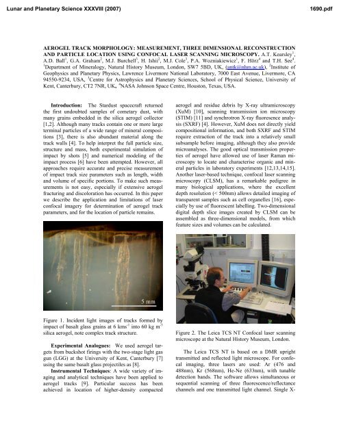
Figure 1. Incident light images of tracks formed by<br />
impact of basalt glass grains at 6 kms -1 into 60 kg m -3<br />
silica aerogel, note complex track structure.<br />
Experimental Analogues: We used aerogel targets<br />
from buckshot firings with the two-stage light gas<br />
gun (LGG) at the University of Kent, Canterbury [7]<br />
using the same basalt glass projectiles as [8].<br />
Instrumental Techniques: A wide variety of imaging<br />
<strong>and</strong> analytical techniques have been applied to<br />
aerogel tracks [9]. Particular success has been<br />
achieved in location of higher-density compacted<br />
aerogel <strong>and</strong> residue debris by X-ray ultramicroscopy<br />
(XuM) [10], scanning transmission ion microscopy<br />
(STIM) [11] <strong>and</strong> synchrotron X-ray fluoresence analysis<br />
(SXRF) [4]. However, XuM does not directly yield<br />
compositional information, <strong>and</strong> both SXRF <strong>and</strong> STIM<br />
require extraction of the track into a relatively small<br />
subsample before imaging, although they also provide<br />
microanalyses. The good optical transmission properties<br />
of aerogel have allowed use of laser Raman microscopy<br />
to locate <strong>and</strong> characterise organic <strong>and</strong> mineral<br />
particles in laboratory experiments [12,13,14,15].<br />
Another laser-based technique, confocal laser scanning<br />
microscopy (CLSM), has a remarkable pedigree in<br />
many biological applications, where the excellent<br />
depth resolution (< 500nm) allows detailed imaging of<br />
transparent samples such as cell organelles [16], especially<br />
by use of fluorescent labelling. Two-dimensional<br />
digital depth slice images created by CLSM can be<br />
assembled as three-dimensional models, from which<br />
feature sizes <strong>and</strong> volumes can be calculated.<br />
Figure 2. The Leica TCS NT Confocal laser scanning<br />
microscope at the Natural History Museum, London.<br />
The Leica TCS NT is based on a DMR upright<br />
transmitted <strong>and</strong> reflected light microscope. For confocal<br />
imaging, three lasers are used: Ar (476 <strong>and</strong><br />
488nm), Kr (568nm), He-Ne (633nm), with tunable<br />
detection b<strong>and</strong>s. The software allows simultaneous or<br />
sequential scanning of three fluorescence/reflectance<br />
channels <strong>and</strong> one transmitted light channel. Single X-
<strong>Lunar</strong> <strong>and</strong> <strong>Planetary</strong> <strong>Science</strong> <strong>XXXVIII</strong> (<strong>2007</strong>) <strong>1690.pdf</strong><br />
Y slices can be captured, or Z-axis stacks of slices to<br />
provide volumetric measurements <strong>and</strong> rendered 3D<br />
images. Spatial resolution depends on the objective<br />
lens <strong>and</strong> aperture, e.g. a 10x objective <strong>and</strong> n.a. 3 aperture<br />
give a 1mm X-axis field of view, Z-axis stack<br />
interval of 2.4µm <strong>and</strong> 1.95x1.95x2.4µm voxel (volume<br />
element) size.<br />
Results: We have obtained CLSM depth slice stacks<br />
from laser reflectance of hypervelocity impact tracks<br />
<strong>and</strong> embedded projectile grains in silica aerogel of a<br />
wide range of density (~5 – 60 kg m -3 ), both in large<br />
(3cm) blocks <strong>and</strong> sub-millimetre keystones [17]. The<br />
main mass of the aerogel gives a little laser scattering<br />
background, as expected from the ‘blue smoke’ appearance<br />
characteristic of silica aerogel. The track<br />
edges are accentuated by strong contrast between a<br />
zone of intense laser scattering in dense aerogel <strong>and</strong><br />
absence of reflected signal from the empty track.<br />
Red (λ 612-648 nm) <strong>and</strong> blue-green (λ 472-508nm)<br />
scattering is strongest from fractured <strong>and</strong> dense<br />
aerogel at track margins, around terminal particles <strong>and</strong><br />
on rough-cut keystone surfaces. Yellow (λ 556-582<br />
nm) reflection is particularly strong from basalt glass<br />
fragments, <strong>and</strong> the larger particles correspond to the<br />
dark grains seen in our optical images.<br />
Figure 3. Tracks, in CLSM, all depth slices combined<br />
(equivalent to full-thickness image). Red <strong>and</strong> bluegreen<br />
reflections combined at left, yellow at right.<br />
Figure 4. A track in aerogel seen in combined red <strong>and</strong><br />
blue-green reflection images; left, a single depth slice;<br />
right, four cross sections from the lines indicated.<br />
In Fig 4, a single CLSM slice (left) shows sections<br />
through two tracks at this level. The large track at left<br />
has a ‘carrot’ form [2] common to impacts by solid<br />
particles of relatively high density [5]. A large radial<br />
fracture is visible as a sub-circular protrusion to the<br />
left of the main track. The cross sections (right) reveal<br />
complex shape for the upper part of the large track,<br />
<strong>and</strong> also a further two tracks (arrowed) whose width<br />
can be measured.<br />
Conclusions: CLSM should prove to be a valuable<br />
technique for quantification of aerogel track morphology<br />
<strong>and</strong> particle location, especially for the small<br />
tracks expected in aerogel on the interstellar side of the<br />
Stardust Sample Tray Assembly. CLSM can be performed<br />
on samples in aerogel keystones or quickstones<br />
[18], <strong>and</strong> also on cm-scale, unprepared aerogel blocks.<br />
It is therefore suitable for track characterisation at an<br />
early stage of curation <strong>and</strong> preparation. Selection of<br />
precise wavelengths for image collection is critical to<br />
success in distinguishing particle residue from dense<br />
aerogel, although reflections across a wide λ range can<br />
reveal the aerogel surface, permitting imaging of the<br />
track morphology.<br />
References: [1] Brownlee D.E. et al. (2006)<br />
<strong>Science</strong>, 314, 1711-1716. [2] Hörz F. et al. (2006)<br />
<strong>Science</strong>, 314, 1716-1719. [3] Zolensky M.E. et al.<br />
(2006) <strong>Science</strong>, 314, 1735-1739. [4] Flynn G. et al.<br />
(2006) <strong>Science</strong>, 314, 1731-1735. [5] Burchell M.J. et<br />
al. Characteristics of cometary dust tracks in Stardust<br />
aerogel <strong>and</strong> laboratory calibrations, submitted to<br />
MAPS. [6] Domínguez G. et al. (2004) Icarus 172,<br />
613–624. [7] Burchell M.J. et al. (1999) Meas. Sci.<br />
Tech. 10, 41-50. [8] Kearsley A.T. et al. (<strong>2007</strong>) MAPS,<br />
42.2, in press. [9] Burchell M.J. et al. (2006) Ann. Rev.<br />
Earth Planet. Sci., 34, 385-418. [10] Graham G.A. et<br />
al. (2005) LPSC XXXVI , #2078. [11] Graham G.A. et<br />
al. (2004) MAPS, 39.9, 1461-1473. [12] Burchell M.J.<br />
et al. (2001) MAPS, 36, 209-221. [13] Graham G.A. et<br />
al. (2001) Proc. Roy. Microscopical Soc., 36/4, 251-<br />
254. [14] Burchell M.J. et al. (2004) J. Raman Spectr.,<br />
35, 249-253. [15] Burchell M.J. et al. (2006) MAPS,<br />
41.2, 217-232.<br />
[16] http://www.olympusconfocal.com/theory/<br />
[17] Westphal A. et al. MAPS, 39, 1375–86. [18] Ishii<br />
H.A. et al. (2005) MAPS, 40.11, 1755–1759.<br />
Acknowledgements: We thank NASA for the<br />
Stardust aerogel block used as target in the basalt<br />
powder shot; PPARC for support of the gun facility at<br />
Canterbury; Andrew Westphal <strong>and</strong> Chris Snead at<br />
Berkeley for preparation of the keystone. Staff at<br />
LLNL acknowledge their work was performed under<br />
the auspices of the US Department of Energy by the<br />
Lawrence Livermore National Laboratory under Contract<br />
No. W-7405-ENG-48.