Reoviruses,Rotaviruses, & Caliciviruses
Reoviruses,Rotaviruses, & Caliciviruses
Reoviruses,Rotaviruses, & Caliciviruses
You also want an ePaper? Increase the reach of your titles
YUMPU automatically turns print PDFs into web optimized ePapers that Google loves.
4010_33-48 2/11/04 9:09 AM Page 504<br />
<strong>Reoviruses</strong>, <strong>Rotaviruses</strong>,<br />
& <strong>Caliciviruses</strong><br />
<strong>Reoviruses</strong> are medium-sized viruses with a doublestranded,<br />
segmented RNA genome. The family includes<br />
human rotaviruses, the most important cause of infantile<br />
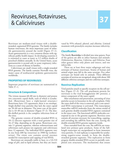
gastroenteritis around the world (Figure 37–1).<br />
Acute gastroenteritis is a very common disease with significant<br />
public health impact. In developing countries it<br />
is estimated to cause as many as 3.5 million deaths of<br />
preschool children annually. In the United States, acute<br />
gastroenteritis is second only to acute respiratory infections<br />
as a cause of disease in families.<br />
<strong>Caliciviruses</strong> are small viruses with a single-stranded<br />
RNA genome. The family contains Norwalk virus, the<br />
major cause of nonbacterial epidemic gastroenteritis<br />
worldwide.<br />
PROPERTIES OF REOVIRUSES<br />
Important properties of reoviruses are summarized in<br />
Table 37–1.<br />
Structure & Composition<br />
The virions measure 60–80 nm in diameter and possess<br />
two concentric capsid shells, each of which is icosahedral.<br />
(<strong>Rotaviruses</strong> have a triple-layered structure.)<br />
<strong>Rotaviruses</strong> have 132 capsomeres; there is no envelope.<br />
Single-shelled virus particles that lack the outer capsid<br />
are 50–60 nm in diameter. The inner core of the particles<br />
is 33–40 nm in diameter (Figure 37–2). The double-shelled<br />
particle is the complete infectious form of<br />
the virus.<br />
The genome consists of double-stranded RNA in<br />
10–12 discrete segments with a total genome size of<br />
16–27 kbp, depending on the genus. <strong>Rotaviruses</strong> contain<br />
11 genome segments, whereas orthoreoviruses and<br />
orbiviruses each possess ten segments and coltiviruses<br />
have 12 segments. The individual RNA segments vary<br />
in size from 680 bp (rotavirus) to 3900 bp (orthoreovirus).<br />
The virion core contains several enzymes<br />
needed for transcription and capping of viral RNAs.<br />
<strong>Reoviruses</strong> are unusually stable to heat, to a 3.0–9.0<br />
range of pH, and to lipid solvents, but they are inacti-<br />
504<br />
37<br />
vated by 95% ethanol, phenol, and chlorine. Limited<br />
treatment with proteolytic enzymes increases infectivity.<br />
Classification<br />
The family Reoviridae is divided into nine genera. Four<br />
of the genera are able to infect humans and animals:<br />
Orthoreovirus, Rotavirus, Coltivirus, and Orbivirus. Four<br />
other genera infect only plants and insects, and one<br />
infects fish.<br />
There are at least three major subgroups and nine<br />
serotypes of human rotaviruses. Strains of human and<br />
animal origin may fall in the same serotype. Five other<br />
serotypes are found only in animals. Three different<br />
serotypes of reovirus are recognized, along with about 100<br />
different orbivirus serotypes and two coltivirus serotypes.<br />
Reovirus Replication<br />
Viral particles attach to specific receptors on the cell surface<br />
(Figure 37–3). The cell attachment protein for<br />
reoviruses is the viral hemagglutinin (σ1 protein), a<br />
minor component of the outer capsid.<br />
After attachment and penetration, uncoating of virus<br />
particles occurs in lysosomes in the cell cytoplasm. Only<br />
the outer shell of the virus is removed, and a core-associated<br />
RNA transcriptase is activated. This transcriptase<br />
transcribes mRNA molecules from the minus strand of<br />
each genome double-stranded RNA segment contained<br />
in the intact core. The functional mRNA molecules correspond<br />
in size to the genome segments. Reovirus cores<br />
contain all enzymes necessary for transcribing, capping,<br />
and extruding the mRNAs from the core, leaving the<br />
double-stranded RNA genome segments inside.<br />
Once extruded from the core, the mRNAs are translated<br />
into primary gene products. Some of the fulllength<br />
transcripts are encapsidated to form immature<br />
virus particles. A viral replicase is responsible for synthesizing<br />
negative-sense strands to form the doublestranded<br />
genome segments. This replication to form<br />
progeny double-stranded RNA occurs in partially completed<br />
core structures. The mechanisms that ensure
4010_33-48 2/11/04 9:09 AM Page 505<br />
Unknown<br />
Bacteria<br />
Adenovirus<br />
Astrovirus<br />
Calicivirus<br />
Rotavirus<br />
assembly of the correct complement of genome segments<br />
into a developing viral core are unknown. Viral<br />
polypeptides probably self-assemble to form the inner<br />
and outer capsid shells.<br />
<strong>Reoviruses</strong> produce inclusion bodies in the cytoplasm<br />
in which virus particles are found. These viral factories<br />
are closely associated with tubular structures<br />
(microtubules and intermediate filaments). Rotavirus<br />
morphogenesis involves budding of single-shelled particles<br />
into the rough endoplasmic reticulum. The “pseudoenvelopes”<br />
so acquired are then removed and the<br />
outer capsids are added (Figure 37–3). This unusual<br />
pathway is utilized because the major outer capsid protein<br />
is glycosylated.<br />
Cell lysis results in release of progeny virions.<br />
ROTAVIRUSES<br />
<strong>Rotaviruses</strong> are a major cause of diarrheal illness in<br />
human infants and young animals, including calves and<br />
REOVIRUSES, ROTAVIRUSES, & CALICIVIRUSES / 505<br />
Unknown<br />
Parasites<br />
Other bacteria<br />
Toxigenic<br />
Escherichia coli<br />
Astrovirus<br />
Adenovirus<br />
Calicivirus<br />
Developed countries Developing countries<br />
Rotavirus<br />
Figure 37–1. An estimate of the role of etiologic agents in severe diarrheal illnesses requiring hospitalization of<br />
infants and young children in developed countries (left) and in developing countries (right). (Reproduced, with permission,<br />
from Kapikian AZ: Viral gastroenteritis. JAMA 1993;269:627.)<br />
Table 37–1. Important properties of reoviruses.<br />
Virion: Icosahedral, 60–80 nm in diameter, double capsid shell<br />
Composition: RNA (15%), protein (85%)<br />
Genome: Double-stranded RNA, linear, segmented (10–12<br />
segments); total genome size 16–27 kbp<br />
Proteins: Nine structural proteins; core contains several<br />
enzymes<br />
Envelope: None (transient pseudoenvelope is present<br />
during rotavirus particle morphogenesis)<br />
Replication: Cytoplasm; virions not completely uncoated<br />
Outstanding characteristics:<br />
Genetic reassortment occurs readily<br />
<strong>Rotaviruses</strong> are the major cause of infantile diarrhea<br />
<strong>Reoviruses</strong> are good models for molecular studies of viral<br />
pathogenesis<br />
piglets. Infections in adult humans and animals are also<br />
common. Among rotaviruses are the agents of human<br />
infantile diarrhea, Nebraska calf diarrhea, epizootic<br />
diarrhea of infant mice, and SA11 virus of monkeys.<br />
<strong>Rotaviruses</strong> resemble reoviruses in terms of morphology<br />
and strategy of replication.<br />
Figure 37–2. Electron micrograph of a negatively<br />
stained preparation of human rotavirus. (D, doubleshelled<br />
particles; S, single-shelled particles; E, empty<br />
capsids; i, fragment of inner shell; io, fragments of a<br />
combination of inner and outer shell.) Inset: Singleshelled<br />
particles obtained by treatment of the viral<br />
preparation with sodium dodecyl sulfate. Bars, 50 nm.<br />
(Courtesy of J Esparza and F Gil.)
4010_33-48 2/11/04 9:09 AM Page 506<br />
506 / CHAPTER 37<br />
Viroplasm VP1,2,3,6<br />
NSP2,5<br />
Double-shelled virus<br />
Cell<br />
lysis<br />
RER<br />
Nucleus<br />
Transient enveloped<br />
particle<br />
Envelope<br />
removal<br />
Classification & Antigenic Properties<br />
<strong>Rotaviruses</strong> have been classified into five groups (A–E)<br />
based on antigenic epitopes on the internal structural<br />
protein VP6. These can be detected by immunofluorescence,<br />
ELISA, and immune electron microscopy (IEM).<br />
Group A rotaviruses are the most frequent human<br />
pathogens. Outer capsid proteins VP4 and VP7 carry epitopes<br />
important in neutralizing activity, with VP7 glycoprotein<br />
being the predominant antigen. These typespecific<br />
antigens differentiate among rotaviruses and are<br />
demonstrable by Nt tests. Multiple serotypes have been<br />
identified among human and animal rotaviruses. Some<br />
animal and human rotaviruses share serotype specificity.<br />
For example, monkey virus SA11 is antigenically very<br />
similar to human serotype 3. The gene-coding assignments<br />
responsible for the structural and antigenic specificities<br />
of rotavirus proteins are shown in Figure 37–4.<br />
?<br />
Assembly of single-shelled particles<br />
?<br />
Ca 2+<br />
Progeny virions<br />
Lysosome<br />
VP4<br />
?<br />
NSP<br />
7M G<br />
7M G<br />
Translation<br />
VP1,2,3,4,6,7<br />
NSP1,2,3,5<br />
RER<br />
Molecular epidemiologic studies have analyzed isolates<br />
based on differences in the migration of the 11<br />
genome segments following electrophoresis of the RNA<br />
in polyacrylamide gels (Figure 37–5). These differences<br />
in electropherotypes can be used to differentiate group<br />
A viruses from other groups, but they cannot be used to<br />
predict serotypes.<br />
Animal Susceptibility<br />
Infecting virus<br />
Single-shelled<br />
particle<br />
7MG 7MG 7MG 7MG NSP3<br />
VP7<br />
mRNA<br />
Figure 37–3. Overview of the rotavirus replication cycle. (Reproduced, with permission, from Estes MK: <strong>Rotaviruses</strong><br />
and their replication. In: Fields Virology, 3rd ed. Fields BN et al [editors]. Lippincott-Raven, 1996.)<br />
NSP4<br />
<strong>Rotaviruses</strong> have a wide host range. Most isolates have<br />
been recovered from newborn animals with diarrhea.<br />
Cross-species infections can occur in experimental inoculations,<br />
but it is not clear if they occur in nature. Swine<br />
rotavirus infects both newborn and weanling piglets.<br />
Newborns often exhibit subclinical infection due perhaps<br />
to the presence of maternal antibody, whereas overt<br />
disease is more common in weanling animals.<br />
+<br />
+<br />
+<br />
+
4010_33-48 2/11/04 9:09 AM Page 507<br />
RNA<br />
segment Protein<br />
1<br />
2<br />
3<br />
4<br />
5<br />
6<br />
7<br />
8<br />
9<br />
10<br />
11<br />
Propagation in Cell Culture<br />
<strong>Rotaviruses</strong> are fastidious agents to culture. Most group<br />
A human rotaviruses can be cultivated if pretreated with<br />
the proteolytic enzyme trypsin and if low levels of<br />
trypsin are included in the tissue culture medium. This<br />
cleaves an outer capsid protein and facilitates uncoating.<br />
Very few non-group A rotavirus strains have been cultivated.<br />
Pathogenesis<br />
VP1<br />
VP2<br />
VP3<br />
VP4<br />
NSP1<br />
VP6<br />
NSP2<br />
NSP3<br />
VP7<br />
NSP4<br />
NSP5<br />
<strong>Rotaviruses</strong> infect cells in the villi of the small intestine<br />
(gastric and colonic mucosa are spared). They multiply<br />
in the cytoplasm of enterocytes and damage their transport<br />
mechanisms. One of the rotavirus-encoded proteins,<br />
NSP4, is a viral enterotoxin and induces secretion<br />
by triggering a signal transduction pathway. Damaged<br />
cells may slough into the lumen of the intestine and<br />
release large quantities of virus, which appear in the<br />
stool (up to 10 10 particles per gram of feces). Viral<br />
excretion usually lasts 2–12 days in otherwise healthy<br />
patients but may be prolonged in those with poor nutrition.<br />
Diarrhea caused by rotaviruses may be due to<br />
impaired sodium and glucose absorption as damaged<br />
cells on villi are replaced by nonabsorbing immature<br />
REOVIRUSES, ROTAVIRUSES, & CALICIVIRUSES / 507<br />
VP2<br />
VP4,<br />
neutralization<br />
antigen<br />
VP6,<br />
subgroup<br />
antigen<br />
VP7,<br />
neutralization<br />
antigen<br />
Subcore<br />
Figure 37–4. Gene-coding assignments for antigenic specificities of rotavirus proteins. Shown on the left are the<br />
genome RNA segments and the encoded protein products. In the center is a schematic representation of the complete<br />
rotavirus particle with the location of the structural proteins in the different shells indicated.The figure on the<br />
right shows the three-dimensional structure of a virus particle. A complete particle is drawn on the left half; the<br />
structure on the right half has part of the outer and inner shells removed to show the middle and inner shells. (Reproduced<br />
from Estes MK: <strong>Rotaviruses</strong> and their replication. In: Fields Virology, 3rd ed. Fields BN et al [editors]. Lippincott-Raven,<br />
1996. Modified from Conner ME, Matson DO, Estes MK: Rotavirus vaccines and vaccination potential. Curr Top Microbiol<br />
Immunol 1994;185:285, with an unpublished structure of BVV Prasad and A Shaw.)<br />
crypt cells. It may take 3–8 weeks for normal function<br />
to be restored.<br />
Clinical Findings & Laboratory Diagnosis<br />
<strong>Rotaviruses</strong> cause the major portion of diarrheal illness<br />
in infants and children worldwide but not in adults<br />
(Table 37–2). There is an incubation period of 1–3<br />
days. Typical symptoms include watery diarrhea, fever,<br />
abdominal pain, and vomiting, leading to dehydration.<br />
In infants and children, severe loss of electrolytes and<br />
fluids may be fatal unless treated. Patients with milder<br />
cases have symptoms for 3–8 days and then recover<br />
completely. However, viral excretion in the stool may<br />
persist up to 50 days after onset of diarrhea. Asymptomatic<br />
infections, with seroconversion, occur. In children<br />
with immunodeficiencies, rotavirus can cause severe and<br />
prolonged disease.<br />
Adult contacts may be infected, as evidenced by seroconversion,<br />
but they rarely exhibit symptoms, and virus<br />
is infrequently detected in their stools. A common source<br />
of infection is contact with pediatric cases. However, epidemics<br />
of severe disease have occurred in adults, especially<br />
in closed populations, as in a geriatric ward. Group<br />
B rotaviruses have been implicated in large outbreaks of<br />
severe gastroenteritis in adults in China (Table 37–2).
4010_33-48 2/11/04 9:09 AM Page 508<br />
508 / CHAPTER 37<br />
Figure 37–5. Electrophoretic profiles of rotavirus RNA<br />
segments.Viral RNAs were electrophoresed in 10%<br />
polyacrylamide gels and visualized by silver stain. Different<br />
rotavirus groups and RNA patterns are illustrated:<br />
a group A monkey virus (SA11; lane A), a group A<br />
human rotavirus (lane B), a group B human adult diarrhea<br />
virus (lane C), and a group A rabbit virus that<br />
exhibits a “short” RNA pattern (lane D). <strong>Rotaviruses</strong> contain<br />
11 genome RNA segments, but sometimes two or<br />
three segments migrate closely together and are difficult<br />
to separate. (Photograph provided by T Tanaka and<br />
MK Estes.)<br />
Laboratory diagnosis rests on demonstration of virus<br />
in stool collected early in the illness and on a rise in antibody<br />
titer. Virus in stool is demonstrated by IEM, latex<br />
agglutination tests, or ELISA. Genotyping of rotavirus<br />
nucleic acid from stool specimens by the polymerase<br />
chain reaction is the most sensitive detection method.<br />
Serologic tests can be used to detect an antibody titer<br />
rise, particularly ELISA.<br />
Epidemiology & Immunity<br />
<strong>Rotaviruses</strong> are the single most important worldwide<br />
cause of gastroenteritis in young children. Estimates<br />
range from 3 billion to 5 billion for annual diarrheal<br />
episodes in children under 5 years of age in Africa, Asia,<br />
and Latin America, resulting in as many as 5 million<br />
deaths. Developed countries have a high morbidity rate<br />
but a low mortality rate. Typically, up to 50% of cases of<br />
acute gastroenteritis of hospitalized children throughout<br />
the world are caused by rotaviruses.<br />
Rotavirus infections usually predominate during the<br />
winter season. Symptomatic infections are most common<br />
in children between ages 6 months and 2 years,<br />
and transmission appears to be by the fecal-oral route.<br />
Nosocomial infections are frequent.<br />
<strong>Rotaviruses</strong> are ubiquitous. By age 3 years, 90% of<br />
children have serum antibodies to one or more types.<br />
This high prevalence of rotavirus antibodies is maintained<br />
in adults, suggesting subclinical reinfections by<br />
the virus. Rotavirus reinfections are common; it has<br />
been shown that young children can suffer up to five<br />
reinfections by 2 years of age. Asymptomatic infections<br />
are more common with successive reinfections. Local<br />
immune factors, such as secretory IgA or interferon,<br />
may be important in protection against rotavirus infection.<br />
Asymptomatic infections are common in infants<br />
before age 6 months, the time during which protective<br />
maternal antibody acquired passively by newborns<br />
should be present. Such neonatal infection does not prevent<br />
reinfection, but it does protect against the development<br />
of severe disease during reinfection.<br />
Treatment & Control<br />
Treatment of gastroenteritis is supportive, to correct the<br />
loss of water and electrolytes that may lead to dehydration,<br />
acidosis, shock, and death. Management consists<br />
of replacement of fluids and restoration of electrolyte<br />
balance either intravenously or orally, as feasible. The<br />
infrequent mortality from infantile diarrhea in developed<br />
countries is due to routine use of effective replacement<br />
therapy.<br />
In view of the fecal-oral route of transmission, wastewater<br />
treatment and sanitation are significant control<br />
measures.<br />
An oral live attenuated rhesus-based rotavirus vaccine<br />
was licensed in the United States in 1998 for vaccination<br />
of infants. It was withdrawn a year later because of reports<br />
of intussusception (bowel blockages) as an uncommon<br />
but serious side effect associated with the vaccine. A safe<br />
and effective vaccine remains the best hope for reducing<br />
the worldwide burden of rotavirus disease.<br />
REOVIRUSES<br />
The viruses of this genus, which have been studied most<br />
thoroughly by molecular biologists, are not known to<br />
cause human disease.<br />
Classification & Antigenic Properties<br />
<strong>Reoviruses</strong> are ubiquitous, with a very wide host range.<br />
Three distinct but related types of reovirus have been
4010_33-48 2/11/04 9:09 AM Page 509<br />
Table 37–2. Viruses associated with acute gastroenteritis in humans. 1<br />
Important as a<br />
Size Cause of<br />
Virus (nm) Epidemiology Hospitalization<br />
<strong>Rotaviruses</strong><br />
Group A 60–80 Single most important cause (viral or bacterial) of endemic Yes<br />
severe diarrheal illness in infants and young children worldwide<br />
(in cooler months in temperate climates).<br />
Group B 60–80 Outbreaks of diarrheal illness in adults and children in China. No<br />
Group C 60–80 Sporadic cases and occasional outbreaks of diarrheal illness<br />
in children.<br />
No<br />
Enteric adenovirus<br />
<strong>Caliciviruses</strong><br />
70–90 Second most important viral agent of endemic diarrheal illness<br />
of infants and young children worldwide.<br />
Yes<br />
Norwalk 27–40 Important cause of outbreaks of vomiting and diarrheal illness<br />
in older children and adults in families, communities, and<br />
institutions; frequently associated with ingestion of food.<br />
No<br />
Sapporo 27–40 Sporadic cases and occasional outbreaks of diarrheal illness<br />
in infants, young children, and the elderly.<br />
No<br />
Astroviruses 28–30 Sporadic cases and occasional outbreaks of diarrheal illness<br />
in infants, young children, and the elderly.<br />
No<br />
1 Modified from Kapikian AZ: Viral gastroenteritis. JAMA 1993;269:627.<br />
recovered from many species and are demonstrable by<br />
Nt and HI tests. <strong>Reoviruses</strong> contain a hemagglutinin for<br />
human O or bovine erythrocytes.<br />
Epidemiology<br />
<strong>Reoviruses</strong> cause many inapparent infections, because<br />
most people have serum antibodies by early adulthood.<br />
Antibodies are also present in other species. All three<br />
types have been recovered from healthy children, from<br />
young children during outbreaks of minor febrile illness,<br />
from children with diarrhea or enteritis, and from<br />
chimpanzees with epidemic rhinitis.<br />
Human volunteer studies have failed to demonstrate<br />
a clear cause-and-effect relationship of reoviruses to<br />
human illness. In inoculated volunteers, reovirus is<br />
recovered far more readily from feces than from the nose<br />
or throat. An association of reovirus type 3 with biliary<br />
atresia in infants has been suggested.<br />
Pathogenesis<br />
<strong>Reoviruses</strong> have become important model systems for<br />
the study of the pathogenesis of viral infection at the<br />
molecular level. Defined recombinants from two<br />
reoviruses with differing pathogenic phenotypes are<br />
REOVIRUSES, ROTAVIRUSES, & CALICIVIRUSES / 509<br />
used to infect mice. Segregation analysis is then used to<br />
associate particular features of pathogenesis with specific<br />
viral genes and gene products. The pathogenic properties<br />
of reoviruses are primarily determined by the protein<br />
species found on the outer capsid of the virion.<br />
ORBIVIRUSES<br />
Orbiviruses are a genus within the reovirus family. They<br />
commonly infect insects, and many are transmitted by<br />
insects to vertebrates. About 100 serotypes are known.<br />
None of these viruses cause serious clinical disease in<br />
humans, but they may cause mild fevers. Serious animal<br />
pathogens include bluetongue virus of sheep and<br />
African horse sickness virus. Antibodies to orbiviruses<br />
are found in many vertebrates, including humans.<br />
The genome consists of ten segments of doublestranded<br />
RNA, with a total genome size of 18 kbp. The<br />
replicative cycle is similar to that of reoviruses.<br />
Orbiviruses are sensitive to low pH, in contrast with the<br />
general stability of other reoviruses.<br />
CALICIVIRUSES<br />
In addition to rotaviruses and noncultivable adenoviruses,<br />
members of the family Caliciviridae are impor-
4010_33-48 2/11/04 9:09 AM Page 510<br />
510 / CHAPTER 37<br />
tant agents of viral gastroenteritis in humans. The most<br />
significant member is Norwalk virus. Properties of caliciviruses<br />
are summarized in Table 37–3.<br />
Classification & Antigenic Properties<br />
<strong>Caliciviruses</strong> are similar to picornaviruses but are<br />
slightly larger (27–40 nm) and contain a single major<br />
structural protein. They exhibit a distinctive morphology<br />
in the electron microscope (Figure 37–6). The family<br />
Caliciviridae is divided into four genera: Norovirus,<br />
which includes the Norwalk viruses; Sapovirus, which<br />
includes the Sapporo-like viruses; Lagovirus, the rabbit<br />
hemorrhagic disease virus; and Vesivirus, which includes<br />
vesicular exanthem virus of swine, feline calicivirus, and<br />
marine viruses found in pinnipeds, whales, and fish.<br />
The first two genera contain human viruses that cannot<br />
be cultured; the latter two genera contain only animal<br />
strains that can be grown in vitro. Rabbit hemorrhagic<br />
disease virus was introduced in 1995 in Australia as a<br />
biologic control agent to reduce that country’s population<br />
of wild rabbits.<br />
Historically, the Norwalk viruses were referred to as<br />
“small round structured viruses” based on their detection<br />
by electron microscopy.<br />
Human calicivirus serotypes are not defined. The<br />
Norwalk viruses are subdivided into two genogroups.<br />
Clinical Findings & Laboratory Diagnosis<br />
Norwalk virus is the most important cause of epidemic<br />
viral gastroenteritis in adults (Table 37–2). Epidemic<br />
nonbacterial gastroenteritis is characterized by (1)<br />
absence of bacterial pathogens; (2) gastroenteritis with<br />
rapid onset and recovery and relatively mild systemic<br />
signs; and (3) an epidemiologic pattern of a highly communicable<br />
disease that spreads rapidly with no particular<br />
predilection in terms of age or geography. Various<br />
Table 37–3. Important properties of caliciviruses.<br />
Virion: Icosahedral, 27–40 nm in diameter; cup-like depressions<br />
on capsid surface<br />
Genome: Single-stranded RNA, linear, positive-sense, nonsegmented;<br />
7.4–8.3 kb in size; contains genome-linked protein<br />
(VPg)<br />
Proteins: Polypeptides cleaved from a precursor polyprotein;<br />
capsid is composed of a single protein<br />
Envelope: None<br />
Replication: Cytoplasm<br />
Outstanding characteristics:<br />
Norwalk viruses are major cause of nonbacterial epidemic<br />
gastroenteritis<br />
Human viruses are noncultivable<br />
descriptive terms have been used in reports of different<br />
outbreaks (eg, epidemic viral gastroenteritis, viral diarrhea,<br />
winter vomiting disease) depending on the predominant<br />
clinical feature.<br />
Norwalk viral gastroenteritis has an incubation period<br />
of 24–48 hours. Onset is rapid, and the clinical course is<br />
brief, lasting 12–60 hours; symptoms include diarrhea,<br />
nausea, vomiting, low-grade fever, abdominal cramps,<br />
headache, and malaise. The illness can be incapacitating<br />
during the symptomatic phase, but hospitalization is<br />
rarely required. Norwalk infections are more likely to<br />
induce vomiting than those with Sapporo-like viruses.<br />
Dehydration is the most common complication in the<br />
young and elderly. No sequelae have been reported.<br />
Volunteer experiments have clearly shown that the<br />
appearance of Norwalk virus coincides with clinical illness.<br />
Antibody develops during the illness and is usually<br />
protective on a short-term basis against reinfection with<br />
the same agent. Long-term immunity does not correspond<br />
well to the presence of serum antibodies. Some<br />
volunteers can be reinfected with the same virus after<br />
about 2 years.<br />
Reverse transcriptase-polymerase chain reaction is<br />
the most widely used technique for detection of human<br />
caliciviruses in clinical specimens (feces, vomitus) and<br />
environmental samples (contaminated food, water).<br />
Because of the genetic diversity among circulating<br />
strains, the choice of polymerase chain reaction primer<br />
pairs is very important.<br />
Electron microscopy is frequently used to detect<br />
virus particles in stool samples. However, Norwalk virus<br />
particles are usually present in low concentration and<br />
are difficult to recognize; they should be identified by<br />
immunoelectron microscopy. ELISA immunoassays<br />
based on recombinant virus-like particles can detect<br />
antibody responses, with a fourfold or greater rise in<br />
IgG antibody titer in acute and convalescent-phase sera<br />
indicative of a recent infection. However, the necessary<br />
reagents are not widely available, and the antigens are<br />
not able to detect responses to all antigenic types of<br />
Norwalk virus.<br />
Epidemiology & Immunity<br />
Human caliciviruses have worldwide distribution. Norwalk<br />
viruses are the most common cause of nonbacterial<br />
gastroenteritis in the United States, causing an estimated<br />
23 million cases annually.<br />
The viruses are most often associated with epidemic<br />
outbreaks of waterborne, food-borne, and shellfish-associated<br />
gastroenteritis. Community outbreaks can occur<br />
in any season. All age groups can be affected. Outbreaks<br />
occur throughout the year, with a seasonal peak during<br />
cooler months. Most outbreaks involve food-borne or<br />
person-to-person transmission.
4010_33-48 2/23/04 4:28 PM Page 511<br />
REOVIRUSES, ROTAVIRUSES, & CALICIVIRUSES / 511<br />
Figure 37–6. Electron micrographs of virus particles found in stools of patients with gastroenteritis.These viruses<br />
were visualized following negative staining. Specific viruses and the original magnifications of the micrographs are<br />
as follows. A: Rotavirus (185,000 ×). B: Enteric adenovirus (234,000 ×). C: Coronavirus (249,000 ×). D: Torovirus (coronavirus)<br />
(249,000 ×). E: Calicivirus (250,000 ×). F: Astrovirus (196,000 ×). G: Norwalk virus (calicivirus) (249,000 ×).<br />
H: Parvovirus (249,000 ×).The electron micrographs in panels C–H were originally provided by T Flewett; panel E was<br />
originally obtained from CR Madeley. Bars, 100 nm. (Reproduced, with permission, from Graham DY, Estes MK: Viral infections<br />
of the intestine. Pages 566–578 in: Principles and Practice of Gastroenterology and Hepatology. Gitnick G et al [editors].<br />
Elsevier Science Publishing Co., 1988.)
4010_33-48 2/11/04 9:09 AM Page 512<br />
512 / CHAPTER 37<br />
Characteristics of Norwalk virus include a low infectious<br />
dose (as few as 10 virus particles), relative stability<br />
in the environment, and multiple modes of transmission.<br />
It survives 10 ppm chlorine and heating to 60 °C;<br />
it can be maintained in steamed oysters.<br />
Fecal-oral spread is probably the primary means of<br />
transmission of Norwalk virus. During a 5-year period<br />
in the United States (1996–2000), food was implicated<br />
in 39% of outbreaks of Norwalk gastroenteritis, personto-person<br />
contact in 12%, and water in 3%, with the<br />
source in 18% unknown.<br />
Outbreaks of Norwalk gastroenteritis occur in multiple<br />
settings. In the same 5-year period as above, 39%<br />
occurred in restaurants, 29% in nursing homes and hospitals,<br />
12% in schools and daycare centers, 10% in vacation<br />
settings, including cruise ships, and 9% in other<br />
settings.<br />
No in vitro neutralization assay is available to study<br />
immunity. Volunteer challenge studies have shown that<br />
about 50% of adults are susceptible to illness. Norwalk<br />
virus antibody is acquired later in life than rotavirus<br />
antibody, which develops early in childhood. In developing<br />
countries, most children have developed Norwalk<br />
virus antibodies by 4 years of age.<br />
Treatment & Control<br />
Treatment is symptomatic. The low infectious dose permits<br />
efficient transmission of the virus. Because of the<br />
infectious nature of the stools, care should be taken in<br />
their disposal. Effective hand washing can decrease<br />
transmission in family or institutional settings. Careful<br />
processing of food is important, as many food-borne<br />
outbreaks occur. Purification of drinking water and<br />
swimming pool water should decrease Norwalk virus<br />
outbreaks. There is no vaccine.<br />
ASTROVIRUSES<br />
Astroviruses are about 28–30 nm in diameter and<br />
exhibit a distinctive morphology in the electron microscope<br />
(Figure 37–6). They contain single-stranded, positive-sense<br />
RNA, 6.8–7.9 kb in size. At least eight<br />
serotypes of human viruses are recognized by IEM and<br />
neutralization. Astroviruses cause diarrheal illness and<br />
may be shed in extraordinarily large quantities in feces.<br />
Astroviruses are transmitted by the fecal-oral route<br />
through contaminated food or water, person-to-person<br />
contact, or contaminated surfaces. They are recognized<br />
as pathogens for infants and children, elderly institutionalized<br />
patients, and immunocompromised persons<br />
(Table 37–2). They may be shed for prolonged periods<br />
by immunocompromised hosts.<br />
REVIEW QUESTIONS<br />
1. A 36-year-old man enjoyed a meal of raw oysters.<br />
Twenty-four hours later he became ill, with<br />
sudden onset of vomiting, diarrhea, and<br />
headache.The most likely cause of his gastroenteritis<br />
is<br />
(A) Astrovirus<br />
(B) Hepatitis A virus<br />
(C) Norwalk virus<br />
(D) Rotavirus, group A<br />
(E) Echovirus<br />
2. This virus is the most important cause of gastroenteritis<br />
in infants and young children. It<br />
causes infections that are often severe and may<br />
be life-threatening, especially in infants.<br />
(A) Echovirus<br />
(B) Norwalk virus<br />
(C) Rotavirus, group A<br />
(D) Orbivirus<br />
(E) Parvovirus<br />
3. An outbreak of epidemic gastroenteritis<br />
occurred at a wooded summer camp 24 hours<br />
after a party for visiting families. Some of the<br />
visiting parents became ill also. Samples taken 2<br />
weeks later from the well that was the source of<br />
drinking water at the camp were negative for<br />
fecal coliforms. The most likely source of the<br />
outbreak was<br />
(A) Mosquitoes or ticks, present in high numbers<br />
in the area<br />
(B) Contaminated food served at the party<br />
(C) A nearby stream used for fishing<br />
(D) A visiting parent who was developing<br />
pneumonia<br />
(E) The swimming pool<br />
4. This viral gastroenteritis agent has a segmented,<br />
double-stranded RNA genome and a doubleshelled<br />
capsid. It is a member of which virus<br />
family?<br />
(A) Adenoviridae<br />
(B) Astroviridae<br />
(C) Caliciviridae<br />
(D) Reoviridae<br />
(E) Coronaviridae<br />
5. Rotavirus and Norwalk virus are distinctly different<br />
viruses. However, they share which one of<br />
the following characteristics?<br />
(A) Fecal-oral mode of transmission<br />
(B) They mainly cause disease in infants and<br />
young children
4010_33-48 2/11/04 9:09 AM Page 513<br />
(C) They induce generally mild disease in<br />
young children<br />
(D) Infection patterns show no seasonal variation<br />
(E) A double-stranded RNA genome<br />
6. Because rotavirus infections can be serious, a vaccine<br />
would be beneficial. Which of the following<br />
is most correct regarding a rotavirus vaccine?<br />
(A) A killed human rotavirus group A vaccine is<br />
in use in the United States (2003)<br />
(B) A live attenuated vaccine was withdrawn<br />
from use because of reports of intussusception<br />
(1998)<br />
(C) Vaccine development is complicated by<br />
rapid antigenic variation by the virus<br />
(D) Available antiviral drugs make a vaccine<br />
unnecessary<br />
(E) Vaccine development is complicated<br />
because the virus cannot be grown in cell<br />
culture<br />
REOVIRUSES, ROTAVIRUSES, & CALICIVIRUSES / 513<br />
Answers<br />
1. C 4. D<br />
2. C 5. A<br />
3. B 6. B<br />
REFERENCES<br />
Glass RI et al: The epidemiology of rotavirus diarrhea in the United<br />
States: Surveillance and estimates of disease burden. J Infect<br />
Dis 1996;174(Suppl 1):S5.<br />
Green KY, Chanock RM, Kapikian AZ: Human caliciviruses. In:<br />
Fields Virology, 4th ed. Knipe DM et al (editors). Lippincott<br />
Williams & Wilkins, 2001.<br />
Kapikian AZ, Hoshino Y, Chanock RM: <strong>Rotaviruses</strong>. In: Fields<br />
Virology, 4th ed. Knipe DM et al (editors). Lippincott<br />
Williams & Wilkins, 2001.<br />
Monroe SS, Ando T, Glass RI (guest editors): International Workshop<br />
on Human <strong>Caliciviruses</strong>. J Infect Dis 2000;181<br />
(Supp12). [Entire issue.]<br />
Smith AW et al: Calicivirus emergence from ocean reservoirs:<br />
Zoonotic and interspecies movements. Emerg Infect Dis<br />
1998;4:13.


