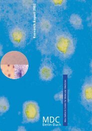
Research Report 2012 - of the Max Delbrück Center for Molecular ...
Research Report 2012 - of the Max Delbrück Center for Molecular ...
Research Report 2012 - of the Max Delbrück Center for Molecular ...

You also want an ePaper? Increase the reach of your titles
YUMPU automatically turns print PDFs into web optimized ePapers that Google loves.
<strong>Research</strong> <strong>Report</strong> <strong>2012</strong><br />
MAX DELBRÜCK CENTER<br />
FOR MOLECULAR MEDICINE<br />
BERLIN-BUCH<br />
MEMBER OF THE<br />
HELMHOLTZ ASSOCIATION
<strong>Research</strong> <strong>Report</strong> <strong>2012</strong><br />
(covers <strong>the</strong> period 2010-2011)<br />
<strong>Max</strong> <strong>Delbrück</strong> <strong>Center</strong> <strong>for</strong><br />
<strong>Molecular</strong> Medicine (MDC)<br />
Berlin-Buch<br />
Robert-Rössle-Str. 10<br />
13125 Berlin<br />
Tel.: + 49-30-9406-0<br />
Fax: + 49-30-949-4161<br />
e-mail: communications@mdc-berlin.de<br />
This <strong>Research</strong> <strong>Report</strong> is also available<br />
on <strong>the</strong> World Wide Web<br />
http:// www.mdc-berlin.de<br />
The MDC is a member <strong>of</strong> <strong>the</strong><br />
Helmholtz Association <strong>of</strong><br />
National <strong>Research</strong> <strong>Center</strong>s<br />
The following <strong>Research</strong> <strong>Report</strong>s<br />
have been published previously:<br />
<strong>Research</strong> <strong>Report</strong> 1996<br />
(covers <strong>the</strong> period 1992-1995)<br />
<strong>Research</strong> <strong>Report</strong> 1996/97<br />
(covers <strong>the</strong> period 1996-1997)<br />
<strong>Research</strong> <strong>Report</strong> 2000<br />
(covers <strong>the</strong> period 1998-1999)<br />
<strong>Research</strong> <strong>Report</strong> 2002<br />
(covers <strong>the</strong> period 2000-2001)<br />
<strong>Research</strong> <strong>Report</strong> 2004<br />
(covers <strong>the</strong> period 2002-2003)<br />
<strong>Research</strong> <strong>Report</strong> 2006<br />
(covers <strong>the</strong> period 2004-2005)<br />
<strong>Research</strong> <strong>Report</strong> 2008<br />
(covers <strong>the</strong> period 2006-2007)<br />
<strong>Research</strong> <strong>Report</strong> 2010<br />
(covers <strong>the</strong> period 2008-2009)<br />
Editor-in-Chief<br />
Pamela Cohen<br />
Editorial Board<br />
Carmen Birchmeier<br />
Fred Luft<br />
Nikolaus Rajewsky<br />
Claus Scheidereit<br />
Thomas Willnow<br />
Additional Content<br />
Sabine Baars<br />
Barbara Bachtler<br />
Luiza Bengtsson<br />
Almut Caspary<br />
Almuth Galley<br />
Michaela Herzig<br />
Russ Hodge<br />
Lydia Klinger<br />
Annett Krause<br />
Michaela Langer<br />
Dana Lafuente<br />
Cornelia Maurer<br />
Lucy Patterson<br />
Christina Quensel<br />
Christine Rieffel-Braune<br />
Ulrich Scheller<br />
Ann-Kathrin Schöpflin<br />
Katharina Schulz<br />
Jutta Steinkötter<br />
Barbara Urban<br />
Josef Zens<br />
Book Design<br />
Nicola Graf<br />
Portrait Photos<br />
David Ausserh<strong>of</strong>er<br />
Peter Himsel (p. 62, p. 218)<br />
Private (p. 220)<br />
Additional Photography<br />
David Ausserh<strong>of</strong>er<br />
Katharina Bohm (p. 225, p. 237, p. 245, p. 267)<br />
Printing<br />
Brandenburgische Universitätsdruckerei und<br />
Verlagsgesellschaft Potsdam mbH<br />
Printed in Germany 2011<br />
Legend to Cover Figure:<br />
Endocytic uptake <strong>of</strong> injected β-lactoglobulin<br />
(red) in proximal tubular cells which express<br />
ClC-5 (green) and are labeled with <strong>the</strong> brush<br />
border marker villin (blue). (this report<br />
page 148)<br />
Adapted from Science 328, 1398-1401<br />
DOI: 10.1126/science.1188070<br />
Novarino G., Weinert S., Rickheit G., Jentsch T.J.<br />
et al. Chloride Conductance Is Crucial <strong>for</strong> Renal<br />
Endosomal Chloride-Proton Exchange Ra<strong>the</strong>r Than<br />
Endocytosis.<br />
Reprinted with permission from AAAS. Copyright<br />
(2010) American Association <strong>for</strong> <strong>the</strong> Advancement<br />
<strong>of</strong> Science. All Rights Reserved.
<strong>Research</strong> <strong>Report</strong> <strong>2012</strong>
Table <strong>of</strong> Contents<br />
Inhalt<br />
01<br />
II Table <strong>of</strong> Contents<br />
II Table <strong>of</strong> Contents<br />
Inhalt<br />
VIII Director’s Introduction<br />
Vorwort des Direktors<br />
Cardiovascular and Metabolic Disease <strong>Research</strong><br />
Coordinator: Thomas E. Willnow<br />
02 Basic Cardiovascular Function<br />
04 <strong>Molecular</strong> Cardiovascular <strong>Research</strong><br />
Thomas E. Willnow<br />
08 Developmental Neurobiology<br />
Annette Hammes (<strong>Delbrück</strong> Fellow)<br />
10 Anchored Signalling<br />
Wal<strong>the</strong>r Rosenthal, Enno Klußmann<br />
14 <strong>Molecular</strong> Muscle Physiology<br />
Ingo L. Morano<br />
18 Neuromuscular and Cardiovascular Cell Biology<br />
Michael Gotthardt<br />
20 Zebrafish Cardiovascular Developmental Genetics<br />
Salim Seyfried<br />
22 Angiogenesis and Cardiovascular Pathology<br />
Ferdinand le Noble<br />
24 <strong>Molecular</strong> and Cellular Basis <strong>of</strong> Embryonic Development<br />
Francesca M. Spagnoli<br />
26 Developmental Biology and Pathophysiology <strong>of</strong> <strong>the</strong> Kidney<br />
Kai M. Schmidt-Ott<br />
28 Genetics and Pathophysiology <strong>of</strong> Cardiovascular Diseases<br />
28 Medical Genomics and Genetics <strong>of</strong> Cardiovascular and Metabolic Diseases<br />
Norbert Hübner<br />
32 Hypertension, Genetics, Eicosanoids, and Cardiovascular Disease<br />
Friedrich C. Luft<br />
36 Cardiovascular <strong>Molecular</strong> Genetics<br />
Ludwig Thierfelder<br />
40 Magnetic Resonance<br />
Thoralf Niendorf<br />
44 Cardiovascular Hormones<br />
Michael Bader<br />
48 Mobile DNA<br />
Zsuzsanna Izsvák<br />
50 Genetics <strong>of</strong> Allergic Disease<br />
Young-Ae Lee
67<br />
52 Cell Signalling and Mass Spectrometry<br />
Matthias Selbach<br />
54 microRNAs and Mechanisms <strong>of</strong> Metabolic Diseases<br />
Mat<strong>the</strong>w Poy<br />
56 Ma<strong>the</strong>matical Modelling <strong>of</strong> Cellular Systems<br />
Jana Wolf<br />
58 <strong>Molecular</strong> Genetics <strong>of</strong> Metabolic and Reproductive Disorders<br />
Mathias Treier<br />
62 Electrochemical Signaling in Development and Disease<br />
Daniela Panakova<br />
64 <strong>Molecular</strong> Epidemiology<br />
Tobias Pischon<br />
Cancer <strong>Research</strong><br />
Coordinator: Claus Scheidereit<br />
68 Signalling Pathways, Cell and Tumor Biology<br />
72 Signal Transduction in Tumor Cells<br />
Claus Scheidereit<br />
76 Signals Provided by Wnt/β-catenin and Met/Gab1/Shp2 in<br />
Development and Cancer<br />
Walter Birchmeier<br />
80 Signaling Mechanisms in Embryonic Stem Cells<br />
Daniel Besser (<strong>Delbrück</strong> Fellow)<br />
82 Regulation <strong>of</strong> Cell Shape Dynamics by Rho GTPase Proteins<br />
Oliver Rocks<br />
84 Genetics <strong>of</strong> Tumor Progression and Metastasis<br />
Ulrike Ziebold<br />
86 Cell Differentiation and Tumorigenesis<br />
Achim Leutz<br />
90 Cancer, Stem Cells, and Transcription Factors<br />
Frank Rosenbauer<br />
92 Surgical Oncology<br />
Peter M. Schlag<br />
96 Cancer Genetics and Cellular Stress Responses in Pathogenesis<br />
and Treatment <strong>of</strong> Lymphatic Malignancies<br />
Clemens A. Schmitt<br />
98 Mechanisms <strong>of</strong> Protein Quality Control<br />
Thomas Sommer<br />
102 Folding Sensors <strong>of</strong> <strong>the</strong> Endoplasmic Reticulum<br />
Christian Hirsch (<strong>Delbrück</strong> Fellow)<br />
104 Nuclear Signalling and Chromosomal Domains<br />
Harald Saumweber<br />
Table <strong>of</strong> Contents III
141<br />
IV Table <strong>of</strong> Contents<br />
106 Computational Biology and Data Mining<br />
Miguel Andrade<br />
108 Structural and Functional Genomics<br />
108 Macromolecular Structure and Interaction<br />
Udo Heinemann<br />
112 Structure and Mechanism <strong>of</strong> Membrane-Remodeling G proteins<br />
Oliver Daumke<br />
114 Tumor Immunology<br />
114 Differentiation and Growth Control in Lymphocyte<br />
Development and Immunopathogenesis<br />
Martin Lipp<br />
118 Regulatory Mechanisms <strong>of</strong> Lymphocyte Trafficking<br />
in Homeostasis and Immunopathogenesis<br />
Uta E. Höpken (<strong>Delbrück</strong> Fellow)<br />
120 Immune Regulation and Cancer<br />
Klaus Rajewsky<br />
122 <strong>Molecular</strong> Immunology and Gene Therapy<br />
Thomas Blankenstein<br />
126 <strong>Molecular</strong> Cell Biology and Gene Therapy<br />
Wolfgang Uckert<br />
128 Biology and Targeted Therapy <strong>of</strong> Lymphoma<br />
Bernd Dörken<br />
132 <strong>Molecular</strong> Immuno<strong>the</strong>rapy<br />
Antonio Pezzutto<br />
134 Clinical and <strong>Molecular</strong> Oncology<br />
Peter Daniel<br />
136 Experimental Pharmacology<br />
Iduna Fichtner<br />
Diseases <strong>of</strong> <strong>the</strong> Nervous System<br />
Coordinator: Carmen Birchmeier<br />
144 Signalling Pathways and Mechanisms in <strong>the</strong> Nervous System<br />
144 Developmental Biology/Signal Transduction<br />
Carmen Birchmeier<br />
148 Physiology and Pathology <strong>of</strong> Ion Transport<br />
Thomas Jentsch<br />
152 Neuronal Connectivity<br />
Fritz G. Rathjen
184<br />
156 <strong>Molecular</strong> Physiology <strong>of</strong> Somatic Sensation<br />
Gary R. Lewin<br />
160 <strong>Molecular</strong> Neurobiology <strong>of</strong> Cell-surface Channels and Receptors<br />
Ines Ibañez-Tallon<br />
162 RNA Editing and Hyperexcitability Disorders<br />
Jochen C. Meier<br />
164 Signaling and Transport Processes<br />
Björn Christian Schroeder<br />
166 Temperature Detection and Thermoregulation<br />
Jan Siemens<br />
168 Neural Circuits and Behaviour<br />
James Poulet<br />
170 Pathophysiological Mechanisms <strong>of</strong> Neurological and<br />
Psychiatric Disorders<br />
170 Cellular Neurosciences<br />
Helmut Kettenmann<br />
174 Proteomics and <strong>Molecular</strong> Mechanisms <strong>of</strong> Neurodegenerative Disorders<br />
Erich Wanker<br />
178 Aging-related Protein Misfolding and Detoxification Mechanisms<br />
Jan Bieschke (<strong>Delbrück</strong> Fellow)<br />
180 Ma<strong>the</strong>matical Cell Physiology<br />
Martin Falcke<br />
Berlin Institute <strong>of</strong> Medical Systems Biology (BIMSB)<br />
Coordinator: Nikolaus Rajewsky<br />
188 Systems Biology <strong>of</strong> Gene Regulatory Elements<br />
Nikolaus Rajewsky<br />
192 RNA Biology and Post-transcriptional Regulation<br />
Markus Landthaler<br />
194 Signaling Dynamics in Single Cells<br />
Alexander Löwer<br />
196 Novel Sequencing Technology, Medical and Functional Genomics<br />
Wei Chen<br />
198 Integrative Metabolomics and Proteomics Plat<strong>for</strong>m<br />
Stefan Kempa<br />
200 Bioin<strong>for</strong>matics in Quantitative Biology<br />
Christoph Dieterich<br />
* Cell Signalling and Mass Spectrometry<br />
Matthias Selbach (* please see report on page 52)<br />
** Ma<strong>the</strong>matical Modelling <strong>of</strong> Cellular Systems<br />
Jana Wolf (** please see report on page 56)<br />
Table <strong>of</strong> Contents V
VI Table <strong>of</strong> Contents<br />
204<br />
225<br />
237<br />
Experimental and Clinical <strong>Research</strong> <strong>Center</strong> (ECRC)<br />
Director: Friedrich Luft<br />
210 Muscle <strong>Research</strong> Unit, Clinical <strong>Research</strong> Group, and MyoGrad<br />
Simone Spuler<br />
212 Neutrophil Biology in Vascular Diseases<br />
Ralph Kettritz<br />
214 Cardiovascular Magnetic Resonance<br />
Jeanette Schulz-Menger<br />
216 Blood Vessel Function and Target-Organ Damage<br />
Maik Gollasch<br />
218 Cardiovascular Genetics<br />
Silke Rickert-Sperling<br />
220 Endocrinology, Diabetes, and Nutrition<br />
Joachim Spranger<br />
222 Mechanisms <strong>of</strong> Hypertension-Induced Target Organ Damage<br />
Ralf Dechend & Dominik N. Müller<br />
Technology Plat<strong>for</strong>ms<br />
226 Mass Spectrometry<br />
Gunnar Dittmar<br />
228 Confocal and 2-Photon Microscopy<br />
Anje Sporbert<br />
230 Preparative Flowcytometry<br />
Hans-Peter Rahn<br />
232 Electron Microscopy<br />
Bettina Purfürst<br />
234 Transgenics<br />
Boris Jerchow<br />
Technology Transfer<br />
240 TPH2 activator: A drug <strong>for</strong> depression<br />
Saleh Bashammakh<br />
240 Activation <strong>of</strong> PPARdelta as Novel Strategy <strong>for</strong> <strong>the</strong> Prevention <strong>of</strong><br />
Restenosis after Angioplasty<br />
Florian Blaschke<br />
241 Protein Misfolding in Alzheimer’s and Huntington’s Disease,<br />
Enabling Technologies <strong>for</strong> Drug Discovery<br />
Annett Böddrich<br />
241 TAT-ARC protein transduction is a novel <strong>the</strong>rapeutic approach <strong>for</strong><br />
fulminant liver failure in mice<br />
Stefan Donath<br />
242 Specific inhibitors <strong>of</strong> <strong>the</strong> protein tyrosine phosphatase Shp2: improvement by<br />
medicinal chemistry and test in xenotransplanted mice<br />
Stefanie Grosskopf
245<br />
267<br />
Inside backcover<br />
Inside backcover<br />
242 Development <strong>of</strong> Small Molecule Antagonists <strong>for</strong> Chemokine Receptors<br />
Gerd Müller<br />
243 Eicosanoid-like drugs <strong>for</strong> <strong>the</strong> prevention and treatment <strong>of</strong> cardiac arrhythmias<br />
Wolf-Hagen Schunck<br />
243 Exploitation <strong>of</strong> novel transduction targets <strong>for</strong> pain relief<br />
Christiane Wetzel<br />
Academics / Akademische Aktivitäten<br />
246 Academic Appointments<br />
Berufungen<br />
250 Awards<br />
Preise<br />
251 Post-Doctoral Programs<br />
Postdoktorandenförderung<br />
252 PhD Program<br />
PhD-Programm<br />
255 Conferences and Scientific Meetings<br />
Kongresse und Wissenschaftliche Tagungen<br />
257 Seminars<br />
Seminare<br />
264 <strong>Research</strong> Projects<br />
Forschungsprojekte<br />
Overview / Überblick<br />
268 The Helmholtz Association<br />
Die Helmholtz-Gemeinschaft<br />
271 The Campus Berlin-Buch<br />
Der Campus-Berlin-Buch<br />
275 Organizational Structure<br />
Organisationsstruktur<br />
281 Communications<br />
Kommunikation<br />
283 Press Office<br />
Pressestelle<br />
285 Facts and Figures<br />
Fakten und Kennzahlen<br />
288 Organigram<br />
Organigramm<br />
290 Index<br />
Campus Map<br />
Campusplan<br />
How to find your way to <strong>the</strong> MDC<br />
Wie gelangen Sie zum MDC<br />
Table <strong>of</strong> Contents VII
Director’s Introduction<br />
Vorwort des Direktors<br />
This report appears on <strong>the</strong> eve <strong>of</strong> <strong>the</strong> 20th anniversary<br />
<strong>of</strong> <strong>the</strong> MDC and finds us in an important<br />
period <strong>of</strong> transition. The mission upon which our<br />
institute was founded two decades ago was almost<br />
unique at <strong>the</strong> time, but <strong>the</strong> idea <strong>of</strong> translating findings<br />
from basic biological research into <strong>the</strong> field <strong>of</strong> medicine<br />
has now become a central <strong>the</strong>me in research. Translational<br />
research is evolving quickly in an environment<br />
<strong>of</strong> equally rapid changes. Our mission, however, stays<br />
<strong>the</strong> same: bringing science from bench to bedside. To<br />
achieve this, we have to keep pace with <strong>the</strong> changes.<br />
Take, <strong>for</strong> instance, our long-term relationship to <strong>the</strong><br />
Charité Universitätsmedizin. This collaboration has<br />
been very fruitful and we are now poised to take it to<br />
a new level. <strong>Research</strong> activities at <strong>the</strong> MDC and Charité<br />
will soon become closely intertwined in a new structure.<br />
We are still working out <strong>the</strong> details, but <strong>the</strong> goal<br />
is to develop a deeper, new type <strong>of</strong> partnership. It will<br />
promote clinical research and should allow us to take<br />
better advantage <strong>of</strong> our increasing understanding <strong>of</strong><br />
basic disease processes.<br />
We benefit from our neighbor on campus, <strong>the</strong> Leibniz-<br />
Institut für Molekulare Pharmakologie (FMP), in <strong>the</strong><br />
<strong>for</strong>m <strong>of</strong> common technology plat<strong>for</strong>ms, expertise in<br />
structural and chemical biology, and some very productive<br />
research collaborations, <strong>for</strong> example, a project from<br />
Gary Lewin and Thomas Jentsch on mechanisms <strong>of</strong> pain<br />
perception which resulted in a Science paper in December<br />
2011.<br />
Our partnership with universities – above all <strong>the</strong> Charité<br />
– is multifaceted, be it through joint appointments<br />
<strong>of</strong> faculty, PhD programs or through our participation in<br />
special research projects (Sonder<strong>for</strong>schungsbereiche)<br />
and Clusters <strong>of</strong> Excellence such as Neurocure (see p. 265<br />
<strong>of</strong> this report). And a new, very direct <strong>for</strong>m <strong>of</strong> contact<br />
will occur when our Berlin Institute <strong>for</strong> Medical Systems<br />
Biology (BIMSB) moves to a new building on <strong>the</strong> North<br />
Campus <strong>of</strong> <strong>the</strong> Humboldt University, in <strong>the</strong> city center.<br />
Systems biology is a promising field <strong>of</strong> research, especially<br />
<strong>for</strong> <strong>the</strong> MDC. The constellation <strong>of</strong> groups and<br />
VIII Director’s Introduction<br />
Dieser Zwei-Jahresbericht erscheint im 20. Jahr<br />
unseres Bestehens. Wir blicken auf eine bewegte<br />
Zeit zurück und sehen, so viel ist gewiss, spannenden<br />
Zeiten entgegen. Die Gründer des MDC haben<br />
uns 1992 den Auftrag gegeben, Erkenntnisse aus der biomedizinischen<br />
Grundlagen<strong>for</strong>schung zu gewinnen und<br />
sie möglichst rasch in die Anwendung zu überführen:<br />
Was wir heute als „translationale Medizin“ kennen, war<br />
seinerzeit ein Novum. Dieses Forschungsfeld und seine<br />
Rahmenbedingungen ändern sich rasant. Der Auftrag<br />
aber, Forschung aus dem Labor ans Krankenbett zu bringen<br />
(„from bench to bedside“) ist aktueller denn je, und<br />
um ihn zu erfüllen, müssen wir mit den Veränderungen<br />
Schritt halten.<br />
So sind wir dabei, unsere langjährige und ohnehin schon<br />
sehr enge Partnerschaft mit der Charité – Universitätsmedizin<br />
Berlin in den nächsten Jahren auf eine neue Ebene<br />
zu heben. Eine gemeinsame institutionelle Verbindung<br />
soll sicherstellen, dass biomedizinische Grundlagen<strong>for</strong>schung<br />
und klinische Forschung voneinander noch mehr<br />
als bisher pr<strong>of</strong>itieren.<br />
Wir werden auch unsere fruchtbare Kooperation mit dem<br />
Leibniz-Institut für Molekulare Pharmakologie (FMP) auf<br />
dem Campus <strong>for</strong>tführen, gerade auf dem Gebiet der Chemischen<br />
Biologie und Wirkst<strong>of</strong>f<strong>for</strong>schung. Es gibt bereits<br />
eine ganze Reihe von sehr produktiven gemeinsamen Projekten,<br />
die zum Beispiel zu einer Publikation von Thomas<br />
Jentsch und Gary Lewin zur Schmerzwahrnehmung führten,<br />
welche im Dezember 2011 in Science erschien.<br />
Unsere Zusammenarbeit mit den Berliner Universitäten<br />
und hier natürlich vor allem mit der Charité ist sehr vielfältig.<br />
Sie reicht von der Doktorandenausbildung über viele<br />
gemeinsame Berufungen bis hin zu unseren Beteiligungen<br />
an Sonder<strong>for</strong>schungsbereichen und Exzellenzclustern<br />
wie beispielsweise Neurocure (siehe S. 265 in diesem Bericht).<br />
Das „Berlin Institute <strong>for</strong> Medical Systems Biology“<br />
(BIMSB) wird diese Kooperation noch vertiefen, wenn es<br />
sein neues Gebäude auf dem Nordcampus der Humboldt-<br />
Universität in Berlin-Mitte bezieht.<br />
Das BIMSB steht für ein wichtiges Zukunftsfeld am MDC,<br />
die Systembiologie. Hier gibt es bereits bemerkenswerte
Photographer David Ausserh<strong>of</strong>er, Copyright MDC<br />
plat<strong>for</strong>ms in BIMSB has already had some remarkable<br />
successes. The groups <strong>of</strong> Nikolaus Rajewsky, scientific<br />
head <strong>of</strong> <strong>the</strong> BIMSB, and Wei Chen found a new way to<br />
assemble a high-quality version <strong>of</strong> <strong>the</strong> genome <strong>of</strong> planaria.<br />
One recent project from <strong>the</strong> groups <strong>of</strong> Matthias<br />
Selbach, Wei Chen and Jana Wolf managed to follow<br />
<strong>the</strong> molecular expression <strong>of</strong> an entire genome in quantitative<br />
terms. And most recently, Nikolaus Rajewsky<br />
was awarded <strong>the</strong> Gottfried Wilhelm Leibniz Prize <strong>2012</strong><br />
in November 2011, <strong>the</strong> highest scientific award in Germany.<br />
Systems biology projects integrate <strong>the</strong> latest<br />
technologies and approaches in genomics, proteomics,<br />
and ma<strong>the</strong>matical modeling.<br />
The overall success <strong>of</strong> our structure and approach can be<br />
demonstrated in several ways. First and <strong>for</strong>emost, <strong>the</strong><br />
MDC has produced excellent science, exposing mechanisms<br />
that underlie many types <strong>of</strong> disease. In spite <strong>of</strong><br />
our relatively short history, our work has gained a high<br />
reputation in <strong>the</strong> research community. This is demonstrated<br />
by our global ranking in terms <strong>of</strong> scientific impact<br />
and also by our ability to attract excellent young<br />
scientists from all over <strong>the</strong> world. Francesca Spagnoli,<br />
<strong>for</strong> instance, who is working on <strong>the</strong> mechanisms that<br />
underlie liver and pancreas development, established<br />
her group with <strong>the</strong> help <strong>of</strong> a major European <strong>Research</strong><br />
Council Starting Grant, providing a million Euros over<br />
five years. All in all, currently nine ERC Grant holders<br />
Pr<strong>of</strong>. Dr. Walter Rosenthal<br />
Erfolge. Der Arbeitsgruppe von Nikolaus Rajewsky, dem<br />
wissenschaftlichen Leiter des BIMSB, gelang es gemeinsam<br />
mit dem Team um Wei Chen, das Genom von Plattwürmern<br />
(Planaria) hochaufgelöst darzustellen. Und den<br />
Arbeitsgruppen von Matthias Selbach, Wei Chen und Jana<br />
Wolf gelang es, die molekulare Expression eines ganzen<br />
Genoms quantitativ zu beschreiben. Nikolaus Rajewsky<br />
wurde übrigens im November 2011 einer der Gottfried-<br />
Wilhelm-Leibniz-Preise für <strong>2012</strong> zuerkannt, das ist der<br />
höchste deutsche Wissenschaftspreis. Die Projekte der<br />
Systembiologie basierten auf dem Einsatz modernster<br />
Technologien und auf neuen Forschungsansätzen aus<br />
den Bereichen Genomik, Proteomik und ma<strong>the</strong>matischer<br />
Modellierung.<br />
Die wichtigste Voraussetzung für solche Erfolge ist Exzellenz<br />
in der Forschung. Das MDC braucht hier Vergleiche<br />
nicht zu scheuen, im Gegenteil: in internationalen Ranglisten<br />
liegen wir regelmäßig in den Spitzengruppen. Das,<br />
gepaart mit unseren erstklassigen Arbeitsbedingungen,<br />
macht uns attraktiv für ausgezeichnete Nachwuchs<strong>for</strong>scherinnen<br />
und -<strong>for</strong>scher aus aller Welt. Francesca Spagnoli<br />
etwa, die die Mechanismen der Gewebsentstehung<br />
von Leber und Bauchspeicheldrüse er<strong>for</strong>scht, erhielt<br />
einen prestigeträchtigen ERC Starting Grant. Mit ihr arbeiten<br />
derzeit neun Forscher am MDC, die alle einen ERC<br />
Grant erhielten (vier davon Advanced Grants). Außerdem<br />
sind zwei unserer Wissenschaftler, Matthias Selbach und<br />
Director’s Introduction IX
work at <strong>the</strong> MDC, four <strong>of</strong> <strong>the</strong>m with Advanced Grants.<br />
In addition, two <strong>of</strong> our researchers are in <strong>the</strong> prestigious<br />
EMBO Young Investigator Programme (YIP), i.e. Matthias<br />
Selbach and Oliver Daumke. Matthias Selbach is working<br />
in <strong>the</strong> field <strong>of</strong> proteomics and Oliver Daumke is doing<br />
important work on <strong>the</strong> structure and interaction <strong>of</strong><br />
G-proteins. These molecules are central to all biological<br />
processes and play a crucial role in disease.<br />
The MDC is equally attractive to established scientists.<br />
Mathias Treier, <strong>for</strong>merly <strong>of</strong> <strong>the</strong> EMBL and <strong>the</strong> University<br />
<strong>of</strong> Cologne, arrived in 2011 to lead a new group focused<br />
on <strong>the</strong> Genetics <strong>of</strong> Metabolic and Reproductive Disorders.<br />
And <strong>the</strong> final months <strong>of</strong> 2011 are witnessing <strong>the</strong><br />
establishment <strong>of</strong> a new lab by Klaus Rajewsky, who is<br />
highly recognized <strong>for</strong> his study <strong>of</strong> B cells, hematopoeitic<br />
stem cells and <strong>the</strong>ir role in cancer. His work on conditional<br />
mutagenesis has helped make <strong>the</strong> Cre/lox system<br />
one <strong>of</strong> <strong>the</strong> most important genetic technologies in<br />
modern biomedical research.<br />
In <strong>the</strong> last two years, Tobias Pischon added epidemiology<br />
as a field <strong>of</strong> expertise at <strong>the</strong> MDC. We will move<br />
fur<strong>the</strong>r into this field in <strong>the</strong> years to come, e.g. by working<br />
with <strong>the</strong> national cohort which is supported by <strong>the</strong><br />
Helmholtz Association.<br />
As a publicly funded institute, we are well aware <strong>of</strong> our<br />
obligation to keep <strong>the</strong> public in<strong>for</strong>med <strong>of</strong> our research.<br />
We are currently drawing our activities in this area into<br />
an integrated communications department that will<br />
find new ways <strong>of</strong> reaching <strong>the</strong> public, thus inspiring <strong>the</strong><br />
next generation <strong>of</strong> scientists.<br />
X Director’s Introduction<br />
Oliver Daumke, Mitglieder des sehr angesehenen EMBO<br />
Young Investigator Programme (YIP). Matthias Selbach arbeitet<br />
auf dem Gebiet der Proteomik und Oliver Daumke<br />
hat wichtige Arbeiten zur Struktur und Interaktion von<br />
G-Proteinen veröffentlicht. Diese Moleküle spielen eine<br />
zentrale Rolle bei allen biologischen Prozessen und bei<br />
Krankheiten.<br />
Aber auch für bereits etablierte Wissenschaftlerinnen<br />
und Wissenschaftler ist das MDC attraktiv. So leitet<br />
Mathias Treier, zuvor am EMBL und an der Universität<br />
Köln, seit 2011 eine neue Gruppe, die sich mit der Genetik<br />
von St<strong>of</strong>fwechsel- und Reproduktionsstörungen beschäftigt.<br />
Und Ende 2011 hat Klaus Rajewsky sein neues Labor<br />
bezogen. Er ist höchst anerkannt für seine Untersuchung<br />
von B-Zellen, hämatopoietischen Stammzellen und deren<br />
Rolle bei der Krebsentstehung. Mit seinen Arbeiten zur<br />
konditionellen Mutagenese in Mäusen hat er dazu beigetragen,<br />
dass das Cre/lox-System inzwischen zu einer der<br />
wichtigsten gentechnologischen Methoden der modernen<br />
biomedizinischen Forschung geworden ist.<br />
In den zurückliegenden zwei Jahren hat Tobias Pischon<br />
am MDC die Epidemiologie aufgebaut. Wir werden uns<br />
in diesem Bereich künftig noch stärker engagieren, beispielsweise<br />
durch die Mitarbeit an der Nationalen Kohorte<br />
unter Beteiligung der Helmholtz-Gemeinschaft.<br />
Als öffentlich gefördertes Institut sind wir uns unserer<br />
Verpflichtung sehr bewusst, die Öffentlichkeit kontinuierlich<br />
über unsere Forschung zu in<strong>for</strong>mieren. Hierzu arbeiten<br />
wir gegenwärtig an einem integrierten Kommunikationskonzept.<br />
Wir wollen neue Wege zum Austausch mit<br />
der Öffentlichkeit gehen und dazu beitragen, die nächste<br />
Generation von Wissenschaftlerinnen und Wissenschaftlern<br />
für unser Gebiet zu begeistern.
Cardiovascular and<br />
Metabolic Diseases<br />
Coordinator: Thomas Willnow<br />
Basic Cardiovascular Function<br />
Genetics and Pathophysiology <strong>of</strong> Cardiovascular Diseases<br />
Cardiovascular and Metabolic Disease <strong>Research</strong> 1
Cardiovascular and Metabolic<br />
Disease <strong>Research</strong><br />
Thomas Willnow and Friedrich C. Luft<br />
Cardiovascular disease calls to mind myocardial<br />
infarction, stroke, ruptured aneurysms, endstage<br />
kidney disease, and small-vessel complications<br />
<strong>of</strong> diabetes mellitus. Indeed, <strong>the</strong> end products <strong>of</strong><br />
cardiovascular disease are <strong>the</strong> most common causes <strong>of</strong><br />
death worldwide. Cardiovascular disease has recently<br />
become global. For instance, almost half <strong>the</strong> disease<br />
burden in low-and-middle-income countries <strong>of</strong> Europe<br />
and Central Asia and fur<strong>the</strong>r East now comes from cardiovascular<br />
diseases. In <strong>the</strong> United States, 60 million<br />
people have known cardiovascular disease, 50 million<br />
have hypertension, 13 million have coronary disease,<br />
and 5 million have had a stroke. These figures comprise<br />
1 in 5 <strong>of</strong> <strong>the</strong> US population. Those who have cardiovascular<br />
disease but do not know it probably number<br />
about <strong>the</strong> same as those who do. Figures <strong>for</strong> <strong>the</strong> European<br />
Union are no different. Five percent <strong>of</strong> <strong>the</strong> US and<br />
EU population are known to have type 2 diabetes mellitus.<br />
This “adult-onset” diabetes is now <strong>the</strong> most common<br />
cause <strong>of</strong> diabetes in children. Kidney disease that<br />
leads to end-stage renal failure is increasing exponentially<br />
and diabetes, coupled with hypertension, is <strong>the</strong><br />
most common cause. The number <strong>of</strong> people with a body<br />
mass index (BMI) >30 is approaching 30% <strong>of</strong> <strong>the</strong> population<br />
and half <strong>the</strong> population is overweight (BMI >25%).<br />
These figures are alarming, since <strong>the</strong>y could mean a reversal<br />
<strong>of</strong> <strong>the</strong> previous trend toward ever-increasing life<br />
expectancy in our societies.<br />
Our primary aim is to prevent cardiovascular disease<br />
and we are also necessarily concerned with its treatment.<br />
At <strong>the</strong> MDC, we primarily focus on basic research<br />
to achieve <strong>the</strong>se goals. Progress will require understanding<br />
<strong>the</strong> underlying genetic and pathophysiological<br />
mechanisms and developing utilitarian models in<br />
diverse cell and animal systems. Only after <strong>the</strong>se goals<br />
are well underway can we <strong>of</strong>fer a passport to in vivo<br />
translational approaches and ultimately alter outcomes<br />
in patients. Our mission is broad in scope and necessarily<br />
involves molecular genetic, cell, and systems biology<br />
approaches that are similar to those used by our colleagues<br />
and collaborators focusing on cancer research<br />
2 Cardiovascular and Metabolic Disease <strong>Research</strong><br />
or neurosciences. Medical research has evolved so that<br />
categorization in terms <strong>of</strong> diseases or target organs is<br />
no longer <strong>the</strong> center <strong>of</strong> attention. The following reports<br />
come from scientists working on a diverse spectrum<br />
ranging from ma<strong>the</strong>matical modeling, bioin<strong>for</strong>matics,<br />
chromatin-based epigenetics, broad-based to narrowlyfocused<br />
molecular genetics, proteomics, protein structural<br />
biology, to cell and animal systems extending from<br />
yeast, zebrafish, <strong>the</strong> mouse, rat, and larger mammals to<br />
humans. Although <strong>the</strong>y are eclectic and flexible, our<br />
investigators have focal points <strong>of</strong> interest, including<br />
developmental biology and <strong>the</strong> development <strong>of</strong> organ<br />
and vascular systems; <strong>the</strong>y focus on specific protein<br />
receptor families, classical cell, organ, and organismal<br />
metabolism, and vessels and organs as targets <strong>of</strong> inflammation,<br />
a<strong>the</strong>rosclerosis, and hypertension-induced<br />
injury. Although we no longer have clinical departments<br />
adjacent to <strong>the</strong> campus, we have integrated <strong>the</strong> Experimental<br />
and Clinical <strong>Research</strong> <strong>Center</strong> (ECRC) into our ef<strong>for</strong>ts.<br />
As a result, direct collaboration, translation to<br />
o<strong>the</strong>r model systems, and transfer to patient-oriented<br />
research is not only possible but also mandated. The<br />
MDC has been selected to organize and conduct a major<br />
epidemiological cohort investigation, which has cardiovascular<br />
disease as a central focus. The establishment<br />
<strong>of</strong> <strong>the</strong> German <strong>Center</strong> <strong>for</strong> Cardiovascular <strong>Research</strong>, in<br />
which <strong>the</strong> MDC and <strong>the</strong> Charité Medical Faculty are<br />
prominently represented, will expand our Cardiovascular<br />
and Metabolic Disease research ef<strong>for</strong>ts. The investigators<br />
will present <strong>the</strong>mselves in <strong>the</strong> following pages.<br />
We will comment only on a few highlights.<br />
Matthias Selbach, Wei Chen, Jana Wolf and colleagues<br />
tracked <strong>the</strong> global protein syn<strong>the</strong>tic output <strong>of</strong> a mammalian<br />
cell <strong>for</strong> <strong>the</strong> first time by measuring quantities and<br />
lifetimes and predicting rates <strong>of</strong> syn<strong>the</strong>sis <strong>for</strong> <strong>the</strong> cell’s<br />
RNAs and <strong>the</strong> resultant proteins. The group tracked <strong>the</strong><br />
output <strong>of</strong> more than 5,000 genes produced by a single<br />
murine cell line. An accurate census <strong>of</strong> <strong>the</strong> cell requires<br />
counting <strong>the</strong> number <strong>of</strong> mRNAs syn<strong>the</strong>sized from each<br />
gene, <strong>the</strong> number <strong>of</strong> proteins made from <strong>the</strong> template<br />
<strong>of</strong> each mRNA, and <strong>the</strong> rate at which each type <strong>of</strong> mol-
Photo: Ilya Chuykin, <strong>Research</strong> Group: Bader, Copyright MDC<br />
ecule is degraded. The investigators found that genes<br />
with similar combinations <strong>of</strong> mRNA and protein stability<br />
shared functional properties, indicating that protein<br />
half-lives evolved under energetic and dynamic constraints.<br />
Quantitative in<strong>for</strong>mation on all <strong>the</strong> stages <strong>of</strong><br />
gene expression is a rich resource and helps to provide a<br />
greater understanding <strong>of</strong> underlying design principles.<br />
This basic research and <strong>the</strong> P-SILAC methodology are<br />
immediately applicable to cardiovascular cells. Friedrich<br />
Luft’s group has already picked up this gauntlet in collaboration<br />
with <strong>the</strong> Selbach laboratory.<br />
“Treasure your exceptions” – sage advice from William<br />
Bateson a century ago. He implied that rare genetic diseases<br />
could lead to very generalizable findings. Dominant<br />
mutations in NOTCH3 cause cerebral autosomal<br />
dominant arteriopathy with subcortical infarcts and<br />
leukoencephalopathy (CADASIL), a genetic archetype <strong>of</strong><br />
cerebral ischemic small vessel disease, leading to stroke<br />
and dementia. Now, Norbert Hübner and his laboratory<br />
reports on <strong>the</strong> development <strong>of</strong> a mouse model involving<br />
a large P1-derived artificial chromosome bearing<br />
a NOTCH3 point mutation that reproduces <strong>the</strong> main<br />
pathological features <strong>of</strong> CADASIL. The model permits<br />
<strong>the</strong> direct study <strong>of</strong> cerebrovascular dysfunction and microcirculatory<br />
failure.<br />
Zsuzsanna Izsvák and Zoltán Ivics are largely responsible<br />
<strong>for</strong> <strong>the</strong> transposon becoming a “molecule <strong>of</strong> <strong>the</strong><br />
year.” Transposons, termed “jumping genes” by Barbara<br />
McClintock, are bits <strong>of</strong> DNA that make natural copies<br />
<strong>of</strong> <strong>the</strong>mselves. The structures can move around to new<br />
positions on chromosomes as cells divide. Transposons<br />
arose probably >500,000 million years ago in ancient<br />
organisms, possibly through infections by viruses that<br />
inserted genes into cells. Gradually <strong>the</strong> transposons<br />
spread to create millions <strong>of</strong> copies that have left <strong>the</strong>ir<br />
footprints throughout <strong>the</strong> genomes <strong>of</strong> humans and<br />
o<strong>the</strong>r organisms. The two investigators have patiently<br />
developed transposon technology <strong>for</strong> gene transport,<br />
helpful not only in <strong>the</strong> production <strong>of</strong> animal models, but<br />
also in gene <strong>the</strong>rapy applications <strong>for</strong> humans.<br />
The giant muscle protein titin is an essential structural<br />
component <strong>of</strong> <strong>the</strong> sarcomere. Titin <strong>for</strong>ms a continuous<br />
periodic backbone along <strong>the</strong> my<strong>of</strong>iber that provides<br />
resistance to mechanical strain. Michael Gotthardt’s<br />
laboratory developed a novel titin-eGFP “knock-in”<br />
mouse and demonstrated that sarcomeric titin is more<br />
dynamic than previously appreciated. Whereas protein<br />
syn<strong>the</strong>sis and developmental stage did not alter titin<br />
dynamics in this model, <strong>the</strong>re was a strong, inhibitory<br />
effect <strong>of</strong> calcium on titin mobility. Their results suggest<br />
that <strong>the</strong> largely unrestricted movement <strong>of</strong> titin within<br />
and between sarcomeres primarily depends on calcium.<br />
Thus, <strong>the</strong> <strong>for</strong>tification <strong>of</strong> <strong>the</strong> titin filament system is an<br />
active ra<strong>the</strong>r than a passive process. The findings have<br />
direct bearing on our understanding <strong>of</strong> heart muscle<br />
diseases that make up a large percentage <strong>of</strong> heart failure<br />
patients.<br />
Patient-oriented research also falls within <strong>the</strong> spectrum<br />
<strong>of</strong> our activities. Clinical <strong>Research</strong> <strong>Center</strong> (CRC) investigators<br />
observed that simply drinking 500 ml <strong>of</strong> tap water<br />
within 30 min can have pr<strong>of</strong>ound effects on blood<br />
pressure, up to 50 mm Hg increase, particularly in persons<br />
ingesting sympathomimetic drugs such as phenylpropanolamine.<br />
The findings can explain how “street<br />
drugs” lead to stroke. In collaboration with Gary Lewin’s<br />
laboratory, <strong>the</strong> TrpV4 ion channel was identified as <strong>the</strong><br />
osmosensor in <strong>the</strong> portal circulation that responds to<br />
<strong>the</strong> water. In studies led by Kai Schmidt-Ott, neutropnil<br />
gelatinase-associated lipocalin (NGAL) was shown<br />
to be a reliable marker <strong>for</strong> persons developing acute<br />
kidney injury, a major harbinger <strong>of</strong> death in hospitals.<br />
Thus, preventative measures could be applied in a more<br />
timely fashion.<br />
Cardiovascular and Metabolic Disease <strong>Research</strong> 3
Basic Cardiovascular Function<br />
<strong>Molecular</strong> Cardiovascular<br />
<strong>Research</strong><br />
VPS10P domain receptors such as<br />
SORLA and sortilin comprise a recently<br />
identified class <strong>of</strong> intracellular<br />
sorting proteins that are predominantly expressed<br />
in neurons but also in non-neuronal<br />
cell types. VPS10P domain receptors were<br />
previously considered to be orphan receptors<br />
with activities in neuronal protein trafficking<br />
that were poorly understood. However, new<br />
findings revealed unexpected roles <strong>for</strong> <strong>the</strong>se<br />
receptors as essential regulators <strong>of</strong> neuronal<br />
viability and function. Recent work from our<br />
laboratory now has uncovered <strong>the</strong> molecular<br />
mechanisms <strong>of</strong> regulated protein transport<br />
and signaling through VPS10P domain receptors.<br />
Loss <strong>of</strong> this regulation contributes to<br />
devastating disorders <strong>of</strong> <strong>the</strong> nervous system<br />
including Alzheimer disease and o<strong>the</strong>r dementias,<br />
but also stroke or spinal cord injury.<br />
Remarkably, we also identified critical roles<br />
<strong>for</strong> SORLA and sortilin in renal ion homeostasis<br />
and systemic lipoprotein metabolism,<br />
highlighting <strong>the</strong> importance <strong>of</strong> VPS10P<br />
domain receptors <strong>for</strong> both neurological and<br />
cardiovascular (patho)physiologies.<br />
4 Cardiovascular and Metabolic Disease <strong>Research</strong><br />
Thomas E. Willnow<br />
Introduction<br />
The VPS10P domain is a protein module that was first<br />
recognized in <strong>the</strong> vacuolar protein sorting 10 protein<br />
(VPS10P) in Saccharomyces cerevisiae. VPS10P is a sorting<br />
receptor that directs <strong>the</strong> trafficking <strong>of</strong> lysosomal<br />
enzymes from <strong>the</strong> Golgi to <strong>the</strong> vacuole (<strong>the</strong> lysosome<br />
in Yeast). Subsequently, this protein domain was found<br />
to constitute <strong>the</strong> unifying structural feature <strong>of</strong> a new<br />
group <strong>of</strong> type 1-membrane receptors that are conserved<br />
throughout evolution from baker’s yeast to man. The<br />
members <strong>of</strong> this gene family are now known as VPS10P<br />
domain receptors. Five receptors are found in vertebrates:<br />
sortilin, SORLA, SORCS1, SORCS2, and SORCS3<br />
(Fig. 1).<br />
VSP10P domain receptors were initially considered a<br />
ra<strong>the</strong>r peculiar group <strong>of</strong> sorting proteins with unknown<br />
function. However, <strong>the</strong> mammalian receptors <strong>of</strong> <strong>the</strong><br />
gene family surfaced as potential disease genes in a<br />
number <strong>of</strong> association studies in patients. These diseases<br />
encompass Alzheimer’s disease (AD) and o<strong>the</strong>r<br />
types <strong>of</strong> age-related dementias (in which SORLA and<br />
SORCS1 have been implicated), bipolar disorders (in<br />
which SORCS2 has been implicated), as well as senescence<br />
<strong>of</strong> <strong>the</strong> nervous system (in which sortilin has been<br />
implicated). In addition, several common cardiovascular<br />
and metabolic disorders involving VPS10P domain receptors<br />
were identified including type 2 diabetes (which<br />
has been linked to SORCS1 and SORCS3), a<strong>the</strong>rosclerosis<br />
(linked to SORLA), as well as dyslipidemia, and myocardial<br />
infarction (linked to sortilin).
Identification <strong>of</strong> Alzheimer disease risk<br />
genotype that predicts efficiency <strong>of</strong><br />
SORLA expression in <strong>the</strong> brain<br />
Safak Caglayan, Vanessa Schmidt, Anne-Sophie Carlo<br />
SORLA is an intracellular sorting receptor <strong>for</strong> <strong>the</strong> amyloid<br />
precursor protein (APP) that modulates processing<br />
<strong>of</strong> this precursor into amyloidogenic and non-amyloidogenic<br />
products. Loss <strong>of</strong> receptor expression in neurons<br />
<strong>of</strong> cortex and hippocampus has been documented in<br />
individuals suffering from sporadic Alzheimer’s disease<br />
(AD). Also, genetic variants in SORL1 (<strong>the</strong> gene encoding<br />
SORLA) have been associated with late-onset AD in <strong>the</strong><br />
human population. However, a direct link between distinct<br />
SORL1 gene variants and receptor protein expression<br />
was lacking so far.<br />
To identify SORL1 risk genotypes that may determine<br />
receptor protein expression in <strong>the</strong> human brain, we col-<br />
Figure 1. VPS10P domain receptors.<br />
Structural organization <strong>of</strong> VPS10P domain<br />
receptors from yeast (VPS10P) and humans<br />
(sortilin, SORLA, SORCS-1, -2, -3). The extracellular<br />
domains <strong>of</strong> <strong>the</strong> receptors are ei<strong>the</strong>r<br />
composed <strong>of</strong> one (sortilin, SORLA, SORCS1,<br />
Figure 1. Insm1 expression<br />
-2, -3) or two VPS10P domains (VPS10P), and<br />
in <strong>the</strong> developing embryo. To<br />
may carry additional modules involved in<br />
analyze Insm1 expression, we<br />
protein-protein interaction (leucine-rich do-<br />
took advantage <strong>of</strong> <strong>the</strong> Insm1lamains,<br />
complement-type repeats, EGF-type<br />
cZ allele in which lacZ sequen-<br />
repeats and fibronectin-type III domains) or<br />
ces replace <strong>the</strong> Insm1-coding<br />
regulation <strong>of</strong> ligand binding (β-propeller).<br />
sequence. Insm1lacZ/+ mice.<br />
LacZ expression is detected in<br />
<strong>the</strong> entire primary sympa<strong>the</strong>tic<br />
ganglion chain (arrowhead), as<br />
well as in sensory ganglia and in<br />
<strong>the</strong> central nervous system.<br />
Figure 2. Sortilin deficiency in mice protects<br />
from a<strong>the</strong>rosclerotic plaque <strong>for</strong>mation.<br />
A<strong>the</strong>rosclerotic lesions (arrowhead) are seen<br />
in <strong>the</strong> aortas <strong>of</strong> mice expressing wild-type<br />
sortilin (B) but not in animals genetically<br />
deficient <strong>for</strong> <strong>the</strong> receptor (A).<br />
Structure <strong>of</strong> <strong>the</strong> Group<br />
Group Leader<br />
Pr<strong>of</strong>. Dr. Carmen Birchmeier-Kohler<br />
lected brain autopsy material from 88 confirmed cases<br />
<strong>of</strong> sporadic Scientific AD. Manager DNA, RNA, and proteins Shiqi Jia were extracted<br />
Dr. Michael Strehle<br />
from <strong>the</strong>se specimens and used Ulrich <strong>for</strong> genotyping, Koestner* RNA<br />
Senior Scientists<br />
Diana Lenhard<br />
pr<strong>of</strong>iling, and SORLA protein quantification by ELISA, re-<br />
Dr. Thomas Müller<br />
Robin Schneider<br />
spectively. Dr. Alistair Our Garratt studies identified a distinct SORL1 haplo-<br />
Jochen Welcker<br />
type Scientists in <strong>the</strong> 3’ gene region consisting <strong>of</strong> SNPs rs1699102<br />
Technicians<br />
and Dr. rs2070045 Dominique Bröhl that is associated with poor receptor<br />
Bettina Barby<br />
expression Dr. Cyril Cheret in <strong>the</strong> brain <strong>of</strong> AD patients. Maria These Braunschweig* gene varia-<br />
Dr. Katja Grossmann*<br />
tions alter <strong>the</strong> SORL1 transcript sequence,<br />
Dr. Elena Rocca<br />
Sven Buchertresulting<br />
in<br />
a change Dr. Elvira from Rohde frequent (guest) to rare codon Karin Gottschling usage in <strong>the</strong> risk<br />
genotype. Dr. Robert Studies Storm in cultured cells Carola confirm Griffel less efficient<br />
Dr. Hagen Wende<br />
Andrea Leschke<br />
translation <strong>of</strong> <strong>the</strong> minor receptor transcripts into pro-<br />
Dr. Elena Vasyutina<br />
Ivonne Schiffner<br />
tein as compared to <strong>the</strong> major transcript variant.<br />
Animal Caretakers<br />
Graduate and Undergraduate<br />
In conclusion, Students our findings have identified Claudia Päseler <strong>the</strong> first func-<br />
Fabian Ahrens*<br />
Petra Stallerow<br />
tional SORL1 gene variant that affects expression <strong>of</strong> this<br />
Kira Balueva<br />
AD risk gene in <strong>the</strong> human brain Secretariat and <strong>the</strong>y suggest im-<br />
Justyna Cholewa-Waclaw<br />
paired Dr. Timkehet Teffera*<br />
Maciej translation Czajkowski* as <strong>the</strong> underlying cause <strong>of</strong> poor receptor<br />
Pedro expression Garcia Vallecillo* in affected individuals. *part <strong>of</strong> <strong>the</strong> period reported<br />
Cardiovascular and Metabolic Disease <strong>Research</strong> 5
Quantitative modeling <strong>of</strong> amyloidogenic<br />
processing and its influence by SORLA in<br />
Alzheimer’s disease<br />
Vanessa Schmidt<br />
The extent <strong>of</strong> proteolytic processing <strong>of</strong> APP into neurotoxic<br />
Aβ peptides is central to <strong>the</strong> pathology <strong>of</strong> AD. Accordingly,<br />
modifiers that increase Aβ production rates<br />
are risk factors in <strong>the</strong> sporadic <strong>for</strong>m <strong>of</strong> AD.<br />
In a novel systems biology approach we combined<br />
quantitative biochemical studies with ma<strong>the</strong>matical<br />
modeling to establish a kinetic model <strong>of</strong> amyloidogenic<br />
processing, and to evaluate <strong>the</strong> influence by SORLA, an<br />
inhibitor <strong>of</strong> APP processing and important genetic AD<br />
risk factor. Contrary to previous hypo<strong>the</strong>ses, our studies<br />
demonstrate that secretases represent allosteric<br />
enzymes that require cooperativity by APP oligomerization<br />
<strong>for</strong> efficient processing. Cooperativity enables swift<br />
adaptive changes in secretase activity with even small<br />
alterations in APP concentration. We also show that<br />
SORLA prevents APP oligomerization both in cultured<br />
cells and in <strong>the</strong> brain in vivo, eliminating <strong>the</strong> preferred<br />
<strong>for</strong>m <strong>of</strong> <strong>the</strong> substrate and causing secretases to switch<br />
to a less efficient non-allosteric mode <strong>of</strong> action.<br />
Our data represent <strong>the</strong> first ma<strong>the</strong>matical description<br />
<strong>of</strong> <strong>the</strong> contribution <strong>of</strong> genetic risk factors to AD substantiating<br />
<strong>the</strong> relevance <strong>of</strong> subtle changes in SORLA levels<br />
<strong>for</strong> amyloidogenic processing as proposed <strong>for</strong> patients<br />
carrying SORL1 risk alleles.<br />
Brain-derived neurotrophic factor reduces<br />
amyloidogenic processing through control<br />
<strong>of</strong> SORLA gene expression<br />
Michael Rohe<br />
SORLA acts as sorting receptor <strong>for</strong> APP that regulates<br />
intracellular trafficking and processing into amyloidogenic<br />
Aβ. Overexpression <strong>of</strong> SORLA in neurons reduces<br />
while inactivation <strong>of</strong> gene expression (as in knockout<br />
mouse models) accelerates amyloidogenic processing<br />
and senile plaque <strong>for</strong>mation.<br />
The current study aimed at identifying molecular pathways<br />
that control SORLA gene transcription in vivo and<br />
that may contribute to low levels <strong>of</strong> receptor expression<br />
in <strong>the</strong> brain <strong>of</strong> patients with AD. Using screening<br />
approaches in primary neurons, we identified brain-derived<br />
neurotrophic factor (BDNF) as a major inducer <strong>of</strong><br />
Sorla that activates receptor gene transcription through<br />
<strong>the</strong> extracellular regulated kinase (ERK) pathway. In line<br />
with a physiological role as regulator <strong>of</strong> Sorla, expres-<br />
6 Cardiovascular and Metabolic Disease <strong>Research</strong><br />
sion <strong>of</strong> <strong>the</strong> receptor is significantly impaired in mouse<br />
models with genetic (Bdnf -/-) or disease-related loss<br />
<strong>of</strong> BDNF activity in <strong>the</strong> brain (Huntington’s disease).<br />
Intriguingly, exogenous application <strong>of</strong> BDNF reduced<br />
Aβ production in primary neurons and in <strong>the</strong> brain <strong>of</strong><br />
wild-type mice in vivo, but not in animals genetically<br />
deficient <strong>for</strong> Sorla.<br />
These findings demonstrate that <strong>the</strong> beneficial effects<br />
ascribed to BDNF in APP metabolism act through induction<br />
<strong>of</strong> Sorla that encodes a negative regulator <strong>of</strong> neuronal<br />
APP processing<br />
SORLA/SORL1 functionally interacts with<br />
SPAK to control renal activation <strong>of</strong> Na + -<br />
K + -Cl - cotransporter 2<br />
Juliane Reiche, Anne-Sophie Carlo<br />
Proper control <strong>of</strong> NaCl excretion in <strong>the</strong> kidney is central<br />
to bodily functions. Yet, many mechanisms that regulate<br />
reabsorption <strong>of</strong> sodium and chloride in <strong>the</strong> kidney<br />
remain incompletely understood.<br />
Here, we identified an important role played by SOR-<br />
LA in functional activation <strong>of</strong> renal ion transporters.<br />
We demonstrate that SORLA is expressed in epi<strong>the</strong>lial<br />
cells <strong>of</strong> <strong>the</strong> thick ascending limb (TAL) <strong>of</strong> Henle’s loop<br />
and that lack <strong>of</strong> receptor expression in this cell type in<br />
SORLA-deficient mice results in <strong>the</strong> inability to properly<br />
reabsorb sodium and chloride during osmotic stress.<br />
The underlying cellular defect was correlated with an<br />
inability <strong>of</strong> <strong>the</strong> TAL to phosphorylate Na + -K + -Cl - cotransporter<br />
(NKCC) 2, <strong>the</strong> major sodium transporter in <strong>the</strong><br />
distal nephron. SORLA functionally interacts with Ste-<br />
20-related proline-alanine-rich kinase (SPAK), an activator<br />
<strong>of</strong> NKCC2, and receptor deficiency is associated with<br />
mis-sorting <strong>of</strong> SPAK.<br />
Our data suggest a novel regulatory pathway whereby<br />
intracellular trafficking <strong>of</strong> SPAK by <strong>the</strong> sorting receptor<br />
SORLA is crucial <strong>for</strong> proper NKCC2 activation, and <strong>for</strong><br />
maintenance <strong>of</strong> renal ion balance.<br />
Sort1, encoded by <strong>the</strong> cardiovascular<br />
risk locus 1p13.3, is a novel regulator <strong>of</strong><br />
hepatic lipoprotein export<br />
Tilman Breiderh<strong>of</strong>f<br />
Recent genome-wide association studies (GWAS) have<br />
revealed strong association <strong>of</strong> hypercholesterolemia<br />
and myocardial infarction with SNPs on human chromosome<br />
1p13.3. This locus covers three genes: SORT1,<br />
CELSR2 and PSRC1. However, which <strong>of</strong> <strong>the</strong> candidates
epresents <strong>the</strong> cardiovascular disease gene remained<br />
unclear.<br />
Here, we demonstrated that sortilin, encoded by SORT1,<br />
is a novel intracellular sorting receptor <strong>for</strong> apolipoprotein<br />
(apo) B100. It interacts with apoB100 in <strong>the</strong> Golgi<br />
and facilitates <strong>the</strong> <strong>for</strong>mation and hepatic export <strong>of</strong><br />
apoB100-containing lipoproteins, <strong>the</strong>reby regulating<br />
plasma low-density lipoprotein (LDL) cholesterol.<br />
Absence <strong>of</strong> sortilin in gene-targeted mice reduces secretion<br />
<strong>of</strong> lipoproteins from <strong>the</strong> liver and ameliorates<br />
hypercholesterolemia and a<strong>the</strong>rosclerotic lesion <strong>for</strong>mation<br />
in LDL receptor-deficient animals (Fig. 2). In contrast,<br />
sortilin overexpression stimulates hepatic release<br />
<strong>of</strong> lipoproteins and increases plasma LDL levels.<br />
Our data have uncovered a novel regulatory pathway<br />
in hepatic lipoprotein export and suggest a molecular<br />
explanation <strong>for</strong> <strong>the</strong> cardiovascular risk being associated<br />
with 1p13.3.<br />
Perspective<br />
Dysregulation <strong>of</strong> vesicular protein transport is emerging<br />
as a molecular mechanism <strong>of</strong> major importance<br />
underlying many disease processes. Obviously, intracellular<br />
sorting receptors <strong>of</strong> <strong>the</strong> VPS10P domain receptor<br />
gene family play key roles in <strong>the</strong>se processes. Our future<br />
work has yet to refine <strong>the</strong> molecular details how sortilin<br />
and SORLA affect trafficking and functional expression<br />
<strong>of</strong> neuronal target proteins including APP. Fur<strong>the</strong>rmore,<br />
novel activities <strong>of</strong> <strong>the</strong> neuronal protein transport machinery<br />
may be uncovered as we learn more about <strong>the</strong><br />
orphan receptors SORCS1, SORCS2, and SORCS3. As well<br />
as in <strong>the</strong> nervous system, SORLA and sortilin are also<br />
distinctly expressed in non-neuronal cell types in kidney<br />
and liver. Our studies have uncovered important roles<br />
played by <strong>the</strong>se receptors in renal ion transport and<br />
blood pressure regulation (SORLA) or in control <strong>of</strong> systemic<br />
cholesterol homeostasis (sortilin). In <strong>the</strong> future,<br />
we expect to gain novel insights into protein sorting<br />
pathways that may be central to <strong>the</strong> development <strong>of</strong><br />
renal and metabolic disorders.<br />
Selected Publications<br />
Kjolby, M, Andersen, OM, Breiderh<strong>of</strong>f, T, Fjorback, AW, Pedersen, KM,<br />
Madsen, P, Jansen, P, Heeren, J, Willnow, TE*, Nykjaer, A*. (2010). Sort1,<br />
encoded by <strong>the</strong> cardiovascular risk locus 1p13.3, is a novel regulator<br />
<strong>of</strong> hepatic lipoprotein export. Cell Metab. 12, 213-223. * joined<br />
corresponding authorship<br />
Reiche, R, Theilig, F, Rafiqi, FH, Militz, D, Mutig, K, Todiras, M, Christensen,<br />
EI, Ellison, DH, Bader, M, Nykjaer, A, Bachmann, S, Alessi, D, Willnow, TE.<br />
(2010). SORLA/SORL1 functionally interacts with SPAK to control renal<br />
activation <strong>of</strong> Na + -K + -Cl- cotransporter 2. Mol. Cell. Biol. 30, 3027-37.<br />
Rohe, M, Synowitz, M, Glass, R, Paul, SM, Nykjaer, A, Willnow, TE. (2009).<br />
Brain-derived neurotrophic factor reduces amyloidogenic processing<br />
through control <strong>of</strong> SORLA gene expression. J. Neurosci. 29, 15472-8.<br />
Jansen, P, Giehl, K, Nyengaard, JR, Teng, K, Lioubinski, O, Sjoegaard, SS,<br />
Breiderh<strong>of</strong>f, T, Gotthardt, M, Lin, F, Eilers, A, Petersen, CM, Lewin, GR,<br />
Hempstead, BL, Willnow, TE*, Nykjaer, A*. (2007). Roles <strong>for</strong> <strong>the</strong> proneurotrophin<br />
receptor sortilin in neuronal development, aging and brain<br />
injury. Nat. Neurosci. 10, 1449-57. * joined corresponding authorship<br />
Rogaeva, E, Meng, Y, Lee, JH, Gu,Y, Kawarai, T, Zou, F, Katayama,T,<br />
Baldwin, CT, Cheng, R, Hasegawa, H, Chen, F, Shibata, N, Lunetta, KL,<br />
Pardossi-Piquard, R, Bohm, C, Wakutani, Y, Cupples, LA, Cuenco, KT,<br />
Green, RC, Pinessi, L, Rainero, I, Sorbi, S, Bruni, A, Duara, R, Friedland, RP,<br />
Inzelberg, R, Hampe, W, Bujo, H, Song, YQ, Andersen, OM, Willnow, TE,<br />
Graff-Rad<strong>for</strong>d, N, Petersen, R, Dickson, D, Der, SD, Fraser, PE, Schmitt-<br />
Ulms, G, Younkin, S, Mayeux, R, Farrer, LA, St George-Hyslop, P. (2007).<br />
The neuronal sortilin-related receptor SORL1 is genetically associated<br />
with Alzheimer’s Disease. Nat. Genet. 39, 168-177.<br />
Structure <strong>of</strong> <strong>the</strong> Group<br />
Group Leader<br />
Pr<strong>of</strong>. Dr. Dr. h.c. Thomas E. Willnow<br />
Scientists<br />
Dr. Tilman Breiderh<strong>of</strong>f<br />
Dr. Anne-Sophie Carlo<br />
Dr. Annabel Christ<br />
Dr. Juliane Reiche*<br />
Dr. Michael Rohe<br />
Dr. Vanessa Schmidt<br />
Graduate students<br />
Tilman Burgert<br />
Safak Caglayan<br />
Anna Christa<br />
Katja Herzog*<br />
Es<strong>the</strong>r Kur<br />
Technical Assistants<br />
Christine Kruse<br />
Kristin Kampf<br />
Maria Schmeisser<br />
Melanie Liekweg*<br />
Tatjana Pantzlaff<br />
Secretariat<br />
Verona Kuhle<br />
*part <strong>of</strong> <strong>the</strong> period reported<br />
Cardiovascular and Metabolic Disease <strong>Research</strong> 7
Developmental Neurobiology<br />
Sonic hedgehog (SHH) is a regulator<br />
<strong>of</strong> <strong>for</strong>ebrain development that acts<br />
through its receptor patched 1. However,<br />
little is known about <strong>the</strong> cellular mechanisms<br />
whereby SHH governs specification<br />
<strong>of</strong> <strong>the</strong> rostral diencephalon ventral midline<br />
(RDVM), a major <strong>for</strong>ebrain organizer, during<br />
neurulation. We identified LRP2, a member <strong>of</strong><br />
<strong>the</strong> LDL receptor gene family, as component<br />
<strong>of</strong> <strong>the</strong> SHH signaling machinery in <strong>the</strong> RDVM.<br />
LRP2 acts as an apical SHH binding protein<br />
that sequesters SHH in its target field and<br />
controls internalization and cellular trafficking<br />
<strong>of</strong> SHH/patched 1 complexes. Lack <strong>of</strong><br />
LRP2 in mice and in cephalic explants results<br />
in failure to respond to SHH despite functional<br />
expression <strong>of</strong> patched 1 and smoo<strong>the</strong>ned,<br />
whereas overexpression <strong>of</strong> LRP2 variants in<br />
cells increases SHH signaling capacity. Our<br />
data identify a critical role <strong>for</strong> LRP2 in SHH<br />
signaling and reveal <strong>the</strong> molecular mechanism<br />
underlying <strong>for</strong>ebrain anomalies in mice<br />
and patients with LRP2 defects.<br />
LRP2 is an auxiliary SHH receptor required<br />
to specify <strong>the</strong> embryonic ventral <strong>for</strong>ebrain<br />
(Annabel Christ, Anna Christa, Es<strong>the</strong>r Kur)<br />
Impaired development <strong>of</strong> <strong>the</strong> ventral <strong>for</strong>ebrain <strong>of</strong> LRP2<br />
mutants is caused by defects in SHH distribution<br />
In recent studies we could identify <strong>the</strong> SHH pathway as<br />
<strong>the</strong> primary target <strong>of</strong> LRP2 function. In LRP2 deficient<br />
embryos SHH protein fails to localize to <strong>the</strong> apical sur-<br />
8 Cardiovascular and Metabolic Disease <strong>Research</strong><br />
Annette Hammes<br />
(<strong>Delbrück</strong> Fellow)<br />
Collaborative research program with <strong>the</strong> group<br />
<strong>of</strong> Pr<strong>of</strong>. Thomas E. Willnow<br />
face <strong>of</strong> <strong>the</strong> rostral diencephalon ventral midline (RDVM)<br />
neuroepi<strong>the</strong>lium. Consequently <strong>the</strong> SHH downstream<br />
signaling cascade is severely disturbed, resulting in a<br />
misspecification <strong>of</strong> <strong>the</strong> <strong>for</strong>ebrain organizer region ultimately<br />
causing holoprosencephaly.<br />
Two mechanisms might explain <strong>the</strong> SHH signaling defects<br />
in LRP2 mutants: LRP2 could act as a SHH receptor<br />
mediating proper binding <strong>of</strong> <strong>the</strong> morphogen to <strong>the</strong> apical<br />
surface <strong>of</strong> <strong>the</strong> neuroepi<strong>the</strong>lium. Alternatively, lack <strong>of</strong><br />
receptor expression might impact on downstream signaling<br />
events in <strong>the</strong> SHH pathway.<br />
Cell intrinsic SHH downstream signaling pathway is<br />
functional in LRP2 deficient neuroepi<strong>the</strong>lial cells<br />
SAG, a small molecule agonist, can actually activate<br />
smoo<strong>the</strong>ned and <strong>the</strong>re<strong>for</strong>e <strong>the</strong> downstream signaling<br />
cascade in <strong>the</strong> absence <strong>of</strong> SHH.<br />
SAG treatment could rescue SHH downstream target<br />
expression in <strong>the</strong> LRP2 mutants, suggesting that <strong>the</strong> cell<br />
intrinsic SHH downstream signaling machinery in LRP2<br />
mutants is intact. Is <strong>the</strong>re<strong>for</strong>e <strong>the</strong> proper targeting and<br />
binding <strong>of</strong> SHH to <strong>the</strong> apical surface <strong>of</strong> <strong>the</strong> neuroepi<strong>the</strong>lium<br />
disturbed despite <strong>the</strong> presence <strong>of</strong> <strong>the</strong> essential<br />
components <strong>of</strong> <strong>the</strong> SHH signaling machinery like primary<br />
cilia, patched 1 and smoo<strong>the</strong>ned in Lrp2-/- embryos?<br />
LRP2 is <strong>the</strong> initial binding site <strong>for</strong> SHH at <strong>the</strong> apical<br />
surface <strong>of</strong> <strong>the</strong> neuroepi<strong>the</strong>lium<br />
To address <strong>the</strong> issue <strong>of</strong> SHH binding properties in LRP2<br />
deficient embryos we kept neurulation stage embryos in<br />
culture and incubated <strong>the</strong>m with labeled SHH. Wild type<br />
embryos showed binding <strong>of</strong> <strong>the</strong> added SHH, whereas<br />
mutants lacking LRP2 could not bind SHH. These results<br />
suggest that LRP2 is an essential component <strong>of</strong> a SHH<br />
receptor complex in <strong>the</strong> neuroepi<strong>the</strong>lium crucial <strong>for</strong> SHH<br />
binding to patched 1 and indispensable <strong>for</strong> SHH signaling<br />
to occur. This concept was also streng<strong>the</strong>ned by fur<strong>the</strong>r<br />
results from our lab showing that proper intracel-
lular trafficking <strong>of</strong> SHH with its receptor patched is also<br />
dependent on <strong>the</strong> presence <strong>of</strong> LRP2. We could demonstrate<br />
that in <strong>the</strong> <strong>for</strong>ebrain neuroepi<strong>the</strong>lium <strong>of</strong> wild type<br />
embryos SHH as well as LRP2 are sorted to early endosomes<br />
and <strong>the</strong>n to <strong>the</strong> recycling, ra<strong>the</strong>r than to <strong>the</strong> lysosomal<br />
compartment. LRP2 deficient embryos don’t show<br />
this trafficking to endosomes at all.<br />
LRP2 increases cellular SHH signaling capacity<br />
To finally elucidate <strong>the</strong> molecular mechanism <strong>of</strong> LRP2 in<br />
SHH signaling, we per<strong>for</strong>med additional studies in cell<br />
culture models. By expressing Lrp2 constructs in SHH responsive<br />
fibroblasts we could show a significant increase<br />
in <strong>the</strong> SHH signaling capacity <strong>of</strong> <strong>the</strong>se cells after SHH<br />
stimulation compared to cells without LRP2 expression.<br />
Conclusion<br />
We propose a model whereby LRP2 <strong>for</strong>ms a co-receptor<br />
complex with patched 1 <strong>for</strong> SHH on <strong>the</strong> apical surface <strong>of</strong><br />
<strong>the</strong> neuroepi<strong>the</strong>lium in a time window critical <strong>for</strong> <strong>for</strong>ebrain<br />
specification. SHH internalized by <strong>the</strong> patched 1/<br />
LRP2 complex is recycled to <strong>the</strong> apical surface <strong>of</strong> <strong>the</strong> neuroepi<strong>the</strong>lium,<br />
presumably to fur<strong>the</strong>r increase local concentration<br />
<strong>of</strong> inductive signals in this <strong>for</strong>ebrain organizer<br />
center.<br />
Altoge<strong>the</strong>r <strong>the</strong>se studies present a new concept in SHH<br />
signaling in <strong>the</strong> ventral <strong>for</strong>ebrain and also reveal <strong>the</strong> molecular<br />
mechanisms underlying <strong>the</strong> developmental disturbances<br />
in <strong>the</strong> CNS in mice and in patients with LRP2<br />
deficiencies, suffering from Donnai-Barrow syndrome associated<br />
with <strong>for</strong>ebrain anomalies.<br />
A role <strong>for</strong> LRP2 in adult neurogenesis<br />
(Chandresh Gajera, Helena Emich)<br />
In <strong>the</strong> adult brain, Lrp2 is expressed in ependymal cells, a<br />
specialized epi<strong>the</strong>lial layer lining <strong>the</strong> brain ventricles. Intriguingly,<br />
expression <strong>of</strong> <strong>the</strong> receptor is restricted to <strong>the</strong><br />
stem cell niche in <strong>the</strong> lateral ventricle. The subependymal<br />
zone (SEZ) is one <strong>of</strong> two regions <strong>of</strong> ongoing adult neurogenesis<br />
in <strong>the</strong> mouse <strong>for</strong>ebrain. Neuronal precursors generated<br />
in this region migrate to <strong>the</strong> olfactory bulb where<br />
<strong>the</strong>y differentiate into mature neurons. SHH promotes<br />
neurogenesis in <strong>the</strong> adult SEZ, and this effect requires repression<br />
<strong>of</strong> BMP activity.<br />
Recently, we obtained exciting new results from <strong>the</strong><br />
analysis <strong>of</strong> a new LRP2 deficient mouse model, providing<br />
evidence that LRP2 has a direct role in adult neurogenesis.<br />
We showed that LRP2 deficiency in adult mice causes<br />
misspecification <strong>of</strong> stem cells and impaired proliferation<br />
<strong>of</strong> neural precursor cells in <strong>the</strong> SEZ, resulting in decreased<br />
numbers <strong>of</strong> neuroblasts reaching <strong>the</strong> olfactory bulb. Reduced<br />
neurogenesis coincides with increased BMP4 expression<br />
and enhanced activation <strong>of</strong> downstream media-<br />
tors phospho-SMAD1/5/8 and ID3 in <strong>the</strong> stem cell niche.<br />
Our findings suggest a novel mechanism whereby LRP2mediated<br />
downregulation <strong>of</strong> BMP4 signaling in <strong>the</strong> ependyma<br />
modulates <strong>the</strong> microenvironment <strong>of</strong> <strong>the</strong> SEZ. We<br />
hypo<strong>the</strong>size that LRP2 enables adult neurogenesis to proceed<br />
by controlling <strong>the</strong> critical balance between <strong>the</strong> competing<br />
morphogens BMP4 and SHH in <strong>the</strong> stem cell niche.<br />
We are currently testing whe<strong>the</strong>r LRP2 function in <strong>the</strong><br />
ependyma directly modulates SHH concentrations in <strong>the</strong><br />
stem cell niche, similar to <strong>the</strong> mechanism detected in <strong>the</strong><br />
neuroepi<strong>the</strong>lium <strong>of</strong> Lrp2-/- embryos.<br />
Perspectives<br />
SHH binding, involving several auxiliary receptors to<br />
patched 1, like LRP2, as well as <strong>the</strong> intracellular sorting<br />
<strong>of</strong> SHH to recycling endosomes are new concepts in SHH<br />
signaling, which are <strong>of</strong> major importance. Regulation <strong>of</strong><br />
<strong>the</strong> SHH pathway is central <strong>for</strong> specification <strong>of</strong> <strong>the</strong> embryonic<br />
neuroepi<strong>the</strong>lium, <strong>for</strong> adult neurogenesis in <strong>the</strong><br />
cortical stem cell niche, and in o<strong>the</strong>r SHH responsive tissues<br />
during development and in disease. Our future work<br />
will focus on refining studies <strong>of</strong> <strong>the</strong> LRP2 and SHH pathway<br />
also in <strong>the</strong> context <strong>of</strong> primary cilia function in <strong>the</strong><br />
embryonic and adult <strong>for</strong>ebrain. Insights from our analyses<br />
<strong>of</strong> <strong>the</strong> LRP2 and SHH pathway and interacting signaling<br />
networks will lead to improved understanding <strong>of</strong> <strong>the</strong><br />
molecular and pathophysiological mechanisms underlying<br />
embryonic <strong>for</strong>ebrain and adult neurogenesis defects<br />
in animals and humans.<br />
Selected Publications<br />
Willnow, TE, Hammes, A, Eaton, S. (2007) Lipoproteins and <strong>the</strong>ir<br />
receptors in embryonic development – more than cholesterol clearance.<br />
Development, 134: 3239-3249<br />
Gajera CR, Emich H, Lioubinski O, Christ A, Beckervordersand<strong>for</strong>th-Bonk<br />
R, Yoshikawa K, Bachmann S, Christensen EI, Götz M, Kempermann G,<br />
Peterson AS, Willnow TE, Hammes A (2010) LRP2 in ependymal cells<br />
regulates BMP signaling in <strong>the</strong> adult neurogenic niche. J Cell Sci.1;123 (Pt<br />
11): 1922-1930.<br />
Christ A, Terryn S, Schmidt V, Christensen EI, Huska MR, Andrade-Navarro<br />
MA, Hübner N, Devuyst O, Hammes A, Willnow TE. (2010) The soluble<br />
intracellular domain <strong>of</strong> megalin does not affect renal proximal tubular<br />
function in vivo. Kidney Int. 78(5): 473-477<br />
Kur E, Christa A, Veth KN, Gajera CR, Andrade-Navarro MA, Zhang J, Willer<br />
JR, Gregg RG, Abdelilah-Seyfried S, Bachmann S, Link BA, Hammes A,<br />
Willnow TE. (2011) Loss <strong>of</strong> Lrp2 in zebrafish disrupts pronephric tubular<br />
clearance but not <strong>for</strong>ebrain development. Dev Dyn. 240(6): 1567-1577<br />
Christ A, Christa A, Kur E, Lioubinski O, Bachmann S, Willnow TE, Hammes<br />
A. (2011) LRP2 is an auxiliary SHH receptor required to condition <strong>the</strong><br />
<strong>for</strong>ebrain ventral midline <strong>for</strong> inductive signals. Dev Cell. in press.<br />
Structure <strong>of</strong> <strong>the</strong> Group<br />
Group Leader<br />
Dr. Annette Hammes<br />
Graduate Student<br />
Chandresh Gajera (2010)<br />
Master Student<br />
Helena Emich (2010)<br />
Cardiovascular and Metabolic Disease <strong>Research</strong> 9
Anchored Signalling<br />
A-kinase anchoring proteins (AKAPs)<br />
are scaffolding proteins necessary <strong>for</strong><br />
<strong>the</strong> <strong>for</strong>mation <strong>of</strong> multi-protein complexes<br />
that spatially and timely co-ordinate<br />
<strong>the</strong> propagation and integration <strong>of</strong> cellular<br />
signalling. AKAPs sequester protein kinase<br />
A (PKA) and fur<strong>the</strong>r signalling molecules<br />
including o<strong>the</strong>r kinases, phosphodiesterases<br />
and phosphatases to various cellular compartments.<br />
We have shown that interactions<br />
between AKAPs and PKA in renal collecting<br />
duct principal cells are crucial <strong>for</strong> vasopressin<br />
(AVP)-mediated water reabsorption. In<br />
addition, we have identified several o<strong>the</strong>r<br />
proteins involved in <strong>the</strong> process, including<br />
myosin Vb, Rab11 and p38 MAPK. In cardiac<br />
myocytes, we identified a protein complex<br />
comprising AKAP18d, PKA and phospholamban<br />
that participates in <strong>the</strong> control <strong>of</strong> <strong>the</strong><br />
myocyte relaxation. A major focus <strong>of</strong> our<br />
group is <strong>the</strong> identification and characterisation<br />
<strong>of</strong> new AKAPs and o<strong>the</strong>r proteins and<br />
<strong>the</strong>ir interactions to gain insight into <strong>the</strong><br />
molecular mechanisms underlying AVPmediated<br />
water reabsorption and cardiac<br />
myocyte contractility. Dysregulation <strong>of</strong> AKAPbased<br />
signalling causes or is associated with<br />
diseases including chronic heart failure. A<br />
10 Cardiovascular and Metabolic Disease <strong>Research</strong><br />
Walter Rosenthal Enno Klußmann<br />
second focus <strong>of</strong> <strong>the</strong> group is <strong>the</strong> development<br />
<strong>of</strong> small molecule disruptors <strong>of</strong> AKAPdependent<br />
protein-protein interactions <strong>for</strong><br />
validation <strong>of</strong> AKAPs as potential drug targets<br />
<strong>for</strong> <strong>the</strong> treatment <strong>of</strong> chronic heart failure.<br />
Identification and characterisation <strong>of</strong><br />
new AKAPs controlling AVP-mediated<br />
water reabsorption and cardiac myocyte<br />
contractility<br />
(Philipp Skroblin, Marie Christine Moutty, Dörte<br />
Faust, Andrea Geelhaar)<br />
AKAP-dependent protein-protein interactions are crucially<br />
involved in <strong>the</strong> control <strong>of</strong> cardiac myocyte contractility<br />
and arginine-vasopressin (AVP)-mediated water<br />
reabsorption in <strong>the</strong> kidney. Several AKAPs involved <strong>the</strong><br />
two processes have been identified. For example, we<br />
identified AKAP18d and observed that its direct interaction<br />
with PKA and phospholamban in cardiac myocytes<br />
enhances reuptake <strong>of</strong> Ca 2+ into <strong>the</strong> sarcoplasmic<br />
reticulum. Thereby, AKAP18d participates in <strong>the</strong> control<br />
<strong>of</strong> cardiac myocyte relaxation. In renal principal cells,<br />
AKAP18d directly interacts with PKA and phosphodiesterase<br />
4 (PDE4). This signalling module is involved in <strong>the</strong><br />
control <strong>of</strong> <strong>the</strong> localisation <strong>of</strong> <strong>the</strong> water channel aquaporin-2<br />
(AQP2) and <strong>the</strong>reby in <strong>the</strong> regulation <strong>of</strong> AVPmediated<br />
water reabsorption. Several lines <strong>of</strong> evidence<br />
indicate that <strong>the</strong>re are AKAPs, and in particular AKAPs<br />
and <strong>the</strong>ir interactions with PKA, involved in <strong>the</strong> two processes<br />
that have not been identified, yet. There<strong>for</strong>e, our<br />
group has initiated <strong>the</strong> identification <strong>of</strong> novel AKAPs.
A B C<br />
Figure 1. The AKAP GSKIP binds PKA and GSK3β, and controls <strong>the</strong> PKA-mediated phosphorylation <strong>of</strong> GSK3β. A. Heteronuclear single quantum<br />
coherence (HSQC) NMR experiments were carried out to determine amino acids <strong>of</strong> GSKIP involved in <strong>the</strong> interaction with regulatory RII<br />
subunits <strong>of</strong> PKA. Upper panel, schematic representation <strong>of</strong> GSKIP; indicated are <strong>the</strong> PKA (amino acids 28-52) and GSK3β binding (amino<br />
acids 115-139) domains; lower panel, NMR structure (PDB ID: 1SGO) <strong>of</strong> GSKIP with <strong>the</strong> PKA binding domain indicated. B. and C. GSKIP <strong>for</strong>ms<br />
a complex with PKA and GSK3β, thus mediating <strong>the</strong> phosphorylation <strong>of</strong> GSK3β by PKA. B. HEK293 cells were transfected to express CFP<br />
(control) or CFP-GSKIP and <strong>the</strong> phosphorylation <strong>of</strong> GSK3β was analysed by Western Blotting. CFP-GSKIP enhanced <strong>the</strong> phosphorylation <strong>of</strong><br />
GSK3β after activation <strong>of</strong> PKA (via <strong>for</strong>ksolin). This effect was blocked with <strong>the</strong> PKA inhibitor (H89). C. Schematic illustrations <strong>of</strong> <strong>the</strong> PKA-GSKIP-<br />
GSK3β complex. B. and C., adapted from Hundsrucker et al., 2010.<br />
PKA anchoring domains <strong>of</strong> AKAPs are structurally conserved<br />
amphipathic helices, 14-18 amino acid residues<br />
in length. Based on 3D structures and single amino acid<br />
substitution analyses <strong>of</strong> PKA-binding domains, we developed<br />
an AKAP signature motif. This motif was used<br />
<strong>for</strong> homology screening <strong>of</strong> protein databases to retrieve<br />
putative PKA-binding proteins. The PKA-binding domains<br />
<strong>of</strong> identified proteins were spot syn<strong>the</strong>sized as arrays<br />
<strong>of</strong> 25mer overlapping peptides. An interaction with<br />
PKA was elucidated by incubation <strong>of</strong> <strong>the</strong> peptide arrays<br />
with radioactively labelled PKA. This approach identified<br />
several new AKAPs, amongst <strong>the</strong>m GSKIP (GSK3β<br />
interaction protein). Fur<strong>the</strong>r characterization <strong>of</strong> GSKIP<br />
confirmed its AKAP function and showed that it <strong>for</strong>ms<br />
a ternary complex consisting <strong>of</strong> GSKIP, PKA and GSK3β.<br />
GSKIP facilitates <strong>the</strong> PKA-catalysed phosphorylation<br />
and thus <strong>the</strong> inactivation <strong>of</strong> GSK3β (Fig. 1). Hence GSKIP<br />
provides <strong>the</strong> means <strong>for</strong> integrating PKA and GSK3β signalling.<br />
The binding <strong>of</strong> GSKIP to GSK3β is evolutionarily<br />
conserved, whereas <strong>the</strong> ability to interact with PKA is<br />
restricted to vertebrates.<br />
In <strong>the</strong> future we will analyse a potential involvement<br />
<strong>of</strong> GSKIP and its interactions in biological processes, in<br />
particular in <strong>the</strong> regulation <strong>of</strong> cardiac myocytes contractility<br />
and AVP-mediated water reabsorption. For this we<br />
have generated a knockout mouse model.<br />
Pharmacological interference with AKAP-<br />
PKA interactions – implication <strong>for</strong> <strong>the</strong><br />
treatment <strong>of</strong> heart failure<br />
Cindy Büssow, Adeeb El-Dahshan, Beate Eisermann,<br />
Frank Götz, Jelena Milic, Gesa Schäfer, Silvia Niquet,<br />
Kerstin Zühlke, Marie Christine Moutty, Jessica Tröger<br />
AVP binds to vasopressin V2 receptors (V2R) on <strong>the</strong> surface<br />
<strong>of</strong> renal collecting duct principal cells and <strong>the</strong>reby<br />
stimulates activation <strong>of</strong> PKA. PKA, in turn, phosphorylates<br />
AQP2 eliciting <strong>the</strong> redistribution <strong>of</strong> <strong>the</strong> water channel<br />
from intracellular vesicles into <strong>the</strong> plasma membrane.<br />
This facilitates water reabsorption from primary<br />
urine and <strong>the</strong>reby fine-tuning <strong>of</strong> body water homeostasis.<br />
Defects <strong>of</strong> <strong>the</strong> process cause nephrogenic diabetes<br />
Cardiovascular and Metabolic Disease <strong>Research</strong> 11
Figure 2. PKA and p38-MAP kinase crosstalk contributes to regulation <strong>of</strong> aquaporin-2 abundance. Under resting conditions, when <strong>the</strong> water<br />
channel AQP2 mainly localizes in <strong>the</strong> perinuclear region <strong>of</strong> renal principal cells, it is phosphorylated at S261. Activation <strong>of</strong> PKA through AVP<br />
leads to inhibition <strong>of</strong> p38-MAPK, which, in turn, is associated with dephosphorylation <strong>of</strong> AQP2 at S261 and de-polyubiquitination <strong>of</strong> AQP2,<br />
preventing proteasomal degradation<br />
insipidus (NDI). On <strong>the</strong> o<strong>the</strong>r hand, up-regulation <strong>of</strong><br />
<strong>the</strong> process in response to elevated levels <strong>of</strong> AVP such<br />
as in chronic heart failure (CHF) or <strong>the</strong> syndrome <strong>of</strong> inappropriate<br />
antidiuretic hormone secretion (SIADH) can<br />
cause excessive water retention and hyponatremia, <strong>the</strong><br />
most common electrolyte disorder. Our data indicated<br />
that disruption <strong>of</strong> AKAP-PKA interactions in renal principal<br />
cell prevents <strong>the</strong> AVP-mediated water reabsorption.<br />
In addition, disruption <strong>of</strong> AKAP-PKA interactions<br />
in cardiac myocytes, isolated hearts and rodent models<br />
increases contractility. Thus disruption <strong>of</strong> AKAP-PKA interactions<br />
may beneficially influence development and/<br />
or progression <strong>of</strong> chronic heart failure. There<strong>for</strong>e, we<br />
initiated <strong>the</strong> identification <strong>of</strong> small molecule disruptors<br />
<strong>of</strong> AKAP-PKA interaction and aim to commercialise this<br />
approach.<br />
We established a technology plat<strong>for</strong>m, which allows <strong>the</strong><br />
identification <strong>of</strong> disruptors by high-throughput screening<br />
<strong>of</strong> small molecule libraries. Identified hits are optimised<br />
with regard to affinity and selectivity by chemi-<br />
12 Cardiovascular and Metabolic Disease <strong>Research</strong><br />
cal modification. Through this we identified a small<br />
molecule, FMP-API-1, which disrupts AKAP-PKA interactions<br />
in cardiac myocytes by binding to PKA and, at <strong>the</strong><br />
same time, activates PKA. This dual effect increases <strong>the</strong><br />
contractility <strong>of</strong> cultured cardiac myocytes and isolated<br />
rat hearts. Fur<strong>the</strong>r molecules, which apparently disrupt<br />
AKAP-PKA interactions without activating PKA are currently<br />
being characterised. Their effects on <strong>the</strong> development<br />
<strong>of</strong> chronic heart failure are tested in two mouse<br />
models; one is <strong>the</strong> thoracic aortic constriction (TAC)<br />
model, <strong>the</strong> second is a model <strong>for</strong> excessive water retention,<br />
SIADH. One <strong>of</strong> <strong>the</strong> molecules prevents <strong>the</strong> AVP-induced<br />
redistribution <strong>of</strong> AQP2 from intracellular vesicles<br />
into <strong>the</strong> plasma membrane <strong>of</strong> renal principal cells and<br />
thus excessive water retention in <strong>the</strong> SIADH model. In<br />
<strong>the</strong> TAC model, it decreases cardiac hypertrophy and<br />
improves contractility parameters <strong>of</strong> <strong>the</strong> failing heart.<br />
There<strong>for</strong>e, small molecule disruptors <strong>of</strong> AKAP-PKA interactions<br />
may pave <strong>the</strong> way to a novel concept <strong>for</strong> <strong>the</strong><br />
treatment <strong>of</strong> chronic heart failure.
Identification <strong>of</strong> proteins controlling<br />
AVP-mediated water reabsorption<br />
Jana Bogum, Dörte Faust, Andrey C.C. Goncalves,<br />
Aline Kirschner<br />
Only a few proteins and stimuli are known to regulate<br />
<strong>the</strong> localisation <strong>of</strong> AQP2. A major goal <strong>of</strong> our group is<br />
to gain mechanistic insight into <strong>the</strong> molecular mechanisms<br />
controlling AVP-mediated water reabsorption by<br />
identifying new players that participate in <strong>the</strong> control<br />
<strong>of</strong> AQP2. Such new players may also be involved in o<strong>the</strong>r<br />
cAMP/PKA-controlled exocytic processes such as renin<br />
secretion from juxtaglomerular cells in <strong>the</strong> kidney.<br />
The C-terminal tail <strong>of</strong> AQP2 contains a phosphorylation<br />
site at S261 (pS261), which is phosphorylated under<br />
resting conditions (Fig. 2). The phosphorylation decreases<br />
in response to V2R stimulation in cultured principal<br />
cells. Recently, we have shown that p38 mitogen-activated<br />
protein kinase (MAPK) is one <strong>of</strong> <strong>the</strong> kinases controlling<br />
<strong>the</strong> phosphorylation <strong>of</strong> S261. The AVP-dependent<br />
decrease <strong>of</strong> pS261 is associated with a reduction<br />
in p38 MAPK activity, a decrease in poly-ubiquitination<br />
<strong>of</strong> AQP2 and reduced proteasomal degradation. This increases<br />
AQP2 abundance. This novel regulatory mechanism<br />
<strong>of</strong> AQP2 abundance is likely to play a role in rapidly<br />
increasing water reabsorption by <strong>the</strong> renal collecting<br />
duct in response to AVP.<br />
Selected Publications<br />
Christian, F, Szaszák, M, Drewianka, S, Friedl, S, Lorenz, D, Furkert, J,<br />
Vargas, C, Schmieder, P, Götz, F, Göttert, H, Joshi, M, Reif, B, Haase,<br />
H, Morano, I, Kass, R, Hampel, K, Kashin, D, Genieser, H-G, Herberg,<br />
FW, Willoughby, D, Cooper, DMF, Baillie, GS, Houslay, MD, v. Kries, JP,<br />
Zimmermann, B, Rosenthal, W, Klussmann, E. (2011). Small molecule<br />
AKAP/PKA interaction disruptors that activate PKA increase cardiac<br />
contractility. J. Biol. Chem. 286, 9079–9096.<br />
Nedvetsky, PI, Tabor, V, Tamma, G, Beulshausen, S, Mutig, K, Boltzen, M,<br />
Petrucci, O, Vossenkämper, A, Wiesner, B, Bachmann, S, Rosenthal, W,<br />
Klussmann, E. (2010). Protein kinase A and p38 MAP Kinase crosstalk<br />
regulates <strong>the</strong> abundance <strong>of</strong> aquaporin-2 in renal principal cells at <strong>the</strong><br />
post-translational level. J. Am. Soc. Nephrol. 21, 1645-56.<br />
Hundsrucker, C, Skroblin, P, Christian, F, Zenn, M, Popara, V, Joshi, M,<br />
Herberg, FW, Reif, B, Rosenthal, W, Klussmann, E (2010). The domain <strong>of</strong><br />
unknown function (DUF) 727 <strong>of</strong> glycogen synthase kinase interaction<br />
protein (GSKIP) facilitates anchoring <strong>of</strong> protein kinase A. J. Biol. Chem.<br />
285, 5507–5521.<br />
Lygren, B, Carlson, C, Santamaria, K, Lissandron, V, McSorley, T,<br />
Litzenberg, J, Lorenz, D, Wiesner, B, Rosenthal, W, Zaccolo, M, Tasken, K,<br />
Klussmann, E. (2007) AKAP complex regulates Ca2+ re-uptake into heart<br />
sarcoplasmic reticulum. EMBO. Rep. 8, 1061-1067.<br />
Stefan, E, Wiesner, B, Baillie, GS, Mollajew, R, Henn, V, Lorenz, D, Furkert,<br />
J, Santamaria, K, Nedvetsky, P, Hundsrucker, C, Beyermann, M, Krause, E,<br />
Pohl, P, Gall, I, MacIntyre, AN, Bachmann, S, Houslay, MD, Rosenthal, W,<br />
and Klussmann, E. (2007) Compartmentalization <strong>of</strong> cAMP-dependent<br />
signaling by phosphodiesterase-4D is involved in <strong>the</strong> regulation <strong>of</strong><br />
vasopressin-mediated water reabsorption in renal principal cells. J. Am.<br />
Soc. Nephrol. 1, 199-212.<br />
Patent applications<br />
Klußmann E, Schäfer, G. Milic, J., Rosenthal, W. Schillinger, C., Krause,<br />
G., Rademann, J. Peptidomimetika zur Hemmung von AKAP-PKA-<br />
Interaktionen als Arzneist<strong>of</strong>fe. EP11168008.8.<br />
Klußmann E, Milic, J., Bergmann, M., Rosenthal, W.<br />
Arylaminomethylenbenzothiophenone als Arzneimittel.<br />
2011011818380200DE.<br />
Structure <strong>of</strong> <strong>the</strong> Group<br />
Group Leaders<br />
Pr<strong>of</strong>. Dr. Walter Rosenthal<br />
PD Dr. Enno Klußmann<br />
Scientists<br />
Dr. Adeeb El-Dahshan<br />
(externally funded)<br />
Dr. Andrey da Costa Goncalves<br />
(externally funded)<br />
Dr. Frank Götz (externally<br />
funded)<br />
Dr. Jelena Milic<br />
Dr. Marie Christine Moutty<br />
(externally funded)<br />
Dr. Kerstin Zühlke (externally<br />
funded)<br />
Graduate Students<br />
Philipp Skroblin (part time;<br />
externally funded)<br />
Gesa Schäfer (part time;<br />
externally funded)<br />
Jana Bogum (part time;<br />
externally funded)<br />
Dörte Faust (part time)<br />
Jessica Tröger (part time)<br />
Technical Assistance<br />
Beate Eisermann (externally<br />
funded)<br />
Andrea Geelhaar<br />
Silvia Niquet (externally funded)<br />
Aline Kirschner<br />
Vivian Schultz (1st year<br />
apprentice)<br />
Secretariat<br />
Shirley-Ann Jennifer Felsenberg<br />
Cardiovascular and Metabolic Disease <strong>Research</strong> 13
Ingo L. Morano<br />
<strong>Molecular</strong> Muscle Physiology<br />
Contraction <strong>of</strong> all muscle types is elicited<br />
by increasing myoplasmic Ca2+ and<br />
interaction <strong>of</strong> Type II myosins with<br />
thin (actin) filaments. In striated muscle, Ca2+ bind to troponin C, which turn <strong>the</strong> thin filament<br />
“on”, allowing myosin <strong>for</strong>ce-generating<br />
actin interactions. We are studying <strong>the</strong> functional<br />
roles in particular <strong>of</strong> subunits <strong>of</strong> key<br />
proteins <strong>of</strong> Ca2+ handling and <strong>for</strong>ce generation,<br />
i.e. <strong>the</strong> L-type Ca2+ channel and type II<br />
myosins in striated and smooth muscle. Any<br />
change <strong>of</strong> <strong>the</strong>se proteins by mutation, differential<br />
gene expression, alternative splicing <strong>of</strong><br />
<strong>the</strong> transcripts, or post-translational modification<br />
modulate striated and smooth muscle<br />
function. Understanding muscle contraction<br />
regulation at <strong>the</strong> molecular and functional<br />
levels provide <strong>the</strong> opportunity to develop<br />
new <strong>the</strong>rapeutic strategies <strong>for</strong> <strong>the</strong> treatment<br />
<strong>of</strong> cardiovascular and skeletal muscle dysfunction.<br />
N- and C-terminal domains <strong>of</strong> essential<br />
myosin light chains (ELC) modulate<br />
cardiac functions<br />
Daria Petzhold, Janine Lossie, Ralf Meißner, Petra<br />
Sakel, Petra Domaing<br />
We modelled <strong>the</strong> missing 46 N-terminal amino acid <strong>of</strong><br />
<strong>the</strong> ELC to <strong>the</strong> contemporary actin-myosin-S1 complex<br />
(Figure 1). The N-terminus <strong>of</strong> ELC showed a rod-like 91<br />
14 Cardiovascular and Metabolic Disease <strong>Research</strong><br />
Å structure being long enough to bridge <strong>the</strong> gap between<br />
<strong>the</strong> ELC core <strong>of</strong> myosin-S1 and <strong>the</strong> appropriate<br />
binding site <strong>of</strong> <strong>the</strong> ELC on <strong>the</strong> actin filament. ELC/actin<br />
interaction could be inhibited by a peptide-competition<br />
approach with syn<strong>the</strong>tic petides comprising <strong>the</strong> most<br />
N-terminal amino acids 1-15, which bind to <strong>the</strong> appropriate<br />
ELC binding site <strong>of</strong> actin. Recordings <strong>of</strong> cardiac<br />
function during peptide competition could <strong>the</strong>n direct<br />
into a possible physiological role <strong>of</strong> ELC/actin interaction.<br />
To test <strong>the</strong> hypo<strong>the</strong>sis that expression <strong>of</strong> N-terminal ELC<br />
peptides could modulate intrinsic contractility <strong>of</strong> <strong>the</strong><br />
whole heart, we generated transgenic rats (TGR) that<br />
overexpressed minigenes encoding <strong>the</strong> N-terminal 15<br />
amino acids <strong>of</strong> human atrial ELC (TGR/hALC-1/1-15) or<br />
human ventricular ELC (TGR/hVLC-1/1-15) is<strong>of</strong>orms in<br />
cardiomyocytes. Syn<strong>the</strong>tic N-terminal peptides revealed<br />
specific actin binding, with a significantly (P
Figure 1. 3D-model <strong>of</strong> <strong>the</strong> actomyosin complex. (A) Gauss–Connolly<br />
surfaces are used to visualize <strong>the</strong> molecular complex. Actin units are<br />
coloured orange, brown, and yellow. The myosin S1 head (green),<br />
<strong>the</strong> regulatory light chain (red), <strong>the</strong> shortened essential light chain<br />
(white), <strong>the</strong> 46 N-terminal residues <strong>of</strong> A1 (blue), and clusters <strong>of</strong> acidic<br />
residues on actin (pink) are shown. (B) More detailed view on <strong>the</strong> potential<br />
interaction <strong>of</strong> N-terminal APKK <strong>of</strong> A1 with acidic residues on<br />
actin. Ionic interactions between lysine residues (K3 and K4) <strong>of</strong> APKK<br />
and acidic residues (E361 and E364) on actin were assumed.<br />
<strong>the</strong> cell-permeable hVLC-1 revealed an enhancement <strong>of</strong><br />
<strong>the</strong> intrinsic contractility <strong>of</strong> cardiomyocytes without affecting<br />
<strong>the</strong> intracellular Ca2+ . Fur<strong>the</strong>rmore, we analysed<br />
structural requirements <strong>for</strong> <strong>the</strong> non-endocytotic uptake<br />
mode <strong>of</strong> cell-penetrating peptides, We demonstrated<br />
that <strong>the</strong> transduction efficiency <strong>of</strong> arginine-rich peptides<br />
increases with higher peptide structural rigidity.<br />
Consequently, cyclic arginine-rich cell-penetrating peptides<br />
showed enhanced cellular uptake kinetics relative<br />
to <strong>the</strong>ir linear and more flexible counterpart.<br />
In addition, we tested <strong>the</strong> hypo<strong>the</strong>sis that different<br />
binding affinities <strong>of</strong> <strong>the</strong> C-terminus <strong>of</strong> human cardiac<br />
ELC is<strong>of</strong>orms to <strong>the</strong> IQ1 motif <strong>of</strong> <strong>the</strong> myosin lever arm<br />
provide a molecular basis <strong>for</strong> distinct sarcomeric sorting<br />
and inotropic activity. We employed circular dichroism<br />
and surface plasmon resonance spectroscopy to investigate<br />
structural properties, secondary structures, and<br />
protein-protein interactions <strong>of</strong> a recombinant head-rod<br />
fragment <strong>of</strong> rat cardiac β-MYH amino acids 664-915<br />
(rβ-MYH ) with hALC1 and hVLC-1. Double epitope-<br />
664-915<br />
tagging competition was used to monitor <strong>the</strong> intracellular<br />
localization <strong>of</strong> exogenously introduced hALC-1<br />
and hVLC-1 constructs in neonatal rat cardiomyocytes.<br />
Contractile functions <strong>of</strong> A1 is<strong>of</strong>orms were investigated<br />
by monitoring shortening and intracellular-free Ca2+ (Fura-2) <strong>of</strong> adult rat cardiomyocytes infected with adenoviral<br />
(Ad) vectors using hALC-1 or β-galactosidase<br />
as expression cassettes. hALC-1 bound more strongly<br />
(three-fold lower K ) to rβ-MYH than did hVLC-1.<br />
D 664-915<br />
Figure 2. Proposed model <strong>for</strong> sympa<strong>the</strong>tic control <strong>of</strong> I CaL by<br />
ahnak1. Under basal conditions, I CaL carried by <strong>the</strong> a1C-subunit<br />
is repressed by strong ahnak1/β2-subunit binding (left panel).<br />
Upon sympa<strong>the</strong>tic stimulation, PKA sites in ahnak1 and/or in β2<br />
become phosphorylated. This releases <strong>the</strong> β2-subunit from ahnak1<br />
inhibition resulting in increased I CaL .<br />
Sorting specificity <strong>of</strong> both ELC is<strong>of</strong>orms to sarcomeres<br />
<strong>of</strong> cardiomyocytes rose in <strong>the</strong> order hVLC-1 to hALC-1.<br />
Replacement <strong>of</strong> endogenous hVLC-1 by hALC-1 in adult<br />
rat cardiomyocytes increased contractility while <strong>the</strong><br />
systolic Ca2+ signal remained unchanged. Thus, intense<br />
myosin binding <strong>of</strong> hALC-1 provides a mechanism <strong>for</strong><br />
preferential sarcomeric sorting and Ca2+ -independent<br />
positive inotropic activity.<br />
Hypertrophic cardiomyopathy (HCM) is caused by<br />
mutations in genes encoding proteins <strong>of</strong> <strong>the</strong> cardiac<br />
sarcomere. Thus, HCM associates with five missense<br />
mutations in <strong>the</strong> essential ventricular myosin light<br />
chain gene (M149V, E143K, A57G, E56G, R154H). We<br />
employed circular dichroism and surface plasmon resonance<br />
spectroscopy to investigate structural properties<br />
and protein-protein interactions <strong>of</strong> a recombinant<br />
head-rod fragment <strong>of</strong> rat cardiac β-myosin heavy chain<br />
(rβ-MYH ) and normal or five mutated (M149V,<br />
664-915<br />
E143K, A57G, E56G, R154H) hVLC-1 <strong>for</strong>ms. Double epitope<br />
tagging competition was used to monitor <strong>the</strong> intracellular<br />
localization <strong>of</strong> exogenously introduced normal<br />
and E56G mutated (hVLC-1E56G ) hVLC-1 constructs<br />
in neonatal rat cardiomyocytes. Fluorescence lifetime<br />
imaging microscopy (FLIM) was applied to map <strong>the</strong> microenvironment<br />
<strong>of</strong> normal and E56G mutated hVLC-1 in<br />
permeabilized muscle fibers. Affinity <strong>of</strong> M149V, E143K,<br />
A57G, and R154H mutated hVLC-1/ rβ-MYH com-<br />
664-915<br />
plexes were significantly lower compared with <strong>the</strong><br />
normal hVLC-1/ rβ-MYH complex interaction. In<br />
664-915<br />
particular <strong>the</strong> E56G mutation induced an about 30fold<br />
lower MYH affinity. Sorting specificity <strong>of</strong> E56G-mutated<br />
hVLC-1 was negligible compared with normal hVLC-1.<br />
Fluorescence lifetime <strong>of</strong> fibers replaced with hVLC-1E56G increased significantly compared with hVLC-1 replaced<br />
fibers. Disturbed myosin binding <strong>of</strong> mutated hVLC-1<br />
may provide a pathomechanism <strong>for</strong> <strong>the</strong> development <strong>of</strong><br />
hypertrophic cardiomyopathy.<br />
Cardiovascular and Metabolic Disease <strong>Research</strong> 15
-C 2+ +C 2+<br />
Figure 3. Membrane resealing assay per<strong>for</strong>med on wild-type mouse<br />
single skeletal muscle fibres. Membrane damage (5x5µm) was induced<br />
with a two-photon confocal laser-scanning microscope (LSM<br />
510 META, Zeiss) coupled to a 488-nm Argon Ti:Sapphire Laser in<br />
<strong>the</strong> presence <strong>of</strong> FM 1-43FX fluorescence dye. Top: Plot <strong>of</strong> fluorescence<br />
intensity (n=10) against time in <strong>the</strong> presence (red line) and<br />
absence (black line) <strong>of</strong> Ca 2+ . Data are means SEM. Bottom: fluorescence<br />
obtained in <strong>the</strong> presence (right) and absence (left) <strong>of</strong> Ca 2+ .<br />
The role <strong>of</strong> myomesin missense<br />
mutations on <strong>the</strong> genesis <strong>of</strong> hypertrophic<br />
cardiomyopathy<br />
Romy Siegert (In collaboration with Cemil Öczelik,<br />
University Medicine Charité, Berlin)<br />
Myomesin plays an important structural and functional<br />
role in <strong>the</strong> M-band <strong>of</strong> striated muscles. Three missense<br />
mutations in <strong>the</strong> myomesin gene have recently been<br />
detected in patients with HCM. We studied <strong>the</strong> molecular<br />
pathomechanisms causing <strong>the</strong> development <strong>of</strong> HCM<br />
by <strong>the</strong> myomesin mutation V1490I. Analytical ultracentrifugation<br />
experiments, circular dichroism spectra, and<br />
SPR spectroscopy <strong>of</strong> myomesin fragments were carried<br />
out to investigate <strong>the</strong> effects <strong>of</strong> <strong>the</strong> mutation V1490I<br />
on structure and function <strong>of</strong> myomesin domains 11-13<br />
and 12-13. Both <strong>the</strong> wild type and mutated myomesin<br />
domains My11-13 revealed similar secondary structures<br />
and <strong>for</strong>med stable dimers. Mutated myomesin domains<br />
My11-13 and My12-13 dimers revealed a reduced <strong>the</strong>rmal<br />
stability and a significantly decreased dimerisation<br />
affinity, showing disturbed functional properties<br />
<strong>of</strong> V1490I mutated myomesin. However, monomeric<br />
myomesin domains My11-12, i.e. without dimerisation<br />
domain 13 showed no difference in <strong>the</strong>rmal stability<br />
between wild type and V1490I mutated myomesin. In<br />
16 Cardiovascular and Metabolic Disease <strong>Research</strong><br />
Figure 4. Effect <strong>of</strong> adipocyte-conditioned medium (AM) on<br />
shortening amplitude (top trace) and Fura-2 signal (bottom trace)<br />
<strong>of</strong> an adult rat cardiomyocyte. A, B) Representative chart recordings<br />
<strong>of</strong> shortening amplitudes.<br />
conclusion, <strong>the</strong> V1490I mutation associated with HCM<br />
lead to myomesin proteins with abnormal functional<br />
properties which affect dimerisation properties <strong>of</strong> myomesin<br />
domain 13. These effects may contribute to <strong>the</strong><br />
pathogenesis <strong>of</strong> HCM.<br />
Ahnak1 – a novel, prominent modulator<br />
<strong>of</strong> cardiac L-type Ca 2+ channels<br />
Hannelore Haase, Ines Pankonien, Karin Karczewski,<br />
Steffen Lutter (in collaboration with Nathan Dascal,<br />
University Tel Aviv, Israel)<br />
Ahnak1 is located at <strong>the</strong> sarcolemma and T-tubuli <strong>of</strong><br />
cardiomyocytes indirectly associated with <strong>the</strong> voltage-<br />
dependent L-type Ca2+ channel (L-VDCC) via its β2sub-<br />
unit. Previous work suggested that <strong>the</strong> interaction<br />
between ahnak1 and β2-subunit plays a role in L-type<br />
Ca2+ current (ICaL) regulation. We per<strong>for</strong>med structurefunction<br />
studies with <strong>the</strong> most C-terminal domain <strong>of</strong><br />
ahnak1 (188 amino acids) containing a PxxP consensus<br />
motif (designated as 188-PSTP) using ventricular cardiomyocytes<br />
isolated from rats, wild-type (WT) mice and<br />
ahnak1-deficient mice. In vitro binding studies revealed<br />
that 188-PSTP conferred high-affinity binding to β2 (KD approximately 60 nM). Replacement <strong>of</strong> proline residues<br />
by alanines (188-ASTA) decreased β2 affinity about 20fold.<br />
Both 188-PSTP and 188-ASTA were functional in<br />
ahnak1-expressing rat and mouse cardiomyocytes during<br />
whole-cell patch clamp. Upon intracellular application,<br />
<strong>the</strong>y increased <strong>the</strong> net Ca2+ influx by enhancing<br />
ICaL density and/or increasing ICaL inactivation time<br />
course without altering voltage dependency. Specifically,<br />
188-ASTA, which failed to affect ICaL density, markedly<br />
slowed ICaL inactivation resulting in a 50-70% increase<br />
in transported Ca2+ during a 0 mV depolarising<br />
pulse. Both ahnak1 fragments also slowed current inac-
tivation with Ba 2+ as charge carrier. By contrast, nei<strong>the</strong>r<br />
188-PSTP nor 188-ASTA affected any ICaL characteristics<br />
in ahnak1-deficient mouse cardiomyocytes. Our results<br />
indicate that <strong>the</strong> presence <strong>of</strong> endogenous ahnak1 is required<br />
<strong>for</strong> tuning <strong>the</strong> voltage-dependent component <strong>of</strong><br />
ICaL inactivation by ahnak1 fragments. We suggest that<br />
ahnak1 modulates <strong>the</strong> accessibility <strong>of</strong> molecular determinants<br />
in β2 and/or scaffolds selectively different βsubunit<br />
is<strong>of</strong>orms in <strong>the</strong> heart.<br />
The functional role <strong>of</strong> <strong>the</strong> ahnak protein<br />
family in adult skeletal muscle fibers<br />
Andreas Marg, Hannelore Haase, Petra Domaing<br />
AHNAK function and subcellular localization in skeletal<br />
muscle are unclear. The aim <strong>of</strong> <strong>the</strong> project is <strong>the</strong> elucidation<br />
<strong>of</strong> <strong>the</strong> role <strong>of</strong> ahnak protein family in skeletal muscle<br />
fibers. The transmembrane protein dysferlin seems<br />
to anchor ahnak1 and ahnak2 to <strong>the</strong> sarcolemma, thus<br />
providing a membrane-stabilizing dysferlin-ahnak-actin<br />
complex. We investigate whe<strong>the</strong>r <strong>the</strong> ahnak protein<br />
family is important <strong>for</strong> membrane stability and<br />
Ca2+ handling <strong>of</strong> skeletal muscle fibers. A laser-assisted<br />
membrane resealing and a Fura2-based fluorescence<br />
assay <strong>of</strong> electrically stimulated enzymatically isolated<br />
single skeletal muscle fibers from mouse Flexor digitorum<br />
brevis will be applied.<br />
We used specific AHNAK1 and AHNAK2 antibodies to<br />
analyzed <strong>the</strong> detailed localization <strong>of</strong> both proteins in<br />
mouse skeletal muscle. Co-localization <strong>of</strong> AHNAK1 and<br />
AHNAK2 with vinculin clearly demonstrates that both<br />
proteins are components <strong>of</strong> <strong>the</strong> costameric network. In<br />
contrast, no AHNAK expression was detected in <strong>the</strong> Ttubule<br />
system. A laser wounding assay with AHNAK1deficient<br />
fibers suggests that AHNAK1 is not involved<br />
in membrane repair (Figure 3). Using atomic <strong>for</strong>ce microscopy<br />
(AFM), we observed a significantly higher<br />
transverse stiffness <strong>of</strong> AHNAK1-/-fibers. These findings<br />
suggest novel functions <strong>of</strong> AHNAK proteins in skeletal<br />
muscle.<br />
Identification <strong>of</strong> new adipocyte-derived<br />
cardiodepressant factors<br />
Christiane Look (in collaboration with Valeria<br />
Lamounier-Zepter, University Dresden)<br />
The causal relationship between obesity and heart failure<br />
is broadly acknowledged; however, <strong>the</strong> pathophysiological<br />
mechanisms involved remain unclear. We investigated<br />
whe<strong>the</strong>r human adipocytes secrete cardioactive<br />
substances that may affect cardiomyocyte contractility.<br />
We cultivated adipocytes obtained from human white<br />
adipose tissue and incubated isolated rat adult cardio-<br />
myocytes with adipocyte-conditioned or control medium.<br />
Human adipocytes exhibited cardiodepressant<br />
activity with a direct and acute effect on cardiomyocyte<br />
contraction. This adipocyte-derived negative inotropic<br />
activity directly depressed shortening amplitude as<br />
well as intracellular systolic peak Ca2+ in cardiomyocytes<br />
within a few minutes (Figure 4).<br />
Through mass spectrometry and immunoblotting, we<br />
have identified <strong>the</strong> cardiodepressant factor as “fatty<br />
acid binding protein 4” (FABP4). FABP4 acutely depressed<br />
shortening amplitude as well as intracellular<br />
systolic peak Ca2+ in a dose-dependent manner in isolated<br />
rat cardiomyocytes. Heart-specific FABP is<strong>of</strong>orm<br />
(FABP3) revealed a similar cardiodepressant effect. The<br />
N-terminal amino acids 1 to 20 <strong>of</strong> FABP4 could be identified<br />
as <strong>the</strong> most effective cardiodepressive domain.<br />
We could exclude any effect <strong>of</strong> FABP4 on action potential<br />
duration and L-type Ca2+ current, suggesting a reduced<br />
excitation-contraction gain caused by FABP4 as<br />
<strong>the</strong> main inhibitory mechanism. We conclude that <strong>the</strong><br />
release <strong>of</strong> FABP4 from adipocytes may be involved in<br />
<strong>the</strong> development <strong>of</strong> cardiac contractile dysfunction <strong>of</strong><br />
obese subjects.<br />
Selected Publications<br />
Lättig-Tünnemann G, Prinz M, H<strong>of</strong>fmann D, Behlke J, Palm-Apergi C,<br />
Morano I, Herce HD, Cardoso MC. (2011) Backbone rigidity and static<br />
presentation <strong>of</strong> guanidinium groups increases cellular uptake <strong>of</strong><br />
arginine-rich cell-penetrating peptides. Nat. Commun. 2:453.<br />
Lamounier-Zepter V, Look C, Alvarez J, Christ T, Ravens U, Schunck W-H,<br />
Ehrhart-Bornstein M, Bornstein SR, Morano I (2009) Adipocyte fatty<br />
acid-binding protein suppresses cardiomyocyte contraction: a new link<br />
between obesity and heart disease. Circ. Res.,105:326-34.<br />
Tünnemann, G, Behlke, J, Karczewski, P, Haase, H. Cardoso, MCh, Morano,<br />
I. (2007). Modulation <strong>of</strong> muscle contraction by a cell permeable peptide.<br />
J Mol Med.12:1405-12<br />
Aydt EM, Wolff G, Morano I (2007) <strong>Molecular</strong> modelling <strong>of</strong> <strong>the</strong> myosin-<br />
S1(A1) is<strong>of</strong>orm. J. Struct. Biol., 159:158-63.<br />
Haase H, Dobbernack G, Tünnemann G, Karczewski P, Cardoso C,<br />
Petzhold D, Schlegel WP, Lutter S, Pierschalek P, Behlke J, Morano I. (2006)<br />
Minigenes encoding N-terminal domains <strong>of</strong> human cardiac myosin light<br />
chain-1 improve heart function <strong>of</strong> transgenic rats. FASEB J. 20:865-73.<br />
Structure <strong>of</strong> <strong>the</strong> Group<br />
Group Leader<br />
Pr<strong>of</strong>. Dr. Ingo Morano<br />
Scientists<br />
Dr. Hannelore Haase<br />
Dr. Daria Petzhold<br />
Clemens Koehncke<br />
Dr. Christiane Look<br />
Graduate and undergraduate<br />
students<br />
Romy Siegert<br />
Janine Lossie<br />
Ines Pankonien<br />
Ralf Meißner<br />
Technical assistants<br />
Petra Sakel<br />
Steffen Lutter<br />
Karin Karczewski<br />
Petra Domaing<br />
Secretariat<br />
Christiane Waltschev<br />
Cardiovascular and Metabolic Disease <strong>Research</strong> 17
Neuromuscular and<br />
Cardiovascular Cell Biology<br />
Our long-term goal is to establish<br />
how changes in biomechanics are<br />
translated into molecular signals<br />
and vice versa. Specifically, we are interested<br />
in understanding how increased filling <strong>of</strong><br />
<strong>the</strong> cardiac ventricle leads to improved<br />
contraction (Frank-Starling mechanism <strong>of</strong><br />
<strong>the</strong> heart), how alternative splicing relates<br />
to diastolic heart failure, and how exercise<br />
or immobilization change skeletal muscle<br />
growth. We focus on titin, <strong>the</strong> largest protein<br />
in <strong>the</strong> human body and <strong>the</strong> multifunctional<br />
coxsackie-adenovirus receptor (CAR).<br />
To lay <strong>the</strong> groundwork <strong>for</strong> <strong>the</strong> in vivo analysis <strong>of</strong> titin’s<br />
multiple signaling, elastic, and adaptor domains, we<br />
have generated various titin deficient mice (knock-in<br />
and conditional knockout animals) and established a<br />
tissue culture system to study titin’s muscle and nonmuscle<br />
functions. We utilize a combination <strong>of</strong> cellbiological,<br />
biochemical, and genetic tools to establish<br />
titin as a stretch sensor converting mechanical into biochemical<br />
signals. Using a comparable loss <strong>of</strong> function<br />
approach we have created a conditional knockout <strong>of</strong> <strong>the</strong><br />
coxsackie-adenovirus receptor to demonstrate that CAR<br />
is crucial <strong>for</strong> embryonic development and determines<br />
<strong>the</strong> electrical properties <strong>of</strong> <strong>the</strong> heart.<br />
Cardiac alternative splicing<br />
Michael Radke, Thirupugal Govindarajan, Martin Liss,<br />
Padmanabhan Vakeel, Vita Dauksaite<br />
18 Cardiovascular and Metabolic Disease <strong>Research</strong><br />
Michael Gotthardt<br />
Alternative splicing is a major mechanism to adjust<br />
<strong>the</strong> cardiac proteome to <strong>the</strong> differential requirements<br />
<strong>of</strong> embryonic and postnatal physiology. We use a naturally<br />
occurring splice deficient rat strain, high throughput<br />
technology, and in silico analysis to identify factors<br />
that regulate cardiac alternative splicing and <strong>the</strong>ir target<br />
pr<strong>of</strong>iles. This approach has led us to identify various<br />
regulatory, metabolic, and structural proteins that<br />
determine cardiovascular physiology and adaptation.<br />
In addition to improving our knowledge <strong>of</strong> splicing as a<br />
prerequisite <strong>for</strong> cardiac adaptation, we will translate our<br />
findings towards developing novel <strong>the</strong>rapeutic options<br />
<strong>for</strong> patients with cardiovascular disease emphasizing<br />
diastolic dysfunction with titin as a main contributor to<br />
<strong>the</strong> elastic properties <strong>of</strong> <strong>the</strong> heart.<br />
Titin based mechanotransduction<br />
Michael Radke, Thirupugal Govindarajan, Christopher<br />
Polack, Douaa Megahed<br />
Titin is a unique molecule that contains elastic spring elements<br />
and a kinase domain, as well as multiple phosphorylation<br />
sites. There<strong>for</strong>e, it has been frequently speculated<br />
that titin could act as a stretch sensor in muscle.<br />
So far it has remained unknown how <strong>the</strong> stretch signal<br />
is processed, i.e. how <strong>the</strong> mechanical stimulus stretch is<br />
converted into a biochemical signal.<br />
To investigate <strong>the</strong> stretch signaling pathway, we apply<br />
mechanical strain in vivo (plaster cast <strong>for</strong> skeletal muscle;<br />
aortic banding <strong>for</strong> <strong>the</strong> heart) and in tissue culture<br />
(cultivation <strong>of</strong> primary cells on elastic membranes). The<br />
resulting changes in <strong>the</strong> proteome <strong>of</strong> our titin kinase<br />
and spring element deficient animals are used to map<br />
<strong>the</strong> mechanotransduction pathway.
Sarcomeric and non-sarcomeric<br />
functions <strong>of</strong> titin<br />
Nora Bergmann, Katharina da Silva Lopes, Thirupugal<br />
Govindarajan, Fabian Freiberg, Franziska Rudolph<br />
Overlapping titin molecules <strong>for</strong>m a continuous filament<br />
along <strong>the</strong> muscle fiber. Toge<strong>the</strong>r with <strong>the</strong> multiple<br />
binding sites <strong>for</strong> sarcomeric proteins, this makes titin<br />
a suitable blueprint <strong>for</strong> sarcomere assembly. The use<br />
<strong>of</strong> transgenic techniques does not only allow us to address<br />
<strong>the</strong> function <strong>of</strong> titin’s individual domains in sarcomere<br />
assembly, but also to follow sarcomere assembly<br />
and disassembly using fluorescently tagged proteins.<br />
Understanding <strong>the</strong> structural and biomechanical functions<br />
<strong>of</strong> titin will help elucidate <strong>the</strong> pathomechanisms<br />
<strong>of</strong> various cardiovascular diseases and ultimately aid<br />
<strong>the</strong> development <strong>of</strong> suitable <strong>the</strong>rapeutic strategies.<br />
Recently titin has been proposed to per<strong>for</strong>m non-muscle<br />
functions following its localization to mitotic chromosomes<br />
and cleavage furrows as well as <strong>the</strong> D-titin<br />
related chromosome abnormalities and aneupoidity<br />
found in Drosophila melanogaster. Our preliminary data<br />
indicate that titin is present in virtually every cell-type<br />
tested and our knockout <strong>of</strong> titin’s M-band region displays<br />
a defect in implantation. Currently we are extending<br />
our analysis to <strong>the</strong> titin dependent regulation <strong>of</strong> <strong>the</strong><br />
cell-cycle.<br />
Functional analysis <strong>of</strong> <strong>the</strong><br />
Coxsackie-Adenovirus Receptor<br />
Chen Chen, Uta Wrackmeyer, Ulrike Lisewski, Fabian<br />
Freiberg, Joanna Kaldrack<br />
CAR was cloned as a receptor used by adeno- and coxsackievirus<br />
to enter cells but its physiological role has remained<br />
obscure. We have generated both tissue culture<br />
and animal models to study CAR’s function in cardiac<br />
remodeling, inflammatory cardiomyopathy, and basic<br />
cellular processes such as endocytosis and cell-cell con-<br />
tact <strong>for</strong>mation. Our data suggest a critical role <strong>of</strong> CAR<br />
in <strong>the</strong> conduction <strong>of</strong> electrical signals from <strong>the</strong> atria to<br />
<strong>the</strong> cardiac ventricle. The inducible heart-specific knockout<br />
<strong>of</strong> CAR has enabled us to completely block <strong>the</strong> entry<br />
<strong>of</strong> coxsackievirus into cardiomyocytes and prevent all<br />
signs <strong>of</strong> inflammatory cardiomyopathy.<br />
Selected Publications<br />
da Silva Lopes K, Pietas A, Radke MH, Gotthardt M. (2011) Titin<br />
visualization in real time reveals an unexpected level <strong>of</strong> mobility within<br />
and between sarcomeres. J Cell Biol. 193(4):785-98<br />
Shi, Y., Chen, C., Lisewski, U., Wrackmeyer, U., Radke, M., Westermann,<br />
D., Sauter, M., Tschöpe, C., Poller, W., Klingel, K., Gotthardt, M. (2009)<br />
Cardiac deletion <strong>of</strong> <strong>the</strong> Coxsackievirus-adenovirus-receptor abolishes<br />
CVB3 infection and prevents myocarditis in vivo. JACC 7;53(14):1219-26<br />
Lisewski, U., Shi, Y., Wrackmeyer, U., Chen, C., Fischer, R., Schirdewan, A.,<br />
Juettner, R., Rathjen, F., Poller, W., Radke, M., Gotthardt, M. (2008) The<br />
tight junction protein CAR regulates cardiac conduction and cell-cell<br />
communication. JExMed 205(10):2369-79<br />
Radke, M., Peng, J., Wu, Y., McNabb, M., Nelson, O.L., Granzier, H.,<br />
Gotthardt, M. (2007) Targeted deletion <strong>of</strong> Titin’s N2B region leads to<br />
diastolic dysfunction and cardiac atrophy. Proc. Natl. Acad. Sci. USA.<br />
104(9), 3444-3449<br />
Peng J., Raddatz, K., Molkentin, J.D., Wu, Y., Labeit, S., Granzier, H.,<br />
Gotthardt, M. (2007) Cardiac hypertrophy and reduced contractility in<br />
titin kinase deficient hearts. Circulation 13;115(6):743-5<br />
Structure <strong>of</strong> <strong>the</strong> Group<br />
Group Leader<br />
Pr<strong>of</strong>. Dr. med. Michael Gotthardt<br />
Scientists<br />
Michael Radke<br />
Nora Bergmann*<br />
Uta Wrackmeyer<br />
Ulrike Lisewski*<br />
Padmanabhan Vakeel*<br />
Vita Dauksaite*<br />
Graduate students<br />
Thirupugal Govindarajan<br />
Chen Chen*<br />
Katharina da Silva Lopes*<br />
Martin Liss<br />
Fabian Freiberg<br />
Franziska Rudolph*<br />
Schematic diagram <strong>of</strong> <strong>the</strong> sarcomere.<br />
Titin <strong>for</strong>ms a continuous filament<br />
system along <strong>the</strong> muscle fiber overlapping<br />
in <strong>the</strong> M-band (titin C-terminus)<br />
and in <strong>the</strong> Z-disc (N-terminus). The<br />
titin kinase is found near <strong>the</strong> edge <strong>of</strong><br />
<strong>the</strong> M-band region, while <strong>the</strong> elastic<br />
PEVK resides in <strong>the</strong> I-band. Titin interacts<br />
with a plethora <strong>of</strong> sarcomeric<br />
proteins, such as T-cap and C-protein.<br />
Christopher Polack*<br />
Joanna Kaldrack*<br />
Douaa Megahed*<br />
Technical Assistants<br />
Beate Goldbrich<br />
Janine Fröhlich*<br />
Carolin Gärtner**<br />
Nora Lange**<br />
Secretariat<br />
Sylvia Olbrich<br />
* part <strong>of</strong> <strong>the</strong> period reported<br />
** guest, part <strong>of</strong> <strong>the</strong> period<br />
reported<br />
Cardiovascular and Metabolic Disease <strong>Research</strong> 19
Salim Seyfried<br />
Zebrafish Cardiovascular<br />
Developmental Genetics<br />
Vertebrate organs are derived from<br />
epi<strong>the</strong>lial or endo<strong>the</strong>lial sheets <strong>of</strong><br />
cells that undergo complex morphogenetic<br />
trans<strong>for</strong>mations. We are studying<br />
<strong>the</strong> zebra fish heart, a relatively simple organ<br />
compared with its mammalian counterpart,<br />
to better understand <strong>the</strong> signaling events<br />
that instruct <strong>the</strong> assembly <strong>of</strong> <strong>the</strong> early heart<br />
tube.<br />
Initially this organ consists <strong>of</strong> only <strong>the</strong> outer myocardial<br />
and inner endocardial cell layers. We would like to understand:<br />
What are <strong>the</strong> signals that regulate <strong>the</strong> morphogenesis<br />
<strong>of</strong> myocardium and endocardium? To what<br />
extent do <strong>the</strong>se two tissues communicate during cardiac<br />
looping, cushion <strong>for</strong>mation, and trabeculation? What<br />
determines <strong>the</strong> differentiation <strong>of</strong> endocardium into its<br />
different subpopulations such as cushion cells? In collaboration<br />
with clinical researchers, we are using developmental<br />
genetics combined with cell biological and<br />
pharmacological approaches to develop animal models<br />
<strong>for</strong> human cardiovascular diseases. Our long-term interest<br />
is to understand how <strong>the</strong> cellular mechanisms controlling<br />
zebrafish cardiogenesis shape our own heart<br />
and its associated blood vessels.<br />
Asymmetric behaviors <strong>of</strong> myocardial cells<br />
drive zebrafish heart tube <strong>for</strong>mation<br />
Stefan Rohr, Cécile Otten<br />
Many vertebrate organs are derived from monolayered<br />
epi<strong>the</strong>lia that undergo morphogenetic changes<br />
to acquire <strong>the</strong>ir final shapes. Little was known about<br />
<strong>the</strong> tissue movements or cellular dynamics underlying<br />
early cardiac morphogenesis. In particular, <strong>the</strong> process<br />
20 Cardiovascular and Metabolic Disease <strong>Research</strong><br />
by which <strong>the</strong> flat heart field is trans<strong>for</strong>med into a linear<br />
tube was largely unexplored in vertebrates. In a recent<br />
study, we described a completely unexpected tissue<br />
morphogenetic process by which <strong>the</strong> nascent heart<br />
tube is generated in <strong>the</strong> zebrafish embryo. We discovered<br />
that asymmetric involution <strong>of</strong> <strong>the</strong> myocardial epi<strong>the</strong>lium<br />
from <strong>the</strong> right side <strong>of</strong> <strong>the</strong> heart field initiates a<br />
complex tissue inversion which creates <strong>the</strong> ventral floor<br />
<strong>of</strong> <strong>the</strong> primary heart tube whereas myocardial cells derived<br />
from <strong>the</strong> left side <strong>of</strong> <strong>the</strong> heart field contribute exclusively<br />
to <strong>the</strong> future dorsal ro<strong>of</strong> <strong>of</strong> this organ. Intriguingly,<br />
asymmetric left-right gene expression within <strong>the</strong><br />
myocardium correlates with asymmetric tissue morphogenesis<br />
and disruption <strong>of</strong> left-right gene expression<br />
causes randomized myocardial tissue involution. Failure<br />
to generate a heart tube did not affect <strong>the</strong> acquisition<br />
<strong>of</strong> atrial versus ventricular cardiac cell shapes. There<strong>for</strong>e,<br />
establishment <strong>of</strong> basic cardiac cell shapes precedes cardiac<br />
function. Toge<strong>the</strong>r, <strong>the</strong>se results provided a framework<br />
<strong>for</strong> characterizing single cell behaviors during <strong>the</strong><br />
<strong>for</strong>mation <strong>of</strong> <strong>the</strong> zebrafish primary heart tube.<br />
Control <strong>of</strong> cardiac laterality by<br />
combinatorial TGF-β network signaling<br />
Justus Veerkamp, Florian Priller, Franziska Rudolph,<br />
Zoltan Cseresnyes, Marc Renz<br />
Defective L/R patterning has been associated with a<br />
plethora <strong>of</strong> congenital cardiac and o<strong>the</strong>r organ mal<strong>for</strong>mations<br />
which underscores <strong>the</strong> great medical relevance<br />
<strong>of</strong> this process. It was known that signaling by two TGFβ<br />
signaling pathways, Nodals and BMPs, is essential <strong>for</strong><br />
<strong>the</strong> correct establishment <strong>of</strong> cardiac laterality. However,<br />
it was largely unknown, how <strong>the</strong>se two TGF-β signaling<br />
pathways crosstalk and which effector genes are
equired <strong>for</strong> cardiac morphogenesis. In an extension <strong>of</strong><br />
our previous study on <strong>the</strong> cell biology <strong>of</strong> cardiac tube<br />
<strong>for</strong>mation, we could show that endocardial and myocardial<br />
progenitor cell motility is critically dependent<br />
on BMP signaling dosage. Transcriptome pr<strong>of</strong>iling <strong>of</strong><br />
BMP-dependent cardiac gene expression combined<br />
with functional studies revealed <strong>the</strong> importance <strong>of</strong> extracellular<br />
matrix composition <strong>for</strong> cardiac cell motility.<br />
We found that Nodal signaling antagonizes BMP signaling<br />
within <strong>the</strong> cardiac field, in part by regulating <strong>the</strong><br />
extracellular matrix gene hyaluronan synthase 2, and<br />
that <strong>the</strong> asymmetric modulation <strong>of</strong> extracellular matrix<br />
composition weakens BMP signaling activity on <strong>the</strong> left.<br />
Our findings imply that Nodal initiates cardiac laterality<br />
by dampening BMP activity, which increases motility <strong>of</strong><br />
cardiac progenitor cells towards <strong>the</strong> left (Fig. 1).<br />
Control <strong>of</strong> myocardial morphogenesis<br />
by <strong>the</strong> endocardial cerebral cavernous<br />
mal<strong>for</strong>mations complex<br />
Marc Renz, Cécile Otten, Franziska Rudolph, Johan<br />
Duchene (in collaboration with Yuan Zhu, Ulrich Sure,<br />
University <strong>of</strong> Duisburg-Essen)<br />
Familial cerebral cavernous mal<strong>for</strong>mations (CCM) are<br />
inherited vascular abnormalities with a poorly understood<br />
etiology that are caused by mutations in CCM1/<br />
KRIT1, CCM2, or CCM3/PDCD10. While <strong>the</strong> CCM complex<br />
has been mainly implicated in endo<strong>the</strong>lial vessel <strong>for</strong>mation<br />
and maintenance <strong>of</strong> vascular integrity, little was<br />
known about <strong>the</strong> putative cardiac function <strong>of</strong> <strong>the</strong>se<br />
proteins. In zebrafish and mouse, loss <strong>of</strong> CCM components<br />
causes massive cardiac dilation phenotypes that<br />
have not been fur<strong>the</strong>r characterized. In functional studies,<br />
we found that loss <strong>of</strong> <strong>the</strong> CCM complex affects myocardial<br />
morphogenesis including <strong>for</strong>mation <strong>of</strong> <strong>the</strong> atrioventricular<br />
cushions at <strong>the</strong> boundary between both<br />
chambers. As an entry point to <strong>the</strong> molecular description<br />
<strong>of</strong> <strong>the</strong> CCM complex during cardiac development,<br />
we per<strong>for</strong>med whole transcriptome gene expression<br />
pr<strong>of</strong>iling on highly purified embryonic heart tissue containing<br />
endocardial and myocardial cells <strong>of</strong> zebrafish.<br />
These analyses revealed that zebrafish endo<strong>the</strong>lial CCM<br />
complex proteins act as inhibitors <strong>of</strong> <strong>the</strong> shear-stress<br />
responsive transcription factors <strong>of</strong> <strong>the</strong> Krüppel-like factor<br />
2 (Klf2) subfamily, which are induced by oscillatory,<br />
blood flow during cardiac cushion <strong>for</strong>mation. Loss <strong>of</strong><br />
Klf2a or Klf2b ameliorates ccm2 mutant cardiac dilation<br />
defects. There<strong>for</strong>e, <strong>the</strong> CCM complex controls endocardial<br />
cell biology by restricting expression <strong>of</strong> <strong>the</strong> flowresponsive<br />
transcriptional regulators Klf2a/b to <strong>the</strong><br />
cardiac cushions, <strong>the</strong>reby ensuring proper myocardial<br />
morphogenesis.<br />
Figure 1. Misexpression <strong>of</strong> <strong>the</strong> Nodal ligand Spaw within myocardium<br />
antagonizes local BMP signaling activity. Shown is <strong>the</strong> cardiac<br />
field with clones <strong>of</strong> cells that misexpress <strong>the</strong> Nodal ligand Spaw<br />
from a Tg[myl7:spaw_IRES_CAAX-eGFP] expression construct on <strong>the</strong><br />
right side <strong>of</strong> <strong>the</strong> cardiac cone (white cells). The transgenic reporter<br />
line Tg[BRE-AAVmlp:dmKO2] mw40 drives expression <strong>of</strong> destabilized<br />
monomeric Kusabira-Orange 2 (dmKO2) and indicates <strong>the</strong> activity<br />
<strong>of</strong> BMP signaling. Misexpression <strong>of</strong> Spaw on <strong>the</strong> right side <strong>of</strong> <strong>the</strong><br />
cardiac cone (see red arrowheads) antagonizes BMP signaling. BMP<br />
signaling intensities are indicated by color range. L, left; R, right.<br />
Selected Publications<br />
Zhang, J, Piontek, J, Wolburg, H, Piehl, C, Liss, M, Otten, C, Christ, A,<br />
Willnow, TE, Blasig, IE, Abdelilah-Seyfried, S. (2010). Establishment <strong>of</strong><br />
a neuroepi<strong>the</strong>lial barrier by Claudin5a is essential <strong>for</strong> zebrafish brain<br />
ventricular lumen expansion. Proc. Natl. Acad. Sci USA, 107:1425-30.<br />
Rohr, S, Otten, C, Abdelilah-Seyfried, S. (2008). Asymmetric involution <strong>of</strong><br />
<strong>the</strong> myocardial field drives heart tube <strong>for</strong>mation in zebrafish, Circ. Res.<br />
102, e12-19.<br />
Bit-Avragim, N, Hellwig, N, Rudolph, F, Munson, C, Stainier, D, and<br />
Abdelilah-Seyfried, S. (2008). Divergent polarization mechanisms during<br />
vertebrate epi<strong>the</strong>lial development mediated by <strong>the</strong> Crumbs complex<br />
protein Nagie oko. J. Cell Sci. 121, 2503-2510.<br />
Cibrián-Uhalte, E, Langenbacher, A, Shu, X, Chen, JN, Abdelilah-Seyfried,<br />
S. (2007). Involvement <strong>of</strong> Na,K ATPase in myocardial cell junction<br />
maintenance. J. Cell Biol. 176, 223-230.<br />
Rohr, S, Bit-Avragim, N, Abdelilah-Seyfried, S. (2006). Heart and soul/<br />
PRKCi and Nagie oko/Mpp5 regulate myocardial coherence and<br />
remodeling during cardiac morphogenesis. Development 133, 107-115.<br />
Structure <strong>of</strong> <strong>the</strong> Group<br />
Group Leader<br />
Dr. Salim Seyfried<br />
Scientists<br />
Dr. Elena Cibrián Uhalte*<br />
Dr. Nicole Hellwig*<br />
Dr. Veronica Lombardo*<br />
Dr. Cécile Otten<br />
Graduate Students<br />
Ann-Christin Dietrich*<br />
Florian Priller<br />
Marc Renz<br />
Justus Veerkamp<br />
Jingjing Zhang*<br />
Technical Assistants<br />
Nicole Cornitius*<br />
Jana Richter<br />
* part <strong>of</strong> <strong>the</strong> period reported<br />
Cardiovascular and Metabolic Disease <strong>Research</strong> 21
Angiogenesis and Cardiovascular<br />
Pathology<br />
Vascular network remodeling and <strong>the</strong><br />
<strong>for</strong>mation <strong>of</strong> new blood vessels (angiogenesis<br />
and arteriogenesis) plays<br />
an important role in <strong>the</strong> pathophysiology <strong>of</strong><br />
ischemic cardiovascular disease, hypertension,<br />
and cancer, which are <strong>the</strong> most common<br />
causes <strong>of</strong> mortality in western society.<br />
Our goal is to generate novel genetic insights<br />
in <strong>the</strong> regulation <strong>of</strong> vascular development<br />
that can translate into <strong>the</strong>rapeutic strategies<br />
to treat <strong>the</strong>se diseases. Our research<br />
projects <strong>the</strong>re<strong>for</strong>e aim at understanding <strong>the</strong><br />
molecular regulation <strong>of</strong> angiogenesis and<br />
arteriogenesis. We focus on three crucial<br />
aspects: differentiation and guidance <strong>of</strong><br />
angiogenic vessel sprouts by endo<strong>the</strong>lial tip<br />
cells, imprinting <strong>of</strong> arterial-venous identity<br />
in blood vessels by hemodynamic factors,<br />
and <strong>the</strong> <strong>for</strong>mation and adaptation <strong>of</strong> native<br />
collaterals in <strong>the</strong> context <strong>of</strong> ischemic diseases.<br />
We address vascular development using<br />
an integrative molecular and physiological<br />
approach in zebrafish, and mouse models.<br />
Our signalling pathways <strong>of</strong> interest include<br />
Dll4-Notch, VEGF-receptor-1, neuron navigators,<br />
and mechanosensing <strong>of</strong> hemodynamic<br />
factors.<br />
22 Cardiovascular and Metabolic Disease <strong>Research</strong><br />
Ferdinand le Noble<br />
Tip Cell biology and Vessel Guidance<br />
Recent studies <strong>of</strong> vascular network development in <strong>the</strong><br />
embryo identified several novel aspects <strong>of</strong> angiogenesis<br />
crucial to generate a functional, and stable branched<br />
vascular network. These aspects include: <strong>the</strong> differentiation<br />
and guidance <strong>of</strong> endo<strong>the</strong>lial tip cells in angiogenic<br />
vessels, and <strong>the</strong> <strong>for</strong>mation <strong>of</strong> functional branches.<br />
We addressed tip cell <strong>for</strong>mation and angiogenic sprout<br />
guidance using a genetic approach and in vivo imaging<br />
techniques in mouse, and zebrafish. We discovered that<br />
neural guidance genes expressed in <strong>the</strong> vascular system<br />
control vessel branching morphogenesis by regulating<br />
<strong>the</strong> movement <strong>of</strong> endo<strong>the</strong>lial tip cells at <strong>the</strong> leading<br />
edge <strong>of</strong> angiogenic vessel sprouts. We demonstrated<br />
that Delta-like 4 (Dll4)-Notch signalling plays a critical<br />
role in <strong>the</strong> differentiation <strong>of</strong> endo<strong>the</strong>lial cells into tip<br />
cells in response to VEGF gradients. Recently, we defined<br />
<strong>the</strong> role <strong>of</strong> VEGF receptor-1, Flt1, in tip cell <strong>for</strong>mation using<br />
zebrafish. We showed that soluble Flt1 produced by<br />
sprouting vessels acts, in a Notch-dependent manner,<br />
as a negative regulator <strong>of</strong> tip cell <strong>for</strong>mation and sprout<br />
guidance. Soluble Flt1 mediated sprout guidance is a<br />
process that involved interaction with <strong>the</strong> nervous system.<br />
This interaction determines <strong>the</strong> distribution <strong>of</strong> soluble<br />
Flt1 surrounding <strong>the</strong> sprout, and controls <strong>the</strong> direction<br />
<strong>of</strong> sprout expansion. We are currently investigating<br />
<strong>the</strong> role <strong>of</strong> Flt1 at <strong>the</strong> neuro-vascular interface in <strong>the</strong><br />
context <strong>of</strong> vessel guidance and neurogenesis. We are<br />
fur<strong>the</strong>rmore interested in how attractive and repulsive<br />
guidance cues are conveyed into physical movement <strong>of</strong><br />
tip cells towards or away from <strong>the</strong> gradient. While it is<br />
established that during evolution <strong>the</strong> vascular system<br />
has co-opted growth control mechanisms from <strong>the</strong> nervous<br />
system, we recently observed that neural guidance<br />
molecules can also shape endodermal organs. In zebraf-
In vivo imaging <strong>of</strong> vessel branching in control (left panel) and Flt1<br />
morphants (right panel) using transgenic zebrafish embryos expressing<br />
GFP in <strong>the</strong> vasculature. Note <strong>the</strong> stereotype vessel pattern<br />
in control, and <strong>the</strong> hyperbranched vessels in Flt1 morphants. (From<br />
Janna Krueger et. al., Development, 2011).<br />
ish, neuron navigator 3 (Nav3) controls embryonic liver<br />
development involving coordination <strong>of</strong> hepatoblast migration,<br />
in a process highly reminiscent <strong>of</strong> tip cell guidance.<br />
We are currently characterizing <strong>the</strong> physiological<br />
role <strong>of</strong> o<strong>the</strong>r neuron navigator family members, in <strong>the</strong><br />
context <strong>of</strong> angioblast migration and sprout differentiation.<br />
Arteriogenesis and Ischemic Disease<br />
Arteriogenesis, <strong>the</strong> outward remodeling <strong>of</strong> pre-existing<br />
small collateral arterial networks, occurs as a response<br />
to vascular occlusion or stenosis and importantly determines<br />
<strong>the</strong> clinical outcome <strong>of</strong> ischemic cardiovascular<br />
disease. Release <strong>of</strong> vasodilators and activation <strong>of</strong> inflammatory<br />
pathways allowing influx <strong>of</strong> monocytes may result<br />
in revascularization and restoration <strong>of</strong> blood flow<br />
into <strong>the</strong> hypoperfused ischemic area. Therapeutic arteriogenesis<br />
is considered <strong>of</strong> major clinical importance to<br />
treat <strong>the</strong> increasing population with complex occlusive<br />
artery diseases. Distinct differences exist between animal<br />
strains and patients with regard to collateral development<br />
and response to angiogenic growth factors.<br />
We aim at understanding <strong>the</strong> molecular mechanism<br />
accounting <strong>for</strong> such differences. In particular we focus<br />
on <strong>the</strong> <strong>for</strong>mation <strong>of</strong> native collaterals and efficiency <strong>of</strong><br />
collateral recruitment and maintenance. In mice, <strong>the</strong><br />
native collaterals are already detectable at time <strong>of</strong> birth<br />
suggesting that <strong>the</strong> critical time-window <strong>for</strong> native collateral<br />
<strong>for</strong>mation is in <strong>the</strong> embryo. Using a loss and gain<br />
<strong>of</strong> function approach in mice, we obtained evidence<br />
showing that Dll4-Notch acts as a negative regulator<br />
<strong>of</strong> native collateral development. We are now testing<br />
if modulating native collateral number via interference<br />
with Notch signalling can improve <strong>the</strong> clinical outcome<br />
in stroke and peripheral hindlimb ischemia models. The<br />
adaptive vascular recovery upon such pathological insults,<br />
are evaluated using laser-doppler flow, and standard<br />
angiography techniques, MRI and microCT.<br />
Hemodynamic <strong>for</strong>ces exerted by flowing blood play a<br />
critical role in initiation and maintenance <strong>of</strong> arteriogenic<br />
responses. We are interested which biophysical<br />
signals exerted by flowing blood can activate specific<br />
genetic programs essential <strong>for</strong> arterial differentiation<br />
and maturation. We recently observed that only flow<br />
related pulsatile shear <strong>for</strong>ces induced and maintained<br />
arterial identity genes. Exposure to constant shear<br />
<strong>for</strong>ces induced venous specification pathways. The morphological<br />
processes underlying flow induced arterial<br />
patterning included shear stress induced lumen size<br />
adaptation, and intussusception also called splitting<br />
angiogenesis. Intussusception resulted in molding out<br />
expanding arterioles from <strong>the</strong> capillary plexus. Arterial<br />
flow induced expression <strong>of</strong> <strong>the</strong> gap junction protein<br />
Gja5. Genetic ablation <strong>of</strong> Gja5 in mice resulted in<br />
reduced arteriogenesis. In particular we noted reduced<br />
native collateral <strong>for</strong>mation, and impaired flow induced<br />
outward remodelling in arteries from Gja5 mutant<br />
mice. Our work suggests that Gja5 is involved in mechanotransduction,<br />
and <strong>the</strong>reby mediates shear signals in<br />
arterial structure. Using a systems biology approach we<br />
also measured flow regulated microRNA’s in endo<strong>the</strong>lial<br />
cells. We characterized and validated a subset <strong>of</strong> <strong>the</strong>se<br />
microRNA’s and found that <strong>the</strong>y play a unique role in<br />
regulating arterial specification during vascular network<br />
development. The <strong>the</strong>rapeutic potential <strong>of</strong> <strong>the</strong>se<br />
findings will be explored in CABG models.<br />
Selected Publications<br />
Krueger J, Liu D, Scholz K, Zimmer A, Shi Y, Klein C, Siekmann A, Schulte-<br />
Merker S, Cudmore M, Ahmed A, le Noble F (2011). Flt1 acts as a<br />
negative regulator <strong>of</strong> tip cell <strong>for</strong>mation and branching morphogenesis in<br />
<strong>the</strong> zebrafish embryo. Development. 138 (10): 2111-20.<br />
Klein C, Mikutta J, Krueger J, Scholz K, Brinkmann J, Liu D, Veerkamp<br />
J, Siegel D, Abdelilah-Seyfried S, le Noble F (2011). Neuron navigator<br />
3a regulates liver organogenesis during zebrafish embryogenesis.<br />
Development. 138 (10): 1935-45.<br />
Buschmann I, Pries A, Styp-Rekowska B, Hillmeister P, Loufrani L, Henrion<br />
D, Shi Y, Duelsner A, Hoefer I, Gatzke N, Wang H, Lehmann K, Ulm L,<br />
Ritter Z, Hauff P, Hlushchuk R, Djonov V, van Veen T, le Noble F (2010).<br />
Pulsatile shear and Gja5 modulate arterial identity and remodeling<br />
events during flow-driven arteriogenesis. Development. 137 (13):<br />
2187-96.<br />
Pries AR, Hoepfner M, le Noble F, Dewhirst MW, Secomb TW (2010). The<br />
shunt problem: control <strong>of</strong> functional shunting in normal and tumour<br />
vasculature. Nature Rev Cancer. 10 (8): 587-93.<br />
Structure <strong>of</strong> <strong>the</strong> Group<br />
Group Leader<br />
Pr<strong>of</strong>. Dr. Ferdinand le Noble<br />
Scientists<br />
Dr. rer. Nat. Christian Klein<br />
Dr. rer. Nat. Yu Shi<br />
Dr. rer. Nat. Mariana<br />
Lagos-Quintana<br />
Dr. rer. Nat. Fumie Nakazawa<br />
Students<br />
Janna Krueger<br />
Stefan Kunert<br />
Qui Jiang<br />
Technicians<br />
Katja Scholz<br />
Anja Zimmer<br />
Linda Balzuweit<br />
Secretary<br />
Frau B. Poppe<br />
Cardiovascular and Metabolic Disease <strong>Research</strong> 23
Understanding how distinct cell types<br />
arise from common multipotent progenitor<br />
cells is a major quest in developmental<br />
biology.<br />
The endoderm germ layer gives rise to a number <strong>of</strong><br />
vital organs, including <strong>the</strong> lungs, liver, pancreas and<br />
intestine. This remarkable diversity derives from a homogenous<br />
population <strong>of</strong> multipotent cells. The central<br />
aim <strong>of</strong> our research is to understand <strong>the</strong> mechanisms<br />
that pattern and establish competence within <strong>the</strong> embryonic<br />
endoderm in order to progressively specify <strong>the</strong><br />
pancreatic organ domain. In particular, we focus on <strong>the</strong><br />
spatio-temporal mechanisms that restrict specification<br />
<strong>of</strong> <strong>the</strong> pancreas versus neighboring tissues, such as <strong>the</strong><br />
liver. Additionally, we are interested in <strong>the</strong> interplay between<br />
tissue-architecture and cell fate specification in<br />
<strong>the</strong> developing pancreas. A comprehensive analysis <strong>of</strong><br />
pancreas <strong>for</strong>mation will serve as a paradigm <strong>for</strong> understanding<br />
fundamental mechanisms <strong>of</strong> organogenesis.<br />
In addition, <strong>the</strong> knowledge <strong>of</strong> <strong>the</strong>se key developmental<br />
steps will be fundamental <strong>for</strong> advances in <strong>the</strong>rapeutic<br />
approaches <strong>for</strong> incurable diseases, which target <strong>the</strong><br />
pancreas, such as diabetes.<br />
In vivo lineage analysis <strong>of</strong> pancreatic and<br />
hepatic precursor cells<br />
Elisa Rodríguez Seguel, Francesco Boccellato,<br />
Heike Naumann<br />
During embryogenesis, <strong>the</strong> pancreas originates from<br />
distinct outgrowths <strong>of</strong> <strong>the</strong> dorsal and ventral <strong>for</strong>egut<br />
endoderm. Both outgrowths give rise to endocrine and<br />
exocrine cells and, subsequently, fuse to <strong>for</strong>m one single<br />
organ. The ventral pancreas arises next to <strong>the</strong> hepatic<br />
endoderm and <strong>the</strong>y possibly originate from a common<br />
24 Cardiovascular and Metabolic Disease <strong>Research</strong><br />
Francesca M. Spagnoli<br />
<strong>Molecular</strong> and Cellular Basis <strong>of</strong><br />
Embryonic Development<br />
bipotent precursor. However, a single cell having <strong>the</strong> dual<br />
potential to differentiate along <strong>the</strong> hepatic and pancreatic<br />
lineages has never been isolated nei<strong>the</strong>r in vivo nor<br />
in vitro. One main focus <strong>of</strong> my laboratory is to investigate<br />
how pancreatic versus hepatic fate decision occurs in <strong>the</strong><br />
endoderm at both <strong>the</strong> cellular and molecular level.<br />
To conduct a comprehensive in vivo analysis <strong>of</strong> <strong>the</strong> hepatic-pancreatic<br />
lineage in <strong>the</strong> mouse embryo, we use<br />
transgenic reporter models that express EGFP or photoconvertible<br />
fluorescent proteins under <strong>the</strong> control <strong>of</strong><br />
lineage-specific promoters. We are using <strong>the</strong>se new genetic<br />
tools to: i. address in vivo and in vitro if <strong>the</strong> liver<br />
and pancreas arise from a common bipotent precursor;<br />
and ii. trace and molecularly pr<strong>of</strong>ile <strong>the</strong> presumptive<br />
precursor cell and its descendants in <strong>the</strong> mouse embryo.<br />
All toge<strong>the</strong>r, <strong>the</strong>se experiments will determine how <strong>the</strong><br />
hepatic-pancreatic lineage is established in vivo, whe<strong>the</strong>r<br />
a bipotent endodermal precursor exists, and provide<br />
us with its molecular signature.<br />
<strong>Molecular</strong> mechanisms controlling<br />
pancreas versus liver fate decision<br />
Nuria Cerdá-Esteban, Julia K<strong>of</strong>ent, Igor Pongrac<br />
Hepatic and pancreatic endoderm share a common<br />
set <strong>of</strong> intrinsic regulatory factors, such as <strong>the</strong> FoxA and<br />
GATA transcription factors, and are exposed to <strong>the</strong> same<br />
extrinsic signals, FGF and BMP. However, it remains<br />
unclear how <strong>the</strong> same factors can activate pancreatic<br />
genes, such as Pdx1 and Ptf1a, in <strong>the</strong> future pancreas,<br />
without inducing <strong>the</strong>m in hepatic progenitors; and<br />
analogously, <strong>the</strong>y enable liver development, but not<br />
pancreatic development in <strong>the</strong> hepatic endoderm. Both<br />
intrinsic and extrinsic regulators <strong>of</strong> this cell fate decision<br />
are under study in my laboratory.<br />
How and when extrinsic factors, such as BMPs, control<br />
<strong>the</strong> emergence <strong>of</strong> liver versus pancreas from <strong>the</strong> same
A<br />
Stepwise specification <strong>of</strong> pancreatic<br />
progenitor cells within <strong>the</strong> endoderm.<br />
(A) Schematic model <strong>of</strong> <strong>the</strong> emergence <strong>of</strong><br />
<strong>the</strong> pancreatic and hepatic lineage from a<br />
common endodermal precursor. (B-C) Prox1-<br />
EGFP transgenic (Prox1-EGFP Tg) mouse<br />
strain reproduces <strong>the</strong> endogenous pattern <strong>of</strong> Prox1 expression in <strong>the</strong> mouse<br />
embryos between embryonic stage (E) 7.5 (B, B’) and 9.5 (C, C’). Arrows<br />
indicate Prox1 expression in <strong>the</strong> anterior definitive endoderm (B, B’) at E7.5.<br />
(D) Whole-mount immun<strong>of</strong>luorescence <strong>for</strong> Pdx1, marking both dorsal and<br />
pancreatic buds, and HNF4a in <strong>the</strong> hepatic bud at E9.5. Abbreviations:<br />
Alb, albumin; BP, bipotent precursor; DE, definitive endoderm; Lv, liver; Pa,<br />
pancreas; vp, ventral pancreas; dp, dorsal pancreas; ht, heart.<br />
embryonic region? To address <strong>the</strong>se open questions,<br />
we have developed genetic approaches to perturb <strong>the</strong><br />
signaling in vivo in a controlled temporal and spatial<br />
fashion. These experiments will elucidate: if BMP exerts<br />
a role in vivo in hepatic versus pancreatic fate decision<br />
in mammals; <strong>the</strong> window <strong>of</strong> time during which <strong>the</strong> endoderm<br />
is competent to respond to BMP; and different<br />
potential functions <strong>of</strong> BMP in <strong>the</strong> endoderm according<br />
to <strong>the</strong> embryonic stages.<br />
Our previous studies have elucidated <strong>the</strong> molecular<br />
events downstream <strong>of</strong> <strong>the</strong> GATA factors within <strong>the</strong> anterior<br />
endoderm in Xenopus laevis. We identified <strong>the</strong> TALE<br />
homeodomain protein, TG-Interacting Factor 2 (TGIF2),<br />
as a downstream target <strong>of</strong> GATA factors and developmental<br />
regulator <strong>of</strong> <strong>the</strong> pancreatic versus hepatic fate<br />
decision in <strong>the</strong> endoderm. Interestingly, TGIF2 promotes<br />
pancreatic fate within <strong>the</strong> endoderm at <strong>the</strong> expense <strong>of</strong><br />
hepatic markers. We are currently investigating whe<strong>the</strong>r<br />
TGIF2 is sufficient to convert liver into pancreas upon<br />
over-expression in ex-vivo models, such as embryonic<br />
and adult liver cells. This gene candidate study will provide<br />
insights into intrinsic regulators <strong>of</strong> <strong>the</strong> pancreatic<br />
versus hepatic decision and how a same factor can activate<br />
one fate and repress <strong>the</strong> o<strong>the</strong>r.<br />
In addition to TGIF2, we have defined a suite <strong>of</strong> novel<br />
GATA putative targets involved in <strong>the</strong> <strong>for</strong>mation <strong>of</strong> pancreas<br />
and/or liver. Among those, we are currently pursuing<br />
functional studies on: i. antagonists <strong>of</strong> <strong>the</strong> BMP<br />
signaling pathway, as cell-autonomous mechanism<br />
protecting <strong>the</strong> pancreatic territory from BMP; and ii.<br />
epigenetic modulators, as potential dynamic regulators<br />
<strong>of</strong> cell identity.<br />
All toge<strong>the</strong>r, <strong>the</strong>se experiments will provide insight<br />
into <strong>the</strong> mechanisms that restrict cell fate within <strong>the</strong><br />
anterior endoderm and define developmental regulators<br />
that are able not only to specify one fate but also<br />
antagonize <strong>the</strong> o<strong>the</strong>r (eg. pancreatic versus hepatic).<br />
This knowledge will be crucial <strong>for</strong> establishing lineagereprogramming<br />
strategies <strong>of</strong> liver to pancreas toward a<br />
new cure <strong>for</strong> diabetes.<br />
Control <strong>of</strong> morphogenesis and tissuearchitecture<br />
in <strong>the</strong> developing pancreas<br />
Kristin Petzold, Heike Naumann<br />
In ano<strong>the</strong>r line <strong>of</strong> research, we are investigating <strong>the</strong><br />
mechanisms that control pancreas morphogenesis during<br />
embryonic development. The development <strong>of</strong> functional<br />
organ architectures relies on coordinated morphogenesis<br />
and growth. In <strong>the</strong> developing pancreas, <strong>the</strong><br />
branching epi<strong>the</strong>lium is organized in discrete domains,<br />
including a distal tip, which is <strong>the</strong> reservoir <strong>of</strong> multipotent<br />
progenitors, and a trunk domain <strong>of</strong> differentiated<br />
cells. How branching occurs and is coordinated with progenitor<br />
proliferation in <strong>the</strong> pancreas is largely unknown.<br />
We have started to shed light on <strong>the</strong>se questions, by investigating<br />
<strong>the</strong> role <strong>of</strong> Rho GTPase and its regulator <strong>the</strong><br />
RhoGAP-domain-containing protein, Stard13. We found<br />
that Stard13 transcript is expressed in <strong>the</strong> pancreatic<br />
endoderm and enriched at <strong>the</strong> distal tip when branching<br />
occurs in <strong>the</strong> mouse embryo. Conditional ablation <strong>of</strong><br />
Stard13 expression in <strong>the</strong> developing pancreas disrupts<br />
epi<strong>the</strong>lial morphogenesis and tip domain organization,<br />
resulting in hampered proliferation <strong>of</strong> tip progenitor<br />
cells and, subsequent, organ hypoplasia. Finally, we have<br />
shown that Stard13 integrates <strong>the</strong>se events by exerting<br />
negative control over Rho signaling during pancreas development.<br />
Our findings not only identify a function <strong>of</strong><br />
<strong>the</strong> RhoGAP Stard13 in conferring spatio-temporal regulation<br />
on <strong>the</strong> ubiquitous Rho GTPase within <strong>the</strong> pancreas,<br />
but also <strong>of</strong>fer new insights into <strong>the</strong> mechanisms by<br />
which <strong>the</strong> pancreatic epi<strong>the</strong>lium shapes itself to create a<br />
mature organ, linking Rho control <strong>of</strong> epi<strong>the</strong>lial remodeling<br />
to pancreas organ size determination.<br />
Selected Publications<br />
Spagnoli*, FM, Rosa*, A, Brivanlou, AH. (2009). Mesendodermal lineage<br />
specification in embryonic stem cells is under <strong>the</strong> control <strong>of</strong> a conserved<br />
microRNA family. Developmental Cell 16, 517-27.*co-first authors<br />
Spagnoli, FM and Brivanlou, AH. (2008). The Gata5 target, TGIF2, defines<br />
<strong>the</strong> pancreatic region by modulating BMP signals within <strong>the</strong> endoderm.<br />
Development 135, 451-461.<br />
Spagnoli, FM. (2007). From endoderm to pancreas: a multistep journey.<br />
The CMLS Journal 64, 2378-2390.<br />
Sapkota, G, Alarcón, G, Spagnoli, FM, Brivanlou, AH., Massagué J. (2007).<br />
Balancing BMP signaling through integrated inputs into <strong>the</strong> Smad1<br />
linker. <strong>Molecular</strong> Cell. 25, 441–454.<br />
Spagnoli, FM and Brivanlou, AH. (2006). The RNA Binding Protein, Vg1RBP,<br />
is Required <strong>for</strong> Pancreatic Fate Specification. Developmental Biology. 292,<br />
442-456.<br />
Structure <strong>of</strong> <strong>the</strong> Group<br />
Group Leader<br />
Dr. Francesca M. Spagnoli<br />
Scientists<br />
Dr. Elisa Rodríguez Seguel<br />
Dr. Francesco Boccellato*<br />
Graduate Students<br />
Nuria Cerdá-Esteban<br />
Julia K<strong>of</strong>ent*<br />
Kristin Petzold<br />
Igor Pongrac<br />
Technical Assistant<br />
Heike Naumann<br />
*part <strong>of</strong> <strong>the</strong> period reported<br />
Cardiovascular and Metabolic Disease <strong>Research</strong> 25
Developmental Biology and<br />
Pathophysiology <strong>of</strong> <strong>the</strong> Kidney<br />
The kidney is a central organ in cardiovascular<br />
diseases. It excretes toxins into<br />
<strong>the</strong> urine, regulates volume and solute<br />
homeostasis in <strong>the</strong> body, and produces hormones.<br />
The kidney is not only itself a target in<br />
cardiovascular disease and but also is centrally<br />
involved in cardiovascular homeostasis. Moreover,<br />
kidney failure constitutes one <strong>of</strong> <strong>the</strong> most<br />
important risk factors <strong>for</strong> o<strong>the</strong>r cardiovascular<br />
diseases. The kidney is composed <strong>of</strong> structural<br />
units called nephrons, which consist <strong>of</strong> several<br />
different types <strong>of</strong> renal epi<strong>the</strong>lial cells that facilitate<br />
directional transport. Our group studies<br />
<strong>the</strong> molecular mechanisms <strong>of</strong> epi<strong>the</strong>lial morphogenesis<br />
and <strong>the</strong> maintenance <strong>of</strong> epi<strong>the</strong>lial<br />
integrity later in life. We focus on transcription<br />
factors and <strong>the</strong>ir regulation <strong>of</strong> aspects <strong>of</strong><br />
epi<strong>the</strong>lial differentiation. We employ a wide<br />
spectrum <strong>of</strong> approaches, including genetic and<br />
experimental animal models <strong>of</strong> kidney disease,<br />
molecular and developmental biology techniques,<br />
and systems biology.<br />
Introduction<br />
In <strong>the</strong> mammalian embryo, <strong>for</strong>mation <strong>of</strong> <strong>the</strong> definitive<br />
kidney is initiated during mid-embryogenesis, when <strong>the</strong><br />
ureteric bud, an epi<strong>the</strong>lial tubule extending from <strong>the</strong><br />
posterior Wolffian duct, interacts with an adjacent progenitor<br />
cell population, <strong>the</strong> metanephric mesenchyme.<br />
The Wolffian duct undergoes branching morphogenesis<br />
to give rise to <strong>the</strong> ureter, renal pelvis and collecting duct<br />
system, while <strong>the</strong> metanephric mesenchyme converts<br />
into epi<strong>the</strong>lial cells that subsequently get patterned<br />
26 Cardiovascular and Metabolic Disease <strong>Research</strong><br />
Kai M. Schmidt-Ott Emmy Noe<strong>the</strong>r <strong>Research</strong> Group<br />
along <strong>the</strong> proximal-distal axis to <strong>for</strong>m <strong>the</strong> different cell<br />
types <strong>of</strong> <strong>the</strong> nephron. As <strong>the</strong>se cells differentiate, <strong>the</strong>y<br />
obtain epi<strong>the</strong>lial characteristics, including <strong>the</strong> establishment<br />
<strong>of</strong> apico-basal polarity and <strong>the</strong> <strong>for</strong>mation <strong>of</strong><br />
epi<strong>the</strong>lial-specific junctions. Our laboratory investigates<br />
<strong>the</strong> molecular mechanisms underlying <strong>the</strong>se events,<br />
which are intriguing <strong>for</strong> several reasons.<br />
First, mammalian embryonic kidney development constitutes<br />
a classical model system in developmental<br />
biology, in which branching morphogenesis and tubulogenesis<br />
occur in parallel. The sequence <strong>of</strong> events can<br />
be closely monitored in organ cultures, in which key in<br />
vivo aspects <strong>of</strong> nephrogenesis are recapitulated. Exogenous<br />
or genetic perturbations <strong>of</strong> kidney development<br />
result in congenital kidney diseases. Fur<strong>the</strong>rmore, adult<br />
kidney epi<strong>the</strong>lia preserve <strong>the</strong> ability to reactivate molecular<br />
pathways from earlier developmental stages in<br />
certain disease states, including tumors, kidney fibrosis,<br />
and acute tubular injury.<br />
WNT Signaling in Kidney Development<br />
and Disease<br />
Wnt proteins are a family <strong>of</strong> secreted molecules that<br />
are important <strong>for</strong> various aspect <strong>of</strong> embryonic development.<br />
Several WNT molecules, including Wnt4, Wnt7b,<br />
and Wnt9b, are centrally involved in kidney development.<br />
We previously found that Wnt signaling via <strong>the</strong><br />
β-catenin/TCF/Lef pathway mediates aspects <strong>of</strong> epi<strong>the</strong>lial<br />
differentiation in metanephric mesenchymal progenitor<br />
cells. In addition, this pathway provides proliferative<br />
and antiapoptotic signals that regulate <strong>the</strong> size<br />
<strong>of</strong> <strong>the</strong> progenitor pool. As part <strong>of</strong> a DFG funded project<br />
(Schm 1730/2-1) we characterize <strong>the</strong> target gene program<br />
<strong>of</strong> TCF/Lef signaling in <strong>the</strong> kidney. One crucial aspect<br />
<strong>of</strong> our future studies will be to elucidate <strong>the</strong> role <strong>of</strong><br />
Wnt signaling in kidney disease, with an emphasis on
A B<br />
C<br />
Epi<strong>the</strong>lial morphogenesis, differentiation and disease are studied in model systems such as <strong>the</strong> kidney (A) and <strong>the</strong> placenta (B). Systems biology<br />
approaches are used to gain global insight into molecular mechanisms. C shows an example <strong>of</strong> an experiment to detect genome-wide binding<br />
<strong>of</strong> a transcription factor to DNA as determined by chromatin immunoprecipitation followed by next generation sequencing. Binding intensity<br />
(green) is shown relative to <strong>the</strong> genomic position. Genes (blue) are shown on <strong>the</strong> bottom.<br />
<strong>the</strong> canonical TCF/Lef-dependent pathway. For this purpose<br />
we are using different genetic and experimental<br />
mouse models, which we analyze using genome-wide<br />
analysis <strong>of</strong> expression and transcription factor binding.<br />
We are working in close collaboration with Thomas Willnow<br />
(MDC), Friedrich C. Luft (ECRC), and Jonathan Barasch<br />
(Columbia University, New York). We believe that a<br />
detailed elucidation <strong>of</strong> <strong>the</strong> transcriptional network controlled<br />
by TCF/Lef will yield fundamental insights into<br />
growth, remodeling, and regeneration in <strong>the</strong> kidney.<br />
Grainyhead-type Transcription Factors in<br />
Epi<strong>the</strong>lial Development and Homeostasis<br />
While TCF/Lef-dependent signaling may account <strong>for</strong><br />
early developmental programs in renal epi<strong>the</strong>lial progenitors,<br />
it may in fact be inhibitory to terminal differentiation<br />
<strong>of</strong> tubular epi<strong>the</strong>lia. There<strong>for</strong>e, we are seeking<br />
complementary transcriptional regulators that induce<br />
<strong>the</strong> establishment <strong>of</strong> epi<strong>the</strong>lial polarity and <strong>the</strong> expression<br />
<strong>of</strong> segment-specific markers. Using expression pr<strong>of</strong>iling,<br />
we identified two candidate transcriptional regulators<br />
<strong>of</strong> terminal differentiation, which belong to <strong>the</strong><br />
CP2 group <strong>of</strong> transcription factors (Tcfcpl1 and Grhl2),<br />
which are highly and specifically expressed in <strong>the</strong> distal<br />
nephron. Characterization <strong>of</strong> Grhl2 is part <strong>of</strong> a DFGfunded<br />
project (Schm 1730/3-1) within FG “Epi<strong>the</strong>lial<br />
mechanisms <strong>of</strong> renal volume regulation” (Speaker: Sebastian<br />
Bachmann, Charité Berlin). The projects benefit<br />
from our close collaborations with Jonathan Barasch<br />
(Columbia University, New York), Walter Birchmeier<br />
(MDC), Carmen Birchmeier (MDC), Thomas Willnow<br />
(MDC), and Michael Bader (MDC).<br />
<strong>Molecular</strong> Diagnosis <strong>of</strong> Renal Injury<br />
Re-expression <strong>of</strong> embryonic marker molecules is a common<br />
feature in disease states and is believed to participate<br />
in compensation and regeneration. Neutrophil<br />
gelatinase-associated lipocalin (NGAL) is a protein in<br />
<strong>the</strong> developing kidney that is sufficient to induce differentiation<br />
in embryonic renal epi<strong>the</strong>lial progenitors.<br />
NGAL is also markedly reactivated in tubular injury <strong>of</strong><br />
<strong>the</strong> kidney and its urinary excretion is closely correlated<br />
with <strong>the</strong> temporal onset and severity <strong>of</strong> tubular injury.<br />
In collaboration with Jonathan Barasch (Columbia Uni-<br />
versity, New York), Friedrich C. Luft (ECRC), Ralph Kettritz<br />
(ECRC), we are conducting basic and clinical studies to<br />
study <strong>the</strong> biology and clinical applications <strong>of</strong> NGAL in<br />
kidney injury. We are aiming to optimize diagnostic algorithms<br />
that utilize NGAL measurements to predict renal<br />
injury and to differentiate renal injury from related clinical<br />
entities. We also aim to identify additional novel and<br />
phase-specific biomarkers <strong>of</strong> kidney injury and compare<br />
<strong>the</strong>m to <strong>the</strong> available diagnostic methods in nephrology.<br />
For additional in<strong>for</strong>mation please visit: www.mdc-berlin.de/schmidt-ott<br />
Selected Publications<br />
Nickolas TL, Schmidt-Ott KM*, Canetta P, Forster C, Singer E, Sise M, Elger<br />
A, Maarouf O, Sola-Del Valle DA, O’Rourke M, Sherman E, Lee P, Geara<br />
A, Imus P, Guddati A, Polland A, Rahman W, Elitok S, Malik N, Giglio J,<br />
El-Sayegh S, Devarajan P, Hebbar S, Saggi SJ, Hahn B, Kettritz R, Luft FC,<br />
Barasch J. Diagnostic and Prognostic Stratification in <strong>the</strong> Emergency<br />
Department Using Urinary Biomarkers <strong>of</strong> Nephron Damage – A<br />
Multicenter Prospective Cohort Study. Journal <strong>of</strong> <strong>the</strong> American College <strong>of</strong><br />
Cardiology. <strong>2012</strong>. * Co-First und -Corresponding author.<br />
Singer E, Elger A, Elitok S, Kettritz R, Nickolas TL, Barasch J, Luft FC,<br />
Schmidt-Ott KM. Urinary neutrophil gelatinase-associated lipocalin<br />
distinguishes pre-renal from intrinsic renal failure and predicts outcomes.<br />
Kidney Int. 2011; 80(4):405-14.<br />
Werth, M., Walentin, K., Aue, A., Schönheit, J., Wuebken, A.,<br />
Pode-Shakked, N., Vilianovitch, L., Erdmann, B., Dekel, B., Bader, M.,<br />
Barasch, J., Rosenbauser, F., Luft, F.C., and Schmidt-Ott, K.M. 2010. The<br />
transcription factor grainyhead-like 2 (Grhl2) regulates <strong>the</strong> molecular<br />
composition <strong>of</strong> <strong>the</strong> epi<strong>the</strong>lial apical junctional complex. Development.<br />
2010;137(22):3835-45.<br />
Paragas N, Qiu A, Zhang Q, Samstein B, Deng SX, Schmidt-Ott KM, Viltard<br />
M, Yu W, Forster CS, Gong G, Liu Y, Kulkarni R, Mori K, Kalandadze A, Ratner<br />
AJ, Devarajan P, Landry DW, D’Agati V, Lin CS, Barasch J. The Ngal reporter<br />
mouse detects <strong>the</strong> response <strong>of</strong> <strong>the</strong> kidney to injury in real time. Nat Med.<br />
2011;17(2):216-22.<br />
Schmidt-Ott K.M., Masckauchan T.N., Chen X., Hirsh B.J., Sarkar A., Yang<br />
J., Paragas N., Wallace V.A., Du<strong>for</strong>t D., Pavlidis P., Jagla B., Kitajewski J.,<br />
Barasch J. (2007) β-Catenin/TCF/Lef Controls A Differentiation-Associated<br />
Transcriptional Program In Renal Epi<strong>the</strong>lial Progenitors. Development.<br />
134(17):3177-90.<br />
Structure <strong>of</strong> <strong>the</strong> Group<br />
Group Leader Pr<strong>of</strong>. Dr. Kai M. Schmidt-Ott<br />
Graduate Students<br />
Annekatrin Aue<br />
Antje Elger<br />
Janett Franz<br />
Eugenia Singer<br />
Katharina Walentin<br />
Anne-Katharina Wübken<br />
Undergrad-Students<br />
Rimma Berenstein<br />
Janine Martitz<br />
Elisbeth Pötschke<br />
Heike Wolf<br />
Technical Assistants<br />
Gabriel Kirchgraber<br />
Tatjana Luganskaja<br />
Antje Sommer<br />
Cardiovascular and Metabolic Disease <strong>Research</strong> 27
Genetics and Pathophysiology <strong>of</strong> Cardiovascular Diseases<br />
Norbert Hübner<br />
Medical Genomics and Genetics<br />
<strong>of</strong> Cardiovascular and Metabolic<br />
Diseases<br />
Many genetic studies in model<br />
organisms and humans, including<br />
human genome-wide association<br />
studies, have pinpointed genomic<br />
regions that contribute to susceptibility to<br />
common disease. However, to date, <strong>the</strong>se<br />
data have provided limited insights into <strong>the</strong><br />
genes, molecular pathways and mechanisms<br />
underlying disease pathophysiology. Our<br />
laboratory uses state-<strong>of</strong>-<strong>the</strong>-art and emerging<br />
large-scale technologies and advanced<br />
computation in an expanded multi-disciplinary<br />
approach to identify gene networks and<br />
genomic mechanisms underlying common<br />
cardiovascular and metabolic diseases. We<br />
have a particular interest in inflammatory<br />
and metabolic genetic risk factors that lead<br />
to cardio- and cerebrovascular target organ<br />
damage and cardiomyopathies. We propose<br />
to use primarily <strong>the</strong> rat as model organism to<br />
identify <strong>the</strong> major gene pathways <strong>for</strong> human<br />
cardiovascular and metabolic disorders.<br />
We carried out systems approaches that are based on<br />
<strong>the</strong> genetic regulation <strong>of</strong> gene expression in rat models<br />
<strong>of</strong> <strong>the</strong> cardiometabolic syndrome and generated<br />
28 Cardiovascular and Metabolic Disease <strong>Research</strong><br />
<strong>the</strong> data required at <strong>the</strong> appropriate scale. In <strong>the</strong> past,<br />
we successfully mapped genetic determinants <strong>of</strong> gene<br />
expression (eQTLs) in left ventricle, skeletal muscle, fat,<br />
kidney, and arterial vessels in a panel <strong>of</strong> rat recombinant<br />
inbred (RI) strains. The integrated use <strong>of</strong> genetic mapping,<br />
gene expression, and computational analysis enabled<br />
us to identify Ephx2 as a candidate gene <strong>for</strong> heart<br />
failure, Ogn <strong>for</strong> cardiac hypertrophy, and CD36 <strong>for</strong> hypertension.<br />
It is our goal to reconstruct biological networks<br />
underlying complex human cardiovascular and<br />
metabolic disorders. In order to model <strong>the</strong> complexity<br />
<strong>of</strong> comprehensive strain- and tissue specific data, we<br />
construct co-expression networks and determine <strong>the</strong><br />
genomic loci underlying <strong>the</strong>ir regulation.<br />
Decoding <strong>the</strong> genome <strong>of</strong> <strong>the</strong><br />
spontaneously hypertensive rat<br />
The spontaneously hypertensive rat (SHR) is <strong>the</strong> most<br />
widely studied animal model <strong>of</strong> hypertension. Scores<br />
<strong>of</strong> SHR quantitative loci (QTLs) have been mapped <strong>for</strong><br />
hypertension and metabolic phenotypes. Our collaborators<br />
and we have sequenced <strong>the</strong> SHR genome at more<br />
than 20-fold coverage by paired-end sequencing on <strong>the</strong><br />
Illumina plat<strong>for</strong>m. We identified more than 3.6 million<br />
high-quality single nucleotide polymorphisms (SNPs)<br />
between <strong>the</strong> SHR and Brown Norway (BN) reference genome,<br />
with a high rate <strong>of</strong> validation. We also identified<br />
more than 300,000 short indels between <strong>the</strong> SHR and<br />
reference genomes. These SNPs and indels resulted in<br />
several loss <strong>of</strong> stop codons and frameshifts compared
Analysis <strong>of</strong> <strong>the</strong> role <strong>of</strong> transcription factors (TF) as mediators <strong>of</strong> genetic perturbations underlying trans-eQTLs identified EBI2 on chromosome<br />
15 that controlled expression <strong>of</strong> <strong>the</strong> transcription factor IRF7. Predicted IRF7 targets were enriched in <strong>the</strong> set <strong>of</strong> genes that were differentially<br />
expressed with respect to <strong>the</strong> EBI2 geneotype on chromosome 15.<br />
with <strong>the</strong> BN reference sequence. We also identified<br />
more than 10,000 larger deletions that result in complete<br />
or partial absence <strong>of</strong> some 100 genes in <strong>the</strong> SHR<br />
genome compared with <strong>the</strong> BN reference and more<br />
than 500 copy number variants (CNVs) that overlap<br />
with <strong>the</strong> gene regions <strong>of</strong> 688 genes. Genomic regions<br />
containing genes whose expression had been previously<br />
mapped as cis-regulated expression quantitative trait<br />
loci (eQTLs) were significantly enriched with SNPs, short<br />
indels, and larger deletions, suggesting that some <strong>of</strong><br />
<strong>the</strong>se variants have functional effects on gene expression.<br />
This near complete catalog <strong>of</strong> genomic differences<br />
between two extensively studied rat strains provides<br />
<strong>the</strong> starting point <strong>for</strong> complete elucidation, at <strong>the</strong> molecular<br />
level, <strong>of</strong> <strong>the</strong> physiological and pathophysiological<br />
phenotypic differences between individuals from<br />
<strong>the</strong>se strains.<br />
Cardiovascular and Metabolic Disease <strong>Research</strong> 29
Gene networks and DNA sequence<br />
variation can provide insights into <strong>the</strong><br />
aetiology <strong>of</strong> common diseases<br />
We used integrated genome-wide approaches across<br />
seven tissues in a panel <strong>of</strong> rat recombinant inbred (RI)<br />
strains that are derived from SHR and BN rats to identify<br />
gene networks and <strong>the</strong> loci underlying <strong>the</strong>ir regulation.<br />
We constructed a co-expression network based on gene<br />
expression in left ventricle, skeletal muscle, fat, kidney,<br />
and arterial vessels previously generated by us and our<br />
collaborators, which will allow us to discriminate processes<br />
that are likely distinct with respect to <strong>the</strong> myocardium.<br />
For <strong>the</strong> identification <strong>of</strong> gene expression networks we<br />
used a two-step procedure to integrate eQTL data <strong>of</strong><br />
transcription factors (TFs) and TF-target genes to identify<br />
TF-driven gene networks. TFs act trough transcription<br />
factor binding sites (TFBSs) in promoters and enhancers<br />
<strong>of</strong> TF-target genes. We incorporate published data on<br />
transcription factors and <strong>the</strong>ir experimentally-proven<br />
binding sites provided by TRANSFAC. In <strong>the</strong> first step,<br />
we identified TFs with known TFBS models (defined by<br />
position weight matrices in TRANSFAC) whose expression<br />
mapped to eQTLs across tissues. TFs were mostly<br />
(> 90%), under trans-regulatory genetic control. In <strong>the</strong><br />
second step, we tested <strong>for</strong> enrichement <strong>of</strong> predicted<br />
TFBSs <strong>of</strong> transcription factors in <strong>the</strong> putative promoter<br />
sequences <strong>of</strong> genes that mapped as trans-eQTLs. By <strong>the</strong><br />
use <strong>of</strong> co-expression analysis combined with functional<br />
enrichment we were able to define an interferon regulatory<br />
factor 7 (IRF7)-driven inflammatory network (IDIN)<br />
enriched <strong>for</strong> viral response genes, which represents a<br />
molecular biomarker <strong>for</strong> macrophages and which was<br />
regulated in multiple tissues by a locus on rat chromosome<br />
15q25. We subsequently showed that Epstein-<br />
Barr virus induced gene 2 (Ebi2, also known as Gpr183),<br />
which lies at this locus and controls B lymphocyte migration,<br />
is expressed in macrophages and regulates <strong>the</strong><br />
IDIN. The human orthologous locus on chromosome<br />
13q32 controlled <strong>the</strong> human equivalent <strong>of</strong> <strong>the</strong> IDIN,<br />
which was conserved in monocytes. IDIN genes were<br />
more likely to associate with susceptibility to type 1<br />
diabetes (T1D) -a macrophage-associated autoimmune<br />
disease- than randomly selected immune response<br />
genes. The human locus controlling <strong>the</strong> IDIN was associated<br />
with <strong>the</strong> risk <strong>of</strong> T1D and was associated with<br />
EBI2 (GPR183) expression. These data implicate IRF7<br />
network genes and <strong>the</strong>ir regulatory locus in <strong>the</strong> pathogenesis<br />
<strong>of</strong> T1D. Our data demonstrate that combined<br />
analyses <strong>of</strong> gene networks and DNA sequence variation<br />
can provide new insights into <strong>the</strong> aetiology <strong>of</strong> common<br />
30 Cardiovascular and Metabolic Disease <strong>Research</strong><br />
diseases that may not be apparent from genome-wide<br />
association studies alone.<br />
Establishment <strong>of</strong> a mouse model <strong>for</strong><br />
cerebral ischemic small vessel disease<br />
Cerebral ischemic small vessel disease (SVD) is <strong>the</strong> leading<br />
cause <strong>of</strong> vascular dementia and a major contributor<br />
to stroke in humans. Hypertension is a prominent risk<br />
factor <strong>for</strong> cerebral ischemic small vessel disease but also<br />
dominant mutations in NOTCH3 cause cerebral autosomal<br />
dominant arteriopathy with subcortical infarcts<br />
and leukoencephalopathy (CADASIL), a genetic archetype<br />
<strong>of</strong> cerebral ischemic small vessel disease. Progress<br />
toward understanding <strong>the</strong> pathogenesis <strong>of</strong> this disease<br />
and developing effective <strong>the</strong>rapies has been hampered<br />
by <strong>the</strong> lack <strong>of</strong> a good animal model. We have developed<br />
a mouse model <strong>for</strong> CADASIL via <strong>the</strong> introduction <strong>of</strong> a CA-<br />
DASIL-causing Notch3 point mutation into a large P1derived<br />
artificial chromosome (PAC). In a collaboration<br />
with Anne Joutel (Paris) we show that in vivo expression<br />
<strong>of</strong> <strong>the</strong> mutated PAC transgene in <strong>the</strong> mouse reproduced<br />
<strong>the</strong> endogenous Notch3 expression pattern and main<br />
pathological features <strong>of</strong> CADASIL, including Notch3 extracellular<br />
domain aggregates and granular osmiophilic<br />
material (GOM) deposits in brain vessels, progressive<br />
white matter damage, and reduced cerebral blood flow.<br />
Mutant mice displayed attenuated myogenic responses<br />
and reduced caliber <strong>of</strong> brain arteries as well as impaired<br />
cerebrovascular autoregulation and functional hyperemia.<br />
Fur<strong>the</strong>r, we identified a substantial reduction <strong>of</strong><br />
white matter capillary density. These neuropathological<br />
changes occurred in <strong>the</strong> absence <strong>of</strong> ei<strong>the</strong>r histologically<br />
detectable alterations in cerebral artery structure or<br />
blood-brain barrier breakdown. These studies provide in<br />
vivo evidence <strong>for</strong> cerebrovascular dysfunction and microcirculatory<br />
failure as key contributors to hypoperfusion<br />
and white matter damage in this genetic model <strong>of</strong><br />
cerebral ischemic small vessel disease.
Selected Publications<br />
McDermott-Roe C et al. Endonuclease G is a novel determinant <strong>of</strong><br />
cardiac hypertrophy and mitochondrial function. Nature 2011 Oct<br />
5;478(7367): 114-8. doi: 10.1038/nature10490.<br />
Heinig M et al. A trans-acting locus regulates an anti-viral expression<br />
network and type 1 diabetes risk. Nature 23:460-4, 2010.<br />
Joutel A et al. Cerebrovascular dysfunction and microcirculation<br />
rarefaction precede white matter lesions in a mouse genetic model <strong>of</strong><br />
cerebral ischemic small vessel disease. Journal <strong>of</strong> Clinical Investigation<br />
120:433-45, 2010.<br />
Monti J et al. Soluble epoxide hydrolase is a susceptibility factor <strong>for</strong><br />
heart failure in a rat model <strong>of</strong> human disease. Nature Genetics 40:529-<br />
37, 2008.<br />
Saar K et al. SNP and haplotype mapping <strong>for</strong> genetic analysis in <strong>the</strong> rat.<br />
Nature Genetics 40:560-6, 2008.<br />
Structure <strong>of</strong> <strong>the</strong> Group<br />
Group Leader<br />
Pr<strong>of</strong>essor Norbert Hübner<br />
Scientists<br />
Dr. Anja Bauernfeind<br />
Dr. Claudia Gösele<br />
Dr. Matthias Heinig<br />
Oliver Hummel<br />
Dr. Henrike Maatz<br />
Dr. Carola Rintisch<br />
Dr. Franz Rüschendorf<br />
Dr. Klaus Rohde<br />
Dr. Kathrin Saar<br />
Dr. Herbert Schulz<br />
Dr. Henriette Uhlenhaut<br />
Graduate Students<br />
Christin Bäth<br />
Samreen Falak<br />
Katharina Grunz<br />
Sebastian Schäfer<br />
Technical Assistants<br />
Anita Müller<br />
Sabine Schmidt<br />
Susanne Blachut<br />
Dr. Giannino Patone<br />
Mathias Gerhardt<br />
Secretariat<br />
Kornelia Dokup<br />
Maren Stauch<br />
Cardiovascular and Metabolic Disease <strong>Research</strong> 31
Hypertension, Genetics, Eicosanoids,<br />
and Cardiovascular Disease<br />
Our goal is to reduce cardiovascular<br />
mortality; hypertension is <strong>the</strong> most<br />
important risk factor. We have several<br />
projects. First, we are pursuing <strong>the</strong> molecular<br />
genetics <strong>of</strong> hypertension and brachydactyly.<br />
This 15-year project has directed our<br />
attention to epigenetics. The brachydactyly<br />
phenotype has led us to elucidate cis- and<br />
trans-regulatory epigenetic phenomena. Simultaneously,<br />
our group actively contributes<br />
to <strong>the</strong> elucidation <strong>of</strong> <strong>the</strong> cytochrome P450<br />
(CYP) pathway <strong>of</strong> eicosanoid <strong>for</strong>mation that<br />
has been only recently discovered to play a<br />
pivotal role in <strong>the</strong> pathogenesis <strong>of</strong> hypertension<br />
and target organ damage. Our results<br />
<strong>of</strong>fer novel <strong>the</strong>rapeutic strategies based on<br />
<strong>the</strong> mechanisms <strong>of</strong> CYP-eicosanoid action<br />
<strong>for</strong> <strong>the</strong> prevention <strong>of</strong> acute kidney injury and<br />
cardiac arrhythmias. Finally, we are collaborating<br />
on measuring body sodium stores<br />
with a novel magnetic resonance technique.<br />
Genetics<br />
Friedrich C. Luft<br />
Affected individuals with autosomal-dominant hypertension<br />
with brachydactyly type E syndrome develop<br />
severe progressive hypertension and, if left untreated,<br />
develop stroke by age
Figure 1. (A) Hands X-ray <strong>of</strong> one BDE affected subject carrying <strong>the</strong> translocation t(4;12)(q13.2-13.3;p11.2). The shortened metacarpals IV<br />
and V are obvious and define <strong>the</strong> brachydactyly type E phenotype. (B) FISH with BAC RP11-967K21 on metaphase chromosomes <strong>of</strong> t(4;12)<br />
(q13.2-13.3;p11.2) lymphocytes. Red spots represent hybridization signals <strong>of</strong> BAC RP11-967K21 <strong>of</strong> <strong>the</strong> wildtype chromosome 12 and <strong>the</strong> two<br />
derivative chromosomes der(4) and der(12), what provides <strong>the</strong> evidence that <strong>the</strong> BAC covers <strong>the</strong> breakpoint on chromosome 12. (C) t(4;12)<br />
(q13.2-13.3;p11.2) chromosome ideogram. (D) Flow chart <strong>of</strong> <strong>the</strong> C6 chromosome con<strong>for</strong>mation capture technique<br />
<strong>the</strong> chromosome 12 architecture, we designed a novel<br />
chromosome con<strong>for</strong>mation capture technique, named<br />
6C (Fig.1 D). Cis-regulation <strong>of</strong> gene expression is mainly<br />
done through chromatin loops during interphase. We<br />
use human chondrocytes fragmented chromatin and<br />
per<strong>for</strong>m chromatin immuno-precipitation (ChIP) with<br />
different antibodies against histone modifications. After<br />
<strong>the</strong> circularization <strong>of</strong> <strong>the</strong> chromatin ends we use <strong>the</strong><br />
PTHLH promoter sequence as bait. Inverse-orientated<br />
oligos PCR-amplify any cis-regulatory sequence within<br />
<strong>the</strong> circular chromatin. The results will be not only relevant<br />
<strong>for</strong> <strong>the</strong> autosomal-dominant brachydactyly and<br />
hypertension rearrangement syndrome, but also will<br />
have more general regulatory implications in chondrogenesis.<br />
Eicosanoids<br />
Eicosanoids are signaling molecules made by oxygenation<br />
<strong>of</strong> arachidonic acid (AA) and o<strong>the</strong>r essential omega-6<br />
and omega-3 polyunsaturated fatty acids (n-6 and<br />
n-3 PUFAs). They exert complex control over many bodily<br />
systems, mainly in inflammation or immunity, and as<br />
second messengers <strong>of</strong> diverse growth factors and hormones<br />
throughout <strong>the</strong> cardiovascular system. Classical<br />
eicosanoids such as prostaglandins, thromboxanes and<br />
leukotrienes are produced via cyclooxygenase- or lipoxygenase-initiated<br />
pathways. As exemplified by drugs like<br />
aspirin, iloprost, and montelukast, <strong>the</strong>se pathways are<br />
already clinically targeted to treat fever, pain, asthma,<br />
and cardiovascular disease. Our research interest has<br />
been focused on <strong>the</strong> so-called third and most recently<br />
discovered branch <strong>of</strong> eicosanoid <strong>for</strong>mation that is catalyzed<br />
by cytochrome P450 (CYP) enzymes. CYP enzymes<br />
function as hydroxylases or epoxygenases and convert<br />
n-6 and n-3 PUFAs to unique sets <strong>of</strong> biologically active<br />
hydroxy- and epoxy-metabolites, collectively termed<br />
CYP-eicosanoids. Prominent examples include 20-hydroxyeicosatetraenoic<br />
acid (20-HETE) and epoxyeicosatrienoic<br />
acids (EETs). O<strong>the</strong>rs and we demonstrated that<br />
hypertension and target organ damage are associated<br />
with an imbalance <strong>of</strong> 20-HETE/EET <strong>for</strong>mation. 20-HETE<br />
mediates vasoconstriction, inflammation and apoptosis,<br />
whereas EETs exert opposite beneficial effects. A<br />
major challenge <strong>for</strong> current research is to identify <strong>the</strong><br />
initiating steps in CYP-eicosanoid signaling that may<br />
serve as novel targets <strong>for</strong> <strong>the</strong> treatment <strong>of</strong> cardiovascular<br />
disease. Promising application fields may include<br />
ischemic organ damage and cardiac arrhythmias as described<br />
in detail below. We collaborate with John R. Falck<br />
Cardiovascular and Metabolic Disease <strong>Research</strong> 33
Figure 2. (Upper left panel): Ischemia activates phospholipases A 2 (PLA 2 ) that release arachidonic acid and pre<strong>for</strong>med 20-HETE from membrane<br />
stores leading to accumulation <strong>of</strong> free 20-HETE in <strong>the</strong> ischemic tissue. 20-HETE activates pro-inflammatory and pro-apoptotic pathways in vascular<br />
endo<strong>the</strong>lial and tubular epi<strong>the</strong>lial cells, likely via a thus far unidentified 20-HETE receptor. (Upper right panel): Ischemia/reperfusion (I/R)<br />
caused loss <strong>of</strong> renal function as reflected by decreased creatinine clearance in a rat model <strong>of</strong> acute kidney injury. Pretreatment with a selective<br />
inhibitor <strong>of</strong> CYP4A/4F enzymes (HET0016) or a 20-HETE antagonist (6,15-20-HEDE) protected against I/R-induced functional decline. A 20-HETE<br />
agonist (5,14-20-HEDE) reversed <strong>the</strong> beneficial effect <strong>of</strong> <strong>the</strong> CYP4A/4F inhibitor. (Lower panel): Ischemia (from 0-40 min) resulted in a severe<br />
reduction <strong>of</strong> cortical (and medullary) oxygenation as measured with microsensors. Reoxygenation was largely improved in <strong>the</strong> early reperfusion<br />
phase after pretreating <strong>the</strong> animals with <strong>the</strong> 20-HETE antagonist compared to vehicle control. The 20-HETE antagonist also protected against<br />
apoptosis <strong>of</strong> tubular epi<strong>the</strong>lial cells as revealed by a Tunel-assay detecting DNA fragmentation in <strong>the</strong> nuclei <strong>of</strong> apoptotic cells.<br />
(UT Southwestern, Dallas) in developing CYP-eicosanoid<br />
derived pharmacological approaches <strong>for</strong> preventing<br />
<strong>the</strong>se disease conditions.<br />
Overproduction <strong>of</strong> 20-HETE is a common feature <strong>of</strong> ischemia-reperfusion<br />
injury <strong>of</strong> <strong>the</strong> heart, brain and kidney,<br />
clinically reflected as myocardial infarction, stroke and<br />
acute renal failure. In <strong>the</strong> kidney, 20-HETE is produced<br />
by CYP4A/4F is<strong>of</strong>orms both in preglomerular arterioles<br />
and proximal tubules. We used a rat model <strong>of</strong> acute<br />
kidney injury to test <strong>the</strong> hypo<strong>the</strong>sis that 20-HETE may<br />
play a primary role in disease development. LC-MS/MS<br />
analysis revealed that ischemia indeed causes a rapid<br />
increase <strong>of</strong> renal 20-HETE levels. Next, we pretreated<br />
<strong>the</strong> animals be<strong>for</strong>e inducing renal ischemia with, ei<strong>the</strong>r<br />
an inhibitor <strong>of</strong> 20-HETE syn<strong>the</strong>sis, a 20-HETE antagonist,<br />
a 20-HETE agonist, or vehicle via <strong>the</strong> renal artery.<br />
Pretreatment with ei<strong>the</strong>r <strong>the</strong> inhibitor or <strong>the</strong> antagonist<br />
protected from renal failure. The inhibitor and antagonist<br />
also markedly reduced tubular lesion scores,<br />
inflammatory cell infiltration, and tubular epi<strong>the</strong>lial cell<br />
apoptosis. Administering <strong>the</strong> antagonist accelerated<br />
<strong>the</strong> recovery <strong>of</strong> renal perfusion, as well as renal medul-<br />
34 Cardiovascular and Metabolic Disease <strong>Research</strong><br />
lary and cortical re-oxygenation, during <strong>the</strong> early reperfusion<br />
phase. In contrast, <strong>the</strong> 20-HETE agonist did not<br />
improve renal injury and reversed <strong>the</strong> beneficial effect<br />
<strong>of</strong> <strong>the</strong> inhibitor. Thus, 20-HETE generation and its action<br />
mediated kidney injury due to ischemia-reperfusion.<br />
This finding may <strong>of</strong>fer novel <strong>the</strong>rapeutic strategies <strong>for</strong><br />
<strong>the</strong> prevention <strong>of</strong> acute kidney injury in clinical settings<br />
such as cardiovascular surgery and kidney transplantation.<br />
This part <strong>of</strong> <strong>the</strong> reported studies was per<strong>for</strong>med<br />
in close collaboration with Duska Dragun (Charité, Berlin)<br />
and o<strong>the</strong>r members <strong>of</strong> <strong>the</strong> recently established DFG<br />
Forschergruppe 1368 (Hemodynamic mechanisms <strong>of</strong><br />
acute kidney injury). A ProInno-project with Michael<br />
Ro<strong>the</strong> (Lipidomix GmbH, Berlin-Buch) helped us to establish<br />
an LC-MS/MS procedure <strong>for</strong> quantifying 20-HETE<br />
and <strong>the</strong> whole set <strong>of</strong> o<strong>the</strong>r CYP-eicosanoids in biological<br />
samples.<br />
Dietary supplementation with <strong>the</strong> n-3 PUFAs eicosapentaenoic<br />
acid (EPA) and docosahexaenoic acid (DHA)<br />
protects against cardiac arrhythmia and o<strong>the</strong>r cardiovascular<br />
diseases by unknown mechanisms. We tested<br />
<strong>the</strong> hypo<strong>the</strong>sis that EPA and DHA may compete with
Figure 3. Shown is a 3.0T 23Na magnetic resonance image <strong>of</strong> a lower<br />
calf be<strong>for</strong>e and after aldosterone-producing adenoma removal.<br />
Intensity reflects tissue sodium content.<br />
AA <strong>for</strong> <strong>the</strong> conversion by CYP enzymes, resulting in <strong>the</strong><br />
<strong>for</strong>mation <strong>of</strong> alternative, physiologically active, metabolites.<br />
CYP2C/2J is<strong>of</strong>orms converting AA to EETs preferentially<br />
epoxidized <strong>the</strong> ω-3 double bond and <strong>the</strong>reby<br />
produced 17,18-epoxyeicosatetraenoic (17,18-EEQ) and<br />
19,20-epoxydocosapentaenoic acid (19,20-EDP) from<br />
EPA and DHA. We found that <strong>the</strong>se ω-3 epoxides are<br />
highly active as antiarrhythmic agents, suppressing <strong>the</strong><br />
calcium-induced increased spontaneous beating rate<br />
<strong>of</strong> neonatal rat cardiomyocytes at low nanomolar concentrations.<br />
Rats given dietary EPA/DHA supplementation<br />
exhibited substantial replacement <strong>of</strong> AA by EPA<br />
and DHA in membrane phospholipids in plasma, heart,<br />
kidney, liver, lung, and pancreas, with less pronounced<br />
changes in <strong>the</strong> brain. The changes in fatty acids were accompanied<br />
by concomitant changes in endogenous CYP<br />
metabolite pr<strong>of</strong>iles. 17,18-EEQ and 19,20-EDP became<br />
<strong>the</strong> predominant CYP-epoxyeicosanoids in <strong>the</strong> heart and<br />
o<strong>the</strong>r tissues after EPA/DHA supplementation. Our results<br />
demonstrate that CYP enzymes efficiently convert<br />
EPA and DHA to novel epoxy and hydroxy metabolites<br />
that could mediate some <strong>of</strong> <strong>the</strong> beneficial cardiovascular<br />
effects <strong>of</strong> dietary ω-3 fatty acids. We are currently<br />
developing several <strong>of</strong> <strong>the</strong>se metabolites <strong>for</strong> first-in-humans<br />
studies (see Wolf Schunck and PreGoBio report).<br />
Moreover, we are closely collaborating with Dominik<br />
Müller (MDC/ECRC), Robert Fischer (Charité, Berlin) and<br />
o<strong>the</strong>r members <strong>of</strong> <strong>the</strong> DFG Forschergruppe 1054 (Sexspecific<br />
mechanisms <strong>of</strong> myocardial hypertrophy) to fur<strong>the</strong>r<br />
investigate <strong>the</strong> role <strong>of</strong> CYP-eicosanoids in cardiac<br />
hypertrophy and arrhythmia.<br />
Collaboration<br />
Friedrich Luft has a close collaboration between Jens<br />
Titze (University <strong>of</strong> Erlangen) and Thoralf Niendorf<br />
(MDC-ECRC). The project involves sodium (Na + ) storage<br />
and disposition in hypertensive patients. We have<br />
developed a method to measure sodium with magnetic<br />
resonance (MR). We used 3Tesla (T), 7T, and 9.4T<br />
technology to quantify Na + content in skin and skeletal<br />
muscle with 23 Na + magnetic resonance ( 23 Na + MRI). We<br />
compared 23 Na + MRI data with actual tissue Na + content<br />
measured by chemical analysis in animal and human<br />
tissue. We <strong>the</strong>n non-invasively quantified tissue Na+<br />
content in patients with aldosteronism and in patients<br />
with refractory hypertension, compared to controls. Skin<br />
and muscle Na + content with 23 Na + MRI showed a high<br />
intra-method precision and was closely related to Na +<br />
measurements by chemical analysis. We found a 29%<br />
increase in muscle Na+ content in patients with aldosteronism.<br />
Muscle Na + excess was mobilized after successful<br />
treatment without accompanying weight loss.<br />
We conclude that 23 Na + MRI allows quantification <strong>of</strong><br />
hidden Na + stores in humans, which o<strong>the</strong>rwise escapes<br />
clinical notice. We suggest that this tool will facilitate<br />
understanding <strong>the</strong> relationships between Na + accumulation,<br />
Na + distribution, hypertension, and edema.<br />
Selected Publications<br />
Toka, O., Maass, P.G., Aydin, A., Toka, H., Hübner, N., Rüschendorf, F., Gong,<br />
M., Luft, F.C., Bähring, S. 2010. Childhood hypertension in autosomaldominant<br />
hypertension with brachydactyly. Hypertension. 56, 988-94.<br />
Maass, P.G., Wirth, J., Aydin, A., Rump, A., Stricker, S., Tinschert, S., Otero,<br />
M., Tsuchimochi, K., Goldring, M.B., Luft, F.C., Bähring, S. 2010. A cisregulatory<br />
site downregulates PTHLH in translocation t(8;12)(q13;p11.2)<br />
and leads to Brachydactyly Type E. Hum Mol Genet. 19, 848-60.<br />
H<strong>of</strong>f, U., Lukitsch, I., Chaykovska, L., Ladwig, M., Arnold, C., Manthati,<br />
V.L., Fuller, T.F., Schneider, W., Gollasch, M., Muller, D.N., Flemming, B.,<br />
Seeliger, E., Luft, F.C., Falck, J.R., Dragun, D., Schunck, W.H. 2011. Inhibition<br />
<strong>of</strong> 20-HETE syn<strong>the</strong>sis and action protects <strong>the</strong> kidney from ischemia/<br />
reperfusion injury. Kidney Int. 79, 57-65.<br />
Arnold, C., Markovic, M., Blossey, K., Wallukat, G., Fischer, R., Dechend,<br />
R., Konkel, A., von Schacky, C., Luft, F.C., Muller, D.N., Ro<strong>the</strong>, M., Schunck,<br />
W.H. 2010. Arachidonic acid-metabolizing cytochrome P450 enzymes<br />
are targets <strong>of</strong> {omega}-3 fatty acids. J Biol Chem. 285, 32720-33.<br />
Kopp, C., Linz, P., Wachsmuth, L., Dahlmann, A., Horbach, T., Schöfl, C.,<br />
Niendorf, T., Müller, D.N., Neininger, M., Eckardt, K-U., Schmieder, R.E.,<br />
Luft, F.C., Uder, M., Titze, J. <strong>2012</strong>. 23 Na+ magnetic resonance imaging to<br />
approach disorders <strong>of</strong> internal Na+ balance. Hypertension (in press)<br />
Structure <strong>of</strong> <strong>the</strong> Group<br />
Group Leader<br />
Friedrich C. Luft<br />
Genetics section<br />
Sylvia Bähring<br />
Associated Scientists<br />
Atakan Aydin<br />
Philipp Maass<br />
Technical assistants<br />
Astrid Mühl<br />
Eireen Klein<br />
Irene H<strong>of</strong>linger<br />
Bioengineer<br />
Yvette Wefeld-Neuemfeld<br />
Eicosanoid section<br />
Wolf-Hagen Schunck<br />
Scientists and students<br />
Christina Westphal<br />
Anne Konkel<br />
Michael Öchsner<br />
Technical assistants<br />
Christel Andrée<br />
Ramona Zummach<br />
Cardiovascular and Metabolic Disease <strong>Research</strong> 35
Cardiovascular <strong>Molecular</strong><br />
Genetics<br />
The foundation <strong>of</strong> a great number <strong>of</strong><br />
adult heart diseases <strong>of</strong>ten lies in genetic<br />
alterations <strong>of</strong> distinct structural<br />
or functional pathways. The elucidation <strong>of</strong><br />
specific lesions <strong>of</strong> <strong>the</strong> various myocardial<br />
defects such as inherited cardiomyopathies,<br />
is one focus <strong>of</strong> our laboratory. Correct fetal<br />
programming is known as a prerequisite <strong>for</strong><br />
normal function in adulthood but little is<br />
known on how this is achieved at <strong>the</strong> molecular<br />
level. A second focus <strong>of</strong> our research<br />
is, <strong>the</strong>re<strong>for</strong>e, <strong>the</strong> molecular dissection <strong>of</strong><br />
adequate programming steps in <strong>the</strong> fetal<br />
heart. Interfering with cardiac mitochondrial<br />
function during development is our tool to<br />
shed light on this issue. Whe<strong>the</strong>r or not liver<br />
X receptor agonists are potential targets <strong>for</strong><br />
<strong>the</strong> treatment <strong>of</strong> metabolic, inflammatory<br />
and cardiovascular diseases and negatively<br />
interfere with cytokine-induced nuclear<br />
receptor corepressor dissociation from <strong>the</strong><br />
C-reactive protein promoter, thus maintaining<br />
this gene in a repressed state, is ano<strong>the</strong>r<br />
subject <strong>of</strong> our interest.<br />
Genetics <strong>of</strong> cardiomyopathies<br />
Sabine Klaassen*, Arnd Heuser<br />
Arrhythmogenic right ventricular cardiomyopathy<br />
(ARVC) is an inherited heart disease predominantly af-<br />
36 Cardiovascular and Metabolic Disease <strong>Research</strong><br />
Ludwig Thierfelder<br />
fecting <strong>the</strong> right ventricle and a prevalent cause <strong>of</strong> ventricular<br />
arrhythmias and sudden death, especially in<br />
young adults. Genetically, heterozygous loss-<strong>of</strong>-function<br />
mutations in desmosomal proteins (desmocollin2, desmoglein2,<br />
plakoglobin, desmoplakin, and plakophilin2)<br />
have been mainly associated with ARVC. We have described<br />
mutations in plakophilin2 (PKP2) and desmocollin2<br />
(DSC2) as a cause <strong>of</strong> autosomal dominant ARVC.<br />
PKP2 mutations account <strong>for</strong> a significant proportion <strong>of</strong><br />
ARVC cases (10-45%). PKP2 and DSC2 are components<br />
<strong>of</strong> <strong>the</strong> desmosomal intercellular junction complex (see<br />
figure 1) known to be essential <strong>for</strong> maintaining tissue<br />
integrity and increasingly implicated in cell signalling.<br />
While <strong>the</strong> involvement <strong>of</strong> multiple desmosomal protein<br />
genes has led to speculation regarding <strong>the</strong> sensitivity <strong>of</strong><br />
myocardium to mechanical disruption, <strong>the</strong> pathogenic<br />
mechanisms leading to ARVC in humans are largely unknown.<br />
We currently try to elucidate <strong>the</strong> genetic and<br />
molecular mechanisms <strong>of</strong> various human PKP2 mutations<br />
and <strong>the</strong>ir consequences in <strong>the</strong> pathology <strong>of</strong> <strong>the</strong><br />
intercellular junction complex using cell culture experiments<br />
and transgenic mouse models. Fur<strong>the</strong>rmore, we<br />
investigate different knockout models <strong>of</strong> desmosomal<br />
components to define <strong>the</strong> adhesive and signaling contributions<br />
<strong>of</strong> <strong>the</strong>sel proteins in <strong>the</strong> maintenance <strong>of</strong> cardiac<br />
tissue.<br />
Left ventricular noncompaction <strong>of</strong> <strong>the</strong> myocardium<br />
(LVNC) has recently been recognized as a distinct primary<br />
cardiomyopathy with a genetic etiology. LVNC is<br />
characterized by a unique congenital cardiac morphology,<br />
consisting <strong>of</strong> numerous, excessively prominent ventricular<br />
trabeculations and deep intertrabecular recesses.<br />
The heterogeneity <strong>of</strong> <strong>the</strong> clinical features includes
Figure 1. The structural organization <strong>of</strong> desmosomes. (A) The schematic<br />
drawing shows desmosomal cadherins (DSG, desmoglein;<br />
DSC, desmocollin); <strong>the</strong> armadillo-family members plakoglobin<br />
(PG) and <strong>the</strong> plakophilins (PKP); and <strong>the</strong> intermediate filament<br />
(IF)-binding protein desmoplakin (DSP). (B) Electron micrograph <strong>of</strong><br />
desmosomes from murine heart.<br />
progressive deterioration in cardiac function resulting<br />
in congestive heart failure, arrhythmias, thrombembolic<br />
events, and sudden cardiac death. The disorder is<br />
assumed to result from an intrauterine arrest in <strong>the</strong> process<br />
<strong>of</strong> compaction <strong>of</strong> <strong>the</strong> developing myocardium. We<br />
found mutations in genes encoding sarcomere proteins<br />
in a significant proportion <strong>of</strong> LVNC patients. Heterozygous<br />
mutations in genes encoding β-myosin heavy<br />
chain (MYH7), a-cardiac actin (ACTC1), cardiac troponin<br />
T (TNNT2),cardiac myosin-binding protein C (MYBPC3),<br />
and alpha-tropomyosin (TPM1) account <strong>for</strong> 29% <strong>of</strong> cases<br />
<strong>of</strong> isolated LVNC in adult patients. MYH7 mutations associated<br />
with Ebstein’s anomaly and LVNC link structural<br />
proteins, cardiomyopathy, and congenital heart<br />
mal<strong>for</strong>mations.<br />
Mechanisms and consequences <strong>of</strong><br />
embryonic heart regeneration<br />
Jörg Drenckhahn<br />
The prenatal heart possesses an impressive growth<br />
plasticity in response to both endogenous as well as<br />
environmental or maternal conditions. In this regard,<br />
our recent findings have shown that <strong>the</strong> embryonic<br />
murine heart has a remarkable regenerative capacity.<br />
We have inactivated <strong>the</strong> X-linked gene encoding Holocytochrome<br />
c synthase (Hccs), an enzyme essential <strong>for</strong><br />
normal function <strong>of</strong> <strong>the</strong> mitochondrial electron transport<br />
chain, specifically in <strong>the</strong> developing mouse heart.<br />
Figure 2. Immunolocalisation <strong>of</strong> desmosomal proteins Plakophilin2<br />
(green) and Plakoglobin (red) in neonatal rat cardiomyocytes;<br />
Nkx2.5 (magenta).<br />
Loss <strong>of</strong> Hccs activity results in cellular energy starvation<br />
causing disturbed cardiomyocyte differentiation<br />
and ultimately cellular degeneration. In contrast to <strong>the</strong><br />
observed mid-gestational lethality <strong>of</strong> hemizygous Hccs<br />
knockout (KO) males, heterozygous females appeared<br />
normal during postnatal life with surprisingly few clusters<br />
<strong>of</strong> defective myocardium, considering an expected<br />
mosaic <strong>of</strong> affected and normal cardiomyocytes as a result<br />
<strong>of</strong> random X chromosomal inactivation. However,<br />
analyses <strong>of</strong> heterozygous female (Hccs +/-) embryos<br />
revealed <strong>the</strong> expected 50:50 ratio <strong>of</strong> Hccs deficient to<br />
normal cardiac cells at mid-gestation with a progressive<br />
reduction in disease tissue to 10% prior to birth. We<br />
could show that this significant change is accounted <strong>for</strong><br />
by increased proliferation <strong>of</strong> remaining healthy cardiac<br />
cells. These data reveal a previously unrecognised but<br />
impressive regenerative capacity <strong>of</strong> <strong>the</strong> mid-gestational<br />
heart that can compensate <strong>for</strong> an effective loss <strong>of</strong><br />
at least 50% <strong>of</strong> cardiac tissue to enable <strong>for</strong>mation <strong>of</strong> a<br />
functional heart at birth.<br />
Yet despite this regeneration, hearts <strong>of</strong> neonatal Hccs<br />
+/- females are hypoplastic at birth evident as a reduction<br />
in heart weight, thinning <strong>of</strong> left ventricular (LV)<br />
walls and a significantly reduced number <strong>of</strong> cardiomyocytes<br />
compared to littermate controls. The reduction in<br />
LV mass, however, normalizes until adulthood and this<br />
could be attributed to an increase in cardiomyocyte<br />
size (hypertrophy) in Hccs +/- females (while prolifera-<br />
Cardiovascular and Metabolic Disease <strong>Research</strong> 37
tion rates are unaltered suggesting that cardiomyocyte<br />
number is not normalized). These data suggest <strong>the</strong> activation<br />
<strong>of</strong> compensatory cardiac growth mechanisms in<br />
<strong>the</strong> postnatal heart after disturbed heart development.<br />
Investigation <strong>of</strong> <strong>the</strong> underlying molecular mechanisms<br />
revealed alterations in <strong>the</strong> activation status <strong>of</strong> several<br />
metabolic regulators, such as mTOR and Ampk, as well<br />
as activation <strong>of</strong> ER (endoplasmic reticulum) stress and<br />
amino acid metabolism. Current projects intend to<br />
thoroughly characterize <strong>the</strong> metabolic pr<strong>of</strong>ile <strong>of</strong> hypoplastic<br />
Hccs +/- hearts and clarify <strong>the</strong> role <strong>of</strong> certain key<br />
metabolic switches.<br />
Unfavourable intrauterine growth conditions have been<br />
shown to predispose <strong>the</strong> heart <strong>for</strong> cardiac disease later in<br />
life, a process referred to as fetal programming. To study<br />
<strong>the</strong> consequences <strong>of</strong> embryonic heart regeneration <strong>for</strong><br />
<strong>the</strong> adult heart we applied different cardiac stress models<br />
to Hccs +/- females. Although both Angiotensin II<br />
infusion as well as pressure overload did not affect cardiac<br />
function when compared to treated controls, <strong>the</strong>y<br />
dramatically altered cardiomyocyte growth kinetics<br />
as well as molecular stress response (primarily affecting<br />
Jak/Stat3 as well as p38 MAP-Kinase signalling) in<br />
Hccs +/- hearts. This clearly confirmed that disturbing<br />
prenatal cardiac development renders <strong>the</strong> response <strong>of</strong><br />
<strong>the</strong> postnatal heart to pathological conditions. Ongoing<br />
research is trying to address <strong>the</strong> functional relevance <strong>of</strong><br />
various molecular pathways identified from <strong>the</strong> above<br />
experiments <strong>for</strong> sustaining normal cardiac function in<br />
<strong>the</strong> hypoplastic heart upon stress.<br />
38 Cardiovascular and Metabolic Disease <strong>Research</strong><br />
Finally, we aim to identify <strong>the</strong> mechanisms that drive regeneration<br />
<strong>of</strong> <strong>the</strong> embryonic heart in Hccs +/- females,<br />
focusing on both induction <strong>of</strong> proliferation in healthy<br />
cells as well as <strong>the</strong> fate <strong>of</strong> diseased (Hccs deficient) cells<br />
(see Figure 3). One <strong>of</strong> <strong>the</strong> most striking observations<br />
from <strong>the</strong>se studies was <strong>the</strong> activation <strong>of</strong> a multitude<br />
<strong>of</strong> cell protective mechanisms specifically in embryonic<br />
Hccs deficient cardiomyocytes, including antioxidative<br />
defence, antiapoptotic and cell survival pathways and<br />
maintenance <strong>of</strong> protein homeostasis. To test whe<strong>the</strong>r<br />
this cardioprotective signalling in response to mitochondrial<br />
dysfunction is unique to <strong>the</strong> prenatal heart,<br />
we established an inducible, cardiomyocyte specific<br />
Hccs KO model in <strong>the</strong> adult heart. Strikingly, several cell<br />
protective mechanisms identified in embryonic cardiomyocytes<br />
cannot be activated in <strong>the</strong> adult heart upon<br />
Hccs deficiency. These data rise <strong>the</strong> interesting hypo<strong>the</strong>sis<br />
that loss <strong>of</strong> embryonic cardioprotection contributes<br />
to disease susceptibility <strong>of</strong> <strong>the</strong> postnatal heart. Ongoing<br />
projects aim <strong>for</strong> <strong>the</strong> functional validation <strong>of</strong> <strong>the</strong> identified<br />
pathways <strong>for</strong> survival <strong>of</strong> embryonic versus adult<br />
cardiomyocytes.<br />
Nuclear Receptors as Potential Target<br />
<strong>for</strong> <strong>the</strong> Treatment and Prevention<br />
<strong>of</strong> Metabolic, Inflammatory and<br />
Cardiovascular Disease<br />
Florian Blaschke<br />
Members <strong>of</strong> <strong>the</strong> nuclear receptor superfamily <strong>of</strong> liganddependent<br />
transcription factors play essential roles in<br />
GFP Cytochrome c Merge DNA<br />
Figure 3. Immun<strong>of</strong>luorescence staining <strong>of</strong> an embryonic heart derived from a heterozygous Hccs knockout female (developmental stage<br />
13.5 days post coitum). In <strong>the</strong>se hearts Hccs deficient cardiomyocytes can be identified by <strong>the</strong> expression <strong>of</strong> <strong>the</strong> reporter gene GFP (green<br />
fluorescence protein, see arrows in left panel). As Hccs is essential <strong>for</strong> <strong>the</strong> assembly <strong>of</strong> <strong>the</strong> mitochondrial electron transporter cytochrome<br />
c, <strong>the</strong> latter is completely absent in Hccs deficient (GFP positive) cells (see arrows in middle and right panel) but highly abundant in <strong>the</strong><br />
surrounding healthy cells (cytochrome c stained in red). (confocal laser scanning microscopy, images taken from <strong>the</strong> ventricular myocardium,<br />
blue nuclei in <strong>the</strong> right panel are stained with TO-PRO®-3, scalebar = 50 µm)
development, homeostasis, reproduction and immune<br />
function. Several members <strong>of</strong> this family, including <strong>the</strong><br />
estrogen receptor (ER) and peroxisome proliferatoractivated<br />
receptors (PPARs) are target <strong>of</strong> drugs that are<br />
used in a variety <strong>of</strong> clinical settings. The ability <strong>of</strong> nuclear<br />
receptors to switch from a transcriptional repressor<br />
to a transcriptional activator by binding <strong>of</strong> syn<strong>the</strong>tic<br />
or natural ligands provided important insight into <strong>the</strong><br />
mechanism(s) <strong>of</strong> gene regulation. Given <strong>the</strong>ir pleiotropic<br />
effects and <strong>the</strong>ir activation by specific ligands,<br />
new drugs targeting nuclear receptors are emerging as<br />
promising <strong>the</strong>rapeutics <strong>for</strong> <strong>the</strong> treatment <strong>of</strong> cardiovascular,<br />
metabolic and inflammatory disease.<br />
Activation and proliferation <strong>of</strong> vascular smooth muscle<br />
cells (VSMCs) are recognized to play a decisive role in<br />
vascular proliferative diseases such as primary a<strong>the</strong>rosclerosis,<br />
postangioplasty restenosis, vein graft graft<br />
disease, and transplant vasculopathy. Due to <strong>the</strong> crucial<br />
role <strong>of</strong> VSMCs in <strong>the</strong> pathobiology <strong>of</strong> vascular occlusive<br />
disease, a pharmacologic blockade <strong>of</strong> VSMC activation<br />
and <strong>the</strong> cell cycle machinery is a promising <strong>the</strong>rapeutic<br />
approach to prevent and treat vascular proliferative disease.<br />
Using both in vitro and in vivo approaches, we are<br />
investigating <strong>the</strong> role <strong>of</strong> various nuclear receptors and<br />
<strong>the</strong>ir activation by syn<strong>the</strong>tic ligands in VSMC activation<br />
and cell cycle progression and are characterizing <strong>the</strong><br />
molecular pathways utilized to regulate gene expression.<br />
Cardiac hypertrophy leading to heart failure is a major<br />
cause <strong>of</strong> morbidity and mortality worldwide. The reasons<br />
why cardiac dilatation and failure eventually occur<br />
are unknown although alterations in energy status and<br />
metabolism are proposed to play an important role. We<br />
are elucidating <strong>the</strong> role <strong>of</strong> nuclear receptors in cardiac<br />
metabolism and hypertrophy both in vitro and in vivo<br />
and characterize <strong>the</strong> molecular mechanism(s) utilized<br />
to regulate gene expression involved in cardiomyocyte<br />
growth and energy metabolism.<br />
Selected Publications<br />
Blaschke F, Takata Y, Caglayan E, Collins A, Tontonoz P, Hsueh WA,<br />
Tangirala RK. (2006). A nuclear receptor corepressor-dependent<br />
pathway mediates suppression <strong>of</strong> cytokine induced C-reactive protein<br />
gene expression by liver X receptor. Circ Res. 99(12):e88-99<br />
Klaassen S, Probst S, Oechslin E, Gerull B, Krings G, Schuler P, Greutmann<br />
M, Hürlimann D, Yegitbasi M, Pons L, Gramlich M, Drenckhahn<br />
JD, Heuser A, Berger F, Jenni R, Thierfelder L. (2008). Mutations<br />
in sarcomere protein genes in left ventricular noncompaction.<br />
Circulation.117(22):2893-901<br />
Drenckhahn JD, Schwarz QP, Gray S, Laskowski A, Kiriazis H, Ming Z,<br />
Harvey RP, Du XJ, Thorburn DR, Cox TC. (2008). Compensatory growth<br />
<strong>of</strong> healthy cardiac cells in <strong>the</strong> presence <strong>of</strong> diseased cells restores tissue<br />
homeostasis during heart development. Dev Cell.15(4):521-33.<br />
Postma AV, van Engelen K, van de Meerakker J, Rahman T, Probst S, Baars<br />
MJH, Bauer U, Pickardt T, Sperling SR, Berger F, Moorman AFM, Mulder<br />
BJM, Thierfelder L, Keavney B, Goodship J, Klaassen S (2011) Mutations<br />
in <strong>the</strong> sarcomere gene MYH7 in Ebsteins anomaly. Circ Cardiovasc Genet<br />
4: 43-50<br />
Probst S, Oechslin E, Schuler P, Greutmann M, Boyé P, Knirsch W, Berger<br />
F, Thierfelder L, Jenni R, Klaassen S (2011) Sarcomere gene mutations in<br />
isolated left ventricular noncompaction cardiomyopathy do not predict<br />
clinical phenotype. Circ Cardiovasc Genet 4:367-374<br />
Structure <strong>of</strong> <strong>the</strong> Group<br />
Group Leader<br />
Pr<strong>of</strong>. Dr. Ludwig Thierfelder<br />
Scientists<br />
Dr. Florian Blaschke<br />
Dr. Jörg Drenckhahn<br />
Dr. Arnd Heuser<br />
Dr. Sabine Klaassen*<br />
Graduate students<br />
Maria Hennig<br />
Florian Kirchner<br />
Manuela Magarin<br />
Ute Rimpler<br />
Robert Zinke<br />
Timm Zörgiebel<br />
Technical assistants<br />
Iska Liebner*<br />
Stefanie Schelenz<br />
Maik Schröder<br />
Friederike Skole<br />
Martin Taube<br />
* part <strong>of</strong> time reported<br />
Cardiovascular and Metabolic Disease <strong>Research</strong> 39
Magnetic Resonance (MR)<br />
The group’s research concentrates on<br />
<strong>the</strong> development <strong>of</strong> MR-methodology<br />
and MR technology with a focus on<br />
new ways <strong>of</strong> mapping and probing morphology,<br />
function, physiology and metabolism<br />
toge<strong>the</strong>r with explorations <strong>of</strong> <strong>the</strong> benefits<br />
and challenges <strong>of</strong> ultrahigh field (UHF) imaging<br />
to advance cardiovascular, neurovascular,<br />
molecular and o<strong>the</strong>r MRI applications. These<br />
ef<strong>for</strong>ts are designed to spatially resolve and<br />
characterize (patho)physiological processes<br />
and biophysical mechanisms to promote a<br />
transfer from basic research to (pre)clinical<br />
studies and vice versa. However, signal-tonoise<br />
ratio (SNR) and imaging speed have become<br />
an increasingly stringent limit in new<br />
MRI applications. Promising in this regard<br />
is <strong>the</strong> increase in magnetic field strengths<br />
available <strong>for</strong> both animal (9.4 T) and wholebody<br />
MR (7.0 T) scanners, though ultrahighfield<br />
MRI has earned <strong>the</strong> moniker <strong>of</strong> being<br />
among <strong>the</strong> most challenging MR applications.<br />
40 Cardiovascular and Metabolic Disease <strong>Research</strong><br />
Thoralf Niendorf<br />
MR-Physics, MR Methodology, RF Coil<br />
Technology and Ancillary Hardware <strong>for</strong><br />
Ultrahigh Field MR<br />
The sensitivity advantage af<strong>for</strong>ded by UHF-MR imaging<br />
is <strong>the</strong> driving <strong>for</strong>ce behind several technological developments.<br />
One ongoing development pioneered and driven<br />
by our group is a move towards multi-channel radi<strong>of</strong>requency<br />
(RF) coil arrays. This includes <strong>the</strong> development<br />
and clinical evaluation <strong>of</strong> many element RF coil arrays<br />
designed <strong>for</strong> RF transmission (RX) and signal reception<br />
(TX). To this end, <strong>the</strong> group designed, implemented,<br />
evaluated and put to clinical use 4-channel, 8-channel<br />
and 16-channel TX/RX configurations (please see Figure<br />
1a) tailored <strong>for</strong> cardiovascular MRI at 7.0 T. To stay at <strong>the</strong><br />
<strong>for</strong>efront <strong>of</strong> research <strong>the</strong> group has recently proposed<br />
and implemented a 32 channel TX/RX coil array <strong>for</strong> cardiac<br />
MR at 7.0 T. The group’s research activities aim to<br />
go beyond conventional proton imaging to foster explorations<br />
into imaging <strong>of</strong> sodium, flourine, and o<strong>the</strong>r<br />
nuclei to gain a better insight into metabolic and (nano)<br />
molecular processes. Consequently, various versions <strong>of</strong><br />
23 Na, 19 F, 1 H coil arrays – all designed <strong>for</strong> clinical applications<br />
or preclinical studies – were built and used to support<br />
national (<strong>for</strong> example: German Metrology Institute<br />
(PTB), Berlin, Germany; ECRC, Charite’, Berlin, Germany,<br />
University <strong>of</strong> Erlangen, Erlangen, Germany) and international<br />
(<strong>for</strong> example: NIH, Be<strong>the</strong>sda, USA) collaborations.<br />
Since UHF-MR is still in its infancy <strong>the</strong> group also<br />
focuses on developing ancillary hardware with <strong>the</strong> ultimate<br />
goal to improve image quality <strong>for</strong> advanced diagnostics.<br />
This includes development and clinical evaluation<br />
<strong>of</strong> novel triggering devices/techniques <strong>for</strong> which<br />
members <strong>of</strong> our group have filed patent applications.
a) b) c)<br />
d)<br />
Figure 1. a) Photograph <strong>of</strong> <strong>the</strong> anterior section <strong>of</strong> <strong>the</strong> transceiver RF coil arry tailored <strong>for</strong> cardiac MR at 7.0 T. Four chamber views <strong>of</strong> <strong>the</strong> beating<br />
heart derived from (b) high spatial resolution imaging <strong>for</strong> cardiac chamber quantification, (c) parametric T2* mapping used <strong>for</strong> myocardial<br />
tissue characterization and (d) fat (red)/water imaging, all enabled by multi-channel transceiver RF-coil technology.<br />
Since MRI accidents are listed as number 9 <strong>of</strong> <strong>the</strong> top 10<br />
risks in modern medicine – risks which are even more<br />
pronounced at ultrahigh fields – <strong>the</strong> group is pro-active<br />
in reducing if not eliminating <strong>the</strong>se risks by developing<br />
mobile warning devices.<br />
Explorations into Cardiovascular Diseases<br />
Using UHF-MRI<br />
The field <strong>of</strong> UHF cardiovascular MRI (CMR) is in <strong>the</strong> spotlight<br />
<strong>of</strong> <strong>the</strong> groups MR research. These developments<br />
are fueled by <strong>the</strong> signal-to-noise ratio advantage inherent<br />
to higher magnetic field strengths and are driven by<br />
explorations into novel MR technology. Such improvements<br />
would benefit an ever growing set <strong>of</strong> indications<br />
<strong>for</strong> CMR. Arguably, <strong>the</strong> potential <strong>of</strong> ultrahigh-field CMR<br />
is as yet untapped, and <strong>the</strong> advantages are sometimes<br />
<strong>of</strong>fset by a number concomitant physics effects that<br />
bear <strong>the</strong> potential to spoil <strong>the</strong>se benefits. However,<br />
emerging applications <strong>of</strong> UHF CMR – such as (i) high<br />
spatial resolution cardiac chamber assessment including<br />
morphology and quantification <strong>of</strong> cardiac function,<br />
(ii) myocardial tissue characterization using parametric<br />
mapping and (iii) fat/water separated imaging – have<br />
been developed by our group as illustrated in Figure 1b-d.<br />
This example shows high spatial resolution images <strong>of</strong><br />
<strong>the</strong> beating heart derived from healthy subjects. All<br />
<strong>the</strong>se ef<strong>for</strong>ts are designed to approach <strong>the</strong> ultimate<br />
goal <strong>of</strong> non-invasive assessment and early diagnosis <strong>of</strong><br />
cardiac diseases including MR biomarker based myocardial<br />
tissue characterization and sub-millimeter spatial<br />
resolution imaging <strong>of</strong> <strong>the</strong> cardiac and vascular anatomy<br />
and function. For this purpose, careful assessment <strong>of</strong> RF<br />
safety <strong>of</strong> coronary stents at ultrahigh magnetic fields,<br />
development <strong>of</strong> enabling RF technology, implementation<br />
<strong>of</strong> spatially selective excitation techniques to facility<br />
zoomed-imaging <strong>of</strong> subtle cardiac anatomy and tiny<br />
vessels and push towards dedicated MR methodology<br />
are key objectives <strong>of</strong> <strong>the</strong> groups research program. To<br />
this end <strong>the</strong> group builds on strong collaboration with<br />
clinical partners (<strong>for</strong> example: Cardiac MR working<br />
group, ECRC, Berlin, Germany) to pursue joint ultrahigh<br />
field CMR ef<strong>for</strong>ts towards <strong>the</strong> development <strong>of</strong> novel imaging<br />
guided diagnostic and <strong>the</strong>rapeutic approaches.<br />
Explorations into Neurovascular Diseases<br />
Using UHF-MRI<br />
The potential <strong>of</strong> higher magnetic field strengths <strong>for</strong><br />
brain imaging in clinical practice and basic research has<br />
yet to be fully realized. Here, one important question is<br />
<strong>the</strong> choice <strong>of</strong> imaging techniques and imaging protocols<br />
<strong>for</strong> microstructural imaging <strong>of</strong> <strong>the</strong> brain. To explore<br />
this new territory our group has entered into a close collaboration<br />
with clinical partners <strong>for</strong> advanced imaging<br />
<strong>of</strong> neuroinflammatory diseases (Dept. <strong>of</strong> Neurology and<br />
Neurocure, Charite’, Berlin) and <strong>for</strong> diagnostic stroke<br />
imaging (<strong>Center</strong> <strong>for</strong> Stroke <strong>Research</strong> (CSB), Berlin). These<br />
ef<strong>for</strong>ts are tailored to drive clinical applications through<br />
MR-physics developments. To this end, rapid spin echo<br />
based pulse sequences successfully developed by members<br />
<strong>of</strong> our group <strong>of</strong>fer a distortion-free alternative <strong>for</strong><br />
susceptibility weighted brain imaging. This technique<br />
helps to highlight small cerebral veins with high anatomical<br />
detail and can be put to use to gain a better understanding<br />
<strong>of</strong> <strong>the</strong> pathogenesis <strong>of</strong> multiple sclerosis by<br />
<strong>the</strong> assessment <strong>of</strong> perivascular vein density. The challenge<br />
to spin echo techniques is avoiding excessive RF<br />
Cardiovascular and Metabolic Disease <strong>Research</strong> 41
power deposition which will is a focus in <strong>the</strong> MR-physics<br />
research program.<br />
Experimental Imaging and (Nano)<br />
<strong>Molecular</strong> Probing<br />
The ultimate aim <strong>of</strong> <strong>the</strong> group is to harmonize research<br />
carried out in <strong>the</strong> area <strong>of</strong> preclinical animal imaging<br />
with that <strong>of</strong> clinical imaging. For <strong>the</strong> <strong>for</strong>mer <strong>the</strong> crux <strong>of</strong><br />
<strong>the</strong> matter still is adequate characterization and phenotyping<br />
<strong>of</strong> animal models. Realizing this opportunity<br />
MRI <strong>of</strong> small rodents is conceptually appealing since it<br />
is non-destructive, provides superb spatial and temporal<br />
resolution, <strong>of</strong>fers high reproducibility and is suitable<br />
<strong>for</strong> longitudinal studies. For all <strong>the</strong>se reasons <strong>the</strong> group<br />
became a peer-reviewed member <strong>of</strong> <strong>the</strong> Forschergruppe<br />
FOR 1368 funded by <strong>the</strong> DFG to study renal hemodynamics<br />
and oxygenation by means <strong>of</strong> a new multimodality<br />
approach. At <strong>the</strong> first stage <strong>of</strong> <strong>the</strong> project <strong>the</strong><br />
group focussed on <strong>the</strong> assessment <strong>of</strong> severity <strong>of</strong> acute<br />
kidney injury in an ischemia/reperfusion mouse model<br />
using T2 mapping including a comparison with Neutrophil<br />
Gelatinase Associated Lipocalin (NGAL) and Kidney<br />
Injury Molecule-1 (KIM1). As part <strong>of</strong> <strong>the</strong> joints ef<strong>for</strong>ts<br />
with <strong>the</strong> ECRC’s independent research group on MR<br />
in Immunology <strong>the</strong> team has made major progress to<br />
study brain inflammation during <strong>the</strong> pathogenesis <strong>of</strong><br />
autoimmune encephalomyelitis using <strong>the</strong> benefits <strong>of</strong><br />
cryogenically-cooled RF coils <strong>for</strong> MR microscopy to reveal<br />
pre-symptomatic cerebellar lesions and ventricle<br />
enlargement in an experimental autoimmune encephalomyelitis<br />
(EAE) mouse model. To examine <strong>the</strong> in vivo<br />
uptake <strong>of</strong> fluorine ( 19 F) nanoparticles by inflammatory<br />
cells during encephalomyleitis <strong>the</strong> group investigated<br />
<strong>the</strong> impact <strong>of</strong> <strong>the</strong> particle size on 19F-labeling and immunomodulation.<br />
The groups experimental cardiac MR<br />
research includes assessment <strong>of</strong> myocardial infarction<br />
and myocardial injury, explorations into genetically or<br />
pharmacologically induced hypertension, characterization<br />
<strong>of</strong> progression and regression <strong>of</strong> exercise or pressure<br />
overload induced myocardial hypertrophy and<br />
identification <strong>of</strong> sex specific effects <strong>of</strong> cardiac damage<br />
and cardiac (dys)function.<br />
42 Cardiovascular and Metabolic Disease <strong>Research</strong><br />
Selected Publications<br />
Dieringer MA, Renz W, Lindel T, Seifert F, Frauenrath T, von Knobelsdorff-<br />
Brenkenh<strong>of</strong>f F, Waiczies H, H<strong>of</strong>fmann W, Rieger J, Pfeiffer H, Ittermann<br />
B, Schulz-Menger J, Niendorf T. Design and application <strong>of</strong> a four-channel<br />
transmit/receive surface coil <strong>for</strong> functional cardiac imaging at 7T. J<br />
Magn Reson Imaging 2011;33(3):736-741.<br />
Grassl A, Winter L, Thalhammer C, Renz W, Kellman P, Martin C, von<br />
Knobelsdorff-Brenkenh<strong>of</strong>f F, Tkachenko V, Schulz-Menger J, Niendorf T.<br />
Design, evaluation and application <strong>of</strong> an eight channel transmit/receive<br />
coil array <strong>for</strong> cardiac MRI at 7.0T. Eur J Radiol 2011.<br />
Martin C, Frauenrath T, Ozerdem C, Renz W, Niendorf T. Development<br />
and evaluation <strong>of</strong> a small and mobile Magneto Alert Sensor (MALSE)<br />
to support safety requirements <strong>for</strong> magnetic resonance imaging. Eur<br />
Radiol 2011;21(10):2187-2192.<br />
Knopp C, Linz P, Wachsmuth L, Dahlmann A, Horbach T, Schöfl C, Renz<br />
W, Santoro D, Niendorf T, Müller D, Neininger M, Cavallaro A, Eckhardt<br />
KU, Schmieder RE, Luft FC, Uder M, Titze J. 23Na magnetic resonance<br />
imaging <strong>of</strong> tissue sodium Hypertension 2011;accepted <strong>for</strong> publication.<br />
Waiczies H, Lepore S, Janitzek N, Hagen U, Seifert F, Ittermann B, Purfurst<br />
B, Pezzutto A, Paul F, Niendorf T, Waiczies S. Perfluorocarbon particle size<br />
influences magnetic resonance signal and immunological properties <strong>of</strong><br />
dendritic cells. PLoS One 2011;6(7):e21981.<br />
Patent applications<br />
T. Niendorf, T. Frauenrath, W. Renz<br />
Magnetic Field Apparatus and Method <strong>of</strong> Operating a Magnetic Field<br />
Apparatus<br />
PCT/EP2011/052801<br />
T. Niendorf, L. Winter<br />
Magnetresonanztomographiesystem und Verfahren zur Untersuchung<br />
eines Körpers<br />
EP 11155991.0<br />
T. Niendorf, T. Frauenrath<br />
Magnetic Resonance Tomography (MRT) Apparatus and Method <strong>of</strong><br />
Operating a Magnetic Resonance (MR) Apparatus<br />
EP 1115600.1<br />
Structure <strong>of</strong> <strong>the</strong> Group<br />
Group Leader<br />
Pr<strong>of</strong>. Dr. rer.nat Thoralf Niendorf<br />
Scientists<br />
Tobias Frauenrath<br />
Blanca Lopez-Aranguren<br />
Andreas Pohlmann<br />
Davide Santoro<br />
Graduate Students<br />
Katharina Fuchs<br />
Andreas Graessl<br />
Jan Hentschel<br />
Fabian Hezel<br />
Oliver Kraus<br />
(as <strong>of</strong> 12/2011)<br />
Elena Tovar<br />
Lukas Winter<br />
Technical Staff<br />
Antje Els<br />
Sabrina Klix<br />
Babette Wagenhaus<br />
Secretariat<br />
Rosita Knispel
Photo: Dong Liu, <strong>Research</strong> Group: le Noble, Copyright MDC<br />
Cardiovascular and Metabolic Disease <strong>Research</strong> 43
Cardiovascular Hormones<br />
The group focuses on <strong>the</strong> molecular<br />
biology and function <strong>of</strong> hormone systems<br />
involved in cardiovascular regulation.<br />
The physiological functions <strong>of</strong> <strong>the</strong>se<br />
systems are analyzed by <strong>the</strong> production and<br />
analysis <strong>of</strong> transgenic and gene-targeted<br />
animal models. Among <strong>the</strong> hormones studied,<br />
serotonin is <strong>of</strong> special interest, since it<br />
is not only involved in vascular homeostasis<br />
and o<strong>the</strong>r peripheral functions, but also<br />
serves as potent and multifunctional neurotransmitter<br />
in <strong>the</strong> brain. In addition, <strong>the</strong><br />
group is interested in embryology and stem<br />
cell research, aiming to apply <strong>the</strong>se fields to<br />
<strong>the</strong> rat.<br />
Renin-angiotensin system<br />
Michael Bader<br />
Natalia Alenina, Mihail Todiras, Luiza Rabelo*, Gabin<br />
Sihn, Anthony Rousselle, Laura de Souza<br />
The renin-angiotensin system (RAS) is <strong>of</strong> central importance<br />
in blood pressure regulation and in <strong>the</strong> initiation<br />
<strong>of</strong> target organ damage. In particular, local angiotensin-<br />
II generating systems in tissues such as brain, heart,<br />
vessels, and kidney are involved in <strong>the</strong>se processes.<br />
There<strong>for</strong>e, transgenic rats with local up- or downregulation<br />
<strong>of</strong> RAS components in <strong>the</strong>se organs, e.g. by <strong>the</strong><br />
local expression <strong>of</strong> antisense-RNA or <strong>of</strong> a peptide-liberating<br />
protein, were produced and analyzed to clarify<br />
<strong>the</strong> local functions <strong>of</strong> angiotensin II. O<strong>the</strong>r genetically<br />
44 Cardiovascular and Metabolic Disease <strong>Research</strong><br />
altered mouse and rat models <strong>for</strong> non-classical RAS<br />
components such as ACE2, <strong>the</strong> (pro)renin receptor, angiotensin(1-7)<br />
and its receptor Mas, have elucidated<br />
<strong>the</strong> physiological function <strong>of</strong> <strong>the</strong>se molecules. Toge<strong>the</strong>r<br />
with transgenic rats overexpressing ACE2 or angiotensin(1-7),<br />
Mas-knockout mice characterized <strong>the</strong> ACE2/<br />
angiotensin(1-7)/Mas system as a cardioprotective axis<br />
that counteracts <strong>the</strong> classical RAS effects in particular<br />
improving endo<strong>the</strong>lial function. Fur<strong>the</strong>rmore, <strong>the</strong>se<br />
animals showed that angiotensin(1-7) and Mas are important<br />
<strong>for</strong> insulin sensitivity and <strong>the</strong> pathogenesis <strong>of</strong><br />
metabolic syndrome.<br />
Kallikrein-kinin system and chemokines<br />
Ines Schadock, Carlos Barros*, Alessander Guimaraes*,<br />
Fatimunnisa Qadri, Johan Duchene, Silke Mühlstedt<br />
The kallikrein-kinin system (KKS) is an important hormone<br />
system <strong>for</strong> cardiovascular regulation also mostly<br />
counteracting <strong>the</strong> effects <strong>of</strong> <strong>the</strong> RAS. As models <strong>for</strong> <strong>the</strong><br />
functional analysis <strong>of</strong> <strong>the</strong> KKS in intact animals, transgenic<br />
rats were generated expressing different components<br />
<strong>of</strong> <strong>the</strong> system, such as tissue kallikrein, <strong>the</strong> kinin<br />
B1 or <strong>the</strong> B2 receptor ei<strong>the</strong>r ubiquitously or specifically<br />
in cardiovascular organs. These animals supported <strong>the</strong><br />
protective role <strong>of</strong> <strong>the</strong> KKS in kidney and heart against<br />
ischemic, diabetic, and hypertrophic injury. Knockout<br />
mice <strong>for</strong> <strong>the</strong> kinin B1 receptor were generated and<br />
revealed important functions <strong>of</strong> this protein in pain<br />
perception and inflammation. Moreover, <strong>the</strong> B1 receptor<br />
turned out to be involved in arteriogenesis, sepsis,<br />
stroke, multiple sclerosis and high-fat diet induced obesity.<br />
Mice lacking both kinin receptors and <strong>the</strong>reby being<br />
devoid <strong>of</strong> a functional KKS were also generated and
Control<br />
PRR-MO<br />
Zebrafish<br />
Figure 1. (Pro)renin receptor depletion and eye development in mouse and zebrafish<br />
The expression <strong>of</strong> (pro)renin receptor (PRR) was depleted by morpholino-antisense oligonucleotides (MO) in<br />
zebrafish and Cre-recombinase catalyzed deletion <strong>of</strong> <strong>the</strong> PRR gene in <strong>the</strong> retina <strong>of</strong> mice, leading to massive<br />
alterations in <strong>the</strong> photoreceptor (PR) and retinal pigment epi<strong>the</strong>lium (RPE) layers.<br />
shown to be completely normal at baseline suggesting<br />
that <strong>the</strong> KKS is irrelevant <strong>for</strong> development and basic<br />
regulation <strong>of</strong> <strong>the</strong> cardiovascular system but is involved<br />
in <strong>the</strong> pathogenesis <strong>of</strong> multiple diseases and, thus, a relevant<br />
drug target.<br />
Moreover, downstream mediators <strong>of</strong> kinins such as<br />
<strong>the</strong> chemokine CXCL5 and its receptor CXCR1 as well<br />
as o<strong>the</strong>r chemokines, such as CXCL12, are functionally<br />
analyzed in newly developed knockout mouse models.<br />
Mice with a local deletion <strong>of</strong> CXCL12 in cardiomyocytes<br />
presented an unexpected deteriorating role <strong>of</strong> this chemokine<br />
<strong>for</strong> <strong>the</strong> heart function after cardiac damage.<br />
Control<br />
PRR-retina-KO<br />
Natriuretic peptide system<br />
Jens Buttgereit*<br />
Mouse<br />
There are 3 natriuretic peptides (NP), ANP, BNP, and CNP,<br />
which interact with two natriuretic peptide receptors,<br />
NPR-A and NPR-B, to induce a multitude <strong>of</strong> actions in<br />
heart, kidney, vessels, brain and o<strong>the</strong>r tissues. The receptors<br />
are dimeric molecules, which after activation<br />
syn<strong>the</strong>size cyclic GMP. We have shown that dimerization<br />
is essential <strong>for</strong> <strong>the</strong> activation <strong>of</strong> <strong>the</strong> receptors and<br />
have designed dominant negative mutants to downregulate<br />
<strong>the</strong> activity <strong>of</strong> <strong>the</strong> receptors in cells and transgenic<br />
animals. Transgenic rat models expressing a dominant<br />
Cardiovascular and Metabolic Disease <strong>Research</strong> 45
negative mutant <strong>for</strong> NPR-B exhibit sympa<strong>the</strong>tic activation<br />
and develop cardiac hypertrophy supporting a<br />
cardioprotective action <strong>of</strong> this receptor and its ligand<br />
CNP. Moreover, <strong>the</strong>se animals show an impaired bone<br />
growth in accordance with <strong>the</strong> phenotype <strong>of</strong> knockout<br />
mice <strong>for</strong> NPR-B and CNP and humans with mutations in<br />
<strong>the</strong> NPR-B gene.<br />
Serotonin system<br />
Natalia Alenina, Saleh Bashammakh, Valentina<br />
Mosienko, Susann Mat<strong>the</strong>s, Daniel Beis, Ashish<br />
Ranjan, Maik Grohmann<br />
Serotonin is a monoamine which functions as an important<br />
neurotransmitter in <strong>the</strong> central nervous system<br />
and as a major peripheral mediator produced by<br />
enterochromaffin cells <strong>of</strong> <strong>the</strong> gut and transported and<br />
released by platelets in <strong>the</strong> circulation. We discovered<br />
that vertebrates have two tryptophan hydroxylases, <strong>the</strong><br />
rate limiting enzymes in serotonin syn<strong>the</strong>sis, TPH1 and<br />
TPH2. Mice deficient in TPH1, <strong>the</strong> is<strong>of</strong>orm responsible<br />
<strong>for</strong> <strong>the</strong> syn<strong>the</strong>sis <strong>of</strong> serotonin in <strong>the</strong> gut, showed that peripheral<br />
serotonin is involved in thrombosis, pulmonary<br />
hypertension, remodelling <strong>of</strong> mammary glands, tumor<br />
angiogenesis, liver regeneration, and hepatitis, but not<br />
in bone metabolism as previously suggested. Mice deficient<br />
in TPH2, <strong>the</strong> is<strong>of</strong>orm responsible <strong>for</strong> <strong>the</strong> syn<strong>the</strong>sis<br />
<strong>of</strong> serotonin in <strong>the</strong> brain, were surprisingly viable and<br />
fertile, despite a near complete lack <strong>of</strong> serotonin in <strong>the</strong><br />
brain, and showed growth retardation and altered autonomic<br />
control leading to impairment <strong>of</strong> sleep, respiration,<br />
and cardiovascular parameters. In addition, <strong>the</strong>se<br />
mice exhibit increased aggression and maternal neglect.<br />
They also show a depression – like phenotype and<br />
reduced anxiety in a number <strong>of</strong> behavioral paradigms.<br />
Interestingly, in zebrafish Tph2 knockdown led to morphogenesis<br />
defects during pharyngeal arch <strong>for</strong>mation,<br />
supporting an important role <strong>of</strong> TPH2-derived serotonin<br />
as a morphogenetic factor in <strong>the</strong> development <strong>of</strong><br />
neural crest derived tissue in this species. Fur<strong>the</strong>rmore,<br />
we developed protocols <strong>for</strong> <strong>the</strong> in vitro differentiation<br />
<strong>of</strong> embryonic stem (ES) cells into serotonin-producing<br />
neurons and per<strong>for</strong>med gene expression analysis. To<br />
evaluate if candidate genes revealed by microarray are<br />
crucial <strong>for</strong> <strong>the</strong> development <strong>of</strong> serotonergic neurons in<br />
vivo, <strong>the</strong> morpholino-based knockdown <strong>of</strong> <strong>the</strong>se genes<br />
is per<strong>for</strong>med in zebrafish.<br />
46 Cardiovascular and Metabolic Disease <strong>Research</strong><br />
Androgen receptor<br />
Gabin Sihn, Silke Mühlstedt<br />
In a collaborative work with <strong>the</strong> Charité studying gender<br />
effects in cardiac hypertrophy and failure, <strong>the</strong> group<br />
generated and characterized animal models with altered<br />
androgen receptor expression in distinct cell types<br />
<strong>of</strong> <strong>the</strong> heart.<br />
Importins<br />
Franziska Ro<strong>the</strong>r, Stefanie Hügel, Na Liu, Ariane<br />
Schabe, Tatiana Shmidt*, Ilya Chuykin<br />
Importins are essential components <strong>of</strong> <strong>the</strong> machinery<br />
that transports proteins into <strong>the</strong> nucleus <strong>of</strong> eukaryotic<br />
cells. In a collaborative approach with <strong>the</strong> University <strong>of</strong><br />
Lübeck to study <strong>the</strong> physiological functions <strong>of</strong> alpha importins<br />
we have generated knockout mice <strong>for</strong> five paralogs.<br />
The most obvious phenotype was discovered in<br />
mice lacking importin alpha7: Both sexes <strong>of</strong> <strong>the</strong>se animals<br />
are infertile. The molecular basis <strong>of</strong> <strong>the</strong> male infertility<br />
is currently being analyzed. The female infertility is<br />
based on an essential function <strong>of</strong> importin alpha7 during<br />
zygotic genome activation <strong>of</strong> developing embryos.<br />
Fur<strong>the</strong>rmore, importin alpha7 is pivotal <strong>for</strong> influenza<br />
virus infection <strong>of</strong> cells. In addition, we could show that<br />
<strong>the</strong> absence <strong>of</strong> importin alpha5 during mouse development<br />
does not significantly interfere with neuronal differentiation<br />
and proper brain development, in contrast<br />
to <strong>the</strong> prediction based on a study in cell culture.<br />
Transgenic and stem cell technology<br />
Alexander Krivokharchenko, Elena Popova, Irina<br />
Lapidus, Natalia Alenina, Larissa Vilianovitch, Ilya<br />
Chuykin, Jöran Kessler<br />
The group has also a strong emphasis in <strong>the</strong> field <strong>of</strong> rat<br />
embryology and stem cell research. The rat is <strong>the</strong> preferred<br />
animal in physiological and behavioural studies.<br />
In order to obtain rat pluripotent stem cells two methodologies<br />
were applied in our group: isolation <strong>of</strong> ES cells<br />
from rat preimplantation embryos and generation <strong>of</strong> induced<br />
pluripotent stem (iPS) cells from fibroblasts upon<br />
infection with lentiviruses carrying pluripotency genes.<br />
These cells are used to explore <strong>the</strong> signalling cascades<br />
underlying mechanisms <strong>of</strong> pluripotency in <strong>the</strong> rat, to<br />
develop protocols in regenerative <strong>the</strong>rapy, and to establish<br />
homologous recombination and <strong>the</strong>reby allowing<br />
gene targeting in <strong>the</strong> rat. Moreover, to elucidate mechanisms<br />
<strong>of</strong> germ cell self-renewal and differentiation, culture<br />
<strong>of</strong> spermatogonial stem cells was established from<br />
Stra8-GFP transgenic rat. Fur<strong>the</strong>rmore, transgenic rats
have been produced carrying constructs, which express<br />
small interference RNAs suited to downregulate specific<br />
genes. The first target gene was <strong>the</strong> insulin receptor<br />
yielding a unique inducible rat model <strong>for</strong> diabetes mellitus<br />
type 2.<br />
Selected Publications<br />
Cui, Y., Niziolek, P.J., MacDonald, B.T., Zylstra, C.R., Alenina, N., Robinson,<br />
D.R., Zhong, Z., Mat<strong>the</strong>s, S., Jacobsen, C.M., Conlon, R.A., Brommage,<br />
R., Liu, Q., Mseeh, F., Powell, D.R., Yang, Q., Zambrowicz, B., Gerrits, H.,<br />
Gossen, J.A., He, X., Bader, M., Williams, B.O., Warman, M.L., Robling,<br />
A.G. LRP5 functions in bone to regulate bone mass. Nat Med. 2011, 17,<br />
684-691<br />
Alenina, N., Kikic, D., Todiras, M., Mosienko, V., Qadri, F., Plehm, R., Boye,<br />
P., Vilianovich, L., Sohr, R., Tenner, K., Hörtnagl, H., Bader, M. (2009)<br />
Growth retardation and altered autonomic control in mice lacking brain<br />
serotonin. Proc. Natl. Acad. Sci. USA. 106, 10332-10337<br />
Kotnik, K., Popova, E., Todiras, M., Mori, M.A., Alenina, N., Seibler, J., Bader,<br />
M. (2009) Inducible transgenic rat model <strong>for</strong> diabetes mellitus based on<br />
shRNA-mediated gene knockdown. PLoS ONE. 4, e5124<br />
Shmidt, T., Hampich, F., Ridders, M., Schultrich, S., Hans, V.H., Tenner, K.,<br />
Vilianovich, L., Qadri, F., Alenina, N., Hartmann, E., Köhler, M., Bader, M.<br />
(2007) Normal brain development in importin alpha 5 deficient mice.<br />
Nat. Cell. Biol. 9, 1337-1338.<br />
Langenickel, TH, Buttgereit, J, Pagel-Langenickel, I, Lindner, M, Monti,<br />
J, Beuerlein, K, Al-Saadi, N, Plehm, R, Popova, E, Tank, J, Dietz, R,<br />
Willenbrock, R, Bader, M. (2006). Cardiac hypertrophy in transgenic<br />
rats expressing a dominant-negative mutant <strong>of</strong> <strong>the</strong> natriuretic peptide<br />
receptor B. Proc. Natl. Acad. Sci. USA 103, 4735-4740.<br />
Structure <strong>of</strong> <strong>the</strong> Group<br />
Group Leader<br />
Pr<strong>of</strong>. Dr. Michael Bader<br />
Scientists<br />
Dr. Natalia Alenina<br />
Dr. Carlos Barros*<br />
Dr. Saleh Bashammakh<br />
Dr. Jens Buttgereit*<br />
Dr. Ilya Chuykin<br />
Dr. Johan Duchene<br />
Dr. Maik Grohmann<br />
Dr. Alessander Guimaraes*<br />
Dr. Alexander Krivokharchenko<br />
Dr. Irina Lapidus<br />
Dr. Na Liu<br />
Dr. Elena Popova<br />
Dr. Fatimunnisa Qadri<br />
Dr. Luiza Rabelo*<br />
Dr. Franziska Ro<strong>the</strong>r<br />
Dr. Tatiana Shmidt*<br />
Dr. Gabin Sihn<br />
Dr. Laura de Souza<br />
Dr. Mihail Todiras<br />
Graduate Students<br />
Daniel Beis<br />
Stefanie Hügel<br />
Jöran Kessler<br />
Susann Mat<strong>the</strong>s<br />
Valentina Mosienko<br />
Silke Mühlstedt<br />
Ashish Ranjan<br />
Anthony Rousselle<br />
Ariane Schabe<br />
Ines Schadock<br />
Larissa Vilianovich<br />
Technical Assistants<br />
Adelheid Böttger*<br />
Nicole Frass<br />
Cathrin Gerhard<br />
Susanne Goncalves<br />
Sabine Grüger<br />
Reika Langanki<br />
Lisa Mallis<br />
Andrea Müller<br />
Ralph Plehm<br />
Thorsten Riepenhausen<br />
Madeleine Skorna-Nussbeck<br />
Secretariat<br />
Iris Apostel-Krause<br />
* part <strong>of</strong> <strong>the</strong> period reported<br />
Cardiovascular and Metabolic Disease <strong>Research</strong> 47
Mobile DNA<br />
Transposons (“jumping genes”) are discrete<br />
segments <strong>of</strong> DNA that have <strong>the</strong><br />
distinctive ability to move and replicate<br />
within genomes across <strong>the</strong> tree <strong>of</strong> life.<br />
Transposons <strong>of</strong>fer a new model to study DNA<br />
recombination in higher organism, as well<br />
as host-parasite interaction. Transposons are<br />
also natural gene delivery vehicles that are<br />
being developed as genetic tools. Our laboratory<br />
is following <strong>the</strong> strategy <strong>of</strong> understanding<br />
<strong>the</strong> mechanism <strong>of</strong> transposition and its<br />
regulation and translate this knowledge to<br />
derive transposon-based genetic tools <strong>for</strong><br />
genome manipulation or <strong>for</strong> gene <strong>the</strong>rapy.<br />
Transposon-host interactions<br />
D. Grzela, A. Deveraj<br />
Transposons occupy a significant portion <strong>of</strong> our genomes.<br />
However, <strong>the</strong> vast majority <strong>of</strong> transposons<br />
remain silent due to accumulated mutations in <strong>the</strong>ir<br />
genomes. The transposition <strong>of</strong> <strong>the</strong> few, active copies<br />
is strongly regulated, but this control is sensitive to<br />
environmental stress. Our results show that transposons<br />
might exist in a “latent” <strong>for</strong>m in <strong>the</strong> genome and<br />
are able to sense developmental and environmental<br />
changes and manipulate stress signaling. Thus, cellular<br />
mechanisms that are directly involved in development<br />
or stress-response have crucial role in establishing stable<br />
host-transposon co-existence.<br />
Genome manipulation – Transposon mutagenesis<br />
in rat spermatogonial stem cells<br />
L. Mátés, I. Grabundzija, S. Bashir, A. Osiak<br />
Transposons can be harnessed as vehicles <strong>for</strong> introducing<br />
mutations into genes. Our goal is to establish<br />
48 Cardiovascular and Metabolic Disease <strong>Research</strong><br />
Zsuzsanna Izsvák Co-head: Zoltán Ivics<br />
tools based on SB as well as on <strong>the</strong> piggyBac and Tol2<br />
transposon systems to manipulate vertebrate genomes<br />
(transgenesis, genomic screens) in organisms where<br />
this technology was not available be<strong>for</strong>e. One particular<br />
application relates to loss-<strong>of</strong>-function insertional mutagenesis<br />
in rats with <strong>the</strong> goal <strong>of</strong> generating knockout<br />
animals. The genes inactivated by transposon insertion<br />
are “tagged” by <strong>the</strong> transposable element, which can<br />
be used <strong>for</strong> subsequent cloning <strong>of</strong> <strong>the</strong> mutated allele.<br />
With <strong>the</strong> goal <strong>of</strong> knocking out genes implicated in disease,<br />
we carried out a pilot screen in rat spermatogonial<br />
stem cells. The project has enormous potential to develop<br />
powerful genomic tools <strong>for</strong> rat that is <strong>the</strong> preferred<br />
model organism <strong>of</strong> cardiovascular, as well as toxicology<br />
and behavioral studies.<br />
Deciphering <strong>the</strong> genetic background <strong>of</strong><br />
hormone-induced breast cancer S. Bashir<br />
The SB transposon is suitable <strong>for</strong> somatic mutagenesis<br />
and emerged as a new tool in cancer research as an alternative<br />
to retroviral mutagenesis. Transposon-based<br />
insertional mutagenesis screens are able to identifiy<br />
both oncogenes and tumor-suppressor genes. We are<br />
engaged in two projects aiming at <strong>the</strong> discovery <strong>of</strong><br />
novel driver mutations that are associated with neuroblastoma<br />
(in mice) and estrogen-induced mammary<br />
cancer (in rats). The transposon mutagenesis approach<br />
is expected to be a powerful tool to decipher gene regulatory<br />
networks cooperating in cancer development,<br />
progression and metastasis.<br />
The role <strong>of</strong> stress-induced transcriptional<br />
activation <strong>of</strong> transposable elements in<br />
modifying gene expression in <strong>the</strong> human<br />
genome N. Fuchs<br />
Recent comparative genomics studies have shown that<br />
a substantial proportion <strong>of</strong> constrained noncoding ele-
ments unique to mammals arose from mobile elements,<br />
pointing to TEs as a major creative <strong>for</strong>ce in <strong>the</strong> evolution<br />
<strong>of</strong> mammalian gene regulation. We recently set out to<br />
investigate <strong>the</strong> cellular functions <strong>of</strong> hyper-conserved,<br />
TE-derived repeats in <strong>the</strong> evolution <strong>of</strong> gene regulation.<br />
Transposon-based, non-viral gene transfer<br />
DNA-based transposons are natural gene delivery vehicles,<br />
and molecular reconstruction <strong>of</strong> SB represents a<br />
cornerstone in applying transposition-mediated gene<br />
delivery in vertebrate species, including humans. Our<br />
recently developed 100-fold hyperactive SB system,<br />
SB100X (<strong>Molecular</strong> <strong>of</strong> <strong>the</strong> Year, 2009) opened new avenues<br />
<strong>for</strong> gene delivery approaches.<br />
Transposons as efficient and reproducible<br />
vectors <strong>for</strong> mammalian transgenesis<br />
L. Mátés, A. Deveraj, S. Bashir<br />
Recently, we demonstrated that <strong>the</strong> SB100X sytem supports<br />
efficient transgenesis in various mammaian species.<br />
This approach seems to work with similar or improved<br />
efficacies to retro/lentiviral strategies, but it is<br />
a non-viral technique and is much simpler, safer to use<br />
(no elevated security level <strong>of</strong> laboratory is required),<br />
and <strong>the</strong>re<strong>for</strong>e more accessible to research laboratories.<br />
One <strong>of</strong> <strong>the</strong> important findings is that transposon-based<br />
transgenesis produces trangenics <strong>of</strong> superior quality<br />
in all tested species, in that no major mosaicism is<br />
observed in <strong>the</strong> transgenic animals generated by using<br />
transposable elements.<br />
Transposons as non-viral vectors <strong>for</strong> gene<br />
<strong>the</strong>rapy I. Ammar, C. Miskey, K. Voigt, J. Wang,<br />
H. Escobar, E. Grueso, M. Swierczek<br />
Using <strong>the</strong> SB100X system, we are currently per<strong>for</strong>ming<br />
preclinical studies <strong>for</strong> gene <strong>the</strong>rapy <strong>of</strong> Wiskott-Aldrich<br />
syndrome, chronic granulomatous disease (CGD),<br />
Goucher disease, Fanconi anemia and dysferlinopathy.<br />
In collaboration with clinical (U. Aachen, Germany) and<br />
industrial partners (NsGene, Denmark), <strong>the</strong> first clinical<br />
trials, using <strong>the</strong> Sleeping Beauty transposon are planned<br />
<strong>for</strong> <strong>2012</strong> to treat age-related macular degeneration<br />
(AMD) and Alzheimer disease, respectively .<br />
Transposon-mediated induced pluripotency<br />
I. Grabundzija, J. Wang, N. Fuchs<br />
Due to <strong>the</strong> superior safety features <strong>of</strong> <strong>the</strong> SB-mediated<br />
gene delivery, we have established an SB100X-mediated<br />
protocol <strong>of</strong> generating induced pluripotent cells<br />
(iPSCs). In order to monitor <strong>the</strong> genomic stability <strong>of</strong> <strong>the</strong><br />
transposon-generated iPSCs, we use low- and high resolution<br />
assays to detect genome-wide rearrangements<br />
(translocations, inversions, deletions) at a resolution <strong>of</strong><br />
500 bp genome.<br />
Generation <strong>of</strong> knockout rats by insertional mutagenesis with<br />
gene-trap transposons in spermatogonial stem cells. Cultured stem<br />
cells are transfected with gene-trap transposon and transposase<br />
constructs that will lead to thousands <strong>of</strong> transposon insertions<br />
covering all chromosomes. Those cells in which insertions occurred<br />
in expressed genes can be selected based on activation <strong>of</strong> <strong>the</strong> genetrap<br />
marker, and <strong>the</strong> insertion sites can be mapped. Cell clones or<br />
polyclonal insertion libraries can be transplanted into <strong>the</strong> testes <strong>of</strong><br />
sterile males, in which <strong>the</strong> spermatogonial stem cells will undergo<br />
spermatogenesis. These transplanted males are crossed with<br />
wild-type females to pass <strong>the</strong> insertions through <strong>the</strong> germline and<br />
generate transgenic/knockout animals.<br />
Selected Publications<br />
Izsvák, Z., Fröhlich, J., Grabundzija, I., Shirley, J.R., Powell, H.M., Chapman,<br />
K.M., Ivics, Z., and Hamra, F.K. (2010) Generating Knockout Rats by<br />
Sleeping Beauty Transposon Mutagenesis in Spermatogonial Stem Cells.<br />
Nature Methods 7(6):443-5.<br />
Wang, Y., Liska, F., Gosele, C., Šedová, L., Křen, V., Křenová, D., Ivics, Z.,<br />
Hubner, N. and Izsvák, Z. (2010) A Novel Active Endogenous Retrovirus<br />
Family Contributes to Genome Variability in Rat Inbred Strains. Genome<br />
<strong>Research</strong> 20(1):19-27.<br />
Ivics, Z, Li, M.A., Mátés, L, Boeke, JD, Nagy, A, Bradley, A, Izsvák, Z. (2009)<br />
Transposon-mediated genome manipulations in vertebrates. Nature<br />
Methods 6(6), 415-422.<br />
Mátés, L, Chuah, MK, Belay, E, Jerchow, B, Manoj, N, Acosta-Sanchez,<br />
A, Grzela, DP, Schmitt, A, Becker, K, Matrai, J, Ma, L, Samara-Kuko, E,<br />
Gysemans, C, Pryputniewicz, D, Miskey,C, Fletcher, B, VandenDriessche,<br />
T, Ivics, Z., Izsvák, Z. (2009). <strong>Molecular</strong> evolution <strong>of</strong> a novel hyperactive<br />
Sleeping Beauty transposase enables robust stable gene transfer in<br />
vertebrates. Nature Genet. 41(6), 753-761.<br />
Sinzelle, L, Kapitonov, VV, Grzela, DP, Jursch, T, Jurka, J, Izsvák, Z, Ivics, Z.<br />
(2008). Transposition <strong>of</strong> a Reconstructed Harbinger Element in Human<br />
Cells and Functional Homology with Two Human Cellular Genes. Proc.<br />
Natl. Acad. Sci. USA, 105(12):4715-20<br />
Structure <strong>of</strong> <strong>the</strong> Group<br />
Group Leader Pr<strong>of</strong>. Dr. Zsuzsanna Izsvák<br />
Co-head Pr<strong>of</strong>. Dr. Zoltán Ivics<br />
Scientists<br />
Dr. Lajos Mátés*<br />
Dr. Csaba Miskey*<br />
Nina Fuchs<br />
Es<strong>the</strong>r Grueso<br />
Anna Osiak<br />
Students<br />
Sanum Bashir<br />
Helena Escobar<br />
Dawid Grzela*<br />
Anantharam Deveraj<br />
Ivana Grabundzija<br />
Marta Swierczek<br />
Katrin Voigt*<br />
Jichang Wang<br />
Yongming Wang*<br />
Technical Assistant<br />
Anna Dalda<br />
Secretariat<br />
Kornelia Dokup/Maren Stauch<br />
* part <strong>of</strong> <strong>the</strong> period reported<br />
Cardiovascular and Metabolic Disease <strong>Research</strong> 49
Young-Ae Lee<br />
Genetics <strong>of</strong> Allergic Disease<br />
The allergic diseases, eczema, asthma,<br />
and hay fever, are among <strong>the</strong> most<br />
common chronic diseases in man. In<br />
<strong>the</strong> industrialized countries, 25-30% <strong>of</strong> <strong>the</strong><br />
population are affected. Genetic and environmental<br />
factors interact to determine<br />
disease susceptibility, and family and twin<br />
studies indicate that <strong>the</strong> genetic contribution<br />
is substantial.<br />
Our group is using genetic and genomic<br />
approaches to identify genes and genetic<br />
variants that predispose to eczema, asthma,<br />
and chronic inflammation. Characterization<br />
<strong>of</strong> <strong>the</strong> genes, molecules, cells, and pathways<br />
involved in allergic diseases will not only<br />
elucidate disease mechanisms but will also<br />
enable <strong>the</strong> development <strong>of</strong> disease prediction<br />
and prevention algorithms.<br />
Genome-wide association study reveals a<br />
common variant on chromosome 11q13<br />
that is associated with eczema and<br />
allergic airways disease<br />
To identify genetic variants contributing to eczema, we<br />
conducted a genome-wide association study including<br />
over 4105 cases and 5472 controls. Highly significant<br />
association was found with a common sequence<br />
variant (rs7927894) on chromosome 11q13.5 near <strong>the</strong><br />
gene C11orf30. Approximately 13% <strong>of</strong> European individuals<br />
are homozygous <strong>for</strong> <strong>the</strong> risk allele, and <strong>the</strong>ir risk<br />
<strong>of</strong> developing eczema is 1.47 times that <strong>of</strong> noncarriers.<br />
50 Cardiovascular and Metabolic Disease <strong>Research</strong><br />
C11orf30 encodes <strong>the</strong> nuclear protein EMSY which has<br />
been implicated in breast cancer susceptibility, chromatin<br />
modification, DNA repair, and transcriptional regulation.<br />
The potential involvement <strong>of</strong> C11orf30 in multiple<br />
inflammatory and malignant epi<strong>the</strong>lial diseases strongly<br />
suggests a role <strong>for</strong> C11orf30 in epi<strong>the</strong>lial immunity,<br />
growth, and/or differentiation 1 .<br />
We analyzed <strong>the</strong> effect <strong>of</strong> this risk variant on <strong>the</strong> general<br />
population level in over 9,300 individuals <strong>of</strong> <strong>the</strong><br />
prospectively evaluated ALSPAC birth cohort (Avon<br />
Longitudinal Study <strong>of</strong> Parents and Children). Beyond<br />
<strong>the</strong> association with eczema, we demonstrate that<br />
rs7927894[T] is also a risk factor <strong>for</strong> asthma and hay<br />
fever. The estimated population attributable risk fractions<br />
<strong>for</strong> eczema, associated atopic asthma, or hay fever<br />
were remarkably large at 9.3%, 24.9%, and 23.5%,<br />
respectively. Finally in eczema, we found a synergistic<br />
interaction <strong>of</strong> rs7927894[T] with filaggrin gene (FLG)<br />
mutations which are a major cause <strong>of</strong> epidermal barrier<br />
dysfunction, and replicated <strong>the</strong> interaction in <strong>the</strong> German<br />
MAS birth cohort (Figure). The synergistic effect <strong>of</strong><br />
rs7927894[T] and FLG mutations on eczema risk, as well<br />
as <strong>the</strong> association <strong>of</strong> both variants with eczema-associated<br />
asthma and hay fever point to an involvement <strong>of</strong><br />
rs7927894[T] in a functional pathway that is linked to<br />
<strong>the</strong> barrier defect 2 .<br />
Systematic association screening in<br />
<strong>the</strong> epidermal differentiation complex<br />
(EDC) identifies an SPRR3 repeat number<br />
variant as a risk factor <strong>for</strong> eczema<br />
Genetically determined skin barrier impairment is a<br />
recognized cause <strong>of</strong> eczema. Our genome-wide association<br />
study <strong>for</strong> eczema revealed association with mark-
Figure Consistent evidence <strong>for</strong> synergistic interaction between<br />
chromosome 11 and <strong>the</strong> filaggrin mutations in <strong>the</strong> ALSPAC and MAS<br />
birth cohorts. The four risk groups correspond to all combinations<br />
<strong>of</strong> <strong>the</strong> two risk factors according to <strong>the</strong> presence (+) or absence (-) <strong>of</strong><br />
rs7927894[T] and <strong>the</strong> filaggrin null mutations (FLG). Horizontal bars<br />
represent <strong>the</strong> 95% confidence intervals.<br />
ers within <strong>the</strong> epidermal differentiation complex (EDC)<br />
in addition to <strong>the</strong> known filaggrin mutations. Since<br />
<strong>the</strong> EDC contains tens <strong>of</strong> genes that are important <strong>for</strong><br />
<strong>the</strong> maturation and cornification <strong>of</strong> <strong>the</strong> epidermis, we<br />
screened <strong>the</strong> NCBI database <strong>for</strong> putatively functional<br />
polymorphisms in <strong>the</strong> EDC genes and tested <strong>the</strong>m <strong>for</strong><br />
association with eczema. Of 20 variants with predicted<br />
major impact on protein function, 4 were validated: a<br />
nonsense mutation in filaggrin 2 (rs12568784), a stop<br />
codon mutation in LCE1D (rs41268500), a 24-bp deletion<br />
in SPRR3 (rs28989168), and a frameshift mutation<br />
in S100A3 (rs11390146). Association testing <strong>of</strong> <strong>the</strong> validated<br />
polymorphisms in 555 eczema patients and 375<br />
controls identified a significant effect <strong>of</strong> rs28989168<br />
(SPRR3) on eczema. The association was replicated in<br />
ano<strong>the</strong>r 1314 cases and 1322 controls, yielding an overall<br />
odds ratio <strong>of</strong> 1.30 (95% confidence interval, 1.12-<br />
1.51; P=0.00067). Small proline rich proteins (SPRR) are<br />
cross-bridging proteins in <strong>the</strong> cornified cell envelope<br />
which provides <strong>the</strong> main barrier function <strong>of</strong> stratified<br />
squamous epi<strong>the</strong>lia. The extra repeat in <strong>the</strong> eczemaassociated<br />
variant <strong>of</strong> SPRR3 might <strong>the</strong>re<strong>for</strong>e disturb <strong>the</strong><br />
barrier properties <strong>of</strong> <strong>the</strong> cornified envelope.<br />
Towards <strong>the</strong> genetic prediction <strong>of</strong><br />
childhood asthma<br />
The increasing prevalence <strong>of</strong> asthma and <strong>the</strong> lack <strong>of</strong><br />
curative <strong>the</strong>rapy underscores <strong>the</strong> need <strong>for</strong> effective disease<br />
prediction and prevention. We have per<strong>for</strong>med <strong>the</strong><br />
first genetic prediction study <strong>for</strong> asthma using <strong>the</strong> filaggrin<br />
(FLG) null mutations and early allergic sensitization<br />
to food allergens which are recognized risk factors <strong>for</strong><br />
asthma.<br />
We found that, in infants with eczema and food sensitization<br />
within <strong>the</strong> first three years <strong>of</strong> life, <strong>the</strong> presence<br />
<strong>of</strong> a FLG null mutation predicts <strong>the</strong> future development<br />
<strong>of</strong> asthma with a specificity and a positive predictive<br />
value <strong>of</strong> 100%. The combination <strong>of</strong> FLG mutations and<br />
early food sensitization, predicted a sizeable proportion<br />
(17.2%) <strong>of</strong> <strong>the</strong> infants with eczema who made <strong>the</strong> transition<br />
to asthma later in childhood. Moreover, longitudinal<br />
pulmonary function tests demonstrated that this<br />
subgroup <strong>of</strong> asthma children carried a poor prognosis<br />
with a steady decline in pulmonary function until puberty.<br />
This study demonstrated that <strong>the</strong> determination<br />
<strong>of</strong> <strong>the</strong> FLG carrier status in infants with eczema and<br />
food sensitization within <strong>the</strong> first three years <strong>of</strong> life allows<br />
<strong>the</strong> early prediction <strong>of</strong> asthma be<strong>for</strong>e <strong>the</strong> onset <strong>of</strong><br />
symptoms and at a critical time <strong>of</strong> immune development<br />
when preventive interventions are likely to be effective.<br />
Selected Publications<br />
Esparza-Gordillo J, Weidinger S, Folster-Holst R, Bauerfeind A,<br />
Ruschendorf F, Patone G, Rohde K, Marenholz I, Schulz F, Kerscher T,<br />
Hubner N, Wahn U, Schreiber S, Franke A, Vogler R, Heath S, Baurecht<br />
H, Novak N, Rodriguez E, Illig T, Lee-Kirsch MA, Ciechanowicz A, Kurek<br />
M, Piskackova T, Macek M, Lee YA *, Rue<strong>the</strong>r A. A common variant on<br />
chromosome 11q13 is associated with atopic dermatitis. Nat Genet<br />
2009;41:596-601. * Corresponding author<br />
Marenholz I, Bauerfeind A, Esparza-Gordillo J, Kerscher T, Granell<br />
R, Nickel R, Lau S, Henderson J, Lee YA. The eczema risk variant on<br />
chromosome 11q13 (rs7927894) in <strong>the</strong> population-based ALSPAC<br />
cohort: a novel susceptibility factor <strong>for</strong> asthma and hay fever. Hum Mol<br />
Genet 2011;20:2443-2449.<br />
Marenholz I, Rivera VA, Esparza-Gordillo J, Bauerfeind A, Lee-Kirsch MA,<br />
Ciechanowicz A, Kurek M, Piskackova T, Macek M, Lee YA. Association<br />
screening in <strong>the</strong> Epidermal Differentiation Complex (EDC) identifies<br />
an SPRR3 repeat number variant as a risk factor <strong>for</strong> eczema. J Invest<br />
Dermatol 2011;131:1644-1649.<br />
M<strong>of</strong>fatt MF, Gut IG, Demenais F, Strachan DP, Bouzigon E, Heath S, von<br />
ME, Farrall M, Lathrop M, Cookson WO, GABRIEL consortium [Charité<br />
contributors: Lee YA, Esparza-Gordillo J, Nickel R, Wahn U, Lau S,<br />
Marenholz I]. A large-scale, consortium-based genomewide association<br />
study <strong>of</strong> asthma. N Engl J Med 2010;363:1211-1221.<br />
Marenholz I, Kerscher T, Bauerfeind A, Esparza-Gordillo J, Nickel R, Keil<br />
T, Lau S, Rohde K, Wahn U, Lee YA. An interaction between filaggrin<br />
mutations and early food sensitization improves <strong>the</strong> prediction <strong>of</strong><br />
childhood asthma. J Allergy Clin Immunol 2009;123:911-916.<br />
Structure <strong>of</strong> <strong>the</strong> Group<br />
Group Leader<br />
Pr<strong>of</strong>essor Young-Ae Lee<br />
Scientists<br />
Dipl.-Biol. Jamina Eckhard<br />
Dr. Jorge Esparza-Gordillo<br />
Marie Curie fellow)<br />
Dipl.-Biol. Tamara Kerscher<br />
Dr. Ingo Marenholz<br />
Anja Matanovic, MA<br />
Technical Assistants<br />
Christina Flachmeier<br />
Susanne Kolberg<br />
Inka Szangolies<br />
Secretary<br />
Kornelia Dokup<br />
Maren Stauch<br />
Cardiovascular and Metabolic Disease <strong>Research</strong> 51
Cell Signalling and Mass<br />
Spectrometry<br />
Proteins are <strong>the</strong> chief actors in almost<br />
every biological process. While we<br />
know a lot about <strong>the</strong> function <strong>of</strong><br />
individual proteins <strong>the</strong>re is less in<strong>for</strong>mation<br />
about <strong>the</strong> system as a whole. Recent<br />
developments in mass spectrometry have<br />
dramatically improved <strong>the</strong> analytical power<br />
<strong>of</strong> this technology. We are using quantitative<br />
shotgun proteomics to investigate cellular<br />
signalling at <strong>the</strong> protein level on a global<br />
scale. Main areas <strong>of</strong> research are gene expression<br />
control, protein-protein interactions<br />
and in vivo quantitative proteomics.<br />
Recently developed quantitative methods make it possible<br />
to obtain precise functional in<strong>for</strong>mation and to<br />
monitor temporal changes in <strong>the</strong> proteome by mass<br />
spectrometry. In one approach, named SILAC (<strong>for</strong> stable-isotope<br />
labelling with amino acids in cell culture),<br />
cells are differentially labeled by cultivating <strong>the</strong>m in <strong>the</strong><br />
presence <strong>of</strong> ei<strong>the</strong>r normal or a heavy isotope–substituted<br />
amino acids, such as 13C-labeled lysine. Due to <strong>the</strong>ir<br />
mass difference, pairs <strong>of</strong> chemically identical peptides<br />
<strong>of</strong> different stable-isotope composition can be distinguished<br />
in a mass spectrometer. The ratio <strong>of</strong> intensities<br />
<strong>for</strong> such peptide pairs accurately reflects <strong>the</strong> abundance<br />
ratio <strong>for</strong> <strong>the</strong> corresponding proteins. Quantitative proteomics<br />
with SILAC has emerged as a very powerful approach<br />
to investigate signaling processes. We are using<br />
this technology as our central tool to address fundamental<br />
biological questions at <strong>the</strong> Systems level.<br />
52 Cardiovascular and Metabolic Disease <strong>Research</strong><br />
Matthias Selbach<br />
Gene expression control<br />
Olivia Ebner, Björn Schwanhäusser, Erik McShane<br />
The four fundamental cellular processes involved in<br />
gene expression are transcription, mRNA degradation,<br />
translation and protein degradation. Each <strong>of</strong> <strong>the</strong>se four<br />
steps is controlled by gene-regulatory events. So far, little<br />
is known about how <strong>the</strong> combined effect <strong>of</strong> all regulatory<br />
events shapes gene expression. The fundamental<br />
question <strong>of</strong> how genomic in<strong>for</strong>mation is processed<br />
to obtain a specific cellular proteome is <strong>the</strong>re<strong>for</strong>e still<br />
largely unknown. We are using metabolic pulse labeling<br />
approaches to comprehensively quantify gene expression<br />
(Fig. 1 A). Protein turnover can be quantified<br />
using SILAC. Similarly, newly syn<strong>the</strong>sized RNA can be<br />
labelled with nucleoside analogues. Mass spectrometry<br />
and next generation sequencing (in collaboration with<br />
<strong>the</strong> group <strong>of</strong> Wei Chen) <strong>the</strong>n allows us to quantify <strong>the</strong><br />
absolute abundance <strong>of</strong> mRNAs and proteins and <strong>the</strong>ir<br />
half-lives in parallel. These data can <strong>the</strong>n be used to calculate<br />
syn<strong>the</strong>sis rates <strong>of</strong> mRNAs and proteins by ma<strong>the</strong>matical<br />
modeling (collaboration with <strong>the</strong> group <strong>of</strong> Jana<br />
Wolf). Based on this approach we recently obtained <strong>the</strong><br />
first census <strong>of</strong> mammalian gene expression (Fig. 1 B).<br />
Intriguingly, our data indicates that gene expression<br />
is predominantly controlled at <strong>the</strong> level <strong>of</strong> translation.<br />
Currently, we are investigating how <strong>the</strong> different levels<br />
<strong>of</strong> gene expression change upon perturbation. In addition,<br />
we are using pulsed SILAC (pSILAC) to directly<br />
quantify changes in protein syn<strong>the</strong>sis. For example, we<br />
are studying <strong>the</strong> impact <strong>of</strong> microRNAs and RNA-binding<br />
proteins on protein production.
A<br />
B<br />
Figure 1. Global quantification <strong>of</strong> mammalian gene expression by<br />
double pulse labelling. (A) Proteins are pulse labelled by transferring<br />
cells from culture medium with normal (that is, light) amino<br />
acids to medium containing heavy stable isotope-containing amino<br />
acids. Newly syn<strong>the</strong>sized proteins and pre-existing proteins can be<br />
differentiated in a mass spectrometer to quantify protein half-lives.<br />
Similarly, newly syn<strong>the</strong>sized mRNAs are pulse labelled with <strong>the</strong><br />
nucleoside analogue 4-thiouridine, biotinylated, separated from preexisting<br />
mRNAs and analyzed by next generation sequencing. (B)<br />
Our data reveals cellular copy numbers, half-lives and syn<strong>the</strong>sis rates<br />
<strong>for</strong> more than 5,000 genes (numbers in <strong>the</strong> figure are averages).<br />
The thickness <strong>of</strong> <strong>the</strong> arrows indicates <strong>the</strong> impact <strong>of</strong> <strong>the</strong> respective<br />
process <strong>for</strong> gene expression control. Translation rate constants are<br />
<strong>the</strong> most important factor.<br />
Protein-protein interaction<br />
Fabian Hosp, Florian Paul, Marieluise Kirchner<br />
Proteins typically interact with o<strong>the</strong>r proteins to exert<br />
a specific cellular function. Identifying interaction partners<br />
<strong>the</strong>re<strong>for</strong>e provides direct insights into protein function<br />
and can reveal disease mechanisms. We are using<br />
quantitative mass spectrometry to analyze protein-protein<br />
interactions (PPIs). This approach has two unique<br />
advantages. First, accurate quantification allows us to<br />
detect interactions with extremely high confidence.<br />
Second, quantification reveals how interactions change<br />
in response to a stimulus. Currently, we are studying<br />
PPIs involved in neurodegeneration, Rho-GTPase signaling<br />
and epi<strong>the</strong>lial-mesenchymal transition (collaboration<br />
with <strong>the</strong> lab <strong>of</strong> Walter Birchmeier).<br />
In vivo quantitative proteomics: The<br />
SILAC zoo<br />
Jiaxuan Chen, Matthias Sury<br />
Cell culture-based experiments cannot recapitulate all <strong>of</strong><br />
<strong>the</strong> complex interactions among different cell types and<br />
tissues that occur in vivo. Small animal models such as<br />
worms and fruit flies are attractive alternatives that are<br />
extensively used in many areas <strong>of</strong> biomedical research,<br />
especially in genetics and development. While SILAC has<br />
enormously improved quantitative proteomics in cultured<br />
cells, <strong>the</strong> method was not yet used in small animal<br />
models. We have extended this technology to Caenorhabditis<br />
elegans (collaboration with <strong>the</strong> lab <strong>of</strong> Nikolaus<br />
Rajewsky) and Drosophila melanogaster. Currently, we<br />
are using <strong>the</strong>se models to study gene expression during<br />
development and protein-protein interaction in vivo.<br />
Selected Publications:<br />
Schwanhausser, B., Busse, D., Li, N., Dittmar, G., Schuchhardt, J., Wolf, J.,<br />
Chen, W., and Selbach, M. (2011). Global quantification <strong>of</strong> mammalian<br />
gene expression control. Nature 473, 337-342.<br />
Paul, F.E., Hosp, F., and Selbach, M. (2011). Analyzing protein-protein<br />
interactions by quantitative mass spectrometry. Methods 54, 387-395.<br />
Sury, M.D., Chen, J.X., and Selbach, M. (2010). The SILAC fly allows <strong>for</strong><br />
accurate protein quantification in vivo. Mol Cell Proteomics 9, 2173-<br />
2183.<br />
Vermeulen, M., and Selbach, M. (2009). Quantitative proteomics: a tool<br />
to assess cell differentiation. Curr Opin Cell Biol 21, 761-766.<br />
Selbach, M., Schwanhausser, B., Thierfelder, N., Fang, Z., Khanin, R., and<br />
Rajewsky, N. (2008). Widespread changes in protein syn<strong>the</strong>sis induced<br />
by microRNAs. Nature 455, 58-63.<br />
Structure <strong>of</strong> <strong>the</strong> Group<br />
Group Leader<br />
Pr<strong>of</strong>. Matthias Selbach<br />
Scientists<br />
Dr. Marieluise Kirchner<br />
Dr. Björn Schwanhäusser<br />
Dr. Matthias Sury<br />
Graduate Students<br />
Jiaxuan Chen<br />
Olivia Ebner<br />
Fabian Hosp<br />
Florian Paul<br />
Erik McShane<br />
Technical Assistant<br />
Christian Sommer<br />
Secretariat<br />
Sabine Froese<br />
Petra Haink<br />
Cardiovascular and Metabolic Disease <strong>Research</strong> 53
Mat<strong>the</strong>w Poy<br />
microRNAs and Mechanisms <strong>of</strong><br />
Metabolic Diseases<br />
Type II diabetes has finally become recognized<br />
as a major challenge to global<br />
health. It is imperative to improve our<br />
understanding <strong>of</strong> <strong>the</strong> molecular mechanisms<br />
behind this disorder and develop new drug<br />
<strong>the</strong>rapies. The pathophysiology <strong>of</strong> diabetes<br />
is undoubtedly complex, typically characterized<br />
by hyperglycemia resulting from varying<br />
states <strong>of</strong> insulin resistance and impaired<br />
β-cell function. Oftentimes, <strong>the</strong> failure to<br />
regulate circulating blood glucose levels is a<br />
consequence <strong>of</strong> <strong>the</strong> inability to produce sufficient<br />
amounts <strong>of</strong> insulin by <strong>the</strong> pancreatic<br />
β-cells. In our group, we focus on fundamental<br />
pathways regulating glucose metabolism<br />
and how altered pancreatic islet physiology<br />
contributes to metabolic disorders such as<br />
type 2 diabetes.<br />
The mechanisms governing gene expression patterns<br />
integrate both transcriptional activation, posttranscriptional<br />
gene silencing, and post-translational<br />
modifications. In <strong>the</strong> last decade <strong>the</strong> complex picture<br />
<strong>of</strong> gene regulation has been extended by <strong>the</strong> discovery<br />
<strong>of</strong> microRNAs. MicroRNAs are short, approximately 22<br />
nucleotide long non-coding RNAs which are thought to<br />
be involved in a number <strong>of</strong> evolutionary conserved regulatory<br />
pathways. Many published reports have clearly<br />
illustrated a role <strong>for</strong> individual microRNA sequences in<br />
developmental timing, apoptosis, proliferation, differ-<br />
54 Cardiovascular and Metabolic Disease <strong>Research</strong><br />
entiation, and organ development. However, in light<br />
<strong>of</strong> <strong>the</strong> many advances in <strong>the</strong> fields <strong>of</strong> microRNAs and<br />
RNA interference, many questions remain concerning<br />
<strong>the</strong> functional role <strong>of</strong> microRNAs in tissues like <strong>the</strong> pancreatic<br />
islet. Our work aims to test <strong>the</strong> hypo<strong>the</strong>sis that<br />
microRNAs expressed in <strong>the</strong> islet play an important role<br />
in <strong>the</strong> development and function <strong>of</strong> pancreatic β-cells<br />
through <strong>the</strong>ir ability to regulate gene expression. Many<br />
direct targets <strong>of</strong> microRNAs expressed in <strong>the</strong> pancreatic<br />
islet have never been studied with respect to β-cell biology<br />
and it is important to understand how <strong>the</strong>y contribute<br />
to islet function. Using newly developed mouse<br />
models, a molecular understanding <strong>of</strong> <strong>the</strong> exact nature<br />
<strong>of</strong> microRNAs is necessary to develop <strong>the</strong>rapeutic strategies<br />
<strong>for</strong> <strong>the</strong> treatment <strong>of</strong> metabolic diseases like diabetes.<br />
Selected Publications<br />
Epi<strong>the</strong>lial microRNAs regulate gut mucosal immunity via epi<strong>the</strong>lium-T<br />
cell crosstalk. Biton M, Levin A, Slyper M, Alkalay I, Horwitz E, Mor H,<br />
Kredo-Russo S, Avnit-Sagi T, Cojocaru G, Zreik F, Bentwich Z, Poy MN,<br />
Artis D, Walker MD, Hornstein E, Pikarsky E, Ben-Neriah Y. (2011) Nat<br />
Immunol. 12(3):239-46.<br />
Poy, MN, Hausser, J, Trajkovski, M, Braun, M, Collins, S, Rorsman, P,<br />
Zavolan, M and St<strong>of</strong>fel, M. (2009). miR-375 maintains normal pancreatic<br />
a- and β-cell mass. Proc. Natl. Acad. Sci. USA. 106(14):5813-8.<br />
Yi, R, Poy, MN, St<strong>of</strong>fel, M, and Fuchs, E. (2008) A skin microRNA<br />
promotes differentiation by repressing ‘stemness’. Nature.<br />
452(7184):225-29.<br />
Wolfrum, C, Poy, MN, and St<strong>of</strong>fel, M. (2005) Apolipoprotein M is required<br />
<strong>for</strong> preβ-HDL <strong>for</strong>mation, cholesterol efflux to HDL and protects against<br />
a<strong>the</strong>rosclerosis. Nature Med. 11, 418-422.<br />
Krek, A*, Grun, D*, Poy, MN*, Wolf, R, Rosenberg, L, Epstein, EJ,<br />
MacMenamin, P, da Piedade, I, Gunsalus, KC, St<strong>of</strong>fel, M, Rajewsky,<br />
N. (2005) Combinatorial microRNA target predictions. Nat. Genet.<br />
37(5):495-500. (* equal contribution)
Decreased β-cell mass in miR-375KO pancreatic islets.<br />
Representative sections <strong>of</strong> pancreas from 10-week-old<br />
a)wild type and b)miR-375KO mice visualized by immun<strong>of</strong>luorescence<br />
after staining with insulin (green) and<br />
glucagon (red). Bar represents 25 mm.<br />
Structure <strong>of</strong> <strong>the</strong> Group<br />
Group Leader<br />
Dr. Mat<strong>the</strong>w Poy<br />
Post-doctoral Fellows<br />
Nicholas Redshaw<br />
Clinton Becker<br />
Graduate Students<br />
Dörte Matthäus<br />
Sudhir Gopal Tattikota<br />
Thomas Rathjen<br />
Maria Dolaptchieva<br />
Technical Assistants<br />
Rainer Leben<br />
Petra Straub<br />
Secretariat<br />
Sylvia Olbrich<br />
Cardiovascular and Metabolic Disease <strong>Research</strong> 55
Jana Wolf<br />
Ma<strong>the</strong>matical Modelling<br />
<strong>of</strong> Cellular Systems<br />
Complex diseases are <strong>of</strong>ten characterised<br />
by an accumulation <strong>of</strong> multiple<br />
perturbations in ra<strong>the</strong>r large and complex<br />
cellular networks. The consequences<br />
<strong>of</strong> <strong>the</strong>se perturbations, such as mutations<br />
or over-expression <strong>of</strong> proteins, can hardly<br />
be analysed by pure reasoning. Here, ma<strong>the</strong>matical<br />
modelling contributes to a deeper<br />
understanding <strong>of</strong> <strong>the</strong> regulatory systems and<br />
provides thus a better basis <strong>for</strong> <strong>the</strong> interpretation<br />
<strong>of</strong> high-throughput data and identification<br />
<strong>of</strong> effective drug targets.<br />
Our group develops and analyses ma<strong>the</strong>matical models<br />
<strong>of</strong> signalling pathways and gene-regulatory networks<br />
in normal and disease states. For our investigations we<br />
use tools such as simulations, bifurcation analyses and<br />
sensitivity analyses. These give insights into <strong>the</strong> dynamical<br />
properties <strong>of</strong> <strong>the</strong> systems and help to identify most<br />
sensitive processes and critical regulations. Ano<strong>the</strong>r important<br />
aspect is <strong>the</strong> investigation <strong>of</strong> cell specific differences<br />
in signalling and gene-regulatory networks since<br />
<strong>the</strong>se are critically involved in <strong>the</strong> prediction <strong>of</strong> <strong>the</strong> efficiency<br />
and possible side-effects <strong>of</strong> drugs.<br />
Modelling mammalian signal<br />
transduction pathways: IKK/ NF-κB<br />
signalling and Wnt/ β-catenin signalling<br />
Bente K<strong>of</strong>ahl, Dinto Jose, Janina Mo<strong>the</strong>s, Uwe Benary<br />
In a collaborative approach with <strong>the</strong> group <strong>of</strong> Claus<br />
Scheidereit at <strong>the</strong> MDC we aim <strong>for</strong> a systems level un-<br />
56 Cardiovascular and Metabolic Disease <strong>Research</strong><br />
derstanding <strong>of</strong> <strong>the</strong> IKK/ NF-κB signalling pathway. This<br />
pathway consists <strong>of</strong> a canonical and a non-canonical<br />
branch. Both have a distinct timing and distinct biological<br />
functions but are interconnected. On <strong>the</strong> one hand<br />
substrates and inducers <strong>of</strong> <strong>the</strong> non-canonical branch<br />
are produced in <strong>the</strong> canonical branch, on <strong>the</strong> o<strong>the</strong>r hand<br />
a control <strong>of</strong> <strong>the</strong> canonical part by <strong>the</strong> non-canonical<br />
branch was reported. We are interested in <strong>the</strong> regulation<br />
<strong>of</strong> <strong>the</strong> long-time behaviour <strong>of</strong> <strong>the</strong> overall pathway<br />
and its malfunction in diseases, e.g. Hodgkin-lymphoma.<br />
In particular, we want to dissect <strong>the</strong> contribution<br />
<strong>of</strong> canonical and non-canonical modules under <strong>the</strong>se<br />
conditions. To that end we are investigating <strong>the</strong> kinetic<br />
properties, feedback regulations and interacting modules<br />
<strong>of</strong> both signalling branches.<br />
The Wnt/ β-catenin pathway is ano<strong>the</strong>r important signal<br />
transduction pathway. Its deregulation is associated<br />
with various types <strong>of</strong> cancer. We use ma<strong>the</strong>matical<br />
modelling <strong>of</strong> <strong>the</strong> canonical Wnt/ β-catenin pathway to<br />
analyse <strong>the</strong> effect <strong>of</strong> transcriptional feedbacks (e.g. via<br />
Axin, β-TrCP/ HOS) and <strong>the</strong> cross-talk <strong>of</strong> Wnt/ β-catenin<br />
signalling to o<strong>the</strong>r pathways, most importantly NF-κB.<br />
Quantification <strong>of</strong> mammalian gene<br />
expression<br />
Doro<strong>the</strong>a Busse<br />
In a collaborative project with <strong>the</strong> groups <strong>of</strong> Matthias<br />
Selbach and Wei Chen <strong>the</strong> multistep process <strong>of</strong> gene<br />
expression including transcription, translations and <strong>the</strong><br />
turnover <strong>of</strong> mRNA and protein was <strong>for</strong> <strong>the</strong> first time<br />
quantified on a genome-wide scale. Our approach,<br />
combining mass-spec measurements, next generation
sequencing and ma<strong>the</strong>matical modelling, comprised<br />
<strong>the</strong> simultaneous measurement <strong>of</strong> absolute mRNA and<br />
protein abundance and turnover by parallel metabolic<br />
pulse labelling. The syn<strong>the</strong>sis rates <strong>of</strong> mRNAs and proteins<br />
were predicted by ma<strong>the</strong>matical modelling. An<br />
important finding is that <strong>the</strong> cellular abundance <strong>of</strong> proteins<br />
is predominantly controlled at <strong>the</strong> level <strong>of</strong> translation.<br />
The study also shows that genes with similar<br />
combinations <strong>of</strong> mRNA and protein stability share functional<br />
properties.<br />
Metabolic synchronisation by travelling<br />
waves in cell layers<br />
Jana Schütze<br />
The coordination <strong>of</strong> cellular dynamics is a prerequisite<br />
<strong>of</strong> <strong>the</strong> functionality <strong>of</strong> tissues and organs. Generally,<br />
this coordination may occur by signal transduction,<br />
neuronal control, or exchange <strong>of</strong> messenger molecules.<br />
The extent to which metabolic processes are involved is<br />
less understood. Previously, a communicaton between<br />
cells via <strong>the</strong> exchange <strong>of</strong> metabolites, called dynamic<br />
quorum sensing, was shown in stirred cell suspensions.<br />
However, it was an open question whe<strong>the</strong>r <strong>the</strong> coupling<br />
<strong>of</strong> cells without stirring would be sufficient <strong>for</strong> <strong>the</strong> coordination<br />
<strong>of</strong> cellular behaviour. We studied this question<br />
by an experimental-<strong>the</strong>oretical approach using a<br />
simple experimental system, that is, a layer <strong>of</strong> resting<br />
yeast cells. We could <strong>for</strong> <strong>the</strong> first time show that a local<br />
addition <strong>of</strong> substrate led to <strong>the</strong> <strong>for</strong>mation <strong>of</strong> intercellular<br />
glycolytic waves in <strong>the</strong>se cell layers. The <strong>the</strong>oretical<br />
analysis <strong>of</strong> this phenomenon showed that <strong>the</strong> radial<br />
wave velocity arises from <strong>the</strong> substrate gradient. Overall,<br />
<strong>the</strong> results demonstrate that metabolic processes<br />
introduce an additional level <strong>of</strong> local intercellular coordination.<br />
Robustness <strong>of</strong> cellular rhythms<br />
Katharina Baum<br />
Many examples <strong>for</strong> rhythmic phenomena can be found<br />
on <strong>the</strong> cellular level, e.g. circadian rhythms, <strong>the</strong> cell cycle,<br />
calcium or metabolic oscillations. These rhythms<br />
are involved in various functions and react specifically<br />
to environmental changes. Our project addresses <strong>the</strong><br />
question <strong>of</strong> what determines <strong>the</strong> balance between<br />
sensitivity and robustness <strong>of</strong> a cellular rhythm. To this<br />
end we compare <strong>the</strong> dynamical properties <strong>of</strong> various<br />
well-established ma<strong>the</strong>matical models <strong>for</strong> different<br />
rhythms. We find models <strong>for</strong> calcium oscillations to be<br />
very sensitive, those <strong>for</strong> circadian rhythms very robust.<br />
These findings correspond well to <strong>the</strong> biological func-<br />
Simulation <strong>of</strong> travelling waves in a yeast cell layer.<br />
Local addition <strong>of</strong> glucose leads to a repetitive <strong>for</strong>mation <strong>of</strong> metabolic<br />
wave fronts with a constant period. The waves annihilate<br />
upon head-on collision. (a) Distribution <strong>of</strong> intracellular substrate<br />
shortly be<strong>for</strong>e and after <strong>the</strong> collision <strong>of</strong> wave fronts, (b) corresponding<br />
time-space-plot.<br />
tions since <strong>for</strong> calcium oscillations a period-encoded<br />
signal transduction was shown, whereas circadian oscillations<br />
generate an internal time in<strong>for</strong>mation. In a<br />
subsequent analysis, <strong>the</strong> impact <strong>of</strong> local kinetic parameters<br />
versus that <strong>of</strong> <strong>the</strong> system topology on <strong>the</strong> robustness<br />
is studied.<br />
Selected Publications<br />
Schwanhäusser, B, Busse, D, Li, N, Dittmar, G, Wolf*, J, Chen*, W, Selbach*,<br />
M. (2011). Genome-wide parallel quantification <strong>of</strong> mRNA and protein<br />
levels and turnover in mammalian cells. Nature 473, 337-342.<br />
(*corresponding authors)<br />
Schütze1, J, Mair1, T, Hauser, MJB, Falcke, M, Wolf, J. (2011). Metabolic<br />
synchronisation by travelling waves in yeast cell layers. Biophys. J.<br />
100(4), 809-813. (1equal contribution)<br />
K<strong>of</strong>ahl, B, Wolf, J. (2010). Ma<strong>the</strong>matical modelling <strong>of</strong> Wnt/ β-catenin<br />
signalling. Biochem. Soc. Trans. 38, 1281-1285.<br />
Schütze, J, Wolf, J. (2010). Spatio-temporal dynamics <strong>of</strong> glycolysis in cell<br />
layers. A ma<strong>the</strong>matical model. Biosystems 99, 104-108.<br />
Structure <strong>of</strong> <strong>the</strong> Group<br />
Group Leader<br />
Dr. Jana Wolf<br />
Scientists<br />
Dr. Doro<strong>the</strong>a Busse<br />
Graduate Students<br />
Katharina Baum<br />
Uwe Benary<br />
Dinto Jose<br />
Bente K<strong>of</strong>ahl<br />
Janina Mo<strong>the</strong>s*<br />
Jana Schütze*<br />
Secretariat<br />
Sabine Fröse<br />
Petra Haink<br />
*part <strong>of</strong> <strong>the</strong> period reported<br />
Cardiovascular and Metabolic Disease <strong>Research</strong> 57
<strong>Molecular</strong> Genetics <strong>of</strong> Metabolic<br />
and Reproductive Disorders<br />
The specification <strong>of</strong> cell types during<br />
organ development has been studied<br />
intensively over <strong>the</strong> last decade. The<br />
future challenge will be to unravel how different<br />
cell types within one organ function<br />
in a concerted action so that an organ can<br />
fulfill its physiological task. We study various<br />
aspects <strong>of</strong> mammalian physiology, from<br />
<strong>the</strong> single cell stage to <strong>the</strong> complex interplay<br />
between organs that allow an organism to<br />
maintain homeostasis. Ultimately <strong>the</strong> goal is<br />
to understand how mammalian physiology<br />
is orchestrated to allow an organism to survive<br />
in a changing metabolic environment.<br />
Stem cells, transcription factors and<br />
epigenetic regulation<br />
Stem/progenitor cell populations constitute <strong>the</strong> basic<br />
building units from which organs and whole organisms<br />
are created. We have identified with <strong>the</strong> transcriptional<br />
regulator SALL4 a key player that is required to maintain<br />
<strong>the</strong> pluripotency state <strong>of</strong> embryonic stem cells. SALL4 is<br />
highly expressed in <strong>the</strong> inner cell mass (ICM) <strong>of</strong> <strong>the</strong> blastocyst<br />
which will give rise to <strong>the</strong> embryo proper and <strong>the</strong><br />
primitive endoderm. We are currently employing omics<br />
technologies to understand <strong>the</strong> regulation and function<br />
<strong>of</strong> this central player in stem cell biology. Using a<br />
biotin/streptavidin system we have unraveled <strong>the</strong> SALL4<br />
protein complex and its protein interaction network in<br />
58 Cardiovascular and Metabolic Disease <strong>Research</strong><br />
Mathias Treier Start <strong>of</strong> <strong>the</strong> group: May 2011<br />
embryonic stem cells. In addition, we have determined<br />
by chromatin immunoprecipitation in combination<br />
with high throughput massively parallel sequencing<br />
(ChiPseq) <strong>the</strong> chromosomal localization pattern <strong>of</strong><br />
<strong>the</strong>se protein complexes. Currently, we try to determine<br />
<strong>the</strong> epigenetic alterations that result from changes <strong>of</strong><br />
<strong>the</strong>se protein complex activities which are modulated<br />
through growth hormone factor signaling.<br />
Ciliopathies<br />
At <strong>the</strong> organ level we are interested how cells are able<br />
to react to changes in <strong>the</strong>ir microenvironment. Recently,<br />
<strong>the</strong> cilium has emerged as a crucial sensor <strong>of</strong> growth<br />
factors and physiological parameters in body fluids.<br />
With <strong>the</strong> GLIS family <strong>of</strong> transcriptional regulators, we<br />
have identified molecular players that are involved in<br />
transmitting <strong>the</strong> signal from <strong>the</strong> cilium to <strong>the</strong> nucleus<br />
allowing i.e. kidney cells to respond to changes in primary<br />
urine composition. In particular, we could show<br />
that cilia signaling mediated by <strong>the</strong> transcriptional<br />
regulator GLIS2 is essential to maintain kidney architecture.<br />
We are now investigating at <strong>the</strong> molecular level<br />
how cilia signal transduction is regulated through posttranslational<br />
modifications. Understanding cilia signaling<br />
in general will have implications <strong>for</strong> many human<br />
ciliopathies that are caused by malfunctioning <strong>of</strong> this<br />
organelle i.e. Bardet-Biedel syndrome which leads to<br />
obesity.<br />
Energy homeostasis and eating behaviour<br />
The ultimate goal <strong>for</strong> any living organism is to maintain<br />
energy homeostasis in its quest to survive. We are par-
Figure 1. Schematic drawing <strong>of</strong> a cilium transmitting an extra-cellular signal to <strong>the</strong> nucleus<br />
A primary cilium consists <strong>of</strong> a central axoneme made <strong>of</strong> microtubules enclosed by a distinct cell membrane. Several structural elements such<br />
as <strong>the</strong> periciliary membrane, <strong>the</strong> transition fibers and basal bodies <strong>for</strong>m a selective barrier at <strong>the</strong> entrance <strong>of</strong> <strong>the</strong> cilium and create a unique<br />
environment that allows <strong>for</strong> compartmentalization. Cilia are sensory organelles that can probe <strong>the</strong> extracellular environment, but <strong>the</strong>y also<br />
act as signaling centers. Bending <strong>of</strong> <strong>the</strong> cilium by renal tubular flow causes an intracellular Ca2+ influx that sets <strong>of</strong>f a variety <strong>of</strong> signaling<br />
cascades. Components <strong>of</strong> <strong>the</strong> Hedgehog (and o<strong>the</strong>r) signal transduction pathway(s) have been shown to depend on ciliary localization. GLI<br />
and GLIS transcription factors can move from <strong>the</strong>re to <strong>the</strong> nucleus to regulate gene expression.<br />
ticular interested in <strong>the</strong> neuronal circuits <strong>of</strong> <strong>the</strong> central<br />
nervous system (CNS) that are involved in <strong>the</strong> regulation<br />
<strong>of</strong> energy homeostasis. We have identified with <strong>the</strong><br />
brain-specific homeobox protein BSX an essential player<br />
marking a neuronal network within <strong>the</strong> hypothalamus<br />
that is central to <strong>the</strong> regulation <strong>of</strong> food intake and locomotor<br />
activity, <strong>the</strong> two main components that de-<br />
termine energy balance. We are currently investigating<br />
how higher brain centers integrate peripheral signals<br />
<strong>for</strong> satiety and hunger to regulate our drive to eat. Fur<strong>the</strong>rmore<br />
we try to understand how epigenetic changes<br />
through diet regulate metabolic processes in <strong>the</strong> development<br />
<strong>of</strong> obesity <strong>the</strong> major risk factor <strong>for</strong> noncommunicable<br />
diseases. Finally we have started to unravel <strong>the</strong><br />
Cardiovascular and Metabolic Disease <strong>Research</strong> 59
Figure 2. Yin and Yang relationship <strong>of</strong> FOXL2 and SOX9 in <strong>the</strong> maintenance <strong>of</strong> sexual identity in mammals<br />
During initial phases <strong>of</strong> sex determination, SRY up-regulates Sox9 expression, and subsequent positive autoregulatory loops involving<br />
SOX9 itself, toge<strong>the</strong>r with FGF9 and prostaglandin D2 signaling, activate and maintain Sox9 expression in male gonads, whereas β-catenin<br />
stabilized by WNT4 and RSPO1 signaling suppresses Sox9 expression in female gonads. After birth β-catenin activity declines and thus in<br />
adult female gonads, FOXL2 and estrogen receptors are required to actively repress Sox9 expression to ensure female somatic cell fate. The<br />
transcriptional repression <strong>of</strong> Sox9 by FOXL2 and estrogen receptors is necessary throughout <strong>the</strong> lifetime <strong>of</strong> <strong>the</strong> female to prevent transdifferentiation<br />
<strong>of</strong> <strong>the</strong> somatic compartment <strong>of</strong> <strong>the</strong> ovary into a testis.<br />
reciprocal interactions between hormonal signals and<br />
higher cognitive functions in <strong>the</strong> regulation <strong>of</strong> eating<br />
behaviour.<br />
Sexual reproduction<br />
Reproductive fitness is central <strong>for</strong> any species to adapt to<br />
changing environmental conditions. Sexual reproduction<br />
allows <strong>the</strong> fast reshuffling <strong>of</strong> genetic in<strong>for</strong>mation<br />
but requires <strong>the</strong> maintenance <strong>of</strong> male and female germ<br />
lines within a species. We have recently uncovered <strong>the</strong><br />
60 Cardiovascular and Metabolic Disease <strong>Research</strong><br />
underling molecular mechanism <strong>of</strong> sexual identity in<br />
mammals revealing an unexpected plasticity through<br />
a YIN and YANG relationship <strong>of</strong> female and male sexual<br />
identity established by <strong>the</strong> mutually exclusive expression<br />
<strong>of</strong> <strong>the</strong> two transcriptional regulators FOXL2 and<br />
SOX9. We try now to understand <strong>the</strong> epigenetic code<br />
that maintains sexual identity in mammals.<br />
Transcriptional regulators will continue to be at center<br />
stage <strong>of</strong> our investigations. In particular, we have<br />
started to look how metabolic changes influence <strong>the</strong>
epigenetic landscape to modulate transcriptional responses<br />
to environmental cues. With a series <strong>of</strong> mouse<br />
models <strong>for</strong> human diseases that we have created over<br />
<strong>the</strong> last years, we are now in a position to dissect even<br />
complicated physiological questions at <strong>the</strong> organismal<br />
level.<br />
Selected Publications<br />
Uhlenhaut N, Jakob S, Anlag K, Eisenberger T, Sekido R, Kress J, Treier AC,<br />
Klugmann C, Klasen C, Holter N, Riethmacher D, Schütz G, Cooney A,<br />
Lovell-Badge R & Treier M (2009). Somatic sex reprogramming <strong>of</strong> adult<br />
ovaries to testes by FOXL2 ablation. Cell 139:1130-1142.<br />
Coldren C, Lai Z, Shragg P, Rossi E, Zuffardi O, Mattina T, Ivy D, Curfs L,<br />
Mattson S, Riley E, Treier M & Grossfeld P (2009) Comparative genomic<br />
hybrdization mapping suggests a role <strong>for</strong> BSX and Neurogranin in<br />
neurocognitive and behavioral defects in <strong>the</strong> 11q terminal deletion<br />
disorder (Jacobsen syndrome) Neurogenetics 10(2):89-95<br />
Attanasio M, Uhlenhaut NH, Sousa V, O’Toole JF, Otto E, Anlag K,<br />
Klugmann C, Treier AC, Helou J, Sayer JA, Seelow D, Nürnberg G, Becker<br />
C, Chudley AE, Nürnberg P, Hildebrandt F & Treier M (2007) Loss <strong>of</strong> GLIS2<br />
causes nephronophthisis in humans and mice by increased apoptosis<br />
and fibrosis. Nat. Genetics 39:1018-1024.<br />
Sakkou M., Wiedmer, P, Anlag, K, Hamm, A., Seuntjens, E., Ettwiller,<br />
L.,Tschop, M & Treier M (2007) A Role <strong>for</strong> Brain-Specific Homeobox<br />
Factor Bsx in <strong>the</strong> Control <strong>of</strong> Hyperphagia and Locomotory Behavior<br />
Cell Metabolism 5:450-463<br />
Elling, U., Klasen, C., Eisenberger, T., Anlag, K. & Treier, M. (2006)<br />
Murine inner cell mass derived lineages depend on Sall4 function.<br />
PNAS 103(44), 16319-24.<br />
Structure <strong>of</strong> <strong>the</strong> Group<br />
Group Leader<br />
Pr<strong>of</strong>. Dr. Mathias Treier<br />
Scientists<br />
Dr. Jorge Boucas<br />
Christian Klasen<br />
Dr. Stephan Scherneck<br />
Dr. Anna-Corina Treier<br />
Dr. Petra Wiedmer<br />
Dr. Xiushan Yin<br />
Technical Assistants<br />
Jessica Beßner<br />
Franziska Block<br />
Antje Brouwer-Lehmitz<br />
Jessica Grace Eichner<br />
Cardiovascular and Metabolic Disease <strong>Research</strong> 61
Electrochemical Signaling in<br />
Development and Disease<br />
Physiological cues that include electrical<br />
and ionic impulses or metabolic intermediates<br />
have been long implicated in<br />
<strong>the</strong> orchestration <strong>of</strong> organ development during<br />
early embryonic patterning. While much is<br />
known about genetically encoded developmental<br />
pathways, <strong>the</strong> mechanisms by which <strong>the</strong> ions and<br />
small molecules interact with traditional morphogenetic<br />
signals during embryogenesis are poorly<br />
understood. We have developed technologies<br />
to explore <strong>the</strong> role <strong>of</strong> electrochemical and o<strong>the</strong>r<br />
physiologic stimuli during development in <strong>the</strong><br />
zebrafish. Utilizing <strong>the</strong>se technologies, we have<br />
recently demonstrated that Wnt11 non-canonical<br />
signaling, a major developmental pathway<br />
regulating tissue morphogenesis and organ <strong>for</strong>mation,<br />
patterns intercellular electrical coupling<br />
in <strong>the</strong> myocardial epi<strong>the</strong>lium through effects on<br />
transmembrane Ca2+ conductance mediated via<br />
<strong>the</strong> L-type calcium channel. This finding <strong>of</strong>fers a<br />
new entry point into an aspect <strong>of</strong> Wnt non-canonical<br />
signaling that has been proven difficult<br />
to explore.<br />
<strong>Molecular</strong> bases <strong>of</strong> interactions between Wnt<br />
non-canonical pathway and L-type Ca 2+ channel<br />
Tareck Rharass<br />
The cardiac L-type Ca 2+ channel (LTCC) belongs to a family<br />
<strong>of</strong> voltage-gated Ca 2+ channels that are essentially <strong>the</strong><br />
62 Cardiovascular and Metabolic Disease <strong>Research</strong><br />
Daniela Panáková Start <strong>of</strong> <strong>the</strong> group: July 2011<br />
major point <strong>of</strong> entry <strong>for</strong> Ca2+ ions into <strong>the</strong> cytoplasm. Even<br />
though channel opening is primarily regulated by membrane<br />
potential, many o<strong>the</strong>r molecular mechanisms<br />
have been implicated in <strong>the</strong> modulation <strong>of</strong> LTCC function.<br />
Channel phosphorylation is <strong>the</strong> most commonly<br />
explored way <strong>of</strong> LTCC activation and Wnt signaling might<br />
be a plausible pathway to test <strong>for</strong> LTCC modulation via<br />
specific kinases like PKA, PKC or PKG. Next, Wnt signaling<br />
could interfere with LTCC localization to specific membrane<br />
microdomains, and thus modulate Ca2+ current<br />
through LTCC. Alternatively, Wnt signaling might interfere<br />
with LTCC channel trafficking. In order to define <strong>the</strong><br />
molecular mechanism by which Wnt11 regulates LTCC at<br />
a subcellular resolution, we have moved from zebrafish<br />
embryonic hearts to cell-based systems. This allows us to<br />
per<strong>for</strong>m not only immunological experiments, but also<br />
more importantly detailed biochemistry.<br />
Characterization <strong>of</strong> <strong>the</strong> components <strong>of</strong><br />
<strong>the</strong> Wnt non-canonical transduction<br />
pathway involved in <strong>the</strong> L-type Ca 2+ channel<br />
attenuation<br />
Marie Swinarski<br />
The specificity and a wide range <strong>of</strong> diverse biological effects<br />
<strong>of</strong> Wnt signaling are mediated via binding and affinity<br />
<strong>of</strong> distinct Wnts to <strong>the</strong>ir corresponding receptors,<br />
Frizzleds. It has been shown that Wnt11 interacts with<br />
Fzd7 genetically as well as physically. A crucial step in<br />
Wnt signaling is <strong>the</strong> translocation <strong>of</strong> Dishevelled (Dsh)<br />
from <strong>the</strong> cytoplasm to <strong>the</strong> plasma membrane, relaying<br />
<strong>the</strong> Wnt signal to various downstream effectors. Dsh<br />
has been implicated so far in all known aspects <strong>of</strong> Wnt
a b c<br />
Wnt11 regulates Ca 2+ transient amplitudes in cardiomyocytes: a, b,<br />
Colour maps <strong>of</strong> Ca 2+ transient amplitudes from wildtype hearts (a)<br />
and Wnt11 morphants (b). Colour code depicts Ca 2+ transient amplitudes<br />
in flouresence ratio units (F340 / F380). Squares indicate<br />
automated ROIs <strong>for</strong> measurements averaged in c. c, Averaged Ca 2+<br />
transients from ROIs in a, b.<br />
signaling, canonical and non-canonical. The N-terminal<br />
DIX domain regulates Wnt canonical signaling, while<br />
<strong>the</strong> central PDZ and C-terminal DEP domains <strong>of</strong> Dsh are<br />
responsible <strong>for</strong> <strong>the</strong> specific activation <strong>of</strong> <strong>the</strong> non-canonical,<br />
planar cell polarity (PCP) pathway. In addition, <strong>the</strong><br />
DEP domain has been shown to have <strong>the</strong> ability to activate<br />
Wnt/Ca 2+ signaling. It has been recently reported<br />
that a polybasic stretch within <strong>the</strong> DEP domain <strong>of</strong> Dsh<br />
is required <strong>for</strong> Dsh recruitment by Fzd to activate PCP<br />
signaling. We aim to define <strong>the</strong> requirement <strong>of</strong> <strong>the</strong> Wnt<br />
transduction machinery in <strong>the</strong> process <strong>of</strong> attenuation <strong>of</strong><br />
L-type Ca 2+ channel by measuring <strong>the</strong> changes in intracellular<br />
Ca 2+ fluxes in <strong>the</strong> series <strong>of</strong> Fzd and Dsh gain- and<br />
loss-<strong>of</strong>-function experiments.<br />
Quantitative analysis <strong>of</strong> Ca2+ compartmentalization<br />
by non-canonical Wnt-dependent<br />
mechanisms<br />
Alexander Meyer<br />
To fur<strong>the</strong>r understand <strong>the</strong> role <strong>of</strong> Wnt11 signaling in<br />
regulating Ca 2+ fluxes, we decided to take a systematic<br />
approach. We are taking advantage <strong>of</strong> <strong>the</strong> ratiometric<br />
high-resolution Ca 2+ imaging with Fura-2, that we have<br />
developed, to per<strong>for</strong>m quantitative analysis <strong>of</strong> Ca 2+<br />
fluxes in both excitable and non-excitable tissues. We<br />
will test, what role if any Wnt11 signaling plays on store<br />
operated Ca 2+ in excitable cardiomyocytes. Conversely,<br />
it is unknown whe<strong>the</strong>r <strong>the</strong> Wnt11/LTCC pathway exists<br />
in non-excitable tissues like epiblast cells <strong>of</strong> gastrulating<br />
zebrafish blastula. We will use <strong>the</strong> mutants and<br />
drug treatments to distinguish between loss and gain<br />
<strong>of</strong> Wnt11 function. We will construct organelle specific<br />
Ca 2+ sensors to determine <strong>the</strong> role <strong>of</strong> Wnt11 in Ca 2+ compartmentalization<br />
in in vivo experiments. The final aim is<br />
to create a comprehensive quantitative atlas <strong>of</strong> [Ca 2+ ] i in<br />
Wnt/Ca 2+ signaling in distinct cell types.<br />
Role <strong>of</strong> Wnt11/Ca 2+ signaling in cardiac<br />
development<br />
We have demonstrated that Wnt11 patterns electrical<br />
coupling through regulation <strong>of</strong> LTCC conductance. However,<br />
we do not know what cellular responses precede<br />
this patterning. To better understand <strong>the</strong> patterning <strong>of</strong><br />
electrical coupling, we first aim to determine whe<strong>the</strong>r<br />
<strong>the</strong> observed electrical gradient <strong>for</strong>ms solely as a result<br />
<strong>of</strong> ionic changes across <strong>the</strong> plasma membrane. Fur<strong>the</strong>r,<br />
due to <strong>the</strong> fact that gap junctions facilitate action potential<br />
propagation between <strong>the</strong> cells, <strong>the</strong>y are among<br />
<strong>the</strong> most likely candidates that can be targeted by Wnt<br />
non-canonical signaling to alter electrical coupling. We<br />
have shown that Connexinx43 protein is strongly upregulated<br />
at <strong>the</strong> plasma membrane in <strong>the</strong> Wnt11 morphant<br />
hearts. We will first determine <strong>the</strong> mechanisms <strong>of</strong> this<br />
upregulation. In addition, we will test whe<strong>the</strong>r o<strong>the</strong>r cardiac-specific<br />
connexins are affected as well. Alternatively,<br />
it is feasible to envisage <strong>the</strong> development <strong>of</strong> electrical<br />
coupling as a process <strong>of</strong> junction <strong>for</strong>mation. Indeed, Wnt<br />
non-canonical signaling has been implicated in regulating<br />
cell adhesion and junctional remodeling in many cell<br />
biological and developmental contexts. Hence, we have<br />
begun addressing <strong>the</strong> question whe<strong>the</strong>r Wnt11/Ca 2+ signaling<br />
is involved in junction <strong>for</strong>mation by testing <strong>the</strong> localization<br />
and abundance <strong>of</strong> various junctional markers.<br />
Selected Publications<br />
Wang, J, Panáková, D, Kikuchi, K, Holdway, JE, Gemberling, M, Burris,<br />
JS, Singh, SP, Dickson, AL, Lin, YF, Sabeh, MK, Werdich, AA, Yelon, D,<br />
Macrae, CA, Poss, KD. (2011). The regenerative capacity <strong>of</strong> zebrafish<br />
reverses cardiac failure caused by genetic cardiomyocyte depletion.<br />
Development. 138(16):3421-3430.<br />
Becker, JR, Deo, RC, Werdich, AA, Panáková, D, Coy, S, MacRae, CA. (2011).<br />
Human cardiomyopathy mutations induce myocyte hyperplasia and<br />
activate hypertrophic pathways during cardiogenesis in zebrafish. Dis<br />
Model Mech. 3,400-410.<br />
Boström, P, Mann, N, Wu, J, Quintero, PA, Plovie, ER, Panáková, D, Gupta,<br />
RK, Xiao, C, MacRae, CA, Rosenzweig, A, Spiegelman, BM. (2010). C/<br />
EBPβ controls exercise-induced cardiac growth and protects against<br />
pathological cardiac remodeling. Cell. 143(7), 1072-1083.<br />
Panáková, D, Werdich, AA, Macrae, CA. (2010). Wnt11 patterns a<br />
myocardial electrical gradient through regulation <strong>of</strong> <strong>the</strong> L-type Ca2+<br />
channel. Nature. 466(7308):874-878.<br />
Structure <strong>of</strong> <strong>the</strong> Group<br />
Group Leader<br />
Dr. Daniela Panáková<br />
Scientists<br />
Dr. Tareck Rharass<br />
(November 2011)<br />
Graduate Students<br />
Marie Swinarski, M.Sc.<br />
(September 2011)<br />
Technical Assistants<br />
Alexander Meyer<br />
Dipl. Ing. (August 2011)<br />
Secretariat<br />
Sabine Fröse<br />
Petra Haink<br />
Cardiovascular and Metabolic Disease <strong>Research</strong> 63
<strong>Molecular</strong> Epidemiology<br />
The <strong>Molecular</strong> Epidemiology <strong>Research</strong><br />
Group at <strong>the</strong> <strong>Max</strong> <strong>Delbrück</strong> <strong>Center</strong> <strong>for</strong><br />
<strong>Molecular</strong> Medicine (MDC) Berlin-Buch<br />
is committed to studying at <strong>the</strong> molecular<br />
level <strong>the</strong> relationship between lifestyle, genetic,<br />
metabolic, and environmental factors<br />
with risk and outcome <strong>of</strong> chronic diseases in<br />
human populations. The focus is on molecular<br />
biomarkers that have <strong>the</strong> potential <strong>for</strong><br />
accurate and precise assessment <strong>of</strong> exposure,<br />
intermediary effects, and early disease<br />
development. Our aims are to contribute to<br />
<strong>the</strong> understanding <strong>of</strong> disease etiology and<br />
pathogenesis in humans, to allow a more precise<br />
prediction <strong>of</strong> diseases, and to improve <strong>the</strong><br />
identification <strong>of</strong> high risk individuals as well as<br />
<strong>of</strong> <strong>the</strong> quantification <strong>of</strong> <strong>the</strong> effect <strong>of</strong> interventions,<br />
thus envisioning a reduction <strong>of</strong> chronic<br />
disease risk through targeted prevention.<br />
Projects<br />
Tobias Pischon<br />
Role <strong>of</strong> Metabolic Factors in Chronic Disease Risk<br />
(Jürgen Janke, Katharina Nimptsch, Tobias Pischon,<br />
Astrid Steinbrecher, in collaboration with Krasimira<br />
Aleksandrova and Heiner Boeing, German Institute <strong>of</strong><br />
Human Nutrition)<br />
Cardiovascular diseases (CVD) and cancer are among <strong>the</strong><br />
most common chronic diseases in developed countries,<br />
and <strong>the</strong>y are <strong>the</strong> leading causes <strong>of</strong> death worldwide.<br />
64 Cardiovascular and Metabolic Disease <strong>Research</strong><br />
Start <strong>of</strong> <strong>the</strong> group: December 2010<br />
Although our understanding <strong>of</strong> <strong>the</strong> pathophysiology <strong>of</strong><br />
CVD and cancer has undergone a remarkable evolution<br />
during <strong>the</strong> past decades, our knowledge about <strong>the</strong> contribution<br />
<strong>of</strong> metabolic factors to disease risk and <strong>of</strong> <strong>the</strong><br />
impact <strong>of</strong> genetic and non-genetic risk factors on metabolism<br />
in humans is ra<strong>the</strong>r limited. Such knowledge,<br />
however, is important <strong>for</strong> scientific reasons to gain insight<br />
into pathophysiology, and <strong>for</strong> clinical and public<br />
health reasons because it may help to improve <strong>the</strong> prediction<br />
<strong>of</strong> disease incidence and prognosis, and it may<br />
point to targets <strong>for</strong> prevention. The <strong>Molecular</strong> Epidemiology<br />
<strong>Research</strong> Group is analysing <strong>the</strong> role <strong>of</strong> metabolic<br />
factors with risk <strong>of</strong> chronic diseases in existing and<br />
newly established cohort studies. In addition, we are interested<br />
in studying <strong>the</strong>ir determinants, which may provide<br />
insights <strong>for</strong> measures to reduce disease risk. For example,<br />
obesity is among <strong>the</strong> metabolic factors that are<br />
likely to be most relevant on a population level <strong>for</strong> risk<br />
<strong>of</strong> cardiovascular and o<strong>the</strong>r chronic diseases. However,<br />
results from large cohort studies suggest that body fat<br />
distribution patterns – particularly abdominal adiposity<br />
– significantly contributes to risk <strong>of</strong> disease and premature<br />
death beyond obesity per se. Abdominal adiposity<br />
is associated with o<strong>the</strong>r metabolic abnormalities, such<br />
as elevated blood pressure, abnormal glucose metabolism,<br />
and dyslipidaemia, which tend to cluster and increase<br />
<strong>the</strong> risk <strong>for</strong> CVD and type 2 diabetes mellitus, and<br />
is described as “Metabolic Syndrome”. Recent evidence<br />
suggests that individual components <strong>of</strong> <strong>the</strong> Metabolic<br />
Syndrome may also be associated with risk <strong>of</strong> certain<br />
types <strong>of</strong> cancer. In fact, we recently were able to show<br />
that <strong>the</strong> Metabolic Syndrome, as defined by international<br />
expert groups, is also associated with a higher risk <strong>of</strong><br />
colon cancer. Interestingly, this association was largely
accounted <strong>for</strong> by abdominal obesity and abnormal glucose<br />
metabolism, thus again highlighting <strong>the</strong> role <strong>of</strong><br />
<strong>the</strong>se factors <strong>for</strong> risk <strong>of</strong> chronic diseases. Never<strong>the</strong>less,<br />
<strong>the</strong> underlying mechanisms are not completely understood.<br />
Adipose-tissue-derived cytokines and hormones<br />
may play a key role in <strong>the</strong> etiology <strong>of</strong> metabolic diseases.<br />
Thus, fat tissue is not merely a passive triglyceride reservoir<br />
<strong>of</strong> <strong>the</strong> body, but also produces a vast amount <strong>of</strong><br />
cytokines and hormones, collectively called adipokines<br />
or adipocytokines. For example, we have shown in prospective<br />
studies that high levels <strong>of</strong> adiponectin, an adipocyte<br />
derived hormone, are associated with a lower<br />
risk <strong>of</strong> CVD, suggesting a protective role <strong>of</strong> adiponectin<br />
in <strong>the</strong> development <strong>of</strong> CVD in humans. In ongoing studies<br />
we are fur<strong>the</strong>r analysing <strong>the</strong> role <strong>of</strong> <strong>the</strong>se and o<strong>the</strong>r<br />
metabolic factors with chronic disease risk.<br />
Genetic Variation in metabolic factors<br />
and risk <strong>of</strong> chronic diseases<br />
(Jürgen Janke, Katharina Nimptsch, Tobias Pischon,<br />
Herbert Schulz, in collaboration with Young-Ae Lee<br />
and Norbert Hübner, MDC; Krasimira Aleksandrova<br />
and Heiner Boeing, German Institute <strong>of</strong> Human<br />
Nutrition; and international partners)<br />
A number <strong>of</strong> metabolic factors have been identified to<br />
be purportedly related to risk <strong>of</strong> chronic diseases. However,<br />
current evidence relies mostly on observational<br />
studies, and, <strong>the</strong>re<strong>for</strong>e, causality remains to be established.<br />
One approach to test <strong>for</strong> causality is to conduct<br />
‘Mendelian randomization’ studies. This design can assess<br />
gene-related risk factors <strong>for</strong> causal association with<br />
clinical outcomes under <strong>the</strong> assumption that individuals<br />
inherit gene variants randomly from <strong>the</strong>ir parents.<br />
Thus, in principle, if circulating biomarker levels are associated<br />
with disease risk <strong>the</strong>n single nucleotide polymorphisms<br />
that are related to meaningful variation in<br />
biomarker levels should also be associated with disease.<br />
Based on this principle, <strong>the</strong> <strong>Molecular</strong> Epidemiology <strong>Research</strong><br />
Group is studying <strong>the</strong> association <strong>of</strong> genetic variation<br />
in metabolic factors with risk <strong>of</strong> chronic diseases in<br />
observational studies to provide support to <strong>the</strong> evidence<br />
<strong>of</strong> <strong>the</strong> causality <strong>of</strong> exposure-disease relationships.<br />
The National Cohort – A prospective<br />
epidemiologic study resource <strong>for</strong> health<br />
and disease research in Germany<br />
(Jürgen Janke, Sabine Mall, Tobias Pischon, Astrid<br />
Steinbrecher, in collaboration with partners from<br />
o<strong>the</strong>r research institutions)<br />
With <strong>the</strong> aim to develop new strategies <strong>for</strong> risk assessment,<br />
early detection, and prevention <strong>of</strong> major chronic<br />
diseases, a network <strong>of</strong> German research institutions,<br />
including <strong>the</strong> <strong>Max</strong> <strong>Delbrück</strong> <strong>Center</strong> <strong>for</strong> <strong>Molecular</strong> Medicine<br />
(MDC) Berlin-Buch, plans a large prospective cohort<br />
to be used as a common, national resource <strong>for</strong> studies<br />
on risk factors and etiologic mechanisms <strong>of</strong> major diseases<br />
in <strong>the</strong> German population. The National Cohort<br />
will include a total <strong>of</strong> 200,000 participants, to be recruited<br />
through a network <strong>of</strong> 18 study centers, organized in<br />
eight clusters throughout Germany. At each center, a<br />
random sample <strong>of</strong> <strong>the</strong> general population will be drawn<br />
within defined strata <strong>of</strong> age and sex. All participants<br />
will be invited to <strong>the</strong> study centers to take part in physical<br />
and medical examinations, <strong>the</strong> collection <strong>of</strong> biomaterials,<br />
personal interviews, and to fill in questionnaires.<br />
All participants <strong>of</strong> <strong>the</strong> National Cohort will be re-invited<br />
<strong>for</strong> a second examination five years after <strong>the</strong>ir baseline<br />
recruitment. Participants will be re-contacted every 2-3<br />
years and asked to fill in short questionnaires about<br />
changes in lifestyle and o<strong>the</strong>r characteristics and about<br />
<strong>the</strong> occurrence <strong>of</strong> selected, major diseases (“active”<br />
follow-up). In parallel, a mortality follow-up and systematic<br />
record linkage with existing disease registries<br />
will be per<strong>for</strong>med periodically (“passive” follow-up).<br />
Biomaterials will be stored in a centralized biobank and<br />
in decentralized back-up storages at <strong>the</strong> recruitment<br />
clusters. Within this initiative, <strong>the</strong> MDC coordinates <strong>the</strong><br />
Cluster Berlin-Brandenburg, which consists <strong>of</strong> <strong>the</strong> MDC,<br />
<strong>the</strong> Charité – University Medical <strong>Center</strong> Berlin, <strong>the</strong> German<br />
Institute <strong>of</strong> Human Nutrition Potsdam-Rehbruecke<br />
(DIfE), and <strong>the</strong> Robert Koch Institute (RKI). The Cluster<br />
comprises three study centers that are located at or<br />
near <strong>the</strong> sites <strong>of</strong> <strong>the</strong> partner institutions (Berlin-North,<br />
run by <strong>the</strong> MDC; Berlin-<strong>Center</strong>, run by <strong>the</strong> Charité; and<br />
Berlin-South, run by DIfE). With <strong>the</strong>se study centers <strong>the</strong><br />
cluster will recruit a total <strong>of</strong> 30,000 participants (10,000<br />
in each center) into <strong>the</strong> National Cohort. The study protocol<br />
<strong>for</strong> <strong>the</strong> National Cohort has been proposed to <strong>the</strong><br />
Federal Ministry <strong>for</strong> Education and <strong>Research</strong> and has<br />
been reviewed by an International Referee Panel. Recruitment<br />
<strong>for</strong> <strong>the</strong> National Cohort is expected to start<br />
in early 2013.<br />
Cardiovascular and Metabolic Disease <strong>Research</strong> 65
Pretests <strong>for</strong> <strong>the</strong> National Cohort<br />
(Jürgen Janke, Sabine Mall, Tobias Pischon, Astrid<br />
Steinbrecher, in collaboration with partners from<br />
o<strong>the</strong>r research institutions)<br />
A major foundation <strong>for</strong> <strong>the</strong> planning and preparation <strong>of</strong><br />
<strong>the</strong> National Cohort is <strong>the</strong> implementation <strong>of</strong> pretests<br />
to assess <strong>the</strong> feasibility <strong>of</strong> methods <strong>for</strong> recruitment <strong>of</strong><br />
future study participants and <strong>for</strong> assessment <strong>of</strong> study<br />
data. In addition, <strong>the</strong>se pretests <strong>for</strong>m <strong>the</strong> basis to build<br />
up recruitment centers and hire qualified personnel <strong>for</strong><br />
future recruitment <strong>of</strong> cohort participants. Within <strong>the</strong>se<br />
pretests, <strong>the</strong> Cluster Berlin-Brandenburg recruits 300<br />
participants from <strong>the</strong> general population <strong>of</strong> Berlin; 100<br />
recruited by <strong>the</strong> MDC, 100 by <strong>the</strong> Charité, and 100 by<br />
DIfE. These participants undergo a base set <strong>of</strong> detailed<br />
interviews and physical examination that are standardized<br />
across all study centers. In addition, <strong>the</strong> MDC<br />
conducts feasibility studies in collaboration with partner<br />
institutions, to evaluate methods to assess physical<br />
activity, physical fitness, nutrition, and oral health.<br />
Fur<strong>the</strong>r, <strong>the</strong> MDC develops methods <strong>for</strong> shipment and<br />
long-term storage <strong>of</strong> biomaterials in <strong>the</strong> Cluster Berlin-<br />
Brandenburg.<br />
66 Cardiovascular and Metabolic Disease <strong>Research</strong><br />
Selected Publications<br />
Aleksandrova K, Boeing H, Jenab M, Bueno-de-Mesquita HB, Jansen E,<br />
van Duijnhoven F, Fedirko V, Rinaldi S, Romieu I, Riboli E, Romaguera D,<br />
Overvad KK, Ostergaard JN, Olsen A, Tjonneland AA, Boutron-Ruault<br />
MC, Clavel-Chapelon F, Morois S, Masala G, Agnoli C, Panico S, Tumino R,<br />
Vineis P, Kaaks R, Lukanova A, Trichopoulou A, Naska A, Bamia C, Peeters<br />
PH, Rodriguez L, Buckland G, Sanchez MJ, Dorronsoro M, Huerta JM,<br />
Barricarte Gurrea A, Hallmans G, Palmqvist R, Khaw KT, Wareham NJ,<br />
Allen NE, Tsilidis KK, Pischon T. (2011). Metabolic Syndrome and Risks <strong>of</strong><br />
Colon and Rectal Cancer: <strong>the</strong> European Prospective Investigation into<br />
Cancer and Nutrition Study. Cancer Prev Res (Phila). In press.<br />
Montonen J, Boeing H, Schleicher E, Fritsche A, Pischon T. (2011).<br />
Association <strong>of</strong> changes in body mass index during earlier adulthood and<br />
later adulthood with circulating obesity biomarker concentrations in<br />
middle-aged men and women. Diabetologia. 54:1676-83.<br />
Pischon T, Hu FB, Girman CJ, Rifai N, Manson JE, Rexrode KM, Rimm EB.<br />
(2011). Plasma total and high molecular weight adiponectin levels and<br />
risk <strong>of</strong> coronary heart disease in women. A<strong>the</strong>rosclerosis. In press.<br />
Pischon T, Boeing H, H<strong>of</strong>fmann K, Bergmann M, Schulze MB, Overvad<br />
K, van der Schouw YT, Spencer E, Moons KG, Tjonneland A, Halkjaer J,<br />
Jensen MK, Stegger J, Clavel-Chapelon F, Boutron-Ruault MC, Chajes V,<br />
Linseisen J, Kaaks R, Trichopoulou A, Trichopoulos D, Bamia C, Sieri S, Palli<br />
D, Tumino R, Vineis P, Panico S, Peeters PH, May AM, Bueno-de-Mesquita<br />
HB, van Duijnhoven FJ, Hallmans G, Weinehall L, Manjer J, Hedblad B,<br />
Lund E, Agudo A, Arriola L, Barricarte A, Navarro C, Martinez C, Quiros JR,<br />
Key T, Bingham S, Khaw KT, B<strong>of</strong>fetta P, Jenab M, Ferrari P, Riboli E. (2008).<br />
General and abdominal adiposity and risk <strong>of</strong> death in Europe. N Engl J<br />
Med 359:2105-20.<br />
Weikert C, Westphal S, Berger K, Dierkes J, Mohlig M, Spranger J, Rimm<br />
EB, Willich SN, Boeing H, Pischon T. Plasma resistin levels and risk <strong>of</strong><br />
myocardial infarction and ischemic stroke. (2008) J Clin Endocrinol<br />
Metab 93:2647-53.<br />
Structure <strong>of</strong> <strong>the</strong> Group<br />
Group Leader<br />
Pr<strong>of</strong>. Dr. med. Tobias Pischon, MPH<br />
Scientists<br />
Dr. Jürgen Janke<br />
Sabine Mall<br />
Dr. Katharina Nimptsch, MPH<br />
Dr. Herbert Schulz<br />
Dr. Astrid Steinbrecher<br />
Technical Assistants<br />
Henning Damm<br />
Julia Gloede<br />
Manuela Stendal<br />
Annette Veauthier<br />
Secretariat<br />
Isabell Strobl
Cancer <strong>Research</strong><br />
Coordinator: Claus Scheidereit<br />
Signalling Pathways, Cell and Tumor Biology<br />
Structural and Functional Genomics<br />
Tumor Immunology
Cancer <strong>Research</strong> Program<br />
Claus Scheidereit<br />
Cancer denotes a spectrum <strong>of</strong> malignant neoplastic<br />
diseases with unregulated growth and invasive<br />
dissemination. Arising through an accumulation<br />
<strong>of</strong> critical mutations in <strong>the</strong> genome, cancer cells<br />
lose <strong>the</strong>ir own self-controls, which normally monitor genomic<br />
stability or decide on self-elimination. The malignant<br />
cells evade <strong>the</strong> complex, normal control functions<br />
<strong>of</strong> <strong>the</strong>ir tissue environment and proliferate in an uncontrolled<br />
manner. They escape surveillance by <strong>the</strong> immune<br />
system and metastasize through <strong>the</strong> body’s blood and<br />
lymph system to <strong>for</strong>m secondary tumors. Cancer reveals<br />
a high heterogeneity due to different cell type origins as<br />
well as in regard to <strong>the</strong> spectrum <strong>of</strong> aquired genomic<br />
abnormalities. Due to different consequences <strong>for</strong> disease<br />
development and progression, driver and passenger<br />
mutations are discriminated in individual cancer genomes.<br />
At <strong>the</strong> level <strong>of</strong> an individual tumor, cancer stem<br />
cells may give rise to fur<strong>the</strong>r heterogeneity, representing<br />
only a fraction <strong>of</strong> <strong>the</strong> bulk tumor mass. They may<br />
establish and maintain malignant development and, as<br />
in <strong>the</strong> case <strong>of</strong> tissue stems cells, give rise to a variety <strong>of</strong><br />
tumor cell types in a given cancer.<br />
Novel options to detect and treat cancer come from<br />
discoveries made in basic research after <strong>the</strong>ir translation<br />
into <strong>the</strong> clinic. The MDC Cancer <strong>Research</strong> Program<br />
combines a spectrum <strong>of</strong> interdisciplinary basic research<br />
laboratories and clinically oriented groups. The program<br />
covers a broad and interdisciplinary research field, with<br />
a comprehensive spectrum <strong>of</strong> experimental expertise,<br />
ranging from cell biology, biochemistry, stem cell and<br />
developmental biology to immunology, murine tumor<br />
models and bioin<strong>for</strong>matics. Clinical studies are conducted<br />
in close collaboration with clinical research groups <strong>of</strong><br />
<strong>the</strong> Charité Universitätsmedizin Berlin.<br />
The research activities are subdivided into <strong>the</strong> three<br />
topics (i) Signaling Pathways, Cell Biology and Cancer, (ii)<br />
Structural and Functional Genomics and (iii) Tumor Immunology.<br />
One aim <strong>of</strong> <strong>the</strong> program is <strong>the</strong> discovery and<br />
characterization <strong>of</strong> genes and molecular mechanisms,<br />
that are crucial <strong>for</strong> <strong>the</strong> development and treatment <strong>of</strong><br />
cancer. Ano<strong>the</strong>r aim is to gain an understanding <strong>of</strong> <strong>the</strong><br />
68 Cancer <strong>Research</strong><br />
interactions <strong>of</strong> tumors with <strong>the</strong> immune system. New<br />
insights and discoveries will be important <strong>for</strong> <strong>the</strong> development<br />
<strong>of</strong> novel cancer treatments.<br />
In <strong>the</strong> reporting period, Oliver Rocks was recruited as<br />
a Helmholtz junior research group leader in 2011 from<br />
Mount Sinai Hospital, Toronto. He has made important<br />
contributions to <strong>the</strong> understanding <strong>of</strong> signaling through<br />
Ras and o<strong>the</strong>r GTPases. Klaus Rajewsky is in <strong>the</strong> process<br />
<strong>of</strong> moving his laboratory from Harvard University to <strong>the</strong><br />
MDC. He has laid <strong>the</strong> foundations <strong>for</strong> conditional gene<br />
recombination techniques in <strong>the</strong> mouse and made seminal<br />
contributions to <strong>the</strong> understanding <strong>of</strong> lymphocyte<br />
development and lymphomagenesis. Frank Rosenbauer<br />
has accepted <strong>the</strong> position <strong>of</strong> director and full pr<strong>of</strong>essor<br />
at <strong>the</strong> University <strong>of</strong> Münster and leaves <strong>the</strong> MDC at <strong>the</strong><br />
end <strong>of</strong> 2011.<br />
Selected scientific highlights<br />
Colorectal carcinoma (CRC) is one <strong>of</strong> <strong>the</strong> most commonly<br />
diagnosed <strong>for</strong>ms <strong>of</strong> cancer, with 73,000 people<br />
affected in Germany every year. CRC can be caused by<br />
mutations in a number <strong>of</strong> genes that have been identified,<br />
including genes <strong>of</strong> <strong>the</strong> Wnt/β-catenin pathway.<br />
But only a few genes have been associated with CRC<br />
metastasis. In <strong>the</strong> past years, <strong>the</strong> laboratories <strong>of</strong> Walter<br />
Birchmeier and Peter Schlag have identified several<br />
genes (including S100A4, MACC1, and BAMBI) which<br />
play an important role in CRC progression and metastasis;<br />
<strong>the</strong> groups have elucidated <strong>the</strong>ir mechanisms <strong>of</strong><br />
action. S100A4/metastasin is a Wnt/β-catenin target<br />
gene. In collaboration with Robert H. Shoemaker <strong>of</strong> <strong>the</strong><br />
National Cancer Institute (NCI) in Frederick, Ulrike Stein<br />
and Peter Schlag could identify niclosamide, an established<br />
antihelminthic drug, as an inhibitor <strong>of</strong> S100A4<br />
expression. Niclosamide blocks S100A4-induced metastasis<br />
<strong>for</strong>mation in a mouse model <strong>of</strong> colon cancer, has<br />
<strong>the</strong>rapeutic potential and is a candidate drug <strong>for</strong> clinical<br />
trials (Sack et al., J Natl Cancer Inst 2011).<br />
The role <strong>of</strong> <strong>the</strong> BCL9-2 gene in intestinal tumorigenesis<br />
was investigated in <strong>the</strong> laboratory <strong>of</strong> Walter Birchmeier.
Through expression analysis <strong>of</strong> normal and trans<strong>for</strong>med<br />
epi<strong>the</strong>lia in mice and humans, <strong>the</strong>y showed that BCL9-<br />
2 is upregulated in almost all colon tumors. Transgenic<br />
overexpression <strong>of</strong> BCL9-2 increased <strong>the</strong> <strong>for</strong>mation <strong>of</strong> adenomas<br />
that progressed into invasive carcinoma. BCL9-<br />
2 promotes early phases <strong>of</strong> intestinal tumor progression<br />
in humans and in transgenic mice. BCL9-2 increases <strong>the</strong><br />
expression <strong>of</strong> a subset <strong>of</strong> canonical Wnt target genes,<br />
but also regulates genes that are required <strong>for</strong> early stages<br />
<strong>of</strong> tumor progression (Brembeck et al., Gastroenterology<br />
2011)<br />
Epigenetic regulation is <strong>the</strong> heritable alteration <strong>of</strong> gene<br />
expression through chromatin modifications such as<br />
DNA methylation, histone actetylation or methylation.<br />
Through sequence-specific DNA binding to genomic<br />
sites and recruitment <strong>of</strong> chromatin-modifying enzymes,<br />
C/EBP transcription factors act as key regulators in epigenesis.<br />
C/EBPβ is a Ras/MAP kinase signal-sensitive<br />
transcription factor and controls genes involved in metabolism,<br />
immunity, tumourigenesis and regeneration<br />
(Wethmar et al., Genes & Development 2010). The group<br />
<strong>of</strong> Achim Leutz showed that <strong>the</strong> arginine methyltransferase<br />
4 (PRMT4/CARM1) interacts with and methylates<br />
C/EBPβ to inhibit its binding to chromatin remodeling<br />
complexes and to Mediator, a general transcription coactivator.<br />
This inhibition is abrogated upon Ras/MAP<br />
kinase signaling and C/EBPβ phosphorylation, resulting<br />
in altered gene expression and cellular differentiation.<br />
The study revealed <strong>the</strong> importance <strong>of</strong> <strong>the</strong> action<br />
<strong>of</strong> histone-modifying “epigenetic” enzymes on a genespecific<br />
transcription factor, which may be exploited in<br />
drug development (Kowenz-Leutz et al., EMBO J 2010).<br />
Cytotoxic cancer <strong>the</strong>rapy by means <strong>of</strong> chemo<strong>the</strong>rapeutic<br />
drugs or irradiation eradicates tumor cells through<br />
genotoxic stress with <strong>the</strong> final generation <strong>of</strong> DNA<br />
double-strand breaks. Therapy resistance may at least<br />
partially be caused by <strong>the</strong> simultaneous activation <strong>of</strong><br />
<strong>the</strong> transcription factor 1, a cellular survival factor that<br />
counteracts programmed cell death. It was not completely<br />
understood how NF-kB is activated by DNA<br />
damage. The group <strong>of</strong> Claus Scheidereit has decivered a<br />
complex signal transduction pathway that is induced by<br />
DNA damage and unleashes <strong>the</strong> activation <strong>of</strong> IkB kinases<br />
and NF-kB. The pathway is triggered by ATM and by<br />
PARP-1 and <strong>the</strong> laboratory showed how <strong>the</strong>se molecules<br />
activate NF-kB in a process that depends on <strong>the</strong> <strong>for</strong>mation<br />
<strong>of</strong> poly(APD-ribose) and ubiquitin modifications.<br />
This pathway contains multiple components, some<br />
<strong>of</strong> which may be target structures <strong>for</strong> <strong>the</strong> development<br />
<strong>of</strong> new <strong>the</strong>rapeutic drugs (Hinz et al., <strong>Molecular</strong> Cell<br />
2010).<br />
During early stages <strong>of</strong> cancer development, activated<br />
oncogenes (e.g. Ras) can cause a permanent growth<br />
arrest <strong>of</strong> tumor cells, called senescence, as a fail-safe<br />
program to suppress fur<strong>the</strong>r malignant development.<br />
Clemens Schmitt and his colleagues demonstrated<br />
that through <strong>the</strong> initial induction <strong>of</strong> programmed cell<br />
death in a fraction <strong>of</strong> <strong>the</strong> tumor cells, certain oncogenes<br />
(c-Myc) can drive <strong>the</strong> remaining tumor cells into senescence,<br />
by provoking healthy stroma cells and immune<br />
cells to secrete cytokines (TGFβ), which <strong>the</strong>n trigger <strong>the</strong><br />
senescence program. Tumor cell senescence elicited by<br />
<strong>the</strong> immune system appears to be an important principle<br />
<strong>of</strong> action <strong>of</strong> cell death that is generated by chemo<strong>the</strong>rapy<br />
in cancer treatment (Reimann et al., Cancer Cell<br />
2010). NF-kB signaling can promote oncogenesis and<br />
it can counteract cytotoxic <strong>the</strong>rapy through activation<br />
<strong>of</strong> prosurvival genes. However, in contexts where o<strong>the</strong>r<br />
oncogenes provide pro-survival signals, NF-kB instead<br />
enhances sensitivity to cytotoxic chemo<strong>the</strong>rapy, <strong>the</strong>reby<br />
exerting a tumor-suppressor function, as Clemens<br />
Schmitt and colleagues have found. The observed beneficial<br />
action <strong>of</strong> NF-kB in a mouse model <strong>of</strong> non-Hodgkin<br />
lymphoma was mirrored in patients suffering a subtype<br />
<strong>of</strong> diffuse large cell lymphoma (DLBCL). By cross-species<br />
comparison <strong>of</strong> transcriptome data and clinical data<br />
from lymphoma patients, germinal center B-cell-like<br />
(GCB) DLBCL with an overexpression <strong>of</strong> Bcl-2 could be<br />
identified as a clinically relevant subgroup, where NF-kB<br />
hyperactivity results in a significantly superior <strong>the</strong>rapy<br />
outcome (Jing et al., Genes & Development 2011).<br />
The interferon-inducible dynamin-related myxovirus resistance<br />
protein A (MxA) is a GTPase that inhibits replication<br />
<strong>of</strong> diverse viruses. On viral infection, intracellular<br />
localization <strong>of</strong> MxA is altered, causing a misdistribution<br />
<strong>of</strong> viral components. The group <strong>of</strong> Oliver Daumke has<br />
solved <strong>the</strong> X-ray structure <strong>of</strong> MxA, yielding insight into<br />
how this cellular factor controls viral proteins. Based on<br />
fur<strong>the</strong>r recent results, a structural model <strong>for</strong> a mechano-chemical<br />
coupling in ring-like MxA oligomers could<br />
be established as a fundamental mechanism <strong>for</strong> this antiviral<br />
effector protein. The findings may be improtant<br />
<strong>for</strong> <strong>the</strong> development <strong>of</strong> novel antiviral drugs (Gao et al.,<br />
Nature 2010; Gao et al., Immunity, 2011). Endocytosis is<br />
important <strong>for</strong> <strong>the</strong> cellular uptake <strong>of</strong> nutrients, <strong>for</strong> neuronal<br />
signal transmission and in <strong>the</strong> immune system.<br />
Viruses such as HIV also invade cells through endocytosis.<br />
A crucial component <strong>of</strong> <strong>the</strong> endocytotic machinery<br />
is <strong>the</strong> protein dynamin, which acts as a molecular motor<br />
and <strong>for</strong>ms an oligomeric structure surrounding <strong>the</strong> neck<br />
<strong>of</strong> endocytotic vesicles. Under GTP consumption, dynamin<br />
causes detachment <strong>of</strong> <strong>the</strong> vesicle from <strong>the</strong> membrane.<br />
Oliver Daumke and collaborators also solved <strong>the</strong><br />
Cancer <strong>Research</strong> 69
X-ray structure <strong>of</strong> GTP-free dynamin, providing a structural<br />
model <strong>for</strong> <strong>the</strong> mechanochemical coupling that reconciles<br />
earlier models <strong>of</strong> dynamin function (Faelber et<br />
al., Nature 2011).<br />
The human genome contains numerous repetitive elements,<br />
including long terminal repeats (LTRs); <strong>for</strong> some<br />
time, it has been assumed that <strong>the</strong>y play a role in tumorigenesis.<br />
Stephan Mathas, Bernd Dörken and <strong>the</strong>ir<br />
collaboration partners showed that <strong>the</strong> epigenetic<br />
activation <strong>of</strong> an LTR triggers <strong>the</strong> aberrant expression<br />
<strong>of</strong> a differentiation gene (Colony-Stimulating Factor 1<br />
Receptor, CSF1R) in Hodgkin’s lymphoma cells, which<br />
supports <strong>the</strong> survival <strong>of</strong> <strong>the</strong> tumor cells. A similar reactivation<br />
<strong>of</strong> normally silent genes was observed in o<strong>the</strong>r<br />
related lymphomas as well. Thus, <strong>the</strong> detection <strong>of</strong> LTR<br />
reactivations could have diagnostic and prognostic implications<br />
(Lamprecht et al., Nature Medicine 2010)<br />
Immune-tolerance mechanisms have made it difficult<br />
to identify T cell receptors (TCR) <strong>of</strong> high-affinity T cells<br />
which are directed against self-antigens, e.g. <strong>of</strong> tumor<br />
cells. However, TCRs with specificity <strong>for</strong> <strong>for</strong>eign human<br />
antigens can be identified in <strong>the</strong> non-tolerant T cell<br />
repertoire <strong>of</strong> <strong>the</strong> mouse. Thus, mice bearing <strong>the</strong> human<br />
TCR repertoire could be used to screen <strong>the</strong> entire<br />
repertoire against human self-antigens in an unbiased<br />
manner. After many years <strong>of</strong> work, <strong>the</strong> group <strong>of</strong> Thomas<br />
Blankenstein succeeded in establishing transgenic mice<br />
with <strong>the</strong> entire human TCRαβ locus, which functionally<br />
replaces <strong>the</strong> missing murine TCR repertoire. This transgenic<br />
model may be useful in identifying <strong>the</strong>rapeutically<br />
applicable TCRs against human tumors, which can<br />
<strong>the</strong>n be used in a targeted immune-<strong>the</strong>rapy in human<br />
patients (Li et al., Nature Medicine 2010).<br />
70 Cancer <strong>Research</strong><br />
Photo: Song Gao and Alexej Dick<br />
<strong>Research</strong> Group: Daumke, Copyright MDC
Cancer <strong>Research</strong> 71
Signalling Pathways, Cell and Tumor Biology<br />
Signal Transduction in<br />
Tumor Cells<br />
The major interest <strong>of</strong> our laboratory is<br />
<strong>the</strong> understanding <strong>of</strong> gene expression<br />
and signal transduction processes in<br />
tumor cells and in development. We have<br />
characterized various aspects <strong>of</strong> <strong>the</strong> regulation<br />
<strong>of</strong> NF-kB transcription factors, whose<br />
activities are controlled by IkB proteins and<br />
IkB kinases and which bear a wide physiological<br />
and medical significance. The NF-kB<br />
system controls important steps in immunity<br />
and inflammation, as well as cell death,<br />
proliferation or metabolism. Our major<br />
ef<strong>for</strong>ts are to decipher <strong>the</strong> mechanisms that<br />
underlie gene regulation by IKK and NF-kB<br />
in development and in <strong>the</strong> pathogenesis <strong>of</strong><br />
human diseases.<br />
The molecular constituents <strong>of</strong> IKK and<br />
NF-kB signaling cascades<br />
The NF-kB system controls hundreds <strong>of</strong> genes, is present<br />
in many, if not all cell types <strong>of</strong> <strong>the</strong> body and can be<br />
activated by a vast array <strong>of</strong> physiological agents and<br />
stress situations. The mammalian NF-kB family comprises<br />
five related members, p50, p52, p65, c-Rel and<br />
RelB, which <strong>for</strong>m distinct hetero- and homodimers and<br />
bind to inhibitory cytoplasmic IkB molecules, IkBα, β<br />
or ε, or to nuclear IkB homologues, such as Bcl-3 and<br />
MAIL. As a characteristic feature <strong>of</strong> NF-kB, two <strong>of</strong> its<br />
subunits, p50 and p52, are generated by proteasomal<br />
72 Cancer <strong>Research</strong><br />
Claus Scheidereit<br />
processing <strong>of</strong> <strong>the</strong>ir precursor proteins, p105 and p100.<br />
The unprocessed precursors act as IkBs by sequestering<br />
o<strong>the</strong>r NF-kB subunits in <strong>the</strong> cytoplasm. Both precursors<br />
are subject to signal-induced, ubiquitin- and IkB kinase<br />
(IKK) dependent proteolysis. Two distinct IKK pathways<br />
are activated by extracellular stimuli and ei<strong>the</strong>r trigger<br />
degradation <strong>of</strong> IkBs to release prototypic p50-p65<br />
(canonical pathway) or processing <strong>of</strong> p100 to release<br />
p52-containing heterodimers (non-canonical pathway).<br />
The two branches depend on IKKβ and IKKg/NEMO or<br />
on IKKα, respectively. A critical role <strong>for</strong> non-degradative<br />
K63-linked and also linear polyubiquitination has been<br />
recognized. These ubiquitin-modifications are generated<br />
by ubiquitin ligases, such as TRAF2, TRAF6 and LU-<br />
BAC. These polymers act as recruitment scaffolds <strong>for</strong> <strong>the</strong><br />
IKK complex and o<strong>the</strong>r components, which have ubiquitin<br />
binding motifs. This recruitment is required <strong>for</strong> Tloop<br />
phosphorylation and activation <strong>of</strong> IKKs. A third IKK<br />
pathway is activated by DNA double strand breaks and<br />
depends on nuclear shuttling and SUMO-modification<br />
<strong>of</strong> IKKg, as well as on <strong>the</strong> kinase ATM.<br />
DNA damage induced NF-kB activation<br />
requires a nuclear PARP-1 signalosome<br />
and a cytoplasmic ATM-TRAF6-triggered<br />
polyubiquitin signaling cascade<br />
Seda Çöl Arslan, Patrick Beaudette, Daniel Heinze,<br />
Michael Hinz, Nadine Mikuda, Giulietta Roel, Michael<br />
Stilmann<br />
In our recent work we have investigated <strong>the</strong> mechanisms<br />
<strong>of</strong> IKK and NF-kB activation by DNA double<br />
strand breaks, induced by g-irradiation or chemo<strong>the</strong>ra-
peutic drugs. The activation <strong>of</strong> NF-kB by genotoxic<br />
stress is an integral part <strong>of</strong> <strong>the</strong> cellular survival strategy<br />
and has been implicated in tumor cell resistance<br />
to chemo- or radio<strong>the</strong>rapy as well as in senescence. A<br />
number <strong>of</strong> pathway components and postranslational<br />
modifications underlying DNA damage induced IKK and<br />
NF-kB activation could be revealed in <strong>the</strong> past few years.<br />
It was unclear how DNA damage triggers activation <strong>of</strong><br />
cytoplasmic IKK, and, in turn, nuclear translocation <strong>of</strong><br />
NF-kB. We could show that activated poly(ADP-ribose)<br />
polymerase-1 (PARP-1) acts as a DNA damage sensor,<br />
which, along with activated ATM, <strong>for</strong>ms a nuclear<br />
signaling complex, where poly(ADP-ribose) acts as a<br />
structural scaffold (Fig 1). The signalosome also contains<br />
<strong>the</strong> regulatory IKK subunit IKKg, ATM and PIASy,<br />
which SUMOylates IKKg as an essential step in <strong>the</strong> relay<br />
<strong>of</strong> <strong>the</strong> nuclear signal to cytoplasmic IKK activation. We<br />
have found that <strong>the</strong> PARP-1 signalosome is needed to<br />
protect cells from DNA damage-induced apoptosis elicited<br />
by irradiation or chemo<strong>the</strong>rapeutic drugs. PARP-1<br />
is essential <strong>for</strong> NF-kB activation in a number <strong>of</strong> different<br />
tumor cell types and tissues tested. We have also<br />
investigated how <strong>the</strong> DNA damage signal is relayed out<br />
<strong>of</strong> <strong>the</strong> nucleus. Activated ATM is exported into <strong>the</strong> cytoplasm,<br />
where it binds to <strong>the</strong> ubiquitin ligase TRAF6,<br />
causing TRAF6 activation and Lys-63-linked polyubiquitin<br />
<strong>for</strong>mation. The kinase TAK1, along with its ubiquitin-<br />
Figure 1. IKK and NF-kB activation by genotoxic stress. PARP-1<br />
senses DNA breaks and syn<strong>the</strong>sizes poly(ADP-ribose) (PAR). PARauto-modified<br />
PARP-1 recruits PIASy, IKKg and activated ATM, involving<br />
direct PAR binding by PIASy and ATM. Subsequently, IKKg is<br />
SUMOylated by PIASy and exported to <strong>the</strong> cytoplasm. Ano<strong>the</strong>r fraction<br />
<strong>of</strong> activated ATM is exported to <strong>the</strong> cytoplasm and binds directly<br />
to TRAF6, resulting in TRAF6 activation and Ubc13-mediated<br />
K63-linked polyubiquitination. TAB2-TAK1 and cIAP1 are recruited<br />
via polyubiquitin-binding domains, resulting in TAK1 activation.<br />
These processes are independent <strong>of</strong> PIASy and PARP-1. However,<br />
exported SUMOylated IKKg, as well as <strong>the</strong> cytoplasmic ATM-TRAF6dependent<br />
axis, are both required <strong>for</strong> monoubiquitination <strong>of</strong> IKKg<br />
at Lys285. The Lys285 monoubiquitin modification is needed <strong>for</strong><br />
<strong>the</strong> catalytic activation <strong>of</strong> <strong>the</strong> IKK holocomplex and <strong>of</strong> NF-kB.<br />
binding adaptor TAB2 and <strong>the</strong> IKK kinase complex, is<br />
recruited to <strong>the</strong> ubiquitin polymers, to be enzymatically<br />
activated. However, final activation <strong>of</strong> <strong>the</strong> IKK complex<br />
requires SUMOylation <strong>of</strong> IKKg and subsequent monoubiquitination<br />
<strong>of</strong> IKKg at lysine 285, identified by “selected<br />
reaction monitoring” (SRM) mass spectrometry<br />
(MS) (collaboration with Gunnar Dittmar). In ongoing<br />
studies, we investigate characteristics <strong>of</strong> <strong>the</strong> genotoxic<br />
pathway in vivo by bioimaging, taking advantage <strong>of</strong> NFkB-luciferase<br />
reporter mice. SRM and “stable isotope<br />
labeling with amino acids in cell culture” (SILAC) MS is<br />
used to characterize posttranslational modifications<br />
and to identify fur<strong>the</strong>r components <strong>of</strong> <strong>the</strong> IKK pathway.<br />
The function <strong>of</strong> non-canonical versus<br />
canonical NF-kB in lymphoid tumor cells<br />
Jan Ebert, Eva Kärgel, Kívia A. Pontes de Oliveira,<br />
Buket Yilmaz, in cooperation with Miguel Andrade,<br />
Gunnar Dittmar, Norbert Hübner, and Jana Wolf<br />
Hodgkin’s lymphoma (HL) and many o<strong>the</strong>r tumor types<br />
rely on IkB kinases (IKK) and NF-kB transcription factors<br />
to promote, amongst o<strong>the</strong>rs, <strong>the</strong>ir proliferation and survival.<br />
In HL cells, canonical as well as non-canonical IKK/<br />
NF-kB signaling is constitutively activated. We investigate<br />
at a genome and transcriptome wide level how canonical<br />
(p50, p65), non-canonical (p52, IKKα, NIK) and<br />
Cancer <strong>Research</strong> 73
Figure 2. The molecular controls <strong>of</strong> early hair follicle development in <strong>the</strong> mouse (see Zhang et al., 2009, <strong>for</strong> details). At embryonic day E12.5<br />
WNT is uni<strong>for</strong>mly active in <strong>the</strong> upper dermis. First focal epidermal and dermal WNT activity was observed at E13.5, and is entirely dependent<br />
on epidermal WNT/β-catenin activity. The only focally expressed WNT at E13.5 is WNT10b, suggesting that it may be <strong>the</strong> HF placode inducing<br />
WNT. Early WNT10b activity initiates expression <strong>of</strong> <strong>the</strong> WNT inhibitor Dkk4, which may prevent WNT signaling in <strong>the</strong> direct surroundings <strong>of</strong><br />
<strong>the</strong> developing placode. Between E13.5 and E14.5 Edar expression is focally up-regulated by WNT/β-catenin. This leads to activation <strong>of</strong> EDA-<br />
A1/EDAR/NF-kB signaling, which induces expression <strong>of</strong> NF-kB target genes, such as Dkk4, Wnt10b and Wnt10a. Higher concentrations <strong>of</strong><br />
DKK4 protein caused by joint NF-kB and β-catenin transcriptional activity will fur<strong>the</strong>r refine hair follicle placode patterning. Apart from Dkk4,<br />
EDA-A1/EDAR/NF-kB signaling also maintains expression <strong>of</strong> Wnt10b, and perhaps also <strong>of</strong> WNT10a and Lef-1, leading to a positive feedback<br />
loop between WNT and EDA-A1 signaling.<br />
accessory components (Bcl-3) <strong>of</strong> <strong>the</strong> NF-kB system may<br />
control distinct tumor-biological functions and how<br />
gene specificity is reached. The final aim is a full dissection<br />
<strong>of</strong> <strong>the</strong> IKK-dependent oncogenic network in HL<br />
cells in order to predict novel <strong>the</strong>rapeutic targets. We<br />
have determined <strong>the</strong> complete genomic binding maps<br />
<strong>for</strong> canonical and non-canonical NF-kB species by ChIPsequencing<br />
and characterized <strong>the</strong>ir differential transcriptomic<br />
impact, using siRNA-mediated knockdown.<br />
In complementary studies we investigate <strong>the</strong> mechanisms<br />
and structures that underlie non-canonical NFkB<br />
signaling, e.g. p100 to p52 conversion, using biochemical<br />
analyses, MS-based quantification <strong>of</strong> pathway<br />
components and ma<strong>the</strong>matical modelling.<br />
A functional interplay <strong>of</strong> TAK1 and RIP1<br />
determines <strong>the</strong> decision between TNFαinduced<br />
apoptosis versus programmed<br />
necrosis<br />
Seda Çöl Arslan<br />
One <strong>of</strong> <strong>the</strong> best characterized stimuli <strong>of</strong> canonical<br />
NF-kB signaling is TNFα. By binding to TNF receptor I<br />
(TNF-RI), TNFα induces <strong>the</strong> <strong>for</strong>mation <strong>of</strong> distinct multiprotein<br />
complexes, which trigger NF-kB activation, but<br />
also lead to apoptosis or receptor-induced programmed<br />
necrosis in cells that cannot activate NF-kB mediated<br />
74 Cancer <strong>Research</strong><br />
gene expression. We have analyzed <strong>the</strong> molecular network<br />
that influences <strong>the</strong>se death decisions. We could<br />
demonstrate how TAK1 specifically protects cells from<br />
TNFα-induced, RIP1-dependent programmed necrosis.<br />
Ongoing studies adress <strong>the</strong> regulation <strong>of</strong> RIP1 in this<br />
process by postranslational modifications.<br />
Mutual requirements <strong>of</strong> Eda-A1/Edar/<br />
NF-kB and Wnt/β-catenin signaling in<br />
early ectodermal organogenesis<br />
Karsten Krieger, Ruth Schmidt-Ullrich, Philip Tomann<br />
To elucidate <strong>the</strong> in vivo function <strong>of</strong> NF-kB in embryonic<br />
development and organogenesis, we use different<br />
loss-<strong>of</strong>-function, gain-<strong>of</strong>-function and reporter mouse<br />
models. Previously, we could demonstrate a key role<br />
<strong>for</strong> NF-kB in <strong>the</strong> induction <strong>of</strong> hair follicle and secondary<br />
lymph node development, and in <strong>the</strong> morphogenesis <strong>of</strong><br />
ectodermal organs such as teeth or mammary glands.<br />
Hair follicle development requires intense molecular interactions<br />
between <strong>the</strong> early embryonic ectoderm and<br />
<strong>the</strong> underlying mesenchyme. This leads to focal <strong>for</strong>mation<br />
<strong>of</strong> an epi<strong>the</strong>lial thickening (placode), which will<br />
grow into <strong>the</strong> mesenchyme and eventually give rise to<br />
a hair follicle. Essential signals that regulate early hair<br />
follicle development include WNT/β-catenin, as well as<br />
TNF family member ectodysplasin A1 (EDA-A1) and its
eceptor EDAR. WNT/β-catenin signaling specifies <strong>the</strong><br />
initial hair fate decision <strong>of</strong> epidermal keratinocytes,<br />
while NF-kB appears to be required <strong>for</strong> hair placode<br />
growth. We have demonstrated that WNT/β-catenin is<br />
initially activated independently <strong>of</strong> EDA-A1/EDAR/NFkB,<br />
but depends on NF-kB activity <strong>for</strong> focal hair placode<br />
patterning. In contrast, initial EDA-A1/EDAR/NFkB<br />
signaling is not activated in skin <strong>of</strong> mice expressing<br />
<strong>the</strong> secreted WNT inhibitor DKK1, or lacking epi<strong>the</strong>lial<br />
β-catenin. In this context, we have shown that WNT/βcatenin<br />
is essential <strong>for</strong> NF-kB activation and that Edar is<br />
a direct WNT/β-catenin target gene. However, at later<br />
time points <strong>of</strong> hair follicle development, localized WNT<br />
activity disappears from EDA-A1/EDAR/NF-kB mutant<br />
skin, implying that EDA-A1/EDAR/NF-kB is required<br />
<strong>for</strong> <strong>the</strong> maintenance <strong>of</strong> WNT signaling (Fig. 2). We<br />
found that NF-kB is needed <strong>for</strong> placodal up-regulation<br />
<strong>of</strong> Wnt10a and Lef-1 and that Wnt10b is a direct NF-kB<br />
target gene. Our data reveal a complex interplay and<br />
interdependence <strong>of</strong> WNT/β-catenin and EDA-A1/EDAR/<br />
NF-kB signaling. To unravel fur<strong>the</strong>r functions <strong>of</strong> NF-kB<br />
in ectodermal organogenesis, we currently investigate<br />
NF-kB-dependent target gene signatures in hair follicle<br />
development. Our preliminary data suggests that<br />
NF-kB has a multifunctional role in hair follicle induction,<br />
including <strong>the</strong> regulation <strong>of</strong> stem cell markers. The<br />
function <strong>of</strong> NF-kB in hair follicle stem cells is now under<br />
investigation.<br />
Selected Publications<br />
Hinz, M, Broemer, M, Çöl Arslan, S, Dettmer, R, Scheidereit, C. (2007).<br />
Signal-responsiveness <strong>of</strong> IkB kinases is determined by Cdc37-assisted<br />
transient interaction with Hsp90. J. Biol. Chem. 282, 32311-32319.<br />
Zhang, Y, Tomann, P, Andl, T, Gallant, N, Huelsken, J, Jerchow, B,<br />
Birchmeier, W, Paus, R, Piccolo, S, Mikkola, ML, Morrisey, EE, Overbeek,<br />
PA, Scheidereit, C, Millar, SE, Schmidt-Ullrich, R. (2009). Reciprocal<br />
requirements <strong>for</strong> Eda/Edar/NF-kB and Wnt/β-catenin signaling<br />
pathways in hair follicle induction. Dev. Cell 17, 49-61.<br />
Stilmann M, Hinz M, Arslan SÇ, Zimmer A, Schreiber V, Scheidereit C.<br />
(2009). A nuclear poly(ADP-ribose)-dependent signalosome confers DNA<br />
damage-induced IkappaB kinase activation. Mol Cell 36, 365-78.<br />
Hinz M, Stilmann M, Arslan SÇ, Khanna KK, Dittmar G, Scheidereit C.<br />
(2010). A cytoplasmic ATM-TRAF6-cIAP1 module links nuclear DNA<br />
damage signaling to ubiquitin-mediated NF-kB activation. Mol Cell 40,<br />
63-74.<br />
Arslan SÇ and Scheidereit C. (2011). The prevalence <strong>of</strong> TNFα-induced<br />
necrosis over apoptosis is determined by TAK1-RIP1 interplay. PLoS One<br />
6(10):e26069<br />
Structure <strong>of</strong> <strong>the</strong> Group<br />
Group Leader<br />
Pr<strong>of</strong>. Dr. Claus Scheidereit<br />
Scientists<br />
Dr. Annette Ahlers*<br />
Dr. Seda Çöl Arslan<br />
Dr. Michael Hinz<br />
Dr. Eva Kärgel<br />
Dr. Giulietta Roel<br />
Dr. Ruth Schmidt-Ullrich<br />
Dr. Michael Stilmann*<br />
Dr. Buket Yilmaz<br />
Graduate and undergraduate<br />
students<br />
Patrick Beaudette*<br />
Jan Ebert*<br />
Daniel Heinze*<br />
Karsten Krieger*<br />
Nadine Mikuda*<br />
Kivía A. Pontes de Oliveira<br />
Philip Tomann<br />
Technical Assistants<br />
Lydia Blankenstein*<br />
Sabine Jungmann<br />
Inge Krahn<br />
Sarah Ugowski<br />
Doris Lange*<br />
Secretariat<br />
Daniela Keyner<br />
* part <strong>of</strong> <strong>the</strong> period reported<br />
Cancer <strong>Research</strong> 75
Signals Provided by Wnt/β-catenin<br />
and Met/Gab1/Shp2 in<br />
Development and Cancer<br />
The molecular and functional analysis<br />
<strong>of</strong> cell adhesion and signaling in<br />
development and tumor progression<br />
has been <strong>the</strong> major focus <strong>of</strong> my laboratory.<br />
We discovered that β-catenin, which is a<br />
central component <strong>of</strong> canonical Wnt signaling,<br />
binds to <strong>the</strong> transcription factors LEF/<br />
TCF and mediates gene expression. We have<br />
recently used conditional loss and-gain-<strong>of</strong>function<br />
mutations <strong>of</strong> β-catenin in mice to<br />
study <strong>the</strong> role <strong>of</strong> canonical Wnt signaling in<br />
<strong>the</strong> development <strong>of</strong> precursor and stem cells<br />
in skin, heart, limbs and brain, and in cancer<br />
stem cells <strong>of</strong> <strong>the</strong> skin. We have also studied<br />
Wnt/β-catenin signaling in metastasis <strong>of</strong> human<br />
tumors.<br />
A second interest <strong>of</strong> <strong>the</strong> lab is <strong>the</strong> role <strong>of</strong><br />
scatter factor/hepatocyte growth factor and<br />
Met receptor signaling in development and<br />
tumor progression. We showed by genetic<br />
means in mice that downstream <strong>of</strong> Met,<br />
signaling <strong>of</strong> Gab1 through <strong>the</strong> tyrosine phosphatase<br />
Shp2 controls migration <strong>of</strong> muscle<br />
precursor cells to <strong>the</strong> limbs. Shp2 also acts<br />
downstream <strong>of</strong> many o<strong>the</strong>r receptor tyrosine<br />
kinases, <strong>for</strong> instance in Schwann cell development<br />
and in kidney branching.<br />
76 Cancer <strong>Research</strong><br />
Walter Birchmeier<br />
In <strong>the</strong> present report period, we have per<strong>for</strong>med <strong>the</strong> following<br />
investigations on Wnt/β-catenin and Met/Shp2<br />
signaling:<br />
Combined Met and Wnt Signaling Drive<br />
Mammary Gland Cancer Stem Cells<br />
Jane Holland, Klaus Eckert, Balasz Gyorffy (Charité<br />
Berlin) and Walter Birchmeier<br />
We generated mice that express gain-<strong>of</strong>-function mutations<br />
<strong>of</strong> β-catenin and hepatocyte growth factor under<br />
<strong>the</strong> WAP promoter (WAP-∆Nβ-catn; HGF). Compound<br />
mutant mice underwent rapid tumor <strong>for</strong>mation as<br />
early as one week postpartum. CD24hi /CD29 + surface<br />
pr<strong>of</strong>iling showed that <strong>the</strong> combined activation <strong>of</strong> Wnt<br />
and Met gave rise to <strong>the</strong> expansion <strong>of</strong> mammary cancer<br />
stem cells (MaCSCs) at <strong>the</strong> expense <strong>of</strong> normal progenitor<br />
and stem cell pools. Treatment using pharmacological<br />
inhibitors <strong>of</strong> Wnt and Met on <strong>the</strong> MaCSC revealed<br />
that, remarkably, <strong>the</strong> Wnt pathway impinges on stemness,<br />
while <strong>the</strong> Met pathway functions to regulate cell<br />
fate specification. Gene expression analyses identified<br />
several key interacting pathways such as CXCR4, which<br />
were regulated by Wnt and Met crosstalk. We also<br />
crossed in a loss-<strong>of</strong>-function CXCR4 mutation into <strong>the</strong><br />
WAP-∆Nβ-catn; HGF mice, and found that <strong>the</strong> absence<br />
<strong>of</strong> CXCR4 could significantly delay tumor onset <strong>of</strong> Wnt/<br />
Met driven tumors.<br />
In a cohort <strong>of</strong> 3,500 patient and 21 public datasets,<br />
our mouse signature <strong>of</strong> 322 genes controlled by Wnt/β<br />
catenin and Met was capable <strong>of</strong> discriminating between<br />
molecular subtypes <strong>of</strong> human breast cancer. The
A<br />
B<br />
E<br />
F<br />
Figure 1. Cooperation <strong>of</strong> <strong>the</strong> Wnt and Met Signaling Pathways Drives Mammary Gland Tumorigenesis Through <strong>the</strong> Activation <strong>of</strong> Mammary<br />
Cancer Stem Cells.<br />
(A) Investigation into <strong>the</strong> cross-talk <strong>of</strong> <strong>the</strong> Wnt and Met signaling pathways during mammary gland tumorigenesis was facilitated by <strong>the</strong><br />
generation <strong>of</strong> a compound mutant mouse expressing gain-<strong>of</strong>-function β-catenin and HGF, under <strong>the</strong> WAP promoter (WAP-∆Nβ-catn;HGF).<br />
(B) Compound mutant mice underwent rapid tumor <strong>for</strong>mation as early as 1 week postpartum, whilst single mutant mice showed signs <strong>of</strong><br />
tumor growth after 52 weeks. (C) Freshly isolated tumor cells cultured in serum-free, anchorage independent conditions were measured <strong>for</strong><br />
<strong>the</strong>ir ability to <strong>for</strong>m mammospheres (self-renew), grow acini structures in 3D-matrigel conditions (differentiate) or produce outgrowths when<br />
transplanted in cleared mammary fat pads <strong>of</strong> NOD/SCID/Il2 -/- mice (tumor recapitulation). (D) Isolated mammary cancer stem cells (MaCSCs)<br />
grown as mammosphere cultures over 7 days were efficiently enriched <strong>for</strong> MaCSCs. (E, F) Treatment <strong>of</strong> MaCSCs using inhibitors <strong>for</strong> <strong>the</strong> Wnt<br />
(ICG-001) or Met (PHA 665752) pathway could show that <strong>the</strong> Wnt pathway controls proliferation (stemness), while <strong>the</strong> Met pathway functions<br />
to regulate cell fate specification (differentiation), i.e. <strong>the</strong> inversion from hollow cysts to filled trans<strong>for</strong>med structures. (G) Inhibitor treatment<br />
was also followed by Affymetrix gene pr<strong>of</strong>iling, and a heat map is shown. Stemness genes (Top2a, Birc5 and Hells) and fate-determining<br />
genes (CXCR4, Krt6a and Gata3) were indeed downregulated by Wnt and Met inhibitor treatment, respectively. (H) Top 10 ranking genes<br />
synergistically regulated by <strong>the</strong> combination <strong>of</strong> Wnt and Met inhibitors were tabulated. CXCL12/SDF1 importance could be proven genetically<br />
(not shown). (I) An intrinsic mouse gene set was generated and used to discriminate between molecular subtypes <strong>of</strong> human breast cancer.<br />
This gene signature correlated best to <strong>the</strong> survival <strong>of</strong> patients with Basal and HER2 breast cancers.<br />
best correlation to survival was achieved in <strong>the</strong> Basal<br />
and HER2 subgroup <strong>of</strong> breast cancer patients. Fur<strong>the</strong>rmore,<br />
<strong>the</strong> expression <strong>of</strong> CXCL12 also provided a strong<br />
predictor <strong>for</strong> patient’s survival. Taken toge<strong>the</strong>r, our findings<br />
point to an important interplay between Wnt and<br />
Met signaling that regulate intrinsic MaCSC programs<br />
during mammary gland tumorigenesis.<br />
The tyrosine phosphatase Shp2 acts<br />
downstream <strong>of</strong> GDNF/Ret in branching<br />
morphogenesis <strong>of</strong> <strong>the</strong> developing mouse<br />
kidney<br />
Regina Willecke, Julian Heuberger, Kai Schmidt-Ott,<br />
Katharina Walentin, Katja Grossmann (Salk Institute,<br />
La Jolla, USA), Odyssé Michos and Frank Costantini<br />
(Columbia University, New York, USA) and Walter<br />
Birchmeier<br />
C<br />
G H<br />
I<br />
D<br />
We have ablated Shp2 in <strong>the</strong> developing kidneys <strong>of</strong> mice<br />
using <strong>the</strong> ureteric bud epi<strong>the</strong>lium-specific Hoxb7/Cre.<br />
Mutant mice produced a phenotype that is similar to<br />
mutations <strong>of</strong> <strong>the</strong> genes <strong>of</strong> <strong>the</strong> GDNF/Ret receptor system,<br />
that is: strongly reduced ureteric bud branching<br />
and downregulation <strong>of</strong> <strong>the</strong> Ret target genes Etv4 and<br />
Etv5. Shp2 mutant embryonic kidneys also displayed<br />
reduced cell proliferation at <strong>the</strong> branch tips and branching<br />
defects, which could not be overcome by GDNF in<br />
organ culture. We also examined compound mutants<br />
<strong>of</strong> Shp2 and Sprouty1, which is an inhibitor <strong>of</strong> receptor<br />
tyrosine kinase signaling in <strong>the</strong> kidney. Sprouty1 single<br />
mutants produce supernumerary ureteric buds, which<br />
branch excessively. Sprouty1 mutants rescued branching<br />
deficits in Ret -/- and GDNF -/- kidneys. We found that<br />
Sprouty1; Shp2 double mutants showed no rescue <strong>of</strong><br />
kidney branching. Our data thus indicate an intricate in-<br />
Cancer <strong>Research</strong> 77
A B C<br />
D<br />
H H’<br />
Figure 2. Wnt/β-catenin and Bmp signals control distinct sets <strong>of</strong> transcription factors in cardiac progenitor cells<br />
In vivo analysis <strong>of</strong> cardiac morphogenesis and differentiation in control, RBPJ single and RBPJ/Axin2 double mutant embryos at E9.25. (A-C)<br />
Whole-mount in situ hybridization <strong>for</strong> Nkx2-5. (D-F) Immunohistochemistry <strong>for</strong> TropT2 (red) and Nkx2-5 (green). (G) Heatmap <strong>of</strong> gene<br />
expression arrays. The restored expression <strong>of</strong> particular genes is marked by arrowheads in D,F and on <strong>the</strong> right in G. Orange in G stands <strong>for</strong><br />
cardiac-specific genes, green <strong>for</strong> Wnt genes and magenta <strong>for</strong> Bmp-controlled genes in RBPJ/Axin2 double mutant embryos. Note that <strong>the</strong> RBPJ<br />
mutation produces severe defects in second heart field (SHF) morphogenesis, but that cardiac morphogenesis and <strong>the</strong> expression <strong>of</strong> crucial<br />
genes is restored by crossing in <strong>the</strong> Axin2 mutation. LV, left ventricle; OFT, outflow tract; RV, right ventricle.<br />
Analysis <strong>of</strong> isolated cardiac progenitors <strong>of</strong> controls, RBPJ single, RBPJ/Axin2 LacZ/+ double and RBPJ/Axin2 LacZ/+ /Bmp4 triple mutants using (H) FACS<br />
<strong>of</strong> YFP+ cells. Two populations <strong>of</strong> YFP+ cells: P1-progenitors, orange; P2-cells, blue were identified. (H’) Relative mRNA expression levels <strong>of</strong><br />
indicated mRNAs in <strong>the</strong> P1 progenitor population <strong>of</strong> controls (orange bars), RBPJ single mutants (yellow bars) and double mutants (yellowblack-striped<br />
bars). Note that <strong>the</strong> expression <strong>of</strong> crucial cardiac transcription factors and Bmp4 is attenuated in RBPJ single and is restored in<br />
double mutant SHF progenitors (P1). Error bars represent SEM, (*) P
chromatin and a decrease in <strong>the</strong> repressive <strong>for</strong>m. Cancer<br />
stem cells <strong>of</strong> <strong>the</strong> salivary gland grew as non-adherent<br />
spheres and retained <strong>the</strong> capacity <strong>for</strong> differentiation,<br />
if β-catenin was suppressed by siRNA or <strong>the</strong> chemical<br />
inhibitor ICG-001. This depended on repressive chromatin,<br />
as shown by <strong>the</strong> fact that 5-azacytidine or HDAC inhibitors<br />
restored stemness.<br />
Wnt/β-catenin and Bmp signals control<br />
distinct sets <strong>of</strong> transcription factors in<br />
cardiac progenitor cells<br />
Alexandra Klaus, Marion Müller, Herbert Schulz,<br />
Yumiko Saga (Natl. Institute <strong>of</strong> Genetics, Mishima<br />
Shizuoka, Japan), James F. Martin (Texas A&M Sytem<br />
Heart Science <strong>Center</strong>, Houston, USA) and Walter<br />
Birchmeier<br />
Progenitor cells <strong>of</strong> <strong>the</strong> first and second heart fields depend<br />
on cardiac-specific transcription factors <strong>for</strong> <strong>the</strong>ir<br />
differentiation. Here we define by genetic means <strong>the</strong> hierarchy<br />
<strong>of</strong> signaling events that controls <strong>the</strong> expression<br />
<strong>of</strong> cardiac-specific transcription factors in mouse embryos,<br />
and show that Wnt and Bmp act downstream <strong>of</strong><br />
Notch/RBPJ during initiation and commitment <strong>of</strong> second<br />
heart field progenitors. Mutation <strong>of</strong> Axin2 enhances<br />
canonical Wnt signaling and suffices to rescue <strong>the</strong><br />
cardiac differentiation arrest caused by loss <strong>of</strong> RBPJ, <strong>the</strong><br />
major transcriptional mediator <strong>of</strong> Notch signals. Analysis<br />
<strong>of</strong> isolated cardiac progenitors, embryo cultures and<br />
fur<strong>the</strong>r genetic experiments defined <strong>the</strong> crosstalk between<br />
Wnt, Bmp and Notch, and classified <strong>the</strong> expression<br />
<strong>of</strong> heart-specific transcription factors according to<br />
<strong>the</strong>ir dependence on Wnt (Nkx2-5, Isl1, Baf60c) and Bmp<br />
(Gata4, SRF, Mef2c) signals.<br />
BCL9-2 Promotes Early Stages <strong>of</strong><br />
Intestinal Tumor Progression.<br />
Felix Brembeck, Maria Wiese and Nathalie Zatula<br />
(University Göttingen) and Tamara Grigoryan, Yiyang<br />
Dai, Johannes Fritzmann and Walter Birchmeier<br />
We examined <strong>the</strong> roles <strong>of</strong> <strong>the</strong> two BCL9 and <strong>the</strong> two<br />
Pygopus genes in canonical Wnt signaling <strong>of</strong> normal<br />
and trans<strong>for</strong>med intestinal epi<strong>the</strong>lia in mice and humans.<br />
Specific antibodies against <strong>the</strong> four proteins<br />
were generated in rabbits. BCL9 and Pygopus2 proteins<br />
were produced in all normal intestinal and colon cancer<br />
cells. BCL9-2 was detectable only in <strong>the</strong> villi, not in<br />
<strong>the</strong> crypts <strong>of</strong> <strong>the</strong> normal intestine. However, BCL9-2<br />
was upregulated in adenomas and in almost all colon<br />
tumors. Accordingly, transgenic overexpression <strong>of</strong><br />
BCL9-2 in <strong>the</strong> intestine <strong>of</strong> BCL9-2; APC(Min/+) mice<br />
increased <strong>the</strong> <strong>for</strong>mation <strong>of</strong> adenomas that progressed<br />
to invasive tumors, resulting in reduced survival time.<br />
Using small interfering RNA analysis, we found that<br />
BCL9s and Pygopus are not direct targets <strong>of</strong> canonical<br />
Wnt in colon cancer cells, but Wnt signaling correlated<br />
with levels <strong>of</strong> BCL9-2. BCL9-2 regulated <strong>the</strong> expression<br />
<strong>of</strong> β-catenin-dependent and independent target genes<br />
that have been associated with early stages <strong>of</strong> intestinal<br />
tumorigenesis. We conclude that BCL9-2 promotes<br />
early phases <strong>of</strong> intestinal tumor progression in humans<br />
and in transgenic mice. BCL9-2 increases <strong>the</strong> expression<br />
<strong>of</strong> a subset <strong>of</strong> canonical Wnt target genes but also regulates<br />
genes that are required <strong>for</strong> early stages <strong>of</strong> tumor<br />
progression.<br />
Selected Publications<br />
Brembeck, FH, Wiese, M, Zatula, N, Grigoryan, T, Dai, Y, Fritzmann, J,<br />
Birchmeier, W. (2011). BC 2 Promotes Early Stages <strong>of</strong> Intestinal Tumor<br />
Progression. Gastroenterology, 141, 1359-1370.<br />
Grossmann, K, Wende, H, Paul, FE, Garratt, AN, Fineberg, K, Besser, D,<br />
Schulz, H, Selbach, M, Birchmeier, W, Birchmeier, C. (2009). The tyrosine<br />
phosphatase Shp2 directs Neuregulin-1/ErbB signaling throughout<br />
Schwann cell development. Proc. Natl. Acad. Sci. USA 106, 16704-16709.<br />
Fritzmann, J, Morkel, M, Besser, D, Budczies, J, Kosel, F, Brembeck, FH,<br />
Stein, U, Fichtner, I, Schlag, PM, Birchmeier, W. (2009). A colorectal<br />
cancer expression pr<strong>of</strong>ile that includes trans<strong>for</strong>ming growth factor beta<br />
inhibitor BAMBI predicts metastatic potential. Gastroenterology 137,<br />
165-175.<br />
Klaus, A, Saga, Y, Taketo, MM, Tzahor, E., Birchmeier, W. (2007). Distinct<br />
roles <strong>of</strong> Wnt/ß-catenin and Bmp signaling during early cardiogenesis.<br />
Proc. Natl. Acad. Sci. USA 104, 18531-18536.<br />
Chmielowiec, J, Borowiak, M, Morkel, M, Stradal, T, Munz, B, Werner, S,<br />
Wehland, J, Birchmeier, C, Birchmeier, W. (2007). c-Met is essential <strong>for</strong><br />
wound healing in <strong>the</strong> skin. J. Cell Biol. 177, 151-162.<br />
Structure <strong>of</strong> <strong>the</strong> Group<br />
Group Leader<br />
Pr<strong>of</strong>. Dr. Walter Birchmeier<br />
Scientists<br />
Dr. Özlem Akilli-Öztürk<br />
Dr. Annika Fendler*<br />
Dr. Johannes Fritzmann*<br />
Dr. Xin Fu*<br />
Dr. Tamara Grigoryan<br />
Dr. Jane Holland<br />
Dr. Alexandra Klaus<br />
Dr. Peter Wend*<br />
Graduate Students<br />
Liang Fang<br />
Ritesh Gupta<br />
Julian Heuberger<br />
Linxiang Lan<br />
Hubert Pakula<br />
Jingjing Qi<br />
Giovanni Valenti*<br />
Regina Willecke*<br />
Qionghua Zhu*<br />
Technical Assistants<br />
Chris Eckert*<br />
Frauke Kosel<br />
Marion Müller<br />
Simone Stein<br />
Regina Vogel<br />
Secretariat<br />
Irmgard Wiznerowicz<br />
Associated Pre-GoBio Group<br />
(since November 2010)<br />
Dr. Stefanie Grosskopf<br />
Sandra Miksche*<br />
*part <strong>of</strong> <strong>the</strong> time reported<br />
Cancer <strong>Research</strong> 79
Signaling Mechanisms in<br />
Embryonic Stem Cells<br />
Embryonic stem cells (ESC) are pluripotent<br />
cells, which can proliferate indefinitely<br />
and participate in <strong>the</strong> <strong>for</strong>mation<br />
<strong>of</strong> most cell types. Studies on human ESCs<br />
(hESC) provide an insight into human embryogenesis<br />
and <strong>the</strong> development <strong>of</strong> tools<br />
<strong>for</strong> pharmacology and regenerative medicine.<br />
The focus <strong>of</strong> our studies is <strong>the</strong> maintenance<br />
<strong>of</strong> <strong>the</strong> pluripotent state in murine<br />
ESCs (mESCs) and hESCs. We found that <strong>the</strong><br />
Activin/Nodal pathway is activated in pluripotent<br />
hESCs and blocked upon differentiation.<br />
Pluripotent cells require <strong>the</strong>se signaling<br />
regulating a specific subset <strong>of</strong> target genes.<br />
In addition, <strong>the</strong> BMP pathway is in <strong>the</strong> <strong>of</strong>fstate<br />
in pluripotent cells and activated upon<br />
differentiation. This pathway counteracts <strong>the</strong><br />
effects <strong>of</strong> Activin signaling. Moreover, it has<br />
been shown that somatic cells <strong>for</strong> various<br />
sources can be reprogrammed by transfection<br />
<strong>of</strong> four defined transcription factors, i.e.<br />
Oct4, Sox2, Klf4, and c-Myc, to a pluripotent<br />
state. These induced pluripotent stem cells<br />
(iPS) are very exciting new cell populations<br />
<strong>for</strong> stem cell research.<br />
Role <strong>of</strong> TGFβ signaling in pluripotency <strong>of</strong><br />
embryonic stem cells<br />
(PhD student: Angel Quiroga-Negreira, funded by <strong>the</strong><br />
DFG, SPP1109 Embryonal and Tissue-specific Stem Cells<br />
and HGF-systems biology NW1 SB Cancer DKFZ.I.4)<br />
80 Cancer <strong>Research</strong><br />
Daniel Besser<br />
(<strong>Delbrück</strong> Fellow)<br />
In this project we are analyzing <strong>the</strong> effects <strong>of</strong> TGFβ signaling<br />
in ESCs in <strong>the</strong> regulation <strong>of</strong> pluripotency and differentiation.<br />
Our results suggest that blockage <strong>of</strong> Activin<br />
signaling is able to induce differentiation, at least<br />
to a certain extent, in mESCs growing undifferentiated<br />
in <strong>the</strong> presence <strong>of</strong> LIF. Fur<strong>the</strong>rmore, genetic programs<br />
modulated after Activin blockage with a chemical inhibitor<br />
were shown to be significantly coincident with<br />
those regulated upon LIF withdrawal. In addition, global<br />
expression comparative analysis <strong>of</strong> genetic program<br />
regulation between human and mouse ESCs revealed<br />
an approaching tendency between BMP4 mediated<br />
differentiation in hESCs and that caused by LIF removal<br />
in mESCs. It was shown that human and mouse ESCs<br />
are activating Wnt signaling under <strong>the</strong>se conditions,<br />
by up-regulating key transcription factor Lef1 and by<br />
modulating <strong>the</strong> expression <strong>of</strong> several Wnt ligands and<br />
inhibitors. Both cell lines regulated <strong>the</strong> Cdx/Hox gene<br />
cluster in <strong>the</strong> referred differentiating conditions and<br />
this regulation was dependent on Wnt signaling, indicating<br />
<strong>the</strong> importance <strong>of</strong> this pathway during gastrulation<br />
and embryo patterning. Thus, <strong>the</strong> undifferentiated<br />
state may not only be preserved by a high Activin<br />
and low Bmp signaling status but also by Wnt inhibition<br />
through <strong>the</strong> expression <strong>of</strong> Wnt-inhibitors which<br />
are rapidly down-regulated upon differentiation. Wnt<br />
induction alone is able to reduce pluripotency in hESCs<br />
but not in mESCs. A model is put <strong>for</strong>ward in which as<br />
an initial step Bmp signaling is required to abandon<br />
<strong>the</strong> undifferentiated state and initiate differentiation<br />
into meso-endodermal lineages, and in a second step<br />
Wnt signaling is required to induce genes responsible<br />
<strong>for</strong> <strong>the</strong> embryo pattering organization.
A B C<br />
E-cadherin can replace Oct4 in reprogramming. (A) Derivation <strong>of</strong> ESKM-induced pluripotent stem cell clones following viral transduction <strong>of</strong><br />
MEFs in <strong>the</strong> presence <strong>of</strong> Ecad but <strong>the</strong> absence <strong>of</strong> Oct4 (ESKM). Transduced MEFs were seeded and embryonic-stem-cell-like colonies were<br />
observed. (B) Morphology <strong>of</strong> two independent primary clones photographed 12 days after viral transduction <strong>of</strong> MEFs with ESKM (upper<br />
panel) or SKM (without E-cadherin, lower panel). (C) Immun<strong>of</strong>luorescence analysis <strong>of</strong> established ESKM (2, 3) and SKM cells <strong>for</strong> expression<br />
<strong>of</strong> Oct4 (green). Nuclei were stained with DAPI (blue).<br />
Regulation <strong>of</strong> Oct4: How <strong>the</strong> regulator is<br />
regulated (MDC)<br />
(PhD students: Sebastian Diecke and Angel<br />
Quiroga-Negreira)<br />
The maintenance <strong>of</strong> pluripotency in embryonic stem<br />
cells (ESC) is a complex process that is mediated by <strong>the</strong><br />
interaction <strong>of</strong> different molecular mechanisms. At <strong>the</strong><br />
core <strong>of</strong> <strong>the</strong>se molecular mechanisms lies <strong>the</strong> autoregulatory<br />
complex <strong>of</strong> <strong>the</strong> transcription factors Oct4, Sox2<br />
and Nanog. In general differentiation <strong>of</strong> <strong>the</strong> ESCs is associated<br />
with <strong>the</strong> downregulation <strong>of</strong> <strong>the</strong>se core transcription<br />
factors. We have shown that spontaneous<br />
differentiation <strong>of</strong> (hESC) leads to morphological and<br />
molecular changes without a significant decrease in <strong>the</strong><br />
expression <strong>of</strong> <strong>the</strong> core transcription factor Oct4. There<strong>for</strong>e,<br />
we hypo<strong>the</strong>size that posttranslational modifications,<br />
such as protein phosphorylation, may decisively<br />
influence <strong>the</strong> function <strong>of</strong> Oct4. Using two-dimensional<br />
gel electrophoresis (2-DG) we established a possible<br />
link between <strong>the</strong> differentiation-associated morphological<br />
and molecular changes in <strong>the</strong> hESCs and <strong>the</strong><br />
Oct4-phosphorylation state. Fur<strong>the</strong>rmore, modulation<br />
<strong>of</strong> <strong>the</strong> PKA signaling pathway and a point mutation <strong>of</strong><br />
<strong>the</strong> potential PKA phosphorylation site caused a change<br />
in <strong>the</strong> Oct4 protein migration pattern in <strong>the</strong> 2-DG and<br />
lead to a change in its DNA binding properties. Finally,<br />
we demonstrated that Oct4 phosphorylation not only<br />
influences <strong>the</strong> maintenance <strong>of</strong> pluripotency in hESCs,<br />
but is also important <strong>for</strong> <strong>the</strong> induction <strong>of</strong> pluripotency<br />
in somatic cells. In conclusion, this study provides new<br />
insights into <strong>the</strong> regulatory mechanisms <strong>of</strong> <strong>the</strong> core<br />
transcription factor Oct4 and also provides a direct link<br />
between Oct4 phosphorylation and somatic cell reprogramming.<br />
Deciphering mechanisms during<br />
reprogramming <strong>of</strong> mouse embryonic<br />
fibroblasts to pluripotent stem cells<br />
(PhD students: Torben Redmer and Sebastian Diecke,<br />
funded by START-MSC Consortium, BMBF joint project<br />
grant)<br />
We report new functions <strong>of</strong> <strong>the</strong> cell-adhesion molecule<br />
E-cadherin in murine pluripotent cells. E-cadherin is<br />
highly expressed in mouse embryonic stem cells, and<br />
interference with E-cadherin causes differentiation.<br />
During cellular reprogramming <strong>of</strong> mouse fibroblasts by<br />
Oct4, Sox2, Klf4 and c-Myc (OSKM), fully reprogrammed<br />
cells were exclusively observed in <strong>the</strong> E-cadherin positive<br />
cell population and could not be obtained in <strong>the</strong><br />
absence <strong>of</strong> E-cadherin. Moreover, reprogrammed cells<br />
could be established by viral E-cadherin in <strong>the</strong> absence<br />
<strong>of</strong> exogenous Oct4 (see figure). Thus, reprogramming<br />
requires spatial cues that cross-talk with essential<br />
transcription factors. The cell-adhesion molecule Ecadherin<br />
has important functions in pluripotency and<br />
reprogramming.<br />
Selected Publications<br />
Redmer, T., Diecke, S., Grigoryan, T., Quiroga-Negreira, A., Birchmeier, W.,<br />
and Besser, D. (2011) E-cadherin is crucial <strong>for</strong> ESC pluripotency and can<br />
replace OCT4 during somatic cell reprogramming. EMBO Rep., 12, 720-<br />
726.<br />
Chirasani, S.R., Sternjak, A., Wend, P., Momma, S., Campos, B., Herrmann,<br />
I.M., Graf, D., Mitsiadis, T., Herold-Mende, C., Besser, D., Synowitz, M.,<br />
Kettenmann, H., and Glass, R. (2010) Bone morphogenetic protein-7<br />
release from endogenous neural precursor cells suppresses <strong>the</strong><br />
tumourigenicity <strong>of</strong> stem-like glioblastoma cells. Brain 133, 1961-1972.<br />
Haurie, V., Durrieu-Gaillard, S., Dumay-Odelot, H., Da Silva, D., Rey, C.,<br />
Prochazkova, M., Roeder, R.G., Besser, D., and Teichmann, M. (2010) Two<br />
is<strong>of</strong>orms <strong>of</strong> human RNA polymerase III with specific functions in cell<br />
growth and trans<strong>for</strong>mation. Proc. Natl. Acad. Sci. U S A. 107, 4176-4181.<br />
Grossmann, K., Wende, H., Paul, F., Garratt, A., Feinberg, K., Besser,<br />
D., Schulz, H., Selbach, M., Birchmeier, W., and Birchmeier, C. (2009)<br />
The tyrosine phosphatase Shp2 directs Neuregulin-1/ErbB signaling<br />
throughout Schwann cell development, Proc. Natl. Acad. Sci. USA. 106,<br />
16704-16709.<br />
Fritzmann, J.*, Morkel, M.*, Besser, D. *, Budczies, J., Kosel, F., Brembeck,<br />
F.H., Stein, U., Fichtner, I., Schlag, P.M., and Birchmeier, W. (2009) A<br />
Colorectal Cancer Expression Pr<strong>of</strong>ile that Includes Trans<strong>for</strong>ming Growth<br />
Factor β Inhibitor BAMBI Predicts Metastatic Potential. Gastroenterology,<br />
137, 165-75 (* equal contribution).<br />
Structure <strong>of</strong> <strong>the</strong> Group<br />
Group Leader<br />
Dr. Daniel Besser<br />
Graduate Students<br />
Sebastian Diecke<br />
Angel Quiroga-Negreira*<br />
Torben Redmer*<br />
* part <strong>of</strong> <strong>the</strong> period reported<br />
Cancer <strong>Research</strong> 81
Regulation <strong>of</strong> Cell Shape Dynamics<br />
by Rho GTPase Proteins<br />
Cells continuously change <strong>the</strong>ir shape<br />
and <strong>the</strong>ir attachment to <strong>the</strong> environment<br />
in order to move or to adopt<br />
a specialized structure. These events are<br />
fundamental during embryonic development,<br />
immune surveillance or wound repair.<br />
The reorganization <strong>of</strong> <strong>the</strong> cytoskeleton and<br />
<strong>the</strong> adhesion machinery <strong>the</strong>reby is highly<br />
dynamic and requires precise coordination.<br />
Any deregulation can contribute to cancer<br />
metastasis and various developmental, cardiovascular<br />
and neurological disorders. Our<br />
lab is interested in <strong>the</strong> family <strong>of</strong> Rho GTPase<br />
proteins which have emerged as master<br />
regulators <strong>of</strong> <strong>the</strong> cytoskeleton. We combine<br />
advanced microscopy with cell biological<br />
and genetic approaches to study <strong>the</strong> precision<br />
control that ensures specificity in Rho<br />
signaling and thus proper cell responses. Our<br />
aim is to identify mechanisms that couple<br />
Rho GTPase activity to specific environmental<br />
cues and functional contexts in a cell.<br />
Insights into this spatio-temporal regulation<br />
will provide a deeper understanding <strong>of</strong> <strong>the</strong><br />
processes that drive specific cell morphology<br />
changes, in normal and disease settings.<br />
Signaling Specificity <strong>of</strong> Rho GTPases<br />
Rho GTPases are molecular switches that cycle between<br />
an ‘on’ and ‘<strong>of</strong>f’ state. Only in <strong>the</strong> activated state <strong>the</strong>y<br />
bind effector proteins and <strong>the</strong>reby relay incoming signals<br />
fur<strong>the</strong>r downstream <strong>of</strong> a signaling pathway. This<br />
82 Cancer <strong>Research</strong><br />
Oliver Rocks<br />
Start <strong>of</strong> <strong>the</strong> group: April 2011<br />
cycle is controlled by three classes <strong>of</strong> regulatory proteins:<br />
<strong>the</strong> large families <strong>of</strong> activating RhoGEFs (Rho guanine<br />
nucleotide exchange factors) and inactivating RhoGAPs<br />
(Rho GTPase activating proteins), and by RhoGDI proteins<br />
(Rho guanine nucleotide dissociation inhibitors),<br />
which mediate <strong>the</strong> sequestration <strong>of</strong> <strong>the</strong> membrane<br />
anchored Rho GTPases into <strong>the</strong> cytosol. The Rho family<br />
comprises about 20 members <strong>of</strong> which <strong>the</strong> most prominent<br />
ones are <strong>the</strong> RhoA, Rac1 and Cdc42 proteins. Numerous<br />
Rho-regulated signaling pathways have been<br />
outlined over <strong>the</strong> past years, many <strong>of</strong> which drive different<br />
aspects <strong>of</strong> cellular morphodynamics. The different<br />
members <strong>of</strong> <strong>the</strong> Rho family <strong>the</strong>reby have distinct functions<br />
and, in cells, can collaborate or antagonize each<br />
o<strong>the</strong>r. Cell movement or shape change <strong>the</strong>re<strong>for</strong>e is <strong>the</strong><br />
result <strong>of</strong> a complex interplay <strong>of</strong> <strong>the</strong>se pathways.<br />
Morphological changes occur in response to different<br />
environmental cues and in <strong>the</strong> framework <strong>of</strong> very different<br />
signaling networks, such as in neuronal cells or<br />
lymphocytes. However, Rho proteins do not have any<br />
intrinsic features that allow <strong>the</strong>m to select <strong>the</strong>ir protein<br />
environment <strong>for</strong> signal transduction. Major challenge<br />
in <strong>the</strong> field <strong>the</strong>re<strong>for</strong>e is to understand specificity in Rho<br />
GTPase signaling. The question is how <strong>the</strong> cell manages<br />
to restrict <strong>the</strong> activity <strong>of</strong> <strong>the</strong>se universal switch proteins<br />
to a specific subcellular signaling network in response<br />
to a specific stimulus, and <strong>the</strong>n to generate <strong>the</strong> desired<br />
biological output. The spatial and temporal in<strong>for</strong>mation<br />
to facilitate such selectivity must be mostly contributed<br />
by <strong>the</strong> RhoGEF/GAP/GDI regulatory proteins. Notably, in<br />
contrast to <strong>the</strong> related Ras family GTPases, deregulated<br />
Rho GTPase signaling in pathological settings is mostly<br />
caused by alterations at <strong>the</strong> level <strong>of</strong> <strong>the</strong> regulatory proteins<br />
and not by mutations <strong>of</strong> <strong>the</strong> Rho proteins <strong>the</strong>mselves.
Figure 1. Schematic representation <strong>of</strong> <strong>the</strong> molecular switch function<br />
<strong>of</strong> Rho GTPases and <strong>the</strong> function <strong>of</strong> <strong>the</strong>ir regulatory proteins.<br />
RhoGEFs and RhoGAPs<br />
The genome encodes over 145 different RhoGEFs and<br />
GAPs which cover a diverse spectrum <strong>of</strong> subcellular<br />
targeting and protein interaction domains. It is conceivable<br />
that <strong>the</strong>ir multi-domain architecture helps to<br />
assemble specific signaling complexes. However, <strong>the</strong><br />
majority <strong>of</strong> <strong>the</strong> RhoGEFs and GAPs is only poorly characterized<br />
and systematic studies to identify common<br />
functional properties have not been per<strong>for</strong>med so far.<br />
Thus, <strong>the</strong> whole cellular repertoire <strong>of</strong> context-specific<br />
Rho regulation has not been explored and general concepts<br />
<strong>of</strong> signaling specificity are yet to be uncovered.<br />
A preceding proteomics project yielded a complete Rho-<br />
GEF/GAP cDNA library and interactome dataset which<br />
we now exploit. Based on this knowledge, we are currently<br />
characterizing novel RhoGEFs and GAPs with implications<br />
in cell adhesion, growth cone motility and<br />
guided cell migration. We use live cell microscopy and<br />
biosensors to obtain in<strong>for</strong>mation on appropriate spatial<br />
and temporal microscales and will employ threedimensional<br />
cell culture systems which better reflect<br />
biological processes in <strong>the</strong> organismal context. By expanding<br />
our studies to contextually related GEFs and<br />
GAPs we aim <strong>for</strong> a broader understanding <strong>of</strong> <strong>the</strong> regulation<br />
<strong>of</strong> <strong>the</strong>se different signaling networks. We <strong>the</strong>re<strong>for</strong>e<br />
explore common functional protein modules in <strong>the</strong> interactomes<br />
and investigate how <strong>the</strong>se modules are individually<br />
adapted. In an orthogonal approach, we try to<br />
delineate general determinants <strong>of</strong> signaling specificity<br />
that are encoded in <strong>the</strong> multidomain architecture <strong>of</strong> <strong>the</strong><br />
RhoGEFs/GAPs. Individual functional building blocks on<br />
<strong>the</strong>se proteins will be used to rewire <strong>the</strong> signaling properties<br />
<strong>of</strong> unrelated regulatory proteins.<br />
Temporal framework <strong>of</strong> Rho GTPase<br />
signaling<br />
Very little is known also about <strong>the</strong> time control <strong>of</strong> Rho<br />
signaling. Most Rho family GTPases are lipid modified<br />
Figure 2. Modular domain architecture <strong>of</strong> <strong>the</strong> human RhoGEF and<br />
RhoGAP proteins.<br />
and need to be membrane associated to properly exert<br />
<strong>the</strong>ir signaling function. Exactly how on membranes <strong>the</strong><br />
<strong>for</strong>mation <strong>of</strong> Rho signaling complexes is initiated, maintained<br />
and terminated and whe<strong>the</strong>r a precise temporal<br />
control is important <strong>for</strong> proper signaling is unclear and<br />
is technically difficult to be studied in cells. We employ<br />
state-<strong>of</strong>-<strong>the</strong>-art imaging approaches to analyze <strong>the</strong> dynamics<br />
<strong>of</strong> membrane interaction <strong>of</strong> Rho GTPases and<br />
how it is affected by regulatory and effector proteins.<br />
We <strong>the</strong>reby aim to elucidate <strong>the</strong> temporal framework in<br />
which Rho signaling complexes operate and to find novel<br />
modes <strong>of</strong> signal modulation.<br />
Selected Publications<br />
Rocks, O., Gerauer, M., Vartak, N., Koch, S., Huang, ZP., Pechlivanis,<br />
M., Kuhlmann, J., Brunsveld, L., Chandra, A., Ellinger, B., Waldmann,<br />
H., Bastiaens, PI. (2010) The Palmitoylation Machinery Is a Spatially<br />
Organizing System <strong>for</strong> Peripheral Membrane Proteins. Cell 141(3):458-471<br />
Dekker, FJ.*, Rocks, O.*, Vartak*, N., Menninger*, S., Hedberg, C.,<br />
Balamurugan, R., Wetzel, S., Renner, S., Gerauer, M., Schölermann, B.,<br />
Rusch, M., Kramer, JW., Rauh, D., Coates, GW., Brunsveld, L., Bastiaens,<br />
PI., Waldmann, H. (2010) Small-molecule inhibition <strong>of</strong> APT1 affects Ras<br />
localization and signaling. Nat Chem Biol 6(6):449-56 *equal contribution<br />
Rocks, O., Peyker, A. & Bastiaens, PI. (2006) Spatio-temporal segregation<br />
<strong>of</strong> Ras signals: one ship, three anchors, many harbors. Curr Op Cell Biol<br />
18(4):351-7<br />
Rocks, O., Peyker, A., Kahms, M., Verveer, PJ., Koerner, C., Lumbierres, M.,<br />
Kuhlmann, J., Waldmann, H., Wittingh<strong>of</strong>er, A. & Bastiaens, PI. (2005) An<br />
Acylation Cycle Regulates Localization and Activity <strong>of</strong> Palmitoylated Ras<br />
Is<strong>of</strong>orms. Science 307:1746-1752<br />
Structure <strong>of</strong> <strong>the</strong> Group<br />
Group Leader<br />
Dr. Oliver Rocks*<br />
Graduate Students<br />
Matti Baumann*<br />
Carolin Riemer*<br />
Markus Müller*<br />
Juliane Brümmer*<br />
Technical Assistant<br />
Marlies Grieben*<br />
Secretariat<br />
Petra Haink*<br />
*start <strong>of</strong> <strong>the</strong> group: May 2011<br />
Cancer <strong>Research</strong> 83
Genetics <strong>of</strong> Tumor Progression<br />
and Metastasis<br />
The mammal cell cycle has been extensively<br />
studied never<strong>the</strong>less its intricacies<br />
remain elusive. Recent evidences<br />
suggest that in embryonic and cancer stem<br />
<strong>the</strong> cell cycle and <strong>the</strong> potential <strong>for</strong> stemness<br />
are tightly connected.<br />
To tackle <strong>the</strong>se complex questions, we have modified<br />
murine embryonic type stem cells characterizing both<br />
transcriptional and signalling modules in stemness,<br />
proliferation or differentiation. Similarly, we use mouse<br />
models to define <strong>the</strong> molecular circuitry underlying<br />
cancer causing stem cells. Central to our work is <strong>the</strong><br />
understanding <strong>of</strong> <strong>the</strong> molecular consequences <strong>of</strong> deregulation<br />
<strong>of</strong> <strong>the</strong> retinoblastoma protein (RB) and E2Fs,<br />
since in most human tumors <strong>the</strong> RB pathway is mutated<br />
leading to unfettered activity <strong>of</strong> <strong>the</strong> E2F transcription<br />
factors. To reveal how E2Fs control growth, we search<br />
<strong>for</strong> interaction partners and analyze chromatin modifiers.<br />
Lastly, we analyze <strong>the</strong> E2F3-specific contribution<br />
to tumour progression by identifying novel metastasis<br />
specific targets. In summary, our goal is to understand<br />
how specific biological processes as cellular stemness,<br />
growth control and suppression <strong>of</strong> tumorigenesis are<br />
coupled.<br />
Interaction partners and possible<br />
co-activators <strong>of</strong> E2F3<br />
Björn von Eyss and Katharina Möllmann<br />
The RB-tumorsuppressor pathway depends on <strong>the</strong><br />
activity <strong>of</strong> <strong>the</strong> E2F-transcription factors, since E2F3<br />
contributes extensively to <strong>the</strong> ectopic S-phase and apoptosis<br />
in tumor-prone pRB mutant mice. Searching<br />
<strong>for</strong> specific interaction partners <strong>of</strong> E2F3, we identified<br />
HELLS, a SNF2-like helicase with putative chromatin<br />
84 Cancer <strong>Research</strong><br />
Ulrike Ziebold<br />
regulatory function. We show that <strong>the</strong> SNF2-like helicase<br />
HELLS interacts with E2F3 in vivo and cooperates<br />
with its oncogenic functions. Depletion <strong>of</strong> HELLS severely<br />
perturbs <strong>the</strong> induction <strong>of</strong> E2F-target genes, hinders<br />
cell cycle re-entry and growth. Using chromatin immmunoprecipitation<br />
coupled to sequencing we identified<br />
genome-wide targets <strong>of</strong> HELLS and E2F3. Our analysis<br />
revealed that HELLS binds promoters <strong>of</strong> active genes,<br />
including <strong>the</strong> trithorax-related MLL1, and co-regulates<br />
E2F3-dependent genes. Our analysis is <strong>the</strong> first to link<br />
HELLS with E2F-controlled processes that are critical<br />
to establish a proliferative tumour circuitry. Strikingly,<br />
just as E2F3, HELLS is overexpressed in human tumours<br />
including prostate cancer, indicating that ei<strong>the</strong>r factor<br />
may contribute to <strong>the</strong> malignant progression <strong>of</strong><br />
tumours. Our work reveals that HELLS is important <strong>for</strong><br />
E2F3 in tumour cell proliferation.<br />
E2F target genes in proliferation and<br />
metastasis<br />
Kirsten Vormbrock<br />
E2f3 is <strong>the</strong> only E2F amplified in human tumours, whereby<br />
high E2F3 expression marks invasiveness predicting<br />
poor patient survival. This led to <strong>the</strong> hypo<strong>the</strong>sis that<br />
E2F3 regulates unique sets <strong>of</strong> target genes. To search<br />
<strong>for</strong> such targets we use a genetically defined model to<br />
study <strong>the</strong> transcriptional events that are different in benign<br />
medullary thyroid carcinomas (MTCs) as compared<br />
to metastatic MTCs in pRB/E2F3 deficient mice. We transcriptionally<br />
pr<strong>of</strong>iled <strong>the</strong> tumours resulting in a cohort<br />
<strong>of</strong> putative metastatic biomarkers. Importantly, some<br />
<strong>of</strong> <strong>the</strong>se markers are also markers <strong>of</strong> human metastatic<br />
thyroid malignancies. Importantly, <strong>the</strong>se markers all fall<br />
into <strong>the</strong> class <strong>of</strong> genes responsible <strong>for</strong> chromosomal instability<br />
(CIN). Thus, we link individual E2F-target genes<br />
with CIN and <strong>the</strong> onset <strong>of</strong> metastasis.
The miRNA biogenesis and <strong>the</strong> control <strong>of</strong><br />
proliferation<br />
Sebastian Memczak in collaboration with Nikolaus<br />
Rajewsky<br />
The highly conserved E2F transcription factor family<br />
controls cell cycle progression by activation or repression<br />
<strong>of</strong> target genes. Since an aberrant expression <strong>of</strong><br />
<strong>the</strong> transcriptional activator E2F3 is associated with unscheduled<br />
proliferation leading to cancer, we searched<br />
<strong>for</strong> novel target genes. We identified target genes that<br />
are involved in <strong>the</strong> biogenesis <strong>of</strong> small non-coding RNAs<br />
(miRNAs). The expression <strong>of</strong> <strong>the</strong>se miRNA-biogenesisassociated<br />
proteins was found to be strictly cell cycle<br />
controlled on both mRNA as well as on protein level.<br />
Since our finding may have consequences <strong>for</strong> diseases<br />
as cancer, we investigated <strong>the</strong> cell cycle and E2F3 dependent<br />
expression and procession <strong>of</strong> primary miRNA<br />
transcripts versus mature miRNAs. To mirror an aberrantly<br />
regulated E2F3 activity we are also applying a<br />
mouse model that is deficient <strong>for</strong> pRB, where tumours<br />
develop. Our analyses will gain more insight into <strong>the</strong> interplay<br />
between <strong>the</strong> miRNA and <strong>the</strong> pRB/E2F-pathway<br />
in proliferation and carcinogenesis.<br />
<strong>Molecular</strong> determinants <strong>of</strong> cancer stem<br />
cells activity<br />
In collaboration with Peter Wend and Walter Birchmeier<br />
Recent evidence suggests that ES as well as cancer stem<br />
cells share core transcriptional and signalling modules.<br />
This points towards <strong>the</strong> fact that both cell types share<br />
common ancestry. We deployed genetic mouse models<br />
and studied Wnt/β-catenin and Bmp signalling in <strong>the</strong><br />
salivary gland tumours, since <strong>the</strong>se tumours contain<br />
significant amounts <strong>of</strong> cancer stem cells. We characterized<br />
<strong>the</strong>se β-catenin/BMP-receptor mutant cancer<br />
stem cells and show that <strong>the</strong>y are characterized by<br />
embryonic-pluripotency associated transcriptional and<br />
epigenetic traits. Significantly, <strong>the</strong> cancer stem cells<br />
strongly depend on wnt/β-catenin-activities to drive<br />
stemness, only abrogation <strong>of</strong> β-catenin allows <strong>the</strong> cells<br />
to epigenetically reprogram and to differentiate. Our results<br />
imply that it is β-catenin that is critical to properly<br />
control salivary gland stem cells via te<strong>the</strong>ring chromatin<br />
remodelling complexes to target promoters. If β-catenin<br />
is depleted or rendered inactive via small molecule inhibition<br />
<strong>the</strong> epigenetic state <strong>of</strong> <strong>the</strong> cancer stem cells<br />
is altered, leading to a loss in tumorigenicity. In <strong>the</strong> future,<br />
we will dissect <strong>the</strong> exact transcriptional circuitry <strong>of</strong><br />
<strong>the</strong>se stem cells to decipher <strong>the</strong> molecular blueprint <strong>of</strong><br />
cancer stem cells.<br />
Selected Publications<br />
Ziebold, U., Caron, A., Bronson, R., and Lees, J.A. (2003). E2F3-loss<br />
has opposing effects on different pRb-deficient tumors, resulting in<br />
suppression <strong>of</strong> pituitary tumors but metastasis <strong>of</strong> medullary thyroid<br />
carcinomas. Mol. Cell. Biol. 18, 6542-6552.<br />
Boden, C., and Ziebold, U. (2004). Cell division – an Overview, in<br />
Encyclopedic Reference <strong>of</strong> Genomics and Proteomics in <strong>Molecular</strong><br />
Medicine K. Ruckpaul and D. Ganten, Eds. Springer Verlag, Heidelberg.<br />
Tapon, N. and Ziebold, U. (2008). Invasion and metastasis: stem cells,<br />
screens and survival. EMBO Rep. 9, 1078-1083.<br />
Grouwels G, Cai Y, Hoebeke I, Leuckx G, Heremans Y, Ziebold U, Stangé<br />
G, Chintinne M, Ling Z, Pipeleers D, Heimberg H, Van de Casteele M.<br />
(2010). Ectopic expression <strong>of</strong> E2F1 stimulates beta-cell proliferation and<br />
function. Diabetes 59, 1435-1444.<br />
von Eyss B., Maaskola, J., Memczak, S., Möllmann, K., Schuetz, A.,<br />
Loddenkemper, C., Tanh, M-D., Otto, A., Muegge, K., Udo Heinemann,<br />
U., Rajewsky, N., and Ziebold, U. The SNF2-like helicase HELLS mediates<br />
E2F3-dependent transcription and cellular trans<strong>for</strong>mation. EMBO J.<br />
accepted.<br />
Patents<br />
MIT Case No. 8756 “ANTIBODY TO P10”.<br />
Structure <strong>of</strong> <strong>the</strong> Group<br />
Group Leader<br />
Dr. Ulrike Ziebold<br />
Scientists<br />
Kirsten Vormbrock<br />
Björn von Eyß<br />
Graduate Students<br />
Sebastian Memczak<br />
Katharina Möllmann<br />
The Snf2-like helicase, HELLS, cooperates<br />
with E2F3. It is known that <strong>the</strong> E2F3 expression<br />
increases with <strong>the</strong> aggressiveness <strong>of</strong><br />
prostate cancer. Here, we show that <strong>the</strong><br />
increase also correlates with <strong>the</strong> expression<br />
<strong>of</strong> HELLS, suggesting that its activity might<br />
also be critical <strong>for</strong> <strong>the</strong>se tumors. Patient<br />
biopsies <strong>of</strong> prostate cancer (PCA) were<br />
assembled into a tissue microarray and<br />
representative sections were analyzed by<br />
Hematoxylin and Eosin (H&E) and also used<br />
<strong>for</strong> immunohistochemistry to detect ei<strong>the</strong>r<br />
E2F3 or HELLS and was counterstained with<br />
Hematoxylin.<br />
Technical Assistants<br />
Gitta Blendinger<br />
Cynthia Voss<br />
Secretariat<br />
Petra Haink<br />
Sonja Giering<br />
Cancer <strong>Research</strong> 85
Cell Differentiation and<br />
Tumorigenesis<br />
Stem cells, differentiation & C/EBP<br />
transcription factors<br />
Multi-potency and self-renewal capacity are somatic<br />
stem cell features that disappear during lineage commitment/differentiation<br />
and recur during tumorigenic<br />
conversion through epigenetic mechanisms. An important<br />
challenge in <strong>the</strong> future application <strong>of</strong> stem cell<br />
based bio-medicine is to understand such instructive<br />
cues and to manipulate mechanisms <strong>of</strong> somatic cell differentiation<br />
and stem cell maintenance.<br />
CCAAT Enhancer Binding Protein (C/EBP) transcription<br />
factors are epigenetic key players and regulate a wide<br />
variety <strong>of</strong> genes involved in stem cell functions, commitment<br />
to differentiation, proliferation, regeneration,<br />
metabolism, immunity, senescence, and tumorigenesis,<br />
in <strong>the</strong> intestine, liver, pancreas, lung, skin, mammary<br />
gland, and in female reproduction. C/EBPs harbor<br />
strong trans-differentiation and instructive potential,<br />
such as myeloid lineage commitment and monocyte/<br />
granulocyte differentiation in <strong>the</strong> hematopoietic system.<br />
Our research is focused on <strong>the</strong> dynamic regulation<br />
<strong>of</strong> C/EBPs that balances cellular self-renewal capacity<br />
versus differentiation.<br />
<strong>Research</strong> Topics<br />
Structure & Function <strong>of</strong> C/EBP<br />
C/EBPs comprise a family <strong>of</strong> 6 genes in vertebrates.<br />
Four members, C/EBPα,β,δ and ε, are highly related<br />
whereas two o<strong>the</strong>rs, C/EBPg and ζ, are more divergent.<br />
C/EBPα,β,δ, and ε display high homology in <strong>the</strong>ir C-terminal<br />
basic DNA binding leucine zipper domain (bZip),<br />
which <strong>for</strong>ms a bifurcated coiled-coil structure and de-<br />
86 Cancer <strong>Research</strong><br />
Achim Leutz<br />
termines interaction with distinct DNA sequences.<br />
The first third <strong>of</strong> <strong>the</strong> C/EBP N-termini represent strong<br />
trans-activation domains (TAD) whereas <strong>the</strong> center sequences<br />
harbor regulatory domains (RD). Phylogenetic<br />
tree construction suggested early quadruplication <strong>of</strong> C/<br />
EBP genes during vertebrate evolution. Compound gene<br />
deletion studies in <strong>the</strong> mouse revealed that C/EBPα and<br />
β, both which are already active in early embryogenesis,<br />
are <strong>the</strong> most important C/EBPs.<br />
How extracellular signals appropriately instruct C/EBPs<br />
to orchestrate <strong>the</strong>ir multifaceted functions, ranging<br />
from regulation <strong>of</strong> “stemness” on <strong>the</strong> one hand, to “terminal<br />
differentiation” on <strong>the</strong> o<strong>the</strong>r hand, are central<br />
questions <strong>of</strong> our research. C/EBPs interact with many<br />
essential co-factor complexes <strong>of</strong> <strong>the</strong> cell cycle and <strong>of</strong><br />
<strong>the</strong> gene regulatory/epigenetic machinery. Tailor-made<br />
C/EBP is<strong>of</strong>orms are generated by alternative translation<br />
initiation and by a multitude <strong>of</strong> post-translational<br />
amino acid side chain modifications. These modifications<br />
play key roles in <strong>the</strong> orchestration <strong>of</strong> variegated<br />
C/EBP functions by instructing differential protein interactions<br />
in different settings (Figure 1).<br />
Translational regulation <strong>of</strong> C/EBP<br />
Valérie Bégay, Jeske J. Smink, Klaus Wethmar<br />
C/EBPα and C/EBPβ are intronless, single-exon genes,<br />
yet both genes are expressed as various protein is<strong>of</strong>orms<br />
<strong>of</strong> different lengths. Both, C/EBPα and C/EBPβ<br />
transcripts harbor small upstream open reading frames<br />
(uORF) in <strong>the</strong>ir mRNAs that sense <strong>the</strong> activity <strong>of</strong> <strong>the</strong><br />
translation initiation machinery and relay translation<br />
starts to alternative downstream in-frame initiation
sites. High activity <strong>of</strong> <strong>the</strong> translation machinery results<br />
in generation <strong>of</strong> truncated proteins, which lack part<br />
<strong>of</strong> <strong>the</strong> N-terminus. Truncated C/EBP is<strong>of</strong>orms may act<br />
as dominant-negative C/EBP products and as genetic<br />
repressors. Pharmacological interference with mTOR<br />
signaling or removal <strong>of</strong> <strong>the</strong> uORF by targeted recombination<br />
in <strong>the</strong> mouse inhibited downstream translation<br />
initiation and thus increased <strong>the</strong> ratio <strong>of</strong> long C/<br />
EBPβ-LAP vs. short LIP is<strong>of</strong>orm. Animals that can no longer<br />
switch between C/EBPβ is<strong>of</strong>orms developed many<br />
diseases, including defects in mammary gland development,<br />
liver regeneration, bone homeostasis, metabolism,<br />
and cancer.<br />
Mice expressing only <strong>the</strong> truncated LIP is<strong>of</strong>orm developed<br />
tumors at various sites, displayed multiple metabolic<br />
defects, and enhanced bone turnover. Constitutive<br />
expression <strong>of</strong> truncated LIP is<strong>of</strong>orm in monocytic<br />
precursors strongly enhanced <strong>the</strong> <strong>for</strong>mation <strong>of</strong> multinucleated<br />
osteoclasts, whereas <strong>the</strong> C/EBPβ LAP is<strong>of</strong>orm<br />
inhibited osteoclastogenesis. Pr<strong>of</strong>iling <strong>of</strong> <strong>the</strong> osteoclast<br />
transcriptome revealed MafB as a C/EBP target and as<br />
an inhibitor <strong>of</strong> osteoclastogenesis. The data showed<br />
that proper C/EBPβ is<strong>of</strong>orm regulation through mTOR<br />
signaling determines <strong>the</strong> rate <strong>of</strong> bone turnover and represents<br />
a potential target <strong>for</strong> treatment <strong>of</strong> osteoporosis<br />
and o<strong>the</strong>r degenerative bone diseases.<br />
In contrast to <strong>the</strong> LIP strain, mice deficient <strong>for</strong> <strong>the</strong> C/<br />
EBPβ uORF initiation codon (C/EBPβ∆uORF) fail to initiate<br />
translation <strong>of</strong> <strong>the</strong> truncated C/EBPβ LIP is<strong>of</strong>orm<br />
Figure 1: C/EBP modifications<br />
A: C/EBPα,β are intronless genes, yet different protein<br />
is<strong>of</strong>orms are generated. An upstream open reading<br />
frame (uORF, red) in <strong>the</strong> single exon mRNA directs<br />
translation initiation to alternative start sites to generate<br />
different protein is<strong>of</strong>orms, termed LAP*, LAP, and<br />
LIP in <strong>the</strong> case <strong>of</strong> C/EBPβ.<br />
B: LAP*, LAP, and LIP differ in <strong>the</strong> N-terminal transactivation<br />
(TAD, green) and regulatory domains (RD,<br />
red) and have common C-terminal DNA-binding bZip<br />
domains (bZip, yellow).<br />
C: C/EBPα,β are decorated with multiple post-translational<br />
modifications (PTM, shown only <strong>for</strong> one is<strong>of</strong>orm,<br />
as indicated by magenta, green, blue marbles) that<br />
may create a PTM-index to determine alternative or<br />
sequential functions. PTM entail lysine acetylation<br />
and methylation (mono-, di-, trimethylation), arginine<br />
methylation (mono-, asymmetric or symmetric dimethylation),<br />
and/or phosphorylation (on threonine,<br />
serine, or tyrosine) to generate different PTM patterns<br />
that may orchestrate co-factor interactions (see also<br />
Figure 2).<br />
and only produce long is<strong>of</strong>orms. C/EBPβ∆uORF mice are<br />
deficient <strong>for</strong> branching morphogenesis during mammary<br />
gland development and in liver regeneration. C/<br />
EBPβ∆uORF mice display hyperactive acute phase response<br />
and show delayed and blunted entry <strong>of</strong> hepatocytes<br />
into S-phase due to persistent repression <strong>of</strong><br />
E2F-regulated genes. Thus, switching between C/EBPβ<br />
is<strong>of</strong>orms is important during developmental processes<br />
and regeneration. Taken toge<strong>the</strong>r, deregulated is<strong>of</strong>orm<br />
expression may be involved in many types <strong>of</strong> diseases,<br />
including tumor <strong>for</strong>mation.<br />
Epigenetic functions and posttranslational<br />
C/EBP modifications<br />
Elisabeth Kowenz-Leutz, Gün<strong>the</strong>r Kahlert, Maria<br />
Knoblich, Qingbin Liu, Marina Scheller, Bilyana Stoilova<br />
Several years ago, we found that C/EBPα and β may<br />
instruct even non-hematopoietic cells, such as skin<br />
fibroblasts, to express myeloid genes. Accordingly, C/<br />
EBPs entail epigenetic competence and gene regulatory<br />
functions to orchestrate cell fate. Studies <strong>of</strong> <strong>the</strong> last two<br />
decades have confirmed that committed cells may retain<br />
plasticity to convert to o<strong>the</strong>r cell lineages. C/EBPs<br />
may also induce lymphoid to myeloid conversion and<br />
such lineage switch can actually be observed in several<br />
types <strong>of</strong> lymphoma and leukemia. Employing a mutagenesis<br />
approach to elucidate mechanisms <strong>of</strong> myeloid<br />
reprogramming, we identified a region in C/EBPβ that,<br />
in conjunction with <strong>the</strong> bZip domain, is necessary and<br />
Cancer <strong>Research</strong> 87
sufficient to induce lymphoid-myeloid conversion. Studies<br />
are underway to determine <strong>the</strong> C/EBPβ interaction<br />
partners involved in reprogramming to elucidate <strong>the</strong><br />
mechanism <strong>of</strong> trans-differentiation and to address <strong>the</strong><br />
question whe<strong>the</strong>r reprogramming is involved in tumorigenic<br />
trans<strong>for</strong>mation.<br />
Considering <strong>the</strong> structural/functional C/EBP features<br />
and seeking <strong>for</strong> an explanation <strong>for</strong> how C/EBPβ toggles<br />
between multiple functions, it came to our attention<br />
that several arginine (R) and lysine (K) residues were targets<br />
<strong>for</strong> post-translational modifications (PTM) and that<br />
PTM occurrence depended on signaling. Using a systematic<br />
mass spectrometric analysis (in collaboration<br />
with Gunnar Dittmar, MDC), we identified more than<br />
50 novel modifications on C/EBPα and β from chicken,<br />
rat, and human, including K-acetylation and K-, R-methylation.<br />
Activation <strong>of</strong> receptor tyrosine kinase and ras/<br />
MAP-kinase signaling pathways induced C/EBPβ phosphorylation,<br />
structural conversion, changed K-, R-methylation<br />
pattern, and altered interactions with epigenetic<br />
protein complexes. Biochemical and molecular genetic<br />
analysis revealed that e.g. methylation <strong>of</strong> N-terminal C/<br />
EBPβ R3 abrogated interaction with both, SWI/SNF and<br />
Mediator complexes and that activation <strong>of</strong> ras-MAPK<br />
signaling inhibited methylation <strong>of</strong> R3 by PRMT4/CARM1<br />
or K39 by G9a. Moreover, mutagenesis <strong>of</strong> R3, K39 or<br />
some o<strong>the</strong>r R-residues in <strong>the</strong> transactivation domain<br />
enhanced gene activation by more than two orders <strong>of</strong><br />
88 Cancer <strong>Research</strong><br />
Figure 2: Indexing <strong>of</strong> C/EBP by post-transcriptional<br />
modifications<br />
Various signals cause different post-translational<br />
modifications. C/EBP and its post-translational<br />
modifications direct recruitment and/or assembly<br />
<strong>of</strong> alternative co-factor complexes (complex I,II,III,<br />
etc.) <strong>of</strong> <strong>the</strong> epigenetic and transcriptional machinery.<br />
Note that <strong>for</strong> simplicity only one C/EBP is<strong>of</strong>orm<br />
and only one alternative modification is shown.<br />
Various modifications may develop patterns and<br />
crosstalk during sequential and/or alternative c<strong>of</strong>actor<br />
recruitment to orchestrate differential gene<br />
expression.<br />
magnitude. These data implied <strong>the</strong> existence <strong>of</strong> a signaldependent,<br />
intra-molecular crosstalk and modification<br />
directed interactions between C/EBPβ and <strong>the</strong> epigenetic<br />
machinery. This notion was confirmed by elimination<br />
<strong>of</strong> methyltransferases or by C/EBPβ mutations that<br />
prevented methylation, resulting in enhanced SWI/SNF<br />
and Mediator interactions and gene activation. We have<br />
now also identified a post-translational modification<br />
site in C/EBPα that is recurrently mutated in leukemia.<br />
The leukemic mutation enhanced <strong>the</strong> susceptibility <strong>of</strong><br />
C/EBPα <strong>for</strong> inactivation by E2F proteins, which plays a<br />
decisive role in tumorigenesis.<br />
Post-translational modifications <strong>of</strong> histones, transcription<br />
factors, and co-factors by methylation <strong>of</strong> lysine<br />
and arginine residues are hallmarks <strong>of</strong> gene regulation.<br />
Such PTMs are thought to promote or obstruct protein<br />
interactions that are involved in establishing <strong>the</strong><br />
epigenetic state <strong>of</strong> chromatin. We use high-end mass<br />
spectrometric methods to explore <strong>the</strong> existence <strong>of</strong> a<br />
post-translational “Indexing Code” on C/EBPs that may<br />
direct protein interactions. The functional consequences<br />
<strong>of</strong> individual PTMs are difficult to discern and require<br />
<strong>the</strong> knowledge <strong>of</strong> interaction partners <strong>for</strong> fur<strong>the</strong>r functional<br />
investigations. Accordingly, we have set up a systematic<br />
differential screening approach to detect PTMdependent<br />
interactions in <strong>the</strong> human proteome using<br />
chemically unmodified and modified C/EBP peptides.<br />
Such peptides were applied in parallel to human pro-
teomic expression libraries. Interactions were detected<br />
and quantified by laser scanning <strong>of</strong> peptide-coupled<br />
fluorophores. The interaction screens revealed epigenetically<br />
important proteins that exclusively bound to<br />
ei<strong>the</strong>r non-modified or modified peptides. This method<br />
complements mass spectrometric methods to detect<br />
C/EBP interactions and extends <strong>the</strong> possibility to fully<br />
explore <strong>the</strong> C/EBP interactome and its dependence on<br />
PTMs.<br />
“Indexing Code” hypo<strong>the</strong>sis <strong>of</strong> signaling<br />
coupled epigenetic transcription factor<br />
functions<br />
A number <strong>of</strong> implications and conceptual advances are<br />
contained in our results. The multitude <strong>of</strong> C/EBP modifications<br />
suggests an “Indexing Code” <strong>of</strong> post-translational<br />
transcription factor modifications that precedes<br />
“Histone Code” modifications on nucleosomes (Figure<br />
2). Initially, signaling events mediate covalent post<br />
translational modifications that modulate <strong>the</strong> structure<br />
<strong>of</strong> C/EBPs (such as through K,R-methylation). Post<br />
translationally modified C/EBPs subsequently direct<br />
docking <strong>of</strong> chromatin modulating complexes through<br />
“reader” domains in such complexes that recognize C/<br />
EBPs and <strong>the</strong>ir modification status (such as in SWI/SNF<br />
or Mediator interactions with C/EBPβ). The enzymatic<br />
activities <strong>of</strong> such complexes are thus recruited to <strong>the</strong><br />
sites occupied by C/EBPs on DNA and may alter chromatin<br />
structure, histone modifications, and gene expression.<br />
Our results imply that writing/reading such<br />
a C/EBP “Indexing Code” is downstream <strong>of</strong> signaling<br />
events received from <strong>the</strong> cell surface, relaying extracellular<br />
signals to epigenetic events that finally determine<br />
cell fate. This concept is an extension <strong>of</strong> <strong>the</strong> “Histone<br />
Code” hypo<strong>the</strong>sis to non-histone transcription factor<br />
proteins, which are <strong>the</strong> primary recipients <strong>of</strong> signals to<br />
eventually modulate chromatin and <strong>the</strong> gene expression<br />
status. Accordingly, we surmise that <strong>the</strong> modifications<br />
on C/EBPs orchestrate co-factor interactions in<br />
order to direct <strong>the</strong> gene regulatory and epigenetic machineries<br />
in a gene context specific fashion. The multiplicity<br />
<strong>of</strong> C/EBP modifications comprises numerous<br />
combinatorial interactions to mediate functional and<br />
evolutionary plasticity.<br />
Many mechanistic (gene regulation in development<br />
and disease) and medical implications (pharmacology)<br />
are contained in this concept. We anticipate that<br />
unraveling <strong>the</strong> PTM-dependent C/EBP interaction network<br />
might help to elucidate complex functions in various<br />
tissues including cell differentiation, regeneration,<br />
and tumorigenesis.<br />
Selected Publications<br />
Smink, J.J, Bégay,V., Schoenmaker, T., Sterneck, E., de Vries, T.J., and<br />
Leutz, A. (2009) Transcription factor C/EBPβ is<strong>of</strong>orm ratio regulates<br />
osteoclastogenesis through MafB. The EMBO J., 28: 1769-1781<br />
Wethmar, K., Bégay, V., Smink, J., Wiesenthal, V., Zaragoza, K., Dörken, B.,<br />
Calkhoven, C.F., and Leutz, A. (2010) C/EBPβ∆uORF mice – a mammalian<br />
model <strong>for</strong> uORF-mediated translational control. Genes & Development,<br />
24:15-20<br />
Kowenz-Leutz, E., Pless, O., Dittmar, G., Knoblich, M., and Leutz, A. (2010)<br />
Crosstalk between C/EBPβ phosphorylation, arginine methylation, and<br />
interaction with SWI/SNF and Mediator complexes implies an indexing<br />
code during gene regulation. The EMBO J, 29:1105-1115<br />
Leutz, A., Pless, O., Lappe, M., Dittmar, G., Kowenz-Leutz, E. (2011)<br />
Crosstalk between phosphorylation and multi-site arginine/lysine<br />
methylation in C/EBPs. Transcription, 2:1-6<br />
Pless, O., Kowenz-Leutz, E., Dittmar, G., and Leutz, A. (2011) A differential<br />
proteome screening system <strong>for</strong> post-translational modificationdependent<br />
C/EBP interactions. Nature Protocols, 6:359-364<br />
Structure <strong>of</strong> <strong>the</strong> Group<br />
Group Leader<br />
Pr<strong>of</strong>. Dr. Achim Leutz<br />
Scientists<br />
Dr. Valérie Bégay<br />
Dr. Susanne Kaufer*<br />
Dr. Elisabeth Kowenz-Leutz<br />
Dr. Goncalo Regalo<br />
Dr. Marina Scheller<br />
Dr. Jeske Smink<br />
Dr. Klaus Wethmar<br />
Graduate students<br />
Julia Böhm<br />
Branko Cirovic<br />
Gün<strong>the</strong>r Kahlert<br />
Qingbin Liu<br />
Johanna Schiller<br />
Julia Schulz<br />
Bilyana Stoilova<br />
Technical Assistants<br />
Juliette Berrgemann<br />
Anja Klevesath*<br />
Maria Knoblich<br />
Susanne Ostermay*<br />
Romy Leu*<br />
Ruth Zarmstorff<br />
Apprentices, trainees<br />
Sonja Kumsteller*<br />
Secretariat<br />
Sylvia Sibilak<br />
* part <strong>of</strong> <strong>the</strong> time<br />
Cancer <strong>Research</strong> 89
Cancer, Stem Cells, and<br />
Transcription Factors<br />
Dynamic PU.1 Expression in<br />
Hematopoiesis and Leukemia<br />
One <strong>of</strong> <strong>the</strong> main interests <strong>of</strong> our laboratory is to understand<br />
how transcription factors direct normal stem<br />
cell functions, such as self-renewal and differentiation,<br />
how <strong>the</strong>y program precursors to adopt a certain lineage<br />
choice and how disruption <strong>of</strong> transcription factor activity<br />
leads to cancer (stem) cell trans<strong>for</strong>mation. Using both<br />
transgenic and knockout mouse models, we are particularly<br />
interested in discovering crucial molecular up- and<br />
downstream mechanisms that regulate <strong>the</strong> expression<br />
and function <strong>of</strong> transcription factors. A current research<br />
focus in our laboratory is on PU.1. The Ets-family member<br />
PU.1 is essential <strong>for</strong> both myeloid and lymphoid lineages.<br />
PU.1 knockout mice exhibit early lethality and<br />
lack <strong>of</strong> B-lymphocytes and mature myeloid cells in fetal<br />
livers. In addition, PU.1 is important <strong>for</strong> HSC self-renewal<br />
and differentiation into <strong>the</strong> earliest myeloid and lymphoid<br />
progenitors. Fur<strong>the</strong>rmore, PU.1 must be properly<br />
downregulated in early thymocytes to allow normal T<br />
cell development. It was shown that graded changes in<br />
PU.1 concentrations have drastic effects on lineage fate<br />
decisions. There<strong>for</strong>e, a greater understanding <strong>of</strong> PU.1<br />
gene regulation is <strong>the</strong> key to deciphering its role in normal<br />
hematopoiesis and malignant trans<strong>for</strong>mation.<br />
Although <strong>the</strong> well-controlled tissue-specific expression<br />
<strong>of</strong> PU.1 is essential <strong>for</strong> hematopoiesis and leukemia,<br />
little is known <strong>of</strong> how this pattern is established. We<br />
identified an upstream regulatory cis-element (URE)<br />
whose targeted deletion in mice decreases PU.1 expression<br />
and causes leukemia. However, we showed fur<strong>the</strong>r<br />
that <strong>the</strong> URE alone is insufficient to confer physiological<br />
PU.1 expression, but requires <strong>the</strong> cooperation with<br />
90 Cancer <strong>Research</strong><br />
Frank Rosenbauer<br />
o<strong>the</strong>r, previously unidentified elements. Using a combination<br />
<strong>of</strong> transgenic studies, global chromatin assays<br />
and detailed molecular analyses we presented evidence<br />
that PU.1 is regulated by a novel mechanism involving<br />
cross-talk between different cis-elements toge<strong>the</strong>r with<br />
lineage-restricted autoregulation. In this model, PU.1<br />
regulates its expression in B cells and macrophages by<br />
differentially associating with cell-type specific transcription<br />
factors at one <strong>of</strong> its cis-regulatory elements to<br />
establish differential activity patterns at o<strong>the</strong>r elements<br />
(Figure).<br />
Functional cooperation <strong>of</strong> PU.1 and<br />
microRNAs<br />
Whereas PU.1-dependent induction <strong>of</strong> myeloid-specific<br />
target genes has been intensively studied, negative<br />
regulation <strong>of</strong> stem cell or alternate lineage programs<br />
remains incompletely characterized. To test <strong>for</strong> such<br />
negative regulatory events, we searched <strong>for</strong> PU.1-controlled<br />
microRNAs (miRs) by expression pr<strong>of</strong>iling using<br />
a PU.1-inducible myeloid progenitor cell line model. We<br />
provide evidence that PU.1 directly controls expression<br />
<strong>of</strong> at least 4 <strong>of</strong> <strong>the</strong>se miRs (miR-146a, miR-342, miR-<br />
338, and miR-155) through temporally dynamic occupation<br />
<strong>of</strong> binding sites within regulatory chromatin<br />
regions adjacent to <strong>the</strong>ir genomic coding loci. Ectopic<br />
expression <strong>of</strong> <strong>the</strong> most robustly induced PU.1 target<br />
miR, miR-146a, directed <strong>the</strong> selective differentiation <strong>of</strong><br />
HSCs into functional peritoneal macrophages in mouse<br />
transplantation assays. In agreement with this observation,<br />
disruption <strong>of</strong> Dicer expression or specific antagonization<br />
<strong>of</strong> miR-146a function inhibited <strong>the</strong> <strong>for</strong>mation<br />
<strong>of</strong> macrophages during early zebrafish (Danio rerio) development.<br />
Taken toge<strong>the</strong>r, we described a PU.1-orches-
trated miR program that mediates key functions <strong>of</strong> PU.1<br />
during myeloid differentiation.<br />
Epigenetic control <strong>of</strong> hematopoietic stem<br />
cell function<br />
DNA methylation is a major epigenetic modification,<br />
which in mammals is controlled by at least 3 different<br />
DNA-methyltranferases (DNMTs): DNMT3a and -b <strong>for</strong> de<br />
novo methylation and DNMT1 <strong>for</strong> methylation maintenance.<br />
The impact <strong>of</strong> methylation on stem cell features<br />
has been studied in embryonic stem (ES) cells, but little<br />
was known about its function in somatic stem cells.<br />
We could show that alternative functional programs <strong>of</strong><br />
HSCs are governed by gradual differences in <strong>the</strong> methylation<br />
level. Constitutive methylation is essential <strong>for</strong><br />
HSC self-renewal, but dispensable <strong>for</strong> homing, cell cycle<br />
control and suppression <strong>of</strong> apoptosis. Remarkably, HSCs<br />
from mice with reduced DNMT 1 activity fail to suppress<br />
key myeloerythroid regulators and as a consequence can<br />
differentiate into myeloerythroid but not into lymphoid<br />
progeny. We revealed that a similar methylation dosage<br />
effect controls stem cell function in leukemia. Thus, our<br />
data identified DNA methylation as an essential epigenetic<br />
mechanism to protect stem cells from premature<br />
activation <strong>of</strong> predominant differentiation programs and<br />
suggest that methylation dynamics determines stem<br />
cell functions in tissue homeostasis and cancer. Consequently,<br />
<strong>the</strong>se results provide <strong>the</strong> hope that demethylating<br />
drugs may be instrumental to impair <strong>the</strong> function<br />
<strong>of</strong> cancer stem cells in cancer <strong>the</strong>rapy.<br />
Regulated interaction between cis-regulatory elements<br />
orchestrates <strong>the</strong> PU.1 expression pattern. Lack <strong>of</strong> PU.1<br />
expression in non-hematopoietic cells is due to <strong>the</strong> lack in<br />
activation <strong>of</strong> critical enhancer elements located upstream<br />
<strong>of</strong> <strong>the</strong> PU.1 gene. The intermediate PU.1 levels expressed in<br />
B cells are driven by <strong>the</strong> assembly <strong>of</strong> a B-cell-specific transcription<br />
factor complex at <strong>the</strong> -14 kb URE, which includes<br />
E2A and FOXO1, and <strong>the</strong> <strong>for</strong>mation <strong>of</strong> a PU.1 autoregulatory<br />
loop. However, this complex is obviously not able<br />
to activate additional cis-elements in <strong>the</strong> PU.1 locus. In<br />
contrast, myeloid progenitors express C/EBPα which binds<br />
<strong>the</strong> URE, induces <strong>the</strong> activation <strong>of</strong> <strong>the</strong> -12 kb enhancer to<br />
allow <strong>for</strong>mation <strong>of</strong> a second PU.1 autoregulatory loop and<br />
binding <strong>of</strong> additional PU.1-driven transcription factors,<br />
such as EGR2, to increase PU.1 expression levels. Black arrows:<br />
Transcription factor interactions/ncRNA. Red arrows:<br />
enhancer – promoter interactions.<br />
Selected Publications<br />
Ghani S, Riemke P, Schönheit J, Lenze D, Stumm J, Hoogenkamp M,<br />
Lagendijk A, Heinz S, Bonifer C, Bakkers J, Abdelilah-Seyfried S, Hummel<br />
M, Rosenbauer F. (2011) Macrophage development from hematopoietic<br />
stem cells requires PU.1 coordinated microRNA expression. Blood 118,<br />
2275-2284.<br />
Leddin M, Perrod C, Hoogenkamp M, Ghani S, Assi S, Heinz S, Wilson NK,<br />
Follows G, Schönheit J, Vockentanz L, Mosamam A, Chen W, Tenen DG,<br />
Wes<strong>the</strong>ad DR, Göttgens B, Bonifer C, Rosenbauer F. (2011) Two distinct<br />
auto-regulatory loops operate at <strong>the</strong> PU.1 locus in B cells and myeloid<br />
cells. Blood 117, 2827-2838.<br />
Bröske AM, Vockaentanz L, Kharazi S, Huska M, Mancini E, Scheller<br />
M, Enns A, Prinz M, Jaenisch R, Nerlov C, Leutz A, Andrade-Navarro<br />
MA, Jacobsen SEW, Rosenbauer F. (2009) DNA methylation protects<br />
hematopoietic stem cell multipotency from myeloerythroid restriction.<br />
Nature Genet 41, 1207-1215.<br />
Steidl U, Rosenbauer F, Verhaak RGW, Gu X, Out HH, Bruns I, Steidl C,<br />
Costa DB, Klippel S, Wagner K, Aivado M, Kobbe G, Valk PJ, Passegué<br />
E, Libermann TA, Delwel R, and Tenen DG. (2006) Essential role <strong>of</strong> Jun<br />
family transcription factors in PU.1-induced leukemic stem cells. Nature<br />
Genet 38, 1269-1277.<br />
Rosenbauer F, Owens BM, Yu L, Tumang JR, Steidl U, Kutok JL, Clayton LK,<br />
Wagner K, Scheller M, Iwasaki H, Liu C, Hackanson B, Akashi K, Leutz A,<br />
Rothstein TL, Plass C, and Tenen DG. (2006) Lymphoid cell growth and<br />
trans<strong>for</strong>mation are suppressed by a key regulatory element <strong>of</strong> <strong>the</strong> gene<br />
encoding PU.1. Nature Genet 38, 27-37.<br />
Structure <strong>of</strong> <strong>the</strong> Group<br />
Group Leader<br />
Dr. Frank Rosenbauer<br />
Scientists<br />
Dr. Irina Savelyeva<br />
Dr. Saeed Ghani*<br />
Graduate students<br />
Lena Vockentanz*<br />
Jörg Schönheit<br />
Chiara Perrod<br />
Mathias Leddin*<br />
Technical Assistants<br />
Victoria Malchin*<br />
Alexander Keist*<br />
Nancy Endruhn*<br />
Alina Schenck*<br />
Secretariat<br />
Sonja Giering<br />
Petra Haink<br />
* part <strong>of</strong> <strong>the</strong> period reported<br />
Cancer <strong>Research</strong> 91
Surgical Oncology<br />
The current challenging concept in<br />
cancer research is <strong>the</strong> identification<br />
<strong>of</strong> molecules and pathways driving<br />
tumor progression and metastasis. In<br />
this context, <strong>the</strong> strategy <strong>of</strong> our group is to<br />
find and characterize new molecules, gain<br />
insights into <strong>the</strong>ir function in cancer cell<br />
signaling networks, and verify <strong>the</strong>ir importance<br />
as biomarkers <strong>for</strong> clinical diagnosis<br />
and <strong>for</strong> <strong>the</strong>rapy. Most promising molecules<br />
are among o<strong>the</strong>rs MACC1 and S100A4. We<br />
already demonstrated <strong>the</strong>ir clinical relevance<br />
in view to improved subclassification <strong>of</strong><br />
tumors regarding progression and metastasis.<br />
Fur<strong>the</strong>rmore, our knowledge on <strong>the</strong>se<br />
novel molecular targets is used to develop<br />
innovative <strong>the</strong>rapeutic tools. This has been<br />
accomplished by employing high throughput<br />
screens to find selective small molecule inhibitors<br />
or by using gene <strong>the</strong>rapy <strong>for</strong> molecular<br />
intervention. The ultimate goal <strong>of</strong> all our<br />
ef<strong>for</strong>ts is <strong>the</strong> translation and validation <strong>of</strong><br />
<strong>the</strong>se novel <strong>the</strong>rapeutic strategies in clinical<br />
trials.<br />
Subclassification <strong>of</strong> solid cancer by gene<br />
expression pr<strong>of</strong>iling and functional<br />
analyses<br />
W. Kemmner, S. Förster, Q. Wang, P.M. Schlag,<br />
U. Stein.<br />
92 Cancer <strong>Research</strong><br />
Peter M. Schlag<br />
In cooperation with W. Birchmeier (MDC), M. Dietel,<br />
R. Schäfer (Charité), H. Lehrach (<strong>Max</strong>-Planck-Institute<br />
<strong>for</strong> <strong>Molecular</strong> Genetics, Berlin), J. Sleeman (Institute<br />
<strong>for</strong> Toxicology and Genetics, Karlsruhe),<br />
P. Malfer<strong>the</strong>iner (Otto-von-Guericke University<br />
Magdeburg), M. Vieth (Klinikum Bayreuth), M. Yashiro<br />
(Osaka University, Japan), R.H. Shoemaker, A. Monks<br />
(National Cancer Institute-Frederick, MD)<br />
Biomarkers are expected to improve subclassification<br />
<strong>of</strong> solid tumors, guide molecularly targeted <strong>the</strong>rapy<br />
and monitor <strong>the</strong>rapeutic response. Transcriptome expression<br />
pr<strong>of</strong>iling <strong>of</strong> different tumor entities (e.g. colon,<br />
esophageal, gastric, breast cancer) was per<strong>for</strong>med by<br />
exploiting our tumorbiobank with more than 10.000<br />
primary tumor samples, normal tissues toge<strong>the</strong>r with<br />
clinical outcome data. The majority <strong>of</strong> <strong>the</strong>se samples<br />
originates from patients treated in clinical phase II/<br />
III trials. Most promising differentially expressed and<br />
functionally relevant genes are MACC1, S100A4, Bambi,<br />
FECH, Claudins 3 and 4. Following <strong>the</strong> discovery phase,<br />
clinical validation studies in independent cohorts are<br />
currently in progress, ranging from <strong>the</strong> final clinical deployment<br />
<strong>of</strong> diagnostic plat<strong>for</strong>ms and <strong>the</strong>rapeutic interventions.<br />
MACC1–targeting strategies <strong>for</strong> a newly<br />
identified metastasis biomarker<br />
U. Stein, M. Juneja, F. Schmid, A. Pichorner, C. Lemos,<br />
W. Wal<strong>the</strong>r, P.M. Schlag.<br />
In cooperation with I. Fichtner, H. Schmidt, F. Rathjen,<br />
B. Jerchow, G. Dittmar (MDC), J.P. von Kries (FMP)
control<br />
niclosamide<br />
day 8 day 12 day 16 day 22<br />
control niclosamide control<br />
niclosamide<br />
Figure 1. In vivo luminescence monitoring <strong>of</strong> niclosamide effect on metastasis inhibition in xenografted mice. Colon cancer cells stably<br />
expressing firefly luciferase were intrasplenically transplanted into NOD/SCID mice followed by daily intraperitoneal treatment <strong>of</strong> mice with<br />
niclosamide. In situ imaging and imaging <strong>of</strong> isolated organs was per<strong>for</strong>med, signal intensity <strong>of</strong> grayscale images (256 scale) were color coded,<br />
and overlayed with bright field picture. Tumors at <strong>the</strong> spleens are observed in <strong>the</strong> control as well as in <strong>the</strong> treatment group. Liver metastasis,<br />
however, was completely inhibited by niclosamide.<br />
We identified <strong>the</strong> previously undescribed gene MACC1<br />
(Metastasis-Associated in Colon Cancer 1) in subjects<br />
with colon cancer. MACC1 is a stage-independent predictor<br />
<strong>for</strong> metastasis and survival, and allows early<br />
identification <strong>of</strong> high-risk patients. MACC1 induces migration<br />
and invasion in vitro, and metastasis <strong>for</strong>mation<br />
in several mice models. In order to develop MACC1-specific<br />
metastasis inhibitors, we test different intervention<br />
strategies: (i) We identified <strong>the</strong> 5’-regulatory region<br />
<strong>of</strong> MACC1 which demonstrates promoter activity. This<br />
region is used <strong>for</strong> high throughput screenings <strong>for</strong> identification<br />
<strong>of</strong> small molecules that repress MACC1 expression<br />
leading to metastasis inhibition. (ii) We found<br />
MACC1 protein binding partners that are essential <strong>for</strong><br />
MACC1 function to induce cell motility and finally cancer<br />
metastasis. Peptides targeting this protein-protein<br />
binding are currently tested <strong>for</strong> <strong>the</strong>ir metastasis inhibitory<br />
potential. (iii) We identified downstream targets<br />
<strong>of</strong> <strong>the</strong> transcription factor MACC1, which accomplish<br />
MACC1-induced effects such as motility and metastasis<br />
induction, <strong>the</strong>reby serving as targets <strong>for</strong> restricting metastasis.<br />
For monitoring metastasis inhibitory abilities<br />
<strong>of</strong> potential compounds we generated a MACC1-specific<br />
xenograft model suitable <strong>for</strong> non-invasive bioluminescence<br />
in vivo imaging. We generated genetically<br />
engineered transgenic MACC1 mice. The MACC1 knockout<br />
mouse model is underway. Our meta-analyses in<br />
human colorectal cancer summarized expression and<br />
mutational data (e.g. newly identified MACC1 SNPs) <strong>of</strong><br />
metastasis-associated genes <strong>the</strong>reby unveiling crucial<br />
combinatorial gene sets <strong>for</strong> improvement <strong>of</strong> patients’<br />
prognosis but also underlining <strong>the</strong> outstanding role <strong>of</strong><br />
MACC1. We also clinically validated <strong>the</strong> essential impact<br />
<strong>of</strong> MACC1 <strong>for</strong> tumor progression, invasiveness and<br />
metastasis in fur<strong>the</strong>r human cancer entities. For clinical<br />
practice, <strong>the</strong> potential <strong>of</strong> <strong>the</strong> identified MACC1 inhibitors<br />
to restrict metastasis <strong>for</strong>mation can be monitored<br />
by using our innovative approach <strong>of</strong> MACC1 transcript<br />
determination in patients’ blood as biomarker <strong>for</strong> metastasis,<br />
prognosis, and monitoring <strong>of</strong> <strong>the</strong>rapy response.<br />
In cooperation with <strong>the</strong> pharmaceutical industry we<br />
currently prepare <strong>the</strong> patent-based commercial implementation<br />
<strong>of</strong> MACC1 as biomarker <strong>for</strong> diagnosis and<br />
<strong>the</strong>rapy response <strong>for</strong> colorectal cancer patients.<br />
Inhibition <strong>of</strong> colon cancer metastasis by<br />
Wnt/β-catenin-pathway-oriented small<br />
molecules targeting <strong>the</strong> metastasis<br />
progressor S100A4<br />
U. Stein, U. Sack, M. Dahlmann, W. Wal<strong>the</strong>r, P.M. Schlag.<br />
In cooperation with I. Fichtner (MDC), R.H. Shoemaker,<br />
N. Scudiero (National Cancer Institute-Frederick, MD)<br />
We previously identified <strong>the</strong> metastasis-inducing gene<br />
S100A4 as target gene <strong>of</strong> <strong>the</strong> Wnt/β-catenin signalling<br />
pathway. Now we established in vivo intervention strategies<br />
targeting S100A4 <strong>for</strong> metastasis reduction or even<br />
Cancer <strong>Research</strong> 93
Figure 2. CPE expression in MCF-7 xenotransplant breast cancer<br />
tissue two days after nonviral in vivo gene transfer. CPE expressing areas<br />
appear as scattered brown staining within <strong>the</strong> tumor. CPE expression<br />
causes necrosis in <strong>the</strong> claudin-3 and -4 overexpressing tumor.<br />
prevention. (i) We identified small molecule inhibitors<br />
from a high throughput screening <strong>of</strong> pharmacologically<br />
active compounds, employing <strong>the</strong> S100A4 gene promoter<br />
reporter system (e.g. niclosamide). These compounds<br />
inhibit S100A4 expression by intervening in <strong>the</strong> Wnt/βcatenin<br />
cascade. (ii) We also used compounds known<br />
to interfere with <strong>the</strong> Wnt/β-catenin signalling pathway<br />
(e.g. sulindac). Treatment with <strong>the</strong>se identified S100A4<br />
inhibitors resulted in reduced cell migratory and invasive<br />
abilities, and led to reduced metastasis <strong>for</strong>mation<br />
in xenografted mice. (iii) We also systemically applied<br />
shRNA acting on S100A4 and demonstrated prevention<br />
<strong>of</strong> metastasis <strong>for</strong>mation in mice. Metastasis reduction<br />
was monitored over weeks by bioluminescence in vivo<br />
imaging. Now we translate our findings on S100A4-specific<br />
metastasis prevention/reduction into clinical practice.<br />
Our newly established assay <strong>for</strong> S100A4 transcript<br />
determination in patients’ blood will be used to monitor<br />
treatment success. Toge<strong>the</strong>r with <strong>the</strong> National Cancer<br />
Institute, MD, USA, we test <strong>the</strong>se already FDA-approved<br />
compounds in liver metastasis-resected colorectal cancer<br />
patients <strong>for</strong> inhibition <strong>of</strong> S100A4-induced recurrence<br />
in clinical phase II trials.<br />
Imaging <strong>of</strong> cancer using a novel<br />
RNAi-based detection mechanism<br />
W. Kemmner, K. Wan, P.M. Schlag.<br />
In cooperation with B. Ebert (Department <strong>of</strong><br />
Biomedical Optics, PTB Berlin) and R. Haag (Free<br />
University Berlin)<br />
94 Cancer <strong>Research</strong><br />
Tumor growth can be monitored by bioluminescence in<br />
vivo imaging as discussed above. Here we introduce a<br />
siRNA-based probe that is capable <strong>of</strong> amplifying an endogenous<br />
fluorescence which is selectively accumulated<br />
within cancerous tissue. Small interference RNA (siRNA)<br />
represents a promising <strong>the</strong>rapeutic option. However,<br />
intravenously administered siRNA is rapidly degraded<br />
and subjected to renal filtration, resulting in poor cellular<br />
uptake and pharmacokinetics. To confer drug-like<br />
properties such as stability, cellular delivery, and tissue<br />
bioavailability to siRNAs, novel strategies have to be developed.<br />
In previous studies, we have demonstrated a<br />
significant down-regulation <strong>of</strong> Ferrochelatase (FECH)<br />
mRNA-expression in gastric, colonic, and rectal carcinomas<br />
leading to <strong>the</strong> accumulation <strong>of</strong> protoporphyrin IX<br />
(PpIX), a fluorescent metabolite <strong>of</strong> <strong>the</strong> heme syn<strong>the</strong>sis.<br />
We were able to show that FECH-siRNA encapsulated<br />
within folate-PEG cationic lipoplexes enabled a sensitive<br />
in-vivo tumor imaging <strong>of</strong> xenografted tumors in<br />
nude mice. This approach opens a new way <strong>for</strong> monitoring<br />
<strong>the</strong> efficient transportation <strong>of</strong> siRNA-based probes<br />
into cells <strong>of</strong> <strong>the</strong> living tissue and may <strong>of</strong>fer new possibilities<br />
<strong>for</strong> simultaneous photodynamic diagnosis and<br />
treatment.<br />
Novel targeted suicide cancer gene<br />
<strong>the</strong>rapy using <strong>the</strong> Clostridium<br />
perfringens enterotoxin (CPE)<br />
W. Wal<strong>the</strong>r, S. Petkov, O. N. Bölling, D. Kobelt,<br />
J. Aumann, U. Stein, Peter M. Schlag.<br />
In cooperation with I. Fichtner (MDC), J. Piontek and<br />
I. E. Blasig (FMP, Berlin)<br />
Expression arrays revealed, that claudin-3 and -4 are<br />
highly overexpressed in numerous human epi<strong>the</strong>lial<br />
tumors such as colon, breast, pancreatic, ovarian and<br />
prostate cancer. The Clostridium perfringens enterotoxin<br />
(CPE) is a pore-<strong>for</strong>ming bacterial toxin, which specifically<br />
binds to <strong>the</strong> transmembrane proteins claudin-3<br />
and -4, located in tight junctions <strong>of</strong> epi<strong>the</strong>lial cells. CPE<br />
binding to claudin-3 and -4 triggers <strong>for</strong>mation <strong>of</strong> membrane<br />
pore complexes leading to rapid cell death. This<br />
can be exploited <strong>for</strong> selective tumor cell killing by CPE<br />
gene <strong>the</strong>rapy. We constructed CPE expressing vectors<br />
to transfect various human claudin-3 and -4 overexpressing<br />
tumor cells and tumors. The CPE gene transfer<br />
exerted rapid and selective cytotoxicity in vitro and in<br />
vivo. This novel approach clearly demonstrates that CPE<br />
can be used <strong>for</strong> targeted in vitro and in vivo suicide gene<br />
<strong>the</strong>rapy <strong>of</strong> claudin-3 and -4 over expressing epi<strong>the</strong>lial<br />
tumors, mediating rapid and efficient tumor eradica
tion. There<strong>for</strong>e, targeting defined molecules may become<br />
an attractive complementary approach to current<br />
cancer <strong>the</strong>rapies.<br />
Establishment <strong>of</strong> nonviral gene<br />
<strong>the</strong>rapy to treat solid tumors in clinical<br />
application<br />
W. Wal<strong>the</strong>r, D. Kobelt, S. Burock, U. Stein, P. M. Schlag.<br />
In cooperation with I. Fichtner (MDC), B. Wittig<br />
(Free University, Berlin), M. Schr<strong>of</strong>f and M. Schmidt<br />
(MOLOGEN AG, Berlin), U. Trefzer (Charité), LPS GmbH<br />
(Hamburg)<br />
In addition to <strong>the</strong> a<strong>for</strong>ementioned approach we address<br />
<strong>the</strong> development <strong>of</strong> safe, efficient and applicable gene<br />
transfer technologies and made great ef<strong>for</strong>ts <strong>for</strong> <strong>the</strong><br />
bench-to-bedside translation <strong>of</strong> nonviral gene <strong>the</strong>rapy<br />
<strong>of</strong> solid tumors. In result <strong>of</strong> our completed phase I clinical<br />
gene transfer trial (DeReGe 62), which showed <strong>the</strong><br />
limitations <strong>of</strong> plasmid vectors, we adapted <strong>the</strong> minimalistic<br />
MIDGE vector (Mologen, Berlin) <strong>for</strong> improved<br />
transgene expression and its potential clinical use. This<br />
vector system generated pronounced increase in transfer<br />
efficiency and in high-level transgene expression, as<br />
shown <strong>for</strong> TNF-α. This is successfully exploited in our<br />
chemosensitization strategy <strong>for</strong> solid tumor treatment,<br />
combining nonviral gene <strong>the</strong>rapy with chemo<strong>the</strong>rapy<br />
in pre-clinical studies. Currently this gene <strong>the</strong>rapeutic<br />
concept will be continued with a phase I gene <strong>the</strong>rapy<br />
trial. This trial will evaluate safety and efficiency <strong>of</strong> local<br />
nonviral jet-injection transfer <strong>of</strong> <strong>the</strong> TNF-α expressing<br />
MIDGE-vector in melanoma patients. At given success,<br />
<strong>the</strong> chemosensitizing potential <strong>of</strong> TNF-α gene transfer<br />
will be fur<strong>the</strong>r evaluated in a phase II clinical trial <strong>for</strong> improved<br />
<strong>the</strong>rapeutic efficacy <strong>of</strong> chemo<strong>the</strong>rapy in <strong>the</strong> local<br />
treatment <strong>of</strong> melanoma patients. In general, establishment<br />
<strong>of</strong> a feasible nonviral gene transfer technology<br />
also opens up new horizons <strong>for</strong> molecular intervention<br />
<strong>of</strong> o<strong>the</strong>r specific targets using siRNA/shRNA or new selective<br />
transgenes.<br />
Selected Publications<br />
Stein U, Wal<strong>the</strong>r W, Arlt F, Schwabe H, Smith J, Fichtner I, Birchmeier<br />
W, Schlag PM. MACC1, a newly identified key regulator <strong>of</strong> HGF/Met<br />
signaling, predicts colon cancer metastasis. Nature Med 15:59-67,<br />
2009<br />
Schuhmacher C, Gretschel S, Lordick F, Reichardt P, Hohenberger W,<br />
Eisenberger CF, Haag C, Mauer ME, Hasan B, Welch J, Ott K, Hoelscher<br />
A, Schneider PM, Bechstein W, Wilke H, Lutz MP, Nordlinger B, Cutsem<br />
EV, Siewert JR, Schlag PM. Neoadjuvant chemo<strong>the</strong>rapy compared<br />
with surgery alone <strong>for</strong> locally advanced cancer <strong>of</strong> <strong>the</strong> stomach and<br />
cardia: European Organisation <strong>for</strong> <strong>Research</strong> and Treatment <strong>of</strong> Cancer<br />
randomized trial 40954. J Clin Oncol 28:5210-5218, 2010<br />
Förster S, Gretschel S, Jöns T, Yashiro M, Schlag PM, Kemmner W, THBS4<br />
a novel heavily accumulated tumor stroma constituent <strong>of</strong> diffuse-type<br />
gastric adenocarcinomas identified by transcriptome-wide expression<br />
pr<strong>of</strong>iling, Mod Pathol, in press 2011<br />
Sack U, Wal<strong>the</strong>r W, Scudiero D, Selby M, Kobelt D, Lemm M, Fichtner<br />
I, Schlag PM, Shoemaker RH, Stein U. Novel effect <strong>of</strong> anti-helmintic<br />
niclosamide on S100A4-induced metastasis in colon cancer. J Natl<br />
Cancer Inst 103:1018-1836, 2011<br />
Wal<strong>the</strong>r W, Petkov S, Kuvardina ON, Aumann J, Kobelt D, Fichtner I,<br />
Lemm M, Piontek J, Blasig IE, Stein U, Schlag PM. Novel Clostridium<br />
perfringens enterotoxin suicide gene <strong>the</strong>rapy <strong>for</strong> selective treatment <strong>of</strong><br />
claudin-3 and -4 overexpressing tumors. Gene Ther, in press 2011<br />
Patents<br />
Stein U, Schwabe H, Wal<strong>the</strong>r W, Schlag PM.<br />
Verwendung des neu-identifizierten Gens 7a5/Prognostin für<br />
Tumordiagnostik und Tumor<strong>the</strong>rapie.<br />
7a5/Prognostin and use <strong>the</strong>re<strong>of</strong> <strong>for</strong> <strong>the</strong> diagnostic and <strong>the</strong>rapy <strong>of</strong><br />
tumors .<br />
Patent, granted JP 51-9895 2011<br />
Patent, granted AU 2004259281 2011<br />
Patent, granted US 7,851,168 B2 2010<br />
Stein U, Wal<strong>the</strong>r W, Sack U, Scudiero D, Schlag PM, Shoemaker RH.<br />
Niclosamide <strong>for</strong> treatment <strong>of</strong> cancer metastasis<br />
Patent application EP 11162875.6 2011<br />
Wal<strong>the</strong>r W, Kobelt D, Schlag PM, Schmidt M.<br />
Chemosensitivierung von Melanomen mittels Gentransfer eines TNFalpha<br />
exprimierenden minimalistischen MIDGE-hTNF Vektors“<br />
Patent application EP 10 195 929.4 2010<br />
Stein U, Herrmann P, Burock S, Schlag PM.<br />
Use <strong>of</strong> metastasis progressor S100A4 transcripts in body fluids <strong>of</strong><br />
colorectal and gastric cancer patients.<br />
Patent application PCT/EP2010/001891 2010<br />
Patent application US 61/261,017 2009<br />
Structure <strong>of</strong> <strong>the</strong> Group<br />
Group Leader<br />
Pr<strong>of</strong>. Dr. Dr. Peter M. Schlag<br />
Dr. habil. Wolfgang Kemmner<br />
(Group leader Gene Pr<strong>of</strong>iling)<br />
Pr<strong>of</strong>. Dr. Ulrike Stein<br />
(Group leader Tumor<br />
Metastasis)<br />
PD Dr. Wolfgang Wal<strong>the</strong>r<br />
(Group leader Gene Therapy)<br />
Scientists<br />
Dr. Mathias Dahlmann<br />
Dr. Susann Förster<br />
Andri Lederer<br />
Dr. Clara Lemos<br />
Dr. Wang Qing<br />
Dr. Ulrike Sack<br />
Graduate students<br />
Nele Galling<br />
Manisha Juneja<br />
Dennis Kobelt<br />
Stoyan Petkov<br />
Andreas Pichorner<br />
Janine Radtke<br />
Felicitas Schmid<br />
Technical Assistants<br />
Jutta Aumann<br />
Sabine Grigull<br />
Pia Herrmann<br />
Sarah Kaiser<br />
Gudrun Koch<br />
Bianca Kochnowsky<br />
Janice Smith<br />
Cynthia Voß<br />
Cancer <strong>Research</strong> 95
Cancer Genetics and Cellular Stress<br />
Responses in Pathogenesis and Treatment<br />
<strong>of</strong> Lymphatic Malignancies<br />
Our research program is driven by our<br />
interest in cellular stress responses<br />
(so called ‘failsafe mechanisms’) that<br />
may serve as anti-tumor barriers when challenged<br />
by trans<strong>for</strong>ming oncogenes, and, in<br />
turn, must be bypassed or inactivated be<strong>for</strong>e<br />
a full-blown malignancy can actually <strong>for</strong>m.<br />
Importantly, ultimate stress responses such<br />
as apoptosis or cellular senescence – both<br />
terminal ‘cell-cycle exit’ programs – do not<br />
only counter tumorigenesis, but are utilized<br />
as chemo<strong>the</strong>rapy-induced stress responses<br />
as well.<br />
Hence, principles <strong>of</strong> oncogenesis and mechanisms <strong>of</strong><br />
treatment sensitivity seem to critically overlap and impinge<br />
on each o<strong>the</strong>r during tumor <strong>for</strong>mation, cancer<br />
<strong>the</strong>rapy and relapsed or progressive disease conditions.<br />
We are particularly interested in <strong>the</strong> connection <strong>of</strong> failsafe<br />
programs and cell metabolism as well as non-cellautonomous<br />
implications <strong>of</strong> <strong>the</strong>se programs, and, thus,<br />
primarily utilize mouse models harboring lymphomas<br />
(and o<strong>the</strong>r tumor entities) with defined genetic lesions<br />
as experimental plat<strong>for</strong>ms.<br />
Apoptotic lymphoma cells trigger TGF-β<br />
secretion <strong>of</strong> host macrophages that<br />
results in tumor-suppressive lymphoma<br />
cell senescence<br />
Maurice Reimann, Soyoung Lee, Jan Dörr, Bernd<br />
Dörken and collaboration partners<br />
Activated RAS/BRAF oncogenes induce cellular senescence<br />
as a tumor suppressive barrier in early cancer development<br />
– as previously demonstrated by our group<br />
96 Cancer <strong>Research</strong><br />
Clemens A. Schmitt<br />
(Braig et al., Nature, 2005) – at least in part, via an oncogene-evoked<br />
DNA damage response (DDR). In contrast,<br />
Myc activation – although producing a DDR as well – is<br />
known to primarily elicit an apoptotic countermeasure.<br />
Using <strong>the</strong> Eµ-myc transgenic mouse lymphoma model,<br />
we identified TGF-β as <strong>the</strong> pivotal senescence trigger<br />
in vivo that is delivered by macrophages upon <strong>the</strong>ir activation<br />
by apoptotic lymphoma cells. These findings,<br />
detectable in human aggressive B-cell lymphomas as<br />
well, establish a novel network <strong>of</strong> heterotypic cell-cell<br />
interactions in which apoptotic tumor cells launch a<br />
paracrine response in non-malignant bystanders <strong>of</strong> <strong>the</strong><br />
tumor microenvironment that limits tumorigenesis by<br />
cellular senescence.<br />
Opposing roles <strong>of</strong> NF-kB transcription<br />
factors in drug resistance and <strong>the</strong>rapyinduced<br />
senescence<br />
Soyoung Lee, Maurice Reimann, Hua Jing, Julia Kase<br />
and collaboration partners<br />
Cellular senescence is associated with <strong>the</strong> production<br />
<strong>of</strong> a broad spectrum <strong>of</strong> secretable factors, termed <strong>the</strong><br />
“senescence-associated secretory phenotype (SASP)”.<br />
SASP include NF-kB- and C/EBPβ-dependent pro-inflammatory<br />
cytokines, growth factors and matrix metalloproteases,<br />
whose intracellular, auto-/paracrine and<br />
heterologous stromal and immune effects are poorly<br />
understood. Utilizing a mouse lymphoma model and<br />
analyzing transcriptome and clinical data from lymphoma<br />
patients, we were able to show that <strong>the</strong>rapy-induced<br />
senescence presents with and depends on active<br />
NF-kB signaling, whereas NF-kB simultaneously promotes<br />
resistance to apoptosis. Fur<strong>the</strong>r characterization<br />
and genetic engineering <strong>of</strong> primary mouse lymphomas<br />
according to distinct NF-kB-related oncogenic networks
Opposing roles <strong>of</strong> NF-kB in anti-cancer treatment outcome unveiled by cross-species investigations.<br />
Human DLBCL consist <strong>of</strong> two subgroups, GCB- and ABC (activated B-cell)-type DLBCL, which can be separated by gene expression pr<strong>of</strong>iling<br />
(left). The ABC subtype is characterized by constitutive NF-kB activity and poor clinical outcome. Based on <strong>the</strong>ir genuine NF-kB activity and<br />
treatment responsiveness, we proposed an “ABC-like” cohort in primary Eµ-myc transgenic mouse lymphomas, and linked <strong>the</strong>ir chemoinsensitivity<br />
to high NF-kB and anti-apoptotic Bcl2 activity, because bcl2 is a direct NF-kB target gene (middle). In contrast, “GCB-like” mouse<br />
lymphomas engineered to overexpress Bcl2 in an NF-kB-independent manner – as frequently found in human GCB DLBCL due to a Bcl2activating<br />
t(14;18) translocation – responded to DNA-damaging anticancer agents with tumor-controlling cellular senescence in an NFkB-dependent<br />
manner. “Re-application” <strong>of</strong> <strong>the</strong>se parameters to <strong>the</strong> human DLBCL transcriptome and clinical outcome data set unveiled <strong>the</strong><br />
hi<strong>the</strong>rto unknown superior prognosis <strong>of</strong> NF-kB-hyperactive GCB DLBCL patients with high Bcl2 expression (right).<br />
reminiscent <strong>of</strong> diffuse large B-cell lymphoma (DLBCL)<br />
subtypes guided us to identify Bcl2-overexpressing<br />
germinal center B-cell-like (GCB) DLBCL as a clinically<br />
relevant subgroup with significantly superior outcome<br />
when NF-kB is hyperactive. Our data illustrate <strong>the</strong> power<br />
<strong>of</strong> cross-species investigations to functionally test<br />
genetic mechanisms in transgenic mouse tumors that<br />
recapitulate distinct features <strong>of</strong> <strong>the</strong> corresponding human<br />
entity, and to ultimately utilize <strong>the</strong> mouse modelderived<br />
genetic in<strong>for</strong>mation to redefine novel, clinically<br />
relevant patient subcohorts.<br />
Exploiting senescence-related metabolic<br />
alterations as novel <strong>the</strong>rapeutic targets<br />
Jan Dörr, Yong Yu, Soyoung Lee and collaboration partners<br />
Functional non-invasive positron emission tomography<br />
(PET)-based imaging using ei<strong>the</strong>r glucose or thymidine<br />
as tracers prompted <strong>the</strong> discovery <strong>of</strong> an unexpected<br />
feature <strong>of</strong> cellular senescence: a glucose-avid but thymidine-negative<br />
hypermetabolic phenotype. Extensive<br />
biochemical and genetic analyzes confirmed boosted<br />
glycolysis and oxidative phosphorylation in senescence,<br />
and led to <strong>the</strong> dissection <strong>of</strong> <strong>the</strong> underlying mechanism.<br />
Senescent cells present with a massive secretory phenotype<br />
(SASP, see above) that overwhelms <strong>the</strong> capacity<br />
<strong>of</strong> proper protein biosyn<strong>the</strong>sis, posttranslational modification<br />
and fur<strong>the</strong>r processing as exosome cargo; as a<br />
consequence, senescent cells display signs <strong>of</strong> endoplasmic<br />
reticulum stress, <strong>the</strong> unfolded protein response,<br />
enhanced global ubiquitination – and autophagy to<br />
“buffer” proteotoxicity. In turn, energy depletion or inhibition<br />
<strong>of</strong> autophagy results in selective cell death out <strong>of</strong><br />
senescence, allowing us to utilize senescence and <strong>the</strong>se<br />
subsequent, conceptually novel treatment approaches<br />
in a “syn<strong>the</strong>tically lethal-like” fashion to eliminate potentially<br />
harmful senescent cancer but no normal cells.<br />
Selected Publications<br />
Dörr, J.R., Y. Yu, M. Milanovic, G. Beuster, J.H.M. Däbritz, K. Schleicher, S.<br />
Kratzat, B. Purfürst, S. Walenta, W. Mueller-Klieser, U. Keller, A.K. Buck,<br />
B. Dörken, S. Lee, and C.A. Schmitt. 2011. Syn<strong>the</strong>tic lethal metabolic<br />
targeting <strong>of</strong> cellular senescence in cancer <strong>the</strong>rapy. Submitted.<br />
Jing, H., J. Kase, J.R. Dörr, M. Milanovic, D. Lenze, M. Grau, G. Beuster, S.<br />
Ji, M. Reimann, P. Lenz, M. Hummel, B. Dörken, G. Lenz, C. Scheidereit,<br />
C.A. Schmitt, and S. Lee. 2011. Opposing roles <strong>of</strong> NF-kB in anti-cancer<br />
treatment outcome unveiled by cross-species investigations. Genes Dev.<br />
25: 2137-2146.<br />
Lee, S., C.A. Schmitt, and M. Reimann. 2011. The Myc/macrophage<br />
tango: Oncogene-induced senescence, Myc style. Semin Cancer Biol.<br />
Epub ahead <strong>of</strong> print.<br />
Haugstetter, A.M., C. Loddenkemper, D. Lenze, J. Gröne, C. Standfuß,<br />
I. Petersen, B. Dörken, and C.A. Schmitt. 2010. Cellular senescence<br />
predicts treatment outcome in metastasized colorectal cancer. Br. J.<br />
Cancer 103: 505-509.<br />
Reimann, M., S. Lee, C. Loddenkemper, J.R. Dörr, V. Tabor, P. Aichele, H.<br />
Stein, B. Dörken, T. Jenuwein, and C.A. Schmitt. 2010. Tumor stromaderived<br />
TGF-β limits Myc-driven lymphomagenesis via Suv39h1dependent<br />
senescence. Cancer Cell 17: 262-272.<br />
Structure <strong>of</strong> <strong>the</strong> Group<br />
Group Leader Pr<strong>of</strong>. Dr. Clemens Schmitt, M.D<br />
Vice Group Leader Dr. Soyoung Lee, Ph.D.<br />
Scientists<br />
Dr. Julia Kase, M.D.<br />
Dr. Maja Milanovic, Ph.D.<br />
Dr. Maurice Reimann, Ph.D.<br />
Bianca Teichmann, M.Sc.<br />
Graduate students<br />
Damaris Anell Rendon<br />
Gregor Beuster<br />
Henry Däbritz<br />
Jan Dörr<br />
Jing Du<br />
Hua Jing<br />
Julia Ohme<br />
Yong Yu<br />
Technicians<br />
Nadine Burbach<br />
Carmen Judis<br />
Sven Maßwig<br />
Sandra Wegener<br />
Cancer <strong>Research</strong> 97
Mechanisms <strong>of</strong> Protein<br />
Quality Control<br />
TThe endoplasmic reticulum (ER) is<br />
a cellular organelle through which<br />
a significant proportion <strong>of</strong> proteins<br />
pass on <strong>the</strong>ir way to <strong>the</strong>ir functional sites<br />
in membranes, exocytic and endocytic compartments,<br />
or <strong>the</strong> cell exterior. Far from being<br />
a passive traffic way, <strong>the</strong> ER is home to an<br />
array <strong>of</strong> molecular chaperones, which help<br />
proteins to fold and guide <strong>the</strong>ir maturation.<br />
Despite this support, protein biogenesis is an<br />
error-prone process. A considerable fraction<br />
<strong>of</strong> all newly syn<strong>the</strong>sized polypeptides fail to<br />
attain <strong>the</strong>ir native con<strong>for</strong>mation due to mutations,<br />
transcriptional and translational errors,<br />
folding defects, or imbalanced subunit<br />
syn<strong>the</strong>sis. Mature proteins can be damaged<br />
by environmental stress conditions, such as<br />
high-energy radiation, chemical insults, or<br />
metabolic by-products. Malfunction or aggregation<br />
<strong>of</strong> defective proteins challenges<br />
<strong>the</strong> homeostasis <strong>of</strong> <strong>the</strong> ER and <strong>the</strong> cell as a<br />
whole. Disturbed protein homeostasis leads<br />
to a number <strong>of</strong> important diseases, among<br />
<strong>the</strong>m cancer. As a defense mechanism, evolution<br />
has produced a protein quality control<br />
(PQC) network that operates on several levels<br />
to maintain <strong>the</strong> integrity <strong>of</strong> <strong>the</strong> ER.<br />
98 Cancer <strong>Research</strong><br />
Thomas Sommer<br />
The work <strong>of</strong> this group focuses on how ER homeostasis is<br />
maintained (Fig. 1). In o<strong>the</strong>r words, how quality control<br />
pathways selectively dispose aberrant proteins without<br />
jeopardizing correctly folded polypeptides. Signals contained<br />
in misfolded proteins <strong>of</strong> <strong>the</strong> ER-lumen and membranes<br />
are decoded by ubiquitin ligases anchored in <strong>the</strong><br />
ER membrane. Proteins committed <strong>for</strong> degradation are<br />
exported from <strong>the</strong> ER lumen or membrane in a process<br />
termed protein dislocation. Subsequently, substrate<br />
molecules are ubiquitylated and degraded by cytoplasmic<br />
26S proteasomes. This process is referred to as ER<br />
associated degradation or ERAD. However, misfolded<br />
proteins are not <strong>the</strong> only substrates <strong>of</strong> this system. It<br />
also plays a regulatory role. For example it eliminates<br />
rate-limiting enzymes <strong>of</strong> sterol syn<strong>the</strong>sis in response <strong>of</strong><br />
<strong>the</strong> flux through this pathway.<br />
Since <strong>the</strong> ERAD pathway appears to be conserved from<br />
yeast to mammals, we have used <strong>the</strong> model organism<br />
Saccharomyces cerevisiae to investigate <strong>the</strong> fundamental<br />
mechanisms and to identify <strong>the</strong> key components <strong>of</strong><br />
this important pathway. These are <strong>the</strong> HRD ubiquitin<br />
ligase and <strong>the</strong> Doa10 ubiquitin ligase. The HRD-ligase<br />
is crucial <strong>for</strong> turnover <strong>of</strong> membrane-bound and ER-luminal<br />
substrates. Doa10 targets membrane proteins <strong>for</strong><br />
degradation that carry lesions in <strong>the</strong>ir cytoplasmic domains.<br />
Both yeast ubiquitin ligases and <strong>the</strong>ir identified<br />
co-factors are summarized in Fig. 2. The mammalian<br />
counterparts <strong>of</strong> <strong>the</strong> yeast components are mentioned<br />
as well.<br />
In <strong>the</strong> last years, <strong>the</strong> group has identified and characterized<br />
components <strong>of</strong> <strong>the</strong>se ligase complexes using<br />
genetics, molecular biology, and protein purification<br />
strategies. However, to unravel <strong>the</strong> basic mechanisms
Figure 1. Protein homeostasis in <strong>the</strong> endoplasmic reticulum. Proteins translocate into <strong>the</strong> ER via Sec61 (step 1). Unfolded proteins (U) can<br />
engage chaperones to fold into <strong>the</strong>ir native con<strong>for</strong>mation (step 2). Export factors select correctly folded proteins (F) (step 3) and transport<br />
<strong>the</strong>m to <strong>the</strong> GOLGI (step 4). Retention factors prevent exit <strong>of</strong> unfolded proteins (step 5). Misfolded proteins (M) are <strong>the</strong> result <strong>of</strong> unproductive<br />
folding ef<strong>for</strong>ts. Chaperones try to remodel <strong>the</strong>se polypeptides into a folding competent state (step 6). Retention factors keep aberrant<br />
proteins in <strong>the</strong> ER (step 7). Eventually, defective proteins (M), along with a fraction <strong>of</strong> folding intermediates (U), are relegated into <strong>the</strong> cytosol<br />
and disposed (step 8).<br />
how <strong>the</strong>se protein machines work, additional in vitro<br />
approaches and quantitative methods have to be established.<br />
Hrd3 acts as a ‘Holdase’ and substrate<br />
receptor <strong>of</strong> <strong>the</strong> HRD-ligase<br />
Franziska Zimmermann and Sathish Kumar<br />
Lakshimpathy<br />
In order to understand how substrates bind to <strong>the</strong> ERluminal<br />
sub-module <strong>of</strong> <strong>the</strong> HRD ubiquitin ligase (Hrd3,<br />
Yos9) we started to work with purified components.<br />
Since <strong>the</strong> function <strong>of</strong> Hrd3 does nei<strong>the</strong>r rely on its membrane<br />
anchor nor on its cytosolic carboxyl-terminus this<br />
protein can be expressed in insect cells in a soluble <strong>for</strong>m<br />
that comprises <strong>the</strong> entire ER luminal domain. We also<br />
expressed Yos9 in insect cells and purified it. Expressed<br />
and purified Hrd3 binds Yos9 from yeast extracts, indicating<br />
that <strong>the</strong> protein has a native fold. Using denatured<br />
firefly luciferase we demonstrated a direct interaction<br />
<strong>of</strong> Hrd3 with a misfolded protein. Next, we tested<br />
whe<strong>the</strong>r Hrd3-binding deploys holdase activity by pre-<br />
venting <strong>the</strong> aggregation <strong>of</strong> a denatured protein. To this<br />
end we diluted denatured luciferase in buffer containing<br />
Hrd3 and monitored its aggregation by static light<br />
scattering. This experiment clearly shows that Hrd3<br />
prevents aggregation. Moreover, its ‘holdase’ activity is<br />
similar to that <strong>of</strong> <strong>the</strong> Hsp70-type ER-chaperone Kar2/<br />
BiP. In this assay, Yos9 and a Yos9 mutant defective in<br />
<strong>the</strong> glycan-binding MRH domain also display some ‘holdase’<br />
activity, indicating that Yos9 averts protein aggregation<br />
independently <strong>of</strong> its lectin function.<br />
Our in vitro data imply that Hrd3 directly binds misfolded<br />
proteins. In vivo, Hrd3 was shown to interact with<br />
<strong>the</strong> ER-luminal Hsp70-type chaperone Kar2. We favor<br />
<strong>the</strong> idea that Kar2 segregates bound clients from Hrd3<br />
and mobilizes <strong>the</strong>m after a scanning procedure. The<br />
structural rearrangement upon ATP hydrolysis <strong>of</strong> Kar2<br />
could be required <strong>for</strong> this process because Hrd3 does<br />
not hold an ATPase function.<br />
Future experiments will address <strong>the</strong> function <strong>of</strong> Hrd3<br />
and Yos9 in protein aggregation and protein folding and<br />
its interaction with <strong>the</strong> chaperone machinery.<br />
Reprinted from Hirsch et al (2009) with permission by <strong>the</strong> Nature Publishing Group (NPG). Copyright NPG. All rights reserved.<br />
Cancer <strong>Research</strong> 99
Reprinted from Hirsch et al (2009) with permission by <strong>the</strong> Nature Publishing Group (NPG). Copyright NPG. All rights reserved.<br />
Figure 2. ER-associated protein degradation in yeast and mammalian cells. <strong>Molecular</strong> chaperones and proteins <strong>of</strong> <strong>the</strong> glycosylhydrolase-47<br />
family (Mns1 and Htm1) detect misfolded polypeptides and direct <strong>the</strong>m to membrane bound ligases (Doa10, RMA1, HRD). After dislocation<br />
to <strong>the</strong> cytosolic face <strong>of</strong> <strong>the</strong> ER membrane, substrates are ubiquitylated by an ubiquitin ligase. All ligase complexes comprise a central, catalytic<br />
active RING finger protein (E3), ubiquitin-conjugating enzymes (E2), and additional factors. The AAA ATPase Cdc48 releases ubiquitylated<br />
molecules from <strong>the</strong> ER membrane. The adapter proteins Rad23 and Dsk2 escort <strong>the</strong> ubiquitylated substrates to <strong>the</strong> 26S proteasome <strong>for</strong> degradation.<br />
Concurrently Png1 deglycosylates glycoproteins through its association with Rad23. Proteins containing a glycan-interaction motif<br />
or ubiquitin-binding domains are depicted in red and blue, respectively. Proteins are labeled with <strong>the</strong>ir yeast names in blue and green letters<br />
indicate <strong>the</strong> mammalian counterpart.<br />
The ubiquitination capacity <strong>of</strong> <strong>the</strong><br />
HRD-ligase<br />
Katrin Bagola and <strong>Max</strong>imillian von <strong>Delbrück</strong><br />
One <strong>of</strong> <strong>the</strong> major, yet unsolved questions in <strong>the</strong> field is<br />
how proteins are extracted from <strong>the</strong> ER. We previously<br />
could demonstrate that ubiquitination <strong>of</strong> dislocated<br />
substrates is essential <strong>for</strong> <strong>the</strong>ir dislocation, suggesting<br />
that ubiquitin conjugation contributes to <strong>the</strong> directionality<br />
<strong>of</strong> <strong>the</strong> membrane passage. Fur<strong>the</strong>rmore we have<br />
shown that over expression <strong>of</strong> ubiquitinK48R delays<br />
ERAD significantly. However, this simple picture <strong>of</strong> ERAD<br />
being executed by attachment <strong>of</strong> a simple K48-linked<br />
polyubiquitin chain to substrates is challenged by recent<br />
mass. Spectrometric analyses.<br />
To this end, we have established an in vitro ubiquitinconjugation<br />
assay with purified components. The cyto-<br />
100 Cancer <strong>Research</strong><br />
solic domains <strong>of</strong> all components <strong>of</strong> <strong>the</strong> HRD ligase are<br />
expressed in E. coli. To make sure that <strong>the</strong> proteins are<br />
soluble, we expressed several <strong>of</strong> <strong>the</strong>m without <strong>the</strong>ir<br />
transmembrane segments. Using this system, we were<br />
able to demonstrate that ubiquitin-conjugation at <strong>the</strong><br />
ER-surface is a complex reaction and involves a variety<br />
<strong>of</strong> different ubiquitin chains and not only K48-ubiquitin<br />
chains. We will continue this powerful in vitro approach<br />
and will include quantitative parameters.<br />
The function <strong>of</strong> Cdc48/p97 at cellular<br />
membranes<br />
Martin Mehnert and Ernst Jarosch<br />
The AAA-ATPase Cdc48, in mammals termed p97 or VCP,<br />
is a key player in many ubiquitin-dependent processes<br />
like homeotypic membrane fusion, cell cycle control,
proteasome-mediated protein degradation and DNA repair.<br />
To fulfill <strong>the</strong>se activities, Cdc48/p97 teams up with<br />
a large set <strong>of</strong> diverse ancillary proteins in a temporally<br />
and spatially controlled manner. Yeast Ubx2, <strong>for</strong> example,<br />
recruits Cdc48 Npl4/Ufd1 to <strong>the</strong> ER membrane and establishes<br />
its interaction with ubiquitin ligase complexes<br />
involved in ERAD. In <strong>the</strong> past this group were among <strong>the</strong><br />
first to unravel <strong>the</strong> important function <strong>of</strong> <strong>the</strong> yeast Cdc-<br />
48 Npl4/Ufd1 complex in ERAD. They demonstrated that a<br />
loss <strong>of</strong> Cdc48 Npl4/Ufd1 significantly delays ERAD. Notably,<br />
mutations in Cdc48 Npl4/Ufd1 primarily impair <strong>the</strong> extraction<br />
<strong>of</strong> <strong>the</strong> substrates from <strong>the</strong> ER without affecting<br />
<strong>the</strong>ir polyubiquitylation.<br />
Dfm1 is a small integral protein <strong>of</strong> <strong>the</strong> ER membrane<br />
that displays strong structural similarities to a subunit<br />
<strong>of</strong> <strong>the</strong> HRD ligase, Der1. Dfm1 is also associated with<br />
at least one <strong>of</strong> <strong>the</strong> ERAD ligases. Preliminary results<br />
strongly imply that Dfm1 does not bind to Cdc48 Npl4/Ufd1<br />
but ra<strong>the</strong>r to ano<strong>the</strong>r Cdc48-subcomplex, which may<br />
fulfill functions aside from <strong>the</strong> proteolysis.<br />
In <strong>the</strong> future we will establish a functional network <strong>of</strong><br />
membrane-bound Cdc48/p97-related activities and<br />
unravel novel mechanisms by which protein ubiquitylation<br />
affects <strong>the</strong> fate <strong>of</strong> secretory proteins.<br />
Htm1p is a mannosidase that generates<br />
N-linked Man7GlcNAc2 glycans to<br />
accelerate <strong>the</strong> degradation <strong>of</strong> misfolded<br />
glycoproteins<br />
Anett Köhler and Christian Hirsch<br />
N-linked glycans are essential <strong>for</strong> <strong>the</strong> breakdown <strong>of</strong><br />
glycoproteins. The covalently attached oligosaccharide<br />
structure is used as a signal to display <strong>the</strong> folding status<br />
<strong>of</strong> <strong>the</strong> protein. Newly syn<strong>the</strong>sized proteins receive a Glc3Man9GlcNAc2<br />
modification. Such a glycan structure<br />
protects a newly syn<strong>the</strong>sized protein from degradation.<br />
Subsequently it is trimmed by glucosidases and mannosidases<br />
until a specific signal is generated, which is<br />
recognized by <strong>the</strong> quality control ubiquitin ligase. Since<br />
trimming <strong>of</strong> glycans is slow, <strong>the</strong>se processing steps provide<br />
a time window in which a newly syn<strong>the</strong>sized protein<br />
can adopt its cognate con<strong>for</strong>mation.<br />
To determine if Htm1p acts as a mannosidase in this<br />
pathway, Anett Köhler and Christian Hirsch developed<br />
an in vitro system consisting <strong>of</strong> Htm1p purified from<br />
yeast cells and <strong>the</strong> commercially available bovine pancreatic<br />
ribonuclease B (RNAse B) as model substrate. This<br />
protein has a single glycosylation site at Asn34, which is<br />
occupied by heterogeneous oligomannose-type glycans<br />
containing 5–9 mannose residues. Additionally, four disulfide<br />
bonds stabilize <strong>the</strong> structure <strong>of</strong> RNAse B, which<br />
allow conversion <strong>of</strong> RNAse B into a misfolded quality<br />
control substrate by reductive denaturation. Using this<br />
assay <strong>the</strong>y could demonstrate that a complex <strong>of</strong> Htm1p<br />
and <strong>the</strong> oxidoreductase Pdi1p converts Man8GlcNAc2<br />
oligosaccharides <strong>of</strong> <strong>the</strong> glycoprotein RNAse B to <strong>the</strong><br />
Man7GlcNAc2 <strong>for</strong>m, which enhances <strong>the</strong> elimination <strong>of</strong><br />
aberrant glycoproteins.<br />
Selected Publications<br />
Gauss, R., Jarosch, E., Sommer, T., and Hirsch, C. (2006) A complex <strong>of</strong><br />
Yos9p and <strong>the</strong> HRD ligase integrates endoplasmic reticulum quality<br />
control into <strong>the</strong> degradation machinery. Nature Cell Biol., 8, 849-854<br />
Clerc, S., Hirsch, C., Oggier, D. M., Deprez, P., Jakob, C., Sommer, T.,<br />
and Aebi, M. (2009) HTM1 protein generates <strong>the</strong> N-glycan signal <strong>for</strong><br />
glycoprotein degradation in <strong>the</strong> endoplasmic reticulum. J. Cell. Biol.<br />
184, 159-172<br />
Hirsch, C., Gauss, R., Horn, S.C., Neuber, O., and Sommer, T. (2009) The<br />
ubiquitylation machinery <strong>of</strong> <strong>the</strong> endoplasmic reticulum. Nature 458,<br />
453-460<br />
Horn, S.C., Hanna, J., Hirsch, C., Volkwein, C., Schütz, A., Heinemann, U.,<br />
Sommer, T., and Jarosch, E. (2009) Usa1 functions as a scaffold <strong>of</strong> <strong>the</strong><br />
HRD-ubiquitin ligase. Mol. Cell 36, 782-793<br />
Buchberger, A, Bukau, B, and Sommer, T. (2010) Protein quality control in<br />
<strong>the</strong> cytosol and <strong>the</strong> endoplasmic reticulum: bro<strong>the</strong>rs in arms. Mol Cell<br />
40, 238-252<br />
Structure <strong>of</strong> <strong>the</strong> Group<br />
Group Leader<br />
Dr. Thomas Sommer<br />
Scientists<br />
Dr. Christian Hirsch<br />
Dr. Ernst Jarosch<br />
Dr. Sathish Kumar Lakshimpathy<br />
Graduate and Undergraduate<br />
Students<br />
Katrin Bagola<br />
Holger Brendebach<br />
Anett Köhler<br />
Martin Mehnert<br />
Marcel Nowak<br />
<strong>Max</strong>imilian von <strong>Delbrück</strong><br />
Franziska Zimmermann<br />
Technical Assistants<br />
Mareen Kamarys<br />
Corinna Volkwein<br />
Angelika Wittstruck<br />
Secretary<br />
Sylvia Klahn<br />
Cancer <strong>Research</strong> 101
Folding Sensors <strong>of</strong> <strong>the</strong><br />
Endoplasmic Reticulum<br />
Inevitable by-products <strong>of</strong> protein syn<strong>the</strong>sis<br />
are faulty polypeptides that compromise<br />
cellular functions. To avert damage from<br />
<strong>the</strong> cell, most cellular compartments harbour<br />
protein quality control systems that recognize<br />
misfolded polypeptides and arrange<br />
<strong>the</strong>ir disposal. Proteins that fail to meet <strong>the</strong><br />
quality standards <strong>of</strong> <strong>the</strong> endoplasmic reticulum<br />
(ER) are delivered to <strong>the</strong> cytosol <strong>for</strong><br />
proteasomal destruction by a pathway that<br />
is termed ER-associated degradation (ERAD).<br />
This pathway employs folding sensors to<br />
monitor <strong>the</strong> ER <strong>for</strong> terminally misfolded<br />
polypeptides. Their task is complicated by<br />
<strong>the</strong> fact that <strong>the</strong> ER is largely dominated<br />
by nascent polypeptides that also display<br />
structural defects. Consequently, <strong>the</strong> folding<br />
sensors <strong>of</strong> <strong>the</strong> ER must reliably discriminate<br />
terminally misfolded proteins from <strong>the</strong> large<br />
pool <strong>of</strong> folding intermediates that will mature<br />
productively. In our group, we investigate<br />
how <strong>the</strong> ER folding sensors <strong>of</strong> <strong>the</strong> model<br />
organism Saccharomyces cerevisiae selectively<br />
single out terminally misfolded polypeptides<br />
<strong>for</strong> destruction by <strong>the</strong> proteasome.<br />
102 Cancer <strong>Research</strong><br />
Christian Hirsch<br />
(<strong>Delbrück</strong> Fellow)<br />
Identification and characterization <strong>of</strong><br />
novel ERAD Substrates<br />
Laura A. Jaenicke, Holger Brendebach, Matthias<br />
Selbach and Christian Hirsch<br />
Currently, only few model substrates are available to<br />
study ERAD. This limitation restricts our understanding<br />
<strong>of</strong> <strong>the</strong> ERAD system, since <strong>the</strong> characterized client<br />
proteins represent only a fraction <strong>of</strong> <strong>the</strong> folding defects<br />
that may arise in <strong>the</strong> ER. The detection <strong>of</strong> structural defects<br />
that are not represented by existing model substrates<br />
may involve additional folding sensors that are<br />
not yet characterised. To extend <strong>the</strong> range <strong>of</strong> known<br />
ERAD substrates, we determined <strong>the</strong> substrate spectrum<br />
<strong>of</strong> <strong>the</strong> HRD complex by quantitative proteomics.<br />
We identified 132 proteins <strong>of</strong> <strong>the</strong> secretory pathway<br />
that exhibit elevated protein levels in ∆hrd1 cells. For<br />
most substrates we observed a 1.5 to 2 fold increase in<br />
protein abundance. This change appears minor, implying<br />
that <strong>the</strong> folding machinery <strong>of</strong> <strong>the</strong> ER is more efficient<br />
than anticipated. Among <strong>the</strong> identified proteins<br />
was Erg3p, a glycoprotein involved in sterol-syn<strong>the</strong>sis.<br />
Using this substrate, we could demonstrate that <strong>the</strong><br />
elimination <strong>of</strong> Erg3p requires Htm1p and Yos9p, two<br />
proteins that partake in <strong>the</strong> glycan-dependent turnover<br />
<strong>of</strong> aberrant proteins. We fur<strong>the</strong>r showed that <strong>the</strong> HRD<br />
ligase also mediates <strong>the</strong> breakdown <strong>of</strong> Erg3p and CPY*<br />
engineered to lack N-glycans. The degradation <strong>of</strong> <strong>the</strong>se<br />
non-glycosylated substrates is enhanced by a mutant<br />
variant <strong>of</strong> Yos9p that has lost its affinity <strong>for</strong> oligosaccharides,<br />
demonstrating that Yos9p has a previously unrecognized<br />
role in <strong>the</strong> detection <strong>of</strong> misfolded proteins that<br />
lack N-linked glycans.
In-vitro analysis <strong>of</strong> <strong>the</strong> folding sensor Htm1p<br />
Anett Koehler in collaboration with Heike<br />
Stephanowitz and Eberhard Krause (FMP, Berlin Buch)<br />
The ER quality control utilizes <strong>the</strong> mannosidase Mns1p<br />
as a timer to discriminate between newly syn<strong>the</strong>sized<br />
glycoproteins and those that failed to mature within<br />
a given time frame: Initially, Man9GlcNAc2 oligosaccharides<br />
protect newly syn<strong>the</strong>sized glycoproteins from<br />
degradation by <strong>the</strong> ER quality control system. Polypeptides<br />
that display folding defects after <strong>the</strong>ir processing<br />
by Mns1p are subjected to ERAD. Because Mns1p<br />
acts on glycoproteins regardless <strong>of</strong> <strong>the</strong>ir con<strong>for</strong>mation,<br />
Mns1p alone does not suffice to determine <strong>the</strong> fate <strong>of</strong><br />
a protein. O<strong>the</strong>rwise, permanent residents <strong>of</strong> <strong>the</strong> ER<br />
would be destroyed after <strong>the</strong>ir processing by Mns1p. To<br />
prevent <strong>the</strong> ER from cannibalizing itself, two subunits <strong>of</strong><br />
<strong>the</strong> HRD complex, Hrd3p and Yos9p <strong>for</strong>m a plat<strong>for</strong>m to<br />
ensure that <strong>the</strong> ligase ubiquitylates only proteins which<br />
are terminally misfolded. Apparently, Hrd3p recruits potential<br />
substrates that expose unstructured regions to<br />
<strong>the</strong> luminal domain <strong>of</strong> <strong>the</strong> ligase. Subsequently, <strong>the</strong> lectin<br />
Yos9p queries proteins bound by Hrd3p <strong>for</strong> a specific<br />
Man7GlcNAc2 oligosaccharide. Only substrates that<br />
display such a bipartite signal are ubiquitylated by <strong>the</strong><br />
HRD ligase. How is <strong>the</strong> Man7GlcNAc2 signal generated?<br />
O<strong>the</strong>rs and we have demonstrated that Htm1p converts<br />
oligosaccharides that were processed by Mns1p to <strong>the</strong><br />
Man7GlcNAc2 <strong>for</strong>m. We used <strong>the</strong> glycoprotein RNAse B<br />
as a substrate <strong>for</strong> Htm1p and analysed <strong>the</strong> single oligosaccharide<br />
<strong>of</strong> RNAse B by matrix-assisted laser desorption/ionization<br />
time <strong>of</strong> flight spectrometry. Incubation<br />
<strong>of</strong> RNAse B with purified Htm1p reduced <strong>the</strong> amount <strong>of</strong><br />
detectable Man8GlcNAc2, while Man7GlcNAc2 abundance<br />
increased significantly. Since <strong>the</strong>se changes were<br />
specific to Htm1p and undetectable in <strong>the</strong> presence <strong>of</strong><br />
Htm1p-D279N, a mutant variant that does not support<br />
<strong>the</strong> degradation <strong>of</strong> aberrant glycoproteins, our results<br />
demonstrate that Htm1p generates <strong>the</strong> glycan structure<br />
that is recognized by <strong>the</strong> lectin Yos9p. Next, <strong>the</strong><br />
substrate specificity <strong>of</strong> Htm1p will be investigated with<br />
established ERAD substrates such as CPY*, a mutant<br />
<strong>for</strong>m <strong>of</strong> <strong>the</strong> vacuolar carboxpeptidase Y. We have also<br />
reported that Htm1p <strong>for</strong>ms a tight complex with <strong>the</strong><br />
oxidoreductase Pdi1p. Currently, <strong>the</strong> function <strong>of</strong> Pdi1p<br />
in this complex is not known. There<strong>for</strong>e we will analyse<br />
<strong>the</strong> ability <strong>of</strong> Htm1p to process aberrant glycoproteins<br />
alone or in combination with Pdi1p or functional mutans<br />
<strong>of</strong> this protein in to determine <strong>the</strong> precise role <strong>of</strong><br />
each factor during <strong>the</strong> Htm1p-dependent processing <strong>of</strong><br />
glycoproteins.<br />
Structure <strong>of</strong> <strong>the</strong> HRD ligase<br />
Christian Hirsch in collaboration with Friedrich Förster<br />
(MPI Martinsried, Germany)<br />
The HRD ligase is a transmembrane complex that<br />
comprises five distinct polypeptides. Toge<strong>the</strong>r, <strong>the</strong>se<br />
subunits link important events on both sides <strong>of</strong> <strong>the</strong> ER<br />
membrane: substrate recruitment at <strong>the</strong> luminal side,<br />
and protein ubiquitylation at its cytosolic face. It follows<br />
that <strong>the</strong> conduit that exports ERAD substrates to <strong>the</strong> cytosolic<br />
face <strong>of</strong> <strong>the</strong> HRD complex should be in <strong>the</strong> proximity<br />
<strong>of</strong> <strong>the</strong> ligase. Perhaps <strong>the</strong> HRD complex itself discharges<br />
aberrant proteins into <strong>the</strong> cytosol. To assess how<br />
<strong>the</strong> individual subunits <strong>of</strong> <strong>the</strong> HRD ligase cooperate during<br />
substrate selection and ubiquitylation we intend to<br />
obtain in<strong>for</strong>mation on <strong>the</strong> architecture <strong>of</strong> <strong>the</strong> HRD ligase.<br />
We can isolate <strong>the</strong> HRD ligase from detergent solubilized<br />
microsomes by affinity purification. In a subsequent<br />
separation step on a gel filtration column, <strong>the</strong> complex<br />
elutes in a single peak at a volume that corresponds to<br />
a molecular weight <strong>of</strong> approximately 700 kDa. The molecular<br />
weights <strong>of</strong> <strong>the</strong> five known subunits <strong>of</strong> <strong>the</strong> HRD<br />
complex add up to 341 kDa. Since <strong>the</strong> HRD ligase comprises<br />
at least two copies <strong>of</strong> each subunit, expected and<br />
observed molecular weight are in agreement. We will<br />
analyse <strong>the</strong> HRD complex by cryo-electron microscopy in<br />
combination with single-particle reconstruction in order<br />
to determine <strong>the</strong> structure <strong>of</strong> <strong>the</strong> ligase.<br />
Selected Publications<br />
Jänicke L, Brendebach H, Selbach M, Sommer T, Hirsch C. (2011). Yos9p<br />
assists in <strong>the</strong> degradation <strong>of</strong> certain nonglycosylated proteins from <strong>the</strong><br />
endoplasmic reticulum. Mol. Biol. Cell, 22(16):2937-45.<br />
Horn SC, Hanna J, Hirsch C, Volkwein C, Schütz A, Heinemann U,<br />
Sommer T, Jarosch E. (2009), Usa1 Functions as a Scaffold <strong>of</strong> <strong>the</strong> HRD-<br />
Ubiquitin Ligase. Mol. Cell 36(5):782-93.<br />
Hirsch C, Gauss R, Horn SC, Neuber O, Sommer T. (2009). The<br />
ubiquitylation machinery <strong>of</strong> <strong>the</strong> endoplasmic reticulum. Nature<br />
26;458(7237):453-60.<br />
Clerc S, Hirsch C, Oggier DM, Deprez P, Jakob C, Sommer T, Aebi M.<br />
(2009). Htm1 protein generates <strong>the</strong> N-glycan signal <strong>for</strong> glycoprotein<br />
degradation in <strong>the</strong> endoplasmic reticulum. J Cell Biol. 184(1):159-72.<br />
Gauss R, Jarosch E, Sommer T, Hirsch C. (2006). A complex <strong>of</strong> Yos9p<br />
and <strong>the</strong> HRD ligase integrates ER-quality control into <strong>the</strong> degradation<br />
machinery. Nat Cell Biol. 8(8):849-54.<br />
Structure <strong>of</strong> <strong>the</strong> Group<br />
Group Leader<br />
Dr. Christian Hirsch<br />
Graduate Student<br />
Anett Koehler<br />
Cancer <strong>Research</strong> 103
Nuclear Signalling and<br />
Chromosomal Domains<br />
We are interested in chromatin<br />
based mechanisms in <strong>the</strong> control<br />
<strong>of</strong> developmental gene expression.<br />
In this context we focus on <strong>the</strong> following<br />
topics: 1.) chromatin switches, Notch<br />
signaling and cell cycle control 2.) Role <strong>of</strong><br />
histone phosphorylation in chromatin structure<br />
and gene expression 3.) Drosophila<br />
heart development. For our studies we use<br />
Drosophila, a model system providing us<br />
with numerous tools that is amenable to<br />
genetic, cytogenetic and molecular analysis.<br />
From <strong>the</strong>re we may transfer our knowledge<br />
to more human related systems.<br />
Cell cycle regulation related to Notch<br />
signalling<br />
We previously characterized Bx42/SKIP as a conserved<br />
chromatin co-activator protein <strong>for</strong> <strong>the</strong> Notch pathway<br />
(manuscript in revision). Tissue specific knock down <strong>of</strong><br />
Bx42/SKIP in larval imaginal discs produces Notch-like<br />
phenotypes and results in <strong>the</strong> down regulation <strong>of</strong> several<br />
Notch target genes. Bx42/SKIP physically interacts<br />
both with <strong>the</strong> Notch intracellular domain and with <strong>the</strong><br />
Notch antagonist Hairless. Expression <strong>the</strong> central SKIP<br />
element, <strong>the</strong> highly conserved SNW region that lacks<br />
<strong>the</strong> Hairless interacting domain, results in dominant<br />
negative phenotypes that are suppressed by over-expression<br />
<strong>of</strong> <strong>the</strong> full length SKIP protein, by Notch gain<br />
<strong>of</strong> function mutations and are enhanced by Notch loss<br />
<strong>of</strong> function alleles and Hairless overexpression. Over-<br />
104 Cancer <strong>Research</strong><br />
Harald Saumweber<br />
expression <strong>of</strong> SNW region induces reduced proliferation<br />
and interferes with <strong>the</strong> expression with several cell cycle<br />
regulators. In Drosophila S2 cells SKIP down regulation<br />
results in an immediate arrest in <strong>the</strong> cell cycle at <strong>the</strong><br />
G2/M transition consistent with similar observations<br />
on imaginal discs <strong>of</strong> Drosophila larvae. According to our<br />
present knowledge cells are still alive but enter a senescent<br />
state. Work is in progress testing <strong>the</strong> expression <strong>of</strong><br />
several suitable markers <strong>for</strong> cell viability, cell cycle control<br />
and epigenetic modification.<br />
As a side project we identified <strong>the</strong> physical interaction<br />
<strong>of</strong> Bx42/SKIP with several is<strong>of</strong>orms <strong>of</strong> a Drosphila LIMdomain<br />
protein. Over expression data in imaginal discs<br />
show a strong phenotype, reminiscent <strong>of</strong> Notch loss <strong>of</strong><br />
function indicating a role antagonistic to Bx42/SKIP <strong>for</strong><br />
<strong>the</strong>se proteins in Notch signalling.<br />
Drosophila heart development<br />
The initial steps in <strong>the</strong> development <strong>of</strong> <strong>the</strong> Drosophila<br />
heart tube are remarkably similar in molecular terms<br />
to early stages in vertebrate heart development. As a<br />
PhD project in <strong>the</strong> MDC transcard program we initiated<br />
a study on Drosophila heart development since this is<br />
more easily approachable in a genetically tractable organism.<br />
We started a systematic in situ RNA screen <strong>for</strong><br />
heart expression <strong>of</strong> a panel <strong>of</strong> ~150 “heart genes” expressed<br />
in vertebrates. In parallel we established strains<br />
with heart specific changes in Notch expression. Since<br />
such strains show a heart phenotype <strong>the</strong>y will be used<br />
to screen <strong>for</strong> genetic interactors that should enhance or<br />
suppress <strong>the</strong> respective phenotype. Genes identified by<br />
this approach should uncover novel genes involved in<br />
(upstream/downstream) Notch regulation in <strong>the</strong> heart.
Section <strong>of</strong> a polytene chromosome showing <strong>the</strong> Co-localization <strong>of</strong><br />
BEAF and Z4 in interbands. Immun<strong>of</strong>luorescence <strong>of</strong> Z4 (red), BEAF<br />
(green) and DNA staining by Hoechst in blue.<br />
Chromatin domains and boundaries<br />
Formation <strong>of</strong> boundaries is part <strong>of</strong> a concept, that eukaryotic<br />
genomes are organized into functionally independent<br />
chromosomal domains. A well known chromosomal<br />
domain is <strong>the</strong> globin domain in man, mouse and<br />
chicken whose boundary function essentially requires<br />
<strong>the</strong> CTCF protein. The Drosophila homologue dCTCF is<br />
found at a restricted number <strong>of</strong> interbands on polytene<br />
chromosomes and is required <strong>for</strong> boundary <strong>for</strong>mation<br />
in homeotic gene clusters. Due to <strong>the</strong> determined local<br />
change in chromatin condensation at <strong>the</strong> band/interband<br />
interface, chromatin mechanisms and boundary<br />
conditions must exist at this interface that are responsible<br />
<strong>for</strong> <strong>the</strong> difference<br />
Mutation <strong>of</strong> <strong>the</strong> interband specific zinc finger protein Z4<br />
results in a dramatic loss <strong>of</strong> band/interband structure,<br />
presumably by affecting <strong>the</strong> maintenance <strong>of</strong> chromosomal<br />
structure at such boundaries. The Z4 protein is<br />
in a complex with known boundary factors (BEAF and<br />
CP190), with histone modifying enzymes like <strong>the</strong> histone<br />
kinase Jil-1 and interacts with <strong>the</strong> novel chromodomain<br />
protein Chriz. Chriz is at <strong>the</strong> core <strong>of</strong> <strong>the</strong> complex<br />
required <strong>for</strong> Jil-1 and Z4 binding, H3S10 interphase<br />
phosphorylation and <strong>the</strong> maintenance <strong>of</strong> chromosome<br />
structure [1]. Te<strong>the</strong>ring active but not inactive Jil-1 kinase<br />
results in local chromatin decondensation. Nor-<br />
mally, Jil-1 binding is dependent on <strong>the</strong> presence <strong>of</strong> <strong>the</strong><br />
Chriz complex. As shown by down regulation in Drosophila<br />
salivary glands and S2 cells loss <strong>of</strong> Chriz results<br />
in a significant loss <strong>of</strong> Jil-1 (and Z4) protein, presumably<br />
by altered stability <strong>of</strong> <strong>the</strong> unbound proteins. Transcription<br />
<strong>of</strong> Z4 and Jil-1 are not affected under <strong>the</strong>se conditions.<br />
Loss <strong>of</strong> chromosomal Jil-1 <strong>the</strong>n contributes to a<br />
loss <strong>of</strong> chromatin structure in interbands. Z4-mutations<br />
may add to <strong>the</strong> phenotype by a different mechanism.<br />
Down regulation <strong>of</strong> Z4 in imaginal discs has no influence<br />
on H3S10 specific histone phosphorylation but affects<br />
interband specific H3K4 trimethylation. ChIP data<br />
show that <strong>the</strong> complex is found at many transcriptionally<br />
active genes close to <strong>the</strong> promoter. Currently, using<br />
S2 cells we want to elucidate <strong>the</strong> correlation <strong>of</strong> <strong>the</strong><br />
binding <strong>of</strong> <strong>the</strong> complex and gene activity. Also work is<br />
in progress to purify <strong>the</strong> whole complex and to sort out<br />
<strong>the</strong> contributions <strong>of</strong> its constituent proteins.<br />
Selected Publications<br />
Gan M, Moebus S, Eggert H and Saumweber H (2011) The Chriz-Z4<br />
complex recruits JIL-1 to polytene chromosomes, a requirement <strong>for</strong><br />
interband-specific phosphorylation <strong>of</strong> H3S10. J. Biosci. 36, 425-438.<br />
Bartkuhn M, Straub T, Herold M, Herrmann M, Rathke C, Saumweber<br />
H., Gilfillan GD, Becker PB, Renkawitz R. (2009) Active promoters and<br />
insulators are marked by <strong>the</strong> centrosomal protein 190. EMBO J. 2009 28,<br />
877-888.<br />
[Mohan, M., Bartkuhn, M., Herold, M., Philippen, A., Heinl, N., Leers, J.,<br />
White, R. A. H., Renkawitz-Pohl, R., Saumweber, H., and Renkawitz, R.<br />
(2007) EMBO J 26, 4203-4214.<br />
Mendjan, S, Taipale, M, Kind, J, Holz, H, Gebhard, P, Schelder, M,<br />
Vermeulen, M, Buscaino, A, Duncan, K, Mueller, J, Wilm, M, Stunnenberg,<br />
Saumweber, H and Akhtar, A (2006) Nucleoporins are involved in <strong>the</strong><br />
transcriptional regulation <strong>of</strong> dosage compensation in Drosophila. Mol<br />
Cell 21, 1-13.<br />
Structure <strong>of</strong> <strong>the</strong> Group<br />
Group Leader<br />
Pr<strong>of</strong>. Dr. Harald Saumweber<br />
Scientists<br />
Dr. Dereje Negeri<br />
PhD students<br />
Alexander Glotov<br />
(since 2010; <strong>for</strong>mer ASPIRE<br />
fellow)<br />
Thomas Zielke<br />
(since 2009; fellow <strong>of</strong> <strong>the</strong> HGS<br />
graduate school molecular<br />
biology/cancer program)<br />
Jennifer Jamrath<br />
(since 2009; fellow <strong>of</strong> <strong>the</strong><br />
Transcard research program)<br />
Shaza Dehne<br />
(Syrian state PhD support fellow<br />
since 2007)<br />
Undergraduates<br />
Alexander Glahs<br />
(HU MSc student since 4/2009)<br />
Cancer <strong>Research</strong> 105
106 Cancer <strong>Research</strong><br />
Miguel Andrade<br />
Computational Biology and Data Mining<br />
Our group focuses on <strong>the</strong> development<br />
and application <strong>of</strong> computational<br />
methods that are used to<br />
research <strong>the</strong> molecular and genetic components<br />
<strong>of</strong> human disease. Recently, our main<br />
topics <strong>of</strong> research have been protein interaction<br />
networks, pr<strong>of</strong>iling <strong>of</strong> gene expression<br />
during induced cell reprogramming, and <strong>the</strong><br />
prediction <strong>of</strong> novel non-coding transcripts.<br />
Study <strong>of</strong> protein-protein interaction<br />
networks<br />
Martin Schaefer and Jean-Fred Fontaine<br />
Knowledge on protein-protein interactions (PPIs) is<br />
important to elucidate protein function. Several highthroughput<br />
techniques <strong>of</strong>ten produce large lists <strong>of</strong><br />
PPIs, which can be scored <strong>for</strong> quality with alternative<br />
schemes. Often <strong>the</strong>se must be evaluated with just a<br />
few known PPIs. To facilitate this evaluation we developed<br />
a method that uses a repetitive sampling strategy<br />
to assist <strong>the</strong> selection <strong>of</strong> <strong>the</strong> most discriminant scoring<br />
and cut-<strong>of</strong>fs. On a related subject, we collaborated with<br />
Erich Wanker to generate and study a Yeast-two-Hybrid<br />
PPI network focused on 450 signalling-related proteins,<br />
which was used to predict modulators <strong>of</strong> EGF/Erk signaling.<br />
We are also studying ways to organize and filter<br />
PPI data according to reliability and functional annotations:<br />
in collaboration with Tiago Lopez and Hiroaki Kitano<br />
we compared six PPI databases <strong>for</strong> <strong>the</strong>ir coverage<br />
and topological network properties and explored how<br />
<strong>the</strong> addition <strong>of</strong> tissue in<strong>for</strong>mation improves <strong>the</strong> biological<br />
relevance <strong>of</strong> <strong>the</strong> data.<br />
Protein sequence analysis<br />
Nancy Mah, David Fournier, Arvind Mer<br />
We study particular aspects <strong>of</strong> protein function and<br />
structure using computational analyses: <strong>the</strong> C-terminal<br />
<strong>of</strong> cancer-related periostin (with Sebastian Hoersch), repeats<br />
in Golgi-component p115 (with Udo Heinemann),<br />
<strong>the</strong> evolution and specificity <strong>of</strong> <strong>the</strong> RNA-interacting<br />
PAZ domain from murine Piwi in complex with a piRNA<br />
(with Bernd Simon), and <strong>the</strong> function <strong>of</strong> several zebrafish<br />
proteins: <strong>the</strong> homologs <strong>of</strong> <strong>the</strong> human LRP2 receptor<br />
(with Thomas Willnow), and <strong>the</strong> actinodin family,<br />
whose loss in <strong>the</strong> tetrapoda lineage might be related to<br />
<strong>the</strong> emergence <strong>of</strong> limbs with fingers in tetrapoda (with<br />
Marie-Andrée Akimenko and Luis Sánchez-Pulido).<br />
We also develop methods <strong>for</strong> <strong>the</strong> analysis <strong>of</strong> protein sequences<br />
and structures such as PDBpaint, which visualizes<br />
protein features in structures, and two methods<br />
with Carol Perez-Iratxeta: prediction <strong>of</strong> mycobacterial<br />
outer membrane proteins using genomic comparisons;<br />
and K2D3 <strong>for</strong> prediction <strong>of</strong> protein secondary structure<br />
from circular dichroism spectra.<br />
Predictions from large transcript and<br />
genomic datasets<br />
Enrique Muro, Paul Krzyzanowski (OHRI group, Ottawa)<br />
The complete genomic sequences from thousands <strong>of</strong><br />
organisms deposited in public databases can be used<br />
to learn about gene function and evolution. We studied<br />
<strong>the</strong> position <strong>of</strong> bacterial pseudogenes from more than<br />
600 prokaryotic genomes to find that <strong>the</strong>y have a weak<br />
but significant tendency to be situated in <strong>the</strong> last half <strong>of</strong><br />
operons, whereas essential genes tend to be in <strong>the</strong> first<br />
half <strong>of</strong> operons (collaboration with Gabriel Moreno-Hagelsieb).<br />
This is <strong>the</strong> first evidence that genes in operons<br />
are arranged in decreasing order <strong>of</strong> importance.<br />
Transcript datasets allow <strong>the</strong> prediction <strong>of</strong> novel transcripts.<br />
We followed this strategy to find evidence <strong>for</strong><br />
<strong>the</strong> origin <strong>of</strong> natural antisense transcripts (NATs), some<br />
<strong>of</strong> which, trans-NATs, interact in trans with transcripts<br />
to which are complementary. The origin <strong>of</strong> trans-NATs is<br />
puzzling since <strong>the</strong>ir generation requires <strong>the</strong> <strong>for</strong>mation
<strong>of</strong> a large region <strong>of</strong> complementarity to <strong>the</strong> target gene.<br />
We hypo<strong>the</strong>sized that gene duplication evolving into a<br />
pseudogene could result in trans-NATs to <strong>the</strong> parental<br />
gene. Analysis <strong>of</strong> <strong>the</strong> Expressed Sequence Tags (EST)<br />
data originating from human pseudogenes supported<br />
this hypo<strong>the</strong>sis: we observed a number <strong>of</strong> transcripts<br />
antisense to pseudogenes; collectively <strong>the</strong>y displayed<br />
a region <strong>of</strong> higher similarity to <strong>the</strong> parental gene near<br />
<strong>the</strong>ir 3’ ends.<br />
We developed a method to detect non-coding RNAs<br />
(ncRNAs) using evidence <strong>of</strong> miRNA processing from<br />
cDNA-derived EST libraries. To illustrate <strong>the</strong> application<br />
<strong>of</strong> this approach we built a tiling microarray to test hundreds<br />
<strong>of</strong> predicted ncRNAs in differentiating myoblasts<br />
and mES cells (with Michael Rudnicki).<br />
Analysis <strong>of</strong> gene expression<br />
Nancy Mah, Marie Gebhardt, Ma<strong>the</strong>w Huska<br />
Recently, we have collaborated with several groups on<br />
DNA microarray pr<strong>of</strong>iling <strong>of</strong> gene expression: to demonstrate<br />
that knock down <strong>of</strong> DNA methyltransferase 1<br />
pushes haematopoietic stem cells to a myeloerythroid<br />
progenitor state (with Frank Rosenbauer), to study <strong>the</strong><br />
molecular basis <strong>of</strong> <strong>the</strong> decrease <strong>of</strong> muscle regenerative<br />
potential associated to ageing in murine skeletal muscle<br />
(with Guillaume Grenier), and to demonstrate that<br />
<strong>the</strong> intracellular domain <strong>of</strong> megalin lacks <strong>the</strong> capacity<br />
to regulate gene expression (with Thomas Willnow).<br />
<strong>Research</strong> on stem cells pointed to key transcription factors<br />
that are now used to reprogram somatic cells into<br />
induced pluripotent stem cells (iPSCs). We studied gene<br />
expression datasets, including a time series following<br />
<strong>the</strong> first three days <strong>of</strong> reprogramming <strong>of</strong> human fibroblasts<br />
into iPSCs, to search <strong>for</strong> factors that could improve<br />
<strong>the</strong> efficiency <strong>of</strong> <strong>the</strong> reprogramming procedure<br />
(collaboration with James Adjaye and Ralf Mrowka).<br />
The main functional effect observed is an immune response,<br />
which is triggered by <strong>the</strong> viral induction protocol<br />
and reduces <strong>the</strong> efficiency <strong>of</strong> reprogramming. We<br />
show <strong>the</strong> activation <strong>of</strong> particular pluripotency markers<br />
and suppression <strong>of</strong> genes favoring <strong>the</strong> epi<strong>the</strong>lial-tomesenchymal<br />
transition and suggest ways to improve<br />
<strong>the</strong> reprogramming protocol (Figure).<br />
Data and text mining<br />
Jean-Fred Fontaine, Adriano Barbosa-Silva<br />
We develop web tools that assist researchers exploring<br />
<strong>the</strong> biomedical literature to understand <strong>the</strong> function <strong>of</strong><br />
genes and <strong>the</strong>ir relation to diseases. LAITOR (with Jose<br />
Miguel Ortega) recognizes interactions between protein<br />
and genes from text. Génie prioritizes <strong>the</strong> complete set <strong>of</strong><br />
genes from a species according to how <strong>the</strong>ir associated lit-<br />
Model <strong>for</strong> improving <strong>the</strong> OCT4, SOX2, KLF4 and c-MYC based<br />
reprogramming protocol. Top: <strong>the</strong> standard protocol results in<br />
relatively few iPS cells. Bottom: suppression <strong>of</strong> <strong>the</strong> initial viral<br />
response, combined with FACS-based enrichment <strong>of</strong> cells expressing<br />
ES-specific cell surface receptors and inhibition <strong>of</strong> GSK3 and EMT,<br />
could increase reprogramming efficiency.<br />
erature (or that <strong>of</strong> <strong>the</strong>ir orthologs) matches a topic defined<br />
by <strong>the</strong> user. MLTrends (with Gareth Palidwor) graphs term<br />
usage in Medline versus time.<br />
Selected Publications<br />
Fontaine, JF, Suter, B, Andrade-Navarro, MA. (2011). QiSampler:<br />
evaluation <strong>of</strong> scoring schemes <strong>for</strong> high-throughput datasets using a<br />
repetitive sampling strategy on gold standards. BMC <strong>Research</strong> Notes. 4, 57.<br />
Zhang, J, Wagh, P, Guay, D, Sanchez-Pulido, L, Padhi, BK, Korzh, V,<br />
Andrade-Navarro, MA, Akimenko, M. (2010). Loss <strong>of</strong> fish actinotrichia<br />
proteins and <strong>the</strong> fin-to-limb transition. Nature. 466, 234-237.<br />
Mah, N, Wang, Y, Liao, MC, Prigione, A, Jozefczuk, J, Lichtner, B, Wolfrum,<br />
K, Haltmeier, M, Flöttmann, M, Schaefer, M, Hahn, A, Mrowka, R,<br />
Klipp, E, Andrade-Navarro, MA, Adjaye, J. (2011). <strong>Molecular</strong> insights<br />
into reprogramming-initiation events mediated by <strong>the</strong> OSKM gene<br />
regulatory network. PLoS One. 6:e24351.<br />
Muro, EM, Mah, N, Moreno-Hagelsieb, G, Andrade-Navarro, MA. (2010).<br />
The pseudogenes <strong>of</strong> Mycobacterium leprae reveal <strong>the</strong> functional<br />
relevance <strong>of</strong> gene order within operons. Nucleic Acids <strong>Research</strong>. 39,<br />
1732-1738.<br />
Fournier, D, Andrade-Navarro, MA. 2011. PDBpaint, a visualization<br />
webservice to tag protein structures with sequence annotations.<br />
Bioin<strong>for</strong>matics. 27:2605-2606.<br />
Structure <strong>of</strong> <strong>the</strong> Group<br />
Group Leader<br />
Dr. Miguel Andrade<br />
Scientists<br />
Dr. Enrique Muro<br />
Dr. Jean-Fred Fontaine<br />
Dr. Adriano Barbosa<br />
Dr. Nancy Mah<br />
Dr. Conrad Plake* (guest)<br />
Graduate Students and<br />
Undergraduate Students<br />
Martin Schaefer<br />
David Fournier<br />
Marie Gebhardt<br />
Arvind Mer<br />
Sven Giese*<br />
Blanca Vazquez*<br />
Christine Winter*<br />
Paul Krzyzanowski*<br />
Technical Assistants<br />
Mat<strong>the</strong>w Huska*<br />
Secretariat<br />
Sylvia Olbrich<br />
*part <strong>of</strong> <strong>the</strong> period reported<br />
Cancer <strong>Research</strong> 107
Structural and Functional Genomics<br />
Macromolecular Structure and<br />
Interaction<br />
In <strong>the</strong> Heinemann laboratory, crystallographic<br />
analysis is combined with biochemical<br />
and biophysical experiments<br />
to elucidate <strong>the</strong> molecular basis <strong>of</strong> central<br />
intracellular processes at atomic detail. One<br />
strand <strong>of</strong> research addresses <strong>the</strong> regulation<br />
<strong>of</strong> gene expression at <strong>the</strong> levels <strong>of</strong> transcription<br />
initiation or mRNA homeostasis. The<br />
transcription factor Klf4 is involved in controlling<br />
<strong>the</strong> differentiation state <strong>of</strong> cells and<br />
has been used in a cocktail with three o<strong>the</strong>r<br />
transcription factors to re-program terminally<br />
differentiated cells to a state <strong>of</strong> pluripotency.<br />
The crystal structure <strong>of</strong> <strong>the</strong> DNA-binding<br />
domain <strong>of</strong> Klf4 bound to its cognate DNA<br />
sequence reveals <strong>the</strong> structural basis <strong>of</strong> DNA<br />
sequence recognition by this protein. Proteins<br />
regulating <strong>the</strong> fate <strong>of</strong> mRNA in cells are<br />
<strong>of</strong>ten composed <strong>of</strong> RNA-binding modules.<br />
One such module is <strong>the</strong> cold shock domain<br />
which we study in bacterial cold shock proteins<br />
and mammalian factors such as YB-1<br />
or Lin28. It has been demonstrated that cold<br />
shock domains bind extended DNA or RNA<br />
single strands with pyrimidine-preferential<br />
contacts at a conserved protein surface with<br />
seven distinct sub-sites. A second strand<br />
108 Cancer <strong>Research</strong><br />
Udo Heinemann<br />
<strong>of</strong> research uses <strong>the</strong> same methodology to<br />
investigate aspects <strong>of</strong> intracellular transport.<br />
In collaboration with <strong>the</strong> Sommer and Wanker<br />
laboratories, factors involved in ER-associated<br />
protein degradation and protein retrotranslocation<br />
from <strong>the</strong> ER into <strong>the</strong> cytoplasm<br />
are studied. We are also interested in <strong>the</strong><br />
structural basis <strong>of</strong> vesicular transport, in particular<br />
<strong>the</strong> te<strong>the</strong>ring <strong>of</strong> ER-derived vesicles to<br />
<strong>the</strong> Golgi membrane. Within <strong>the</strong> Helmholtz<br />
Protein Sample Production Facility we produce<br />
proteins, crystals and crystal structure<br />
<strong>for</strong> collaborating laboratories. Our work is<br />
greatly facilitated by privileged access to <strong>the</strong><br />
synchrotron storage ring BESSY in Berlin.<br />
<strong>Molecular</strong> basis <strong>of</strong> gene-expression<br />
control<br />
In eukaryotic cells, gene expression is regulated at <strong>the</strong><br />
levels <strong>of</strong> transcription initiation and mRNA processing,<br />
transport, translation and degradation. Small non-coding<br />
RNAs play an important role in <strong>the</strong> latter processes.<br />
We are studying <strong>the</strong> molecular basis <strong>of</strong> gene-expression<br />
control by crystallographic and biochemical analyses<br />
<strong>of</strong> classical transcription factors as well as proteins involved<br />
in maintaining mRNA homeostasis.<br />
Klf4 (Krueppel-like factor-4) belongs to <strong>the</strong> family <strong>of</strong> SP/<br />
Klf zinc-finger transcriptions factors and is indispensable<br />
<strong>for</strong> terminal maturation <strong>of</strong> epi<strong>the</strong>lial tissues. In a
A<br />
B<br />
C<br />
D<br />
Figure 1. DNA sequence recognition by <strong>the</strong> zinc-finger domain <strong>of</strong><br />
transcription factor Klf4. (A) Crystal structure <strong>of</strong> <strong>the</strong> Klf4 ZFD bound<br />
to double-stranded DNA with <strong>the</strong> consensus binding sequence.<br />
(B) Sequence alignment <strong>of</strong> <strong>the</strong> Klf4 zinc-finger region with <strong>the</strong><br />
transcription factor Zif268 and consensus sequences <strong>for</strong> <strong>the</strong> SP, Klf<br />
and SP/Klf families. (C) DNA sequence readout by Klf4. Hydrogen<br />
bonds are marked by arrows. (D) Distortion <strong>of</strong> <strong>the</strong> DNA structure induced<br />
by <strong>the</strong> <strong>for</strong>mation <strong>of</strong> a Klf4 His416·G8·C3’ pseudo-base triple.<br />
Adapted from Schuetz et al., 2011.<br />
cocktail with transcription factors Oct4, Sox2 and c-Myc,<br />
Klf4 can promote an opposite effect, <strong>the</strong> generation <strong>of</strong><br />
induced pluripotent cells from differentiated tissues.<br />
The domain organization <strong>of</strong> Klf4 with amino-terminal<br />
proline-rich, acidic and PEST domains suggests that<br />
only <strong>the</strong> carboxy-proximal zinc-finger region adopts a<br />
compact, globular fold. A crystal structure analysis <strong>of</strong><br />
<strong>the</strong> zinc-finger region bound to double-stranded cognate<br />
DNA reveals <strong>the</strong> basis <strong>of</strong> direct DNA sequence<br />
readout by Klf4. Each <strong>of</strong> <strong>the</strong> three classical CCHH zinc<br />
fingers contacts three base pairs <strong>of</strong> DNA in <strong>the</strong> major<br />
groove and achieves DNA sequence recognition by side<br />
chain-base hydrogen bonding (Figure 1). Six out <strong>of</strong> <strong>the</strong><br />
ten base pairs contribute directly to sequence readout<br />
by Klf4, and <strong>the</strong> two carboxy-terminal zinc fingers are<br />
required <strong>for</strong> site specificity. It was shown that <strong>the</strong> lack<br />
<strong>of</strong> <strong>the</strong>se two zinc fingers blocks <strong>the</strong> differentiation promoting<br />
activity <strong>of</strong> Klf4.<br />
Cold shock domains are present in bacterial cold shock<br />
proteins and several eukaryotic transcriptions factors.<br />
They are known to bind DNA and/or RNA single strands<br />
and thus integrate <strong>the</strong> transcription and translation<br />
control levels <strong>of</strong> gene expression regulation. Following<br />
earlier work revealing <strong>the</strong> binding mode <strong>of</strong> DNA single<br />
strands to bacterial cold shock proteins it could be demonstrated<br />
that RNA strands share this binding mode by<br />
stretching a single strand across a conserved protein<br />
surface <strong>of</strong> <strong>the</strong> cold shock domain and establishing base<br />
contacts at seven distinct sub-sites. For <strong>the</strong> cold shock<br />
protein Bs-CspB we could show that pyrimidine-rich<br />
RNA strands bind with tenfold reduced affinity compared<br />
to DNA strands <strong>of</strong> identical sequence. Whereas<br />
bacterial cold shock proteins consist <strong>of</strong> a single cold<br />
shock domain, cold shock domains <strong>of</strong> eukaryotic proteins,<br />
such as <strong>the</strong> transcription/translation factor YB-1<br />
Cancer <strong>Research</strong> 109
A<br />
Figure 2. Armadillo fold <strong>of</strong> <strong>the</strong> human golgin p115. (A) The globular<br />
head region <strong>of</strong> p115, p115GHR, is composed <strong>of</strong> canonical armadillo<br />
repeats (ARM2 to ARM10). An unsual repeat, ARM11, contains an<br />
additional α-helix (USO) that folds into <strong>the</strong> concave surface <strong>of</strong> p115.<br />
(B) Superposition <strong>of</strong> <strong>the</strong> canonical armadillo repeats <strong>of</strong> p115 (left)<br />
and <strong>of</strong> β-catenin (right). The structure analysis proves that p115 is<br />
composed <strong>of</strong> regular armadillo repeats and not <strong>of</strong> a new type <strong>of</strong><br />
repeats typical <strong>for</strong> vesicle te<strong>the</strong>ring factors. From Striegl et al., 2010.<br />
are <strong>of</strong>ten associated with natively unfolded polypeptide<br />
regions. An exception to this rule is found in <strong>the</strong> protein<br />
Lin28 where a cold shock domain is present along with<br />
a tandem zinc-knuckle domain. Lin28 inhibits <strong>the</strong> maturation<br />
<strong>of</strong> <strong>the</strong> let-7 microRNA by two different mechanisms.<br />
We could demonstrate that Lin28 binds DNA and<br />
RNA in a modus resembling <strong>the</strong> bacterial cold shock<br />
proteins and <strong>the</strong>reby alters <strong>the</strong> con<strong>for</strong>mation <strong>of</strong> let-7<br />
precursor RNA in a chaperonin-like fashion.<br />
<strong>Molecular</strong> basis <strong>of</strong> intracellular transport<br />
Eukaryotic cells are compartmentalized, and many proteins<br />
and o<strong>the</strong>r molecules must travel to <strong>the</strong>ir site <strong>of</strong><br />
activity by crossing membrane bilayers. These transport<br />
steps are associated to protein maturation and quality<br />
control processes which ensure that defective macromolecules<br />
are recognized and degraded along <strong>the</strong>ir<br />
path. We study two different transport processes in<br />
yeast and human cells, <strong>the</strong> retro-translocation <strong>of</strong> folding<br />
incompetent protein chains from <strong>the</strong> lumen <strong>of</strong> <strong>the</strong><br />
endoplasmic reticulum (ER) into <strong>the</strong> cytoplasm and <strong>the</strong><br />
te<strong>the</strong>ring <strong>of</strong> ER-derived transport vesicles at <strong>the</strong> membrane<br />
<strong>of</strong> <strong>the</strong> Golgi apparatus.<br />
In collaboration with <strong>the</strong> MDC laboratories <strong>of</strong> Thomas<br />
Sommer and Erich Wanker we study <strong>the</strong> structural basis<br />
110 Cancer <strong>Research</strong><br />
B<br />
<strong>of</strong> <strong>the</strong> ER-associated degradation <strong>of</strong> misfolded proteins<br />
(ERAD) which is promoted by <strong>the</strong> HRD ubiquitin-ligase<br />
complex. Defective proteins are identified in <strong>the</strong> ER<br />
lumen, ubiquitinated and retro-translocated into <strong>the</strong><br />
cytoplasm by <strong>the</strong> HRD-ligase complex and ultimately<br />
degraded by <strong>the</strong> proteasome. The lectin Yos9 is important<br />
<strong>for</strong> <strong>the</strong> recognition <strong>of</strong> unfolded proteins in <strong>the</strong> ER<br />
lumen by scanning proteins which still expose hydrophobic<br />
patches characteristic <strong>for</strong> incompletely folded<br />
molecules <strong>for</strong> mature surface carbohydrate structures.<br />
Crystal structure analysis showed Yos9 to adopt a dimeric<br />
protein structure which likely reflects a dimeric<br />
structure <strong>of</strong> <strong>the</strong> entire HRD-ligase complex. Evidence<br />
<strong>for</strong> this HRD-ligase dimer was earlier presented when it<br />
was shown that <strong>the</strong> cytosolic subunit Usa1 <strong>of</strong> <strong>the</strong> HRD<br />
ligase also promoted dimerization. Ano<strong>the</strong>r protein involved<br />
in ERAD is <strong>the</strong> AAA ATPase p97 (Cdc48 in yeast)<br />
whose interaction with regulatory ligands we also study<br />
by protein crystallography. Very recently, we could cocrystallize<br />
p97 with <strong>the</strong> human protein ASPL and determine<br />
<strong>the</strong> highly unusual structure <strong>of</strong> p97-bound ASPL.<br />
Vesicular transport in eukaryotic cells depends on conserved<br />
sets <strong>of</strong> proteins involved in <strong>the</strong> sequential steps<br />
<strong>of</strong> vesicle budding, uncoating, and te<strong>the</strong>ring to <strong>the</strong><br />
target membrane, as well as in membrane fusion and<br />
cargo release. Vesicle te<strong>the</strong>ring at <strong>the</strong> target membrane
is regulated by Rab/Ypt GTPases and involves both heteromultimeric<br />
te<strong>the</strong>ring complexes and factors characterized<br />
by extended coiled-coil regions. We have extended<br />
earlier studies <strong>of</strong> <strong>the</strong> transport protein particle<br />
(TRAPP), a factor required <strong>for</strong> ER-to-Golgi transport by<br />
elucidating <strong>the</strong> crystal structure <strong>of</strong> <strong>the</strong> TRAPP-associated<br />
protein Tca17. For <strong>the</strong> golgin p115, whose crystal<br />
structure was determined earlier, we could eliminate a<br />
confusion as to <strong>the</strong> nature <strong>of</strong> <strong>the</strong> repeats <strong>for</strong>ming <strong>the</strong><br />
protein structure by proving unequivocally that p115<br />
is made up <strong>of</strong> armadillo repeats and not <strong>of</strong> a new kind<br />
<strong>of</strong> repeats typical <strong>for</strong> te<strong>the</strong>ring proteins (Figure 2). Fur<strong>the</strong>rmore,<br />
<strong>the</strong> structures <strong>of</strong> <strong>the</strong> golgin-binding protein<br />
GRASP65 and its yeast homolog Grh1 were analyzed<br />
by crystallizing <strong>the</strong>ir PDZ-like domains predicted to link<br />
GRASP65 to p115 via GM130 or Grh1 to Uso1 via Bug1.<br />
Collectively, <strong>the</strong>se studies highlight important aspects<br />
<strong>of</strong> vesicle te<strong>the</strong>ring at <strong>the</strong> cis-Golgi membrane<br />
Protein Sample Production Facility<br />
In close co-operation with <strong>the</strong> Helmholtz-Zentrum für<br />
Infektions<strong>for</strong>schung (HZI) in Braunschweig, we operate<br />
<strong>the</strong> Helmholtz Protein Sample Production Facility<br />
(Helmholtz PSPF). The PSPF <strong>of</strong>fers expertise in protein<br />
production and biophysical characterization <strong>for</strong> structural<br />
biology to external and internal partners, using<br />
various host systems tailored to specific experimental<br />
requirements. On <strong>the</strong> technical side, <strong>the</strong> PSPF has a focus<br />
on <strong>the</strong> production <strong>of</strong> transient and o<strong>the</strong>r non-covalent<br />
protein-protein complexes.<br />
The Helmholtz PSPF <strong>of</strong>fers an ideal opportunity to set<br />
up collaborative projects on topics that are initially outside<br />
<strong>the</strong> area <strong>of</strong> expertise <strong>of</strong> <strong>the</strong> Heinemann laboratory.<br />
Frequently, <strong>the</strong>se co-operations yield crystal structures<br />
and molecular insight on proteins linked to human disease<br />
or target proteins <strong>for</strong> pharmacological intervention<br />
with small molecules. Recently, a collaboration with <strong>the</strong><br />
laboratory <strong>of</strong> Ludwig Thierfelder has yielded <strong>the</strong> crystal<br />
structure <strong>of</strong> a mutant <strong>for</strong>m <strong>of</strong> <strong>the</strong> desmosomal protein<br />
plakophilin-2 that is linked to arrhythmogenic right<br />
ventricular cardiomyopathy. In collaboration with <strong>the</strong><br />
laboratories <strong>of</strong> Hartmut Oschkinat and Jörg Rademann<br />
(both Leibniz-Institut für Molekulare Pharmacology,<br />
Berlin) <strong>the</strong> binding <strong>of</strong> small-molecule ligands to <strong>the</strong> PDZ<br />
domains <strong>of</strong> <strong>the</strong> proteins Shank3 from <strong>the</strong> postsynaptic<br />
density region and Dishevelled-3 involved in Wnt signaling<br />
was studied by high-resolution crystallographic<br />
analysis. It could be shown that ligand binding to both<br />
PDZ domains follows similar principles, and a foundation<br />
<strong>for</strong> <strong>the</strong> design <strong>of</strong> ligands binding with higher affinity<br />
was provided.<br />
Selected Publications<br />
Striegl H, Andrade-Navarro MA, Heinemann U (2010). Armadillo motifs<br />
involved in vesicular transport. PLoS ONE 5, e8991.<br />
Kümmel D, Walter J, Heck M, Heinemann U, Veit M (2010).<br />
Characterization <strong>of</strong> <strong>the</strong> self-palmitoylation activity <strong>of</strong> <strong>the</strong> transport<br />
protein particle component Bet3. Cell. Mol. Life Sci. 67, 2653-2664.<br />
Gün<strong>the</strong>r S, Schlundt A, Sticht J, Roske Y, Heinemann U, Wiesmüller K-H,<br />
Jung G, Falk K, Rötzschke O, Freund C (2010). Bidirectional binding <strong>of</strong><br />
invariant chain peptides to an MHC class II molecule. Proc. Natl. Acad.<br />
Sci. USA 107, 22219-22224.<br />
Schuetz A, Nana D, Rose C, Zocher G, Milanovic M, Koenigsmann J, Blasig<br />
R, Heinemann U, Carstanjen D (2011). The structure <strong>of</strong> <strong>the</strong> Klf4 DNAbinding<br />
domain links to self-renewal and macrophage differentiation.<br />
Cell. Mol. Life Sci. 68, 3121-3131.<br />
Sachs R, <strong>Max</strong> KEA, Heinemann U, Balbach J (2011). RNA single-strands<br />
bind to a conserved surface <strong>of</strong> <strong>the</strong> major cold shock protein in crystals<br />
and solution. RNA, in press.<br />
Structure <strong>of</strong> <strong>the</strong> Group<br />
Group Leader<br />
Pr<strong>of</strong>. Dr. Udo Heinemann<br />
Scientists<br />
Dr. David Carter<br />
Dr. Ulrich Gohlke<br />
Dr. Ulf Lenski*<br />
Dr. Jürgen J. Müller*<br />
Dr. Yvette Roske<br />
Dr. Anja Schütz<br />
Graduate students<br />
S<strong>of</strong>ia Banchenko*<br />
Claudia Maria Haas<br />
Jennifer Hanna*<br />
Florian Mayr<br />
Heide Peters*<br />
Harald Striegl*<br />
Chengcheng Wang*<br />
Technical Assistants<br />
Ingrid Berger*<br />
Tracy Dornblut*<br />
Anette Feske*<br />
Andreas Knespel<br />
Silke Kurths*<br />
Janett Tischer<br />
Secretariat<br />
Birgit Cloos<br />
Cancer <strong>Research</strong> 111
Structure and Mechanism <strong>of</strong><br />
Membrane-Remodeling G Proteins<br />
Membrane-remodeling guanine<br />
nucleotide binding proteins (G<br />
proteins) include members <strong>of</strong> <strong>the</strong><br />
dynamin and septin superfamilies. Whereas<br />
dynamin-related G proteins are mechanochemical<br />
enzymes, that use <strong>the</strong> energy <strong>of</strong><br />
GTP binding and hydrolysis to de<strong>for</strong>m cellular<br />
membranes, septin-related G proteins assemble<br />
into linear filaments at <strong>the</strong>ir target<br />
membranes and orchestrate <strong>the</strong> recruitment<br />
<strong>of</strong> interaction partners. Our projects aim to<br />
elucidate <strong>the</strong> structural basis <strong>of</strong> <strong>the</strong> nucleotide-dependent<br />
assembly <strong>of</strong> <strong>the</strong>se proteins<br />
and to understand <strong>the</strong>ir detailed mechanism<br />
<strong>of</strong> action. To this end, we use a combination<br />
<strong>of</strong> structural, biochemical and cell-based<br />
methods.<br />
Mx GTPases<br />
Mx (myxovirus-resistance) proteins are interferon-induced<br />
effector molecules in <strong>the</strong> innate immune system<br />
mediating cellular resistance against a wide range <strong>of</strong><br />
pathogens including influenza virus. As typical members<br />
<strong>of</strong> <strong>the</strong> dynamin superfamily, <strong>the</strong>y can tubulate liposomes<br />
and oligomerize in ring-like structures around<br />
<strong>the</strong>se membrane tubules. Using X-ray crystallography,<br />
we determined <strong>the</strong> structure <strong>of</strong> <strong>the</strong> stalk <strong>of</strong> human MxA<br />
which folds into a four-helical bundle. Fur<strong>the</strong>rmore, <strong>the</strong><br />
stalk assembled via three interfaces in a zig-zag fashion<br />
in <strong>the</strong> crystals. We showed by biochemical and antiviral<br />
experiments that <strong>the</strong> arrangement <strong>of</strong> <strong>the</strong> stalks in <strong>the</strong><br />
crystal reflects <strong>the</strong> assembly <strong>of</strong> full-length MxA in a physiological<br />
context. Based on <strong>the</strong>se results, we suggested a<br />
112 Cancer <strong>Research</strong><br />
Oliver Daumke<br />
model <strong>for</strong> ring-like oligomers <strong>of</strong> MxA and a mechanism<br />
<strong>for</strong> <strong>the</strong> activation <strong>of</strong> GTPase activity by dimerization <strong>of</strong> G<br />
domains <strong>of</strong> neighbouring MxA rings (Fig. 1a).<br />
We also determined <strong>the</strong> full-length MxA structure. Besides<br />
<strong>the</strong> G domain and <strong>the</strong> stalk, we identified a third<br />
domain, <strong>the</strong> bundle signalling element (BSE), which is<br />
located in <strong>the</strong> centre <strong>of</strong> <strong>the</strong> MxA molecule. Interestingly,<br />
<strong>the</strong> BSE appears to mediate con<strong>for</strong>mational changes between<br />
<strong>the</strong> G domain and <strong>the</strong> stalk <strong>of</strong> <strong>the</strong> neighbouring<br />
molecule, <strong>the</strong>reby suggesting a pathway how structural<br />
changes in <strong>the</strong> G domain are transmitted to <strong>the</strong> stalk.<br />
Dynamin GTPases<br />
Dynamin is <strong>the</strong> founding member <strong>of</strong> <strong>the</strong> dynamin superfamily.<br />
The multi-domain protein oligomerizes<br />
around <strong>the</strong> neck <strong>of</strong> clathrin-coated vesicles and induces<br />
vesicle scission in a GTP hydrolysis-dependent fashion.<br />
However, <strong>the</strong> molecular mechanism <strong>of</strong> vesicle scission<br />
is poorly understood.<br />
Based on our previous work on <strong>the</strong> MxA stalk, we determined<br />
<strong>the</strong> X-ray structure <strong>of</strong> dynamin in <strong>the</strong> absence<br />
<strong>of</strong> nucleotides. Dynamin shows a four-domain architecture<br />
composed <strong>of</strong> <strong>the</strong> G domain, <strong>the</strong> bundle signalling<br />
element, <strong>the</strong> stalk and <strong>the</strong> lipid binding pleckstrin<br />
homology domain. Interestingly, an interaction site between<br />
<strong>the</strong> stalk and <strong>the</strong> PH domain is <strong>of</strong>ten mutated in<br />
patients suffering from Charcot-Marie-Tooth neuropathy<br />
or centronuclear myopathy, two congenital diseases<br />
leading to progressive muscle weakness in <strong>the</strong> limbs.<br />
Supported by biochemical and cell-based functional assays,<br />
we propose a molecular model <strong>for</strong> helical dynamin<br />
oligomers (Fig. 1b). Fur<strong>the</strong>rmore, we suggest how <strong>the</strong><br />
interplay between <strong>the</strong> dynamin domains contributes to<br />
<strong>the</strong> mechano-chemical coupling. In <strong>the</strong> future, we aim<br />
to confirm our models by determining structures <strong>of</strong> reaction<br />
intermediates in <strong>the</strong> mechano-chemical cycle.
Eps15 homology domain containing<br />
proteins (EHDs)<br />
EHD members are ubiquitously expressed dynaminrelated<br />
ATPases which are built <strong>of</strong> an amino-terminal<br />
G domain, followed by a helical domain and a carboxyterminal<br />
regulatory Eps15 homology domain. EHDs can<br />
be found at vesicular and tubular structures in <strong>the</strong> cell,<br />
and <strong>the</strong>y are implicated in several trafficking pathways<br />
including <strong>the</strong> exit <strong>of</strong> cargo proteins from <strong>the</strong> endocytic<br />
recycling compartment.<br />
We showed that also EHD2 oligomerizes in ring-like<br />
structures around tubulated liposomes. By solving <strong>the</strong><br />
crystal structure <strong>of</strong> an EHD2 dimer, we found that stable<br />
dimerization <strong>of</strong> EHD2 is mediated via a highly conserved<br />
interface in <strong>the</strong> G domain. The lipid-binding sites<br />
in each dimer are located at <strong>the</strong> tip <strong>of</strong> <strong>the</strong> helical domains<br />
and create a highly curved lipid interaction site.<br />
Based on a biochemical analysis, we suggested a model<br />
<strong>for</strong> EHD2 oligomeric rings (Fig. 1c) whose architecture is<br />
remarkably different from <strong>the</strong> MxA rings and <strong>the</strong> dynamin<br />
helix. Currently, we are characterizing <strong>the</strong> structural<br />
changes associated with lipid binding and oligomerization,<br />
using an electron paramagnetic spin resonance<br />
approach. Finally, we are interested to study <strong>the</strong> physiological<br />
function <strong>of</strong> EHD2 using cell-based experiments.<br />
Structure and function <strong>of</strong> GTPases <strong>of</strong><br />
Immunity-associated Proteins (GIMAPs)<br />
GIMAPs comprise a septin-related G protein family in<br />
humans. The seven members are predominantly expressed<br />
in cells <strong>of</strong> <strong>the</strong> immune system. Some GIMAPs<br />
localize to <strong>the</strong> mitochondrial membrane and are proposed<br />
to regulate apoptosis by controlling <strong>the</strong> activity<br />
<strong>of</strong> Bcl2 family members.<br />
We determined four X-ray structures <strong>of</strong> a representative<br />
member, GIMAP2, in different nucleotide-loading<br />
states. In combination with biochemical experiments,<br />
this work elucidated <strong>the</strong> molecular basis <strong>of</strong> GTP-de-<br />
Figure 1: Oligomerization <strong>of</strong> dynamin and septin-like G proteins.<br />
a) Model <strong>of</strong> ring-like oligomers <strong>of</strong> <strong>the</strong> MxA GTPase. We suggest that<br />
MxA assembles via <strong>the</strong> stalks in ring-like structures around ribonucleoprotein<br />
complexes <strong>of</strong> negative-strand RNA viruses, <strong>the</strong>reby interfering<br />
with viral replication (mod. from ref. 1 and 4).<br />
b) Model <strong>of</strong> helical dynamin oligomers. Similarly to MxA, assembly<br />
takes place via <strong>the</strong> stalks. The dynamin helix oligomerizes around <strong>the</strong><br />
neck <strong>of</strong> clathrin-coated vesicles leading to vesicle scission (mod. from<br />
ref. 2).<br />
c) Oligomeric ring model <strong>of</strong> <strong>the</strong> EHD2 ATPase, where oligomerization<br />
is solely mediated via <strong>the</strong> G domain, but not by <strong>the</strong> helical domains /<br />
stalks. This assembly mode suggests a different mechanism <strong>of</strong> membrane<br />
remodelling at cellular membranes compared to dynamin (mod.<br />
from ref. 5).<br />
d) Linear GTP-dependent oligomer <strong>of</strong> GIMAP2. Assembly takes place<br />
via two interfaces in <strong>the</strong> G domain (mod. from ref. 3). Instead <strong>of</strong> <strong>the</strong><br />
stalk, an amphipathic a-helix might mediate assembly <strong>of</strong> interaction<br />
partners.<br />
pendent oligomerization via <strong>the</strong> G domains (Fig. 1d).<br />
Fur<strong>the</strong>rmore, we showed that GIMAP2 in Jurkat cells is<br />
located to <strong>the</strong> surface <strong>of</strong> lipid droplets which are cellular<br />
storage and signalling compartments. Interestingly,<br />
GIMAP2 binds GTP with high affinity but does not hydrolyze<br />
it. Our future research is directed towards <strong>the</strong><br />
identification <strong>of</strong> proteins which regulate <strong>the</strong> GTPase cycle<br />
<strong>of</strong> GIMAP2. Fur<strong>the</strong>rmore, we aim to identify <strong>the</strong> link<br />
between GIMAPs, lipid droplets and apoptosis.<br />
Selected Publications<br />
Gao S, von der Malsburg A, Dick A, Faelber K, Schröder GF, Haller O, Kochs<br />
G, Daumke O. (2011) Structure <strong>of</strong> <strong>the</strong> interferon-induced MxA GTPase<br />
sheds light on <strong>the</strong> domain interplay during <strong>the</strong> antiviral function.<br />
Immunity, in print.<br />
Faelber K, Posor Y, Gao S, Held M, Roske Y, Schulze D, Haucke V, Noé F,<br />
Daumke O. (2011) Crystal structure <strong>of</strong> nucleotide-free dynamin. Nature,<br />
in print.<br />
Schwefel D, Fröhlich C, Eichhorst J, Wiesner B, Behlke J, Aravind L, Daumke<br />
O. (2010) Structural basis <strong>of</strong> oligomerization in septin-like GIMAP2.<br />
PNAS 107, 20299-304.<br />
Gao S, von der Malsburg A, Paeschke S, Behlke J, Haller O, Kochs G,<br />
Daumke O. (2010) Structural basis <strong>of</strong> oligomerization in <strong>the</strong> stalk region<br />
<strong>of</strong> dynamin-like MxA. Nature 465, 502-506.<br />
Daumke O, Lundmark R, Vallis Y, Martens S, Butler PJ, McMahon HM.<br />
(2007) Architectural and mechanistic insights into an EHD ATPase<br />
involved in membrane remodelling. Nature 449, 923-927.<br />
Structure <strong>of</strong> <strong>the</strong> Group<br />
Group Leader<br />
Pr<strong>of</strong>. Dr. rer. nat. Oliver Daumke<br />
Scientists<br />
Dr. Katja Fälber<br />
Dr. Stephen Marino<br />
Dr. Shuang Liao<br />
Graduate Students<br />
David Schwefel (PhD 2011)<br />
Song Gao (PhD 2011)<br />
Janko Brand<br />
Claudio Shah<br />
Chris Fröhlich<br />
Arasu Balasubramaniyam Sivanandam<br />
Kathrin Schulte<br />
Alexej Dick<br />
Technical assistants<br />
Sabine Werner<br />
Marion Papst<br />
Trainees<br />
Dennis Schulze<br />
Sabine Kraft<br />
Secretariat<br />
Birgit Cloos<br />
Cancer <strong>Research</strong> 113
Tumor Immunology<br />
Differentiation and Growth Control<br />
in Lymphocyte Development and<br />
Immunopathogenesis<br />
114 Cancer <strong>Research</strong><br />
Martin Lipp<br />
Chemokines are essential regulators<br />
<strong>of</strong> lymphocyte migration throughout<br />
<strong>the</strong> body. The chemokine system<br />
controls lymphocyte recirculation in immune<br />
system homeostasis as well as <strong>the</strong> activation-dependent<br />
and tissue-selective trafficking<br />
<strong>of</strong> lymphocytes and dendritic cells during<br />
immune responses. In addition, chemokines<br />
are critical factors <strong>for</strong> <strong>the</strong> development and<br />
organization <strong>of</strong> secondary lymphoid organs.<br />
Our main focus is <strong>the</strong> role <strong>of</strong> homeostatic<br />
chemokine receptors like CXCR5 and CCR7<br />
in lymphoid organ development, systemic<br />
immune responses, and chronic inflammatory<br />
diseases. In addition, we are interested<br />
in <strong>the</strong> immune modulatory and growthinducing<br />
functions <strong>of</strong> chemokine receptors<br />
encoded by human herpesviruses, and <strong>the</strong><br />
function <strong>of</strong> sphingophospholipid receptors<br />
in <strong>the</strong> immune system.<br />
CCR7 mediates homeostatic lymphocyte<br />
recirculation and mucosal immunity<br />
(in collaboration with: Claudia Berek, Deutsches<br />
Rheuma-Forschungszentrum, Berlin)<br />
The homeostatic chemokine receptor CCR7 controls not<br />
only lymphocyte trafficking to and within secondary<br />
lymphoid organs, but also homeostatic migration <strong>of</strong> T<br />
and B lymphocytes through non-lymphoid peripheral<br />
tissues. Mice that lack <strong>the</strong> chemokine receptor CCR7<br />
showed massive gastric accumulation <strong>of</strong> lymphocytes<br />
with <strong>the</strong> development <strong>of</strong> tertiary lymphoid organs<br />
(TLOs), and histomorphological changes including cystic<br />
hyperplasia. Recently, we have been able to identify <strong>the</strong><br />
<strong>for</strong>mation <strong>of</strong> gastric TLOs and development <strong>of</strong> gastric<br />
hypertrophy <strong>of</strong> CCR7 -/- mice as a manifestation <strong>of</strong> an autoimmune<br />
gastritis (AIG). Autoreactive CD4 + T cells were<br />
pivotal <strong>for</strong> <strong>the</strong> induction <strong>of</strong> AIG and lymphoid follicle<br />
<strong>for</strong>mation, most likely in cooperation with DCs, macrophages<br />
and B cells. The local induction <strong>of</strong> an inflammatory<br />
milieu was followed by a loss <strong>of</strong> Sonic Hedgehog<br />
(Shh)-producing parietal cells and a disturbed differentiation<br />
<strong>of</strong> mucus-producing epi<strong>the</strong>lial cells. Finally, we<br />
could show that manifestation <strong>of</strong> autoimmune gastritis<br />
occured in <strong>the</strong> absence <strong>of</strong> secondary lymph nodes and<br />
preceded <strong>the</strong> development <strong>of</strong> tertiary lymphoid organs.<br />
Taken toge<strong>the</strong>r, <strong>the</strong>se findings identify an inflammatory<br />
process where gastric autoreactive T cells independent<br />
from organized tertiary lymphoid organs and classical<br />
lymph nodes can induce and maintain autoimmune<br />
gastritis.
Differentiation and transcriptional<br />
control <strong>of</strong> CXCR5 hi /ICOS hi follicular<br />
B helper T cells (T FH )<br />
T cell help to B cells is essential to <strong>the</strong> development <strong>of</strong><br />
humoral immune responses and required at several<br />
stages <strong>of</strong> B cell differentiation into antibody-secreting<br />
plasma cells and <strong>the</strong> development <strong>of</strong> immunological<br />
memory. B cell help is mediated by a specialized subset<br />
<strong>of</strong> CD4 T cells, called follicular T helper (T FH ) cells, that<br />
develop independently <strong>of</strong> classical T helper subsets<br />
such as TH1, TH2 or TH17 cells. Cognate interaction <strong>of</strong><br />
antigen presenting B cells with T FH cells at <strong>the</strong> edges<br />
<strong>of</strong> follicles in secondary lymphoid organs enables <strong>the</strong><br />
B cells to enter B cell follicles where <strong>the</strong>y massively expand<br />
and start to develop germinal centers. T FH cells are<br />
also required during <strong>the</strong> GC reaction, when class switch<br />
recombination and affinity maturation occur, as <strong>the</strong>y<br />
control multiple steps <strong>of</strong> B cell differentiation into highaffinity<br />
antigen-presenting plasma cells or long-lived<br />
memory B cells. We could show by large-scale gene<br />
expression pr<strong>of</strong>iling that B helper cell activity is largely<br />
confined to CD4 T cells coexperessing <strong>the</strong> chemokine<br />
receptor CXCR5, which guides <strong>the</strong>se cells into B cell follicles,<br />
and costimulatory molecules <strong>of</strong> <strong>the</strong> CD28 family<br />
such as <strong>the</strong> inducible costimulator ICOS. Currently, we<br />
are addressing <strong>the</strong> molecular mechanisms underlying<br />
Follicular T helper (T FH ) cells support B cell differentiation<br />
into antibody-secreting plasma cells and memory<br />
B cells.<br />
Naive, recirulating CD4 T cells that become activated<br />
by antigen-presenting dendritic cells (DC) in <strong>the</strong> T cell<br />
zone <strong>of</strong> secondary lymphoid organs upregulate <strong>the</strong><br />
chemokine receptors CXCR5 and CCR7 and migrate to<br />
<strong>the</strong> edges <strong>of</strong> B cell follicles in search <strong>for</strong> antigen-experienced<br />
B cells. Upon activation, selected cells develop<br />
into T follicular helper (T FH ) cells. Cognate interaction<br />
with antigen-presenting B cells leads to downregulation<br />
<strong>of</strong> CCR7 and enables T FH cells to enter <strong>the</strong> B cell<br />
follicle via CXCR5. T FH cells are typically identified by<br />
coexpression <strong>of</strong> <strong>the</strong> chemokine receptor CXCR5, which<br />
guides <strong>the</strong>se cells into <strong>the</strong> follicle, and costimulatory<br />
molecules such as ICOS, that are required <strong>for</strong> <strong>the</strong> differentiation<br />
<strong>of</strong> T FH cells and thus germinal center <strong>for</strong>mation.<br />
Coexpression <strong>of</strong> <strong>the</strong>se molecules is conserved<br />
between human (A) and mouse (B) T FH cells.<br />
<strong>the</strong> different stages <strong>of</strong> T cell differentiation. Based on<br />
FH<br />
our observation that T cells express unique patterns <strong>of</strong><br />
signaling molecules and transcription factors, we are<br />
investigating <strong>the</strong> role <strong>of</strong> selected candidates genes such<br />
as basic helix-loop-helix transcription factors and <strong>the</strong><br />
Delta Notch signaling pathway on T cell differentia-<br />
FH<br />
tion and <strong>the</strong>ir impact on humoral immune responses.<br />
Functional role <strong>of</strong> CXCR5 in <strong>the</strong><br />
pathogenesis <strong>of</strong> H. pylori-induced chronic<br />
inflammatory gastritis<br />
(in collaboration with: Thomas F. Meyer and Anton<br />
Aebischer, <strong>Max</strong> Planck Institute <strong>for</strong> Infection Biology,<br />
Berlin)<br />
Ectopic lymphoid follicles are a key feature <strong>of</strong> chronic inflammatory<br />
autoimmune and infectious diseases, such<br />
as rheumatoid arthritis, Sjögren’s syndrome, and Helicobacter<br />
pylori-induced gastritis. Homeostatic chemokines<br />
are considered to be involved in <strong>the</strong> <strong>for</strong>mation <strong>of</strong><br />
such tertiary lymphoid tissue. In particular, expression<br />
<strong>of</strong> CXCL13, <strong>the</strong> ligand <strong>for</strong> CXCR5, has been associated<br />
with <strong>the</strong> <strong>for</strong>mation <strong>of</strong> ectopic lymphoid follicles and <strong>the</strong><br />
development <strong>of</strong> MALT-lymphoma in H. pylori-infected<br />
patients. We defined <strong>the</strong> role <strong>of</strong> CXCR5 in <strong>the</strong> development<br />
<strong>of</strong> mucosal tertiary lymphoid tissue and gastric<br />
Cancer <strong>Research</strong> 115
inflammation in a mouse model <strong>of</strong> chronic H. pylori infection.<br />
In contrast to wild type mice, CXCR5-deficient<br />
mice failed to develop organized gastric lymphoid follicles<br />
and exhibited altered Th17 responses to H. pylori.<br />
Fur<strong>the</strong>rmore, CXCR5 deficient mice showed reduced<br />
pathogen-specific serum IgG and IgA levels and an overall<br />
decrease in chronic gastric immune responses. In<br />
conclusion, <strong>the</strong> development <strong>of</strong> mucosal tertiary ectopic<br />
follicles during chronic H. pylori infection is strongly<br />
dependent on <strong>the</strong> CXCL13/CXCR5 signaling axis and<br />
lack <strong>of</strong> de novo lymphoid tissue <strong>for</strong>mation attenuates<br />
chronic immune responses.<br />
Development <strong>of</strong> a novel chronic murine<br />
model <strong>of</strong> rheumatoid arthritis<br />
Rheumatoid arthritis (RA) is a common autoimmune<br />
disease with unknown etiology that affects around<br />
1% <strong>of</strong> <strong>the</strong> population. In RA, chronic inflammation <strong>of</strong><br />
<strong>the</strong> synovium <strong>of</strong> diarthrodial joints leads to irreversible<br />
joint damage, which results in chronic pain and disability.<br />
A characteristic feature <strong>for</strong> RA is <strong>the</strong> infiltration <strong>of</strong><br />
<strong>the</strong> synovial tissue by granulocytes and large numbers<br />
<strong>of</strong> mononuclear cells. Ano<strong>the</strong>r hallmark is <strong>the</strong> development<br />
<strong>of</strong> self-reactive T and B cells, leading to autoantibody<br />
production. Current animal models <strong>of</strong> RA have<br />
fundamental limitations as <strong>the</strong>y are lacking <strong>the</strong> complexity<br />
<strong>of</strong> <strong>the</strong> human disease. Among <strong>the</strong> restrictions is<br />
<strong>the</strong> failure <strong>of</strong> most models to induce <strong>the</strong> <strong>for</strong>mation <strong>of</strong><br />
RA-specific autoantibodies like ACPA, <strong>the</strong> rare development<br />
<strong>of</strong> an auto-antigen driven chronic course <strong>of</strong> <strong>the</strong><br />
disease, and <strong>the</strong> restriction to few susceptible strains.<br />
There<strong>for</strong>e, we developed a novel murine arthritis model<br />
by combining classical antigen-induced arthritis (AIA)<br />
and <strong>the</strong> autoantigen collagen type II (CII) used in <strong>the</strong><br />
collagen-induced arthritis (CIA) model. The rationale<br />
behind this set up was to mimic a chronic disease associated<br />
with autoantibody production – as observed<br />
in human RA – in mouse strains such as BALB/c and<br />
C57/BL6 that are commonly used in immunological research.<br />
Thus, knock out animals on <strong>the</strong>se backgrounds<br />
can be used <strong>for</strong> arthritis research without <strong>the</strong> need <strong>for</strong><br />
backcrossing into more susceptible strains. In BALB/c<br />
mice, <strong>the</strong> novel antigen- and collagen-induced arthritis<br />
(ACIA) model leads to a pr<strong>of</strong>ound synovitis and strong<br />
cartilage and bone erosion. Moreover, it is characterized<br />
by an efficient and rapid <strong>for</strong>mation <strong>of</strong> high serum levels<br />
<strong>of</strong> IgG against cyclic citrullinated peptides (CCP) and CII.<br />
In comparison between <strong>the</strong> two mouse strains, arthritis<br />
pathology was less pronounced in <strong>the</strong> C57BL/6 mouse<br />
strain and chronic inflammation could not be induced<br />
in C57BL/6 mice with <strong>the</strong> novel ACIA model.<br />
116 Cancer <strong>Research</strong><br />
A viral chemokine receptor (vGPCR)triggered<br />
animal model reveals<br />
epigenetic mechanisms in immune<br />
escape and tumor progression<br />
(in collaboration with: Wei Chen and Gunnar Dittmar,<br />
MDC)<br />
The human herpes virus 8-encoded G protein-coupled<br />
chemokine receptor (vGPCR) has been implicated in <strong>the</strong><br />
pathogenesis <strong>of</strong> Kaposi´s sarcoma (KS) particularly because<br />
<strong>of</strong> its high constitutive signaling activity. We have<br />
used retroviral transduction to generate vGPCR-trans<strong>for</strong>med<br />
BALB/c-3T3 fibroblasts that are tumorigenic<br />
in nude mice, but as expected fail to induce tumors in<br />
<strong>the</strong>ir immunocompetent counterparts. However, tumor<br />
fragments obtained from nude mice grow progressively<br />
in BALB/c mice. Unexpectedly, vGPCR-expressing cells<br />
established from grafted tumor fragments gave rise to<br />
tumors in immunocompetent mice. Short interfering<br />
RNA directed at vGPCR abrogated tumorigenesis <strong>of</strong> tumor-derived<br />
cells in nude mice, demonstrating that <strong>the</strong><br />
tumor development is specifically driven by <strong>the</strong> vGPCR<br />
oncogene, but not by o<strong>the</strong>r successive oncogenic mutations.<br />
We have now compared gene expression pr<strong>of</strong>iles<br />
<strong>of</strong> primary, vGPCR-induced tumors, cell lines established<br />
from <strong>the</strong>se tumors as well as <strong>the</strong> parenteral BALB/c-<br />
3T3 and vGPCR-trans<strong>for</strong>med BALB/c-3T3 cells. This approach,<br />
in combination with ChIP-on-Chip histone modification<br />
analyses, large-scale DNA methylation pr<strong>of</strong>iling<br />
using Reduced-Representation Bisulfite Sequencing<br />
(RRBS), and mass spectrometry-based SILAC proteomics<br />
led to <strong>the</strong> identification <strong>of</strong> critical gene networks comprising<br />
signaling and immunomodulatory genes, which<br />
are regulated by epigenetic modifications related to <strong>the</strong><br />
successive passage <strong>of</strong> <strong>the</strong> vGPCR expressing 3T3 cells in<br />
nude and immuno-competent mice. Hence, this novel<br />
animal model will contribute to our understanding <strong>of</strong><br />
<strong>the</strong> role <strong>of</strong> <strong>the</strong> tumor microenvironment <strong>for</strong> <strong>the</strong> induction<br />
<strong>of</strong> chromatin remodeling and epigenetic changes,<br />
which in turn trigger immune escape and progressive<br />
tumorigenesis.
Sphingosine-1-phospate receptor 4<br />
(S1P4) deficiency pr<strong>of</strong>oundly affects<br />
dendritic cell function and TH17-cell<br />
differentiation<br />
The group <strong>of</strong> sphingosine-1-phosphate (S1P) receptors<br />
comprises five G protein-coupled receptors mediating a<br />
wide variety <strong>of</strong> biological functions. In order to characterize<br />
<strong>the</strong> as yet unidentified in vivo function <strong>of</strong> <strong>the</strong> S1P 4<br />
receptor that is predominantly expressed on lymphocytic<br />
and hematopoietic cells, we have created and analysed<br />
two different S1P 4 -deficient mouse models. The phenotype<br />
<strong>of</strong> S1P 4 -deficient animals suggest a role <strong>of</strong> S1P 4 in<br />
megakaryocyte maturation as well as in T cell biology.<br />
S1P 4 -deficient animals showed normal peripheral lymphocyte<br />
numbers and a regular architecture <strong>of</strong> secondary<br />
lymphoid organs. Interestingly, S1P 4 only marginally<br />
affects T cell function in vivo. In contrast, DC migration<br />
and cytokine secretion are pr<strong>of</strong>oundly affected by S1P 4<br />
deficiency. Lack <strong>of</strong> S1P 4 expression on DCs significantly<br />
reduces T H 17 differentiation <strong>of</strong> T H cells. Fur<strong>the</strong>rmore,<br />
in various in vivo models <strong>of</strong> T H 1 or T H 2 dominated immune<br />
reactions, S1P 4 deficiency consistently increased<br />
<strong>the</strong> amplitude <strong>of</strong> T H 2 dominated immune responses,<br />
while those depending on T 1 dominated mechanisms<br />
H<br />
-/- were diminished. Finally, S1p mice showed decreased<br />
4<br />
pathology in a model <strong>of</strong> DSS induced colitis. In summary,<br />
<strong>for</strong> <strong>the</strong> first time, we show that S1P signaling is<br />
4<br />
involved in <strong>the</strong> regulation <strong>of</strong> DC function and T 17 T cell<br />
H<br />
differentiation. S1P mediated S1P signaling also modi-<br />
4<br />
fies <strong>the</strong> course <strong>of</strong> various immune diseases in a murine<br />
model. We propose that S1P may constitute an inter-<br />
4<br />
esting target to influence <strong>the</strong> course <strong>of</strong> various autoimmune<br />
pathologies.<br />
Selected Publications<br />
van de Pavert SA, Olivier BJ, Goverse G, Vondenh<strong>of</strong>f MF, Greuter M, Beke<br />
P, Kusser K, Höpken UE, Lipp M, Niederrei<strong>the</strong>r K, Blomh<strong>of</strong>f R, Sitnik K,<br />
Agace WW, Randall TD, de Jonge WJ, Mebius RE (2009). Chemokine<br />
CXCL13 is essential <strong>for</strong> lymph node initiation and is induced by retinoic<br />
acid and neuronal stimulation. Nat. Immunol., 10:1193-1199 [Epub<br />
2009 Sep 27]<br />
Golfier S, Kondo S, Schulze T, Takeuchi T, Vassileva G, Achtman AH, Gräler<br />
MH, Abbondanzo SJ, Wiekowski M, Kremmer E, Endo Y, Lira SA, Bacon<br />
KB, Lipp M. (2010) Shaping <strong>of</strong> terminal megakaryocyte differentiation<br />
and proplatelet development by sphingosine-1-phosphate receptor<br />
S1P4. FASEB J. 24:4701-4710. [Epub 2010 Aug 4]<br />
Winter S, Rehm A, Wichner K, Scheel T, Batra A, Siegmund B, Berek C,<br />
Lipp M, Höpken UE. (2011). Manifestation <strong>of</strong> spontaneous and early<br />
autoimmune gastritis in CCR7-deficient mice. Am. J. Pathol. 179:754-<br />
765 [Epub 2010 Aug 27]<br />
Winter S, Aebischer A, Loddenkemper C, Räbel K, H<strong>of</strong>fmann K, Meyer TF,<br />
Lipp M, Höpken UE. (2010). The homeostatic chemokine receptor CXCR5<br />
is required <strong>for</strong> mucosa-associated lymphoid neogenesis in chronic<br />
Helicobacter pylori-induced inflammation. J. Mol. Med. 88:1169-1180<br />
[Epub 2011 Jun 12]<br />
Schulze T, Golfier S, Tabeling C, Räbel K, Gräler MH, Witzenrath M,<br />
Lipp M. (2011) Sphingosine-1-phospate receptor 4 (S1P4) deficiency<br />
pr<strong>of</strong>oundly affects dendritic cell function and TH17-cell differentiation<br />
in a murine model. FASEB J. 11:4024-4036 [Epub 2011 Aug 8]<br />
Structure <strong>of</strong> <strong>the</strong> Group<br />
Group Leader<br />
Dr. Martin Lipp<br />
Scientists<br />
PD Dr. Uta E. Höpken<br />
Dr. Gerd Müller<br />
Dr. Hossein Panjideh<br />
Dr. med. Tobias Schulze*<br />
Dr. Peter Rahn (FACS Operator)<br />
Graduate and undergraduate<br />
students<br />
Uta Baddack<br />
Jakub Bartodziej*<br />
Sven Hartmann*<br />
Mathias Koch<br />
Ulrike Jahn*<br />
Claudia Krause<br />
Cathleen Kriegel*<br />
Nedjoua Mallem<br />
Johannes Neuhaus<br />
Felix Oden<br />
Kristina Schradi<br />
Dennis Stauß<br />
Katharina Wichner<br />
Susann Winter<br />
Technicians<br />
Andra Eisenmann<br />
Jenny Grobe<br />
Kerstin Krüger<br />
Katrin Räbel*<br />
Thorsten Riepenhausen*<br />
Susanne Scheu<br />
Heike Schwede<br />
Florian Weigend*<br />
Secretary<br />
Daniela Keyner<br />
* Part <strong>of</strong> <strong>the</strong> period reported.<br />
Cancer <strong>Research</strong> 117
Regulatory Mechanisms <strong>of</strong> Lymphocyte Trafficking<br />
in Homeostasis and Immunopathogenesis<br />
Regulated lymphocytic recirculation<br />
is pivotal in immune system homeostasis<br />
and immunopathogenesis. We<br />
focus on <strong>the</strong> role <strong>of</strong> <strong>the</strong> chemokine receptor<br />
system in homeostatic lymphocytic recirculation,<br />
mucosal immune responses, and<br />
lymphoid neo-organogenesis during chronic<br />
inflammatory diseases. Secondly, we aim to<br />
dissect cellular requirements and molecular<br />
pathways which contribute to <strong>the</strong> trans<strong>for</strong>mation<br />
<strong>of</strong> secondary lymphoid organs toward<br />
lymphoma-permissive niches in preclinical<br />
mouse models <strong>for</strong> B cell lymphoma.<br />
Lymphocytic homeostasis and mucosal<br />
immunity<br />
Chemokine receptors are central regulators in <strong>the</strong><br />
maintenance <strong>of</strong> cellular homeostasis <strong>of</strong> mucosal tissues<br />
S. Winter, K. Wichner<br />
Homeostatic lymphocytic trafficking through extralymphoid<br />
tissues is required <strong>for</strong> immune surveillance<br />
and <strong>the</strong> establishment <strong>of</strong> self tolerance. We previously<br />
showed that <strong>the</strong> chemokine receptor CCR7 controls<br />
homeostatic recirculation <strong>of</strong> lymphocytes through peripheral<br />
tissues. CCR7 deficiency resulted in massive<br />
accumulation <strong>of</strong> gastrointestinal lymphocytes associated<br />
with <strong>the</strong> development <strong>of</strong> tertiary lymphoid follicles<br />
(TLFs). We now identified <strong>the</strong> <strong>for</strong>mation <strong>of</strong> gastric TLFs<br />
and development <strong>of</strong> gastric hypertrophy <strong>of</strong> CCR7-/- mice<br />
as a manifestation <strong>of</strong> an autoimmune gastritis (AIG).<br />
Activated autoreactive CD4 + T cells were pivotal <strong>for</strong> <strong>the</strong><br />
induction <strong>of</strong> AIG and <strong>the</strong> development <strong>of</strong> TLFs. The local<br />
induction <strong>of</strong> an inflammatory millieu, including <strong>the</strong> secretion<br />
<strong>of</strong> IFNg and IL-1β, was followed by a loss <strong>of</strong> Sonic<br />
118 Cancer <strong>Research</strong><br />
Uta E. Höpken<br />
(<strong>Delbrück</strong> Fellow)<br />
A B<br />
Figure 1. Lymphoma cell homing and cellular interactions in <strong>the</strong><br />
lymphoma niche. (A) FRCs secrete CCL21 through which CCR7-expressing<br />
lymphoma cells are attracted into LNs or spleen and home<br />
to FRCs in <strong>the</strong> T-cell region. Lymphoma B cells secrete LT through<br />
which <strong>the</strong>y stimulate LT receptor–expressing FRCs. In <strong>the</strong> niche,<br />
lymphoma cells receive survival signals, i.e. Ihh from FRCs and CD40<br />
stimulation through CD40L-expressing CD4 + T cells. B, lymphoma B<br />
cell; FRC, fibroblastic reticular cell; Ihh, Indian hedgehog; LT receptor,<br />
lymphotoxin receptor; T, T cell. (B) Contact <strong>of</strong> tumor cells (blue,<br />
CD45.2 + ) with gp38 + FRCs (red) and DCs (green) shown by immun<strong>of</strong>luorescence<br />
staining (scale bar, 25 µm).<br />
Hedgehog-producing parietal cells and a disturbed differentiation<br />
<strong>of</strong> mucus-producing epi<strong>the</strong>lial cells. The<br />
importance <strong>of</strong> lymphoid tissues as inductive sites <strong>for</strong><br />
AIG development was analysed in CCR7/Ltα-double<br />
deficient mice which lack LNs and TLFs. Notably, <strong>the</strong> induction<br />
<strong>of</strong> autoreactive T cell responses could take place<br />
independently <strong>of</strong> organized LNs and TLFs. Taken toge<strong>the</strong>r,<br />
we define <strong>the</strong> observed pathomorphological phenotype<br />
in CCR7-/- mice as an autoimmune disease that results in<br />
atrophic gastritis. We show that immuno-pathogenetic<br />
steps that lead to AIG can proceed at sites that are separate<br />
from classical LNs and organized TLFs.<br />
Chemokine receptor function is required <strong>for</strong> m<br />
ucosa-associated lymphoid neogenesis<br />
S. Winter (collaboration with: T. F. Meyer and<br />
A. Aebischer, MPI <strong>for</strong> Infection Biology, Berlin)<br />
A potential relationship between chronic inflammation,<br />
establishment <strong>of</strong> extranodal TLFs and lymphoma<br />
pathogenesis has been inferred from gastric MALT lym-
phomas in humans. Expression <strong>of</strong> CXCL13, <strong>the</strong> ligand <strong>for</strong><br />
CXCR5, has been associated with <strong>the</strong> <strong>for</strong>mation <strong>of</strong> TLFs<br />
and <strong>the</strong> development <strong>of</strong> MALT-lymphoma in H. pylori-infected<br />
patients. We defined <strong>the</strong> causative role <strong>of</strong> CXCR5<br />
in <strong>the</strong> development <strong>of</strong> mucosal TLFs and gastric inflammation<br />
in a mouse model <strong>of</strong> H. pylori-induced chronic<br />
gastritis. CXCR5 -/- mice failed to develop organized gastric<br />
TLFs. They exhibited impaired Th17 responses, lower<br />
H. pylori-specific serum Ig levels, and an overall decrease<br />
in chronic gastric immune responses. In conclusion, <strong>the</strong><br />
development <strong>of</strong> mucosal TLFs during chronic H. pylori<br />
infection is strongly dependent on <strong>the</strong> CXCL13/CXCR5<br />
signaling axis and lack <strong>of</strong> de novo lymphoid tissue <strong>for</strong>mation<br />
attenuates chronic immune responses.<br />
Immunosurveillance and interactions<br />
between tumor cells and its<br />
microenvironment<br />
Targeting <strong>the</strong> secretory pathway in CTLs to modulate<br />
<strong>the</strong>ir cytolytic capacity in cancer immuno<strong>the</strong>rapy<br />
(collaboration with A. Rehm and B. Dörken, MDC,<br />
Charité; I. Schmitt-Knosalla, Charité-BCRT)<br />
This project studies whe<strong>the</strong>r <strong>the</strong> estrogen-inducible<br />
tumor-associated antigen, EBAG9, has a concurrent<br />
impact on T cell-mediated tumor immunosurveillance.<br />
Cytotoxic T lymphocytes (CTL) are essential <strong>for</strong> immunosurveillance<br />
and score cells <strong>for</strong> <strong>the</strong> display <strong>of</strong> tumorderived<br />
peptides. In EBAG9-/- mice, we characterized <strong>the</strong><br />
consequences <strong>of</strong> EBAG9-deletion in CTL-mediated immune<br />
responses. Loss <strong>of</strong> EBAG9 amplifies <strong>the</strong> release<br />
<strong>of</strong> lytic granules and confers CTLs with an enhanced<br />
cytolytic activity. Fur<strong>the</strong>r pro<strong>of</strong> <strong>for</strong> <strong>the</strong> high functional<br />
avidity and efficiency <strong>of</strong> EBAG9-deficient CTLs was obtained<br />
from transplantation experiments where minor<br />
histocompatibility antigen-mismatched tissues were<br />
rapidly rejected. In tumor immuno<strong>the</strong>rapy, modulating<br />
<strong>the</strong> cell biological roadblocks in T cell activation and cytolytic<br />
capacity on a single cell level emerges as a strategy<br />
to increase avidity and to streng<strong>the</strong>n anti-tumor T<br />
cell efficiency. Our identification <strong>of</strong> <strong>the</strong> estrogen tunable<br />
repressor <strong>of</strong> CTL activity, EBAG9, will allow us to modulate<br />
CTL efficiency depending on <strong>the</strong> prevalent estrogen<br />
levels. In view <strong>of</strong> <strong>the</strong> suggested immunosurveillance<br />
function <strong>of</strong> CTL in cancer, we want to address if estrogen<br />
inhibition affects adoptive T cell <strong>the</strong>rapy <strong>of</strong> tumors<br />
in a preclinical model.<br />
Cellular and molecular dissection <strong>of</strong> <strong>the</strong> lymphomastroma<br />
in B cell lymphoma<br />
A. Mensen, K. Schradi (collaboration with A. Rehm,<br />
and B. Dörken, MDC, Charité)<br />
The crosstalk between lymphoid tumor cells and <strong>the</strong>ir<br />
environment provides pivotal signals <strong>for</strong> <strong>the</strong> initiation<br />
and progression <strong>of</strong> lymphoma. Histomorphological<br />
and immunophenotypical aspects <strong>of</strong> lymphoma recapitulate<br />
features characteristic <strong>of</strong> benign lymphoid<br />
neogenesis. We investigated <strong>the</strong> cooperation between<br />
homeostatic chemokine receptor-controlled dissemination<br />
and lymphotoxin (LT)-promoted niche <strong>for</strong>mation<br />
in a transgenic mouse model <strong>of</strong> Myc-driven aggressive<br />
B cell lymphoma. We showed that CCR7 regulates<br />
Em-Myc lymphoma homing to LNs and distinctive microanatomic<br />
sites <strong>of</strong> <strong>the</strong> spleen. CCR7-controlled access<br />
<strong>of</strong> lymphoma cells to <strong>the</strong> splenic T cell zone led to a survival<br />
advantage compared to CCR7-deficient lymphoma<br />
cells which were excluded from this zone. Within <strong>the</strong><br />
niche, lymphoma cells stimulated a reciprocal crosstalk<br />
with gp38 + fibroblastic reticular cells. This reciprocal<br />
cooperation program was mediated by lymphoma B<br />
cell-presented LT which acted on LTβR-bearing stromal<br />
cells followed by alteration <strong>of</strong> stromal cellular composition<br />
(Figure 1). Crosstalk inhibition by LTα deletion and<br />
by employing a LTβR-immunoglobulin fusion protein<br />
impaired lymphoma growth. Currently, we are investigating<br />
<strong>the</strong> role <strong>of</strong> cellular and molecular elements <strong>of</strong><br />
secondary lymphoid organs (SLOs) during growth <strong>of</strong> an<br />
indolent lymphoma in mice. Our aim is <strong>the</strong> dissection<br />
<strong>of</strong> cellular requirements and molecular pathways which<br />
contribute to <strong>the</strong> trans<strong>for</strong>mation <strong>of</strong> SLOs toward lymphoma-permissive<br />
niches. Based on <strong>the</strong> identification<br />
<strong>of</strong> pivotal stromal cell types and signaling pathways, we<br />
will probe pharmacological interventions targeted at<br />
<strong>the</strong> crosstalk between lymphoma cells and <strong>the</strong> microenvironment.<br />
Selected Publications<br />
Rehm A, Mensen A, Schradi K, Gerlach K, Winter S, Büchner G, Dörken<br />
B , Lipp M, Höpken UE. (2011). Cooperative function <strong>of</strong> CCR7 and<br />
lymphotoxin in <strong>the</strong> <strong>for</strong>mation <strong>of</strong> a lymphoma-permissive niche within<br />
murine secondary lymphoid organs. Blood 118, 1020-1033.<br />
Winter S, Rehm A, Wichner K, Scheel T, Batra A, Siegmund B, Berek C,<br />
Lipp M, Höpken UE. (2011). Manifestation <strong>of</strong> spontaneous and early<br />
autoimmune gastritis in CCR7-/- mice. Am. J. Pathol. 179, 754-765.<br />
Winter S, Aebischer A, Loddenkemper C, Räbel K, H<strong>of</strong>fmann K, Meyer TF,<br />
Lipp M, Höpken UE. (2010). The homeostatic chemokine receptor CXCR5<br />
is required <strong>for</strong> mucosa-associated lymphoid neogenesis in chronic<br />
Helicobacter pylori-induced inflammation. J. Mol. Med. 88, 1169-1180.<br />
Höpken UE, Winter S, Achtman AH, Krüger K, Lipp M (2010). CCR7<br />
regulates lymphocyte egress and recirculation through body cavities. J.<br />
Leukoc. Biol. 87: 671-682.<br />
Rüder, C, Höpken, UE*, Wolf, J, Mittrücker, H-W, Engels, B, Erdmann,<br />
B, Wollenzin, S, Uckert, W, Dörken, B, Rehm, A*. (2009). The tumorassociated<br />
antigen EBAG9 negatively regulates <strong>the</strong> cytolytic capacity <strong>of</strong><br />
mouse CD8+ T cell. J. Clin. Invest. 119, 2184-2203 *equal contribution<br />
Structure <strong>of</strong> <strong>the</strong> Group<br />
Group Leader PD Dr. Uta E. Höpken<br />
Graduate Students<br />
Angela Mensen<br />
Kristina Schradi<br />
Solveig Tetzlaff<br />
Katharina Wichner<br />
Susann Winter<br />
Technical Assistants<br />
Katrin Räbel<br />
Heike Schwede<br />
Cancer <strong>Research</strong> 119
Immune Regulation and Cancer<br />
Using conditional targeted mutagenesis<br />
in mice as a main tool, we<br />
explore mechanisms <strong>of</strong> normal and<br />
malignant development in <strong>the</strong> immune<br />
system. Main topics over <strong>the</strong> last two years<br />
have been <strong>the</strong> identification <strong>of</strong> signals mediating<br />
B cell differentiation and survival,<br />
<strong>the</strong> modeling <strong>of</strong> human B cell lymphomas in<br />
mice and <strong>the</strong> analysis <strong>of</strong> microRNA control<br />
in <strong>the</strong> hematopoietic system. The work was<br />
carried out at <strong>the</strong> Program in Cellular and<br />
<strong>Molecular</strong> Medicine, Children’s Hospital, and<br />
Immune Disease Institute, Harvard Medical<br />
School, Boston, USA. The group will move to<br />
<strong>the</strong> MDC at <strong>the</strong> end <strong>of</strong> 2011.<br />
Signaling pathways controlling B cell<br />
development and homeostasis<br />
Emmanuel Derudder, Lakshmi Srinivasan, Sandrine<br />
Sander, Dinis Calado, Kevin Otipoby, Jane Seagal,<br />
Christine Patterson<br />
We have established in published and ongoing work<br />
that i) canonical NF-kB signaling is dispensable <strong>for</strong> early<br />
B cell development, but crucial <strong>for</strong> <strong>the</strong> maturation <strong>of</strong> B<br />
cells in <strong>the</strong> peripheral immune system, ii) PI3 kinase delivers<br />
a constitutive “tonic” survival signal to mature B<br />
cells downstream <strong>of</strong> <strong>the</strong> B cell antigen receptor (BCR),<br />
and iii) <strong>the</strong> transcriptional regulator c-Myc is critically<br />
involved, in addition to Bcl6, in <strong>the</strong> differentiation <strong>of</strong><br />
germinal center B cells (from which most B cell lymphomas<br />
originate). In a separate line <strong>of</strong> work we have<br />
shown that contrary to current dogma peripheral B cell<br />
subsets do not represent irreversibly committed sepa-<br />
120 Cancer <strong>Research</strong><br />
Klaus Rajewsky<br />
Start <strong>of</strong> <strong>the</strong> group: July 2011<br />
rate cell lineages. For additional activities <strong>of</strong> <strong>the</strong> group<br />
in this general area <strong>the</strong> reader is referred to <strong>the</strong> literature.<br />
Modeling human B cell lymphomas in mice<br />
Dinis Calado, Baochun Zhang, Tomoharu Yasuda,<br />
Sandrine Sander, Shuang Li, Karl Köchert<br />
These experiments go hand-in-hand with our work addressing<br />
mechanisms <strong>of</strong> normal B cell physiology, by<br />
studying signaling and transcriptional networks during<br />
development and homeostasis <strong>of</strong> <strong>the</strong>se cells. In this latter<br />
context we have concentrated on <strong>the</strong> canonical and<br />
alternative NF-kB and PI3 kinase pathways, which are<br />
pivotal in several B cell malignancies. We have developed<br />
compound mutant mice modeling <strong>the</strong> cooperation<br />
<strong>of</strong> <strong>the</strong> c-Myc oncogene with NF-kB and PI3 kinase<br />
signaling by conditionally ablating or overexpressing<br />
key members <strong>of</strong> <strong>the</strong>se pathways. In a general sense,<br />
<strong>the</strong>se experiments aim at i) obtaining direct evidence<br />
<strong>for</strong> cooperating oncogenic events in lymphomagenesis<br />
in vivo, ii) identifying secondary mutations in <strong>the</strong><br />
mouse tumors using state <strong>of</strong> <strong>the</strong> art technologies (gene<br />
expression analysis, SNP array analysis, exome sequencing)<br />
and correlating <strong>the</strong>m with <strong>the</strong> mutational analysis<br />
<strong>of</strong> primary human tumors and iii) studying <strong>the</strong> role <strong>of</strong><br />
<strong>the</strong> immune system in <strong>the</strong> control <strong>of</strong> <strong>the</strong> tumors. Recent<br />
work along <strong>the</strong>se lines has established mouse models<br />
<strong>of</strong> Activated B Cell-like Diffuse Large B Cell Lymphoma<br />
(ABC-DLBCL) (through concomitant inactivation <strong>of</strong> <strong>the</strong><br />
transcriptional repressor Blimp1 and constitutive canonical<br />
NF-kB signaling) and <strong>of</strong> sporadic Burkitt lymphoma<br />
(through germinal center B cell-specific deregulation<br />
<strong>of</strong> c-Myc expression and PI3 kinase activity). In<br />
addition we successfully modeled plasma cell tumors<br />
through c-Myc and canonical NF-kB induction, and <strong>of</strong><br />
human Epstein-Barr-Virus (EBV) induced Post-Transplant-Lymphoproliferative<br />
Disorder (PTLD) through
B cell-specific expression <strong>of</strong> <strong>the</strong> EBV protein LMP1. This<br />
latter mouse model reproduces not only EBV-driven<br />
lymphomagenesis, but also <strong>the</strong> immune surveillance <strong>of</strong><br />
<strong>the</strong>se tumors, leading to a novel <strong>the</strong>rapeutic approach<br />
based on <strong>the</strong> recognition <strong>of</strong> <strong>the</strong> tumor cells by natural<br />
killer cells. Our EBV related experiments are embedded<br />
in a broader project dissecting pathogenic mechanisms<br />
in Hodgkin lymphoma and are per<strong>for</strong>med in close collaboration<br />
with Martin Janz and Stephan Mathas from<br />
<strong>the</strong> Dörken group.<br />
MicroRNA control in <strong>the</strong> immune system<br />
and lymphomagenesis<br />
Changchun Xiao, Lakshmi Srinivasan, Sergei Koralov,<br />
Tirtha Chakraborty, Alex Pellerin, Emmanuel<br />
Derudder, Siying Peng, Verena Labi, Pavel Volchkov,<br />
Robin Graf, Karl Köchert<br />
Focusing on microRNAs specifically expressed and regulated<br />
in <strong>the</strong> hematopoietic system we have used conditional<br />
loss- and signal-on mutations <strong>of</strong> both microRNA<br />
genes and putative targets to determine <strong>the</strong> impact<br />
and mechanisms <strong>of</strong> microRNA control in immune reactions.<br />
In combination with cell type-specific ablation <strong>of</strong><br />
key factors involved in microRNA biogenesis (such as<br />
Dicer, Drosha and DGCR8) <strong>the</strong>se experiments showed<br />
that microRNAs play a critical regulatory role in a large<br />
variety <strong>of</strong> contexts and revealed basic features <strong>of</strong> in vivo<br />
microRNA control, like dose-response relationships and<br />
<strong>the</strong> targeting <strong>of</strong> multiple (components <strong>of</strong>) signaling or<br />
transcriptional pathways. Ongoing work focuses on <strong>the</strong><br />
identification <strong>of</strong> functional microRNA targets by conditional<br />
mutagenesis <strong>of</strong> seed matches in 3’ untranslated<br />
regions and <strong>the</strong> role <strong>of</strong> specific microRNAs in normal<br />
and malignant hematopoietic development.<br />
Selected Publications<br />
Zhang, B., Kracker. S., Yasuda, T., Casola, S., Vannemann, M., Hömig-<br />
Höltzel, C., Wang, Z., Derudder, E., Li, S., Chakraborty, T., Cotter, S.E.,<br />
Koyama, S., Currie, T., Freeman, G.J., Kutok, J.L., Rodig, S.J., Dran<strong>of</strong>f, G.,<br />
Rajewsky, K. (<strong>2012</strong>). Immune surveillance and <strong>the</strong>rapy <strong>of</strong> lymphomas<br />
driven by Epstein-Barr-Virus protein LMP1 in a mouse model. Cell, in<br />
press.<br />
Calado, DP, Zhang, B, Srinivasan, L, Sasaki, Y, Seagal, J, Unitt, C, Rodig,<br />
S, Kutok, J, Tarakhovsky, A, Schmidt-Supprian, M, Rajewsky, K. (2010).<br />
Constitutive Canonical NF-kB Activation Cooperates with Disruption <strong>of</strong><br />
BLIMP1 in <strong>the</strong> Pathogenesis <strong>of</strong> Activated B Cell-like Diffuse Large Cell<br />
Lymphoma. Cancer Cell. 18, 580-589.<br />
Kuchen, S, Resch, W, Yamane, A, Kuo, N, Li, Z, Chakraborty, T, Wei, L,<br />
Laurence, A, Yasuda, T, Peng, S, Hu-Li, J, Lu, K, Dubois, W, Kitamura, Y,<br />
Charles, N, Sun, HW, Muljo, S, Schwartzberg, PL, Paul, WE, O’Shea, J,<br />
Rajewsky, K, Casellas, R. (2010). Regulation <strong>of</strong> microRNA expression and<br />
abundance during lymphopoiesis. Immunity. 32, 828-839.<br />
Srinivasan, L, Sasaki, Y, Calado, DP, Zhang, B, Paik, JH, DePinho, RA,<br />
Kutok, JL, Kearney, JF, Otipoby, KL, Rajewsky, K. PI3 kinase signals BCRdependent<br />
mature B cell survival. (2009). Cell. 139, 573-586.<br />
Xiao, C, Rajewsky, K. (2009). MicroRNA control in <strong>the</strong> immune system:<br />
basic principles. Cell. 136, 26-36. Review.<br />
The PI3 kinase pathway delivers <strong>the</strong> constitutive “tonic” survival<br />
signal in mature B cells. Its constitutive activation (by expression<br />
<strong>of</strong> P110*) rescues BCR (IgM) negative mature B cells (B220 positive)<br />
in vivo (FACS analysis) that organize into follicles (FO) and adjacent<br />
marginal zone (MZ) within <strong>the</strong> spleen (immun<strong>of</strong>luorescent staining)<br />
(Srinivasan et al. 2009).<br />
Constitutive canonical NF-kB activation (by expression <strong>of</strong> Ikk2ca)<br />
and block <strong>of</strong> terminal B cell differentiation (by ablation <strong>of</strong> Blimp1)<br />
results in tumor development. Shown are <strong>the</strong> spleens <strong>of</strong> control<br />
and tumor bearing mice (left) and <strong>the</strong> corresponding tissue sections<br />
(right, H&E and immunohistochemical staining <strong>of</strong> IRF4) demonstrating<br />
<strong>the</strong> development <strong>of</strong> lymphomas which resemble human<br />
ABC-DLBCL (Calado et al. 2010).<br />
Structure <strong>of</strong> <strong>the</strong> Group*<br />
Group Leader<br />
Pr<strong>of</strong>. Dr. Klaus Rajewsky<br />
Scientists<br />
Dr. Dinis Calado<br />
Dr. Emmanuel Derudder<br />
Dr. Karl Köchert<br />
Dr. Verena Labi<br />
Dr. Dr. Sandrine Sander<br />
Dr. Thomas Sommermann<br />
Dr. Tomoharu Yasuda<br />
Graduate Students<br />
Robin Graf<br />
Shuang Li<br />
Technical Assistants<br />
Claudia Grosse<br />
Maria Mühlbauer<br />
Josefine Ruß<br />
Lab Manager<br />
Signe Knespel<br />
Secretariat<br />
Meriam Bezohra<br />
*(as <strong>of</strong> January <strong>2012</strong>)<br />
Cancer <strong>Research</strong> 121
<strong>Molecular</strong> Immunology<br />
and Gene Therapy<br />
We focus on three areas in cancer<br />
immunology: 1. Developing novel<br />
cancer models that better reflect<br />
human disease and exhibit one or more <strong>of</strong><br />
<strong>the</strong> following characteristics: <strong>the</strong> sporadic<br />
nature <strong>of</strong> cancer, knowledge <strong>of</strong> a tumor<br />
antigen to allow analysis <strong>of</strong> spontaneous<br />
anti-tumor responses, non-invasive bioluminescence<br />
imaging <strong>of</strong> tumor or T cells, regulation<br />
<strong>of</strong> oncogene expression in vivo or serve<br />
as suitable <strong>the</strong>rapy model. 2. The role <strong>of</strong> <strong>the</strong><br />
tumor stroma <strong>for</strong> tumor progression and as<br />
target <strong>for</strong> immuno<strong>the</strong>rapy. 3. Adoptive T cell<br />
<strong>the</strong>rapy <strong>of</strong> cancer. Here, we present projects<br />
using several novel transgenic mouse models<br />
that allow to address questions related to<br />
<strong>the</strong> three areas.<br />
The immune response to sporadic<br />
colorectal cancer in a novel mouse model<br />
Current mouse models do not reflect <strong>the</strong> sporadic nature<br />
<strong>of</strong> colon cancer and do not allow <strong>the</strong> analysis <strong>of</strong><br />
anti-tumor immune response due to <strong>the</strong> lack <strong>of</strong> known<br />
tumor antigens. Two transgenic mouse models with<br />
spontaneous tumor development were generated,<br />
directing <strong>the</strong> expression <strong>of</strong> SV40 T antigen (Tag) ei<strong>the</strong>r<br />
constitutively (Vil-Cre x LoxP-Tag transgenic mice)<br />
or stochastically (Vil-Cre-ER T2 x LoxP-Tag transgenic<br />
mice) into <strong>the</strong> putative stem cell region <strong>of</strong> <strong>the</strong> crypts.<br />
122 Cancer <strong>Research</strong><br />
Thomas Blankenstein<br />
Vil-Cre x LoxP-Tag mice developed multiple adenocarcinomas<br />
<strong>of</strong> <strong>the</strong> small intestine and colon at an average<br />
age <strong>of</strong> 6 months but also neonatal cytotoxic T lymphocyte<br />
(CTL) tolerance. There<strong>for</strong>e, Vil-Cre-ER T2 x LoxP-Tag<br />
mice were established, in which expression <strong>of</strong> <strong>the</strong> dormant<br />
Tag was induced by stochastic, gastrointestinalspecific<br />
activation <strong>of</strong> Cre recombinase; notably in a<br />
tamoxifen-independent fashion. These mice developed<br />
highly invasive, metastasizing sporadic colon carcinomas<br />
at an average age <strong>of</strong> 20 months. Colon carcinomas<br />
expressed epi<strong>the</strong>lial and/or neuroendocrine markers<br />
depending on <strong>the</strong> grade <strong>of</strong> differentiation. Young Vil-<br />
Cre-ER T2 x LoxP-Tag mice had retained CTL responses<br />
against epitope IV <strong>of</strong> Tag. The tumors induced strong<br />
anti-Tag IgG responses. We report, <strong>for</strong> <strong>the</strong> first time, a<br />
mouse model based on stochastic, tissue-specific activation<br />
<strong>of</strong> a dormant oncogene in <strong>the</strong> colon allowing <strong>the</strong><br />
analysis <strong>of</strong> anti-tumor immune response against primary<br />
colorectal cancer.<br />
In vivo imaging <strong>of</strong> an inducible oncogenic<br />
tumor antigen visualizes tumor<br />
progression and predicts cytotoxic T<br />
lymphocyte tolerance<br />
Visualizing oncogene/tumor antigen expression by<br />
non-invasive imaging is <strong>of</strong> great interest <strong>for</strong> understanding<br />
processes <strong>of</strong> tumor development and <strong>the</strong>rapy.<br />
We established transgenic mice conditionally expressing<br />
a fusion protein <strong>of</strong> <strong>the</strong> SV40 large T antigen and<br />
firefly luciferase (TagLuc) that allows monitoring <strong>of</strong> oncogene/tumor<br />
antigen expression by bioluminescent<br />
imaging (BLI) upon Cre recombinase-mediated activation.<br />
Independent <strong>of</strong> Cre-mediated recombination <strong>the</strong>
Figure 1. Bioluminescence imaging in vivo <strong>of</strong> tumor and T cells. Transgenic mice with <strong>the</strong> indicated constructs were generated. CAG,<br />
chimeric promoter composed <strong>of</strong> CMV enhancer and chicken β-actin rabbit β-globin promoter; Tag, SV40 large T; FLuc, firefly luciferase;<br />
RLuc, renilla luciferase; LoxP, signal sequence <strong>for</strong> Cre recombinase; TRE, tetracycline response element; rtTA, tet-on transactivator.<br />
TagLuc gene was expressed at low level in different tissues,<br />
probably due to <strong>the</strong> leakiness <strong>of</strong> <strong>the</strong> stop cassette.<br />
The level <strong>of</strong> spontaneous TagLuc expression, detected<br />
by BLI, varied between <strong>the</strong> different transgenic lines,<br />
depended on <strong>the</strong> nature <strong>of</strong> <strong>the</strong> transgenic expression<br />
cassette and correlated with Tag-specific CTL tolerance.<br />
Following liver-specific Cre-loxP site-mediated excision<br />
<strong>of</strong> <strong>the</strong> stop cassette that separated <strong>the</strong> promoter from<br />
<strong>the</strong> TagLuc fusion gene hepatocellular carcinoma development<br />
was visualized. The ubiquitous low level TagLuc<br />
expression caused <strong>the</strong> failure <strong>of</strong> transferred effector T<br />
cells to reject Tag-expressing tumors ra<strong>the</strong>r than causing<br />
graft-versus-host disease. This model may be useful<br />
to study different levels <strong>of</strong> tolerance, to monitor tumor<br />
development at an early stage and to rapidly visualize<br />
<strong>the</strong> efficacy <strong>of</strong> <strong>the</strong>rapeutic intervention versus potential<br />
side effects <strong>of</strong> low-level antigen expression in normal<br />
tissues.<br />
T cells targeting <strong>the</strong> cancer-driving<br />
oncogene reject large genetically<br />
unstable tumors, while oncogene<br />
inactivation selects escape variants<br />
The genetic instability <strong>of</strong> cancer cells frequently causes<br />
drug resistance. We established a cancer model, which<br />
allowed targeting <strong>of</strong> a bioluminescent oncogene by<br />
drug-mediated inactivation or mono-specific CD8 + effector<br />
T (T ) cells. Drug <strong>the</strong>rapy <strong>of</strong> large tumors with high<br />
E<br />
genetic instability was effective but long-term selected<br />
resistant clones. In contrast, T cells completely rejected<br />
E<br />
large tumors (≥500 mm3 ), if <strong>the</strong> target antigen was expressed<br />
in sufficient amounts and a cancer-driving fashion.<br />
While drug-mediated treatment selectively killed<br />
<strong>the</strong> cancer cells and left <strong>the</strong> tumor vasculature intact,<br />
which likely facilitated survival and growth <strong>of</strong> resistant<br />
clones, TE cell treatment led to blood vessel destruction<br />
and probably “bystander” elimination <strong>of</strong> escape vari-<br />
Cancer <strong>Research</strong> 123
Figure 2. Transgenic mice with a human T cell receptor repertoire. The indicated genomic regions <strong>of</strong> <strong>the</strong> TCR-α and TCR-β locus on yeast artificial<br />
chromosomes were introduced into <strong>the</strong> mouse germline by use <strong>of</strong> embryonic stem cells. V, variable gene; D, diversity gene; J, joining gene;<br />
C, constant gene; TRD, TCR-d locus; Neo, neomycin gene; pink and light blue boxes, yeast selectable markers. Size <strong>of</strong> chromosomal region is<br />
indicated in paren<strong>the</strong>sis.<br />
ants, which did not require antigen cross-presentation<br />
by tumor stroma cells.<br />
Visualizing <strong>the</strong> dynamics <strong>of</strong> adoptively<br />
transferred T cells during tumor rejection<br />
We generated transgenic mice ubiquitously expressing<br />
<strong>the</strong> Renilla luciferase (RLuc) gene, used <strong>the</strong>se mice as<br />
donor <strong>for</strong> bioluminescent T cells and followed fate and<br />
migration <strong>of</strong> transferred T cells during rejection <strong>of</strong> large<br />
established tumors by non-invasive in vivo imaging.<br />
Transferred T cells preferentially accumulated within<br />
antigen positive tumors and expanded efficiently. They<br />
did not always expand in a simple linear fashion, but<br />
showed an oscillating pattern <strong>of</strong> expansion and contraction<br />
that was <strong>of</strong>ten followed by a rebound, until<br />
complete tumor rejection was achieved. Fur<strong>the</strong>rmore,<br />
visualizing <strong>the</strong> recall response showed that <strong>the</strong> transferred<br />
T cells responded expeditiously, indicating longterm<br />
memory <strong>for</strong> as long as 1 year after rejecting <strong>the</strong><br />
tumor.<br />
Transgenic mice with a diverse human<br />
T-cell antigen receptor repertoire<br />
Due to tolerance mechanisms <strong>the</strong> T-cell receptor (TCR)<br />
<strong>of</strong> high avidity T cells against self- (e.g. tumor) antigens<br />
remains elusive. In mice, TCRs specific <strong>for</strong> <strong>for</strong>eign human<br />
antigens from <strong>the</strong> non-tolerant repertoire can be<br />
identified. Moreover, if <strong>the</strong>y are constructed to express<br />
<strong>the</strong> human TCR repertoire, one can analyze <strong>the</strong> unskewed<br />
repertoire against human self-antigens. Here,<br />
we generated transgenic mice with <strong>the</strong> entire human<br />
TCR-αβ gene loci (1.1 and 0.7 megabases), whose T cells<br />
express a diverse human TCR repertoire that compensates<br />
mouse TCR-deficiency. A human major histocompatibility<br />
class I transgene (HLA-A*0201) increased <strong>the</strong><br />
generation <strong>of</strong> CD8 + T cells with human compared to<br />
124 Cancer <strong>Research</strong><br />
mouse TCRs. Functional CD8 + T cells against several human<br />
tumor antigens were induced and those against<br />
<strong>the</strong> Melan-A/MART-1 antigen used similar TCRs as detected<br />
in T-cell clones <strong>of</strong> autoimmune vitiligo or melanoma<br />
patients. Toge<strong>the</strong>r, <strong>the</strong>se mice allow <strong>the</strong> identification<br />
<strong>of</strong> pathogenic and <strong>the</strong>rapeutic human TCRs. Our<br />
goal is to employ TCR gene <strong>the</strong>rapy <strong>of</strong> cancer with TCRs<br />
selected from <strong>the</strong> transgenic mice.<br />
The only proposed T cell epitope derived<br />
from <strong>the</strong> TEL-AML1 translocation is not<br />
naturally processed<br />
Adoptive <strong>the</strong>rapy with TCR-engineered T cells is a promising<br />
approach in cancer treatment. While usage <strong>of</strong> T<br />
cells specific <strong>for</strong> tumor-associated antigens (TAAs) can<br />
lead to serious side effects due to autoimmunity, targeting<br />
true tumor-specific mutations, such as <strong>the</strong> products<br />
<strong>of</strong> translocations in leukemias, should reduce such a risk.<br />
A potentially ideal target might be <strong>the</strong> chimeric protein<br />
TEL-AML1, which results from <strong>the</strong> chromosomal translocation<br />
12;21 and represents <strong>the</strong> most common fusion<br />
gene in childhood B cell precursor acute lymphoblastic<br />
leukemia (BCP-ALL). Within <strong>the</strong> fusion region <strong>of</strong> TEL-<br />
AML1, a single epitope has been described by reverse<br />
immunology as immunogenic in HLA-A*0201 restriction<br />
settings. As a potential source <strong>of</strong> TCRs specific <strong>for</strong><br />
this TEL-AML1 epitope, we have used mice expressing a<br />
human TCR-αβ repertoire and HLA-A*0201. Surprisingly,<br />
we have found that, although a specific functional CD8 +<br />
T cell response against this peptide could be evoked, <strong>the</strong><br />
described epitope was in fact not endogenously processed.<br />
Analyses done with a potent antigen presenting<br />
cell line, as well as with purified human proteasomes,<br />
support <strong>the</strong> conclusion that this peptide cannot be proposed<br />
as a potential target in immuno<strong>the</strong>rapy <strong>of</strong> ALL in<br />
HLA-A*0201-restricted fashion.
Selected Publications<br />
Buschow, C.*, Charo, J.*, Anders, K., Loddenkemper, C., Jukica, A.,<br />
Alsamah, W., Perez, C., Willimsky, G. and Blankenstein, Th. (2010). In<br />
vivo imaging <strong>of</strong> an inducible oncogenic tumor antigen visualizes tumor<br />
progression and predicts CTL tolerance. J. Immunol. 184: 2930-2938.<br />
*equal contribution<br />
Li, L.-P.*, Lampert, C.*, Chen, X., Leitao, C., Popovic, J., Müller, W. and<br />
Blankenstein, Th. (2010). Trangenic mice with a diverse human<br />
T-cell antigen receptor repertoire. Nat. Med. 16: 1029-1034. *equal<br />
contribution<br />
Czeh, M., Loddenkemper, C., Shalapour, S., Schön, C., Robine, S.,<br />
Goldscheid, E., Stein, H., Schüler, T., Willimsky, G. and Blankenstein, Th.<br />
(2010). The immune response to sporadic colorectal cancer in a novel<br />
mouse model. Oncogene 29: 6591-6602.<br />
Popovic´, J., Li, L-P., Kloetzel, P.-M., Leisegang, M., Uckert, W. and<br />
Blankenstein, Th. (2011). The only proposed T cell epitope derived from<br />
<strong>the</strong> TEL-AML1 translocation is not naturally processed. Blood 118: 946-<br />
954.<br />
Anders, K., Buschow, C., Herrmann, A., Milojkovic, A., Loddenkemper, C.,<br />
Kammertoens, T., Daniel, P., Yu, H., Charo, J. and Blankenstein, Th. (2011).<br />
T cells targeting <strong>the</strong> cancer-driving oncogene reject large genetically<br />
unstable tumors, while oncogene inactivation selects escape variants.<br />
Cancer Cell, in press<br />
Structure <strong>of</strong> <strong>the</strong> Group<br />
Group Leader<br />
Pr<strong>of</strong>. Dr. Thomas Blankenstein<br />
Scientists<br />
Dr. Kathleen Anders<br />
Dr. Jehad Charo<br />
Dr. Thomas Kammertöns<br />
Dr. Catarina Leitao*<br />
Dr. Liang-Ping Li*<br />
Dr. Joanna Listopad<br />
Dr. Jelena Popovic<br />
Dr. Jan Schmollinger*<br />
Dr. Gerald Willimsky<br />
Dr. Matthias Friedrich*<br />
Dr. Ana Milojkovic*<br />
Graduate and undergraduate<br />
students<br />
Dana Briesemeister*<br />
Xiaojing Chen<br />
Dana Hoser<br />
Ana Jukica<br />
Cynthia Perez<br />
Michael Ro<strong>the</strong><br />
Christian Schön<br />
Karin Schmidt*<br />
Matthias Obenaus*<br />
Katrin Hönig*<br />
Technical assistants<br />
Kathrin Borgwald<br />
Angelika Gärtner<br />
Markus Hensel<br />
Sabrina Horn<br />
Stefanie Kupsch<br />
Tanja Specowius<br />
Christel Westen<br />
Monika Babka*<br />
Stephanie Fürl*<br />
Secretariat<br />
Karin Karamatskos<br />
Sylvia Klahn<br />
* part <strong>of</strong> <strong>the</strong> period reported<br />
Cancer <strong>Research</strong> 125
<strong>Molecular</strong> Cell Biology<br />
and Gene Therapy<br />
Over <strong>the</strong> past decade, <strong>the</strong> genetic<br />
introduction <strong>of</strong> T cell receptor (TCR)<br />
genes into T cells has been developed<br />
as a strategy to provide defined antigen-specific<br />
T cell immunity. TCR gene transfer aims<br />
to target tumor cells or virus-infected cells<br />
by genetic modification <strong>of</strong> T cells to become<br />
tumor- or virus-reactive. The potential value<br />
<strong>of</strong> this strategy – designated as TCR gene<br />
<strong>the</strong>rapy – was established in mouse models<br />
and <strong>the</strong> feasibility <strong>of</strong> infusion <strong>of</strong> autologous<br />
TCR gene-modified T cells was shown in phase<br />
I clinical trials. However, most studies were<br />
accompanied by side effects and revealed first,<br />
<strong>the</strong> importance to select <strong>the</strong> right candidate<br />
as target antigen and second, <strong>the</strong> need to improve<br />
<strong>the</strong> methodology <strong>for</strong> TCR gene <strong>the</strong>rapy.<br />
We address questions related to: generation<br />
<strong>of</strong> TCR gene-modified T cells with new antigen<br />
specificity and high functional avidity, safety<br />
aspects <strong>of</strong> TCR gene-modified T cells with<br />
respect to on- and <strong>of</strong>f-target effects, adoptive<br />
transfer <strong>of</strong> TCR gene-modified T cells in mice<br />
as a preclinical model <strong>for</strong> <strong>the</strong> application <strong>of</strong><br />
TCR gene <strong>the</strong>rapy in humans.<br />
Designer TCR <strong>for</strong> immuno<strong>the</strong>rapy<br />
Daniel Sommermeyer<br />
First clinical trials using TCR gene-modified T cells have<br />
shown that a high TCR expression level is crucial <strong>for</strong> a<br />
126 Cancer <strong>Research</strong><br />
Wolfgang Uckert<br />
successful <strong>the</strong>rapy. One promising TCR modification to<br />
increase expression on human T cells is <strong>the</strong> replacement<br />
<strong>of</strong> human TCRα- and TCRβ-chain constant (C) regions by<br />
<strong>the</strong>ir murine counterparts (“murinization”). Because <strong>of</strong><br />
<strong>the</strong> likely immunogenicity <strong>of</strong> <strong>the</strong>se hybrid constructs,<br />
we identified <strong>the</strong> decisive differences between <strong>the</strong> human<br />
and <strong>the</strong> murine sequence to reduce <strong>the</strong> amount <strong>of</strong><br />
<strong>for</strong>eign sequences.<br />
For <strong>the</strong> TCRα-chain C-region, a domain <strong>of</strong> four amino<br />
acids was sufficient <strong>for</strong> improved TCR expression. Five<br />
essential amino acid exchanges were identified in <strong>the</strong><br />
TCRβ-chain C-region, with exchange <strong>of</strong> a glutamic<br />
acid (human) <strong>for</strong> a basic lysine (mouse) at position 18<br />
<strong>of</strong> <strong>the</strong> C-region, being most important. The minimally<br />
murinized TCR variants enhanced expression <strong>of</strong> human<br />
TCR by supporting preferential pairing <strong>of</strong> transgenic TCR<br />
chains. Additionally, <strong>the</strong> usage <strong>of</strong> minimally murinized<br />
TCR chains improved <strong>the</strong> function <strong>of</strong> transduced primary<br />
human T cells when compared to cells transduced<br />
with wild type TCR. For TCR gene <strong>the</strong>rapy, <strong>the</strong> utilization<br />
<strong>of</strong> minimal instead <strong>of</strong> completely murinized C-regions<br />
reduces <strong>the</strong> number <strong>of</strong> <strong>for</strong>eign residues by approximately<br />
90 percent and <strong>the</strong>reby <strong>the</strong> risk <strong>of</strong> immunogenicity<br />
<strong>of</strong> transgenic TCR.<br />
Redirection <strong>of</strong> T cell antigen specificity by<br />
TCR gene transfer<br />
Peter Meyerhuber, Lilian Stärck, Matthias Leisegang<br />
in collaboration with Helga Bernhard<br />
Human epidermal growth factor receptor 2 (HER2) has<br />
been successfully targeted as a breast cancer-associated<br />
antigen by various strategies. HER2 is also overexpressed<br />
in o<strong>the</strong>r solid tumors (e.g. stomach cancer)<br />
as well as in hematological malignancies (e.g. acute
Figure 1. MHC-restricted fratricide limits survivin-based TCR gene <strong>the</strong>rapy.<br />
lymphoblastic leukemia). HER2-targeted <strong>the</strong>rapies are<br />
currently under clinical investigation <strong>for</strong> a panel <strong>of</strong> malignancies.<br />
We isolated <strong>the</strong> TCR genes from an HLA-A2allorestricted<br />
cytotoxic T cell clone and introduced <strong>the</strong><br />
TCRα- and β-chain genes into a retroviral vector. Murinization<br />
and codon optimization <strong>of</strong> <strong>the</strong> HER2-reactive<br />
TCR was necessary to achieve efficient TCR expression in<br />
primary human T cells. The tumor recognition efficiency<br />
<strong>of</strong> HER2-TCR gene-modified T cells was similar to <strong>the</strong> parental<br />
CTL clone from which <strong>the</strong> TCR genes were isolated.<br />
These results could contribute to <strong>the</strong> development<br />
<strong>of</strong> TCR gene <strong>the</strong>rapy <strong>for</strong> <strong>the</strong> treatment <strong>of</strong> HER2 + tumors.<br />
Matthias Leisegang in collaboration with Dolores Schendel<br />
The apoptosis inhibitor protein survivin is overexpressed<br />
in many tumors, making it a candidate target molecule<br />
<strong>for</strong> various <strong>for</strong>ms <strong>of</strong> immuno<strong>the</strong>rapy. To explore survivin<br />
as a target antigen <strong>for</strong> TCR gene <strong>the</strong>rapy using survivinspecific<br />
transgenic TCR T cells, we isolated HLA-A2-allorestricted<br />
survivin-specific T cells with high functional<br />
avidity. Transgenic TCR were derived from <strong>the</strong>se T cells<br />
and specifically recognized HLA-A2 + survivin + tumor cells.<br />
Surprisingly, HLA-A2 + but not HLA-A2 – T cells expressing<br />
transgenic TCR underwent extensive apoptosis over time.<br />
This demise was caused by HLA-A2-restricted fratricide<br />
that occurred due to survivin expression in T cells, which<br />
created ligands <strong>for</strong> transgenic TCR recognition (Fig. 1).<br />
There<strong>for</strong>e, survivin-specific TCR gene <strong>the</strong>rapy would be<br />
limited to application in HLA-A2-mismatched stem cell<br />
transplantation. We also noted that TCR gene-modified<br />
T cells killed T cell clones <strong>of</strong> various specificities derived<br />
from HLA-A2 + but not HLA-A2 – donors. These results<br />
raise a general question regarding <strong>the</strong> development <strong>of</strong><br />
cancer vaccines that target proteins that are also expressed<br />
in activated lymphocytes, since induction <strong>of</strong><br />
high-avidity T cells that expand in lymph nodes following<br />
vaccination or later accumulate at tumor sites might<br />
limit <strong>the</strong>mselves by self-MHC-restricted fratricide while<br />
at <strong>the</strong> same time inadvertently eliminating neighboring<br />
T cells <strong>of</strong> o<strong>the</strong>r specificities.<br />
Side effects in a mouse model <strong>of</strong> TCR<br />
gene <strong>the</strong>rapy<br />
Ton Schumacher (NCI, Amsterdam, NL) in<br />
collaboration with Elisa Kieback<br />
TCR gene <strong>the</strong>rapy is being developed to target tumors<br />
and pathogens, and its clinical testing has commenced<br />
in patients with cancer. In a mouse model <strong>of</strong> TCR gene<br />
<strong>the</strong>rapy under conditions that closely mimic <strong>the</strong> clinical<br />
setting, we showed that lethal cytokine driven autoimmune<br />
pathology can occur. The pairing <strong>of</strong> transgenic<br />
and endogenous TCR chains in TCR gene-modified T<br />
cells led to <strong>the</strong> <strong>for</strong>mation <strong>of</strong> self-reactive TCR that were<br />
responsible <strong>for</strong> <strong>the</strong> observed autoimmunity. Fur<strong>the</strong>rmore,<br />
we demonstrated that adjustments in <strong>the</strong> design<br />
<strong>of</strong> gene <strong>the</strong>rapy vectors and target T cell populations<br />
can be used to reduce <strong>the</strong> risk <strong>of</strong> TCR gene <strong>the</strong>rapy-induced<br />
autoimmune pathology.<br />
Selected Publications<br />
Sommermeyer, D and Uckert, W. (2010). Minimal amino acid exchange<br />
in human T cell receptor (TCR) constant regions fosters improved<br />
function <strong>of</strong> TCR gene-modified T cells. J. Immunol. 184: 6223-6231.<br />
Leisegang, M, Turqueti-Neves, A, Engels, B, Blankenstein, T, Schendel, DJ,<br />
Uckert, W*, Noessner, E*. (2010). T cell receptor gene-modified T cells<br />
with shared renal cell carcinoma specificity <strong>for</strong> adoptive T cell <strong>the</strong>rapy.<br />
Clin. Cancer Res. 16: 2333-2343. (*Equal contribution).<br />
Meyerhuber, P, Conrad, H, Stärck, L, Leisegang, M, Busch, DH, Uckert, W*,<br />
Bernhard, H*. (2010). Targeting <strong>the</strong> epidermal growth factor receptor<br />
(HER) family by T cell receptor gene-modified T lymphocytes. J. Mol.<br />
Med. 88: 1113-1121. (*Equal contribution).<br />
Leisegang, M, Wilde, S, Milosevic, S, Spranger, S, Frankenberger, B,<br />
Uckert, W*, Schendel, DJ*. (2010). MHC-restricted fratricide <strong>of</strong> recipient<br />
lymphocytes expressing transgenic T cell receptors specific <strong>for</strong> <strong>the</strong><br />
apoptosis-inhibitor protein survivin. J. Clin. Invest. 120: 3869-3877.<br />
(*Equal contribution).<br />
Bendle, GM, Linnemann, C, Hooijkaas, AI, de Witte, MA, Jorritsma, A,<br />
Bies, L, Kaiser, ADM, Pouw, N, Debets, R, Kieback, E, Uckert, W, Song,<br />
J-Y, Haanen JBAG, Schumacher, TNM. (2010). Lethal Graft-versus-host<br />
disease in mouse models <strong>of</strong> T cell receptor gene <strong>the</strong>rapy. Nature Med.<br />
16: 565-570.<br />
Structure <strong>of</strong> <strong>the</strong> Group<br />
Group Leader<br />
Pr<strong>of</strong>. Dr. Wolfgang Uckert<br />
Scientists<br />
Dr. Matthias Leisegang<br />
Dr. Elisa Kieback<br />
Dr. Daniel Sommermeyer<br />
Dr. Lilian Stärck<br />
Graduate and Undergraduate<br />
Students<br />
Mario Bunse<br />
Inan Edes<br />
Felix Lorenz<br />
Peter Meyerhuber<br />
Simone Reuß*<br />
Linda von H<strong>of</strong>f*<br />
Technical Assistants<br />
Uta Fischer*<br />
Janina Hauchwitz<br />
Kordelia Hummel<br />
Irmgard Küttner<br />
Matthias Richter<br />
Carolin Schmidt*<br />
Klaus Zöllner*<br />
Secretary<br />
Anette Madel (SFB TR36)<br />
* part <strong>of</strong> <strong>the</strong> time reported<br />
Cancer <strong>Research</strong> 127
Biology and Targeted Therapy<br />
<strong>of</strong> Lymphoma<br />
A<br />
prerequisite to develop new approaches<br />
<strong>for</strong> <strong>the</strong> treatment <strong>of</strong> malignant<br />
lymphomas is a thorough<br />
understanding <strong>of</strong> <strong>the</strong> molecular mechanisms<br />
that govern oncogenic trans<strong>for</strong>mation. Our<br />
group is interested in <strong>the</strong> role <strong>of</strong> transcription<br />
factor networks and signaling pathways<br />
with a special emphasis on cellular dedifferentiation<br />
and reprogramming <strong>of</strong> lymphoid<br />
malignancies. To this end, we investigate<br />
<strong>the</strong> link between alterations in physiological<br />
transcription factor activities, lineage-inappropriate<br />
gene expression and oncogenic<br />
trans<strong>for</strong>mation. In this context, we focus in<br />
particular on <strong>the</strong> transcription factor E2A,<br />
which is essential <strong>for</strong> <strong>the</strong> development <strong>of</strong> B<br />
and T lymphoid cells. In various lymphoma<br />
entities, its function is disrupted by genomic<br />
alterations as well as functional antagonism.<br />
In addition, it is <strong>the</strong> aim <strong>of</strong> <strong>the</strong> group to<br />
translate <strong>the</strong>se findings into new treatment<br />
strategies <strong>for</strong> human B and T cell-derived<br />
malignancies.<br />
128 Cancer <strong>Research</strong><br />
Bernd Dörken<br />
Deregulated transcription factor networks<br />
and <strong>the</strong>ir role in dedifferentiation,<br />
reprogramming, proliferation and<br />
survival <strong>of</strong> malignant lymphomas<br />
S. Mathas, M. Janz, B. Dörken<br />
Cellular differentiation <strong>of</strong> hematopoietic stem cells into<br />
distinct lineages is controlled by a complex network<br />
<strong>of</strong> transcription factors that establish lineage-specific<br />
gene expression and/or suppress alternative developmental<br />
fates. During <strong>the</strong> last two decades, it has become<br />
increasingly clear that disruption <strong>of</strong> <strong>the</strong> physiological<br />
differentiation process is intimately linked to<br />
lineage infidelity, cellular reprogramming and eventually<br />
tumor development in <strong>the</strong> hematopoietic system.<br />
A number <strong>of</strong> lymphoid tumors display a phenotype<br />
that is in accordance with such a reprogramming process,<br />
including classical Hodgkin lymphoma (cHL), primary<br />
effusion lymphoma (PEL) and anaplastic large cell<br />
lymphoma (ALCL). Among <strong>the</strong>se, cHL constitutes <strong>the</strong><br />
most prominent example <strong>for</strong> lineage infidelity and reprogramming:<br />
in striking contrast to <strong>the</strong>ir origin from<br />
B cells, <strong>the</strong> malignant Hodgkin-/Reed-Sternberg (HRS)<br />
cells <strong>of</strong> cHL have almost completely lost <strong>the</strong>ir B cellspecific<br />
gene expression program and acquired expression<br />
<strong>of</strong> genes characteristic <strong>for</strong> o<strong>the</strong>r hematopoietic lineages.<br />
We have demonstrated that <strong>the</strong> B cell-associated<br />
transcription factor E2A is functionally inhibited in HRS<br />
cells by <strong>the</strong> upregulation <strong>of</strong> <strong>the</strong> antagonistic helix-loophelix<br />
proteins ID2 and ABF1, leading to a complete inhibition<br />
<strong>of</strong> <strong>the</strong> normal B cell-associated E2A DNA binding<br />
activity, suppression <strong>of</strong> B cell-specific genes and activation<br />
<strong>of</strong> non-B cell, i. e. lineage-inappropriate genes,
Model <strong>of</strong> aberrant endogenous long terminal repeat (LTR) activation in Hodgkin lymphoma cells. In normal mature B cells, DNA upstream <strong>of</strong><br />
<strong>the</strong> CSF1R locus including <strong>the</strong> CSF1R LTR is organized in compact heterochromatin that carries inactive histone marks such as histone H3 K9<br />
trimethylation and methylated DNA. This recruits methyl binding proteins (MeCP), polycomb complexes (PcG) and heterochromatin protein<br />
(HP1). The heterochromatic state <strong>of</strong> <strong>the</strong> LTR is maintained by <strong>the</strong> CBFA2T3 silencing complex. The normal CSF1R promoter is repressed by<br />
<strong>the</strong> transcription factor PAX5 which blocks <strong>the</strong> PU.1-mediated recruitment <strong>of</strong> RNA polymerase II. In HRS cells, PU.1 and CBFA2T3 expression<br />
is lost, silent chromatin is not maintained and DNA is demethylated. In addition, inducible transcription factors such as NF-kB and AP-1 are<br />
chronically activated and bind to <strong>the</strong> LTR. Chromatin is remodelled, leading to <strong>the</strong> recruitment <strong>of</strong> RNA polymerase II to <strong>the</strong> LTR and active<br />
transcription.<br />
among <strong>the</strong>m CSF1R, GATA3 and TCF1. Interestingly, we<br />
have observed in fur<strong>the</strong>r studies that disruption <strong>of</strong> <strong>the</strong><br />
physiological E2A activity is not limited to cHL, but can<br />
also be found in o<strong>the</strong>r lymphoma entities such as PEL,<br />
ALCL and T cell-derived Sézary syndrome, demonstrating<br />
that alterations in E2A activity belong to <strong>the</strong> most<br />
frequent recurrent functional aberrations in lymphoid<br />
neoplasms. As a striking example <strong>for</strong> lineage-inappropriate<br />
gene expression, we have identified <strong>the</strong> overexpression<br />
<strong>of</strong> <strong>the</strong> myeloid CSF1 receptor in B cell-derived<br />
HL, a phenomenon that mediates strong mitogenic and<br />
survival signals <strong>for</strong> HL cells. Remarkably, we observed<br />
that expression <strong>of</strong> <strong>the</strong> CSF1 receptor is not mediated<br />
by <strong>the</strong> canonical myeloid promoter, but by an upstream<br />
long terminal repeat (LTR) that is aberrantly activated<br />
due to a loss <strong>of</strong> epigenetic control.<br />
Identification <strong>of</strong> survival pathways <strong>of</strong><br />
lymphoid cells <strong>for</strong> <strong>the</strong> development <strong>of</strong><br />
new <strong>the</strong>rapeutic strategies<br />
M. Janz, S. Mathas, B. Dörken<br />
A second major aim <strong>of</strong> our work is to translate basic research<br />
findings into new approaches <strong>for</strong> <strong>the</strong> treatment<br />
<strong>of</strong> lymphoid malignancies, including Hodgkin and non-<br />
Hodgkin lymphomas as well as multiple myeloma (MM).<br />
In continuation <strong>of</strong> earlier work, we concentrate on <strong>the</strong><br />
role <strong>of</strong> <strong>the</strong> NF-kB and AP1 transcription factor system in<br />
apoptosis resistance and proliferation. For instance, we<br />
have shown that overexpression <strong>of</strong> <strong>the</strong> NF-kB/IkB family<br />
member BCL3 constitutes a novel molecular defect<br />
<strong>of</strong> <strong>the</strong> NF-kB system in cHL and ALCL. Fur<strong>the</strong>rmore, we<br />
Cancer <strong>Research</strong> 129
have demonstrated that AP1 is involved in <strong>the</strong> dedifferentiation<br />
process <strong>of</strong> HRS cells by maintaining high<br />
expression <strong>of</strong> <strong>the</strong> E2A antagonist ID2 and that overexpression<br />
<strong>of</strong> <strong>the</strong> AP1 family member FRA2 in ALCL cells<br />
contributes to <strong>the</strong>ir malignant trans<strong>for</strong>mation. AP1<br />
activity might be enhanced fur<strong>the</strong>r by <strong>the</strong> CREB family<br />
member ATF3, which is overexpressed in HRS and<br />
ALCL tumor cells. In addition, we have described that<br />
p53-dependent apoptosis can be induced in HRS cells<br />
by <strong>the</strong> MDM2-antagonist nutlin-3, indicating that activation<br />
<strong>of</strong> <strong>the</strong> p53 pathway might represent a novel<br />
treatment strategy <strong>for</strong> cHL. In recent work, we have<br />
shown that aberrant expression <strong>of</strong> <strong>the</strong> NOTCH coactivator<br />
Mastermind-like 2 (MAML2) provides an alternative<br />
mechanism to activate NOTCH signaling in human lymphoma<br />
cells. Moreover, MAML-derived small-peptide<br />
constructs were found to block NOTCH activity and<br />
disrupt <strong>for</strong>mation <strong>of</strong> <strong>the</strong> NOTCH transcription complex<br />
(NTC), suggesting that direct targeting <strong>of</strong> <strong>the</strong> NTC can<br />
serve as a treatment strategy <strong>for</strong> NOTCH-dependent<br />
malignancies. To identify potential new <strong>the</strong>rapeutic<br />
targets in MM, we have used a gene expression pr<strong>of</strong>iling<br />
approach to characterize <strong>the</strong> response <strong>of</strong> myeloma<br />
cells to essential growth factors <strong>of</strong> <strong>the</strong> bone marrow.<br />
These studies demonstrated that SGK1, encoding a<br />
serine/threonine kinase highly homologous to AKT, is<br />
a prominent transcriptional target <strong>of</strong> cytokine-induced<br />
signaling in myeloma cells promoting <strong>the</strong>ir malignant<br />
growth.<br />
Targeting <strong>the</strong> secretory pathway in<br />
cytotoxic T lymphocytes to modulate<br />
<strong>the</strong>ir cytolytic capacity in cancer<br />
immuno<strong>the</strong>rapy<br />
Armin Rehm and Bernd Dörken; in collaboration<br />
with Uta E. Höpken (MDC), Wolfgang Uckert (MDC),<br />
Thomas Willnow (MDC), and Isabela Schmitt-<br />
Knosalla, Charite-BCRT<br />
The estrogen-tunable gene EBAG9 tempers lymphocyte<br />
killing activity <strong>of</strong> cytotoxic T lymphocytes (CTLs), implicating<br />
that estrogen and its receptor inhibitors could<br />
also be intimately linked with T cell-mediated cancer<br />
immunosurveillance. We could show that EBAG9 deletion<br />
stimulates <strong>the</strong> cytolytic function <strong>of</strong> CTLs by negatively<br />
regulating cytolytic effector molecule trafficking<br />
from <strong>the</strong> trans-Golgi network to secretory lysosomes,<br />
also called lytic granules (Rüder et al., J Clin Invest,<br />
2009).<br />
In an EBAG9-deleted mouse strain we could not only<br />
demonstrate improved control <strong>of</strong> bacterial infections,<br />
130 Cancer <strong>Research</strong><br />
but also accelerated cytolytic destruction <strong>of</strong> tumor-peptide<br />
pulsed target cells. Fur<strong>the</strong>r pro<strong>of</strong> <strong>for</strong> <strong>the</strong> unusually<br />
high functional avidity and efficiency <strong>of</strong> EBAG9-deleted<br />
mice was obtained from transplantation experiments<br />
where minor histocompatibility antigen-mismatched<br />
tissues were rejected in a rapid manner. Thus, we will<br />
exploit an engineered EBAG9-deficient CTL system to<br />
per<strong>for</strong>m adoptive transfers in tumor-bearing mice.<br />
Fur<strong>the</strong>rmore, in carcinoma cells EBAG9 overexpression<br />
delays vesicle transport from <strong>the</strong> ER and adjacent intermediate<br />
compartment to <strong>the</strong> Golgi complex and causes<br />
components <strong>of</strong> <strong>the</strong> ER quality control and glycosylation<br />
machinery to mislocalize. As a consequence, EBAG9<br />
induces <strong>the</strong> deposition <strong>of</strong> tumor-associated glycan antigens<br />
on <strong>the</strong> cell surface, which are thought to contribute<br />
to tumor pathogenesis through <strong>the</strong> mediation <strong>of</strong><br />
adhesion, invasion, and metastasis (Wolf et al., FASEB J,<br />
2010).<br />
While <strong>the</strong> function <strong>of</strong> <strong>the</strong> sorting receptors SorLA and<br />
Sortilin within <strong>the</strong> neuronal system has been well appreciated,<br />
<strong>the</strong> role <strong>of</strong> both receptors in <strong>the</strong> immune system<br />
remains enigmatic. In a Sortilin-deficient mouse strain<br />
we have elucidated that this sorting receptor plays a pivotal<br />
and non-redundant role during <strong>the</strong> cytotoxic effector<br />
response. In contrast to <strong>the</strong> function <strong>of</strong> EBAG9 which<br />
is restricted to CTLs, all three effector cell populations<br />
including CTLs, Th1, and NK cells, were found to be affected<br />
in Sortilin KO animals.<br />
From both animal models we infer that <strong>the</strong> modulation<br />
<strong>of</strong> cell biological roadblocks in T cell activation emerges<br />
as a reasonable strategy to increase avidity and<br />
streng<strong>the</strong>n anti-tumor T cell efficiency.<br />
Defining a lymphoma survival niche<br />
within secondary lymphoid organs<br />
Armin Rehm and Bernd Dörken; in collaboration with<br />
Uta E. Höpken (MDC) and Georg Lenz, Charite-MKFZ<br />
The role <strong>of</strong> genetic lesions is recognized as being essential<br />
<strong>for</strong> <strong>the</strong> malignant trans<strong>for</strong>mation <strong>of</strong> lymphocytes. However,<br />
<strong>the</strong>re is increasing evidence that a crosstalk between<br />
lymphoma cells and <strong>the</strong>ir microenvironment, also<br />
referred to as tumor stroma, is also critically involved in<br />
growth, survival and even chemo<strong>the</strong>rapy-resistance <strong>of</strong><br />
lymphoma cells.<br />
Malignant lymphocytes phenocopy migratory and lodging<br />
behaviour <strong>of</strong> <strong>the</strong>ir benign counterparts; apparently<br />
<strong>the</strong>y are also subject to microenvironmental constraints<br />
since <strong>the</strong>y only rarely survive ex vivo in cell culture. We<br />
have applied transgenic mouse models which deve-
lop oncogene-driven lymphoma to dissect <strong>the</strong> stroma<br />
conditions necessary <strong>for</strong> lymphoma progression. In <strong>the</strong><br />
aggressive Em-Myc B cell lymphoma model we showed<br />
that <strong>the</strong> chemokine receptor CCR7 regulates access<br />
<strong>of</strong> lymphoma B cells to lymphoma-supporting niches<br />
within secondary lymphoid organs. Moreover, lodging<br />
within specific microanatomical sites launched a reciprocal<br />
cellular crosstalk with fibroblastic reticular stromal<br />
cells. This crosstalk was triggered by B cell-derived<br />
lymphotoxin, which acted upon <strong>the</strong> cognate lymphotoxin<br />
β-receptor on stromal cells. Subsequently, stromal<br />
cells provided <strong>the</strong> tumor cells with <strong>the</strong> survival factor<br />
Indian Hedgehog, but also with <strong>the</strong> chemoattractants<br />
CCL19 and CCL21 which allowed fur<strong>the</strong>r recruitment<br />
<strong>of</strong> lymphoma B cells. Immuno-pharmacological interference<br />
with this crosstalk employing a lymphotoxin<br />
β-receptor immunoglobulin delayed lymphoma growth<br />
substantially (Rehm A. et al., Blood, 2011).<br />
In an indolent B cell lymphoma model, we currently extend<br />
<strong>the</strong>se observations and aim at a more complete<br />
dissection <strong>of</strong> cellular stromal networks and signaling<br />
pathways that are crucial <strong>for</strong> lymphomagenesis in vivo.<br />
Selected Publications<br />
Fagerli, U-M, Ullrich, K, Stühmer, T, Holien, T, Köchert, K, Holt, RU,<br />
Bruland, O, Chatterjee, M, Nogai, H, Lenz, G, Shaughnessy, JD, Mathas,<br />
S, Sundan, A, Bargou, RC, Dörken, B, Børset, M, Janz, M. (2011). Serum/<br />
glucocorticoid-regulated kinase 1 (SGK1) is a prominent target gene<br />
<strong>of</strong> <strong>the</strong> transcriptional response to cytokines in multiple myeloma and<br />
supports <strong>the</strong> growth <strong>of</strong> myeloma cells. Oncogene. 30, 3198-3206.<br />
Köchert, K, Ullrich, K, Kreher, S, Aster, JC, Kitagawa, M, Jöhrens, K,<br />
Anagnostopoulos, I, Jundt, F, Lamprecht, B, Zimber-Strobl, U, Stein,<br />
H, Janz, M, Dörken, B, Mathas, S. (2010). High-level expression <strong>of</strong><br />
Mastermind-like 2 contributes to aberrant activation <strong>of</strong> <strong>the</strong> NOTCHsignaling<br />
pathway in human lymphomas. Oncogene. 30, 1831-1840.<br />
Lamprecht*, B, Walter*, K, Kreher*, S, Kumar, R, Hummel, M, Lenze, D,<br />
Köchert, K, Bouhlel, MA, Richter, J, Soler, E, Stadhouders, R, Jöhrens,<br />
K, Wurster, KD, Callen, D, Harte, MF, Giefing, M, Barlow, R, Stein, H,<br />
Anagnostopoulos, I, Janz, M, Cockerill, P, Siebert, R, Dörken, B, Bonifer, C,<br />
Mathas, S. (2010). De-repression <strong>of</strong> an endogenous long terminal repeat<br />
activates <strong>the</strong> CSF1R proto-oncogene in human lymphoma. Nature Med.<br />
16, 571-579. *) equal contribution.<br />
Rehm, A., Mensen, A., Schradi, K., Gerlach, K., Winter, S., Büchner, G.,<br />
Dörken, B., Lipp, M., Höpken, U.E. (2011). Cooperative function <strong>of</strong> CCR7<br />
and lymphotoxin in <strong>the</strong> <strong>for</strong>mation <strong>of</strong> a lymphoma-permissive niche<br />
within murine secondary lymphoid organs. Blood 118, 1020-1033<br />
Wolf, J., Reimer, T.A., Schuck, S., Rüder, C., Gerlach, K., Müller, E.C., Otto,<br />
A., Dörken, B., Rehm, A. (2010). Role <strong>of</strong> EBAG9 protein in coat protein<br />
complex I-dependent glycoprotein maturation and secretion processes<br />
in tumor cells. FASEB J. 24, 4000-4019<br />
Structure <strong>of</strong> <strong>the</strong> Group<br />
Group Leader<br />
Pr<strong>of</strong>. Dr. Bernd Dörken<br />
Scientists<br />
Dr. Armin Rehm<br />
Dr. Stephan Mathas<br />
Dr. Martin Janz<br />
Dr. Stephan Kreher<br />
Dr. Björn Lamprecht<br />
Graduate students<br />
Jana Wolf*<br />
Angela Mensen*<br />
Stefanie Wittstock<br />
Marcel Gaetjen<br />
Karl Köchert*<br />
Shuang Li<br />
Ekaterina Perets*<br />
Katrin Ullrich<br />
Kathrin Wurster<br />
Technical Assistants<br />
Kerstin Gerlach<br />
Simone Lusatis<br />
Franziska Hummel<br />
Brigitte Wollert-Wulf<br />
Ute Nitschke<br />
Undergraduate Students<br />
Markus Biedermann<br />
* part <strong>of</strong> <strong>the</strong> period reported<br />
Cancer <strong>Research</strong> 131
<strong>Molecular</strong> Immuno<strong>the</strong>rapy<br />
Goal <strong>of</strong> our work is <strong>the</strong> implementation<br />
<strong>of</strong> basic research into clinical trials.<br />
We will fur<strong>the</strong>r develop our genemodified<br />
tumor cell vaccine (developed with<br />
Th. Blankenstein and D. Schendel) <strong>for</strong> treatment<br />
<strong>of</strong> renal cell cancer by combining vaccination<br />
with <strong>the</strong>rapies that modulate regulatory<br />
T cells (T-regs). A multicenter clinical trial<br />
<strong>of</strong> Dendritic cell vaccination in patients with<br />
chronic myeloid leukemia (CML) with persisting<br />
minimal residual disease after imatinib<br />
treatment will start in early <strong>2012</strong>. In preclinical<br />
models <strong>of</strong> DNA vaccination <strong>the</strong> chemokines<br />
CCL19 and CCL21 have been shown to<br />
enhance <strong>the</strong> vaccine activity, we envisage<br />
a clinical study using this strategy in Her-2<br />
overexpressing breast cancer patients. Epitopes<br />
<strong>of</strong> <strong>the</strong> B-cell antigens CD22 and CD79a/b<br />
are being evaluated as targets <strong>for</strong> T-cell receptor<br />
(TCR) gene transfer in cooperation with<br />
Th. Blankenstein and D. Schendel. In order to<br />
avoid <strong>the</strong> Fc-mediated side-effects <strong>of</strong> classical<br />
immunotoxins and <strong>the</strong> costs associated with<br />
antibody-GMP production we are generating<br />
peptides binding to CD22 as vehicle <strong>for</strong> toxins.<br />
Clinical vaccination studies using dendritic<br />
cells or modified tumor cells <strong>for</strong> <strong>the</strong> induction<br />
<strong>of</strong> tumor specific immune responses<br />
Jörg Westermann and J. Kopp, in cooperation with Th.<br />
Blankenstein, W. Uckert and D. Schendel (GSF, Munich).<br />
A pilot clinical trial using a gene-modified immunogenic<br />
tumor cell vaccine that expresses costimulatory<br />
molecules and secretes Interleukin-7 has been concluded<br />
in renal carcinoma (RCC). Evaluation <strong>of</strong> <strong>the</strong> immune<br />
132 Cancer <strong>Research</strong><br />
Antonio Pezzutto<br />
response <strong>of</strong> vaccinated patients and our recent data on<br />
regulatory T cells in patients undergoing <strong>the</strong>rapy with<br />
<strong>the</strong> multikinase inhibitors sunitinib and sorafenib have<br />
led us to design a follow-up trial including administration<br />
<strong>of</strong> antibodies that deplete regulatory T-cells.<br />
Following on a successfully concluded clinical vaccination<br />
trial using in vitro-generated, bcr-abl positive dendritic<br />
cells (DC) in patients with chronic myeloid leukemia<br />
(CML) we are going to start a DC-vaccination trial<br />
in CML patients with minimal residual cytogenetic or<br />
molecular disease after treatment with tyrosine-kinase<br />
inhibitors by early <strong>2012</strong>.<br />
DNA Vaccination: preclinical models<br />
Jörg Westermann, Tam Nguyen-Hoai in cooperation<br />
with M. Lipp<br />
DNA vaccination <strong>of</strong>fers several advantages over <strong>the</strong> use<br />
<strong>of</strong> peptides as vaccines (DNA covers several MHC-I and<br />
MHC-II epitopes, directly targets <strong>the</strong> endogenous presentation<br />
pathway and contains immunostimulatory<br />
CpG sequences). Fur<strong>the</strong>rmore, DNA vaccines can be easily<br />
produced on large-scale <strong>for</strong> <strong>the</strong> use in clinical trials<br />
across HLA barriers. In cooperation with <strong>the</strong> group <strong>of</strong> M.<br />
Lipp we have explored <strong>the</strong> possibility <strong>of</strong> recruiting immune<br />
cells by using plasmid DNA coding <strong>for</strong> <strong>the</strong> chemokines<br />
CCL19 and CCL21 as adjuvants. Coexpression<br />
<strong>of</strong> CCL19 or CCL21 with tumor antigens results in enhancement<br />
<strong>of</strong> a Th1-polarized immune response with<br />
improved protective effect. Fur<strong>the</strong>r improvement <strong>of</strong> <strong>the</strong><br />
vaccine potency has been achieved using a gene-gun<br />
<strong>for</strong> intradermal vaccine application (cooperation with<br />
O.Hohn/S. Norley, Robert-Koch Institute). A preclinical<br />
model in transgenic mice expressing <strong>the</strong> human breastcancer<br />
associated antigen Her-2/neu as a target antigen<br />
is used to evaluate vaccination <strong>for</strong> breast cancer patients.<br />
Adoptive T-cell <strong>the</strong>rapy <strong>for</strong> lymphomas<br />
Simone Reuss and Anne-Kathrin Garz in cooperation with<br />
Th. Blankenstein, W. Ucker and D. Schendel (GSF Munich).
The B-cell antigens CD22 and CD79a/b are appealing<br />
targets <strong>for</strong> immuno<strong>the</strong>rapy: <strong>the</strong>y are expressed on <strong>the</strong><br />
large majority <strong>of</strong> lymphoid leukemias and lymphomas.<br />
Recognition <strong>of</strong> <strong>the</strong>se antigens by transgenic T cells<br />
might be a tremendous tool <strong>for</strong> adoptive immuno<strong>the</strong>rapy.<br />
Generation <strong>of</strong> TCR vectors recognizing presented epitopes<br />
<strong>of</strong> <strong>the</strong>se antigens is being pursued in cooperation<br />
with Th. Blankenstein in a murine system with humanized<br />
TCR gene sequences coexpressed with a chimeric<br />
human HLA-A2 molecule. The TCR repertoire in <strong>the</strong>se<br />
mice is not selected <strong>for</strong> human self-antigens and <strong>the</strong>re<strong>for</strong>e<br />
should give rise to high-affinity T cells after vaccination.<br />
For CD22, a fur<strong>the</strong>r alternative in vitro model<br />
is used in cooperation with D. Schendel: dendritic cells<br />
from a HLA-A2 negative donor are electroporated with<br />
RNA coding <strong>for</strong> CD22 and HLA-A2 or just CD22 and <strong>the</strong>n<br />
used to stimulate autologous T cells. In this way, allo- or<br />
auto-restricted antigen-specific T cells are primed and<br />
T cell clones can be generated. T cell receptor genes can<br />
be isolated and used <strong>for</strong> TCR gene <strong>the</strong>rapy. Retroviral<br />
gene transfer <strong>of</strong> cloned TCRs should allow generation<br />
<strong>of</strong> <strong>the</strong>rapeutic numbers <strong>of</strong> CD22- and CD79-reactive T<br />
cells able to kill lymphoma/leukemia cells.<br />
Development <strong>of</strong> immunotoxins with<br />
reduced side-effects against CD22<br />
positive lymphomas<br />
Oliver Schmetzer and Luisa Lindenberg in cooperation<br />
with G. Moldenhauer (DKFZ)<br />
The clinical efficacy <strong>of</strong> immunotoxins targeting B-cell<br />
lymphomas has been hampered by considerable toxicity<br />
due to unspecific binding to <strong>the</strong> reticular-endo<strong>the</strong>lial<br />
system via Fc-receptor. Moreover, <strong>the</strong> costs <strong>for</strong> GMP<br />
production <strong>of</strong> <strong>the</strong>rapeutic antibodies are prohibitive <strong>for</strong><br />
non-industrial institutions.<br />
B-cell lymphomas are especially sensitive to natural<br />
substances such as dithiocarbamate-based compounds<br />
or catechins isolated from green tea, which might have<br />
reduced systemic side-effects as compared to standardimmunotoxins.<br />
We have screened more than 3000<br />
drugs as conjugates with CD22 antibodies to find new<br />
non-toxic compounds. In addition, 200 plant extracts<br />
used in herbal medicine have been analyzed based on<br />
biodiversity and yet undescribed purified compounds<br />
have been isolated in cooperation with Pr<strong>of</strong>. Maria José<br />
Umbelino Ferreira (Faculty <strong>of</strong> Pharmacy, University <strong>of</strong><br />
Lisbon) and <strong>the</strong> botanical garden <strong>of</strong> <strong>the</strong> FU Berlin. The<br />
conjugation <strong>of</strong> <strong>the</strong>se drugs to a CD22-antibody, which<br />
is rapidly internalized from lymphoma cells, leads to<br />
apoptosis induction in lymphoma cells in vitro and<br />
could provide <strong>the</strong> basis <strong>for</strong> a new generation <strong>of</strong> immunotoxins<br />
with reduced systemic toxicity.<br />
A fur<strong>the</strong>r attempt to reduce Fc-mediated side-effects is<br />
based on <strong>the</strong> use <strong>of</strong> artificial evolutionary methods to<br />
replace antibodies by antigen-binding peptides. mRNA<br />
libraries with initially 1016 different sequences have<br />
been used to create peptide-mRNA conjugates (phenotype-genotype<br />
conjugates) which have <strong>the</strong>n be selected<br />
<strong>for</strong> binding to CD22. Negative selection <strong>for</strong> binding to<br />
o<strong>the</strong>r tissue-proteins was done. Repeated transcription-translation<br />
cycles were combined with selectionenrichment<br />
strategies to increase binding affinity and<br />
specificities <strong>of</strong> <strong>the</strong> peptides. Several generations are being<br />
selected with random mutations increasing <strong>the</strong> sequence<br />
space. Deep-sequencing is applied to determine<br />
<strong>the</strong> mRNA and <strong>the</strong> corresponding peptide-sequences.<br />
Selected Publications<br />
Subklewe M, Sebelin-Wulf K, Beier C, Lietz A, Mathas S, Dorken B,<br />
Pezzutto A. (2007). Dendritic cell maturation stage determines<br />
susceptibility to <strong>the</strong> proteasome inhibitor bortezomib. Hum Immunol.<br />
68: 147-55.<br />
Westermann, J, Kopp, J, van Lessen, A, Hecker, AC, Baskaynak, G,<br />
Le Coutre, P, Dohner, K, Dohner, H, Dorken, B, Pezzutto, A. (2007).<br />
Vaccination with autologous non-irradiated dendritic cells in patients<br />
with bcr/abl+ chronic myeloid leukaemia.Br J Haematol. 137: 297-306.<br />
Westermann J, Hecker AC, Flörcken A, Dörken B, Pezzutto A. (2009).<br />
Granulocyte macrophage-colony stimulating factor plus interleukin-2<br />
plus alpha-interferon plus 5-fluorouracil in <strong>the</strong> treatment <strong>of</strong> metastatic<br />
renal cell cancer: induction <strong>of</strong> CD80/86+ T cells indicates adverse<br />
outcome.J Immuno<strong>the</strong>r. 32: 667-675.<br />
Westermann J, Flörcken A, Willimsky G, van Lessen A, Kopp J, Takvorian<br />
A, Jöhrens K, Lukowsky A, Schönemann C, Sawitzki B, Pohla H, Frank R,<br />
Dörken B, Schendel DJ, Blankenstein T, Pezzutto A. (2011). Allogeneic<br />
gene-modified tumor cells (RCC-26/IL-7/CD80) as a vaccine in patients<br />
with metastatic renal cell cancer: a clinical phase-I study. Gene Ther. 18:<br />
354-363.<br />
Nguyen-Hoai T, Baldenh<strong>of</strong>er G, Sayed Ahmed MS, Pham-Duc M, Vu<br />
MD, Lipp M, Dörken B, Pezzutto A, Westermann J. (2011). CCL21 (SLC)<br />
improves tumor protection by a DNA vaccine in a Her2/neu mouse<br />
tumor model. Cancer Gene Ther. 2011 [Epub ahead <strong>of</strong> print]<br />
Patent applications:<br />
EpCAM MHC-Klasse-II bindende Peptide und davon abgeleitete<br />
Mutanten als Verstärker der zellulären tumorreaktiven immunantwort<br />
(MDC 0506 EP)<br />
Peptides regulating <strong>the</strong> surface expression <strong>of</strong> <strong>the</strong> T cell receptor<br />
European Patent Application EP1870420<br />
Structure <strong>of</strong> <strong>the</strong> Group<br />
Group Leader Pr<strong>of</strong>. Dr. med. Antonio Pezzutto<br />
Scientists<br />
PD Dr. med. Jörg Westermann<br />
Dr. rer. nat. Tam Nguyen-Hoai<br />
Dr. rer. nat Oliver Schmetzer<br />
Cand. Dr. rer. nat. Simone Reuss<br />
Graduate and undergraduate students<br />
Minh Duc Vu<br />
Anne-Kathrin Garz<br />
Luisa Lindenberg<br />
Technical assistants<br />
Margarete Gries<br />
Nicole Hellmig<br />
Peter Tang<br />
Cancer <strong>Research</strong> 133
Clinical and <strong>Molecular</strong> Oncology<br />
Anticancer drug <strong>the</strong>rapies rely on <strong>the</strong><br />
induction <strong>of</strong> cell cycle arrest or cell<br />
death through cellular stress programs.<br />
Deregulation <strong>of</strong> such stress pathways<br />
in cancer cells by genetic or epigenetic<br />
inactivation results in insufficient responses<br />
to treatment and poor propgnosis. Genetic<br />
pathway analyses <strong>the</strong>re<strong>for</strong>e provide a rational<br />
basis <strong>for</strong> a molecular understanding <strong>of</strong><br />
<strong>the</strong> response to anticancer <strong>the</strong>rapies and <strong>the</strong><br />
clinical use <strong>of</strong> cancer <strong>the</strong>rapeutics.<br />
The aim <strong>of</strong> <strong>the</strong> group is to identify genetic defects in<br />
cancer that result in aggressive disease and resistance<br />
to clinical cancer <strong>the</strong>rapy. Recent data indicate that<br />
specific defects in cell signaling <strong>for</strong> apoptosis (type I<br />
death) or autophagy (type II death) can be overcome by<br />
rational selection <strong>of</strong> targeted anticancer drugs. Fur<strong>the</strong>r<br />
projects are aimed to gain insights into novel aspects<br />
<strong>of</strong> cell cycle and cell death regulation and <strong>the</strong>ir intricate<br />
interactions.<br />
Understanding resistance to anticancer<br />
<strong>the</strong>rapy<br />
In cooperation with F. Essmann (Interfaculty Institute<br />
<strong>for</strong> Biochemistry, Univ. Tübingen), C. Belka (Clinic<br />
and Polyclinic <strong>for</strong> Radiation Therapy and Radiation<br />
Oncology, LMU München), A. Gross (Weizman<br />
Institute, Rehovot, IL), R. Preissner, I. Sturm and<br />
P. Hemmati (Charité), T. Blankenstein (MDC) and<br />
Gunnar Dittmar (MDC)<br />
Many anticancer <strong>the</strong>rapies activate nuclear stress responses<br />
to induce cell cycle arrest and DNA repair.<br />
When repair fails, <strong>the</strong> same stress responses trigger cellular<br />
senescence or death by apoptosis or autophagy<br />
134 Cancer <strong>Research</strong><br />
Peter Daniel<br />
and demise <strong>of</strong> <strong>the</strong> affected cell. The molecular basis <strong>of</strong><br />
<strong>the</strong>se events has been studied extensively during recent<br />
years and comprehensive models are now established<br />
<strong>for</strong> large parts <strong>of</strong> <strong>the</strong>se signaling events. Novel diseaserelated<br />
mechanisms in resistance to cell death, cancer<br />
disease prognosis and <strong>the</strong>rapeutic targets are identified<br />
by our group in leukemias and solid tumors. In this context,<br />
we recently described selective loss <strong>of</strong> multiple cell<br />
death activators, including BH3-only proteins Nbk and<br />
Bim, in carcinoma <strong>of</strong> <strong>the</strong> kidney. This is a unifying feature<br />
<strong>of</strong> renal carcinoma and appears to be linked to <strong>the</strong><br />
impressive clinical resistance <strong>of</strong> this tumor entity to anticancer<br />
<strong>the</strong>rapy. Similar events were identified in acute<br />
leukemias. Links to epigenetic regulation and mTOR/PI3<br />
kinase signaling are currently addressed.<br />
Regulation <strong>of</strong> cell death by pro-apoptotic<br />
Bcl-2 family members<br />
Apoptosis is mediated through at least three major<br />
pathways that are regulated by (1) <strong>the</strong> death receptors,<br />
(2) <strong>the</strong> mitochondria, and (3) <strong>the</strong> endoplasmic reticulum<br />
(ER). In most cells, <strong>the</strong>se pathways are controlled<br />
by <strong>the</strong> Bcl-2 family <strong>of</strong> proteins that can be divided into<br />
antiapoptotic and proapoptotic members. Although <strong>the</strong><br />
overall amino acid sequence homology between <strong>the</strong><br />
family members is relatively low, <strong>the</strong>y contain highly<br />
conserved domains, referred to as Bcl-2 homology domains<br />
(BH1 to BH4) that are essential <strong>for</strong> homo- and<br />
heterocomplex <strong>for</strong>mation as well as <strong>for</strong> <strong>the</strong>ir cell death<br />
inducing capacity. Structural and functional analyses<br />
revealed that <strong>the</strong> proapoptotic homologs can be subdivided<br />
into <strong>the</strong> Bax subfamily and <strong>the</strong> growing BH3only<br />
subfamily. BH3-only proteins link upstream signals<br />
from different cellular or functional compartments<br />
to <strong>the</strong> mitochondrial apoptosis pathway (see figure).<br />
Puma, Noxa, Hrk, and Nbk (Bik) are induced by p53 and<br />
mediate cell death originating from <strong>the</strong> nucleus, e.g.<br />
upon DNA damage. Nbk localizes to <strong>the</strong> ER and acti-
vates <strong>the</strong> pro-apoptotic multidomain protein Bax (but<br />
not <strong>the</strong> homologous Bak) indirectly, through a ER-initiated<br />
death pathway that has been recently elucidated<br />
by our group.<br />
Aims <strong>of</strong> our work are (1) to gain structural and functional<br />
insights into how Bcl-2 subfamilies promote or inhibit<br />
cell death signals and how <strong>the</strong>se properties may be<br />
utilized <strong>for</strong> development <strong>of</strong> cell death-promoting cancer<br />
<strong>the</strong>rapies, (2) to address interactions with oncogene sigalling<br />
and p53 dependent and independent sigalling<br />
by <strong>the</strong> p14ARF tumor suppressor and (3) to understand<br />
pathway interactions with cell death signalling as resistance<br />
mechanisms to targeted cancer <strong>the</strong>rapy, e.g. in<br />
mTOR/PI3K survival signalling.<br />
We recently showed that <strong>the</strong> anti-apoptotic Bcl-2 homolog<br />
Mcl-1 is stabilized upon interaction with BH3only<br />
proteins. Mcl-1 <strong>the</strong>reby intereferes with activation<br />
<strong>of</strong> <strong>the</strong> Bak pathway during apoptosis induced by<br />
BH3-only proteins. Inhibition <strong>of</strong> <strong>the</strong> Bak pathway is <strong>of</strong><br />
general relevance as a resistance mechanism against<br />
targeted cancer <strong>the</strong>rapy and was shown to mediate<br />
resistance to <strong>the</strong> death ligand TRAIL in Bax deficient<br />
carcinoma cells. Bak re-activation could be achieved by<br />
several kinase inhibitors including Sorafenib. We found<br />
that such central defects in cell death resistance can be<br />
overcome even more efficiently by a new class <strong>of</strong> apoptosis<br />
targeting drugs that mimic <strong>the</strong> function <strong>of</strong> <strong>the</strong><br />
endogenous cell death activator SMAC that interferes<br />
with protection from cell death by <strong>the</strong> Inhibitor <strong>of</strong> Apoptosis<br />
Proteins (IAP) with XIAP playing a dominant role<br />
in TRAIL resistance. Interference with <strong>the</strong> E3-ubiquitin<br />
ligase function <strong>of</strong> XIAP appears to be an important aspect<br />
<strong>of</strong> proteasome targeting drugs such as Bortezomib.<br />
Selected Publications<br />
Zaltsman Y, Shachnai L, Yivgi-Ohana N, Schwarz M, Maryanovich M,<br />
Houtkooper RH, Vaz FM, De Leonardis F, Fiermonte G, Palmieri F, Gillissen<br />
B, Daniel PT, Jimenez E, Walsh S, Koehler CM, Roy SS, Walter L, Hajnóczky<br />
G, Gross A. MTCH2/MIMP is a major facilitator <strong>of</strong> tBID recruitment to<br />
mitochondria. Nat Cell Biol. 2010, 12:553-62.<br />
Anders K, Buschow C, Herrmann A, Milojkovic A, Loddenkemper C,<br />
Kammertoens T, Daniel P, Yu H, Charo J, Blankenstein T. Monospecific<br />
CD8+ effector T cells reject large genetically unstable tumors, while<br />
oncogene inactivation selects escape variants. Cancer Cell, 2011, in press<br />
Hemmati PG, Müer A, Gillissen B, Overkamp T, Milojkovic A, Wendt J,<br />
Dörken B, Daniel PT. Systematic genetic dissection <strong>of</strong> p14ARF-mediated<br />
mitochondrial cell death signaling reveals a key role <strong>for</strong> p21CDKN1 and<br />
<strong>the</strong> BH3-only protein Puma/bbc3. J Mol Med. 2010, 88:609-22.<br />
Gillissen B, Wendt J, Richter A, Richter A, Müer A, Overkamp T, Gebhardt<br />
N, Preissner R, Belka C, Dörken B, Daniel PT. Endogenous Bak inhibitors<br />
Mcl-1 and Bcl-xL: differential impact on TRAIL resistance in Bax-deficient<br />
carcinoma. J Cell Biol. 2010, 188:851-62.<br />
Gillissen B, Essmann F, Hemmati P, Richter A, Richter A, Öztop I,<br />
Chinnadurai G, Dörken B and Daniel PT. Mcl-1 mediates <strong>the</strong> Bax<br />
dependency <strong>of</strong> Nbk/Bik-induced apoptosis. J Cell Biol 2007, 179: 701-15.<br />
a<br />
b<br />
c<br />
Function <strong>of</strong> BH3-only proteins as death sensors<br />
A: BH3-only proteins act as functional interface between death<br />
signals and <strong>the</strong> mitochondrial apoptosis pathway or death by autophagy.<br />
Anti-apoptotic Bcl-2 proteins put an at least dual layer <strong>of</strong><br />
protection on activation <strong>of</strong> Bax/Bak that redistribute upon activation<br />
to <strong>for</strong>m pores in <strong>the</strong> outer mitochondrial membrane <strong>for</strong> <strong>the</strong> release<br />
<strong>of</strong> pro-apoptotic factors such as cytochrome c. B (left): Binding <strong>of</strong> a<br />
BH3-domain to Bcl-x L : Bcl-2 Homology (BH) domains 1 (yellow), BH2<br />
(red) and BH3 (green) <strong>of</strong> Bcl-x L <strong>for</strong>m a cleft that binds a-helical BH3<br />
domains (right; violet: Bcl-x L , yellow/orange: BH3 domain <strong>of</strong> Bak).<br />
BH3-only proteins displace Bax/Bak from binding to e.g. Bcl-x L or Mcl-<br />
1. C: Conditional adenoviral expression <strong>of</strong> <strong>the</strong> BH3-only protein Nbk<br />
(”on” condition) induces redistribution <strong>of</strong> GFP-tagged Bax (green) to<br />
mitochondria (TOM20, red) and a punctuate <strong>for</strong>mation <strong>of</strong> Bax clusters<br />
due to Bax oligomerization. Blue: DAPI stained nuclei.<br />
Structure <strong>of</strong> <strong>the</strong> Group<br />
Group Leader Pr<strong>of</strong>. Dr. Peter Daniel<br />
Scientists<br />
Dr. Bernhard Gillissen<br />
Dr. Khoo Boon Yin<br />
Dr. Ana Milojkovic*<br />
Dr. Jana Wendt<br />
Graduate Students<br />
Martina Hampel<br />
Li-Min Liu*<br />
Annika Müer<br />
Anja Müller<br />
Tim Overkamp<br />
Thomas Pretzsch<br />
Nicolas Terliesner<br />
Technical Assistants<br />
Anja Richter<br />
Antje Richter<br />
Secretariat<br />
Anna Michalak<br />
*part <strong>of</strong> <strong>the</strong> reporting period<br />
Cancer <strong>Research</strong> 135
Experimental Pharmacology<br />
Our group fur<strong>the</strong>r continued with<br />
<strong>the</strong> establishment, characterization<br />
and use <strong>of</strong> patient-derived tumour<br />
xenografts. During <strong>the</strong> recent years, especially<br />
colon cancer and lung metastases <strong>of</strong><br />
different tumour entities were in <strong>the</strong> focus<br />
<strong>of</strong> interest. These preclinical models remain a<br />
high congruence with <strong>the</strong> original specimen<br />
concerning heterogeneity, histology, expression<br />
<strong>of</strong> biomarkers, and response to <strong>the</strong>rapy.<br />
With <strong>the</strong> help <strong>of</strong> <strong>the</strong>se xenografts dynamic<br />
regulations <strong>of</strong> biomarkers in dependence <strong>of</strong><br />
<strong>the</strong>rapy were investigated in a standardized<br />
way at <strong>the</strong> genetic and protein level. They<br />
were used to evaluate <strong>the</strong> <strong>the</strong>rapeutic potential<br />
<strong>of</strong> a variety <strong>of</strong> classical or targeted drugs.<br />
The nanoparticle group developed vesicles<br />
with <strong>the</strong> aim to overcome <strong>the</strong> blood-brain<br />
barrier in order to improve <strong>the</strong> <strong>the</strong>rapy <strong>of</strong><br />
brain malignancies.<br />
Within <strong>the</strong> stem cell project we fur<strong>the</strong>r<br />
established protocols <strong>for</strong> <strong>the</strong> in vitro cultivation<br />
and teratoma <strong>for</strong>mation with <strong>the</strong><br />
background to standardize procedures and<br />
marker pr<strong>of</strong>iles <strong>for</strong> <strong>the</strong> estimation <strong>of</strong> <strong>the</strong> differentiation<br />
status <strong>of</strong> pluripotent cells.<br />
136 Cancer <strong>Research</strong><br />
Iduna Fichtner<br />
Potential <strong>of</strong> patient derived xenografts<br />
<strong>for</strong> <strong>the</strong> preclinical development <strong>of</strong><br />
anticancer drugs<br />
In close cooperation with <strong>the</strong> Evangelische Lungenklinik<br />
Berlin-Buch a panel <strong>of</strong> non-small cell lung cancer xenografts<br />
was established and characterized <strong>for</strong> identity<br />
with <strong>the</strong> original specimens. These xenografts showed<br />
a clinically related response pr<strong>of</strong>ile to classical (Etoposide,<br />
Carboplatin, Paclitaxel, Gemcitabine) or targeted<br />
(Erlotinib, Cetuximab) <strong>the</strong>rapies.<br />
In a Phase II like approach <strong>the</strong> <strong>the</strong>rapeutic pr<strong>of</strong>ile <strong>of</strong> a<br />
novel Epothilone, Sagopilone, was evaluated in 22 different<br />
xenografts and showed an overall response in<br />
64% <strong>of</strong> models (3 stable diseases and 11 partial responses).<br />
In addition, <strong>the</strong> gene expression pattern was<br />
determined by Affymetrix chips be<strong>for</strong>e and after <strong>the</strong>rapy.<br />
Sagopilone induced tubulin is<strong>of</strong>orms in all tumour<br />
samples but genes related to mitotic arrest were found<br />
only in responder models. High expression levels <strong>of</strong><br />
genes <strong>for</strong> cell adhesion/angiogenesis (EPHA4, integrin<br />
alpha 6, carbonic anhydrase 9 and 12 ) and wild-type<br />
TP53 were indicative <strong>for</strong> resistance <strong>of</strong> <strong>the</strong> tumours towards<br />
Sagopilone. A combination with drugs targeting<br />
vascular endo<strong>the</strong>lial growth factor signaling (Bevacizumab,<br />
Sorafenib) restored antitumour efficacy. This<br />
example proves <strong>the</strong> value <strong>of</strong> <strong>the</strong> xenograft system to<br />
optimize clinical <strong>the</strong>rapies.<br />
In a fur<strong>the</strong>r study we were interested in <strong>the</strong> modulation<br />
<strong>of</strong> epigenetic gene regulation and <strong>the</strong> correlation<br />
with response to DNA methylation inhibitors like 5-Azacytidine.<br />
We could show in cooperation with <strong>the</strong> DKFZ<br />
Heidelberg that an elaidic acid derivative <strong>of</strong> azacytidine
Figure 1. Schematic summary <strong>of</strong> targeted <strong>the</strong>rapy by fluid membrane liposomes (L= Liposome, LG= Ligand, MTO: Mitoxantrone)<br />
had strong epigenetic modulatory potency in human<br />
cancer cell lines and induced a genome-wide DNA demethylation.<br />
It had a higher antitumoral activity than<br />
<strong>the</strong> parent drug in an orthotopic mouse tumour model<br />
<strong>for</strong> acute lymphatic leukemia.<br />
Nanoparticles <strong>for</strong> <strong>the</strong> treatment <strong>of</strong> brain<br />
malignancies<br />
A systemic treatment <strong>of</strong> primary and secondary brain tumors<br />
remains challenging because <strong>of</strong> <strong>the</strong> highly efficient<br />
blood-brain barrier preventing <strong>the</strong> passage <strong>of</strong> xenobiotics.<br />
Liposomes were developed to be used as drug transporters<br />
<strong>for</strong> a <strong>the</strong>rapy <strong>of</strong> <strong>the</strong>se malignancies.<br />
A broad variety <strong>of</strong> small liposomes with a rigid or a fluid<br />
bilayer were prepared encapsulating Mitoxantrone<br />
(MTO). These liposomes were additionally equipped with<br />
a 19mer angiopeptide (LG) to target <strong>the</strong> low density lipoprotein<br />
receptor-related protein (LRP), which mediates<br />
endocytosis across <strong>the</strong> blood-brain barrier (Figure 1 ).<br />
In vitro investigations using epi<strong>the</strong>lial cells revealed that<br />
<strong>the</strong> cellular uptake <strong>of</strong> liposomes containing calcein was<br />
mainly depending on membrane fluidity and vesicle<br />
charge. Liposomes with a positive charge and fluid vesicles<br />
containing <strong>the</strong> helper lipid dioleylphosphatidyl-ethanolamine<br />
(DOPE) in <strong>the</strong>ir membrane were taken up at<br />
a 5.5-fold higher rate than rigid vesicles. The fluid, ligand<br />
equipped liposomes also led to a 400% higher transcytosis.<br />
That effect was positively correlated with membrane<br />
fluidity in <strong>the</strong> outer part <strong>of</strong> <strong>the</strong> bilayer as electron paramagnetic<br />
resonance measurements revealed.<br />
Human MT-3 breast cancer cells were transplanted into<br />
<strong>the</strong> brain <strong>of</strong> nude mice in order to test <strong>the</strong> <strong>the</strong>rapeutic<br />
effect in vivo. Mice were treated intravenously with 4<br />
mg/kg mitoxantrone or different drug-entrapping liposomes<br />
to investigate <strong>the</strong> pharmacological effect. A<br />
treatment with angiopeptide bearing, fluid membrane<br />
liposomes resulted in a significant reduction in <strong>the</strong> tumor<br />
volume <strong>of</strong> 88 % and 73 % <strong>of</strong> subcutaneously and<br />
intracerebrally growing tumors, respectively, in comparison<br />
to saline treated control group. In addition, all<br />
liposomal <strong>for</strong>mulations reduced <strong>the</strong> side effects (gastrointestinal<br />
toxicity and dehydration) compared with<br />
<strong>the</strong> free drug. Pharmacokinetic investigations demonstrated<br />
a clear prolongation <strong>of</strong> liposome circulation in<br />
<strong>the</strong> blood but no enhanced accumulation <strong>of</strong> Mitoxantrone<br />
in <strong>the</strong> brain.<br />
These data show that it is possible to significantly improve<br />
<strong>the</strong> <strong>the</strong>rapy <strong>of</strong> brain metastases with targeted<br />
Mitoxantrone containing liposomes.<br />
The targeted liposomes developed in our group represent<br />
a plat<strong>for</strong>m technology which can be adapted to<br />
o<strong>the</strong>r receptors or to o<strong>the</strong>r drugs in order to optimize a<br />
systemic treatment <strong>of</strong> brain malignancies.<br />
Stem cell research<br />
Fur<strong>the</strong>r work with stem cells was focusing on <strong>the</strong> development<br />
<strong>of</strong> standardized procedures <strong>for</strong> <strong>the</strong> phenotypic<br />
characterization <strong>of</strong> pluripotency and <strong>the</strong> evaluation <strong>of</strong><br />
marker pr<strong>of</strong>iles <strong>for</strong> differentiation.<br />
As <strong>for</strong> future use <strong>of</strong> stem cells in regenerative medicine<br />
<strong>the</strong> presence <strong>of</strong> murine factors is estimated critically,<br />
cultivation methods were established avoiding<br />
<strong>the</strong> presence <strong>of</strong> feeder cells and serum. Marker pr<strong>of</strong>ile<br />
<strong>of</strong> pluripotency and germ layer differentiation were<br />
Cancer <strong>Research</strong> 137
Figure 2. Bioluminescence<br />
pictures <strong>of</strong> NOD/<br />
SCID mice four (A) and<br />
35 (B) days after intrahepatic<br />
inoculation<br />
<strong>of</strong> murine embryonic<br />
stem cells.<br />
measured by RT-PCR and FACS analysis. Undifferentiated<br />
and differentiated marker pr<strong>of</strong>ile expressions were<br />
found to be not significantly different using both cultivation<br />
methods. Feeder- and serum-free cell cultivation<br />
is <strong>the</strong>re<strong>for</strong>e regarded as a feasible method <strong>for</strong> cell<br />
expansion under standarized conditions <strong>for</strong> in vitro and<br />
in vivo application. A high density <strong>of</strong> murine embryonic<br />
stem cells in reproducible undifferentiated and undirected<br />
differentiated state was achieved in a 3D perfusion<br />
reactor (Cooperation with Charité Berlin). Histology<br />
and immun<strong>of</strong>luorescence revealed no significant differences<br />
between in vitro differentiated cells and teratomas.<br />
Directed endodermal/hepatic differentiation <strong>of</strong><br />
embryonic stem cells was established by a 3-phasedifferentiation<br />
procedure including dimethylsulfoxide,<br />
sodium butyrate and hepatocyte growth factor. Cell<br />
differentiation, monitored by RT-PCR and immunohis-<br />
138 Cancer <strong>Research</strong><br />
A B<br />
tochemistry revealed efficient cell differentiation into<br />
hepatocyte like cells, suitable <strong>for</strong> in vivo application.<br />
Life imaging bioluminescense technique was applied<br />
to follow stem cell engraftment and organ distribution<br />
<strong>of</strong> transplanted cells (Figure 2). Mouse stem cells<br />
were transfected with a plasmid containing <strong>the</strong> luciferase<br />
gene. Gene transfer was found to be stable and<br />
without significant influence on pluripotent marker<br />
expression and cell differentiation capacity in vitro<br />
and in vivo. Transfected cells were transplanted subcutaneously,<br />
into liver and spleen <strong>of</strong> NOD/SCID mice.<br />
Time dependent cell engraftment was monitored by<br />
bioluminescence and cell differentiation into different<br />
germ layer cells was measured by RT-PCR. A time dependent<br />
expansion <strong>of</strong> embryonic cells was monitored<br />
at <strong>the</strong> different transplantation sites. Stem cell derived<br />
germ layer development into endodermal, ectodermal
and mesodermal cell types were associated with down<br />
regulation <strong>of</strong> pluripotent marker expression in vivo. The<br />
differential organ specific milieu had no distinct effect<br />
on cell engraftment and differentiation. There<strong>for</strong>e we<br />
conclude that <strong>the</strong> transplantation techniques <strong>for</strong> undifferentiated<br />
and differentiated embryonic stem cells<br />
and <strong>the</strong> life imaging methods are applicable <strong>for</strong> fur<strong>the</strong>r<br />
investigations <strong>of</strong> <strong>the</strong> regenerative capacity <strong>of</strong> stem cells<br />
in experimentally injured organs.<br />
Selected Publications<br />
Hammer,S., Sommer,A., Fichtner,I., Becker,M., Rolff,J., Merk,J., Klar,U.,<br />
and H<strong>of</strong>fmann,J. (2010). Comparative pr<strong>of</strong>iling <strong>of</strong> <strong>the</strong> novel epothilone,<br />
sagopilone, in xenografts derived from primary non small cell lung<br />
cancer. Clin Cancer Res 16, 1452-1465.<br />
Brückner,B., Rius,M., Markelova,M.R., Fichtner,I., Hals,P.A., Sandvold,M.L.,<br />
and Lyko,F. (2010). Delivery <strong>of</strong> 5-Azacytidine to Human Cancer Cells<br />
by Elaidic Acid Esterification Increases Therapeutic Drug Efficacy. Mol<br />
Cancer Ther 9, 1256-1264.<br />
Orthmann,A., Zeisig,R., Koklic,T., Sentjurc,M., Wiesner,B., Lemm,M.,<br />
and Fichtner,I. (2010). Impact <strong>of</strong> membrane properties on uptake and<br />
transcytosis <strong>of</strong> colloidal nanocarriers across an epi<strong>the</strong>lial cell barrier<br />
model. J Pharm Sci 99, 2423-2433.<br />
Orthmann,A., Fichtner,I., and Zeisig.R. (2011). Improving <strong>the</strong> transport <strong>of</strong><br />
chemo<strong>the</strong>rapeutic drugs across <strong>the</strong> blood–brain barrier. Expert Rev Clin<br />
Pharmacol 4, 477-490.<br />
Gerlach JC, Lübberstedt M, Edsbagge J, Ring A, Hout M, Baun M,<br />
Rossberg, Knöspel F, Peters G, Eckert K, Wulf-Goldenberg A, Björquist P,<br />
Stachelscheid H, Urbaniak T, Schatten G, Miki, T Schmelzer E, Zeilinger K.<br />
(2010). Interwoven four-compartment capillary membrane technology<br />
<strong>for</strong> three-dimensional perfusion with decentralized mass exchange to<br />
scale up embryonic stem cell culture. Cells Tissues Organs 192(1), 39-49<br />
Structure <strong>of</strong> <strong>the</strong> Group<br />
Group Leader<br />
Dr. Iduna Fichtner<br />
Scientists<br />
Dr. Jens H<strong>of</strong>fmann<br />
Dr. Michael Becker<br />
Dr. Klaus Eckert<br />
Dr. Reiner Zeisig<br />
Dr. Diana Behrens<br />
Graduate students<br />
Annika Wulf-Goldenberg<br />
Jana Rolff<br />
Marlen Keil<br />
Andrea Orthmann<br />
Maria Rivera Markelova<br />
Maria Stecklum<br />
Technical assistants<br />
Margit Lemm<br />
Secretariat<br />
Sylvia Schulz<br />
Cancer <strong>Research</strong> 139
Diseases <strong>of</strong> <strong>the</strong> Nervous System<br />
Coordinator: Carmen Birchmeier<br />
Signalling Pathways and Mechanisms in <strong>the</strong> Nervous System<br />
Pathophysiological Mechanisms <strong>of</strong> Neurological and Psychiatric Disorders
Diseases <strong>of</strong> <strong>the</strong> Nervous System<br />
Carmen Birchmeier<br />
The nervous system is fundamental to <strong>the</strong> body’s<br />
ability to maintain itself, to sense <strong>the</strong> environment,<br />
to move and react to stimuli, and to generate<br />
and control behavior. Hence, disorders <strong>of</strong> <strong>the</strong> nervous<br />
system that manifest <strong>the</strong>mselves as neurological<br />
and psychiatric disease <strong>of</strong>ten severely impair afflicted<br />
individuals and impose a heavy burden on patients,<br />
families, caregivers and society alike. It is crucial to understand<br />
<strong>the</strong> molecular basis <strong>of</strong> normal nervous system<br />
function in order to define <strong>the</strong> mechanisms and <strong>the</strong><br />
etiology <strong>of</strong> neurological and psychiatric disorders. Scientists<br />
<strong>of</strong> <strong>the</strong> MDC Neuroscience Department aim to<br />
understand <strong>the</strong> molecular mechanisms <strong>of</strong> normal nervous<br />
system function, to elucidate disease mechanisms,<br />
and to lay <strong>the</strong> basis <strong>for</strong> novel <strong>the</strong>rapeutic approaches to<br />
neurological and psychiatric disease.<br />
MolNeuro, <strong>the</strong> international neuroscience graduate<br />
school funded by <strong>the</strong> Helmholtz Association, was established<br />
in 2007 as a joint program <strong>of</strong> <strong>the</strong> MDC, Charité<br />
and Free University Berlin under <strong>the</strong> direction <strong>of</strong> Gary<br />
Lewin. In 2010, MolNeuro was evaluated <strong>for</strong> <strong>the</strong> first<br />
time. The expert referees emphasized <strong>the</strong> international<br />
scope <strong>of</strong> <strong>the</strong> program, its potential to recruit excellent<br />
students, and <strong>the</strong> high quality <strong>of</strong> <strong>the</strong> PhD projects conducted<br />
in <strong>the</strong> program. MolNeuro provides successful<br />
candidates with a fully funded position at one <strong>of</strong> <strong>the</strong><br />
participating institutions. The curriculum <strong>of</strong> <strong>the</strong> program<br />
includes a two-year lectures series covering basic<br />
and advanced concepts <strong>of</strong> neurobiology, a student<br />
journal club, and practical experimental courses. In addition,<br />
<strong>the</strong> program <strong>of</strong>fers s<strong>of</strong>t skills training provided<br />
by <strong>the</strong> Helmholtz Association and travel grants <strong>for</strong> students<br />
to attend international conferences to present<br />
<strong>the</strong>ir results. Ano<strong>the</strong>r graduate school, <strong>the</strong> International<br />
<strong>Research</strong> Training Group <strong>for</strong> Myology (Myograd), was<br />
established by Simone Spuler, a joint member <strong>of</strong> <strong>the</strong><br />
Neuroscience Department and <strong>of</strong> <strong>the</strong> Experimental and<br />
Clinical <strong>Research</strong> <strong>Center</strong>. Myograd partner institutions<br />
are <strong>the</strong> Free University Berlin and <strong>the</strong> Medical Faculty <strong>of</strong><br />
<strong>the</strong> Charité in Berlin toge<strong>the</strong>r with <strong>the</strong> Université Pierre<br />
et Marie Curie in Paris. Students enrolled in <strong>the</strong> program<br />
conduct a bi-nationally supervised doctoral <strong>the</strong>sis in<br />
142 Diseases <strong>of</strong> <strong>the</strong> Nervous System<br />
one <strong>of</strong> <strong>the</strong> affiliated research groups and have <strong>the</strong> opportunity<br />
to do a lab rotation between Berlin and Paris.<br />
Practical training is complemented by s<strong>of</strong>t-skill training<br />
courses, Summer Schools and weekly journal clubs that<br />
are broadcast over <strong>the</strong> internet to all participating laboratories.<br />
Several conferences were organized by scientists <strong>of</strong> <strong>the</strong><br />
Neuroscience Department. The Berlin Neuroscience Forum<br />
(BNF) is a biennial meeting that brings toge<strong>the</strong>r<br />
neuroscientists from Berlin institutes with key international<br />
scientists <strong>for</strong> two days <strong>of</strong> lectures and intense<br />
discussion. Gary Lewin and Helmut Kettenmann coorganized<br />
<strong>the</strong> BNF in 2010. Fur<strong>the</strong>r, in <strong>the</strong> framework<br />
<strong>of</strong> <strong>the</strong> Collaborative <strong>Research</strong> Centre 665 “Function and<br />
Dysfunction <strong>of</strong> <strong>the</strong> Nervous System,” a very successful<br />
international symposium was co-organized by <strong>the</strong> MDC<br />
and o<strong>the</strong>r members <strong>of</strong> <strong>the</strong> Collaborative <strong>Research</strong> Centre<br />
in 2011. The meeting took place in Potsdam and was<br />
well attended. The Collaborative <strong>Research</strong> Centre 665<br />
had been recently approved <strong>for</strong> a second term <strong>of</strong> funding<br />
by <strong>the</strong> German Science Foundation (DFG). Participants<br />
from <strong>the</strong> MDC Neuroscience Department include<br />
Carmen Birchmeier, Ines Ibanez-Tallon, Gary Lewin,<br />
Fritz Rathjen and Dietmar Schmitz. In addition, a new<br />
Collaborative <strong>Research</strong> Centre, SFB958 “Scaffolding <strong>of</strong><br />
Membranes,” was established in 2011, and several MDC<br />
PIs, including two from <strong>the</strong> Neuroscience Department,<br />
contribute projects to this research activity.<br />
Four group leaders <strong>of</strong> <strong>the</strong> Neuroscience Department<br />
have been awarded prestigious grants by <strong>the</strong> European<br />
<strong>Research</strong> Council (ERC) during <strong>the</strong> reporting period. Gary<br />
Lewin and Thomas Jentsch each received a 2.5-Mio Euro<br />
ERC Advanced Grant, while Jan-Erik Siemens and James<br />
Poulet were each awarded a 1.5 Mio-Euro ERC Starting<br />
Grant. With <strong>the</strong> grant money, Gary Lewin plans to study<br />
pain perception in African naked mole-rats, a eusocial<br />
mammal living under extreme conditions that doesn’t<br />
feel certain types <strong>of</strong> pain. Jan-Erik Siemens will use his<br />
grant to investigate how animals regulate <strong>the</strong>ir core<br />
body temperature. He is interested in how <strong>the</strong> brain<br />
keeps <strong>the</strong> body at a constant 37 degree Celsius, and<br />
also investigates <strong>the</strong> molecular mechanisms <strong>of</strong> “hot”
and “cold” perception. James Poulet plans to investigate<br />
brain states and behavior.<br />
Scientific highlights from <strong>the</strong> Department<br />
Gary Lewin, Stefan Lechner (MDC), Friedrich Luft (ECRC)<br />
and Jens Jordan (ECRC, now MHH) were recently able to<br />
solve an old mystery: is <strong>the</strong>re an osmoreceptor outside<br />
<strong>the</strong> brain that can regulate blood pressure? Scientists<br />
have long been puzzled by <strong>the</strong> finding that drinking a<br />
glass <strong>of</strong> water can raise blood pressure in patients with<br />
neurological damage and in elderly people. The project,<br />
undertaken in <strong>the</strong> mouse, showed that neurons in <strong>the</strong><br />
liver react to water intake. These osmoreceptors are<br />
able to perceive changes in osmolality and <strong>the</strong>n trigger<br />
a reflex that stimulates vessels in <strong>the</strong> liver to raise blood<br />
pressure. Discovery <strong>of</strong> this osmolalic self-regulation<br />
mechanism has already led to <strong>the</strong> recommendation<br />
that patients suffering from fainting attacks drink water<br />
to maintain a steady blood pressure level (Lechner et<br />
al., Neuron 2011).<br />
Electrical activity provides <strong>the</strong> basis <strong>for</strong> <strong>the</strong> communication<br />
between neurons, and electrical activity is known<br />
to shape <strong>the</strong> structure <strong>of</strong> <strong>the</strong> nervous system during development.<br />
The group <strong>of</strong> Fritz Rathjen has analyzed <strong>the</strong><br />
function <strong>of</strong> an adhesion molecule <strong>of</strong> <strong>the</strong> Ig-CAM family<br />
called CAR and its effect on <strong>the</strong> electrical activity <strong>of</strong> neurons.<br />
This adhesion molecule modulates activity <strong>of</strong> neurons<br />
and regulates calcium levels by releasing it from<br />
internal stores. Their study thus revealed an unexpected<br />
link between adhesion processes, calcium release and<br />
electrical activity (Patzke et al., J. Neuroscience 2010).<br />
According to <strong>the</strong> World Health Organization, smoking<br />
kills more than 5 million people each year. The group <strong>of</strong><br />
Ines Ibanez-Tallon has recently identified one risk factor<br />
<strong>for</strong> nicotine addiction. They investigated an acetylcholine<br />
receptor that is expressed in <strong>the</strong> habenula region<br />
<strong>of</strong> <strong>the</strong> mid brain and activated by nicotine in smokers. A<br />
point mutation in <strong>the</strong> gene <strong>of</strong> <strong>the</strong> receptor is present in<br />
many heavy smokers and makes <strong>the</strong>m more prone to becoming<br />
addicted to nicotine (Frahm et al., Neuron 2011).<br />
Patients with a <strong>for</strong>m <strong>of</strong> hereditary hearing impediment<br />
display simultaneously an enhanced ability to sense<br />
touch. This surprising finding was made in a collaborative<br />
analysis per<strong>for</strong>med in <strong>the</strong> laboratories <strong>of</strong> Gary<br />
Lewin and Thomas Jentsch. Their study focused on<br />
patients carrying a mutation in <strong>the</strong> potassium channel<br />
KCNQ4. KCNQ4 is required <strong>for</strong> hair cells <strong>of</strong> <strong>the</strong> inner<br />
ear to restore <strong>the</strong> resting potential after excitation.<br />
When <strong>the</strong> channel cannot per<strong>for</strong>m this function due to<br />
a mutation, <strong>the</strong> hair cells are continuously stimulated<br />
and will eventually perish, resulting in loss <strong>of</strong> hearing in<br />
patients. Interestingly, KCNQ4 is also present in touch<br />
receptors <strong>of</strong> <strong>the</strong> skin, and here, dysfunction <strong>of</strong> <strong>the</strong> receptor<br />
does not lead to cell death. Instead, <strong>the</strong> cells become<br />
more sensitive to vibrational stimuli. Apparently,<br />
KCNQ4 acts as an attenuator <strong>of</strong> <strong>the</strong> excitability <strong>of</strong> <strong>the</strong>se<br />
mechanosensitive neurons, and loss <strong>of</strong> its function increases<br />
<strong>the</strong>ir sensitivity. Consequently, patients are<br />
more sensitive to vibrational stimuli at low frequencies<br />
than <strong>the</strong>ir unaffected siblings (Heidenreich et al., Nature<br />
Neuroscience 2011).<br />
Jan Bieschke and Erich Wanker identified a new natural<br />
compound that modulates <strong>the</strong> <strong>for</strong>mation <strong>of</strong> amyloid aggregates<br />
in cells. Misfolding <strong>of</strong> proteins and <strong>for</strong>mation<br />
<strong>of</strong> toxic protein aggregates are <strong>the</strong> reasons <strong>for</strong> <strong>the</strong> death<br />
<strong>of</strong> neurons in Alzheimer’s disease, a neurodegenerative<br />
disorder. It is thought that two types <strong>of</strong> aggregates exist,<br />
small toxic and large, non-toxic aggregates. Screening<br />
a large library <strong>of</strong> natural compounds, Bieschke and<br />
Wanker found orcein, a pigment that occurs naturally<br />
in certain types <strong>of</strong> lichen. Orcein and a closely related<br />
pigment, O4, bind to misfolded amyloid peptides and<br />
facilitate <strong>the</strong> <strong>for</strong>mation <strong>of</strong> large, non-toxic aggregates<br />
that do not harm neurons. Clinical studies will have to<br />
show if <strong>the</strong>se compounds can be utilized as <strong>the</strong>rapeutics<br />
in neurodegenerative disease (Bieschke et al., Nature<br />
Chemical Biology 2011).<br />
Gliomas represent <strong>the</strong> most common malignant brain<br />
tumor. Patients with gliomas have a very poor prognosis<br />
because <strong>the</strong>se tumors are rarely treatable. Rainer Glass<br />
and Helmut Kettenmann found that endogenous neural<br />
precursor cells possess anti-tumour activity. Precursor<br />
cells produce bone morphogenetic protein-7 (BMP7), a<br />
member <strong>of</strong> <strong>the</strong> TGF-beta family <strong>of</strong> growth and differentiation<br />
factors. Gliomas contain stem-cell like cells that<br />
respond to BMP7 by differentiation, reduced expression<br />
<strong>of</strong> stem-like markers and reduced self-renewal. In<br />
addition, <strong>the</strong>se stem-cell like cells show a reduced tumor<br />
initiation capacity when treated with BMP7. This<br />
anti-tumor response is strongly reduced in older mice,<br />
indicating that endogenous neural precursor cells protect<br />
<strong>the</strong> young brain from glioblastoma (Chirasani et al.,<br />
Brain, 2010).<br />
Glycine is a major inhibitory second messenger in <strong>the</strong><br />
nervous system whose effects are mediated by glycine<br />
receptors (GlyR). GlyR production is regulated by mRNA<br />
editing; in this editing process, a nucleotide in <strong>the</strong> mRNA<br />
is modified, leading to <strong>the</strong> production <strong>of</strong> a protein with<br />
a single amino acid substitution (leucine to proline). The<br />
edited version <strong>of</strong> <strong>the</strong> receptor has a higher affinity <strong>for</strong><br />
glycine. The group <strong>of</strong> Jochen Meier was now able to link<br />
editing to human disease, and found that patients with<br />
temporal lobe epilepsy display an increase in RNA editing<br />
(Förstera et al., Brain 2010).<br />
Diseases <strong>of</strong> <strong>the</strong> Nervous System 143
Signalling Pathways and Mechanisms in <strong>the</strong> Nervous System<br />
Developmental Biology/<br />
Signal Transduction<br />
We investigate signaling systems<br />
as key regulators in development<br />
and maintenance <strong>of</strong> <strong>the</strong> nervous<br />
system and muscle. We found previously<br />
that muscle progenitor/stem cells rely on<br />
Notch signals <strong>for</strong> maintenance and self-renewal<br />
during development and in <strong>the</strong> adult.<br />
In <strong>the</strong> absence <strong>of</strong> <strong>the</strong> Notch signals, progenitor/stem<br />
cells differentiate in an uncontrollable<br />
manner and are <strong>the</strong>re<strong>for</strong>e depleted. We<br />
are currently defining <strong>the</strong> target genes that<br />
mediate <strong>the</strong> ability <strong>of</strong> Notch to maintain<br />
progenitor/stem cells in an undifferentiated<br />
state. Fur<strong>the</strong>r, we are analyzing <strong>the</strong> role <strong>of</strong><br />
<strong>the</strong> Neuregulin signaling system in development<br />
<strong>of</strong> <strong>the</strong> nervous system. Neuregulins<br />
represent a family <strong>of</strong> growth and differentiation<br />
factors that bind and activate <strong>the</strong> ErbB<br />
tyrosine kinase receptors. Signals provided<br />
by this system control important steps in<br />
<strong>for</strong>mation <strong>of</strong> <strong>the</strong> nervous system.<br />
Neuregulin signaling<br />
Hagen Wende, Alistair Garratt, Maria Kolanczyk,<br />
Cyril Cheret, Katja Grossmann, in collaboration with<br />
Matthias Selbach, Florian Paul and Walter Birchmeier<br />
The ErbB tyrosine kinase receptors were originally identified<br />
by virtue <strong>of</strong> <strong>the</strong>ir oncogenic potential. Due to <strong>the</strong>ir<br />
important role in cancer, <strong>the</strong> structure and activity <strong>of</strong><br />
144 Diseases <strong>of</strong> <strong>the</strong> Nervous System<br />
Carmen Birchmeier<br />
ErbB receptors was extensively studied. ErbB receptors<br />
mediate signals provided by Neuregulins that act<br />
as high affinity ligands. Ligand binding to <strong>the</strong> extracellular<br />
domain <strong>of</strong> ErbB receptors promotes receptor dimerization<br />
and activation <strong>of</strong> <strong>the</strong> intracellular tyrosine<br />
kinase domain. Activated receptors phosphorylate each<br />
o<strong>the</strong>r on a number <strong>of</strong> tyrosine residues, which serve as<br />
docking sites <strong>for</strong> <strong>the</strong> downstream enzymes or adaptor<br />
proteins that mediate fur<strong>the</strong>r intracellular signal transduction.<br />
Our genetic analyses have assigned important<br />
developmental functions to ErbB receptors and <strong>the</strong>ir ligand,<br />
Nrg1.<br />
BACE1 (beta-site amyloid precursor protein-cleaving enzyme<br />
1) is a protease famous <strong>for</strong> its role in Alzheimer’s<br />
disease where it is essential <strong>for</strong> <strong>the</strong> generation <strong>of</strong> amyloid-beta<br />
peptide. Despite its important role in disease,<br />
its normal physiological function was unclear. We found<br />
that mutation <strong>of</strong> BACE1 resulted in <strong>the</strong> accumulation<br />
<strong>of</strong> unprocessed Nrg1, a neuronally expressed factor<br />
required <strong>for</strong> myelination and induction <strong>of</strong> <strong>the</strong> muscle<br />
spindle. BACE1-/- mice displayed hypomyelination <strong>of</strong><br />
peripheral nerves, very similar to that seen in mice with<br />
mutations in type III Nrg1. Fur<strong>the</strong>r, <strong>the</strong>se animals have<br />
fewer muscle spindles, similar to mice carrying mutations<br />
in type I Nrg1. Thus, BACE1 is required <strong>for</strong> myelination<br />
and spindle induction, two processes that depend<br />
on Nrg1 and its correct processing.<br />
Binding <strong>of</strong> Nrg1 to ErbB2/3 receptors results in <strong>the</strong> activation<br />
<strong>of</strong> intracellular signal transduction pathways<br />
that initiate changes in Schwann cell behavior. We have<br />
identified a key Nrg1/ErbB signaling component, Shp2,<br />
and defined its role in Schwann cell development and<br />
myelination.
A B C<br />
D<br />
Figure 1. Myelination deficits in conditional ErbB2 and Shp2 mutant mice<br />
(A-C) Electron microscopic analysis <strong>of</strong> peripheral nerves in control (A), conditional Shp2 (B) and ErbB2 (C) mice. The conditional mutation was<br />
introduced into myelinating Schwann cells <strong>of</strong> peripheral nerves using Krox20cre. (D) Comparison <strong>of</strong> myelin thickness in peripheral nerves <strong>of</strong><br />
control, conditional Shp2 and ErbB2 mice. (E) Comparison <strong>of</strong> genes whose expression is deregulated in peripheral nerves <strong>of</strong> control, conditional<br />
Shp2 and ErbB2 mice. The percentages <strong>of</strong> co-regulated genes were determined using a false discovery rate <strong>of</strong> 10−3 as cut-<strong>of</strong>f.<br />
The nonreceptor tyrosine phosphatase Shp2 (PTPN11)<br />
participates in <strong>the</strong> signaling <strong>of</strong> various tyrosine kinase<br />
receptors. We found that conditional mutation <strong>of</strong> Shp2<br />
in neural crest cells and in myelinating Schwann cells<br />
resulted in deficits in glial development that are remarkably<br />
similar to those observed in mice mutant <strong>for</strong><br />
Nrg1 or <strong>the</strong> Nrg1 receptors, ErbB2 and ErbB3 (Fig. 1). In<br />
cultured Shp2 mutant Schwann cells, Nrg1-evoked cellular<br />
responses like proliferation and migration were<br />
virtually abolished, and Nrg1-dependent intracellular<br />
signaling was altered. Pharmacological inhibition <strong>of</strong> Src<br />
family kinases mimicked all cellular and biochemical effects<br />
<strong>of</strong> <strong>the</strong> Shp2 mutation, implicating Src as a primary<br />
E<br />
Shp2 target during Nrg1 signaling. The major Nrg1-dependent<br />
intracellular signaling pathway that depends<br />
on Shp2 in this system is <strong>the</strong> Erk/MAPkinase pathway.<br />
Shp2 is particularly important <strong>for</strong> <strong>the</strong> sustained activation<br />
<strong>of</strong> Erk1/2. Toge<strong>the</strong>r, our genetic and biochemical<br />
analyses demonstrate that Shp2 is an essential component<br />
in <strong>the</strong> transduction <strong>of</strong> Nrg1/ErbB signals, and<br />
implicate Erk1/2 as a major Nrg1-dependent signaling<br />
pathway required <strong>for</strong> Schwann cell development and<br />
myelination. This is also substantiated by recent rescue<br />
experiments that demonstrate that expression <strong>of</strong> activated<br />
MAPkinase suffices to rescue <strong>the</strong> phenotypes <strong>of</strong><br />
Shp2 mutant Schwann cells.<br />
Diseases <strong>of</strong> <strong>the</strong> Nervous System 145
Desmin<br />
Pax3 Lam<br />
Figure 2. Reduced muscle mass and loss <strong>of</strong> progenitor cells in <strong>the</strong> absence <strong>of</strong> canonical Notch signals<br />
(A-C) Muscle in <strong>the</strong> <strong>for</strong>elimb <strong>of</strong> control (A), conditional RBPJ (B) and MyoD;conditional RBPJ (C) mutant mice. The muscle is visualized by<br />
staining with anti-desmin antibodies. Note <strong>the</strong> tiny muscle size in RBPJ mutant mice; <strong>the</strong> muscle mass is substantially rescued in <strong>the</strong><br />
RBPJ;MyoD double mutant. (D-F) Pax3+ progenitor cells and <strong>the</strong> laminin-containing extracellular matrix <strong>of</strong> muscle fibers in <strong>the</strong> back muscle<br />
<strong>of</strong> control (D), conditional RBPJ (E) and MyoD;conditional RBPJ (F) mutant mice. Note <strong>the</strong> absence <strong>of</strong> <strong>the</strong> Pax3+ cells in <strong>the</strong> RBPJ mutant<br />
muscle; Pax3+ cells are however present in control and are rescued in <strong>the</strong> RBPJ;MyoD double mutant.<br />
Notch signaling and <strong>the</strong> maintenance <strong>of</strong><br />
progenitor/stem cells in <strong>the</strong> muscle<br />
Dominique Bröhl, Maciej Czajkowski, Claudia Raßek,<br />
Joscha Griger, in collaboration with Andranik Ivanov,<br />
Wei Chen and Nikolaus Rajewsky<br />
Myogenesis, <strong>the</strong> <strong>for</strong>mation <strong>of</strong> skeletal muscle, is a<br />
tightly regulated process that occurs during development<br />
and regeneration. During mammalian embryonic<br />
and postnatal development, myogenic differentiation<br />
allows <strong>the</strong> <strong>for</strong>mation and growth <strong>of</strong> skeletal muscles.<br />
In <strong>the</strong> postnatal and adult organism, skeletal muscle<br />
grows and regenerates by <strong>the</strong> myogenic differentiation<br />
<strong>of</strong> stem cells, <strong>the</strong> satellite cells.<br />
Notch genes encode cell surface proteins that are evolutionarily<br />
conserved and found in invertebrates like Drosophila<br />
melanogaster as well as in all vertebrate species.<br />
The transcription factor RBP-J (Rbpsuh) is a primary<br />
nuclear mediator <strong>of</strong> Notch signals. Signals provided by<br />
Notch receptors control cell fate decisions, patterning,<br />
146 Diseases <strong>of</strong> <strong>the</strong> Nervous System<br />
Control coRbpj coRbpj;MyoD<br />
and <strong>the</strong>y also affect proliferation and maintenance <strong>of</strong><br />
progenitor cells.<br />
A pool <strong>of</strong> myogenic progenitor cells is <strong>for</strong>med in <strong>the</strong><br />
embryo, and <strong>the</strong>se progenitor cells are maintained during<br />
fur<strong>the</strong>r development. The progenitors reside in <strong>the</strong><br />
muscle and provide a source <strong>of</strong> cells <strong>for</strong> muscle growth.<br />
In addition, <strong>the</strong>y generate satellite cells, <strong>the</strong> stem cells<br />
<strong>of</strong> <strong>the</strong> postnatal muscle. By <strong>the</strong> use <strong>of</strong> conditional mutagenesis<br />
in mice, we demonstrated that <strong>the</strong> major mediator<br />
<strong>of</strong> Notch signaling, <strong>the</strong> transcription factor RBP-J, is<br />
essential to maintain this pool <strong>of</strong> progenitor cells in an<br />
undifferentiated state. In <strong>the</strong> absence <strong>of</strong> RBP-J, <strong>the</strong> cells<br />
undergo uncontrolled myogenic differentiation, leading<br />
to a depletion <strong>of</strong> <strong>the</strong> progenitor pool. This results in a<br />
lack <strong>of</strong> muscle growth in development and to <strong>the</strong> <strong>for</strong>mation<br />
<strong>of</strong> a tiny muscle (Fig. 2). In addition, due to <strong>the</strong><br />
absence <strong>of</strong> progenitors, satellite cells are not <strong>for</strong>med in<br />
conditional RBP-J mutant mice. Thus, canonical Notch<br />
signals mediated by RBP-J are required in <strong>the</strong> developing<br />
muscle to set aside proliferating progenitors and
satellite cells. Recent results moreover show that this<br />
Notch/RBP-J function is also operative in adult muscle<br />
stem cells.<br />
The early depletion <strong>of</strong> <strong>the</strong> myogenic progenitor pool<br />
that is observed in <strong>the</strong> absence <strong>of</strong> canonical Notch signals<br />
is accompanied by an upregulated expression <strong>of</strong><br />
MyoD. We recently found that <strong>the</strong> drastic effect on progenitor<br />
maintenance is rescued by <strong>the</strong> mutation <strong>of</strong> <strong>the</strong><br />
muscle-specific transcription factor MyoD (Fig. 2) Such<br />
rescued mutants reveal important new Notch functions<br />
in myogenesis.<br />
Selected Publications<br />
Grossmann, K. S., Wende, H., Paul, F. E., Cheret, C., Garratt, A. N., Zurborg,<br />
S., Feinberg, K., Besser, D., Schulz, H., Peles, E. et al. (2009). The tyrosine<br />
phosphatase Shp2 (PTPN11) directs Neuregulin-1/ErbB signaling<br />
throughout Schwann cell development. Proc Natl Acad Sci U S A 106,<br />
16704-9.<br />
Willem, M., Garratt, A. N., Novak, B., Citron, M., Kaufmann, S., Rittger, A.,<br />
DeStrooper, B., Saftig, P., Birchmeier, C. and Haass, C. (2006). Control <strong>of</strong><br />
peripheral nerve myelination by <strong>the</strong> beta-secretase BACE1. Science 314,<br />
664-6.<br />
Newbern, J. and Birchmeier, C. (2010). Nrg1/ErbB signaling networks<br />
in Schwann cell development and myelination. Semin Cell Dev Biol 21,<br />
922-8.<br />
Vasyutina, E., Lenhard, D. C., Wende, H., Erdmann, B., Epstein, J. A. and<br />
Birchmeier, C. (2007). RBP-J (Rbpsuh) is essential to maintain muscle<br />
progenitor cells and to generate satellite cells. Proc Natl Acad Sci U S A<br />
104, 4443-8.<br />
Vasyutina, E., Martarelli, B., Brakebusch, C., Wende, H. and Birchmeier, C.<br />
(2009). The small G-proteins Rac1 and Cdc42 are essential <strong>for</strong> myoblast<br />
fusion in <strong>the</strong> mouse. Proc Natl Acad Sci U S A 106, 8935-40.<br />
Structure <strong>of</strong> <strong>the</strong> Group<br />
Group Leader<br />
Pr<strong>of</strong>. Dr. Carmen Birchmeier-Kohler<br />
Scientific Manager<br />
Dr. Michael Strehle<br />
Secretariat<br />
Dr. Timkehet Teffera<br />
Senior Scientists<br />
Dr. Thomas Müller<br />
Scientists<br />
Dr. Dominique Bröhl<br />
Dr. Cyril Cheret<br />
Dr. Luis Hernandez Miranda*<br />
Dr. Shiqi Jia<br />
Dr. Maria Kolanczyk<br />
Dr. Jochen Welcker<br />
Dr. Hagen Wende<br />
Graduate and Undergraduate<br />
Students<br />
Kira Balueva<br />
Maciej Czajkowski<br />
Joscha Griger*<br />
Ulrich Koestner<br />
Katharina Paulick<br />
Claudia Raßek<br />
Technicians<br />
Bettina Barby<br />
Maria Braunschweig*<br />
Sven Buchert<br />
Karin Gottschling<br />
Ivonne Schiffner<br />
Mandy Terne<br />
Animal Caretakers<br />
Claudia Päseler<br />
Petra Stallerow<br />
Associated Junior Group<br />
Group Leader<br />
Dr. Alistair Garratt<br />
Scientist<br />
Dr. Daniela Ragancokova<br />
Technician<br />
Carola Griffel<br />
* part <strong>of</strong> <strong>the</strong> period reported<br />
Diseases <strong>of</strong> <strong>the</strong> Nervous System 147
Physiology and Pathology<br />
<strong>of</strong> Ion Transport<br />
Ion transport across cellular membranes<br />
is crucial <strong>for</strong> cellular homeostasis and<br />
has integrative functions such as transepi<strong>the</strong>lial<br />
transport or signal transduction.<br />
We study ion transport at various levels:<br />
biophysical and structure-function analysis<br />
<strong>of</strong> transport proteins, <strong>the</strong>ir role in cellular<br />
functions such as cell volume regulation or<br />
endocytosis, to <strong>the</strong> role in <strong>the</strong> organism. The<br />
physiological importance <strong>of</strong> ion transport<br />
proteins is <strong>of</strong>ten evident from pathologies<br />
resulting from <strong>the</strong>ir disruption in mice or<br />
men. We have discovered several human<br />
‘channelopathies’ and have generated and<br />
analyzed many mouse models.<br />
We focus on CLC chloride channels and<br />
transporters, Anoctamin Ca2+ -activated Cl- channels, KCNQ potassium channels, and<br />
KCC potassium-chloride co-transporters.<br />
Their mutational inactivation led to pathologies<br />
ranging from epilepsy, deafness, and<br />
neurodegeneration to osteopetrosis and kidney<br />
stones. We are particularly interested in<br />
<strong>the</strong> control <strong>of</strong> neuronal excitability, sensory<br />
physiology and in <strong>the</strong> role <strong>of</strong> chloride and pH<br />
in endosomes and lysosomes.<br />
148 Diseases <strong>of</strong> <strong>the</strong> Nervous System<br />
Thomas J. Jentsch<br />
(1) CLC chloride channels and<br />
transporters<br />
Eun-Yeong Bergsdorf, Sabrina Jabs, Lila Leisle,<br />
Carmen Ludwig, Vanessa Plans, Tobias Stauber, Lena<br />
Wartosch, Stefanie Weinert<br />
The CLC gene family, discovered in our laboratory in<br />
1990, encodes plasma membrane chloride channels<br />
and chloride transporters <strong>of</strong> intracellular membranes.<br />
By means <strong>of</strong> KO mouse models <strong>for</strong> most CLCs, in <strong>the</strong><br />
past couple <strong>of</strong> years we identified associated β-subunits<br />
(barttin and Ostm1), discovered that certain vesicular<br />
CLCs are electrogenic Cl - /H + -exchangers, per<strong>for</strong>med<br />
structure-function analysis, and uncovered several new<br />
pathologies resulting from <strong>the</strong>ir dysfunction. Vesicular<br />
CLCs were believed to be Cl - channels that facilitate vesicular<br />
acidification by shunting proton pump currents,<br />
but surprisingly, vesicular CLCs are Cl - /H + -exchangers.<br />
To determine whe<strong>the</strong>r Cl - /H + -exchange can be replaced<br />
by a Cl - conductance as in <strong>the</strong> classical model <strong>of</strong> vesicular<br />
acidification, we have now generated ClC-5 and<br />
ClC-7 KI mice in which we converted <strong>the</strong>se exchangers<br />
with single point mutations into pure Cl - conductors.<br />
Surprisingly, <strong>the</strong> phenotypes <strong>of</strong> <strong>the</strong>se mice largely recapitulated<br />
those seen in <strong>the</strong> respective KOs, pointing to<br />
a previously unrecognized role <strong>of</strong> vesicular Cl - accumulation.<br />
In o<strong>the</strong>r projects, we demonstrated that ClC-7 is<br />
important <strong>for</strong> lysosomal protein degradation, but that<br />
<strong>the</strong> enlargement <strong>of</strong> lamp-1-positive compartments is<br />
not a consequence <strong>of</strong> protein accumulation; that lysosomal<br />
acidification is prominently supported by a lysosomal<br />
cation conductance; that ubiquitylation is not<br />
important <strong>for</strong> <strong>the</strong> role <strong>of</strong> ClC-5 in renal endocytosis, nor<br />
is <strong>the</strong>re any compensation by ClC-3 or ClC-4. We have
Figure 1. Model <strong>for</strong> vesicular acidification. (a) In <strong>the</strong> classical model <strong>for</strong> vesicular acidification, <strong>the</strong> current generated by <strong>the</strong> H + -ATPase is neutralized<br />
by an influx <strong>of</strong> negatively charged Cl - ions through a Cl - channel, <strong>the</strong> molecular identity <strong>of</strong> which was unknown. We had hypo<strong>the</strong>sized<br />
that ClC-5 to ClC-7 represent <strong>the</strong>se Cl - channels in various endosomal and lysosomal compartments. (b) Our studies, however, revealed that<br />
<strong>the</strong>se vesicular CLC proteins are Cl - /H + exchangers, as shown here <strong>for</strong> ClC-5. (c) To elucidate whe<strong>the</strong>r <strong>the</strong> coupling <strong>of</strong> Cl - fluxes to a countertransport<br />
<strong>of</strong> protons is biologically important, we converted ClC-5 (and in o<strong>the</strong>r experiments ClC-7) into a pure Cl - conductance by inserting<br />
a single point mutation. We <strong>the</strong>re<strong>for</strong>e artificially create <strong>the</strong> ‘classical’ model shown in (a). Any phenotype observed in knock-in mice carrying<br />
<strong>the</strong>se uncoupling (unc) mutations cannot be attributed to a defect in vesicular acidification, but specifically to a loss <strong>of</strong> Cl - /H + -coupling. (d)<br />
When ClC-5 is eliminated in knock-out mice, however, vesicular acidification is impaired.<br />
also investigated sorting signals <strong>for</strong> all vesicular CLCs<br />
and have directed ClC-7 from lysosomes to <strong>the</strong> plasma<br />
membrane where it is amenable to detailed biophysical<br />
analysis. We have compared in detail <strong>the</strong> pathology<br />
caused by disruption <strong>of</strong> ClC-7/Ostm1 and ClC-6, have<br />
shown <strong>for</strong> <strong>the</strong> first time that ClC-6 is also a Cl - /H + -exchanger,<br />
and have investigated <strong>the</strong> selectivity <strong>of</strong> vesicular<br />
CLCs in a structure-function study.<br />
(2) Cation-Cl- cotransporters<br />
Carsten Pffeffer, Guillermo Spitzmaul, Patricia Seja<br />
We have previously knocked-out all KCl-cotransporter<br />
is<strong>of</strong>orms (KCC1-4) in mice and have obtained specific<br />
and highly interesting phenotypes. We are continuing<br />
our studies on KCCs with conditional KOs. Our major focus<br />
is on KCCs expressed in neurons, where KCC2 in particular<br />
lowers <strong>the</strong> cytoplasmic chloride concentration.<br />
Such a low concentration is necessary <strong>for</strong> <strong>the</strong> inhibitory<br />
action <strong>of</strong> GABA and glycine, which act on ligand-gated<br />
chloride channels. We are currently investigating various<br />
mouse lines in which KCC2 has been inactivated in<br />
specific sets <strong>of</strong> neurons.<br />
We have also studied <strong>the</strong> transporter that is <strong>the</strong> major<br />
player in elevating cytoplasmic chloride in neurons be<strong>for</strong>e<br />
<strong>the</strong> expression <strong>of</strong> KCC2 kicks in, namely <strong>the</strong> NaK2Cl<br />
cotransporter NKCC1. NKCC1 raises intraneuronal Cl -<br />
above its electrochemical equilibrium, potentially leading<br />
to an excitatory response to <strong>the</strong>se neurotransmit-<br />
ters. We found that NKCC1 is an important, though not<br />
<strong>the</strong> only, transporter elevating intraneuronal chloride.<br />
However, in contrast to speculations by o<strong>the</strong>rs, no morphological<br />
changes were observed. Using Nkcc1 -/- mice,<br />
we have now shown that spontaneous neuronal activity<br />
is reduced in brain slice preparations <strong>of</strong> <strong>the</strong>se mice<br />
and that <strong>the</strong> neuronal excitability that is dependent on<br />
Nkcc1 drives synaptic network maturation early in development.<br />
(3) KCNQ potassium channels<br />
Matthias Heidenreich, Guillermo Spitzmaul, Pawel<br />
Fidzinski, Patricia Preston, Lena Wartosch<br />
There are five different is<strong>of</strong>orms <strong>of</strong> KCNQ (Kv7) potassium<br />
channels, KCNQ1-KCNQ5. KCNQ2-KCNQ5 mediate<br />
‘M-currents’ that regulate neuronal excitability. We<br />
had previously shown that KCNQ2 and KCNQ3 underlie<br />
a <strong>for</strong>m <strong>of</strong> human epilepsy and that dominant KCNQ4<br />
mutations are a cause <strong>of</strong> human deafness and have<br />
published a few years ago a mouse model <strong>for</strong> KCNQ4<br />
deafness. We are now investigating possible vestibular<br />
phenotypes in <strong>the</strong>se mice as well as <strong>the</strong> role <strong>of</strong> KCNQ4<br />
in dorsal root ganglia and in mechanosensation.<br />
Recently we have generated a KCNQ5 KI mouse carrying<br />
a dominant negative mutation. Using <strong>the</strong>se mice,<br />
we have shown in a collaboration with Roger Nicoll<br />
(UCSF) that KCNQ5 mediates a component <strong>of</strong> <strong>the</strong> afterhyperpolarization<br />
current in <strong>the</strong> hippocampus. We<br />
Diseases <strong>of</strong> <strong>the</strong> Nervous System 149
Figure 2. Uncoupling <strong>the</strong> Cl - transport <strong>of</strong> ClC-5 from H + countertransport impairs endocytosis. Confocal microscopy pictures <strong>of</strong> a cross-section<br />
<strong>of</strong> a renal proximal tubule from a female mouse heterozygous <strong>for</strong> <strong>the</strong> uncoupling ClC-5 unc mutation and <strong>for</strong> a mutant that changes <strong>the</strong><br />
C-terminus in two amino-acids (ClC-5 + ). This mutated C-terminus is no longer recognized by <strong>the</strong> ClC-5 antibody against <strong>the</strong> wild-type ClC-5<br />
C-terminus. Owed to X-linked inactivation <strong>of</strong> ei<strong>the</strong>r allele, <strong>the</strong> proximal tubule is chimeric, expressing in <strong>the</strong> upper cells <strong>the</strong> uncoupled ClC-5<br />
(recognized by <strong>the</strong> antibody) and in <strong>the</strong> lower part <strong>of</strong> <strong>the</strong> picture <strong>the</strong> ClC-5 with WT 2Cl - /H + -exchange activity, which is not recognized by <strong>the</strong><br />
antibody (left picture). Uptake <strong>of</strong> fluorescently labeled lactoglobulin that was injected into <strong>the</strong> bloodstream is shown in <strong>the</strong> right panel. Cells<br />
in which Cl - /H + -exchange has been converted to a Cl - conductance display a severe cell-autonomous impairment <strong>of</strong> endocytosis. Taken from<br />
Novarino et al., Science 2010.<br />
have found that KCNQ4 is not only expressed in sensory<br />
cochlear hair cells, but also in sensory neurons that are<br />
involved in mechanosensation.<br />
About ten years ago, we had identified KCNE3 as a<br />
β-subunit <strong>of</strong> KCNQ1 which renders this channel constitutively<br />
open. We have now generated and analyzed a<br />
KCNE3 knock-out mouse and have shown that KCNQ1/<br />
KCNE3 channels are important as a recycling pathway<br />
<strong>for</strong> intestinal and tracheal Cl- secretion.<br />
(4) Anoctamin (TMEM16) Ca 2+ -activated<br />
Cl - channels<br />
Gwendolyn Billig, Pawel Fidzinski, Balázs Pál, Kristin<br />
Schnuppe<br />
We have started new projects to define functions <strong>of</strong><br />
members <strong>of</strong> <strong>the</strong> newly identified Anoctamin family <strong>of</strong><br />
Ca2+ -activated Cl- channels. As a first result, we have<br />
shown that Ano2 is <strong>the</strong> long-sought Ca2+ -activated<br />
Cl- channel <strong>of</strong> olfactory sensory neurons. This channel<br />
has been postulated to provide a roughly 10-fold am-<br />
150 Diseases <strong>of</strong> <strong>the</strong> Nervous System<br />
plification <strong>of</strong> olfactory sensitivity. In our Ano2 knockout<br />
mouse model, Ca 2+ -activated Cl - currents <strong>of</strong> olfactory<br />
sensory neurons are completely abolished, but only<br />
moderate effects are detected at <strong>the</strong> level <strong>of</strong> <strong>the</strong> olfactory<br />
epi<strong>the</strong>lium and mice behave normally in behavioral<br />
tests <strong>for</strong> olfaction. Our results require a revision <strong>of</strong> <strong>the</strong><br />
text-book model <strong>for</strong> olfactory signal transduction.<br />
Selected Publications<br />
Billig, GM, Pál, B, Fidzinski, P, Jentsch. TJ. (2011). Ca2+ -activated Cl- channels are dispensable <strong>for</strong> olfaction. Nature Neurosci 14, 763-749.<br />
Leisle, L, Ludwig, CF, Wagner, FA, Jentsch, TJ, Stauber, T. (2011). ClC-7<br />
is a slowly voltage-gated 2Cl- /H + exchanger and requires Ostm1 <strong>for</strong><br />
transport activity. EMBO J. 30, 2140-2152.<br />
Weinert, S, Jabs, S, Supanchart, C, Schweizer, M, Gimber, N, Richter,<br />
M, Rademann, J, Stauber, T, Kornak, U, Jentsch, TJ. (2010). Lysosomal<br />
pathology and osteopetrosis upon loss <strong>of</strong> H + -driven lysosomal Cl- accumulation. Science 328, 1401-1403.<br />
Novarino, G, Weinert, S, Rickheit, G, Jentsch, TJ. (2010). Endosomal<br />
chloride-proton exchange ra<strong>the</strong>r than chloride conductance is crucial <strong>for</strong><br />
renal endocytosis. Science 328, 1398-1401.<br />
Lange P.F., Wartosch L., Jentsch T.J., Fuhrmann J.C. (2006). ClC-7 requires<br />
Ostm1 as a β-subunit to support bone resorption and lysosomal<br />
function. Nature 440, 220-223.
Structure <strong>of</strong> <strong>the</strong> Group<br />
Group Leader<br />
Pr<strong>of</strong>. Dr. Dr. Thomas J. Jentsch<br />
Scientists<br />
Dr. Luiza Bengtsson*<br />
Dr. Pawel Fidzinski<br />
Dr. Chandresh Gajera*<br />
Maja Hoegg-Beiler, PhD*<br />
Dr. Herrmann-Josef Kaiser*<br />
Dr. Gaia Novarino*<br />
Dr. Balazs Pal*<br />
Dr. Vanessa Plans*<br />
Dr. Guillermo Spitzmaul*<br />
Dr. Tobias Stauber<br />
Dr. Stefanie Weinert<br />
Graduate Students/Postdocs<br />
after graduation<br />
Dr. Matthias Heidenreich<br />
Dr. Carsten Pfeffer*<br />
Dr. Lena Wartosch*<br />
Graduate Students<br />
Sebastian Albrecht*<br />
Eun-Yeong Bergsdorf*<br />
Gwendolyn Billig<br />
Kathrin Gödde<br />
Sabrina Jabs<br />
Lilia Leisle<br />
Carmen Ludwig*<br />
Karina Oberheide*<br />
Kristin Schnuppe<br />
Figure 3. Expression <strong>of</strong> <strong>the</strong> Ca 2+ -activated Cl - channel Ano2 in olfactory<br />
epi<strong>the</strong>lia. Confocal microscopy <strong>of</strong> a cross-section <strong>of</strong> murine nose, showing<br />
Ano2 immunoreactivity in green. Only in <strong>the</strong> right part <strong>of</strong> <strong>the</strong> figure<br />
counterstaining <strong>for</strong> nuclei (blue) is shown. Ano2 is exclusively expressed<br />
on <strong>the</strong> apical surface <strong>of</strong> <strong>the</strong> main olfactory epi<strong>the</strong>lium (MOE) and <strong>the</strong><br />
vomeronasal organ (VNO). It is not found in <strong>the</strong> respiratory epi<strong>the</strong>lium (RE).<br />
Taken from Billig et al., Nature Neuroscience 2011.<br />
Sebastian Schütze*<br />
Patricia Seja<br />
Felizia Voss*<br />
Technical Assistants<br />
Gabriela Arriola*<br />
Anyess von Bock*<br />
Alexander Fast*<br />
Nicole Krönke<br />
Ina Lauterbach*<br />
Rainer Leben*<br />
Janet Liebold<br />
Ruth Pareja<br />
Katrin Räbel*<br />
Mario Ringler*<br />
Patrick Seidler<br />
Andrea Weidlich*<br />
Stefanie Wernick*<br />
Silke Zillmann<br />
Students<br />
Gregor Däubler*<br />
Doro<strong>the</strong>a Deuschel*<br />
Florian Wagner*<br />
Animal Care<br />
Petra Göritz<br />
<strong>Research</strong> Coordinator<br />
Dr. Dietmar Zimmer<br />
*part <strong>of</strong> <strong>the</strong> period reported<br />
Diseases <strong>of</strong> <strong>the</strong> Nervous System 151
Neuronal Connectivity<br />
The complex functions <strong>of</strong> our nervous<br />
system rely on <strong>the</strong> correct wiring <strong>of</strong><br />
neurons which is established during<br />
embryonic and early postnatal development.<br />
The so-called growth cone at <strong>the</strong> tip <strong>of</strong> an<br />
extending axon is a highly dynamic fan-like<br />
structure that explores its environment by<br />
protruding and retracting filopodia and lamellipodia.<br />
It steers <strong>the</strong> axon over distances<br />
that may stretch over several centimeters to<br />
its target region where synaptic contacts are<br />
<strong>for</strong>med. Proper wiring is orchestrated at different<br />
levels by multiple cellular and molecular<br />
mechanisms. Our research group focuses<br />
on two subtopics <strong>of</strong> <strong>the</strong> “wiring” problem:<br />
<strong>the</strong> branching <strong>of</strong> axons and modulation <strong>of</strong><br />
<strong>the</strong> neuronal circuitry by electric activity.<br />
Axonal branching is essential to build a<br />
complex neuronal circuitry<br />
For an integrated processing <strong>of</strong> in<strong>for</strong>mation perceived<br />
from different places <strong>of</strong> <strong>the</strong> body (convergence) or <strong>for</strong><br />
<strong>the</strong> divergent distribution <strong>of</strong> in<strong>for</strong>mation it is essential<br />
that individual neurons establish contacts to multiple<br />
neurons that might be located in different parts <strong>of</strong> <strong>the</strong><br />
nervous system. To generate this complex circuitry axons<br />
are able to branch at specific places during extension<br />
and <strong>the</strong> resulting daughter axons grow to different<br />
target regions. Branching that is common to almost ev-<br />
152 Diseases <strong>of</strong> <strong>the</strong> Nervous System<br />
Fritz G. Rathjen<br />
ery neuron <strong>the</strong>re<strong>for</strong>e contributes to <strong>the</strong> enormous complexity<br />
<strong>of</strong> circuits within <strong>the</strong> nervous system.<br />
A relatively simple and accessible model system <strong>for</strong> <strong>the</strong><br />
molecular analysis <strong>of</strong> axonal branching is <strong>the</strong> projection<br />
<strong>of</strong> sensory axons into <strong>the</strong> spinal cord. When entering<br />
<strong>the</strong> cord <strong>the</strong> growth cone <strong>of</strong> a sensory axon splits<br />
into two arms after which one <strong>of</strong> <strong>the</strong> resulting daughter<br />
axons grows in rostral whereas <strong>the</strong> o<strong>the</strong>r extends in<br />
caudal direction. After a waiting period collaterals are<br />
generated from <strong>the</strong>se stem axons that grow to <strong>the</strong>ir termination<br />
zones where fur<strong>the</strong>r arborization occurs and<br />
synapses are finally established. Collaterals <strong>of</strong> proprioceptive<br />
neurons terminate in <strong>the</strong> ventral cord, whereas<br />
nociceptive and mechanoreceptive collaterals are confined<br />
to <strong>the</strong> dorsal horn.<br />
The natriuretic peptide C (CNP) is<br />
required <strong>for</strong> <strong>the</strong> bifurcation <strong>of</strong> sensory<br />
axons<br />
Our detailed analysis <strong>of</strong> cGMP signaling in mutant mice<br />
using axon tracing methods with fluorescent dyes or<br />
genetic markers revealed that a cGMP signaling cascade<br />
is essential <strong>for</strong> <strong>the</strong> bifurcation <strong>of</strong> sensory axons at <strong>the</strong><br />
entry zone <strong>of</strong> <strong>the</strong> spinal cord (Figure 1). Three components<br />
<strong>of</strong> this signal pathway are currently known: <strong>the</strong><br />
ligand natriuretic peptide C (CNP), <strong>the</strong> receptor guanylyl<br />
cyclase Npr2 (natriuretic peptide receptor 2) and <strong>the</strong><br />
cGKIα (cGMP-dependent kinase Iα). In <strong>the</strong> absence <strong>of</strong><br />
one <strong>of</strong> <strong>the</strong>se components, sensory axons are unable to<br />
bifurcate; instead, all axons simply turn in ei<strong>the</strong>r rostral<br />
or caudal direction. The ligand is released by precursor<br />
cells in <strong>the</strong> dorsal horn and its binding to Npr2 on <strong>the</strong>
A<br />
B C<br />
surface <strong>of</strong> sensory growth cones leads to <strong>the</strong> activation<br />
<strong>of</strong> <strong>the</strong> intracellular guanylyl cyclase domain <strong>of</strong> Npr2<br />
that syn<strong>the</strong>sizes cGMP from GTP. cGMP <strong>the</strong>n activates<br />
<strong>the</strong> kinase cGKIα which in turn phosphorylates so far<br />
unknown intracellular proteins. Candidates <strong>for</strong> phosphorylation<br />
might be cytoskeletal elements (actin or<br />
tubulin associated proteins) that provide <strong>the</strong> machinery<br />
<strong>for</strong> bifurcation or o<strong>the</strong>r intracellular signaling components.<br />
Our current ef<strong>for</strong>ts focus on <strong>the</strong> identification <strong>of</strong><br />
<strong>the</strong>se phosphorylation targets as well as on <strong>the</strong> question<br />
whe<strong>the</strong>r axonal branching <strong>of</strong> o<strong>the</strong>r types <strong>of</strong> neurons<br />
also depends on this cGMP signaling pathway.<br />
Figure 1. The second messenger cGMP is implicated in <strong>the</strong> branching<br />
<strong>of</strong> sensory axons when <strong>the</strong>se enter <strong>the</strong> spinal cord. A) Schematic<br />
view <strong>of</strong> <strong>the</strong> path taken by sensory axons within <strong>the</strong> spinal cord.<br />
A sensory neuron within <strong>the</strong> dorsal root ganglion, highlighted in<br />
red, enters <strong>the</strong> spinal cord at <strong>the</strong> dorsal root entry zone, bifurcates<br />
and <strong>the</strong> two resulting daughter axons grow into rostral or caudal<br />
direction. After a waiting period collaterals grow out from <strong>the</strong> stem<br />
axons which terminate ei<strong>the</strong>r in <strong>the</strong> dorsal or ventral part <strong>of</strong> <strong>the</strong><br />
spinal cord. At <strong>the</strong> developmental stage when sensory axons reach<br />
<strong>the</strong> spinal cord <strong>the</strong> ligand CNP is released from precursor cells <strong>of</strong><br />
<strong>the</strong> dorsal quarter <strong>of</strong> <strong>the</strong> cord – indicated in blue. B) The cGMPdependent<br />
signaling cascade essential <strong>for</strong> bifurcation consists <strong>of</strong><br />
<strong>the</strong> ligand CNP, <strong>the</strong> receptor guanylyl cyclase Npr2 and <strong>the</strong> kinase<br />
cGKIα. The ligand CNP binds to Npr2 which generates cGMP from<br />
GTP on <strong>the</strong> intracellular side. cGMP in turn activates <strong>the</strong> kinase<br />
cGKIα which phosphorylates so far unknown components. C) In <strong>the</strong><br />
absence <strong>of</strong> one <strong>of</strong> <strong>the</strong>se components sensory axons do not bifurcate.<br />
WT – wildtype, upper panel; mutant, lower panel. (Taken from<br />
Schmidt and Rathjen, 2010).<br />
The cell adhesion molecule CAR<br />
establishes a direct link between electric<br />
activity and neural cell adhesion at<br />
developmental stages.<br />
In <strong>the</strong> mature nervous system electric activity is <strong>the</strong><br />
language and <strong>the</strong>re<strong>for</strong>e provides <strong>the</strong> basis <strong>for</strong> <strong>the</strong> communication<br />
between neurons. However, neuroscientists<br />
have established evidence that electric activity <strong>of</strong> neurons<br />
is also implicated in shaping <strong>the</strong> structure <strong>of</strong> <strong>the</strong><br />
nervous system during embryonic and postnatal development.<br />
Even at very early stages <strong>of</strong> development neurons<br />
reveal on a cell by cell basis spontaneous electrical<br />
Diseases <strong>of</strong> <strong>the</strong> Nervous System 153
Figure 2. Scheme <strong>of</strong> <strong>the</strong> molecular interactions <strong>of</strong> CAR.<br />
CAR is a transmembrane protein with two Ig domains in its extracellular part (D1 and D2). This extracellular region might exist in different<br />
con<strong>for</strong>mations. Possible homophilic interactions might occur through D1-D1 self-association <strong>for</strong>ming a U-shaped structure which might<br />
occur between two CAR polypeptides within <strong>the</strong> same plasma membrane. O<strong>the</strong>r data suggest that homophilic interactions <strong>of</strong> CAR result<br />
from an antiparallel D1-D2 interaction. Heterophilic interactions to ECM glycoproteins are indicated by arrows and are mediated by <strong>the</strong><br />
membrane proximal domain <strong>of</strong> CAR (D2). (Taken from Patzke et al., 2010).<br />
activity. As soon as functional circuits <strong>for</strong>m, spontaneous<br />
activity becomes correlated between neurons contributing<br />
to <strong>the</strong> refinements <strong>of</strong> circuits. Although this<br />
function <strong>of</strong> electric activity is known since many years<br />
<strong>the</strong> links between neuronal activity and molecules contributing<br />
to <strong>the</strong> structural remodeling <strong>of</strong> circuits are less<br />
understood.<br />
Cell adhesion molecules (CAM) are considered as candidates<br />
to link electric activity to structural changes.<br />
CAMs establish initial cell-cell contacts and provide a<br />
plat<strong>for</strong>m <strong>for</strong> contact mediated intercellular signaling.<br />
Four structural groups <strong>of</strong> CAMs are expressed in <strong>the</strong> developing<br />
nervous system: cadherins/protocadherin; Ig-<br />
CAMs (immunoglobulin cell adhesion molecules), integrins<br />
and neurexins/neuroligins. Of <strong>the</strong>se several IgCAMs<br />
are expressed at early stages and reveal a localization<br />
on axonal surfaces. We have <strong>the</strong>re<strong>for</strong>e asked whe<strong>the</strong>r<br />
IgCAMs influence electric activity itself during development<br />
and tested whe<strong>the</strong>r <strong>the</strong> absence <strong>of</strong> an IgCAM affects<br />
<strong>the</strong> frequency <strong>of</strong> action potentials. Neurons <strong>of</strong> dif-<br />
154 Diseases <strong>of</strong> <strong>the</strong> Nervous System<br />
ferent IgCAM-deficient mice were cultivated and <strong>the</strong>ir<br />
action potentials were analyzed by current clamp recordings.<br />
We observed that <strong>the</strong> IgCAM CAR specifically<br />
modulates electric activity while several o<strong>the</strong>r IgCAMs<br />
do not. Calcium imaging <strong>the</strong>n revealed that CAR regulates<br />
calcium levels by releasing it from internal stores.<br />
Our studies revealed an unexpected link between adhesion<br />
processes, calcium release and electric activity. The<br />
IgCAM CAR modulates calcium levels and <strong>the</strong>reby influences<br />
<strong>the</strong> frequency <strong>of</strong> action potentials. We hypo<strong>the</strong>size<br />
that CAR coordinates cellular actions with repect<br />
to <strong>the</strong> propagation <strong>of</strong> electric activity within groups <strong>of</strong><br />
cells.<br />
Fur<strong>the</strong>r characterization indicated that CAR is a typical<br />
cell adhesion molecule that is primarily expressed<br />
in <strong>the</strong> developing and almost absent in <strong>the</strong> adult nervous<br />
system. It reveals homophilic as well as heterophilic<br />
interactions. Our structural, binding as well as<br />
adhesion studies predict a flexible ectodomain <strong>of</strong> CAR<br />
allowing con<strong>for</strong>mational shifts <strong>for</strong> cis or trans homo-
philic interactions on neurons. Within <strong>the</strong> same plasma<br />
membrane CAR might occur as dimer through D1-D1<br />
interactions <strong>for</strong>ming a U-like structure. Con<strong>for</strong>mational<br />
changes might enable trans interactions between CAR<br />
polypeptides presented from juxtaposed neurons. This<br />
trans homophilic interactions can occur between two<br />
N-terminal located domains or via D1 and D2 interfaces<br />
<strong>of</strong> CAR in a linear arrangement from opposing neurons<br />
(Figure 2).<br />
Selected Publications<br />
Schäfer, M.K., Nam, Y.C., Moumen, A., Keglowich, L., Bouché, E., Küffner,<br />
M., Bock, H.H., Rathjen, F.G., Raoul, C., Frotscher, M. (2010) L1 syndrome<br />
mutations impair neuronal L1 function at different levels by divergent<br />
mechanisms. Neurobiol Dis. 40: 222-237<br />
Patzke,C., <strong>Max</strong>,K.E., Behlke,J., Schreiber,J., Schmidt,H., Dorner,A.A.,<br />
Kroger,S., Henning,M., Otto,A., Heinemann,U., and Rathjen,F.G. (2010).<br />
The coxsackievirus-adenovirus receptor reveals complex homophilic and<br />
heterophilic interactions on neural cells. J. Neurosci. 30, 2897-2910.<br />
Schmidt,H. and Rathjen,F.G. (2010). Signalling mechanisms regulating<br />
axonal branching in vivo. Bioessays 32, 977-985.<br />
Schmidt,H., Stonkute,A., Juttner,R., Koesling,D., Friebe,A., and Rathjen,F.G.<br />
(2009). C-type natriuretic peptide (CNP) is a bifurcation factor <strong>for</strong><br />
sensory neurons. Proc. Natl. Acad. Sci. U. S. A 106, 16847-16852.<br />
Schmidt, H., Stonkute, A., Jüttner, R., Schäffer, S., Budgereit, J., Feil, R.,<br />
H<strong>of</strong>mann, F., and Rathjen, F.G. (2007). The receptor guanylyl cyclase Npr2<br />
is essential <strong>for</strong> sensory axon bifurcation within <strong>the</strong> spinal cord. J. Cell<br />
Biol., 179, 331-340.<br />
Structure <strong>of</strong> <strong>the</strong> Group<br />
Group Leader<br />
Fritz G. Rathjen<br />
Scientists<br />
René Jüttner<br />
Christopher Patzke*<br />
Hannes Schmidt<br />
Luminita Stoenica*<br />
PhD students<br />
Jadwiga Schreiber*<br />
Rogerio Craveiro*<br />
Florian Hetsch*<br />
Hanna Langhorst*<br />
Vincent Ramillon<br />
Agne Stonkute*<br />
Gohar Ter-Avetisyan<br />
Philipp Tröster<br />
Master <strong>of</strong> Science students<br />
Alexandre Dumoulin<br />
Nancy Freitag*<br />
Mie Kristensen*<br />
Marlon Kazmierczak<br />
Katja Nitze<br />
Technical assistants<br />
Hannelore Drechsler*<br />
Madlen Driesner<br />
Mechthild Henning<br />
Anne Köhn*<br />
Secretariat<br />
Birgit Cloos (part time)<br />
* part <strong>of</strong> <strong>the</strong> period reported<br />
Diseases <strong>of</strong> <strong>the</strong> Nervous System 155
<strong>Molecular</strong> Physiology <strong>of</strong><br />
Somatic Sensation<br />
Somatic sensation includes all those<br />
sensations that we consciously feel after<br />
stimulation <strong>of</strong> <strong>the</strong> body, e.g. touch,<br />
warmth, cooling, or even limb movement.<br />
We experience <strong>the</strong>se sensations as a direct<br />
result <strong>of</strong> <strong>the</strong> activation <strong>of</strong> sensory neurons<br />
that are located in <strong>the</strong> dorsal root ganglia<br />
(DRG). In our group we are interested in <strong>the</strong><br />
molecular mechanisms that allow <strong>the</strong>se<br />
neurons to transduce <strong>the</strong>se varied stimuli.<br />
Sensory neurons can, <strong>for</strong> example, detect<br />
changes in temperature <strong>of</strong> <strong>the</strong> skin in nonnoxious<br />
(not painful) as well as <strong>the</strong> noxious<br />
range (painful heat, or cold). They can also<br />
detect gentle movement <strong>of</strong> <strong>the</strong> skin as well<br />
as intense mechanical stimulation <strong>of</strong> <strong>the</strong><br />
skin that is normally harmful. The nature <strong>of</strong><br />
<strong>the</strong> transduction molecules involved toge<strong>the</strong>r<br />
with <strong>the</strong> developmental events that lead<br />
to specification <strong>of</strong> <strong>the</strong> appropriate sensory<br />
neuron sub-types are actively investigated<br />
<strong>the</strong> lab.<br />
<strong>Molecular</strong> Basis <strong>of</strong> Mechanotransduction<br />
Yinth Andrea Bernal-Sierra, Liudmilla Lapatsina,<br />
Stefan G. Lechner, Kate Poole, Christiane Wetzel<br />
Mechanotransduction is <strong>the</strong> process whereby receptor<br />
proteins present in <strong>the</strong> endings <strong>of</strong> sensory neurons are<br />
156 Diseases <strong>of</strong> <strong>the</strong> Nervous System<br />
Gary R. Lewin<br />
able to detect mechanical stimulation <strong>of</strong> <strong>the</strong> tissue <strong>the</strong>y<br />
innervate. We have used in<strong>for</strong>mation from genetic experiments<br />
with <strong>the</strong> nematode worm C.elegans to identify<br />
possible vertebrate candidate proteins that might<br />
detect mechanical stimuli. Genetic screens <strong>for</strong> touch<br />
insensitive worms have turned up around 15 genes<br />
whose function is necessary to confer touch sensitivity.<br />
These genes were named mec <strong>for</strong> mechanically insensitive<br />
and we have focused on identifying a role mammalian<br />
orthologs <strong>of</strong> <strong>the</strong>se genes in touch sensation.<br />
The mec genes in C.elegans have been proposed to work<br />
toge<strong>the</strong>r in a mechanotransduction complex. An essential<br />
component <strong>of</strong> this complex is <strong>the</strong> membrane protein<br />
MEC-2 that <strong>for</strong>ms a hairpin in <strong>the</strong> membrane and<br />
might regulate <strong>the</strong> activity <strong>of</strong> <strong>the</strong> mechanotransducing<br />
channel. We have cloned and characterized vertebrates<br />
homologues <strong>of</strong> mec genes and have created mouse mutant<br />
alleles to characterize <strong>the</strong> in vivo function <strong>of</strong> <strong>the</strong>se<br />
genes. MEC-2 is a member <strong>of</strong> a large family <strong>of</strong> proteins<br />
that contain a stomatin-like domain. A member <strong>of</strong> this<br />
family called SLP3 (stomatin like protein-3) was cloned<br />
by our group, and we subsequently generated a mouse<br />
model with a null mutation <strong>of</strong> <strong>the</strong> SLP3 locus. In SLP3<br />
mutant mice many mechanoreceptors (or touch receptors)<br />
in <strong>the</strong> skin do not work in <strong>the</strong> absence <strong>of</strong> <strong>the</strong> SLP3<br />
protein. In order to analyze touch sensation in mice we<br />
also developed a novel behavioral assay <strong>for</strong> touch driven<br />
behavior in rodents. This assay is based on <strong>the</strong> ability<br />
<strong>of</strong> mice to detect and react to gratings, which are fine<br />
enough to have a textured quality. We were very pleased<br />
to find that SLP3 mutant mice have severe deficits in<br />
<strong>the</strong>ir ability to detect such textured surfaces. Current<br />
work in <strong>the</strong> lab focuses on <strong>the</strong> role <strong>of</strong> related members<br />
<strong>of</strong> <strong>the</strong> stomatin-domain family in mechanotransduc-
A single sensory neuron growing a check patterned substrate. Top right (A) shows <strong>the</strong> stripes <strong>of</strong> laminin printed onto a glass surface.<br />
Note that different proteins can be printed in <strong>the</strong> vertical (red) or horizontal axis (green). (B) Phase contrast picture <strong>of</strong> a sensory neuron<br />
growing on <strong>the</strong> patterned substrate. Note that <strong>the</strong> neuron only grows neurites along <strong>the</strong> stripes to <strong>for</strong>m a window like pattern<br />
<strong>of</strong> growth. In this picture <strong>the</strong> neuron was recorded with a patch pipette (RE) and <strong>the</strong> neurites can be mechanically stimulated with a<br />
nanomotor (MS). (C) The neurons was filled with a yellow dye via <strong>the</strong> recording pipette to show that neurites belong to <strong>the</strong> indicated<br />
cell. (D) Colour combine showing <strong>the</strong> pattern toge<strong>the</strong>r with <strong>the</strong> yellow neurons and its neurites.<br />
tion, structure function studies and <strong>the</strong> identification <strong>of</strong><br />
fur<strong>the</strong>r essential interaction partners <strong>for</strong> SLP3. We are<br />
also per<strong>for</strong>ming molecular and cell biology studies to<br />
determine what <strong>the</strong> structural basis <strong>of</strong> SLP3 function is<br />
in enabling mechanotransduction.<br />
Neuronal nanodetection and<br />
micro-patterning, engineering sensory<br />
neurons <strong>for</strong> function<br />
Kate Poole and Ludmilla Lapatsina<br />
The mechanosensitive ion channels that are expressed<br />
by sensory neurons can be measured using high-resolution<br />
electrophysiology techniques. We have recently<br />
shown that such ion channels in <strong>the</strong> membranes <strong>of</strong> cultured<br />
DRG neurons can be activated by stimuli in <strong>the</strong><br />
nanometer range. We have also ga<strong>the</strong>red considerable<br />
evidence that <strong>the</strong> mechanosensitive channels are actu-<br />
ally opened via a protein te<strong>the</strong>r that attaches to laminincontaining<br />
extracellular matrices. In order to study <strong>the</strong><br />
influence <strong>of</strong> different extracellular matrices on mechanotransduction<br />
and to quantify <strong>the</strong> tiny <strong>for</strong>ces that are<br />
required to open mechanosensitive channels we have<br />
started to a use variety <strong>of</strong> new micro-fabrication techniques.<br />
For example, we have used micro-patterning <strong>of</strong><br />
matrix molecules in order <strong>for</strong>ce neurons in culture to<br />
adopt morphologies that better match <strong>the</strong> in vivo situation.<br />
We can also use such patterning to test <strong>the</strong> local<br />
influence <strong>of</strong> specific matrix molecules on transduction<br />
ability or axon branching behavior. We have shown that<br />
sensory neurons can be made to grow to produce highly<br />
structured patterns in vivo (see Figure 1). Ano<strong>the</strong>r application<br />
<strong>of</strong> micro-engineering is to make neurons grow on<br />
three dimensional surfaces that allow us to gauge <strong>the</strong><br />
<strong>for</strong>ces needed to open mechanosensitive ion channels<br />
in single cells.<br />
Diseases <strong>of</strong> <strong>the</strong> Nervous System 157
Touch, hearing and <strong>the</strong> development <strong>of</strong><br />
mechanosensation<br />
Henning Frenzel, Regina Hartl, Jan Walcher, Julia<br />
Haseleu, Stefan Lechner, and Simone Pifferi<br />
Hereditary deafness is a relatively common phenomenon<br />
and a large number <strong>of</strong> genes have been identified<br />
that when mutated lead to deafness in mouse and<br />
man. We are working with several deaf mutant mice to<br />
examine whe<strong>the</strong>r genes required <strong>for</strong> normal mechanotransduction<br />
in <strong>the</strong> inner ear may also be required <strong>for</strong><br />
normal cutaneous sensation. Our data indicate that<br />
members <strong>of</strong> <strong>the</strong> unconventional myosin protein family<br />
have a common function in sensory neurons and in<br />
hair cells, mechanotransducing cells <strong>of</strong> <strong>the</strong> inner ear. In<br />
both cell types <strong>the</strong>se proteins may function to regulate<br />
<strong>the</strong> adaptation <strong>of</strong> <strong>the</strong> mechanotransduction channels.<br />
We are currently working on fur<strong>the</strong>r hearing genes<br />
that may also affect cutaneous mechanosensation. The<br />
same genes as we study in <strong>the</strong> mouse are also mutated<br />
in humans and it is possible that <strong>the</strong> perception <strong>of</strong> cutaneous<br />
touch stimuli is altered in such patients. We<br />
are measuring pyschometric functions in normals and<br />
hearing impaired people in order to describe quantitatively<br />
differences in <strong>the</strong> perception <strong>of</strong> touch. We are also<br />
carrying out a large twin study, to examine <strong>the</strong> heritability<br />
<strong>of</strong> touch acuity in humans. Initial results suggest<br />
that genetic factors are very important in determining<br />
how good our sense <strong>of</strong> touch is. We are also pursuing<br />
<strong>the</strong> hypo<strong>the</strong>sis that some <strong>of</strong> <strong>the</strong> genetic factors influencing<br />
touch may also directly affect <strong>the</strong> second mechanosensory<br />
sense, hearing.<br />
We have examined one gene encoding <strong>the</strong> potassium<br />
channel KCNQ4 which is involved in late onset hearing<br />
loss in close collaboration with <strong>the</strong> group <strong>of</strong> Thomas Jentsch.<br />
This ion channel is only found in very specific types<br />
<strong>of</strong> mechanoreceptors and it was found that pathological<br />
mutations lead to a change in <strong>the</strong> tuning <strong>of</strong> <strong>the</strong>se<br />
receptors so that <strong>the</strong>y are more sensitive than normal to<br />
low frequency vibration. Interestingly, patients carrying<br />
KCNQ4 mutations displayed altered touch per<strong>for</strong>mance<br />
in quantitative tests <strong>of</strong> vibration sensitivity that could<br />
be accounted <strong>for</strong> by <strong>the</strong> observed changes in touch receptor<br />
function.<br />
We have been interested in <strong>the</strong> development <strong>of</strong> mechanosensation<br />
<strong>for</strong> many years and it was remarkable how<br />
little was known in this area. We have recently shown,<br />
in a very detailed study, that sensory neurons acquire<br />
<strong>the</strong>ir competence to detect mechanical stimuli very early<br />
in embryonic development. Interestingly, very distinct<br />
developmental mechanisms are used to induce such<br />
158 Diseases <strong>of</strong> <strong>the</strong> Nervous System<br />
competence in neurons that underlie touch sensation<br />
as opposed to nociception (Painful stimuli). For example,<br />
we have shown that NGF plays a critical role in <strong>the</strong><br />
acquisition <strong>of</strong> transduction competence by nociceptors.<br />
Tuning pain sensitivity<br />
Stefan Lechner, Ewan St John Smith , Tobias Albert<br />
and Rui Wang<br />
Nociception is describes our ability to respond to potentially<br />
or actually damaging stimuli. An important aspect<br />
<strong>of</strong> <strong>the</strong> biology <strong>of</strong> nociception is that after injury people<br />
and animals become much more sensitive to sensory<br />
stimulation than be<strong>for</strong>e injury. This phenomenon is<br />
sometimes called sensitization and it is <strong>of</strong>ten desirable<br />
to block this process after inflammation to prevent<br />
pain becoming pathologically severe. We are interested<br />
in <strong>the</strong> cellular and molecular basis <strong>of</strong> sensitization. We<br />
recently discovered that some endogenous chemicals,<br />
such as ATP and UTP that are released from damaged<br />
cells during inflammation can potently increase <strong>the</strong><br />
magnitude <strong>of</strong> <strong>the</strong> mechanosensitive current in sensory<br />
neurons. This would have <strong>the</strong> effect <strong>of</strong> making nociceptors<br />
innervating inflamed tissue more sensitive to mechanical<br />
stimuli, and such a phenomenon may underlie<br />
<strong>the</strong> tenderness that follows inflammation. Identification<br />
<strong>of</strong> <strong>the</strong> mechanotransducer as a target <strong>of</strong> inflammation<br />
indicates that, this as yet unknown ion channel,<br />
may be an excellent molecular target to block in order<br />
to treat pain after inflammation.<br />
The Naked Mole Rat a pain free<br />
mammal?<br />
Damir Omerbasic, Ewan St.John Smith, Tania<br />
Kovalchuk, Jane Reznick<br />
The naked mole rat is an unusual subterranean rodent<br />
in many respects. It is <strong>the</strong> only known poikilo<strong>the</strong>rmic<br />
mammal (ie. cold blooded), it lives in colonies with an<br />
insect-like social structure, and it is also <strong>the</strong> longestlived<br />
rodent species known (lifetimes in excess <strong>of</strong> 25<br />
yrs). Interestingly, although this animal has normal<br />
acute pain responses it displays no hypersensitivity (so<br />
called hyperalgesia) to a variety <strong>of</strong> inflammatory and<br />
chemical stimuli. What is particularly striking in <strong>the</strong> naked<br />
mole rat is that <strong>the</strong> animals completely lack a neuronal<br />
or behavioral response to acid. We suspect that at<br />
<strong>the</strong> heart <strong>of</strong> this specialized adaptation lies in distinct<br />
gene variants encoding ion channels and associated<br />
channels that are required <strong>for</strong> <strong>the</strong> transduction <strong>of</strong> painful<br />
stimuli. We are at present cloning and characterizing<br />
genes coding ion channels from <strong>the</strong> naked mole rat to
address this issue. We have cloned and characterized<br />
<strong>the</strong> naked mole rat capsaicin receptor, an ion channel<br />
called TRPV1 as well as <strong>the</strong> tyrosine kinase receptor trkA<br />
<strong>the</strong> activation <strong>of</strong> which by NGF can potently potentiate<br />
TRPV1. We were very interested in understanding how<br />
naked mole-rats are completely insensitive to acid. In a<br />
very recent study we could show that this insensitivity<br />
can be explained by a unique variant <strong>of</strong> <strong>the</strong> nociceptorexpressed<br />
voltage gated sodium channel Na V 1.7 which<br />
is potently blocked by protons in this species leading to<br />
behavioral insensitivity to acid.<br />
Selected Publications<br />
Chiang L-Y, Poole K, Oliveira BE, Duarte N, Sierra YAB, Bruckner-<br />
Tuderman L, Koch M, Hu J, Lewin GR (2011) Laminin-332 coordinates<br />
mechanotransduction and growth cone bifurcation in sensory neurons.<br />
Nat Neurosci 14:993–1000.<br />
Heidenreich M, Lechner SG, Vardanyan V, Wetzel C, Cremers CW, De<br />
Leenheer EMR, Aránguez G, Moreno-Pelayo MA, Jentsch TJ, Lewin GR<br />
(2011) KCNQ4 K+ channels tune mechanoreceptors <strong>for</strong> normal touch<br />
sensation in mouse and man. Nature Neuroscience in press.<br />
Hu J, Chiang L-Y, Koch M, Lewin GR (2010) Evidence <strong>for</strong> a protein te<strong>the</strong>r<br />
involved in somatic touch. EMBO J 29:855–867.<br />
Lechner SG, Markworth S, Poole K, Smith ESJ, Lapatsina L, Frahm S, May<br />
M, Pischke S, Suzuki M, Ibañez-Tallon I, Luft FC, Jordan J, Lewin GR (2011)<br />
The molecular and cellular identity <strong>of</strong> peripheral osmoreceptors. Neuron<br />
69:332–344.<br />
Smith ESJ, Omerbašic´ D, Lechner SG, Anirudhan G, Lapatsina L, Lewin<br />
GR (2011) The molecular basis <strong>of</strong> acid insensitivity in <strong>the</strong> African naked<br />
mole-rat. Science in press.<br />
Structure <strong>of</strong> <strong>the</strong> Group<br />
Group Leader<br />
Pr<strong>of</strong>essor Gary R. Lewin<br />
Scientists<br />
Dr Stefan Lechner<br />
Dr Simone Pifferi<br />
Dr Kate Poole<br />
Dr Ewan St. John Smith<br />
Dr Christiane Wetzel<br />
Dr Liudmila Lapatsina<br />
Dr Henning Frenzel<br />
Dr Jane Reznick*<br />
Graduate Students<br />
Yinth Andrea Bernal-Sierra<br />
Li-Yang Chiang*<br />
Regina Hartl<br />
Sören Markworth*<br />
Damir Omerbašic<br />
Rui Wang*<br />
Jan Walcher<br />
Tobias Albert*<br />
Tania Kovalchuk*<br />
Julia Haseleu*<br />
Technical Assistants<br />
Kathleen Barda<br />
Liana Kosizki<br />
Anke Scheer<br />
Heike Thränhardt<br />
Secretary<br />
Manuela Brandenburg<br />
*For part <strong>of</strong> <strong>the</strong> time reported<br />
Diseases <strong>of</strong> <strong>the</strong> Nervous System 159
<strong>Molecular</strong> Neurobiology <strong>of</strong> Cellsurface<br />
Channels and Receptors<br />
TThe dissection <strong>of</strong> neuronal circuits<br />
underlying a specific function requires<br />
both <strong>the</strong> identification <strong>of</strong> <strong>the</strong> cell populations<br />
and <strong>the</strong> pr<strong>of</strong>iling and characterization<br />
<strong>of</strong> <strong>the</strong> ion channels that control <strong>the</strong> excitability<br />
<strong>of</strong> those cell-types as possible molecular<br />
targets <strong>for</strong> <strong>the</strong>rapeutic interventions. To<br />
approach this question, our group focuses<br />
on <strong>the</strong> circuits controlling nicotine addiction<br />
and reward, and central and peripheral<br />
responses to pain. We have optimized membrane-te<strong>the</strong>red<br />
toxins to silence genetically<br />
defined CNS cell types to understand <strong>the</strong><br />
contributions <strong>of</strong> specific cell types to circuits<br />
controlling behavior. In addition, we have<br />
discovered that increased expression <strong>of</strong> <strong>the</strong><br />
Chrnb4 subunit dramatically enhances <strong>the</strong><br />
response <strong>of</strong> targeted neurons to nicotine and<br />
this results in altered nicotine consumption.<br />
We are combining <strong>the</strong>se tools with mouse<br />
transgenesis and Cre-dependent viral vectors<br />
to understand both <strong>the</strong> biology <strong>of</strong> nicotine<br />
dependence, and to dissect circuits controlling<br />
central pain perception.<br />
Silencing neurotransmission with<br />
calcium channel specific t-toxins.<br />
Based on <strong>the</strong> homologies <strong>of</strong> lynx1 and snake toxins<br />
we developed membrane-te<strong>the</strong>red versions <strong>of</strong> peptide<br />
neurotoxins to inactivate ion channels. Because <strong>of</strong> <strong>the</strong>ir<br />
160 Diseases <strong>of</strong> <strong>the</strong> Nervous System<br />
Ines Ibañez-Tallon<br />
mode <strong>of</strong> action at <strong>the</strong> cell-surface, membrane-anchored<br />
peptide molecules act only on ion channels and receptors<br />
present in <strong>the</strong> membrane <strong>of</strong> <strong>the</strong> cell that is expressing<br />
<strong>the</strong> t-toxin or t-peptide, and not on identical receptors<br />
present on neighboring cells that do not express<br />
<strong>the</strong> te<strong>the</strong>red construct. We have cloned approximately<br />
40 different recombinant t-toxins derived from <strong>the</strong> venom<br />
<strong>of</strong> several predatory animals and characterized <strong>the</strong>ir<br />
activity on voltage and ligand-gated ion channels.<br />
By targeting t-toxins to specific neurons we can dissect<br />
neuronal circuits in a variety <strong>of</strong> circuits and organisms.<br />
We have developed a system based on constitutive,<br />
inducible and Cre-dependent vectors encoding<br />
fluorescent membrane-te<strong>the</strong>red toxins that allows cell<br />
autonomous blockade <strong>of</strong> high-voltage-activated Ca v 2.1<br />
and Ca v 2.2 calcium channels (Figure 1A). These channels<br />
are located at synaptic terminals where <strong>the</strong>y play an essential<br />
and joint role in coupling <strong>the</strong> presynaptic action<br />
potential to <strong>the</strong> neurotransmitter release process. Using<br />
<strong>the</strong>se optimized t-toxins we can block each channel<br />
individually or simultaneously in neurons. Validation<br />
<strong>of</strong> this strategy in vivo is demonstrated in central and<br />
peripheral neurons. Mice injected with lentiviral vectors<br />
show selective interference with <strong>the</strong> dopaminergic nigrostriatal<br />
pathway leading to stereotypic circling (Figure<br />
1B). These optimized te<strong>the</strong>red toxins provide new<br />
tools <strong>for</strong> cell-specific and temporal manipulation <strong>of</strong> calcium<br />
channel mediated activities<br />
Application <strong>of</strong> t-toxins to <strong>the</strong> dissection<br />
<strong>of</strong> <strong>the</strong> nociceptive circuits<br />
Nociceptive sensory neurons innervating <strong>the</strong> skin are<br />
equipped with specific sets <strong>of</strong> voltage-gated ion channels,<br />
which are required <strong>for</strong> <strong>the</strong>ir ability to encode touch,
A B C<br />
Figure 2. (A) Computational model <strong>of</strong> a3b4a5 nAChR<br />
pentamer showing that S435 responsible <strong>for</strong> potentiation<br />
<strong>of</strong> nicotinic currents by b4 locates within <strong>the</strong><br />
intracellular vestibule in close proximity to <strong>the</strong> a5<br />
SNP variant D398N. B) Tabac mice overexpress b4 in<br />
<strong>the</strong> MHb C) and show strong aversion to nicotine (D)<br />
Tabac mice injected with a lentivirus encoding <strong>the</strong><br />
a5-D397N variant (red fluorescence) in MHb (E) prefer<br />
nicotine containing solution (25 μg/ml).<br />
temperature and pain. We have shown that expression<br />
<strong>of</strong> membrane-te<strong>the</strong>red MrVIA in nociceptive neurons <strong>of</strong><br />
transgenic mice. reduces Nav1.8-voltage-gated sodium<br />
channel currents, and decreases inflammatory hyperalgesia<br />
and sensitivity to noxious cold. Fur<strong>the</strong>rmore,<br />
we have generated transgenic mice encoding te<strong>the</strong>red<br />
conotoxin MVIIA against Ca v 2.2 channels, which are essential<br />
<strong>for</strong> <strong>the</strong> release <strong>of</strong> pro-nociceptive neurotransmitters<br />
such as substance P to <strong>the</strong> spinal cord upon inflammatory<br />
insult or in response to nerve injury. Consistently<br />
t-MVIIA transgenic mice have reduced inflammatory hyperalgesia<br />
and protection from neuropathic pain.<br />
Nicotine dependence and consumption.<br />
Recently, we have identified a new molecular mechanism<br />
dependent on <strong>the</strong> beta4 nAChR subunit that controls<br />
nicotine aversion in mice. This is interesting given<br />
<strong>the</strong> identification <strong>of</strong> multiple single nucleotide polymorphisms<br />
(SNPs) in <strong>the</strong> CHRNA5-CHRNA3-CHRNB4<br />
gene cluster that strongly influence nicotine dependence<br />
in human populations. We identified a single<br />
residue (ser 435) in <strong>the</strong> β4 subunit that is essential <strong>for</strong><br />
its ability to potentiate nAChR currents, (Figure 2). This<br />
residue lies in <strong>the</strong> intracellular vestibule in close proximity<br />
to <strong>the</strong> most common SNP linked to heavy smoking:<br />
α5 D398N. Functional analysis <strong>of</strong> this α5 SNP variant<br />
indicated reduced nicotine-evoked currents that<br />
could be competed by increased levels <strong>of</strong> b4. Consistently,<br />
<strong>the</strong> nicotine aversion observed in Tabac mice<br />
(transgenic mice over-expressing b4) was reversed<br />
upon viral-mediated expression <strong>of</strong> this α5 variant in<br />
A B<br />
Structure <strong>of</strong> <strong>the</strong> Group<br />
Group Leader Dr. Ines Ibañez-Tallon<br />
Scientists<br />
Dr. Silke Frahm<br />
Dr. Julio Santos-Torres<br />
PhD Students<br />
Marta Slimak<br />
D<br />
Figure 1. (A) Schematic representation <strong>of</strong><br />
te<strong>the</strong>red toxins (t-toxins) blocking calcium<br />
voltage-gated channels at <strong>the</strong> presynaptic<br />
terminal in neurons; (B) Mice microinjected<br />
with t-toxin lentivirus show contralateral<br />
rotation with saline and ipsilateral<br />
rotation with apomorphine treatment,<br />
indicating inhibition <strong>of</strong> dopamine neurotransmitter<br />
release. See also Auer et al,<br />
2010 Nature Methods<br />
<strong>the</strong> medial habenular (MHb) midbrain area <strong>of</strong> Tabac<br />
mice. These studies demonstrated that α3β4α5 receptors<br />
contribute to nicotine consumption and identified<br />
<strong>the</strong> MHb as a critical element in <strong>the</strong> circuitry controlling<br />
nicotine-dependent phenotypes.<br />
Selected Publications<br />
Santos-Torres J, Slimak MA, Auer S, Ibañez-Tallon I. (2011) Cross-reactivity <strong>of</strong><br />
acid-sensing ion channel and Na+/H+ exchanger antagonists with nicotinic<br />
acetylcholine receptors . J.Physiol. In press<br />
Frahm S, Slimak MA, Santos-Torres J, Ferrarese L , Santos-Torres J, Auer S,<br />
Filkin S, Pons S , Fontaine J-F, , Tsetlin V, Maskos U, Ibañez-Tallon I. (2011)<br />
Aversion to nicotine is regulated by <strong>the</strong> balanced activity <strong>of</strong> β4 and α5 nicotinic<br />
receptor subunits in <strong>the</strong> medial habenula. Neuron. 70 :522-35<br />
Krasteva G, Canning BJ, Hartmann P, Veres TZ, Papadakis T, Mühlfeld C,<br />
Schliecker K, Tallini YN, Braun A, Hackstein H, Baal N, Weihe E, Schütz B,<br />
Kotlik<strong>of</strong>f M, Ibañez-Tallon I, Kummer W. (2011). Cholinergic chemosensory<br />
cells in <strong>the</strong> trachea regulate breathing. Proc Natl Acad Sci U S A. 2011 108<br />
:9478-83.<br />
Stürzebecher AS, Hu J, Smith ES, Frahm S, Santos-Torres J, Kampfrath B,<br />
Auer S, Lewin GR, Ibañez-Tallon I. (2010) An in vivo te<strong>the</strong>red toxin approach<br />
<strong>for</strong> <strong>the</strong> cell-autonomous inactivation <strong>of</strong> voltage-gated sodium channel currents<br />
in nociceptors. J.Physiol. 588: 1695-1707.<br />
Auer S, Stürzebecher AS, Jüttner R, Santos-Torres J, Hanack C, Frahm S, Liehl<br />
B, Ibañez-Tallon I. (2010). Silencing neurotransmission with membranete<strong>the</strong>red<br />
toxins. Nat. Methods 7:229-36.<br />
C<br />
E<br />
Beatriz Antolin-Fontes<br />
Technical Assistants<br />
Monika Schwarz<br />
Secretariat<br />
Dr. Timkehet Teffera<br />
Diseases <strong>of</strong> <strong>the</strong> Nervous System 161
RNA Editing and Hyperexcitability<br />
Disorders<br />
In a healthy organism, a balance is maintained<br />
between excitation and inhibition<br />
<strong>of</strong> electrical impulses generated by<br />
neurons in <strong>the</strong> brain. Deregulation <strong>of</strong> this<br />
balance results in nervous system disorders.<br />
A core aspect <strong>of</strong> our work concerns <strong>the</strong> study<br />
<strong>of</strong> <strong>the</strong> brain at <strong>the</strong> molecular level, by investigating<br />
RNA editing and RNA splicing. We<br />
search <strong>for</strong> disease-associated alterations in<br />
mRNA in <strong>the</strong> nervous system and elucidate<br />
<strong>the</strong>ir pathogenic potential at molecular and<br />
cellular levels <strong>of</strong> investigation. Within this<br />
context, we are more closely scrutinizing<br />
glycine and GABA(A) receptors as well as<br />
gephyrin, id est <strong>the</strong> key components <strong>of</strong> <strong>the</strong><br />
molecular machine responsible <strong>for</strong> inhibition<br />
<strong>of</strong> electrical impulses in <strong>the</strong> brain.<br />
Hippocampal glycine receptors (GlyR)<br />
and temporal lobe epilepsy (TLE)<br />
GlyR are well known to mediate synaptic inhibition in<br />
<strong>the</strong> spinal cord and brain stem. Their role in supra-medullar<br />
brain areas has remained enigmatic because in<br />
<strong>the</strong> hippocampus, <strong>for</strong> example, presynaptic glycinergic<br />
terminals are rare. Our recent discovery <strong>of</strong> RNA editing<br />
<strong>of</strong> GlyR gene transcripts (Figure 1A) is disclosing new<br />
functional aspects <strong>of</strong> <strong>the</strong> resulting gain-<strong>of</strong>-function<br />
GlyR channels. C-to-U RNA editing provokes amino<br />
acid substitution leucine <strong>for</strong> proline within <strong>the</strong> ligand<br />
162 Diseases <strong>of</strong> <strong>the</strong> Nervous System<br />
Jochen C. Meier<br />
binding domain, and due to <strong>the</strong>ir high agonist affinity,<br />
RNA-edited GlyR silence neuronal activity through<br />
shunt-type inhibition. We also found that inclusion <strong>of</strong><br />
exon 8A during RNA splicing leads to preferential association<br />
<strong>of</strong> GlyRα3L with glutamatergic synapses, where<br />
glycine is one <strong>of</strong> <strong>the</strong> co-agonists at NMDA receptors.<br />
<strong>Molecular</strong> analysis <strong>of</strong> RNA specimen from patients with<br />
intractable TLE fur<strong>the</strong>rmore unveiled increased RNA editing<br />
<strong>of</strong> GlyRα3, and <strong>the</strong> majority <strong>of</strong> patients express <strong>the</strong><br />
long splice variant α3L. To address <strong>the</strong> functional role<br />
<strong>of</strong> RNA-edited GlyRα3L, a knock-in mouse model was<br />
generated and allows <strong>for</strong> cell-type specific expression<br />
<strong>of</strong> <strong>the</strong>se receptors. We are investigating <strong>the</strong>ir functional<br />
impact on glutamatergic synaptic transmission in <strong>the</strong><br />
hippocampus and determine whe<strong>the</strong>r <strong>the</strong> hippocampal<br />
neuronal network <strong>of</strong> <strong>the</strong> knock-in mice is able to generate<br />
cognitively relevant network oscillatory activity<br />
or whe<strong>the</strong>r it is prone to seizure-like events. Moreover,<br />
we are screening <strong>for</strong> specific ligands able to antagonize<br />
<strong>the</strong>se receptors as <strong>the</strong>se drugs can be a novel <strong>the</strong>rapeutic<br />
option.<br />
Hippocampal gephyrin and TLE<br />
Gephyrin is a multi-domain protein that evolved from<br />
fusion <strong>of</strong> two bacterial proteins involved in molybdenum<br />
c<strong>of</strong>actor (Moco) syn<strong>the</strong>sis, MogA and MoeA. Gephyrin<br />
is able to syn<strong>the</strong>size Moco as it preserved enzymatic<br />
activity <strong>of</strong> <strong>the</strong> individual E. Coli homologous<br />
proteins. In mammals, <strong>the</strong> most important Molybdenum<br />
enzyme is sulfite oxidase, which detoxifies cellular<br />
metabolites by catalyzing <strong>the</strong> last step in degradation<br />
<strong>of</strong> sulfur-containing amino acids and sulfatides. Human<br />
Moco deficiency is a hereditary metabolic disorder
A B<br />
(A) Deamination commutes cytidine to uridine, and C-to-U RNA editing produces gain-<strong>of</strong>-function GlyR. RNA splicing fur<strong>the</strong>r increases diversity<br />
<strong>of</strong> GlyR gene transcripts, and in case <strong>of</strong> <strong>the</strong> α3 subunit inclusion <strong>of</strong> exon 8A confers preference <strong>of</strong> GlyRα3L <strong>for</strong> glutamatergic synapses. A<br />
characteristic <strong>of</strong> epilepsy patients is increased RNA editing associated with preponderant expression <strong>of</strong> GlyRα3L. Using a corresponding knockin<br />
mouse line we are investigating whe<strong>the</strong>r <strong>the</strong> hippocampal neuronal network <strong>of</strong> <strong>the</strong>se mice is prone to seizure-like activity (red trace). (B)<br />
Skipping <strong>of</strong> exon 4 during RNA splicing <strong>of</strong> gephyrin mRNA occurs in epilepsy patients and leads to syn<strong>the</strong>sis <strong>of</strong> truncated gephyrins which infere<br />
with GABAergic synaptic transmission. Seizure-characteristic cellular stress (e.g. alkalosis) induces exon skipping, as visualized with a novel<br />
molecular tool consisting <strong>of</strong> two fluorescent proteins separated by a gephyrin genomic fragment spanning exons 3 to 5 (E3-5). The additional<br />
guanine nucleotide (’G’) is required to automatically switch on gene expression if E4 is skipped. This tool will now be modified <strong>for</strong> neuronal selfdefence<br />
against cellular stress.<br />
characterized by severe neurodegeneration, epilepsy<br />
and early childhood death. We have identified a number<br />
<strong>of</strong> gephyrin RNA splice variants deficient in Moco<br />
syn<strong>the</strong>sis. In addition, several irregularly spliced gephyrin<br />
gene transcripts were isolated out <strong>of</strong> TLE patients.<br />
Epilepsy gephyrins interfere with postsynaptic stabilization<br />
<strong>of</strong> GABA(A) receptors and, accordingly, inhibit<br />
synaptic GABAergic transmission. Fur<strong>the</strong>rmore, <strong>the</strong>y<br />
do not exert enzymatic activity, but whe<strong>the</strong>r or not this<br />
also contributes to <strong>the</strong> pathogenesis <strong>of</strong> TLE remains to<br />
be determined. None<strong>the</strong>less, we could identify some <strong>of</strong><br />
<strong>the</strong> reasons underlying irregular gephyrin RNA splicing<br />
in <strong>the</strong> cornu ammonis <strong>of</strong> patients with epilepsy. Cellular<br />
stress such as it results from seizure activity elicits exon<br />
skipping in gephyrin mRNA. A molecular tool (Figure 1B)<br />
was developed, which enables affected neurons to conduct<br />
<strong>the</strong>ir own defence against cellular stress through<br />
compensatory gene expression, and allows us to screen<br />
<strong>for</strong> compounds to prevent exon skipping in gephyrin<br />
mRNA. As any gene <strong>of</strong> interest can be expressed conditionally<br />
upon cellular stress, neurons can protect <strong>the</strong>mselves<br />
against a variety <strong>of</strong> harmful conditions, including<br />
glutamate excitotoxicity. Thus, we are heading <strong>for</strong> novel<br />
genetic and pharmaceutical <strong>the</strong>rapeutic strategies <strong>for</strong><br />
treatment <strong>of</strong> TLE and o<strong>the</strong>r neurodegenerative diseases.<br />
Selected Publications<br />
Förstera, B, Belaidi, AA, Jüttner, R, Bernert, C, Tsokos, M, Lehmann,<br />
TN, Horn, P, Dehnicke, C, Schwarz, G, Meier, JC. (2010). Irregular RNA<br />
splicing curtails postsynaptic gephyrin in <strong>the</strong> cornu ammonis <strong>of</strong><br />
patients with epilepsy. Brain 133,3778-3794.<br />
Legendre, P, Förstera, B, Jüttner, R, Meier, JC. (2009). Glycine receptors<br />
caught between genome and proteome – Functional implications <strong>of</strong><br />
RNA editing and splicing. Front. Mol. Neurosci. 2,23.<br />
Eichler, SA, Förstera, B, Smolinsky, B, Jüttner, R, Lehmann, TN, Fähling,<br />
M, Schwarz, G, Legendre, P, Meier, JC. (2009). Splice-specific roles <strong>of</strong><br />
glycine receptor a3 in <strong>the</strong> hippocampus. Eur. J. Neurosci. 30,<br />
1077-1091.<br />
Eichler, SA, Kirischuk, S, Jüttner, R, Schäfermeier, PK, Legendre, P,<br />
Lehmann, T-N, Gloveli, T, Grantyn, R, Meier, JC. (2008). Glycinergic<br />
tonic inhibition <strong>of</strong> hippocampal neurons with depolarizing GABAergic<br />
transmission elicits histopathological signs <strong>of</strong> temporal lobe epilepsy.<br />
J. Cell. Mol. Med. 12,2848-2866.<br />
Smolinsky, B, Eichler, SA, Buchmeier, S, Meier, JC*, Schwarz, G*.<br />
(2008). Splice-specific functions <strong>of</strong> gephyrin in molybdenum c<strong>of</strong>actor<br />
biosyn<strong>the</strong>sis. J. Biol. Chem. 283,17370-17379. * Equal contribution.<br />
Structure <strong>of</strong> <strong>the</strong> Group<br />
Group Leader<br />
Pr<strong>of</strong>. Dr. Jochen C. Meier<br />
Scientists<br />
Cand. Dr. rer. nat. Benjamin Förstera<br />
Dr. Marcus Semtner<br />
Graduate Students<br />
Aline Winkelmann<br />
Technical Assistants<br />
Carola Bernert<br />
Silke Dusatko<br />
Diseases <strong>of</strong> <strong>the</strong> Nervous System 163
Signaling and Transport<br />
Processes<br />
Our work focuses on signal and transport<br />
processes involving members<br />
<strong>of</strong> <strong>the</strong> recently identified TMEM16<br />
family <strong>of</strong> membrane proteins. Mutations in<br />
several TMEM16 genes cause human inherited<br />
diseases including muscular dystrophy,<br />
<strong>the</strong> rare bleeding disorder, Scott syndrome,<br />
and a variant <strong>of</strong> cerebellar ataxia. Two <strong>of</strong> <strong>the</strong><br />
TMEM16 proteins are calcium activated chloride<br />
channels (CaCCs), known to be important<br />
<strong>for</strong> various functions including photo<br />
transduction, pain perception, and smooth<br />
muscle contraction. It is still unknown if<br />
o<strong>the</strong>r members <strong>of</strong> this family are ion transport<br />
proteins. Working this out is part <strong>of</strong> our<br />
investigation. Using cultured cells and transgenic<br />
animals we try to better understand<br />
<strong>the</strong> function <strong>of</strong> TMEM16 proteins in healthy<br />
individuals and diseases associated with<br />
<strong>the</strong>se genes.<br />
Introduction<br />
CaCCs are chloride ion channels, which open in response<br />
to elevated concentrations <strong>of</strong> intracellular free calcium.<br />
Though CaCCs have first been described in 1982, <strong>the</strong><br />
molecular correlates <strong>of</strong> CaCCs have only been identified<br />
to be members <strong>of</strong> <strong>the</strong> TMEM16 family three years ago.<br />
164 Diseases <strong>of</strong> <strong>the</strong> Nervous System<br />
Björn Christian Schroeder<br />
TMEM16 genes are found in most eukaryotes and proteins<br />
are expressed in many tissues including nervous<br />
system, muscle and epi<strong>the</strong>lia. As CaCCs <strong>the</strong>y depolarize<br />
some smooth muscle cells, modify action potentials <strong>of</strong><br />
neurons, drive fluid secretion in glands, and cause crenation<br />
after osmotic challenge. However, <strong>the</strong>ir function<br />
in o<strong>the</strong>r cells, e.g. hepatocytes, is currently not known.<br />
Interestingly some members <strong>of</strong> <strong>the</strong> TMEM16 family are<br />
highly expressed in specific <strong>for</strong>ms <strong>of</strong> cancer, e.g. gastrointestinal<br />
stromal tumors and head and neck squamous<br />
cell carcinomas.<br />
Structure / function and cell physiology<br />
<strong>of</strong> TMEM16 proteins<br />
Only TMEM16A and TMEM16B have been shown to be<br />
CaCCs yet. We have cloned several o<strong>the</strong>r members <strong>of</strong> <strong>the</strong><br />
TMEM16 family and determined <strong>the</strong>ir subcellular localization.<br />
TMEM16F is located in <strong>the</strong> plasma membrane<br />
and we were able to record currents from HEK-293 cells<br />
expressing this protein using patch clamp technique<br />
(figure 1a). Surprisingly <strong>the</strong> current only appears several<br />
minutes after break in and a robust signal required<br />
high calcium concentrations > 10 μM. These striking differences<br />
to TMEM16A or TMEM16B mediated currents<br />
might be explained by a slow relocation <strong>of</strong> an interacting<br />
factor. We have also been able to construct a mutant<br />
with faster appearing currents and faster gating kinetics,<br />
showing <strong>the</strong> current being mediated by TMEM16F<br />
directly. Our results established a third member <strong>of</strong> <strong>the</strong><br />
TMEM16 family as ion channel. Experiments to identify<br />
<strong>the</strong> potential interacting factor and domains involved in<br />
<strong>the</strong> differences in calcium dependence using chimeras<br />
are ongoing.
A<br />
B C<br />
TMEM16 in vivo studies<br />
Mice deficient <strong>for</strong> <strong>the</strong> TMEM16A gene die few days after<br />
birth, possibly due to a lung phenotype similar to cystic<br />
fibrosis in humans. This it makes it difficult to investigate<br />
<strong>the</strong> physiological role <strong>of</strong> this channel during later<br />
stages <strong>of</strong> live. To overcome this problem we have, in collaboration<br />
with <strong>the</strong> group <strong>of</strong> Christian Hübner, University<br />
<strong>of</strong> Jena, generated conditional TMEM16A and o<strong>the</strong>r<br />
TMEM16 knock out mice.<br />
One interesting phenotype we found in <strong>the</strong>se TMEM16A<br />
knock out mice is an enormously enlarged small intestine<br />
(figure 1b). This is reminiscent to <strong>the</strong> megacolon in<br />
Hirschsprung’s disease, where parts <strong>of</strong> <strong>the</strong> bowl are not<br />
properly innervated by <strong>the</strong> enteric nervous system, resulting<br />
in intestinal obstruction due to paralysis <strong>of</strong> <strong>the</strong><br />
peristaltic movement. Our interpretation is that TME-<br />
M16A also plays an important role in peristalsis. Indeed<br />
we and o<strong>the</strong>rs found a robust expression <strong>of</strong> TMEM16A<br />
in interstitial cells <strong>of</strong> Cajal, which serve as pacemaker<br />
cells <strong>of</strong> <strong>the</strong> gastrointestinal tract. This finding makes it<br />
likely that TMEM16A is required ei<strong>the</strong>r <strong>for</strong> <strong>the</strong> spontaneous<br />
activity or coordination <strong>of</strong> this cell type. Interestingly<br />
we observed less severe phenotype in older animals,<br />
indicating a change <strong>of</strong> <strong>the</strong> way intestinal smooth<br />
muscle cells are innervated. Interstitial cells <strong>of</strong> Cajal like<br />
cells do also exist in o<strong>the</strong>r organs like urethra and fallopian<br />
tube, and it is possible that <strong>the</strong> chloride channel<br />
has a similar role in o<strong>the</strong>r hollow organs too.<br />
Inner and outer leaflet <strong>of</strong> <strong>the</strong> cellular plasma membrane<br />
have different lipid compositions. Especially <strong>the</strong><br />
negatively charged phospholipid phosphatidylserine<br />
is normally located in <strong>the</strong> inner but not <strong>the</strong> outer leaflet.<br />
During activation <strong>of</strong> platelets phosphatidylserine<br />
is redistributed to <strong>the</strong> outer leaflet where it provides a<br />
binding side <strong>for</strong> interacting coagulation factors. In Scott<br />
syndrome this translocation is disturbed, resulting in<br />
impaired blood clotting. Recently a mutation in a splice<br />
Figure 1 (A): Whole cell patch clamp recordings from HEK293 cells transfected with<br />
green fluorescent protein (left), TMEM16F (middle) and TMEM16F mutant (right).<br />
Starting from a holding potential <strong>of</strong> -70 mV cells were clamped from -100 mV to<br />
+100 mV in 10 mV steps. TMEM16F transfected cells show a slow, depolarization<br />
activated current appearing typically 5 minutes after break in under conditions <strong>of</strong><br />
high calcium in <strong>the</strong> patch pipette solution. The current in <strong>the</strong> mutant appears after<br />
2-3 minutes and shows a faster kinetic.<br />
(B): Mega-intestine <strong>of</strong> TMEM16A knock out mouse. The small intestine (top) is much<br />
larger than <strong>the</strong> colon (bottom). This might be a consequence <strong>of</strong> missing intestinal<br />
peristaltic movement due to leak <strong>of</strong> TMEM16A expression in interstitial cells <strong>of</strong> Cajal.<br />
(C): Phosphatidylserine (PS) expression on <strong>the</strong> plasma membrane <strong>of</strong> wild type and<br />
TMEM16F knock out platelets measured by flow cytometry. The PS binding protein<br />
Annexin V does not attach to resting platelets (left). 10 minutes after activation with<br />
thrombin a substantial fraction <strong>of</strong> wild type (top-right), but not TMEM16F knock<br />
out (bottom-right) platelets show Annexin V binding. The negative charged PS in<br />
<strong>the</strong> plasma membrane <strong>of</strong> activated thrombocytes serves as binding site <strong>for</strong> proteins<br />
involved in blood coagulation.<br />
acceptor site <strong>of</strong> <strong>the</strong> TMEM16F gene has been described<br />
in a single Scott-patient, suggesting that TMEM16F is<br />
involved in phospholipid transport. To analyze <strong>the</strong> role<br />
<strong>of</strong> TMEM16F in platelets we per<strong>for</strong>med annexin V binding<br />
assays on thrombocytes isolated from wild type and<br />
TMEM16F knock out mice. Wild type platelets show a<br />
robust raise in signal value after activation – indicating<br />
an increase in <strong>the</strong> amount <strong>of</strong> surface phosphatidylserine,<br />
which was almost absent in TMEM16 deficient<br />
platelets (figure 1c). With this and o<strong>the</strong>r findings we<br />
showed that <strong>the</strong> TMEM16F mouse mimics <strong>the</strong> situation<br />
in Scott patients and confirms <strong>the</strong> role <strong>of</strong> TMEM16F <strong>for</strong><br />
this disease. This mouse is <strong>the</strong> first animal model <strong>for</strong><br />
this disorder and analysis <strong>of</strong> <strong>the</strong> phenotype, especially<br />
regarding o<strong>the</strong>r phosphatidylserine expressing cells,<br />
e.g. during apoptosis, is still ongoing. In <strong>the</strong> future we<br />
plan to per<strong>for</strong>m electrophysiological measurements<br />
and chloride transport assays on platelets to investigate<br />
<strong>the</strong> role <strong>of</strong> a chloride conductance <strong>for</strong> this disease.<br />
We hope that our research will not only help to understand<br />
<strong>the</strong> function <strong>of</strong> various TMEM16 genes, but also<br />
help to prevent diseases and conditions like inflammatory<br />
bowel disease or ischemia.<br />
Selected Publication<br />
Schroeder, BC, Cheng, T, Jan, YN, Jan, LY. (2008). Expression cloning <strong>of</strong><br />
TMEM16A as a calcium-activated chloride channel subunit. Cell. 134,<br />
1019-1029.<br />
Structure <strong>of</strong> <strong>the</strong> Group<br />
Group Leader<br />
Dr. Björn Christian Schroeder<br />
Scientists<br />
Dr. <strong>Max</strong>im Sokolov<br />
Graduate Students<br />
Hanad Farah<br />
Vicky Hakim<br />
Alena Maul<br />
Technical Assistants<br />
Ariane Giese<br />
Manuela Reich<br />
Secretariat<br />
Sylvia Olbrich<br />
Diseases <strong>of</strong> <strong>the</strong> Nervous System 165
Temperature Detection and<br />
Thermoregulation<br />
Temperature affects all aspects <strong>of</strong> life,<br />
down to every enzymatic reaction. Thus,<br />
internal temperature homeostasis is<br />
<strong>of</strong> critical importance to our health as deviation<br />
from a normal, tightly controlled level (37<br />
°Celsius) can cause fatal organ failures. Moreover,<br />
clinically controlled reduction <strong>of</strong> CBT<br />
under pathological conditions such as stroke,<br />
trauma, and certain surgical procedures has<br />
<strong>the</strong> potential to reduce tissue damage and to<br />
improve recovery. We are using multi-disciplinary<br />
approaches to analyze peripheral and central<br />
temperature sensors on <strong>the</strong> cellular and<br />
molecular level. In this context we are studying<br />
members <strong>of</strong> <strong>the</strong> TRP family <strong>of</strong> receptor ion<br />
channels that are not only exquisitely sensitive<br />
to temperature changes but also respond<br />
to inflammatory and painful stimuli. We also<br />
seek to identify and characterize neuroendocrine<br />
signals involved <strong>the</strong>rmoregulation with<br />
<strong>the</strong> ultimate goal to pharmacologically influence<br />
CBT in medically relevant settings.<br />
Introduction<br />
Every organism employs a multitude <strong>of</strong> regulatory<br />
mechanisms to achieve and maintain homeostasis <strong>of</strong><br />
bodily conditions such as energy expenditure and body<br />
temperature. Both, energy expenditure and internal temperature<br />
are intricately connected and regulated by neuroendocrine<br />
feedback loops that are centered in <strong>the</strong> hypothalamus.<br />
These neuronal networks rely on metabolic<br />
as well as temperature in<strong>for</strong>mation that are provided by<br />
sensors residing both centrally and peripherally.<br />
166 Diseases <strong>of</strong> <strong>the</strong> Nervous System<br />
Jan Siemens<br />
Temperature-sensitive cells in <strong>the</strong> hypothalamus and in<br />
peripheral organs detect temperature changes, which<br />
is directly relevant to CBT regulation (Fig. 1). However<br />
molecules and mechanisms underlying central temperature<br />
detection by hypothalamic neurons are unknown.<br />
Transient Receptor Potential (TRP) ion channel receptors<br />
have been found to constitute important components<br />
<strong>of</strong> peripheral temperature sensory neurons. How <strong>the</strong>se<br />
receptors mediate <strong>the</strong>ir temperature sensitivity on <strong>the</strong><br />
molecular level is largely unknown.<br />
The Thermosensory TRP Receptor Proteom<br />
Christina Hanack, Henning Kuich, Sonja Winkler, Jana<br />
Rossius (in collaboration with Gunnar Dittmar and Rick<br />
Scavetta from <strong>the</strong> mass spectrometry core facility)<br />
An emerging scheme <strong>for</strong> any membrane-bound receptor<br />
is that <strong>the</strong>y don’t function as single autonomous<br />
entities but ra<strong>the</strong>r in a complex with accessory proteins<br />
that shape and regulate receptor function. However,<br />
<strong>for</strong> TRP receptors such accessory subunits have largely<br />
remained elusive. We are using genetic-biochemical<br />
means to isolate and identify components <strong>of</strong> <strong>the</strong>rmosensitive<br />
TRP channel protein complexes from in vivo<br />
peripheral somatosensory neurons. We will proceed by<br />
characterizing verified components <strong>for</strong> <strong>the</strong>ir role as TRP<br />
receptor modulators.<br />
Central Temperature Detection<br />
Dr. Mirko Moroni, Fabian Paul and Kun Song<br />
Within <strong>the</strong> hypothalamus, <strong>the</strong> preoptic area (PO) and<br />
<strong>the</strong> anterior hypothalamus (AH) play an important role<br />
in detecting and regulating core body temperature. Local<br />
PO/AH warming and cooling experiments as well as<br />
studies introducing hypothalamic lesions and genetic
Cartoon depicting <strong>the</strong>rmoregulatory<br />
pathways. Peripheral<br />
<strong>the</strong>rmoreceptors detect<br />
environmental and visceral<br />
temperatures and report<br />
<strong>the</strong>se to <strong>the</strong> hypothalamus.<br />
Hypothalamic temperature<br />
receptors detect internal<br />
temperature. The <strong>the</strong>rmoregulatory<br />
center integrates<br />
temperature in<strong>for</strong>mation<br />
and initiates heat-loss or<br />
heat gain responses in<br />
peripheral organs.<br />
manipulations have shown that neurons in <strong>the</strong>se regions<br />
detect changes in local brain temperature necessary<br />
<strong>for</strong> core temperature control. The mechanisms<br />
governing central temperature detection and regulation<br />
have remained a mystery. Two <strong>of</strong> <strong>the</strong> main reasons<br />
<strong>for</strong> this discrepancy are: 1.) Thermo-sensitive detector<br />
cells reside deep inside hypothalamic structures <strong>of</strong> <strong>the</strong><br />
brain and are thus difficult to access; 2.) Detector cells<br />
are also scarce and intermingled with large numbers <strong>of</strong><br />
neurons involved in o<strong>the</strong>r functions, which complicates<br />
<strong>the</strong>ir analysis. We are employing genetic labeling techniques<br />
and functional (calcium-) imaging methods to<br />
identify <strong>the</strong>se neurons and electrophysiology & molecular<br />
biology to understand <strong>the</strong>ir mechanisms detecting<br />
temperature change.<br />
Torpor, a model system to study Thermoregulation<br />
at <strong>the</strong> Intersection with Metabolism<br />
Henning Kuich, Fabian Paul<br />
Thermoregulation and metabolism are intricately intertwined,<br />
but how and at what level pathways intersect<br />
is all but clear. For example, obesity can lead to altered<br />
CBT and diabetic patients <strong>of</strong>ten have reduced core body<br />
temperature. In view <strong>of</strong> clinical demand <strong>for</strong> understanding<br />
and treating metabolic disorders, it is desirable to<br />
pursue novel routes and investigate whe<strong>the</strong>r intersection<br />
points with <strong>the</strong>rmoregulatory pathways may provide<br />
new targets <strong>for</strong> intervention.<br />
Homeo<strong>the</strong>rmic (“warm blooded”) animals -including<br />
humans- display a decreased core body temperature<br />
under caloric restriction. Strikingly, under <strong>the</strong>se conditions,<br />
mice can reduce CBT to a few degrees Celsius<br />
above ambient temperature (16°C-25°C), a dramatic<br />
deviation from <strong>the</strong>ir usual CBT, which under normal<br />
conditions is centered -similar to humans- around 37°C.<br />
This low temperature state is referred to as torpor (or<br />
“daily torpor”) and exemplifies <strong>the</strong> intricate connection<br />
between metabolism and <strong>the</strong>rmoregulation. The hypothalamus<br />
is <strong>the</strong> integration site <strong>for</strong> both homeostatic<br />
processes and thus plays an important role in orchestrating<br />
<strong>the</strong> torpor response. We are analyzing neuro-<br />
endocrine mechanisms governing <strong>the</strong> torpor response<br />
with <strong>the</strong> ultimate goal to utilize this hypometabolic and<br />
hypo<strong>the</strong>rmic state in medically relevant settings.<br />
Identification <strong>of</strong> molecules involved in<br />
developmental and functional aspects <strong>of</strong><br />
somatosensory neurons with temperature-<br />
and mechanosensitive characteristics.<br />
Dr. Katrin Schrenk-Siemens, Jana Rossius<br />
The developmental program that allows somatosensory<br />
neurons to differentiate into a diversity <strong>of</strong> cell types<br />
with very specific traits is largely unknown.<br />
We are employing human embryonic stem cell (hESC)<br />
systems to recapitulate developmental programs in vitro<br />
in order to differentiate hESCs into somatosensory-like<br />
neurons. First results show that we are able to obtain<br />
neurons with mechanosensitive-like properties. We are<br />
currently generating transgenic hESC lines with <strong>the</strong> goal<br />
a) to enhance differentiation efficiency into specific sensory<br />
subsets, and b) to visualize subpopulations <strong>of</strong> sensory<br />
neurons by <strong>the</strong> expression <strong>of</strong> fluorescent markers.<br />
This line <strong>of</strong> experiments will allow <strong>the</strong> production and<br />
isolation <strong>of</strong> enriched/pure populations <strong>of</strong> sensory neurons,<br />
an invaluable tool <strong>for</strong> <strong>the</strong> functional characterization<br />
<strong>of</strong> sensory transduction processes that is currently<br />
impossible due to <strong>the</strong> heterogeneity <strong>of</strong> <strong>the</strong>se cells in<br />
sensory ganglia in vivo.<br />
Selected Publications<br />
Siemens, J, Zhou, S, Piskorowski, R, Nikai, T, Lumpkin, EA, Basbaum, AI,<br />
King, D, Julius, D. (2006). Spider Toxins activate <strong>the</strong> Capsaicin Receptor to<br />
produce Inflammatory Pain. Nature. 444, 208-212.<br />
Bautista, DM, Siemens J, Glazer JM, Tsuruda, PR, Basbaum, AI, Stucky, CL,<br />
Jordt, SE, Julius, D. (2007). The Menthol Receptor TRPM8 is <strong>the</strong> Principal<br />
Detector <strong>of</strong> Environmental Cold. Nature. 448, 204-208.<br />
Trevisani, M, Siemens, J, Materazzi, S, Bautista, DM, Nassini, R, Campi,<br />
B, Imamachi, N, Andrè, E, Patacchini, R, Cottrell, GS, Gatti, R, Basbaum,<br />
AI, Bunnett, NW, Julius, D, Geppetti, P. (2007). 4-Hydroxynonenal, an<br />
Endogenous Aldehyde, causes Pain and Neurogenic Inflammation<br />
through Activation <strong>of</strong> <strong>the</strong> Irritant Receptor, TRPA1. Proc Natl Acad Sci.<br />
104, 13519-24.<br />
Bohlen, CJ, Priel, A, Zhou, S, King, D, Siemens, J, Julius D. (2010). A<br />
bivalent tarantula toxin activates <strong>the</strong> capsaicin receptor, TRPV1, by<br />
targeting <strong>the</strong> outer pore domain. Cell. 141, 834-45.<br />
Lechner, SG, Siemens, J. (2011). Sensory transduction, <strong>the</strong> gateway to<br />
perception: mechanisms and pathology. EMBO Rep. 12, 292-5.<br />
Structure <strong>of</strong> <strong>the</strong> Group<br />
Group Leader<br />
Dr. Jan Siemens<br />
Scientists<br />
Dr. Mirko Moroni<br />
Dr. Katrin Schrenk-Siemens<br />
Graduate Students<br />
Christina Hanack<br />
Sonja Winkler<br />
Henning Kuich<br />
Fabian Paul<br />
Kun Song<br />
Technical Assistant<br />
Jana Rossius<br />
Administrative Assistant<br />
Manuela Brandenburg<br />
Diseases <strong>of</strong> <strong>the</strong> Nervous System 167
Neural Circuits and Behaviour<br />
The goal <strong>of</strong> our lab is to understand<br />
principles by which cortical circuits<br />
process sensory in<strong>for</strong>mation and guide<br />
adaptive motor behaviour. We focus on<br />
cortical regions associated with tactile sensing<br />
and movement <strong>of</strong> <strong>the</strong> mouse <strong>for</strong>elimb.<br />
Our lab combines electrophysiological and<br />
optical neuronal recording techniques with<br />
genetically targeted manipulations <strong>of</strong> neural<br />
activity in mice during trained behaviour to<br />
investigate <strong>the</strong> link between neural activity,<br />
sensory perception and motor behaviour.<br />
We are particularly interested in what role<br />
synchronisation <strong>of</strong> neuronal activity plays in<br />
cortical processing and behaviour.<br />
Sensori-motor integration during mouse<br />
<strong>for</strong>elimb behaviour<br />
Group members: Dr Birgit Voigt, Diana H<strong>of</strong>fmann<br />
We have developed behavioural methods to train mice<br />
to make targeted reaching movements with <strong>the</strong>ir <strong>for</strong>elimb.<br />
We record and manipulate cortical neural activity<br />
to investigate how cortex controls <strong>for</strong>elimb movements<br />
and processes sensory in<strong>for</strong>mation during reaching<br />
movements.<br />
168 Diseases <strong>of</strong> <strong>the</strong> Nervous System<br />
James Poulet<br />
Somatosensory perception<br />
Group members: Dr Nevena Milenkovic, Leiron<br />
Ferrarese<br />
We investigate sensory processing in <strong>for</strong>epaw primary<br />
somatosensory cortex (S1) using whole-cell recordings<br />
and stainings in awake mice. We deliver sensory stimuli<br />
to <strong>the</strong> paw and measure neuronal responses in S1 in<br />
mice trained to in<strong>for</strong>m us when and what stimuli have<br />
been presented. We will go on to use optogenetic techniques<br />
to stimulate and inhibit identified populations<br />
<strong>of</strong> cortical neurons to help us unravel how S1 activity<br />
results in sensory perception.<br />
The contribution <strong>of</strong> subtypes <strong>of</strong> cortical<br />
neurons to sensory processing<br />
Group members: Dr. Jean-Sebastien Jouhanneau,<br />
Anja Dorrn, Wen-Jie Zhao<br />
We use two-photon microscopy to target whole-cell<br />
recordings from cortical neuronal subtypes genetically<br />
labelled with GFP to investigate synaptic and network<br />
mechanisms <strong>of</strong> sensory processing. We can visualise<br />
neurons in vivo in neocortex and record <strong>the</strong>ir activity<br />
with micropipettes. For example, in collaboration with<br />
Pr<strong>of</strong>. Alison Barth at Carnegie Mellon University USA,<br />
we are investigating neurons GFP labelled under <strong>the</strong><br />
control <strong>of</strong> <strong>the</strong> immediate early gene c-fos promoter. The<br />
fos-GFP+ve neurons are a highly reactive sub-population<br />
<strong>of</strong> excitatory cortical neurons in primary somatosensory<br />
cortex that may drive sensory processing and<br />
network activity.
Synaptic mechanisms underlying<br />
synchronous hippocampal sharp wave<br />
ripple oscillations<br />
Group member: Anja Dorrn<br />
We have made whole cell recordings from hippocampal<br />
pyramidal neurons in <strong>the</strong> awake resting mouse to<br />
investigate <strong>the</strong> network and synaptic mechanisms underlying<br />
sharp wave ripple oscillations in a collaborative<br />
project with Pr<strong>of</strong>. Dietmar Schmitz at NeuroCure. Sharp<br />
wave ripples are fast oscillations (~150 Hz) in hippocampus<br />
that occur during sleep and quite wakefulness<br />
and have recently been shown to be involved in memory<br />
<strong>for</strong>mation. In vitro and in vivo recordings have shown<br />
that sharp wave ripples receive phasic excitatory and inhibitory<br />
inputs during <strong>the</strong> ripple.<br />
Selected Publications<br />
Maier N*, Tejero-Cantero A*, Dorrn A, Winterer J., Beed P, Morris G,<br />
Kempter R, Poulet JFA*, Leibold C* and Schmitz D* (2011) Coherent<br />
phasic excitation during hippocampal ripples. Neuron. in Press *equal<br />
contribution<br />
Crochet S, Poulet JFA, Kremer Y, Petersen CC (2011) Synaptic<br />
mechanisms underlying sparse coding <strong>of</strong> active touch. Neuron. 69:<br />
1160-75.<br />
Structure <strong>of</strong> <strong>the</strong> Group<br />
Group Leader<br />
Dr. James Poulet<br />
Scientists<br />
Dr. Jean-Sebastian Jouhanneau<br />
Dr. Nevena Milenkovic<br />
Dr. Birgit Voigt<br />
Figure 1. Left: In vivo two-photon<br />
microscopy setup. Dual whole cell<br />
recordings were targeted to nearby<br />
cortical neurons. Middle: Cells were<br />
filled with Alexa594. Right: Dual<br />
recordings reveal higher spiking rates<br />
in fosGFP+ve than fosGFP-ve neurons.<br />
Figure 2. Left: Biocytin reconstruction<br />
<strong>of</strong> CA1 pyramidal neuron. Middle:<br />
(above) Example local field potential<br />
recording <strong>of</strong> hippocampal ripple<br />
oscillation and (below) whole-cell<br />
membrane potential recordings <strong>of</strong><br />
stained neuron shows synaptic input<br />
during <strong>the</strong> ripple. Right: averaged LFP<br />
and membrane potential oscillations<br />
during <strong>the</strong> ripple.<br />
Yassin L, Benedetti BL, Jouhanneau J-S, Wen JA, Poulet JFA, Barth AL, An<br />
embedded subnetwork <strong>of</strong> highly active neurons in <strong>the</strong> neocortex. (2010)<br />
Neuron 68: 1043-50.<br />
Poulet JFA, Petersen CC (2008) Internal brain state regulates membrane<br />
potential synchrony in barrel cortex <strong>of</strong> behaving mice. Nature. 454:<br />
881-5.<br />
Borgdorff AJ, Poulet JFA, Petersen CC. (2007) Facilitating sensory<br />
responses in developing mouse somatosensory barrel cortex. J<br />
Neurophysiol. 97: 2992-3003.<br />
Graduate students<br />
Anja Dorrn; Leiron Ferrarese;<br />
Wen-Jie Zhoa<br />
Diana H<strong>of</strong>fmann<br />
Technical Assistants<br />
Janet König<br />
Diseases <strong>of</strong> <strong>the</strong> Nervous System 169
Pathophysiological Mechanisms <strong>of</strong> Neurological and Psychiatric Disorders<br />
Cellular Neurosciences<br />
Our goal is to understand <strong>the</strong> role <strong>of</strong><br />
glial cells in physiology and pathology.<br />
We focus on questions as to<br />
how neuronal activity is sensed by astrocytes,<br />
how astrocytes communicate among<br />
each o<strong>the</strong>r, and how <strong>the</strong>y feedback on neurons.<br />
A second focus addresses <strong>the</strong> role <strong>of</strong><br />
connexins, <strong>the</strong> gap junction proteins, <strong>for</strong> <strong>the</strong><br />
<strong>for</strong>mation <strong>of</strong> myelin and oligodendrocyte<br />
function. Thirdly, we study <strong>the</strong> expression <strong>of</strong><br />
transmitter receptors in microglial cells and<br />
how activation <strong>of</strong> <strong>the</strong>se receptors influences<br />
microglial functions. This is <strong>of</strong> particular<br />
interest within <strong>the</strong> context <strong>of</strong> pathology and<br />
we are currently studying this question in<br />
stroke and gliomas. A fourth line <strong>of</strong> research<br />
addresses <strong>the</strong> question as to how glioma<br />
cells interact with <strong>the</strong> intrinsic brain cells,<br />
specifically microglia and stem cells. We are<br />
aiming to understand this interaction on a<br />
molecular level, in particular with <strong>the</strong> hope<br />
<strong>of</strong> identifying <strong>the</strong>rapeutical targets.<br />
Introduction<br />
The central nervous system contains two major cell<br />
populations, neurons and glial cells. The neurons are regarded<br />
as <strong>the</strong> elements mediating <strong>the</strong> electrical activity<br />
in <strong>the</strong> brain. As a consequence, neuroscience research<br />
170 Diseases <strong>of</strong> <strong>the</strong> Nervous System<br />
Helmut Kettenmann<br />
<strong>of</strong> <strong>the</strong> past has focused on this cell type. The functional<br />
role <strong>of</strong> glial cells is not as obvious: while <strong>the</strong>y were first<br />
described as cells providing only structural support to<br />
neurons, a series <strong>of</strong> more recent studies on glial cell<br />
function has attracted <strong>the</strong> attention <strong>of</strong> <strong>the</strong> neuroscience<br />
community. It has become evident that glial cells<br />
are essential <strong>for</strong> <strong>the</strong> proper functioning <strong>of</strong> <strong>the</strong> brain. The<br />
different types <strong>of</strong> glial cells fulfil distinct tasks. Oligodendrocytes<br />
are <strong>the</strong> myelin-<strong>for</strong>ming cells <strong>of</strong> <strong>the</strong> central<br />
nervous system and ensure a rapid signal conduction in<br />
<strong>the</strong> white matter. The role <strong>of</strong> astrocytes is less well defined;<br />
<strong>the</strong>y provide guiding structures during development<br />
and represent important elements <strong>for</strong> controlling<br />
<strong>the</strong> composition <strong>of</strong> <strong>the</strong> extracellular space mediating<br />
signals between <strong>the</strong> brain endo<strong>the</strong>lium and neurons.<br />
They <strong>for</strong>m intimate contact with synapses and neuronal<br />
activity results in astrocyte responses. Microglial<br />
cells are immuno-competent cells in <strong>the</strong> brain and <strong>the</strong>ir<br />
functional role is best defined as <strong>the</strong> first responsive elements<br />
during pathologic events. The present research<br />
program is focused on four topics: (1) <strong>the</strong> role <strong>of</strong> astrocytes<br />
in in<strong>for</strong>mation processing (2) <strong>the</strong> impact <strong>of</strong> connexin<br />
expression <strong>for</strong> oligodendrocytes function (3) <strong>the</strong><br />
response <strong>of</strong> microglial cells to brain injury and (4) <strong>the</strong><br />
interaction <strong>of</strong> gliomas with microglia and stem cells.<br />
Mechanisms <strong>of</strong> neuron-astrocyte<br />
interactions<br />
Vitali Matyash, Bruno Benedetti, Adriana Rocha<br />
This project aims to understand signaling mechanisms<br />
between astrocytes and neurons. We recently have focused<br />
on <strong>the</strong> barrel cortex as a model to study neuronglia<br />
interactions. The sensory input <strong>of</strong> <strong>the</strong> whiskers in
(A) Oligodendrocyte coupling in <strong>the</strong> cerebellar white matter <strong>of</strong><br />
connexin(Cx)47 and Cx30 double deficient mice. Confocal image<br />
shows biocytin/streptavidin-Cy3 (red) labelled cells coupled within<br />
a network. The gap junction permeable tracer biocytin was injected<br />
into a single oligodendrocyte by whole-cell patch clamp. In <strong>the</strong>se<br />
mice all coupled cells were positive <strong>for</strong> eGFP (green), which is selectively<br />
expressed by oligodendrocytes under <strong>the</strong> activity <strong>of</strong> <strong>the</strong> Cx47<br />
promoter. No GFAP-positive astrocytes (blue) were detected. Scale<br />
bar: 10 μm. (B) White matter pathology in cerebellum <strong>of</strong> P80-P90<br />
Cx47 -/- Cx30 -/- mice. Ultrathin section depicting severe vacuolation<br />
accompanied by myelin unfolding. In <strong>the</strong>se mice degenerating<br />
axons were also observed. Magnification: 2500X. (C) PDGFRα expression<br />
(red) in cerebellar white matter <strong>of</strong> a Cx47 -/- Cx30 -/- mouse<br />
displaying ataxia symptoms (P47). In this mouse loss <strong>of</strong> eGFPpositive<br />
oligodendrocytes (green) in cerebellar white matter is<br />
accompanied by an increase in cells expressing PDGFRα, a specific<br />
marker <strong>for</strong> early oligodendrocyte precursors (in collaboration with<br />
Klaus Willecke´s group in Bonn). Scale bar: 100 μm.<br />
rodents is represented in <strong>the</strong> somatosensory cortex.<br />
Each whisker projects into a defined cortical area, <strong>the</strong><br />
barrel field. These areas are morphologically delineated<br />
and can be recognized in acute brain slices without additional<br />
staining. The barrel cortex is a well established<br />
model <strong>for</strong> plasticity since removal <strong>of</strong> whiskers results in<br />
changes <strong>of</strong> <strong>the</strong> barrel fields. After stimulation in <strong>the</strong> cortical<br />
layer 4, <strong>the</strong> input to <strong>the</strong> barrel field, we can record<br />
responses in astrocytes and in neurons by using Ca 2+ imaging<br />
and patch-clamp recording. While <strong>the</strong> neuronal<br />
activity spreads beyond barrel borders, <strong>the</strong> astrocyte activity<br />
is restricted to <strong>the</strong> barrel field. We interfered with<br />
intracellular calcium signalling in astrocytes by dialysis<br />
with <strong>the</strong> calcium chelator BAPTA. Such treatment increased<br />
excitability <strong>of</strong> <strong>the</strong> nearby neurons. The effect <strong>of</strong><br />
astrocytic calcium chelation was mimicked by pharmacological<br />
inhibition <strong>of</strong> GABA receptors, suggesting that<br />
such type <strong>of</strong> control is GABA-mediated through a combined<br />
involvement <strong>of</strong> GABA A and GABA B receptors. This<br />
finding demonstrates a role <strong>of</strong> astrocytes in <strong>the</strong> regulation<br />
<strong>of</strong> neuronal inhibition in somatosensory (barrel)<br />
cortex and adds a new variant to <strong>the</strong> growing number<br />
<strong>of</strong> pathways with which astrocytes can modulate neuronal<br />
networks.<br />
How does connexin expression affect<br />
oligodendrocyte function?<br />
Marta Maglione, Nadine Richter (in collaboration<br />
with Klaus Willecke, University <strong>of</strong> Bonn)<br />
In a collaboration with Pr<strong>of</strong>. Klaus Willecke’s group<br />
in Bonn we are studying <strong>the</strong> expression <strong>of</strong> connexins,<br />
<strong>the</strong> molecular substrate <strong>for</strong> gap junctions in oligodendrocytes.<br />
Gap junctions are communication channels<br />
which allow <strong>the</strong> exchange <strong>of</strong> molecules between cells.<br />
They can also connect different compartments within<br />
a given cell. By using different combinations <strong>of</strong> mouse<br />
lines with connexin deletions, we determine which<br />
<strong>of</strong> <strong>the</strong> connexins are essential to <strong>for</strong>m gap junctions<br />
among oligodendrocytes and among astrocytes and<br />
oligodendrocytes. This also has an important clinical<br />
impact. Mutations in connexins expressed by oligodendrocytes<br />
can lead to defects in myelin <strong>for</strong>mation, an<br />
important function <strong>of</strong> oligodendrocytes. We also have<br />
studied a mouse mutant which mimics a connexin mutation<br />
which in humans leads to leukodystrophy. We<br />
are, <strong>the</strong>re<strong>for</strong>e, interested in <strong>the</strong> question how coupling<br />
determines myelin <strong>for</strong>mation and maintenance (Funded<br />
by <strong>the</strong> Deutsche Forschungsgemeinschaft).<br />
What are <strong>the</strong> physiological features <strong>of</strong><br />
microglial cells in brain tissue?<br />
Christiane Nolte, Stefanie Seifert, Grietje Krabbe,<br />
Maria Pannell, Julia Parnis, Larisa Bulavina,<br />
Susanne Wolf<br />
Microglial cells are <strong>the</strong> pathologic sensors and represent<br />
<strong>the</strong> immune cells <strong>of</strong> <strong>the</strong> central nervous system. During<br />
any kind <strong>of</strong> disease or any pathological event such as<br />
after trauma, stroke or in multiple sclerosis, <strong>the</strong> resting<br />
microglial cell trans<strong>for</strong>ms into an activated <strong>for</strong>m<br />
characterized by an ameboid morphology. Activated<br />
microglia can proliferate, migrate to <strong>the</strong> site <strong>of</strong> injury,<br />
phagocytose, and release a variety <strong>of</strong> factors like cytokines,<br />
chemokines, nitric oxide and growth factors. They<br />
also express a variety <strong>of</strong> receptors <strong>for</strong> chemokines and<br />
cytokines as expected from a macrophage-like cell. We<br />
Diseases <strong>of</strong> <strong>the</strong> Nervous System 171
have addressed <strong>the</strong> question whe<strong>the</strong>r microglia would<br />
also express receptors to sense neuronal activity. We<br />
have recently identified receptors <strong>for</strong> <strong>the</strong> neurotransmitters/neurohormones<br />
GABA, adrenaline, dopamine,<br />
bradykinin, serotonin, endo<strong>the</strong>lin-1, substance P and<br />
histamine. We found that activation <strong>of</strong> <strong>the</strong>se receptors<br />
can modulate microglial functions such as migration or<br />
cytokine release. To extent our studies from cell cultures<br />
to more intact systems, we developed an approach to<br />
obtain Ca 2+ recordings from microglia in situ. We injected<br />
a retrovirus encoding a calcium sensor into <strong>the</strong><br />
cortex <strong>of</strong> mice two days after stimulation <strong>of</strong> microglial<br />
proliferation by a stab wound injury. We recorded transient<br />
Ca 2+ responses to application <strong>of</strong> ATP, endo<strong>the</strong>lin-1,<br />
substance P, histamine and serotonin. The fluorescence<br />
amplitude <strong>of</strong> ATP was increased only at day 6 compared<br />
to o<strong>the</strong>r time points, while responses to all o<strong>the</strong>r ligands<br />
did not vary. Only half <strong>of</strong> <strong>the</strong> microglial cells that<br />
responded to ATP also responded to endo<strong>the</strong>lin-1, serotonin<br />
and histamine. Substance P, in contrast, showed<br />
a complete overlap with <strong>the</strong> ATP responding microglial<br />
population at day 6, at day 42 this population was<br />
reduced to 55%. Cultured cells were less responsive to<br />
<strong>the</strong>se ligands. This study shows that in situ microglia<br />
consists <strong>of</strong> heterogeneous populations with respect to<br />
<strong>the</strong>ir sensitivity to neuropeptides and – transmitters.<br />
172 Diseases <strong>of</strong> <strong>the</strong> Nervous System<br />
Microglial phagocytic activity<br />
in vitro and in situ can be<br />
analyzed by uptake <strong>of</strong> microspheres.<br />
Representative confocal pictures<br />
<strong>of</strong> <strong>the</strong> uptake <strong>of</strong> micropsheres<br />
(green) by microglial<br />
cells (labeled red) in neonatal<br />
cultured microglia (upper),<br />
amoeboid microglia in situ<br />
(middle) and ramified adult<br />
microglia in mouse coronal<br />
brain slices (lower). Microglial<br />
cells in (A) and (B) were<br />
stained with tomatolectin<br />
whereas microglia in (C) were<br />
visualized by Iba-1 immunohistochemistry.<br />
Note <strong>the</strong> high<br />
uptake activity <strong>of</strong> younger microglia<br />
including neonatal and<br />
amoeboid microglia compared<br />
to adult microglia. Scale bar:<br />
10 μm.<br />
Recently we studied <strong>the</strong> impact <strong>of</strong> serotonin receptor<br />
activation on distinct microglial properties. First, we<br />
tested <strong>the</strong> impact <strong>of</strong> serotonin on <strong>the</strong> microglial response<br />
to an insult caused by a laser lesion in acute slices.<br />
In <strong>the</strong> presence <strong>of</strong> serotonin <strong>the</strong> microglial processes<br />
moved more rapidly towards <strong>the</strong> laser lesion which is<br />
considered to be a chemotactic response to ATP. Similarly<br />
<strong>the</strong> chemotactic response <strong>of</strong> cultured microglia<br />
to ATP was also enhanced by serotonin in a Boyden<br />
chamber assay. Quantification <strong>of</strong> phagocytic activity by<br />
determining <strong>the</strong> uptake <strong>of</strong> microspheres showed that<br />
<strong>the</strong> amoeboid microglia in slices from early postnatal<br />
animals or microglia in culture respond to serotonin application<br />
with a decreased phagocytic activity whereas<br />
we could not detect any significant change in ramified<br />
microglia in situ. The presence <strong>of</strong> microglial serotonin<br />
receptors was confirmed by patch-clamp experiments<br />
in culture and amoeboid microglia and by qPCR analysis<br />
<strong>of</strong> RNA isolated from primary cultured and acutely isolated<br />
adult microglia. These data suggest that microglia<br />
express functional serotonin receptors linked to distinct<br />
microglial properties. (funded by Deutsche Forschungsgemeinschaft).<br />
Do microglial cells influence glioma cells?<br />
Rainer Glass, Jitender Kumar, Katyayni Vinnakota,<br />
MinChi Ku, Feng Hu, Susanne Wolf, Petya Georgieva<br />
Gliomas comprise <strong>the</strong> majority <strong>of</strong> cerebral tumors and<br />
patients have a poor prognosis since <strong>the</strong>re is essentially<br />
no concept <strong>for</strong> successful treatment. Gliomas include<br />
astrocytomas, oligodendrogliomas, and <strong>the</strong> most malignant<br />
(and untreatable) brain tumor, <strong>the</strong> glioblastoma<br />
multi<strong>for</strong>me. We have found that microglial cells strongly<br />
promote glioma growth and invasion. There is an interesting<br />
interplay between microglial and glioma cells.<br />
Glioma cells release <strong>the</strong> metalloprotease MMP2 which<br />
is important <strong>for</strong> degradation <strong>of</strong> extracellular matrix and<br />
promotes invasion. This metalloprotease is, however,<br />
released in an inactive, larger <strong>for</strong>m and it needs to be<br />
cleaved to acquire its activity. This cleavage is accomplished<br />
by <strong>the</strong> ectoenzyme MT1-MMP. A factor released<br />
by <strong>the</strong> glioma cells activates toll-like receptors in <strong>the</strong><br />
surrounding microglial cells and triggers <strong>the</strong> expression<br />
<strong>of</strong> <strong>the</strong> MT1-MMP. Thus, glioma cells exploit microglial<br />
cells to promote <strong>the</strong>ir invasion. Glioma cells obviously<br />
need microglia since <strong>the</strong>y can not produce MT1-MMP<br />
<strong>the</strong>mselves: A <strong>for</strong>ced expression MT1-MMP in glioma<br />
cells leads to <strong>the</strong>ir death. Thus interfering with TLR receptors<br />
or <strong>the</strong>ir intracellular pathways might reduce <strong>the</strong><br />
rapid expansion <strong>of</strong> glioma cells and microglia have become<br />
a new target <strong>for</strong> glioma research.
Microglial cells in <strong>the</strong> acute<br />
brain slices from Cx3cr1 +/<br />
GFP mouse. Image shows <strong>the</strong><br />
maximum intensity projection<br />
<strong>of</strong> <strong>the</strong> 60μm-thick volume<br />
imaged with 2-photon<br />
microscope, scale bar 20 μm.<br />
We found that <strong>the</strong> clinically approved antibiotic minocycline<br />
blocked <strong>the</strong> increase in MT1-MMP expression and<br />
activity in cultivated microglia stimulated with glioma<br />
conditioned medium. Glioma growth within an organotypic<br />
brain slice preparation was reduced by minocycline<br />
and this reduction depended on <strong>the</strong> presence <strong>of</strong> microglia.<br />
Glioma growth in an experimental mouse model was<br />
strongly reduced by <strong>the</strong> addition <strong>of</strong> minocycline to drinking<br />
water, compared to untreated controls. Coherently,<br />
we observed in our orthotopic glioma implantation model,<br />
that MT1-MMP was abundantly expressed in glioma<br />
associated microglia in controls, but was strongly attenuated<br />
in tumors <strong>of</strong> minocycline treated animals. Overall,<br />
our study indicates that <strong>the</strong> clinically approved antibiotic<br />
minocycline is a promising new candidate <strong>for</strong> adjuvant<br />
<strong>the</strong>rapy against malignant gliomas.<br />
Do stem cells influence glioma cells?<br />
Rainer Glass, Kristin Stock, Anika Langenfurth<br />
We have previously observed that gliomas attract neural<br />
precursor cells from <strong>the</strong> subventricular zone. These<br />
cells migrate over large distances and enwrap <strong>the</strong> tumor<br />
yet <strong>the</strong>y do not originate from <strong>the</strong> tumor proper as<br />
was previously suspected. This intrinsic anti-tumorigenic<br />
response is strongly related to age in an animal model<br />
and occurs only during youth when neural precursor<br />
cells are more active. Consequently, in older animals<br />
this interaction does not occur. The precursor cells inhibit<br />
tumor growth and addition <strong>of</strong> exogenous precursors<br />
prolongs <strong>the</strong> survival rate in older animals.<br />
In <strong>the</strong> search <strong>for</strong> a mechanism by which endogenous<br />
neural precursor cells exert <strong>the</strong>ir anti-tumorigenic effect,<br />
we studied bone morphogenic proteins (BMP).<br />
We found that endogenous neural precursor cells per<strong>for</strong>m<br />
an anti-tumor response by specifically targeting<br />
glioma cells with stem-like properties. These recently<br />
identified subpopulation <strong>of</strong> glioma cells control tumor<br />
growth and recurrence. In vitro, neural precursor cells<br />
predominantly express BMP7; BMP7 is constitutively<br />
released from neurospheres and induces canonical BMP<br />
signaling in glioma stem cells. Exposure <strong>of</strong> human and<br />
murine glioma stem cells to neurosphere-derived BMP7<br />
induces glioma stem cell differentiation and reduced<br />
<strong>the</strong> ability <strong>for</strong> self-renewal and <strong>the</strong> ability <strong>for</strong> tumor<br />
initiation. Neural precursor cell-derived BMP or recombinant<br />
BMP7 reduces glioma expansion from glioma<br />
stem cells by down-regulating <strong>the</strong> transcription factor<br />
Olig2. In vivo, large numbers <strong>of</strong> BMP7-expressing neural<br />
precursor cells encircle glioma in young mice and induce<br />
canonical BMP signaling in glioma stem cells. This<br />
anti-tumor response is strongly reduced in older mice.<br />
Our results indicate that neural precursor cells protect<br />
<strong>the</strong> young brain from glioma by releasing BMP7, which<br />
acts as a paracrine tumor suppressor that represses proliferation,<br />
self-renewal and tumor-initiation <strong>of</strong> glioma<br />
stem cells.<br />
Selected Publications<br />
Markovic, DS, Vinnakota, K, Chirasani, SR, Synowitz, M, Raguet, H,<br />
Stock, K, Sliwa, M, Lehmann, S, Kälin, R, van Rooijen, N, Holmbeck, K,<br />
Heppner, FL, Kiwit, J, Matyash, V, Lehnardt, S, Kaminska, B, Glass, R, and<br />
Kettenmann, H. (2009) Glioma induce and exploit microglial MT1-MMP<br />
expression <strong>for</strong> tumor expansion, PNAS 106,12530-12535.<br />
Chirasani, SR, Sternjak, A, Wend, P, Momma, S, Campos, B, Herrmann,<br />
IM, Graf, D, Mitsiadis, T, Herold-Mende, C, Besser, D, Synowitz, M,<br />
Kettenmann H, Glass R. (2010) Bone morphogenetic protein-7 release<br />
from endogenous neural precursor cells suppresses <strong>the</strong> tumourigenicity<br />
<strong>of</strong> stem-like glioblastoma cells. Brain 133,1961-1972.<br />
Maglione, M, Tress, O, Haas, B, Karram, K, Trotter, J, Willecke, K,<br />
Kettenmann H. (2010) Oligodendrocytes in mouse corpus callosum<br />
are coupled via gap junction channels <strong>for</strong>med by connexin47 and<br />
connexin32. Glia 58, 1104-1017.<br />
Benedetti, B, Matyash, V, Kettenmann, H. (2011) Astrocytes control<br />
GABAergic inhibition <strong>of</strong> neurons in <strong>the</strong> mouse barrel cortex. J. Physiol.<br />
58, 1159-1172.<br />
Seifert, S, Pannell, M, Uckert, W, Färber, K, Kettenmann H. (2011)<br />
Transmitter- and hormone-activated Ca2+ responses in adult microglia<br />
in situ recorded after viral transduction <strong>of</strong> a recominant Ca2+ sensor. Cell<br />
Calcium 49, 365-375.<br />
Structure <strong>of</strong> <strong>the</strong> Group<br />
Group Leader<br />
Pr<strong>of</strong>. Dr. rer. nat. Helmut Kettenmann<br />
Assistant to <strong>the</strong> Group Leader<br />
Meino Gibson<br />
Scientists<br />
Dr. Rainer Glass<br />
Dr. Marta Maglione<br />
Dr. Vitali Matyash<br />
Dr. Susanne Wolf<br />
Dr. Christiane Nolte (part time)<br />
Graduate Students<br />
Larisa Bulavina<br />
Bruno Benedetti<br />
Feng Hu<br />
Petya Georgieva<br />
MinChi Ku<br />
Jitender Kumar<br />
Maria Pannell<br />
Julia Parnis<br />
Stefanie Seifert<br />
Kristin Stock<br />
Grietje Tessmann<br />
Katyayni Vinnakota<br />
Technical Assistants<br />
Brigitte Gerlach (part time)<br />
Irene Haupt<br />
Karin Heufelder (part time)<br />
Regina Piske<br />
Nadine Scharek (part time)<br />
Michaela Seeger-Zografakis<br />
(part time)<br />
Secretariat<br />
Birgit Jarchow<br />
Diseases <strong>of</strong> <strong>the</strong> Nervous System 173
The main objective <strong>of</strong> our work is to understand<br />
<strong>the</strong> functional organization<br />
<strong>of</strong> biological systems and <strong>the</strong> assignment<br />
<strong>of</strong> single proteins to functional complexes<br />
in <strong>the</strong> context <strong>of</strong> signaling cascades<br />
and disease processes. In particular, we aim<br />
to elucidate <strong>the</strong> molecular mechanisms <strong>of</strong><br />
protein misfolding and diseases related to it.<br />
More than 35 systemic and neurological diseases are<br />
caused by <strong>the</strong> <strong>for</strong>mation <strong>of</strong> abnormally folded protein<br />
species, among <strong>the</strong>m <strong>the</strong> late-onset neurodegenerative<br />
disorders (NDs) Alzheimer’s disease (AD), Parkinson’s<br />
disease (PD) and Huntington’s disease (HD). These NDs<br />
have several common pathological mechanisms on <strong>the</strong><br />
symptomatic as well as <strong>the</strong> molecular level. Therapies<br />
available <strong>for</strong> <strong>the</strong>se illnesses do not address diseasecausing<br />
molecular mechanisms and have limited effectiveness.<br />
Deposits <strong>of</strong> misfolded protein species in tissues and disease<br />
phenotypes in <strong>the</strong> non-familial, common <strong>for</strong>ms <strong>of</strong><br />
AD and PD are observed later in life. The cellular environment<br />
in young, healthy cells can efficiently prevent<br />
<strong>the</strong> conversion <strong>of</strong> mutant, aggregation-prone proteins<br />
into toxic <strong>for</strong>ms. Mounting evidence indicates that<br />
protein homeostasis, regulated by a highly complex<br />
network <strong>of</strong> molecular interactions, balances biosyn<strong>the</strong>sis,<br />
folding, translocation and clearance <strong>of</strong> cellular<br />
proteins. It seems likely that this system also influences<br />
misfolding, aggregation and toxicity <strong>of</strong> neurodegenerative<br />
disease proteins (NDPs), in a manner dependent on<br />
proteins involved in chaperone pathways, <strong>the</strong> ubiquitin<br />
174 Diseases <strong>of</strong> <strong>the</strong> Nervous System<br />
Erich E. Wanker<br />
Proteomics and <strong>Molecular</strong> Mechanisms<br />
<strong>of</strong> Neurodegenerative Disorders<br />
proteasome system (UPS) and autophagy. However, <strong>the</strong><br />
precise processes disrupted during neurodegeneration<br />
and <strong>the</strong>ir connection to disease-associated phenotypes<br />
remains unclear. It is also unclear why certain types <strong>of</strong><br />
cells accumulate misfolded proteins while o<strong>the</strong>rs do not.<br />
Chemical compounds and proteins that are able to<br />
modulate protein misfolding pathways are valuable<br />
starting points <strong>for</strong> <strong>the</strong>rapy development and highly<br />
important tools <strong>for</strong> analysing <strong>the</strong> complex protein assembly<br />
process. For this reason we are interested in <strong>the</strong><br />
identification <strong>of</strong> molecules that directly influence <strong>the</strong><br />
amyloid <strong>for</strong>mation cascade. In a recent investigation<br />
we demonstrated that <strong>the</strong> orcein-related small molecule<br />
O4 decreases <strong>the</strong> concentration <strong>of</strong> small, toxic Aβ<br />
oligomers in aggregation reactions. In ano<strong>the</strong>r study,<br />
we have identified <strong>the</strong> neuron-specific protein CRMP1<br />
that dramatically influences polyglutamine (polyQ)mediated<br />
huntingtin aggregation in cell-free and in vivo<br />
disease model systems. We also recently found that <strong>the</strong><br />
protein MED15 enhances spontaneous polyQ-mediated<br />
ataxin-1 aggregation in cell-free assays, and have evidence<br />
suggesting that this may be an effect mediated<br />
by its coiled-coil domain. We use high throughput interaction<br />
screening technologies such as an automated<br />
yeast-two-hybrid (Y2H) system to screen <strong>for</strong> interaction<br />
partners <strong>of</strong> NDPs, generating a first comprehensive PPI<br />
network <strong>for</strong> proteins involved in different ND processes.<br />
Finally, we investigated <strong>the</strong> intracellular signal transduction<br />
creating a signaling-related protein-protein<br />
interaction (PPI) map, <strong>for</strong> a better understanding <strong>of</strong><br />
causal connections between signaling proteins and <strong>for</strong><br />
<strong>the</strong> identification <strong>of</strong> proteins that modulate <strong>the</strong> flow <strong>of</strong><br />
in<strong>for</strong>mation in such networks.
Figure 1. Predicting <strong>the</strong> potential directions <strong>of</strong> signal flow in PPI networks.<br />
Inferring edge directions from PPI data. For each interaction in <strong>the</strong> undirected PPI network (HPPI1), a naïve Bayesian classifier was used to<br />
predict <strong>the</strong> edge direction from topological network properties as well as shortest PPI paths connecting membrane receptors and transcription<br />
factors. An activated signaling network (HPPI2) was assembled from all interactions that had a direction assigned.<br />
Identification and characterization <strong>of</strong><br />
small molecules that modulate protein<br />
misfolding pathways<br />
In our studies we aim to find and characterize new<br />
mechanisms <strong>for</strong> modulating protein misfolding pathways<br />
that might lead to <strong>the</strong> discovery <strong>of</strong> new classes <strong>of</strong><br />
drugs against protein misfolding diseases such as AD<br />
and PD.<br />
In previous studies, we have identified <strong>the</strong> natural compound<br />
(-)-epigallocatechin gallate (EGCG) as a modulator<br />
<strong>of</strong> polyQ-mediated huntingtin aggregation (Ehrnhoefer<br />
& Bischke et al., 2008) as well as α-synuclein and<br />
amyloid-β amyloid <strong>for</strong>mation (Ehrnhoefer & Bieschke<br />
2008). We found that <strong>the</strong> compound EGCG is able to redirect<br />
<strong>the</strong> amyloid fibril <strong>for</strong>mation pathway by binding<br />
to α-synuclein and amyloid-β monomers and by stimulating<br />
<strong>the</strong> assembly <strong>of</strong> <strong>of</strong>f-pathway, highly stable oligomers<br />
which are non-toxic <strong>for</strong> mammalian cells. Also,<br />
<strong>the</strong> compound has <strong>the</strong> ability to convert pre<strong>for</strong>med,<br />
mature α-synuclein and amyloid-β fibrils into smaller,<br />
amorphous protein aggregates that are non-toxic <strong>for</strong><br />
mammalian cells (Bieschke et al., 2010). These findings<br />
suggest that EGCG is a potent remodelling agent <strong>of</strong> mature<br />
amyloid fibrils and support our hypo<strong>the</strong>sis that <strong>the</strong><br />
compound has chemical chaperone function.<br />
Several lines <strong>of</strong> experimental evidence indicate that<br />
soluble, pre-fibrillar assemblies <strong>of</strong> <strong>the</strong> amyloid-β polypeptide<br />
ra<strong>the</strong>r than mature, end-stage amyloid fibrils<br />
cause neuronal dysfunction and memory impairment<br />
in AD. This suggests a new mechanism <strong>for</strong> detoxifying<br />
amyloid-β aggregates: An acceleration <strong>of</strong> fibrillogenesis<br />
might reduce <strong>the</strong> levels <strong>of</strong> toxic aggregation intermediates.<br />
To address this question, we searched <strong>for</strong> chemical<br />
compounds that promote spontaneous amyloid-β<br />
<strong>for</strong>mation. Using a filter retardation assay, <strong>the</strong> natural<br />
dye orcein and related substances were identified. They<br />
directly bind to small transient amyloid oligomers and<br />
stimulate <strong>the</strong>ir assembly into mature amyloid fibrils.<br />
Very recently, we could demonstrate that <strong>the</strong> acceleration<br />
<strong>of</strong> amyloid-β fibrillogenesis effected by <strong>the</strong> orceinrelated<br />
small molecule O4 decreased <strong>the</strong> concentration<br />
<strong>of</strong> small, toxic Aβ oligomers in aggregation reactions<br />
(Bieschke & Herbst et al., 2011). In addition, O4 treatment<br />
suppressed inhibition <strong>of</strong> long-term potentiation<br />
by Aβ oligomers in hippocampal brain slices. The molecule<br />
directly binds to hydrophobic amino acid residues<br />
in Aβ peptides and stabilizes <strong>the</strong> self-assembly <strong>of</strong><br />
seeding-competent, β-sheet-rich prot<strong>of</strong>ibrils and fibrils.<br />
These results support <strong>the</strong> assumption that small, diffusible<br />
pre-fibrillar amyloid species ra<strong>the</strong>r than mature<br />
fibrillar aggregates are toxic <strong>for</strong> mammalian cells. They<br />
also suggest that conversion <strong>of</strong> small aggregation intermediates<br />
into large amyloid structures might be a<br />
viable <strong>the</strong>rapeutic approach to treat protein misfolding<br />
diseases.<br />
Identification <strong>of</strong> proteins that modulate<br />
polyQ-mediated huntingtin aggregation<br />
HD is an inherited neurodegenerative disorder that is<br />
caused by an expansion <strong>of</strong> a polyQ tract in <strong>the</strong> protein<br />
huntingtin, which leads to a characteristic accumulation<br />
<strong>of</strong> insoluble Htt aggregates in affected neurons and<br />
eventually to cellular dysfunction and toxicity. However,<br />
<strong>the</strong> molecular pathways underlying brain-specific,<br />
polyQ-induced neurodegeneration in HD are still unknown.<br />
Recently, a large number <strong>of</strong> interaction partners<br />
were identified that associate with <strong>the</strong> N-terminal domain<br />
<strong>of</strong> huntingtin, which harbours <strong>the</strong> aggregationprone<br />
polyQ tract. We hypo<strong>the</strong>sized that perturbation<br />
<strong>of</strong> functional huntingtin protein complexes in neurons<br />
Diseases <strong>of</strong> <strong>the</strong> Nervous System 175
Figure 2. Integration <strong>of</strong> directed PPIs with dynamic protein<br />
phosphorylation data.<br />
A network view <strong>of</strong> core pathway EGF/ERK proteins with 733 directinteracting<br />
partners. Dark gray nodes represent <strong>the</strong> 28 known EGF/<br />
ERK core pathway proteins (labeled with <strong>the</strong>ir Entrez Official Gene<br />
Symbols) from EGFR downstream in a counterclockwise arrangement.<br />
Blue and green nodes correspond to potential input and output<br />
nodes, respectively, that are linked to <strong>the</strong> core pathway. Proteins<br />
that are both input and output nodes are shown in purple.<br />
induces protein misfolding and neurotoxicity. To identify<br />
tissue-specific, dysregulated huntingtin protein<br />
interactions, a bioin<strong>for</strong>matic approach was developed.<br />
By filtering publically available protein-protein interaction<br />
(PPI) data with in<strong>for</strong>mation from gene expression<br />
studies <strong>of</strong> brain and non-brain tissues, a brain-specific<br />
huntingtin PPI network was created, linking 14 potentially<br />
dysregulated proteins directly or indirectly to <strong>the</strong><br />
disease protein. Analysis <strong>of</strong> published data confirmed<br />
<strong>the</strong> predictive value <strong>of</strong> this network modelling strategy.<br />
Systematic investigations with in vitro and Drosophila<br />
model systems <strong>of</strong> HD demonstrated that <strong>the</strong> potentially<br />
dysregulated huntingtin interaction partners influence<br />
polyQ-mediated protein misfolding and neurodegeneration.<br />
The neuron-specific protein CRMP1 e.g. is recruited<br />
to inclusion bodies with aggregated huntingtin<br />
protein in brains <strong>of</strong> HD transgenic mice and efficiently<br />
inhibits polyQ-mediated huntingtin exon 1 aggregation<br />
in cell free assays. Our results <strong>of</strong>fer a new strategy <strong>for</strong><br />
identifying perturbed, tissue-specific human PPIs and<br />
modulators <strong>of</strong> protein misfolding and aggregation.<br />
Systematic interaction mapping links<br />
proteins to neurodegenerative diseases<br />
Neurodegenerative diseases (NDs) have many pathological<br />
mechanisms in common, e.g. mitochondrial<br />
dysfunction, oxidative stress, neuronal apoptosis, dysfunction<br />
<strong>of</strong> protein homeostasis and accumulation <strong>of</strong><br />
misfolded protein aggregates.<br />
176 Diseases <strong>of</strong> <strong>the</strong> Nervous System<br />
Previous investigations <strong>of</strong> PPIs that focused on NDs<br />
have provided only single-disease networks with no interconnection.<br />
In this study we present <strong>the</strong> first comprehensive<br />
PPI network which links a large number <strong>of</strong><br />
novel proteins to ND processes. This network was generated<br />
by systematic interaction screening using an automated<br />
yeast-2-hybrid (Y2H) technology. Utilizing 414<br />
bait proteins involved in neurodegenerative diseases<br />
such as AD, PD, HD or ALS we identified ~20,000 PPIs,<br />
~5,000 <strong>of</strong> which were defined as high quality interactions,<br />
applying a bioin<strong>for</strong>matic confidence scoring system.<br />
In addition, ~1,000 Y2H PPIs were validated in an<br />
automated cell-based LUMIER co-immunoprecipitation<br />
assay. We plan to validate more interactions using <strong>the</strong><br />
LUMIER and MAPPIT assays, as well as in co-immuniprecipitation<br />
from mouse brains.<br />
Finally, in an RNAi screen <strong>of</strong> 1,143 ND-associated proteins<br />
we found that 210 could alter mutant huntingtin<br />
aggregation. This in<strong>for</strong>mation was integrated with <strong>the</strong><br />
Y2H protein interaction data, and 34 protein clusters<br />
containing or closely connected to huntingtin were<br />
identified in <strong>the</strong> resultant network. We selected 12 <strong>of</strong><br />
<strong>the</strong>se clusters <strong>for</strong> fur<strong>the</strong>r study and plan to use overexpression<br />
assays to find and validate additional modifiers.<br />
Identification <strong>of</strong> human proteins that<br />
modulate misfolding and proteotoxicity<br />
<strong>of</strong> pathogenic ataxin-1<br />
Proteins with long, pathogenic polyglutamine (polyQ)<br />
sequences have an enhanced propensity to misfold and<br />
assemble into insoluble protein aggregates. Although<br />
modulators <strong>of</strong> polyQ-mediated protein toxicity and aggregation<br />
have been previously identified in lower model<br />
organisms, it remains unclear whe<strong>the</strong>r <strong>the</strong>ir human<br />
homologues are relevant <strong>for</strong> pathogenesis <strong>of</strong> polyQ<br />
diseases. To identify <strong>the</strong> mechanism <strong>of</strong> action <strong>of</strong> polyQ<br />
modulators, we computationally predicted 200 human<br />
genes that were expected to modulate misfolding and<br />
proteotoxicity <strong>of</strong> polyQ ataxin-1, a protein responsible<br />
<strong>for</strong> <strong>the</strong> neurodegenerative disease spinocerebellar<br />
ataxia type-1 (SCA1). We systematically screened <strong>the</strong>se<br />
modulators in cell-based overexpression and RNAi assays<br />
and identified 21 human proteins that influence<br />
polyQ-induced ataxin-1 misfolding and proteotoxicity.<br />
Using Y2H and LUMIER interaction assays, we found<br />
that 10 modifier proteins interact with ataxin-1. Analyzing<br />
<strong>the</strong> protein sequences <strong>of</strong> human modulators, we<br />
discovered a recurrent presence <strong>of</strong> coiled-coil domains<br />
in ataxin-1 toxicity enhancers, while such domains were<br />
not present in suppressors. Since coiled-coil domains
can mediate protein interactions and multimerization,<br />
we studied <strong>the</strong>ir effects on polyQ ataxin-1 aggregation<br />
in vitro, focusing on <strong>the</strong> glutamine-rich protein MED15.<br />
We found that MED15, <strong>for</strong> which coiled-coil domains<br />
were computationally predicted, enhances spontaneous<br />
polyQ-mediated ataxin-1 aggregation in cell-free<br />
assays, while no such effect was observed with <strong>the</strong><br />
glutamine-rich protein Pum1, which lacks coiled-coil<br />
regions. These results support recent investigations indicating<br />
that coiled-coil domains promote spontaneous<br />
polyQ-mediated protein aggregation and suggesting<br />
that such domains in interacting proteins might stimulate<br />
abnormal misfolding and proteotoxicity <strong>of</strong> polyQ<br />
disease proteins.<br />
A directed protein interaction network<br />
<strong>for</strong> investigating intracellular signal<br />
transduction<br />
Cellular signal transduction is a complex process involving<br />
protein-protein interactions (PPIs) that transmit in<strong>for</strong>mation.<br />
For instance, signals from <strong>the</strong> plasma membrane<br />
are transduced to transcription factors through<br />
series <strong>of</strong> PPIs to regulate gene expression.<br />
To obtain a global view <strong>of</strong> cellular signaling and to predict<br />
potential signal modulators, we searched <strong>for</strong> protein<br />
interaction partners <strong>of</strong> more than 450 signaling-related<br />
proteins by means <strong>of</strong> automated yeast two-hybrid<br />
interaction mating. The resulting PPI network connected<br />
1126 proteins through 2626 PPIs. After expansion<br />
<strong>of</strong> this interaction map with publicly available PPI<br />
data, we generated a directed network resembling <strong>the</strong><br />
signal transduction flow between proteins with a naïve<br />
Bayesian classifier (Fig. 1). We exploited in<strong>for</strong>mation<br />
on <strong>the</strong> shortest PPI paths from membrane receptors to<br />
transcription factors to predict input and output relationships<br />
between interacting proteins. Integration <strong>of</strong><br />
directed PPIs with time-resolved protein phosphorylation<br />
data revealed network structures that dynamically<br />
conveyed in<strong>for</strong>mation from <strong>the</strong> activated epidermal<br />
growth factor and extracellular signal-regulated kinase<br />
(EGF/ERK) signaling cascade to directly associated proteins<br />
and more distant proteins in <strong>the</strong> network (Fig. 2).<br />
From <strong>the</strong> model network, we predicted 18 previously<br />
unknown modulators <strong>of</strong> EGF/ERK signaling, which we<br />
validated in mammalian cell-based assays. This generic<br />
experimental and computational approach provides a<br />
framework <strong>for</strong> elucidating causal connections between<br />
signaling proteins and facilitates <strong>the</strong> identification <strong>of</strong><br />
proteins that modulate <strong>the</strong> flow <strong>of</strong> in<strong>for</strong>mation in signaling<br />
networks (Vinayagam et al., 2011).<br />
Selected Publications<br />
Bieschke, J, Herbst, M, Wiglenda, T, Friedrich, RP, Boeddrich, A, Schiele,<br />
F, Kleckers, D, Lopez del Amo, JM, Grüning, B, Wang, Q, Schmidt, MR,<br />
Lurz, R, Anwyl, R, Schnoegl, S, Fändrich, M, Frank, RF, Reif, B, Gün<strong>the</strong>r, S,<br />
Walsh, DM and Wanker, EE. (2011) Small molecule conversion <strong>of</strong> toxic<br />
oligomers to non-toxic β-sheet-rich amyloid fibrils. Nat Chem Biol. 2011<br />
Nov 20. Epub ahead <strong>of</strong> print.<br />
Bieschke, J, Russ, J, Friedrich, RP, Ehrnhoefer, DE, Wobst, H, Neugebauer,<br />
K and Wanker, EE. (2010) EGCG remodels mature alpha-synuclein<br />
and amyloid-beta fibrils and reduces cellular toxicity. PNAS USA.<br />
107(17):7710-5.<br />
Ehrnhoefer, DE, Bieschke, J, Boeddrich, A, Herbst, M, Masino, L, Lurz,<br />
R, Engemann, S, Pastore, A and Wanker, EE. (2008) EGCG redirects<br />
amyloidogenic polypeptides into unstructured, <strong>of</strong>f-pathway oligomers.<br />
Nat Struct Mol Biol. 15(6):558-66.<br />
Goehler, H, Dröge, A, Lurz, R, Schnoegl, S, Chern<strong>of</strong>f, YO and Wanker,<br />
EE. (2010) Pathogenic polyglutamine tracts are potent inducers <strong>of</strong><br />
spontaneous Sup35 and Rnq1 amyloidogenesis. PLoS One. 5(3):e9642.<br />
Vinayagam, A, Stelzl, U, Foulle, R, Plassmann, S, Zenkner, M, Timm, J,<br />
Assmus, HE, Andrade-Navarro, MA and Wanker, EE. (2011) A Directed<br />
Protein Interaction Network <strong>for</strong> Investigating Intracellular Signal<br />
Transduction. Science Signaling 4 (189: rs8).<br />
Structure <strong>of</strong> <strong>the</strong> Group<br />
Group Leader<br />
Pr<strong>of</strong>. Dr. Erich Wanker<br />
Scientists<br />
Dr. Vinayagam Arunachalam*<br />
Dr. Jan Bieschke<br />
Raphaele Foulle<br />
Dr. Ralf Friedrich<br />
Christian Hänig<br />
Dr. Angeli Möller*<br />
Dr. Katja Mühlenberg<br />
Dr. Albrecht Otto*<br />
Dr. Pablo Porras-Millan*<br />
Dr. Ellen Ramminger<br />
Dr. Tamás Raskó<br />
Dr. Sean-Patrick Riechers<br />
Dr. Martin Strödicke<br />
Dr. Bernhard Suter*<br />
Dr. Babila Tachu<br />
Dr. Thomas Wiglenda<br />
Affiliated Scientists:<br />
Dr. Sarah Stricker* (Charite Berlin)<br />
Graduate and Undergraduate<br />
Students<br />
Anup Arumughan<br />
Jennifer Augsten*<br />
Michael Henriksen*<br />
Daniel Hirsch*<br />
Manuela Jacob<br />
Sha Jin*<br />
Matthias Könn<br />
Annekathrin Möller<br />
Anna Norton*<br />
Yetunde Odunsi*<br />
Eugenia Rojas*<br />
Jenny Russ<br />
Maliha Shah<br />
Nadine Strempel*<br />
Philipp Trepte*<br />
José Miguel Urquiza Ortiz*<br />
Anne Wagner<br />
Katja Welsch*<br />
Heike Wobst*<br />
Scientific-Technical Staff<br />
Ronny Kalis<br />
Sandra Neuendorf<br />
Stephanie Plaßmann<br />
Kirstin Rau<br />
Martina Zenkner<br />
Technical Assistants<br />
Nouhad Benlasfer*<br />
Gerlinde Grelle<br />
Daniela Kleckers<br />
Susanne Köppen<br />
Susanne Kostka<br />
Alexandra Redel<br />
Dana Rotte<br />
Kati Scharf*<br />
Nancy Schugardt<br />
Anke Thieme*<br />
Jan Timm*<br />
Carsta Werner*<br />
Project Management (NGFN-Plus)<br />
Dr. Paul Schultze-Motel*<br />
Project Management (GO-Bio)<br />
Dr. Annett Böddrich<br />
Sigrid Schnögl<br />
Administrative Assistants:<br />
Ina Dieckmann<br />
Erika Pisch<br />
* part <strong>of</strong> <strong>the</strong> period reported<br />
Diseases <strong>of</strong> <strong>the</strong> Nervous System 177
Age-related Protein Misfolding<br />
and Detoxification Mechanisms<br />
The misfolding <strong>of</strong> endogenous protein<br />
or peptide fragments to <strong>for</strong>m cytotoxic<br />
deposits made from fibrillar protein<br />
aggregates characterizes amyloid diseases.<br />
The misfolded polypeptides, are specific<br />
<strong>for</strong> each disease, such as amyloid β (Aβ) in<br />
Alzheimer’s disease (AD), α-synuclein (αS) in<br />
Parkinson’s disease (PD) or fragments <strong>of</strong> <strong>the</strong><br />
huntingtin protein in Huntigton’s disease<br />
(HD). The specific nature and mechanism <strong>of</strong><br />
<strong>the</strong> cytotoxicity are yet unknown. However,<br />
a wealth <strong>of</strong> evidence points to smaller oligomeric<br />
aggregates ra<strong>the</strong>r than large fibrils<br />
being <strong>the</strong> crucial species <strong>for</strong> cellular toxicity.<br />
Promoting <strong>the</strong> <strong>for</strong>mation <strong>of</strong> large aggregates<br />
may <strong>the</strong>re<strong>for</strong>e be a detoxifying mechanism<br />
(Cohen & Bieschke 2006) as an acceleration<br />
<strong>of</strong> fibrillogenesis could reduce <strong>the</strong> levels <strong>of</strong><br />
toxic aggregation intermediates. The mechanistic<br />
details <strong>of</strong> protein misfolding, <strong>the</strong> autocatalytic<br />
replication <strong>of</strong> misfolded protein<br />
aggregates, and possible detoxifying mechanisms<br />
are <strong>the</strong> major points <strong>of</strong> our research.<br />
Endogenous Modifiers <strong>of</strong> Amyloid<br />
Formation<br />
We aim to identify key components <strong>of</strong> <strong>the</strong> cellular detoxification<br />
machinery and study <strong>the</strong> influence <strong>of</strong> endogenous<br />
proteins on aggregate assembly and disassembly<br />
using several screening based methods:<br />
178 Diseases <strong>of</strong> <strong>the</strong> Nervous System<br />
Dr. Jan Bieschke<br />
(<strong>Delbrück</strong> Fellow)<br />
A<br />
B<br />
C<br />
Figure. Effect <strong>of</strong> modulator protein on αS aggregation (A)<br />
Thi<strong>of</strong>lavin T fluorescence kinetic assay, (B) atomic <strong>for</strong>ce microscopy,<br />
(C), αS-GFP aggregate <strong>for</strong>mation in yeast.<br />
Cellular aggregation assays and siRNA<br />
interference<br />
Katja Welsch<br />
Aggregation-prone fragments <strong>of</strong> Huntingtin that posess<br />
an extended polyglutamine sequence are expressed in<br />
mammalian cells (PC12-Htt-ex1-Q103). Target genes
from a library <strong>of</strong> proteins that are related to protein<br />
folding or to <strong>the</strong> aging pathways are simultaneously<br />
downregulated by siRNA during aggregation or after<br />
<strong>the</strong> completion <strong>of</strong> aggregation to assess <strong>the</strong>ir influence<br />
on amyloid <strong>for</strong>mation and removal by <strong>the</strong> cells. We have<br />
identified several new modulators <strong>of</strong> Huntingtin toxicity<br />
and validated <strong>the</strong>ir mechanistic role by selective cellular<br />
downregulation and overexpression.<br />
Genome-wide functional screening <strong>for</strong><br />
amyloid modulators<br />
Ralf Friedrich, Maliha Shah<br />
A genome-wide library <strong>of</strong> proteins is recombinantly expressed<br />
from E. coli (ca. 14.000 constructs) and added<br />
to monomeric or fibrillar <strong>for</strong>ms <strong>of</strong> <strong>the</strong> amyloidogenic<br />
proteins in vitro. Kinetic assays are used to identify proteins<br />
that influence Aβ peptide or α-synuclein protein<br />
aggregation. We have identified several modulator proteins<br />
that efficiently reduce αS aggregate <strong>for</strong>mation in<br />
vitro as well as in yeast models (Fig.). Their mechanism<br />
<strong>of</strong> action is currently being studied by biophysical methods<br />
and NMR spectroscopy (collaboration with Philipp<br />
Selenko, FMP Berlin).<br />
Amyloid Aggregate-Toxicity Relationship<br />
in Aβ and tau-Protein<br />
Heike Wobst, Sha Jin<br />
Using model substances, such as EGCG as well as specific<br />
oligomer <strong>for</strong>mation protocols, we create fibrillar<br />
and oligomeric species <strong>of</strong> Aβ peptides and tau-protein<br />
fragments that are ei<strong>the</strong>r on-pathway or <strong>of</strong>f-pathway<br />
to amyloid <strong>for</strong>mation. We aim to understand <strong>the</strong> relationship<br />
between aggregation state and cytotoxicity <strong>of</strong><br />
<strong>the</strong>se species and test <strong>the</strong>ir capacity to inducue homologous<br />
and heterologous amyloid <strong>for</strong>mation.<br />
To that end we are, on one hand, focussing on detailed<br />
mechanistic studies using biophysical and biochemical<br />
assays, single molecule fluorescence techniques and<br />
atomic <strong>for</strong>ce microscopy. On <strong>the</strong> o<strong>the</strong>r hand we are correlating<br />
<strong>the</strong>se results with aggregate uptake and toxicity<br />
assays in mammalian cell culture, to which aggregate<br />
sub-populations are added.<br />
Mechanisms <strong>of</strong> Anti-Amyloid Drug Action<br />
J. Bieschke, G. Grelle, S. Kostka; collaboration with<br />
E. Wanker<br />
The collaborative project <strong>of</strong> <strong>the</strong> Bieschke / Wanker<br />
group aims to find and mechanistically understand new<br />
approaches to interfere with <strong>the</strong> <strong>for</strong>mation <strong>of</strong> toxic protein<br />
aggregates in diseases like Alzheimer’s and Parkin-<br />
son’s disease. We have scrutinized several natural compounds<br />
<strong>for</strong> <strong>the</strong>ir <strong>the</strong>rapeutic potential.<br />
The flavonoid (-)-epigallocatechin-gallate (EGCG) from<br />
green tea and related substances were found to be<br />
potent inhibitors <strong>of</strong> amyloid <strong>for</strong>mation <strong>for</strong> a variety <strong>of</strong><br />
polypeptides such as Aβ, α-synuclein and huntingtin.<br />
We had previously found that EGCG prevents amyloid<br />
<strong>for</strong>mation by a unique mechanism. It stimulates <strong>the</strong><br />
production <strong>of</strong> <strong>of</strong>f-pathway oligomers early in <strong>the</strong> aggregation<br />
process and thus diverts <strong>the</strong> misfolding process<br />
away <strong>for</strong>m <strong>the</strong> toxic species (Ehrnhoefer & Bieschke<br />
2008). We now could demonstrate that EGCG also dissolves<br />
pre<strong>for</strong>med amyloid fibrils by binding to <strong>the</strong> fibrillar<br />
<strong>for</strong>m <strong>of</strong> <strong>the</strong> protein and remodelling <strong>the</strong> protein con<strong>for</strong>mation<br />
in situ (Bieschke 2010).<br />
Soluble oligomers <strong>of</strong> Aβ ra<strong>the</strong>r than mature, end-stage<br />
amyloid fibrils are believed to be central to neuronal<br />
dysfunction and memory impairment in Alzheimer’s<br />
disease. Promoting <strong>the</strong> <strong>for</strong>mation <strong>of</strong> large aggregates<br />
may <strong>the</strong>re<strong>for</strong>e be a detoxifying mechanism (Cohen & Bieschke<br />
2006) as an acceleration <strong>of</strong> fibrillogenesis could<br />
reduce <strong>the</strong> levels <strong>of</strong> toxic aggregation intermediates.<br />
By using small molecules derived from Orcein, a natural<br />
compound from lichen, we have now provided pro<strong>of</strong><br />
<strong>of</strong> concept <strong>for</strong> such a <strong>the</strong>rapeutic strategy (Bieschke &<br />
Herbst 2011).<br />
Selected Publications<br />
Bieschke J & Herbst M, et al. Small molecule conversion <strong>of</strong> toxic<br />
oligomers to non-toxic beta-sheet-rich amyloid fibrils (2011) Nature<br />
Chem Biol (in press)<br />
Bieschke J, Russ J, Friedrich RP, Ehrnhoefer DE, Wobst H, Neugebauer K,<br />
Wanker EE, EGCG remodels pre<strong>for</strong>med ß-sheet rich amyloid fibrils and<br />
reduces cellular toxicity, PNAS 107(17):7710-5<br />
Ehrnhoefer DE & Bieschke J, Boeddrich A, Herbst M, Masino L,<br />
Lurz R, Engemann S, Pastore A, Wanker EE. (2008). EGCG redirects<br />
amyloidogenic polypeptides into unstructured, <strong>of</strong>f-pathway oligomers.<br />
Nature Struct Mol Biol. 15, 558-66.<br />
Cohen E & Bieschke J, Perciavalle RM, Kelly JW, Dillin A. (2006). Opposing<br />
activities protect against age-onset proteotoxicity. Science 313,<br />
1604-10.<br />
Structure <strong>of</strong> <strong>the</strong> Group<br />
Group Leader<br />
Dr. Jan Bieschke<br />
Scientists<br />
Dr. Ralf Friedrich,<br />
Dr. med. Sarah Stricker*<br />
(collaboration with Charité<br />
Hospital KAP program)<br />
Graduate Students<br />
Katja Welsch*<br />
Maliha Shah<br />
Heike Wobst* (Master student),<br />
Sha Jin* (Master student)<br />
Jennifer Augsten* (collaboration<br />
with Charité Hospital KAP<br />
program)<br />
Technical Assistants<br />
Gerlinde Grelle<br />
Susanne Kostka<br />
*part <strong>of</strong> <strong>the</strong> period reported<br />
Diseases <strong>of</strong> <strong>the</strong> Nervous System 179
Ma<strong>the</strong>matical Cell Physiology<br />
The group „Ma<strong>the</strong>matical Cell Physiology“<br />
develops ma<strong>the</strong>matical models<br />
<strong>of</strong> cellular processes. Current projects<br />
comprise second messenger signaling<br />
systems (cAMP, Ca2+ ), membrane potential<br />
dynamics, ion transport in <strong>the</strong> salivary gland,<br />
cardiac myocyte cell models, actin dynamics<br />
and cell motility. All studies are close collaborations<br />
with experimental groups.<br />
In many cell types, <strong>the</strong> inositol trisphosphate receptor<br />
IP R is one <strong>of</strong> <strong>the</strong> most important components control-<br />
3<br />
ling intracellular calcium dynamics, and an understanding<br />
<strong>of</strong> this receptor is necessary <strong>for</strong> an understanding<br />
<strong>of</strong> <strong>the</strong> control <strong>of</strong> gene expression, secretion, muscle<br />
contraction and many o<strong>the</strong>r processes controlled by calcium.<br />
IP R <strong>for</strong>m channel clusters on <strong>the</strong> membrane <strong>of</strong><br />
3<br />
<strong>the</strong> endoplasmic reticulum. In a collaboration with <strong>the</strong><br />
laboratories <strong>of</strong> Ian Parker’s in Irvine, Cali<strong>for</strong>nia, and C.W.<br />
Taylor’s in Cambridge, UK, we used total internal reflection<br />
fluorescence (TIRF) microscopy <strong>of</strong> two mammalian<br />
cell lines to define <strong>the</strong> temporal relationships between<br />
Ca2+ puffs on cluster level (inter-puff intervals, IPI) and<br />
cellular Ca2+ spikes (inter-spike intervals, ISI) evoked by<br />
flash photolysis <strong>of</strong> caged IP . We found that IPI are much<br />
3<br />
shorter than ISI, that puff activity is stochastic with a<br />
recovery time that is much shorter than <strong>the</strong> refractory<br />
period <strong>of</strong> <strong>the</strong> cell, and that IPI are not periodic. We conclude<br />
that cellular Ca2+ spikes do not arise from oscillatory<br />
dynamics <strong>of</strong> IP R clusters, but that repetitive Ca 3 2+<br />
spiking with its longer time scales is an emergent property<br />
<strong>of</strong> <strong>the</strong> dynamics <strong>of</strong> <strong>the</strong> whole cluster array.<br />
180 Diseases <strong>of</strong> <strong>the</strong> Nervous System<br />
Martin Falcke<br />
Intracellular Ca 2+ dynamics are a stochastic system, but<br />
a complete stochastic <strong>the</strong>ory had not been developed<br />
yet. We <strong>for</strong>mulated <strong>the</strong> <strong>the</strong>ory in terms <strong>of</strong> interpuff interval<br />
and puff duration distributions because, unlike<br />
<strong>the</strong> properties <strong>of</strong> individual channels, we can measure<br />
<strong>the</strong>m in vivo. Our <strong>the</strong>ory reproduces <strong>the</strong> typical spectrum<br />
<strong>of</strong> Ca 2+ signals like puffs, spiking, and bursting.<br />
We find conditions <strong>for</strong> spiking and calculate ISI distributions.<br />
Signal <strong>for</strong>m, average ISI and ISI distributions<br />
depend sensitively on <strong>the</strong> details <strong>of</strong> cluster properties<br />
and <strong>the</strong>ir spatial arrangement. In contrast to that, <strong>the</strong><br />
relation between <strong>the</strong> average and <strong>the</strong> standard deviation<br />
<strong>of</strong> ISIs does not depend on cluster properties and<br />
cluster arrangement and is robust with respect to cell<br />
variability. It is controlled by <strong>the</strong> global feedback processes<br />
in <strong>the</strong> Ca 2+ signaling pathway (e.g., via IP3-3-kinase<br />
or endoplasmic reticulum depletion). That relation<br />
is essential <strong>for</strong> pathway function because it ensures<br />
frequency encoding despite <strong>the</strong> randomness <strong>of</strong> ISIs and<br />
determines <strong>the</strong> maximal spike train in<strong>for</strong>mation content.<br />
Hence, we find a division <strong>of</strong> tasks between global<br />
feedbacks and local cluster properties that guarantees<br />
robustness <strong>of</strong> function while maintaining sensitivity <strong>of</strong><br />
control <strong>of</strong> <strong>the</strong> average ISI. We also developed a multiscale<br />
cell simulation tool, which confirmed <strong>the</strong> results<br />
on ISI distributions, <strong>the</strong> role <strong>of</strong> global feedbacks and robustness<br />
properties by an independent method.<br />
We studied also <strong>the</strong> early stage <strong>of</strong> Ca 2+ -induced Ca 2+<br />
release (CICR) in <strong>the</strong> diadic cleft <strong>of</strong> cardiac ventricular<br />
myocytes. A crucial question <strong>for</strong> <strong>the</strong> understanding <strong>of</strong><br />
excitation contraction coupling is whe<strong>the</strong>r <strong>the</strong> activation<br />
<strong>of</strong> <strong>the</strong> ryanodine receptors (RyRs) on <strong>the</strong> sarcoplasmic<br />
reticulum is triggered by one or by multiple open
L-type Ca 2+ channels (LCCs). We addressed <strong>the</strong> problem<br />
through a modelling approach that allowed <strong>for</strong> investigation<br />
<strong>of</strong> both possibilities. The model is based on a<br />
spatially resolved description <strong>of</strong> a Ca 2+ release unit, consisting<br />
<strong>of</strong> <strong>the</strong> junctional sarcoplasmic reticulum and <strong>the</strong><br />
diadic cleft. Our study shows that <strong>the</strong> mechanisms <strong>of</strong><br />
<strong>the</strong> early stage <strong>of</strong> CICR shape measurable properties <strong>of</strong><br />
CICR in a characteristic way. From here we conclude that<br />
<strong>the</strong> activation <strong>of</strong> RyRs requires multiple open LCCs.<br />
Our modeling <strong>of</strong> actin based cell motility aims at suggesting<br />
mechanisms which explain <strong>the</strong> velocity dynamics<br />
observed with bacterial propulsion, in reconstituted<br />
systems like ActA coated beads and oil droplets, <strong>the</strong><br />
morphodynamic phenotypes <strong>of</strong> protrusions observed<br />
at <strong>the</strong> leading edge <strong>of</strong> a variety <strong>of</strong> motile cells and <strong>the</strong><br />
<strong>for</strong>ce velocity relation <strong>of</strong> motile cells. The similarity <strong>of</strong><br />
<strong>the</strong> molecular components involved in all <strong>of</strong> <strong>the</strong>se processes<br />
suggests it to be possible to find such a unifying<br />
mechanism. Our approach is based on <strong>the</strong> description<br />
<strong>of</strong> <strong>the</strong> length and attachment dynamics <strong>of</strong> actin filaments<br />
in <strong>the</strong> <strong>for</strong>ce generating network. We completed<br />
<strong>the</strong> modeling <strong>of</strong> reconstituted systems with <strong>the</strong> steady<br />
and oscillatory motion <strong>of</strong> ActA coated beads and published<br />
<strong>the</strong> results. The model reproduces <strong>the</strong> marked<br />
state switches in protrusion morphodynamics found<br />
experimentally between epi<strong>the</strong>lial cells in control conditions<br />
and cells expressing constitutively active Rac.<br />
The model also suggests a mechanistic explanation <strong>of</strong><br />
experimental distortions in protrusion morphodynamics<br />
induced by deregulation <strong>of</strong> Arp2/3 and c<strong>of</strong>ilin activity.<br />
Cells migrate through a crowded environment during<br />
processes such as metastasis or wound healing, and<br />
must generate and withstand substantial <strong>for</strong>ces. The<br />
cellular motility responses to environmental <strong>for</strong>ces are<br />
represented by <strong>the</strong>ir <strong>for</strong>ce-velocity relation (Figure 1),<br />
which has been measured <strong>for</strong> fish keratocytes but remains<br />
unexplained. Even pN opposing <strong>for</strong>ces slow down<br />
lamellipodium motion by three orders <strong>of</strong> magnitude to<br />
Figure 1. Force velocity relation <strong>of</strong> a fish<br />
keratocyte pushing against a scanning<br />
<strong>for</strong>ce microscope cantilever. The red line<br />
shows experimental results, <strong>the</strong> black<br />
line simulation results.<br />
<strong>the</strong> velocity values shown in Figure 1. At larger opposing<br />
<strong>for</strong>ces, <strong>the</strong> retrograde flow <strong>of</strong> actin accelerates until it<br />
compensates <strong>for</strong> polymerization, and cell motion stalls.<br />
Subsequently, <strong>the</strong> lamellipodium adapts to <strong>the</strong> stalled<br />
state. We present a mechanism quantitatively explaining<br />
<strong>the</strong> cell’s <strong>for</strong>ce-velocity relation; and its changes<br />
upon application <strong>of</strong> drugs that hinder actin polymerization<br />
or actomyosin based contractility. Elastic properties<br />
<strong>of</strong> filaments close to <strong>the</strong> lamellipodium leading<br />
edge and retrograde flow shape <strong>the</strong> <strong>for</strong>ce-velocity relation.<br />
Our results shed new light on how <strong>the</strong>se migratory<br />
responses are regulated, and on <strong>the</strong> mechanics and<br />
structure <strong>of</strong> <strong>the</strong> lamellipodium. The results have been<br />
submitted <strong>for</strong> publication.<br />
Selected Publications<br />
Thurley, K., and M. Falcke. 2011. Derivation <strong>of</strong> Ca2+ signals from puff<br />
properties reveals that pathway function is robust against cell variability<br />
but sensitive <strong>for</strong> control. Proc Nat Acad Sci USA 108:427-432.<br />
Thurley, K., I. Smith, S. C. Tovey, C. W. Taylor, I. Parker, and M. Falcke. 2011.<br />
Time scales <strong>of</strong> IP -evoked Ca 3 2+ spikes emerge from Ca2+ puffs only at <strong>the</strong><br />
cellular level. Biophys J in press.<br />
Enculescu, M., M. Sabouri-Ghomi, G. Danuser, and M. Falcke. 2010.<br />
Modeling <strong>of</strong> Protrusion Phenotypes Driven by <strong>the</strong> Actin-Membrane<br />
Interaction. Biophys J 98:1571-1581.<br />
Skupin, A., H. Kettenmann, and M. Falcke. 2010. Calcium Signals Driven<br />
by Single Channel Noise. PLoS Comput Biol 6:e1000870.<br />
Schendel, T., R. Thul, J. Sneyd, and M. Falcke. 2011. How does <strong>the</strong><br />
ryanodine receptor in <strong>the</strong> ventricular myocyte wake up – by a single or<br />
by multiple open L-type Ca2+ channels? Eur Biophys J in press.<br />
Structure <strong>of</strong> <strong>the</strong> Group<br />
Group Leader<br />
PD Dr. habil. M. Falcke<br />
Scientist<br />
Dr. K. Thurley<br />
Graduate Students<br />
J. Zimmermann<br />
G. Mönke<br />
T. Schendel<br />
(till June 2011)<br />
B. Gamanut<br />
(till December 2010)<br />
Diseases <strong>of</strong> <strong>the</strong> Nervous System 181
Berlin Institute <strong>for</strong> Medical<br />
Systems Biology<br />
Coordinator: Nikolaus Rajewsky
Berlin Institute <strong>for</strong> Medical<br />
Systems Biology<br />
Nikolaus Rajewsky<br />
The Berlin Institute <strong>for</strong> Medical Systems Biology<br />
was launched in 2008 as a major expansion <strong>of</strong><br />
<strong>the</strong> MDC’s expertise in molecular medicine in <strong>the</strong><br />
three major disease areas.<br />
The aim <strong>of</strong> this initiative is to combine and synergize<br />
major systems biology approaches and high-end technologies<br />
with relevant disciplines. The availability <strong>of</strong><br />
huge amounts <strong>of</strong> data from new types <strong>of</strong> technologies<br />
and high-throughput genomic, proteomic and metabolomic<br />
plat<strong>for</strong>ms as well as imaging data demand integrated,<br />
systematic and quantitative approaches <strong>for</strong> <strong>the</strong><br />
understanding and modeling <strong>of</strong> all scales <strong>of</strong> life. BIMSB<br />
is creating a research environment <strong>for</strong> excellent scientific<br />
groups with interdisciplinary and systems biology<br />
approaches at <strong>the</strong> MDC.<br />
The Berlin Institute <strong>for</strong> Medical Systems Biology closely<br />
collaborates with research institutions and universities<br />
184 Berlin Institute <strong>for</strong> Medical Systems Biology<br />
in Berlin and is involved in <strong>the</strong> structuring <strong>of</strong> an Integrative<br />
<strong>Research</strong> Institute (IRI) with <strong>the</strong> Humboldt University<br />
and <strong>the</strong> Charité. A new research building will be<br />
constructed in Berlin-Mitte on <strong>the</strong> ‘Campus Nord’ <strong>of</strong> <strong>the</strong><br />
Humboldt University. The signing <strong>of</strong> a joint Memorandum<br />
<strong>of</strong> Understanding by all three institutions confirms<br />
this endeavor. The Senate <strong>of</strong> Berlin and <strong>the</strong> BMBF have<br />
allocated resources <strong>for</strong> <strong>the</strong> BIMSB building and research<br />
activities in Berlin-Mitte. A design study <strong>for</strong> <strong>the</strong> construction<br />
<strong>of</strong> <strong>the</strong> building was completed as basis <strong>for</strong> <strong>the</strong><br />
architecture architectural competition and to prepare<br />
<strong>the</strong> construction <strong>of</strong> <strong>the</strong> new research building.<br />
The central location close to <strong>the</strong> HU and Charité will<br />
develop into an innovative research campus, along with<br />
<strong>the</strong> new Sports Medicine Facility completed in 2010 and<br />
a research building <strong>for</strong> <strong>the</strong> Institute <strong>of</strong> Biology to be constructed<br />
in <strong>the</strong> near future.<br />
(Photo: David Ausserh<strong>of</strong>er/Copyright: MDC)<br />
Chancellor Angela Merkel pushed <strong>the</strong> start button <strong>for</strong> a new state-<strong>of</strong>-<strong>the</strong>-art DNA sequencer during her visit at <strong>the</strong> BIMSB <strong>of</strong> <strong>the</strong> MDC Berlin-<br />
Buch. Looking on are Dr. Jonas Korlach, co-inventor <strong>of</strong> <strong>the</strong> technology <strong>of</strong> Pacific Biosciences and a native Berliner, Dr. Wei Chen (in front), head<br />
<strong>of</strong> <strong>the</strong> Scientific Genomics Plat<strong>for</strong>m at BIMSB. In <strong>the</strong> background Federal Minister <strong>of</strong> Education and <strong>Research</strong>, Annette Schavan, and Pr<strong>of</strong>essor<br />
Nikolaus Rajewsky (l.), Coordinator <strong>of</strong> <strong>the</strong> BIMSB.
Post-transcriptional regulatory networks (Copyright MDC)<br />
5‘UTR<br />
transcription factors<br />
co- factors, chromatin…<br />
enhancers/promoter<br />
protein<br />
nucleus<br />
cytoplasm<br />
catalytic functions, metabolic and structural<br />
components, signal transduction & regulation<br />
<strong>of</strong> gene expression<br />
phenotype in development, physiology,<br />
health and disease<br />
Pre-mRNA<br />
mature mRNA<br />
coding<br />
transcription<br />
capping, splicing & 3’end processing<br />
transport<br />
CAP AAAAAAAAAAAAAA<br />
ribosome<br />
Initial BIMSB funding was provided by <strong>the</strong> initiative<br />
“Spitzen<strong>for</strong>schung und Innovation in den Neuen Ländern”<br />
(Advanced <strong>Research</strong> and Innovation in <strong>the</strong> New<br />
States) in 2008-2010. The BIMSB was granted 7.5 million<br />
Euros by <strong>the</strong> BMBF and 4.4 million Euros from <strong>the</strong><br />
Senate <strong>of</strong> Berlin <strong>for</strong> <strong>the</strong> initial project set-up, covering<br />
junior research groups and unique scientific infrastructures.<br />
Follow-up funding <strong>of</strong> <strong>the</strong> BMBF provided 5.6 million<br />
Euros in 2011-2013 and 1.5 million Euros in 2011<br />
from <strong>the</strong> Senate <strong>of</strong> Berlin. Increasing central and additional<br />
funds from <strong>the</strong> MDC have been added to sustain<br />
and expand <strong>the</strong> BIMSB.<br />
During <strong>the</strong> initial phase, <strong>the</strong> first junior groups have<br />
been recruited and an international exchange program<br />
has been launched with <strong>the</strong> <strong>Center</strong> <strong>for</strong> Functional Genomics<br />
<strong>of</strong> New York University (NYU), USA. Major investments<br />
in high-end technologies enabled <strong>the</strong> BIMSB to<br />
set up state-<strong>of</strong>-<strong>the</strong>-art scientific technology plat<strong>for</strong>ms<br />
<strong>for</strong> genomics, proteomics, metabolomics and bioin<strong>for</strong>matics.<br />
The most recent investment is <strong>the</strong> acquisition <strong>of</strong><br />
a single molecule real-time sequencer, which was inaugurated<br />
by German Chancellor Angela Merkel.<br />
Scientific mission<br />
Basic and biomedical research has provided a wealth <strong>of</strong><br />
in<strong>for</strong>mation about <strong>the</strong> role <strong>of</strong> individual genes in vari-<br />
3‘UTR<br />
microRNAs + RNA-binding proteins<br />
regulation <strong>of</strong> mRNA transport<br />
localization, turnover and proteinsyn<strong>the</strong>sis<br />
ous diseases. And even though new technologies add<br />
massive genome wide data <strong>of</strong> various states <strong>of</strong> cells and<br />
diseases, it becomes an increasing challenge to identify<br />
causes and improve <strong>the</strong>rapies <strong>of</strong> diseases. The most<br />
prevalent and devastating diseases such as cancer, cardiovascular<br />
disease, diabetes, metabolic diseases and<br />
neurodegenerative disorders are multifactorial and can<br />
only be understood by quantitative models that predict<br />
<strong>the</strong> interactions and functions <strong>of</strong> numerous components.<br />
Thus systems biology is by nature highly interdisciplinary<br />
and combines molecular biology, biochemistry,<br />
ma<strong>the</strong>matics, physics and engineering.<br />
The importance <strong>of</strong> medical systems biology is particularly<br />
apparent in <strong>the</strong> field <strong>of</strong> post-transcriptional gene<br />
regulation and RNA biology (Special issue, Cell 2009).<br />
Many new classes <strong>of</strong> non-coding and small RNAs (such<br />
as microRNAs) have recently been discovered and<br />
shown to act in <strong>the</strong> regulatory networks in multicellular<br />
organisms. Fur<strong>the</strong>rmore, <strong>the</strong> human genome encodes<br />
hundreds <strong>of</strong> proteins with RNA binding domains.<br />
These proteins regulate mRNA localization, mRNA turnover,<br />
protein syn<strong>the</strong>sis and epigenetic regulation and<br />
are crucial components <strong>for</strong> <strong>the</strong> dynamic regulation <strong>of</strong><br />
gene expression. Small RNAs and RNA binding proteins<br />
have been shown to play many roles in <strong>the</strong> regulation<br />
<strong>of</strong> phenotypes in health and disease and <strong>the</strong>re<strong>for</strong>e have<br />
Berlin Institute <strong>for</strong> Medical Systems Biology 185
a huge potential <strong>for</strong> medical applications. In short, <strong>the</strong><br />
grand challenge and overall scientific mission <strong>of</strong> <strong>the</strong><br />
BIMSB is to decipher <strong>the</strong> ‘post-transcriptional regulatory<br />
code’ and to directly integrate it with o<strong>the</strong>r major<br />
cellular regulatory mechanisms, in particular transcriptional<br />
regulatory circuits, epigenetics, signal transduction<br />
pathways, protein-protein interaction networks,<br />
and post-translational modifications.<br />
Advanced technologies such as next-generation sequencing<br />
and mass spectrometry, combined with imaging<br />
approaches and biochemical methods, will allow<br />
genome-wide quantitative analysis <strong>of</strong> regulatory<br />
mechanisms and resolution <strong>of</strong> RNA function on many<br />
different levels.<br />
BIMSB research groups, scientific<br />
plat<strong>for</strong>ms and <strong>the</strong> international PhD<br />
exchange program<br />
Young scientists leading independent research groups<br />
and scientific plat<strong>for</strong>ms in genomics, proteomics and<br />
quantitative biology have been appointed at <strong>the</strong> BIMSB.<br />
A few examples from ongoing work reveal how <strong>the</strong>ir<br />
research complements and expands <strong>the</strong> MDC’s overall<br />
mission toward medical systems biology, and demonstrate<br />
that <strong>the</strong> concept <strong>of</strong> synergies <strong>of</strong> high-throughput<br />
data and multi-expert teams results in top publications.<br />
The following group leaders have been appointed to <strong>the</strong><br />
BIMSB: Markus Landthaler, an excellent scientist in <strong>the</strong><br />
field <strong>of</strong> post-transcriptional regulation and RNA binding<br />
proteins, arrived in 2009 from <strong>the</strong> lab <strong>of</strong> Thomas Tuschl<br />
at Rockefeller University. Alexander Löwer, an expert in<br />
signaling dynamics in single cells, joined in 2011 from<br />
Harvard Medical School. Baris Tursun, from Columbia<br />
University, will join us in <strong>2012</strong> with a group working on<br />
gene regulation, cell fate decision and transdifferentiation.<br />
The Genomics Plat<strong>for</strong>m is led by Wei Chen, an expert<br />
in next-generation sequencing technologies and <strong>the</strong>ir<br />
application to systems-wide approaches in genomic<br />
research. Christoph Dieterich heads <strong>the</strong> Bioin<strong>for</strong>matics<br />
Plat<strong>for</strong>m, where he supports <strong>the</strong> computational side<br />
<strong>of</strong> genome-wide analysis and bioin<strong>for</strong>matics. The third<br />
plat<strong>for</strong>m, headed by Stefan Kempa, enables MDC and<br />
BIMSB research groups to analyze <strong>the</strong> proteome and<br />
metabolome through mass spectromentry in a highthroughput<br />
<strong>for</strong>mat. The integration <strong>of</strong> <strong>the</strong> technologies<br />
<strong>of</strong> all <strong>of</strong> <strong>the</strong>se scientific plat<strong>for</strong>ms supports a systemswide<br />
understanding <strong>of</strong> complex regulatory mechanisms,<br />
as shown in highlights from <strong>the</strong> groups’ research.<br />
<strong>Research</strong>ers at <strong>the</strong> BIMSB not only use but also develop<br />
186 Berlin Institute <strong>for</strong> Medical Systems Biology<br />
innovative methods to enhance existing high-end technologies.<br />
Published examples are eFACS (fluorescenceactivated<br />
cell sorting <strong>of</strong> embryos, Nature Methods<br />
2009), pulsed SILAC (stable isotope labeling by amino<br />
acids in cell culture, Nature 2008), detection <strong>of</strong> microR-<br />
NAs (‘miRDeep’, Nature Biotechnology 2008), in vivo<br />
PAR-CLIP (<strong>Molecular</strong> Cell, in press), and automated time<br />
lapse-microscopy <strong>of</strong> living cells and <strong>the</strong>ir combination<br />
with integrative database approaches and ma<strong>the</strong>matical<br />
modeling.<br />
The labs <strong>of</strong> Jana Wolf, Wei Chen and Matthias Selbach<br />
have published a ‘first complete census <strong>of</strong> <strong>the</strong> cell’ in<br />
Nature (2011), by combining high-throughput measurements<br />
<strong>of</strong> RNAs and proteins. Their work models <strong>the</strong><br />
specific steps <strong>of</strong> gene expression and quantifies transcription,<br />
translation and turnover <strong>of</strong> messenger RNAs<br />
and proteins.<br />
The Rajewsky group collaborated with <strong>the</strong> Selbach and<br />
Landthaler labs to per<strong>for</strong>m a transcriptome-wide analysis<br />
<strong>of</strong> regulatory interactions <strong>of</strong> <strong>the</strong> disease-relevant<br />
RNA-binding protein HuR (<strong>Molecular</strong> Cell 2011).<br />
The Kempa, Dieterich, Rajewsky and Chen labs established<br />
a technology pipeline including massive parallel<br />
sequencing and shotgun proteomics <strong>for</strong> <strong>the</strong> first comprehensive<br />
de novo assembly and validation <strong>of</strong> an animal<br />
transcriptome (planaria)(Genome <strong>Research</strong> 2011).<br />
In a joint ef<strong>for</strong>t by BIMSB groups, data integration is being<br />
achieved via <strong>the</strong> doRINA database combining RNA<br />
data, RNA-binding and RNA-protein interactions, i.e.<br />
relevant post-transcriptional elements (publication in<br />
press).<br />
Ano<strong>the</strong>r focus <strong>of</strong> <strong>the</strong> BIMSB is <strong>the</strong> international and interdisciplinary<br />
training <strong>of</strong> young scientists, especially<br />
PhD students. This led to <strong>the</strong> development <strong>of</strong> an exchange<br />
program jointly operated by <strong>the</strong> MDC and New<br />
York University (NYU) in <strong>the</strong> United States, where students<br />
are integrated in labs at BIMSB as well as NYU.<br />
O<strong>the</strong>r international partnerships, with Kyoto Medical<br />
School, Japan, and Weizman University, Israel are currently<br />
being developed.<br />
Within <strong>the</strong> MDC-NYU PhD exchange program, students<br />
carry out collaborative projects between <strong>the</strong> <strong>Center</strong> <strong>for</strong><br />
Functional Genomics in New York and <strong>the</strong> BIMSB in Berlin<br />
and may spend up to 50% <strong>of</strong> <strong>the</strong>ir time in ei<strong>the</strong>r location.<br />
They are co-mentored by NYU and MDC faculty<br />
and can participate in projects and classes at both institutions.<br />
In 2009 and 2010 this collaboration has already<br />
led to important publications, and several more are in<br />
<strong>the</strong> pipeline. NYU pr<strong>of</strong>essors Kris Gunsalus and Stephen<br />
Small visited <strong>for</strong> summer sabbaticals in 2009 and 2010<br />
respectively. Additionally, <strong>the</strong> BIMSB is active in coordi-
nating scientific workshops and international conferences<br />
(Berlin Summer Meeting), collaborative ef<strong>for</strong>ts<br />
and third-party funding projects with local, national<br />
and international partners.<br />
Partners<br />
By definition, systems biology must be open to <strong>the</strong> integration<br />
<strong>of</strong> a wide range <strong>of</strong> expertise. The BIMSB’s strategic<br />
concept has been developed in collaboration with<br />
virtually all local universities and scientific institutions<br />
in Berlin as well as o<strong>the</strong>r partners in Germany. Several<br />
projects funded by <strong>the</strong> BMBF, Helmholtz Association,<br />
<strong>the</strong> DFG and o<strong>the</strong>r sources are being carried out as collaborative<br />
projects between BIMSB investigators and<br />
partners throughout Berlin.<br />
The major strategic collaboration is <strong>the</strong> BIMSB’s contribution<br />
to <strong>the</strong> HU’s Integrative <strong>Research</strong> Institute<br />
(IRI), where joint ef<strong>for</strong>ts and additional resources are<br />
<strong>for</strong>eseen and new senior recruitments are planned in<br />
cross-cutting topics between <strong>the</strong> areas <strong>of</strong> infection and<br />
immunology, neuroscience, systems and <strong>the</strong>oretical biology.<br />
BIMSB research and technology also contributes<br />
to Graduate School and Excellence Cluster concepts <strong>of</strong><br />
<strong>the</strong> HU and Charité (under evaluation in 2011 & <strong>2012</strong>).<br />
BIMSB investigators have additionally established industry<br />
collaborations <strong>for</strong> joint projects and technology<br />
development (i.e. with life Technologies, Roche, Illumina<br />
and Pacific Biosciences).<br />
In addition to Berlin-wide collaborations, BIMSB Investigators<br />
interact with research teams throughout Europe<br />
and world-wide. The potential <strong>of</strong> its technologies are<br />
also integrated in European research and infrastructure<br />
networks.<br />
Future perspectives<br />
Photographer Alexander Baltz, Copyright MDC<br />
MDC-NYU PhD Exchange students (2011): Back row (from left to right): Fabian Bindel, Marlon Stoeckius, Mathias Munschauer, Jiaxuan Chen<br />
Front row (from left to right): Rina Ahmed, Alexander Baltz, Anna-Carina Jungkamp, Martina Weigt<br />
The BIMSB will be an institutional branch <strong>of</strong> <strong>the</strong> MDC<br />
on <strong>the</strong> HU Campus in Berlin-Mitte, a location that will<br />
more closely interconnect <strong>the</strong> MDC with experimental<br />
and <strong>the</strong>oretical institutions in Berlin including universities,<br />
<strong>the</strong> Charité University hospital, research institutions,<br />
<strong>the</strong> DFG <strong>Research</strong> <strong>Center</strong> Ma<strong>the</strong>on and o<strong>the</strong>r nonuniversity<br />
research institutes. The structure and size<br />
<strong>of</strong> <strong>the</strong> new building in Berlin-Mitte will <strong>of</strong>fer research<br />
space <strong>for</strong> up to 25 research groups including technology<br />
plat<strong>for</strong>ms, ample space <strong>for</strong> visiting scientists and<br />
extensive workshop and conference facilities. The start<br />
<strong>of</strong> BIMSBs activities in <strong>the</strong> new building are scheduled<br />
<strong>for</strong> 2015. Meanwhile, <strong>the</strong>re will be fur<strong>the</strong>r recruitments<br />
<strong>of</strong> junior and senior faculty. The BIMSB activities as well<br />
as <strong>the</strong> overall scientific concept were positively evaluated<br />
in 2009.<br />
Berlin Institute <strong>for</strong> Medical Systems Biology 187
Systems Biology <strong>of</strong> Gene<br />
Regulatory Elements<br />
My lab uses experimental (molecular<br />
biology, biochemistry) toge<strong>the</strong>r<br />
with computational methods<br />
(bioin<strong>for</strong>matics, computational biology, etc)<br />
to dissect, systems-wide, function and evolution<br />
<strong>of</strong> gene regulation in metazoans. One<br />
major focus is to understand more about<br />
post transcriptional gene regulation exerted<br />
by small RNAs, in particular microRNAs.<br />
We are developing predictive models <strong>for</strong><br />
targets <strong>of</strong> microRNAs. We also investigate<br />
general mechanisms <strong>of</strong> gene regulation by<br />
microRNAs and RNA binding proteins in<br />
cell lines and in vivo. For example, we are<br />
studying function and mechanisms <strong>of</strong> posttranscriptional<br />
gene regulation during early<br />
development in C. elegans. Fur<strong>the</strong>rmore, we<br />
have established planaria as a model system<br />
in our lab. These freshwater flatworms are<br />
famous <strong>for</strong> <strong>the</strong>ir almost unlimited ability to<br />
regenerate any tissue via pluripotent, adult<br />
stem cells. We investigate molecular mechanisms<br />
<strong>of</strong> pluripotency and <strong>the</strong> role <strong>of</strong> posttranscriptional<br />
gene regulation in planarian<br />
stem cell biology and regeneration.<br />
188 Berlin Institute <strong>for</strong> Medical Systems Biology<br />
Nikolaus Rajewsky<br />
Introduction<br />
A major lesson from recent genomics is that metazoans<br />
share to a large degree <strong>the</strong> same repertoire <strong>of</strong> proteinencoding<br />
genes. It is thought that differences between<br />
cells within a species, between species, or between<br />
healthy and diseased animals are in many cases due to<br />
differences in when, where and how genes are turned<br />
on or <strong>of</strong>f. Gene regulatory in<strong>for</strong>mation is to a large degree<br />
hardwired into <strong>the</strong> non-coding parts <strong>of</strong> <strong>the</strong> genome.<br />
Our lab focuses on decoding transcriptional regulation<br />
(identification and characterization <strong>of</strong> targets <strong>of</strong><br />
transcription factors in non-coding DNA) and post-transcriptional<br />
control mediated by RNA binding proteins<br />
and small, non-coding RNAs, in particular microRNAs.<br />
microRNAs are a recently discovered large class <strong>of</strong> regulatory<br />
genes, present in virtually all metazoans. They<br />
have been shown to bind to specific cis-regulatory sites<br />
in 3’ untranslated regions (3’ UTRs) <strong>of</strong> protein-encoding<br />
mRNAs and, by unknown mechanisms, to repress protein<br />
production <strong>of</strong> <strong>the</strong>ir target mRNAs. Our understanding<br />
<strong>of</strong> <strong>the</strong> biological function <strong>of</strong> animal microRNAs is<br />
just beginning to emerge, but it is clear that microRNAs<br />
are regulating or involved in a large variety <strong>of</strong> biological<br />
processes and human diseases, such as developmental<br />
timing, differentiation, signalling, homeostasis <strong>of</strong><br />
key metabolic gene products such as cholesterol, cardiovascular<br />
dieseases and cancer. Overall, however, it<br />
is clear that miRNAs are only a small part <strong>of</strong> <strong>the</strong> entire<br />
post transcriptional gene regulation apparatus used by<br />
cells, and we are beginning to systematically explore<br />
<strong>the</strong> largely unknown function <strong>of</strong> RNA binding proteins.<br />
It is clear that a better understanding <strong>of</strong> gene regulation<br />
and in particular <strong>of</strong> <strong>the</strong> just emerging universe <strong>of</strong>
a b c<br />
Figure 1. (a) expression <strong>of</strong> human microRNAs be<strong>for</strong>e and after HuR knockdown as determined by small RNA cloning and next generation<br />
sequencing. Only one human microRNA (miR-7) significantly changes expression levels. (b) validation by qPCR. (c) The HNRNPK gene<br />
harbors miR-7 in one <strong>of</strong> its introns. HNRNPK expression is unchanged upon HuR knockdown, demonstrating that HuR regulates <strong>the</strong> posttranscriptional<br />
expression <strong>of</strong> miR-7 (Lebedeva et al., <strong>Molecular</strong> Cell 2011)<br />
non-coding RNAs can only come by integrating various<br />
data sources (comparative sequence analysis, mRNA<br />
expression data, protein-protein interactions, mutant<br />
phenotypes from RNAi screens, polymorphism data,<br />
experimentally defined gene regulatory networks, ChIPchip<br />
data, etc) since each data source alone is only a<br />
partial description <strong>of</strong> how cells function. For example,<br />
to understand microRNA function, we not only need to<br />
identify <strong>the</strong>ir targets but also to decode how microRNAs<br />
are transcriptionally regulated. A major focus <strong>of</strong> <strong>the</strong> lab<br />
is <strong>the</strong>re<strong>for</strong>e in developing methods that integrate different<br />
data sources and methods to produce global and yet<br />
specific predictions about how, when, and where genes<br />
are regulated. This will ultimately lead to <strong>the</strong> identification<br />
and functional description <strong>of</strong> gene regulatory<br />
networks. We will continue to test, develop and “translate”<br />
<strong>the</strong>se methods and <strong>the</strong>ir predictions using specific<br />
biological systems, such as metabolism in mammals,<br />
regeneration in planarians, early embryogenesis in C.<br />
elegans.<br />
Specifically, we have developed one <strong>of</strong> <strong>the</strong> first microR-<br />
NA target finding algorithms and could later on show<br />
that microRNAs very likely regulate thousands <strong>of</strong> genes<br />
within vertebrates, flies, and nematodes (Krek et al.,<br />
Nature Genetics 2005; Lall et al., Current Biology 2006;<br />
Gruen et al., PloS Computational Biology 2006). We<br />
have fur<strong>the</strong>r helped to elucidate <strong>the</strong> function <strong>of</strong> microR-<br />
NAs in pancreatic beta cells (insulin secretion), in liver<br />
(cholesterol level), and o<strong>the</strong>r systems. More recently,<br />
we have shown that microRNAs can leave cell type specific<br />
mRNA expression signatures on hundreds <strong>of</strong> genes<br />
(Sood et al., PNAS 2006), and that human genotyped SNP<br />
data can be used to explicitly demonstrate and quantify<br />
<strong>the</strong> contribution <strong>of</strong> microRNA targets to human fitness<br />
(Chen and Rajewsky, Nature Genetics 2007). We have<br />
fur<strong>the</strong>r developed computational methods (miRDeep)<br />
to predict miRNAs from high throughput sequencing<br />
data (Friedlaender et al., Nature Biotechnology 2008).<br />
We have also pioneered approaches that allowed to<br />
experimentally assay, genome-wide, <strong>the</strong> impact <strong>of</strong> miR-<br />
NAs on protein syn<strong>the</strong>sis (Selbach et al., Nature 2008;<br />
see pSILAC website). A major ongoing ef<strong>for</strong>t is currently<br />
to use and develop several key high- throughput technologies<br />
<strong>for</strong> in vivo studies in C. elegans and planaria:<br />
high-throughput proteomics (SILAC), RNA sequencing,<br />
and new methods that allow <strong>the</strong> genome-wide identification<br />
<strong>of</strong> binding sites <strong>of</strong> RNA binding proteins. Part<br />
<strong>of</strong> this work involved collaborations with <strong>the</strong> St<strong>of</strong>fel lab<br />
(Rockefeller University, Piano & Gunsalus labs (NYU),<br />
Chen and Selbach labs (MDC).<br />
Early embryogenesis in C. elegans<br />
Although C. elegans is one <strong>of</strong> <strong>the</strong> most famous model<br />
systems <strong>for</strong> developmental biology, it has been impossible<br />
to use most high-throughput technologies to<br />
study differential gene expression and networks during<br />
very early embryogenesis (<strong>for</strong> example <strong>the</strong> oocyte<br />
to one-cell embryo transition upon fertilization). However,<br />
high-throughput technologies are needed to solve<br />
several fundamental problems in embryogenesis, <strong>for</strong><br />
example how post-transcriptional and later transcriptional<br />
regulatory networks drive development. One key<br />
problem is that <strong>the</strong> state <strong>of</strong> <strong>the</strong> art method to obtain<br />
precisely staged early embryos consists <strong>of</strong> sorting embryos<br />
via mouth pipetting, thus making it impractical<br />
to obtain large samples. To overcome this problem,<br />
Berlin Institute <strong>for</strong> Medical Systems Biology 189
Figure 2. “in vivo PAR-CLIP”. Biochemical identification <strong>of</strong> targets <strong>of</strong> RNA binding proteins at nucleotide resolution. RNAs in C. elegans are<br />
metabolically labeled with photoreactive nucleosides (4SU). After crosslinking <strong>of</strong> RNA:Protein interactions, a RNA binding protein <strong>of</strong> interest<br />
is immunoprecipitated and <strong>the</strong> attached RNA is sequenced via next generation sequencing techniques. Nucleotides in physical contact with<br />
<strong>the</strong> RNA binding protein show conversions <strong>of</strong> “T” to “C” and thus allow <strong>the</strong> mapping <strong>of</strong> <strong>the</strong> binding sites at nucleotide resolution (Jungkamp<br />
et al., <strong>Molecular</strong> Cell in press).<br />
we have developed a novel method (“eFACS”) that allows<br />
us to sort embryos at precise stages during embryogenesis<br />
via FACS sorting (Stoeckius, Maaskola et<br />
al., Nature Methods 2009). For example, we can now<br />
routinely obtain ~60,000 one-cell stage embryos (at a<br />
purity <strong>of</strong> >98%) in one FACS run, enough to apply virtually<br />
any high-throughput method <strong>of</strong> interest. We have<br />
used eFACS to assay <strong>the</strong> dynamics <strong>of</strong> small RNA expression<br />
during embryogenesis. We discovered a wealth<br />
<strong>of</strong> orchestrated, specific changes between and within<br />
virtually all classes <strong>of</strong> small RNAs. These findings open<br />
<strong>the</strong> door <strong>for</strong> many computational and functional follow<br />
up studies (Stoeckius et al., in preparation). Part <strong>of</strong> this<br />
work involved collaborations with <strong>the</strong> Piano lab (NYU)<br />
and Chen lab (MDC).<br />
Stem cell biology<br />
We used massive next generation sequencing to identify<br />
miRNAs and piRNAs in S. mediterranea. We also<br />
identified miRNAs that seem specifically linked to stem<br />
cell biology. A number <strong>of</strong> <strong>the</strong>se miRNAs are conserved<br />
in humans (Friedlaender & Adamidi et al., PNAS 2009).<br />
We have fur<strong>the</strong>r developed experimental and computational<br />
methods that allowed us to assemble <strong>the</strong> pla-<br />
190 Berlin Institute <strong>of</strong> Medical Systems Biology<br />
narian transcriptome via next generation sequencing<br />
<strong>of</strong> mRNAs (Adamidi et al., Genome <strong>Research</strong> 2011). In<br />
an ongoing project, we have used FACS and subsequent<br />
RNA-seq and shotgun proteomics to obtain planarian<br />
stem cells and to define which genes are specifically expressed<br />
in <strong>the</strong>m. Comparison to mammalian embryonic<br />
stem cells revealed that molecular mechanisms <strong>for</strong> pluripotency<br />
are deeply conserved throughout life and that<br />
planarian stem cells are indeed in<strong>for</strong>mative <strong>for</strong> mammalian<br />
stem cell biology (Oenal et al., in preparation).<br />
Part <strong>of</strong> this work involved collaborations with <strong>the</strong> Sanchez<br />
lab (Utah), Chen, Dieterich, and Kempa labs (MDC).<br />
Towards systematic decoding <strong>of</strong> <strong>the</strong><br />
“post-transcriptional regulatory code”<br />
We have started to use biochemical methods such as<br />
PAR-CLIP (Hafner et al., Cell 2010) to investigate mechanisms<br />
and function <strong>of</strong> post-transcriptional gene regulation.<br />
For example, we have studied <strong>the</strong> human RNA<br />
binding protein HuR. We identified >4000 functionally<br />
relevant targets <strong>of</strong> HuR and could show that HuR regulates<br />
mRNA processing (Lebedeva et al., <strong>Molecular</strong> Cell<br />
2011), previously unknown functions <strong>of</strong> HuR. Very inter-
estingly, we also found that HuR directly and strongly<br />
controls <strong>the</strong> expression <strong>of</strong> one conserved human microRNA<br />
(miR-7, Figure 1). In fact, although we found that<br />
HuR directly regulates <strong>the</strong> expression <strong>of</strong> >4000 human<br />
coding mRNAs, <strong>the</strong> regulation <strong>of</strong> miR-7 by HuR is by far<br />
<strong>the</strong> strongest regulatory interaction that we could detect.<br />
This finding illustrates <strong>the</strong> need <strong>for</strong> studying RNA<br />
binding proteins and microRNAs toge<strong>the</strong>r. We are currently<br />
trying to elucidate <strong>the</strong> function <strong>of</strong> this interaction<br />
in in vivo models (zebrafish). In general, ultimately<br />
we need to study regulatory relationships not in cell<br />
lines but in vivo. In a pro<strong>of</strong> <strong>of</strong> principle experiment, we<br />
have shown that it is possible to biochemically identify<br />
targets <strong>of</strong> RBPs in C. elegans and at nucleotide resolution<br />
(Jungkamp et al., <strong>Molecular</strong> Cell in press). This new<br />
method (“iPAR-CLIP”) allows us to systematically identify<br />
<strong>the</strong> targets and to study <strong>the</strong> function <strong>of</strong> any RBP in<br />
C. elegans (Figure 2, 3). One immediate goal is to identify<br />
<strong>the</strong> targets <strong>of</strong> DICER and o<strong>the</strong>r central components<br />
<strong>of</strong> small RNA pathways in vivo (Rybak et al., in preparation).<br />
Part <strong>of</strong> this work involved collaborations with <strong>the</strong><br />
Landthaler, Selbach, and Kempa labs (MDC).<br />
Figure 3. Targets <strong>of</strong> GLD-1 identified by in vivo PAR-CLIP can be<br />
validated in vivo. Transgenic worms were generated which express<br />
ei<strong>the</strong>r <strong>the</strong> WT 3’ UTR <strong>of</strong> lin-28 (a novel GLD-1 target) or <strong>the</strong> WT 3’<br />
UTR with two point mutations in <strong>the</strong> binding site identified via<br />
PAR-CLIP. The reporter carrying <strong>the</strong> mutated 3’ UTR is de-repressed<br />
in <strong>the</strong> expression domain <strong>of</strong> GLD-1 (Jungkamp et al., <strong>Molecular</strong> Cell<br />
in press).<br />
Selected Publications<br />
Krek, A, Gruen, D, Poy, MN, Wolf R, Rosenberg L, Epstein EJ, MacMenamin<br />
P, da Piedade I, Gunsalus KC, St<strong>of</strong>fel M, and Rajewsky N (2005).<br />
Combinatorial microRNA target predictions. Nature Genetics 37,<br />
495-500 .<br />
Selbach M; Schwanhaeusser B; Thierfelder N; Fang Z; Khanin R; and<br />
Rajewsky N (2008). Widespread changes in protein syn<strong>the</strong>sis induced by<br />
microRNAs. Nature 455, 58-63<br />
Adamidi C, Wang Y, Gruen D, Mastrobuoni G, You X, Tolle D, Dodt M,<br />
Mackowiak SD, Gogol-Doering A, Oenal P, Rybak A, Ross E, Sánchez<br />
Alvarado A, Kempa S, Dieterich C, Rajewsky N, Chen W (2011). De novo<br />
assembly and validation <strong>of</strong> planaria transcriptome by massive parallel<br />
sequencing and shotgun proteomics. Genome <strong>Research</strong> 21, 1193-1200.<br />
Lebedeva S, Jens, M, Theil K, Schwanhaeusser B, Selbach M, Landthaler<br />
M, Rajewsky N (2011). Transcriptome-wide analysis <strong>of</strong> regulatory<br />
interactions <strong>of</strong> <strong>the</strong> RNA binding protein HuR. <strong>Molecular</strong> Cell 43, 340-352.<br />
Jungkamp AC, Stoeckius M, Mecenas D, Mastrubuoni G, Kempa S,<br />
Rajewsky N (2011). In vivo and transcriptome wide identification <strong>of</strong> RNA<br />
binding protein target sites. <strong>Molecular</strong> Cell, in press.<br />
Structure <strong>of</strong> <strong>the</strong> Group<br />
Group Leader<br />
Pr<strong>of</strong>. Dr. Nikolaus Rajewsky<br />
Scientists<br />
Ca<strong>the</strong>rine Adamidi<br />
Dr Anna Elefsioniti<br />
Dr Zhuo (Minnie) Fang<br />
Dr Dominic Gruen<br />
Dr Agnieszka Rybak<br />
Dr Jordi Solana<br />
Graduate Students<br />
Robin Graf* (joint with Klaus<br />
Rajewsky)<br />
Stephanie Grosswendt<br />
Andranik Ivanov<br />
Marvin Jens<br />
Anna-Carina Jungkamp<br />
Toshiaki Kogame<br />
Svetlana Lebedeva<br />
Jonas Maaskola<br />
Sebastian Mackowiak<br />
Pinar Oenal<br />
Marlon Stoeckius<br />
Nadine Thierfelder*<br />
Kathrin Theil<br />
Technical Assistants<br />
Salah Ayoub<br />
Margareta Herzog*<br />
Signe Knespel*<br />
Jonas Maaskola*<br />
Lena von Oertzen<br />
Secretariat<br />
Alex Tschernycheff<br />
* part <strong>of</strong> <strong>the</strong> time reported<br />
Berlin Institute <strong>of</strong> Medical Systems Biology 191
RNA Biology and<br />
Post-transcriptional Regulation<br />
Our main interest is <strong>the</strong> understanding<br />
<strong>of</strong> post-transcriptional regulatory<br />
networks controlling gene expression<br />
in humans. Nascent RNA stably associates<br />
with RNA-binding proteins, RNA helicases<br />
and nucleases to <strong>for</strong>m ribonucleoprotein<br />
complexes. These complexes play a key role<br />
in <strong>the</strong> regulation <strong>of</strong> spatial and temporal<br />
changes in protein syn<strong>the</strong>sis by controlling<br />
transport, storage and translation <strong>of</strong> mRNAs.<br />
Deregulation and failed coordination <strong>of</strong><br />
<strong>the</strong>se mechanisms contribute to pathophysiologicial<br />
development and conditions. A<br />
prerequisite <strong>for</strong> a systems level understanding<br />
<strong>of</strong> post-transcriptional regulation is a<br />
transcriptome-wide high-resolution map<br />
<strong>of</strong> <strong>the</strong> RNA-protein contacts that allows us<br />
to study how <strong>the</strong>se interactions control <strong>the</strong><br />
fate <strong>of</strong> cytoplasmic RNA. To achieve this goal<br />
we use a novel crosslinking-immunoprecipitation<br />
approach (PAR-CLIP) in combination<br />
with next-generation sequencing to identify<br />
functional RNA-protein interactions at near<br />
single-nucleotide resolution.<br />
Post-transcriptional regulation by<br />
RNA-binding proteins and microRNAs<br />
Kerstin Baethge, Yasuhiro Murakawa, Alexandra<br />
Vasile<br />
192 Berlin Institute <strong>for</strong> Medical Systems Biology<br />
Markus Landthaler<br />
Mammalian genomes encode several hundred RNAbinding<br />
proteins, each containing one or multiple domains<br />
able to recognize target mRNA in a sequence-<br />
and/or structure-dependent manner. The association <strong>of</strong><br />
<strong>the</strong>se proteins with transcripts regulates <strong>the</strong> biogenesis<br />
and translation <strong>of</strong> mRNA. For a large number <strong>of</strong> RNAbinding<br />
proteins <strong>the</strong> target mRNAs and <strong>the</strong>ir function<br />
in RNA metabolism are unknown, limiting our understanding<br />
<strong>of</strong> post-transcriptional regulatory processes.<br />
In particular, we are interested in RNA-binding proteins<br />
that positively and negatively modulate <strong>the</strong> activity<br />
<strong>of</strong> microRNAs. By combining maps <strong>of</strong> functional RNAprotein<br />
interactions with cell-based and biochemical<br />
assays, we determine <strong>the</strong> dynamic assembly <strong>of</strong> RNAbinding<br />
proteins and microRNAs on <strong>the</strong>ir target mRNAs<br />
as well as <strong>the</strong> elements and mechanisms guiding mRNA<br />
maturation, localization, turnover and protein syn<strong>the</strong>sis.<br />
Specificity and function <strong>of</strong> RNA helicases<br />
Lea Gregersen, Mathias Munschauer<br />
RNA helicases are a family <strong>of</strong> highly conserved proteins<br />
that utilize NTP hydrolysis to unwind RNA structures<br />
and/or remodeling <strong>of</strong> ribonucleoprotein complexes.<br />
RNA helicases participate in all biological processes that<br />
involve RNA metabolism, including transcription, splicing<br />
and translation and have been implicated in disease<br />
states such as tumorgenesis and <strong>the</strong> replication <strong>of</strong> viruses.<br />
We are using our PAR-CLIP approach to define<br />
functional interactions <strong>of</strong> helicases and RNA that are<br />
typically transient in nature.<br />
The identification <strong>of</strong> RNA target sites provides <strong>the</strong> foundation<br />
<strong>for</strong> biochemical and reverse genetic approaches
to investigate <strong>the</strong> remodeling mechanism <strong>of</strong> ribonucleoprotein<br />
complexes by helicases. These studies will<br />
provide insights into <strong>the</strong> determinants <strong>of</strong> target RNA<br />
selection, functional interactions with o<strong>the</strong>r RNA-interacting<br />
proteins and <strong>the</strong> physiological role <strong>of</strong> RNA helicases.<br />
Global Changes in <strong>the</strong> mRNA-protein<br />
interactome upon cellular stress<br />
Alexander Baltz, Miha Milek, Emanuel Wyler<br />
Systematic understanding <strong>of</strong> biological networks requires<br />
data from all levels <strong>of</strong> gene regulation; from gene<br />
transcription, RNA processing, turnover, translation, and<br />
localization to protein modification and abundance. We<br />
are developing methods to monitor <strong>the</strong> dynamic changes<br />
<strong>of</strong> <strong>the</strong> mRNA-protein interactome during <strong>the</strong> cellular<br />
stress response using state-<strong>of</strong>-<strong>the</strong>-art quantitative proteomics<br />
and next-generation sequencing approaches.<br />
These data will provide insight into <strong>the</strong> mechanisms<br />
that lead to changes in mRNA turnover and in protein<br />
syn<strong>the</strong>sis as cellular consequences <strong>of</strong> stress.<br />
Selected Publications<br />
Zhang, X, Yalcin, S, Lee, DF, Yeh, TY, Lee, SM, Su, J, Mungamuri, SK,<br />
Rimmelé, P, Kennedy, M, Sellers, R, Landthaler, M, Tuschl, T, Chi, NW,<br />
Lemischka, I, Keller, G, Ghaffari S. (2011) FOXO1 is an essential regulator<br />
<strong>of</strong> pluripotency in human embryonic stem cells. Nat Cell Biol. 13,<br />
1092-1099.<br />
PAR-CLIP (Photoactivatable-Ribonucleoside-Enhanced<br />
Crosslinking and Immunoprecipitation)<br />
Photoreactive 4-thiouridine ( 4S U), when provided to<br />
cultured cells, is incorporated into nascent RNAs and<br />
crosslinked to RNA-interacting proteins by long-wave UV<br />
irradiation. The protein <strong>of</strong> interest is immunoprecipitated<br />
and <strong>the</strong> covalently bound RNA reduced to a short protected<br />
segment by ribonuclease treatment. In contrast to o<strong>the</strong>r<br />
CLIP approaches, <strong>the</strong> sites <strong>of</strong> crosslinking are identified by<br />
mapping T to C transitions residing in <strong>the</strong> cDNA <strong>of</strong> libraries<br />
prepared from RNA crosslinked to a specific RNA-interacting<br />
protein. The identified sequence clusters are used to derive<br />
consensus binding motifs and <strong>the</strong> location <strong>of</strong> binding sites<br />
<strong>of</strong> RNA-binding proteins.<br />
Lebedeva, S, Jens, M, Theil, K, Schwanhäusser, B, Selbach, M, Landthaler,<br />
M, Rajewsky, N. (2011) Transcriptome-wide Analysis <strong>of</strong> Regulatory<br />
Interactions <strong>of</strong> <strong>the</strong> RNA-Binding Protein HuR. Mol Cell. 43, 340-352.<br />
Ohrt, T, Staroske, W, Mütze, J, Crell, K, Landthaler, M, Schwille, P. (2011)<br />
Fluorescence cross-correlation spectroscopy reveals mechanistic insights<br />
into <strong>the</strong> effect <strong>of</strong> 2’-O-methyl modified siRNAs in living cells. Biophys J.<br />
100, 2981-2990.<br />
Hafner, M*, Landthaler, M*, Burger, L, Khorshid, M, Hausser, J, Berninger,<br />
J, Rothballer, A, Ascano, M, Jr., Jungkamp, AC, Munschauer, M, Ulrich, A,<br />
Dewell, S, Zavolan, M, Tuschl, T. (2010) Transcriptome-wide identification<br />
<strong>of</strong> RNA-binding protein and microRNA target sites by PAR-CLIP. Cell 141,<br />
129-141 (* equal contribution)<br />
Hausser, J*, Landthaler, M*, Jaskiewicz, J, Gaidatzis, D, Zavolan, M. (2009)<br />
Relative contribution <strong>of</strong> sequence and structure features to <strong>the</strong> mRNAbinding<br />
<strong>of</strong> Argonaute/miRNA complexes and <strong>the</strong> degradation <strong>of</strong> miRNA<br />
targets. Genome Res 19, 2009-2020 (*equal contribution)<br />
Structure <strong>of</strong> <strong>the</strong> Group<br />
Group Leader<br />
Dr. Markus Landthaler<br />
PostDocs<br />
Dr. Miha Milek<br />
Dr. Emanuel Wyler<br />
Graduate Students<br />
Kerstin Baethge<br />
Alexander Baltz<br />
Lea Gregersen<br />
Mathias Munschauer<br />
Yasuhiro Murakawa<br />
Alexandra Vasile<br />
Technical Assistant<br />
Ouidad Benlasfer<br />
Secretariat<br />
Sabriana Deter<br />
Berlin Institute <strong>for</strong> Medical Systems Biology 193
Signaling Dynamics in Single Cells<br />
The regulation <strong>of</strong> proliferation and<br />
differentiation is a fundamental challenge<br />
<strong>for</strong> multicellular organisms. The<br />
decision between alternative cell fates is<br />
<strong>the</strong>re<strong>for</strong>e tightly controlled by external and<br />
internal signals. By now, many molecular<br />
players that process <strong>the</strong>se signals have been<br />
identified, providing topological maps <strong>of</strong> <strong>the</strong><br />
signaling networks. The next challenge is to<br />
understand how <strong>the</strong>se networks act dynamically<br />
in living cells and how <strong>the</strong>y interact<br />
with each o<strong>the</strong>r to control <strong>the</strong> physiological<br />
response <strong>of</strong> a cell. As individual cells within a<br />
population <strong>of</strong>ten show distinct responses to<br />
<strong>the</strong> same signals depending on <strong>the</strong>ir internal<br />
state, we focus on <strong>the</strong> analysis <strong>of</strong> single cells<br />
and investigate <strong>the</strong> properties that unite<br />
<strong>the</strong>m and <strong>the</strong> sources <strong>of</strong> variation that make<br />
<strong>the</strong>m different. We use time-lapse microscopy<br />
<strong>of</strong> living cells to measure signaling<br />
dynamics with high temporal resolution and<br />
combine <strong>the</strong> resulting quantitative data with<br />
ma<strong>the</strong>matical modeling to gain a predictive<br />
understanding <strong>of</strong> cellular decision processes.<br />
The tumor suppressor p53 is one <strong>of</strong> <strong>the</strong> central regulators<br />
<strong>of</strong> cellular proliferation. It is activated by cellular<br />
stress, <strong>for</strong> example damage to <strong>the</strong> genome, and controls<br />
target genes involved in various cellular programs<br />
including cell cycle regulation, repair and apoptosis.<br />
Among <strong>the</strong> target genes are regulators <strong>of</strong> p53 itself,<br />
194 Berlin Institute <strong>for</strong> Medical Systems Biology<br />
Alexander Löwer<br />
Start <strong>of</strong> <strong>the</strong> group: May 2011<br />
generating positive and negative feedback. These feedback<br />
loops shape p53’s dynamic response and lead to<br />
uni<strong>for</strong>m pulses <strong>of</strong> protein accumulation, where <strong>the</strong> extent<br />
<strong>of</strong> damage is solely encoded in <strong>the</strong> number <strong>of</strong> pulses<br />
(“digital signaling”).<br />
Due to its central position in <strong>the</strong> regulation <strong>of</strong> proliferation,<br />
p53 interacts with various o<strong>the</strong>r cellular signaling<br />
pathways, <strong>for</strong> example <strong>the</strong> TGFb and NFkB pathways.<br />
Focusing on p53 and its interacting signaling networks,<br />
<strong>the</strong> group addresses four fundamental questions:<br />
1) How does <strong>the</strong> architecture <strong>of</strong> signaling networks<br />
and <strong>the</strong> function <strong>of</strong> <strong>the</strong>ir feedback loops shape <strong>the</strong><br />
dynamic response?<br />
2) How is <strong>the</strong> dynamic activation <strong>of</strong> a signaling pathway<br />
translated into a physiological response <strong>of</strong> a cell?<br />
3) How do distinct signaling pathways interact to determine<br />
cellular decisions?<br />
4) How can we control cell decisions using genetic or<br />
chemical perturbations?<br />
Dynamics and function <strong>of</strong> p53 in cellular<br />
homeostasis<br />
Surprisingly, we detected pulses <strong>of</strong> p53 accumulation in<br />
normally proliferating cells. These pulses are triggered<br />
by transient intrinsic DNA damage during cell cycle progression.<br />
However, pulses in proliferating cells do not<br />
lead to <strong>the</strong> expression <strong>of</strong> canonical p53 target genes.<br />
Instead, <strong>the</strong>y are filtered by fur<strong>the</strong>r signal processing<br />
based on posttranslational modifications. This observation<br />
led us to <strong>the</strong> question whe<strong>the</strong>r p53 pulses during<br />
proliferation have a specific function in cellular homeostasis.<br />
We hypo<strong>the</strong>size that <strong>the</strong> filtering mechanism is
A) Multiple internal and external factors increase heterogeneity<br />
between individual genetically identical cells <strong>of</strong> <strong>the</strong> same type.<br />
Experimental techniques that average <strong>the</strong> behavior <strong>of</strong> many cells<br />
obscure <strong>the</strong>se variations and can result in misleading assumptions<br />
about cellular responses (from Loewer et al. (2011) Curr Opin Genet<br />
Dev). B-D) p53 pulses in cycling cells do not activate p21 expression.<br />
B) Time-lapse images <strong>of</strong> p53 and p21 reporter after damage<br />
or in non-stressed conditions C) Trajectories from <strong>the</strong> cells depicted<br />
above showing p53 (black) and p21 (red) dynamics. D) A persistence<br />
detector based on post-translational modification filters p53 pulses<br />
in cycling cells. When activating acetylation is increased or inhibitory<br />
methylation decreased, p53 pulses in non-stressed conditions lead<br />
to activation <strong>of</strong> <strong>the</strong> p21 promoter as well (from Loewer et al. (2010) Cell).<br />
specific <strong>for</strong> certain p53 target genes, while o<strong>the</strong>rs respond<br />
by transcriptional activation. There is evidence<br />
that a class <strong>of</strong> target genes involved in <strong>the</strong> regulation<br />
<strong>of</strong> metabolism and <strong>the</strong> redox state <strong>of</strong> a cell is induced<br />
by basal p53 levels. We will establish fluorescent reporters<br />
<strong>for</strong> <strong>the</strong>se target genes and determine whe<strong>the</strong>r <strong>the</strong>ir<br />
expression correlates with p53 pulses in cycling cells.<br />
Using single cell reporters <strong>of</strong> <strong>the</strong> cellular redox and energy<br />
state, we will determine if p53 pulses play a role in<br />
cellular homeostasis. In collaboration with Stefan Kempas<br />
group, we will investigate <strong>the</strong> influence <strong>of</strong> p53 on<br />
metabolism.<br />
Dynamics <strong>of</strong> <strong>the</strong> TGFb pathway<br />
The TGFb pathway interacts closely with <strong>the</strong> p53 network<br />
to induce a cytostatic response. We will first establish<br />
reporter cell lines <strong>for</strong> central components <strong>of</strong> <strong>the</strong><br />
TGFb network and characterize pathway activation in<br />
individual cells in response to different input stimuli.<br />
We will determine <strong>the</strong> variability <strong>of</strong> <strong>the</strong> response and<br />
investigate <strong>the</strong> underlying signaling logic. We will focus<br />
on identifying <strong>the</strong> dominant feedback mechanisms that<br />
determine <strong>the</strong> dynamic behavior <strong>of</strong> <strong>the</strong> network and validate<br />
results by siRNA or small molecule inhibitor mediated<br />
perturbations. To induce a cytostatic response, <strong>the</strong><br />
dynamic activation <strong>of</strong> <strong>the</strong> TGFb pathway must have an<br />
impact on <strong>the</strong> regulatory network controlling cell cycle<br />
progression. To date, several interactions between <strong>the</strong>se<br />
networks have been identified, but <strong>the</strong>ir individual contributions<br />
and <strong>the</strong>ir interplay on a systems level are less<br />
well understood. We will use ma<strong>the</strong>matical modeling<br />
in combination with quantitative single cell data to<br />
address this question, focusing on <strong>the</strong> hypo<strong>the</strong>sis that<br />
feed-<strong>for</strong>ward loops in <strong>the</strong> network implement persistence<br />
detectors.<br />
Interactions <strong>of</strong> signaling pathways<br />
In a cell, signaling pathways do not act as isolated units,<br />
but are part <strong>of</strong> larger regulatory networks. To understand<br />
how p53 interacts with o<strong>the</strong>r pathways, we will<br />
generate combined reporter cell lines <strong>for</strong> <strong>the</strong> p53 network<br />
and <strong>the</strong> TGFb and NFkB pathways. This will allow<br />
us to correlate how <strong>the</strong> state <strong>of</strong> one network influences<br />
<strong>the</strong> activity <strong>of</strong> ano<strong>the</strong>r. By stimulating different pathways<br />
simultaneously or in varying temporal order, we<br />
can determine <strong>the</strong> interaction logic, <strong>for</strong> example whe<strong>the</strong>r<br />
<strong>the</strong> pathways act synergistically or antagonistically.<br />
We will combine quantitative data, modeling and network<br />
perturbations to understand <strong>the</strong> underlying principles<br />
<strong>of</strong> pathway interaction on a molecular level. In<br />
<strong>the</strong> future, we will expand this analysis to additional<br />
pathways, including <strong>the</strong> Wnt and MAPK pathway, to<br />
understand how <strong>the</strong> integrated activity <strong>of</strong> <strong>the</strong> cellular<br />
signaling network determines cellular decisions.<br />
Selected Publications<br />
Batchelor, E., Loewer, A., Mock, C., Lahav, G. (2011). Stimulus-dependent<br />
dynamics <strong>of</strong> p53 in single cells. Mol. Syst. Biol. 7: 488.<br />
Loewer, A., Batchelor, E., Gaglia, G., Lahav, G. (2010). Basal dynamics<br />
<strong>of</strong> p53 reveals transcriptionally attenuated pulses in cycling cells. Cell<br />
142(1): 89-100<br />
Stempfle, D.*, Kanwar, R.*, Loewer, A.*, Fortini, M.E., Merdes, G., (2010).<br />
In vivo Reconstitution <strong>of</strong> g-Secretase in Drosophila Results in Substrate<br />
Specificity. MCB 30(13): 3165-75<br />
Batchelor, E., Loewer, A., Lahav, G. (2009). The ups and downs <strong>of</strong> p53:<br />
Understanding protein dynamics in single cells. Nat Rev Cancer 9: 371-377<br />
Toettcher, JE.*, Loewer, A.*, Os<strong>the</strong>imer, GJ., Yaffe, MB., Tidor, B., Lahav, G.<br />
(2009). Distinct mechanisms act in concert to mediate cell cycle arrest.<br />
PNAS 106(3): 788-90 * equal contribution<br />
Structure <strong>of</strong> <strong>the</strong> Group<br />
Group Leader<br />
Dr. Alexander Löwer (May 2011)<br />
Graduate Students<br />
Elena Cristiano (October 2011)<br />
Ana Finzel Perez (October 2011)<br />
Henriette Strasen (September 2011)<br />
Technical Assistants<br />
Andrea Katzer (July 2011)<br />
Berlin Institute <strong>for</strong> Medical Systems Biology 195
Wei Chen<br />
Novel Sequencing Technology,<br />
Medical and Functional Genomics<br />
The recent introduction <strong>of</strong> massive<br />
parallel sequencing technology has<br />
revolutionized genomic research.<br />
These so-called next generation sequencing<br />
plat<strong>for</strong>ms, such as Roche/454, Illumina/<br />
solexa and ABI/Solid system can sequence<br />
DNA orders <strong>of</strong> magnitude faster and at much<br />
lower cost than conventional Sanger method.<br />
With <strong>the</strong>ir incredible sequencing capacity,<br />
my lab has been focused on developing<br />
and implementing various genomic assays<br />
based on this new generation <strong>of</strong> sequencers.<br />
Apart from <strong>of</strong>fering services to <strong>the</strong> institute<br />
as a core facility provider, we are now applying<br />
<strong>the</strong> assays in transcriptome pr<strong>of</strong>iling,<br />
characterizing estrogen receptor mediated<br />
gene regulation in breast cancer cells, as well<br />
as identifying genetic factors underlying human<br />
diseases.<br />
Scientific Genomics Plat<strong>for</strong>m<br />
The Scientific Genomics Plat<strong>for</strong>m has set up all three<br />
commercially available next-generation sequencing<br />
systems including one Roche/454 FLX titanium,<br />
two ABI/SOLiD 4, two Illumina/Solexa GAIIX, four Hiseq2000,<br />
and recently implemented <strong>the</strong> state-<strong>of</strong>-art<br />
single molecule real time sequencing technology (Pacific<br />
Bioscience RS). Based on <strong>the</strong>se technologies, we<br />
have focused on <strong>the</strong> development and implementation<br />
196 Berlin Institute <strong>for</strong> Medical Systems Biology<br />
<strong>of</strong> various genomic assays. The applications range from<br />
de novo genome sequencing, genome resequencing,<br />
transcriptome sequencing, small RNA sequencing and<br />
ChIP-seq. With a maximum capacity <strong>of</strong> 1.5 x 10¹² bases<br />
sequenced per week, we have been involved in more<br />
than 30 internal and more than 10 external projects.<br />
De novo transcriptome assembly<br />
(Yongbo, Xintian You, Andreas Gogol Doering,<br />
Mirjam Feldkamp )<br />
To facilitate <strong>the</strong> genomic research in non-model organisms<br />
without reference genome sequences, we have<br />
devised an efficient and powerful method that can be<br />
successfully used to obtain a high-quality, complex transcriptome<br />
without <strong>the</strong> need to sequence and assemble<br />
genomic DNA. The two key ingredients <strong>of</strong> our methods<br />
are (1) <strong>the</strong> simultaneous usage <strong>of</strong> complementary sequencing<br />
technologies and (2) careful normalization <strong>of</strong><br />
<strong>the</strong> cDNA library. Toge<strong>the</strong>r with research groups led by<br />
Nikolaus Rajewsky, Stefan Kempa and Christoph Dieterich,<br />
we have de novo assembled a major fraction <strong>of</strong><br />
<strong>the</strong> S. mediterranea transcriptome, which dramatically<br />
expands and refines planarian gene annotation, demonstrated<br />
by validation <strong>of</strong> several previously unknown<br />
transcripts with stem cell-dependent expression patterns.<br />
The application <strong>of</strong> our robust transcriptome characterization<br />
pipeline in o<strong>the</strong>r organisms without genome<br />
assembly, such as grizzly bear is ongoing.
Quantitative pr<strong>of</strong>iling <strong>of</strong> transcriptome<br />
dynamics<br />
(Na Li)<br />
Pulse labeling <strong>of</strong> newly syn<strong>the</strong>sized RNA using nucleoside<br />
analogue 4-thiouridine (4sU) allowed <strong>the</strong> newly<br />
syn<strong>the</strong>sized transcripts to be separated from pre-existing<br />
fraction. By deep sequencing <strong>of</strong> <strong>the</strong> mRNA transcripts<br />
in <strong>the</strong> two fractions, we cannot only globally<br />
estimate <strong>the</strong> absolute level <strong>of</strong> each transcript at steady<br />
state, but also <strong>the</strong>ir turnover rates. Toge<strong>the</strong>r with research<br />
groups led by Matthias Selbach and Jana Wolf,<br />
based on parallel measurement <strong>of</strong> protein and mRNA<br />
level as well as <strong>the</strong>ir half lifes, we have constructed a<br />
first genome wide quantitative model on mammalian<br />
gene expression. Quantitative in<strong>for</strong>mation about all<br />
stages <strong>of</strong> gene expression provides a rich resource and<br />
helps to provide a greater understanding <strong>of</strong> <strong>the</strong> underlying<br />
design principles.<br />
Systematic identification <strong>of</strong> genetic<br />
factors underlying monogenic diseases<br />
(Yuhui Hu, Sebastian Froehler, Yongbo Wang, Wei<br />
Sunny Sun, Ana Babic, Claudia Langnick, Mirjam<br />
Feldkamp, Madlen Sohn )<br />
To expedite <strong>the</strong> molecular elucidation <strong>of</strong> genetic factors<br />
underlying monogenic diseases, we have applied two<br />
strategies, (1) characterization <strong>of</strong> breakpoints in diseaseassociated<br />
balanced chromosome rearrangements and<br />
(2) high-throughput mutation screening by combining<br />
target region enrichment and next generation sequencing.<br />
So far, toge<strong>the</strong>r with <strong>the</strong> group led by Hilger Ropers<br />
at <strong>Max</strong>-Planck-Institute <strong>for</strong> <strong>Molecular</strong> Genetics, we<br />
have succeeded in <strong>the</strong> identification <strong>of</strong> causative genes<br />
implicated in intellectual disability or related neurological<br />
disorders. The functional characterization <strong>of</strong> <strong>the</strong>se<br />
genes is ongoing.<br />
Global characterization <strong>of</strong> Estrogen<br />
receptor mediated regulatory network in<br />
breast cancer cells<br />
(Yuhui Hu, Andreas Gogol Doering, Wei Sun, Martina<br />
Weigt, Anna-Maria Stoehl)<br />
About 70% <strong>of</strong> breast cancers are positive <strong>for</strong> <strong>the</strong> nuclear<br />
receptor estrogen receptor-α (ER-α) and require estrogen<br />
<strong>for</strong> proliferation. During <strong>the</strong> genomic action, ER-α<br />
binds to <strong>the</strong> regulatory region <strong>of</strong> <strong>the</strong> target genes, modifies<br />
<strong>the</strong> chromatin modification and consequently increases<br />
or decreases <strong>the</strong>ir expression level. To better un-<br />
derstand <strong>the</strong> transcription regulation mediated by ER-α,<br />
we undertook a sequencing-based systematic approach<br />
by integrating <strong>the</strong> genome-wide analysis <strong>of</strong> ER-α and<br />
Pol II binding sites as well as histone modifications, with<br />
<strong>the</strong> mRNA expression pr<strong>of</strong>iling in breast cancer cells<br />
(MCF-7). By combining <strong>the</strong> large resources produced<br />
by ChIP-seq and mRNA-seq in this study, we are able to<br />
dissect, from a multi-dimensional angle, <strong>the</strong> regulatory<br />
network mediated by ER-α. Fur<strong>the</strong>r understanding <strong>of</strong><br />
<strong>the</strong> dynamics and feedbacks <strong>of</strong> <strong>the</strong> network may play<br />
an important role in optimizing <strong>the</strong> <strong>the</strong>rapy strategy <strong>for</strong><br />
patients afflicted with ER-α positive breast cancer.<br />
Selected Publications<br />
Najmabadi H, Hu H, Garshasbi M, Zemojtel T, Abedini SS, Chen W,<br />
Hosseini M, Behjati F, Haas S, Jamali P, Zecha A, Mohseni M, Püttmann<br />
L, Vahid LN, Jensen C, Moheb LA, Bienek M, Larti F, Mueller I, Weissmann<br />
R, Darvish H, Wrogemann K, Hadavi V, Lipkowitz B, Esmaeeli-Nieh S,<br />
Wieczorek D, Kariminejad R, Firouzabadi SG, Cohen M, Fattahi Z, Rost<br />
I, Mojahedi F, Hertzberg C, Dehghan A, Rajab A, Banavandi MJ, H<strong>of</strong>fer<br />
J, Falah M, Musante L, Kalscheuer V, Ullmann R, Kuss AW, Tzschach<br />
A, Kahrizi K, Ropers HH. Deep sequencing reveals 50 novel genes <strong>for</strong><br />
recessive cognitive disorders. Nature. 2011 Sep 21;478(7367):57-63.<br />
Schwanhäusser B, Busse D, Li N, Dittmar G, Schuchhardt J, Wolf J*, Chen<br />
W*, Selbach M* Global quantification <strong>of</strong> mammalian gene expression<br />
control Nature. 2011 May 19;473(7347):337-42<br />
*shared corresponding author<br />
Adamidi C, Wang Y, Gruen D, Mastrobuoni G, You X, Tolle D, Dodt M,<br />
Mackowiak SD, Gogol-Doering A, Oenal P, Rybak A, Ross E, Alvarado AS,<br />
Kempa S*, Dieterich C*, Rajewsky N*, Chen W*. De novo assembly and<br />
validation <strong>of</strong> planaria transcriptome by massive parallel sequencing and<br />
shotgun proteomics. Genome Res. 2011 May 2. [Epub ahead <strong>of</strong> print]<br />
*shared corresponding author<br />
Hu, H.,Wrogemann, K., Kalscheuer, V., Tzschach, A., Richard, H., Haas,<br />
S. A., Menzel, C., Bienek, M., Froyen, G., Raynaud, M.,Van Bokhoven,<br />
H.,Chelly, J.,Ropers, H., Chen, W. Mutation screening in 86 known<br />
X-linked mental retardation genes by droplet-based multiplex PCR<br />
and massive parallel sequencing. The HUGO Journal: Volume 3, Issue 1<br />
(2010), Page 41.<br />
Chen W, Kalscheu V, Tzschach A, Menzel C, Ullmann R, Schulz M, Erdogan<br />
F, Li N, Kijas Z, Arkesteijn G, Pajares IL, Goetz-Sothmann M, Heinrich U,<br />
Rost I, Dufke A, Grassh<strong>of</strong>f U, Glaeser BG, Vingron M, Ropers HH.(2008)<br />
Mapping translocation breakpoints by next-generation sequencing.<br />
Genome Res. 18: 1143-1149<br />
Structure <strong>of</strong> <strong>the</strong> Group<br />
Group Leader<br />
Dr. Wei Chen<br />
Scientist<br />
Dr. Yuhui Hu<br />
Dr. Andreas Gogol Doering<br />
Dr. Sebastian Froehler<br />
Graduate students<br />
Na Li<br />
Wei Sun<br />
Wei Sunny Sun<br />
Yongbo Wang<br />
Martina Weigt<br />
Ana Babic<br />
Xintian You<br />
Technical Assistants<br />
Claudia Langnick<br />
Mirjam Feldkamp<br />
Anna-Maria Stroehl<br />
Madlen Sohn<br />
Berlin Institute <strong>for</strong> Medical Systems Biology 197
Integrative Metabolomics<br />
and Proteomics<br />
Within <strong>the</strong> past decades biochemical<br />
data <strong>of</strong> single processes,<br />
metabolic and signaling pathways<br />
were collected and advances in technology<br />
led to improvements <strong>of</strong> sensitivity<br />
and resolution <strong>of</strong> bioanalytical techniques.<br />
These achievements build <strong>the</strong> bases <strong>for</strong> <strong>the</strong><br />
so called ‘genome wide biochemistry’.<br />
High throughput techniques are <strong>the</strong> tool<br />
<strong>for</strong> large scale ‘-omics’ studies allowing <strong>the</strong><br />
obtainment <strong>of</strong> a nearly complete picture <strong>of</strong><br />
a determinate cell state, concerning its metabolites,<br />
transcripts and proteins. However,<br />
single level study <strong>of</strong> a living organism cannot<br />
give a complete understanding <strong>of</strong> <strong>the</strong> mechanism<br />
regulating biological functions. The<br />
integration <strong>of</strong> transcriptomics, proteomics<br />
and metabolomics data with existing knowledge<br />
allows connecting biological processes<br />
which were treated as independent so far. In<br />
this context <strong>the</strong> aim <strong>of</strong> our group is to apply<br />
metabolomics and proteomics techniques <strong>for</strong><br />
absolute quantification and to analyze turnover<br />
rates <strong>of</strong> proteins and metabolites using<br />
stable isotopes. In addition, <strong>the</strong> development<br />
<strong>of</strong> data analysis workflows and integrative<br />
strategies are in <strong>the</strong> focus <strong>of</strong> our interest.<br />
198 Berlin Institute <strong>for</strong> Medical Systems Biology<br />
Stefan Kempa<br />
Scientific plat<strong>for</strong>m <strong>for</strong> proteomics and<br />
metabolomics<br />
For scientific collaborations and projects, we provide<br />
quantitative proteome and metabolome analysis to<br />
address complex biological questions by applying standard<br />
and customized methodologies <strong>for</strong>:<br />
· Metabolic pr<strong>of</strong>iling (targeted, non-targeted, lipid<br />
pr<strong>of</strong>iling)<br />
· Stable isotope resolved metabolomics (in cell<br />
cultures and in vivo)<br />
· Proteome analyses (SILAC, in vivo SILAC and targeted<br />
proteomics)<br />
These analyses are per<strong>for</strong>med with three different<br />
types <strong>of</strong> mass spectrometers:<br />
· two dimensional gas chromatography coupled mass<br />
spectrometer (GCxGC-TOF MS)<br />
· nano LC / UPLC liquid chromatography coupled<br />
triple quadrupol mass spectrometer (LC-QQQ]<br />
· nano LC liquid chromatography coupled LTQ-Orbitrap<br />
mass spectrometer (nLC-LTQ-Orbitrap MS)<br />
The combination <strong>of</strong> technologies allows us to quantify<br />
proteins and metabolites from cells, organs and organisms<br />
and to monitor <strong>the</strong>ir turnover rates in a genome<br />
wide scale.<br />
Functional analysis and evaluation <strong>of</strong><br />
cancer metabolism as <strong>the</strong>rapeutic target<br />
Despite <strong>the</strong> enormous diversity <strong>of</strong> cancer, all tumor cells<br />
seem to display a de-regulation <strong>of</strong> <strong>the</strong> energy metabolism.<br />
Already in 1924 <strong>the</strong> famous Nobel Prize biochem-
Distribution <strong>of</strong> carbon atoms derived from glucose or glutamine<br />
within <strong>the</strong> central carbohydrate metabolism <strong>of</strong> cancer cells<br />
Cancer cells were incubated with stable isotope labeled glucose<br />
and glutamine and <strong>the</strong> resulting distribution <strong>of</strong> carbon atoms was<br />
analyzed using stable isotope resolved metabolomics.<br />
ist Otto Warburg described an anaerobic type <strong>of</strong> metabolism<br />
per<strong>for</strong>med by cancer tissues. This phenomenon,<br />
called <strong>the</strong> “Warburg effect”, is modulated by <strong>the</strong> action<br />
<strong>of</strong> well-known oncogenes but <strong>the</strong> underlying mechanisms<br />
are not fully resolved yet. It was known that cancer<br />
cells up-regulate <strong>the</strong> glycolytic pathway but it has<br />
now been shown that anaplerotic pathways are also<br />
induced at <strong>the</strong> same time. In this context, a high rate <strong>of</strong><br />
glutamine consumption by cancer cells was described<br />
(Figure). We aim to analyze <strong>the</strong>se events <strong>of</strong> metabolic<br />
reprogramming on a molecular level using proteome<br />
and metabolome analyses in vitro and in vivo and we<br />
will evaluate “cancer metabolism” as target <strong>for</strong> an antiproliferative<br />
<strong>the</strong>rapy.<br />
Stem cell metabolism<br />
Higher organisms consist <strong>of</strong> multiple cell types <strong>for</strong>ming<br />
tissues, organs and <strong>the</strong> whole organism. Ongoing from<br />
<strong>the</strong> fertilized oocyte cells start to divide and to differentiate.<br />
During this process stem cells undergo a reprogramming<br />
at all levels e.g. epigenome, transcriptome,<br />
proteome and also metabolome. We are interested in<br />
how differentiation processes are connected to changes<br />
<strong>of</strong> <strong>the</strong> metabolism. To answer this question we work<br />
with mammalian cell lines and model organisms.<br />
The planarian Schmidtea mediterranea is a well-established<br />
model organism at BIMSB. These freshwater flatworms<br />
can regenerate all lost body tissue after amputation<br />
due to a population <strong>of</strong> pluripotent somatic stem<br />
cells called neoblasts that constitute up to 30 percent <strong>of</strong><br />
<strong>the</strong> whole organism. In collaboration with <strong>the</strong> group <strong>of</strong><br />
Nikolaus Rajewsky we analyze <strong>the</strong> biochemistry <strong>of</strong> neoblasts<br />
on a molecular level.<br />
Data integration<br />
Modern “-omics” technologies like transcriptomics, proteomics<br />
and metabolomics are established at BIMSB.<br />
Those techniques generate large amounts <strong>of</strong> data and<br />
cover a wide range <strong>of</strong> biological processes. To analyze<br />
and store such large amounts <strong>of</strong> data we, toge<strong>the</strong>r with<br />
o<strong>the</strong>r BIMSB groups, are establishing <strong>the</strong> BIMSB systems<br />
database. It will help to structure and synchronize<br />
large datasets from multiple analytical plat<strong>for</strong>ms and<br />
will build up a resource <strong>for</strong> integrative analyses.<br />
Selected Publications<br />
Adamidi C, Wang Y, Gruen D, Mastrobuoni G, You X, Tolle D, Dodt M,<br />
Mackowiak S, Gogol-Doering A, Oenal P, Rybak A, Ross E, Sánchez<br />
Alvarado A, Kempa S, Dieterich C, Rajewsky N, Chen W, (2011) De novo<br />
assembly and validation <strong>of</strong> Planaria transcriptome by massive parallel<br />
sequencing and shotgun proteomics. Genome <strong>Research</strong> 21(7):1193-200<br />
Wienkoop S, Weiß J, May P, Kempa S, Irgang S, Recuenco-Munoz L,<br />
Pietzke M, Schwemmer T, Rupprecht J, Weckwerth W, (2010) Targeted<br />
proteomics <strong>for</strong> Chlamydomonas reinhardtii combined with rapid<br />
subcellular protein fractionation, metabolomics and metabolic flux<br />
analyses. Mol Biosyst 6(6):1018-31<br />
Christian N, May P, Kempa S, Handorf T, Ebenhoh O, (2009) An<br />
integrative approach towards completing genome-scale metabolic<br />
networks. Mol Biosyst 5: 1889-1903<br />
Kempa S, May P, Wienkoop S, Usadel B, Christian N, Rupprecht J, Weiss<br />
J, Recuenco Munoz L, Ebenhöh O, Weckwerth W, and Wal<strong>the</strong>r D. (2008)<br />
Metabolomics and Proteomics Assisted Genome Annotation and<br />
Analysis <strong>of</strong> <strong>the</strong> Draft Metabolic Network <strong>of</strong> Chlamydomonas reinhardtii.<br />
Genetics 179: 1–10<br />
Kempa S, Rozhon W, Šamaj J, Erban A, Baluška F, Becker T, Haselmayer J,<br />
Schleiff E, Kopka J, Hirt H and Jonak C (2007) A plastid localized glycogen<br />
synthase kinase 3 modulates stress tolerance and carbohydrate<br />
metabolism. Plant J. 49(6):1076-90<br />
Structure <strong>of</strong> <strong>the</strong> Group<br />
Group Leader<br />
Stefan Kempa<br />
Scientist<br />
Guido Mastrobuoni<br />
Graduate students<br />
Matthias Pietzke<br />
Fabian Bindel<br />
Christin Zasada<br />
Technical Assistants<br />
Julia Diesbach<br />
Secretary<br />
Sabrina Deter<br />
Berlin Institute <strong>for</strong> Medical Systems Biology 199
Bioin<strong>for</strong>matics in<br />
Quantitative Biology<br />
Bioin<strong>for</strong>matics is a highly dynamic discipline,<br />
which operates at <strong>the</strong> interface<br />
<strong>of</strong> life sciences, computer science<br />
and <strong>for</strong>mal sciences. Currently, emerging<br />
technologies in nucleic acids sequencing,<br />
mass spectrometry and imaging revolutionize<br />
biology. For <strong>the</strong> first time, a holistic quantification<br />
<strong>of</strong> biological systems at <strong>the</strong> level <strong>of</strong><br />
genomes, transcripts, proteins and metabolites<br />
is in reach. Bioin<strong>for</strong>matics supports this<br />
technology-driven transition <strong>of</strong> biology into<br />
a truly quantitative science.<br />
We use existing and develop novel methods<br />
<strong>for</strong> high-throughput data acquisition,<br />
processing, model building and inference in<br />
quantitative biology. Computational studies<br />
are backed up by experimental work within<br />
our group and within active collaborations.<br />
Scientific Bioin<strong>for</strong>matics Plat<strong>for</strong>m<br />
The plat<strong>for</strong>m component encompasses scientific computing<br />
and hardware-oriented tasks such as overseeing<br />
<strong>the</strong> BIMSB high-per<strong>for</strong>mance computing infrastructure.<br />
At <strong>the</strong> time <strong>of</strong> writing, <strong>the</strong> computing cluster consists<br />
<strong>of</strong> >800 CPU cores and >700 TB hard disk storage. We<br />
provide dedicated hardware <strong>for</strong> memory-intensive applications<br />
(up to 1TB main memory) as well as environments<br />
<strong>for</strong> parallel s<strong>of</strong>tware development. With this infrastructure,<br />
we collaborate with <strong>the</strong> MDC and external<br />
200 Berlin Institute <strong>for</strong> Medical Systems Biology<br />
Christoph Dieterich<br />
scientists and provide computing expertise <strong>for</strong> management<br />
systems, data exchange and web services. We also<br />
assist s<strong>of</strong>tware development <strong>for</strong> scientific analysis as<br />
well as bioin<strong>for</strong>matics <strong>for</strong> workflows and pipelines <strong>for</strong><br />
experimentalists.<br />
Computational approaches to study<br />
eukaryotic gene regulation<br />
The expression <strong>of</strong> a gene is directly regulated by interactions<br />
<strong>of</strong> proteins and nucleic acids or by RNA-RNA interactions.<br />
We work on statistical methods <strong>for</strong> regulatory<br />
motif (e.g. binding site) and module (e.g. promoter elements)<br />
prediction in posttranscriptional gene regulation.<br />
Additionally, we study <strong>the</strong> evolution <strong>of</strong> regulators<br />
and binding sites.<br />
We integrate potential effects <strong>of</strong> <strong>the</strong>se evolutionary<br />
changes with expressive graphical models. Moreover,<br />
our group implemented <strong>the</strong> BIMSB database <strong>of</strong> posttranscriptional<br />
regulatory elements (http://dorina.mdc-berlin.de)<br />
in a collaborative ef<strong>for</strong>t to systematically curate,<br />
store and integrate transcriptome-wide binding site data.<br />
Ano<strong>the</strong>r topic are long-ranging interactions between<br />
different genomic loci, which are still enigmatic yet <strong>of</strong><br />
tremendous importance to gene regulation. We support<br />
this important endeavor by an experimental design<br />
algorithm <strong>for</strong> <strong>the</strong> chromatin con<strong>for</strong>mation capture<br />
(3C) technique.<br />
Pristionchus – a model system in ecology<br />
Pristionchus pacificus has been established as a satellite<br />
system to Caenorhabditis elegans over <strong>the</strong> last years.<br />
P. pacificus is an omnivorous nematode and lives in a
The doRiNA database – screenshot <strong>of</strong> <strong>the</strong> Pumillio 2 target site region in <strong>the</strong> 3’ UTR <strong>of</strong> CDKN1B.<br />
Kedde2010 et al. have shown that p27 (CDKN1B) is post-transcriptionally regulated by mir-221 and mir-222 conditional on an<br />
Pumillio-induced RNA structure switch. We independently corroborate this scenario by querying <strong>the</strong> doRiNA database.<br />
necromenic association with scarab beetles, which is<br />
thought to constitute an evolutionary intermediate between<br />
free living nematodes and true parasites. To complement<br />
our biological knowledge, we sequenced <strong>the</strong> P.<br />
pacificus genome and found it to be enriched <strong>for</strong> gene<br />
families involved in stress response and metabolism <strong>of</strong><br />
xenobiotics. Additionally, <strong>the</strong>re are several instances <strong>of</strong><br />
lateral gene transfer. For example, cellulase gene predictions<br />
were confirmed by experiment and shown to<br />
be integrated into <strong>the</strong> host biology by activity assays.<br />
Ano<strong>the</strong>r set <strong>of</strong> predicted protein-coding genes did not<br />
show any similarity to <strong>the</strong> known protein universe yet<br />
is transcribed and translated. We are functionally investigating<br />
gene candidates <strong>of</strong> lateral gene transfer and de<br />
novo <strong>for</strong>mation.<br />
Genome evolution – content, order and<br />
variation<br />
Phylogenetic pr<strong>of</strong>iling <strong>of</strong> gene content may guide us in<br />
studying species adaptation to certain environments.<br />
Similarly, gene order is constrained by several partly unknown<br />
factors. We invented algorithms <strong>for</strong> identifying<br />
gene order conservation without assigning orthology a<br />
priori. We are able to look deep into <strong>the</strong> history <strong>of</strong> genome<br />
structure evolution since <strong>the</strong>se approaches do not rely on<br />
a given whole-genome alignment. We are likewise interested<br />
in reconstructing <strong>the</strong> order <strong>of</strong> <strong>the</strong>se events.<br />
Selected Publications<br />
Adamidi C, Wang Y, Gruen D, Mastrobuoni G, You X, Tolle D, Dodt M,<br />
Mackowiak SD, Gogol-Doering A, Oenal P, Rybak A, Ross E, Sánchez<br />
Alvarado A, Kempa S, Dieterich C, Rajewsky N, Chen W.(2011), ‘De novo<br />
assembly and validation <strong>of</strong> planaria transcriptome by massive parallel<br />
sequencing and shotgun proteomics.’, Genome Res 21(7):1193-1200.<br />
Fröhler S, Dieterich C. ‘ACCUSA – accurate SNP calling on draft genomes.’,<br />
Bioin<strong>for</strong>matics 26(10):1364-1365.<br />
Dieterich, C. & Sommer, R. J. (2009), ‘How to become a parasite – lessons<br />
from <strong>the</strong> genomes <strong>of</strong> nematodes.’, Trends Genet 25(5), 203-209.<br />
Dieterich, C.; Clifton, S. W.; Schuster, L. N.; Chinwalla, A.; Delehaunty,<br />
K.; Dinkelacker, I.; Fulton, L.; Fulton, R.; Godfrey, J.; Minx, P.; Mitreva,<br />
M.; Roeseler, W.; Tian, H.; Witte, H.; Yang, S.-P.; Wilson, R. K. & Sommer,<br />
R. J. (2008), ‘The Pristionchus pacificus genome provides a unique<br />
perspective on nematode lifestyle and parasitism.’, Nat Genet 40(10),<br />
1193-1198.<br />
Structure <strong>of</strong> <strong>the</strong> Group<br />
Group Leader<br />
Dr. Christoph Dieterich<br />
Scientists<br />
Dr. Andreas Ipsen<br />
Graduate Students<br />
Rina Ahmed<br />
Zisong Chang<br />
Michael Piechotta<br />
System administration<br />
Andreas Kuntzagk<br />
Dr. Martin Siegert<br />
Database development<br />
Gerd Anders<br />
Secretariat<br />
Sabrina Deter<br />
Berlin Institute <strong>for</strong> Medical Systems Biology 201
The Experimental and<br />
Clinical <strong>Research</strong> <strong>Center</strong> (ECRC)<br />
Director: Friedrich Luft
The Experimental and Clinical<br />
<strong>Research</strong> <strong>Center</strong> (ECRC)<br />
Friedrich Luft<br />
The ECRC is a joint project <strong>of</strong> <strong>the</strong> MDC and <strong>the</strong><br />
Charité. The MDC has as its mission <strong>the</strong> pursuit<br />
<strong>of</strong> science relevant to medicine. The Charité is<br />
obliged to educate medical students and train physicians<br />
in all disciplines, including clinical science. The<br />
ECRC was conceived to realize those goals in a combined<br />
way. Its mission is to accelerate <strong>the</strong> translation <strong>of</strong><br />
discoveries from fundamental research into new procedures<br />
<strong>for</strong> diagnoses, prevention, and <strong>the</strong>rapies <strong>for</strong> common<br />
diseases (primarily cardiovascular disease, cancer,<br />
and neurological disorders) that threaten our society.<br />
The approach to <strong>the</strong> mission is to train clinician-scientists<br />
and PhD scientists who are specifically interested<br />
in translational research.<br />
Several instruments have been developed to facilitate<br />
this mission. Clinicians or basic researchers can apply<br />
<strong>for</strong> independent project groups to conduct a translational<br />
project within <strong>the</strong> ECRC. Funding is provided <strong>for</strong><br />
three-five years. Each funding decision is based on a<br />
competitive evaluation involving external experts. Extensions<br />
are possible predicated on extramural funding<br />
and scientific per<strong>for</strong>mance. The current ECRC groups<br />
presented here and newer groups now getting established<br />
follow this track.<br />
Clinicians and researchers <strong>of</strong> <strong>the</strong> MDC can jointly submit<br />
proposals <strong>for</strong> collaborative projects, which can be<br />
funded through <strong>the</strong> ECRC after a competitive evaluation.<br />
These clinical research cooperative projects (Klinsches<br />
Kooperationsprogramm) are carried out at <strong>the</strong><br />
ECRC and/or <strong>the</strong> MDC.<br />
Since clinical and translational scientists are “made<br />
ra<strong>the</strong>r than born,” we have provided a jointly sponsored<br />
MDC and Charité training program <strong>for</strong> clinicians.<br />
This clinical scientist program (Kliniker Ausbildungsprogramm)<br />
is basically a research fellowship <strong>for</strong> clinician<br />
trainees. The clinicians take a break from <strong>the</strong>ir clinical<br />
duties and join scientists at one <strong>of</strong> <strong>the</strong> MDC laboratories.<br />
The program is project oriented and positions are<br />
awarded according to peer review. The program fre-<br />
204 The Experimental and Clinical <strong>Research</strong> <strong>Center</strong><br />
quently results in long-term collaborations between <strong>the</strong><br />
clinician and host laboratory. The projects are not solely<br />
bench science; clinicians can per<strong>for</strong>m patient-oriented<br />
research via <strong>the</strong> CRC and outpatient specialty clinics,<br />
(Hochschulambulanzen), ECRC-associated “biobanks”,<br />
and at <strong>the</strong> high-field magnetic resonance imaging facility.<br />
Scientific projects<br />
Several successful projects have already been carried<br />
out within <strong>the</strong> ECRC framework. Some began as studies<br />
<strong>of</strong> molecular, cellular, or animal model systems related<br />
to disease, <strong>of</strong>ten at a very basic level. O<strong>the</strong>rs began as<br />
work in which physicians were serving as caregivers<br />
<strong>for</strong> patients, who served as <strong>the</strong> starting point to probe<br />
mechanisms underlying particular health problems. An<br />
example <strong>of</strong> <strong>the</strong> latter is <strong>the</strong> discovery by Ludwig Thierfelder’s<br />
(MD) group <strong>of</strong> a connection between mutations<br />
in <strong>the</strong> gene encoding plakophilin and a condition called<br />
arrhythmogenic right ventricular cardiomyopathy. Walter<br />
Birchmeier (PhD) and his laboratory, while studying<br />
WNT signaling, observed that when <strong>the</strong> gene encoding<br />
this protein was disrupted a cardiomyopathy developed<br />
in mice. Working toge<strong>the</strong>r, <strong>the</strong> groups identified<br />
a disease mechanism causing cardiac arrest in affected<br />
people. They can be helped by implantation <strong>of</strong> an automated<br />
defibrillator. An analogous example involves <strong>the</strong><br />
observation that quickly drinking plain water can have<br />
surprising effects on blood pressure regulation and metabolism.<br />
Work involving Jens Jordan and <strong>the</strong> CRC physicians<br />
(all MDs) and Gary Lewin’s laboratory (all PhDs)<br />
led to <strong>the</strong> cloning <strong>of</strong> a novel osmoreceptor in <strong>the</strong> portal<br />
circulation. Yet ano<strong>the</strong>r example is <strong>the</strong> long-term (>15<br />
year) combined pursuit by Bernd Dörken (MD) and his<br />
group with Claus Scheidereit (PhD) and his laboratory<br />
on <strong>the</strong> role <strong>of</strong> nuclear factor ‘kappa-light-chain-enhancer’<br />
<strong>of</strong> activated B-cells (NF-kB) in <strong>the</strong> pathogenesis <strong>of</strong><br />
Hodgkin’s lymphoma. Yet o<strong>the</strong>r projects are specifically<br />
oriented toward treatments. For instance, Thomas Blankenstein<br />
(PhD) and Anthony Pezutto (MD) are develop-
Photographer David Ausserh<strong>of</strong>er, Copyright MDC<br />
German Chancellor Angela Merkel (center) visits a lab in <strong>the</strong> “Experimental and Clinical <strong>Research</strong> <strong>Center</strong> (ECRC)”. From left: Philipp DuBois<br />
(Charité – Universitätsmedizin Berlin), Dr. Jens Fielitz (Charité – Universitätsmedizin Berlin), and Pr<strong>of</strong>. Friedrich Luft (Director <strong>of</strong> <strong>the</strong> ECRC).<br />
Bundeskanzlerin Angela Merkel (Mitte) in einem Labor des „Experimental and Clinical <strong>Research</strong> <strong>Center</strong> (ECRC)“. Von links: Philipp DuBois<br />
(Charité – Universitätsmedizin Berlin), Dr. Jens Fielitz (Charité – Universitätsmedizin Berlin) und Pr<strong>of</strong>. Friedrich Luft (Direktor des ECRC).<br />
ing allogeneically modified tumor cells as a vaccine in<br />
patients with metastatic renal cell cancer. Wolfgang<br />
Walter and Ulrike Stein (PhDs) are working with Peter<br />
Schlag (MD) on novel Clostridium perfringens enterotoxin<br />
suicide gene <strong>the</strong>rapy in a selective treatment <strong>of</strong><br />
claudin-3- and -4-overexpressing tumors.<br />
The o<strong>the</strong>r success stories are too numerous to list here.<br />
Importantly, <strong>the</strong>se collaborations have also spurred a<br />
great deal <strong>of</strong> extramural funding including joint federal<br />
grants (Deutsche Forschungsgemeinschaft), clinical<br />
research groups (Klinische Forschergruppen), large program<br />
projects (Sonder<strong>for</strong>schungsbereiche), and topicoriented<br />
training grants (Graduierten Kollegs).<br />
Main components <strong>of</strong> <strong>the</strong> ECRC<br />
The main physical components <strong>of</strong> <strong>the</strong> ECRC are a clinical<br />
research center (CRC) <strong>for</strong> studies involving patients<br />
and healthy humans and a high-field magnetic resonance<br />
facility <strong>for</strong> <strong>the</strong> examination <strong>of</strong> humans and model<br />
animals. A third building to house <strong>the</strong> experimental<br />
research center (ERC), whose focus will be disease-oriented<br />
experimental research, is nearing completion.<br />
Most <strong>of</strong> <strong>the</strong> ECRC laboratories and <strong>the</strong> patient-oriented<br />
clinical research center (CRC) are located on <strong>the</strong> Campus<br />
Buch in <strong>the</strong> <strong>for</strong>mer Robert-Rössle Clinic <strong>of</strong> <strong>the</strong> Charité.<br />
Several outpatient clinics have already been established<br />
<strong>the</strong>re; namely one <strong>for</strong> neuroimmunological diseases<br />
(multiple sclerosis), ano<strong>the</strong>r <strong>for</strong> skeletal muscle disease,<br />
ano<strong>the</strong>r <strong>for</strong> cardiomyopathies, ano<strong>the</strong>r <strong>for</strong> genetic and<br />
diabetic renal diseases, ano<strong>the</strong>r <strong>for</strong> metabolic diseases,<br />
and yet ano<strong>the</strong>r <strong>for</strong> pediatric pulmonary and allergic<br />
diseases. The skeletal muscle disease clinic was established<br />
in <strong>the</strong> context <strong>of</strong> a DFG-financed clinical research<br />
group and deals with ca. 1200 patients with rare muscle<br />
diseases per year.<br />
The CRC has examining rooms, specialized procedure<br />
facilities such as microneurography, microdialyis, a metabolic<br />
chamber, normobaric hypoxia facilities, and tools<br />
<strong>for</strong> invasive hemodynamic monitoring. Available imaging<br />
technologies include ultrasound, echocardiography,<br />
Doppler tools, and access to high-field MRI. Personnel<br />
include study nurses, dietitians, biostatisticians, and<br />
ancillaries. The unit also has an erstwhile bone marrow<br />
transplant unit with appropriate safeguards. A “good<br />
manufacturing practice” (GMP) laboratory has been<br />
The Experimental and Clinical <strong>Research</strong> <strong>Center</strong> 205
established to provide cellular <strong>the</strong>rapies. At present,<br />
preparations are underway to launch a Phase I study <strong>for</strong><br />
tumor immune <strong>the</strong>rapy using dendritic cells, financed<br />
by <strong>the</strong> Helmholtz Association.<br />
CRC physicians are trained in internal medicine, neurology,<br />
clinical pharmacology, and <strong>of</strong>tentimes a combination<br />
<strong>of</strong> <strong>the</strong>se specialties. Credit towards certification in<br />
internal medicine, neurology, and clinical pharmacology<br />
is dependent upon German Medical Society (Ärztekammer)<br />
approval.<br />
The CRC encompasses not only clinical research group<br />
projects but will cooperate closely with <strong>the</strong> Helmholtz<br />
Epidemiological Cohort study, <strong>for</strong> which 40,000 human<br />
subjects will be recruited from <strong>the</strong> Berlin region, from<br />
a total <strong>of</strong> 200,000 who are being recruited throughout<br />
Germany. Tobias Pischon, MD, who heads <strong>the</strong> new Department<br />
<strong>of</strong> Epidemiology at <strong>the</strong> MDC leads this endeavor.<br />
His activities are located in <strong>the</strong> ECRC building.<br />
The ultrahigh-field MR facility was opened in January<br />
2009. The facility was recently enlarged to include a second<br />
floor housing a new 3 Tesla clinical scanner, in addition<br />
to a 9.4 Tesla animal scanner and a 7 Tesla whole<br />
body scanner, resulting in a total <strong>of</strong> 913 square m <strong>of</strong> research<br />
space <strong>for</strong> MR technologies. Thoralf Niendorf was<br />
appointed director <strong>of</strong> <strong>the</strong> facility in 2009. A physicist by<br />
training, he is a leading expert in MR imaging with research<br />
experience in both academia and industry.<br />
To house <strong>the</strong> ERC, <strong>the</strong> MDC began construction <strong>of</strong> a new<br />
building in close proximity to <strong>the</strong> MR facility in spring<br />
2010. The building will be completed in early <strong>2012</strong>. The<br />
ERC building will comprise ca. 2600 m 2 <strong>of</strong> highly flexible<br />
laboratory and <strong>of</strong>fice space <strong>for</strong> translational research<br />
projects.<br />
Educational mission<br />
An important motivation <strong>for</strong> <strong>the</strong> project is <strong>the</strong> need <strong>for</strong> a<br />
new breed <strong>of</strong> researcher: a “physician-scientist” equally<br />
at home in <strong>the</strong> clinic and <strong>the</strong> basic research laboratory.<br />
Translational research involves all levels <strong>of</strong> biological organization,<br />
from <strong>the</strong> structures and functions <strong>of</strong> single<br />
molecules to <strong>the</strong> overall health <strong>of</strong> an organism over <strong>the</strong><br />
long term, Physicians and basic researchers have complementary<br />
perspectives and expertise that need to be<br />
combined, ideally in a single person, to design meaningful<br />
basic experiments that will shed light on disease<br />
processes in patients.<br />
The ECRC will provide vital infrastructure <strong>for</strong> some o<strong>the</strong>r<br />
projects that are currently being planned, such as <strong>the</strong><br />
establishment <strong>of</strong> a German <strong>Center</strong> <strong>for</strong> Cardiovascu-<br />
206 The Experimental and Clinical <strong>Research</strong> <strong>Center</strong><br />
lar <strong>Research</strong> (Deutsches Zentrum für Kardiovaskuläre<br />
Forschung) and a German Institute <strong>for</strong> Cardiovascular<br />
<strong>Research</strong> (Deutsches Institut für Kardiovaskuläre Forschung),<br />
in which both <strong>the</strong> MDC and Charité will participate.<br />
Structure <strong>of</strong> <strong>the</strong> ECRC<br />
Director<br />
Friedrich C. Luft<br />
Chief Administrator<br />
Regina Jünger<br />
Program Manager<br />
Cornelia Maurer<br />
Steering committee<br />
Walter Rosenthal (MDC Director) Annette Grüters-<br />
Kieslich (Charité Dean) Cornelia Lanz (MDC<br />
Administrative Director) Gerrit Fleige (Administrative<br />
Head <strong>of</strong> <strong>the</strong> Charité Faculty)<br />
ECRC Council (chairman)<br />
Thomas Sommer<br />
Selected publications <strong>of</strong> ECRC research groups<br />
Sack, U, Wal<strong>the</strong>r, W, Scudiero, D, Selby, M, Kobelt, D, Lemm, M,<br />
Fichtner, I, Schlag, PM, Shoemaker, RH, Stein, U (2011) Novel effect<br />
<strong>of</strong> antihelminthic Niclosamide on S100A4-mediated metastatic<br />
progression in colon cancer. J Natl Cancer Inst. 103, 1018-36.<br />
Steininger, A, Möbs, M, Ullmann, R, Köchert, K, Kreher, S, Lamprecht, B,<br />
Anagnostopoulos, I, Hummel, M, Richter, J, Beyer, M, Janz, M, Klemke,<br />
CD, Stein, H, Dörken, B, Sterry, W, Schrock, E, Mathas, S, Assaf, C (2011)<br />
Genomic loss <strong>of</strong> <strong>the</strong> putative tumor suppressor gene E2A in human<br />
lymphoma. J Exp Med. 208, 1585-93.<br />
Zelarayán, LC, Noack, C, Sekkali, B, Kmecova, J, Gehrke, C, Renger, A,<br />
Zafiriou, MP, van der Nagel, R, Dietz, R, de Windt, LJ, Balligand, JL,<br />
Bergmann, MW (2008) Beta-Catenin downregulation attenuates<br />
ischemic cardiac remodeling through enhanced resident precursor cell<br />
differentiation. Proc Natl Acad Sci U S A. 105, 19762-7.<br />
Knoblauch, H, Geier, C, Adams, S, Budde, B, Rudolph, A, Zacharias, U,<br />
Schulz-Menger, J, Spuler, A, Yaou, RB, Nürnberg P, Voit, T, Bonne, G, Spuler,<br />
S (2010) Contractures and hypertrophic cardiomyopathy in a novel FHL1<br />
mutation. Ann Neurol. 67, 136-40.<br />
Lechner, SG, Markworth, S, Poole, K, Smith, ES, Lapatsina, L, Frahm,<br />
S, May, M, Pischke, S, Suzuki, M, Ibañez-Tallon, I, Luft, FC, Jordan, J,<br />
Lewin, GR (2011) The molecular and cellular identity <strong>of</strong> peripheral<br />
osmoreceptors. Neuron. 69, 332-44.
The Clinical <strong>Research</strong> <strong>Center</strong><br />
(CRC) <strong>of</strong> <strong>the</strong> ECRC<br />
Structure <strong>of</strong> <strong>the</strong> CRC<br />
Physician-in-chief<br />
Friedrich C. Luft (Internist, Nephrologist, Intensivist,<br />
Clinical Pharmacologist)<br />
Michael Boschmann (Clinical Pharmacologist)<br />
Verena Haas (Nutritionist)<br />
Heidrun Mehling (Clinical Pharmacologist)<br />
Jochen Steiniger (Human Physiologist)<br />
Nadine Krüger (Study Nurse)<br />
Gabriele Rahn (Study Engineer)<br />
Manuela Stendal (Study Nurse)<br />
Anke Strauss (Study Nurse)<br />
Katja Giersch (Doctoral Student)<br />
Anja Mähler (Doctoral Student)<br />
Anna Pakula (Doctoral Student)<br />
<strong>Research</strong> on human subjects<br />
Patient/proband-oriented research is <strong>the</strong> final step in<br />
bringing medical innovations to humans or in revealing<br />
physiological or pathological mechanisms in man,<br />
namely “bench-to-beside or bedside-to-bench”. A special<br />
unit (Clinical <strong>Research</strong> <strong>Center</strong> or CRC) that is separate<br />
from normal hospital wards is necessary <strong>for</strong> this<br />
work. The CRC is staffed with physicians, nurses, nutritionists,<br />
and human physiologists. The facility is available<br />
to any Charité group that can benefit from <strong>the</strong><br />
facility and available expertise. Any approved <strong>for</strong>m <strong>of</strong><br />
research on human subjects is eligible <strong>for</strong> CRC support.<br />
The CRC at work<br />
Patient-oriented research is per<strong>for</strong>med in hospitals or<br />
outpatient clinics. However, economic pressures, thirdparty<br />
payers, and concerns about cross-financing and<br />
interest conflicts have sorely limited human research in<br />
<strong>the</strong>se settings. Phase III studies are generally feasible in<br />
hospital settings; however, Phase I and Phase II studies<br />
require special units outside <strong>of</strong> <strong>the</strong> health-care system.<br />
Fur<strong>the</strong>rmore, studies involving human physiology and<br />
disease mechanisms also require a separate setting.<br />
Since its founding in 2000, <strong>the</strong> CRC has produced over<br />
100 peer-reviewed scientific investigations. The CRC<br />
features physicians and nurses with special expertise,<br />
nutritionists, human physiologists, biostatisticians, and<br />
if indicated, o<strong>the</strong>r disciplines <strong>for</strong> success. Our CRC has<br />
core personnel encompassing <strong>the</strong>se skills. Fur<strong>the</strong>rmore,<br />
<strong>the</strong>y are capable <strong>of</strong> providing certification to satisfy internal<br />
review boards (IRB; ethical committees) and regulatory<br />
agencies.<br />
Our CRC is situated in an area <strong>for</strong>merly used as a hospital<br />
ward and bone marrow transplant unit. This provides<br />
superb facilities suitable <strong>for</strong> <strong>the</strong> most demanding studies.<br />
We have focused on outpatient and “day” studies,<br />
although inpatient investigations could be per<strong>for</strong>med<br />
with sufficient prior notice to mobilize <strong>the</strong> necessary<br />
personnel. The primary focus has been on cardiovascular<br />
regulation and metabolism. However, any discipline<br />
<strong>of</strong> patient-oriented research is welcome. We are also<br />
able to transfer CRC expertise to o<strong>the</strong>r Charité sites.<br />
Thus, <strong>the</strong> CRC recently participated in intensive care<br />
unit research on critical illness myopathy per<strong>for</strong>med on<br />
<strong>the</strong> Charité Virchow Campus. Access to <strong>the</strong> CRC facility<br />
requires an IRB-approved protocol and funding source,<br />
and is subject to peer review by <strong>the</strong> ECRC steering committee.<br />
The CRC has unique facilities that are not available anywhere<br />
else in Germany. One example is a chamber to<br />
adjust <strong>the</strong> inspired oxygen fraction (FIO2) under normobaric<br />
conditions. We use this facility to address hypoxiainducible<br />
factor-activated signaling mechanisms. We<br />
per<strong>for</strong>m microdialysis to study metabolism at a tissue<br />
level in skin, adipose tissue, and skeletal muscle. We<br />
have groups able to biopsy <strong>the</strong>se tissues <strong>for</strong> cell and<br />
molecular studies. We recently brought into service a<br />
metabolic chamber complete with live-in and exercise<br />
The Experimental and Clinical <strong>Research</strong> <strong>Center</strong> 207
facilities. This chamber is coupled with mass spectrometry-determined<br />
gas measurements so that we can<br />
measure metabolism precisely. We per<strong>for</strong>m microneurography,<br />
tilt-table lower-body negative pressure, beatby-beat<br />
blood pressure and heart rate measurements,<br />
and a complete array <strong>of</strong> autonomic testing. CRC investigators<br />
are particularly interested in cardiac function<br />
and <strong>the</strong>re<strong>for</strong>e we are closely allied to ECRC clinical investigators<br />
interested in magnetic resonance imaging<br />
and spectroscopy.<br />
The CRC has been <strong>the</strong> site <strong>of</strong> numerous investigations<br />
into <strong>the</strong> cardiovascular and metabolic effects <strong>of</strong> water,<br />
leading to <strong>the</strong> anatomic definition <strong>of</strong> a novel autonomic<br />
spinal reflex sensed by an hepatic osmoreceptor cloned<br />
at <strong>the</strong> MDC (Gary Lewin). O<strong>the</strong>r recent study highlights<br />
include insights into metabolic disturbances in patients<br />
with Dunnigan’s lipodystrophy (LMNA mutations), a biopsy<br />
and microdialysis study in <strong>the</strong> framework <strong>of</strong> our<br />
skeletal muscle research group (KFO192). Additionally,<br />
CRC investigators have enrolled a total <strong>of</strong> 170 overweight<br />
and obese, o<strong>the</strong>rwise healthy subjects, who<br />
were randomized to ei<strong>the</strong>r reduced carbohydrate or reduced<br />
fat, total energy restricted diet (-30% <strong>of</strong> energy<br />
intake be<strong>for</strong>e diet) <strong>for</strong> 6 months. Body composition was<br />
208 The Experimental and Clinical <strong>Research</strong> <strong>Center</strong><br />
Photographer David Ausserh<strong>of</strong>er, Copyright MDC<br />
Pr<strong>of</strong>. Dr. Friedrich Luft (ECRC Director) demonstrates <strong>the</strong> 7-Tesla MR whole body scanner at <strong>the</strong> <strong>Max</strong> <strong>Delbrück</strong> <strong>Center</strong> <strong>for</strong> <strong>Molecular</strong> Medicine<br />
(MDC) Berlin-Buch.<br />
Pr<strong>of</strong>. Dr. Friedrich Luft (ECRC-Direktor) bei einer Demonstration des 7-Tesla-Magnetresonanztomographen des <strong>Max</strong>-<strong>Delbrück</strong>-Centrums<br />
für Molekulare Medizin (MDC) Berlin-Buch.<br />
estimated by bioimpedance analyses and abdominal fat<br />
distribution by magnetic resonance tomography. Sensitivity<br />
to insulin was carefully documented. Subjects<br />
were also submitted to fat spectroscopy <strong>of</strong> liver and oral<br />
glucose tolerance testing. In all, 102 subjects completed<br />
<strong>the</strong> diet intervention with measurements <strong>of</strong> intrahepatic<br />
lipid content. Both hypocaloric diets decreased body<br />
weight, total body fat, visceral fat, and intrahepatic lipid<br />
content. The results showed that a prolonged hypocaloric<br />
diet low in carbohydrates and high in fat has <strong>the</strong><br />
same beneficial effects on intrahepatic lipid accumulation<br />
as <strong>the</strong> traditional low-fat hypocaloric diet. The decrease<br />
in intrahepatic lipids appears to be independent<br />
<strong>of</strong> visceral fat loss and is not tightly coupled to changes<br />
in whole body insulin sensitivity during 6 months <strong>of</strong> an<br />
energy-restricted diet. The subjects are being followed<br />
to watch <strong>the</strong>se trends over <strong>the</strong> long term. A fur<strong>the</strong>r focus<br />
<strong>of</strong> CRC investigators has been cardiovascular regulation.<br />
The CRC was pivotal in a novel European intervention<br />
trial investigating baroreflex activation using<br />
a pacemaker-like device. CRC investigators also established<br />
<strong>the</strong> mechanisms by which this <strong>the</strong>rapy might<br />
have clinical utility.
Personalia<br />
We congratulate CRC alumni Jens Jordan, Jens Tank,<br />
Stefan Engeli, Sven Haufe, and Christoph Schröder (CRC<br />
graduates), who have founded a Department <strong>of</strong> Clinical<br />
Pharmacology at <strong>the</strong> Hannover Medical School (MHH).<br />
We welcome Joachim Spranger and Knut Mai from <strong>the</strong><br />
Department <strong>of</strong> Endocrinology at <strong>the</strong> Charité, who now<br />
join <strong>the</strong> ECRC and who have major interests in CRC endeavors.<br />
Recent publications<br />
Boschmann M, Engeli S, Moro C, Luedtke A, Adams F, Gorzelniak K, Rahn<br />
G, Mähler A, Dobberstein K, Krüger A, Schmidt S, Spuler S, Luft FC, Smith<br />
SR, Schmidt HH, Jordan J (2010). LMNA mutations, skeletal muscle lipid<br />
metabolism, and insulin resistance. J Clin Endocrinol Metab. 95, 1634-43.<br />
Haufe S, Engeli S, Budziarek P, Utz W, Schulz-Menger J, Hermsdorf M,<br />
Wiesner S, Otto C, Haas V, de Greiff A, Luft FC, Boschmann M, Jordan J<br />
(2010) Cardiorespiratory fitness and insulin sensitivity in overweight<br />
or obese subjects may be linked through intrahepatic lipid content.<br />
Diabetes. 59, 1640-7.<br />
Tank J, Heusser K, Diedrich A, Luft FC, Jordan J (2010) A novel<br />
pharmacological approach to determining parasympa<strong>the</strong>tic heart rate<br />
reserve in human subjects. Clin Pharmacol Ther. 88, 630-3.<br />
Scheffers IJ, Kroon AA, Schmidli J, Jordan J, Tordoir JJ, Mohaupt MG, Luft<br />
FC, Haller H, Menne J, Engeli S, Ceral J, Eckert S, Erglis A, Narkiewicz K,<br />
Philipp T, de Leeuw PW (2010) Novel baroreflex activation <strong>the</strong>rapy in<br />
resistant hypertension: results <strong>of</strong> a European multi-center feasibility<br />
study. J Am Coll Cardiol. 56, 1254-8.<br />
Haufe S, Engeli S, Kast P, Böhnke J, Utz W, Haas V, Hermsdorf M, Mähler A,<br />
Wiesner S, Birkenfeld AL, Sell H, Otto C, Mehling H, Luft FC, Eckel J, Schulz-<br />
Menger J, Boschmann M, Jordan J (2011) Randomized comparison <strong>of</strong><br />
reduced fat and reduced carbohydrate hypocaloric diets on intrahepatic<br />
fat in overweight and obese human subjects. Hepatology. 53, 1504-14.<br />
The Experimental and Clinical <strong>Research</strong> <strong>Center</strong> 209
Muscle <strong>Research</strong> Unit, Clinical<br />
<strong>Research</strong> Group, and MyoGrad<br />
Simone Spuler directs <strong>the</strong> skeletal muscle<br />
disease program project (KFO192,<br />
Speaker, Friedrich C. Luft) <strong>for</strong> <strong>the</strong> entire<br />
Charité. Much <strong>of</strong> <strong>the</strong> research activities are<br />
located at <strong>the</strong> ECRC. The outpatient clinic at<br />
<strong>the</strong> ECRC cares <strong>for</strong> >1000 patients with genetic<br />
and acquired skeletal muscle diseases.<br />
In 2010, KFO192 was complemented by an<br />
international research-training grant <strong>for</strong><br />
myology, “MyoGrad” (GK1631). This unique<br />
training grant crosses national boundaries<br />
and unites <strong>the</strong> Institute de Myologie in Paris,<br />
University Paris VI (French Speaker, Thomas<br />
Voit), with <strong>the</strong> Charité and <strong>the</strong> MDC (German<br />
Speaker, Simone Spuler). The dysferlinopathies<br />
are <strong>the</strong> main scientific focus <strong>of</strong> Simone<br />
Spuler’s group. Mutations in <strong>the</strong> gene encoding<br />
dysferlin lead to adult-onset progressive<br />
proximal and distal muscle weakness and<br />
loss <strong>of</strong> walking ability within 10-15 years.<br />
Dysferlin mutations are responsible <strong>for</strong> most<br />
limb-girdle muscular dystrophies. There is no<br />
treatment.<br />
Dysferlinopathies<br />
Simone Spuler<br />
Dysferlin, a large protein located at <strong>the</strong> sarcolemma, is<br />
involved in membrane repair after microinjury, a physiological<br />
consequence <strong>of</strong> normal muscle activity. Inflam-<br />
210 The Experimental and Clinical <strong>Research</strong> <strong>Center</strong><br />
matory changes accompany <strong>the</strong> dysferlinopathies. We<br />
observed that <strong>the</strong> complement inhibitory factor CD55<br />
is selectively downregulated on dysferlin-deficient skeletal<br />
muscle, leading to increased vulnerability to complement<br />
attacks. As a result, complement-modulating<br />
<strong>the</strong>rapies are being developed. We will head <strong>the</strong> first<br />
international complement-directed treatment trial <strong>for</strong><br />
dysferlinopathies in <strong>2012</strong>. We found that dysferlin mutations<br />
lead to misfolding and aggregation <strong>of</strong> dysferlin<br />
and muscle amyloidosis. Dysferlinopathies <strong>the</strong>reby<br />
resemble o<strong>the</strong>r protein-misfolding diseases, such as<br />
Alzheimer’s disease and Parkinson’s disease. Verena<br />
Schöwel is rescuing missense-mutated dysferlin from<br />
degradation and instead relocating <strong>the</strong> molecule to<br />
<strong>the</strong> plasma membrane. Ute Zacharias is investigating<br />
<strong>the</strong> effect <strong>of</strong> <strong>the</strong> potent modulator <strong>of</strong> skeletal muscle<br />
growth, myostatin, on dysferlin-deficient human myotubes.<br />
These ef<strong>for</strong>ts could lead to novel treatments.<br />
Dystrophies and metabolism<br />
Muscular dystrophies are closely related to generalized<br />
disturbances in metabolism. The role <strong>of</strong> muscle in metabolism<br />
(largest bodily organ) is so obvious that muscle<br />
diseases were previously considered to be endocrine<br />
disorders. Perhaps this idea was prescient. Today, with<br />
numerous genes identified, we have good evidence<br />
that myopathies are very closely related to metabolic<br />
disorders. For instance, patients with Dunnigan’s disease<br />
feature partial lipodystrophy, early-onset type-2<br />
diabetes mellitus, and non-alcoholic steatosis hepatis.<br />
Our group, in conjunction with CRC investigators, has<br />
focused on <strong>the</strong>se connections in detail.
Figures: Left: Human muscle fiber with proliferating satellite cells. Right: Human myotube stained <strong>for</strong> dysferlin (red) and Hoechst stain (blue)<br />
Satellite cells<br />
Muscle satellite cells have <strong>the</strong> full potential to proliferate,<br />
differentiate, and conduct repair. After muscle injury,<br />
<strong>the</strong>se characteristics are highly important. However<br />
in tissue culture, <strong>the</strong>se cells rapidly lose <strong>the</strong>se properties.<br />
Stephanie Adam isolates and characterizes <strong>the</strong>se<br />
unique human satellite cells. She and Carmen Birchmeier<br />
at <strong>the</strong> MDC are focusing in particular on Notch<br />
and Wnt signaling pathways. The potential <strong>of</strong> this work<br />
is particularly relevant <strong>for</strong> <strong>the</strong> ECRC, since we have available<br />
a GMP stem cell laboratory. The laboratory is a core<br />
facility <strong>for</strong> all disciplines at <strong>the</strong> ECRC, MDC, and Charité.<br />
When <strong>the</strong> time comes, we will “translate”, no doubt<br />
about it!<br />
We are also interested in acquired myopathies. We<br />
have a program within KFG190 to investigate 3-hydroxy-3-methylglutaryl-coenzym-A-reductaseinhibitors<br />
(statins) and <strong>the</strong> production <strong>of</strong> myopathy. Indeed,<br />
this drug may be <strong>the</strong> most common cause <strong>of</strong> acquired<br />
myopathies. We have made inroads on this problem<br />
and are now conducting large-scale genetic screens <strong>of</strong><br />
this disorder with “lipidologists” at <strong>the</strong> Charité.<br />
Primary funding sources<br />
Deutsche Forschungsgemeinschaft (KFO192 and GK1631).<br />
Selected Publications<br />
Spuler S, Carl M, Zabojszcza J, Straub V, Bushby K, Moore SA, Bähring<br />
S, Wenzel K, Vinkemeier U, Rocken C. Dysferlin deficient muscular<br />
dystrophy features amyloidosis. Ann Neurol, 2008, 63: 323-328<br />
Utz W, Schmidt S, Schulz-Menger J, Luft FC, Spuler S. Cardiac<br />
involvement in sporadic inclusion body myositis. Circulation, 2010; 121:<br />
706-8<br />
Knoblauch H, Geier C, Adams S, Budde B, Rudolph A, Schulz-Menger J,<br />
Spuler A, Ben Yaou R, Nürnberg, P, Voit T, Bonne G, Spuler S. Contractures<br />
and hypertrophic cardiomyopathy in a novel FHL1 mutation. Ann Neurol,<br />
2010; 67:136–140<br />
Rajab A, Straub V, Seelow D, Varon R, Barresi R, Schulze A, Lucke B,<br />
Lützkendorf S, Mohsen K, McCann LJ, Spuler S, Schuelke M. PTRF-CAVIN<br />
mutations cause generalized caveolopathy with lipodystrophy, rippling<br />
muscle disease, and complex cardiac arrhythmias. PLoS Genetics, 2010,<br />
6:e1000874<br />
Knoblauch H*, Schöwel V*, Kress W, Kassner U, Spuler S. Ano<strong>the</strong>r side to<br />
statin related side effects. Ann Int Med, 2010; 152:478-479<br />
Structure <strong>of</strong> <strong>the</strong> Group<br />
Group Leader<br />
Pr<strong>of</strong>. Dr. med. Simone Spuler<br />
Scientists<br />
Dr. med. vet. Stephanie Adams<br />
Lena Brandlhuber (PhD student)<br />
Dr. med. Ulrike Grieben<br />
Dr. rer. nat. Stefanie Grunwald<br />
Christian Herrmann<br />
(M.D., graduate student)<br />
Sarah Keller<br />
(M.D., graduate student)<br />
Séverine Kunz (Ph.D. student)<br />
Dr. rer. nat. Andreas Marg<br />
Susanne Philippi (PhD student)<br />
Dr. med. Verena Schöwel<br />
Dr. Ing. Tobias Timmel<br />
Joanna Schneider<br />
(M.D., graduate student)<br />
Dr. rer.nat. Katrin Wenzel<br />
PD Dr. rer. nat. Ute Zacharias<br />
Technicians<br />
Kornelia Gräning<br />
Stephanie Meyer<br />
Manager <strong>of</strong> sponsored<br />
programs<br />
Susanne Wissler<br />
The Experimental and Clinical <strong>Research</strong> <strong>Center</strong> 211
Ralph Kettritz<br />
Neutrophil Biology in Vascular<br />
Diseases<br />
Neutrophil biology in vascular diseases<br />
The group is interested in molecular mechanisms that<br />
control <strong>the</strong> functioning <strong>of</strong> neutrophils. Neutrophils<br />
not only protect <strong>the</strong> host from infection, but also participate<br />
in autoimmunity, cardiovascular diseases, and<br />
cancer. We care <strong>for</strong> patients with a systemic necrotizing<br />
vasculitis. Affected patients develop life-threatening<br />
pulmonary hemorrhage, upper-airway destruction, and<br />
renal failure, all from small-vessel vasculitis. The smallvessel<br />
vasculitis is caused by autoantibodies called anti-neutrophil<br />
cytoplasmic autoantibodies (ANCA). The<br />
neutrophil harbors not only <strong>the</strong> target ANCA antigens,<br />
proteinase 3 (PR3) and myeloperoxidase (MPO), but also<br />
is <strong>the</strong> major effector cell mediating <strong>the</strong> vascular damage.<br />
Standard treatment with steroids and cytotoxic<br />
agents has greatly improved survival; however, <strong>the</strong><br />
treatment is toxic and better options are sorely needed.<br />
Improved understanding <strong>of</strong> disease mechanisms will<br />
help to identify novel treatment targets and improve<br />
outcomes. Above-and-beyond ANCA vasculitis, we are<br />
interested in all aspects <strong>of</strong> neutrophil biology.<br />
CD177 and PR3 <strong>for</strong>m a larger signaling<br />
complex that allows neutrophil<br />
activation by PR3-ANCA<br />
We discovered that <strong>the</strong> ANCA antigen, PR3, is expressed<br />
on <strong>the</strong> membrane surface <strong>of</strong> a neutrophil subset via<br />
<strong>the</strong> GPI-anchored CD177 (neutrophil antigen B1, NB1).<br />
However, transmembrane molecules are needed to initiate<br />
signal transduction and cell activation. In collaboration<br />
with our partners in <strong>the</strong> MDC proteomics facility,<br />
we identified several candidates including <strong>the</strong> hetereo-<br />
212 The Experimental and Clinical <strong>Research</strong> <strong>Center</strong><br />
dimeric transmembrane receptor Mac-1 (CD11b/CD18).<br />
We showed co-localization, co-immunoprecipitation<br />
and direct interaction <strong>of</strong> NB1 and Mac-1 using surface<br />
plasmon resonance analysis (SPR). Interestingly, SPR<br />
showed direct protein-protein interactions with both<br />
CD11b and CD11a. In contrast, when <strong>the</strong>se integrins<br />
were presented as heterodimeric transmembrane proteins<br />
on transfected cells, only CD11b/CD18 (Mac-1)<br />
transfected cells adhered to immobilized NB1 protein.<br />
We subsequently establish a functional role <strong>of</strong> <strong>the</strong> NB1-<br />
Mac-1 receptor interaction <strong>for</strong> PR3-ANCA mediated<br />
neutrophil activation.<br />
We per<strong>for</strong>med a high-throughput screen toge<strong>the</strong>r with<br />
J. P. von Kries at <strong>the</strong> FMP to identify compounds that<br />
inhibit <strong>the</strong> NB1-PR3 interaction. We generated NB1transfected<br />
HEK293 cells that expressed <strong>the</strong> NB1 receptor<br />
on <strong>the</strong> cell surface. We observed that two selected<br />
compounds demonstrated a dose-dependent inhibition<br />
<strong>of</strong> PR3 binding to NB1 that was also seen when human<br />
neutrophils were used. In contrast, <strong>the</strong> compounds did<br />
not decrease mPR3 expression on resting neutrophils,<br />
nor did <strong>the</strong>y prevent a TNF-a-mediated increase in<br />
mPR3 on NB1 pos neutrophils, suggesting that binding <strong>of</strong><br />
degranulated PR3 to NB1-expressing neutrophils plays<br />
no major role in activation-mediated increased mPR3<br />
expression.<br />
A mouse model allows testing novel<br />
mechanisms and treatment targets<br />
We established an animal model <strong>for</strong> ANCA-induced<br />
necrotizing crescentic glomerulonephritis (NCGN). We<br />
tested <strong>the</strong> hypo<strong>the</strong>sis that <strong>the</strong> PI3Kinase gamma is<strong>of</strong>orm<br />
(PI3Kg) is pivotal to ANCA-activated neutrophil
functions and in disease induction. We found that specific<br />
PI3Kg inhibition by <strong>the</strong> small molecule AS605240<br />
abrogated ANCA-induced respiratory burst, degranulation,<br />
and GM-CSF-mediated neutrophil migration.<br />
Moreover, we observed that bone marrow transplantation<br />
from PI3Kg -/- mice protected from NCGN and that<br />
oral treatment <strong>of</strong> WT bone marrow transplanted mice<br />
with AS605240 was also protective. These data suggest<br />
that specific inhibition <strong>of</strong> PI3Kg may provide a novel<br />
treatment target.<br />
ANCA are generated by plasma cells. Thus, we hypo<strong>the</strong>sized<br />
that <strong>the</strong> proteasome inhibitor bortezomib (BTZ)<br />
reduces MPO-specific plasma cells and <strong>the</strong>reby protects<br />
from NCGN. After disease induction, mice ei<strong>the</strong>r<br />
received a steroid/cyclophosphamide (S/CYC) combination<br />
or BTZ, respectively. All untreated control mice developed<br />
urine abnormalities and NCGN. S/CYC and BTZ<br />
significantly reduced urine abnormalities, NCGN, and<br />
leukocyte infiltration. BTZ significantly diminished total<br />
and MPO-specific plasma cells in spleen and bone marrow<br />
whereas B lymphocytes, total, naive and effector<br />
CD4, and CD8 T cells were spared. We show <strong>the</strong> utility<br />
<strong>of</strong> <strong>the</strong> mouse model to compare efficiency and adverse<br />
events <strong>of</strong> new drugs to standard protocols.<br />
Does <strong>the</strong> neutrophil participate in<br />
Renin-Angiotensin-Aldosterone<br />
mediated effects?<br />
Mineralocorticoid receptor (MR) activation by aldosterone<br />
controls salt homeostasis and inflammation in<br />
several tissues and cell types. We investigated <strong>the</strong> hypo<strong>the</strong>sis<br />
that aldosterone modulates inflammatory<br />
neutrophil responses via <strong>the</strong> MR. We found that neutrophils<br />
possess MR. Preincubation with aldosterone<br />
dose-dependently inhibited NF-kB activation in IL-8 and<br />
GM-CSF treated neutrophils on fibronectin. Aldosterone<br />
had no effect on TNF- and LPS-mediated NF-kB activation<br />
or on IL-8 and GM-CSF induced ERK, p38 MAPK, and<br />
PI3K/Akt activation. Spironolactone prevented NF-kB<br />
inhibition, indicating an MR-specific aldosterone effect.<br />
Aldosterone completely prevented NF-kB-dependent<br />
TNFa, generation by IL-8. Conditioned medium from IL-<br />
8-treated neutrophils increased ICAM-1 expression on<br />
endo<strong>the</strong>lial cells and subsequently <strong>the</strong> adhesion <strong>of</strong> IL-<br />
8-treated neutrophils to endo<strong>the</strong>lial cells. These effects<br />
were reduced when conditioned medium from aldosterone-pretreated<br />
neutrophils was used and spironolactone<br />
blocked <strong>the</strong> aldosterone effect. Our data indicate<br />
that a functional MR exists in neutrophils controlling<br />
inflammation.<br />
Figure 1. The PR3 receptor NB1 <strong>for</strong>ms a signaling complex with <strong>the</strong><br />
b2-integrin Mac-1 on <strong>the</strong> neutrophil membrane.<br />
CTR S/CYC BTZ<br />
Figure 2. Proteosome inhibition by Bortezomib protects from<br />
necrotizing crescentic glomerulonephritis in a ANCA mouse model.<br />
Selected Publications<br />
Jerke, U., Rolle, S., Dittmar, G., Bayat, B., Santoso, S., Sporbert, A., Luft, F.,<br />
and Kettritz, R. (2011). Complement Receptor Mac-1 Is an Adaptor <strong>for</strong> NB1<br />
(CD177)-mediated PR3-ANCA Neutrophil Activation. J Biol Chem 286,<br />
7070-7081.<br />
Bontscho, J., Schreiber, A., Manz, R.A., Schneider, W., Luft, F.C., and Kettritz,<br />
R. (2011). Myeloperoxidase-specific plasma cell depletion by bortezomib<br />
protects from anti-neutrophil cytoplasmic autoantibodies-induced<br />
glomerulonephritis. Journal <strong>of</strong> <strong>the</strong> American Society <strong>of</strong> Nephrology:<br />
JASN 22, 336-348.<br />
Bergmann, A., Eulenberg, C., Wellner, M., Rolle, S., Luft, F., and Kettritz,<br />
R. (2010). Aldosterone abrogates nuclear factor kappaB-mediated<br />
tumor necrosis factor alpha production in human neutrophils via <strong>the</strong><br />
mineralocorticoid receptor. Hypertension 55, 370-379.<br />
Choi, M., Eulenberg, C., Rolle, S., von Kries, J.P., Luft, F.C., and Kettritz, R. (2010).<br />
The use <strong>of</strong> small molecule high-throughput screening to identify inhibitors<br />
<strong>of</strong> <strong>the</strong> proteinase 3-NB1 interaction. Clin Exp Immunol 161, 389-396.<br />
Schreiber, A., Rolle, S., Peripelittchenko, L., Rademann, J., Schneider, W., Luft,<br />
F.C., and Kettritz, R. (2010). Phosphoinositol 3-kinase-gamma mediates<br />
antineutrophil cytoplasmic autoantibody-induced glomerulonephritis.<br />
Kidney Int 77, 118-128.<br />
Structure <strong>of</strong> <strong>the</strong> Group<br />
Group Leader<br />
Univ. Pr<strong>of</strong>. Dr. Ralph Kettritz<br />
Graduate students and clinical<br />
fellows<br />
Priv. Doz. Dr. Adrian Schreiber<br />
Dr. Mira Choi<br />
Dr. Julia Bontscho<br />
Dr. Uwe Jerke<br />
Dr. Astrid Bergmann<br />
Claudia Eulenberg<br />
Alexandra Jennerjahn<br />
Associated scientists and clinical<br />
scientists<br />
Pr<strong>of</strong>. Friedrich C. Luft<br />
Technical Assistants<br />
Susanne Rolle<br />
Sylvia Krüger<br />
The Experimental and Clinical <strong>Research</strong> <strong>Center</strong> 213
Cardiovascular Magnetic<br />
Resonance<br />
The CMR group at <strong>the</strong> Experimental<br />
Clinical <strong>Research</strong> <strong>Center</strong> (ECRC) and<br />
Clinics <strong>for</strong> Cardiology and Nephrology<br />
<strong>of</strong> <strong>the</strong> HELIOS Clinics Berlin Buch has focused<br />
its research on <strong>the</strong> in vivo assessment <strong>of</strong><br />
functional and structural myocardial abnormalities<br />
related to inflammatory diseases<br />
and coronary heart disease. We are focusing<br />
on clinical research using a 1.5 clinical<br />
MRI-scanner with dedicated cardiac s<strong>of</strong>tware.<br />
We developed new approaches <strong>for</strong> <strong>the</strong><br />
differentiation <strong>of</strong> tissue changes in myocardial<br />
diseases. The application as research<br />
tools and <strong>the</strong> translation into a clinical setting<br />
are <strong>the</strong> main interests <strong>of</strong> <strong>the</strong> group. The<br />
successful work led to <strong>the</strong> introduction <strong>of</strong><br />
<strong>the</strong> University Pr<strong>of</strong>essorship in Cardiology <strong>for</strong><br />
Noninvasive Imaging focused on Cardiovascular<br />
magnetic resonance (CMR).<br />
Since 2009 <strong>the</strong> group was able to expand<br />
<strong>the</strong>ir research activities by developing translational<br />
research tools applying advanced<br />
imaging modalities in close collaboration<br />
with <strong>the</strong> Berlin Ultra-High-Field Facility at<br />
MDC.<br />
214 The Experimental and Clinical <strong>Research</strong> <strong>Center</strong><br />
Jeanette Schulz-Menger<br />
Myocardial Injury<br />
The diagnosis <strong>of</strong> myocardial inflammation in different<br />
diseases and <strong>the</strong> assessment <strong>of</strong> myocardial tissue<br />
changes during follow-up is a challenging task in cardiovascular<br />
research and clinical cardiology. Clinical<br />
presentation <strong>of</strong> patients with myocarditis or myocardial<br />
involvement in systemic disorders <strong>of</strong>ten mimics o<strong>the</strong>r<br />
disorders and may vary from flu-like symptoms or subclinical<br />
disease to acute heart failure and sudden cardiac<br />
death. Cardiovascular magnetic resonance (CMR)<br />
has <strong>the</strong> capability to differentiate between <strong>the</strong> various<br />
<strong>for</strong>ms <strong>of</strong> myocardial injuries (e.g. edema, hyperemia and<br />
fibrosis). In <strong>the</strong> late 90s we developed an approach <strong>for</strong><br />
<strong>the</strong> noninvasive detection <strong>of</strong> acute myocarditis by CMR.<br />
In 2008/09 we published <strong>the</strong> noninvasive detection <strong>of</strong><br />
myocardial edema and <strong>the</strong> capability <strong>of</strong> a multi-sequential<br />
approach <strong>for</strong> follow-up. Myocardial inflammation<br />
also has a high prognostic impact in different systemic<br />
diseases. However early assessment is difficult. We applied<br />
CMR technology in patients with different systemic<br />
disorders including Churg Strauss syndrome and were<br />
able to detect myocardial involvement already patients<br />
with preserved left-ventricular function. Interestingly,<br />
applying that approach it is also possible to detect<br />
subtle reversible myocardial injury after bindge drinking.<br />
It is well known, that that development <strong>of</strong> fibrosis<br />
is <strong>of</strong> potential prognostic impact in hypertrophy. Using<br />
contrast enhanced CMR we were able do differentiate<br />
<strong>the</strong> pattern in various non-ischemic disease and visualize<br />
fibrotic tissue in hypertrophy caused by different<br />
diseases.<br />
Fur<strong>the</strong>rmore we tried to get insight <strong>the</strong> gender-related<br />
remodeling process in hypertrophic cardiomyopathy.
Cardiac short axis view: Quantitative<br />
T2-mapping<br />
Coronary Artery Disease and<br />
Arteriosclerosis<br />
The detection <strong>of</strong> coronary artery stenosis is a growing<br />
field in CMR. Quantitative analysis <strong>of</strong> perfusion is a<br />
crucial step <strong>for</strong> evaluation <strong>of</strong> significant ischemia and<br />
development <strong>of</strong> new <strong>the</strong>rapeutic strategies. In 2009 we<br />
could show that <strong>the</strong> lipid-apheresis has an impact on<br />
myocardial perfusion in young patients with elevated<br />
LPa. In 2009 we published, that <strong>the</strong> quantitative dual<br />
bolus perfusion is <strong>of</strong> higher accuracy having <strong>the</strong> potential<br />
to reduce <strong>the</strong> sample size. The field is our step into<br />
Ultra-High-Field research. In collaboration with <strong>the</strong> University<br />
<strong>of</strong> Ox<strong>for</strong>d we published our first paper on CMRdriven<br />
infarct-detection in mices. Recently, we could<br />
show that <strong>the</strong> CMR-driven quantification <strong>of</strong> scar chronic<br />
myocardial infarction can identify high risk patients.<br />
Cardiac MR at Ultra Highfield<br />
The close collaboration with BUFF at MDC allowed to<br />
publish <strong>the</strong> world-wide first paper assessing cardiac<br />
function in volunteers at 7.0Tesla.<br />
Based on fur<strong>the</strong>r technical improvement new noninvasive<br />
steps into myocardium will take place in a strong<br />
collaboration with <strong>the</strong> group <strong>of</strong> <strong>the</strong> Ultra-Highfield-Facility<br />
long axis view: contrast-enhanced CMR showing<br />
subendocardial scar<br />
Selected Publications<br />
Zagrosek A, Abdel-Aty H, Boye P, Wassmuth R, Messroghli D, Utz W,<br />
Rudolph A, Bohl S, Dietz R, Schulz-Menger J. Cardiac magnetic resonance<br />
monitors reversible and irreversible myocardial injury in myocarditis.<br />
JACC Cardiovasc Imaging. 2009;2(2):131-138.<br />
Zagrosek A, Messroghli D, Schulz O, Dietz R, Schulz-Menger J. Effect <strong>of</strong><br />
binge drinking on <strong>the</strong> heart as assessed by cardiac magnetic resonance<br />
imaging. JAMA. 2010;304(12):1328-1330.<br />
Rudolph A, Abdel-Aty H, Bohl S, Boye P, Zagrosek A, Dietz R, Schulz-<br />
Menger J. Noninvasive detection <strong>of</strong> fibrosis applying contrast-enhanced<br />
cardiac magnetic resonance in different <strong>for</strong>ms <strong>of</strong> left ventricular<br />
hypertrophy relation to remodeling. J Am Coll Cardiol. 2009;53(3):<br />
284-291.<br />
Bohl S, Medway DJ, Schulz-Menger J, Schneider JE, Neubauer S,<br />
Lygate CA. Refined approach <strong>for</strong> quantification <strong>of</strong> in vivo ischemiareperfusion<br />
injury in <strong>the</strong> mouse heart. Am J Physiol Heart Circ Physiol.<br />
2009;297(6):H2054-2058.<br />
Boyé P, Abdel-Aty H, Zacharzowsky U, Bohl S, Schwenke C, van der Geest<br />
RJ, Dietz R, Schirdewan A, Schulz-Menger J. Prediction <strong>of</strong> life-threatening<br />
arrhythmic events in patients with chronic myocardial infarction by<br />
contrast-enhanced CMR. JACC Cardiovasc Imaging. 2011 Aug;4(8):871-9.<br />
Structure <strong>of</strong> <strong>the</strong> Group<br />
Group Leader<br />
Univ-Pr<strong>of</strong>. Dr. Jeanette Schulz-Menger<br />
Scientists<br />
Dr. Philipp Boye<br />
Dipl.-ing Matthias Dieringer<br />
Dr. Florian v. Knobelsdorff-Brenkenh<strong>of</strong>f<br />
Vathie Kourosh<br />
Dr. André Rudolph<br />
Dr. med. Dipl.-Phys. Wolfgang Utz<br />
Dr. Ralf Wassmuth<br />
Dr. Anja Zagrosek<br />
Graduate students<br />
Sana El-Mammoud<br />
Henriette Gruettner<br />
Maria Krauss<br />
Martin P<strong>of</strong>ahl<br />
Marcel Prothmann<br />
Valerij Tkachenko<br />
Julius Traber<br />
Ralf Felix Trauzeddel<br />
Technical Assistants<br />
Denise Kleindienst<br />
Kerstin Kretschel<br />
Franziska Neumann<br />
(Koop medrad)<br />
Evelyn Polzin<br />
Study nurse/<br />
Project assistance<br />
Annette Köhler<br />
Elke Nickel<br />
The Experimental and Clinical <strong>Research</strong> <strong>Center</strong> 215
Blood Vessel Function and<br />
Target-Organ Damage<br />
Current projects<br />
Our group focuses on ion channels, primarily in vascular<br />
smooth muscle cells (VSMC), to clarify mechanisms<br />
contributing to hypertension and cardiovascular<br />
disease. Calcium-activated potassium channels (K Ca )<br />
and transient receptor potential (TRP) channels have<br />
received special attention. We investigated <strong>the</strong> role <strong>of</strong><br />
TRP ion channels in agonist-independent G q/11 proteincoupled<br />
receptor activation. We showed that G q/11 -coupled<br />
receptors function as membrane stretch receptors<br />
in VSMC. We are currently studying angiotensin (Ang)<br />
II type 1a receptors (AT 1a R) and ryanodine receptor is<strong>of</strong>orms<br />
(Figure 1) in VSMC myogenic tone. Michael Bader<br />
is helping us here. We collaborate with Wolf-Hagen<br />
Schunck, who has peaked our interests in eicosanoids.<br />
We found that epoxyeicosatrienoic acids are vasodilatory,<br />
largely through <strong>the</strong>ir ability to activate endo<strong>the</strong>lial<br />
NO synthase and NO release. We also collaborate with<br />
Huang Yu, Hong Kong, China. We found that endo<strong>the</strong>lium-derived<br />
contracting factors (EDCF) depend on cyclooxygenase-2<br />
and induce inflammation. We also have a<br />
project focusing on <strong>the</strong> vascular adventia as a source <strong>for</strong><br />
relaxing factors. Finally, in collaboration with <strong>the</strong> German<br />
Institute <strong>of</strong> Human Nutrition (DifE), we are studying<br />
human diabetic nephropathy with a focus on genetics.<br />
TRP channels<br />
Maik Gollasch<br />
TRPC1 and TRPV1 channels are expressed in <strong>the</strong> vasculature.<br />
We used TRPC1 deficient mice and found that<br />
TRPC1 downregulates endo<strong>the</strong>lial-derived hyperpolarizing<br />
factor (EDHF)-type vasodilatations and <strong>the</strong>reby<br />
contribute to blood pressure regulation. The findings<br />
216 The Experimental and Clinical <strong>Research</strong> <strong>Center</strong><br />
suggest that TRPC1 inhibition may lower blood pressure.<br />
We have also examined TRPV1 channels in regulating<br />
renal blood flow. TRPV1 channels and <strong>the</strong>ir natural<br />
activator 20-HETE may contribute to ischemia/reperfusion<br />
(I/R) -induced kidney injury.<br />
Eicosanoids<br />
EETs serve as endo<strong>the</strong>lial-derived hyperpolarizing factors<br />
(EDHF), but may also affect vascular function by<br />
o<strong>the</strong>r mechanisms. Our current research in this area is<br />
directed towards identifying red blood cells as source <strong>of</strong><br />
vasodilatory EETs and natural inhibitors <strong>of</strong> sEH/TRPV1<br />
channels. Hypertension and vascular dysfunction result<br />
in <strong>the</strong> increased release <strong>of</strong> EDCF. We found that endo<strong>the</strong>lial<br />
cyclooxygenase (COX)-2 can generate EDCF and<br />
identified <strong>the</strong> possible EDCF candidate. The results support<br />
a role <strong>for</strong> prostaglandin (PG)F(2alpha) in endo<strong>the</strong>lium-dependent<br />
contractions. We also identified protein<br />
kinase Cδ and cytokine monocyte chemoattractant protein-1<br />
participation in <strong>the</strong>se processes.<br />
Vasodilator signals from perivascular<br />
adipose tissue<br />
We have identified a vasorelaxing factor produced in<br />
<strong>the</strong> vascular advential (ADRF). Our recent work showed<br />
that KCNQ (K 7) channels could represent <strong>the</strong> subtype<br />
v<br />
<strong>of</strong> K channels involved. The “third gas”, namely H S,<br />
v 2<br />
could represent ADRF. However, o<strong>the</strong>r adipokines may<br />
also play a role. Alterations in <strong>the</strong> paracrine control <strong>of</strong><br />
arterial tone by periadventitial adipose tissue have been<br />
found in animal models <strong>of</strong> hypertension and metabolic<br />
disease. Cystathionine gamma-lyase deficient mice are
Figure 1. Intracellular calcium release events visualized by calcium<br />
sensitive dyes in <strong>the</strong> vessel wall <strong>of</strong> intact resistance arteries (panels<br />
A and B) and in freshly-isolated arterial smooth muscle cells (panel<br />
C) <strong>of</strong> mice. Global calcium elevations cause vasoconstriction<br />
(panels A, B) whereas calcium sparks (panel C) control vasodilation.<br />
Both calcium signals are caused by opening <strong>of</strong> ryanodine receptor<br />
calcium release channels in smooth muscle. 2-D calcium sparks are<br />
measured by Nipkow spinning disc laser scanning microscopy.<br />
Figure 2. Mutations detected in symptomatic patients with<br />
Gitelman syndrome.<br />
available to us to clarify <strong>the</strong> role <strong>of</strong> H 2 S. ADRF, or perhaps<br />
its putative targets, might represent exciting new<br />
targets <strong>for</strong> <strong>the</strong> development <strong>of</strong> drugs <strong>for</strong> treatment <strong>of</strong><br />
cardiovascular and metabolic disorders.<br />
Genetic renal diseases<br />
An outgrowth <strong>of</strong> Maik Gollasch’s clinical responsibllities<br />
has been a focus on clinical genetics related to renal<br />
diseases. We discovered novel SLC12A3 mutations<br />
in symptomatic patients with Gitelman syndrome<br />
(Figure 2) and mutations in CLDN16 in familial hypomagnesemia<br />
with hypercalciuria and nephrocalcinosis.<br />
We have per<strong>for</strong>med functional analyses <strong>of</strong> mutations<br />
causing familiar kidney diseases with specific emphasis<br />
on TRPC6 channels in focal and segmental glomerulosclerosis<br />
(FSGS). In collaboration with <strong>the</strong> German Institute<br />
<strong>of</strong> Human Nutrition, we are studying genetic factors,<br />
which contribute to human diabetic nephropathy.<br />
For this purpose, we recently established <strong>the</strong> Registry<br />
<strong>of</strong> Diabetic Nephropathy (http://www.charite-buch.de/<br />
rdn/), and are contineously recruiting patients <strong>for</strong> our<br />
study.<br />
Primary funding<br />
The Deutsche Forschungsgemeinschaft (Go 766/15-1, GO 766/13_1, GO<br />
766/12-1) and cooperative projects between <strong>the</strong> MDC and ECRC have<br />
funded this work.<br />
Selected Publications<br />
Wong SL, Lau CW, Wong WT, Xu A, Au CL, Ng CF, Ng SS, Gollasch M, Yao X,<br />
Huang Y. Pivotal role <strong>of</strong> protein kinase Cdelta in angiotensin II-induced<br />
endo<strong>the</strong>licyclooxygenase-2 expression: a link to vascular inflammation.<br />
Arterioscler Thromb Vasc Biol. 2011 May;31(5):1169-76.<br />
Wong SL, Leung FP, Lau CW, Au CL, Yung LM, Yao X, Chen ZY, Vanhoutte<br />
PM, Gollasch M, Huang Y. Cyclooxygenase-2-derived prostaglandin<br />
F2alpha mediates endo<strong>the</strong>lium-dependent contractions in <strong>the</strong> aortae<br />
<strong>of</strong> hamsters with increased impact during aging. Circ Res. 2009 Jan<br />
30;104(2):228-35.<br />
Roser M, Eibl N, Eisenhaber B, Seringer J, Nagel M, Nagorka S, Luft FC, Frei<br />
U, Gollasch M. Gitelman syndrome. Hypertension. 2009 Jun;53(6):893-7.<br />
Essin K, Gollasch M, Rolle S, Weissgerber P, Sausbier M, Bohn E,<br />
Autenrieth IB, Ruth P, Luft FC, Nauseef WM, Kettritz R. BK channels in<br />
innate immune functions <strong>of</strong> neutrophils and macrophages. Blood. 2009<br />
Feb 5;113(6):1326-31.<br />
Mederos y Schnitzler M, Storch U, Meibers S, Nurwakagari P, Breit<br />
A, Essin K, Gollasch M, Gudermann T. Gq-coupled receptors as<br />
mechanosensors mediating myogenic vasoconstriction. EMBO J. 2008<br />
Dec 3;27(23):3092-103<br />
Structure <strong>of</strong> <strong>the</strong> Group<br />
Group Leader<br />
Univ.-Pr<strong>of</strong>. Dr. med. Dr. rer. nat. Maik Gollasch<br />
Graduate students and clinical<br />
fellows<br />
Johanna Schleifenbaum<br />
Carolin Köhn<br />
Lena Löhr<br />
István A. Szijártó<br />
Ye Zhu<br />
Jasmin Kehr<br />
Dr. Marwan Mannaa<br />
Associated scientists<br />
Mario Kaßmann<br />
Galyna Dubrovska<br />
Oluwatosin Adaramoye<br />
(guest scientist)<br />
Friedrich C. Luft (advisor)<br />
Technical assistants<br />
Yoland-Marie Anistan<br />
The Experimental and Clinical <strong>Research</strong> <strong>Center</strong> 217
Cardiovascular Genetics<br />
Most cardiovascular disease have<br />
complex genetic and environmental<br />
origins. Our lab studies<br />
molecular mechanisms underlying cardiac<br />
development and function using molecular<br />
biological techniques and bioin<strong>for</strong>matics<br />
expertise. We focus on <strong>the</strong> transcriptional<br />
regulation process, which plays a key role <strong>for</strong><br />
normal and abnormal cardiogenesis leading<br />
in <strong>the</strong> latter case to congenital heart disease<br />
(CHD). A rapidly growing number <strong>of</strong> factors<br />
have been shown to be involved in regulating<br />
<strong>the</strong> pattern and timing <strong>of</strong> expression <strong>of</strong><br />
genes responsible <strong>for</strong> <strong>the</strong> cardiac lineage<br />
determination, heart chamber <strong>for</strong>mation,<br />
valvulogenesis and conduction-system<br />
development. Spatiotemporal and quantitative<br />
regulation <strong>of</strong> cardiac transcription<br />
factors must occur in a precise manner to<br />
ensure fine regulation <strong>of</strong> downstream targets.<br />
However, <strong>the</strong> ability <strong>of</strong> transcription<br />
factor binding to DNA is highly influenced by<br />
<strong>the</strong> chromatin status and epigenetic mechanisms<br />
have an important role in establishing<br />
and maintaining transcriptional programs.<br />
To understand networks directing gene<br />
expression, <strong>the</strong> interplay between different<br />
transcription factors, co-regulatory elements<br />
and epigenetic factors has to be considered.<br />
218 The Experimental and Clinical <strong>Research</strong> <strong>Center</strong><br />
Silke Rickert-Sperling Start <strong>of</strong> <strong>the</strong> group: April 2011<br />
Regulation <strong>of</strong> cardiac gene expression<br />
Jenny Schlesinger, Markus Schüler, Marcel Grunert,<br />
Vikas Bansal<br />
We per<strong>for</strong>med a systems biology study in murine cardiomyocytes<br />
integrating mRNA pr<strong>of</strong>iles with DNA–binding<br />
events <strong>of</strong> key cardiac transcription factors (Gata4, Mef2a,<br />
Nkx2.5, and Srf), activating histone modifications<br />
(H3ac, H4ac, H3K4me2, and H3K4me3), and microRNA<br />
pr<strong>of</strong>iles obtained in wild-type and RNAi–mediated<br />
knockdown. Even <strong>the</strong>se non-paralogous transcription<br />
factors partially compensate each o<strong>the</strong>r’s function. Srf<br />
and Gata4 driven gene expression is highly dependent<br />
on <strong>the</strong> co-presence <strong>of</strong> H3ac, whereas Nkx2.5 and Mef2a<br />
activate transcription in a H3ac independent manner.<br />
A significant proportion <strong>of</strong> indirect Srf downstream<br />
targets are potentially regulated through Srf-regulated<br />
microRNAs. These findings show <strong>the</strong> impact <strong>of</strong> interdependencies<br />
between different factors and layers <strong>of</strong><br />
regulation on <strong>the</strong> cardiac transcriptome. Fur<strong>the</strong>r functional<br />
tests are required to evaluate regulatory circuits<br />
on a single gene basis. It is <strong>of</strong> interest to study how <strong>the</strong><br />
interplay <strong>of</strong> <strong>the</strong> different factors stabilizes <strong>the</strong> overall<br />
function <strong>of</strong> a given network, and how this contributes<br />
to <strong>the</strong> resistance to external disturbances as well as<br />
pathogenic mutations missing phenotypic impact.<br />
Chromatin-remodelling factor DPF3<br />
(BAF45c)<br />
Martin Lange, Ka<strong>the</strong>rina Bellmann, Huanhuan Cui,<br />
Qin Zhang<br />
We have identified <strong>the</strong> first chromatin-remodelling factor,<br />
namely DPF3 (BAF45c), which is capable <strong>of</strong> binding
A B C D<br />
Regulation <strong>of</strong> <strong>the</strong> cardiac transcriptome by combinatorial binding <strong>of</strong> transcription factors (TF) Gata4 (green), Mef2a (blue), Nkx2.5 (yellow)<br />
and Srf (red). Shown are in total 1671 genes, which are targets <strong>of</strong> at least one factor. (A) Direct downstream targets identified by chromatinimmunoprecipitation.<br />
(B) Direct and indirect downstream targets identified by siRNA knockdown <strong>of</strong> respective factors. Promoter analyses <strong>of</strong><br />
miR-125b-1 (C) and DPF3 (D) using luciferase reporter gene assays show regulation by Srf and Nkx2.5 respectively.<br />
acetylated as well as methylated histone modifications<br />
through its double plant-homeodomain (PHD-finger).<br />
Thus it bridges distinct regulatory signals <strong>of</strong> <strong>the</strong> histone<br />
code and enables a tissues-specific read-out based on<br />
its neural, cardiac and muscle specific expression. DPF3<br />
is associated with <strong>the</strong> BAF chromatin-remodelling complex<br />
and came to our attention due to its differential<br />
expression in hypertrophic hearts <strong>of</strong> Tetralogy <strong>of</strong> Fallot.<br />
DPF3 is evolutionarily conserved and knockdown<br />
<strong>of</strong> dpf3 in zebrafish leads to incomplete cardiac looping<br />
and severely reduced ventricular contractility, with<br />
disassembled muscular fibers. Changes in chromatin<br />
structure and gene transcription are frequently induced<br />
by external stimuli. In particular phosphorylation <strong>of</strong><br />
chromatin-associated proteins is mediated by different<br />
kinases, such as p38, CaMK and CKII, and represents a<br />
powerful interface <strong>for</strong> <strong>the</strong> transmission <strong>of</strong> extracellular<br />
signals to chromatin. So far, <strong>the</strong> upstream signalling<br />
pathway <strong>of</strong> DPF3 is undiscovered and a particular focus<br />
<strong>of</strong> our research.<br />
Congenital heart disease<br />
Cornelia Dorn, Martje Tönjes, Ilona Dunkel, Susanne<br />
Thomsen, Sascha Werner<br />
Cardiac mal<strong>for</strong>mations represent a broad panel <strong>of</strong> in<br />
part overlapping phenotypes, reflecting <strong>the</strong> modular<br />
background <strong>of</strong> cardiogenesis. The biological network<br />
modifying <strong>the</strong> impact <strong>of</strong> key regulators is still widely a<br />
black box. One important lesson learned from <strong>the</strong> study<br />
<strong>of</strong> animal models, as well as patients with complex CHD<br />
is that our primary hope that one gene would simply<br />
refer to one phenotype is not fulfilled. Even one particular<br />
mutation can be associated with a panel <strong>of</strong> different<br />
cardiac mal<strong>for</strong>mations and <strong>the</strong> majority <strong>of</strong> CHD is<br />
not following Mendelian inheritance. Never<strong>the</strong>less, it is<br />
undoubtful that <strong>the</strong>re is a clear genetic impact. We currently<br />
enter a novel era <strong>of</strong> research, which is characterized<br />
by fast evolving technological advances enabling<br />
A<br />
<strong>the</strong> generation, study and interpretation <strong>of</strong> more and<br />
more complex data. This will allow <strong>the</strong> study <strong>of</strong> congenital<br />
heart disease in a manner, which provides insights<br />
into <strong>the</strong> underlying biological network influenced by<br />
genetic, epigenetic and environmental factors, and stochastic<br />
events.<br />
Selected Publications<br />
Schlesinger, J, Schueler, M, Grunert, M, Fischer, JJ, Zhang, Q, Krueger,<br />
T, Lange, M, Tönjes, M, Dunkel, I, Sperling, SR. (2011). The cardiac<br />
transcription network modulated by Gata4, Mef2a, Nkx2.5, Srf, histone<br />
modifications, and microRNAs. PLoS Genet. 7, e1001313.<br />
Sperling, SR. (2011). Systems biology approaches to heart development<br />
and congenital heart disease. Cardiovasc Res. 91, 269–278.<br />
Lange, M, Kaynak, B, Forster, UB, Tönjes, M, Fischer, JJ, Grimm, C,<br />
Schlesinger, J, Just, S, Dunkel, I, Krueger, T, Mebus, S, Lehrach, H, Lurz,<br />
R, Gobom, J, Rottbauer, W, Abdelilah-Seyfried, S, Sperling, S. (2008).<br />
Regulation <strong>of</strong> muscle development by DPF3, a novel histone acetylation<br />
and methylation reader <strong>of</strong> <strong>the</strong> BAF chromatin remodeling complex.<br />
Genes Dev. 22, 2370–2384.<br />
Toenjes, M, Schueler, M, Hammer, S, Pape, U, Fischer, J, Berger, F, Vingron,<br />
M, Sperling, S. (2008) Prediction <strong>of</strong> cardiac transcription networks based<br />
on molecular data and complex clinical phenotypes. Mol Biosyst, 4,<br />
589–598.<br />
Purmann, A, Toedling, J, Schueler, M, Carninci, P, Lehrach, H, Hayashizaki,<br />
Y, Huber, W, Sperling, S. (2007). Genomic organization <strong>of</strong> transcriptomes<br />
in mammals: Coregulation and c<strong>of</strong>unctionality. Genomics. 89, 580–587.<br />
Structure <strong>of</strong> <strong>the</strong> Group<br />
Group Leader<br />
Pr<strong>of</strong>. Dr. Silke Rickert-Sperling<br />
Scientist<br />
Dr. Markus Schüler<br />
Graduate Students<br />
Vikas Bansal*<br />
Ka<strong>the</strong>rina Bellmann*<br />
Huanhuan Cui<br />
Cornelia Dorn<br />
Marcel Grunert<br />
Martin Lange*<br />
Jenny Schlesinger<br />
Martje Tönjes*<br />
Qin Zhang*<br />
Undergraduate Student<br />
Sascha Werner<br />
Technical Assistants<br />
Susanne Thomsen<br />
Ilona Dunkel<br />
Secretariat<br />
Barbara Gibas<br />
*part <strong>of</strong> <strong>the</strong> period reported<br />
The Experimental and Clinical <strong>Research</strong> <strong>Center</strong> 219
Endocrinology, Diabetes<br />
and Nutrition<br />
Ageing <strong>of</strong> <strong>the</strong> population as well as<br />
increases in <strong>the</strong> prevalence <strong>of</strong> obesity,<br />
type 2 diabetes, and physical inactivity<br />
contribute to <strong>the</strong> projection that cardiovascular<br />
disease but also cancer will remain<br />
among <strong>the</strong> major causes <strong>of</strong> death in Germany.<br />
Against <strong>the</strong> background <strong>of</strong> an individual<br />
genetic make-up, environmental risk factors<br />
interact with metabolic/endocrine pathways<br />
to finally modify disease risk. Apparently,<br />
<strong>the</strong> endogenous reaction to environmental<br />
stimuli depends on numerous non-modifiable<br />
factors such as age, gender, but also<br />
stage <strong>of</strong> disease and existing co-morbidities.<br />
Thus, an age-related metabolic inflexibility<br />
to exogenous stimuli is likely to contribute<br />
to metabolic and cardiovascular ageing. We<br />
<strong>the</strong>re<strong>for</strong>e aim to identify novel and fur<strong>the</strong>r<br />
elucidate known endogenous metabolic<br />
modifiers <strong>of</strong> cardio-metabolic health. We<br />
analyze <strong>the</strong> interrelation between environment,<br />
specific molecular targets and cardiometabolic<br />
risk in relation to age and comorbidities<br />
to finally translate those findings<br />
into preventive practice. The group interacts<br />
with several o<strong>the</strong>r research groups in Berlin<br />
and outside and is partner <strong>of</strong> several joint research<br />
projects from <strong>the</strong> DFG, BMBF and EU.<br />
220 The Experimental and Clinical <strong>Research</strong> <strong>Center</strong><br />
Joachim Spranger Start <strong>of</strong> <strong>the</strong> group: August 2011<br />
Metabolism and brain<br />
Weight reduction is recommended in obese individuals<br />
at increased CV risk, but long-term consequences<br />
are unclear. Neuroendocrine counter-regulation may<br />
induce an unfavorable metabolic phenotype associated<br />
with an increased CV risk. We aim to elucidate<br />
<strong>the</strong> relevance <strong>of</strong> those central-nervous circuits by delineating<br />
<strong>the</strong> effects on metabolism and cardiovascular<br />
risk. A DFG-funded clinical research coordinated by<br />
members <strong>of</strong> this research group is devoted to investigate<br />
<strong>the</strong> endocrine regulation <strong>of</strong> body weight maintenance.<br />
Among o<strong>the</strong>rs we currently per<strong>for</strong>m a large<br />
human intervention trial investigating <strong>the</strong> relation <strong>of</strong><br />
lifestyle to body weight maintenance. In collaboration<br />
with o<strong>the</strong>r partners from <strong>the</strong> MDC and Charite,<br />
we study hormonal regulators <strong>of</strong> energy metabolism<br />
by analysing <strong>the</strong> relevance <strong>of</strong> novel hormones such as<br />
thyronamines, but also <strong>the</strong> impact <strong>of</strong> estrogen receptor<br />
signaling and <strong>the</strong> role <strong>of</strong> miRNAs. Toge<strong>the</strong>r with<br />
partners from <strong>the</strong> Charite <strong>Center</strong> <strong>for</strong> Advance Neuroimaging<br />
and <strong>the</strong> Bernstein Institute <strong>of</strong> Computational<br />
Neuroscience, we study behavioural measures associated<br />
to central-nervous reward systems using fMRI<br />
technology. Our group established experimentally deep<br />
brain stimulation <strong>of</strong> specific brain regions associated<br />
with <strong>the</strong> development <strong>of</strong> obesity and analyse short- and<br />
longterm effects <strong>of</strong> such treatment in diet- but also<br />
genetically obesity-prone mice. We hypo<strong>the</strong>size that<br />
<strong>the</strong> deep brain stimulation may be a useful <strong>the</strong>rapeutic<br />
approach in morbidly obese individuals, specifically<br />
by modification <strong>of</strong> central-nervous reward systems.
A B C<br />
The group made use <strong>of</strong> metabolite pr<strong>of</strong>iling technologies to predict disease status in prospective observation studies and to detect intracellular<br />
pathways associated with metabolic inflexibility. Exemplarily we were able to identify <strong>the</strong> Pentose Phosphate Pathway as one bottle neck<br />
affecting glucotoxic b-cell dysfunction (Göring et al, Diabetologia 2011).<br />
Metabolites <strong>of</strong> <strong>the</strong> PPP drive differences in <strong>the</strong> metabolic intracellular patterns <strong>of</strong> INS-1E cells acutely and chronically treated with low (3<br />
mmol/l, blue circles) and high (16 mmol/l red circles) glucose. PCA score plots (A) illustrate separation <strong>of</strong> samples according to culture conditions<br />
and exposure time. Each data point represents <strong>the</strong> entire metabolite pr<strong>of</strong>ile <strong>of</strong> a sample. Loading plot (B) describes <strong>the</strong> contribution <strong>of</strong><br />
each metabolite to PC1. The PPP metabolites glucono-δ-lactone, 6-phosphogluconic acid and gluconic acid, <strong>the</strong> pyrimidine pathway intermediate<br />
carbamyl-aspartic acid and <strong>the</strong> TCA cycle intermediate a-ketoglutaric acid were <strong>the</strong> most dominant factors driving <strong>the</strong> observed separation.<br />
Colour intensity <strong>of</strong> circles relates to duration <strong>of</strong> exposure, with lightest dots corresponding to 5 min and darkest to 48 h. (C, D) Insulin<br />
mRNA levels significantly decreased during high glucose or 6-AN (an inhibitor <strong>of</strong> 6PGD) treatment. INS-1E cells were exposed to 6-AN <strong>for</strong> 48 h<br />
(C, D) and human islets <strong>for</strong> 72 h (E). Glucose-stimulated insulin secretion is impaired in 6PGD-depleted human islets (E). Glucose-stimulated<br />
insulin secretion was per<strong>for</strong>med in islets 96 h after transfection with 100 nmol/l si6PGD at 5.5 or 22.2 mmol/l glucose. (F) Basal and stimulated<br />
insulin denote <strong>the</strong> amount <strong>of</strong> insulin secreted during 1 h at 2.8 or 16.7 mmol/l glucose (basal [white bars] and stimulated [black bars],<br />
respectively). (G) Stimulatory index is given as <strong>the</strong> ratio <strong>of</strong> stimulated to basal insulin. (H) 6GPD mRNA levels are expressed relative to b-actin.<br />
(G, H) Light grey bars, scramble siRNA; dark grey bars, si6PGD. Data shown are means±SE; *p
Ralf Dechend<br />
222 The Experimental and Clinical <strong>Research</strong> <strong>Center</strong><br />
Start <strong>of</strong> <strong>the</strong> group: February 2011<br />
Dominik N. Müller<br />
Mechanisms <strong>of</strong> Hypertension-<br />
Induced Target Organ Damage<br />
Ralf Dechend and Dominik N. Müller<br />
lead a group <strong>of</strong> young investigators<br />
pursuing <strong>the</strong> question <strong>of</strong> how hypertension<br />
induces target-organ damage. In a<br />
translational approach, <strong>the</strong> Dechend/Müller<br />
lab focuses primarily on <strong>the</strong> placenta, vessels,<br />
heart and kidneys. The primary mediator<br />
that has captured <strong>the</strong>ir attention is <strong>the</strong><br />
renin-angiotensin system. The group also<br />
cooperates closely with MDC and Charité scientists.<br />
The group has also been a resource<br />
<strong>for</strong> young clinicians and doctoral students<br />
beginning <strong>the</strong>ir careers in experimental<br />
cardiovascular research. In 2011, Dominik N.<br />
Müller received a pr<strong>of</strong>essorship <strong>for</strong> Experimental<br />
Medicine at <strong>the</strong> University <strong>of</strong> Erlangen.<br />
The immune system, salt and hypertension-induced<br />
target organ damage<br />
Hypertension induces target-organ damage; however,<br />
<strong>the</strong> mechanisms are unclear. We hypo<strong>the</strong>size that immunological<br />
processes are important. Angiotensin (Ang)<br />
II induces inflammation, fibrosis, and growth remodeling<br />
via <strong>the</strong> AT1 receptor. Innate immunity is pivotal in Ang<br />
II-related target-organ damage. Never<strong>the</strong>less, our understanding<br />
concerning adaptive immunity and targetorgan<br />
damage is rudimentary. T cells, macrophages, and<br />
dendritic cells all harbor <strong>the</strong> AT1 receptor. Ang II stimulates<br />
T cell proliferation and dendritic cell migration. We<br />
found that regulatory T cells modulate Ang II-induced<br />
target-organ damage to a point <strong>of</strong> <strong>the</strong>rapeutic utility.<br />
We also have shown that direct renin inhibition greatly<br />
ameliorates autoimmune encephalitis in mice, a nonhypertensive<br />
model <strong>for</strong> multiple sclerosis. Interestingly,<br />
non-specific immunosuppression ameliorates all aspects<br />
<strong>of</strong> target-organ damage in our models.<br />
Toge<strong>the</strong>r with <strong>the</strong> Titze lab (Erlangen/Vanderbilt) we provided<br />
evidence that sodium a known risk factor <strong>for</strong> cardiovascular<br />
disease interacts with <strong>the</strong> immune system.<br />
Macrophages are actively regulating <strong>the</strong> homeostatic<br />
process necessary to allow <strong>the</strong> body to store sodium in<br />
<strong>the</strong> skin. Upon sodium excess, sodium accumulates in<br />
<strong>the</strong> skin and activates <strong>the</strong> osmotic stress gene TonEBP<br />
and VEGF-C secretion promoting <strong>the</strong> clearance <strong>of</strong> hypertonic<br />
fluid from <strong>the</strong> interstitium. This circuit being critically<br />
dependent on macrophages wards <strong>of</strong>f hypertension<br />
in case <strong>of</strong> excess sodium supply. In <strong>the</strong> same collaboration<br />
toge<strong>the</strong>r with <strong>the</strong> group <strong>of</strong> Thoralf Niendorf, we have set<br />
skin and muscle Na + content with 23Na + MRI. This technology<br />
will allow us in <strong>the</strong> future to quantify <strong>the</strong> tissue<br />
sodium in humans with cardiovascular disease. In collaboration<br />
with Norbert Hübner, we are investigating <strong>the</strong><br />
role <strong>of</strong> sodium on epigenetic regulation <strong>of</strong> macrophage<br />
differentiation. In collaboration with Markus Kleinewietfeld<br />
(Yale), we study <strong>the</strong> role <strong>of</strong> salt on T cell activation.<br />
Cardiovascular and non-cardiovascular<br />
function <strong>of</strong> (pro)renin receptor (PRR)<br />
Our understanding about <strong>the</strong> role <strong>of</strong> <strong>the</strong> prorenin receptor<br />
(PRR) in physiology and pathology changed dramatically.<br />
Initially, it was believed that <strong>the</strong> PRR is a cardiovascular<br />
receptor regulating <strong>the</strong> renin-angiotensin system.<br />
However, recent data (by o<strong>the</strong>rs and us) demonstrate a<br />
fundamental non-cardiovascular role <strong>of</strong> <strong>the</strong> PRR. In collaboration<br />
with Michael Bader, we learned that complete<br />
knockout <strong>of</strong> PRR in mouse embryonic stem cells fails to<br />
generate chimeras when injected into blastocysts. We
have set up podocyte-specific PRR knockout mice (cKO),<br />
which died ~2-3 weeks after birth. Within 14 days, cKO<br />
animals developed nephrotic syndrome, albuminuria,<br />
due to podocyte foot process fusion, and cytoskeletal<br />
changes. Podocyte-specific PRR deletion also led to disturbed<br />
processing <strong>of</strong> multivesicular bodies and enrichment<br />
<strong>of</strong> autophagosomal and lysosomal markers. Our<br />
findings indicated a functional block in autophagosomelysosome<br />
fusion and overload <strong>of</strong> <strong>the</strong> proteasome protein<br />
degradation machinery suggesting that <strong>the</strong> PRR is essential<br />
<strong>for</strong> podocyte function and survival. The PRR exists<br />
also as a soluble receptor (sPRR). We have set up an ELISA<br />
and currently study <strong>the</strong> role <strong>of</strong> sPRR as a biomarker in cardiovascular<br />
disease and in cancer patients.<br />
Cypchrome P450 derived Eicosanoids and<br />
target organ damage<br />
In a DFG-funded Forschergruppen project (FOR 1054)<br />
toge<strong>the</strong>r with Wolf Schunck (MDC) and Robert Fischer<br />
(Charité), we investigated <strong>the</strong> role <strong>of</strong> CYP-derived epoxyeicosatrienoic<br />
acids (EETs) on maladaptive cardiac<br />
hypertrophy, electrical remodeling and sudden death.<br />
We found that eicosanoids play a pivotal role in electrical<br />
remodeling. We have taken this concept also into humans.<br />
At <strong>the</strong> ECRC, toge<strong>the</strong>r with AG Schunck, AG Fischer<br />
and AG Boschmann, we conduct a fish oil study in human<br />
volunteers to determine eicosanoid levels. We also<br />
per<strong>for</strong>med a detailed structure analysis <strong>for</strong> <strong>the</strong> antiarrhythmic<br />
potency <strong>of</strong> CYP-derived eicosanoids leading a<br />
patent application “Novel Eicosanoid Derivatives”, which<br />
is <strong>the</strong> basis <strong>for</strong> <strong>the</strong> foundation <strong>of</strong> a company <strong>for</strong> <strong>the</strong> development<br />
<strong>of</strong> new drugs <strong>for</strong> atrial fibrillations. We also investigate<br />
<strong>the</strong>ir role <strong>of</strong> eicosanoids in <strong>the</strong> pathogenesis <strong>of</strong><br />
preeclampsia. The project is run by <strong>the</strong> senior post-doc <strong>of</strong><br />
our group Florian Herse and supported by his DFG grant.<br />
Pathogenesis <strong>of</strong> preeclampsia<br />
We studied AT1 receptor activating antibodies (AT1-AA),<br />
<strong>the</strong>ir role in preeclampsia and <strong>the</strong>ir participation in a<br />
<strong>for</strong>m <strong>of</strong> humoral transplant rejection and in systemic<br />
sclerosis. We could show that endo<strong>the</strong>lin-1 is an important<br />
mediator in <strong>the</strong> AT1-AA induced preeclamptic<br />
phenotype. We were <strong>the</strong> first to establish state <strong>of</strong> <strong>the</strong><br />
art diagnostic procedures per<strong>for</strong>med in <strong>the</strong> clinic <strong>for</strong> preeclamptic<br />
women in a rodent model <strong>for</strong> preeclampsia,<br />
establishing a causal link between reduced trophoblast<br />
invasion and placental perfusion. Subsequently we provided<br />
experimental in-vivo evidence that AT1-AA account<br />
<strong>for</strong> <strong>the</strong> observed reduced Ang II sensitivity observed in<br />
preeclamptic patients. We could demonstrate that circulating<br />
and local, uteroplacental Ang II are responsible<br />
<strong>for</strong> different phenotype in pregnancy. In clinical studies,<br />
we began to investigate physiological parameters and<br />
novel biomarkers during and after preeclampsia, which<br />
help to explain <strong>the</strong> increased cardiovascular risk <strong>of</strong> <strong>for</strong>-<br />
mer preeclamptic patients. Besides AT1 receptor antibodies,<br />
we also investigated <strong>the</strong> role <strong>of</strong> alpha adrenergic<br />
receptor autoantibodies In a translational approach we<br />
per<strong>for</strong>med a clinical pilot study in refractory hypertensive<br />
patients with immunoadsorption, isolated <strong>the</strong>ir autoantibodies<br />
and compared <strong>the</strong>m to similar antibodies generated<br />
with immunization in rabbits. In a subsequent study<br />
we generated autoantibodies in rats in long-term immunization<br />
experiments and characterized <strong>the</strong> relevance <strong>for</strong><br />
hypertension induced target organ damage in vivo.<br />
Selected Publications<br />
Machnik A, Neuh<strong>of</strong>er W, Jantsch J, Dahlmann A, Tammela T, Machura<br />
K, Park J-K, Beck F-X, Müller DN, Derer W, Goss J, Ziomber A, Dietsch<br />
P, Wagner H, van Rooijen N, Kurtz A, Hilgers KF, Alitalo K, Eckardt KU,<br />
Luft FC, Kerjaschki D, Titze. J. (2009) Macrophages regulate saltdependent<br />
volume and blood pressure by a VEGF-C dependent buffering<br />
mechanism. Nat Med 15, 545-52.<br />
Kvakan, H, Kleinewietfeld, M, Qadri, F, Park, JK, Fischer, R, Schwarz, I,<br />
Rahn, HP, Plehm, R, Wellner, M, Elitok, S, Gratze, P, Dechend, R, Luft, FC,<br />
Muller, DN. (2009) Regulatory T cells ameliorate angiotensin II-induced<br />
cardiac damage. Circulation. 119, 2904-2912.<br />
Herse F, Fain JN, Janke J, Engeli S, Kuhn C, Frey N, Weich HA, Bergmann A,<br />
Kappert K, Karumanchi SA, Luft FC, Muller DN, Staff AC, Dechend R. (2011)<br />
Adipose Tissue-Derived Soluble Fms-Like Tyrosine Kinase 1 Is an Obesity-<br />
Relevant Endogenous Paracrine Adipokine. Hypertension. 58, 37-42.<br />
Wenzel K, Rajakumar A, Haase H, Geusens N, Hubner N, Schulz H,<br />
Brewer J, Roberts L, Hubel CA, Herse F, Hering L, Qadri F, Lindschau C,<br />
Wallukat G, Pijnenborg R, Heidecke H, Riemekasten G, Luft FC, Muller<br />
DN, Lamarca B, Dechend R. (2011) Angiotensin II type 1 receptor<br />
antibodies and increased angiotensin II sensitivity in pregnant rats<br />
Hypertension. 58:77-84.<br />
Riediger F, Quack I, Qadri F, Hartleben B, Park JK, Potth<strong>of</strong>f SA, Sohn<br />
D, Sihn G, Rousselle A, Fokuhl V, Maschke U, Purfürst B, Schneider W,<br />
Rump LC, Luft FC, Dechend R, Bader M, Huber TB, Nguyen G and Muller<br />
DN. (2011) Prorenin receptor is essential <strong>for</strong> podocyte autophagy and<br />
survival. J Am Soc Nephrol. in press<br />
Structure <strong>of</strong> <strong>the</strong> Group<br />
Group Leaders<br />
PD Dr. Ralf Dechend, Pr<strong>of</strong>. Dr. Dominik N. Müller<br />
Senior Post-doc and<br />
lab manager<br />
Dr. Florian Herse<br />
Post-doc Scientists<br />
Dr. Lajos Marko<br />
Dr. Verena Fokuhl<br />
Dr. Nadine Haase<br />
Dr. Fatimunnisa Qadri<br />
(part time)<br />
Dr. Maren Wellner (part time)<br />
Dr. Katrin Wenzel (until 2010)<br />
Clinical fellows<br />
Dr. Norbert Henke<br />
Dr. Heda Kvakan<br />
Dr. Fabian Riediger<br />
Dr. Stefan Verlohren<br />
PhD students<br />
Lydia Hering<br />
Anne Konkel<br />
Ulrike Maschke<br />
Lukasz Przybyl<br />
Associated scientists and<br />
clinical scientists<br />
Dr. Wolf-Hagen Schunck<br />
Dr. Gerd Wallukat<br />
Dr. Wolfgang Derer<br />
Dr. Robert Fischer<br />
Bastian Spalleck<br />
Technicians<br />
Juliane Anders<br />
Jana Czychi<br />
Ute Gerhard<br />
Ilona Kamer<br />
May-Britt Köhler<br />
Jutta Meisel<br />
Gabi N’diaye<br />
Petra Quass<br />
Study nurse<br />
Heike Schenk<br />
Manager <strong>of</strong> sponsored<br />
programs<br />
Susanne Wissler<br />
The Experimental and Clinical <strong>Research</strong> <strong>Center</strong> 223
Technology Plat<strong>for</strong>ms<br />
Mass Spectrometry<br />
Confocal and 2-Photon Microscopy<br />
Preparative Flowcytometry<br />
Electron Microscopy<br />
Transgenics
Mass Spectrometry<br />
Cellular signaling<br />
Cells interact with <strong>the</strong>ir environment and react to different<br />
stimuli and changes in <strong>the</strong> environment. These<br />
reactions can ei<strong>the</strong>r be based on transcriptional activity<br />
or chemical modifications <strong>of</strong> existing proteins, so called<br />
post-translational modifications. The identification <strong>of</strong><br />
proteins, <strong>the</strong>ir expression levels and <strong>the</strong> state <strong>of</strong> posttranslational<br />
modification provides insight into <strong>the</strong><br />
regulation <strong>of</strong> <strong>the</strong>se networks. This requires <strong>the</strong> identification<br />
<strong>of</strong> several hundreds or thousands proteins and<br />
<strong>the</strong>ir quantification in a complex mixture. In addition<br />
<strong>the</strong> in<strong>for</strong>mation has to be rapidly collected. All <strong>the</strong>se requirements<br />
are matched by modern mass spectrometry<br />
and result in <strong>the</strong> methods rise to be <strong>the</strong> default method<br />
<strong>for</strong> large-scale protein identification in life sciences.<br />
The core facility mass spectrometry <strong>of</strong>fers a wide range<br />
<strong>of</strong> mass spectrometry methods <strong>for</strong> <strong>the</strong> identification<br />
and quantification <strong>of</strong> proteins and peptides. Besides<br />
<strong>the</strong> identification <strong>of</strong> proteins in gel slices, <strong>the</strong> mass<br />
spectrometry core facility uses a number <strong>of</strong> proteomic<br />
techniques in different collaborations with groups at<br />
<strong>the</strong> MDC. We are working closely with <strong>the</strong>se groups to<br />
optimize <strong>the</strong> methods <strong>for</strong> <strong>the</strong> different projects.<br />
Targeted proteomics<br />
Monitoring <strong>the</strong> regulation <strong>of</strong> proteins under different<br />
conditions leads to a better understanding underlying<br />
signal transduction cascades <strong>of</strong> regulatory networks.<br />
For many enzymatic cascades and interaction networks<br />
are <strong>the</strong> major players already identified. In order to study<br />
<strong>the</strong> responses <strong>of</strong> an interaction network it is sufficient<br />
to monitor just <strong>the</strong>se proteins. This avoids <strong>the</strong> sequenc-<br />
226 Technology Plat<strong>for</strong>ms<br />
Gunnar Dittmar<br />
ing <strong>of</strong> unrelated, not regulated proteins in <strong>the</strong> cell. A<br />
method which allows <strong>the</strong> selection <strong>of</strong> a limited number<br />
<strong>of</strong> proteins is selected reaction monitoring (SRM).<br />
The technique has <strong>the</strong> advantage <strong>of</strong> a high sensitivity<br />
combined with short run times on <strong>the</strong> liquid chromatography<br />
systems. This opens <strong>the</strong> possibility <strong>of</strong> measuring<br />
large quantities <strong>of</strong> different samples in a short time<br />
period and quantifying all components <strong>of</strong> <strong>the</strong> cascade.<br />
Several collaborations <strong>of</strong> <strong>the</strong> core facility within <strong>the</strong><br />
MDC are now based on SRM- experiments. The core facility<br />
has access to two different mass-spectrometers<br />
capable <strong>for</strong> this type <strong>of</strong> experiments (Q-Trap 5500 and<br />
Q-Trap 4000).<br />
Non-targeted proteomic approaches<br />
Metabolic labeling <strong>of</strong> cells <strong>of</strong>fers great advantages <strong>for</strong><br />
<strong>the</strong> quantification <strong>of</strong> proteins. The cells <strong>of</strong> interest are<br />
<strong>the</strong>re<strong>for</strong>e cultured in media, which contain isotopic labeled<br />
(non radioactive) amino acids (SILAC – stable isotope<br />
labeling in cell culture). The additional mass <strong>of</strong> <strong>the</strong><br />
amino acids can later be detected by mass spectrometry.<br />
Comparison <strong>of</strong> two different samples is possible<br />
by analyzing <strong>the</strong> sample on a high accuracy mass spectrometer<br />
(LTQ-Orbitrap). The results <strong>of</strong> <strong>the</strong>se measurements<br />
are relative ratios <strong>for</strong> each protein detected in <strong>the</strong><br />
two different samples. This technique is currently used<br />
by <strong>the</strong> core facility <strong>for</strong> <strong>the</strong> quantification <strong>of</strong> immunoprecipitations<br />
and large scale protein identifications.
Development <strong>of</strong> an SRM method <strong>for</strong> quantification <strong>of</strong> peptides.<br />
Starting from <strong>the</strong> initial MS/MS spectrum <strong>of</strong> <strong>the</strong> peptide <strong>of</strong><br />
interest <strong>the</strong> SRM-transitions are constructed. Elution pr<strong>of</strong>ile <strong>of</strong><br />
different peptides deducted from SRM measurements, which<br />
lead to <strong>the</strong> quantification <strong>of</strong> <strong>the</strong> peptide <strong>of</strong> interest.<br />
Identification <strong>of</strong> post-translational<br />
modifications<br />
Besides <strong>the</strong> translational regulation <strong>of</strong> proteins, ano<strong>the</strong>r<br />
layer <strong>of</strong> regulation exists, which is mediated by<br />
post-translational modifications. For <strong>the</strong> understanding<br />
<strong>of</strong> <strong>the</strong> molecular mechanisms, which regulate <strong>the</strong>se<br />
proteins, it is necessary to gain insight into <strong>the</strong> different<br />
post-translational modifications <strong>of</strong> proteins. The core<br />
facility actively pursues <strong>the</strong> development <strong>of</strong> new methods<br />
<strong>for</strong> <strong>the</strong> identification <strong>of</strong> modification sites by ubiquitin-like<br />
proteins. For <strong>the</strong> identification <strong>of</strong> phosphorylation<br />
sites <strong>the</strong> core facility now provides specialized<br />
methods, e.g. phospho-ion scan with polarity switching<br />
<strong>for</strong> improved sensitivity.<br />
Selected Publications<br />
Schwanhäusser, B. et al. Global quantification <strong>of</strong> mammalian gene<br />
expression control. Nature 473, 337–342 (2011).<br />
Jerke, U. et al. Complement receptor Mac-1 is an adaptor <strong>for</strong> NB1<br />
(CD177)-mediated PR3-ANCA neutrophil activation. J Biol Chem 286,<br />
7070–7081 (2011).<br />
Pless, O., Kowenz-Leutz, E., Dittmar, G. & Leutz, A. A differential<br />
proteome screening system <strong>for</strong> post-translational modificationdependent<br />
transcription factor interactions. Nat Protoc 6, 359–364<br />
(2011).<br />
Hinz, M. et al. A cytoplasmic ATM-TRAF6-cIAP1 module links nuclear<br />
DNA damage signaling to ubiquitin-mediated NF-κB activation. Mol Cell<br />
40, 63–74 (2010).<br />
Stolz, A. et al. The CHK2-BRCA1 tumour suppressor pathway ensures<br />
chromosomal stability in human somatic cells. Nat Cell Biol 12, 492–499<br />
(2010).<br />
Structure <strong>of</strong> <strong>the</strong> Group<br />
Group Leader<br />
Dr. Gunnar Dittmar<br />
Scientist<br />
Dr. Rick Scavetta<br />
Technician<br />
Rebekka Migotti<br />
Students<br />
Gün<strong>the</strong>r Kahlert<br />
Patrick Beaudette<br />
Technology Plat<strong>for</strong>ms 227
Confocal and 2-Photon Microscopy<br />
The aim <strong>of</strong> <strong>the</strong> Confocal and 2-Photon<br />
Microscopy Core Facility (MCF) at <strong>the</strong><br />
MDC is to provide researchers at <strong>the</strong><br />
MDC and ECRC with high-end microscopy<br />
systems to support advanced light microscopy<br />
research. The MCF <strong>of</strong>fers individual, customized<br />
support including project planning,<br />
sample preparation, image acquisition and<br />
image analysis <strong>for</strong> a wide range <strong>of</strong> specimens<br />
from fixed cells and tissue sections to live<br />
organisms and animals. The MCF provides<br />
access and training <strong>for</strong> different microscope<br />
systems: confocal laser scanning microscopy,<br />
intravital and 2-Photon microscopy, widefield<br />
fluorescence microscopy, TIRF microscopy,<br />
laser-assisted micro-dissection and<br />
catapulting, as well as stereo fluorescence<br />
microscopy. In addition, several computer<br />
workstations with advanced image analysis<br />
s<strong>of</strong>tware and customised image processing<br />
tools are available.<br />
Available techniques & Selected<br />
applications<br />
Confocal microscopy with efficient separation <strong>of</strong> spectrally<br />
overlapping fluorophores and optical z- sectioning<br />
is beneficial <strong>for</strong> multi-colour, 4D fluorescence imaging<br />
<strong>of</strong> structures from <strong>the</strong> subcellular level (Fig.A) to <strong>the</strong><br />
228 Technology Plat<strong>for</strong>ms<br />
Anje Sporbert<br />
morphology <strong>of</strong> small organisms (Fig. C/D) and large tissue<br />
areas (mosaic scanning, Fig.F). Microscope stage<br />
incubators to control CO 2 concentration, humidity and<br />
temperature allow <strong>for</strong> advanced live specimen experiments.<br />
2-Photon (2-P) microscopy is generally used to image<br />
thick, turbid specimen, e.g. mouse and zebrafish embryos<br />
and large tissue preparations. The pulsed infrared (IR)<br />
lasers used penetrate deeper into biological samples<br />
and are less harmful in terms <strong>of</strong> photo damage than visible<br />
lasers. Special setups allow <strong>for</strong> intravital imaging,<br />
e.g. to monitor cell trafficking <strong>of</strong> <strong>the</strong> lymphatic system,<br />
or migration <strong>of</strong> cells into tissues. 2-P systems are also<br />
able to measure second harmonic generation (SHG)<br />
signals, emitted by highly ordered biological structures,<br />
e.g. collagen, thus providing a label-free imaging technique<br />
<strong>for</strong> such tissue types (Fig. E).<br />
TIRF-(Total Internal Reflection Fluorescence) Microscopy<br />
only excites fluorophores that are located close<br />
to <strong>the</strong> coverslip. This makes TIRF an ideal technique to<br />
image dynamic processes close to <strong>the</strong> cell membrane,<br />
e.g. vesicle transport, endocytosis and exocytosis, and<br />
receptor-ligand interactions.<br />
Laser microdissection microscopy uses a laser to cut out<br />
selected cells or tissue areas and lift <strong>the</strong>m up to be captured<br />
by a collection device. Isolated live or fixed cells or<br />
tissue parts can be processed fur<strong>the</strong>r, e.g. by extracting<br />
DNA or RNA.<br />
In addition to routine user training and equipment<br />
maintenance, we also work in collaboration with several<br />
research groups from <strong>the</strong> MDC/ECRC. We contribute<br />
to advanced imaging projects, <strong>for</strong> instance quantifying
<strong>the</strong> distribution and colocalisation <strong>of</strong> surface markers<br />
on neutrophils (AG Kettritz), imaging and simulating zebrafish<br />
heart development (AG Seyfried, Fig. C), measuring<br />
<strong>the</strong> mobility <strong>of</strong> sarcomere proteins (AG Gotthardt),<br />
monitoring <strong>the</strong> activation <strong>of</strong> neuronal synapse proteins<br />
(AG Willnow, Fig. B) and <strong>of</strong> mitochondrial proteins (AG<br />
Müller) in living cells, analysing calcium waves upon receptor<br />
activation in intact retina (AG Schröder), measuring<br />
cell volume (AG Lewin, Fig. F), and imaging human<br />
skin and hair follicles (R. Paus/University Lübeck, Fig. E).<br />
Development <strong>of</strong> Core Facility<br />
Management tools<br />
Quality assessment tests:<br />
To ensure <strong>the</strong> optimal and reproducible per<strong>for</strong>mance <strong>of</strong><br />
<strong>the</strong> microscopes hosted by <strong>the</strong> MCF, we developed test<br />
routines that we apply at regular intervals to monitor<br />
important microscope parameters, including laser power,<br />
objective quality, and detector registration. We plan<br />
to fur<strong>the</strong>r extend <strong>the</strong>se test routines and documentation<br />
into a tool <strong>for</strong> microscope quality assessment <strong>for</strong><br />
core facilities.<br />
A: M-band and z-disk in rat cardiomyocyte (F-actin staining)<br />
B: live mouse neuron after synaptic activation (pHfluorin,<br />
courtesy <strong>of</strong> M. Rohe/AG Willnow)<br />
C: zebrafish heart (courtesy <strong>of</strong> C. Otten/AG Seyfried)<br />
D: vasculature <strong>of</strong> whole zebrafish embryo (flt1YFP, flkCherry,<br />
courtesy <strong>of</strong> S. Kunert/AG LeNoble)<br />
E: aut<strong>of</strong>luorescence and SHG <strong>of</strong> human hairfollicle (2-Photon<br />
imaging)<br />
F: dorsal horn <strong>of</strong> mouse spinal cord (labeled cholera toxin<br />
subunit B, courtesy <strong>of</strong> J. Haseleu/AG Lewin)<br />
Customized core facility management s<strong>of</strong>tware:<br />
We developed a customized Core Facility management<br />
tool box including a user database, online user registration<br />
<strong>for</strong>m, online microscope booking calendar, user<br />
contact <strong>for</strong>m with email distribution lists, trouble ticket<br />
system and MCF wiki. These s<strong>of</strong>tware tools will make<br />
<strong>the</strong> MCF administrative tasks more time-efficient, <strong>the</strong><br />
access <strong>of</strong> users to MCF easy and transparent and ensure<br />
a better contact between MCF users and staff to<br />
enable e.g. an individual user support and faster troubleshooting<br />
<strong>of</strong> microscope problems.<br />
Selected Publications<br />
Zoltan Cseresnyes, Fabian Kriegel and Anje Sporbert “A customised<br />
web-based tool box <strong>for</strong> microscopy core facility management” Imaging<br />
& Microscopy Issue 4 Vol 13 2011<br />
Jerke U, Rolle S, Dittmar G, Bayat B, Santoso S, Sporbert A, Luft F,<br />
Kettritz R. Complement receptor Mac-1 is an adaptor <strong>for</strong> NB1 (CD177)mediated<br />
PR3-ANCA neutrophil activation. J Biol Chem. 2011 Mar<br />
4;286(9):7070-81.<br />
Cseresnyes Z, Schwarz U, Green CM. Analysis <strong>of</strong> replication factories in<br />
human cells by super-resolution light microscopy. BMC Cell Biol. 2009<br />
Dec 16;10:88.<br />
Bisch<strong>of</strong>f M, Cseresnyés Z. Cell rearrangements, cell divisions and cell<br />
death in a migrating epi<strong>the</strong>lial sheet in <strong>the</strong> abdomen <strong>of</strong> Drosophila.<br />
Development. 2009 Jul;136(14):2403-11<br />
Schmidt V, Sporbert A, Rohe M, Reimer T, Rehm A, Andersen OM,<br />
Willnow TE. SorLA/LR11 regulates processing <strong>of</strong> amyloid precursor<br />
protein via interaction with adaptors GGA and PACS-1. J Biol Chem.<br />
2007 Nov 9;282(45):<br />
Structure <strong>of</strong> <strong>the</strong> Group<br />
Group Leader<br />
Dr. Anje Sporbert<br />
Scientists<br />
Dr. Zoltan Cseresnyes<br />
(since 2010)<br />
Students<br />
Fabian Kriegel<br />
(part time)<br />
Technology Plat<strong>for</strong>ms 229
Preparative Flowcytometry<br />
Fluorescence-activated cell sorting<br />
(FACS) is a specialized type <strong>of</strong> flowcytometry.<br />
It was designed in 1971 by Len<br />
Herzenberg to provide a fast physical separation<br />
method <strong>for</strong> fractionating a heterogeneous<br />
mixtures <strong>of</strong> cells into distinct subsets,<br />
based upon <strong>the</strong> light scattering and fluorescent<br />
characteristics <strong>of</strong> different cell types.<br />
The FACS Core Facility assists researchers at<br />
<strong>the</strong> MDC with two state-<strong>of</strong>-<strong>the</strong>-art, digital<br />
high-speed “FACSAria” sorters and one analog<br />
high-speed “FACSVantage SE” sorter from<br />
BD. The facility has been used by more than<br />
30 scientific groups at <strong>the</strong> MDC. Sorted cells<br />
are <strong>of</strong>ten used <strong>for</strong> microarray- or sequencingbased<br />
gene expression analysis, quantitative<br />
real-time PCR, DNA sequencing, live cell<br />
imaging or adoptive transfer experiments in<br />
animals.<br />
Recent Projects:<br />
Sophisticated FACS applications are used in <strong>the</strong> research<br />
group <strong>of</strong> Frank Rosenbauer, with <strong>the</strong> <strong>the</strong>me “Cancer,<br />
Stem Cells, and Transcription Factors”. The interest <strong>of</strong><br />
this group is to integrate hematopoitic stem cell characteristics<br />
with underlying genetic and epigenetic regulatory<br />
mechanisms. To identify specific differentiation<br />
states <strong>of</strong> hematopoetic and leukemic cells, <strong>the</strong> group<br />
developed new staining methods using up to nine col-<br />
230 Technology Plat<strong>for</strong>ms<br />
Hans-Peter Rahn<br />
ors. Routinely, seven color sorts <strong>of</strong> mouse bone marrow<br />
cells are used to analyse hematopoetic cells in vivo and<br />
in vitro.<br />
The “<strong>Molecular</strong> Tumor Genetics and Immunogenetics”<br />
group <strong>of</strong> Martin Lipp investigates <strong>the</strong> function <strong>of</strong> T- and<br />
B- cells during <strong>the</strong> development <strong>of</strong> acute and chronic<br />
inflammation. The group focuses on distinct memory<br />
and effector T cell populations which are important <strong>for</strong><br />
<strong>the</strong> development <strong>of</strong> antibody-based adaptive immune<br />
responses. In this connection central memory, effector/<br />
memory und follicular B helper T cells are enriched by<br />
FACS using specific patterns <strong>of</strong> surface markers on <strong>the</strong>se<br />
cells.<br />
The “Hematoloy, Oncology and Tumorimmunology”<br />
group <strong>of</strong> Bernd Dörken “Hematoloy, Oncology and Tumorimmunology”<br />
uses FACS enrichment <strong>of</strong> tumor and<br />
primary murine hematopoetic cells in order to analyse<br />
<strong>the</strong> function <strong>of</strong> ectopically expressed genes in comparison<br />
to <strong>the</strong> endogenous expression levels in lymphoma<br />
cells.<br />
Toge<strong>the</strong>r with <strong>the</strong> research group <strong>of</strong> Manfred Gossen,<br />
FACS was used to create long term stable transgenic cell<br />
clones, without <strong>the</strong> influence <strong>of</strong> additional anitbiotic resistance<br />
genes.<br />
To study early embryogenesis employing high-throughput<br />
genomics as well as biochemistry assays, <strong>the</strong> “Systems<br />
Biology” lab <strong>of</strong> Nikolaus Rajewsky devised a new<br />
method to collect large amounts <strong>of</strong> precisely staged<br />
C. elegans embryos by FACS. This method will make a<br />
contribution towards a more complete understanding<br />
<strong>of</strong> gene regulatory networks during early C. elegans development.
Selected Publications<br />
Broske, A.M., Vockentanz, L., Kharazi, S., Huska, M.R., Mancini, E., Scheller,<br />
M., Kuhl, C., Enns, A., Prinz, M., Jaenisch, R., et al. (2009). DNA methylation<br />
protects hematopoietic stem cell multipotency from myeloerythroid<br />
restriction. Nat Genet 41, 1207-1215.<br />
Rasheed, A.U., Rahn, H.P., Sallusto, F., Lipp, M., and Muller, G. (2006).<br />
Follicular B helper T cell activity is confined to CXCR5(hi)ICOS(hi) CD4 T<br />
cells and is independent <strong>of</strong> CD57 expression. Eur J Immunol 36, 1892-<br />
1903.<br />
Mathas, S., Kreher, S., Meaburn, K.J., Johrens, K., Lamprecht, B., Assaf,<br />
C., Sterry, W., Kadin, M.E., Daibata, M., Joos, S., et al. (2009). Gene<br />
deregulation and spatial genome reorganization near breakpoints prior<br />
to <strong>for</strong>mation <strong>of</strong> translocations in anaplastic large cell lymphoma. Proc<br />
Natl Acad Sci U S A 106, 5831-5836.<br />
Kaufman, W.L., Kocman, I., Agrawal, V., Rahn, H.P., Besser, D., and Gossen,<br />
M. (2008). Homogeneity and persistence <strong>of</strong> transgene expression by<br />
omitting antibiotic selection in cell line isolation. Nucleic Acids Res 36,<br />
e111.<br />
Stoeckius, M., Maaskola, J., Colombo, T., Rahn, H.P., Friedlander, M.R., Li,<br />
N., Chen, W., Piano, F., and Rajewsky, N. (2009). Large-scale sorting <strong>of</strong><br />
C. elegans embryos reveals <strong>the</strong> dynamics <strong>of</strong> small RNA expression. Nat<br />
Methods 6, 745-751.<br />
Left: Inside view <strong>of</strong> a FACSAria 2 (flow cell<br />
with sort block and deflection plates),<br />
Right: Schematic view <strong>of</strong> <strong>the</strong> separation<br />
principle<br />
Structure <strong>of</strong> <strong>the</strong> Group<br />
Group Leader<br />
Hans-Peter Rahn<br />
Technical Assistant<br />
Kirstin Rautenberg<br />
Technology Plat<strong>for</strong>ms 231
Electron Microscopy<br />
There is a constant need <strong>for</strong> high resolution<br />
microscopy from a wide range<br />
<strong>of</strong> research teams on <strong>the</strong> campus. The<br />
EM facility provides tools and expertise <strong>for</strong><br />
various collaborations like advice in preparation<br />
techniques, service, common research<br />
projects and training <strong>of</strong> guests. Especially<br />
high is <strong>the</strong> demand <strong>for</strong> phenotyping on <strong>the</strong><br />
ultrastructural level to characterize genetically<br />
modified specimen. Due to <strong>the</strong> use <strong>of</strong><br />
high speed digital camera systems it is now<br />
increasingly possible to make large picture<br />
series and, hence, quantify morphological<br />
details. In <strong>the</strong> period reported, we per<strong>for</strong>med<br />
more than 18 EM collaborations using different<br />
specimen like cell cultures, embryos,<br />
heart, aorta, skin, kidney, pancreas, nerves,<br />
muscle and structures in negative contrast.<br />
Three examples are given here:<br />
Phenotyping: Changes in podocyte<br />
morphology in <strong>the</strong> kidney<br />
(in collaboration with Fabian Riediger and<br />
Dominik N. Müller, ECRC/MDC)<br />
The podocyte-specific inactivation <strong>of</strong> <strong>the</strong> prorenin receptor,<br />
a new member <strong>of</strong> <strong>the</strong> renin-angiotensin system,<br />
leads to severe glomerular damage, renal failure and<br />
early death in mice. We could show by classical embedding<br />
methods that <strong>the</strong> podocytes in conditional prorenin<br />
232 Technology Plat<strong>for</strong>ms<br />
Bettina Purfürst<br />
knock out mice reveal severe foot process effacement<br />
(Figure A, B, arrowheads) without major alterations <strong>of</strong><br />
<strong>the</strong> glomerular basement membrane diameter. Moreover,<br />
in numerous podocytes we could identify a massive<br />
accumulation <strong>of</strong> multivesicular bodies, different procession<br />
stages <strong>of</strong> lysosomes and an expanded endoplasmic<br />
reticulum (Figure B). These data support a range <strong>of</strong> o<strong>the</strong>r<br />
findings that <strong>the</strong> prorenin receptor may be essential<br />
<strong>for</strong> protein turnover and autophagy homeostasis.<br />
Quantification <strong>of</strong> <strong>the</strong> lysosomal<br />
compartment in lymphocytes<br />
(in collaboration with Constantin Rüder and Armin<br />
Rehm, MDC)<br />
In this case, we examined <strong>the</strong> role <strong>of</strong> EBAG9 (estrogen<br />
receptor-binding fragment-associated antigen 9) in <strong>the</strong><br />
process <strong>of</strong> granule biogenesis and maturation in CD8 +<br />
T-lymphocytes (CTLs). Wild type and EBAG9-/- lymphocytes<br />
were labeled over night with <strong>the</strong> endocytic tracer<br />
horseradish peroxidase to load lytic granules. Following<br />
DAB staining, this compartment is directly visible in <strong>the</strong><br />
electron microscope (Figure C) and can be quantified.<br />
The relative size distribution revealed a statistically significant<br />
shift towards a smaller granule diameter <strong>of</strong> <strong>the</strong><br />
lytic granules in EBAG9-deficient cells, indicating that<br />
EBAG9 modulates <strong>the</strong> ultimate generation <strong>of</strong> secretory<br />
granules in this kind <strong>of</strong> cytotoxic T lymphocytes.
Localization <strong>of</strong> perfluorocarbon particles<br />
in dendritic cells<br />
(in collaboration with Sonia Waiczies, Experimental<br />
Ultrahigh-Field MR)<br />
Perfluorocarbon particles are used in magnetic resonance<br />
imaging <strong>for</strong> <strong>the</strong> non-invasive tracking <strong>of</strong> cells in<br />
vivo. To characterize <strong>the</strong> efficiency <strong>of</strong> cellular labeling and<br />
putative impacts <strong>of</strong> <strong>the</strong>se particles on cellular dynamics,<br />
we per<strong>for</strong>med a first set <strong>of</strong> experiments to localize <strong>the</strong>m<br />
in dendritic cells (Fig. D). The fluorine-rich particles are<br />
easy to detect as bright spheroids in <strong>the</strong> cytoplasm <strong>of</strong><br />
<strong>the</strong> immune cells. They have a membrane-like surface,<br />
some <strong>of</strong> <strong>the</strong>m are clustered and have an amorphous grey<br />
compartment (see inset, arrowhead). Electron microscopy<br />
allowed an exact determination <strong>of</strong> <strong>the</strong> particles size,<br />
a feature not only important <strong>for</strong> <strong>the</strong> labeling efficiency<br />
but also <strong>for</strong> <strong>the</strong> immunological status <strong>of</strong> <strong>the</strong> cells. Fur<strong>the</strong>r<br />
experiments are planned to optimize <strong>the</strong> labeling<br />
conditions in o<strong>the</strong>r cell types (eg. T cells and stem cells)<br />
as well as localize <strong>the</strong> particles within target organs using<br />
<strong>the</strong> relevant preclinical mouse models.<br />
A,B:<br />
Conditional prorenin receptor knock out mice develop podocyte<br />
damages in <strong>the</strong> kidney.<br />
In wild type animals (A), <strong>the</strong> podocytes (P) show normal<br />
morphology and foot processes (arrowheads), whereas in<br />
cKO mice (B) <strong>the</strong> foot processes fuse, and massive protein<br />
degradation associated vacuolization occurs within <strong>the</strong><br />
cytoplasm <strong>of</strong> <strong>the</strong> podocytes.<br />
Epon-embedding, contrast uranyl acetate / lead citrate,<br />
bar = 2 μm.<br />
C:<br />
Visualization <strong>of</strong> <strong>the</strong> lytic granule compartment in mouse<br />
CD8 + T-lymphocytes (CTLs). Cells were labeled with <strong>the</strong> endocytic<br />
tracer horseradish peroxidase, and <strong>the</strong> dark stained,<br />
electron dense lysosomes were quantified.<br />
Epon-embedding, contrast uranyl acetate (cell) and DAB<br />
staining (lysosomes), bar = 2 μm.<br />
D:<br />
Localization and size determination <strong>of</strong> perfluorocarbon<br />
particles in dendritic cells. Numerous bright spheroids are<br />
distributed within <strong>the</strong> cytoplasm, <strong>of</strong>ten arranged in clusters<br />
with an amorphous grey compartment (inset, arrowhead).<br />
Epon-embedding, contrast uranyl acetate / lead citrate,<br />
bar = 2 μm.<br />
Selected Publications<br />
Rüder, C, Höpken, UE, Wolf, J, Mittrücker, H-W, Engels, B, Erdmann,<br />
B, Wollenzin, S, Uckert, W, Dörken, B, Rehm, A. (2009). The tumorassociated<br />
antigen EBAG9 negatively regulates <strong>the</strong> cytolytic capacity <strong>of</strong><br />
mouse CD8 + T cells. J Clin. Invest. 119, 2184-2203.<br />
Werth, M, Walentin, K, Aue, A, Schönheit, J, Wuebken, A, Pode-Shakked,<br />
N, Vilianovitch, L, Erdmann, B, Dekel, B, Bader, M, Barasch, J, Rosenbauer,<br />
F, Luft, FC, Schmidt-Ott, KM. (2010). The transcription factor<br />
grainyhead-like 2 regulates <strong>the</strong> molecular composition <strong>of</strong> <strong>the</strong> epi<strong>the</strong>lial<br />
apical junctional complex. Development 137, 3835-3845.<br />
Vaegter, CB, Jansen, P, Fjorback, AW, Glerup, S, Skeldal, S, Kjolby, M,<br />
Richner, M, Erdmann, B, Nyengaard, JR, Tessarollo, L, Lewin, GR,<br />
Willnow, TE, Chao, MV, Nykjaer, A. (2011). Sortilin associates with<br />
Trk receptors to enhance anterograde transport and signalling by<br />
neurotrophins. Nature Neurosc. 14, 54-61.<br />
Waiczies, H, Lepore S, , Janitzek, N, Hagen, U, Seifert, F, Ittermann,<br />
B, Purfürst, B, Pezzutto, A, Paul, F, Niendorf, T, Waiczies, S. (2011).<br />
Perfluorocarbon particle size influences magnetic resonance signal and<br />
immunological properties <strong>of</strong> dendritic cells. PLoS One 6, e21981.<br />
Riediger, F, Quack, I, Qadri, F, Hartleben, B, Park, J-K, Potth<strong>of</strong>f, S, Sohn, D,<br />
Sihn, G, Rousselle, A, Fokuhl, V, Maschke, U, Purfürst, B, Schneider, W,<br />
Rump, LC, Luft, FC, Dechend, R, Bader, M, Huber, TB, Nguyen, G, Müller,<br />
DN. Prorenin receptor is essential <strong>for</strong> podocyte autophagy and survival<br />
in mice. JASN (J <strong>of</strong> <strong>the</strong> American Society <strong>of</strong> Nephrology), in press<br />
Structure <strong>of</strong> <strong>the</strong> Group<br />
Group Leader<br />
Dr. Bettina Purfürst<br />
Technical Assistants<br />
Marianne Vannauer<br />
Margit Vogel (part-time)<br />
Technology Plat<strong>for</strong>ms 233
Transgenics<br />
The Transgenic Core Facility (TCF) <strong>of</strong>fers<br />
expertise in all kinds <strong>of</strong> mouse Assisted<br />
Reproductive Technologies (ART).<br />
Starting at <strong>the</strong> planning phase, we support<br />
scientists from <strong>the</strong> MDC and <strong>the</strong>ir external<br />
collaborators with <strong>the</strong> design and layout <strong>of</strong><br />
<strong>the</strong>ir projects. One focus <strong>of</strong> our work is <strong>the</strong><br />
generation <strong>of</strong> genetically modified mouse<br />
lines. This is accomplished ei<strong>the</strong>r by gene<br />
targeting in embryonic stem (ES) cells and<br />
subsequent generation <strong>of</strong> chimeric mice<br />
from recombinant ES cell clones or by microinjection<br />
<strong>of</strong> plasmid or BAC type transgenes<br />
into <strong>the</strong> pronuclei <strong>of</strong> fertilized oocytes.<br />
A second focus <strong>of</strong> our work is <strong>the</strong> conservation <strong>of</strong> precious<br />
mouse lines and <strong>the</strong> rederivation <strong>of</strong> conserved<br />
lines. This service has been continuously expanded during<br />
<strong>the</strong> last years both in quantity and in <strong>the</strong> different<br />
protocols that have been optimized in <strong>the</strong> lab. Since<br />
most groups choose to conserve mouse sperm this is<br />
<strong>the</strong> main <strong>for</strong>m <strong>of</strong> germ plasm we freeze down and store<br />
in liquid Nitrogen. However, we also <strong>of</strong>fer <strong>the</strong> long term<br />
cryopreservation <strong>of</strong> pre-implantation embryos. Due to<br />
<strong>the</strong> growing number <strong>of</strong> mouse models worldwide, we<br />
see an increase in <strong>the</strong> number <strong>of</strong> organizations who <strong>of</strong>fer<br />
to send frozen material instead <strong>of</strong> live mice. These<br />
lines are revitalized at <strong>the</strong> MDC following a variety <strong>of</strong><br />
protocols depending on <strong>the</strong> method <strong>of</strong> cryoconservation.<br />
234 Technology Plat<strong>for</strong>ms<br />
Boris Jerchow<br />
In addition to <strong>the</strong> above, we have started a project to rederive<br />
a number <strong>of</strong> immune-compromised lines by embryo<br />
transfer to reach an elevated standard above <strong>the</strong><br />
regular MDC status. In addition in 2011 many new lines<br />
will be introduced by in vitro fertilization directly to this<br />
elevated standard.<br />
There are individual projects that do not fit into any <strong>of</strong><br />
<strong>the</strong> above that can be supported by <strong>the</strong> TCF and we will<br />
help whenever it comes to <strong>the</strong> production, isolation,<br />
manipulation, culture or retransfer <strong>of</strong> pre-implantation<br />
embryos. Moreover, we can give advice on cloning and<br />
targeting strategies, BAC preparation, ES cell culture, ES<br />
cell strain background, coat color genetics, ES cell derivation,<br />
and help with <strong>the</strong> selection <strong>of</strong> targeted ES cell<br />
clones from international knock out consortia <strong>for</strong> <strong>the</strong><br />
generation <strong>of</strong> <strong>the</strong> corresponding gene targeted mouse<br />
lines.<br />
Record 2010:<br />
· 8 transgenic lines<br />
· 5 recombinant ES cell lines<br />
· 12 lines from recombinant ES cells<br />
· 44 sperm freezings<br />
· 15 embryo freezing<br />
· 1 hygienic rederivation<br />
· 4 rederivations <strong>of</strong> cryoconserved lines
Katja Becker<br />
Andrea Leschke<br />
Christine Krause<br />
Pronuclear microinjection. When a zygote is fixed to<br />
<strong>the</strong> holding capillary, both <strong>the</strong> injection needle filled<br />
with DNA solution as well as one <strong>of</strong> <strong>the</strong> pronuclei are<br />
brought into focus. Then <strong>the</strong> zona pellucida is penetrated<br />
with <strong>the</strong> injection capillary aiming at one <strong>of</strong> <strong>the</strong><br />
pronuclei (a). The injection capillary is moved fur<strong>the</strong>r<br />
to <strong>the</strong> far side <strong>of</strong> <strong>the</strong> pronucleus and <strong>the</strong>n slightly<br />
pulled back to place <strong>the</strong> capillary’s tip in its middle (b).<br />
When <strong>the</strong> injection pressure is applied a swelling <strong>of</strong> <strong>the</strong><br />
injected pronucleus has to be clearly visible (compare<br />
arrowheads in a to those in c).<br />
Structure <strong>of</strong> <strong>the</strong> Group<br />
Group Leader<br />
Dr. Boris Jerchow<br />
Technology Plat<strong>for</strong>ms 235
XXXXXX Technology Transfer<br />
XXXXXXX
Technology Transfer<br />
Technologietransfer<br />
Since 2007, <strong>the</strong> MDC has been supporting a technology<br />
transfer program to stimulate <strong>the</strong> transfer<br />
<strong>of</strong> results from basic research to commercialization.<br />
The MDC technology transfer program funds practically<br />
oriented projects with <strong>the</strong> aim to open up new<br />
career perspectives in applied research or in business.<br />
MDC scientists may apply <strong>for</strong> funding <strong>for</strong> <strong>the</strong>ir own position<br />
and <strong>the</strong> position <strong>of</strong> a technical assistant as well as<br />
<strong>for</strong> consumables amounting to a maximum <strong>of</strong> 30,000<br />
euros per year. The program provides funding <strong>for</strong> 18<br />
months with <strong>the</strong> possibility <strong>of</strong> extension <strong>for</strong> up to 36<br />
months.<br />
Until 2010, only projects in <strong>the</strong> early commercialization<br />
phase could be funded, thus enabling <strong>the</strong> acquisition <strong>of</strong><br />
extramural funding, <strong>for</strong> instance from GO-Bio, following<br />
<strong>the</strong> MDC grant. Since January 2011, however, <strong>the</strong><br />
MDC grant can also follow expired GO-Bio funding and<br />
can thus promote projects that are in later commercialization<br />
stages. Through <strong>the</strong> funding <strong>of</strong> both early and<br />
late commercialization stages, <strong>the</strong> program supports<br />
<strong>the</strong> overall improvement <strong>of</strong> commercialization opportunities<br />
at <strong>the</strong> MDC.<br />
Early-Stage Commercialization Funding<br />
Currently, seven early-stage projects are being funded.<br />
One project from <strong>the</strong> first round <strong>of</strong> funding (“Development<br />
<strong>of</strong> Small Molecule Antagonists <strong>for</strong> Chemokine<br />
Receptors” <strong>of</strong> Gerd Müller, Lipp <strong>Research</strong> Group) has<br />
now been completed. Ano<strong>the</strong>r project from <strong>the</strong> first<br />
round (“TPH2 activator: A drug <strong>for</strong> depression” <strong>of</strong> Saleh<br />
Bashammakh, Bader <strong>Research</strong> Group) has received an<br />
18-month extension in funding. Fur<strong>the</strong>rmore, following<br />
an interim evaluation, <strong>the</strong> projects from <strong>the</strong> second<br />
round <strong>of</strong> funding (“Eicosanoid-like drugs <strong>for</strong> <strong>the</strong> prevention<br />
and treatment <strong>of</strong> cardiac arrhythmias” <strong>of</strong> Wolf-<br />
Hagen Schunck, Luft <strong>Research</strong> Group, and “TAT-ARC<br />
protein transduction is a novel <strong>the</strong>rapeutic approach <strong>for</strong><br />
fulminant liver failure in mice“ <strong>of</strong> Stefan Donath, Dietz<br />
<strong>Research</strong> Group) will continue to be funded <strong>for</strong> an additional<br />
year and a half. In <strong>the</strong> third call <strong>for</strong> proposals, <strong>the</strong><br />
238 Technology Transfer<br />
Seit 2007 unterstützt das MDC den Transfer wissenschaftlicher<br />
Ergebnisse aus der Grundlagen<strong>for</strong>schung<br />
hin zur kommerziellen Verwertung. Das<br />
MDC-interne Technologietransferprogramm fördert anwendungsnahe<br />
Projekte und soll Wissenschaftlern eine<br />
Karriereperspektive in der angewandten Forschung<br />
oder der Wirtschaft eröffnen. MDC-Wissenschaftler<br />
können die eigene Stelle, eine TA-Stelle und bis zu 30<br />
T€ Sachmittel pro Jahr zunächst für 18 Monate einwerben.<br />
Eine Verlängerung auf insgesamt 36 Monate ist<br />
möglich.<br />
Bis 2010 konnten nur Projekte der frühen Verwertungsphase<br />
gefördert werden und so die Möglichkeit eröffnen,<br />
im Anschluss an die MDC-Förderung Drittmittelförderung,<br />
wie z.B. Go-Bio, einzuwerben. Seit Januar<br />
2011 kann die MDC-Förderung aber auch an eine ausgelaufene<br />
Go-Bio-Förderung anschließen und kommt<br />
damit auch späteren Verwertungsphasen zugute. Durch<br />
die Förderung von sowohl frühen als auch späten Verwertungsphasen<br />
dient das Programm nun insgesamt der<br />
Verbesserung der Verwertungsmöglichkeiten am MDC.<br />
Frühe Verwertungsförderung<br />
Derzeit werden sieben Projekte in einem frühen Stadium<br />
gefördert. Ein Projekt aus der ersten Förderrunde<br />
(“Entwicklung von Antagonisten für den Chemokinrezeptor<br />
CXCR5P” von Gerd Müller, AG Lipp) ist mittlerweile<br />
abgeschlossen. Ein weiteres Projekt aus der<br />
ersten Runde (“Tryptophanhydroxylasen als pharmakologische<br />
Zielstrukturen” von Saleh Bashammakh, AG<br />
Bader) hat für eine 18-monatige Anschlussförderung<br />
erhalten. Auch die Projekte aus der zweiten Förderrunde<br />
(„Development <strong>of</strong> anti-arrhythmic drugs based<br />
on <strong>the</strong> structure <strong>of</strong> novel endogenous cardioprotective<br />
eicosanoids“ von Wolf-Hagen Schunck, AG Luft<br />
und “TAT-ARC Proteintransducktion als innovativer<br />
Therapieansatz bei Herz-und Hirninfarkt sowie akutem<br />
Leberversagen“ von Stefan Donath, AG Dietz) werden<br />
nach einer Zwischenevaluierung für 1,5 Jahre weitergefördert.<br />
In der dritten Ausschreibungsrunde wurde das
project “Specific inhibitors <strong>of</strong> <strong>the</strong> protein tyrosine phosphatase<br />
Shp2: improvement by medicinal chemistry<br />
and test in xenotransplanted tumor mice” (Stephanie<br />
Grosskopf, W. Birchmeier <strong>Research</strong> Group) was recommended<br />
<strong>for</strong> funding. In <strong>the</strong> fourth round <strong>of</strong> applications<br />
in 2010, <strong>the</strong> projects “Activation <strong>of</strong> PPARdelta as Novel<br />
Strategy <strong>for</strong> <strong>the</strong> Prevention <strong>of</strong> Restenosis after Angioplasty<br />
“ <strong>of</strong> Florian Blaschke (Thierfelder <strong>Research</strong> Group)<br />
and “TAT-ARC protein transduction is a novel <strong>the</strong>rapeutic<br />
approach <strong>for</strong> fulminant liver failure in mice” <strong>of</strong> Christiane<br />
Wetzel (Lewin <strong>Research</strong> Group) were funded.<br />
Late-Stage Commercialization Funding<br />
The project <strong>of</strong> Annett Böddrich (Erich Wanker <strong>Research</strong><br />
Group) “Protein Misfolding in Alzheimer’s and Huntington’s<br />
Disease, Enabling Technologies <strong>for</strong> Drug Discovery”<br />
was funded from 2007-2009 through <strong>the</strong> GO-Bio initiative.<br />
Following this grant by <strong>the</strong> BMBF, Erich Wanker acquired<br />
funds <strong>for</strong> his project from <strong>the</strong> Helmholtz Enterprise<br />
Fund (HEF). On March 23, 2011, it was decided to<br />
fund his project <strong>for</strong> a fur<strong>the</strong>r 12 months from an internal<br />
MDC program, <strong>the</strong> Pre-GO-Bio Program.<br />
Spin-Offs<br />
Currently, one spin-<strong>of</strong>f project is being funded at <strong>the</strong><br />
MDC. Thoralf Niendorf and his team acquired 100,000<br />
euros from <strong>the</strong> Helmholtz Enterprise Fund to found a<br />
company to manufacture novel MRI components.<br />
Projekt „Shp2-Inhibitoren als neuartige Therapeutika<br />
zur Behandlung von SHP-2-abhängigen Krankheiten“<br />
(Stephanie Grosskopf, AG W. Birchmeier) zur Förderung<br />
empfohlen. In der vierten Antragsrunde 2010 werden<br />
das Projekt „Activation <strong>of</strong> Nuclear Receptors as Novel<br />
Strategy <strong>for</strong> <strong>the</strong> Prevention <strong>of</strong> Restenosis after Angioplasty“<br />
von Florian Blaschke (AG Thierfelder) und das<br />
Projekt „Exploitation <strong>of</strong> novel transduction targets <strong>for</strong><br />
pain relief “ von Christiane Wetzel (AG Lewin) gefördert.<br />
Spätere Verwertungsförderung<br />
Das Projekt von Annett Böddrich (AG Erich Wanker) „Inhibition<br />
<strong>of</strong> amyloidogenesis: Development <strong>of</strong> a drug<br />
<strong>the</strong>rapy <strong>for</strong> <strong>the</strong> treatment <strong>of</strong> Huntington’s and Alzheimer’s<br />
disease” wurde von 2007-2009 durch GO-Bio-Mittel<br />
gefördert. Im Anschluss an die BMBF-Förderung konnte<br />
Erich Wanker für sein Vorhaben Mittel aus dem Helmholtz-Enterprise-Fonds<br />
einwerben. Am 23. März 2011<br />
wurde auf eine Weiterfinanzierung für 12 Monate aus<br />
dem MDC-internen Pre-GO-Bio-Programm entschieden.<br />
Ausgründungen<br />
Aktuell wird ein Ausgründungvorhaben am MDC gefördert.<br />
Thoralf Niendorf und sein Team konnten 100<br />
T€ zur Gründung einer Firma für neuartige Komponenten<br />
für die Magnetresonanztomographie (MRT)<br />
aus dem Helmholtz-Enterprise-Fonds (HEF) einwerben.<br />
Figures <strong>for</strong> Technology Transfer in 2010 / Kennzahlen zum Technologietransfer 2010<br />
Patent applications / Patentanmedlungen 12<br />
Patent rights / Schutzrechtsbestand 309<br />
License agreements / Lizenzverträge (Neuabschlüsse) 4<br />
License revenues / Lizenzerträge 405,000€<br />
R&D commissions (number) / FuE-Aufträge (Anzahl) 15<br />
R&D commissions (proceeds) / FuE-Aufträge (Erträge) 388,000€<br />
R&D cooperations (number) / FuE-Kooperationen (Anzahl) 468<br />
R&D cooperations (proceeds) / FuE-Kooperationen (Erträge) 197,000€<br />
Technology Transfer 239
Stefanie Saleh Bashammakh Grosskopf<br />
TPH2 activator: A drug <strong>for</strong> depression<br />
Saleh Bashammakh 1 , Susann Mat<strong>the</strong>s 1 , Katja Tenner 1 ,<br />
Anja Schütz 2 , Jens von Kries 3 , Maik Grohmann 1 ,<br />
Michael Bader 1<br />
1 MDC; 2 PSPF; 3 FMP<br />
Serotonin (5-HT) is syn<strong>the</strong>sized by two different tryptophan<br />
hydroxylases, TPH1 and TPH2. TPH1 is responsible<br />
<strong>for</strong> <strong>the</strong> biosyn<strong>the</strong>sis <strong>of</strong> 5-HT in peripheral tissues, where<br />
5-HT functions as a hormone. TPH2, discovered by our<br />
group, initiates <strong>the</strong> 5-HT biosyn<strong>the</strong>sis in <strong>the</strong> brain. Central<br />
5-HT works as a neurotransmitter playing a role in<br />
behavioral and autonomic control. Low 5-HT levels in<br />
<strong>the</strong> brain have been linked to depressive disorders in<br />
humans and, <strong>the</strong>re<strong>for</strong>e, substances increasing 5-HT at<br />
synapses are <strong>the</strong> most frequently prescribed antidepressant<br />
drugs. The aim <strong>of</strong> our project is to find small<br />
chemicals, which exclusively activate TPH2 in order to<br />
increase <strong>the</strong> 5-HT concentration in <strong>the</strong> brain without<br />
Florian Blaschke<br />
Florian Blaschke and Ludwig Thierfelder (MDC) in<br />
collaboration with Dominik N. Müller (ECRC), Wolf-<br />
Hagen Schunck (MDC), and Friedrich Jung (BCRT)<br />
Balloon angioplasty and, subsequently, coronary stenting<br />
has revolutionized <strong>the</strong> perspective <strong>of</strong> stable and unstable<br />
coronary artery disease management in <strong>the</strong> last<br />
decades. However, <strong>the</strong> long term results <strong>of</strong> stent usage<br />
have been blighted by <strong>the</strong> dual problems <strong>of</strong> in-stent<br />
restenosis and stent thrombosis. Our approach differs<br />
from <strong>the</strong> principle <strong>of</strong> <strong>the</strong> currently clinically available<br />
drug-eluting stents. Instead <strong>of</strong> an aggressive pharmacologic<br />
cytotoxic and cytostatic effect, our approach is<br />
based on a selective inhibition <strong>of</strong> proliferation and migration<br />
<strong>of</strong> vascular smooth muscle cells, an acceleration<br />
<strong>of</strong> re-endo<strong>the</strong>lialization and an inhibition <strong>of</strong> thrombocyte<br />
activation and aggregation.<br />
240 Technology Transfer<br />
interfering with <strong>the</strong> peripheral functions <strong>of</strong> 5-HT. Since<br />
TPH2 harbours a unique self-inhibitory domain, this<br />
should be possible. By high throughput screening <strong>of</strong><br />
37,000 compounds with an enzymatic activity assay we<br />
found several substance families, which increase TPH2<br />
activity. The most effective substance S9863 increased<br />
TPH2 activity about 3 fold in vitro. TPH2-expressing<br />
PC12 cells treated with S9863 showed a 2.5 fold increase<br />
in 5-HT content. This effect was TPH2-specific<br />
since 5-HT levels were not affected in TPH1-expressing<br />
BON cells. Fur<strong>the</strong>r experiments will test this substance<br />
in vivo and aim to improve it by chemical modifications<br />
<strong>for</strong> high efficacy and bioavailability. Due to <strong>the</strong> specificity<br />
<strong>for</strong> <strong>the</strong> 5-HT system in <strong>the</strong> brain TPH2 activators<br />
promise a better treatment <strong>of</strong> depressive disorders with<br />
less side effects than <strong>the</strong> currently available drugs and<br />
could represent a novel class <strong>of</strong> antidepressant drugs.<br />
Activation <strong>of</strong> PPARdelta as Novel Strategy <strong>for</strong> <strong>the</strong> Prevention <strong>of</strong><br />
Restenosis after Angioplasty<br />
The nuclear receptor PPARdelta can both activate and<br />
repress gene expression in a cell type-specific manner.<br />
This project is based on our findings that PPARdelta ligand<br />
coated drug-eluting stents inhibit in-stent restenosis<br />
in a rabbit model <strong>of</strong> experimental a<strong>the</strong>rosclerosis.<br />
In collaboration with Dr. Marcus Weber from <strong>the</strong> Konrad-Zuse-Zentrum<br />
in Berlin and John R. Falck from <strong>the</strong><br />
University <strong>of</strong> Texas Southwestern, we will develop novel<br />
syn<strong>the</strong>tic PPARdelta activators with improved release<br />
kinetics and evaluate <strong>the</strong> ligand in a porcine coronary<br />
artery stent model.
Annett Böddrich<br />
Protein Misfolding in Alzheimer’s and Huntington’s Disease,<br />
Enabling Technologies <strong>for</strong> Drug Discovery<br />
Annett Böddrich, Sigrid Schnögl, Thomas Wiglenda,<br />
Babila Tachu, Sandra Neuendorf, Nancy Schugardt,<br />
Daniela Kleckers, Erich E. Wanker<br />
Alzheimer’s and Huntington’s disease are neurodegenerative<br />
disorders <strong>for</strong> which no effective causal treatment<br />
exists to date. Both diseases are believed to be caused<br />
by misfolding and aggregation <strong>of</strong> disease-relevant polypeptides.<br />
Under this hypo<strong>the</strong>sis, we have started drug<br />
discovery ef<strong>for</strong>ts to identify first-in-class modulators <strong>of</strong><br />
neurotoxic misfolded protein species. Small molecule<br />
modulators <strong>of</strong> aggregation <strong>of</strong> <strong>the</strong> amyloid-beta polypeptide<br />
in Alzheimer and <strong>the</strong> huntingtin protein in Huntington’s<br />
disease have been found. The most effective<br />
compound emerging from <strong>the</strong>se screenings showed efficacy<br />
in two mouse models transgenic <strong>for</strong> Alzheimer’s<br />
disease. In <strong>the</strong> course <strong>of</strong> <strong>the</strong>se studies we established a<br />
Stefan Donath<br />
TAT-ARC protein transduction is a novel <strong>the</strong>rapeutic<br />
approach <strong>for</strong> fulminant liver failure in mice<br />
Junfeng An, Katarzyna Pogodzinski, Rainer Dietz,<br />
Stefan Donath<br />
The apoptosis repressor with caspase recruitment domain<br />
(ARC) is a recently discovered death repressor that<br />
inhibits both death receptor and mitochondrial apoptotic<br />
signaling. Acute liver failure (ALF) is associated<br />
with massive hepatocyte cell death and high mortality<br />
rates. Therapeutic approaches targeting hepatocyte injury<br />
in ALF are hampered by <strong>the</strong> activation <strong>of</strong> distinct<br />
stimulus-dependent pathways, mechanism <strong>of</strong> cell<br />
death and a limited <strong>the</strong>rapeutic window.<br />
Here, we investigated <strong>the</strong> in vivo effects <strong>of</strong> ARC fused<br />
with <strong>the</strong> transduction domain <strong>of</strong> HIV-1 (TAT-ARC) on<br />
Fas- and TNF-mediated murine models <strong>of</strong> fulminant<br />
liver failure. Treatment with TAT-ARC protein completely<br />
abrogated o<strong>the</strong>rwise lethal liver failure induced by Fas-<br />
variety <strong>of</strong> in vitro and in vivo assays that shall be fur<strong>the</strong>r<br />
developed into a technology plat<strong>for</strong>m <strong>for</strong> <strong>the</strong> identification<br />
and characterisation <strong>of</strong> chemical and biological<br />
substances that interact with <strong>the</strong> protein misfolding<br />
and aggregation cascade in Alzheimer or o<strong>the</strong>r protein<br />
misfolding disorders, like e.g. Parkinson’s disease.<br />
Until 2011 <strong>the</strong> project was supported by <strong>the</strong> GO-Bio<br />
initiative (Gründungs<strong>of</strong>fensive Biotechnologie) <strong>of</strong> <strong>the</strong><br />
Federal Ministry <strong>of</strong> Education and <strong>Research</strong> (BMBF) as<br />
well as <strong>the</strong> Helmholtz Enterprise Fund, a funding instrument<br />
established by <strong>the</strong> Helmholtz Association to promote<br />
technology transfer. The Pre/Post-Go-Bio program<br />
<strong>of</strong> <strong>the</strong> MDC will provide funds <strong>for</strong> <strong>the</strong> continuation <strong>of</strong><br />
<strong>the</strong> activities. Fur<strong>the</strong>r financing <strong>for</strong> technology development<br />
and transfer into a commercial framework is presently<br />
being sought.<br />
agonistic antibody (Jo2), concanavalin A (ConA) or D-galactosamine/lipopolysaccharide<br />
(GalN/LPS) administration.<br />
Importantly, survival <strong>of</strong> mice was even preserved<br />
when TAT-ARC <strong>the</strong>rapy was initiated in a delayed manner<br />
after stimulation with Jo2, ConA or GalN/LPS. ARC<br />
blocked hepatocyte apoptosis by directly interacting<br />
with members <strong>of</strong> <strong>the</strong> death-inducing signaling complex.<br />
TNF-mediated liver damage was inhibited in a JNKdependend<br />
manner. TAT-ARC protein transduction was<br />
well tolerated in <strong>the</strong> animals and no signs <strong>of</strong> hepatotoxicity<br />
or relevant immunologic side effects were seen.<br />
The efficacy <strong>of</strong> TAT-ARC protein transduction in multiple<br />
murine models <strong>of</strong> ALF demonstrates its <strong>the</strong>rapeutic potential<br />
<strong>for</strong> reversing o<strong>the</strong>rwise lethal liver failure.<br />
Technology Transfer 241
Stefanie Grosskopf<br />
Specific inhibitors <strong>of</strong> <strong>the</strong> protein tyrosine phosphatase Shp2: improvement<br />
by medicinal chemistry and test in xenotransplanted tumor mice<br />
Stefanie Grosskopf, Sandra Miksche, Chris Eckert,<br />
Walter Birchmeier (MDC) and Silke Radetzki,<br />
Carola Seyffarth, Andreas Oder, Edgar Specker, Jörg<br />
Rademann, Jens von Kries (FMP), in cooperation with<br />
Oncotest GmbH, Freiburg.<br />
The small molecule PHPS1 was previously identified by<br />
our labs as an inhibitor <strong>of</strong> <strong>the</strong> tyrosine phosphatase Shp2<br />
(PTPN11), which acts as an oncogene product in several<br />
human tumor types. A four-step syn<strong>the</strong>sis procedure<br />
was developed to prepare a small library <strong>of</strong> potentially<br />
improved inhibitors <strong>of</strong> PHPS1, which was evaluated by<br />
SAR using in vitro and in vivo experiments. Specific site<br />
modifications <strong>of</strong> PHPS1 with aromatic and heterocyclic<br />
moieties were examined in particular. The most active<br />
Shp2 inhibitor now available shows in enzyme assays<br />
an IC in <strong>the</strong> nanomolar range, which is 20-fold better<br />
50<br />
than PHPS1. In addition, this inhibitor is highly selective<br />
Gerd Müller<br />
Development <strong>of</strong> Small Molecule Antagonists <strong>for</strong><br />
Chemokine Receptors<br />
Mathias Koch, Hendrik Falk, Florian Weigend,<br />
Jens-Peter von Kries1 , Martin Lipp and Gerd Müller<br />
MDC and 1FMP Homeostatic chemokine receptors and <strong>the</strong>ir ligands<br />
play a vital role in <strong>the</strong> development and organization<br />
<strong>of</strong> secondary lymphoid tissues, as well as <strong>the</strong> recruitment<br />
<strong>of</strong> lymphoid cells in ei<strong>the</strong>r innate or acquired immune<br />
responses. Moreover, <strong>the</strong>y have been implicated<br />
in a host <strong>of</strong> clinically important diseases such as cancer<br />
metastasis and inflammation and, consequently,<br />
are deemed as suitable targets <strong>for</strong> <strong>the</strong> development <strong>of</strong><br />
novel <strong>the</strong>rapies.<br />
The project focuses on <strong>the</strong> development <strong>of</strong> small molecule<br />
compounds that modulate chemokine receptor<br />
function <strong>for</strong> <strong>the</strong>rapeutic use in chronic inflammatory<br />
autoimmune diseases and non-Hodgkin lymphoma. It<br />
242 Technology Transfer<br />
against related tyrosine phosphatases, i.e. 29- and 45fold<br />
more active against Shp2 than against SHP-1 and<br />
PTP1B, respectively. The most active Shp2 inhibitors also<br />
block HGF-stimulated epi<strong>the</strong>lial-mesenchymal transition<br />
<strong>of</strong> MDCK-C cells as well as <strong>of</strong> human pancreatic<br />
tumor cells HPAF II in <strong>the</strong> low micromolar range. The<br />
best compounds also show inhibition <strong>of</strong> colony <strong>for</strong>mation<br />
in s<strong>of</strong>t agar with Shp2-dependent human lung tumor<br />
cells. In vivo testing <strong>of</strong> <strong>the</strong> best inhibitor with human<br />
lung tumor cell xenografts in mice blocks tumor<br />
growth, with a T/C value <strong>of</strong> 43 %. This compound is well<br />
tolerated in <strong>the</strong> animals and is thus suitable <strong>for</strong> fur<strong>the</strong>r<br />
development as an agent against cancers like juvenile<br />
myelomonocytic leukemia or carcinomas.<br />
covers <strong>the</strong> early phase in drug development from drug<br />
discovery by high throughput screens <strong>of</strong> compound libraries<br />
to preclinical development <strong>of</strong> <strong>the</strong> most promising<br />
candidate substances.
Wolf-Hagen Schunck<br />
Eicosanoid-like drugs <strong>for</strong> <strong>the</strong> prevention and<br />
treatment <strong>of</strong> cardiac arrhythmias<br />
Christina Westphal and Wolf-Hagen Schunck (MDC) in<br />
collaboration with Dominik N. Müller, Robert Fischer,<br />
Gerd Wallukat and Friedrich C. Luft (ECRC)<br />
This project is based on our previous finding that<br />
17,18-epoxyeicosatetraenoic acid (EEQ), a cytochrome<br />
P450-dependent eicosapentaenoic acid metabolite,<br />
contributes to <strong>the</strong> antiarrhythmic effects <strong>of</strong> dietary<br />
omega-3 fatty acids. The omega-3 epoxyeicosanoid activates<br />
a thus far unidentified Gi protein-coupled receptor<br />
in cardiomyocytes and <strong>the</strong>reby triggers an endogenous<br />
signaling pathway that protects <strong>the</strong> heart against<br />
electrical and structural remodeling. In laboratory tests,<br />
17,18-EEQ exerted antiarrhythmic effects with EC50values<br />
<strong>of</strong> 1-2 nM and was 1000-fold more potent than<br />
<strong>the</strong> parental omega-3 fatty acid. The natural compound<br />
is prone to autoxidation, rapid metabolic inactivation,<br />
and degradation. In collaboration with John R. Falck<br />
Christiane Wetzel<br />
Exploitation <strong>of</strong> novel transduction targets <strong>for</strong> pain relief<br />
Christiane Wetzel and Gary R. Lewin<br />
There is an unmet need <strong>for</strong> more effective analgesics<br />
and novel strategies <strong>for</strong> analgesic drug development.<br />
With Stomatin-like protein-3 (STOML-3) we have identified<br />
a potential molecular target that directly participates<br />
in <strong>the</strong> transduction <strong>of</strong> noxious and innocuous<br />
mechanical stimuli in sensory neurons. We found that<br />
STOML3 deficient mice have many mechanoreceptors<br />
and nociceptors which are essentially insensitive to mechanical<br />
stimulation. Interestingly, STOML3 deficient<br />
mice show only very minor symptoms <strong>of</strong> neuropathic<br />
pain and this may be due to <strong>the</strong> impairment <strong>of</strong> touch<br />
reception in <strong>the</strong>se mice. We are developing novel high<br />
throughput screens screens <strong>for</strong> small molecules that<br />
disrupt STOML3 function.<br />
from University <strong>of</strong> Texas Southwestern, we have developed<br />
syn<strong>the</strong>tic agonists that display improved chemical<br />
and metabolic stability, enhanced biological activity,<br />
and increased water-solubility. Our initial in vivo studies<br />
documented <strong>the</strong> capacity <strong>of</strong> this novel class <strong>of</strong> compounds<br />
to reduce <strong>the</strong> incidence and severity <strong>of</strong> myocardial<br />
infarction-induced ventricular arrhythmia in rodent<br />
models. Ongoing studies are aimed at developing suitable<br />
drug candidates <strong>for</strong> <strong>the</strong> prevention and treatment<br />
<strong>of</strong> atrial fibrillation.<br />
For fur<strong>the</strong>r validation and lead optimization <strong>of</strong> small<br />
molecules we employ a set <strong>of</strong> experimental paradigms.<br />
For example we can test whe<strong>the</strong>r candidate compounds<br />
block mechanosensitive currents in isolated sensory<br />
neurons using <strong>the</strong> whole-cell patch-clamp technique.<br />
We can fur<strong>the</strong>r test if local application <strong>of</strong> our compounds<br />
to <strong>the</strong> skin will block mechanosensitivity at <strong>the</strong><br />
receptive endings <strong>of</strong> single primary afferents in <strong>the</strong> skin<br />
using <strong>the</strong> in vitro skin-nerve technique. Eventually we<br />
will per<strong>for</strong>m behavioral tests in order to determine if <strong>the</strong><br />
application <strong>of</strong> novel, potentially analgesic compounds<br />
lead to a lack <strong>of</strong> hypersensitivity to touch evoked pain in<br />
an animal model <strong>of</strong> neuropathic pain.<br />
Technology Transfer 243
Academics<br />
Akademische Aktivitäten<br />
Photographer Katharina Bohm, Copyright MDC
Academic Appointments 2010-2011<br />
Berufungen 2010-2011<br />
2010<br />
Pr<strong>of</strong>. Michael Bader appointed to W3 pr<strong>of</strong>essorship<br />
at <strong>the</strong> Charité<br />
Pr<strong>of</strong>essor Michael Bader, research group leader at <strong>the</strong><br />
MDC and associate pr<strong>of</strong>essor at <strong>the</strong> Charité, has been<br />
<strong>of</strong>fered W3 positions as Pr<strong>of</strong>essor <strong>of</strong> Pharmacology at<br />
<strong>the</strong> University <strong>of</strong> Heidelberg Medical School and Pr<strong>of</strong>essor<br />
<strong>of</strong> Pharmacology and Toxicology at <strong>the</strong> University<br />
<strong>of</strong> Lübeck Medical School. In a counter<strong>of</strong>fer, <strong>the</strong> MDC<br />
appointed him to a W3 pr<strong>of</strong>essorship at <strong>the</strong> Charité.<br />
Michael Bader has been a research group leader at <strong>the</strong><br />
MDC since 1993. His work focuses on <strong>the</strong> elucidation<br />
<strong>of</strong> active regulatory mechanisms <strong>of</strong> various hormones<br />
such as angiotensin, bradykinin and serotonin. These<br />
hormones play an important role in <strong>the</strong> regulation <strong>of</strong><br />
<strong>the</strong> cardiovascular system and pathological changes in<br />
blood pressure.<br />
Dr. Zsuzsanna Izsvák receives permanent group<br />
leader position at <strong>the</strong> MDC<br />
Dr. Zsuzsanna Izsvák came to <strong>the</strong> MDC in 1999, initially<br />
as a postdoc in <strong>the</strong> research group <strong>of</strong> her husband Dr.<br />
Zoltán Ivics. After receiving a EURYI Award in 2005, she<br />
established her own group. In Winter 2011, following<br />
an excellent review by external experts, she was <strong>of</strong>fered<br />
a tenured position as leader <strong>of</strong> her research group at <strong>the</strong><br />
MDC.<br />
Pr<strong>of</strong>. Matthias Selbach receives W2 pr<strong>of</strong>essorship<br />
at <strong>the</strong> Charité<br />
Pr<strong>of</strong>. Matthias Selbach studied biology at <strong>the</strong> University<br />
<strong>of</strong> Münster and received his PhD degree from Humboldt<br />
University Berlin. He <strong>the</strong>n went as postdoc to <strong>the</strong><br />
laboratory <strong>of</strong> Matthias Mann, <strong>Max</strong> Planck Institute <strong>for</strong><br />
Biochemistry in Martinsried. Since 2007, Pr<strong>of</strong>. Matthias<br />
Selbach has led an independent junior research group at<br />
<strong>the</strong> MDC. Within a very short time, he fur<strong>the</strong>red <strong>the</strong> development<br />
<strong>of</strong> mass spectrometry at <strong>the</strong> MDC. He is engaged<br />
in various collaborations with research groups <strong>of</strong><br />
246 Academics<br />
2010<br />
Pr<strong>of</strong>. Michael Bader auf ordentliche W3-Pr<strong>of</strong>essur<br />
an die Charité berufen<br />
Pr<strong>of</strong>. Michael Bader, Forschungsgruppenleiter am MDC<br />
und außerordentlicher Pr<strong>of</strong>essor an der Charité, hat<br />
Rufe auf die W3-Pr<strong>of</strong>essur für Pharmakologie an der<br />
Medizinischen Fakultät der Universität Heidelberg<br />
und auf die W3-Pr<strong>of</strong>essur für Pharmakologie und Toxikologie<br />
an der Medizinischen Fakultät der Universität<br />
Lübeck erhalten. Zur Rufabwehr konnte das MDC ihn<br />
auf eine W3-Pr<strong>of</strong>essur an der Charité berufen. Pr<strong>of</strong>. Michael<br />
Bader ist seit 1993 Forschungsgruppenleiter am<br />
MDC. Schwerpunkte seiner Arbeit sind die Aufklärung<br />
von Wirk- und Regulationsmechanismen verschiedener<br />
Hormone, wie z.B. Angiotensin, Bradykinin und Serotonin.<br />
Diese Hormone spielen insbesondere in der Regulation<br />
des Herz-Kreislaufsystems und bei pathologischen<br />
Veränderungen bei Bluthochdruck eine wichtige Rolle.<br />
Dr. Zsuzsanna Izsvák erhält permanente<br />
Gruppenleiterposition am MDC<br />
Dr. Zsuzsanna Izsvák kam 1999 zunächst als Postdoc in<br />
die Arbeitsgruppe ihres Ehemanns Dr. Zoltán Ivics an<br />
das MDC. Mit einem EURYI Award konnte sie 2005 ihre<br />
eigene Gruppe aufbauen. Nach vorangegangener exzellenter<br />
Begutachtung durch externe Gutachter wurde<br />
ihr im Winter 2011 von den Gremien des MDC die Entfristung<br />
ihrer Stelle angeboten.<br />
Pr<strong>of</strong>. Matthias Selbach erhält W2-Pr<strong>of</strong>essur an der<br />
Charité<br />
Pr<strong>of</strong>. Matthias Selbach studierte Biologie an der Westfälischen<br />
Wilhelms-Universität Münster und promovierte<br />
an der Humboldt-Universität zu Berlin. Danach wechselte<br />
er als Postdoc in das Labor von Matthias Mann,<br />
<strong>Max</strong>-Planck-Institut für Biochemie, Martinsried. Seit<br />
2007 ist Pr<strong>of</strong>. Selbach Leiter einer unabhängigen Nachwuchs<strong>for</strong>schungsgruppe<br />
am MDC. In kürzester Zeit hat<br />
Pr<strong>of</strong>. Selbach die Massenspektrometrie am MDC aufge-
<strong>the</strong> Charité and <strong>the</strong> MDC and has published extensively.<br />
The methods <strong>of</strong> quantitative mass spectrometry developed<br />
by Matthias Selbach are crucial <strong>for</strong> proteomics research.<br />
There are only a few scientists worldwide who<br />
have been so instrumental in driving this technology<br />
<strong>for</strong>ward.<br />
2011<br />
Helmholtz Junior <strong>Research</strong> Group <strong>for</strong> Dr. Daniela<br />
Panáková<br />
Dr. Panáková’s main area <strong>of</strong> research is <strong>the</strong> regulation<br />
<strong>of</strong> calcium currents via electrochemical signals and<br />
<strong>the</strong>ir impact on <strong>the</strong> <strong>for</strong>mation <strong>of</strong> <strong>the</strong> heart and <strong>the</strong><br />
development <strong>of</strong> cardiovascular disease. Dr. Panáková<br />
comes from Slovakia and studied at Comenius University<br />
in Bratislava. She did her doctoral work at <strong>the</strong> <strong>Max</strong><br />
Planck Institute <strong>of</strong> <strong>Molecular</strong> Cell Biology and Genetics<br />
in Dresden, where she continued to work <strong>for</strong> two years<br />
as a postdoc after completing her doctorate. In 2007,<br />
she went to Harvard Medical School in Boston. First,<br />
she worked in <strong>the</strong> cardiovascular research center at <strong>the</strong><br />
Massachusetts General Hospital and <strong>the</strong>n from 2009 at<br />
<strong>the</strong> Brigham and Women’s Hospital. In 2008, she was<br />
awarded a grant in recognition <strong>of</strong> her achievements by<br />
<strong>the</strong> Human Frontier Science Program (HFSP).<br />
Helmholtz Junior <strong>Research</strong> Group <strong>for</strong> Dr. Oliver Rocks<br />
Dr. Oliver Rocks studies proteins (Rho proteins) which<br />
play a crucial role in signal transduction in <strong>the</strong> organism<br />
and, thus, regulate important cellular processes. If <strong>the</strong>se<br />
processes are disrupted, cancer cells can metastasize,<br />
or cardiovascular diseases and neurological disorders<br />
can result. Born in Meppen, Germany, Dr. Rocks studied<br />
biochemistry in Bielefeld and obtained his PhD degree<br />
from <strong>the</strong> University <strong>of</strong> Bochum. In 2005, he received<br />
<strong>the</strong> Otto Hahn Medal <strong>of</strong> <strong>the</strong> <strong>Max</strong> Planck Society <strong>for</strong> out-<br />
baut, ist in vielfältige Kooperationen mit Forschungsgruppen<br />
der Charité und des MDC eingebunden und<br />
publiziert außerordentlich erfolgreich. Die von Pr<strong>of</strong>.<br />
Matthias Selbach entwickelten Methoden der quantitativen<br />
Massenspektrometrie sind entscheidend für<br />
die Proteomics-Forschung. Es gibt weltweit nur wenige<br />
Wissenschaftler, die in der Lage sind, diese Technologie<br />
so voranzutreiben.<br />
2011<br />
Helmholtz-Nachwuchsgruppe für<br />
Dr. Daniela Panáková<br />
Dr. Daniela Panáková er<strong>for</strong>scht vor allem die Steuerung<br />
von Kalziumströmen durch elektrochemische Signale<br />
und ihre Auswirkungen auf die Entwicklung des<br />
Herzens sowie auf die Entstehung von Herz-Kreislauf-<br />
Erkrankungen. Dr. Panáková stammt aus der Slowakei<br />
und studierte an der Comenius Universität in Bratislava.<br />
Sie machte ihre Doktorarbeit am <strong>Max</strong>-Planck-Institut<br />
für molekulare Zellbiologie und Genetik in Dresden, wo<br />
sie zwei Jahre nach ihrer Promotion als Postdoktorandin<br />
arbeitete. 2007 ging sie an die Harvard Medical School<br />
nach Boston, USA. Zunächst arbeitete sie dort am Herz-<br />
Kreislauf-Forschungszentrum des Massachusetts General<br />
Hospital und ab 2009 am Brigham and Women`s<br />
Hospital. 2008 erhielt sie aufgrund ihrer Leistungen<br />
eine Förderung durch das internationale Human Frontier<br />
Science Program (HFSP).<br />
Helmholtz-Nachwuchsgruppe für Dr. Oliver Rocks<br />
Dr. Oliver Rocks er<strong>for</strong>scht Proteine (Rho-Proteine), die<br />
eine entscheidende Rolle bei der Weiterleitung von<br />
Signalen im Organismus spielen und damit wichtige<br />
zelluläre Prozesse steuern. Sind diese Prozesse gestört,<br />
können Krebszellen metastasieren oder Herz-Kreislauf-<br />
Krankheiten sowie neurologische Erkrankungen die<br />
Folge sein. Dr. Oliver Rocks stammt aus Meppen. Er studierte<br />
Biochemie in Bielefeld und promovierte an der<br />
Universität Bochum. 2005 erhielt er die Otto-Hahn-Me-<br />
Academics 247
standing scientific achievement. This was followed by<br />
research stays at various <strong>Max</strong> Planck Institutes in Munich<br />
and Dortmund as well as at <strong>the</strong> Weizmann Institute<br />
in Rehovot, Israel and at <strong>the</strong> European <strong>Molecular</strong><br />
Biology Laboratory (EMBL) in Heidelberg. From 2007 to<br />
2010, he received a HFSP fellowship to conduct research<br />
at <strong>the</strong> Samuel Lunenfeld <strong>Research</strong> Institute at Mount<br />
Sinai Hospital in Toronto, Canada.<br />
BIMBS Junior <strong>Research</strong> Group <strong>for</strong> Dr. Alexander Löwer<br />
Alexander Löwer began in May 2011 as a new junior<br />
group leader at <strong>the</strong> Berlin Institute <strong>for</strong> Medical Systems<br />
Biology (BIMSB). During his postdoc in <strong>the</strong> Department<br />
<strong>of</strong> Systems Biology at Harvard Medical School he studied<br />
<strong>the</strong> regulatory networks <strong>of</strong> <strong>the</strong> tumor suppressor<br />
p53 using state-<strong>of</strong>-<strong>the</strong>-art imaging methods.<br />
Pr<strong>of</strong>essor Tobias Pischon appointed to a W3<br />
pr<strong>of</strong>essorship in epidemiology<br />
Pr<strong>of</strong>. Tobias Pischon studied medicine at <strong>the</strong> Free University<br />
<strong>of</strong> Berlin (FU) and health care systems at <strong>the</strong> Technical<br />
University <strong>of</strong> Berlin (TU). He earned his doctorate at<br />
<strong>the</strong> Free University and <strong>the</strong>n worked as postdoc at <strong>the</strong><br />
Charité, where he also completed his Habilitation. From<br />
<strong>the</strong>re Pr<strong>of</strong>. Tobias Pischon went on a postdoctoral fellowship<br />
to <strong>the</strong> Harvard School <strong>for</strong> Public Health in Boston.<br />
From 2008 to 2010, he led <strong>the</strong> biomarkers research<br />
group at <strong>the</strong> German Institute <strong>of</strong> Human Nutrition Potsdam-Rehbrücke<br />
(DIfE) and during this period developed<br />
his strong internationally competitive research pr<strong>of</strong>ile.<br />
Tobias Pischon studies <strong>the</strong> role and function <strong>of</strong> various<br />
molecular and o<strong>the</strong>r biomarkers in cardiovascular and<br />
metabolic diseases and is also responsible at <strong>the</strong> MDC<br />
<strong>for</strong> <strong>the</strong> National Cohort within <strong>the</strong> Helmholtz Association.<br />
Pr<strong>of</strong>. Mathias Treier comes to <strong>the</strong> MDC from EMBL<br />
Pr<strong>of</strong>. Mathias Treier earned his doctorate at <strong>the</strong> European<br />
<strong>Molecular</strong> Biology Laboratory (EMBL) and <strong>the</strong>n joined<br />
<strong>the</strong> lab <strong>of</strong> Dirk Bohmann, also EMBL, as a postdoc. His research<br />
contributions on <strong>the</strong> function and ubiquitination<br />
<strong>of</strong> c-Jun were widely recognized. He <strong>the</strong>n went to <strong>the</strong><br />
University <strong>of</strong> Cali<strong>for</strong>nia at San Diego, USA, working as<br />
a postdoc under <strong>the</strong> supervision <strong>of</strong> Michael Rosenfeld.<br />
There, with his analyses <strong>of</strong> transcription factors that are<br />
important in embryonic development and endocrinology,<br />
he made important contributions to understanding<br />
<strong>the</strong> <strong>for</strong>mation <strong>of</strong> <strong>the</strong> pituitary gland. Pr<strong>of</strong>. Matthias Treier<br />
has conducted groundbreaking research, <strong>for</strong> example<br />
on <strong>the</strong> transcription factor FOXL2, which is responsible<br />
during embryonic development <strong>for</strong> <strong>the</strong> <strong>for</strong>mation <strong>of</strong> <strong>the</strong><br />
248 Academics<br />
daille der <strong>Max</strong>-Planck-Gesellschaft für außerordentliche<br />
wissenschaftliche Leistungen. Es folgten Stationen an<br />
verschiedenen <strong>Max</strong>-Planck-Instituten in München und<br />
Dortmund sowie am Weizmann-Institut in Rehovot, Israel,<br />
und am Europäischen Labor für Molekularbiologie<br />
(EMBL) in Heidelberg. Von 2007 bis 2010 <strong>for</strong>schte er mit<br />
einem HFSP-Stipendium am Samuel Lunenfeld <strong>Research</strong><br />
Institute am Mount Sinai Hospital in Toronto, Kanada.<br />
BIMSB-Nachwuchsgruppe für Dr. Alexander Löwer<br />
Dr. Alexander Löwer begann im Mai 2011 als neuer<br />
Nachwuchsgruppenleiter am Berlin Institute <strong>for</strong><br />
Medical Systems Biology (BIMSB). Während seiner<br />
Postdoc-Zeit in den USA untersuchte er mit neuesten<br />
Imaging-Methoden an der Harvard Medical School im<br />
Department <strong>of</strong> Systems Biology die Regulationsnetzwerke<br />
des Tumorsupressors p53.<br />
Pr<strong>of</strong>. Tobias Pischon wird auf eine W3-Pr<strong>of</strong>essur in<br />
Epidemiologie berufen<br />
Pr<strong>of</strong>. Tobias Pischon studierte Medizin an der Freien Universität<br />
Berlin (FU) und Gesundheitswesen an der Technischen<br />
Universität Berlin (TU). Er promovierte an der<br />
FU und arbeitete zunächst als Postdoc an der Charité,<br />
wo er auch habilitierte. Mit einem Stipendium wechselte<br />
Pr<strong>of</strong>. Tobias Pischon als Postdoc an die Harvard School<br />
<strong>for</strong> Public Health, University <strong>of</strong> Harvard, Boston, USA.<br />
Von 2008 bis 2010 leitete er die Arbeitsgruppe Biomarker<br />
am DIfE und entwickelte während dieser Zeit sein<br />
exzellentes, international kompetitives Forschungspr<strong>of</strong>il.<br />
Pr<strong>of</strong>. Tobias Pischon er<strong>for</strong>scht die Rolle und Funktion<br />
verschiedener molekularer und anderer Biomarker bei<br />
kardiovaskulären und metabolischen Erkrankungen<br />
und ist am MDC u.a. auch für die Nationale Kohorte innerhalb<br />
der Helmholtz-Gemeinschaft zuständig.<br />
Pr<strong>of</strong>. Mathias Treier wechselt vom EMBL ans MDC<br />
Pr<strong>of</strong>. Mathias Treier promovierte am EMBL, wechselte<br />
dann als Postdoc ins Labor von Dirk Bohmann, EMBL,<br />
und machte dort mit den Beiträgen zur Funktion und<br />
Ubiquitinierung von c-jun auf sich aufmerksam. Als<br />
Postdoc bei Michael Rosenfeld, University <strong>of</strong> Cali<strong>for</strong>nia,<br />
San Diego, USA, leistete er mit seinen Analysen von Transkriptionsfaktoren,<br />
die in der Embryonalentwicklung<br />
und Endokrinologie von Bedeutung sind, wichtige Beiträge<br />
zur Aufklärung der Hypophysenentwicklung. Pr<strong>of</strong>.<br />
Mathias Treier gelangen wegweisende Beiträge z.B. zu<br />
dem Transkriptionsfaktor FOXL2, der während der Embryonalentwicklung<br />
für die Ausbildung des Ovars und im<br />
erwachsenen Organismus für den Erhalt der Funktionalität<br />
des Ovars verantwortlich ist. Der Transkriptions
ovary and in <strong>the</strong> adult organism <strong>for</strong> <strong>the</strong> maintenance<br />
<strong>of</strong> ovary functionality. The transcription factor also prevents<br />
ovary-to-testes transdifferentiation. In May 2011,<br />
Pr<strong>of</strong>. Mathias Treier joined <strong>the</strong> MDC where he leads <strong>the</strong><br />
research group “Genetics <strong>of</strong> Metabolic and Reproductive<br />
Disorders”.<br />
Pr<strong>of</strong>. Klaus Rajewsky returns to Germany<br />
from <strong>the</strong> U.S.<br />
The immunologist Pr<strong>of</strong>. Klaus Rajewsky returned to Germany<br />
after ten years’ research activity at Harvard University<br />
in Boston in order to establish a research group<br />
at <strong>the</strong> MDC at <strong>the</strong> end <strong>of</strong> 2011. The internationally renowned<br />
researcher is thus <strong>the</strong> first German scientist to<br />
return to Germany to conduct research after going to<br />
<strong>the</strong> U.S. upon reaching <strong>the</strong> mandatory retirement age<br />
<strong>of</strong> 65 in Germany. In 2001, Pr<strong>of</strong>essor Rajewsky accepted<br />
an <strong>of</strong>fer from Harvard University because as emeritus<br />
pr<strong>of</strong>essor in Germany he would only have been able to<br />
continue working to a limited extent at <strong>the</strong> University<br />
<strong>of</strong> Cologne. Klaus Rajewsky has conducted fundamental<br />
research on <strong>the</strong> development and function <strong>of</strong> B cells, <strong>the</strong><br />
antibody factories <strong>of</strong> <strong>the</strong> immune system. In his laboratory<br />
in Cologne in <strong>the</strong> mid-nineties, toge<strong>the</strong>r with his<br />
student Ralf Küppers and <strong>the</strong> pathologist Martin-Leo<br />
Hansmann, he succeeded in identifying B cells as <strong>the</strong><br />
origin <strong>of</strong> Hodgkin’s lymphoma, <strong>the</strong> most common cancer<br />
<strong>of</strong> <strong>the</strong> lymphatic system. Ano<strong>the</strong>r research focus <strong>of</strong><br />
Klaus Rajewsky is <strong>the</strong> significance <strong>of</strong> microRNAs <strong>for</strong> <strong>the</strong><br />
development and function <strong>of</strong> <strong>the</strong> immune system. MicroRNAs<br />
are small, highly diverse molecules, which consist<br />
<strong>of</strong> ribonucleic acid and are involved in <strong>the</strong> regulation<br />
<strong>of</strong> almost all life processes. Klaus Rajewsky works closely<br />
with researchers at <strong>the</strong> MDC and <strong>the</strong> Charité both in <strong>the</strong><br />
field <strong>of</strong> microRNAs and Hodgkin’s lymphoma.<br />
faktor verhindert außerdem eine Transdifferenzierung<br />
in die Testes. Seit Mai 2011 hat Pr<strong>of</strong>. Mathias Treier am<br />
MDC eine Forschungsgruppe „Genetik metabolischer<br />
und reproduktiver Störungen“.<br />
Pr<strong>of</strong>. Klaus Rajewsky kehrt aus den USA nach<br />
Deutschland zurück<br />
Der Immunologe Pr<strong>of</strong>. Klaus Rajewsky kehrt nach zehnjähriger<br />
Forschungstätigkeit an der Universität Harvard<br />
in Boston, USA, nach Deutschland zurück und wird bis<br />
Ende 2011 eine Forschungsgruppe am MDC aufbauen.<br />
Der international renommierte Forscher ist damit der<br />
erste deutsche Wissenschaftler, der nach Überschreitung<br />
des Pensionsalters aus den USA nach Deutschland<br />
in die Forschung zurückkehrt. 2001 hatte er ein Angebot<br />
der Harvard Universität angenommen, da er in<br />
Deutschland nach seiner Emeritierung im Alter von 65<br />
Jahren nur eingeschränkt an der Universität Köln hätte<br />
weiterarbeiten können. Pr<strong>of</strong>. Klaus Rajewsky hat grundlegende<br />
Arbeiten über die Entwicklung und Funktion<br />
der B-Zellen, den Antikörperfabriken des Immunsystems,<br />
vorgelegt. Mitte der neunziger Jahre war es ihm<br />
zusammen mit seinem Studenten Ralf Küppers und<br />
dem Pathologen Martin-Leo Hansmann in seinem Labor<br />
in Köln gelungen, B-Zellen als Ursprungszellen des Hodgkin-Lymphoms,<br />
des häufigsten Lymphdrüsenkrebses,<br />
zu identifizieren. Ein weiterer Forschungsschwerpunkt<br />
von Pr<strong>of</strong>. Klaus Rajewsky ist die Bedeutung der microR-<br />
NAs für die Entwicklung und Funktion des Immunsystems.<br />
MicroRNAs sind kleine, hochdiverse Moleküle, die<br />
aus Ribonukleinsäure bestehen und an der Kontrolle<br />
fast aller Lebensprozesse beteiligt sind. Sowohl auf dem<br />
Gebiet der microRNAs als auch bei der Er<strong>for</strong>schung der<br />
Hodgkinschen Krankheit arbeitet Pr<strong>of</strong>. Klaus Rajewsky<br />
eng mit Forschern am MDC und der Charité zusammen.<br />
Academics 249
Awards<br />
Preise<br />
2010<br />
Oliver Daumke<br />
Bayer Early Excellence in Science Award<br />
Bayer Science & Education Foundation<br />
EMBO Young Investigator Programme (YIP)<br />
Elisa Kieback (<strong>Research</strong> Group: Wolfgang Uckert)<br />
Nachwuchswissenschaftler/in-Preis<br />
Forschungsverbund Berlin e. V.<br />
Friedrich Luft<br />
Elected honorary member<br />
British Hypertension Society<br />
Thoralf Niendorf<br />
Helmholtz-Förderung zur Firmengründung, Impuls- und<br />
Vernetzungsfonds der Helmholtz-Gemeinschaft<br />
Bereich Helmholtz-Enterprise<br />
James Poulet<br />
ERC Starting Grant<br />
European <strong>Research</strong> Council<br />
Mat<strong>the</strong>w Poy<br />
ERC Starting Grant<br />
European <strong>Research</strong> Council<br />
Nikolaus Rajewsky<br />
Berliner Wissenschaftspreis<br />
Regierender Bürgermeister von Berlin<br />
Elected member EMBO<br />
European <strong>Molecular</strong> Biology Organization<br />
Matthias Selbach<br />
Analytica Forschungspreis<br />
Gesellschaft für Biochemie und Molekularbiologie e.V., Roche<br />
EMBO Young Investigator Programme (YIP)<br />
Salim Seyfried<br />
Heisenberg-Stipendium der Deutschen<br />
Forschungsgemeinschft (DFG)<br />
Ulrike Stein (<strong>Research</strong> Group: Peter Schlag)<br />
Monika Kutzner-Preis<br />
Berlin-Brandenburgische Akademie der Wissenschaften<br />
Arbeitsgemeinschaft Internistische Onkologie-<br />
Wissenschaftspreis der Deutschen Krebsgesellschaft<br />
250 Academics<br />
2011<br />
Katja Herzog (<strong>Research</strong> Group: Thomas Willnow)<br />
“For Women in Science” Award<br />
L’Oréal Deutschland, die Deutsche UNESCO-Kommission<br />
und die Christiane-Nüsslein-Volhard-Stiftung<br />
Walter Rosenthal<br />
Elected member Leopoldina<br />
Nationale Akademie der Wissenschaften<br />
German National Academy <strong>of</strong> Sciences Leopoldina<br />
Klaus Rajewsky<br />
ERC Advanced <strong>Research</strong> Grant<br />
European <strong>Research</strong> Council<br />
Nikolaus Rajewsky<br />
Gottfried Wilhelm Leibniz-Preis<br />
Deutsche Forschungsgemeinschaft (DFG) <strong>2012</strong><br />
Thomas Willnow<br />
Franz-Volhard-Preis<br />
Deutsche Gesellschaft für Nephrologie (DGfN)<br />
Photo courtesy <strong>of</strong> Bayer AG, Copyright Bayer AG<br />
Pr<strong>of</strong>. Dr. Andreas Busch, Head <strong>of</strong> Global Drug Discovery at Bayer<br />
Pharmaceuticals, presents <strong>the</strong> Bayer Early Excellence in Science Award<br />
to Dr. Oliver Daumke, MDC, on March 24, 2011.<br />
Pr<strong>of</strong>. Dr. Andreas Busch, Leiter der Global Drug Discovery bei Bayer<br />
Pharmaceuticals, überreicht den Bayer Early Science Award an Pr<strong>of</strong>.<br />
Oliver Daumke, MDC, am 24. März 2011.
Post-Doctoral Programs<br />
Postdoktorandenförderung<br />
<strong>Max</strong> <strong>Delbrück</strong> and Cécile Vogt<br />
Postdoctoral Programs<br />
At <strong>the</strong> MDC, <strong>the</strong> traditional framework <strong>for</strong> promoting <strong>the</strong><br />
early independence <strong>of</strong> young researchers has been and continues<br />
to be <strong>the</strong> <strong>Max</strong> <strong>Delbrück</strong> Program with its <strong>Delbrück</strong><br />
Fellowships. In 2010 and 2011 four postdocs – Dr. Uta Höpken,<br />
Dr. Annette Hammes, Dr. Jan Bieschke and Dr. Christian<br />
Hirsch – received funding from <strong>the</strong> <strong>Max</strong> <strong>Delbrück</strong> Program.<br />
In addition, since <strong>the</strong> beginning <strong>of</strong> 2011, female researchers<br />
may apply <strong>for</strong> a fellowship from <strong>the</strong> Cécile Vogt Program<br />
(Cécile Vogt Fellows) to finance <strong>the</strong>ir own position. The Cécile<br />
Vogt Program is designed specifically to enable <strong>the</strong> early independence<br />
<strong>of</strong> female researchers and to support <strong>the</strong> visibility<br />
<strong>of</strong> <strong>the</strong>ir achievements. In contrast to <strong>the</strong> three-year<br />
fellowships in <strong>the</strong> <strong>Max</strong> <strong>Delbrück</strong> Program, <strong>the</strong> fellowships in<br />
<strong>the</strong> Cécile Vogt Program are <strong>for</strong> four years in order to take potential<br />
maternal/parental leave into account. The researcher’s<br />
own position is financed in both programs. A budget <strong>for</strong><br />
consumables is also available.<br />
Career Pathways Seminars<br />
In November 2010, <strong>the</strong> lecture series “Career Pathways” was<br />
resumed. In 2010, four speakers gave talks about alternatives<br />
to an academic career. In 2011, <strong>the</strong>re were ten speakers,<br />
among <strong>the</strong>m three MDC alumni. Workshops and halfday<br />
seminars were also <strong>of</strong>fered on <strong>the</strong> topics <strong>of</strong> applications<br />
and research fellowships. On “Career Development Day”, <strong>the</strong><br />
postdocs obtained in<strong>for</strong>mation on research fellowship programs<br />
from <strong>the</strong> MDC advisers <strong>for</strong> <strong>the</strong> EU and <strong>the</strong> international<br />
programs (Dr. Sabine Baars and Dr. Oksana Seumenicht)<br />
and representatives <strong>of</strong> <strong>the</strong> Alexander von Humboldt-Foundation,<br />
<strong>the</strong> German Academic Exchange Service (DAAD), European<br />
<strong>Molecular</strong> Biology Organization (EMBO) and <strong>the</strong> Human<br />
Frontier Science Program (HFSP) as well as from <strong>the</strong> EU<br />
Liaison Office <strong>of</strong> <strong>the</strong> German <strong>Research</strong> Organizations (KoWi).<br />
Postdoctoral Association<br />
Since 2011, <strong>the</strong> postdocs at <strong>the</strong> MDC have organized <strong>the</strong>mselves<br />
to <strong>for</strong>m <strong>the</strong> “MDC Postdoctoral Association”. The<br />
spokespersons (currently Dr. Anne-Sophie Carlo and Dr. Rick<br />
Scavetta) participate in <strong>the</strong> Scientific Council <strong>of</strong> <strong>the</strong> MDC.<br />
along with additional elected postdocs.<br />
<strong>Max</strong>-<strong>Delbrück</strong>- und Cécile-Vogt-Postdoktoranden-Programme<br />
Die Förderung einer frühzeitigen Unabhängigkeit von Nachwuchs<br />
wis senschaftlern (sogenannten Postdoktoranden)<br />
er folgt am MDC bislang im Rahmen des <strong>Max</strong>-<strong>Delbrück</strong>-Programms<br />
(„<strong>Delbrück</strong> Fellowships“). In den Jahren 2010 und<br />
2011 wurden die vier Nachwuchswissenschaftler Dr. Uta<br />
Höpken, Dr. Annette Hammes, Dr. Jan Bieschke und Dr. Christian<br />
Hirsch über das <strong>Max</strong>-<strong>Delbrück</strong>-Programm gefördert.<br />
Seit Anfang 2011 können Wissenschaftlerinnen auch im<br />
Rahmen des Cécile-Vogt-Programms („Cécile Vogt Fellows“)<br />
ihre eigene Stelle beantragen. Das Cécile-Vogt-Programm<br />
soll gezielt die frühe Selbstständigkeit von Wissenschaftlerinnen<br />
ermöglichen und die Sichtbarkeit ihrer Leistungen<br />
unterstützen. Anstatt der dreijährigen Förderdauer des <strong>Max</strong>-<br />
<strong>Delbrück</strong>-Programms werden im Rahmen des Cécile-Vogt-<br />
Programms vier Jahre gefördert, um eventuellen Familienzeiten<br />
Rechnung tragen zu können. In beiden Programmen wird<br />
die eigene Stelle gefördert. Ein Sachmittelbudget steht zur<br />
Verfügung.<br />
„Career Pathways“ Seminare<br />
Im November 2010 wurde die Vortragsserie „Career Pathways“<br />
(Karrierewege) wieder aufgenommen. Im Jahr 2010<br />
haben vier Referenten des Deutschen Akademischen Austauschdienstes<br />
und im Jahr 2011 zehn Referenten, darunter<br />
3 MDC-Alumni, über Alternativen zu einer wissenschaftlichen<br />
Karriere berichtet. Des Weiteren wurden Workshops<br />
und halbtägige Seminare zu Bewerbungs- und Forschungsförderungs<strong>the</strong>men<br />
angeboten. Auf dem „Career Development<br />
Day“ in<strong>for</strong>mierten die MDC-Referenten für EU- und<br />
Internationale Programme (Dr. Sabine Baars und Dr. Oksana<br />
Seumenicht) sowie Vertreter der Alexander von Humboldt-<br />
Stiftung (DAAD), der Europäischen Organisation für Molekularbiologie<br />
(EMBO), des Human Frontier Science Programm<br />
(HFSP) und der Kooperationsstelle EU der Wissenschaftsorganisationen<br />
über Forschungsförderprogramme.<br />
Post-Doctoral Association<br />
Die Postdoktoranden des MDC organisieren sich seit 2011 in<br />
der „MDC Post Doctoral Association“. Ihre Sprecher (derzeit<br />
Dr. Anne-Sophie Carlo und Dr. Rick Scavetta) nehmen mit<br />
weiteren gewählten Postdoktoranden am Wissenschaftlichen<br />
Rat des MDC teil.<br />
Academics 251
PhD Program<br />
PhD-Programm<br />
The MDC considers <strong>the</strong> training <strong>of</strong> new generations<br />
<strong>of</strong> researchers in <strong>Molecular</strong> Medicine as a<br />
basic prerequisite <strong>for</strong> sustainable development<br />
and international scientific success. In 2003, <strong>the</strong> MDC<br />
and Humboldt University (HU) established an International<br />
PhD Program to <strong>of</strong>fer training and interdisciplinary<br />
education to graduate students. The programme<br />
combines academic research in molecular medicine,<br />
education and training, and provides high-end technology<br />
and interdisciplinary project opportunities, including<br />
many collaborations between research groups on<br />
Campus.<br />
Following <strong>the</strong> success <strong>of</strong> <strong>the</strong> international PhD program,<br />
<strong>the</strong> MDC and its university partners were awarded<br />
a substantial 6-year grant from <strong>the</strong> Helmholtz Association<br />
to establish <strong>the</strong> Helmholtz Graduate School <strong>of</strong><br />
“<strong>Molecular</strong> Cell Biology” in 2007. The Helmholtz Graduate<br />
School <strong>of</strong>fers structured support and training, and<br />
education opportunities to students conducting <strong>the</strong>ir<br />
<strong>the</strong>sis project at <strong>the</strong> MDC toge<strong>the</strong>r with partners at<br />
<strong>the</strong> Humboldt University, <strong>the</strong> Freie University, and <strong>the</strong><br />
Leibniz-Institut für Molekulare Pharmakologie (FMP).<br />
Currently, 350 PhD students work towards <strong>the</strong>ir PhD<br />
degree at <strong>the</strong> MDC and benefit from <strong>the</strong> structured program<br />
and research school training. One third <strong>of</strong> <strong>the</strong>se<br />
students come from abroad, representing 39 countries.<br />
The graduate school consolidates various activities<br />
including:<br />
· Welcome centre to assist international students at<br />
<strong>the</strong>ir start in Berlin<br />
· University interface<br />
· PhD committee meetings to regularly review project<br />
progress<br />
· Lectures and seminar series in molecular cell biology<br />
and specialized areas<br />
· Advanced methods courses<br />
· Introduction to new technologies to complement <strong>the</strong><br />
research skills training<br />
252 Academics<br />
Das MDC betrachtet die Ausbildung neuer Generationen<br />
von Forschern auf dem Gebiet der<br />
molekularen Medizin als grundlegende Voraussetzung<br />
für eine nachhaltige Entwicklung und für<br />
internationalen wissenschaftlichen Erfolg. Im Jahr 2003<br />
starteten das MDC und die Humboldt-Universtität zu<br />
Berlin (HU) ein internationales PhD-Programm, um graduierten<br />
Studenten eine interdisziplinäre Ausbildung<br />
zu bieten. Das Programm verbindet wissenschaftliche<br />
Forschung auf dem Gebiet der molekularen Medizin<br />
mit Aus- und Weiterbildung. Es bietet High-End-Technologie<br />
und interdisziplinäre Projektmöglichkeiten<br />
einschließlich vieler Formen der Zusammenarbeit zwischen<br />
Forschungsgruppen auf dem Campus.<br />
Nach dem Erfolg des internationalen PhD-Programms<br />
wurde dem MDC und seinen Universitätspartnern<br />
durch die Helmholtz-Gemeinschaft eine bedeutende<br />
Förderung für sechs Jahre zugesprochen, um 2007 die<br />
Helmholtz-Graduiertenschule <strong>Molecular</strong> Cell Biology zu<br />
gründen. Den Studenten, die ihr Dissertationsprojekt<br />
am MDC zusammen mit Partnern an der Humboldt-Universität,<br />
der Freien Universität und dem Leibniz- Institut<br />
für Molekulare Pharmakologie (FMP) durchführen, bietet<br />
die Helmholtz-Graduiertenschule strukturierte Förderung<br />
und Ausbildungsmöglichkeiten. Gegenwärtig<br />
arbeiten am MDC 350 Doktorandinnen und Doktoranden<br />
an ihrer Promotion und pr<strong>of</strong>itieren von dem strukturierten<br />
Programm und der Forschungsausbildung. Ein<br />
Drittel dieser Studierenden kommt aus dem Ausland<br />
und zwar aus insgesamt 39 Ländern.<br />
Die Graduiertenschule vereinigt verschiedene Aktivitäten<br />
wie<br />
· Welcome <strong>Center</strong>, um internationalen Studierenden<br />
ihren Start in Berlin zu erleichtern<br />
· eine Schnittstelle zu den Universitäten<br />
· Treffen der Doktorandenkomitees zur regelmäßigen<br />
Überprüfung des Projekt<strong>for</strong>tschritts<br />
· Vorlesungen und Seminarreihen auf dem Gebiet der<br />
molekularen Zellbiologie und Spezialgebieten
Photographer David Ausserh<strong>of</strong>er, Copyright MDC<br />
· S<strong>of</strong>t skill training and career development program<br />
to complement <strong>the</strong> scientific training<br />
· Annual Campus Symposium and 3-day PhD Retreat<br />
are organized by our students and promote networking<br />
and exchange <strong>of</strong> scientific ideas<br />
· Building <strong>of</strong> an alumni network<br />
To help students keep track <strong>of</strong> various activities and<br />
courses during <strong>the</strong>ir PhD and facilitate <strong>the</strong> organization<br />
<strong>of</strong> individual training needs and interests, <strong>the</strong> Graduate<br />
School operates a credit point system which summarizes<br />
<strong>the</strong> students’ achievements in <strong>the</strong> MDC PhD-Certificate.<br />
Additional and more specialized research schools<br />
have been established and are integrated within <strong>the</strong><br />
Helmholtz Graduate School. These research schools<br />
specialize in cardiovascular disease (TransCard), molecular<br />
neurobiology (MolNeuro) and systems biology<br />
(BIMSB-NYU exchange program):<br />
Close collaboration between <strong>the</strong> MDC and <strong>the</strong> Freie<br />
University (FU) led 2007 to <strong>the</strong> establishment <strong>of</strong> <strong>the</strong><br />
Helmholtz <strong>Research</strong> School in <strong>Molecular</strong> Neurobiology<br />
MolNeuro. The training curriculum focuses on basic<br />
and advanced concepts <strong>of</strong> <strong>Molecular</strong> Neurobiology.<br />
The ‘Berlin Brain Days’, an annual student’s conference,<br />
<strong>of</strong>fer students all over Berlin to meet and discuss <strong>the</strong>ir<br />
· Kurse in <strong>for</strong>tgeschrittenen Methoden<br />
· Einführung in neue Technologien zur Ergänzung der<br />
Forscherausbildung<br />
· Training in S<strong>of</strong>t Skills und ein Karriere-Entwicklungsprogramm<br />
zur Ergänzung der wissenschaftlichen<br />
Ausbildung<br />
· jährliches Campus-Symposium und dreitägiger<br />
PhD-Retreat werden durch unsere Studierenden<br />
organisiert – eine Förderung der Vernetzung und des<br />
Austauschs von wissenschaftlichen Ideen<br />
· Aufbau eines Alumni-Netzwerks<br />
Die Graduiertenschule hat ein Leistungspunktsystem<br />
(European Credit Transfer System), in dem die Leistungen<br />
der Studierenden während ihres PhD-Studiums im<br />
MDC-Promotionszeugnis aufgeführt werden. Das hilft<br />
den Studierenden, die Übersicht über verschiedene Akttivitäten<br />
und Kurse zu behalten und ihre individuellen<br />
Ausbildungser<strong>for</strong>dernisse und Interessen zu berücksichtigen.<br />
Zusätzliche und weiter spezialisierte Kollegs (<strong>Research</strong><br />
Schools) wurden gegründet und sind in die Helmholtz-<br />
Graduiertenschule integriert. Diese <strong>Research</strong> Schools<br />
sind spezialisiert auf kardiovaskuläre Krankheiten<br />
Academics 253
Photographer David Ausserh<strong>of</strong>er, Copyright MDC<br />
research projects. In 2009 <strong>the</strong> Helmholtz <strong>Research</strong><br />
School in Translational Cardiovascular and Metabolic<br />
Medicine TransCard was established from Funds <strong>of</strong> <strong>the</strong><br />
Helmholtz-Association. The Programme <strong>of</strong>fers lectures,<br />
summer schools and e-learning in <strong>the</strong> Area <strong>of</strong> Cardiovascular<br />
and Metabolic <strong>Research</strong>. Also in 2009, <strong>the</strong> Berlin<br />
Institute Medical Systems Biology and <strong>the</strong> <strong>Center</strong> <strong>for</strong><br />
Genomics and Systems Biology <strong>of</strong> <strong>the</strong> New York University<br />
started <strong>the</strong>ir joint PhD-Exchange-Program. The<br />
students are working on collaborative projects with <strong>the</strong><br />
scientific focus on post-transcriptional gene regulation<br />
in systems biology. All students are, in addition to <strong>the</strong>ir<br />
MDC supervisor, under supervision <strong>of</strong> a NYU faculty<br />
member and conduct <strong>the</strong>ir research in <strong>the</strong> respective<br />
laboratories both, in Berlin and New York.<br />
Calls <strong>for</strong> applications to <strong>the</strong> International PhD Program<br />
are announced twice per year. The International PhD<br />
Program receives about 1,500 applications per year, <strong>of</strong><br />
which approximately 120 candidates are invited <strong>for</strong><br />
on-site assessment by MDC research group leaders<br />
in Berlin and about 40 <strong>of</strong> <strong>the</strong>se candidates will be <strong>of</strong>fered<br />
a PhD position in one <strong>of</strong> <strong>the</strong> MDC or University<br />
laboratories. Successful candidates will find excellent<br />
research facilities and outstanding training opportunities<br />
in one <strong>of</strong> Germany’s leading research centers. In<br />
addition to <strong>the</strong> semi-annual International PhD Program<br />
recruitment rounds outstanding candidates may present<br />
a dedicated research project toge<strong>the</strong>r with one <strong>of</strong><br />
<strong>the</strong> MDC research group leaders anytime to apply <strong>for</strong> an<br />
MDC-scholarship.<br />
254 Academics<br />
(TransCard), molekulare Neurobiologie (MolNeuro) und<br />
das Austauschprogramm Systembiologie (BIMSB-NYU).<br />
Die enge Zusammenarbeit zwischen dem MDC und der<br />
Freien Universität (FU) führte 2007 zur Gründung der<br />
Helmholtz <strong>Research</strong> School in <strong>Molecular</strong> Neurobiology<br />
MolNeuro. Im Fokus des Ausbildungscurriculums stehen<br />
Grundkonzepte und <strong>for</strong>tgeschrittene Konzepte der<br />
Molekularen Neurobiologie. Die jährlich stattfindende<br />
Studierendenkonferenz „Berlin Brain Days“, bietet Studierenden<br />
in ganz Berlin die Möglichkeit zu einem Treffen<br />
und zur Diskussion ihrer Forschungsprojekte. 2009<br />
wurde die Helmholtz International <strong>Research</strong> School in<br />
Translational Cardiovascular and Metabolic Medicine<br />
TransCard aus Mitteln der Helmholtz-Gemeinschaft<br />
gegründet. Das Programm umfasst Vorlesungen, Sommerkurse<br />
und E-Learning auf dem Gebiet der kardiovaskulären<br />
Forschung und St<strong>of</strong>fwechsel<strong>for</strong>schung. Das<br />
Berlin Institute <strong>for</strong> Medical Systems Biology und das<br />
<strong>Center</strong> <strong>for</strong> Genomics and Systems Biology (BIMSB) der<br />
New York University starteten ebenfalls im Jahr 2009<br />
ihr gemeinsames PhD-Austauschprogramm. Die Studierenden<br />
arbeiten an gemeinschaftlichen Projekten<br />
mit dem wissenschaftlichen Fokus auf post-transkriptionelle<br />
Genregulation in der Systembiologie. Zusätzlich<br />
zu ihrem Betreuer vom MDC haben alle Studierenden<br />
ein Fakultätsmitglied der New York University als Betreuer<br />
und betreiben ihre Forschung in den entsprechenden<br />
Laboren sowohl in Berlin als auch in New York.<br />
Ausschreibungen für das internationale PhD-Programm<br />
werden zweimal jährlich veröffentlicht. Dafür werden<br />
jedes Jahr rund 1500 Bewerbungen eingereicht. Etwa<br />
120 Kandidatinnen und Kandidaten werden zu Assessment-Interviews<br />
durch MDC-Forschungsgruppenleiter<br />
nach Berlin eingeladen. Von diesen wird etwa 40 Personen<br />
eine Doktorandenstelle in einem der MDC- oder<br />
Universitätslabore angeboten. Erfolgreiche Kandidatinnen<br />
und Kandidaten finden dann hervorragende Forschungseinrichtungen<br />
und Ausbildungsmöglichkeiten<br />
in einem der führenden Forschungszentren Deutschlands.<br />
Zusätzlich zu den halbjährlich stattfindenden<br />
internationalen Bewerbungsrunden für das PhD-Programm<br />
können herausragende Kandidatinnen und Kandidaten<br />
jederzeit ein bestimmtes Forschungsprojekt bei<br />
einem der MDC-Forschungsgruppenleiter vorstellen,<br />
um sich für ein MDC-Stipendium zu bewerben.
Conferences and Scientific Meetings<br />
Kongresse und Wissenschaftliche Tagungen<br />
2010<br />
29 January Neujahrsempfang des Campus<br />
Organizer: Campus Öffentlichkeitsarbeit<br />
22-24 March 9th Transgenic Technology Meeting 2010 (TT2010)<br />
Konferenz der International Society <strong>for</strong> Transgenic Technologies (ISTT)<br />
Organizer: Dr. Boris Jerchow<br />
16 April 1st Annual Scientific Symposium Ultrahigh Field Magnetic Resonance Clinical Needs<br />
Organizer: Pr<strong>of</strong>. Thoralf Niendorf<br />
22-23 April 6. Laborrunde-Konferenz<br />
Organizer: Ralf Streckwall<br />
26 April-31 May Ausstellung „EVA YEH In den Raum – Objekte aus Plexiglas“<br />
Organizer: MDC Vorstand<br />
3-7 May Basic Gene Mapping Course<br />
Organizer: Gruppe Bioin<strong>for</strong>matik<br />
5 June Lange Nacht der Wissenschaften (Long Night <strong>of</strong> <strong>the</strong> Sciences)<br />
Organizer: Campus Öffentlichkeitsarbeit<br />
11 June Preisverleihung „Land der Ideen“<br />
Organizer: BBB Management GmbH Campus Berlin-Buch<br />
24-26 June 3rd Berlin Summer Meeting: Computational & Experimental <strong>Molecular</strong> Biology<br />
Organizer: Pr<strong>of</strong>. Nikolaus Rajewsky<br />
29 September Cancer Day<br />
Organizer: Pr<strong>of</strong>. Claus Scheidereit<br />
30 September-2 October 115. Jahreskongress der Deutschen Gesellschaft für Physikalische Medizin<br />
und Rehabilitation<br />
Organizer: Pr<strong>of</strong>. Friedrich Luft<br />
22-29 October Basic Gene Mapping Course<br />
Organizer: Gruppe Bioin<strong>for</strong>matik<br />
26-27 October Deutsch-Israel Symposium<br />
Organizers: Pr<strong>of</strong>. Thomas Sommer, Dr. Almut Caspary<br />
1-4 November Brain Days<br />
Organizer: Pr<strong>of</strong>. Helmut Kettenmann<br />
8 December Festveranstaltung Arnold Graffi und Nikolaj Tim<strong>of</strong>feev Ressovsky<br />
Organizer: MDC Vorstand<br />
Academics 255
2011<br />
20 January Neujahrsempfang des Campus<br />
Organizer: Campus Öffentlichkeitsarbeit<br />
22 February Wissenschaft und Wirtschaft: Treffpunkt Campus Berlin-Buch<br />
Organizer: Freundeskreis des MDC<br />
23 March Bayer Early Excellence in Science Award <strong>for</strong> Oliver Daumke<br />
Organizer: Bayer Healthcare, MDC, Freundeskreis des MDC<br />
28 March Geburtstagsveranstaltung zum 70. Geburtstag von Pr<strong>of</strong>. Dr. Detlev Ganten,<br />
Gründungsdirektor des MDC<br />
Organizer: MDC Vorstand<br />
14-15 April 7. Laborrunde-Konferenz<br />
Organizer: Ralf Streckwall<br />
25 May Benefizkonzert: Musik für Japan aus Berlin-Buch<br />
Organizer: MDC Vorstand<br />
28 May Lange Nacht der Wissenschaften (Long Night <strong>of</strong> <strong>the</strong> Sciences)<br />
Organizer: Campus Öffentlichkeitsarbeit<br />
16-17 June Brain Tumor Meeting<br />
Organizer: Pr<strong>of</strong>. Helmut Kettenmann<br />
24 June 2nd Annual Scientific Symposium<br />
Ultrahigh Field Magnetic Resonance Clinical Needs<br />
Organizer: Pr<strong>of</strong>. Thoralf Niendorf<br />
23-25 June 4th Berlin Summer Meeting: Computational & Experimental <strong>Molecular</strong> Biology<br />
Organizer: Pr<strong>of</strong>. Nikolaus Rajewsky<br />
11-14 September Stem Cell Meeting<br />
Organizers: Dr. Daniel Besser, Pr<strong>of</strong>. Carmen Birchmeier, Dr. Ulrike Ziebold<br />
8-13 September 40. EMC – European Muscle Conference<br />
Organizer: Pr<strong>of</strong>. Ingo Morano<br />
21-25 September EMBO Conference: Ubiquitin and Ubiqutin-like Modifiers (Location: Dubrovnik)<br />
Organizer: Pr<strong>of</strong>. Thomas Sommer<br />
27-28 October Symposium zum Schwerpunkt „Tea & Health“<br />
Organizer: Franz-Volhard-Centrum für Klinische Forschung am<br />
Experimental & Clinical <strong>Research</strong> <strong>Center</strong> (ECRC)<br />
7-9 December Berlin Brain Days<br />
Organizers:<br />
International Graduate Program Medical Neurosciences and NeuroCure<br />
Berlin School <strong>of</strong> Mind and Brain<br />
International Doctoral Program Computational Neuroscience<br />
Helmholtz International <strong>Research</strong> School <strong>Molecular</strong> Neurobiology<br />
GRK 1123 Learning and Memory<br />
International Graduate School Languages <strong>of</strong> Emotion<br />
256 Academics
Seminars 2010<br />
Seminare 2010<br />
First name Last name Organization Talk title<br />
Antoine Adamantidis Department <strong>of</strong> Psychiatry and Behavioural<br />
Sciences, Stan<strong>for</strong>d University<br />
Optogenetic probing <strong>of</strong> hypothalamic regulation<br />
<strong>of</strong> arousal<br />
Jochen Balbach Universität Halle/Wittenberg Structural exploration <strong>of</strong> protein energy landscapes<br />
by NMR<br />
Ovidiu Baltatu Grigore T. Popa University <strong>of</strong> Medicine and<br />
Pharmacy, Romania<br />
Endo<strong>the</strong>lin Antagonism – A Case <strong>of</strong> Reverse<br />
Translation <strong>Research</strong><br />
Michael Banner Trinity College, Cambridge Public attitudes towards animals – a UK perspective.<br />
Gavin Bendle The Ne<strong>the</strong>rlands Cancer Institute Lethal graft-versus-host disease in mouse models <strong>of</strong><br />
T cell receptor gene <strong>the</strong>rapy<br />
Carsten Beta University <strong>of</strong> Potsdam/<br />
Biological Physics<br />
Micr<strong>of</strong>luidic tools <strong>for</strong> <strong>the</strong> study <strong>of</strong> cell motility and<br />
chemotaxis<br />
Andreas Beyer TU Dresden The power <strong>of</strong> data integration: molecular interpretation<br />
<strong>of</strong> functional screens<br />
Lutz Birnbaumer National Institute <strong>of</strong> Environmental<br />
Health Sciences, North Carolina<br />
Stefan Blankenberg Universitätsmedizin der Johannes-<br />
Gutenberg-Universität Mainz<br />
Camille Boutin The Developmental Biology Institute<br />
<strong>of</strong> Marseilles<br />
Thomas Brand Faculty <strong>of</strong> Medicine, Imperial College<br />
London<br />
David Brunner ETH Zürich, Institute <strong>for</strong> Biomedical<br />
Engineering<br />
Juan A. Bueren Hematopoietic Gene Therapy Division,<br />
Madrid<br />
Dmitry Bulavin Institute <strong>of</strong> <strong>Molecular</strong> and Cell Biology,<br />
Singapore<br />
Guido Cavaletti Dipartimento di Neuroscienze e Tecnologie<br />
Biomediche, Monza<br />
Ehud Cohen The Institute <strong>for</strong> Medical <strong>Research</strong> Israel-<br />
Canada, Hebrew University Medical School<br />
Gazing at <strong>the</strong> mammalian methylome with a Solexa<br />
sequencing machine: defining thousands <strong>of</strong> novel<br />
unmethylated regions with methylation-sensitive<br />
restriction enzymes<br />
Translational Concepts in Cardiovascular Medicine –<br />
From Genome to Clinical Application<br />
Postnatal electroporation <strong>of</strong> <strong>the</strong> <strong>for</strong>ebrain<br />
The Popeye genes, a novel class <strong>of</strong> heart rate<br />
regulators<br />
What is next in MR Imaging: Traveling and o<strong>the</strong>r<br />
Mysterious Waves<br />
Gene Therapy and Cell Reprogramming in F<br />
anconi Anemia<br />
Wip1 phosphatase at <strong>the</strong> crossroads <strong>of</strong> cancer and aging<br />
Physiology and <strong>Molecular</strong> Biology <strong>of</strong> Drug induced<br />
Neuropathies<br />
Reducing Insulin/IGF signaling as a novel<br />
neurodegeneration <strong>the</strong>rapy: current knowledge and future<br />
prospects<br />
Ruud Delwel Erasmus University Rotterdam Uncovering <strong>the</strong> patho-physiology <strong>of</strong> human acute myeloid<br />
leukemia with defects in <strong>the</strong> transcription regulator C/EBPalpha<br />
Academics 257
Seminars 2010<br />
Seminare 2010<br />
First name Last name Organization Talk title<br />
Marian Difiglia MGH Harvard Early Membrane Trafficking Defects in<br />
Huntington‘s Disease<br />
Oliver Ebenhoeh University <strong>of</strong> Aberdeen Entropic principles in carbohydrate metabolism<br />
Gaetano Gargiulo The Ne<strong>the</strong>rlands Cancer Institute (NKI) Polycomb complexes repress developmental regulators<br />
in murine neural progenitor cells<br />
Ivan Gesteira Costa<br />
Filho<br />
258 Academics<br />
<strong>Center</strong> <strong>of</strong> In<strong>for</strong>matics, Federal University<br />
<strong>of</strong> Pernambuco<br />
Inferring (Epi-)genetic Gene Regulation in Cell<br />
Differentiation<br />
Christian Göritz Karolinska Institutet Stockholm Adult neural stem and niche cells and <strong>the</strong>ir<br />
response to injury<br />
Núria Gresa Cerebral Isquemia and Neuro degeneration<br />
department, Barcelona<br />
Anti-inflammatory and neuroprotective effects <strong>of</strong><br />
C/EBP delta inhibition in microglia<br />
Atan Gross Weizmann Institute <strong>of</strong> Science Balancing cell life and death decisions<br />
Christel Herold-Mende University <strong>of</strong> Heidelberg Differentiation resistance: an un<strong>for</strong>eseen<br />
<strong>the</strong>rapeutic challenge<br />
Wiebke Herzog Westfälische Wilhelms University<br />
Münster<br />
Endo<strong>the</strong>lial cell migration in zebrafish<br />
Mark S Hipp Stan<strong>for</strong>d University Proteostasis and <strong>the</strong> ubiquitin proteasome system in<br />
neurodegenerative disease<br />
Thomas Jahn University <strong>of</strong> Cambridge <strong>Molecular</strong> Mechanisms <strong>of</strong> Protein Aggregation: from<br />
<strong>the</strong> test tube into Drosophila<br />
Ralf Jauch Genome Institute <strong>of</strong> Singapore How proteins understand genomes – <strong>the</strong> structural<br />
biochemistry <strong>of</strong> transcription factors<br />
Christoph Kae<strong>the</strong>r Leibniz Institute <strong>for</strong> Age <strong>Research</strong>,<br />
Fritz Lippmann Institute, Jena<br />
Assembly and quality control <strong>of</strong> gamma-secretase, a key<br />
enzyme in Alzheimer´s disease and Notch signaling<br />
Michael Kahn University <strong>of</strong> Sou<strong>the</strong>rn Cali<strong>for</strong>nia Differential Coactivator Usage in Stem Cells and Cancer<br />
Stem Cells<br />
Oktay Kirak Whitehead Institut <strong>for</strong> Biomedical<br />
<strong>Research</strong>, Cambridge<br />
Thomas Langmann University <strong>of</strong> Regensburg, Institute <strong>of</strong><br />
Human Genetics<br />
Nathan D. Lawson University <strong>of</strong> Massachusetts<br />
Medical School<br />
„Next Generation“ Mouse Models with pre-defined T and<br />
B Cell Receptor obtained via Somatic Cell Nuclear Transfer<br />
Microglial activation in retinal degeneration<br />
How to make a blood vessel sprout
First name Last name Organization Talk title<br />
Serge Leyvraz Centre Pluridisciplinaire d‘Oncologie<br />
Lausanne<br />
Adoptive transfer <strong>the</strong>rapy in <strong>the</strong> clinic: <strong>the</strong> Lausanne<br />
experience<br />
Carsten Linnemann The Ne<strong>the</strong>rlands Cancer Insitute Identifying new T cell receptors <strong>for</strong> T cell receptor<br />
gene <strong>the</strong>rapy<br />
Dima Lukatsky Ben-Gurion University <strong>of</strong> <strong>the</strong> Negev Multi-scale sequence correlations are evolutionary selected<br />
to increase protein promiscuity<br />
Jörg Männer Georg-August-Universität Göttingen Cardiac looping in higher vertebrate embryos: an uneasy<br />
deal with morphology and chirality<br />
Manuela Marega <strong>Molecular</strong> Oncology Laboratory, University<br />
<strong>of</strong> Milano-Bicocca<br />
Stephen F. Marino <strong>Max</strong> Planck Institute <strong>of</strong> Biophysics,<br />
Frankfurt/M.<br />
Jochen Meier <strong>Max</strong>-<strong>Delbrück</strong>-Centrum für Molekulare<br />
Medizin (MDC) Berlin-Buch<br />
Amir Orian The Ruth and Bruce Rappport Faculty <strong>of</strong><br />
Medicine, Israel Institute <strong>of</strong> Technology<br />
<strong>Molecular</strong> Mechanisms <strong>for</strong> <strong>the</strong> Progression <strong>of</strong> Chronic<br />
Myeloid Leukemia<br />
High-level production and characterization <strong>of</strong> a G-protein<br />
coupled receptor signalling complex<br />
RNA plasticity in hyperexcitability disease<br />
Role <strong>of</strong> SUMO Targeted Ubiquitin Ligases in transcription<br />
during Development<br />
Anant Parekh University <strong>of</strong> Ox<strong>for</strong>d Store-operated calcium channels: gating and function in<br />
health and disease<br />
Francesca Peri EMBL Heidelberg Microglia: <strong>the</strong> guardians <strong>of</strong> <strong>the</strong> developing brain<br />
Valentina Perissi University <strong>of</strong> Cali<strong>for</strong>nia San Diego The many faces <strong>of</strong> <strong>the</strong> NCoR corepressor complex.<br />
Juri Rappsilber University <strong>of</strong> Edinburgh From stable cores to elusive peripheries – increased<br />
vision into protein complexes by cross-linking and mass<br />
spectrometry<br />
Oliver Rocks Samuel Lunenfeld <strong>Research</strong> Institute,<br />
Toronto<br />
Spatio-temporal control <strong>of</strong> Rho signalling<br />
Jürgen Sandkühler <strong>Center</strong> <strong>for</strong> Brain <strong>Research</strong>, Vienna Learning and Memory in Pain Pathways<br />
Annika Schäfer Klinik für Urologie, Charité –<br />
Universitsmedizin Berlin<br />
Marc Schmidt-<br />
Supprian<br />
<strong>Max</strong> Planck Institute <strong>of</strong> Biochemistry,<br />
Martinsried<br />
MiRNA Regulation in Prostate Carcinoma: Diagnostic,<br />
Prognostic and Therapeutic Implications<br />
The role <strong>of</strong> <strong>the</strong> tumor suppressor A20 in<br />
B cell physiology<br />
Gunnar F. Schröder Forschungszentrum Jülich Structure refinement at low-resolution<br />
Academics 259
Seminars 2010<br />
Seminare<br />
First name Last name Organization Talk title<br />
Stephan Schulz Institut für Pathologie der TUM Combining Morphology, Immun<strong>of</strong>luorescence and<br />
Bioluminescence in vivo to clarify Migration <strong>of</strong> allogeneic<br />
T Cells<br />
Licia Selleri Weill Medical College <strong>of</strong> Cornell University Genetic and Transcriptional Control <strong>of</strong> Organ Size:<br />
Lessons from <strong>the</strong> Spleen<br />
Arndt Siekmann <strong>Max</strong> Planck Institute <strong>for</strong> <strong>Molecular</strong><br />
Biomedicine Münster<br />
Robert Slany Department <strong>of</strong> Genetics, Friedrich-<br />
Alexander-Universität Erlangen-Nürnberg<br />
260 Academics<br />
Role <strong>of</strong> chemokine signaling during angiogenesis in<br />
zebrafish embryos<br />
Errors in Hematopoiesis – MLL Fusions in Acute Leukemia<br />
Daniel Speiser Ludwig Institute <strong>for</strong> Cancer <strong>Research</strong> Avidity and clonality <strong>of</strong> human tumor-specific CD8 T cells<br />
Julia Stindl University <strong>of</strong> Regensburg Calcium signaling mediated by oxytocin in cultured<br />
hypothalamic neurons<br />
Leslie M Thompson University <strong>of</strong> Cali<strong>for</strong>nia Therapeutic strategies to treat Huntington‘s disease<br />
Mathias Treier Universität Köln A hypothalamic network connecting appetite, activity<br />
and memory<br />
Erik van Nimwegen Biozentrum, University <strong>of</strong> Basel Motif Activity Response Analysis: Inferring genome-wide<br />
transcription regulation in mammals<br />
Sachdev Vinay School <strong>of</strong> Life sciences, University <strong>of</strong> Skövde Alternate signaling pathways underlying <strong>the</strong> cause <strong>of</strong><br />
Hypoxic Brain damage<br />
Renate Voit German Cancer <strong>Research</strong> <strong>Center</strong>,<br />
Heidelberg<br />
Falk Weih Leibniz Institute <strong>for</strong> Age <strong>Research</strong>, Fritz<br />
Lippmann Institute, Jena<br />
Katrin Willig <strong>Max</strong> Planck Institute <strong>for</strong> Biophysical<br />
Chemistry<br />
AMP-activated protein kinase and SIRTUINs: Master<br />
regulators <strong>of</strong> transcription<br />
Regulation <strong>of</strong> T cell development in thymus by RelA<br />
and RelB<br />
Live-cell STED microscopy<br />
F. Gregory Wulczyn Charité – Universitätsmedizin Berlin Glimpses <strong>of</strong> a parallel universe: Regulation <strong>of</strong> miRNA<br />
biogenesis in stem cells<br />
Thomas Wunderlich Institute <strong>for</strong> Genetics,<br />
University <strong>of</strong> Cologne<br />
Katharina Zimmermann Institut für Physiologie und<br />
Pathophysiologie, Friedrich-Alexander-<br />
Universität Erlangen-Nürnberg<br />
Dissecting inflammatory signaling pathways in<br />
obesity-associated diseases.<br />
Is inhibition <strong>of</strong> outward potassium currents sufficient to<br />
initiate cold transduction in nociceptor nerve endings?
Seminars 2011<br />
Seminare 2011<br />
First name Last name Organization Talk title<br />
Susanne Arnold Institute <strong>for</strong> Neuroanatomy,<br />
RWTH Aachen<br />
Brendan<br />
James<br />
Power <strong>of</strong> Life – Mitochondria and <strong>the</strong>ir role in<br />
neurodegeneration<br />
Battersby Biomedicum, Helsinki Role <strong>of</strong> GIMAPs and o<strong>the</strong>r factors in tissue-specific<br />
regulation <strong>of</strong> mitochondrial genetics<br />
Rui Benedito <strong>Max</strong> Planck Institute <strong>for</strong> <strong>Molecular</strong><br />
Biomedicine Münster<br />
Willem Bintig Institut für Biophysik, Leibniz<br />
Universität Hannover<br />
Creating top Notch differences among equals during<br />
vascular development<br />
Purinergic receptors, gap junctions, and voltage gated<br />
ion channels – overview <strong>of</strong> different projects<br />
Rico Bongaarts Union Biometrica, Belgium Analysis and sorting <strong>of</strong> model organisms (C. elegans,<br />
Drosophila, Zebrafish) using Union Biometrica’s<br />
Copas and Biosorter® large particle flow cytometry<br />
instruments<br />
Denis Burdakov Department <strong>of</strong> Pharmacology,<br />
University <strong>of</strong> Cambridge<br />
Controlling <strong>the</strong> switches <strong>of</strong> brain state: regulation<br />
<strong>of</strong> orexin neurons<br />
Dinis Calado Harvard Medical School Genetic Interactions that Determine Mature<br />
B-cell Tumor Type<br />
Maren Eckey Adolf Butenandt Institut,<br />
München<br />
Ute Felbor Institute <strong>of</strong> Human Genetics,<br />
University <strong>of</strong> Greifswald<br />
Dean W. Felsher Stan<strong>for</strong>d School <strong>of</strong> Medicine,<br />
Division <strong>of</strong> Oncology<br />
Christian Gaser Structural Brain Mapping Group,<br />
Dept. <strong>of</strong> Psychiatry, University <strong>of</strong> Jena<br />
Susana Godinho Dana-Farber Cancer Institute,<br />
Boston<br />
Achim Gossler Institute <strong>for</strong> <strong>Molecular</strong> Biology<br />
Hannover<br />
Dolores Hambardzumyan Stem Cell Biology and Regenerative<br />
Medicine, Lerner <strong>Research</strong> Institute,<br />
Cleveland<br />
Thorsten Hoppe Institute <strong>for</strong> Genetics,<br />
University <strong>of</strong> Cologne<br />
The chromatin remodeler ISWI in mammals –<br />
two orthologues with opposing functions<br />
Genetic and clinical aspects <strong>of</strong> cerebral cavernous<br />
mal<strong>for</strong>mations<br />
Oncogene Addiction Inside and Out<br />
Mia Horowitz University <strong>of</strong> Tel Aviv EHDs and endocytosis<br />
MR-Morphometry <strong>for</strong> assessment <strong>of</strong> structural<br />
brain plasticity<br />
Centrosome amplification: >> cancer‘s friend or foe?<br />
Function and regulation <strong>of</strong> <strong>the</strong> homeobox gene Noto<br />
Radiation resistance and cancer stem-like cells in<br />
brain tumors<br />
Life and Destruction: Ubiquitin-mediated Proteolysis<br />
in Longevity and Age-related Diseases<br />
Roland Jahns Universitätsklinikum Würzburg A New Therapeutic Approach in Heart Failure:<br />
Cardioprotective Cyclic Peptides<br />
Academics 261
Seminars 2011<br />
Seminare 2011<br />
First name Last name Organization Talk title<br />
Sergey Kasparov Department <strong>of</strong> Physiology and<br />
Pharmacology University <strong>of</strong> Bristol<br />
262 Academics<br />
Opto-genetic analysis <strong>of</strong> central chemosensitivity<br />
Kai Kessenbrock University <strong>of</strong> Cali<strong>for</strong>nia, San Francisco Proteolytic control <strong>of</strong> inflammation and cancer<br />
Friedemann Kiefer <strong>Max</strong> Planck Institute <strong>for</strong> <strong>Molecular</strong><br />
Biomedicine Münster<br />
Fabian Kiessling RExperimental <strong>Molecular</strong> Imaging,<br />
RWTH Aachen<br />
Morphogenetic events during <strong>the</strong> birth <strong>of</strong><br />
lymphatic vessels.<br />
Characterizing tissue vascularisation by multimodal<br />
imaging from <strong>the</strong> molecular to <strong>the</strong> macroscopic scale<br />
Darius Vasco Köster Institute Curie, Paris The role <strong>of</strong> Caveolae in Membrane Mechanics<br />
James G. McNally National Cancer Institute, Be<strong>the</strong>sda Measuring <strong>the</strong> live cell kinetics <strong>of</strong> transcription: at <strong>the</strong><br />
crossroads <strong>of</strong> binding, movement and structure<br />
Sebastian Meijsing <strong>Max</strong> Planck Institute <strong>for</strong> <strong>Molecular</strong><br />
Genetics Berlin<br />
Henning Morawietz University Hospital Carl Gustav Carus,<br />
University <strong>of</strong> Technology Dresden<br />
Daniel J. Murphy Theodor Bovery Institut, Universität<br />
Würzburg<br />
Pavel I. Nedvetzky Mostov Laboratory; School <strong>of</strong><br />
Medicine, Dept. <strong>of</strong> Anatomy;<br />
University <strong>of</strong> Cali<strong>for</strong>nia, San Francisco<br />
Cajetan Neubauer Laboratory <strong>of</strong> <strong>Molecular</strong> Biology<br />
Cambridge<br />
Paul Nghiem Fred Hutchinson Cancer <strong>Research</strong><br />
<strong>Center</strong>, Seattle<br />
Ulrike Nuber Lund University, Stem cell gene<br />
regulation<br />
The site code: DNA-binding site sequence directs<br />
Glucocorticoid receptor structure and activity<br />
Arteriovenous differentiation and mechanosensitive<br />
gene expression in endo<strong>the</strong>lial cells<br />
Exploiting oncogene induced dependency to target<br />
Myc in cancer<br />
The role <strong>of</strong> cAMP-dependent protein kinase in epi<strong>the</strong>lial<br />
morphogenesis in vitro<br />
ReIE and tmRNA: structural insights into <strong>the</strong> rescue <strong>of</strong><br />
stalling ribosomes<br />
Merkel Cell Carcinoma<br />
Therapeutic translation <strong>of</strong> molecular mechanisms in two<br />
brain diseases: Rett syndrome and rhabdoid tumors<br />
Hanspeter Pircher Universitätsklinik Freiburg Mouse models <strong>for</strong> studying virus- and tumor-specific<br />
T cell responses in vivo<br />
Gerrit Praefcke <strong>Center</strong> <strong>for</strong> <strong>Molecular</strong> Medicine<br />
Cologne<br />
Stephan Preibisch <strong>Max</strong> Planck Institute <strong>of</strong> <strong>Molecular</strong><br />
CellBiology and Genetics, Dresden<br />
Lipid modification in <strong>the</strong> dynamin-like GBP1<br />
In-toto reconstruction <strong>of</strong> Drosophila embryogenesis<br />
by processing Selective Plane Illumination Microscopy<br />
(SPIM) images<br />
Klaus Rajewsky Harvard Medical School Conditional signal-on mutants in <strong>the</strong> analysis <strong>of</strong><br />
lymphocyte homeostasis and trans<strong>for</strong>mation
First name Last name Organization Talk title<br />
Eva Rosenbaum Institut für Strukturbiologie Grenoble Intracellular Proteolysis and Extremophilic<br />
Adaptation in Archaea<br />
Aurélien Roux University <strong>of</strong> Geneva How membrane curvature regulates<br />
dynamin-mediated fission<br />
Hermann Schindelin Institute <strong>of</strong> Structural Biology<br />
Würzburg<br />
Marion Schmidt Albert Einstein College <strong>of</strong> Medicine,<br />
New York<br />
From A to O – Proteins in <strong>the</strong> Endoplasmic Reticulum<br />
Proteasome-mediated regulation <strong>of</strong> ribosome<br />
biogenesis<br />
Diane Scott Imperial College London Induction <strong>of</strong> antigen-specific allograft tolerance<br />
by intranasal peptide treatment<br />
Elizabeth Simpson Imperial College London Minor H antigens: tools <strong>for</strong> reprogramming<br />
rejection responses<br />
Andrei Sommer Institute <strong>of</strong> Micro and Nanomaterials,<br />
University <strong>of</strong> Ulm<br />
The Light-Cell-Pump: Forcing Cells to Uptake Small<br />
Molecules, e.g., EGCG<br />
Peter Stadler Universität Leipzig Beyond microRNAs: treasures in short-read<br />
sequencing data<br />
Kunchithapadam<br />
Swaminathan National University <strong>of</strong> Singapore Structural insights <strong>of</strong> CRFR1, Pac1R and PKA in <strong>the</strong> urocortin<br />
and adenylate cyclase mediated cell signaling events<br />
Maria Elena Torres-Padilla Institut de Genetique et de Biologie<br />
Moleculaire et Cellulaire (IGBMC),<br />
France<br />
Epigenetic mechanisms in early mammalian<br />
development<br />
Gleb Turchinovich Cancer <strong>Research</strong> UK <strong>Molecular</strong> control <strong>of</strong> gamma/delta T cell functional<br />
selection<br />
Dirk Winnemöller Miltenyi Biotech, Bergisch Gladbach New Methods in Stem Cell <strong>Research</strong> – Enabling <strong>the</strong><br />
Reprogramming Workflow<br />
Susanne Wolf University <strong>of</strong> Zurich Impact <strong>of</strong> in utero events on neurogenesis<br />
Yang Ye Shanghai Institute “Identification <strong>of</strong> bioactive compounds responsible <strong>for</strong><br />
<strong>the</strong>rapeutic benefits <strong>of</strong> traditional herbal medicine”<br />
Hua Yu City <strong>of</strong> Hope Duarte, Cali<strong>for</strong>nia STAT3 in tumor immunity and cancer inflammation<br />
Dan Zhu Institute <strong>of</strong> Micro and Nanomaterials,<br />
University <strong>of</strong> Ulm<br />
The Light-Cell-Pump: Forcing Cells to Uptake Small<br />
Molecules, e.g., EGCG<br />
Academics 263
<strong>Research</strong> Projects 2010-2011<br />
Forschungsprojekte 2010-2011<br />
DFG <strong>Research</strong> Grants / DFG Sachbeihilfe<br />
Adenoviraler Gentransfer kostimulatorischer Moleküle in Tumorzellen<br />
zur Hemmung von T-Zell-Delection und Steigerung der T-Zell-<br />
Zytotoxizität gegen Tumore<br />
Aktivitäts-abhängige Regulation von neuralen<br />
Zelloberflächenproteinen und ihre Funktion bei der Synaptogenese:<br />
Funktionelle Charakterisierung von CALEB<br />
Analyse der Funktion alternativ gespleißter schwerer Myosinketten im<br />
glatten Muskel der Maus durch Knock-Out<br />
Analyse von cis und trans Elementen der DNA Replikationsinitiation in<br />
Drosophila melanogaster<br />
Angiotensin II-induzierte Entzündungsreaktionen und ihre Kontrolle<br />
durch P450-abhängige Metabolite mehrfach ungesättigter Fettsäuren<br />
Bedeutung der a2A-Adrenorezeptoren für die autonomen<br />
Funktionsstörungen bei Diabetes mellitus<br />
Bedeutung der Delta-Is<strong>of</strong>ormen der Ca+/Calmodulin-abhängigen<br />
Proteinkinase II für Differenzierung und Funktion von Myozyten<br />
Biologische Funktionen des Proteinkinase A-Ankerproteins AKAP18<br />
Biologische Funktionen von neuartigen Proteinkinasen<br />
A-Ankerproteinen (Ht31/Rt31), die cAMP- und RHO-Signale<br />
integrieren können<br />
Blutdruckregulation und Nierenfunktion/Interaktion von Renin-<br />
Angiotensin- und NO-System: Untersuchungen an AT2-Rezeptordefizienten<br />
Mäusen<br />
Cell biological characterization <strong>of</strong> EBAG9/RCAS1 as a modifier <strong>of</strong><br />
tumor-associated O-linked glycan expression<br />
Cellular functions <strong>of</strong> <strong>the</strong> transposon-derived domesticated SETMAR<br />
gene in human cells<br />
Characterization <strong>of</strong> <strong>the</strong> Hub1 protein modification system<br />
Charakterisierung eines neuen, löslichen Cytochrom-P450-Systems<br />
und eines am Aromatenumsatz beteiligten Cytochrom-P450-Systems<br />
in der Hefe Trichosporon spec. SBUG 752<br />
Charakterisierung von transgenen Mäusen zur Untersuchung der<br />
Immunmodulationsfähigkeit von HCMV-Genprodukten<br />
Diabetes, abdominal obesity, weight change, metabolic factors and<br />
risk <strong>of</strong> liver cancer in <strong>the</strong> European Prospective Investigation into<br />
Cancer and Nutrition<br />
Die Bedeutung von Cytochrom P450 (CYP) 2J2 und deren Metabolite<br />
in der Pathogenese der Präeklampsie<br />
Die Funktion des Dynamin II am trans-Golgi-Netzwerk, die Bedeutung<br />
der Prolin-reichen Domäne und die Charakterisierung der mit ihr<br />
interagierenden Proteinkomplexe<br />
Die Niere als Gegenregulator/Stabilisator von Blutdruckanstiegen<br />
vermittelt durch G-Protein-gekoppelte Rezeptoren. Untersuchungen<br />
an RGS2- und Spinophilin-defizienten Mäusen<br />
Die physiologische Signifikanz des hepatischen<br />
Fettsäurebindungsproteins<br />
Dysfunktion des Noradrenalin-Transporters (NET) als Ursache von<br />
Kardiovaskulären Erkrankungen<br />
Editierung Glycin-Rezeptor alpha-spezifischer mRNAs unter<br />
physiologischen und pathophysiologischen Bedingungen<br />
264 Academics<br />
Funktionelle Analyse des neuralen Zelloberflächenproteins<br />
Neurotractin durch Gen-Inaktivierung in der Maus und Identifikation<br />
heterophiler Interaktionspartner<br />
Funktionelle Analyse des OPA1-Gens in OPA1-defizienten<br />
Mausmutanten und im Zellkulturmodell<br />
Genetics <strong>of</strong> endocardial-myocardial interactions during zebrafish<br />
heart development<br />
Genetische Analyse eines Mausmodells für das menschliche<br />
Blepharophimosis/Ptosis/Epicanthus Inversus Syndrom (BPES)<br />
Golgi-spezifische Proteinkinase C-Bindungsproteine und -Substrate<br />
und ihre Bedeutung für die Bildung konstitutiver Transportvesikel<br />
Identification <strong>of</strong> phosphorylation targets <strong>of</strong> zebrafish atypical protein<br />
kinase C using chemical genetics<br />
Identifizierung des an Chromosom 11p15 gekoppelten<br />
Krankheitsgens für autosomal-dominant vererbte Noncompaction<br />
des linksventrikulären Myocards<br />
Identifizierung und funktionelle Charaktersierung neuer<br />
Zytostatika-Resistenzgene mittels 2D-Protein-Elektrophorese und<br />
Mikrosequenzierung<br />
in vivo Beobachtung von manipulierten dendritischen Zellen in<br />
Tumor-Modellen<br />
Investigations on <strong>the</strong> roles <strong>of</strong> Claudins during zebrafish cardiovascular<br />
development<br />
Klonierung und Charakterisierung des Gens für Hypertonie und<br />
Brachydaktylie auf dem kurzen Arm von Chromosom 12<br />
Kon<strong>for</strong>mation, Stabilität und Wechselwirkungen von Plasmid<br />
pSM19035-kodierten Proteinen<br />
Künstliche neuronale Netzwerke zur Analyse von Gen-Gen-<br />
Interaktionen im Lipidst<strong>of</strong>fwechsel<br />
Lipid sorting and <strong>for</strong>mation <strong>of</strong> distinct plasma membrane domains<br />
during cell polarization in Drosophila<br />
Mechanismen der Protein-Qualitätskontrolle des endoplasmatischen<br />
Retikulums<br />
Mechanismus der Cytochrom P450 Katalyse: FT-Infrarot spektroskopische<br />
Untersuchungen am Häm-Thiolat-Protein und Redoxpartnern<br />
Modulation der Claudinoligomerisierung zur Beeinflussung der<br />
Blut-Hirnschranke<br />
Molekularbiologische Analyse von Gab1, einem spezifischen Substrat<br />
der Rezeptor-Tyrosinkinase c-Met<br />
Molekulare Charakterisierung cis-aktiver Genregulation<br />
Brachydaktylie-relevanter Gene<br />
Molekulare Interventionsstrategien zur Verminderung der<br />
S100A4-induzierten Metastasierung des kolorektalen Karzinoms<br />
Neue Thymidin-Analoga als Proliferationsmarker für die<br />
Tumordiagnostik mittels PET und die endogene Radio<strong>the</strong>rapie<br />
Regulationsmechanismen und Funktion der RING-Finger E3<br />
Ubiquitinligasen MuRF1 und 3 in der kardialen Hypertrophie.<br />
Roles <strong>of</strong> DNA repair pathways in Sleeping Beauty transposition in<br />
vertebrate cells
Rolle von LRP2/Megalin in der Entwicklung des Vorderhirns<br />
Sensibilisierung Therapie-resistenter Tumorzellen durch Hyper<strong>the</strong>rmie<br />
und Kontrolle Apoptose-regulierender Gene<br />
Sleeping Beauty transposon-mediated transgenesis in <strong>the</strong> pig genome<br />
Stable transfection in Schistosoma mansoni using transposons<br />
Struktur der Protein-RNA Wechselwirkungen in Ribosomen. Domänen<br />
ribosomaler RNA-Protein-Kontaktstellen sollen durch Bindungsstudien<br />
und Kristallisation in ihrer Struktur untersucht werden<br />
Strukturelle und funktionelle Charakterisierung von b-Amyloid<br />
Aggregations-Intermediaten<br />
T-Zellrezeptor-Gentransfer zur Konstruktion von Cancer-Testis<br />
Antigen-spezifischen T-Zellen für die Behandlung von multiplen<br />
Myelomen<br />
Targeting <strong>the</strong> secretory pathway in cytotoxic T lymphocytes to<br />
modulate <strong>the</strong>ir cytolytic capacity in cancer immuno<strong>the</strong>rapy<br />
The Activator (Ac) element from maize: transposition mechanism and<br />
regulation and advancement as a mutagenesis tool<br />
The molecular causes <strong>of</strong> autosomal-dominant hypertension with<br />
brachydactyly (OMIM 112410)<br />
The role <strong>of</strong> <strong>the</strong> cytoskeleton in <strong>the</strong> vasopressin-induced aquaporin-2<br />
shuttle in renal collecting duct principal cells<br />
Tierexperimentelle Untersuchungen zur Effizienz eines Hyper<strong>the</strong>rmieinduzierbaren<br />
Vektors für die nicht-virale Gen<strong>the</strong>rapie<br />
Transgene Tiere zur Untersuchung der in vivo-Funktion der Interaktion<br />
zwischen essentiellen leichten Myosinketten und Aktin im Herzen<br />
Untersuchungen der physiologischen und klinischen Funktionen<br />
essentieller leichter Myosinketten-Domänen im Herzen mittels<br />
transgener Ratten<br />
Untersuchungen zu Kernexportprozessen in der Hefe Saccharomyces<br />
cerevisiae<br />
Wechselwirkung zwischen Astrozyten und Subtypen von Neuronen<br />
– ein morpho-physiologischer Ansatz mit neuen transgenen<br />
Tiermodellen<br />
Zellzyklusregulation, Apoptoseresistenz und klinische Resistenz<br />
kolorektaler Adenokarzinome gegenüber multimodaler Therapie<br />
Zur Rolle der löslichen Epoxidhydrolase bei der Hypertonieentstehung<br />
Zwei Target-Strategie zur Hemmung der menschlichen Telomerase<br />
Emmy Noe<strong>the</strong>r-Programm Forschungsstipendium:<br />
Charakterisierung der Zielzellen des Proteins 24p3/NGAL im<br />
metanephrischen Mesenchym<br />
Emmy Noe<strong>the</strong>r-Programm Nachwuchsgruppe:<br />
Die Rolle b-Catenin/TCF-abhängiger transkriptioneller Netzwerke bei<br />
Morphogenese und Regeneration renaler Epi<strong>the</strong>lien<br />
Exzellenzcluster:<br />
EXC 257: NeuroCure - Towards a better outcome <strong>of</strong> neurological<br />
disorders<br />
Forschergruppe:<br />
FOR 427: Pathogenese der spinozerebellären Ataxie Typ 3 (SCA3)<br />
Forschungsstipendium:<br />
Adoptiver Transfer von verschiedenen dendritischen Zelluntergruppen<br />
(CD8alpha+ und CD8alpha- dendritische Zellen) in Verbindung mit<br />
verschiedenen immunogenen und tolerogenen Stimuli<br />
Einfluß von (n-3) Fettsäuren auf das TNF-alpha- und Leptin-System<br />
und deren Bedeutung für die Entstehung der Insulinresistenz und des<br />
Typ 2-Diabetes beim Menschen: Eine epidemiologische Analyse der<br />
Health Pr<strong>of</strong>essionals Follow-up Studie<br />
Listeria monocytogenes infection and <strong>the</strong> host cell cycle<br />
Modulation der Signalwege von Sonic hedgehog und Notch<br />
bei cerebellären Vorläuferzellen zur Induktion von primitiven<br />
neuroektodermalen Tumoren<br />
Recombinant, Truly Tumor-Specific, Very High-Affinity T Cell Receptor<br />
<strong>for</strong> Cancer Therapy<br />
Graduiertenkolleg:<br />
GRK 80: Modellstuduien zu Struktur, Eigenschaften und<br />
Erkennung biologisch relevanter Moleküle auf atomarer<br />
Ebene<br />
GRK 238: Schadensmechanismen im Nervensystem: Einsatz von<br />
bildgebenden Verfahren<br />
GRK 268: Dynamik und Evolution zellulärer und makromolekularer<br />
Prozesse<br />
GRK 276: Signalerkennung und -umsetzung<br />
GRK 331: Temperaturabhängige Effekte für Therapie und Diagnostik<br />
GRK 426: Molekularbiologische Grundlagen der Therapie<br />
GRK 754: Geschlechtsspezifische Mechanismen bei<br />
Myokardhypertrophie<br />
GRK 865: Vaskuläre Regulationsmechanismen<br />
GRK 1360: Genomische und systembiologische Analyse molekularer<br />
Netzwerke<br />
GRK 1631: Internationales Graduiertenkolleg für Myologie<br />
GRK 1772: Computergestützte Systembiologie<br />
Graduiertenschulen:<br />
GSC 86: Berlin School <strong>of</strong> Mind & Brain<br />
GSC 203: Berlin-Brandenburg School <strong>for</strong> Regenerative Therapies<br />
Academics 265
Collaborative <strong>Research</strong> <strong>Center</strong>s:<br />
SFB 366: Cellular Signal Recognition and Transduction<br />
SFB 449: Structure and function <strong>of</strong> membrane-integral receptors<br />
SFB 507: The role <strong>of</strong> non-neuronal cells in neurological disease<br />
SFB 515: Mechanisms <strong>of</strong> Developmental and Experience-Dependent<br />
Neural Plasticity<br />
SFB 577: <strong>Molecular</strong> Basis <strong>of</strong> Clinical Variability in Mendelian Disorders<br />
SFB 618: Theoretical Biology: Robustness, modularity and<br />
evolutionary design <strong>of</strong> living systems<br />
SFB 633: Induction and Modulation <strong>of</strong> T-Cell Mediated Immune<br />
Responses in <strong>the</strong> Gastrointestinal Tract<br />
SFB 665: Developmental Disturbances in <strong>the</strong> Nervous System<br />
SFB 740: From Molecules to Modules: Organisation and Dynamics <strong>of</strong><br />
cellular Functional Units<br />
SFB 958: Scaffolding <strong>of</strong> Membranes: <strong>Molecular</strong> Mechanisms and<br />
Cellular Functions<br />
Transregio:<br />
TRR 3: Mesial Temporal Lobe Epilepsies<br />
TRR 19: Inflammatory Cardiomyopathy - <strong>Molecular</strong> Pathogenesis and<br />
Therapy<br />
TRR 36: Principles and Applications <strong>of</strong> Adoptive T Cell Therapy<br />
TRR 43: The brain as a target <strong>of</strong> inflammatory processes<br />
TRR 52: Transcriptional Programming <strong>of</strong> Individual T Cell Subsets<br />
TRR 54: Growth and Survival, Plasticity and cellular Interactivity <strong>of</strong><br />
lymphatic Malignancies<br />
EU Projects FP7/ EU Projekte FP7<br />
EU Cooperation<br />
SET-DEV FP7-SCIENCE-IN-SOCIETY-2007-1<br />
Science, Ethics and Technological Responsibility in Developing and<br />
Emerging Countries<br />
01.03.2008 – 31.07.2010<br />
SFMET FP7-HEALTH-2007-A<br />
HGF/SF and MET in metastasis<br />
01.04.2008 – 31.03.2011<br />
PERSIST FP7-HEALTH-2007-B<br />
Persisting Transgenesis<br />
01.01.2009 – 31.09.2011<br />
EURATRANS FP7-HEALTH-2009-two-stage<br />
European large-scale functional genomics in <strong>the</strong> rat <strong>for</strong> translational<br />
research<br />
01.04.2010 – 30.09.2014<br />
SynSys FP7-HEALTH-2009-two-stage<br />
Synaptic Systems: dissecting brain function in health and disease<br />
01.07.2010 – 30.06.2014<br />
EUROSPIN FP7-HEALTH-2009-single-stage<br />
European Consortium on Synaptic Protein Networks in Neurological and<br />
Psychiatric Diseases<br />
01.01.2010 – 31.12.2013<br />
EU People – Marie Curie<br />
InduStem FP7-PEOPLE-IAPP-2008<br />
Comparative stem cell research in mouse and humans<br />
01.01.2009 – 31.12.<strong>2012</strong><br />
ATTRACT FP7-PEOPLE-ITN-2008<br />
Advanced Teaching and TRaining <strong>for</strong> Adoptive Cell Therapy<br />
01.10.2009 – 30.09.2013<br />
EVONET FP7-PEOPLE-2007-1-1-ITN<br />
Evolution <strong>of</strong> gene regulatory networks in animal development<br />
01.10.2008 – 30.09.<strong>2012</strong><br />
ENDOPANC FP7-PEOPLE-IRG-2008<br />
Novel signals guiding endodermal progenitor cells toward a pancreatic fate<br />
01.07.2009 – 30.06.2013<br />
MuRF and hypertrophy FP7-PEOPLE-IRG-2008<br />
Regulation and function <strong>of</strong> <strong>the</strong> E3 ubiquitin ligases Muscle RING finger 1<br />
and 3 in cardiac hypertrophy<br />
01.01.2010 – 31.12.2013<br />
266 Academics<br />
DFG Förderung des MDC (SFB; TRR) – Stand November 2011<br />
Sonder<strong>for</strong>schungsbereich:<br />
SFB 366: Zelluläre Signalerkennung und -umsetzung<br />
SFB 449: Struktur und Funktion membranständiger Rezeptoren<br />
SFB 507: Die Bedeutung nicht-neuronaler Zellen bei neurologischen<br />
Erkrankungen<br />
SFB 515: Mechanismen entwicklungs- und erfahrungsabhängiger<br />
Plastizität des Nervensystems<br />
SFB 577: Molekulare Grundlagen klinischer Variabilität monogen<br />
bedingter Krankheiten<br />
SFB 618: Theoretische Biologie: Robus<strong>the</strong>it, Modularität und<br />
evolutionäres Design lebender Systeme<br />
SFB 633: Induktion und Modulation T-zellvermittelter<br />
Immunreaktionen im Gastrointestinaltrakt<br />
SFB 665: Entwicklungsstörungen im Nervensystem<br />
SFB 740: Von Molekülen zu Modulen: Organisation und Dynamik<br />
zellulärer Funktionseinheiten<br />
SFB 958: Einrüstung von Membranen: Molekulare Mechanismen und<br />
zelluläre Funktionen<br />
Transregio:<br />
TRR 3: Mesiale Temporallappen-Epilepsien<br />
TRR 19: Inflammatorische Kardiomyopathie - Molekulare<br />
Pathogenese und Therapie<br />
TRR 36: Grundlagen und Anwendung adoptiver T-Zell<strong>the</strong>rapie<br />
TRR 43: Das Gehirn als Zielorgan von entzündlichen Prozessen<br />
TRR 52: Transkriptionelle Programmierung individueller T-Zell-<br />
Populationen<br />
TRR 54: Wachstum und Überleben, Plastizität und zelluläre<br />
Interaktivität lymphatischer Neoplasien<br />
Kinase crosslinking FP7-PEOPLE-2009-IOF<br />
Capturing kinase-substrate pairs in intact mammalian cells by using<br />
unnatural amino acid mutagenesis and photocrosslinking<br />
01.02.2011 – 31.01.2014<br />
Touch in situ FP7-PEOPLE-2009-IEF<br />
Mechanotransduction in situ<br />
01.08.2010 – 31.07.<strong>2012</strong><br />
A<strong>the</strong>rochemokine FP7-PEOPLE-2009-IEF<br />
Investigation <strong>of</strong> <strong>the</strong> role <strong>of</strong> CXCL5/CXCR1 pathway in a<strong>the</strong>rosclerosis<br />
1.7.2010 – 30.08.<strong>2012</strong><br />
EU Ideas – ERC<br />
HEPATOPANCREATIC ERC-2009-StG<br />
MECHANISMS UNDERLYING CELL FATE DECISION BETWEEN PANCREAS<br />
AND LIVER<br />
01.11.2009 – 31.10.2014<br />
IsletVasc ERC-2010-StG<br />
<strong>Molecular</strong> Mechanisms Regulating Pancreatic Islet Vascularization<br />
01.11.2010 – 31.10.2015<br />
BrainStates ERC-2010-StG<br />
Brain states, synapses and behaviour<br />
01.02.2011 – 31.01.2016<br />
LYMPHOMA ERC-2010-AdG<br />
Modeling lymphoma pathogenesis in mice – from basic mechanisms to<br />
pre-clinical models<br />
Start: 01.07.2011 – 30.06.2016<br />
CardioSplice ERC-StG-2011<br />
A systems and targeted approach to alternative splicing in <strong>the</strong> developing<br />
and diseased heart: Translating basic cell biology to improved<br />
cardiac function<br />
01.01.<strong>2012</strong> – 31.12.2016<br />
ThermoReg ERC-StG-2011<br />
Peripheral and Central Mechanisms <strong>of</strong> Temperature Detection and Core<br />
Body Thermoregulation<br />
01.02.<strong>2012</strong> – 31.01.2017<br />
Extremophile Mammal ERC-AdG-2011<br />
<strong>Molecular</strong> exploitation <strong>of</strong> an extremophile mammal<br />
01.04.<strong>2012</strong> – 31.03.2017<br />
TRANSPOSOstress ERC-AdG-2011<br />
Impact <strong>of</strong> stress-induced transposon activities on human disease<br />
<strong>2012</strong> – 2017<br />
EU Capacities – Infrastructures:<br />
EU-OPENSCREEN; EuroBioImaging
Overview<br />
Überblick<br />
Overview 267
The Helmholtz Association<br />
Die Helmholtz-Gemeinschaft<br />
With a staff <strong>of</strong> about 31,000 employees working<br />
in 17 research centers, <strong>the</strong> Helmholtz<br />
Association is <strong>the</strong> largest research<br />
organization in Germany. It has an annual budget <strong>of</strong><br />
around 3.3 billion euros, 30 percent <strong>of</strong> which comes<br />
from externally funded grants from <strong>the</strong> public and<br />
private sectors.<br />
The research centers belonging to <strong>the</strong> Helmholtz<br />
Association conduct research in six core fields: (1)<br />
Energy (2) Earth and Environment (3) Health (4) Key<br />
Technologies (5) Structure <strong>of</strong> Matter and (6) Aeronautics,<br />
Space and Transport.<br />
Eight Helmholtz centers cooperate in <strong>the</strong> research field <strong>of</strong><br />
Health in six programs: cancer research, cardiovascular<br />
and metabolic diseases, function and dysfunction<br />
<strong>of</strong> <strong>the</strong> nervous system, infection and immunology,<br />
environmental health and systemic analysis <strong>of</strong><br />
multifactorial diseases. The most important research<br />
activities are carried out at five centers: <strong>the</strong> German<br />
Cancer <strong>Research</strong> <strong>Center</strong> (DKFZ) in Heidelberg, at <strong>the</strong><br />
German <strong>Center</strong> <strong>for</strong> Neurodegenerative Diseases (DZNE) in<br />
Bonn, which was founded in 2009, at Helmholtz Zentrum<br />
München – German <strong>Research</strong> <strong>Center</strong> <strong>for</strong> Environmental<br />
Health (HMGU), at <strong>the</strong> Helmholtz <strong>Center</strong> <strong>for</strong> Infection<br />
<strong>Research</strong> (HZI) in Braunschweig and at <strong>the</strong> MDC. In<br />
addition, <strong>the</strong> Helmholtz centers Forschungszentrum<br />
Jülich (FZJ), <strong>the</strong> Helmholtz Centre <strong>for</strong> Heavy Ion <strong>Research</strong><br />
(GSI) in Darmstadt, <strong>the</strong> Helmholtz-Zentrum Geesthacht<br />
Centre <strong>for</strong> Materials and Coastal <strong>Research</strong> (GKSS) and<br />
<strong>the</strong> Helmholtz Centre <strong>for</strong> Environmental <strong>Research</strong> (UFZ)<br />
in Leipzig make important contributions. The focus<br />
is on a strong, excellent and dynamic basic research<br />
and <strong>the</strong> transfer <strong>of</strong> experimental insights to <strong>the</strong> clinic.<br />
The MDC participates in three programs. The program<br />
Cardiovascular and Metabolic Diseases is coordinated<br />
by <strong>the</strong> MDC.<br />
Program-Oriented Funding<br />
In <strong>the</strong> autumn <strong>of</strong> 2001, all research activities <strong>of</strong> <strong>the</strong><br />
Helmholtz Association were restructured and bundled<br />
268 Overview<br />
Die Helmholtz-Gemeinschaft ist mit ihren rund<br />
31.000 Mitarbeiterinnen und Mitarbeitern in<br />
17 Forschungszentren sowie einem Jahresbudget<br />
von rund 3,3 Milliarden Euro, davon ca. 30%<br />
Drittmittel aus dem öffentlichen und privatwirtschaftlichen<br />
Bereich, die größte Wissenschaftsorganisation<br />
Deutschlands.<br />
Die Forschungszentren der Helmholtz-Gemeinschaft arbeiten<br />
in sechs Forschungsbereichen: Energie, Erde und<br />
Umwelt, Gesundheit, Schlüsseltechnologien, Struktur<br />
der Materie sowie Verkehr und Weltraum.<br />
Im Forschungsbereich Gesundheit kooperieren<br />
acht Helmholtz-Zentren in sechs Forschungsprogrammen:<br />
Krebs<strong>for</strong>schung, Herz-Kreislauf- und St<strong>of</strong>fwechselerkrankungen,<br />
Funktion und Dysfunktion des Nervensystems,<br />
Infektion und Immunität, Umweltbedingte<br />
Störungen der Gesundheit sowie Systemische Analyse<br />
von multifaktoriellen Erkrankungen. Die wichtigsten<br />
Forschungsaktivitäten sind an 5 Zentren angesiedelt:<br />
am Deutschen Krebs<strong>for</strong>schungszentrum (DKFZ) in Heidelberg,<br />
am Deutschen Zentrum für Neurodegenerative<br />
Erkrankungen (DZNE) in Bonn, das 2009 neu gegründet<br />
wurde, am Helmholtz Zentrum München für Gesundheit<br />
und Umwelt (HMGU), am Helmholtz-Zentrum<br />
für Infektions<strong>for</strong>schung (HZI) in Braunschweig und am<br />
MDC. Darüber hinaus leisten die Helmholtz-Zentren:<br />
Forschungszentrum Jülich (FZJ), Helmholtz-Zentrum für<br />
Schwerionen<strong>for</strong>schung (GSI) in Darmstadt, Forschungszentrum<br />
Geesthacht (GKSS) und Helmholtz-Zentrum für<br />
Umwelt<strong>for</strong>schung (UFZ) in Leipzig, wichtige Beiträge.<br />
Der Fokus liegt auf einer starken, exzellenten und dynamischen<br />
Grundlagen<strong>for</strong>schung und der Übertragung<br />
der gewonnenen Erkenntnisse in die Klinik. Das MDC<br />
ist an drei Programmen beteiligt. Das Programm ‚Herz-<br />
Kreislauf- und St<strong>of</strong>fwechselerkrankungen’ wird federführend<br />
vom MDC getragen.<br />
Die Programmorientierte Förderung<br />
Im Herbst 2001 wurde die gesamte Forschung der
Photographer Katharina Bohm, Copyright MDC<br />
in <strong>the</strong> six core fields mentioned above. Thematic<br />
programs were defined within <strong>the</strong>se core fields to be<br />
carried out by one or more centers.<br />
Since this reorganization, resources are no longer<br />
provided to <strong>the</strong> individual institutions, but ra<strong>the</strong>r are<br />
awarded on a competitive basis to research programs<br />
that may involve several centers. A strategic evaluation<br />
is <strong>the</strong> basis <strong>for</strong> selecting which research programs are<br />
to be funded. This task is carried out by renowned<br />
experts from around <strong>the</strong> world. Their reports <strong>for</strong>m <strong>the</strong><br />
basis <strong>for</strong> <strong>the</strong> decision on <strong>the</strong> amount <strong>of</strong> funding and<br />
<strong>for</strong> determining <strong>the</strong> funding ratio between <strong>the</strong> federal<br />
and state governments <strong>for</strong> each program. In addition,<br />
as a result <strong>of</strong> <strong>the</strong> last evaluation process, two strategic<br />
cross-program initiatives are receiving funding upon<br />
recommendation <strong>of</strong> <strong>the</strong> reviewers. One is translational<br />
research, which seeks to transfer findings from basic<br />
research as quickly as possible into clinical applications.<br />
The o<strong>the</strong>r is <strong>the</strong> establishment <strong>of</strong> <strong>the</strong> Helmholtz Cohort,<br />
a population-based study which seeks to analyze and<br />
monitor a large number <strong>of</strong> individuals over a 20-year<br />
period to improve <strong>the</strong> prevention and early diagnosis <strong>of</strong><br />
<strong>the</strong> common diseases.<br />
The first funding period was from 2003 to 2008. The<br />
second program period is also <strong>for</strong> a term <strong>of</strong> five years,<br />
Helmholtz-Gemeinschaft neu strukturiert. Die<br />
Helmholtz -Gemeinschaft hat ihre Forschungsaktivitäten<br />
in den sechs oben genannten großen Bereichen<br />
gebündelt und innerhalb dieser Forschungsbereiche<br />
<strong>the</strong>matische Programme definiert, die von einem oder<br />
mehreren Zentren getragen werden.<br />
Ressourcen werden nicht mehr einzelnen Institutionen,<br />
sondern zentrenübergreifenden Forschungsprogrammen,<br />
die sich untereinander im Wettbewerb befinden,<br />
zur Verfügung gestellt. Eine strategische Begutachtung<br />
bildet die Basis für die Finanzierung der Forschungsprogramme.<br />
Diese Aufgabe übernehmen renommierte<br />
Experten aus aller Welt. Ihre Gutachten bilden die<br />
Grundlage für die Entscheidung, in welcher Höhe und<br />
in welcher Aufteilung Bund und Länder die einzelnen<br />
Programme fördern. Ein weiteres Ergebnis des letzten<br />
Begutachtungsprozesses sind zwei programmübergreifende<br />
strategische Initiativen, die auf Empfehlung der<br />
Gutachter gefördert werden. Dies sind die Translationale<br />
Forschung, um Ergebnisse aus der Grundlagen<strong>for</strong>schung<br />
schneller für die klinische Anwendung zu nutzen,<br />
sowie der Aufbau einer großen Populationsstudie<br />
„Helmholtz-Kohorte“ über die kommenden zwei Jahrzehnte,<br />
um Vorbeugung und Frühdiagnostik bei den<br />
großen Volkskrankheiten zu verbessern.<br />
Die erste Förderperiode lief von 2003 bis 2008. Die<br />
Overview 269
2009/2010 to 2013/2014, depending on <strong>the</strong> year <strong>of</strong><br />
<strong>the</strong> review. The reviews <strong>for</strong> <strong>the</strong> second funding period<br />
took place in <strong>the</strong> spring <strong>of</strong> 2008 (Aeronautics, Space and<br />
Transport, Health, Earth and Environment) and in spring<br />
2009 (Energy, Structure <strong>of</strong> Matter, Key Technologies).<br />
The program reviews <strong>for</strong> <strong>the</strong> third funding period are<br />
scheduled to take place in spring 2013 and 2014.<br />
Review <strong>of</strong> <strong>the</strong> <strong>Center</strong>s<br />
The MDC per<strong>for</strong>med extremely well and received<br />
excellent marks from <strong>the</strong> reviewers in all three program<br />
reviews <strong>for</strong> <strong>the</strong> second funding period. The results <strong>of</strong><br />
<strong>the</strong>se evaluations impressively confirm <strong>the</strong> results <strong>of</strong><br />
<strong>the</strong> center evaluation from October 2006.<br />
The reviews <strong>of</strong> <strong>the</strong> individual centers at <strong>the</strong> research<br />
group level take place between <strong>the</strong> evaluations <strong>for</strong><br />
<strong>the</strong> program-oriented funding. The last evaluation <strong>of</strong><br />
<strong>the</strong> MDC research groups took place in October 2006.<br />
In <strong>the</strong>se reviews <strong>the</strong> positive development <strong>of</strong> <strong>the</strong> MDC<br />
was impressively confirmed. During two days, sixteen<br />
experts from Germany and o<strong>the</strong>r countries evaluated<br />
<strong>the</strong> work <strong>of</strong> all MDC research groups and were impressed<br />
by <strong>the</strong> scientific achievements <strong>of</strong> <strong>the</strong> MDC. Of <strong>the</strong> total<br />
<strong>of</strong> 40 peer-reviewed research groups at <strong>the</strong> MDC, 48<br />
percent – almost half – received <strong>the</strong> mark <strong>of</strong> “excellent”,<br />
and 35 percent <strong>of</strong> <strong>the</strong> groups an “outstanding”. At <strong>the</strong><br />
same time, <strong>the</strong> reviewers stressed that <strong>the</strong> MDC, as one<br />
<strong>of</strong> <strong>the</strong> great non-university research institutions in <strong>the</strong><br />
field <strong>of</strong> <strong>the</strong> life sciences in Germany, is also one <strong>of</strong> <strong>the</strong><br />
most successful. According to <strong>the</strong> reviewers, <strong>the</strong> special<br />
orientation <strong>of</strong> <strong>the</strong> MDC with its focus on cardiovascular<br />
and cancer research and <strong>the</strong> neurosciences facilitates<br />
<strong>the</strong> understanding <strong>of</strong> <strong>the</strong> fundamental molecular<br />
causes across various diseases.<br />
The next center review is scheduled <strong>for</strong> March <strong>2012</strong>.<br />
270 Overview<br />
zweite Programmperiode beträgt ebenfalls fünf Jahre,<br />
2009/2010 bis 2013/2014, je nach Zeitpunkt der Begutachtung.<br />
Die Begutachtungen für die zweite Förderperiode<br />
erfolgten im Frühjahr 2008 (Verkehr und Weltraum,<br />
Gesundheit, Erde und Umwelt) und im Frühjahr 2009<br />
(Energie, Struktur der Materie, Schlüsseltechnologien).<br />
Die Programmbegutachtungen für die dritte Förderperiode<br />
stehen im Frühjahr 2013 und 2014 an.<br />
Begutachtung der Zentren<br />
Das MDC hat in allen drei Programmbegutachtungen<br />
im Rahmen der zweiten Förderperiode äußerst erfolgreich<br />
abgeschnitten und Höchstnoten von den Gutachtern<br />
erhalten. Die Ergebnisse dieser Begutachtungen<br />
bestätigten eindrucksvoll das Ergebnis der Zentrumsbegutachtung<br />
vom Oktober 2006.<br />
Zwischen den Begutachtungen im Rahmen der programmorientierten<br />
Förderung finden die Begutachtungen<br />
der einzelnen Zentren auf der Ebene der Forschungsgruppen<br />
statt. Die Forschungsgruppen des MDC<br />
wurden zuletzt im Oktober 2006 begutachtet. Dabei<br />
konnte die positive Entwicklung des MDC eindrucksvoll<br />
bestätigt werden. 16 Experten aus dem In- und Ausland<br />
haben an zwei Tagen die Arbeiten aller Forschungsgruppen<br />
des MDC evaluiert und sich von den wissenschaftlichen<br />
Leistungen des MDC beeindruckt gezeigt. Von den<br />
insgesamt 40 begutachteten Forschungsgruppen des<br />
MDC wurden insgesamt 48 %, also knapp die Hälfte,<br />
als „excellent“, und 35% der Gruppen als „outstanding“<br />
(„herausragend“) eingestuft. Gleichzeitig hoben die<br />
Gutachter hervor, dass das MDC als eines der größten<br />
außeruniversitären Forschungszentren auf dem Gebiet<br />
der Lebenswissenschaften in Deutschland auch zu den<br />
erfolgreichsten zählt. Die besondere Ausrichtung des<br />
MDC mit seinen Forschungsschwerpunkten Herz-Kreislauf<strong>for</strong>schung,<br />
Krebs<strong>for</strong>schung und Neurowissenschaften<br />
macht es nach Auffassung der Gutachter möglich,<br />
die molekularen Ursachen krankheitsübergreifend zu<br />
verstehen.<br />
Die nächste Zentrenbegutachtung wird im März <strong>2012</strong><br />
erfolgen.
The Campus-Berlin-Buch<br />
Der Campus-Berlin-Buch<br />
Biotechnology Park with Innovation and<br />
Founders’ <strong>Center</strong><br />
The basic and clinical research activities on <strong>the</strong> Berlin-<br />
Buch Campus and in <strong>the</strong> neighboring hospitals provide<br />
a stimulating environment <strong>for</strong> <strong>the</strong> development <strong>of</strong> <strong>the</strong><br />
Biotech Park with its Innovation and Founders’ <strong>Center</strong><br />
(IGZ), which toge<strong>the</strong>r have evolved into one <strong>of</strong> <strong>the</strong><br />
largest such centers in Germany. To date, more than 60<br />
million euros have been invested in <strong>the</strong> expansion and<br />
modernization <strong>of</strong> <strong>the</strong> campus infrastructure, <strong>the</strong> shared<br />
facilities, and <strong>the</strong> Biotech Park. This sum includes GA<br />
funds from <strong>the</strong> program “Joint task – improvement<br />
<strong>of</strong> <strong>the</strong> regional economic structure” and ERDF funds<br />
(European Regional Development Funds). Overall, since<br />
Photographer David Ausserh<strong>of</strong>er, Copyright MDC<br />
In front <strong>of</strong> <strong>the</strong> <strong>Max</strong> <strong>Delbrück</strong> Communications <strong>Center</strong> (MDC.C) on<br />
<strong>the</strong> Campus Berlin-Buch: Dr. Andreas Mätzold (Managing Director,<br />
BBB Management GmbH Campus Berlin-Buch), Pr<strong>of</strong>. Walter<br />
Rosenthal (MDC Scientific Director), Dr. Ulrich Scheller (Managing<br />
Director, BBB Management GmbH Campus Berlin-Buch) and Klaus<br />
Wowereit (Mayor <strong>of</strong> Berlin) (from left).<br />
Vor dem <strong>Max</strong> <strong>Delbrück</strong> Communications <strong>Center</strong> (MDC.C) auf dem<br />
Campus Berlin-Buch: Dr. Andreas Mätzold (Geschäftsführung<br />
der BBB Management GmbH Campus Berlin-Buch), Pr<strong>of</strong>. Walter<br />
Rosenthal (Vorsitzender des MDC-Stiftungsvorstands), Dr. Ulrich<br />
Scheller (Geschäftsführung der BBB Management GmbH Campus<br />
Berlin-Buch) und Klaus Wowereit (Regierender Bürgermeister von<br />
Berlin) (v. l.).<br />
Biotechnologiepark mit Innovations- und<br />
Gründerzentrum<br />
Grundlagen<strong>for</strong>schung und klinische Forschung auf dem<br />
Campus sowie benachbarte Kliniken bilden ein stimulierendes<br />
Umfeld für die Entwicklung des BiotechParks<br />
mit seinem Innovations- und Gründerzentrum (IGZ).<br />
Der BiotechPark mit IGZ hat sich zu einem der größten<br />
in Deutschland entwickelt. In den Ausbau und die<br />
Modernisierung der Campusinfrastruktur, der Gemeinschaftseinrichtungen<br />
des Campus und in den Biotechnologiepark<br />
sind bislang mehr als 60 Millionen Euro<br />
investiert worden. Die Fördermittel kommen aus der<br />
Gemeinschaftsaufgabe zur Verbesserung der regionalen<br />
Wirtschaftsstruktur (GA) und aus dem Europäischen<br />
Fonds für Regionale Entwicklung (EFRE). Die angesiedelten<br />
Unternehmen selbst haben seit Mitte der 90er Jahre<br />
rund 180 Millionen Euro investiert.<br />
Im BiotechPark <strong>for</strong>schen und produzieren auf einer Fläche<br />
von rund 27.500 Quadratmetern gegenwärtig 51<br />
kleine und mittelständige Unternehmen. 38 davon sind<br />
im Bereich der Biomedizin tätig. Die Firmen beschäftigen<br />
etwa 770 Mitarbeiter. Fünf Unternehmen siedelten<br />
sich im Jahre 2010 neu an. Die aktuelle Auslastung<br />
(2011) im BiotechPark liegt mit 92% höher als im Vorjahr<br />
(86%).<br />
Von Bedeutung für die nachhaltige Entwicklung des<br />
BiotechParks wie auch für den Gesamtstandort Berlin<br />
ist, dass sich das Wachstum der Bestandsfirmen kontinuierlich<br />
<strong>for</strong>tsetzt. Von ganz besonderem Stellenwert<br />
ist die Entscheidung der Eckert & Ziegler AG, die Produktionskapazitäten<br />
am Standort Berlin-Buch in den<br />
nächsten Jahren um ca. 10.000 Quadratmeter zu erweitern.<br />
Die Campusnutzergemeinschaft hat deshalb unter<br />
Zustimmung des Landes Berlin beschlossen, für diese<br />
Erweiterung ein Teilgrundstück am Campuseingang<br />
Robert-Rössle-Straße 10 an die EZAG zu veräußern. Der<br />
Spatenstich für die Errichtung der ersten Baustufe mit<br />
5.000 Quadratmeter fand im Mai 2011 statt. Campusfirmen<br />
wie Glycotope, Celares, Invitek und Bavarian<br />
Overview 271
<strong>the</strong> mid-nineties, <strong>the</strong> investment volume <strong>of</strong> <strong>the</strong> companies<br />
located on campus has amounted to approximately<br />
180 million euros.<br />
Fifty-one small and medium-sized companies, 38 <strong>of</strong><br />
which are in <strong>the</strong> biomedical sector, are currently engaged<br />
in research and production in <strong>the</strong> Biotech Park.<br />
Approximately 27,500 square meters <strong>of</strong> rental commercial<br />
space are provided <strong>for</strong> <strong>the</strong> young firms, which<br />
altoge<strong>the</strong>r have about 770 employees. In 2010 five<br />
new companies moved to <strong>the</strong> park. The current occupancy<br />
rate (2011) is 92%, which is higher than in <strong>the</strong><br />
previous year (86%).<br />
The continual growth <strong>of</strong> <strong>the</strong> companies on campus is<br />
important <strong>for</strong> <strong>the</strong> sustainable development <strong>of</strong> <strong>the</strong> Biotech<br />
Park and <strong>for</strong> <strong>the</strong> location <strong>of</strong> Berlin as a whole. The<br />
decision <strong>of</strong> Eckert & Ziegler AG to increase production<br />
capacity at <strong>the</strong> Berlin-Buch site and to expand its facilities<br />
by about 10,000 square meters in <strong>the</strong> next few<br />
years is <strong>of</strong> special importance. With <strong>the</strong> approval <strong>of</strong><br />
<strong>the</strong> State <strong>of</strong> Berlin, <strong>the</strong> campus user community has<br />
<strong>the</strong>re<strong>for</strong>e decided to sell part <strong>of</strong> a piece <strong>of</strong> property at<br />
<strong>the</strong> campus entrance at Robert-Rössle-Str. 10 to Ernst<br />
& Ziegler AG. The groundbreaking ceremony <strong>for</strong> <strong>the</strong><br />
first phase <strong>of</strong> construction with 5,000 square meters<br />
took place in May 2011.<br />
O<strong>the</strong>r campus companies such as Glycotope, Celares,<br />
Invitek and Bavarian Nordic have also expanded <strong>the</strong>ir<br />
lab and <strong>of</strong>fice space within <strong>the</strong> Founders’ <strong>Center</strong>.<br />
In 2010, in its role as operation and development company<br />
<strong>for</strong> <strong>the</strong> Berlin-Buch Campus, BBB Management<br />
GmbH organized a competition <strong>for</strong> <strong>the</strong> best concept<br />
<strong>for</strong> <strong>the</strong> urban development <strong>of</strong> <strong>the</strong> campus. This concept<br />
will allow <strong>the</strong> existing total floor area to be nearly<br />
doubled within <strong>the</strong> next decades through an orderly<br />
expansion. In addition, <strong>the</strong> attractiveness <strong>of</strong> <strong>the</strong> campus<br />
will continue to increase and internal communication<br />
relationships will be improved significantly.<br />
BBB has been working intensively to find viable solutions<br />
to securing <strong>the</strong> supply <strong>of</strong> electrical energy <strong>for</strong><br />
<strong>the</strong> Campus. BBB has continued and expanded its<br />
networking activities on all levels to support campus<br />
companies and to streng<strong>the</strong>n <strong>the</strong> image <strong>of</strong> <strong>the</strong> location.<br />
As in previous years, <strong>the</strong> Berlin-Buch Campus was<br />
showcased at numerous national and international<br />
fairs and events. Event programs were organized and<br />
carried out <strong>for</strong> 32 visitor groups comprising representatives<br />
from science, business and industry, <strong>the</strong> political<br />
arena and <strong>the</strong> media as well as <strong>for</strong> individual visitors<br />
from 12 countries. To promote sustained campus<br />
development, BBB continued to work intensively to<br />
272 Overview<br />
Photographer Katharina Bohm, Copyright MDC<br />
Nordic erweiterten ihre Labor- und Bür<strong>of</strong>lächen innerhalb<br />
des Gründerzentrums.<br />
Als Betreiber des Campus hat die BBB Management<br />
GmbH im Jahr 2010 gemeinsam mit den großen Campusnutzern<br />
ein Leitkonzept zur städtebaulichen Weiterentwicklung<br />
des Campus erarbeiten lassen. Dazu<br />
führte die BBB im Auftrag der Campusnutzergemeinschaft<br />
einen städtebaulichen Wettbewerb durch. Das<br />
Leitkonzept gestattet, die auf dem Campus vorhandene<br />
Bruttogeschossfläche in den nächsten Jahrzehnten<br />
durch geordneten und planmäßigen Ausbau nahezu zu<br />
verdoppeln, die Attraktivität des Campus weiter zu steigern<br />
sowie die inneren Kommunikationsbeziehungen<br />
wesentlich zu verbessern.<br />
Intensiv hat die BBB daran gearbeitet, für die sichere<br />
Grundversorgung des Campus mit Elektroenergie<br />
machbare Lösungen zu finden. Fortgeführt und erweitert<br />
hat die BBB auch die Netzwerkarbeit auf allen<br />
Ebenen zur Unterstützung von Unternehmen und zur<br />
Stärkung des Standorts. Der Standort wurde erneut international<br />
und national auf Messen und Veranstaltungen<br />
präsentiert. Für 32 Besuchergruppen mit Vertretern<br />
aus Wissenschaft, Wirtschaft, Politik und Medien sowie<br />
Besuchern aus 12 Ländern wurden Veranstaltungs-
heighten <strong>the</strong> pr<strong>of</strong>ile <strong>of</strong> Berlin-Buch as a Health Region,<br />
<strong>for</strong> example by applying <strong>for</strong> EUR 500,000 in funding <strong>for</strong><br />
a period <strong>of</strong> three years.<br />
BBB Management GmbH Campus Berlin-Buch was<br />
founded by <strong>the</strong> <strong>Max</strong> <strong>Delbrück</strong> <strong>Center</strong> <strong>for</strong> <strong>Molecular</strong><br />
Medicine (MDC) Berlin-Buch. The Leibniz-Institut für<br />
Molekulare Pharmakologie (FMP) and Bayer HealthCare<br />
Pharmaceuticals are co-shareholders.<br />
Life Science Learning Lab<br />
In 2010 <strong>the</strong> Life Science Learning Lab, Berlin’s most<br />
well-known extracurricular learning location, <strong>of</strong>fered<br />
challenging lab courses and advanced training courses<br />
<strong>for</strong> around 21,000 children, secondary school students,<br />
teachers, specialists in <strong>the</strong> field and participants from<br />
<strong>the</strong> general public. The topics covered included current<br />
methods and applications <strong>of</strong> genetic research, cell biology<br />
and molecular medicine. The Life Science Learning<br />
Lab, which is run by BBB Management GmbH, <strong>of</strong>fered<br />
courses on <strong>the</strong> Berlin-Buch Campus that were attended<br />
by 9,500 students. Experimental courses <strong>of</strong>fered at<br />
Berlin day care centers and elementary schools were attended<br />
by 11,000 kindergarten and elementary school<br />
children.<br />
The MDC has supported <strong>the</strong>se activities significantly,<br />
<strong>for</strong> example by funding a lab <strong>for</strong> middle and high school<br />
students as well as holiday courses and a science camp<br />
<strong>for</strong> staff children to help facilitate <strong>the</strong> combination <strong>of</strong><br />
work and family life.<br />
In 2010 <strong>the</strong> Life Science Learning Lab received EUR<br />
385,000 in funding from <strong>the</strong> Economic Stimulus Program<br />
II to establish a second gene lab, to renovate <strong>the</strong><br />
common areas and exhibition spaces and to upgrade<br />
<strong>the</strong> presentation equipment in <strong>the</strong> training area.<br />
On <strong>the</strong> basis <strong>of</strong> a long-term agreement with <strong>the</strong> campus<br />
institutions regarding public relations work, in 2010<br />
<strong>the</strong> Life Science Learning Lab organized and carried out<br />
event programs <strong>for</strong> more than 35 visitor groups with<br />
representatives from science, business and industry,<br />
<strong>the</strong> political arena and <strong>the</strong> media. As in previous years,<br />
central activities <strong>of</strong> <strong>the</strong> Campus PR/Life Science Learning<br />
Lab team included <strong>the</strong> editing and publication <strong>of</strong> <strong>the</strong><br />
newsletter “CampusNews”, <strong>the</strong> publication <strong>of</strong> booklets<br />
and flyers <strong>for</strong> location marketing as well as <strong>the</strong> successful<br />
realization <strong>of</strong> events such as <strong>the</strong> New Year’s reception<br />
<strong>of</strong> <strong>the</strong> campus institutions and <strong>the</strong> “Long Night <strong>of</strong><br />
<strong>the</strong> Sciences,” a strong magnet <strong>for</strong> visitors. One highlight<br />
was <strong>the</strong> participation <strong>of</strong> <strong>the</strong> Berlin-Buch Campus<br />
in <strong>the</strong> Year <strong>of</strong> Science 2010. In addition, BBB is actively<br />
engaged in <strong>the</strong> network <strong>of</strong> its Berlin partners and in <strong>the</strong><br />
programme organisiert und betreut. Zugunsten einer<br />
nachhaltigen Campusentwicklung hat sich die BBB erneut<br />
intensiv in die weitere Pr<strong>of</strong>ilierung des Stadtteils<br />
Berlin-Buch zu einer Gesundheitsregion eingebracht<br />
und dafür Fördermittel in Höhe von 500.000 € für drei<br />
Jahre beantragt.<br />
Die BBB Management GmbH Campus Berlin-Buch ist<br />
eine Gründung des <strong>Max</strong>-<strong>Delbrück</strong>-Centrums für Molekulare<br />
Medizin (MDC) Berlin-Buch. Mitgesellschafter<br />
sind das Leibniz-Institut für Molekulare Pharmakologie<br />
(FMP) sowie die BayerAG.<br />
Das Gläserne Labor<br />
Das BBB Management GmbH betriebene Gläserne Labor<br />
hat als bekanntester außerschulischer Lernort Berlins<br />
im Jahre 2010 ca. 21.000 Kindern, Schülern, Lehrkräften<br />
und einem breiten Laien- und Fachpublikum<br />
anspruchsvolle Laborkurse und Fortbildungen zu aktuellen<br />
Methoden und Anwendungen der Gen<strong>for</strong>schung,<br />
Zellbiologie und molekularen Medizin angeboten. Davon<br />
besuchten 9.500 Schüler die Laborkursen auf dem<br />
Campus; für 11.000 Kita-Kinder und Grundschulkinder<br />
bot das Gläserne Labor Experimentierkurse in Berliner<br />
Kitas und Grundschulen an.<br />
Das MDC hat diese Aktivitäten maßgeblich unterstützt,<br />
z.B. durch die Finanzierung eines Schülerlabors sowie<br />
von Angeboten zur naturwissenschaftlichen Ferienbetreuung<br />
für Mitarbeiterkinder zur Verbesserung der<br />
Vereinbarkeit von Beruf und Familie.<br />
Das Gläserne Labor erhielt 2010 aus dem Konjunkturprogram<br />
II Fördermittel in Höhe von 385 T€ für Einrichtung<br />
eines zweiten Genlabors, für die Erneuerung der<br />
Aufenthalts- und Ausstellungsbereiche sowie der Präsentationstechnik<br />
im Schulungsbereich.<br />
Auf der Grundlage einer langfristigen Vereinbarung<br />
mit den Campus-Einrichtungen zur Öffentlichkeitsarbeit<br />
des Campus betreute das Gläserne Labor im Jahre<br />
2010 über 35 Besuchergruppen mit Vertretern aus<br />
Wissenschaft, Wirtschaft, Politik und Medien. Dafür<br />
organisierte und begleitete sie entsprechende Veranstaltungsprogramme.<br />
Zu den zentralen Aktivitäten der<br />
Campus-Öffentlichkeitsarbeit zählte wie in den Vorjahren<br />
die Gestaltung und Herausgabe der Mitarbeiter-<br />
und Imagezeitung „CampusNews“, die Publikation von<br />
Broschüren zum Standortmarketing des Campus sowie<br />
die Organisation und erfolgreiche Durchführung von<br />
Veranstaltungen wie dem Neujahrsempfang der Campus-Einrichtungen<br />
und der äußerst erfolgreichen und<br />
besucherstarken „Langen Nacht der Wissenschaften.“<br />
Overview 273
communication plat<strong>for</strong>ms and activities <strong>of</strong> <strong>the</strong> Berlin-<br />
Brandenburg Technology Cluster.<br />
In “CampusSterne”, <strong>the</strong> campus day care center initiated<br />
by BBB Management which opened five years ago, all 30<br />
available places available at <strong>the</strong> time are filled. In a joint<br />
endeavor in 2010, <strong>the</strong> CampusSterne management and<br />
<strong>the</strong> MDC succeeded in soliciting funds to expand <strong>the</strong><br />
day care center to provide places <strong>for</strong> 60 children and to<br />
streng<strong>the</strong>n <strong>the</strong> pr<strong>of</strong>ile and quality <strong>of</strong> its <strong>of</strong>ferings.<br />
Leibniz-Institut für Molekulare<br />
Pharmakologie (FMP)<br />
The Leibniz-Institut für Molekulare Pharmakologie<br />
(FMP) conducts basic research in <strong>the</strong> field <strong>of</strong> molecular<br />
pharmacology. Due to <strong>the</strong> close spatial proximity to <strong>the</strong><br />
MDC, <strong>the</strong> already existing collaborations between <strong>the</strong><br />
two institutes have been considerably intensified. The<br />
research concepts <strong>of</strong> <strong>the</strong> MDC and <strong>the</strong> FMP complement<br />
each o<strong>the</strong>r: while molecular medicinal research at <strong>the</strong><br />
MDC is particularly dedicated to diseases or clinical<br />
symptoms and <strong>the</strong>ir molecular explanations, <strong>the</strong> focus<br />
<strong>of</strong> <strong>the</strong> FMP research is on <strong>the</strong> one hand <strong>the</strong> search <strong>for</strong><br />
new substances which interact with proteins and<br />
change <strong>the</strong>ir functions. On <strong>the</strong> o<strong>the</strong>r hand, protein<br />
systems are studied <strong>for</strong> <strong>the</strong>ir suitability as targets <strong>for</strong><br />
drug development. This qualifies <strong>the</strong>m as research tools<br />
as well as starting molecules <strong>for</strong> new drugs.<br />
The close connection between <strong>the</strong> two research<br />
establishments extends into <strong>the</strong> organizational level.<br />
Guest scientist contracts make it possible <strong>for</strong> scientists<br />
<strong>of</strong> one institute to use <strong>the</strong> equipment in <strong>the</strong> o<strong>the</strong>r. Both<br />
establishments manage <strong>the</strong> Tim<strong>of</strong>éeff-Ressovsky-Haus<br />
which houses <strong>the</strong> Screening Unit. MDC and FMP send<br />
representatives to important committees <strong>of</strong> <strong>the</strong> o<strong>the</strong>r<br />
establishment respectively. The planning <strong>of</strong> costly and<br />
long-term research projects as well as <strong>the</strong> appointment<br />
<strong>of</strong> leading scientists take place in joint agreement. The<br />
MDC and <strong>the</strong> FMP arrange and finance joint events <strong>for</strong><br />
<strong>the</strong>ir PhD students.<br />
274 Overview<br />
Ein Veranstaltungshöhepunkt war die Beteiligung des<br />
Campus am Wissenschaftsjahr 2010. Darüber hinaus<br />
bringt sich die BBB aktiv in die Aktivitäten und Kommunikationsplatt<strong>for</strong>men<br />
des Berlin-Brandenburger<br />
Technologie-Clusters sowie des Netzwerkes der Berlin<br />
Partner ein.<br />
Die von der BBB initiierte und vor fünf Jahren eröffnete<br />
betriebsnahe Kindertagesstätte „CampusSterne“ ist<br />
mit 30 verfügbaren Plätzen ausgebucht. Gemeinsam<br />
mit dem Kita-Betreiber und dem MDC ist es 2010 gelungen,<br />
Fördermittel für die räumliche Erweiterung der<br />
Kita auf 60 Plätze einzuwerben sowie Pr<strong>of</strong>il und Qualität<br />
der Betreuungsangebote zu verbessern.<br />
Das Leibniz-Institut für Molekulare<br />
Pharmakologie (FMP)<br />
betreibt Grundlagen<strong>for</strong>schung auf dem Gebiet der molekularen<br />
Pharmakologie. Begünstigt durch die räumliche<br />
Nähe auf dem Campus Berlin-Buch bestehen intensive<br />
Forschungskooperationen zwischen MDC und<br />
FMP. Während sich die molekular-medizinische Forschung<br />
des MDC besonders mit Krankheiten oder klinischen<br />
Symptomen und ihren molekularen Grundlagen<br />
beschäftigt, steht im Mittelpunkt der FMP-Forschung<br />
auf der einen Seite die Suche nach neuen Substanzen,<br />
die mit Proteinen in Wechselwirkung treten und deren<br />
Funktion ändern können. Auf der anderen Seite werden<br />
Proteinsysteme auf ihre Eignung als Ziele für Wirkst<strong>of</strong>fentwicklungen<br />
untersucht. Dadurch kommen sie als<br />
Werkzeuge für die Forschung sowie als Ausgangsmoleküle<br />
für neue Arzneimittel in Frage.<br />
Kooperationsverträge machen es möglich, dass Wissenschaftler<br />
des einen Instituts Geräte des anderen<br />
verwenden. Beide Einrichtungen bewirtschaften das<br />
Tim<strong>of</strong>éeff-Ressovsky-Haus, das die Screening Unit beherbergt.<br />
MDC und FMP entsenden Vertreter in die<br />
wichtigsten Gremien der jeweils anderen Einrichtung.<br />
Kostspielige, langfristige Forschungsprojekte werden<br />
gemeinsam geplant, führende Wissenschaftler in gegenseitigem<br />
Einvernehmen ernannt. Auch organisieren<br />
und finanzieren MDC und FMP gemeinsam Veranstaltungen<br />
für ihre Promotionsstudenten.
Organizational Structure<br />
Organisationsstruktur<br />
The <strong>Max</strong> <strong>Delbrück</strong> <strong>Center</strong> <strong>for</strong> <strong>Molecular</strong><br />
Medicine (MDC) Berlin-Buch is a<br />
foundation under public law <strong>of</strong> <strong>the</strong><br />
State <strong>of</strong> Berlin with <strong>the</strong> purpose <strong>of</strong> pursuing<br />
medical research at <strong>the</strong> molecular and<br />
cellular levels and implementing its clinical<br />
and practical application.<br />
Board <strong>of</strong> Trustees<br />
The Board <strong>of</strong> Trustees is <strong>the</strong> supervisory body <strong>of</strong> <strong>the</strong><br />
MDC and monitors <strong>the</strong> conduct <strong>of</strong> operations with respect<br />
to legality, appropriateness, economic efficiency<br />
and financial viability. The Board decides on general<br />
research objectives as well as important research policy<br />
and financial matters <strong>of</strong> <strong>the</strong> Foundation.<br />
Members <strong>of</strong> <strong>the</strong> Board <strong>of</strong> Trustees<br />
Ministerial Director Bärbel Brumme-Bo<strong>the</strong><br />
(since 2010)<br />
Federal Ministry <strong>of</strong> Education and <strong>Research</strong> (BMBF),<br />
Berlin (Chair)<br />
Dr. Jutta Koch-Unterseher<br />
Senate Administration <strong>for</strong> Education, Science and<br />
<strong>Research</strong>, Berlin (Vice-Chair)<br />
Pr<strong>of</strong>. Dr. Michael Bader<br />
MDC Berlin-Buch<br />
Pr<strong>of</strong>. Dr. Magdalena Götz<br />
Helmholtz Zentrum München*<br />
Pr<strong>of</strong>. Dr. Roger Goody<br />
<strong>Max</strong> Planck Institute <strong>of</strong> <strong>Molecular</strong> Physiology,<br />
Dortmund*<br />
Ministerial Counselor Dr. Jan Grapentin<br />
(since August 2010)<br />
Federal Ministry <strong>of</strong> Education and <strong>Research</strong> (BMBF),<br />
Berlin<br />
Das <strong>Max</strong>-<strong>Delbrück</strong>-Centrum für Molekulare<br />
Medizin (MDC) Berlin-Buch<br />
ist eine Stiftung des öffentlichen<br />
Rechts des Landes Berlin mit dem Zweck,<br />
medizinische Forschung auf molekularer<br />
und zellulärer Ebene und ihre klinische<br />
Anwendung und praktische Umsetzung zu<br />
betreiben.<br />
Das Kuratorium<br />
Das Kuratorium ist das Aufsichtsgremium des MDC. Es<br />
überwacht die Rechtmäßigkeit, Zweckmäßigkeit und<br />
Wirtschaftlichkeit der Geschäftsführung. Es entscheidet<br />
über die allgemeinen Forschungsziele und über<br />
wichtige <strong>for</strong>schungspolitische und finanzielle Angelegenheiten<br />
der Stiftung.<br />
Mitglieder des Kuratoriums<br />
Ministerialdirektorin Bärbel Brumme-Bo<strong>the</strong><br />
(seit 2010)<br />
Bundesministerium für Bildung und Forschung<br />
(BMBF), Berlin (Vorsitz)<br />
Dr. Jutta Koch-Unterseher<br />
Senatsverwaltung für Bildung, Wissenschaft und<br />
Forschung, Berlin (stellv. Vorsitz)<br />
Pr<strong>of</strong>. Dr. Michael Bader<br />
MDC Berlin-Buch<br />
Pr<strong>of</strong>. Dr. Magdalena Götz<br />
Helmholtz Zentrum München*<br />
Pr<strong>of</strong>. Dr. Roger Goody<br />
<strong>Max</strong>-Planck-Institut für molekulare Physiologie,<br />
Dortmund*<br />
Ministerialrat Dr. Jan Grapentin (seit August 2010)<br />
Bundesministerium für Bildung und Forschung<br />
(BMBF), Berlin<br />
Overview 275
Pr<strong>of</strong>. Dr. Annette Grüters-Kieslich<br />
Dean, Charité – Universitätsmedizin Berlin<br />
Pr<strong>of</strong>. Dr. Mathias Hentze<br />
EMBL Heidelberg*<br />
Pr<strong>of</strong>. Dr. Nancy Hynes (since August 2010)<br />
Friedrich Miescher Institute, Basel*<br />
Pr<strong>of</strong>. Dr. Elisa Izaurralde<br />
<strong>Max</strong> Planck Institute <strong>for</strong> Developmental Biology,<br />
Tübingen*<br />
Ministerial Counselor Oda Keppler (until July 2010)<br />
Federal Ministry <strong>of</strong> Education and <strong>Research</strong> (BMBF)<br />
Pr<strong>of</strong>. Dr. Elisabeth Knust<br />
<strong>Max</strong> Planck Institute <strong>of</strong> <strong>Molecular</strong> Cell Biology and<br />
Genetics, Dresden*<br />
Pr<strong>of</strong>. Dr. Michael W. Linscheid (until 2010)<br />
Vice-President <strong>for</strong> <strong>Research</strong>, Humboldt University<br />
Berlin<br />
Pr<strong>of</strong>. Dr. Peter A. Frensch (since April 2011)<br />
Vice-President <strong>for</strong> <strong>Research</strong>, Humboldt University<br />
Berlin<br />
Pr<strong>of</strong>. Dr. Thomas Lüscher (since August 2010)<br />
University Hospital Zurich, Head <strong>of</strong> Cardiology*<br />
Pr<strong>of</strong>. Dr. Renato Paro<br />
<strong>Center</strong> <strong>of</strong> Biosystems, Swiss Federal Institute <strong>of</strong><br />
Technology Zurich, Basel*<br />
Regierungsdirektorin Marianne Pyrczek<br />
Federal Ministry <strong>of</strong> Finance, Berlin<br />
Pr<strong>of</strong>. Dr. Monika Schäfer-Korting<br />
Free University <strong>of</strong> Berlin<br />
Senatsdirigent Martin Schmahl<br />
Senate Administration <strong>for</strong> Health, Social Affairs,<br />
Berlin<br />
Dr. Birgit Schnieders<br />
Federal Ministry <strong>of</strong> Health, Bonn<br />
Pr<strong>of</strong>. Dr. Matthias Selbach<br />
MDC Berlin-Buch<br />
* also members <strong>of</strong> <strong>the</strong> Scientific Committee<br />
276 Overview<br />
Pr<strong>of</strong>. Dr. Annette Grüters-Kieslich<br />
Dekanin, Charité – Universitätsmedizin Berlin<br />
Pr<strong>of</strong>. Dr. Mathias Hentze<br />
EMBL Heidelberg*<br />
Pr<strong>of</strong>. Dr. Nancy Hynes (seit August 2010)<br />
Friedrich Miescher Institut, Basel*<br />
Pr<strong>of</strong>. Dr. Elisa Izaurralde<br />
<strong>Max</strong> Planck Institut für Entwicklungsbiologie,<br />
Tübingen*<br />
Ministerialrätin Oda Keppler (bis Juli 2010)<br />
Bundesministerium für Bildung und Forschung<br />
(BMBF)<br />
Pr<strong>of</strong>. Dr. Elisabeth Knust<br />
<strong>Max</strong> Planck Institut für Molekulare Zellbiologie und<br />
Genetik, Dresden*<br />
Pr<strong>of</strong>. Dr. Michael W. Linscheid (bis 2010)<br />
Vizepräsident für Forschung der<br />
Humboldt-Universität zu Berlin<br />
Pr<strong>of</strong>. Dr. Peter A. Frensch (seit April 2011)<br />
Vizepräsident für Forschung der<br />
Humboldt-Universität zu Berlin<br />
Pr<strong>of</strong>. Dr. Thomas Lüscher (seit August 2010)<br />
Universitätsspital Zürich, Klinik für Kardiologie*<br />
Pr<strong>of</strong>. Dr. Renato Paro<br />
<strong>Center</strong> <strong>of</strong> Biosystems, ETH Zürich, Basel*<br />
Regierungsdirektorin Marianne Pyrczek<br />
Bundesministerium der Finanzen, Berlin<br />
Pr<strong>of</strong>. Dr. Monika Schäfer-Korting<br />
Freie Universität Berlin<br />
Senatsdirigent Martin Schmahl<br />
Senatsverwaltung für Gesundheit, Soziales, Berlin<br />
Dr. Birgit Schnieders<br />
Bundesministerium für Gesundheit, Bonn<br />
Pr<strong>of</strong>. Dr. Matthias Selbach<br />
MDC Berlin-Buch<br />
* zugleich Mitglieder des Wissenschaftlichen Ausschusses
Scientific Committee<br />
The Scientific Committee <strong>of</strong> <strong>the</strong> Board <strong>of</strong> Trustees prepares<br />
<strong>the</strong> decisions <strong>of</strong> <strong>the</strong> Board <strong>of</strong> Trustees in scientific<br />
matters. The Scientific Committee is responsible <strong>for</strong><br />
<strong>the</strong> ongoing evaluation <strong>of</strong> <strong>the</strong> results <strong>of</strong> <strong>the</strong> research<br />
work <strong>of</strong> <strong>the</strong> MDC through scientific assessment. Toge<strong>the</strong>r<br />
with <strong>the</strong> scientific members <strong>of</strong> <strong>the</strong> Board <strong>of</strong><br />
Trustees, up to seven external specialists sit on <strong>the</strong><br />
Scientific Committee.<br />
Members <strong>of</strong> <strong>the</strong> Scientific Committee<br />
Pr<strong>of</strong>. Dr. Mathias Hentze<br />
EMBL Heidelberg (Chair)*<br />
Pr<strong>of</strong>. Dr. Renato Paro<br />
<strong>Center</strong> <strong>of</strong> Biosystems, Swiss Federal Institute<br />
<strong>of</strong> Technology Zurich, Basel (Vice-Chair)*<br />
Pr<strong>of</strong>. Dr. Ruedi Aebersold<br />
Swiss Federal Institute <strong>of</strong> Technology Zurich, Institute<br />
<strong>of</strong> <strong>Molecular</strong> Systems Biology<br />
Pr<strong>of</strong>. Dr. Corinne Antignac<br />
INSERM, Hopital Necker – Enfants Malades, Paris,<br />
France<br />
Pr<strong>of</strong>. Dr. Frederico Caligaris-Cappio<br />
(since September 2011)<br />
University Scientific Institute San Raffaele,<br />
Department <strong>of</strong> OncoHematology<br />
Pr<strong>of</strong>. Dr. Anna Dominiczak<br />
University <strong>of</strong> Glasgow, Cardiovascular <strong>Research</strong><br />
Centre, Glasgow, United Kingdom<br />
Pr<strong>of</strong>. Dr. Martin Eilers (since September 2011)<br />
University <strong>of</strong> Würzburg, Theodor Boveri Institute,<br />
Biocenter<br />
Pr<strong>of</strong>. Dr. Magdalena Götz<br />
Helmholtz Zentrum München*<br />
Pr<strong>of</strong>. Dr. Roger Goody<br />
<strong>Max</strong> Planck Institute <strong>of</strong> <strong>Molecular</strong> Physiology,<br />
Dortmund*<br />
Pr<strong>of</strong>. Dr. Christoph Huber (until September 2011)<br />
University <strong>of</strong> Mainz<br />
Pr<strong>of</strong>. Dr. Nancy Hynes (since August 2010)<br />
Friedrich Miescher Institute, Basel*<br />
Pr<strong>of</strong>. Dr. Elisa Izaurralde<br />
<strong>Max</strong> Planck Institute <strong>for</strong> Developmental Biology,<br />
Tübingen*<br />
Pr<strong>of</strong>. Dr. Elisabeth Knust<br />
<strong>Max</strong> Planck Institute <strong>of</strong> <strong>Molecular</strong> Cell Biology and<br />
Genetics, Dresden*<br />
Der Wissenschaftliche Ausschuss<br />
Der Wissenschaftliche Ausschuss des Kuratoriums bereitet<br />
die Entscheidungen des Kuratoriums in wissenschaftlichen<br />
Fragen vor. Er trägt die Verantwortung für<br />
die <strong>for</strong>tlaufende Ergebnisbewertung der Forschungsarbeiten<br />
des MDC durch wissenschaftliche Begutachtung.<br />
Dem Wissenschaftlichen Ausschuss gehören<br />
neben den wissenschaftlichen Mitgliedern des Kuratoriums<br />
bis zu sieben externe Fachwissenschaftler an.<br />
Mitglieder des Wissenschaftlichen Ausschusses<br />
Pr<strong>of</strong>. Dr. Mathias Hentze<br />
EMBL Heidelberg (Vorsitzender)*<br />
Pr<strong>of</strong>. Dr. Renato Paro<br />
<strong>Center</strong> <strong>of</strong> Biosystems, ETH Zürich, Basel<br />
(Stellv. Vorsitzender)*<br />
Pr<strong>of</strong>. Dr. Ruedi Aebersold<br />
ETH Zürich, Institut für Molekulare Systembiologie<br />
Pr<strong>of</strong>. Dr. Corinne Antignac<br />
INSERM, Hopital Necker – Enfants Malades, Paris,<br />
Frankreich<br />
Pr<strong>of</strong>. Dr. Frederico Caligaris-Cappio<br />
(seit September 2011)<br />
University Scientific Institute San Raffaele,<br />
Department <strong>of</strong> OncoHematology<br />
Pr<strong>of</strong>. Dr. Anna Dominiczak<br />
University <strong>of</strong> Glasgow, Cardiovascular <strong>Research</strong><br />
Centre, Glasgow, United Kingdom<br />
Pr<strong>of</strong>. Dr. Martin Eilers (seit September 2011)<br />
Julius-<strong>Max</strong>imilians-Universität Würzburg,<br />
Theodor-Boveri-Institut, Biocenter<br />
Pr<strong>of</strong>. Dr. Magdalena Götz<br />
Helmholtz Zentrum München*<br />
Pr<strong>of</strong>. Dr. Roger Goody<br />
<strong>Max</strong>-Planck-Institut für molekulare Physiologie,<br />
Dortmund*<br />
Pr<strong>of</strong>. Dr. Christoph Huber (bis September 2011)<br />
Universität Mainz<br />
Pr<strong>of</strong>. Dr. Nancy Hynes (seit August 2010)<br />
Friedrich Miescher Institut, Basel*<br />
Pr<strong>of</strong>. Dr. Elisa Izaurralde<br />
<strong>Max</strong> Planck Institut für Entwicklungsbiologie,<br />
Tübingen*<br />
Pr<strong>of</strong>. Dr. Elisabeth Knust<br />
<strong>Max</strong> Planck Institut für Molekulare Zellbiologie und<br />
Genetik, Dresden*<br />
Overview 277
Pr<strong>of</strong>. Dr. Kerstin Krieglstein<br />
University <strong>of</strong> Freiburg, Institute <strong>of</strong> Anatomy<br />
and Cell Biology<br />
Pr<strong>of</strong>. Dr. Thomas Lüscher (since August 2010)<br />
University Hospital Zurich, Cardiology Clinic*<br />
* Also members <strong>of</strong> <strong>the</strong> Board <strong>of</strong> Trustees<br />
Executive Board<br />
The Executive Board manages <strong>the</strong> <strong>Max</strong> <strong>Delbrück</strong> <strong>Center</strong><br />
and consists <strong>of</strong> a scientific member, Pr<strong>of</strong>. Dr. Walter<br />
Rosenthal, and an administrative member, Cornelia<br />
Lanz. The Chair <strong>of</strong> <strong>the</strong> Executive Board is Pr<strong>of</strong>. Dr. Walter<br />
Rosenthal.<br />
Scientific Council<br />
The Scientific Council advises <strong>the</strong> Executive Board in<br />
matters <strong>of</strong> fundamental scientific importance.<br />
Members <strong>of</strong> <strong>the</strong> Scientific Council:<br />
Dr. Salim Abdelilah-Seyfried<br />
Pr<strong>of</strong>. Dr. Michael Bader<br />
Dr. Daniel Besser<br />
Pr<strong>of</strong>. Dr. Carmen Birchmeier-Kohler<br />
Pr<strong>of</strong>. Dr. Walter Birchmeier<br />
Pr<strong>of</strong>. Dr. Thomas Blankenstein<br />
Pr<strong>of</strong>. Dr. Oliver Daumke (Vice-Chair)<br />
Pr<strong>of</strong>. Dr. Bernd Dörken<br />
Dr. Iduna Fichtner<br />
Pr<strong>of</strong>. Dr. Michael Gotthardt<br />
Pr<strong>of</strong>. Dr. Udo Heinemann<br />
Pr<strong>of</strong>. Dr. Norbert Hübner<br />
Dr. Inés Ibanez-Tallon<br />
Dr. Zsuzsanna Izsvák<br />
Pr<strong>of</strong>. Dr. Thomas Jentsch<br />
Pr<strong>of</strong>. Dr. Helmut Kettenmann<br />
Dr. Markus Landthaler<br />
Dr. Stefan Lechner<br />
Pr<strong>of</strong>. Dr. Ferdinand le Noble<br />
Pr<strong>of</strong>. Dr. Achim Leutz<br />
Pr<strong>of</strong>. Dr. Gary R. Lewin (Chair)<br />
PD Dr. Martin Lipp<br />
Pr<strong>of</strong>. Dr. Friedrich C. Luft<br />
PD Dr. Jochen Meier<br />
Pr<strong>of</strong>. Dr. Ingo L. Morano<br />
278 Overview<br />
Pr<strong>of</strong>. Dr. Kerstin Krieglstein<br />
Universität Freiburg, Institut für Anatomie und<br />
Zellbiologie<br />
Pr<strong>of</strong>. Dr. Thomas Lüscher (seit August 2010)<br />
Universitätsspital Zürich, Klinik für Kardiologie*<br />
* zugleich Mitglieder des Kuratoriums<br />
Der Stiftungsvorstand<br />
Der Stiftungsvorstand leitet das Institut und besteht<br />
aus einem wissenschaftlichen Mitglied, Pr<strong>of</strong>. Dr. Walter<br />
Rosenthal, und einem administrativen Mitglied,<br />
Cornelia Lanz. Vorsitzender des Stiftungsvorstands ist<br />
Pr<strong>of</strong>. Dr. Walter Rosenthal<br />
Wissenschaftlicher Rat<br />
Der Wissenschaftliche Rat berät den Stiftungsvorstand<br />
in den Angelegenheiten von grundsätzlicher<br />
wissenschaftlicher Bedeutung.<br />
Mitglieder des Wissenschaftlichen Rates:<br />
Dr. Salim Abdelilah-Seyfried<br />
Pr<strong>of</strong>. Dr. Michael Bader<br />
Dr. Daniel Besser<br />
Pr<strong>of</strong>. Dr. Carmen Birchmeier-Kohler<br />
Pr<strong>of</strong>. Dr. Walter Birchmeier<br />
Pr<strong>of</strong>. Dr. Thomas Blankenstein<br />
Pr<strong>of</strong>. Dr. Oliver Daumke (Stellv. Vorsitzender)<br />
Pr<strong>of</strong>. Dr. Bernd Dörken<br />
Dr. Iduna Fichtner<br />
Pr<strong>of</strong>. Dr. Michael Gotthardt<br />
Pr<strong>of</strong>. Dr. Udo Heinemann<br />
Pr<strong>of</strong>. Dr. Norbert Hübner<br />
Dr. Inés Ibanez-Tallon<br />
Dr. Zsuzsanna Izsvák<br />
Pr<strong>of</strong>. Dr. Thomas Jentsch<br />
Pr<strong>of</strong>. Dr. Helmut Kettenmann<br />
Dr. Markus Landthaler<br />
Dr. Stefan Lechner<br />
Pr<strong>of</strong>. Dr. Ferdinand le Noble<br />
Pr<strong>of</strong>. Dr. Achim Leutz<br />
Pr<strong>of</strong>. Dr. Gary R. Lewin (Vorsitzender)<br />
PD Dr. Martin Lipp<br />
Pr<strong>of</strong>. Dr. Friedrich C. Luft<br />
PD Dr. Jochen Meier<br />
Pr<strong>of</strong>. Dr. Ingo L. Morano
Dr. Thomas Müller<br />
Pr<strong>of</strong>. Dr. Thoralf Niendorf<br />
Dr. Cristiane Nolte<br />
Pr<strong>of</strong>. Dr. Tobias Pischon<br />
Dr. James Poulet<br />
Dr. Mat<strong>the</strong>w N. Poy<br />
Pr<strong>of</strong>. Dr. Nikolaus Rajewsky<br />
Pr<strong>of</strong>. Dr. Fritz G. Rathjen<br />
Dr. Armin Rehm<br />
Dr. Oliver Rocks<br />
Dr. Frank Rosenbauer<br />
Pr<strong>of</strong>. Dr. Claus Scheidereit<br />
Pr<strong>of</strong>. Dr. Peter Schlag<br />
Dr. Ruth Schmidt-Ullrich<br />
Dr. Björn Christian Schroeder<br />
Dr. Anja Schütz<br />
Pr<strong>of</strong>. Dr. Matthias Selbach<br />
Dr. Jan-Erik Siemens<br />
Pr<strong>of</strong>. Dr. Thomas Sommer<br />
Dr. Francesca Spagnoli<br />
Pr<strong>of</strong>. Dr. Ludwig Thierfelder<br />
Pr<strong>of</strong>. Dr. Mathias Treier<br />
Pr<strong>of</strong>. Dr. Erich Wanker<br />
Pr<strong>of</strong>. Dr. Thomas Willnow<br />
Dr. Jana Wolf<br />
Staff Council<br />
The Staff Council is <strong>the</strong> collective representation <strong>of</strong><br />
<strong>the</strong> employees <strong>of</strong> <strong>the</strong> MDC, based on <strong>the</strong> Staff Representation<br />
Act <strong>of</strong> Berlin.<br />
The Staff Council is involved in decisions regarding<br />
personnel and social matters and monitors <strong>the</strong> observance<br />
<strong>of</strong> labor rights, which are regulated in laws,<br />
collective agreements, internal agreements and administrative<br />
provisions. The Staff Council receives suggestions<br />
and complaints <strong>of</strong> <strong>the</strong> employees and clarifies<br />
<strong>the</strong>se toge<strong>the</strong>r with <strong>the</strong> responsible departments<br />
or <strong>the</strong> Executive Board.<br />
The Staff Council currently has 11 members from all<br />
employee groups at <strong>the</strong> MDC. Since <strong>the</strong> MDC will have<br />
more than 1,000 employees at <strong>the</strong> time <strong>of</strong> <strong>the</strong> next<br />
election in November <strong>2012</strong>, hence 13 members will be<br />
elected to <strong>the</strong> Staff Council.<br />
Dr. Thomas Müller<br />
Pr<strong>of</strong>. Dr. Thoralf Niendorf<br />
Dr. Cristiane Nolte<br />
Pr<strong>of</strong>. Dr. Tobias Pischon<br />
Dr. James Poulet<br />
Dr. Mat<strong>the</strong>w N. Poy<br />
Pr<strong>of</strong>. Dr. Nikolaus Rajewsky<br />
Pr<strong>of</strong>. Dr. Fritz G. Rathjen<br />
Dr. Armin Rehm<br />
Dr. Oliver Rocks<br />
Dr. Frank Rosenbauer<br />
Pr<strong>of</strong>. Dr. Claus Scheidereit<br />
Pr<strong>of</strong>. Dr. Peter Schlag<br />
Dr. Ruth Schmidt-Ullrich<br />
Dr. Björn Christian Schroeder<br />
Dr. Anja Schütz<br />
Pr<strong>of</strong>. Dr. Matthias Selbach<br />
Dr. Jan-Erik Siemens<br />
Pr<strong>of</strong>. Dr. Thomas Sommer<br />
Dr. Francesca Spagnoli<br />
Pr<strong>of</strong>. Dr. Ludwig Thierfelder<br />
Pr<strong>of</strong>. Dr. Mathias Treier<br />
Pr<strong>of</strong>. Dr. Erich Wanker<br />
Pr<strong>of</strong>. Dr. Thomas Willnow<br />
Dr. Jana Wolf<br />
Personalrat<br />
Der Personalrat ist auf der Grundlage des Personalvertretungsgesetzes<br />
des Landes Berlin die kollektive<br />
Interessenvertretung der Mitarbeiterinnen und Mitarbeiter<br />
des MDC.<br />
Der Personalrat ist an Entscheidungen zu personellen<br />
und sozialen Belangen beteiligt und überwacht die<br />
Einhaltung von Arbeitnehmerrechten, die in Gesetzen,<br />
Tarifverträgen, Dienst vereinbarungen und Verwaltungsvorschriften<br />
geregelt sind. Er nimmt Anregungen<br />
und Beschwerden der Mitarbeiter entgegen<br />
und klärt diese in Zusammenarbeit mit den zuständigen<br />
Abteilungen oder dem Vorstand.<br />
Der Personalrat hat bis zur nächsten Wahl elf Mitglieder<br />
aus allen Beschäftigtengruppen des MDC. Da das<br />
MDC bei der nächsten Wahl im November <strong>2012</strong> über<br />
1.000 Beschäftigte haben wird, werden dann dreizehn<br />
Mitglieder in den Personalrat gewählt.<br />
Overview 279
Members <strong>of</strong> <strong>the</strong> Staff Council<br />
Ingo Kahl (Chair)<br />
Robby Fechner (Vice-Chair)<br />
Lutz Else (Vice-Chair)<br />
Dagmar Gerhard (Vice-Chair)<br />
Manuela Adl<strong>of</strong>f<br />
Gitta Blendinger<br />
Pr<strong>of</strong>. Dr. Oliver Daumke<br />
Carola Bernert<br />
Daniela Keyner<br />
Siegne Knespel<br />
Hagen Rostalski<br />
Brigitta Wedekind (Secretary)<br />
Women’s Representative<br />
The Women’s Representative is responsible <strong>for</strong> matters<br />
and measures concerning equal opportunities<br />
<strong>for</strong> women at <strong>the</strong> MDC. She advises not only in <strong>the</strong><br />
planning but also in <strong>the</strong> decisions <strong>of</strong> <strong>the</strong> Board and<br />
o<strong>the</strong>r organizational units, in particular with regard<br />
to personnel, welfare, and organizational decisions.<br />
In her work, <strong>the</strong> Women’s Representative follows <strong>the</strong><br />
legal requirements <strong>of</strong> <strong>the</strong> Berlin State Equal Opportunities<br />
Act (LGG) (http://www.berlin.de/sen/frauen/<br />
landesdienst/lgg.html) and <strong>the</strong> “<strong>Research</strong>-Oriented<br />
Gender Equality Standards” <strong>of</strong> <strong>the</strong> German <strong>Research</strong><br />
Foundation (DFG) (http://www.dfg.de/foerderung/<br />
grundlagen_dfg_foerderung/chancengleichheit/<br />
index.html) and monitors and ensures <strong>the</strong>ir observance.<br />
The current Women’s Representative at <strong>the</strong><br />
MDC is Dr. Christiane Nolte.<br />
280 Overview<br />
Mitglieder des Personalrates<br />
Ingo Kahl (Vorsitzender)<br />
Robby Fechner (Stellv. Vorsitzender)<br />
Lutz Else (Stellv. Vorsitzender)<br />
Dagmar Gerhard (Stellv. Vorsitzende)<br />
Manuela Adl<strong>of</strong>f<br />
Gitta Blendinger<br />
Pr<strong>of</strong>. Dr. Oliver Daumke<br />
Carola Bernert<br />
Daniela Keyner<br />
Siegne Knespel<br />
Hagen Rostalski<br />
Brigitta Wedekind (Sekretariat)<br />
Frauenvertreterin<br />
Die Frauenvertreterin ist für Angelegenheiten und<br />
Maßnahmen der Gleichstellung der Frauen am MDC<br />
zuständig. Sie wird sowohl bei der Planung als auch<br />
bei Entscheidungen des Vorstands und anderer Organisationseinheiten,<br />
insbesondere bei personellen,<br />
sozialen und organisatorischen Entscheidungen<br />
beratend beteiligt. In ihrer Arbeit orientiert sich die<br />
Frauenvertreterin eng an den rechtlichen Vorgaben<br />
des Berliner Landesgleichstellungsgesetzes (LGG)<br />
(http://www.berlin.de/sen/frauen/landesdienst/<br />
lgg.html), sowie an den „Forschungsorientierten<br />
Gleichstellungsstandards der DFG“ (http://www.<br />
dfg.de/foerderung/grundlagen_dfg_foerderung/<br />
chancengleichheit/index.html) und achtet auf deren<br />
Einhaltung. Derzeit nimmt Dr. Christiane Nolte die<br />
Funktion der Frauenvertreterin am MDC wahr.
Communications<br />
Kommunikation<br />
Communications at <strong>the</strong> MDC<br />
Communication is a vital part <strong>of</strong> science, and <strong>the</strong> ultimate<br />
aim <strong>of</strong> <strong>the</strong> MDC Communications Department is<br />
to support and enable science in <strong>the</strong> best possible way.<br />
This is achieved through traditional communication<br />
routes including scientific conferences and press releases,<br />
as well as through new means <strong>of</strong> exchange such as<br />
social media. Media relations are a special issue and are<br />
thus covered in a section <strong>of</strong> <strong>the</strong>ir own (see page 283).<br />
The communications team, headed by Josef Zens, deals<br />
with visiting groups, internal communications, online<br />
activities, teacher training, and event management<br />
with an emphasis on scientific conferences and public<br />
affairs, i.e. strategic and political communication.<br />
The Communications Department hosted 17 visitor<br />
groups in 2011, as well as 14 events including scientific<br />
conferences and <strong>the</strong> yearly “Long Night <strong>of</strong> Sciences”<br />
with over 3,000 visitors (see p. 255). A particular high-<br />
Photographer Katharina Bohm, Copyright MDC<br />
Kommunikation am MDC<br />
Kommunikation ist ein essentieller Teil der Wissenschaft.<br />
Ziel der Kommunikationsabteilung am MDC<br />
ist, die Wissenschaft zu unterstützen und dazu beizutragen,<br />
möglichst optimale Bedingungen für die Forschung<br />
zu schaffen. Dies kann auf traditionellem Weg<br />
geschehen, beispielsweise durch Kongressorganisation<br />
oder Pressearbeit, aber auch durch neue Wege des Austauschs,<br />
etwa über Social Media. Pressearbeit ist ein<br />
eigenes Feld und wird daher von einer eigenen Einheit<br />
abgedeckt (siehe S. 283). Das Kommunikations-Team<br />
von Josef Zens befasst sich mit der internen Kommunikation<br />
ebenso wie mit Besuchergruppen, Online-<br />
Aktivitäten, Lehrerausbildung, Event-Management mit<br />
einem Schwerpunkt auf wissenschaftlichen Konferenzen,<br />
und mit „Public Affairs”, d.h. mit strategischer und<br />
politischer Kommunikation.<br />
Scientists interact with visitors <strong>of</strong> all ages during <strong>the</strong> “Long Night <strong>of</strong> <strong>the</strong> Sciences”, May 28, 2011.<br />
Besucherinnen und Besucher aller Altersgruppen lassen sich bei der “Langen Nacht der Wissenschaften” am 28. Mai 2011 Forschung erklären.<br />
Overview 281
Photographer Katharina Bohm, Copyright MDC<br />
Visitors view <strong>the</strong> “Best Scientific Image Contest” during <strong>the</strong> “Long<br />
Night <strong>of</strong> Sciences” on May 28, 2011.<br />
Besucher der Langen Nacht der Wissenschaften (28. Mai 2011)<br />
betrachten die Bilder des Wettstreits “Best Scientific Image Contest”<br />
light was <strong>the</strong> visit <strong>of</strong> <strong>the</strong> German Chancellor, Dr. Angela<br />
Merkel, who came in September 2011, along with Berlin’s<br />
Senator <strong>of</strong> Education and <strong>Research</strong>, Jürgen Zöllner,<br />
and Dr. Annette Schavan, Federal Minister <strong>of</strong> Education<br />
and <strong>Research</strong>. We had numerous o<strong>the</strong>r VIP guests including<br />
<strong>the</strong> Governing Mayor <strong>of</strong> Berlin, Klaus Wowereit,<br />
members <strong>of</strong> Berlin’s Senate and <strong>the</strong> German Parliament,<br />
high-ranking diplomats, and delegations from all<br />
over <strong>the</strong> world.<br />
In 2011 a few new communications projects were<br />
launched, including iMDC, our magazine <strong>for</strong> staff members,<br />
guests, alumni and friends <strong>of</strong> MDC (editor-in-chief<br />
Dr. Barbara Urban), “Labor trifft Lehrer” our teachertraining<br />
program run by Dr. Luiza Bengtsson in close<br />
cooperation with <strong>the</strong> Buch Life Sciences Learning Lab<br />
“Gläsernes Labor”, and several community-building ef<strong>for</strong>ts<br />
that took place both online and in <strong>the</strong> real world<br />
(conceived by Dr. Lucy Patterson) including sports activities,<br />
a lemonade stand and a “Glühwein” stand.<br />
In <strong>the</strong> upcoming two years our communication ef<strong>for</strong>ts<br />
will concentrate on strategic issues such as <strong>the</strong> development<br />
<strong>of</strong> a new common institutional plat<strong>for</strong>m <strong>for</strong> translational<br />
research at <strong>the</strong> MDC and Charité, public affairs<br />
with an emphasis on political contacts, and our web<br />
presence. We will <strong>of</strong> course continue to provide various<br />
o<strong>the</strong>r types <strong>of</strong> support and <strong>of</strong>fer even more services <strong>for</strong><br />
MDC’s scientists.<br />
The members <strong>of</strong> <strong>the</strong> communications team are:<br />
Dr. Luiza Bengtsson, Pamela Cohen, Lien-Georgina<br />
Dettmann, Russ Hodge, Michaela Langer, Dr. Lucy<br />
Patterson, Dr. Timkehet Teffera, Dr. Barbara Urban,<br />
Josef Zens (head).<br />
Contact: communications@mdc-berlin.de<br />
282 Overview<br />
Die Kommunikationsabteilung betreute 2011 insgesamt<br />
17 Besuchergruppen sowie 14 Veranstaltungen,<br />
darunter wissenschaftliche Konferenzen und die jährlich<br />
stattfindende „Lange Nacht der Wissenschaften”<br />
mit über 3.000 Besuchern (siehe S. 255). Der Höhepunkt<br />
war die Visite der deutschen Bundeskanzlerin Angela<br />
Merkel im September 2011. Sie besuchte das MDC<br />
zusammen mit Berlins Wissenschaftssenator Jürgen<br />
Zöllner und der Bundesministerin für Bildung und Forschung<br />
Annette Schavan. Viele weitere bedeutende Persönlichkeiten<br />
waren zu Gast am MDC, darunter Berlins<br />
Regierender Bürgermeister Klaus Wowereit, Mitglieder<br />
des Berliner Senats und des Bundestages sowie hochrangige<br />
Diplomaten und wissenschaftliche Delegationen<br />
aus der ganzen Welt.<br />
Im Jahr 2011 wurde eine Reihe von neuen Kommunikations-Projekten<br />
ins Leben gerufen; dazu zählen das<br />
iMDC, unser Magazin für Mitarbeiterinnen und Mitarbeiter,<br />
Gäste, Alumni und Freunde des MDC (Chefredakteurin<br />
ist Dr. Barbara Urban), „Labor trifft Lehrer”, ein<br />
Fortbildungsprojekt für Lehrer von Dr. Luiza Bengtsson,<br />
das in enger Zusammenarbeit mit dem „Gläsernen Labor”<br />
läuft, sowie Vorhaben zur Stärkung der MDC- bzw.<br />
Campus-Identität, die sowohl online als auch in der echten<br />
Welt realisiert wurden (konzipiert von Dr. Lucy Patterson).<br />
Selbst wenn manches in<strong>for</strong>mellen Charakter<br />
hat, ist der Austausch am Limonaden- oder Glühweinstand<br />
nicht zu unterschätzen.<br />
In den kommenden beiden Jahren wird sich die Kommunikationsarbeit<br />
auf strategische Themen konzentrieren,<br />
in erster Linie auf die gemeinsame institutionelle<br />
Platt<strong>for</strong>m für die translationale Medizin zwischen<br />
MDC und Charité, sowie auf Public Affairs und auf den<br />
Web-Auftritt. Wir werden natürlich weiter wie bisher<br />
Unterstützung für die Forschung anbieten und wollen<br />
den Service noch ausbauen.<br />
Im Kommunikationsteam sind:<br />
Dr. Luiza Bengtsson, Pamela Cohen, Lien-Georgina<br />
Dettmann, Russ Hodge, Michaela Langer, Dr. Lucy<br />
Patterson, Dr. Timkehet Teffera, Dr. Barbara Urban,<br />
Josef Zens (head).<br />
Kontakt: communications@mdc-berlin.de
Press Office<br />
Pressestelle<br />
Press Office<br />
Primarily supported by taxpayer contributions, <strong>the</strong> MDC<br />
strives to communicate its work and progress to a broad<br />
public. In <strong>the</strong> period cited, <strong>the</strong> Press Office published<br />
150 press releases, among <strong>the</strong>m 50 in English. The press<br />
releases resulted in approximately 120 interview requests.<br />
MDC news was covered by <strong>the</strong> media in Europe<br />
as well as abroad. The Press Office published six press<br />
reports documenting part <strong>of</strong> this coverage.<br />
Pressestelle<br />
Hauptsächlich finanziert von Steuergeldern bemüht<br />
sich das MDC die Medien über seine Forschungsarbeit<br />
zu in<strong>for</strong>mieren. In den vergangenen beiden Jahren veröffentlichte<br />
die MDC-Pressestelle 150 Pressemitteilungen,<br />
davon 50 in Englisch. Sie waren Anlass für rund 120<br />
Interviewanfragen. MDC-Pressemitteilungen fanden<br />
Resonanz sowohl in den Medien in Europa als auch in<br />
Übersee. Die Pressestelle gab sechs Pressespiegel heraus,<br />
in denen ein Teil der Medienberichterstattung dokumentiert<br />
ist.<br />
Photographer David Ausserh<strong>of</strong>er, Copyright MDC<br />
The press and German Chancellor Angela Merkel (third from right) in front <strong>of</strong> <strong>the</strong> “Experimental and Clinical <strong>Research</strong> <strong>Center</strong> (ECRC)” <strong>of</strong> <strong>the</strong><br />
MDC and <strong>the</strong> Charité. German Minister <strong>of</strong> <strong>Research</strong>, Annette Schavan, Pr<strong>of</strong>. Annette Grüters-Kieslich (Deacon, Charité – Universitätsmedizin<br />
Berlin), Pr<strong>of</strong>. Jürgen Mlynek (President, Helmholtz Association <strong>of</strong> German <strong>Research</strong> <strong>Center</strong>s), Pr<strong>of</strong>. Detlev Ganten (Chair, Charité Foundation<br />
Council), Cornelia Lanz (Administrative Director, MDC), Pr<strong>of</strong>. Walter Rosenthal (Scientific Director, MDC) and Berlin’s Science Senator Jürgen<br />
Zöllner (from left).<br />
Presse und Bundeskanzlerin Angela Merkel (3. v. r.) vor dem „Experimental and Clinical <strong>Research</strong> <strong>Center</strong> (ECRC)“ von MDC und Charité.<br />
Bundes<strong>for</strong>schungsministerin Annette Schavan, Pr<strong>of</strong>. Annette Grüters-Kieslich (Dekanin der Charité – Universitätsmedizin Berlin), Pr<strong>of</strong>. Jürgen<br />
Mlynek (Präsident der Helmholtz-Gemeinschaft Deutscher Forschungszentren), Pr<strong>of</strong>. Detlev Ganten (Vorsitzender des Charité-Stiftungsrats),<br />
Cornelia Lanz (Administrativer MDC-Vorstand), Pr<strong>of</strong>. Walter Rosenthal (Vorsitzender des Stiftungsvorstands des MDC) und Berlins<br />
Wissenschaftssenator Jürgen Zöllner (v. l.).<br />
Overview 283
The Press Office promoted five scientific conferences,<br />
two <strong>of</strong> which it organized. For all five conferences, <strong>the</strong><br />
Office researched and wrote extensive press material.<br />
In addition, <strong>the</strong> Office invited journalists to <strong>the</strong> visit <strong>of</strong><br />
German Chancellor Angela Merkel and Annette Schavan,<br />
Federal Minister <strong>of</strong> Education and <strong>Research</strong>, provided<br />
<strong>the</strong>m with background material and published<br />
press releases. Toge<strong>the</strong>r with <strong>the</strong> <strong>of</strong>fice <strong>of</strong> <strong>the</strong> Director,<br />
<strong>the</strong> Communications Department, and <strong>the</strong> BBB Management<br />
GmbH, <strong>the</strong> Press Office organized <strong>the</strong> visit <strong>of</strong><br />
Klaus Wowereit, Berlin`s Governing Mayor, and published<br />
a press release.<br />
The Press Office contributed to <strong>the</strong> exhibition<br />
“WeltWissen” which celebrated 300 years <strong>of</strong> science and<br />
research in Berlin and developed an exhibit <strong>for</strong> <strong>the</strong> research<br />
ship “MS Wissenschaft”. It organized lab visits <strong>for</strong><br />
<strong>the</strong> “Long Night <strong>of</strong> <strong>the</strong> Sciences” as well as <strong>the</strong> programs<br />
<strong>for</strong> ten visits <strong>of</strong> students from <strong>the</strong> USA, a high-ranking<br />
Chinese delegation, and a group <strong>of</strong> science journalists<br />
from Europe and provided <strong>for</strong> background in<strong>for</strong>mation.<br />
It also organized more than 30 photo shootings.<br />
The Press Office members are: Barbara Bachtler (head),<br />
Ann-Kathrin Schöpflin<br />
284 Overview<br />
Die Pressestelle begleitete fünf Kongresse mit ihrer<br />
Pressearbeit, davon bereitete sie zwei auch inhaltlich<br />
und organisatorisch vor. Sie lud die Hauptstadtpresse<br />
zum Besuch von Bundeskanzlerin Angela Merkel und<br />
Bundes<strong>for</strong>schungsministerin Annette Schavan und betreute<br />
die Journalisten während des Besuchs. Mit dem<br />
Vorstand, der Abteilung Kommunikation und der BBB<br />
Management GmbH bereitete sie den Besuch von Berlins<br />
Regierendem Bürgermeister Klaus Wowereit vor<br />
und machte ebenfalls die Pressearbeit.<br />
Die Pressestelle beteiligte sich an der Ausstellung „Welt-<br />
Wissen“ zu 300 Jahren Wissenschaft und Forschung in<br />
Berlin und erarbeitete ein Exponat für die „MS Wissenschaft“.<br />
Sie organisierte Laborbesichtigungen für die<br />
„Lange Nacht der Wissenschaften“, machte das Programm<br />
für zehn Studentengruppen aus den USA, für<br />
eine hochrangige chinesische Delegation sowie für eine<br />
Gruppe von Journalisten aus mehreren europäischen<br />
Ländern. Weiter organisierte sie über 30 Fototermine.<br />
In der Pressestelle arbeiten: Barbara Bachtler (Leitung),<br />
Ann-Kathrin Schöpflin
Facts and Figures<br />
Fakten und Kennzahlen<br />
Personnel / Personal<br />
Personnel (as <strong>of</strong> December 31, 2010) / Personalstand in Köpfen mit Stichtag 31.12.2010<br />
Total/ limited contract/ permanent/ in-house financing/ third-party financing/<br />
Gesamt befristet unbefristet grundfinanziert drittmittelfinanziert<br />
Scientists (not including PhD students) 254 214 40 144 110<br />
Wissenschaftler (ohne Doktoranden)<br />
PhD students 262 262 0 121 141<br />
Doktoranden und studentische Hilfskräfte<br />
Technical Assistants (TAs) in <strong>the</strong> scientific sphere 202 114 88 144 58<br />
Technische Angestellte im wissenschaftlichen Bereich<br />
O<strong>the</strong>r / Wissenschaftsunterstützendes Personal<br />
inklusive Tierhauspersonal, Infrastruktur,<br />
Administration und Technologietransfer. 259 74 185 251 8<br />
In training / In Ausbildung 48 48 0 48 0<br />
Total / Summe 1025 712 313 708 317<br />
Technical Assistants / TAs<br />
PhD Students / Doktoranden<br />
25%<br />
20% 25%<br />
25%<br />
*Infrastructure, administration, and technology transfer/ *Infrastruktur, Verwaltung und Technologietransfer<br />
5%<br />
O<strong>the</strong>r* / Wissenschaftsunterstützendes<br />
Personal*<br />
In training / In Ausbildung<br />
Scientists (not including PhD students) /<br />
Wissenschaftler (ohne Doktoranden)<br />
Overview 285
Number <strong>of</strong> employees / Zahl der Angestellten<br />
Personnel Development at <strong>the</strong> MDC / Entwicklung des Personals MDC<br />
1200<br />
1000<br />
Type <strong>of</strong> Employment Contract / Art der Verträge<br />
800<br />
600<br />
400<br />
200<br />
0<br />
286 Overview<br />
limited contract / permanent /<br />
befristet unbefristet<br />
Scientists / Wissenschaftler 214 40<br />
PhD students / Doktoranden 262 0<br />
Technical Assistants / TAs 114 88<br />
O<strong>the</strong>r* / Andere* 74 185<br />
In training / In Ausbildung 48 0<br />
n Total/ Gesamt n Limited contracts / befristet n third party funding /drittmittelfinanziert<br />
1992 1993 1994 1995 1996 1997 1998 1999 2000 2001 2002 2003 2004 2005 2006 2007 2008 2009 2010 2011<br />
250<br />
200<br />
150<br />
100<br />
50<br />
0<br />
Year / Jahr<br />
Scientists / PhD students / Technical<br />
Wissenschaftler Doktoranden Assistants / TAs<br />
n limited contract / befristet<br />
n permanent / unbefristet<br />
O<strong>the</strong>r* /<br />
Andere*<br />
In training /<br />
In Ausbildung<br />
Type <strong>of</strong> Financing / Art der Finanzierung n in-house financing / grundfinanziert<br />
n third-party financing / drittmittelfinanziert<br />
in-house financing/ third-party financing/ 250<br />
grundfinanziert drittmittelfinanziert<br />
200<br />
Scientists / Wissenschaftler 144 110<br />
PhD students / Doktoranden 121 141<br />
150<br />
Technical Assistants / TAs 144 58<br />
100<br />
O<strong>the</strong>r* / Andere* 251 8<br />
50<br />
In training / In Ausbildung 48 0<br />
0<br />
Scientists / PhD students / Technical<br />
Wissenschaftler Doktoranden Assistants / TAs<br />
O<strong>the</strong>r* /<br />
Andere*<br />
In training /<br />
In Ausbildung
Financing / Finanzierung<br />
Costs <strong>of</strong> research programs in 2010 (in thousands <strong>of</strong> €) / Kosten der Forschungsprogramme 2010 in T€ (Vollkosten)<br />
Vollkostendarstellung ohne Einbeziehung der programmungebundenen Forschung<br />
Programs & Categories / Programme & Kategorien Costs / Kosten<br />
Cancer <strong>Research</strong> / Krebs<strong>for</strong>schung 18,394<br />
Cardiovascular and metabolic disesases / 23,758<br />
Herz- Kreislauf- und St<strong>of</strong>fwechselerkrankungen<br />
Function and dysfunction <strong>of</strong> <strong>the</strong> nervous system / 12,070<br />
Funktion und Dysfunktion des Nervensystems<br />
Total <strong>Research</strong> Area Health / Gesamt 54,222<br />
Cardiovascular and metabolic disesases /<br />
Herz- Kreislauf- und St<strong>of</strong>fwechselerkrankungen<br />
Extramural funds / Drittmittelgeber Amounts / Drittmittelausgaben<br />
Federal Ministry <strong>of</strong> Education and <strong>Research</strong> / 10,627<br />
Bundesministerium für Bildung und Forschung (BMBF)<br />
German <strong>Research</strong> Foundation / Deutsche Forschungsgemeinschaft 5,747<br />
Industry and o<strong>the</strong>r organizations / Industrie und sonstige Organisationen 4,483<br />
EU / EU 2,087<br />
State <strong>of</strong> Berlin / Land Berlin 127<br />
Total / Gesamt 24,008<br />
State <strong>of</strong> Berlin / Land Berlin 1%<br />
Federal Ministry <strong>of</strong> Education and <strong>Research</strong> /<br />
Bundesministerium für Bildung und Forschung (BMBF)<br />
46%<br />
44%<br />
9%<br />
25%<br />
EU / EU<br />
19%<br />
Cancer <strong>Research</strong> / Krebs<strong>for</strong>schung<br />
Extramural funding in 2010 (in thousands <strong>of</strong> € / Drittmittelfinanzierung 2010 in T€<br />
22%<br />
34%<br />
Function and dysfunction <strong>of</strong> <strong>the</strong> nervous system /<br />
Funktion und Dysfunktion des Nervensystems<br />
Industry and o<strong>the</strong>r organizations /<br />
Industrie und sonstige Organisationen<br />
German <strong>Research</strong> Foundation /<br />
Deutsche Forschungsgemeinschaft<br />
Overview 287
Organization Chart <strong>of</strong> <strong>the</strong> <strong>Max</strong> <strong>Delbrück</strong> <strong>Center</strong> <strong>for</strong> <strong>Molecular</strong>e Medicine<br />
Foundation under public law<br />
288 Overview<br />
Cardiovascular and<br />
Metabolic Diseases<br />
Coordinator:<br />
Pr<strong>of</strong>. Dr. Thomas Willnow<br />
Basic Cardiovascular Function<br />
Pr<strong>of</strong>. Dr. Thomas Willnow<br />
Dr. Annette Hammes (DF)<br />
Pr<strong>of</strong>. Dr. Walter Rosenthal<br />
Pr<strong>of</strong>. Dr. Ingo L. Morano<br />
Pr<strong>of</strong>. Dr. Michael Gotthardt<br />
Dr. Salim Seyfried<br />
Pr<strong>of</strong>. Dr. Ferdinand le Noble<br />
Dr. Francesca Spagnoli<br />
Pr<strong>of</strong>. Dr. Kai Schmidt-Ott<br />
Genetics and Pathophysiology<br />
<strong>of</strong> Cardiovascular Diseases<br />
Pr<strong>of</strong>. Dr. Norbert Hübner<br />
Pr<strong>of</strong>. Dr. Friedrich C. Luft (cr)<br />
Pr<strong>of</strong>. Dr. Ludwig Thierfelder<br />
Pr<strong>of</strong>. Dr. Thoralf Niendorf<br />
Pr<strong>of</strong>. Dr. Michael Bader<br />
Dr. Zsuzsanna Izsvák<br />
Pr<strong>of</strong>. Dr. Young-Ae Lee<br />
Pr<strong>of</strong>. Dr. Matthias Selbach<br />
Dr. Mat<strong>the</strong>w Poy<br />
Dr. Jana Wolf<br />
Pr<strong>of</strong>. Dr. Mathias Treier<br />
Dr. Daniela Panáková<br />
Pr<strong>of</strong>. Dr. Tobias Pischon<br />
Legend<br />
DF = <strong>Delbrück</strong> Fellow<br />
cr = cross-reference<br />
Mass Spectrometry<br />
Scientific Advisory Board<br />
Chair<br />
Pr<strong>of</strong>. Dr. Matthias Hentze<br />
Scientific Council<br />
Chair<br />
Pr<strong>of</strong>. Dr. Gary Lewin<br />
Cancer<br />
<strong>Research</strong><br />
Coordinator:<br />
Pr<strong>of</strong>. Dr. Claus Scheidereit<br />
Signalling Pathways,<br />
Cell and Tumor Biology<br />
Pr<strong>of</strong>. Dr. Claus Scheidereit<br />
Pr<strong>of</strong>. Dr. Walter Birchmeier<br />
Dr. Daniel Besser (DF)<br />
Dr. Oliver Rocks<br />
Dr. Ulrike Ziebold<br />
Pr<strong>of</strong>. Dr. Achim Leutz<br />
Dr. Frank Rosenbauer<br />
Pr<strong>of</strong>. Dr. Peter M. Schlag<br />
Pr<strong>of</strong>. Dr. Clemens Schmitt<br />
Pr<strong>of</strong>. Dr. Thomas Sommer<br />
Dr. Christian Hirsch (DF)<br />
Pr<strong>of</strong>. Dr. Harald Saumweber<br />
Dr. Miguel Andrade<br />
Structural and Functional<br />
Genomics<br />
Pr<strong>of</strong>. Dr. Udo Heinemann<br />
Pr<strong>of</strong>. Dr. Oliver Daumke<br />
Tumor Immunology<br />
PD Dr. Martin Lipp<br />
Dr. Uta Höpken (DF)<br />
Pr<strong>of</strong>. Dr. Klaus Rajewsky<br />
Pr<strong>of</strong>. Dr. Thomas Blankenstein<br />
Pr<strong>of</strong>. Dr. Wolfgang Uckert<br />
Pr<strong>of</strong>. Dr. Bernd Dörken<br />
Pr<strong>of</strong>. Dr. Antonio Pezzutto<br />
Pr<strong>of</strong>. Dr. Peter Daniel<br />
Dr. Iduna Fichtner<br />
Confocal and 2-Photon<br />
Microscopy<br />
Diseases <strong>of</strong> <strong>the</strong><br />
Nervous System<br />
Coordinator:<br />
Pr<strong>of</strong>. Dr. Carmen Birchmeier<br />
Chair, Scientific Director<br />
Pr<strong>of</strong>. Dr. Walter Rosenthal<br />
Signalling Pathways and<br />
Mechanisms in <strong>the</strong> Nervous<br />
System<br />
Pr<strong>of</strong>. Dr. Carmen Birchmeier<br />
Pr<strong>of</strong>. Dr. Thomas Jentsch<br />
Pr<strong>of</strong>. Dr. Fritz G. Rathjen<br />
Pr<strong>of</strong>. Dr. Gary Lewin<br />
Dr. Inés Ibañez-Tallon<br />
Pr<strong>of</strong>. Dr. Jochen Meier<br />
Dr. Björn Schröder<br />
Dr. Jan Erik Siemens<br />
Dr. James Poulet<br />
Pathophysiological Mechanisms<br />
<strong>of</strong> Neurological and<br />
Psychiatric Disorders<br />
Pr<strong>of</strong>. Dr. Helmut Kettenmann<br />
Pr<strong>of</strong>. Dr. Erich Wanker<br />
Dr. Jan Bieschke (DF)<br />
Dr. Martin Falcke<br />
Board <strong>of</strong> Trustees<br />
Chair<br />
MinDir’in Bärbel Brumme-Bo<strong>the</strong><br />
Board <strong>of</strong> Directors<br />
<strong>Molecular</strong> Medicine<br />
Core Facilities<br />
Preparative<br />
Flowcytometry<br />
Administrative Director<br />
Cornelia Lanz<br />
Berlin Institute <strong>of</strong><br />
Medical Systems Biology<br />
Coordinator:<br />
Pr<strong>of</strong>. Dr. Nikolaus Rajewsky<br />
Management:<br />
Dr. Jutta Steinkötter<br />
Pr<strong>of</strong>. Dr. Nikolaus Rajewsky<br />
Dr. Markus Landthaler<br />
Dr. Alexander Löwer<br />
Dr. Wei Chen<br />
Dr. Stefan Kempa<br />
Dr. Christoph Dieterich<br />
Pr<strong>of</strong>. Dr. Matthias Selbach (cr)<br />
Dr. Jana Wolf (cr)<br />
Electron Microscopy<br />
Dr. Gunnar Dittmar Dr. Anje Sporbert Dr. Hans-Peter Rahn<br />
Dr. Bettina Purfürst
Staff Council<br />
Chair<br />
Ingo Kahl<br />
ECRC (jointly with Charité)<br />
Clinical Reasearch<br />
Director:<br />
Pr<strong>of</strong>. Dr. Friedrich C. Luft<br />
Head <strong>of</strong> administration:<br />
Dr. Regina Jünger (Charité)<br />
Program-Management:<br />
Dr. Cornelia Maurer<br />
Pr<strong>of</strong>. Dr. Friedrich C. Luft<br />
Pr<strong>of</strong>. Dr. Simone Spuler<br />
Pr<strong>of</strong>. Dr. Ralph Kettritz<br />
Pr<strong>of</strong>. Dr. Jeanette Schulz-Menger<br />
Pr<strong>of</strong>. Dr. Maik Gollasch<br />
Pr<strong>of</strong>. Dr. Silke Rickert-Sperling<br />
Pr<strong>of</strong>. Dr. Joachim Spranger<br />
Pr<strong>of</strong>. Dr. Dominik N. Müller/<br />
Dr. Ralf Dechend<br />
Transgenics<br />
Dr. Boris Jerchow<br />
Representatives<br />
Woman’s Representative: Dr. Christiane Nolte<br />
Data Protection Officer: Christine Rieffel-Braune<br />
Anti-Corruption Officer: Ina Herrmann<br />
Ombudsman: Pr<strong>of</strong>. Dr. Jens Reich<br />
Contact Person <strong>for</strong> PhDs: Pr<strong>of</strong>. Dr. Thomas Sommer<br />
Science Administration<br />
Administration and Infrastructure<br />
Scientific Coordination Administrative Coordination<br />
Dr. Christina Quensel<br />
Dr. Almut Caspary<br />
Dr. Iwan Meij<br />
Press Office<br />
Barbara Bachtler<br />
Communications<br />
Josef Zens<br />
EU / Third Party Funds<br />
Dr. Sabine Baars<br />
International Office<br />
Dr. Oksana Seumenicht<br />
PhD-Coordination<br />
Dr. Michaela Herzig<br />
Safety<br />
Biological Safety: Dr. Regina Dietl<br />
Radiation Protection<br />
& Laboratory Safety: Dr. Frank-Peter Kirsch<br />
Dana Lafuente<br />
Katrin Rosswog<br />
Internal Audit<br />
Uta Geng<br />
Personnel<br />
Dr. Hans-J. Seehrich<br />
Finances / Controlling<br />
Andrea Winnefeld<br />
Animal Facilities<br />
Dr. Karin Jacobi<br />
Library<br />
Dr. Doro<strong>the</strong>a Busjahn<br />
IT<br />
Bernd Lemke<br />
Technical Facility Management<br />
Construction: Ralf Streckwall<br />
Operation: Michael Arnold<br />
Purchasing and Central Services<br />
Wolfgang Groll<br />
Legal Affairs and<br />
Technology Transfer<br />
Christine Rieffel-Braune<br />
Overview 289
Index<br />
AAdamidi, Ca<strong>the</strong>rine .......................................................................................................191<br />
Adams, Stephanie ..........................................................................................................211<br />
Adaramoye, Oluwatosin ..............................................................................................217<br />
Adl<strong>of</strong>f, Manuela ..............................................................................................................280<br />
Aebersold, Ruedi.............................................................................................................277<br />
Ahlers, Annette ..................................................................................................................75<br />
Ahmed, Rina ....................................................................................................................201<br />
Akilli-Öztürk, Özlem .........................................................................................................79<br />
Albert, Tobias ...................................................................................................................159<br />
Albrecht, Sebastian .......................................................................................................151<br />
Alenina, Natalia .................................................................................................................47<br />
Anders, Kathleen ............................................................................................................125<br />
Anders, Gerd ....................................................................................................................201<br />
Anders, Juliane ................................................................................................................223<br />
Andrade, Miguel .............................................................................................................107<br />
Anistan, Yoland-Marie ..................................................................................................217<br />
Antignac, Corinne ..........................................................................................................277<br />
Antolin-Fontes, Beatriz.................................................................................................161<br />
Apostel-Krause, Iris ..........................................................................................................47<br />
Arnold, Michael .............................................................................................................289<br />
Arriola, Gabriela .............................................................................................................151<br />
Arslan, Seda Cöl .................................................................................................................75<br />
Arumughan, Anup .........................................................................................................177<br />
Arunachalam, Vinayagam...........................................................................................177<br />
Aue, Annekatrin .................................................................................................................27<br />
Augsten, Jennifer ................................................................................................. 177, 179<br />
Aumann, Jutta ...................................................................................................................95<br />
Aydin, Atakan .....................................................................................................................35<br />
Ayoub, Salah ....................................................................................................................191<br />
BBaars, Sabine ...................................................................................................................289<br />
Babic, Ana .........................................................................................................................197<br />
Babka, Monika ................................................................................................................125<br />
Bachtler, Barbara .................................................................................................. 284, 289<br />
Baddack, Uta ...................................................................................................................117<br />
Bader, Michael ..................................3, 7, 27, 44, 47, 216, 222, 223, 233, 238, 240,<br />
................................................................................................................. 246, 275, 278, 288<br />
Baethge, Kerstin .............................................................................................................193<br />
Bagola, Katrin ..................................................................................................................101<br />
Bähring, Sylvia ...................................................................................................................35<br />
Baltz, Alexander .............................................................................................................193<br />
Balueva, Kira ....................................................................................................................147<br />
Balzuweit, Linda ................................................................................................................23<br />
Banchenko, S<strong>of</strong>ia ............................................................................................................111<br />
Bansal, Vikas ....................................................................................................................219<br />
Barbosa, Adriano ............................................................................................................107<br />
Barby, Bettina ..................................................................................................................147<br />
Barda, Kathleen ..............................................................................................................159<br />
Barros, Carlos ......................................................................................................................47<br />
Bartodziej, Jakub ............................................................................................................117<br />
Bashammakh, Saleh ........................................................................................................47<br />
Bashir, Sanum ....................................................................................................................49<br />
290 Overview<br />
Bäth, Christin......................................................................................................................31<br />
Bauernfeind, Anja .............................................................................................................31<br />
Baum, Katharina ...............................................................................................................57<br />
Baumann, Matti ................................................................................................................83<br />
Beaudette, Patrick .............................................................................................................75<br />
Beaudette, Patrick ..........................................................................................................227<br />
Becker, Clinton ...................................................................................................................55<br />
Becker, Michael ...............................................................................................................139<br />
Becker, Katja .....................................................................................................................235<br />
Begay, Valerie......................................................................................................................89<br />
Behrens, Diana ................................................................................................................139<br />
Beis, Daniel ..........................................................................................................................47<br />
Bellmann, Ka<strong>the</strong>rina .....................................................................................................219<br />
Benary, Uwe ........................................................................................................................57<br />
Benedetti, Bruno ............................................................................................................173<br />
Bengtsson, Luiza ................................................................................................... 151,282<br />
Benlasfer, Nouhad .........................................................................................................177<br />
Benlasfer, Ouidad ...........................................................................................................193<br />
Berenstein, Rimma ...........................................................................................................27<br />
Berger, Ingrid ...................................................................................................................111<br />
Bergmann, Nora ................................................................................................................19<br />
Bergmann, Astrid ...........................................................................................................213<br />
Bergsdorf, Eun-yeong ...................................................................................................151<br />
Bernal-Sierra, Yinth Andrea ........................................................................................159<br />
Bernert, Carola ...................................................................................................... 163, 280<br />
Berrgemann, Juliette .......................................................................................................89<br />
Besser, Daniel ............................................................. 79, 81, 147, 173, 231, 256, 278<br />
Beßner, Jessica ...................................................................................................................61<br />
Beuster, Gregor ..................................................................................................................97<br />
Bezohra, Meriam ............................................................................................................121<br />
Biedermann, Markus ....................................................................................................131<br />
Bieschke, Jan ................................................................... 143, 175, 177, 178, 179, 251<br />
Billig, Gwendolyn ...........................................................................................................151<br />
Bindel, Fabian ..................................................................................................................199<br />
Birchmeier, Walter ....................................27, 53, 68, 76, 77, 78, 79, 81, 85, 92, 95,<br />
..............................................................................................144, 204, 239, 242, 278, 288<br />
Birchmeier-Kohler, Carmen.............................27, 79, 81, 141, 142, 144, 147, 211,<br />
...........................................................................................................................256, 278, 288<br />
Blachut, Susanne ..............................................................................................................31<br />
Blankenstein, Lydia ...........................................................................................................75<br />
Blankenstein, Thomas............................................70, 122, 125, 127, 132, 133, 134,<br />
................................................................................................................. 135, 204, 278, 288<br />
Blaschke, Florian................................................................................................................39<br />
Blendinger, Gitta .....................................................................................................85, 280<br />
Block, Franziska ..................................................................................................................61<br />
Boccellato, Francesco .......................................................................................................25<br />
Böddrich, Annett ............................................................................................................177<br />
Bogum, Jana .......................................................................................................................13<br />
Böhm, Julia ..........................................................................................................................89<br />
Bontscho, Julia ................................................................................................................213<br />
Borgwald, Kathrin ..........................................................................................................125<br />
Böttger, Adelheid ..............................................................................................................47<br />
Boucas, Jorge ......................................................................................................................61<br />
Boyé, Philipp ....................................................................................................................215<br />
Brand, Janko .....................................................................................................................113<br />
Brandenburg, Manuela ...................................................................................... 159, 167
Brandlhuber, Lena ..........................................................................................................211<br />
Braunschweig, Maria ....................................................................................................147<br />
Breiderh<strong>of</strong>f, Tilmann ......................................................................................................6, 7<br />
Brendebach, Holger ......................................................................................................101<br />
Briesemeister, Dana ......................................................................................................125<br />
Bröhl, Dominique ...........................................................................................................147<br />
Brouwer-Lehmitz, Antje ..................................................................................................61<br />
Brümmer, Juliane ..............................................................................................................83<br />
Brumme-Bo<strong>the</strong>, Bärbel ...................................................................................... 275, 288<br />
Buchert, Sven...................................................................................................................147<br />
Bulavina, Larisa ...............................................................................................................173<br />
Bunse, Mario....................................................................................................................127<br />
Burbach, Nadine ................................................................................................................97<br />
Burgert, Tilman ....................................................................................................................7<br />
Busjahn, Doro<strong>the</strong>a .........................................................................................................289<br />
Busse, Doro<strong>the</strong>a ................................................................................................................57<br />
Buttgereit, Jens ................................................................................................................. 47<br />
CCaglayan, Safak................................................................................................................5, 7<br />
Calado, Dinis ....................................................................................................................121<br />
Caligaris-Cappio, Frederico .........................................................................................277<br />
Carlo, Anne-Sophie .......................................................................................... 5, 6, 7, 251<br />
Carter, David ....................................................................................................................111<br />
Caspary, Almut .............................................................................................................. 289<br />
Cerdá-Esteban, Nuria.......................................................................................................25<br />
Chang, Zisong .................................................................................................................201<br />
Charo, Jehad ....................................................................................................................125<br />
Chen, Chen ..........................................................................................................................19<br />
Chen, Jiaxuan .....................................................................................................................53<br />
Chen, Xiaojing .................................................................................................................125<br />
Chen, Wei ............................................................. 2, 52, 53, 56, 57, 91, 116, 146, 184,<br />
................................................................. 186, 189, 190, 191, 196, 197, 199, 201, 231<br />
Cheret, Cyril ......................................................................................................................147<br />
Chiang, Li-Yang ...............................................................................................................159<br />
Choi, Mira .........................................................................................................................213<br />
Christ, Annabel ....................................................................................................7, 8, 9, 21<br />
Christa, Anna ...............................................................................................................7, 8, 9<br />
Chuykin, Ilya .......................................................................................................................47<br />
Cibrian-Uhalte, Elena ......................................................................................................21<br />
Cirovic, Branko ...................................................................................................................89<br />
Cloos, Birgit ...................................................................................................111, 113, 155<br />
Cohen, Pamela ................................................................................................................282<br />
Cornitius, Nicole ................................................................................................................21<br />
Craveiro, Rogerio ............................................................................................................155<br />
Cristiano, Elena ...............................................................................................................195<br />
Cseresnyes, Zoltan .........................................................................................................229<br />
Cui, Huanhuan ................................................................................................................219<br />
Czajkowski, Maciej ........................................................................................................147<br />
Czychi, Jana ......................................................................................................................223<br />
Dda Costa Goncalves, Andrey ..........................................................................................13<br />
da Silva Lopes, Katharina ................................................................................................19<br />
Däbritz, Henry ....................................................................................................................97<br />
Dahlmann, Mathias .........................................................................................................95<br />
Dalda, Anna ........................................................................................................................49<br />
Damm, Henning ................................................................................................................66<br />
Daniel, Peter ....................................................................................... 125, 134, 135, 288<br />
Däubler, Gregor ..............................................................................................................151<br />
Dauksaite, Vita ............................................................................................................18, 19<br />
Daumke, Oliver ...................................... 69, 70, 112, 113, 250, 256, 278, 280, 288<br />
de Souza, Laura ..................................................................................................................47<br />
Dechend, Ralf ........................................................................................35, 222, 223, 233<br />
Dehne, Shaza ...................................................................................................................105<br />
Derer, Wolfgang..............................................................................................................223<br />
Derudder, Emmanuel ...................................................................................................121<br />
Deter, Sabrina ...............................................................................................193, 199, 201<br />
Dettmann, Lien-Georgina ..........................................................................................282<br />
Deuschel, Doro<strong>the</strong>a ......................................................................................................151<br />
Deveraj, Anantharam ......................................................................................................49<br />
Dick, Alexej .......................................................................................................................113<br />
Diecke, Sebastian ..............................................................................................................81<br />
Dieckmann, Ina ...............................................................................................................177<br />
Dieringer, Matthias .......................................................................................................215<br />
Diesbach, Julia ................................................................................................................199<br />
Dieterich, Christoph ...................................186, 190, 191, 196, 197, 199, 200, 201<br />
Dietl, Regina .....................................................................................................................289<br />
Dietrich, Ann-Christin .....................................................................................................21<br />
Dittmar, Gunnar.........................................53, 57, 73, 75, 88, 89, 92, 116, 134, 166,<br />
..............................................................................................197, 213, 226, 227, 229, 288<br />
Doering, Andreas Gogo................................................................................................197<br />
Dokup, Kornelia .................................................................................................. 31, 49, 51<br />
Dolaptchieva, Maria .........................................................................................................55<br />
Domaing, Petra .......................................................................................................... 14, 17<br />
Dörken, Bernd...............................................70, 89, 96, 97, 119, 121, 128, 129, 130,<br />
................................................................. 131, 133, 135, 204, 206, 230, 233, 278, 288<br />
Dorn, Cornelia .................................................................................................................219<br />
Dornblut, Tracy ...............................................................................................................111<br />
Dörr, Jan ...............................................................................................................................97<br />
Dominiczak, Anna .........................................................................................................277<br />
Dorrn, Anja .......................................................................................................................169<br />
Drechsler, Hannelore ....................................................................................................155<br />
Drenckhahn, Jörg ..............................................................................................................39<br />
Driesner, Madlen ............................................................................................................155<br />
Du, Jing .................................................................................................................................97<br />
Dubrovska, Galyna.........................................................................................................217<br />
Duchene, Johan .................................................................................................................47<br />
Dumoulin, Alexandre ...................................................................................................155<br />
Dunkel, Ilona....................................................................................................................219<br />
Dusatko, Silke ..................................................................................................................163<br />
EEbert, Jan .............................................................................................................................75<br />
Ebner, Olivia ........................................................................................................................53<br />
Eckhard, Jamina .................................................................................................................51<br />
Eckert, Chris ........................................................................................................................79<br />
Eckert, Klaus ....................................................................................................................139<br />
Edes, Inan ..........................................................................................................................127<br />
Eichner, Jessica Grace ......................................................................................................61<br />
Eilers, Martin ...................................................................................................................277<br />
Eisenmann, Andra .........................................................................................................117<br />
Eisermann, Beate .......................................................................................................11, 13<br />
El-Dahshan, Adeeb ....................................................................................................11, 13<br />
Elefsioniti, Anna .............................................................................................................191<br />
Elger, Antje ..........................................................................................................................27<br />
El-Mammoud, Sana ......................................................................................................215<br />
Els, Antje ..............................................................................................................................42<br />
Else, Lutz ...........................................................................................................................280<br />
Emich, Helena ......................................................................................................................9<br />
Endruhn, Nancy .................................................................................................................91<br />
Escobar, Helena .................................................................................................................49<br />
Esparza-Gordillo, Jorge ....................................................................................................51<br />
Eulenberg, Claudia ........................................................................................................213<br />
FFalak, Samreen ...................................................................................................................31<br />
Fälber, Katja......................................................................................................................113<br />
Falcke, Martin ........................................................................................57, 180, 181, 288<br />
Fang, Liang ..........................................................................................................................79<br />
Fang, Minnie ....................................................................................................................191<br />
Farah, Hanad ...................................................................................................................165<br />
Fast, Alexander ...............................................................................................................151<br />
Faust, Dörte .................................................................................................................10, 13<br />
Fechner, Robby ................................................................................................................280<br />
Feldkamp, Mirjam ..........................................................................................................197<br />
Felsenberg, Shirley-Ann Jennifer .................................................................................13<br />
Fendler, Annika ..................................................................................................................79<br />
Ferrarese, Leiron .............................................................................................................169<br />
Feske, Anette ...................................................................................................................111<br />
Index 291
292 Index<br />
Fichtner, Iduna ............................... 79, 81, 92, 93, 94, 95, 136, 139, 206, 278, 288<br />
Fidzinski, Pawel ...............................................................................................................151<br />
Finzel Perez, Ana .............................................................................................................195<br />
Fischer, Uta .......................................................................................................................127<br />
Fischer, Robert .................................................................................................................223<br />
Flachmeier, Christina .......................................................................................................51<br />
Fokuhl, Verena .................................................................................................................223<br />
Fontaine, Jean-Fred .......................................................................................................107<br />
Förster, Susann ..................................................................................................................95<br />
Förstera, Benjamin ........................................................................................................163<br />
Foulle, Raphaele .............................................................................................................177<br />
Fournier, David ................................................................................................................107<br />
Frahm, Silke ......................................................................................................................161<br />
Franz, Janett .......................................................................................................................27<br />
Frass, Nicole ........................................................................................................................47<br />
Frauenrath, Tobias ............................................................................................................42<br />
Freiberg, Fabian .................................................................................................................19<br />
Freitag, Nancy .................................................................................................................155<br />
Frensch, Peter A. .............................................................................................................276<br />
Frenzel, Henning ............................................................................................................159<br />
Friedrich, Matthias ........................................................................................................125<br />
Friedrich, Ralf ......................................................................................................... 177, 179<br />
Fritzmann, Johannes .......................................................................................................79<br />
Froehler, Sebastian ........................................................................................................197<br />
Froese, Sabine ..................................................................................................... 53, 57, 63<br />
Fröhlich, Chris..................................................................................................................113<br />
Fröhlich, Janine ...........................................................................................................19, 49<br />
Fu, Xin ...................................................................................................................................79<br />
Fuchs, Katharina ................................................................................................................42<br />
Fuchs, Nina ..........................................................................................................................49<br />
Fürl, Stephanie ................................................................................................................125<br />
GGaetjen, Marcel ..............................................................................................................131<br />
Gajera, Chandresh .....................................................................................................9, 151<br />
Galling, Nele .......................................................................................................................95<br />
Gamanut, B. ....................................................................................................................181<br />
Gao, Song ..........................................................................................................................113<br />
Garratt, Alistair N. ..........................................................................................................147<br />
Gärtner, Carolin .................................................................................................................19<br />
Gärtner, Angelika ...........................................................................................................125<br />
Garz, Anne-Kathrin ........................................................................................................133<br />
Gebhardt, Marie .............................................................................................................107<br />
Geelhaar, Andrea .......................................................................................................10, 13<br />
Geng, Uta .........................................................................................................................289<br />
Georgieva, Petya .............................................................................................................173<br />
Gerhard, Cathrin ...............................................................................................................47<br />
Gerhard, Dagmar ...........................................................................................................280<br />
Gerhard, Ute ....................................................................................................................223<br />
Gerhardt, Matthias ..........................................................................................................31<br />
Gerlach, Kerstin ..............................................................................................................131<br />
Gerlach, Brigitte .............................................................................................................173<br />
Ghani, Saeed.......................................................................................................................91<br />
Gibas, Barbara .................................................................................................................219<br />
Gibson, Meino .................................................................................................................173<br />
Giering, Sonja ..............................................................................................................85, 91<br />
Giese, Sven .......................................................................................................................107<br />
Giese, Ariane ....................................................................................................................165<br />
Gillissen, Bernhard ........................................................................................................135<br />
Glahs, Alexander ............................................................................................................105<br />
Glass, Rainer ....................................................................................................................173<br />
Gloede, Julia .......................................................................................................................66<br />
Glotov, Alexander ..........................................................................................................105<br />
Gödde, Kathrin ................................................................................................................151<br />
Gohlke, Ulrich ..................................................................................................................111<br />
Goldbrich, Beate ................................................................................................................19<br />
Gollasch, Maik ................................................................................................ 35, 216, 217<br />
Goncalves, Susanne .........................................................................................................47<br />
Goody, Roger .......................................................................................................... 275, 277<br />
Göritz, Petra ........................................................................................................................15<br />
Gösele, Claudia ..................................................................................................................31<br />
Götz, Magdalena ................................................................................................. 275, 277<br />
Gotthardt, Michael ..............................................................3, 7, 18, 19, 229, 278, 288<br />
Gottschling, Karin ..........................................................................................................147<br />
Götz, Frank ...................................................................................................................11, 13<br />
Govindarajan, Thirupugal .......................................................................................18, 19<br />
Grabundzija, Ivana ...........................................................................................................49<br />
Graessl, Andreas ................................................................................................................42<br />
Graf, Robin .............................................................................................................. 121, 191<br />
Gräning, Kornelia ...........................................................................................................211<br />
Gregersen, Lea ................................................................................................................193<br />
Grapentin, Jan .................................................................................................................275<br />
Grelle, Gerlinde ..................................................................................................... 177, 179<br />
Grieben, Marlies ................................................................................................................83<br />
Grieben, Ulrike ................................................................................................................211<br />
Gries, Margarete ............................................................................................................133<br />
Griffel, Carola ..................................................................................................................147<br />
Griger, Joscha ..................................................................................................................147<br />
Grigoryan, Tamara ............................................................................................................79<br />
Grigull, Sabine ....................................................................................................................95<br />
Grobe, Jenny ....................................................................................................................117<br />
Grohmann, Maik ...............................................................................................................47<br />
Groll, Wolfgang ...............................................................................................................289<br />
Grosse, Claudia ...............................................................................................................121<br />
Grosskopf, Stefanie ..........................................................................................................79<br />
Grosswendt, Stephanie ...............................................................................................191<br />
Gruen, Dominic ..............................................................................................................191<br />
Grueso, Es<strong>the</strong>r ....................................................................................................................49<br />
Gruettner, Henriette .....................................................................................................215<br />
Grüger, Sabine ...................................................................................................................47<br />
Grüters-Kieslich, Annette............................................................................................276<br />
Grunert, Marcel ..............................................................................................................219<br />
Grunwald, Stefanie .......................................................................................................211<br />
Grunz, Katharina ...............................................................................................................31<br />
Grzela, Dawid .....................................................................................................................49<br />
Guimaraes, Alessander ...................................................................................................47<br />
Gupta, Ritesh ......................................................................................................................79<br />
HHaas, Claudia Maria ......................................................................................................111<br />
Haase, Nadine .................................................................................................................223<br />
Haase, Hannelore .....................................................................................13, 16, 17, 223<br />
Haink, Petra ..................................................................................... 53, 57, 63, 83, 85, 91<br />
Hakim, Vicky, ....................................................................................................................165<br />
Hammes, Annette ................................................................................................8, 9, 251<br />
Hampel, Martina ............................................................................................................135<br />
Hanack, Christina...........................................................................................................167<br />
Hänig, Christian .............................................................................................................177<br />
Hanna, Jennifer ..............................................................................................................111<br />
Hartl, Regina ....................................................................................................................159<br />
Hartmann, Sven .............................................................................................................117<br />
Haseleu, Julia...................................................................................................................159<br />
Hauchwitz, Janina .........................................................................................................127<br />
Haupt, Irene .....................................................................................................................173<br />
Heidenreich, Matthias .................................................................................................151<br />
Heinemann, Udo ..................................85, 101, 103, 106, 108, 111, 155, 278, 288<br />
Heinig, Matthias ...............................................................................................................31<br />
Heinze, Daniel ....................................................................................................................75<br />
Hellmig, Nicole ...............................................................................................................133<br />
Hellwig, Nicole ...................................................................................................................21<br />
Henke, Norbert ...............................................................................................................223<br />
Hennig, Maria ....................................................................................................................39<br />
Henning, Mechthild ......................................................................................................155<br />
Henriksen, Michael........................................................................................................177<br />
Hensel, Markus ...............................................................................................................125<br />
Hentschel, Jan ....................................................................................................................42<br />
Hentze, Mathias ..........................................................................................276, 277, 288<br />
Hering, Lydia ....................................................................................................................223<br />
Hernandez, Miranda Luis ............................................................................................147<br />
Herrmann, Pia ....................................................................................................................95<br />
Herrmann, Christian .....................................................................................................211<br />
Herrmann, Ina .................................................................................................................289
Herse, Florian ..................................................................................................................223<br />
Herzig, Michaela ........................................................................................................... 289<br />
Herzog, Margareta ........................................................................................................191<br />
Herzog, Katja, ..............................................................................................................7, 250<br />
Hetsch, Florian ................................................................................................................155<br />
Heufelder, Karin ..............................................................................................................173<br />
Heuser, Arnd .......................................................................................................................39<br />
Hezel, Fabian ......................................................................................................................42<br />
Hinz, Michael......................................................................................................................75<br />
Hirsch, Daniel ..................................................................................................................177<br />
Hirsch, Christian ................................................................99, 100, 101, 102, 103, 251<br />
Hodge, Russ .....................................................................................................................282<br />
Hoegg-Beiler, Maja ....................................................................................................... 151<br />
H<strong>of</strong>fmann, Jens ..............................................................................................................139<br />
H<strong>of</strong>fmann, Diana ...........................................................................................................169<br />
Holfinger, Irene ..................................................................................................................35<br />
Holland, Jane ......................................................................................................................79<br />
Hönig, Katrin ...................................................................................................................125<br />
Höpken, Uta E. ...................................................... 117, 118, 119, 130, 131, 233, 251<br />
Horn, Sabrina ..................................................................................................................125<br />
Hoser, Dana ......................................................................................................................125<br />
Hosp, Fabian .......................................................................................................................53<br />
Hu, Feng ............................................................................................................................173<br />
Hu, Yuhui...........................................................................................................................197<br />
Huber, Christoph ............................................................................................................277<br />
Hübner, Norbert ......................3, 9, 28, 31, 35, 49, 51, 65, 73, 222, 223, 278, 288<br />
Hügel, Stefanie ..................................................................................................................47<br />
Hummel, Oliver ................................................................................................................ 31<br />
Hummel, Kordelia ..........................................................................................................127<br />
Hummel, Franziska ........................................................................................................131<br />
Huska, Mat<strong>the</strong>w .............................................................................................................107<br />
Hynes, Nancy ......................................................................................................... 276, 277<br />
IIbañez-Tallon, Ines ......................................142, 143, 159, 160, 161, 206, 278, 288<br />
Ipsen, Andreas.................................................................................................................201<br />
Ivanov, Andranik .............................................................................................................191<br />
Ivics, Zoltán .................................................................................................. 3, 48, 49, 246<br />
Izaurralde, Elisa ..................................................................................................... 276, 277<br />
Izsvák, Zsuzsanna..................................................................... 3, 48, 49, 246, 278, 288<br />
JJabs, Sabrina ....................................................................................................................151<br />
Jacob, Manuela ...............................................................................................................177<br />
Jacobi, Karin .....................................................................................................................289<br />
Jahn, Ulrike .......................................................................................................................117<br />
Jamrath, Jennifer............................................................................................................105<br />
Janke, Jürgen ......................................................................................................................66<br />
Janz, Martin .....................................................................................................................131<br />
Jarchow, Birgit .................................................................................................................173<br />
Jarosch, Ernst ...................................................................................................................101<br />
Jennerjahn, Alexandra .................................................................................................213<br />
Jens, Marvin .....................................................................................................................191<br />
Jentsch, Thomas J. ............................ 142, 143, 148, 150, 151, 158, 159, 278, 288<br />
Jerchow, Boris ................................................................49, 75, 92, 234, 235, 255, 289<br />
Jerke, Uwe ........................................................................................................................213<br />
Jia, Shiqi.............................................................................................................................147<br />
Jiang, Qui .............................................................................................................................23<br />
Jin, Sha ..................................................................................................................... 177, 179<br />
Jing, Hua ............................................................................................................................. 97<br />
Jose, Dinto ...........................................................................................................................57<br />
Jouhanneau, Jean-Sebastian .....................................................................................169<br />
Judis, Carmen .....................................................................................................................97<br />
Jukica, Ana ........................................................................................................................125<br />
Jumpertz, Reiner ............................................................................................................221<br />
Juneja, Manisha ................................................................................................................95<br />
Jungkamp, Anna-Carina ..............................................................................................191<br />
Jungmann, Sabine ............................................................................................................75<br />
Jünger, Regina .................................................................................................................289<br />
Jüttner, René ....................................................................................................................155<br />
KKahl, Ingo ................................................................................................................ 280, 289<br />
Kahlert, Gün<strong>the</strong>r .....................................................................................................89, 227<br />
Kaiser, Sarah .......................................................................................................................95<br />
Kaiser, Herrmann-Josef ................................................................................................151<br />
Kaldrack, Joanna................................................................................................................19<br />
Kalis, Ronny ..................................................................................................................... 177<br />
Kamarys, Mareen ...........................................................................................................101<br />
Kamer, Ilona .....................................................................................................................223<br />
Kammertöns, Thomas ..................................................................................................125<br />
Kampf, Kristin .......................................................................................................................7<br />
Karamatskos, Karin ........................................................................................................125<br />
Karczewski, Karin ........................................................................................................16,17<br />
Kärgel, Eva ...........................................................................................................................75<br />
Kase, Julia ............................................................................................................................97<br />
Kaßmann, Mario ............................................................................................................217<br />
Katzer, Andrea .................................................................................................................195<br />
Kaufer, Susanne .................................................................................................................89<br />
Kazmierczak, Marlon ....................................................................................................155<br />
Kehr, Jasmin .....................................................................................................................217<br />
Keil, Marlen ......................................................................................................................139<br />
Keist, Alexander .................................................................................................................91<br />
Keller, Sarah ........................................................................................................................11<br />
Kemmner, Wolfgang ........................................................................................................95<br />
Kempa, Stefan .................................... 186, 190, 191, 195, 196, 197, 198, 199, 201<br />
Keppler, Oda.....................................................................................................................276<br />
Kerscher, Tamara ...............................................................................................................51<br />
Kessler, Jöran ......................................................................................................................47<br />
Kettenmann, Helmut ...............81, 142, 143, 170, 173, 181, 255, 256, 278, 288<br />
Kettritz, Ralph ............................................................................. 27, 212, 213, 217, 229<br />
Keyner, Daniela .............................................................................................. 75, 117, 280<br />
Kieback, Elisa .................................................................................................................. 127<br />
Kirchgraber, Gabriel .........................................................................................................27<br />
Kirchner, Florian ................................................................................................................39<br />
Kirchner, Marieluise .........................................................................................................53<br />
Kirsch, Frank-Peter .........................................................................................................289<br />
Kirschner, Aline ..................................................................................................................13<br />
Klaassen, Sabine................................................................................................................39<br />
Klahn, Sylvia ........................................................................................................... 101, 125<br />
Klasen, Christian ...............................................................................................................61<br />
Klaus, Alexandra ................................................................................................................79<br />
Kleckers, Daniela ............................................................................................................177<br />
Klein, Christian ..................................................................................................................23<br />
Klein, Eireen ........................................................................................................................35<br />
Kleindienst, Denise........................................................................................................215<br />
Klevesath, Anja ..................................................................................................................89<br />
Klix, Sabrina ........................................................................................................................42<br />
Klußmann, Enno ........................................................................................................10, 13<br />
Knespel, Andreas............................................................................................................111<br />
Knespel, Signe ..............................................................................................121, 191, 280<br />
Knispel, Rosita ....................................................................................................................42<br />
Knoblich, Maria .................................................................................................................89<br />
Knust, Elisabeth .................................................................................................... 276, 277<br />
Kobelt, Dennis ....................................................................................................................95<br />
Koch, Gudrun .................................................................................................................... 95<br />
Koch, Mathias..................................................................................................................117<br />
Köchert, Karl .......................................................................................................... 121, 131<br />
Kochnowsky, Bianca .........................................................................................................95<br />
Koch-Unterseher, Jutta ................................................................................................275<br />
Koehncke, Clemens ......................................................................................................... 17<br />
Koestner, Ulrich ..............................................................................................................147<br />
K<strong>of</strong>ahl, Bente ......................................................................................................................57<br />
K<strong>of</strong>ent, Julia.........................................................................................................................25<br />
Kogame, Toshiaki ...........................................................................................................191<br />
Köhler, Annette .............................................................................................................. 215<br />
Köhler, May-Britt ............................................................................................................223<br />
Köhler, Anett ......................................................................................................... 101, 103<br />
Köhn, Anne .......................................................................................................................155<br />
Köhn, Carolin .................................................................................................................. 217<br />
Kolanczyk, Maria ............................................................................................................147<br />
Index 293
294 Index<br />
Kolberg, Susanne ............................................................................................................. 51<br />
König, Janet .....................................................................................................................169<br />
Konkel, Anne .............................................................................................................35, 223<br />
Könn, Matthias ...............................................................................................................177<br />
Köppen, Susanne ...........................................................................................................177<br />
Kosel, Frauke .......................................................................................................................79<br />
Kosizki, Liana ...................................................................................................................159<br />
Kostka, Susanne ................................................................................................... 177, 179<br />
Kourosh, Vathie .............................................................................................................. 215<br />
Kovalchuk, Tania ............................................................................................................ 159<br />
Kowenz-Leutz, Elisabeth ................................................................................................89<br />
Kraft, Sabine ................................................................................................................... 113<br />
Krahn, Inge ......................................................................................................................... 75<br />
Kraus, Oliver, .......................................................................................................................42<br />
Krause, Claudia ...............................................................................................................117<br />
Krause, Christine ............................................................................................................235<br />
Krauss, Maria ...................................................................................................................215<br />
Kreher, Stephan ..............................................................................................................131<br />
Kretschel, Kerstin ...........................................................................................................215<br />
Kriegel, Cathleen ........................................................................................................... 117<br />
Kriegel, Fabian .................................................................................................................229<br />
Krieglstein, Kerstin ........................................................................................................278<br />
Krieger, Karsten .................................................................................................................75<br />
Kristensen, Mie ...............................................................................................................155<br />
Krivokharchenko, Alexander .........................................................................................47<br />
Krönke, Nicole, ................................................................................................................151<br />
Krueger, Janna....................................................................................................................23<br />
Krüger, Kerstin.................................................................................................................117<br />
Krüger, Sylvia ...................................................................................................................213<br />
Kruse, Christine ...................................................................................................................7<br />
Krzyzanowski, Paul ........................................................................................................107<br />
Ku, MinChi ........................................................................................................................173<br />
Kuhle, Verona .......................................................................................................................7<br />
Kuich, Henning ...............................................................................................................167<br />
Kumar, Jitender ...............................................................................................................173<br />
Kumsteller, Sonja ..............................................................................................................89<br />
Kunert, Stefan ....................................................................................................................23<br />
Kuntzagk, Andreas .........................................................................................................201<br />
Kunz, Séverine .................................................................................................................211<br />
Kupsch, Stefanie .............................................................................................................125<br />
Kur, Es<strong>the</strong>r .....................................................................................................................7, 8, 9<br />
Kurths, Silke .....................................................................................................................111<br />
Küttner, Irmgard .............................................................................................................127<br />
Kvakan, Heda ...................................................................................................................223<br />
LLabi, Verena ......................................................................................................................121<br />
Lafuente, Dana .............................................................................................................. 289<br />
Lagos-Quintana, Mariana ..............................................................................................23<br />
Lakshimpathy, Sathish Kumar ...................................................................................101<br />
Lamprecht, Björn ............................................................................................................131<br />
Lan, Linxiang .......................................................................................................................79<br />
Langer, Michaela ............................................................................................................282<br />
Lanz, Cornelia ..................................................................................................................288<br />
Landthaler, Markus .......................................................186, 191, 192, 193, 278, 288<br />
Langanki, Reika ..................................................................................................................47<br />
Lange, Nora .........................................................................................................................19<br />
Lange, Doris ........................................................................................................................75<br />
Lange, Martin ..................................................................................................................219<br />
Langhorst, Hanna ..........................................................................................................155<br />
Langnick, Claudia ...........................................................................................................197<br />
Lapatsina, Liudmilla ......................................................................................................159<br />
Lapidus, Irina ..................................................................................................................... 47<br />
Lauterbach, Ina ...............................................................................................................151<br />
Le Noble, Ferdinand........................................................................ 23, 43, 278, 288, 22<br />
Lebedeva, Svetlana ........................................................................................................191<br />
Leben, Rainer ............................................................................................................55, 151<br />
Lechner, Stefan ..................................................................................................... 159, 278<br />
Leddin, Mathias .................................................................................................................91<br />
Lederer, Andri .....................................................................................................................95<br />
Lee, Young-Ae .............................................................................................50, 51, 65, 288<br />
Lee, Soyoung ................................................................................................................96, 97<br />
Leisegang, Matthias ......................................................................................................127<br />
Leisle, Lilia .........................................................................................................................151<br />
Leitao, Catarina ...............................................................................................................125<br />
Lemke, Bernd ...................................................................................................................289<br />
Lemm, Margit ..................................................................................................................139<br />
Lemos, Clara ....................................................................................................................... 95<br />
Lenski, Ulf .........................................................................................................................111<br />
Leschke, Andrea ..............................................................................................................235<br />
Leu, Romy .............................................................................................................................89<br />
Leutz, Achim .....................................................................69, 86, 89, 91, 227, 278, 288<br />
Lewin, Gary R........................ .......3, 7, 142, 143, 156, 159, 16, 204, 206, 208, 229,<br />
........................................................................................................233, 239, 243, 278, 288<br />
Li, Shuang .........................................................................................................................121<br />
Li, Liang-Ping....................................................................................................................125<br />
Li, Na ...................................................................................................................................197<br />
Liao, Shuang .......................................................................................................... 113, 131<br />
Liebner, Iska.........................................................................................................................39<br />
Liebold, Janet ...................................................................................................................151<br />
Liekweg, Melanie ................................................................................................................7<br />
Lindenberg, Luisa ...........................................................................................................133<br />
Linscheid, Michael W. ..................................................................................................276<br />
Lipp, Martin............................................................114, 117, 119, 131, 132, 133, 230,<br />
........................................................................................................231, 238, 242, 278, 288<br />
Lisewski, Ulrike ..................................................................................................................19<br />
Liss, Martin ........................................................................................................... 18, 19, 21<br />
Listopad, Joanna .............................................................................................................125<br />
Liu, Na ...................................................................................................................................47<br />
Liu, Qingbin .........................................................................................................................89<br />
Liu, Li-Min .........................................................................................................................135<br />
Löhr, Lena ..........................................................................................................................217<br />
Lombardo, Veronica..........................................................................................................21<br />
Look, Christiane .................................................................................................................17<br />
Lopez-Aranguren, Blanca ...............................................................................................39<br />
Lorenz, Felix .....................................................................................................................127<br />
Lossie, Janine ...............................................................................................................14, 17<br />
Löwer, Alexander ......................................................................186, 194, 195, 248, 288<br />
Ludwig, Carmen .............................................................................................................151<br />
Luft, Friedrich C., .............2, 3, 27, 32, 35, 42, 143, 159, 203, 205, 206, 207, 208,<br />
........209, 210, 211, 213, 217, 223, 229, 233, 238, 243, 250, 255, 278, 288, 289<br />
Luganskaja, Tatjana ..........................................................................................................27<br />
Lusatis, Simone ...............................................................................................................131<br />
Lüscher, Thomas ................................................................................................... 276, 278<br />
Lutter, Steffen..............................................................................................................16, 17<br />
MMaaskola, Jonas .............................................................................................................191<br />
Maass, Philipp ....................................................................................................................35<br />
Maatz, Henrike ..................................................................................................................31<br />
Mackowiak, Sebastian .................................................................................................191<br />
Madel, Anette .................................................................................................................127<br />
Magarin, Manuela ............................................................................................................39<br />
Maglione, Marta ............................................................................................................173<br />
Mah, Nancy ......................................................................................................................107<br />
Mai, Knut ..........................................................................................................................221<br />
Malchin, Victoria ...............................................................................................................91<br />
Mall, Sabine ........................................................................................................................66<br />
Mallem, Nedjoua ...........................................................................................................117<br />
Mallis, Lisa ...........................................................................................................................47<br />
Mannaa, Marwan ..........................................................................................................217<br />
Marenholz, Ingo ................................................................................................................51<br />
Marg, Andreas.................................................................................................................211<br />
Marino, Stephen .............................................................................................................113<br />
Markelova, Maria Rivera ..............................................................................................139<br />
Marko, Lajos .....................................................................................................................223<br />
Markworth, Sören ..........................................................................................................159<br />
Martitz, Janine ...................................................................................................................27<br />
Maschke, Ulrike ..............................................................................................................223<br />
Maßwig, Sven .....................................................................................................................97<br />
Mastrobuoni, Guido ......................................................................................................199<br />
Matanovic, Anja ................................................................................................................51
Mátés, Lajos ........................................................................................................................49<br />
Mathas, Stephan ............................................................................................................131<br />
Matthäus, Dörte ................................................................................................................55<br />
Mat<strong>the</strong>s, Susann ...............................................................................................................47<br />
Matyash, Vitali ................................................................................................................173<br />
Maul, Alena ......................................................................................................................165<br />
Maurer, Cornelia .............................................................................................................289<br />
Maurer, Lukas ..................................................................................................................221<br />
Mayr, Florian ....................................................................................................................111<br />
McShane, Erik .................................................................................................................... 53<br />
Megahed, Douaa ........................................................................................................18, 19<br />
Mehnert, Martin ............................................................................................................101<br />
Meier, Jochen C. ..............................................................143, 162, 163, 259, 278, 288<br />
Meij, Iwan ........................................................................................................................ 289<br />
Meisel, Jutta ....................................................................................................................223<br />
Meißner, Ralf ...............................................................................................................14, 17<br />
Memczak, Sebastian, .......................................................................................................85<br />
Mensen, Angela .................................................................................................... 119, 131<br />
Mer, Arvind ...................................................................................................................... 107<br />
Meyer, Alexander ..............................................................................................................63<br />
Meyer, Stephanie ...........................................................................................................211<br />
Meyerhuber, Peter .........................................................................................................127<br />
Michalak, Anna ...............................................................................................................135<br />
Migotti, Rebekka ............................................................................................................227<br />
Miksche, Sandra ................................................................................................................79<br />
Mikuda, Nadine .................................................................................................................75<br />
Milanovic, Maja .................................................................................................................97<br />
Milek, Miha ......................................................................................................................193<br />
Milenkovic, Nevena .......................................................................................................169<br />
Milic, Jelena .................................................................................................................11, 13<br />
Milojkovic, Ana ..................................................................................................... 125, 135<br />
Miskey, Csaba .....................................................................................................................49<br />
Möller, Angeli ..................................................................................................................177<br />
Möller, Annekathrin ......................................................................................................177<br />
Möllmann, Katharina ......................................................................................................85<br />
Mönke, G. ..........................................................................................................................181<br />
Morano, Ingo L. .......................................................................13, 14, 17, 256, 278, 288<br />
Moroni, Mirko, .................................................................................................................167<br />
Mosienko, Valentina ........................................................................................................47<br />
Mo<strong>the</strong>s, Janina ...................................................................................................................57<br />
Moutty, Christine ............................................................................................... 10, 11, 13<br />
Müer, Anika ......................................................................................................................135<br />
Mühl, Astrid ........................................................................................................................35<br />
Mühlbauer, Maria ..........................................................................................................121<br />
Mühlenberg, Katja ........................................................................................................ 177<br />
Mühlstedt, Silke.................................................................................................................47<br />
Müller, Andrea ...................................................................................................................47<br />
Müller, Anita .......................................................................................................................31<br />
Müller, Anja ......................................................................................................................135<br />
Müller, Dominik N.................................. 35, 42, 222, 223, 229, 232, 233, 240, 243<br />
Müller, Gerd .....................................................................................................................117<br />
Müller, Jürgen J. ..............................................................................................................111<br />
Müller, Marion ...................................................................................................................79<br />
Müller, Markus ...................................................................................................................83<br />
Müller, Thomas ...............................................................................................................147<br />
Munschauer, Mathias ..................................................................................................193<br />
Murakawa, Yasuhiro .....................................................................................................193<br />
Muro, Enrique .................................................................................................................107<br />
NN’diaye, Gabi ....................................................................................................................223<br />
Nakazawa, Fumie ..............................................................................................................23<br />
Naumann, Heike ...............................................................................................................25<br />
Negeri, Dereje .................................................................................................................105<br />
Neuendorf, Sandra ........................................................................................................177<br />
Neuhaus, Johannes .......................................................................................................117<br />
Neumann, Franziska .....................................................................................................215<br />
Nguyen-Hoay, Tam ........................................................................................................133<br />
Nickel, Elke .......................................................................................................................215<br />
Niendorf, Thoralf ..........35, 40, 42, 206, 222, 233, 239, 250, 255, 256, 279, 288<br />
Nimptsch, Katharina........................................................................................................66<br />
Niquet, Silvia ...............................................................................................................11, 13<br />
Nitschke, Ute ...................................................................................................................131<br />
Nitze, Katja .......................................................................................................................155<br />
Nolte, Christiane ............................................................................... 173, 279, 280, 289<br />
Norton, Anna ...................................................................................................................177<br />
Novarino, Gaia ................................................................................................................151<br />
Nowak, Marcel ................................................................................................................101<br />
OObenaus, Matthias ........................................................................................................125<br />
Oberheide, Karina ..........................................................................................................151<br />
Oechsner, Michael ............................................................................................................35<br />
Oden, Felix ........................................................................................................................117<br />
Odunsi, Yetunde .............................................................................................................177<br />
Oenal, Pinar......................................................................................................................191<br />
Ohme, Julia .........................................................................................................................97<br />
Olbrich, Sylvia .......................................................................................... 19, 55, 107, 165<br />
Omerbašic, Damir ..........................................................................................................159<br />
Orthmann, Andrea ........................................................................................................139<br />
Osiak, Anna .........................................................................................................................49<br />
Ostermay, Susanne ......................................................................................................... 89<br />
Otten, Cecile .......................................................................................................................21<br />
Otto, Albrecht ..................................................................................................................177<br />
Overkamp, Tim ................................................................................................................135<br />
PPakula, Hubert ...................................................................................................................79<br />
Pal, Balazs .........................................................................................................................151<br />
Panáková, Daniela ................................................................................. 62, 63, 247, 288<br />
Panjideh, Hossein ..........................................................................................................117<br />
Pankonien, Ines ...........................................................................................................16, 17<br />
Pannell, Maria .................................................................................................................173<br />
Pantzlaff, Tatjana .................................................................................................................7<br />
Papst, Marion ..................................................................................................................113<br />
Pareja, Ruth ......................................................................................................................151<br />
Parnis, Julia .......................................................................................................................173<br />
Paro, Renatox ......................................................................................................... 276, 277<br />
Päseler, Claudia ...............................................................................................................147<br />
Patone, Giannino ..............................................................................................................31<br />
Patterson, Lucy ................................................................................................................282<br />
Patzke, Christopher, ......................................................................................................155<br />
Paul, Florian ........................................................................................................................53<br />
Paul, Fabian ......................................................................................................................167<br />
Paulick, Katharina ..........................................................................................................147<br />
Perets, Ekaterina .............................................................................................................131<br />
Perez, Cynthia ..................................................................................................................125<br />
Perrod, Chiara .....................................................................................................................91<br />
Peters, Heide ....................................................................................................................111<br />
Petkov, Stoyan ....................................................................................................................95<br />
Petzhold, Daria............................................................................................................14, 17<br />
Petzold, Kristin ...................................................................................................................25<br />
Pezzutto, Antonio ...................................................................... 42, 132, 133, 233, 288<br />
Pfeffer, Carsten ...............................................................................................................151<br />
Philippi, Susanne ............................................................................................................211<br />
Pichorner, Andreas ............................................................................................................95<br />
Piechotta, Michael .........................................................................................................201<br />
Pietzke, Matthias ...........................................................................................................199<br />
Pifferi, Simone .................................................................................................................159<br />
Pisch, Erika ........................................................................................................................177<br />
Pischon, Tobias ..............................................................64, 65, 66, 206, 248, 279, 288<br />
Piske, Regina ....................................................................................................................173<br />
Plake, Conrad ...................................................................................................................107<br />
Plans, Vanessa .................................................................................................................151<br />
Plaßmann, Stephanie ...................................................................................................177<br />
Plehm, Ralph .......................................................................................................................47<br />
P<strong>of</strong>ahl, Martin .................................................................................................................215<br />
Pohlmann, Andreas ..........................................................................................................39<br />
Polack, Christopher ...................................................................................................18, 19<br />
Polzin, Evelyn, ..................................................................................................................215<br />
Pongrac, Igor .......................................................................................................................25<br />
Pontes de Oliveira, Kivía A. .............................................................................................75<br />
Index 295
296 Index<br />
Popova, Elena .....................................................................................................................47<br />
Popovic, Jelena ................................................................................................................125<br />
Poppe, Brunhilde .............................................................................................................. 23<br />
Porras-Millan, Pablo ......................................................................................................177<br />
Pötschke, Elisbeth ............................................................................................................ 27<br />
Poulet, James ......................................................... 142, 143, 168, 169, 250, 279, 288<br />
Poy, Mat<strong>the</strong>w .......................................................................54, 55, 191, 250, 279, 288<br />
Pretzsch, Thomas ...........................................................................................................135<br />
Priller, Florian ..................................................................................................................... 21<br />
Prothmann, Marcel .......................................................................................................215<br />
Przybyl, Lukasz ................................................................................................................223<br />
Purfürst, Bettina ..................................................................42, 97, 223, 232, 233, 288<br />
Pyrczek, Marianne .........................................................................................................276<br />
Q<br />
Qadri, Fatimunnisa .................................................................................................47, 223<br />
Qi, Jingjing ...........................................................................................................................79<br />
Qing, Wang ........................................................................................................................ 95<br />
Quass, Petra .....................................................................................................................223<br />
Quensel, Christina .........................................................................................................289<br />
Quiroga-Negreira, Angel ................................................................................................81<br />
RRäbel, Katrin..................................................................................................117, 119, 151<br />
Rabelo, Luiza .......................................................................................................................47<br />
Radke, Michael ........................................................................................................... 18, 19<br />
Radtke, Janine ....................................................................................................................95<br />
Ragancokova, Daniela ................................................................................................. 147<br />
Rahn, Hans-Peter .............................................................................. 117, 223, 230, 231<br />
Rajewsky, Nikolaus.........................53, 54, 85, 146, 183, 184, 186, 188, 189, 191,<br />
.................................... 193, 196, 197, 199, 201, 230, 231, 250, 255, 256, 279, 288<br />
Rajewsky, Klaus ....................................................... 68, 120, 121, 249, 250, 262, 288<br />
Ramillon, Vincent ...........................................................................................................155<br />
Ramminger, Ellen ...........................................................................................................177<br />
Ranjan, Ashish ...................................................................................................................47<br />
Rasko, Tamás....................................................................................................................177<br />
Raßek, Claudia ................................................................................................................147<br />
Rathjen, Thomas ...............................................................................................................55<br />
Rathjen, Fritz G. ...................................... 19, 92, 142, 143, 152, 153, 155, 279, 288<br />
Rau, Kirstin .......................................................................................................................177<br />
Rautenberg, Kirstin .......................................................................................................231<br />
Redel, Alexandra .............................................................................................................177<br />
Redmer, Torben ..................................................................................................................81<br />
Redshaw, Nicholas ............................................................................................................55<br />
Regalo, Goncalo .................................................................................................................89<br />
Rehm, Armin .......................................................................................................... 131, 279<br />
Reich, Manuela ...............................................................................................................165<br />
Reich, Jens.........................................................................................................................289<br />
Reiche, Juliane ..................................................................................................................6, 7<br />
Reimann, Maurice.............................................................................................................97<br />
Rendon, Damaris Anell ...................................................................................................97<br />
Renz, Marc ...........................................................................................................................21<br />
Reuß, Simone ........................................................................................................ 127, 133<br />
Reznick, Jane ....................................................................................................................159<br />
Rharass, Tareck ...................................................................................................................61<br />
Richter, Anja .....................................................................................................................135<br />
Richter, Antje ...................................................................................................................135<br />
Richter, Jana ........................................................................................................................21<br />
Richter, Matthias ............................................................................................................127<br />
Rickert-Sperling, Silke ......................................................................................... 218, 219<br />
Riechers, Sean-Patrick ..................................................................................................177<br />
Riediger, Fabian ..............................................................................................................223<br />
Rieffel-Braune, Christine .............................................................................................289<br />
Riemer, Carolin ...................................................................................................................83<br />
Riepenhausen, Thorsten .......................................................................................47, 117<br />
Rimpler, Ute ........................................................................................................................39<br />
Ringler, Mario ..................................................................................................................151<br />
Rintisch, Carola ..................................................................................................................31<br />
Rocks, Oliver ...................................................................68, 82, 83, 247, 259, 279, 288<br />
Rodríguez Seguel, Elisa ...................................................................................................25<br />
Roel, Giulietta .................................................................................................................... 75<br />
Rohde, Klaus .......................................................................................................................31<br />
Rohe, Michael .........................................................................................................6, 7, 229<br />
Rojas, Eugenia .................................................................................................................177<br />
Rolff, Jana ..........................................................................................................................139<br />
Rolle, Susanne ................................................................................................................ 213<br />
Rosenbauer, Frank.............................................. 68, 90, 91, 107, 230, 233, 279, 288<br />
Rosenthal, Walter............................................ 10, 13, 206, 250, 271, 278, 283, 288<br />
Roske, Yvette ....................................................................................................................111<br />
Rossius, Jana ....................................................................................................................167<br />
Rosswog, Katrin ..............................................................................................................289<br />
Rostalski, Hagen .............................................................................................................280<br />
Ro<strong>the</strong>, Michael ................................................................................................................125<br />
Ro<strong>the</strong>r, Franziska ...............................................................................................................47<br />
Rotte, Dana ......................................................................................................................177<br />
Rousselle, Anthony ...........................................................................................................47<br />
Rudolph, André ...............................................................................................................215<br />
Rudolph, Franziska ............................................................................................. 19, 20, 21<br />
Rüschendorf, Franz ...........................................................................................................31<br />
Ruß, Josephine ................................................................................................................121<br />
Russ, Jenny .......................................................................................................................177<br />
Rybak, Agnieszka ............................................................................................................191<br />
SSaar, Kathrin .......................................................................................................................31<br />
Sack, Ulrike ..........................................................................................................................95<br />
Sakel, Petra ...................................................................................................................14, 17<br />
Samreen, Falak ...................................................................................................................31<br />
Sander, Sandrine ........................................................................................................... 121<br />
Santoro, Davide ................................................................................................................ 42<br />
Santos-Torres, Julio ........................................................................................................161<br />
Saumweber, Harald ....................................................................................104, 105, 288<br />
Savelyeva, Irina ................................................................................................................. 91<br />
Scavetta, Rick ...................................................................................................................227<br />
Schabe, Ariane .................................................................................................................. 47<br />
Schadock, Ines ....................................................................................................................47<br />
Schaefer, Martin ............................................................................................................ 107<br />
Schäfer, Sebastian ............................................................................................................31<br />
Schäfer, Gesa ...............................................................................................................11, 13<br />
Schäfer-Korting, Monika..............................................................................................276<br />
Scharek, Nadine ..............................................................................................................173<br />
Scharf, Kati .......................................................................................................................177<br />
Scheer, Anke .....................................................................................................................159<br />
Scheidereit, Claus ............................56, 67, 68, 69, 72, 75, 97, 204, 255, 279, 288<br />
Schelenz, Stefanie .............................................................................................................39<br />
Scheller, Marina .................................................................................................................89<br />
Schenck, Alina ................................................................................................................... 91<br />
Schendel, T. ......................................................................................................................181<br />
Schenk, Heike ..................................................................................................................223<br />
Scherneck, Stephan ..........................................................................................................61<br />
Scheu, Susanne ...............................................................................................................117<br />
Schiffner, Ivonne,............................................................................................................147<br />
Schiller, Johanna ................................................................................................................89<br />
Schlag, Peter M. ......................68, 79, 81, 92, 93, 94, 95, 205, 206, 250, 279, 288<br />
Schleifenbaum, Johanna .............................................................................................217<br />
Schlesinger, Jenny ..........................................................................................................219<br />
Schlöcker, Maria .............................................................................................................221<br />
Schmahl, Martin ............................................................................................................276<br />
Schmeisser, Maria ...............................................................................................................7<br />
Schmetzer, Oliver ...........................................................................................................133<br />
Schmid, Felicitas ................................................................................................................95<br />
Schmidt, Carolin, ............................................................................................................127<br />
Schmidt, Hannes ............................................................................................................155<br />
Schmidt, Karin .................................................................................................................125<br />
Schmidt, Sabine.................................................................................................................31<br />
Schmidt, Vanessa .........................................................................................5, 6, 7, 9, 229<br />
Schmidt-Ott, Kai M. ................................................................... 3, 26, 27, 77, 233, 288<br />
Schmidt-Ullrich, Ruth ........................................................................................... 75, 279<br />
Schmitt, Clemens A. ................................................................................69, 96, 97, 288<br />
Schmollinger, Jan ...........................................................................................................125<br />
Schneider, Joanna ..........................................................................................................211
Schnieders, Birgit ...........................................................................................................276<br />
Schnögl, Sigrid ................................................................................................................177<br />
Schnuppe, Kristin ...........................................................................................................151<br />
Scholz, Katja ....................................................................................................................... 23<br />
Schön, Christian .............................................................................................................125<br />
Schönheit, Jörg ..................................................................................................................91<br />
Schöpflin, Ann-Kathrin.................................................................................................284<br />
Schöwel, Verena .............................................................................................................211<br />
Schradi, Kristina ................................................................................................... 117, 119<br />
Schreiber, Jadwiga .........................................................................................................155<br />
Schreiber, Adrian ............................................................................................................213<br />
Schrenk-Siemens, Katrin ..............................................................................................167<br />
Schröder, Maik .................................................................................................................. 39<br />
Schroeder, Björn Christian ...................................................................... 164, 165, 279<br />
Schugardt, Nancy .......................................................................................................... 177<br />
Schüler, Markus ..............................................................................................................219<br />
Schulte, Kathrin ............................................................................................................. 113<br />
Schultz, Vivian ................................................................................................................... 13<br />
Schultze-Motel, Paul .....................................................................................................177<br />
Schulz, Herbert ...........................................................................................................31, 66<br />
Schulz, Julia .........................................................................................................................89<br />
Schulz, Sylvia ...................................................................................................................139<br />
Schulze, Dennis ............................................................................................................. 113<br />
Schulze, Tobias ................................................................................................................117<br />
Schulz-Menger, Jeanette ............................................... 42, 206, 209, 211, 214, 215<br />
Schunck, Wolf-Hagen ............................................................................................35, 223<br />
Schütz, Anja ........................................................................................................... 111, 279<br />
Schütze, Jana ..................................................................................................................... 57<br />
Schütze, Sebastian ....................................................................................................... 151<br />
Schwanhäusser, Björn .....................................................................................................53<br />
Schwarz, Monika ........................................................................................................... 161<br />
Schwede, Heike..................................................................................................... 117, 119<br />
Schwefel, David ............................................................................................................. 113<br />
Seeger-Zografakis, Michaela ......................................................................................173<br />
Seehrich, Hans-J. ...........................................................................................................289<br />
Seidler, Patrick .................................................................................................................151<br />
Seifert, Stefanie ..............................................................................................................173<br />
Seja, Patricia .....................................................................................................................151<br />
Selbach, Matthias .............. 2, 3, 52, 53, 56, 57, 79, 81, 102, 103, 144, 186, 189,<br />
................................................................. 191, 193, 197, 246, 247, 250, 276, 279, 288<br />
Semtner, Marcus ............................................................................................................163<br />
Seumenicht, Oksana .....................................................................................................289<br />
Seyfried, Salim .........................................9, 20, 21, 23, 91, 219, 229, 250, 278, 288<br />
Shah, Claudio ................................................................................................................. 113<br />
Shah, Maliha .......................................................................................................... 177, 179<br />
Shi, Yu ....................................................................................................................................23<br />
Shmidt, Tanja..................................................................................................................... 47<br />
Sibilak, Sylvia ......................................................................................................................89<br />
Siegert, Martin ................................................................................................................201<br />
Siegert, Romy ..............................................................................................................16, 17<br />
Siemens, Jan ..............................................................................142, 166, 167, 279, 288<br />
Sihn, Gabin ......................................................................................................................... 47<br />
Singer, Eugenia ..................................................................................................................27<br />
Skole, Friederike .................................................................................................................39<br />
Skorna-Nussbeck, Madeleine ...................................................................................... 47<br />
Skroblin, Philipp ......................................................................................................... 10, 13<br />
Slimak, Marta ................................................................................................................. 161<br />
Smink, Jeske ........................................................................................................................89<br />
Smith, Janice...................................................................................................................... 95<br />
Sohn, Madlen ................................................................................................................. 197<br />
Sokolov, <strong>Max</strong>im ..............................................................................................................165<br />
Solana, Jordi .....................................................................................................................191<br />
Sommer, Antje ...................................................................................................................27<br />
Sommer, Christian ............................................................................................................53<br />
Sommer, Thomas ..............98, 101, 103, 108, 110, 206, 255, 256, 279, 288, 289<br />
Sommermann, Thomas ...............................................................................................121<br />
Sommermeyer, Daniel ..................................................................................................127<br />
Song, Kun ......................................................................................................................... 167<br />
Spagnoli, Francesca M. ........................................................................ 24, 25, 279, 288<br />
Spalleck, Bastian ............................................................................................................223<br />
Specowius, Tanja ............................................................................................................125<br />
Spitzmaul, Guillermo, ...................................................................................................151<br />
Sporbert, Anje .................................................................................... 213, 228, 229, 288<br />
Spranger, Joachim ................................................................................66, 209, 220, 221<br />
Spuler, Simone ................................................................142, 206, 209, 210, 211, 221<br />
St. John Smith,Ewan .....................................................................................................159<br />
Stallerow, Petra .............................................................................................................. 147<br />
Stärk, Lilian ...................................................................................................................... 127<br />
Stauber, Tobias ............................................................................................................... 151<br />
Stauch, Maren .................................................................................................... 31, 49, 51<br />
Stauß, Dennis ..................................................................................................................117<br />
Stecklum, Maria .............................................................................................................139<br />
Stein, Simone .....................................................................................................................79<br />
Stein, Ulrike ........................................................................................................................ 95<br />
Steinbrecher, Astrid ..........................................................................................................66<br />
Stendal, Manuela ..............................................................................................................66<br />
Stilmann, Michael ............................................................................................................75<br />
Stock, Kristin ................................................................................................................... 173<br />
Stoeckius, Marlon ......................................................................................................... 191<br />
Stoenica, Luminita ........................................................................................................ 155<br />
Stoilova, Bilyana ............................................................................................................... 89<br />
Stonkute, Agne .............................................................................................................. 155<br />
Strasen, Henriette......................................................................................................... 195<br />
Straub, Petra ...................................................................................................................... 55<br />
Streckwall, Ralf .............................................................................................................. 289<br />
Strehle, Michael ..............................................................................................................147<br />
Strempel, Nadine .......................................................................................................... 177<br />
Stricker, Sarah ........................................................................................................ 177, 179<br />
Striegl, Harald .................................................................................................................111<br />
Strobl, Isabell ......................................................................................................................66<br />
Strödicke, Martin ...........................................................................................................177<br />
Stroehl, Anna-Maria......................................................................................................197<br />
Sun, Wei ........................................................................................................................... 197<br />
Sun, Wei Sunny ...............................................................................................................197<br />
Sury, Matthias ....................................................................................................................53<br />
Suter, Bernhard .............................................................................................................. 177<br />
Swierczek, Marta .............................................................................................................. 49<br />
Swinarski, Marie ............................................................................................................... 63<br />
Szangolies, Inka .................................................................................................................51<br />
Szijártó, István A. ............................................................................................................217<br />
TTachu, Babila....................................................................................................................177<br />
Tang, Peter ........................................................................................................................133<br />
Tattikota, Sudhir Gopal .................................................................................................. 55<br />
Taube, Martin .................................................................................................................... 39<br />
Teffera, Timkehet ....................................................................................... 147, 161, 282<br />
Teichmann, Bianca ...........................................................................................................97<br />
Ter-Avetisyan, Gohar .................................................................................................... 155<br />
Terliesner, Nicolas ......................................................................................................... 135<br />
Terne, Mandy .................................................................................................................. 147<br />
Tessmann, Grietje ..........................................................................................................173<br />
Tetzlaff, Solveig .............................................................................................................. 119<br />
Theil, Kathrin ...................................................................................................................191<br />
Thieme, Anke ...................................................................................................................177<br />
Thierfelder, Nadine ....................................................................................................... 191<br />
Thierfelder, Ludwig..........................................36, 39, 111, 204, 239, 240, 279, 288<br />
Thomsen, Susanne ........................................................................................................219<br />
Thränhardt, Heike ......................................................................................................... 159<br />
Thurley, K. .........................................................................................................................181<br />
Timm, Jan ........................................................................................................................ 177<br />
Timmel, Tobias ................................................................................................................211<br />
Tischer, Janett .................................................................................................................111<br />
Tkachenko, Valerij, ........................................................................................................215<br />
Todiras, Mihail ....................................................................................................................47<br />
Tomann, Philip ...................................................................................................................75<br />
Tönjes, Martje................................................................................................................. 219<br />
Tovar, Elena .........................................................................................................................42<br />
Traber, Julius ....................................................................................................................215<br />
Trauzeddel, Ralf Felix ....................................................................................................215<br />
Treier, Anna-Corina .......................................................................................................... 61<br />
Treier, Mathias ........................................................... 58, 61, 248, 249, 260, 279, 288<br />
Trepte, Philipp .................................................................................................................177<br />
Index 297
298 Index<br />
Tröger, Jessica ..............................................................................................................11, 13<br />
Tröster, Philipp ................................................................................................................155<br />
Tschernycheff, Alex ....................................................................................................... 191<br />
UUckert, Wolfgang .....................119, 125, 126, 127, 130, 132, 173, 233, 250, 288<br />
Ugowski, Sarah ..................................................................................................................75<br />
Uhlenhaut, Henriette ......................................................................................................31<br />
Ullrich, Katrin ..................................................................................................................131<br />
Urban, Barbara................................................................................................................282<br />
Urquiza Ortiz, José Miguel ..........................................................................................177<br />
Utz, Wolfgang .................................................................................................................215<br />
Vv. Knobelsdorff-Brenkenh<strong>of</strong>f, Florian .......................................................................215<br />
Vakeel, Padmanabhan, .............................................................................................18, 19<br />
Valenti, Giovanni .............................................................................................................. 79<br />
Vannauer, Marianne .................................................................................................... 233<br />
Vasile, Alexandra ........................................................................................................... 193<br />
Vazquez, Blanca ............................................................................................................. 107<br />
Veauthier, Annette .......................................................................................................... 66<br />
Veerkamp, Justus ..............................................................................................................21<br />
Verlohren, Stefan .......................................................................................................... 223<br />
Vilianovich, Larissa .......................................................................................................... 47<br />
Vinnakota, Katyayni ......................................................................................................173<br />
Vockentanz, Lena ............................................................................................................. 91<br />
Vogel, Regina ......................................................................................................................79<br />
Vogel, Margit .................................................................................................................. 233<br />
Voigt, Birgit ......................................................................................................................169<br />
Voigt, Katrin ....................................................................................................................... 49<br />
Volkwein, Corinna ..........................................................................................................101<br />
von Bock, Anyess ............................................................................................................151<br />
von <strong>Delbrück</strong>, <strong>Max</strong>imilian .......................................................................................... 101<br />
von Eyß, Björn .....................................................................................................................85<br />
von H<strong>of</strong>f, Linda ............................................................................................................... 127<br />
von Oertzen, Lena ......................................................................................................... 191<br />
Vormbrock, Kirsten ...........................................................................................................85<br />
Voss, Cynthia .............................................................................................................. 85, 95<br />
Voss, Felizia ..................................................................................................................... 151<br />
Vu, Minh Duc ...................................................................................................................133<br />
WWagenhaus, Babette ...................................................................................................... 42<br />
Wagner, Anne ..................................................................................................................177<br />
Wagner, Florian.............................................................................................................. 151<br />
Walcher, Jan, ....................................................................................................................159<br />
Walentin, Katharina .........................................................................................................27<br />
Wallukat, Gerd ................................................................................................................223<br />
Wal<strong>the</strong>r, Wolfgang .......................................................................................................... 95<br />
Waltschev, Christiane ......................................................................................................17<br />
Wang, Chengcheng .......................................................................................................111<br />
Wang, Jichang ................................................................................................................... 49<br />
Wang, Rui..........................................................................................................................159<br />
Wang, Yongbo .................................................................................................................197<br />
Wang, Yongming ...............................................................................................................49<br />
Wanker, Erich E. ............. 106, 108, 110, 143, 174, 177, 179, 239, 241, 279, 288<br />
Wartosch, Lena ...............................................................................................................151<br />
Wassmuth, Ralf ..............................................................................................................215<br />
Wedekind, Brigitta .........................................................................................................280<br />
Wefeld-Neuemfeld, Yvette ............................................................................................35<br />
Wegener, Sandra ...............................................................................................................97<br />
Weidlich, Andrea ............................................................................................................151<br />
Weigend, Florian ........................................................................................................... 117<br />
Weigt, Martina ...............................................................................................................197<br />
Weinert, Stefanie .......................................................................................................... 151<br />
Welcker, Jochen ..............................................................................................................147<br />
Wellner, Maren .............................................................................................................. 223<br />
Welsch, Katja ........................................................................................................ 177, 179<br />
Wend, Peter.........................................................................................................................79<br />
Wende, Hagen ................................................................................................................147<br />
Wendt, Jana .................................................................................................................... 135<br />
Wenzel, Katrin ...................................................................................................... 211, 223<br />
Werner, Carsta ............................................................................................................... 177<br />
Werner, Sabine ............................................................................................................... 113<br />
Werner, Sascha ...............................................................................................................219<br />
Wernick, Stefanie ...........................................................................................................151<br />
Westen, Christel ............................................................................................................ 125<br />
Westermann, Jörg......................................................................................................... 133<br />
Westphal, Christina ........................................................................................................ 35<br />
Wethmar, Klaus ................................................................................................................ 89<br />
Wetzel, Christiane ........................................................................................................ 159<br />
Wichner, Katharina.............................................................................................. 117, 119<br />
Wiedmer, Petra ................................................................................................................. 61<br />
Wiglenda, Thomas ........................................................................................................ 177<br />
Willecke, Regina ................................................................................................................79<br />
Willimsky, Gerald ...........................................................................................................125<br />
Willnow, Thomas E. ........................................ 1, 2, 4, 7, 8, 9, 21, 27, 106, 107, 130,<br />
........................................................................................................229, 233, 250, 279, 288<br />
Winkelmann, Aline ........................................................................................................163<br />
Winkler, Sonja ................................................................................................................ 167<br />
Winnefeld, Andrea.........................................................................................................289<br />
Winter, Christine ............................................................................................................107<br />
Winter, Lukas ......................................................................................................................42<br />
Winter, Susann ..................................................................................................... 117, 119<br />
Wissler, Susanne .................................................................................................. 211, 223<br />
Wittstock, Stefanie ........................................................................................................131<br />
Wittstruck, Angelika .................................................................................................... 101<br />
Wiznerowicz, Irmgard .....................................................................................................79<br />
Wobst, Heike ......................................................................................................... 177, 179<br />
Wolf, Heike ..........................................................................................................................27<br />
Wolf, Jana ....................... 2, 52, 53, 56, 57, 73, 119, 131, 186, 197, 233, 279, 288<br />
Wolf, Susanne ................................................................................................................ 173<br />
Wollert-Wulf, Brigitte ...................................................................................................131<br />
Wrackmeyer, Uta ...............................................................................................................19<br />
Wübken, Anne-Katharina ..............................................................................................27<br />
Wulf-Goldenberg, Annika ...........................................................................................139<br />
Wurster, Kathrin ............................................................................................................ 131<br />
Wyler, Emanuel ...............................................................................................................193<br />
Yasuda, Tomoharu .........................................................................................................121<br />
YYilmaz, Buket ..................................................................................................................... 75<br />
Yin, Khoo Boon ................................................................................................................135<br />
Yin, Xiushan ....................................................................................................................... 61<br />
You, Xintian ..................................................................................................................... 197<br />
Yu, Yong ............................................................................................................................... 97<br />
ZZacharias, Ute ................................................................................................................ 211<br />
Zagrosek, Anja.................................................................................................................215<br />
Zarmstorff, Ruth ................................................................................................................89<br />
Zasada, Christin ..............................................................................................................199<br />
Zeisig, Reiner ...................................................................................................................139<br />
Zenkner, Martina............................................................................................................177<br />
Zens, Josef .....................................................................................................281, 282, 289<br />
Zhang, Jingjing ..................................................................................................................21<br />
Zhang, Qin ........................................................................................................................219<br />
Zhoa, Wen-Jie ..................................................................................................................169<br />
Zhu, Qionghua ...................................................................................................................79<br />
Zhu, Ye ...............................................................................................................................217<br />
Ziebold, Ulrike S. ........................................................................................78, 84, 85, 256<br />
Zielke, Thomas ................................................................................................................105<br />
Zillmann, Silke .................................................................................................................151<br />
Zimmer, Anja ......................................................................................................................23<br />
Zimmer, Dietmar ............................................................................................................151<br />
Zimmermann, Franziska ..............................................................................................101<br />
Zimmermann, J. ..............................................................................................................181<br />
Zinke, Robert.......................................................................................................................39<br />
Zöllner, Klaus ...................................................................................................................127<br />
Zörgiebel, Timm.................................................................................................................39<br />
Zühlke, Kerstin ........................................................................................................... 11, 13<br />
Zummach, Ramona ..........................................................................................................35
Campus Map<br />
Campusplan<br />
Robert-Rössle-Str. 10<br />
13126 Berlin<br />
Tel.: +49-30-9489-2920<br />
Fax: +49-30-9489-2927<br />
www.campus-berlin-buch.de<br />
Common Facilities<br />
A 8 Gate House with Café <strong>Max</strong> and apartments<br />
A 9 Reception gate<br />
A 13 Life Science Learning Lab; CampusInfo<strong>Center</strong><br />
A 14 Cafeteria<br />
Guesthouses <strong>of</strong> <strong>the</strong> MDC<br />
B 54 Hans-Gummel-Guest House<br />
B 61 Salvadore-Luria-House with kindergarten<br />
<strong>Research</strong><br />
<strong>Max</strong> <strong>Delbrück</strong> <strong>Center</strong> <strong>for</strong> <strong>Molecular</strong> Medicine (MDC)<br />
C 27 Walter-Friedrich-House<br />
C31.1-3 <strong>Max</strong>-<strong>Delbrück</strong>-House<br />
C 83 <strong>Max</strong>-<strong>Delbrück</strong>-Communications <strong>Center</strong><br />
C 84 Hermann-von-Helmholtz-House<br />
C 87 Tim<strong>of</strong>éeff-Ressovsky-House<br />
C 71<br />
B 63 <strong>Research</strong> services<br />
B 64 }<br />
C 88 Ultrahigh Field Facility<br />
A 10 Library<br />
Leibniz-Institut für Molekulare Pharmakologie<br />
C 81 Leibniz-Institut für Molekulare Pharmakologie (FMP)<br />
Shared Facilities by MDC and FMP<br />
C84.1 <strong>Research</strong> services<br />
C 87 Tim<strong>of</strong>éeff-Ressovsky-House<br />
Clinical <strong>Research</strong><br />
B 42-45 Experimental and Clinical <strong>Research</strong> <strong>Center</strong> (ECRC)<br />
Companies<br />
A 15 car mechanics, EZAG, Charles River, WISAG<br />
B 55 Oskar und Cécile Vogt House<br />
BBB-post <strong>of</strong>fice, 8sens.biognostic, FILT, MRW, ConGen,<br />
E.R.D.E., HUMAN, neptuntec, ART-CHEM, TECAN,<br />
Dr. Scherrer, LIPIDOMIX, Roboklon, Fresenius, Patent<br />
lawyer Dr. Baumbach, GHP, niontec, MRI, Octropharm,<br />
ART-CHEM, NIKON, Institut E & G<br />
B 64 epo<br />
D 16/23 Eckert & Ziegler AG, NEMOD, Eurotope, Glykotope,<br />
BEBIG, Eckert Consult, Isotope Products<br />
D 72 Bavarian Nordic<br />
D 79 Erwin Negelein House<br />
Akademie der Gesundheit, Isotope Products,<br />
BioTeZ, imaGenes, BG Berlin-Genetics,<br />
D 80 Otto Warburg House<br />
ALRISE, Silence Therapeutics, Evotec AG,<br />
Dreyer & Bosse, Celares<br />
D 82 Karl-Lohmann-House<br />
Eckert & Ziegler, BEBIG, AJ Innuscreen<br />
D 85 Arnold Graffi House<br />
BBB, I.M.S.M., stratec, aokin, Biosyntan, L.O.S., emp,<br />
Klin.Forschung, Pr<strong>of</strong>.Wanker,MerLion, ProteaImmun,<br />
GLYCOTOPE, Nutrineu, ascenion, S.Langmacker accountant
How to find your way to <strong>the</strong> MDC<br />
Wie gelangen Sie zum MDC<br />
beschneiden bei einer Breite von 190mm