Serum Biomarker Chip Datasheet - Whatman
Serum Biomarker Chip Datasheet - Whatman
Serum Biomarker Chip Datasheet - Whatman

Create successful ePaper yourself
Turn your PDF publications into a flip-book with our unique Google optimized e-Paper software.
<strong>Serum</strong> <strong>Biomarker</strong> <strong>Chip</strong><br />
High Throughput Comparative Analysis of Known<br />
<strong>Serum</strong> <strong>Biomarker</strong>s<br />
Reproducibly pattern the relative abundance<br />
of 120 human serum biomarkers<br />
Panels of biomarkers related to early detection,<br />
benefits of therapy and likelihood of recurrence<br />
Sensitive two-color labeling and fluorescent detection<br />
Familiar genomic microarray chip format compatible<br />
with fluorescent scanners<br />
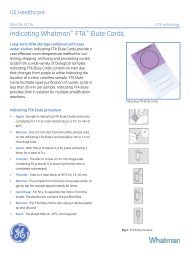
The <strong>Serum</strong> <strong>Biomarker</strong> <strong>Chip</strong> enables proteomics researchers to pattern the<br />
molecular signature of human serum. The chip addresses the need for<br />
high throughput technologies used to study and develop a range of<br />
research applications from risk stratification, to include disease prognosis,<br />
drug eligibility, prediction of safety and efficacy, and therapeutic<br />
monitoring.<br />
The <strong>Serum</strong> <strong>Biomarker</strong> <strong>Chip</strong> is a single antibody capture array built on<br />
the FAST ® Slide dual pad platform. Each slide has two identical arrays<br />
of antibodies printed in triplicate. Two-color fluorescent detection permits<br />
the researcher to reproducibly pattern the relative abundance of 120<br />
human serum proteins between two samples, such as serum samples<br />
from diseased and healthy individuals.<br />
The <strong>Serum</strong> <strong>Biomarker</strong> <strong>Chip</strong> kit includes two arrayed dual pad slides,<br />
two Incubation Chambers, Protein Array Wash and Blocking Buffers.<br />
The slide holder and labeling/detection reagents are available separately.<br />
W H A T M A N S E R U M B I O M A R K E R C H I P D A T A S H E E T
Two-Color Labeling &<br />
Detection Kit<br />
The Two-Color Labeling and Fluorescent<br />
Detection Kit is designed to label two protein<br />
samples. The labeled proteins are<br />
pooled and probed against arrayed antibodies<br />
in a competitive binding assay, and<br />
detected using indirect fluorescence.<br />
• Highly efficient and uniform labeling<br />
of complex serum samples<br />
• Reproducible labeling and signal detection<br />
• Stable, robust and fast non-enzymatic<br />
procedure<br />
• Reduces pH dependency of labeling<br />
efficiency<br />
• System solution includes labeling<br />
reagents, fluorescent conjugate and<br />
bench-friendly protocol<br />
The kit is intended for use with two-pad<br />
FAST Slides, including the <strong>Serum</strong><br />
<strong>Biomarker</strong> <strong>Chip</strong>. The kit contains the<br />
Universal Linkage System (ULS)<br />
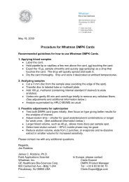
Labeling and Detection Procedure Overview<br />
chemistry to label samples containing<br />
approximately 250 µg of protein in serum,<br />
plasma, or a whole cell lysate. The kit is<br />
designed to label two different protein<br />
samples, each with a different hapten.<br />
Sufficient labeling reagent is provided to<br />
perform a hapten swapping experiment.<br />
Benefits of the two-color<br />
dye-swap assay<br />
• Accounts for hapten-specific differences<br />
in either Biotin-ULS or Fluorescein-<br />
ULS labeling efficiencies<br />
• Averages differences in antibodyantigen<br />
binding interactions caused<br />
by steric hindrance<br />
• Minimizes chip-to-chip variability – includes<br />
an internal control within the assay<br />
The first pad on the slide is probed with a<br />
mixture of two different protein samples,<br />
each labeled with a different hapten; the<br />
second pad is probed with the same two<br />
protein samples but with the haptens<br />
reversed. The normalized intensity for each<br />
Sample A Sample B Sample A Sample B<br />
Block slide with<br />
S&S Blocking Buffer<br />
for 30 min<br />
at room<br />
temperature<br />
Mix 1<br />
Mix 2<br />
element of each pad is calculated as the<br />
average of the biotin- and fluoresceinlabeled<br />
derived intensities from a two-pad<br />
experiment. The ratio between the signal<br />
intensity at each spot corresponds to the<br />
concentration ratio of the proteins found in<br />
the two samples. This method is attractive<br />
for antibody chips as it takes into account<br />
any hapten-specific differences in antigenantibody<br />
interactions.<br />
The use of the ULS labeling system minimizes<br />
background by using indirect fluorescence<br />
detection, labels multiple amino<br />
acids, and requires no additional materials<br />
or reagents.<br />
Label serum proteins with<br />
Biotin-ULS ( ) and<br />
Fluorescein-ULS ( )<br />
for 2-6 hours at 42 °C<br />
Terminate reaction<br />
and remove unbound<br />
ULS reagent<br />
with spin columns<br />
Pool Samples<br />
Incubate labeled proteins<br />
with <strong>Serum</strong> <strong>Biomarker</strong> <strong>Chip</strong><br />
for 4 hours to overnight at room temperature<br />
W H A T M A N S E R U M B I O M A R K E R C H I P D A T A S H E E T<br />
Wash 6X to remove<br />
unbound proteins
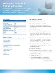
APPLICATION DATA<br />
<strong>Serum</strong> protein profiles of cancer patients.<br />
<strong>Serum</strong> proteins from cancer patients and<br />
corresponding age- and gender-matched controls<br />
were labeled with Biotin-ULS and Fluorescein-<br />
ULS, respectively, then pooled and probed<br />
against the <strong>Serum</strong> <strong>Biomarker</strong> <strong>Chip</strong>.<br />
The competitive binding was detected using<br />
both streptavidin-Dy647 and anti-fluorescein<br />
antibody-Dy547 conjugates. Spot-finding,<br />
background corrections, and two-channel<br />
data sampling was done using ArrayVision<br />
FAST ® software.<br />
The SBC Analysis Workbook is a Microsoft ® Excel<br />
file that converts fluorescent data into numerical<br />
values that represent the abundance of antigen in<br />
Sample A relative to Sample B. The average spot<br />
intensities for each analyte were plotted to reveal<br />
the over- and under-abundant serum biomarkers<br />
in the cancer patient (y-axis) compared to serum<br />
of a healthy individual (x-axis) for bladder, breast,<br />
colon, and prostate cancer.<br />
Upper and lower purple control lines represent<br />
1.25 standard deviations above and below a ratio<br />
of 1 (no change in protein expression). Based on<br />
previous experimental results with the same<br />
serum labeled with both haptens and competed<br />
against itself, we recommend that only ratios<br />
greater than 1.25 std devs on either side of<br />
ratio=1 be considered significant expression level<br />
differences (these values lie outside the normal<br />
noise level of the assay).<br />
Ratios are plotted on a log 2 scale so that a<br />
value of 0 represents no change in expression<br />
between the 2 samples, with ratios representing<br />
up and down regulation being distributed<br />
equally around 0.<br />
Incubate <strong>Serum</strong> <strong>Biomarker</strong> <strong>Chip</strong><br />
with two-color fluorescent<br />
detection solution<br />
for 1 hour at room temperature<br />
Log2 Ratio<br />
Log2 Ratio<br />
Log2 Ratio<br />
Log2 Ratio<br />
0.40<br />
0.30<br />
0.20<br />
0.10<br />
0.00<br />
-0.10<br />
-0.20<br />
-0.30<br />
-0.40<br />
-0.50<br />
2.00<br />
1.50<br />
1.00<br />
0.50<br />
0.00<br />
-0.50<br />
-1.00<br />
-1.50<br />
1.50<br />
1.00<br />
0.50<br />
0.00<br />
-0.50<br />
-1.00<br />
-1.50<br />
-2.00<br />
0.50<br />
0.40<br />
0.30<br />
0.20<br />
0.10<br />
0.00<br />
-0.10<br />
-0.20<br />
Wash 6X to remove<br />
unbound proteins<br />
Excite<br />
547 nm<br />
Bladder Cancer<br />
Breast Cancer<br />
Colon Cancer<br />
Prostate Cancer<br />
Scan at two wavelengths,<br />
spot-find, and sample data<br />
Excite<br />
647 nm<br />
W H A T M A N S E R U M B I O M A R K E R C H I P D A T A S H E E T<br />
Log2 Ratio<br />
0.50<br />
0.40<br />
0.30<br />
0.20<br />
0.10<br />
0.00<br />
-0.10<br />
-0.20<br />
Prostate Cancer<br />
Bioinformatic<br />
analysis
<strong>Biomarker</strong>-Specific Antibodies<br />
Alpha fetoprotein Haptoglobulin MMP-2<br />
Alpha1 antichymotrypsin Hemoglobin MMP-3<br />
Alpha 2 macroglobulin Hepatocyte growth factor MMP-9<br />
Angiogenin ICAM-1 Myeloperoxidase<br />
Angiopoietin-2 IgA Myoglobin<br />
Angiostatin IgG Neuron-specific enolase<br />
Apolipoprotein A1 IgM RANTES<br />
Apolipoprotein J IL-1- Osteopontin<br />
Beta2 microglobulin IL1- PDGF (all isoforms)<br />
Bone sialoprotein IL-2 PDGF (BB isoform only)<br />
CA15-3 IL-2 receptor- Placental alkaline phosphatase<br />
CA19-9 IL-2 receptor- Plasminogen<br />
CA 50 IL-3 Plasminogen activator inhibitor<br />
CA125 IL-4 Prostatic acid phosphatase<br />
Carcinoembryonic antigen (group 2 specific) IL-5 PSA (free)<br />
Carcinoembryonic antigen (group 4 specific) IL-6 PSA (total)<br />
Cathepsin B IL-7 PSA-ACT complex<br />
Ceruloplasmin IL-8 S100<br />
Chondroitin sulftate IL-10 <strong>Serum</strong> albumin<br />
Chorionic gonadotropin- IL-12p40 Sialyl Lewis X<br />
Chorionic gonadotropin- IL12-p70 TAG-72<br />
Chromogranin IL-13 Tetranectin<br />
Collagen type I IL-17 TGF-<br />
Complement C4 Insulin TGF-<br />
C-Reactive protein Insulin growth factor binding protein 3 Thrombospondin-1<br />
Cyclin-dependent kinase inhibitor 2A Insulin-like growth factor 1 Thrombopoietin<br />
Cytokeratin fragment 21-1 (CYFRA 21-1) Interferon- Thyroglobulin<br />
Eotaxin IP-10 TIMP1<br />
Epidermal growth factor Kallikrein-5 TIMP2<br />
Epidermal growth factor receptor Kallikrein-9 TNF-<br />
ErbB2 Kallikrein-12 TNF-<br />
E-selectin Kallikrein-14 Transferrin<br />
Estrogen receptor Laminin Tumor-associated trypsin inhibitor<br />
Fas Low-density lipoprotein Tyrosinase<br />
Fas ligand MCP-1 Urokinase plasminogen activator<br />
Ferritin MCP-2 VCAM-1<br />
Fibroblast growth factor-7 MCP-3 VE-cadherin<br />
Fibroblast growth factor-basic MCP-4 VEGF<br />
G-CSF M-CSF VEGF-D<br />
GM-CSF MIP-1- Von Willebrand factor<br />
Sample Processing<br />
Service<br />
<strong>Whatman</strong> recognizes that not all scientists<br />
have access to the software and instrumentation<br />
needed to process and scan<br />
microarrays, and to collect and analyze<br />
the resulting data. The <strong>Whatman</strong> serum<br />
biomarker sample processing service makes<br />
it simple for researchers to outsource the<br />
tests or try this new technology before<br />
adopting it in their labs.<br />
• Customer samples are sent to <strong>Whatman</strong><br />
and processed using the <strong>Serum</strong><br />
<strong>Biomarker</strong> <strong>Chip</strong><br />
• Data are uploaded to a passwordprotected<br />
ftp site within 10 business<br />
days of receipt of samples at <strong>Whatman</strong><br />
W H A T M A N S E R U M B I O M A R K E R C H I P D A T A S H E E T<br />
Custom <strong>Serum</strong><br />
<strong>Biomarker</strong> <strong>Chip</strong>s<br />
• Add up to 48 additional antibodies to the<br />
existing <strong>Serum</strong> <strong>Biomarker</strong> <strong>Chip</strong> array<br />
• Additional antibodies are either provided<br />
by the customer or customer-specified<br />
commercial antibodies<br />
• <strong>Chip</strong>s are shipped with an antibody map<br />
• Labeling and detection reagents are<br />
available in bulk quantities (see ordering<br />
information)<br />
• Contact <strong>Whatman</strong> customer service to<br />
discuss your requirements
One standard <strong>Serum</strong><br />
<strong>Biomarker</strong> Kit<br />
Contains<br />
2 slides<br />
<strong>Serum</strong> <strong>Biomarker</strong> <strong>Chip</strong>s and two-color labeling and detection reagents may be<br />
purchased in bulk quantities:<br />
24 slides (for 24 paired samples or 48 total samples)<br />
96 slides (for 96 paired samples or 192 total samples)<br />
Ordering Information<br />
Each slide contains<br />
2 arrays for use with<br />
1 serum sample pair<br />
One Two-Color Labeling and Fluorescent Detection Kit contains sufficient<br />
reagents to label the two slides in a standard <strong>Serum</strong> <strong>Biomarker</strong> <strong>Chip</strong> Kit.<br />
Description Qty/Pkg Item #<br />
<strong>Serum</strong> <strong>Biomarker</strong> <strong>Chip</strong> 10 486 077<br />
The kit contains:<br />
<strong>Serum</strong> <strong>Biomarker</strong> <strong>Chip</strong> Arrayed FAST Slides 2<br />
Dual Pad Incubation/Processing Chambers 2<br />
Protein Array Wash Buffer 1 x 125 ml<br />
Protein Array Blocking Buffer 1 x 5 ml<br />
Bulk <strong>Serum</strong> <strong>Biomarker</strong> <strong>Chip</strong> Kits 24 chips 10 486 098<br />
(contain the same items as the 2-chip kit) 96 chips 10 486 099<br />
Fast Frame Slide Holder for 4 slides 1 10 486 001<br />
<strong>Chip</strong> Clip Slide Holder for 1 slide 1 10 486 081<br />
Two-color Labeling & Fluorescent Detection Kit 10 486 085<br />
The kit contains:<br />
Biotin-ULS 20 µl<br />
Fluorescein-ULS 20 µl<br />
10x Protein Labeling Buffer 80 µl<br />
10x KREAstop 80 µl<br />
Streptavidin-DY647 Conjugate 150 µl<br />
Anti-Fluorescein Antibody-DY547 conjugate 350 µl<br />
Micro Bio-Spin ® Chromatography Columns 8<br />
User Manual 1<br />
Bulk Two-Color Labeling & Fluorescent Detection Reagents<br />
Two-Color Labeling Kit 24 (for 24 chips) 10 486 100<br />
Two-Color Labeling Kit 96 (for 96 chips) 10 486 101<br />
ArrayVision FAST Software<br />
Single User Software 10 486 035<br />
ArrayVision Demo USA/Canada 10 486 034<br />
SBC Analysis Workbook Downloadable at www.arraying.com<br />
For the latest protocols and product information, visit www.arraying.com.<br />
The <strong>Whatman</strong> <strong>Serum</strong> <strong>Biomarker</strong> <strong>Chip</strong> is intended for research purposes only, not for diagnostic use.<br />
ArrayVision<br />
FAST Software<br />
• Compatible with virtually all image<br />
formats used to analyze fluorescent<br />
microarrays.<br />
• Comprehensive normalization and<br />
background correction includes<br />
Dy547/Dy647 functions to<br />
improve accuracy in comparing<br />
protein abundance.<br />
• Scatter plot and elemental display<br />
provide quick data assessment and<br />
reports based on user-defined criteria.<br />
• Template definitions and analysis<br />
parameters are contained in the <strong>Serum</strong><br />
<strong>Biomarker</strong> <strong>Chip</strong>-defined protocol.<br />
• Data analysis is performed using the<br />
SBC Analysis Workbook, a preconfigured<br />
Microsoft ® Excel file that<br />
converts fluorescent data into<br />
Dy547/Dy647 ratio values; available<br />
at www.arraying.com.<br />
• Seamless data export to Microsoft ®<br />
Access, XML, GeneSpring and<br />
DecisionSite.<br />
<strong>Chip</strong> Clip <br />
W H A T M A N S E R U M B I O M A R K E R C H I P D A T A S H E E T<br />
The BioScience <strong>Chip</strong> Clip securely<br />
holds the FAST Slide 2-pad slide and<br />
incubation chamber for processing of<br />
the <strong>Serum</strong> <strong>Biomarker</strong> <strong>Chip</strong>.<br />
The <strong>Chip</strong> Clip, used in conjunction<br />
with dual-well silicone Incubation<br />
Chambers, ensures leak-proof barriers<br />
around the two arrayed pads on the<br />
<strong>Serum</strong> <strong>Biomarker</strong> <strong>Chip</strong> Slide.<br />
The slide and Incubation Chamber are<br />
easily inserted into and removed from<br />
the <strong>Chip</strong> Clip; side rails hold the chamber<br />
firmly against the slide surface.<br />
Manufactured from durable, injection-molded Delrin ® .
<strong>Whatman</strong> Quality<br />
<strong>Whatman</strong> is a global leader in separations technology and is known in the scientific<br />
community for providing Innovative Life Science products and solutions. Our instinct for<br />
simplification accelerates the rate of discovery, reduces costs and saves time.<br />
For more information, visit www.whatman.com<br />
<strong>Whatman</strong> ®, FAST ® and <strong>Chip</strong> Clip are trademarks of the <strong>Whatman</strong> Group. ULS is licensed<br />
from and a registered trademark of KREATECH Biotechnology B.V., and covered by one or more<br />
patents of KREATECH Biotechnology BV, including, but not restricted to, the following: EP<br />
0,539,466; US 5,580,990; US 5,714,327; US 5,985,566; US 6,133.038; and EP 1,019,420.<br />
DY647 and DY547 are registered trademarks of Dyomics GmbH. DY647 is covered by the<br />
license of Dyomics GmbH, Germany, to produce and distribute dyes for in vitro applications<br />
claimed by the patent family DE 44 45 065. Delrin is a registered trademark of E.I. duPont de<br />
Nemours and Company. ArrayVision is a trademark of GE Healthcare. Bio-Spin is a registered<br />
trademark of Bio-Rad Laboratories, Inc. GeneSpring is a trademark of Silicon Genetics.<br />
DecisionSite is a trademark of Spotfire, Inc. Microsoft, Access and Excel are either registered<br />
trademarks or trademarks of Microsoft Corporation in the United States and/or other countries.<br />
North America <strong>Whatman</strong> Inc.<br />
200 Park Avenue, Suite 210<br />
Florham Park, NJ 07932 USA<br />
Tel: 1-800-WHATMAN (US and Canada)<br />
Fax: 1-973-245-8329<br />
Email: info@whatman.com<br />
Europe <strong>Whatman</strong> International Ltd.<br />
Springfield Mill, James <strong>Whatman</strong> Way<br />
Maidstone<br />
Kent ME14 2LE UK<br />
Tel: +44 (0)1622 676670<br />
Fax: +44 (0)1622 677011<br />
Email: information@whatman.com<br />
<strong>Whatman</strong> GmbH<br />
Hahnestrasse 3<br />
D-37586 Dassel<br />
Germany<br />
Tel: +49 (0) 5564 204 100<br />
Fax: +49 (0) 5564 204 533<br />
Email: information@whatman.com<br />
Japan <strong>Whatman</strong> Japan KK<br />
JPR Ichigaya Building 6F<br />
4-7-15 Kudan-Minami,<br />
Chiyoda-ku, Tokyo 102-0074 Japan<br />
Tel: +81 (0)3 5215 1240<br />
Fax: +81 (0)3 5215 1245<br />
Email: japaninfo@whatman.com<br />
Asia Pacific <strong>Whatman</strong> Asia Pacific Pte Ltd.<br />
171 Chin Swee Road<br />
#08-01 San Centre<br />
Singapore 169877<br />
Tel: +65 6534 0138<br />
Fax: +65 6534 2166<br />
Email: wap@whatman.com<br />
51699 (US) S9036-831 (EU) 04/06