RHINOGENIC AND OTOTIC INTRACRANIAL SUPPURATION
RHINOGENIC AND OTOTIC INTRACRANIAL SUPPURATION
RHINOGENIC AND OTOTIC INTRACRANIAL SUPPURATION

Create successful ePaper yourself
Turn your PDF publications into a flip-book with our unique Google optimized e-Paper software.
<strong>RHINOGENIC</strong> <strong>AND</strong> <strong>OTOTIC</strong><br />
<strong>INTRACRANIAL</strong> <strong>SUPPURATION</strong><br />
Dr H. BOODHOO<br />
F.C.S<br />
Consultant Neurosurgeon
CASE PRESENTATION
Age: 8 yrs<br />
Sex: male<br />
PATIENT PROFILE<br />
Address: Vacoas<br />
Mother: selfemployed<br />
Father: carpenter<br />
Sibling: 5yr a&w
HISTORY<br />
Referred from private clinic on 24/06/08<br />
Initially attended JH with:<br />
Fever<br />
Vomiting<br />
Abdominal pain<br />
No headache<br />
No fits<br />
No visual complaints<br />
Duration: 3 days
Admitted<br />
? Early GE<br />
HISTORY (cont.)<br />
Mother signed DAMA<br />
Admitted in Clinic<br />
Persisting complaints<br />
Next day: neck stiffness<br />
Intravenous antibiotic therapy<br />
Investigation: ↑↑ WCC<br />
Special investigation: CT Brain ±Contrast
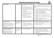
Pansinusitis<br />
CT BRAIN<br />
Right frontal brain abscess<br />
Right fronto temporo parietal subdural<br />
empyema<br />
Referred urgently to neurosurgical unit,<br />
Victoria Hospital
PAST HISTORY<br />
h/o fall from stairs 3yrs back- had a<br />
lacerated wound on right forehead<br />
PMH<br />
PSH<br />
Drug history<br />
Allergic history<br />
Immunisation history<br />
Social history
On Examination<br />
General physical examination<br />
Sick looking<br />
Extremely thin<br />
Unusually quiet<br />
Wt. 16kgs<br />
P: 92/min T/°C: 37.6 RR: 14/min<br />
No pallor, no jaundice<br />
No clubbing<br />
No lymphadenopathy<br />
ENT: nasal secretions ++ rt.>lt.
On Examination (cont)<br />
Systemic examination<br />
CVS: Normal HS, no murmur<br />
RS: chest was clear, trachea centrally<br />
located, no adventitious sounds<br />
Abdomen: scaphoid, no organomegaly, mild<br />
RUQ tenderness<br />
Genitals: normal
On Examination (cont)<br />
CNS examination<br />
GCS: E 4M 5V 6<br />
Higher mental functions<br />
Mild neck stiffness<br />
No cerebellar signs<br />
No photophobia<br />
Moving all limbs<br />
Fundoscopy: no papilledema<br />
Cranial nerve examination: normal
INVESTIGATION<br />
Hematological: ↑↑ WCC<br />
Biochemistry: normal<br />
LFT: normal<br />
Special investigation- CT BRAIN ±C
Admitted<br />
MANAGEMENT<br />
Continued i.v. antibiotic therapy<br />
i.v. fluid therapy<br />
Conditioned stabilised<br />
Urgent referral to E.N.T Hospitaladmitted<br />
BAWO- Pus +++ right maxillary sinus<br />
Back to VH next day
MANAGEMENT (cont)<br />
25/06/08: Cranial surgery<br />
1. Right small frontal craniotomy for<br />
drainage of brain abscess<br />
2. Wide temporoparietal craniotomy for<br />
evacuation of subdural empyema<br />
Nursed in ICU<br />
I.v antibiotics/ i.v phenytoin
POST-OP<br />
Marked improvement in clinical condition<br />
Uncomplicated recovery phase<br />
Lab culture report: sterile<br />
Drains removed after 48 hrs<br />
Referred to nutritionist- high protein diet<br />
Progress CT brain showed good evacuation of<br />
brain abscess & empyema, no features of infarct<br />
or ↑ ICP<br />
Continued on i.v antibiotics for two weeks
Still having RUQ pain<br />
Ultrasound abdomen<br />
1. Gall bladder filled with<br />
calculi<br />
2. Small rt. Renal calculus<br />
Surgical opinion<br />
Pediatric opinion<br />
Still under investigation<br />
Review with surgeon<br />
POST-OP
With Neurosurgeon<br />
Oral antibiotics<br />
Oral AED<br />
Repeat CT of brain<br />
REVIEW
Thank you!!
4/18/2011<br />
16 years Male<br />
Patient Profile 2<br />
Comores Island<br />
c/o Chronic discharge Left ear<br />
Headache, confusion, fever<br />
GCS10/15 (E3M5V2)<br />
Spastic, neck stiffness
Emergency combined surgical<br />
treatment<br />
Radical mastoidectomy and<br />
posterior fossa craniectomy
4/18/2011 30
4/18/2011 31
2 cm x 1 cm<br />
CAVERNOUS SINUS<br />
Located on each of sella turcica and<br />
body of sphenoid bone<br />
Superior orbital fissure to apex of<br />
petrous bone
ANATOMY<br />
Facial veins connect with the cavernous<br />
sinus via ophthalmic veins<br />
Thrombophlebitis of cavernous sinus can<br />
spread to superior and inferior petrosal<br />
sinuses
ANATOMY<br />
Posterior intercavernous sinus superior and<br />
inferior petrosal sinuses<br />
Receive blood from superior and inferior<br />
ophthalmic vein<br />
They drain posteriorly and inferiorly through the<br />
superior and inferior petrosal sinuses and<br />
pterygoid plexuses
Infections of<br />
SPREAD<br />
Face, nose, orbit, tonsils, soft palate, pharynx,<br />
air sinuses, middle ear and mastoid can all<br />
spread to cavernous sinuses<br />
Sphenoid and posterior ethmoid sinuses<br />
Jaw –tooth extraction, maxillary surgery via<br />
(pterygoid plexuses)
Fever<br />
SYMPTOMS & SIGNS<br />
Ptosis/chemosis<br />
Oculomotor palsies (III, IV, VI)<br />
Contralateral hemiparesis (thrombosis ICA)
CT brain<br />
Irregular filling defect<br />
Convex bulging of the lateral wall<br />
Dilatation of superior opthalmic vein<br />
Thickening of extra ocular muscles and<br />
periorbital edema
Antibiotics (high doses)<br />
TREATMENT<br />
(Staph aureus, Strep pneumonia, Haemophilus<br />
influenzae<br />
Anticoagulant (no evidence of cortical venous<br />
infarct)<br />
Surgery- sphenoid sinus sepsis<br />
100 % mortality to 30 %
<strong>RHINOGENIC</strong><br />
<strong>INTRACRANIAL</strong> SEPSIS
Leading neurological manifestation<br />
Fever 96%<br />
Seizures 70%<br />
Neurological signs 58%
Epidemiology<br />
Most common in males<br />
Seasonal variation
Etiology<br />
Spread<br />
Direct- Erosion of Tegmen tympani<br />
Erosion of posterior wall of frontal sinus<br />
Retrograde septic thrombophlebitis<br />
Facial or scalp infection<br />
Dental sepsis<br />
Meningitis<br />
Cranial surgery e.g. depressed fracture<br />
Infection at distant sites
Etiology<br />
Otorhinolaryngeal infection- 40-70 %<br />
Paranasal sinusitis<br />
Otitis media<br />
Mastoiditis<br />
Cranial trauma- 6-30%
Predisposing factors<br />
Diabetes Mellitus<br />
Alcoholism<br />
Chest infection<br />
Sepsis<br />
HIV<br />
Immunodepression- steroids, cytotoxic<br />
drugs<br />
Poor nutrition, poor hygiene, delayed<br />
treatment
“Frequent use of broad<br />
spectrum antibiotics may<br />
contribute to subdural<br />
empyema”
Most common pathogens<br />
Strep pneumoniae- 16%<br />
Group B strep- 13%<br />
H. Influenzae- 13%<br />
Salmonella spp- 13%<br />
E. coli- 10%<br />
Pseudomonas aeruginosa- 10%
Pathogens<br />
Pus- sterile in 40%<br />
Use of broad spectrum antibiotics<br />
NTSO- non typhoidal salmonella<br />
organisms have been reported in the<br />
setting of advanced AIDS infection
Diagnosis<br />
Difficult to clinically differentiate between<br />
meningitis and SDE<br />
Diagnosis is based on strong clinical<br />
suspicion<br />
Triad of- fever<br />
sinusitis<br />
neurological deficit
Investigation<br />
Infants: brain sonography<br />
CT Bain with contrast, brain and paranasal<br />
sinuses, posterior fossa cuts
Investigation<br />
CT Brain (contrast)<br />
Thin rim of fluid, slightly hyperdense to<br />
CSF with surrounding enhancement,<br />
adjacent disproportionate cortical edema<br />
and effacement of cortical sulci<br />
Cranial ultrasound can substitute CT in<br />
infants<br />
LP must be avoided
Management<br />
Timing of surgery<br />
Simultaneous neurosurgical and ENT<br />
intervention<br />
SDE requires surgical evacuation of<br />
infected material, irrespective of its volume
Management<br />
Craniotomy was determined to be the<br />
surgical procedure of choice in SDE<br />
Allows complete evacuation<br />
Decompression of cerebral hemisphere
Prognosis<br />
Early diagnosis and treatment<br />
High degree of suspicion<br />
“Prolonged fever, seizures, neurological<br />
signs”
Age<br />
GCS<br />
Prognostic factors<br />
Timing/ aggressiveness of treatment<br />
Progression of disease
Outcome<br />
Mortality- 100% before advent of<br />
antibiotics & CT<br />
Decreased to 40% after CT Scan<br />
10-12% presently
Intracranial subdural empyema is a<br />
neurosurgical emergency<br />
It is rapidly fatal if not recognised early<br />
and managed promptly
Early drainage, simultaneous<br />
eradication of the primary source of<br />
sepsis and intravenous administration<br />
of high doses of appropriate<br />
antibiotics agents represents the<br />
mainstay of treatment
1. Direct spread<br />
Spread<br />
Erosion through the postwall of<br />
frontal<br />
sinus which has one-half the<br />
thickness of<br />
the anterior wall<br />
2. Indirect mechanisms<br />
Retrograde thrombophlebitis<br />
i l i
Lumbar Puncture L.P<br />
L.P performed in the presence of clinical<br />
features of raised ICP and focal<br />
neurological signs are extremely<br />
dangerous
Disparity between CT imaging and<br />
clinical findings<br />
-integrity of arachnoid membraneprevent<br />
spread<br />
-improve blood brain barrier<br />
-Wide cerebral decomposition via a wide<br />
craniotomy
DIAGNOSIS<br />
Infective sinustis<br />
Periorbital swelling<br />
Purulent dural discharge<br />
Positive Neurosurgical signs<br />
MUST HAVE CT SCAN BRAIN<br />
& PNS
Role of Non Operative<br />
treatment<br />
Fully concious patient, with small EDE (no<br />
radiological mass effect) with no<br />
neurological deficit, signs of clinical<br />
improvement (temperature ; ESR ;<br />
WCC<br />
May be treated with intravenous antibiotics<br />
and prophylatic antiepileptic provided the<br />
primary source of sepsis has been<br />
surgically eradicated
Unlike SDE, EDE is a disease that<br />
should be managed without<br />
morbidity or death
INFRATENTORIAL EMPYEMA<br />
Rare, highly lethal form of intracranial<br />
suppuration<br />
Lumbar puncture<br />
Cereballar abcess<br />
Hydrocephalus<br />
Extension of pus to cerebello pontine angle
INFRATENTORIAL EMPYEMA<br />
TREATMENT<br />
Early aggressive surgical drainage and<br />
decompression of the cerebellum by a<br />
wide posterior fossa craniectomy ,<br />
eradication of the primary source of<br />
infection (usually mastoiditis) treatment of<br />
concomittant hydrocephalus high dose<br />
intravenous antibiotics
THANK YOU