ortema pforzheim - Arcus Sportklinik
ortema pforzheim - Arcus Sportklinik
ortema pforzheim - Arcus Sportklinik

Create successful ePaper yourself
Turn your PDF publications into a flip-book with our unique Google optimized e-Paper software.
September 2009<br />
Imprint:<br />
Publisher:<br />
ARCUS Kliniken Pforzheim<br />
Rastatter Str. 17-19<br />
75179 Pforzheim<br />
Phone: +49 7231 60556 0<br />
web www.sportklinik.de<br />
email info@sportklinik.de<br />
Editorial Management:<br />
Prof. univ. cath. Cuenca EC Bernhard Rieser<br />
rieser@sportklinik.de<br />
Editor and Marketing:<br />
Heiko Hecht<br />
hecht@sportklinik.de<br />
Graphics & Layout:<br />
Buero 01<br />
Pforzheim<br />
Print:<br />
Kraft Druck GmbH<br />
Ettlingen<br />
Disclaimer:<br />
Please note that statements made in this brochure are of general nature and do not necessarily apply for<br />
every patient. Therefore, individual advice of your treating physician is absolutely necessary.
Welcome<br />
Dear patients,<br />
With this information brochure we would like to present you the most important<br />
part of our operative work. We refer to 20 years of own experience in in- and outpatient<br />
services and the current scientific status.<br />
Since 1989, more than 65.000 patients have been operated and about 150.000<br />
patients treated in the ARCUS Clinics. With more than 7.600 surgeries and about<br />
38.000 treated patients in 2009, we have become one of the biggest orthopaedic<br />
sports-traumatologic accidental surgery centers in Germany and Europe.<br />
Where does this success come from?<br />
It is based on tireless dedication and hard work, consequent implementation of latest<br />
operation- and treatment methods and full use of the best technical possibilities.<br />
We always used a substantial part of our revenues for new investments. And finally,<br />
in 2006, we were able to open up a new clinic equipped with the highest technical<br />
standards and a very pleasant, patient- and staff-friendly atmosphere. It has more<br />
than 6 operating theatres, 70 beds and 22 beds in the ward station on a total of<br />
17.000 m² that means together with already available capacities of the former clinic<br />
9 operating theatres, 90 inpatient beds and 30 ward station beds. The clinic is<br />
divided into a private clinic and a clinic for other patients with 30 beds which are<br />
listed on the bed requirement planning of the state of Baden-Württemberg. Here,<br />
also patients with statutory health insurance can be offered in-patient treatment.<br />
We want to provide you an understandable overview of our range of services and<br />
answer open questions in the case of a planned operation. Should you have any<br />
further questions about our services, special surgery techniques or our clinics in<br />
general, please do not hesitate and contact us.<br />
More information please find on www.sportklinik.de<br />
Your ARCUS Clinics Team<br />
Rastatter Str. 17-19 • 75179 Pforzheim • Germany • Phone 07231- 60556- 0 • www.sportklinik.de • info@sportklinik.de<br />
General Ellenbogen Information<br />
3
Table of Contents<br />
General Information<br />
Welcome 3<br />
Table of Contents 4<br />
Clinic Portrait / Competence Center / Science 6<br />
Basic Values of the ARCUS Clinics Pforzheim 7<br />
Spectrum of Surgery / Facts & Figures 8<br />
Medical Management 9<br />
Specialist Areas 11<br />
Diagnostics 12<br />
Quality Management 15<br />
Interesting Facts & Organization 16<br />
Anesthesia 18<br />
Operative Spectrum - Knee<br />
Meniscus 22<br />
Anterior Cruciate Ligament (ACL) 26<br />
Knee-Cap (Patella) 33<br />
Arthrosis 36<br />
Orthobiology 44<br />
Knee Malalignment 46<br />
Knee Endoprosthetics 49<br />
Operative Spectrum - Shoulder<br />
Shoulder Impingement Syndrome 55<br />
Calcified Tendinitis of the Shoulder (tendinosis calcarea) 57<br />
Shoulder Luxation 59<br />
Rotator Cuff Damages 61<br />
Injuries and Arthrosis of the Acromioclavicular Joint (AC-joint) 63<br />
Collarbone Fracture (clavicle fracture) 65<br />
Humeral head fracture 67<br />
Shoulder Endoprosthetics 69<br />
Operative Spectrum - Hip<br />
Hip Joint Arthrosis (coxarthrosis) 73<br />
Hip Arthroscopy 76<br />
Step-by-step Plan for Treatment of Coxarthrosis 78<br />
Total Endoprosthesis: Material and Fixation 79<br />
4 Rastatter Str. 17-19 • 75179 Pforzheim • Germany • Phone 07231- 60556- 0 • www.sportklinik.de • info@sportklinik.de
Operative Spectrum - Elbow<br />
Tennis Elbow 82<br />
Golfer’s Elbow 84<br />
Sulcus-ulnaris Syndrome or Cubital Tunnel Syndrome 84<br />
Loose Joint Bodies 85<br />
Osteochondrosis Dissecans 85<br />
Stiff Elbow and Elbow Arthrosis 86<br />
Elbow Prostheses 87<br />
Luxations and Instability 88<br />
Operative Spectrum - Foot<br />
Foot / Ankle / Achilles Tendon 89<br />
Big Toe 89<br />
Small Toe 91<br />
Metatarsus/ Tarsus 92<br />
Heel 93<br />
Achillodynia 94<br />
Achilles Tendon Rupture 95<br />
Ankle Disorders 96<br />
Neurosurgery / Spinal Column<br />
General Information 102<br />
Cervical Spine (CS) 103<br />
Lumbar Spine (LS) 109<br />
How to find us 115<br />
Rastatter Str. 17-19 • 75179 Pforzheim • Germany • Phone 07231- 60556- 0 • www.sportklinik.de • info@sportklinik.de<br />
General Ellenbogen Information<br />
5
The ARCUS Clinics – a Portrait<br />
The ARCUS Clinics comprise a private clinic with 60 beds which was opened in 1995,<br />
and a clinic also approved by the statutory health insurance system with 30 beds.<br />
The new clinic complex was opened up in 2006. Here, 6 operating theatres equipped<br />
with state-of-the-art technology, and 22 beds in the ward station and the intensive<br />
care unit are available.<br />
The privately insured patient which can chose individual surgical treatment within<br />
the private clinic is offered a specialized unit with first class hotel comfort – an<br />
excellent overall service.<br />
The statutorily insured patient is, although statutory health insurance companies do<br />
only pay for “basic primary health care”, still provided a high-level clinic standard<br />
i.e. a standard on far above-average level compared to most other clinics.<br />
Competence Center<br />
In the ARCUS Clinics up to 7.500 patients are operated each year – with increasing<br />
tendency. Main focuses are on sports traumatology, knee-, hip-, shoulder-, elbow-,<br />
orthopedic-, and accident surgery, endoprosthetics and in the private clinic also on<br />
spinal surgery. External cooperating surgeons additionally cover vascular- and neurosurgery<br />
and an experienced team of anesthetists offers besides intra-operative<br />
control also post-operative pain therapy for in-patients. In cases of cardiologic<br />
problems during and after surgery we can refer to our cardiology section and stateof-the-art<br />
technical equipment.<br />
In the adjoining orthopedic joint practice patients can get out-patient treatment.<br />
This enables us to constantly control and optimize our own operation- and aftercare<br />
results what already proved successful e.g. rehabilitation periods of our patients<br />
could demonstrably be shortened.<br />
Special importance since many years has treatment of top athletes in the conservative<br />
and surgical area. As medical partner of the “Deutsche Sporthilfe” we offer a 24-hour<br />
acute service for sponsored top athletes. This comprises best diagnostics, operative<br />
treatment if necessary and a comprehensive rehabilitation program to accelerate<br />
recovery and support the athlete to regain physical fitness as soon as possible.<br />
Our medical range of services is completed by cooperating partners in therapy,<br />
rehabilitation, prevention and orthopedic technology.<br />
Perfect interdisciplinary collaboration of surgeons of different areas, physiotherapists<br />
and orthopedic technicians form the basis for an optimal and focused patient care<br />
both in the in- and the out-patient sector.<br />
Science<br />
The leading physicians of the ARCUS Clinics are members of all important national<br />
and international professional associations and regularly work for them as referees.<br />
Moreover, the ARCUS Sports Clinic cooperates with the association for science and<br />
further education in orthopedics. Together they regularly organize training programs<br />
for physicians and physiotherapists which are acknowledged as such by the<br />
Ärztekammer Nordbaden.<br />
6<br />
Rastatter Str. 17-19 • 75179 Pforzheim • Germany • Phone 07231- 60556- 0 • www.sportklinik.de • info@sportklinik.de
Basic Values of the ARCUS Clinics Pforzheim<br />
Our Mission<br />
Our Medical Demand<br />
In the ARCUS Clinics Pforzheim, long-term experience and specialization in different<br />
medical areas as well as use and development of medical state-of-the-art technology<br />
is the key to success. Scientific exchange of experiences and know-how is part of<br />
our daily work life. Our international appreciation is our continuous commitment.<br />
Patient Focus<br />
Orientation towards the patient – our customer – is the basis of our activities. We<br />
make highest demands on the quality of patient care and offer dedicated medical<br />
attendance from prevention and therapy until rehabilitation. Competent care and<br />
service improve healing results.<br />
The architecture of the ARCUS Clinics creates an environment where efficiency and<br />
the patients’ individual needs are optimally harmonized.<br />
Employee Focus<br />
The dedication of our qualified employees ensures the success of our clinic. Therefore<br />
we expect above-average performances and support professional development by<br />
providing further education measures. Professional and socially competent communication<br />
between the employees is the most important condition for a good<br />
working team.<br />
Managers are role models and support the employees’ dedication through a cooperative<br />
management style.<br />
Economy<br />
Since many years now, the ARCUS Clinics Pforzheim have been successful private<br />
facilities on the health sector.<br />
Optimal treatment concepts and results as well as economic success are inseparably<br />
linked with each other and one area strengthens the other.<br />
Rastatter Str. 17-19 • 75179 Pforzheim • Germany • Phone 07231- 60556- 0 • www.sportklinik.de • info@sportklinik.de 7<br />
General Ellenbogen Information
Spectrum of Surgery / Facts & Figures<br />
Figures<br />
2009 1.Quarter 2010<br />
Anterior Cruciate Ligament Surgery 1222 335<br />
Meniscus Surgery 1632 394<br />
Cartilage Surgery 175 41<br />
Hip Arthroscopy 172 60<br />
Shoulder Surgery 1101 348<br />
(except prostheses)<br />
Elbow Surgery 179 68<br />
Total 7671 2210<br />
(except prostheses)<br />
Endoprosthetics (artificial joints)<br />
2009 1.Quarter 2010<br />
Knee 662 210<br />
Hip 327 109<br />
Shoulder 101 36<br />
Total 1105 361<br />
8<br />
Rastatter Str. 17-19 • 75179 Pforzheim • Germany • Phone 07231- 60556- 0 • www.sportklinik.de • info@sportklinik.de
Medical Management<br />
Prof. univ. cath. Cuenca EC<br />
Bernhard Rieser<br />
Medical Director<br />
Partner of the ARCUS Sports Clinic<br />
Medical Specialist for Orthopedic<br />
Surgery<br />
Dr. med. Wolfgang Miehlke<br />
Leading Physician<br />
Medical Specialist for Orthopedic<br />
Surgery, Trauma Surgery<br />
and Sports Medicine<br />
Prof. Dr. med. Christian Heisel<br />
Leading Physician<br />
Medical Specialist for Orthopedic<br />
Surgery, Special Orthopedic Surgery<br />
and Trauma Surgery<br />
Dr. med. Ludwig Bös<br />
Leading Physician<br />
Partner of the ARCUS Sports Clinic<br />
Medical Specialist for Orthopedic<br />
Surgery and Sports Medicine<br />
Dr. med. Thomas Ambacher<br />
Leading Physician<br />
Medical Specialist for Orthopedic<br />
Surgery, Trauma Surgery and Sports<br />
Medicine<br />
Prof. Dr. med. Uwe Spetzger<br />
Leading Physician<br />
Medical Specialist for Neurosurgery<br />
Dr. med. Andree Ellermann<br />
Leading Physician<br />
Partner of the ARCUS Sports Clinic<br />
Medical Specialist for Orthopedic<br />
Surgery, Trauma Surgery, Sports<br />
Medicine and Chirotherapy<br />
Prof. Dr. med.<br />
Rüdiger Schmidt-Wiethoff<br />
Rüdiger Schmidt-Wiethoff<br />
Leading Physician<br />
Medical Specialist for Orthopedic<br />
Surgery, Special Orthopedic Surgery,<br />
Trauma Surgery and Sports Medicine<br />
Rastatter Str. 17-19 • 75179 Pforzheim • Germany • Phone 07231- 60556- 0 • www.sportklinik.de • info@sportklinik.de 9<br />
General Ellenbogen Information
10<br />
ORTEMA PFORZHEIM<br />
H e r z ll ii c h<br />
W ii ll ll k o m m e n<br />
UNSERE LEISTUNGEN IM ÜBERBLICK:<br />
<br />
<br />
<br />
Schuh- und Einlagen-Technik:<br />
Korrektur des Gangbildes<br />
Rumpforthesen-Technik:<br />
Fixierend und wachstumslenkend<br />
Arm- und Beinprothesen:<br />
Nutzung modernster Technologien<br />
<br />
<br />
<br />
Bandagen-Technik:<br />
Von Kopf bis Fuß nach Maß<br />
Orthesen und Knieorthesen:<br />
Stabilisierung und Entlastung<br />
Sport-Orthopädie:<br />
Protektion & Prävention<br />
DAS K-COM KNIEORTHESENKONZEPT<br />
DIE KNIEORTHESE<br />
AUS CARBONFASER<br />
Kreuzbandriss Arthrose Varus Valgus Kinderversorgung<br />
INDIVIDUELLE ANFERTIGUNG, EXTREM LEICHT<br />
MIT OPTIMALER ANATOMISCHER PASSFORM<br />
www.<strong>ortema</strong>.de<br />
ORTEMA GmbH Filiale Pforzheim · Rastatter Straße 17-19 · 75179 Pforzheim · Tel. +49(0)72 31-139 66 67 · Fax +49(0)72 31-1 39 66 84 · <strong>pforzheim</strong>@<strong>ortema</strong>.de<br />
ORTEMA GmbH Filiale Waiblingen · Alter Postplatz 13 · 71332 Waiblingen · Tel. +49(0)7151-985994-0 · Fax +49(0)7151-985994-94 · waiblingen@<strong>ortema</strong>.de<br />
Hauptsitz ORTEMA GmbH · Kurt-Lindemann-Weg 10 · 71706 Markgröningen · Tel. +49(0)7145-912081 · Fax +49(0)7145-912980 · info@<strong>ortema</strong>.de
Specialist Areas<br />
We cover the whole spectrum of orthopedic surgery. Therefore, in order to ensure<br />
our high quality standard, eight leading physicians manage the area of their specialization.<br />
Our Focus Areas:<br />
• Sports Traumatology<br />
• Knee Surgery<br />
• Shoulder- and Elbow Surgery<br />
• Hip Surgery<br />
• Foot- and Ankle Joint Surgery<br />
• Endoprosthetics<br />
• Trauma Surgery<br />
• Neuro- and Spinal Surgery (for privately insured patients and self-payers)<br />
• Blood Vessel Surgery<br />
• Cardiology<br />
Specialist Practices within the ARCUS Clinics<br />
Besides the orthopedic clinics, there are also different specialist practices integrated<br />
into the ARCUS Clinics complex to extend the spectrum.<br />
• Orthopedic joint practice Rieser / Bös / Ellermann / Miehlke / Ambacher /<br />
Schmidt-Wiethoff / Heisel / Sobau<br />
• Private practice for neuro- and spinal surgery Prof. Dr. med. Uwe Spetzger<br />
• Practice for radiology and nuclear medicine Dr. med. Berthold Winter<br />
• Private practice for cardiology Dr. med. W.O. Schüler & Colleagues<br />
• Specialist practice for anesthesia and pain therapy Dr. med. Carla Weber<br />
Rastatter Str. 17-19 • 75179 Pforzheim • Germany • Phone 07231- 60556- 0 • www.sportklinik.de • info@sportklinik.de<br />
General Ellenbogen Information<br />
11
Diagnostics<br />
Thanks to state-of-the-art technical equipment of the latest generation, the ARCUS<br />
Clinics can always refer to the best method to provide optimal diagnostics and<br />
therapy planning.<br />
Cross-Sectional Diagnostic Imaging and Digital X-Ray<br />
In the adjoining practices there exist two 1.5 Tesla MRI scanners (nuclear spin) with<br />
the latest equipment, technology for digital X-ray, a Dual Source CT, a nuclear medicine<br />
section as well as a cardiac catheter laboratory for comprehensive diagnostics.<br />
All digital images taken with CT, MRI and digital X-ray as well as the arthroscopic<br />
images generated during surgery are stored in a central PACS-system and can be<br />
retrieved at any time in the treatment rooms of the orthopedic joint practice, the<br />
wards and in the operating theatres. There are certified monitors available for<br />
reporting in all sections.<br />
CT (computed tomography)<br />
The Siemens Dual Source SOMATOM Definition CT (2 x 64 rows) is by using a second<br />
x-ray tube and a second detector much more efficient than devices of the “simple”<br />
construction. Its excellent image quality and high resolution at the lowest possible<br />
radiation exposure for the patient enables fast and precise diagnosis and increases<br />
its reliability. It also enables us to examine coronary heart vessels without cardiac<br />
catheter. Temporal resolution of the SOMATOM Definition is with 83 ms not dependant<br />
on the patients’ heart rate. This makes it possible to examine every heart at<br />
every heart rate e.g. diagnosis of acute chest pain, visualization of coronary arteries<br />
and function analysis of the heart. Combined with the currently highest possible<br />
resolution of less than 0.4 mm, the SOMATOM Definition can display smallest anatomic<br />
structures, whether complex osseous structures or finest details of the coronary<br />
tree. Thanks to the large gantry aperture, the scan length of 200 cm and the highest<br />
possible x-ray generator performance almost all acute in-patients regardless of their<br />
physical constitution and size can be examined and valuable time gained between<br />
scan and diagnosis.<br />
MRI (magnetic resonance imaging = nuclear spin<br />
tomography)<br />
The ARCUS Clinics have two 1.5 Tesla MRIs of the latest generation at their disposal.<br />
Equipped with AudioComfort, a combination of several innovative technical measures<br />
for noise reduction, the former usual noise level reached during MRI can be<br />
reduced by up to 97%. The ability to scan the patient in the feet first position as<br />
well as total-body examinations in the time of only 12 minutes make the Magnetom<br />
Avanto the most efficient and patient-friendliest system of its class and is decisive<br />
for pre-operative diagnostics of poly-traumatized patients.<br />
The Magnetom Avanto is furthermore equipped with the new and innovative Timtechnology.<br />
Heart is the revolutionary matrix coil concept where 76 coil elements can<br />
be combined with up to 32 high-frequency channels [76x32]. This visibly improves<br />
recording speed and picture quality. The Magnetom Avanto also stands out through<br />
12<br />
Rastatter Str. 17-19 • 75179 Pforzheim • Germany • Phone 07231- 60556- 0 • www.sportklinik.de • info@sportklinik.de
Diagnostik<br />
especially powerful gradient systems (comparable with “motors” for MR), what<br />
facilitates fast examinations of the heart or detailed analyses of brain functions.<br />
Cardio MRI is thanks to modern software a simple and fast examination of heart<br />
function, myocardial morphology, extension of infarction and 3D-coronary anatomy.<br />
In most cases the examination is completed in less than 30 minutes. This method is<br />
of particular importance for sports medicine. The decided diagnostic of heart muscle<br />
inflammation is not comparable with any other method.<br />
Digigal X-Ray<br />
The orthopedic joint practice has a dose-reduced direct-digital x-ray apparatus at its<br />
disposal. With only 40% of usual radiation exposure it enables images with higher<br />
resolution and therefore better basis for diagnostics.<br />
Mobile CT and Navigation Device<br />
With the CT, complex surgery procedures can also be carried out with navigation.<br />
This enables better results when being confronted with complicated anatomic conditions<br />
or complex fractures.<br />
Operating Theatres<br />
All nine operating theatres are connected to the digital clinic network. This ensures<br />
internal as well as external data transfer. All images taken during surgery are recorded<br />
and stored in the patient’s file. By means of an external surrounding camera<br />
system also transfer of external footage is possible (besides arthroscopic images).<br />
When conducting live-surgery, this enables transfer of e.g. positioning of the patient<br />
or preparation of transplants/implants to national and international congresses<br />
and other events.<br />
On two screen walls, surgery can be followed from the outside. The operation manager<br />
is responsible for occupancy and optimal allocation of the operating theatre.<br />
Sterilization Zone<br />
Our operating theatres are provided with sterile material via nonintersecting corridors.<br />
Sterilization is equipped with top quality devices only. Each instrument used can<br />
be referred to the respective patient via a bar code. With this, absolute traceability,<br />
the so-called sterile-chain can be documented.<br />
A modern system documents all working steps and provides insight into availability<br />
of the OP sets. Moreover it automatically controls withholding periods.<br />
Rastatter Str. 17-19 • 75179 Pforzheim • Germany • Phone 07231- 60556- 0 • www.sportklinik.de • info@sportklinik.de 13<br />
General Ellenbogen Information
Anzeige<br />
ARCADIS Orbic 3D - Mehr Präzision bei der operativen<br />
Versorgung von Knochen- und Gelenkbrüchen<br />
Die präzise Identifizierung und Repositionierung dislozierter Knochenfragmente, das<br />
Setzen von Pedikelschrauben in die Wirbelsäule und die Lagekontrolle von Osteosynthesematerial<br />
zählen zu den größten Herausforderungen in der Unfallchirurgie und<br />
Neurochirurgie. In vielen Fällen liefert die konventionelle 2D-Projektionsbildgebung<br />
nicht genügend Informationen, um solche Eingriffe präzise zu kontrollieren. Für die<br />
exakte Versorgung von Knochen- und Gelenkfrakturen steht mit dem mobilen C-<br />
Bogen ARCADIS® Orbic 3D von Siemens ein hervorragendes System zur Verfügung,<br />
das mittels der interaoperativen 3D-Bildgebung deutliche Informationsvorteile über<br />
die jeweilige Fraktur der Knochen und Gelenke im Vergleich zu herkömmlicher<br />
2D-Bilddarstellung bietet. Hauptanwendungen sind Versorgungen der oberen und<br />
unteren Extremität, der gesamte Wirbelsäule, Hüfte/Becken sowie des Gesichtsschädels.<br />
Die intraoperative 3D-Bildgebung ermöglicht dem Chirurgien die sofortige<br />
Beurteilung der Ergebnisse, wie z.B. der Lagekontrolle von Schrauben. Notwendige<br />
Korrekturen können direkt während der OP erfolgen, das postoperative CT wird<br />
in den meisten Fällen nicht mehr benötigt und eine nochmalige Operation kann<br />
vermieden werden. Dies bedeutet eine entscheidende Verbesserung sowohl für die<br />
körperliche Belastung des Patienten, als auch für den klinischen Arbeitsablauf und<br />
die notwendigen Kosten.<br />
Das digitale Röntgen.<br />
Digitales Röntgen<br />
Digitaler OP<br />
„Mehr Zeit für Medizin.“<br />
Dr. med. Michael Müller-Autz – STARC PACS-Anwender<br />
Digitales Röntgen & Digitaler OP<br />
STARC medical GmbH · Jathostraße 9 · 30916 Isernhagen · Tel. 0511 260962-00 · Fax 0511 260962-90 · info@starc-medical.de · www.starc-medical.de<br />
14<br />
Rastatter Str. 17-19 • 75179 Pforzheim • Germany • Phone 07231- 60556- 0 • www.sportklinik.de • info@sportklinik.de
Quality Management<br />
In 2005, already before moving into our new buildings, the ARCUS Clinics implemented<br />
a comprehensive quality management system in which all employees were<br />
gradually included.<br />
Thanks to the great acceptance and the dedication of our staff we were able to form<br />
working groups which from then on continuously have been analyzing, adapting<br />
and optimizing our internal working procedures and structures.<br />
On this basis we decided to choose CTQ (Cooperation for Transparency and Quality<br />
in the Healthcare Sector) as quality management system.<br />
The aim of this system is to motivate directors and employees of the respective facility<br />
to implement an internal quality management system with patient orientation<br />
and continuously improve it on a self-managing basis (source: http://www.ktq.de/..).<br />
The first certification was successfully completed in November 2006 by the company<br />
NIS Zert. Recertification was passed in 2009.<br />
Responsible for quality management are:<br />
Quality Manager: Quality Representative:<br />
Sigrun Goos Dr. med. Wolfgang Miehlke<br />
Head of Nursing Services Leading Physician ARCUS Clinics<br />
goos@arcus-klinik.de miehlke@arcus-klinik.de<br />
Qualitätsphilosophie & Qualitätspolitik<br />
The ARCUS Clinics management has committed to integrate quality management in<br />
any operating structure. Orientation towards the patient is the focus of our activities<br />
and patient satisfaction is our continuous aim.<br />
Our employees are the main driving force for the success of our clinic.<br />
Therefore employee oriented management, a broad offer of further education<br />
programs and professional cooperation are being paid special attention.<br />
In all areas and professional groups quality is a major aim and all our employees<br />
are bound to active contribution. Volunteer working groups help improving the<br />
quality. This continuous process of improvement includes all structures, processes<br />
and results of our clinic.<br />
Rastatter Str. 17-19 • 75179 Pforzheim • Germany • Phone 07231- 60556- 0 • www.sportklinik.de • info@sportklinik.de<br />
General Ellenbogen Information<br />
15
Interesting Facts & Organization<br />
Day of Surgery<br />
You are planning to undergo surgery at our clinic. We would like to provide you<br />
with some information.<br />
On the day of surgery<br />
• do not eat for 6 hours before the operation<br />
• do not drink for 2 hours before the operation (exception: some mineral water or<br />
normal water in combination with medication, see chapter „Anaesthesia“ from<br />
page 18).<br />
• do not chew gums or suck on sweets<br />
• do not smoke<br />
• do not use make-up or cream on your face<br />
Furter information regarding anaesthesia please find in chapter “Anaesthesia”<br />
from page 18.<br />
Appointment and length of stay:<br />
Please note that the time of your appointment and the actual start of the operation<br />
may vary; amongst other things because of the time needed for preparation<br />
procedures.<br />
This does also apply for the time needed in the recovery room before you are moved<br />
to your room or can leave the hospital (if treated out-patiently). Length of your stay<br />
depends on many different factors and therefore cannot be definitely planned. It<br />
is only an estimated time slot.<br />
Leaving the Recovery Room:<br />
• the most important criteria is the physical condition of the patient. Whether<br />
being in the condition to leave the hospital is exclusively subject to the decision<br />
of the anesthesiologist and surgeon<br />
• also important is the completeness of the medical documents needed for further<br />
treatment<br />
16<br />
Rastatter Str. 17-19 • 75179 Pforzheim • Germany • Phone 07231- 60556- 0 • www.sportklinik.de • info@sportklinik.de
Interesting Facts & Organization<br />
Average Recovery Time after Surgery:<br />
• 2 hours for minor surgeries<br />
• at least 4 hours for larger surgeries, for major surgeries also over night if need<br />
be<br />
We hope you understand that there might be longer waiting times. Please apologize<br />
for any inconvenience.<br />
Accompanying Person:<br />
• your accompanying person can leave the house in the meantime. Please leave a<br />
contact phone number with the recovery room staff and you will be informed<br />
as soon as the patient is able to leave the hospital<br />
• out of hygienic reasons, access to the recovery room is not allowed (special exceptions:<br />
e.g. operations of children)<br />
• to facilitate transport of the patient to the car, a wheel chair is at your disposable.<br />
Please leave it in front of the recovery room afterwards.<br />
Pharmacy:<br />
Please note that the pharmacy is only open until 7.00 pm. You should hand in the<br />
prescription for the thrombosis prophylaxis in time.<br />
For out-patient operations:<br />
You will get the first anti-thrombosis injection before leaving the recovery room<br />
out of our stock. Thus it is important that you take one anti-thrombosis injection<br />
out of the package you received and leave it with the operation theatre staff at<br />
the reception desk.<br />
Please do not underestimate the importance of a consequently carried out thrombosis<br />
prophylaxis. Even young patients are in the potential risk of thrombosis.<br />
Rastatter Str. 17-19 • 75179 Pforzheim • Germany • Phone 07231- 60556- 0 • www.sportklinik.de • info@sportklinik.de<br />
General Ellenbogen Information<br />
17
Anesthesia<br />
General Information<br />
There are different anesthetic procedures possible to stop the feeling of pain during<br />
surgery. Under general anesthesia you are asleep during the procedure; under<br />
regional anesthesia, only a particular part of the body becomes anesthetized.<br />
Sometimes the best solution is a combination of both methods, e.g. for hip-, and<br />
knee replacement surgery, or cruciate ligament replacement and shoulder operations.<br />
By using “pain catheters” excellent pain therapy can even be ensured in the days<br />
following the operation.<br />
All operating theatres of the ARCUS Clinics are equipped with state-of-the-art anesthesia<br />
apparatuses and monitoring units.<br />
Our anesthesiological team will care for your safety and well-being during the<br />
whole surgery. We ensure a pain free procedure, seamless monitoring of your vital<br />
functions such as circulation and respiration, and thus are anytime able to react to<br />
any changes and take the appropriate measures.<br />
What should be considered before anesthesia?<br />
You will receive individual advice regarding the appropriate and necessary anesthetic<br />
procedure. Please consider that you can contribute largely to the success of<br />
anesthesia. Therefore, the following introductions should be strictly observed:<br />
• do not eat for 6 hours before the operation<br />
• do not drink for 2 hours before the operation (exception: some mineral water<br />
or normal water in combination with medication)<br />
• do not chew gums or suck on sweets<br />
• do not smoke<br />
• do not use make-up or cream on your face<br />
• please inform the anesthesiologist about all medication you take regularly at<br />
home. He will decide which medication can be taken on the day of surgery. It<br />
also may be necessary to stop taking particular medicines some days before surgery<br />
(2-10 days). This does apply in particular for medicines with anticoagulant<br />
activity (e.g. Marcumar), acetylsalicylic-acid-containing drugs (e.g. Aspirin, ASS),<br />
clopidogrel (e.g. Plavix, Iscover) as well as metformin-containing substances for<br />
treatment of Diabetes mellitus.<br />
18<br />
Rastatter Str. 17-19 • 75179 Pforzheim • Germany • Phone 07231- 60556- 0 • www.sportklinik.de • info@sportklinik.de
Anesthesia<br />
Preanesthetic Preparation<br />
Before going under anesthesia, an infusion cannula is placed into your arm vein to<br />
give you a mild sedative. Small electrodes are affixed to your chest for later cardiac<br />
monitoring. Then you are moved to the preparation room. Here, we start as preparation<br />
of the anesthesia with seamless monitoring of your cardiac activity (ECG)<br />
and continuous measurement of the oxygen level in your blood (via finger sensor).<br />
Your blood pressure is checked automatically.<br />
General Anesthesia<br />
To induce general anesthesia, well-tolerated narcotics and analgesics are injected<br />
into your vein through the previously placed permanent venous cannula, and during<br />
anesthesia permanently given into the blood with a syringe pump. As soon<br />
as you are asleep a breathing aid in form of a laryngeal mask is inserted into your<br />
mouth. Ventilation via laryngeal mask is a simple and gentle procedure without any<br />
negative effect on the vocal cord functions. If the operation requires the patient to<br />
be positioned in prone- or lateral position, easing ventilation is generally reached<br />
with endotracheal intubation with medicinal muscle-relaxation i.e. by means of a<br />
laryngoscope and under visual control, a tube is inserted past the vocal cords directly<br />
into the trachea. Of course we are monitoring you the whole time with the utmost<br />
care. While you are under anesthesia, your heart- and circulation- as well as your<br />
breathing parameters are recorded with a modern automatic monitoring system.<br />
This enables us to immediately react on anything abnormal. The ideal depth of anesthesia<br />
is investigated by recording your brain activities. The length of anesthesia<br />
is adjusted precisely to the duration of the operation. This means you will wake up<br />
immediately after the end of the operation.<br />
Aftercare will then be carried out in the ward station, where you can drink something<br />
shortly after the operation and see your family.<br />
Rastatter Str. 17-19 • 75179 Pforzheim • Germany • Phone 07231- 60556- 0 • www.sportklinik.de • info@sportklinik.de<br />
Ellenbogen<br />
Anesthesia<br />
19
Fig. 1:<br />
Plexus Anesthesia<br />
Fig. 2:<br />
Spinal Anesthesia<br />
Fig. 3:<br />
Spinal Anesthesia<br />
Anesthesia<br />
Regional Anesthesia<br />
Plexus Anesthesia<br />
For operations of shoulder, elbow and hand, a possible anesthetic procedure is<br />
plexus anesthesia.<br />
Here, sensivity to pain in your arm or shoulder is stopped temporarily for several<br />
hours by anesthetizing the nerve plexus supplying your shoulder and arm with a<br />
local anesthetic. In this time it is “normal” that you are not able to move your arm.<br />
Additionally you are given a “mild” general anesthesia to ensure your well-being<br />
and comfort during surgery.<br />
Spinal Anesthesia<br />
Pain free operations of the lower stomach area below the belly button are also<br />
enabled by spinal anesthesia.<br />
For this local anesthesia of the spinal marrow a very thin cannula is used to inject<br />
the anesthetic between the 3rd and the 4th spinous process of the lumbar vertebrae<br />
(far away from the spinal cord) into the so-called “liquor area”.<br />
You can make it much easier for us to find the spinal channel by arching your back<br />
during the puncture i.e. bend forwards and press your chin to your chest.<br />
After only a short time you will feel a sensation of warmth and an increasing heaviness<br />
of your legs. Before starting the operation, sufficient spread of the anesthesia<br />
is checked. Depending on the type of local anesthetic used, it may last for up to 6<br />
hours. Sometimes, anesthetizing the bladder nerves may cause temporary urinary<br />
retention. In rare cases, especially younger patients may develop headaches after<br />
spinal anesthesia.<br />
Peridural Anesthesia<br />
Peridural anesthesia (PDA) is also regarded as one of the procedures which are<br />
close to the spinal cord. Compared with spinal anesthesia however, the hard outer<br />
membrane surrounding the spinal cord and the nerves branching from it are not<br />
punctured. This means that more local anesthetic has to be injected than with spinal<br />
anesthesia, and that the effect comes slightly delayed. Basically, a PDA could be<br />
used as sole anesthetic agent for surgical procedures of the lower part of the body;<br />
however, because of the delayed effect it is rather used as additional method for<br />
postoperative pain therapy with major surgeries. The thin catheter placed into the<br />
peridural space for this procedure can be used for continuous pain therapy during<br />
the first days after surgery.<br />
Whether one of these techniques is appropriate in your case should be discussed<br />
with your anesthesiologist.<br />
20<br />
Rastatter Str. 17-19 • 75179 Pforzheim • Germany • Phone 07231- 60556- 0 • www.sportklinik.de • info@sportklinik.de
Anesthesia<br />
Combination of General- and Regional Anesthesia<br />
As already mentioned before, it may be useful for many operations to combine<br />
both types of anesthesia.<br />
General anesthesia saves you having to consciously witness the operation and ensures<br />
safe artificial ventilation.<br />
Regional anesthesia stops the pain during and after surgery. By continuously giving<br />
local anesthetics you will need considerably less strong analgesics, so that there are<br />
fewer side effects such as nausea, vomiting or tiredness.<br />
In case that a catheter has been inserted, it is also possible to use it several days for<br />
in-patient pain therapy.<br />
Leg nerve Block (so-called 3-in-1 blockade / femoral and<br />
sciatic catheter)<br />
These forms of so-called peripheral regional anesthesia are used especially for<br />
cruciate ligament surgery, knee joint replacement as well as complex foot surgery.<br />
Here, normally after indication of the general anesthesia, the femoral nerve in the<br />
groin supplying the front parts of the knee joint, knee extensor and hip flexor muscles<br />
is identified by means of an electrical nerve stimulation device. A single injection<br />
of local anesthetics brings long-lasting pain reduction. Insertion of a thin catheter<br />
near the nerve enables further injections in the days following the operation, so<br />
that first physiotherapeutic treatment can be carried out largely without pain. If<br />
knee joint replacement shall be done by means of a (partial-) prosthesis, a second<br />
catheter is being placed near the sciatic nerve which mainly supplies the posterior<br />
thigh area and the lower leg.<br />
Therefore it is quite understandable that even after major surgery you will feel no<br />
or only slight pain when waking up from the general anesthetic.<br />
For arm- and shoulder operations there are analogue procedures.<br />
Peripheral Nerve Blocks<br />
For operations on hands or feet, additional very effective pain therapy can also be<br />
achieved by nerve blocks which are carried out peripherally, that means further away<br />
from the main nerve trunk. This includes the hand- and the foot block.<br />
The advantage is only small impairment on muscle activity of the affected limbs, a<br />
reduced demand for anesthetics and long postoperative pain reduction.<br />
Rastatter Str. 17-19 • 75179 Pforzheim • Germany • Phone 07231- 60556- 0 • www.sportklinik.de • info@sportklinik.de<br />
Ellenbogen<br />
Anesthesia<br />
21
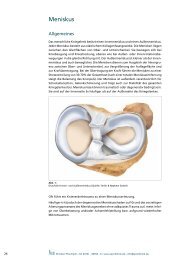
Meniscus<br />
General Information<br />
The human knee joint has an internal and external meniscus. Each meniscus consists<br />
of elastic collagenous fiber tissue. Both menisci lie between the sliding surfaces of<br />
thigh and lower leg. When flexing or stretching the knee, they move along into the<br />
same direction, just as they do with inner- and outer rotating movements. The external<br />
meniscus is smaller and more flexible than the internal meniscus. Menisci<br />
serve as balance of the incongruence between the thigh and the lower leg and as<br />
extension of the supporting surface and power transmission. When transmitting<br />
power, menisci effect a stress distribution of 30-70 % of total load (after removal<br />
of the complete meniscus stress on the cartilage increases). Furthermore, the meniscus<br />
is responsible for shock absorption and joint lubrication, and also increases<br />
stability of the entire knee joint. Meniscus ruptures can have a traumatic or degenerative<br />
reason, and they occur three times as often on the inner side as on the<br />
outer side of the knee joint.<br />
Fig. 1:<br />
Top view inner- (medial) and outer (lateral) meniscus (source: Smith & Nephew GmbH)<br />
Often, the trauma of a twisted knee results in a meniscus injury.<br />
However, in most cases it is the degenerative meniscus damage which occurs due<br />
to the early aging process of the meniscus tissue without having an adequate trauma.<br />
This is mostly the result of overload and/or axial malposition, or the result of<br />
repeated microtraumas.<br />
22<br />
Rastatter Str. 17-19 • 75179 Pforzheim • Germany • Phone 07231- 60556- 0 • www.sportklinik.de • info@sportklinik.de
Meniscus<br />
Medical Conditions<br />
The most common symptoms of meniscus damages are pain in the outer- or inner<br />
side of the knee joint, especially under stress and specific rotational movements. A<br />
“block” in the joint i.e. temporary inability to flex or stretch the knee is a specific<br />
indication for a basket handle- or lap tear. Another indication can be swelling or<br />
hyperthermia of the knee joint due to the acute irritation.<br />
Fig. 2:<br />
Complex rupture after partial meniscectomy<br />
Therapy<br />
Fig. 3:<br />
Complex rupture without any suture<br />
option<br />
Therapy of meniscus damages can, depending on the degree of severity, be carried<br />
out conservatively or surgically. When having a stable meniscus rupture which is<br />
relatively free of symptoms and stands physical stresses of everyday life, treatment<br />
can be made with combined medical-physical therapy.<br />
Operative therapy is made with a minimally invasive and arthroscopic technique.<br />
Because of known long-term consequences, therapists always try to retain as much<br />
meniscus tissue as possible with young patients. When having a basket handle or lap<br />
treat of the meniscus, in some cases even stitching up the rupture is enough. When<br />
these ruptures lie within the central area of the meniscus which is well supplied with<br />
blood, there are good chances of recovery. The chance of this kind of therapy being<br />
successful has to be decided by the experienced surgeon during surgery.<br />
Fig. 4:<br />
Bucket handle tear<br />
Fig. 5:<br />
Meniscus suture<br />
Rastatter Str. 17-19 • 75179 Pforzheim • Germany • Phone 07231- 60556- 0 • www.sportklinik.de • info@sportklinik.de 23<br />
Ellenbogen Knee
Meniscus<br />
Meniscus Suture<br />
In our ARCUS Clinics different suturing techniques are used, depending on the need.<br />
All of them are well-proven, and show few complications and good chances of recovery.<br />
In order to accelerate wound healing of the torn part of the meniscus and<br />
induce ingrowth of blood vessels, fissures are previously prepared by “needling” and<br />
“rasping” them with microsurgical instruments. When having a small fissure only<br />
or a cruciate ligament rupture at the same time, this often is completely sufficient<br />
and is seen as indirect suture technique. When having an isolated meniscus injury<br />
or a bigger fissure, however, a direct meniscus suture is necessary and carried out<br />
by stitching up the fissure.<br />
Partical Meniscectomy<br />
If it turns out that stitching up the meniscus is not possible, partial meniscectomy is<br />
being carried out. Here, as much as necessary but as little meniscal tissue as possible<br />
is being removed to keep the remaining meniscus stable and functional. Due to this<br />
partial removal of the meniscus the supporting surface becomes smaller, but (of<br />
course depending on the amount of tissue removed) this normally has no negative<br />
effect on joint functions.<br />
Aftercare<br />
After surgery, you are not allowed to drive yourself. In most cases we prescribe<br />
an anti-inflammatory medication which has to be taken regularly. Furthermore,<br />
prophylaxis of thrombosis and embolism by an abdominal injection is essential as<br />
long as walking on crutches. A drainage positioned into the knee joint normally<br />
is removed after one or two days, suture material after 10-12 days. This process is<br />
being carried out by the referring specialist or family doctor.<br />
Having had a meniscus suture, the knee should not be bent under stress for more<br />
than 90 degrees within the first 12 weeks (do not squat!). During the first 2 weeks,<br />
the only pressure the knee shall be load with is sole contact. The 3rd and 4th week,<br />
load can amount to 20 kg and afterwards the patient can start with moderate<br />
muscle training. In most cases, start of intensive sporting activities is possible after<br />
3-4 months.<br />
After partial meniscectomy it is not allowed to put full weight on the leg for about<br />
5-7 days. Moreover, as long as walking on crutches, adequate prophylaxis of thrombosis<br />
and embolism is necessary.<br />
24<br />
Rastatter Str. 17-19 • 75179 Pforzheim • Germany • Phone 07231- 60556- 0 • www.sportklinik.de • info@sportklinik.de
Meniscus<br />
Meniscus Replacement<br />
When a large portion or even the complete meniscus had to be removed with a young<br />
patient, meniscus transplantation or meniscus replacement should be discussed as<br />
a lacking meniscus may very early lead to diseases such as arthritis. The treatment<br />
can delay beginning arthrosis and its success is closely connected with existence of<br />
health cartilage tissue, intact ligaments and the physiological axis of the leg.<br />
Transplantation of a donor meniscus (“allograft”) is possible as well as implantation<br />
of artificial meniscus replacement tissue (“CMI” = collagen meniscus implant or<br />
“ACTIFIT” = polyurethane meniscus implant). Implants are operatively tailored to fit<br />
perfectly into the prepared defect. Then, the chosen implant is being sutured and<br />
has to heal for several weeks. The new tissue shall restore normal functions of the<br />
meniscus, relieve pain and even stop the degenerative process. Due to very strict<br />
indications, however, this surgery is being carried out rather rare.<br />
Fig. 6:<br />
Meniscus replacement (source: ReGen Biologics)<br />
Aftercare<br />
After meniscus replacement surgery, walking on crutches for 2-3 months is necessary<br />
to support the healing process of the donor meniscus.<br />
Rastatter Str. 17-19 • 75179 Pforzheim • Germany • Phone 07231- 60556- 0 • www.sportklinik.de • info@sportklinik.de 25<br />
Ellenbogen Knee
Anterior Cruciate Ligament (ACL)<br />
General Information<br />
Cruciate ligament injuries are often the result of acute accident- or sports injuries.<br />
When having injured the cruciate ligament, the knee joint swells up due to the hematoma.<br />
More symptoms are painful limitation of knee movability and, depending<br />
on the severity of injury, the feeling of instability on the affected leg. In this acute<br />
condition, diagnosis may be very difficult as pain, swelling and tense muscles hinder<br />
medical examination. A positive result of the pivot-shift test is seen as reliable sign<br />
for an anterior cruciate ligament rupture; a positive Lachman provides the best<br />
likelihood ratio.<br />
Besides the orthopedic examination, magnetic resonance imaging (MRI) is recommendable<br />
with new cruciate ligament injuries as a high percentage of patients also<br />
have concomitant injuries such as meniscus-, medial collateral ligament-, and cartilage<br />
damages. With the magnetic resonance imaging the entire extent of the injury can<br />
be detected. Therefore, MRI has special relevance with regard to surgery planning<br />
as well as for allocation of concomitant injuries to be operated (e.g. menisci, lateral<br />
ligaments and/or the dorsolateral capsule edge with rupture of the Popliteus tendon).<br />
anterior cruciate<br />
ligament<br />
inner (medial)<br />
meniscus<br />
Tibia (shinbone)<br />
Fig. 1:<br />
Knee joint with cruciate ligaments and menisci (source: Smith & Nephew GmbH)<br />
Difficulties with Cruciate Ligament Ruptures<br />
Femoral condyle<br />
posterior cruciate<br />
ligament<br />
outer (lateral)<br />
meniscus<br />
Fibula<br />
Our cruciate ligaments form the central stabilizing column of the knee joint<br />
(fig. 1). Their principle purpose is to prevent the knee joint against abrupt stopping-<br />
and accelerating movements as well as rotational movements. Injuries of cruciate<br />
ligaments occur in more than 90 % of all cases to the anterior cruciate ligament<br />
(ACL). The cruciate ligament rupture causes serious impact on natural movements of<br />
the joints. Although with muscular and trained athletes a cruciate ligament rupture<br />
26<br />
Rastatter Str. 17-19 • 75179 Pforzheim • Germany • Phone 07231- 60556- 0 • www.sportklinik.de • info@sportklinik.de
Anterior Cruciate Ligament (ACL)<br />
can be compensated in the beginning with conservative therapy, damage of further<br />
structures and with this a considerably higher risk of arthrosis has to be expected.<br />
After having had a cruciate ligament rupture, most patients focus on regaining their<br />
condition first. Need for surgery depends on activity, symptoms of instability and age,<br />
and especially the athletic patient benefits here from prompt operative treatment.<br />
Conservative treatment, however, is also completely justified with low instability<br />
symptoms and low physical activity. With cruciate ligament injuries in childhood and<br />
adolescence, operative reconstruction by the use of appropriate techniques should<br />
be considered to prevent serious consequential injuries such as damages of secondary<br />
joint cartilages or menisci. We have just published comprehensive experiences and<br />
numerous studies regarding this issue.<br />
Fig. 2:<br />
Arthroscopic image of a fresh ACL-rupture<br />
Current Surgical Techniques<br />
Thanks to the enormous development of arthroscopic surgical techniques, treatment<br />
options for cruciate ligament replacements have improved considerably over recent<br />
years. Shorter operation times and a reduced surgical trauma, less pain and better<br />
cosmetic results speak for today’s minimally invasive operation methods. Correct<br />
surgical treatment, however, needs maximum experience (fig. 2+3) and therefore<br />
should be carried out in specialized centers. In the ARCUS Clinics in Pforzheim more<br />
than 1200 arthroscopic cruciate ligament surgeries are carried out every year. Arthroscopic<br />
cruciate ligament replacement using autologous tendon transplants has<br />
reached standard level by now. Used are hamstring tendon transplants (semitendinosus-<br />
and gracilis tendon) in triple- and quadruple binding technique as well as<br />
patellar tendon strips, quadriceps tendons and after multiple ruptures also donor<br />
grafts. Common characteristics of all these transplants are their tear resistance and<br />
flexibility which are similar to the anterior cruciate ligament. But they differ regarding<br />
the removal technique and their anchoring possibilities.<br />
Fig. 3:<br />
Cruciate ligament reconstruction of semitendinosus tendon graft<br />
Rastatter Str. 17-19 • 75179 Pforzheim • Germany • Phone 07231- 60556- 0 • www.sportklinik.de • info@sportklinik.de 27<br />
Ellenbogen Knee
Fig. 4:<br />
Quadrupled harmstring tendon<br />
graft reinforced by Endobuttons ®<br />
or Retrobuttons ® .<br />
(source: Arthrex GmbH)<br />
Fig. 5:<br />
Patellar tendon graft as ACL/<br />
PCL reconstruction (source:<br />
Arthrex GmbH)<br />
Fig. 6:<br />
Double-bundle ACL reconstruction<br />
(schematic image)<br />
Anterior Cruciate Ligament (ACL)<br />
Hamstring Grafts (hamstring tendons: semitendinosus- and gracilis<br />
tendon)<br />
Through a small incision at the inner shinbone head, the semitendinosus- and gracilis<br />
tendon are being removed and then doubled to create a quadruple-transplant<br />
(fig.4). Alternatively, when having a sufficiently long semitendinosus tendon, there<br />
is also the possibility to remove the semitendinosus tendon only and tie it together<br />
to a triple- respectively quadruple bundle.<br />
Advantages of the usage of hamstring tendons are fewer problems with removal,<br />
less pain, and cosmetically more favorable scars. Another essential advantage of<br />
this method is the hamstring graft gaining almost the natural elasticity of a cruciate<br />
ligament during the healing process. Relevant dysfunctions due to the removal of<br />
the hamstring do not occur.<br />
Partellar Tendon (tendon below knee cap)<br />
As cruciate ligament replacement, the middle third of the tendon is being removed<br />
as “bone-tendon-bone” graft (fig. 5). Advantage of this method is stable fixation<br />
and fast bone ingrowth of the transplant.<br />
Disadvantageous however is pain which may occur at the donor site and a possible<br />
reduction of muscle power of the thigh extensor muscle. Statistics show that the socalled<br />
“anterior knee pain” occurs more often after having had an anterior cruciate<br />
ligament reconstruction with patellar tendon than with hamstring graft.<br />
„Double-Bundle“ Reconstruction<br />
Some teams favor currently a new procedure using hamstring tendons in doublebundle<br />
constructions. With this technique, replacement of the ACL is made according<br />
to its anatomic structure with a doubled transplant string of anteromedial and<br />
posterolateral fiber bundles (fig. 6). The higher biomechanical efficiency gained by<br />
this double-bundle reconstruction technique however has so far only been proven<br />
by experimental simulations. Furthermore, it needs more complex surgery- and<br />
anchoring techniques which long-term efficiency regarding optimized knee stabilization<br />
has not been shown yet. Within the scope of controlled studies, this method<br />
is also being used by us.<br />
Quadriceps Tendon (tendon of thigh extensor)<br />
The quadriceps tendon graft with small patellar bone block is mainly used in revision<br />
surgery (re-rupture of cruciate ligament). Although it shows biomechanical characteristics<br />
comparable to the natural cruciate ligament, removal of the transplant is<br />
very complex and time-consuming, and therefore did not gain general acceptance<br />
as first line therapy. Advantage of this method is the possibility of implant-free<br />
press-fit anchoring of the quadriceps tendon graft into the thigh bone, whereby<br />
biologically optimal healing and simplified surgery in case of revision treatment is<br />
ensured. Disadvantages are the demanding surgical procedure for removal of the<br />
tendon and weakening of the thigh extension functions.<br />
28<br />
Rastatter Str. 17-19 • 75179 Pforzheim • Germany • Phone 07231- 60556- 0 • www.sportklinik.de • info@sportklinik.de
Anterior Cruciate Ligament (ACL)<br />
Donor Tendons<br />
Donor tendons (allografts) are mainly used in America. Advantage of this method<br />
is the fact that removal of suitable reconstruction material is no longer required.<br />
Disadvantageous however are possible immune responses and the higher failure<br />
rate. Usage of donor tendons is being considered as alternative treatment especially<br />
with secondary- or third operations when there is lack of the patient’s own transplant<br />
possibilities. Since 1993, the ARCUS Clinics are regarded the most experienced<br />
specialized surgery unit in Germany using donor tendons for cruciate ligament<br />
reconstruction.<br />
Fixation of Cruciate Ligament Grafts<br />
Common aim of all reconstruction techniques is primary stable graft anchorage.<br />
For this purpose, there are many different fixation materials such as metallic or<br />
bioabsorbable interference screws, staples, pins or fixation buttons available (fig.<br />
7, 8a, 8b). For all systems used at present, an initial retention force which meets<br />
post-operative stabilization demands has been certified. In the end, however,<br />
anchorage of the implant until complete healing remains the real weak point of<br />
cruciate ligament plastics.<br />
Fig. 7:<br />
Fixation of ACL replacement:<br />
Transfix ® and bioabsorbable screw<br />
(source: Arthrex GmbH)<br />
Time of Cruciate Ligament Reconstruction<br />
Fig. 8a:<br />
Fixation of ACL replacement: Endobutton ® or<br />
Retrobutton ® (source: Arthrex GmbH)<br />
When having a new rupture, treatment in the sense of first line therapy can be done<br />
within the first 24 to 48 hours. This option is possible for example when treating an<br />
osseous rupture of the cruciate ligament or other concomitant injuries that need<br />
immediate medical care (e.g. meniscus ruptures that can be stitched up or complex<br />
knee instabilities with rupture of medial- or lateral collateral ligament). In normal<br />
cases, surgery is planned after 4-6 weeks when the inflammation has subsided.<br />
During this inflamed phase, operative treatment is not recommended due to the<br />
proven increased complication rate in the sense of post-operative movement disor-<br />
Fig. 8b:<br />
Fixation material: bioabsorbable<br />
screw and Endobutton ®<br />
(source: Smith & Nephew GmbH)<br />
Rastatter Str. 17-19 • 75179 Pforzheim • Germany • Phone 07231- 60556- 0 • www.sportklinik.de • info@sportklinik.de 29<br />
Ellenbogen Knee
Anterior Cruciate Ligament (ACL)<br />
ders. Reduction of this “6-week-period” is possible and supportable when the joint<br />
becomes irritation-free before.<br />
Until the date selected for surgery, the joint is being treated with functional conservative<br />
methods, where the focus lies on how to reduce the swelling and regain<br />
functional mobility. Furthermore, preoperative usage of stabilizing knee orthoses<br />
is indicated for strong instability symptoms and concomitant lesions of the medial<br />
collateral ligament.<br />
Fig. 9:<br />
Donjoy ® knee brace (source: Ormed.DJO)<br />
Aftercare<br />
Rehabilitation after cruciate ligament reconstruction surgery is an important component<br />
of our therapy concept. On the one hand, treatment concentrates on regaining<br />
the full range of physiological mobility, full muscular control and coordination, and<br />
returning to full activity. On the other hand, current methods of Aftercare are adapted<br />
to scientifically proven phases of healing. At present, the accelerated rehabilitation<br />
program propagated in the 90ies has given way to adapted and more restrictive<br />
postoperative therapy planning which considers individual tissue reactions and the<br />
healing process. Today, postoperative care with knee orthoses stabilizing the knee<br />
joint is considered standard. With optimal rehabilitation, stable reconstruction of<br />
knee joint function and –stability can be expected after 6-9 months.<br />
ARCUS rehabilitation program for cruciate ligament reconstruction:<br />
Stationary phase (2-3 days):<br />
Ice-pack and lymph drainage. Start with physiotherapy in the pain free area as well<br />
as “walking school” on elbow crutches. Further measures are muscle stimulation,<br />
lymph drainage and thrombosis prophylaxis. Removal of redon-drainage the 2nd<br />
day after surgery.<br />
30<br />
Rastatter Str. 17-19 • 75179 Pforzheim • Germany • Phone 07231- 60556- 0 • www.sportklinik.de • info@sportklinik.de
Anterior Cruciate Ligament (ACL)<br />
Post-stationary phase:<br />
Therapy to reduce swelling, physiotherapy. Primarily work on active stretching,<br />
quadriceps isometry, self training, physical exercises and dynamic splint: 1st week<br />
60° of knee reflextion, 2nd - 4th week 90 °. Afterwards approval of physical mobility.<br />
Increase weight slowly: in first week, only “heel-to-toe” movement of the foot with<br />
elbow crutches and with a load of no more than 5 kg is permitted, 2.-3. week about<br />
20 kg, then full body weight can put onto affected leg depending on muscular<br />
control and toning.<br />
Coordination- and proprioceptive training (balance board, posturomed, areostep,<br />
aqua jogging). Ergometer. Squat- and leg press training possible (in closed system).<br />
Please avoid forced stretching against resistance in order to treat the donor site<br />
with care.<br />
Sporting activities:<br />
• cycling, walking approx. 6 weeks after surgery<br />
• jogging approx. 3 months after surgery<br />
• contact sports such as football, handball, skiing, tennis approx 6-9 months after<br />
surgery<br />
Medial- or Lateral Collateral Ligament Injuries<br />
Injuries of the medial collateral ligament can thanks to their tendency to spontaneous<br />
healing often be treated conservatively. An exception is a complete rupture<br />
of the medial capsular ligament complex with involvement of posterior transverse<br />
ligament and dorsomedial capsule. Here, indication for surgery is suture of ruptured<br />
ligament structures. Injuries on the outside of the knee joint generally are not<br />
being seen as favorable spontaneous prognosis. In these cases immediate surgical<br />
reconstruction is needed.<br />
Posterior Ligament Rupture<br />
Injuries of the posterior cruciate ligament are mostly the result of a violent weight<br />
shift of the lower leg backwards compared to the thigh; for example through direct<br />
impact from the front onto the shinbone head. With immediate correct diagnosis,<br />
the posterior cruciate ligament injury shows a good spontaneous healing tendency.<br />
It requires consequent wearing of a special PTS® splint (fig. 10) which permanently<br />
supports and pushes the lower leg to the frontside. Should the “dorsal drawer<br />
test” however, remain positive even after several weeks of conservative treatment,<br />
surgery is unavoidable.<br />
Current Surgical Techniques<br />
Surgical therapy of the posterior cruciate ligament rupture is carried out – as the<br />
ACL rupture – on fully endoscopic basis (fig. 11), whereby the patient’s own tendon<br />
grafts are used for ligament replacement.<br />
Fig. 10:<br />
Knee positioning splint for<br />
PCL-rupture<br />
(source: medi GmbH & Co. KG)<br />
Fig. 11:<br />
PCL replacement, schematic image<br />
(source: Arthrex GmbH)<br />
Rastatter Str. 17-19 • 75179 Pforzheim • Germany • Phone 07231- 60556- 0 • www.sportklinik.de • info@sportklinik.de 31<br />
Ellenbogen Knee
32<br />
Ärzte-Serviceline 0180 2 95 95 95<br />
(6 Cent pro Gespräch aus dem deutschen Festnetz. Aus Mobilfunknetzen sind abweichende Preise möglich.)<br />
Die effizienteste Form<br />
der Quadrizeps-Stimulation *<br />
Mit der patentierten multipath Technologie<br />
Die Rehabilitation nach Knie- und Hüftoperationen ist oft<br />
mühsam und langwierig. Mit Kneehab XP kommen Ihre<br />
Patienten schneller wieder auf die Beine. Kneehab XP<br />
verfügt über die einzigartige, patentierte multipath<br />
Technologie, die deutlich mehr Muskelfasern stimuliert<br />
und stärkere Muskelkontraktionen auslöst. Das ist gut für<br />
den Quadrizeps und gut für Ihre Patien ten – denn es be-<br />
schleunigt die Heilung um bis zu sieben Tage*.<br />
* Dr. H. H. Pässler, Sven Feil (M. A.): Die Effektivität des kneehab.<br />
ATOS-Klinikzentrum Heidelberg 2006<br />
www.neurotechgroup.com<br />
AZ_KneehabXP_185x280.indd 1 07.01.2009 12:48:50 Uhr
Knee-Cap (Patella)<br />
General Information<br />
The knee-cap (patella) is a free running “supporting bone” for the extensor tendon<br />
of the thigh. It does not have any firm osseous joint guidance, but is only attached to<br />
muscles, tendons and ligaments. It glides in a V-shaped groove of the femur (femoral<br />
trochlea or sliding bearing). When having a congenital malformation of the trochlea<br />
or in reaction to changes in muscle balance (e.g after surgery), it is susceptible to<br />
problems and injuries. The patient mostly suffers from „anterior knee pain“. The<br />
most common diseases are the plica symptom (pain), habitual- or traumatic patella<br />
luxation, and cartilage-bone damages at patella and its sliding bearing.<br />
Femur sliding bearing<br />
Fig. 1:<br />
Patella / Femur sliding bearing<br />
Plica Syndrome<br />
Patella<br />
Here, enlarged synovial folds and thickened synovial membranes might, due to<br />
repeated impactions, cause pain or even changes to the free movement of the<br />
patella. This could result in uneven- or excessive loading and with this in damages<br />
of the cartilage of the knee-cap. If conservative therapy is not sufficient, the plica<br />
may be removed arthoscopically.<br />
Rastatter Str. 17-19 • 75179 Pforzheim • Germany • Phone 07231- 60556- 0 • www.sportklinik.de • info@sportklinik.de<br />
Ellenbogen Knee<br />
33
Femur<br />
Patella<br />
Fig. 2:<br />
Outer capsule: patella lateralization<br />
Knee-Cap (Patella)<br />
Habitual- or Traumatic Patella Luxation<br />
A distinction is made between congenital disorder and acute injuries when having<br />
had an accident. The habitual patella (sub) luxation occurs congenitally and instability<br />
of knee-cap is due to shallow tracks or weak ligaments and muscles to hold<br />
the kee-cap and knee joint capsule. With the traumatic patella luxation instability<br />
is usually the result of an accident (luxation towards the outside).<br />
Conservative Therapy<br />
Depending on severity of the knee deviation, a conservative treatment approach<br />
can be carried out first. Exercises shall train the vastus medialis muscle regarding leg<br />
extension. Important is cooperation of the patient as treatment can only be successful<br />
when exercises are consequently being carried out for at least 3-6 months. Longer<br />
periods of immobilization and leg rest, however, should be avoided in any case.<br />
Surgical Treatment of Habitual Patella Luxation<br />
In case that conservative treatment alone is not enough, operative measures have to<br />
be considered. Depending on cause and detected damages correcting surgery may<br />
be necessary. Lateral release (fig. 2+3) and/or medial tightening (fig. 4) are treatment<br />
options. Another option for treatment of cartilage damages of the knee-cap or<br />
osseous knee-cap luxation may be to transfer piece of the lower leg bone inwards.<br />
Here, the attachment of the patellar ligament at the tibia is detached from the<br />
bone, and reattached with screws about 1-2 cm further inside. Should the damage<br />
be caused by thigh problems, surgical correction of the hip joint may be necessary.<br />
Depending on the case, it makes sense to carry out supplemental cartilage therapies<br />
and/or a combination of the treatment methods described above.<br />
Patella<br />
capsule cut<br />
Fig. 3:<br />
Outer capsule after lateral release<br />
knee arthroscopy<br />
(right)<br />
Fig. 4:<br />
Inner capsule: medial tightening<br />
34<br />
Rastatter Str. 17-19 • 75179 Pforzheim • Germany • Phone 07231- 60556- 0 • www.sportklinik.de • info@sportklinik.de
Knee-Cap (Patella)<br />
Surgical Treatment of Traumatic Patella Luxation<br />
In some cases, when only the joint capsule has been torn by the traumatic patella<br />
luxation and caused a haematoma within the knee, an arthroscopic knee washout<br />
can be sufficient to prepare the knee for conservative treatment. Operative methods<br />
are needed for cracked cartilages or torn medial patellofemoral ligaments (MPFL).<br />
Most often, the cracked-off cartilage-bone fragment can be reattached through<br />
a small insicion, using bioabsorbable anchors. A suture of the cartilage capsule<br />
can also be treated with this arthroscopic method. Replacement of a torn MPFL is<br />
biomechanically necessary for restoring the patella function and carried out with<br />
the patient’s own tendon material from the inner side of the thigh. Similar to ACL<br />
reconstruction, the method of choice is minimally-invasive surgery.<br />
Aftercare<br />
During aftercare the patient can put full weight onto the straight leg after 2-3 weeks,<br />
squatting or climbing stairs is possible after 5-6 weeks. At this time, an intensive<br />
muscle training shall be started to strengthen the especially quick weakening vastus<br />
medialis muscle.<br />
Spontaneous Cartilage-Bone Lesions (osteochondrosis<br />
dissecans)<br />
There are cases where the area around the patella and its sliding bearing is not sufficiently<br />
supplied with blood and begins to die. At an advanced stage, the cartilage<br />
lying above is also destroyed.<br />
Therapy<br />
Initial treatment depends on the stage and is conservative in most cases. Rest, no<br />
sporting activities and anti-inflammatory medication may be necessary for pain relief.<br />
If X-ray or MRI examinations show progress of the disease, small holes should be<br />
drilled surgically into the center of inflammation to stimulate vascularization and<br />
healing. This is called antegrade- or retrograde drilling. In some cases, dead tissue<br />
has to be removed before it comes loose and becomes a “joint mouse”. This would<br />
cause further cartilage damage to still healthy sections of the joint. Afterwards, the<br />
bone lying below is also surgically drilled in order to stimulate vascularization and<br />
regeneration of cartilage tissue. In recent years, we have increasingly become able<br />
to successfully treat such disorders with bone-cartilage transplants (mosaic plastics,<br />
see chapter arthrosis).<br />
Rastatter Str. 17-19 • 75179 Pforzheim • Germany • Phone 07231- 60556- 0 • www.sportklinik.de • info@sportklinik.de<br />
Ellenbogen Knee<br />
35
Fig. 1:<br />
Knee joint damaged by arthrosis<br />
(source: medi GmbH & Co. KG)<br />
grade I<br />
grade II<br />
grade III<br />
grade IV<br />
Fig. 2:<br />
Schemed image of arthrosis<br />
grades of severity<br />
(source: medi GmbH & Co. KG)<br />
Arthrosis<br />
How does a normal joint actually work?<br />
Generally spoken, a joint movably connects the ends of two bones. To avoid these<br />
rough bones rubbing against each other directly, these contact surfaces are covered<br />
with an approx. 3-4 cm thick layer of cartilage (fig. 1). This layer is extremely<br />
smooth, reduces friction within the joint (lower than two smoth ice surfaces against<br />
each other) and elastically absorbs shocks when walking. These special mechanical<br />
characteristics are maintained by complicated biochemical, molecular and electrophysiological<br />
connections and require an intact closed surface and a stable collagen<br />
fiber network. This complex “composite material” is produced and controlled by<br />
cartilage cells (chondrocytes). Disorders can be of mechanical kind (sudden physical<br />
force such as the impact experienced in a car accident, high grade sprains, chronic<br />
overweight, varus/valgus malalignment, cruciate ligament instabilities, lacking menisci)<br />
or of biomechanical kind (metabolic diseases, rheumatism, gout, calcification,<br />
circulatory disorder). Several facts are here ensured: so does reasonable endurance<br />
sport, marathon runners included, not increase danger of arthrosis, whereas the<br />
varus/valgus alignment especially in combination with meniscus damage, radical<br />
meniscus surgery and/or overweight poses a significant risk of arthritis.<br />
Cartilage damage is divided into four different levels of severity:<br />
1. stage: slight superficial fibrillation<br />
2. stage: deeper tear and large surface fibrillations<br />
3. stage: deeper defect (to the bone) with strong fibrillation, mechanically not<br />
acceptable<br />
4. stage: exposed bone<br />
Traumatogenic Cartilage Damages<br />
When twisting one’s knee or incurring a contusion as a result of an accident (skiing,<br />
playing football etc.) a piece of cartilage (diameter approx. 1-2 cm) may crack off<br />
the complete cartilage layer. Surrounding edges are intact and of normal height, the<br />
bone below is unaffected and shows good regenerative ability. This kind of damages<br />
responds well to all treatment methods mentioned in the following.<br />
Degenerative Wear<br />
Less positive are prospects for cartilage damages developed during one’s lifetime<br />
by monotonous stress alone or in conjunction with varus- or valgus deformity, gout,<br />
rheumatism, or damages to menisci or cruciate ligaments. These damages soften the<br />
cartilage (1. stage) and later result in fibrillation of the complete layer (2. stage).<br />
In stage 2-3, the cartilage layer is only half as thick as normal and extremely frayed<br />
and there may occur detached or loose fragments of cartilage.<br />
This stage can no longer be repaired by the body itself without outside help. Specialists<br />
already talk of severe cartilage damage even though pain may still be bearable<br />
for the patient and thus is not perceived as warning signal. Especially this early stage<br />
of progressive cartilage degeneration though offers good prospects of successful<br />
cartilage surgery. Unfortunately, many patients wait too long.<br />
36<br />
Rastatter Str. 17-19 • 75179 Pforzheim • Germany • Phone 07231- 60556- 0 • www.sportklinik.de • info@sportklinik.de
Arthrosis<br />
In the final stage (4. stage) the cartilage tissue is completely destroyed, the „tyre<br />
tread“ completely gone. Bone grinds on bone, grooves develop and osseous overhangs<br />
make stretching the leg increasingly difficult. Varus- and valgus deformities<br />
increase. Even modern surgical measures for cartilage reconstruction are only limitedly<br />
successful in this stage. Real hyaline cartilage cannot regenerate. The only thing an<br />
orthopaedic surgeon can do is facilitating growth of replacement- and fiber cartilage<br />
and help improving its quality and strenght, or carry out time-consuming cell<br />
culture and transplantations which, however, are still quite limited in their range<br />
of application.<br />
Basically it can be said that once cartilage damage has begun, the wearing process<br />
will continue with increasing speed, and without early therapeutic intervention,<br />
freedom from pain can only be achieved by implanting artificial joint prostheses.<br />
Treatment of Arthrosis<br />
Method of treatment depends on cause and severity of the disease. With instruments<br />
for arthroscopic surgery we are not only able to see the cartilage damage and record<br />
it for later documentation; we can also very gently treat the damage by means of<br />
these micro-instruments at the same time.<br />
Debridement<br />
Frayed edges are removed with a mini cutter and the surface is smoothed. Instable<br />
cartilage parts are removed to prevent further fraying.<br />
At the same time meniscus damages are being repaired whereby, in an early stage,<br />
suturing the meniscus should be the preferred treatment method. In some cases, it<br />
makes sense to remove part of the synovial membrane to reduce risk of contusions<br />
within the joint.<br />
Methods of Cartilage Regeneration (stem cell<br />
techniques)<br />
They base on “migration” of bone marrow stem cells into damaged cartilage areas<br />
where they develop into replacement cartilages.<br />
Thus, having a case of half-thickness cartilage defect with furthermore severe fraying,<br />
there is still a possibility to stimulate the body’s own cartilage repair. For the first<br />
3-4 years, this replacement cartilage is relatively rich in cells and does not consist of<br />
as many cartilage cells (chondrocytes) which produce synovial fluid. Furthermore, it<br />
can not stand as much mechanical load as original hyaline-cartilage and therefore<br />
often causes knee irritations and knee pain. However, this replacement cartilage<br />
(fibrocartilage) is still better than a completely exposed bone. Just compare this<br />
damage for example with a burn injury: the skin is wrinkled, less elastic, sensitive<br />
to injury, does not get a tan and is hairless - but it is far better than having a permanently<br />
open wound.<br />
There are clear indications that in most cases (unfortunately not always) replacement<br />
cartilage develops into better load-resistant hyaline-cartilage after several years.<br />
Rastatter Str. 17-19 • 75179 Pforzheim • Germany • Phone 07231- 60556- 0 • www.sportklinik.de • info@sportklinik.de<br />
Ellenbogen Knee<br />
37
Arthrosis<br />
“Microfracture Technique” (according to Steadman)<br />
Operative beginnings of surgical refreshing of cartilage date back to the 50’s (Pridie<br />
drillings). Here, several holes of about 2 mm are sieve-like drilled into the exposed<br />
surface of the bone. With this, small “islands of regeneration” are created, but only<br />
in few cases a continuing cartilaginous scar tissue. Nowadays, we prefer the less<br />
traumatizing “microfracture technique” according to Steadman which has been<br />
developed in the early 90s (fig. 3+4).<br />
The bone surface is pierced with a fine awl to create hairline cracks and tiny holes,<br />
resulting in a stronger cartilaginous scar tissue to cover the entire affected area.<br />
Just imagine grass seeds on trodden down and dry soil: without previously breaking<br />
up the ground, the seed would have no chance to take root. After sowing the seeds,<br />
it is not allowed to walk on the lawn for some time to protect it. The same applies<br />
to a joint: piercing the bone loosens the bony surface and enables bone marrow<br />
stem cells to seep out and potential blood stem cells to settle. And to protect this<br />
sensitive area, it is necessary to walk on crutches at the beginning.<br />
Fig. 3:<br />
Grade IV cartilage damage at the knee,<br />
treatment with micro fracture<br />
Fig. 4:<br />
Cartilage repair 1 year after micro fracture<br />
Abrasion Arthroplasty (according to L. L. Johnson)<br />
If parts of the bone are already exposed (4. stage), one treatment possibility is to<br />
debride and smooth the remaining bone and wait for improvement. But there is also<br />
a chance of helping the body filling bald areas with new cartilage-like scar tissue<br />
again – and results can be as good as after treatment with microfracture technique.<br />
We just refresh the exposed, extremely hardened surface of the bone with small<br />
cutters, as developed by L. Jonson in the early 80’s.<br />
38<br />
Rastatter Str. 17-19 • 75179 Pforzheim • Germany • Phone 07231- 60556- 0 • www.sportklinik.de • info@sportklinik.de
Arthrosis<br />
Different Methods of Cartilage Transplantation<br />
Cartilage-Bone Transplantation (OATS and mosaic plastics)<br />
Small cartilage-bone cylinder-shaped pieces are removed from knee areas with lower<br />
physical load and fit into prepared holes in the defective area. Advantage: this<br />
method creates immediately functioning hyaline cartilage for the defective area<br />
and healing is very fast thanks to the “press-fitted” bony cylinders. Furthermore,<br />
expenses are rather limited. However, this method is technically demanding and<br />
requires a high degree of surgical expertise and experience, especially when being<br />
carried out arthroscopically.<br />
Fig. 5:<br />
Mosaic plastics at femoral condyle of knee joint<br />
Fig. 6:<br />
Mosaic plastics at knee joint<br />
Thus, although seen as routine surgery on knee joint and ankle, it is still not recommendable<br />
for shoulder and hip.<br />
Specific demands of ankle surgery: the typical cartilage-bone defect is situated<br />
behind the inner ankle, inaccessible from the front. Thus, the inner ankle has to be<br />
detached first to be able to press the donor-cylinder extracted from the knee (the<br />
ankle joint does not have enough cartilage tissue to create a transplant) into the<br />
defect of the talus, and then screwed back in afterwards.<br />
Problems finding donor sites arise in about 10% of all cases when 1-2 donor cylinders<br />
shall be extracted. Therefore, the amount of donor cylinders is limited. There<br />
exist artificial, absorbable plugs with cartilage-like characteristics (Trufit®, fig. 7),<br />
which have proven to be very successful in filling up these donor-holes. In some<br />
cases, smaller defects (also at ankle joint) can be treated with such absorbable plugs<br />
immediately. After 1-2 years these plugs are replaced by the body’s own bone-,<br />
cartilage- and connective tissue cells.<br />
Cartilage Cell Cultivation = Autologous Chondrocyte Transplantation<br />
ACT<br />
This method caused quite a stir in the media in the mid 1990’s. In a first operation,<br />
several cartilage particles are removed from the knee joint, propagated in a complex<br />
cell culture and finally implanted in the defective area in a second surgical procedure.<br />
The new cells have to grow and propagate further cells for a new cartilaginous structure<br />
– a very complex process that requires strict adherence to Aftercare guidelines<br />
given; that may include walking on crutches for 8-10 weeks. During this time, enough<br />
exercise with a continuous passive motion device (4-6 weeks, 4-6 hours each day)<br />
Rastatter Str. 17-19 • 75179 Pforzheim • Germany • Phone 07231- 60556- 0 • www.sportklinik.de • info@sportklinik.de<br />
Fig. 7:<br />
Scheme of resorbable Trufit ® dowel<br />
Ellenbogen Knee<br />
39
Arthrosis<br />
as after microfracture treatment is necessary, respectively strongly recommended<br />
by us. With this exercise, formation of a good and stable new cartilage surface is<br />
essentially supported (see below).<br />
In some cases, after verification of the diagnosis and indication, approval of cost<br />
transfer has to be obtained from the health insurance company first before cell<br />
removal and transplantation afterwards can be carried out. Thus, bureaucracy sometimes<br />
makes 3 surgical steps necessary.<br />
And costs are high; the cultivation of new cells in a laboratory alone costs 4.500.00<br />
– 8.000.00 Euros – and is covered from public health insurance companies only for<br />
part of the patients. Furthermore, operation technique is very demanding and may<br />
require a second surgical procedure, let alone the arthroscopically controlled check<br />
after 1-2 years.<br />
All these methods can help rebuilding the attacked or worn cartilage layer of the<br />
joint. However, this can only be successful if any possible causal disturbance factor<br />
has been eliminated:<br />
• Meniscal lesions have to be smoothed or even better sutured. Meniscus transplantations<br />
are so far not available for wider clinical usage.<br />
• Ligament instabilities have to be eliminated, especially the anterior cruciate<br />
ligament (ACL) has to be sufficiently stable.<br />
• Leg axes need to be straight (see chapter varus/valgus malalignment).<br />
• Overweight shall consequently be reduced until a bodymass index of less than 25<br />
is reached. This is done by reducing food intake and starting sporting activities<br />
(at the beginning in the water).<br />
Aftercare for joint-preserving Arthrosis Treatment<br />
See chapter varus/valgus malalignment page 46.<br />
40<br />
Rastatter Str. 17-19 • 75179 Pforzheim • Germany • Phone 07231- 60556- 0 • www.sportklinik.de • info@sportklinik.de
Arthrosis<br />
Results<br />
Until today, there unfortunately has not been found any general patent remedy<br />
for the advanced stage of the degenerative joint disease – except for artificial joint<br />
reconstruction which should be delayed as long as possible. With treatment by continuous<br />
passive exercise the Canadian R. Salter achieved excellent results already in<br />
1984 (in an animal experiment with a 6 weeks continuous training even in cases of<br />
severe joint damage). Of course, on the one hand, a human patient cannot be tied<br />
to a motion device for 6 weeks, and on the other hand many diseases heal much<br />
faster and better with animals than with humans. However, we think that this aftercare<br />
concept is a revolutionary method for the future.<br />
Our experiences with abrasion arthroplasty for arthrosis treatment – whether with<br />
or without continuous passive motion - date back to its beginnings in Germany<br />
in 1984. In own tests carried out during aftercare we can, similar to the American<br />
studies, achieve good and satisfying results in about 60-70%. Please consider that<br />
without any treatment constant worsening of joint functions has to be expected.<br />
Supportive Medication Therapy<br />
Injections with hyaluronic acid have shown to be successful in improving joint lubrication,<br />
and we at the ARCUS Clinisc are very proud to have actively contributet to<br />
the launch of hyaluronic acid in Germany in the early 90’s. A step that has proven<br />
to be successful in the long term. In extensive studies, hyaluronic acid has mainly<br />
been proven positive. Generally, a series of 3 to 5 injections is recommended (at the<br />
price of approx. 230 euros each/status 2009). Unfortunately, neither public health<br />
insurance companies nor the employers’ liability insurance association are willing<br />
to pay for this treatment so far; the patient is yet again self-payer.<br />
In order to support cartilage repair, we recommend an additional long-term therapy<br />
with cartilage builders glucosamine and chondroitin (e.g. 3x1 capsule ARTROSTAR ®).<br />
This combination of approx. 1500 mg glucosamine and approx. 1200 mg chondroitin<br />
sulfate per day is commonly regarded as supportive cartilage therapy. International<br />
studies and in the meantime working groups in Germany as well are proving the<br />
anti-inflammatory- and cartilage supporting characteristics of these substances<br />
which furthermore are free of unpleasant gastrointestinal side effects such as the<br />
for cartilage unfavorable NSAR. A capsule of ARTROSTAR ® contains 500 mg glucosamine<br />
HCL and 400 mg chondroitin sulfate. Unfortunately, public health insurance<br />
companies do not pay for this treatment. Positive effects of homeopathic medicines<br />
such as Zeel® or Ney Arthros® are much less proven and only a few treatments are<br />
normally not suitable as therapy approach.<br />
Rastatter Str. 17-19 • 75179 Pforzheim • Germany • Phone 07231- 60556- 0 • www.sportklinik.de • info@sportklinik.de<br />
Ellenbogen Knee<br />
41
Arthrosis<br />
Exercise Therapy<br />
The best thing you can do for your joint is to have plenty of exercise while avoiding<br />
overexertion, e.g. by loosing weight, wearing well-padded shoes in the initial phase,<br />
avoiding long walks/runs on hard surfaces such as asphalt etc. Train your muscles<br />
with slowly increasing endurance sports. Suitable are “soft straightforward sports”<br />
such as cycling, walking, Nordic walking and swimming.<br />
New Physiotherapy Options:<br />
• Aqua jogging which enables intensive circulatory- and muscular training without<br />
overloading the affected knee.<br />
• Reflective muscle training with the whole body vibrator type Galileo. The patient<br />
stands on a plate vibrating with about 40 Hz what is automatically balanced by<br />
the muscles. This has already been proven to be an effective training method for<br />
muscles and bones. We are currently investigating positive effects on cartilage<br />
regeneration after the surgical procedures mentioned above.<br />
Tips for Further Improvement of Treatment Success:<br />
• Stick to the period of no or restricted weight-bearing given by us.<br />
• Move your joint as intensively as possible without overloading it.<br />
• Make use of the advantages of hyaluronic acid injections for the affected joint:<br />
start first series 3 weeks after surgery; repeat after 6-12 months.<br />
• Work consequently on weight reduction. Even a few kilograms less add up with<br />
2-3 million steps per year as a knee-, ankle- or hip joint is loaded with the 2 to<br />
5 fold of body weight; depending on height of step.<br />
• Drink enough water (instead of coffee or soft drinks) so that body and cartilage<br />
do not become dehydrated and with this brittle and prone to injuries.<br />
• Train your joints according to a varied program:<br />
- In the initial build-up phase, about 2-6 months after surgery soft, we recommend<br />
guided movements without transmitting the load of body weight such<br />
as cycling or water training. Then slowly start with walking / Nordic walking<br />
e.g. also combined with special training shoes (MBT-shoes).<br />
- Later you can add a mixture of cross-training, running (at the beginning on<br />
hard ground, then cross-country), as well as total body exercise in the gym.<br />
• Accept that not every joint affected by arthrosis can regain its former sporting<br />
ability.<br />
• Keep to the follow-up appointments stipulated.<br />
• Make use of our offer for an arthroscopic check about 1 year after surgery.<br />
• Please consider that without any treatment constant worsening of joint functions<br />
has to be expected.<br />
42<br />
Rastatter Str. 17-19 • 75179 Pforzheim • Germany • Phone 07231- 60556- 0 • www.sportklinik.de • info@sportklinik.de
Arthrosis<br />
Rastatter Str. 17-19 • 75179 Pforzheim • Germany • Phone 07231- 60556- 0 • www.sportklinik.de • info@sportklinik.de 43<br />
Ellenbogen Knee
Fig. 2:<br />
Minimally invasive Achilles tendon<br />
suture<br />
Fig. 3:<br />
Injection of body-own growth factors<br />
(PRP)<br />
Ortho-Biologie<br />
Modern Therapy Methods in Orthopaedics / Trauma<br />
Surgery<br />
Tissue Regeneration Principles<br />
Different types of human tissue need different time periods for regeneration. An<br />
injury of the oral mucosa for example heals completely within a few days, muscle<br />
injuries need 3-4 weeks, bones usually 6-12 weeks.<br />
It has been known for some time now, that any information needed for the formation<br />
of new tissue cells lies in these cells and in-between areas as well as to a large<br />
extent within the platelets.<br />
Fig. 1:<br />
Development of stem cell into bone cell. Besides correct surrounding conditions (type of surrounding<br />
tissue, pressure, exercise, rest, chemical composition of surrounding etc.) availability of several different<br />
growth factors at the perfect time and in the optimal concentration are necessary.<br />
Bone Growth Factors:<br />
Research to this topic dates back until far into the 80’s, and since the mid-90’s, concentrates<br />
of such collagenous bone growth factors, gained of animal bone extract,<br />
are allowed as preparation and for clinical use. In 1997, the ARCUS Clinics have been<br />
one of the first orthopaedic specialist units to officially use these bone growth factors,<br />
and since then has been treating hundreds of bone defects that do not normally<br />
heal very successfully. Particularly suitable is this material for very problematic bone<br />
healing processes with infections (infected pseudo-arthrosis).<br />
Besides such concentrates which provide a huge number of bone growth factors<br />
in the perfect mixture ratio quasi as seed capital for bone healing, there are also<br />
treatment possibilities with isolated special factors (e.g. BMP7). However, they have<br />
to be given in a local overdose what may increase possible unwanted side effects.<br />
Growth Factors of Platelets PRP (Platelet Rich Plasma)<br />
In recent years, treatment with the body’s own growth factors - 10 to 60 ml of the<br />
patient’s own blood which is specially prepared and centrifuged – has become more<br />
and more popular. Here, growth factors within the platelets are enriched and the<br />
liquid gained (several milliliters) is given into the areas with low healing tendency<br />
(e.g. Achilles tendon- or meniscus suture). And there are also many cases in which<br />
artificial bone material is saturated with those factors to fill bone defects. The<br />
advantage is improvement of healing free of side effects, especially in cases with<br />
unfavorable conditions. Another new treatment method is injecting the patient’s<br />
own factors directly into the affected area. When using this method at the Achilles<br />
tendon, the patient is with suitable orthotic shoe inlays capable of walking already<br />
after 4-5 weeks thanks to good healing acceleration.<br />
44<br />
Rastatter Str. 17-19 • 75179 Pforzheim • Germany • Phone 07231- 60556- 0 • www.sportklinik.de • info@sportklinik.de
Ortho-Biologie<br />
Support of Cartilage Repair<br />
Real regrowth of original cartilage substance is still a dream. But nevertheless we<br />
have learned a lot about cartilage reconstruction (fibrous cartilage) and how it can<br />
be improved to become a more stable long-term scar. Besides operative requirements,<br />
injection therapy with hyaluronic acid has become an important factor. It does not<br />
only reduce friction (with “synovial fluid”) but affected cartilage cells are also provided<br />
an immediately functioning basic frame for formation of cartilage as “composite<br />
material”. Furthermore, hyaluronic acid icreases, and that is very important,<br />
the water-binding capacity of the attacked and frayed cartilage. It becomes more<br />
elastic and nutrition of cartilage cells is improved by the water flowing in and out<br />
with every step. More water molecules in turn increase the piezoelectric effect to<br />
which the biological meaning of stimulation of cartilage metabolism is attributed.<br />
There is enough literature available about the positive effects of hyaluronic acid<br />
therapy. It is no coincidence that more and more orthopaedists and surgeons recommend<br />
this therapy. They daily experience patients which achieve an improvement<br />
of their symptoms - in some cases already after the third injection - and that<br />
this condition lasts for 6-12 months (after a therapy of only 3-5 injections). That is<br />
incredibly much longer than the pure biochemical half-life of molecules within a<br />
joint which in most cases is 1-3 days.<br />
Unfortunately, decision-making committees of public insurance companies refer<br />
to the few neutral or negative studies (which can be found on every scientifically<br />
discussed problem) and the current situation is: self-payer status.<br />
Costs<br />
Our surgeons at the ARCUS Clinics are highly qualified specialists which always<br />
offer the latest state-of-the-art surgical techniques, as far as they have proven to<br />
be successful. To what extent health insurance companies are willing to pay for the<br />
respective treatments however, has to be checked in each individual case. Unfortunately<br />
we cannot give any generally binding statement as to how one or another<br />
medical progress will be reflected in cost transfer of public- or private health insurance<br />
companies in the future.<br />
We care about your health. Therefore we will always offer you the most promising<br />
treatment options; regardless of the cost situation. In a personal interview we will<br />
see which treatment option suits you and your needs. We will always be at your<br />
disposal to inform you about minimally necessary measures, the recommended<br />
therapy and an optimal, comprehensive and holistic treatment.<br />
Rastatter Str. 17-19 • 75179 Pforzheim • Germany • Phone 07231- 60556- 0 • www.sportklinik.de • info@sportklinik.de<br />
Ellenbogen Knee<br />
45
Knee Malalignment (varus-/valgus<br />
malalignment)<br />
Transposition Osteotomy for Treatment of Knee<br />
Malalignments<br />
Normally the leg axis should be straight. Small deviations into bandy-leg (mostly<br />
men) or knock-knees (mostly women) are not regarded as disease. Problematic however<br />
are bigger deviations as well as partial meniscectomy, chronical instability (old<br />
cruciate ligament rupture) and continuing knee overload e.g. through ball games,<br />
tennis, intensive skiing etc..<br />
Conservative Treatment Options<br />
• Reduction of knee load by changing to more gentle sports (e.g. swimming, cycling,<br />
walking, training in gym etc.). This also includes reasonable muscle- and<br />
coordination training. In many cases, elastic joint bandages can help improving<br />
perception of joint movements (proprioception).<br />
• Reduction of body weight (what you always wanted to do).<br />
• Marginal elevations on shoe soles.<br />
• Focused walking instructions after walking analysis and provision of inlays if<br />
necessary.<br />
• Hyaluronic acid injections into arthritis joint (in the meantime proven successful<br />
and by specialists approved). Or injection of the body’s own growth factors (first<br />
results are positive). These modern biological treatment methods however, are<br />
so far not covered by public health insurance companies.<br />
• Regular intake of a combination of glucosamin- and chondroitin capsules. They<br />
contain elements of the bone matrix, are favored particularly in the USA, and<br />
are said to have the same pain reducing effect than usual rheumatism medicines<br />
(e.g. Diclofenac), but without side effects. Recommended is a daily dosage of<br />
1500 mg glucosamine and 1200 mg chondroitin.<br />
Surgical Treatment Options<br />
In the case of severe symptoms, serious leg axis deformities or high sporting activity,<br />
anatomic correction of the knee malalignment should be discussed in order to delay<br />
the time of knee joint prosthesis. Such correcting measures will furthermore support<br />
the conservative therapy options mentioned above to be successful. The typical age<br />
is between 30 and 60 years. Beyond this age, the recovery rate of 80% will decrease<br />
considerably and an artificial knee joint (knee resurfacing) will certainly be of more<br />
help to the patient.<br />
Treating a varus deformity the most common method is opening up the inner side,<br />
or removing a v-shaped piece of bone on the outer side of the shinbone head. Correction<br />
of a valgus deformity is carried out just above the knee joint. In both cases,<br />
the bone is carefully severed and then precisely positioned to be fixed again with<br />
metal clamps or screws until healing of this artificially created “fracture”. By using<br />
46<br />
Rastatter Str. 17-19 • 75179 Pforzheim • Germany • Phone 07231- 60556- 0 • www.sportklinik.de • info@sportklinik.de
Knee Malalignment (varus-/valgus<br />
malalignment)<br />
so-called stably-angled plates and screws the healing process has considerably been<br />
improved in recent years:<br />
• reduced pain<br />
• improved mobility<br />
• possibility of a reliable healing rate.<br />
Such treatments can normally be combined with cartilage regeneration- or cartilage<br />
transplantation procedures. All metal parts used are removed after 1 year when<br />
carrying out an arthroscopically controlled check. Insufficiently healed cartilage can<br />
immediately be treated again.<br />
Even if it seems to be a serious surgical treatment: these correction measures heal<br />
faster than cartilage within the knee joint as it needs time for creation of a new<br />
cartilage layer, the so-called “bioprosthesis”. By combining all these surgical therapy<br />
methods (operative refreshment of arthrosis and cartilage transplantation if necessary,<br />
correcting measures for knee deformities, cartilage supportive hyaluronic injections)<br />
it is today in fact possible to stimulate regrowth of new stable cartilage tissue.<br />
Fig. 1:<br />
Bow-leg deformity with singlesided<br />
wear of inner joint space<br />
Fig. 3:<br />
Stably-angled spread-up correction<br />
of bow-legged lower leg<br />
Fig. 4:<br />
Stably-angled spread-up correction<br />
at thigh<br />
Aftercare for joint-preserving Arthrosis Treatment:<br />
Depending on severity and extent of the arthrosis, walking on forearm crutches to<br />
keep pressure off the affected area might be necessary for up to 10 weeks. During<br />
the first 4-6 weeks, only a “heel-to-toe” movement of the foot with a load of not<br />
more than 5 kg is permitted; during the following 2-6 weeks about 20 kg (walking<br />
on crutches is still necessary). In this period in some cases even driving is possible.<br />
Cars with automatic transmission can be driven much earlier with operated left knee<br />
joint. For the whole time though, you should move your joint as much as possible.<br />
Therefore we prescribe use of a continuous passive motion device. The more often<br />
you use this device (at least 4 hours per day and additionally 2-3 hours at night) the<br />
better the result (proven by numerous studies).<br />
Rastatter Str. 17-19 • 75179 Pforzheim • Germany • Phone 07231- 60556- 0 • www.sportklinik.de • info@sportklinik.de<br />
Fig. 2:<br />
X-ray image of whole leg with<br />
planning scheme for determination<br />
of correction angle<br />
Ellenbogen Knee<br />
47
Knee Malalignment (varus-/valgus<br />
malalignment)<br />
Should your joint react with swelling and pain after these 8-10 weeks of careful<br />
rehabilitation training, it is still not ready to carry your whole body weight. Another<br />
phase without too much pressure, further medication and external treatment measures<br />
such as radiotherapy and ointment bandages is inevitable. Please don’t give<br />
up in this hard time, as after 3-6 months (in rare cases up to 9 months) noticeable<br />
and lasting improvement, even in cases with severe arthrosis, will show up.<br />
Until today, there unfortunately has not been found any general patent remedy<br />
for the advanced stage of the degenerative joint disease – except for artificial joint<br />
reconstruction which should be delayed as long as possible. Please consider that<br />
without any treatment constant worsening of joint functions has to be expected.<br />
Hochleistungsdisziplin Kniegelenk<br />
Wir haben unsere Implantate für die arthroskopische Sportmedizin<br />
entwickelt. Die hohen Ansprüche von Patienten aus dem Leistungssport<br />
mit Kreuzband- und Meniskusverletzungen sind unser Maßstab<br />
für anatomische Präzision, komplikationsfreie Einheilungsprozesse<br />
und Haltbarkeit auch bei extremer Beanspruchung des Gelenks.<br />
Unser Beitrag zu uneingeschränkter Bewegungsfreiheit –<br />
auch für Nichtleistungssportler.<br />
Smith &Nephew GmbH<br />
Mendelssohnstraße 15d<br />
22761 Hamburg · Deutschland<br />
Tel. +49 (0)40 70 700 - 0<br />
Fax +49 (0)40 70 700 - 201<br />
endo.hamburg@smith-nephew.com<br />
www.smith-nephew.de<br />
48<br />
Rastatter Str. 17-19 • 75179 Pforzheim • Germany • Phone 07231- 60556- 0 • www.sportklinik.de • info@sportklinik.de
Knee Endoprosthetics<br />
General Information<br />
Arthrosis stands for the degenerative wear of joint partners rubbing against each<br />
other (cartilage wear). Healthy joints are covered with a cartilage layer in order to<br />
improve sliding properties. However, there are cases when these joint surfaces are<br />
such worn, that the underlying bone surfaces are in direct contact and there is no<br />
more chance for improvement by arthroscopic cartilage shaving (joint arthroscopy)<br />
or osseous joint transposition. Here, artificial replacement of the joint is in most<br />
cases a reasonable alternative to joint reconstruction. Major aim is long-lastingly<br />
reduce pain and rebuild the patient’s physical activity.<br />
Already more than hundred years ago there have been made attempts to artificially<br />
replace painful joints. First successes were achieved in the 60’s and many prostheses<br />
of this time had a long life-time. Since then, the number of artificial joints implanted<br />
has continuously increased. Today, more than 300 000 hip- and knee joints are<br />
successfully used every year in Germany. Thanks to the good results of the last decades,<br />
artificial shoulder joint replacement is experiencing a growing number too.<br />
This artificial joint is a so-called endoprosthesis (Greek: endo-inside) and is often<br />
also called total endoprosthesis as both joint segments are replaced by an artificial<br />
surface respectively an artificial joint partner. In case that only one joint segment is<br />
replaced, it is called hemi-endoprosthesis. This form is sometimes used for treatment<br />
of shoulder fractures. At hip- and knee joint however, always both parts of the joint<br />
are replaced as they carry the whole body weight and a hemi-prosthesis does not<br />
ensure sufficient pain reduction.<br />
Of course there are also risks with endoprosthetics as it is a complex and challenging<br />
surgical procedure. Nowadays, implanting an artificial joint replacement is a routine<br />
operation but possible complications such as inflammation, thrombosis (blood clots)<br />
or damage of surrounding structures (nerves and vessels) cannot be excluded. Therefore,<br />
such surgical treatment should only be carried out in specialized units which<br />
have sufficient experiences with of artificial joint replacement. As parts of the joint<br />
are replaced by metal implants, loosening of the prosthesis can happen in the long<br />
term. However, a lifespan of more than 25 years is possible with hip prostheses. This<br />
depends on the one hand on the implants and the anchoring technique used, but<br />
on the other hand also on the surgeon and its experience.<br />
Used materials correspond to highest demands. They shall ensure highest sliding<br />
properties with minimal friction and at the same time have the lowest wearing effect<br />
as possible. Therefore, they are adjusted to each other perfectly and selected<br />
accordingly. Today, the most common materials used are: metal alloys (titanium-<br />
and cobalt-chromium-alloys), plastics (polyethylene) and ceramics (aluminium- or<br />
zirconium oxide).<br />
Rastatter Str. 17-19 • 75179 Pforzheim • Germany • Phone 07231- 60556- 0 • www.sportklinik.de • info@sportklinik.de 49<br />
Ellenbogen Knee
Fig. 1:<br />
Unicompartmental patellofemoral replacement<br />
(source: Smith & Nephew GmbH)<br />
Knee Endoprosthetics<br />
Anatomy and Functions<br />
The biggest human joint is the movable connection between femur and tibia and<br />
consists of three parts: an inside and an outside section of the joint between the<br />
femur and the tibia (femorotibial joint), in between in the joint gap the menisci,<br />
and the third part which lies between patella (kneecap) and thigh bone (patellofemoral<br />
joint).<br />
All parts of the joint are covered with a cartilaginous layer and are enclosed in a<br />
common joint capsule. The synovial membrane produces a liquid that nurtures the<br />
cartilage wich has together with the menisci a shock absorbing effect. Ligament<br />
structures between the bones stabilize the joint (e.g. cruciate- and collateral ligaments);<br />
muscles and tendons ensure movability of the joint (primarily straightening<br />
and bending). With bent knees, small rotational movements are also possible.<br />
Fig. 2:<br />
Bicompartmental partial replacement<br />
(source: Smith & Nephew GmbH)<br />
Knee Joint Arthrosis (gonarthrosis)<br />
Fig. 3:<br />
Total knee endoprosthesis<br />
(source: Smith & Nephew GmbH)<br />
Most common reason for a knee joint disease is cartilage wear (arthrosis), which is<br />
mainly caused by leg axis deformities (varus- or valgus malalignment). Moreover,<br />
gonarthrosis may occur as the consequence of injuries, rheumatic- and metabolic<br />
disorders as well as deformities.<br />
Loss of cartilage results in increasing stiffening and deformation of the joint, whereby<br />
stretching is in most cases the first movement to be lost. Osseous overhangs<br />
(osteophytes) form which in some cases even can be felt through the skin. At the<br />
same time, the pain occurs; at the beginning only in association with initial movements<br />
or periods of stress, later also at night and at rest. This results in increasing<br />
reduction of walking distances and finally in limitation of the quality of life.<br />
Arthrosis can be seen in a normal X-ray image, whereby the narrowing of the joint<br />
cavity between femur and tibia is regarded as indirect sign of cartilage loss. Joint<br />
50<br />
Rastatter Str. 17-19 • 75179 Pforzheim • Germany • Phone 07231- 60556- 0 • www.sportklinik.de • info@sportklinik.de
Knee Endoprosthetics<br />
surfaces are often destroyed and do not optimally fit together, and the leg axis becomes<br />
more and more deformed (bow-leg or knock-knee malposition). Moreover,<br />
the joint may swell due to the permanent irritation.<br />
Knee Joint Prosthesis: Material – Fixation - Durability<br />
If a joint maintaining therapy is not possible due to severity of the damage or<br />
because of the age of the patient, and all other conservative and surgical options<br />
(physiotherapy, painkillers, joint irrigation etc.) have been tried, a total knee replacement<br />
is necessary (TKR).<br />
Major aim of this surcigal procedure is a pain-free, stable and movable knee joint<br />
whereby the natural leg axis is restored.<br />
Operation techniques and implants which have been more and more improved over<br />
the last decades make this therapy method to one of the most common and most<br />
successful routine operations in orthopaedic surgery (with about 150 surgeries per<br />
year in Germany).<br />
The knee endoprostheses used these days are known as resurfacing implants as<br />
they only replace worn cartilage surfaces, while preserving the patient’s own knee<br />
ligament structures. Depending on the severity of the arthrosis, there are different<br />
types of prostheses which do only replace frayed parts of the joint and support<br />
healthy areas.<br />
Generally a distinction is made between the following types of<br />
prostheses:<br />
• Unicompartmental prostheses (fig. 1, 4, 5) (unicondylar sledge or patellofemoral<br />
replacement): the isolated replacement of only one joint segment requires the<br />
other segments to be affected only very slightly and the ligament structures to<br />
be intact.<br />
• Bicompartmental prostheses (fig. 2, 6) (mono- or bicondylar).<br />
1) Monocondylar prostheses only replace inner (medial) joint segments and the<br />
joint between patella and thigh.<br />
2) Bicondylar prostheses (TKR) replace joint surfaces of inner (medial) and outer<br />
(lateral) joint segments, while maintaining the patient’s own ligament structures<br />
as far as possible. The segments of thigh and lower leg are not mechanically<br />
linked with each other. In case that the ligament structures are damaged, a<br />
zylinder-shaped connection of both segments enables stabilization of the joint<br />
even without ligaments. Knee endoprostheses lying parallel to the bone which<br />
were formerly used very often, dispense preserving the patient’s own ligament<br />
structures under resection of large bone areas. Today, these prostheses are<br />
used only in exceptional cases.<br />
All prosthesis types are available in different sizes; by means of the pre-operative<br />
planning sketch, model size and fixation of the prosthesis are specified. Here, individual<br />
requirements such as age, gender, shape of bone, body weight etc., are<br />
taken into consideration. According to this planning, leg axes are measured and the<br />
prosthesis planned in its alignment.<br />
Rastatter Str. 17-19 • 75179 Pforzheim • Germany • Phone 07231- 60556- 0 • www.sportklinik.de • info@sportklinik.de<br />
Ellenbogen Knee<br />
51
Fig. 4:<br />
Slide prosthesis, front view<br />
Knee Endoprosthetics<br />
There exist different fixation techniques for implantations: The cemented TKR is<br />
regarded as the “gold standard” worldwide. Here, the implants are fixed to the<br />
bone with antibiotic cement. In rare cases, also the cement-free “press-fit” anchoring<br />
technique can be used. Depending on the fixation technique used, the components<br />
are either made of titanium or a chrome-cobalt alloy. As gliding support a polyethylene<br />
inlay is inserted between the replaced surfaces, which is either fixed to the basis<br />
plate or rotates and glides between the surfaces (mobile inlay).<br />
Regardless of the fixation type, a life-span of normally 12-15 years is expected.<br />
When being in a good physical condition, second surgery to change the prosthesis<br />
can be carried out at any time. Here, special prostheses are available for any stage<br />
of bone defects.<br />
Fig. 5:<br />
Slide prosthesis, side view<br />
Fig. 6:<br />
Total knee endoprosthesis<br />
Treatment prior to Surgery and Surgical Procedure<br />
Surgery is preceded by an in-depth patient interview, clinical and radiological examinations<br />
and detailed planning. Additionally, an internistic/anaesthesiological check<br />
including ECG, pulmonary function- and blood test is carried out. In most cases the<br />
operation is carried out using a tourniquet, and need for allogenic- or autologous<br />
blood can normally be excluded. The blood collected in the drainage during surgery<br />
can at the end be led back to the patient via a feedback system. Stationary<br />
(in-patient) admission usually takes place the day of surgery.<br />
Depending on the individual arrangement made, the operation is carried out under<br />
general anaesthesia or spinal anaesthesia. Access to the affected joint is conducted<br />
through an approx. 10 cm long incision at the front side of the knee. When having<br />
removed the destroyed joint surfaces with precision instruments, the prosthesis<br />
components are fixed to the femur and the tibia. Due to the general public opinion<br />
and own experiences, replacement of the posterior patella surface is only carried<br />
out in cases of severe damage. When having checked the artificial joint for mobility,<br />
the wound is closed layer by layer with insertion of drainage tubes. Finally, a control<br />
X-ray is taken after surgery.<br />
52<br />
Rastatter Str. 17-19 • 75179 Pforzheim • Germany • Phone 07231- 60556- 0 • www.sportklinik.de • info@sportklinik.de
Knee Endoprosthetics<br />
Aftercare<br />
Joint replacement operations are exclusively carried out unter in-patient conditions.<br />
In order to ensure optimal success, early postoperative mobilization by means of<br />
physiotherapy is strongly recommended. Depending on the already mentioned<br />
techniques for implantation, in most cases immediate load on the operated leg is<br />
permitted. However, crutches have to be used for 4-6 weeks to protect the soft part<br />
tissue. For most patients, the stay in the clinic of about 7-10 days is followed by a<br />
3-week rehabilitation program. Within the scope of regular, outpatient check-ups<br />
at close intervals, the progress of the patient is documented and if necessary the<br />
mobilization therapy prolonged.<br />
Joint Replacement and Sports<br />
Having a severe knee joint arthrosis, noticeable limitation of physical activities has<br />
to be expected. When the symptoms are gone after joint replacement surgery, the<br />
desire for more sportive exercise certainly comes up again. Internationally there is a<br />
broad consensus that at least so-called “low-impact” sports such as cycling, swimming,<br />
sailing, diving, playing golf and bowling can be supported. Sports such as tennis,<br />
basket ball and skiing however, are only possible to a limited extent. Completely<br />
avoided shall be contact sports such as foot ball or hand ball. Recommendations<br />
for those different sports are also dependent on the patient’s performance level.<br />
As a rule of thumb it can be said that sports practiced prior to surgery are allowed<br />
afterwards as well.<br />
Rastatter Str. 17-19 • 75179 Pforzheim • Germany • Phone 07231- 60556- 0 • www.sportklinik.de • info@sportklinik.de<br />
Ellenbogen Knee<br />
53
54<br />
Leben in Bewegung<br />
Die Zimmer Holdings Inc. ist das weltweit führende reine Orthopädie-Unternehmen in der<br />
Forschung, Entwicklung, Produktion und im Vertrieb von Implantaten der Wiederherstellungs-<br />
und Unfallchirurgie. Das Unternehmen entwickelt, produziert und vertreibt u.a. künstlichen<br />
Gelenk ersatz, Trauma- und Wirbelsäulenprodukte, Produkte zur biologischen Regeneration<br />
defekter Gelenke und Dentalimplantate. Zimmer besitzt Niederlassungen in 25 Ländern, beschäftigt<br />
über 7.700 Mitarbeiter und vertreibt Produkte und Technologien in mehr als 100 Ländern.<br />
Menschen erwarten von einem künstlichen<br />
Gelenk, dass sie sich damit<br />
wieder gut bewegen und aktiv am Leben<br />
teilnehmen können – und dass es<br />
möglichst lange hält.<br />
Zimmer entwickelt seit 75 Jahren Implantate,<br />
Werkstoffe und Operationsmethoden<br />
und kann Langzeitergebnisse<br />
von bis zu 25 Jahren vorweisen. Und wir<br />
forschen weiter: An weichteil schonenden<br />
minimalinvasiven Operationstechniken,<br />
immer individuelleren Lösungen und<br />
knochensparenden Implantaten für<br />
die Ansprüche jüngerer und aktiver<br />
Patienten.<br />
Zimmer Germany GmbH<br />
Merzhauser Straße 112<br />
DE-79100 Freiburg<br />
www.zimmergermany.de<br />
ZIM_Anz_Patient_d_185x135_neuer Text.indd 1 12.01.2009 14:58:23 Uhr<br />
Am Anfang steht immer die Idee.<br />
Warenzeichen von Smith & Nephew.<br />
Gemeinsam mit renommierten Ärzten und Wissenschaftlern<br />
schaffen wir in unseren Forschungseinrichtungen in der Schweiz,<br />
Großbritannien und Belgien neue Produktlösungen für die Orthopädie<br />
und Traumatologie. Die Schenkelhalsprothese NANOS oder das<br />
GENESIS II Kniesystem in Kombination mit dem innovativen Werkstoff<br />
OXINIUM sind beste Beispiele dafür. Wir von der neuen<br />
Smith & Nephew lassen kreative Visionen Realität<br />
werden – für die optimale Versorgung des Patienten.<br />
Smith & Nephew GmbH<br />
Orthopädie & Traumatologie<br />
Mainstraße 2<br />
D 45768 Marl<br />
T +49 (0)2365 91 81 0<br />
F +49 (0)2365 91 81 10<br />
info.marl@smith-nephew.com<br />
www.smith-nephew.de<br />
www.arthrose-info.com
Shoulder Impingement Syndrome<br />
Causes<br />
The shoulder joint is stabilized and moved mainly by the four tendons of the socalled<br />
rotator cuff. These tendons are situated in a narrow bony canal between the<br />
ball and the socket (acromion) which directly surrounds the shoulder joint. During<br />
the course of life, possible signs of wear can cause increasing narrowing of this<br />
canal what in turn may lead to painful inflammations of tendons and bursae. In<br />
advanced stages permanent damages of the rotator cuff may occur which lead to<br />
lasting painful movement restrictions of the shoulder joint.<br />
Signs and Symptoms<br />
Patients mostly complain of pain at night when lying on the shoulder or pain which<br />
occurs with spreading movements from a certain angle. This pain is due to compression<br />
of the rotator cuff and the bursae lying above between ball and socket. Mobility<br />
of the arm is in most cases painfully limited; sometimes occurs also loss of power.<br />
Diagnostics<br />
Examination and questioning of the patient give in most cases decisive indications<br />
to impingement. Besides this, bony changes which result in narrowing of the sliding<br />
canal of the supraspinatus tendon can be visualized by special x-ray images. In order<br />
to confirm this diagnose of tendon damage, sonography of the shoulder or a MRI<br />
are needed.<br />
Treatment<br />
In early stages freedom or at least reduction of pain can be achieved by simple<br />
measures such as: temporary rest (avoiding working overhead within the painful<br />
area, no carrying of weights with outstretched arms), medication with analgesic and<br />
decongestant effect, local ice- or heat treatment as well as special physiotherapy.<br />
However, if the painful condition continues even after several months of consequent<br />
treatment, or the diagnosed tendon damage is confirmed, surgical repair of<br />
the tendon and removal of the bony narrowing is recommended. In case that the<br />
tendon has only minor damages, enlargement of the bony narrowing is sufficient.<br />
Rastatter Str. 17-19 • 75179 Pforzheim • Germany • Phone 07231- 60556- 0 • www.sportklinik.de • info@sportklinik.de 55<br />
Ellenbogen Shoulder
Shoulder Impingement Syndrome<br />
Surgical Treatment<br />
The first operation step is thorough assessment of the damage by arthroscopy.<br />
Further steps depend on the injuries detected during arthroscopy. Injured tendons<br />
can be treated through a small incision. In case that there is no obvious damage at<br />
the tendons, the only thing to do is grinding the bone edges at the acromion (socket)<br />
to reduce narrowing of the tendon gliding canal. Grinding the acromion is called<br />
acromioplasty and can be performed arthroscopically through two small incisions.<br />
After surgery, an in-patient stay of 1-2 days is necessary. If no operation of the tendon<br />
has been necessary, special rest of the operated shoulder is not needed. However,<br />
the shoulder joint should be treated with care for about 4-6 weeks. Informations<br />
about a special aftercare program you will receive from your ward physician. Pain<br />
limits always have to be respected, but supportive therapies such as decongestant<br />
medication and local ice-treatment are possible.<br />
Results and Risks<br />
With about 90% of the patients, complaints resolve within 3-6 months. In rare cases<br />
painful movement restriction of the arm remains. Operation-related risks such as<br />
infects, injuries to nerves and vessels occur rather infrequent.<br />
56<br />
Rastatter Str. 17-19 • 75179 Pforzheim • Germany • Phone 07231- 60556- 0 • www.sportklinik.de • info@sportklinik.de
Calcified Tendinitis of the Shoulder<br />
(tendinosis calcarea)<br />
Causes<br />
The shoulder joint is stabilized and moved mainly by the four tendons of the socalled<br />
rotator cuff. These tendons are situated in a narrow bony canal between the<br />
ball and the socket which directly surrounds the shoulder joint. During the course<br />
of life and often unnoticed at first may occur signs of wear, particularly with the<br />
supraspinatus tendon. This in turn can result in calcifications which also occur mostly<br />
with the supraspinatus tendon. These calcifications may lead to the so-called shoulder<br />
impingement syndrome (p. 55).<br />
Signs and Symptoms<br />
Patients mostly complain of pain at night when lying on the shoulder or pain which<br />
occurs with spreading movements from a certain angle. In most cases mobility of the<br />
arm is painfully limited. Acute pain may also be an indirect indication for break-up<br />
of a calcium deposit.<br />
Diagnosis<br />
Examination and questioning of the patient offers in most cases the essential hint<br />
that leads to a disorder in the area of the rotator cuff. Diagnosed calcium deposits<br />
can then be confirmed by x-ray images and sonography. These measures also help<br />
to differentiate between a chronic stage or an acute stage with break-up of the<br />
calcium deposit.<br />
Treatment<br />
Generally speaking, calcium deposits can resolve spontaneously. Unfortunately, this<br />
involves in most cases considerable discomfort. Being in this stage the motto is “first<br />
wait and see”. By temporary rest, analgesic- and decongestant medication, and local<br />
treatment with ice or heat, pain can be eased and calcium resorption supported.<br />
However, if the pain continues unchanged over a longer period of time, and the<br />
calcium deposit remains on the x-ray images without any changes operative removal<br />
of the deposit is recommended. Alternatively, the calcium deposit can also be<br />
destroyed by shockwave therapy, but effectiveness of this method is not definitely<br />
proven so far.<br />
Rastatter Str. 17-19 • 75179 Pforzheim • Germany • Phone 07231- 60556- 0 • www.sportklinik.de • info@sportklinik.de 57<br />
Ellenbogen Shoulder
Calcified Tendinitis of the Shoulder<br />
(tendinosis calcarea)<br />
Operation of Calcium Deposits within the Rotator Cuff<br />
First step of this operation is localizing the calcium deposit by means of arthoscopy.<br />
After longitudinal incision of the affected tendon, the calcium is removed with a<br />
spoon as completely as possible. Possible occurring bone edges which lead to compression<br />
of the affected tendon are grinded arthroscopically. In rare cases, when<br />
the deposit cannot be found during arthroscopy, a small incision is needed for calcium<br />
removal. Very often, complete removal is not possible, but a large part of the<br />
remaining calcium residues resolves little by little by itself.<br />
Aftercare<br />
After surgery, an in-patient stay of 1-2 days is necessary. Discomfort will not disappear<br />
immediately after surgery but slowly during the following weeks. Therefore, rest for<br />
4-6 weeks is recommended. Mobilization of the shoulder should be started carefully<br />
with the painfree area, and pain limits should strictly be respected. Aftercare can<br />
be supported by decongestant medication and local ice treatment. Convalescence<br />
of about 3-6 months should be expected until recovery of painfree functioning of<br />
the shoulder joint. In order to achieve good operation results, aftercare should be<br />
performed according to a previously defined schedule which is given to you by your<br />
ward physician.<br />
Results and Risks<br />
With about 90% of the patients, the surgery brings back a painfree situation. With<br />
incomplete removal of the calcium deposit, some discomfort may remain. Operationrelated<br />
risks such as infects or injuries to nerves and vessels occur rather infrequent.<br />
58<br />
Rastatter Str. 17-19 • 75179 Pforzheim • Germany • Phone 07231- 60556- 0 • www.sportklinik.de • info@sportklinik.de
Shoulder Luxation<br />
Causes<br />
The shoulder joint consists of a ball and a socket. Stabilization of the joint is ensured<br />
by the joint capsule, tendons and muscles as well as a cartilaginous ring (the socalled<br />
Labrum) which encloses the socket. Causes for shoulder dislocations can be of<br />
generic kind (e.g. too large capsule, loose ligament apparatus) or may be the result<br />
of injuries. Injuries of the shoulder may lead to tear of the labrum or the capsule or<br />
even to break-off of parts of the socket. This in turn can impair the stability of the<br />
shoulder joint in a way that repeated luxations occur, or that a painful movement<br />
restriction remains.<br />
Fig. 1:<br />
Fixation of torn labrum<br />
(source: Arthrex GmbH)<br />
Diagnosis<br />
Examination and questioning of the patient in most cases give the decisive hint<br />
whether the shoulder joint is instable and therefore endangered by possible further<br />
luxations. Bony injuries have to be excluded by x-ray examination. With younger<br />
patients under 40, the most common diagnose is a torn labrum. This can be confirmed<br />
by sonographic or MRI examination. With patients over 40, most common<br />
injuries are injuries of the tendons surrounding the shoulder joint. These diagnoses<br />
can also be confirmed by sonography or MRI.<br />
Treatment<br />
Fig. 2:<br />
Complete fixation of labrum<br />
(source: Arthrex GmbH)<br />
Luxations which are due to generic disorders are at first treated conservatively with<br />
muscle training to stabilize the joint. Operative treatment is only recommendable if<br />
this muscle training does not show any reduction of the luxation risk after at least<br />
one year of consequent therapy. With young, physically and professionally active<br />
patients where the luxation occurred after injury of the shoulder joint, operative<br />
treatment of the torn labrum or bony injury is preferred; of course after respective<br />
diagnostic and confirmation. With elderly patients it is often possible to wait and<br />
see what can be achieved by temporary rest and stabilizing muscle training, at least<br />
Rastatter Str. 17-19 • 75179 Pforzheim • Germany • Phone 07231- 60556- 0 • www.sportklinik.de • info@sportklinik.de 59<br />
Ellenbogen Shoulder
Shoulder Luxation<br />
if an injury of the tendons could be excluded. Generally it can be said that therapy<br />
has to be planned individually for every patient.<br />
Surgical Stabilization of the Shoulder<br />
The first operation step is thorough assessment of the damage by arthroscopy.<br />
Further steps depend on the injuries detected during arthroscopy. In case that no<br />
severe capsule injuries are diagnosed, stabilization can be performed arthroscopically.<br />
However, should severe damages to the labrum and the capsule ligament apparatus<br />
be confirmed, open stabilization through an approx. 6 cm long incision at the front<br />
side of the shoulder joint is inevitable. Primary aim of the operation is to re-attach<br />
the torn labrum to the rim of the socket. Here, small metal dowels are used. If there<br />
are furthermore bony injuries at the rim of the socket, an additional piece of bone<br />
retracted from the iliac crest has to be screwed to the socket.<br />
Aftercare<br />
After surgery, an in-patient stay of about 2 days is necessary. In order to avoid repeated<br />
luxations of the shoulder joint, special aftercare is recommended. It includes<br />
conservative measures such as limitation of movement and consequent wearing of<br />
a shoulder sling for 6 weeks. Detailed information material is given to you by your<br />
ward physician. Furthermore, special muscle training as well as avoidance of overhead-<br />
and contact sports is necessary for about 4-6 months.<br />
Results and Risks<br />
With 90-95% of the patients stabilization can be regained. In rare cases, limitations<br />
to outward movements of the arm due to shortening of the joint capsule remain.<br />
Operation-related complications such as infects and injuries of nerves and vessels<br />
occur rather infrequent.<br />
60<br />
Rastatter Str. 17-19 • 75179 Pforzheim • Germany • Phone 07231- 60556- 0 • www.sportklinik.de • info@sportklinik.de
Rotator Cuff Damages<br />
Causes<br />
The shoulder joint is stabilized and moved mainly by the four tendons of the socalled<br />
rotator cuff. These tendons are situated in a narrow bony canal between the<br />
ball and the socket which directly surrounds the shoulder joint. During the course<br />
of life and often unnoticed at first may occur signs of wear, particularly with the<br />
supraspinatus tendon. In extreme cases a complete hole can develop in a tendon;<br />
and here, the supraspinatus tendon is the very often affected too. In many cases<br />
there already has been a damage to the tendon which first is detected after a fall<br />
or physical overload. Acute tendon ruptures occur rather infrequent.<br />
Fig. 3:<br />
Supraspinatus tendon defect<br />
(source: Zimmer Germany GmbH)<br />
Signs and Symptoms<br />
Patients mostly complain of pain at night when lying on the shoulder or pain which<br />
occurs with spreading movements from a certain angle. This pain can radiate up<br />
to the hand and in most cases mobility of the arm is painfully limited. Sometimes<br />
there also is loss of power.<br />
Diagnosis<br />
Fig. 4:<br />
Supraspinatus tendon suture<br />
(source: Zimmer Germany GmbH)<br />
Examination and questioning of the patient in most cases give the decisive hint to<br />
a damage of the rotator cuff. Bony changes which lead to narrowing of the sliding<br />
canal of the rotator cuff can be detected by special x-ray images. Normally, a sonographic<br />
examination is enough to securely confirm the tendon damage; only in<br />
rare cases MRI is needed.<br />
Rastatter Str. 17-19 • 75179 Pforzheim • Germany • Phone 07231- 60556- 0 • www.sportklinik.de • info@sportklinik.de 61<br />
Ellenbogen Shoulder
Rotator Cuff Damages<br />
Treatment<br />
With younger patients and with the rare acute injuries, surgery is recommended in<br />
order to re-attach the tendon to the bone and with this regain power and functionality.<br />
With elderly patients of more than 70 years, simple measures such as temporary<br />
rest, decongestant- and analgesic medication, local ice- or heat treatment as<br />
well as special physiotherapy can bring a painfree situation again. However, if an<br />
inacceptable and painful movement restriction remains even after several months<br />
of consequent therapy, repair of the tendon and removal of the bony entrapment<br />
should be done by surgical treatment.<br />
Surgical Treatment of Rotator Cuff Damages<br />
The first operation step is thorough assessment of the damage by arthroscopy. Further<br />
steps depend on the injuries detected during arthroscopy. The bony entrapment<br />
can be removed in arthroscopic or open surgery by grinding the narrowing bony<br />
edges. In most cases the tendon can be reattached to the bone through an about<br />
4 cm long incision.<br />
Aftercare<br />
After surgery, an in-patient stay of about 2-3 days is necessary. For optimal healing<br />
of the tendon in the bone, an abduction pillow has to be worn for 6 weeks. In order<br />
to achieve the best possible operation result, some long term aftercare instructions<br />
given to you by your ward physician should be objected. Physiotherapeutic treatment<br />
for example is necessary for about 6 months. For a good overall result the exercises<br />
learned during physiotherapy should be performed at home for further 3-6 months<br />
whereby the pain limit always has to be respected. Aftercare can be supported by<br />
decongestant medication and local ice treatment.<br />
Results and Risks<br />
With 80-90% of the patients, an almost painfree situation can be regained. Experiences<br />
show however, that functions and power are not essentially improved and<br />
that early stress or non-wearing of the abduction pillow can result in the sutured<br />
tendon tearing a second time. After surgery, movement restrictions or loss of power<br />
in the arm may remain. Operation-related risks such as infects or injuries to nerves<br />
and vessels occur rather infrequent.<br />
62<br />
Rastatter Str. 17-19 • 75179 Pforzheim • Germany • Phone 07231- 60556- 0 • www.sportklinik.de • info@sportklinik.de
Injuries and Arthrosis of the<br />
Acromioclavicular Joint (AC-joint)<br />
Causes<br />
The joint lying between the socket and the collarbone is called AC-joint. Falling<br />
onto the shoulder is often accompanied by ruptures of the joint ligaments. This<br />
can result in an upward dislocation of the collarbone. But the AC-joint can also be<br />
damaged in association with degenerative signs (arthrosis) which may occur when<br />
physically working hard.<br />
Signs and Symptoms<br />
After ligament ruptures arises painful movement restriction of the shoulder joint,<br />
which fortunately recedes with 80% of the patients. Very often, patients also complain<br />
of pain at night when lying on the shoulder or pain which occurs with spreading<br />
movements from a certain angle. When palpating the joint, pain arises on pressure.<br />
In some cases after ligament ruptures, one can even see that the outer end of the<br />
collarbone points upwards.<br />
Fig. 5:<br />
Stabilization of AC-joint (source: Arthrex GmbH)<br />
Diagnosis<br />
Examination and questioning of the patient in most cases give the decisive indication<br />
of an AC-joint damage. With ligament ruptures, the outer end of the collarbone<br />
may visible and palpable point upwards and cause heavy pain. Bony changes can<br />
be visualized by focused x-ray images. With arthrosis, cause of discomfort respectively<br />
localization of the AC-joint as definite cause can be achieved by injecting an<br />
analgesic into the joint space.<br />
Treatment<br />
Fig. 6:<br />
Stabilization of AC-joint (source: Arthrex GmbH)<br />
Treatment of ligament ruptures of the AC-joint can be performed either conservatively<br />
or surgically. If surgical treatment is necessary depends on severity of the<br />
ligament rupture. Up to a grade III rupture we in most cases recommend conservative<br />
therapy first. That means resting the shoulder and avoiding weight stress and<br />
Rastatter Str. 17-19 • 75179 Pforzheim • Germany • Phone 07231- 60556- 0 • www.sportklinik.de • info@sportklinik.de 63<br />
Ellenbogen Shoulder
Injuries and Arthrosis of the<br />
Acromioclavicular Joint (AC-joint)<br />
spreading movements over the horizontal level for about 6-8 weeks. Only in few<br />
cases there is absolute need for immediate operation. When having treated the<br />
ligament rupture conservatively and a painful movement restriction develops there<br />
are several operation methods to regain painfree mobility and correct position of<br />
the collarbone even after years.<br />
Pain which results from degenerative wear of the AC-joint can be eliminated by<br />
chiseling off the outer end of the collarbone.<br />
Operative Treatment<br />
In case of an isolated arthrosis of the AC-joint, the outer end of the collarbone is<br />
removed (approx. 7-10 mm). This can be done by arthroscopic surgery or open surgery<br />
through two small incisions. If the arthrosis results from a former injury with<br />
rupture of the joint ligaments, the outer end of the collarbone is removed (like with<br />
AC-arthroscopy) and the collarbone reattached in correct position with a body-own<br />
ligament which passes to the socket (there additionally may also be need for wires,<br />
screws or an artificial ligament). When operating ligament ruptures directly after<br />
the accident we mostly make use of temporary fixation of the collarbone by wires.<br />
They are removed in a second small operation 6-8 weeks later after healing of the<br />
ligament suture.<br />
Aftercare<br />
After surgery, an in-patient stay of about 2-3 days is necessary. Aftercare of AC-joint<br />
operations depends on the surgical method used. Removal of the outer end of the<br />
collarbone alone does not require special rest. When having stabilized the joint<br />
with ligaments, wire slings or screws, limitation to movement has to be objected<br />
for about 6-8 weeks to avoid avulsion of the ligaments or breakage of the metal<br />
implant. You will receive a special aftercare program from your ward physician. The<br />
pain limit always has to be respected. Aftercare can be supported by decongestant<br />
medication and local ice treatment.<br />
Results and Risks<br />
About 80% of the patients regain painfree mobility of the shoulder joint within 3-6<br />
months. After joint stabilization with metal implants, there is the danger of 20%<br />
of breakage or loosening of the metal as well as repeated upward dislocation of<br />
the outer end of the collarbone. Further operation-related risks which occur more<br />
often are wound healing disorders and infects. Injuries to nerves and vessels occur<br />
rather infrequent.<br />
64<br />
Rastatter Str. 17-19 • 75179 Pforzheim • Germany • Phone 07231- 60556- 0 • www.sportklinik.de • info@sportklinik.de
Collarbone Fracture (clavicle fracture)<br />
Collarbone fractures are often the result of a fall onto the shoulder. Especially affected<br />
are sportsmen who practice sports with higher risk of falling (cycling, skiing,<br />
playing football).<br />
Signs and Symptoms<br />
Directly after falling heavy pain occurs in the area around the fracture. The shoulder<br />
joint can hardly be moved due to the severe pain. The mere weight of the arm<br />
hanging down triggers strong discomfort. Therefore patients use the healthy arm<br />
to hold the affected arm to the body.<br />
Diagnosis<br />
Most collarbone fractures are visible at first sight as the mostly extremely dislocated<br />
fragments of the collarbone lye directly under the skin. Palpation of the fracture is<br />
very painful; the bone ends grinding against each other can often be felt. For assessment<br />
of the fracture and further treatment planning, a special x-ray is necessary.<br />
Fig. 1:<br />
Collarbone fracture without<br />
dislocation that can heal without<br />
surgical treatment<br />
Treatment<br />
Fig. 2:<br />
Heavily dislocated collarbone fracture<br />
Fractures without or with slight dislocations can be treated by strictly resting the<br />
arm for 3-4 weeks. With strong professional or sportive shoulder strain as well as<br />
fractures with heavy dislocation or bone fragments which are lying on top of each<br />
other we definitely recommend operative treatment. This shortens duration of<br />
aftercare and normally offers better results. We prefer stabilization by means of a<br />
plate which is performed through an about 10 cm long incision directly over the<br />
affected area. During surgery, the bone fragments are moved back into the correct<br />
position and both fracture ends are stabilized with a metal- or titanium plate and<br />
at least 6 screws.<br />
Fig. 3:<br />
that has been stabilized by means of a metal plate<br />
Rastatter Str. 17-19 • 75179 Pforzheim • Germany • Phone 07231- 60556- 0 • www.sportklinik.de • info@sportklinik.de 65<br />
Ellenbogen Shoulder
Collarbone Fracture (clavicle fracture)<br />
Fig. 4:<br />
Fracture at the external end of the<br />
collarbone<br />
Aftercare<br />
Fig. 5:<br />
A special clavicle hook plate is needed for stabilization<br />
After surgery the arm should be relieved by wearing a shoulder sling until removal<br />
of the stitches after 2 weeks. This eases the pain and supports healing of the wound.<br />
From the first day on you can move your arm in the shoulder sling. You will learn<br />
respective exercises from your physiotherapist. After about 2 days you can leave the<br />
hospital. From the third week after surgery you can take off the sling and move the<br />
arm without additional weights. Most fractures are stably healed after 12 weeks<br />
but the plate should not be removed before one year after surgery what is done in<br />
a short out-patient surgery. Complications such as infects, healing disorders of the<br />
fracture or a repeated fracture after plate removal occur rather rare.<br />
66<br />
Rastatter Str. 17-19 • 75179 Pforzheim • Germany • Phone 07231- 60556- 0 • www.sportklinik.de • info@sportklinik.de
Humeral head fracture<br />
A humeral head fracture is often a result of a fall onto the shoulder. Especially<br />
affected are elderly women with osteoporosis as well as sportsmen who practice<br />
sports with higher risk of falling (cycling, skiing, motorcycling).<br />
Signs and Symptoms<br />
Directly after falling heavy pain occurs in the area around the fracture. The shoulder<br />
joint can hardly be moved due to the severe pain. In many cases the patient can<br />
feel the bone ends grinding against each other. The healthy arm should hold the<br />
affected arm to the body.<br />
Fig. 1:<br />
Slightly dislocated fracture,<br />
healing by immobilization and<br />
physiotherapy<br />
Diagnosis<br />
Fig. 2:<br />
Completely dislocated fracture<br />
Fig. 3:<br />
Stabilization with plate<br />
In order to assess whether it is “only” a heavy contusion or a real humeral head<br />
fracture, x-ray images are needed. With them also type and severity of the fracture<br />
can be defined. A distinction is made between fractures with 2, 3 and 4 bigger bone<br />
fragments as well as difficult comminuted fractures. By means of the x-ray images<br />
taken further treatment can be planned.<br />
Rastatter Str. 17-19 • 75179 Pforzheim • Germany • Phone 07231- 60556- 0 • www.sportklinik.de • info@sportklinik.de 67<br />
Ellenbogen Shoulder
Humeral head fracture<br />
Treatment<br />
By means of the x-ray images is decided whether an operation is necessary. Heavily<br />
dislocated fractures are operated. Fractures without or with slight dislocation can<br />
normally be treated conservatively by temporarily resting the affected arm and<br />
practicing physiotherapeutic exercises afterwards. However, with heavy professional<br />
or sportive strain we nevertheless recommend surgery also for slightly dislocated<br />
fractures as it shortens the period of aftercare and supports better results. The surgery<br />
method used depends on the type of fracture. Fractures with only one fragment can<br />
often be stabilized with screws. Complicated fractures with 3 or 4 bone fragments<br />
require stabilization by nails or plates. And with comminuted fractures is sometimes<br />
even makes sense to use a prosthesis. We prefer stabilization with a plate – even<br />
with complicated fractures. During surgery all bone fragments are moved back into<br />
the correct position by opening the skin directly over the fracture for approx. 15 cm.<br />
Then all those fragments are affixed to one plate (of metal or titanium).<br />
Aftercare<br />
After surgery the arm should be relieved by wearing a shoulder sling until removal<br />
of the stitches after 2 weeks. This eases the pain and supports healing of the<br />
wound. From the first day after surgery on you can move your arm in the shoulder<br />
sling. You will learn respective exercises from your physiotherapist. After about 4-5<br />
days you can leave the hospital. From the third week after surgery you can take off<br />
the sling and move the arm without additional weights. Most fractures are stably<br />
healed after 12 weeks but the plate should not be removed before one year after<br />
surgery. Complications such as infects, healing disorders of the fracture or a repeated<br />
fracture after plate removal occur rather rare. Typical for humeral head fractures<br />
is that with about a third of all patients, discomfort when lying on the shoulder, as<br />
well as discomfort or movement restrictions when spreading the arm or moving it<br />
outwards can remain despite correct stabilization.<br />
68<br />
Rastatter Str. 17-19 • 75179 Pforzheim • Germany • Phone 07231- 60556- 0 • www.sportklinik.de • info@sportklinik.de
Shoulder Endoprosthetics<br />
Shoulder prosthesis with arthrosis and after bone fractures<br />
Causes<br />
Within the frame of age-related wearing processes, or after circulatory disorders<br />
or fractures of the ball, there might occur loss of joint cartilage and damage of the<br />
shoulder joint with painful limitation to its functions. In later stages then the musculature<br />
recedes what results in increasing stiffening of the joint.<br />
Fig. 1:<br />
Shoulder ball replacement without socket<br />
(source: Zimmer Germany GmbH)<br />
Signs and Symptoms<br />
At the beginning, increasing pain occurs with rotational movements and spreading<br />
of the arm to the side. Later, every movement is accompanied by pain and the<br />
shoulder joint shows progressing movement restrictions; the patient suffers from<br />
pain at night and rest pain.<br />
Diagnosis<br />
Fig. 2:<br />
Total shoulder joint endoprosthesis<br />
(source: Zimmer Germany GmbH)<br />
Examination and questioning of the patient in most cases offers the decisive indication<br />
of shoulder joint arthrosis. This assumption can then be confirmed by x-ray.<br />
For operation planning and assessment of the tendons surrounding the shoulder<br />
joint, further examination by sonography is necessary. In some cases even CT or MRI<br />
are needed.<br />
Rastatter Str. 17-19 • 75179 Pforzheim • Germany • Phone 07231- 60556- 0 • www.sportklinik.de • info@sportklinik.de 69<br />
Ellenbogen Shoulder
Shoulder Endoprosthetics<br />
Treatment<br />
In early stages it is often possible to regain a painfree situation with simple measures<br />
such as: temporary rest, decongestant- and analgesic medication, local ice- or heat<br />
treatment and special physiotherapy. It is important to keep the shoulder mobile –<br />
even if only within the painfree area – and train the muscles. In some cases injections<br />
with anti-inflammatory substances into the joint may be necessary. However, if this<br />
heavily painful movement restriction remains, a progressing movement restriction<br />
and muscular athrophy develop and respective damage can clearly be seen on the<br />
x-ray image, usage of prosthesis should be considered. With younger patients under<br />
50-60 implanting a prosthesis should be delayed as long as possible to avoid future<br />
problems such as for example loosening of the prosthesis.<br />
Fig. 3:<br />
Inverse shoulder prosthesis<br />
(source: Zimmer Germany GmbH)<br />
Fig. 4:<br />
Shoulder ball replacement without shaft<br />
Implantation of shoulder prosthesis or humeral head<br />
prosthesis<br />
Access is made through an approx. 15 cm long incision at the front side of the<br />
shoulder joint. The damaged fragment of the humeral head is removed, and as long<br />
as the cartilage of the socket is intact, it may be enough to only replace the ball.<br />
The prosthesis consists of a steel ball with attached shaft which is cemented to the<br />
upper arm. Otherwise also the socket has to be provided with a new surface which<br />
is mostly made of special synthetics.<br />
70<br />
Rastatter Str. 17-19 • 75179 Pforzheim • Germany • Phone 07231- 60556- 0 • www.sportklinik.de • info@sportklinik.de
Shoulder Endoprosthetics<br />
Aftercare<br />
After surgery, an in-patient stay of about one week is necessary. In order to achieve<br />
the optimal operation result, aftercare should be carried out according to a previously<br />
defined scheme which is given to you by your ward physician. It depends on the<br />
type of prosthesis used and on the tendons sutured during operation. During the<br />
first weeks, limitation to outward rotations has to be objected in order to prevent<br />
sutured tendons from tearing again. Physiotherapy is necessary for about 3-6 months<br />
but to achieve the best possible result, exercises should be practiced independently<br />
at home for further 3-6 months.<br />
Results and Risks<br />
With 80-90% of the patients, shoulder prosthesis helps regaining painfree functioning<br />
of the shoulder joint. The achievable level of movability depends on condition of<br />
the joint prior to operation and your contribution. Operation-related risks such as: A<br />
humerus fracture during attachment of the prosthesis, sprains of the artificial joint,<br />
infects or injuries to nerves and vessels occur rather rare. As with any artificial joint,<br />
the shoulder prosthesis may loosen some day and need to be replaced. According to<br />
the experiences made so far, an average lifespan of 10 years is expected, whereby<br />
loosening mostly occurs with the artificial socket.<br />
Rastatter Str. 17-19 • 75179 Pforzheim • Germany • Phone 07231- 60556- 0 • www.sportklinik.de • info@sportklinik.de 71<br />
Ellenbogen Shoulder
Schulterarthroskopie für Fortgeschrittene<br />
Art. Nr. 121 100<br />
BORT OmoFX<br />
Pos.Nr. 20.09.01.0011<br />
www.bort.com<br />
Die Vielfalt unserer Produkte für sämtliche Schulterprozeduren<br />
wie z.B. die Rotatorenmanschettenrekonstruktion ist so groß<br />
wie der anatomische Anspruch dieser komplexen Indikationen.<br />
Das intelligente Design und die Zuverlässigkeit unserer Instrumente<br />
und Implantate ermöglichen optimale Operationsergebnisse für die<br />
nachhaltige Wiederherstellung der Bewegungsfreiheit der Patienten.<br />
BORT OmoFX<br />
Smith &Nephew GmbH<br />
Mendelssohnstraße 15d<br />
22761 Hamburg · Deutschland<br />
Tel. +49 (0)40 70 700 - 0<br />
Fax +49 (0)40 70 700 - 201<br />
endo.hamburg@smith-nephew.com<br />
www.smith-nephew.de<br />
Variable Arm-Abduktionsorthese zur therapeutisch<br />
korrekten Lagerung in 30° oder 50°<br />
Impingementsyndrom, Schultergelenksverletzungen, Zustand nach<br />
Rotatorenmanschetten-Ruptur – es gibt viele Indikationen bei denen<br />
die Lagerung des Schulter- und Ellenbogengelenkes notwendig wird.<br />
Aber nur eine optimale Lösung für den Patienten:<br />
Die BORT OmoFX mit freipositionierbaren Stabilisierungsbändern<br />
vereint sichere Fixierung und therapeutisch sinnvolle Lagerung des<br />
Armes.<br />
BORT OmoFX – hoher Tragekomfort für und gute Compliance bei dem<br />
Patienten.<br />
O R T H O P Ä D I E | C H I R U R G I E | S P O R T M E D I Z I N | R E H A B I L I T A T I O N<br />
72<br />
Rastatter Str. 17-19 • 75179 Pforzheim • Germany • Phone 07231- 60556- 0 • www.sportklinik.de • info@sportklinik.de
Hip Joint Arthrosis (coxarthrosis)<br />
Anatomy and Functions<br />
The hip joint (fig. 1) connects torso and legs and consists of the acetabulum in the<br />
pelvic bone and the femoral head. All joint portions are covered with a cartilaginous<br />
sliding layer and are enclosed by the joint capsule. The synovial membrane produces<br />
a liquid that nurtures the cartilage which in the end serves as kind of a shock absorber.<br />
As more than half of the femoral head lies within the bony-connective-tissue<br />
socket you can also talk of nut lying in its shell.<br />
Cartilage<br />
All joint portions are covered with a cartilage cover.<br />
Labrum<br />
This ring-shaped cartilaginous sealing (labrum, fig. 2) forms the edge of this bony<br />
socket.<br />
Capsule<br />
The joint is enclosed by a connective-tissue capsule whose inner layer – the synovia<br />
– permanently produces the so-called synovial fluid.<br />
Ligaments<br />
The joint capsule is stabilized by strong ligament structures. Joint capsule, ligaments<br />
and surrounding musculature keep the joint in its position.<br />
Fig. 1:<br />
X-ray image of healthy hip joint<br />
Hip Joint Arthrosis (coxarthrosis)<br />
Fig. 2:<br />
Labrum of the hip<br />
Most common cause of a hip joint disorder is cartilage degeneration: i.e. arthrosis<br />
of the hip or coxarthrosis. In most cases the reason for this degeneration is known<br />
and a distinction is made between three main causes:<br />
1. Mechanical hip dysfunctions (e.g. offset disturbance)<br />
2. Circulatory disorders (e.g. osteonecrosis of the femoral head)<br />
3. Inflammatory disorders (e.g. chronic polyarthritis)<br />
But the mechanical hip disorder is by far the most common cause.<br />
Rastatter Str. 17-19 • 75179 Pforzheim • Germany • Phone 07231- 60556- 0 • www.sportklinik.de • info@sportklinik.de 73<br />
Ellenbogen Hüfte Hip
Fig. 3:<br />
Offset<br />
Hip Joint Arthrosis (coxarthrosis)<br />
The Femoral Neck Offset<br />
Figure 3 shows the normal form of femoral neck and femoral head in cross-section.<br />
The femoral head protrudes the femoral neck both at the front and the back side.<br />
This midsection of the femoral neck is called offset. There are often disease patterns<br />
where this passage is much flatter (offset disturbance, fig. 4); this is mostly the result<br />
of a growth disorder of sportily active patients in the adolescence.<br />
This offset disturbance leads to the femoral neck hitting the socket edge when bending<br />
forwards (fig. 5). The first thing which becomes injured is the “sealing ring” of<br />
the hip, the so-called labrum. An early symptom of this offset disturbance is groin<br />
pain. During the following course of disease the cartilage of the socket becomes<br />
destroyed. Without treatment this loss of protecting cartilage leads to an increasing<br />
arthrosis with stiffening of the joint. At the advanced stage, ball and socket<br />
become partly damaged and do not optimally fit into each other any longer. At the<br />
same time run-in- and stress pain starts, later pain occurs also at night and while at<br />
rest. All this finally results in reduction of the walking distance and in an enormous<br />
reduction of the quality of life.<br />
Fig. 4:<br />
Offset disturbance<br />
Diagnosis<br />
Fig. 5:<br />
Impingement<br />
Diagnosis can be set by typical anamnesis, examination and by means of a normal<br />
x-ray image, whereby narrowing of the joint space between hip- and femoral bone<br />
is an indirect sign of cartilage loss. The MRI enables more precise examination of<br />
labrum and cartilage.<br />
74<br />
Rastatter Str. 17-19 • 75179 Pforzheim • Germany • Phone 07231- 60556- 0 • www.sportklinik.de • info@sportklinik.de
Hip Joint Arthrosis (coxarthrosis)<br />
Therapy of Offset Disturbance and Prevention for Hip<br />
Arthrosis<br />
Therapy of this offset disturbance is always carried out surgically as there is no reliable<br />
conservative therapy known. This means restoration of the femoral neck offset<br />
and removal or suture of the torn labrum. With this the cartilage is protected and<br />
hip arthrosis prevented.<br />
If a patient suffers from groin pain, differentiated assessment shows the dimensions<br />
of this growth disorder and already existing damages. Besides clinical examination<br />
and conventional x-ray images, the most important method here is MRI; and it is<br />
decisive that NMR is made with intra-articular contrast medium and on special sequences.<br />
This is the only possibility to achieve a differentiated result about labrum<br />
and condition of the cartilage.<br />
In order to avoid early degeneration of the hip joint correcting surgery should be<br />
carried out. We offer you a new operation technique at the ARCUS Clinics to repair<br />
this defect by means of hip arthroscopy (p 76). The torn part of the labrum is removed<br />
under arthroscopic control and the lacking femoral neck midsection formed<br />
artificially. This takes away the femoral neck entrapment and degeneration of the<br />
hip can be stopped or avoided.<br />
Hüftbandage zur konservativen<br />
oder postoperativen Behandlung<br />
von Hüftgelenkerkrankungen.<br />
www.sporlastic.de · info@sporlastic.de<br />
Ellenbogen Hüfte Hip<br />
75<br />
75
Hip Arthroscopy<br />
In recent years, hip arthroscopy has become a standard surgery method when treating<br />
hip disorders. With this technique formerly common large incisions and resulting<br />
soft tissue damages as well as a long rehabilitation period can be prevented.<br />
Indications for hip arthroscopy are:<br />
• Loose joint bodies<br />
• Labrum ruptures<br />
• Degenerative changes<br />
• Beginning hip arthrosis (p. 73)<br />
• Movement restrictions of the hip<br />
• Cartilage injuries<br />
• Inflammations of the synovial membrane<br />
• Tear of the central hip ligament (Lig. teres)<br />
• Joint infections<br />
• Impingement of the hip (see step-by-step plan for treatment of hip arthrosis p. 78)<br />
• Problems after hip replacement surgery<br />
The surgery technique is very challenging and requires long term experience. Therefore<br />
we are very proud that more than 100 hip arthroscopies are carried out at the<br />
ARCUS Clinics every year.<br />
Fig. 1:<br />
Hip arthroscopy<br />
76<br />
Rastatter Str. 17-19 • 75179 Pforzheim • Germany • Phone 07231- 60556- 0 • www.sportklinik.de • info@sportklinik.de
Hip Arthroscopy<br />
We would like to outline two common indications as example:<br />
Loose Joint Bodies<br />
The most common cause for loose joint bodies (fig. 2) are accidents, followed by<br />
degeneration of the joint and synovial joint diseases. The loose joint bodies may<br />
then get caught and with this damage the joint; therefore it is recommended to<br />
remove them. This is possible by arthroscopic surgery through two or three 1cm<br />
long incisions what is an excellent alternative to formerly usual open operations.<br />
Femoroacetabular Impingement of the Hip<br />
This so-called femoacetabular impingement of the hip joint occurs due to changed<br />
anatomical conditions at femoral neck and/or the socket edge. It stands for direct<br />
contact of the two bones the when bending forwards, whereby the cartilaginous<br />
rim of the socket (the so-called labrum), as well as the cartilage within the socket<br />
become entrapped. These problems often occur with young patients and symptoms<br />
are pain in the area of the hip and movement restrictions.<br />
The labrum- and cartilage damage and the repeated bone contact results in continuous<br />
joint degeneration and finally in destruction of the joint by arthrosis.<br />
Through small incisions (about 1cm length), disturbing bony protrusions at the<br />
femoral neck and the socket edge can be removed and labrum and cartilage be<br />
treated (fig. 3+4). In many cases this prevents progression of the arthrosis and restores<br />
pain free mobility.<br />
Arthroscopic Surgery Aftercare<br />
Restrictions after hip arthroscopy depend mainly on extent of the surgery. During<br />
the first 2-3 weeks, putting full weight on the hip is possible when limiting physical<br />
activity, i.e. no sports activities and additional stress. In this initial period, also<br />
walking on crutches can be of help. In case that bone has been removed from the<br />
femoral neck or bone stimulating techniques carried out, strict stress reduction might<br />
be necessary for 2-4 weeks. Physiotherapeutic treatment prevents these limitations<br />
to activity and therefore should be started the first postoperative day. Thrombosis<br />
prophylaxis during the period of load reduction reduces the risk of blood clots in<br />
the leg veins.<br />
Fig. 2:<br />
Loose joint bodies<br />
Fig. 3:<br />
Preoperative x-ray image<br />
Fig. 4:<br />
Postoperative x-ray image<br />
Rastatter Str. 17-19 • 75179 Pforzheim • Germany • Phone 07231- 60556- 0 • www.sportklinik.de • info@sportklinik.de 77<br />
Ellenbogen Hip
Fig. 1:<br />
46 year old man with arthritis<br />
Fig. 2:<br />
Hip with surface replacement<br />
(source: Smith & Nephew GmbH)<br />
Fig. 3:<br />
Total endoprosthesis<br />
Step-by-step Plan for Treatment of<br />
Coxarthrosis<br />
In case that joint arthrosis had been diagnosed, there was so far only the option<br />
of artificial hip joint replacement (THR) if conservative treatment methods such as<br />
physiotherapy, thalassotherapy, massages, pain medication etc. had already proven<br />
unsuccessful.<br />
Furthermore, treatment did consider neither severity of the disease nor age of the<br />
patient. Therefore we developed a step-by-step plan which ensures stage-related<br />
therapy.<br />
1. Moderate coxarthrosis with protrusions:<br />
Considerable improvement of discomfort can be achieved by recovering the stage of<br />
compensated arthrosis with arthroscopic hip surgery (p. 76). Disturbing osteophytes<br />
at femoral neck and socket are removed and the contract capsule partly recessed<br />
what brings back movability. Additionally, removal of torn parts of the labrum and<br />
inflamed portions of the synovial membrane allow considerable pain reduction. And<br />
with this method even loose joint bodies can be removed what enables the patient<br />
to be physically active again and delay an artificial hip implant.<br />
2. Advanced arthrosis with young patients<br />
(Female patients under 60, male patients under 65):<br />
When the joint is completely destroyed, joint-preserving surgery no longer makes<br />
sense. However, in order to preserve as many bones as possible, younger patients are<br />
implanted a femoral head cap (fig. 2) - a resection of the femoral neck is not necessary.<br />
Advantage is here preservation of normal anatomy (offset, force transmission<br />
and size of femoral head) what is needed for the normal range of movement. The<br />
resulting stability enables sportive activity without limitations. Another important<br />
advantage is the protection of bone substance which might become decisive with<br />
regard to a future revision.<br />
Not every hip arthrosis can optimally be treated with a femoral head cap. In such<br />
cases we alternatively use short-stem hip prostheses.<br />
3. Advanced arthrosis with elderly patients<br />
(Female patients over 60, male patients over 65):<br />
As the femoral neck is here not strong enough to carry surface replacement due to<br />
the reduced level of calcium carbonate in the bones, complete hip arthroplasty is<br />
the only option. This is another treatment where we achieve enormous progress,<br />
and besides better materials there have also been essential improvements with the<br />
operation technique. By developing the concept of minimally-invasive surgery (MIS)<br />
we only need very small incisions (6-8 cm). But the decisive advantage is the fact that<br />
almost no muscles have to be detached. This brings minimization of tissue trauma,<br />
an overall gentle operation method and less pain. Immediate full strain is possible<br />
and blood loss is reduced what in turn accelerates rehabilitation.<br />
78<br />
Rastatter Str. 17-19 • 75179 Pforzheim • Germany • Phone 07231- 60556- 0 • www.sportklinik.de • info@sportklinik.de
Total Endoprosthesis: Material and Fixation<br />
Hip joint prosthesis: material and fixation<br />
Continuous improvements in both surgical techniques and the quality of implants<br />
since the 1960s make this procedure one of the most common and most successful<br />
routine operations in orthopedic surgery (about 400.000 per year in Europe). The<br />
prosthesis is modeled on the actual human joint, i.e. it consists of a socket and a shaft<br />
to which a ball head is fitted. By means of pre-operative planning the model size<br />
and fixation method of the prosthesis is specified whereby individual requirements<br />
such as age, gender, shape of bone, body weight, etc. are taken into consideration.<br />
There are three different fixation techniques used with implantations:<br />
• Cement-free endoprosthesis fixation: shaft and socket are press-fitted exactly<br />
into the bone (fig. 1 + 2).<br />
• Cemented endoprosthesis fixation: hip socket and shaft are fixed with quickhardening<br />
antibiotic bone cement (fig. 3).<br />
• Hybrid endoprosthesis fixation: the socket is fixed cement-free; the shaft anchored<br />
using bone cement (fig. 4).<br />
The cemented socked is made of polyethylene, the cemented shaft of a cobaltchromium<br />
alloy. Titanium implants, often equipped with special macro- or microstructured<br />
surfaces are particularly suitable for cement-free fixation thanks to their<br />
excellent integration into the bone.<br />
As so-called slide bearings (joint portions with direct contact) between the socket<br />
and the artificial femoral head polyethylene/ceramic-, ceramic/ceramic- or metal/<br />
metal combinations are used. Thanks to latest developments in this area (e.g. Durasul,<br />
Sulzer Orthopedics or especially hardened ceramics) the abrasion behavior<br />
of the components has been optimized to the extent that many years of usage are<br />
tolerated with almost no material wear.<br />
Fig. 2:<br />
Cement-free endoprosthesis<br />
Fig. 3:<br />
Cemented endoprosthesis<br />
Fig. 4:<br />
Hybrid endoprosthesis<br />
Fig. 1:<br />
Cement-free joint replacement<br />
(source: Smith & Nephew GmbH)<br />
Fig. 5:<br />
Hip with short-shaft prosthesis<br />
(source: Smith & Nephew GmbH)<br />
Rastatter Str. 17-19 • 75179 Pforzheim • Germany • Phone 07231- 60556- 0 • www.sportklinik.de • info@sportklinik.de 79<br />
Ellenbogen Hip
Total Endoprosthesis: Material and Fixation<br />
Resurfacing<br />
Treatment of young patients with advanced hip joint arthrosis can – alternatively<br />
to the usual THR surgery - also be carried out by implanting a hip cap. Here the<br />
femoral head is covered with a metal cap with the advantage that practically no<br />
bone has to be sacrificed. Furthermore the physiological size of the femoral head is<br />
re-built what results in considerably improved mobility and stability. Most important<br />
requirement is a good bone quality as there is the risk of a femoral neck fracture<br />
when suffering from osteoporosis.<br />
Another option for younger patients which cannot undergo implantation of a hip<br />
cap (e.g. with femoral head necrosis) is a short-shaft prosthesis. Here only a small<br />
part of the femoral neck has to be removed (p. 79, fig. 5).<br />
Fig. 6:<br />
Hip with surface replacement<br />
(source: Smith & Nephew GmbH)<br />
Aftercare<br />
Fig. 7:<br />
Hip with surface replacement<br />
(source: Smith & Nephew GmbH)<br />
Endoprosthetic operations are carried out exclusively on in-patient conditions. In<br />
order to ensure an optimal operation success, early postoperative mobilization by<br />
means of physiotherapy is recommended. Independent of the surgery method, full<br />
load is permitted almost immediately whereby walking on crutches is necessary for<br />
3-4 weeks to protect the soft tissues.<br />
Most patients stay in hospital for 7-10 days followed by 3-4 weeks of rehabilitation<br />
time. The progress of the patient is documented by regular out-patient control<br />
check-ups at close intervals. If necessary, mobilization therapy has to be continued<br />
on an out-patient basis.<br />
80<br />
Rastatter Str. 17-19 • 75179 Pforzheim • Germany • Phone 07231- 60556- 0 • www.sportklinik.de • info@sportklinik.de
Total Endoprosthesis: Material and Fixation<br />
Joint Replacement and Sports<br />
Having a severe hip joint arthrosis, noticeable limitation of physical activities has<br />
to be expected. When the symptoms are gone after joint replacement surgery, the<br />
desire for more sportive exercise certainly comes up again. Internationally there is a<br />
broad consensus that at least so-called “low-impact” sports such as cycling, swimming,<br />
sailing, diving, playing golf and bowling can be supported. Sports such as tennis,<br />
basket ball and skiing however, are only possible to a limited extent. Completely<br />
avoided shall be contact sports such as foot ball or hand ball. Recommendations<br />
for those different sports are also dependent on the patient’s performance level.<br />
As a rule of thumb it can be said that sports practiced prior to surgery are allowed<br />
afterwards as well.<br />
Rastatter Str. 17-19 • 75179 Pforzheim • Germany • Phone 07231- 60556- 0 • www.sportklinik.de • info@sportklinik.de 81<br />
Ellenbogen Hip
Fig. 1:<br />
Radial epicondyle<br />
Elbow<br />
General Information<br />
The elbow is a very stable and highly congruent joint which comprises three different<br />
portions. It serves as hinge between upper arm and ulna and enables turning<br />
movements of ulna, radius and upper arm.<br />
Stabilization is given through the bony guide, the tight capsular ligament apparatus<br />
and the muscles surrounding the joint. Nevertheless, the elbow is the second most<br />
frequently-dislocating joint.<br />
Injuries and disorders of the elbow are mostly of complex nature, and understanding<br />
different possible diseases is an essential condition for successful therapy. Especially<br />
the anatomic vicinity to nerves and vessels requires an experienced surgeon specialized<br />
in this area.<br />
Besides clinical examination, there are also conventional x-ray, sonography, MRI and<br />
CT available for diagnostics.<br />
Tennis Elbow (Epicondylitis humeri radialis)<br />
This diagnosis rather stands for a symptom. It refers to pain occurring on the lateral<br />
epicondylus of the upper arm with pain radiating to the forearm extensor muscle<br />
(fig.1).<br />
In many cases this disease is caused by unusual heavy physical activity or monotonous<br />
work (e.g. desk jobs). This triggers an inflammation of the forearm extensor<br />
muscle base at the lateral humeral condyle. Normally, when being treated early with<br />
symptomatic therapy including physiotherapy, anti-inflammatory procedures and<br />
wearing of a bandage, acute symptoms can be eased or healed.<br />
Thus it is recommended to even treat chronic progressions (symptoms for more<br />
than 6 months) conservatively first for a sufficiently long time before considering<br />
surgical therapy.<br />
However, if these conservative treatment methods remain unsuccessful, operation<br />
indication is set after a differentiated diagnosis of the elbow with conventional xray<br />
and MRI (fig. 2). In case that surgical treatment is needed, the elbow should be<br />
operated not only open (via incision) but also arthroscopically (with camera technology).<br />
This is indispensable for identification and treatment of the reason for this<br />
chronic progression which is lying inside the joint. One of the most common causes<br />
for chronic pain development is instability of the outer capsule-ligament apparatus.<br />
82<br />
Rastatter Str. 17-19 • 75179 Pforzheim • Germany • Phone 07231- 60556- 0 • www.sportklinik.de • info@sportklinik.de
Elbow<br />
Depending on the severity of the instability, treatment is done by two different<br />
methods:<br />
• When treating a minor instability it is often sufficient to detach the forearm extensor<br />
from the epicondylus and remove chronically inflamed tissue. Afterwards,<br />
the tendons are with slight shirring stably affixed to the bone again. Aftercare is<br />
relatively uncomplicated. Under reduced load for approx. 6-8 weeks movability<br />
is increasingly regained.<br />
• In case that the elbow needs further stabilization, the outer capsule-ligament<br />
apparatus is strengthened with a tendon implant which has been taken from<br />
the triceps tendon (fig. 3+4). Here, aftercare of 10-12 weeks is necessary.<br />
Operative treatment of chronic epicondylitis humeri radialis requires a differentiated<br />
view on the complete joint and comprehensive knowledge of the surgeon to<br />
understand and treat all causes of this symptomatic pain.<br />
Fig. 3:<br />
Titanium screw fixation of<br />
tendon transplant<br />
Fig. 4:<br />
Inserted tendon transplant<br />
Rastatter Str. 17-19 • 75179 Pforzheim • Germany • Phone 07231- 60556- 0 • www.sportklinik.de • info@sportklinik.de<br />
Fig. 2:<br />
Inflammatory edema at radial<br />
epicondyle<br />
Ellenbogen Elbow<br />
83
Fig. 5:<br />
Epicondylitis humeri ulnaris<br />
Fig. 6:<br />
Claw hand<br />
Elbow<br />
Golfer’s Elbow (Epicondylitis humeri ulnaris)<br />
This painful inflammation of the tendinous sheath which forms the forearm’s flexion<br />
muscle base at the medial epicondylus (fig. 2) can be treated much more effective<br />
with conservative methods. Generally, this painful symptomatic is a result of overload<br />
of the attachment zone of the forearm flexor.<br />
Only in rare cases it is a consequence of complex joint diseases. Thus, if conservative<br />
therapy does not ease the pain, this chronic inflammation can be treated relatively<br />
uncomplicated in an open operation (incision). The forearm flexors are detached<br />
from the medial epicondylus, the inflamed tissue removed and the tendons sutured.<br />
Aftercare needs about 6-8 weeks and includes resting the arm while having physiotherapy<br />
with lymph drainage.<br />
Sulcus-ulnaris Syndrome or Cubital Tunnel Syndrome<br />
This syndrome is a chronic nerve entrapment or nerve irritation within the bone<br />
channel at the medial epicondylus.<br />
Symptoms are numbness and tingling of the 4th and 5th finger with radiating electrifying<br />
pain from the inner side of the elbow up into the hand. Sometimes, patients<br />
also have a clasping feeling over the medial epicondylus. In the advances stage it may<br />
also lead to paralysis and weakening of intrinsic hand muscles (claw hand, fig. 6).<br />
Reasons are often chronic pressure load, elbow arthrosis, rheumatoid arthritis or<br />
scarring after accidents and operations.<br />
Besides clinical examination, neurological examination with determination of velocity<br />
of nerve conduction is used for diagnostics.<br />
In case that conservative therapy with anti-inflammatory treatment, wearing of a<br />
resting splint and general care does not bring the expected relieve, operative neurolysis<br />
(release of the nerve) should be carried out.<br />
In simple cases it is enough to treat the nerve in its “bed” by removing possible<br />
bondings and other interfering factors (e.g. bony irritations). This is done through<br />
an incision at the inner side of the elbow. However, should become obvious that<br />
the nerve might not recover due to heavily modified anatomical conditions in the<br />
nerve channel, nerve relocation should be considered.<br />
The ulnar nerve is then placed into another area within the subcutaneous fatty tissue<br />
or the forearm’s flexion muscles before the ulnar epicondylus.<br />
Immediately after surgery of the ulnar nerve the elbow can be moved again. Immobilization<br />
is generally not necessary, but special care should be observed for about<br />
2 – 6 weeks. About 6 months after surgery the surgeon should carry out a second<br />
measurement of the nerve conduction velocity to control the success achieved.<br />
84<br />
Rastatter Str. 17-19 • 75179 Pforzheim • Germany • Phone 07231- 60556- 0 • www.sportklinik.de • info@sportklinik.de
Elbow<br />
Loose Joint Bodies<br />
Symptoms are clear. It generally is a sensation of entrapment with limitation of<br />
movability, a painful joint, grinding and cracking.<br />
Loose joint bodies (fig. 7) are usually the result of an already existing disease such<br />
as arthrosis (loss of cartilage with damage of the joint), Osteochondrosis dissecans<br />
(circulatory disorder of a bone area with loss of cartilage), instabilities, synovial<br />
chondromatosis (formation of cartilaginous bodies within the synovial membrane)<br />
or the consequence of an accident.<br />
In most cases conservative therapy would not bring the result expected as loose joint<br />
bodies form a higher risk of irreparable consequential damages to the joint cartilage.<br />
In order to not only ease pain but also retain the joint it is recommendable to remove<br />
those loose joint bodies with arthroscopic surgery (minimally-invasive camera technique).<br />
If necessary, the real cause for the disorder can be treated at the same time.<br />
Osteochondrosis Dissecans or Aseptic Bone Necrosis<br />
(Morbus Panner)<br />
This disease mostly occurs with active and sportive children/teens and rather with<br />
boys than with girls. It is a circulatory disorder of the humerus near the joint surface<br />
of the radial head. Most obvious symptom is load-dependent pain, and depending<br />
on the stage of the disease also the sensation of entrapment and limitation of movability.<br />
This circulatory disorder of the bone leads to displacement of its cartilage<br />
layer what in turn results in further consequences (as with loose joint bodies). The<br />
best prognosis is given when the disease is recognized at an early stage. By reducing<br />
pressure load and resting the elbow, a pain-free condition can be achieved<br />
and rejection of the cartilage prevented. However, should the disease already be in<br />
an advanced stage with (partial-) displacement of cartilaginous tissue, good results<br />
can better be achieved with arthroscopic surgery by drilling the affected area and<br />
carrying out an appropriate cartilage therapy (fig. 8+9).<br />
Fig. 8:<br />
Drilling of damaged cartilage bone area<br />
Fig. 9:<br />
Cartilage therapy by microfracture surgery after<br />
removal of loose joint body<br />
loose joint body<br />
Fig. 7:<br />
Removal of loose joint body<br />
Rastatter Str. 17-19 • 75179 Pforzheim • Germany • Phone 07231- 60556- 0 • www.sportklinik.de • info@sportklinik.de 85<br />
Ellenbogen Elbow
Stiff Elbow and Elbow Arthrosis<br />
Mobility limitations of the elbow are either the result of changed soft tissue conditions<br />
or bony, mechanical barriers. In most cases it is a combination of both. Reasons<br />
for these movement restrictions may be accidents and their consequences (e.g.<br />
step joints with resulting joint degeneration, loose joint bodies, adhesions within<br />
the joint, scarring after operations) or a “normal” arthrosis (wear and tear of joint<br />
cartilage and bony sections of the joint). But limitation can also occur without visible<br />
radiological changes. In these cases limitation is mostly caused by adhesions<br />
within the joint (arthrofibrosis) with an additional shortening/shrinkage of the<br />
joint capsule. Depending on the form of elbow stiffness, conservative therapy may<br />
be enough to increase mobility again. Useful may be manual therapy with physical<br />
exercise, anesthesiologic methods such as local pain catheters, or usage of special<br />
mobilizing splints. In most cases however (depending on discomfort and level of<br />
limitation) the joint needs operative treatment (open or arthroscopic) in order to<br />
regain a satisfactory level of mobility.<br />
The aims of arthrolysis (operative loosening of adhesions - possible in arthroscopic<br />
or open surgery) are restoration of mobility and furthermore improvement of<br />
joint mechanics. Thus depending on the diagnosis, disturbing bone spurs and loose<br />
joint bodies are removed, the cartilage surface smoothed, adhesions mobilized and<br />
shortened joint capsules cut. With this, pain is considerably reduced, often even<br />
eliminated. And the long-term prognosis is noticeably improved.<br />
With completely destroyed joints or after exhausted joint-preserving therapy, the<br />
elbow can also be replaced by an artificial joint/prosthesis.<br />
86 Rastatter Str. 17-19 • 75179 Pforzheim • Germany • Phone 07231- 60556- 0 • www.sportklinik.de • info@sportklinik.de<br />
86<br />
Elbow<br />
Fig. 10-12:<br />
Before and after joint cleansing of an arthritic joint
Elbow<br />
Elbow Prostheses<br />
Elbow prostheses offer perfect mobility, pain reduction and durability. However, they<br />
should not be load with more than 4-5 kg in order to avoid loosening or break-off.<br />
This low loading capacity is the main reason why prostheses should rather be used<br />
with elderly patients. They are mainly used for rheumatoid arthritis and posttraumatic<br />
conditions.<br />
Fig. 13:<br />
Osteosynthesis as attempt to preserve<br />
the joint<br />
Fig. 14:<br />
Endoprosthetic joint replacement<br />
with so-called coupled prosthesis<br />
Fig. 15:<br />
Radial head prosthesis after posttraumatic<br />
arthritis<br />
Rastatter Str. 17-19 • 75179 Pforzheim • Germany • Phone 07231- 60556- 0 • www.sportklinik.de • info@sportklinik.de 87<br />
Ellenbogen Elbow
Fig. 16:<br />
Soft tissue injury caused<br />
by luxation<br />
Elbow<br />
Luxations and Instability<br />
An acute luxation needs prompt action. The dislocated elbow (fig. 16) should be<br />
put back in place within six hours in order to avoid threatening damages to vessels<br />
and nerves. Putting back the joint is generally performed under a short general<br />
anesthesia. Still under anesthesia the surgeon can determine necessary aftercare<br />
by checking stability. The severity of instability and bony concomitant injuries is<br />
decisive for further procedures.<br />
For better assessment of possible consequential injuries, focused diagnosis with xray,<br />
radioscopy, MRI and/or CT is necessary.<br />
Treatment of young, but also of chronic consequential injuries/instabilities requires<br />
comprehensive knowledge and operative spectrum of the treating surgeon. Offering<br />
the best possible treatment for such complex injuries in most cases requires<br />
individual assessment.<br />
Fig. 17:<br />
Luxated elbow joint<br />
Fig. 18:<br />
Stabilization with external<br />
fixation and reconstruction of<br />
capsular ligament apparatus<br />
Fig. 19:<br />
After removal of external fixation<br />
(6 weeks after surgery)<br />
88<br />
Rastatter Str. 17-19 • 75179 Pforzheim • Germany • Phone 07231- 60556- 0 • www.sportklinik.de • info@sportklinik.de
From Heel to Toe –<br />
Foot Problems and their Treatment<br />
Anatomy<br />
The foot has a very complex structure where 26 bones together with more than 100<br />
ligaments and 20 muscles form a stable system. Numerous nerves react to pressure<br />
and movement patterns and together with a well trained musculature ensure safe<br />
standing and moving. An office clerk walks about 1400 steps, a housewife with<br />
children about 13000 and postmen about 18000 steps per day (source: German<br />
Federal Ministry of Health). Thus our feet are loaded with about 1-2 million steps<br />
per year. Narrow shoes certainly make existing problems worse, but are probably<br />
not the actual cause.<br />
Disorders may be caused by:<br />
• Thickening and deformation of the ball of big toe (Hallux valgus)<br />
• Inflammation of the bursa at the ball of big toe<br />
• Stiffening of big toe joint (Hallux rigidus)<br />
• Hammer- or claw toes<br />
• Splayfoot disorders with calluses<br />
Problems with the big toes – Hallux Valgus<br />
Thickenings at the base joint of the big toe, favored by pressure spots of narrow<br />
shoes move the big toe outwards. Inflammatory reactions in an intermittent course<br />
at the ball of foot make the situation worse.<br />
At the initial stage a capsule indent with tightening at the outer and loosening at<br />
the inner side is enough (pure soft-tissue operation e.g. according to the McBride<br />
method).<br />
Fig. 1:<br />
Image of severe Hallux valgus due to bone<br />
deformity<br />
(source: www.stiftung-fusschirurgie.de)<br />
Fig. 2:<br />
Big toe correction surgery according to Scarf<br />
(source: www.stiftung-fusschirurgie.de)<br />
Rastatter Str. 17-19 • 75179 Pforzheim • Germany • Phone 07231- 60556- 0 • www.sportklinik.de • info@sportklinik.de 89<br />
Ellenbogen Foot
From Heel to Toe –<br />
Foot Problems and their Treatment<br />
To treat severe malpositioning of the big toe, the bone needs correcting surgery too;<br />
and there are numerous methods to do so. The formerly common method which<br />
even today is still practiced in some cases, to simply resect the base joint of the big<br />
toe (method according to Keller-Brandes) should only be used in exceptional cases.<br />
There are more modern proven possibilities to preserve the toe’s base joint such as<br />
surgery according to Chevron or Kramer when treating moderate cases or Scarf and<br />
Hackenbroich when treating severe cases.<br />
Aftercare is normally carried out without wearing a plaster. With a so-called forefoot<br />
relief shoe or a vacuum shoe (e.g. Vacopedes) even careful walking is possible.<br />
Fig. 3-4:<br />
X-ray images before and after correction surgery of severe deformity of big- and little toe<br />
(source: DePuy)<br />
90<br />
Rastatter Str. 17-19 • 75179 Pforzheim • Germany • Phone 07231- 60556- 0 • www.sportklinik.de • info@sportklinik.de
From Heel to Toe –<br />
Foot Problems and their Treatment<br />
Big Toe Arthritis<br />
Hallux rigidus<br />
After accidents or chronic overload (e.g. by several sprains from playing football)<br />
joint wear within the base joint of the big toe can result in painful limitation to<br />
mobility. First remedy brings a stiff inlay with dispensing aid at the soles. Recommendation<br />
as regards the operation method is still stiffening the big toe’s base joint, as<br />
pain-free and normal loading capacity can be achieved without bigger long-term<br />
consequences. In case that bony prominences are disturbing and arthritis is not in a<br />
so far advanced stage, operative joint cleansing and if necessary slight shortening<br />
may help for some time. Small artificial toe joints are another option, but they do<br />
not bring back satisfactory full loading capacity in every case.<br />
Problems with the small toes<br />
Hammer Toes<br />
Unequal muscle drawing results in distortion of one or several of the small toes.<br />
Pressure points develop at the raised middle joint as well as at the tiptoe and under<br />
the metatarsal head. In the initial stage when the distortion can still be corrected,<br />
wearing inlays is often sufficient.<br />
Fig. 6:<br />
Pressure problems with hammer toe<br />
(source: www.stiftung-fusschirurgie.de)<br />
Fig. 7:<br />
Repositioning of metatarsal head<br />
Operation method according to Weil<br />
(source: DePuy)<br />
When treating a contracted malposition, the head of the base joint is removed<br />
(surgery method according to Hohmann) to form a replacement joint. Aftercare is<br />
normally carried out without wearing a plaster. Walking with a so-called forefoot<br />
relief shoe is possible. Modern operation techniques preserve the metatarsophalangeal<br />
joint (surgery method according to Weil) and ensure better pain reduction<br />
when rolling the forefoot (metatarsalgia).<br />
Fig. 5:<br />
Hallux rigidus after stiffening<br />
of big toe<br />
Rastatter Str. 17-19 • 75179 Pforzheim • Germany • Phone 07231- 60556- 0 • www.sportklinik.de • info@sportklinik.de 91<br />
Ellenbogen Foot
Fig. 8:<br />
Bone formation after fatigue<br />
fracture of metatarsal bone<br />
Fig. 9:<br />
Cast replacement by removable<br />
vacuum support shoe<br />
(source: Oped GmbH, Vacopedes)<br />
From Heel to Toe –<br />
Foot Problems and their Treatment<br />
Metatarsus<br />
Fatigue Fracture<br />
Too much load over a longer period such as jogging or long hiking trips may lead<br />
to hairline cracks in the bone which initially are not visible on normal x-ray images.<br />
The MRI helps making a diagnosis. Generally, treatment is done conservatively i.e.<br />
by pressure relief or a plaster-replacing shoe (e.g. Vacopedes).<br />
Tarsus<br />
Arthrosis<br />
The small tarsal bones can develop the same signs of wear than bigger joints of our<br />
body. Artificial joint replacement however is not necessary, as with simple stiffening<br />
of the affected small bones satisfactory and pain-free loading capacity can be restored<br />
without triggering functional limitations. After such a stiffening operation,<br />
walking on crutches is necessary for some weeks. But in many cases, full loading<br />
and thus walking with removable plaster-replacing shoe (e.g. Vacopedes) is possible<br />
after about 4 weeks.<br />
Bone Circulation Disorders (aseptic bone necrosis)<br />
The bone oedema syndrome has only been known for some years now. First discovered<br />
by MRI analyses, the main causes have still not been investigated in detail.<br />
Theories about what causes this disorder are overload, micro fractures, circulation<br />
disorders etc. (source: http://en.wikipedia.org/wiki/Aseptic_necrosis).<br />
The patient feels load-dependent pain in the foot without finding a certain possible<br />
reason; in most cases there are no external signs. Causal therapy is - except for detected<br />
fatigue fractures - not known so far. Besides relieving measures such as rest,<br />
inlays, relief or immobilization, the therapeutic approach focuses on stimulation of<br />
blood circulation. There have been good healing results when treating cases in the<br />
initial stage with hyperbaric oxygen therapy, but public health insurance companies<br />
unfortunately are not offering any reimbursement options. Alternatively, there is<br />
a therapy method based on circulation-stimulating infusions (Iloprost) which also<br />
shows noticeable shortening of this painful and disabling course of disease. Operative<br />
treatment is useful at a later stage (drilling of the affected bone area, near<br />
to joints also cartilage bone transplantations e.g. knee or Achilles joint up to joint<br />
replacement in severe cases).<br />
92<br />
Rastatter Str. 17-19 • 75179 Pforzheim • Germany • Phone 07231- 60556- 0 • www.sportklinik.de • info@sportklinik.de
From Heel to Toe –<br />
Foot Problems and their Treatment<br />
Heel<br />
Plantar Calcaneal Spur<br />
Inflammations of tendons within the plantar plate may lead to persistent pain<br />
comparable to the tennis elbow syndrome. Treatment is in almost all cases conservative<br />
with physiotherapy, rest, anti-inflammatory medication (also cortisone<br />
infiltrations), shockwave treatment (free of side effects) or radiotherapy. In recent<br />
times good results have also been achieved when treating severe cases with injection<br />
of body-own growth factors in combination with rest e.g. through removable<br />
plaster-replacing shoe.<br />
Hindfoot- and Achilles Tendon Problems<br />
An increasingly growing heel bone (Haglund exostosis) irritates the Achilles tendon<br />
and triggers pressure damages, so that chronic inflammation and fraying and even<br />
severe partial tendon ruptures may be the result. The body’s own repair processes<br />
try to cover this area with excessive scarring, but the developing thickened tendons<br />
increase discomfort even more. In cases where conservative therapy (anti-inflammatory<br />
medication, physiotherapy, Achilles tendon bandages, rest etc.) did not have<br />
the necessary healing effect, arthroscopic removal of the bony dorsal heel bone<br />
and treatment of the Achilles tendon has proven successful; partly with additional<br />
infiltration of body-own growth factors to supports healing of the normally poorly<br />
circulated Achilles tendon. We gladly offer our patients a vacuum shoe for the first<br />
weeks after surgery. All in all, this bone removal is an effective operation method<br />
and should not be delayed by conservative approaches.<br />
Fig. 10:<br />
Haglund exostosis before operation<br />
Fig. 11:<br />
Heel after operative bone removal<br />
Rastatter Str. 17-19 • 75179 Pforzheim • Germany • Phone 07231- 60556- 0 • www.sportklinik.de • info@sportklinik.de 93<br />
Ellenbogen Foot
Achilles Tendon Problems<br />
Achillodynia<br />
Achillodynia is the degeneration process of soft tissue in the centre of the poorly<br />
circulated Achilles tendon. The body’s own incomplete attempts to repair the damage<br />
lead to painful thickening of the tendons.<br />
Main causes are: monotonous overload (painful running training especially with<br />
shortened Achilles tendon), overpronation (pes valgus) or other foot deformities,<br />
wearing too stiff soles or too much cushioned heels what results in the Achilles tendon<br />
experiencing strong prestraining with every step, arthrosis-dependent uneven<br />
loading (knee, hip etc.).<br />
Fig. 1:<br />
Achillodynia (left side with thickened tendon)<br />
Fig. 2:<br />
Achillodynia with partial rupture of Achilles<br />
tendon<br />
Harmless in the initial stage, but very painful. Therapy is at first conservative with<br />
reduction of exercise load, check of running shoes and their cushioning, physiotherapy<br />
with instructions for stretching exercises, in some cases also kinetic tape. Individual<br />
inlays, especially after professional video-recorded walking-running analysis. Shockwave<br />
therapy (public health insurance companies do not cover the costs so far) and<br />
also inflammation- and pain reducing therapy via x-ray or proton radiation. We also<br />
made good experiences with the injection therapy of body own growth factors (see<br />
chapter ortho-biology, page 44), while other infiltrations e.g. with cortisone should<br />
be used very carefully because of the danger of rupture of the tendon. In cases of<br />
therapy resistance, the degenerative thickening of the tendon is removed surgically.<br />
New American approaches consider the mentioned hindfoot-, heel- and Achilles<br />
tendon problems and also their extensions to the metatarsus including forefoot<br />
deformities to be one single syndrome: the CT-band syndrome (calf-to-toe band, J.<br />
Oster 2009). Therefore, lower legs with calf musculature, Achilles tendon, plantar<br />
fascia, foot- and toe joints are regarded and treated together as one single unit.<br />
94<br />
Rastatter Str. 17-19 • 75179 Pforzheim • Germany • Phone 07231- 60556- 0 • www.sportklinik.de • info@sportklinik.de
Achilles Tendon Problems<br />
Achilles Tendon Rupture<br />
The complete rupture of the Achilles tendon is one of the most common sports injuries,<br />
mostly occurring with males between 30 and 50. Treatment is here medically<br />
necessary as this injury otherwise results in a scarred extension of the tendon with<br />
sometimes considerable loss of power and gait disorders. Conservative treatment<br />
is possible as long as the tendon ends have not spread. Within the first 24 hours, a<br />
lower leg cast is applied (or a lower leg orthosis which has to be worn very consequently).<br />
Afterwards an individually adjusted lower leg walker has to be worn for 4<br />
weeks; removable only at night and for body care. Complementary physiotherapeutic<br />
treatment is needed even longer. Among experts, advantages and disadvantages of<br />
such a conservative therapy are still controversial. We see better and above all faster<br />
positive results in immediate operative treatment with minimally invasive technique<br />
(small incisions, suture for initial hold of tendon fibers and accelerated healing<br />
process through injection of growth factors). Here, the patient is with appropriate<br />
orthoses capable of walking within 4-5 weeks in most cases.<br />
Fig. 3:<br />
Minimally invasive Achilles tendon suture<br />
Fig. 4:<br />
Injection of body-own growth factors<br />
(PRP)<br />
Fig. 5:<br />
Closed wound<br />
Rastatter Str. 17-19 • 75179 Pforzheim • Germany • Phone 07231- 60556- 0 • www.sportklinik.de • info@sportklinik.de 95<br />
Ellenbogen Foot<br />
95
Ankle Disorders and their Treatment<br />
Anatomy<br />
The ankle joint comprises the the true ankle joint (upper ankle joint) and the subtalar<br />
joint (lower ankle joint). The latter is rarely affected by injuries and therefore<br />
discusses in association with ankle osteoarthritis. The upper ankle joint (UAJ) is the<br />
connection between the ground-contacting foot with the talus as lower portion<br />
of the joint and the tibia carrying the body weight which forms the upper joint<br />
surface together with the inner malleolus. Laterally lying we can see the fibula.<br />
This construction enables a sliding movement = flexion and extension and thus<br />
the power when walking or jumping. Stable ligaments prevent lateral tilting. At<br />
the inner malleoulus the broad and stable delta ligament, at the outer malleolus<br />
three considerably thinner ligaments from which the anterior- and medial collateral<br />
ligament are often partly or even completely ruptured when twisting one‘s ankle<br />
(supination trauma, distorsion).<br />
Ligament Injuries<br />
• Strain of ligaments and joint capsule, sprain (grade I)<br />
Treatment: ice, compressions, elevated resting, full loading possible after few<br />
days.<br />
• Partial rupture (grade II)<br />
Treatment: as grade I, additionally walking on crutches necessary for some days<br />
and wearing of a stabilizing ankle brace (e.g. Aircast or Malleo-Tri-Step) for 2-4<br />
weeks. Physiotherapy is recommended for improvement of coordination and<br />
muscular stabilization as prevention against new injuries.<br />
• Ligament rupture (grade III).<br />
Treatment: as grade II, but with the joint being splinted for up to 6 weeks. With<br />
severe instability even operative ligament suture.<br />
• Chronic instability of lateral collateral ligament after repeated distortion. If physiotherapy,<br />
broadened heels and elastic bandages do not bring improvement,<br />
operative ligament surgery should be considered. When having a sufficiently<br />
stable and worn-out scar, it can be doubled and sutured again to tighten it<br />
(surgery according to Broström). Advantage: anatomic reconstruction. In case of<br />
a rupture of two ligaments or severe instability, strengthening of the ligaments<br />
has to be carried out by using additional material e.g. periosteum (periosteal<br />
flap surgery), tendons from the foot or recently also tendons from the knee.<br />
Advantage: very good stabilizing effect. Disadvantage: complex surgery, difficult<br />
anatomic positioning.<br />
Complications of such operations:<br />
- Infections (less than 1%)<br />
- Slight movement restrictions (1-2%)<br />
- Sensory disturbances at the outer side of the foot<br />
- Insufficient stabilization<br />
• Syndesmosis rupture: Rupture of the tight ligament connection between lateral<br />
malleolus and shinbone.<br />
With proven rupture (by MRI), fixation with screw and walking on crutches for<br />
6 weeks is necessary.<br />
96<br />
Rastatter Str. 17-19 • 75179 Pforzheim • Germany • Phone 07231- 60556- 0 • www.sportklinik.de • info@sportklinik.de
Ankle Disorders and their Treatment<br />
Tendon Injuries<br />
Subluxation of the peroneal tendon: normally lying behind the lateral malleolus,<br />
the tendon forcefully snaps out of its position. Therapy: operative protection by<br />
partially tightening soft tissue or relocation of a lateral malleolus lamella.<br />
Tibialis posterior tendon tears: weakness or rupture of the flexor tendon behind<br />
the medial malleolus with increasingly painful pes valgus. If conservative therapy<br />
approaches are not successful, reconstruction of the tendon often combined with<br />
correction of the heel bone against the pes valgus deformity are necessary.<br />
Ankle Fracture<br />
• Medial malleolar fracture (second most common bone fracture).<br />
Treatment: smaller stable fractures can be treated conservatively with orthesis or<br />
plaster. Bigger fractures at the inner- or outer malleolus are regarded as serious<br />
joint injuries and have to be treated surgically; otherwise, there is danger of<br />
early degeneration of the joint (arthrosis).<br />
Arthroscopic Ankle Surgery<br />
General Information<br />
There are numerous sports injuries and deteriorative illnesses at the ankle joint.<br />
Indication for surgery is mostly made on the basis of pain, swelling, movement<br />
restriction, instability, jams or blood in the joint.<br />
meniscoid lesion<br />
working<br />
access<br />
access for<br />
arthroscope<br />
Fig. 1:<br />
Scheme of ankle arthroscopy: automated milling machine on left side, optics with<br />
attached camera on right side (Dyonics, McGinty 91)<br />
Rastatter Str. 17-19 • 75179 Pforzheim • Germany • Phone 07231- 60556- 0 • www.sportklinik.de • info@sportklinik.de 97<br />
Ellenbogen Foot
Ankle Disorders and their Treatment<br />
Arthroscopic Ankle Surgeries<br />
• Consequential damages after dislocations with stress pain, instability or blockages<br />
through loose joint bodies are the most common causes. Furthermore inflammations<br />
of the synovial membrane, adhesions and scarrings with movement<br />
restrictions (arthrofibrosis) and limitations to mobility as a result of entrapped<br />
scar tissue (meniscoid syndrome) can be treated successfully.<br />
• Protruding osteophytes at the front side of tibia and talus are especially with<br />
ball players and dancers a common cause for pain, swelling and movement<br />
restriction. Arthroscopic removal of these osteophytes is a promising and wellproven<br />
treatment option.<br />
• When suffering from joint cartilage damage, a cartilage treatment similar to<br />
the one at the knee joint can be carried out. In most cases, the damaged cartilage<br />
is carefully removed, smoothed and the underlying bone brushed up in<br />
order to stimulate fibrocartilage regeneration. In rare cases treatment includes<br />
transplantation of donor cartilage.<br />
Major Ankle Surgeries<br />
Deep cartilage defects with underlying bone defects are called Osteochondritis dissecans<br />
(OD). They occur mostly at the inner side of the talus which is very difficult to<br />
reach, and causes are severe distortions, chronic instabilities or circulatory disorders<br />
of the bone. They are divided into four grades:<br />
• Grade I: Bone oedema, circulatory disorder without changes in the bone structure,<br />
intact cartilage layer.<br />
Treatment: rest, use of crutches, circulation stimulating measures (hyperbaric<br />
oxygen therapy HBOT or iloprost injections, see chapter foot, p. 89). In case of<br />
persisting progression, the affected area can be drilled from the rear side under<br />
arthroscopic control.<br />
• Grade II: beginning demarcation, displacement of a mostly oval-shaped cartilage<br />
bone fragment, in most cases still intact cartilage layer, but sometimes partly<br />
frayed. This lesion is potentially instable and reaches grade III soon without<br />
treatment. Thus, either early and consequent conservative treatment with rest<br />
for a longer period or surgical treatment is necessary.<br />
• Grade III: the cartilage bone fragment has detached completely from the talus<br />
bone but still lies in its „mouse bed“. This condition does not heal by itself. For<br />
therapy please refer to stage IV.<br />
• Grade IV: The cartilage bone fragment has broken off the bone and as loose<br />
joint body causes additional damages by interfering with the joint.<br />
There is only one option: surgical treatment. In favorable cases refixation of<br />
the fragment, otherwise simple removal of the fragment and stimulation of<br />
self-healing (mostly possible without removal of inner malleolus, but then with<br />
reduced chances of fast healing). Another promising method is cartilage bone<br />
transplantation: a healthy cylinder-shaped piece of cartilage bone is taken from<br />
the knee and transplanted into the defect area of the talus. Here, the inner<br />
98<br />
Rastatter Str. 17-19 • 75179 Pforzheim • Germany • Phone 07231- 60556- 0 • www.sportklinik.de • info@sportklinik.de
Ankle Disorders and their Treatment<br />
malleolus has to be removed in order to reach the affected area; it is affixed<br />
with screws afterwards.<br />
Advantage: load stable condition within 5-6 weeks. Complete surgery possible in<br />
one session. Public health insurance companies cover the costs of this operation<br />
method.<br />
Disadvantage: Transplant has to be extracted from the knee.<br />
Alternative possibility: autologous chondrocyte transplantation (ACT) = cultivation<br />
and transplantation of the body’s own cartilage cells (see chapter arthrosis, p. 36).<br />
Advantage: high-tech procedure with best chances for development of new loadstable<br />
hyaline cartilage. No additional injury at the donor site. By using recent technologies<br />
of spheroid ACT even detachment of the inner malleolus can be avoided.<br />
Disadvantage: expensive (public insurance companies cover the costs only to a limited<br />
extent; mostly clinics are allowed only a limited number of ACT-operations.<br />
Surgically challenging. This method is only appropriate for flat defects; otherwise<br />
the bone defect has to be filled up when removing the cells. Normally, two surgical<br />
steps are needed (first cell extraction for cultivation and later transplantation). In<br />
some cases however, after verification of the diagnosis and indication, approval of<br />
cost transfer has to be obtained from the health insurance company first before<br />
cell removal and transplantation afterwards can be carried out. Here, bureaucracy<br />
makes 3 surgical steps inevitable.<br />
Rastatter Str. 17-19 • 75179 Pforzheim • Germany • Phone 07231- 60556- 0 • www.sportklinik.de • info@sportklinik.de 99<br />
Ellenbogen Foot
Ankle Disorders and their Treatment<br />
Besides the information given, please also note the<br />
following:<br />
Prior to operation<br />
Do not put your joint under unnecessary strain; this might extend your recovery<br />
period after the operation. If necessary train walking on crutches.<br />
Arrange treatment dates with your physiotherapist or masseur, beginning about<br />
1-2 days after the operation. For further information about a planned operation<br />
please refer to chapter anesthesia from page 18.<br />
Aftercare<br />
With most ankle joint surgery patients an extended program for postoperative care<br />
is arranged. Special details of aftercare are mentioned in the operation record.<br />
Generally applies:<br />
Do not put full weight on the foot the first days after surgery and rest your foot<br />
in an elevated position. Until completed wound healing (5-10 days) support your<br />
foot by walking on crutches. Cool the joint with dry ice-pack several times a day<br />
for approx. 15 min.<br />
Movement and Drainage<br />
After rest of 2-3 hours get up and walk a few steps. By activating the lower leg musculature<br />
you can help preventing thrombosis best. In case that the small drainage<br />
bottle becomes filled, please do not worry and leave it alone. Under no circumstances<br />
empty the bottle, nothing will happen.<br />
Dressing Change and Aftercare<br />
The day after surgery please visit us for change of dressing. The next change after<br />
2-3 days can be carried out from your referring specialist or family doctor.<br />
Taking a shower is possible after 1 week; removal of stitches is done approx. 14 days<br />
after surgery. Full loading is permitted after 5-10 days in most cases. Please carefully<br />
read the chapter „Aftercare“ of your operation record which serves as information<br />
for you and for other physicians as well as physiotherapists dealing with the case.<br />
We kindly ask you to arrange a follow-up examination after 3 weeks and in some<br />
cases again after 3 months.<br />
100<br />
Rastatter Str. 17-19 • 75179 Pforzheim • Germany • Phone 07231- 60556- 0 • www.sportklinik.de • info@sportklinik.de
IMMER EINEN SPRUNG VORAUS<br />
AIRCAST® AIRGO<br />
■ ATF-Zugband zur einstellbaren Limitierung in Pro- und Supination<br />
■ hohe Stabilität und einfachste Handhabung<br />
Hotline: 0180 1 676 333<br />
I D E E N B E W E G E N .<br />
www.ormed-djo.de<br />
3438_08_aircast_185x135 1 07.08.09 19:29<br />
Rastatter Str. 17-19 • 75179 Pforzheim • Germany • Phone 07231- 60556- 0 • www.sportklinik.de • info@sportklinik.de 101<br />
© ORMED.DJO 08/09<br />
Ellenbogen Foot
Neurosurgery / Spinal Column<br />
General Information<br />
With increasing age occur signs of wear and tear at intervertebral discs and spinal<br />
joints. Damages of the disc tissue are due to most different reasons as herniated<br />
discs are usually the result of accidents or other unforeseeable events. The intervertebral<br />
disc consists of a hard fibrous ring and a softer inner part. The disc tissue is<br />
not supplied by blood vessels; supply is ensured only through diffusion, a complex<br />
system of suction- and pressure effect of the vessels surrounding the spinal column<br />
and the spinal canal.<br />
If the water content of the intervertebral disc declines, it loses its shock absorbing<br />
effect and shrinks what can clearly be seen in MRT. Its height is visibly reduced and<br />
the MRT signal shows lower water content. By increasing wear and tear the disc<br />
finally loses its elasticity and protrudes; and the fibrous ring is forcefully stretched<br />
– what causes heavy back pain. Of course, this situation may recede, but in most<br />
cases the increasing wearing down of the intervertebral disc and the pressure from<br />
within onto the fibrous ring result in small tears through which the gelatinous<br />
portion of the disc tissue is being squeezed out (sequestration). By squeezing onto<br />
nerves or the spinal cord within the spinal canal, this leaked disc tissue can cause<br />
heavy pain and even neurological deficits such as paralysis, changes in sensation or<br />
bladder-gastrointestinal disorders.<br />
A previously damaged disc cannot offer the same shock absorbing effect than a<br />
healthy one. All these degenerative changes result in the vertebral bodies being<br />
moved together and stressed excessively. Osteochondrosis as the result of this condition<br />
is also perfectly visible in MRT. Due to the chronic overload of the vertebral<br />
bodies with low spondylolisthesis further signs of degeneration occur (facet joint<br />
arthrosis). Bony and cartilaginous protrusions lead to narrowing of the nerve channels<br />
(foraminal stenosis) and the spinal channel (spinal stenosis). Both degenerative<br />
narrowing conditions with chronic pressure lead to stress-dependent pain and after<br />
longer duration also to neurological deficits.<br />
102<br />
Rastatter Str. 17-19 • 75179 Pforzheim • Germany • Phone 07231- 60556- 0 • www.sportklinik.de • info@sportklinik.de
Neurosurgery / Spinal Column<br />
Cervical Spine (CS)<br />
Die Halswirbelsäule (HWS) mit insgesamt 7 Wirbelkörpern ist der beweglichste<br />
Wirbelsäulenabschnitt und stellt die mobile Verbindung zwischen dem Kopf und<br />
der restlichen Wirbelsäule her. Aus dieser Bewegungsfunktion resultieren die<br />
unterschiedlichen Probleme bei degenerativen (verschleißbedingten) und traumatischen<br />
(verletzungsbedingten) Erkrankungen. In der Halswirbelsäule liegt direkt<br />
hinter den Bandscheiben das Rückenmark, sodass aus dieser engen Lagebeziehung<br />
viele Probleme entstehen. Degenerative Halswirbelsäulenerkrankungen kommen<br />
gehäuft auch bei jüngeren Menschen vor. Klinisch-neurologisch muss man zwischen<br />
radikulären (Kompression der Nervenwurzeln) und medullären (Kompression des<br />
Rückenmarks) Symptomen und Defiziten unterscheiden. Ein Druck auf das Rückenmark<br />
muss nicht immer Schmerzen verursachen, ist jedoch ein langfristiges Problem,<br />
da die Schädigung der Rückenmarksbahnen langsam fortschreitende Störungen mit<br />
Gleichgewichtsstörungen, Gangunsicherheit und schleichender Querschnittsymptomatik<br />
verursachen (cervikale Myelopathie).<br />
Degenerative Osteochondrosis<br />
Cause<br />
Is wear and tear or a gradual restructuring process with degeneration of the intervertebral<br />
disc and protrusions or extensions of the vertebral bodies. This bone- and<br />
connective tissue growth results in nerve- or spinal cord compression. Generally it<br />
can be said that pain cannot be treated satisfactorily with conservative therapy.<br />
Symptoms<br />
Are often chronic pain in neck and back of the head or pain radiating to shoulder,<br />
arm and fingers. Depending on the nerve affected and the dimension of this nerve<br />
entrapment symptoms may also be lack of force, paralysis and changes in sensation.<br />
Diagnosis<br />
Is made by neurologic examinations as well as special examinations of the spinal<br />
column; in individual cases also by electrophysiological measurements. X-ray images<br />
of the cervical spine and sometimes so-called functional images are necessary.<br />
Moreover, a CT and/or a MRT should be carried out. In rare cases even a cervical<br />
myelography for further invasive diagnostics is necessary.<br />
Therapy<br />
In case that no neurologic deficits occur, primary aim should be conservative therapy<br />
with intensive physiotherapy and pain medication. If no adequate improvement can<br />
be achieved, indication for operative measures should be checked and discussed new.<br />
Here, individual advice and definition of the optimally suitable surgery method are<br />
of decisive importance.<br />
Rastatter Str. 17-19 • 75179 Pforzheim • Germany • Phone 07231- 60556- 0 • www.sportklinik.de • info@sportklinik.de 103<br />
Neurosurgery Ellenbogen / Spinal Column
Neurosurgery / Spinal Column<br />
Operation Method<br />
Minimally-invasive technique (under local anesthesia):<br />
periradicular infiltration (PRI) – radiologically<br />
controlled facet infiltration<br />
When treating with periradicular infiltration (PRI) and the radiologically controlled<br />
facet infiltration a mixture of local anesthetic and a crystalloid corticoid preparation<br />
is used. After local anesthesia of the skin a thin cannula is inserted under radiological<br />
control directly to the point where the nerve exits the foramen and 1 – 2 ml of the<br />
medication injected. Under x-ray control, this medicine can also be applied into the<br />
small vertebral bodies near the cervical spine. Its local effectiveness requires only low<br />
dosages; the medication does only work directly at the site of action and degrades<br />
very slowly. However, in most cases there are several sessions necessary to achieve<br />
long-term freedom from pain.<br />
Microsurgical Operations:<br />
Ventral discectomy with placeholder<br />
Here, access is made via the front of the neck with complete removal of the damaged<br />
disc. Decompression of nerve structures (nerve roots and spinal marrow) is<br />
made with the operating microscope. At the end a placeholder is inserted instead<br />
of the damage disc which has a re-erecting effect to the intervertebral disc space<br />
and relieves the neuroforamen; the development of osteophytes is prevented.<br />
Different materials such as polymer cement (PMMA) or synthetic cages (PEEK) are<br />
used. Only in rare and difficult cases with accompanying loosening of the mobile<br />
segment screwing together of the cervical spine and a titanium plate is necessary.<br />
Ventral Foraminotomy<br />
Here, access is also made via the front side of the neck to open up the nerve channel.<br />
Compared to ventral discectomy however, preservation of the disc is possible.<br />
Dorsal microsurgical techniques:<br />
Foraminotomy, Laminectomy, Laminoplastics<br />
With dorsal foraminotomy, a bony entrapped nerve root can be decompressed by<br />
removing the osteophytes. A sole laminectomy with complete removal of the vertebral<br />
arch offers a good possibility to relieve the spinal cord. However there is the<br />
risk of instability afterwards and so it is used only rarely or in combination with a<br />
stabilizing procedure. Laminoplastics with dorsal extension and reconstruction of<br />
the spinal canal is another possibility to treat cervical myelopathy when suffering<br />
from cervical spinal canal stenosis. This method is especially promising when treating<br />
an ossification of the posterior longitudinal ligament.<br />
104<br />
Rastatter Str. 17-19 • 75179 Pforzheim • Germany • Phone 07231- 60556- 0 • www.sportklinik.de • info@sportklinik.de
Neurosurgery / Spinal Column<br />
Aftercare<br />
Directly after surgery the patient starts with mobilizing exercises. After microsurgical<br />
operations at the cervical spine there are no noticeable limitations to sitting,<br />
lying or walking, and even wearing of a neck collar is not necessary. The in-patient<br />
stay at the hospital normally lasts 2-3 days. The following week start physiotherapy<br />
and further conservative measures. As regards other activities after a cervical spine<br />
surgery we would be pleased to give you individual advice.<br />
Disc Herniation<br />
Cause<br />
Due to degeneration of the intervertebral disc there sometimes occur tears in the<br />
fibrous ring through which the inner gelatinous portion of the disc is squeezed<br />
out. The possible resulting pressure onto nerves or the spinal cord causes pain and<br />
neurologic deficits.<br />
Symptoms<br />
Acute neck pain and heavily radiating pain to arms and fingers with the feeling of<br />
weakness and numbness or other abnormal sensations. With severe neurological<br />
deficits such as paralysis, changes in sensation or bladder-gastrointestinal disorders,<br />
immediate operation may be necessary in individual cases.<br />
Diagnosis<br />
Diagnosis is made with neurologic examinations and special examinations of the<br />
spinal column; if necessary also electrophysiological measurements. Moreover, Xray<br />
images of the cervical spine with functional images as well as a current MRT<br />
are needed.<br />
Therapy<br />
Without obvious neurological deficits, conservative therapy should be the first<br />
choice. Treatment comprises pain medication and if necessary immobilization with<br />
neck brace. After the acute phase starts intensive physiotherapy. In case that conservative<br />
therapy does not bring the required result, indication for operative measures<br />
should be checked and discussed new. Here, individual advice and definition of the<br />
optimal time of operation as well as the optimally suitable operation method is of<br />
decisive importance.<br />
Operation Method<br />
Ventral microsurgical discectomy with placeholder<br />
Here, access is made via the front of the neck with complete removal of the damaged<br />
disc. Decompression of nerve structures (nerve roots and spinal marrow) is made<br />
with the operating microscope. At the end a placeholder is inserted instead of the<br />
damage disc which has a re-erecting effect to the intervertebral disc space and relieves<br />
the neuroforamen; the development of osteophytes is prevented. Different<br />
materials such as polymer cement (PMMA) or synthetic cages (PEEK) are used.<br />
Rastatter Str. 17-19 • 75179 Pforzheim • Germany • Phone 07231- 60556- 0 • www.sportklinik.de • info@sportklinik.de 105<br />
Neurosurgery Ellenbogen / Spinal Column
Neurosurgery / Spinal Column<br />
Ventral microsurgical discectomy with artificial intervertebral disc<br />
With younger patients with a mobile and only slightly damaged disc, preservation<br />
of the mobile segment can be achieved by implanting a disc prosthesis after microsurgical<br />
removal of the real disc.<br />
Dorsal microsurgical foraminotomy<br />
With dorsal foraminotomy small and outwards situated soft herniated discs can be<br />
removed and the entrapped nerve root decompressed.<br />
Aftercare<br />
Directly after microsurgical operation mobilization is recommended. Drainages are<br />
only inserted in exceptional cases and the patient normally does not have to wear<br />
a neck brace. The in-patient stay lasts normally 2-3 days. In the week following the<br />
surgery physiotherapy and further conservative measures are started.<br />
Cervical Myelopathy<br />
Cause<br />
Cervical myelopathy is a gradual wearing process with degeneration of the disc.<br />
Developing bony protrusions (osteophytes) narrow the spinal canal at the cervical<br />
spine and thus lead to compression of the spinal cord.<br />
Symptoms<br />
In many cases occurs chronic pain in neck and back of the head, but often this does<br />
only pose little discomfort. However, even if not very strong - the permanent pressure<br />
onto the spinal cord results in the medium term in irreversible damages to the<br />
spinal cord and therefore poses respective risks.<br />
Diagnosis<br />
Is made by neurologic examinations as well as special examinations of the spinal<br />
column; in individual cases also by electrophysiological measurements. X-ray images<br />
of the cervical spine and sometimes so-called functional images are necessary. Moreover,<br />
a CT and/or a MRT should be carried out whereby assessment of the MRT as<br />
regards possible signs in the spinal cord for myelopathy is of particular importance.<br />
Therapy<br />
With a manifest cervical myelopathy, conservative treatment is relatively promising.<br />
Operation Method<br />
Microsurgical ventral discectomy with placeholder<br />
Here, access is made via the front of the neck with complete removal of the damaged<br />
disc. Decompression of nerve structures (nerve roots and spinal marrow) is<br />
made with the operating microscope. At the end a placeholder is inserted instead<br />
of the damage disc which has a re-erecting effect to the intervertebral disc space<br />
and with this prevents the development of new osteophytes. Different materials<br />
such as polymer cement (PMMA) or synthetic cages (PEEK) are used.<br />
106<br />
Rastatter Str. 17-19 • 75179 Pforzheim • Germany • Phone 07231- 60556- 0 • www.sportklinik.de • info@sportklinik.de
Neurosurgery / Spinal Column<br />
Microsurgical ventral decompression with removal of vertebral bodies<br />
and plate osteosynthesis<br />
Access is made via the front of the neck. By means of an operation microscope the<br />
vertebral body (or bodies) and the adjacent discs are removed what creates a long<br />
relieving effect to the spinal cord. In order to re-stabilize the spinal column afterwards,<br />
implantation of a tricortical iliac crest bone grafting or a metal vertebral<br />
body replacement is necessary. Additionally a ventral plate osteosynthesis has to be<br />
carried out in order to ensure sufficient stability. However, this relatively complex<br />
operation method is rather rare.<br />
Dorsal microsurgical decompression and laminoplastics<br />
A microsurgical decompression is carried out in several stages. Here, the ligamenta<br />
flava and possible osteophytes are removed. Reconstruction of the spinal canal by<br />
laminoplastics is a preferred surgery method when treating very long cervical stenoses<br />
especially with elderly patients.<br />
Aftercare<br />
Directly after surgery the patient starts with mobilizing exercises. After microsurgical<br />
operations at the cervical spine there are no noticeable limitations to sitting, lying<br />
or walking, and even wearing of a neck collar is not necessary. The in-patient stay<br />
at the hospital normally lasts 3-4 days. Only in rare cases and with previous neurologic<br />
deficits rehabilitation treatment is necessary. As regards other activities after<br />
a cervical spine surgery we would be pleased to give you individual advice.<br />
Fractures<br />
Cause<br />
Acute or recent physical violence to the cervical spine with rupture of the ligament<br />
structures and fractures of the vertebral bodies, vertebral arches and vertebral<br />
joints. But fractures may also be the result of tumors, inflammations of the spinal<br />
column or osteoporosis.<br />
Symptoms<br />
Neck pain, headache and other discomfort near the whole spinal column. Furthermore<br />
movement restrictions of the spinal column and various degrees of neurological<br />
deficits.<br />
Diagnosis<br />
Is made with a neurological examination and a special examination of the spinal<br />
column. Furthermore, X-ray images of the cervical spine as well as so-called functional<br />
images are necessary, and a CT and a MRT should be carried out. In rare cases<br />
nuclear medicine examinations are needed for further diagnostics.<br />
Therapy<br />
FIn case that no obvious instability or neurological deficits can be detected, treatment<br />
can be done conservatively. This includes stabilization by means of a neck brace and<br />
pain medication. After the acute phase controlled physiotherapy can be started.<br />
When treating fractures of the cervical spine offering individual advice regarding<br />
Rastatter Str. 17-19 • 75179 Pforzheim • Germany • Phone 07231- 60556- 0 • www.sportklinik.de • info@sportklinik.de 107<br />
Neurosurgery Ellenbogen / Spinal Column
Neurosurgery / Spinal Column<br />
indication to operation as well as choosing the most appropriate operation method<br />
is of decisive importance.<br />
Operation Method<br />
Microsurgical ventral discectomy with removal of vertebral bodies and<br />
plate osteosynthesis<br />
Access is made via the front of the neck. By means of an operation microscope<br />
the damaged parts of the disc and the broken vertebral body are removed what<br />
enables decompression of the nerves and the spinal cord. Afterwards the cervical<br />
spine is re-stabilized by implanting a tricortical iliac crest bone or a metal vertebral<br />
body replacement. Additionally a ventral plate osteosynthesis is needed to ensure<br />
sufficient stability.<br />
Dorsal microsurgical decompression with stabilization<br />
A microsurgical decompression is carried out in several stages by removing ligaments<br />
and compressed parts of the bone. Stabilization of the cervical spine is carried out<br />
by dorsal fixation with a screw-rod system. In rare cases and only with severe ruptures<br />
with massive instability, ventral and dorsal stabilization techniques need to<br />
be applied both at the same time.<br />
Aftercare<br />
Mobilization should follow surgery as soon as possible. After complex stabilization<br />
operations, the patient should furthermore wear a neck crest during the initial phase.<br />
Especially with previous neurologic deficits, rehabilitation treatment is necessary.<br />
108<br />
Rastatter Str. 17-19 • 75179 Pforzheim • Germany • Phone 07231- 60556- 0 • www.sportklinik.de • info@sportklinik.de
Neurosurgery / Spinal Column<br />
Lumbar Spine (LS)<br />
The lumbar spine (LS) is the lower part of the spinal column with a total of 5 vertebral<br />
bodies. It connects legs and pelvis with the rest of the spinal column. By walking<br />
upright, this part of the spinal column is exposed to heavy stress, what often causes<br />
painful irritations summarized under the generic term “back pain”. Important is here<br />
differentiation in this wide range of disease patterns which range from simple pain<br />
symptoms to complex disc herniations with neurological deficits. Degenerative lumbar<br />
spine disorders are very common symptoms and may also affect younger patients.<br />
Disc Herniation<br />
Cause<br />
The degeneration process of a disc results in loss of liquid, reduction of height and<br />
finally in laceration of the fibrous ring. The leaked disc tissue exerts pressure on the<br />
nerves what causes pain and also may lead to neurological deficits.<br />
Symptoms<br />
Acute and chronic, stress-dependent back pain which radiates to the leg. Movement<br />
restrictions of the spinal column, weakness and loss of power of legs and feet. The<br />
sensation of tingling, warmth and cold in the legs. Gait disorder with the feeling of<br />
insecurity when walking. Impairment of the rolling movements of the foot when<br />
walking. Bladder- and bowel voiding dysfunctions.<br />
Diagnosis<br />
Is made with a neurological examination and a special examination of the spinal<br />
column. Furthermore, X-ray images and functional images of the lumbar spine are<br />
needed and a MRT should be carried out as it offers optimal view on the herniated<br />
disc.<br />
Therapy<br />
Cases without obvious neurological deficits should be treated conservatively first.<br />
This comprises intensive physiotherapy and pain medication; in some cases also local<br />
infiltrations. However, if these measures do not bring adequate improvement, indication<br />
for operative treatment should be assessed and discussed. Here, individual<br />
advice and definition of the best strategy and the optimal operation method are<br />
of decisive importance. Herniated discs which cause acute high levels of paralysis or<br />
bladder-colon disturbances need to be treated in an emergency operation.<br />
Operation Method<br />
Minimally-invasive percutaneous nucleotomy<br />
DThis procedure is carried out under local anesthesia. Under radiologic control a<br />
cannula is inserted sideways into the disc space to inject contrast medium. Then the<br />
respective disc tissue is removed by means of a pneumatically operated suction- and<br />
cutter mechanism. This technique is especially suitable for patients with subligamental<br />
disc herniation. Patients with relevant bony narrowing of the nerve exit canals<br />
(foraminal stenosis), in an advanced stage of degeneration with resulting reduction<br />
of disc height (osteochondrosis), or a definitely sequestered disc do not benefit from<br />
this operation method in the long term.<br />
Rastatter Str. 17-19 • 75179 Pforzheim • Germany • Phone 07231- 60556- 0 • www.sportklinik.de • info@sportklinik.de 109<br />
Neurosurgery Ellenbogen / Spinal Column
Neurosurgery / Spinal Column<br />
Transforaminal endoscopic discectomy<br />
This operation method can be carried out under local- or general anesthesia. Access<br />
is made via an 8-10 cm long incision from the side next to the spinal column. Through<br />
this incision the endoscope is inserted to the nerve exit canal (foramen) to remove<br />
the sequestered disc under endoscopic control. This minimally-invasive technique is<br />
especially suitable for patients with extremely outside-lying disc herniation (lateral<br />
sequester).<br />
Microsurgical sequestrectomy<br />
This microsurgical operation is carried out under local anesthesia. Access is made<br />
through a 2 – 3 cm long incision, whereby a special speculum is used to retract the<br />
back muscles to the side. The spinal canal and the nerves lying inside are reached via<br />
the natural bone window. After preparation by means of the operation microscope<br />
the leaked disc material can be removed with a special miniature forceps. The disc<br />
itself is preserved and only degeneratively changed material is removed; this helps<br />
to maintain the function of the disc. Entrapped nerves are immediately relieved and<br />
the radicular radiating pain eases. Disturbances in sensation and paralysis however<br />
need some time for complete regression.<br />
Aftercare<br />
With microsurgical operations, mobilization is started immediately after surgery.<br />
Getting up and walking is already possible the day of surgery. Also wearing a girdle<br />
is not necessary as statics and biomechanics of the lumbar spine are not impaired<br />
by this gentle microscopic treatment. The in-patient stay normally lasts 2 – 3 days,<br />
physiotherapy and other conservative therapies are started the week after surgery.<br />
Although patients feel recovered after a very short time, carrying heavy load or undertaking<br />
strenuous sporting activities should be avoided during the first 3 – 4 weeks.<br />
Duration of disability varies individually and depends on many different factors.<br />
Facet Syndrome<br />
Cause<br />
Excessive strain on the spinal column not only causes damages to the discs but also<br />
leads to mechanical wear and tear on the vertebral joints, or more precisely on the<br />
inner surfaces – the so-called facets. Thus the term facet syndrome stands for wear<br />
and tear (arthrosis) of the small vertebral joints. Due to signs of wear or degenerative<br />
spondylolisthesis occurs excessive strain on the joints and with this continuous<br />
wear and damage of the joint cartilage. Exuberant bone formation and extension<br />
of joint facets are the result.<br />
Symptoms<br />
Back pain after periods of long standing, sitting or lying and when getting up. Most<br />
common complaints are back pain radiating to the buttock and the groin to the<br />
thigh. Rotating movements of the spinal column are painful, especially getting up<br />
in the morning can be very difficult.<br />
110<br />
Rastatter Str. 17-19 • 75179 Pforzheim • Germany • Phone 07231- 60556- 0 • www.sportklinik.de • info@sportklinik.de
Neurosurgery / Spinal Column<br />
Diagnosis<br />
Is made with a neurologic examination and a special examination of the spinal column.<br />
Moreover, X-ray images of the lumbar spine with functional images as well<br />
as images of CT and MRI help proving the diagnosis. Also helpful may be testinfiltrations<br />
of the vertebral joints.<br />
Therapy<br />
Conservative measures and pain medication can be successful when treating the facet<br />
syndrome. With many patients, sling table therapy is very successful in the short<br />
term. All in all however, success of conservative therapies when treating advanced<br />
stages of facet degeneration.<br />
Operation Method<br />
Radiologically controlled facet infiltration (diagnostic and<br />
therapeutic)<br />
Radiologically controlled facet infiltrations are carried out under local anesthesia.<br />
After local anesthesia of the skin a thin cannula is inserted directly into the facet joint<br />
at the lumbar spine and a small amount of local anesthetic (approx. 2 ml) injected<br />
under radiologic control. For diagnosis it initially is helpful to perform injections<br />
from both sides of the mobile segment. When therapeutically treating a facet block<br />
which can also be combined with a periradicular infiltration (PRI), a mixture of a local<br />
anesthetic and a crystalloid corticoid compound is injected. Here, too, a thin cannula<br />
is inserted into the skin after local anesthesia and a small amount of the mixture<br />
(2 – 3 ml) injected directly into the small vertebral joints. This medicine can also be<br />
injected directly into the tissue surrounding the nerve exit at the foramen. The local<br />
effectiveness needs only small dosages and the mixture has a long-lasting effect.<br />
Nevertheless several sessions are necessary to achieve long term freeness of pain.<br />
Interspinous Retractor<br />
This low invasive operation can be carried out optionally under local anesthesia or<br />
short-acting anesthesia. Under x-ray control the retractor is inserted through an approx.<br />
4 cm long incision at the back and then positioned between the bony spinous<br />
processes to relieve the vertebral joints. The operation is normally carried out on<br />
in-patient basis but patients can already walk the day after surgery. This in-patient<br />
stay lasts about 1 – 2 days.<br />
Aftercare<br />
There is no special aftercase necessary after radiologically controlled facet infiltration<br />
or the PRI. The therapy can lead to comprehensive reduction or even complete elimination<br />
of discomfort. After implantation of an interspinous retractor, progression<br />
is controlled via x-ray and clinical check-ups. Complementary conservative therapy<br />
measures are in most cases very helpful.<br />
Rastatter Str. 17-19 • 75179 Pforzheim • Germany • Phone 07231- 60556- 0 • www.sportklinik.de • info@sportklinik.de 111<br />
Neurosurgery Ellenbogen / Spinal Column
Neurosurgery / Spinal Column<br />
Spinal Canal Stenosis<br />
Cause<br />
Narrowing of the verterbral canal occurs mostly with elderly patients due to chronic<br />
wear as well as exuberant bone formation, vertebral joint arthrosis, disc protrusions,<br />
ligament thickenings and vertebral body dislocations. The spinal canal with the<br />
nerves inside becomes increasingly narrow and the continuous pressure results in<br />
pain and neurological deficits.<br />
Symptoms<br />
In most cases stress-dependent back pain which radiates to the legs. Other symptoms<br />
are weakness or loss of muscle power in legs and feet or the sensation of tingling,<br />
warmth and cold in the legs. Furthermore gait disorder with the feeling of insecurity<br />
when walking. Characteristic are also the symptoms of spinal intermittent claudication<br />
(claudicatio spinalis). Here, walking distances have to be more and more reduced.<br />
Diagnosis<br />
Is made by neurologic examinations as well as special examinations of the spinal<br />
column. X-ray images of the lumbar spine and so-called functional images are necessary.<br />
Moreover, a CT and/or a MRT should be carried out. In rare cases even a lumbar<br />
function myelography for further invasive diagnostics is necessary.<br />
Therapy<br />
With a severe spinal canal stenosis, conservative treatment has little prospect of success<br />
as it is nearly impossible to have influence on the bony narrowing of the lumbar<br />
spinal canal i.e. the mechanical compression of the nerves. In the case of massive<br />
instability of the lumbar spine, even microsurgical decompression is not sufficient.<br />
In these individual cases, stabilization has to be considered. Here, individual advice<br />
and choice of the optimal surgery method are of essential importance.<br />
Operation Method<br />
Microsurgical Decompression<br />
This microsurgical operation is performed under general anesthesia. Access is made<br />
through a 2 – 3 cm long incision, whereby a special speculum is used to retract the<br />
back muscles to the side. The spinal canal and the nerves lying inside are reached<br />
via the natural bone window. Under microscopic control the enlarged facet joint<br />
segments are undermined by means of diamond micro milling cutters and small<br />
punches what extends the nerve exit canal and the spinal canal. Thanks to an operation<br />
method newly developed some years ago (Spetzger, et al) the spinal canal<br />
can be decompressed from both sides through only one access. This method has by<br />
now already become one of the standard procedures for microsurgical treatment of<br />
spinal canal stenoses (Spetzger, et al). The disc itself is preserved if no obvious disc<br />
herniation can be detected. In many cases, the facet joint is heated up with special<br />
forceps and treated with bipolar coagulation. This has a positive effect on local pain.<br />
Trapped nerves are relieved and the radiating pain eases quickly. With following<br />
conservative therapy extending the walking distance is achieved very soon.<br />
112<br />
Rastatter Str. 17-19 • 75179 Pforzheim • Germany • Phone 07231- 60556- 0 • www.sportklinik.de • info@sportklinik.de
Neurosurgery / Spinal Column<br />
Aftercare<br />
With microsurgical operations, mobilization is started immediately after surgery.<br />
Getting up and walking is already possible the day of surgery. Also wearing a girdle<br />
is not necessary as statics and biomechanics of the lumbar spine are not impaired<br />
by this gentle microscopic treatment. The in-patient stay normally lasts 3 - 4 days,<br />
physiotherapy and other conservative therapies are started the week after surgery.<br />
Fractures<br />
Cause<br />
Fractures are often caused by acute violence on the thoracic- or lumbar spine or<br />
are the result of an accident in the past with rupture of ligament structures and<br />
fractures of vertebral bodies, vertebral arches and vertebral joints. But fractures<br />
can also occur in connection with tumors, inflammations of the spinal column or as<br />
sign of osteoporosis.<br />
Symptoms<br />
Severe diffuse back pain and discomfort throughout the whole spinal column with<br />
movement restrictions up to neurologic deficits. Especially with elderly patients or<br />
as a result of certain diseases, fractures may occur as a result of minor falls.<br />
Diagnosis<br />
Is made with a neurologic examination as well as a special examination of the spinal<br />
column. Furthermore, X-ray images with functional images as well as a CT and a<br />
MRI are necessary; in some cases even nuclear medicine examinations are needed<br />
for more precise diagnostics. Important is here differentiation and consideration<br />
of the actual cause.<br />
Therapy<br />
If there is neither obvious instability nor any neurological deficit detectable, treatment<br />
can be done conservatively. Adjustment of an individual girdle as well as<br />
stabilization by means of special physiotherapeutic measures together with pain<br />
medication bring often complete healing. When treating fractures of the thoracic-<br />
and lumbar spine, individual advice as well as discussion of the indication and<br />
most appropriate operation technique is of decisive importance. With osteoporotic<br />
fractures or vertebral body metastases, minimally-invasive techniques with injection<br />
of PMMA-cement show very good results.<br />
Operation Method<br />
Vertebroplasty and Kyphoplasty<br />
Minimally-invasive surgery can be performed optionally under local- or general<br />
anesthesia. In prone position and under permanent x-ray control, one or two cannulae<br />
are inserted through the pedicle of the affected vertebrae. Then the vertebral<br />
body is re-erected by inflating a balloon (kyphoplasty). In case that this special<br />
positioning has already caused this re-erection, polymer cement (PMMA) is directly<br />
injected into the vertebral body (vertebroplasty). This pasty cement fills the broken<br />
vertebrae from the inside and hardens within several minutes so that immediate<br />
stabilization is achieved.<br />
Rastatter Str. 17-19 • 75179 Pforzheim • Germany • Phone 07231- 60556- 0 • www.sportklinik.de • info@sportklinik.de 113<br />
Neurosurgery Ellenbogen / Spinal Column
Neurosurgery / Spinal Column<br />
Aftercare<br />
Mobilization is possible directly after surgery what enables the patient to get up<br />
and walk already the day of operation. Girdles are only necessary in individual cases.<br />
The in-patient stay normally lasts 1 – 2 days and in the week following the surgery<br />
physiotherapy and other conservative treatments can be started.<br />
114<br />
Rastatter Str. 17-19 • 75179 Pforzheim • Germany • Phone 07231- 60556- 0 • www.sportklinik.de • info@sportklinik.de
Directions<br />
Directions via the A8 motorway Karlsruhe-Stuttgart<br />
• Leave the A8 motorway at exit 43 junction Pforzheim West with direction to<br />
Pforzheim-Zentrum (city center).<br />
• Stay in the left hand lane and turn into the Karlsruher Straße (B10). Follow the<br />
road for about 400 m.<br />
• Leave the Karlsruher Straße (B10) and turn right into the Stuttgarter Straße.<br />
• Follow the road for about 600 m. Then turn right into the Rastatter Straße.<br />
FRANKFURT<br />
KARLSRUHE<br />
B10<br />
A8<br />
AS PFORZHEIM WEST<br />
Stuttgarter Str.<br />
INDUSTRIEGEBIET<br />
WILFERDINGERHÖHE<br />
B10 / Karlsruher Str.<br />
Raststatter Str.<br />
Wilhelm-Becker Str.<br />
Rastatter Str. 17-19 • 75179 Pforzheim • Germany<br />
Phone +49 7231- 60556- 0 • Fax +49 7231- 60556- 3044<br />
www.sportklinik.de • info@sportklinik.de<br />
STUTTGART<br />
MÜNCHEN<br />
ZENTRUM<br />
Ellenbogen<br />
115
ARCUS Kliniken<br />
Rastatter Str. 17-19<br />
75179 Pforzheim<br />
Germany<br />
Phone +49 7231 605560<br />
Fax +49 7231 60556 3044<br />
www.sportklinik.de<br />
info@sportklinik.de<br />
Rastatter Str. 17-19 • 75179 Pforzheim • Germany • Phone 07231- 60556- 0 • www.sportklinik.de • info@sportklinik.de