Hip Joint Arthrosis (coxarthrosis) - Arcus Sportklinik
Hip Joint Arthrosis (coxarthrosis) - Arcus Sportklinik
Hip Joint Arthrosis (coxarthrosis) - Arcus Sportklinik

You also want an ePaper? Increase the reach of your titles
YUMPU automatically turns print PDFs into web optimized ePapers that Google loves.
<strong>Hip</strong> <strong>Joint</strong> <strong>Arthrosis</strong> (<strong>coxarthrosis</strong>)<br />
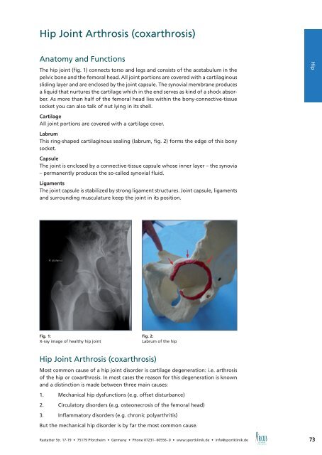
Anatomy and Functions<br />
The hip joint (fig. 1) connects torso and legs and consists of the acetabulum in the<br />
pelvic bone and the femoral head. All joint portions are covered with a cartilaginous<br />
sliding layer and are enclosed by the joint capsule. The synovial membrane produces<br />
a liquid that nurtures the cartilage which in the end serves as kind of a shock absorber.<br />
As more than half of the femoral head lies within the bony-connective-tissue<br />
socket you can also talk of nut lying in its shell.<br />
Cartilage<br />
All joint portions are covered with a cartilage cover.<br />
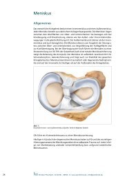
Labrum<br />
This ring-shaped cartilaginous sealing (labrum, fig. 2) forms the edge of this bony<br />
socket.<br />
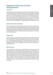
Capsule<br />
The joint is enclosed by a connective-tissue capsule whose inner layer – the synovia<br />
– permanently produces the so-called synovial fluid.<br />
Ligaments<br />
The joint capsule is stabilized by strong ligament structures. <strong>Joint</strong> capsule, ligaments<br />
and surrounding musculature keep the joint in its position.<br />
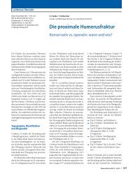
Fig. 1:<br />
X-ray image of healthy hip joint<br />
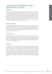
<strong>Hip</strong> <strong>Joint</strong> <strong>Arthrosis</strong> (<strong>coxarthrosis</strong>)<br />
Fig. 2:<br />
Labrum of the hip<br />
Most common cause of a hip joint disorder is cartilage degeneration: i.e. arthrosis<br />
of the hip or <strong>coxarthrosis</strong>. In most cases the reason for this degeneration is known<br />
and a distinction is made between three main causes:<br />
1. Mechanical hip dysfunctions (e.g. offset disturbance)<br />
2. Circulatory disorders (e.g. osteonecrosis of the femoral head)<br />
3. Inflammatory disorders (e.g. chronic polyarthritis)<br />
But the mechanical hip disorder is by far the most common cause.<br />
Rastatter Str. 17-19 • 75179 Pforzheim • Germany • Phone 07231- 60556- 0 • www.sportklinik.de • info@sportklinik.de 73<br />
Ellenbogen Hüfte <strong>Hip</strong>
Fig. 3:<br />
Offset<br />
<strong>Hip</strong> <strong>Joint</strong> <strong>Arthrosis</strong> (<strong>coxarthrosis</strong>)<br />
The Femoral Neck Offset<br />
Figure 3 shows the normal form of femoral neck and femoral head in cross-section.<br />
The femoral head protrudes the femoral neck both at the front and the back side.<br />
This midsection of the femoral neck is called offset. There are often disease patterns<br />
where this passage is much flatter (offset disturbance, fig. 4); this is mostly the result<br />
of a growth disorder of sportily active patients in the adolescence.<br />
This offset disturbance leads to the femoral neck hitting the socket edge when bending<br />
forwards (fig. 5). The first thing which becomes injured is the “sealing ring” of<br />
the hip, the so-called labrum. An early symptom of this offset disturbance is groin<br />
pain. During the following course of disease the cartilage of the socket becomes<br />
destroyed. Without treatment this loss of protecting cartilage leads to an increasing<br />
arthrosis with stiffening of the joint. At the advanced stage, ball and socket<br />
become partly damaged and do not optimally fit into each other any longer. At the<br />
same time run-in- and stress pain starts, later pain occurs also at night and while at<br />
rest. All this finally results in reduction of the walking distance and in an enormous<br />
reduction of the quality of life.<br />
Fig. 4:<br />
Offset disturbance<br />
Diagnosis<br />
Fig. 5:<br />
Impingement<br />
Diagnosis can be set by typical anamnesis, examination and by means of a normal<br />
x-ray image, whereby narrowing of the joint space between hip- and femoral bone<br />
is an indirect sign of cartilage loss. The MRI enables more precise examination of<br />
labrum and cartilage.<br />
74<br />
Rastatter Str. 17-19 • 75179 Pforzheim • Germany • Phone 07231- 60556- 0 • www.sportklinik.de • info@sportklinik.de
<strong>Hip</strong> <strong>Joint</strong> <strong>Arthrosis</strong> (<strong>coxarthrosis</strong>)<br />
Therapy of Offset Disturbance and Prevention for <strong>Hip</strong><br />
<strong>Arthrosis</strong><br />
Therapy of this offset disturbance is always carried out surgically as there is no reliable<br />
conservative therapy known. This means restoration of the femoral neck offset<br />
and removal or suture of the torn labrum. With this the cartilage is protected and<br />
hip arthrosis prevented.<br />
If a patient suffers from groin pain, differentiated assessment shows the dimensions<br />
of this growth disorder and already existing damages. Besides clinical examination<br />
and conventional x-ray images, the most important method here is MRI; and it is<br />
decisive that NMR is made with intra-articular contrast medium and on special sequences.<br />
This is the only possibility to achieve a differentiated result about labrum<br />
and condition of the cartilage.<br />
In order to avoid early degeneration of the hip joint correcting surgery should be<br />
carried out. We offer you a new operation technique at the ARCUS Clinics to repair<br />
this defect by means of hip arthroscopy (p 76). The torn part of the labrum is removed<br />
under arthroscopic control and the lacking femoral neck midsection formed<br />
artificially. This takes away the femoral neck entrapment and degeneration of the<br />
hip can be stopped or avoided.<br />
Hüftbandage zur konservativen<br />
oder postoperativen Behandlung<br />
von Hüftgelenkerkrankungen.<br />
www.sporlastic.de · info@sporlastic.de<br />
Ellenbogen Hüfte <strong>Hip</strong><br />
75<br />
75
<strong>Hip</strong> Arthroscopy<br />
In recent years, hip arthroscopy has become a standard surgery method when treating<br />
hip disorders. With this technique formerly common large incisions and resulting<br />
soft tissue damages as well as a long rehabilitation period can be prevented.<br />
Indications for hip arthroscopy are:<br />
• Loose joint bodies<br />
• Labrum ruptures<br />
• Degenerative changes<br />
• Beginning hip arthrosis (p. 73)<br />
• Movement restrictions of the hip<br />
• Cartilage injuries<br />
• Inflammations of the synovial membrane<br />
• Tear of the central hip ligament (Lig. teres)<br />
• <strong>Joint</strong> infections<br />
• Impingement of the hip (see step-by-step plan for treatment of hip arthrosis p. 78)<br />
• Problems after hip replacement surgery<br />
The surgery technique is very challenging and requires long term experience. Therefore<br />
we are very proud that more than 100 hip arthroscopies are carried out at the<br />
ARCUS Clinics every year.<br />
Fig. 1:<br />
<strong>Hip</strong> arthroscopy<br />
76<br />
Rastatter Str. 17-19 • 75179 Pforzheim • Germany • Phone 07231- 60556- 0 • www.sportklinik.de • info@sportklinik.de
<strong>Hip</strong> Arthroscopy<br />
We would like to outline two common indications as example:<br />
Loose <strong>Joint</strong> Bodies<br />
The most common cause for loose joint bodies (fig. 2) are accidents, followed by<br />
degeneration of the joint and synovial joint diseases. The loose joint bodies may<br />
then get caught and with this damage the joint; therefore it is recommended to<br />
remove them. This is possible by arthroscopic surgery through two or three 1cm<br />
long incisions what is an excellent alternative to formerly usual open operations.<br />
Femoroacetabular Impingement of the <strong>Hip</strong><br />
This so-called femoacetabular impingement of the hip joint occurs due to changed<br />
anatomical conditions at femoral neck and/or the socket edge. It stands for direct<br />
contact of the two bones the when bending forwards, whereby the cartilaginous<br />
rim of the socket (the so-called labrum), as well as the cartilage within the socket<br />
become entrapped. These problems often occur with young patients and symptoms<br />
are pain in the area of the hip and movement restrictions.<br />
The labrum- and cartilage damage and the repeated bone contact results in continuous<br />
joint degeneration and finally in destruction of the joint by arthrosis.<br />
Through small incisions (about 1cm length), disturbing bony protrusions at the<br />
femoral neck and the socket edge can be removed and labrum and cartilage be<br />
treated (fig. 3+4). In many cases this prevents progression of the arthrosis and restores<br />
pain free mobility.<br />
Arthroscopic Surgery Aftercare<br />
Restrictions after hip arthroscopy depend mainly on extent of the surgery. During<br />
the first 2-3 weeks, putting full weight on the hip is possible when limiting physical<br />
activity, i.e. no sports activities and additional stress. In this initial period, also<br />
walking on crutches can be of help. In case that bone has been removed from the<br />
femoral neck or bone stimulating techniques carried out, strict stress reduction might<br />
be necessary for 2-4 weeks. Physiotherapeutic treatment prevents these limitations<br />
to activity and therefore should be started the first postoperative day. Thrombosis<br />
prophylaxis during the period of load reduction reduces the risk of blood clots in<br />
the leg veins.<br />
Fig. 2:<br />
Loose joint bodies<br />
Fig. 3:<br />
Preoperative x-ray image<br />
Fig. 4:<br />
Postoperative x-ray image<br />
Rastatter Str. 17-19 • 75179 Pforzheim • Germany • Phone 07231- 60556- 0 • www.sportklinik.de • info@sportklinik.de 77<br />
Ellenbogen <strong>Hip</strong>
Fig. 1:<br />
46 year old man with arthritis<br />
Fig. 2:<br />
<strong>Hip</strong> with surface replacement<br />
(source: Smith & Nephew GmbH)<br />
Fig. 3:<br />
Total endoprosthesis<br />
Step-by-step Plan for Treatment of<br />
Coxarthrosis<br />
In case that joint arthrosis had been diagnosed, there was so far only the option<br />
of artificial hip joint replacement (THR) if conservative treatment methods such as<br />
physiotherapy, thalassotherapy, massages, pain medication etc. had already proven<br />
unsuccessful.<br />
Furthermore, treatment did consider neither severity of the disease nor age of the<br />
patient. Therefore we developed a step-by-step plan which ensures stage-related<br />
therapy.<br />
1. Moderate <strong>coxarthrosis</strong> with protrusions:<br />
Considerable improvement of discomfort can be achieved by recovering the stage of<br />
compensated arthrosis with arthroscopic hip surgery (p. 76). Disturbing osteophytes<br />
at femoral neck and socket are removed and the contract capsule partly recessed<br />
what brings back movability. Additionally, removal of torn parts of the labrum and<br />
inflamed portions of the synovial membrane allow considerable pain reduction. And<br />
with this method even loose joint bodies can be removed what enables the patient<br />
to be physically active again and delay an artificial hip implant.<br />
2. Advanced arthrosis with young patients<br />
(Female patients under 60, male patients under 65):<br />
When the joint is completely destroyed, joint-preserving surgery no longer makes<br />
sense. However, in order to preserve as many bones as possible, younger patients are<br />
implanted a femoral head cap (fig. 2) - a resection of the femoral neck is not necessary.<br />
Advantage is here preservation of normal anatomy (offset, force transmission<br />
and size of femoral head) what is needed for the normal range of movement. The<br />
resulting stability enables sportive activity without limitations. Another important<br />
advantage is the protection of bone substance which might become decisive with<br />
regard to a future revision.<br />
Not every hip arthrosis can optimally be treated with a femoral head cap. In such<br />
cases we alternatively use short-stem hip prostheses.<br />
3. Advanced arthrosis with elderly patients<br />
(Female patients over 60, male patients over 65):<br />
As the femoral neck is here not strong enough to carry surface replacement due to<br />
the reduced level of calcium carbonate in the bones, complete hip arthroplasty is<br />
the only option. This is another treatment where we achieve enormous progress,<br />
and besides better materials there have also been essential improvements with the<br />
operation technique. By developing the concept of minimally-invasive surgery (MIS)<br />
we only need very small incisions (6-8 cm). But the decisive advantage is the fact that<br />
almost no muscles have to be detached. This brings minimization of tissue trauma,<br />
an overall gentle operation method and less pain. Immediate full strain is possible<br />
and blood loss is reduced what in turn accelerates rehabilitation.<br />
78<br />
Rastatter Str. 17-19 • 75179 Pforzheim • Germany • Phone 07231- 60556- 0 • www.sportklinik.de • info@sportklinik.de
Total Endoprosthesis: Material and Fixation<br />
<strong>Hip</strong> joint prosthesis: material and fixation<br />
Continuous improvements in both surgical techniques and the quality of implants<br />
since the 1960s make this procedure one of the most common and most successful<br />
routine operations in orthopedic surgery (about 400.000 per year in Europe). The<br />
prosthesis is modeled on the actual human joint, i.e. it consists of a socket and a shaft<br />
to which a ball head is fitted. By means of pre-operative planning the model size<br />
and fixation method of the prosthesis is specified whereby individual requirements<br />
such as age, gender, shape of bone, body weight, etc. are taken into consideration.<br />
There are three different fixation techniques used with implantations:<br />
• Cement-free endoprosthesis fixation: shaft and socket are press-fitted exactly<br />
into the bone (fig. 1 + 2).<br />
• Cemented endoprosthesis fixation: hip socket and shaft are fixed with quickhardening<br />
antibiotic bone cement (fig. 3).<br />
• Hybrid endoprosthesis fixation: the socket is fixed cement-free; the shaft anchored<br />
using bone cement (fig. 4).<br />
The cemented socked is made of polyethylene, the cemented shaft of a cobaltchromium<br />
alloy. Titanium implants, often equipped with special macro- or microstructured<br />
surfaces are particularly suitable for cement-free fixation thanks to their<br />
excellent integration into the bone.<br />
As so-called slide bearings (joint portions with direct contact) between the socket<br />
and the artificial femoral head polyethylene/ceramic-, ceramic/ceramic- or metal/<br />
metal combinations are used. Thanks to latest developments in this area (e.g. Durasul,<br />
Sulzer Orthopedics or especially hardened ceramics) the abrasion behavior<br />
of the components has been optimized to the extent that many years of usage are<br />
tolerated with almost no material wear.<br />
Fig. 2:<br />
Cement-free endoprosthesis<br />
Fig. 3:<br />
Cemented endoprosthesis<br />
Fig. 4:<br />
Hybrid endoprosthesis<br />
Fig. 1:<br />
Cement-free joint replacement<br />
(source: Smith & Nephew GmbH)<br />
Fig. 5:<br />
<strong>Hip</strong> with short-shaft prosthesis<br />
(source: Smith & Nephew GmbH)<br />
Rastatter Str. 17-19 • 75179 Pforzheim • Germany • Phone 07231- 60556- 0 • www.sportklinik.de • info@sportklinik.de 79<br />
Ellenbogen <strong>Hip</strong>
Total Endoprosthesis: Material and Fixation<br />
Resurfacing<br />
Treatment of young patients with advanced hip joint arthrosis can – alternatively<br />
to the usual THR surgery - also be carried out by implanting a hip cap. Here the<br />
femoral head is covered with a metal cap with the advantage that practically no<br />
bone has to be sacrificed. Furthermore the physiological size of the femoral head is<br />
re-built what results in considerably improved mobility and stability. Most important<br />
requirement is a good bone quality as there is the risk of a femoral neck fracture<br />
when suffering from osteoporosis.<br />
Another option for younger patients which cannot undergo implantation of a hip<br />
cap (e.g. with femoral head necrosis) is a short-shaft prosthesis. Here only a small<br />
part of the femoral neck has to be removed (p. 79, fig. 5).<br />
Fig. 6:<br />
<strong>Hip</strong> with surface replacement<br />
(source: Smith & Nephew GmbH)<br />
Aftercare<br />
Fig. 7:<br />
<strong>Hip</strong> with surface replacement<br />
(source: Smith & Nephew GmbH)<br />
Endoprosthetic operations are carried out exclusively on in-patient conditions. In<br />
order to ensure an optimal operation success, early postoperative mobilization by<br />
means of physiotherapy is recommended. Independent of the surgery method, full<br />
load is permitted almost immediately whereby walking on crutches is necessary for<br />
3-4 weeks to protect the soft tissues.<br />
Most patients stay in hospital for 7-10 days followed by 3-4 weeks of rehabilitation<br />
time. The progress of the patient is documented by regular out-patient control<br />
check-ups at close intervals. If necessary, mobilization therapy has to be continued<br />
on an out-patient basis.<br />
80<br />
Rastatter Str. 17-19 • 75179 Pforzheim • Germany • Phone 07231- 60556- 0 • www.sportklinik.de • info@sportklinik.de
Total Endoprosthesis: Material and Fixation<br />
<strong>Joint</strong> Replacement and Sports<br />
Having a severe hip joint arthrosis, noticeable limitation of physical activities has<br />
to be expected. When the symptoms are gone after joint replacement surgery, the<br />
desire for more sportive exercise certainly comes up again. Internationally there is a<br />
broad consensus that at least so-called “low-impact” sports such as cycling, swimming,<br />
sailing, diving, playing golf and bowling can be supported. Sports such as tennis,<br />
basket ball and skiing however, are only possible to a limited extent. Completely<br />
avoided shall be contact sports such as foot ball or hand ball. Recommendations<br />
for those different sports are also dependent on the patient’s performance level.<br />
As a rule of thumb it can be said that sports practiced prior to surgery are allowed<br />
afterwards as well.<br />
Rastatter Str. 17-19 • 75179 Pforzheim • Germany • Phone 07231- 60556- 0 • www.sportklinik.de • info@sportklinik.de 81<br />
Ellenbogen <strong>Hip</strong>