ORIGINAL ARTICLE - MVZ Labor PD Dr. Volkmann und Kollegen
ORIGINAL ARTICLE - MVZ Labor PD Dr. Volkmann und Kollegen
ORIGINAL ARTICLE - MVZ Labor PD Dr. Volkmann und Kollegen

Create successful ePaper yourself
Turn your PDF publications into a flip-book with our unique Google optimized e-Paper software.
Clin. Lab. 9+10/2011<br />
AUTOANTIBODIES AGAINST IM<strong>PD</strong>H2 AND HCV INFECTION<br />
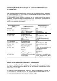
Figure 1. IIFT of index patient’s serum: a, b: In house cultivated HEp-2 cells. Anti-IM<strong>PD</strong>H2 reacts with distinct cytoplasmic<br />
granules and condensed granular aggregates. c, d: Two commercial HEp-2 cell preparations (c: Euroimmun, d: Inova Diagnostics)<br />
exhibiting the “rods and rings” fluorescence pattern. e, f: In house cultivated HEp-2 cells addition of (e) mycophenolic<br />
acid or (f) ribavirin into the culture medium. Both mycophenolic acid and ribavirin shift the granular fluorescence pattern<br />
(seen in a and b) to “rods and rings”. g, h: IIFT of rabbit anti-IM<strong>PD</strong>H2 Si on (g) in house cultivated and (h) commercial (Inova<br />
Diagnostics) HEp-2 cells, exhibiting identical fluorescence patterns as seen with the serum of the index patient.<br />
Zeiss Axioplan 2, magnifications 400 x.<br />
only a slight increase of granular aggregates within the<br />
cytoplasm of cells cultivated for prolonged periods (>20<br />
hours).<br />
IM<strong>PD</strong>H2 constitutes the target antigen<br />
The immunofluorescence pattern observed with the index<br />
patient’s serum could not be attributed to antibodies<br />
against one of the major known cytoplasmic autoantigens<br />
(actin, tubulin, mitochondria, microsomes, ribosomes,<br />
lysosomes, endosomes, tRNA synthetases, Golgi<br />
proteins) either by IIFT or by appropriate antibody specific<br />
assays. However, the serum precipitated a substantial<br />
amount of a 55 kDa 35 S-methionine-labeled protein<br />
from HEp-2 cell extract (Figure 3). Screening a cDNA<br />
expression library revealed three clones encoding IMP-<br />
DH2 and spanning the complete coding region of 1545<br />
nucleotides including 74 nucleotides of the 5´-untranslated<br />
region and 52 nucleotides of the 3´-untranslated<br />
sequence (positions 19 to 1689).<br />
Direct confirmation of IM<strong>PD</strong>H2 as target antigen was<br />
made by the positive reaction of the index patient’s serum<br />
with recombinant IM<strong>PD</strong>H2 in western and line<br />
blots (Figures 3a, 3b) and with 35 S-methionine-IMP-<br />
DH2 in RIPA (Figure 4). As indirect confirmation, a<br />
757<br />
commercial rabbit anti-IM<strong>PD</strong>H2 (anti-IM<strong>PD</strong>H2Si) reacted<br />
essentially in the same way in IIFT as the index<br />
patient’s serum (Figure 1g, 1h). The in house rabbit<br />
anti-IM<strong>PD</strong>H2 (anti-IM<strong>PD</strong>H2ih) irrespective of its reactivity<br />
with His(6)-tag-IM<strong>PD</strong>H2 (Figure 3a, lane 7),<br />
commercial IM<strong>PD</strong>H2 and 35 S-methionine-IM<strong>PD</strong>H2<br />
(Figure 4) was, however, unable to recognize IM<strong>PD</strong>H2<br />
in any preparation of HEp-2 cells. This observation suggests<br />
that this antibody was unable to recognize epitopes<br />
being presented by intracellular IM<strong>PD</strong>H2 (Table<br />
1).<br />
Mycophenolic acid and ribavirin rearrange intracellular<br />
IM<strong>PD</strong>H2<br />
The reason for the different arrangements of IM<strong>PD</strong>H2<br />
observed within the cytoplasm of in house cultivated<br />
HEp-2 cells and the two commercial preparations is unknown.<br />
It may be caused by additives within the culture<br />
media, not disclosed by the manufacturers. As could be<br />
shown recently (19), addition of mycophenolic acid to<br />
culture cells induced a rearrangement of cytoplasmic<br />
IM<strong>PD</strong>H2 from granular particles into the “rods and<br />
rings”. We could show this transition of IM<strong>PD</strong>H2 after<br />
addition of therapeutic concentrations (12.49 µmol) of