ORIGINAL ARTICLE - MVZ Labor PD Dr. Volkmann und Kollegen
ORIGINAL ARTICLE - MVZ Labor PD Dr. Volkmann und Kollegen
ORIGINAL ARTICLE - MVZ Labor PD Dr. Volkmann und Kollegen

You also want an ePaper? Increase the reach of your titles
YUMPU automatically turns print PDFs into web optimized ePapers that Google loves.
Clin. Lab. 2011;57:753-765<br />
©Copyright<br />
Clin. Lab. 9+10/2011<br />
<strong>ORIGINAL</strong> <strong>ARTICLE</strong><br />
Autoantibodies against Inosine-5’-Monophosphate Dehydrogenase<br />
2 – Characteristics and Prevalence in Patients with HCV-Infection<br />
H. P. SEELIG 1 , H. APPELHANS 1 , O. BAUER 1 , M. BLÜTHNER 1 , K. HARTUNG 2 ,<br />
P. SCHRANZ 1 , D. SCHULTZE 3 , CLAUDIA A. SEELIG 1 , M. VOLKMANN 1<br />
1 Medizinisches Versorgungszentrum <strong>Labor</strong> Prof. Seelig, Karlsruhe, Germany<br />
2 Institut für <strong>Labor</strong>atoriums- <strong>und</strong> Transfusionsmedizin, Klinikum Bremerhaven Reinkenheide, Bremerhaven, Germany<br />
3 Institut für Klinische Mikrobiologie <strong>und</strong> Immunologie, St. Gallen, Switzerland<br />
SUMMARY<br />
Backgro<strong>und</strong>: Indirect immunofluorescence (IIFT) on in house HEp-2 cell preparations revealed a novel antibody<br />
giving a granular cytoplasmic pattern not described before, which on two commercial cell preparations revealed a<br />
“rings and rods” pattern. This pattern was also observed in four HCV-RNA carriers and prompted the identification<br />
of the reactive antigen and the evaluation of the antibody prevalence in HCV-RNA carriers and control<br />
groups.<br />
Methods: The antigen’s molecular weight was determined by radioimmunoprecipitation of 35 S-methionine labeled<br />
cell proteins. Expression library screening and sequencing was performed by standard techniques using an<br />
oligo(dT)-primed human HeLa cell cDNA expression library. Antibodies against the novel antigen Inositol-5’monophosphatdehydrogenase<br />
2 (IM<strong>PD</strong>H2) were analyzed by IIFT, western blot, line blot, and radioimmunoprecipitation<br />
assay (RIPA). IIFT was performed on commercial HEp-2 cells and cells cultivated in house for 24 –<br />
60 hours, with or without the IM<strong>PD</strong>H2 inhibitors mycophenolic acid (MPA) or ribavirin, and subjected to various<br />
fixation conditions. Western and line blots were performed with IM<strong>PD</strong>H2 synthesized in E. coli, RIPA with 35 Smethionine-IM<strong>PD</strong>H2<br />
from in vitro transcription/translation products. Sera screened were positive for HCV-RNA<br />
(108), HBV-DNA (100), anti-mitochondrial (31), anti-actin (42), and anti-nuclear antibodies (51) and negative for<br />
HCV-RNA (100) and blood donors (100).<br />
Results: IM<strong>PD</strong>H2 is capable of considerable intracellular rearrangements (upon action of inhibitors like MPA and<br />
ribavirin), which explains the contrasting immunofluorescence patterns in cells from different sources. By RIPA,<br />
proven to be the sole assay suitable for screening of anti-IM<strong>PD</strong>H2 in human sera, autoantibodies were fo<strong>und</strong> in<br />
35.2 % of HCV-RNA carriers and in low concentrations in 31 % of anti-actin positive patients suspicious of autoimmune<br />
hepatitis. Antibodies reacted preferentially with conformational epitopes. Compared to the low concentration<br />
of anti-IM<strong>PD</strong>H2 fo<strong>und</strong> in other disease groups, high antibody concentrations were observed in HCV-RNA<br />
carriers.<br />
Conclusions: The common occurrence of anti-IM<strong>PD</strong>H2 in HCV-RNA carriers may be related to ribavirin therapy,<br />
causing intracellular aggregation of IM<strong>PD</strong>H2 thereby altering its immunogenicity. In this study the “rods and<br />
rings” immunofluorescence pattern observed could be ascribed to anti-IM<strong>PD</strong>H2. Anti-IM<strong>PD</strong>H2 may cause difficulties<br />
in interpretation of immunofluorescence patterns in routine autoantibody testing.<br />
(Clin. Lab. 2011;57:753-765)<br />
ABBREVIATIONS<br />
IM<strong>PD</strong>H2: Inosine-5’-Monophosphate Dehydrogenase 2<br />
(NCBI Acc. No. NM_000884, GI: 66933015, Swiss-<br />
Prot. No.: P12268). According to the NCBI nomenclature<br />
the adenine nucleotide of the first methionine en-<br />
753<br />
coding ATG is defined as position +93). ABR: antibody<br />
ratio.<br />
_____________________________________________<br />
Manuscript accepted March 29, 2011
INTRODUCTION<br />
In the course of routine screening for autoantibodies<br />
against nuclear antigens by means of indirect immunofluorescence<br />
test (IIFT) with HEp-2-cells, we detected<br />
in the serum of a female patient an unusual cytoplasmic<br />
staining not described before. The antigenic target of<br />
this reaction was identified, by screening an expression<br />
library, as type 2 inosine-5’-monophosphate dehydrogenase<br />
(1) the key enzyme in the synthesis of purine<br />
nucleotides. Inosine-5’-monophosphate dehydrogenase<br />
(IM<strong>PD</strong>H), catalyzes the transition of inosine monophosphate<br />
to xanthine monophosphate, which constitutes the<br />
rate limiting step in the de novo synthesis of guanine<br />
nucleotides, precursors of RNA and DNA. In mammalian<br />
species there exist two isoforms, IM<strong>PD</strong>H1 and<br />
IM<strong>PD</strong>H2 (2), each of which is composed of 514 amino<br />
acids (55.8 kDa) with a homology of 84 % on amino<br />
acid level. Whilst IM<strong>PD</strong>H1 is constitutively expressed<br />
in normal cells, the expression and activity of IM<strong>PD</strong>H2,<br />
however, is augmented in proliferating normal and malignant<br />
tissues and cells, in malignant transformation,<br />
and differentiation (3–9). The possible occurrence of<br />
autoantibodies against IM<strong>PD</strong>H2 was reported in the serum<br />
of one out of 21 patients with autoimmune hepatitis<br />
(10), studied by immunoproteomic analysis of HepG2<br />
cell proteins. Contemporaneously to our findings it has<br />
been shown, that IM<strong>PD</strong>H2 probably acts as a tumor associated<br />
antigen (11). Our patient, however, showed no<br />
evidence of a disease related to cell proliferation or neoplasm<br />
nor could her clinical symptoms be associated to<br />
the presence of autoimmune hepatitis or another autoimmune<br />
disease. Subsequently, we detected IM<strong>PD</strong>H2<br />
autoantibodies by IIFT in four patients who suffered<br />
from chronic hepatitis C virus (HCV) infections. HCV<br />
infection may be involved in the pathogenesis of various<br />
autoimmune and rheumatic disorders and chronically<br />
infected HCV patients have been reported to produce<br />
a plethora of autoantibodies (12–17). We determined<br />
the presence of anti-IM<strong>PD</strong>H2 in patients with HCV infections,<br />
hepatitis B virus (HBV) infections, autoimmune<br />
liver diseases, and rheumatic diseases.<br />
Anti-IM<strong>PD</strong>H2 was detected in patients harboring HCV<br />
infections and autoimmune liver diseases. We could<br />
show that the so-called “rods and rings” fluorescence<br />
pattern (18), which sometimes can be seen within the<br />
cytoplasm of some commercial HEp-2 cell preparations<br />
used for screening of autoantibodies, is caused by anti-<br />
IM<strong>PD</strong>H2. This finding may be important particularly<br />
with regard to considerations concerning the pathogenesis<br />
of IM<strong>PD</strong>H2 autoantibodies since it has been shown<br />
that the IM<strong>PD</strong>H2 inhibitor mycophenolic acid causes<br />
reversible intracellular aggregations of IM<strong>PD</strong>H2 molecules<br />
(19), which impress as “rods and rings” shaped<br />
structures. As could be shown here, ribavirin, an inhibitor<br />
of IM<strong>PD</strong>H2 used in treatment of HCV infections,<br />
also causes an aggregation of IM<strong>PD</strong>H2 in “rods and<br />
rings” structures. Possibly the aggregated enzyme attains<br />
a higher degree of immunogenicity, promoting the<br />
H. P. SEELIG et al.<br />
754<br />
development of autoantibodies in patients treated with<br />
IM<strong>PD</strong>H2 inhibitors. The preferred origination of autoantibodies<br />
against constituents of subcellular aggregates<br />
and particles is debated and evidenced since more than<br />
two decades (20-24).<br />
MATERIALS AND METHODS<br />
Patient and control sera<br />
The index patient was a 71-year-old female (HCV-RNA<br />
negative), suffering for 15 years from extended osteoporosis<br />
who <strong>und</strong>erwent pain management therapy because<br />
of major osteoporotic ache. To exclude systemic rheumatic<br />
disease, a search for antinuclear antibodies was<br />
performed with a negative result. However, the current<br />
investigation by indirect immunofluorescence tests<br />
(IIFT) revealed a distinct cytoplasmic staining in HEp-<br />
2-cells not attributable to a known autoantibody. The<br />
serum precipitated a 55 kDa protein from 35 S-methionine<br />
labeled HEp-2 cell proteins and an expression library<br />
was screened, which revealed IM<strong>PD</strong>H2 as the reactive<br />
antigen.<br />
To establish the prevalence of anti-IM<strong>PD</strong>H2, we<br />
screened (by IIFT, immunoblot, and radioimmunoprecipitation<br />
assay (RIPA)) the sera of patients positive for<br />
HCV-RNA (n = 108) and of disease controls (hospitalized<br />
patients) negative for HCV-RNA (n = 100), sera of<br />
patients positive for HBV-DNA (n = 113), with mitochondrial<br />
antibodies (AMA Type M2, suspected primary<br />
biliary cirrhosis; n = 31) or actin antibodies (suspected<br />
autoimmune hepatitis; n = 42), with overt or suspected<br />
rheumatic diseases showing antibodies against<br />
nuclear antigens (n = 51) with different nuclear staining<br />
patterns (homogeneous, speckled, nucleolar, rim), and<br />
the sera of healthy blood donors (n = 100).<br />
Screening of autoantibodies (actin, tubulin, mitochondria,<br />
microsomes, ribosomes, lysosomes, endosomes,<br />
tRNA synthetases, Golgi proteins), HCV-RNA, and<br />
HBV-DNA was performed by approved, CE labeled in<br />
house tests according to Quality Systems of the Council<br />
Directive 98/79/EC. All samples were coded and analysed<br />
for the presence of anti-IM<strong>PD</strong>H2 without knowledge<br />
of virus and autoantibody status.<br />
cDNA library screening and synthesis of recombinant<br />
His(6)-tag IM<strong>PD</strong>H2 in E. coli<br />
Expression library screening and sequencing was performed<br />
by standard techniques using an oligo(dT)primed<br />
human HeLa cell cDNA expression library in<br />
Lambda ZAP (Stratagene, Amsterdam, The Netherlands)<br />
in conjunction with the index patient’s serum.<br />
Following two ro<strong>und</strong>s of screening, three clones were<br />
selected as positive and subjected to sequence analysis<br />
using ABI Prism 3130XL and the Cycle Sequencing Kit<br />
Big dye Terminator from Applied Biosystems (Darmstadt,<br />
Germany). Sequence homologies were identified<br />
using the NCBI BLAST program.<br />
Full length IM<strong>PD</strong>H2 containing a His(6)-tag was syn-<br />
Clin. Lab. 9+10/2011
thesized in E. coli BL21 Star cells (NovageneLlc) according<br />
to standard techniques and purified from inclusion<br />
bodies by Ni2 + -agarose (Quiagen, Hilden, Germany).<br />
Purity was checked after sodium dodecylsulfate<br />
polyacrylamide gel electrophoresis (SDS-PAGE) on<br />
12.5 % gels stained with Coomassie Brillant Blue.<br />
Rabbit antibodies against IM<strong>PD</strong>H2<br />
Rabbit anti-IM<strong>PD</strong>H2 were either obtained commercially<br />
(Sigma, Munich, Germany) containing 0.07<br />
mg/mL anti-IM<strong>PD</strong>H2 (anti-IM<strong>PD</strong>H2Si) or produced in<br />
house by immunization of a female New Zealand White<br />
rabbit (Charles River <strong>Labor</strong>atories, Kisslegg, Germany)<br />
with 0.3 mg His(6)-tag-IM<strong>PD</strong>H2 in PBS followed by 3<br />
injections (0.3 mg protein) within 2.5 months (anti-<br />
IM<strong>PD</strong>H2ih). Antibody synthesis was monitored on line<br />
blots spotted with His(6)-tag-IM<strong>PD</strong>H2.<br />
Antibody detection<br />
Indirect immunofluorescence test (IIFT)<br />
For antibody screening by IIFT, HEp-2 cells were cultivated<br />
on glass microscope slides for 20 hours and fixed<br />
in methanol - 20 °C, 5 minutes, acetone 20 °C, 1<br />
minute. They were incubated with patient serum diluted<br />
1:80 in PBS as described (25). For detection of bo<strong>und</strong><br />
antibodies FITC labeled rabbit anti-human-IgG, Fc specific<br />
(Invitrogen, Karlsruhe, Germany) was used in optimal<br />
dilutions determined by checker board titration. The<br />
serum of the index patient was also screened on seven<br />
different commercial HEp-2 cell preparations (Inova<br />
Diagnostics, San Diego, CA, USA; Orgentec, Mainz,<br />
Germany; Euroimmun, Lübeck, Germany; Menarini,<br />
Berlin, Germany; Generic Assays, Berlin, Germany;<br />
AESKU, Wendelsheim, Germany; AID, Straßberg, Germany).<br />
To explore a possible influence of culture time or kind<br />
of cell fixation on the intracellular localization of IMP-<br />
DH2, HEp-2 cells were harvested after growing periods<br />
of 16 – 60 hours and fixed with acetone (-20 °C, 5 minutes),<br />
methanol (-20 °C, 5 minutes), ethanol-glacial acetic<br />
acid (95:5 [vol:vol], 4 °C, 5 minutes), methanol-aceton<br />
(1:1 [vol:vol], -20 °C, 5 minutes), methanol (-20 °C,<br />
5 minutes, permeabilization by acetone at 20 °C, 1 minute)<br />
methanol-ethanol (1:1 [vol:vol], -20 °C, 5 minutes),<br />
formalin (4 %, 4 °C, 10 minutes), paraformaldehyde<br />
(4 %, 20 °C, 10 minutes, permeabilization by Triton X-<br />
100, 0.5 %, 20 °C, 10 minutes), paraformaldehyde (4 %,<br />
20 °C, 10 minutes, and methanol (-20 °C, 5 minutes),<br />
ethanol (95 %, 20 °C, 5 minutes).<br />
The effect of either mycophenolic acid or ribavirin on<br />
the intracellular localization of IM<strong>PD</strong>H2 was monitored<br />
24 hours after addition of 12.49 µmol mycophenolic<br />
acid (Sigma) or 16.38 µmol ribavirin (Sigma) to the cell<br />
culture media.<br />
AUTOANTIBODIES AGAINST IM<strong>PD</strong>H2 AND HCV INFECTION<br />
Clin. Lab. 9+10/2011 755<br />
Radioimmunoprecipitation of 35 S-methionine labeled<br />
cell proteins<br />
HEp-2 cells (5 x 10 6 ) in methionine-free medium (RP-<br />
MI1640, Sigma) were incubated with 25 µL L- 35 S-methionine<br />
(PerkinElmer, Rodgau, Germany) for 16 hours,<br />
washed (3 x in 10 mL ice cold PBS), and incubated<br />
with 3 mL of ice-cold RIP1 buffer (10 mM Tris-HCl,<br />
pH 8.0, 500 mM NaCl, 0.1 % Igepal, 2 mM PMSF) for<br />
20 minutes. Cells were harvested, sonified on ice (Sonifier<br />
Cell Disruptor B15; Branson, Danbury, CT, USA)<br />
for 3 x 30 seconds at output level 7, pulsed, 55 % duty<br />
cycle and centrifuged (4000 x g, 10 minutes). Aliquots<br />
of the supernatant containing labelled cell proteins were<br />
stored frozen at - 80 °C until used.<br />
The patient’s serum and well characterized control sera<br />
(20 µL each) were incubated with 30 µL protein-A<br />
coated magnetic beads (Invitrogen, Darmstadt, Germany)<br />
for 4 hours on a rotating mixer at 4 °C. After washing<br />
(4 x 700 µL RIP1 buffer) 100 µL 35 S-methionine<br />
labelled HEp-2 cell proteins (10 7 cpm) were added and<br />
incubated on a rotating mixer (16 hours, 4 °C). After<br />
washing as stated above the beads were heated (95 °C, 5<br />
minutes) in 30 µL sampling buffer (10 mmol Tris-HCl,<br />
pH 6.8, 2 % SDS, 2.5 % β-mercaptoethanol, 10 % glycerol,<br />
0.1 %, bromophenol blue). After brief centrifugation<br />
(10000 x g) supernatants were subjected to 9 %<br />
SDS-PAGE followed by autoradiography using Hyperfilm<br />
MP films (GE Healthcare, Munich, Germany) for<br />
24 – 48 hours.<br />
Immunoblot<br />
(1) Western blots: Separation of IM<strong>PD</strong>H2 on 12.5 %<br />
SDS-PAGE was done as described (26) in a Mini Protean<br />
II electrophoresis chamber (BioRad, Munich, Germany).<br />
Per lane 0.2 – 0.5 µg purified His(6)-tag-IM<strong>PD</strong>-<br />
H2 or commercially available IM<strong>PD</strong>H2 (0.13 mg/mL in<br />
20 mM Tris-HCl, pH 8.0, 0.5 m EDTA, 1 mM DTT;<br />
Sigma) were applied. After electrophoresis, proteins<br />
were transferred to nitrocellulose Protran BA85 membranes<br />
(VWR International, Darmstadt, Germany).<br />
Membranes were blocked in blocking buffer for 0.5<br />
hours and after washing 3 times according to Towbin et<br />
al. (27) incubated with human or rabbit sera diluted<br />
1:100 in blocking buffer for 1 hour, washed 3 times in<br />
wash buffer (10 mM Tris-HCl, pH 7.4, 150 mM NaCl,<br />
0.1 % Triton X-100) and consecutively incubated with<br />
alkaline phosphatase conjugated goat anti-human immunoglobulins<br />
(Dianova, Hamburg, Germany) or goat<br />
anti-rabbit immunoglobulins (Dianova). After washing<br />
3 times in wash buffer, bo<strong>und</strong> antibodies were visualized<br />
using BCIP with NBT as substrate.<br />
(2) Line blots were prepared by using a Nanoplotter 2.0<br />
(GeSiM, Großerkmannsdorf, Germany) with a four<br />
channel print head and applying different amounts of<br />
IM<strong>PD</strong>H2 on nitrocellulose BA85 membranes. After<br />
spotting, membranes were blocked as described above<br />
and cut in strips of 3 mm width. Routinely, protein solutions<br />
with concentrations between 12.5 µg/mL and 100<br />
µg/mL in 0.1 mol borate buffer, pH 9.0 were used for
spotting, resulting in four lanes containing 4, 8, 16 or<br />
32 ng IM<strong>PD</strong>H2 per strip. Membranes were blocked and<br />
processed as described above. Human and rabbit sera to<br />
be tested were diluted 1:100 in blocking buffer.<br />
Radioimmunoprecipitation assay (RIPA)<br />
For production of radiolabelled IM<strong>PD</strong>H2-tracer, the<br />
DNA sequence encoding amino acids 1 - 514 of human<br />
IM<strong>PD</strong>H2 was cloned into a pCITE4a vector (Novagen).<br />
In vitro transcription/translation (ivTT) was performed<br />
using a T7 promoter controlled coupled transcription/<br />
translation system based on rabbit reticulocyte lysate<br />
(Promega, Heidelberg, Germany) according to the manufacturer’s<br />
instructions. Briefly, 25 µL rabbit reticulocyte<br />
lysate, 2 µL reaction buffer, 1 µL amino acid mixture<br />
(without methionine), 1 µL T7 polymerase, 3.5 µL<br />
L- 35 S-methionine (PerkinElmer), 1 µL RNasin (40 U;<br />
Lonza, Verviers, Belgium) and 1 µg template DNA<br />
were pipetted into a nuclease free 1.5 mL reaction tube.<br />
Nuclease free water was added to a final volume of<br />
50 µL. After 90 minutes incubation at 30 °C, unincorporated<br />
35 S-methionine was removed on a Sephadex G-<br />
50 MicroSpin column (GE Healthcare). Aliquots were<br />
stored at -80 °C until used. The percentage of 35 S-methionine<br />
incorporated into the protein was calculated after<br />
liquid scintillation counting. Mean incorporation rates<br />
were 9.5 % (SD 2.5 %). The quality of the 35 S-methionine-IM<strong>PD</strong>H2<br />
was checked on SDS-PAGE (12.5 %,<br />
200 V), sample preparation and autoradiography was<br />
performed as described above.<br />
For antibody screening, 35 S-methionine labelled IM<strong>PD</strong>-<br />
H2 (tracer) was diluted in dilution buffer (20 mM Tris-<br />
HCl, pH 7.4, 150 mM NaCl, 0.15 % Tween-20, 100000<br />
KIE Aprotinin [Bayer, Leverkusen, Germany], 10 mM<br />
benzamidine, 0.1 % BSA) to an activity of 40000 cpm<br />
in 50 µL. Duplicates of patient and control sera (5 µL)<br />
were pipetted into the wells of a 96-well microtiter<br />
plate. 50 µL tracer were added to each well. After incubation<br />
for 3 hours at 4 °C on a plate shaker, 20 µL recombinant<br />
protein A bo<strong>und</strong> to fractogel (Merck, Darmstadt,<br />
Germany) were added (4 °C, 60 minutes). The reaction<br />
mixes were transferred to a 0.65 µm Durapore<br />
96-well filter plate (Millipore, Schwalbach, Germany).<br />
Unbo<strong>und</strong> antigen was removed by vacuum filtration and<br />
washing the filter plates 30 times with 100 µL RIP2<br />
buffer (20 mM Tris-HCl, pH 7.4, 150 mM NaCl,<br />
0.05 % Tween-20). After drying the plates overnight,<br />
20 µL liquid scintillation mix MicroScintO (PerkinElmer)<br />
were added to each well and the plates were<br />
counted using a Topcount NXT 96-well liquid scintillation<br />
counter (PerkinElmer).<br />
All assays were performed in duplicate. Five negative<br />
controls (healthy blood donors) were used to calculate<br />
the antibody concentrations (antibody ratio, ABR) according<br />
to the following formula: Antibody ratio (ABR)<br />
= (cpm (sample)/[mean cpm (negative controls) + 3.041<br />
SD (negative controls)]) x 10 (28). Sera positive for<br />
anti-IM<strong>PD</strong>H2 were retested with a homologous RIP assay,<br />
used for determination of antibodies against ste-<br />
H. P. SEELIG et al.<br />
756<br />
roid-21-hydroxylase to exclude a possible non-specific<br />
binding of the tracer.<br />
Statistical methods<br />
Receiver operating characteristic (ROC) curve analysis<br />
was conducted to determine the cut-off range between<br />
sera regarded as positive or negative for anti-IM<strong>PD</strong>H2.<br />
Each of the patient groups was compared with the control<br />
group of healthy blood donors. The specificity was<br />
regarded to be 98 %. The area <strong>und</strong>er the ROC curve<br />
(AUC) with 95 % confidence interval (CI) was calculated<br />
from the AUC, SE, CI according to Hanley (29).<br />
RESULTS<br />
Different immunofluorescence patterns evoked by<br />
patient serum<br />
The serum of the index patient screened on in house<br />
cultivated HEp-2 cells exhibited an intense staining of<br />
distinct granular particles diffusely dispersed within the<br />
cytoplasm (Figure 1, a, b). In many cells the granules<br />
appeared as condensed globular-shaped aggregates<br />
reaching numbers from one per cell up to more than a<br />
dozen (antibody titer 1: 1280). Only a very weak granular<br />
cytoplasmic fluorescence could be detected in five<br />
different commercial HEp-2 cell preparations (Orgentec,<br />
Menarini, Generic Assays, AESKU, AID), indicating<br />
that these cell preparations actually are not suitable<br />
for the identification of anti-IM<strong>PD</strong>H2 in human sera.<br />
A rather different immunofluorescence pattern, however,<br />
was obtained when the patient’s serum was reacted<br />
with two other commercial HEp-2 cell preparations<br />
(Inova Diagnostics, Euroimmun). Instead of a granular<br />
staining within the cytoplasm, one to three thin, smooth<br />
fluorescent filaments and/or one to three small plain<br />
rings with central apertures (Figure 1c, 1d) were seen.<br />
This uncommon fluorescence pattern was only recently<br />
described as “rods and rings” pattern in the context of<br />
proficiency tests for antinuclear antibodies (18).<br />
Possible antibody specificities associated with this pattern,<br />
however, were not identified.<br />
Subsequently, sera of four additional patients with this<br />
pattern of reactivity (granular within the cytoplasm on<br />
in house and “rods and rings” on the commercial HEp-2<br />
cell preparations (Euroimmun, Inova Diagnostics)) were<br />
fo<strong>und</strong>. Examination of the clinical data revealed that all<br />
four patients had past or ongoing HCV infections,<br />
which prompted the exploration of the prevalence of<br />
these antibodies in HCV-RNA positive patients.<br />
To examine whether the two different fluorescence patterns<br />
evoked by the same serum depended on variations<br />
in culture conditions or cell fixation, HEp-2 cells cultivated<br />
between 16 and 60 hours and thereafter treated<br />
with various fixatives as described above, were reacted<br />
with the patient’s serum. However, neither culture time<br />
nor kind of fixative could be linked to a possible transition<br />
of the two contrasting staining patterns. There was<br />
Clin. Lab. 9+10/2011
Clin. Lab. 9+10/2011<br />
AUTOANTIBODIES AGAINST IM<strong>PD</strong>H2 AND HCV INFECTION<br />
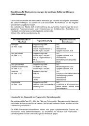
Figure 1. IIFT of index patient’s serum: a, b: In house cultivated HEp-2 cells. Anti-IM<strong>PD</strong>H2 reacts with distinct cytoplasmic<br />
granules and condensed granular aggregates. c, d: Two commercial HEp-2 cell preparations (c: Euroimmun, d: Inova Diagnostics)<br />
exhibiting the “rods and rings” fluorescence pattern. e, f: In house cultivated HEp-2 cells addition of (e) mycophenolic<br />
acid or (f) ribavirin into the culture medium. Both mycophenolic acid and ribavirin shift the granular fluorescence pattern<br />
(seen in a and b) to “rods and rings”. g, h: IIFT of rabbit anti-IM<strong>PD</strong>H2 Si on (g) in house cultivated and (h) commercial (Inova<br />
Diagnostics) HEp-2 cells, exhibiting identical fluorescence patterns as seen with the serum of the index patient.<br />
Zeiss Axioplan 2, magnifications 400 x.<br />
only a slight increase of granular aggregates within the<br />
cytoplasm of cells cultivated for prolonged periods (>20<br />
hours).<br />
IM<strong>PD</strong>H2 constitutes the target antigen<br />
The immunofluorescence pattern observed with the index<br />
patient’s serum could not be attributed to antibodies<br />
against one of the major known cytoplasmic autoantigens<br />
(actin, tubulin, mitochondria, microsomes, ribosomes,<br />
lysosomes, endosomes, tRNA synthetases, Golgi<br />
proteins) either by IIFT or by appropriate antibody specific<br />
assays. However, the serum precipitated a substantial<br />
amount of a 55 kDa 35 S-methionine-labeled protein<br />
from HEp-2 cell extract (Figure 3). Screening a cDNA<br />
expression library revealed three clones encoding IMP-<br />
DH2 and spanning the complete coding region of 1545<br />
nucleotides including 74 nucleotides of the 5´-untranslated<br />
region and 52 nucleotides of the 3´-untranslated<br />
sequence (positions 19 to 1689).<br />
Direct confirmation of IM<strong>PD</strong>H2 as target antigen was<br />
made by the positive reaction of the index patient’s serum<br />
with recombinant IM<strong>PD</strong>H2 in western and line<br />
blots (Figures 3a, 3b) and with 35 S-methionine-IMP-<br />
DH2 in RIPA (Figure 4). As indirect confirmation, a<br />
757<br />
commercial rabbit anti-IM<strong>PD</strong>H2 (anti-IM<strong>PD</strong>H2Si) reacted<br />
essentially in the same way in IIFT as the index<br />
patient’s serum (Figure 1g, 1h). The in house rabbit<br />
anti-IM<strong>PD</strong>H2 (anti-IM<strong>PD</strong>H2ih) irrespective of its reactivity<br />
with His(6)-tag-IM<strong>PD</strong>H2 (Figure 3a, lane 7),<br />
commercial IM<strong>PD</strong>H2 and 35 S-methionine-IM<strong>PD</strong>H2<br />
(Figure 4) was, however, unable to recognize IM<strong>PD</strong>H2<br />
in any preparation of HEp-2 cells. This observation suggests<br />
that this antibody was unable to recognize epitopes<br />
being presented by intracellular IM<strong>PD</strong>H2 (Table<br />
1).<br />
Mycophenolic acid and ribavirin rearrange intracellular<br />
IM<strong>PD</strong>H2<br />
The reason for the different arrangements of IM<strong>PD</strong>H2<br />
observed within the cytoplasm of in house cultivated<br />
HEp-2 cells and the two commercial preparations is unknown.<br />
It may be caused by additives within the culture<br />
media, not disclosed by the manufacturers. As could be<br />
shown recently (19), addition of mycophenolic acid to<br />
culture cells induced a rearrangement of cytoplasmic<br />
IM<strong>PD</strong>H2 from granular particles into the “rods and<br />
rings”. We could show this transition of IM<strong>PD</strong>H2 after<br />
addition of therapeutic concentrations (12.49 µmol) of
H. P. SEELIG et al.<br />
Figure 2. Radioimmunoprecipitation of 35 S-methionine labeled HEp-2 cell proteins by the serum of the index patient (lanes 3,<br />
4) precipitating a substantial amount of a 55 kDa protein. Control sera from two healthy blood donors (lanes 1, 2) and from<br />
two patients harboring antibodies against threonyl tRNA synthetase (PL7) with a calculated molecular weight of 82.1 kDa<br />
(lanes 5, 6). SDS page followed by autoradiography for 24 hours.<br />
mycophenolic acid to the cell cultures (Figure 1e). Because<br />
of the suspected prevalence of anti-IM<strong>PD</strong>H2 antibodies<br />
in patients with HCV infections and the therapeutic<br />
use of another IM<strong>PD</strong>H2 inhibitor, ribavirin, in<br />
these patients, we were also interested, whether ribavirin<br />
can cause an identical rearrangement of cytoplasmic<br />
IM<strong>PD</strong>H2. As can be seen from Figure 1f, ribavirin in<br />
concentrations of therapeutic serum levels (16.38 µmol)<br />
also induces a prompt rearrangement of IM<strong>PD</strong>H2 in<br />
“rods and rings” in cultivated HEp-2 cells studied 24<br />
hours after addition.<br />
Evaluation of possible assays for detection of anti-<br />
IM<strong>PD</strong>H2 in human sera<br />
Searching for an appropriate assay for screening of anti-<br />
IM<strong>PD</strong>H2 in human sera, the following approaches were<br />
followed:<br />
First, His(6)-tag-IM<strong>PD</strong>H2, synthesized in E. coli and<br />
affinity purified on Ni2 + -agarose was used for western<br />
and line blots. Second, full length 35 S-methionine labeled<br />
IM<strong>PD</strong>H2 was synthesized by in vitro transcripttion/translation<br />
and applied in RIPA.<br />
As shown by SDS-PAGE (Figure 3a, lane A) the purity<br />
of His(6)-tag-IM<strong>PD</strong>H2 was >95 %, comparable to that<br />
of a commercially available recombinant enzyme. The<br />
electrophoretic mobility corresponded well with the calculated<br />
molecular weight of 55 kDa.<br />
758<br />
Blotted on nitrocellulose (Figure 3a, lanes 1 - 9),<br />
His(6)-tag-IM<strong>PD</strong>H2 was reactive with the index patient’s<br />
serum (lane 5), rabbit anti-IM<strong>PD</strong>H2Si (lane 7),<br />
rabbit anti-IM<strong>PD</strong>H2ih (lane 8) and rabbit anti-His(6)-tag<br />
(lane 9) but not with sera of three healthy blood donors<br />
(lanes 1 - 3) and a serum of a HCV-RNA positive but<br />
IIFT negative patient (lane 4). As compared to the index<br />
patient (lane 5), the serum of one of the four HCV-RNA<br />
positive patients (lane 6) exhibiting the same two-tiered<br />
immunofluorescence pattern, suggestive of anti-IM<strong>PD</strong>-<br />
H2, showed a rather weak reactivity irrespective of IIFT<br />
antibody titer comparable to that of the index serum<br />
(1:1280). Identical results were obtained with a commercial<br />
IM<strong>PD</strong>H2 preparation, except for a missing reaction<br />
with anti-His(6)-tag. The disparity of reaction<br />
strength of the index serum and that of the HCV-RNA<br />
positive patient seen in Figure 3a (lanes 5, 6) is also re-<br />
lated to the recombinant commercial antigen lacking the<br />
His(6)-tag.<br />
Applying this kind of western blot for screening of anti-<br />
IM<strong>PD</strong>H2 in 108 HCV-RNA positive patients only one<br />
borderline and two weakly positive reacting sera could<br />
be detected. As revealed by IIFT and RIPA (vide infra)<br />
one of these two sera (Figure 3a, line 6) showed the<br />
typical anti-IM<strong>PD</strong>H2 immunofluorescence pattern (titer<br />
1:1280) and a positive RIPA (ABR 87.7). The second<br />
serum and the borderline one, however, were nonreac-<br />
Clin. Lab. 9+10/2011
Clin. Lab. 9+10/2011<br />
AUTOANTIBODIES AGAINST IM<strong>PD</strong>H2 AND HCV INFECTION<br />
Figure 3. a: SDS-Page of His(6)-tag-IM<strong>PD</strong>H2 (lane A) and 35 S-methionine labeled IM<strong>PD</strong>H2 synthesized by in vitro translation/<br />
transcription (lane B) for purity assessment (lane M = molecular weight markers). Lanes 1 – 9 depict His(6)-tag-IM<strong>PD</strong>H2<br />
blotted on nitrocellulose and reacted with control sera of three healthy blood donors (lanes 1-3), serum of a HCV-RNA positive<br />
patient with negative immunofluorescence staining (lane 4), serum of index patient (lane 5), serum of a HCV-RNA positive patient<br />
showing an immunofluorescence pattern suspicious of anti-IM<strong>PD</strong>H2 (lane 6), rabbit anti-IM<strong>PD</strong>H2 ih (lane 7), rabbit anti-<br />
IM<strong>PD</strong>H2 Si (lane 8), and rabbit anti His(6)-tag (lane 9). The purity of the His(6)-tag-IM<strong>PD</strong>H2 (lane A) is >95 %. Whereas the<br />
reaction of the index patient (lane 5) is rather strong, the reaction with the other IIFT positive patient (lane 6) is rather weak.<br />
ih = in house, Si = Sigma.<br />
b: Line blots containing four lines of 4, 8, 16, and 32 ng spotted His(6)-tag-IM<strong>PD</strong>H2 respectively and reacted with serum of a<br />
blood donor (lane 1), serum of the index patient (lane 2), and sera of HCV-RNA positive patients showing an immunofluorescence<br />
pattern suspicious for anti-IM<strong>PD</strong>H2 (lanes 3 – 6). The control line contains spotted anti-human IgG. Only the index patient<br />
reacts with the spotted His(6)-tag-IM<strong>PD</strong>H2. No reaction was seen with the 4 other sera, also containing anti-IM<strong>PD</strong>H2 as<br />
demonstrated by RIPA (Figure 4).<br />
tive in IIFT as well as in RIPA.<br />
The disparity of the reaction strength of His(6)-tag-IM-<br />
<strong>PD</strong>H2 with the serum of the index patient and that of<br />
IIFT and HCV-RNA positive patients increased considerably<br />
when using line blots. Whereas an unchanged<br />
strong reaction was revealed by the index patient (Figure<br />
3b, lane 2), neither the four HCV-RNA positive sera<br />
detected by immunofluorescence (lanes 3 – 6) nor any<br />
one of the other 104 HCV-RNA containing sera reacted<br />
with spotted IM<strong>PD</strong>H2 in line blots.<br />
The disappointing results obtained with western and<br />
line blots prompted the design of a RIPA using 35 S-methionine<br />
labeled IM<strong>PD</strong>H2 produced by in vitro transcription/translation.<br />
Using this kind of antigen not only<br />
the index patent’s serum but also the four HCV-RNA<br />
positive sera, depicted in Figure 3b (lanes 3 - 6) as well<br />
759<br />
as the two rabbit sera (anti-IM<strong>PD</strong>H2Si, anti-IM<strong>PD</strong>H2ih)<br />
precipitated 35 S-metionine-IM<strong>PD</strong>H2 in a dose dependent<br />
manor (Figure 4). This data strongly suggests that<br />
the RIPA is more suitable for anti-IM<strong>PD</strong>H2 screening<br />
in human sera than western or line blots. A summary of<br />
the reactivity of the various antigens with human sera<br />
and rabbit antibodies <strong>und</strong>er different test conditions is<br />
given in Table 1.<br />
High prevalence of anti-IM<strong>PD</strong>H2 in HCV-RNA carriers<br />
To evaluate the prevalence of anti-IM<strong>PD</strong>H2 in HCV-<br />
RNA carriers, 108 HCV-RNA positive sera were<br />
screened by RIPA and compared with sera of patients<br />
negative for HCV-RNA, patients suffering from HBV<br />
infections, autoimmune liver disease, and autoimmune
H. P. SEELIG et al.<br />
Figure 4. RIPA of serial dilutions of the serum of the index patient (2), of rabbit anti-IM<strong>PD</strong>H2 Si (1), rabbit anti-IM<strong>PD</strong>H2 ih (3)<br />
and of four HCV-RNA positive sera (thin dotted lines) showing an immunofluorescence pattern suspicious of containing anti-<br />
IM<strong>PD</strong>H2. Rabbit sera were diluted with preimmune serum, human sera with that of a blood donor. All sera show a dose-dependent<br />
precipitation of 35 S-methionine-IM<strong>PD</strong>H2.<br />
rheumatic diseases and healthy controls. The cut-off between<br />
anti-IM<strong>PD</strong>H2 positive and negative patients was<br />
determined by receiver operator curves of 108 HCV-<br />
RNA carriers and 100 healthy blood donors (Figure 5).<br />
The area <strong>und</strong>er the curve (AUC) was calculated 0.762<br />
representing fairly good test conditions. The 95 % confidence<br />
interval was 0.698 to 0.819. Defining an assay<br />
specificity of 98 % the cut off was calculated at an antibody<br />
ratio (ABR) of 10.5. Under these settings anti-IM-<br />
<strong>PD</strong>H2 could be detected in HCV-RNA positive sera<br />
with a sensitivity of 35.2 %. However, there was no correlation<br />
between HCV-virus load and the presence or<br />
absence of anti-IM<strong>PD</strong>H2 in HCV-RNA carriers. The<br />
mean virus load in anti-IM<strong>PD</strong>H2 positive HCV-RNAcarriers<br />
was 1.4 x 10 6 IU/mL, mean virus load in anti-<br />
IM<strong>PD</strong>H2 negative HCV-RNA carriers was 1.6 x 10 6<br />
IU/mL. There was also no correlation between the concentration<br />
of anti-IM<strong>PD</strong>H2 (given as antibody ratio)<br />
and virus load (R 2 of the linear regression = 0.0026).<br />
As depicted in Figure 6, anti-IM<strong>PD</strong>H2 also could be detected<br />
in blood donors (2.0 %), HCV-RNA-negative patients<br />
(5.0 %), HBV-DNA positive patients (6.2 %), in<br />
patients positive for antinuclear antibodies (13.7 %),<br />
and in a high number of patients with actin antibodies<br />
760<br />
(31.0 %). The prevalence of anti IM<strong>PD</strong>H2 in patients<br />
with mitochondrial antibodies of anti-M2 type (3.2 %)<br />
was relatively low. Regarding antibody concentrations,<br />
high ABRs >20 were fo<strong>und</strong> only in HCV-RNA positive<br />
patients except for one HCV-RNA negative (ABR<br />
44.9), one blood donor (ABR 39.6) and the index patient<br />
(ABR 61.9), who also was negative for HCV-<br />
RNA. When anti-IM<strong>PD</strong>H2 positive sera (ABR >20)<br />
were retested by IIFT only sera harboring antibody concentrations<br />
of ABR >35 showed a positive immunofluorescence<br />
test with a granular pattern on in house and a<br />
“rings and rods” pattern on the two commercial HEp-2<br />
cell preparations.<br />
An ABR >20 was seen only in 11.1 % of the HCV-<br />
RNA positive patients. There were no major differences<br />
regarding age and sex of the antibody positive and antibody<br />
negative HCV-RNA positive patients. Mean age<br />
of anti-IM<strong>PD</strong>H2 negative patients was 49.6 years (67 %<br />
male, 33 % female), mean age of anti-IM<strong>PD</strong>H2-positive<br />
patients was 50.6 years (75 % male, 25 % female). All<br />
patients were subjected to a therapy of interferon and ribavirin<br />
either actually or months or years before obtaining<br />
the sera used for this screening.<br />
Clin. Lab. 9+10/2011
Clin. Lab. 9+10/2011<br />
AUTOANTIBODIES AGAINST IM<strong>PD</strong>H2 AND HCV INFECTION<br />
Table 1. Overview of reactivity of anti-IM<strong>PD</strong>H2 from index patient, HCV-RNA carriers, and rabbits seen with different IMP-<br />
DH2 antigens studied by IIFT, western blot, line blot, and RIPA. Only 52.7 % of RIPA positive sera exhibiting an ABR > 20<br />
revealed a positive IIFT and only two of 38 RIPA positive anti-IM<strong>PD</strong>H2 sera of HCV-RNA carriers showed a rather poor reaction<br />
on western blot (+) and none of these 38 sera reacted on line blots. There was one serum weakly positive in the western<br />
blot, which, however, did not react in RIPA (ABR = 6,8). [RIPA] = radioimmunoprecipitation assay; [iv TT] = IM<strong>PD</strong>H2 generated<br />
by in vitro transcription – translation.<br />
Method Antigens<br />
Serum<br />
Index patient<br />
761<br />
Sera<br />
HCV-RNA +<br />
Rabbit<br />
anti-IM<strong>PD</strong>H Si<br />
Rabbit<br />
anti-IM<strong>PD</strong>H ih<br />
IIFT pattern HEp-2 cells [in house] granular granular granular Ø<br />
IIFT pattern HEp-2 cells [commercial] rings/rods rings/rods rings/rods Ø<br />
Western blot IM<strong>PD</strong>H2 [His(6)-tag] +++ Ø -+ +++ +++<br />
Western blot IM<strong>PD</strong>H2 [commercial] +++ Ø -+ +++ +++<br />
Line blot IM<strong>PD</strong>H2 [His(6)-tag] +++ Ø +++ +++<br />
RIPA IM<strong>PD</strong>H2 [iv TT] +++ +++ +++ +++<br />
Figure 5. Receiver operator curve of anti-IM<strong>PD</strong>H2 measured by RIPA and expressed in antibody ratios (ABR) of HCV-RNA<br />
negative control sera (healthy blood donors) as compared to HCV-RNA positive patients. Assuming an assay specificity of<br />
98 % the sensitivity for detection of anti-IM<strong>PD</strong>H2 in 108 HCV-RNA carriers was 35 %. Diagonal line indicates no discrimination,<br />
AUC = area <strong>und</strong>er the curve, SE = standard error of AUC, CI = lower and upper confidence interval.
H. P. SEELIG et al.<br />
Figure 6. Prevalence of anti-IM<strong>PD</strong>H2 in different groups of patients. Black boxes indicate the absolute numbers of negative patients<br />
(cut off: ABR
(36). Obviously, this state of molecule rearrangement<br />
may be induced also by other yet unknown factors as<br />
exemplified by its existence in certain commercial diagnostic<br />
HEp-2 cell preparations, unlikely having been<br />
subjected to prior MPA or ribavirin exposure. In this<br />
latter case the HEp-2 cell cultures may have been manipulated<br />
in an unknown manner, inducing the transition<br />
of IM<strong>PD</strong>H2 into the “rods and rings” state. As far<br />
as routine immunofluorescence tests are concerned,<br />
these observations and the fact that five out of seven<br />
commercial HEp-2 cell preparations were not able to<br />
detect anti-IM<strong>PD</strong>H2 exemplify to what extent different<br />
commercial cell substrates impinge on results and interpretations<br />
of the tests (Figure 1).<br />
As far as anti-IM<strong>PD</strong>H2 itself is concerned, certain differences<br />
may exist in its epitope specificity. Antibodies<br />
fo<strong>und</strong> in HCV-RNA carriers differ at least from those of<br />
the index patient in that they react preferentially with<br />
conformational epitopes.<br />
This is indicated by the fact that the index patient recognized<br />
recombinant IM<strong>PD</strong>H2 expressed in E. coli and<br />
subjected to denaturing conditions of western blot,<br />
whereas the antibodies fo<strong>und</strong> in HCV-RNA carriers,<br />
despite high antibody titers, showed only a very weak<br />
(western blots) or no reaction (line blots) with this<br />
source of antigen. However, the antibodies recognize<br />
IM<strong>PD</strong>H2 synthesized by in vitro transcription and translation<br />
in rabbit reticulocyte lysate and therefore probably<br />
displaying a higher degree of conformational structure.<br />
For this reason RIPA was used in this study for the<br />
evaluation of the prevalence of anti-IM<strong>PD</strong>H2. The<br />
existence of a broad diversity in epitope and conformational<br />
recognition also may be seen in the observation<br />
that the in house rabbit anti-IM<strong>PD</strong>H2 recognized the recombinant<br />
antigens in western blot and RIPA but neither<br />
the natural nor the “rods and rings” forms of IMP-<br />
DH2 in HEp-2 cells. Differences in epitope specificity<br />
also may be responsible for the low prevalence of anti-<br />
IM<strong>PD</strong>H2 (4.76 %) in patients with autoimmune hepatitis<br />
detailed by Quing at al. (10) using immunoproteomics,<br />
principally based on the western blot technology,<br />
which has been shown in this study to be less suited<br />
than the RIPA assay for detection of these antibodies.<br />
Using the immunoprecipitation assay, we detected anti-<br />
IM<strong>PD</strong>H2 in low concentrations in 31 % of the patients<br />
with suspected autoimmune hepatitis. The high prevalence<br />
of anti-IM<strong>PD</strong>H2 in patients with colorectal carcinoma<br />
demonstrated by immunoproteomics by He et al.<br />
(11), on the other hand, may speak in favor of the existence<br />
in those patients of anti-IM<strong>PD</strong>H2 preferentially<br />
reacting with denatured antigens. To clarify these presumptions,<br />
however, detailed studies of epitope specificities<br />
in various groups of anti-IM<strong>PD</strong>H2 positive patients<br />
are necessary.<br />
The reason for the high prevalence of anti-IM<strong>PD</strong>H2 in<br />
HCV-RNA carriers remains obscure. Generally, the occurrence<br />
of a new autoantibody in patients with HCVinfections<br />
is not very surprising since an augmentation<br />
of immunological disorders (e. g. mixed cryoglobulin-<br />
AUTOANTIBODIES AGAINST IM<strong>PD</strong>H2 AND HCV INFECTION<br />
Clin. Lab. 9+10/2011 763<br />
emia, thyroid disorders, arthritis, vasculitis, Sjögren<br />
syndrome, lung fibrosis) (37 - 39), and of autoantibodies<br />
such as smooth muscle antibodies, rheumatoid<br />
factor, liver-kidney-microsomal antibodies, anticardiolipin,<br />
antinuclear antibodies, cryoglobulins, thyroid<br />
antibodies or ribosomal P protein antibodies (12–<br />
17,40,41) as well as a correlation between the occurrence<br />
of antibodies and disease activity (42,43) have<br />
been proposed. Furthermore, treatment with interferonalpha<br />
commonly used in HCV-RNA carriers may render<br />
them more susceptible for developing autoantibodies<br />
and less frequently autoimmune diseases (e. g.<br />
Hashimoto thyroiditis, lupus erythematosus or type 1<br />
diabetes) (47-49).<br />
The reported figures for the prevalence of autoantibodies<br />
in HCV infections vary considerably (e. g. antinuclear<br />
antibodies 10 – 21 %, smooth muscle antibodies<br />
10 – 55 %) whereas in patients lacking active liver disease<br />
the prevalence of autoantibodies seems not to be<br />
significantly increased as compared to healthy controls<br />
(43-46). It has been suggested, that for antibody development<br />
host features especially continuous liver cell<br />
damage and hepatocyte necrosis are likely to be more<br />
important than viral factors, e. g. HCV-genotype (46).<br />
In line with this, we also did not find a correlation between<br />
the development of anti-IM<strong>PD</strong>H2 and virus load.<br />
Ribavirin, widely used in therapy for HCV infection, introduces<br />
profo<strong>und</strong> alterations of the intracellular distribution<br />
of IM<strong>PD</strong>H2 and therefore possibly enhances its<br />
immunogenicity. According to our information all anti-<br />
IM<strong>PD</strong>H2 positive patients were subjected to ribavirin<br />
therapy, but detailed data concerning the time elapsed<br />
between therapy and antibody testing or the duration of<br />
therapy etc. could not be made available. A vague link<br />
between ribavirin and anti-IM<strong>PD</strong>H2 may be seen in the<br />
trend of a lower HCV-RNA load in patients exhibiting<br />
higher anti-IM<strong>PD</strong>H2 levels (Figure 6). To answer these<br />
questions, prospective studies considering therapy, activity<br />
of liver disease, and concomitant autoimmune<br />
phenomena are needed.<br />
A high prevalence (31 %) of anti-IM<strong>PD</strong>H2 in low concentrations<br />
(Figure 6) also was fo<strong>und</strong> in patients with<br />
clinical suspicion of autoimmune hepatitis (AIH) and<br />
harboring actin-antibodies (titer 1: >80), demonstrated<br />
by IIFT (liver of phalloidin treated mice and vinblastine<br />
treated HEp-2). However, detailed information concerning<br />
the state of liver disease (activity, remissions)<br />
and therapy were not available, neither from these patients<br />
nor from those with the suspicion of primary biliary<br />
cirrhosis (PBC) harboring mitochondrial antibodies<br />
of type M2-specificity. In general, the casual occurrence<br />
of anti-IM<strong>PD</strong>H2 or other autoantibodies (10) in patients<br />
with AIH or PBC, intrinsically prone to exhibit autoimmune<br />
phenomena, may not be surprising. Based on our<br />
more “hypothetical” diagnoses, it seems that in AIH<br />
with a rather high prevalence of low titer anti-IM<strong>PD</strong>H2<br />
specific pathological processes may govern the formation<br />
of anti-IM<strong>PD</strong>H2 not prevailing in PBC where the<br />
prevalence of anti-IM<strong>PD</strong>H does not exceed that of con-
trols (HCV-RNA negatives, blood donors). Whether the<br />
development of anti-IM<strong>PD</strong>H2 in AIH may be caused by<br />
particular therapeutic management, e. g. mycophenolate,<br />
remains obscure. Sole liver damage, on the other<br />
side, may not be sufficient for the generation of anti-<br />
IM<strong>PD</strong>H2, since its prevalence in HBV-DNA carriers<br />
did not exceed the controls. As demonstrated by the occurrence<br />
of the “rings and rods” fluorescence pattern in<br />
culture cells, the intracellular aggregation of IM<strong>PD</strong>H2<br />
may also be caused by further unknown pharmacological<br />
or pathological processes. Thus one might speculate<br />
with regard to the therapeutic interventions of inhibition<br />
of IM<strong>PD</strong>H2 by ribavirin in patients with chronic<br />
HCV infections and in some autoimmune liver diseases<br />
by mycophenolic acid (30, 31), that the development<br />
of autoantibodies against IM<strong>PD</strong>H2 may be related<br />
to therapeutic procedures or to other hitherto unknown<br />
pathological processes causing intracellular aggregation<br />
of IM<strong>PD</strong>H2.<br />
Acknowledgement:<br />
We greatly appreciate the help of Mr. Peter Charles,<br />
Kennedy Institute, London, who thoroughly corrected<br />
any English spelling and grammar errors.<br />
Declaration of Interest:<br />
The authors declare that to the best of their knowledge<br />
there are no current or potential conflicts of interests.<br />
References:<br />
1. Blüthner M, Appelhans H, Moosbrugger I, Wiemann C, Lenz D,<br />
Seelig H.-P. Identification of Inosine-1,2-monophosphate dehydrogenase<br />
as a novel autoantigen. In: Conrad K, Chan , EKL,<br />
Fritzler M.J, Humbel R.L, von Landenberg P, Shoenfeld Y (Eds.)<br />
From pathogenesis to therapy of autoimmune diseases. Report on<br />
the 9th <strong>Dr</strong>esden Symposium on autoantibodies 2009;106-7.<br />
2. Senda M, Natsumeda Y. Tissue-differential expression of two<br />
distinct genes for human IMP dehydrogenase (E.C.1.1.1.205).<br />
Life Sci 1994;54:1917-26.<br />
3. Jackson RC, Weber G, Morris HP. IMP dehydrogenase, an enzyme<br />
linked with proliferation and malignancy. Nature 1975;<br />
256:331-3.<br />
4. Itoh O, Kuroiwa S, Atsumi S, Umezawa K, Takeuchi T, Hori M.<br />
Induction by the guanosine analogue oxanosine of reversion<br />
toward the normal phenotype of K-ras-transformed rat kidney<br />
cells. Cancer Res 1989;49:996-1000.<br />
5. Collart FR, Chubb CB, Mirkin BL, Huberman E. Increased inosine-5′-phosphate<br />
dehydrogenase gene expression in solid tumor<br />
tissues and tumor cell lines. Cancer Res 1992;52:5826-8.<br />
6. Hager PW, Collart FR, Huberman E, Mitchell BS. Recombinant<br />
human inosine monophosphate dehydrogenase type I and type II<br />
proteins. Purification and characterization of inhibitor binding.<br />
Biochem Pharmacol 1995;49:1323-9.<br />
H. P. SEELIG et al.<br />
764<br />
7. Zimmermann A, Gu JJ, Spychala J, Mitchell BS. Inosine monophosphate<br />
dehydrogenase expression: transcriptional regulation<br />
of the type I and type II genes. Adv Enzyme Regul 1996;36:75-<br />
84.<br />
8. Brouwer C, Vermunt-de Koning DG, Trueworthy RC, et al.<br />
Monitoring of inosine monophosphate dehydrogenase activity in<br />
mononuclear cells of children with acute lymphoblastic leukemia:<br />
enzymological and clinical aspects. Pediatr Blood Cancer 2006;<br />
46:434-8.<br />
9. Floryk D, Thompson TC. Antiproliferative effects of AVN944, a<br />
novel inosine 5-monophosphate dehydrogenase inhibitor, in prostate<br />
cancer cells. Int J Cancer 2008; 123: 2294-2302.<br />
10. Qing Xia, Feng Lu, Hui-Ping Yan, et al. Autoantibody profiling<br />
of Chinese patients with autoimmune hepatitis using immunoproteomic<br />
analysis. J Proteom Res 2008;7:1963-70.<br />
11. He Y, , Mou Z, Li W, et al. Identification of IM<strong>PD</strong>H2 as a tumorassociated<br />
antigen in colorectal cancer using immunoproteomics<br />
analysis. Int J Colorectal Dis 2009;24:1271-9.<br />
12. Abuaf N, Lunel F, Giral P, et al. Non-organ specific autoantibodies<br />
associated with chronic C virus hepatitis. J Hepatol 1993;<br />
18:359-64.<br />
13. Clifford BD, Donahue D, Smith L, et al. High prevalence of serological<br />
markers of autoimmunity in patients with chronic hepatitis<br />
C. Hepatology 1995;21:613-9.<br />
14. De Larrañaga GF, Harris N, Pierangeli SS, Alonso BS, Schroder<br />
T, Fainboim H. Low prevalence of autoimmune antiphospholipid<br />
antibodies in hepatic diseases. Medicina 2000;60:919-22.<br />
15. Ordi-Ros J, Villarreal J, Monegal F, Sauleda S, Esteban I, Vilardell<br />
M. Anticardiolipin antibodies in patients with chronic heaptitis<br />
C virus infection: Characterization in relation to antiphospholipid<br />
syndrome. Clin Diagn Lab Immunol 2000;7:241-4.<br />
16. Kessenbrock K, Fritzler MJ, Groves M, et al. Diverse humoral<br />
autoimmunity to the ribosomal P proteins in systemic lupus erythematosus<br />
and hepatitis C virus infection. J Mol Med 2007;9:<br />
953-9.<br />
17. Ferri S, Muratori L, Lenzi M, Granito A, Bianchi FB, Vergani D.<br />
HCV and autoimmunity. Curr Pharm Des 2008;14,1678-85.<br />
18. Dellavance A, Júnior AG, Nuccitelli B, et al. Third Brazilian<br />
Consensus for autoantibodies screening in HEp-2 cells (ANA).<br />
Recommendations for standardization of autoantibodies screening<br />
trial in HEp-2 cells, quality control and clinical associations. Rev<br />
Bras Reumatol 2009;49:99-109.<br />
19. Ji YS, Gu JJ, Makhov AM, Griffith JD, Michell BS. Regulation<br />
of the interaction of inosine monophosphate dehydrogenase with<br />
mycophenolic acid by GTP. J Biol Chem 2006;281:206-12.<br />
20. Tan EM. Antinuclear antibodies: diagnostic markers and clues to<br />
the basis of systemic autoimmunity. Pediatr Infect Dis 1998;7,S3-<br />
S9.<br />
21. Tan EM. Interactions between autoimmunity and molecular and<br />
cell biology. J Clin Invest 1989;84:1-6.<br />
22. Fritzler MJ, Salazar M. Diversity and origin of rheumatologic<br />
autoantibodies. Clinical Microbiology Reviews 1991;4:256-69.<br />
23. Elkon K. Autoantibodies in systemic lupus erythematosus. Curr<br />
Opin Rheum 1995;7:384-8.<br />
Clin. Lab. 9+10/2011
24. Gilbert D, Brard F, Fabien J, Tron F. Do naturally occurring autoantibodies<br />
participate in the constitution of pathological B-cell repertoire<br />
in systemic lupus erythematosus? J Autoimmunity 1996;<br />
9:247-57.<br />
25. Seelig HP, Ehrfeld H, Renz M. Interferon-gamma-inducible protein<br />
p16 - A new target of antinuclear antibodies in patients with<br />
systemic lupus erythematosus. Arthritis Rheum 1994;37:1672-83.<br />
26. Sambrook J, Fritsch EF, Maniatis T. Molecular cloning: a laboratory<br />
manual. 1989; 2 nd Edition, Cold Spring Harbor <strong>Labor</strong>atory<br />
Press (New York).<br />
27. Towbin H, Staehelin T, Gordon J. Electrophoretic transfer of proteins<br />
from polyacrylamide gels to nitrocellulose sheets: procedure<br />
and some applications. Proc Natl Acad Sci (USA) 1979;9:4350-4.<br />
28. Frey A, Di Canzio J, Zurakowski, D. A statistically defined endpoint<br />
titer determination method for immunoassays. J Immunol<br />
Methods 1998;221:35-41.<br />
29. Hanley JA, McNeil BJ. The meaning and use of the area <strong>und</strong>er a<br />
receiver operating characteristic (ROC) curve. Radiology 1982;<br />
143:29-36.<br />
30. Jothimani D, Cramp M, Mitchell J, Cross T. The treatment of<br />
autoimmune hepatitis - a review of current and evolving therapies.<br />
J Gastroenterol Hepatol 2010;1440-46.<br />
31. Sharzehi K, Huang MA, Schreibman IR, Brown KA. Mycophenolate<br />
mofetil for the treatment of autoimmune hepatitis in patients<br />
refractory or intolerant to conventional therapy. Can J<br />
Gastroenterol 2010;24:588-92.<br />
32. Ishikawa, H. Mizoribine and mycophenolate mofetil. Curr Med<br />
Chem 1999,6:575-97.<br />
33. Nimmesgern E, Fox T, Fleming, MA, Thomson JA. Conformational<br />
changes and stabilization of inosine 5'- monophosphate<br />
dehydrogenase associated with ligand binding and inhibition by<br />
mycophenolic acid. J Biol Chem 1996;271:19421-7.<br />
34. Picardi A, Gentilucci UV, Zardi EM, D’Avola D, Amoroso A,<br />
Afeltra A. Curr Pharm Des 2004;10:2081-2092.<br />
35. Gish RG. Treating HCV with ribavirin analogues and ribavirinlike<br />
molecules. J Antimicrobial Chemotherapy 2006;57:8-13.<br />
36. Köhler GA, Gong X, Bentink S, et al. The functional basis of mycophenolic<br />
acid resistance in candida albicans IMP dehydrogenase.<br />
J Biol Chem 2005;280:11295-302.<br />
37. Pawlotsky JM, Ben Yahia M, Andre C, et al. Immunological disorders<br />
in C virus chronic active hepatitis: a prospective case-control<br />
study. Hepatology 1994;19:841-8.<br />
38. Adinolfi LE, Utili R, Attanasio V, et al. Epidemiology, clinical<br />
spectrum and prognostic value of mixed cryoglobulinemia in<br />
heapatitis C virus patients: a prospective study. Ital J Gastroenterol<br />
1996;28:1-9.<br />
Clin. Lab. 9+10/2011<br />
AUTOANTIBODIES AGAINST IM<strong>PD</strong>H2 AND HCV INFECTION<br />
765<br />
39. Wong VS, Egner W, Elsey T, Brown, D, Alexander GJ. Incidence,<br />
character and clinical relevance of mixed cryoglobulinemia<br />
in patients with chronic hepatitis C virus infection. Clin Exp<br />
Immunol 1996;104:25-31.<br />
40. Lenzi M, Johnson PJ, McFarlane IG, et al. Antibodies to hepatitis<br />
C virus in autoimmune liver disease: evidence for geographical<br />
heterogeneity. Lancet 1991;338:277-80.<br />
41. Cassani F, Muratori L, Manotti P, et al. Serum autoantibodies and<br />
the diagnosis of type-1 autoimmune hepatitis in Italy: a reappraisal<br />
at the light of hepatitis C virus infection. Gut 1992;33:1260-3.<br />
42. Cassani F, Cataleta M, Valentini P, et al. Serum autoantibodies in<br />
chronic hepatitis C: Comparison with autoimmune hepatitis and<br />
impact on the disease profile. Hepatology 1997;26:561-6.<br />
43. Lenzi M, Bellentani S, Saccoccio G, et al. Prevalence of nonorgan<br />
specific autoantibodies and chronic liver disease in the<br />
general population: a nested case-control study of the Dionysos<br />
cohort. Gut 1999;45:435-41.<br />
44. Czaja AJ, Carpenter HA, Santrach PJ, Moore SB, Taswell HF,<br />
Homburger HA. Evidence against hepatitis viruses as important<br />
causes of severe autoimmune hepatitis in the United States. J<br />
Hepatol 1993;18:342-52.<br />
45. Vergani D. NOSA in HCV infection: markers or makers of disease?<br />
Gut 1999;45:328-9.<br />
46. Muratori P, Muratori L, Stroffolini T, et al. Prevalence of nonorgan<br />
specific autoantibodies in HCV-infected subjects in the<br />
general population. Clin Exp Immunol 2003;131:118-21.<br />
47. Wilson LE, Widman D, Dikman SH, Gorevic <strong>PD</strong>. Autoimmune<br />
disease complicating antiviral therapy for hepatitis C virus infection.<br />
Semin Arthritis Rheum 2002;32:163-73.<br />
48. Pellicano R, Smedile A, Peyre S, et al. Autoimmune manifestations<br />
during interferon therapy in patients with chronic hepatitis<br />
C: the hepatologist's view. Minerva Gastroenterol Dietol 2005;51:<br />
55-61.<br />
49. Biggioggero M, Gabbriellini L, Meroni PL. Type I interferon<br />
therapy and its role in autoimmunity. Autoimmunity 2010;43:<br />
248-54.<br />
Correspondence:<br />
Prof. <strong>Dr</strong>. med. Hans-Peter Seelig<br />
Medizinisches Versorgungszentrum <strong>Labor</strong> Prof. Seelig<br />
Kriegsstraße 99, 76133 Karlsruhe, Germany<br />
Tel.: +49 721-85000-152<br />
Fax: +49 721-85000-115<br />
E-mail: labseelig@laborseelig.de