Carátula 2da parte - FBMC
Carátula 2da parte - FBMC
Carátula 2da parte - FBMC
You also want an ePaper? Increase the reach of your titles
YUMPU automatically turns print PDFs into web optimized ePapers that Google loves.
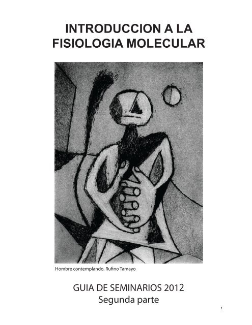
INTRODUCCION A LA<br />
FISIOLOGIA MOLECULAR<br />
Hombre contemplando. Runo Tamayo<br />
GUIA DE SEMINARIOS 2012<br />
Segunda <strong>parte</strong><br />
1
IFM 2012<br />
Día Fecha Prof Tema Seminarios TP/Seminarios<br />
Lu 08-oct feriado<br />
Ma 09-oct<br />
Mie<br />
10-oct FM<br />
Sistema respiratorio. Sangre. Intercambio de<br />
gases.<br />
TP. Fisio del reposo A<br />
Jue 11-oct TP. Fisio del reposo A<br />
Lu<br />
15-oct<br />
FM<br />
Sistema respiratorio. Sangre. Intercambio de<br />
gases.<br />
S9. Respiratorio<br />
Ma 16-oct 14hs Recuperatorio 1er parcial S9. Respiratorio<br />
Mie 17-oct LS Sistema Circulatorio: sangre y hemodinámica TP. Fisio del Reposo B<br />
Jue 18-oct TP. Fisio del Reposo B<br />
Lu 22-oct FM Sistema Circulatorio: corazón S10. Sangre-Corazón<br />
Ma 23-oct S10. Sangre-Corazón<br />
Mie<br />
24-oct LS<br />
Sistema Circulatorio: regulacion local del flujo<br />
sanguíneo<br />
TP.Fisio del ejercicio<br />
A<br />
Jue 25-oct TP.Fisio del ejercicioA<br />
Lu 29-oct LS Sistema excretor S11. Hemodinamia<br />
Ma 30-oct S11. Hemodinamia<br />
Mie<br />
31-oct LS<br />
Sistema Circulatorio: regulacion neural del<br />
flujo sanguíneo<br />
TP. Fisio del ejercicio<br />
B<br />
Jue 01-nov TP.Fisio del ejercicioB<br />
Lu<br />
05-nov LS<br />
Sistema endocrino. Control de glucemia.<br />
Regulación de la Temperatura<br />
S12. Excretor<br />
Ma 06-nov S12. Excretor<br />
Mie 07-nov LS Regulación autónoma. TP.Ejercicio-Análisis<br />
Jue 08-nov TP.Ejercicio-Análisis<br />
Lu 12-nov S13. Endocrino<br />
Ma 13-nov S13. Endocrino<br />
Mie 14-nov TP. Excretor<br />
Jue 15-nov TP. Excretor<br />
Lu 19-nov S14. Autónomo<br />
Ma 20-nov S14. Autónomo<br />
Mie 21-nov<br />
Jue 22-nov<br />
Lu 26-nov<br />
Ma 27-nov<br />
Mie 28-nov 2do parcial<br />
Jue 29-nov<br />
Lu 03-dic<br />
Ma 04-dic<br />
Mie 05-dic<br />
Jue 06-dic<br />
Lu 10-dic 14hs Recuperatorio 2do parcial<br />
Ma 11-dic<br />
Mie 12-dic<br />
Jue 13-dic
IFM 2012<br />
Seminario 9: Sistema respiratorio. Intercambio de gases.<br />
-Bibliografía Sugerida. Del libro “Principles of Human Physiology” de Stanfield &<br />
German: Capítulo 15, The respiratory system: pulmonary ventilation; y Capítulo 16:<br />
The respiratory system: Gas exchange and regulation of breathing.<br />
1. El sistema respiratorio se compone de una sucesión de compartimentos por los<br />
cuales circula el aire. Indique en un esquema los compartimentos de conducción y los<br />
canales en los que se produce el intercambio. ¿Cuál de estos compartimentos presenta<br />
la mayor superficie?<br />
2. ¿Qué función tienen y dónde se ubican las células goblet y las células ciliadas?<br />
3. Durante la respiración se produce un cambio rítmico en la presión alveolar.<br />
a. Dibuje la evolución de este ritmo e indique cuál fase corresponde a la inspiración y<br />
cuál a la espiración.<br />
b. ¿Qué fuerzas operan durante la inspiración y cuáles durante la espiración?<br />
c. ¿Qué elementos anatómicos periféricos y del sistema nervioso central están involucrados<br />
en la generación de estas fuerzas?<br />
4. El compliance (distensibilidad) pulmonar deriva de las propiedades mecánica de<br />
los alvéolos<br />
a. ¿Cómo se define este parámetro pulmonar?<br />
Utilizando un pulmón aislado se procede a inflarlo (Inf) y desinflarlo (Def) de aire<br />
(air) para medir el compliance. El mismo pulmón es sometido a cambios de volumen<br />
llenándolo y vaciándolo de solución salina (Saline). Las siguientes curvas muestran<br />
los resultados.<br />
b. Explique la diferencia del trayecto de las curvas de inflado y desinflado del<br />
pulmón.<br />
c. Explique la diferencia entre estas curvas y las mostradas por el pulmón cuando éste<br />
es llenado de solución salina.<br />
3
5. Los surfactantes afectan el ‘compliance’ pulmonar<br />
a. Explique de qué tipo de sustancia se trata.<br />
b. ¿Cuál es el efecto de los surfactantes sobre el compliance?<br />
c. ¿Cómo se alcanza este efecto?<br />
d. Dibuje, sobre las curvas de inflado y desinflado que se muestra en la pregunta 4,<br />
cómo se modificarían si se agregara un surfactante exógeno?<br />
6. En la siguiente figura se muestra la relación entre los cambios en el volumen<br />
pulmonar en función de la presión pulmonar para individuos sanos y para pacientes<br />
con fibrosis o con enfisema. La fibrosis se produce por el depósito de mucosa en el<br />
pulmón, mientras que en el enfisema se observa una destrucción de la matriz<br />
extracelular.<br />
¿Qué asociación hipotética puede realizar entre la breve descripción de la enfermedad<br />
que se dio más arriba y los efectos sobre el compliance?<br />
7. ¿Cuál es el problema respiratorio principal en bebés prematuros que carecen de<br />
surfactantes?<br />
8. Explicar cuáles son los pasos que permiten el transporte de oxígeno desde la<br />
atmósfera hacia los tejidos, en un mamífero, siguiendo la siguiente guía:<br />
a. ¿Cómo entra este gas a la sangre y cuál es la fuerza impulsora del transporte?<br />
b. ¿Cómo se transporta en la sangre?<br />
c. ¿Qué fuerza impulsora regula el flujo del oxígeno hacia los tejidos?<br />
9. Confeccionar una tabla indicando los valores de las presiones parciales del O 2 y el<br />
CO 2 en: el aire, la cavidad alveolar, la vena pulmonar y la arteria pulmonar. Explicar<br />
cuáles son las razones por las que se producen las variaciones observadas en cada<br />
compartimiento respecto del aire. ¿Por qué razón no tenemos en cuenta al gas<br />
mayoritario en el aire, el N 2?<br />
10. La hemoglobina es una proteína alostérica. Explique este concepto. ¿Cómo se<br />
expresa esta propiedad en su relación con el O 2?<br />
11. La mioglobina y la hemoglobina fetal tienen un comportamiento diferente como<br />
transportadores de O 2. Dibuje esquemáticamente un gráfico que muestre la relación<br />
entre el porcentaje de saturación de O 2 de la hemoglobina adulta, la fetal y la<br />
mioglobina en función de la presión de O 2 y explique de qué manera se expresa en<br />
este gráfico el carácter alostérico de la Hb.<br />
12. Cuando se habla del contenido de O 2 en la sangre, ¿se puede dar esta medida en<br />
mm de Hg? Explique su respuesta.<br />
4
13. El O 2 es transportado en la sangre por dos vías, mientras que el CO 2 es<br />
transportado por tres vías. ¿Cuáles son cada una de estas vías y qué porcentaje de cada<br />
gas es transportado por cada una de ellas?<br />
14. El eritrocito juega un rol fundamental en la remoción de CO 2 de los tejidos. ¿Qué<br />
fenómenos tienen lugar en estas células de la sangre que hacen más eficiente la<br />
remoción de CO 2? ¿Por qué no suceden en el plasma?<br />
15. En un vaso artificial que permite el flujo de O 2 a través de la pared del mismo<br />
fluye sangre oxigenada. El vaso fue colocado en un medio desoxigenado y a medida<br />
que la sangre fluía por el mismo se midió el nivel de saturación de oxígeno de la<br />
misma. Los resultados se muestran en la figura:<br />
a. ¿Cómo se explica la caída en la saturación de O 2 a lo largo del tubo?<br />
b. ¿Dónde espera que se observen estos efectos en el sistema circulatorio? ¿Por qué?<br />
Utilizando el mismo esquema experimental se midió cómo influye la velocidad del<br />
flujo en la desoxigenación de la sangre y se observaron los siguientes resultados.<br />
5
c. ¿Cómo se explica la relación oxigenación – velocidad del flujo?<br />
d. ¿Influye el hematocrito en esta relación?<br />
16. El monóxido de carbono es potentemente venenoso. ¿Cuál es la razón?<br />
17. El aumento en la concentración de dióxido de carbono trae aparejado un cambio<br />
en el pH de la sangre. ¿Es este cambio un aumento o una disminución? ¿A qué se<br />
debe que el cambio sea relativamente pequeño? El sistema de control de pH que<br />
involucra al CO 2 ¿es abierto o cerrado?, explique.<br />
18. El efecto Bohr es consistente con la necesidad de una mayor provisión de O 2 a los<br />
tejidos. Explicar esta afirmación.<br />
19. En un estudio se analiza el efecto del pH sanguíneo sobre el sistema respiratorio.<br />
En un animal anestesiado el pH sanguíneo es manipulado farmacológicamente, al<br />
tiempo en que se registra la actividad del nervio frénico (inerva al diafragma)<br />
mediante electrodos extracelulares que permiten evaluar la tasa de disparo en el<br />
mismo.<br />
a. ¿Qué cambios espera registrar en la actividad eléctrica del nervio frénico ante<br />
una disminución en el pH?<br />
b. ¿Qué efectos produce el cambio de actividad del nervio frénico en las fibras<br />
musculares del diafragma?<br />
c. ¿Cuál es el mecanismo por el cual se transmite la información desde el nervio<br />
frénico a las fibras del diafragma?<br />
6
IFM 2012<br />
Seminario 10: Sangre. Corazón.<br />
-Bibliografía Sugerida: ver Seminario 9. Capítulo 20: Cardiac electrophysiology and the<br />
electrocardiogram; Capítulo 21: The heart as a pump. Del libro: Medical Physiology de<br />
Boron & Boulpaep.<br />
Sangre<br />
1. Estudios experimentales mostraron que la altura respecto del nivel del mar produce un<br />
aumento en el hematocrito. Si tomamos como dato que la altura implica una disminución<br />
en la presión parcial de oxígeno, ¿cómo se explica el aumento en el hematocrito? ¿Qué<br />
rol juega el 2,3-difosfoglicerato (DPG) en la adaptación del organismo a la altura? Utilice<br />
las curvas de disociación en su explicación.<br />
2. En las siguientes curvas se estudió los niveles de oxigenación de la sangre en<br />
presencia de concentraciones crecientes de 2,3-difosfoglicerato.<br />
a. Explique el efecto del DPG.<br />
b. Indique cuál de las curvas<br />
corresponde a la mayor<br />
concentración de DPG, y cuál<br />
a la menor.<br />
3. La anemia nutricional se define como una deficiencia dietaria de hierro. En estos<br />
pacientes se observa un hematocrito normal pero un menor contenido de O2 en sangre.<br />
¿Cómo se pueden explicar estos resultados?<br />
4. ¿Cuáles son las funciones principales de las proteínas plasmáticas? ¿Cuál es la más<br />
abundante?<br />
5. El técnico de laboratorio encargado de realizar los análisis de sangre confundió un<br />
tubo de anticoagulante con otro que tenía el mismo anticoagulante con un agregado de<br />
7
NaCl. Al analizar el hematocrito del paciente obtuvo valores por debajo de los normales.<br />
Si la sangre analizada fuera la suya ¿empezaría la dieta del guiso de hígado y lentejas?<br />
6. Usted se inscribe para competir en el Tour de France y en un vestuario le recomiendan<br />
extraerse sangre y reinyectarse los glóbulos rojos el día anterior a la competencia. ¿Cuál<br />
es el objetivo de este “tip” deportivo (no se detecta en el antidoping)? ¿Sería realmente<br />
ventajoso?<br />
Corazón<br />
1. El corazón puede ser considerado como dos bombas actuando en serie, ¿cómo se<br />
explica esta aseveración?<br />
2. Los siguientes son componentes funcionales del ciclo cardíaco. Indique con números<br />
del 1 al 10 el orden en que se producen, siendo 1 el Llenado auricular.<br />
__ Contracción isovolumétrica<br />
__ Llenado ventricular<br />
__ Apertura de las válvula auriculoventricular<br />
__ Cierre de las válvulas auriculoventriculares<br />
__ Contracción ventricular<br />
__ Contracción auricular<br />
1_ Llenado auricular<br />
__ Apertura de las válvulas semilunares (de ventrículo a arterias)<br />
__ Cierre de las válvulas semilunares (de ventrículo a arterias)<br />
__ Relajación ventricular<br />
3. La presión atmosférica es de 760 mm Hg, mientras que la presión ventricular en su<br />
pico máximo es de 120 mm Hg. ¿Cómo se explica que el corazón no colapse bajo el gran<br />
gradiente de presión?<br />
4. Las fibras musculares cardíacas forman un sincicio funcional. Explique esta idea, y<br />
cuál es la función del mismo. Ahora, si es así, ¿por qué es necesario que las fibras<br />
musculares sean excitables, en lugar de seguir pasivamente al marcapasos?<br />
5. ¿En qué se diferencian los potenciales de acción de una fibra marcapasos, de los<br />
potenciales de acción de una fibra muscular cardiaca de la aurícula o el ventrículo?<br />
Responda en términos de: i) las conductancias involucradas; ii) la duración del potencial<br />
de acción<br />
6. La contracción de la musculatura estriada, lisa y cardiaca se inicia por la<br />
despolarización supraumbral de estas fibras musculares. Sin embargo, el fenómeno que<br />
da origen a dicha despolarización es diferente en cada caso. Explique el origen de la señal<br />
y su naturaleza.<br />
8
7. El acople excitación-contracción en el músculo cardíaco se da a través del aumento de<br />
la concentración intracelular de Ca 2+ ¿cuáles son las fuentes de Ca 2+ ? ¿cómo se vehiculiza<br />
el catión divalente desde estas fuentes al citosol? ¿cuál es su blanco de acción?<br />
8. Relacione las siguientes estructuras con los siguientes fenómenos: Estructuras: canal<br />
de calcio sensible a rianodina, canal de calcio de tipo L sensible al voltaje, troponina C.<br />
Fenómenos: contracción muscular, entrada de calcio del medio externo al citoplasma,<br />
“Ca ++ -spark”. En su respuesta ubique cada estructura en un compartimiento celular.<br />
9. ¿Qué es un “Ca ++ -spark”? ¿Cuáles son sus propiedades funcionales? ¿Cómo sirven<br />
estas propiedades a la función fisiológica que cumple el Ca ++ -spark?<br />
10. ¿Cuáles son los principales marcapasos del corazón de los mamíferos? ¿Cómo se<br />
comunican entre si? ¿Cuáles son sus respectivas funciones<br />
11. Está investigando las características de las fibras marcapasos con técnicas<br />
electrofisiológicas en el corazón de mamíferos. Ha impalado una fibra en un corazón<br />
aislado en una cámara especial de registro y esta fibra dispara en forma rítmica de manera<br />
espontánea. Tan excitado estás con este primer registro que asumís que, efectivamente,<br />
estás registrando una fibra marcapasos. ¿Alcanzan estos datos para aseverarlo? Justificar<br />
la respuesta.<br />
12. ¿Qué es el gasto cardíaco (cardiac output)? Este puede definirse desde un punto de<br />
vista hemodinámico o cardíaco ¿cuáles son las fórmulas que lo describen?<br />
13. La contracción auricular y la ventricular cardíaca están desfasadas en el tiempo.<br />
a. ¿Qué señal inicia la contracción auricular y cuál la ventricular?<br />
b. ¿Por qué es necesario este desfase?<br />
c. ¿Cómo se regula este desfase?<br />
d. ¿Cómo afecta este desfase la frecuencia cardiaca?<br />
e. ¿Qué efectos tiene sobre la frecuencia cardiaca el sistema autónomo?<br />
14. El flujo de sangre en el corazón está regulado por válvulas ¿Qué regula el cierre y<br />
apertura de las válvulas? Indique cuál de los siguientes parámetros cardíacos está más<br />
afectado por la eficiencia de la actividad de las válvulas: ciclo cardíaco y gasto cardíaco.<br />
Explique por qué.<br />
15. Durante el ciclo cardíaco, los períodos de sístole y diástole se asocian<br />
funcionalmente con los períodos de contracción y relajación ventricular, respectivamente.<br />
Si bien el estado de las aurículas es ignorado en dicha clasificación, ¿qué sucede con la<br />
presión auricular durante dichos períodos ventriculares?<br />
16. Aborde los aspectos de ciclo cardíaco que se observan en el siguiente diagrama:<br />
a. ¿Cómo se explica el retardo entre la fase de subida en la presión de la aorta y la fase de<br />
subida de la presión en el ventrículo?<br />
b. ¿Cómo se explica que la presión auricular y ventricular estén en antifase?<br />
9
c. Indique los períodos de contracción y relajación ventricular isovolumétricas.<br />
17. El siguiente gráfico muestra la variación de presión vs. volumen ventricular para un<br />
corazón normal (plano transparente) y para un corazón que padece estenosis mitral (plano<br />
gris).<br />
Nota: la válvula mitral es la que regula el flujo entre la aurícula y el ventrículo izquierdo.<br />
10
a. Indique cuál de las siguientes variables cambian con esta enfermedad:<br />
frecuencia cardíaca<br />
volumen sistólico<br />
presión sistólica<br />
b. De una explicación funcional a los cambios observados.<br />
11
IFM 2012<br />
Seminario 11: Hemodinámica<br />
-Bibliografía Sugerida: Medical Physiology de Boron & Boulpaep, Capítulo 17:<br />
Organization of the cardiovascular system, Capítulo 18: Arteries and veins; Capítulo 19:<br />
The microcirculation.<br />
-Bibliografía obligatoria: “Role of Shear Stress and Endothelial Prostaglandins in Flow-<br />
and Viscosity-Induced Dilation of Arteriole In Vitro”. Koller et al., Circulation Research<br />
72: 1276-1284 (1993)<br />
1. Los vasos del sistema circulatorio están conectados en configuraciones ‘en serie’ y ‘en<br />
paralelo’. Explique estos términos y dé ejemplos de tramos del sistema que operan en una<br />
y otra configuración.<br />
2. La ley de Poiseuille indica que el flujo depende de la presión y de la resistencia.<br />
a. ¿Cuál es la función que relaciona a estas dos variables? ¿qué factores de la sangre y del<br />
sistema vascular influyen en la resistencia?<br />
b. Se dice que el flujo de la sangre es mayoritariamente laminar ¿qué define a este tipo de<br />
flujo?<br />
c. ¿Cómo se define viscosidad?<br />
3. El efecto Fahraeus-Lindquist describe que la viscosidad aparente de la sangre que<br />
circula por tubos con diámetro muy pequeño (
9. Con el fin de investigar el flujo sanguíneo en los vasos del riñón se analizó cómo<br />
respondían segmentos de arterias aisladas ante flujos crecientes. Los parámetros que se<br />
midieron fueron presión y diámetro. Los resultados se muestran en la figura que se<br />
presenta más abajo. En condiciones control (Endothelium intact = endotelio intacto) se<br />
observa un aumento en el diámetro a medida que aumenta la presión, debido al aumento<br />
en el flujo.<br />
En otra series de experimentos se repitió el experimento en arterias en las cuales se dañó<br />
el endotelio (Endothelium disrupted = endotelio dañado) o en la que se expusieron las<br />
arterias intactas a un inhibidor de la síntesis de NO (L-NMMA).<br />
a. ¿Qué ilustran estos resultados?<br />
Utilizando este esquema experimental, y a partir de estos resultados, usted quiere analizar<br />
si el sistema autónomo tiene capacidad de regular este efecto local. Usted realiza esta<br />
investigación en el marco de su Tesina de Licenciatura y dado que se quiere recibir<br />
pronto sólo puede hacer dos series experimentales imprescindibles para dar una respuesta<br />
básica a su pregunta.<br />
b. ¿Qué experimentos realizaría? Explique el razonamiento que hizo al elegir cada<br />
tratamiento.<br />
13
IFM 2012<br />
Seminario 12: Sistema renal<br />
-Bibliografía Sugerida: Capítulo 17: The Urinary System: Renal Function y Capítulo<br />
18: The Urinary System: Fluid and Electrolyte Balance. Del libro: Human Physiology<br />
de Germann & Stanfield.<br />
1. Las presiones que se ponen en juego en el proceso de filtración glomerular son<br />
la hidrostática y la oncótica.<br />
a. Explicar el origen de cada una de estas presiones.<br />
b. ¿En qué dirección actúa la presión hidrostática prevalente, y en cuál la presión<br />
oncótica prevalente? Explicar esta direccionalidad.<br />
c. La presión osmótica que ejercen los iones no juega un rol importante en la<br />
filtración glomerular. Explicar la razón por la cual esta aseveración es correcta.<br />
2. Si disminuye la resistencia en la arteriola aferente, ¿qué espera observar, un<br />
aumento o una disminución en el volumen filtrado en el glomérulo? ¿Cómo varía su<br />
respuesta si lo que se produce es una disminución en la resistencia en la arteriola<br />
eferente? Explique sus respuestas.<br />
3. Teniendo en cuenta las consideraciones realizadas en la pregunta 2, explique de<br />
qué manera el reflejo miogénico puede ayudar a evitar que ante un aumento en la<br />
presión arterial se produzca una mayor filtración glomerular. ¿En qué arteriola espera<br />
que se produzca el reflejo miogénico, en la aferente o en la eferente, para lograr el<br />
efecto mencionado?<br />
4. ¿En qué porción del nefrón se produce la mayor reabsorción de H 2O, y en cuál se<br />
produce una regulación específica de la reabsorción de H 2O?<br />
5. La formación de un gradiente osmótico en la médula renal es función del:<br />
a. glomérulo<br />
b. vasa recta<br />
c. asa de Henle<br />
d. mácula densa<br />
Explicar la elección de la(s) respuesta(s) correcta(s).<br />
6. La hormona antidiurética (o vasopresina) participa en la regulación de la presión<br />
arterial. ¿Cómo se produce esta regulación? Incluya en su respuesta los conceptos de<br />
baroreceptores, neurohormona, acuaporina y osmolaridad de la orina.<br />
7. La renina es una hormona cuya acción se ejerce (directa o indirectamente) a nivel<br />
de la vasculatura, del riñón y del sistema nervioso central. Explique los tres niveles de<br />
acción de la renina.<br />
8. ¿Cuál es el rol del canal de Na + en las células epiteliales del riñón? ¿En qué región<br />
del tubo renal se encuentra, y qué cara de la célula epitelial ocupa?<br />
9. En base al siguiente gráfico conteste las preguntas que se formulan.<br />
a. ¿Qué parámetro puede utilizar para deducir la velocidad máxima de transporte en<br />
el sistema ejemplificado en este gráfico?<br />
b. Identifique el umbral de excreción renal.<br />
c. Explique cómo se relacionan las curvas de reabsorción/excreción.<br />
14
d. ¿Qué variación esperaría observar en estos gráficos en un animal en el cual se<br />
inhibe la acción de la insulina?<br />
10. El pH sanguíneo puede ser compensado a nivel respiratorio y renal. ¿Qué<br />
proteínas membranales permiten regular el pH en el epitelio renal? Indique de qué<br />
manera la función de estas proteínas regula el pH.<br />
11. Animales mutantes nulos (knock-out, KO) para el gen que codifica al canal de Na +<br />
epitelial SGK1 mostraron diferencias con el wild-type (wt) solamente cuando fueron<br />
sometidos a una dieta con bajo contenido de Na + . ¿Cuál(es) de estas observaciones<br />
es(son) correcta(s)?<br />
a. La orina de los animales KO presentó mayores niveles de Na + que la orina de los<br />
animales wt.<br />
b. La sangre de los animales KO presentó menores niveles de Na + que la sangre de<br />
animales wt.<br />
c. Los animales KO respondieron mejor al tratamiento con aldosterona que los wt.<br />
12. La aldosterona potencia la reabsorción de Na + aumentando la expresión de los<br />
canales de Na + epiteliales y modificando la actividad de la bomba de Na + /K + . En<br />
experimentos en que se midió la corriente eléctrica sensible a ouabaína (bloqueante<br />
específico de esta bomba) se observó que la aldosterona aumenta dicha corriente. ¿Por<br />
qué razón no alcanzó con modificar la densidad de canales de Na + sino que, además,<br />
fue necesario aumentar la actividad de la bomba?<br />
13. ¿Cuánto tarda un volumen de sangre igual a la volemia (5000 ml) en pasar por los<br />
riñones si el flujo sanguíneo renal es del 25% del volumen minuto (5000 ml)? ¿Qué<br />
cantidad de veces por día pasa un volumen como éste por los riñones?<br />
14. El flujo plasmático renal (FPR) es la cantidad de plasma que pasa por minuto por<br />
los riñones. Utilizando los datos de la pregunta anterior y considerando un<br />
hematocrito del 45 %, ¿cuál es su valor?<br />
15. En un estudio experimental se administra una droga que inhibe completamente la<br />
reabsorción de glucosa por el riñón. Complete la información faltante en la siguiente<br />
tabla considerando que la droga no afecta la VFG (velocidad de filtrado glomerular).<br />
Nota: La velocidad de excreción de una sustancia es la velocidad de aparición de<br />
dicha sustancia en la orina que se acumula en la vejiga.<br />
15
antes de la droga después de la droga<br />
[inulina] plasma 1 mg / ml 1 mg / ml<br />
[glucosa] plasma 1 mg / ml 1 mg / ml<br />
vel. excreción inulina 100 mg / min 100 mg / min<br />
vel. excreción glucosa 0 mg / min mg / min<br />
aclaramiento inulina ml / min ml / min<br />
aclaramiento glucosa ml / min ml / min<br />
16. Considere dos tipos de sustancias:<br />
Sustancia A: sometida solamente a filtración glomerular, no se reabsorbe ni secreta.<br />
Sustancia B: sometida a filtración glomerular y reabsorción.<br />
Para cada sustancia dibuje las tres siguientes curvas:<br />
a. Relación entre la tasa de filtrado glomerular y concentración en plasma<br />
b. Relación entre la tasa de excreción y concentración en plasma<br />
c. Relación entre la tasa reabsorción y concentración en el plasma.<br />
Superponga en un mismo par de ejes las tres curvas, donde el eje x es la<br />
concentración de la sustancia en plasma y el eje y es el valor relativo de c/u de las tres<br />
tasas de movimiento de la sustancia.<br />
Concentración de A en plasma (mg/ml) Concentración de B en plasma (mg/ml)<br />
Se provoca una vasoconstricción en la arteria aferente del glomérulo. Dibuje<br />
nuevamente las curvas a y b anteriormente requeridas en condiciones control y luego<br />
del tratamiento.<br />
17. Las acuaporinas (AQPS) son una familia de canales de membrana que permiten<br />
el flujo de agua a favor de su gradiente. AQP2 es el tipo predominante de acuaporina<br />
sensible a AVP (vasopresina) en el ducto colector, localizados en la membrana apical.<br />
Su regulación está involucrada en el balance osmótico corporal a corto y largo plazo.<br />
AVP se une a sus receptores basolaterales (tipo V2) activando adenilato ciclasa vía<br />
proteína G, esto a su vez induce un incremento en cAMP y PKA. PKA fosforila a<br />
AQP2 incrementando la cantidad de AQP2 en la membrana apical, presumiblemente<br />
por estimulación de la fusión de vesículas de AQP2 con la membrana. El incremento<br />
de AQP2 en membrana aumenta instantáneamente la reabsorción de agua y la<br />
concentración de la orina.<br />
16
Usted se propone evaluar el efecto de la dopamina (Dop) en el tráfico<br />
vesículas/membranas de AQP2. Para ello usted mantiene fetas de médula interna renal<br />
canina (zona rica en ductos colectores) en cultivo y las estimula con AVP, Dop o<br />
combinaciones de ambas. Luego de incubar, homogeiniza las células y por<br />
centrifugación diferencial separa fracciones ricas en membrana apical o vesículas. Las<br />
muestras se analizan por SDS-PAGE seguido por inmunoblot y densitometría para<br />
cuantificar la cantidad de proteína.<br />
a. En base a sus resultados ¿cuál es el efecto de la dopamina en el tráfico vesicular<br />
de AQP2?<br />
b. ¿La dopamina actuará directamente sobre AQP2 o será dependiente de la<br />
activación previa por AVP?<br />
c. Proponga un mecanismo de acción de dopamina y el experimento que haría para<br />
ponerlo a prueba.<br />
Además del mecanismo propuesto por usted, un colega le regala un anticuerpo que<br />
reconoce específicamente AQP2 fosforilada (p-AQP2). Usted corre a preparar otro<br />
experimento donde nuevamente estimula a sus cultivos con AVP, AVP y dopamina o<br />
dopamina sola. Luego corre un gel de extratos totales de células del colector.<br />
d. ¿Está sorprendido por el incremento de p-AQP2 por AVP?<br />
17
e. ¿Cómo explica el efecto de la dopamina más AVP?<br />
f. Teniendo en cuenta que la dopamina también baja p-AQP2, ¿se sustenta la<br />
hipótesis de que actúe sobre la vía de AVP?<br />
g. ¿Qué hipótesis alternativa se le ocurre? Plantee el experimento para ponerla a<br />
prueba.<br />
18
IFM 2012<br />
Seminario 13: Endocrinología<br />
-Bibliografía obligatoria: “Measurements of cytoplasmic Ca2+ in islet cells clusters<br />
show that glucose rapidly recruits ß-cells and gradually increases the individual cell<br />
response” Jonkers y Henquin. Diabetes 50: 540-550. (2001)<br />
-Bibliografía Sugerida: “Organization of Endocrine Control”, capítulo 46 Del libro:<br />
Medical Physiology de Boron & Boulpaep.<br />
1. a. ¿En qué se distinguen las secreciones paracrinas de las endocrinas?<br />
b. explique el concepto de secreción autocrina<br />
c. distinga las secreciones endocrinas de las exócrinas. Para cada caso dé un par de<br />
ejemplos. ¿Cómo diferenciaría una glándula endocrina de una exócrina?<br />
2. ¿En qué se diferencia la secreción endocrina de la que tiene lugar en una sinapsis?<br />
¿Qué rol juega el Ca 2+ en los procesos de secreción y cuáles son los reservorios de<br />
calcio que se ponen en juego en dichos procesos?<br />
3. ¿Qué diferencias existen entre las hormonas y las neurohormonas? ¿Cuál es el<br />
vehículo de las señales en el sistema nervioso, y cuál el de las señales en el sistema<br />
endocrino?<br />
4. ¿Puede establecer alguna relación entre la estructura química de una hormona y la<br />
localización celular de su receptor? En caso afirmativo indique cuál es esa relación.<br />
¿En qué tipo de hormonas la estimulación de la síntesis y de la secreción no pueden<br />
disociarse?<br />
5. Haga un cuadro comparativo diferenciando las hormonas liberadas por la<br />
neurohipófisis y la adenohipófisis. Incorpore en su cuadro los reguladores de la<br />
liberación de cada hormona, su blanco y el efecto. ¿Cuáles son las hormonas liberadas<br />
por la neurohipófisis?¿Qué regula su liberación?¿Cuáles son sus blancos de acción?<br />
6. ¿Qué es un reflejo neuroendocrino? ¿Cuántos tipos existen y en qué se diferencian?<br />
Dar ejemplos de retroalimentacion negativa de lazo corto y lazo largo en el control de<br />
la secreción hormonal.<br />
7. La glándula suprarrenal se divide en dos regiones, la corteza y la médula.<br />
Identifique las hormonas liberadas por cada una de estas regiones y sus funciones e<br />
indique qué regula la liberación de las mismas.<br />
8. Dibujar esquemáticamente la anatomía general del páncreas y describir sus <strong>parte</strong>s<br />
principales. ¿Cuáles son los tipos celulares del páncreas endócrino y qué hormonas<br />
secretan? ¿Cuáles son las acciones primarias de la insulina y el glucagon?<br />
9. En la unidad funcional células α-células β del páncreas, ¿cuál de las células integra<br />
la información? ¿Cómo se transmite dicha información al otro tipo celular?<br />
19
10. ¿Cuál es la diferencia principal entre un paciente cuyo páncreas tiene células ß<br />
disfuncionales y aquél cuyo páncreas tiene células ß con baja sensibilidad a la<br />
glucosa? ¿Qué tipo de tratamiento puede paliar los síntomas en cada uno de estos<br />
casos?<br />
11. La diabetes tipo 2 está caracterizada por altos niveles de glucosa en sangre debido<br />
a una secreción reducida de insulina por <strong>parte</strong> de las células beta del páncreas o bien<br />
por una reducida acción de la hormona. Misaki y colaboradores (J. Pharmacol. Exp.<br />
Ther. 332: 871-878) decidieron probar la acción de una nueva droga recientemente<br />
sintetizada, la iptakalim en cultivos de células beta para evaluar su posible empleo<br />
como droga hipoglucemiante. El experimento de la figura 1 muestra los resultados de<br />
exponer celulas beta previamente cargadas con Fura-2 (sensor de calcio) a distintas<br />
sustancias. Los cultivos se crecieron en un medio que contenía 5 mM de glucosa<br />
(normoglucemia).<br />
Datos: el diazoxide produce la apertura del canal de K + sensible a ATP, la nifedipina bloquea<br />
los canales de calcio sensibles a voltaje tipo L.<br />
En la figura 2 se mide la liberación de insulina por <strong>parte</strong> de las células beta, por medio<br />
de un RIA del perfusato en el control y luego de los tratamientos.<br />
Figura 1<br />
Figura 2<br />
a. Explique en detalle los resultados de la figura 1 y proponga el mecanismo de<br />
acción de la droga.<br />
b. Teniendo en cuenta la figura 2, ¿piensa que la iptakalim tiene un futuro en la<br />
terapéutica de esta enfermedad?<br />
c. Diseñe un experimento para probar la droga in vivo.<br />
20
Glosario del trabajo de Jonkers y Henquin.<br />
Fura. Sustancia derivada del quelante de calcio BAPTA, que al unirse a calcio sufre<br />
un corrimiento en su espectro de absorción a pesar de que mantiene su espectro de<br />
emisión. Este indicador de calcio es utilizado para medir cambios en la concentración<br />
del cation en tiempo real. Esto se hace iluminando a la preparación con la longitud de<br />
excitación del indicador no unido al calcio (340 nm) y con la onda de excitación del<br />
indicador unido al calcio (380 nm), y midiendo la intensidad de luz con filtros que<br />
capten las frecuencias de onda típicas de la emisión del indicador (510 nm). La razón<br />
entre la emisión lograda con 340 dividido la emisión lograda a 380 permite estimar<br />
los cambios en la concentración intracelular del calcio.<br />
Tinción con yoduro de propidio (propidium iodide, PO) + naranja de acridina<br />
(acrydine orange AO). Tinción que distingue células vivas (fluorescen en verde,<br />
AO) de células que tienen sus membranas dañadas (fluorescen en rojo, PO).<br />
Tolbutamide: Esta droga estimula la secreción de insulina. Actúa bloqueando los<br />
canales de K + sensibles al ATP.<br />
1) ¿Cuál es la pregunta que intenta dilucidar este trabajo?<br />
2) Haga un diagrama del protocolo experimental que incluya: a. preparación, b. ¿qué<br />
se mide,? c. ¿cómo se estimula?, d. ¿qué controles se realizan?<br />
3) Discutir la Figura 1, haciendo énfasis en las diferencias observadas entre células<br />
aisladas y grupos de células (clusters); y entre las diferentes concentraciones.<br />
Consejo: mire los experimentos graficados en la Figura 5 para entender las<br />
mediciones que se realizan.<br />
4) Discutir la Figura 2. ¿Cuál es el parámetro que se analiza? ¿En qué se diferencia<br />
de lo que se grafica en la Figura 3 (A y B)?<br />
5) ¿Qué argumento agregan los datos graficados en las Figuras 3C y 3D?<br />
6) ¿Cuál es el sentido de medir el efecto de la tolbutamida en cada caso?<br />
7) ¿A qué pregunta responden los experimentos cuyos resultados se grafican en las<br />
Figuras 4 y 5? ¿Cuáles son los resultados y qué indican?<br />
8) ¿Qué indican los resultados graficados en la Figura 6?<br />
9) Describa los resultados que se presentan en la Figura 7. ¿Qué se concluye?<br />
21
Measurements of Cytoplasmic Ca 2 in Islet Cell Clusters<br />
Show That Glucose Rapidly Recruits -Cells and<br />
Gradually Increases the Individual Cell Response<br />
Françoise C. Jonkers and Jean-Claude Henquin<br />
The proportion of isolated single -cells developing a metabolic,<br />
biosynthetic, or secretory response increases<br />
with glucose concentration (recruitment). It is unclear<br />
whether recruitment persists in situ when -cells are<br />
coupled. We therefore measured the cytoplasmic free<br />
Ca 2 correction ([Ca 2 ] i) (the triggering signal of glucose-induced<br />
insulin secretion) in mouse islet single<br />
cells or clusters cultured for 1–2 days. In single cells,<br />
the threshold glucose concentration ranged between 6<br />
and 10 mmol/l, at which concentration a maximum of<br />
65% responsive cells was reached. Only 13% of the<br />
cells did not respond to glucose plus tolbutamide. The<br />
proportion of clusters showing a [Ca 2 ] i rise increased<br />
from 20 to 95% between 6 and 10 mmol/l glucose,<br />
indicating that the threshold sensitivity to glucose differs<br />
between clusters. Within responsive clusters, 75%<br />
of the cells were active at 6 mmol/l glucose and 95–100%<br />
at 8–10 mmol/l glucose, indicating that individual cell<br />
recruitment is not prominent within clusters; in clusters<br />
responding to glucose, all or almost all cells participated<br />
in the response. Independently of cell recruitment,<br />
glucose gradually augmented the magnitude of<br />
the average [Ca 2 ] i rise in individual cells, whether<br />
isolated or associated in clusters. When insulin secretion<br />
was measured simultaneously with [Ca 2 ] i, a good<br />
temporal and quantitative correlation was found between<br />
both events. However, -cell recruitment was<br />
maximal at 10 mmol/l glucose, whereas insulin secretion<br />
increased up to 15–20 mmol/l glucose. In conclusion,<br />
-cell recruitment by glucose can occur at the stage of<br />
the [Ca 2 ] i response. However, this type of recruitment<br />
is restricted to a narrow range of glucose concentrations,<br />
particularly when -cell association decreases the<br />
heterogeneity of the responses. Glucose-induced insulin<br />
secretion by islets, therefore, cannot entirely be ascribed<br />
to recruitment of -cells to generate a [Ca 2 ] i<br />
response. Modulation of the amplitude of the [Ca 2 ] i<br />
response and of the action of Ca 2 on exocytosis (amplifying<br />
actions of glucose) may be more important.<br />
Diabetes 50:540–550, 2001<br />
From the Unité d’Endocrinologie et Métabolisme, University of Louvain<br />
Faculty of Medicine, Brussels, Belgium.<br />
Address correspondence and reprint requests to Dr. J.-C. Henquin, Unité<br />
d’Endocrinologie et Metabolisme, UCL 55.30, avenue Hippocrate 55, B-1200<br />
Brussels, Belgium. E-mail: henquin@endo.ucl.ac.be.<br />
Received for publication 14 April 2000 and accepted in revised form<br />
1 December 2000.<br />
[Ca 2 ] i, cytoplasmic free Ca 2 concentration.<br />
The control of insulin secretion by glucose involves<br />
two major pathways that both require<br />
metabolism of the sugar by -cells (1). The<br />
triggering pathway serves to raise the cytoplasmic<br />
free Ca 2 concentration ([Ca 2 ] i), which stimulates<br />
exocytosis of insulin granules. This rise essentially<br />
depends on the influx of Ca 2 through voltage-dependent<br />
Ca 2 channels activated by a membrane depolarization<br />
that is underlain by closure of ATP-sensitive K channels<br />
(2–4). The amplifying pathway, which does not imply<br />
changes in the activity of ATP-sensitive K channels and in<br />
[Ca 2 ] i, serves to produce as yet incompletely identified<br />
signals that augment the action of Ca 2 on exocytosis<br />
(5–9).<br />
The glucose dependency of insulin secretion and many<br />
other events occurring in -cells is characterized by a<br />
sigmoidal relationship. This type of response may result<br />
from an increase in the individual response of functionally<br />
homogeneous -cells, from the progressive recruitment<br />
into an active state of -cells with distinct glucose sensitivities,<br />
or both (10).<br />
Isolated -cells differ in their individual sensitivity to<br />
glucose. Measurements of insulin secretion (11–14), insulin<br />
biosynthesis (15,16), and glucose metabolism (17,18)<br />
have shown a large heterogeneity in the glucose responsiveness<br />
of single -cells. The threshold glucose concentration<br />
is variable, hence the percentage of cells developing a functional<br />
response increases with the glucose concentration.<br />
Physiologically, -cells are not isolated but associated<br />
within the islets of Langerhans, where intercellular coupling<br />
or paracrine influences may erase their individual<br />
differences to constitute a functionally homogeneous population.<br />
Thus, in contrast to the heterogeneous responses<br />
produced in isolated -cells, glucose induced a uniform<br />
increase in NAD(P)H autofluorescence in -cells residing<br />
within intact islets (19). However, studies of the nucleus<br />
size (20), of the insulin gene promoter activity (21), of protein<br />
synthesis (16), and of the rough endoplasmic reticulum<br />
size (22) suggest that some degree of -cell heterogeneity<br />
persists in situ. There also exist differences in the threshold<br />
for glucose-induced electrical activity in -cells within islets,<br />
but the range is limited to 5.5–11 mmol/l glucose (23–25),<br />
and interislet variability partly accounts for these differences.<br />
Whether glucose-induced recruitment of -cells into an<br />
active secretory state persists or is abolished when the<br />
cells are coupled is still unresolved. Sustained stimulation<br />
of insulin secretion in vivo by hours of hyperglycemia 22<br />
or<br />
540 DIABETES, VOL. 50, MARCH 2001
y glibenclamide revealed differences in the degree of<br />
-cell degranulation within individual islets—a picture<br />
that is compatible with heterogeneity of secretion (26).<br />
Unfortunately, the question is difficult to address directly<br />
in vitro because no current technique permits measurements<br />
of insulin secretion from individual cells within<br />
islets or clusters. The [Ca 2 ] i rise is the most important<br />
event that can be monitored to identify -cells stimulated<br />
to secrete amidst inert but associated cells.<br />
In this study, therefore, we characterized the effects of<br />
increasing concentrations of glucose on [Ca 2 ] i in small<br />
clusters of mouse islet cells to determine whether -cells<br />
are progressively recruited to produce the signal triggering<br />
insulin secretion. We compared these effects with those in<br />
islet single cells and correlated them with the changes in<br />
insulin secretion.<br />
RESEARCH DESIGN AND METHODS<br />
Solutions. The control medium used for islet isolation and for the experiments<br />
was a bicarbonate-buffered solution that contained (in mmol/l) 120<br />
NaCl, 4.8 KCl, 2.5 CaCl 2, 1.2 MgCl 2, and 24 NaHCO 3. It was gassed with O 2-CO 2<br />
(94:6) to maintain pH 7.4 and was supplemented with 0.5 mg/ml bovine serum<br />
albumin (fraction V). The Ca 2 -free solution used to disperse islets in isolated<br />
cells and clusters contained (in mmol/l) 138 NaCl, 5.6 KCl, 1.2 MgCl 2, 5<br />
HEPES, and 1 EGTA, with 100 IU/ml penicillin and 100 g/ml streptomycin,<br />
and its pH was adjusted to 7.35 with NaOH. The medium used for cultures was<br />
RPMI 1640 medium containing 10 mmol/l glucose (except in experimental<br />
series 2, in which 7 mmol/l glucose was used), 2 mmol/l glutamine, 10%<br />
heat-inactivated fetal calf serum, 100 IU/ml penicillin, and 100 g/ml streptomycin.<br />
Preparation. Islets were aseptically isolated by collagenase digestion of the<br />
pancreas of fed female NMRI mice, followed by hand selection (27). To obtain<br />
isolated cells and clusters, the islets were incubated for 5 min in a Ca 2 -free<br />
solution. After centrifugation, this solution was replaced by culture medium,<br />
and the islets were disrupted by gentle pipetting through a siliconized glass<br />
pipette. Clusters and isolated cells were then cultured for 1 or 2 days on<br />
22-mm circular glass coverslips (28).<br />
Experimental series. Four independent series of experiments were performed.<br />
In the first series, we compared the effects of a wide range of glucose<br />
concentrations (6–20 mmol/l) on [Ca 2 ] i changes in islet single cells and<br />
clusters of 2–20 cells from the same preparations (same coverslips). The<br />
second series was similar except for the glucose concentration in the culture<br />
medium (7 instead of 10 mmol/l). In the third series, we investigated the<br />
characteristics of [Ca 2 ] i changes in the different cells of selected clusters.<br />
Because one cell showing a [Ca 2 ] i change in a cluster may mask the presence<br />
of a nonresponding cell located above or below, clusters of 2–15 cells forming<br />
monolayers (no superimposed nuclei) were selected. In the fourth series, we<br />
directly compared the changes in [Ca 2 ] i and insulin secretion in the same<br />
preparations (same coverslip).<br />
Measurements of [Ca 2 ] i. Clusters and cells attached to the coverslips were<br />
loaded for 60 min with fura-2 (series 3) or for 90 min with fura-PE3 (series 1,<br />
2, and 3) in control medium containing 10 mmol/l glucose and 1 mol/l fura-2<br />
or fura-PE3 acetoxymethylester. The coverslip was then transferred into a<br />
temperature-controlled perifusion chamber (Intracell; Royston, Herts, U.K.) of<br />
which it formed the bottom. The chamber was placed on the stage of an<br />
inverted microscope (40 objective) and perifused (1.5 ml/min) at 37°C with<br />
control medium containing increasing glucose concentrations. Cells and<br />
clusters were successively excited at 340 and 380 nm, and the fluorescence<br />
emitted at 510 nm was captured by a CCD camera (Photonic Science,<br />
Turnbridge Wells, U.K.). The images were analyzed by the MagiCal system<br />
(Applied Imaging, Sunderland, U.K.). The intervals between successive [Ca 2 ] i<br />
measurements (ratios of the 340- and 380-nm images) were 2.4 s for the<br />
experiments lasting 40 min (series 3) and 4.8 s for the longer experiments<br />
(series 1, 2, and 3). Other details of the technique, including the method for in<br />
vitro calibration of the signals, can be found elsewhere (28,29).<br />
At the end of each experiment, the perifusion was stopped and the<br />
chamber was filled with 1 ml phosphate-buffered saline containing 75 mol/l<br />
propidium iodide and 0.67 mol/l acridine orange (Sigma, St. Louis, MO)<br />
during 5 min (30). Excitation at 490 nm and reading of the emitted fluorescence<br />
at 510 nm visualizes living cells in green and dead cells in red. After the<br />
test of cell viability, the chamber was filled with 1 ml of control solution<br />
containing 1 mol/l bisbenzimide (Sigma) for 30 min. The preparation was<br />
F.C. JONKERS AND J.-C. HENQUIN<br />
excited at 365 nm to reveal fluorescent nuclei, permitting unambiguous<br />
identification of single cells and counting of the number of cells within<br />
clusters.<br />
Combined measurements of insulin secretion and [Ca 2 ] i. Clusters and<br />
isolated cells cultured for 2 days on a coverslip were loaded with fura PE3<br />
before being transferred into the recording system, as described above, except<br />
that a 20 objective was used. The chamber was then perfused at a flow rate<br />
of 1.5 ml/min, and the effluent was collected in 2-min fractions that were<br />
immediately centrifuged to eliminate cells detached from the preparation.<br />
Insulin was measured in duplicate 400-l aliquots of each fraction. The<br />
characteristics of the assay have previously been reported (27). It should be<br />
borne in mind that [Ca 2 ] i was measured over a window covering 0.1% of the<br />
coverslip area. The [Ca 2 ] i signal was thus representative of the changes<br />
occurring in the many more cells and clusters from which insulin secretion<br />
was measured. The possible presence of dead cells and the number of cells in<br />
the observed field were not evaluated in this series.<br />
Immunodetection of somatostatin and glucagon cells. To determine the<br />
proportion of non–-cells in the preparations, coverslips with cells and<br />
clusters cultured for 2 days were fixed in Bouin Allen’s fluid (European<br />
Laboratory Supplies, Bienvere, Belgium) during 6hatroom temperature.<br />
They were then processed to immunostain - and -cells with a mixture of<br />
antiglucagon and antisomatostatin serum, each at a dilution of 1:25,000 (Novo<br />
Biolabs, Bagsvaerd, Denmark). Positive cells were identified by a peroxidase<br />
method using 3,3-diaminobenzidine as the substrate for staining. The preparations<br />
were then counterstained with hemalun. The method has been<br />
described in full elsewhere (31). Labeled non–-cells were counted for five<br />
different cultures, and their proportion was determined by counting the<br />
number of nuclei.<br />
Presentation of results. The experiments are illustrated by representative<br />
recordings, and quantified data are presented as means SE.<br />
RESULTS<br />
Cellular composition of the preparations. On average,<br />
the cell populations attached to the coverslips comprised<br />
15% single cells and 85% of cells within clusters of different<br />
sizes. Preparations from five different cultures were immunostained<br />
with a mixture of antiglucagon and antisomatostatin<br />
serum. The proportion of non–-cells was 13 <br />
1.4% in the whole preparations but was higher among<br />
isolated cells (33 3.8%) than within clusters (9 1.1%),<br />
of which 58% did not contain non–-cells. However, the<br />
probability that single non–-cells were studied is less<br />
because of our selection of fields containing relatively<br />
large cells. Mouse -cells are larger than - and -cells<br />
(32). The clusters used for the experiments of series 1–3<br />
were selected on their size, which comprised between 2<br />
and a maximum of 15–20 cells (mean 7.2 0.3 cells, n <br />
220).<br />
Influence of increasing glucose concentrations on<br />
[Ca 2 ] i in islet single cells and clusters. Islet single<br />
cells and clusters cultured for 1–2 days in the presence of<br />
10 mmol/l glucose were stimulated by stepwise increases<br />
in the glucose concentration while their [Ca 2 ] i was measured.<br />
No recordings were obtained during perifusion with<br />
solutions containing 6 mmol/l glucose. However, in other<br />
experiments, [Ca 2 ] i was consistently low and stable in<br />
the presence of 4 mmol/l glucose (F.C.J., unpublished<br />
data). The typical response to higher glucose concentrations<br />
was characterized by repetitive transient elevations<br />
of [Ca 2 ] i (Fig. 1). Sustained elevations of [Ca 2 ] i were not<br />
observed, even at 20 mmol/l glucose. In some single cells,<br />
[Ca 2 ] i oscillations were induced by 6 mmol/l glucose (cell<br />
1), whereas other cells only responded to a higher glucose<br />
concentration (cell 2) or did not respond at all (cell 3). In<br />
glucose-sensitive cells, tolbutamide consistently increased<br />
the [Ca 2 ] i rise produced by 20 mmol/l glucose. Most<br />
glucose-insensitive cells responded to tolbutamide (e.g.,<br />
cell 3 in Fig. 1). In clusters, [Ca 2 ] i responses 23<br />
were also<br />
DIABETES, VOL. 50, MARCH 2001 541
-CELL RECRUITMENT BY GLUCOSE<br />
FIG. 1. Examples of [Ca 2 ] i responses to increasing glucose (G) concentrations in islet single cells or clusters. The recording of [Ca 2 ] i was<br />
started (time 0) after 10 min of perifusion with a medium containing 4 mmol/l glucose, simultaneously with the rise of the glucose concentration<br />
to 6 mmol/l. Each concentration of glucose was applied for 12 min. The experiment was terminated by addition of 100 mol/l tolbutamide (Tolb)<br />
to the medium containing 20 mmol/l glucose. Cells 1 and 2 and clusters 1 and 2 showed different sensitivities to glucose. Cell 3 responded to<br />
tolbutamide but not to glucose. All preparations were cultured for 1 day. The quantification of these different responses is presented in Figs. 2<br />
and 3.<br />
characterized by large oscillations (no sustained elevation)<br />
but showed a lesser variability than in single cells.<br />
However, differences in the threshold concentration of<br />
glucose were also observed between clusters (Fig. 1).<br />
These differences were noted within the same preparation<br />
and did not simply reflect interpreparation variability.<br />
The percentage of islet single cells showing a [Ca 2 ] i<br />
rise in the presence of different glucose concentrations is<br />
shown in Fig. 2A. It was slightly less on day 2 than day 1,<br />
but the overall glucose dependency was not influenced by<br />
culture time. Whereas [Ca 2 ] i was consistently low and<br />
stable in the presence of 4 mmol/l glucose (F.C.J., 24<br />
unpub-<br />
542 DIABETES, VOL. 50, MARCH 2001
FIG. 2. Influence of the glucose concentration on the percentage of islet single cells (A) and clusters (B) showing a [Ca 2 ] i response<br />
(recruitment). The preparations were cultured for 1 day (D1) or 2 days (D2) in the presence of 10 mmol/l glucose before being stimulated by<br />
increasing glucose concentrations and eventually by 100 mol/l tolbutamide (Tolb.), as shown in Fig. 1. The total number of studied single cells<br />
were 190 (D1) and 190 (D2), and those of studied clusters were 47 (D1) and 53 (D2), from 10 different cultures. Values are means SE. The inset<br />
shows the results from an independent series of experiments including 40 clusters from seven cultures for 1 day (D1) in the presence of only 7 mmol/l<br />
glucose (Cult. G7).<br />
lished data), 20% islet single cells responded to 6<br />
mmol/l glucose. The proportion increased with the glucose<br />
concentration and reached a plateau of 60% between<br />
10 and 15 mmol/l glucose. In the presence of<br />
tolbutamide, only 13% of the cells remained inert. None of<br />
these cells were dead according to the propidium iodide/<br />
acridine orange technique (see RESEARCH DESIGN AND METH-<br />
ODS); they were probably -cells because these cells do not<br />
respond to tolbutamide in the mouse (33). The proportion<br />
of islet cell clusters showing a [Ca 2 ] i response also<br />
increased between 6 and 10 mmol/l glucose and reached a<br />
maximum of 90–95% (Fig. 2B). The few clusters that were<br />
still inert in 20 mmol/l glucose alone all responded to<br />
tolbutamide. The results of this first series of experiments<br />
indicate that glucose recruits islet single cells and clusters<br />
to generate a [Ca 2 ] i response.<br />
We also measured the influence of the glucose concentration<br />
on the amplitude of the [Ca 2 ] i response. In both<br />
single cells and clusters, glucose induced a concentrationdependent<br />
rise in mean [Ca 2 ] i that reached a maximum at<br />
15 mmol/l glucose (Fig. 3A and B). This rise was partly<br />
accounted for by the recruitment of active cells and<br />
clusters. However, when only those cells or clusters<br />
active at a given glucose concentration were taken into<br />
consideration, a rise in mean [Ca 2 ] i was still observed<br />
as the glucose concentration was raised (Fig. 3C and D).<br />
The phenomenon can be seen in Fig. 1. In none of the<br />
242 glucose-responsive cells and 95 glucose-responsive<br />
clusters did [Ca 2 ] i suddenly switch from an oscillatory<br />
pattern to a sustained elevation. This indicates that glucose<br />
augments the amplitude of the individual response,<br />
and, as shown in Fig. 3C and D, this effect is larger in<br />
clusters than in single cells.<br />
A second independent series of experiments was performed<br />
with clusters of islet cells cultured for 1 day in the<br />
presence of a lower concentration of glucose (7 instead of<br />
10 mmol/l glucose). As shown by the insets in Figs. 2B and<br />
3D, the recruitment of clusters and the rise in mean [Ca 2 ] i<br />
F.C. JONKERS AND J.-C. HENQUIN<br />
in active clusters were similar to those observed in the first<br />
series. This indicates that our findings are not dependent<br />
on a specific duration of the culture or glucose concentration<br />
during the culture.<br />
Influence of the glucose concentration on the [Ca 2 ] i<br />
response in individual cells within clusters. The<br />
above data have shown that the glucose sensitivity of<br />
different clusters is variable but did not provide any information<br />
about the homogeneity of the response within each<br />
cluster. The third series of experiments, therefore, characterized<br />
the individual cell response in monolayer<br />
clusters. The upper trace in Fig. 4A illustrates the global<br />
[Ca 2 ] i response in a cluster of 10 cells that started to<br />
respond in 6 mmol/l glucose. The four lower traces show<br />
that the signal was synchronous and of similar amplitude<br />
in individual cells. Figure 4B illustrates the response of<br />
another cluster (14 cells) that was also active at 6 mmol/l<br />
glucose. Although synchronous in all cells, the [Ca 2 ] i<br />
response was of smaller amplitude in some cells (C3–C4)<br />
than in others (C1–C2). When this difference in amplitude<br />
was observed, it usually persisted even at higher glucose<br />
concentrations.<br />
The characteristic synchronous [Ca 2 ] i response to glucose<br />
is illustrated by the series of pseudocolor images<br />
of Fig. 5A, recorded in a cluster of eight cells. All cells<br />
showed simultaneous increases or decreases in [Ca 2 ] i.<br />
Cell recruitment within an active cluster was only rarely<br />
observed. In the cluster illustrated by Fig. 5B, one cell was<br />
active at 6 mmol/l glucose, whereas the other three cells<br />
remained silent. At 7 mmol/l glucose, a synchronized response<br />
occurred in the four cells, with sometimes a slightly<br />
greater amplitude in the first cell than in the others.<br />
[Ca 2 ] i waves propagating across this or other clusters<br />
were not observed. In summary, one can consider that<br />
the progressive increase in the glucose concentration<br />
recruited clusters 4A, 4B, and 5B first, and then cluster 5A,<br />
but that no recruitment of individual cells occurred 25<br />
within<br />
DIABETES, VOL. 50, MARCH 2001 543
-CELL RECRUITMENT BY GLUCOSE<br />
FIG. 3. Influence of the glucose concentration on average [Ca 2 ] i in islet single cells and clusters. A and B: Average [Ca 2 ] i was measured in all<br />
cells and clusters regardless of the presence or absence of a [Ca 2 ] i response. The n values are thus identical at all glucose concentrations: 190<br />
single cells 1 day (D1), 190 single cells 2 days (D2), 47 clusters D1, and 53 clusters D2. The observed increase in [Ca 2 ] i therefore reflects both<br />
the increase in the proportion of responding cells and clusters and the increase in the individual responses. C and D: Only those cells and clusters<br />
showing a [Ca 2 ] i response were included in the calculations. Hence, the n values are different at each glucose concentration. The curves<br />
therefore illustrate the increase in the individual responses. Values are means SE. The inset shows the increase in [Ca 2 ] i measured in the<br />
clusters cultured for 1 day in the presence of only 7 mmol/l glucose (Cult. G7). The preparations used for these calculations were the same as<br />
those shown in Fig. 2B. Tolb., tolbutamide.<br />
the active clusters, except in cluster 5B, in which one cell<br />
became active before the others.<br />
The incidence of these different types of responses is<br />
presented in Fig. 6A. The whole columns show that the<br />
percentage of clusters with a [Ca 2 ] i response increased<br />
with the glucose concentration to reach 100% at 10 mmol/l<br />
glucose. The black section of each column corresponds to<br />
those clusters in which not all cells were active. This<br />
proportion was small and decreased as the glucose concentration<br />
was raised (Fig. 6A). In other words, when a<br />
cluster responded to glucose by a [Ca 2 ] i rise, the vast<br />
majority of cells or all cells contributed to the response.<br />
These observations indicate that individual cell recruitment<br />
within clusters is an infrequent phenomenon.<br />
However, it remained possible that the synchronization<br />
of [Ca 2 ] i oscillations was affected by glucose. The results<br />
of this analysis are shown in Fig. 6B. In the clusters that<br />
were active at 6-7 mmol/l glucose, [Ca 2 ] i oscillations<br />
were synchronous in 85–90% of the cells. As the glucose<br />
concentration was raised, the regularity of the responses<br />
increased to characterize 95% of the clusters at 10 mmol/l<br />
glucose (Fig. 6B). The few nonresponsive or asynchronous<br />
cells within the active clusters may be non–-cells (33).<br />
Correlations between glucose-induced [Ca 2 ] i responses<br />
and insulin secretion. In a fourth series of<br />
experiments, we directly compared the effects of glucose<br />
on cytosolic [Ca 2 ] i and insulin secretion in the same<br />
preparations of islet single cells and clusters. Figure 7A<br />
shows the mean changes induced by six glucose concentrations<br />
tested in sequence. The [Ca 2 ] i trace corresponds<br />
to the average changes in all single cells and clusters<br />
present in the observation field, and the insulin secretion<br />
profile reflects the activity of all cells attached to the<br />
coverslip. Raising the glucose concentration from 4 to 7<br />
mmol/l caused a large peak followed by a smaller sustained<br />
elevation of both [Ca 2 ] i and insulin secretion.<br />
Subsequent increases in the glucose concentration induced<br />
progressive parallel elevations of [Ca 2 ] i and insulin<br />
secretion. The average integrated increases in [Ca 2 ] i and<br />
insulin secretion above basal levels are shown 26<br />
in Fig. 7B<br />
544 DIABETES, VOL. 50, MARCH 2001
FIG. 4. Examples of the homogeneity of the [Ca 2 ] i responses to<br />
increasing glucose (G) concentrations in islet cell clusters. Clusters<br />
forming monolayers were selected to permit analysis of [Ca 2 ] i in<br />
individual cells without the confounding problem of active cells masking<br />
inactive ones in another layer. Successive [Ca 2 ] i measurements<br />
were obtained at 2.4-s intervals. The upper trace shows the global<br />
response of the cluster and the lower traces show the response of four<br />
individual cells (C1–C4). The changes in [Ca 2 ] i were synchronous in<br />
all cells of both clusters, but their amplitude was either similar (A) or<br />
variable (B) between cells of the cluster.<br />
and C. Both parameters displayed a similar glucose dependency.<br />
Finally, the glucose dependency of insulin secretion was<br />
compared with that of the recruitment of islet single cells<br />
and clusters from the same preparations (Fig. 8). A [Ca 2 ] i<br />
response was induced in the majority of clusters and<br />
responsive cells already by 7 mmol/l glucose, whereas<br />
insulin secretion kept increasing up to 15–20 mmol/l<br />
glucose. Glucose-induced insulin secretion, therefore, cannot<br />
entirely be ascribed to the recruitment of -cells to<br />
generate a [Ca 2 ] i signal. The increase in the individual<br />
cell response (Fig. 7) certainly plays a major role.<br />
DISCUSSION<br />
Isolated single -cells display heterogeneous metabolic,<br />
biosynthetic, and secretory responses to glucose. Because<br />
of their different threshold sensitivities to glucose, increasing<br />
numbers of cells become active (are recruited) as the<br />
sugar concentration is raised (10). It is also widely<br />
F.C. JONKERS AND J.-C. HENQUIN<br />
accepted that single -cells exhibit heterogeneous [Ca 2 ] i<br />
responses to glucose (34–38). However, only one aspect of<br />
this heterogeneity is well established: the pattern of the<br />
response to a stimulatory concentration of glucose is<br />
irregular and variable. In contrast, only limited evidence,<br />
based on the use of few glucose concentrations, suggests<br />
the existence of a variable sensitivity to glucose (35,37).<br />
The present study clearly establishes that the threshold<br />
glucose concentration inducing a [Ca 2 ] i rise is variable<br />
between individual isolated mouse -cells. The proportion<br />
of -cells showing an elevation of [Ca 2 ] i increases with<br />
the rise in glucose concentration. The phenomenon of<br />
recruitment thus also exists at the [Ca 2 ] i level. Interestingly,<br />
the proportion of 60–65% active cells in 15 mmol/l<br />
glucose is in agreement with the percentage of rat -cells<br />
secreting insulin (as shown by reverse hemolytic plaque<br />
assay) in response to a similar glucose concentration (14,<br />
15,39). This similarity suggests that, when glucose recognition<br />
(metabolism and subsequent steps) is sufficient to<br />
lead to a [Ca 2 ] i rise, it also leads to insulin secretion in<br />
individual cells.<br />
The major aim of our study, however, was not to characterize<br />
glucose-induced [Ca 2 ] i changes in isolated cells<br />
but to assess whether recruitment also exists in a more<br />
physiological situation, when -cells are associated in clusters<br />
that may be more representative of their situation<br />
within islets. The results show that the threshold glucose<br />
concentration inducing a [Ca 2 ] i response is also variable<br />
between clusters. Raising the glucose concentration recruited<br />
more and more active clusters. The difference with<br />
single cells did not reside in the glucose sensitivity (Km between 7 and 8 mmol/l for both types of preparations) but<br />
in the maximum response. All or practically all clusters<br />
responded to glucose compared with 60–65% of single<br />
cells. The inclusion of unrecognized -cells in the studied<br />
single cells probably contributes to but cannot entirely<br />
explain the difference.<br />
Whereas some heterogeneity was observed between<br />
clusters, the individual cell response within clusters was<br />
more homogeneous. No more than 25% of the clusters<br />
responding to 6 mmol/l glucose included unresponsive<br />
cells. This proportion decreased close to zero as the<br />
glucose concentration was raised to 8–10 mmol/l. Moreover,<br />
the synchrony of the response was the rule; asynchronous<br />
[Ca 2 ] i changes in neighboring cells occurred in<br />
no more than 15% of the active clusters at 6-7 mmol/l<br />
glucose, and this proportion decreased with the rise in<br />
glucose. Recruitment of individual cells within clusters<br />
rarely occurs; when a cluster is recruited, all or nearly all<br />
its cells respond. [Ca 2 ] i measurements by confocal microscopy<br />
in intact mouse islets have shown that - and<br />
-cells (subsequently identified by immunocytochemistry)<br />
display distinct responses from those of -cells (33,40). It<br />
is thus possible that the few nonresponsive or asynchronous<br />
cells within clusters are non–-cells. On the other<br />
hand, because -cells can be coupled with -cells in vitro<br />
(41), we cannot exclude the possibility that some non–cells<br />
are entrained by -cells within clusters.<br />
Our results further show that average [Ca 2 ] i in single<br />
cells and clusters increases with the glucose concentration.<br />
This increase corresponds to both the recruitment of<br />
active cells and a change in the magnitude of the 27<br />
individual<br />
DIABETES, VOL. 50, MARCH 2001 545
-CELL RECRUITMENT BY GLUCOSE<br />
FIG. 5. Illustration of the homogeneity or heterogeneity of the [Ca 2 ] i responses to increasing glucose (G) concentrations in islet cell clusters.<br />
The left-hand pictures show the analyzed clusters after staining of the nuclei with bisbenzimide. The series of color pictures show pseudocolor<br />
images of [Ca 2 ] i in the clusters at the time indicated by arrows (blue corresponds to low [Ca 2 ] i and red to high [Ca 2 ] i). The traces show the<br />
integrated [Ca 2 ] i changes with time. A: Cluster of eight cells, with one nucleus slightly out of focus; all cells responded synchronously. B: Cluster<br />
of four cells; the upper right cell only responded to 6 mmol/l glucose; subsequently all four cells responded synchronously.<br />
cell response. In previous studies using cultured -cells<br />
from ob/ob mice (36,37), the glucose-dependent increase in<br />
[Ca 2 ] i was ascribed to recruitment of individual cells<br />
showing abrupt transitions, at variable glucose concentrations,<br />
between three states: low basal [Ca 2 ] i, oscillatory<br />
[Ca 2 ] i, and steadily elevated [Ca 2 ] i (reached by 17 and<br />
40% of the cells in 11 and 20 mmol/l glucose, respectively)<br />
(36). Such abrupt changes between oscillations and sustained<br />
elevations of [Ca 2 ] i were never observed in our<br />
preparations, in which the glucose-dependent28increase in<br />
546 DIABETES, VOL. 50, MARCH 2001
FIG. 6. Homogeneity of the [Ca 2 ] i responses induced by increasing glucose concentrations in islet cell clusters. Preparations cultured for 1 or<br />
2 days were stimulated by increasing glucose concentrations as shown in Fig. 4. Because the distribution of the different groups was similar after<br />
1 and 2 days, the data were pooled. A: Percentage of clusters in which the [Ca 2 ] i response was present in all cells or in some cells only. The total<br />
number of studied clusters was 120. B: Percentage of the active clusters in which the [Ca 2 ] i response was synchronous or asynchronous. The sum<br />
of the two columns is thus 100%, but the number of clusters was different at each glucose concentration.<br />
[Ca 2 ] i was more gradual. There is no doubt that the<br />
[Ca 2 ] i response of individual -cells to glucose is not of<br />
an all-or-none type. Finally, we did not observe [Ca 2 ] i<br />
waves propagating across the clusters. This is in contrast<br />
with a previous study (42) that described such propagations<br />
in clusters of -cells from ob/ob mice tested 3 h after<br />
dispersion of the islets; two examples were shown, but the<br />
incidence of the phenomenon was not given. These waves<br />
were attributed to electrical coupling of the cells. In<br />
another study, glucose-induced [Ca 2 ] i waves propagating<br />
within monolayers of cultured -cells and also between<br />
physically separated clusters have been ascribed to<br />
rhythmic release and diffusion of unknown stimulating<br />
factors (43). We cannot exclude the possibility that we<br />
have missed [Ca 2 ] i waves propagating too fast for the<br />
resolution of our system or propagating only over distances<br />
exceeding the size of the studied clusters.<br />
An important observation of the present study is that the<br />
recruitment of -cells occurs over a narrow range of<br />
glucose concentrations: 6–10 mmol/l. Our findings are in<br />
complete agreement with the glucose dependency of the<br />
appearance of electrical activity in -cells within intact<br />
mouse islets (23–25). This electrical activity indeed reflects<br />
Ca 2 influx, the major mechanism underlying the<br />
glucose-induced [Ca 2 ] i rise (2–4,29). By synchronizing<br />
the changes in membrane potential, electrical coupling<br />
(44,45) synchronizes Ca 2 influx and thereby minimizes<br />
the heterogeneity of the triggering signal of -cells in situ.<br />
However, the quantitative correlation is not perfect. As<br />
already pointed out by others (36), Ca 2 -dependent electrical<br />
activity in intact islets is more finely regulated by the<br />
changes in glucose concentration than are the [Ca 2 ] i<br />
oscillations in single -cells or clusters.<br />
We do not believe that paracrine effects explain the high<br />
incidence and good synchronization of the [Ca 2 ] i responses<br />
in clusters. Thus, no more than 42% of the clusters<br />
contained at least one non–-cell, whereas over 95% of the<br />
F.C. JONKERS AND J.-C. HENQUIN<br />
clusters stimulated by 10 mmol/l glucose displayed a<br />
synchronous response. Moreover, isolated non–-cells<br />
were scattered among single cells and clusters. There is<br />
thus no reason why the small amounts of hormone that<br />
they release (little glucagon is expected to be secreted<br />
under our experimental conditions) should differentially<br />
influence isolated -cells and clusters of -cells in a<br />
constantly perifused system. It has also been suggested<br />
that oscillations of the K concentration in the confined<br />
extracellular space of the islet might contribute to the<br />
synchronization of the membrane potential changes in<br />
-cells (46). Such a mechanism is unlikely to remain<br />
operative in our model of perifused monolayer clusters.<br />
Recruitment of -cells into an active secretory state by<br />
increases in the glucose concentration has been described<br />
in preparations of isolated single -cells maintained in<br />
culture (11–15) or tested several hours after islet isolation<br />
(47). Whether the phenomenon exists under physiological<br />
conditions, when -cells are associated within islets, has<br />
not been established because secreting and nonsecreting<br />
cells cannot be readily distinguished. The problem was<br />
approached by recording the triggering signal of glucoseinduced<br />
insulin secretion—the rise in [Ca 2 ] i. In fresh and<br />
cultured mouse islets, half-maximum and maximum stimulation<br />
of insulin secretion are produced by 15 and 30<br />
mmol/l glucose, respectively (48). The concentration dependency<br />
of insulin secretion by our preparations of<br />
mouse islet cells and clusters was clearly shifted to the<br />
left. We have no explanation for this difference, which<br />
does not seem to have been reported (and studied) previously.<br />
Nevertheless, the recruitment of -cells to induce a<br />
[Ca 2 ] i response was even more sensitive to glucose,<br />
which indicates that the phenomenon only partially accounts<br />
for glucose-induced insulin secretion.<br />
This conclusion is based on the evidence that, except<br />
under artificial conditions of combined and strong activation<br />
of protein kinase A and protein kinase 29<br />
C (9,49),<br />
DIABETES, VOL. 50, MARCH 2001 547
-CELL RECRUITMENT BY GLUCOSE<br />
FIG. 7. Simultaneous measurements of [Ca 2 ] i and insulin secretion in preparations of islet single cells and clusters. A: Mean changes of [Ca 2 ] i<br />
and insulin secretion in 24 experiments with preparations from 12 cultures. Peaks and nadirs of [Ca 2 ] i oscillations are erased by averaging. The<br />
glucose (G) concentration was increased stepwise as indicated, from 4 to 30 mmol/l (G4–G30). B: Increase of [Ca 2 ] i above basal concentration<br />
at 4 mmol/l: mean [Ca 2 ] i was calculated for the 14 min or only last 8 min of stimulation with each glucose concentration. C: Increase of insulin<br />
secretion above the basal rate at 4 mmol/l glucose: mean secretion rate was calculated for each period of stimulation (14 min or only last 8 min).<br />
Values are means SE for 24 experiments.<br />
glucose does not increase insulin secretion if [Ca 2 ] i does<br />
not rise in -cells or is not already elevated above basal<br />
levels (1). However, the reverse is not necessarily true.<br />
There is no proof that all -cells displaying a [Ca 2 ] i rise in<br />
a glucose-stimulated islet or cluster secrete insulin. Three<br />
theoretical possibilities can be envisaged. First, the [Ca 2 ] i<br />
signal is present in all cells, but its magnitude is different.<br />
It is not exceptional to observe -cells within a cluster<br />
(e.g., Fig. 4B) or groups of -cells within whole islets<br />
(29), in which [Ca 2 ] i is elevated to a lesser extent than<br />
elsewhere in the preparation. This difference usually persists<br />
throughout the whole range of glucose concentrations<br />
and its significance is unclear. Second, glucose<br />
metabolism may be similar in all -cells, as suggested by<br />
cell-sized NAD(P)H measurements in intact islets (19), but<br />
the efficacy of a similar [Ca 2 ] i signal on secretion may be<br />
modulated by paracrine influences. However, it is not<br />
immediately apparent which paracrine signal could positively<br />
affect increasing numbers of -cells as the glucose<br />
concentration is raised. Third, subtle metabolic differences<br />
exist between -cells in situ, which either are<br />
beyond the resolution of NAD(P)H measurements or involve<br />
signals undetected by this method. In this case, the<br />
action of Ca 2 on exocytosis could be different; in other<br />
words, a similar triggering [Ca 2 ] i signal could lead to<br />
different secretory responses because of differences in the<br />
amplifying action of glucose (1,5–8).<br />
In conclusion, -cell recruitment by glucose can occur<br />
at the stage of the [Ca 2 ] i response. However, this type of<br />
recruitment is restricted to a narrow range of glucose<br />
concentrations, particularly when -cell association decreases<br />
the heterogeneity of the responses. Recruitment 30<br />
of<br />
548 DIABETES, VOL. 50, MARCH 2001
FIG. 8. Influence of the glucose concentration on the appearance of a<br />
[Ca 2 ] i response (recruitment) in islet single cells (E) and clusters<br />
() and on insulin secretion for the 14 min (F) or only the last 8 min<br />
of stimulation (f). All data were obtained from the same preparations<br />
as those shown in Fig. 6. Note that the vast majority of -cells (at least<br />
85%) are situated within clusters, which, therefore, are the major<br />
contributors to insulin secretion.<br />
-cells to generate a [Ca 2 ] i response contributes to, but<br />
does not entirely explain, glucose-induced insulin secretion.<br />
The increase in [Ca 2 ] i in individual -cells plays a<br />
major role. If heterogeneity of insulin secretion exists in<br />
situ, it probably occurs at a step of stimulus secretion<br />
coupling downstream of the [Ca 2 ] i rise. This step could<br />
be a modulation of the action of Ca 2 on exocytosis<br />
through the amplifying effect of glucose (1).<br />
ACKNOWLEDGMENTS<br />
This work was supported by the Interuniversity Poles of<br />
Attraction Program (P4/21), Belgian State Prime Minister’s<br />
Office, Federal Office for Scientific, Technical, and Cultural<br />
Affairs; by grant 3.4552.98 from the Fonds de la Recherche<br />
Scientifique Médicale, Brussels; and by grant 95/00–188<br />
from the General Direction of Scientific Research of the<br />
French Community of Belgium. F.C.J. holds a research<br />
fellowship from the Fonds pour la Formation à la Recherche<br />
dans l’Industrie et dans l’Agriculture, Brussels.<br />
We are grateful to Dr Y. Guiot and Prof. J. Rahier for<br />
their help with the immunostaining, to Fabien Knockaert<br />
for technical assistance, and to Stéphanie Roiseux for<br />
editorial help.<br />
REFERENCES<br />
1. Henquin JC: The triggering and amplifying pathways of the regulation of<br />
insulin secretion by glucose. Diabetes 49:1751–1760, 2000<br />
2. Ashcroft FM, Rorsman P: Electrophysiology of the pancreatic beta-cell.<br />
Prog Biophys Mol Biol 54:87–143, 1989<br />
3. Misler S, Barnett DW, Gillis KD, Pressel DM: Electrophysiology of stimulus-secretion<br />
coupling in human -cells. Diabetes 41:1221–1228, 1992<br />
4. Atwater I, Mears D, Rojas E: Electrophysiology of the pancreatic -cells. In<br />
Diabetes Mellitus. Le Roith D, Taylor SI, Olefsky JM, Eds. Philadelphia,<br />
Lippincott-Raven, 1996, p. 78–102<br />
5. Gembal M, Gilon P, Henquin JC: Evidence that glucose can control insulin<br />
release independently from its action on ATP-sensitive K channels in<br />
mouse -cells. J Clin Invest 89:1288–1295, 1992<br />
6. Gembal M, Detimary P, Gilon P, Gao ZY, Henquin JC: Mechanisms by<br />
which glucose can control insulin release independently from its action on<br />
F.C. JONKERS AND J.-C. HENQUIN<br />
ATP-sensitive K channels in mouse -cells. J Clin Invest 91:871–880,<br />
1993<br />
7. Sato Y, Henquin JC: The K -ATP channel-independent pathway of regulation<br />
of insulin secretion by glucose: in search of the underlying mechanism.<br />
Diabetes 47:1713–1721, 1998<br />
8. Sato Y, Aizawa T, Komatsu M, Okada N, Yamada T: Dual functional role of<br />
membrane depolarization/Ca2 influx in rat pancreatic -cells. Diabetes<br />
41:438–443, 1992<br />
9. Komatsu M, Schermerhorn T, Noda M, Straub SG, Aizawa T, Sharp GW:<br />
Augmentation of insulin release by glucose in the absence of extracellular<br />
Ca2 : new insights into stimulus-secretion coupling. Diabetes 46:1928–<br />
1938, 1997<br />
10. Pipeleers D, Kiekens R, Ling Z, Wilikens A, Schuit F: Physiologic relevance<br />
of heterogeneity in the pancreatic beta-cell population. Diabetologia<br />
37:S57–S64, 1994<br />
11. Salomon D, Meda P: Heterogeneity and contact-dependent regulation of<br />
hormone secretion by individual B cells. Exp Cell Res 162:507–520, 1986<br />
12. Hiriart M, Matteson DR: Na channels and two types of Ca channels in rat<br />
pancreatic cells identified with the reverse hemolytic plaque assay. J Gen<br />
Physiol 91:617–639, 1988<br />
13. Lewis CE, Clark A, Ashcroft SJH, Cooper GJS, Morris JF: Calcitonin<br />
gene-related peptide and somatostatin inhibit insulin release from individual<br />
rat cells. Mol Cell Endocrinol 57:41–49, 1988<br />
14. Hiriart M, Ramirez-Medeles MM: Functional subpopulations of individual<br />
pancreatic -cells in culture. Endocrinology 128:3193–3198, 1991<br />
15. Bosco D, Meda P: Actively synthesizing (-cells secrete preferentially after<br />
glucose stimulation. Endocrinology 129:3157–3166, 1991<br />
16. Schuit FC, In’t Veld PA, Pipeleers DG: Glucose stimulates proinsulin<br />
biosynthesis by a dose-dependent recruitment of pancreatic cells. Proc<br />
Natl Acad Sci USA85:3865–3869, 1988<br />
17. Kiekens R, In’t Veld P, Mahler T, Schuit F, Van De Winkel M, Pipeleers D:<br />
Differences in glucose recognition by individual rat pancreatic B cells are<br />
associated with intercellular differences in glucose-induced biosynthetic<br />
activity. J Clin Invest 89:117–125, 1992<br />
18. Heimberg H, De Vos A, Vandercammen A, Van Schaftingen E, Pipeleers D,<br />
Schuit F: Heterogeneity in glucose sensitivity among pancreatic -cells is<br />
correlated to differences in glucose phosphorylation rather than glucose<br />
transport. EMBO J 12:2873–2879, 1993<br />
19. Bennett BD, Jetton TL, Ying G, Magnuson MA, Piston DW: Quantitative<br />
subcellular imaging of glucose metabolism within intact pancreatic islets.<br />
J Biol Chem 271:3647–3651, 1996<br />
20. Hellerstrom C, Petersson B, Hellman B: Some properties of the -cells in<br />
the islets of Langerhans studied with regard to the position of the cells.<br />
Acta Endocrinol 34:449–456, 1960<br />
21. Moitoso de Vargas L, Sobolewski J, Siegel R, Moss LG: Individual cells<br />
within the intact islet differentially respond to glucose. J Biol Chem<br />
272:26573–26577, 1997<br />
22. Orci L: A portrait of the pancreatic B-cell. Diabetologia 10:163–187, 1974<br />
23. Dean PM, Matthews EK: Electrical activity in pancreatic islet cells: effect<br />
of ions. J Physiol Lond 210:265–275, 1970<br />
24. Meissner HP: Electrical characteristics of the beta-cells in pancreatic<br />
islets. J Physiol Paris 72:757–767, 1976<br />
25. Beigelman PM, Ribalet B, Atwater I: Electric activity of mouse pancreatic<br />
beta-cells. II. Effects of glucose and arginine. J Physiol Paris 73:201–217,<br />
1977<br />
26. Stefan Y, Meda P, Neufeld M, Orci L: Stimulation of insulin secretion<br />
reveals heterogeneity of pancreatic cells in vivo. J Clin Invest 80:175–<br />
183, 1987<br />
27. Jonas JC, Gilon P, Henquin JC: Temporal and quantitative correlations<br />
between insulin secretion and stably elevated or oscillatory cytoplasmic<br />
Ca2 in mouse pancreatic -cells. Diabetes 47:1266–1273, 1998<br />
28. Jonkers F, Jonas JC, Gilon P, Henquin JC: Influence of cell number on the<br />
characteristics and synchrony of Ca 2 oscillations in clusters of mouse<br />
pancreatic islet cells. J Physiol 520:839–849, 1999<br />
29. Gilon P, Henquin JC: Influence of membrane potential changes on cytoplasmic<br />
Ca 2 concentration in an electrically excitable cell, the insulinsecreting<br />
pancreatic -cell. J Biol Chem 267:20713–20720, 1992<br />
30. Bank HL: Assessment of islet cell viability using fluorescent dyes. Diabetologia<br />
30:812–816, 1987<br />
31. Sempoux C, Guiot Y, Dubois D, Nollevaux MC, Saudubray J-M, Nihoul-<br />
Fekete C, Rahier J: Pancreatic -cells proliferation in persistent hyperinsulinemic<br />
hypoglycemia of infancy: an immunohistochemical study of 18<br />
cases. Modern Pathology 11:444–449, 1998<br />
32. Berts A, Gylfe E, Hellman B: Ca 2 oscillations in pancreatic islet cells<br />
secreting glucagon and somatostatin. Biochem Biophys Res Commun<br />
208:644–649, 1995<br />
31<br />
DIABETES, VOL. 50, MARCH 2001 549
-CELL RECRUITMENT BY GLUCOSE<br />
33. Nadal A, Quesada I, Soria B: Homologous and heterologous asynchronicity<br />
between identified -, - and -cells within intact islets of Langerhans in<br />
the mouse. J Physiol 517:85–93, 1999<br />
34. Pralong W-F, Spät A, Wollheim CB: Dynamic pacing of cell metabolism by<br />
intracellular Ca 2 transients. J Biol Chem 269:27310–27314, 1994<br />
35. Herchuelz A, Pochet R, Pastiels CH, Van Praet A: Heterogeneous changes<br />
in [Ca 2 ] i induced by glucose, tolbutamide and K in single rat pancreatic<br />
B cells. Cell Calcium 12:577–586, 1991<br />
36. Hellman B, Gylfe E, Grapengiesser E, Lund P-E, Berts A: Cytoplasmic Ca 2<br />
oscillations in pancreatic -cells. Biochim Biophys Acta 1113:295–305,<br />
1992<br />
37. Grapengiesser E, Gylfe E, Hellman B: Glucose sensing of individual<br />
pancreatic -cells involves transitions between steady-state and oscillatory<br />
cytoplasmic Ca 2 . Cell Calcium 13:219–228, 1992<br />
38. Wang JL, Corbett JA, Marshall A, McDaniel ML: Glucose-induced insulin<br />
secretion from purified -cells. J Biol Chem 268:7785–7791, 1993<br />
39. Giordano E, Cirulli V, Bosco D, Rouiller D, Halban P, Meda P: -Cell size<br />
influences glucose-stimulated insulin secretion. Am J Physiol 265:C358–<br />
C364, 1993<br />
40. Asada N, Shibuya I, Iwanaga T, Niwa K, Kanno T: Identification of - and<br />
-cells in intact isolated islets of Langerhans by their characteristic<br />
cytoplasmic Ca 2 concentration dynamics and immunocytochemical staining.<br />
Diabetes 47:751–757, 1998<br />
41. Meda P, Santos RM, Atwater I: Direct identification of electrophysiologically<br />
monitored cells within intact mouse islets of Langerhans. Diabetes<br />
35:232–236, 1986<br />
42. Gylfe E, Grapengiesser E, Hellman B: Propagation of cytoplasmic Ca 2<br />
oscillations in clusters of pancreatic -cells exposed to glucose. Cell<br />
Calcium 12:229–240, 1991<br />
43. Bertuzzi F, Davalli AM, Nano R, Socci C, Codazzi F, Fesce R, Di Carlo V,<br />
Pozza G, Grohovaz F: Mechanisms of coordination of Ca 2 signals in<br />
pancreatic islet cells. Diabetes 48:1971–1978, 1999<br />
44. Meissner HP: Electrophysiological evidence for coupling between cells<br />
of pancreatic islets. Nature 262:502–504, 1976<br />
45. Eddlestone GT, Goncalves A, Bangham JA, Rojas E: Electrical coupling<br />
between cells in islets of Langerhans from mouse. J Membr Biol 77:1–14,<br />
1984<br />
46. Perez-Armendariz EM, Atwater I, Bennett MVL: Mechanisms for fast<br />
intercellular communication within a single islet of Langerhans. In Pacemaker<br />
Activity and Intercellular Communication. Huizinga JD, Ed. Boca<br />
Raton, FL, CRC press, 1995, p. 305–321<br />
47. Van Schravendijk CF, Kiekens R, Pipeleers DG: Pancreatic cell heterogeneity<br />
in glucose-induced insulin secretion. J Biol Chem 267:21344–<br />
21348, 1992<br />
48. Detimary P, Jonas JC, Henquin JC: Possible links between glucose-induced<br />
changes in the energy state of pancreatic -cells and insulin release:<br />
unmasking by decreasing a stable pool of adenine nucleotides in mouse<br />
islets. J Clin Invest 96:1738–1745, 1995<br />
49. Sato Y, Nenquin M, Henquin JC: Relative contribution of Ca 2 -dependent<br />
and Ca 2 -independent mechanisms to the regulation of insulin secretion<br />
by glucose. FEBS Lett 421:115–119, 1998<br />
550 DIABETES, VOL. 50, MARCH 2001<br />
32
IFM 2012<br />
Seminario 14: Regulación autónoma<br />
-Bibliografía obligatoria: “Autonomic control of cardiovascular performance and<br />
whole body O2 delivery and utilization in swine during treadmill exercise.” De<br />
Stubenitzky et al. Cardiovascular Research 39: 459–474 (1998).<br />
Bibliografía Sugerida: Capítulo 15: The autonomic nervous system. Capítulo 22:<br />
regulation of Arterial Pressure and Cardiac Output; del libro: Medical Physiology de<br />
Boron & Boulpaep.<br />
1. Esquematice un circuito simpático y un circuito parasimpático desde la neurona<br />
ubicada en el sistema nervioso central hasta su blanco (un blanco genérico), e indique<br />
qué neurotransmisores y qué receptores se ponen en juego en este circuito.<br />
2. ¿Cuáles son los blancos de acción del sistema simpático en el contexto del sistema<br />
circulatorio? ¿Cuáles son los mecanismos por los que afectan a estos blancos (no se les<br />
pregunta por la cascada de segundos mensajeros sino por los efectos que tienen sobre los blancos en<br />
sí)?<br />
3. Describa el reflejo barorreceptor e indique:<br />
a. ¿Qué tipo de estímulos lo originan?<br />
b. ¿Dónde se ubica el detector?<br />
c. ¿Cuál es el efector?<br />
d. ¿Qué función cumple?<br />
4. Describa el reflejo quimioreceptor e indique<br />
a. ¿Qué tipo de estímulos lo originan?<br />
b. ¿Dónde se ubica el detector?<br />
c. ¿Cuál es el efector?<br />
d. ¿Qué función cumple?<br />
5. Esquematice un reflejo espinal y defina qué elementos del reflejo barorreceptor se<br />
corresponden con qué elementos de este esquema.<br />
6. La deformación mecánica de las terminales nerviosas es considerada el mecanismo<br />
primario de la activación de los barorreceptores. Sin embargo se cree que la<br />
sensibilidad de estos receptores podría estar modulada de forma paracrina por la<br />
liberación de factores desde las células endoteliales.<br />
a. En la figura A se muestra el efecto de concentraciones crecientes de prostaciclina<br />
(PGI2) en el seno carotideo aislado de conejos, sobre la actividad eléctrica de los<br />
barorreceptores. ¿Qué puede concluir de la sensibilidad de estas terminales nerviosas<br />
en función de la presión arterial? ¿Cómo se ve modificada esta propiedad en presencia<br />
de PGI2?<br />
b. En la figura B se observa el % de cambio en el diámetro del seno carotídeo en<br />
función del aumento de la presión, tanto para las condiciones control como las del<br />
tratamiento con una alta concentración de PGI2. ¿De acuerdo a los resultados<br />
observados en la figura A, es coherente este resultado? ¿Por qué?<br />
33
c. ¿Cuál sería el valor fisiológico del efecto descripto en estos experimentos? En<br />
condiciones fisiológicas el PGI 2 podría provenir del endotelio vascular. ¿Qué<br />
experimentos podría realizar para llegar a esta conclusión?<br />
7. ¿Cómo se explica que el efecto del sistema simpático sea considerado<br />
vasoconstrictor, mientras que sobre la vasculatura que irriga al músculo esquelético<br />
tiene el efecto opuesto?<br />
8. Se realizaron experimentos en gatos descerebrados (pero con reflejos autónomos<br />
intactos), en los que, mediante un catéter ubicado en la arteria carótida, se aplicaron<br />
escalones de presión en el seno carotídeo (CSP). La presión arterial fue registrada<br />
mediante otro catéter ubicado en la arteria carótida (CAP). La actividad cardíaca fue<br />
monitoreada mediante un electrocardiograma (ECG).<br />
34
a. ¿Qué efecto produjo la elevación de la presión en el seno carotídeo sobre el ritmo<br />
cardíaco? ¿Cómo explica este efecto?<br />
b. ¿Cómo explica la caída en la presión arterial?<br />
c. Los autores reportan que la aplicación de atropina, un inhibidor de receptores<br />
colinérgicos muscarínicos, inhibió el efecto. Explique este resultado.<br />
9. Estás investigando, en ratones, si el aumento de la actividad cardíaca producido<br />
por el ejercicio físico está mediado i) por los terminales simpáticos que inervan al<br />
corazón o ii) por la glándula adrenal.<br />
a. ¿Qué experimento (y su debido control) realizarías para responder a la pregunta?<br />
Indicar qué parámetro cardiovascular vas a utilizar para monitorear la actividad<br />
cardiaca. Explicá el experimento y los resultados esperados en caso que la hipótesis i<br />
o la hipótesis ii no sean rechazadas (en forma excluyente… aunque en la realidad<br />
nunca lo es).<br />
Supongamos que encontrás que la hipótesis i es la correcta (o sea, la activación de los<br />
terminales simpáticos son suficientes para explicar la activación del corazón debida al<br />
ejercicio y la activación de la glándula adrenal no tiene al corazón como blanco de<br />
acción) y decidís publicar el hallazgo. Los reviewers de la revista reclaman que<br />
investigues si el nivel de ejercicio al que fueron sometidos los animales fue suficiente<br />
para producir una activación de la glándula adrenal.<br />
b. ¿Qué experimento sencillo realizarías para comprobar (o descartar) que la glándula<br />
adrenal sufrió una activación sustancial durante el ejercicio, y aún así esto no fue<br />
importante para modificar el parámetro cardíaco elegido?<br />
35
Análisis del artículo<br />
Glosario:<br />
Propranolol es un antagonista que tiene igual afinidad por los receptores β1 y β2. La<br />
fentolamina bloquea receptores del tipo α.<br />
Consejo: para poder seguir este artículo se les recomienda confeccionar un glosario<br />
con el significado de las siglas.<br />
1) Realice un esquema de los parámetros que se registran en este artículo y cuál es el<br />
método que se utiliza. En el marco del sistema circulatorio qué se espera del bloqueo<br />
de receptores β y α. Identifique los efectos sobre la vasculatura y sobre el corazón.<br />
2) Explique las observaciones de la Figura 1 relacionando cada parámetro con el<br />
resto:<br />
a. ¿A qué se debe el aumento en el gasto cardíaco (CO) durante el ejercicio?<br />
b. ¿Qué factor explica que la presión arterial no cambia a pesar del aumento en CO?<br />
c. ¿Cómo se relaciona este factor con la actividad física?<br />
d. La presión arterial no cambia pero sí lo hace la presión de la arteria pulmonar,<br />
¿cómo se explica este resultado?<br />
e. ¿Cómo se explica que no varíe el volumen sistólico (SV) cuando se observa un<br />
aumento en la presión del ventrículo izquierdo?<br />
3) En el texto se especifica que la presión parcial arterial de CO 2 disminuye durante el<br />
ejercicio, ¿cómo se explica? Analizar la Figura 2.<br />
4) Explique las observaciones de la Figura 3 relacionando cada parámetro con el<br />
resto. ¿Cuáles son los efectos del propranolol sobre el sistema circulatorio? Analice<br />
los resultados de la Figura 4.<br />
5) Explique las observaciones de la Figura 5 relacionando todos los parámetros.<br />
¿Cuál es el efecto de la fentolamina? ¿Cómo se diferencian de los efectos del bloqueo<br />
de los receptores β? Explique las observaciones que indican la Figura 6 y compárelas<br />
con la 4.<br />
6) Analice la Figura 9 relacionando cada parámetro con el resto (ignore atropina +<br />
propranolol). ¿Cuál es el efecto de la atropina y cómo se diferencia del efecto de<br />
bloquear los receptores adrenérgicos α y β? Explique los resultados de la Figura 10.<br />
36
Cardiovascular Research 39 (1998) 459–474<br />
Autonomic control of cardiovascular performance and whole body O 2<br />
delivery and utilization in swine during treadmill exercise<br />
Rene´ Stubenitsky, Pieter D. Verdouw, Dirk J. Duncker*<br />
Experimental Cardiology, Thoraxcenter, Cardiovascular Research Institute COEUR, Erasmus University Rotterdam, 3000 DR Rotterdam, Netherlands<br />
Abstract<br />
Received 22 October 1997; accepted 4 March 1998<br />
Objective: The present study determined the role of the autonomic nervous system (ANS) in the regulation of systemic and pulmonary<br />
circulation and of O delivery and utilization in swine at rest and during graded treadmill exercise. Methods: Instrumented swine (n512)<br />
2<br />
were subjected to treadmill exercise (1–5 km/h) under control conditions and in the presence of single and combined b-adrenergic,<br />
a-adrenergic and muscarinic (M) receptor blockade. Results: Exercise produced a four-fold increase in body O consumption, due to a<br />
2<br />
doubling of both cardiac output and the arterio–mixed-venous O content difference. The latter resulted from an increase in O extraction,<br />
2 2<br />
from 4561% at rest to 7461% at 5 km/h, as the O carrying capacity [haemoglobin concentration (Hb)] increased by only |10%. The<br />
2<br />
increase in cardiac output resulted from a doubling of the heart rate and a small (,10%) increase in stroke volume. The mean aortic<br />
pressure (MAP) was unchanged, implying a 50% decrease in systemic vascular resistance (P#0.05). In contrast, exercise had no<br />
significant effect on pulmonary vascular resistance. The sympathetic division of the ANS controlled O delivery via b-adrenoceptors<br />
2<br />
(heart rate and contractility) and Hb concentration via a-adrenoceptor-mediated splenic contraction. In addition, the sympathetic division<br />
modulated systemic vascular tone via a- and b-adrenoceptors, but also exerted a vasodilator influence on the pulmonary circulation via<br />
b-adrenoceptors. The parasympathetic division controlled O delivery in part directly (heart rate) and in part indirectly via inhibition of<br />
2<br />
b-adrenoceptor activity (heart rate and contractility), even during heavy exercise. In addition, the parasympathetic division exerted a<br />
direct vasodilator influence on the pulmonary, but not on the systemic, circulation. Conclusions: Thus, in swine, in a manner similar to<br />
that in humans, both the sympathetic and parasympathetic division of the ANS contribute to cardiovascular homeostasis during exercise<br />
up to levels of high intensity. © 1998 Elsevier Science B.V. All rights reserved.<br />
Keywords: Autonomic nervous system; Exercise; O consumption; O extraction; Pulmonary circulation; Systemic circulation; Swine<br />
2 2<br />
1. Introduction closely. Thus, both swine and humans have a low initial<br />
exercise capacity but can be very well trained and show<br />
The large animal most frequently used in exercise similar Hb increments and pH decreases in response to<br />
studies is the dog, despite differences in their response to exercise [1–4]. Despite the increase in the use of swine in<br />
exercise compared to that of humans. For example, dogs exercise studies, there is a paucity of information on<br />
(i) possess a much higher initial exercise capacity than cardiovascular control mechanisms in swine during exerhumans,<br />
(ii) are capable of elevating the haemoglobin cise. Consequently, the role of the autonomic nervous<br />
concentration (Hb) and, hence, the O -carrying capacity of system (ANS) in the regulation of the systemic and<br />
2<br />
their blood to a greater extent than humans, and (iii) tend pulmonary circulation and of O transport and utilization<br />
2<br />
to maintain arterial pH during heavy exercise [1–3]. in chronically instrumented swine during treadmill exercise<br />
However, swine are now becoming increasingly more were examined in this study. The importance of sympapopular<br />
in studies of exercise; their cardiovascular re- thetic circulatory control was assessed by studying animals<br />
sponse to exercise resembles the response in humans more in the absence and presence of either non-selective badrenoceptor<br />
blockade or a-adrenoceptor blockade. Since<br />
* Corresponding author. Tel.: 131 10 408 8029; Fax: 131 10 436<br />
5607; E-mail: duncker@tch.fgg.eur.nl Time for primary review 33 days.<br />
0008-6363/98/$19.00 © 1998 Elsevier Science B.V. All rights reserved.<br />
PII: S0008-6363(98)00102-3<br />
37
460 R. Stubenitsky et al. / Cardiovascular Research 39 (1998) 459 –474<br />
presynaptic a -adrenoceptor blockade can result in en- cal wires and catheters were tunnelled subcutaneously to<br />
2<br />
hanced catecholamine release from sympathetic nerve the back, the chest was closed and the animals were<br />
endings [5], we also studied the effect of a-adrenoceptor allowed to recover. All electrical wires and catheters were<br />
blockade in the presence of b-adrenoceptor blockade. To protected with an elastic vest.<br />
determine the contribution of parasympathetic circulatory<br />
control, animals were also studied in the absence and 2.2. Post-surgical period<br />
presence of non-selective muscarinic (M) receptor blockade.<br />
Finally, the contribution of modulation of b-adren- On the first two days post-surgery, the animals received<br />
ergic activity by the parasympathetic system was evaluated daily injections of buprenorphine (0.3 mg i.v.). During the<br />
by determining the effects of combined b-adrenoceptor first week after surgery, the animals received intravenous<br />
and M-receptor blockade. injections of 25 mg/kg amoxicillin and 5 mg/kg gentamycin<br />
on a daily basis to prevent infection. Catheters<br />
were flushed daily with physiologic saline containing 2000<br />
2. Methods IU/ml heparin.<br />
Twelve crossbred Landrace3Yorkshire swine (six male 2.3. Experimental protocols<br />
and six female; three–four months of age and 2461 kgat<br />
the time of surgery; 3062 kg at the time of study) were Studies were performed two–four weeks after surgery,<br />
used in this study. All experiments were performed in with the animals exercising on a motor driven treadmill.<br />
accordance with the ‘‘Guiding Principles in the Care and For each animal, experimental protocols were performed<br />
Use of Laboratory Animals’’, as approved by the Council on different days and in random order.<br />
of the American Physiological Society and with prior<br />
approval of the Animal Care Committee of the Erasmus 2.3.1. Reproducibility of responses to exercise (n59)<br />
University Rotterdam. Adaptation of animals to the labora- With swine lying quietly on the treadmill, resting<br />
tory conditions started one week prior to surgery and was haemodynamic measurements, consisting of LV, left atrial,<br />
continued until one week post-surgery. aortic and pulmonary artery blood pressures and cardiac<br />
output were obtained and arterial and mixed venous blood<br />
2.1. Surgical procedures samples were collected. Haemodynamic measurements<br />
were repeated and the rectal temperature was measured<br />
Full details of all experimental procedures have been while the animals were standing on the treadmill. Subpublished<br />
previously [6–9]. Briefly, swine were sedated sequently, a five-stage exercise protocol was begun (1, 2,<br />
with ketamine (30 mg/kg, i.m.), anaesthetized with 3, 4 and 5 km/h) with each stage lasting 2–3 min.<br />
thiopental (10 mg/kg, i.v.), intubated and mechanically Haemodynamic variables were measured and blood sam-<br />
ventilated with a mixture of O and nitrous oxide (1:2), to ples were collected during the last 30 s of each exercise<br />
2<br />
which 0.2–1% (v/v) isoflurane was added. Anaesthesia stage, when haemodynamics had reached a steady state. At<br />
was maintained with midazolam (2 mg/kg11 mg/kg/h, the conclusion of the exercise protocol, the animals were<br />
i.v.) and fentanyl (10 mg/kg/h, i.v.). Under sterile con- allowed to rest for 90 min. Then, the animals received an<br />
ditions, the chest was opened via the fourth left intercostal intravenous infusion of saline (10 ml), which was infused<br />
space and an 8 French (Fr) fluid-filled polyvinylchloride over 5 min. Five minutes after completion of the infusion,<br />
(PVC) catheter was inserted into the aortic arch, for the resting measurements were obtained and the five-stage<br />
measurement of blood pressure and collection of arterial exercise protocol was repeated.<br />
blood samples, and was secured with a purse string suture.<br />
After the pericardium was opened, a precalibrated electro- 2.3.2. Effects of b-adrenergic receptor blockade (n512)<br />
magnetic flow probe (Skalar, Delft, Netherlands) was Ninety minutes after swine underwent the five stage<br />
positioned around the ascending aorta for the measurement exercise protocol under control conditions, the animals<br />
of cardiac output. A high fidelity pressure transducer received intravenous propranolol (0.5 mg/kg dissolved in<br />
(Konigsberg Instruments, Pasadena, CA, USA) was in- 10 ml of saline and infused over 5 min). Five minutes after<br />
serted into the left ventricle (LV) via the apical dimple, for completion of the infusion, resting measurements were<br />
recording of LV pressure and its first derivative (LVdP/dt). obtained and the five-stage exercise protocol was repeated.<br />
An 8 Fr PVC catheter was inserted into the LV for The dose of propranolol results in .95% inhibition of<br />
calibration of the Konigsberg transducer signal, and into isoprenaline-induced increases in heart rate and LVdP/<br />
the left atrium via the left atrial appendage for the dt [6], and produces stable haemodynamic alterations<br />
max<br />
measurement of local blood pressure. Similar catheters over a 30-min period [7] in awake swine.<br />
were also inserted into the pulmonary artery for the<br />
measurement of blood pressure, administration of drugs 2.3.3. Effects of a-adrenergic receptor blockade (n58)<br />
and for collection of mixed venous blood samples. Electri- Ninety min after swine underwent the five stage exercise<br />
38
R. Stubenitsky et al. / Cardiovascular Research 39 (1998) 459 –474 461<br />
protocol under control conditions, a-adrenergic receptor ter LV pressure signal. CO was measured with an electroblockade<br />
was produced by intravenous administration of magnetic flowmeter system (Transflow 601 System, Skaphentolamine<br />
(1 mg/kg in 10 ml of saline, infused over 5<br />
min). Five minutes after completion of the infusion, resting<br />
lar, Delft, Netherlands).<br />
measurements were obtained and the exercise protocol was<br />
repeated. The dose of phentolamine used results in .95%<br />
2.5. Blood gas measurements<br />
inhibition of the noradrenaline-induced (0.3 mg/kg, i.a.)<br />
increase in carotid vascular resistance [10], and produces<br />
stable haemodynamic alterations over a 30-min period [11]<br />
Blood specimens were maintained anaerobically in iced<br />
heparinized syringes until the conclusion of each exercise<br />
in anaesthetized swine.<br />
trial. Measurements of arterial and mixed venous<br />
2.3.4. Effects of a-adrenergic receptor blockade in b-<br />
adrenergic receptor blocked swine (n59)<br />
Swine received intravenous propranolol (0.5 mg/kg in<br />
10 ml of saline, infused over 5 min). Five minutes after<br />
completion of the infusion, resting measurements were<br />
obtained and the five-stage exercise protocol was performed.<br />
After 90 min of rest, propranolol (0.2 mg/kg in 10<br />
ml of saline) was re-administered. Five minutes later,<br />
a-adrenergic receptor blockade was produced by infusing<br />
phentolamine at a dose of 1 mg/kg (in 10 ml of saline),<br />
and 5 min later, the exercise protocol was repeated. We<br />
have previously shown that the propranolol dose regimen<br />
produces identical responses during two consecutive exercise<br />
protocols [9].<br />
2.3.5. Effects of M-receptor blockade and the effects of<br />
P O (mmHg), P CO (mmHg) and pH were then performed<br />
2 2<br />
with a blood gas analyzer (Acid–Base Laboratory Model<br />
505, Radiometer, Copenhagen, Denmark), while arterial<br />
and mixed venous O2 saturation (S O , %) and arterial<br />
2<br />
haemoglobin concentration (Hb, g%) were measured with<br />
a haemoximeter (OSM2, Radiometer). From these mea-<br />
surements, arterial and mixed venous blood O2 contents<br />
(mmol/l) were computed as (Hbarterial30.6213O 2<br />
saturation)1(0.001313P O ). Body O2 delivery (D O ) was<br />
2 2<br />
computed as the product of CO and arterial blood O2 content; body O2 consumption (V O ) was computed as the<br />
2<br />
product of CO and the difference in O2 content between<br />
arterial and mixed venous (pulmonary artery) blood. Body<br />
O2 extraction was computed as V O /D O ?100%.<br />
2 2<br />
2.6. Data acquisition and analysis<br />
b-adrenergic receptor blockade in M-receptor-blocked Haemodynamic data were recorded and digitized on-line<br />
swine (n512) using an eight channel data-acquisition program AT-<br />
Ninety min after swine underwent the five stage exercise CODAS (Dataq Instruments, Akron, OH, USA) and stored<br />
protocol under control conditions, M-receptor blockade on a computer for off-line analysis using a program written<br />
was produced by the continuous intravenous infusion of in MatLab (The Mathworks Inc., MA, USA). A minimum<br />
atropine (30 mg/kg/min, i.v.). Ten minutes after the start of 15 consecutive beats were used for analysis of the<br />
of the infusion, when haemodynamics had reached a new digitized haemodynamic signals. From the individual<br />
steady state, resting measurements were obtained and the beats, heart rate (HR), LV peak systolic blood pressure<br />
five-stage exercise protocol was repeated. After completion (LVSP), LV pressure at end-diastole (defined as the onset<br />
of the exercise protocol, the atropine infusion was stopped of positive LVdP/dt; LVEDP), mean left atrial- (MLAP),<br />
and the animals allowed to rest for another 90 min. Then, mean aortic- (MAP) and mean pulmonary artery blood<br />
swine received an intravenous infusion of propranolol (0.5<br />
mg/kg dissolved in 10 ml of saline). After completion of<br />
(MPAP) pressures, as well as LVdP/dtmax and cardiac<br />
output (CO), were determined, while stroke volume (SV5<br />
propranolol administration, the infusion of atropine (30 CO/HR), systemic vascular resistance (SVR5MAP/CO)<br />
mg/kg/min, iv) was restarted, and 10 min later, the and pulmonary vascular resistance [PVR5(MPAP2<br />
exercise protocol was repeated. The dose of atropine was MLAP)/CO] were calculated.<br />
considered to produce complete vagal blockade, as a Statistical analysis was performed using three-way (sex,<br />
doubling of the dose did not produce further increases in exercise and treatment) analysis of variance for repeated<br />
heart rate. measures. Since no significant differences were observed<br />
between males and females in any of the protocols, the<br />
2.4. Haemodynamic measurements data from males and females have been pooled and<br />
analyzed together. Subsequently, we performed two-way<br />
Blood pressure in the aorta, left atrium and pulmonary (exercise and treatment) analysis of variance for repeated<br />
artery were measured using Combitrans® pressure trans- measures. When a significant effect of exercise was<br />
ducers (Braun, Melsungen, Germany), with the reference observed, post-hoc testing was done using Dunnett’s test.<br />
point at mid-chest level. LV pressure was measured using a When a significant effect of treatment was observed, post-<br />
Konigsberg micromanometer, which was calibrated using hoc testing was done using either a paired t-test or<br />
the fluid filled LV catheter; LVdP/dt was obtained via Wilcoxon Signed Rank test, as appropriate. To compare<br />
electrical differentiation of the Konigsberg micromanome- the effects of different treatments that were studied on<br />
39
462 R. Stubenitsky et al. / Cardiovascular Research 39 (1998) 459 –474<br />
different days (e.g. the changes produced by phentolamine 3. Results<br />
versus the changes produced by phentolamine in b-adrenoceptor-blocked<br />
animals), we performed two-way analysis 3.1. Exercise responses in male and female swine<br />
of variance of the changes produced by the interventions.<br />
A P-value of less than or equal to 0.05 was considered to Analysis of variance indicated that there were no<br />
be statistically significant (two-tailed). All data are pre- significant differences between the haemodynamic and<br />
sented as mean6S.E.M. blood gas responses to exercise in the six male and six<br />
female swine, either during control conditions, or in the<br />
presence of adrenergic or muscarinic receptor blockade<br />
2.7. Drugs (not shown). Therefore, data from male and female swine<br />
were pooled and analyzed together.<br />
Phentolamine (10 mg/ml, Regitine®, Ciba-Geigy, Arnhem,<br />
Netherlands) was dissolved in water containing 3.2. Reproducibility of responses to exercise<br />
glucose (35 mg/ml) and further diluted in saline to<br />
produce a final concentration of 1 mg/kg/10 ml. Propran- 3.2.1. Haemodynamics<br />
olol (Sigma-Aldrich, Bornem, Belgium) was dissolved in During exercise, CO increased from 3.660.3 l/min at<br />
308C saline, to a concentration of 0.5 mg/kg/10 ml. rest (lying down) to 7.860.5 l/min at 5 km/h (P#0.01),<br />
Atropine (Sigma-Aldrich) was dissolved in 308C saline, to which was principally due to an increase in HR, from<br />
a concentration of 30 mg/kg/ml. Fresh drug solutions 11564 to 24163 bpm (P#0.01), as the SV only increased<br />
were prepared on the day of each experiment. significantly (|10%) at lower levels of exercise (Fig. 1).<br />
Fig. 1. Reproducibility of haemodynamic responses to graded treadmill exercise in chronically instrumented swine. Data points were obtained at rest [lying<br />
(0 L) and standing (0 S)]<br />
and during five levels of treadmill exercise (1–5 km/h). LV5left ventricle; PA5pulmonary artery; LA5left atrium. Data are<br />
expressed as the mean6S.E.M.; n59, except for mean LA pressure and pulmonary vascular resistance (n54); *P#0.05 vs. rest (lying), †P#0.05 vs.<br />
control at the corresponding time point.<br />
40
R. Stubenitsky et al. / Cardiovascular Research 39 (1998) 459 –474 463<br />
Since preload did not change during exercise (reflected by hence, arterial O content increased by 1262% (P#0.05)<br />
2<br />
a maintained LVEDP, not shown) and afterload was at the highest level of exercise compared to resting<br />
elevated [reflected by the increase in LVSP, from 12265 to conditions (Fig. 2). Mixed venous P decreased from<br />
O 2<br />
15265 mmHg (P#0.01)], the maintained SV was the 4361 mmHg at rest to 2761 mmHg at 5 km/h (P#0.01);<br />
result of an increase in LV contractility, reflected by the similarly, the S and O content decreased during exer-<br />
O 2<br />
2<br />
increase in LVdP/dt , from 31206210 to 65006350 cise, from 5361% at rest to 2561% at 5 km/h and from<br />
max<br />
mmHg/s (P#0.01). MAP decreased slightly when the 2.8660.11 to 1.5060.12 mmol/l, respectively (both P#<br />
animals changed position from lying down to standing up, 0.01), while P increased from 5461 to5962 mmHg<br />
CO 2<br />
but, during exercise, MAP increased from 9163 to9863 and pH decreased from 7.3960.01 to 7.3660.01 (both<br />
mmHg (P#0.05). The modest change in MAP in the P#0.05). Since both CO and the arterio–venous O 2<br />
presence of a doubling of CO implies that the SVR content difference nearly doubled, whole body V in-<br />
O 2<br />
decreased. In contrast, MPAP increased, from 1461 to creased four-fold, from 8.260.6 to 33.162.1 mmol/min<br />
3262 mmHg (P#0.01), while the difference between (P#0.01). All variables returned to baseline resting values<br />
MPAP and MLAP increased virtually in parallel with CO, within 90 min; a second period of exercise resulted in<br />
so that the PVR was not significantly altered. After 90 min highly reproducible responses.<br />
of rest, when all haemodynamic variables had returned to<br />
baseline resting values, a second period of exercise resulted<br />
in nearly identical haemodynamic responses, with 3.3. Effects of b-adrenergic receptor blockade<br />
the exception of a slightly lower HR and a larger SV<br />
during the second exercise period (Fig. 1). Unpublished 3.3.1. Haemodynamics<br />
data from our laboratory indicate that three consecutive Under resting conditions, propranolol decreased CO by<br />
exercise trials in four swine did not reveal haemodynamic 1063%, which was bradycardia-mediated (Fig. 3). SV did<br />
differences between the three exercise tests (not shown). not change despite a 3063% decrease in LVdP/dt and<br />
max<br />
unaltered LV systolic pressure and was, therefore, most<br />
likely due to the increase in LVEDP, from 862 to1362<br />
3.2.2. Blood gases and V mmHg (P#0.05). Propranolol had no effect on MAP,<br />
O 2<br />
Exercise resulted in a slight decrease in arterial P , implying that SVR had increased, while PVR was not<br />
CO 2<br />
from 4361 mmHg at rest to 4061 mmHg at 5 km/h and affected. Propranolol blunted the exercise-induced in-<br />
an increase in pH from 7.4460.01 to 7.4760.01 (both creases in CO, HR, LVdP/dt and LV systolic pressure<br />
max<br />
P#0.05) (not shown). Arterial S did not change compared to control exercise, while SVR was higher at<br />
O 2<br />
(9661% at rest and 9661% at 5 km/h), but Hb and, each level of exercise compared to control conditions.<br />
Fig. 2. Reproducibility of systemic O transport responses to treadmill exercise. Cardiac output (left panel), haemoglobin (Hb) concentration (upper right<br />
2<br />
panel), arterial O saturation (middle right panel) and mixed venous O saturation (lower right panel), plotted as a function of whole body O consumption<br />
2 2 2<br />
(V ), are shown. Data points were obtained at rest (lying) and during five levels of exercise before (control) and after administration of 10 ml of saline.<br />
O 2<br />
Data are expressed as the mean6S.E.M., n59; *P#0.05 vs. control.<br />
41
464 R. Stubenitsky et al. / Cardiovascular Research 39 (1998) 459 –474<br />
Fig. 3. Effects of b-adrenergic receptor blockade on haemodynamic responses to graded treadmill exercise in chronically instrumented swine. Data points<br />
were obtained at rest [lying (0 L) and standing (0 S)]<br />
and during five levels of treadmill exercise (1–5 km/h) before (control) and after administration of<br />
propranolol (0.5 mg/kg i.v.). LV5left ventricle; PA5pulmonary artery; LA5left atrium. Data are expressed as the mean6S.E.M.; n512, except for mean<br />
LA pressure and pulmonary vascular resistance (n58); *P#0.05 vs. rest (lying), †P#0.05 vs. control at the corresponding time point.<br />
Exercise in the presence of propranolol resulted in an and 7.3760.01 at 5 km/h, respectively) (not shown).<br />
increase in SV that was maintained at higher levels of Arterial S and Hb were also not altered, but mixed<br />
O 2<br />
exercise, probably as a result of the propranolol-induced venous S values were lower at rest and particularly<br />
O 2<br />
elevation of LVEDP. Despite the lower CO during exercise during exercise compared to control conditions (Fig. 4),<br />
after propranolol compared to control exercise, the pres- reflecting an increase in O extraction, from 4361 to<br />
2<br />
sure drop across the pulmonary bed was not different from 4862% at rest and from 6962 to7762% at 5 km/h (both<br />
that of control conditions, implying that exercise in the P#0.05). The widening of the arterio–venous O content<br />
2<br />
presence of propranolol resulted in a higher PVR com- difference could only in part compensate for the lower CO,<br />
pared to control exercise. so that body V was 13–17% lower during exercise at<br />
O 2<br />
3–5 km/h compared to that found during control exercise<br />
(Fig. 4).<br />
3.3.2. Blood gases and V O2<br />
Propranolol had no effect on arterial P and pH at rest<br />
CO 2<br />
(4461 mmHg and 7.4660.01, respectively) or during 3.4. Effects of a-adrenergic receptor blockade<br />
exercise (4061 mmHg and 7.4860.01 at 5 km/h, respectively).<br />
Similarly, propranolol had no effect on mixed 3.4.1. Haemodynamics<br />
venous P and pH either at rest (5461 mmHg and Under resting conditions phentolamine decreased MAP<br />
CO 2<br />
7.3860.01, respectively) or during exercise (5861 mmHg by 1163%, which was due to a 3066% decrease in SVR,<br />
42
R. Stubenitsky et al. / Cardiovascular Research 39 (1998) 459 –474 465<br />
Fig. 4. Effects of b-adrenergic receptor blockade on the systemic O transport responses to treadmill exercise. Cardiac output (left panel), haemoglobin<br />
2<br />
(Hb) concentration (upper right panel), arterial O saturation (middle right panel) and mixed venous O saturation (lower right panel), plotted as a function<br />
2 2<br />
of whole body O consumption (V ), are shown. Data points were obtained at rest (lying) and during five levels of exercise before (control) and after<br />
2 O 2<br />
administration of propranolol (0.5 mg/kg i.v.). Data are expressed as the mean6S.E.M., n57; *P#0.05 vs. control.<br />
as CO increased by 1766% (all P#0.05); the latter was<br />
exclusively mediated by an increase in HR and was<br />
the arterio–venous O2 content difference was balanced by<br />
the increase in CO both at rest and during exercise,<br />
accompanied by an increase in LVdP/dt max (Fig. 5).<br />
During exercise, these effects of phentolamine persisted,<br />
with the phentolamine-induced increase in CO being even<br />
resulting in whole body VO levels that were not different<br />
2<br />
from those observed during control conditions (Fig. 6).<br />
more pronounced at 5 km/h than at rest. Despite increasing<br />
CO at rest and during exercise, phentolamine had no<br />
effect on MPAP at rest and even caused a decrease at<br />
3.5. Effects of a-adrenergic receptor blockade in b-<br />
adrenergic receptor-blocked swine<br />
higher levels of exercise compared to exercise under<br />
control conditions, indicating that PVR was slightly lower<br />
during exercise in the presence of phentolamine, which<br />
3.5.1. Haemodynamics<br />
In b-blocked animals, phentolamine caused decreases in<br />
reached levels of statistical significance at 3 and 5 km/h.<br />
MAP, LV systolic pressure, LVEDP and MLAP at rest and<br />
during treadmill exercise, while HR and LVdP/dtmax did<br />
not change and CO increased only at the highest levels of<br />
3.4.2. Blood gases and V exercise, compared to exercise in the presence of proprano-<br />
O2<br />
Phentolamine had no effect on arterial P and pH at<br />
lol alone (Fig. 7). Phentolamine resulted in a lower SVR at<br />
CO2 rest and during exercise, but did not decrease PVR in the<br />
rest (4462 mmHg and 7.4460.01, respectively) or during<br />
exercise (3961 mmHg and 7.4660.01 at 5 km/h, respectively).<br />
Similarly, phentolamine had no effect on mixed<br />
b-blocked animals.<br />
venous P and pH either at rest (5361 mmHg and 3.5.2. Blood gases and V<br />
CO O<br />
2 2<br />
7.3860.01, respectively) or during exercise (5561 mmHg In b-blocked animals, phentolamine had no effect on<br />
and 7.3760.01 at 5 km/h, respectively; not shown). arterial or mixed venous P and pH (not shown) or<br />
CO 2<br />
Arterial S and Hb were also not altered, but phen- arterial S , but increased mixed venous S during<br />
O O O<br />
2 2 2<br />
tolamine increased mixed venous S at rest and during exercise, compared to exercise in the presence of proprano-<br />
O 2<br />
exercise, compared to control (Fig. 6), reflecting a de- lol alone (Fig. 8). In addition, phentolamine abolished the<br />
crease in O2 extraction, from 4461 to3962% at rest and<br />
from 6962 to 6462% at 5 km/h (both P#0.05). In<br />
addition, phentolamine abolished the exercise-induced<br />
exercise-induced increase in Hb, resulting in a lower<br />
arterial O2 content during exercise compared to exercise<br />
with propranolol alone. The resultant narrowing of the<br />
increase in Hb, resulting in a lower arterial O2 content<br />
during exercise in the presence of phentolamine<br />
arterio–venous O2 content difference balanced the slight<br />
increase in CO, so that, compared to propranolol alone,<br />
(5.3060.22 mmol/l) compared to control exercise<br />
(5.7360.23 mmol/l, P#0.05). The resultant narrowing of<br />
whole body VO was not affected by phentolamine, either<br />
2<br />
at rest or during exercise.<br />
43
466 R. Stubenitsky et al. / Cardiovascular Research 39 (1998) 459 –474<br />
Fig. 5. Effects of a-adrenergic receptor blockade on haemodynamic responses to graded treadmill exercise in chronically instrumented swine. Data points<br />
were obtained at rest [lying (0 L) and standing (0 S)]<br />
and during five levels of treadmill exercise (1–5 km/h) before (control) conditions and after<br />
administration of phentolamine (1.0 mg/kg i.v.). LV5left ventricle; PA5pulmonary artery; LA5left atrium. Data are expressed as the mean6S.E.M.;<br />
n58, except for mean LA pressure and pulmonary vascular resistance (n54); *P#0.05 vs. rest (lying), †P#0.05 vs. control at the corresponding time<br />
point.<br />
3.6. Effects of M-receptor blockade and effects of b- control. In M-blocked animals, administration of propranoadrenergic<br />
receptor blockade in M-receptor-blocked<br />
swine<br />
lol resulted in decreases in CO, HR, LVdP/dtmax and LV<br />
systolic pressure, and increases in LVEDP and SVR at rest<br />
and during exercise, but had no effect on MAP. PVR was<br />
3.6.1. Haemodynamics not affected at rest but was higher during exercise com-<br />
Under resting conditions, atropine increased CO, HR, pared to exercise with atropine alone (Fig. 9).<br />
LV systolic pressure and LVdP/dt max (Fig. 9). The increase<br />
in CO (3166%) was considerably less than the increase in<br />
HR (8867%), as SV decreased by 3063%; the latter was<br />
probably due to the decrease in LVEDP. Atropine had no<br />
effect on MAP, implying that SVR had decreased. In<br />
3.6.2. Blood gases and VO2<br />
Atropine had no effect on arterial P CO (4561 mmHg at<br />
2<br />
rest and 4162 mmHg at 5 km/h), or pH (7.4560.01 and<br />
contrast, PVR increased, from 2.860.5 to 3.460.7 mmHg/<br />
l/min (P#0.05). The effects of atropine on all haemo-<br />
7.4860.01) (neither shown), or SO and Hb (Fig. 10),<br />
2<br />
either at rest or during exercise. Similarly, atropine had no<br />
dynamic variables gradually waned during exercise, with<br />
the exception of HR and LVdP/dt max,<br />
which were significantly<br />
elevated at all levels of exercise compared to<br />
effect on mixed venous P CO (5161 mmHg at rest and<br />
2<br />
5661 mmHg at 5 km/h), pH (7.4060.01 and 7.3760.01),<br />
or S O (Fig. 10). Since the arterio–venous O2 content<br />
2<br />
44
R. Stubenitsky et al. / Cardiovascular Research 39 (1998) 459 –474 467<br />
Fig. 6. Effects of a-adrenergic receptor blockade on the systemic O transport responses to treadmill exercise. Cardiac output (left panel), haemoglobin<br />
2<br />
(Hb) concentration (upper right panel), arterial O saturation (middle right panel) and mixed venous O saturation (lower right panel), plotted as a function<br />
2 2<br />
of whole body O consumption (V ), are shown. Data points were obtained at rest (lying) and during five levels of exercise before (control) and after<br />
2 O 2<br />
administration of phentolamine (1 mg/kg i.v.). Data are expressed as the mean6S.E.M., n57; *P#0.05 vs. control.<br />
difference was also not affected, body VO increased in<br />
2<br />
parallel with CO at rest and during exercise at 1 km/h<br />
O2 uptake [12], our findings suggest that the maximum O2<br />
uptake for these animals would be approximately 36<br />
compared to control conditions, but was not different from<br />
control at higher exercise levels. Although atropine had no<br />
mmol/min (|30 ml/min/kg). Maximum VO values in<br />
2<br />
swine (weighing approximately 30 kg) have been reported<br />
significant effect on the relation between VO and SVR (not<br />
2<br />
shown), the relation between VO and mixed venous O<br />
2<br />
2<br />
saturation was shifted slightly upwards (P#0.05), suggesting<br />
that M-activation exerted a small vasoconstrictor<br />
to be in the range of 25–50 ml/min/kg [2,12,14–16].<br />
Ciccone et al. [15] reported that the maximally attainable<br />
O2 uptake during exercise in miniswine was approximately<br />
25 ml/min/kg. However, in the same study, stimulation<br />
influence on the systemic bed (Fig. 10).<br />
with an electric cattle prod (which was not employed in the<br />
In M-blocked animals, administration of propranolol had<br />
negligible effects on arterial and mixed venous PCO and<br />
2<br />
pH, or arterial SO and O content, but mixed venous S<br />
2 2 O2<br />
was lower at rest and during exercise compared to that<br />
present study) during exercise resulted in maximum VO<br />
2<br />
levels of approximately 40 ml/min/kg, which suggests<br />
that these high values of O2 uptake might have been at<br />
least in part the result of exogenously imposed mental<br />
found with atropine alone, reflecting an increase in O2 extraction (Fig. 10). The widening of the arterio–venous<br />
stress. Nonetheless, values of 30–50 ml/min/kg are<br />
similar to those obtained in normal man [17], but are still<br />
O2 content difference could only in part compensate for<br />
the lower CO, so that body VO was reduced at rest and<br />
2<br />
during exercise compared to atropine alone (Fig. 10).<br />
much lower than those obtained in dogs [18–20], ponies<br />
[21] and horses [22], in which maximum VO is in the<br />
2<br />
range of 60–130 ml/min/kg. Although other factors, such<br />
as body composition, may contribute to these interspecies<br />
differences in maximum V O , it would appear that the<br />
2<br />
exercise capacity of sedentary swine, like that of sedentary<br />
4. Discussion man, is comparatively moderate [15,23]. Thus, similar<br />
4.1. Responses to treadmill exercise<br />
values for maximum VO have been reported for domestic<br />
2<br />
swine and the leaner miniswine [2,12,14–16]. In addition,<br />
swine, like humans, can be easily trained and show a<br />
Exercise resulted in increases in V O , from 8 mmol<br />
2<br />
O 2/min (|6 ml O 2/min/kg) at rest to 32 mmol O 2/min<br />
(|25 ml O 2 /min/kg) at 5 km/h. HR was 238–251 bpm at<br />
5 km/h, which represents 85–90% of the reported maximum<br />
HR in swine (275–285 bpm) [12–14]. Since HR is<br />
dramatic improvement in exercise capacity during training<br />
[4,14,16,23], whereas dogs show a more modest increase<br />
in exercise capacity during exercise training [23,24].<br />
Arterial O2 tension and saturation are unaltered during<br />
submaximal and maximal exercise in normal humans [17],<br />
an excellent indication of VO as a percentage of maximum<br />
2<br />
and in chronically instrumented dogs [2,25] or horses [26].<br />
45
468 R. Stubenitsky et al. / Cardiovascular Research 39 (1998) 459 –474<br />
Fig. 7. Effects of a-adrenergic receptor blockade on haemodynamic responses to graded treadmill exercise in chronically instrumented swine in the<br />
presence of b-adrenergic receptor blockade. Data points were obtained at rest [lying (0 L) and standing (0 S)]<br />
and during five levels of exercise after<br />
administration of propranolol (0.5 mg/kg i.v.) and after propranolol (0.2 mg/kg i.v.) and phentolamine (1 mg/kg i.v.). LV5left ventricle; PA5pulmonary<br />
artery; LA5left atrium. Data are expressed as the mean6S.E.M.; n59, except for mean LA pressure and pulmonary vascular resistance (n53); *P#0.05<br />
vs. rest (lying), †P#0.05 vs. propranolol at the corresponding time point.<br />
In contrast, 7–10 mmHg decreases in arterial O tension detectable changes in arterial O saturation. The arterial<br />
2 2<br />
have been reported in chronically instrumented swine P decreased slightly during exercise, which was associ-<br />
CO 2<br />
[2,27], although this is not an ubiquitous finding [16]. An ated with a small increase in arterial pH at higher levels of<br />
explanation for a decrease in arterial O tension in swine is exercise. Similarly, previous studies in swine also reported<br />
2<br />
not readily found. For example, the thoracotomy pro- a decrease in arterial P , although arterial pH did not<br />
CO 2<br />
cedure, necessary for instrumentation, is an unlikely change at exercise levels up to 50–70% of maximum O 2<br />
explanation, since, in the aforementioned studies, ponies uptake [2,16,27]; exercise at maximum O uptake was<br />
2<br />
and dogs were also instrumented. That swine have a much associated with a significant decrease in pH [2,16,27],<br />
lower O uptake capacity than dogs or ponies is also an which was probably due to anaerobic metabolism, as<br />
2<br />
unlikely explanation, as, in humans, a decrease in arterial reflected by increased lactate production [16,27]. These<br />
P is sometimes noted in trained individuals with a findings suggest that, in the present study, swine were<br />
O 2<br />
markedly elevated O uptake capacity, rather than in performing aerobic exercise.<br />
2<br />
sedentary individuals [17]. In the present study, we did not The exercise-induced four-fold increase in V was the<br />
O 2<br />
observe significant changes in arterial P in going from result of a 60–65% increase in O extraction, in combina-<br />
O 2<br />
2<br />
rest to exercise, although in some of the protocols, a trend tion with a 130–150% increase in O delivery, the latter<br />
2<br />
towards a decrease was noted (P.0.10). Importantly, the being met by a 120–140% increase in CO and a 10–15%<br />
changes in arterial P were never sufficient to produce increase in Hb. In exercising dogs [28–30], horses<br />
O 2<br />
46
R. Stubenitsky et al. / Cardiovascular Research 39 (1998) 459 –474 469<br />
Fig. 8. Effects of a-adrenergic receptor blockade on the systemic O transport responses to treadmill exercise in the presence of b-adrenergic receptor<br />
2<br />
blockade. Cardiac output (left panel), haemoglobin (Hb) concentration (upper right panel), arterial O saturation (middle right panel) and mixed venous O<br />
2 2<br />
saturation (lower right panel), plotted as a function of whole body O consumption (V ), are shown. Data points were obtained at rest (lying) and during<br />
2 O 2<br />
five levels of exercise after administration of propranolol (0.5 mg/kg i.v.) and after administration of propranolol (0.2 mg/kg i.v.) and phentolamine (1<br />
mg/kg i.v.). Data are expressed as the mean6S.E.M., n57; *P#0.05 vs. propranolol; ‡P50.07 vs. propranolol.<br />
[26,31,32] and sheep [33], O delivery is facilitated by an tained despite an increase in afterload (LVSP), which must<br />
2<br />
increase in the O -carrying capacity of arterial blood due have been due to the increase in contractility (LVdP/dt ),<br />
2 max<br />
to an increase in Hb (by 20–50%). The latter is a result of as preload (LVEDP) was not altered. The increase in CO<br />
splenic contraction, which expresses erythrocyte-rich blood did not result in appreciable increases in MAP, indicating<br />
into the general circulation [26,30,31]. In swine and that SVR decreased commensurately.<br />
humans, an increase in Hb also occurs but is usually During exercise, MPAP increased in excess of the<br />
smaller (,15%) [2,16,27,34,35] and may, in part, result increase in MLAP. The increase in pressure difference<br />
from extravasation of plasma during exercise resulting across the pulmonary bed tended to be smaller than the<br />
from increased capillary plasma filtration, thereby leading increase in CO, so that PVR tended to decrease (P.0.05).<br />
to haemoconcentration [36]. Under resting conditions, In man, PVR decreases by more than 50% during exercise,<br />
mixed venous S is considerably higher in ponies (74% but in quadrupeds such as sheep and horses, there is only a<br />
O 2<br />
[26]) and dogs (64% [29] and 72% [37]) than in swine 25% decrease in PVR [42,43]. The present study is the first<br />
(55%, present study). Consequently, ponies and dogs can to report the responses of the pulmonary vascular bed in<br />
nearly triple their fractional O2 extraction during heavy<br />
exercise compared to resting conditions [26,29], whereas it<br />
swine to treadmill exercise. Our findings indicate that, in<br />
swine, PVR decreases even less during exercise than in<br />
can at most double in swine.<br />
The exercise-induced increase in CO was principally the<br />
other quadrupeds.<br />
result of an increase in HR, as SV increased only at the 4.2. Sympathetic control of cardiovascular function<br />
lower three levels of exercise. Previous studies in swine<br />
have documented either no change [1,38] or a small<br />
during exercise<br />
(,15%) increase in SV [2,12] during exercise. SV also 4.2.1. b-Adrenoceptor blockade<br />
increases slightly in ponies [21,39] or dogs [28,40], but it b-adrenoceptors are present in the heart where they<br />
can increase by up to 100% in man during exercise in the modulate HR and contractility, and are located on blood<br />
upright position [41]. However, when humans exercise in vessels, where they mediate vasodilation. In awake resting<br />
the supine position, which is more comparable to the body swine, b-adrenoceptor blockade resulted in lower HR and<br />
position of quadrupeds, SV increases are much less<br />
(|25%) [41]. This is the result of an increase in SV to<br />
LVdP/dtmax and higher SVR under resting conditions,<br />
confirming previous observations from our laboratory that<br />
approximately 80% of the maximum SV when changing significant b-adrenergic activity is present in swine in the<br />
from the upright to the supine position, so that only a resting state [9]. This contrasts with studies in awake<br />
slightly further increase is possible during subsequent resting dogs [44,45] and resting (supine) humans [46,47],<br />
supine exercise [41]. In the present study, SV was main- in which b-adrenergic activity can be minimal. The effect<br />
47
470 R. Stubenitsky et al. / Cardiovascular Research 39 (1998) 459 –474<br />
Fig. 9. Effects of M-receptor blockade and the effects of combined b-adrenergic and M-receptor blockade on haemodynamic responses to graded treadmill<br />
exercise in chronically instrumented swine. Data points were obtained at rest [lying (0 L) and standing (0 S)]<br />
and during five levels of exercise under control<br />
conditions (open circles), during atropine infusion (30 mg/kg/min i.v.; solid circles), and during atropine infusion (30 mg/kg/min i.v.) after administration<br />
of propranolol (0.5 mg/kg i.v.; open and solid squares). LV5left ventricle; PA5pulmonary artery; LA5left atrium. Data are expressed as the<br />
mean6S.E.M.; n512, except for mean LA pressure and pulmonary vascular resistance (n57); *P#0.05 vs. rest (lying), †P#0.05 vs. control at the<br />
corresponding time point, closed squares indicate P#0.05 atropine1propranolol vs. atropine at the corresponding time point.<br />
of b-adrenoceptor blockade on HR, contractility and CO Propranolol produced an increase in SVR that could<br />
became progressively greater during exercise. Consequent- have been caused, at least in part, by unopposed aly,<br />
the lower CO was no longer compensated for by an adrenergic vasoconstriction. However, comparison of the<br />
increase in O2 extraction, so that whole body VO was<br />
2<br />
13–17% lower compared to that found when animals were<br />
SVR in the phentolamine-treated swine (22.761.0 mmHg/<br />
l/min under resting conditions, Fig. 5) and the phenundergoing<br />
control exercise. Our observations correlate tolamine plus propranolol-treated animals (26.260.9<br />
well with data obtained in humans, which indicate that mmHg/l/min, Fig. 7), representing a propranolol-induced<br />
there is impaired exercise capacity during b-adrenoceptor increase in SVR of 3.5 mmHg/l/min, to the increase in<br />
blockade [48]. In the present study, we failed to find SVR produced by propranolol alone (from 28.061.7 to<br />
evidence for anaerobic metabolism during the higher levels 31.262.0 mmHg/l/min, Fig. 3), which was almost the<br />
of exercise, however, it is possible that lactate production same [3.2 mmHg/l/min, P5nonsignificant (NS)], sug-<br />
was only modest, due to the relatively short duration of gests that the increase in SVR was primarily due to<br />
each exercise stage (2–3 min) and was, therefore, buffered blockade of b-adrenoceptors per se. Interestingly, propran-<br />
by the blood. It is likely that longer periods of exercise at olol increased PVR during exercise, demonstrating for the<br />
the highest level of exercise would have resulted in first time that, in swine, exercise is associated with an<br />
decrements of arterial pH.<br />
increased b-adrenergic vasodilator influence in the pul-<br />
48
R. Stubenitsky et al. / Cardiovascular Research 39 (1998) 459 –474 471<br />
Fig. 10. Effects of M-receptor blockade and the effects of combined b-adrenergic and M-receptor blockade on the systemic O transport responses to<br />
2<br />
treadmill exercise. Cardiac output (left panel), haemoglobin (Hb) concentration (upper right panel), arterial O saturation (middle right panel) and mixed<br />
2<br />
venous O saturation (lower right panel), plotted as a function of whole body O consumption (V ), are shown. Data points were obtained at rest (lying)<br />
2 2 O 2<br />
and during five levels of exercise under control conditions, during atropine infusion (30 mg/kg/min i.v.), and during atropine infusion (30 mg/kg/min i.v.)<br />
after administration of propranolol (0.5 mg/kg i.v.). Data are expressed as the mean6S.E.M., *P#0.05 vs. control, †P#0.05 vs. atropine.<br />
monary circulation. The observation of b-adrenergic vaso- [14,16], or ponies [26]. The phentolamine-induced indilation<br />
in the pulmonary bed is in agreement with findings creases in HR and contractility were b-adrenoceptor<br />
in several other species, including the cat [49], dog [50] mediated, as they were abolished by propranolol. Phenand<br />
sheep [51]. tolamine decreased SVR during exercise but also at rest,<br />
indicating that, even under resting conditions, significant<br />
4.2.2. a-Adrenoceptor blockade systemic a-adrenergic tone is present in resting swine, a<br />
Blockade of a-adrenergic receptors influences car- finding that is consistent with observations in resting dogs<br />
diovascular performance and O transport through several [56,57] and man [46]. The phentolamine-induced decrease<br />
2<br />
mechanisms. First, blockade of prejunctional a -adrenergic in SVR may have been, at least in part, mediated by<br />
2<br />
receptors interrupts the negative feedback control of increased b-adrenergic vasodilation (possibly secondary to<br />
catecholamine release (norepinephrine) [5,52]. The resul- increased catecholamine release), as pretreatment with<br />
tant increase in norepinephrine levels augments cardiac propranolol blunted the phentolamine-induced decrease at<br />
b-adrenergic stimulation, thereby increasing HR and con- rest by approximately 50% (P#0.05). Phentolamine detractility.<br />
Second, a-adrenergic blockade can decrease creased PVR during exercise at 3 and 5 km/h. The slight<br />
vascular tone by interrupting post-junctional a - and a - vasodilation produced by phentolamine was blocked by<br />
1 2<br />
receptor-mediated vasoconstriction of resistance vessels of propranolol, indicating that it was probably the result of<br />
inactive skeletal muscle and visceral organs [35,53]. increased b-adrenoceptor-mediated vasodilation. The lack<br />
Conversely, a -adrenergic receptors on coronary vascular of effect of a-adrenoceptor blockade on PVR in the<br />
2<br />
endothelium stimulate the release of nitric oxide, which presence of propranolol may be due to simultaneous<br />
opposes the direct vasoconstrictor effect [54]. Third, a- blockade by phentolamine of vascular smooth muscle a -<br />
1<br />
adrenergic blockade decreases Hb concentrations by inter- receptors and endothelial a -adrenoceptors that mediate<br />
2<br />
rupting post-junctional a-mediated splenic contraction pulmonary vasoconstriction and vasodilation, respectively<br />
[33,55]. [58]. Future studies using selective a-receptors are needed<br />
In the present study, phentolamine increased HR and to determine the importance of the a-receptor subtype in<br />
contractility at rest (which were probably reflex-mediated the control of pulmonary vasomotion in swine during<br />
in response to the hypotension) but also during exercise. exercise.<br />
The latter cannot be explained by activation of the In this study, we observed that the exercise-induced<br />
baroreceptor reflex, since, at higher levels of exercise and increase in Hb was abolished when swine were pretreated<br />
hence sympathetic activity, systemic hypotension produced with the nonselective a-adrenergic receptor blocker, phenby<br />
vasodilators like adenosine or dipyridamole do not tolamine, which is in agreement with earlier observations<br />
produce further increases in HR during exercise in swine in exercising sheep [33]. If the increase in Hb had been<br />
49
472 R. Stubenitsky et al. / Cardiovascular Research 39 (1998) 459 –474<br />
due to extravasation of plasma, phentolamine would have down), which may have contributed to the increase in V O .<br />
2<br />
been expected to further increase Hb, as arteriolar vasodi- The atropine-induced decrease in SVR was probably<br />
lation will lead to increased intracapillary filtration caused by the higher VO levels, although a small vasodilat-<br />
2<br />
pressures [36]. Sato et al. [55] demonstrated in dogs that or influence may have been exerted via an increase in<br />
catecholamine-induced splenic contraction is elicited via b-adrenergic activity. In contrast, the increase in PVR that<br />
a-adrenergic receptor stimulation. Thus, the increase in Hb followed blockade of M-receptors was not mediated via<br />
during exercise in swine is most likely due to a-adreno- increased b-adrenoceptor vasodilation. The finding of a<br />
ceptor-mediated splenic contraction. direct vasodilator effect of M-receptor activation is consistent<br />
with the presence of vasodilator M-receptors on<br />
pulmonary vascular endothelium [58].<br />
4.3. Parasympathetic control of cardiovascular function<br />
during exercise<br />
M-receptors are located in the heart, particularly in the<br />
5. Conclusions<br />
atria, where they modulate HR directly and indirectly by<br />
influencing the release of catecholamines from the sympa-<br />
thetic nerve endings. Muscarine receptors are also present<br />
in blood vessels, where they can produce vasoconstriction<br />
via direct vascular smooth muscle contraction or vasodilat-<br />
ion via endothelium-dependent mechanisms. Atropine<br />
nearly doubled HR in awake resting swine, an effect that<br />
was blunted in animals in which the b-adrenoceptors were<br />
blocked, indicating that the parasympathetic nervous sys-<br />
tem controls HR both indirectly, via b-adrenoceptor activity,<br />
and directly. These findings also indicate that, in resting<br />
swine, the parasympathetic part of the ANS dominates<br />
over the sympathetic part, as it does in other large<br />
mammalian species, such as the dog [53], horse [39] and<br />
man [59], but not in rodent species [60,61]. M-receptor<br />
blockade also markedly increased LVdP/dt max, but this<br />
effect was abolished when b-adrenoceptors were blocked.<br />
The observation that combined M-and b-adrenoceptor<br />
blockade produced similar decreases in LVdP/dtmax com-<br />
pared to b-adrenoceptor blockade alone suggests that the<br />
parasympathetic system did not directly modulate contrac-<br />
Autonomic control of global LV function, systemic and<br />
pulmonary circulation and whole body O2 balance in<br />
awake swine at rest and during treadmill exercise are<br />
described in this study. The sympathetic division controlled<br />
O2 delivery via b-adrenoceptors (HR and contractility)<br />
and Hb concentration via a-adrenoceptor-mediated<br />
splenic contraction. Also, besides modulating systemic<br />
vascular tone via a- and b-adrenoceptors, the sympathetic<br />
division exerted a vasodilator influence on the pulmonary<br />
circulation via b-adrenoceptors. The parasympathetic divi-<br />
sion controlled O2 delivery in part directly (HR) and in<br />
part indirectly, via inhibition of b-adrenoceptor activity<br />
(HR and contractility). In addition, the parasympathetic<br />
division exerted a direct vasodilator influence on the<br />
pulmonary, but not on the systemic, circulation. Thus, in a<br />
manner similar to that in man, in swine, both the sympa-<br />
thetic and parasympathetic division of the ANS contribute<br />
to cardiovascular homeostasis at rest and during exercise,<br />
even at high intensity.<br />
tility. It is more likely that modulation occurred via<br />
alterations in HR (‘‘Treppe’’ effect [62]) or via inhibition Acknowledgements<br />
of b-adrenoceptor activity. Interestingly, atropine still<br />
resulted in significant increases in HR, even at 5 km/h<br />
when HR was 238 bpm (.85% of maximum HR),<br />
indicating that vagal withdrawal was not complete, even<br />
during heavy exercise. In humans and dogs, the parasympathetic<br />
division also exerts an influence on the heart,<br />
up to near maximum levels of exercise [63–65]. HR, after<br />
The authors gratefully acknowledge Mr Rob van Bre-<br />
men for technical assistance. The research of Dr. Duncker<br />
has been made possible by a Research Fellowship of the<br />
Royal Netherlands Academy of Arts and Sciences.<br />
b-adrenoceptor blockade alone (17665 bpm), was not<br />
different from that after combined b-adrenoceptor and<br />
References<br />
M-receptor blockade (16964 bpm), indicating that vagal [1] Laughlin MH, Korthuis R, Duncker DJ, Bache RJ, Regulation of<br />
control during exercise occurred via inhibition of b-adren-<br />
blood flow to cardiac and skeletal muscle during exercise. In:<br />
ergic activity.<br />
An unexpected observation was that atropine increased<br />
VO under resting conditions by as much as 45%. In a<br />
2<br />
previous study in anaesthetized dogs, an increase (16%) in<br />
Rowell LB, Shepherd JT, editors. Handbook of physiology (Section<br />
12); exercise: Regulation and integration of multiple systems. New<br />
York: Am Physiol Soc/Oxford University Press, 1996:705–769.<br />
[2] Hastings AB, White FC, Sanders TM, Bloor CM. Comparative<br />
physiological responses to exercise stress. J Appl Physiol<br />
whole body VO after administration of atropine was also<br />
2<br />
found [66]. Although a direct effect on VO is thus possible<br />
2<br />
in the present study, the animals appeared to be more alert<br />
during infusion of atropine (although they were still lying<br />
1982;52:1077–1083.<br />
[3] Sanders M, White F, Bloor C. Cardiovascular responses of dogs and<br />
pigs exposed to similar physiological stress. Comp Biochem Physiol<br />
1977;58:365–370.<br />
[4] Scheffer MG, Verdouw PD. Decreased incidence of ventricular<br />
50
R. Stubenitsky et al. / Cardiovascular Research 39 (1998) 459 –474 473<br />
fibrillation after an acute coronary artery ligation in exercised pigs. [26] Manohar M. Transmural coronary vasodilator reserve and flow<br />
Basic Res Cardiol 1983;78:298–309. distribution during maximal exercise in normal and splenectomized<br />
[5] Heyndrickx GR, Vilaine JP, Moerman EJ, Leusen I. Role of ponies. J Physiol (Lond) 1987;387:425–440.<br />
prejunctional a2-adrenergic receptors in the regulation of myocardial<br />
performance during exercise in conscious dogs. Circ Res<br />
[27] Armstrong RB, Delp MD, Goljan EF, Laughlin MH. Distribution of<br />
blood flow in muscles of miniature swine during exercise. J Appl<br />
1984;54:683–693. Physiol 1987;62:1285–1298.<br />
[6] Duncker DJ, Saxena PR, Verdouw PD. Systemic haemodynamic and [28] Khouri EM, Gregg DE, Rayford CR. Effect of exercise on cardiac<br />
beta-adrenoceptor antagonistic effects of bisoprolol in conscious<br />
pigs: a comparison with propranolol. Arch Int Pharmacodyn Ther<br />
1987;290:54–63.<br />
[7] Duncker DJ, Saxena PR, Verdouw PD. Systemic haemodynamics of<br />
dihydropyridine derivatives in conscious pigs with or without<br />
propranolol. Eur J Pharmacol 1988;156:401–409.<br />
[8] Duncker DJ, Haitsma DB, Van der Geest IEJ, et al. Systemic,<br />
pulmonary and coronary haemodynamic actions of the novel dopa-<br />
mine receptor agonist Z1046 in awake pigs at rest and during<br />
treadmill exercise. Br J Pharmacol 1997;120:1101–1113.<br />
[9] Stubenitsky R, Van der Weerd RWP, Haitsma DP, Verdouw PD,<br />
21<br />
Duncker DJ. Cardiovascular effects of the novel Ca -sensitizer<br />
EMD 57033 in chronically instrumented pigs at rest and during<br />
treadmill exercise. Br J Pharmacol 1997;122:1257–1270.<br />
[10] Verdouw PD, Duncker DJ, Saxena PR. Poor vasoconstrictor response<br />
to adrenergic stimulation in the arteriovenous anastomoses<br />
present in the carotid vascular bed of young yorkshire pigs. Arch Int<br />
Pharmacodyn Ther 1984;272:56–70.<br />
[11] Bom AH, Duncker DJ, Saxena PR, Verdouw PD. 5-Hydroxytryptamine-induced<br />
tachycardia in the pig: mediation by a new type<br />
of 5-hydroxytryptamine receptor. Br J Pharmacol 1988;93:663–671.<br />
[12] White FC, Sanders M, Bloor CM. Coronary reserve at maximal<br />
heart rate in the exercising swine. Cardiac Rehab 1981;1:31–39.<br />
output, left coronary flow and myocardial metabolism in the<br />
unanesthetized dog. Circ Res 1965;17:427–437.<br />
[29] Von Restorff W, Holtz J, Bassenge E. Exercise induced augmentation<br />
of myocardial oxygen extraction in spite of normal coronary<br />
dilatory capacity in dogs. Pflug Arch Eur J Physiol 1977;372:181–<br />
185.<br />
[30] Vatner SF, Higgins CB, Millard RW, Franklin D. Role of the spleen<br />
in the peripheral vascular response to severe exercise in untethered<br />
dogs. Cardiovasc Res 1974;8:276–282.<br />
[31] Persson SGB, Lydin G. Circulatory effects of splenectomy in the<br />
horse. Zentralbl Veterinaermed Reihe A 1973;20:521–530.<br />
[32] Jones JH, Longworth KE, Lindholm A, et al. Oxygen transport<br />
during exercise in large mammals. I. Adaptive variation in oxygen<br />
demand. J Appl Physiol 1989;67:862–870.<br />
[33] Mundie TG, Januszkiewicz AJ, Ripple GR. Effects of epinephrine,<br />
phenoxybenzamine, and propranolol on maximal exercise in sheep.<br />
Lab Anim Sci 1992;42:486–490.<br />
[34] Steinhardt M, Petzold K, Lyhs L. Blutspeicherfunktion der milz<br />
beim hausschwein. I. Einfluss von adrenalin und korperlicher ¨ arbeit<br />
auf den hamatokritwert ¨ und hamoglobingehalt. ¨<br />
Arch Exp Vet-<br />
erinaermed 1970;24:817–826.<br />
[35] Rowell LB. Human cardiovascular adjustments to exercise and<br />
[13] Sanders M, White FC, Peterson TM, Bloor CM. Myocardial blood<br />
thermal stress. Physiol Rev 1974;54:75–159.<br />
flow distribution in miniature pigs during exercise. Basic Res [36] Harrison MH. Effects of thermal stress and exercise on blood<br />
Cardiol 1977;72:326–331.<br />
volume in humans. Physiol Rev 1985;65:149–209.<br />
[14] Breisch EA, White FC, Nimmo LE, McKirnan MD, Bloor CM. [37] Shen W, Xu X, Ochoa M, et al. Role of nitric oxide in the regulation<br />
Exercise-induced cardiac hypertrophy: a correlation of blood flow<br />
of oxygen consumption in conscious dogs. Circ Res 1994;75:1086–<br />
and microvasculature. J Appl Physiol 1986;60:1259–1267.<br />
1095.<br />
[15] Ciccone CD, Lakas CS, Zambraski EJ. Oxygen consumption in [38] Sanders M, White FC, Peterson TM, Bloor CM. Characteristics of<br />
treadmill-exercised Yucatan miniature swine. Med Sci Sports Exerc<br />
coronary blood flow and transmural distribution in miniature pigs.<br />
1982;14:467–470.<br />
Am J Physiol 1978;235:H601–H609.<br />
[16] Laughlin MH, Klabunde RE, Delp MD, Armstrong RB. Effects of [39] Engelhardt Wv. Cardiovascular effects of exercise and training in<br />
dipyridamole on muscle blood flow in exercising miniature swine.<br />
horses. Adv Vet Sci Comp Med 1977;7:173–205.<br />
Am J Physiol 1989;257:H1507–H1515.<br />
[40] Vatner SF, Franklin D, Higgins CB, Patrick T, Braunwald E. Left<br />
[17] Cerretelli P, Prampero PE di. Gas exchange in exercise. In: Farhi<br />
ventricular response to severe exertion in untethered dogs. J Clin<br />
LE, Tenney SM, editors. Handbook of physiology (Section 3), The<br />
Invest 1972;51:3052–3060.<br />
respiratory system (vol 4), Gas exchange. Bethesda: Am Physiol [41] Janicki JS, Sheriff DD, Robotham JL, Wise RA. Cardiac output<br />
Soc, 1987:297–340.<br />
during exercise: contributions of the cardiac, circulatory, and<br />
[18] Barger AC, Richards V, Metcalfe J, Gunther B. Regulations of the<br />
respiratory systems. In: Rowell LB, Shepherd JT, editors. Handbook<br />
circulation during exercise: cardiac output and metabolic adjustment<br />
of physiology (Section 12); Exercise: Regulation and integration of<br />
in the normal dog. Am J Physiol 1956;184:613–623.<br />
multiple systems. New York: Am Physiol Soc/Oxford University<br />
[19] Cerretelli P, Puper ¨ J, Mangili F, Cuttica F, Ricci B. Circulation in<br />
Press, 1996:649–704.<br />
exercising dogs. J Appl Physiol 1964;19:29–32.<br />
[42] Reeves JT, Taylor AE. Pulmonary hemodynamics and fluid ex-<br />
[20] Young DR, Mosher R, Erve P, Spector H. Energy metabolism and<br />
change in the lungs during exercise. In: Rowell LB, Shephard JT,<br />
gas exchange during treadmill running dogs. J Appl Physiol<br />
editors. Handbook of physiology (Section 12); Exercise: Regulation<br />
1959;14:834–838.<br />
and integration of multiple systems. New York: Am Physiol Soc/<br />
[21] Parks CM, Manohar M. Transmural coronary vasodilator reserve<br />
Oxford University Press, 1996:585–613.<br />
and flow distribution during severe exercise in ponies. J Appl [43] Kane DW, Tesauro T, Newman JH. Adrenergic modulation of the<br />
Physiol 1983;54:1641–1652.<br />
pulmonary circulation during strenuous exercise in the sheep. Am<br />
[22] Armstrong RB, Essen-Gustavson B, Hoppeler H, et al. O2 delivery<br />
at VO2max and oxidative capacity in muscles of standardbred horses.<br />
J Appl Physiol 1992;73:2274–2282.<br />
Rev Respir Dis 1993;147:1233–1238.<br />
[44] Pitt B. Effect of b-adrenergic receptor blockade on coronary<br />
hemodynamics in the resting unanaesthetized dog. Cardiovasc Res<br />
[23] White FC, Roth DM, Bloor CM. The pig as a model for myocardial<br />
1970;4:89–92.<br />
ischemia and exercise. Lab Anim Sci 1986;36:351–356.<br />
[45] Bassenge E, Kucharczyk M, Holtz J, Stolan D. Treadmill exercise in<br />
[24] Cohen MV. Training in dogs with normal coronary arteries: lack of<br />
dogs under b-adrenergic blockade: adaptation of coronary and<br />
effect on collateral development. Cardiovasc Res 1990;24:121–128.<br />
systemic hemodynamics. Pflug Arch Eur J Physiol 1972;332:40–55.<br />
[25] Von Restorff W, Hofling B, Holtz J, Bassenge E. Effect of increased [46] McHodgson JB, Cohen MD, Szentpetery S, Thames MD. Effects of<br />
blood fluidity through hemodilution on coronary circulation at rest regional a- and b-blockade on resting and hyperemic coronary<br />
and during exercise in dogs. Pflug Arch Eur J Physiol 1975;357:15– blood flow in conscious, unstressed humans. Circulation<br />
24. 1989;79:797–809.<br />
51
474 R. Stubenitsky et al. / Cardiovascular Research 39 (1998) 459 –474<br />
˚<br />
[47] Epstein SE, Robinson BI, Kahler RL, Braunwald E. Effect of [57] Bache RJ, Dai X, Herzog CA, Schwartz JS. Effects of non-selective<br />
b-adrenergic blockade on the cardiac response to maximal and and selective alpha1-adrenergic blockade on coronary blood flow<br />
submaximal exercise in man. J Clin Invest 1965;44:1745. during exercise. Circ Res 1987;61(Suppl II):II-36–II-41.<br />
[48] Tesch PA. Exercise performance and beta-blockade. Sports Med [58] Barnes PJ, Liu SF. Regulation of pulmonary vascular tone. Phar-<br />
1985;2:389–412. macol Rev 1995;47:87–131.<br />
[49] Hyman AL, Nandiwada P, Knight DS, Kadowitz PJ. Pulmonary<br />
vasodilator responses to catecholamines and sympathetic nerve<br />
stimulation in the cat. Evidence that vascular beta2 and adrenoreceptors<br />
are innervated. Circ Res 1981;48:407–415.<br />
[50] Murray PA, Lodato RF, Michael JR. Neural antagonists modulate<br />
pulmonary vascular pressure-flow plots in conscious dogs. J Appl<br />
Physiol 1986;60:1900–1907.<br />
[51] Kane DW, Tesauro T, Koizumi T, Gupta R, Newman JH. Exercise<br />
induced pulmonary vasoconstriction during combined blockade of<br />
nitric oxide synthase and beta-adrenergic receptors. J Clin Invest<br />
1994;93:677–683.<br />
[52] Langer SZ. Presynaptic receptors and their role in the regulation of<br />
transmitter release. Br J Pharmacol 1977;60:481–497.<br />
[53] Vatner SF, Pagani MS. Cardiovascular adjustments to exercise:<br />
hemodynamics and mechanisms. Prog Cardiovasc Dis 1976;19:91–<br />
108.<br />
[59] Robinson BF, Epstein SE, Beiser GD, Braunwald E. Control of heart<br />
rate by the autonomic nervous system: studies in man on the<br />
interrelation between baroreceptor mechanisms and exercise. Circ<br />
Res 1966;19:400–411.<br />
[60] Angus JA, Korner PI. Regional vascular resistance and heart rate<br />
responses mediated through H1-and H2-receptors in the unanaesthetized<br />
rabbit. Eur J Pharmacol 1977;45:45–53.<br />
[61] Gho BCG, Schoemaker RG, van den Doel M, Duncker DJ, Verdouw<br />
PD. Myocardial protection by transient ischaemia in non-cardiac<br />
tissue. Circulation 1996;94:2193–2200.<br />
[62] Koch-Weser J, Blinks JR. Influence of the interval between beats on<br />
myocardial contractility. Pharmacol Rev 1963;15:610–652.<br />
[63] Ekblom B, Goldbarg AN, Kilbom A, Astrand P-O. Effects of<br />
atropine and propranolol on the oxygen transport system during<br />
exercise in man. Scand J Clin Lab Invest 1972;30:35–42.<br />
[54] Cocks TM, Angus JA. Endothelium-dependent relaxation of cor- [64] Keck EWO, Allwood MJ, Marshall RJ, Shepherd JT. Effects of<br />
onary arteries by noradrenaline and serotonin. Nature<br />
catecholamines and atropine on cardiovascular response to exercise<br />
1983;305:627–630.<br />
in the dog. Circ Res 1961;9:566–570.<br />
[55] Sato N, Shen YT, Kiuchi K, Shannon RP, Vatner SF. Splenic [65] Atkins JM, Horwitz LD. Cardiac autonomic blockade in exercising<br />
contraction-induced increases in arterial O2 reduce requirement for<br />
dogs. J Appl Physiol 1977;42:878–883.<br />
CBF in conscious dogs. Am J Physiol 1995;269:H491–H503. [66] Westenskow DR, Huffaker JK, Stanley TH. The effect of dopamine,<br />
[56] Heyndrickx GR, Muylaert P, Pannier JL. a-Adrenergic control of atropine, phenylephrine and cardiac pacing on oxygen consumption<br />
oxygen delivery to myocardium during exercise in conscious dogs. during fentanyl–nitrous oxide anaesthesia in the dog. Can Anaesth<br />
Am J Physiol 1982;242:H805–H809. Soc J 1981;28:121–124.<br />
52


