Identification of Pseudomonas syringae pv. actinidiae as Causal ...
Identification of Pseudomonas syringae pv. actinidiae as Causal ...
Identification of Pseudomonas syringae pv. actinidiae as Causal ...

Create successful ePaper yourself
Turn your PDF publications into a flip-book with our unique Google optimized e-Paper software.
Bacterial Canker <strong>of</strong> Yellow Kiwifruit<br />
tests), metabolism <strong>of</strong> glucose, hydrolysis <strong>of</strong> aesculin,<br />
gelatine hydrolysis and fluorescence on the medium B<br />
<strong>of</strong> King et al. (1954) (KB). In addition, the utilization<br />
<strong>of</strong> sorbitol, inositol, erythritol, adonitol, glycerol, mannitol,<br />
l-alanine, l-tyrosine, arabinose, d-xylose and<br />
d-fructose w<strong>as</strong> <strong>as</strong>sessed.<br />
Pathogenicity and host range tests were performed<br />
using the 10 isolates obtained from yellow kiwi fruit<br />
Hort16A and belonging to LOPAT tests group Ia and<br />
P. s. <strong>pv</strong>. <strong>actinidiae</strong> NCPPB 3739, pathotype strain, isolated<br />
from A. deliciosa in Japan. For inoculation, colonies<br />
grown on NSA for 24 h, at 25–27°C were used.<br />
Bacterial suspensions corresponding to 1–2 · 10 6<br />
cfu ml )1 were prepared in SS. Pot-cultivated A. chinensis<br />
Hort 16A, A. deliciosa cv. Hayward, hazelnut (Corylus<br />
avellana L.), walnut (Juglans regia L.), pear (Pyrus communis<br />
L.), apple (Malus pumila Mill.), peach (Prunus persica<br />
Batsch.), tomato (Lycoperscoin esculentum Miller)<br />
and lilac (Syringa vulgaris L.) were inoculated at the end<br />
<strong>of</strong> spring. Inoculations were performed on young shoots<br />
gently wounded with a sterile scalpel. Immediately after<br />
inoculation, two drops <strong>of</strong> the suspensions were placed<br />
on the wound. In addition, the leaf blade w<strong>as</strong> also inoculated<br />
by gently pricking the tissue with a sterile syringe<br />
containing the bacterial suspension. Control plants were<br />
wounded in the same way and treated with sterile SS.<br />
For each isolate, three shoots and three leaves were<br />
inoculated. Re-isolations were performed after symptoms<br />
appeared using techniques previously described.<br />
DNA extraction and ERIC-PCR analysis<br />
The isolates obtained from yellow kiwi fruit were compared<br />
by means <strong>of</strong> enterobacterial repetitive intergenic<br />
consensus (ERIC-PCR) analysis with P. s. <strong>pv</strong>. <strong>actinidiae</strong><br />
NCPPB 3739 (KW11), type-strain, P. avellanae<br />
BPIC 631 and ISF NOC111, P. s. <strong>pv</strong>. coryli NCPPB<br />
4273, P. s. <strong>pv</strong>. <strong>syringae</strong> ISF 1A, P. s. <strong>pv</strong>. tomato ISF<br />
615 and P. s. <strong>pv</strong>. ph<strong>as</strong>eolicola 1448a. For each isolate<br />
and strain, a loop full (diameter, approximately 2 mm)<br />
<strong>of</strong> a single colony that had been grown for 24 h on<br />
NSA at 25 to 27°C, w<strong>as</strong> suspended in SS and centrifuged<br />
at 12 000 · g for 2 min. Then, the supernatant<br />
w<strong>as</strong> discarded and the pellet w<strong>as</strong> suspended in 100 ll<br />
<strong>of</strong> NaOH 0.05m to an optical density corresponding to<br />
1–2 · 10 8 cfu ml )1 . The suspension w<strong>as</strong> placed in<br />
water at 95°C for 15 min and then centrifuged at<br />
12 000 · g, for 2 min. Subsequently, the supernatant<br />
w<strong>as</strong> stored at )20°C for ERIC-PCR analysis. The<br />
ERIC primer sets were synthesized by Primm (Milano,<br />
Italy). The repetitive-sequence PCR (ERIC-PCR)<br />
method used w<strong>as</strong> that <strong>of</strong> Louws et al. (1994). The<br />
PCR amplifications were performed in duplicate. PCR<br />
products were separated by gel electrophoresis on<br />
1.5% agarose (Seakem, Rockland, ME, USA) in 0.5·<br />
TAE buffer at 5 V⁄cm over 5 h, stained with ethidium<br />
bromide, visualized under a UV transilluminator<br />
Spectroline (Spectronic Corporation, Westburg, NY,<br />
USA) and photographed with a Kodak Gel Logic 100<br />
Imaging System apparatus (E<strong>as</strong>tman Kodak Company,<br />
Rochester, NY, USA).<br />
Results and Discussion<br />
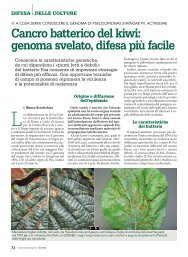
Symptoms <strong>of</strong> a hitherto new dise<strong>as</strong>e were observed on<br />
A. chinensis cv. Hort 16A cultivated in the province <strong>of</strong><br />
Latina (Latium region), central Italy. Angular necrotic<br />
spots were observed on the leaves that in some c<strong>as</strong>es<br />
coalesced resulting in further necrosis and wilting <strong>of</strong><br />
the leaf. Elongated cracks along the leaf petiole were<br />
frequently observed. In addition, in some c<strong>as</strong>es, oozing<br />
w<strong>as</strong> noticed from the main trunk and branches. A discolouration<br />
<strong>of</strong> the bark tissue w<strong>as</strong> also observed. From<br />
infected tissues, bacterial colonies were isolated on<br />
NSA. From these isolations, ten representative colonies<br />
were chosen on the b<strong>as</strong>is <strong>of</strong> the colour and<br />
morphology (i.e. round edge, white-creamy colour,<br />
levan-positive) that were used in biochemical tests. All<br />
ten colonies showed characteristics <strong>of</strong> <strong>Pseudomon<strong>as</strong></strong><br />
LOPAT group Ia: levan-positive, oxid<strong>as</strong>e-negative,<br />
potato s<strong>of</strong>t rot-negative and arginine dehydrol<strong>as</strong>enegative,<br />
tobacco hypersensitivity-positive. These colonies<br />
were selected for further biochemical tests, a<br />
pathogenicity test, <strong>as</strong> well <strong>as</strong> for ERIC-PCR analysis.<br />
The results <strong>of</strong> biochemical and nutritional tests are<br />
reported in Table 1.<br />
All ten isolates and P. s. <strong>pv</strong>. <strong>actinidiae</strong> NCPPB 3739<br />
were pathogenic to both A. chinensis and A. deliciosa<br />
plants. All <strong>of</strong> them caused complete wilting <strong>of</strong> the<br />
plant inoculated into the shoots within 10 days after<br />
inoculation. Isolate ISF ACT3, which showed some<br />
differences in biochemical tests (Table 1), but not in<br />
the ERIC-PCR banding pattern reacted very aggressive,<br />
killing the plant after 4–5 days after the inoculation.<br />
Leaves showed extensive necrosis 1 week after<br />
the inoculation. Control plants and those <strong>of</strong> the other<br />
Table 1<br />
Results <strong>of</strong> biochemical and nutritional tests showed by the isolates<br />
obtained from Actinidia chinensis and by <strong>Pseudomon<strong>as</strong></strong> <strong>syringae</strong> <strong>pv</strong>.<br />
<strong>actinidiae</strong> NCPPB 3739 isolated from A. deliciosa<br />
Isolated from<br />
A. chinensis<br />
P. <strong>syringae</strong> <strong>pv</strong>. <strong>actinidiae</strong><br />
NCPPB 3739<br />
Levan + +<br />
Oxid<strong>as</strong>e ) )<br />
Potato s<strong>of</strong>t-rot ) )<br />
Arginine dehydrol<strong>as</strong>e ) )<br />
Tobacco hypersensitivity + +<br />
Fluorescence on KB ) )<br />
Glucose metabolism O O<br />
Aesculin hydrolysis ) )<br />
Gelatin hydrolysis ) ) a<br />
Utilization <strong>of</strong>:<br />
Adonitol ) ) a<br />
l-alanine + +<br />
Arabinose ) )<br />
Erythritol ) ) a<br />
d-fructose + +<br />
Glycerol + +<br />
Inositol + +<br />
Mannitol + +<br />
Sorbitol + +<br />
l-tyrosine + +<br />
d-xylose ) )<br />
O, Oxidative metabolism.<br />
a The isolate ISF ACT3 showed a positive response.<br />
769