phylogenetic relationships and classification of didelphid marsupials ...
phylogenetic relationships and classification of didelphid marsupials ... phylogenetic relationships and classification of didelphid marsupials ...
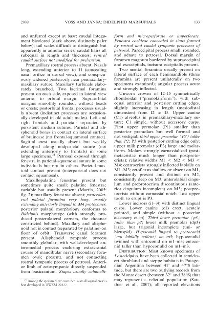
132 BULLETIN AMERICAN MUSEUM OF NATURAL HISTORY NO. 322 Fig. 51. Lestodelphys halli (based on UWZM 22422, an adult male from Clemente Onelli, Río Negro, Argentina; and MVZ 173727, an adult male from Lihuel-Calel, La Pampa, Argentina).
2009 VOSS AND JANSA: DIDELPHID MARSUPIALS 133 and unfurred except at base; caudal integument bicolored (dark above, distinctly paler below); tail scales difficult to distinguish but apparently in annular series; caudal hairs all subequal in length and thickness; ventral caudal surface not modified for prehension. Premaxillary rostral process absent. Nasals long, extending anterior to I1 (concealing nasal orifice in dorsal view), and conspicuously widened posteriorly near premaxillarymaxillary suture. Maxillary turbinals elaborately branched. Two lacrimal foramina present on each side, exposed in lateral view anterior to orbital margin. Supraorbital margins smoothly rounded, without beads or crests; postorbital frontal processes usually absent (indistinct processes are occasionally developed in old adult males). Left and right frontals and parietals separated by persistent median sutures. Parietal and alisphenoid bones in contact on lateral surface of braincase (no frontal-squamosal contact). Sagittal crest usually absent but weakly developed along midparietal suture (not extending anteriorly to frontals) in some large specimens. 31 Petrosal exposed through fenestra in parietal-squamosal suture in some individuals but not in others. Parietal-mastoid contact present (interparietal does not contact squamosal). Maxillopalatine fenestrae present but sometimes quite small; palatine fenestrae variable but usually present (Martin, 2005: fig. 2); maxillary fenestrae absent; posterolateral palatal foramina very long, usually extending anteriorly lingual to M4 protocones; posterior palatal morphology conforms to Didelphis morphotype (with strongly produced posterolateral corners, the choanae constricted behind). Maxillary and alisphenoid not in contact (separated by palatine) on floor of orbit. Transverse canal foramen present. Alisphenoid tympanic process smoothly globular, with well-developed anteromedial process enclosing extracranial course of mandibular nerve (secondary foramen ovale present), and not contacting rostral tympanic process of petrosal. Anterior limb of ectotympanic directly suspended from basicranium. Stapes usually columelli- 31 Among the specimens we examined, a small sagittal crest is best developed in UWZM 22422. form and microperforate or imperforate. Fenestra cochleae concealed in sinus formed by rostral and caudal tympanic processes of petrosal. Paroccipital process small, rounded, and adnate to petrosal. Dorsal margin of foramen magnum bordered by supraoccipital and exoccipitals, incisura occipitalis present. Two mental foramina usually present on lateral surface of each hemimandible (three foramina are present unilaterally on two specimens examined); angular process acute and strongly inflected. Unworn crowns of I2–I5 symmetrically rhomboidal (‘‘premolariform’’), with subequal anterior and posterior cutting edges, slightly increasing in length (mesiodistal dimension) from I2 to I5. Upper canine (C1) alveolus in premaxillary-maxillary suture; C1 simple, without accessory cusps. First upper premolar (P1) smaller than posterior premolars but well formed and not vestigial; third upper premolar (P3) taller than P2; P3 with posterior cutting edge only; upper milk premolar (dP3) large and molariform. Molars strongly carnassialized (postmetacristae much longer than postprotocrista); relative widths M1 , M2 , M3 , M4; centrocrista strongly inflected labially on M1–M3; ectoflexus shallow or absent on M1, consistently present and distinct on M2, consistently deep on M3; anterolabial cingulum and preprotocrista discontinuous (anterior cingulum incomplete) on M3; postprotocrista without carnassial notch. Last upper tooth to erupt is P3. Lower incisors (i1–i4) with distinct lingual cusps. Lower canine (c1) erect, acutely pointed, and simple (without a posterior accessory cusp). Third lower premolar (p3) taller than p2; lower milk premolar (dp3) large, but trigonid incomplete (uni- or bicuspid). Hypoconid lingual to protoconid (not labially salient) on m3; hypoconulid twinned with entoconid on m1–m3; entoconid taller than hypoconulid on m1–m3. DISTRIBUTION: Most known specimens of Lestodelphys have been collected in semidesert shrubland and steppe habitats in Patagonian Argentina between 41u and 47uS latitude, but there are two outlying records from the Monte desert (between 32u and 38uS) that may represent a relictual population (Sauthier et al., 2007); all reported elevations
- Page 81 and 82: 2009 VOSS AND JANSA: DIDELPHID MARS
- Page 83 and 84: 2009 VOSS AND JANSA: DIDELPHID MARS
- Page 85 and 86: 2009 VOSS AND JANSA: DIDELPHID MARS
- Page 87 and 88: 2009 VOSS AND JANSA: DIDELPHID MARS
- Page 89 and 90: 2009 VOSS AND JANSA: DIDELPHID MARS
- Page 91 and 92: 2009 VOSS AND JANSA: DIDELPHID MARS
- Page 93 and 94: 2009 VOSS AND JANSA: DIDELPHID MARS
- Page 95 and 96: 2009 VOSS AND JANSA: DIDELPHID MARS
- Page 97 and 98: 2009 VOSS AND JANSA: DIDELPHID MARS
- Page 99 and 100: 2009 VOSS AND JANSA: DIDELPHID MARS
- Page 101 and 102: 2009 VOSS AND JANSA: DIDELPHID MARS
- Page 103 and 104: 2009 VOSS AND JANSA: DIDELPHID MARS
- Page 105 and 106: 2009 VOSS AND JANSA: DIDELPHID MARS
- Page 107 and 108: 2009 VOSS AND JANSA: DIDELPHID MARS
- Page 109 and 110: 2009 VOSS AND JANSA: DIDELPHID MARS
- Page 111 and 112: 2009 VOSS AND JANSA: DIDELPHID MARS
- Page 113 and 114: 2009 VOSS AND JANSA: DIDELPHID MARS
- Page 115 and 116: 2009 VOSS AND JANSA: DIDELPHID MARS
- Page 117 and 118: 2009 VOSS AND JANSA: DIDELPHID MARS
- Page 119 and 120: 2009 VOSS AND JANSA: DIDELPHID MARS
- Page 121 and 122: 2009 VOSS AND JANSA: DIDELPHID MARS
- Page 123 and 124: 2009 VOSS AND JANSA: DIDELPHID MARS
- Page 125 and 126: 2009 VOSS AND JANSA: DIDELPHID MARS
- Page 127 and 128: 2009 VOSS AND JANSA: DIDELPHID MARS
- Page 129 and 130: 2009 VOSS AND JANSA: DIDELPHID MARS
- Page 131: 2009 VOSS AND JANSA: DIDELPHID MARS
- Page 135 and 136: 2009 VOSS AND JANSA: DIDELPHID MARS
- Page 137 and 138: 2009 VOSS AND JANSA: DIDELPHID MARS
- Page 139 and 140: 2009 VOSS AND JANSA: DIDELPHID MARS
- Page 141 and 142: 2009 VOSS AND JANSA: DIDELPHID MARS
- Page 143 and 144: 2009 VOSS AND JANSA: DIDELPHID MARS
- Page 145 and 146: 2009 VOSS AND JANSA: DIDELPHID MARS
- Page 147 and 148: 2009 VOSS AND JANSA: DIDELPHID MARS
- Page 149 and 150: 2009 VOSS AND JANSA: DIDELPHID MARS
- Page 151 and 152: 2009 VOSS AND JANSA: DIDELPHID MARS
- Page 153 and 154: 2009 VOSS AND JANSA: DIDELPHID MARS
- Page 155 and 156: 2009 VOSS AND JANSA: DIDELPHID MARS
- Page 157 and 158: 2009 VOSS AND JANSA: DIDELPHID MARS
- Page 159 and 160: 2009 VOSS AND JANSA: DIDELPHID MARS
- Page 161 and 162: 2009 VOSS AND JANSA: DIDELPHID MARS
- Page 163 and 164: 2009 VOSS AND JANSA: DIDELPHID MARS
- Page 165 and 166: 2009 VOSS AND JANSA: DIDELPHID MARS
- Page 167 and 168: 2009 VOSS AND JANSA: DIDELPHID MARS
- Page 169 and 170: 2009 VOSS AND JANSA: DIDELPHID MARS
- Page 171 and 172: 2009 VOSS AND JANSA: DIDELPHID MARS
- Page 173 and 174: 2009 VOSS AND JANSA: DIDELPHID MARS
- Page 175 and 176: 2009 VOSS AND JANSA: DIDELPHID MARS
- Page 177: 2009 VOSS AND JANSA: DIDELPHID MARS
2009 VOSS AND JANSA: DIDELPHID MARSUPIALS 133<br />
<strong>and</strong> unfurred except at base; caudal integument<br />
bicolored (dark above, distinctly paler<br />
below); tail scales difficult to distinguish but<br />
apparently in annular series; caudal hairs all<br />
subequal in length <strong>and</strong> thickness; ventral<br />
caudal surface not modified for prehension.<br />
Premaxillary rostral process absent. Nasals<br />
long, extending anterior to I1 (concealing<br />
nasal orifice in dorsal view), <strong>and</strong> conspicuously<br />
widened posteriorly near premaxillarymaxillary<br />
suture. Maxillary turbinals elaborately<br />
branched. Two lacrimal foramina<br />
present on each side, exposed in lateral view<br />
anterior to orbital margin. Supraorbital<br />
margins smoothly rounded, without beads<br />
or crests; postorbital frontal processes usually<br />
absent (indistinct processes are occasionally<br />
developed in old adult males). Left <strong>and</strong><br />
right frontals <strong>and</strong> parietals separated by<br />
persistent median sutures. Parietal <strong>and</strong> alisphenoid<br />
bones in contact on lateral surface<br />
<strong>of</strong> braincase (no frontal-squamosal contact).<br />
Sagittal crest usually absent but weakly<br />
developed along midparietal suture (not<br />
extending anteriorly to frontals) in some<br />
large specimens. 31 Petrosal exposed through<br />
fenestra in parietal-squamosal suture in some<br />
individuals but not in others. Parietal-mastoid<br />
contact present (interparietal does not<br />
contact squamosal).<br />
Maxillopalatine fenestrae present but<br />
sometimes quite small; palatine fenestrae<br />
variable but usually present (Martin, 2005:<br />
fig. 2); maxillary fenestrae absent; posterolateral<br />
palatal foramina very long, usually<br />
extending anteriorly lingual to M4 protocones;<br />
posterior palatal morphology conforms to<br />
Didelphis morphotype (with strongly produced<br />
posterolateral corners, the choanae<br />
constricted behind). Maxillary <strong>and</strong> alisphenoid<br />
not in contact (separated by palatine) on<br />
floor <strong>of</strong> orbit. Transverse canal foramen<br />
present. Alisphenoid tympanic process<br />
smoothly globular, with well-developed anteromedial<br />
process enclosing extracranial<br />
course <strong>of</strong> m<strong>and</strong>ibular nerve (secondary foramen<br />
ovale present), <strong>and</strong> not contacting<br />
rostral tympanic process <strong>of</strong> petrosal. Anterior<br />
limb <strong>of</strong> ectotympanic directly suspended<br />
from basicranium. Stapes usually columelli-<br />
31 Among the specimens we examined, a small sagittal crest is<br />
best developed in UWZM 22422.<br />
form <strong>and</strong> microperforate or imperforate.<br />
Fenestra cochleae concealed in sinus formed<br />
by rostral <strong>and</strong> caudal tympanic processes <strong>of</strong><br />
petrosal. Paroccipital process small, rounded,<br />
<strong>and</strong> adnate to petrosal. Dorsal margin <strong>of</strong><br />
foramen magnum bordered by supraoccipital<br />
<strong>and</strong> exoccipitals, incisura occipitalis present.<br />
Two mental foramina usually present on<br />
lateral surface <strong>of</strong> each hemim<strong>and</strong>ible (three<br />
foramina are present unilaterally on two<br />
specimens examined); angular process acute<br />
<strong>and</strong> strongly inflected.<br />
Unworn crowns <strong>of</strong> I2–I5 symmetrically<br />
rhomboidal (‘‘premolariform’’), with subequal<br />
anterior <strong>and</strong> posterior cutting edges,<br />
slightly increasing in length (mesiodistal<br />
dimension) from I2 to I5. Upper canine<br />
(C1) alveolus in premaxillary-maxillary suture;<br />
C1 simple, without accessory cusps.<br />
First upper premolar (P1) smaller than<br />
posterior premolars but well formed <strong>and</strong><br />
not vestigial; third upper premolar (P3) taller<br />
than P2; P3 with posterior cutting edge only;<br />
upper milk premolar (dP3) large <strong>and</strong> molariform.<br />
Molars strongly carnassialized (postmetacristae<br />
much longer than postprotocrista);<br />
relative widths M1 , M2 , M3 ,<br />
M4; centrocrista strongly inflected labially on<br />
M1–M3; ect<strong>of</strong>lexus shallow or absent on M1,<br />
consistently present <strong>and</strong> distinct on M2,<br />
consistently deep on M3; anterolabial cingulum<br />
<strong>and</strong> preprotocrista discontinuous (anterior<br />
cingulum incomplete) on M3; postprotocrista<br />
without carnassial notch. Last upper<br />
tooth to erupt is P3.<br />
Lower incisors (i1–i4) with distinct lingual<br />
cusps. Lower canine (c1) erect, acutely<br />
pointed, <strong>and</strong> simple (without a posterior<br />
accessory cusp). Third lower premolar (p3)<br />
taller than p2; lower milk premolar (dp3)<br />
large, but trigonid incomplete (uni- or<br />
bicuspid). Hypoconid lingual to protoconid<br />
(not labially salient) on m3; hypoconulid<br />
twinned with entoconid on m1–m3; entoconid<br />
taller than hypoconulid on m1–m3.<br />
DISTRIBUTION: Most known specimens <strong>of</strong><br />
Lestodelphys have been collected in semidesert<br />
shrubl<strong>and</strong> <strong>and</strong> steppe habitats in Patagonian<br />
Argentina between 41u <strong>and</strong> 47uS latitude,<br />
but there are two outlying records from<br />
the Monte desert (between 32u <strong>and</strong> 38uS) that<br />
may represent a relictual population (Sauthier<br />
et al., 2007); all reported elevations


