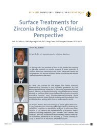
Anterior CEREC CAD/CAM Porcelain - Eureka Smile Center
Anterior CEREC CAD/CAM Porcelain - Eureka Smile Center
Anterior CEREC CAD/CAM Porcelain - Eureka Smile Center

Create successful ePaper yourself
Turn your PDF publications into a flip-book with our unique Google optimized e-Paper software.
CONTINUING EDUCATION<br />
<strong>Anterior</strong> <strong>CEREC</strong> <strong>CAD</strong>/<strong>CAM</strong><br />
<strong>Porcelain</strong> Treatment of GERD<br />
Eroded Teeth<br />
Abstract<br />
Erosion and worn teeth are an every<br />
day occurrence in dental practice. Gastroesophageal<br />
reflux disease (GERD) is a common<br />
condition where acid is regurgitated<br />
from the stomach into the esophagus and<br />
then to the oral cavity, which can lead to<br />
dental erosion. Critical to the long-term success<br />
of the restorative treatment is proper<br />
diagnosis and medical control so that enamel<br />
replacement will be successful. Single<br />
visit computer-aided design/computer-aided<br />
manufacturing all-porcelain restorations<br />
were placed in this case to meet functional<br />
and esthetic needs after medical control of<br />
the regurgitation had been achieved.<br />
Learning Objectives<br />
Jack D. Griffin Jr, DMD<br />
Private Practice<br />
<strong>Eureka</strong>, Missouri<br />
636.938.4141<br />
Email: Esmilecenter@aol.com<br />
Web site:<br />
www.<strong>Eureka</strong>smile.com<br />
After reading this article, the reader<br />
should be able to:<br />
• identify patient history and erosion patterns<br />
characteristic of intrinsic gastric<br />
reflux disease.<br />
• discuss computer aided design/computer<br />
aided manufacturing treatment advantages,<br />
sequences, and techniques for<br />
restoring eroded anterior teeth.<br />
• explain ways to characterize <strong>CEREC</strong><br />
porcelain to meet esthetic demands.<br />
It is natural for a clinician to see a case of tooth degradation and begin<br />
treating it without first determining what caused the degradation.<br />
Sometimes, identification and correction of the cause is more important<br />
than repairing the tooth damage. Worn, short, discolored teeth are<br />
commonly seen in the dental office and bruxism is often the first etiology<br />
suspected, without consideration for other factors that may make the<br />
tooth more susceptible to wear.<br />
Erosion of dental structures is often a common component of enamel<br />
and dentin loss, and identifying its cause is as important as correcting<br />
the tooth degradation. 1 Anorexia and bulimia nervosa, chronic alcoholism,<br />
and gastric disturbances are some of the conditions in which<br />
acids from the stomach can cause dental erosion. There is also a correlation<br />
between tooth erosion and gastroesophageal reflux disease<br />
(GERD). 2<br />
This case describes the etiology and medical co-therapy needed with<br />
a focus on restoring the eroded enamel of the anterior dentition with<br />
computer-aided design/computer-aided manufacturing (<strong>CAD</strong>/<strong>CAM</strong>) single-visit<br />
veneers.<br />
Etiology<br />
GERD is a chronic condition whereby acids travel from the stomach<br />
through the esophagus and into the mouth because of a faulty sealing by<br />
the lower esophageal sphincter. 3 Gastric juices that travel upward<br />
toward the esophagus and low pH levels can cause esophageal irritation<br />
with the classic heartburn sensation.<br />
Many people have acid backup into the esophagus, but in GERD<br />
patients this acidic fluid may stay in the esophagus long enough to cause<br />
erosion of the lining. 4 Chronic acid reflux can cause epithelial changes in<br />
the lining of the esophagus resulting in a potentially precancerous condition<br />
called Barrett’s esophagus. 5 Should this acid regurgitate to the<br />
mouth, erosion of the teeth is possible.<br />
Gravity, swallowing, and saliva are the most important protective<br />
mechanisms against this condition. The greatest amount of tissue damage<br />
occurs during sleep because the supine position allows the secretions<br />
2 CONTEMPORARY ESTHETICS | DECEMBER 2006
to spend more time in the mouth.<br />
Because swallowing and saliva secretions<br />
are also reduced, it is more likely<br />
that acid may remain in the esophagus<br />
for extended periods leading to<br />
dental and soft-tissue damage.<br />
GERD should be suspected when<br />
there is excessive occlusal wear combined<br />
with lingual erosion, incisal<br />
wear, and oral malodor. 6 Enamel<br />
wear tends to be more prevalent on<br />
the lingual surfaces in acid regurgitation<br />
conditions like GERD or bulimia<br />
nervosa as opposed to facial enamel<br />
wear seen from consumption of<br />
acidic foods and drinks. 7 Patients<br />
may self-medicate with antacids to<br />
help deal with symptoms. These signs<br />
also may be prevalent in children<br />
along with potentially more dental<br />
caries. 8<br />
A thorough history may reveal<br />
heartburn, belching, sore throat,<br />
swallowing difficulty, sour taste, or<br />
stomach problems 2 to 3 days a week<br />
for several months. 9 However, this<br />
condition frequently is asymptomatic,<br />
and the only evidence may be the irreversible<br />
erosion of tooth structure. 10<br />
The dentist is often the first health<br />
care professional to suspect this medical<br />
condition because of the affected<br />
dentition. Despite the ability to repair<br />
most any tooth degradation with<br />
modern dentistry, the best efforts can<br />
fail if the cause of the problem is not<br />
addressed. 11 The dental community<br />
can be helpful in its diagnosis and<br />
referral for treatment. 12 Characteristics<br />
or symptoms often seen in<br />
patients with GERD are:<br />
• Excessive lingual cervical enamel<br />
wear<br />
• Incisal or occlusal wear with erosion<br />
“GERD should be suspected when there is<br />
excessive occlusal wear combined with lingual erosion,<br />
incisal wear, and oral malodor.”<br />
• Occasional or chronic “heartburn”<br />
• Use of over-the-counter antacids<br />
• Frequent belching<br />
• Difficulty in swallowing or sore<br />
throat<br />
• Sour or bitter taste<br />
• Bad breath<br />
• History of stomach problems.<br />
These clinical signs should alert<br />
the dental team that there may be a<br />
medical component to the dental<br />
attrition and a thorough medical history<br />
with prudent questioning about<br />
the presence of heartburn, bad taste<br />
or breath, stomach problems, or<br />
throat irritations should be done<br />
when dental erosion is obvious. 13,14<br />
A thorough health history should<br />
be completed and a consult with a<br />
primary care physician, internist, or<br />
gastroenterologist should be considered<br />
when these signs and symptoms<br />
are present. 15 Persistence by the dental<br />
team may result in correct medical<br />
intervention and treatment of this<br />
condition. 16 Only after consultation,<br />
ruling out eating disorders, and control<br />
of intra-oral acid should clinical<br />
dental procedures move from maintenance<br />
to restoration. 17<br />
<strong>CAD</strong>/<strong>CAM</strong> <strong>CEREC</strong> Restorations<br />
There are many porcelain restorative<br />
materials, with or without<br />
metal, that are esthetically pleasing<br />
and have the strength to replace missing<br />
enamel. Procera (Nobel Biocare),<br />
OPC (Pentron), InCeram (Vident),<br />
and others have proved suitable for<br />
CONTEMPORARY ESTHETICS | DECEMBER 2006 3<br />
this purpose. <strong>Porcelain</strong> choice today<br />
often is determined by the clinician’s<br />
comfort and experience with the<br />
material. 18<br />
There are 2 porcelain choices for<br />
tooth colored <strong>CEREC</strong> restorations:<br />
Vita Mark II (Vident) and Pro<strong>CAD</strong><br />
(Ivoclar Vivadent). Both have the<br />
ability to be stained and glazed and<br />
have proven to be durable, cost effective,<br />
and very esthetic restorations<br />
for full porcelain coverage. 19<br />
The mean survival rate for milled<br />
<strong>CAD</strong>/<strong>CAM</strong> all-porcelain restorations<br />
has been reported at a greater than<br />
97% success rate for nearly 5 years,<br />
which is comparable to other all-ceramic<br />
systems. 18 Wear rates for these<br />
materials are very similar to natural<br />
enamel, which minimizes iatrogenic<br />
abrasion of the opposing teeth. 20<br />
With experience, esthetic anterior<br />
restorations can be created in 1<br />
appointment particularly if the team<br />
has the ability to stain and glaze the<br />
restorations. Further customization<br />
can be achieved by cutting back and<br />
adding translucency or custom shade<br />
variations of porcelain, or by using<br />
blocks with varying porcelain<br />
translucency. It must be stressed that<br />
doing single appointment <strong>CEREC</strong><br />
restorations can be an esthetic compromise<br />
when compared to the experienced<br />
lab technician who works<br />
with porcelain customization on a<br />
daily basis. Model work, cutting<br />
back porcelain after milling, stacking,<br />
and customization of porcelain<br />
require time, experience, and a thor-
CONTINUING EDUCATION<br />
ough understanding of the material.<br />
In this case, time and finances were<br />
factors in the material and technique<br />
selection. Some advantages of CER-<br />
EC anterior restorations are:<br />
• Efficient design and fabrication<br />
• Tooth morphology controlled by<br />
the clinician<br />
• No need for temporary fabrication<br />
or problems<br />
• No lab fees and minimal supply<br />
costs<br />
• No bacterial accumulation on<br />
preparation during temporary<br />
phase<br />
• Excellent adaptation of porcelain<br />
to tooth<br />
• Ability to be stained or glazed<br />
quickly<br />
• Wear similar to natural teeth.<br />
Because the restorations are<br />
Figure 1—<br />
Preoperative<br />
full-face<br />
smile with<br />
what patient<br />
feels are<br />
dark, worn<br />
teeth.<br />
Figure 3—Left smile indicates a “hiding” of<br />
the teeth as the patient refuses to give a full<br />
smile because of unhappiness with teeth.<br />
made directly by acquiring a preoperative<br />
image of the mock-up and then<br />
an image of the prepared teeth, there<br />
is no need for final impressions, temporary<br />
fabrication, temporary removal,<br />
or a second appointment for<br />
cementation. This reduces the number<br />
of appointments and lab fees as<br />
well as sensitivity occasionally associated<br />
with temporary wear. Immediate<br />
dentin sealing with dual-cure bonding<br />
and luting agents may increase dentinal<br />
bond strengths, decrease marginal<br />
gap formations, reduce bacterial<br />
microleakage, and cause less dentinal<br />
sensitivity than placing provisionals<br />
on nonbonded teeth. 21<br />
There are also some disadvantages<br />
when using <strong>CEREC</strong> for multiple<br />
anterior teeth. The office team is<br />
responsible for fit, shading, shaping,<br />
Figure 2—Facial abfraction and erosion<br />
areas show and make teeth appear darker.<br />
Figure 4—The color of the teeth is near A2<br />
in the incisal and A3.5 in gingival. Gingival<br />
heights are uneven and inflammation is present<br />
near abfraction areas of laterals.<br />
4 CONTEMPORARY ESTHETICS | DECEMBER 2006<br />
and customization of the porcelain.<br />
Despite only being 1 appointment, as<br />
much as or even more total chair<br />
time is needed than traditional porcelain<br />
techniques. Generally the time<br />
needed for an office to do anterior<br />
work with 1 milling machine is about<br />
1 hour for patient involvement,<br />
mock-up, and anesthesia, plus about<br />
30 minutes per tooth being restored.<br />
Case Presentation<br />
A 20-year-old man with advanced<br />
incisal wear was concerned<br />
about the wear of the incisors, dark<br />
tooth color, and shortness of the<br />
front teeth (Figure 1). He wanted<br />
whiter teeth that were longer,<br />
straighter, and more indicative of his<br />
age (Figure 2).<br />
In the past, he had been given a<br />
bruxism splint because of nocturnal<br />
grinding. There were obvious signs<br />
of parafunctional incisal wear; a loss<br />
of 2 mm to 4 mm of tooth length<br />
was generalized and the smile showed<br />
less tooth structure than the patient<br />
liked (Figure 3). Facial erosion<br />
without decay was prominent in both<br />
arches and the maxillary incisors<br />
(teeth Nos. 8 and 9) showed little<br />
facial wear (Figure 4). Erosion was<br />
noted on the lingual of teeth Nos. 3,<br />
14, and 19 and was particularly evident<br />
on the lingual of the upper incisors<br />
with incisal edge concavities<br />
along with rotated teeth and crowding<br />
(Figure 5).<br />
The patient experienced no pain<br />
in his teeth but recently had noticed<br />
an increase in cold sensitivity. There<br />
was no periapical pathology or any<br />
periodontal concerns with oral exam<br />
or radiographs. There was interproximal<br />
decay between some of the max-
illary anterior teeth with loss of<br />
almost all lingual enamel (Figure 6).<br />
Differential Diagnosis and Medical<br />
Treatment<br />
The history of a bruxism habit<br />
was consistent with incisal wear,<br />
occlusal enamel loss, and abfraction.<br />
22 Because of the erosive wear<br />
patterns, lack of chronic acidic food<br />
history, and patient history, intrinsic<br />
acid regurgitation was suspected over<br />
an extrinsic consumed source. 23,24<br />
There was no history of any reflux<br />
pathology although the patient complained<br />
of a “bad taste” and “bad<br />
breath.” He was first referred to a<br />
primary care physician by this office<br />
only to return without any diagnosis<br />
of reflux pathology or treatment for<br />
any reflux condition.<br />
Months later, a consult with a<br />
gastroenterologist revealed chronic<br />
acid regurgitation and the patient<br />
was prescribed ranitidine to treat<br />
GERD. The medication was later<br />
changed to esomeprazole and the<br />
symptoms of heartburn, acidic taste,<br />
and belching were mostly eradicated.<br />
The patient was also later diagnosed<br />
and treated for ulcerative colitis by<br />
the same specialist after further<br />
testing.<br />
Restorative Plan<br />
Once the regurgitation was<br />
under medical control, a treatment<br />
plan was made that addressed the<br />
missing enamel, interproximal decay,<br />
and esthetic concerns. An “ideal”<br />
plan would have been to replace missing<br />
enamel on all teeth as soon as<br />
the reflux was controlled. However,<br />
because of finances, time, and patient<br />
cosmetic desires, treatment was to be<br />
done in stages over several years.<br />
Composites, lab-made porcelain veneers,<br />
and in-office <strong>CAD</strong>/<strong>CAM</strong> porcelain<br />
restorations were considered<br />
for replacing missing enamel to cover<br />
and protect exposed dentin. 25<br />
For over 20 years, porcelain<br />
veneers have been used to replace<br />
missing enamel and to make cosmetic<br />
smile enhancements. 26 <strong>Porcelain</strong><br />
has long been accepted for long-term<br />
restorations for missing tooth structure<br />
as as gingival anatomy, tooth<br />
morphology, function, and occlusion<br />
are all considered and incorporated<br />
into preparation design. This along<br />
with a controlled medical condition<br />
can lead to esthetic restorations with<br />
excellent long-term results. 27-30<br />
Enamel bonding is predictable, effective,<br />
and long-lasting while bonding<br />
to dentin is also clinically viable<br />
in the right circumstances with bond<br />
strengths approaching those achieved<br />
on enamel. 31,32 The treatment plan<br />
was formulated to provide anterior<br />
dentin coverage with porcelain margins<br />
on enamel when possible.<br />
Laser gingivoplasty was planned<br />
to correct inconsistent tooth lengths<br />
and to reduce the appearance of a<br />
gummy smile, allowing the fabrication<br />
of teeth that were proportional<br />
in length with consistent gingival<br />
architecture. Gingival enhancements<br />
were then done to teeth Nos. 4 and<br />
12. The pocket depths were measured<br />
to 2 mm to 3.5 mm and would<br />
be recontoured with critical attention<br />
paid to preserve biologic width for a<br />
healthy, long-term result. 33<br />
In this case, the posterior facial<br />
abfraction areas on both the lingual<br />
and facial of the molars were first<br />
restored with direct composite res-<br />
CONTEMPORARY ESTHETICS | DECEMBER 2006 5<br />
torations to cover exposed dentin.<br />
There was also some occlusal pitting<br />
that was restored as well. The bite<br />
was deemed stable and the patient<br />
had good function with no obvious<br />
mandibular joint concerns. Once<br />
these areas were stabilized, the chief<br />
complaint of the anterior teeth was<br />
addressed.<br />
Preview of <strong>Smile</strong> Rehabilitation<br />
A direct composite mock-up was<br />
done without anesthesia so that<br />
speech and facial movements were<br />
not compromised by numbing. The<br />
mock-up was done for several reasons:<br />
so the patient could see a rough<br />
preview before being numb; so phonetics,<br />
function, and esthetics could<br />
be evaluated; and so the anatomy<br />
could be closely copied into the<br />
<strong>CAD</strong>/<strong>CAM</strong> restorations.<br />
The teeth were etched with 37%<br />
phosphoric acid to make sure the<br />
materials would stay attached during<br />
<strong>CEREC</strong> fabrication. To allow for<br />
easy interproximal removal, bonding<br />
agent was not applied. An anterior<br />
hybrid shade B1 was quickly placed<br />
by finger molding and plastic instrument<br />
into a rough, overcontoured<br />
form of the desired shape (Matrixx<br />
AH, Discus Dental). The patient and<br />
assistant both chose Vita shade 1M1<br />
(Vident) and so a similar shaded composite<br />
was used for a patient preview.<br />
All exposed dentin was covered<br />
and incisal edges were added so that<br />
the teeth were roughly 70% to 80%<br />
as wide as they were tall to meet current<br />
accepted esthetic standards. A<br />
slightly over-bulked, between 0.5<br />
mm and 1.0mm over-contoured,<br />
mock-up for <strong>CEREC</strong> correlation was<br />
desirable to allow for customized
CONTINUING EDUCATION<br />
anatomy placement after milling was<br />
completed. A basic facial contour<br />
was made with particular attention<br />
to incisal edge length and facial position<br />
(Figure 7). The mock-up surface<br />
irregularities were easily corrected in<br />
the <strong>CEREC</strong> design process. The clinical<br />
advantages of a direct composite<br />
mock-up are:<br />
1. The patient can see a rough<br />
“preview” of basic shape<br />
2. Approximate shade can be approved<br />
by patient<br />
3. Can check basic phonetics<br />
4. Can evaluate smile line and<br />
incisal edge placement<br />
5. It will act as a bonded “reduction<br />
guide” during tooth preparation<br />
6. The basic shape will be copied<br />
and applied to the final <strong>CEREC</strong><br />
porcelain shape using “correla-<br />
Figure 5—Wear of incisal edges consistent<br />
with history of bruxism and the acidic condition<br />
of the mouth may speed up this parafunctional<br />
wear.<br />
Figure 8—A titanium dioxide reflective medium<br />
will be applied to the mock-up so that the<br />
computer will copy the tooth morphology.<br />
tion” design mode.<br />
Restorative Procedure<br />
After isolation with a SeeMore<br />
4-way retractor (Discus Dental), a<br />
glycerin based dusting adhesive and a<br />
titanium dioxide reflective medium<br />
were applied to the tooth and the<br />
adjacent teeth (Figure 8). This mockup<br />
image was then acquired by the<br />
<strong>CEREC</strong> 3D acquisition unit in Correlation<br />
design mode, which accurately<br />
copies the anatomy of the preoperative<br />
tooth into the milled porcelain.<br />
Tooth No. 8 was chosen first<br />
because it is helpful in <strong>CEREC</strong> design<br />
to have a central incisor as a benchmark<br />
for correct cant, size, and midline.<br />
The mock-up material was then<br />
removed with a coarse diamond and<br />
a rounded chamfer was placed slight-<br />
Figure 6—Loss of lingual enamel on incisors<br />
is consistent with acid regurgitation conditions<br />
such as bulimia, anorexia nervosa, and<br />
GERD.<br />
Figure 9—A 1.5 mm to 2.0 mm reduction is<br />
done to cover exposed dentin, decay, and<br />
restored areas.<br />
6 CONTEMPORARY ESTHETICS | DECEMBER 2006<br />
ly subgingival on the facial and<br />
extended around the tooth to cover<br />
all exposed dentin and ending on<br />
enamel wherever possible. As with<br />
most porcelain restorations, care was<br />
taken to make sure there were no<br />
sharp corners that might impart<br />
stress points within the final porcelain<br />
restoration. A reduction of 1.5<br />
mm to 2.0 mm on the buccal and lingual<br />
was prepared from the extensions<br />
of the mock-up to provide<br />
ample porcelain thickness (Figure 9).<br />
The preparation was then powdered<br />
and imaged (Figure 10). Design<br />
was completed in about 3 minutes<br />
in Correlation mode and the<br />
milling time was about 15 minutes<br />
per tooth. Every other tooth was<br />
done so that neighboring anatomy<br />
could be used to correlate the preop-<br />
Figure 7—The mock-up was finished in just a<br />
few minutes and basic contouring was done<br />
with a finish bur.<br />
Figure 10—Reflective powder is applied to<br />
the prep and imaged into the computer.
erative and postoperative images to<br />
allow porcelain to be consistent with<br />
the mock-up. While tooth No. 8 was<br />
being milled, tooth No. 6 was preoperatively<br />
scanned, prepared, and rescanned<br />
in the same fashion. In<br />
about the same time it takes to mill a<br />
restoration, the next tooth was prepared,<br />
powdered, imaged, and designed.<br />
In that fashion, the milling<br />
machine seldom stops because when<br />
one is finished, another is started.<br />
While milling tooth No. 6, a<br />
diode laser (Odyssey, Ivoclar Vivadent)<br />
was used to recontour gingiva<br />
and add crown length and a more<br />
symmetrical gingival architecture to<br />
teeth Nos. 5, 9, and 12. Pocket<br />
depths were measured, marked, and<br />
then contoured for preservation of<br />
the biological width (Figure 11). 34<br />
Figure 11—While milling, a diode laser is<br />
used to add length to several teeth while<br />
making gingiva more even in height.<br />
Figure 14—With porcelain duplications of<br />
the mock-ups, the mock-up for tooth No. 7 is<br />
imaged into the computer before prepping.<br />
The recontoured gingiva was wiped<br />
with hydrogen peroxide and gently<br />
scrubbed (Figure 12).<br />
As each restoration was milled, it<br />
was tried-in and used as a benchmark<br />
correlation for design of the neighboring<br />
tooth. An “every other” tooth<br />
method allows the clinician to maintain<br />
landmarks for the <strong>CEREC</strong> copying<br />
and to help maintain contours relative<br />
to the mock-up. For example,<br />
after teeth Nos. 6 and 8 were done<br />
being milled, they were tried-in and<br />
only contacts were adjusted (Figure<br />
13). This provided adjacent porcelain<br />
similar to the mock-ups, which could<br />
then be powdered and put into the<br />
computer so that tooth No. 7 could<br />
be copied (Figure 14). Preparation on<br />
the lingual was carried onto healthy,<br />
uneroded enamel and finished as<br />
Figure 12—The areas of gingivoplasty will<br />
be cleaned with peroxide and a microbrush.<br />
Figure 15—While the bicuspids are milling,<br />
the anterior porcelain is tried-in and held in<br />
place with a nonmatching flowable applied<br />
interproximally.<br />
CONTEMPORARY ESTHETICS | DECEMBER 2006 7<br />
described to allow 1.5 mm to 2.0 mm<br />
of clearance for porcelain strength.<br />
While the bicuspids were milled,<br />
the anterior 6 were placed and held<br />
with a nonmatching flowable composite<br />
interproximally (Figure 15).<br />
This was easily identified for removal<br />
later. Anatomical adjustments were<br />
made with a Gold Finish diamond<br />
(Diatech) and high-speed handpiece<br />
with copious water. Facial contours<br />
were made so that 3 planes—gingival<br />
third, middle third, incisal third—<br />
were evident to give a less bulky<br />
appearance. Likewise the first bicuspids<br />
were shaped and refined as<br />
milling was completed.<br />
An office-made, hard/soft bruxism<br />
splint (Erkoloc Pro, Glidewell<br />
Laboratories) was made to help the<br />
porcelain survive potential parafunc-<br />
Figure 13—With the mock-up still on tooth<br />
No. 7, the milled porcelain is tried on teeth<br />
Nos. 6 and 8 and used for the correlation<br />
design of tooth No. 7.<br />
Figure 16—At 1 week postoperative, tissues<br />
were healing very well and sensitivity was<br />
minimal.
CONTINUING EDUCATION<br />
tional habits. An alginate was taken<br />
during the try-in so that the splint<br />
could be made during porcelain customization.<br />
Because of the possibility<br />
of acidic regurgitation, the patient<br />
was also given a topical fluoride gel<br />
to place in the bruxism splint at night<br />
in an effort to help control any possible<br />
acid attack. 35 It must be noted<br />
that it may be harmful to the enamel<br />
to give a bruxism splint to a patient<br />
who has uncontrolled acid reflux<br />
because the splint may act as a reservoir<br />
for regurgitated acid leading to<br />
more tooth destruction.<br />
Customization<br />
The <strong>CEREC</strong> Vita Blocs are 1<br />
color and there are several ways to<br />
give a more natural appearance of<br />
shade variation within the restora-<br />
Figure 17—Slight inflammation on lateral<br />
because of laser treatment and cement left<br />
from cementation.<br />
Figure 20—Tissue response has been excellent<br />
and <strong>CEREC</strong> characterization has been<br />
stable.<br />
tion. Because the porcelain has some<br />
translucency, the incisal third will<br />
have a different look than the gingival<br />
third simply because there is no<br />
natural tooth under the restoration<br />
in that area. There is also the potential<br />
to control shade variations by<br />
using luting agents of a different<br />
shade and opacities when bonding<br />
the restoration.<br />
Custom staining and glazing can<br />
also be done while cutback and addition<br />
of character porcelain takes<br />
more time. In this case, several<br />
shades of stain were used to provide<br />
a more natural appearance. A bluish<br />
stain was added in small amounts on<br />
the lingual of the incisal edges to<br />
mimic translucency and a white stain<br />
was added in the incisal areas to<br />
mimic hypocalcified areas often seen<br />
Figure 18—<br />
After 2 years<br />
the smile<br />
improvements<br />
are obvious.<br />
Figure 21—Lower incisor straightening was<br />
done during this period and composites and<br />
porcelain were placed to maintain occlusion.<br />
8 CONTEMPORARY ESTHETICS | DECEMBER 2006<br />
in young teeth. These character additions<br />
are done according to the discretion<br />
of the staff and the desires of<br />
the patient.<br />
The staining and glazing was<br />
done in a single bake in about 25<br />
minutes with cooling, providing a<br />
more natural appearance and a surface<br />
that may reduce opposing tooth<br />
wear. Glazing may also reduce<br />
craze line development from milling<br />
scratches that could lead to subsequent<br />
porcelain fracture. 36 Care must<br />
be taken to evaluate the degree of surface<br />
gloss that is desired and is controlled<br />
by porcelain finishing, glaze<br />
thickness, and furnace holding time.<br />
The porcelain was etched with<br />
hydrofluoric acid for 2 minutes and<br />
silane was applied. After air drying, a<br />
dual cure bonding agent (Cabrio,<br />
Figure 19—Color and length changes were<br />
well tolerated and appreciated by the patient<br />
and with time, the restorations are providing<br />
durable service while the GERD is controlled.<br />
Figure 22—The patient now smiles much<br />
more freely without coercion to show teeth.
Discus Dental) was mixed and applied.<br />
Insure Yellow Red Light dual<br />
cure cement (Cosmedent) was mixed<br />
and loaded into the restorations and<br />
then placed on the teeth. Cleanup<br />
was done with brushes, scalers, floss,<br />
and gauze and then cured. The<br />
restorations were cemented and<br />
cleaned up 5 hours after starting.<br />
Healing and Follow-up<br />
After 1 week, tissues were healing<br />
well in the areas of laser refinement<br />
and the color, shape, and size<br />
were accepted by the patient (Figure<br />
16). At this stage there was slight<br />
inflammation remaining in the tissue<br />
and there was cement left interproximally<br />
and subgingivally that was<br />
removed with a composite knife and<br />
scaler (Figure 17).<br />
During the next 2 years, healing<br />
stabilized and the patient was very<br />
pleased with the results; there was a<br />
much improved smile and confidence<br />
in appearance (Figure 18). The tissues<br />
remained healthy and home care<br />
improved with time. There were no<br />
obvious signs of regurgitation and<br />
the patient was still under physician<br />
care with medication for GERD<br />
(Figures 19 and 20).<br />
During that 2-year period,<br />
orthodontic treatment was done on<br />
the lower incisors to correct crowding<br />
and composites were placed on<br />
the lower incisors with “transitional”<br />
<strong>CEREC</strong> porcelain placed on the<br />
lower cuspids and first bicuspids to<br />
maintain the occlusion (Figure 21).<br />
The treatment plan includes doing<br />
porcelain coverage on the posterior<br />
teeth as time and finances become<br />
available, but the composites are<br />
holding up well while the reflux is<br />
controlled. The enhanced smile is<br />
evident from right and left lateral<br />
views showing esthetically pleasing<br />
results with good function.<br />
The smile improvement was well<br />
worth the time and effort by the<br />
patient and he is very happy with the<br />
results, which is the overall final<br />
evaluation of any case (Figure 22).<br />
Conclusion<br />
Gastroesophogeal reflux disease<br />
is widespread and must be an etiological<br />
consideration in many erosion<br />
and wear cases. <strong>CEREC</strong> 3D is most<br />
efficient for doing posterior dentistry<br />
but is capable of doing quality anterior<br />
restorations as long as the staff is<br />
committed to the time and detail that<br />
is required. Even though this work<br />
was done in 1 appointment, faster is<br />
not always better if postoperative<br />
complications, staff stress, or patient<br />
dissatisfaction arises.<br />
The porcelain customization<br />
allows for an esthetically pleasing<br />
result and <strong>CEREC</strong> Vita porcelain has<br />
provided a suitable enamel replacement,<br />
and given an environment for<br />
periodontal health. The prognosis is<br />
good for the long-term success of<br />
these restorations because medical,<br />
periodontal, and esthetic concerns<br />
were addressed. As patient time and<br />
resources permit, cosmetic enhancements<br />
will be made to other teeth to<br />
complete the smile reconstruction<br />
and to cover missing enamel. ●c<br />
References<br />
1. Shipley S, Taylor K, Mitchell W. Identifying causes<br />
of dental erosion. Gen Dent. 2005;53(1):73-76.<br />
2. Bartlett DW, Evans DF, Smith BG. The relationship<br />
between gastro-esophageal reflux disease<br />
and dental erosion. J Oral Rehabil.1996; 23(5):<br />
289-297.<br />
3. Sifrim D, Zerbib F. Gastroesophageal reflux dis-<br />
CONTEMPORARY ESTHETICS | DECEMBER 2006 9<br />
ease. Curr Opin Gastroenterol. 2002;18(4):447-<br />
453.<br />
4. Marks JW. Gastroesophageal reflux disease<br />
(GERD) index. Dec 2003. Available at: http:<br />
//www.medicinenet.com/script/main/hp.asp.<br />
Accessed: October 31, 2006.<br />
5. Oberg S, Wenner J, Johansson J, et al. Barrett<br />
esophagus: risk factors for progression to dysplasia<br />
and adenocarcinoma. Ann Surg.2005; 242(1):<br />
49-54.<br />
6. Bartlett DW, Evans DF, Anggiansah A, et al. A<br />
study of the association between gastro-esophageal<br />
reflux and palatal dental erosion. Br<br />
Dent J. 1996;181(4):125-131.<br />
7. Valena V, Young WG. Dental erosion patterns<br />
from intrinsic acid regurgitation and vomiting.<br />
Aust Dent J. 2002;47(2):106-115.<br />
8. Linnett V, Seow WK, Connor F, et al. Oral health<br />
of children with gastro-esophageal reflux disease:<br />
a controlled study. Aust Dent J.2002; 47(2):<br />
156-162.<br />
9. Bartlett D, Smith B. Clinical investigations of gastro-oesophageal<br />
reflux: Part 1. DentUpdate.<br />
1996;23(5):205-208.<br />
10. Moazzez R, Bartlett D, Anggiansah A. Dental erosion,<br />
gastro-oesophageal reflux disease and saliva:<br />
how are they related? J Dent. 2004;32(6):489-<br />
494.<br />
11. Aziz K, Ziebert AJ, Cobb D. Restoring erosion associated<br />
with gastroesophageal reflux using<br />
direct resins: case report. Oper Dent.2005; 30(3):<br />
395-401.<br />
12. Hefferren JJ. Why is there and should there be<br />
more attention paid to dental erosion? Compend<br />
Contin Educ Dent. 2004;25(9):4-8.<br />
13. Lussi A, Jaeggi T. Erosion caused by gastric reflux<br />
in children. Discussion of etiology, clinical<br />
appearance and therapy in two cases. Schweiz<br />
Monatsschr Zahnmed. 2004;114(10):1018-1030.<br />
14. Shaheen NJ. Advances in Barrett’s esophagus<br />
and esophageal adenocarcinoma. Gastroenterology.<br />
2005;128(6):1554-1566.<br />
15. Ali DA, Brown RS, Rodriguez LO, et al. Dental<br />
erosion caused be silent gastroesophageal<br />
reflux disease. J Am Dent Assoc. 2002;133(6):<br />
734-737.<br />
16. Chey WD, Inadomi JM, Booher AM, et al.<br />
Primary-care physicians’ perceptions and practices<br />
on the management of GERD: results of a<br />
national survey. Am J Gastroenterol. 2005;<br />
100(6):1237-1242.<br />
17. Frydrych AM, Davies GR, McDermott BM. Eating<br />
disorders and oral health: a review of the literature.<br />
Aust Dent J. 2005;50(1):6-15.<br />
18. Dumfahrt H. <strong>Porcelain</strong> laminate veneers. A retrospective<br />
evaluation after 1 to 10 years of service:<br />
Part 1—Clinical procedure. Int J Prosthodont.<br />
1999;12(6):505-513.<br />
19. Christensen GJ. Bonding to dentin and enamel<br />
where does it stand in 2005? J Am Dent Assoc.<br />
2005;136(9):1299-1302.<br />
20. Dental Adhesives. Reality. Houston, Tex: Reality<br />
Publishing. 2004:241-299.
CONTINUING EDUCATION<br />
21. Robbins JW. Esthetic gingival recontouring—a<br />
plea for honesty. Quintessence Int. 2000;31(8):<br />
553-556.<br />
22. Abrahamsen TC. The worn dentition—pathognomonic<br />
patterns of abrasion and erosion. Int Dent<br />
J. 2005;55(4):268-276.<br />
23. Litonjua LA, Andreana S, Bush PJ, et al. Tooth<br />
wear: attrition, erosion, and abrasion. Quintessence<br />
Int. 2003;34(6):435-436.<br />
24. Gandara BK, Truelove EL. Diagnosis and management<br />
of dental erosion. J Contemporary Dent<br />
Practice. 1999;1(1):16-23.<br />
25. Ibarra G, Senna G, Cobb D, et al. Restoration of<br />
enamel and dentin erosion due to gastroesophageal<br />
reflux disease: a case report. Prac<br />
Proced Aesthet Dent. 2001;13(4):297-306.<br />
26. Swift EJ Jr, Friedman MJ. <strong>Porcelain</strong> veneer outcomes,<br />
part II. J Esthet Restor Dent. 2006;18(2):<br />
110-113.<br />
27. Peumans M, Van Meerbeek B, Lambrechts P, et<br />
al. <strong>Porcelain</strong> veneers: a review of the literature.J<br />
Dent. 2000;28(3):163-177.<br />
28. Bassett JL. Replacement of missing mandibular<br />
lateral incisors with a single pontic all-ceramic<br />
prosthesis: a case report. Pract Periodontics<br />
Aesthet Dent. 1997;9(4):455-461.<br />
29. Tipton PA. Aesthetic tooth alignment using<br />
etched porcelain restorations. Pract Proced<br />
Asthet Dent. 2001;13(7):551-555.<br />
30. Martin N, Jedynakiewicz NM. Clinical performance<br />
of <strong>CEREC</strong> ceramic inlays: a systematic<br />
review. Dent Mater. 1999;15(1):54-61.<br />
31. Masek RT. Achieving high-level esthetics with<br />
<strong>CEREC</strong>. Compen Contin Educ Dent. 2001;22(6):19-<br />
26.<br />
32. Abozenada B, Pober R, Giordano R. In-vitro wear<br />
of restorative dental materials. J Dent Res.<br />
2002;81:1-4.<br />
33. Magne P. Immediate dentin sealing: a fundamental<br />
procedure for indirect bonded restorations. J<br />
Esthet Restor Dent. 2005;17(3):144-154.<br />
34. Lanning SD, Waldrop TC, Gunsolley JC, et al.<br />
Circle X on Reader Service Card<br />
10 CONTEMPORARY ESTHETICS | DECEMBER 2006<br />
Surgical crown lengthening: evaluation of the<br />
biological width. J Periodontol. 2003;74(4):468-<br />
474.<br />
35. Amaechi BT, Higham SM. Dental erosion: possible<br />
approaches to prevention and control. J Dent.<br />
2005;33(3):243-252.<br />
36. Giordano R. Milling and finishing effects on<br />
machinable blocks. Block Talk (newsletter).<br />
2005;1:2.<br />
Product References<br />
Products: Matrixx AH, See More 4-way retractor,<br />
Cabrio<br />
Manufacturer: Discus Dental<br />
Location: Culver City, California<br />
Phone: 800.422.9448<br />
Web site: www.discusdental.com<br />
Products: Vita shade 1M1, InCeram, Vita Mark II<br />
Manufacturer: Vident<br />
Location: Brea, California<br />
Phone: 800.828.3829<br />
Web site: www.vident.com<br />
Product: Procera<br />
Manufacturer: Nobel Biocare<br />
Location: Yorba Linda, California<br />
Phone: 800.993.8100<br />
Web site: www.nobelbiocare.com<br />
Product: OPC<br />
Manufacturer: Pentron<br />
Location: Wallingford, Connecticut<br />
Phone: 800.551.0283<br />
Web site: www.pentron.com<br />
Products: Pro<strong>CAD</strong>, diode lasere Odyssey<br />
Manufacturer: Ivoclar Vivadent<br />
Location: Amherst, New York<br />
Phone: 800.533.6825<br />
Web site: www.ivoclarvivadent.com<br />
Product: Gold Finish diamond<br />
Manufacturer: Diatech<br />
Location: Charleston, South Carolina<br />
Phone: 800.222.1851<br />
Web site: www.diatechusa.com<br />
Product: Erkoloc Pro<br />
Manufacturer: Glidewell Laboratories<br />
Location: Newport Beach, California<br />
Phone: 800.854.7256<br />
Web site: www.glidewell-lab.com<br />
Product: Insure Yellow Red Light dual cure<br />
cement<br />
Manufacturer: Cosmedent<br />
Location: Chicago, Illinois<br />
Phone: 800.621.6729<br />
Web site: www.cosmedent.com
Instructions<br />
CONTINUING EDUCATION<br />
1. Which of the following conditions can cause dental<br />
erosion because of acids from the stomach?<br />
a. anorexia<br />
b. bulimia<br />
c. chronic alcoholism<br />
d. all of the above<br />
2. Gastroesophageal reflux disease (GERD) is:<br />
a. a chronic condition whereby acids travel from<br />
the stomach through the esophagus and into the<br />
mouth because of a faulty sealing by the lower<br />
esophageal sphincter.<br />
b. an erosive condition caused mainly by acidi<br />
drinks and foods.<br />
c. a medical condition for which there is no<br />
treatment.<br />
d. an elusive diagnosis and has no effect on dental<br />
diagnosis and treatment.<br />
3. GERD should be suspected when there is excessive<br />
occlusal wear combined with:<br />
a. lingual erosion<br />
b. incisal wear<br />
c. oral malodor<br />
d. all of the above<br />
4. GERD is frequently asymptomatic and the only evidence<br />
may be:<br />
a. heart burn.<br />
b. irreversible erosion of tooth structure.<br />
c. oral malodor.<br />
d. insomnia.<br />
5. Characteristics or symptoms often seen in patients<br />
with GERD are:<br />
a. excessive lingual cervical enamel wear.<br />
b. occasional or chronic “heart burn.”<br />
c. frequent belching.<br />
d. all of the above<br />
CONTINUING EDUCATION QUIZ: DECEMBER 2006<br />
Contemporary Esthetics offers 12 Continuing Education (CE) credit hours per year. Each clinical CE article is followed by a 10-question, multiple-choice test,<br />
providing 1 hour of credit. To receive credit, record your answers on the enclosed answer sheet or submit them on a separate piece of paper. You may also phone<br />
your answers in to 888.596.4605, or fax them to 703.404.1801. Be sure to include your name, address, phone number, Social Security number, and method of payment.<br />
The deadline for submission of quizzes is 12 months after the date of publication. Participants must attain a score of 70% on each quiz to receive<br />
credit. To register, call 888.596.4605. Participants are urged to contact their state registry boards for special CE requirements.<br />
12 CONTEMPORARY ESTHETICS | DECEMBER 2006<br />
6. The mean survival rate for milled <strong>CAD</strong>/<strong>CAM</strong> allporcelain<br />
restorations has been reported at a greater<br />
than 97% success rate for nearly:<br />
a. 1 year.<br />
b. 3 years.<br />
c. 5 years.<br />
d. 7 years.<br />
7. Which of the following is an advantage of <strong>CEREC</strong><br />
anterior restorations?<br />
a. Tooth morphology controlled by the manufacturer.<br />
b. No bacterial accumulation on preparation during<br />
temporary phase.<br />
c. Ability to be stained over a period of time.<br />
d. Minimal lab fees.<br />
8. Generally, the time needed for an office to do anterior<br />
work with 1 milling machine is about 1 hour for<br />
patient involvement, mock-up, and anesthesia, plus<br />
how long per tooth being restored?<br />
a. 15 minutes<br />
b. 30 minutes<br />
c. 1 hour<br />
d. 90 minutes<br />
9. <strong>Porcelain</strong> has been accepted for long-term restorations<br />
for missing tooth structure as long as which of the following<br />
are incorporated into the preparation design?<br />
a. gingival anatomy<br />
b. tooth morphology<br />
c. function<br />
d. all of the above<br />
10. Which of the following is a clinical advantage of a<br />
direct composite mock-up?<br />
a. The patient will not have to see a rough “preview”<br />
of the basic shape.<br />
b. The patient will not need to approve the<br />
approximate shade.<br />
c. Basic phonetics can be checked.<br />
d. <strong>Smile</strong> line and incisal edge placement does not<br />
need to be evaluated.
Contemporary Esthetics STATUS<br />
■ Presently Enrolled in CE Program Not Enrolled ■ 1 exam completed = $14.00<br />
(PLEASE PRINT CLEARLY)<br />
SSN<br />
ADA/AGD# _________________________________________________________<br />
Name _____________________________________________________________<br />
Address ___________________________________________________________<br />
City ______________________________________________________________<br />
State Zip Daytime Fax ________________________<br />
Daytime Phone ______________________________________________________<br />
Please make checks payable to:<br />
ASCEND DENTAL MEDIA<br />
and mail with this form to: Ascend Dental Media CE Department,<br />
405 Glenn Drive, Suite 4, Sterling, VA 20164-4432<br />
PAYMENT INFORMATION<br />
Payment by Credit Card<br />
(Please use a Visa, MasterCard, or American Express Card)<br />
Please enroll me in the Contemporary Esthetics Continuing Education Program marked<br />
below:<br />
■ Please enroll me in the 12-month CE Program for $134<br />
(a 20% saving versus paying for each exam individually).<br />
Program includes all 12 exams in Contemporary Esthetics for 1 year<br />
(excluding supplements).<br />
■ CHECK (payable to Ascend Dental Media)<br />
■ CREDIT CARD – Please complete information and sign below:<br />
Card Number<br />
■ Visa ■ MasterCard ■ American Express<br />
Expiration Date: Mo/Y<br />
______________________________________________________ _________<br />
SIGNATURE DATE<br />
CIRCLE ANSWERS<br />
1. a b c d 6. a b c d<br />
2. a b c d 7. a b c d<br />
3. a b c d 8. a b c d<br />
4. a b c d 9. a b c d<br />
5. a b c d 10. a b c d<br />
CONTINUING EDUCATION ANSWER FORM: DECEMBER 2006<br />
SCORING SERVICES<br />
• By Mail<br />
• Fax: 703.404.1801<br />
• Phone-in: 888.596.4605<br />
(9am-5pm ET, Mon.-Fri.)<br />
• Customer Service Questions?<br />
Please Call: 888.596.4605<br />
PROGRAM EVALUATION<br />
Please evaluate this issue’s programs by responding<br />
to the following statements, using the scale of:<br />
3 = Excellent to 1 = Poor.<br />
• Clarity of objectives . . . . . . . . . . . . . . . . . . . . .<br />
• Usefulness of the content . . . . . . . . . . . . . . . . .<br />
• Benefit to your clinical practice . . . . . . . . . . . .<br />
• Usefulness of the references . . . . . . . . . . . . . .<br />
• Quality of the written presentation . . . . . . . . .<br />
• Quality of the illustrations . . . . . . . . . . . . . . . .<br />
• Clarity of review questions . . . . . . . . . . . . . .<br />
• Relevance of review questions . . . . . . . . . . .<br />
3 2 1<br />
3 2 1<br />
3 2 1<br />
3 2 1<br />
3 2 1<br />
3 2 1<br />
3 2 1<br />
3 2 1<br />
Please list future CE topic preferences:<br />
______________________________________________________<br />
______________________________________________________<br />
• Did the lessons achieve their<br />
educational objectives? . . . . . . . . . . . . ■ Yes ■ No<br />
• Did the articles present new<br />
information? . . . . . . . . . . . . . . . . . . . . . ■ Yes ■ No<br />
• How much time did it take you<br />
to complete the CE? . . . . . . . . . . . . . . ______ min<br />
PRACTICE INFORMATION<br />
■ DDS/DMD<br />
■ Full-time registered Hygienist<br />
■ Dental Assistant<br />
■ Part-time registered Hygienist<br />
■ General Practice<br />
■ Periodontist<br />
■ Oral Surgeon<br />
■ Prosthodontist<br />
■ Endodontist<br />
■ Other<br />
CONTEMPORARY ESTHETICS | DECEMBER 2006 13<br />
■ Pedodontist (includes dentists with nonspecified<br />
ADA specialty)<br />
DEADLINE FOR SUBMISSION OF ANSWERS IS<br />
12 MONTHS AFTER THE DATE OF PUBLICATION.