Endodontics Endodontics - American Association of Endodontists
Endodontics Endodontics - American Association of Endodontists
Endodontics Endodontics - American Association of Endodontists
Create successful ePaper yourself
Turn your PDF publications into a flip-book with our unique Google optimized e-Paper software.
Cover artwork: Rusty Jones, MediVisuals, Inc.<br />
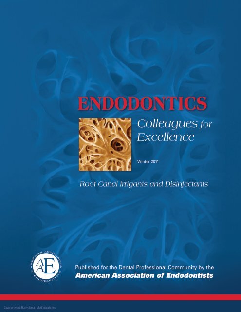
<strong>Endodontics</strong><br />
Colleagues for<br />
Excellence<br />
Winter 2011<br />
Winter 2011<br />
Root Canal Irrigants and Disinfectants<br />
Published for the Dental Pr<strong>of</strong>essional Community by the<br />
<strong>American</strong> <strong>Association</strong> <strong>of</strong> <strong>Endodontists</strong>
D<br />
iagnosis, instrumentation, obturation and restoration are the main steps involved in the treatment <strong>of</strong> teeth with pulpal<br />
and periapical diseases. Elimination or significant reduction <strong>of</strong> irritants and prevention <strong>of</strong> recontamination <strong>of</strong> the root<br />
canal after treatment are the essential elements for successful outcomes. Although many advances have been made in<br />
different aspects <strong>of</strong> endodontics within the last few years to preserve natural dentition, the main objective <strong>of</strong> this field remains<br />
elimination <strong>of</strong> microorganisms from the root canal systems and prevention <strong>of</strong> recontamination after treatment. The<br />
common belief that inadequate obturation is the major cause <strong>of</strong> endodontic failures has been proven to be fallacious as<br />
obturation reflects the adequacy <strong>of</strong> cleaning and shaping. In other words, what you take out <strong>of</strong> a root canal may be more<br />
important than what you put in it.<br />
What are the Irritants for Pulpal and Periapical Tissues?<br />
The major causes <strong>of</strong> pulpal and periapical diseases are living and nonliving irritants. The latter group includes mechanical,<br />
thermal and chemical irritants. The living irritants include various microorganisms including bacteria, yeasts and viruses.<br />
When pathological changes occur in the dental pulp, the root canal space acquires the ability to harbor various irritants<br />
including several species <strong>of</strong> bacteria, along with their toxins and byproducts. Investigations in animals and patients have<br />
shown that pulpal and/or periradicular diseases do not develop without the presence <strong>of</strong> bacteria. 1,2 Advanced culturing and<br />
molecular biology techniques have shown that primary root canal infections are polymicrobial (10-30 bacterial species)<br />
in nature and are dominated by obligate anaerobic bacteria. 3 The variety <strong>of</strong> microorganisms present in root canal-treated<br />
teeth with persistent periapical lesions is more restricted (1-3 species) in comparison to primary root canal infections, which<br />
are dominated by E. faecalis, a facultative anaerobic gram-positive coccus that is resistant to intracanal medications, able to<br />
form bi<strong>of</strong>ilms and able to invade dentinal tubules. 4 Because the presence <strong>of</strong> bacteria negatively influences the outcome <strong>of</strong><br />
root canal treatment, 4-6 every effort should be made to eradicate infections during treatment.<br />
What are the Obstacles in Removing Irritants From the Root Canal Systems?<br />
The complexity <strong>of</strong> the root canal system, presence<br />
<strong>of</strong> numerous dentinal tubules in the roots, invasion<br />
<strong>of</strong> the tubules by microorganisms, formation<br />
<strong>of</strong> smear layer during instrumentation and presence<br />
<strong>of</strong> dentin as a tissue are the major obstacles<br />
in achieving the primary objectives <strong>of</strong> complete<br />
cleaning and shaping <strong>of</strong> root canal systems. 7 Microscopic<br />
examinations <strong>of</strong> root canals show that<br />
they are irregular and complex systems with<br />
many cul-de-sacs, fins and lateral canals (Figures<br />
1a and 1b).<br />
In the root, dentinal tubules extend from the<br />
pulp- to the cementum-dentin junction (Figure<br />
<strong>Endodontics</strong>: Colleagues for Excellence<br />
2). Investigators have reported the presence <strong>of</strong> bacteria in the dentinal tubules (Figure 3) <strong>of</strong> infected teeth at approximately<br />
half the distance between the root canal walls and the cementum-dentin junction. 7 The presence <strong>of</strong> these natural complexities<br />
is important for clinicians to consider during cleaning and shaping <strong>of</strong> the root canal system. 8,9 In addition to these<br />
natural difficulties, it is known<br />
that a smear layer is created during<br />
cleaning and shaping that<br />
covers the instrumented root<br />
canal walls. 7 This smear layer<br />
(Figure 4) contains inorganic<br />
and organic substances as well<br />
Fig. 2. SEM <strong>of</strong> dentinal tubules running<br />
from the pre-dentin towards the<br />
cementum.<br />
Fig. 3. SEM <strong>of</strong> dentinal tubules<br />
containing microorganisms.<br />
Fig. 1a. Internal anatomy<br />
<strong>of</strong> a maxillary second<br />
molar after decalcification,<br />
dehydration and placement<br />
in India ink. Courtesy <strong>of</strong> Dr.<br />
JV Baroni Barbizam.<br />
Fig. 4. The smear layer consists <strong>of</strong><br />
organic and inorganic substances as<br />
well as fragments <strong>of</strong> odontoblastic<br />
processes, various species <strong>of</strong> bacteria<br />
and necrotic debris.<br />
2<br />
Fig. 1b. Microcomputed tomography images <strong>of</strong> canal isthmuses in<br />
mesial roots <strong>of</strong> mandibular molars. Courtesy <strong>of</strong> Dr. L. Gu.<br />
as fragments <strong>of</strong> odontoblastic<br />
processes, microorganisms and<br />
necrotic debris. Intracanal irrigants<br />
and medications are used
during root canal treatment to reach the natural complexities and remove the smear layer. Intracanal irrigants exert<br />
their effects mechanically and chemically. Mechanical effects <strong>of</strong> irrigants are generated by the back and forth flow <strong>of</strong> the<br />
irrigation solution during cleaning and shaping <strong>of</strong> the infected root canals, significantly reducing the bacterial load. 10,11<br />
Studies show that irrigants that possess antibacterial properties have clearly superior effectiveness in bacterial reduction<br />
and elimination when compared with saline solution. 11,12<br />
What are the Ideal Properties <strong>of</strong> an Irrigant?<br />
To effectively clean and disinfect the root canal system, an irrigant should be able to disinfect and penetrate dentin and<br />
its tubules, <strong>of</strong>fer long-term antibacterial effect (substantivity), remove the smear layer, and be nonantigenic, nontoxic<br />
and noncarcinogenic. In addition, it should have no adverse effects on dentin or the sealing ability <strong>of</strong> filling materials. 7<br />
Furthermore, it should be relatively inexpensive, convenient to apply and cause no tooth discoloration. 7 Other desirable<br />
properties for an ideal irrigant include the ability to dissolve pulp tissue and inactivate endotoxins. 13<br />
What are the Types, Advantages and Disadvantages <strong>of</strong> Current Irrigants?<br />
The irrigants that are currently used during cleaning and shaping can be divided into antibacterial and decalcifying<br />
agents or their combinations. They include sodium hypochlorite (NaOCl), chlorhexidine, ethylenediaminetetraacetic<br />
acid (EDTA), and a mixture <strong>of</strong> tetracycline, an acid and a detergent (MTAD).<br />
Sodium Hypochlorite (NaOCl)<br />
Sodium hypochlorite (household bleach) is the most commonly used root canal irrigant. It is an antiseptic and inexpensive<br />
lubricant that has been used in dilutions ranging from 0.5% to 5.25%. Free chlorine in NaOCl dissolves vital and<br />
necrotic tissue by breaking down proteins into amino acids. 14 Decreasing the concentration <strong>of</strong> the solution reduces its<br />
toxicity, antibacterial effect and ability to dissolve tissues. 14 Increasing its volume or warming it increases its effectiveness<br />
as a root canal irrigant. 14<br />
Advantages <strong>of</strong> NaOCl include its ability to dissolve organic substances present in the root canal system and its affordability.<br />
The major disadvantages <strong>of</strong> this irrigant are its cytotoxicity when injected into periradicular tissues, foul<br />
smell and taste, ability to bleach clothes and ability to cause corrosion <strong>of</strong> metal objects. 15 In addition, it does not kill all<br />
bacteria, 12,16-18 nor does it remove all <strong>of</strong> the smear layer. 19 It also alters the properties <strong>of</strong> dentin. 20,21 The results <strong>of</strong> a recent<br />
in vitro study show that the most effective irrigation regimen is 5.25% at 40 minutes, whereas irrigation with 1.3% and<br />
2.5% NaOCl for this same time interval is ineffective in removing E. faecalis from infected dentin cylinders. 22 Based on<br />
the findings <strong>of</strong> this study, the authors recommend the use <strong>of</strong> other irrigants to increase the antibacterial effects during<br />
cleaning and shaping <strong>of</strong> root canals.<br />
Sodium hypochlorite is generally not utilized in its most active form in a clinical setting. For proper antimicrobial<br />
activity, it must be prepared freshly just before its use. 23,24 In the majority <strong>of</strong> cases, however, it is purchased in large<br />
containers and stored at room temperature while being exposed to oxygen for<br />
extended periods <strong>of</strong> time. Exposure <strong>of</strong> the solution to oxygen, room temperature<br />
and light can inactivate it significantly. 24<br />
Fig. 5a. NaOCl was<br />
inadvertently expressed<br />
into the periapical<br />
tissues through the<br />
apical foramen <strong>of</strong> the<br />
right maxillary cuspid<br />
during cleaning and<br />
shaping.<br />
<strong>Endodontics</strong>: Colleagues for Excellence<br />
Figure 5b. No treatment<br />
was necessary for the<br />
hematoma and swelling.<br />
Reprinted with permission<br />
from <strong>Endodontics</strong>: Principles<br />
and Practice 4 th ed.,<br />
Torabinejad and Walton,<br />
2009.<br />
Extrusion <strong>of</strong> NaOCl into periapical tissues (Figures 5a and 5b) can cause<br />
severe injury to the patient. 25,26 To minimize NaOCl accidents, the irrigating<br />
needle should be placed short <strong>of</strong> the working length, fit loosely in the canal<br />
and the solution must be injected using a gentle flow rate. Constantly moving<br />
the needle up and down during irrigation prevents wedging <strong>of</strong> the needle in<br />
the canal and provides better irrigation. The use <strong>of</strong> irrigation tips with sideventing<br />
reduces the possibility <strong>of</strong> forcing solutions into the periapical tissues.<br />
Treatment <strong>of</strong> NaOCl accidents is palliative and consists <strong>of</strong> observation <strong>of</strong> the<br />
patient as well as prescribing antibiotics and analgesics.<br />
Chlorhexidine<br />
Chlorhexidine gluconate has been used for the past 50 years for caries prevention,<br />
27 in periodontal therapy and as an oral antiseptic mouthwash. 28 It has a<br />
3<br />
Continued on p. 4
oad-spectrum antibacterial action, sustained action and low toxicity. 14 Because <strong>of</strong> these properties, it has also been recommended<br />
as a potential root canal irrigant. 14,27 The major advantages <strong>of</strong> chlorhexidine over NaOCl are its lower cytotoxicity<br />
and lack <strong>of</strong> foul smell and bad taste. However, unlike NaOCl, it cannot dissolve organic substances and necrotic tissues present<br />
in the root canal system. In addition, like NaOCl, it is unable to kill all bacteria and cannot remove the smear layer. 29,30<br />
Ethylenediaminetetraacetic Acid (EDTA)<br />
Chelating agents such as ethylenediaminetetraacetic acid (EDTA), citric acid and tetracycline are used for removal <strong>of</strong> the<br />
inorganic portion <strong>of</strong> the smear layer. 7 NaOCl is an adjunct solution for removal <strong>of</strong> the remaining organic components. Irrigation<br />
with 17% EDTA for one minute followed by a final rinse with NaOCl is the most commonly recommended method<br />
to remove the smear layer. 14 Longer exposures can cause excessive removal <strong>of</strong> both peritubular and intratubular dentin. 31<br />
EDTA has little or no antibacterial effect. 32<br />
MTAD<br />
An alternative solution to EDTA for removing the smear layer is the use <strong>of</strong> BioPure<br />
MTAD (DENTSPLY Tulsa Dental Specialties, Tulsa, Okla.), a mixture <strong>of</strong> a tetracycline isomer,<br />
an acid (citric acid) and a detergent. 33 MTAD was developed as a final rinse to disinfect<br />
the root canal system and remove the smear layer. The effectiveness <strong>of</strong> MTAD to completely<br />
remove the smear layer (Figure 6) is enhanced when a low concentration <strong>of</strong> NaOCl (1.3%) is<br />
used as an intracanal irrigant before placing 1 ml <strong>of</strong> MTAD in a canal for 5 minutes and rinsing<br />
it with an additional 4 ml <strong>of</strong> MTAD as the final rinse. 33 It appears to be superior to CHX in<br />
antimicrobial activity. 30 In addition, it has sustained antibacterial activity, is biocompatible and<br />
enhances bond strength. 14 Table 1 shows the advantages and disadvantages <strong>of</strong> current irrigants<br />
utilized during root canal treatment.<br />
Irrigation Devices and Techniques<br />
For many years various methods have been proposed and<br />
developed to make root canal irrigants more effective<br />
in removing debris and bacteria from the root canal<br />
system. These techniques can be classified into two broad<br />
categories: manual and rotary agitation. 34 The manual<br />
irrigation techniques include irrigation with needles,<br />
agitation with brushes, and manual dynamic agitation<br />
with files or gutta-percha points. The rotary irrigation<br />
techniques include rotary brushes, continuous irrigation<br />
during instrumentation, sonic and ultrasonic vibrations, and<br />
application <strong>of</strong> negative pressure during irrigation <strong>of</strong> the<br />
root canal system. The use <strong>of</strong> these methods results in better<br />
canal cleanliness when compared with that <strong>of</strong> conventional<br />
syringe needle irrigation. However, there is no high level <strong>of</strong><br />
evidence that correlates the clinical efficacy <strong>of</strong> these devices<br />
with better treatment outcomes. Clinical data are needed to<br />
support the use <strong>of</strong> these devices in endodontics.<br />
Lasers<br />
<strong>Endodontics</strong>: Colleagues for Excellence<br />
Some investigators have reported that lasers can be used to vaporize tissues in the main canal, remove the smear layer and<br />
eliminate the residual tissue in the apical portion <strong>of</strong> the root canals. 7 Several investigators have reported that the efficacy <strong>of</strong><br />
lasers depends on many factors including the power level, the duration <strong>of</strong> exposure, the absorption <strong>of</strong> light in the tissue, the<br />
geometry <strong>of</strong> the root canal and the tip-to-target distance. 35-37 The efficacy <strong>of</strong> the lasers to completely clean the root canals<br />
remains to be seen. The main difficulty continues to be access to small canal spaces with the relatively large probes that<br />
deliver the laser beams and the expense <strong>of</strong> these units.<br />
4<br />
Table 1<br />
Fig. 6. SEM <strong>of</strong> dentinal tubules after<br />
removal <strong>of</strong> the smear layer by MTAD.<br />
Advantages and Disadvantages <strong>of</strong> Currently Used Intracanal Irrigants<br />
Characteristics MTAD NaOCI CHX EDTA<br />
Shelf life stability + - + +<br />
Antimicrobial activity + + + -<br />
Ability to remove smear layer + - - +<br />
Biocompatibility + - + +<br />
Ability to dissolve pulp tissue + + - +/-<br />
Dentin conditioning properties + - - +<br />
Positive effect on root canal<br />
seal<br />
Negative effect on dentin<br />
structure<br />
Upregulation <strong>of</strong> regional<br />
immune response<br />
+ - - +/-<br />
- + - +<br />
+ - - -<br />
Application time (minutes) 5 18 40 22 ? 1 31
Because current solutions and techniques cannot completely remove all irritants, dissolve all organic tissue or remove<br />
the smear layer, various methods have been employed to deliver irrigants more efficiently to the working length.<br />
These include sonic and ultrasonic vibrations as well as application <strong>of</strong> negative pressure to flush out the debris present<br />
in instrumented canals.<br />
EndoVac ® System<br />
The EndoVac ® system (Discus Dental, Culver City, Calif.) is a new irrigation system that consists <strong>of</strong> a delivery/evacuation<br />
tip attached to a syringe <strong>of</strong> irrigant and the high-speed suction source <strong>of</strong> the dental unit. As the cannulas are placed<br />
in the canal, negative pressure pulls irrigant from a fresh supply in the chamber down into the canal to the tip <strong>of</strong> the<br />
cannula, then into the cannula and finally out through the suction hose. 38 Investigators compared the efficacy <strong>of</strong> the EndoVac<br />
® irrigation system with that <strong>of</strong> a needle irrigation system to clean root canals at 1 and 3mm from working length<br />
and found no significant difference between the two groups at the 3mm level. However, their results show significantly<br />
better debridement at 1mm from working length using the EndoVac ® compared with needle irrigation. 38<br />
A recent in vitro study compared three agitation and two irrigation devices with ultrasonic agitation for mechanically<br />
removing bacteria from a plastic-simulated canal that was instrumented to a size 35 with a .06 taper. 39 The irrigation<br />
and agitation techniques evaluated were ultrasonic agitation, needle irrigation, EndoVac ® irrigation, EndoActivator ®<br />
(DENTSPLY Tulsa Dental Specialties, Tulsa, Okla.), F ® File (Plastic Endo, Lincolnshire, Ill) and sonic agitation. Based<br />
on the results <strong>of</strong> this investigation, the authors conclude that ultrasonic agitation is significantly more effective than<br />
needle irrigation and EndoVac ® irrigation at removing intracanal bacteria. Ultrasonic, EndoActivator ® , F ® File and<br />
sonic agitation are similar in their ability to remove bacteria in a plastic-simulated canal. These findings point to the<br />
importance <strong>of</strong> the use <strong>of</strong> antiseptic irrigants in addition to mechanical vibrations and application <strong>of</strong> negative pressure<br />
during cleaning and shaping <strong>of</strong> root canals. The results <strong>of</strong> a recent in vitro study using a combination <strong>of</strong> mechanical<br />
and chemical means to disinfect root canals showed that activation <strong>of</strong> MTAD with the EndoActivator ® system for 1.5<br />
minutes was an effective method to completely inhibit the growth <strong>of</strong> E. faecali. 40<br />
Intracanal Medicaments<br />
Intracanal medicaments have been used to disinfect root canals between appointments and reduce interappointment<br />
pain. 7 The disinfectants can be divided into phenolic compounds such as camphorated monochlorophenol, cresatin,<br />
aldehydes such as formocresol and glutaraldehyde, and halides, as well as other materials like calcium hydroxide<br />
[Ca(OH) 2 ] and some antibiotics. 14 These compounds are potent antibacterial agents under laboratory test conditions,<br />
but their efficacy in clinical use is unpredictable. 7 Some <strong>of</strong> the aldehyde de rivatives have been proposed to neutralize<br />
canal tissue remnants and to render them inert. These can be used to fix fresh tissues for histological examination, but<br />
they may not effectively fix necrotic or decomposed tissues. According to one report, 41 fixed tissues are not inert and<br />
may become more toxic and antigenic after fixation. Intracanal medications have also been used clinically to prevent<br />
post-treatment pain. Studies have shown, however, that routine use <strong>of</strong> these materials as intracanal medications has no<br />
significant effect on prevention <strong>of</strong> pain. 7<br />
Calcium Hydroxide<br />
Ca(OH) 2 is a substance that inhibits microbial growth in canals. 42 The antibacterial effect <strong>of</strong> Ca(OH) 2 is due to its alkaline<br />
pH. It also dissolves necrotic tissue remnants and bacteria and their byproducts. 43 It can be placed as a dry powder,<br />
a powder mixed with a liquid such as water, saline, local anesthetic or glycerin, or a proprietary paste supplied in a syringe.<br />
14 Because <strong>of</strong> its toxicity 44 , Ca(OH) 2 should be placed within the canal with the aid <strong>of</strong> a file or a needle. Extrusion<br />
<strong>of</strong> the material into the periapical tissues can cause tissue necrosis and pain for the patient. Ca(OH) 2 can be removed<br />
from the canal by using irrigants such as saline, NaOCl, EDTA or MTAD.<br />
Corticosteroids<br />
<strong>Endodontics</strong>: Colleagues for Excellence<br />
Corticosteroids are anti-inflammatory agents that have been advocated as intracanal medicaments to reduce postoperative<br />
pain. 45 An animal study has shown a reduction <strong>of</strong> inflammatory cells in periapical tissues following supraperiosteal<br />
infiltration <strong>of</strong> dexamethasone into the buccal vestibule <strong>of</strong> rats. 46 There is no significant clinical evidence that suggests<br />
that they are effective in patients with very high pain levels. 47 The use <strong>of</strong> corticosteroids in patients with irreversible<br />
pulpitis and symptomatic apical periodontitis may be beneficial. 45,48<br />
5<br />
Continued on p. 6
Chlorhexidine Gel<br />
<strong>Endodontics</strong>: Colleagues for Excellence<br />
A 2% CHX gel has recently been advocated as an intracanal medicament. 49 It can be used alone in gel form or mixed with<br />
Ca(OH) 2 . When used for seven days to medicate bovine teeth 50 or human teeth, 51 CHX gel provides antimicrobial activity<br />
for up to 21 days after contamination. When it is used in combination with Ca(OH) 2 , the antimicrobial activity <strong>of</strong> this mixture<br />
is greater than the combination <strong>of</strong> Ca(OH) 2 and saline. 52<br />
Summary<br />
Bacteria are the major cause <strong>of</strong> pulpal and periapical diseases. Complexity <strong>of</strong> the root canal system, invasion <strong>of</strong> the dentinal<br />
tubules by microorganisms, formation <strong>of</strong> smear layer during instrumentation and presence <strong>of</strong> dentin as a tissue are the major<br />
obstacles for complete elimination <strong>of</strong> bacteria during cleaning and shaping <strong>of</strong> root canal systems. The bacterial population<br />
<strong>of</strong> infected root canals can be significantly reduced by using saline irrigation; however, irrigants that have antibacterial effects<br />
have clearly superior effectiveness in bacterial elimination when compared with saline solution. The irrigants that are<br />
currently used during cleaning and shaping include NaOCl, CHX, EDTA and MTAD. None <strong>of</strong> these irrigants has all <strong>of</strong> the<br />
characteristics <strong>of</strong> an ideal irrigant. Sonic and ultrasonic vibrations alone or in combination with antibacterial irrigants as<br />
well as application <strong>of</strong> negative pressure have been used to increase the efficacy <strong>of</strong> these irrigants. Intracanal medicaments<br />
have been used to disinfect root canals between appointments and reduce interappointment pain. The major intracanal<br />
medications currently used in endodontics include Ca(OH) and CH. The search for an ideal material and/or technique to<br />
2<br />
completely clean infected root canals continues.<br />
The AAE wishes to thank Dr. Mahmoud Torabinejad for authoring this issue <strong>of</strong> the newsletter, as well as the following<br />
article reviewers: Drs. James A. Abbott, Peter J. Babick, James C. Kulild, Clara M. Spatafore and Susan L. Wolcott.<br />
References<br />
1. Kakehashi S, Stanley HR, Fitzgerald RJ. The effects <strong>of</strong> surgical exposures <strong>of</strong> dental pulps in germ-free and conventional laboratory rats. Oral Surg Oral Med Oral Pathol<br />
1965;20:340-9.<br />
2. Sundqvist G. Bacteriological studies <strong>of</strong> necrotic dental pulps. Umeå University Odontol Dissertation, No 7. University <strong>of</strong> Umeå, Sweden, 1976.<br />
3. Siqueira Jr JF, Rôças, IN. Endodontic Microbiology in: <strong>Endodontics</strong>: Principles and Practice 4 th ed. Saunders, Philadelphia, PA, 2009.<br />
4. Sjogren U, Figdor D, Persson, Sundqvist G. Influence <strong>of</strong> infection at the time <strong>of</strong> root filling on the outcome <strong>of</strong> endodontic treatment <strong>of</strong> teeth with apical periodontitis. Int Endod J<br />
1997;30:297-306.<br />
5. Sundqvist G, Figdor D, Persson S, Sjogren U. Microbiologic analysis <strong>of</strong> teeth with failed endodontic treatment and the outcome <strong>of</strong> conservative re-treatment. Oral Surg Oral Med<br />
Oral Pathol Oral Radiol Endod 1998;85:86-93.<br />
6. Waltimo T, Trope M, Haapasalo M, Ørstavik D. Clinical efficacy <strong>of</strong> treatment procedures in endodontic infection control and one year follow-up <strong>of</strong> periapical healing. J Endod<br />
2005;31:863-6.<br />
7. Torabinejad M, Handysides R, Khademi A, Bakland LK. Clinical implications <strong>of</strong> the smear layer in endodontics: A review. Oral Surg Oral Med Oral Pathol Oral Radiol Endod<br />
2002;94:658-66.<br />
8. Davis SR, Brayton S, Goldman M. The morphology <strong>of</strong> the prepared root canal: A study utilizing injectable silicone. Oral Surg Oral Med Oral Pathol 1972;34:642-8.<br />
9. Peters OA, Schönenberger K, Laib A. Effects <strong>of</strong> four NiTi preparation techniques on root canal geometry assessed by micro computed tomography. Int Endod J 2001;34:221-30.<br />
10. Byström A, Sundqvist G. Bacteriologic evaluation <strong>of</strong> the efficacy <strong>of</strong> mechanical root canal instrumentation in endodontic therapy. Scand J Dent Res 1981;89:321-8.<br />
11. Siqueira Jr JF, Rocas IN, Favieri A, Lima, KC. Chemomechanical reduction <strong>of</strong> the bacterial population in the root canal after instrumentation and irrigation with 1%, 2.5%, and<br />
5.25% sodium hypochlorite. J Endod 2000;26:331-4.<br />
12. Siqueira Jr JF, Machado AG, Silveira RM, Lopes HP, de Uzeda M. Evaluation <strong>of</strong> the effectiveness <strong>of</strong> sodium hypochlorite used with three irrigation methods in the elimination <strong>of</strong><br />
Enterococcus faecalis from the root canal, in vitro. Int Endod J 1997;30:279-82.<br />
13. Zehnder M. Root canal irrigants. J Endod 2006;32:389-98.<br />
14. Johnson WT, Noblett WC. Cleaning and Shaping in: <strong>Endodontics</strong>: Principles and Practice. 4 th ed. Saunders, Philadelphia, PA, 2009.<br />
15. Gomes BP, Ferraz CCR, Vianna ME, Berber VB, Teixeira FB, de Souza-Filho FJ. In vitro antimicrobial activity <strong>of</strong> several concentrations <strong>of</strong> sodium hypochlorite and chlorhexidine<br />
gluconate in the elimination <strong>of</strong> Enterococcus faecalis. Int Endod J 2001;34:424-8.<br />
16. Sjogren U, Figdor D, Persson S, Sundqvist G. Influence <strong>of</strong> infection at the time <strong>of</strong> root filling on the outcome <strong>of</strong> endodontic treatment <strong>of</strong> teeth with apical periodontitis. Int Endod J<br />
1997;30:297-306.<br />
6
<strong>Endodontics</strong>: Colleagues for Excellence<br />
17. Shuping GB, Ørstavik D, Sigurdsson A, Trope M. Reduction <strong>of</strong> intracanal bacteria using nickel-titanium rotary instrumentation and various medications. J Endod 2000;26:751-5.<br />
18. Shabahang S, Torabinejad M. Effect <strong>of</strong> MTAD on Enterococcus faecalis-contaminated root canals <strong>of</strong> extracted human teeth. J Endod 2003;29:576-9.<br />
19. McCome D, Smith DC. A preliminary scanning electron microscopic study <strong>of</strong> root canals after endodontic procedures. J Endod 1975;1:238-42.<br />
20. Sim TP, Knowles JC, Ng YL, Shelton J, Gulabivala K. Effect <strong>of</strong> sodium hypochlorite on mechanical properties <strong>of</strong> dentine and tooth surface strain. Int Endod J 2001;34:120-32.<br />
21. Grigoratos D, Knowles J, Ng YL, Gulabivala K. Effect <strong>of</strong> exposing dentine to sodium hypochlorite and calcium hydroxide on its flexural strength and elastic modulus. Int Endod J<br />
2001;34:113-9.<br />
22. Retamozo B, Shabahang S, Johnson N, Aprecio RM, Torabinejad M. Minimum contact time and concentration <strong>of</strong> sodium hypochlorite required to eliminate Enterococcus faecalis.<br />
J Endod 2010;36:520-3.<br />
23. Clarkson RM, Moule AJ, Podlich HM. The shelf-life <strong>of</strong> sodium hypochlorite irrigating solutions. Aust Dent J 2001;46:269-76.<br />
24. Piskin B, Turkun M. Stability <strong>of</strong> various sodium hypochlorite solutions. J Endod 1995;21:253-5.<br />
25. Hülsmann M, Hahn W. Complications during root canal irrigation—literature review and case reports. Int Endod J 2000;33:186-93.<br />
26. Reeh ES, Messer HH. Long-term paresthesia following inadvertent forcing <strong>of</strong> sodium hypochlorite through perforation in maxillary incisor. Endod Dent Traumatol 1989;5:200-3.<br />
27. Lee LW, Lan WH, Wang GY. An evaluation <strong>of</strong> chlorhexidine as an endodontic irrigant. J Formos Med Assoc 1990;89:491-7.<br />
28. Southard SR, Drisko CL, Killoy WJ, Cobb CM, Tira DE. The effect <strong>of</strong> 2.0% chlorhexidine digluconate irrigation on clinical parameters and the level <strong>of</strong> Bacteroides gingivalis in<br />
periodontal pockets. J Periodontol 1989;60:302-9.<br />
29. Estrela C et al. Efficacy <strong>of</strong> sodium hypochlorite and chlorhexidine against Enterococcus faecalis—a systematic review. J Appl Oral Sci 2008;16:364-8.<br />
30. Shabahang S, Aslanyan J, Torabinejad M. The substitution <strong>of</strong> chlorhexidine for doxycycline in MTAD: the antibacterial efficacy against a strain <strong>of</strong> Enterococcus faecalis. J Endod<br />
2008;34:288-90.<br />
31. Calt S, Serper A. Smear layer removal by EGTA. J Endod 2000;26:459-61.<br />
32. Torabinejad M, Shabahang S, Kettering J, Aprecio R. Effect <strong>of</strong> MTAD on E Faecalis: an in vitro investigation. J Endod 2003;29:400-3.<br />
33. Torabinejad M, Cho Y, Khademi AA, Bakland LK, Shabahang S. The effect <strong>of</strong> various concentrations <strong>of</strong> sodium hypochlorite on the ability <strong>of</strong> MTAD to remove the smear layer. J<br />
Endod 2003;29:233-9.<br />
34. Gu L, Kim JR, Ling J, Choi KK, Pashley DH, Tay FR et al. Review <strong>of</strong> Contemporary Irrigant Agitation Techniques and Devices. J Endod 2009;35:791-804.<br />
35. Dederich DN, Zakariasen KL, Tulip J. Scanning electron microscopic analysis <strong>of</strong> canal wall dentin following neodymium-yttrium-aluminum-garnet laser irradiation. J Endod<br />
1984;10:428-31.<br />
36. Önal B, Ertl T, Siebert G, Müller G. Preliminary report on the application <strong>of</strong> pulsed CO 2 laser radiation on root canals with AgCl fibers: a scanning and transmission electron<br />
microscopic study. J Endod 1993;19:272-6.<br />
37. Moshonov J, Sion A, Kasirer J, Rotstein I, Stabholz A. Effect <strong>of</strong> argon laser irradiation in removing intracanal debris. Oral Surg Oral Med Oral Pathol Oral Radiol Endod<br />
1995;79:221-5.<br />
38. Nielsen BA, Baumgartner JC. Comparison <strong>of</strong> the EndoVac system to needle irrigation <strong>of</strong> root canals. J Endod 2007;33:611-5.<br />
39. Townsend C, Maki J. An in vitro comparison <strong>of</strong> new irrigation and agitation techniques to ultrasonic agitation in removing bacteria from a simulated root canal. J Endod<br />
2009;35:1040-3.<br />
40. Harhash AI, Shabahang S, Torabinejad M. Effect <strong>of</strong> EndoActivator System on Antibacterial Efficacy <strong>of</strong> MTAD. J Endod 2011;37:In press.<br />
41. Wesselink PR, Thoden van Velzen SK, van den Ho<strong>of</strong> A. The tissue reaction to implantation <strong>of</strong> unfixed and glutaraldehyde fixed heterologous tissue. J Endod 1977;3:229-35.<br />
42. Law A, Messer H. An evidence-based analysis <strong>of</strong> the antibacterial effectiveness <strong>of</strong> intracanal medicaments. J Endod 2004;30:689-94.<br />
43. Yang SF, Rivera EM, Baumgardner KR, Walton RE, Stanford C. Anaerobic tissue-dissolving abilities <strong>of</strong> calcium hydroxide and sodium hypochlorite. J Endod 1995;21:613-6.<br />
44. Badr AE, Omar N, Badria FA. A laboratory evaluation <strong>of</strong> the antibacterial and cytotoxic effect <strong>of</strong> Liquorice when used as root canal medicament. Int Endod J 2011;44:51-8.<br />
45. Ehrmann EH, Messer HH, Adams GG. The relationship <strong>of</strong> intracanal medicaments to postoperative pain in endodontics. Int Endod J 2003;36:868-75.<br />
46. Nobuhara WK Carnes DL, Gilles JA. Anti-inflammatory effects <strong>of</strong> dexamethasone on periapical tissues following endodontic overinstrumentation. J Endod 1993;19:501-7.<br />
47. Trope M. Relationship <strong>of</strong> intracanal medicaments to endodontic flare-ups. Endod Dent Traumatol 1990;6:226-9.<br />
48. Chance KB, Lin L, Skribner JE. Corticosteroid use in acute apical periodontitis: a review with clinical implications. Clin Prev Dent 1988;10:7-10.<br />
49. Dametto FR, Ferraz CC, Gomes BP, Zaia AA, Teixeira FB, de Souza-Filho FJ. In vitro assessment <strong>of</strong> the immediate and prolonged antimicrobial action <strong>of</strong> chlorhexidine gel as an<br />
endodontic irrigant against Enterococcus faecalis. Oral Surg Oral Med Oral Pathol Oral Radiol Endod 2005;99:768-72.<br />
50. Komorowski R, Grad H, Wu XY, Friedman S. Antimicrobial substantivity <strong>of</strong> chlorhexidine-treated bovine root dentin. J Endod 2000;26:315-7.<br />
51. Basrani B et al. Substantive antimicrobial activity in chlorhexidine-treated human root dentin. Oral Surg Oral Med Oral Pathol Oral Radiol Endod 2002;94:240-5.<br />
52. Gomes BP, Vianna ME, Sena NT, Zaia AA, Ferraz CC, de Souza-Filho FJ. In vitro evaluation <strong>of</strong> the antimicrobial activity <strong>of</strong> calcium hydroxide combined with chlorhexidine gel used<br />
as intracanal medicament. Oral Surg Oral Med Oral Pathol Oral Radiol Endod 2006;102:544-50.<br />
7
Dental Dam as Standard <strong>of</strong> Care<br />
Presence <strong>of</strong> bacteria is the primary cause <strong>of</strong> pulpal and periapical diseases. Disinfection <strong>of</strong> the root canal system is accomplished<br />
mechanically and chemically. Isolation <strong>of</strong> a tooth using the dental dam during endodontic treatment provides a mechanical barrier<br />
against accidental mechanical and chemical mishaps. The use <strong>of</strong> the dental dam during endodontic treatment is mandatory in the<br />
United States and is considered the Standard <strong>of</strong> Care. 1<br />
Application <strong>of</strong> a dental dam during endodontic treatment provides a physical barrier for the protection <strong>of</strong> the patient 2 from swallowing<br />
or aspirating instruments and materials3 and confines the irrigating solutions to the field <strong>of</strong> operation. In addition, it provides<br />
a clean environment that enhances vision, retracts tissues and makes treatment more efficient. Other advantages <strong>of</strong> the use <strong>of</strong> the<br />
dental dam during root canal treatment include: protection <strong>of</strong> the dentists and their auxiliary members, 4 minimizing aerosols 5, 6 and<br />
decreasing the potential for transmission <strong>of</strong> systemic diseases such as HIV, hepatitis and tuberculosis. 4<br />
The dental dam is manufactured from latex. Nonlatex dental dam material is available for patients with a latex allergy. It can be<br />
obtained in a variety <strong>of</strong> colors and thicknesses (light, medium, heavy and extra heavy). Application <strong>of</strong> the dental dam takes a few minutes<br />
to provide a clean field <strong>of</strong> operation, protect the patient and prevent legal actions against the dentist. In the absence <strong>of</strong> a dental<br />
dam, an expert testimony is not required when patients swallow or aspirate instruments or materials during endodontic treatment.<br />
For a copy <strong>of</strong> the AAE’s position statement on the use <strong>of</strong> dental dams, visit www.aae.org/guidelines.<br />
References:<br />
1. Cohen S, Schwartz S. Endodontic complications and the law. J Endod 1987;13:191-7.<br />
2. Huggins DR. The rubber dam—an insurance policy against litigation. J Ind Dent Assoc 1986;65:23-4.<br />
3. Taintor JF, Biesterfeld RC. A swallowed endodontic file: case report. J Endod 1978;4:254-5.<br />
4. Forrest WR, Perez RS. AIDS and hepatitis prevention: the role <strong>of</strong> the rubber dam. Oper Dent 1986;11:159.<br />
5. Miller RL, Micik RE. Air pollution and its control in the dental <strong>of</strong>fice. Dent Clin North Am 1978;22:453-76.<br />
6. Wong RC. The rubber dam as a means <strong>of</strong> infection control in an era <strong>of</strong> AIDS and hepatitis. J Ind Dent Assoc 1988;67:41-3.<br />
Tough cases don’t have to mean extracted teeth—many<br />
endodontic treatments can save the natural tooth for a<br />
lifetime! Our handy Treatment Options for the Compromised<br />
Tooth Guide helps you evaluate a variety <strong>of</strong> conditions using:<br />
▪ case examples with radiographs and clinical photographs;<br />
▪ clinical considerations; and<br />
▪ guidance for successful outcomes based on prognosis.<br />
EXCLUSIVE BONUS MATERIALS<br />
This issue <strong>of</strong> the ENDODONTICS: Colleagues for Excellence newsletter is available online at<br />
www.aae.org/colleagues with the following exclusive bonus materials:<br />
• Video: Removal <strong>of</strong> the Smear Layer<br />
• Full-Text Article: Johnson WT, Noblett WC. Cleaning and Shaping in: <strong>Endodontics</strong>: Principles and Practice. 4 th ed. Saunders,<br />
Philadelphia, PA, 2006.<br />
• Table: Properties <strong>of</strong> an Ideal Root Canal Irrigant<br />
• Summary: Irrigation Agitation Techniques and Devices<br />
<strong>American</strong> <strong>Association</strong> <strong>of</strong> <strong>Endodontists</strong><br />
211 E. Chicago Ave., Suite 1100<br />
Chicago, IL 60611-2691<br />
info@aae.org • www.aae.org<br />
Saving the Natural Tooth<br />
Just Got Easier<br />
Also available—Treatment<br />
Options for the Diseased<br />
Tooth patient brochure!<br />
▪ Describes endodontic<br />
treatment options in easyto-understand<br />
language<br />
▪ Explains the benefits <strong>of</strong><br />
implants when a tooth<br />
must be extracted<br />
Download your free copies today at www.aae.org/treatmentoptions<br />
The information in this newsletter is designed to aid dentists. Practitioners must use their<br />
best pr<strong>of</strong>essional judgment, taking into account the needs <strong>of</strong> each individual patient when<br />
making diagnosis/treatment plans. The AAE neither expressly nor implicitly warrants<br />
against any negative results associated with the application <strong>of</strong> this information. If you<br />
would like more information, consult your endodontic colleague or contact the AAE.


