Budapest 1974 - Magyar Természettudományi Múzeum
Budapest 1974 - Magyar Természettudományi Múzeum
Budapest 1974 - Magyar Természettudományi Múzeum

Create successful ePaper yourself
Turn your PDF publications into a flip-book with our unique Google optimized e-Paper software.
ANNALES HISTORICO-NATURALES MUSEI NATIONALES HUNGARICI<br />
Tomus 66. <strong>Budapest</strong> <strong>1974</strong>.<br />
Filamentous Algae in Városligeti-tó, <strong>Budapest</strong><br />
By L. HAJDÚ, <strong>Budapest</strong><br />
Abstract — From the Városligeti-tó (<strong>Budapest</strong>) five filamentous algal species were<br />
identified of wich Spirogyra crassoidea TRANSEAU and Oedogonium tyrolicum WITT-<br />
ROCK are new in the Hungarian Flora. With Spirogyra crassoidea it is not possible<br />
to ascribe taxonomic value either to the length of the spore or that of cell for a correlation<br />
of + 0,6594 (p < 1%) between them. A relationship is suggested to exist between<br />
a rapid decrease of water level and the spore formation of Spirogyra. — 8 figures.<br />
Floristical data<br />
In the autumn of 1972 I observed a thick-threaded Spirogyra in the Városligeti-tó<br />
(-ornamental pond of <strong>Budapest</strong> town park). The specimen was unfortunately on the<br />
vegetative state. In 1973 I examined that part of the pond which was covered with<br />
concrete thoroughly hoping to find also specimens in the proliferative state without<br />
whicht it is impossible to identify the species. In the course of my studies in the lake<br />
I identified the following five filamentous algal species :<br />
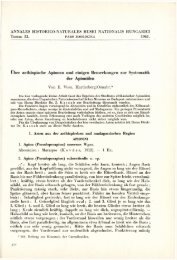
1. Spirogyra crassoidea TRANSEAU (= S. ellipsospora var. crassoidea TRAN-<br />
SEAU) — The vegetative cells have widths of 118—148 jum, and lengths of 201—314<br />
pm. The cross-wall is straight and each cell contains 5—8 spiral chloroplasts.<br />
Copulation is ladder shaped, the copulating cells do not swell. The copulation<br />
channel averaging 27 pm in width, is formed jointly by the two partners. The<br />
zygotes are three-axed, elliptic, their centers are often indented and thus suggestive<br />
of human blood cell (Fig. 1). The length of the spore is 112—157,5 pm, the width<br />
129—158 jitm. The external wall of the spore is thin, its surface is smooth. The<br />
mesosporium is brownish and on its surface small points arranged in irregular<br />
pattern are visible under the immersion lens.<br />
In August the cells contained many calcium oxalate crystals which often measured<br />
up to 20 jtim (Fig. 2). The pyrenoids were about 10 pm in diameter. The description<br />
well conforms to the data in the literature.<br />
Nearly every author dealing with the systematics of the Spirogyra genus<br />
agrees in that in the case of intercalarily growing cells the cell-length is of no importance<br />
from taxonomical view-point. LANGER (1934) for instance writes the<br />
following: " I neglected to publish the lengths of the cells. These are, namely, of<br />
no importance, since the length of the cell is the function of the age of the cell and<br />
as such cannot be taken for a diagnostic character of the species". Since both cells<br />
and spores have fairly constant widths (Table 1) the length of the spore is to change<br />
in conformity to the length of the cell (from the abundant plasma of a long cell a<br />
long spore develops). I succeeded in providing mathematical evidences for this
56 L. HAJDÚ<br />
(SVÁB 19G7) : 16 parallel measurements offered evidences for a close linear correlation<br />
(+0,6594 p
On the basis of HORTOBÁGYI'S algal catalogue (the state in November, 1973)<br />
this species is new in the Hungarian Flora.<br />
2. Oedogonium tyrolicum WITTEOCK. — Monoecious, in the filament the oogonia<br />
are formed in ones. They are oval, with an oval aperture on their upper parts.<br />
The oospores are round-elliptical, smooth-walled and do not fill the oogonium<br />
completely. The antheridium is a little narrower than the filament. The apical<br />
cell is bluntly rounded. The vegetative cells measure 75—113 by 14—25 ^m, the<br />
oogonium is 48—53 pva wide and 73—104 pm long (MROZINSKA—WEBB 1969:<br />
57—10 pm in length). The oospores are 45—52 pm wide, 52—10pm long (MROZLNS-<br />
K A — W E B B 1. c: 40—47itm long). With the exception of the two data for length<br />
nearly all taxonomic features well fit in with the specimens found (Figs. 6—8).<br />
The species lived attached to the leaves of Najas minor.<br />
On the basis of HORTOBÁGYI'S algal catalogue this species is new in the Hungarian<br />
Flora.<br />
3. Aphanochaete repens A BRAUN — It was found to grow as an epiphyton on<br />
Oedogonium filaments. The 10 pm long and 7 pm wide cells formed straight on<br />
occasion ramifying threads. On each cell a hair measuring up to 60 pm originates<br />
which is onion-like thickened at its base.<br />
4. Cladophora, glomerata (L.) KÜTZ. — The bulk of the algal mat on the bottom<br />
of the pond was formed by this species. The apical cell is 45—60 ^um thick. The colony<br />
is of acropetal organization (Fig 4). In culture it developed holdfasts as illustrated<br />
in Fig. 5. There were numerous epiphytes, chiefly diatoms on the filaments.<br />
5. Hydrodictyon reticulatum (L.)LAGERH. (= H. utriculatum ROTH and H. tenellum<br />
ROTH) — This alga of peculiar construction is abundant in eutrophic waters<br />
(KOL 1956, SÖRENSEN 1950).<br />
Ecological data<br />
The Városligeti-tó of <strong>Budapest</strong> is a temporary water. It is filled with tap water<br />
yearly once from the watersystem and has neither out- or inflow. One third of the bottom<br />
of the pond is covered with concrete, and from its other parts the sludge is reved by dredging.<br />
In 1973 subsequent to dredging the pond was filled only later, on March 30.<br />
By the time summer set in a thick algal web had developed on the bottom<br />
of the pond, which consisted of the following three layers. Lowest down were<br />
Hydrodictyon colonies embedded into a half-anaerobic deposit smelling of hydrogen<br />
sulphide. This was covered by a thick, loose, coarse-textured Cladophora mat.<br />
The algal mat was not of equal thickness everywhere and there were furrows to<br />
some depth on its surface. The bright-green Spirogyra tufts were carried into this<br />
furrows and by climbing on one another formed relatively massive conical heaps.<br />
On August 10 the amount of algae found on a quadrate meter surface of the bottom<br />
amounted to 1,8 kg fresh-weight resp. 0,48 kg dry weight in the average of three<br />
samples.<br />
Although in the pond Spirogyra was freuqent in the vegetative form, in the<br />
conjugating form it was only seldom observed. The majority of them does not<br />
reach the stage of zygospore formation. I examined a 2 m 2<br />
spot covered with<br />
Spirogyra in a medium advanced stage of conjugation for three weeks and found<br />
the process to have progressed only in the case of a few filaments and not even $<br />
then were spores produced. According to my observations proliferation occurs first<br />
of all in places near to the water surface. This is evidenced also by the fact that<br />
spore-formation is more frequent in case the water level of the pond decreases-
58 L. HAJDÚ<br />
relatively fast. (The water of the pond oozed away into the ground due to an artifical<br />
groundwater drainage in September. Under a 10 cm water layer a Spirogyra<br />
mat of 5m 2<br />
size was found where the process in 80 per cent of the cases reached the<br />
stage of spore formation. From this mat we have abundant sample both dried and<br />
formalin conserved one. At the time of spore formation there were numerous<br />
epithytes on the Spirogyra threads (PANKOW 1961).<br />
The algal mat of the pond was very uniform. Spots oi Ohara vulgaris L. and<br />
Najas minor were only occasionally found. Ceratophyllum demersum was relatively<br />
numerous. In August the algal mat becomes gradually separated from the bottom<br />
and is drifted to and from on the water surface. This in our case was due to the circumstance<br />
that the oxygen bubles produced by way of assimilation passed easily<br />
through the coarse-textured, loose Cladophora mat yet some of them got captured<br />
•among the small Hydrodictyon-nets below and among the Cladophores passing upwards<br />
the little balloon rearranges the threads of the mat : from the bottom an empty,<br />
gradually growing, Cladophora-walled cone passes upwards with Hydrodictyon<br />
at its top. Reaching the surface the top of the cone opens and later on becomes<br />
•completely detached from the bottom as a consequence of the smallest<br />
motion.wave-<br />
References<br />
HORTOBÁGYI, T.: Catalogus et Iconographia Algarum Hungáriáé in Institutum Bot.<br />
Univ. Agr. Gödöllő (status Nov. 1973, Manuscript).<br />
KADLUBOWSKA, J. Z. (1972): Zygnemaceae in Flora Slodkovodna Polski PWN Krakow.<br />
p. 1-431.<br />
KOL, E. (1956) : Comparative algological and hydrobiological studies in rice fields in Hungary<br />
- Acta Bot. Hung., 2: 309-363.<br />
BANGER, S. (1934): Monographische Bearbeitung der Spirogyren mit besonderer Berücksichtigung<br />
der Vorkriesungarisehen Verhältnisse — Folia Cryptogamica, 1: 1253 —<br />
1306.<br />
MROZINSKA - WEBB, T. (1969) : Oedogoniales in Flora Slodkovodna Polski- PWN Krakow,<br />
p. 1-659.<br />
PANKOW, H. (1961): Über die Ursachen des Fehlens von Epiphyten auf Zygnemalen. —<br />
Arch. Protistenk., 105: 417-444.<br />
ISOKAL, R. R. & SNEATH, P. II. A. (1963): Principles of Numerical Taxonomy. - Freeman<br />
and Co., San Francisco — London, p. 1 — 359.<br />
•SÖRENSEN, I. (1950): Studies in the Ecology of Hydrodictyon reticulatum (L.) Lagerh.<br />
- Oikos, 2: 197-212.<br />
SVÁB, J. (1967): Biometriai módszerek a mezőgazdasági kutatásban — Mezőgazd. Kiadó,<br />
<strong>Budapest</strong>, p. 1 —498.<br />
VAN DEN HOEK, C. (1963) : Revision of the European species of Cladophora — Leiden, E. J.<br />
Brill, p. 1-248.<br />
Author's address: Dr. L. HAJDU<br />
Botanical Department of the<br />
Hungarian Natural History Museum<br />
H — 1146 <strong>Budapest</strong>, Vajdahunyadvár<br />
Hungary