Stream macroalgae of Hawai`i: an identification guide - Hawaii.gov
Stream macroalgae of Hawai`i: an identification guide - Hawaii.gov
Stream macroalgae of Hawai`i: an identification guide - Hawaii.gov

You also want an ePaper? Increase the reach of your titles
YUMPU automatically turns print PDFs into web optimized ePapers that Google loves.
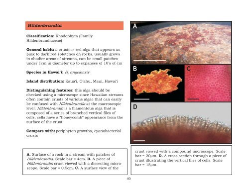
Hildenbr<strong>an</strong>dia<br />
Classification: Rhodophyta (Family<br />
Hildenbr<strong>an</strong>diaceae)<br />
General habit: a crustose red alga that appears as<br />
pink to dark red splotches on rocks, usually grows<br />
in shadier areas <strong>of</strong> streams, c<strong>an</strong> be small patches<br />
under 1cm in diameter up to exp<strong>an</strong>ses <strong>of</strong> 10’s <strong>of</strong> cm<br />
Species in Hawai‘i: H. <strong>an</strong>golensis<br />
Isl<strong>an</strong>d distribution: Kaua‘i, O‘ahu, Maui, Hawai‘i<br />
Distinguishing features: this alga should be<br />
checked using a microscope since <strong>Hawaii</strong><strong>an</strong> streams<br />
<strong>of</strong>ten contain crusts <strong>of</strong> various algae that c<strong>an</strong> easily<br />
be confused with Hildenbr<strong>an</strong>dia at the macroscopic<br />
level; Hildenbr<strong>an</strong>dia is a filamentous alga that is<br />
composed <strong>of</strong> a series <strong>of</strong> br<strong>an</strong>ched vertical files <strong>of</strong><br />
cells, cells have a “honeycomb” appear<strong>an</strong>ce from the<br />
surface <strong>of</strong> the crust<br />
Compare with: periphyton growths, cy<strong>an</strong>obacterial<br />
crusts<br />
A. Surface <strong>of</strong> a rock in a stream with patches <strong>of</strong><br />
Hildenbr<strong>an</strong>dia. Scale bar = 4cm. B. A piece <strong>of</strong><br />
Hildenbr<strong>an</strong>dia crust viewed with a dissecting microscope.<br />
Scale bar = 0.5cm. C. A surface view <strong>of</strong> the<br />
40<br />
crust viewed with a compound microscope. Scale<br />
bar = 20µm. D. A cross section through a piece <strong>of</strong><br />
crust illustrating the vertical files <strong>of</strong> cells. Scale<br />
bar = 15µm.