Stream macroalgae of Hawai`i: an identification guide - Hawaii.gov
Stream macroalgae of Hawai`i: an identification guide - Hawaii.gov
Stream macroalgae of Hawai`i: an identification guide - Hawaii.gov

You also want an ePaper? Increase the reach of your titles
YUMPU automatically turns print PDFs into web optimized ePapers that Google loves.
Linda Lingle<br />
Governor<br />
Department <strong>of</strong> L<strong>an</strong>d <strong>an</strong>d Natural Resources<br />
DIVISION OF AQUATIC RESOURCES<br />
1151 Punchbowl Street, Room 330<br />
Honolulu, HI 96813<br />
May 2004<br />
Photos by Alison Sherwood
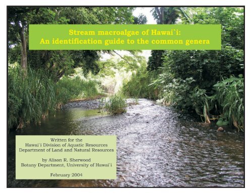
<strong>Stream</strong> <strong>macroalgae</strong> <strong>of</strong> Hawai‘i<br />
An <strong>identification</strong> <strong>guide</strong> to the common genera<br />
DAR Technical Report 04-02<br />
Funded in part by the DLNR<br />
Commission on Water Resource M<strong>an</strong>agement<br />
Alison Sherwood<br />
Division <strong>of</strong> Aquatic Resources<br />
<strong>an</strong>d Department <strong>of</strong> Bot<strong>an</strong>y<br />
University <strong>of</strong> Hawai‘i<br />
3190 Maile Way<br />
Honolulu, HI 96822<br />
asherwoo@hawaii.edu<br />
The Department <strong>of</strong> L<strong>an</strong>d <strong>an</strong>d Natural Resources receives fin<strong>an</strong>cial support under the Federal<br />
Aid in Sport Fish <strong>an</strong>d Wildlife Restoration <strong>an</strong>d other federal programs. Under Title VI <strong>of</strong> the<br />
Civil Rights Act <strong>of</strong> 1964, Section 504 <strong>of</strong> the Rehabilitation Act <strong>of</strong> 1973, Title II <strong>of</strong> the Americ<strong>an</strong>s<br />
with Disabilitites Act <strong>of</strong> 1990, the Age Discrimination Act <strong>of</strong> 1975, Title IX <strong>of</strong> the Education<br />
Amendments <strong>of</strong> 1972, <strong>an</strong>d the laws <strong>of</strong> the State <strong>of</strong> <strong>Hawaii</strong>, the U.S. Department <strong>of</strong> the Interior<br />
<strong>an</strong>d the State <strong>of</strong> <strong>Hawaii</strong> prohibit discrimination on the basis <strong>of</strong> race, color, religion, sex, national<br />
origin, age, <strong>an</strong>d disability. If you believe that you have been discriminated against in <strong>an</strong>y program,<br />
activity or facility, or if you desire information, please write to: Affirmative Action Officer,<br />
Personnel Office, Department <strong>of</strong> L<strong>an</strong>d <strong>an</strong>d Natural Resources, 1151 Punchbowl Street, Rm.<br />
231, Honolulu, HI 96813, or the U.S. Fish & Wildlife Service, Civil Rights Br<strong>an</strong>ch, 4040 N.<br />
Fairfax Drive, Suite 300, Arlington, VA 22203.
Scope <strong>an</strong>d Purpose <strong>of</strong> the Guide<br />
How to Use this Guide<br />
Collection <strong>an</strong>d Preservation Techniques<br />
Additional Sources <strong>of</strong> Information<br />
Table <strong>of</strong> Contents<br />
Key to the Common Genera <strong>of</strong> <strong>Hawaii</strong><strong>an</strong> <strong>Stream</strong> Macroalgae<br />
Descriptions <strong>of</strong> the Genera<br />
Index<br />
1<br />
1<br />
2<br />
3<br />
4<br />
9<br />
48
Scope <strong>an</strong>d Purpose <strong>of</strong> the Guide<br />
The purpose <strong>of</strong> this <strong>guide</strong> is to provide basic<br />
descriptions <strong>an</strong>d photographs <strong>of</strong> Hawai‘i’s most<br />
common stream algae in order to aid state biologists<br />
in their m<strong>an</strong>agement-based investigations. It is<br />
recognized that time <strong>an</strong>d resources are generally not<br />
available for species-level taxonomic <strong>identification</strong>s<br />
<strong>of</strong> stream algae, <strong>an</strong>d the <strong>guide</strong> is designed to lead the<br />
biologist to a generic placement, with some additional<br />
sources listed if further information is desired. Given<br />
that the systematics <strong>of</strong> most groups <strong>of</strong> freshwater algae<br />
in Hawai‘i have not been investigated using molecular<br />
tools <strong>an</strong>d thorough systematic comparisons,<br />
<strong>identification</strong> <strong>of</strong> collections to the generic level seems<br />
a prudent compromise at this point in time.<br />
Although the vast majority <strong>of</strong> stream <strong>macroalgae</strong><br />
in the state require examination at the compound<br />
microscope level <strong>of</strong> magnification for <strong>identification</strong>, a<br />
few <strong>of</strong> Hawai‘i’s stream <strong>macroalgae</strong> c<strong>an</strong> be identified<br />
through direct observation. In either case,<br />
characteristics such as color, br<strong>an</strong>ching pattern <strong>an</strong>d<br />
habitat will be needed.<br />
How to use this Guide<br />
The <strong>guide</strong> begins with a description <strong>of</strong><br />
recommended collection <strong>an</strong>d preservation techniques<br />
for stream <strong>macroalgae</strong>, along with a bibliography <strong>of</strong><br />
<strong>identification</strong> literature <strong>an</strong>d websites for freshwater<br />
algae that may be consulted for species-level<br />
1<br />
<strong>identification</strong>, clarification <strong>of</strong> the present <strong>guide</strong> or<br />
simply to obtain more detailed information on<br />
freshwater algae. A key to the common genera <strong>of</strong><br />
freshwater <strong>macroalgae</strong> in <strong>Hawaii</strong><strong>an</strong> streams is<br />
subsequently presented. The key employs fieldrecognizable<br />
characters wherever possible; however,<br />
in most cases at least some microscopical<br />
examination is necessary for determination <strong>of</strong> the<br />
appropriate characters. A set <strong>of</strong> page numbers is<br />
provided next to each genus name in the key - these<br />
pages contain text descriptions <strong>of</strong> the import<strong>an</strong>t<br />
characteristics <strong>of</strong> the genus, along with photographs<br />
<strong>of</strong> representatives (overall habit plus<br />
photomicrographs <strong>of</strong> diagnostic characters), <strong>an</strong><br />
estimation <strong>of</strong> the number <strong>of</strong> species present in<br />
<strong>Hawaii</strong><strong>an</strong> streams, <strong>an</strong>d the known distribution <strong>of</strong><br />
each genus within the state <strong>of</strong> Hawai‘i. Five main<br />
groups <strong>of</strong> stream <strong>macroalgae</strong> are recognized <strong>an</strong>d<br />
presented here: 1) Cy<strong>an</strong>obacteria, or the<br />
photosynthetic prokaryotic org<strong>an</strong>isms, 2)<br />
Chlorophyta, or the green algae, 3) Rhodophyta, or<br />
the red algae, 4) Tribophyta, or the yellow-green<br />
algae, <strong>an</strong>d 5) Bacillariophyta, or the diatoms. The<br />
most common macroalgal groups in <strong>Hawaii</strong><strong>an</strong><br />
streams are the green algae <strong>an</strong>d Cy<strong>an</strong>obacteria,<br />
followed by the red algae, diatoms <strong>an</strong>d yellow-green<br />
algae; however, the diatoms possess considerably<br />
more diversity in the periphyton community th<strong>an</strong><br />
represented here. The broad taxonomic category to<br />
which each representative belongs is indicated at<br />
the top <strong>of</strong> the description pages through the color <strong>of</strong><br />
the heading bar, as follows: Cy<strong>an</strong>obacteria =<br />
turquoise, Chlorophyta = green, Rhodophyta = red,
Tribophyta = yellow, Bacillariophyta = gray.<br />
Distribution information, in terms <strong>of</strong> what is known<br />
so far, is indicated for each genus. It should be<br />
recognized, however, that the potential <strong>an</strong>d likelihood<br />
for extension <strong>of</strong> r<strong>an</strong>ges on other isl<strong>an</strong>ds is high.<br />
Additionally, the reports included in this <strong>guide</strong><br />
represent records from streams only – m<strong>an</strong>y <strong>of</strong> these<br />
genera have representatives in marine, brackish <strong>an</strong>d<br />
subaerial/terrestrial habitats, which are not included<br />
here. The <strong>guide</strong> has been exclusively illustrated using<br />
<strong>Hawaii</strong><strong>an</strong> material.<br />
Collection <strong>an</strong>d preservation techniques<br />
Since almost all stream <strong>macroalgae</strong> require at<br />
least some microscopical examination for <strong>identification</strong>,<br />
collection <strong>an</strong>d preservation techniques should<br />
be developed to fit the needs <strong>an</strong>d intentions <strong>of</strong> the<br />
study. One necessary piece <strong>of</strong> equipment for stream<br />
<strong>macroalgae</strong> sampling is a viewbox (or snorkel mask,<br />
if the collector is in the water; e.g. for fish surveys).<br />
A view box is relatively easy to construct, <strong>an</strong>d c<strong>an</strong><br />
be made by cutting a square hole in the center <strong>of</strong> a<br />
plastic tupperware container, <strong>an</strong>d using silicone<br />
aquarium gel to seal in a piece <strong>of</strong> glass cut to the<br />
correct size. The collection <strong>of</strong> <strong>macroalgae</strong> from the<br />
stream is accomplished most easily using longh<strong>an</strong>dled<br />
forceps, which allow the alga to be removed<br />
in one piece at its base. Inclusion <strong>of</strong> the basal attachment<br />
is import<strong>an</strong>t, since some taxonomic descriptions<br />
use information pertaining to this part <strong>of</strong><br />
the pl<strong>an</strong>t. A few forms may be more easily removed<br />
2<br />
using a single-edged razor blade (e.g.<br />
Hildenbr<strong>an</strong>dia, Calothrix, some diatom films), or<br />
toothbrushes. Some gelatinous growths in side<br />
pools are easily collected using a turkey baster<br />
(e.g. Anabaena). It is also import<strong>an</strong>t to search all<br />
habitats in the vicinity <strong>of</strong> the stream to ensure<br />
that all taxa are represented in the collections –<br />
including rocks <strong>an</strong>d other bottom substrata in<br />
fast-flowing stream areas, slow flowing areas at<br />
the edges <strong>of</strong> the stream, side pools, waterfall faces,<br />
seep faces, floating material in the stream <strong>an</strong>d<br />
vegetation. As well, <strong>macroalgae</strong> prefer different<br />
light regimes, <strong>an</strong>d so both brightly illuminated<br />
<strong>an</strong>d shaded areas should be searched. Scintillation<br />
vials or WhirlPak bags work best for storing<br />
collections from the field, but it is import<strong>an</strong>t not to<br />
overfill the containers with algae since this will<br />
result in their rapid degradation. Most field collections<br />
kept cold (on ice or in a refridgerator) will<br />
remain in good condition for several days.<br />
The most highly recommended preservative<br />
for freshwater <strong>macroalgae</strong> is CaCO 3 -buffered<br />
glutaraldehyde (a 2.5% solution made from<br />
dilution <strong>of</strong> 25% glutaraldehyde, add a pinch <strong>of</strong><br />
CaCO 3 powder to buffer the solution). This<br />
preservative maintains the original color <strong>of</strong> the<br />
specimens very well, <strong>an</strong>d also minimizes the<br />
morphological distortion inherent in the<br />
preservation process. Samples preserved in<br />
glutaraldehyde should be kept cold until they are<br />
identified. Large numbers <strong>of</strong> collections are easiest<br />
to org<strong>an</strong>ize in scintillation vials, since these c<strong>an</strong><br />
be stored in flats <strong>of</strong> 100 vials in a fridge. In the
absence <strong>of</strong> glutaraldehyde, Lugol’s Iodine Solution<br />
could be used, which has the benefit <strong>of</strong> staining the<br />
starch-positive groups <strong>of</strong> algae, but affects the<br />
overall color <strong>of</strong> the samples. Alcohol-based<br />
preservatives are not recommended since they<br />
destroy import<strong>an</strong>t information about specimen<br />
color, <strong>an</strong>d severely distort the internal cellular<br />
details.<br />
Finally, collections should be thoroughly<br />
labeled with all relev<strong>an</strong>t information, such as<br />
collection location (including GPS coordinates,<br />
where possible), the collector, date <strong>an</strong>d habitat.<br />
General Books:<br />
Additional Sources <strong>of</strong> Information<br />
Dillard, G.E. 1999. Common Freshwater Algae <strong>of</strong> the<br />
United States. J. Cramer, Berlin/Stuttgart,<br />
Germ<strong>an</strong>y.<br />
Entwisle, T.J., Sonnem<strong>an</strong>, J.A. <strong>an</strong>d Lewis, S.H.<br />
1997. Freshwater Algae in Australia: a Guide to<br />
Conspicuous Genera. Sainty & Associates, Australia.<br />
Pentecost, A. 1984. Introduction to Freshwater<br />
Algae. The Richmond Publishing Co. Ltd., Surrey,<br />
Engl<strong>an</strong>d.<br />
Prescott, G.W. 1951. Algae <strong>of</strong> the Western Great<br />
Lakes Area. WM. C. Brown Publishers, Dubuque,<br />
Iowa, U.S.A.<br />
3<br />
Websites:<br />
General algal taxonomic information:<br />
http://www.algaebase.org/<br />
Bowling Green State University Algal Homepage:<br />
http://www.bgsu.edu/Departments/biology/algae/<br />
index.html<br />
Great Lakes Diatom Homepage:<br />
http://www.umich.edu/~phytolab/<br />
GreatLakesDiatomHomePage/top.html<br />
Freshwater Algae from southeastern Ohio:<br />
http://vis-pc.pl<strong>an</strong>tbio.ohiou.edu/algaeindex.htm<br />
Algae Homepage <strong>of</strong> the Smithsoni<strong>an</strong> Institution:<br />
http://www.nmnh.si.edu/bot<strong>an</strong>y/projects/algae/<br />
CYANOSITE – Cy<strong>an</strong>obacterial research:<br />
http://www-cy<strong>an</strong>osite.bio.purdue.edu/index.html<br />
Desmid information:<br />
http://www.desmids.info/<br />
Freshwater algae <strong>of</strong> the British Isles:<br />
http://www.nwl.ac.uk/~loissys/algal_coded_list.htm<br />
Fritsch collection <strong>of</strong> illustrations <strong>of</strong> freshwater<br />
algae:<br />
http://www.ife.ac.uk/fritsch/<br />
Diatom Collection <strong>of</strong> the California Academy <strong>of</strong><br />
Sciences:<br />
http://www.calacademy.org/research/diatoms/<br />
diatoms.html
Key to the common genera <strong>of</strong> <strong>Hawaii</strong><strong>an</strong><br />
stream <strong>macroalgae</strong><br />
1.a. Large alga (several to m<strong>an</strong>y centimeters in<br />
length) with a pl<strong>an</strong>t body differentiated into a stemlike<br />
axis with whorls <strong>of</strong> br<strong>an</strong>ches, not surrounded<br />
by mucilage – Chara (p.28)<br />
1.b. Org<strong>an</strong>ism either microscopic or macroscopic at<br />
the individual level, but cellular structures not<br />
evident without microscopic examination - 2<br />
2.a. Alga siphonous, as seen at the microscopic<br />
level (lacking cellular cross walls), or in some other<br />
form (e.g. needle-shaped colonies) - 3<br />
2.b. Alga filamentous when observed at the light<br />
microscopic level (includes crustose algae composed<br />
<strong>of</strong> filaments) - 4<br />
3.a. Alga a felty green mass <strong>of</strong> filaments, coarse to<br />
the touch, cells a yellowish-green color when<br />
observed under the light microscope, lacking cross<br />
walls - Vaucheria (p.44)<br />
3.b. Alga forming small star-shaped clusters <strong>of</strong><br />
needle-like cells, usually growing on larger algae or<br />
mosses, chloroplasts a golden color - Synedra (p.48)<br />
4.a. Filaments br<strong>an</strong>ched when observed<br />
macroscopically or using the light microscope, or<br />
4<br />
main axis composed <strong>of</strong> multiple filaments<br />
(multiseriate) - 5<br />
4.b. Filaments unbr<strong>an</strong>ched, or only very<br />
occasionally br<strong>an</strong>ched when observed<br />
macroscopically or using the light microscope,<br />
main axis composed <strong>of</strong> only a single filament<br />
(uniseriate) - 18<br />
5.a. Cell contents blue-green, olive-green or<br />
pinkish in color, lacking a nucleus <strong>an</strong>d<br />
chloroplast - 6<br />
5.b. Cell contents colored grass-green, yellowgreen,<br />
or as in 5a, but containing at least one<br />
nucleus <strong>an</strong>d chloroplast - 11<br />
6.a. Main axis <strong>of</strong> the alga (when viewed<br />
microscopically) composed <strong>of</strong> multiple filaments <strong>of</strong><br />
cells, alga appears cartilaginous <strong>an</strong>d is <strong>of</strong>ten<br />
pigmented a yellowish-brown color - Stigonema<br />
(p.24)<br />
6.b. Main axis <strong>of</strong> the alga composed <strong>of</strong> only a<br />
single filament (uniseriate) - 7<br />
7.a. Algal filaments uniform in diameter along<br />
their length or composed <strong>of</strong> bead-like cells - 8<br />
7.b. Algal filaments gradually tapering in diameter<br />
along their length - Dichothrix (p.14)
8.a. Alga with distinctly inflated cells, appearing<br />
bead-like under the light microscope, cell contents<br />
a uniform light bluish-green color, no distinct<br />
sheath surrounding the filaments - Nostochopsis<br />
(p.19)<br />
8.b. Algal filament contained within a sheath, with<br />
cells more or less the same diameter (not bead-like<br />
<strong>an</strong>d rounded), although some may have slight<br />
constrictions at cross walls - 9<br />
9.a. Alga typically forming two false br<strong>an</strong>ches at the<br />
same point - Scytonema (p.23)<br />
9.b. Alga typically only forming a single false<br />
br<strong>an</strong>ch at <strong>an</strong>y one point, if at all - 10<br />
10.a. Filaments long, single false br<strong>an</strong>ches<br />
common, br<strong>an</strong>ching occurs adjacent to a larger,<br />
clearer cell (heterocyst) in the filament - Tolypothrix<br />
(p.25)<br />
10.b. Single false br<strong>an</strong>ches rare, filaments quite<br />
short - Microchaete (p.16)<br />
11.a. Chloroplasts a yellow-green, grass-green or<br />
dark green color - 12<br />
11.b. Chloroplasts olive-gray, blue, pink, purple or<br />
red in color - 16<br />
12.a. Alga br<strong>an</strong>ching only at the base, near the<br />
point <strong>of</strong> attachment - Basicladia (p.26)<br />
5<br />
12.b. Alga br<strong>an</strong>ching in regions other th<strong>an</strong> just at<br />
the base, near the point <strong>of</strong> attachment, or not at<br />
all (but possessing a multiseriate main axis) - 13<br />
13.a. Chloroplasts dense <strong>an</strong>d net-like, <strong>of</strong>ten<br />
seeming to fill the cell - Cladophora (p.29)<br />
13.b. Chloroplast a plate-like b<strong>an</strong>d, especially<br />
evident in the main axis cells - 14<br />
14.a. Alga multiseriate (multiple filaments<br />
composing the main axis) in the upper regions <strong>of</strong><br />
the pl<strong>an</strong>t - Schizomeris (p.35)<br />
14.b. Alga uniseriate throughout (only one<br />
filament composing the axis) - 15<br />
15.a. Alga forming a distinct colony, filaments<br />
gradually tapering toward their ends -<br />
Chaetophora (p.27)<br />
15.b. Alga a feathery tuft, cells <strong>of</strong> br<strong>an</strong>ches not<br />
markedly different in diameter from the main axis,<br />
tips <strong>of</strong> br<strong>an</strong>ches tapering to a point or ending in a<br />
fine hair - Stigeoclonium (p.37)<br />
15.c. Alga a feathery tuft, cells <strong>of</strong> the br<strong>an</strong>ches<br />
smaller th<strong>an</strong> those <strong>of</strong> the main axis, tips <strong>of</strong><br />
br<strong>an</strong>ches not tapering to a point or ending in a<br />
fine hair - Cloniophora (p.30)
16.a. Alga composed <strong>of</strong> a main axis with whorls <strong>of</strong><br />
br<strong>an</strong>ches emerging from it, gelatinous to the touch<br />
- Batrachospermum (p.40)<br />
16.b. Alga either crustose or filamentous, but not<br />
gelatinous - 17<br />
17.a. Alga crustose, resembling spots <strong>of</strong> red paint,<br />
usually on rock surfaces - Hildenbr<strong>an</strong>dia (p.42)<br />
17.b. Alga br<strong>an</strong>ched <strong>an</strong>d filamentous, forming long<br />
bluish-green or bluish-gray str<strong>an</strong>ds, main axis<br />
with a bead-like appear<strong>an</strong>ce that c<strong>an</strong> be seen<br />
without a microscope - Compsopogon (p.41)<br />
17.c. Alga filamentous <strong>an</strong>d br<strong>an</strong>ched, chloroplasts<br />
bluish or reddish in color, round-shaped<br />
reproductive structures (monospor<strong>an</strong>gia) common<br />
at the tips <strong>of</strong> br<strong>an</strong>ches - Audouinella (p.39)<br />
18.a. Cell contents blue-green, olive-green or<br />
pinkish in color, lacking a nucleus <strong>an</strong>d chloroplast<br />
- 19<br />
18.b. Cells with chloroplasts colored grass-green,<br />
yellow-green, or as in 18a, but containing at least<br />
one nucleus <strong>an</strong>d chloroplast - 25<br />
19.a. Cells bead-like or barrel-shaped, heterocysts<br />
present at various positions within the filaments,<br />
or only at the ends - 20<br />
19.b. Cells otherwise - 21<br />
6<br />
20.a. Alga with a thick outer mucilage layer that<br />
gives the org<strong>an</strong>ism a definite shape - Nostoc (p.18)<br />
20.b. Alga without a thick outer mucilage layer,<br />
does not retain its shape when removed from the<br />
water, heterocysts common at multiple positions<br />
within a filament - Anabaena (p.11)<br />
20.c. Alga without a thick outer mucilage layer,<br />
does not retain its shape when removed from the<br />
water, heterocysts only at the ends <strong>of</strong> the filaments<br />
- Cylindrospermum (p.13)<br />
21.a. Filaments <strong>of</strong> the alga tapering in diameter<br />
along their length, with a heterocyst at the base <strong>of</strong><br />
the filament - Calothrix (p.12)<br />
21.b. Filaments much the same diameter along<br />
their length - 22<br />
22.a. Only a single filament present within a<br />
surrounding sheath, if present at all - 23<br />
22.b. Two or more filaments sometimes or always<br />
seen within a single surrounding sheath - 24<br />
23.a. Filaments not surrounded by a sheath -<br />
Oscillatoria (p.20)<br />
23.b. Filaments usually surrounded by a sheath,<br />
ent<strong>an</strong>gled to form a definite mat-like structure that
is conspicuous at the macroscopic level -<br />
Phormidium (p.21)<br />
23.c. Filaments surrounded by a sheath, c<strong>an</strong> be<br />
ent<strong>an</strong>gled in small growths but not as large mats -<br />
Lyngbya (p.15)<br />
24.a. M<strong>an</strong>y filaments bundled together within a<br />
sheath, ent<strong>an</strong>gled to form a mat that is usually<br />
greenish in color - Schizothrix (p.22)<br />
24.b. Only a few filaments typically bundled<br />
together within a sheath, mat is usually bluishpurple<br />
or purplish-gray in color - Microcoleus<br />
(p.17)<br />
25.a. Cells with grass-green chloroplasts <strong>an</strong>d<br />
containing starch (positive stain with Lugol’s<br />
Iodine Solution) - 26<br />
25.b. Cells with yellow-green or brown<br />
chloroplasts, not containing true starch (alga does<br />
not positively stain with Lugol’s Iodine Solution) -<br />
30<br />
26.a. Cells with round chloroplasts that appear<br />
flattened against one wall <strong>of</strong> the cell, chloroplasts<br />
do not occupy majority <strong>of</strong> the cell - Klebsormidium<br />
(p.31)<br />
26.b. Cells otherwise - 27<br />
27.a. Cells containing net-like chloroplasts - 28<br />
7<br />
27.b. Cells otherwise - 29<br />
28.a. Some cells with conspicuous rings at one<br />
end, chloroplast usually not dense <strong>an</strong>d net-like<br />
structure c<strong>an</strong> be easily discerned - Oedogonium<br />
(p.33)<br />
28.b. Cells without rings at the ends, chloroplasts<br />
usually very dense <strong>an</strong>d the net-like structure<br />
difficult to see - Rhizoclonium (p.34)<br />
29.a. Cells with one or more spiral-shaped<br />
chloroplasts - Spirogyra (p.36)<br />
29.b. Cells with one plate-like chloroplast that may<br />
be either lying in a flat pl<strong>an</strong>e or twisted once -<br />
Mougeotia (p.32)<br />
29.c. Cells with two star-shaped chloroplasts -<br />
Zygnema (p.38)<br />
30a. Cells with yellow-green chloroplasts, cell walls<br />
in two sections that overlap in the central region,<br />
forming H-shaped sections - Tribonema (p.43)<br />
30b. Cells with brown chloroplasts, cell walls<br />
highly ornamented (diatoms) - 31<br />
31a. Cells arr<strong>an</strong>ged in filaments such that all or<br />
most <strong>of</strong> the adjacent cell surfaces are touching - 32
31b. Cells in filaments such that only the corners<br />
<strong>of</strong> the adjacent cells are touching (zig-zag<br />
filaments) - 33<br />
32a. Filaments constructed <strong>of</strong> cylindrical cells<br />
composed <strong>of</strong> overlapping pieces, ornamentation<br />
in the form <strong>of</strong> org<strong>an</strong>ized lines <strong>of</strong> holes (striae)<br />
along the two halves <strong>of</strong> the cylinder - Melosira<br />
(p.46)<br />
32b. Cells <strong>of</strong> filament appear rect<strong>an</strong>gular, when<br />
individually separated appear rounded with six<br />
protrusions equally spaced around cell perimeter<br />
- Hydrosera (p.45)<br />
33a. Cells appear rect<strong>an</strong>gular in filaments, but<br />
elliptical when seen in valve view - Pleurosira<br />
(p.47)<br />
33b. Cells in filaments form appear rect<strong>an</strong>gular,<br />
small “musical-note” shaped ornamentations<br />
visible on the cell wall - Terpsinöe (p.49)<br />
8
Anabaena<br />
Classification: Cy<strong>an</strong>obacteria (Family Nostocaceae)<br />
General habit: unbr<strong>an</strong>ched filaments in <strong>an</strong><br />
ent<strong>an</strong>gled mass, <strong>of</strong>ten in clumps or mats,<br />
surrounding mucilage does not have a definite<br />
shape that is retained when the alga is removed<br />
from the water<br />
Species in Hawai‘i: A. catenula, Anabaena sp.<br />
Isl<strong>an</strong>d distribution: Kaua‘i, O‘ahu<br />
Distinguishing features: bead or barrel-shaped<br />
cells in unbr<strong>an</strong>ched filaments, slightly larger <strong>an</strong>d<br />
less pigmented cells (heterocysts; sites <strong>of</strong> nitrogen<br />
fixation; Fig. C) spaced along filaments, similar to<br />
Nostoc but colony does not have a well-defined<br />
mucilage coating that retains its shape<br />
Compare with: Cylindrospermum, Nostoc,<br />
Nostochopsis<br />
9<br />
A. Piece <strong>of</strong> <strong>an</strong> epilithic Anabaena mat. Scale bar<br />
= 2mm. B. Filaments <strong>of</strong> Anabaena as seen with<br />
the compound microscope. Scale bar = 20µm. C.<br />
Single filament with both vegetative cells (colored)<br />
<strong>an</strong>d heterocysts (larger <strong>an</strong>d less pigmented).<br />
Scale bar = 15µm.
Calothrix<br />
Classification: Cy<strong>an</strong>obacteria (Family<br />
Rivulariaceae)<br />
General habit: pl<strong>an</strong>ts either growing individually or<br />
clustered in tufts or mats (sometimes in a starshaped<br />
formation), c<strong>an</strong> grow on rock surfaces or<br />
epiphytically on other algae or pl<strong>an</strong>ts<br />
Species in Hawai‘i: C. braunii, C. fusca<br />
Isl<strong>an</strong>d distribution: Necker, O‘ahu, Maui<br />
Distinguishing features: Filaments taper from the<br />
base (which usually ends in a heterocyst; Figs C, D)<br />
to a fine point at the distal end, cells shorter th<strong>an</strong><br />
wide in the lower regions <strong>of</strong> the filament <strong>an</strong>d<br />
gradually becoming longer th<strong>an</strong> wide toward the tips<br />
Compare with: Scytonema, Tolypothrix<br />
10<br />
A. An epilithic tuft <strong>of</strong> Calothrix. Scale bar = 1mm.<br />
B. View <strong>of</strong> several filaments under a compound<br />
microscope. Scale bar = 25µm. C, D. Individual<br />
filaments <strong>of</strong> Calothrix illustrating tapering <strong>of</strong> the<br />
filaments <strong>an</strong>d heterocysts at the ends. Scale bar =<br />
25µm.
Cylindrospermum<br />
Classification: Cy<strong>an</strong>obacteria (Family Nostocaceae)<br />
General habit: a series <strong>of</strong> intertwined filaments in a<br />
mucilage coat, forms dark green mats or patches on<br />
rocks in streams, occasionally on submerged<br />
vegetation<br />
Species in Hawai‘i: C. catenatum, C. stagnale,<br />
Cylindrospermum sp.<br />
Isl<strong>an</strong>d distribution: Kaua‘i, O‘ahu, Maui, Hawai‘i<br />
Distinguishing features: heterocysts present at one<br />
or both ends <strong>of</strong> the filaments (Fig. D), cells<br />
cylindrical or barrel-shaped, cross walls defined by<br />
constrictions, gonidia (reproductive bodies)<br />
occasionally visible at the ends <strong>of</strong> filaments adjacent<br />
to heterocysts<br />
Compare with: Anabaena, Oscillatoria, Phormidium<br />
11<br />
A. A piece <strong>of</strong> <strong>an</strong> epilithic mat <strong>of</strong> Cylindrospermum.<br />
Scale bar = 1mm. B. View under a compound<br />
microscope <strong>of</strong> a Cylindrospermum mat, illustrating<br />
intertwined filaments. Scale bar = 25µm. C. Filament<br />
with a gonidium <strong>an</strong>d heterocyst at the end.<br />
Scale bar = 20µm. D. Filament with a terminal<br />
heterocyst. Scale bar = 10µm.
Dichothrix<br />
Classification: Cy<strong>an</strong>obacteria (Family<br />
Rivulariaceae)<br />
General habit: forming small mats or feathery tufts<br />
on a variety <strong>of</strong> surfaces in streams, sometimes<br />
growing only as solitary pl<strong>an</strong>ts, yellowish-brown in<br />
color<br />
Species in Hawai‘i: D. baueri<strong>an</strong>a, D. fusca<br />
Isl<strong>an</strong>d distribution: Maui, Hawai‘i<br />
Distinguishing features: filaments tapering slightly<br />
along their length, heterocysts usually at the larger<br />
ends <strong>of</strong> filaments (although they c<strong>an</strong> be present in<br />
the central areas <strong>of</strong> the filaments as well), usually 2-<br />
6 filaments contained within a single sheath for at<br />
least part <strong>of</strong> their length<br />
Compare with: Scytonema, Tolypothrix<br />
12<br />
A. A piece <strong>of</strong> <strong>an</strong> epilithic mat <strong>of</strong> Dichothrix. Scale<br />
bar = 2mm. B. Filaments enclosed in a single<br />
sheath for part <strong>of</strong> their length. Scale bar = 20µm.<br />
C. Basal heterocysts on Dichothrix filaments.<br />
Scale bar = 10µm.
Lyngbya<br />
Classification: Cy<strong>an</strong>obacteria (Family<br />
Oscillatoriaceae)<br />
General habit: loose filaments that occasionally<br />
aggregate macroscopically in mats or tufts, c<strong>an</strong> be<br />
epiphytic on other vegetation or epilithic on rock<br />
surfaces, color varies from bluish-green to reddish<br />
or purplish<br />
Species in Hawai‘i: L. aestuarii, L. cladophorae, L.<br />
major, L. martensi<strong>an</strong>a, L. putealis, Lyngbya sp.<br />
Isl<strong>an</strong>d distribution: O‘ahu, Hawai‘i<br />
Distinguishing features: filaments unbr<strong>an</strong>ched,<br />
mostly cylindrical <strong>an</strong>d tapering very little if at all,<br />
enclosed in a sheath (Figs B, C), heterocysts absent,<br />
filaments loosely ent<strong>an</strong>gled rather th<strong>an</strong> forming a<br />
distinct mat-like structure (as in Phormidium)<br />
Compare with: Oscillatoria, Phormidium<br />
13<br />
A. Filaments <strong>of</strong> Lyngbya entwined on a br<strong>an</strong>ch.<br />
Scale bar = 1mm. B. Two individual filaments <strong>of</strong><br />
Lyngbya illustrating the surrounding sheath.<br />
Scale bar = 30µm. C. A long sheath surrounding<br />
a Lyngbya filament. Scale bar = 30µm. D. High<br />
magnification view <strong>of</strong> a terminal region <strong>of</strong> a filament.<br />
Scale bar = 20µm.
Microchaete<br />
Classification: Cy<strong>an</strong>obacteria (Family<br />
Microchaetaceae)<br />
General habit: ent<strong>an</strong>gled filaments, macroscopically<br />
seen as small, stellate tufts or as mats on rock<br />
surfaces or submerged vegetation in streams,<br />
usually brownish or gray in color<br />
Species in Hawai‘i: M. uberrima, M. tenera<br />
Isl<strong>an</strong>d distribution: Kaua‘i, Hawai‘i<br />
Distinguishing features: filaments not tapering<br />
along their length, enclosed in a sheath, occasional<br />
false br<strong>an</strong>ching present, heterocysts c<strong>an</strong> be at the<br />
ends <strong>of</strong> filaments (Fig. C) or along the length <strong>of</strong> the<br />
filament<br />
Compare with: Dichothrix, Scytonema, Tolypothrix<br />
14<br />
A. A piece <strong>of</strong> <strong>an</strong> epilithic mat formed by<br />
Microchaete. Scale bar = 2mm. B. Compound<br />
microscope view <strong>of</strong> intertwined Microchaete filaments<br />
from a mat formation. Scale bar = 70µm.<br />
C. High magnification view <strong>of</strong> a small filament<br />
illustrating the basal heterocyst <strong>an</strong>d uniformity <strong>of</strong><br />
filament diameter along its length. Scale bar =<br />
15µm.
Microcoleus<br />
Classification: Cy<strong>an</strong>obacteria (Family<br />
Phormidiaceae)<br />
General habit: forms dark blue-green patches or<br />
mats on submerged surfaces, composed <strong>of</strong> filaments<br />
in confluent sheaths, usually a purplish-gray mass<br />
Species in Hawai‘i: M. lacustris, M. vaginatus<br />
Isl<strong>an</strong>d distribution: O‘ahu, Hawai‘i<br />
Distinguishing features: parallel filaments<br />
enclosed in a mucilaginous sheath, slight tapering<br />
toward the end <strong>of</strong> the filament (Fig. B), cross walls<br />
sometimes have a gr<strong>an</strong>ular appear<strong>an</strong>ce (Fig. B)<br />
Compare with: Phormidium, Schizothrix<br />
15<br />
A. A piece <strong>of</strong> <strong>an</strong> epilithic mat formed by<br />
Microcoleus. Scale bar = 2mm. B. A compound<br />
microscope view <strong>of</strong> a Microcoleus filament, illustrating<br />
gr<strong>an</strong>ular cross walls (some species). Scale<br />
bar = 10µm. C, D. Parallel trichomes enclosed in a<br />
sheath. Scale bar = 15µm.
Nostoc<br />
Classification: Cy<strong>an</strong>obacteria (Family Nostocaceae)<br />
General habit: colonies c<strong>an</strong> be membr<strong>an</strong>ous,<br />
globular or lobed, or dried in clumps, composed <strong>of</strong><br />
unbr<strong>an</strong>ched filaments <strong>of</strong> cells, enclosed in a thick<br />
mucilage that lends a definite shape to the colony<br />
(which retains its shape when removed from the<br />
water)<br />
Species in Hawai‘i: N. foliaceum, N. hatei, N. linckia,<br />
N. microscopicum, N. paludosum, N. pruniforme, N.<br />
sphaericum, N. verrucosum, Nostoc sp.<br />
Isl<strong>an</strong>d distribution: Kaua‘i, O‘ahu, Maui, Hawai‘i<br />
Distinguishing features: colonies surrounded by a<br />
thick layer <strong>of</strong> mucilage which gives the alga a<br />
definite shape, cells <strong>of</strong> filaments resemble those <strong>of</strong><br />
Anabaena (bead or barrel-shaped), heterocysts<br />
frequent along length <strong>of</strong> filament (Fig. F)<br />
Compare with: Anabaena, Cylindrospermum,<br />
Nostochopsis<br />
A. View <strong>of</strong> <strong>an</strong> individual epilithic Nostoc colony<br />
Scale bar = 3mm. B. Numerous Nostoc colonies<br />
attached to a rock. Scale bar = 5cm. C. Compound<br />
16<br />
microscope view <strong>of</strong> inside a colony, showing filaments<br />
with heterocysts (larger cells). Scale bar =<br />
20µm. D. Filaments with highly pigmented<br />
sheaths. Scale bar = 20µm. E. Arr<strong>an</strong>gement <strong>of</strong><br />
filaments within a colony. Scale bar = 100µm. F.<br />
An individual filament with several heterocysts<br />
Scale bar = 15µm.
Nostochopsis<br />
Classification: Cy<strong>an</strong>obacteria (Family<br />
Nostochopsaceae)<br />
General habit: bright blue-green globular colonies,<br />
composed <strong>of</strong> filaments encased in mucilage, colonies<br />
retaining their shape when removed from the water<br />
Species in Hawai‘i: N. lobatus, N. radi<strong>an</strong>s<br />
Isl<strong>an</strong>d distribution: Kaua‘i, O‘ahu, Maui<br />
Distinguishing features: macroscopically similar to<br />
Nostoc, but filaments br<strong>an</strong>ching, appears as bright<br />
blue-green to grass-green colonies in streams,<br />
usually attached to rocks, heterocysts c<strong>an</strong> be either<br />
apical, or both intercalary <strong>an</strong>d apical (depending on<br />
the species)<br />
Compare with: Nostoc, Chaetophora<br />
17<br />
A. An individual colony <strong>of</strong> epilithic Nostochopsis.<br />
Scale bar = 2mm. B. Compound microscope view<br />
<strong>of</strong> filaments arrr<strong>an</strong>ged in a colony. Scale bar =<br />
20µm. C. Br<strong>an</strong>ched filament with heterocysts.<br />
Scale bar = 20µm. D. Intertwined filaments <strong>of</strong><br />
Nostochopsis. Scale bar = 15µm.
Oscillatoria<br />
Classification: Cy<strong>an</strong>obacteria (Family<br />
Oscillatoriaceae)<br />
General habit: forming either <strong>an</strong> exp<strong>an</strong>ded pl<strong>an</strong>t<br />
mass or present as loose filaments, attached to<br />
submerged rocks or vegetation in a stream, color<br />
c<strong>an</strong> r<strong>an</strong>ge greatly from blue-green to dark red,<br />
brown or purple<br />
Species in Hawai‘i: O. chalybea, O. curviceps, O.<br />
formosa, O. limosa, O. princeps, O. s<strong>an</strong>cta, O.<br />
splendida, O. subbrevis, Oscillatoria sp.<br />
Isl<strong>an</strong>d distribution: Kaua‘i, O‘ahu, Hawai‘i<br />
Distinguishing features: unbr<strong>an</strong>ched filaments<br />
lacking a sheath, filaments cylindrical <strong>an</strong>d not<br />
tapering along their length, heterocysts absent, c<strong>an</strong><br />
be either single filaments or aggregated in small<br />
colonies, when live, filaments <strong>of</strong>ten exhibit a<br />
characteristic “oscillating” movement<br />
Compare with: Lyngbya, Phormidium<br />
18<br />
A. A piece <strong>of</strong> <strong>an</strong> epilithic mat formed by<br />
Oscillatoria filaments. Scale bar = 2mm. B. Compound<br />
microscope view <strong>of</strong> <strong>an</strong> Oscillatoria mat.<br />
Scale bar = 40µm. C. Filaments <strong>of</strong> a small species<br />
<strong>of</strong> Oscillatoria. Scale bar = 15µm. D, E. Filaments<br />
<strong>of</strong> large species <strong>of</strong> Oscillatoria, demonstrating the<br />
lack <strong>of</strong> a surrounding sheath. Scale bar = 15µm<br />
(D), 10µm (E).
Phormidium<br />
Classification: Cy<strong>an</strong>obacteria (Family<br />
Phormidiaceae)<br />
General habit: masses <strong>of</strong> ent<strong>an</strong>gled filaments<br />
forming mats that are attached to rocks or<br />
vegetation, or growing in sediments in slowerflowing<br />
stream areas, c<strong>an</strong> r<strong>an</strong>ge in color from light<br />
blue-green to red/purple or black/gray<br />
Species in Hawai‘i: P. ambiguum, P. amoenum, P.<br />
corium, P. favosum, P. formosum, P. foveolarum, P.<br />
inundatum, P. pachydermaticum, P. retzii, P.<br />
splendidum, P. stagnina, P. subfuscum, P.<br />
subincrustatum, P. tinctorium, P. uncinatum,<br />
Phormidium sp.<br />
Isl<strong>an</strong>d distribution: Kaua‘i, O‘ahu, Maui, Hawai‘i<br />
Distinguishing features: unbr<strong>an</strong>ched filaments<br />
ent<strong>an</strong>gled <strong>an</strong>d forming a mat, filaments not tapering<br />
along their length, heterocysts absent, cells at the<br />
ends <strong>of</strong> filaments with a variety <strong>of</strong> possible<br />
morphologies (Figs E-G)<br />
Compare with: Lyngbya, Oscillatoria<br />
19<br />
A. A mat <strong>of</strong> Phormidium growing on a damp rock<br />
wall. Scale bar = 5cm. B. A Phormidium mat growing<br />
in bottom sediment <strong>of</strong> a stream. Scale bar =<br />
5cm. C. A dissecting microscope view <strong>of</strong> a<br />
Phormidium mat. Scale bar = 2mm. D. Compound<br />
microscope view <strong>of</strong> intertwined filaments in a<br />
Phormidium mat. Scale bar = 80µm. E, F, G. Individual<br />
filaments illustrating some <strong>of</strong> the variation<br />
in cell size <strong>an</strong>d terminal cell morphology. Scale<br />
bar = 10µm.
Schizothrix<br />
Classification: Cy<strong>an</strong>obacteria (Family<br />
Schizotrichaceae)<br />
General habit: series <strong>of</strong> filaments enclosed in a<br />
sheath <strong>of</strong> mucilage, forming broad mats that are<br />
attached to rock surfaces or lightly embedded in<br />
sediment<br />
Species in Hawai‘i: S. calcicola, S. friesii, S.<br />
lacustris, S. rivularis<br />
Isl<strong>an</strong>d distribution: Ni‘ihau, Kaua‘i, O‘ahu,<br />
Moloka‘i, Maui, Hawai‘i<br />
Distinguishing features: several filaments enclosed<br />
in a sheath <strong>of</strong> mucilage, the sheath c<strong>an</strong> either be<br />
colorless or yellowish, cells cylindrical in shape but<br />
with the apical cell tapering<br />
Compare with: Dichothrix, Microcoleus, Phormidium<br />
20<br />
A. Dissecting microscope view <strong>of</strong> a mat formed by<br />
Schizothrix filaments. Scale bar = 2mm. B. Several<br />
filaments enclosed in a mucilage sheath. Scale<br />
bar = 10µm. C. India Ink preparation illustrating<br />
the sheath surrounding Schizothrix filaments.<br />
Scale bar = 15µm.
Scytonema<br />
Classification: Cy<strong>an</strong>obacteria (Family<br />
Scytonemataceae)<br />
General habit: a falsely br<strong>an</strong>ched cy<strong>an</strong>obacterium<br />
that forms thick mats or tufts th<strong>an</strong> c<strong>an</strong> be<br />
abund<strong>an</strong>t in streams, <strong>of</strong>ten yellowish-brown in color<br />
from pigmentation, most commonly attached to rock<br />
surfaces<br />
Species in Hawai‘i: S. arch<strong>an</strong>gelii, S. chiastum, S.<br />
coactile, S. crispum, S. figuratum, S. fritchii, S.<br />
guy<strong>an</strong>ese, S. h<strong>of</strong>m<strong>an</strong>nii, S. myochrous, S. ocellatum,<br />
S. rivulare, S. stuposum, S. tolypothricoides, S.<br />
varium, Scytonema sp.<br />
Isl<strong>an</strong>d distribution: Kaua‘i, O‘ahu, Maui, Hawai‘i<br />
Distinguishing features: false br<strong>an</strong>ches normally<br />
occur in pairs in this genus (Figs B, D, E), only a<br />
single filament is contained within the surrounding<br />
sheath, sheath c<strong>an</strong> be thick <strong>an</strong>d layered in<br />
appear<strong>an</strong>ce <strong>an</strong>d <strong>of</strong>ten highly pigmented in <strong>Hawaii</strong><strong>an</strong><br />
specimens<br />
Compare with: Dichothrix, Tolypothrix<br />
21<br />
A. A tuft <strong>of</strong> Scytonema as viewed under a dissecting<br />
microscope. Scale bar = 1cm. B. Filament <strong>of</strong><br />
Scytonema under a compound microscope illustrating<br />
double false br<strong>an</strong>ching. Scale bar = 25µm.<br />
C. A heterocyst <strong>of</strong> Scytonema. Scale bar = 25µm.<br />
D, E. Double false br<strong>an</strong>ching <strong>of</strong> Scytonema filaments.<br />
Scale bar = 25µm (D), 30µm (E).
Stigonema<br />
Classification: Cy<strong>an</strong>obacteria (Family<br />
Stigonemataceae)<br />
General habit: filaments multiseriate (several cells<br />
thick on the main axis), forming small clumps amid<br />
other filaments <strong>of</strong> algae, sometimes present as a<br />
mat, specimens in Hawai‘i usually highly pigmented<br />
a yellowish-brown color, epilithic or epiphytic<br />
Species in Hawai‘i: S. mamillosum, S. minutum<br />
Isl<strong>an</strong>d distribution: L<strong>an</strong>a‘i, Maui, Hawai‘i<br />
Distinguishing features: this is the only common<br />
multiseriate cy<strong>an</strong>obacterium in the <strong>Hawaii</strong><strong>an</strong><br />
stream algal flora, filaments br<strong>an</strong>ched with a firm<br />
mucilaginous sheath, c<strong>an</strong> be highly pigmented,<br />
heterocysts present<br />
Compare with: Scytonema<br />
22<br />
A. A piece <strong>of</strong> a mat formed by Stigonema filaments<br />
, viewed with a dissecting microscope.<br />
Scale bar = 5mm. B. Multiseriate filaments <strong>of</strong><br />
Stigonema viewed under a compound microscope.<br />
Scale bar = 50µm.
Tolypothrix<br />
Classification: Cy<strong>an</strong>obacteria (Family<br />
Microchaetaceae)<br />
General habit: a falsely br<strong>an</strong>ched cy<strong>an</strong>obacterium<br />
that forms fluffy tufts <strong>an</strong>d exp<strong>an</strong>ded masses due to<br />
intertwined filaments, bluish-green to brown in<br />
color, usually epilithic, c<strong>an</strong> be several cm in length<br />
Species in Hawai‘i: T. distorta, T. nodosa, T. tenuis<br />
Isl<strong>an</strong>d distribution: O‘ahu, Hawai‘i<br />
Distinguishing features: false br<strong>an</strong>ches occur<br />
singly, arising at a heterocyst (pale colored cell,<br />
larger th<strong>an</strong> vegetative cells), filaments surrounded<br />
by a sheath that is usually thin <strong>an</strong>d sometimes<br />
layered<br />
Compare with: Scytonema<br />
23<br />
A. An epilithic mat formed by Tolypothrix. Scale<br />
bar = 1mm. B. Compound microscope view <strong>of</strong><br />
intertwined Tolypothrix filaments. Scale bar =<br />
100µm. C. A single filament illustrating single<br />
false br<strong>an</strong>ching at a heterocyst. Scale bar = 30µm.
Basicladia<br />
Classification: Chlorophyta (Family<br />
Cladophoraceae)<br />
General habit: commonly reported in other<br />
locations as growing on the backs <strong>of</strong> turtles, in<br />
Hawai‘i it has only been reported growing on rock<br />
surfaces, bright green in color<br />
Species in Hawai‘i: B. chelonum<br />
Isl<strong>an</strong>d distribution: O‘ahu<br />
Distinguishing features: this distinctive green alga<br />
is typically quite small, filaments have a coarse<br />
appear<strong>an</strong>ce <strong>an</strong>d cells have thick walls, chloroplast<br />
usually a dense net <strong>an</strong>d appearing to fill the cell,<br />
composed <strong>of</strong> a series <strong>of</strong> filaments that br<strong>an</strong>ch only<br />
toward the base <strong>of</strong> the pl<strong>an</strong>t<br />
Compare with: Cladophora, Rhizoclonium<br />
24<br />
A. Basicladia pl<strong>an</strong>t viewed under a dissecting<br />
microscope. Scale bar = 1mm. B. Pl<strong>an</strong>t viewed<br />
under a compound microscope, showing br<strong>an</strong>ching<br />
near the base. Scale bar = 250µm. C, D.<br />
Empty cells near the apex <strong>of</strong> the filament indicating<br />
zoospore production <strong>an</strong>d release. Scale bar =
Chaetophora<br />
Classification: Chlorophyta (Family<br />
Chaetophoraceae)<br />
General habit: a series <strong>of</strong> br<strong>an</strong>ched filaments that<br />
radiate from a common central point, filaments<br />
encased in a s<strong>of</strong>t mucilage, appearing as small<br />
yellow-green globules in a stream, usually epilithic<br />
or epiphytic<br />
Species in Hawai‘i: C. eleg<strong>an</strong>s<br />
Isl<strong>an</strong>d distribution: Kaua‘i, O‘ahu<br />
Distinguishing features: composed <strong>of</strong> a series <strong>of</strong><br />
highly br<strong>an</strong>ched filaments, colony surrounded by<br />
s<strong>of</strong>t mucilage that lends a distinctive shape to the<br />
org<strong>an</strong>ism, filaments either tapering to a blunt point<br />
or to a long hair, chloroplast b<strong>an</strong>d-shaped <strong>an</strong>d<br />
encircling the wall <strong>of</strong> the cells (especially visible in<br />
main axis cells)<br />
Compare with: Cloniophora, Stigeoclonium<br />
25<br />
A. Colony <strong>of</strong> epilithic Chaetophora as viewed under<br />
a dissecting microscope. Scale bar = 500µm.<br />
B. Arr<strong>an</strong>gement <strong>of</strong> filaments within a Chaetophora<br />
colony. Scale bar = 50µm. C. Tips <strong>of</strong> br<strong>an</strong>ched<br />
filaments showing b<strong>an</strong>d-like chloroplasts in cells.<br />
Scale bar = 25µm.
Chara<br />
Classification: Chlorophyta (Family Characeae)<br />
General habit: coarse pl<strong>an</strong>ts, may be easily<br />
mistaken for a higher pl<strong>an</strong>t, composed <strong>of</strong> a main<br />
axis with unbr<strong>an</strong>ched br<strong>an</strong>chlets extending from<br />
nodes<br />
Habitats found: generally floating or only mildly<br />
embedded in substrate <strong>of</strong> slow-flowing or pooled<br />
areas, occasionally found in areas <strong>of</strong> moderate flow,<br />
yellowish-green to grass-green in color<br />
Species in Hawai‘i: C. braunii, C. zeyl<strong>an</strong>ica<br />
Isl<strong>an</strong>d distribution: Kaua‘i, O‘ahu, Maui<br />
Distinguishing features: very long internodal cells,<br />
unbr<strong>an</strong>ched br<strong>an</strong>chlets (Fig. C), pl<strong>an</strong>ts quite large<br />
compared with other <strong>macroalgae</strong> in <strong>Hawaii</strong><strong>an</strong><br />
streams <strong>an</strong>d may be easily mistaken for higher<br />
pl<strong>an</strong>ts<br />
Compare with: higher aquatic pl<strong>an</strong>ts, may possibly<br />
be confused with large clumps <strong>of</strong> Cladophora,<br />
Rhizoclonium or Compsopogon<br />
26<br />
A. A mat <strong>of</strong> Chara pl<strong>an</strong>ts ent<strong>an</strong>gled with other<br />
vegetation in a small roadside stream. Scale bar =<br />
30cm. B. A single Chara pl<strong>an</strong>t. Scale bar = 10cm.<br />
C. A nodal region <strong>of</strong> Chara illustrating the unbr<strong>an</strong>ched<br />
br<strong>an</strong>chlets. Scale bar = 250µm. D. The<br />
female reproductive structure (oogonium). Scale<br />
bar = 50µm.
Cladophora<br />
Classification: Chlorophyta (Family<br />
Cladophoraceae)<br />
General habit: br<strong>an</strong>ched filaments forming feathery<br />
tufts or coarse clusters, bright green to yellow-green<br />
in color, c<strong>an</strong> form large growths in slower flowing<br />
stream areas, less th<strong>an</strong> 1cm to greater th<strong>an</strong> 30cm in<br />
length<br />
Species in Hawai‘i: C. c<strong>an</strong>alicularis, C. fracta, C.<br />
glomerata, C. longiarticulata, C. rivularis, C. sericea,<br />
C. vagabunda, Cladophora sp.<br />
Isl<strong>an</strong>d distribution: Necker, Kaua‘i, O‘ahu, Maui,<br />
Hawai‘i<br />
Distinguishing features: br<strong>an</strong>ched, filamentous<br />
green alga, br<strong>an</strong>ches smaller in diameter th<strong>an</strong> the<br />
main axis, chloroplast a net-like structure th<strong>an</strong> c<strong>an</strong><br />
densely fill the cell in some cases<br />
Compare with: Cloniophora, Oedogonium,<br />
Rhizoclonium, Vaucheria<br />
A. A region <strong>of</strong> a stream overgrown with Cladophora.<br />
Scale bar = 30cm. B. A single Cladophora pl<strong>an</strong>t<br />
attached to a rock in a stream. Scale bar = 2cm. C,<br />
D. Individual pl<strong>an</strong>ts viewed under a dissecting<br />
27<br />
microscope. Scale bar = 500µm (C, D). E.<br />
Br<strong>an</strong>ched filaments <strong>of</strong> Cladophora viewed under a<br />
compound microscope. Scale bar = 200µm. F.<br />
High magnification view <strong>of</strong> a filament showing the<br />
dense, reticulate chloroplast. Scale bar = 60µm.
Cloniophora<br />
Classification: Chlorophyta (Family<br />
Chaetophoraceae)<br />
General habit: pl<strong>an</strong>ts forming grass-green or<br />
sometimes yellow-green tufts on rocks, composed <strong>of</strong><br />
br<strong>an</strong>ched filaments, not as coarse to the touch as<br />
Cladophora, <strong>of</strong>ten with a feathery appear<strong>an</strong>ce,<br />
usually epilithic<br />
Species in Hawai‘i: C. macrocladia, C. plumosa, C.<br />
spicata, Cloniophora sp.<br />
Isl<strong>an</strong>d distribution: Kaua‘i, O‘ahu, Maui, Hawai‘i<br />
Distinguishing features: a filamentous, br<strong>an</strong>ched<br />
alga, br<strong>an</strong>ches sometimes in whorl-like formation,<br />
main axis cells larger th<strong>an</strong> cells <strong>of</strong> br<strong>an</strong>ches,<br />
chloroplast b<strong>an</strong>d-shaped, mostly encircling the cell<br />
(especially in the main axis cells)<br />
Compare with: Chaetophora, Cladophora,<br />
Stigeoclonium<br />
A, B. A rock in a stream covered with Cloniophora.<br />
Scale bar = 10cm (A), 2cm (B). C. An individual<br />
Cloniophora pl<strong>an</strong>t viewed under a dissecting microscope.<br />
Scale bar = 5mm. D. Main axis <strong>of</strong> a filament<br />
28<br />
showing the b<strong>an</strong>d-shaped chloroplasts. Scale bar<br />
= 50µm. E, F. Br<strong>an</strong>ched Cloniophora filament<br />
showing b<strong>an</strong>d-shaped chloroplasts. Scale bar =<br />
50µm.
Klebsormidium<br />
Classification: Chlorophyta (Family<br />
Klebsormidiaceae)<br />
General habit: unbr<strong>an</strong>ched filaments, a single cell<br />
in thickness, no differentation along the length <strong>of</strong><br />
the filament, forming ent<strong>an</strong>gled mats on submerged<br />
surfaces<br />
Species in Hawai‘i: K. flaccidum, K. rivulare, K.<br />
subtile<br />
Isl<strong>an</strong>d distribution: O‘ahu, Moloka‘i, Hawai‘i<br />
Distinguishing features: filaments unbr<strong>an</strong>ched,<br />
usually growing in unattached mats, chloroplasts<br />
single within the cell, round <strong>an</strong>d flattened against<br />
the cell wall<br />
Compare with: Rhizoclonium<br />
29<br />
A. A piece <strong>of</strong> <strong>an</strong> epilithic mat formed by<br />
Klebsormidium filaments. Scale bar = 1cm. B. A<br />
single filament (unbr<strong>an</strong>ched), showing the chloroplast<br />
flattened against one edge <strong>of</strong> the cell wall.<br />
Scale bar = 20µm.
Mougeotia<br />
Classification: Chlorophyta (Family<br />
Zygnemataceae)<br />
General habit: unbr<strong>an</strong>ched filaments, a single cell<br />
in thickness, forming growths around the edges <strong>of</strong><br />
ponds <strong>an</strong>d the slow-flowing areas <strong>of</strong> streams,<br />
yellow-green in color<br />
Species in Hawai‘i: M. capucina, Mougeotia sp.<br />
Isl<strong>an</strong>d distribution: Kaua‘i, O‘ahu, Maui, Hawai‘i<br />
Distinguishing features: filaments unbr<strong>an</strong>ched,<br />
uniform diameter along the length <strong>of</strong> the filament,<br />
chloroplast a flat plate that is occasionally slightly<br />
twisted in view (Fig. D), “lens-shaped” cross walls<br />
evident (Figs D-E).<br />
Compare with: Oedogonium, Rhizoclonium,<br />
Spirogyra, Tribonema, Zygnema<br />
A. A large growth <strong>of</strong> Mougeotia covering rock surfaces<br />
on the bottom <strong>of</strong> a stream. Scale bar = 1m. B.<br />
Mougeotia filaments viewed under a dissecting<br />
microscope. Scale bar = 500µm. C, D, E. Filaments<br />
30<br />
<strong>of</strong> Mougeotia showing plate-like chloroplasts with<br />
pyrenoids suspended in the middle <strong>of</strong> the cell.<br />
Scale bar = 30µm (C), 20µm (D), 25µm (E).
Oedogonium<br />
Classification: Chlorophyta (Family Oedogoniaceae)<br />
General habit: unbr<strong>an</strong>ched filaments <strong>of</strong> cylindrical<br />
cells, c<strong>an</strong> form dense mats <strong>of</strong> coiled filaments,<br />
yellowish-green in color, epilithic or epiphytic on<br />
submerged surfaces in streams<br />
Species in Hawai‘i: O. crispum, O. globosum, O.<br />
undulatum, Oedogonium sp.<br />
Isl<strong>an</strong>d distribution: Kaua‘i, O‘ahu, Maui, Hawai‘i<br />
Distinguishing features: filaments unbr<strong>an</strong>ched,<br />
chloroplast a reticulate network (Fig. E),<br />
characteristic “rings” present at one end <strong>of</strong> m<strong>an</strong>y <strong>of</strong><br />
the cells (Fig. E, F) due to unequal cell divisions<br />
Compare with: Cladophora, Rhizoclonium<br />
31<br />
A. Large growth <strong>of</strong> Oedogonium attached to a<br />
submerged br<strong>an</strong>ch in a stream. Scale bar = 5cm.<br />
B. Oedogonium filaments viewed under a dissecting<br />
microscope. Scale bar = 1mm C. Unbr<strong>an</strong>ched<br />
filaments. Scale bar = 40µm. D. Variation in cell<br />
shape in Oedogonium. Scale bar = 25µm. E. High<br />
magnification view <strong>of</strong> Oedogonium cells illustrating<br />
the reticulate chloroplast. Scale bar = 30µm.<br />
F. View <strong>of</strong> rings formed at the ends <strong>of</strong> cells in<br />
Oedogonium due to unequal cell division processes.<br />
Scale bar = 30µm.
Rhizoclonium<br />
Classification: Chlorophyta (Family<br />
Cladophoraceae)<br />
General habit: unbr<strong>an</strong>ched filaments that resemble<br />
<strong>an</strong> unbr<strong>an</strong>ched Cladophora, yellow-green to grassgreen<br />
or dark green in color, c<strong>an</strong> form tufts or dense<br />
mats, attached to rock or vegetation<br />
Species in Hawai‘i: R. crassipellitum, R.<br />
hieroglyphicum, R. hookeri<br />
Isl<strong>an</strong>d distribution: Kaua‘i, O‘ahu, Maui, Hawai‘i<br />
Distinguishing features: filaments generally<br />
unbr<strong>an</strong>ched, uniform in diameter along the length<br />
<strong>of</strong> the filament (except for the apical cell),<br />
chloroplast a dense network that <strong>of</strong>ten appears to<br />
fill the cell<br />
Compare with: Cladophora, Oedogonium<br />
32<br />
A. A mat <strong>of</strong> Rhizoclonium viewed under a dissecting<br />
microscope. Scale bar = 500µm. B. Unbr<strong>an</strong>ched<br />
filaments. Scale bar = 50µm. C, D. Cells<br />
<strong>of</strong> Rhizoclonium illustrating the dense, reticulate<br />
chloroplast. Scale bar = 50µm (C), 30µm (D).
Schizomeris<br />
Classification: Chlorophyta (Family<br />
Schizomeridaceae)<br />
General habit: pl<strong>an</strong>ts uniseriate in lowers regions<br />
but becoming multiseriate above, yellow-green to<br />
grass-green in color, loosely attached to rock<br />
surfaces in streams<br />
Species in Hawai‘i: S. leibleinii<br />
Isl<strong>an</strong>d distribution: O‘ahu, Maui<br />
Distinguishing features: main axis composed <strong>of</strong><br />
only a single filament in the lower areas, but<br />
becoming multiseriate (composed <strong>of</strong> several<br />
filaments) in the upper regions, brick-like cells,<br />
chloroplast plate-like <strong>an</strong>d <strong>of</strong>ten encircling <strong>an</strong>d filling<br />
the cells, especially in mature regions<br />
Compare with: not likely to be confused with other<br />
common stream algal genera<br />
33<br />
A. Schizomeris pl<strong>an</strong>ts viewed under a dissecting<br />
microscope. Scale bar = 500µm. B. Lugol’s Iodine<br />
Solution-stained filament <strong>of</strong> Schizomeris showing<br />
the surface <strong>of</strong> the filament. Scale bar = 25µm. C.<br />
Unstained view <strong>of</strong> filament. Scale bar = 20µm. D.<br />
A cross-section through a filament. Scale bar =<br />
40µm. E. High magnification view <strong>of</strong> a crosssection<br />
through a filament. Scale bar = 25µm.
Spirogyra<br />
Classification: Chlorophyta (Family<br />
Zygnemataceae)<br />
General habit: unbr<strong>an</strong>ched filaments with one or<br />
more spiralled chloroplasts, c<strong>an</strong> form dense mats,<br />
clots or tufts, yellow-green to grass-green to<br />
brownish in color, c<strong>an</strong> grow rapidly into the<br />
domin<strong>an</strong>t species <strong>of</strong> a stream region<br />
Species in Hawai‘i: S. dictyospora, S. dubia, S.<br />
eleg<strong>an</strong>tissima, S. fallax, S. fluviatilis, S. plena, S.<br />
rivularis, Spirogyra spp.<br />
Isl<strong>an</strong>d distribution: Kaua‘i, O‘ahu, Maui, Hawai‘i<br />
Distinguishing features: filaments unbr<strong>an</strong>ched,<br />
uniform in diameter along their length, chloroplast<br />
one or more spiralled in a regular pattern through<br />
the cell, sexual reproduction occasionally observed<br />
(Fig. C - as conjugation <strong>of</strong> filaments)<br />
Compare with: Mougeotia, Oedogonium,<br />
Rhizoclonium, Zygnema<br />
A. A patch <strong>of</strong> Spirogyra filaments growing on a<br />
cement surface in a stream. Scale bar = 2cm. B.<br />
Filaments viewed under a dissecting microscope.<br />
Scale bar = 2mm. C. Conjugated filaments <strong>of</strong> Spirogyra<br />
with zygospores (right filament). Scale bar =<br />
34<br />
50µm. D-I. Some variation in chloroplast morphology<br />
in <strong>Hawaii</strong><strong>an</strong> Spirogyra. Scale bar = 50µm<br />
(D, E), 30µm (F), 60µm (G), 40µm (H, I).
Stigeoclonium<br />
Classification: Chlorophyta (Family<br />
Chaetophoraceae)<br />
General habit: br<strong>an</strong>ched filaments forming feathery<br />
tufts or exp<strong>an</strong>ses, yellow-green to grass-green in<br />
color, not enclosed in mucilage, attached to<br />
submerged rock or vegetation in streams<br />
Species in Hawai‘i: S. amoenum, S. falkl<strong>an</strong>dicum, S.<br />
fasciculare, S. flagelliferum, S. lubricum, S.<br />
nudiusculum, S. pachydermum, S. segarare, S.<br />
setigerum, S. stagnatile, S. subsecundem, S. tenue,<br />
S. variabile, Stigeoclonium sp.<br />
Isl<strong>an</strong>d distribution: Kaua‘i, O‘ahu, Maui, Hawai‘i<br />
Distinguishing features: br<strong>an</strong>ched filaments,<br />
br<strong>an</strong>ches c<strong>an</strong> be arr<strong>an</strong>ged opposite, alternate, or<br />
sometimes dichotomously, filaments tapering to a<br />
blunt point or a long hair, chloroplast plate-like <strong>an</strong>d<br />
encircling the cell (especially visible along the main<br />
axis), not as much distinction in the diameter <strong>of</strong> the<br />
main axis versus the br<strong>an</strong>ches (compared with<br />
Cloniophora)<br />
Compare with: Chaetophora, Cloniophora<br />
35<br />
A. An epilithic tuft <strong>of</strong> Stigeoclonium as viewed<br />
under a dissecting microscope. Scale bar = 0.5cm.<br />
B. Br<strong>an</strong>ched filaments viewed under a compound<br />
microscope. Scale bar = 50µm. C. Main axis <strong>of</strong> a<br />
pl<strong>an</strong>t showing the plate-like chloroplasts. Scale<br />
bar = 30µm. D, E. Br<strong>an</strong>ching patterns <strong>of</strong> some<br />
Stigeoclonium viewed under a compound microscope.<br />
Scale bar = 50µm (D), 80µm (E).
Zygnema<br />
Classification: Chlorophyta (Family<br />
Zygnemataceae)<br />
General habit: unbr<strong>an</strong>ched filaments, forming<br />
cottony masses or tufts <strong>of</strong> filaments, yellow-green to<br />
grass-green or slightly darker in color, c<strong>an</strong> develop<br />
into large pl<strong>an</strong>t masses in slower-flowing areas <strong>of</strong><br />
streams <strong>an</strong>d ponds<br />
Species in Hawai‘i: Zygnema sp.<br />
Isl<strong>an</strong>d distribution: Kaua‘i, O‘ahu, Maui, Hawai‘i<br />
Distinguishing features: unbr<strong>an</strong>ched filaments,<br />
filaments uniform in diameter along their length,<br />
each cell contains two axial, stellate chloroplasts,<br />
each with a pyrenoid (Figs D-E), sexual<br />
reproduction through conjugation <strong>of</strong> filaments <strong>an</strong>d<br />
subsequent development <strong>of</strong> zygospores (Fig. C)<br />
Compare with: Mougeotia, Oedogonium,<br />
Rhizoclonium, Spirogyra<br />
36<br />
A. Zygnema viewed under a dissecting microscope.<br />
Scale bar = 3mm. B. Aggregation <strong>of</strong> unbr<strong>an</strong>ched<br />
Zygnema filaments viewed under a<br />
compound microscope. Scale bar = 100µm. C. A<br />
filament (post conjugation) containing zygospores.<br />
Scale bar = 40µm. D, E. Chloroplast morphologies<br />
in <strong>Hawaii</strong><strong>an</strong> Zygnema. Scale bar = 30µm.
Audouinella<br />
Classification: Rhodophyta (Family<br />
Acrochaetiaceae)<br />
General habit: br<strong>an</strong>ched filaments, usually present<br />
in tufts or a spreading, mat-like arr<strong>an</strong>gement, c<strong>an</strong><br />
be steel blue, gray, reddish, or purple in color,<br />
usually found in faster-flowing areas <strong>of</strong> streams,<br />
growing directly on rock surfaces or epiphytically on<br />
mosses<br />
Species in Hawai‘i: A. chalybea, A. eugenea, A.<br />
pygmaea<br />
Isl<strong>an</strong>d distribution: Kaua‘i, O‘ahu, Maui, Hawai‘i<br />
Distinguishing features: br<strong>an</strong>ched filaments,<br />
br<strong>an</strong>ches only slightly smaller in diameter th<strong>an</strong> the<br />
main axis, chloroplasts one to several per cell (platelike<br />
or ribbon-like), reproduction through rounded<br />
spores (termed monospores) produced at the tips <strong>of</strong><br />
br<strong>an</strong>ches<br />
Compare with: Cladophora<br />
37<br />
A. Audouinella pl<strong>an</strong>ts (purplish-red color) attached<br />
to mosses in a stream. Scale bar = 2cm. B.<br />
A single pl<strong>an</strong>t viewed under a dissecting microscope.<br />
Scale bar = 3mm. C. Br<strong>an</strong>ched filaments.<br />
Scale bar = 50µm. D. A series <strong>of</strong> monospor<strong>an</strong>gia<br />
produced along a filament <strong>of</strong> Audouinella. Scale<br />
bar = 30µm. E. A high magnification view <strong>of</strong> the<br />
chloroplast morphology <strong>of</strong> Audouinella. Scale bar<br />
= 30µm.
Batrachospermum<br />
Classification: Rhodophyta (Family<br />
Batrachospermaceae)<br />
General habit: usually found in faster-flowing areas<br />
<strong>of</strong> streams, greenish-gray in color, pl<strong>an</strong>t body<br />
enclosed in a s<strong>of</strong>t mucilage<br />
Species in Hawai‘i: B. spermatiophorum,<br />
Batrachospermum spp.<br />
Isl<strong>an</strong>d distribution: O‘ahu, Maui, Hawai‘i<br />
Distinguishing features: typically found in swifter<br />
flowing areas <strong>of</strong> streams, also in shadier stream<br />
regions, br<strong>an</strong>ched (whorled) filaments, attached,<br />
whorls present along the main axis <strong>of</strong> the pl<strong>an</strong>t,<br />
sexual reproduction through fertilization <strong>of</strong><br />
carpogonia by spermatia (Fig. D)<br />
Compare with: Chara, Cloniophora<br />
38<br />
A. Batrachospermum pl<strong>an</strong>t viewed under a dissecting<br />
microscope. Scale bar = 1mm. B. Compound<br />
microscope view <strong>of</strong> the axis <strong>of</strong> a pl<strong>an</strong>t, with<br />
lateral whorls <strong>of</strong> filaments. Scale bar = 400µm. C.<br />
Closer view <strong>of</strong> a main axis showing the br<strong>an</strong>ched<br />
filaments <strong>of</strong> the whorls. Scale bar = 100µm. D. A<br />
fertilized carpogonium (female reproductive structure)<br />
extending from the lower side <strong>of</strong> the center<br />
whorl. Scale bar = 50µm.
Compsopogon<br />
Classification: Rhodophyta (Family<br />
Compsopogonaceae)<br />
General habit: br<strong>an</strong>ched filaments, becoming<br />
corticated in mature regions, typically bluish-gray to<br />
light greenish-gray in color, c<strong>an</strong> form dense mats,<br />
attached to rocks or vegetation<br />
Species in Hawai‘i: C. coeruleus (most previous<br />
reports as Compsopogonopsis leptoclados, which is<br />
now in synonymy), Compsopogon sp.<br />
Isl<strong>an</strong>d distribution: Kaua‘i, O‘ahu, Maui, Hawai‘i<br />
Distinguishing features: br<strong>an</strong>ched filamentous<br />
alga, the main axis <strong>of</strong> mature pl<strong>an</strong>ts has a “beadlike”<br />
appear<strong>an</strong>ce, main axis covered with cortical<br />
cells (Fig. F), reproduction through production <strong>of</strong><br />
monospores (Fig. F)<br />
Compare with: Cladophora<br />
A. A rock in a small stream covered with bluishcolored<br />
Compsopogon filaments. Scale bar = 20cm.<br />
B. Br<strong>an</strong>ched filaments <strong>of</strong> Compsopogon as viewed<br />
under a dissecting microscope. Scale bar = 250µm.<br />
C. A mat <strong>of</strong> Compsopogon filaments viewed under a<br />
dissecting microscope. Scale bar = 3mm. D.<br />
Br<strong>an</strong>ched filaments seen under a compound micro-<br />
39<br />
scope. Scale bar = 100µm. E. Cortication on the<br />
larger filaments. Scale bar = 100µm. F. A cortical<br />
cell producing two monospor<strong>an</strong>gia. Scale bar =<br />
50µm.
Hildenbr<strong>an</strong>dia<br />
Classification: Rhodophyta (Family<br />
Hildenbr<strong>an</strong>diaceae)<br />
General habit: a crustose red alga that appears as<br />
pink to dark red splotches on rocks, usually grows<br />
in shadier areas <strong>of</strong> streams, c<strong>an</strong> be small patches<br />
under 1cm in diameter up to exp<strong>an</strong>ses <strong>of</strong> 10’s <strong>of</strong> cm<br />
Species in Hawai‘i: H. <strong>an</strong>golensis<br />
Isl<strong>an</strong>d distribution: Kaua‘i, O‘ahu, Maui, Hawai‘i<br />
Distinguishing features: this alga should be<br />
checked using a microscope since <strong>Hawaii</strong><strong>an</strong> streams<br />
<strong>of</strong>ten contain crusts <strong>of</strong> various algae that c<strong>an</strong> easily<br />
be confused with Hildenbr<strong>an</strong>dia at the macroscopic<br />
level; Hildenbr<strong>an</strong>dia is a filamentous alga that is<br />
composed <strong>of</strong> a series <strong>of</strong> br<strong>an</strong>ched vertical files <strong>of</strong><br />
cells, cells have a “honeycomb” appear<strong>an</strong>ce from the<br />
surface <strong>of</strong> the crust<br />
Compare with: periphyton growths, cy<strong>an</strong>obacterial<br />
crusts<br />
A. Surface <strong>of</strong> a rock in a stream with patches <strong>of</strong><br />
Hildenbr<strong>an</strong>dia. Scale bar = 4cm. B. A piece <strong>of</strong><br />
Hildenbr<strong>an</strong>dia crust viewed with a dissecting microscope.<br />
Scale bar = 0.5cm. C. A surface view <strong>of</strong> the<br />
40<br />
crust viewed with a compound microscope. Scale<br />
bar = 20µm. D. A cross section through a piece <strong>of</strong><br />
crust illustrating the vertical files <strong>of</strong> cells. Scale<br />
bar = 15µm.
Tribonema<br />
Classification: Tribophyta (Family<br />
Tribonemataceae)<br />
General habit: unbr<strong>an</strong>ched filaments, light yellowgreen<br />
in color, c<strong>an</strong> form dense mats <strong>an</strong>d exp<strong>an</strong>ses <strong>of</strong><br />
growth, <strong>of</strong>ten free-floating masses but sometimes<br />
attached to a substrate when immature<br />
Species in Hawai‘i: T. aequale, T. affine, T. minus,<br />
Tribonema sp.<br />
Isl<strong>an</strong>d distribution: O‘ahu, Hawai‘i<br />
Distinguishing features: filaments unbr<strong>an</strong>ched,<br />
usually several chloroplasts within the cell, cells<br />
composed <strong>of</strong> overlapping “H-pieces” that are<br />
especially evident at the ends <strong>of</strong> the filaments (Figs<br />
C, D), cell contents do not display a positive staining<br />
reaction with Lugol’s Iodine Solution<br />
Compare with: Mougeotia, Rhizoclonium,<br />
Oedogonium<br />
41<br />
A. A mat <strong>of</strong> Tribonema filaments viewed through a<br />
dissecting microscope. Scale bar = 500µm. B.<br />
Several filaments (unbr<strong>an</strong>ched). Scale bar =<br />
30µm. C. Unbr<strong>an</strong>ched filament with one or several<br />
chloroplasts per cell. Scale bar = 10µm. D. Hshaped<br />
cell walls in overlapping pieces. Scale bar<br />
= 10µm.
Vaucheria<br />
Classification: Tribophyta (? incertae sedis) (Family<br />
Vaucheriaceae)<br />
General habit: forming green felty patches on damp<br />
surfaces, or growing attached to surfaces within a<br />
stream, usually a grass-green color, coarse in<br />
texture<br />
Species in Hawai‘i: Vaucheria sp.<br />
Isl<strong>an</strong>d distribution: O‘ahu, Maui<br />
Distinguishing features: siphonous (multinucleate,<br />
lacking internal cell walls within a<br />
filament, or cross walls), <strong>identification</strong> <strong>of</strong> Vaucheria<br />
to species requires examination <strong>of</strong> morphological<br />
characters pertaining to sexual reproduction, these<br />
characters are <strong>of</strong>ten absent in field-collected<br />
material, cell contents do not display a positive<br />
staining reaction with Lugol’s Iodine Solution<br />
Compare with: Cladophora, Rhizoclonium<br />
42<br />
A. A mat <strong>of</strong> Vaucheria growing on a damp wall.<br />
Scale bar = 10cm. B. A mat <strong>of</strong> Vaucheria viewed<br />
through a dissecting microscope. Scale bar =<br />
700µm. C. A large, multi-nucleate cell. Scale bar =<br />
40µm. D. Br<strong>an</strong>ched filament. Scale bar = 60µm.
Hydrosera<br />
Classification: Bacillariophyta (Family<br />
Biddulphiaceae)<br />
General habit: filamentous chains <strong>of</strong> cells, c<strong>an</strong> be<br />
either attached to a substrate or free-floating,<br />
appear as light brown masses <strong>of</strong> filaments that c<strong>an</strong><br />
easily be broken apart when removed from the<br />
stream, growths c<strong>an</strong> be up to m<strong>an</strong>y cm in length<br />
Species in Hawai‘i: H. whampoensis<br />
Isl<strong>an</strong>d distribution: Kaua‘i, O‘ahu, Maui, Hawai‘i<br />
Distinguishing features: cells exhibit a<br />
characteristic shape <strong>of</strong> overlapping tri<strong>an</strong>gles when<br />
in valve view (Fig. D); material should be examined<br />
in this view for positive <strong>identification</strong><br />
Compare with: Melosira, Pleurosira, Terpsinöe<br />
43<br />
A. Epiphytic filaments <strong>of</strong> Hydrosera in a stream.<br />
Scale bar = 5cm. B. Chains <strong>of</strong> cells viewed under<br />
a dissecting microscope. Scale bar = 110µm. C.<br />
View under a compound microscope <strong>of</strong> a<br />
Hydrosera chain. Scale bar = 50µm. D. Valve view<br />
<strong>of</strong> a Hydrosera cell. Scale bar = 25µm.
Melosira<br />
Classification: Bacillariophyta (Family<br />
Melosiraceae)<br />
General habit: filamentous chains <strong>of</strong> cells, c<strong>an</strong> be<br />
either attached to a substrate or free-floating,<br />
appear as light brown filaments, <strong>of</strong>ten ent<strong>an</strong>gled<br />
with other filamentous algae, growths c<strong>an</strong> be several<br />
cm in length<br />
Species in Hawai‘i: M. vari<strong>an</strong>s<br />
Isl<strong>an</strong>d distribution: O‘ahu, Hawai‘i<br />
Distinguishing features: chains <strong>of</strong> cells <strong>of</strong> uniform<br />
diameter, cells appear cylindrical <strong>an</strong>d have multiple<br />
golden-colored chloroplasts (Fig. B, C)<br />
Compare with: Hydrosera, Pleurosira, Terpsinöe<br />
44<br />
A. Mat <strong>of</strong> Melosira chains viewed under a dissecting<br />
microscope. Scale bar = 500µm. B, C.<br />
Unbr<strong>an</strong>ched chains <strong>of</strong> cells viewed under a compound<br />
microscope. Scale bar = 30µm.
Pleurosira<br />
Classification: Bacillariophyta (Family<br />
Triceratiaceae)<br />
General habit: filamentous zig-zag-shaped chains<br />
<strong>of</strong> cells, light brown in color, <strong>of</strong>ten ent<strong>an</strong>gled with<br />
other filamentous algae, growths c<strong>an</strong> be several cm<br />
in length<br />
Species in Hawai‘i: P. laevis<br />
Isl<strong>an</strong>d distribution: Kaua‘i, O‘ahu, Maui<br />
Distinguishing features: chains <strong>of</strong> cells in a zig-zag<br />
formation (Figs A, B), cells containing numerous<br />
golden-colored chloroplasts, cells in valve view are<br />
elliptical with small ornamentations in the majority<br />
<strong>of</strong> the valve surface (Fig. D)<br />
Compare with: Hydrosera, Melosira, Terpsinöe<br />
45<br />
A. A zig-zag shaped filament <strong>of</strong> Pleurosira cells.<br />
Scale bar = 100µm. B. A closer view <strong>of</strong> the chain<br />
<strong>of</strong> cells under a compound microscope. Scale bar<br />
= 45µm. C. M<strong>an</strong>y chloroplasts within a single cell.<br />
Scale bar = 45µm. D. A Pleurosira cell in valve<br />
view. Scale bar = 15µm. E. A cell in girdle (side)<br />
view. Scale bar = 45µm.
Synedra<br />
Classification: Bacillariophyta (Family<br />
Fragilariaceae)<br />
General habit: radiating needle-like colonies, seen<br />
as small, light brown colored “balls”, usually<br />
epiphytic on mosses or algae<br />
Species in Hawai‘i: S. ulna<br />
Isl<strong>an</strong>d distribution: Kaua‘i, O‘ahu, Maui, Hawai‘i<br />
Distinguishing features: cells formed into needlelike<br />
clusters, cells a golden color due to the<br />
presence <strong>of</strong> multiple chloroplasts, cells c<strong>an</strong><br />
sometimes be stacked into vertical colonies (Fig. B)<br />
Compare with: periphyton growth<br />
46<br />
A. A mat <strong>of</strong> needle-like clusters <strong>of</strong> Synedra cells<br />
seen under a dissecting microscope. Scale bar =<br />
500µm. B. A series <strong>of</strong> cells attached together.<br />
Scale bar = 25µm. C. A girdle (side) view <strong>of</strong> a cell<br />
showing the chloroplasts. Scale bar = 50µm. D. A<br />
mount <strong>of</strong> diatoms showing a large Synedra cell in<br />
the center. Scale bar = 50µm.
Terpsinöe<br />
Classification: Bacillariophyta (Family<br />
Biddulphiaceae)<br />
General habit: zig-zag-shaped filaments <strong>of</strong> cells,<br />
light brown in color, c<strong>an</strong> form growths <strong>of</strong> up to<br />
several cm in length, usually attached to mosses or<br />
other algae, sometimes epilithic<br />
Species in Hawai‘i: T. musica<br />
Isl<strong>an</strong>d distribution: O‘ahu, Maui<br />
Distinguishing features: filaments composed <strong>of</strong> zigzag<br />
chains <strong>of</strong> cells (Figs B, C), cells containing<br />
multiple golden-colored chloroplasts, cells viewed<br />
from the side have small ornamentations that<br />
resemble musical notes (Fig. B) - hence the species<br />
epithet<br />
Compare with: Hydrosera, Melosira, Pleurosira<br />
47<br />
A. A chain <strong>of</strong> cells viewed under a dissecting<br />
microscope. Scale bar = 200µm. B, C. A closer<br />
view <strong>of</strong> the chain <strong>of</strong> cells under a compound microscope,<br />
showing the mucilage pad adhesion.<br />
Scale bar = 100µm. D. A cell viewed under a compound<br />
microscope. Scale bar = 25µm.
Index<br />
Anabaena<br />
Audouinella<br />
Basicladia<br />
Batrachospermum<br />
Calothrix<br />
Chaetophora<br />
Chara<br />
Cladophora<br />
Cloniophora<br />
Compsopogon<br />
Cylindrospermum<br />
Dichothrix<br />
Hildenbr<strong>an</strong>dia<br />
Hydrosera<br />
Klebsormidium<br />
Lyngbya<br />
Melosira<br />
Microchaete<br />
Microcoleus<br />
Mougeotia<br />
Nostoc<br />
Nostochopsis<br />
Oedogonium<br />
Oscillatoria<br />
Phormidium<br />
Pleurosira<br />
Rhizoclonium<br />
Schizomeris<br />
Schizothrix<br />
Scytonema<br />
Spirogyra<br />
Stigeoclonium<br />
Stigonema<br />
Synedra<br />
Terpsinöe<br />
Tribonema<br />
Tolypothrix<br />
Vaucheria<br />
Zygnema<br />
48<br />
9<br />
37<br />
24<br />
38<br />
10<br />
25<br />
26<br />
27<br />
28<br />
39<br />
11<br />
12<br />
40<br />
43<br />
29<br />
13<br />
44<br />
14<br />
15<br />
30<br />
16<br />
17<br />
31<br />
18<br />
19<br />
45<br />
32<br />
33<br />
20<br />
21<br />
34<br />
35<br />
22<br />
46<br />
47<br />
41<br />
23<br />
42<br />
36