

On the Structure of the Excretory Organs of Amphioxus. Part 2.—The ...
On the Structure of the Excretory Organs of Amphioxus. Part 2.—The ...
On the Structure of the Excretory Organs of Amphioxus. Part 2.—The ...

Create successful ePaper yourself
Turn your PDF publications into a flip-book with our unique Google optimized e-Paper software.
STRUCTURE OF THK EXCRETORY ORGANS OK AMPHIOXUS. 185<br />
<strong>On</strong> <strong>the</strong> <strong>Structure</strong> <strong>of</strong> <strong>the</strong> <strong>Excretory</strong> <strong>Organs</strong> <strong>of</strong><br />
<strong>Amphioxus</strong>.<br />
<strong>Part</strong> <strong>2.—The</strong> Nephridium in <strong>the</strong> Adult. <strong>Part</strong> 3.—Hatschek's<br />
Nephridium. <strong>Part</strong> 4.—The Nephridium in <strong>the</strong> Larva.<br />
By<br />
Edwin S. Goodrich, F.K.S.,<br />
Fellow <strong>of</strong> Merton College, Oxford.<br />
With Plates 11—16, and 1 Text-figure.<br />
<strong>Part</strong> <strong>2.—The</strong> Nephridium in <strong>the</strong> Adult.<br />
IN <strong>Part</strong> 1 <strong>of</strong> this contribution, which appeared some years<br />
ago (7), it was shown that <strong>the</strong> nephridia <strong>of</strong> <strong>Amphioxus</strong> bear<br />
a startling resemblance to <strong>the</strong> nephridia <strong>of</strong> certain Polychsete<br />
worms, such as Phyllodice; <strong>the</strong>y are segmentai, <strong>the</strong>y are<br />
formed <strong>of</strong> an excretory canal opening to <strong>the</strong> exterior (atrium),<br />
but ending internally in blind branches; <strong>the</strong>se blind ends are<br />
provided with typical solenocytes. The last fact was <strong>the</strong> only<br />
striking novelty <strong>the</strong>n contributed to <strong>the</strong> descriptions <strong>of</strong> Weiss<br />
(13) and Boveri (1). Although I can now add but little <strong>of</strong><br />
real importance to my previous account, it is necessary to<br />
return to <strong>the</strong> subject again owing to <strong>the</strong> publication by<br />
various authors <strong>of</strong> certain statements as to <strong>the</strong> presence <strong>of</strong><br />
internal openings. These statements, if not soon disproved,<br />
will spread confusion and error in <strong>the</strong> literature <strong>of</strong> <strong>the</strong> subject<br />
which it may take years to eradicate. Had not o<strong>the</strong>r pressing<br />
work prevented me, I should myself long ago have attempted
186 EDWIN S. GOODRICH.<br />
to dispose <strong>of</strong> <strong>the</strong>m. Such convincing evidence can now be<br />
brought forward against <strong>the</strong>se views, that it may be hoped<br />
<strong>the</strong> question will soon be definitely settled. Moreover, I take<br />
this opportunity <strong>of</strong> adding certain details which serve to<br />
complete our knowledge <strong>of</strong> <strong>the</strong>se interesting organs.<br />
In 1904 Boveri published a note (la) in which, while<br />
accepting my description <strong>of</strong> solenocytes, he still maintained<br />
that <strong>the</strong> lumen oE <strong>the</strong> nephridial canal opens into <strong>the</strong> dorsal,<br />
or hyper-branchial, ccslom by one or more funnels.<br />
Felix, in bis excellent account <strong>of</strong> <strong>the</strong> development <strong>of</strong> <strong>the</strong><br />
excretory organs <strong>of</strong> <strong>the</strong> Vertebrata (2), fully adopted my<br />
view as to <strong>the</strong> structure <strong>of</strong> <strong>the</strong> nephridia <strong>of</strong> <strong>Amphioxus</strong>, and<br />
gave some figures, derived from Boveri's original paper (1),<br />
but "corrected after G-oodrich." In <strong>the</strong>se figures <strong>the</strong> funnels<br />
were closed up.<br />
Shortly afterwards Felix changed his opinion, having examined<br />
Boveri's sections, and republished <strong>the</strong> latter's figures<br />
in <strong>the</strong>ir original condition (with open funnels) in a second<br />
work on <strong>the</strong> excretory organs <strong>of</strong> <strong>the</strong> Vertebrata (3). K. C.<br />
Schneider likewise accepts Boveri's description, but gives no<br />
new figures to support his opinion (12).<br />
Now, it may at once be stated that I am firmly convinced<br />
that such internal funnels do not exist. Indeed, I am<br />
prepared not only to affirm that <strong>the</strong>y do not occur in any<br />
<strong>Amphioxus</strong> I have examined, but also to prove <strong>the</strong> correctness<br />
<strong>of</strong> ray description to auy competent person who is willing to<br />
look at my preparations. My affirmation is based on a long<br />
and patient study <strong>of</strong> numberless specimens, both living and<br />
preserved. It is naturally to sections that one turns for <strong>the</strong><br />
final verdict, and I may say that, although I have examined<br />
hundreds <strong>of</strong> sections <strong>of</strong> specimens <strong>of</strong> different sizes and ages,<br />
preserved according to a variety <strong>of</strong> methods, cut in all<br />
directions, stained in various ways, never once have I been<br />
able to discover such an opening. Occasionally, if <strong>the</strong> section<br />
is broken, <strong>the</strong> preservation defective, or <strong>the</strong> staining imperfect,<br />
one may meet with what at first sight appears to be<br />
a communication between <strong>the</strong> coelom and <strong>the</strong> nephridial
STRUCTURE OF THE EXCRETORY ORGANS OF AMPHIOXUS. 187<br />
canal; but this deceptive appearance is soon exposed on a<br />
more critical examination <strong>of</strong> <strong>the</strong> preparation. Thick sections<br />
are especially misleading. No observation made on a section<br />
more than 5 ju thick is in <strong>the</strong> least conclusive. The technical<br />
difficulties are very great in <strong>the</strong> study <strong>of</strong> <strong>Amphioxus</strong>;<br />
<strong>the</strong> tissues are brittle, <strong>the</strong> cells very small and difficult<br />
to stain satisfactorily. Formol and Flemming's fluid, corrosive-acetic,<br />
and picro-sulphuric-formol are all good preservatives.<br />
Great care must, however, be taken to avoid<br />
shrinkage, and for this purpose <strong>the</strong> method <strong>of</strong> double<br />
embedding in celloidin and paraffin is most useful. By far<br />
<strong>the</strong> best sections are obtained from pieces <strong>of</strong> <strong>the</strong> pharynx<br />
removed from <strong>the</strong> fresh animal, and preserved separately.<br />
<strong>On</strong>e may use ei<strong>the</strong>r carmine or hrematoxylin for staining <strong>the</strong><br />
nuclei; but it is quite essential to add some suitable cytoplasmic<br />
stain such as acid fuchsin. For <strong>the</strong> particular<br />
purpose we are now concerned with, perhaps some strong<br />
staining reagent like Mann's methyl-blue eosin is <strong>the</strong> best<br />
for working' out minute details under high powers, though<br />
picro-nigrosin also yields valuable results.<br />
Turning now to <strong>the</strong> structure <strong>of</strong> <strong>the</strong> nephridium, we find<br />
<strong>the</strong> external pore opening at <strong>the</strong> very top <strong>of</strong> <strong>the</strong> atrial cavity,<br />
on <strong>the</strong> anterior outer surface <strong>of</strong> <strong>the</strong> secondary or tongue bar<br />
{<strong>of</strong>., figs. 1, 2, 7, and text-figure). The pore leads into<br />
a canal which gives <strong>of</strong>f a short posterior limb, and a much<br />
longer anterior limb. The latter passes forwards to <strong>the</strong> next<br />
primary bar, and downwards into <strong>the</strong> triangular ccelomic<br />
cavity delimited by <strong>the</strong> ligamentum denticulatum. In a<br />
fully developed nephridium both <strong>the</strong> anterior and posterior<br />
limbs give <strong>of</strong>f diverticula <strong>of</strong> varying length, which may<br />
sometimes branch. These are shown in fig. 1 <strong>of</strong> <strong>Part</strong> 1 (7),<br />
and are seen again in <strong>the</strong> reconstructions given in this paper<br />
(figs. 1, 2, 3).<br />
Let us pass to <strong>the</strong> conclusive evidence which can only be<br />
obtained from sections. The wall <strong>of</strong> <strong>the</strong> nephridial canal<br />
contains many nuclei (figs. 7, 13). In some places <strong>the</strong>y are<br />
so closely packed that <strong>the</strong>y seem to press against each o<strong>the</strong>r.
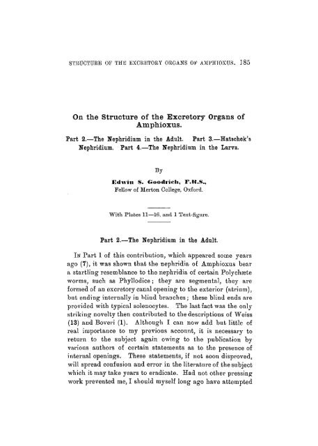
188 EDWIN S. GOODRICH.<br />
In o<strong>the</strong>r regions <strong>of</strong> <strong>the</strong> canal <strong>the</strong>y may be more sparsely<br />
distributed. Cell outlines are rarely visible. The cytoplasm<br />
sol.<br />
cep<br />
:ep<br />
.... bv.<br />
-ch.<br />
Diagram <strong>of</strong> a section through <strong>the</strong> nephridium, passing along a<br />
X>lane at right angles to <strong>the</strong> long axis <strong>of</strong> <strong>the</strong> animal, and<br />
parallel to <strong>the</strong> gill bar.<br />
a. e. Atrial epi<strong>the</strong>lium, h. Base <strong>of</strong> secondary gill-bar, bv.<br />
Blood-vessel, c. ep. Coeloinic epi<strong>the</strong>lium, ch. Chamber containing<br />
solenocytes. n. Wall <strong>of</strong> nephridial canal, op. Nephridiopore.<br />
sol. Solenocyte cell, and t. its tube containing<br />
;i flagellum.<br />
usually contains numerous granules <strong>of</strong> an excretory nature.<br />
As we approach <strong>the</strong> tip <strong>of</strong> a diverticulum, we find that <strong>the</strong>
STRUCTURE OP THE EXCRETORY ORGANS OE A3IPHIOXOS. 189<br />
nuclei do not gradually decrease in number, but suddenly<br />
stop in <strong>the</strong> immediate neighbourhood <strong>of</strong> <strong>the</strong> solenocyte tubes<br />
(figs. 6, 20). Here, where <strong>the</strong>se tubes spring out <strong>of</strong> <strong>the</strong><br />
canal, <strong>the</strong>re are no nuclei; but <strong>the</strong> wall itself is continued as<br />
a sheet <strong>of</strong> more or less granular cytoplasm completely closing<br />
<strong>of</strong>f <strong>the</strong> lumen <strong>of</strong> <strong>the</strong> canal (figs. 6, 9, 20, 21). This canal wall<br />
may be thick or thin, <strong>the</strong> variation in thickness depending,<br />
I believe, chiefly on <strong>the</strong> state <strong>of</strong> tension <strong>of</strong> <strong>the</strong> fluid inside<br />
<strong>the</strong> canal. In good thin sections <strong>the</strong> wall is always visible.<br />
Indeed, <strong>the</strong> better <strong>the</strong> section, and <strong>the</strong> more perfect <strong>the</strong><br />
stain, <strong>the</strong> clearer becomes <strong>the</strong> limiting wall, whatever may<br />
be <strong>the</strong> direction in which it is cut.<br />
Figs. 19 and 20 represent two sections takeu parallel to<br />
<strong>the</strong> surface <strong>of</strong> <strong>the</strong> nephridiutn, sagittal sections <strong>of</strong> <strong>the</strong> animal.<br />
The first just shaves through <strong>the</strong> outer wall <strong>of</strong> <strong>the</strong> canal, and<br />
shows many solenocytes lying on <strong>the</strong> blood-vessel. The<br />
second, which ouly corresponds to <strong>the</strong> left hand portion <strong>of</strong><br />
<strong>the</strong> first figure, cuts deeper into <strong>the</strong> canal through <strong>the</strong><br />
extremity <strong>of</strong> one <strong>of</strong> <strong>the</strong> branches, where may be seen <strong>the</strong><br />
solenocyte tubes piercing <strong>the</strong> closing wall. In <strong>the</strong> nest section<br />
<strong>the</strong> nuclei <strong>of</strong> <strong>the</strong> opposite side begin to appear, <strong>the</strong> whole<br />
thickness <strong>of</strong> <strong>the</strong> small solenocyte-bearing <strong>of</strong>fshoot having<br />
been nearly cut through. The following section would show<br />
only a slice <strong>of</strong> <strong>the</strong> wall. There is no opening. Fig. 21 gives<br />
a similar view <strong>of</strong> ano<strong>the</strong>r nephridium in <strong>the</strong> same animal.<br />
Two consecutive sections through <strong>the</strong> lowermost tip <strong>of</strong> <strong>the</strong><br />
anterior limb <strong>of</strong> <strong>the</strong> nephridinm are drawn in figs. 13, 14.<br />
Here again are seen <strong>the</strong> tubes piercing <strong>the</strong> wall, in which<br />
<strong>the</strong>re is no trace <strong>of</strong> an opening.<br />
Figs. 5 and 6 represent sections from a series nearly<br />
transverse to <strong>the</strong> animal and parallel to <strong>the</strong> bar. That in<br />
fig. 5 passes through <strong>the</strong> external pore, and shaves <strong>of</strong>f <strong>the</strong><br />
wall <strong>of</strong> a diverticulum. The next section (fig. 6) cuts through<br />
<strong>the</strong> extremity <strong>of</strong> this diverticulum. It is seen that <strong>the</strong> lumen<br />
is closed <strong>of</strong>f from <strong>the</strong> cceloni by a distinct cytoplasmic wall,<br />
through which pass solenocyte tubes. In fig. 7 is drawn a<br />
portion <strong>of</strong> <strong>the</strong> same section when <strong>the</strong> microscope has been<br />
VOL. 54, PAST 2. NEW SERIES. 14
190 EDWIN S. GOODRICH.<br />
focussed to <strong>the</strong> lower surface; <strong>the</strong> nuclei <strong>of</strong> <strong>the</strong> wall are again<br />
visible. There is no opening.<br />
Innumerable figures could be given <strong>of</strong> series <strong>of</strong> sectious all<br />
telling <strong>the</strong> same story. But <strong>the</strong> critic will say : if <strong>the</strong> diverticula<br />
are really closed, sections taken at right angles through<br />
<strong>the</strong>ir tip should show <strong>the</strong> tubes cut across embedded in <strong>the</strong><br />
thickness <strong>of</strong> <strong>the</strong> wall. Such sections are not difficult to find,<br />
and I figure several on Plates 12 and 13.<br />
Figs. 10 and 12 represent two consecutive sections across<br />
<strong>the</strong> tip <strong>of</strong> a branch. In <strong>the</strong> fh'sb are seen <strong>the</strong> tubes entering<br />
<strong>the</strong> wall, while <strong>the</strong> next (fig. 12) strikes <strong>the</strong> lumen. A small<br />
part <strong>of</strong> this figure is shown slightly diagrammatised (fig. 11)<br />
on a larger scale. Again three consecutive sections are<br />
drawn in figs. 15, 16, and 17. Here two sections cut through<br />
<strong>the</strong> solid wall before <strong>the</strong> lumen is reached. Lastly, fig. 18<br />
represents a section through two adjacent processes, one <strong>of</strong><br />
which has been cut so as to expose <strong>the</strong> lumen, while <strong>the</strong> o<strong>the</strong>r<br />
shows very clearly <strong>the</strong> soleuocyte tubes piercing <strong>the</strong> wall aud<br />
embedded in its cytoplasm.<br />
The evidence <strong>of</strong> all <strong>the</strong>se sections is quite unequivocal; it<br />
would serve no good purpose to multiply instances; <strong>the</strong>re is<br />
no opening, <strong>the</strong> wall is continuous, and is traversed by <strong>the</strong><br />
tubes <strong>of</strong> <strong>the</strong> solenocytes.<br />
But <strong>the</strong>re is o<strong>the</strong>r evidence <strong>of</strong> a different nature leading to<br />
<strong>the</strong> same conclusion. I have observed in a living nepliridium<br />
<strong>the</strong> fluid inside <strong>the</strong> nephridhil canal so compressed, perhaps<br />
by <strong>the</strong> overlying cover-glass, that it dilated <strong>the</strong> tip <strong>of</strong> <strong>the</strong><br />
diverticulum so as to give rise to a bulgiug vesicle at its<br />
extremity. Now, such a swelling could obviously not be<br />
formed if <strong>the</strong> tip were open.<br />
We may now turn to injectious to corroborate our view. I<br />
have receutly injected <strong>the</strong> dorsal hyperbranchial coslom with<br />
Indian ink. The minute black particles were held in suspension<br />
in sea-water. Such a fluid, if introduced with a<br />
hypodermic syringe, can be made to fill <strong>the</strong> coslom. It is<br />
clear that if <strong>the</strong> nephridium communicated with <strong>the</strong> coslom<br />
<strong>the</strong> ink would penetrate into <strong>the</strong> canal; this would happen
STRUCTURE OF THE EXCRETORY ORGANS OP AHPHIOXUS. 191<br />
all <strong>the</strong> more easily, since a powerful ciliary current works<br />
towards <strong>the</strong> external pore. Sections <strong>of</strong> such injected specimens<br />
show conclusively that not a single particle <strong>of</strong> ink has<br />
entered <strong>the</strong> nephridial canal, although <strong>the</strong> iuk lias penetrated<br />
into every chink <strong>of</strong> <strong>the</strong> ccelom.<br />
But, it may be asked, if <strong>the</strong> facts are so plain and conclusive,<br />
how is it that so keen-sighted and accurate an observer<br />
as Boveri has been deceived?- Well, if it will not be considered<br />
presumptuous on my part, I will attempt to explain<br />
how <strong>the</strong> mistake arose. 1 To begin with, <strong>the</strong> sections he<br />
examined were not appropriately stained. The uuclei are<br />
clear, but <strong>the</strong> cytoplasm scarcely staiued at all. In <strong>the</strong><br />
majority <strong>of</strong> <strong>the</strong> sections which I had <strong>the</strong> opportunity <strong>of</strong><br />
seeing <strong>the</strong> wall which closes <strong>the</strong> tips <strong>of</strong> <strong>the</strong> diverticula was<br />
very, difficult to make out, though I could detect it on close<br />
examination in a suitable light. I naturally turned with<br />
great interest to <strong>the</strong> section given on PI. 33, fig. 17, <strong>of</strong> <strong>the</strong><br />
original memoir (1), and <strong>of</strong> which a photograph is published<br />
in <strong>the</strong> 'Anatomischen Anzeiger' (la). Anyone on first<br />
looking at this section might be led to believe in <strong>the</strong> existence<br />
<strong>of</strong> a funnel. The appearance is extraordinarily deceptive.<br />
But it is deceptive, and <strong>the</strong> deception is due to two<br />
things. First <strong>of</strong> all <strong>the</strong> nuclei are deeply stained, but <strong>the</strong><br />
cytoplasm practically colourless and transparent; in <strong>the</strong><br />
second place <strong>the</strong> section is thick. The figure given by Boveri<br />
is really an optical section <strong>of</strong> <strong>the</strong> preparation. The closingwall<br />
can, indeed, be seen, but only with <strong>the</strong> greatest difficulty.<br />
The misleading appearance <strong>of</strong> a funnel is due to <strong>the</strong><br />
sudden cessation <strong>of</strong> <strong>the</strong> nuclei round <strong>the</strong> base <strong>of</strong> <strong>the</strong> solenocyte<br />
tubes; an appearance which is fur<strong>the</strong>r heightened by<br />
<strong>the</strong> limit <strong>of</strong> <strong>the</strong> coelomic epi<strong>the</strong>lium at <strong>the</strong> same spot (see<br />
p. 193, and text-figure).<br />
Let it not be thought that in insisting on <strong>the</strong> absence <strong>of</strong> an<br />
opening I am unduly influenced by a, priori considerations<br />
1<br />
Soon after <strong>the</strong> publication <strong>of</strong> his paper (la) I wrote to Pr<strong>of</strong>essor<br />
Boveri, who tlien very kindly sent rue his preparations, and I gladly<br />
take this opportunity <strong>of</strong> thanking him for his courtesy.
192 JiDWJJf S. GOODBICH.<br />
due to <strong>the</strong>oretical bias. It is true that I hold that <strong>the</strong> renal<br />
organ <strong>of</strong> <strong>Amphioxus</strong> is a nephridium homologous with <strong>the</strong><br />
nephridia <strong>of</strong> Annelids and Platyhelminths, and not homologous<br />
with <strong>the</strong> kidney tubules <strong>of</strong> <strong>the</strong> Oraniata (5, 7); but it<br />
is now well known that <strong>the</strong> true nephridia <strong>of</strong> Annelids may<br />
open into <strong>the</strong> coeloin. There is no a priori reason why<br />
<strong>the</strong>y should not do so in <strong>Amphioxus</strong>. However, no nephridium<br />
has yet been found possessing both solenocytes and an<br />
internal opening, though such intermediate stages must presumably<br />
have existed.<br />
The Relation <strong>of</strong> <strong>the</strong> Nephridium to <strong>the</strong> Bloodsupply.—The<br />
general blood-supply has been well described<br />
and figured by Boveri (1). But according to my observations<br />
<strong>the</strong> vessels occur not so much as narrow capillaries, as<br />
in <strong>the</strong> form <strong>of</strong> a large expanded vessel spreading over <strong>the</strong><br />
area occupied by <strong>the</strong> excretory organ. This is shown in<br />
sections (figs. 7, 23), and also in <strong>the</strong> reconstructions given on<br />
Plate 11. It will, moreover, be noticed that, although <strong>the</strong><br />
greater part <strong>of</strong> <strong>the</strong> bloodvessel lies on <strong>the</strong> inner or atrial<br />
surface <strong>of</strong> <strong>the</strong> nepln-idium, yet several loops pass round to<br />
<strong>the</strong> outer or coelomic surface. Thus a considerable part o£<br />
<strong>the</strong> nephridial canal is entirely surrounded by <strong>the</strong> bloodvessels.<br />
The soleuocytes radiate out from <strong>the</strong> canal, and<br />
always lie on <strong>the</strong> wall <strong>of</strong> a bloodvessel, being attached to it<br />
by a protoplasmic process (figs. 4, 15). The way in which<br />
<strong>the</strong>se cells are distributed is shown in figs. 14, 19, and<br />
diagrams 2 and 3, and <strong>the</strong> text-figure. It will <strong>the</strong>re be seen<br />
that <strong>the</strong> longer tubes, which are <strong>of</strong> course those belonging to<br />
cells fur<strong>the</strong>st away from <strong>the</strong> canal, pass over <strong>the</strong> shorter<br />
tubes to reach <strong>the</strong>ir destination. Never do <strong>the</strong> solenocytes<br />
project freely into <strong>the</strong> ccelom; when <strong>the</strong>y appear to do so in<br />
sections this is, I believe, due to <strong>the</strong> cell having become<br />
detached accidentally, ei<strong>the</strong>r during <strong>the</strong> process <strong>of</strong> preservation<br />
or <strong>of</strong> cutting. The tubes are <strong>the</strong>refore fixed at both<br />
ends.<br />
In <strong>the</strong> text-figure may also be seen <strong>the</strong> peculiar disposition<br />
<strong>of</strong> <strong>the</strong> solenocytes at <strong>the</strong> top <strong>of</strong> <strong>the</strong> secondary gill-bar. Here
STItUCTCJBK 01' THK EXCKRTOIiY 01WANS 01' AJIFHIOXUS. 193<br />
<strong>the</strong> canal <strong>of</strong> tlie nephridium gives <strong>of</strong>f two or three short<br />
diverticuln, which are turned away from <strong>the</strong> coelom towards<br />
<strong>the</strong> middle line. The numerous solenocytes projecting from<br />
<strong>the</strong>se diverticula lie in a sort <strong>of</strong> pocket or chamber (figs. 1, 2,<br />
3, 23), which only communicates with <strong>the</strong> coelom by means <strong>of</strong><br />
a dorsal opening, over which pass a large number <strong>of</strong> solenocyte<br />
tubes. In one region <strong>the</strong> inner wall <strong>of</strong> this chamber is<br />
formed by <strong>the</strong> skeletal rod <strong>of</strong> <strong>the</strong> gill-bar (figs. 3; 28). Somewhat<br />
similar pockets are found occasionally in connection<br />
with o<strong>the</strong>r parts <strong>of</strong> <strong>the</strong> nephridinm, as, for instance, <strong>the</strong><br />
anterior limb <strong>of</strong> <strong>the</strong> canal. The cavity in <strong>the</strong> chambers is,<br />
I believe, ra<strong>the</strong>r <strong>of</strong> <strong>the</strong> nature <strong>of</strong> a lymph space than <strong>of</strong> a<br />
true ccelomic cavity.<br />
The Relation <strong>of</strong> <strong>the</strong> Nephridium to <strong>the</strong> Coclomic<br />
Epi<strong>the</strong>lium.—It is important to determine exactly what is<br />
<strong>the</strong> disposition <strong>of</strong> <strong>the</strong> ccelomic epi<strong>the</strong>lium in <strong>the</strong> neighbourhood<br />
<strong>of</strong> <strong>the</strong> nephridium. Boveri (1) and Weiss (13) have<br />
already shown that <strong>the</strong> canal is covered by <strong>the</strong> ccelomic<br />
epi<strong>the</strong>lium ; but this epi<strong>the</strong>lium only clo<strong>the</strong>s <strong>the</strong> outer or<br />
coelomic surface (text-figure). It passes on to <strong>the</strong> nephridium<br />
from <strong>the</strong> atrial wall, covering <strong>the</strong> canal and its blind<br />
branches to <strong>the</strong>ir extremity. Here it is not reflected so as to<br />
pass over to <strong>the</strong> inner or atrial surface <strong>of</strong> <strong>the</strong> organ, but ends<br />
abruptly nenr <strong>the</strong> base <strong>of</strong> <strong>the</strong> solenocyte tubes (figs. 2, G,<br />
8, 9).<br />
Thus <strong>the</strong> nephridium and <strong>the</strong> bloodvessels which accompany<br />
it maybe said to lie "morphologically" entirely outside<br />
<strong>the</strong> coelom j between <strong>the</strong> coelomic epi<strong>the</strong>lium and <strong>the</strong> atrial<br />
epi<strong>the</strong>lium. The nephridium is, in fact, retroperitoueal.<br />
This is true, I believe, <strong>of</strong> <strong>the</strong> solenocytes <strong>the</strong>mselves, though<br />
less easy to prove. For <strong>the</strong> coelomic epi<strong>the</strong>lium stops short<br />
where <strong>the</strong> solenocytes begin (figs. 6, 8, 2), passing nei<strong>the</strong>r on<br />
<strong>the</strong> inner side over <strong>the</strong> bloodvessel, nor outside <strong>the</strong>m over<br />
<strong>the</strong>ir coelomic surface. For a long time I was under <strong>the</strong><br />
impression that a very delicate membranous extension <strong>of</strong> <strong>the</strong><br />
epi<strong>the</strong>lium covered over <strong>the</strong> coelomic surface <strong>of</strong> <strong>the</strong> solenocytes;<br />
but I am now satisfied that this is not <strong>the</strong> case,
194 EDWIN S. GOODRICH.<br />
although sometimes <strong>the</strong> epi<strong>the</strong>lium seems to stretch over <strong>the</strong><br />
base <strong>of</strong> <strong>the</strong> solenocyte tubes for a considerable way. The<br />
coelomic epi<strong>the</strong>lium is not continuous with <strong>the</strong> wall <strong>of</strong> <strong>the</strong><br />
canal at <strong>the</strong> tip <strong>of</strong> <strong>the</strong> diverticula, but <strong>of</strong>ten can be seen in<br />
sections to end with a free and jagged edge. Over <strong>the</strong> region<br />
where <strong>the</strong> solenocytes occur <strong>the</strong>re is a gap in <strong>the</strong> coelomic<br />
epi<strong>the</strong>lium, so that coelomic fluid freely ba<strong>the</strong>s <strong>the</strong> solenocyte<br />
tubes (figs. 2, 3). That <strong>the</strong> space in which lie <strong>the</strong>se tubes,<br />
and even <strong>the</strong> deep pockets described above (p. 193), communicate<br />
with <strong>the</strong> coalom is evident in specimens injected with<br />
Indian ink.<br />
Since no epi<strong>the</strong>lium covers <strong>the</strong> solenocytes <strong>the</strong>ir true relation<br />
to <strong>the</strong> coalom cannot be made out for certain in •<strong>the</strong><br />
adult. Without going into <strong>the</strong> question <strong>of</strong> <strong>the</strong>ir development<br />
in this paper, I may say that a careful examination <strong>of</strong><br />
M. Legros's excellent preparations has convinced me that in<br />
<strong>the</strong> very earliest stages <strong>of</strong> its development <strong>the</strong> whole rudiment<br />
<strong>of</strong> <strong>the</strong> nephridium and solenocytes lies enclosed between<br />
<strong>the</strong> coelomic epi<strong>the</strong>lium and <strong>the</strong> atrial wall. There is nothing<br />
unusual in <strong>the</strong> solenocytes coming into secondary contact<br />
with <strong>the</strong> coelomic fluid. We know that in <strong>the</strong> Actinotrocha<br />
larva <strong>the</strong> nephridium pierces <strong>the</strong> wall <strong>of</strong> <strong>the</strong> preseptal<br />
htomocoel, and <strong>the</strong> solenocytes project freely in <strong>the</strong> blood<br />
(8). In many Polychsetes also <strong>the</strong> nephridium passes through<br />
<strong>the</strong> coelomic epi<strong>the</strong>lium and <strong>the</strong> solenocytes lie naked in <strong>the</strong><br />
coelomic fluid (6).<br />
To sum up <strong>the</strong> chieE points in this contribution :—The<br />
careful examination <strong>of</strong> <strong>the</strong> nephridia in sections and in <strong>the</strong><br />
living state shows that <strong>the</strong>y have no internal opening. The<br />
tubes <strong>of</strong> <strong>the</strong> solenocytes pierce <strong>the</strong> wall <strong>of</strong> <strong>the</strong> nephridial<br />
canal, and open into its lumen. The flagellum passes down<br />
<strong>the</strong> tube into <strong>the</strong> lumen. The solenocytes are attached to<br />
<strong>the</strong> wall <strong>of</strong> <strong>the</strong> bloodvessels, which expand in this region, and<br />
may surround <strong>the</strong> canal. Both <strong>the</strong> bloodvessels and <strong>the</strong><br />
nephridial canal are covered by <strong>the</strong> coelomic epi<strong>the</strong>lium,<br />
being situated between it and <strong>the</strong> atrial wall. Over that<br />
region which is occupied by <strong>the</strong> solenocytes <strong>the</strong>re is a gap in
STRUOTUBK (.)!
19G EDWIN S. C400DR1CH.<br />
<strong>of</strong> Annelid larvee (trochosphores), but I can form no opinion<br />
as to <strong>the</strong> reality <strong>of</strong> any such resemblance."<br />
The next author to mention <strong>the</strong> organ is MacBride, who<br />
briefly describes its development, believing that it arises<br />
from <strong>the</strong> communication between <strong>the</strong> gut and <strong>the</strong> second<br />
myotome (II). 1<br />
Van Wijhe (14) describes <strong>the</strong> canal <strong>of</strong> Hatschek's nephridium<br />
in <strong>the</strong> adult, applying to it <strong>the</strong> name Schlundforsatz:<br />
" eine enge Rohre, welche dem linkeu Seitenrande der linkeu<br />
Aorta angeschmigt ist. Das enge lumen wird von einem<br />
einschichtigen Cylinderepi<strong>the</strong>l begrenzt und bildet streckenweise<br />
seitliche Ausbuchtungen. Wo eine solche angeschnitten<br />
wird, konnen zwei Lumina im Schnittbilde auftreten.<br />
Unmittelbar hinter dem Velum miindet die Rohre<br />
mit einer feinen Offnung in den Schlund aus." He denies,<br />
however, <strong>the</strong> presence <strong>of</strong> <strong>the</strong> ccelomic cavity described by<br />
Hatschek, and does not accept <strong>the</strong> latter's <strong>the</strong>ory as to <strong>the</strong><br />
organ's function. " Nach meiner Meinung/' says van Wijhe,<br />
" ist das organ nicht anderes als ein Rudiment des vordereu<br />
Darmendes, welches beim Embryo in das Flimmersackchen<br />
(linke Entoderrnsackchen) ausmiindete."<br />
It is to G-oldschnidt that we are indebted for <strong>the</strong> first<br />
description <strong>of</strong> solenocytes in <strong>the</strong> nepliridium <strong>of</strong> Hatschek (4),<br />
placing its homology with <strong>the</strong> posterior nephridia beyond<br />
doubfc. His account seems, however, to be based on imperfect<br />
material, and he falls into <strong>the</strong> error <strong>of</strong> ascribing to <strong>the</strong><br />
canal an internal opening such as Boveri had described in<br />
<strong>the</strong> paired nephridia.<br />
I have recently had <strong>the</strong> opportunity <strong>of</strong> studying this<br />
interesting organ in adult and larval specimens in Helgoland,<br />
3 and am thus able to give a more complete description<br />
<strong>of</strong> it.<br />
1 I am unable to agree with <strong>the</strong> view <strong>of</strong> ei<strong>the</strong>r Hatschek or MacBride<br />
as to <strong>the</strong> origin <strong>of</strong> this nephridium.<br />
-' I gladly seize this opportunity <strong>of</strong> thanking Pr<strong>of</strong>. Heincke, Pr<strong>of</strong>.<br />
Havtlaub, and <strong>the</strong> staff <strong>of</strong> <strong>the</strong> Konigl. Biologische Anstalt for <strong>the</strong> kind<br />
way in which <strong>the</strong>y received me in Helgoland.
STRUCTURE 0.1;' THE EXCRETORY ORGANS Ob' AMl'EIOXUS. 197<br />
The nephridium <strong>of</strong> Hatschek reaches its maximum development<br />
in <strong>the</strong> adult, where it is indeed <strong>the</strong> largest nephridinm<br />
in <strong>the</strong> body, some 2 mm. in length. Lying on <strong>the</strong> left side,<br />
below and parallel to <strong>the</strong> notochord, it opens just behind <strong>the</strong><br />
velum into <strong>the</strong> pharynx, 1 and runs forward a long distance<br />
to a point just in front oE <strong>the</strong> ciliated groove (Raderorgan).<br />
Here it ends blindly, and along its course are given <strong>of</strong>f short<br />
blind diverticula (figs. 27, 42, 43, 44). Solenocytes are set<br />
on <strong>the</strong> dorsal and lateral sui'faces <strong>of</strong> <strong>the</strong> organ along almost<br />
its whole length, being especially numerous on <strong>the</strong> diverticula<br />
(fig. 28). Altoge<strong>the</strong>r au enormous number <strong>of</strong> solenocytes<br />
are present on this nephridinm in <strong>the</strong> adult <strong>Amphioxus</strong>.<br />
The canal runs along <strong>the</strong> floor <strong>of</strong> a narrow cavity beside<br />
<strong>the</strong> aoi-ta (figs. 42—44). It is to <strong>the</strong> wall <strong>of</strong> this cavity that<br />
<strong>the</strong> solenocytes are attached, and it appears to be <strong>of</strong> coclomic<br />
nature; at all events it is in open communication with <strong>the</strong><br />
myocoele <strong>of</strong> <strong>the</strong> first myotome in larval stages (figs. 25, 26,<br />
33). In <strong>the</strong> adult, however, it is closed <strong>of</strong>f, and <strong>the</strong> lining<br />
epi<strong>the</strong>lium seems to be very irregularly developed, forming<br />
no distinct layer <strong>of</strong> cells (fig. 28).<br />
In <strong>the</strong> larva <strong>of</strong> about 13 gill-slits, <strong>of</strong> <strong>the</strong> left series only<br />
(fig. 33), <strong>the</strong> nephridium can be well seen by transparency<br />
as a short tube opening behind into <strong>the</strong> pharynx (fig. 24).<br />
Its dorsal surface is entirely beset with solenocytes in several<br />
closely packed rows (fig. 29). An optical section <strong>of</strong> <strong>the</strong><br />
organ at this stage is represented in fig. 38, showing clearly<br />
<strong>the</strong> way in which <strong>the</strong> tubes <strong>of</strong> <strong>the</strong> solenocytes pierce <strong>the</strong> thin<br />
dorsal wall.<br />
We may summarise as follows <strong>the</strong> observations recorded<br />
above :—The nephridium <strong>of</strong> Hatschek is a true nephridium,<br />
similar in structure to <strong>the</strong> posterior paired nephridia. In<br />
<strong>the</strong> adult, where it reaches its maximum development, it<br />
extends along <strong>the</strong> left aorta from in front <strong>of</strong> <strong>the</strong> ciliated<br />
1 <strong>On</strong> one occasion only I have found an opening from <strong>the</strong> canal into<br />
tlie hinder region <strong>of</strong> <strong>the</strong> buccal cavity itself, as well as <strong>the</strong> posterior<br />
opening into <strong>the</strong> pharynx.
198 EDWIN S. UOOPRIOH.<br />
groove backwards to <strong>the</strong> pharynx into which it opens.<br />
Very numerous solenocytes are set chiefly on short blind<br />
diverticula. It has no internal opening, and lies in a cavity,<br />
which is in communication with <strong>the</strong> myocoele <strong>of</strong> <strong>the</strong> first<br />
myotome in <strong>the</strong> larva.<br />
That this nephridium is in every way similar to and homologous<br />
with <strong>the</strong> paired posterior nephridia <strong>the</strong>re can be no<br />
doubt. Van Wijhe's suggestion, mentioned above, must<br />
<strong>the</strong>refore be abandoned. Two peculiarities, however, still<br />
remain to be explained; its unpaired character and its opening<br />
into <strong>the</strong> alimentary canal. No one, so far as I am aware,<br />
has yet worked out <strong>the</strong> exact relation <strong>of</strong> <strong>the</strong> gill-slits to <strong>the</strong><br />
somites in <strong>the</strong> larva <strong>of</strong> <strong>Amphioxus</strong>, and my own observations<br />
on this point are very incomplete. Bat judging from <strong>the</strong><br />
course <strong>of</strong> <strong>the</strong> dorsal spinal nerves (fig. 30), <strong>the</strong> first gill-slit<br />
<strong>of</strong> <strong>the</strong> left (on <strong>the</strong> right side) series, which is <strong>the</strong> first to<br />
appear in <strong>the</strong> larva, corresponds to <strong>the</strong> third myotome.<br />
Probably its true morphological position is between <strong>the</strong><br />
second and third myotome. Presumably Hatschek's nephridium<br />
would correspond to <strong>the</strong> next gill-slit in front, between<br />
<strong>the</strong> second and first myotomes, did such a slit exist. As for<br />
its unpaired character, I can for <strong>the</strong> present <strong>of</strong>fer no better<br />
explanation than this, that it is <strong>the</strong> left <strong>of</strong> an original<br />
anterior pair <strong>of</strong> nephridia, <strong>the</strong> one-sided development <strong>of</strong><br />
which is no doubt correlated with <strong>the</strong> general asymmetry <strong>of</strong><br />
<strong>the</strong> anterior region so conspicuous in <strong>the</strong> larva. But this<br />
question can only be pr<strong>of</strong>itably discussed after an exhaustive<br />
study <strong>of</strong> <strong>the</strong> development, and must <strong>the</strong>refore be put aside<br />
for <strong>the</strong> present. In <strong>the</strong> same way a detailed knowledge <strong>of</strong><br />
tho development <strong>of</strong> this organ, and <strong>of</strong> <strong>the</strong> posterior nephridia,<br />
is necessary before one can discuss <strong>the</strong> significance <strong>of</strong> <strong>the</strong><br />
anomalous position <strong>of</strong> <strong>the</strong> opening.
STRUCTURE 01? THE EXCRETORY OHG-ANS OV AMmfOXUS. 199<br />
<strong>Part</strong> 4.—The Development <strong>of</strong> <strong>the</strong> Left Series <strong>of</strong> Nephridia in<br />
<strong>the</strong> Larva.<br />
For many years I have been trying to trace <strong>the</strong> development<br />
<strong>of</strong> <strong>the</strong> nephridia in <strong>Amphioxus</strong>. In 1902 I collected a<br />
large amount <strong>of</strong> material from <strong>the</strong> Pantano at Faro; but illhealth<br />
prevented iny working out <strong>the</strong> development on <strong>the</strong><br />
living larva,, and I failed to do so on <strong>the</strong> preserved specimens.<br />
It was not till last year that I was again able, in<br />
Helgoland, to study <strong>the</strong> living larva, and succeeded in<br />
tracing some stages <strong>of</strong> <strong>the</strong> development <strong>of</strong> <strong>the</strong> excretory<br />
organs. In <strong>the</strong> meantime Legros had been studying <strong>the</strong><br />
same subject in Naples, and published anonymously a preliminary<br />
notice <strong>of</strong> his results a short time ago (16). x<br />
In <strong>the</strong> present paper I shall not discuss in detail <strong>the</strong> first<br />
origin <strong>of</strong> <strong>the</strong> nephridia, but restrict myself to a description <strong>of</strong><br />
<strong>the</strong> stages found in <strong>the</strong> larva with from ten to fifteen gill-slits<br />
<strong>of</strong> <strong>the</strong> left hand series, and no trace <strong>of</strong> <strong>the</strong> right ha,nd series.<br />
These are <strong>the</strong> only stages which I have been able to study<br />
sufficiently in <strong>the</strong> living state.<br />
Fig. 30 gives a left side view <strong>of</strong> a young larva with eleven<br />
slits. The anterior gill-slits are still well on <strong>the</strong> right side,<br />
but <strong>the</strong> hinder slits are in or near <strong>the</strong> middle line. The<br />
future dorsal edge <strong>of</strong> each slit may, <strong>of</strong> course, at this stage<br />
be more ventral than <strong>the</strong> future ventral edge. The nephridia<br />
are seen as small rounded sacs near <strong>the</strong> posterior ventral<br />
corner <strong>of</strong> each slit. Every slit from <strong>the</strong> first to <strong>the</strong> last has<br />
such a. nephridium. At this early stage, <strong>the</strong>i'e is no atrium,<br />
<strong>the</strong> slits have an internal margin <strong>of</strong> thick branchial epi<strong>the</strong>lium,<br />
which is thrown into characteristic folds when <strong>the</strong><br />
branchial muscles contract, while <strong>the</strong> external margin <strong>of</strong> <strong>the</strong><br />
slit is formed by a thin fold <strong>of</strong> <strong>the</strong> body wall, acting as a sort<br />
1<br />
Through <strong>the</strong> kindness <strong>of</strong> M. Legros I have had <strong>the</strong> opportunity <strong>of</strong><br />
examining his sections, and I cannot agree with his conclusions as to<br />
<strong>the</strong> origin <strong>of</strong> <strong>the</strong> nephridia from <strong>the</strong> ccelomic epi<strong>the</strong>lium, nor as to <strong>the</strong><br />
presence <strong>of</strong> internal openings. But I believe he has modified his views<br />
considerably on <strong>the</strong>se points since <strong>the</strong> publication <strong>of</strong> <strong>the</strong> note.
200 EDWJN S. GOODRICH.<br />
<strong>of</strong> sphincter (figs. 33 and 36). A shallow branchial chamber<br />
lined with epidermis is thus formed, leading from <strong>the</strong> external<br />
to <strong>the</strong> internal opening. It is in this chamber that <strong>the</strong><br />
nephridium opens, at a place corresponding apparently to <strong>the</strong><br />
point <strong>of</strong> junction <strong>of</strong> <strong>the</strong> ectoderm with <strong>the</strong> endoderm (fig. 41).<br />
The position <strong>of</strong> <strong>the</strong> nephridiopore can be seen in figs. 36 and<br />
39. When <strong>the</strong> atrium becomes formed by <strong>the</strong> closing <strong>of</strong>f <strong>of</strong><br />
<strong>the</strong> space between <strong>the</strong> metapleural folds, with which <strong>the</strong><br />
branchial cavities become merged, <strong>the</strong> pores open into <strong>the</strong><br />
atrium. A ventral view <strong>of</strong> a stage where <strong>the</strong> atrium has just<br />
begun to be formed posteriorly shows one or two nephridia<br />
behind <strong>the</strong> last open gill-slit (fig. 35). Probably <strong>the</strong>se<br />
nephridia belong to <strong>the</strong> posterior gill-slits, which have closed<br />
up (Willey, 15); <strong>the</strong>y open now directly on <strong>the</strong> surface<br />
(fig. 30).<br />
The young nephridium is a flattened sac, without internal<br />
opening (figs. 36 and 39). From its inner end spring a large<br />
number oE solenocytes; <strong>the</strong>ir tubes pierce its wall, and <strong>the</strong>ir<br />
flagella pass into <strong>the</strong> lumen <strong>of</strong> <strong>the</strong> sac. The majority <strong>of</strong> <strong>the</strong><br />
solenocytes spread over <strong>the</strong> blood-vessel which runs along<br />
<strong>the</strong> future dorsal edge <strong>of</strong> <strong>the</strong> slits. The solenocytes <strong>of</strong> <strong>the</strong><br />
first few slits scarcely extend beyond this limit; but, passing<br />
backwards to more posterior nephridia, we find that <strong>the</strong><br />
solenocytes spread far<strong>the</strong>r and far<strong>the</strong>r up towards <strong>the</strong> dorsal<br />
aorta, <strong>the</strong> tubes leng<strong>the</strong>ning out as <strong>the</strong> cells lie far<strong>the</strong>r from<br />
<strong>the</strong> nephridial sac. At about <strong>the</strong> fifth or sixth nephridium<br />
some <strong>of</strong> <strong>the</strong> solenocytes actually reach <strong>the</strong> aorta (fig. 40).<br />
The tubes in this case may attain a really astonishing length,<br />
stretching right across <strong>the</strong> field <strong>of</strong> a T^-th oil-immersion<br />
objective with oc. 8.<br />
Fig. 34 represents <strong>the</strong> posterior gill region <strong>of</strong> a living<br />
larva, in which <strong>the</strong> remarkable development <strong>of</strong> <strong>the</strong> solenocytes<br />
is well shown. Here a group <strong>of</strong> <strong>the</strong> longest solenocytes,<br />
some twelve to eighteen in number, spread out over <strong>the</strong> aorta<br />
in a most beautifully regular fan-like arrangement in each<br />
segment. A section <strong>of</strong> this region is shown in fig. 31; <strong>the</strong><br />
fan-like disposition is found in each segment to <strong>the</strong> hindmost
STRUOTUBE OP THE EXCRETORY ORGANS 01? AJII'HIOXTJS. 201<br />
limit <strong>of</strong> <strong>the</strong> series <strong>of</strong> nephridia. Presumably <strong>the</strong> dorsal<br />
solenocytes degenerate later, since <strong>the</strong>y are not known to<br />
exist in <strong>the</strong> adult.<br />
The observations on <strong>the</strong> larval nephridia recorded in this<br />
part may be summarised as follows:—To every giil-slit<br />
corresponds a nephridium, consisting <strong>of</strong> a sac closed internally,<br />
but opening to <strong>the</strong> exterior apparently at <strong>the</strong> point<br />
where <strong>the</strong> ectoderm joins <strong>the</strong> endoderm iu <strong>the</strong> shallow branchial<br />
chamber. From <strong>the</strong> internal blind end <strong>of</strong> <strong>the</strong> nepliridial<br />
sac spring numerous solenocytes, some <strong>of</strong> which reach and<br />
spread over <strong>the</strong> aorta at every segment in a fan-like arrangement.<br />
This structure is only fully developed from about <strong>the</strong><br />
eighth segment backwards to <strong>the</strong> last nephridium.<br />
July 3rd, 1909.<br />
LIST OF REFERENCES.<br />
1. Boveri, Th.—"Die Nicrenkaniilchen des <strong>Amphioxus</strong>," 'Zool. Jahrb.<br />
Anat. Abt.,' Bd. 5, 1892.<br />
la. " Bemerk. iiber cl. Bau. d. Nierenkaniilclien des Ampliioxus,"<br />
' Anat. Ann;.,' Bd. 25, 1904<br />
2. Felix, W.—'• Entw. des Hamapparates," ' Hertwig's Handbuch d.<br />
Entw. d. Wirbeltiere,' Bd. 3, 1904.<br />
3. " Entw. des Exkretions-systeni," ' Anat. Hefte. Ergeb. Anat.<br />
u. Entw.,' Bd. 13, 1904<br />
4. G-oldschmidt, B,.—"Amphioxides," ' Wiss. Ergeb. d. deutsch. Ticfseo-<br />
Expedition " Valdivia," ' Bd. 12, 1905.<br />
5. Goodrich, E. S.— " <strong>On</strong> <strong>the</strong> Ccelom, etc ," ' Quart. Journ. Micr. Soi.,'<br />
vol. 37, 1894<br />
6. " <strong>On</strong> <strong>the</strong> Nephridia <strong>of</strong> tlie Polychaita," <strong>Part</strong>s I, II, and III,<br />
ibid., vols. 40, 1897, 41, 1898, and 43, 1900.<br />
7. " <strong>On</strong> <strong>the</strong> <strong>Structure</strong> <strong>of</strong> <strong>the</strong> <strong>Excretory</strong> Oi-gans <strong>of</strong> <strong>Amphioxus</strong>,"<br />
<strong>Part</strong> I, ibid., vol. 45, 1902.<br />
8. " <strong>On</strong> <strong>the</strong> Body-cavities and Nephridia <strong>of</strong> <strong>the</strong> Actinotrocha<br />
larva," ibid., vol. 47, 1903.<br />
9. Hatschek, B.—" Mittb. iiber Amphioxns," ' Zool. Anz., ! Bd. 7, 18S4.
202 EDWIN S. GOODRICH.<br />
10. Lankester, E. R., and Willey, A.—"The Development <strong>of</strong> <strong>the</strong> Atrial<br />
Chamber <strong>of</strong> <strong>Amphioxus</strong>," ' Quart. Joum. Micr. Sci.,' vol. 31, 1890.<br />
11. MacBride, E. W.—" The Early Development <strong>of</strong> Ampliioxus," ibid.,<br />
vol. 40, 1898.<br />
12. Schneider, K. C —'Lehrbuch des Vergl. Histologie,' Jena, 1902.<br />
13. Weiss, F. E.—" <strong>Excretory</strong> Tubules in <strong>Amphioxus</strong>," ' Quart. Journ.<br />
Micr. Sci.,' vol. 31, 1890.<br />
14. Wijhe, J. W. van.—" Beitr. /,. Anat. des Kopfregion des <strong>Amphioxus</strong>,"<br />
'Petrns Camper,' vol. 1, 1901.<br />
15. Willey, A.—" The Later Larval Development <strong>of</strong> <strong>Amphioxus</strong>,"<br />
' Quart. Journ. Micr. Sci.,' vol. 32, 1891.<br />
16. Legros, R.—" Sur le dcvel. des fentes branchiales et des cansdicu1.es<br />
de Weiss-Boveri chez 1'<strong>Amphioxus</strong>," ' Anat. Anz.,' Bd. 34, 1909.<br />
Published anonymously.<br />
LIST OF REIMSRENCJS LETTERS.<br />
a. c. and a. ep. Atrial epi<strong>the</strong>lium, ao. Aorta, b. Secondary gill-bar.<br />
b. c. Buccal cavity, b. ep. Epi<strong>the</strong>lium <strong>of</strong> buccal cavity, br. Brain.<br />
br. c. Branchial epi<strong>the</strong>lium. bv. Blood-vessel. ca. Cavity in which<br />
runs Hatschek's nephridium. c. ep. Coelomic epi<strong>the</strong>lium, c. gl. Clubshaped<br />
gland. ch. Solenocyte chamber. cr. Cirrlius. c. w. Cut<br />
wall <strong>of</strong> nephridial canal, d. n. Dorsal nerve, e.
STRUCTURE OF THE EXCKJETORY ORGANS OJ? AMPHIOXUS. 203<br />
EXPLANATION OF PLATES 11—16,<br />
illustrating Mr. Edwin S. Goodrich's paper "<strong>On</strong> <strong>the</strong> <strong>Structure</strong><br />
<strong>of</strong> <strong>the</strong> <strong>Excretory</strong> <strong>Organs</strong> <strong>of</strong> <strong>Amphioxus</strong>."<br />
PLATE 11.<br />
PIG. 1.—Diagrammatic reconstruction <strong>of</strong> a left nepliriditim and <strong>the</strong><br />
neighbouring blood-vessels, from a series <strong>of</strong> sections taken parallel to<br />
<strong>the</strong> gill-baa*. The solenocytes are not represented. Side view from <strong>the</strong><br />
outside. Op. indicates <strong>the</strong> position <strong>of</strong> <strong>the</strong> nephridiopore on <strong>the</strong> opposite<br />
side.<br />
FIG. 2.—Similar reconstruction <strong>of</strong> a right nephridium, from a series<br />
<strong>of</strong> sections transverse to <strong>the</strong> gill-bar. In two places <strong>the</strong> ccelomic<br />
epi<strong>the</strong>lium and <strong>the</strong> solenocytes are shown.<br />
TIG. 3.—Reconstruction <strong>of</strong> a portion <strong>of</strong> a left nephridium, <strong>the</strong> bloodvessels,<br />
and <strong>the</strong> top <strong>of</strong> <strong>the</strong> secondary gill-bar, seen from behind.<br />
FIG. 4.—Small portion <strong>of</strong> a section shaving <strong>of</strong>f <strong>the</strong> wall <strong>of</strong> a<br />
nephridial canal, and showing <strong>the</strong> bases <strong>of</strong> <strong>the</strong> solenocyte tubes<br />
embedded in <strong>the</strong> cytoplasm. Cam. Z. 2 mm. ap. oil-imm., oc. S.<br />
PLATE 12.<br />
FIGS. 5 and 6.—Two consecutive sections, parallel to <strong>the</strong> gill-bar,<br />
through a nephridium, showing <strong>the</strong> solenocyte tubes passing through<br />
<strong>the</strong> thickness <strong>of</strong> <strong>the</strong> wall <strong>of</strong> <strong>the</strong> canal. Cam. Z. 2 mm. oc. 4.<br />
FIG. 7.—Drawing <strong>of</strong> <strong>the</strong> lower surface <strong>of</strong> <strong>the</strong> section <strong>of</strong> which <strong>the</strong><br />
upper surface is represented in fig. 6. Cam. Z. 2 mm. ap oil-imm.,<br />
oc. 4.<br />
FIG. 8.—Section across <strong>the</strong> anterior limb <strong>of</strong> a nephridium, showing<br />
<strong>the</strong> coelomic epi<strong>the</strong>lium passing over <strong>the</strong> outer surface <strong>of</strong> <strong>the</strong> canal.<br />
FIG. 9.—Similar section showing solenocyte tubes piercing <strong>the</strong> wall<br />
<strong>of</strong> <strong>the</strong> canal. Cain. Z. 2 mm. ap. oil-imm., oc. 4.<br />
FIG. 10.—Section parallel to a gill-bar, cutting <strong>the</strong> wall <strong>of</strong> a diverticulum<br />
<strong>of</strong> <strong>the</strong> nephridial canal (at tb.). Cam. Z. 2 mm. ap. oil-imm.,<br />
oc. 4.<br />
FIG. 11.—Diagrammatic view <strong>of</strong> a small portion <strong>of</strong> <strong>the</strong> wall <strong>of</strong> <strong>the</strong><br />
diverticulum in <strong>the</strong> same section, showing <strong>the</strong> bases <strong>of</strong> <strong>the</strong> solenocyte<br />
tubes.
204 EDAVIIS: S. GOODRICH.<br />
FIG. 1.2.—Next section to that drawn in fig. 10.<br />
FIGS. 13 and 14.—Two consecutive sections through <strong>the</strong> ventral end<br />
<strong>of</strong> fcho anterior limb <strong>of</strong> a nephridial canal. Cam. L. TV oil-imm., oc. 3.<br />
PLATE 13.<br />
FIGS. 15. 16, and 17.—Three consecutive sections, parallel to a gillliar,<br />
through <strong>the</strong> extremity <strong>of</strong> a diverticulmn <strong>of</strong> <strong>the</strong> nephridial canal.<br />
Cam. Z. 2 mm. ap. oil-imm., oc. 12. In figs. 15 and 16 <strong>the</strong> solenocyte<br />
tubes are cut in <strong>the</strong> thickness <strong>of</strong> <strong>the</strong> wall <strong>of</strong> <strong>the</strong> canal.<br />
FIG. 18.—Section across <strong>the</strong> ends <strong>of</strong> two adjacent nephridial diver -<br />
ticula. The bases <strong>of</strong> solenocyte tubes are clearly seen embedded in <strong>the</strong><br />
cytoplasmic wall. Cam. Z. 2 mm. ap. oil-imm., oc'18.<br />
FIG. 19.—Longitudinal section cutting <strong>the</strong> surface <strong>of</strong> a neplrridiuin.<br />
Cam. L. -j\,- oil-imm., oc. 3.<br />
FIG. 20.—View <strong>of</strong> <strong>the</strong> portion <strong>of</strong> <strong>the</strong> next section corresponding to<br />
<strong>the</strong> left-hand region <strong>of</strong> fig. 19.<br />
FIG. 21.—Similar section <strong>of</strong> ano<strong>the</strong>r nephridium.<br />
FIG. 22.—Diagram to illustrate <strong>the</strong> direction <strong>of</strong> <strong>the</strong> sections drawn in<br />
figs. 5, 9, 10, 13, 15, and 19.<br />
PLATE 14.<br />
FIG. 23.—Section across <strong>the</strong> top <strong>of</strong> one primary and two secondary<br />
gill-bars, showing <strong>the</strong> position <strong>of</strong> <strong>the</strong> solenocyte chambers (ch.), and <strong>of</strong><br />
<strong>the</strong> blood-vessels. The position <strong>of</strong> <strong>the</strong> external pore at a lower level is<br />
indicated by a cross X.<br />
FIG. 24. —Transverse section <strong>of</strong> a larva, passing through <strong>the</strong> mouth,<br />
and opening <strong>of</strong> Hatschek's nephridium. Cam. Z. D., oc. 3.<br />
FIG. 25.—Transverse section far<strong>the</strong>r forward passing just beyond <strong>the</strong><br />
anterior end <strong>of</strong> Hatschek's nephridium, where <strong>the</strong> cavity in which it lies<br />
opens into <strong>the</strong> first myoccele.<br />
FIG. 26.—More enlarged view <strong>of</strong> a portion <strong>of</strong> <strong>the</strong> next section,<br />
showing <strong>the</strong> solenocyte tubes in a cavity continuous with <strong>the</strong> first<br />
myoccele.<br />
FIG. 27.—Anterior end <strong>of</strong> an adult <strong>Amphioxus</strong>, ventral view. The<br />
buccal cavity has been opened up by ratting along <strong>the</strong> mid-ventral line<br />
Hatschek's nephridium is seen on <strong>the</strong> left side <strong>of</strong> <strong>the</strong> notocliord.
STRUCTURE OP THE EXCRETORY ORGANS OP AMPHIOXUS. 205<br />
FIG. 28.—Small portion <strong>of</strong> a transverse section <strong>of</strong> <strong>the</strong> head, showing<br />
Hatschek's nepliridium. Cam. L. oil-imm., oc. 3.<br />
FIG. 29.—Similar view <strong>of</strong> a larva (<strong>the</strong> same as that in fig. 24, from<br />
Helgoland, with about thirteen gill-slits). Cam. L T\- oil-imm., oc. 3.<br />
FIG. 30.—Portion <strong>of</strong> a longitudinal section <strong>of</strong> a larva, showing a<br />
nephridium opening behind <strong>the</strong> last open gill-slit. Cam. Z. 2 mm. ap.<br />
oil-imm., oc. 4.<br />
FIG. 31.—Portion <strong>of</strong> a longitudinal section <strong>of</strong> a larva, showing <strong>the</strong><br />
fan-like group <strong>of</strong> solenocytes on <strong>the</strong> aorta. Cam. Z. 2 mm. ap. oil-imm.,<br />
oc. 4.<br />
PJLATE 15.<br />
FTG. 32.—Left side view <strong>of</strong> a larva, drawn from living and preserved<br />
specimens.<br />
FIG. 33.—Left side view <strong>of</strong> <strong>the</strong> anterior region <strong>of</strong> a slightly older<br />
larva on a larger scale, from living and preserved specimens. The cilia<br />
are not indicated.<br />
FIG. 34.—Left side view <strong>of</strong> <strong>the</strong> posterior branchial region <strong>of</strong> a larva,<br />
showing <strong>the</strong> disposition <strong>of</strong> <strong>the</strong> solenocytes. From <strong>the</strong> living.<br />
Fin. 35.—Ventral view <strong>of</strong> a region <strong>of</strong> a larva, showing <strong>the</strong> last open<br />
gill-slit, and two more posterior nephridia. From <strong>the</strong> living.<br />
FIG. 36.—Ventral view <strong>of</strong> two posterior gill-slits <strong>of</strong> a living larva.<br />
FIG. 37 —Solenocytes from Hatschek's nephridium in <strong>the</strong> larva.<br />
FIG. 38.—Optical section <strong>of</strong> Hatschek's nephridium in <strong>the</strong> larva.<br />
From <strong>the</strong> living.<br />
FIG. 39.—Ventral view <strong>of</strong> a nephridium showing its opening just<br />
within <strong>the</strong> margin <strong>of</strong> a posterior gill-slit in a larva. Solenocytes cut<br />
short.<br />
PLATE 16.<br />
FIG. 40.—Left side view <strong>of</strong> a single nephridium in a larva. From<br />
<strong>the</strong> living.<br />
FIG. 41.—Portion <strong>of</strong> a transverse section <strong>of</strong> a larva, passing through<br />
<strong>the</strong> nephridiopore. Cam. Z. 2 mm. ap. oil-imm., oe. 4.<br />
FIGS. 42, 43, and 44.—Portions <strong>of</strong> three transverse sections <strong>of</strong> <strong>the</strong><br />
head <strong>of</strong> <strong>the</strong> adult, showing Hatschek's nephridium. In front <strong>of</strong> <strong>the</strong><br />
ciliated pit (fig. 42), at <strong>the</strong> level <strong>of</strong> <strong>the</strong> ciliated pit (fig. 43), and behind<br />
it (fig. 44).<br />
VOL. 54, PART 2. NEW SERIES. 15
^f^ f^^<br />
: ^S^


