

NEWSLETTER - Mycological Society of America
NEWSLETTER - Mycological Society of America
NEWSLETTER - Mycological Society of America

Create successful ePaper yourself
Turn your PDF publications into a flip-book with our unique Google optimized e-Paper software.
<strong>Mycological</strong><br />
<strong>Society</strong> <strong>of</strong> <strong>America</strong><br />
<strong>NEWSLETTER</strong><br />
vol. 32 no. 1 June 1981<br />
ISSN 0541-4938
OFFICERS OF THE MYCOLOGICAL SOCIETY OF AMERICA<br />
MARIE L. FARR, President MARGARET BARR BIGELOW, President-Elect<br />
BARC-Wes t<br />
Beltsville, Maryland 20705<br />
University <strong>of</strong> Massachusetts<br />
Amherst, Massachusetts 01003<br />
HARRY D . THIERS , Vice-president ROGER GOOS, See.-Treas.<br />
Department <strong>of</strong> Biology<br />
San Francisco State University<br />
San Francisco, California 94132<br />
JAMES W. KIMBROUGH, Past Pres. (1980)<br />
University <strong>of</strong> Florida<br />
Gainesville, Florida 32611<br />
LARRY F. GRAND, Councilor<br />
(1979-81)<br />
North Carolina State University<br />
Raleigh, North Carolina 27607<br />
0 'NEIL R. COLLINS, Counci lor<br />
(1980-83)<br />
Department <strong>of</strong> Botany<br />
University <strong>of</strong> California<br />
Berkeley, California 947 20<br />
WALTER J. SUNDBERG, Coun&Zor<br />
(1980-83)<br />
Department <strong>of</strong> Botany<br />
Southern Illinois University<br />
Carbondale, Illinois 62901<br />
JATIES REID, Counci lor (1979-1981)<br />
University <strong>of</strong> Manitoba<br />
Winnipeg, Manitoba, Canada R3T 2N2<br />
AFFILIATED SOCIETIES<br />
Department <strong>of</strong> Botany<br />
University <strong>of</strong> Rhode Island<br />
Kingston, Rhode Island 02881<br />
ROBERT L. GILBERTSON, Past Pres. (1979)<br />
University <strong>of</strong> Arizona<br />
Tucson, Arizona 85721<br />
EDWARD E . BUTLER, Counci LOP<br />
(1979-81)<br />
University <strong>of</strong> California<br />
Davis, California 95616<br />
DONALD T . WICKLOW, Counci LOP<br />
(1980-83)<br />
Northern Regional Research Center<br />
1815 N. University Street<br />
Peoria, Illinois 61604<br />
IAN K. ROSS, Councilor<br />
(1980-83)<br />
Department <strong>of</strong> Biological Sciences<br />
University <strong>of</strong> California<br />
Santa Barbara, California 93106<br />
The Boston <strong>Mycological</strong> Club, Patrick Peterson, Treas., 21 112 Inman St.,<br />
Cambridge, MA 02139<br />
Colorado <strong>Mycological</strong> <strong>Society</strong>, Joan L. Betz, Secretary, 501 Clermont Pkwy.,<br />
Denver, CO 80220<br />
The New York <strong>Mycological</strong> <strong>Society</strong>, Attn.: Emil Lang,1700 York Ave.,<br />
New York, NY 10028<br />
The North <strong>America</strong>n Plycological Association, Gary Linc<strong>of</strong>f, Pres., New York<br />
Botanical Garden, Bronx, NY 10458<br />
Ohio Mushroom <strong>Society</strong>, W. Sturgeon, Pres., 121 Brookline Ave.,<br />
Youngstown, OH 44505<br />
Oregon <strong>Mycological</strong> <strong>Society</strong>, Reg. Agent, Donald Goetz, 6548 SE 30th Ave.,<br />
Portland, OR 97202<br />
Puget Sound <strong>Mycological</strong> <strong>Society</strong>, 200 2nd. Ave., North, Seattle, WA 98109<br />
Soci6t6 Mycologique de France, 36 Rue Ge<strong>of</strong>froy-Ste. Hilaire, Paris Ve,<br />
France
~ditors' Note . ......<br />
General Announcements ...<br />
Symposia, Meetings, Forays.<br />
New Research. .......<br />
Forthcoming Courses ....<br />
Fungi for Distribution. ..<br />
Fungi Wanted. .......<br />
Identifications ......<br />
Publications Available. ..<br />
Publications Wanted ....<br />
New Books by Members. ...<br />
Miscellaneous<br />
MYCOLOGICAL SOCIETY OF AMERICA <strong>NEWSLETTER</strong><br />
Volume 32, NO. 1, June 1981<br />
Edited by Donald H. Pfister and Geraldine C. Kaye<br />
.......<br />
TABLE OF CONTENTS<br />
Postodoctoral Positions/<br />
Assistantships . ......... .12<br />
Positions Wanted . ......... -13<br />
Changes in Affiliation . ...... .14<br />
..........<br />
Travels, Visits. .14<br />
Papers, Symposia, Workshops. .... .15<br />
Honors, Awards, Promotions . .... .I6<br />
Personal News. . .......... .18<br />
Notes and Comments . ........ .18<br />
Late News and Additions. ...... .20<br />
Annual Meeting--Schedule & Abstracts .21<br />
EDITORS' NOTE<br />
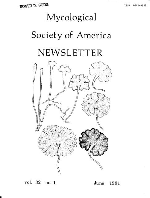
The cover and illustrations used in this issue are by Roland Thaxter. They have been<br />
taken from his original plates on deposit in the newly reorganized archives <strong>of</strong> the Farlow<br />
Reference Library and Herbarium. The cover, which depicts the Hyphomycete, Desmidiospora<br />
myrmecophila Thaxter (Botanical Gazette 16: 1891, plate 20), is unreduced. In looking<br />
over Thaxter's original illustrations, we were again struck by the uniformly high quality<br />
and attention to detail they display. We have purposely excluded Laboulbeniales in favor<br />
<strong>of</strong> the other groups he worked on.<br />
The bulk <strong>of</strong> this issue <strong>of</strong> the Newsletter consists <strong>of</strong> the program and abstracts for<br />
the MSA 50th Anniversary Meeting. Judging by the number <strong>of</strong> abstracts, it is going to be a<br />
record-breaking event. Meeting-related materials begin on page 21.<br />
Donald Pfister and Geraldine Kaye
NOTICE FROM EDITOR-IN-CHIEF OF IIYCOLOGIA<br />
GENERAL ANNOUNCEMENTS<br />
Members <strong>of</strong> MSA are reminded that the journal will be printed in a larger format<br />
than present, beginning with the January/February, 1982, issue (Vol. 74). "Instructions<br />
to Authors" were printed in the December, 1980, issue <strong>of</strong> the <strong>NEWSLETTER</strong> (pp. 37-40).<br />
Copies <strong>of</strong> the instruction^^^ are available from the Editor-in-Chief upon request. At the<br />
current rate <strong>of</strong> receipt <strong>of</strong> manuscripts and the turn-around time, articles submitted after<br />
1 July should reflect the new format. The time between receipt <strong>of</strong> the manuscript and its<br />
return to the author(s) after review is about four to six weeks.<br />
PIYCOLOGY IN CHINA<br />
To MSA Members:<br />
On behalf <strong>of</strong> the president <strong>of</strong> the Chinese <strong>Society</strong> <strong>of</strong> Pfycology I am pleased to<br />
inform you <strong>of</strong> the establishment <strong>of</strong> the society last September at Peking. Since the<br />
normalization <strong>of</strong> diplomatic relations between <strong>America</strong> and China, scientific cooperation<br />
has taken place, such as exchange <strong>of</strong> materials, literature, visits, etc. So far as<br />
mycological activities are concerned, there is no exchange being made yet, although indi-<br />
vidual contact has been getting along nicely. I wonder if there is any possibility to<br />
extend these events to a larger scale through the efforts <strong>of</strong> you and your society, extend-<br />
ing to the exchange <strong>of</strong> visiting groups, scholars, providing information <strong>of</strong> activities.<br />
Not a few Chinese young mycologists <strong>of</strong> various fields are anxious to promote them-<br />
selves staying for some time in <strong>America</strong>n institutions. Things as such are <strong>of</strong> very much<br />
interest in this country for the present.<br />
<strong>Mycological</strong> research has been going on smoothly in this country. So far as taxono-<br />
mic studies, there has been organized Editorial Board <strong>of</strong> Chinese Cryptogamic Floras, <strong>of</strong><br />
which I have been elected Editor-in-Chief. Problems relating to agriculture, industry<br />
& medical mycology are all being studied in different institutions, factories, and hospi-<br />
tals. In our institute there are tens <strong>of</strong> staff working on various fields <strong>of</strong> taxonomy and<br />
industrial mycology as well. I myself have been studying rusts and smuts since long.<br />
With the conditions as mentioned above, I write to you hoping for your favourable<br />
consideration in establishing intimate cooperation between your & our societies <strong>of</strong> myco-<br />
logy. Hoping to hear from you at your soonest convenience.<br />
TEXAS 1,UCOLOGIST<br />
Sincerely yours,<br />
Wang Yun-chang D. Sc.<br />
Pr<strong>of</strong>essor <strong>of</strong> llycology<br />
President, Chinese <strong>Society</strong> <strong>of</strong> ?.lycology<br />
Institute <strong>of</strong> Microbiology<br />
Academia Sinica<br />
Bei j ing , China<br />
11 March 1981<br />
A Texas Hycology Discussion Group has been organized by Dr. Milton Huppert. The<br />
first meeting was scheduled for April, 1981. For more information, please contact Dr.<br />
Garry T. Cole.<br />
MICROBIAL ECOLOGISTS<br />
A <strong>Society</strong> for Microbial Ecologists <strong>of</strong> the Tropics has been formed under the Secretary-<br />
ship <strong>of</strong> Dr. R. R. Mishra, Dept. <strong>of</strong> Botany, School <strong>of</strong> Life Sciences, North-Eastern Hill<br />
Univ., Shillong, India.
July 1981<br />
SYMPOS IAj MEET INGSj FORAYS<br />
25-26: FORAY IN S.E. OHIO (Lake Hope State Park). Write Ohio Mushroom <strong>Society</strong>, Walt<br />
Sturgeon, 121 Brookline Ave., Youngstown, OH 44505.<br />
26-31: XI CONGRESS0 BRASILEIRO DE MICROBIOLOGIA, Universidade Federal de Santa Catarina,<br />
Florianopolis-SC. Contact: Aquilles Amaury Cordova Santos, Universidade Federal<br />
de Santa Catarina, Depto. de Microbiologia e Parasitologia, Caixa Postal 476,<br />
88000 Florianopolis SC, Brasil.<br />
August 1981<br />
2-6: 73rd ANNUAL MEETING, AMERICAN PHYTOPATHOLOGICAL SOCIETY, New Orleans, LA.<br />
Contact Steven C. Nelson, Director <strong>of</strong> Convention Services, 3340 Pilot Knob Rd.,<br />
St. Paul, MN 55121.<br />
15-21: MSA GOLDEN ANNIVERSAm! See details on page 21.<br />
16-20: ROCKY MOUNTAIN MUSHROOM CONFERENCE at Snowmass, CO. Contact: Dept. <strong>of</strong> Pr<strong>of</strong>essional<br />
Education, Rocky Mountain Poison Center, W. 8th & Cherokee Sts., Denver, CO 80204.<br />
16-21: FIFTH NORTH AMERICAN CONFCPZNCE ON NYCORPXIZAE, sponsored by Faculty <strong>of</strong> Forestry<br />
and Geodesy <strong>of</strong> Laval University, Quebec City. Send inquiries to the Conference,<br />
c/o Dgpartment dlEcologie et Pgdologie, Univ. Laval, Ste.-Foy, Qu6. G1K 7P4, Canada.<br />
21-28: INTERNATIONAL BOTANICAL CONGRESS, Sydney, N.S.W., Australia.<br />
27-31:<br />
23: COLORADO MUSHROOM FAIR, at Denver Botanic Gardens. Sponsored by Colorado Mycologi-<br />
cal <strong>Society</strong>, 501 Clermont, Denver, CO 80220.<br />
WILD FlIISHROOMS TELLURIDE, An Educational Conference on the Study and Cultivation <strong>of</strong><br />
Wild Mushrooms. Dr. Rolf Singer will address plenary session. Cost $125 for<br />
meals, campsite and program. Contact: Fungophile, P.O. Box 15183, Lakewood, CO<br />
80215.<br />
28-30: NORTHEASTERN MYCOLOGICAL FORAY, Bennington College, Bennington, VT. Senior Myco-<br />
logist: Dr. Robert Shaffer. Write James Kronick or Robert Peabody.<br />
September 1981<br />
4-6: 28th CHARLES HORTON PECK FORAY, at Star Lake Campus <strong>of</strong> Potsdam College, Star Lake,<br />
N.Y. (in northwestern Adirondacks between Watertown and Tupper Lake.) Cost about<br />
$40 for 6 meals, 2 nights lodging. Contact: Jim Ginns, Wm. Saunders Bldg., Central<br />
Experimental Farm, Ottawa, Ont. KIA OC6, Canada, or J. L. Lowe, College Env. Science<br />
& Forestry, Syracuse, NY 13210.<br />
8-11: FUSARIUM IDENTIFICATION WORKSHOP, Univ. <strong>of</strong> Minnesota. Limit 30 participants; fee<br />
$75 students, $100 others. Instructors P. E. Nelson, T. A. Toussoun from Pennsyl-<br />
vania State Univ.; Thor Kommedahl, Carol Windels from Univ. <strong>of</strong> Minnesota; S. N.<br />
Smith from California. Registration: R. A. Meronuck, Office Special Programs,<br />
Univ. Minnesota, St. Paul, MN 55108.<br />
3
4<br />
October 1981<br />
1-4: A. H. SMITH LAKE STATES FORAY, near Wisconsin Dells, WI. Contact: H. Burdsall.<br />
3-4: FORAY IN N.E. OHIO (Mill Creek Park, Youngstown). Write: Ohio Mushroom <strong>Society</strong>,<br />
Walt Sturgeon, 121 Brookline Ave., Youngstown, OH 44505.<br />
5-7: FUNGUS/INSECT PSLATIONSHIPS, at Eastern Branch Meetings <strong>of</strong> Entomological Soc. <strong>of</strong><br />
<strong>America</strong>, Syracuse, NY. Contact moderators: Quentin Wheeler, Meredith Blackwell.<br />
15-18: MARGAP,ET McKENNY MEMORIAL FORAY (NAMA Annual Foray), Ft. Worden State Park, Port<br />
Townsend, WA. Hosts Puget Sound <strong>Mycological</strong> Soc.; principal mycologist Dr. Daniel<br />
Stuntz. Attendance limited to NMIA, PSMS members.<br />
18-22: NAMA POST FoRAY/FORAY at Lake Quinalt on the Olympic Peninsula. Contact: D. W.<br />
Schmitt, 13737 Peninsula P1. S.W. Port Orchard, WA 98366.<br />
8-12 February: VIIIth CONGRESS, INTERNATIONAL SOCIETY FOR HUMAN AND ANIMAL MYCOLOGY,<br />
Plassey Univ., Palmerston North, Mew Zealand. Further details from: The Secretariat,<br />
P.O. Box 63, Palmerston North, New Zealand.<br />
1-3 September: 5th INTERNATIOIiAL SYMPOSIUTf ON MYCOTOXINS AND PHYCOTOXINS, International<br />
Union <strong>of</strong> Pure and Applied Chemistry, Vienna, Austria. Sponsored by World Health<br />
Organization. Write: Pr<strong>of</strong>. Palle Krogh, Chairman, IUPAC Symposium Scientific<br />
Committee, c/o Verein Ssterreichischer Chemiker, Eschenbachgasse 9, A-1010<br />
Vienna, Austria.<br />
The "Annual" STUNTZ FORAY has been (at least for now) changed to a biennial foray. The next<br />
one will be in 1982--the host, place and time will be announced. If you wish to be included,<br />
on the mailing list, contact David R. Hosford.<br />
25-29 July: XI1 CONGRESS0 BRASILEIRO DE MICROBIOLOGIA, SZo Paulo, Brasil. Address Socie-<br />
dade Brasileira de Microbiologia, c/o JoZo S. Furtado, Instituto de Botsnica, C.P.<br />
4005, 01000 SBo Paulo SP, Brasil.<br />
28 Aug.-3 Sep.: THIRD INTERNATIONAL MYCOLOGICAL CONGRESS, Tokyo, Japan. Contact Organising<br />
Committee, c/o Sec. gen. Pr<strong>of</strong>. K. Tubaki, Institute <strong>of</strong> Biological Sciences, Univ. <strong>of</strong><br />
Tsukuba, Sakura-mura, Ibaraki 300-31, Japan, or David L. Hawkesworth, Secretary,<br />
International <strong>Mycological</strong> Association.<br />
NEW RESEARCH<br />
D. R..HOSFORD, J. TRAPPE: Effects <strong>of</strong> the Mt. St. Helen's eruptions on fungi, with parti-<br />
cular emphasis on mycorrhizal aspects.<br />
T. M. MUHSIN: Studies on dematiaceous Hyphomycetes <strong>of</strong> Iraq.<br />
J. W. KIMBROUGH: Biology, taxonomy, and ecology <strong>of</strong> Elaphomyces in Florida; Revisionary<br />
studies on the Coryneliales (G. L. Benny, J. W. Kimbrough).<br />
D. URGENT: Myc<strong>of</strong>lora <strong>of</strong> mountain hemlock in the Marble Mt. Wilderness Area; The genus<br />
Mycena in coastal ecosystems.<br />
F. DiCOSMO: Ontogeny and morphogenesis <strong>of</strong> Coelomycetes.
D. B. 0. SAVILE: Taxonomy <strong>of</strong> cereal rusts, for a forthcoming publication. Several hundred<br />
specimens later it is plain that things are not always what they seem!<br />
J. P. DEY: Fruticose and foliose lichens <strong>of</strong> North Carolina.<br />
J. N. REY, G. TURIAN: Development <strong>of</strong> the dematiaceous Coniosporium aeroalgicolum Tur.,<br />
the common colonizer <strong>of</strong> subaerial algal comunities.<br />
F. RHOADES: Light microscopic study <strong>of</strong> the achlorophyllous orchid Eburophyton austiniae<br />
and its fungal "host."<br />
C. WALKER: Mycorrhizae <strong>of</strong> Sitka spruce; Endogonaceae on deep coal-mining spoils.<br />
E. L. STEWART: Selection and utilization <strong>of</strong> mycorrhizal fungi in revegetation <strong>of</strong> Minne-<br />
sota mining sites; Systematics <strong>of</strong> Deuteromycotina from fresh sawn lumber.<br />
M.A. Thesis projects at Humboldt State University:<br />
SUE SWEET: Taxonomy and ecology <strong>of</strong> the macr<strong>of</strong>ungi <strong>of</strong> the Lanphere-Christensen Dunes<br />
Preserve, Arcata, CA.<br />
CHARLES McLAUCHLIN: Fleshy fungi associated with mountain hemlock (Tsuga mertensiana).<br />
JAN ACSAI: Some effects <strong>of</strong> bracken fern phytotoxins on ectomycorrhizal fungi <strong>of</strong><br />
conifers.<br />
KEITH LHELAN: Mycorrhizae <strong>of</strong> coast redwoods (Sequoia sempervirens Endl.)<br />
JOHN WALKER: Revisionary-taxonomic work and compilation <strong>of</strong> information on Australian<br />
Uredinales.<br />
D. R. HOSFORD: Macr<strong>of</strong>ungal succession on selected sites within the "Red zone" (devastated)<br />
area <strong>of</strong> Mt. St. Helens; The ecology and taxonomy <strong>of</strong> Armillaria ponderosa in Central<br />
Washington.<br />
ROBERT FOGEL: A taxonomic revision <strong>of</strong> the genus Hymenogaster (Basidiomycotina).<br />
GARRY T. COLE: Identification <strong>of</strong> surface wall antigens in Coccidioides immitis and<br />
Candida albicans (in collaboration with Dr. Milton Huppert).<br />
FORTHCOMING COURSES<br />
CULTIVATION OF EDIBLE PWSHP\OOMS will be taught by Xalph Kurtzman, Wednesday evenings<br />
30 Sept.-9 Dec. 1981. For information contact Linda Hawn, University Extension, Univ. <strong>of</strong><br />
California, 2223 Fulton, Berkeley CA 94720.<br />
The Plant Pathology Dept., Univ. <strong>of</strong> Minnesota, St. Paul, PfN .55101, will present<br />
advanced courses in: ASCO?.NCOTINA-DEUTEROMYCOTINA, winter quarter 1981-82; and BASIDIO-<br />
MYCOTINA, spring quarter 1982. Lake Itasca Biology Session, July 19-Aug. 22, will include<br />
FIELD PZYCOLOGY; request information from Chairman, Field Biology Program, 349 Bell Museum<br />
<strong>of</strong> Natural History, 10 Church S.E., Univ. <strong>of</strong> Minnesota, Plinneapolis, MN 55455.<br />
LICHENOLOGY will be taught at the Mountain Lake (VA) Biological Station, July 16-Aug.<br />
19. For information: Director, Mountain Lake Biological Station, Gilmer Hall, Univ. <strong>of</strong><br />
Virginia, Charlottesville, VA 22901.<br />
N. C. Schenck and J. W. Kimbrough will teach BIOLOGY, TAXONOMY, AND ECOLOGY OF MYCOR-<br />
RHIZAE, Fall 1982. Contact them at Dept. <strong>of</strong> Biology, Univ. <strong>of</strong> Florida, Gainesville, FL<br />
32611.
6<br />
During Fall Term 1981, Kenneth Erb will teach ALGAE AND FUNGI AND THEIR RELATION TO<br />
THE ENVIRONMENT at H<strong>of</strong>stra Univ., Hempstead, NY 11550.<br />
K. E. Conway will teach MYCOLOGY during Fall 1981 semester at Oklahoma State Univ.<br />
N. L. Goodman will teach LABORATORY AND CLINICAL DIAGNOSIS OF HUMAN AND ANIMAL<br />
MYCOSES, Univ. <strong>of</strong> Kentucky, July 6-31, 1981. Two sessions: Cutaneous and subcutaneous<br />
mycoses, July 6-17; Systemic mycoses, July 20-31.<br />
Reminder: Nancy S. ~eber's course on FALL MUSHROOMS will be held Sep. 6-12, 1981<br />
at Dillman's Sand Lake Lodge, Lac du Flambeau, WI 54538. For novice and intermediate<br />
level mushroom hunters. Contact her for details.<br />
MUSHROOM IDENTIFICATION FOR THE BEGINNER (1 credit <strong>of</strong> biology) will be <strong>of</strong>fered<br />
October 1981 (tentatively 9, 10, 11) at the Cispus Environmental Learning Center. Contact<br />
the Dept. <strong>of</strong> Biological Sciences, Central Washington Univ., Ellensburg, WA 98926.<br />
Instructor: D. R. Hosford, Assoc. Pr<strong>of</strong>.<br />
MARINE EIICROBIOLOGY will be <strong>of</strong>fered by Dr. A. R. Cavaliere <strong>of</strong> Gettysburg College,<br />
July 20-Aug. 21. For information write: Duke Univ. Marine Laboratory, Beaufort, NC<br />
28516.<br />
FUNGI, ALGAE AND BRYOPHYTES are <strong>of</strong>fered three times each year by correspondence from<br />
Pr<strong>of</strong>. B. Kendrick. These are credit courses.<br />
A BASIC MUSHROOM IDENTIFICATION COURSE is given twice a year by the Colorado Mycologi-<br />
cal <strong>Society</strong>. [See Notes & Comments for address.]<br />
BASIDIOMYCETES<br />
FUNGI FOR DISTRIBUXION<br />
Once again V. Demoulin will provide cultures and specimens <strong>of</strong> Gasteromycetes and Hebelo-<br />
mina neerlandica (= E. microspora).<br />
D. R. Hosford <strong>of</strong>fers collections <strong>of</strong> Rhizopogon and other Gasteromycetes indigenous to<br />
Washington State and/or Hymenomycetes in exchange for Gasteromycete specimens and/or<br />
cultures <strong>of</strong> the same.<br />
FUNGI IMPERFECT1<br />
R. H. Morrison has cultures <strong>of</strong> Drechslera sp. on turf grasses and forage grasses. He<br />
<strong>of</strong>fers 2. dictyoides, D. phlei, D. nobleae, 2. erythrospila, D. catenaria, 2. poae,<br />
D. siccans, 2. festucae.<br />
-<br />
MISCELLANEOUS<br />
R. A. Humber will supply cultures <strong>of</strong> entomopathogens, many genera, mostly common Hypho-<br />
mycetes and Entomophthorales.<br />
Harold Eddleman writes "I am happy to send a list <strong>of</strong> 200 species available for exchange.<br />
These cultures are mainly for teaching, fermentation <strong>of</strong> foods and biomass, and gene-<br />
tics." [See Notes & Comments section.]<br />
Gaston Guzmsn <strong>of</strong>fers Mexican macr<strong>of</strong>ungi as exchange with fungi from other countries.<br />
John Walker has specimens <strong>of</strong> Australian plant parasitic fungi available for exchange.
FUNGI WANTE.D<br />
J. W. Paden: Galiella, Plectania, Urnula, other Sarcosomataceae, freshly collected, air<br />
dried specimens for culturing. Also Sarcoscypha, Microstoma (Sarcoscyphaceae).<br />
R. Currah (care <strong>of</strong> J. W. Carmichael): Cultures <strong>of</strong> Gymnoascaceae.<br />
L. J. Spielman: Cultures <strong>of</strong> fresh collections (not dried) <strong>of</strong> Valsa or Cytospora.<br />
R. P. Korf: Any and all Discomycetes collected in Azores, Madeira, Canaries (identified<br />
or not).<br />
0. Petrini: Specimens <strong>of</strong> Pleospora, Hypoxylon and Phaeosphaeria (Xylariaceae also<br />
wanted). If possible fresh collections.<br />
J. W. Kimbrough: Specimens <strong>of</strong> Coryneliales.<br />
BASIDIOMYCETES<br />
V. Demoulin: Gasteromycetes, especially Lycoperdon.<br />
K. Seifert: Air-dried fresh specimens <strong>of</strong> any Dacrymycetaceae.<br />
J. A. Schmitt (Saarbriicken): Russula specimens from <strong>America</strong> with descriptions from<br />
fresh collection.<br />
H. L. Lara: White-rot Basidiomycetes (preferably edible ones) such as Lentinus edodes.<br />
D. Prusso: Specimens <strong>of</strong> Tulostoma, especially from western U.S., with collection data.<br />
Liu Bo: A collection <strong>of</strong> Tremella mesenterica.<br />
K. Wells: Freshly collected, air-dried specimens <strong>of</strong> Tremella, Exidia, and Exidiopsis.<br />
W. J. Sundberg: Lepiota (sensu lato), specimens.<br />
B. S. Luther: Specimens <strong>of</strong> Lindtneria from anywhere.<br />
0. K. Miller: Cultures <strong>of</strong> Scleroderma.<br />
R. Fogel: Cultures and specimens <strong>of</strong> Hymenogaster and related hypogeous genera.<br />
G. Mueller: Cultures with voucher specimens and notes, if possible, <strong>of</strong> Laccaria sp.<br />
D. A. Wright: Resupinate Hydnaceae, with proper collection data (site, date, habitat).<br />
D. R. Hosford: Specimens, with field notes, <strong>of</strong> Rhizopogon, other hypogeous Gasteromycetes,<br />
Sclerodermas, and Tulostomas. Cultures <strong>of</strong> Rhizopogon.<br />
FUNGI IMPERFECT1<br />
D. F. Hindal: Cultures <strong>of</strong> Aspergillus rugulosus. Studies are being conducted at West<br />
Virginia University on the production <strong>of</strong> antifungal metabolites by this fungus.<br />
Additional isolates are needed to determine whether the characteristics <strong>of</strong> our isolate<br />
are unique or a common feature <strong>of</strong> this fungus.<br />
A. Y. Rossman: Cultures <strong>of</strong> Cylindrocladium.<br />
S. Norman: Cercospora vosicola cultures.<br />
K. Seifert: Cultures or specimens <strong>of</strong> Trichurus spp.<br />
A. Weintraub: Prepared microscope slides <strong>of</strong> Alternaria spp.<br />
B. C. Sutton: Cultures <strong>of</strong> Coelomycetes, especially non-pycnidial species, for develop-<br />
mental studies.<br />
F. DiCosmo: Coelomycetes with appendaged conidia, cultures or specimens. Any aero-aquatic<br />
anamorphs esp. Helicoon spp. or Helicodendron spp.
M. J. OIBrien: Any cultures <strong>of</strong> long-spored Cercosporas; also C e r c o s brachiata ~ on<br />
Amaranthus sp., C. rhoina on sp.; Septoria rhoina on sp.; and 5. maculifera<br />
on Cuphea sp.<br />
J. Gallup: Strains <strong>of</strong> Alternaria/Stemphylium exhibiting good elements <strong>of</strong> sporulation<br />
needed for research project. Strains desired from various areas <strong>of</strong> the U.S.A. We<br />
are attempting to characterize the major antigens for these genera. Send postcard<br />
with address if you would like a transport container sent to you.<br />
LOWER FUNGI<br />
C. Walker: Any Endogonaceae, Glomus fascicula,tus to help with taxonomic study.<br />
T. Hammill: 14ucor spp., especially from the Mucedo section <strong>of</strong> llucor.<br />
R. Robbins: Axenic cultures <strong>of</strong> Achlya, Pythium, Brevilegnia, Saprolegnia, Phytophthora,<br />
or other Phycomycetes. Please notify before sending.<br />
R. W. Martin: Cultures <strong>of</strong> the parasitic Olpidiopsis spp. growing on members <strong>of</strong> the<br />
Saprolegniales or Peronosporales. Cultures may or may not be axenic. Contact before<br />
sending.<br />
S. A. Warner: Cultures <strong>of</strong> Sirolpidium spp., Myzocytium spp., Lagenidium spp. other than<br />
L. gieanteum or &. callinectes.<br />
-<br />
R. A. Humber: Entomophthorales.<br />
J. K. Misra: 'Cultures <strong>of</strong> Achlya for ecological studies.<br />
MYXOMYCETES<br />
M. L. Farr: Recent collections (specimens) <strong>of</strong> Diachea (any species), Myxomycetes. Must<br />
not be fumigated or heat-dried.<br />
H. W. Keller: Specimens <strong>of</strong> Licea, Clastodenna and Perichaena.<br />
K. L. Braun: Myxomycete specimens from Mexico.<br />
MISCELLANEOUS<br />
D. R. Hosford: Desert and arid-steppe fungi and hypogeous fungi, particularly Rhizopogon<br />
and Sedecula pulvinata.<br />
R. A. Humber: Entomogenous fungi--all groups, cultures or specimens. Contact R. A.<br />
Humber for permits.<br />
H. Eddleman: Cultures or samples from native fermentations especially fermentations <strong>of</strong><br />
sorghum grain in Africa for food or alcohol. I am also working on fermentation <strong>of</strong><br />
soybean protein to cheese-like food. [See Notes & Comments.]<br />
E. A. Johnson: Fungi with the ability to depolymerize algal cell walls including brown,<br />
green and red macroalgae.<br />
L. M. Johnson: Any fungi capable <strong>of</strong> degrading pesticidal compounds, particularly cultures<br />
that may be capable <strong>of</strong> metabolizing acylanilides.<br />
E. L. Stewart: Hypogeous fungi from the Gulf Coast, Midwest and Lake States.<br />
K. E. Conway: Cultures and/or specimens suitable for classroom activities to show develop-<br />
mental stages <strong>of</strong> various groups.<br />
Q. Wheeler: Beetles associated with plasmodia and sporocarps <strong>of</strong> llyxomycetes; beetles<br />
associated with fruiting bodies <strong>of</strong> puffballs; and mycophagous Coleoptera generally.<br />
Preserve in 70% ethanol.<br />
J. Walker: Specimens <strong>of</strong> plant parasitic fungi (all groups), other micr<strong>of</strong>ungi, mycorrhizal<br />
Phycomycetes, Ascomycetes, and Basidiomycetes.
ASCOMYCETES<br />
J. W. Paden: Sarcosomataceae.<br />
IDENTIFICATIONS<br />
R. Currah (Care <strong>of</strong> J. W. Carmichael): Will identify Gymnoascaceae.<br />
J. W. Kimbrough: Coprophilous Discomycetes.<br />
G. L. Benny: Coryneliales.<br />
F. DiCosmo: Phacfdiaceae, anamorphs and teleomorphs, and Coelomycetes with appendaged<br />
conidia.<br />
J. Walker: Gaeumannomyces and related scolecospored Ascomycetes.<br />
BAS IDIOPIYCETE S<br />
B. S. Luther: Lindtneria or Lindtneria-like fungi.<br />
W. J. Sundberg: Lepiota sp., notes and/or photos desirable.<br />
V. Demoulin: Lycoperdales and Sclerodermatales.<br />
J. A. Schmitt (Saarbriicken): Russula, dried specimens with description <strong>of</strong> the fresh<br />
collection.<br />
D. Largent : Rhodophyllaceae (notes and photo required).<br />
D. R. Hosford: Hypogeous Gasteromycetes, particularly Rhizopogons (with notes); and<br />
Gasteromycetes in general.<br />
J. Walker: Uredinales on Acacia; other rusts.<br />
FUNGI IMPERFECT1<br />
R. H. Morrison: Drechslera sp. from grasses.<br />
J. R. Newhouse: Any Cylindrocladium spp.<br />
M. Christensen: Aspergillus, recently isolated cultures.<br />
LOWER FUNGI<br />
C. Walker: Endogonaceae.<br />
R. A. Humber: Entomophthorales (contact R. A. Humber for permits).<br />
MYXOMYCETES<br />
H. W. Keller: Corticolous Myxomycetes.<br />
MISCELLANEOUS<br />
R. A. Humber: Any entornopathogenic fungus (permits will be sent).<br />
E. L. Stewart: Gulf coast, Midwest, and Lake States hypogeous fungi.<br />
PUBLICATIONS FOR G IVE-AWAY, SALE, OR EXCHANGE<br />
B. Kendrick <strong>of</strong>fers for sale: 14YCOLOGY (Miiller & Loeffler), $15; THE WHOLE FUNGUS<br />
(ed. Kendrick), $20; GENERA OF HYPHOMYCETES, $19; CHALARA & ALLIED GENERA, $10; ICONES<br />
GENERUM COELOMYCETUM I-XI1 (240 genera) + key, $24.<br />
John Walker <strong>of</strong>fers reprints <strong>of</strong> taxonomic papers on Gaeumannomyces, Ophiobolus, etc<br />
CATALOGUE OF MYCOLOGICAL HERBARIA AND CULTURE COLLECTIONS IN AUSTRALASIA. Exchange<br />
welcomed.
10<br />
Write to James W. Kimbrough for a list <strong>of</strong> available reprints.<br />
A free postpaid issue <strong>of</strong> BIONEWS contains ready-to-use student guides for microbiology,<br />
genetics, and applications <strong>of</strong> microcomputers in laboratories. Request from H. Eddleman as<br />
many as you can use for students and staff; 12,000 available. [See Notes & Comments.]<br />
A. Weintraub has a number <strong>of</strong> books <strong>of</strong> medical and mycological interest for sale, as<br />
listed in MSA Newsletter 1980(1).<br />
R. J. Bandoni has complete, unbound volumes <strong>of</strong> MYCOLOGIA vols. 25, 27, 28, 30, 31,<br />
32 for exchange for any MYCOLOGIA vols. 38-55.<br />
Tawfik M. Muhsin, Biology Dept., Education College, Basrah Univ., ~asrah/Iraq, has<br />
for give-away publications on aquatic fungi <strong>of</strong> Iraq: "Species <strong>of</strong> Saprolegnia" and "E-<br />
yuchus and Calyptralegnia."<br />
Andy MacKinnon <strong>of</strong>fers new copies <strong>of</strong> THE BIRD'S NEST FUNGI by Harold J. Brodie for<br />
$5 Canadian.<br />
R. C; Summerbell will supply on request reprints <strong>of</strong> issues 1-7, ROT NOTS, the fungal<br />
voice <strong>of</strong> Western Canada (mycological humor and local news).<br />
price! ]<br />
[~d. note: It's worth the<br />
Journals for sale: Complete set <strong>of</strong> SABOURAUDIA (vols. 1-15 bound, vols. 16-18 unbound);<br />
REVIEW OF MEDICAL & VETERINARY MYCOLOGY 1943-1978 (all but 1978 bound). Best <strong>of</strong>fer plus<br />
shipping charges (sold collectively only). Please contact Harry I. Lurie, M.D., Box 662,<br />
Medical College <strong>of</strong> Virginia, Richmond, VA 23298.<br />
J. L. Maas <strong>of</strong>fers MYCOLOGIA 54(1962) - 69(1977) incl.; $5 per volume, shipping extra.<br />
A. W. Poitras has for sale mycological books (send for list); MYCOLOGIA 40-72<br />
(1948-present, unbound); AMER. J. BOT. 35-67 (1948-1979, unbound).<br />
A limited number <strong>of</strong> complete sets <strong>of</strong> KARSTENIA 1-20 (1950-1980), including cumulative<br />
index (total 1590 pages) is available from Finnish <strong>Mycological</strong> <strong>Society</strong>. Price US $125<br />
including surface mail. Send order to Roland Skgten, Botanical Museum, Univ. <strong>of</strong> Helsinki,<br />
SF-00170 Helsinki 17, Finland. Make checks payable to Finnish <strong>Mycological</strong> <strong>Society</strong>.<br />
Publishers' clearance: THE NEl.lATODE-DESTROYING FUNGI by G. L. Barron is now available<br />
for US $7.50 (40% <strong>of</strong>f list). Write to: Canadian Biological Publications, Box 214, Guelph,<br />
Ont. N1H 659, Canada.<br />
New York Botanical Garden: MYCOLOGIA INDEX TO VOLUMES 1-58 (1909-1966). Clothbound,<br />
1107 p. $20 (special half-price) plus postage. Publications Office, NYBG, Bronx, NY 10458.<br />
R. P. Korf has for sale: Coker & Beers, BOLETI OF NORTH CAROLINA; l1cIlvaine & Maca-<br />
dams, ONE THOUSAND AMERICAN FUNGI; Stevens, PLANT DISEASE FUNGI; Barnett, ILLUSTRATED<br />
GENERA OF IMPERFECT FUNGI, ed. 2; MYCOLOGIA 56 (1974).<br />
V. Demoulin <strong>of</strong>fers Z. PILZK. 9 (N.F.) fasc. 8-12 (1930); TRANS. BRIT. MYCOL. SOC. 29<br />
(1946); Savulescu, MONOGPWJIA UREDINALELOR DIN ROMANIA, 1953. Exchange or sale to be<br />
discussed.<br />
Johannes A. Schmitt (Saarbriicken) has available his "Zur Verbreitung und Okologie<br />
epigaischer Gasteromycetes (~auchpilze) im Saarland," Abhandl. Arb. Gem. tier-u. pf1.-geogr.<br />
Heimatfarsch. Saarl. 8, 13-60 (1978).
Farlow Herbarium <strong>of</strong>fers for sale: R. G. Wasson, G. & F. Cowan, and W. Rhodes, MARIA<br />
SABINA AND HER MAZATEC MUSHROOM VELADA, 1974. Deluxe edition with four records and musical<br />
score, inscribed by Dr. Wasson.<br />
Farlow Herbarium also has for sale: M. A. Sherwood, TAXONOMIC STUDIES IN THE PHACIDI-<br />
ALES: THE GENUS COCCOMYCES (RHYTIsMATAcEAE). Occasional Paper No. 15, 120 p. $12.<br />
Dept. Plant Pathology, attn. Pr<strong>of</strong>. Robert Dickey, Cornell Univ., Ithaca NY 14853 has<br />
for sale: JOUR. AGRICULTURAL RESEARCH 1-78 (1913-1949), complete, bound, $110 plus shipping;<br />
CONTRIBUTIONS OF THE BOYCE THOMPSON INSTITUTE 1-24 (1925-71), complete, bound, $90 plus<br />
shipping; REV. APPLIED MYCOLOGY 26-31 (1947-52); vol. 35 (1956) plus supp., unbound, $10<br />
per volume plus shipping; IOWA ACADEMY OF SCIENCE, PROC. 51-59 (1944-52) with vol. 55<br />
(1948) missing, bound, $24 the lot, plus shipping. Prices are suggested prices; any<br />
reasonable <strong>of</strong>fer will be considered.<br />
PUBLICATIONS WANTED<br />
Johannes A. Schmitt, Saarbrucken, needs publications on Russula (North <strong>America</strong>,<br />
South <strong>America</strong>, Alaska, Asia).<br />
31.<br />
V. Demoulin wishes any volume <strong>of</strong> Z. PILZK. from 10 (N.F.) to 33, except 23, 29 and<br />
A. MacKinnon needs LOWER FUNGI IN THE LABORATORY, ed. by M. S. Fuller.<br />
Tawfik M. tluhsin (address in previous section) is interested in any paper related<br />
to aquatic fungi (Oomycetes), Hyphomycetes, or nematode-trapping fungi.<br />
John Walker seeks BIRD'S NEST FUNGI, H. Brodie (contact A. MacKinnon, previous<br />
section!); USTILAGINALES OF THE WORLD, G. L. Zundel.<br />
llichael McGinnis is looking for old medical mycology and tropical medicine books.<br />
David R. Hosford would like an original copy <strong>of</strong> Persoon's SYNOPSIS METHODICA FUNGORUM,<br />
1801. Please state price.<br />
J. K. Flisra needs AQUATIC PHYCOMYCETES by P. K. Sparrow Jr. (2nd ed., 1960) in any<br />
condition; also any reprints pertaining to aquatic fungi.<br />
Somebody at Dept. <strong>of</strong> Biology, Humboldt State Univ. (perhaps Jan Acsai and Keith<br />
Whelan?) is looking for 2 copies <strong>of</strong> PfYCOTROPHY IN PLANTS by Arthur P. Kelley (1950), Chron-<br />
ica Botanica Co., Waltham, MA.<br />
R. B. Gardiner seeks BIOLOGY AND CONTROL OF THE SMUT FUNGI by G. W. Fischer & C. S.<br />
Holton; MANUAL OF NORTH AMERICAN SMUT FUNGI, G. W. Fischer.<br />
G. W. Karr needs a copy <strong>of</strong> PLANT DISEASE REPORTER INDEX 1978, vol. 62 no. 13.<br />
Library, NYS Agricultural Experiment Station, Geneva, NY 14456 is lacking MYCOLOGIA 33<br />
(1941). If someone can provide this volume, please contact Gail Hyde, Librarian.<br />
W. R. Burk would like to receive reprints on the Gasteromycetes.<br />
Daniel G. Lahaie, 43 Naismith Cres., Kanata, Ont. K2L 2K7, Canada, needs: Favre, J.<br />
(1960), CATALOGUE DESCRIPTIF DES CHAMPIGNONS SUPERIEURS DE LA ZONE SUBALPINE DU PARC NATIONAL<br />
SUISSE; Favre, J. (1955), LES CHAMPIGNONS SUPERIEURS DE LA ZONE ALPINE DU PARC NATIONAL<br />
SUISSE.. .<br />
11
12<br />
B. Kendrick and F. DiCosmo request any reporting or reprints citing teleomorph-anamorph<br />
connections.<br />
W. J. Sundberg seeks pre-1960 reprints, etc. on systematics <strong>of</strong> fleshy fungi.<br />
NEW BOOKS BY MSA MEMBERS<br />
1:arie L. Farr: HOW TO KNOW THE TRUE SLIME MOLDS. Pictured Key Nature Series. Wm. Brown<br />
Co., Dubuque, IA.<br />
J. W. Carmichael, W. B. Kendrick, I. L. Conners, L. Sigler: GENERA OF HYPHOMYCETES. 1980.<br />
$21.50. Univ. <strong>of</strong> Alberta Press, 4-54 Athabasca Hall, Univ. Alberta, Edmonton, Canada.<br />
Colorado <strong>Mycological</strong> <strong>Society</strong>: ROCKY MOUNTAIN MUSHROOM,COOKBOOK. 120 p. $6. Write Tom<br />
Flynn, 3024 S. Winona Ct., Denver CO 80236.<br />
<strong>America</strong>n Phytopathological <strong>Society</strong>: COIZPENDIUM OF COTTON DISEASES. G. M. Watkins, ed.<br />
104 p.; 45 bEw, 59 color illus. $11. APS, 3340 Pilot Knob Road, St. Paul MIJ 55121.<br />
M. C. Clark, ed.: A FUNGUS FLORA OF WARWICKSHIRE. 1980. 272 p. S<strong>of</strong>t cover or loose<br />
signatures (for binding). Pub. by British <strong>Mycological</strong> Soc. for Birmingham Natural<br />
History Soc., special <strong>of</strong>fer to MSA members, b7 (surface post). Payment in pounds<br />
sterling preferred. Write: M. C. Clark, 1 Bittell Lane, Barnt Green, Birmingham<br />
B45 8NS, England. [Ed. Note: We have fliers.] (Submitted by P. F. Lehmann.)<br />
G. T. Cole & B. Kendrick, eds.: BIOLOGY OF CONIDIAL FUNGI, vols. 1, 2. May, 1981.<br />
Academic Press.<br />
Johannes A. Schmitt (Saarbriicken): ATLAS DEI? PILZE DES SAARLANDES, ed.: Der Minister<br />
fiir Umwelt, Raumordnung und Bauwesen. In: Wissenschaftliche Schriftenreihe der<br />
Obersten Naturschutzbehiirde. To be pubTin 1982.<br />
M. R. McGinnis: LABORATORY HANDBOOK OF MEDICAL MYCOLOGY. 1980. 661 p. Academic Press.<br />
G. GuzmSn reports that the third edition <strong>of</strong> his IDENTIFICACI~N DE LOS HONGOS (Identification<br />
<strong>of</strong> mushrooms), Ed. Limusa, Mexico City was published in Dec. 1980. Price $12 plus<br />
postage.<br />
MISCELLANEOUS<br />
A. WEINTRAUB <strong>of</strong>fers various used microscopes. See this section, Dec. 1980 <strong>NEWSLETTER</strong>.<br />
POSTDOCTORAL POSITIONS AND ASSISTANTSHIPS<br />
Oregon State Univ.: Postdoctoral position, 12 mo., experimental mycology: SPORE DISCHARGE<br />
MECHANISIIS IN PLANT PATHOGENIC FUNGI. Contact C. M. Leach, Dept. <strong>of</strong> Botany and Plant<br />
Pathology, OSU, Corvallis, OR 97331 (503-754-3451).<br />
Illinois State Univ.: Research Assistantship for M.S. candidate to study INTERACTION OF<br />
VA MYCORRHIZAE AND PRAIRIE PLANT SPECIES. Eleven mo. appointment includes tuition<br />
waiver. Training in mycology and plant ecology/taxonomy required. Contact A. E.<br />
Liberta, Dept. <strong>of</strong> Biological Sciences, ISU, Normal, IL 61761.<br />
Univ. <strong>of</strong> Texas: Research Assistantships. Contact Dr. G. T. Cole, Dept. <strong>of</strong> Botany, U. <strong>of</strong><br />
T., Austin, TX 78712.
Southern Illinois Univ.: Teaching Assistantship; duties in MYCOLOGY-FOREST PATHOLOGY or<br />
BOTANY-BIOLOGY. Contact W. J. Sundberg, Dept. <strong>of</strong> Botany, SIU, Carbondale, IL 62901.<br />
Central Washington Univ.: BIOLOGY Teaching Assistantships available for 1982, on a<br />
competitive basis, to graduate students interested in M.S.-Mycology. Applications<br />
must be received by Dec. 31, 1981. Inquire: Graduate School or Dept. <strong>of</strong> Biological<br />
Sciences, CWU, Ellensburg, WA 98926.<br />
Univ. <strong>of</strong> Florida: Assistantships available to do FUNGAL IDENTIFICATIONS. J. Kimbrough,<br />
Dept. <strong>of</strong> Botany, U <strong>of</strong> F., Gainesville, FL 32611.<br />
Oklahoma State Univ.: Assistantships available in Plant Pathology Dept., OSU, Stillwater,<br />
OK 74078. Attn. Dr. William L. Klarman.<br />
Wright State Univ.: Teaching Assistantships in BIOLOGY M.S. program; Teaching Assistant-<br />
ships and Fellowships in BIO-MEDICAL Ph.D. program. Students interested in BIOLOGY<br />
OF MARINE PHYCOMYCETES contact James P. Amon, Dept. <strong>of</strong> Biological Sciences, WSU,<br />
Dayton, OH 45435.<br />
Texas Tech Univ.: Teaching Assistantships for graduate students (M.S., Ph.D.) in MICRO-<br />
BIOLOGY, BOTANY or ZOOLOGY. Contact Dr. Caryl E. Heintz, Dept. Biological Sciences,<br />
Texas Tech Univ., Lubbock, TX 79409.<br />
Univ. <strong>of</strong> Minnesota: Teaching Assistantships and Fellowships (competitive) available.<br />
Contact: Director <strong>of</strong> Graduate Studies, Dept. <strong>of</strong> Botany, U. <strong>of</strong> PI., 220 BioSciences<br />
Center, St. Paul, MN 55108.<br />
POSITIONS WANTED<br />
W. ELLIOTT HORNER seeks employment in mycology research, biodeterioration or pathology;<br />
ecology <strong>of</strong> fungi; utilization <strong>of</strong> fungi for biomodification; forest pathology; inter-<br />
actions involving fungi. He has an M.S. in ~ycology/Pathology from SUNY College <strong>of</strong><br />
Forestry, under Zabel and Wang.<br />
J. K. MISRA seeks a post-doctoral fellowship, 1981-82. Dr. Misra is teaching mycology<br />
to undergraduate students and doing taxonomic and ecological studies <strong>of</strong> fungi<br />
inhabiting aquatic ecosystems. Current address: c/o Justice B. B. P.lisra, 8 Faizabad<br />
Road, Lucknow 226 007, India.<br />
ROBERT A. FROPITLING is seeking a tenure-track university faculty position in microbiology<br />
or (specifically) medical mycology. He received a Ph.D. in Medical Microbiology,<br />
June 1979, major pr<strong>of</strong>essor Glenn S. Bulmer, Univ. <strong>of</strong> Oklahoma Health Sciences Center.<br />
Research interests are the pathogenesis and epidemiology <strong>of</strong> Cryptococcus ne<strong>of</strong>ormans,<br />
and the immune mechanisms involved in host protection in cryptococcosis. Available<br />
June 1982.<br />
PATRICIA E. BOYD wishes technical employment in academic or industrial research. She has<br />
an M.S. in Marine Sciences, major pr<strong>of</strong>essor Jan Kohlmeyer. Research interests:<br />
marine microbial ecology; T.E.M. <strong>of</strong> higher fungi; radioactive tracer studies. Avail-<br />
able mid-summer 1981; contact J. Kohlmeyer.<br />
13
14<br />
CHANGES IN AFFILIATION<br />
KURT R. DAHLBERG joined the Microbiology Dept., Central Research, Ralston Purina Company,<br />
St. Louis, MO, on 1 Peb. 1981.<br />
LAYNE M. JOHNSON was awarded a Ph.D., Fall, 1980, from Dept. <strong>of</strong> Bacteriology/~licrobiology,<br />
Iowa State Univ., and began a postdoctoral research fellowship at Univ. <strong>of</strong> Oklahoma,<br />
Dept. <strong>of</strong> ~otany/~licrobiology under the direction <strong>of</strong> Douglas M. Munnecke. Dr. Johnson<br />
is working on pesticide degradation in relation to fungi and bacteria.<br />
LINDA M. KOHN will begin a postdoctoral fellowship, Dept. <strong>of</strong> Botany, Univ. <strong>of</strong> Toronto,<br />
Toronto, Ont., 115s 1A1, Canada, as <strong>of</strong> July 1, 1981.<br />
J. K. MISRA returned to previous assignment as Lecturer, Sri Jai Narani Degree College,<br />
Lucknow, after completing 3 years as Teacher Research Fellow under Pr<strong>of</strong>. J. Pai <strong>of</strong><br />
the Botany Dept., Lucknow Univ.<br />
JOSEPH R. NEWHOUSE is now a Graduate Research Assistant working on a Ph.D. degree in<br />
Plant Pathology at the Dept. <strong>of</strong> Plant Pathology and Agricultural Microbiology, West<br />
Virginia .Univ., Morgantown.<br />
S. L. ROSENBERG has moved from Research Microbiologist, Lawrence Berkeley Laboratory,<br />
to Senior Microbiologist, SRI International, Menlo Park.<br />
JOHN B. SUTHEFCLAND will join the Dept. <strong>of</strong> Biological Sciences, Texas Tech. Univ., Lubbock,<br />
in August 1981.<br />
MEREDITH BLACKWELL will join the Botany Department <strong>of</strong> Louisiana State University on<br />
July 1, 1981 as Assistant Pr<strong>of</strong>essor.<br />
TRAVELS, VISITS<br />
HENRY ALDRICH, Univ. <strong>of</strong> Florida, visited the Botany Dept., Univ. <strong>of</strong> Minnesota, May 11-12,<br />
and presented a seminar.<br />
ROBERT BANDONI <strong>of</strong> Univ. <strong>of</strong> British Columbia visited Don Prusso at Univ. <strong>of</strong> Nevada, Reno.<br />
Dr. Bandoni was an honored guest <strong>of</strong> the Iil\TR Alumni Association.<br />
Visitors to C. W. HESSELTINE, USDA, Peoria, IL, included: L. R. BATRA, USDA, Beltsville,<br />
HI.; EMORY G. SIMMONS, Univ. <strong>of</strong> Plassachusetts, Amherst, MA; PAUL E. NELSON, Penn.<br />
State Univ., University Park, PA.<br />
A January visitor to the Dept. <strong>of</strong> Plant Pathology, Oklahoma State Univ., Stillwater, OK,<br />
was DR. NORMAN BORLANG, who presented several seminars about his work and the problems<br />
associated with overpopulation.<br />
P. E. BOYD <strong>of</strong> J. Kohlmeyer's group visited Portsmouth Polytechnic, Portsmouth, England,<br />
April 14-16, to attend the British rlycological <strong>Society</strong> Ecology Group Meeting. E. B.<br />
GARETH JONES, Portsmouth Polytechnic, presented a seminar and visited J. Kohlmeyer's<br />
lab, May 1-7.<br />
IRIS CHARVAT, on sabbatical from Univ. <strong>of</strong> Minnesota, visited laboratories and botanical<br />
gardens in Europe in Sept. 1980. Labs visited included Max-Planck Institute in<br />
Cologne; Dr. Karl Esser, Ruhr Univ., Bochum; Drs. Larry Olson and Lena Lange, Genetics<br />
Inst., Univ. <strong>of</strong> Copenhagen.<br />
V. DEIIOULIN will attend the International Botanical Congress in Sydney, Aug. 1981.
BILL DENISON, OSU, visited Cornell Univ. in March, presenting two talks to advanced myco-<br />
logy students, one on Ascomycete Evolution, the other on Oregon Lichens and Bill's<br />
tree-climbing rigging <strong>of</strong> Douglas firs.<br />
DR. CHARLES G. ELLIOTT, Dept. <strong>of</strong> Botany, Univ. <strong>of</strong> Glasgow, presented a seminar, "Sexual<br />
reproduction in Phytophthora," while visiting Ian K. Ross, Univ. California, Santa<br />
Barbara, March 24, 1981.<br />
HALVOR GJAERWI <strong>of</strong> the Norwegian Plant Protection Institute spent two months working on<br />
African rusts with George B. Cummins at the Univ. <strong>of</strong> Arizona.<br />
ROBERT W. LICHTWARDT visited the Univ. <strong>of</strong> Florida, Dept. <strong>of</strong> Botany on May 19, 1981.<br />
J. K. MISRA attended the Summer Institute <strong>of</strong> Ecology for a Better Environment at Banares<br />
Hindu Univ., Varanesi, under the directorship <strong>of</strong> Pr<strong>of</strong>. H. D. Kuner, Botany Dept.<br />
DR. HIROWKI OHARA, Doshisha omen's Univ., Kamigyoku, Kyoto, Japan, visited and collected<br />
with David Hosford in Ellensburg, WA in Oct. 1980. He is interested in the biology<br />
and ecology <strong>of</strong> Armillaria ponderosa and related species. Future work at CWU is anti-<br />
cipated.<br />
DON PRUSSO visited Dr. K. Suberkropp at New Mexico State Univ. and Dr. C. Leathers at<br />
Arizona State Univ. in April while on a trip collecting desert Gasteromycetes.<br />
GERRY KAYE visited the Waltham Experimental Field Station in December 1980. Nobody was<br />
there at the time. She also failed to visit the Poison Research Centre, Erindale<br />
College, Mississauga, Ontario, in Feh. 1981.<br />
DONALD P. ROGERS visited in the Dept. <strong>of</strong> Botany <strong>of</strong> Howard Univ. on April 24 to present a<br />
lecture on "Fungi and human affairs'' to students and faculty.<br />
WALTER J. SUNDBERG spent a mini-sabbatical (Apr. 26-May 1, 1981) at Univ. <strong>of</strong> Minnesota<br />
in Dr. Elwin Stewart's lab, also visiting forest pathology classes and the USDA North<br />
Central Forestry Sciences Lab. Dr. Sundberg is developing a forest pathology course<br />
at SIU. He presented a herbarium seminar on some Illinois fungi and worked over the<br />
Lepiota collections.<br />
BRIAN C. SUTTON visited the Botany Lab., Univ. <strong>of</strong> Madras, India in Jan. 1981 to collect<br />
Coelomycetes, and also was invited as guest speaker at the AGM <strong>of</strong> the <strong>Mycological</strong><br />
<strong>Society</strong> <strong>of</strong> India, held in Poona.<br />
Visitors to the Farlow Library and Herbarium included: JOHN H. HAINES, KENT P. DUMONT,<br />
STEPHANIE P. DIGBY, DOUGLAS ZOOK, MICHAEL FOOZ, and LAWRENCE SCHUSTER.<br />
PAPERS, SYMPOSIA, WORKSHOPS<br />
IRIS CHAFUAT: Paper, "~evelo~mental regulation <strong>of</strong> lysosomal enzymes in Schizophyllum<br />
commune," International Cell Biology Congress, Aug. 31-Sep. 5, 1980, West Berlin.<br />
Seminar, "Autophagy during sporulation in plasmodia1 slime molds," Nov. 17, 1980,<br />
Biology Dept., Lake Forest College, IL.<br />
GARRY T. COLE: Invited seminar on surface wall structure and chemistry <strong>of</strong> fungal<br />
conidia, at Mus6um National dlHistoire Naturelle, Paris, March 1981.<br />
15
V. DEPIOULIN: Invited lectures, to Dutch llycological <strong>Society</strong> on Gasteromycetes (Wagenin-<br />
gen, 11 Apr. 1981), and to Univs. <strong>of</strong> Oslo and Bergen on Gasteromycete systematics<br />
and on the origin <strong>of</strong> higher fungi (18 to 27 May 1981).<br />
ROBERT FOGEL: Seminar, "Mycorrhizae and nutrient cycling in forest ecosystems," presented<br />
to Biology Dept., Oakland Univ., and Science Research Club <strong>of</strong> Michigan.<br />
LAFAYETTE FREDERICK: Seminar, "Wall development in Neurospora ascospores" in Dept. <strong>of</strong><br />
Biological Sciences, Wayne State Univ., April 13.<br />
JANET GALLUP: Paper (with Donald H<strong>of</strong>fman and Peter Kozak), presented to Academy <strong>of</strong> Allergy,<br />
San Francisco, Narch, 1981: "Spore-specific antigens <strong>of</strong> Alternaria."<br />
MICHAEL 0. GARPAWAY: Lecture, to faculty and students at College <strong>of</strong> Agriculture <strong>of</strong> Univ.<br />
<strong>of</strong> SZo Paulo in Piracicaba, Brazil, Dec. 3, 1980: "Regulation <strong>of</strong> sporulation <strong>of</strong><br />
Bipolaris maydis race T: role <strong>of</strong> nutrition and sporulation regulators."<br />
JIM KIMBROUGH: Seminar, "~iology and taxonomy <strong>of</strong> mycorrhizae," at Univ. <strong>of</strong> Texas at<br />
Austin, May 26, 1981.<br />
J. KOHLPEYER, T. M. CHARLES (poster), P. E. BOYD (paper), at Aquatic lfycology Meeting <strong>of</strong><br />
the B.M.S. Ecology Group, Portsmouth, England, April 15, 1981.<br />
P. E. BOYD: Paper, Southeastern Estuarine Research <strong>Society</strong> Meeting, Jekyll Island, GA,<br />
Nov. 1, 1980.<br />
CLETUS P. KURTZMAN: Seminar, "Molecular approaches to yeast systematics," to Dept. <strong>of</strong><br />
Biological Sciences, Western Illinois Univ., March 1981.<br />
ORSON K. MILLER, JR.: Lectures to Washington <strong>Mycological</strong> Club; Wildflower Pilgrimage<br />
at UNC, Asheville, NC; Forest Products Lab. and Dept. <strong>of</strong> Plant Pathology at Ifadison,<br />
WI.<br />
RYTAS VILGALYS, STEVEN MILLER, AND ESTAOUIO CASTRO-PENDOSA (Dept. <strong>of</strong> Biology, VPI)<br />
presented papers at IbIiddle Atlantic States Mycology Conference, Univ. <strong>of</strong> Maryland,<br />
College Park, April 25, 1981.<br />
CHARLES W. MI?.lS: Seminar, "Biology <strong>of</strong> rust fungi," for Dept. <strong>of</strong> Biology, Baylor Univ.,<br />
Waco, TX.<br />
G. TURIAN: Talk, "Control <strong>of</strong> polarized fungal growth," at the Dept. <strong>of</strong> Plant Biology,<br />
Univ. Claude Bernard, Lyon (France), 18 March 1981.<br />
JOHN WALKER: Third Daniel McAlpine Guest Lecturer, Australasian Plant Pathology <strong>Society</strong><br />
Conference, Perth, Western Australia, Nay 1980.<br />
HONORS, AWARDS, PROMOT I ONS<br />
GEORGE B. CUIQfINS received the honorary degree <strong>of</strong> Doctor <strong>of</strong> Agricultural Science from<br />
Purdue Univ. at this spring's Commencement.<br />
LAFAYETTE FREDERICK was elected to the Executive Committee <strong>of</strong> the Association <strong>of</strong> South-<br />
eastern Biologists. He was appointed member <strong>of</strong> Advisory Committee <strong>of</strong> Foreign Currency<br />
Exchange Program for Systematic Biology <strong>of</strong> the Smithsonian Institution.
C. W. HESSELTINE was one <strong>of</strong> a team <strong>of</strong> seven who received the Distinguished Service Award<br />
for the trickling ammonia process used for the control <strong>of</strong> mold growth in corn,<br />
Washington, Play 28. The citation reads, "For discovery and development <strong>of</strong> the<br />
trickling ammonia process to save fuel, maintain feed safety and reduce costs in<br />
drying <strong>of</strong> wet corn."<br />
THE SHADE TREE LABORATORIES, Univ. <strong>of</strong> Massachusetts (Waltham and Amherst), Dr. Francis W.<br />
Holmes, Director, has received the 1980 Annual Environmental Merit Award <strong>of</strong> the US<br />
Environmental Protection Agency. The EPA is "greatly impressed with your many<br />
efforts ...y our research, your writings, and your assistance to local communities<br />
certainly have led to a better environment for all. This is the highest honor we can<br />
bestow."<br />
FRANCIS W. HOWS was reappointed for a third term as Chairman <strong>of</strong> the International <strong>Society</strong><br />
<strong>of</strong> Arboriculture's Research Committee, which evaluates applications for research<br />
grants on shade tree problems.<br />
BRYCE KENDRICK has been elected a Fellow <strong>of</strong> the Royal <strong>Society</strong> <strong>of</strong> Canada.<br />
CLETUS P. KURT-ZMAN has been elected to the International Commission on Yeasts.<br />
LIU BO is engaged as a member <strong>of</strong> the Editorial Board <strong>of</strong> the Chinese Cryptogamic Flora by<br />
the Chinese Academy <strong>of</strong> Sciences, as <strong>of</strong> March 1981.<br />
ORSON K. MILLER, JR. received a Certificate for Excellence in Teaching for 1980-81.<br />
RICHARD B. WENDREN has been promoted from Director <strong>of</strong> Technical Services to Vice President<br />
<strong>of</strong> Technical Services, Aeration Industries, Ninn.<br />
ELWIN L. STEWART has been promoted from Assistant to Associate Pr<strong>of</strong>essor (with tenure),<br />
July 1980.<br />
B. C. SUTTON was awarded the D.Sc. by Univ. <strong>of</strong> London for work on Coelomycetes and Hypho-<br />
mycetes.<br />
MOLD W. KELLER was elected Vice President <strong>of</strong> the Plant Sciences Section <strong>of</strong> the Ohio<br />
Academy <strong>of</strong> Science. He has also served as reviewer on a National Science Foundation<br />
Panel.<br />
WILLIAM A. SHERWOOD has been promoted to Pr<strong>of</strong>essor <strong>of</strong> Biology, and has been elected Chairman<br />
<strong>of</strong> the Biology Department (a rotating chairmanship).<br />
MICHAEL McGINNIS was appointed Adjunct Associate Pr<strong>of</strong>essor <strong>of</strong> Botany, Dept. <strong>of</strong> Botany, Univ.<br />
<strong>of</strong> North Carolina at Chapel Hill. He has also been appointed Editor, Journal <strong>of</strong><br />
Clinical Microbiology, and will be in charge <strong>of</strong> medical mycology, mycobacteriology,<br />
and the aerobic Actinomycetes.<br />
A student <strong>of</strong> KARL L. BRAUN is a Regional Winner in the Space Shuttle Student Involvement<br />
Program. GLENN APPLEBY'S experiment, dealing with growth and differentiation in<br />
Physarum polycephalum under zero gravity, will now be considered in the national<br />
competition. The top ten high school students will have their experiments performed<br />
on the Space Shuttle at a later date.
PERSONAL NEWS<br />
ROBERT and ANNE FROMTLING are proud to announce the arrival <strong>of</strong> Katherine Elizabeth (Kitty)<br />
Fromtling on April 9, 1981.<br />
ROBERT and MIKAL FOGEL announce the birth <strong>of</strong> Tenille Elizabeth on 25 April 1981.<br />
REG HASKINS reports: "Compulsory retirement (65) from Canadian public service catches up<br />
to me 16 July 1981--maybe then a have time to catch up!!!"<br />
FREDERICK T. WOLF retires in June, after 42 years as a Vanderbilt faculty member.<br />
C. J. ALEXOPOULOS underwent major surgery in March but is now recovering quite well.<br />
(Reported by G. T. Cole.)<br />
NOTES AND COMMENTS<br />
IS PITH REALLY ESSENTIAL. . .<br />
... for making good sections <strong>of</strong> any kind <strong>of</strong> plant or mushroom material? Industry supplies<br />
vast amounts <strong>of</strong> packing material consisting <strong>of</strong> pieces <strong>of</strong> hard plastic foam with very fine<br />
pores ["plastic peanuts"]. We have used them for many years with good success, having<br />
neither pains in getting them nor paying for them. Pioreover, they are very useful for<br />
cleaning lenses <strong>of</strong> microscopes. The center <strong>of</strong> the "peanut" is free <strong>of</strong> dust, acids, etc.,<br />
and grease and other dirt is easily removed by pressing the freshly broken ends against<br />
the glass.<br />
50TH ANNIVERSARY BWER STICKER<br />
(Submitted by Gerlind Eger-Hummel)<br />
A special edition <strong>of</strong> the traditional bumper sticker has been produced. The gold and<br />
brown sticker proclaims "KYCOLOGISTS HAVE MORE FUNGI! MYCOLOGICAL SOCIETY OF N-iERICA<br />
1931-1981" and sports two diagrammatic Armellaria mellea at the side. They're available<br />
for $1 each from Amy Y. Rossman, USDA/SEA, BARC Room 313, Beltsville ID 20705.<br />
TEACHING AIDS, CULTUKES, INDEPENDENT RESEARCH<br />
Having formerly taught high school and college microbiology, I have spent the last 7<br />
years self-employed on a tiny farm looking for microbial applications in agriculture. I<br />
support myself by publishing teaching materials and selling cultures.<br />
Other teachers and I write student guides (8 112 by 11 inch sheets) and we have sold<br />
a few thousand. This summer I plan to reprint all <strong>of</strong> them in the form <strong>of</strong> a tabloid-sized<br />
newspaper and send it (as BIONEWS) free to anyone interested. If there is sufficient<br />
interest, I will continue it quarterly as a free or subscription publication. I sent out<br />
an earlier issue in 1977. A tear sheet is available from me.<br />
If persons who exchange cultures with me give permission, I add their cultures to the<br />
list <strong>of</strong> cultures available by exchange or purchase (in past, price has been $1.25 per<br />
culture); send for lists <strong>of</strong> fungal and bacterial cultures.<br />
I am presently working mainly on fermentations <strong>of</strong> soybean milk curd by the 500 species<br />
<strong>of</strong> bacteria, molds, and yeasts in my collection. Perhaps a new tasty cheese-like product <strong>of</strong><br />
soybeans can be developed.<br />
(Submitted by Harold Eddleman;<br />
Indiana Biolab<br />
Palmyra, IN 47164<br />
(812) 364-6739)
COLLECTED PAPERS AT UNC<br />
The collected papers <strong>of</strong> various botanists <strong>of</strong> the Univ. <strong>of</strong> North Carolina-Chapel Hill<br />
are being compiled. Among the mycologists, the papers <strong>of</strong> Drs. William C. Coker and<br />
Lindsay S. Olive have been completed, and the writings <strong>of</strong> Dr. John N. Couch are being<br />
worked on. This work is being done at the Botany Library.<br />
(Submitted by William R. Burk)<br />
AE.IATEIR MYCOLOGY IN NORTH AMERICA; OR, THE FUN OF FUNGI<br />
William R. Burk has set up a display on this theme in the main lobby <strong>of</strong> the Botany<br />
Building, UNC-Chapel Hill.<br />
"MARY BANNINGS MUSHROOMS" AT NEW YORK STATE MUSEUM<br />
An exhibit <strong>of</strong> 51 selected watercolor plates from the unpublished Banning Manuscript<br />
is on display at the New York State Pluseum, Albany, N.Y. through September 27, 1981. These<br />
paintings and accompanying text, done in the 1870's and '80's, have been written about and<br />
talked about and have been the objects <strong>of</strong> heated controversy, but they have never before<br />
been exhibited. They are as fresh and colorful today as they were then, and the personal<br />
anecdotes from the text are "gems." The exhibit is definitely worth seeing if you are in<br />
the Albany area this summer. For further information, contact John H. Haines.<br />
CLINICAL MYCOLOGY IN TAIWAN<br />
The first Pleeting <strong>of</strong> Clinical Mycology <strong>of</strong> the Chinese <strong>Society</strong> <strong>of</strong> Microbiology,<br />
sponsored by the Veterans General Hospital, was held in Taipei, Taiwan, R.O.C. on March 29,<br />
1981. Chaired by Pr<strong>of</strong>. Tien-ming Jen <strong>of</strong> the National Defense Medical Center, the meeting<br />
consisted <strong>of</strong> 7 lectures and one workshop. The 85 active participants and over 40 guests<br />
came from all over the Island. For more detailed information contact Pr<strong>of</strong>. Tien-ming Jen.<br />
GRANT FOR PLANT PATHOLOGY TEACHING AT AUBURN UNIV.<br />
The Merck Company Foundation has awarded $5000 to Auburn university's Dept. <strong>of</strong> Botany,<br />
Plant Pathology, and Microbiology. The grant, given to enhance the quality <strong>of</strong> the Depart-<br />
ment's plant pathology educational program, will be used specifically to support mycology<br />
teaching and to further the collection <strong>of</strong> fungi maintained by Dr. Gareth Morgan-Jones.<br />
SOCIEDAD MEXICAIJA DE EICOLOGIA<br />
At the Annual Meeting in January, the Sociedad Plexicana de i*licologla elected the<br />
following <strong>of</strong>ficers: President, Dr. Ruben Lopez llartinez; Secretary, Dra. Concepcion<br />
Toriello; Treasurer, Biol. Lucia Varela; Voting Member, Biol. Joaquin Cifuentes B.; Presi-<br />
dent <strong>of</strong> Editorial Committee, Dr. Gaston ~uzm&.<br />
McILVAINEA<br />
As editor <strong>of</strong> McILVAINEA, the journal <strong>of</strong> the North <strong>America</strong>n <strong>Mycological</strong> Association<br />
(NAMA), I appreciate suggestions for proposed articles on topics <strong>of</strong> interest to the amateur<br />
mycologist.<br />
Walter Litten<br />
College <strong>of</strong> the Atlantic<br />
Bar Harbor, ME 04609<br />
Dr. JOHANNES A SCHMITT (Saarbriicken) would be very glad to receive information on fungi<br />
in Bonsai.
OSA COLOR SYSTEM<br />
In 1977, the Optical <strong>Society</strong> <strong>of</strong> <strong>America</strong> developed a set <strong>of</strong> 558 uniformly scaled color<br />
samples. The first reproduction <strong>of</strong> these samples is presented in the Spring 1981 issue <strong>of</strong><br />
COLOR RESEARCH AND APPLICATION (vol. 6, no. 1). They accompany an article by Dorothy<br />
Nickerson entitled "OSA Uniform Color Scales Samples: a unique set." The samples are<br />
reproduced by a new computer-controlled process. For more information, contact: Shirley<br />
Hochberg, John Wiley & Sons, Inc., 605 Third Avenue, New York,NY 10158.<br />
YOU SATJ IT HERE FIRST DEPT.<br />
Reg Haskins submits this gem from a scientific journal editors' annual report: "The<br />
reduction in published pages since last year is accounted for by the fall in the number <strong>of</strong><br />
pages published. "<br />
WILLAMETTE VALLEY MUSIlROOM SOCIETY<br />
The TM4S celebrated its seventeenth birthday on May 25th, reports THE PUFFBALL,<br />
Newsletter <strong>of</strong> the <strong>Society</strong>. A summary <strong>of</strong> the group's history is featured in the April 1981<br />
issue. The club now consists <strong>of</strong> over 80 members; new President is Vincent Brus. The club<br />
has a circulating library and a slide and photo file. Dr. Bob Novak <strong>of</strong> the Oregon College<br />
<strong>of</strong> Education has been ~~ycological Advisor since 1976. Meetings are held at 7:30 p.m.,<br />
first Monday <strong>of</strong> the month, October through December and February through May, at Far West<br />
Federal Savings and Loan, Salem, OR. Forays throughout the season are limited to members<br />
and guests. To join, or to subscribe to THE PUFFBALL, write WVMS, 5084 Skyline Rd.,<br />
Salem, OR 97302.<br />
COLORADO MYCOLOGICAL SOCIETY<br />
The Colorado <strong>Mycological</strong> <strong>Society</strong> holds monthly club meetings at Denver Botanic Gardens,<br />
second Monday <strong>of</strong> the month at 7:30 p.m. Members are primarily interested in wild mushrooms<br />
for fun, food, photography, and crafts. Activities include a Plushroom Fair, an Identifica-<br />
tion Course, and a cookbook (see relevant sections <strong>of</strong> this issue). Inquiries should be sent<br />
to the Secretary, 501 Clermont, Denver, CO 80220.<br />
LATE NEWS AND ADDITIONS<br />
The THIRD INTERNATIONAL SYMPOSIUPI ON MICROBIAL ECOLOGY will be held August 7-12, 1983,<br />
at Michigan State University, East Lansing, Michigan, U.S.A. Those wishing to receive more<br />
information should write to Third International Symposium on Microbial Ecology, The Kellogg<br />
Center for Continuing Education, Vichigan State University, East Lansing, Michigan 48824,<br />
U.S.A. This symposium is an <strong>of</strong>ficial meeting <strong>of</strong> ICOME, the International Cornittee on<br />
Microbial Ecology. The aim <strong>of</strong> the symposium is to assemble microbial ecologists with<br />
interests in a wide range <strong>of</strong> habitats so they can discuss the interactions <strong>of</strong> microorganisms<br />
and the underlying processes that regulate these interactions. The symposium program will<br />
include lectures by keynote speakers, plenary sessions, contributed papers (both oral and<br />
posters), and round-table discussions.
ANNUAL MEETING<br />
MSA FORAY TO MCCORMICK'S CREEK STATE PARK<br />
Saturday, 15 August<br />
The park contains forests <strong>of</strong> Ameri-<br />
can beech, sycamore, maple, shagbark<br />
hickory, oak, and tulip trees, and groves<br />
<strong>of</strong> introduced pines. Deep ravines; a<br />
mile-long canyon and creek; a river;<br />
abandoned limestone quarries; sinkholes;<br />
and springs provide moist collecting<br />
sites, all connected by well-marked<br />
trails. Lichens, ferns, and mosses are<br />
abundant; so is poison ivy. Cost $15<br />
including transportation and box lunch.<br />
Leave from residence halls at 9:00 a.m.<br />
return 5:00 p.m.<br />
Leader: Michael Tansey, Department<br />
<strong>of</strong> Biology, Jordan Hall, Indiana Univer-<br />
sity, Bloomington, IN 47405.<br />
MYCOLOGISTS HAVE MORE FUNGI!<br />
MYCOLOGICAL SOCIETY OF AMERICA<br />
1931-1981<br />
A bumper sticker with the above message<br />
and Armellaria mellea is available from Amy<br />
Rossman for $1. You can be the first in<br />
your neighborhood to proclaim the anniversary.<br />
AN AUGMENTED EDITION OF<br />
A BRIEF HISTORY OF MYCOLOGY<br />
IN NORTH MERICA<br />
Donald P. Rogers has written a supple-<br />
ment to his A Brief Histo* <strong>of</strong> ElycoZogy in<br />
North <strong>America</strong>. This was first published by<br />
the Second International <strong>Mycological</strong> Congress<br />
in 1977. Tomark the anniversary, the text<br />
<strong>of</strong> the 1977 edition will be reprinted along<br />
with a supplement. We hope printing will be<br />
completed in time for the meetings.
MYCOLOGICAL SOCIETY OF AMERICA<br />
Golden Anniversary Program<br />
Indiana University, Bloomington, 1981<br />
SATURDAY, AUGUST 15<br />
9.a.m.-5.p.m. Foray to McCormick's Creek State Park.<br />
SUNDAY, AUGUST 16<br />
9.a.m.-4 p.m. Meeting <strong>of</strong> the MSA Council.<br />
MONDAY MORNING, AUGUST 17<br />
8:30-9:30 Presidential Address: Developmental studies on the MSA. M. L. FARR.<br />
9:45-11:45 SYMPOSIUM: Highlights <strong>of</strong> Mycology: I. Ecology and Biogeography.<br />
9:45-11:45 Contributed Papers. Ultrastructure. E5-Ell<br />
MONDAY AFTERNOON, AUGUST 17<br />
1:OO-3:00 SYMPOSIUM: Highlights <strong>of</strong> Mycology: 11. Physiology and Metabolism. E12-El5<br />
3:15-5:16 SYMPOSIUM: Highlights <strong>of</strong> Mycology: 111. Development and Cell Biology.<br />
E16-El8<br />
1:OO-4:40 Contributed Papers. Taxonomy. E19-E30<br />
TUESDAY MORNING, AUGUST 18<br />
8:30-9:30 Annual Lecture: Structure and ultrastructure. E. S. LUTTRELL.<br />
9:45-11:45 SYMPOSIUM: Medical Mycology. E31-E34<br />
9:45-11:50 Contributed Papers. Taxonomy and Morphology. E35-E42<br />
TUESDAY AFTERNOON, AUGUST 18<br />
1:OO-4:00 SYMPOSIUM: Future Trends in Mycology. E43-E48<br />
1:OO-4:00 Contributed Papers. Ultrastructure. E49-E58<br />
4:15-5:00 Special Lecture: History <strong>of</strong> MSA Forays. WM. BRIDGE COOKE.<br />
6:30-7:30 MSA SOCIAL.<br />
7:30-9:30 MSA ANNIVERSARY BANQUET.<br />
WEDNESDAY MORNING, AUGUST 19<br />
8: 30-9: 30 John R. Raper Memorial Lecture: Breeding strategies in the Higher Basidio-<br />
mycetes. R. F. 0. KEMP.<br />
9:45-11:45 SYMPOSIUM: Highlights <strong>of</strong> Mycology: IV. Fungal Genetics. E59-E61<br />
9:45-11: 55 Contributed Papers. Taxonomy. E62-E69<br />
WEDNESDAY AFTERNOON, AUGUST 19<br />
1: 00-4: 00 SYMPOSIUM: Highlights <strong>of</strong> F.lycology: V. Taxonomy and Evolution. E70-E75<br />
1: 00-5: 10 Contributed Papers. Physiology, Biochemistry and Medical Mycology.<br />
E76-E90<br />
7:30-1O:OO SYMPOSIUM: Teaching Mycology in the Eighties. E91-E95<br />
THURSDAY MORNING, AUGUST 20<br />
8:30-9:30 Business Meeting.<br />
9:45-11:45 SYMPOSIUM: Highlights <strong>of</strong> Mycology: VI. Uses <strong>of</strong> Fungi. E96-El00<br />
9:45-11: 50 Contributed Papers. Genetics and Cytology. E101-El07<br />
9:45-11:45 Posters. Taxonomy and Ultrastructure. E108-El22<br />
THURSDAY AFTERNOON, AUGUST 20<br />
1:OO-4:30 SYMPOSIUM: Ultrastructure <strong>of</strong> the Fungal Zoospore. E123-El27<br />
1:OO-3:40 Contributed Papers. Ecology. E128-El37<br />
1:30-4:00 Posters. Genetics, Physiology and Biochemistry. E138-El50
PRESIDENTIAL ADDRESS SPECIAL LECTURE<br />
M. L. FARR. h. 335, Bldg. OI.lA, Beltsville WM. BRIDGE COOKE. 1135 Wilshire Ct., Cincinnati,<br />
Wcultural Research Center, BeltsvFlle. MD Ohio 45230. History <strong>of</strong> the MSA Forays.<br />
25705<br />
Dwelopmtal studies on the MSA.<br />
A survey is given <strong>of</strong> the origin <strong>of</strong> the Foray and its<br />
development before and after the Second World War.<br />
Presidential Address, recount* the history <strong>of</strong> the Some <strong>of</strong> the problems <strong>of</strong> conducting a Foray are noted.<br />
&cologia <strong>Society</strong> <strong>of</strong> <strong>America</strong> on the oecasim <strong>of</strong> The recommendations based on the 1971 questionnaire<br />
its golden dversary. are listed. A series <strong>of</strong> pictures <strong>of</strong> activities and<br />
groups making up some <strong>of</strong> the Forays is presented.<br />
ANNUAL LECTURE JOHN R. RAPER MEMORIAL LECTURE<br />
E. S. LUTTRELL. Dept. <strong>of</strong> Plant Pathology,<br />
University <strong>of</strong> Georgia, Athens, GA 30602,<br />
Structure and ultrastructure.<br />
Within the past two decades electron microscopy has<br />
recapitulated the history <strong>of</strong> morphology. At this<br />
point ultrastructurists might pr<strong>of</strong>it from a con-<br />
sideration <strong>of</strong> where they are going. morphologists<br />
from a consideration <strong>of</strong> whence they have come.<br />
Special consideration should be given to the areas<br />
<strong>of</strong> artifact, structure-function relationships, phy-<br />
logeny, and presentation <strong>of</strong> results. The search for<br />
perfection in the preservation <strong>of</strong> structure, al-<br />
though worthy, must be accompanied by the realiza-<br />
tion that all is artifact, that reality is best<br />
inferred from the artifacts <strong>of</strong> a variety <strong>of</strong> methods.<br />
Ultrastructural studies close to the molecular level,<br />
where structure is function, have reemphasized that<br />
study <strong>of</strong> structure apart from function is meaning-<br />
less, and also dull. Morphology requires intersect-<br />
ing evidence from physiology and genetics. Ultra-<br />
structurists have participated with other molecular<br />
biologists in the rehabilitation <strong>of</strong> phylogeny. Com-<br />
parative morphology furnishes an essential basis<br />
for concepts <strong>of</strong> phylogeny. Concepts <strong>of</strong> phylogeny in<br />
turn influence interpretations <strong>of</strong> structure. Elec-<br />
tron microscopy has emphasized photographic docu-<br />
mentation, and, oddly, drawing. Photography records<br />
prior interpretation. Drawing is a process <strong>of</strong><br />
interpretation. Morphology includes anatomy, his-<br />
tology, cytology, ultrastructure, and embryology<br />
and considers all <strong>of</strong> these in relation to physiology,<br />
genetics, ecology and evolution.<br />
R.F.O. KEMP, Department <strong>of</strong> Botany, University <strong>of</strong><br />
Edinburgh, EH9 3JH.<br />
John R. Raper Memorial Lecture: Breeding strat-<br />
egies in the Higher Basidiomycetes.<br />
The researches <strong>of</strong> the late Pr<strong>of</strong>essor John Raper<br />
acted as a focal point for the earlier studies <strong>of</strong><br />
Buller, Vandendries, Ouintanilha and others, and in<br />
so doing provided a base from which a new generat-<br />
ion <strong>of</strong> workers spread out, like successful basidio-<br />
mycetes, into a great diversity <strong>of</strong> habitats. On the<br />
foundations built on Schizophyllum some <strong>of</strong> the<br />
recent findings in Coprinus and other genera will<br />
be discussed in relation to the breeding cycle,<br />
taxonomy and speciation. In Coprinus and Leccinum<br />
recognition substances are formed by spores or<br />
oidia and the homing and lethality patterns may<br />
indicate phylogenetic relationships. As well as<br />
having classical bipolar and tetrapolar species<br />
Coprinus now has clampless tetrapolar ones with<br />
two new types <strong>of</strong> gene control. A comparative study<br />
<strong>of</strong> mating systems suggests that other systems <strong>of</strong><br />
control may exist. Studies on homing and lethal<br />
reactions indicate that many species may speciate<br />
metabolically. Heterogenic i/c has been found in<br />
-<br />
C. bisporus. Oidial tests between i/c strains show<br />
lethal reactions like those involving different<br />
species. Only 4 mt alleles are known and heterogenic<br />
i/c appears to be controlled by 2 alleles at each<br />
<strong>of</strong> 2 loci. It is possible that the formation <strong>of</strong><br />
?-spored basidia can give some genetic isolation<br />
which ubiquitous hyphal filsion prevents. The<br />
reactions between mycelia are like those found in<br />
wood rotting species such as Piptoporus and Coriolus<br />
and their sianificance in relation to the<br />
individual and the species will be discussed.
24<br />
dictyosomes. After cleavage <strong>of</strong> the oogonium,<br />
J. R. Aist see Hoch El14<br />
most <strong>of</strong> the "fingerprint" <strong>of</strong> the oospheres<br />
had acid phosphatase activity. Such activity<br />
was also found in the forming oospore walls,<br />
E70 9. C. ALGRiCu. Un!ver.~tu oi F:!>rlda~ Gainesviile~ FL 32611.<br />
Ei!estones in bcolesr: Taxonomv ant Pbulagenr; The<br />
??i:!:!iucetes.<br />
dictvosomes. and in vesicles beneath the<br />
- -.' -<br />
plasmalemma. Alkaline phosphatases were primarily<br />
localized in association with the oogonial<br />
wall in all stages observed.<br />
H. V. Amerson see Gray El19<br />
A5 the centurr's third' decade beqcn, +/xom?cete sustematics was<br />
durinateZ $v r?e l:~terr In England and T. k. Racbride :P the JOSEPH F. AM MI RAT^ Department <strong>of</strong> Botany, E25<br />
Unite6 States. The listers' !h:rd edltlion <strong>of</strong> 4 .Mone9rz~tt <strong>of</strong> % University <strong>of</strong> Washington, Seattle, WA 98195 and<br />
lu.cez5zoa . - - . - had seen ~uSllsieC !n !?'25r i!:us?rated with GERWIN KELLER and HELENE DILITZ, Institut f;r<br />
s?ertacu!ar color p!atec and coveria? %xomrcetes wnr ldwide. Mikrobiologie, Der universit'a't Innsbruck, A-6020,<br />
Mactrice's kart:, Amer~can S!:me Mo:d: had been. pob!:shea IR Innsbruck, Austria<br />
. ,773<br />
-.L- :no rcprerente? Vte Sest sv-aprrs 06 ?lo*th Pmerrcan Species. Pigmentation in Cortinarius (Agaricalesl-A preliminary<br />
Near!? 31: prezent-da genera haC ken describet. Sane remarkable report on Dermocybe and Leprocybe species from North<br />
..ife-?istorv observat:ons had been or were :n t>c process ?f <strong>America</strong> .<br />
De:n4 pu?ii?he:: C:af ?:nu or9anisms knosfi 35 t5e ?rotoste!lds.<br />
The summar>. ~uh:.:rzt:on: bu Grar and A!exo~oulos !i?te) cn the<br />
3in:oq~ - .- <strong>of</strong> the - %xornrcetes~ bu Martin and A1exopou)cs (1Qb9) on<br />
their ?axor.,:mr~ i74 L.S. C!llve1: & !?icet?roans i1975) represent<br />
terize groups <strong>of</strong> species as well as individual taxa<br />
and also shed light on the relationships <strong>of</strong> North<br />
<strong>America</strong>n and European taxa. Pigment studies were<br />
done using TLC with six solvent systems.<br />
the most rompiete summar:es <strong>of</strong> current thou9ht in the fiela.<br />
J. F. Ammirati see HcCaw El03<br />
E57 G. R.ALIAGA and J.T.ELLZEY. * University<br />
<strong>of</strong> Texas at El Paso, El Paso, TX 79968.<br />
Ultrastructure <strong>of</strong> the distribution <strong>of</strong><br />
acid and alkaline phosphatases in the<br />
sexual cycle <strong>of</strong> Achlya recurva.<br />
Oogonial development was studied utilizing<br />
the Barka-Anderson modification <strong>of</strong> the Go-<br />
mori reaction. The differentiation <strong>of</strong> the<br />
oogonium was subdivided into stage A-1, A-2,<br />
A-3 and B. Transmission electron microsco-<br />
py revealed that in stage A-1, cytoplasmic<br />
vacuoles were in the dense-body configura-<br />
tion and showed no lead phosphate precipi-<br />
tate. Acid phosphatase activity was observed<br />
within vesicles located beneath the cell mem-<br />
brane <strong>of</strong> oogonial protuberances. Premeiotic<br />
nuclei were evenly distributed within the cy-<br />
toplasm. In stage A-2, the dense-body vacu-<br />
oles acquired laminations. The nuclei were<br />
in prophase I. Acid phosphatase activity<br />
was associated with the wall <strong>of</strong> oogonial pro-<br />
tuberances. In stage A-3, all dense-body<br />
configurations had been transformed into<br />
"fingerprint" vacuoles. A noticeable precip-<br />
itate <strong>of</strong> lead phosphate was observed associa-<br />
ted with "fingerprint" vacuoles and numerous<br />
S. L. ANAGNOSTAKIS. The Connecticut Agricultural<br />
Experiment Station, Box 1106, New Haven, CT 06504.<br />
Genetics <strong>of</strong> the vegetative isolation system in<br />
Endothia parasitica.* El39<br />
Genes for vegetative compatibility (v-c) in E. para-<br />
sitica can partially isolate vegetative (homokary-<br />
otic) strains in nature. Strains in a given group<br />
are vegetatively compatible with each other but not<br />
with strains in other groups. This has implications<br />
for the spread <strong>of</strong> cytoplasmically determined hypo-<br />
virulence for the biological control <strong>of</strong> chestnut<br />
blight. Incompatibility is heterogenic. The mat-<br />
ing type gene does not function as a v-c gene<br />
as it does in Neurospora crassa. So far 155 dif-<br />
ferent v-c groups have been found; 67 from natural<br />
cankers in Connecticut. If all loci have only two<br />
alleles and interactions are allelic(not occur-<br />
ring between alleles at two different loci), then<br />
at least 8 loci would be needed to yield 155 dif-<br />
ferent v-c groups (256 v-c groups maximum). Crosses<br />
made to define the genes responsible indicate that<br />
there may be non-allelic interactions.<br />
*A 1978 revision <strong>of</strong> t:tc Diaporthales by Barr<br />
(Mycologia Memoir 17) suggests that this species<br />
should be called Cryphonectria parasitica.
E102. J.B. ANDERSON. Department <strong>of</strong> Botany, University<br />
<strong>of</strong> Toronto, Erindale Campus, Mississauga, Ontario<br />
L5L 1C6. The genetics <strong>of</strong> diploidy in Armillaria<br />
mellea.<br />
Nuclei in vegetative cells <strong>of</strong> higher fungi are pre-<br />
dominantly haploid. Diploid nuclei are formed with<br />
regularity only in asci and basidia and these nuclei<br />
undergo wiosis directly after karyogamy. Diploid<br />
nuclei do arise sporadically from fusion <strong>of</strong> haploid<br />
nuclei in vegetative cells. In many species <strong>of</strong><br />
higher fungi stable diploids can be recovered from<br />
heterokaryons composed <strong>of</strong> haploid nuclei by employing<br />
selective techniques in the laboratory.<br />
In nature the vegetative cells <strong>of</strong> Homobasidiomycetes<br />
are, as a rule, dikaryotic. The dikaryon is a stable<br />
association <strong>of</strong> two haploid nuclei in each cell. The<br />
one known exception (proving this rule?) is =-<br />
--<br />
laria mellea. Mating in this fungus is determined by<br />
bifactorial heterothallism, a two-locus, multiallelic<br />
system occurring commonly in this group. Compatible<br />
matings <strong>of</strong> auxotrophic haploids <strong>of</strong> A. mellea, however,<br />
routinely yield stable, prototrophic diploids. No<br />
selective system is needed to recover diploids in<br />
compatible matings. The diploids in compatible<br />
matings are identical to the naturally occurring<br />
vegetative phase <strong>of</strong> this fungus. Recently stable<br />
protutrophs were also recovered from all incompat-<br />
ible pairings <strong>of</strong> auxotrophs (common-A, common-B,<br />
common-A,B), but only when nutritional selection was<br />
applied to these pairings. Surprisingly, all <strong>of</strong> the<br />
prototrophs recovered by this method were diploid<br />
(with uninucleate cells) rather than heterokaryotic.<br />
I will report on the synthesis, mating behavior, and<br />
stability <strong>of</strong> diploids <strong>of</strong> A. mellea and will discuss<br />
the relationship <strong>of</strong> diploidy to that better-known<br />
institution in the Basidiomycetes, the dikaryon.<br />
132 R. K. ANTIBUS*and A. E. LINKINS. Virginia<br />
Polytechnic Institute and State University,<br />
Blacksburg, VA 24061.<br />
The surface acid phosphatase activities <strong>of</strong><br />
selected ectomycorrhizal fungi and resynthesized<br />
ectomycorrhizae.<br />
The activities <strong>of</strong> surface acid phosphatases are<br />
<strong>of</strong>ten taken as an indication <strong>of</strong> the ability <strong>of</strong><br />
mycorrhizae to solubilize insoluble forms <strong>of</strong> soil<br />
phosphate for subsequent uptake and utilization by<br />
host plants. Experiments were conducted to compare<br />
the abilities <strong>of</strong> different species <strong>of</strong> ectomycorrhizal<br />
fungi and ectomycorrhizae formed by these fungi to<br />
hydrolyze p-nitrophenyl phosphate. In addition the<br />
variation in surface acid phosphatase was examined<br />
in four geographic isolates <strong>of</strong> Cenococcum geophilum:<br />
three from Alaska and one from Maryland. Results<br />
indicated that ectomycorrhizae formed by Entoloma<br />
sericeum and rotundifolia demonstrated<br />
significantly higher acid phosphatase activities<br />
than those formed by other mycorrhizal fungi. Only<br />
-<br />
E. sericeum mycorrhizae produced activities <strong>of</strong> acid<br />
phosphatase which were significantly greater than<br />
those <strong>of</strong> nonmycorrhizal roots. Mycelium <strong>of</strong> g.<br />
sericeum grown in pure culture produced significantly<br />
higher acid phosphatase activities than did mycelium<br />
<strong>of</strong> Hebeloma pusillum and C. geophilum. The effects<br />
<strong>of</strong> pH and temperature on mycelial surface acid<br />
phosphatase activity has also been examined. The<br />
possible ecological implications <strong>of</strong> these findings<br />
will be discussed.<br />
KAREN K. BAKER. Center for Electron Optics, El18<br />
Michigan State University, E. Lansing, MI 48824<br />
Ultrastructural aspects <strong>of</strong> the aecial, uredial.<br />
and telial stages <strong>of</strong> Puecinia asparagi DC.<br />
Puccinia asparagi DC. is a macrocyclic, autoecious<br />
rust occurring on Asparagus <strong>of</strong>ficinalis. The most<br />
apparent manifestations <strong>of</strong> the disease are the aecial<br />
pustules on the stalks in the spring and the u~edial<br />
and telial pustules on the fern stems during the<br />
summer. Pycnia are inconspicuous and were not ob-<br />
served in the first year <strong>of</strong> this study.<br />
Aecidioid aecia occur in groups on stems. Hyphae are<br />
both inter- and intra- cellular. Aecia have a well-<br />
defined peridium, 1 cell-layer thick. Aeciospores<br />
are binucleate and finely verrucose. Verrucae are<br />
electron transparent in ultrathin sections. Wedge-<br />
shaped disjunctor cells separate the aeciospores.<br />
In Michigan, Ascochyta asparagi was <strong>of</strong>ten fbund asso-<br />
ciated with the aecial lesions.<br />
Uredia occur on the stems <strong>of</strong> the fern growth during<br />
the summer. The pustules are subepidermal and erumpent.<br />
Urediospores are pedicillate early during<br />
development and aL maturit,~ are echinulate with 4<br />
equatorial pores. Echinulations are electron transparent<br />
in ultrathin sections. Darluca filum, a<br />
hyperparasite <strong>of</strong> uredial sori, was associated with<br />
some uredial lesions. Telia occur on the stems <strong>of</strong><br />
the fern growth during the late summer and fall.<br />
Teliospores are oblong, 2-celled and pedicellate<br />
with no ornamentation. Telial lesions are <strong>of</strong>ten<br />
found within uredial pustules.<br />
L.R. BALLOU* and R.W. HOLTON. Botany Department,<br />
University <strong>of</strong> Tennessee, Knoxville, TN 37916.<br />
Studies on cyclic AMP, selected modulators and<br />
enzymes during induction and early fruit body<br />
development <strong>of</strong> Coprinus cinereus. El42<br />
Preliminary experiments indicate that the classical<br />
inverse relationship <strong>of</strong> CAMP concentration and<br />
exogenous carbon during induction <strong>of</strong> fruiting does<br />
not exist in Coprinus cinereus. Instead, cAMP levels<br />
are highest in young dikaryotic hyphae while there is<br />
abundant exogenous carbon. Cultures grown in alter-<br />
nating light-dark cycles exhibit a diurnal rhythm <strong>of</strong><br />
CAMP production with levels <strong>of</strong> the nucleotide always<br />
increasing during the light period and decreasing<br />
during the dark period, suggesting that light may<br />
override the effect <strong>of</strong> exogenous carbon relative to<br />
cAMP metabolism. Thus, CAMP appears to act as an<br />
inhibitor <strong>of</strong> fruit body initiation while cultures<br />
are physiologically immature and a high ratio <strong>of</strong> the<br />
nucleotide to protein or dry weight is observed.<br />
Cultures grown in continuous dalkness will not<br />
initiate even though CAMP levels are relatively low<br />
during all stages <strong>of</strong> vegetative growth. Enzymes<br />
normally considered to be positively modulated by<br />
CAMP such as glycogen phosphorylase and triacylgly-<br />
ceride lipase have reduced activities in the presence<br />
<strong>of</strong> elevated endogenous levels <strong>of</strong> CAMP. These data<br />
indicate that the regulation <strong>of</strong> these enzymes by<br />
CAMP is not direct, but a sequential series <strong>of</strong> bio-<br />
chemical events involving a number <strong>of</strong> different<br />
modulating compounds including CAMP. Current exper-<br />
iments are directed toward investigating the inter-<br />
action <strong>of</strong> CAMP and a multifunctional Ca++-dependent<br />
protein modulator (calmodulin) relative to the<br />
activities <strong>of</strong> protein kinases and subsequent post-<br />
translational protein modifications taking place<br />
during the inductive period.
C. W. Bacon see Rykard E38; Smith E86<br />
T. J. Baroni see Bryan E40<br />
El25 D.J.S. Barr. Agriculture Canada, Ottawa, Ontario<br />
KIA OC6. The flagellar apparatus and microtubule<br />
rootlet system.<br />
The microtubule rootlet system in flagellated cells<br />
serves to anchor the flagellar apparatus and<br />
provides cytoskeletal support for the cell. In the<br />
Oomycetes, Hyphochytridiomycetes and Plasmodiophor-<br />
omycetes certain rootlet microtubules extend along<br />
the plasmalemma and provide a more or less rigid<br />
framework for the zoospore. The absence <strong>of</strong> such<br />
plasmalemma-associated microtubules in the Chytrid-<br />
iomycetes allows the characteristic amoeboid move-<br />
ment <strong>of</strong> actively swimming zoospores in this class.<br />
The position <strong>of</strong> rootlet microtubules in the cyto-<br />
plasm in fungal zoospores, and rootlet association<br />
with various organelles, helps define the unique<br />
cellular organization in different taxa. Substant-<br />
ial differences in the rootlet systems and charact-<br />
eristics <strong>of</strong> the transition zones in flagella<br />
indicate that each class has a different ancestral<br />
origin.<br />
E92 W.E. BARSTOW. Botany Department, University <strong>of</strong><br />
Georgia, Athens, GA 30602<br />
Teaching strategies for the eighties.<br />
What's going on in the heads <strong>of</strong> all those people out<br />
there who are listening to us? What kind <strong>of</strong> mental<br />
structures and problem-solving strategies are they<br />
using? Teachers in the 1980's will take advantage <strong>of</strong><br />
the growing body <strong>of</strong> information about how people<br />
process information in order to restructure their<br />
teaching strategies. Mycology courses <strong>of</strong> the<br />
future may be quite different from the typical lec-<br />
ture-lab method <strong>of</strong> instruction. In order to imple-<br />
ment improved teaching strategies we will need a<br />
better understanding <strong>of</strong>: (1) how people think,<br />
(2) the difference between concrete and formal<br />
thought, (3) what goes on in the minds <strong>of</strong> students<br />
during a learning cycle, (4) what kinds <strong>of</strong> instruc-<br />
tional approaches are most effective in science<br />
courses, and (5) what new kinds <strong>of</strong> teaching<br />
strategies are being implemented in institutions <strong>of</strong><br />
higher learning.<br />
El23 BARSTOW, W.E. Botany Department, The University<br />
<strong>of</strong> Georgia, Athens, GA 30602. Zoosporogenesis<br />
in the Blastocladiales and other aquatic fungi.<br />
Zoospore ontogeny in the free-living members <strong>of</strong> the<br />
Blastocladiales begins with the centripetal forma-<br />
tion <strong>of</strong> a continuous cross wall that separates the<br />
developing sporangium from the thallus. Septation<br />
is quickly followed by the secretion and deposition<br />
<strong>of</strong> an amorphous electron-opaque discharge papi 1 la.<br />
During the time <strong>of</strong> papilla formation the nuclei be-<br />
come surrounded by numerous lipid globules and micro-<br />
bodies which later associate to form the microbody-<br />
lipid-complex <strong>of</strong> the zoospore. Soon after papilla<br />
formation the electron-opaque progenitors <strong>of</strong> gamma<br />
(y) particles appear within cisternae <strong>of</strong> rough E. R.<br />
These "sub-units" increase in number and size during<br />
zoospore differentiation and toward the end <strong>of</strong> nu-<br />
clear cap formation fuse to form the y matrix. Fla-<br />
gellum formation begins when one <strong>of</strong> the two centri-<br />
oles elongates to become a kinetosome. The kineto-<br />
some subsequently gives rise to the flagellar axone-<br />
me. Cleavage and flagella formation are concomitant<br />
since expanding vesicles form both the flagellar<br />
sheath and the plasma membrane <strong>of</strong> the zoospore. At<br />
the completion <strong>of</strong> cleavage the flagella are in the<br />
cleavage channels. Nuclear cap formation begins<br />
when ribosomes aggregate in the space surrounding<br />
the anterior two-thirds <strong>of</strong> the nucleus. Small<br />
vesicles appear around the periphery <strong>of</strong> the aggre-<br />
gated ribosomes and then fuse to form the outer<br />
nuclear cap membrane which is continuous with the<br />
nuclear membrane. At the completion <strong>of</strong> cap forma-<br />
tion nine sets <strong>of</strong> three microtubules extend from the<br />
kinetosome outward around the nucleus and nuclear<br />
cap. It is against the background above that zoo-<br />
spore formation in other aquatic fungi will be com-<br />
pared and contrasted.<br />
W.E. BARSTOW and W.L. LINGLE. Botany Department,<br />
University <strong>of</strong> Georgia, Athens, GA 30602<br />
Tubular smooth E.R. in the Blastocladiales. El16<br />
A survey <strong>of</strong> changes in subcellular organization dur-<br />
ing the life cycles <strong>of</strong> Blastocladiella emersonii,<br />
Blastocladiella britannica and Catenaria anguillulae<br />
was undertaken to determine the origin, organization<br />
and development a1 fate <strong>of</strong> tubular endomembranes . We<br />
studied the appearance and structure <strong>of</strong> tubular<br />
smooth ER during growth, sporangium formation and<br />
zoospore differentiation in all three organisms.<br />
Scattered tubular endomembranes appear in the early<br />
growth phase and at later stages are found in organ-<br />
ized bundles that branch and interconnect throughout<br />
the cytoplasm <strong>of</strong> the coenocytic thallus. A ribosorre-<br />
free region surrounds each tubular membrane and occu-<br />
pies the space between the tubules found in bundles.<br />
The tubular smooth ER <strong>of</strong> B. emersonii average 60 nm<br />
in diameter, the tubular membranes <strong>of</strong> B. britannica<br />
average 40 nm in diameter and those <strong>of</strong> 5. anguillulae<br />
average 51 nm in diameter. In all three organisms<br />
the tubular membranes were frequently seen connected<br />
with elements <strong>of</strong> rough endoplasmic reticulum. The<br />
tubular membranes lose their specific organization<br />
during the latter part <strong>of</strong> the growth phase and dis-<br />
appear during sporangium formation. Individual com-<br />
ponents <strong>of</strong> the tubular membranes show connections<br />
with cisternae <strong>of</strong> rough ER during the early phase <strong>of</strong><br />
sporogenesis. In all three organisms the cisternae<br />
connected with tubular smooth ER are filled with<br />
electron-opaque subunits <strong>of</strong> gamma particles found in<br />
the zoospore. We believe that the tubular smooth ER<br />
found in these three organisms are storage membranes,<br />
or membrane components, allowing for the rapid forma-<br />
tion <strong>of</strong> membrane-bound organelles and plasma membrane<br />
during the latter stages <strong>of</strong> sporogenesis.
T. Beauchop see Heath El07<br />
J. Berkman see Torzilli El33<br />
C. S. Berry see Haskins E21<br />
E83 G.N. Bistis, Drew University, Madison, N. J.<br />
07940.<br />
Chemotropic interactions between trichogynes and<br />
coni di a <strong>of</strong> opposite mati ng-type in Neurospora<br />
crassa.<br />
Interactions between 1 i vi ng tri chogynes emanating<br />
from individual protoperi theci a formed on a mature<br />
tnycel i um and transplanted conidia taken from a<br />
culture <strong>of</strong> opposite mating-type have been observed<br />
microscopically. The first sign <strong>of</strong> an interaction<br />
<strong>of</strong>ten occurs within 3-4 h after transfer if the<br />
conidium has settled within several hundred micro-<br />
meters <strong>of</strong> a trichogyne. The latter responds to the<br />
conidium by a series <strong>of</strong> approximate adjustments in<br />
its direction <strong>of</strong> growth that will, within several<br />
hours, bring the apex <strong>of</strong> the trichogyne to the<br />
coni di urn. Following pl asmogamy between these two<br />
cells, the protoperi theci um resumes its growth and<br />
develops into a perithecium.. Since these inter-<br />
actions occur only between tri chogynes and conidia<br />
<strong>of</strong> opposite mating-type they must be an expression<br />
<strong>of</strong> a function <strong>of</strong> the mating-type alleles (A, g) in<br />
the sexual phase <strong>of</strong> the life cycle. This conclusion<br />
is corroborated by similar observations <strong>of</strong> the<br />
behavior <strong>of</strong> some <strong>of</strong> the gm strains <strong>of</strong> Griffiths and<br />
DeLange (1978). Those mutants which they assumed<br />
carry an apparently functionless a1 lele (i .e. , do<br />
not form fertile perithecia with wild type A<br />
testers) failed to display any <strong>of</strong> the above inter-<br />
actions with wi 1 d type A testers.<br />
El H. BLACKWELL'* and R. L. GILBERTSON. Louisiana<br />
State University, Baton Rouge, LA 70803, and<br />
University <strong>of</strong> Arizona, Tucson, AZ 85721.<br />
Fungi <strong>of</strong> the Sonoran Desert.<br />
A number <strong>of</strong> groups <strong>of</strong> fungi are known to be<br />
well-represented in the Sonoran Desert. Myxomy-<br />
cetes, rusts, smuts, wood-rotting fungi, and<br />
downy mildews have been studied. Groups which<br />
are notably absent include the fleshy terrestial<br />
Basidiomycetes with the exception <strong>of</strong> gasteromy-<br />
cetes. Although a few agarics, mostly wood-rot-<br />
ting fungi, are present, no boletes have been<br />
found. Terrestial discomycetes are also absent.<br />
Living plants, dead wood <strong>of</strong> desert trees and<br />
shrubs and succulent cacti, dung, and soil are<br />
important substrates. Only endomycorrhizal<br />
associations are known in the Sonoran Desert.<br />
Some fungal groups present have obvious morpho-<br />
logical adaptations such as sclerotia, chlamydo-<br />
spores, and thick-walled resting spores. Many<br />
have high tolerance to heat and desiccation and<br />
are not widely distributed in other vegetation<br />
zones.<br />
M. Blackwell see Gilbertson El38<br />
R.L. BLANTON. UNIVERSITY OF NORTH CAROLINA, E9<br />
CHAPEL HILL, NC 27514.<br />
Ultrastructural studies on the development <strong>of</strong><br />
aerial fruiting bodies by the ciliate, Sorogena<br />
stoianovitchae.<br />
Sorogena stoianovitchae is a unique ciliated<br />
protozoan in which the swimming cells aggregate to<br />
form a multicellular sorogen that develops into an<br />
aerial, mycetozoan-like fruiting body. The process<br />
<strong>of</strong> sorogenesis has been studied with the light,<br />
transmission electron, and scanning electron micro-<br />
scopes. The sorogen rises by secreting both a<br />
sheath and a mucous matrix. The sheath is micro-<br />
fibrillar and serves to contain the mucous matrix,<br />
which hydrates and helps to move the sorogen upward.<br />
Both the sheath and mucous matrix materials appear<br />
to be produced by rough endoplasmic reticulum<br />
systems. The rER is extensive in the Sorogena<br />
swimming cells and becomes even more developed<br />
during aggregation and early sorogenesis. The<br />
mucous material appears to be the product <strong>of</strong> a<br />
subpellicular ER system while the sheath appears to<br />
be the product <strong>of</strong> an ER system located deeper in the<br />
cytoplasm. In both cases, the ER expands to form<br />
cisternae containing the material. This work was<br />
supported by an NSF grant to Dr. L.S. Olive and an<br />
NSF Graduate Fellowship, UNC Graduate Research<br />
Assistantship, and a Department <strong>of</strong> Botany Coker<br />
Fellowship to R.L. Blanton.<br />
JEAN BOISE. Department <strong>of</strong> Botany, University <strong>of</strong><br />
Massachusetts, Amherst, MA 01003<br />
A New Species <strong>of</strong> Poronia.<br />
El12<br />
A new species <strong>of</strong> the genus Poronia is described<br />
from a North Carolina collection. The flattened,<br />
fertile apex <strong>of</strong> the stipitate stroma surfaces<br />
through sand, a habitat shared by a small group <strong>of</strong><br />
tropical species found in the Eastern Hemisphere.<br />
In this case, the stromata originate from the<br />
buried, dead culms <strong>of</strong> Spartina patens, a salt-marsh<br />
inhabitant, which is also a forage grass for the<br />
wild sheep and horses <strong>of</strong> the North Carolina coast.<br />
C. E. BRACKER. Purdue University, W. Lafayette,<br />
IN 47907.<br />
MYCO-TEM in retrospect: Big lessons come in ~ 1 6<br />
little packages.<br />
Transmission electron microscopy in mycology has come<br />
<strong>of</strong> age. Its past is not just history, but a preview<br />
<strong>of</strong> what lies ahead. From its troubled and uncertain<br />
beginnings MYCO-TEM has emerged through periods <strong>of</strong><br />
descriptive revelation and exalted curiosity to find<br />
its proper role as a tool used for solving problems.<br />
Some <strong>of</strong> the pains and progress <strong>of</strong> ultrastructure<br />
technology will be highlighted, and some advances in<br />
our present understanding <strong>of</strong> fungal ultrastructure<br />
will be reviewed. So much has come from research<br />
conducted on such incredibly small samples; on a<br />
volume or weight basis, few items on earth rival<br />
the value <strong>of</strong> an ultrathin section. Advances have<br />
followed the emergence <strong>of</strong> new techniques from shadow<br />
casting, to thin sectioning, to negative staining,<br />
to freeze-etching, and now freeze-substitution.<br />
But the advances have also reflected the research<br />
philosophies <strong>of</strong> individual investigators and the<br />
needs for critical pieces <strong>of</strong> information at crucial
28<br />
times. Modern correlations <strong>of</strong> ultrastructure with<br />
experimental and analytical data emphasize how the<br />
keen and penetrating speculations <strong>of</strong> descriptive and<br />
developmental cytologists have galloped far ahead <strong>of</strong><br />
the ability or resources to verify or experimentally<br />
test them. Thus, many assumptions and hypotheses<br />
have crept silently through and somehow been accepted<br />
as fact. The ultimate goal <strong>of</strong> all MYCO-TEM is to<br />
help explain the structure and events <strong>of</strong> living<br />
fungi. And the significant effort <strong>of</strong> the 80's must<br />
be to consciously bridge the gap between the static<br />
electron image and the dynamic developing cell.<br />
But now, as always, an electron micrograph still<br />
represents "a frozen moment in the eternal flux <strong>of</strong><br />
life.''<br />
Ell C. E. BRACKER and C. M. MONTECILLO. Purdue<br />
University, W. Lafayette, IN 47907.<br />
Plasmodesmata in Endomyces geotrichum.<br />
Exquisite plasmodesmata are found in septa <strong>of</strong> the<br />
sporulating hyphae <strong>of</strong> Endom ces ~eotrichum (a perfect<br />
stage <strong>of</strong> Ceotrichum ca* The multiperforate<br />
septa are traversed by as many as 300 intercellular<br />
channels. High resolution views <strong>of</strong> transverse and<br />
longitudinal sections <strong>of</strong> plasmodesmata reveal the<br />
delicate structure and associations among the<br />
components <strong>of</strong> individual plasmodesmata. The plasma<br />
membrane is clearly continuous from cell to cell<br />
through these perforations. A network <strong>of</strong> tubular<br />
endoplasmic reticulum lines each side <strong>of</strong> a septum,<br />
and branches <strong>of</strong> this network extend completely<br />
through the plasmodesmata as continuous condensed<br />
desmotubules linking the endomembrane systems <strong>of</strong><br />
adjacent cells. The desmotubules are <strong>of</strong>ten reduced<br />
to the smallest possible diameter so that the lumen<br />
is lost. A direct structural link was discovered<br />
between endoplasmic reticulum and plasma membrane<br />
at the point <strong>of</strong> transition to a desmotubule. This<br />
link is suspected <strong>of</strong> stabilizing the association<br />
between plasmodesma and endoplasmic reticulum. The<br />
plasmodesmata in E. geotrichum allow the clearest<br />
fine structural delineation <strong>of</strong> plasmodesmata yet<br />
possible and provide new evidence for evaluating<br />
models <strong>of</strong> structure and functioning <strong>of</strong> plasmodesmata.<br />
C. E. Bracker see Montecillo El0<br />
E54 JAMES P. BRASELTON. Department <strong>of</strong> Botany,<br />
Ohio University, Athens, OH 45701.<br />
Karyotypic analysis <strong>of</strong> Plasmodiophora brassicae.<br />
The nuclei <strong>of</strong> Plasmodiophora brassicae Woron.<br />
and other members <strong>of</strong> the Plasmodiophorales are<br />
several micrometers in diameter; therefore,<br />
light microscopy is unreliable for determining<br />
chromosomal numbers. Transmission electron<br />
microscopy <strong>of</strong> serial sections <strong>of</strong> pachytene nuclei<br />
was used to determine the number <strong>of</strong> synaptonemal<br />
complexes, hence the haploid chromosomal number,<br />
for 2. brassicae. In addition to chromosomal<br />
number, the completed karyotype includes the<br />
distribution <strong>of</strong> ends <strong>of</strong> synaptonemal complexes<br />
on the inner surface <strong>of</strong> the nuclear envelope,<br />
lengths <strong>of</strong> the synaptonemal complexes, and<br />
nuclear volume.<br />
Supported in part by a grant from the Ohio<br />
University Research Committee.<br />
A. BRYAN and T. J. BARONI*. State University <strong>of</strong> New<br />
York, College at Cortland, Cortland, NY 13045.<br />
E40<br />
Development and histochemical analysis <strong>of</strong> the basidiocarps<br />
<strong>of</strong> Marasmius resinosus (Peck) Saccardo.<br />
Marasmius resinosus is a diminutive white agaric<br />
which appears to be saprobic on decaying hardwood<br />
leaves, twigs and other deaaying plant substrates in<br />
predominantly hardwood forests <strong>of</strong> North <strong>America</strong>. This<br />
agaric is distinctive because <strong>of</strong> the production <strong>of</strong><br />
sticky resinous-like secretions produced from parti-<br />
cular surface cells <strong>of</strong> the stipe, pileus and lamellar<br />
edges. The resinous substance previously had not been<br />
examined in a detailed study, however we suspected<br />
this substance could possibly be a mucopolysaccha-<br />
ride. An histochemical analysis indicates that the<br />
extracellular material has similar features to and<br />
perhaps is an acidic mucopolysaccharide. These spe-<br />
cialized surface cells and their secretions are be-<br />
lieved to be an important morphological adaptation<br />
which may be linked to the ecology <strong>of</strong> this agaric.<br />
E. BUNDERSON'~~~ D. J. WEBER~*. Wicat (Video Disc<br />
Company) Orem, Utah 84057 and Brigham Young<br />
University, Provo, Utah 84602. Future trends E48<br />
in the application <strong>of</strong> instruments in research and<br />
teaching.<br />
Important information on fundamental mycological ques-<br />
tions will be obtained by research using sophistica-<br />
ted instruments. The role and function <strong>of</strong> elements in<br />
fungi will be evaluated using induced coupled plasma<br />
atomic emission spectrometry and neutron activation<br />
analysis. The specific location <strong>of</strong> elements in fungal<br />
cells will be determined using energy dispersive x-ray<br />
microanalysis in conjunction with electron microscopy.<br />
Specific protein components will be separated using<br />
high performance liquid chromatography and two di-<br />
mension gel electrophoresis. The numerous exotic com-<br />
pounds synthesized by fungi will be identified using<br />
infra-red spectrometry, IH-NMR, l3c-t4klR and mass<br />
spectrometry. Morphological features will be seen<br />
in three dimension with x-ray crystallography and<br />
computerized graphics. The degree <strong>of</strong> skill necessary<br />
to operate the sophisticated instruments will be re-<br />
duced because the micro computers will program the<br />
correct parameters <strong>of</strong> the instruments. Only simple<br />
commands will be needed to instruct the instruments<br />
to perform complicated analyses. Routine tasks will<br />
be performed by mechanical robots who will be super-<br />
vised by graduate students. Teaching <strong>of</strong> mycology<br />
will be enriched by the use <strong>of</strong> video discs. Visual<br />
concepts and general principles will be taught by in-<br />
teractive computerized video discs. Laboratory tech-<br />
niques, instrument operation, and specimen identifica-<br />
tion will be taught with video discs. Thus the pro-<br />
fessor will be free for more creative activities and<br />
advanced discussions with his students. Computerized<br />
storage systems will keep the research scientist up<br />
on the literature and new mycological developments.
E63 ADRIAN CARTER. University <strong>of</strong> Toronto, Toronto, I. CHARVAT* and W. LILLY. University <strong>of</strong> ~ g 0<br />
Ontario, Canada, M5S 1Al. Minnesota, St. Paul, MN 55108. Lake Forest<br />
The importance <strong>of</strong> culture characters and College, Lake Forest, IL 60045.<br />
ascospore symmetry in Chaetomium taxonomy. Location and regulation <strong>of</strong> acid phorphatase<br />
in Schirophyllum comum.<br />
At the beginning <strong>of</strong> the twentieth century only<br />
about 20-30 Chaetomium species were recognized Ultrastructure and biochemistry <strong>of</strong> acid phosphatase<br />
and these were distinguished mainly on the basis (AcPase), a marker enzyme for the lytic compart-<br />
<strong>of</strong> perithecial hair type and ascospore size. At the ment, has been studied in Schizophyllum commune<br />
present time there are over 150 described species colonies grown in petri plates on membranes cover-<br />
<strong>of</strong> Chaetomium and the sole use <strong>of</strong> such taxonomic ing a solid,defined medium. Locations <strong>of</strong> AcPase<br />
characters is no longer adequate. Additional in vegetative hyphae were determined by enzyme<br />
features such as wrcelial growth rate, colony colour, localizations utilizing a lead coupler and analyzed<br />
presence or absence <strong>of</strong> anamorphs as well as by electron microscopy. Electron micrographs show-<br />
ascospore symmetry are considered here along with ed lead phosphate, the primary reaction product,<br />
the more traditional ones. The utility <strong>of</strong> these present in vacuoles and tubules <strong>of</strong> the endomembrane<br />
characters in Chaetomium taxonomy is illustrated in system <strong>of</strong> B-mut, a constitutive mutant in dich<br />
a discussion <strong>of</strong> several poorly known or misunderstood only certain sequences <strong>of</strong> development are present.<br />
species including C, subaffine Serg., C. globosum Specific activities and isozymes <strong>of</strong> acid phosphatase<br />
Kze. ex Fries, C, nozdrenkoae Serge, & heterosZcrum were studied in extracts from 4 to 16 day old cul-<br />
Rikhy & Mukerji, & cruentum Ames, megalocarpurn tures <strong>of</strong> a homokaryon, dikaryon, and B-nut. A<br />
Bainier, & hyderabadense Salam & Nusrath, and linear increase in specific activity <strong>of</strong> AcPase with<br />
- C. bostrychodes Zopf.<br />
age to a peak at nine days was found in extracts<br />
from the haokaryon and dikaryon. Nine days is also<br />
R. A. Cattolico see McCaw El03 when these colonies reach the edge <strong>of</strong> the petri<br />
plate. At all days specific activity <strong>of</strong> AcPase<br />
from colonies <strong>of</strong> the dikaryon was higher than from<br />
El49 A. H. CHAN, D. A. CO'IYER* and L. S. TISA.<br />
the homokaryon, and the specific activity <strong>of</strong> B-mut<br />
University <strong>of</strong> Windsor, Uindsor, Ontario,<br />
was higher than those <strong>of</strong> wild-types. Only two bands<br />
Canada N9B 3P4.<br />
<strong>of</strong> activity were found in polyacrylamide gels <strong>of</strong><br />
Spore activating agents program the temporal extracts <strong>of</strong> young homokaryotic and dikaryotic<br />
and quantitative expression <strong>of</strong> trehalase and<br />
colonies; however, a third band was present in the<br />
P-glucosidase during Dictyostelium discoidem colonies <strong>of</strong> both wild-types. ~hese results<br />
germination.<br />
concerning the isozymes <strong>of</strong> acid phosphatase disagree<br />
with those published by Wang and Raper (PNAS<br />
Spores. <strong>of</strong> Dictyostelium discoideum are constitutive- 66. 882-889)<br />
ly dormant and thus require an activation treatment I. Charvat see Loesch El43<br />
in order to germinate. Spores may be activated<br />
with the foliowing treatments: au~oactivation,<br />
heat-activation at 45 C for 30 min, 20% DMSO for 30 T. E. CHASE* and R. C. ULLRICH. Dept. <strong>of</strong> Botany,<br />
min, and 8 M urea for 30 min. After subjecting Agric. Expt. Sta., University <strong>of</strong> Vermont and State<br />
spores to the above treatments, the activities <strong>of</strong> Agricultural College, Burlington, VT 05405.<br />
trehalase and )-glucosidase were monitored during Sexuality and dispersal patterns <strong>of</strong><br />
El40<br />
the resulting gemination process. It was observed Heterobasidion annosum.<br />
that trehalase activity in 2. discoideum strain SGI<br />
exhibited both temporal and quantitative shifts in Monobasidiospore isolates were established from 53<br />
activity which were dependent on the methods used fruiting bodies <strong>of</strong> E. annosum originating from four<br />
for spore activation. The activity <strong>of</strong> the enzyme Pinus resinosa plantations in Vermont. Clamp connec-<br />
8-glucosidase also appeared to be quantitatively tions were produced in matings between homokaryons<br />
dependent on the method <strong>of</strong> spore activation. in a pattern consistent with multiallellic, bipolar<br />
Similar results were observed when wild type incompatibility. The incompatibility alleles in<br />
strain NC4 was employed in activation experiments. these strains were used as naturally-occurring gen-<br />
We conclude that the activities <strong>of</strong> developmentally etic markers <strong>of</strong> the distribution <strong>of</strong> the fungus in the<br />
regulated enzymes such as trehalase and<br />
plantations. It is apparent that several fruiting<br />
p -glucosidase are initially programmed directly bodies commonly develop from individual mycelia.<br />
or indirectly by spore activating treatments. Also, vegetative clones <strong>of</strong> the fungus grow from one<br />
host to another. Genetic mosaics, probably a result<br />
<strong>of</strong> the Buller phenomenon, have been detected in some<br />
instances. Isolates from all 53 fruiting bodies belong<br />
to a single biological species. Contigency chisquare<br />
tests reveal that the distribution <strong>of</strong> alleles<br />
within plantations is nonrandom; these distributions<br />
are not due to the vegetative growth <strong>of</strong> the fungus<br />
alone. Multiple inoculations by cells or propagules<br />
carrying identical mating-type alleles are the most<br />
likely explanations <strong>of</strong> the deviations from random<br />
distributions not attributable to clonal growth.<br />
This is the first Basidiomycete for which a nonrandom<br />
distribution based upon other than vegetative growth<br />
has been reported.
E80 P. Churchill, Indiana State University, Terre<br />
Haute, IN 47809<br />
KENNETH J. CURRY. Natural History Museum, Los E37<br />
Effect <strong>of</strong> an antifungal agent produced by<br />
Bacillus subtilis on the growth, metabolism and<br />
morphology <strong>of</strong> selected fungi.<br />
Angeles, CA 90007.<br />
Morphological investigation <strong>of</strong> Dipodascus tothii<br />
Zsolt.<br />
Some antibiotic substances produced by Bacillus<br />
Ascospores placed on cornmeal agar germinate in<br />
- subtilis are known to affect fungi. Such substances<br />
about 14 hrs. Each cell <strong>of</strong> the developing organism<br />
are usually characterized as polypeptides. Recently<br />
is uninucleate as observed by phase contrast<br />
we have isolated a potent antifungal agent from B. microscopy. Gametangial formation is initiated<br />
subtilis. Preliminary results demonstrated that<br />
within 24 hrs. by the development <strong>of</strong> lateral extenthis<br />
substance affected a wide range <strong>of</strong> fungi<br />
sions from two adjacent cells. The uninucleate<br />
including Ascomycetes, Basidiomycetes, Hyphomycetes<br />
gametangia are delimited from their originating<br />
and Zygomycetes. The effect whichwas both fungicells<br />
by wall development and subsequently fuse.<br />
cidal and fungistatic affected pathogenic and non-<br />
When the wall separating the gametangia breaks down,<br />
pathogenic fungi. The antibiotic substance inhibited<br />
the nuclei apparently fuse. Nuclear events in the<br />
hyphal growth, sporulation and spore germination. As<br />
developing ascus are obscured by the development <strong>of</strong><br />
demonstrated by photomicrographs, hyphal tip cells<br />
numerous vacuoles. The ascus contains a variable<br />
swell and become vacuolated, eventually becoming<br />
number <strong>of</strong> spores frequently exceeding 100. After<br />
filled with a large refractile granule. Hyphal tip<br />
ascospore discharge, another ascus typically begins<br />
cells <strong>of</strong> Leptosphaerulina australis , when inhibited<br />
development inside the first. This orsanism is<br />
from growing by the substance, swell and divide in<br />
very similar to Dipodascopsis uninucleata (Biggs)<br />
the longitudinal plane prior to eventual death. Data<br />
Batra et Millner, differing mainly in having gamerelating<br />
to the actual nature <strong>of</strong> the growth inhibitangia<br />
in a lateral, rather than intercalary,<br />
ting action will be presented. This data includes<br />
position.<br />
the-description <strong>of</strong> the cellular and metabolic effects<br />
on the cells <strong>of</strong> Candida albicans, Aspergillus niger<br />
and Fusarium sp. Characterization <strong>of</strong> the growth<br />
inhibiting substance from g. subtilis in terms<br />
<strong>of</strong> solubility, amino acid composition and concentra-<br />
D. D. DEAN* and A. J. DOMNAS. University <strong>of</strong><br />
North Carolina, Chapel Hill, NC 27514.<br />
The extracellular proteolytic enzymes <strong>of</strong><br />
Lagenidium giganteum.<br />
E78<br />
.on potenci kill also be pr&ented.<br />
*<br />
J. CLARK and P. MIILLEAVY. School <strong>of</strong> Biological<br />
Sciences, University <strong>of</strong> Kentucky, Lexington, KY<br />
40506 and Department <strong>of</strong> Radiology, Case Western<br />
Reserve University, Cleveland, OH 44105.<br />
The effects <strong>of</strong> polyploidy on the lifespan <strong>of</strong> the<br />
myxomycete Didymium iridis.<br />
Lagenidium giganteum produces a mixture <strong>of</strong> extracellular<br />
proteases. Proteolytic activity is concurrent<br />
with growth and can be induced with various proteins.<br />
The induction and repression <strong>of</strong> the proteases with<br />
glucose is in agreement with that found in other fungal<br />
systems. Of unique interest is the increase in<br />
proteolytic activity found when cholesterol, a factor<br />
necessary for zoosporogenesis, is added to the growth<br />
The lifespans <strong>of</strong> Didymium iridis plasmodia, representing<br />
an isogenic ploidal series, decreases with<br />
increasing ploidy levels. Polyploid nuclei, especially<br />
8N and above, are unstable and rapidly decrease<br />
in size. The survivors <strong>of</strong> this process reach<br />
a stable diploid to tetraploid level which is maintained<br />
until the start <strong>of</strong> programmed senescence. The<br />
termination <strong>of</strong> the lifespan is then accomplished by<br />
a distinct senescent phase which is characterized<br />
by progressively increasing nuclear size.<br />
medium.<br />
Polyacrylamide disc gel electrophoresis was used to<br />
demonstrate the heterogeneity <strong>of</strong> the mixture. It was<br />
shown to contain trypsin, chymotrypsin, elastase and<br />
collagenase-like activities. The chymotrypsin and<br />
trypsin-like activities were greatly enhanced, in a<br />
dose-response manner, when cholesterol was present in<br />
the growth medium.<br />
The collagenase was purified by affinity and ionexchange<br />
chromatography. The purified enzyme hydrolyzes<br />
Azocoll, insoluble bovine Achilles' tendon<br />
J. Clark see Lott E41<br />
J. C. Clausz see Edinger El44<br />
collagen and acid-soluble calfskin collagen. No<br />
other protein or synthetic substrate is hydrolyzed.<br />
SDS polyacrylamide gel electrophoresis was used to<br />
R. J. Cole see Wicklow El29<br />
D. A. Cotter see Chan E149; Hamer El48<br />
analyze the products <strong>of</strong> collagenase-digested collagen.<br />
This technique indicates that the enzyme cleaves the<br />
molecule in the helical regions, although the bond<br />
El20 KENNETH J. CURRY. Natural History Museum, Los<br />
Angeles, CA 90007.<br />
Carbon assimilation patterns in the genera<br />
Dipodascus Lagerheim and Dipodascopsis<br />
Batra et Millner<br />
specificity has not been determined. The purified<br />
enzyme has an apparent molecular weight <strong>of</strong> 100,000.<br />
This may be a dimeric form <strong>of</strong> the enzyme as a protein<br />
<strong>of</strong> molecular weight 50,000 is associated with the<br />
preparation.<br />
Twenty-four strains representing the different<br />
species that have been considered to belong to the<br />
genus Dipodascus and the more recently described<br />
genus Dipodascopsis were assessed for their ability<br />
to assimilate twenty-f ive different carbon sources<br />
consisting mostly <strong>of</strong> carbohydrates. The results<br />
were subjected to simple cluster analysis. Two<br />
major clusters were formed corresponding to the two<br />
respective genera being studied.
E8 T. E. Dolan. University <strong>of</strong> Georgia, Athens,<br />
Georgia 30602.<br />
Mitosis in Monoblepharella sp.<br />
Mitosis in Monoblepharella sp. (C. Chytridiomycetes)<br />
is being studied with a combination <strong>of</strong> light and<br />
electron microscopic techniques. Nuclear behavior<br />
in a population <strong>of</strong> germinating encysted zoospores<br />
is monitored at the light microscopic level using<br />
.a variety <strong>of</strong> stains and fluorescent dyes. Dividing<br />
nuclei can be effectively located allowing determination<br />
<strong>of</strong> a time point representing highest incidence<br />
<strong>of</strong> mitosis in the population <strong>of</strong> germlings.<br />
Germlings <strong>of</strong> the appropriate age can then be ultrathin<br />
sectioned for examination with the electron<br />
microscope. Lack <strong>of</strong> information on nuclear<br />
division in monoblepharidalean fungi remains to<br />
this date. Mitosis in Monoblepharella sp. will be<br />
discussed with reference to data gathered on other<br />
Chytridiomycetes.<br />
A. J. Domnas see Dean E78; Warner E79<br />
E6 DAVID W. DORWARD* and MARTHA J. POWELL. Miami<br />
University, Oxford, Ohio 45056. Structural linking<br />
fibrils associated with the microbody-lipid<br />
globule complex in Chytriomyces aureus and<br />
- C. hyalinus.<br />
Determining how the orientation and association among<br />
organelles are maintained within zoospores <strong>of</strong> the<br />
Chytridiales is important to understanding how zoo-<br />
spores function. The sub-cellular organization <strong>of</strong><br />
zoospores in two closely related chytrids, Chytrio-<br />
myces hyalinus Karling and C. aureus Karling,is<br />
similar. Zoospores from both species contain a<br />
microbody-lipid globule complex (MLC type 1Bz) with<br />
an elongate microbody opposed to the surface <strong>of</strong> the<br />
lipid globule toward the cell's interior and a fenes-<br />
trated cisterna (the rumposome) opposed to the sur-<br />
face <strong>of</strong> the lipid globule toward the plasma membrane.<br />
Electron microscopy <strong>of</strong> the MLC fixed in glutaralde-<br />
hyde and osmium tetroxide, with or without tannic<br />
acid, reveals minute linking fibrils connecting the<br />
rumposome to the plasma membrane, to the microbody,<br />
and to microtubules <strong>of</strong> the rootlet extending from the<br />
kinetosome. It is concluded that these fibrils are<br />
responsible, at least in part, for the consistent<br />
location <strong>of</strong> the MLC in the zoospore body. A model<br />
for the role <strong>of</strong> the fibrils in the transmission <strong>of</strong><br />
taxis signals from the plasma membrane to the kineto-<br />
some via the rumposome is discussed.<br />
inated in the glebal mass before as well as<br />
following discharge. Gemae with emergent<br />
germ tubes closely resemble typical germ-<br />
inating fungal spores. The fat cells <strong>of</strong> the<br />
collenchymal layer making up the "wall" <strong>of</strong><br />
the glebal mass break down near maturity<br />
and become amorphous masses <strong>of</strong> cellular<br />
material with interspersed cellular membrane<br />
fragments, most noticeably loose sets <strong>of</strong><br />
parenthesomes. Following ejection <strong>of</strong> the<br />
glebal mass, the gemmae continue to grow<br />
and additional gemmae begin to germinate,<br />
cons ti tuting the germinal cell population in<br />
cultured material. Basidiospores do not<br />
germinate under normal laboratory conditions.<br />
The stickiness <strong>of</strong> the discharged glebal<br />
masses is due to degraded collenchymal cells.<br />
W. D. EDINGER and J. C. CLAUSZ*. Dept.<br />
Food Science, Cornell University, Ithaca,<br />
NY 14853 and Dept. Biology, Carroll<br />
College, Waukesha, W I 53186. El44<br />
The Uptake <strong>of</strong> 65~n by Achlya flagellata.<br />
Zinc has been established as an essential<br />
nutrient for fungal growth. Uptake <strong>of</strong> zinc<br />
during log phase <strong>of</strong> growth was measured<br />
using 73-1-65 as a tracer. The effects <strong>of</strong> cul-<br />
ture medium composition, added glucose, and<br />
carbonyl cyanide m-chlorophenylhydrazone<br />
(CCCP-an inhibitor <strong>of</strong> oxidative phosphoryla-<br />
tion) were investigated. Results show ini-<br />
tial uptake was rapid, averaging 0.37 nmol/<br />
mg dry wt/min during the first five minutes,<br />
and maximum accumulation was approximately<br />
8 nmol/mg after two hours. Additional glu-<br />
cose added to peptone-yeast extract-glucose<br />
(PYG) medium before the addition <strong>of</strong> isotope<br />
stimulated uptake to the levels reported<br />
above. Without the addition <strong>of</strong> glucose or<br />
with a chemically defined medium, the level<br />
<strong>of</strong> uptake was greatly reduced - 1 and 2<br />
nmol/mg dry wt after two hours, respectively.<br />
Addition <strong>of</strong> CCCP to PYB supplemented with<br />
additional glucose resulted in an uptake<br />
level 15% that <strong>of</strong> the uninhibited, glucose<br />
supplemented culture, thus indicating the<br />
involvement <strong>of</strong> oxidative, phosphorylation<br />
for rapid uptake.<br />
E51 M.J. DYKSTRA. University <strong>of</strong> Georgia,<br />
Athens, Georgia 30602.<br />
A light and electron microscopical<br />
investigation <strong>of</strong> Sphaerobolus stellatus.<br />
H. H. EDWARDS and R. V. GESSNER*. Western El17<br />
Illinois University, Macomb, IL 61455.<br />
Ultrastructure <strong>of</strong> the hyphopodia <strong>of</strong> Buergenerula<br />
Fruiting bodies were examined in early<br />
spartinae.<br />
stages <strong>of</strong> formation, near maturity, and at The salt-marsh fungus Buergenerula spartinae<br />
maturity (both pre-glebal ejection and post- (Ascomycetes) was grown on an agar medium containing<br />
ejection). Spores, gemae, and the fat<br />
cells described by Buller which comprise<br />
the ejected glebal mass were examined. ~h~<br />
polycarbonate membrane filters. Hyphopodia formed<br />
on the filter pads and were fixed and embedded for<br />
transmission electron microscopy. Thin sections<br />
indicate that the developing hyphopodia have a double<br />
palisade layer and underlying hyphal mat wall and contain numerous mitochondria. An extrainvolved<br />
in glebal ejection were found to be cellular material is associated with the attachment<br />
nearly indistinguishable and after <strong>of</strong> the hyphopodia to surfaces and penetration pegs<br />
glebal ejection except for the marked were not observed. In mature hyphopodia, the<br />
diminution <strong>of</strong> alpha-glycogen in the palisade cytoplasm becomes parietally displaced and a large<br />
layer following ejection. The gemmae gem- central vacuole can be observed.
R. P. Elander see Lowe E98 J. W. Fell see Newel1 El31<br />
J. T. Ellzey see Aliaga E57<br />
R. C. Evans see Olszewski E84<br />
E97 MVID F. FARR. yTcology Laboratory, Beltsville<br />
Agricultural Research Center, Beltsville, MD<br />
20705.<br />
Mushroom industry: DiversFfication with additional<br />
species.<br />
Per capita consmption <strong>of</strong> mushroams in the United<br />
States has increased significantly aver the last<br />
decade. But this increased demand has not led to<br />
any diversification in the ccmnercially produced<br />
mhroams. This is not the case in other countries<br />
<strong>of</strong> the mrld where mushroams such as Pleurotus spp.,<br />
Lentinus edodes (Berk. ) Sing. and Volvaria volvacea<br />
in carmercial production. Why is<br />
only one mushroom ccmnercially produced in thls<br />
country? The tm major factors are mst likely<br />
taste preference and the econdcs <strong>of</strong> mushroan<br />
production in the U.S. The correlation <strong>of</strong> mushroans<br />
with the poisonings ranains a significant barrier<br />
but there are indications that this may be changing<br />
so that the public may be more willing to try<br />
diverse species. To illustrate the econanics <strong>of</strong><br />
mushroan production in other countries the cultiva-<br />
tion <strong>of</strong> &- such as Pleumtus stidiosus<br />
0. K. Miller in Thailand will be detahsane<br />
cases transferring the techniques to the U. S . may<br />
eventually be possible. However, in most cases<br />
significant changes would be necessary before<br />
production could be successful.<br />
E24 D. F. FARR* and E. R. FARR. Mycology Laboratory,<br />
USDA, SEA, Beltsville, MD 20705; Botany Depart-<br />
ment, Smithsonian Institution, Washington DC<br />
20560.<br />
Generic Distinctions in the Strophariaceae<br />
(Agaricales)<br />
Generic distinctions between Pholiota (Fr.) Kumm.,<br />
Stropharia (Fr.) Qudl. , Hypholoma (Fr.) Kumm.<br />
(Nematoloma) and Psilocybe (Fr.) Kumm. have been the<br />
subiect <strong>of</strong> controversy. The occurrence <strong>of</strong> acantho-<br />
M. S. FULLER. University <strong>of</strong> Georgia, Athens, GA<br />
30602. The aquatic fungi.<br />
E71<br />
The taxonomy and phylogeny <strong>of</strong> the aquatic fungi, as<br />
understood today, will be discussed. Classical mono-<br />
graphs and outstanding contributions that have re-<br />
sulted in our current point <strong>of</strong> view will be presented.<br />
The impact <strong>of</strong> electron microscopy and <strong>of</strong> comparative<br />
biochemistry on our understanding <strong>of</strong> the taxonomy.<br />
and phylogeny <strong>of</strong> aquatic fungi will be reviewed.<br />
Using data from comparative morphology and compara-<br />
tive biochemistry, the author will present his<br />
views on the phylogeny and relationships <strong>of</strong> aquatic<br />
fungi and give suggestions for future studies.<br />
R. V. Gessner see Edwards El17<br />
R. L. GILBERTSON and M. BLACKWELL. University<br />
<strong>of</strong> Arizona, Tucson, AZ 85721, and Louisiana<br />
State University, Baton Rouge, LA 70803. A<br />
geneology <strong>of</strong> <strong>America</strong>n mycologists. El38<br />
Many <strong>America</strong>n mycologists are direct mycological<br />
descendants <strong>of</strong> Anton deBary. However, other<br />
mycological lineages have arisen de novo. The<br />
lines are traced as far as has been possible.<br />
Additional information is solicited from observ-<br />
ers.<br />
R. L. Gilbertson see Blackwell El; Larsen E4<br />
D. A. GLAWE* and J. D. ROGERS. Washington State<br />
University, Pullman, WA 99164 E29<br />
Observations on the anamorphs <strong>of</strong> some species <strong>of</strong><br />
Diatrypaceae in culture.<br />
Cultures <strong>of</strong> diatrypaceous fungi were initiated from<br />
ascospores and incubated on potato dextrose agar. -<br />
Diatrypella pulvinata and Eutypa sp. produced<br />
conidia in sympodial sequence in a holoblastic<br />
manner. Diatrype stigma and Eutypella cerviculata<br />
produced conidia from percurrently proliferated<br />
(annellated) conidiogenous loci in an apparently<br />
cytes in some species <strong>of</strong> Stropharia provides a new holoblastic manner. Diatr~pe albopruinosa and<br />
character which may be useful in defining these Diatrype virescens produced conidia from percurgenera.<br />
Similar structures occur in the Aphy1j.o- rently proliferated conidiogenous loci and from<br />
phorales and perhaps in Flammulina Karst. (Agarica- sympodiall~ proliferated loci; both phenomena <strong>of</strong><br />
les), but no structure identical to the acanthocyte conidiogenesis occurred regularly and together in<br />
has been reported. the same culture plate. No phialides or other<br />
The correlation <strong>of</strong> the acanthocyte with other charac- Structures indicative <strong>of</strong> enteroblastic conidioters<br />
delimiting genera in this family will be<br />
genesis were observed among any <strong>of</strong> these fungi.<br />
discussed. For example, spore color differences be- Implications <strong>of</strong> these data to taxonomic problems<br />
tween Pholiota and Stropharia can be very slight. in the Diatrypaceae will be discussed.<br />
Pholiota subcaerulea A. H. Sm. & Hesler and<br />
Stropharia aeruginosa (Curt. ex Fr.) Que/l. are two<br />
species which appear to differ mainly by spore color.<br />
Both produce acanthocytes and therefore may best be<br />
considered congeneric which would improve the<br />
distinctiveness <strong>of</strong> the boundary between these two<br />
genera.
El19 D. J. GRAY*, H. V. AMERSON, and C. G. VAN<br />
DYKE. North Carolina State University.<br />
Raleiqh. NC 27650. Ultrastructure <strong>of</strong> the<br />
infection and colonization <strong>of</strong> Pinus taeda<br />
by Cronartium uercuum f. sp. fusiforme<br />
( fusiform rus-<br />
Germ-tubes develo~ed from basidiospores <strong>of</strong><br />
Cronartium uercuum f. sp. fusiforme grew<br />
randomly on %- inus taeda hypocotyls, eventually<br />
forminq irregularly-shaped appressoria.<br />
~hin-walled pegs developed from<br />
the a~~ressoria and directly entered epidermal<br />
ceils. Intracellular structures <strong>of</strong> a<br />
distinct morphology, subsequently formed in<br />
epidermal cells, were bounded by sheath-like<br />
material and the invaginated pl asmalemma.<br />
These intracellul ar structures possessed simple<br />
perforate septa in neck regions and nonseptate<br />
to variously septate expanded vesicle<br />
-like bodies. Infection <strong>of</strong> adjacent cells and<br />
tissues was accomplished by hypha-1 ike structures<br />
that were continuous with the vesiclelike<br />
bodies. These hypha-like structures were<br />
able to breach the epidermal wall and form<br />
intercellular hyphae or intracellular hyphae<br />
in adjoining cells. Intercellular hyphae<br />
ramified into the cortex, irregularly branching<br />
to form intracellular hyphae and monokaryotic<br />
haustoria.<br />
El3 DAVID H. GRIFFIN. College <strong>of</strong> Environmental Science<br />
and Forestry, Syracuse, NY 13210.<br />
Nutritional processes in fungi.<br />
The study <strong>of</strong> fungal nutrition has involved four<br />
general kinds <strong>of</strong> .research, the study <strong>of</strong> nutritional<br />
requirements, the kinetics <strong>of</strong> uptake <strong>of</strong> nutrients,<br />
the study <strong>of</strong> exocellular enzyme production and mode<br />
<strong>of</strong> action, and the study <strong>of</strong> membrane function. The<br />
history <strong>of</strong> nutritional studies <strong>of</strong> fungi will be<br />
briefly surveyed according to these four categories,<br />
highlighting the accomplishments that have been<br />
attained and the gaps in our knowledge that need to<br />
be filled in the future.<br />
El26 STANLEY N. GROVE. Goshen College, Goshen, IN<br />
46526.<br />
Ultrastructure <strong>of</strong> the Fungal Zoospore: Encyst-<br />
ment and Wall Synthesis.<br />
The fungal zoospore is a highly specialized cell<br />
containing in its complex structure all the infor-<br />
mation and materials needed to initiate a new somatic<br />
mycelium. When exposed to suitable conditions this<br />
unwal led, motile cell responds by undergoing encyst-<br />
ment followed by cyst germination. Light and<br />
electron microscopic studies show that the complex<br />
series <strong>of</strong> changes associated with these events may<br />
include loss <strong>of</strong> motility, loss <strong>of</strong> characteristic<br />
shape, loss <strong>of</strong> specialized structures, and a re-<br />
organization <strong>of</strong> protoplasmic components. Many <strong>of</strong><br />
these changes involve the utilization <strong>of</strong> stored<br />
or prepackaged materials and the resorption <strong>of</strong> cell<br />
components no longer needed. The reorganization<br />
may also include the activation <strong>of</strong> endomembrane asso-<br />
ciated activities such as the generation <strong>of</strong> new wall<br />
material and plasma membrane. The return to the<br />
somatic phase is successfully completed when the<br />
protoplasmic components become sufficiently organized<br />
and activated to generate the characteristic somatic<br />
morphology. In the mycelial fungi this requires the<br />
initiation <strong>of</strong> the polar growth upon which the fonna-<br />
tion <strong>of</strong> a walled hypha and eventually a mycelium<br />
depends.<br />
STANLEY N. GROVE* and JAMES A. SWEIGARD. Goshen<br />
College, Goshen, IN 46526. Ell<br />
The Spitzenkbrper core persists after tip growth<br />
is arrested by cytochal asins.<br />
Light and electron microscopic studies show that only<br />
the phase contrast-dark layer <strong>of</strong> apical vesicles<br />
surrounding the inner core <strong>of</strong> the Spitzenkcrper<br />
(apical body) disappears immediately when polar<br />
growth ceases in hyphae <strong>of</strong> Rhizoctonia solani. When<br />
slide cultures are perfused with cytochalasins (CA<br />
or CE at ug/ml in 0.1% DMSO) polar growth stops in<br />
20-60 seconds accompanied by the loss <strong>of</strong> the dark<br />
portion <strong>of</strong> the Spitzenkarper while control cultures<br />
perfused with 0.1% OMSO continue growing at pre-<br />
treatment rates. However, the phase contrast-light<br />
core or inner zone enlarges and remains visible in<br />
the apical cytoplasm for several minutes. It re-<br />
tracts from the extreme apex and moves about in the<br />
apical cytoplasm in close association with the large<br />
number <strong>of</strong> mitochondria which during polar growth are<br />
located imnediately behind the Spitzenk8rper. The<br />
ultrastructural equivalent <strong>of</strong> this core can be<br />
determined as follows. A suitable hypha is observed<br />
and photographed by light microscopy while growing,<br />
cytochalasin is perfused into the culture, at the<br />
appropriate time fixative is also perfused into the<br />
culture, after all motion stops the hypha is again<br />
photographed, the same hypha is then prepared for<br />
electron microscopy. The living and fixed views<br />
are then compared directly with electron micro-<br />
graphs <strong>of</strong> the series <strong>of</strong> thin sections to determine<br />
the fine structural equivalency.<br />
M. GUNASEKARAN* AND V . P . KURUP . S t . Jude Children's<br />
Research Hospital, Memphis, TN 38101 and<br />
Medical College <strong>of</strong> Wisconsin and WoodVA Medical<br />
Center, Milwaukee, WI 53193. Differentiation <strong>of</strong><br />
human pathogenic Nocardia species by gas liquid<br />
chromatography.<br />
E81<br />
Although conventional tests to identify aerobic<br />
actinomycetes, including Nocardia, provide valuable<br />
information, their routine application is <strong>of</strong>ten<br />
tedious. Gas-liquid chromatography (GLC) <strong>of</strong>fers an<br />
attractive alternative for differentiating Nocardia<br />
species. Twenty-five strains <strong>of</strong> Nocardia - representing<br />
the 3 human pathogenic species, N. asteroides<br />
(15), g. brasiliensis (I), and 3. caviae (9) - were<br />
cultured in Sabouraud dextrose broth. Freeze-dried<br />
cells were either pyrolyzed or, after mild fragmentation<br />
with methanolic HC1 nonvolatile compounds,<br />
were converted to volatile trimethylsilyl or trifluroacetyl<br />
derivatives. Pyrolysis GLC analyses <strong>of</strong><br />
lyopholized cells yielded a characteristic and reproducible<br />
fragment pattern that could be used as<br />
"fingerprints" in identification <strong>of</strong> species <strong>of</strong><br />
Nocardia. GLC analysis <strong>of</strong> volatile derivatives <strong>of</strong><br />
cellular fatty acids disclosed similarities among<br />
strains <strong>of</strong> the same species but also differences<br />
among the separate species. A variety <strong>of</strong> acids were<br />
found in these organisms, including branched and
straight-chained acids, as well as hydroxy acids.<br />
Comparison <strong>of</strong> the presence or absence and relative<br />
amounts <strong>of</strong> these acids was valuable in different-<br />
iating the species. On the other hand, very few<br />
quantitative differences were found in carbohydrate<br />
pr<strong>of</strong>iles. The results from the two GLC methods are<br />
comparable to those obtained with conventional mic-<br />
robiologic tests ; however, GLC is simpler and much<br />
less time-consuming.<br />
El08 J.H. HAINES. New York State Museum, Albany,<br />
NY 12230.<br />
The use <strong>of</strong> archival material in herbaria.<br />
Fungus taxonomists must, sooner or later, borrow<br />
types and other authentic specimens from herbaria<br />
as part <strong>of</strong> their research. Do they get what they<br />
need? As exemplified by the Peck Herbarium in<br />
Albany, notebooks, letters and drawings can be used<br />
to document collections in a way useful to mono-<br />
graphers. Peck's original notebooks document pre-<br />
publication name changes <strong>of</strong> new species, explain<br />
inigmatic "herbarium names" and give complete col-<br />
lection data. Peck's incoming correspondence some-<br />
times gives collection numbers necessary for deter-<br />
mining the holotypes <strong>of</strong> Peck's species. A col-<br />
lection <strong>of</strong> watercolor and pencil drawings which<br />
were done to scale from fresh type specimens <strong>of</strong>ten<br />
convey more information about the size, shape and<br />
color <strong>of</strong> a specimen than either the dried specimen<br />
or the original description.<br />
Although it is the author <strong>of</strong> a taxonomic paper who<br />
is ultimately responsible for giving complete and<br />
accurate type data, it is <strong>of</strong>ten only the curator<br />
<strong>of</strong> a major collection that has access to archival<br />
materials.<br />
El48 J. E. HAMER* and D. A. COTTER. University <strong>of</strong><br />
Windsor, Windsor, Ontario, Canada, N9B 3P4.<br />
A survey <strong>of</strong> RNA synthesis inhibitors affecting<br />
Dictyostelium discoideum spore germination.<br />
Tne spore germination process <strong>of</strong> the cellular slime<br />
mold, Dictyostelim discoidem, consists <strong>of</strong> four<br />
sequential stages: activation, post-activation lag,<br />
swelling, and emergence. RNA synthesis initiates<br />
in the early swelling stage <strong>of</strong> germination and<br />
increases dramatically during the emergence stage<br />
<strong>of</strong> germination. During this time period all<br />
species <strong>of</strong> cellular RNA are actively synthesized.<br />
In ascertaining the involvement <strong>of</strong> RNA synthesis<br />
during germination, a wide variety <strong>of</strong> inhibitors<br />
have been employed without careful consideration<br />
<strong>of</strong> drug permeability or specificity. Treatment <strong>of</strong><br />
germinating spores with thiolutin, for example,<br />
prevents RNA synthesis while inhibiting spore<br />
swelling. Daunomycin, at high concentration,<br />
also blocks RNA synthesis but halts germination<br />
after early spore swelling. ?he drug named<br />
4-nitroquinoline-1-oxide, on the other hand, has<br />
no effect on spore swelling but terminates RNA<br />
synthesis at low concentrations; emergence is<br />
inhibited under these conditions. Studies with<br />
other inhibitory agents such as ultraviolet<br />
light, nogalamycin, cordycepin, and actinomycin D<br />
support the concept that RNA synthesis may be<br />
blocked without inhibiting spore swelling. From<br />
the work with these inhibitors we conclude that<br />
activation, post-activation lag, and spore swell-<br />
ing may occur in the absence <strong>of</strong> RNA synthesis.<br />
Tnus, RNA synthesis is only required for the<br />
completion <strong>of</strong> the emergence stage <strong>of</strong> germination.<br />
.<br />
E.F. HASKINS*, M.D. MCGUINNESS and C.S. BERRY.<br />
Department <strong>of</strong> Botany, University <strong>of</strong> Washington,<br />
Seattle, WA 98195.<br />
A newly discovered organism showing affinities<br />
with the slime molds.<br />
E21<br />
We have isolated a new organism from Lythrum<br />
sal icar ia L. (~oosestri fe) which produces protoplasmodia.<br />
These divide by binary plasmotomy and<br />
differentiate into sessile fruiting bodies, which<br />
upon germination release flagellate swarm cells.<br />
Following germination, we have not seen evidence<br />
<strong>of</strong> persistent spore walls. The plasmodia <strong>of</strong> this organism<br />
resemble those <strong>of</strong> the Myxomycetes<br />
Echinostel iurn minutum (pink- and wh I te-spored forms)<br />
and E. corynophorum. However, the absence <strong>of</strong><br />
persistent spore walls after germination makes the<br />
affinity with these taxa uncertain.<br />
E. F. Haskins see McGuinness El05<br />
I. B. HEATH. Biology Department, York University,<br />
Toronto, Ontario, M3J 1P3, Canada.<br />
Areas <strong>of</strong> zoospore research ripe for productive ~127<br />
investigation.<br />
There are 3 primary reasons for conducting research<br />
on fungal zoospores.<br />
a) They are eukaryotic cells with some features<br />
rendering them especially useful for elucidation <strong>of</strong><br />
general cellular and molecular phenomena. Examples<br />
<strong>of</strong> such phenomena which may be successfully explained<br />
by future zoospore research will be outlined together<br />
with the background data which predicts some chance<br />
<strong>of</strong> success.<br />
b) They are produced by taxa whose phylogenetic<br />
position is uncertain but which are <strong>of</strong>ten considered<br />
"primitive". Data on zoospore structure have been<br />
useful in understanding the phylogeny <strong>of</strong> the taxa and<br />
may also help explain the pathways <strong>of</strong> evolution <strong>of</strong><br />
universal cellular processes. This type <strong>of</strong> research<br />
needs to be pursued in carefully selected taxa to be<br />
most effective. Suggestions for such selections will<br />
be made.<br />
c) Last, but certainly not least, they are intrinsi-<br />
cally interesting cells, some with economic signifi-<br />
cance, which deserve analysis out <strong>of</strong> sheer curiosity.<br />
&well documented research towards this goal is<br />
valuable to those interested in the group and thus<br />
need not be indicated.<br />
I.B. HEATH*, T. BEAUCHOP and R. SKIPP. Biology<br />
Dept., York University, Toronto, Ontario, M3J 1P3,<br />
Canada and D.S.I.R., Palmerston North, New Zealand.<br />
Neocallimastix, fungus or protist?<br />
El07<br />
The genus Neocallimastix was described to accommodate<br />
a group <strong>of</strong> herbivore rumen occupants which produce<br />
polyflagellate motile cells. It was originally<br />
assigned to the zo<strong>of</strong>lagellates but more recently has<br />
been considered to belong to the "phycomycetes". We<br />
present evidence to show that the mode <strong>of</strong> zoosporo-<br />
genesis from the monocentric, rhizoid bearing thallus<br />
is comparable with that <strong>of</strong> many chytrids but that the<br />
detailed ultrastructure <strong>of</strong> the cellular organelles<br />
and the motile spores is unlike existing descriptions<br />
<strong>of</strong> any aquatic fungi. The details <strong>of</strong> sporogenesis<br />
and spore structure will be described and their<br />
phylogenetic position discussed.
El06 I.B. HEATH* and K. TANAKA. Biology Dept., York<br />
University, Toronto, Ontario, M3J 1 P3, Canada<br />
and Institute <strong>of</strong> Medical Mycology, Nagoya<br />
University School <strong>of</strong> Medicine, Nagoya, 466, Japan.<br />
Chromosomes <strong>of</strong> Saprolegnia ferax.<br />
Oomycete chromosomes have always been difficult to<br />
count and descriptions <strong>of</strong> their behaviour during<br />
mitosis and meiosis have been controversial. By use<br />
<strong>of</strong> less disruptive and higher resolution techniques<br />
we can now show that they have the following fea-<br />
tures. a) They remain uncondensed and dispersed<br />
throughout the nucleus until late telophase. b) At<br />
meiotic prophase they condense onto synaptonemal<br />
complexes. c) The synaptonemal complexes have dis-<br />
continuous central elements which may represent re-<br />
combination nodes. d) Meiosis I bivalents are<br />
attached to the spindle via 2 kinetochore microtu-<br />
bules whereas meiosis I1 chromatids bear single<br />
microtubules. e) In the examined isolate, synapton-<br />
emal complex counts show n = 21. f) There is a<br />
fourfold range in chromosome size with the smallest<br />
containing the same amount <strong>of</strong> DNA as an E. coli<br />
genome. g) During the -60 min. vegetative nuclear<br />
cycle they replicate within -.7 min.<br />
J. F. Hennen see McCain E67<br />
E87 H. R. HENNEY, JR.* and A. TAVANA. University <strong>of</strong><br />
Houston, Houston, TX 77004.<br />
Proteinases and differentiation <strong>of</strong> Physarum<br />
flavicomum haploid cells.<br />
Differentiation <strong>of</strong> haploid amoebae-swarm cells <strong>of</strong><br />
Physarum flavicomum to microcysts is accompanied by<br />
a change in metabolic pattern to one in which intra-<br />
cellular protein amino acids are actively cataboliz-<br />
ed. Extracellular branched-chain amino acids which<br />
inhibit encystment also inhibit intracellular pro-<br />
teolysis. Recently we found that exposure <strong>of</strong> cells<br />
to compounds known to inhibit proteinases, inhibits<br />
encystment. We have begun isolation and characteri-<br />
zation <strong>of</strong> proteinases inhibited by those compounds<br />
most effective in inhibiting encystment. Extracts<br />
have been fractionated by ultracentrifugation, iso-<br />
electric precipitation, cellulose ion exchange<br />
chromatography, and affinity chromatography.<br />
Electrophoretic, isoelectric point, and inhibition<br />
analyses have been conducted on the purified pro-<br />
teinases.<br />
El28 C. W. HESSELTINE* and R. F. ROGERS. Northern<br />
Regional Research Center, AR, SEA, USDA, Peoria,<br />
Illinois 61604.<br />
Longevity <strong>of</strong> Aspergillus flavus in Corn.<br />
Aspergillus flavus and A. parasiticus are the two<br />
fungi that produce aflatoxin. Aflatoxin is produced<br />
both in the field and in storage. Since corn is<br />
used, the question was asked how long can A. flavus<br />
survive in kernels <strong>of</strong> corn. Twenty-one samples <strong>of</strong><br />
white corn found to contain aflatoxin and A. flavus<br />
in 1972 were examined. In all samples A. flavus was<br />
present, but the counts were always less than in<br />
1972. The number <strong>of</strong> A. flavus propagules was<br />
reduced by 73% to 7.8%. Reduction in the numbers <strong>of</strong><br />
- A. niger and species <strong>of</strong> Fusarium and Penicillium<br />
were recorded.<br />
A. ARJAN HINCHEE. Botany Department, University<br />
<strong>of</strong> Hawaii, 3190 Maile Way, Honolulu, HI 96822<br />
A report on the ultrastructure <strong>of</strong> sporulation<br />
in Echinostelium minutum<br />
E52<br />
Each photoplasmodium <strong>of</strong> Echinostelium minutum<br />
forms a single sporophore upon removal <strong>of</strong> its<br />
bacterial food source. Sporulation generally<br />
begins between 24 and 48 hours after removal <strong>of</strong><br />
the food source, the actual sporulation process<br />
taking between two and three hours to complete.<br />
Ultrastructural observations include stalk develop-<br />
ment, capillitial development, nuclear division<br />
within the sporangium (including nucleolar<br />
extrusion) and spore germination.<br />
2<br />
H. C. HOCH'', J. R. AIST and R. 3. HOWARD~.<br />
ppartments <strong>of</strong> Plant Pgthology, Cornell Univ rsity,<br />
Geneva. NY 14456 and Ithaca, NY 14853 and 'EuPont<br />
Experiment Station, Wilmington, DE 19898.<br />
Freeze-substitution <strong>of</strong> fungal cells: Improved<br />
methodology for ultrastructural preservation. ~114<br />
Freeze-substitution (FS) recently has been used by us<br />
to achieve a more realistic depiction <strong>of</strong> fungal cell<br />
ultrastructure [~roto~lasma 103:281 ; J. Cell Biol.<br />
87:55; Exptl. Mycol. (In press); J. Cell Sci. (1n<br />
press)]. Present methodology now can be generally<br />
encouraged as a superior alternative to conventional<br />
fixation (CF). The following protocol is considerably<br />
less complex than originally reported for fungi<br />
(J. Ultrastruct. Res. 66:224): 1) quickly plunge<br />
specimens into eutectic propane (99.5%) or Freon 23<br />
(fluor<strong>of</strong>orm); 2) quickly transfer specimens to LN<br />
2<br />
(intermediate holding solution) ; 3) then to anhydrous<br />
acetone containing 2.5% OsO and 0.1% uranyl acetate<br />
(-8z0c) for 24-48 hr; 4) in8rease temperature through<br />
-20 C (1 hr), 4 C (1 hr) to 20°C (ca. 30 min or until<br />
the solution begins to darken); 5) rinse specimens<br />
with three changes <strong>of</strong> anhydrous acetone (1 hr); and<br />
6) infiltrate and embed in plastic as with CF.<br />
Well-preserved cells result in part from cytoplasm<br />
that has been frozen extremely rapidly; detectable<br />
ice is not formed extensively. To help achieve this,<br />
fungi are grown as single c5ll layers on cellophane<br />
or dialysis membranes (5 mm ). This protocol<br />
preserves all the structures visualized by the<br />
earlier FS protocol. Additionally, preservation <strong>of</strong><br />
lipid-containing structures, including membranes, is<br />
superior. Several problems remain to be solved with<br />
certain fungi; i.e., difficulties with plastic<br />
infiltration and dislodging <strong>of</strong> specimens from the<br />
plastic during sectioning.<br />
H. R. Hohe see Traub E20
El22 J.R. HOLTER. Department <strong>of</strong> Biology, University<br />
<strong>of</strong> Wisconsin-Flatteville, Platteville, WI 5381 8<br />
Eght and Electron Microscopy <strong>of</strong> Pscnoporus<br />
cinnabarinus.<br />
Myceliun and chLarqydospores <strong>of</strong> Pscnoporus<br />
cinnabarinus have been examined. Growth on maltextract<br />
agar produced several types <strong>of</strong> mhae,<br />
some with abundant clamp connections. An interesting<br />
configuration <strong>of</strong> sane hyphal tips was noted. This<br />
appeared as two clamp-like branches adjacent to the<br />
hyphal tip. A septum delimited a cell at the tip <strong>of</strong><br />
the hypha but no septum was noted at the base <strong>of</strong><br />
either branch. An electron micrograph revealed what<br />
appears to be a typical dolipore septum.<br />
The ch~ospores produced were <strong>of</strong> both terminal<br />
and intercalary origin. Light and electron micrographs<br />
<strong>of</strong> spores showed area8 <strong>of</strong> dense material<br />
which are possibly storage products. Cell walls <strong>of</strong><br />
c~ospores are composed <strong>of</strong> several Layers <strong>of</strong><br />
dense material.<br />
R. W. Holton see Ballou El42<br />
El30 B. W. HORN* and R. W. LICHTWARDT. Department <strong>of</strong><br />
Botany, University <strong>of</strong> Kansas, Lawrence, KS 66045.<br />
Studies on the nutritional relationship <strong>of</strong> larval<br />
Aedes aegypti with Smittium culisetae.<br />
-<br />
Smittium spp. are frequent fungal inhabitants <strong>of</strong> the<br />
hfndgut <strong>of</strong> aquatic Diptera larvae and their possible<br />
mutualistic role in supplying hosts with essential<br />
nutrients was investigated by studying the inter-<br />
action between ?. culisetae and larvae <strong>of</strong> the<br />
mosquito, Aedes aegypti. Axenic larvae were reared<br />
on a sterile semidefined medium lacking an essential<br />
B vitamin or sterols, and the larvae were infested<br />
with 5. culisetae by feeding them trichospores from<br />
axenic cultures <strong>of</strong> the fungus. First instar larvae<br />
became heavily infested but infestations were <strong>of</strong>ten<br />
not maintained in subsequent larval instars. With<br />
the introduction <strong>of</strong> 2. culisetae, some larvae were<br />
able to attain at least one additional instar when<br />
reared without rib<strong>of</strong>lavin, pyridoxine, nicotinamide,<br />
or sterols. The lipid extract and dead whole<br />
mycelium <strong>of</strong> 5. culisetae are adequate sources <strong>of</strong><br />
sterols for larvae and larvae are able to utilize<br />
desmosterol, the predominant sterol in Smittium spp.<br />
<strong>of</strong> a column <strong>of</strong> hyphae from the inner layers at the<br />
apex; the cells gradually become thick-walled and<br />
brown from the peripheral layers inward. Prolifera-<br />
tions from the ascogonial cells near the center <strong>of</strong><br />
the perithecium form a bowl-shaped mass <strong>of</strong> ascogenous<br />
hyphae which expands centrifugally until it appears<br />
in section as a crescentic layer across the middle<br />
<strong>of</strong> the centrum. The centrum pseudoparenchyma above<br />
this incipient hymenium disintegrates, and short<br />
abortive paraphyses extend upward from the sub-<br />
hymenial pseudoparenchyma into the resulting cavity.<br />
The paraphyses disintegrate as the asci develop among<br />
them. The hymenium gradually pushes downward into<br />
the disintegrating subhymenial pseudoparenchyma until<br />
it rests on the perithecial wall. Maturing asci<br />
become detached from the hymenium, fill the perithe-<br />
cia1 cavity, and pass through the ostiole. At the<br />
tip <strong>of</strong> the beak they discharge their ascospores<br />
forcibly.<br />
R. A. HUMBER. USDA-SEA-AR, Boyce Thompson Insti-<br />
tute, Tower Road, Ithaca, New York, 14853 E36<br />
Entomophthom and Massospom: Evidence for a<br />
close phylogenetic relationship.<br />
If the emerging generic classification for the<br />
Entomophthorales is to be <strong>of</strong> maximal utility, it must<br />
reflect real evolutionary relationships and. allow<br />
accurate predictions <strong>of</strong> numerous aspects <strong>of</strong> fungal<br />
biology rather than including one or more highly<br />
artificial genera based strictly on habit or morpho-<br />
logical characters (e.g., the shapes <strong>of</strong> primary<br />
spores) <strong>of</strong> dubious generic value.<br />
In the absence <strong>of</strong> a fossil record for these fungi, it<br />
is necessary to depend upon parallel indications <strong>of</strong><br />
relatedness drawn from several diverse lines <strong>of</strong> evi-<br />
dence. New studies including (1) morphological con-<br />
siderations, (2) patterns <strong>of</strong> development and patho-<br />
biology in the host, (3) patterns <strong>of</strong> growth in vitro<br />
(including a means for division <strong>of</strong> hyphal bodies not<br />
previously reported), and (4) electrophoretic studies<br />
<strong>of</strong> enzymes indicate that Entomophthora Fres. sensu<br />
strict0 (Remaudibre and Ke 1 ler, 1980, Mycotaxon 11,<br />
332) and Massospora Peck are more closely related<br />
than has been suspected in the past. These studies<br />
can serve as a useful model for future attempts to<br />
discover the patterns <strong>of</strong> phylogenetic relatedness<br />
within the Entomophthorales.<br />
These results provide indirect evidence that under<br />
conditions <strong>of</strong> nutritional stress, A. aegypti larvae<br />
may be provided with essential nutrients through<br />
infestations <strong>of</strong> 2. culisetae.<br />
R. A. HUMBER. USDA-SEA-AR, Boyce Thompson Institute,<br />
Tower Road, Ithaca, New York, 14853.<br />
Entomophthoral es (Zygomycetes) : Families, genera,<br />
B. W. Horn see Wicklow El29 and taxonomic criteria. Ell0<br />
R. J. Howard see Hoch El14 Recent revisions <strong>of</strong> the genera <strong>of</strong> the entomopathogenic<br />
Entomophthorales by Remaudibre and colleagues<br />
E39 L. H. HUANG* and E. S. LU!ITRELL. Central<br />
Research, Pfizer Inc., Groton, CT 06340 and<br />
University <strong>of</strong> Georgia, Athens, GA 30602.<br />
Perithecium development in Gnomonia comari<br />
(&cotaxon 11, 269-321, 323-338. 1980) have required<br />
new evaluations <strong>of</strong> the criteria used to delimit the<br />
genera in this order. Consideration <strong>of</strong> primary spore<br />
shape, number <strong>of</strong> nuclei per spore, mechanisms <strong>of</strong><br />
spore discharge, mode(s) <strong>of</strong> secondary sporogenesis,<br />
Perithecia <strong>of</strong> Gnomonia comari (~ia~orthaceae) mature morphology <strong>of</strong> primary sporophores (particularly the<br />
within 14 days on cornmeal agar under continuous presence or absence <strong>of</strong> branching), presence or<br />
fluorescent light at 25 C. The perithecium is ini- absence <strong>of</strong> rhizoids or cystidia, and the mode <strong>of</strong> fortiated<br />
by a coiled, multicellular ascogonium. matlon <strong>of</strong> resting spores suggest that the genera can<br />
Branches from somatic hyphae surround the ascogonium. be adequately defined by (1) nuclear number in the<br />
This hyphal envelope early differentiates into two primary spore, (2) the mode <strong>of</strong> spore discharge, and<br />
regions: a cent- <strong>of</strong> pseudoparenchymatous cells and (3) the morphology <strong>of</strong> the primary sporophore.<br />
a peripheral wall <strong>of</strong> more elongated, flattened cells. Nuclear cytology is found to divide the genera among<br />
The wall produces a long, ostiolate beak by eruption three families.
A familial and generic breakdown is presented which<br />
attempts to compensate for the errors inherent in<br />
the criteria chosen to construct the two modern<br />
classifications proposed to redefine and limit<br />
Entomophthora Fresenius to a small number <strong>of</strong> species<br />
and to rearrange the species thus excluded from<br />
Entomophthora fsensu Lato) into biologically homo-<br />
geneous genera.<br />
El45 A. JAWORSKI* and P. STUMHOFER. Botany Department,<br />
University <strong>of</strong> Georgia, Athens, GA 30602<br />
Inhibition <strong>of</strong> in vitro protein synthesis by<br />
particles in Blastocladiella emersonii zoospores.<br />
Ribosomal pellet fractions from 2. emersonii zoospores<br />
inhibit in vitro protein synthesis using the wheat<br />
genn system. The inhibitor activity sediments in<br />
sucrose gradients as a 15-50s subribosomal particle.<br />
Some inhibitor activity can be detected in the 80s<br />
and above region <strong>of</strong> the sucrose gradients. The<br />
distribution <strong>of</strong> the inhibitor activity correlates<br />
with the sedimentation <strong>of</strong> a set <strong>of</strong> proteins (designac<br />
ed P120, P105, P64, P56 and P42) which are synthe-<br />
sized during late sporulation and disappear during<br />
germination. Incubation <strong>of</strong> the inhibitor fractions<br />
with 3~-labeled poly(A)RNA ruled out RNAse as an<br />
explanation for the inhibitor activity. Treatment <strong>of</strong><br />
the inhibitor particles with 0.5 M KC1 followed by<br />
gel filtration over G-25, G-50;G-75 and G-100<br />
Sephadex did not remove the inhibitor activity. 35~-<br />
labeled proteins P120, P105, P64, P56 and P42 are not<br />
degraded when incubated in the wheat germ cell free<br />
system. Extraction <strong>of</strong> RNA from particles known to<br />
contain inhibitor activity and assay <strong>of</strong> this RNA in<br />
the wheat genn system revealed the presence <strong>of</strong> mRNA<br />
in the inhibitor particle preparations. Collective-<br />
ly, the results suggest the involvement <strong>of</strong> proteins<br />
in blocking mRNA translation in 2. emersonii zoo-<br />
spores. Supported by NSF Grant PCM-14975-01.<br />
El50 T.M. Jen*. Veterans General Hospital, VACRS,<br />
Taipei,Taiwan,R.O.C. and National Defense<br />
Medical Center,P.O.Box 8244,Taipei,Taiwan,<br />
R.O.C.<br />
Fungistatic effect <strong>of</strong> vitamin K3 on<br />
Cryptococcus ne<strong>of</strong>ormans.<br />
In 1979,Jen discovered that in vitro fungicidal<br />
effect <strong>of</strong> vitamin K3; menadione sodium bisulfite<br />
salt <strong>of</strong> 2-methyl-1,4- naphthoquinone on 5<br />
strains <strong>of</strong> Cryptococcus ne<strong>of</strong>ormans could be<br />
inhibited by sodium hydroxide and warfarin<br />
sodium.<br />
In 1980, Jen found that menadione sodium<br />
bisulfite could react against oleic acid and<br />
palmitic acid that were known as the cell<br />
components <strong>of</strong> 5. ne<strong>of</strong>ormans.<br />
Recently, the author has proved on several<br />
occasions that the experimental cryptococcosis<br />
<strong>of</strong> white Swiss mouse could be prevented by<br />
the application <strong>of</strong> menadione sodium bisulfite<br />
intravenously.<br />
H. W. Keller see Schoknecht E109; El9<br />
KENNETH J. KESSLER, JR. North Central Forest<br />
Experiment Station, Forestry Sciences Lab., El13<br />
SIU, Carbondale, IL 62901.<br />
Mycosphaerella leaf spot <strong>of</strong> Juglans nigra L.<br />
In late summer, black walnut seedlings were<br />
inoculated with conidial suspensions <strong>of</strong><br />
Cylindrosporium juglandis Wolf, causal agent <strong>of</strong> a<br />
leaf spot <strong>of</strong> Juglans niRra and J. regia, incubated<br />
for 24-48 hours in a moisture saturated atmosphere,<br />
and then removed to a greenhouse for 2-6 weeks.<br />
Leaflets with abundant lesions were then removed<br />
from the plants, placed in nylon mesh bags, and<br />
overwintered outdoors at several locations in<br />
southern Illinois. Immature perithecia developed<br />
on the leaflets by mid-February. Mature ascospores<br />
first ap,peared early in May within perithecia. The<br />
ascigerous stage was found to be a species <strong>of</strong><br />
Mycosphaerella. Single ascospore cultures <strong>of</strong><br />
the Mycosphaerella were identical to cultures<br />
<strong>of</strong> C. juglandis. Ascospore inoculations <strong>of</strong> black<br />
walnut leaves produced leaf spots typical <strong>of</strong> C.<br />
j uglandis .<br />
E27<br />
S. R. KHAN*^^^ J. W. KIMBROUGH. Univ. <strong>of</strong> Florida,<br />
Department <strong>of</strong> Botany, Gainesvil le, FL. 3261 1<br />
A reevaluation <strong>of</strong> the Teliomycetes based upon septal<br />
and basidial structures.<br />
Studies have shown that the ultrastructure <strong>of</strong> septa<br />
may be useful in showing natural lines <strong>of</strong> relation-<br />
ship in the Basidiomycetes. First reports <strong>of</strong> the fine<br />
structure <strong>of</strong> basidiomycete septa revealed dolipores<br />
with perforate pore caps. Subsequent studies noted<br />
that not all Basidiomycetes possessed such an elabo-<br />
rate septum. Rust, smuts, and the Septobasidiales<br />
were shown to have a simple ascomycetous septum but<br />
without Woronin bodies.<br />
Intermediate types <strong>of</strong> septa have been reported for<br />
a number <strong>of</strong> Basidiomycetes, especially in those taxa<br />
currently considered in the Phragmobasi diomycetes.<br />
The Filobasidiaceae have septa with slight to distinct<br />
flaring at the pore with varying degrees <strong>of</strong> organiza-<br />
tion <strong>of</strong> the pore cap from simple, membraneous vesicles<br />
to cup-like structures. Cupulate units are also found<br />
in certain Tremell a1 es , however other Tremel 1 a1 es ,<br />
Dacrymycetales, and Auricul ariales have a broad1 y flar-<br />
ed do1 ipore and a simp1 ified, basically non-perforate<br />
pore cap.<br />
While studying selected taxa <strong>of</strong> Basidiomycetes we have<br />
noted that basidial and septal types closely correlate<br />
in most groups. The simplified Teliomycete septum,<br />
for example, has been recently found in Exobasidium<br />
and Eocronartium. We feel both have a modified telio-<br />
basidium. We propose, therefore, to expand the concept<br />
<strong>of</strong> the Tel iomycetes to include the following orders:<br />
Uredinales, Ustilaginales, Tilletiales, Septobasidia-<br />
les, Exobasidiales, and the inclusion <strong>of</strong> the parasitic<br />
Auriculariales in the new family Eocronartiaceae <strong>of</strong><br />
the Uredinal es.<br />
J. W. Kimbrough see Khan E27
El34 R. D. KOEHN, Southwest Texas State University,<br />
San Marcos, Tx 78666<br />
Fungi associated with the Edwards Aquifer <strong>of</strong><br />
Central Texas.<br />
Recent reports <strong>of</strong> fungus-like material emerging from<br />
an artesian well fed by water from the Edwards<br />
Aquifer resulted in the investigation reported here.<br />
Water samples were taken from the aquifer via the<br />
artesian well each month for a one year period. From<br />
these water samples, propagules representing 19<br />
genera <strong>of</strong> fungi have been cultured and identified.<br />
These taxa represent fungi common to air samples and<br />
fungi typically found on decaying plant material.<br />
Apparently these propagules are entering the aquifer<br />
through recharge channels. It is also possible that<br />
these fungi might be inhabitants <strong>of</strong> caves associated<br />
with the aquifer area. It is conceivable that fungal<br />
material might serve as a food source for filter-<br />
feeding invertebrates iniiabiting these waters.<br />
E99 C. P. KURTZMAN*. Northern Regional Research<br />
Center, AR, SEA, USDA, Peoria, IL 61604.<br />
Fungi: how they earn their keep.<br />
Human uses <strong>of</strong> fungi can be traced to at least 6000<br />
B.C. where bread making and brewing were depicted in<br />
the art works <strong>of</strong> ancient Egypt, Babylon, and Ur.<br />
However, the role <strong>of</strong> yeasts in the fermentation<br />
process was not recognized until 1837. More recent<br />
discoveries that yeasts and other fungi are capable<br />
<strong>of</strong> producing significant quantities <strong>of</strong> a wide variety<br />
<strong>of</strong> biochemicals has made these organisms <strong>of</strong> great<br />
importance to the fermentation industry. Major areas<br />
<strong>of</strong> fermentation, besides traditional baking and<br />
brewing, include pharmaceuticals such as antibiotics<br />
and steroids, organic acids for foods and industrial<br />
processes, and more recently, fermentation <strong>of</strong><br />
organic wastes for pollution abatement and production<br />
<strong>of</strong> food, feed, and ethanol for fuel.<br />
E76 R. H. KURTZMAN, JR.*, M. S. ZUBER, N. 'W. WIDSTROM,<br />
and A. C. WAISS, JR. Western Regional Research<br />
Center, USDA, SEA, Berkeley, CA 94710; Agronomy<br />
Department, University <strong>of</strong> Missouri, Columbia, MO<br />
65211; Southern Grain Insect Research Laboratory,<br />
Tifton, GA 31793.<br />
Aflatoxin production on ground corn samples, as<br />
affected by genotype and physical parameters.<br />
A method for routine evaluation <strong>of</strong> aflatoxin<br />
production on corn samples, in the laboratory was<br />
developed. Many genotypes <strong>of</strong> corn have been assayed.<br />
Under the conditions <strong>of</strong> the test, the ability to<br />
support the production <strong>of</strong> aflatoxin in B1 by<br />
Aspergillus flavus, NRIU 5520, varied by a factor <strong>of</strong><br />
as much as 40 fold between genotypes. The method<br />
requires only three days to perform and only one-half<br />
gram <strong>of</strong> corn per replicate. It was found that the<br />
concentration, surface area and inoculum size, as<br />
well as the corn genotype affected the amount <strong>of</strong><br />
aflatoxin produced. However, the amount <strong>of</strong> aflatoxin<br />
was not significantly affected by the weight or<br />
volume <strong>of</strong> the substrate at constant surface area and<br />
concentration. Although it has recently been<br />
reported by others, it was surprising to find that<br />
the quantity <strong>of</strong> aflatoxin was inversely related to<br />
the quantity <strong>of</strong> inoculum over a short period.<br />
Apparently, increased concentration <strong>of</strong> Tween 60.<br />
used to disperse the spores, increased aflatoxin<br />
production.<br />
G.A. KUTER*and W. F. WHITTINGHAM. University <strong>of</strong><br />
Wisconsin, Madison, WI 53706<br />
Micr<strong>of</strong>ungal populations associated with El32<br />
decomposing sugar maple leaves.<br />
Successive changes in micr<strong>of</strong>ungal populations<br />
were studied by isolating sample populations from<br />
foliage and abscissed leaves contained in litter<br />
bags harvested at periodic intervals for three years<br />
in succession. Despite quantitative variation in the<br />
relative density <strong>of</strong> taxa appearing on dilution plates<br />
,the general sequence <strong>of</strong> fungi was repeated from year<br />
to year. Species which dominated the populations iso-<br />
lated from foliage (e.g. Aureobasidium pullulans and<br />
Cladosporium c1adosporioides)were abundant through<br />
the summer following leaf fall. Penicillium spp.<br />
were the first fungi to be regularly isolated in the<br />
first.months after abscission. The micr<strong>of</strong>ungal com-<br />
munity increased markedly in diversity in midsummer<br />
with the isolation <strong>of</strong> various Hyphomycetes (e.g.<br />
Acremonium, Gliocladium, Metarkizium, Monocillium,<br />
Paecilomyces, Trichoderma and Verticillium spp.)and<br />
members <strong>of</strong> the Mucorales (e.g. Mortierella and w)<br />
These fungi were characteristic <strong>of</strong> the most advanced<br />
stages <strong>of</strong> decay and were, along with the Penicillia,<br />
common in the mineralized soil horizon in contact<br />
with the leaves. Their isolation coincided with in-<br />
creased activities <strong>of</strong> earthworms and other soil in-<br />
vertebrates in the litter bags. During the initial<br />
stages <strong>of</strong> decay the leaves appear to be resistant to<br />
the colonization <strong>of</strong> most fungi. The tolerance <strong>of</strong><br />
some fungi to inhibitory compounds contained in the<br />
undecayed leaves may be responsible for the time <strong>of</strong><br />
their isolation. Cellulolyttc abilities <strong>of</strong> selected<br />
species did not correlate with their time <strong>of</strong> iso-<br />
lation from the leaves.<br />
V. P. Kurup see Gunasekaran E81<br />
M. J. LARSEN and R. L. GILBERTSON*, Center for<br />
Forest Mycology Research, USFS, Forest Products E4<br />
Laboratory, Madison, WI 53705 and Dept. <strong>of</strong> Plant<br />
Pathology, University <strong>of</strong> Arizona, Tucson, AZ<br />
85721. Distribution and ecological significance<br />
<strong>of</strong> brown rot fungi.<br />
Wood-rotting fungi that cause brown rots comprise on-<br />
ly about six percent <strong>of</strong> the wood-rotting fungi in<br />
North <strong>America</strong>. Brown rot fungi selectively remove<br />
cellulose and other polysaccharides from wood and<br />
leave a residue composed mostly <strong>of</strong> lignin. One hun-<br />
dred and thirteen species <strong>of</strong> brown rot fungi are<br />
known in North <strong>America</strong>. About 70 percent <strong>of</strong> these<br />
are in the Polyporaceae. The other 30 percent are<br />
distributed among several families in the Agaricales,<br />
Aphyllophorales, and Tremellales. Brown rot fungi<br />
are primarily associated with conifers and most have<br />
a world wide distribution in coniferous forest re-<br />
gions. Brown rot residues are extremely stable and<br />
may persist in the upper layers <strong>of</strong> forest soils for<br />
500 years or more. They may constitute up to 26 per-<br />
cent <strong>of</strong> the total volume in the upper foot <strong>of</strong> soil<br />
and amount to over 40,000 lbs per acre dry weight.<br />
Soils with a high content <strong>of</strong> brown rot residues have<br />
a greatly increased water-holding capacity and a high<br />
cation exchange capacity. Brown rot residues in con-<br />
iferous forest soils are a major site <strong>of</strong> ectomycorr-<br />
hizal root development, particularly during the dryer<br />
periods <strong>of</strong> the growing season. They are also a site<br />
<strong>of</strong> nitrogen fixation. These functions <strong>of</strong> brown rot<br />
residues are essential to the perpetuation <strong>of</strong> coni-
ferous forest ecosystems. Wood residues are a criti- R. W. LICHTWARDT. university <strong>of</strong> K ~ L ~<br />
39<br />
~ ~ ~ ~<br />
cal natural resource that must be managed and maintained<br />
at an adequate level to prevent site deterioration.<br />
Increasing demands on wood residues for ener-<br />
KS 66045. The mycology student <strong>of</strong> the eighties-and<br />
wants.<br />
E91<br />
gy make it imperative that management programs to<br />
protect these ecological functions be instituted.<br />
Our crystal ball, though admittedly murky in spots,<br />
depicts the following trends in mycology students <strong>of</strong><br />
the eighties, as compared with the past two decades:<br />
El01 J. F. LESLIE. Stanford Univ., Stanford, CA 94305. Their educational goals will become set earlier in<br />
Fungal population genetics. their careers and decided more objective1 y; they will<br />
be more conscious <strong>of</strong> competition with their peers,<br />
Isolates from natural populations <strong>of</strong> Neurospora and will be looking sooner for that job at the end <strong>of</strong><br />
crassa, N. intemedia, Schizophy Z Zwn cmne, the educational tunnel ; many will have a bent for<br />
Gibberel Za (Fusariwnl moniZiforme, and Vsti Zugo practical and useful applications <strong>of</strong> what they learn;<br />
vioZacea vary genetically with respect to traits tra- some may be less patient in gaining an understanding<br />
ditionally studied by population geneticists; e.g., <strong>of</strong> the fundamentals <strong>of</strong> mycology; on the whole they<br />
recessive lethals expressed in the diplophase, electrophoretically<br />
variant proteins, and meiotic drive.<br />
will be more sophisticated academically and better<br />
prepared in complementary science subjects; they will<br />
Other natural genetic polymorphisns can be more read- want to.keep abreast <strong>of</strong> the rapidly advancing and<br />
ily studied in fungi than in higher organisms, e.g.,<br />
vegetative (heterokaryon) incompatibility, mating<br />
newer fields <strong>of</strong> biology, and will want to know how<br />
mycology relates to this new knowledge; therefore,<br />
type, nutritional requirements, and genetic control they will be more demanding <strong>of</strong> their teachers to<br />
<strong>of</strong> recombination in specific chromosome segments.-- provide them with an up to date treatment <strong>of</strong> the<br />
Mathematically, fungal population genetics theory is fungi. If these needs and wants are not met, their<br />
poorly developed. Since most fungi have a substan- solution will be to avoid mycology courses in favor<br />
tial haploid phase, many standard population genetics <strong>of</strong> other subjects.<br />
assumptions may be ignored; however, since a diploid<br />
phase also exists, some fungal traits may be modeled<br />
with standard population genetics theory. Most math- R. W. Lichtwardt see Horn El30<br />
ematical fungal population genetics theory is concerned<br />
with inbreeding and outbreeding potenthls <strong>of</strong><br />
W. Lilly see Charvat E90<br />
various mating systems. Observed natural genetic W. L. LINGLE and W.E. BARSTOW. Botany Department,<br />
variability is not completely accounted for by the<br />
classic diploid explanations <strong>of</strong> overdominance and<br />
sheltering <strong>of</strong> recessive alleles.--Much <strong>of</strong> fungal population<br />
genetics remains unexplored. Empirically,<br />
natural genetic variability could be sought for chromosome<br />
rearrangements, fungicide resistance, and Seeondary<br />
metabolic pathway utilization. Theoretically,<br />
estimates <strong>of</strong> genetic load and effective population<br />
size could be made and implications for fungal life<br />
styles and wolution examined. The experimental data<br />
and theoretical calculations generated by these anal-<br />
YSeS d~ould add significantly to population genetics<br />
The University <strong>of</strong> Georgia, Athens, GA 30602<br />
Synchrony and ultrastructure <strong>of</strong> encystment <strong>of</strong> E56<br />
~ l ~ ~ t ramosa ~ ~ zoospores l ~ d i ~<br />
Zoospores <strong>of</strong> B. ramosa were induced to encyst by<br />
placing them in GY5 growth medium. The synchrony <strong>of</strong><br />
the encystment processes was monitored at the light<br />
microscopic level by morphological assessment and by<br />
treatment with 1N KOH. During encystment the zoospores<br />
became round and retracted their flagella.<br />
Cell wall formation accompanied the rounding up.<br />
Shortly thereafter rhizoids were visible. By 15 min.<br />
theorv, while shultaneousl~ providing novel insights<br />
into fungal genetics, ecology and taxonomy. --Supported<br />
by PHs Grant AI-01462.<br />
after induction more than 50% <strong>of</strong> the cells had formed<br />
cell walls. Electron microscopic observations showed<br />
that encystment in 2. ramosa is very similar to that<br />
E64 S. D. LIBONATI-BARNES*. Department <strong>of</strong> Botany, <strong>of</strong> Allomyces macrogynus (Barstow and Pommerville,<br />
University <strong>of</strong> Washington, Seattle, WA 98195 Arch. Microbio 1. 128: 179-189). Flagellar axonemes<br />
A review <strong>of</strong> characters critical to the bio- were visible in the cytoplasm <strong>of</strong> encysting cells<br />
systematics <strong>of</strong> pleurotoid fungi: Resupinateae within 5 min. after induction. Vesiculating gamma<br />
and Panelleae. particles were also observed by this time. From 5<br />
through 30 min. after induction the cell walls<br />
The biosystematics <strong>of</strong> Pacific Northwest pleurotoid increased in thickness and gamma particles became<br />
fungi (excentrically stipitate or laterally less dense. By 20 min. after induction the nuclear<br />
attached white-spored agarics) <strong>of</strong> the genera caps were beginning to break down as were the<br />
Hohenbuehelia. Resupinatus, Panellus, and Tectella f 1 age1 lar axonemes .<br />
(Tricholomataceae) has been investigated recently.<br />
A relatively large number <strong>of</strong> new taxa have been<br />
discovered. This is in part due to the fact that<br />
the Pacific Northwest is an area not as yet fully W. L. Lingle see Barstow El16<br />
explored mycologically. It is also due to the A. E. Linkins see Antibus E2<br />
superficial similarity <strong>of</strong> taxa that heret<strong>of</strong>ore have<br />
not been well differentiated from each other nor<br />
from their European counterparts. A critical exam-<br />
ination <strong>of</strong> sporocarps has revealed that both their<br />
anatomy and the distribution <strong>of</strong> their cystidial<br />
types prove to be <strong>of</strong> importance in the taxonomic<br />
scheme. In addition, interesting biological<br />
phenomena have added to our knowledge <strong>of</strong> pleuro-<br />
toids: sporocarp proliferation (Resupinatus),<br />
cold tolerance (Panellus), and nematode-related<br />
cyst id ia (Hohenbuehelia) .
E143: E. LOESCH and I. CHARVAT*. University <strong>of</strong><br />
Minnesota, St. Paul, MN 55108.<br />
Activities <strong>of</strong> B-N-acetylglucosaminidase in a<br />
homokaryon and dikaryon <strong>of</strong> Schizo~h~llum<br />
g ommune .<br />
Homokaryotic and dikaryotic colonies <strong>of</strong> Schiao~hsl-<br />
were grown for 4 to 16 days on membranes<br />
covering a defined solid medium. Extracts <strong>of</strong> these<br />
colonies were used to determine activities <strong>of</strong><br />
B-N-acetylglucosaminidase in the supernatant and<br />
sand fractions. Since for all days the sand frac-<br />
tion contained a significant amount <strong>of</strong> total<br />
activity, 20% for the homokaryon and 24% for the<br />
dikaryon, specific activity and extracted protein<br />
were determined for all supernatant and sand frac-<br />
tions. The supernatant protein concentration for<br />
the homokaryon and dikaryon was highest between 8<br />
and 10 days which was expected since the colonies<br />
reached the edge <strong>of</strong> the petri plates at 9 days. Peak<br />
<strong>of</strong> specific activity for the supernatant fractions<br />
<strong>of</strong> both vegetative hyphae occurred at 14 days.<br />
However, the supernatant <strong>of</strong> the homokaryon had<br />
almost twice the specific activity as the dikaryon.<br />
Similarly in the sand fraction the specific activity<br />
was significantly higher in the homokaryon. Regres-<br />
sion analysis for the supernatant and sand fractions<br />
<strong>of</strong> the homokaryon and dikaryon showed the data fit<br />
a llnear model. The high percent <strong>of</strong> activity in the<br />
sand fractions could indicate an association <strong>of</strong> this<br />
enzyme with the fungal wall. A comparison was also<br />
made between external morphology, specific activity<br />
<strong>of</strong> B-N-acetylglucosaminidase, extracted protein <strong>of</strong><br />
colonies grown on minimal media (6.67 mM nitrogen)<br />
versus those grown on no nitrogen medium. The<br />
dikaryon has substantially higher activity on no<br />
nitrogen medium than on minimal medium for all ages,<br />
but this is not the case with the homokaryon.<br />
E41 T. LOTT* and J. CLARK. School <strong>of</strong> Biological<br />
Sciences, University <strong>of</strong> Kentucky, Lexington, KY<br />
40506.<br />
The development <strong>of</strong> media for axenic growth <strong>of</strong><br />
the myxomycete Didymium iridis.<br />
Bacteria and yeast, commonly associated with sur-<br />
face cultures <strong>of</strong> Didymium iridis plasmodia, can be<br />
eliminated from the mold by repeated transfers onto<br />
a newly developed semi-defined medium containing<br />
specific antibiotics. On this medium plasmodia<br />
will not sporulate and will remain viable for<br />
approximately one month. However, new growth does<br />
not occur in this medium unless one or more bacte-<br />
rial factors are added. These factors are common<br />
to a number <strong>of</strong> species and recent efforts have been<br />
directed at their identification.<br />
E15. JAMES S. LOVETT. Dept. Biological Sciences,<br />
Purdue University, W. Lafayette, IN 47907.<br />
Sporulation,, Spores and Germination<br />
Experimental investigations on the physiology and<br />
biochemistry <strong>of</strong> fungal spore formation, the spore<br />
stage and spore germination will be reviewed.<br />
Major emphasis will be on important advances that<br />
have led to our current understanding <strong>of</strong> these crit-<br />
ical stages and on problems needing further study.<br />
The discussion will center largely around the fol-<br />
lowing questions relating to sporulation and ger-<br />
mination: (1) what causes an organism to shift<br />
from vegetative growth to asexual sporulation;<br />
(2) what processes occur in this shift and which<br />
are essential; (3) what triggers a dormant or semi-<br />
dormant fungal spore to germinate; (4) what process-<br />
es occur in germination and which are essential;<br />
and (5) what do the answers to the above questions<br />
tell us about the regulation <strong>of</strong> sporulation and<br />
spore germination?<br />
D. A. LOWE* and R. P. WDER. Bristol-Myers ~ g g<br />
Industrial Division, Syracuse, N. Y. 13201.<br />
Contributions <strong>of</strong> mycology to the antibiotic<br />
industry.<br />
Approximately half <strong>of</strong> the total volume <strong>of</strong> antibiotics<br />
manufactured today are penicillins and cephalosporins.<br />
These 0-lactem antibiotics originate from the fungi<br />
Peniciilium and Acremonium. The discovery <strong>of</strong> penicil-<br />
lin as a potent non-toxic chemotherapeutic agent in<br />
the early 1940's was the major stimulus to the crea-<br />
tion <strong>of</strong> the present antibiotic industry. The develop-<br />
ment <strong>of</strong> mycology and its applied disciplines has<br />
paralleled the dramatic expansion <strong>of</strong> our ability. to<br />
manipulate microorganisms to yield important medical<br />
compounds.<br />
From the pioneer work in strain improvement at the<br />
University <strong>of</strong> Wisconsin on penicillin and cephalos-<br />
porin cultures, which generated parental stock now<br />
used world wide, mycologytechnologyhasmaintained a<br />
central role in the continued development <strong>of</strong> indus-<br />
trial fermentations. Further, mycology has contrib-<br />
uted to the development <strong>of</strong> parasexual genetics, pro-<br />
toplast fusion, culture preservation, maintenance <strong>of</strong><br />
highly mutated strains, and mass transfer in mycelial<br />
and pelletized growth.<br />
Applied mycology will have an even greater role in<br />
future development due t o the potential <strong>of</strong> recombi-<br />
nant DNA technology. In vitro genetic manipulation<br />
in fungi is still in its infancy as it has been<br />
greatly overshadowed by procaryotic studies. However,<br />
great potential lies in the use <strong>of</strong> eucaryotic systems<br />
as host vehicles for foreign protein production. In<br />
addition, the use <strong>of</strong> intra and inter species DNA<br />
exchange could create hybrids with the capacity for<br />
increased antibiotic production or the ability to<br />
biosynthesize novel antibiotics.<br />
E. S. LUTTRELL. Dept. <strong>of</strong> Plant Pathology, E43<br />
University <strong>of</strong> Georgia, Athens, GA 30602<br />
Future trends in taxonomy and morphology <strong>of</strong><br />
fungi.<br />
In a science it is more appropriate to decide what<br />
the future should be than to speculate on what it<br />
may be. Everything that can be said <strong>of</strong> the future<br />
<strong>of</strong> taxonomy in 1981 was said by W. B. Turrill (Biol.<br />
Rev. 13:342-373) in 1938. This points to the dilem-<br />
ma <strong>of</strong> taxonomy. It must provide a relatively stable<br />
classification to meet the needs <strong>of</strong> the sciences it<br />
serves. At the same time it must incorporate new<br />
data derived from these sciences if it is to maintain<br />
its usefulness and advance toward its own goal, a<br />
complete classification <strong>of</strong> all mycological know-<br />
ledge. Morphology has furnished simple characters<br />
that could be applied extensively in taxonomy. This<br />
is lowest common denominator morphology. The trend<br />
is to intensive studies that utilize methods ranging<br />
from electron microscopy to experimental work on<br />
living fungi. A broader range <strong>of</strong> methods and in-<br />
strumentation is producing even greater increases in
data from biochemistry, physiology, genetics,<br />
ecology, and the applied sciences, as from use <strong>of</strong><br />
selective fungicides in plant disease control. The<br />
product <strong>of</strong> taxonomy is system rather than data. The<br />
special relationship <strong>of</strong> morphology with taxonomy is<br />
largely attitudinal, deriving from the interest <strong>of</strong><br />
morphologists in actively participating in the in-<br />
corporation <strong>of</strong> their data into the system. A<br />
similar interest is becoming apparent in other<br />
sciences, particularly in molecular biology.<br />
E. S. Luttrell see Huang E39;<br />
Rykard E38; Smith E86<br />
E30 D. P. MAHONEY* and J. S. LA FAVRE. Biology<br />
Dapartrnent, California State Uninmity, Los<br />
Angel-, 5151 State University Drive. Loe Angd-,<br />
CA 90032.<br />
An interesting new Coniochaeta with flying-eaucer-<br />
shaped aacosporas.<br />
A new species <strong>of</strong> Codochaeta was isolated from soil<br />
<strong>of</strong> a chaparral community in southern California<br />
during post-fira and heat-aimulation studies. It<br />
la distinguished from the approximataly thirty-two<br />
other Coniochaeta species by its uniquely flattanad,<br />
flying-aaucer-shaped aecospores. Additional features<br />
include its dark non-setosa perithecia with pseudo-<br />
parenchymatous peridia, its cylindrical, 8-spored.<br />
non-amyloid asci without dttinct apical structures.<br />
its lack <strong>of</strong> forcible aecospora discharge, its<br />
germination <strong>of</strong> fresh ascosporw and its Phialo~hora<br />
conidial stage. Codochaeta tetraepora. a frequent<br />
isolate, and s.varal unidentified 8-apored<br />
Coniochaeta species were also present in post-fire<br />
chaparral soils. The new species is compared and<br />
contrasted with these and other soil, wood and dung<br />
inhabiting specicm <strong>of</strong> the genus.<br />
El47 D.E. MC CABE. USDA-IPRU, Boyce Thompson<br />
Institute, Tower Road, Ithaca, New York<br />
14853. The tolerance <strong>of</strong> Conidiobolus<br />
thromboides and Zoophthora radicans to a<br />
variety <strong>of</strong> comnonly used fungicides.<br />
The fungicidal tolerance experiments were undertaken<br />
in an attempt to find selective growth conditions<br />
for either C. thromboides (~ntomo hthora virulenta)<br />
or 2. radicans (Entomophthora sph:erosperm<br />
effects <strong>of</strong> 14 fungicides on mycelial growth were<br />
studied. In all cases where differences in<br />
tolerance were found, C. thromboides was the more<br />
tolerant by about an order <strong>of</strong> magnitude. In<br />
addition to their effects on vegetative growth, 4 <strong>of</strong><br />
the fungicides were tested for their effects on<br />
E67 McCAIN, J. W.* and J. F. HENNEN. Department <strong>of</strong><br />
Botany and Plant Pathology, Purdue University,<br />
West Lafayette, IN 47907.<br />
The rust genus Cumminsiella and the taxonomy <strong>of</strong><br />
the Berberis-Mahonia complex.<br />
the separation <strong>of</strong> Berberis and Mahonia. Our review<br />
<strong>of</strong> the genus Cumminsiella showed that the rust<br />
species are morphologically well separated and that<br />
the rust-host specificities could give additional<br />
information about the relationship between these<br />
two host genera. The association <strong>of</strong> the five<br />
Cumminsiella species on Mahonia with specific<br />
sections or subsections <strong>of</strong> the host genus is shown<br />
by host and rust distribution records and maps.<br />
--<br />
C. santa and two other species occur on Berberis<br />
group Australes. These <strong>America</strong>n barberries have<br />
a number <strong>of</strong> characters approaching those <strong>of</strong> Mahonia.<br />
The rust data support recent intrageneric treatments<br />
<strong>of</strong> Mahonia, but do not necessarily support the<br />
maintenance <strong>of</strong> the two separate host genera.<br />
MICHAEL W. McCAW*, JOSEPH F. AMMIRATI & ROSE ANN<br />
CATTOLICO. Department <strong>of</strong> Botany, University <strong>of</strong><br />
Washington, Seattle, WA 98195 El03<br />
Differential enzyme expression in basidiocarps<br />
<strong>of</strong> Agaricus brunnescens.<br />
Agaricus brunnescens is an unusual agaric in many<br />
respects. It uniformly has two binucleate spores-<br />
per basidium, each giving rise directly to a<br />
mycelium capable <strong>of</strong> producing fertile basidiocarps.<br />
Furthermore, the species has been under severe and<br />
unusual selection pressure for the last three<br />
centuries, as selection for propagation proceeded<br />
on the basis <strong>of</strong> flavor, firmness, uniformity and<br />
yield under the conditions <strong>of</strong> cultivation. As a<br />
result a large number <strong>of</strong> well characterized strains<br />
<strong>of</strong> obvious morphological divergence are now<br />
available. Because <strong>of</strong> this, A. brunnescens was<br />
chosen for a series <strong>of</strong> preliminary experiments on<br />
isozyme variation.<br />
Sixteen enzymes expressing twenty-six loci were used<br />
to examine gene expression in the tissues <strong>of</strong> the<br />
basidiocarp by horizontal starch gel electrophoresis.<br />
Esterase, Leucylglycylglycine peptidase and<br />
Phenylalanylproline peptidase were found to be<br />
differentially expressed in the different tissues<br />
<strong>of</strong> the basldiocarps.<br />
Fifteen strains <strong>of</strong> A. brunnescens were examined for<br />
allelic variation in the loci studie above, and<br />
the differences found correlated with strain color<br />
more than with any other character.<br />
J. W. McClure see Vincent El46<br />
E. D. McCoy see Wagner-Merner Ell1<br />
M.D. MCGUINNESS* and E.F. HASKINS. Department <strong>of</strong><br />
Botany, University <strong>of</strong> Washington, Seattle, WA,<br />
981 95.<br />
conidial (2. radicans) and resting spore (5. e- Heterothall i sm in a species <strong>of</strong> Comatricha.<br />
boides) germination. Here again, 5. thromboides<br />
Elos<br />
proved to be equally or more tolerant to the<br />
fungicides than 2. radicans.<br />
An isolate <strong>of</strong> Comatricha sp. obtained from Carnegiea<br />
igantea (saguaro) collected in Arizona was used in<br />
(his study. On the basis <strong>of</strong> intra-isolate crosses <strong>of</strong><br />
myxamoebal clones, it was demonstrated that this<br />
isolate <strong>of</strong> Comatricha displays a 1-locus, 2-a1 lelic mating-type system.<br />
Cumminsiella -, a new rust fungus on Berberis<br />
from southern Brazil, has characteristics more<br />
similar to Cumminsiella species on Mahonia than to<br />
those on Berberis. Taxonomists have long debated<br />
M. D. McGuinness see Haskins E 21<br />
D. J. McLaughlin see O'Donnell E50<br />
41
E7 R. J. MEYER. Department <strong>of</strong> Botany, University<br />
<strong>of</strong> Georgia, Athens, GA 30602.<br />
Septa in the vegetative mycelium <strong>of</strong> Allomyces<br />
macrogynus.<br />
The structure <strong>of</strong> septa in vegetative mycelia <strong>of</strong><br />
Allomyces macrogynus (Emerson) Emerson and Wilson<br />
is being investigated using light and electron<br />
microscopy. The septa appear to develop centrip-<br />
etally. The developmental stages leading to septum<br />
formation will be presented. Serial reconstructions<br />
and S W results indicate that the septa become<br />
plate-like in face view, except that five or more<br />
small pores are- scattered around the periphery <strong>of</strong><br />
the septum. Despite the presence <strong>of</strong> the pores,<br />
cytoplasmic compartmentalization between cells can<br />
be observed. The functional significance <strong>of</strong> the<br />
septum and a comparison <strong>of</strong> these findings with<br />
previous literature will be discussed.<br />
E5 SUSAN L. FRICKE MEYER. Dept. <strong>of</strong> Plant Pathology,<br />
University <strong>of</strong> Georgia, Athens, GA 30602.<br />
Ultrastructure <strong>of</strong> the Ascomycete Pseudopeziza<br />
medicaginis.<br />
Pseudopeziza medicaginis (Lib.) Sacc. causes common<br />
leaf spot <strong>of</strong> alfalfa. The fungus produces an intra-<br />
cellular mycelium which passes from cell to cell by<br />
means <strong>of</strong> pores dissolved in the host cell walls.<br />
Changes in plant cells after infection by the fungus<br />
include disruption <strong>of</strong> chloroplasts, nuclei, and<br />
plasma membranes, and eventual breakdown <strong>of</strong> the cyto-<br />
plasm into a granular mass. Apothecia are initiated<br />
by the accumulation <strong>of</strong> hyphae in leaf parenchyma<br />
cells, and are produced within restricted lesions in<br />
living leaves <strong>of</strong> the host. Ascocarp formation re-<br />
sults in dissolution <strong>of</strong> host cell walls, and in the<br />
replacement <strong>of</strong> host tissue by fungal tissue. The<br />
apothecia are relatively simple, being composed <strong>of</strong> a<br />
subhymenial layer, and a hymenium <strong>of</strong> asci and<br />
paraphyses which ruptures the leaf epidermis. Septa<br />
<strong>of</strong> ascocarp hyphae are perforate, and have Woronin<br />
bodies associated with them. Paraphyses are highly<br />
vacuolate, with many vacuoles containing electron-<br />
dense granules. During ascosporogenesis, the ascus<br />
nuclei are enveloped by a membrane, after which wall<br />
formation occurs. Undischarged, uninucleate asco-<br />
spores contain lipid bodies and vacuoles, with<br />
electron-dense granules in the latter.<br />
J.K. bUSRA, Sri Jai Narain Degree College,<br />
Lucknow 225 001 India.<br />
Ecology <strong>of</strong> water moulds <strong>of</strong> alkaline ponds.<br />
El37<br />
Occurrence, distribution and seasonal<br />
fluctuation in the number <strong>of</strong> aquatic fungi<br />
inhabiting six alkaline ponds having<br />
variable temperature, pH and many other<br />
chemical factors have been studied. In<br />
order to assess the significance <strong>of</strong> the<br />
factors in the ecology <strong>of</strong> water moulds,<br />
correlation analysis among different<br />
factors including frequency percentage <strong>of</strong><br />
aquatic fungi has been done. A highly<br />
significant and negative correlation<br />
between water temperature and frequency<br />
percentage <strong>of</strong> fungi was found. Dissolved<br />
oxygen and calcium also showed significant<br />
and positive correlation. iGany other<br />
factors exhibited significant correlation<br />
among themselves, whereas some showed<br />
insignificant correlation. The possibi-<br />
lity <strong>of</strong> factors found statistically in-<br />
significant during the course <strong>of</strong> the study<br />
can not be ignored.<br />
H. L. MONOSON* and S. E. PROSE. Bradley E26<br />
University, Peoria, IL 61625<br />
Species <strong>of</strong> Uromyces that parasitize New-World<br />
Euphorbiaceae.<br />
Seventeen species <strong>of</strong> Uromyces are recognized as<br />
occurring on New-World Euphorbiaceae. The taxa are<br />
-<br />
U. actinostemonis, 1. agnatus. 1. carthagenensis,<br />
- U. cisneroanus, 1. -<br />
cnidoscoli. 1. coordinatus,<br />
mayorii, g. oaxacanus, 1. proen<br />
scutellatus, and 2. tuberculatus.<br />
C. M. MONTECILLO* and C. E. BRACKER. Purdue ~10<br />
University, W. Lafayette, IN 47907.<br />
Development <strong>of</strong> the host-pathogen interface <strong>of</strong><br />
Synchytrium macrosporum on Phaseolus vulgaris.<br />
Ultrastructural development <strong>of</strong> the obligate<br />
endoparasitic relationship between Synchytrium<br />
macrosporum and bean leaf cells is shown at several<br />
developmental stages. The morphological - markers<br />
used for the various infection stages were fungal<br />
wall and fungal organelles (endoplasmic reticulum<br />
(ER), Golgi apparatus (GA), lipids). The infection<br />
cell <strong>of</strong> the pathogen was assumed to start as a<br />
wall-less cell surrounded by fungal plasma membrane<br />
(PM) and invaginated host PM. The host-pathogen<br />
interface develops into a zone consisting <strong>of</strong> fungal<br />
PM, fungal wall, a matrix containing a disordered<br />
array <strong>of</strong> fibrils, and highly convoluted host PM. As<br />
the fungal wall increases progressively in thickness<br />
and number <strong>of</strong> layers, ER cisternae change from<br />
parallel adjacent stacks to networks <strong>of</strong> branched<br />
tubular membranes, and later to short cisternae in<br />
circular and irregular patterns. Meanwhile, fungal<br />
GA also form where they did not appear before,<br />
associated with short ER cisternae. The GA increase<br />
in number, and distinctive vesicles are secreted by<br />
the GA. As the pathogen matures, lipids, glycogen,<br />
and dense bodies with lamellations accumulate in<br />
the fungal protoplast, and the cytoplasm becomes<br />
condensed into a small volume. Near the undulating<br />
host PM, host GA show pr<strong>of</strong>use secretory activity,<br />
and numerous vesicular pr<strong>of</strong>iles are continuous with<br />
the PM adjacent to the pathogen cell. Cytological<br />
host cell reactions accompany development <strong>of</strong> the<br />
pathogen. As the pathogen matures and becomes a<br />
resistant spore, it is encased by a wall 4 um thick<br />
consisting <strong>of</strong> at least 180 electron-dense and<br />
electron-lucent layers.<br />
C. M. Montecillo see Bracker Ell
E66 G. M. MUELLER. University <strong>of</strong> Tennessee.<br />
Knoxville, TN 37916. A preliminary<br />
taxometric analysis <strong>of</strong> the genus<br />
Laccaria.<br />
The taxonomy <strong>of</strong> the conmon and widespread<br />
mushroom genus Laccaria is complicated by<br />
the presence <strong>of</strong> a high degree <strong>of</strong> phenotypic<br />
plasticity within a number <strong>of</strong> its putative<br />
taxa. In order to interpret the observed<br />
variability, data from several experimental<br />
techniques were incorporated with results<br />
<strong>of</strong> classical morphological and anatomical<br />
studies to revise the North <strong>America</strong>n mem-<br />
bers <strong>of</strong> the genus. A numerical analysis<br />
<strong>of</strong> resultant morphological measurements was<br />
employed to objectively analyze the observed<br />
variation. Approximately 52 characters,<br />
including geographical-ecological, macro-<br />
and micromorphological, and electrophoretic<br />
data were measured for 30 samples consist-<br />
ing <strong>of</strong> 7 putative taxa. From these data,<br />
the Coefficient <strong>of</strong> Gower was calculated<br />
between each pair <strong>of</strong> samples, and the<br />
resulting matrix was examined for evidence<br />
<strong>of</strong> taxonomic structure using UPGMA cluster<br />
analysis and principal coordinate analysis.<br />
These preliminary analyses indicate phenetic<br />
relationships including supra-specific<br />
groupings within the genus (e.g., Laccaria<br />
laccata and L. tetraspora, A. farinacea<br />
and L. proxima, and _L. ochropurpurea).<br />
P. Mulleavy see Clark E42<br />
E93 A. C. Nelson. University <strong>of</strong> Wisconsin-La Crosse,<br />
La Crosse, WI 54601.<br />
Visual aids-using slides, diagrams and photo-<br />
micrographs.<br />
The use <strong>of</strong> various visual materials for a general<br />
mycology course will be described. The use <strong>of</strong> 35<br />
mm slides is almost unlimited. They can be used<br />
to enhance the general lecture, in the laboratory<br />
to aid students in structure and morphology<br />
studies, and for various types <strong>of</strong> individual<br />
instruction. Laboratory instruction can be enhanced<br />
with the use <strong>of</strong> specially prepared and maintained<br />
specimens. Light, electron, and scanning photo-<br />
micrographs are valuable in the laboratory for a<br />
variety <strong>of</strong> different types <strong>of</strong> study. A compact<br />
television-microscopy system can be used to show<br />
things difficult to find and or interpret. A<br />
variety <strong>of</strong> visual material can add a lot to a<br />
general mycology course and at the same time make<br />
it much more interesting for the student.<br />
El31 S. Y. NEWELLQ and J. W. FELL. University <strong>of</strong><br />
Georgia Marine Institute, Sapelo Island, GA 31327<br />
and School <strong>of</strong> Marine and Atmospheric Science,<br />
University <strong>of</strong> Miami. Miami, FL 33149.<br />
a means <strong>of</strong> eliminating inactive species from our<br />
recordings <strong>of</strong> fungi in leaves (living and decomposind<br />
<strong>of</strong> a marine angiosperm (turtlegrass, Thalassia<br />
testudinum K6nig). Our experiments involved the<br />
use <strong>of</strong> control material consisting <strong>of</strong> leaf discs<br />
containing growing mycelium <strong>of</strong> species suspected<br />
to naturally invade the leaves. Conidia <strong>of</strong> potential<br />
inactive surface inhabitants were applied, and a<br />
variety <strong>of</strong> surface-sterilization treatments were<br />
tested. Percent elimination <strong>of</strong> the inactive species<br />
ranged from 0 to nearly complete, depending on the<br />
"steri 1 ization" method employed. We appl ied a<br />
method which was the best compromise between elimi-<br />
nation <strong>of</strong> inactive species and reduction <strong>of</strong> percent<br />
recovery <strong>of</strong> active species, to a series <strong>of</strong> environ-<br />
mental samples. Results indicated that fungi, other<br />
than perhaps one rhizomycelial chytrid, are not<br />
major decomposers <strong>of</strong> turtlegrass leaves in submerged<br />
sites.<br />
K. L. OtDonnell* and D. J. McLaughlin. University<br />
<strong>of</strong> Minnesota, St. Paul, MN<br />
Promycelium development in the rust E50<br />
malvacearum.<br />
Germinating teliospores <strong>of</strong> 2. malvaceam were fixed<br />
in glutaraldehyde and osmium, flat embedded, serial<br />
sectioned and examined with the electron microscope.<br />
Elongation <strong>of</strong> the promycelium involved apical accum-<br />
ulation <strong>of</strong> vesicles but a SpitsenkBrper was absent.<br />
When the promycelium reached about f!O$ <strong>of</strong> its max-<br />
imum length the diploid nucleus migrated into it.<br />
Septa1 initiation was closely coordinated with the<br />
nuclear positions at meiosis I and I1 and involved<br />
a micr<strong>of</strong>ilamentous septal belt. The fully formed<br />
septa contained a narrow central pore which contained<br />
dense material and became walled <strong>of</strong>f from the adja-<br />
cent basidial compartments. The sterigmal wall was<br />
initiated without disruption <strong>of</strong> the promycelial wall.<br />
Extensive acropetal vacuolation <strong>of</strong> each basidial com-<br />
partment accompanied spore formation. Easidiospore<br />
development involved vesicle accumulation and lim-<br />
ited differentiation <strong>of</strong> the hilar appendix. The<br />
spindle pole body was randomly positioned on the<br />
nucleus during its migration through the sterigma.<br />
A mitotic division occurred in the spore. One to<br />
several adventitious septa formed within the<br />
sterigma or they were absent. Basidial development<br />
in this rust differed from that in other orders <strong>of</strong><br />
auricularioid fungi in septal development, sterigml<br />
initiation and basidial vacuolation. The meiotic<br />
and mitotic spindle pole body cycles in 2. malvac-<br />
earum will be compared.<br />
-<br />
OLSZEWSKI, J. H.* and R. C. EVANS. Biology<br />
Department, Rutgers University, Camden, NJ 08102.<br />
Interactions <strong>of</strong> conidial concentration, nutrients<br />
and extent <strong>of</strong> aeration on conidial germination <strong>of</strong><br />
Bipolaris maydis, race T.<br />
E84<br />
Use <strong>of</strong> cqntrol -val idated surface steri 1 izat ion Conidia <strong>of</strong> Bipolaris maydis germinated in a nutrient-<br />
for detection <strong>of</strong> the active myc<strong>of</strong>lora <strong>of</strong> leaves rich medium in 3-4 hours, but the percent germination<br />
<strong>of</strong> a seagrass. decreased when heavy concentrations <strong>of</strong> conidia were<br />
used. Experiments in which the conidia were incuba-<br />
It is well established that some groups <strong>of</strong> fungi are ted using hanging drop, shaker, stationary, and<br />
capable <strong>of</strong> existing in rather large numbers upon nitrogen-flushed conditions indicated that germina-<br />
natural substrates as entrapped or adherent inactive tion increased with increasing aeration. Increasing<br />
propagules (or perhaps as weakly established micro- conidial concentrations were more inhibitory to ger-<br />
colonies). This is a confounding factor in studies mination <strong>of</strong> conidia incubated in shake culture than<br />
the aim <strong>of</strong> which is the monitoring <strong>of</strong> dynamics <strong>of</strong> in hanging drop. Conidia incubated in hanging drops<br />
active-fungal community structure. We have experi- germinated equally well in nutrient medium and dis-<br />
mented with methods <strong>of</strong> surface "sterilization" as tilled water. However, when conidia were incubated
under conditions that were presumably less aerobic,<br />
germination in water was substantially lower than in<br />
nutrient medium. The carbon source was the most<br />
effective nutrient in stimulating germination under<br />
these conditions. Culture filtrate from g. maydis<br />
incubated 5 days in shake culture in nutrient medium<br />
inhibited conidial germination, but this inhibition<br />
could be partially overcome by diluting the filtrate<br />
with fresh nutrients but not with distilled water.<br />
S. K. Onken see Warren E85<br />
E65 Clark Ovrebo. Matthaei Botanical Gardens,<br />
The University <strong>of</strong> Michigan, Ann Arbor,<br />
MI 48105. The genus Tricholoma in the Great<br />
Lakes Region.<br />
The genus Tricholoma, a member <strong>of</strong> the<br />
Tricholomataceae, has the following combination <strong>of</strong><br />
features that distinguish it from other genera in<br />
the family: sinuate lamellar attachment; smooth,<br />
inamyloid spores that are white in deposit; parallel<br />
lamellar trama; and terrestrial fruiting habit.<br />
Four subgenera are recognized and are in agreement<br />
with those recognized by Rolf Singer. The sectional<br />
classification <strong>of</strong> subgenus Tricholoma is emended<br />
to include five sections instead <strong>of</strong> the three that<br />
are recognized by Singer. Forty-six species are<br />
known from the Great Lakes Region. Characters most<br />
useful in identifying or delimiting species are<br />
coloration <strong>of</strong> the entire carpophore; whether the<br />
pileus is viscid or dry; type <strong>of</strong> vesture on the<br />
pileus surface; presence or absence <strong>of</strong><br />
cheilocystidia; presence or absence <strong>of</strong> clamp<br />
connections; and presence or absence <strong>of</strong> a<br />
pseudoparenchymatous pileal subcutis. Tricholoma<br />
is considered to be an ectomycorrhizal genus.<br />
Based on field observations, most species in the<br />
region are associated either with coniferous trees<br />
or with hardwood trees while a few are found with<br />
both. The fruiting season for Tricholoma generally<br />
begins in late August and continues until the<br />
time <strong>of</strong> continued frosts in October.<br />
El36 R. A. PATERSON. Virginia Polytechnic Institute &<br />
State University, Blacksburg, VA 24061.<br />
Occurrence <strong>of</strong> aquatic phycomycetes in Alaskan<br />
Tundra.<br />
During August 1979, 72 soil samples were collected<br />
from tussock-tundra areas in the Eagle Creek region<br />
<strong>of</strong> Alaska. Samples were taken from the upper soil<br />
surface at points along transects and from horizons<br />
<strong>of</strong> soil pr<strong>of</strong>iles. Plankton samples were collected<br />
from Birch Creek and examined for the presence <strong>of</strong><br />
parasites <strong>of</strong> algae. Cultures were prepared for<br />
isolation and identification <strong>of</strong> aquatic Phycomy-<br />
cetes by submersing soil in water-filled culture<br />
dishes and adding "baits" typically used in the<br />
study <strong>of</strong> these fungi. With the exception <strong>of</strong> species<br />
<strong>of</strong> Pythium, all <strong>of</strong> the fungi isolated from surface<br />
soils and from horizons <strong>of</strong> soil pr<strong>of</strong>iles were<br />
chytrids. It appears that these fungi are limited<br />
to the first, third, and fourth layers beneath the<br />
soil boundary where the soil is fibrous organic or<br />
where it is hammick fiable with live roots and<br />
sphagmun mixed with dead roots and stems. Plankton<br />
samples yielded an abundance <strong>of</strong> fungal parasites <strong>of</strong><br />
algae. Chytrids were common on Tetrospora sp.,<br />
Bumillaria sp., diatomes, and on a unicellular<br />
chrysophyte.<br />
RONALD H. PETERSEN. University <strong>of</strong> Tennessee, E75<br />
Knoxville, TN 37916. Highlights <strong>of</strong> the history<br />
<strong>of</strong> taxonomy and phylogeny <strong>of</strong> Basidianycetes.<br />
The history <strong>of</strong> basidionycete taxonomy is arbitrarily<br />
divided into five epochs. I. The herbalists or pre-<br />
Linnaeus : Sterbeeck, L'Obel, Doduens, the Bauhius ,<br />
Gerard and others, attempting order from chaos.<br />
11. Linnaeus and his sources : Tournefort, Ray,<br />
Dillen, Vaillant, Schaeffer, etc., and the emergence<br />
<strong>of</strong> "trivial" nomenclature. 111. Floras and<br />
compendia: Persoon, Fries, Bulliard, and others, the<br />
necessity to condense and retrench. IV. Empires<br />
and exploration: the great herbaria and gardens <strong>of</strong><br />
Kew, Paris, Berlin, the redundancy <strong>of</strong> the "exotic"<br />
flora. V. The "modern" era, only now emerging,<br />
and the utilization <strong>of</strong> beta techniques and charac-<br />
ten. In this regard, mycology lags behind zoology<br />
and botany both in experience and preciseness <strong>of</strong><br />
vocabulary, and a truly new era will not be entered<br />
until present practitioners can compete with other<br />
sciences .<br />
RONALD H. PETERSEN. University <strong>of</strong> Tennessee, E22<br />
Knoxville, TN 37916. Mycogeographic principles<br />
as exemplified by the coral fungi.<br />
Global distribution patterns <strong>of</strong> fleshy fungi are<br />
becoming clearer as synonymy is sorted and fieldwork<br />
is accomplished in widespread areas by workers expert<br />
in limited fungal groups. Most noticeable are the<br />
circumboreal-taega forest distribution, the pan-<br />
tropical distribution and their extensions. For<br />
example, the taega fungi extend down the Rocky,<br />
Cascade and Appalachian Mountains in North <strong>America</strong>,<br />
while pantropical fungi occur on the coastal plain<br />
and into the low elevation southern Appalachian<br />
Mountains. Examples from the coral fungi are used<br />
to show these distribution patterns.<br />
D. H. PFISTER. Farlow Library and Herbarium, E35<br />
Harvard University, Cambridge, MA 02138<br />
Roland Thaxter's Early Years: 1858-1885.<br />
Roland Thaxter (1858-1932) has been called the<br />
greatest mycologist <strong>of</strong> his time, yet, aside from<br />
W. H. Weston's biographical sketch in MYCOLOGIA,<br />
little other than pr<strong>of</strong>essional details <strong>of</strong> his life<br />
are known in the <strong>Mycological</strong> literature. However,<br />
the years prior to his association with W. G. Farlow<br />
can be expanded upon by using published accounts by<br />
and about his locally well-known and somewhat uncon-<br />
ventional parents--Celia Laighton Thaxter, popular<br />
"poetess," and Levi Lincoln Thaxter, literary<br />
drifter. Levi Thaxter was trained in law, though he<br />
never practiced, had an interest in natural history,<br />
and gave public readings <strong>of</strong> Robert Browning. We<br />
know that young Roland lived for long periods with<br />
his father and made natural history excursions with<br />
him to the then-undeveloped parts <strong>of</strong> Florida and to<br />
Jamaica. Celia Thaxter divided her time between<br />
Newtonville, Massachusetts and Appledore Island in<br />
the Isles <strong>of</strong> Shoals <strong>of</strong>f the New Hampshire coast. A<br />
coterie <strong>of</strong> the artistically inclined assembled<br />
around Celia at Appledore. Might Roland Thaxter's<br />
esoteric field <strong>of</strong> study be traced to his parents'<br />
unconventional interests? Could his outwardly<br />
uneventful life on Scott Street in Cambridge and at<br />
Kittery Point be construed as a reaction to his<br />
unstable upbringing?
E58 D. PORTER. Botany Department, University <strong>of</strong><br />
Georgia, Athens, GA 30602. The structure <strong>of</strong> the<br />
symbiosis in the marine lichen, Arthopyrenia<br />
halodytes and its association with various<br />
intertidal invertebrates.<br />
Arthopyrenia halodytes is a pyrenocarpous, endolithic,<br />
intertidal, marine lichen. The mycobiont is<br />
Pharcidia balani and the phycobiont is Hyella caespi-<br />
g,<br />
a filamentous blue green alga. In the<br />
symbiotic association nearly every cell <strong>of</strong> the<br />
phycobiont is penetrated by at least one determinate,<br />
unbranched, elipsoidal haustorial peg. The infected<br />
algal cells are not degenerated but appear normal.<br />
Such extensive haustorial association between a lichen<br />
fungus and a blue green phycobiont has not been<br />
previously reported. An additional unusual feature<br />
<strong>of</strong> the symbiosis is the apparent absence <strong>of</strong> concentric<br />
bodies in the hyphae <strong>of</strong> the mycobiont. Fruiting<br />
bodies <strong>of</strong> the mycobiont (both perithecia and pycnidia)<br />
are abundant. Algal cells do not appear within these<br />
reproductive structures, although the wall <strong>of</strong> the<br />
fruiting body may be composed in part <strong>of</strong> algal cells.<br />
Specialized reproductive propagules containing cells<br />
<strong>of</strong> both the phycobiont and the mycobiont are not<br />
known for this lichen. It is likely that they<br />
reproduce independently. The blue green alga is<br />
frequently encountered by itself in the intertidal,<br />
although the mycobiont has only been observed<br />
associated with the alga.<br />
The lichen bores into the carbonate shells <strong>of</strong><br />
barnacles, limpets, snails and other intertidal in-<br />
vertebrates. By this action it escapes the intense<br />
herbivory <strong>of</strong> the intertidal, but it weakens the struc-<br />
ture <strong>of</strong> the shells into which it penetrates. There<br />
has not been any obvious histopathological response<br />
to the lichen observed in the tissues <strong>of</strong> any <strong>of</strong> the<br />
invertebrates, and hyphae <strong>of</strong> the mycobiont have not<br />
been seen to invade the tissues <strong>of</strong> the invertebrate.<br />
El24 MARTHA J. POWELL. Miami University, Oxford, Ohio<br />
45056. Endomembranes and the microbody-lipid<br />
globule complex.<br />
Marked differences in the organization <strong>of</strong> endomem-<br />
branes in zoospores <strong>of</strong> Oomycetes and Chytridiomycetes<br />
evince the distinctiveness <strong>of</strong> these two important<br />
classes <strong>of</strong> aquatic fungi. In contrast to oomycetous<br />
zoospores, chytridiomycetous zoospore endomembranes<br />
are more involved in packaging organelles into dis-<br />
tinct assemblages. For example, all chytridiomycetous<br />
zoospores contain a microbody-lipid globule-complex<br />
which is a unique organellar assemblage comprising<br />
membranous cisternae associated with lipid globules,<br />
microbodies, and mitochondria. In addition,cisternae<br />
continuous with nuclear envelopes are associated with<br />
large aggregations <strong>of</strong> ribosomes in some zoospores.<br />
Variations in the presence, appearance and complexity<br />
<strong>of</strong> dictyosomes, endoplasmic reticulum, contractile<br />
vacuoles, and vesicles are found among zoospores from<br />
both classes.<br />
Biochemical and cytochemical studies have begun to<br />
elucidate some <strong>of</strong> the unique roles <strong>of</strong> endomembranes<br />
in zoosporic fungi. Vacuoles isolated from one oomy-<br />
cetous zoospore contain the stored polysaccharide,<br />
mycolaminarian. Specialized vesicles, the gamma<br />
particles, iSolated from another zoospore compart-<br />
mentalize chitin synthesis. Other vesicles have<br />
been found to.contain polysaccharides. Analysis <strong>of</strong><br />
isolated fenestrated cisternae (rumposomes) from the<br />
microbody-lipid globule complex <strong>of</strong> another zoospore<br />
indicates that this endomembrane sequesters calcium<br />
and that a role in the regulation <strong>of</strong> flagellar motion<br />
is feasible. Continued analytical studies are needed<br />
to discover the role <strong>of</strong> endomembranes in zoosporic<br />
fungi and how they are involved in the dissemination,<br />
survival, and later development <strong>of</strong> zoospores.<br />
M. J. Powell see Dorward E6; Vincent El46<br />
S. E. Prose see Monoson E26<br />
El04<br />
C. A. Raper, Wellesley College, Wellesley, MA02181<br />
The hierarchical relationship <strong>of</strong> the B incompatibility<br />
alleles in their effect on asymmetric recovery<br />
<strong>of</strong> nuclei from dikaryons <strong>of</strong> Schizophyllum commune.<br />
The ratio <strong>of</strong> nuclear types with respect to alleles <strong>of</strong><br />
the incompatibility genes in dikaryons <strong>of</strong> homobasidio-<br />
mycetes is l.:l,yet scattered reports in the literature<br />
have indicated an asymmetric recovery <strong>of</strong> component<br />
nuclear types upon dedikaryotization. A systematic<br />
study, in our laboratory, <strong>of</strong> the possible basis for<br />
this phenomenon has revealed a genetic control based<br />
on alleles <strong>of</strong> the B incompatibility genes. The rela-<br />
tionship <strong>of</strong> many pzired combinations <strong>of</strong> alleles <strong>of</strong><br />
Bdand B genes have been tested in large samples. The<br />
zthod 2 dedikaryotizafig routinely used is isolation<br />
<strong>of</strong> component nuclei <strong>of</strong> the dikaryon by protoplast for-<br />
mation and regeneration. The study has strongly indi-<br />
cated a hierarchical relationship among several tested<br />
alleles <strong>of</strong> both and with respect to the majority<br />
type nucleus recovered. For example, nuclei carrying<br />
=are always recovered in a majority from dikaryons<br />
cytological studies <strong>of</strong> protoplasts isolated from the<br />
parent homokaryons vs. the dikaryon have suggested<br />
that the reason for asymmetric recovery <strong>of</strong> these gene-<br />
tic types is the relative inhibition <strong>of</strong> nuclcar divi-<br />
sion in recently dedikaryotized (homokaryotic)cells.<br />
This suggests that an important regulatory effect <strong>of</strong><br />
the interaction <strong>of</strong> B gene alleles in the heterokaryon<br />
is on nuclear division.<br />
Albert D. Robinson. Potsdam College, Potsdam, E89<br />
N. Y. 13676. Zonation in Panus tigrinus.<br />
Panus tiqrinus exhibits light-mediated zonation<br />
when grown on a regimen <strong>of</strong> 12 h light112 h dark.<br />
Concentric rings <strong>of</strong> alternating appressed and<br />
aerial mycelium can be measured and correlated<br />
with the 1 ight-dark cycle and with fluctuations<br />
in rate <strong>of</strong> increase <strong>of</strong> colony diameter.<br />
J. D. Rogers see Glawe E29<br />
R. F. Rogers see Hesseltine El28<br />
45
E88 I. K. Ross. University <strong>of</strong> California, Santa<br />
Barbara, CA 93106.<br />
Factors affecting localization <strong>of</strong> photomorpho-<br />
genetic responses in Coprinus congregatus.<br />
Coprinus congregatus is known to require an inducing<br />
light exposure in order to initiate mushroom<br />
primordia and a second light exposure after primordia<br />
formation to obtain normal mature mushrooms. Studies<br />
in my laboratory indicate that light receptor zones<br />
in the growing mycelium change daily and are greatly<br />
affected in their localization in the mycelium by<br />
various parameters, especially media volume.<br />
Different combinations <strong>of</strong> nutrients and light<br />
exposure timing will affect the particular zone <strong>of</strong><br />
the mycelium that will respond to the light<br />
induction, thus changing the place on the mycelium<br />
that will give rise to the primordia. Loss <strong>of</strong> the<br />
ability to respond to light after a period <strong>of</strong> non-<br />
inducing dark growth (apparent cultural senescence)<br />
appears to be a function <strong>of</strong> nutrients available.<br />
In common with many other fungi, C. congregatus<br />
displays changing patterns <strong>of</strong> polyphenoloxidase<br />
activity correlated with primordia development.<br />
but unlike such fungi, the polyphenoloxidase, which<br />
appears to have the substrate specificity <strong>of</strong> a<br />
laccase, is wholly intra-cellular.<br />
E62 E. L. ROWE. State University <strong>of</strong> New Tork<br />
College <strong>of</strong> hxdronmental Science and Forestry,<br />
Syracuse, NY 13210.<br />
Ultrastructure <strong>of</strong> conidiogenesis in selected<br />
phialidic Hyphomycetes.<br />
Conidiogenesis in three phialidic Hyphomycetes,<br />
Mwothecium verrucaria, Trichoderma hamaturn, and<br />
Tubercularia vulgaris, was observed with<br />
transmission and scanning electron microscopy. All<br />
three fungi fit in Subramanian's Type 3 phialj.de<br />
group, which is characterized by phialides with<br />
multilaminate thickenings in the collarettes and<br />
enteroblastically produced conidia which adhere in<br />
a mucilagenous matrix. The close relationship<br />
between Type 3 phialides and annellides is observed<br />
and discussed. Evidence supporting the validity <strong>of</strong><br />
the use <strong>of</strong> true and false chains <strong>of</strong> conidia in the<br />
classification <strong>of</strong> phialidic species is presented.<br />
The use <strong>of</strong> ultrastructural characteristics as a<br />
taxonomic tool is validated.<br />
E77 G. M. RUSSO* and J. L. VAN ETTEN, Department <strong>of</strong><br />
Plant Pathology, University <strong>of</strong> Nebraska, Lincoln,<br />
NE 68583<br />
Properties and Regulation <strong>of</strong> a Major Protein in<br />
Sclerotia <strong>of</strong> Sclerotinia sclerotiorum.<br />
A protein which comprises approximately 40% <strong>of</strong> the<br />
total protein <strong>of</strong> sclerotia <strong>of</strong> Sclerotinia<br />
sclerotiorum has been purified and its amino acid<br />
composition, 9s well as other physical characteris-<br />
tics, determined. This protein is present in sclero-<br />
tia and apothecia, but is absent from vegetative<br />
mycelium. At present, the function <strong>of</strong> this protein<br />
is unknown. Its synthesis during sclerotia formation<br />
and its degradation during sclerotia germination are<br />
currently being monitored using antibodies made to<br />
the protein. Attempts will be made to isolate and<br />
clone the gene encoding this major protein, so that<br />
the mechanisms regulating its expression can be<br />
further investigated.<br />
0. M. RYKARD*, E. S. LUTTRELL, and C. W. BACON,<br />
University <strong>of</strong> Georgia, Athens, Georgia, 30602 and<br />
R. B. Russell Ag. Res. Center, Athens, Georgia,<br />
3061 3. E38<br />
The conidial state <strong>of</strong> Myriogenospora atramentosa<br />
Light and scanning electron microscopy studies have<br />
shown that the conidial state <strong>of</strong> M rio enos ora<br />
atramentosa produces sympodi a1 co-stead<br />
<strong>of</strong> phialidic conidiophores as previously reported.<br />
The fertile cell first forms a terminal conidium<br />
and subsequently differentiates a new growing point<br />
below and to one side <strong>of</strong> the first conidium. A<br />
short period <strong>of</strong> elongation <strong>of</strong> the conidiophore tip<br />
is followed by swelling and differentiation <strong>of</strong> a<br />
new holoblastic conidium. Repetitions <strong>of</strong> this<br />
process produce a geniculate extension <strong>of</strong> the<br />
conidiogenous cell. The acicular conidia are 1-<br />
celled, hyaline, and range in size from 16-20 x 2.0-<br />
3.0 pm. A gummy substance binds the conidia to-<br />
gether in a tight head, but its subsequent disinte-<br />
gration leaves the conidia attached at the proximal<br />
ends in a whorled arrangement. In development <strong>of</strong><br />
conidiophores on the host and in culture, and in-<br />
conidium germination the conidial state <strong>of</strong><br />
Myriogenospora closely resembles the conidial states<br />
<strong>of</strong> species <strong>of</strong> Balansia, which have been referred to<br />
the genus Ephelis in the Tuberculariaceae. Presence<br />
<strong>of</strong> an ephelidial conidial state confirms the<br />
classification <strong>of</strong> Myriogenospora in the tribe<br />
Balansiae, subfamily Clavicipi toideae, Clavicipi-<br />
taceae. Conidiogenesis is a variable character in<br />
the Clavicipitaceae. It is blastic in the<br />
ephel idial states <strong>of</strong> Myriogenospora and Balansia,<br />
phialidic in the sphacelial states <strong>of</strong> Claviceps.<br />
SAUPE, STEPHEN, G.* Dept. <strong>of</strong> Botany, University<br />
<strong>of</strong> Illinois, Urbana, IL 61801.<br />
Chemosystematic significance <strong>of</strong> cyanide product-<br />
ion and accumulation in fungi.<br />
More than 200 fungi, primarily basidioqycetes, were<br />
screened for cyanogenesis by the Feigl-Anger and<br />
picrate methods. Only fresh, mature fructifications<br />
were tested since drying was demonstrated to destroy<br />
the cyanide-generating potential <strong>of</strong> Marasmius oreades<br />
(Bolt. ex Fr.) Fr. Approximately 6.0% <strong>of</strong> the species<br />
examined accumulated detectable levels <strong>of</strong> HCN (60<br />
nmoles CN/g FW). Cyanogenesis was confirmed for four<br />
species previously reported as cyanogeni c [Cl i toc be<br />
ibba (Fr.)Yummr. Coll bia but racea (Bull*<br />
k r , l. oreades h d h p . ex Fr. )Kummr],<br />
but was not verified for Collybia dr o hila<br />
(Bull. ex Fr.) Kumr or Grifola frondos*ex<br />
Fr. )S.F. Gray. ~yanogene- observed for the<br />
first time in Ama-nita-ci trina Schaeff. ex S.F. Gray,<br />
Cli tocybe epichysi um (Pers. ex Fr. )Bigelow, Daedalea<br />
levels <strong>of</strong> HCN is not an ubiquitous chemical ohenom-<br />
9 -<br />
enon in fungi. With few exceptions, cyanogenesis is<br />
restricted to the Agari cales (incl. Polyporaceae).<br />
The Polyporaceae and Tri cholomataceae (esoeci a1 l v<br />
.--r--<br />
~litocfbe, Collybia and Marasmi us) are characteized<br />
by their ablllty to liberate HCN. Cyanogenesis<br />
appears to be a useful chemosvstematic character at<br />
the familial level or lower.<br />
E68
E69 S. Schatz. University <strong>of</strong> Massachusetts,<br />
Amherst, Ma. 01003<br />
Studies on the Larainaria-Phycomelaina<br />
laminariae host-parasite interaction.<br />
The host-parasite interaction between species <strong>of</strong><br />
the brown- alga Laminaria Lamour. and the<br />
ascomycete, Phycomelaina laminariae (Rostr.) Kohlm.<br />
has been known for some time. Due to its<br />
relatively inaccessible sub-tidal location,<br />
details <strong>of</strong> the life history <strong>of</strong> this fungus are<br />
lacking. Utilizing SCUBA, seasonal collections<br />
over a two year period allowed observations on the<br />
developmental morphology and sequential phases<br />
<strong>of</strong> the life history <strong>of</strong> 2. laminariae as it occurs<br />
on L. saccharina (L.) Lamour. Studies concerning<br />
the phvsiology and ecology <strong>of</strong> this interaction<br />
have revealed that infected plants display a<br />
reduced rate <strong>of</strong> growth and a decreased capacity<br />
for photosynthesis when compared with healthy<br />
plants. Carbon and nitrogen analyses (% dry weight)<br />
<strong>of</strong> host stipe tissue, the site <strong>of</strong> infection,<br />
show that healthy plants display typical seasonal<br />
patterns <strong>of</strong> carbon storage and utilization.<br />
This is not the case in infected plants. The<br />
carbonlnitrogen ratio is consistentlv different<br />
in infected plants. Additionally, several<br />
secondary saprobes have been isolated from host<br />
stipe tissues.<br />
JEAN D. SCHOKNECHT* and HAROLD W. KELLER.<br />
Department <strong>of</strong> Life Sciences, Indiana State Univer-<br />
sity, Terre Haute, IN 47809 and Department <strong>of</strong><br />
Microbiology and Immunology, Wright State Univer-<br />
sity, Dayton, OH 45435. Myxomycete biosystematics:<br />
some recent developments.<br />
In 1975, Schoknecht used energy dispersive spectros-<br />
copy (EDS) and scanning electron microscopy to invest-<br />
igate the nature <strong>of</strong> crystalline and amorphous peridial<br />
deposits. The presence <strong>of</strong> phosphorus in the calcium<br />
carbonate deposits was consistent with the amorphous<br />
deposits characteristic <strong>of</strong> the Physaraceae. Phos-<br />
phorus was characteristically absent from the crystal-<br />
line calcium carbonate deposits <strong>of</strong> Did ium. The<br />
purpose <strong>of</strong> this investigation was t&ine the<br />
relative consistency <strong>of</strong> the presence or absence <strong>of</strong><br />
phosphorus in these peridial deposits and how well<br />
this correlates with present concepts <strong>of</strong> Myxomycete<br />
taxonomy. Diderma is a large genus and many species<br />
were recently examined with EDS; phosphorus was con-<br />
sistently found to be lacking in the amorphous or<br />
crystalline deposits. Phosphorus was also found<br />
lacking in peridial deposits <strong>of</strong> related genera such<br />
as Lepidoderma. The absence <strong>of</strong> phosphorus from the<br />
fructifications <strong>of</strong> Diderma and Lepidoderma together<br />
with capillitial morphology supports the assignment<br />
<strong>of</strong> these two genera to the Didymiaceae. The presence<br />
or absence <strong>of</strong> phosphorus in peridial deposits and<br />
capillitia was determined with EDS for several other<br />
genera (Protophysarum, Physarina. Badhamiopsis,<br />
Diachea, and g) that present a problem to the<br />
Myxomycete systematist. For examplesthe recently<br />
described genus Trabrooksia appears to have affinity<br />
with the Didymiaceae based on capillitial morphology<br />
but examination with EDS shows significant calcium<br />
and phosphorus in the peridium and capillitium thus<br />
indicating a relationship with the Physaraceae.<br />
Careful specimen preparation and interpretation <strong>of</strong><br />
EDS data are important parameters in considering<br />
elemental analysis as a useful tool in Myxomycete<br />
systematics and these parameters are brieflydiscussed<br />
0. K. SEBEK. The Upjohn Company, Kalamazoo, MI<br />
49001. El00<br />
Fungal conversions as a useful method for the<br />
synthesis <strong>of</strong> organic compounds.<br />
In addition to the synthesis <strong>of</strong> a large<br />
number <strong>of</strong> various organic compounds, fungi have<br />
been shown to carry out many simple and chemically<br />
well defined reactions. This latter property<br />
has made them useful reagents in organic syntheses<br />
and also in the preparation <strong>of</strong> economically<br />
valuable products. They have been shown to affect<br />
a wide range <strong>of</strong> substrate molecules (steroids,<br />
carbohydrates, alicyclic, aromatic and<br />
heterocyclic compounds, various antibiotics,<br />
alkaloids, terpenoids, cannabinoids, flavonoids,<br />
RNA, pesticides and 0thers)with remarkable<br />
reaction; regio-and stereospecificities. They<br />
functionalize non-activated carbon atoms and<br />
introduce centers <strong>of</strong> chirality into optically<br />
inactive compounds. Thus they carry out<br />
hydrolysis, oxidation, hydroxylation, epoxidation,<br />
reduction, dehydration, decarboxylation,<br />
glycosylation, aromatization, isomerization, N-.<br />
and 0-demethylation, or deamination. They have<br />
been utilized in the fungal production <strong>of</strong><br />
steroid hormones,sugars, 6-aminopenicillanic,<br />
gluconic, salicylic and gallic acids, or<br />
nucleosides and nucleotides.<br />
Traditional batch, as well as spore and<br />
immobilized cell technologies have been used in<br />
these processes and the efficiency has been<br />
improved by nutritional and genetic methodology<br />
(enzyme induction, mutation).<br />
J E SMITE:, Department <strong>of</strong> Applied Microbiology, ~ 4 5<br />
University <strong>of</strong> Strathclyde, Glasgow, Scotland.<br />
Future trends in industrial applications <strong>of</strong> fungi.<br />
The future implications <strong>of</strong> fungi in industry will be<br />
considered within the full framework <strong>of</strong> Biotechnology.<br />
Traditional fungal-based industries will be consid-<br />
ered in the light <strong>of</strong> changes in fermenter design,<br />
novel substrates and genetic manipulation <strong>of</strong> organ-<br />
isms. New uses <strong>of</strong> fungi will be considered, part-<br />
icularly with reference to renewable and recyclable<br />
substrates. Consideration will be given to gasohol,<br />
SCP, new food fermentations and fungi as synthesizers<br />
<strong>of</strong> industrial chemicals.
*<br />
E86 K. T. SMITH , C. W. BACON and E. S. LUTTRELL.<br />
Dept. <strong>of</strong> Plant Pathology, University <strong>of</strong> Georgia,<br />
Athens, GA 30602.<br />
Translocation from host to fungus and from fungus<br />
to host in grasses infected with Myriogenospora<br />
atramentosa.<br />
Myriogenospora atramentosa persists as a sparse myce-<br />
lium between the leaf orimordia in infected buds <strong>of</strong><br />
various perennial grasses. As the leaves develop the<br />
mycelium masses into stromata that appear as linear<br />
black mats over the mid rib on the upper surface <strong>of</strong><br />
the leaf tips. The fungus is entirely superficial<br />
and, on the expanded leaves, is restricted to the<br />
isolated stromata near the tips <strong>of</strong> the leaves. Dur-<br />
ing the course <strong>of</strong> their development, successive op-<br />
posing leaves may be bound together at the tips by<br />
fusion <strong>of</strong> the stromata on their adaxial surfaces.<br />
This process results in a pair <strong>of</strong> leaves cemented at<br />
their tips by a layer <strong>of</strong> fungus tissue. Although no<br />
penetration <strong>of</strong> the leaf has been observed, the leaf<br />
cuticle in the area beneath the fungus is altered.<br />
Changes in size and shape <strong>of</strong> epidermal cells, varying<br />
with the species <strong>of</strong> grass, indicate movement <strong>of</strong> sub-<br />
stances across the altered cuticle from fungus into<br />
the host leaf. Experiments with leaf pairs in which<br />
the base <strong>of</strong> one excised leaf was immersed in a solu-<br />
tion <strong>of</strong> 14~-sucrose demonstrated movement <strong>of</strong> translo-<br />
cates from one leaf into the fungus stroma and from<br />
the stroma into the second leaf. Sufficient water<br />
moved across the fungus stroma between paired leaves<br />
to maintain turgor in the area <strong>of</strong> an excised leaf im-<br />
mediately in contact with the stroma.<br />
E55 F. W. SPIEGEL. Department <strong>of</strong> Botany, Miami<br />
University, Oxford, OH 45056.<br />
Ultrastructure <strong>of</strong> the flagellate protostelid<br />
Cavostelium apophysatum: -~hylogenetic significance.<br />
The flagellate cell <strong>of</strong> the protostelid Cavostelium<br />
apophysatum Olive typically has one to three long<br />
flagella. Each flagellum is independent <strong>of</strong> the<br />
others and is not paired with a short reflexed<br />
flagellum or linked with the nucleus. The sporocarp<br />
consists <strong>of</strong> one uninucleate spore borne upon a short,<br />
inflated stalk. The spore wall is decorated with<br />
two types <strong>of</strong> spines, delicate solid spines and larger<br />
hollow spines. The stalk consists <strong>of</strong> a basal disk<br />
and an apophysate swelling upon which the spore sits.<br />
The stalk has an amorphous, nonfibrillar structure,<br />
and the apophysis is hollow. The structure <strong>of</strong> the<br />
flagellate cell and the stalk, and the nature <strong>of</strong> the<br />
spore ornamentation are quite different from those<br />
<strong>of</strong> Cavostelium bisporum, suggesting that the two<br />
species are not congeneric. It is suggested that<br />
the two species belong on different branches <strong>of</strong> a<br />
new phylogenetic scheme that will be presented.<br />
P. Stumh<strong>of</strong>er see Jaworski El45<br />
J. A. Sweigard see Grove El15<br />
A. Tavana see Henney E87<br />
K. Tanaka see Heath El06<br />
J. W. TAYLOR. University <strong>of</strong> California, Berkeley,<br />
California 94720.<br />
Mitosis in somatic hyphae <strong>of</strong> Tulasnella araneosa.<br />
Mitosis is being studied in Tulasnella to better<br />
understand the controversial relationship <strong>of</strong> E-<br />
asnella to other Basidiomycotina, and to provide<br />
more information on mitosis in fungi. The lack <strong>of</strong><br />
a morphological marker for mitosis (such as the<br />
clamp connection) in the hyphae <strong>of</strong> Tulasnella makes<br />
electron microscopy very tedious. To surmount this<br />
obstacle, Tulasnella is grown on agar-coated<br />
coverglass and, after fixation in glutaraldehyde,<br />
the nuclei are stained with Mithramycin, a DNA-<br />
specific, fluorescent molecule. Selected nuclei<br />
are marked with circles inscribed in the coverglass<br />
for later retrieval. After the hyphae are postfixed,<br />
dehydrated and embedded "flat" , the selected nuclei<br />
are sectioned and examined. In this manner, a large<br />
number <strong>of</strong> mitotic nuclei can be examined and the<br />
light and electron microscopic images can be<br />
correlated exactly.<br />
E5 3<br />
M.THIBAUT. Laboratoire de Parasitologie, E82<br />
I5 rue de ltEcole de Medecine,<br />
75270 - Paris Cedex 06 - France .<br />
The use <strong>of</strong> laser microscopy for analysis<br />
<strong>of</strong> pathogenic fungi,<br />
Laser microscopy has already found numerous<br />
applications in the field <strong>of</strong> biology and may<br />
also be extended to the study <strong>of</strong> pathogenic<br />
fungi. Small pieces extracted from As~eroillug<br />
fumisatus were studied by this method.<br />
The principle is as follows: a laser beam is<br />
focused to evaporate and excitate micro -<br />
quantities <strong>of</strong> substances with a view to spsc-<br />
tral analysis with photographic recording.<br />
Insoluble as well as soluble substances can<br />
be detected. The laser spectra reveal the<br />
presence <strong>of</strong> calcium, potassium, phosphorus,<br />
magnesium, silicon, copper, zinc and sodium.<br />
The present results are in accordance with<br />
those <strong>of</strong> a previous study by X -ray micro -<br />
analysis ,but in addition,reveal the presence<br />
<strong>of</strong> elements which were not detected by the<br />
microprobe.<br />
Both techniques provide a way <strong>of</strong> obtaining<br />
chemical information without recourse to a<br />
variety <strong>of</strong> staining methods . Therefore these<br />
techniques were found to be rapid and applica-<br />
ble in the analysis <strong>of</strong> pathogenic fungi in<br />
order to study their elemental composition.<br />
The combination <strong>of</strong> laser microscopy with X -<br />
ray microanalysis has been shown to <strong>of</strong>fer<br />
distinct advantages over previously available<br />
systems.Information not accessible with con-<br />
ventional cytochemical procedures are obtained.
E47 W.E. Timberlake. Wayne State University, Detroit, C. GERALD VAN DYKE. North Carolina State<br />
MI 48202.<br />
Future trends in genetics and molecular biology<br />
<strong>of</strong> fungi.<br />
University. Raleigh, NC 27650. E49<br />
SEM <strong>of</strong> the ant parasite Laboulbenia formicarum.<br />
Recent and anticipated innovations in recombinant DNA Lab0 ul hen 7 a formi carum Thaxter ( Laboul ben -<br />
technology, in conjunction with more traditional<br />
genetic approaches, will provide the basis for<br />
important advances in our understanding <strong>of</strong> the<br />
molecular processes regulating fungus grarth and<br />
development.<br />
ales) is an ectoparasite <strong>of</strong> several genera<br />
<strong>of</strong> ants. This parasite is presumed to be<br />
host specific for ants and occurs on nearly<br />
all parts <strong>of</strong> the ant. Parasitized ants <strong>of</strong><br />
four genera, including a newly reported<br />
genus Acanthom , collected on the campus<br />
L. S. Tisa see Chan El49<br />
El33 A.P. TORZILLI* and J. BERKMAN, George Mason<br />
<strong>of</strong> N o r 4 State University were processed<br />
for and viewed with scanning electron<br />
microscopy (SEM) . A1 though C. formicarum<br />
has been studied with light microscopy, SEM<br />
University, Fairfax, VA 22030. serves to enhance the study <strong>of</strong> its morph-<br />
Cell wall degrading ability <strong>of</strong> several salt marsh<br />
fungi .<br />
ology, developmental stages, and location on<br />
the host. Observations by SEM provided<br />
greater detail and depth <strong>of</strong> field perception<br />
Recent investigations have demonstrated that salt <strong>of</strong> the fungal body than previous1 y described<br />
marsh fungi, when growninmedia containingspecific by light microscopy. Many developmental<br />
polysaccharides, are able to produce a range <strong>of</strong> poly- stages <strong>of</strong> the fungus, including the early<br />
saccharidases that are potentially important in<br />
detritus processing (Gessner, R.V. Bot. Mar., 23:<br />
two-ascospore stage, were found on the same<br />
host. The fungus was abundant on some ant<br />
133. 1980). Since most plant polysaccharides are parts; it occurred on antennae, mouth parts,<br />
components <strong>of</strong> the cell wall, and therefore are linked and growing from the compound eye.<br />
to form a heterogeneous polysaccharide complex, enzyme<br />
studies using purified polysaccharides may not C. G. Van Dyke see Gray El19<br />
accurately depict events as they would occur with<br />
native call walls. Therefore, we have examined polysaccharidase<br />
production by three salt marsh fungi<br />
(Pleospora pelagica, Pleospora vagens, and w-<br />
sphaeria typharum) when grown in medium containing<br />
JAMES L. VAN ETTEN. Department <strong>of</strong> Plant Pathology,<br />
University <strong>of</strong> Nebraska, Lincoln, NE 68583 El2<br />
Fungal nuclei c acids.<br />
as the sole carbon source cell walls isolated from The development <strong>of</strong> many new techniques for the<br />
Spartina alterniflora. Enzymatic activities detected<br />
in culture filtrates after six days <strong>of</strong> growth<br />
were a-L-arabinosidase, a-D-galactosidase, 6-Dstudy<br />
<strong>of</strong> nucleic acids has led to an explosion <strong>of</strong><br />
information on the nature <strong>of</strong> cellular genomes and<br />
gene transcription. The application <strong>of</strong> these<br />
galactosidase, B-D-glucosidase, 13-D-xylosidase, ara- techniques to fungi suggests that there are certain<br />
banase, cellulase, pectinase, and xylanase. Not all differences in how DNA is organized and transcribed<br />
the fungi produced the same enzymes. Significant in<br />
terms <strong>of</strong> detritus processing was the demonstration<br />
in fungi as compared to higher eukaryotic organisms.<br />
Some <strong>of</strong> these simi 1 ari ties and differences between<br />
that culture filtrates from these fungi, as well as<br />
from the salt marsh fungus Buergenerula spartinae,<br />
were also able to solubilize Spartina cell walls as<br />
fungi and higher eukaryotes will be discussed.<br />
J. L. Van Etten see Russo E77<br />
measured by reducing sugar liberation.<br />
E20<br />
F. TRAUB and H.R. HOHL. Institute <strong>of</strong> Plant Biology,<br />
Zirich, Switzerland and J.C. CAVENDER*. Ohio<br />
University, Athens. OH 45701.<br />
Cellular slime molds <strong>of</strong> Switzerland.<br />
MICHAEL A. VINCENT, MARTHA J. POWELL and JERRY W.<br />
McCLURE. Miami University, Oxford, Ohio 45056.<br />
Effects <strong>of</strong> Sandoz 6706 on fungal growth and pigmentation.<br />
El46<br />
Effects <strong>of</strong> the commercial herbicide Sandoz 6706<br />
During the course <strong>of</strong> a four-year study <strong>of</strong> the distri-<br />
(Sand02 Wander Co.) have been widely studied in highbution<br />
<strong>of</strong> cellular slime molds in Switzerland, three er plants as as and bacteria. The<br />
new species <strong>of</strong> the Dictyosteliaceae were discovered: effect On the tested seems to be the in-<br />
Dictyostelium fasciculatum, 2. polycarpum and<br />
Polysphondylium filamentosum. These species are<br />
described and characterized.<br />
hibition <strong>of</strong> carotene production and the photoxidation<br />
<strong>of</strong> chlorophylls. Since non-target effects <strong>of</strong> herbicides<br />
can be either beneficial or harmful to both the<br />
pathogenic and saprophytic micr<strong>of</strong>lora, it is impor-<br />
The study showed that by proceeding from species tant to know how this herbicide effects fungi. he<br />
rich to species poor sites the frequent and common first Part <strong>of</strong> this study tests the influence <strong>of</strong> vary-<br />
species disappear, whereas the infrequent and rare ing Of SandOz 6706 On the growth and<br />
species are conserved. Thus, these latter species appearance <strong>of</strong> a range<br />
which represent specialists, appear to be Chytridiomycetes, Zygomycetes, AscomYceteS, Basidto<strong>of</strong><br />
considerable ecological significance.<br />
mycetes and Fungi Imperfecti. In the second part <strong>of</strong><br />
this study the basidiomycetous yeast, Rhodotorula<br />
glutinis, which contains several carotenoid pigments<br />
R. C. Ullrich see Chase El40 includine ., R-carotene .-<br />
was used to learn more about tb- .L<br />
mode <strong>of</strong> action <strong>of</strong> Sandoz 6706. Pigments from the<br />
yeast were extracted, chromatographically separated,<br />
identified, and spectrophotometrically quantitated.
Results indicate a reduction in 0-carotene levels in<br />
the yeast when grown in the presence <strong>of</strong> the herbicide.<br />
Because precursors <strong>of</strong> 0-carotene accumulated, it is<br />
hypothesized that the herbicide Sandoz 6706 inhibits<br />
an enzymatic cyclization in the biosynthesis <strong>of</strong><br />
B-carotene.<br />
A. C. Waiss see Kurtzman E76<br />
Ell1 D. TeSTRAKE WAGNER-MERNER* and E. D. McCOY.<br />
University <strong>of</strong> South Florida, Tampa, EL 33620.<br />
Observations <strong>of</strong> a Laboulbeniales on some Florida<br />
earwigs.<br />
Over three hundred specimens from six species <strong>of</strong><br />
earwigs collected in Florida during 1979-81 were<br />
examined for the presence <strong>of</strong> Laboulbeniales. Thus<br />
far Filariomyces forticulae Shanor has been found<br />
to occur on approximately 5% <strong>of</strong> the specimens <strong>of</strong><br />
Labidura riparia (Pallas), a new host among earwigs.<br />
The relationship <strong>of</strong> the occurrence and cuticular<br />
position <strong>of</strong> this fungus on this host is compared to<br />
the original host, Prolabia pulchella (Serville),<br />
from which the fungus was originally described.<br />
E79 S. A. WARNER* and A. J. DOMNAS. University <strong>of</strong><br />
North Carolina, Chapel Hill, NC 27514.<br />
Utilization <strong>of</strong> cycloartenol versus lanosterol as<br />
an indicator <strong>of</strong> the mode <strong>of</strong> 2,3-oxidosqualene<br />
cyclization in sterol biosynthesis by selected<br />
Oomycetes.<br />
In the biosynthesis <strong>of</strong> sterols the cyclization <strong>of</strong><br />
2,3-oxidosqualene yields lanosterol in animals and<br />
fungi except Oomycetes, whereas cycloartenol is<br />
yielded in plants. We are investigating this pro-<br />
cess in Oomycetes through the analysis <strong>of</strong> cyclo-<br />
artenol versus lanosterol utilization. Lagenidium<br />
giganteum and L. callinectes will metabolize cyclo-<br />
artenol but not lanosterol to other sterols.<br />
Pythium debaryanum. Phytophthora cinnamomi and<br />
Achlya sp. will metabolize both cycloartenol and<br />
lanosterol. Both mechanisms <strong>of</strong> utilization reflect<br />
the patterns <strong>of</strong> transformation found in plant sys-<br />
tems. This, along with other types <strong>of</strong> data, supports<br />
the hypothesis that the Oomycetes arose from the<br />
algae.<br />
E85 H. L. WARREN* and S. K. ONKEN. USDA, SEA, Purdue<br />
University, West Lafayette, IN 47907<br />
Sporulation and growth <strong>of</strong> Diplodia macrospora and<br />
Diplodia maydis on several media.<br />
Diplodia maydis and 2. macrospora sporulate poorly,<br />
if at all in vitro. Studies were conducted to deter-<br />
mine the additional requirements for sporulation and<br />
growth <strong>of</strong> 2. maydis and 2. macrospora in still<br />
culture under continuous light at 23C on 1.5%<br />
sucrose-basal mineral salt medium (SBMS) consisting<br />
<strong>of</strong> 5.95g K2HP04, 2.38g KH2P04, 1.0g MgS047H20, 2.64g<br />
(NH4)2S04, and Pridham Gottlieb trace salts per P.<br />
H20. Biotin, peptone, malt and yeast extracts were<br />
added singly or in combination to the SBMS medium.<br />
For g. macrospora, the highest number <strong>of</strong> spores and<br />
lowest dry weight <strong>of</strong> mycelium was on SBMS plus<br />
biotin (1.4 x 10⁵ spors/ml) followed by 4.3 x 10⁴<br />
spores/ml on SBMS plus malt extract. No sporulation<br />
occurred on SBMS when peptone or yeast extract was<br />
added. Sporulation was also induced on SBMS with<br />
peptone or yeast extract by increasing the sucrose<br />
concentration to 3%. The higher concentration <strong>of</strong><br />
sucrose tended to increase mycelial growth (dry<br />
weight) and reduce sporulation. For 2. maydis, the<br />
highest number <strong>of</strong> spores (5.4 x 10⁵/ml) were pro-<br />
duced on SBMS with malt extract. Fewer spores were<br />
produced on SBMS plus yeast extract, biotin and<br />
peptone. Diplodia maydis sporulated on 9 <strong>of</strong> the 12<br />
media and mycelial growth was abundant on all media.<br />
Increased dry weight <strong>of</strong> mycelium appeared to be<br />
inxependent <strong>of</strong> sporulation. Biotin or a similar<br />
growth substance appear to be necessary for sporula-<br />
tion and development <strong>of</strong> mycelium <strong>of</strong> g. macrospora<br />
on synthetic media. Biotin is not required for<br />
either growth or sporulation <strong>of</strong> 2. maydis.<br />
R. Watling see Whalley El21<br />
D. J. Weber see Bunderson E49<br />
E. D. WEINWIG. Biolow (Arts & sciences) and<br />
Medical Sciences (school <strong>of</strong> ~edicine), Indiana<br />
University, Bloomington, IN 47405.<br />
El4<br />
hgal secondary metabolism.<br />
The golden age <strong>of</strong> the scientific study <strong>of</strong> fungal<br />
secondrrJ metabolisr was launched exactly fifty<br />
years ago with the publication <strong>of</strong> Raintrick's 357<br />
page monograph on "Studies in the biochemistq <strong>of</strong><br />
microorganisms". The present talk will trace the<br />
derelopment durin~; the past half century <strong>of</strong> the<br />
concept, the control, and the posaible functions<br />
<strong>of</strong> secondary metabolism. The presentation will<br />
conclude with a discussion <strong>of</strong> research that remains<br />
to be done - in the area8 <strong>of</strong> (a) diacwery<br />
and enhancement <strong>of</strong> production <strong>of</strong> rueful compounds,<br />
(b) nu pression <strong>of</strong> synthesis <strong>of</strong> horriul metabolites,<br />
4.1 elucibtion <strong>of</strong> mlecu1ar ampeots 01 control<br />
<strong>of</strong> secondary metabolism and (d) application<br />
<strong>of</strong> the fungal model to imprwe our underatuding<br />
<strong>of</strong> animal cell differentiation, dedifferentiation,<br />
and transforration to neoplasia.<br />
A.J.S. WIIALLEY and ROY WATLING, ~iverpool El21<br />
Polytechnic and Royal Botanic Garden, Edinburgh,<br />
U.K.<br />
Daldinia concentrica versus Daldinia vernicosa<br />
and their distribution in the British Isles.<br />
Comparison <strong>of</strong> perfect and imperfect states <strong>of</strong><br />
Daldinia concentrica and D.vernicosa confirm their<br />
separate identity. The smaller stromatal forms <strong>of</strong><br />
D.concentrica occurring on Betula and occasionally<br />
on Faeus do not differ significantly from the<br />
typical form growing on Fraxinus, its common host.<br />
The smaller forms have been confused with<br />
D.vernicosa but the two species are distinct and in<br />
the British Isles D.vernicosa is only found.on w.<br />
D.concentrica is widely distributed in the British<br />
Isles but is not common in Scotland or Ireland. It<br />
has a much wider host range than is generally<br />
appreciated occurring on at least 16 different<br />
angiosperm species. Although Fraxinus is clearly<br />
the most common host in England and Wales there is<br />
an apparent change in host preference in the<br />
northern regions and in Scotland Betula not Fraxinus<br />
is the chief host.<br />
D.vernicosa is also widely distributed but is<br />
apparently much less common being usually associated<br />
with which has been burnt and then weathered.
E28 A.J.S. WHALLEY, Liverpool Polytechnic,<br />
Liverpool L3 3AF, U.K.<br />
Towards natural groupings within the Xylariaceae<br />
forms <strong>of</strong> perturbation. A serious effort is now underway<br />
to present mycological ecology as a rational,<br />
organized body <strong>of</strong> knowledge and to Promote the interfacing<br />
<strong>of</strong> mycological data with ecological theory.<br />
Within the X~lariaceae there are a number <strong>of</strong> genera<br />
such as Xylaria, Hypoxylon, Rosellinia, Daldinia, D. T. WICKLOW,* B. W. HORN, and R. J. COLE. ~129<br />
and Nmularia which are clearly closely related in<br />
spite <strong>of</strong> uncertain generic limits. In recent years<br />
Hypoxylon in particular has received considerable<br />
attention and many investigations highlight the<br />
Northern Regional Research Center, AR, SEA, USDA,<br />
Peoria, IL 61604, and National Peanut Research<br />
Laboratory, AR, SEA, USDA, Dawson, GA 31742.<br />
close affinities which exist between certain species<br />
and species groups. The traditional use <strong>of</strong> stromal<br />
On the adaptive significance <strong>of</strong> sclerotia in the<br />
life history <strong>of</strong> mycotoxigenic Aspergilli and<br />
form and nature <strong>of</strong> the ostiole although useful key<br />
characteristics are found to be <strong>of</strong> less value in the<br />
establishment <strong>of</strong> natural taxa. Conidial state<br />
Penicillia from cereals.<br />
The fungal sclerotium is examined as an important<br />
morphology has proved to be especially interesting<br />
and in a number <strong>of</strong> cases clear perfect-imperfect<br />
state connections have been made. Thus in sections<br />
survival structure in the life history <strong>of</strong> mycotoxigenic<br />
species <strong>of</strong> Aspergillus and Penicillium known to<br />
colonize preharvest cereals. Sclerotia <strong>of</strong> Penicillium<br />
Hypoxylon, Annulata and subsection Papillata <strong>of</strong> the pedemontanum Mosca & Fontana, a producer <strong>of</strong>citreo-<br />
Papillata section conidial states invariably belong viridin, are reported from insect-damaged and fungusto<br />
the form genus Nodulisporium and allied<br />
hyphomycetes. In the subsection Primocinerea the<br />
conidial states can be accommodated in<br />
infested preharvest corn. When fungus-infested ears<br />
were incubated in moist chambers, large numbers <strong>of</strong><br />
sclerotia (50-150) developed within individual ker-<br />
Geniculosporium. Daldinia has nodulnse conidial nels, <strong>of</strong>ten replacing their contents. The sclerotia<br />
states thus linking it with the Hypoxylon section were exposed by removing sections <strong>of</strong> the pericarp.<br />
whilst in Rosellina the possession <strong>of</strong> Geniculosporium This represents the first time that sclerotia <strong>of</strong> any<br />
imperfect forms demonstrates an affinity to the species <strong>of</strong> Penicillium or Aspergillus have been shown<br />
subsection Primocinerea. to develop within the seeds <strong>of</strong> vascular plants.<br />
Secondary metabolites produced in culture provide a<br />
new source <strong>of</strong> information which might be <strong>of</strong><br />
Sclerotium-producing isolates <strong>of</strong> Aspergillus flavus<br />
Link from both cool and warm latitudes were analyzed<br />
taxonomic value. There are indications that for the presence <strong>of</strong> aflatoxins and major indole<br />
different species or species groups are character- metabolites (e.g., cyclopiazonic acid, aflatrem, and<br />
ized by certain metabolites. Thus H.serpens and its dihydroxyaflavinine). Cyclopiazonic acid was<br />
allies can be recognized by the production <strong>of</strong> detected primarily in sclerotia <strong>of</strong> isolates from<br />
butyrolactones. warmer latitudes. Aflatrem and dihydroxyaflavinine<br />
W. F. Whittingham see Kuter El32<br />
were detected in nearly all sclerotia examined, but<br />
not in extracts <strong>of</strong> the culture medium and mycelium <strong>of</strong><br />
Petri dish cultures from which all the sclerotia were<br />
removed. Geographic variation in and intrafungal<br />
E3 D. T. WICKLOW*. Northern Regional Research Center, allocation <strong>of</strong> these compounds in A. flavus are<br />
AR, SEA, USDA, Peoria, IL 61604. examined from the evolutionary ecologist's perspective<br />
<strong>of</strong> selective forces shaping the chemical defense<br />
<strong>Mycological</strong> ecology: a coming <strong>of</strong> age., systems <strong>of</strong> fungi.<br />
Ecological research is directed toward a better<br />
understanding <strong>of</strong> the natural world and its function<br />
as a system. Fortunately for mycologists, fungi are<br />
important biotic components <strong>of</strong> ecosystems. The<br />
collaborative efforts <strong>of</strong> biologists in various biome<br />
studies (International Biological Program) have gone<br />
far in introducing mycologists to ecosystem process<br />
studies, and ecosystem level process modeling is<br />
where fungal ecologists now appear to be making the<br />
greatest contribution to ecology. Fewer examples<br />
are found on the role <strong>of</strong> fungi in the development <strong>of</strong><br />
principles and concepts <strong>of</strong> population and community<br />
ecology. This problem is not exclusive to mycology.<br />
Progress in mycological ecology during the last few<br />
decades can be linked to quantitative studies emphasizing<br />
the importance <strong>of</strong> sampling at the scale <strong>of</strong><br />
fungal interaction and resource use. Biological<br />
explanations for species abundance patterns in fungal<br />
communities are based on one or more <strong>of</strong> the following:<br />
(1) demonstration <strong>of</strong> unique fungal adaptations to<br />
specific environments; (2) characterization <strong>of</strong> the<br />
organism's fundamental niche; (3) interpretat ion <strong>of</strong><br />
the outcome <strong>of</strong> interactions among species populations,<br />
including mutualisms, competition, and predation; (4)<br />
studies <strong>of</strong> colonization processes and mechanisms <strong>of</strong><br />
species replacement in community development; (5)<br />
knasledge <strong>of</strong> how a community responds to different<br />
N. W. Widstrom see Kurtzman E76
El35 V. H. WOJCIK. University <strong>of</strong> Georgia,<br />
Athens, GA 30602<br />
A latent bacterial contaminant in Chytrid and<br />
Hypochytrid stock cultures grown on yeast extract<br />
media .<br />
A bacterial contaminant was found in several stock<br />
cultures <strong>of</strong> Chytrids and Hypochytrids grown on YpSs<br />
agar medium. This contaminant was not readily visi-<br />
ble with the naked eye or dissecting microscope.<br />
Microscopic examination <strong>of</strong> stained fungal prepara-<br />
tions also failed to reveal the contaminant. How-<br />
ever, when the fungi were sub-cultured and grown in<br />
liquid YpSs medium for continued rotary shake cul-<br />
ture, the contaminant would blooui in the medium. In<br />
an attempt to determine the contamination source,<br />
each component <strong>of</strong> the YpSs medium was plated out be-<br />
fore and after autoclaving on a variety <strong>of</strong> fungal<br />
and bacterial media. No bacterial growth appeared<br />
on these media after the component parts were auto-<br />
claved, but contamination continually occurred in<br />
liquid cultures. Alternatively, blood agar was used<br />
as a suitable medium for this fastidious bacterium.<br />
Good bacterial colony growth occurred when contami-<br />
nated cultures were plated on blood agar medium.<br />
When individual components <strong>of</strong> liquid YpSs were plat-<br />
ed on blood agar, the bacterial contaminant was<br />
found in the yeast extract portion both before and<br />
after autoclaving. Several different lots <strong>of</strong> yeast<br />
extract and prepared media containing yeast extract<br />
were examined. The bacterial contaminant was found<br />
in all lots tested. The bacterium is a gram posi-<br />
tive rod tentatively identified as a Corynebacterium<br />
sp. Details <strong>of</strong> this bacterial contaminant will be<br />
discussed along with implications for physiological<br />
work on lower fungi. Preventive measures for ex-<br />
cluding the bacterium from stock and liquid cultures<br />
will also be discussed.<br />
E23<br />
C.B. WOLFE. The Pennsylvania State University,<br />
Mont Alto, PA 17237. Numerical Taxonomy in the Boletes.<br />
Wolfe (1979) recently segregated Austroboletus and<br />
Mucilopilus from Porphyrellus on the basis <strong>of</strong> spore<br />
ornamentation, cystidial character states, and pileus<br />
cuticle differences. Numerical taxonomic analyses <strong>of</strong><br />
taxa assigned to Porphyrellus sensu Singer was recent-<br />
ly conducted using the hTSYS computer programs and the<br />
PCFLOR principal components analysis program. Various<br />
combinations <strong>of</strong> taxa and character states were tested<br />
(a maximum <strong>of</strong> 41 characters with 303 possible charact-<br />
er states). These analyses supported the segregation<br />
<strong>of</strong> Austroboletus and Mucilopilus by Wolfe. A discus-<br />
sion <strong>of</strong> the characters and their significance in the<br />
analyses will be presented.<br />
M. S. Zuber see Kurtzman E76
ABBOTT LABORATORIES<br />
Abbott Park<br />
North Chicago, Illinois 66064<br />
ANALYTAB PRODUCTS<br />
Division <strong>of</strong> Ayerst Laboratories<br />
Plainview, New York 11803<br />
AYERST RESEARCH LABORATORIES<br />
1025 Laurentian Blvd., P.O. Box 6082<br />
BBL MICROBIOLOGY SYSTEMS<br />
P.O. Box 243<br />
Cockeysville, Maryland 21030<br />
BELLCO GLASS, INC.<br />
Complete <strong>of</strong>fering <strong>of</strong><br />
biological laboratory glassware & equipment<br />
P.O. Box B, Vineland, New Jersey 08360<br />
BUTLER COUNTY MUSHROOM FARM<br />
West Winfield, Pennsylvania 16062<br />
CAROLINA BIOLOGICAL SUPPLY COMPANY<br />
2700 York Rd.<br />
Burlington, North Carolina 27215<br />
DEKALB AGRESEARCH, INC.<br />
Dekalb, Illinois 60115<br />
DIFCO LABORATORY PRODUCTS<br />
The complete line <strong>of</strong><br />
microbiological reagents and media<br />
P.O. Box 1058-A<br />
Detroit, Michigan 48232<br />
FORESTRY SUPPLIERS INCOFPOPATED<br />
Quality forestry, engineering and<br />
environmental equipment<br />
205 West Rankin Street,<br />
P.O. Box 8397<br />
Jackson, Mississippi 39204<br />
FUNK SEEDS INTERNATIONAL<br />
A Company <strong>of</strong> CIBA-GEIGY Corporation<br />
Funk's G-Hybrid Corn, Sorghum and Farm Seeds<br />
Bloomington, Illinois 61701<br />
HOECHST-ROUSSEL PHARMACEUTICALS INC.<br />
Route 202-206 North<br />
Somerville, New Jersey 08876<br />
HOFFMAN-LA ROCHE, INC.<br />
Pharmaceuticals, vitamins<br />
and aromatic chemicals<br />
Nutley, New Jersey 07110<br />
JANSSEN PHARMACEUTICALS<br />
c/o Johnson & Johnson<br />
1501 George St.<br />
New Brunswick, New Jersey 08908<br />
LANE SCIENCE EQUIPMENT CO.<br />
105 Chambers St.<br />
New York, New York 10007<br />
SUSTAINING MEMBERS<br />
ELI LILLY AND COMPANY<br />
Pharmaceuticals, agricultural<br />
and industrial products and cosmetics<br />
307 East McCarty<br />
Indianapolis, Indiana 46206<br />
MERCK SHARP AND DOHME RESEARCH LABORATORIES<br />
Division <strong>of</strong> Merck & Co., Inc.<br />
Rahway, New Jersey 07065<br />
NALGE CO., DIV. OF SYBRON COW.<br />
75 Panorama Creek Dr.<br />
P.O. Box 365<br />
Rochester, NY 14602<br />
PELCO<br />
Ted Pella, Inc., P.O. Box 510<br />
Tustin, CA 92680<br />
PFIZER, INC.<br />
Fine chemicals and pharmaceuticals<br />
by means <strong>of</strong> microorganisms<br />
235 East 42nd Street<br />
New York, New York 10017<br />
PIONEER HI-BRED INTERNATIONAL,<br />
INCORPORATED<br />
1206 Mulberry Street<br />
Des Moines, Iowa 50308<br />
THE QUAKER OATS COMPANY<br />
Group Quality Assurance<br />
John Stuart Research Laboratories<br />
617 West Main Street<br />
Barrington, Illinois 60010<br />
ROmI AND HAAS COFPANY<br />
Research Division<br />
P.O. Box 219<br />
Bristol, Pennsylvania 19007<br />
SCHERING CORPOMTION<br />
Pharmaceutical manufacturers<br />
Bloomfield, New Jersey 07003<br />
SEARtE RESEARCH AND DEVELOPMENT<br />
Division <strong>of</strong> G. D. Searle & Co.<br />
P.O. Box 5110<br />
Chicago, Illinois 60680<br />
SMITH KLINE & FRENCH LABORATORIES<br />
Prescription medicines<br />
and other health care products<br />
Philadelphia, Pennsylvania 19101<br />
TRIARCH INCORPORATED<br />
Quality prepared microscope slides,<br />
catalog listed or<br />
custom prepared to your specifications<br />
Ripon, Wisconsin 54971<br />
THE UPJOHN COMPANY<br />
Fine pharmaceuticals since 1886<br />
Kalamazoo, Michigan 49001
D. B. Pfister and G. C. Kaye, Editors<br />
<strong>Mycological</strong> <strong>Society</strong> Newsletter<br />
Far low Herbarium<br />
Harvard University<br />
20 Divinity Ave.<br />
Cambridge, MA 02138<br />
ROGER GnOS<br />
DfPT. OF ROTafi,~y<br />
UNIV. OF RHODF IS.<br />
KINGSTON, R.I. 02,982<br />
NONPROFIT ORC.<br />
U.S. POSTAGE<br />
PAID<br />
BOSTON, MA<br />
PERMIT NO. 58082


