Spore-forming Serratia marcescens subsp. sakuensis subsp. nov ...
Spore-forming Serratia marcescens subsp. sakuensis subsp. nov ...
Spore-forming Serratia marcescens subsp. sakuensis subsp. nov ...

Create successful ePaper yourself
Turn your PDF publications into a flip-book with our unique Google optimized e-Paper software.
International Journal of Systematic and Evolutionary Microbiology (2003), 53, 253–258 DOI 10.1099/ijs.0.02158-0<br />
Correspondence<br />
Ryozo Iriye<br />
kn53278@gipmc.shinshu-u.ac.jp<br />
INTRODUCTION<br />
When the bacterial flora of activated sludge in a domestic<br />
wastewater treatment tank in Saku, Japan, was analysed, coworkers<br />
Doi and Iriye isolated a spore-<strong>forming</strong> bacterium<br />
(strain no. 9; KRED T ) that produced a red-coloured pigment<br />
and endospores, and hydrolysed cooked meat. Due to<br />
the presence of endospores, the spore-<strong>forming</strong> bacterium<br />
was tentatively identified and reported to be a Bacillus sp.<br />
(Doi et al., 1998). However, the DNA G+C content, production<br />
of red pigment and biochemical characteristics<br />
of the bacterium were not in agreement with those of<br />
the genus Bacillus, but instead resembled the characteristics<br />
of the genus <strong>Serratia</strong>. The genus <strong>Serratia</strong> belongs to the<br />
Enterobacteriaceae, and some members of this genus<br />
produce pigments identified as prodigiosin (Hearn et al.,<br />
1970; Gerber, 1975). Some members of the genus <strong>Serratia</strong><br />
also have clinical importance (Grimont & Grimont, 1992;<br />
Brenner, 1984). Production of spores among <strong>Serratia</strong> spp.<br />
has never been observed, but the strain (KRED T ) described<br />
in this study produced spores. Therefore, the spore-<strong>forming</strong><br />
Published online ahead of print on 18 October 2002 as DOI 10.1099/<br />
ijs.0.02158-0.<br />
3Present address: Fujiclean Engineering Company Ltd, Chigusa,<br />
Nagoya 464, Japan.<br />
Abbreviations: DPA, dipicolinic acid (pyridine-2,6-dicarboxylic acid); tR,<br />
retention time.<br />
The DDBJ accession number for the 16S rRNA gene sequence of<br />
<strong>Serratia</strong> <strong>marcescens</strong> <strong>subsp</strong>. <strong>sakuensis</strong> KRED T is AB061685.<br />
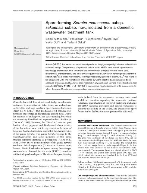
<strong>Spore</strong>-<strong>forming</strong> <strong>Serratia</strong> <strong>marcescens</strong> <strong>subsp</strong>.<br />
<strong>sakuensis</strong> <strong>subsp</strong>. <strong>nov</strong>., isolated from a domestic<br />
wastewater treatment tank<br />
Bindu Ajithkumar, 1 Vasudevan P. Ajithkumar, 1 Ryozo Iriye, 1<br />
Yukio Doi 1 3 and Tadashi Sakai 2<br />
1 Ecological and Toxicological Laboratory, Department of Bioscience and Biotechnology, Faculty<br />
of Agriculture, Shinshu University (United Graduate School of Agriculture, Gifu University),<br />
8304 Minamiminowa, Kamiina, Nagano 399-4598, Japan<br />
2 UltraStructure Research Laboratories Ltd, Tochiku, Yokohama 224-0007, Japan<br />
A strain (KRED T ) that formed endospores and produced the pigment prodigiosin was isolated from<br />
activated sludge. The presence of spores in cells of strain KRED T was evident upon electron<br />
microscopy examination, heat treatment and the detection of dipicolinic acid in the cells.<br />
Biochemical characteristics, and 16S rDNA sequence and DNA–DNA homology data identified<br />
strain KRED T as <strong>Serratia</strong> <strong>marcescens</strong>. The major respiratory quinone of strain KRED T was found to<br />
be ubiquinone Q-8. The formation of endospores by Gram-negative bacteria has not been<br />
observed previously, and has never been reported in any species of <strong>Serratia</strong>. Here, it is shown that<br />
strain KRED T (JCM 11315 T =CIP 107489 T ) represents a <strong>nov</strong>el <strong>subsp</strong>ecies of S. <strong>marcescens</strong>, for<br />
which the name <strong>Serratia</strong> <strong>marcescens</strong> <strong>subsp</strong>. <strong>sakuensis</strong> is proposed.<br />
strain isolated from the wastewater treatment tank posed<br />
a difficult question regarding its taxonomic position.<br />
Polyphasic identification of the <strong>nov</strong>el bacterium, including<br />
16S rDNA sequence phylogeny and genetic relatedness to<br />
confirm the identity of the isolate, and evidence for spore<br />
formation by the bacterium are presented in this work.<br />
METHODS<br />
Isolation and culture conditions. The domestic wastewater was<br />
favourably treated at Komaba sewerage facility, Saku, Nagano, Japan<br />
(Doi et al., 1998) (actual residence time 24 h; typical quality of treated<br />
water: biological oxygen demand, 4?6 mgl 21 ; suspended solids,<br />
4mgl 21 ; total nitrogen, 7?5 mgl 21 ; total phosphate, 2?5 mgl 21 ).<br />
The gross yield coefficient of the sludge was below 8 %, and the<br />
concentration of mixed-liquor suspended solids (MLSS) in the treatment<br />
tank was high (5500 mg l 21 ). To elucidate the bacterial flora<br />
of the activated sludge, bacteria in the suspended liquor in the first<br />
aerobic tank were analysed by the dilution heterotrophic platecount<br />
method. The suspended liquor was diluted with 0?5 % NaCl<br />
(1610 4 dilution) and plated onto culture plates (NG agar, described<br />
below); the cultures were incubated for 24 h at 32 ˚C and<br />
then at room temperature for 6 days.<br />
Culture medium (NG broth) was composed of 8?0 g nutrient broth<br />
(CM-1; Oxoid), 8?0 g glucose, 5?0 g NaCl and 0?5 g yeast extract<br />
(Difco) in 1 l distilled water. Agar (15 g) was added to NG broth for<br />
plate culture (NG agar).<br />
Cell morphology and characterization. Tests for the utilization<br />
of various carbon compounds by the <strong>nov</strong>el bacterium and other biochemical<br />
tests were carried out following standard methods (Smibert<br />
02158 G 2003 IUMS Printed in Great Britain 253
B. Ajithkumar and others<br />
& Krieg, 1994). Cooked-meat hydrolysis was tested by the decrease<br />
in the amount of cooked meat (200 mg; Oxoid) present after incubation<br />
in 0?5 % NaCl (6 ml) at 32 ˚C for 10 days.<br />
Heat-resistance test. Seven-day- and 3-month-old cultures of<br />
strain KRED T on NG agar were subjected to a heat-resistance test.<br />
Each culture was suspended in 0?5 % NaCl (1 ml) or NG broth<br />
(5 ml) (OD660 0?05) and heated at 60, 62, 65, 70 and 75 ˚C for up<br />
to 20 min. Each culture heated in 0?5 % NaCl was shaken at 32 ˚C in NG broth (5 ml) or incubated on an NG agar slant at 32 ˚C. Each culture heated in NG broth was incubated at 32 ˚C with<br />
shaking or incubated on an NG agar slant at 32 ˚C. <strong>Serratia</strong><br />
<strong>marcescens</strong> JCM 1239 T (=ATCC 13880 T ) and Bacillus subtilis IFO<br />
13719 T (=ATCC 6051 T ) were similarly treated and used as controls.<br />
Analysis of dipicolinic acid (DPA). DPA was extracted from<br />
strain KRED T according to published methods (Janssen et al., 1958;<br />
Powell, 1953). Cells of strain KRED T (0?2 g wet weight) that had<br />
been cultivated for 1 week on NG agar plates and washed with water<br />
were heated in 2 ml water at 100 ˚C for 20 min. The suspension was<br />
cooled and centrifuged; the resulting supernatant was condensed to<br />
dryness. The residue was dissolved in 0?1 ml water and then filtered<br />
through a 0?45 mm filter disc. The filtrate (0?05 ml) was analysed<br />
using an LC-MS spectrometer [column AQ-312 (ODS 66150 mm),<br />
YMC, Kyoto, Japan; solvent 10 mM CH3COONH4; flow rate<br />
1 ml min 21 ]. Authentic DPA could be detected at retention time<br />
(tR)3?45 min (m/z values of 168, 124 and 80) [LC-MS spectra were<br />
measured with a Hitachi M-1200AP LC-MS spectrometer; flow rate<br />
1 ml min 21 ; atmospheric pressure chemical ionization (APCI) positive<br />
mode].<br />
Scanning electron microscopy. Electron micrographs of strain<br />
KRED T incubated on NG agar for 7 days at 20 ˚C (cells) and on<br />
NG agar for 18 days at 20 ˚C (spores) were taken at UltraStructure<br />
Research Laboratories; the cells were pre-fixed in 2 % glutaraldehyde<br />
and fixed with 1 % osmic acid. A Hitachi S-4500 scanning electron<br />
microscope was used.<br />
Transmission electron microscopy. A study of the ultrastructure<br />
of the endospores of strain KRED T was carried out at Ultrastructure<br />
Research Laboratories. Strain KRED T was cultivated on NG agar at<br />
32 ˚ C for 20 h and successively for 40 h at 20 ˚<br />
C (libe-<br />
ration of spores was observed with a phase-contrast microscope).<br />
Cells of strain KRED T were pre-fixed with 2 % glutaraldehyde in<br />
phosphate buffer and fixed with 1 % osmic acid (Fig. 1c) and successively<br />
stained with 4 % uranyl acetate (Fig. 1d). Epoxy resin was<br />
used for embedding the cells. Sections (800 A ˚ ) of the cells were prepared<br />
with an LKB U5 ultramicrotome (Amersham Pharmacia) and<br />
examined with a JEOL JEM 800a electron microscope.<br />
Sequencing of 16S rDNA. Extraction of DNA from strain<br />
KRED T and analysis of the sequence of its 16S rDNA was carried<br />
out at NCIMB, Japan, using the PrepMan method (DNA extraction;<br />
Applied Biosystems), and a Microseq full gene 16S rDNA bacterial<br />
sequencing kit (Microseq system; Applied Biosystems) and an ABI<br />
model 310 automated DNA sequencer (Applied Biosystems). The<br />
16S rDNA sequence of strain KRED T was compared to available<br />
sequences using the BLAST program from the National Center for<br />
Biotechnology Information (http://www.ncbi.nlm.nih.gov/) and the<br />
Ribosomal Database Project (Maidak et al., 1996). An evolutionary<br />
Fig. 1. Scanning and transmission electron micrographs of strain KRED T . (a) Strain KRED T after incubation for 7 days on NG<br />
agar. Bar, 1 mm. (b) <strong>Spore</strong>s of strain KRED T after incubation for 18 days on NG agar. Bar, 1 mm. (c) Endospores of strain<br />
KRED T , after incubation for 16 h at 32 ˚C and 40 h at 20 ˚C on NG agar, and pre-fixed with 2 % glutaraldehyde and fixed<br />
with 1 % osmic acid (bar, 0?2 mm), and (d) successively stained with 4 % uranyl acetate (bar, 0?1 mm).<br />
254 International Journal of Systematic and Evolutionary Microbiology 53
distance tree and sequence similarities were calculated using the programs<br />
TREEVIEW and CLUSTAL W, respectively (Saitou & Nei, 1987;<br />
Thompson et al., 1994).<br />
DNA–DNA homology. DNA was extracted from bacteria by the<br />
method of Marmur (1961). DNA–DNA homology was determined<br />
by fluorometric hybridization in microdilution wells [Black Cliniplate;<br />
Labsystems, catalogue no. 95029120 (enhanced binding type)]<br />
according to the method of Ezaki et al. (1989), using biotinylated<br />
DNA. DNA–DNA hybridization was performed at 50 ˚C for 2 h in<br />
26SSC containing 45 % formamide. Fluorescence intensity was<br />
measured with a FP 3000 Fluorolite microplate reader (Shinseirika,<br />
Tokyo) at 360 nm for excitation and 450 nm for emission.<br />
Assay for antibacterial sensitivity. Antibacterial sensitivity<br />
profile of strain KRED T was determined on NG agar plates using<br />
antibacterial discs [Showa Disc (Showa Yakuhin Kako) supplied by<br />
Nissuiseiyaku, Tokyo].<br />
Cellular fatty acid analysis. Cells of strain KRED T that had been<br />
cultured on NG agar (4 days) were washed with distilled water. Wet<br />
cells (200 mg) were refluxed in 10 % KOH/65 % ethanol (5 ml) for<br />
1 h under an N2 atmosphere. The reaction mixture was acidified<br />
with 1 M H2SO4 and fatty acids were extracted with ethyl acetate.<br />
The organic layer was washed with aqueous NaCl and then dried<br />
over MgSO4. After evaporation of solvents, the resulting fatty acid<br />
mixture in ether was methylated with diazomethane and analysed<br />
using a GC-MS spectrometer by EI positive mode. The tR values of<br />
the fatty acid methyl esters (FAMEs) were compared with those of<br />
reference bacterial FAMEs purchased from Supelco. The molecular<br />
formula of each peak was determined using high-resolution GC-MS<br />
analysis (Pizzimenti et al., 1999). Low- and high-resolution GC-MS<br />
spectra were measured by using a JEOL JMS-700 spectrometer with<br />
the JEOL data processing system and a Hewlett Packard 5890 gas<br />
chromatograph [column DB-1 (HP, non-polar); temperature<br />
140–220 ˚C(6˚Cmin21 ) and kept at 220 ˚C for 20 min; injection<br />
temperature 240 ˚C; flow rate 7?2 ml min21 (He gas)].<br />
Isoprenoid quinone analysis. Cells of strain KRED T that had<br />
been cultured on NG agar (4 days) were washed with distilled water.<br />
Isoprenoid quinones were extracted from 500 mg wet cells with<br />
150 ml acetone (three times) by stirring for 2 h each at room temperature.<br />
After centrifugation, the supernatant was condensed to<br />
dryness. The extracts dissolved in acetone were applied to preparative<br />
silica-gel TLC, and then developed with benzene. The isoprenoid<br />
quinones were detected under an UV lamp. The bands due to<br />
menaquinones and ubiquinones were collected and extracted with<br />
acetone. Each fraction was analysed using an LC-MS spectrometer<br />
[Column Zorbax-ODS (4?56250 mm), solvent methanol/diisopropyl<br />
ether (4 : 1, v/v), flow rate 1 ml min 21 (Collins & Jones,<br />
1981; Hiraishi, 1999)]. LC-MS spectra were measured using a<br />
Hitachi M-1200AP LC-MS spectrometer (flow rate 1 ml min 21 ;<br />
APCI positive mode).<br />
Pigment analysis. Cells of strain KRED T that had been cultured<br />
on NG agar (2 days) were washed with distilled water. Pigments<br />
(66 mg) were extracted from 8 g wet cells with 150 ml ethyl acetate<br />
in a separating funnel. The organic layer was washed twice with<br />
saturated NaCl solution and then dried over MgSO 4. After evaporation<br />
of the solvents, the extracts (66 mg) were separated by silica-gel<br />
TLC (benzene/acetone, 5 : 1). The red-coloured band (R f 0?35–0?6)<br />
was collected and extracted with ethyl acetate/ethanol (5 : 1). After<br />
evaporation of the solvents, the pigment (4?3 mg) was dissolved in<br />
5 ml ethyl acetate containing a few drops of 2 M HCl and shaken.<br />
The organic layer was washed with saturated NaCl solution and then<br />
dried over MgSO 4. After evaporation of the solvent, the pigment<br />
was analysed by using a Bruker DRX 500 spectrometer with tetramethylsilane<br />
as an internal standard and Bruker’s Pulse programme<br />
( 1 H at 500?13 MHz and 13 C at 125?77 MHz). The red pigment was<br />
extracted from S. <strong>marcescens</strong> JCM 1239 T using the same method as<br />
described above and analysed using an NMR spectrometer.<br />
RESULTS<br />
<strong>Spore</strong>-<strong>forming</strong> <strong>Serratia</strong> <strong>marcescens</strong><br />
Cell morphology and biochemical<br />
characteristics<br />
Cells of strain KRED T (Fig. 1a) were rods (0?5–0?661?3–<br />
2 mm) that formed granules (generally 0?4–0?6 mm,<br />
but some measured 1?5–1?8 mm; Fig. 1b) identified as<br />
endospores (Fig. 1c, d). Colonies of strain KRED T were<br />
smooth and round, and on nutrient agar the strain produced<br />
a bright-red pigment. Because of endospore formation,<br />
some cells of strain KRED T were found to be swollen when<br />
first isolated, though such swollen cells could not be<br />
observed after successive transfers and maintenance. The<br />
biochemical characteristics of strain KRED T are given in<br />
Table 1, and they were found to be very similar to those of<br />
S. <strong>marcescens</strong>. Strain KRED T differed from the type strain of<br />
S. <strong>marcescens</strong> in terms of its methyl red test [negative on<br />
sucrose (5 days), D-sorbitol (7 days) and L-arabitol, whereas<br />
S. <strong>marcescens</strong> was positive on sucrose, D-sorbitol and<br />
L-arabitol (7 days)]. Strain KRED T did not produce acid<br />
from L-arabitol, whereas S. <strong>marcescens</strong> did.<br />
Elucidation of the ultrastructure of strain KRED T<br />
Endospores of strain KRED T were observed by transmission<br />
electron microscopy analysis (Fig. 1c, d). Cells of strain<br />
KRED T had an outer membrane, a peptidoglycan layer and a<br />
cell membrane (not visible), all characteristics of Gramnegative<br />
bacteria (Fig. 1c). Inside the cells, granules with<br />
cortex layers were observed (Fig. 1c, a white ring), indicating<br />
that endospores were present. Although a spore coat<br />
and spore membrane could not be detected in the cells under<br />
the conditions tested, spore coats could be clearly observed<br />
from some endospores in which a cortex layer was covered<br />
with the peptidoglycan layer of the cell, by staining with<br />
uranyl acetate (Fig. 1d). The spore membrane of strain<br />
KRED T endospores could not be observed, and was presumed<br />
to be very thin. The structure of the endospores of<br />
strain KRED T was similar to that of an endospore of Bacillus<br />
megaterium (Ralph et al., 1992).<br />
Heat resistance<br />
Cultures of strain KRED T that had been incubated for 7 days<br />
and 3 months, respectively, survived heat treatment at 62 ˚C for 15 min in 0?5 % NaCl and 75 ˚C for 20 min in NG<br />
broth. B. subtilis IFO 13719 T incubated for 10 days survived<br />
similar heat treatment, but S. <strong>marcescens</strong> JCM 1239 T did not<br />
survive heat treatment at 60 ˚C for 15 min. Growth from<br />
cultures of strain KRED T that had been subjected to heat<br />
treatment could be observed after 24 h, whereas growth of<br />
the controls (not subjected to heat treatment) was observed<br />
after 12 h. The heat resistance of strain KRED T could be<br />
attributed to it <strong>forming</strong> spores.<br />
http://ijs.sgmjournals.org 255
B. Ajithkumar and others<br />
Table 1. Biochemical characteristics of <strong>Serratia</strong> <strong>marcescens</strong><br />
<strong>subsp</strong>. <strong>sakuensis</strong> and differentiation of this <strong>nov</strong>el <strong>subsp</strong>ecies<br />
from S. <strong>marcescens</strong> <strong>subsp</strong>. <strong>marcescens</strong><br />
Strains: 1, S. <strong>marcescens</strong> <strong>subsp</strong>. <strong>sakuensis</strong>; 2, S. <strong>marcescens</strong> <strong>subsp</strong>.<br />
<strong>marcescens</strong>. Both <strong>subsp</strong>ecies of S. <strong>marcescens</strong> were rod-shaped<br />
organisms that were motile by means of peritrichous flagella. Both<br />
<strong>subsp</strong>ecies were able to grow in the presence of 7 % NaCl, were<br />
Voges–Praskauer-positive and had a fermentative type of metabolism<br />
(O/F test).<br />
Characteristic 1 2<br />
Size (mm) 0?561?3 0?5–0?860?9–2?0*<br />
<strong>Spore</strong> + (Round) 2<br />
Motility + +*<br />
Anaerobic growth + +*<br />
Casein hydrolysis + +*<br />
Tween 80 hydrolysis + +*<br />
Catalase + +*<br />
NO3 reduction to NO2 + +*<br />
NH3 utilization + +*<br />
Indole production 2 2*<br />
Starch hydrolysis<br />
Growth on and acid<br />
production from:<br />
2 2*<br />
Sucrose +3 +<br />
D-Glucose +3 +3<br />
D-Sorbitol +3 +<br />
L-Arabitol +34 +<br />
Hydrolysis of cooked<br />
meat (%)1<br />
80 73<br />
G+C content (mol%) 58 (HPLC) 57?5–60<br />
(Tm, buoyant density)*<br />
*Data are from Grimont & Grimont (1984).<br />
3Methyl red test negative within 7 days.<br />
4Does not produce acid.<br />
1Decrease in the amount of cooked meat present after 10 days<br />
incubation.<br />
DPA<br />
DPA could be detected [10 mg (g bacteria) 21 ] by LC-MS<br />
analysis [tR 3?5 min;m/z168 (M + H) + , 124 (M + H–CO2) +<br />
and 80 (M + H–2CO2) + ].<br />
DNA G+C content<br />
DNA G+C content (as determined by HPLC) of strain<br />
KRED T was 58 mol%, and agreed with the published values<br />
for S. <strong>marcescens</strong> (Grimont & Grimont, 1992; Brenner, 1984).<br />
Phylogenetic analysis and DNA–DNA homology<br />
An almost complete sequence (1532 bp) of the 16S rDNA<br />
of strain KRED T was determined (DDJB accession no.<br />
AB061685). A phylogenetic tree of related sequences based<br />
on BLAST sequence homology (Altschul et al., 1990) was<br />
constructed using the DDBJ data analysis server (CLUSTAL<br />
W) and TREECON software (Van de Peer & De Wachter,<br />
1997); the tree is shown in Fig. 2. The 16S rDNA sequence of<br />
strain KRED T showed 99?6 % similarity with S. <strong>marcescens</strong>,<br />
98?2 % with <strong>Serratia</strong> entomophila, 98?1 % with <strong>Serratia</strong><br />
ficaria, 98?0 % with <strong>Serratia</strong> odorifera, 97?8 % with <strong>Serratia</strong><br />
rubidaea, 97?4 % with Enterobacter cloacae, 97?4 % with<br />
Klebsiella pneumoniae and 97?4 % with Citrobacter freundii.<br />
The level of DNA–DNA hybridization between strain<br />
KRED T and S. <strong>marcescens</strong> JCM 1239 T was found to be 97 %.<br />
Fatty acids<br />
Whole-cell fatty acids of strain KRED T were detected as<br />
saturated fatty acids C12 : 0 (1?25 %), C15 : 0 (0?58 %),<br />
C16 : 0 (33?2 %), C17 : 0 (0?66 %) and C18 : 0 (1?93 %),<br />
mono-hydroxylated fatty acids C14 : 0 (2?83 %) and C16 : 0<br />
(0?14 %), and unsaturated fatty acids C12 : 2 (2?94 %),<br />
C14 : 1 lpar;9?81 %), C17 : 1 (28?31 %), C18 : 2 (0?77 %) and<br />
C19 : 1 (19?94 %).<br />
Fig. 2. Phylogenetic placement of strain KRED T within the<br />
genus <strong>Serratia</strong>, based on 16S rRNA gene sequence analysis.<br />
The significance of each branch is indicated by bootstrap<br />
values at the nodes. The GenBank accession number for<br />
the 16S rDNA sequence of each reference species is shown<br />
in parentheses. Bar, estimated substitutions per nucleotide<br />
position.<br />
256 International Journal of Systematic and Evolutionary Microbiology 53
The profile of strain KRED T was similar to that of<br />
S. <strong>marcescens</strong> (Grimont & Grimont, 1992; Pizzimenti<br />
et al., 1999).<br />
Isoprenoid quinones<br />
Ubiquinone (Q-8; tR 10?8 min) was detected as the major<br />
respiratory quinone of strain KRED T , along with very<br />
small amounts of menaquinones [MK-8, MK-8(H2) , MK-<br />
8(2H2); tR 16?1–16?3 min]. The presence of Q-8 as the<br />
major quinone in the type strain of S. <strong>marcescens</strong> has been<br />
reported previously (Grimont & Grimont, 1992; Collins &<br />
Jones, 1981; Hiraishi, 1999).<br />
Antibacterial sensitivity<br />
Strain KRED T was resistant to tetracycline, erythromycin,<br />
polymyxin, lyncomycin and benzylpencillin, but sensitive to<br />
ampicillin (MIC 50?0 mg ml 21 ), chloramphenicol (MIC<br />
6?25 mg ml 21 ), gentamicin (MIC 6?25 mg ml 21 ), kanamycin<br />
(MIC 12?5 mg ml 21 ), streptomycin (MIC 50?0 mg ml 21 )<br />
and carbenicillin (MIC 12?5 mg ml 21 ).<br />
Pigment analysis<br />
The pigment produced by strain KRED T was identified<br />
as prodigiosin by high-resolution MS (calculated for<br />
C20H25ON3, found 323.1998, measured 323.1990) and on<br />
the NMR spectra with the authentic sample extracted from<br />
the type strain of S. <strong>marcescens</strong>.<br />
DISCUSSION<br />
Strain KRED T was isolated from the bacterial flora of activated<br />
sludge in a domestic wastewater treatment tank<br />
in Saku, Japan. It was present in the sludge at 2?25610 4<br />
c.f.u. ml 21 , whereas Bacillus spp. and total bacteria were<br />
present at 2?3610 6 c.f.u. ml 21 and 5610 6 c.f.u. ml 21 ,<br />
respectively (Doi et al., 1998). From transmission electron<br />
microscopy analysis of strain KRED T , cells of the <strong>nov</strong>el<br />
strain were found to possess an outer membrane, a peptidoglycan<br />
layer and a cell membrane (not visible), all<br />
characteristics of Gram-negative bacteria. The presence of<br />
endospores in cells of the <strong>nov</strong>el strain could be confirmed<br />
due to the presence of a granule with a cortex layer and a<br />
spore coat formed inside cells of strain KRED T . The<br />
structure of the spores of strain KRED T was similar to<br />
that of spores of B. megaterium, while some endospores of<br />
strain KRED T were wrapped in the peptidoglycan layer of<br />
the cell. The spore membrane of strain KRED T was<br />
presumed to be very thin and it could not be observed<br />
under the conditions described above. The formation of<br />
endospores by strain KRED T was also confirmed by the<br />
results of the heat-resistance test and by the presence of DPA<br />
in the cells, although the temperature of heat resistance was<br />
lower than that of Bacillus spp. On the basis of the above<br />
results, cells of strain KRED T were shown to form endospores<br />
which possessed similar characteristics to spores of<br />
Bacillus spp. Strain KRED T was identified as S. <strong>marcescens</strong><br />
<strong>Spore</strong>-<strong>forming</strong> <strong>Serratia</strong> <strong>marcescens</strong><br />
on the basis of FAME analysis, quinone profiling, pigment<br />
analysis and DNA analysis. The 16S rDNA sequence of<br />
strain KRED T showed 99?6 % similarity with the 16S rDNA<br />
sequence of S. <strong>marcescens</strong>. The close relationship of the <strong>nov</strong>el<br />
strain with S. <strong>marcescens</strong> was further confirmed by DNA–<br />
DNA homology analysis. Although DNA–DNA homology<br />
and 16S rDNA sequence analyses showed strain KRED T to<br />
be very similar to S. <strong>marcescens</strong> JCM 1239 T , the unique<br />
spore-<strong>forming</strong> characteristic of strain KRED T supported<br />
its classification as a <strong>nov</strong>el <strong>subsp</strong>ecies of S. <strong>marcescens</strong>, for<br />
which we propose the name <strong>Serratia</strong> <strong>marcescens</strong> <strong>subsp</strong>.<br />
<strong>sakuensis</strong>.<br />
Endospore formation is known to be a characteristic of some<br />
Gram-positive bacteria, including those grouped in genera<br />
such as Acetonema, Bacillus, Clostridium, Desulfotomaculum,<br />
Sporomusa, Sporosarcina and Thermoactinomyces (Edward,<br />
1997; Sneath, 1989). The work presented here is the first<br />
report of the isolation of a spore-<strong>forming</strong> bacteria belonging<br />
to the Enterobacteriaceae, and is a deviation from the general<br />
concept regarding endospore-<strong>forming</strong> bacteria. We propose<br />
that either (i) S. <strong>marcescens</strong> strains that possess the gene<br />
related to endospore formation may be present in nature but<br />
have been unreported until now or (ii) endospores were<br />
present in strain KRED T due to gene transfer. The latter<br />
explanation is more acceptable at this moment in time, since<br />
endospore-<strong>forming</strong> S. <strong>marcescens</strong> have not been found in<br />
nature and the gene related to spore formation has not been<br />
reported for S. <strong>marcescens</strong>. The similarity of the biochemical<br />
characteristics of strain KRED T to those of S. <strong>marcescens</strong> also<br />
support the hypothesis for gene transfer. The activated<br />
sludge from the wastewater treatment facility at Komaba,<br />
Japan, had abundant Bacillus spp. and high concentrations<br />
of magnesium and silicate present (22?5 mgMg 2+ l 21 and<br />
31?9 mg silicate l 21 in pipe water, and 26?1 mgMg 2+ l 21<br />
and 32?7 mg silicate l 21 in the supernatant of the first<br />
aerobic treatment tank). The possibility of gene transfer<br />
between Bacillus and <strong>Serratia</strong> spp. should be investigated in<br />
activated sludge containing high concentrations of Mg 2+<br />
and silicate ions.<br />
Description of <strong>Serratia</strong> <strong>marcescens</strong> <strong>subsp</strong>.<br />
<strong>sakuensis</strong> <strong>subsp</strong>. <strong>nov</strong>.<br />
<strong>Serratia</strong> <strong>marcescens</strong> <strong>subsp</strong>. <strong>sakuensis</strong> (sa.ku.en9sis. N.L. adj.<br />
saku referring to Saku, Nagano, Japan, where the strain was<br />
isolated).<br />
Cells are Gram-negative, short rods (0?561?3 mm).<br />
Facultatively anaerobic. Motile. Forms round endospores.<br />
Colonies on nutrient agar produce the pigment prodigiosin.<br />
Catalase-positive and oxidase-negative. Hydrolyses casein,<br />
cooked meat and soybean oil, but not starch. Reduces<br />
nitrate to nitrite. Indole is not produced. Can grow in 7 %<br />
NaCl. Utilizes glucose, sucrose, D-sorbitol, L-arabitol,<br />
4-hydroxybenzoic acid, gentisic acid and m-erythritol as sole<br />
carbon sources, but not D-tartrate, L-rhamnose, L-arabinose,<br />
D-xylose, lactose, benzoic acid, 3-hydroxybenzoic acid,<br />
trigonelline or raffinose. Produces acids from sucrose,<br />
http://ijs.sgmjournals.org 257
B. Ajithkumar and others<br />
D-glucose and D-sorbitol, but not from L-arabitol.<br />
Fermentative on glucose. Methyl red test is negative on<br />
sucrose (5 days), glucose (7 days), D-sorbitol (7 days)<br />
and L-arabitol. DNA G+C content is 58 mol%. Other<br />
characteristics of the species can be found in Table 1. The<br />
type strain of <strong>Serratia</strong> <strong>marcescens</strong> <strong>subsp</strong>. <strong>sakuensis</strong> is KRED T<br />
(JCM 11315 T =CIP 107489 T ). Isolated from the suspended<br />
water of a domestic wastewater treatment tank in Komaba,<br />
Saku, Nagano, Japan.<br />
ACKNOWLEDGEMENTS<br />
We thank the Mitsui Engineering and Ship Building Co. Ltd, and The<br />
Society of New Activated Sludge Technology/Research (Professor T.<br />
Kitao, Toyohashi University of Technology, Japan) for financial<br />
support, and Professor Hans Trüper (University of Bonn, Germany)<br />
for his advice regarding the Latin epithet. The assistance of Associate<br />
Professor Shinichiro Tabata (Shinshu University, Japan) with scanning<br />
electron microscopy is gratefully acknowledged.<br />
REFERENCES<br />
Altschul, S. F., Gish, W., Miller, W., Myers, E. W. & Lipman, D. J.<br />
(1990). Basic local alignment search tool. J Mol Biol 215, 403–410.<br />
Brenner, D. J. (1984). Family I. Enterobacteriaceae Rahn 1937, Nom.<br />
Fam. Cons. Opin. 15, Jud. Comm. 1958, 73; Ewing, Farmer and<br />
Brenner 1980, 674; Judicial Commission 1981, 104. In Bergey’s<br />
Manual of Systematic Bacteriology, vol. 1, pp. 408–420. Edited by<br />
N. R. Krieg & J. G. Holt. Baltimore: Williams & Wilkins.<br />
Collins, M. D. & Jones, D. (1981). Distribution of isoprenoid quinone<br />
structural types in bacteria and their taxonomic implication.<br />
Microbiol Rev 45, 316–354.<br />
Doi, Y., Lee, B.-S., Iriye, R., Tabata, S. & Tateishi, K. (1998).<br />
Dominantly growing bacteria in malodorless domestic sewage<br />
treatment tanks and their biochemical characteristics. J Antibact<br />
Antifungal Agents (Japan) 26, 53–63.<br />
Edward, R. L. (1997). Prokaryotic diversity: form, ecophysiology, and<br />
habitat. In Manual of Environmental Microbiology, pp. 14–24. Edited<br />
by J. H. Christon, R. K. Guy, J. M. Michael, D. S. Linda & V. W.<br />
Michael. Washington, DC: American Society for Microbiology.<br />
Ezaki, T., Hashimoto, Y. & Yabuuchi, E. (1989). Fluorometric<br />
deoxyribonucleic acid-deoxyribonucleic acid hybridization in microdilution<br />
wells as an alternative to membrane filter hybridization in<br />
which radioisotopes are used to determine genetic relatedness among<br />
bacterial strains. Int J Syst Bacteriol 39, 224–229.<br />
Gerber, N. N. (1975). Prodigiosin-like pigments. C R C Crit Rev<br />
Microbiol 3, 469–485.<br />
Grimont, F. & Grimont, P. A. D. (1992). The genus <strong>Serratia</strong>. InThe<br />
Prokaryotes, vol. 3, 2nd edn, pp. 2822–2848. Edited by A. Balows,<br />
H. G. Trüper, M. Dworkin, W. Harder & K.-H. Schleifer. New York:<br />
Springer-Verlag.<br />
Grimont, P. A. D. & Grimont, F. (1984). Genus VIII. <strong>Serratia</strong> Bizio<br />
1823, 288 AL .InBergey’s Manual of Systematic Bacteriology, vol. 1,<br />
pp. 477–484. Edited by N. R. Krieg & J. G. Holt. Baltimore: Williams<br />
& Wilkins.<br />
Hearn, W. R., Elson, M. K., Williams, R. H. & Medina-Castro, J.<br />
(1970). Prodigiosin [5-(2-pyrryl)-2,2)-dipyrrylmethene] and some<br />
substituted prodigiosenes. J Org Chem 35, 142–145.<br />
Hiraishi, A. (1999). Isoprenoid quinones as biomarkers of microbial<br />
populations in the environment. J Biosci Bioeng 88, 449–460.<br />
Janssen, F. W., Lund, A. J. & Anderson, L. F. (1958). Colorimetric<br />
assay for dipicolinic acid in bacterial spores. Science 127, 26–27.<br />
Maidak, B. L., Olsen, G. J., Larsen, N., Overbeek, R., McCaughey,<br />
M. J. & Woese, C. R. (1996). The Ribosomal Database Project (RDP).<br />
Nucleic Acids Res 24, 82–85.<br />
Marmur, J. (1961). A procedure for the isolation of deoxyribonucleic<br />
acid from microorganisms. J Mol Biol 3, 208–218.<br />
Pizzimenti, F. C., Nostro, A., Marino, A., Villari, A., Verzera, A.,<br />
Trozzi, A. & Alonzo, V. (1999). Fatty acid profiles in pigmented and<br />
non-pigmented strains of S. <strong>marcescens</strong>. New Microbiol 22, 91–98.<br />
Powell, J. F. (1953). Isolation of dipicolinic acid (pyridine-2,<br />
6-dicarboxylic acid) from spores of Bacillus megaterium. Biochem J<br />
54, 205.<br />
Ralph, A., Slepecky, H. & Hemphill, E. (1992). The genus Bacillus –<br />
nonmedical. In The Prokaryotes, vol. 2, 2nd edn, p. 1673. Edited by<br />
A. Balows, H. G. Trüper, M. Dworkin, W. Harder & K.-H. Schleifer.<br />
New York: Springer-Verlag.<br />
Saitou, N. & Nei, M. (1987). The neighbor-joining method: a<br />
new method for reconstructing phylogenetic trees. Mol Biol Evol 4,<br />
406–425.<br />
Smibert, R. M. & Krieg, N. R. (1994). Phenotypic characterization. In<br />
Methods for General and Molecular Bacteriology, pp. 607–651. Edited<br />
by P. Gerhardt, R. G. E. Murray, W. A. Wood & N. R. Krieg.<br />
Washington, DC: American Society for Microbiology.<br />
Sneath, P. H A. (1989). Endospore-<strong>forming</strong> Gram-positive rods and<br />
cocci. In Bergey’s Manual of Systematic Bacteriology, vol. 2,<br />
pp. 1104–1105. Edited by P. H. A. Sneath, N. S. Mair, M. E.<br />
Sharpe & J. G. Holt. Baltimore: Williams & Wilkins.<br />
Thompson, J. D., Higgins, D. G. & Gibson, T. J. (1994). CLUSTAL W:<br />
improving the sensitivity of progressive multiple sequence alignment<br />
through sequence weighting, position-specific gap penalties and<br />
weight matrix choice. Nucleic Acids Res 22, 4673–4680.<br />
Van de Peer, Y. & De Wachter, R. (1997). Construction of<br />
evolutionary distance trees with TREECON for Windows: accounting<br />
for variation in nucleotide substitution rate among sites. Comput<br />
Appl Biosci 13, 227–230.<br />
258 International Journal of Systematic and Evolutionary Microbiology 53