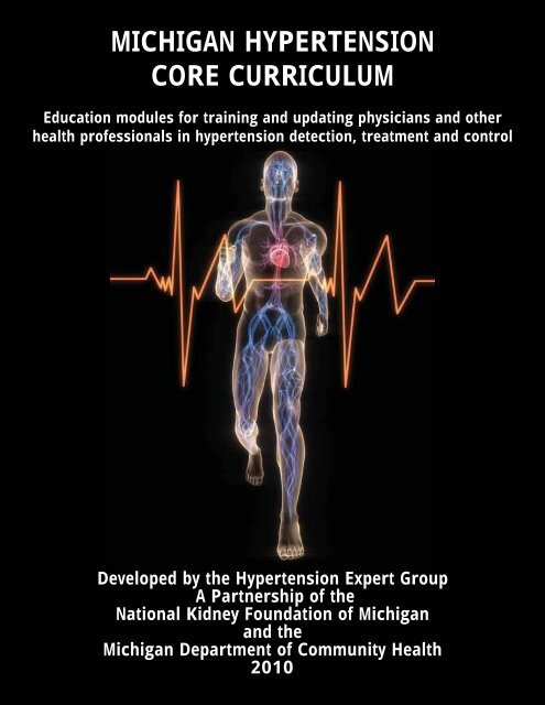
michigan hypertension core curriculum - State of Michigan
michigan hypertension core curriculum - State of Michigan
michigan hypertension core curriculum - State of Michigan

You also want an ePaper? Increase the reach of your titles
YUMPU automatically turns print PDFs into web optimized ePapers that Google loves.
MICHIGAN HYPERTENSION<br />
CORE CURRICULUM<br />
Education modules for training and updating physicians and other<br />
health pr<strong>of</strong>essionals in <strong>hypertension</strong> detection, treatment and control<br />
Developed by the Hypertension Expert Group<br />
A Partnership <strong>of</strong> the<br />
National Kidney Foundation <strong>of</strong> <strong>Michigan</strong><br />
and the<br />
<strong>Michigan</strong> Department <strong>of</strong> Community Health<br />
2010
<strong>Michigan</strong> Hypertension Core Curriculum<br />
2010<br />
Developed by the Hypertension Expert Group<br />
A Partnership <strong>of</strong> the<br />
National Kidney Foundation <strong>of</strong> <strong>Michigan</strong><br />
and the<br />
<strong>Michigan</strong> Department <strong>of</strong> Community Health<br />
NKFM & MDCH 3
April 2010<br />
Dear Colleague:<br />
In 2005, the <strong>Michigan</strong> Department <strong>of</strong> Community Health (MDCH) and the National Kidney Foundation<br />
<strong>of</strong> <strong>Michigan</strong> (NKFM) convened a group <strong>of</strong> <strong>hypertension</strong> experts to identify strategies that will improve<br />
blood pressure control in <strong>Michigan</strong>. Participants included physicians from across <strong>Michigan</strong> specializing<br />
in clinical <strong>hypertension</strong>, leaders in academic research <strong>of</strong> <strong>hypertension</strong> and related disorders, and<br />
representatives <strong>of</strong> key health care organizations that are addressing this condition that afflicts over 70<br />
million U.S. adults. The Hypertension Expert Group has focused on approaches to reduce the burden <strong>of</strong><br />
kidney and cardiovascular diseases through more effective blood pressure treatment strategies.<br />
In an effort to improve <strong>hypertension</strong> control, the group developed educational programs on blood<br />
pressure management, diagnosis and treatment standards. The Expert Group has now turned their<br />
attention toward strengthening academic programs for health care providers in the area <strong>of</strong> clinical<br />
<strong>hypertension</strong>. It was suggested that while all universities and training programs have curricula focused<br />
on cardiovascular diseases, considerable variability exists on how each approaches the diagnosis and<br />
treatment <strong>of</strong> <strong>hypertension</strong>, in part because <strong>hypertension</strong> has not been the domain <strong>of</strong> any single medical<br />
subspecialty.<br />
Thus, our goal was to develop a state-wide <strong>core</strong> <strong>curriculum</strong> designed to serve as a comprehensive<br />
guide for updating clinical knowledge <strong>of</strong> <strong>hypertension</strong> and related disorders. This <strong>core</strong> <strong>curriculum</strong><br />
would ensure that trainees are adequately educated, focused on a basic understanding <strong>of</strong> pressurerelated<br />
vascular pathophysiology and target-organ injury/dysfunction, optimal therapeutic strategies,<br />
and the most recent authoritative evidence-based guidelines and practice standards developed and<br />
promulgated by <strong>hypertension</strong> experts. The <strong>curriculum</strong> will be updated periodically and should continue to<br />
serve as a readily available current source for training.<br />
Sincerely,<br />
John Flack, MD, MPH Sandra Waddell, RN, BSN<br />
Chair, HTN Expert Committee Project Manager, HTN Expert Committee<br />
Wayne <strong>State</strong> University National Kidney Foundation <strong>of</strong> <strong>Michigan</strong><br />
4 Hypertension Core Curriculum
Acknowledgements<br />
Hypertension Expert Workgroup Committee<br />
Ziad Arabi, MD, Senior Staff Physician, Internal Medicine/Hospitalist Medicine, Henry Ford Hospital,<br />
Certified Physician Specialist in Clinical Hypertension*<br />
Aaref Badshah, MD, Chief Medical Resident, Department <strong>of</strong> Internal Medicine<br />
Saint Joseph Mercy-Oakland Hospital *<br />
Jason I Biederman DO, FACOI FASN, Hypertension Nephrology Associates, PC*<br />
Joseph Blount, MD, MPH, FACP, Medical Director, OmniCare Health Plan, Detroit, MI<br />
Mark Britton, MD, PhD, Center <strong>of</strong> Urban and African-American Health Executive Committee, Wayne<br />
<strong>State</strong> University School <strong>of</strong> medicine, Wayne <strong>State</strong> University*<br />
Paul Dake, MD, Family Medicine Residency Program Faculty McLaren Hospital<br />
Benjamin Diaczok, MD, FACP, Program Director, Department <strong>of</strong> Internal Medicine, St. Joseph Mercy<br />
Oakland Hospital*<br />
Mark D Faber MD, FACP, Program Director, Division <strong>of</strong> Nephrology and Hypertension<br />
Henry Ford Hospital, Clinical Associate Pr<strong>of</strong>essor, Wayne <strong>State</strong> University*<br />
John M. Flack, MD, MPH, FAHA, FACP, Pr<strong>of</strong>essor <strong>of</strong> Medicine and Physiology<br />
Chair and Chief, Division <strong>of</strong> Translational Research and Clinical Epidemiology<br />
Department <strong>of</strong> Internal Medicine, Wayne <strong>State</strong> University, Specialist in Chief for Internal Medicine,<br />
Detroit Medical Center* Editor in Chief<br />
Arthur Franke, PhD, National Kidney Foundation <strong>of</strong> <strong>Michigan</strong>, Ann Arbor, MI<br />
Crystal R. Gardner-Martin, MD, Hypertension Nephrology Associates, PC*<br />
Patricia Heiler, MPH, CHES <strong>Michigan</strong> Department <strong>of</strong> Community Health,<br />
Cardiovascular Health Section<br />
Khaled Ismail MD, Hypertension Nephrology Associates, PC*<br />
Diane Levine, MD, FACP, Associate Pr<strong>of</strong>essor <strong>of</strong> Medicine, Vice Chair for Education<br />
Department <strong>of</strong> Internal Medicine, Wayne <strong>State</strong> University<br />
Michael Misuraca DO, Hypertension Nephrology Associates, PC*<br />
Samar A. Nasser, PA-C, MPH, Division <strong>of</strong> Translational Research and Clinical Epidemiology,<br />
Department <strong>of</strong> Internal Medicine, Wayne <strong>State</strong> University*<br />
Silas P. Norman, MD, Assistant Pr<strong>of</strong>essor <strong>of</strong> Medicine, Division <strong>of</strong> Nephrology<br />
Section <strong>of</strong> Transplantation, University <strong>of</strong> <strong>Michigan</strong>*<br />
NKFM & MDCH 5
Kevin L. Piggott, MD, MPH, FAAFP Preventive Medicine Resident, University <strong>of</strong> <strong>Michigan</strong> School <strong>of</strong><br />
Public Health and Family Physician*<br />
Rosalind M. Peters, PhD, RN, Associate Pr<strong>of</strong>essor, College <strong>of</strong> Nursing, Wayne <strong>State</strong> University*<br />
William Repaskey, MD, Internal Medicine Hospitalist, University <strong>of</strong> <strong>Michigan</strong> Hospitals, Ann Arbor, MI*<br />
Robert R. Ross, PA-C, Affiliate Pr<strong>of</strong>essor, U <strong>of</strong> D Mercy PA Program<br />
Robert D. Safian, MD, Director, Cardiac and Vascular Intervention Director, Cardiovascular Fellowship<br />
Training Program, Department <strong>of</strong> Cardiovascular Medicine, William Beaumont Hospital*<br />
Ankur Sandhu MD, Nephrology and Critical Care Fellow, Henry Ford Hospital*<br />
Kiran Saraiya DO, Hypertension Nephrology Associates, PC*<br />
Linda Smith-Wheelock, ACSW, Chief Operating Officer, National Kidney Foundation <strong>of</strong> MI<br />
Hani Al-Sharif MD, Nephrology Fellow, Henry Ford Hospital*<br />
Susan P Steigerwalt MD, FACP, Director, Hypertension clinic SCSP; Member, Division <strong>of</strong> Nephrology<br />
and Hypertension, St John Hospital and Medical Center, Detroit, MI and Providence Hospital,<br />
Southfield, MI, ASH Clinical Hypertension Specialist*<br />
Radhika Thalla MD, Nephrology Consultants P.C, William Beaumont Hospital, Royal Oak, MI*<br />
Velma Theisen, MSN, RN, Manager, Heart Disease and Stroke Prevention Unit<br />
Cardiovascular Health, Nutrition and Physical<br />
Activity Section, <strong>Michigan</strong> Department <strong>of</strong> Community Health*<br />
Joel M Topf, MD, Chief <strong>of</strong> Nephrology, St Clair Specialty Physicians , Director <strong>of</strong> the Chronic Kidney<br />
Disease Clinic, St John Hospital and Medical Center*<br />
Sandra Waddell, RN, BSN, National Kidney Foundation <strong>of</strong> <strong>Michigan</strong>, Ann Arbor , MI<br />
Steven A Yarows, MD, Chelsea Internal Medicine, <strong>Michigan</strong> Hypertension Center,<br />
IHA, Adjunct Pr<strong>of</strong>essor <strong>of</strong> Internal Medicine, Cardiovascular Division, University <strong>of</strong> <strong>Michigan</strong> Health<br />
System*<br />
Jerry Yee, MD, Henry Ford Hospital, Nephrology<br />
* Denotes contributing author<br />
6 Hypertension Core Curriculum<br />
Acknowledgements
Produced April 2010<br />
Permission is granted for the reproduction <strong>of</strong> this publication provided that the reproductions contain<br />
appropriate reference to the source.<br />
Made possible in part by funding from the <strong>Michigan</strong> Department <strong>of</strong> Community Health, Division <strong>of</strong><br />
Chronic Disease and Injury Control, Cardiovascular Health, Nutrition and Physical Activity Section.<br />
Thank you to the National Kidney Foundation <strong>of</strong> <strong>Michigan</strong>’s Scientific Advisory Board. for their review<br />
and input <strong>of</strong> the Hypertension Core Curriculum.<br />
Thank you to Sheila Jackson at the National Kidney Foundation <strong>of</strong> <strong>Michigan</strong> for assisting with the<br />
design and formatting <strong>of</strong> the Hypertension Core Curriculum.<br />
NKFM & MDCH 7
Blood Pressure Measurement........ pp. 10<br />
8 Hypertension Core Curriculum<br />
Table <strong>of</strong> Contents<br />
Essential Hypertension<br />
Primary Hypertension........ pp. 18<br />
Physiological Determinants <strong>of</strong> Blood Pressure.........pp. 24<br />
Special Populations<br />
Intro for special populations........pp. 39<br />
Chronic Kidney Disease........pp. 40<br />
Elderly........pp. 50<br />
Diabetes........pp. 56<br />
Obesity........pp. 64<br />
African Americans........pp. 71<br />
Hispanics........pp. 83<br />
Secondary Hypertension<br />
Obstructive Sleep Apnea........pp. 87<br />
Pheochromocytoma........pp. 90<br />
Polycystic Ovary Syndrome........pp. 96<br />
Primary aldosteronism........pp. 98<br />
Renal Artery Stenosis........pp. 106<br />
Prevention<br />
Public Health Approaches........pp. 145<br />
Pervasive Hypertension Myths<br />
Hypertension is Asymptomatic........pp. 152<br />
Race is an Important Determinant <strong>of</strong> Antihypertensive Drug Response (RAS blockers do not<br />
work in blacks, etc)........pp. 155<br />
Older Patients Need Elevated Systolic Blood Pressures to Perfuse Their Stiff Vessels<br />
<strong>of</strong> Antihypertensive Drugs........pp. 159<br />
Initial Evaluation........pp. 145<br />
Treatment<br />
Lifestyle Modifications........pp. 173<br />
Goals for the Treatment <strong>of</strong> Hypertension........pp. 182<br />
Compelling Indications for Specific Antihypertensive Drug Classes........pp. 185<br />
Overview <strong>of</strong> Major Antihypertensive Drug Classes........pp. 288<br />
Principles <strong>of</strong> Combination Drug Therapy........pp. 207<br />
Adherence........pp. 213<br />
Treatment/Special Situations:<br />
Orthostatic Hypotension........pp. 219<br />
Baroreceptor Dysfunction........pp. 226<br />
Resistant Hypertension........pp. 229<br />
Hypertensive Urgencies/Emergencies........pp. 251<br />
Treatment <strong>of</strong> Hypertension in Patients with CKD........pp. 263<br />
Pregnancy........pp. 263
Hypertension Management Controversies<br />
Low Diastolic Blood Pressure Should Prevent Antihypertensive Drug Therapy <strong>of</strong> Systolic<br />
Hypertension (J-Curve Debate)........pp. 271<br />
Use <strong>of</strong> Dihydropyridine Calcium Antagonists in Chronic Kidney Disease........pp. 274<br />
Case Studies........pp. 276<br />
1. A hypertensive patient taking multiple antihypertensive medications with poor BP control without an<br />
appropriate diuretic prescribed.<br />
2. A well controlled hypertensive patient with refractory hypokalemia despite replacement<br />
3. A hypertensive patient with diabetes who is taking a diuretic and the steps that can be taken to<br />
minimize or prevent diuretic induced hyperglycemia.<br />
4. Hypertensive patient with CKD with poorly controlled BP control experiencing a significant elevation<br />
in creatinine when BP is lowered below his goal BP.<br />
5. A hypertensive patient who is being treated with multiple antihypertensive drugs who has significant<br />
orthostatic hypotension.<br />
6. A hypertensive patient with truly resistant <strong>hypertension</strong>.<br />
7. A hypertensive patient with CKD and heavy proteinuria.<br />
8. A hypertensive patient with CKD, and proper use <strong>of</strong> diuretics appropriate to level <strong>of</strong> renal function.<br />
9. Ms. LN returns 2 weeks after addition <strong>of</strong> an ACE-I and diuretic, and lab results reveal a reduction in<br />
EGFR. What may be the cause <strong>of</strong> the reduction in renal function, and how would you handle?<br />
10. Ms. LN returns 4 weeks after addition <strong>of</strong> an ACE-I and diuretic, and is symptomatic. What may be<br />
causing these symptoms, and how would you handle?<br />
NKFM & MDCH 9
10 Hypertension Core Curriculum<br />
Blood Pressure Measurement<br />
Rosalind Peters PhD, RN and Velma Theisen MSN, RN<br />
Objectives:<br />
At the end <strong>of</strong> this module, participants should be able to:<br />
1. Describe the strengths and limitations <strong>of</strong> different methods <strong>of</strong> measuring BP.<br />
2. Describe the steps necessary to ensure accurate measurement <strong>of</strong> BP in accordance with<br />
national guidelines.<br />
3. Identify resources and references related to accurate home BP measurement.<br />
Pre-Test questions:<br />
1. The point at which the diastolic BP is recorded is<br />
a. the point where the sounds become muffled<br />
b. the last regular sound you hear<br />
c. the point where no sound is heard<br />
d. two millimeters below the last sound heard<br />
2. The point at which the SBP is recorded is<br />
a. the point where the sounds are loudest<br />
b. the first sound you hear<br />
c. the point where the first <strong>of</strong> two consecutive sounds are heard<br />
d. two millimeters after the first sound heard<br />
3. The correct cuff size for an individual is determined by<br />
a. the individual’s age<br />
b. the size <strong>of</strong> the arm circumference<br />
c. the weight <strong>of</strong> the individual<br />
d. the body mass index <strong>of</strong> the individual<br />
4. Evaluating the accuracy <strong>of</strong> the BP measurement device should be done<br />
a. every 3 years<br />
b. only when you suspect it might be inaccurate<br />
c. every 6 months<br />
d. every 12 months<br />
5. Correct positioning the individual for BP measurement includes all <strong>of</strong> the following except<br />
a. seated with feet flat on floor and legs uncrossed<br />
b. back supported<br />
c. arm supported at heart level<br />
d. seated on the side <strong>of</strong> an exam table
Measurement Methods<br />
Early detection, treatment and control <strong>of</strong> <strong>hypertension</strong> require accurate blood pressure<br />
(BP) measurement. 1 This task, which too <strong>of</strong>ten is left to unlicensed assistive personnel, should<br />
be carefully done by the health care pr<strong>of</strong>essional. Accuracy <strong>of</strong> measurement begins with<br />
understanding the three methods used to obtain a BP reading, and ensuring that the equipment<br />
to be used is accurate. The first method is auscultation with an approved and accurate BP<br />
device. The mercury sphygmomanometer is considered to provide the gold standard <strong>of</strong> BP<br />
measurement. However, due to concerns about environmental hazards these devices are being<br />
phased out, and in <strong>Michigan</strong> as <strong>of</strong> January 2009 mercury sphygmomanometers can only be<br />
used to check accuracy <strong>of</strong> other devices or used in a patient’s home. 2<br />
As a result the mercury sphygmomanometers are being replaced with aneroid and/or oscillometric<br />
devices. Aneroid devices also use auscultation to detect blood flow through the artery. BP readings<br />
based on auscultation are subject to measurement error due to environmental factors (e.g., extraneous<br />
room noise), personnel factors (e.g., education, hearing ability, terminal digit preference), and device<br />
factors. Aneroid devices do not maintain stability over time and require frequent re-calibration (e.g.,<br />
every 6 -12 months). The level <strong>of</strong> inaccuracy <strong>of</strong> BP measurements obtained with aneroid devices has<br />
been found to range from 1% to 44%. 3 To overcome the errors <strong>of</strong> auscultation, an ocillometric method<br />
may be used. The ocillometric method detects vibrations in the arterial wall that occur due to blood<br />
flow, and transforms the vibrations into an electrical signal which is displayed as a digital readout <strong>of</strong><br />
BP. However, factors other than blood flow may affect the vibrations. Thus the oscillometric techniques<br />
will underestimate the true BP in patients with arterial stiffness or dysrrhythmias. 4 The ocillometric<br />
method has been used with a variety <strong>of</strong> measurement devices (e.g., upper arm, wrist, finger, and<br />
ambulatory devices). Automated upper arm devices that measure BP at the brachial artery have<br />
been shown to be reliable in clinical practice, and therefore their use is recommended over wrist<br />
or finger devices. Finger devices are not recommended due to inaccuracies related to peripheral<br />
vasoconstriction, alteration in BP at distal sites, and the error <strong>of</strong> limb position in relation to the heart<br />
during measurement. 4,3 Wrist devices are increasingly being used especially with obese people since<br />
the diameter <strong>of</strong> the wrist is usually not affected by obesity. However, wrist devices are subject to the<br />
same errors as finger devices, with the addition <strong>of</strong> altered readings due to the flexion/ hyperextension<br />
NKFM & MDCH 11
<strong>of</strong> the wrist. Additionally, there is difficulty creating an accurate algorithm to estimate BP as there<br />
are two arteries at the wrist contributing to the oscillometric signal. 4 Wrist devices are not currently<br />
recommended for routine clinical practice or decision making. 3,4 Ambulatory BP monitoring (ABPM),<br />
another type <strong>of</strong> ocillometric measurement, may be done when there is the possibility <strong>of</strong> white-coat<br />
<strong>hypertension</strong> or other concerns <strong>of</strong> measurement error. White-coat <strong>hypertension</strong> (persistent elevation<br />
in BP when measured in a clinical setting, but normal BP when the measurement is taken at home),<br />
affects as many as 1 in 3 in the general population but is higher in the elderly and pregnant women. 3,5<br />
ABPM records BP every 15 to 30 minutes (or when triggered at the patient’s request) for a 24 to 48<br />
hour period. The data is stored in the device’s memory until downloaded to a computer for interpretation<br />
by the physician. 5,6 The multiple recordings may provide greater diagnostic accuracy than isolated<br />
clinic measurements. However, when proper, standardized procedures are followed, the average <strong>of</strong><br />
4 duplicate clinic BP readings is as reliable as 24hr ABPM. 7 A third method <strong>of</strong> BP measurement uses<br />
hybrid sphygmomanometers which combines the features <strong>of</strong> both ausculatory and ocillometric<br />
devices. The hybrid combines manual BP measurement techniques but replaces the mercury column<br />
with an electronic pressure detection system. 3 These are relatively new devices with only a few<br />
certified to meet established standards.<br />
BP measurements taken with ocillometric devices (automated or ABPM) are usually lower than<br />
with ausculatory methods. This difference must be reconciled with the fact that BP treatment guidelines<br />
are based on epidemiologic data obtained using ausculatory methods. Thus, lower thresholds for<br />
treatment should be considered if treatment decisions are based on automated measurements. BP<br />
measurements > 135/85 mmHg obtained with an ocillometric device (e.g., ABPM, home monitors)<br />
should be considered abnormal (hypertensive) and treated as such. 4<br />
Measurement Protocol<br />
Accuracy <strong>of</strong> BP measurement requires careful attention to detail when any BP reading is<br />
obtained. Table 1 contains guidelines that should be followed to achieve maximal accuracy.<br />
Measurement Locations<br />
Blood pressure may be measured in numerous locations including pr<strong>of</strong>essional settings (e.g.,<br />
out-patient clinics, hospitals); community sites (e.g., pharmacies), and in patients’ homes. In all <strong>of</strong><br />
these locations, principles <strong>of</strong> accurate measurement must be followed including the use <strong>of</strong> appropriate<br />
equipment and adherence to BP measurement protocols. It is important that the individual measuring<br />
12 Hypertension Core Curriculum
the BP understands current national guidelines for identifying, referring, and managing high BP. In all<br />
settings the individual whose pressure is being recorded should be seated with the back supported,<br />
legs uncrossed, feet flat on the floor, and the arm supported at heart level. The setting should be as<br />
private and as quiet as possible.<br />
In the clinical setting, a conscious decision must be made to establish an appropriate screening<br />
area. This is especially important as some individuals demonstrate “white coat <strong>hypertension</strong>” with the<br />
elevation <strong>of</strong> BP triggered by anxiety or nervousness usually in response to being in the healthcare<br />
setting. To evaluate the presence <strong>of</strong> white-coat <strong>hypertension</strong>, patients are encouraged to measure their<br />
BP at home. Home readings are useful for engaging patients in their own BP treatment program and<br />
also provide additional information for healthcare providers to better manage the therapy.<br />
Patients who purchase a home BP unit need guidance so that they purchase an accurate upper<br />
arm machine, rather than finger or wrist device, and that the correct cuff size is obtained. Patients<br />
should be instructed to choose a monitor that has been tested and validated by either the Association<br />
for the Advancement <strong>of</strong> Medical Instrumentation, the British Hypertension Society, or the International<br />
Protocol for the Validation <strong>of</strong> Automated Blood Pressure Measuring Devices. A list <strong>of</strong> validated monitors<br />
is available on the British Hypertension Society website (www.bhsoc.org/blood_pressure_list.stm) or<br />
the Dabl Educational Trust website (www.dableducational.org/sphygmomanometers/devices_2_sbpm.<br />
html#ArmTable). (Dabl is a leading provider <strong>of</strong> healthcare management systems and research tools<br />
for the prevention and management <strong>of</strong> cardiovascular conditions including high BP). If the device is to<br />
be used for children or pregnant women, then patients need to know to select a monitor that has been<br />
validated for those conditions. Once the monitor is obtained, healthcare providers should assess the<br />
patient’s accuracy in following measurement guidelines, and should compare home monitor readings<br />
with measurements taken in the provider’s <strong>of</strong>fice. Providers should then give the patient directions as to<br />
the frequency and timing <strong>of</strong> the home measurements, as well as instructions as to what data should be<br />
reported to the provider.<br />
Essential Points<br />
1. Diagnosis and treatment decisions for high BP require accurate BP measurement – which<br />
should be done by the pr<strong>of</strong>essional health care provider.<br />
2. Many providers do not follow established protocols for BP measurement resulting in inaccurate<br />
diagnoses and treatment plans.<br />
3. All measurement devices must be calibrated and/or validated for accuracy on a regular<br />
NKFM & MDCH 13
asis. National guidelines recommend every six months for calibration assessment and<br />
accuracy assessed with every use.<br />
4. ABPM readings will be lower than those obtained using <strong>of</strong>fice-based ausculatory<br />
methods. Accordingly, ABPM > 135/85 mm Hg is considered to be in the hypertensive range.<br />
5. The American Heart Association provides national recommendations for accurate BP<br />
measurement by health pr<strong>of</strong>essionals.<br />
6. Home BP readings can enhance management <strong>of</strong> an individuals’ <strong>hypertension</strong>, but should be<br />
implemented and monitored by a healthcare provider with adequate guidance and education<br />
provided to the patient and family.<br />
14 Hypertension Core Curriculum
Table 1: Guidelines for Obtaining Accurate Blood Pressure Readings 2<br />
I. Prepare the equipment:<br />
A. Use equipment that has been (1) validated as accurate against a mercury<br />
sphygmomanometer, (2) checked for disrepair <strong>of</strong> cuff (e.g., cracks or leaks in tubing,<br />
breaks in stitching or tears in fabric), (3) checked that gauge is intact (mercury meniscus<br />
or aneroid needle is at zero), (4) consistent with <strong>State</strong> Legislation.<br />
B. Obtain appropriate cuff size by measuring circumference <strong>of</strong> the patient’s arm and<br />
choosing the cuff size that corresponds to that measurement.<br />
II. Prepare the patient<br />
A. Assess (1) that patient has not recently had nicotine or caffeine and (2) that the patient<br />
has been sitting quietly for 5 minutes prior to measuring BP<br />
B. Position patient: (1) Use a sitting or semi-reclining position with the back supported<br />
and the arm at heart level (middle <strong>of</strong> the cuff should be at mid-sternum level). (2) Legs<br />
should be uncrossed with feet flat and supported on floor or foot rest (not dangling from<br />
examination table or bed)<br />
C. Bare the upper arm <strong>of</strong> any constrictive clothing (You should be able to get at least one<br />
finger under a rolled-up sleeve). Palpate brachial artery, position center <strong>of</strong> cuff bladder<br />
over the brachial artery<br />
III. Take the measurement<br />
A. Support the patient’s arm at heart level<br />
B. For ausculatory measurements:<br />
i. Obtain an estimated systolic pressure by palpation prior to auscultation<br />
ii. Inflate the cuff as rapidly as possible to maximum inflation level (30 mmHg above<br />
estimated systolic BP).<br />
iii. Deflate the cuff slowly at a rate <strong>of</strong> 2 to 3 mmHg/second; (1) note the first <strong>of</strong> 2<br />
regular beats as systolic pressure (palpation helps to avoid under-estimating<br />
systolic pressure due to an ausculatory gap) (2) Use Kortok<strong>of</strong>f V (last sound<br />
heard) as the diastolic pressure (3) continue deflation for 10 mmHg past last<br />
sound to assure sound is not a ‘skipped’ beat.<br />
iv. The measurement should be recorded as an even number and to the nearest 2<br />
mmHg (round upward)<br />
F. Neither the patient nor observer should talk during the measurement<br />
G. If two readings are measured, record the average <strong>of</strong> the readings<br />
IV. Record the measurement – document the following:<br />
A. The obtained BP reading<br />
B. Patient position (sitting, semi-recumbent, lying, standing)<br />
C. Arm used, include arm circumference and cuff size used<br />
D. Type <strong>of</strong> device used to obtain the measurement (mercury, aneroid, automated)<br />
E. <strong>State</strong> <strong>of</strong> the individual (e.g., anxious, relaxed)<br />
F. Time <strong>of</strong> administration <strong>of</strong> any drugs that could affect BP<br />
(*Source: 8,2,3 )<br />
NKFM & MDCH 15
Post-Test Questions:<br />
1. High BP is defined as:<br />
a. An increase in systolic pressure <strong>of</strong> 15 mm Hg greater than baseline<br />
b. Absolute systolic pressure <strong>of</strong> 140 mm Hg or greater<br />
c. Absolute systolic pressure greater than 160 mm Hg<br />
d. Varied depending on the type <strong>of</strong> blood pressure measurement device used<br />
2. Which BP device gives the most accurate BP reading?<br />
a. Ambulatory BP monitor<br />
b. Mercury sphygmomanometer<br />
c. Oscillometric monitor<br />
d. Aneroid sphygmomanometer<br />
3. Arterial stiffness may lead to inaccuracies using which type <strong>of</strong> BP device?<br />
a. Ambulatory BP monitor<br />
b. Mercury sphygmomanometer<br />
c. Oscillometric monitor<br />
d. Aneroid sphygmomanometer<br />
4. Which <strong>of</strong> the following organizations provides data regarding the validity <strong>of</strong> home BP monitors?<br />
a. American Heart Association<br />
b. British Hypertension Society<br />
c. National Heart, Lung, and Blood Institute<br />
d. American Society <strong>of</strong> Hypertension<br />
5. Estimating SBP by palpation is an important step when using which method <strong>of</strong> blood pressure<br />
measurement?<br />
a. Auscultation<br />
b. Ocillometric<br />
c. Ambulatory<br />
d. All <strong>of</strong> the above<br />
16 Hypertension Core Curriculum
References:<br />
1. Chobanian AV, Bakris GL, Black HR, et al. Seventh Report <strong>of</strong> the Joint National Committee<br />
on Prevention, Detection, Evaluation, and Treatment <strong>of</strong> High Blood Pressure. Hypertension.<br />
2003;42(6):1206-1252.<br />
2. Decreasing the Availability <strong>of</strong> Mercury-Based Blood Pressure Manometers. In: <strong>Michigan</strong> So, ed.<br />
Vol PA-493<br />
http://www.legislature.mi.gov/documents/2005-2006/publicact/htm/2006-PA-0493.htm; 2006.<br />
3. Pickering TG, Hall JE, Appel LJ, et al. Recommendations for blood pressure measurement<br />
in humans: an AHA scientific statement from the Council on High Blood Pressure Research<br />
Pr<strong>of</strong>essional and Public Education Subcommittee. J Clin Hypertens (Greenwich). Feb<br />
2005;7(2):102-109.<br />
4. Parati G, Stergiou GS, Asmar R, et al. European Society <strong>of</strong> Hypertension guidelines for blood<br />
pressure monitoring at home: a summary report <strong>of</strong> the Second International Consensus<br />
Conference on Home Blood Pressure Monitoring. J Hypertens. Aug 2008;26(8):1505-1526.<br />
5. O'Brien E, Coats A, Owens P, et al. Use and interpretation <strong>of</strong> ambulatory blood<br />
pressure monitoring: recommendations <strong>of</strong> the British <strong>hypertension</strong> society. BMJ. Apr 22<br />
2000;320(7242):1128-1134.<br />
6. Marchiando RJ, Elston MP. Automated ambulatory blood pressure monitoring: clinical utility in<br />
the family practice setting. Am Fam Physician. Jun 1 2003;67(11):2343-2350.<br />
7. Jula A, Puukka P, Karanko H. Multiple clinic and home blood pressure measurements versus<br />
ambulatory blood pressure monitoring. Hypertension. Aug 1999;34(2):261-266.<br />
8. O'Brien E, Asmar R, Beilin L, et al. European Society <strong>of</strong> Hypertension recommendations<br />
for conventional, ambulatory and home blood pressure measurement. J Hypertens. May<br />
2003;21(5):821-848.<br />
NKFM & MDCH 17
18 Hypertension Core Curriculum<br />
Essential Hypertension<br />
Primary Hypertension<br />
Steven Yarows, MD<br />
Learning objectives<br />
• Understand the correct method <strong>of</strong> taking BP and then correctly interpret and categorize the<br />
results.<br />
• Since 1/3 <strong>of</strong> the US adult population has primary <strong>hypertension</strong>, understanding the important<br />
health and financial costs <strong>of</strong> this disease.<br />
• Understand the three predominant <strong>hypertension</strong> phenotypes - isolated systolic, mixed systolic/<br />
diastolic and isolated diastolic <strong>hypertension</strong>.<br />
Pre-test Questions<br />
A 35 year old obese male comes to the <strong>of</strong>fice for a rash and has his routine BP measured with<br />
a standard cuff <strong>of</strong> 170/104 mmHg. He has a grandfather who died <strong>of</strong> a stroke at 83 years old,<br />
but he thinks his parents are in good health and only take ‘a few’ pills. You assess the rash and<br />
indicate it is tinea crura and advise an anti-fungal cream. You then address his BP by:<br />
A. Have him return in the morning for another BP reading<br />
B. Recheck his BP with a large cuff after sitting for 5 minutes<br />
C. Start a diuretic and have him return for a physical<br />
D. Advise him to lose weight and see him back in a year<br />
The likelihood <strong>of</strong> isolated systolic <strong>hypertension</strong> (ISH) is higher in:<br />
A. Over 70 years old<br />
B. Under 50 years old<br />
Which <strong>of</strong> the following is true?<br />
A. Inadequate control <strong>of</strong> systolic BP is usually the reason for uncontrolled<br />
<strong>hypertension</strong><br />
B. Inadequate control <strong>of</strong> diastolic BP is usually the reason for uncontrolled<br />
<strong>hypertension</strong><br />
C. Inadequate control <strong>of</strong> systolic and diastolic BP are equally likely in individuals<br />
with uncontrolled <strong>hypertension</strong><br />
A 40 year old Black healthy male has been to your <strong>of</strong>fice twice in the past 2 months for upper<br />
respiratory infections and his average BP over these two visits was 160/102 mmHg; both BP<br />
readings were higher than150/96 mm Hg. His parents are both hypertensive on medication and<br />
he used their home BP monitor with a large cuff and it was 150/96 and 164/104 mmHg. What<br />
would be your starting therapy?<br />
A. Hydrochlorothiazide (diuretic) 25mg qd<br />
B. Valsartan (angiotensin receptor blocker) 320mg qd<br />
C. Metoprolol XL (extended release beta blocker) 50mg qd<br />
D. Amlodipine (dihydropyridine calcium channel blocker) 5mg qd<br />
E. Amlodipine/lotensin (dihydropyridine calcium channel blocker + ACE inhibitor)<br />
5/20mg qd
Essential (Primary) Hypertension:<br />
Diagnosis <strong>of</strong> Hypertension<br />
Essential <strong>hypertension</strong> is a misnomer. There is nothing ‘essential’ about <strong>hypertension</strong>; perhaps<br />
“essential BP”, but not <strong>hypertension</strong>. This section will describe ‘primary <strong>hypertension</strong>’.<br />
Blood pressure (BP) is inherently variable within a reasonably predictable range. Hypertension<br />
is defined as elevated average BP over time, preferably with a minimum <strong>of</strong> 3 properly performed<br />
readings on different days and is never based on a single measurement. Your BP at rest is lower<br />
than your BP during activities. There is a natural diurnal variation for people with usual work-sleep<br />
cycles resulting in increased BP just prior to awakening continuing to be elevated in the morning while<br />
decreasing in the evening. BP also decreases in the early afternoon, which is why post-lunch lectures<br />
are difficult, and the nadir in BP occurs at 2-3AM, during sleep. This data is routinely obtained by<br />
24-hour ambulatory blood pressure monitors. The decrease in BP during sleep is known as the ‘dip’<br />
and is absent in some conditions and when absent results in increased cardiovascular (i.e., strokes,<br />
myocardial infarctions, death) events. Several selected conditions known to attenuate or eliminate the<br />
normal nocturnal decline in BP include: 1) chronic kidney disease (CKD), 2) obesity, 3) high sodium<br />
and/or low potassium diets, and 4) sleep disordered breathing.<br />
Too much pressure is potentially deleterious for any system. For example, an overinflated car<br />
tire allows you to drive to the store without any difficulty; however the increased pressure prematurely<br />
wears out the tire. The human circulatory system is similar. Prolonged BP elevation results in<br />
accelerated atherosclerosis and vascular remodeling that heighten the risk <strong>of</strong> stroke (brain), myocardial<br />
infarction (heart), myocardial hypertrophy (heart), kidney failure (kidney), and abdominal aneurysms<br />
(general circulatory). Contrary to pervasive myths, there is no specific BP reading that prognosticates<br />
without fail a cardiovascular catastrophe. When marked BP pressures are detected, repeated<br />
measurements and careful short-term follow-up are critical.<br />
Hypertension Phenotypes (Isolated Systolic, Isolated Diastolic, Isolated Systolic/Diastolic)<br />
BP is represented by two numbers (i.e., 120/50 mmHg). The highest number is the systolic<br />
BP and the lower is the diastolic BP. The BP is typically measured by either the auscultatory or<br />
oscillometric methods. The following is a discussion <strong>of</strong> <strong>hypertension</strong> phenotypes. Hypertension is<br />
classified into distinctive phenotypes. Mixed systolic/diastolic <strong>hypertension</strong> is most common in middle<br />
aged patients when both the diastolic and systolic BP are elevated above 140/90 mmHg in the <strong>of</strong>fice.<br />
Isolated systolic <strong>hypertension</strong> (ISH) is most common after 50 years old, although there is an unusual,<br />
benign form in the youth. 1,2 ISH is also the most risky <strong>hypertension</strong> phenotype despite the fact that<br />
the diastolic BP is not elevated. Isolated diastolic <strong>hypertension</strong> is least prevalent (and also least<br />
risky) <strong>hypertension</strong> phenotype. The different categories <strong>of</strong> <strong>hypertension</strong> have different pathological<br />
mechanisms which will be discussed in the Pathophysiology Section.<br />
Pre-<strong>hypertension</strong> is present when BP readings are between 120-139/80-89 mmHg. 3 These<br />
individuals are at risk for the development <strong>of</strong> <strong>hypertension</strong>. Thus, lifestyle modification (i.e., exercise,<br />
weight loss, salt and alcohol restriction) is recommended. Borderline or high normal BP is when the<br />
<strong>of</strong>fice readings are consistently between 135-140/85-90 mmHg in patients without CKD, diabetes, or<br />
ischemic heart disease.<br />
White Coat Hypertension (<strong>of</strong>fice <strong>hypertension</strong>) is present when the <strong>of</strong>fice BP is >140/90 mmHg,<br />
yet the outside the <strong>of</strong>fice the BP is
Masked Hypertension is when the <strong>of</strong>fice BP is normal (135/85 mmHg). These individuals are at a greatly increased risk <strong>of</strong> cardiovascular<br />
events and medication treatment is advised. Nevertheless, this type <strong>of</strong> <strong>hypertension</strong> is very difficult to<br />
diagnose because ambulatory BP monitoring is not typically undertaken in patients with controlled <strong>of</strong>fice<br />
BP. Masked <strong>hypertension</strong> not infrequently occurs in patients with sleep apnea.<br />
20 Hypertension Core Curriculum<br />
Epidemiology<br />
The prevalence <strong>of</strong> <strong>hypertension</strong> in the United <strong>State</strong>s is 73 million, which is approximately 1/3<br />
<strong>of</strong> the adult population. 4,5 It is higher in men until 45 years <strong>of</strong> age and then is similar in both sexes<br />
until 55 years <strong>of</strong> age after which it occurs more commonly in women (Figures 1-3). The prevalence <strong>of</strong><br />
<strong>hypertension</strong> is higher in African Americans, a demographic group that is afflicted with <strong>hypertension</strong><br />
earlier in life that is more severe compared to whites. Hypertension incidence also markedly increases<br />
with age. A 55 year old has an 83-88% chance <strong>of</strong> becoming hypertensive over 20 years. 6 Higher<br />
<strong>hypertension</strong> prevalence has been observed in those with less education as well as greater obesity<br />
and physical inactivity. Hypertension prevalence also varies by geographic location <strong>of</strong> residence in the<br />
United <strong>State</strong>s. In 2001–2003, age-standardized uncontrolled <strong>hypertension</strong> prevalence was highest<br />
in the District <strong>of</strong> Columbia, Mississippi, Louisiana, Alabama, Texas, Georgia, and South Carolina and<br />
lowest in Vermont, Minnesota, Connecticut, New Hampshire, Iowa, and Colorado. 7 Hypertension is<br />
also 2-3 times more prevalent in women taking birth control pills, especially obese, and older women.<br />
Pharmacological treatment <strong>of</strong> <strong>hypertension</strong> in the United <strong>State</strong>s in 2004 was 68.5% with 52.9%<br />
<strong>of</strong> those treated having their BP controlled, an increase from past surveys. BP control rates are higher<br />
in men than women and decrease with age, mainly due to uncontrolled isolated systolic <strong>hypertension</strong><br />
(57.0%). Although treatment rates are higher in African Americans than whites, the control rates are<br />
worse (Table 1). Mexican-Americans have the lowest treatment rates, and the poorest control rates.<br />
Treatment rates are the highest for subjects 60 years old and greater; however, the control rates are<br />
below the middle age group, as a result <strong>of</strong> the higher incidence <strong>of</strong> isolated systolic <strong>hypertension</strong>.<br />
Hypertension is highly associated with other vascular diseases (77%), including diabetes (76.8%),<br />
metabolic syndrome (61.5%), stroke (69.5%), dyslipidemia (51.8%), CKD (81.8%), peripheral vascular<br />
disease (73.7%), coronary heart disease (73.0%), and both systolic and diastolic heart failure (71.4%).<br />
Figure 1- Rosamond W
Figure 2<br />
Figure 3<br />
NKFM & MDCH 21
TABLE 1. Prevalence <strong>of</strong> Awareness, Treatment, and Control <strong>of</strong> Hypertension by Descriptive<br />
Factors in the REGARDS Cohort 8<br />
All<br />
REGARDS<br />
Participants<br />
Post-test Answers<br />
Awareness <strong>of</strong> Hypertension<br />
Among All Hypertensives<br />
A 35 year old obese male comes to the <strong>of</strong>fice for a rash and has his routine blood pressure<br />
measured with a standard cuff <strong>of</strong> 170/104 mmHg. He has a grandfather who died <strong>of</strong> a stroke at<br />
83 years old, but he thinks his parents are in good health and only take ‘a few’ pills. You assess<br />
the rash and indicate it is tinea crura and advise an anti-fungal cream. You then address his BP<br />
by:<br />
A. Have him return in the morning for another BP reading<br />
B. Recheck his BP with a large cuff after sitting for 5 minutes<br />
Since he is obese and just had a single reading, potentially not performed<br />
properly, it should be repeated correctly. If elevated, he should have<br />
another blood pressure check, although it does not have to be the next<br />
day, unless he has rare hypertensive urgency. (Correct answer)<br />
C. Start a diuretic and have him return for a physical<br />
D. Advise him to lose weight and see him back in a year<br />
The likelihood <strong>of</strong> isolated systolic <strong>hypertension</strong> increases in:<br />
A. Over 70 years old<br />
This is more common after 50 years <strong>of</strong> age (Correct answer)<br />
B. Under 50 years old<br />
Which <strong>of</strong> the following is true?<br />
A. Inadequate control <strong>of</strong> systolic BP is usually the reason for uncontrolled<br />
<strong>hypertension</strong> (Correct answer)<br />
B. Inadequate control <strong>of</strong> diastolic BP is usually the reason for uncontrolled<br />
<strong>hypertension</strong><br />
C. Inadequate control <strong>of</strong> systolic and diastolic BP are equally likely in individuals<br />
with uncontrolled <strong>hypertension</strong><br />
A 40 year old Black healthy male has been to your <strong>of</strong>fice twice in the past 2 months for upper<br />
respiratory infections and his average BP over these two visits was 160/102 mmHg; both BP<br />
22 Hypertension Core Curriculum<br />
Aware <strong>of</strong><br />
Hypertension<br />
Treatment <strong>of</strong> Hypertension<br />
Among Aware Hypertensives<br />
On<br />
Medication<br />
No. <strong>of</strong><br />
Hypertensive<br />
Participants No. % No. aware No. %<br />
Control <strong>of</strong><br />
Hypertension<br />
Among Treated<br />
Hypertensives<br />
Controlled Blood<br />
Pressure<br />
No.<br />
Treated No. %<br />
All<br />
Race<br />
11 606 6023 5477 90.9 5477 4866 88.8 4858 3198 65.8<br />
White 57.2% 3070 2738 89.2 2738 2373 86.7 2371 1663 70.1<br />
Black 42.8% 2953 2739 92.8 2739 2493 91.0 2487 1535 61.7
eadings were higher than150/96 mm Hg. His parents are both hypertensive on medication and<br />
he used their home BP monitor with a large cuff and it was 150/96 and 164/104 mmHg. What<br />
would be your starting therapy?<br />
A. Hydrochlorothiazide (diuretic) 25mg qd<br />
B. Valsartan (angiotensin receptor blocker) 320mg qd<br />
C. Metoprolol XL (extended release beta blocker) 50mg qd<br />
D. Amlodipine (dihydropyridine calcium channel blocker) 5mg qd<br />
E. Amlodipine/lotensin (dihydropyridine calcium channel blocker + ACE inhibitor)<br />
5/20mg qd (Correct answer)<br />
Essential Points:<br />
• Blood pressure is inherently variable within a predictable range. Hypertension is defined as<br />
elevated average BP over time, preferably with a minimum <strong>of</strong> 3 properly performed readings on<br />
different days and is never based on a single measurement<br />
o Proper measurement <strong>of</strong> BP includes that the patient is seated in a chair with a backrest<br />
and have their feet resting on the floor and be resting for a minimum <strong>of</strong> 5 minutes. Most<br />
importantly, a proper sized cuff should be used and unfortunately most Americans now<br />
need the larger cuff due to the increase in obesity.<br />
• Hypertension is classified into different categories.<br />
o Pre-<strong>hypertension</strong> are BP readings between 120-139/80-89 mmHg<br />
o Mixed systolic/diastolic <strong>hypertension</strong> is most common in middle aged patients when both<br />
the diastolic and systolic BP are elevated above 140/90 mmHg at the <strong>of</strong>fice. It is due to<br />
increased arteriolar resistance in the distal smaller vessels.<br />
o Isolated systolic <strong>hypertension</strong> is most common after 50 years old. It is due to increased<br />
large conduit (aorta) vessel stiffness.<br />
o Isolated diastolic <strong>hypertension</strong> is the least common and least risky <strong>hypertension</strong><br />
phenotype.<br />
o White Coat Hypertension (<strong>of</strong>fice <strong>hypertension</strong>) when the patient has elevated <strong>of</strong>fice<br />
BP (>140/90 mmHg), however outside the <strong>of</strong>fice the BP is 140/90 mmHg) accounts for 77% <strong>of</strong> people who have a first<br />
stroke, 74% who have congestive heart failure, and 69% who have a first heart attack.<br />
o Normotensive men and women at 50 years <strong>of</strong> age live approximately 5 years longer than<br />
their normotensive counterparts.<br />
NKFM & MDCH 23
Blood Pressure, Circulatory Physiology, and Hemodynamically-Mediated Target-Organ Injury<br />
John M. Flack, M.D., M.P.H., F.A.H.A., F.A.C.P., F.A..C.G.S.<br />
Pr<strong>of</strong>essor and Chairman<br />
Chief, Division <strong>of</strong> Translational Research and Clinical Epidemiology<br />
Department <strong>of</strong> Internal Medicine<br />
Specialist in Clinical Hypertension<br />
Wayne <strong>State</strong> University<br />
Specialist in Chief for Internal Medicine, Detroit Medical Center<br />
Learning Objectives<br />
At the end <strong>of</strong> this lecture the student will be able to:<br />
1. Articulate the determinants <strong>of</strong> arterial blood pressure in the younger as well as aged<br />
circulatory system.<br />
2. Describe the circadian variation in blood pressure.<br />
3. Identify the mechanisms <strong>of</strong> blood pressure-related target organ injury.<br />
4. List the organs injured by blood pressure elevations and the clinical manifestations <strong>of</strong> such<br />
injury.<br />
5. Describe how cerebral ischemia disrupts normal cerebral autoregulation <strong>of</strong> blood flow.<br />
6. Discuss the microcirculatory adaptations in the kidney to high systemic arterial pressures.<br />
1. Determinants <strong>of</strong> Arterial Blood Pressure<br />
BP depends is determined by both physical and physiological factors. Physiological factors<br />
interface with physical factors to determine BP level. Systole accounts for ~ one-third <strong>of</strong> the cardiac cycle.<br />
Stroke volume (SV) is typically ejected during the initial one-half <strong>of</strong> the systolic phase <strong>of</strong> the cardiac cycle<br />
- or, stated slightly differently, SV is normally ejected during the initial one-sixth <strong>of</strong> the overall cardiac cycle<br />
given that systole accounts for ~ one-third <strong>of</strong> the total cardiac cycle. Cardiac output is cyclic, yet under<br />
normal physiological circumstances flow through the arterial tree is continuous.<br />
The distensibility <strong>of</strong> the aorta, a large conduit vessel, determines the degree <strong>of</strong> the systolic blood<br />
pressure elevation, for a given amount ejected blood (stroke volume) during systole. During ejection<br />
<strong>of</strong> the SV the highly elastic aorta expands, thus dissipating the rise in blood pressure. The expansion<br />
<strong>of</strong> the aorta during systole stores energy. After ejection <strong>of</strong> stroke volume has ceased the aortic elastic<br />
recoil releases stored energy thereby propelling blood forward in the arterial vasculature after the rapid<br />
systolic ejection period. Thus, the aortic elastic properties explain continuous blood flow through the<br />
arterial circulation, even after the active systolic ejection phase. Though the elastic aorta distends, and<br />
thus dampens the rise in SBP, during systole, SBP does, however, rise during the systolic phase <strong>of</strong> the<br />
cardiac cycle. Another significant contributor to the rise in BP during systole relates to reflected pressure<br />
waves from the peripheral arterial vasculature.<br />
24 Hypertension Core Curriculum
Figure 1. Arterial Waveform<br />
These reflected waves “sum up” with the pressure generated from ejected blood into arterial<br />
system and are therefore the major determinants <strong>of</strong> systolic BP; thus, the arterial waveform (at any<br />
location) consists <strong>of</strong> both forward traveling and reflected waveforms. Normally, because <strong>of</strong> the reflected<br />
waves, SBP and pulse pressure (PP) are amplified or increase by ~ 10 – 14 mm Hg when moving from<br />
the aorta to the brachial artery. However, DBP and mean arterial pressure (MAP) change very little<br />
(figure 2).<br />
Figure 2. Change in contours in pressure and flow waves<br />
After the systolic ejection phase, the fall in DBP is dampened as the elastic recoil <strong>of</strong> the large<br />
capacitance vessels propels forward the blood volume that was stored during systole. The reflected<br />
waveforms largely emanate from the peripheral resistance arterioles and timing wise, arrive back in the<br />
aorta during diastole thereby augmenting coronary perfusion pressure.<br />
The difference between SBP, peak BP during the cardiac cycle, and DBP, the lowest BP during<br />
the cardiac cycle, is the pulse pressure (PP). Pulse pressure is predominantly influenced by the amount<br />
<strong>of</strong> blood ejected during systole (SV) and the magnitude <strong>of</strong> the change in pressure inside the arterial<br />
vasculature for a given change in arterial volume (arterial compliance). Arterial compliance will be<br />
discussed in more detail later.<br />
A. Physical Factors: Blood volume (BV) and arterial compliance are important physical factors that<br />
determine BP levels. Blood volume is distributed unevenly between the arterial and venous (capacitance<br />
vessels) sides <strong>of</strong> the vascular system. Approximately two-thirds to three-quarters <strong>of</strong> the BV is contained<br />
within the venous capacitance vessels; the remaining one-quarter to one-third is contained in the arterial<br />
NKFM & MDCH 25
side <strong>of</strong> the vascular tree. Arterial BV is determined by the difference in the BV ejected by the heart/unit<br />
<strong>of</strong> time (cardiac output, C.O.) and the outflow through the arterial resistance vessels into the venous<br />
capacitance vessels (peripheral run<strong>of</strong>f). When C.O. and peripheral run<strong>of</strong>f are balanced, arterial BV and<br />
arterial pressure remain constant. If C.O. increases but peripheral run<strong>of</strong>f doesn’t rise commensurately,<br />
then arterial BV rises and BP also increases.<br />
Arterial elasticity is an important determinant <strong>of</strong> the rise in SBP that occurs for any given increase<br />
in BV. Generally speaking, arterial elasticity is inversely related to age; that is, younger persons have<br />
greater arterial elasticity and with advancing age arterial elasticity declines. Arterial compliance is<br />
determined by elastic properties <strong>of</strong> the large conduit vessels. Arterial compliance is dV/dP - the change<br />
in pressure that occurs with a given change in arterial volume. It should be clear that the greater the<br />
arterial elasticity, the smaller the rise in systolic pressure during the systolic ejection phase <strong>of</strong> the cardiac<br />
cycle. Conversely, lesser arterial elasticity causes a greater rise in systolic BP during the systolic ejection<br />
phase. This also places an extra burden <strong>of</strong> work on the myocardium to maintain cardiac output, in part<br />
because the systolic ejection phase is prolonged under these circumstances.<br />
B. Physiological Factors: Cardiac output (stroke volume [SV] * heart rate [HR]) and peripheral<br />
arterial resistance, largely determined at the level <strong>of</strong> the arterioles, are the major physiological factors<br />
involved in the determination <strong>of</strong> arterial BP.<br />
C. Age-Related Changes in the Aortic Conduit Vessel<br />
There is an age-related reduction in arterial elasticity. This means that the rise in SBP is going<br />
to be greater because, for a given stroke volume, less <strong>of</strong> the SV is “stored” in the stiffer aorta. Pressure<br />
waves travel faster in stiff/less elastic arterial blood vessels leading to increased pressure wave reflection<br />
from the peripheral arterial vasculature. Thus, SBP rises to a greater degree than would be seen in a<br />
younger person with greater arterial elasticity for any given level <strong>of</strong> stroke volume. Also, because less <strong>of</strong><br />
the SV is “stored” in the aorta during the systolic ejection phase, there is a greater run-<strong>of</strong>f <strong>of</strong> the stroke<br />
volume to the periphery. Thus, BP falls to a lower level during diastole. These physiologic changes in<br />
the vasculature underlie the higher levels <strong>of</strong> SBP, lower levels <strong>of</strong> DBP, and widening <strong>of</strong> the pulse pressure<br />
that have been well documented with advancing age. Accordingly, the stiffening <strong>of</strong> the vasculature<br />
places an increased work burden on the myocardium, in part attributable to lengthening <strong>of</strong> the systolic<br />
ejection phase.<br />
The normal aortic distension that occurs when blood is ejected from the heart is mediated by the<br />
aortic elastin fibers located in the media <strong>of</strong> the vessel wall. However, with advancing age and elevated<br />
blood pressure, aortic elastin fibers fragment thus transferring the pulsatile aortic stress to collagen fibers.<br />
This leads to aortic stiffening, a process that is further accelerated by diabetes mellitus and arterial wall<br />
calcification. Plausibly the fragmented elastin fibers with their plethora <strong>of</strong> calcium binding sites plausibly<br />
contribute to arterial wall calcification. Chronic kidney disease, smoking, and diabetes mellitus also<br />
contribute to calcium deposition in the media <strong>of</strong> the arterial wall. Figure 3 displays the hemodynamic<br />
consequences <strong>of</strong> aortic stiffening<br />
26 Hypertension Core Curriculum
Figure 3. Hemodynamic Consequences <strong>of</strong> Aortic Stiffening<br />
2. Blood Pressure Measurement<br />
A variety <strong>of</strong> techniques are available for the measurement <strong>of</strong> arterial BP. The primary, though<br />
not exclusive, method used in clinical settings is indirect estimation <strong>of</strong> brachial artery pressure using an<br />
appropriately sized sphygmomanometer. The arm should be at the level <strong>of</strong> the heart with the palm facing<br />
upward. The BP cuff is inflated above the level <strong>of</strong> systolic BP by ~ 20 mm Hg. How do you know how<br />
high to inflate the BP cuff to determine the SBP level ~ 20 mm Hg above where the systolic BP likely is?<br />
Before you listen for Korotk<strong>of</strong>f sounds, apply an appropriate size cuff to the arm, inflate it until the radial<br />
pulse is no loner palpable. Now you are ready to listen for Korotk<strong>of</strong>f sounds – inflate the cuff ~ 20 mm Hg<br />
above the systolic pressure level where the radial pulse was no longer palpable. This stops all blood flow<br />
in the brachial artery. Next the cuff is gradually deflated and as the pressure inside the brachial artery<br />
exceeds that in the cuff, tapping (Korotok<strong>of</strong>f phase 1) sounds become audible. The cuff is continually<br />
deflated. However, the flow <strong>of</strong> blood through the brachial artery remains episodic until the pressure in<br />
the brachial artery during diastole exceeds the external pressure supplied exerted by the cuff. When this<br />
occurs, blood flow during becomes continuous and the tapping sounds disappear (Phase V Korotok<strong>of</strong>f<br />
sound). In some patients the Korotok<strong>of</strong>f sounds may muffle before they disappear - the BP level <strong>of</strong> this<br />
muffling is (Phase IV Korotok<strong>of</strong>f sounds). In adults, the Phase I and V Korotok<strong>of</strong>f sounds are what are<br />
recorded as the SBP and DBP, respectively.<br />
Systolic blood pressure may vary by 10 or more mm Hg between the arms in ~ 25% <strong>of</strong> hypertensives.<br />
Blood pressure values over the popliteal artery are either as high or more than 20 mm Hg higher than BP<br />
determinations obtained over the brachial artery (arm).<br />
3. Central Aortic Blood Pressure<br />
Central aortic blood pressure is typically lower than the BP level obtained clinically in the brachial<br />
artery. Central aortic blood pressure is likely to be a more important determinant <strong>of</strong> cardiovascular<br />
complications such as stroke and heart failure than peripheral (brachial) blood pressures. This is<br />
because aortic SBP is the pressure that the left ventricle ejects blood against and aortic DBP is a major<br />
determinant <strong>of</strong> coronary perfusion pressure. Moreover, central aortic pressure is the pressure that the<br />
vasculature in the brain is exposed to. Several non-invasive devices that can be used in clinical settings<br />
now allow estimation <strong>of</strong> central aortic pressure from either radial or carotid pulse waveforms using a<br />
validated generalized transfer function. Antihypertensive drugs have been shown to differentially affect<br />
central aortic blood pressure.<br />
B. Elevated or Hypertensive BP Levels<br />
Blood pressure elevations are considered <strong>hypertension</strong> at different levels <strong>of</strong> elevation dependent<br />
NKFM & MDCH 27
upon the other co-morbidities present. This is the diagnostic algorithm for <strong>hypertension</strong> that is used by<br />
the Joint National Committee on the Detection Evaluation and Treatment <strong>of</strong> High Blood Pressure 7 th<br />
Report (also known as the JNC 7). In persons with diabetes mellitus, chronic kidney disease (estimated<br />
glomerular filtration rates [EGFR] < 60 ml/min/1.73 m 2 and/or spot urine albumin:creatinine ratio <strong>of</strong> > 200<br />
mg/g), BP is considered elevated and diagnostic <strong>of</strong> <strong>hypertension</strong> when the systolic BP is > 130 and/or<br />
the diastolic BP is > 80 mm Hg. In all other persons, the BP elevation that is diagnostic <strong>of</strong> <strong>hypertension</strong> is<br />
> 140/90 mm Hg. It should be noted that it takes more than one accurate BP measurement to diagnose<br />
<strong>hypertension</strong> in most instances. However, in the clinical setting some patients with and without the<br />
aforementioned co-morbidities will have BP levels below these diagnostic thresholds yet still be considered<br />
hypertensive because they are taking antihypertensive medications that have lowered their BP readings<br />
to below these thresholds.<br />
4. Circadian Blood Pressure Variation<br />
Throughout the 24-hour time period, in normal persons BP levels typically follow a predictable<br />
pattern. BP has a circadian rhythm. Blood pressure is approximately 10 – 20 % lower at night (2400<br />
- 0599h) than between (0600 - 2200h). The BP nadir occurs early in the morning, a few hours after<br />
midnight, and begins to increase from this low level several hours before awakening. The rise in BP<br />
during the early morning hours occurs in parallel with a rise in pulse rate, increase in blood viscosity, and<br />
increased platelet aggregation. Some individuals - persons with chronic kidney disease/low estimated<br />
glomerular filtration rates, overweight African American women consuming high sodium diets, persons<br />
with low dietary potassium intakes, those with sleep disordered breathing, hyperactive sympathetic<br />
nervous systems - have been shown to have a blunted BP circadian rhythm. That is, BP (either systolic,<br />
diastolic, or both) do not fall at least 10% below average daytime levels at night. These persons are<br />
called “non-dippers”. There are also persons who are hyper-dippers (nighttime BP is > 20 mm Hg lower<br />
than the daytime BP) such as some stroke survivors. Both non-dippers and hyper-dippers have higher<br />
risks for pressure-related cardiovascular injury (e.g., stroke, heart failure) than persons with normal<br />
nocturnal declines in BP <strong>of</strong> ~ 10 – 20%. Twenty four hour BP readings are easily obtained in the clinical<br />
setting. Ambulatory BP monitoring is accomplished with specialized portable BP measurement devices<br />
that provide typically 2 – 3 BP measurements per hour that are stored in the device and are available for<br />
retrieval and analysis when the ambulatory BP monitoring device is returned to the clinic. It is, however,<br />
important to note that in hypertensive individuals ambulatory BP levels are typically lower than <strong>of</strong>fice<br />
cuff BP determinations. Accordingly, the threshold for elevated or abnormal ambulatory BP levels is<br />
numerically lower than for cuff BPs (Table 1).<br />
Table 1. Suggested Values for the Upper Limit <strong>of</strong> Normal Ambulatory Blood Pressure<br />
28 Hypertension Core Curriculum
4. Cardiovascular-Renal Complications <strong>of</strong> Hypertension<br />
Table 2 displays BP sensitive target organs. That is, these are the organs that clinically manifest<br />
dysfunction and/or anatomic changes that can be detected clinically. Yet, with prevention <strong>of</strong> <strong>hypertension</strong><br />
or, once <strong>hypertension</strong> develops, control <strong>of</strong> BP, pressure-related dysfunction <strong>of</strong> these organs is either<br />
preventable or, alternatively, can be forestalled.<br />
Table 2. Blood Pressure Target Organs<br />
Elevated BP, particularly SBP, is associated with multiple microvascular (eg, retinopathy, nephropathy) and<br />
macrovascular (eg, myocardial infarction, atherothrombotic stroke) cardiovascular-renal complications.<br />
Target-organ dysfunction, such as left ventricular systolic dysfunction (systolic and/or diastolic heart failure),<br />
can occur because <strong>of</strong> micro- and macro-vascular disease/dysfunction resulting in chronic ischemia <strong>of</strong> the<br />
myocardium. In addition to these organ-specific complications, <strong>hypertension</strong> causes premature morbidity<br />
and mortality. Though persons without <strong>hypertension</strong> can experience these complications, on average,<br />
persons with <strong>hypertension</strong> experience them relatively prematurely; hypertensives also are at higher overall<br />
risk for these complications than normotensive persons. The risk for virtually all cardiovascular-renal<br />
complications can be reduced with effective antihypertensive treatment. It is, however, important to note<br />
that the risk for pressure-related cardiovascular-renal complications at BP levels well below <strong>hypertension</strong><br />
diagnosis thresholds. Risk approximately doubles for each 20/10 mm Hg higher BP above the level <strong>of</strong><br />
115/75 mm Hg.<br />
5. Clinical Detection <strong>of</strong> Pressure-Related Target Organ Injury<br />
It is not infrequent that clinicians encounter patients in whom they do not have prior medical<br />
records that document important historical trends in BP. Patients are also <strong>of</strong>ten unaware <strong>of</strong> their prior<br />
level <strong>of</strong> BP control. Nevertheless, there are relatively easily detectable clues to the prior level <strong>of</strong> BP<br />
control. Documentation <strong>of</strong> any or all <strong>of</strong> the findings in table 3 would suggest that BP control has been<br />
less than optimal.<br />
NKFM & MDCH 29
Table 3. Clinically Available Clues Indicative <strong>of</strong> Poorly Controlled BP<br />
6. Mechanisms <strong>of</strong> Blood Pressure-Related Target Organ Injury<br />
“Damage” to target-organs such as the heart, kidney, brain, and peripheral vasculature<br />
can occur at BP levels that are within the so-called normal range. This is because BP cutpoints<br />
for the diagnosis <strong>of</strong> <strong>hypertension</strong> such as > 140/90 mm Hg are arbitrary. SBP is more<br />
closely linked to target-organ injury and adverse clinical complications than DBP. Hypertension<br />
or incrementally higher levels <strong>of</strong> BP (even within the normal BP range) can injure target-organs<br />
via several mechanisms. Elevated BP can disrupt the functional and/or anatomic integrity <strong>of</strong> the<br />
vascular endothelium leading to accumulation <strong>of</strong> lipids, macrophages/monocytes, and inflammatory<br />
mediators in the subendothelium; this is the early stage <strong>of</strong> atherogenesis. As a vascular plaque<br />
grows elevated BP can create enough hemodynamic stress on the plaque to either contribute<br />
to or cause plaque rupture. Even in the absence <strong>of</strong> overt atherosclerosis, elevated BP leads to<br />
vascular remodeling/hypertrophy <strong>of</strong> arterial resistance vessels (arterioles) and causes abnormal<br />
vascular function (e.g., raised peripheral arterial resistance, endothelial dysfunction) and chronic<br />
ischemia <strong>of</strong> the involved target-organ such as the brain and kidney. Even if an atherosclerotic<br />
plaque does not rupture, its growth can be facilitated by elevated BP and it may compromise blood<br />
flow enough to cause intermittent (angina pectoris, transient ischemic attack [TIA]) or chronic<br />
ischemia <strong>of</strong> a target-organ. Sometimes the raised BP leads to weakening <strong>of</strong> the arteriolar vessel<br />
wall resulting in aneurysmal dilation <strong>of</strong> the vessel. Aneurysms are prone to rupture. Elevated BP,<br />
particularly SBP, is a major cause <strong>of</strong> left ventricular hypertrophy and ultimately both LV systolic<br />
dysfunction and diastolic heart failure.<br />
Nitric oxide is synthesized from its precursor L-arginine via the action <strong>of</strong> endothelial NO<br />
synthase (eNOS). Nitric oxide (NO) is necessary for normal vascular function. The integrity <strong>of</strong> the<br />
vasculature, both functionally and anatomically, is dependent on adequate NO effect. Pulsatile<br />
blood flow in the arterial system leads to NO release from endothelial cells. Accordingly, pulsatile<br />
blood flow during exercise leads to even greater NO release from the vascular endothelium.<br />
However, with ageing, for example, the arterial vasculature stiffens, in part because <strong>of</strong> remodeling/<br />
hypertrophy <strong>of</strong> the arterial media. This is characteristically associated with increases in<br />
peripheral arterial resistance. This stiffening <strong>of</strong> the vasculature also has another important effect<br />
on endothelial function. The endothelium <strong>of</strong> stiff blood vessels produces less NO than elastic/<br />
pliable vessels do. The lack <strong>of</strong> NO effect has also been termed endothelial dysfunction which<br />
can be measured, though with some difficulty, non-invasively. In several states <strong>of</strong> endothelial<br />
dysfunction, the primary physiological problem is not a lack <strong>of</strong> NO production but rather enhanced<br />
destruction via the mediators <strong>of</strong> high levels <strong>of</strong> oxidative stress. Two examples <strong>of</strong> states <strong>of</strong> high<br />
oxidative stress are obesity and diabetes.<br />
30 Hypertension Core Curriculum
The higher the level <strong>of</strong> BP, the more likely pressure-related target-organs will sustain<br />
injury. Injury to pressure-sensitive target organs is occurs via multiple mechanisms as displayed in<br />
table 4. Endothelial dysfunction, vascular remodeling causing target-organ ischemia, accelerated<br />
atherosclerosis, cardiac remodeling/left ventricular hypertrophy and vascular rarefaction are<br />
examples <strong>of</strong> chronic pressure-related injury. An arterial tear as seen in aortic dissection or rupture<br />
<strong>of</strong> an aneurysm are very dramatic manifestations <strong>of</strong> pressure-related target-organ injury. Table 4.<br />
Mechanisms <strong>of</strong> Blood Pressure-Related Target Organ Injury<br />
7. Mechanisms <strong>of</strong> Pressure-Related Hemodynamically-Mediated Renal Injury<br />
Hypertension has been linked both to chronic kidney disease as well as end-stage renal<br />
disease (ESRD). In fact, <strong>hypertension</strong> is the second leading cause <strong>of</strong> ESRD behind diabetes<br />
mellitus. The distinction between <strong>hypertension</strong> and diabetes mellitus is not entirely distinct. About<br />
70 - 80% <strong>of</strong> persons with diabetes mellitus have <strong>hypertension</strong> (BP > 130/80 mm Hg and/or taking<br />
antihypertensive medications), and obesity augments the risk for both <strong>hypertension</strong> and diabetes<br />
mellitus.<br />
Transmission <strong>of</strong> systemic arterial pressure into the glomerulus, the functional unit <strong>of</strong> the<br />
kidney, is a major cause <strong>of</strong> renal injury. Under normal conditions the glomerulus protects itself from<br />
inordinate transmission <strong>of</strong> arterial pressure into the glomerular capillary loop. The mechanism<br />
by which systemic arterial transmission to the glomerulus is dampened is called autoregulation<br />
<strong>of</strong> renal GFR and blood flow. The afferent arteriole brings blood flow into the glomerulus from<br />
the renal artery where blood is filtered, urine is formed, and blood leaves the glomerulus via the<br />
efferent arteriole. Blood subsequently flows from this glomerular capillary network into another<br />
one, the peritubular capillaries. That is, the efferent arteriole branches into a peritubular capillary<br />
network that surrounds the tubules.<br />
Autoregulation <strong>of</strong> GFR and renal blood flow are accomplished via several mechanisms.<br />
Increases in afferent arteriolar luminal pressure cause constriction <strong>of</strong> this vessel; decreases in<br />
luminal pressure cause dilation <strong>of</strong> this vessel. These afferent luminal caliber changes in response<br />
to changes in pressure are accomplished via the myogenic reflex. Tubuloglomerular feedback<br />
(TGF) is another mechanism through which afferent arteriolar tone can be affected. This<br />
mechanism changes afferent arteriolar tone according to changes in sodium chloride delivery<br />
to the macula densa in the distal nephron. Increased NaCl delivery leads to increased afferent<br />
arteriolar tone while decreased delivery causes afferent arteriolar dilation. Finally, local activation<br />
<strong>of</strong> the RAS system as typically occurs in the setting <strong>of</strong> reduced renal mass (↓ nephron number)<br />
leads to Ang II -mediated efferent >> than afferent arteriole constriction that raises intraglomerular<br />
pressure.<br />
NKFM & MDCH 31
In the setting <strong>of</strong> chronic <strong>hypertension</strong>, the afferent arteriole anatomically remodels and<br />
becomes functionally incapable <strong>of</strong> maximally dilating (figure 4). This causes the lower end <strong>of</strong> the<br />
normal sigmoidal relationship between MAP and intraglomerular pressure to move to a higher<br />
level <strong>of</strong> BP. In contradistinction to the effect <strong>of</strong> chronic <strong>hypertension</strong> on the cerebral blood flow<br />
autoregulatory curve, as kidney function deteriorates and nephron number drops, the upper limit <strong>of</strong><br />
autoregulation moves to a lower BP level resulting in intraglomerular pressure (and GFR) varying<br />
in a more direct relationship with systemic arterial pressure. In other words, the relationship<br />
between glomerular pressure and GFR becomes more linear.<br />
32 Hypertension Core Curriculum<br />
Figure 4<br />
As nephron number falls, the demands on the remaining glomeruli increase to make up for the lost<br />
glomeruli. GFR is maintained, at the expense <strong>of</strong> high intraglomerular pressure, as a consequence<br />
<strong>of</strong> local activation <strong>of</strong> the RAS system that causes Ang II-mediated efferent arteriolar constriction;<br />
at the afferent arteriole, vasodilatory prostaglandins and nitric oxide mediate vasodilatation.<br />
These changes in afferent and efferent arteriolar tone, in aggregate, lead to intraglomerular<br />
capillary <strong>hypertension</strong>. There is also increased glomerular endothelial permeability resulting in<br />
excess filtration <strong>of</strong> plasma proteins in direct relation to the increased glomerular pressure that<br />
is, in turn, elevated because <strong>of</strong> transmission <strong>of</strong> systemic arterial pressure into a dilated afferent<br />
arteriole. A final common pathway leading to further nephron destruction is glomerusclerosis and<br />
tubulointerstitial fibrosis.<br />
To understand the above pathophysiology is to also understand how to protect the<br />
kidney from further loss <strong>of</strong> functioning mass. Attainment <strong>of</strong> low levels <strong>of</strong> BP as well as lowering<br />
intraglomerular pressure with drugs such as angiotensin converting enzyme (ACE) inhibitors and/<br />
or angiotensin receptor blockers (ARB’s) are proven ways to protect kidney function (figure 5).
Figure 5. Chronic Kidney Disease<br />
The aforementioned renal pathophysiology has important therapeutic implications. A note<br />
<strong>of</strong> caution, given that intraglomerular pressure in persons with reduced kidney function is more<br />
directly linked to systemic arterial pressure, lowering BP and/or lowering intraglomerular pressure<br />
can lead to a rise in serum creatinine that occurs as a result <strong>of</strong> lowering <strong>of</strong> GFR. Essentially the<br />
compromised kidney is letting you know that it can’t autoregulate its GFR, at least over the shortterm.<br />
Nevertheless, the compromised kidney maintains function longer, even if GFR falls in the<br />
short-term, when BP is lowered and intraglomerular pressure falls. In fact, the very conditions<br />
where autoregulation <strong>of</strong> GFR is abnormal - reduced renal mass, diabetes mellitus, proteinuric<br />
kidney disease - are indications for the type <strong>of</strong> pharmacological interventions (angiotensin<br />
converting enzyme inhibitor or angiotensin receptor blocker therapy) that may lead to a rise in<br />
serum creatinine. Thus, due to systemic circulatory and renal micro-circulatory effects, lowering<br />
BP with regimens containing ACE inhibitors, angiotensin receptor blockers, and direct renin<br />
inhibitors, will <strong>of</strong>ten lead to a rise in the serum creatinine indicative <strong>of</strong> a drop in the global GFR.<br />
If the patient has been over-diuresed and has a contracted plasma volume, the rise in creatinine<br />
will be even more prominent.<br />
7. Clinically Recognizable Manifestations <strong>of</strong> BP-Related Target-Organ Injury<br />
Table 5 displays common clinically recognizable manifestations <strong>of</strong> BP-related target-organ<br />
injury. These complications represent micro- and macro-vascular complications as well as targetorgan<br />
dysfunction/failure. Careful clinical assessment and examination will detect many <strong>of</strong> these<br />
complications. The fundoscopic examination is <strong>of</strong>ten either poorly executed or not done in clinical<br />
situations, yet it provides great insight into the prior level <strong>of</strong> BP control and is one <strong>of</strong> the easiest<br />
ways to determine the presence <strong>of</strong> BP-related target-organ injury. Though retinopathy is linked<br />
to the level <strong>of</strong> BP, most retinal abnormalities are not specific, per se, to <strong>hypertension</strong> as some <strong>of</strong><br />
these changes also occur, for example, in diabetes mellitus. Control <strong>of</strong> BP can prevent or forestall<br />
virtually all <strong>of</strong> these complications.<br />
NKFM & MDCH 33
34 Hypertension Core Curriculum<br />
Table 5<br />
8. Autoregulation <strong>of</strong> Cerebral Blood Flow<br />
Under normal circumstances, cerebral blood flow (CBF) is ~ 50 ml/100g/min. CBF is<br />
proportional to cerebral oxidative metabolism and is highly sensitive to pCO 2 ; higher levels <strong>of</strong><br />
carbon dioxide cause higher CBF. The cerebral circulation does, however, have a physiological<br />
protective mechanism - cerebral autoregulation - that maintains CBF relatively constant across<br />
a broad range <strong>of</strong> systemic perfusion pressures. In normal, non-hypertensive persons, cerebral<br />
autoregulation keeps CBF constant between mean arterial pressures (MAP) <strong>of</strong> 50 - 150 mm Hg<br />
(figure 6).<br />
Figure 6<br />
This is accomplished by dilatation and constriction <strong>of</strong> cerebral resistance vessels in<br />
response to reductions and elevations, respectively, in systemic BP. In chronic <strong>hypertension</strong>,<br />
when BP is poorly controlled, the entire autoregulatory curve is shifted to the right. The curve is<br />
shifted rightward, in part, because the pressure-related hypertrophy <strong>of</strong> the cerebral resistance<br />
vessels that diminish their capacity for maximum dilation (necessary to maintain blood flow when<br />
systemic pressure falls). However, the hypertrophy <strong>of</strong> these same arterial resistance vessels<br />
allows the arteriole to withstand higher than normal BP levels before its structural and functional<br />
integrity is compromised. Accordingly, in chronic <strong>hypertension</strong> that is not well controlled, the<br />
lower and upper limits <strong>of</strong> cerebral autoregulation move to higher BP levels.
Clinical Implications: In the setting <strong>of</strong> chronic, poorly controlled <strong>hypertension</strong> rapid reductions in<br />
BP can lead to cerebral ischemia attributable to reductions in cerebral blood flow at BP levels that<br />
are within the hypertensive range.<br />
A. Reductions in MAP below the Lower Limit <strong>of</strong> CBF Autoregulation<br />
As systemic perfusion pressure falls below the lower limits <strong>of</strong> CBF autoregulation, cerebral<br />
resistance vessels have dilated maximally but are no longer able to maintain cerebral blood flow.<br />
As CBF falls, cerebral oxidative metabolism is supported by augmentation <strong>of</strong> oxygen extraction<br />
from the declining cerebral blood flow. At this point symptoms <strong>of</strong> cerebral hypoperfusion such as<br />
lethargy, confusion, somnolence, etc. can appear. When CBF falls below ~10 ml/100g/min, the<br />
ionic gradient across neuronal cell membranes becomes disrupted leading to calcium influx and<br />
potassium efflux and neuronal cell injury/death occur. Unlike the heart where oxygen extraction<br />
is maximal at rest, the brain can extract greater amounts <strong>of</strong> oxygen from blood traversing it<br />
when cerebral blood flow falls. This provides at least some measure <strong>of</strong> cushion against cerebral<br />
ischemia.<br />
B. Increases in MAP above the Upper Limit <strong>of</strong> CBF Autoregulation<br />
As systemic perfusion pressure rises cerebral resistance vessels normally constrict.<br />
When systemic perfusion pressure rises above the upper limits <strong>of</strong> CBF autoregulation then CBF<br />
increases dramatically and there is increased permeability <strong>of</strong> the cerebral vasculature leading<br />
to cerebral edema and increased intracranial pressure. Raised intracranial pressure has two<br />
important physiological effects. First, systemic BP increases further. Secondly CBF may fall<br />
though this tendency is counterbalanced by the reflex rise in systemic perfusion pressure.<br />
Symptoms <strong>of</strong> CNS dysfunction again can occur such as seizures, lethargy, stupor, coma, etc.<br />
The clinical term for this life-threatening clinical situation is hypertensive encephalopathy. Table<br />
6 displays clinical symptoms that characterize hypertensive encephalopathy.<br />
Table 6<br />
9. Blood Pressure and Cerebral Blood Flow Regulation during Acute Brain Ischemia<br />
Hypertension is the major risk factor for stroke. Approximately 80% <strong>of</strong> patients with acute<br />
stroke have elevated BP at the time <strong>of</strong> hospital admission. The observation <strong>of</strong> elevated BP at the<br />
time <strong>of</strong> admission has also been made in persons without antecedent <strong>hypertension</strong>.<br />
Blood pressure reflexively rises during acute cerebral ischemia and brain trauma. During<br />
acute cerebral ischemia, cerebral blood flow autoregulation is disrupted in the ischemic areas<br />
<strong>of</strong> the brain surrounding the already infarcted area. Disruption <strong>of</strong> CBF autoregulation is greater<br />
for brainstem ischemic lesions than for hemispheric lesions; severe hemispheric lesions cause<br />
greater disruption <strong>of</strong> autoregulation than minor hemispheric lesions; and subcoritcal lesions<br />
NKFM & MDCH 35
cause greater disruption <strong>of</strong> CBF autoregulation than cortical lesions. This disruption <strong>of</strong> CBF<br />
autoregulation can last for weeks. The disruption <strong>of</strong> autoregulation in the ischemic areas <strong>of</strong> the<br />
brain leads to a dependence <strong>of</strong> systemic perfusion pressure for blood flow and therefore oxygen<br />
delivery into these areas (figure 7).<br />
Figure 7. Relation <strong>of</strong> Systemic Perfusion Pressure to Cerebral Blood Flow<br />
On the other hand, excessive blood flow as can occur with very elevated systemic BP<br />
can facilitate formation <strong>of</strong> cerebral edema. Effective cranial perfusion pressure (CPP) is the<br />
difference between MAP and intracranial pressure (ICP). A significant proportion <strong>of</strong> persons with<br />
acute stroke have elevated ICP; therefore, the rise in systemic perfusion pressure might also<br />
help maintain CBF in the ischemic area <strong>of</strong> the brain when ICP is elevated. The highest levels<br />
<strong>of</strong> systemic BP appear to be associated with intracranial hematomas. Interestingly, this is also<br />
the one stroke subtype where autoregulation <strong>of</strong> CBF may not be impaired, particularly when the<br />
intracranial hematoma is small (< 45 ml).<br />
Blood pressure perceptibly falls during the first 4 days after acute stroke and continues to<br />
fall spontaneously through 7 - 10 days post-stroke. In one large series <strong>of</strong> acute stroke patients,<br />
the fall in BP by day 10 averaged 20/10 mm Hg. Diurnal variation in BP is abnormal during acute<br />
stroke. The normal nocturnal declines in BP, both systolic and diastolic, are markedly attenuated<br />
to absent.<br />
The therapeutic and clinical implications <strong>of</strong> the abnormal autoregulation <strong>of</strong> CBF in acute<br />
cerebral ischemia are pr<strong>of</strong>ound. Though elevated BP is a risk factor for stroke and, in the setting <strong>of</strong><br />
acute stroke, might facilitate cerebral edema formation, the dependence <strong>of</strong> CBF in ischemic brain<br />
areas on systemic perfusion pressure makes antihypertensive treatment risky. Even moderate<br />
reductions in BP might be associated with worsening cerebral ischemia and infarct extension,<br />
particularly in the ischemic penumbra (the damaged but still viable brain tissue surrounding the<br />
infarct).<br />
36 Hypertension Core Curriculum
Clinical Implications: The clinician must resist the temptation to acutely lower BP in patients<br />
presenting with stroke or other manifestations <strong>of</strong> acute cerebral ischemia or head trauma. This<br />
is extremely difficult because clinicians have been conditioned to the fact that elevated BP cases<br />
stroke. Therefore when clinicians encounter elevated BP in the setting <strong>of</strong> stroke there is a strong<br />
inclination to acutely lower the BP via pharmacological means. Unless the systolic BP exceeds ~<br />
230 mm Hg and/or the DBP exceeds ~ 130 mm Hg, the clinician should refrain from intervening<br />
with pharmacological agents to acutely lower BP. Blood flow and therefore oxygen delivery into<br />
the watershed area (ischemic penumbra) is dependent on systemic perfusion pressure. Also,<br />
in ~ 40% <strong>of</strong> patients with acute stroke, intracranial pressure is increased. Raised intracranial<br />
pressure is also an impediment to cerebral blood flow and therefore cerebral oxygen delivery. The<br />
major exception to this recommendation <strong>of</strong> therapeutic restraint is when there are signs <strong>of</strong> new/<br />
worsening target-organ injury that is likely related to elevated BP (e.g., heart failure, worsening<br />
kidney function, etc.). It is wise to keep the BP lower than ~ 185/110 mm Hg when thrombolytic<br />
therapy will be utilized in acute stroke patients.<br />
NKFM & MDCH 37
References/Suggested Reading<br />
Agabiti-Rosei E, Mancia G, O’Rourke MF, Roman MJ, et al. Central blood pressure measurements<br />
and antihypertensive therapy; a consensus document. Hypertension 2007; 50(1):154-160.<br />
Flack JM, Peters R, Shafi T, Alrefai H, Nasser SA, Crook E. Prevention <strong>of</strong> <strong>hypertension</strong> and its<br />
complications: Theoretical basis and guidelines for treatment. J <strong>of</strong> the Amer Society <strong>of</strong> Nephrology,<br />
J Am Soc Nehprol; 14(7 suppl 2):592-8, July 2003.<br />
Flack JM, Patel N, Mehra Vishal, Nasser SA. Hypertension treatment, Chaturvedi S, Levine<br />
SR (eds), in Transient ischemic attacks, Blackwell Futura Publishing, Chapter 14 pp 309-323.<br />
Malden, Massachusetts 2004<br />
Williams B. The aorta and resistant <strong>hypertension</strong>. Journal <strong>of</strong> American College <strong>of</strong> Cardiology<br />
2009; 53(5):452-454<br />
Wong TY, Mitchell. Hypertensive retinopathy. N Engl J Med 2004; 351:2310-2317.<br />
38 Hypertension Core Curriculum
Special Populations<br />
Introduction to Special Populations<br />
Silas P. Norman, MD<br />
The term Special Populations refers to the group <strong>of</strong> patients who either have <strong>hypertension</strong> (HTN) in the<br />
setting <strong>of</strong>, or caused by other diseases. Such groups are typified by patients with diabetes mellitus,<br />
pregnancy and members <strong>of</strong> ethnic minority groups. Treating individuals who have HTN and other<br />
special circumstances can be especially challenging. In order to treat patients most effectively, the<br />
practitioner must have a good sense <strong>of</strong> the confounding situation and understand how blood pressure<br />
medications and treatments are affected by the situation. A provider must also be able to share and<br />
coordinate care with other specialty providers when necessary to ensure the best outcomes.<br />
In treating patients from special populations, the basic premises <strong>of</strong> HTN care remain true. The goal <strong>of</strong><br />
care is to minimize end-organ damage and to do so without harming the patient. Practitioners should<br />
ideally try to utilize the fewest medications possible to achieve blood pressure goals and be conscious<br />
<strong>of</strong> how dosing schedules and medication costs can affect medication adherence. Care should be<br />
culturally competent and the provider should seek to engender trust in the patient-provider relationship.<br />
In the following sections we detail a number <strong>of</strong> specific medical situations that exemplify the common<br />
special populations and outline an approach for treatment and control <strong>of</strong> cardiovascular risk broadly<br />
and HTN specifically in each group considered.<br />
NKFM & MDCH 39
40 Hypertension Core Curriculum<br />
Chronic Kidney Disease<br />
Silas Norman, MD<br />
Learning Objectives:<br />
1. To understand the prevalence <strong>of</strong> chronic kidney disease (CKD) in the U.S. population.<br />
2. To understand the mechanisms <strong>of</strong> HTN in individuals affected by CKD.<br />
3. To understand the importance <strong>of</strong> renin-angiotensin-aldosterone blockade in the treatment <strong>of</strong><br />
HTN in CKD individuals.<br />
4. To understand the leading causes <strong>of</strong> CKD in the U.S.<br />
5. To understand the basic screening procedure for CKD in HTN individuals.<br />
Pre-Test Questions:<br />
1. What is the leading cause <strong>of</strong> end-stage kidney disease (ESRD) in the U.S.?<br />
a. Diabetes mellitus (correct answer)<br />
b. Glomerulonephritis<br />
c. Hypertension<br />
d. Non-steroidal anti-inflammatory drugs<br />
2. Which <strong>of</strong> the following is an appropriate screening test for individuals suspected to have CKD?<br />
a. Serum blood urea nitrogen<br />
b. Serum creatinine<br />
c. Urinary protein<br />
d. A and B<br />
e. B and C (correct answer)<br />
Chronic kidney disease (CKD) is an important but <strong>of</strong>ten unrecognized cause <strong>of</strong> HTN. Chronic<br />
kidney disease stages 3-5, defined by the K/DOQI guidelines as GFR < 59 ml/min affects an estimated<br />
6.2 million Americans with an increasing prevalence in some populations. 1 As CKD progresses the<br />
likelihood <strong>of</strong> HTN also increases such that the virtually all patients that approach end-stage kidney<br />
disease (ESRD) are hypertensive. In addition, clinically, the management and control also becomes<br />
progressively difficult for reasons described below. Chronic kidney disease does not affect all<br />
populations equally, with African- and Hispanic-Americans more likely to develop and experience<br />
progression <strong>of</strong> CKD than their white counterparts. 2-4<br />
Hypertension in CKD is related to the primary function <strong>of</strong> the kidneys themselves. Clearance <strong>of</strong><br />
metabolic waste products is the primary focus for kidneys and this clearance is accomplished utilizing<br />
blood pressure driven filters (glomeruli). As the absolute number <strong>of</strong> glomeruli decrease from any<br />
cause <strong>of</strong> injury, the remaining filters need to incrementally increase their filtering function to keep net<br />
waste removal constant. One mechanism to accomplish increases in filtration is to promote increases<br />
in BP. Increased BP across glomeruli results in an increase perfusion pressure and increase single<br />
nephron glomerular filtration. In the short term, this is a successful adaptation, but over time, exposure
to elevated BP causes progressive glomerular damage. The damage and loss <strong>of</strong> additional glomeruli<br />
further exacerbates the situation such that remaining glomeruli may promote even higher BP in order to<br />
maintain waste clearance.<br />
An additional issue that is well recognized by providers is the difficulty in controlling HTN in the<br />
CKD population. In part, this difficulty is a result <strong>of</strong> trying to interrupt the maladaptive compensation <strong>of</strong><br />
kidneys to inadequate glomerular mass. Maintenance <strong>of</strong> HTN is occurs through multiple mechanisms.<br />
A significant number, though not all, <strong>of</strong> these mechanisms are renin-angiotensin-aldosterone system<br />
(RAAS)-related. Sympathetic nervous activity increases as glomerular filtration rate falls. Renin<br />
production from the juxtaglomerular cells <strong>of</strong> the kidney is stimulated by sympathetic activity and<br />
renal hypoperfusion. Renin, the rate-limiting enzyme in the synthesis <strong>of</strong> angiotensin II, converts<br />
angiotensinogen to angiotensin I which is ultimately converted by angiotensin converting enzyme<br />
(ACE) to the penultimate product <strong>of</strong> the RAAS system, angiotensin II. Angiotensin II is a potent<br />
vasoconstrictor and in addition promotes sodium and water reabsorption from the renal proximal tubule.<br />
Aldosterone production from the adrenal gland occurs in part from stimulation from angiotensin<br />
II. Aldosterone promotes sodium chloride retention and as a result water, thus increasing vascular<br />
volume and as a consequence, blood pressure. The RAAS is the most important intrinsic renal<br />
contribution to HTN. In addition, the contribution <strong>of</strong> the RAAS to HTN as well as to the pathogenesis <strong>of</strong><br />
CKD makes clear why interruption with direct renin inhibitors, angiotensin converting enzyme inhibitors<br />
and angiotensin receptor blockers have an important role in both lowering <strong>of</strong> BP as well as in the<br />
preservation <strong>of</strong> kidney function control.<br />
Kidney disease perpetuates HTN in part because <strong>of</strong> the diseases that cause CKD. Diabetes<br />
mellitus is the leading cause <strong>of</strong> CKD in the U.S. and is responsible for over 40% <strong>of</strong> all end-stage<br />
kidney disease (ESRD) cases. 1 The progressive macro and microvascular damage that occurs as<br />
a consequence <strong>of</strong> glycemic and non-glycemic CVD risk factors (e.g., <strong>hypertension</strong>, dyslipidemia) in<br />
persons with diabetes directly promotes HTN. Diabetes further contributes to HTN through direct<br />
kidney injury in the form <strong>of</strong> diabetic nephrosclerosis. The loss <strong>of</strong> nephron mass contributes to HTN as<br />
described previously. In addition, the proteinuria associated with diabetic nephropathy further injures<br />
nephrons causing more kidney damage and further exacerbating the situation.<br />
The second leading cause <strong>of</strong> advanced CKD and ESRD is essential HTN itself. 1 The<br />
NKFM & MDCH 41
development <strong>of</strong> essential HTN can set in motion a self-perpetuating cycle <strong>of</strong> damage. Hypertension<br />
causes kidney injury, which exacerbates HTN, causing further kidney injury. Of note, not all CKD<br />
attributed to HTN is initially cause by HTN. There are likely many individuals, particularly young people<br />
who develop unrecognized, self-limited glomerular disease that results in kidney injury and HTN. For<br />
these individuals with asymptomatic initial kidney injury, by the time they present for medical care will<br />
have HTN, abnormal kidney function and normal serologic markers. As such, these individuals tend to<br />
be characterized as having CKD caused by HTN rather than HTN caused by CKD.<br />
There are a number <strong>of</strong> additional causes <strong>of</strong> CKD that are worth discussing because although<br />
they are not preventable, early detection and treatment can significantly reduce the morbidity<br />
associated with such diseases. Autosomal dominant polycystic kidney disease (ADPKD) affects 1<br />
in 500 (approximately 600,000 in the U.S.) and contributes to approximately 10% <strong>of</strong> advanced CKD<br />
cases. Polycystic kidney disease individuals develop HTN from the same mechanisms as described<br />
above. In addition, HTN may be further exacerbated by the fact that ADPKD individuals with advanced<br />
CKD tend to have less anemia than non-ADPKD individuals. People affected with ADPKD may<br />
also have HTN from essential HTN or obesity, just like the general population. There are additional<br />
causes <strong>of</strong> CKD (Table 1) that can directly or indirectly contribute to HTN that should be kept in mind in<br />
assessing individuals with HTN.<br />
Chronic kidney disease is important not just because <strong>of</strong> the risk for HTN and ESRD, but<br />
also because <strong>of</strong> the increased risk for cardiovascular (CV) events. 5-7 Individuals with CKD have a<br />
significantly increased risk for CV mortality, starting with relatively small decrements in overall renal<br />
function. 8-10 Hypertension accelerates and further exacerbates this situation. As expected, in the<br />
individual who has the not uncommon combination <strong>of</strong> CKD, HTN and DM is at extraordinarily high risk<br />
<strong>of</strong> CV death. Treatment <strong>of</strong> individuals with CKD is essential to effect what are certainly preventable<br />
excess deaths in this population. Numerous studies demonstrate that control <strong>of</strong> blood pressure can<br />
slow progression <strong>of</strong> CKD, particularly in patients with significant proteinuria at the onset <strong>of</strong> therapy.<br />
Further, and perhaps more importantly, HTN and CKD control can decrease CV events and death.<br />
An analysis <strong>of</strong> the Fourth National Health and Nutrition Examination Survey (NHANES IV)<br />
reveals that only 37% <strong>of</strong> subjects had blood pressures controlled to a goal <strong>of</strong> < 130/< 80 mmHg. 11<br />
The major issue was inadequate control <strong>of</strong> systolic blood pressures. African Americans were more<br />
than twice as likely and the elderly almost five times more likely to have uncontrolled blood pressures<br />
42 Hypertension Core Curriculum
than other groups. Similarly, Wong et al. demonstrated that patients with CKD, along with other CV<br />
co-morbidities had poor control rates, despite higher rates <strong>of</strong> treatment than the general population. 12<br />
This apparent paradox <strong>of</strong> more intensive treatment not leading to greater control highlights the<br />
population <strong>of</strong>ten affected by CKD. CKD has been proven to confer resistance to the BP lowering effect<br />
<strong>of</strong> antihypertensive agents. Large numbers <strong>of</strong> CKD patients come from underserved populations<br />
and populations with limited economic resources and access to quality health care. As practitioners<br />
approach the patient with CKD, recognition <strong>of</strong> the CV risk and the overall poor control should be<br />
prominent in care considerations.<br />
As noted more thoroughly elsewhere in this book, individuals presenting with HTN should<br />
be screened for secondary and reversible causes. With relatively simple, inexpensive testing,<br />
unrecognized CKD can be diagnosed and appropriate treatments prescribed. The work up should<br />
include renal ultrasound to define kidney sizes, character and blood flow, urine dipstick and microscopy<br />
along with a spot determination <strong>of</strong> urinary albumin/creatinine ratio or protein/creatinine ratio and<br />
assessment <strong>of</strong> renal function and serum glucose. In addition, providers need to be aware that there<br />
are not only differences in prevalence <strong>of</strong> disease among race and gender groups, but also differences<br />
in rates <strong>of</strong> HTN control that may affect outcomes. 13-17 Providers must also prepare themselves to follow<br />
up and re-evaluate patients as <strong>of</strong>ten as is necessary to ensure good outcomes.<br />
Although the development <strong>of</strong> CKD can seem complicated, the treatment is <strong>of</strong>ten relatively<br />
straightforward. First and foremost, BP must be controlled. There is a clear relationship between<br />
increasing blood pressures and CV death. 18 In addition, increasing BP clearly contributes to<br />
progressive declines in renal function. 19 Practitioners should adhere to recommendations from the Joint<br />
National Committee on Hypertension (JNC-7). 20 Important for outcome is an appreciation that close<br />
to target is not on-target and that we maintain therapeutic inertia towards achieving good HTN control.<br />
The cornerstone <strong>of</strong> drug therapy for CKD are the ACE-I (and increasingly ARB’s). 21,22 Angiotensin<br />
converting enzyme inhibitors have been shown to improve blood pressure, slow progression <strong>of</strong> CKD<br />
and reduce proteinuria. 23-25 There is an increasing literature supporting ARB use, particularly in those<br />
with type 2 DM. 26,27 In addition, diuretic therapy and dietary sodium reduction should be standard in<br />
the approach to treatment <strong>of</strong> HTN in CKD. As CKD affected individuals will <strong>of</strong>ten have multiple co-<br />
morbidities (HTN, DM, obesity, hyperlipidemia), a holistic approach involving coordination <strong>of</strong> providers<br />
will be necessary to achieve optimal results.<br />
NKFM & MDCH 43
Figures:<br />
Figure 1. Pie-chart with major diseases causing ESRD<br />
Figure 2. Graphic showing HTN control in CKD<br />
Tables:<br />
Table 1. List <strong>of</strong> “all” causes <strong>of</strong> CKD<br />
Table 2. List <strong>of</strong> screening test for CKD<br />
Table 3. Calculators for CKD (CG, MDRD and Swartz)<br />
Table 4. Staging <strong>of</strong> CKD<br />
Essential points check:<br />
1. Chronic kidney disease is common in the U.S.<br />
2. Chronic kidney disease is <strong>of</strong>ten unrecognized or underestimated.<br />
3. Diabetes is the leading cause <strong>of</strong> CKD.<br />
4. Renin, angiotensin and aldosterone are important contributors to HTN<br />
5. Appropriate screening for CKD can reduce morbidity and provide opportunities for early<br />
intervention.<br />
Post-Test Questions:<br />
1. In patients with CKD and diabetes, the first line anti-hypertensive agent is<br />
a. Calcium channel blocker<br />
b. Beta blocker<br />
c. Angiotensin converting enzyme inhibitor (correct answer)<br />
d. Alpha blocker<br />
44 Hypertension Core Curriculum
Table 1<br />
Prerenal Disease<br />
Obstructive uropathy<br />
Vascular disease<br />
Glomerular disease<br />
Tubular disease<br />
Interstitial disease<br />
True volume depletion<br />
Gastrointestinal tract, renal or<br />
sweat losses or bleeding<br />
Heart failure<br />
Hepatic cirrhosis<br />
Nephrotic syndrome<br />
Hypotension<br />
Non-steroidal anti-inflammatory<br />
drugs<br />
Bilateral renal artery stenosis<br />
Prostate disease<br />
Malignancy<br />
Calculi<br />
Congenital abnormalities<br />
Vasculitis<br />
Malignant <strong>hypertension</strong><br />
Scleroderma<br />
Thromboembolic disease<br />
Nephrosclerosis<br />
Glomerulonephritis<br />
Nephrotic syndrome<br />
Multiple myeloma<br />
Hypercalcemia<br />
Uric acid nephropathy<br />
Polycystic kidney disease<br />
Medullary sponge kidney<br />
Pyelonephritis<br />
Interstitial nephritis<br />
Analgesic abuse<br />
Pathophysiology <strong>of</strong> Renal Disease. Table 2-1 pg. 42<br />
Burton David Rose. Second ed. McGraw Hill 1987<br />
NKFM & MDCH 45
Table 2<br />
Table 3<br />
Screening Test for CKD<br />
Estimation <strong>of</strong> GFR<br />
Age, weight and plasma creatinine for Cockr<strong>of</strong>t-Gault<br />
Age, race, gender and plasma creatinine for MDRD<br />
Age, height, gender and plasma creatinine for Schwartz<br />
Evaluation <strong>of</strong> the urine<br />
Urinalysis<br />
Dipstick for protein, blood, glucose<br />
Microalbumin or protein/creatinine ratio<br />
Radiologic Imaging<br />
Renal ultrasound with<br />
doppler<br />
Cockr<strong>of</strong>t-Gault = http://www.clinicalculator.com/english/nephrology/cockr<strong>of</strong>t<br />
Modification <strong>of</strong> diet in renal disease (MDRD) = http://www.mdrd.com<br />
Schwartz = http://www.kidney.org/pr<strong>of</strong>essionals/kdoqi/gfr_calculatorPed.cfm<br />
Table 4<br />
STAGE<br />
DESCRIPTION<br />
=Kidney damage with…<br />
GFR (ml/min/1.73m2)<br />
1 normal or increased GFR > 90<br />
2 mild decrease in GFR 60-89<br />
3 moderate decrease in GFR 30-59<br />
4 severe decrease in GFR 15-29<br />
5 kidney failure
Figure 1: Causes <strong>of</strong> ESRD. USRDS 2008 Annual Data Report<br />
Figure 2:<br />
Distribution <strong>of</strong> Systolic Blood Pressure (mmHg) among Adults with CKD in NHANES IV. Peralta<br />
et al. Hypertension 2005 ; 45 ; 1119-1124<br />
NKFM & MDCH 47
References:<br />
1. U.S. Renal Data System, USRDS 2008 Annual Data Report: Atlas <strong>of</strong> End-Stage Renal Disease<br />
in the United <strong>State</strong>s. In: National Institutes <strong>of</strong> Health N, Bethesda, MD, 2008, ed.; 2008.<br />
2. Cooper-DeH<strong>of</strong>f RM, Aranda JM, Jr., Gaxiola E, et al. Blood pressure control and cardiovascular<br />
outcomes in high-risk Hispanic patients--findings from the International Verapamil SR/Trandolapril<br />
Study (INVEST). Am Heart J 2006;151:1072-9.<br />
3. Pollak MR. Kidney disease and African ancestry. Nat Genet 2008;40:1145-6.<br />
4. Lea J, Cheek D, Thornley-Brown D, et al. Metabolic syndrome, proteinuria, and the risk <strong>of</strong><br />
progressive CKD in hypertensive African Americans. Am J Kidney Dis 2008;51:732-40.<br />
5. Barri YM. Hypertension and kidney disease: a deadly connection. Curr Hypertens Rep<br />
2008;10:39-45.<br />
6. Banerjee D, Contreras G, Jaraba I, et al. Chronic kidney disease stages 3-5 and cardiovascular<br />
disease in the veterans affairs population. Int Urol Nephrol 2009.<br />
7. McCullough PA, Li S, Jurkovitz CT, et al. Chronic kidney disease, prevalence <strong>of</strong> premature<br />
cardiovascular disease, and relationship to short-term mortality. Am Heart J 2008;156:277-83.<br />
8. Foley RN, Parfrey PS, Sarnak MJ. Clinical epidemiology <strong>of</strong> cardiovascular disease in chronic<br />
renal disease. Am J Kidney Dis 1998;32:S112-9.<br />
9. Brantsma AH, Bakker SJ, Hillege HL, de Zeeuw D, de Jong PE, Gansevoort RT. Cardiovascular<br />
and renal outcome in subjects with K/DOQI stage 1-3 chronic kidney disease: the importance <strong>of</strong> urinary<br />
albumin excretion. Nephrol Dial Transplant 2008;23:3851-8.<br />
10. Parikh NI, Hwang SJ, Larson MG, Levy D, Fox CS. Chronic kidney disease as a predictor <strong>of</strong><br />
cardiovascular disease (from the Framingham Heart Study). Am J Cardiol 2008;102:47-53.<br />
11. Peralta CA, Hicks LS, Chertow GM, et al. Control <strong>of</strong> <strong>hypertension</strong> in adults with chronic kidney<br />
disease in the United <strong>State</strong>s. Hypertension 2005;45:1119-24.<br />
12. Wong ND, Lopez VA, L'Italien G, Chen R, Kline SE, Franklin SS. Inadequate control <strong>of</strong><br />
<strong>hypertension</strong> in US adults with cardiovascular disease comorbidities in 2003-2004. Arch Intern Med<br />
2007;167:2431-6.<br />
13. Pogue V, Rahman M, Lipkowitz M, et al. Disparate estimates <strong>of</strong> <strong>hypertension</strong> control from<br />
ambulatory and clinic blood pressure measurements in hypertensive kidney disease. Hypertension<br />
2009;53:20-7.<br />
14. Ibrahim HN, Wang C, Ishani A, Collins AJ, Foley RN. Screening for chronic kidney disease<br />
complications in US adults: racial implications <strong>of</strong> a single GFR threshold. Clin J Am Soc Nephrol<br />
2008;3:1792-9.<br />
15. Kramer H, Palmas W, Kestenbaum B, et al. Chronic kidney disease prevalence estimates<br />
among racial/ethnic groups: the Multi-Ethnic Study <strong>of</strong> Atherosclerosis. Clin J Am Soc Nephrol<br />
2008;3:1391-7.<br />
16. Mehrotra R, Kermah D, Fried L, Adler S, Norris K. Racial differences in mortality among those<br />
with CKD. J Am Soc Nephrol 2008;19:1403-10.<br />
17. Duru OK, Li S, Jurkovitz C, et al. Race and sex differences in <strong>hypertension</strong> control in CKD:<br />
results from the Kidney Early Evaluation Program (KEEP). Am J Kidney Dis 2008;51:192-8.<br />
18. Yusuf S, Sleight P, Pogue J, Bosch J, Davies R, Dagenais G. Effects <strong>of</strong> an angiotensinconverting-enzyme<br />
inhibitor, ramipril, on cardiovascular events in high-risk patients. The Heart<br />
Outcomes Prevention Evaluation Study Investigators. N Engl J Med 2000;342:145-53.<br />
19. Bakris GL, Williams M, Dworkin L, et al. Preserving renal function in adults with <strong>hypertension</strong><br />
and diabetes: a consensus approach. National Kidney Foundation Hypertension and Diabetes<br />
Executive Committees Working Group. Am J Kidney Dis 2000;36:646-61.<br />
20. Chobanian AV, Bakris GL, Black HR, et al. Seventh report <strong>of</strong> the Joint National Committee on<br />
Prevention, Detection, Evaluation, and Treatment <strong>of</strong> High Blood Pressure. Hypertension 2003;42:1206-<br />
52.<br />
48 Hypertension Core Curriculum
21. Onuigbo MA. RAAS blockade, renal failure, ESRD, and death among African Americans in the<br />
AASK Posttrial Cohort Study. Arch Intern Med 2008;168:2383-4.<br />
22. Appel LJ, Wright JT, Jr., Greene T, et al. Long-term effects <strong>of</strong> renin-angiotensin system-blocking<br />
therapy and a low blood pressure goal on progression <strong>of</strong> hypertensive chronic kidney disease in African<br />
Americans. Arch Intern Med 2008;168:832-9.<br />
23. Bakris GL, Gaxiola E, Messerli FH, et al. Clinical outcomes in the diabetes cohort <strong>of</strong> the<br />
INternational VErapamil SR-Trandolapril study. Hypertension 2004;44:637-42.<br />
24. Maschio G, Alberti D, Locatelli F, et al. Angiotensin-converting enzyme inhibitors and kidney<br />
protection: the AIPRI trial. The ACE Inhibition in Progressive Renal Insufficiency (AIPRI) Study Group. J<br />
Cardiovasc Pharmacol 1999;33 Suppl 1:S16-20; discussion S41-3.<br />
25. Lewis EJ, Hunsicker LG, Bain RP, Rohde RD. The effect <strong>of</strong> angiotensin-converting-enzyme<br />
inhibition on diabetic nephropathy. The Collaborative Study Group. N Engl J Med 1993;329:1456-62.<br />
26. Julius S, Kjeldsen SE, Weber M, et al. Outcomes in hypertensive patients at high cardiovascular<br />
risk treated with regimens based on valsartan or amlodipine: the VALUE randomised trial. Lancet<br />
2004;363:2022-31.<br />
27. Mann J, Julius S. The Valsartan Antihypertensive Long-term Use Evaluation (VALUE) trial <strong>of</strong><br />
cardiovascular events in <strong>hypertension</strong>. Rationale and design. Blood Press 1998;7:176-83.<br />
NKFM & MDCH 49
50 Hypertension Core Curriculum<br />
Special Populations: Elderly<br />
Silas P Norman, MD<br />
Learning Objectives:<br />
1. To understand the prevalence <strong>of</strong> HTN in the elderly U.S. population.<br />
2. To understand importance <strong>of</strong> screening for autonomic neuropathy in older individuals.<br />
3. To understand the challenges to treatment <strong>of</strong> HTN in elderly individuals.<br />
Pre-Test Questions:<br />
1. Which <strong>of</strong> the following are special concerns for HTN treatment in the elderly?<br />
a. Tendency towards orthostatic blood pressures<br />
b. Risk for falling<br />
c. Costs <strong>of</strong> medications<br />
d. All <strong>of</strong> the above (Correct answer)<br />
2. Which <strong>of</strong> the following is more likely in older versus younger individuals with HTN?<br />
a. Orthostatic hypotension<br />
b. Isolated systolic HTN<br />
c. Isolated diastolic HTN<br />
d. A and B (Correct answer)<br />
e. A and C<br />
3. What is the goal blood pressure for older individuals with HTN?<br />
a. Higher than the goal for similar young people<br />
b. Lower than the goal for similar young people<br />
c. Age does not directly influence goal BP levels (Correct answer)<br />
Hypertension can be a particular problem for elderly individuals. In addition to issues <strong>of</strong><br />
diagnosis and follow up, treatment can be a particular challenge. As the population <strong>of</strong> the U.S.<br />
continues to age, the challenge <strong>of</strong> effectively managing blood pressure in this population will increase.<br />
Hypertension prevalence is clearly associated with increasing age. In individuals age sixty-five and<br />
over, up to 80% will have HTN. 1 Such prevalence is unmatched by any other medical condition.<br />
The elderly with HTN are also less likely to have HTN in isolation from other co-morbid conditions. 2<br />
As a result, the overall cardiovascular risk experienced by older patients will <strong>of</strong>ten exceed the risk<br />
experienced by a similar group <strong>of</strong> younger candidates. 3, 4 The burden <strong>of</strong> HTN is not experienced by all<br />
elderly populations equally, with African-American and Hispanic groups disproportionately affected by<br />
HTN. 1<br />
In the management <strong>of</strong> HTN in the elderly, there are two distinct sets <strong>of</strong> challenges, one medical<br />
and the other financial. From the medical standpoint, the issues are an increased risk <strong>of</strong> autonomic<br />
dysfunction (AD) and co-morbid illnesses along with documented poor control relative to younger
individuals. 2, 5 The risk <strong>of</strong> AD increases with increasing age. 6, 7 Clinically, this is manifest as orthostatic<br />
blood pressures (SBP drops > 10 mmHg or DBP > 5 mmHg going from sitting to standing), which can<br />
complicate management. The effect <strong>of</strong> orthostatic BP drops is that individuals under treatment are at<br />
increased risk <strong>of</strong> symptomatic hypotension, increased medication side effects and a not insignificant<br />
increase in risk <strong>of</strong> falling. Appropriate management requires first that seated and standing measures<br />
<strong>of</strong> BP are done and second that recommendations for home monitoring specify standing BP’s and third<br />
that the target BP goal is focused on control <strong>of</strong> the standing BP. For many patients this may translate<br />
into the need for larger doses <strong>of</strong> anti-hypertensive medications in the evening (when the person will be<br />
supine for several hours) compared to the morning doses.<br />
Elderly patients are far more likely than younger individuals to have isolated systolic HTN (ISH). 8<br />
Isolated systolic HTN may account for as much as 80% <strong>of</strong> the HTN seen in the elderly population. As<br />
a consequence, the particular choices <strong>of</strong> medication may need to be specialized. A practitioner must<br />
also keep in mind the relatively low control rates found in the elderly. Part <strong>of</strong> the issue is the relative<br />
unawareness many patients have about their diagnoses, with only about 50% <strong>of</strong> individuals aware they<br />
are hypertensive. Despite awareness <strong>of</strong> the high frequency <strong>of</strong> multiple cardiovascular co-morbidities<br />
in the elderly population, HTN is frequently uncontrolled. In fact, elderly patients are up to five times<br />
more likely to have uncontrolled blood pressure than their younger counterparts. 9 A major reason for<br />
the lack <strong>of</strong> SBP control in the elderly has to do with the magnitude <strong>of</strong> the SBP elevation that is <strong>of</strong>ten far<br />
above goal levels. Also, co-morbidities such as obesity, diabetes mellitus, CKD (both depressed GFR<br />
and albuminuria), and the presence <strong>of</strong> pressure-related target-organ injury are more common in elderly<br />
than non-elderly hypertensives. All <strong>of</strong> these conditions have been shown to confer resistance to the BP<br />
lowering effect to anti-hypertensive agents.<br />
Co-morbid illnesses increase the difficulty <strong>of</strong> appropriately treating elderly individuals with HTN.<br />
A significant number <strong>of</strong> individuals will have DM, coronary heart disease (CHD), congestive heart<br />
failure (CHF) or be on chronic anticoagulation therapy. Each <strong>of</strong> these conditions requires sensitivity<br />
to the recommended medication classes for each disease. Elderly individuals may have physical<br />
limitations to home BP monitoring, such as poor vision or difficulty standing for prolonged periods <strong>of</strong><br />
time. In addition, individuals may be particularly sensitive to the effects <strong>of</strong> BP lowering or not tolerate<br />
NKFM & MDCH 51
side effects particularly well. A holistic and integrated approach to HTN management in this population<br />
is necessary for the best outcomes and should include coordination <strong>of</strong> management by what are <strong>of</strong>ten<br />
multiple care providers.<br />
A non-medical, but real concern is the cost <strong>of</strong> medication. 10, 11 Elderly individuals are less likely<br />
to be working and more likely to be on fixed incomes than their younger counterparts. In addition,<br />
as elderly patients may have co-morbid medical conditions, their out <strong>of</strong> pocket medical costs may<br />
be substantial prior to starting anti-hypertensive therapy. The financial issue also impacts access to<br />
health care and BP monitoring which must be considered. In assessing elderly individuals, a provider<br />
needs to be comfortable with having frank and open discussions about the financial realities <strong>of</strong> care<br />
and design a treatment plan that minimizes the economic burden to the treated individual. Such an<br />
approach must involve aggressive and deliberate use <strong>of</strong> generic medications, minimize polypharmacy<br />
and encourage consistent feedback from the treated individual to ensure that the HTN treatment is not<br />
placing an undue burden on the individual.<br />
Hypertension treatment is covered in a separate section <strong>of</strong> the book, but there are a few issues<br />
worth noting. The first is that several studies have demonstrated the effectiveness and utility <strong>of</strong> treating<br />
HTN in older and elderly individuals. 12-14 Cardiovascular events, most notable stroke and heart failure,<br />
can be reduced and treated individuals experience reduced morbidity and in many cases, reduced<br />
mortality. Secondly, the treatment goals for older and elderly individuals are no different from those in<br />
the general population. Specifically, using the JNC-7 recommendations as a guide, BP should be less<br />
than 140 mmHg over less than 90 mmHg in most individuals and less than 130 mmHg over less than<br />
80 mmHg in individuals with DM or CKD. Third, an appreciation for cultural differences is necessary to<br />
achieve optimal results. 15 In regards to specific medications, the thiazide diuretics have shown great<br />
efficacy in the elderly population, as have ACE-I and calcium channel blockers. 13,16,17 Nevertheless,<br />
thiazide diuretics and calcium channel blockers will lower BP more as monotherapy than other<br />
antihypertensive drug classes; however, single drug therapy with any drug class is unlikely to control<br />
BP, especially SBP to below goal levels in most elderly antihypertensives. Thus, in most situations the<br />
practitioner will be using combination drug therapy, typically a diuretic or calcium channel blocker, in<br />
combination with a RAAS inhibitor in a slow, deliberate approach to BP lowering in the elder population.<br />
52 Hypertension Core Curriculum
Figures:<br />
Figure 1. Graph showing prevalence <strong>of</strong> HTN with increasing age<br />
Tables:<br />
Table 1. Trials Involving Older Persons<br />
Essential points check:<br />
1. Hypertension is extremely common in older individuals.<br />
2. Elderly individuals <strong>of</strong>ten have multiple co-morbid medical conditions in addition to HTN.<br />
3. Orthostatic hypotension is a significant issue in the treatment <strong>of</strong> HTN in elderly individuals.<br />
4. The goals for HTN treatment do not change with age.<br />
Post-Test Questions:<br />
1. Compared to young people the prevalence <strong>of</strong> HTN in the elderly is<br />
a. Increased (Correct answer)<br />
b. Decreased<br />
c. The same<br />
References:<br />
1. Marchiando RJ, Elston MP. Automated ambulatory blood pressure monitoring: clinical utility in<br />
the family practice setting. Am Fam Physician 2003;67:2343-50.<br />
2. Wang PS, Avorn J, Brookhart MA, et al. Effects <strong>of</strong> noncardiovascular comorbidities on<br />
antihypertensive use in elderly hypertensives. Hypertension 2005;46:273-9.<br />
3. Vasan RS, Larson MG, Leip EP, et al. Impact <strong>of</strong> high-normal blood pressure on the risk <strong>of</strong><br />
cardiovascular disease. N Engl J Med 2001;345:1291-7.<br />
4. Casiglia E, Mazza A, Tikhon<strong>of</strong>f V, Scarpa R, Guglielmi F, Pessina AC. Arterial <strong>hypertension</strong> and<br />
mortality in the elderly. Am J Hypertens 2002;15:958-66.<br />
5. Hyman DJ, Pavlik VN. Characteristics <strong>of</strong> patients with uncontrolled <strong>hypertension</strong> in the United<br />
<strong>State</strong>s. N Engl J Med 2001;345:479-86.<br />
6. Gupta V, Lipsitz LA. Orthostatic hypotension in the elderly: diagnosis and treatment. Am J Med<br />
2007;120:841-7.<br />
7. Mukai S, Lipsitz LA. Orthostatic hypotension. Clin Geriatr Med 2002;18:253-68.<br />
8. Franklin SS, Jacobs MJ, Wong ND, L'Italien GJ, Lapuerta P. Predominance <strong>of</strong> isolated systolic<br />
<strong>hypertension</strong> among middle-aged and elderly US hypertensives: analysis based on National Health and<br />
Nutrition Examination Survey (NHANES) III. Hypertension 2001;37:869-74.<br />
9. Wong ND, Lopez VA, L'Italien G, Chen R, Kline SE, Franklin SS. Inadequate control <strong>of</strong><br />
<strong>hypertension</strong> in US adults with cardiovascular disease comorbidities in 2003-2004. Arch Intern Med<br />
2007;167:2431-6.<br />
10. Mojtabai R, Olfson M. Medication costs, adherence, and health outcomes among Medicare<br />
beneficiaries. Health Aff (Millwood) 2003;22:220-9.<br />
11. Shulman NB, Martinez B, Brogan D, Carr AA, Miles CG. Financial cost as an obstacle to<br />
<strong>hypertension</strong> therapy. Am J Public Health 1986;76:1105-8.<br />
12. Beckett NS, Peters R, Fletcher AE, et al. Treatment <strong>of</strong> <strong>hypertension</strong> in patients 80 years <strong>of</strong> age<br />
or older. N Engl J Med 2008;358:1887-98.<br />
13. Jamerson K, Weber MA, Bakris GL, et al. Benazepril plus amlodipine or hydrochlorothiazide for<br />
<strong>hypertension</strong> in high-risk patients. N Engl J Med 2008;359:2417-28.<br />
NKFM & MDCH 53
14. Gaede P, Vedel P, Larsen N, Jensen GV, Parving HH, Pedersen O. Multifactorial intervention<br />
and cardiovascular disease in patients with type 2 diabetes. N Engl J Med 2003;348:383-93.<br />
15. Kim MM, Howard DL, Kaufman JS, Holmes D. Predicting medication use in an elderly<br />
hypertensive sample: revisiting the established populations for epidemiologic studies <strong>of</strong> the elderly<br />
study. J Natl Med Assoc 2008;100:1386-93.<br />
16. Tuomilehto J, Rastenyte D, Birkenhager WH, et al. Effects <strong>of</strong> calcium-channel blockade in older<br />
patients with diabetes and systolic <strong>hypertension</strong>. Systolic Hypertension in Europe Trial Investigators. N<br />
Engl J Med 1999;340:677-84.<br />
17. Wing LM, Reid CM, Ryan P, et al. A comparison <strong>of</strong> outcomes with angiotensin-converting-enzyme<br />
inhibitors and diuretics for <strong>hypertension</strong> in the elderly. N Engl J Med 2003;348:583-92.<br />
Table 1<br />
Trials Involving Older Persons<br />
HOT Hypertension Optimal Treatment Study Lancet 1998<br />
HOPE Heart Outcomes Prevention Evaluation Study NEJM 2000<br />
HYVET Hypertension in the Very Elderly Trial NEJM 2008<br />
Syst-Eur Systolic Hypertension in Europe Trial Lancet 1997<br />
SHEP Systolic Hypertension in the Elderly Program JAMA 1991<br />
54 Hypertension Core Curriculum
Figure 1: Prevalence, Treatment and Control <strong>of</strong> HTN in US Adults, NHANES 2003-2004<br />
NKFM & MDCH 55
Learning Objectives:<br />
56 Hypertension Core Curriculum<br />
Special Populations: Diabetes Mellitus<br />
Silas P. Norman, M.D.<br />
1. To understand the prevalence <strong>of</strong> diabetes mellitus (DM) in the U.S. population.<br />
2. To understand the high cardiovascular risk associated with HTN in individuals affected by DM.<br />
3. To understand the importance <strong>of</strong> renin-angiotensin-aldosterone blockade in the treatment <strong>of</strong><br />
HTN in individuals with DM.<br />
Pre-Test Questions:<br />
1. What is the leading cause <strong>of</strong> end-stage kidney disease (ESRD) in the U.S.?<br />
a. Diabetes mellitus (correct answer)<br />
b. Glomerulonephritis<br />
c. Hypertension<br />
d. Non-steroidal anti-inflammatory drugs<br />
2. Which <strong>of</strong> the following are appropriate screening tests for individuals with DM?<br />
a. Urine microalbumin<br />
b. Ophthalmology examination<br />
c. Mon<strong>of</strong>ilament testing<br />
d. A and B(correct answer)<br />
e. A, B and C<br />
Diabetes (DM) is the leading cause <strong>of</strong> end-stage kidney disease and a risk factor for<br />
cardiovascular death. Over twenty million Americans are affected by diabetes and the prevalence<br />
is increasing. 1 According to the Centers for Disease Control and Prevention (CDC-P) DM affects<br />
approximately 10% <strong>of</strong> the adult, non-institutionalized population (Figure 1). 2 The incidence <strong>of</strong> DM is<br />
associated with increasing age, obesity and African-American, Hispanic and Native American ethnicities<br />
(Figure 2). Perhaps the most significant change in the distribution <strong>of</strong> DM has been the increase in<br />
children, adolescents and young adults who are now developing diabetes. 3, 4 Diabetes is also a leading<br />
cause <strong>of</strong> and co-morbidity associated with HTN. Approximately 75% <strong>of</strong> individuals affected by DM will<br />
also have HTN at some point during their lifetimes. 5 The concomitant existence <strong>of</strong> DM and HTN in the
same individual is associated with a significant increase in cardiovascular (CV) death risk highlighting the<br />
importance <strong>of</strong> early, aggressive treatment.<br />
The majority <strong>of</strong> individuals affected with DM are classified as having Type II diabetes mellitus<br />
(DM2.) 1 Type II diabetes, previously referred to as adult-onset diabetes is driven largely by increases<br />
in body weight. In the general population, this largely reflects an imbalance between energy intake and<br />
energy expenditure that favors the former. Currently one-third <strong>of</strong> the U.S. is overweight and an additional<br />
one-third is frankly obese, leading to a high background risk in the population. 6 The prevalence <strong>of</strong> obesity<br />
is rising and as a consequence the prevalence <strong>of</strong> DM2 is also increasing. 7 In addition, with the increasing<br />
age <strong>of</strong> the U.S. population there are a growing number <strong>of</strong> at risk individuals as well as a large number<br />
<strong>of</strong> individuals whom have years <strong>of</strong> accumulated DM morbidity and resultant CV disease outcomes. The<br />
African-American population has an increased risk <strong>of</strong> the development <strong>of</strong> DM. 2, 8 Combined with the high<br />
prevalence <strong>of</strong> HTN and risk for CKD, individuals are at extraordinarily high CV risk. 9 Similarly, Native<br />
Americans have long been known to have a very high frequency <strong>of</strong> DM and increased risks also for CKD.<br />
The Hispanic population is another demographic group at increased risk relative to non-Hispanic Whites,<br />
highlighting the challenges to treating HTN in the diabetic individual.<br />
Individuals affected with DM have accelerated atherosclerosis associated with DM. The increased<br />
vascular disease in part is due to the abnormal binding <strong>of</strong> excess glucose residues with proteins,<br />
lipids and nucleic acids, the so called advanced glycation end products (AGE’s). 10, 11 The eventual<br />
consequence <strong>of</strong> AGE covalently binding to amino acids is the micro- and macrovascular complications<br />
that are common in diabetes. Hypertension is one <strong>of</strong> those complications. In addition, DM affected<br />
patients <strong>of</strong>ten have a compounding issue which is the autonomic neuropathy (micro-vascular<br />
complication) that develops in DM. As a result, DM patients <strong>of</strong>ten have both HTN and significant<br />
orthostatic hypotension, which can complicate treatment.<br />
Approximately 75% <strong>of</strong> persons with diabetes mellitus have <strong>hypertension</strong> (BP :> 130/80 mm Hg or<br />
taking BP medication) Treatment <strong>of</strong> HTN in individuals with DM is important to prevent multiple micro- and<br />
macro-vascular morbidities associated with DM. Poor BP control along with hyperglycemia) is a major<br />
cause <strong>of</strong> retinal bleeding in DM. Furthermore, the majority <strong>of</strong> the cardiovascular complications associated<br />
with DM are related to non-glycemic CVD risk factors. Thus, it is absolutely necessary to control non-<br />
NKFM & MDCH 57
glycemic risk factdors such as <strong>hypertension</strong> to maximally reduce the adverse CVD-renal outcomes in<br />
DM.<br />
Diabetes mellitus is a potent risk factor for CV disease 12-14 and currently ranks as the sixth<br />
leading cause <strong>of</strong> death in the U.S. In addition, DM is the leading cause <strong>of</strong> end-stage kidney disease. 15<br />
Individuals with DM without coronary artery disease (CAD) have a risk for first myocardial infarction<br />
(MI) comparable to non-diabetics who have suffered a previous MI. 16 In addition, the risk <strong>of</strong> stroke<br />
in individuals with DM is 2-3 times the non-diabetic risk. 9 Each <strong>of</strong> these risks can be decreased by<br />
effective blood pressure control. The United Kingdom Prospective Diabetes Study (UKPDS) was<br />
pivotal in that it demonstrated that HTN control had the potential to provide more CV risk reduction<br />
than glucose control in some patients. 17-19 The importance <strong>of</strong> HTN control in diabetics has been<br />
reinforced by the findings <strong>of</strong> many other studies including the Hypertension Optimal Treatment (HOT) 20 ,<br />
Appropriate Blood Pressure Control in Diabetes (ABCD), Fosinopril versus Amlodipine Cardiovascular<br />
Events Trial (FACET), the Heart Outcomes Prevention Evaluation (HOPE) 21, 22 , the Reduction <strong>of</strong> End-<br />
points in NIDDM with the Angiotensin II Antagonist Losartan (RENAAL) 23, 24 and the Irbesartan in<br />
Diabetic Nephropathy Trial (IDNT) 25 studies, among others.<br />
Treatment in DM should center on the common themes <strong>of</strong> HTN control for all patients; lifestyle<br />
modification 26 , treating blood pressures to goal and the use <strong>of</strong> generic medications and daily dosing<br />
schedules to improve adherence. In addition, recognition <strong>of</strong> the overlapping CV risk factors helps to<br />
guide therapy. In addition to glucose control, management <strong>of</strong> obesity and dyslipidemia and recognition<br />
<strong>of</strong> other states for which specific medications or classes <strong>of</strong> medications are indicated (such as aspirin<br />
therapy) can help a provider to maximize outcomes. The DM patient with HTN should also have as<br />
the cornerstone <strong>of</strong> pharmacologic therapy a renin-angiotensin-aldosterone system (RAAS) blocker,<br />
typically an angiotensin converting enzyme inhibitor (ACE-I). 27 As discussed in the section on chronic<br />
kidney disease, the ACE-I and angiotensin receptor blockers appear to not simply decrease blood<br />
pressure, but also reduce proteinuria which is a marker for both kidney disease and CV risk. As DM<br />
individuals are at high CV risk, BP should be targeted to be consistently less than 130 over less than 80<br />
mmHg. 28 In addition, as many diabetics have significant autonomic neuropathy with large changes in<br />
BP from sitting to standing. In this situation the standing BP rather than seated BP should be the target<br />
for daily control. Equally important is dosing medication to achieve 24-hour blood pressure control.<br />
58 Hypertension Core Curriculum
In candidates with significant orthostasis, this may mean preferentially dosing the bulk <strong>of</strong> anti-HTN<br />
medications in the evening so that nocturnal BP is controlled. Such dosing should utilize medications<br />
whose effects will only last 6-8 hours such that in the morning when the patient needs to stand, they will<br />
have an effective blood pressure to do so. Drug classes most likely to cause/contribute to orthostatic<br />
hypotension include alpha blockers, sympatholytic agents (e.g., clonidine) and diuretics (esp. when the<br />
patient has been overdiuresed).<br />
Like all other patients, the DM patient needs close follow up and aggressive effort to make sure BP<br />
goals are achieved. During treatment, in addition to BP measures, attention should be paid to urinary<br />
microalbumin or proteinuria as well as the level <strong>of</strong> glucose control. 29 Diabetic patients with HTN are at<br />
very high risk <strong>of</strong> death and CV morbidity. With aggressive HTN intervention, a significant number <strong>of</strong> CV<br />
events, including incident ESRD can be prevented.Essential point’s check:<br />
1. Diabetes Mellitus is highly prevalent in the U.S.<br />
2. Diabetes Mellitus is a potent risk factor for cardiovascular death.<br />
3. Diabetes is the leading cause <strong>of</strong> end-stage kidney disease.<br />
4. Anti-hypertensive therapy in patients with diabetes mellitus should involve blockade <strong>of</strong> the renin-<br />
angiotensin-aldosterone axis.<br />
5. Appropriate treatment <strong>of</strong> HTN in patients with diabetes has been shown to reduce<br />
cardiovascular morbidity and mortality.<br />
Post-Test Questions:<br />
1. In patients with diabetes mellitus, identify the first line anti-hypertensive agent.<br />
a. Calcium channel blocker<br />
b. Beta blocker<br />
c. Angiotensin converting enzyme inhibitor(correct answer)<br />
d. Alpha blocker<br />
2. In patients with diabetes mellitus Type II, in addition to anti-hypertensive therapy, which other<br />
activity is likely to improve outcomes?<br />
a. Glucose control<br />
NKFM & MDCH 59
. Weight loss<br />
c. Weight gain<br />
d. A and B(correct answer)<br />
e. A and C<br />
3. What is the goal blood pressure for individuals with diabetes mellitus?<br />
Figures:<br />
a. < 140/ < 90 mmHg<br />
b. < 150/ < 90 mmHg<br />
c. < 130/ < 85 mmHg<br />
d. < 130/ < 80 mmHg (correct answer)<br />
Figure 1. Prevalence <strong>of</strong> Diabetes by Age Category<br />
Figure 2. Prevalence <strong>of</strong> Diabetes by Race/Ethnicity<br />
Figure 1.<br />
CDC. National Diabetes Fact Sheet, 2007.<br />
Source: 2003–2006 National Health and Nutrition Examination Survey estimates<br />
<strong>of</strong> total prevalence (both diagnosed and undiagnosed) were projected to year 2007<br />
60 Hypertension Core Curriculum
Figure 2.<br />
CDC. National Diabetes Fact Sheet, 2007.<br />
Source: SEARCH for Diabetes in Youth Study<br />
NHW=Non-Hispanic whites; AA=African Americans; H=Hispanics; API=Asians/Pacific Islanders;<br />
AI=American Indians<br />
NKFM & MDCH 61
References:<br />
1. Association AD. All About Diabetes. http://www.diabetes.org/about-diabetes.jsp. Accessed July<br />
27, 2009. 2009.<br />
2. Higashino H, Miya H, Mukai H, et al. Serum nitric oxide metabolite (NO(x)) levels in<br />
hypertensive patients at rest: a comparison <strong>of</strong> age, gender, blood pressure and complications using<br />
normotensive controls. Clin Exp Pharmacol Physiol 2007;34:725-31.<br />
3. Mokdad AH, Bowman BA, Ford ES, Vinicor F, Marks JS, Koplan JP. The continuing epidemics <strong>of</strong><br />
obesity and diabetes in the United <strong>State</strong>s. Jama 2001;286:1195-200.<br />
4. Hillier TA, Pedula KL. Complications in young adults with early-onset type 2 diabetes: losing the<br />
relative protection <strong>of</strong> youth. Diabetes Care 2003;26:2999-3005.<br />
5. Epstein M, Sowers JR. Diabetes mellitus and <strong>hypertension</strong>. Hypertension 1992;19:403-18.<br />
6. Ashraf K, Raza SS, Ashraf O, Memon W, Memon A, Zubairi TA. Renal lymphangiectasia. British<br />
Journal <strong>of</strong> Radiology 2007;80:e117-8.<br />
7. Krauss RM, Winston M, Fletcher RN, Grundy SM. Obesity: impact <strong>of</strong> cardiovascular disease.<br />
Circulation 1998;98:1472-6.<br />
8. Black SA. Diabetes, diversity, and disparity: what do we do with the evidence? Am J Public<br />
Health 2002;92:543-8.<br />
9. Verdecchia P, Reboldi G, Angeli F, et al. Adverse prognostic significance <strong>of</strong> new diabetes in<br />
treated hypertensive subjects. Hypertension 2004;43:963-9.<br />
10. Peppa M, Uribarri J, Vlassara H. Advanced glycoxidation. A new risk factor for cardiovascular<br />
disease? Cardiovasc Toxicol 2002;2:275-87.<br />
11. Peppa M, Uribarri J, Vlassara H. Glucose, Advanced Glycation End Products and Diabetes<br />
Complications: What is New and What Works. Clinical Diabetes 2003;21:186-7.<br />
12. Stamler J, Vaccaro O, Neaton JD, Wentworth D. Diabetes, other risk factors, and 12-yr<br />
cardiovascular mortality for men screened in the Multiple Risk Factor Intervention Trial. Diabetes Care<br />
1993;16:434-44.<br />
13. Folsom AR, Wu KK, Rosamond WD, Sharrett AR, Chambless LE. Prospective study <strong>of</strong><br />
hemostatic factors and incidence <strong>of</strong> coronary heart disease: the Atherosclerosis Risk in Communities<br />
(ARIC) Study. Circulation 1997;96:1102-8.<br />
14. Turner RC, Millns H, Neil HA, et al. Risk factors for coronary artery disease in non-insulin<br />
dependent diabetes mellitus: United Kingdom Prospective Diabetes Study (UKPDS: 23). Bmj<br />
1998;316:823-8.<br />
15. U.S. Renal Data System, USRDS 2006 Annual Data Report: Atlas <strong>of</strong> End-Stage Renal Disease<br />
in the United <strong>State</strong>s. In: National Institutes <strong>of</strong> Health NIoDaDaKD, Bethesda, MD, 2006, ed.; 2006.<br />
16. Haffner SM, Lehto S, Ronnemaa T, Pyorala K, Laakso M. Mortality from coronary heart disease<br />
in subjects with type 2 diabetes and in nondiabetic subjects with and without prior myocardial infarction.<br />
N Engl J Med 1998;339:229-34.<br />
17. Watkins PJ. UKPDS: a message <strong>of</strong> hope and a need for change. United Kingdom Prospective<br />
Diabetes Study. Diabet Med 1998;15:895-6.<br />
18. Nathan DM. Some answers, more controversy, from UKPDS. United Kingdom Prospective<br />
Diabetes Study. Lancet 1998;352:832-3.<br />
19. Bretzel RG, Voigt K, Schatz H. The United Kingdom Prospective Diabetes Study (UKPDS)<br />
implications for the pharmacotherapy <strong>of</strong> type 2 diabetes mellitus. Exp Clin Endocrinol Diabetes<br />
1998;106:369-72.<br />
20. Curb JD, Pressel SL, Cutler JA, et al. Effect <strong>of</strong> diuretic-based antihypertensive treatment on<br />
cardiovascular disease risk in older diabetic patients with isolated systolic <strong>hypertension</strong>. Systolic<br />
Hypertension in the Elderly Program Cooperative Research Group. Jama 1996;276:1886-92.<br />
21. Chaturvedi N. HOPE and extension <strong>of</strong> the indications for ACE inhibitors? Heart Outcomes<br />
Prevention Evaluation. Lancet 2000;355:246-7.<br />
22. Effects <strong>of</strong> ramipril on cardiovascular and microvascular outcomes in people with diabetes<br />
62 Hypertension Core Curriculum
mellitus: results <strong>of</strong> the HOPE study and MICRO-HOPE substudy. Heart Outcomes Prevention<br />
Evaluation Study Investigators. Lancet 2000;355:253-9.<br />
23. Brenner BM, Cooper ME, de Zeeuw D, et al. The losartan renal protection study--rationale,<br />
study design and baseline characteristics <strong>of</strong> RENAAL (Reduction <strong>of</strong> Endpoints in NIDDM with the<br />
Angiotensin II Antagonist Losartan). J Renin Angiotensin Aldosterone Syst 2000;1:328-35.<br />
24. Bakris GL, Weir MR, Shanifar S, et al. Effects <strong>of</strong> blood pressure level on progression <strong>of</strong> diabetic<br />
nephropathy: results from the RENAAL study. Arch Intern Med 2003;163:1555-65.<br />
25. Lewis EJ, Hunsicker LG, Clarke WR, et al. Renoprotective effect <strong>of</strong> the angiotensin-receptor<br />
antagonist irbesartan in patients with nephropathy due to type 2 diabetes. N Engl J Med 2001;345:851-<br />
60.<br />
26. Shaw K, Gennat H, O'Rourke P, Del Mar C. Exercise for overweight or obesity. Cochrane<br />
Database Syst Rev 2006:CD003817.<br />
27. Mogensen CE. Drug treatment for hypertensive patients in special situations: diabetes and<br />
<strong>hypertension</strong>. Clin Exp Hypertens 1999;21:895-906.<br />
28. Chobanian AV, Bakris GL, Black HR, et al. Seventh report <strong>of</strong> the Joint National Committee on<br />
Prevention, Detection, Evaluation, and Treatment <strong>of</strong> High Blood Pressure. Hypertension 2003;42:1206-<br />
52.<br />
29. Fioretto P, Bruseghin M, Berto I, Gallina P, Manzato E, Mussap M. Renal protection in diabetes:<br />
role <strong>of</strong> glycemic control. J Am Soc Nephrol 2006;17:S86-9.<br />
NKFM & MDCH 63
64 Hypertension Core Curriculum<br />
Special Populations: Obesity<br />
Silas P Norman, MD<br />
Learning Objectives:<br />
1. To understand the prevalence <strong>of</strong> obesity in the U.S. population.<br />
2. To understand the impact <strong>of</strong> obesity on the development <strong>of</strong> HTN.<br />
3. To understand the challenges to treatment <strong>of</strong> HTN in overweight individuals.<br />
Pre-Test Questions:<br />
1. Which <strong>of</strong> the following are increased in obese individuals?<br />
a. The risk <strong>of</strong> <strong>hypertension</strong><br />
b. The risk <strong>of</strong> diabetes mellitus<br />
c. The risk <strong>of</strong> metabolic syndrome<br />
d. All the above (Correct answer)<br />
2. Which <strong>of</strong> the following should be part <strong>of</strong> the management <strong>of</strong> HTN in obese individuals?<br />
a. Increased aerobic activity (Correct answer)<br />
b. Increased average caloric intake<br />
c. Delaying pharmacological <strong>hypertension</strong> treatment until weight loss goals achieved<br />
d. All <strong>of</strong> the above<br />
3. Which <strong>of</strong> the following groups are at increased risk for obesity?<br />
a. African Americans<br />
b. Hispanics<br />
c. Caucasians<br />
d. A and B (Correct answer)<br />
e. A and C<br />
Obesity is an <strong>of</strong>ten unrecognized epidemic in the U.S. Currently and estimated two-thirds <strong>of</strong> the<br />
country is overweight (body mass index, BMI > 25 kg/m2) and one-third is frankly obese (BMI > 30<br />
kg/m2). 1, 2 Increasingly, obese and morbidly obese weights have been found in the pediatric population<br />
resulting in an increasing risk for HTN, DM and future heart disease. 3 The risk for obesity is higher<br />
in the poor and also increases with age. Obesity is a particularly problematic problem in minority<br />
populations as the baseline risk for development <strong>of</strong> DM and CKD among other diseases is elevated at<br />
baseline and strongly influenced by weight gain.<br />
Even before individuals are frankly obese, excessive body weight can have a negative<br />
physiological impact on intravascular volume, the relative distribution <strong>of</strong> intravascular volume between<br />
the peripheral and central vasculature, cardiac function, and vascular hemodynamics (Table 1). The<br />
development <strong>of</strong> excessive body weight and HTN are two components <strong>of</strong> the metabolic syndrome<br />
(Table 2). The metabolic syndrome has been linked to an increased risk <strong>of</strong> adverse CV outcomes and<br />
over 20% <strong>of</strong> the U.S. population meets the definition <strong>of</strong> the syndrome. 4, 5 Individuals with metabolic
syndrome may have additional quality <strong>of</strong> life limitations from their weight itself. The increased<br />
cardiovascular risk found in this population highlights the negative consequences <strong>of</strong> even small<br />
amounts <strong>of</strong> weight gain. Untreated, a number <strong>of</strong> individuals’ metabolic syndrome or just frank obesity<br />
will progress to frank diabetes mellitus, develop <strong>hypertension</strong>, and increasingly manifest dyslipidemias<br />
such as high triglycerides and depressed HDL cholesterol. As such, when treating the obese,<br />
hypertensive population, the provider will need to focus on the overall cardiovascular health <strong>of</strong> the<br />
individual.<br />
Obesity is not equally distributed across populations. African-Americans (AA), particularly<br />
women, have the highest prevalence <strong>of</strong> obesity <strong>of</strong> any racial group in the U.S. 6 Not surprisingly,<br />
AA, especially women, also have the highest prevalence <strong>of</strong> HTN in the U.S. 7 The consequence <strong>of</strong><br />
disproportionate obesity in some populations is that CV morbidity and mortality is also not equally<br />
distributed among populations. The reasons for differences in overweight and obesity among AA (and<br />
Hispanics) compared to non-Hispanic whites has little to do with genetic differences and much to do<br />
with cultural and socio-economic differences. 8, 9 Also, African American women consume more calories<br />
and exercise less than their white counterparts beginning in their late teens.<br />
A number <strong>of</strong> cultural norms can impact the development and progression <strong>of</strong> obesity. For<br />
example, AA and Hispanics have a number <strong>of</strong> traditional foods that we now appreciate as calorie-dense<br />
and carbohydrate rich that directly contribute to increased caloric intake and obesity. Such culturally-<br />
linked food preferences are introduced early, increasing the challenge for both patients and providers<br />
to modify intake. 10 In addition, foods that may pose substantial health risk are frequently targeted<br />
to minority populations. 11 The cultural norms that define beauty in cultures also clearly impact the<br />
likelihood willful caloric restriction, particularly in women. 12 Cultural differences in levels <strong>of</strong> exercise and<br />
physical activity also likely play a substantive role in obesity risk. 13 Modification is further complicated<br />
in individuals with limited financial resources. An awareness <strong>of</strong> cultural differences that may impact<br />
obesity and HTN is necessary to help individuals modify their bodyweights within the context <strong>of</strong> their<br />
daily experiences and expectations.<br />
Socio-economic differences across populations play a significant role in the development <strong>of</strong><br />
obesity and as a consequence, HTN. Among the poor in this country, obesity is disproportionately<br />
prevalent. As AA and Hispanics are disproportionately poor, we see excessive obesity in these groups. 9<br />
Individuals with limited incomes are at dual disadvantages. First, they <strong>of</strong>ten cannot afford healthier<br />
NKFM & MDCH 65
foods and turn out <strong>of</strong> necessity to calorie-rich but nutritionally poor foods. Second, the poor tend to<br />
have lower education levels and less access to medical care, further increasing the risk for obesity<br />
and HTN. In addition, such individuals may be less likely to have their HTN diagnosed and even after<br />
diagnosis, may be less able to afford the recommended treatments.<br />
Obesity is particularly relevant because it adds to and exacerbates the co-morbidities <strong>of</strong>ten<br />
seen in individuals presenting for medical care. Obesity can not only contribute to HTN, DM and<br />
hyperlipidemia, but also makes such diseases harder to control. In the treatment <strong>of</strong> HTN in obese<br />
individuals, weight management needs to be a central focus <strong>of</strong> the overall treatment plan. 14 Weight loss<br />
can result decreases in BP similar to what can be realized with most medications. Not only has obesity<br />
been linked to an increased risk for developing <strong>hypertension</strong>, it is also a marker for pharmacological<br />
treatment resistance. In addition, aerobic activity done as part <strong>of</strong> weight loss can further decrease<br />
BP. For example, regular walking has been shown to attenuate weight gain associated with<br />
age. 15 Instruction on low sodium diets (such as the DASH diet) can also play a positive role in HTN<br />
management. As up to half <strong>of</strong> patients being treated report that their provider has not discussed their<br />
weight with them, it is incumbent on all providers to elevate weight control and weight reduction to the<br />
same status reserved for discussions <strong>of</strong> DM. HTN and other medical disease.<br />
Patients should be reminded that becoming overweight or obese typically has taken years if<br />
not decades. As such, lifestyle modifications designed to promote weight loss will also take weeks<br />
to months before significant weight loss may be realized. At the same time, patients may feel<br />
symptomatically improved with modest weight loss. The promotion <strong>of</strong> lifestyle modifications should<br />
proceed in parallel with pharmacologic treatment. The first priority <strong>of</strong> HTN management is control<br />
<strong>of</strong> the blood pressure. Medications can always be withdrawn as patients make the lifestyle changes<br />
necessary to reduce their tendency towards HTN. Often, encouraging patients with the possibility <strong>of</strong><br />
freedom from some <strong>of</strong> all <strong>of</strong> their anti-hypertensive medications if they adjust their diet and exercise<br />
regimens can be a potent inducement for action. Obesity is a significant but manageable contributor to<br />
HTN and CV disease overall and must be approached on multiple fronts simultaneously for maximum<br />
effect.<br />
Figures:<br />
1. Graph showing prevalence <strong>of</strong> obesity in the U.S., NHANES<br />
2. Graphic showing risk <strong>of</strong> CVD associated with obesity<br />
66 Hypertension Core Curriculum
Tables:<br />
1. Criteria for metabolic syndrome<br />
Essential points check:<br />
1. Obesity is extremely common in the U.S. and increases the risk <strong>of</strong> HTN.<br />
2. Obesity is related to multiple other medical conditions that increase the risk for cardiovascular<br />
death.<br />
3. Obesity disproportionately affects African-American and Hispanic individuals.<br />
4. Weight loss should be a key part <strong>of</strong> the plan to control HTN in overweight individuals.<br />
Post-Test Questions:<br />
1. The highest prevalence <strong>of</strong> <strong>hypertension</strong> in the U.S. is found in<br />
a. Asian women<br />
b. Caucasian women<br />
c. African American women (Correct answer)<br />
d. Native American women<br />
2. The Metabolic Syndrome consists <strong>of</strong> which <strong>of</strong> the following?<br />
a. Elevated blood pressure<br />
b. Elevated serum glucose<br />
c. Central obesity<br />
d. Dyslipidemia<br />
e. All <strong>of</strong> the above (Correct answer)<br />
3. Which <strong>of</strong> the following have been shown to be effective in reducing obesity related<br />
<strong>hypertension</strong>?<br />
a. Weight loss<br />
b. Increased aerobic exercise<br />
c. Low sodium diets<br />
d. All <strong>of</strong> the above (Correct answer)<br />
NKFM & MDCH 67
Figure 1:<br />
Table 1. Metabolic Syndrome 16<br />
Diagnosed by having 3 or more <strong>of</strong> the following:<br />
Central Obesity Value<br />
Waist circumference Male > 40 inches, Female > 35 inches<br />
Dyslipidemia<br />
Fasting tryglicerides > 150 mg/dL<br />
Fasting HDL-C Male < 40mg/dL, Female < 50mg/dL<br />
Elevated blood pressure > 130/85 mmHg<br />
Elevated fasting glucose > 100 mg/dL<br />
68 Hypertension Core Curriculum
Figure 2:<br />
References:<br />
Increased Risk for Disease with<br />
Increased Bodyweight<br />
Increased Risk for Disease with Increased Bodyweight<br />
Increased Relative Risk <strong>of</strong> Obesity Related Diseases<br />
with Higher BMI<br />
Disease BMI <strong>of</strong><br />
25 or less<br />
BMI between<br />
25 and 30<br />
BMI between<br />
30 and 35<br />
BMI <strong>of</strong><br />
35 or more<br />
Heart Disease 1.00 1.39 1.86 1.67<br />
Diabetes (Type 2) 1.00 2.42 3.35 6.16<br />
Hypertension 1.00 1.92 2.82 3.77<br />
Stroke 1.00 1.53 1.59 1.75<br />
Source: Centers for Disease Control. Third National Health and Nutrition Examination Survey. Analysis by The Lewin Group, 1999.<br />
1. Ogden CL, Carroll MD, Curtin LR, McDowell MA, Tabak CJ, Flegal KM. Prevalence <strong>of</strong><br />
overweight and obesity in the United <strong>State</strong>s, 1999-2004. Jama 2006;295:1549-55.<br />
2. Mokdad AH, Ford ES, Bowman BA, et al. Prevalence <strong>of</strong> obesity, diabetes, and obesity-related<br />
health risk factors, 2001. Jama 2003;289:76-9.<br />
3. Bibbins-Domingo K, Coxson P, Pletcher MJ, Lightwood J, Goldman L. Adolescent overweight<br />
and future adult coronary heart disease. N Engl J Med 2007;357:2371-9.<br />
4. Cassandra Arroyo DJ, Yong Liu, Rebecca Din-Dzietham, Sharon Davis. Regional/Racial<br />
Prevalence <strong>of</strong> Metabolic Syndrome: The MSM Regional Assessment Health Surveillance Study, 2003-<br />
2004. Preventing Chronic Disease Public Health Research, Practice, and Policy 2005;2:1-2.<br />
5. Prevalence <strong>of</strong> the metabolic syndrome among a racially/ethnically diverse group <strong>of</strong> U.S. eighthgrade<br />
adolescents and associations with fasting insulin and homeostasis model assessment <strong>of</strong> insulin<br />
resistance levels. Diabetes Care 2008;31:2020-5.<br />
6. Group TL. Costs <strong>of</strong> Obesity. In: American Obesity Association; 2000:1-28.<br />
7. Clinical Guidelines on the Identification, Evaluation, and Treatment <strong>of</strong> Overweight and Obesity<br />
in Adults. The Evidence Report. NIH Publication No. 98-4083. In: National Heart, Lung, and Blood<br />
Institute in cooperation with The National Institute <strong>of</strong> Diabetes and Digestive and Kidney Diseases;<br />
1998.<br />
8. Christakis NA, Fowler JH. The spread <strong>of</strong> obesity in a large social network over 32 years. N Engl<br />
J Med 2007;357:370-9.<br />
NKFM & MDCH 69
9. Singh GK, Kogan MD, Van Dyck PC, Siahpush M. Racial/ethnic, socioeconomic, and behavioral<br />
determinants <strong>of</strong> childhood and adolescent obesity in the United <strong>State</strong>s: analyzing independent and joint<br />
associations. Ann Epidemiol 2008;18:682-95.<br />
10. Lakkakula AP, Zanovec M, Silverman L, Murphy E, Tuuri G. Black children with high preferences<br />
for fruits and vegetables are at less risk <strong>of</strong> being at risk <strong>of</strong> overweight or overweight. J Am Diet Assoc<br />
2008;108:1912-5.<br />
11. Grier SA, Kumanyika SK. The context for choice: health implications <strong>of</strong> targeted food and<br />
beverage marketing to African Americans. Am J Public Health 2008;98:1616-29.<br />
12. Vaughan CA, Sacco WP, Beckstead JW. Racial/ethnic differences in Body Mass Index: the roles<br />
<strong>of</strong> beliefs about thinness and dietary restriction. Body Image 2008;5:291-8.<br />
13. Owens CS. Diabetes and obesity risks in African American young adult freshmen attending a<br />
historically black college/university. J Health Care Poor Underserved 2008;19:1096-118.<br />
14. Chobanian AV, Bakris GL, Black HR, et al. The Seventh Report <strong>of</strong> the Joint National Committee<br />
on Prevention, Detection, Evaluation, and Treatment <strong>of</strong> High Blood Pressure: the JNC 7 report. Jama<br />
2003;289:2560-72.<br />
15. Gordon-Larsen P, Hou N, Sidney S, et al. Fifteen-year longitudinal trends in walking patterns<br />
and their impact on weight change. Am J Clin Nutr 2009;89:19-26.<br />
16. Grundy SM, Brewer HB, Jr., Cleeman JI, Smith SC, Jr., Lenfant C. Definition <strong>of</strong> metabolic<br />
syndrome: Report <strong>of</strong> the National Heart, Lung, and Blood Institute/American Heart Association<br />
conference on scientific issues related to definition. Circulation 2004;109:433-8.<br />
70 Hypertension Core Curriculum
Special Populations: African-Americans<br />
Silas P Norman, MD<br />
Learning Objectives:<br />
1. To appreciate the prevalence <strong>of</strong> HTN in minority populations in the U.S.<br />
2. To understand the differential impact <strong>of</strong> HTN on CV outcomes in minority populations.<br />
3. To understand the challenges to treatment <strong>of</strong> HTN in minority populations.<br />
Pre-Test Questions:<br />
1. Which <strong>of</strong> the following groups have the highest prevalence <strong>of</strong> HTN?<br />
a. African Americans (Correct answer)<br />
b. Hispanics<br />
c. Caucasians<br />
d. Asians<br />
2. African-Americans with HTN are at increased risk for which <strong>of</strong> the following?<br />
a. Myocardial infarction<br />
b. Stroke<br />
c. Chronic kidney disease<br />
d. All <strong>of</strong> the above (Correct answer)<br />
3. What is the approximate prevalence <strong>of</strong> HTN among Hispanics in the U.S.?<br />
a. 70-75%<br />
b. 50-55%<br />
c. 25-30% (Correct answer)<br />
d. 10-15%<br />
Introduction<br />
African-Americans (AA) are disproportionately affected by HTN. In the U.S., over 40% <strong>of</strong> AA<br />
adults are affected by HTN. As a consequence, the CV morbidity and mortality in AA exceeds that seen<br />
in their white counterparts. 1 There are clear differences in the frequencies <strong>of</strong> end-organ complications<br />
such as stroke, myocardial infarctions and end-stage kidney disease. Though genetic factors have<br />
long been thought to underlie the excess HTN risk seen in this population, other factors such as greater<br />
burden <strong>of</strong> risk enhancing conditions such as obesity/physical inactivity, longer duration <strong>of</strong> <strong>hypertension</strong>,<br />
higher prevalence <strong>of</strong> risk enhancing co-morbidities such as diabetes and CKD, and once <strong>hypertension</strong><br />
is treated, lower control rates all likely contribute to the excess risk <strong>of</strong> target-organ injury and adverse<br />
pressure-related clinical events associated with <strong>hypertension</strong> in African Americans relative to white<br />
populations.<br />
NKFM & MDCH 71
Incidence and Prevalence<br />
The overall incidence <strong>of</strong> HTN is very difficult to determine. In part, this difficulty stems from<br />
changes in the definition <strong>of</strong> HTN over time. In addition, although many individuals may start treatment<br />
for HTN, the assumption is that the HTN is not new, but long standing and newly discovered.<br />
Framingham data suggests an overall incidence rate that varies from 3.3% to 8.6% depending on age. 2<br />
The specific incidence in the AA population is similarly unknown. What is known is that AA’s appear<br />
to have a younger onset <strong>of</strong> HTN than their non-AA counterparts. 3 As such, the years at risk may be<br />
greater in AA than in other demographic groups. Prevalence, however, is easier to estimate. Data<br />
from the Centers for Disease Control and Prevention (CDC-P) shows in excess <strong>of</strong> 40% <strong>of</strong> AA’s to have<br />
HTN, with prevalence increasing with increasing age. 4 Like other demographic groups, the overall<br />
prevalence <strong>of</strong> HTN in AA’s appears to be increasing. One <strong>of</strong> the paradoxical consequences <strong>of</strong> the high<br />
prevalence <strong>of</strong> HTN in the AA community is that the experience <strong>of</strong> being hypertensive has been to a<br />
degree normalized, perhaps leading to an under appreciation for the consequences <strong>of</strong> untreated HTN.<br />
In understanding the high prevalence <strong>of</strong> HTN among AA, a number <strong>of</strong> predisposing factors must be<br />
considered.<br />
Genetics<br />
There is likely a genetic contribution to HTN in AA’s, as in other demographic groups, however<br />
there is no substantive evidence that the genetic contribution to HTN in African Americans is greater<br />
than in whites. Numerous investigators have speculated the possibility that AA’s may have an<br />
increased tendency towards sodium retention, a trait that may have been favorably selected during the<br />
passage from Africa to the Americas. Genes known to be associated with increased HTN risk have<br />
also been studied. 5 Such approaches have considered the so-called genetic bottleneck hypothesis.<br />
The hypothesis considers that the individuals brought to the Americas during the slave trade represent<br />
only a fraction <strong>of</strong> the overall genetic diversity <strong>of</strong> Africans, resulting in a population predisposed to<br />
HTN at a frequency not seen in Africans as a whole. Such theories remain speculations as they<br />
have not been validated. Additional investigations have focused on differences in sodium sensitivity,<br />
an intermediate BP phenotype linked to obesity, CKD, endothelial stiffness, impaired nitric oxide<br />
synthesis/metabolism and polymorphisms in the RAAS among other things. To date, outside <strong>of</strong> the<br />
known greater risk factors for <strong>hypertension</strong> – obesity and physical inactivity (especially in women) – the<br />
72 Hypertension Core Curriculum
premature onset <strong>of</strong> HTN and the larger overall HTN burden in the African American population remains<br />
unexplained.<br />
Congenital explanations may also contribute to the excessive HTN seen in AA. African-<br />
Americans do have a higher risk for small for gestational age infants than other ethnic groups. 6 As<br />
renal glomerular development is complete at birth, small infants may have less renal mass and fewer<br />
glomeruli at delivery. Over time, there seems to be an increased predisposition to develop CKD, so<br />
it follows that HTN risk would be increased also. 7, 8 There are also interesting observations regarding<br />
preeclampsia, which suggests that reactivity to oxidative stress, may contribute to HTN. African-<br />
American women are known to have a higher risk <strong>of</strong> preeclampsia as well as HTN compared to their<br />
white counterparts. In addition, for all women affected with preeclampsia, there appears to be a higher<br />
risk in <strong>of</strong>fspring <strong>of</strong> future preeclampsia and HTN. The overall impact <strong>of</strong> such predisposition is unclear,<br />
but may be a disease specific example <strong>of</strong> a generalized vascular hyper reactivity.<br />
Culture and Environment<br />
In the AA community, there is an increased incidence and prevalence <strong>of</strong> obesity, which is<br />
a known risk factor for the development <strong>of</strong> HTN. 9 Obesity has been increasingly diagnosed in the<br />
adolescent population and is more prevalent in AA children than any other racial/ethnic group except<br />
Hispanics. Increasing rates <strong>of</strong> obesity in the AA community undoubtedly contribute to the excess<br />
HTN seen in AA’s. Dietary intake and low rates <strong>of</strong> exercise contribute to the excess obesity seen in<br />
AA’s. Dietary intake is a critical contributor to both obesity and HTN. Foods that are calorie dense,<br />
nutritionally lacking and high in sodium are key instigators <strong>of</strong> HTN. In addition there is a stronger<br />
association between diet and the development <strong>of</strong> metabolic syndrome in AA than in whites and<br />
Hispanics. 10 These types <strong>of</strong> food are common in the AA community provided by fast food restaurants<br />
and convenience stores. Studies have established that the density <strong>of</strong> fast food establishments is<br />
higher in AA than any other communities. 11 Consideration <strong>of</strong> patient’s ability to choose healthy foods<br />
must be put in the context <strong>of</strong> how and where the patient lives. African-Americans are exposed to food<br />
promotions with potentially adverse consequences more frequently than their white counterparts. 12<br />
In addition, the availability <strong>of</strong> alcohol provides another source <strong>of</strong> empty calories that contribute to<br />
obesity. In the same vein, access to quality food is <strong>of</strong>ten limited. Analysis <strong>of</strong> the Multi-Ethnic Study<br />
<strong>of</strong> Atherosclerosis showed that AA’s were more likely to live in neighborhoods with significantly less<br />
NKFM & MDCH 73
access to healthy foods than their white counterparts. 13 Grocery stores are <strong>of</strong>ten difficult to find and<br />
access to fresh fruits and vegetables is difficult. There are a number <strong>of</strong> culturally traditional foods that<br />
clearly contribute to obesity. Fried foods with heavy sodium contents are staples for many individuals.<br />
Socieo-economic status is <strong>of</strong> course a significant part <strong>of</strong> the issue, but so it a lack <strong>of</strong> nutritional<br />
understanding and the difficulty <strong>of</strong> altering established eating patterns. Cultural significance <strong>of</strong> obesity<br />
as it relates to standards <strong>of</strong> beauty, particularly for females can be a strong contributor to obesity. Add<br />
to these circumstances the poverty that heavily overlaps with AA populations and a predisposition to<br />
obesity related to environment is easy to see.<br />
The second contributor to obesity is lack <strong>of</strong> exercise, in particular, aerobic exercise. There are<br />
multiple important overlapping issues. One is the prevalence <strong>of</strong> television and video games, which<br />
have been shown to limit the physical activities <strong>of</strong> all Americans, particularly adolescents. Overall lack<br />
<strong>of</strong> financial resources may preclude joining health and fitness establishments. Concerns for physical<br />
safety limit the ability to walk around ones neighborhood or use local parks. Finally, physicians do not<br />
have diet and exercise discussions with their patients. 14 These effects combine to increase the risk for<br />
obesity and as a consequence, HTN.<br />
Common constellations <strong>of</strong> diseases contribute to the excess HTN seen in AA’s. African-American’s<br />
have a disproportionate prevalence DM, in part due to obesity that also contributes to HTN. The<br />
majority (70 -80%) <strong>of</strong> all diabetics will have HTN at some point during their course <strong>of</strong> disease and some<br />
15% <strong>of</strong> AA have DM. As AA have a high prevalence <strong>of</strong> obesity and DM, the high frequency <strong>of</strong> HTN is<br />
not surprising.<br />
Barriers to Care<br />
Much has been written about AA distrust in the U.S. health-care system. This mistrust may<br />
be manifested not just by avoidance <strong>of</strong> medical care, but also by more subtle behaviors that may<br />
undermine health. In particular, there is evidence that AA may doubt the need for and efficacy<br />
<strong>of</strong> medications. As a result, even AA seeking regular medical care may not have the high quality<br />
outcomes expected. For providers, a belief that HTN in AA is particularly hard to control may<br />
paradoxically lead to less effort on the part <strong>of</strong> providers to effect control, reinforcing the idea that HTN<br />
74 Hypertension Core Curriculum
in AA is poorly controlled. There are issues <strong>of</strong> health literacy that may impact AA patient beliefs and<br />
acceptance <strong>of</strong> medical disease and provides the foundation for effective management. Importantly,<br />
inadequate access to quality health care may also negatively impact <strong>hypertension</strong> control rates in the<br />
U.S.<br />
Treatment<br />
Effective treatment <strong>of</strong> HTN can reduce the incidence <strong>of</strong> CKD and overall cardiovascular<br />
events. 15-17 The International Society on Hypertension in Blacks (ISHIB) has articulated a step-wise,<br />
coherent approach to HTN management in the AA population. 18 The initial part <strong>of</strong> the treatment plan<br />
for HTN in AA and all ethnic groups is accurate measurement <strong>of</strong> height, weight and BP’s and an<br />
appreciation for the inaccuracy <strong>of</strong> self-reporting. 19 In the treatment <strong>of</strong> AA patients with HTN, essentially<br />
a standard approach to management should be taken. 3 This includes accurate measurement <strong>of</strong> HTN,<br />
institution <strong>of</strong> initial drugs as indicated by level <strong>of</strong> BP and other co-morbid conditions that represent a<br />
compelling indication for specific classes <strong>of</strong> medication and regular follow up. There is evidence that<br />
thiazide type diuretics may be particularly useful first-line medications and similarly, long acting CCB’s<br />
also have evidence based efficacy to support recommendations as first line therapy. There are recent<br />
studies that also highlight the benefits <strong>of</strong> combination anti-hypertensive therapies in this group and the<br />
fact that HTN in AA can in fact be controlled with common medications.<br />
While patient race and ethnicity is important to consider in the overall care <strong>of</strong> each patient, most<br />
importantly, providers should recognize that generally all classes <strong>of</strong> medications work in all peoples. 20<br />
Prescribers <strong>of</strong> medications should recognize that cost is an issue for many patients, so an attempt<br />
to consider the cheapest effective medications is important. The need for affordable medications is<br />
highlighted by the fact that the average patient may require 3-4 medications for control. In addition,<br />
adherence to therapy has been shown to be a function <strong>of</strong> dosing frequency, so once-daily medications<br />
should be used when possible. A <strong>core</strong> belief in both patients and providers that HTN can be effectively<br />
controlled in AA is necessary for the best outcomes. Asking patients to monitor blood pressures at<br />
home improves the accuracy <strong>of</strong> the BP measurements as well as engages the patient in the treatment<br />
plan.<br />
NKFM & MDCH 75
As a part <strong>of</strong> the approach to managing HTN in AA, therapeutic lifestyle change (TLC) must play<br />
a central role. Aerobic exercise along with sodium restriction and diets high in fruits and vegetables<br />
clearly improve BP similar to anti-hypertensive monotherapy. 21 Therapeutic lifestyle change has been<br />
shown to decrease both pre-HTN as well as HTN. 22, 23 Educational efforts to improve diet have also<br />
clearly been shown to be effective. 24 Education directed specifically towards increasing intake <strong>of</strong> fruits<br />
and vegetables can be helpful by reducing the tendency towards obesity and reinforcing HTN control. 25,<br />
26 Discussions between patients and providers to help identify both individual and family patterns<br />
<strong>of</strong> eating can help in formulating a more realistic care plan. 27 Aerobic exercise and weight-loss <strong>of</strong> as<br />
little as 5% <strong>of</strong> body mass can provide significant improvements in blood sugar control, lipids, obesity<br />
as well as HTN. 28, 29 As part <strong>of</strong> the treatment contract, explicit recommendations and expectations for<br />
exercise by the patient need to be made. In addition, frank discussions about patient perception about<br />
barriers to exercise such as availability <strong>of</strong> sidewalks and the risk <strong>of</strong> crime. 11 These recommendations<br />
should be evaluated, reinforced and updated at each clinic visit. With a comprehensive approach that<br />
emphasizes prevention and lifestyle modification along with appropriate use <strong>of</strong> medications, good HTN<br />
control can be achieved in the AA population.<br />
Figures:<br />
1. Graph showing prevalence <strong>of</strong> HTN in the U.S., stratified by gender and race (CDC-P)<br />
2. Graphic showing HTN prevalence by age (Fields, LE. Hypertension, 2004; 44:398-404<br />
Tables:<br />
1. Major Cardiovascular Risk Factors in AA (Table 2 Management <strong>of</strong> High Blood Pressure in<br />
African Americans, Archives <strong>of</strong> Internal Medicine, 2003;163:525-541)<br />
2. Therapeutic Lifestyle Changes (Table 5 <strong>of</strong> above article)<br />
3. Treatment algorithm (page 532 <strong>of</strong> above article)<br />
Essential points check:<br />
1. African-Americans have a disproportionate risk for the development <strong>of</strong> HTN.<br />
2. African-American individuals have an earlier onset <strong>of</strong> HTN and may have a more rapid<br />
progression <strong>of</strong> CV disease than their non-Hispanic white counterparts.<br />
3. Socieo-economic and environmental differences, not genetics, account for most <strong>of</strong> the<br />
differences in HTN outcomes seen in the AA population.<br />
Post-Test Questions:<br />
1. The excess HTN in African Americans and Hispanic populations may be related to which <strong>of</strong> the<br />
following?<br />
a. Obesity<br />
b. Limited access to health care<br />
c. Culturally related dietary intake<br />
d. All <strong>of</strong> the above (Correct answer)<br />
76 Hypertension Core Curriculum
2. Which <strong>of</strong> the following contribute to poor HTN control rates in African-Americans?<br />
a. Lack <strong>of</strong> belief among some patients <strong>of</strong> the need for medications<br />
b. Impression among providers that HTN in African Americans is particularly hard to treat<br />
c. Concerns for physical safety limiting aerobic activities<br />
d. All <strong>of</strong> the above (Correct answer)<br />
3. Which <strong>of</strong> the following are part(s) <strong>of</strong> a comprehensive approach to treatment <strong>of</strong> HTN in minority<br />
populations?<br />
a. Accurate measurement <strong>of</strong> height weight and blood pressure<br />
b. Prompt initiation <strong>of</strong> initial pharmacological therapy<br />
c. Encouragement <strong>of</strong> therapeutic lifestyle change<br />
d. All <strong>of</strong> the above (Correct answer)<br />
Figure 1:<br />
HTN in the U.S., stratified by gender and race (CDC-P)<br />
NKFM & MDCH 77
B<br />
Hypertensive Adults<br />
(rate, percent + SE)<br />
100<br />
80<br />
60<br />
40<br />
20<br />
0<br />
P< vs.<br />
Figure 2; 30<br />
Table 2. Major Cardiovascular Risk Factors in African Americans<br />
Behavioral markers that increase for development <strong>of</strong> high blood pressure1,7 Smoking<br />
Obesity<br />
Inactivity<br />
Excessive alcohol intake<br />
High dietary intake <strong>of</strong> fat and sodium<br />
Low dietary intake <strong>of</strong> potassium<br />
Clinical makers associated with high blood pressure1,7,25 Low birth weight<br />
Family history <strong>of</strong> CD, diabetes, or premature heart disease<br />
High-normal blood pressure (130-139/85-89 mm Hg)<br />
Adult weight gain<br />
Major risk factors for CHD28,30,52 Smoking<br />
Elevated blood pressure (whether treated or untreated)<br />
Elevated serum total cholesterol or LDL-C levels<br />
Low serum HDL-C level<br />
Diabetes mellitus<br />
Advancing age<br />
Obesity<br />
Inactivity<br />
Additional important risk factors for CD1,32 History <strong>of</strong> cardiovascular event (MI, stroke, revascularization)<br />
Kidney disease<br />
Evidence <strong>of</strong> target-organ damage (proteinuria, LVH, L heart failure, TIA,<br />
peripheral arterial disease, atherosclerosis, retinopathy)<br />
Male or postmenopausal woman<br />
Family history <strong>of</strong> CVD in women aged
Table 5. Therapeutic Lifestyle Changes<br />
Medical Target Realistic Personal Plan to Achieve Goal<br />
Normal weight for height Lose weight gradually by making permanent change in daily diet for the entire family.<br />
Set a reasonable weight loss goal (even 5-10 lb [2.2-4.5 kg] can make a difference).<br />
Eat fewer fast food and fried foods, and eat more fruits and vegetables.<br />
Dietary goals: Eat more grains, fresh fruits, and vegetables.<br />
Low fat Eat fewer overall fats and healthier fats, such as olive oil.<br />
Low sodium Eat fewer processed foods and fast foods.<br />
High potassium Read labels and pay attention to the sodium and fat content <strong>of</strong> foods.<br />
Adequate calcium Identify high-sodium foods (eg. potato chips or hot dogs) that can be comfortably omitted.<br />
Identify low-sodium, high-potassium snacks (eg. dried fruits, bananas, orange juice, raw vegetables).<br />
Do not salt foods when cooking; instead, taste foods fi rst and add salt at the table if needed.<br />
Use vinegar or lemon juice instead <strong>of</strong> salt for seasoning.<br />
Do not season foods with smoked meats, such as bacon or ham hocks.<br />
Become more aware <strong>of</strong> food sources that are rich in calcium.<br />
If lactose intolerant, try lactose-free milk or yogurt, or drink calcium-fortifi ed juices or soy milk.<br />
Limit alcohol Men: no more than 2 beers, 1 glass or wine, or 1 shot <strong>of</strong> whiskey (or hard liquor) per day.<br />
Women: no more than 1 beer or 1 glass <strong>of</strong> wine per day (a shot <strong>of</strong> whiskey exceeds these recommendations).<br />
Physical fi tness Increase physical activity as part <strong>of</strong> the daily routine; eg, if currently sedentary, get <strong>of</strong>f the bus 6 block from<br />
home, or walk in the evening with a spouse or friend.<br />
Gradually increase time spent at an enjoyable activity to 30-45 min at least 5 days a week.<br />
No tobacco use For nonsmokers, do not start.<br />
For current smokers, attempt smoking cessation, increase tolerance for failure, and be willing to continue the<br />
effort until success is achieved.<br />
Be aware that smokeless tobacco products (eg, chewing tobacco) also have associated risks.<br />
Table 2: 18<br />
<<br />
If BP
References:<br />
1. Lewington S, Clarke R, Qizilbash N, Peto R, Collins R. Age-specific relevance <strong>of</strong> usual blood<br />
pressure to vascular mortality: a meta-analysis <strong>of</strong> individual data for one million adults in 61 prospective<br />
studies. Lancet 2002;360:1903-13.<br />
2. Dannenberg AL, Garrison RJ, Kannel WB. Incidence <strong>of</strong> <strong>hypertension</strong> in the Framingham Study.<br />
Am J Public Health 1988;78:676-9.<br />
3. Chobanian AV, Bakris GL, Black HR, et al. Seventh report <strong>of</strong> the Joint National Committee on<br />
Prevention, Detection, Evaluation, and Treatment <strong>of</strong> High Blood Pressure. Hypertension 2003;42:1206-<br />
52.<br />
4. Center for Disease Control and Prevention DfHDaSP. Facts and Statistics on High Blood<br />
Pressure. In: Services HaH, ed.; 2007.<br />
5. Poston WS, Pavlik VN, Hyman DJ, et al. Genetic bottlenecks, perceived racism, and<br />
<strong>hypertension</strong> risk among African Americans and first-generation African immigrants. J Hum Hypertens<br />
2001;15:341-51.<br />
6. Oberg S, Ge D, Cnattingius S, et al. Ethnic differences in the association <strong>of</strong> birth weight and<br />
blood pressure: the Georgia cardiovascular twin study. Am J Hypertens 2007;20:1235-41.<br />
7. Brenner BM, Mackenzie HS. Nephron mass as a risk factor for progression <strong>of</strong> renal disease.<br />
Kidney Int Suppl 1997;63:S124-7.<br />
8. Luyckx VA, Brenner BM. Low birth weight, nephron number, and kidney disease. Kidney Int<br />
Suppl 2005:S68-77.<br />
9. Krauss RM, Winston M, Fletcher RN, Grundy SM. Obesity: impact <strong>of</strong> cardiovascular disease.<br />
Circulation 1998;98:1472-6.<br />
10. Casazza K, Dulin-Keita A, Gower BA, Fernandez JR. Differential influence <strong>of</strong> diet and physical<br />
activity on components <strong>of</strong> metabolic syndrome in a multiethnic sample <strong>of</strong> children. J Am Diet Assoc<br />
2009;109:236-44.<br />
11. Casagrande SS, Whitt-Glover MC, Lancaster KJ, Odoms-Young AM, Gary TL. Built environment<br />
and health behaviors among African Americans: a systematic review. Am J Prev Med 2009;36:174-81.<br />
12. Grier SA, Kumanyika SK. The context for choice: health implications <strong>of</strong> targeted food and<br />
beverage marketing to African Americans. Am J Public Health 2008;98:1616-29.<br />
13. Franco M, Diez-Roux AV, Nettleton JA, et al. Availability <strong>of</strong> healthy foods and dietary patterns:<br />
the Multi-Ethnic Study <strong>of</strong> Atherosclerosis. Am J Clin Nutr 2009;89:897-904.<br />
14. Ko JY, Brown DR, Galuska DA, Zhang J, Blanck HM, Ainsworth BE. Weight loss advice U.S.<br />
obese adults receive from health care pr<strong>of</strong>essionals. Prev Med 2008;47:587-92.<br />
15. Foley RN, Parfrey PS, Sarnak MJ. Epidemiology <strong>of</strong> cardiovascular disease in chronic renal<br />
disease. J Am Soc Nephrol 1998;9:S16-23.<br />
16. Bakris GL, Williams M, Dworkin L, et al. Preserving renal function in adults with <strong>hypertension</strong><br />
and diabetes: a consensus approach. National Kidney Foundation Hypertension and Diabetes<br />
Executive Committees Working Group. Am J Kidney Dis 2000;36:646-61.<br />
17. U.S. Renal Data System, USRDS 2006 Annual Data Report: Atlas <strong>of</strong> End-Stage Renal Disease<br />
in the United <strong>State</strong>s. In: National Institutes <strong>of</strong> Health NIoDaDaKD, Bethesda, MD, 2006, ed.; 2006.<br />
18. Douglas JG, Bakris GL, Epstein M, et al. Management <strong>of</strong> high blood pressure in African<br />
Americans: consensus statement <strong>of</strong> the Hypertension in African Americans Working Group <strong>of</strong> the<br />
International Society on Hypertension in Blacks. Arch Intern Med 2003;163:525-41.<br />
19. Okosun IS, Bhatt DV, Boltri JM, Ndirangu M. Self-reported and measured height and weight:<br />
impact on racial/ethnic differences in <strong>hypertension</strong>. Ethn Dis 2008;18:415-20. Ferdinand KC,<br />
Ferdinand DP. Race-based therapy for <strong>hypertension</strong>: possible benefits and potential pitfalls. Expert Rev<br />
Cardiovasc Ther 2008;6:1357-66.<br />
21. Tejada T, Fornoni A, Lenz O, Materson BJ. Nonpharmacologic therapy for <strong>hypertension</strong>: does it<br />
really work? Curr Cardiol Rep 2006;8:418-24.<br />
80 Hypertension Core Curriculum
22. Stolley MR, Fitzgibbon ML, Schiffer L, et al. Obesity reduction black intervention trial (ORBIT):<br />
six-month results. Obesity (Silver Spring) 2009;17:100-6.<br />
23. Bavikati VV, Sperling LS, Salmon RD, et al. Effect <strong>of</strong> comprehensive therapeutic lifestyle<br />
changes on pre<strong>hypertension</strong>. Am J Cardiol 2008;102:1677-80.<br />
24. Wolf RL, Lepore SJ, Vandergrift JL, et al. Knowledge, barriers, and stage <strong>of</strong> change as<br />
correlates <strong>of</strong> fruit and vegetable consumption among urban and mostly immigrant black men. J Am Diet<br />
Assoc 2008;108:1315-22.<br />
25. Lakkakula AP, Zanovec M, Silverman L, Murphy E, Tuuri G. Black children with high preferences<br />
for fruits and vegetables are at less risk <strong>of</strong> being at risk <strong>of</strong> overweight or overweight. J Am Diet Assoc<br />
2008;108:1912-5.<br />
26. Thompson D, Baranowski T, Cullen K, et al. Food, Fun and Fitness Internet program for girls:<br />
influencing log-on rate. Health Educ Res 2008;23:228-37.<br />
27. Wang Y, Li J, Caballero B. Resemblance in dietary intakes between urban low-income African-<br />
American adolescents and their mothers: the healthy eating and active lifestyles from school to home<br />
for kids study. J Am Diet Assoc 2009;109:52-63.<br />
28. Winnicki M, Bonso E, Dorigatti F, et al. Effect <strong>of</strong> body weight loss on blood pressure after 6<br />
years <strong>of</strong> follow-up in stage 1 <strong>hypertension</strong>. Am J Hypertens 2006;19:1103-9.<br />
29. Shaw K, Gennat H, O'Rourke P, Del Mar C. Exercise for overweight or obesity. Cochrane<br />
Database Syst Rev 2006:CD003817.<br />
30. Fields LE, Burt VL, Cutler JA, Hughes J, Roccella EJ, Sorlie P. The burden <strong>of</strong> adult <strong>hypertension</strong><br />
in the United <strong>State</strong>s 1999 to 2000: a rising tide. Hypertension 2004;44:398-404.<br />
NKFM & MDCH 81
82 Hypertension Core Curriculum
Special Populations: Hispanics<br />
Silas P Norman, MD<br />
Learning Objectives:<br />
1. To understand the prevalence <strong>of</strong> HTN in the Hispanic population.<br />
2. To understand the effect <strong>of</strong> Hispanic sub-populations on the estimation <strong>of</strong> HTN prevalence.<br />
Pre-Test Questions:<br />
1. What is the approximate prevalence <strong>of</strong> HTN among Hispanics in the U.S.?<br />
a. 70-75%<br />
b. 50-55%<br />
c. 25-30% (Correct answer)<br />
d. 10-15%<br />
2. Hispanics in general represent a relatively elderly population?<br />
a. True<br />
b. False (Correct answer)<br />
3. Blood pressure targets for Hispanic individuals with HTN should be<br />
a. Lower than JNC-7 recommendations<br />
b. Consistent with JNC-7 recommendations (Correct answer)<br />
c. Higher than JNC-7 recommendations<br />
The discussion <strong>of</strong> HTN and HTN related morbidity in the Hispanic population is difficult for several<br />
reasons. First, there is a relative lack <strong>of</strong> data. Hispanic ethnicity has not been consistently captured<br />
and Hispanic sub-populations vary in prevalence significantly across the U.S. Secondly, although<br />
Hispanic individuals share a common ancestry, the HTN risk and self-reported prevalence among<br />
Hispanic sub-populations is significantly different. 1 Third, there is <strong>of</strong>ten co-mingling <strong>of</strong> Hispanic<br />
Americans with recent immigrants, which further confounds study results. Nevertheless, we can make<br />
some general statements about HTN in the Hispanic population.<br />
Overall Hispanics appear to have a prevalence <strong>of</strong> HTN similar and sometimes lower than non-<br />
Hispanic Whites (NHW). 2, 3 The prevalence <strong>of</strong> HTN in Hispanics as estimated by the prevalence in<br />
Mexican Americans is approximately 27-28%. Also, the cardiovascular mortality attributable to HTN<br />
in Hispanics is consistent with that in the NHW population. Hispanics are the largest and fastest<br />
growing ethnic group in the U.S. and also the youngest. As a result, there is a large at-risk population<br />
that requires prevention and treatment services. There is a clear association between Hispanic<br />
acculturation and cardiovascular risk with primarily Spanish-speaking Hispanics likely to have higher<br />
BP than English-speaking Hispanics. 4<br />
There do not appear to be different biological mechanisms for HTN in Hispanics and NHW. Differences<br />
NKFM & MDCH 83
appear to be related to access to care and adherence to anti-hypertensive mediations. The most<br />
striking example <strong>of</strong> a Hispanic sub-population with limited access to health care is the undocumented<br />
Hispanic population, which may number as many as 12 million. In addition, like many Americans,<br />
Hispanics also may be underinsured. Both young and elderly Hispanics appear less likely to take<br />
anti-hypertensive medications than both NHW and African-Americans. 5, 6 The reasons for this seem<br />
to relate primarily to education about HTN and access to treatment. Diet is a major contributor to<br />
HTN and dietary changes that are inconsistent with cultural norms may be difficult for some patients. 7<br />
Interestingly, although there is clearly excess obesity among Hispanics, 8 which along with diet seem to<br />
contribute to an excess prevalence <strong>of</strong> DM, there appears to be a less than expected effect on the risk<br />
for HTN.<br />
Treatment<br />
There is little data to support the idea that Hispanics BP is any more difficult to control than<br />
in non-Hispanic Whites. Much <strong>of</strong> the challenge in treatment relates to improving awareness <strong>of</strong> and<br />
education about HTN. The goals and approach to HTN management in Hispanics is generally similar<br />
to other racial and ethnic groups. At the same time, attention <strong>of</strong> course needs to be paid to issues <strong>of</strong><br />
culturally competent care and the need to recognize patients for whom a Spanish-speaking provider<br />
would be a particular asset. There should be recognition that there are a number <strong>of</strong> different Hispanic<br />
sub-populations, cultural norms and risk factors across the larger Hispanic population. Hispanic<br />
patients should have BP goals established consistent with JNC-7 recommendations and their other<br />
medical co-morbidities and therapeutic lifestyle change should be encouraged.<br />
Figures:<br />
Figure 1-2, figures 1 and 2 from the CDC MMWR Feb 24, 2006/55(07); 177-180<br />
Essential points check:<br />
1. Hypertension prevalence in Hispanics is similar to that in NHW.<br />
2. Improved HTN control in Hispanics requires efforts to improve awareness <strong>of</strong> HTN in the<br />
community.<br />
3. The goals for HTN treatment are the same as in NHW.<br />
Post-Test Questions:<br />
1. The prevalence <strong>of</strong> HTN is very similar among Hispanic sub-populations in the U.S.<br />
a. True<br />
b. False (Correct answer)<br />
c.<br />
2. Compared to non-Hispanic Whites, the risk for HTN in Hispanics can be related to which <strong>of</strong> the<br />
following?<br />
84 Hypertension Core Curriculum
a. Genetic differences<br />
b. Adherence to anti-hypertensive medications<br />
c. Access to health care<br />
d. A and B<br />
e. B and C (Correct answer)<br />
CDC MMWR Feb 24, 2006/55(07); 177-180<br />
NKFM & MDCH 85
CDC MMWR Feb 24, 2006/55(07); 177-180<br />
References:<br />
1. Borrell LN, Crawford ND. Disparities in self-reported <strong>hypertension</strong> in Hispanic subgroups, non-<br />
Hispanic black and non-Hispanic white adults: the national health interview survey. Ann Epidemiol<br />
2008;18:803-12.<br />
2. Center for Disease Control and Prevention DfHDaSP. Facts and Statistics on High Blood<br />
Pressure. In: Services HaH, ed.; 2007.<br />
3. Lorenzo C, Serrano-Rios M, Martinez-Larrad MT, et al. Prevalence <strong>of</strong> <strong>hypertension</strong> in Hispanic<br />
and non-Hispanic white populations. Hypertension 2002;39:203-8.<br />
4. Eamranond PP, Legedza AT, Diez-Roux AV, et al. Association between language and risk<br />
factor levels among Hispanic adults with <strong>hypertension</strong>, hypercholesterolemia, or diabetes. Am Heart J<br />
2009;157:53-9.<br />
5. Raji MA, Kuo YF, Salazar JA, Satish S, Goodwin JS. Ethnic differences in antihypertensive<br />
medication use in the elderly. Ann Pharmacother 2004;38:209-14.<br />
6. Cangiano JL. Hypertension in Hispanic Americans. Cleve Clin J Med 1994;61:345-50.<br />
7. Horowitz CR, Tuzzio L, Rojas M, Monteith SA, Sisk JE. How do urban African Americans and<br />
Latinos view the influence <strong>of</strong> diet on <strong>hypertension</strong>? J Health Care Poor Underserved 2004;15:631-44.<br />
8. Nichaman MZ, Garcia G. Obesity in Hispanic Americans. Diabetes Care 1991;14:691-4.<br />
86 Hypertension Core Curriculum
Obstructive Sleep Apnea<br />
Susan Steigerwalt, MD<br />
Obstructive sleep apnea (OSA) is a common cause <strong>of</strong> secondary HTN and is also an identified<br />
cause <strong>of</strong> non-dipping <strong>of</strong> the nocturnal BP as well as masked HTN (normal <strong>of</strong>fice BP with elevated<br />
ambulatory BP). At-risk hypertensives should be screened for obstructive sleep apnea, at least with a<br />
symptom questionnaire, as diagnosis and subsequent treatment <strong>of</strong> OSA with CPAP (continous positive<br />
pressure to avoid airway occlusion) may lower BP. In established uncontrolled hypertensives, treatment<br />
<strong>of</strong> OSA decreases blood pressure by on average 5/3 mmHg (23). In population studies, OSA has been<br />
associated with an increased risk for congestive heart failure, stroke, coronary artery disease, and sudden<br />
cardiac death. Stroke survivors have a 43-91% prevalence <strong>of</strong> OSA. 1<br />
Clinical clues to OSA include neck size greater than or equal to 17 inches, partner witnessed loud<br />
snoring, choking and/or gasping at night, and excessive daytime sleepiness. Two useful tools are the<br />
Epworth Sleepiness scale (downloadable from the internet) and the Berlin Sleep Questionnaire. If patient<br />
has a positive screening test for OSA, further evaluation with an overnight polysomnogram is indicated.<br />
Treatment is suboptimal, but CPAP is indicated for symptomatic relief <strong>of</strong> daytime sleepiness and has also<br />
been shown to improve <strong>hypertension</strong> control as well as to lower CVD risk.<br />
The severity <strong>of</strong> sleep apnea can be characterized by apnea (complete cessation <strong>of</strong> airflow for<br />
10 seconds)-hypopnea (reduction <strong>of</strong> airflow followed by arousal from sleep or a drop in oxyhemoglobin<br />
saturation) index (AHI). OSA is a collapse <strong>of</strong> the upper airway that results in activation <strong>of</strong> the sympathetic<br />
nervous system which arouses the patient from sleep. Multiple mechanisms appear to cause/contribute<br />
to OSA-related elevations in BP (fig1).<br />
Secondary Hypertension<br />
NKFM & MDCH 87
Figure 1;<br />
Clinical presentation and diagnosis <strong>of</strong> obstructive sleep apnea in adults, Lewis R Kline, MD @<br />
UptoDate<br />
88 Hypertension Core Curriculum
The prevalence <strong>of</strong> OSA in hypertensive populations ranges between 30 – 40%. OSA is more<br />
common in African Americans than whites, men than in women, and in obese compared to lean persons.<br />
Other groups at risk for OSA include those with a crowded posterior pharynx attributable to a small,<br />
recessed chins and/or severely enlarged tonsils. There is a familial clustering <strong>of</strong> OSA as non-obese<br />
patients may also have OSA. A comprehensive review <strong>of</strong> OSA was recently published. 2<br />
References:<br />
1. Bassetti CL, Milanova M, Gugger M. Sleep-disordered breathing and acute ischemic stroke:<br />
diagnosis, risk factors, treatment, evolution, and long-term clinical outcome. Stroke 2006;37:967-72.<br />
2. Bradley TD, Floras JS. Obstructive sleep apnoea and its cardiovascular consequences. Lancet<br />
2009;373:82-93.<br />
NKFM & MDCH 89
90 Hypertension Core Curriculum<br />
Pheochromocytoma<br />
Susan Steigerwalt, MD<br />
Men and women are equally affected by pheochromocytoma. (Approximately 85 - 90% <strong>of</strong> these<br />
tumors are 90% found within adrenal glands) and other catecholamine secreting tumors (extra<br />
adrenal are known as catecholamine secreting paragangliomas) are extremely RARE occurring in<br />
(less than 0.2% <strong>of</strong> all hypertensives ) causes <strong>of</strong> uncontrolled <strong>hypertension</strong>. Ninety eight percent <strong>of</strong><br />
pheochromocytomas are intra-abdominal;the other 2% are located either in the intrathoracic > neck<br />
locations. The majority <strong>of</strong> extra-abdominal pheochromocytomas are located below the diaphragm.<br />
Approximately 10% <strong>of</strong> pheochromocytomas are malignant; extra-abdominal pheochromocytomas as<br />
well as very large tumors are more likely to be malignant. Roughly 10% <strong>of</strong> pheochromocytomas are<br />
familial. Familial tumors are more likely to be bilateral than unilateral.<br />
The diagnosis <strong>of</strong> pheochromocytoma is akin to tying to find a needle in a haystack. Further<br />
complicating the diagnosis <strong>of</strong> pheochromocytoma is the fact that individuals with pheochromocytoma<br />
may not be persistently hypertensive. The classic symptom triad in persons with pheochromocytoma<br />
is paroxysmal <strong>hypertension</strong> (85 – 95%), headache (90%), and generalized sweating (60 -<br />
70%). Orthostatic hypotension is commonly encountered. Other symptoms may occur such as<br />
palpitations, panic disorder, psychiatric disturbances, weight loss, and cardiomyopathy. Factors<br />
triggering the aforementioned symptoms can also help identify those with pheochromocytoma.<br />
Accordingly, precipitation <strong>of</strong> this symptom cluster by anesthesia, exercise, urination, defecation,<br />
bending over, pregnancy or palpation <strong>of</strong> the abdomen, the ingestion <strong>of</strong> tyramine containing foods,<br />
or use <strong>of</strong> monoamine oxidase inhibitors, should heighten ones suspicion regarding the presence <strong>of</strong><br />
pheochromocytoma. A list <strong>of</strong> differential causes mimicking pheochromocytoma is shown in table 1.<br />
Clinical presentation includes discovery in the work up <strong>of</strong> an incidental adrenal mass, suggestive history<br />
in a symptomatic patient, or family history in the case <strong>of</strong> familial disease pregnant patients who have<br />
<strong>hypertension</strong> <strong>of</strong> unknown etiology or duration should be tested for pheochromocytoma.
Table 1<br />
Differential diagnosis <strong>of</strong> pheochromocytoma and type spells<br />
Endocrine<br />
Pheochromocytoma<br />
"Hyperandrenergic spells"<br />
Thyrotoxicosis<br />
Primary hypogonadism (menopausal syndrome)<br />
Medullary thyroid carcinoma<br />
Pancreatic tumors (eg, insulinoma)<br />
Hypoglycemia<br />
Carbohydrate intolerance<br />
Cardiovascular<br />
Labile essential <strong>hypertension</strong><br />
Cardiovascular deconditioning<br />
Pulmonary edema<br />
Syncope<br />
Orthostatic hypotension<br />
Paroxysmal cardiac arrhythmia<br />
Angina<br />
Renovascular disease<br />
Psychologic<br />
Anxiety and panic attacks<br />
Somatization disorder<br />
Hyperventilation<br />
Factitious (eg, drugs, valsalva)<br />
Pharmacologic<br />
Withdrawal <strong>of</strong> andernergic-inhibitor<br />
MAO-inhibitor RX + decongestant<br />
Sympathomimetic ingestion<br />
Illegal drug ingestion (cocaine, PCP, LSD)<br />
Chlorpropamide-alcohol flush<br />
Vancomycin ("red man syndrome")<br />
Neurologic<br />
Postural orthostatic tachycardia syndrome (POTS)<br />
Autonomic neuropathy<br />
Migraine headache<br />
Diencephalic epilepsy (autonomic seizures)<br />
Stroke<br />
Cerebrovascular insufficiency<br />
Other<br />
Unexplained flushing spells<br />
Mast cell disease<br />
Carcinoid syndrome<br />
Recurrent idiopathic anaphylaxis<br />
Reproduced with permission from: Young, WF Jr. Pheochromocytoma and primary aldosteronism.<br />
Cancer Treat Res 1997; 89:239. Copyright © 1997 Springer Science and Business Media.<br />
NKFM & MDCH 91
ANY patient, who develops paroxysmal <strong>hypertension</strong> with anesthesia induction or during a procedure,<br />
or with use <strong>of</strong> MAO inhibitors or consumption <strong>of</strong> tyramine containing foods, should have formal testing<br />
for pheochromocytoma. Pheochromocytoma should also be looked for when a patient presents with<br />
recurrent hyperadrenergic spells (non exertional palpitations, diaphoresis, headache, tremor or pallor),<br />
resistant <strong>hypertension</strong>, and onset <strong>of</strong> <strong>hypertension</strong> at age less than 20, and history <strong>of</strong> gastric stromal<br />
tumor or pulmonary chondromas.<br />
Familial Pheochromocytoma associated MEN2 , or von Hippel-Lindau disease is frequently<br />
asymptomatic and normotensive. Also, Remember, a patient with cutaeneous neur<strong>of</strong>ibromas<br />
associated with NF1 and <strong>hypertension</strong> is much more likely 1-5%) to have a pheochromocytoma as the<br />
cause <strong>of</strong> their <strong>hypertension</strong>.<br />
Diagnostic testing: Urinary and plasma fractionated metanephrines and catecholamines are the<br />
preferred tests. They should be performed by high pressure liquid chromatography (HPLC) to avoid<br />
problems with “interfering substances”- medications such as labetolol do not need to be stopped when<br />
using HPLC. 24 hour urines, while cumbersome, have a sensitivity and specificity <strong>of</strong> 98%. It is critical<br />
to measure creatinine to verify adequacy <strong>of</strong> the collection (10-15 mg/kg body weight in women; 15-25<br />
mg/kg in men). An alternative, reserved mainly for patient where the index <strong>of</strong> suspicion is high, or those<br />
unable or unwilling to collect a 24 hour urine, is plasma fractionated metanephrines: the predictive<br />
value <strong>of</strong> a negative test is excellent; however the specificity is poor , particularly in patients over 60<br />
years <strong>of</strong> age( too many false positive tests). It is important to remember that congestive heart failure,<br />
panic attacks, obstructive sleep apnea, MI and stroke are associated with increased catecholamines,<br />
but rarely the 10x normal seen with pheochromocytoma. The excellent review (figure 1) by Young<br />
and Kaplan gives greater detail on testing. Radiologic localization <strong>of</strong> the tumor follows biochemical<br />
confirmation. CT or MRI <strong>of</strong> the chest, abdomen and pelvis is usually the first test. MIBG scintography<br />
can detect tumors missed by CT or MRI. Plasma and urinary catecholamines have been utilized in<br />
the diagnosis <strong>of</strong> pheochromocytoma. However, there are problems with these tests. They may be<br />
falsely positive because <strong>of</strong> non-specific stimulation <strong>of</strong> the patient during test measurement, because<br />
<strong>of</strong> decreased renal catecholamine clearance in persons with low glomerular filtration rates, and/or<br />
because <strong>of</strong> labetolol metabolites resulting in falsely elevated urinary norepinephrine/epinephrine<br />
metabolites 1 Catecholamine measures may also be falsely negative when tumors are only<br />
episodically secretory. Some tumors are also biochemically silent.<br />
92 Hypertension Core Curriculum
Measurement <strong>of</strong> plasma free metanephrines is the preferred diagnostic test. Metanephrines are<br />
catecholamine metabolites and are much less likely to be falsely negative than measurement <strong>of</strong><br />
catecholamine’s when tumors are episodically secretory. In addition, non-specific stimulation <strong>of</strong><br />
patients during blood draws will not falsely elevate metanephrines. An important compared the<br />
sensitivity and specificity at the upper reference limits <strong>of</strong> commonly used diagnostic tests, including<br />
plasma metanephrines using receiver operating curves 2 . Sensitivity and specificity was highest for<br />
plasma free metanephrines indicating that this test was best for both exclusion <strong>of</strong> pheochromocytoma<br />
as well as for its confirmation.<br />
Most pheochromocytomas are > 2 cm in size are easily localized by CT or MRI scanning. MRI<br />
scanning is, however, more sensitive than CT.<br />
NKFM & MDCH 93
Figure 1:<br />
94 Hypertension Core Curriculum
References:<br />
1. Unger N, Deutschbein T, Walz MK, Mann K, Petersenn S. The value <strong>of</strong> immunoassays for<br />
metanephrines in the biochemical diagnosis <strong>of</strong> pheochromocytomas. Horm Metab Res 2009;41:676-9.<br />
2. Lenders JW, Pacak K, Walther MM, et al. Biochemical diagnosis <strong>of</strong> pheochromocytoma: which<br />
test is best? JAMA 2002;287:1427-34.<br />
Suggested reading:<br />
UptoDate Version 17.3 Clinical Presentation and Diagnosis <strong>of</strong> Pheochromocytoma William F Young MD<br />
Norman Kaplan, MD<br />
NKFM & MDCH 95
Objectives:<br />
96 Hypertension Core Curriculum<br />
Polycystic Ovary Syndrome (PCOS)<br />
Susan Steigerwalt, MD<br />
• Understand/ Describe the prevalence <strong>of</strong> PCOS<br />
• Understand/Explain the pathophysiology <strong>of</strong> <strong>hypertension</strong> in PCO<br />
• Understand/List the Cardiovascular risk factors associated with PCO/ post test questions<br />
Polycystic Ovary Syndrome [PCOS] (characterized by elevated free testosterone and insulin<br />
resistance) should be considered in all young women presenting with <strong>hypertension</strong> with irregular<br />
menses (oligomenorrhea-less than 9 menstrual periods /year), particularly those with primary infertility.<br />
PCOS occurs in 7 % <strong>of</strong> women in industrialized countries. It is, however, more common in African<br />
Americans, Hispanics and obese women (28%in one study).<br />
Forty three percent <strong>of</strong> women with PCOS have metabolic syndrome; 50%in the US are obese,<br />
most commonly centripetal, with preferential location in the visceral fat depot. Accordingly, PCOS is an<br />
increasing problem in the U.S. PCOS is also under-diagnosed, and because <strong>of</strong> its associated features<br />
presents an excellent opportunity to prevent future disease.<br />
A majority <strong>of</strong> women with PCOS have sleep disordered breathing, even those who were not<br />
obese. 1 This relationship appears to correlate directly with insulin levels and presence <strong>of</strong> impaired<br />
glucose tolerance, independent <strong>of</strong> BMI, although average BMI in a recent study was 31. 2 Ninety two<br />
percent <strong>of</strong> women in one study had a high risk <strong>of</strong> OSA based on Epworth sleepiness scale or the<br />
PSQI. 3<br />
Ambulatory blood pressure monitoring may be needed to diagnose <strong>hypertension</strong> in many women with<br />
PCOS. Interestingly, in one study 62% <strong>of</strong> obese and 2% <strong>of</strong> lean PCOS women had a lack <strong>of</strong> nocturnal<br />
fall in BP. In non-obese Taiwanese PCOS patients, higher androgen levels correlated with HTN while<br />
pharmacological blockade <strong>of</strong> the renin angiotensin system lowered both BP and androgens. 4<br />
The mechanism <strong>of</strong> <strong>hypertension</strong> in PCOS is likely multifactoral: increased adrenergic drive,<br />
depressed adiponectin, insulin resistance, inflammation, OSA, and activation <strong>of</strong> the renin angiotensin<br />
system as manifested by elevations in plasma renin activity (PRa )and prorenin levels.. There is also<br />
an association between raised endothelin (ET)-1 levels (a potent vasoconstrictor) and androgens in
PCOS, independent <strong>of</strong> BMI that decrease with metformin treatment. 5 The pathophysiology <strong>of</strong> PCOS<br />
has been elegantly reviewed. 3<br />
PCOS has not been specifically linked to adverse cardiovascular outcomes, per se.<br />
Nevertheless, women with PCOS manifest several high risk markers for future cardiovascular disease<br />
including: 1) an atherogenic lipid pr<strong>of</strong>ile, 2) elevated c-reactive protein, 3) leucocytosis, 4) levels, 5)<br />
increased endothelin, 6) raised insulin levels, 7) left ventricular hypertrophy, 8) diastolic dysfunction, 9)<br />
increased coronary calcification and endothelial dysfunction, and 10) elevated plasminogen activator<br />
inhibitor (PA-I) levels.<br />
In summary, PCOS is common and under-diagnosed; once diagnosed, the patient must be<br />
screened for hyperlipidemia, obstructive sleep apnea and masked <strong>hypertension</strong> preferably with 24 hour<br />
ambulatory blood pressure monitoring. Additional features <strong>of</strong> PCOS such as infertility and increased<br />
risk <strong>of</strong> endometrial cancer must be co-managed with the patients’ gynecologist. Treatment consists <strong>of</strong><br />
weight loss, exercise and insulin sensitizing agents such as metformin, which may also restore fertility,<br />
and comprehensive CVD risk factor management including control <strong>of</strong> BP.<br />
References:<br />
1. Tasali E, Van Cauter E, Ehrmann DA. Relationships between sleep disordered breathing and<br />
glucose metabolism in polycystic ovary syndrome. J Clin Endocrinol Metab 2006;91:36-42.<br />
2. Tasali E, Van Cauter E, H<strong>of</strong>fman L, Ehrmann DA. Impact <strong>of</strong> obstructive sleep apnea on insulin<br />
resistance and glucose tolerance in women with polycystic ovary syndrome. J Clin Endocrinol Metab<br />
2008;93:3878-84.<br />
3. Ehrmann DA. Polycystic ovary syndrome. N Engl J Med 2005;352:1223-36.<br />
4. Chen MJ, Yang WS, Yang JH, Chen CL, Ho HN, Yang YS. Relationship between androgen<br />
levels and blood pressure in young women with polycystic ovary syndrome. Hypertension<br />
2007;49:1442-7.<br />
5. Fogel RB, Malhotra A, Pillar G, Pittman SD, Dunaif A, White DP. Increased prevalence <strong>of</strong><br />
obstructive sleep apnea syndrome in obese women with polycystic ovary syndrome. J Clin Endocrinol<br />
Metab 2001;86:1175-80.<br />
NKFM & MDCH 97
Definition<br />
Prevalence<br />
Clinical Features<br />
Diagnosis<br />
Types <strong>of</strong> Pathology<br />
Evaluation and Therapy<br />
98 Hypertension Core Curriculum<br />
Primary Aldosteronism<br />
Susan Steigerwalt, MD<br />
Pre test questions:<br />
1) What are the characteristics <strong>of</strong> hypertensive patients who should be screened for primary<br />
aldosteronism (PA)?<br />
2) What are the mechanisms for high BP in PA?<br />
3) What is the recommended screening test for PA? What is the role for confirmatory testing?<br />
Primary aldosteronism (PA) is a syndrome resulting from the autonomous hyper secretion <strong>of</strong><br />
aldosterone that is non suppressible by sodium loading. Aldosterone (aldo) is produced by a solitary<br />
adenoma or hyperplasia, or a combination <strong>of</strong> the two. The excess aldosterone results in cardiovascular<br />
hypertrophy and fibrosis as well as proteinuria most prominently when there is high dietary consumption<br />
<strong>of</strong> sodium. Other metabolic perturbations in the setting <strong>of</strong> high circulating aldosterone levels include<br />
an increase in plasminogen activator inhibitor 1 (PAI-1) associated with increased thrombosis and<br />
<strong>hypertension</strong> that may be, though not invariably, accompanied by hypokalemia, metabolic alkalosis,<br />
mild hypernatremia and glucose intolerance. Patient with PA have higher cardiovascular morbidity<br />
and mortality than age and sex-matched patients with primary <strong>hypertension</strong>. Thus, it is important<br />
to diagnose when it is present as there are specific treatments such as use <strong>of</strong> thiazide diuretics to<br />
control the aldosterone-induced volume expansion and aldosterone antagonists (spironolactone and<br />
epleronone) to specifically antagonize elevated circulating aldosterone levels and also the removal <strong>of</strong><br />
solitary adrenal glands when hypersecretion <strong>of</strong> aldosterone can be localized to one adrenal gland.<br />
Primary aldosteronism occurs in about 20% <strong>of</strong> patients with resistant <strong>hypertension</strong> (BP above goal<br />
despite therapeutic doses <strong>of</strong> three antihypertensive medications, including a diuretic or controlled BP<br />
on at least 4 BP lowering medications) and up to 10% <strong>of</strong> unselected hypertensive populations.
Clinical Features: This is usually found in individuals age 30-50; however familial forms may present<br />
in childhood and it may be found in elderly patients with resistant or paroxysmal <strong>hypertension</strong> Based<br />
on recently published guidelines by the Endocrine Society 1 , the following hypertensives have been<br />
recommended for PA case-finding (screening): 1) stage 2 HTN, 2) resistant HTN, 3) HTN and<br />
spontaneous hypokalemia, 4) HTN with adrenal incidentaloma, or 5) HTN plus a family history <strong>of</strong><br />
premature onset HTN or CVA < 40 years.<br />
Although severe hypokalemia (K
intake. 4<br />
Figure 1;<br />
Ion transport in collecting tubule cell<br />
Schematic representation <strong>of</strong> sodium and potassium transport in the sodium reabsorbing cells in the<br />
collecting tubules. The entry <strong>of</strong> filtered Na into the cells is mediated by selective sodium channels in<br />
the apical (luminal) membrane; the energy for this process is provided by the favorable electrochemical<br />
gradient for Na (cell interior electronegative and low cell Na concentration). Reabsorbed Na is pumped<br />
out <strong>of</strong> the cell by the Na-K-ATPase pump in the basolateral (peritubular) membrane. The reabsorption<br />
<strong>of</strong> cationic Na makes the lumen electronegative, thereby creating a favorable gradient for the secretion<br />
<strong>of</strong> K into the lumen via K channels in the apical membrane. Aldosterone, after combining with the<br />
cytosolic mineralocorticoid receptor (Aldo-R), leads to enhanced Na reabsorption and potassium<br />
secretion by increasing both the number <strong>of</strong> open Na channels and the number <strong>of</strong> Na-K-ATPase pumps.<br />
Atrial natriuretic peptide, on the other hand, acts primarily in the inner medullary collecting duct by<br />
combining with its basolateral membrane receptor (ANP-R) and activating guanylate cyclase. ANP<br />
inhibits sodium reabsorption by closing the Na channels. The potassium-sparing diuretics act by closing<br />
Na channels, amiloride and triamterene directly and spironolactone by competing with aldosterone.<br />
Used with permission from Dr. Raymond Townsend @ Uptodate<br />
Diagnosis and Evaluation<br />
Recent guidelines published by the Endocrine Society in 2008 detail the evaluation <strong>of</strong> PA.<br />
(Figure 2) The first test is the plasma aldosterone /renin ratio (ARR), preferably performed the morning.<br />
This screening test has both false positives and false negatives. Although many antihypertensives can<br />
affect levels <strong>of</strong> renin and aldo, only two render the test uninterpretable. The test must be performed<br />
in patients who have not taken either an aldosterone antagonist or a renin inhibitor within the<br />
past 4-6 weeks. A positive screen consists <strong>of</strong> an aldosterone level > 14 ng /dl (416 pmol/l), with a PRA<br />
less than 1 ng /ml/hr (12.8 pm/l in SI units); nevertheless, a not inconsequential proportion <strong>of</strong> patients<br />
with PA will not have circulating aldosterone levels > 14 ng/dl. PRA is now being performed in some<br />
laboratories as a direct renin concentration or DRC, which requires conversion. Knowing local values<br />
100 Hypertension Core Curriculum
and assays is key to interpretation <strong>of</strong> results! The ARR is considered positive when it is at least > 20;<br />
the higher the ARR over 20, the greater the probability <strong>of</strong> Primary Aldosteronism. There are few false<br />
positives when the ARR exceeds 40.<br />
Figure 2:<br />
PAC/PRA ratio in <strong>hypertension</strong> and hypokalemia 5<br />
Use <strong>of</strong> the plasma aldosterone concentration (PAC)-to-plasma renin activity (PRA) ratio to<br />
differentiate among different causes <strong>of</strong> <strong>hypertension</strong> and hypokalemia. Adapted from: Young,<br />
WF Jr, Hogan, MJ, Renin-independent hypermineralocorticoidism. Trends Endocrinol Metab<br />
1994; 5:97.<br />
Confirmatory Testing<br />
Confirmatory testing is based on the principle that volume loading, either intravascularly or<br />
orally, does not suppress the circulating plasma aldosterone level. The recently published Endocrine<br />
Society Guidelines suggest one <strong>of</strong> four suppression tests. . However, the most practical test for most<br />
practitioners to administer due to logistics and issues with payment for services will be an oral sodium<br />
NKFM & MDCH 101
loading with 200 mmoles (4.6 grams) per day over 3 days; provide adequate potassium replacement<br />
prior to doing this test to ensure normal serum potassium levels (hypokalemia can artificially depress<br />
serum aldosterone levels even in during H “autonomous” aldosterone secretion). PA is unlikely to be<br />
present if the urinary aldosterone is < 10 ug/24 h but is very likely if urinary aldosterone secretion is ><br />
12 ug/24 h (> 14 ug/d if Cleveland Clinic assay is used) in the urine collected between days 3 and 4 <strong>of</strong><br />
oral sodium loading. These urinary aldosterone cut-points do not necessarily apply to patients with CKD<br />
because in this condition urinary aldosterone levels may be lower even in the presence <strong>of</strong> PA. Any<br />
patient with severe uncontrolled <strong>hypertension</strong> or heart failure might be placed at risk by volume loading.<br />
Switching to drugs with minimal impact on the RAAS system during testing such as alpha blockers,<br />
CCBs and hydralizine during can be used to control BP. Patient safety comes first!<br />
Once the diagnosis <strong>of</strong> PA has been established, subtype classification using a thin section<br />
(3mm) adrenal CT scan is recommended. Following CT imaging, adrenal venous sampling (AVS) is<br />
critical to lateralizing production <strong>of</strong> aldosterone in a patient who is a surgical candidate. This generally<br />
includes those individuals less than 60 years <strong>of</strong> age, and those with severe <strong>hypertension</strong> or electrolyte<br />
abnormalities, as well as medically non-compliant patients who will not take their pills. If unilateral over-<br />
secretion is documented, adrenalectomy will remove the source <strong>of</strong> aldosterone and improve blood<br />
pressure (though <strong>of</strong>ten not cure). AVS is an operator-dependent procedure, with the right adrenal<br />
gland vein being more difficult to cannulate. Arguably, the major reason to image the adrenal glands<br />
once excessive and non-suppressible aldosterone has been established is to ensure that a large<br />
aldosterone- producing adrenal carcinoma is not missed; CT imaging is also helpful prior to AVS for<br />
localization <strong>of</strong> the more difficult to cannulate right adrenal vein. Though it may be <strong>of</strong> interest to the<br />
curious physician, once elevated, non-suppressible aldosterone secretion has been confirmed the<br />
presence or absence <strong>of</strong> an adrenal adenoma/hyperplasia will not significantly change management.<br />
Accordingly, if no adenoma or hyperplasia is found, adrenal gland is anatomically normal but unilateral<br />
hypersecretion can be confirmed, assuming the patient is a reasonable surgical candidate; laparoscopic<br />
adrenalectormy will still be indicated.<br />
Testing for Familial forms <strong>of</strong> Primary Aldosteronism-<br />
Family history is very important. The key historical information is premature onset <strong>of</strong> HTN and/or<br />
102 Hypertension Core Curriculum
CVA (especially hemmorhagic) prior to 40 years <strong>of</strong> age. Two major syndromes are glucocorticoid<br />
remedial aldosternism (GRA) (detailed below) and autosomal dominant in which patients and/or family<br />
members present with early onset <strong>of</strong> <strong>hypertension</strong> and hemorrhagic stroke. GRA represents a chimera<br />
<strong>of</strong> the aldosterone synthase gene and 11-beta hydroxylase resulting in ACTH dependent aldosterone<br />
production that is always “on”. Aldosterone is aberrantly produced in the zona fasculata instead <strong>of</strong> the<br />
zona glomerulosa. GRA is diagnosed by genetic testing available from the laboratory <strong>of</strong> Dr. Richard<br />
Lifton at Yale University. Treatment is with low dose dexamethasone in the hands <strong>of</strong> an expert in<br />
management <strong>of</strong> this disease.<br />
The Second Familial form <strong>of</strong> GRA is also autosomal dominant. However, this form <strong>of</strong> GRA is not ACTH<br />
dependent and therefore GRA mutation testing is negative. The adrenal anatomic abnormalities are<br />
variable and include both adenoma and hyperplasia.<br />
Treatment<br />
Unilateral laparoscopic adrenalectomy should be <strong>of</strong>fered for surgical candidates with unilateral<br />
hypersecretion <strong>of</strong> aldosterone irrespective <strong>of</strong> whether an adrenal adenoma or hyperplasia or a<br />
normal gland is detected on CT imaging (Figure 3). Cure <strong>of</strong> <strong>hypertension</strong> occurs in only about 50%<br />
<strong>of</strong> individuals. Patients older than 60 or with >5 years duration <strong>of</strong> <strong>hypertension</strong> have poorer results in<br />
regards to <strong>hypertension</strong> cure.<br />
NKFM & MDCH 103
Figure 3; Endocrine Society<br />
See text for details.<br />
APA: aldosterone-producing adenoma; AVS: adrenal venous sampling; CT: computed<br />
tomography; GRA: glucocorticoid-remediable aldosteronism; IHA: idiopathic<br />
hyperaldosteronism; PAH: primary adrenal hyperplasia. Copyright 2008, The Endocrine<br />
Society<br />
Medical management with aldosterone antagonists is indicated prior to surgery as well as over<br />
the long-term for those not undergoing surgery. Spironolactone, which has been available for over 40<br />
years, has the side effects <strong>of</strong> breast tenderness/gynecomastia and sexual dysfunction, particularly with<br />
doses <strong>of</strong> more than 50 mg/d. Recent studies have suggested that in patients with adrenal hyperplasia,<br />
doses <strong>of</strong> 25-50 mg, combined with other medications such as CCBs and chlothalidone, may control BP<br />
with fewer side effects. Higher doses may, however, be required in patients with adrenal adenomas.<br />
Chlorthalidone, a thiazide-like diuretic, will also be an important drug in the therapeutic armamentarium<br />
because <strong>of</strong> the high likelihood <strong>of</strong> plasma volume expansion despite the well established kaliuretic effect<br />
<strong>of</strong> thiazide diuretics. Other antihypertensive agents such as calcium antagonists may be needed to<br />
104 Hypertension Core Curriculum
control BP in addition to aldosterone antagonists and thiazide diuretics.<br />
Epleronone, is also a mineralocorticoid receptor antagonist that has much more specific for the<br />
mineralocorticoid receptor (less testosterone antagonism and progesterone agonism), meaning less<br />
breast tenderness/gynecomastia and sexual dysfunction. It has a smaller evidence base, and is less<br />
potent than spironolactone on a mg per mg basis.<br />
Amiloride, a sodium channel (ENAC) blocker, is also used in patients unable to tolerate<br />
spironolactone or afford epleronone. No long term target organ outcome data are available. In patients<br />
with CKD Stage 4 AA are contraindicated due to the risk <strong>of</strong> hyperkalemia. In eligible patients, the<br />
Endocrine Society Guidelines 1 recommends laparoscopic adrenalectomy as the definitive treatment <strong>of</strong><br />
choice due to lifelong medication side effects and adherence to therapy.<br />
References:<br />
1. Funder JW, Carey RM, Fardella C, et al. Case detection, diagnosis, and treatment <strong>of</strong> patients<br />
with primary aldosteronism: an endocrine society clinical practice guideline. J Clin Endocrinol Metab<br />
2008;93:3266-81.<br />
2. Goodwin JE, Zhang J, Geller DS. A critical role for vascular smooth muscle in acute<br />
glucocorticoid-induced <strong>hypertension</strong>. J Am Soc Nephrol 2008;19:1291-9.<br />
3. Tripodi G, Citterio L, Kouznetsova T, et al. Steroid biosynthesis and renal excretion in human<br />
essential <strong>hypertension</strong>: association with blood pressure and endogenous ouabain. Am J Hypertens<br />
2009;22:357-63.<br />
4. Calhoun DA, Jones D, Textor S, et al. Resistant <strong>hypertension</strong>: diagnosis, evaluation, and<br />
treatment. A scientific statement from the American Heart Association Pr<strong>of</strong>essional Education<br />
Committee <strong>of</strong> the Council for High Blood Pressure Research. Hypertension 2008;51:1403-19.<br />
5. Young WF, Jr., Hogan MJ. Renin-Independent hypermineralocorticoidism. Trends Endocrinol<br />
Metab 1994;5:97-106.<br />
Suggested reading:<br />
Kaplan, Norman: Clinical Hypertension Lippincott Williams and Wilkes 2009: p<br />
339-358<br />
NKFM & MDCH 105
INTRODUCTION<br />
106 Hypertension Core Curriculum<br />
Renal Artery Stenosis<br />
Robert D. Safian, MD, FACC, FAHA, FSCAI, FSVM<br />
Renal artery stenosis (RAS) is caused by a heterogenous group <strong>of</strong> diseases with different<br />
pathophysiology, clinical manifestations, treatment approaches, and outcomes. The two most common<br />
forms <strong>of</strong> RAS are fibromuscular dysplasia (FMD) and atherosclerosis (ARAS), whereas inflammatory<br />
disease <strong>of</strong> the arterial circulation and congenital abnormalities are far less common. Traditionally,<br />
renovascular syndromes have been broadly classified into two categories: Renovascular <strong>hypertension</strong><br />
and ischemic nephropathy. These categories are potentially misleading since they imply a causal<br />
relationship between RAS, <strong>hypertension</strong>, and renal dysfunction. Although causal relationships<br />
are evident in experimental models <strong>of</strong> RAS, they are more difficult to prove in human diseases.<br />
Furthermore, a causal relationship suggests that revascularization <strong>of</strong> RAS should favorably impact BP<br />
and renal function, yet available clinical data have failed to demonstrate unequivocal benefits <strong>of</strong> renal<br />
revascularization.<br />
Despite a lack <strong>of</strong> consensus regarding the appropriateness <strong>of</strong> revascularization for RAS, the<br />
number <strong>of</strong> percutaneous renal artery interventions performed in Medicare beneficiaries between 1996<br />
and 2000 increased from 7,660 to 18,520. 1 The base <strong>of</strong> evidence supporting renal revascularization is<br />
limited by heterogeneous causes <strong>of</strong> <strong>hypertension</strong> and renal dysfunction, insufficient understanding <strong>of</strong><br />
the relationship between renal ischemia and nephropathy, inconsistent techniques for revascularization,<br />
ambiguous terminology and endpoints to assess clinical benefit, and the lack <strong>of</strong> large-scale randomized<br />
trials. These limitations are recognized by numerous medical specialties 2,3 and by third party payors,<br />
and were partly responsible for recent considerations by the Centers for Medicare and Medicaid<br />
Services to deny reimbursement coverage for renal artery stenting. It is important to place RAS in<br />
appropriate perspective, particularly with regard to the identification <strong>of</strong> renal ischemia and nephropathy.<br />
This perspective should incorporate understanding <strong>of</strong> the epidemiology, clinical markers, and diagnosis<br />
<strong>of</strong> RAS; establish a relationship between RAS and important disease states; distinguish renal ischemia<br />
and nephropathy; use optimal revascularization techniques; and avoid renal injury (Table 1). The goals<br />
<strong>of</strong> therapy are to arrest atherosclerosis, control <strong>hypertension</strong>, preserve renal function, and prevent<br />
injury to vital organs.
EPIDEMIOLOGY OF RAS<br />
Fibromuscular dysplasia. FMD is an uncommon disease <strong>of</strong> unknown etiology, typically occurs in<br />
young females < 30 years <strong>of</strong> age, and <strong>of</strong>ten affects the renal, carotid, and femoral arteries. FMD<br />
should be considered in young patients if severe <strong>hypertension</strong> is not associated with obesity, oral<br />
contraceptives, or known renal parenchymal disease. Unilateral or bilateral renal FMD may cause<br />
renovascular <strong>hypertension</strong>, but renal failure/ischemic nephropathy is unusual. The most common form<br />
<strong>of</strong> renal FMD is medial fibroplasia, characterized by a beaded appearance <strong>of</strong> the artery in which the<br />
beads are larger than the vessel (Figure 1). Renovascular <strong>hypertension</strong> due to FMD is readily cured by<br />
balloon angioplasty. 4,5 Renal FMD is characterized by progressive stenosis and occlusion, so patients<br />
should have regular follow-up.<br />
Atherosclerotic renal artery stenosis. ARAS is a common clinical entity, affecting 7% <strong>of</strong> patients<br />
over age 65 with no known racial predilection, 30% <strong>of</strong> patients with coronary artery disease, and 60 %<br />
<strong>of</strong> patients with <strong>hypertension</strong>, coronary or peripheral artery disease, and renal insufficiency. 6,7 Unlike<br />
FMD, ARAS is not a common cause <strong>of</strong> renovascular <strong>hypertension</strong> but is commonly associated with<br />
renal dysfunction. 8 The expectation post-renal stenting, however, should not be cure <strong>of</strong> <strong>hypertension</strong><br />
given that many patients with critical renal artery stenosis had pre-existing essential <strong>hypertension</strong>.<br />
Accordingly, a more realistic expectation is to reduce the intensity <strong>of</strong> antihypertensive drug treatment<br />
needed to control BP. Ischemic nephropathy is an important cause <strong>of</strong> chronic kidney disease and is<br />
the most common cause <strong>of</strong> end-stage renal failure in elderly patients without diabetes, representing<br />
the primary etiology <strong>of</strong> end-stage renal disease in 5-16% <strong>of</strong> patients initiating dialysis each year. 9-13<br />
Ischemic nephropathy leads to progressive renal dysfunction and renal atrophy in 25 % <strong>of</strong> patients,<br />
despite aggressive antihypertensive therapy 2,9,14,15 Renal artery stenting has supplanted angioplasty<br />
for ARAS 6 , due to better lumen enlargement and less restenosis (Figure 2). 16-21<br />
CLINICAL MANIFESTATIONS OF RAS<br />
Screening for RAS. There are no guidelines for routine screening for RAS. In some patients, the<br />
diagnosis <strong>of</strong> RAS is made incidentally during angiographic evaluation <strong>of</strong> lower extremity arterial<br />
diseases, while in others a high index <strong>of</strong> suspicion is required, based on American College <strong>of</strong><br />
Cardiology/American Heart Association (ACC/AHA) guidelines (Table 2, Section 1).<br />
Hypertension Manifestations. Hypertension manifestations include onset <strong>of</strong> severe <strong>hypertension</strong> at<br />
age < 30 (FMD) or at age >55 (ARAS), and resistant, accelerated or malignant <strong>hypertension</strong>. 8,22<br />
NKFM & MDCH 107
Renal manifestations. Renal ischemia may present as acute renal failure, with rise in serum creatinine<br />
(Scr) within 14 days <strong>of</strong> initiation <strong>of</strong> angiotensin converting enzyme inhibitors (ACEI) or angiotensin<br />
receptor blockers (ARB). While considered a marker for bilateral RAS, this observation is neither<br />
sensitive nor specific for RAS, 23 since ACEI-induced renal failure is potentiated by sodium depletion<br />
and pre-existing nephropathy; 24 also, some patients with “critical” [>60% luminal occlusion] bilateral<br />
renal artery stenosis experience no rise in creatinine when treated with RAS blockade. It should,<br />
however, be noted that in patients with reduced nephron mass/reduced kidney function experience a<br />
fall in BP, even when no RAS blocker is used, the creatinine may rise because renal autoregulation <strong>of</strong><br />
glomerluar pressure is significantly dependent on systemic arterial pressure. Thus, even when RAS<br />
blockade is utilized in patients with critical bilateral RAS and the creatinine rises, a significant portion<br />
<strong>of</strong> the rise in creatinine is plausibly attributable to the drop in systemic BP rather than the glomerular<br />
micro-circulatory effects <strong>of</strong> pharmacological interruption <strong>of</strong> the local RAS system. Other renal<br />
manifestations are subtle or insidious, including unexplained chronic renal failure, small kidney, and<br />
asymmetry in renal dimensions. 25,26 Patients with RAS and abnormal perfusion by renal scintigraphy or<br />
other objective measurements should be considered to have renal ischemia (Table 3).<br />
Cardiovascular manifestations. Cardiovascular manifestations usually occur in the setting <strong>of</strong><br />
malignant <strong>hypertension</strong>. The classic manifestation is “flash” pulmonary edema not explained by<br />
coronary artery or valvular disease, especially if left ventricular function is normal. 27,28 Flash pulmonary<br />
edema is most <strong>of</strong>ten associated with critical bilateral RAS, a volume-dependent form <strong>of</strong> <strong>hypertension</strong>.<br />
Other cardiovascular manifestations include severe <strong>hypertension</strong> associated with acute coronary<br />
syndromes, acute aortic syndromes, stroke, transient cerebral ischemia, intracranial hemorrhage,<br />
encephalopathy, and papilledema 22 (Table 3). The mere presence <strong>of</strong> angina, congestive heart failure,<br />
coronary artery disease, and peripheral artery disease are not strong indications for evaluation <strong>of</strong> RAS<br />
in the absence <strong>of</strong> other considerations mentioned above.<br />
108 Hypertension Core Curriculum<br />
ASSESSMENT OF RAS AND ITS CLINICAL SIGNIFICANCE<br />
Establish the diagnosis <strong>of</strong> RAS. If clinical manifestations suggest RAS, the contemporary approach<br />
is to use renal duplex ultrasound (RDU), magnetic resonance angiography (MRA), or computerized<br />
tomography angiography (CTA) to identify RAS 6,8,20 . “Drive-by” angiography and assessment <strong>of</strong> the<br />
renal arterial system are not recommended (Table 2, Section 2) 6 . Accordingly, it is critical to consider
the clinical history and patient characteristics, not just the renal artery anatomy, when deciding to<br />
pursue the diagnosis <strong>of</strong> critical renal artery stenosis.<br />
Invasive angiography is recommended to confirm the diagnosis <strong>of</strong> RAS (Table 4); determine the<br />
etiology (Figure 1); identify dual, accessory or aberrant renal arteries (Figure 3); identify diseases <strong>of</strong><br />
the abdominal aorta (Figure 4); and evaluate the nephrogram (Figure 5). The nephrogram is frequently<br />
overlooked, but provides extremely important information regarding blood flow, renal dimensions,<br />
and neovascularity. Angiographic patterns suggestive <strong>of</strong> nephropathy include intrarenal arteriolar<br />
narrowing, pruning <strong>of</strong> the distal vessels, and loss <strong>of</strong> cortical blood flow 29-31 . In most cases, abdominal<br />
aortography with digital subtraction provides superb images <strong>of</strong> the abdominal aorta and renal circulation<br />
(Figure 4). Since 30 % <strong>of</strong> patients have dual, accessory, or aberrant renal arteries (Figure 3), selective<br />
angiography alone may preclude complete assessment <strong>of</strong> the renal arteries.<br />
Establish a relationship between RAS and vital organ dysfunction. Once “anatomic” RAS is<br />
recognized, it is important to establish a relationship between RAS and vital organ injury (Table 3).<br />
Hypertension per se (especially well-controlled <strong>hypertension</strong>), in the absence <strong>of</strong> renal or cardiovascular<br />
dysfunction, is not an indication for ARAS revascularization. Clinical manifestations <strong>of</strong> renal injury are<br />
similar for vascular and nonvascular causes (Table 3). When RAS and non-vascular etiologies co-exist,<br />
it may be difficult to establish RAS as the cause <strong>of</strong> renal dysfunction.<br />
The renal artery, the kidney, and renal function. In simplistic terms, the kidney is a filter with inflow<br />
(renal arteries), outflow (renal veins), and a reservoir (renal pelvis, ureters, and bladder) (Figure 6).<br />
In the context <strong>of</strong> renal vascular diseases, filter function (renal function) is influenced by inflow to the<br />
filter (renal artery perfusion) and the integrity <strong>of</strong> the filter (nephropathy). Filter dysfunction may be<br />
due to inflow impairment (RAS and renal ischemia), filter impairment (nephropathy), or both. The<br />
essential feature is the filter (kidney); patients with nephropathy may not improve after renal artery<br />
revascularization, depending on the extent <strong>of</strong> baseline nephropathy before revascularization and the<br />
degree <strong>of</strong> renal injury after revascularization. The relationship between renal ischemia and nephropathy<br />
is central to understanding published studies and ongoing trials <strong>of</strong> RAS, and failure to do so is the most<br />
important source <strong>of</strong> ambiguity about the benefits <strong>of</strong> renal revascularization.<br />
Clinical evaluation <strong>of</strong> nephropathy. The complete evaluation <strong>of</strong> patients with ARAS and vital organ<br />
injury must include a baseline assessment <strong>of</strong> nephropathy (Table 5) and renal ischemia (Table 6).<br />
The clinical evaluation for nephropathy includes Scr, urinalysis, renal duplex ultrasound (ultrasound<br />
NKFM & MDCH 109
to assess renal dimensions and duplex to assess renal resistive index [RRI]), and selective renal<br />
arteriography in some patients (to assess cortical blood flow and intrarenal arteriolar patterns).<br />
Individually, none <strong>of</strong> these parameters is an absolute predictor <strong>of</strong> outcome, and over-reliance on any<br />
single test may exclude patients who might benefit from revascularization. 32-34 In any given patient,<br />
certain measures may indicate greater degrees <strong>of</strong> nephropathy than others, but advanced nephropathy<br />
is characterized by proteinuria > 1 gram/24 hr, renal length < 10 cm, and RRI >0.8, compared to others<br />
with less nephropathy.<br />
Scr is the most common measure <strong>of</strong> renal function, but is limited for assessing the extent <strong>of</strong><br />
dysfunction or for distinguishing nephropathy from renal ischemia. Scr is insensitive to glomerular<br />
filtration rate (GFR) until 50-75 % <strong>of</strong> renal mass has been lost (Figure 7). <strong>State</strong>d in another way, a<br />
patient who loses 50 % <strong>of</strong> renal mass (as might occur after nephrectomy or with unilateral renal artery<br />
occlusion) should have a normal Scr; Scr > 2 mg/dL in a patient with unilateral ARAS is generally<br />
indicative <strong>of</strong> significant nephropathy. 35 Selective renal arteriography and renal biopsy can be used<br />
to assess nephropathy. Although a biopsy can reliably confirm nephropathy, it is impractical and<br />
uncommonly used. Arteriographic findings provide useful information that complements the noninvasive<br />
evaluation (Table 4, Figure 5).<br />
Clinical evaluation <strong>of</strong> renal ischemia. Several noninvasive and invasive methods have utility for<br />
estimating renal blood flow, assessing the hemodynamic significance <strong>of</strong> RAS, and identifying renal<br />
ischemia (Table 6). 36,37 Nuclear scintigraphy with Technetium-labeled pentetic acid ( 99M Tc-DTPA) is<br />
reliable for measuring fractional renal blood flow 36-39 and when used in conjunction with 125 I-Iothalamate,<br />
allows accurate measurement <strong>of</strong> total- and single kidney-GFR. In patients with unilateral RAS,<br />
hypoperfusion <strong>of</strong> the stenotic kidney is reasonable evidence for renal ischemia 36 ; patients with normal<br />
renal blood flow may have RAS, but not ischemia.<br />
The invasive evaluation <strong>of</strong> renal ischemia is based on hemodynamic assessment <strong>of</strong> RAS<br />
rather than renal artery perfusion per se (Table 6). Stenosis severity is determined by visual estimates<br />
or quantitative angiography using a single angiographic projection may be inaccurate due to plaque<br />
eccentricity and vessel foreshortening, and has poor correlation with hemodynamic significance. 40<br />
Translesional pressure gradients (TLG) can be measured with small catheters or special pressure<br />
wires, and TLG > 20 mmHg is considered hemodynamically significant. 20 Fractional flow reserve (FFR)<br />
can determine the hemodynamic significance <strong>of</strong> RAS 40,41 , and FFR < 0.80 may predict a favorable BP<br />
110 Hypertension Core Curriculum
esponse to revascularization. 42 Intravascular ultrasound (IVUS) is extremely useful for assessing<br />
vessel dimensions and stenosis severity in FMD patients, and when used with TLG provides a useful<br />
assessment <strong>of</strong> ischemia and improvement after angioplasty. IVUS may also be used to guide stenting<br />
for ambiguous ARAS. 43 Renal frame counts (RFC) and renal blush s<strong>core</strong>s (RBS) are used to assess the<br />
hemodynamic significance <strong>of</strong> RAS 44 , but high RFC and low RBS may be observed with nephropathy<br />
without RAS. 30,31<br />
TREATMENT AND RENAL ARTERY REVASCULATIZATION<br />
Medical management. All patients with atherosclerosis, including those with RAS, should be managed<br />
with appropriate CVD risk factor modification to limit atherosclerosis, including regular exercise, dietary<br />
modification, and smoking cessation. In diabetic patients, strict control <strong>of</strong> blood sugar is recommended<br />
to limit microvascular disease. Since many patients with RAS have <strong>hypertension</strong>, current guidelines<br />
recommend achieving BP
stenosis severity. Patients with non-obstructive FMD (no TLG, no stenosis by IVUS) should be treated<br />
conservatively.<br />
In patients with ARAS, stenting is recommended to eliminate elastic recoil, minimize dissection,<br />
and maximize lumen enlargement. 49-51 Most studies report procedural success rates <strong>of</strong> 95-100 %,<br />
residual diameter stenosis < 10 %, restenosis rates <strong>of</strong> 10-15 % within 1 year, and major complications<br />
in < 2 % (20, 50-53). 20,21,50-52<br />
There are a number <strong>of</strong> important technical and procedural considerations (Table 7), to avoid<br />
renal artery injury, kidney injury, and atheroembolization. Selective renal arteriography should<br />
be guided by abdominal aortography; the catheter-in-catheter or no-touch techniques should be<br />
utilized to minimize contact with the aortic wall and injury to the renal ostium during guiding catheter<br />
engagement 53 (Figure 8). The nephrotoxic effects <strong>of</strong> radiographic contrast are minimized by maintaining<br />
adequate hydration, limiting contrast volume, and using digital subtraction angiography. Renal<br />
embolization during revascularization appears to be fairly common 54,55 and one small randomized study<br />
suggested potential benefit <strong>of</strong> a combination <strong>of</strong> distal embolic protection and intravenous abciximab. 56<br />
Since 14 % <strong>of</strong> patients have early renal bifurcations (Figure 3), complete renal “protection” may not<br />
be possible. Most devices are not designed for the renal circulation and may add procedural time and<br />
complexity, so the long-term value <strong>of</strong> renal embolic protection has not been established.<br />
Is mild improvement in blood pressure clinically relevant? Among conventional risk factors,<br />
<strong>hypertension</strong> has the strongest association with stroke, kidney disease, and congestive heart failure; is<br />
the most important modifiable risk factor in reducing cardiovascular morbidity, mortality, and disability;<br />
is easy to recognize; and has been proven to increase survival when treated in accordance with simple<br />
guidelines. 57 Even small decrements in systolic (10 mmHg) and diastolic (3-6 mmHg) BP are associated<br />
with 30-40 % reduction in risk. 58,59 Therefore, therapies which <strong>of</strong>fer even small absolute improvements<br />
in BP are worthy <strong>of</strong> consideration, because they may translate into large clinical benefits and risk<br />
reduction.<br />
ARAS and <strong>hypertension</strong> outcomes. There are 3 published randomized trials comparing angioplasty<br />
to medical therapy in a total <strong>of</strong> 210 hypertensive patients with ARAS ! 60-62 In one trial, angioplasty<br />
produced significant reduction in BP and medication requirements at three months, whereas medical<br />
therapy did not. 60 Since, 44% <strong>of</strong> medical patients crossed-over to angioplasty at 3 months, the<br />
intention-to-treat analysis revealed no difference in BP or medication requirements at 1-year. In another<br />
112 Hypertension Core Curriculum
trial, angioplasty resulted in significant reduction in BP and antihypertensive medications in patients<br />
with bilateral ARAS. 61 In a third, 28 % <strong>of</strong> medical patients crossed over to angioplasty. A contemporary<br />
review <strong>of</strong> 2 randomized trials, 8 comparative studies, and 34 cohort studies concluded that angioplasty<br />
leads to greater reduction <strong>of</strong> BP than medical therapy, particularly in patients with bilateral RAS. 63<br />
Several prospective studies and meta analysis documented reduction in BP after renal stenting<br />
16,51,64-72 In the only prospective randomized trial <strong>of</strong> angioplasty versus stenting in 85 patients, 19<br />
revascularization resulted in significant reduction <strong>of</strong> systolic (19 mmHg) and diastolic (12 mmHg)<br />
BP. There may be a relationship between renin-angiotensin activation and renal production <strong>of</strong> brain<br />
natriuretic peptide (BNP), and BNP may predict the <strong>hypertension</strong> response to renal stenting. 73,74<br />
After ARAS revascularization, <strong>hypertension</strong> cure (normal BP no medication) is observed in < 10 % <strong>of</strong><br />
patients, regardless <strong>of</strong> revascularization techniques.<br />
RAS and malignant <strong>hypertension</strong>. The augmented renin and angiotensin II levels in some patients<br />
with stenosis <strong>of</strong> a solitary kidney or that may occasionally occur in patients with bilateral ARAS may<br />
promote acute volume loading and vasoconstriction, flash pulmonary edema, and other manifestations<br />
<strong>of</strong> malignant <strong>hypertension</strong>. 6,75-79 Prevention <strong>of</strong> recurrent pulmonary edema is a Class I recommendation<br />
for renal revascularization 6 , based on several small studies. 77,79,80,27<br />
Failure <strong>of</strong> renal revascularization to cure <strong>hypertension</strong>. The explanations for why renal<br />
revascularization does not cure <strong>hypertension</strong> are somewhat speculative (Table 8). There is a persistent<br />
misperception that ARAS patients have renovascular <strong>hypertension</strong>, and contemporary reviews continue<br />
to use this terminology. 2,20 While the experimental Goldblatt models are compelling demonstrations<br />
<strong>of</strong> renin-angiotensin activation due to RAS 81 , the mechanisms <strong>of</strong> <strong>hypertension</strong> in humans with and<br />
without RAS are far more complex, and include sympathetic and cerebral nervous system activation,<br />
vasoactive oxygen species, abnormalities in endothelial dependent relaxation, and ischemic and<br />
hypertensive intra-renal injury. 82-86 Patients with ARAS do not have renovascular <strong>hypertension</strong>, as<br />
evidenced by similarities in the extent <strong>of</strong> renin activation compared to hypertensive patients without<br />
RAS 87,88 and the low cure rate <strong>of</strong> <strong>hypertension</strong> after successful revascularization. Although lumen<br />
enlargement after balloon angioplasty for ARAS is clearly suboptimal, observational studies do not<br />
suggest different cure rates after renal artery bypass or stenting. 89-100 The most likely explanations<br />
are that patients with ARAS have essential <strong>hypertension</strong>, many do not have renal ischemia, and<br />
unrecognized hypertensive nephropathy leads to self-perpetuating <strong>hypertension</strong>.<br />
NKFM & MDCH 113
ARAS, ischemic nephropathy, and improvement in renal function after revascularization.<br />
ARAS patients with renal dysfunction may have varying degrees <strong>of</strong> nephropathy and renal ischemia.<br />
Some studies showed improvement or stabilization <strong>of</strong> Scr after stenting in 22-68 % <strong>of</strong> patients 16,63 , in<br />
contrast to a consistent deterioration after medical therapy alone. 60,63 Several studies documented<br />
improvement in Scr and in the slope <strong>of</strong> reciprocal Scr after stenting, compatible with beneficial effects<br />
<strong>of</strong> revascularization on renal function (104-106). 101-103 In a contemporary prospective study, patients<br />
with documented severe ARAS were treated with aggressive anti-hypertensive therapy and risk factor<br />
modification according to current guidelines. Renal artery stenting was recommended for increasing<br />
Scr, decline in nuclear GFR, or malignant <strong>hypertension</strong>. Despite aggressive medical therapy for 21<br />
months, there was a 7% increase in Scr, a 15% decline in GFR, and 39% <strong>of</strong> patients developed<br />
worsening stenotic-kidney GFR. In contrast, renal stenting resulted in 13-19 % improvement in total-<br />
and single-kidney GFR 36 , similar to other studies. 38,39 Nevertheless 25-30 % have deterioration in renal<br />
function despite revascularization. The key observation in prior studies <strong>of</strong> ischemic nephropathy is the<br />
crucial importance <strong>of</strong> baseline renal function 71,104-108 : Baseline Scr > 1.5 mg/dL is the single strongest<br />
predictor <strong>of</strong> late death 66 look at, and the risk <strong>of</strong> renal failure rises 3-fold for each increment <strong>of</strong> 1.0 mg/dL<br />
in baseline Scr. 88-90<br />
Failure <strong>of</strong> renal revascularization to improve renal function. The explanations for failure to improve<br />
or stabilize renal function after revascularization are multifactorial (Table 8), including revascularization<br />
<strong>of</strong> patients without renal ischemia, insensitivity <strong>of</strong> the Scr to changes in GFR when < 50 % <strong>of</strong> renal<br />
mass is revascularized (e.g. unilateral ARAS) (Figure 7), failure to identify baseline nephropathy, and<br />
procedure-induced nephropathy.<br />
The causes <strong>of</strong> procedure-induced nephropathy after revascularization include acute tubular<br />
necrosis, contrast nephropathy, and renal embolization (Table 9). Acute tubular necrosis is usually due<br />
to acute blood loss, and is characterized by progressive rise in Scr, oliguria, and increased fractional<br />
sodium excretion. Contrast nephropathy is characterized by rising Scr 2-4 days after exposure, oliguria<br />
or normal urine output, and normal urinary sodium concentration. Distal embolization may not be<br />
recognized until the Scr rises days or weeks after discharge. Urinalysis and sodium excretion may<br />
be normal, although urine eosinophils may be present. The treatment for all three conditions includes<br />
correction <strong>of</strong> underlying volume depletion and maintenance <strong>of</strong> adequate hydration.<br />
ARAS and cardiovascular outcomes. Four-year survival rates are 57% and 89% for patients with and<br />
114 Hypertension Core Curriculum
without ARAS, 109 and mortality rates are higher with more severe ARAS and with bilateral ARAS. 6 , 109<br />
The risk <strong>of</strong> cardiovascular events at 6 years is 50%, which exceeds the risk attributable to <strong>hypertension</strong><br />
alone (2,14,113-116,122). 2,14,110-114 Although ARAS adds incremental risk to cardiovascular morbidity<br />
and mortality, there are no data that renal revascularization improves cardiovascular outcomes. The<br />
Cardiovascular Outcomes in Renal Atherosclerotic Lesions trial (CORAL) is actively randomizing 1080<br />
patients with documented <strong>hypertension</strong> and taking > 2 antihhpertensive drugs plus hemodynamically<br />
significant unilateral or bilateral stenosis or renal dysfunction as defined by an estimated GFR < 60<br />
ml/min/1.73 m2, to optimal medical therapy or to optimal medical therapy plus renal artery stenting. 115<br />
The primary end-point is a composite <strong>of</strong> cardiovascular or renal death, stroke, myocardial infarction,<br />
hospitalization for heart failure, doubling <strong>of</strong> Scr, or need for renal replacement therapy.<br />
RECOMMENDATIONS<br />
New classification for RAS, renal ischemia, and nephropathy. We recommend the following<br />
classification to allow identification <strong>of</strong> patients with and without nephropathy, and with and without renal<br />
ischemia (Figure 9, Table 10):<br />
Type 1: Normal kidneys (no nephropathy)<br />
Type 2: Nephropathy (parenchymal disease)<br />
Type A: No renal ischemia (hemodynamically insignificant RAS)<br />
Type B: Renal ischemia (hemodynamically significant RAS)<br />
This classification <strong>of</strong>fers a reasonable framework for evaluation <strong>of</strong> patients with RAS, and allows<br />
identification <strong>of</strong> patients with normal (Type 1) and abnormal (Type 2) kidneys, and with normal (Type<br />
A) and abnormal (Type B) renal perfusion. The goal <strong>of</strong> the classification and the associated clinical<br />
evaluation is to supplement existing guidelines, to provide more consistent terminology, and to <strong>of</strong>fer a<br />
framework for evaluating renal ischemia and nephropathy.<br />
Patient selection for revascularization (Figure 10). The general approach to patient selection for<br />
revascularization involves evaluating patients with appropriate manifestations (Table 2, Section 1) and<br />
performing an imaging study to confirm the diagnosis (Table 2, Section 2). Those who have vital organ<br />
injury (Table 3) should have an evaluation <strong>of</strong> baseline nephropathy (Table 5) and renal ischemia (Table<br />
6), prior to revascularization. The best candidates for revascularization are those with vital organ injury,<br />
renal ischemia, and no nephropathy (Table 10, Figure 9). Renal stenting should be performed with<br />
NKFM & MDCH 115
the goals <strong>of</strong> minimizing injury to the renal artery and kidney (Table 7, Figure 8). All patients should be<br />
evaluated for post-procedural nephropathy (Table 9), and should have regular follow-up. Nevertheless,<br />
the benefits <strong>of</strong> revascularization have not been definitively proven, thus the need for a large,<br />
randomized, adequately powered clinical trial such as CORAL.<br />
Approach to hypertensive patients without vital organ injury. For asymptomatic patients < 30 years<br />
old with controlled or resistant <strong>hypertension</strong>, CTA is reasonable to diagnose FMD. Recommendations<br />
for revascularization are influenced by patient age, FMD location and distribution, hemodynamic<br />
significance <strong>of</strong> stenosis, and tolerance <strong>of</strong> antihypertensive medication. In the majority, angioplasty is<br />
appropriate to control (and likely cure) <strong>hypertension</strong>.<br />
In older patients with new or refractory <strong>hypertension</strong>, imaging studies are reasonable to detect<br />
ARAS, but revascularization is controversial in the absence <strong>of</strong> renal ischemia or cardiovascular<br />
injury. These patients should first undergo a thorough evaluation for occult nephropathy (Table 5) and<br />
renal ischemia using nuclear renal blood flow or invasive assessment (Table 6). For patients with no<br />
nephropathy and normal renal blood flow (Type 1A), we would intensify the antihypertensive regimen<br />
and follow patients clinically for the development <strong>of</strong> vital organ injury. For patients with unilateral or<br />
bilateral ARAS, no nephropathy, and abnormal perfusion we would “reclassify” such patients as having<br />
renal ischemia (Type 1B).<br />
Approach to hypertensive patients with vital organ injury. Hypertensive patients with<br />
manifestations <strong>of</strong> vital organ injury (Table 3) should undergo an imaging study to detect RAS. Patients<br />
with renal FMD should undergo renal angioplasty, if possible. Patients with ARAS should undergo<br />
clinical evaluation to identify nephropathy (Table 5) and renal ischemia (Table 6). The best candidates<br />
for revascularization are those with minimal or no nephropathy and renal ischemia (Type 1B). The worst<br />
candidates are those with advanced nephropathy (Type 2), especially if renal ischemia is absent (Type<br />
2A). If performed, renal revascularization should include the technical considerations to minimize renal<br />
injury (Table 7).<br />
Approach to renal FMD. Since 25 % <strong>of</strong> patients with renal FMD have carotid FMD, carotid duplex<br />
ultrasound is recommended if renal FMD is identified. In addition, patients with carotid FMD may<br />
have berry aneurysms <strong>of</strong> the circle <strong>of</strong> Willis, so intracranial MRA or CTA is advisable, too. Decisions<br />
about revascularization <strong>of</strong> renal FMD are simpler than ARAS, because <strong>of</strong> the high (> 80 %) cure<br />
rate and durability (90 % patency at 10 years) after angioplasty. Since hypertensive FMD patients<br />
116 Hypertension Core Curriculum
generally have renovascular <strong>hypertension</strong>, ACEI and ARB are usually effective. Patients who do not<br />
respond or develop vital organ injury should be considered for revascularization. Clinical follow up<br />
is recommended; uncontrolled <strong>hypertension</strong> or new vital organ injury should prompt repeat invasive<br />
evaluation.<br />
Approach to unilateral ARAS. In patients with controlled-<strong>hypertension</strong> and no vital organ injury, we<br />
recommend assessment for nephropathy (Table 5) and renal ischemia (Table 6), before considering<br />
revascularization. If these studies demonstrate normal kidneys and no renal ischemia (Type 1A), we<br />
recommend antihypertensive therapy (including ACEI or ARB), smoking cessation, exercise, weight<br />
reduction, optimal management <strong>of</strong> hyperglycemia and hypercholesterolemia, and aspirin.<br />
If studies demonstrate normal kidneys and renal ischemia (Type 1B), we consider this a<br />
manifestation <strong>of</strong> “unilateral” vital organ injury, and such patients could be considered for renal stenting.<br />
The physician may be confronted by a more challenging decision in patients with serum creatinine > 2<br />
mg/dL and unilateral ARAS. In these patients, nephropathy is highly likely (Type 2), and preservation <strong>of</strong><br />
renal function is less likely after revascularization. If such patients develop cardiovascular injury, renal<br />
revascularization may be reasonable if renal ischemia is present (Type 2B), although renal function<br />
may not improve; recommendations in these patients should be individualized.<br />
Approach to bilateral ARAS or ARAS <strong>of</strong> a solitary kidney. Patients with bilateral ARAS or ARAS<br />
<strong>of</strong> a solitary kidney may have global severe renal ischemia, and are more prone to pulmonary edema<br />
than those with unilateral RAS. When a diagnosis <strong>of</strong> bilateral ARAS is made, the nature <strong>of</strong> the clinical<br />
evaluation is similar to the one described for unilateral ARAS, and includes a thorough assessment for<br />
nephropathy (Table 5) and renal ischemia (Table 6). The presence <strong>of</strong> advanced nephropathy (Type 2)<br />
is a predictor <strong>of</strong> poor recovery <strong>of</strong> renal function after revascularization, particularly if renal ischemia is<br />
absent (Type 2A). Although there may be benefit for preventing recurrent pulmonary edema if renal<br />
ischemia is suggested by noninvasive or invasive evaluation (Type 2B), renal function may not improve.<br />
Patients with severe bilateral ARAS, minimal or no nephropathy, renal ischemia (Type 1B),<br />
and cardiovascular injury are ideal candidates for renal revascularization. Evidence for nephropathy<br />
may be missed if Scr is the only measure <strong>of</strong> renal function. Such patients should be considered for<br />
revascularization if nuclear GFR is < 60 cc/min/1.73 m2, even in the absence <strong>of</strong> cardiac or cerebral<br />
dysfunction, before the development <strong>of</strong> more advanced renal dysfunction. Nuclear blood flow studies<br />
and/or invasive assessment <strong>of</strong> ischemia are useful in patients with bilateral ARAS to identify the more<br />
NKFM & MDCH 117
hemodynamically impaired renal artery, and to serve as a baseline for follow-up.<br />
Approach to patients with end-stage renal disease (ESRD). Patients who are initiated on dialysis<br />
may have a combination <strong>of</strong> renal ischemia and ischemic nephropathy. 13 In the absence <strong>of</strong> diabetes<br />
or other confirmed nephropathy, it is reasonable to perform an imaging study to diagnose ARAS in<br />
patients who have been on dialysis for less than 1 year (Table 2, Section 1). 6 Patients with unilateral<br />
stenosis or occlusion are unlikely to benefit, but patients with bilateral ARAS or occlusion may separate<br />
from dialysis after renal revascularization (Figure 11). 116 The diagnosis <strong>of</strong> ARAS can be established<br />
noninvasively, but concerns about nephrogenic systemic fibrosis have attenuated the enthusiasm<br />
for gadolinium-enhanced MRA. 117 Conventional arteriography may be the best technique to assess<br />
morphologic suitability for stenting, and requires 10-fold less iodinated contrast than CTA.<br />
Followup <strong>of</strong> patients with RAS. There are no specific guidelines for following patients with RAS. Our<br />
approach to all RAS patients (with or without revascularization) includes semi-annual assessment <strong>of</strong><br />
blood pressure, Scr, and vital organ injury (Table 3). For ARAS patients, we recommend annual or bi-<br />
annual evaluation <strong>of</strong> nuclear GFR and split renal blood flow. After stenting for ARAS, we recommend<br />
repeat nuclear blood flow studies at 3 months after revascularization, once early effects <strong>of</strong> contrast<br />
and medication requirements have stabilized and annual follow up thereafter. Initial improvement in<br />
stented-kidney GFR followed by deterioration is highly suggestive <strong>of</strong> stent restenosis. Other imaging<br />
studies, particularly RDU and CTA, are potentially useful for assessment <strong>of</strong> stenosis severity, and<br />
when combined with nuclear blood flow studies can provide a nice assessment <strong>of</strong> renal perfusion and<br />
restenosis.<br />
Novel biomarkers <strong>of</strong> kidney disease. Scr is not ideal for the diagnosis <strong>of</strong> acute renal injury 118 ,<br />
and assessment <strong>of</strong> contrast nephropathy and atheroembolization may improve if there are better<br />
biomarkers to assess renal injury. Cystatin C is a novel biomarker that may identify patients with subtle<br />
reduction in GFR indicative <strong>of</strong> early renal disease. 119 Neutrophil gelatinase-associated lipocalcin<br />
(NGAL) is a host-defense protein which may allow earlier detection <strong>of</strong> acute injury from contrast or<br />
other causes 118,114 , and may correlate with clinically important outcomes, such as death, ICU admission,<br />
and dialysis.<br />
118 Hypertension Core Curriculum<br />
CONCLUSIONS
Despite publication <strong>of</strong> the 2005 ACC/AHA guidelines, there is persistent controversy<br />
about renal artery revascularization. This controversy stems from imprecise understanding <strong>of</strong> renal<br />
vascular syndromes and their relationship to <strong>hypertension</strong> and renal dysfunction, and is compounded<br />
by failures to differentiate renal ischemia from nephropathy, resulting in confusing and conflicting data<br />
about outcomes.<br />
Renal FMD represents the “purest” form <strong>of</strong> human renovascular disease, is<br />
commonly associated with renovascular <strong>hypertension</strong>, and is rarely associated with nephropathy.<br />
Revascularization by balloon angioplasty is associated with high cure rates <strong>of</strong> <strong>hypertension</strong> and<br />
sustained clinical benefit in the majority <strong>of</strong> patients.<br />
In contrast, ARAS is a disease <strong>of</strong> the elderly with a high incidence <strong>of</strong> generalized<br />
atherosclerosis, multiple risk factors for coronary and peripheral arterial disease, and a strong<br />
association with many diseases known to cause nephropathy. It may be difficult to determine a causal<br />
relationship between ARAS and renal and cardiovascular dysfunction, and to differentiate the extent<br />
<strong>of</strong> renal dysfunction due to ischemia and nephropathy. These distinctions are important since the<br />
outcomes after revascularization are most dependent on the extent and reversibility <strong>of</strong> baseline renal<br />
function (which is most strongly related to nephropathy) and the degree <strong>of</strong> renal ischemia.<br />
The 2005 ACC/AHA guidelines provide a nice template for clinical indications<br />
for evaluation <strong>of</strong> RAS, screening tests to evaluate RAS, indications for renal revascularization,<br />
recommendations for pharmacological management, and recommendations for the type <strong>of</strong> renal<br />
revascularization. It is also important to differentiate renal ischemia from nephropathy, avoid renal injury,<br />
assess nephropathy after revascularization, delineate the causes <strong>of</strong> failure <strong>of</strong> renal revascularization,<br />
and follow patients with RAS. We propose the following terminology for patients with renal vascular<br />
diseases (Table 11):<br />
# 1: The term “renovascular <strong>hypertension</strong>” should be reserved specifically for patients with renin-<br />
dependent <strong>hypertension</strong>, in whom revascularization is expected to cure <strong>hypertension</strong>. For practical<br />
purposes, this is true for many patients with FMD, but not with ARAS.<br />
# 2: Hypertension in patients with ARAS may be classified as “controlled, resistant, accelerated, or<br />
malignant”, depending on the clinical circumstances. Patients with ARAS and <strong>hypertension</strong> should not<br />
NKFM & MDCH 119
e classified as having “renovascular <strong>hypertension</strong>”, since there is no compelling evidence that ARAS<br />
causes <strong>hypertension</strong>, and cure <strong>of</strong> <strong>hypertension</strong> after revascularization is rare.<br />
# 3: The term “renal ischemia” should be reserved for patients with RAS and abnormal renal perfusion<br />
(unilateral or bilateral).<br />
# 4: The term “renal artery stenosis” should be used for patients with “anatomic” stenosis, but has no<br />
implications regarding renal ischemia.<br />
#5: The term “nephropathy” should be reserved for patients with renal parenchymal disease. “Ischemic<br />
nephropathy” should be reserved for patients with renal parenchymal disease associated with<br />
longstanding atherosclerosis and intra-renal arteriolar disease. Other forms <strong>of</strong> nephropathy may be<br />
based on the presence <strong>of</strong> known diseases, such as diabetes (diabetic nephropathy), <strong>hypertension</strong><br />
(hypertensive nephropathy), interstitial diseases (interstitial nephropathy), or may be acquired<br />
as a complication <strong>of</strong> revascularization (acute tubular necrosis, contrast nephropathy, and renal<br />
embolization).<br />
120 Hypertension Core Curriculum
Medical therapies, particularly antihypertensive drug therapy and therapies to limit atherosclerosis,<br />
are the primary therapies for all patients with RAS. The optimal use <strong>of</strong> renal artery revascularization<br />
is poorly defined. Contemporary decisions about renal revascularization must include an<br />
assessment <strong>of</strong> the severity and functional significance <strong>of</strong> RAS (renal ischemia), the condition <strong>of</strong><br />
the kidneys (nephropathy), and the association between RAS and vital organ injury. The benefits<br />
and risks <strong>of</strong> renal revascularization will be improved by careful patient selection. Better results will<br />
be achieved in patients with vital organ injury, renal ischemia, and no nephropathy. Appropriately<br />
designed randomized clinical trails are essential to define the role <strong>of</strong> renal revascularization. Such<br />
trials must incorporate assessment <strong>of</strong> renal ischemia and nephropathy, since these factors have the<br />
strongest influence on outcome.<br />
FIGURE LEGENDS<br />
Figure 1. The most common types <strong>of</strong> renal artery stenosis (RAS). Left, fibromuscular dysplasia<br />
(FMD), characterized by a beaded appearance <strong>of</strong> the mid or distal renal artery in which the beads<br />
are larger than the vessel (medial fibroplasia). Right, atherosclerotic renal artery stenosis (ARAS),<br />
characterized by stenosis <strong>of</strong> the ostium and proximal renal artery.<br />
Figure 2. Endovascular revascularization <strong>of</strong> ARAS. Left, baseline image before intervention<br />
(same as Figure 1, right panel). Middle, suboptimal angiographic result after balloon angioplasty,<br />
characterized by significant residual stenosis, elastic recoil, and dissection (arrow). Right, final<br />
result after stent placement demonstrates optimal lumen enlargement.<br />
Figure 3. Schematic representation <strong>of</strong> common configurations <strong>of</strong> renal arterial origins from the<br />
abdominal aorta. A, single renal artery occurs in 55 % <strong>of</strong> population. B, Single renal artery with early<br />
bifurcation occurs in 14 % <strong>of</strong> population. C, dual arterial circulation in which two major renal arteries<br />
supply a single kidney, occurs in 8 % <strong>of</strong> population. D, single major renal artery and one or more<br />
smaller accessory renal arteries, occur in 7 % <strong>of</strong> population. Other configurations (not shown) occur<br />
in 16 %, including aberrant origins <strong>of</strong> the renal arteries from other visceral vessels, iliac arteries, and<br />
aortic bifurcation. (Modified from Uflacker R. Atlas <strong>of</strong> Vascular Anatomy.2 nd edition. Philadelphia, PA:<br />
Lippincott, Williams, and Wilkins, 2007:609.<br />
Figure 4. Conventional abdominal aortogram using digital subtraction angiography (AP projection)<br />
demonstrating early (left), mid (middle), and late (right) phases <strong>of</strong> the contrast injection. Aortography<br />
is used to identify the configuration <strong>of</strong> the renal arterial origin (see Figure 3) as well as associated<br />
NKFM & MDCH 121
disease <strong>of</strong> the abdominal aorta and visceral circulation.<br />
Figure 5. Arteriographic patterns <strong>of</strong> progressive nephropathy. A, normal kidney (renal resistive index<br />
[RRI] 0.6) with normal cortical blood flow and intrarenal arteriolar circulation. B, mild hypertensive<br />
nephropathy (RRI 0.7) with mild diffuse intrarenal arteriolar narrowing and preserved cortical flood<br />
flow. C, advanced <strong>hypertension</strong> and ischemic nephropathy (RRI 0.8), with diminished cortical blood<br />
flow, vascular pruning (arrows), and diffuse intrarenal arteriolar narrowing. D, end-stage kidney due<br />
to hypertensive nephropathy (RRI 0.9). with no cortical blood flow and extensive intrarenal arteriolar<br />
disease.<br />
Figure 6. Schematic representation <strong>of</strong> the kidney as a filter. Overall filter function is influenced by<br />
inflow to the filter (RAS and renal ischemia), integrity <strong>of</strong> the filter (nephropathy), outflow from the<br />
filter (renal vein obstruction), and disorders <strong>of</strong> collection (obstructive uropathy).<br />
Figure 7. Relationship between glomerular filtration rate (GFR) and serum creatinine concentration.<br />
Loss <strong>of</strong> 50 % <strong>of</strong> GFR is not associated with measurable elevation <strong>of</strong> serum creatinine. When > 75<br />
% <strong>of</strong> GFR is lost, there is a strong relationship between GFR and serum creatinine. (Modified from<br />
reference 35)<br />
Figure 8. Schematic illustrations <strong>of</strong> invasive techniques to avoid renal artery injury and<br />
atheroembolization during renal artery stenting. Left, catheter-in-catheter technique employs a<br />
tapered 4 or 5 French s<strong>of</strong>t-tip diagnostic catheter loaded inside a 6 or 7 French guiding catheter.<br />
After engaging the renal artery with the diagnostic catheter, the 0.014 inch angioplasty wire is<br />
advanced across the stenosis and positioned distally. The guiding catheter is advanced over the<br />
diagnostic catheter, and once positioned the diagnostic catheter is removed. Right, the no-touch<br />
technique utilizes a 0.035-inch J wire inside the guiding catheter, to lift the tip <strong>of</strong>f the aortic wall.<br />
With the 0.035-inch wire in place, the guiding catheter is aligned with the renal artery, and a 0.014-<br />
inch guidewire is used to cross the stenosis. The 0.035”-inch guidewire is removed, and the guiding<br />
catheter is advanced over the 0.014-inch wire to engage the renal artery.<br />
Figure 9. Schematic illustration <strong>of</strong> disorders <strong>of</strong> nephropathy (Type 1 or 2) and renal ischemia<br />
(Type A or B). Type 1A=normal kidneys, no renal ischemia: In the presence <strong>of</strong> unilateral or bilateral<br />
atherosclerotic renal artery stenosis (ARAS), serum creatinine (Scr), total glomerular filtration<br />
rate (GFR), and single-kidney GFR (SK-GFR) are normal, and renal perfusion is symmetric. Type<br />
1B=normal kidneys, renal ischemia: In the presence <strong>of</strong> renal ischemia and unilateral ARAS, Scr is<br />
122 Hypertension Core Curriculum
normal, but total GFR and stenotic kidney GFR are depressed, and renal blood flow is asymmetric.<br />
With bilateral RAS, Scr is normal, but total-and SK-GFR are depressed. Renal blood flow may be<br />
symmetric (shown here) or asymmetric. Type 2A=nephropathy, no renal ischemia: In the presence<br />
<strong>of</strong> unilateral or bilateral ARAS, Scr, total GFR, and SK-GFR are abnormal, and renal perfusion is<br />
symmetric. Type 2 B=nephropathy, renal ischemia: In the presence <strong>of</strong> unilateral ARAS, Scr, total<br />
GFR, and SK-GFR are abnormal, and renal perfusion is asymmetric. With bilateral ARAS, Scr,<br />
total GFR, and SK-GFR are abnormal, and renal blood flow may be symmetric (shown here) or<br />
asymmetric. Invasive assessment <strong>of</strong> renal ischemia may be useful, and complements the nuclear<br />
blood flow evaluation (see Table 6), particularly when nephropathy is present.<br />
Figure 10. Algorithm for the evaluation, treatment, and followup <strong>of</strong> patients with renal artery<br />
stenosis.<br />
Figure 11. Renal arteriographic findings in a 72 year old man with end-stage renal failure on<br />
dialysis for 7 months. Left and right panels demonstrate chronic total occlusion <strong>of</strong> the right and<br />
left renal arteries, respectively (top), high grade residual stenosis and dissection after balloon<br />
angioplasty (middle, arrows), and widely patent renal arteries after stenting (bottom). He remained<br />
<strong>of</strong>f dialysis for 7 years following revascularization with a stable serum creatinine <strong>of</strong> 1.5-1.7 mg/dL,<br />
but died after a large stroke due to cardiac embolization from atrial fibrillation.<br />
NKFM & MDCH 123
Table 1. Contemporary evaluation <strong>of</strong> renal vascular diseases.<br />
Causes <strong>of</strong> Renal Artery Stenosis (RAS)<br />
Fibromuscular dysplasia (FMD)<br />
Atherosclerotic renal artery stenosis (ARAS)<br />
Inflammatory diseases and vasculitis<br />
Congenital abnormalities<br />
Clinical manifestations <strong>of</strong> RAS (see Table 2, Section 1).<br />
Hypertension: Resistant, accelerated, and malignant<br />
Renal: Acute and chronic renal failure<br />
Cardiovascular: Acute cardiac, aortic and cerebral injury<br />
Diagnosis <strong>of</strong> RAS (See Table 2, Section 2).<br />
Rely on imaging techniques (RDU, MRA, CTA)<br />
Invasive angiography in select patients (Table 4)<br />
Avoid assessment <strong>of</strong> renin activity<br />
Assess relationship between RAS and vital-organs<br />
Renal, cardiac, and cerebral injury (Table 3)<br />
Distinguish renal ischemia from nephropathy (Table 5, Table 6)<br />
Avoid renal injury during revascularization (Table 7)<br />
Arrange appropriate follow-up<br />
Abbreviations: RDU=renal duplex ultrasound, MRA= magnetic resonance angiography,<br />
CTA=computerized tomography angiography<br />
124 Hypertension Core Curriculum
Table 2. ACC/AHA guidelines for renal arterial disease (6)<br />
1. Clinical indications for evaluation for RAS<br />
HYPERTENSION MANIFESTATIONS<br />
Hypertension onset age < 30 (FMD)<br />
Hypertension onset age > 55 (ARAS)<br />
Resistant <strong>hypertension</strong><br />
Accelerated <strong>hypertension</strong><br />
Malignant <strong>hypertension</strong><br />
RENAL MANIFESTATIONS<br />
Acute renal failure after ACEI/ARB<br />
Unexplained small kidney<br />
Asymmetry in renal dimensions > 1.5 cm<br />
Unexplained Chronic renal failure<br />
New Dialysis<br />
CARDIOVASCULAR MANIFESTATIONS<br />
Unexplained pulmonary edema<br />
Multivessel CAD alone<br />
PAD alone<br />
Unexplained CHF<br />
Refractory angina<br />
Level <strong>of</strong><br />
Evidence Class<br />
B<br />
B<br />
C<br />
C<br />
C<br />
B<br />
B<br />
B<br />
B<br />
B<br />
B<br />
B<br />
B<br />
C<br />
C<br />
NKFM & MDCH 125<br />
I<br />
I<br />
I<br />
I<br />
I<br />
I<br />
I<br />
I<br />
II A<br />
II A<br />
I<br />
II B<br />
II B<br />
II B<br />
II B
2. Screening tests for RAS<br />
RDU, MRA, CTA<br />
Contrast angiography for ambiguous noninvasive<br />
tests<br />
Captopril renal scintigraphy<br />
Selective renal vein sampling<br />
Plasma renin activity<br />
Captopril stimulated renin secretion<br />
3. Indications for revascularization*<br />
Asymptomatic bilateral ARAS<br />
Asymptomatic solitary ARAS<br />
Asymptomatic unilateral ARAS<br />
RAS and Class I indications for RAS evaluation<br />
RAS and intolerance to medication<br />
Bilateral ARAS and progressive renal dysfunction<br />
Solitary ARAS and progressive renal dysfunction<br />
Unilateral ARAS and chronic renal dysfunction<br />
ARAS and unexplained pulmonary edema<br />
ARAS and unexplained recurrent CHF<br />
ARAS and unstable angina<br />
126 Hypertension Core Curriculum<br />
B<br />
B<br />
C<br />
B<br />
B<br />
B<br />
C<br />
C<br />
C<br />
B<br />
B<br />
B<br />
B<br />
C<br />
B<br />
B<br />
B<br />
I<br />
I<br />
I I I<br />
I I I<br />
I I I<br />
I I I<br />
I I B<br />
I I B<br />
I I B<br />
I I A<br />
I I A<br />
I I A<br />
I I A<br />
I I B<br />
I<br />
I<br />
I I A
4. Recommendations for pharmacological treatment<br />
ACEI or ARB for RAS and <strong>hypertension</strong><br />
Calcium channel blockers for RAS and<br />
<strong>hypertension</strong><br />
B-blockers for RAS and <strong>hypertension</strong><br />
5. Type <strong>of</strong> renal artery revascularization<br />
Renal stent for ARAS patients who meet criteria<br />
Angioplasty for FMD, with bailout stenting<br />
*Assumes hemodynamically significant ARAS (See Table<br />
4A)<br />
Abbreviations: ACC/AHA=American College <strong>of</strong> Cardiology/American Heart Association, RAS=renal<br />
artery stenosis, FMD=fibromuscular dysplasia, ARAS=atherosclerotic renal artery stenosis,<br />
ACEI=angiotensin converting enzyme inhibitor, ARB=angiotensin receptor blocker, CAD=coronary<br />
artery disease, PAD=peripheral arterial disease, CHF=congestive heart failure, RDU=renal duplex<br />
ultrasound, MRA=magnetic resonance angiography, CTA=computerized tomography angiography.<br />
Class I – conditions for which there is evidence for and/or general agreement that a given procedure or<br />
treatment is beneficial, useful, and effective.<br />
Class II – Conditions for which there is conflicting evidence and/or a divergence <strong>of</strong> opinion about the<br />
usefulness/efficacy <strong>of</strong> a procedure or treatment. Class IIa – Weight <strong>of</strong> evidence/opinion is in favor <strong>of</strong><br />
usefulness/efficacy. Class IIb – Usefulness/efficacy is less well established by evidence/opinion.<br />
Class III – Conditions for which there is evidence and/or general agreement that a procedure/treatment<br />
is not useful / effective and in some cases may be harmful.<br />
Level <strong>of</strong> Evidence A – Data derived from multiple randomized clinical trials or meta-analyses.<br />
Level <strong>of</strong> Evidence B – Data derived from a single randomized trial or nonrandomized studies.Level <strong>of</strong><br />
Evidence C – Only consensus opinion <strong>of</strong> experts, case studies, or standard-<strong>of</strong>-care.Table 3.<br />
A<br />
A<br />
A<br />
B<br />
B<br />
NKFM & MDCH 127<br />
I<br />
I<br />
I<br />
I<br />
I
Manifestations <strong>of</strong> vital organ injury<br />
Renal ischemia<br />
Acute renal failure<br />
Chronic renal failure<br />
Unilateral small kidney<br />
Renal hypoperfusion<br />
Cardiovascular injury<br />
Hypertension and unexplained pulmonary edema<br />
Hypertensive crisis and acute coronary syndrome<br />
Hypertensive crisis and acute aortic syndrome<br />
Cerebrovascular injury<br />
Transient ischemic attack<br />
Stroke<br />
Intracranial hemorrhage<br />
Papilledema<br />
Encephalopathy<br />
Table 4. Goals <strong>of</strong> invasive angiography for RAS<br />
Confirm the diagnosis and etiology <strong>of</strong> RAS (see Figure 1)<br />
Identify dual, accessory, or aberrant renal arteries (see Figure 3).<br />
Identify aneurysmal and occlusive diseases <strong>of</strong> the aorta and visceral circulation (see Figure 4)<br />
Evaluate the nephrogram (see Figure 5).<br />
Patterns <strong>of</strong> intra-renal arteriolar disease<br />
Adequacy <strong>of</strong> cortical blood flow<br />
Renal dimensions<br />
Neovascularity<br />
Abbreviations: RAS=renal artery stenosis<br />
128 Hypertension Core Curriculum
Table 5. Clinical evaluation <strong>of</strong> renal parenchymal disease (nephropathy)<br />
Factor Comment<br />
Serum creatinine Easy to measure and inexpensive. Relatively<br />
insensitive to degree <strong>of</strong> renal dysfunction (see Figure<br />
7), and not reliable for differentiating nephropathy<br />
from renal ischemia.<br />
Proteinuria Easy to measure and inexpensive. Proteinuria ≥ 1<br />
gram/24 hour is a good indication <strong>of</strong> nephropathy,<br />
but lesser degrees <strong>of</strong> proteinuria are less reliable.<br />
Renal dimensions Renal length 10 – 12 cm is generally favorable.<br />
Renal resistive index<br />
(RRI)<br />
Renal length ≤ 6 cm indicates irreversible renal injury<br />
(atrophic kidney).<br />
RRI < 0.7 is a good measure <strong>of</strong> reversibility.<br />
Although RRI > 0.8 indicates parenchymal disease,<br />
it should not be used as the sole indicator <strong>of</strong><br />
irreversible renal dysfunction.<br />
Renal arteriogram Preservation <strong>of</strong> cortical blood flow and absence<br />
<strong>of</strong> intra-renal arteriolar disease are indicators <strong>of</strong><br />
reversible renal dysfunction. Poor cortical blood flow<br />
and severe diffuse intra-renal arteriolar disease are<br />
markers <strong>of</strong> advanced nephropathy (see Figure 5).<br />
Renal Biopsy Reliable for histologic confirmation <strong>of</strong> nephropathy,<br />
but not practical for most patients<br />
NKFM & MDCH 129
Table 6. Clinical evaluation <strong>of</strong> renal artery perfusion and renal ischemia<br />
Noninvasive assessment <strong>of</strong> renal blood flow<br />
125 I-Iothalamate GFR (Total GFR)<br />
99M Tc-DTPA (Split renal function and single-kidney GFR)<br />
Invasive assessment <strong>of</strong> significance <strong>of</strong> RAS<br />
Percent diameter stenosis by visual estimates or quantitative angiography<br />
Translesional pressure gradient (TLG)<br />
Fractional flow reserve (FFR)<br />
Intravascular ultrasound (IVUS)<br />
Renal frame counts (RFC)<br />
Renal blush s<strong>core</strong> (RBS)<br />
Abbreviations: GFR= glomerular filtration rate, RAS=renal artery stenosis, 99M Tc-DTPA= technetium-<br />
labeled pentetic acid.<br />
Table 7. Technical considerations to avoid renal injury during renal stenting<br />
Avoid injury to renal artery ostium<br />
Minimize catheter manipulation<br />
No-touch technique (Figure 8)<br />
Catheter-in-catheter technique (Figure 8)<br />
Minimize contrast nephropathy<br />
Hydration<br />
Limit contrast volume<br />
Prevent distal embolization<br />
Distal embolic protection devices (?)<br />
Glycoprotein IIb / IIIa inhibitors (?)<br />
130 Hypertension Core Curriculum
Table 8. Causes <strong>of</strong> failure <strong>of</strong> renal revascularization<br />
Failure to improve / cure <strong>hypertension</strong><br />
Stenosis is not hemodynamically significant (no renal ischemia)<br />
Hypertension is “essential,” not “renovascular”<br />
Renal parenchymal disease and self-perpetuating <strong>hypertension</strong><br />
Intra-renal vascular disease<br />
Suboptimal revascularization result<br />
Failure to improve / stabilize renal function<br />
Stenosis is not hemodynamically significant (no renal ischemia)<br />
Inability to detect improvement using serum creatinine<br />
Pre-existing renal parenchymal disease<br />
Contrast nephropathy<br />
Renal embolization<br />
Acute tubular necrosis<br />
NKFM & MDCH 131
Table 9. Assessment <strong>of</strong> worsening renal function (nephropathy) after renal revascularization<br />
Clinical Features<br />
Creatinine<br />
Acute Tubular<br />
Necrosis<br />
Hypotension and<br />
blood loss<br />
Steady and<br />
progressive rise<br />
Urinalysis Normal Normal<br />
132 Hypertension Core Curriculum<br />
Contrast<br />
Nephropathy<br />
None None<br />
Rises within 2-4<br />
days <strong>of</strong> procedure;<br />
peak at 7 days<br />
Distal Embolization<br />
Steady and progressive<br />
rise; may occur days or<br />
weeks after intervention<br />
Normal; Microhematuria<br />
is uncommon;<br />
eosinophils may be<br />
Urine sodium Increased Normal<br />
present<br />
Normal<br />
Urine output Oliguria Normal or oliguria Normal<br />
Correct cause,<br />
Treatment<br />
hydration<br />
Hydration Hydration<br />
Diuretics Avoid Avoid Avoid
Table 10. Classification <strong>of</strong> RAS, renal perfusion and renal parenchymal disease<br />
Type<br />
I A I B II A II B<br />
Perfusion<br />
NL* Renal ischemia NL* Renal ischemia<br />
No No Yes Yes<br />
Scr<br />
Proteinuria<br />
NL<br />
No<br />
URAS – NL<br />
BRAS – NL or ↑<br />
No<br />
NL or ↑<br />
May be present<br />
NL or ↑<br />
May be present<br />
RRI < 0.7 < 0.7 > 0.8 > 0.8<br />
Arteriolar<br />
narrowing<br />
Arteriolar<br />
pruning<br />
Cortical<br />
blood flow<br />
None None or mild Yes Yes<br />
None None Yes Yes<br />
NL NL ↓ ↓<br />
Biopsy NL NL Abnormal Abnormal<br />
Nuclear<br />
GFR<br />
DTPA Split<br />
Function<br />
NL URAS: NL<br />
BRAS: NL or ↓<br />
URAS: SYM<br />
BRAS: SYM<br />
URAS: ASYM<br />
BRAS: SYM or<br />
ASYM<br />
↓ URAS: ↓<br />
BRAS: ↓<br />
URAS: SYM<br />
BRAS: SYM<br />
URAS: ASYM<br />
BRAS: SYM or<br />
ASYM<br />
TLG None > 20 mmHg None > 20 mmHg<br />
FFR NL < 0.8 NL < 0.8<br />
*These patients have “anatomic” RAS, but no renal ischemia (normal perfusion)<br />
Abbreviations: Scr=serum creatinine, RRI=renal resistive index, GFR= glomerular filtration<br />
rate, DTPA= 99M -Tc-labeled pentetic acid, TLG=translesional pressure gradient, FFR=fractional<br />
flow reserve, URAS=unilateral renal artery stenosis, BRAS=bilateral renal artery stenosis,<br />
NL=normal, ↑=increased, ↓=decreased, SYM=symmetric, ASYM=asymmetric.<br />
NKFM & MDCH 133
Table 11. New terminology for renal vascular diseases<br />
HYPERTENSION<br />
Renovascular Hypertension: Renin-dependent <strong>hypertension</strong>, typical <strong>of</strong> young<br />
patients with FMD; characterized by high likelihood <strong>of</strong> cure <strong>of</strong> <strong>hypertension</strong> after<br />
revascularization.<br />
Essential Hypertension: Typical form <strong>of</strong> <strong>hypertension</strong> in the elderly, associated<br />
with manifestations <strong>of</strong> atherosclerosis; causal relationship between ARAS and<br />
<strong>hypertension</strong> is absent.<br />
Controlled <strong>hypertension</strong>: Blood pressure controlled with < 2 medications<br />
according to current guidelines.<br />
Refractory <strong>hypertension</strong>: Blood pressure exceeds current guidelines<br />
despite > 3 medications.<br />
Accelerated <strong>hypertension</strong>: Previously controlled <strong>hypertension</strong> becomes<br />
progressively uncontrolled, exceeds current guidelines, and remains poorly<br />
controlled despite multiple additional medications.<br />
Malignant <strong>hypertension</strong>: Uncontrolled <strong>hypertension</strong> associated with acute<br />
renal or cardiovascular injury.<br />
RENAL ARTERY STENOSIS (NO ISCHEMIA)<br />
Unilateral RAS: Anatomic unilateral RAS without objective renal ischemia<br />
Bilateral RAS: Anatomic bilateral RAS without objective renal ischemia<br />
RENAL ISCHEMIA<br />
Unilateral RAS: Objective renal ischemia in the distribution <strong>of</strong> the stenotic renal<br />
artery<br />
Bilateral RAS: Objective renal ischemia in one or both renal arteries.<br />
NEPHROPATHY (PARENCHYMAL DISEASE)<br />
134 Hypertension Core Curriculum
Ischemic nephropathy: Renal parenchymal disease due to long-standing intrarenal<br />
arteriolar disease associated with generalized atherosclerosis.<br />
Diabetic Nephropathy: Renal parenchymal disease due to long-standing<br />
diabetes.<br />
Hypertensive Nephropathy: Renal parenchymal disease due to long-standing<br />
<strong>hypertension</strong>, intra-renal arteriolar disease, and self perpetuating <strong>hypertension</strong><br />
Other Nephropathies: Renal parenchymal diseases associated with other known<br />
glomerular or interstitial renal diseases.<br />
Procedure Related Nephropathy: Acquired parenchymal injury (transient or<br />
permanent) that may be related to acute tubular necrosis, radiographic contrast,<br />
renal embolization, or other causes.<br />
Abbreviations: FMD=fibromuscular dysplasia, RAS=renal artery stenosis, ARAS=atherosclerotic renal<br />
artery stenosis<br />
NKFM & MDCH 135
References:<br />
1. Murphy TP, Soares G, Kim M. Increase in utilization <strong>of</strong> percutaneous renal artery interventions<br />
by medicare beneficiaries, 1996-2000. AJR Am J Roentgenol. Sep 2004;183(3):561-568.<br />
2. Cooper CJ, Murphy TP. Is renal artery stenting the correct treatment <strong>of</strong> renal artery stenosis?<br />
The case for renal artery stenting for treatment <strong>of</strong> renal artery stenosis. Circulation. Jan 16<br />
2007;115(2):263-269; discussion 270.<br />
3. Dworkin LD, Jamerson KA. Is renal artery stenting the correct treatment <strong>of</strong> renal artery<br />
stenosis? Case against angioplasty and stenting <strong>of</strong> atherosclerotic renal artery stenosis.<br />
Circulation. Jan 16 2007;115(2):271-276; discussion 276.<br />
4. Begelman SM, Olin JW. Fibromuscular dysplasia. Curr Opin Rheumatol. Jan 2000;12(1):41-47.<br />
5. Slovut DP, Olin JW. Fibromuscular dysplasia. N Engl J Med. Apr 29 2004;350(18):1862-1871.<br />
6. Hirsch AT, Haskal ZJ, Hertzer NR, et al. ACC/AHA 2005 Practice Guidelines for the<br />
management <strong>of</strong> patients with peripheral arterial disease (lower extremity, renal, mesenteric, and<br />
abdominal aortic): a collaborative report from the American Association for Vascular Surgery/<br />
Society for Vascular Surgery, Society for Cardiovascular Angiography and Interventions, Society<br />
for Vascular Medicine and Biology, Society <strong>of</strong> Interventional Radiology, and the ACC/AHA Task<br />
Force on Practice Guidelines (Writing Committee to Develop Guidelines for the Management<br />
<strong>of</strong> Patients With Peripheral Arterial Disease): endorsed by the American Association <strong>of</strong><br />
Cardiovascular and Pulmonary Rehabilitation; National Heart, Lung, and Blood Institute; Society<br />
for Vascular Nursing; TransAtlantic Inter-Society Consensus; and Vascular Disease Foundation.<br />
Circulation. Mar 21 2006;113(11):e463-654.<br />
7. Hansen KJ, Edwards MS, Craven TE, et al. Prevalence <strong>of</strong> renovascular disease in the elderly: a<br />
population-based study. J Vasc Surg. Sep 2002;36(3):443-451.<br />
8. Safian RD, Textor SC. Renal-artery stenosis. N Engl J Med. Feb 8 2001;344(6):431-442.<br />
9. Caps MT, Zierler RE, Polissar NL, et al. Risk <strong>of</strong> atrophy in kidneys with atherosclerotic renal<br />
artery stenosis. Kidney Int. Mar 1998;53(3):735-742.<br />
10. Rimmer JM, Gennari FJ. Atherosclerotic renovascular disease and progressive renal failure.<br />
Ann Intern Med. May 1 1993;118(9):712-719.<br />
11. Mailloux LU, Bellucci AG, Mossey RT, et al. Predictors <strong>of</strong> survival in patients undergoing<br />
dialysis. Am J Med. May 1988;84(5):855-862.<br />
12. Scoble JE, Maher ER, Hamilton G, Dick R, Sweny P, Moorhead JF. Atherosclerotic renovascular<br />
disease causing renal impairment--a case for treatment. Clin Nephrol. Mar 1989;31(3):119-122.<br />
13. Mailloux LU, Napolitano B, Bellucci AG, Vernace M, Wilkes BM, Mossey RT. Renal vascular<br />
disease causing end-stage renal disease, incidence, clinical correlates, and outcomes: a 20year<br />
clinical experience. Am J Kidney Dis. Oct 1994;24(4):622-629.<br />
14. Wollenweber J, Sheps SG, Davis GD. Clinical course <strong>of</strong> atherosclerotic renovascular disease.<br />
Am J Cardiol. Jan 1968;21(1):60-71.<br />
15. Pillay WR, Kan YM, Crinnion JN, Wolfe JH. Prospective multicentre study <strong>of</strong> the natural history<br />
<strong>of</strong> atherosclerotic renal artery stenosis in patients with peripheral vascular disease. Br J Surg.<br />
Jun 2002;89(6):737-740.<br />
16. Leertouwer TC, Gussenhoven EJ, Bosch JL, et al. Stent placement for renal arterial stenosis:<br />
where do we stand? A meta-analysis. Radiology. Jul 2000;216(1):78-85.<br />
17. H<strong>of</strong>fman O, Carreres T, Sapoval MR, et al. Ostial renal artery stenosis angioplasty: immediate<br />
and mid-term angiographic and clinical results. J Vasc Interv Radiol. Jan-Feb 1998;9(1 Pt 1):65-<br />
73.<br />
18. Grim CE, Luft FC, Yune HY, Klatte EC, Weinberger MH. Percutaneous transluminal dilatation in<br />
the treatment <strong>of</strong> renal vascular <strong>hypertension</strong>. Ann Intern Med. Oct 1981;95(4):439-442.<br />
19. van de Ven PJ, Kaatee R, Beutler JJ, et al. Arterial stenting and balloon angioplasty in ostial<br />
atherosclerotic renovascular disease: a randomised trial. Lancet. Jan 23 1999;353(9149):282-<br />
136 Hypertension Core Curriculum
286.<br />
20. White CJ. Catheter-based therapy for atherosclerotic renal artery stenosis. Circulation. Mar 21<br />
2006;113(11):1464-1473.<br />
21. White CJ, Ramee SR, Collins TJ, Jenkins JS, Escobar A, Shaw D. Renal artery stent<br />
placement: utility in lesions difficult to treat with balloon angioplasty. J Am Coll Cardiol. Nov 15<br />
1997;30(6):1445-1450.<br />
22. Davis BA, Crook JE, Vestal RE, Oates JA. Prevalence <strong>of</strong> renovascular <strong>hypertension</strong> in patients<br />
with grade III or IV hypertensive retinopathy. N Engl J Med. Dec 6 1979;301(23):1273-1276.<br />
23. Textor SC. Renal failure related to angiotensin-converting enzyme inhibitors. Semin Nephrol.<br />
Jan 1997;17(1):67-76.<br />
24. Hricik DE, Browning PJ, Kopelman R, Goorno WE, Madias NE, Dzau VJ. Captopril-induced<br />
functional renal insufficiency in patients with bilateral renal-artery stenoses or renal-artery<br />
stenosis in a solitary kidney. N Engl J Med. Feb 17 1983;308(7):373-376.<br />
25. Gifford RW, Jr., McCormack LJ, Poutasse EF. The atrophic kidney: its role in <strong>hypertension</strong>.<br />
Mayo Clin Proc. Nov 1965;40(11):834-852.<br />
26. Olin JW, Melia M, Young JR, Graor RA, Risius B. Prevalence <strong>of</strong> atherosclerotic renal artery<br />
stenosis in patients with atherosclerosis elsewhere. Am J Med. Jan 1990;88(1N):46N-51N.<br />
27. Gray BH, Olin JW, Childs MB, Sullivan TM, Bacharach JM. Clinical benefit <strong>of</strong> renal artery<br />
angioplasty with stenting for the control <strong>of</strong> recurrent and refractory congestive heart failure. Vasc<br />
Med. 2002;7(4):275-279.<br />
28. Diamond JR. Flash pulmonary edema and the diagnostic suspicion <strong>of</strong> occult renal artery<br />
stenosis. Am J Kidney Dis. Mar 1993;21(3):328-330.<br />
29. Gill WM, Jr., Pudvan WR. The arteriographic diagnosis <strong>of</strong> renal parenchymal diseases.<br />
Radiology. Jul 1970;96(1):81-84.<br />
30. Dyer RB, Munitz HA, Bechtold R, Choplin RH. The abnormal nephrogram. Radiographics. Nov<br />
1986;6(6):1039-1063.<br />
31. Halpern M. Angiography in chronic renal disease and renal failure. Radiol Clin North Am. Dec<br />
1972;10(3):467-494.<br />
32. Radermacher J, Chavan A, Bleck J, et al. Use <strong>of</strong> Doppler ultrasonography to predict the<br />
outcome <strong>of</strong> therapy for renal-artery stenosis. N Engl J Med. Feb 8 2001;344(6):410-417.<br />
33. Zeller T, Muller C, Frank U, et al. Stent angioplasty <strong>of</strong> severe atherosclerotic ostial renal artery<br />
stenosis in patients with diabetes mellitus and nephrosclerosis. Catheter Cardiovasc Interv. Apr<br />
2003;58(4):510-515.<br />
34. Zeller T, Frank U, Muller C, et al. Predictors <strong>of</strong> improved renal function after percutaneous stentsupported<br />
angioplasty <strong>of</strong> severe atherosclerotic ostial renal artery stenosis. Circulation. Nov 4<br />
2003;108(18):2244-2249.<br />
35. Levey AS, Bosch JP, Lewis JB, Greene T, Rogers N, Roth D. A more accurate method to<br />
estimate glomerular filtration rate from serum creatinine: a new prediction equation. Modification<br />
<strong>of</strong> Diet in Renal Disease Study Group. Ann Intern Med. Mar 16 1999;130(6):461-470.<br />
36. Hanzel G, Balon H, Wong O, S<strong>of</strong>fer D, Lee DT, Safian RD. Prospective evaluation <strong>of</strong> aggressive<br />
medical therapy for atherosclerotic renal artery stenosis, with renal artery stenting reserved for<br />
previously injured heart, brain, or kidney. Am J Cardiol. Nov 1 2005;96(9):1322-1327.<br />
37. Dean RH, Tribble RW, Hansen KJ, O'Neil E, Craven TE, Redding JF, 2nd. Evolution <strong>of</strong> renal<br />
insufficiency in ischemic nephropathy. Ann Surg. May 1991;213(5):446-455; discussion 455-<br />
446.<br />
38. Leertouwer TC, Derkx FH, Pattynama PM, Deinum J, van Dijk LC, Schalekamp MA. Functional<br />
effects <strong>of</strong> renal artery stent placement on treated and contralateral kidneys. Kidney Int. Aug<br />
2002;62(2):574-579.<br />
39. La Batide-Alanore A, Azizi M, Froissart M, Raynaud A, Plouin PF. Split renal function outcome<br />
after renal angioplasty in patients with unilateral renal artery stenosis. J Am Soc Nephrol. Jun<br />
NKFM & MDCH 137
2001;12(6):1235-1241.<br />
40. Subramanian R, White CJ, Rosenfield K, et al. Renal fractional flow reserve: a hemodynamic<br />
evaluation <strong>of</strong> moderate renal artery stenoses. Catheter Cardiovasc Interv. Apr 2005;64(4):480-<br />
486.<br />
41. De Bruyne B, Manoharan G, Pijls NH, et al. Assessment <strong>of</strong> renal artery stenosis severity by<br />
pressure gradient measurements. J Am Coll Cardiol. Nov 7 2006;48(9):1851-1855.<br />
42. Mitchell J SR, Stewart R. White C. Pressure-derived renal fraction flow reserve with clinical<br />
outcomes following intervention(abstr.). Catheterization and Cardiovascular Interventions.<br />
2005;65:135.<br />
43. Dangas G, Laird JR, Jr., Mehran R, Lansky AJ, Mintz GS, Leon MB. Intravascular ultrasoundguided<br />
renal artery stenting. J Endovasc Ther. Jun 2001;8(3):238-247.<br />
44. Mahmud E, Smith TW, Palakodeti V, et al. Renal frame count and renal blush grade: quantitative<br />
measures that predict the success <strong>of</strong> renal stenting in hypertensive patients with renal artery<br />
stenosis. JACC Cardiovasc Interv. Jun 2008;1(3):286-292.<br />
45. Martin LG RC, O'Bryant T. Vascular Diseases: surgical & interventional therapy 3rd Edition ed.<br />
New York: Churchill Livingstone; 1994.<br />
46. Kidney DD, Deutsch LS. The indications and results <strong>of</strong> percutaneous transluminal angioplasty<br />
and stenting in renal artery stenosis. Semin Vasc Surg. Sep 1996;9(3):188-197.<br />
47. Tegtmeyer CJ, Elson J, Glass TA, et al. Percutaneous transluminal angioplasty: the treatment<br />
<strong>of</strong> choice for renovascular <strong>hypertension</strong> due to fibromuscular dysplasia. Radiology. Jun<br />
1982;143(3):631-637.<br />
48. Sos TA, Pickering TG, Saddekni S, et al. The current role <strong>of</strong> renal angioplasty in the treatment <strong>of</strong><br />
renovascular <strong>hypertension</strong>. Urol Clin North Am. Aug 1984;11(3):503-513.<br />
49. Rocha-Singh K, Jaff MR, Rosenfield K. Evaluation <strong>of</strong> the safety and effectiveness <strong>of</strong> renal artery<br />
stenting after unsuccessful balloon angioplasty: the ASPIRE-2 study. J Am Coll Cardiol. Sep 6<br />
2005;46(5):776-783.<br />
50. Boisclair C, Therasse E, Oliva VL, et al. Treatment <strong>of</strong> renal angioplasty failure by percutaneous<br />
renal artery stenting with Palmaz stents: midterm technical and clinical results. AJR Am J<br />
Roentgenol. Jan 1997;168(1):245-251.<br />
51. Blum U, Krumme B, Flugel P, et al. Treatment <strong>of</strong> ostial renal-artery stenoses with vascular<br />
endoprostheses after unsuccessful balloon angioplasty. N Engl J Med. Feb 13 1997;336(7):459-<br />
465.<br />
52. Dorros G, Jaff M, Jain A, Dufek C, Mathiak L. Follow-up <strong>of</strong> primary Palmaz-Schatz stent<br />
placement for atherosclerotic renal artery stenosis. Am J Cardiol. May 15 1995;75(15):1051-<br />
1055.<br />
53. Feldman RL, Wargovich TJ, Bittl JA. No-touch technique for reducing aortic wall trauma during<br />
renal artery stenting. Catheter Cardiovasc Interv. Feb 1999;46(2):245-248.<br />
54. Henry M, Henry I, Klonaris C, et al. Renal angioplasty and stenting under protection: the way for<br />
the future? Catheter Cardiovasc Interv. Nov 2003;60(3):299-312.<br />
55. Holden A, Hill A. Renal angioplasty and stenting with distal protection <strong>of</strong> the main renal artery in<br />
ischemic nephropathy: early experience. J Vasc Surg. Nov 2003;38(5):962-968.<br />
56. Cooper CJ, Haller ST, Colyer W, et al. Embolic protection and platelet inhibition during renal<br />
artery stenting. Circulation. May 27 2008;117(21):2752-2760.<br />
57. Chobanian AV, Bakris GL, Black HR, et al. The Seventh Report <strong>of</strong> the Joint National Committee<br />
on Prevention, Detection, Evaluation, and Treatment <strong>of</strong> High Blood Pressure: the JNC 7<br />
report.[see comment][erratum appears in JAMA. 2003 Jul 9;290(2):197]. JAMA. May 21<br />
2003;289(19):2560-2572.<br />
58. Staessen JA, Gasowski J, Wang JG, et al. Risks <strong>of</strong> untreated and treated isolated<br />
systolic <strong>hypertension</strong> in the elderly: meta-analysis <strong>of</strong> outcome trials. Lancet. Mar 11<br />
2000;355(9207):865-872.<br />
138 Hypertension Core Curriculum
59. Collins R, Peto R, MacMahon S, et al. Blood pressure, stroke, and coronary heart disease.<br />
Part 2, Short-term reductions in blood pressure: overview <strong>of</strong> randomised drug trials in their<br />
epidemiological context. Lancet. Apr 7 1990;335(8693):827-838.<br />
60. van Jaarsveld BC, Krijnen P, Pieterman H, et al. The effect <strong>of</strong> balloon angioplasty on<br />
<strong>hypertension</strong> in atherosclerotic renal-artery stenosis. Dutch Renal Artery Stenosis Intervention<br />
Cooperative Study Group. N Engl J Med. Apr 6 2000;342(14):1007-1014.<br />
61. Webster J, Marshall F, Abdalla M, et al. Randomised comparison <strong>of</strong> percutaneous angioplasty<br />
vs continued medical therapy for hypertensive patients with atheromatous renal artery stenosis.<br />
Scottish and Newcastle Renal Artery Stenosis Collaborative Group. J Hum Hypertens. May<br />
1998;12(5):329-335.<br />
62. Plouin PF, Chatellier G, Darne B, Raynaud A. Blood pressure outcome <strong>of</strong> angioplasty in<br />
atherosclerotic renal artery stenosis: a randomized trial. Essai Multicentrique Medicaments vs<br />
Angioplastie (EMMA) Study Group. Hypertension. Mar 1998;31(3):823-829.<br />
63. Balk E, Raman G, Chung M, et al. Effectiveness <strong>of</strong> management strategies for renal artery<br />
stenosis: a systematic review. Ann Intern Med. Dec 19 2006;145(12):901-912.<br />
64. Wilms GE, Peene PT, Baert AL, et al. Renal artery stent placement with use <strong>of</strong> the Wallstent<br />
endoprosthesis. Radiology. May 1991;179(2):457-462.<br />
65. Kuhn FP, Kutkuhn B, Torsello G, Modder U. Renal artery stenosis: preliminary results <strong>of</strong><br />
treatment with the Strecker stent. Radiology. Aug 1991;180(2):367-372.<br />
66. Rees CR, Palmaz JC, Becker GJ, et al. Palmaz stent in atherosclerotic stenoses involving<br />
the ostia <strong>of</strong> the renal arteries: preliminary report <strong>of</strong> a multicenter study. Radiology. Nov<br />
1991;181(2):507-514.<br />
67. Hennequin LM, J<strong>of</strong>fre FG, Rousseau HP, et al. Renal artery stent placement: long-term results<br />
with the Wallstent endoprosthesis. Radiology. Jun 1994;191(3):713-719.<br />
68. van de Ven PJ, Beutler JJ, Kaatee R, et al. Transluminal vascular stent for ostial atherosclerotic<br />
renal artery stenosis. Lancet. Sep 9 1995;346(8976):672-674.<br />
69. Henry M, Amor M, Henry I, et al. Stent placement in the renal artery: three-year experience with<br />
the Palmaz stent. J Vasc Interv Radiol. May-Jun 1996;7(3):343-350.<br />
70. Iannone LA, Underwood PL, Nath A, Tannenbaum MA, Ghali MG, Clevenger LD. Effect <strong>of</strong><br />
primary balloon expandable renal artery stents on long-term patency, renal function, and blood<br />
pressure in hypertensive and renal insufficient patients with renal artery stenosis. Cathet<br />
Cardiovasc Diagn. Mar 1996;37(3):243-250.<br />
71. Dorros G, Jaff M, Mathiak L, et al. Four-year follow-up <strong>of</strong> Palmaz-Schatz stent revascularization<br />
as treatment for atherosclerotic renal artery stenosis. Circulation. Aug 18 1998;98(7):642-647.<br />
72. Rocha-Singh KJ, Mishkel GJ, Katholi RE, et al. Clinical predictors <strong>of</strong> improved long-term blood<br />
pressure control after successful stenting <strong>of</strong> hypertensive patients with obstructive renal artery<br />
atherosclerosis. Catheter Cardiovasc Interv. Jun 1999;47(2):167-172.<br />
73. Lai KN, Leung JC, Yandle TG, Fisher S, Nicholls MG. Gene expression and synthesis <strong>of</strong><br />
natriuretic peptides by cultured human glomerular cells. J Hypertens. Apr 1999;17(4):575-583.<br />
74. Silva JA, Chan AW, White CJ, et al. Elevated brain natriuretic peptide predicts blood pressure<br />
response after stent revascularization in patients with renal artery stenosis. Circulation. Jan 25<br />
2005;111(3):328-333.<br />
75. Gross CM, Kramer J, Waigand J, et al. Ostial renal artery stent placement for atherosclerotic<br />
renal artery stenosis in patients with coronary artery disease. Cathet Cardiovasc Diagn. Sep<br />
1998;45(1):1-8.<br />
76. Mansoor S, Shah A, Scoble JE. 'Flash pulmonary oedema'--a diagnosis for both the cardiologist<br />
and the nephrologist? Nephrol Dial Transplant. Jul 2001;16(7):1311-1313.<br />
77. Bloch MJ, Trost DW, Pickering TG, Sos TA, August P. Prevention <strong>of</strong> recurrent pulmonary edema<br />
in patients with bilateral renovascular disease through renal artery stent placement. Am J<br />
Hypertens. Jan 1999;12(1 Pt 1):1-7.<br />
NKFM & MDCH 139
78. Tami LF, McElderry MW, al-Adli NM, Rubin M, Condos WR. Renal artery stenosis presenting as<br />
crescendo angina pectoris. Cathet Cardiovasc Diagn. Jul 1995;35(3):252-256.<br />
79. Khosla S, White CJ, Collins TJ, Jenkins JS, Shaw D, Ramee SR. Effects <strong>of</strong> renal artery stent<br />
implantation in patients with renovascular <strong>hypertension</strong> presenting with unstable angina or<br />
congestive heart failure. Am J Cardiol. Aug 1 1997;80(3):363-366.<br />
80. Missouris CG, Belli AM, MacGregor GA. "Apparent" heart failure: a syndrome caused by renal<br />
artery stenoses. Heart. Feb 2000;83(2):152-155.<br />
81. Goldblatt H, Lynch J, Hanzal RF, Summerville WW. Studies on Experimental Hypertension : I.<br />
The Production <strong>of</strong> Persistent Elevation <strong>of</strong> Systolic Blood Pressure by Means <strong>of</strong> Renal Ischemia.<br />
J Exp Med. Feb 28 1934;59(3):347-379.<br />
82. Korner PI. Cardiovascular hypertrophy and <strong>hypertension</strong>: causes and consequences. Blood<br />
Press Suppl. 1995;2:6-16.<br />
83. Johansson M, Elam M, Rundqvist B, et al. Increased sympathetic nerve activity in renovascular<br />
<strong>hypertension</strong>. Circulation. May 18 1999;99(19):2537-2542.<br />
84. Miyajima E, Yamada Y, Yoshida Y, et al. Muscle sympathetic nerve activity in renovascular<br />
<strong>hypertension</strong> and primary aldosteronism. Hypertension. Jun 1991;17(6 Pt 2):1057-1062.<br />
85. Mathias CJ, Kooner JS, Peart S. Neurogenic components <strong>of</strong> <strong>hypertension</strong> in human renal artery<br />
stenosis. Clin Exp Hypertens A. 1987;9 Suppl 1:293-306.<br />
86. Higashi Y, Sasaki S, Nakagawa K, Matsuura H, Oshima T, Chayama K. Endothelial function and<br />
oxidative stress in renovascular <strong>hypertension</strong>. N Engl J Med. Jun 20 2002;346(25):1954-1962.<br />
87. Martin LG, Cork RD, Wells JO. Renal vein renin analysis: limitations <strong>of</strong> its use in predicting<br />
benefit from percutaneous angioplasty. Cardiovasc Intervent Radiol. Mar-Apr 1993;16(2):76-80.<br />
88. Schreij G, van Es PN, Schiffers PM, Lavrijssen AT, de Leeuw PW. 'Captopril test', with blood<br />
pressure and peripheral renin as response variables in hypertensive patients with suspected<br />
renal artery stenosis. J Hum Hypertens. Sep 1995;9(9):741-746.<br />
89. Krijnen P, van Jaarsveld BC, Deinum J, Steyerberg EW, Habbema JD. Which patients with<br />
<strong>hypertension</strong> and atherosclerotic renal artery stenosis benefit from immediate intervention? J<br />
Hum Hypertens. Feb 2004;18(2):91-96.<br />
90. van Jaarsveld BC, Krijnen P. Prospective studies <strong>of</strong> diagnosis and intervention: the Dutch<br />
experience. Semin Nephrol. Sep 2000;20(5):463-473.<br />
91. Baumgartner I, von Aesch K, Do DD, Triller J, Birrer M, Mahler F. Stent placement in ostial and<br />
nonostial atherosclerotic renal arterial stenoses: a prospective follow-up study. Radiology. Aug<br />
2000;216(2):498-505.<br />
92. Bucek RA, Puchner S, Reiter M, Dirisamer A, Minar E, Lammer J. Long-term follow-up after<br />
renal artery stenting. Wien Klin Wochenschr. Nov 28 2003;115(21-22):788-792.<br />
93. Dorros G, Jaff M, Mathiak L, He T. Multicenter Palmaz stent renal artery stenosis<br />
revascularization registry report: four-year follow-up <strong>of</strong> 1,058 successful patients. Catheter<br />
Cardiovasc Interv. Feb 2002;55(2):182-188.<br />
94. Gill KS, Fowler RC. Atherosclerotic renal arterial stenosis: clinical outcomes <strong>of</strong> stent placement<br />
for <strong>hypertension</strong> and renal failure. Radiology. Mar 2003;226(3):821-826.<br />
95. Lederman RJ, Mendelsohn FO, Santos R, Phillips HR, Stack RS, Crowley JJ. Primary<br />
renal artery stenting: characteristics and outcomes after 363 procedures. Am Heart J. Aug<br />
2001;142(2):314-323.<br />
96. Zeller T, Frank U, Muller C, et al. Stent-supported angioplasty <strong>of</strong> severe atherosclerotic renal<br />
artery stenosis preserves renal function and improves blood pressure control: long-term results<br />
from a prospective registry <strong>of</strong> 456 lesions. J Endovasc Ther. Apr 2004;11(2):95-106.<br />
97. Cherr GS, Hansen KJ, Craven TE, et al. Surgical management <strong>of</strong> atherosclerotic renovascular<br />
disease. J Vasc Surg. Feb 2002;35(2):236-245.<br />
98. Galaria, II, Surowiec SM, Rhodes JM, et al. Percutaneous and open renal revascularizations<br />
have equivalent long-term functional outcomes. Ann Vasc Surg. Mar 2005;19(2):218-228.<br />
140 Hypertension Core Curriculum
99. Steinbach F, Novick AC, Campbell S, Dykstra D. Long-term survival after surgical<br />
revascularization for atherosclerotic renal artery disease. J Urol. Jul 1997;158(1):38-41.<br />
100. Hansen KJ, Starr SM, Sands RE, Burkart JM, Plonk GW, Jr., Dean RH. Contemporary surgical<br />
management <strong>of</strong> renovascular disease. J Vasc Surg. Sep 1992;16(3):319-330; discussion 330-<br />
311.<br />
101. Muray S, Martin M, Amoedo ML, et al. Rapid decline in renal function reflects reversibility<br />
and predicts the outcome after angioplasty in renal artery stenosis. Am J Kidney Dis. Jan<br />
2002;39(1):60-66.<br />
102. Harden PN, MacLeod MJ, Rodger RS, et al. Effect <strong>of</strong> renal-artery stenting on progression <strong>of</strong><br />
renovascular renal failure. Lancet. Apr 19 1997;349(9059):1133-1136.<br />
103. Watson PS, Hadjipetrou P, Cox SV, Piemonte TC, Eisenhauer AC. Effect <strong>of</strong> renal artery stenting<br />
on renal function and size in patients with atherosclerotic renovascular disease. Circulation. Oct<br />
3 2000;102(14):1671-1677.<br />
104. Chaik<strong>of</strong> EL, Smith RB, 3rd, Salam AA, et al. Ischemic nephropathy and concomitant aortic<br />
disease: a ten-year experience. J Vasc Surg. Jan 1994;19(1):135-146; discussion 146-138.<br />
105. Branchereau A, Ede B, Magnan PE, Rosset E, Mathieu JP. Surgery for asymptomatic carotid<br />
stenosis: a study <strong>of</strong> three patient subgroups. Ann Vasc Surg. Nov 1998;12(6):572-578.<br />
106. Cambria RP. Surgery: Indications and Variables that Affect Procedural Outcome, as well as<br />
Morbidity and Mortality. J Invasive Cardiol. Jan 1998;10(1):55-58.<br />
107. Ramos F, Kotliar C, Alvarez D, et al. Renal function and outcome <strong>of</strong> PTRA and stenting for<br />
atherosclerotic renal artery stenosis. Kidney Int. Jan 2003;63(1):276-282.<br />
108. Rivolta R, Bazzi C, Stradiotti P, Paparella M. Stenting <strong>of</strong> renal artery stenosis: is it beneficial in<br />
chronic renal failure? J Nephrol. Nov-Dec 2005;18(6):749-754.<br />
109. Eggers PW, Connerton R, McMullan M. The Medicare experience with end-stage renal disease:<br />
trends in incidence, prevalence, and survival. Health Care Financ Rev. Spring 1984;5(3):69-88.<br />
110. Sheps SG, Osmundson PJ, Hunt JC, Schirger A, Fairbairn JF, 2nd. Hypertension and renal<br />
artery stenosis: seral observations on 54 patients treated medically. Clin Pharmacol Ther. Nov-<br />
Dec 1965;6(6):700-709.<br />
111. Isles C, Main J, O'Connell J, et al. Survival associated with renovascular disease in Glasgow<br />
and Newcastle: a collaborative study. Scott Med J. Jun 1990;35(3):70-73.<br />
112. Valentine RJ, Myers SI, Miller GL, Lopez MA, Clagett GP. Detection <strong>of</strong> unsuspected renal artery<br />
stenoses in patients with abdominal aortic aneurysms: refined indications for preoperative<br />
aortography. Ann Vasc Surg. May 1993;7(3):220-224.<br />
113. Kennedy DJ, Colyer WR, Brewster PS, et al. Renal insufficiency as a predictor <strong>of</strong> adverse<br />
events and mortality after renal artery stent placement. Am J Kidney Dis. Nov 2003;42(5):926-<br />
935.<br />
114. Bachorzewska-Gajewska H, Malyszko J, Sitniewska E, et al. Could neutrophil-gelatinaseassociated<br />
lipocalin and cystatin C predict the development <strong>of</strong> contrast-induced nephropathy<br />
after percutaneous coronary interventions in patients with stable angina and normal serum<br />
creatinine values? Kidney Blood Press Res. 2007;30(6):408-415.<br />
115. coralclinicaltrial.org.<br />
116. Textor SC, Wilcox CS. Renal artery stenosis: a common, treatable cause <strong>of</strong> renal failure? Annu<br />
Rev Med. 2001;52:421-442.<br />
117. Broome DR. Nephrogenic systemic fibrosis associated with gadolinium based contrast agents: a<br />
summary <strong>of</strong> the medical literature reporting. Eur J Radiol. May 2008;66(2):230-234.<br />
118. Nickolas TL, O'Rourke MJ, Yang J, et al. Sensitivity and specificity <strong>of</strong> a single emergency<br />
department measurement <strong>of</strong> urinary neutrophil gelatinase-associated lipocalin for diagnosing<br />
acute kidney injury. Ann Intern Med. Jun 3 2008;148(11):810-819.<br />
119. Stevens LA, Levey AS. Chronic kidney disease in the elderly--how to assess risk. N Engl J Med.<br />
May 19 2005;352(20):2122-2124.<br />
NKFM & MDCH 141
142 Hypertension Core Curriculum<br />
Prevention<br />
Public Health Approaches to Hypertension Control<br />
Kevin Piggott, MD, MPH and Velma Theisen RN, MSN<br />
Essential Points<br />
1. Public health interventions include population-based initiatives and strategies targeting higher<br />
risk groups.<br />
2. Prevention <strong>of</strong> high BP includes a healthy lifestyle with a focus on maintaining a healthy weight,<br />
reducing excess dietary sodium, adequate intake <strong>of</strong> fruits, vegetables, and potassium as well as<br />
on regular physical activity and avoidance <strong>of</strong> excessive alcohol intake.<br />
3. Public health provides leadership for incorporation and application <strong>of</strong> evidence-based guidelines<br />
including screening recommendations, clinical diagnosis and management, along with policy<br />
changes to increase control <strong>of</strong> high BP.<br />
4. Public health supports general population awareness initiatives including media campaigns,<br />
educational material targeted for populations at higher risk and partnership with other<br />
organizations such as during National High Blood Pressure month or other national campaigns.<br />
5. Public health monitors and reports epidemiological data, supports national risk factors surveys,<br />
and identifies gaps and opportunities for reducing the burden <strong>of</strong> high BP in <strong>Michigan</strong>.<br />
Objectives:<br />
1. To describe the burden and impact <strong>of</strong> uncontrolled high blood pressure in <strong>Michigan</strong>.<br />
2. To identify key primary prevention interventions for controlling high BP.<br />
3. To provide resources and references for public health interventions to improve high BP control.<br />
Pre/post Test questions:<br />
1. The prevalence <strong>of</strong> high BP increases with<br />
a. Advancing age<br />
b. African American populations<br />
c. Increasing overweight<br />
d. All <strong>of</strong> the above<br />
2. Pre<strong>hypertension</strong> is a classification introduced in the last national guidelines (JNC VII) and<br />
refers to<br />
a. Readings between 130-139/85-89<br />
b. “White coat <strong>hypertension</strong>”<br />
c. Readings between 120-139/80-89<br />
d. Readings at 140/90<br />
3. The role <strong>of</strong> public health in controlling <strong>hypertension</strong> is<br />
a. Describing and reporting the burden<br />
b. Developing population-based and targeted strategies to reduce the burden<br />
c. Assuring implementation <strong>of</strong> appropriate health measures
d. All <strong>of</strong> the above<br />
e. None <strong>of</strong> the above<br />
4. The National Institutes <strong>of</strong> Health provides periodic updates to national <strong>hypertension</strong> guidelines<br />
for health pr<strong>of</strong>essionals and the latest report is<br />
a. 2006 Joint National Committee on the Prevention, Detection, Evaluation and Treatment<br />
<strong>of</strong> High Blood Pressure<br />
b. Seventh Report <strong>of</strong> the Joint National Committee on the Prevention, Detection, Evaluation<br />
and Treatment <strong>of</strong> High Blood Pressure<br />
c. Trends in Prevalence, Awareness, Treatment and Control <strong>of</strong> Hypertension in the United<br />
<strong>State</strong>s<br />
d. None <strong>of</strong> the above<br />
5. Policy changes improving control <strong>of</strong> <strong>hypertension</strong> might include<br />
a. Regulating the amount <strong>of</strong> sodium added to foods<br />
b. Enhancing third-party coverage <strong>of</strong> prescriptions for treating <strong>hypertension</strong><br />
c. Supporting pay-for-performance initiatives that target <strong>hypertension</strong> control<br />
d. All <strong>of</strong> the above<br />
Public Health measures that promote prevention and enhance control <strong>of</strong> <strong>hypertension</strong> are<br />
effective means to reduce leading causes <strong>of</strong> death such as stroke, kidney disease and heart disease.<br />
The <strong>core</strong> functions <strong>of</strong> public health, - assessment, policy development and assurance - are described<br />
in this chapter to identify public health approaches to <strong>hypertension</strong> control. Public health approaches<br />
focus on population-wide strategies with an emphasis on prevention, early detection and quality<br />
assurance. The traditional medical focus tends to be downstream where the focus is on diagnosis and<br />
treatment <strong>of</strong> individuals This is substantiated by estimates that the U.S. spends 95% <strong>of</strong> the trillions <strong>of</strong><br />
healthcare dollars on direct medical services and only 5% on public health approaches. Yet, medical<br />
services as a determinant <strong>of</strong> health accounts for only 10-15% <strong>of</strong> all preventable mortality. Social<br />
circumstances, behavioral choices and environmental conditions are determinants that account for 55-<br />
60% while genetics accounts for about 30%. 1 The following sections will review <strong>hypertension</strong> from the<br />
public health perspective <strong>of</strong> health assessment, policy development and assurance.<br />
Health Assessment:<br />
The public health burden <strong>of</strong> <strong>hypertension</strong> in the U.S. is substantial. It is estimated that over<br />
70 million adults, 18 yrs and older, have <strong>hypertension</strong> for a prevalence <strong>of</strong> 29 to 31%. 2,3 It is the most<br />
common primary diagnosis in the outpatient setting with 35 million visits in 2002 4 and represents $69.4<br />
billion dollars in direct and indirect healthcare expenditures in 2008. 5 Additionally, an estimated 31%<br />
<strong>of</strong> adults have pre<strong>hypertension</strong> with the prevalence being greater amongst men (40%) than women<br />
(23%). 6 In the U.S., BP increases with age and a majority <strong>of</strong> adults have <strong>hypertension</strong> by the sixth<br />
decade <strong>of</strong> life. By the seventh to eighth decade <strong>hypertension</strong> is present in ≥ 70% <strong>of</strong> this population. 7<br />
Blood pressure has a continuous positive relationship to cardiovascular risk and begins at BP levels<br />
even below the pre-hypertensive range. In fact CVD mortality doubles every 20/10 mm Hg above<br />
115/75 mm Hg. Hypertension is also a significant risk factor for cerebrovascular disease, chronic<br />
kidney disease and peripheral vascular disease. Hypertension in the U.S. accounts for 49 percent <strong>of</strong><br />
episodes <strong>of</strong> heart failure as well as 35 percent <strong>of</strong> myocardial infarctions and strokes. 8 Considering that<br />
heart disease is the leading cause <strong>of</strong> death and cerebrovascular disease is the third leading cause;<br />
<strong>hypertension</strong> has a significant impact on mortality in the U.S. 9 The Behavioral Risk Factor Survey<br />
(BRFS), a telephone survey conducted by the <strong>Michigan</strong> Department <strong>of</strong> Community Health (MDCH) has<br />
NKFM & MDCH 143
monitored <strong>hypertension</strong> screening and control for over a decade. 10 Previous BRFS results showed<br />
that 95 % <strong>of</strong> the respondents have had their BP measured and the latest <strong>Michigan</strong> BRFS reported<br />
29% having ever been told by a physician they had high BP. Of those who had been told they had<br />
high BP, 79.4% reported taking blood pressure medication. The BRFS also monitors and reports<br />
other behavioral risk factors such as weight, fruit and vegetable intake, physical activity and cigarette<br />
smoking. A summary <strong>of</strong> selected risk factors from the 2007 BRFS results compared to the U.S.<br />
average is shown below:<br />
Less Than 5 Fruits No Leisure Cigarette<br />
Overweight Vegetables/Day Physical Inactivity Smoking<br />
<strong>Michigan</strong> 64.6 % 78.7% 20.9% 21.1%<br />
U.S. 63% 75.7% 23% 19.7%<br />
In 2006 the BRFS reported only 4.8% <strong>of</strong> <strong>Michigan</strong>ians were practicing four key healthy lifestyles<br />
- a healthy weight, eating 5 fruits and vegetables per day, engaging in regular physical activity and not<br />
smoking. 10 Evidence from studies show that these healthy lifestyles are efficacious in preventing and<br />
controlling essential <strong>hypertension</strong>. The following information describes the role <strong>of</strong> these behaviors in<br />
<strong>hypertension</strong>.<br />
The established modifiable risk factors for <strong>hypertension</strong> described in The Seventh Report <strong>of</strong><br />
the Joint National Committee are as follows; 1) excess body weight, 2) excess dietary sodium intake,<br />
3) inadequate intake <strong>of</strong> fruits, vegetables, and potassium, 4) physical inactivity, and 5) excess alcohol<br />
intake. 11 Family history <strong>of</strong> <strong>hypertension</strong> and African American ancestry are risk factors for <strong>hypertension</strong><br />
and although not modifiable they can be identified and monitored. Identification <strong>of</strong> those who meet<br />
the criteria for pre<strong>hypertension</strong> is also important to focus the public, patients, and physicians on the<br />
importance <strong>of</strong> this BP range. JNC VII makes it clear that pre<strong>hypertension</strong> is not a disease category.<br />
Rather, its purpose is to be an alert to promote intervention by the adoption <strong>of</strong> healthy lifestyles in<br />
hopes <strong>of</strong> preventing or delaying the development <strong>of</strong> <strong>hypertension</strong>.<br />
Excess body weight increases <strong>hypertension</strong> and is an increasing problem in the U.S. According<br />
to NHANES data, obesity (defined as a BMI ≥ 30 kg/m 2 ) has increased from 22.9% in 1988-1994<br />
to 32.2% in 2003-2004 time periods. 12,13 Overweight and obesity combined (defined as a BMI ≥ 25<br />
kg/m 2 ) has increased from 55.9% in NHANES 1988-1994 to 66.3% in NHANES 2003-2004. In the<br />
Framingham Heart Study, a 5% weight gain over 4 years was associated with a 20 – 30% increased<br />
odds <strong>of</strong> <strong>hypertension</strong>. 14 A BMI <strong>of</strong> 25.0-29.9 is associated with a 2.4 times relative ten year risk <strong>of</strong><br />
developing <strong>hypertension</strong> among men and 3.8 times and 4.2 times relative ten year risk for a BMI<br />
<strong>of</strong> 30-34.9 and ≥ 35 respectively. 15 A 4.5 kg (10 pounds) weight loss can reduce BP and/or prevent<br />
<strong>hypertension</strong> in a large proportion <strong>of</strong> overweight persons with an anticipated 5 – 20 mmHg systolic<br />
blood pressure reduction per 10 kg (22 pounds) loss. 11 Ideally, maintaining a healthy weight throughout<br />
one’s lifetime is preferred.<br />
Consuming a diet less than 2,300 mg <strong>of</strong> sodium per day (about 1 teaspoon <strong>of</strong> salt) is currently<br />
recommended for most Americans; except individuals with <strong>hypertension</strong>, blacks and middle-aged and<br />
older adults who should consume less than 1,500 mg. 16 The average American diet contains 3,375<br />
mg per day. 17 Decades <strong>of</strong> research support a causal relationship between dietary sodium intake<br />
and elevated BP. 18 Multiple meta-analyses <strong>of</strong> randomized controlled studies have found significant<br />
reductions in BP associated with a reduction in sodium intake. 18 A reduction <strong>of</strong> 100 mmol/day <strong>of</strong><br />
sodium intake resulted in a 0.75 – 3.57 mm Hg decrease in SBP and a 0.1 – 1.66 reduction in DBP.<br />
Importantly, lowering sodium intake by just 44 mmol/L demonstrated a 38% reduction in the incidence<br />
144 Hypertension Core Curriculum
<strong>of</strong> <strong>hypertension</strong>. 19 Furthermore, there appears to be a graded response in BP to lower sodium<br />
consumption. 20<br />
Further dietary approaches include eating a diet high in fruits, vegetables and low fat dairy<br />
products but low in saturated and total fat. 11 This is the foundation <strong>of</strong> the recommended Dietary<br />
Approaches to Stop Hypertension (DASH) eating plan. This plan is high in potassium from the fruits and<br />
vegetables as well as high in calcium from low-fat dairy products. Meats are limited. Implementing such<br />
a diet is believed to lower systolic blood pressure by 8 – 14 mm Hg. 11,20 A combination <strong>of</strong> DASH diet and<br />
sodium reduction has effects on blood pressure equivalent to single drug therapy. 20<br />
In persons who are physically inactive, the risk for developing <strong>hypertension</strong> is 30-50% greater<br />
than for those physically fit. 18 Increasing aerobic exercise has been shown to reduce BP in both<br />
hypertensives and non-hypertensives. This inverse relationship has been noted at all ages, in both<br />
genders, and in racial subgroups. It is independent <strong>of</strong> body weight. As a result, exercising for at least<br />
30 minutes per day the majority <strong>of</strong> the days <strong>of</strong> the week is recommended. In doing so, it may be<br />
reasonably anticipated to reduce SBP by 4 – 9 mm Hg. 11<br />
Excess alcohol intake is associated with elevated BP. 21-23 In the ARIC study, it was found that<br />
drinking alcohol in amounts greater than 210 g per week (3 alcoholic beverages) was associated with<br />
a greater risk <strong>of</strong> developing <strong>hypertension</strong> in normotensive persons after 6 years <strong>of</strong> follow-up. The risk<br />
was greatest in African American men. 24 Reduced alcohol consumption in normotensive men has been<br />
shown in a meta-analysis to reduce SBP by almost 4 mm Hg and diastolic by approximately 2 mm<br />
Hg. 24 It is therefore recommended that men have no more than 2 alcoholic beverages per day and for<br />
women, no more than one.<br />
The classification <strong>of</strong> pre<strong>hypertension</strong> (BP <strong>of</strong> 120-139/80-89) was introduced in JNC VII<br />
essentially replacing the prior category <strong>of</strong> “high normal” (BP <strong>of</strong>130-139/85-89) and a range that had<br />
been considered “normal” (BP <strong>of</strong> 120-129/80-84). The significance <strong>of</strong> this classification is to highlight<br />
the importance <strong>of</strong> the increased cardiovascular risk associated with this range <strong>of</strong> BP’s and to initiate<br />
appropriate lifestyle changes to prevent or delay the development <strong>of</strong> <strong>hypertension</strong>. Progression<br />
to <strong>hypertension</strong> from “normal” and “high normal” BP’s during a median <strong>of</strong> 10.9 years <strong>of</strong> follow-up<br />
was 37.3% and 58.1% respectively. 25 Pre<strong>hypertension</strong> is associated with an increased risk <strong>of</strong> left<br />
ventricular hypertrophy, coronary artery disease, myocardial infarction, and renal arteriosclerosis. 26 It<br />
is also associated with an increased cardiovascular and all-cause mortality. 27 Utilizing the lifestyle<br />
measures <strong>of</strong> reduced sodium intake, DASH eating plan, physical activity, maintaining a healthy<br />
weight or losing weight if overweight or obese and limiting alcohol consumption can be effective to<br />
lower prehypertensive BPs but the impact on outcomes is largely unknown. 28 Pharmacotherapy for<br />
uncomplicated pre<strong>hypertension</strong> is not currently recommended. 28,26,11 The PREMIER clinical trial 29<br />
showed that a behavioral program <strong>of</strong> weight loss, sodium reduction, increased physical activity, limited<br />
alcohol intake and the behavioral program with the DASH eating plan can reduce systolic and diastolic<br />
BP. Furthermore, both interventions were superior to advice alone.<br />
Implementation <strong>of</strong> healthy lifestyle changes in the current healthcare system is challenging.<br />
The identification <strong>of</strong> <strong>hypertension</strong> risk factors is a preventive service and the United <strong>State</strong>s Preventive<br />
Services Task Force (USPSTF) does recommend screening for high BP in adults. However, Yarnall<br />
et.al. have shown that 7.4 hours per day is needed for the provision <strong>of</strong> preventive services alone in<br />
the current model <strong>of</strong> healthcare delivery. 30 When <strong>hypertension</strong> is identified, receiving recommended<br />
care occurs only 64.7% <strong>of</strong> the time and when counseling or education is a recommended service, it is<br />
received only 18.3 % <strong>of</strong> the time. 31 There are movements to reform the current acute care model, such<br />
as the patient centered medical home, which may improve the ability to provide these services The<br />
complexities <strong>of</strong> our healthcare system that preclude the delivery <strong>of</strong> recommended services are beyond<br />
the scope <strong>of</strong> this chapter, but they need to be addressed and not ignored. Likewise, it is important<br />
to take seriously what is important to patients and incorporate their preferences in clinical decision<br />
making. 32<br />
NKFM & MDCH 145
Policy Development: Public health pr<strong>of</strong>essionals contribute to policy development by supporting<br />
legislation, or working with organizations to change their internal policies. One well-known public<br />
health effort is smoke-free initiatives. Increasing <strong>of</strong> taxes, supporting local regulations, encouraging<br />
smoke-free worksite policies, encouraging coverage <strong>of</strong> smoking cessation treatment, and smoke-free<br />
legislation for public places are a few <strong>of</strong> the examples. Similar initiatives either are being done or could<br />
be done in <strong>hypertension</strong>. Some examples are as follows:<br />
1) Supporting improvements in coverage <strong>of</strong> prescriptions for <strong>hypertension</strong> control.<br />
Working with employer groups and insurers to recognize the importance <strong>of</strong> eliminating<br />
barriers, both cost and convenience to enhance adherence to antihypertensive treatment.<br />
2) Policies encouraging adequate physical activity in the schools, encouraging healthy<br />
choices in school lunch programs, and supporting community design that encourages<br />
physical activity, all are important strategies to prevent obesity and therefore reduce<br />
future <strong>hypertension</strong>.<br />
3) Policies that encourage restaurants and food manufacturers to label healthy food choices,<br />
the ingredients on products provide information to consumers so they can make healthy food<br />
decisions.<br />
4) Working with food producers to limit added salt in food products.<br />
5) Encouraging farm markets and increasing fresh foods at locations such as convenience<br />
stores where there are limited choices in many poorer communities.<br />
Assurance: Public health initiatives have been implemented to increase the utilization <strong>of</strong> evidencebased<br />
clinical guidelines for <strong>hypertension</strong>.<br />
Examples include the following:<br />
1) Working with the <strong>Michigan</strong> Quality Improvement Consortium (MQIC), MDCH<br />
representatives and staff have provided input to MQIC to support the assurance that<br />
physicians will follow the latest <strong>hypertension</strong> guidelines. The MQIC guidelines are<br />
disseminated statewide and are used by health organizations in pay for<br />
performance and other practice performance evaluations.<br />
2) Another role includes disseminating training material, supporting educational resources,<br />
screening forms and other material to provide guidance for appropriate community<br />
<strong>hypertension</strong> services.<br />
3) Public health supports services for high-risk, underserved populations. Free or low-cost<br />
clinics provide the screening and treatment needed for these populations.<br />
4) Public health programs focus on media and public education campaigns to<br />
inform the population about the importance <strong>of</strong> periodic screening, high BP<br />
control, appropriate treatment goals and treatment modalities. Providing<br />
services to the medically underserved is a critical component <strong>of</strong> public health.<br />
Collaboration with partners like the National High Blood Pressure<br />
Education Program and/or the American Heart Association,<br />
public health staff annually plan a range <strong>of</strong> educational or media<br />
events during National High Blood Pressure Month.<br />
5) Special MDCH projects have been funded: grants to health plans to enhance the<br />
tracking and process improvement activities for members diagnosed with high<br />
BP to increase control rates, a self-instructional BP measurement quality<br />
improvement program has been developed and disseminated to health<br />
pr<strong>of</strong>essionals statewide, projects in worksites have focused on<br />
increasing access to <strong>hypertension</strong> services.<br />
MDCH projects have incorporated and trained public health staff in the latest clinical<br />
guidelines such as the Maternal and Child Infant Program’s nursing interventions for<br />
146 Hypertension Core Curriculum
pregnant women with <strong>hypertension</strong> and the WISEWOMAN program that screens and<br />
counsel’s low-income middle-age women primarily through local health departments for<br />
high BP and other cardiovascular risks.<br />
The above examples are a few <strong>of</strong> the efforts that public health has made to contribute to the<br />
detection, treatment and control <strong>of</strong> <strong>hypertension</strong> in <strong>Michigan</strong>. The role <strong>of</strong> public health differs from<br />
traditional clinical settings because public health considers population-based approaches. The role <strong>of</strong><br />
assessment, policy leadership and assurance are supportive <strong>of</strong> clinical medicine and complement the<br />
effort to prevent and control <strong>hypertension</strong> in our population.<br />
References:<br />
1. McGinnis JM, Williams-Russo P, Knickman JR. The case for more active policy attention to<br />
health promotion. Health Aff (Millwood). Mar-Apr 2002;21(2):78-93.<br />
2. Cherry DK, Burt CW, Woodwell DA. National Ambulatory Medical Care Survey: 2001 summary.<br />
Adv Data. Aug 11 2003(337):1-44.<br />
3. Hajjar I, Kotchen TA. Trends in prevalence, awareness, treatment, and control <strong>of</strong> <strong>hypertension</strong> in<br />
the United <strong>State</strong>s, 1988-2000. JAMA. Jul 9 2003;290(2):199-206.<br />
4. Cherry DK, Woodwell DA. National Ambulatory Medical Care Survey: 2000 summary. Adv Data.<br />
Jun 5 2002(328):1-32.<br />
5. Rosamond W, Flegal K, Furie K, et al. Heart disease and stroke statistics--2008 update: a report<br />
from the American Heart Association Statistics Committee and Stroke Statistics Subcommittee.<br />
Circulation. Jan 29 2008;117(4):e25-146.<br />
6. Wang Y, Wang QJ. The prevalence <strong>of</strong> pre<strong>hypertension</strong> and <strong>hypertension</strong> among US adults<br />
according to the new joint national committee guidelines: new challenges <strong>of</strong> the old problem.<br />
Arch Intern Med. Oct 25 2004;164(19):2126-2134.<br />
7. Whelton PK. Hypertension <strong>curriculum</strong> review: epidemiology and the prevention <strong>of</strong> <strong>hypertension</strong>.<br />
J Clin Hypertens (Greenwich). Nov 2004;6(11):636-642.<br />
8. Padwal R, Straus SE, McAlister FA. Evidence based management <strong>of</strong> <strong>hypertension</strong>.<br />
Cardiovascular risk factors and their effects on the decision to treat <strong>hypertension</strong>: evidence<br />
based review. BMJ. Apr 21 2001;322(7292):977-980.<br />
9. Minino AM, Arias E, Kochanek KD, Murphy SL, Smith BL. Deaths: final data for 2000. Natl Vital<br />
Stat Rep. Sep 16 2002;50(15):1-119.<br />
10. <strong>Michigan</strong> Behavioral Risk Factor Survey. In: Health MDoC, ed.<br />
11. Chobanian AV, Bakris GL, Black HR, et al. Seventh report <strong>of</strong> the Joint National Committee on<br />
Prevention, Detection, Evaluation, and Treatment <strong>of</strong> High Blood Pressure. Hypertension. Dec<br />
2003;42(6):1206-1252.<br />
12. Flegal KM, Carroll MD, Ogden CL, Johnson CL. Prevalence and trends in obesity among US<br />
adults, 1999-2000. JAMA. Oct 9 2002;288(14):1723-1727.<br />
13. Ogden CL, Carroll MD, Curtin LR, McDowell MA, Tabak CJ, Flegal KM. Prevalence <strong>of</strong><br />
overweight and obesity in the United <strong>State</strong>s, 1999-2004. JAMA. Apr 5 2006;295(13):1549-1555.<br />
14. Vasan RS, Larson MG, Leip EP, Kannel WB, Levy D. Assessment <strong>of</strong> frequency <strong>of</strong> progression<br />
to <strong>hypertension</strong> in non-hypertensive participants in the Framingham Heart Study: a cohort study.<br />
Lancet. Nov 17 2001;358(9294):1682-1686.<br />
15. Field AE, Coakley EH, Must A, et al. Impact <strong>of</strong> overweight on the risk <strong>of</strong> developing common<br />
chronic diseases during a 10-year period. Arch Intern Med. Jul 9 2001;161(13):1581-1586.<br />
16. “Dietary Guidelines for Americans, 2005.” 6th Edition. In: Agriculture. USDoHaHSaUSDo, ed.<br />
Washington DC: U.S. Government Printing Office; 2005.<br />
17. National Health and Nutrition Examination Survey (NHANES). In: Statistics NCfH, ed; 2000.<br />
18. Krousel-Wood MA, Muntner P, He J, Whelton PK. Primary prevention <strong>of</strong> essential <strong>hypertension</strong>.<br />
NKFM & MDCH 147
Med Clin North Am. Jan 2004;88(1):223-238.<br />
19. The effects <strong>of</strong> nonpharmacologic interventions on blood pressure <strong>of</strong> persons with high<br />
normal levels. Results <strong>of</strong> the Trials <strong>of</strong> Hypertension Prevention, Phase I. JAMA. Mar 4<br />
1992;267(9):1213-1220.<br />
20. Sacks FM, Svetkey LP, Vollmer WM, et al. Effects on blood pressure <strong>of</strong> reduced dietary sodium<br />
and the Dietary Approaches to Stop Hypertension (DASH) diet. DASH-Sodium Collaborative<br />
Research Group. N Engl J Med. Jan 4 2001;344(1):3-10.<br />
21. MacMahon S. Alcohol consumption and <strong>hypertension</strong>. Hypertension. Feb 1987;9(2):111-121.<br />
22. Klatsky AL, Armstrong MA, Friedman GD. Red wine, white wine, liquor, beer, and risk for<br />
coronary artery disease hospitalization. Am J Cardiol. Aug 15 1997;80(4):416-420.<br />
23. Puddey IB, Beilin LJ, Vandongen R, Rouse IL, Rogers P. Evidence for a direct effect <strong>of</strong><br />
alcohol consumption on blood pressure in normotensive men. A randomized controlled trial.<br />
Hypertension. Sep-Oct 1985;7(5):707-713.<br />
24. Fuchs FD, Chambless LE, Whelton PK, Nieto FJ, Heiss G. Alcohol consumption and the<br />
incidence <strong>of</strong> <strong>hypertension</strong>: The Atherosclerosis Risk in Communities Study. Hypertension. May<br />
2001;37(5):1242-1250.<br />
25. Hansen TW, Staessen JA, Zhang H, et al. Cardiovascular outcome in relation to progression to<br />
<strong>hypertension</strong> in the Copenhagen MONICA cohort. Am J Hypertens. May 2007;20(5):483-491.<br />
26. Kaplan NM. Treating pre<strong>hypertension</strong>: a review <strong>of</strong> the evidence. Curr Hypertens Rep. Aug<br />
2008;10(4):326-329.<br />
27. Bavikati VV, Sperling LS, Salmon RD, et al. Effect <strong>of</strong> comprehensive therapeutic lifestyle<br />
changes on pre<strong>hypertension</strong>. Am J Cardiol. Dec 15 2008;102(12):1677-1680.<br />
28. Svetkey LP. Management <strong>of</strong> pre<strong>hypertension</strong>. Hypertension. Jun 2005;45(6):1056-1061.<br />
29. Funk KL, Elmer PJ, Stevens VJ, et al. PREMIER--a trial <strong>of</strong> lifestyle interventions for blood<br />
pressure control: intervention design and rationale. Health Promot Pract. Jul 2008;9(3):271-280.<br />
30. Yarnall KS, Pollak KI, Ostbye T, Krause KM, Michener JL. Primary care: is there enough time for<br />
prevention? Am J Public Health. Apr 2003;93(4):635-641.<br />
31. McGlynn EA, Asch SM, Adams J, et al. The quality <strong>of</strong> health care delivered to adults in the<br />
United <strong>State</strong>s. N Engl J Med. Jun 26 2003;348(26):2635-2645.<br />
32. Krahn M, Naglie G. The next step in guideline development: incorporating patient preferences.<br />
JAMA. Jul 23 2008;300(4):436-438.<br />
148 Hypertension Core Curriculum
Pervasive Hypertension Myths<br />
Hypertension Is Not Asymptomatic<br />
John M. Flack, MD, MPH<br />
Hypertension has long been dubbed the “silent killer”. As with virtually any myth, there is<br />
typically at least a grain <strong>of</strong> truth inside the myth. Accordingly, some patients with <strong>hypertension</strong> do not<br />
have any recognizable symptoms. However, many other patients do experience symptoms, although<br />
few <strong>of</strong> the symptoms are specific to the hypertensive condition.<br />
A significant reason why many <strong>hypertension</strong> trials report that antihypertensive drugs that lower<br />
blood pressure (BP) are tolerated equivalent to placebo has to do with how BP-related symptoms<br />
are ascertained. In most pharmacological <strong>hypertension</strong> clinical trials the reported side effects/<br />
symptoms are only ascertained when the patient volunteers that they are experiencing something<br />
unusual. However, in other trials where side effects/symptoms were ascertained at each visit, by staff<br />
blinded typically blinded to treatment status, using a sensitive side effect instrument, a different reality<br />
emerges. 1,2,3 That is, there are several lines <strong>of</strong> evidence that <strong>hypertension</strong> does cause symptoms.<br />
First, is that pharmacological treatment that lowers BP results in fewer BP-related symptoms than<br />
placebo 1 in this study 2 and others 4,5 , quality <strong>of</strong> life was also better in participants taking antihypertensive<br />
medications than placebo. Second, is that the burden <strong>of</strong> BP-related symptoms is greater at higher<br />
than lower levels <strong>of</strong> BP. 3 Finally, though this evidence is a bit more indirect, several trials have reported<br />
greater persistence on antihypertensive drug treatment than placebo 6 , an observation consistent with<br />
the occurrence <strong>of</strong> fewer side effects during active antihypertensive treatment.<br />
What complaints fall under the heading <strong>of</strong> pressure-related symptoms? Weakness, fatigue,<br />
sleep disturbance, nervousness, headache, and chest pain have all been linked to higher BP levels<br />
and, in turn, when BP levels fall, the intensity <strong>of</strong> these symptoms declines in parallel. Obviously none <strong>of</strong><br />
these complaints either individually or even in aggregate are specific enough to diagnose <strong>hypertension</strong><br />
by. On the other hand, when you are treating patients with <strong>hypertension</strong> it is very reasonable to query<br />
patients regarding these symptoms and to note the change in their intensity as the BP falls during<br />
treatment. There is one caveat though based on years <strong>of</strong> clinical experience that is important. Some<br />
patients feel worse before they feel better during pharmacological BP lowering. Thus, warning the<br />
patient that they may transiently feel worse before they feel better is an important strategy in preventing<br />
abrupt discontinuation <strong>of</strong> prescribed medications that are responsible for the fall in BP. In the patients<br />
NKFM & MDCH 149
mind they will have no problem identifying that the onset <strong>of</strong> feeling bad was preceded by the new<br />
medication(s) you put them on. Transiently feeling “bad” typically lasts a few days to a week or slightly<br />
longer. However, when it occurs, it is almost always an easily misinterpreted sign <strong>of</strong> success.<br />
It is important to realize that not every patient has symptoms. I have cared for patients with<br />
systolic BP levels in excess <strong>of</strong> 200 mm Hg and highly focused and probing history taking could not elicit<br />
any complaints, either general or specific, that might be construed as pressure-related. On the other<br />
hand, there are countless other patients with much more modest BP elevations who have many or all<br />
<strong>of</strong> the aforementioned pressure-related symptoms. Many <strong>of</strong> these individuals also report resolution<br />
<strong>of</strong> these symptoms with the fall in BP that occurs during the intensification <strong>of</strong> antihypertensive drug<br />
therapy. However, the existence <strong>of</strong> pressure-related symptoms in no way discounts the existence <strong>of</strong><br />
drug- or antihypertensive drug class-specific symptoms.<br />
The relevance <strong>of</strong> recognizing that BP elevations are related to and appear to cause symptoms<br />
should be self-evident. When drug-treated patients with poorly controlled BP are complaining <strong>of</strong> not<br />
feeling well they are most <strong>of</strong>ten, in my experience, complaining <strong>of</strong> pressure-related side effects. Other<br />
patients perceive that they are experiencing drug-related side effects whenever their antihypertensive<br />
drug regimen is changed; much <strong>of</strong> the time they are actually experiencing transient symptomatology<br />
related to the fall in their BP. Thus, if you are constantly changing their antihypertensive medications<br />
to try and find medications that won’t cause side effects you are embarking on a fruitless journey. The<br />
only drugs not likely to be perceived as causing side effects in these patients are drugs that don’t<br />
effectively lower BP. Often times the most appropriate therapeutic decision to make when patients<br />
complain <strong>of</strong> drug-related symptoms, that are in reality pressure-related symptoms, is to intensify<br />
treatment and warn them about transient symptoms that may occur when the BP falls rather than<br />
switching antihypertensive medications in the misguided attempt to find the drug that does not cause<br />
symptoms.<br />
References:<br />
1. Neaton JD, Grimm RH, Jr., Prineas RJ, et al. Treatment <strong>of</strong> Mild Hypertension Study. Final<br />
results. Treatment <strong>of</strong> Mild Hypertension Study Research Group. JAMA 1993;270:713-24.<br />
2. Grimm RH, Jr., Grandits GA, Cutler JA, et al. Relationships <strong>of</strong> quality-<strong>of</strong>-life measures to<br />
long-term lifestyle and drug treatment in the Treatment <strong>of</strong> Mild Hypertension Study. Arch Intern Med<br />
1997;157:638-48.<br />
3. Flack JM, Yunis C, Preisser J, et al. The rapidity <strong>of</strong> drug dose escalation influences<br />
blood pressure response and adverse effects burden in patients with <strong>hypertension</strong>: the Quinapril<br />
Titration Interval Management Evaluation (ATIME) Study. ATIME Research Group. Arch Intern Med<br />
2000;160:1842-7.<br />
150 Hypertension Core Curriculum
4. Weir MR, Flack JM, Applegate WB. Tolerability, safety, and quality <strong>of</strong> life and hypertensive<br />
therapy: the case for low-dose diuretics. Am J Med 1996;101:83S-92S.<br />
5. Fogari R, Zoppi A. Effect <strong>of</strong> antihypertensive agents on quality <strong>of</strong> life in the elderly. Drugs Aging<br />
2004;21:377-93.<br />
6. Wang JG, Staessen JA. The benefit <strong>of</strong> treating isolated systolic <strong>hypertension</strong>. Curr Hypertens<br />
Rep 2001;3:333-9.<br />
NKFM & MDCH 151
Role <strong>of</strong> Race in the Selection <strong>of</strong> Antihypertensive Drug Therapy<br />
152 Hypertension Core Curriculum<br />
John M. Flack, M.D., M.P.H.<br />
Race has long been a primary consideration when selecting antihypertensive drug therapy.<br />
Numerous studies have shown that the average BP lowering response <strong>of</strong> African American<br />
hypertensives as a group has been less than that <strong>of</strong> white hypertensives to antihypertensive drugs that<br />
exert their primary mechanism <strong>of</strong> action on the renin angiotensin system (ACE inhibitors, ARB’s, Beta<br />
Blockers, Direct Renin Inhibitors). Some <strong>hypertension</strong> experts have also touted the notion that diuretics<br />
and calcium antagonists lower BP more effectively in African American than white hypertensives.<br />
However, the BP response data suggesting differential BP responses for racial groups has been less<br />
consistent for diuretics and calcium antagonists than for RAS blockers. Nevertheless, the question<br />
arises as to the relevance <strong>of</strong> extrapolating group BP response data to all individuals <strong>of</strong> any racial group<br />
when making anti-hypertensive drug selections. Indeed, when the avoidance <strong>of</strong> ACE inhibitors in<br />
African Americans with HTN has been promulgated it has been mis-labeled individualization <strong>of</strong> therapy.<br />
As we march steadily toward the era <strong>of</strong> personalized medicine the utilization <strong>of</strong> immutable racial/<br />
ethnic identity as inflexible guide to the prescription <strong>of</strong> antihypertensive drugs for individuals <strong>of</strong> any race<br />
is an outdated concept. A number <strong>of</strong> characteristics that vary at the individual level have been shown<br />
to influence the response to antihypertensive drug therapy. For example, characteristics that attenuate<br />
BP responsiveness to anti-hypertensive drug therapy includes obesity, the presence <strong>of</strong> pressure-related<br />
target-organ injury (e.g., LVH), albuminuria, depressed kidney function, and diabetes. 1,2 These traits<br />
do, however, tend to occur and cluster more in African Americans than whites and likely explain some<br />
<strong>of</strong> the aforementioned racial differences in BP response. Nevertheless, as Flack and Sehgal 3 have<br />
pointed out, the variability in BP response to an antihypertensive drug class is much greater within a<br />
racial group than between them. Moreover, the BP response distributions to monotherapy with a single<br />
anti-hypertensive drugs mostly overlaps between racial groups. This means that very little <strong>of</strong> the BP<br />
response distribution to monotherapy with a single anti-hypertensive drug is unique to any race. And,<br />
counter-intuitively, some African American hypertensives actually respond better to monotherapy with a<br />
RAS blocker than some whites. This makes the blanket extrapolation <strong>of</strong> racial group BP responses to<br />
monotherapy to a single antihypertensive agent an exercise in inaccuracy.
Race Percent<br />
20.0<br />
17.5<br />
15.0<br />
12.5<br />
10.0<br />
7.5<br />
5.0<br />
2.5<br />
0<br />
Figure 1. Blood Pressure Response to Quinapril (1-3 Follow-up Visits) 4<br />
39 27 15 3 -9 -21 -33 -45 -57<br />
Mokwe E et al., Hypertension June 2004<br />
SBP Avg Change<br />
There are other reasons to eschew the generalization <strong>of</strong> racial patterns in BP response to single<br />
antihypertensive agents. One important one is that antihypertensive monotherapy in any racial group<br />
is not terribly important because the vast majority <strong>of</strong> hypertensive patients <strong>of</strong> any race/ethnicity will<br />
need combination antihypertensive drug therapy to achieve and maintain their BP below target levels.<br />
Thus, even if one racial group, on average, tends to respond more robustly to a given antihypertensive<br />
drug class, the majority <strong>of</strong> individuals <strong>of</strong> that group will be left with BP levels above their target levels.<br />
Moreover, there are no racial/ethnic BP response differences to RAS blockers when these agents are<br />
combined with either a diuretic or a calcium antagonist. In addition, an increasingly diverse population<br />
within the confines <strong>of</strong> the United <strong>State</strong>s does not neatly fit the African American: white paradigm that<br />
has dominated the debate in the <strong>hypertension</strong> literature over the last several decades regarding the<br />
impact <strong>of</strong> race/ethnicity on pharmacological BP responses.<br />
Std Deviation<br />
Lower Quartile<br />
Upper Quartile<br />
Interquartile Range<br />
How is the practitioner to use race/ethnicity in their evaluation and treatment <strong>of</strong> <strong>hypertension</strong>?<br />
Ideally, race/ethnicity should not be the sole criterion upon which drug therapy selections are based for<br />
Mean<br />
Caucasian<br />
African<br />
American<br />
-15.3 -10.5<br />
12.2 13.4<br />
-7.3 -2.2<br />
-23.5 -20<br />
16.2 17.8<br />
NKFM & MDCH 153
individuals <strong>of</strong> any race or ethnicity. Once patients have been thoroughly evaluated and the appropriate<br />
BP target selected and communicated to the patient, the practitioner should select therapy based on<br />
the compelling indications for specific treatments that are present. A highly important decision is to<br />
determine whether the patient needs initial treatment with one or two anti-hypertensive agents – a<br />
decision that is significantly dependent upon how far above goal BP the patient is. In patients close<br />
to their goal BP, say less than 10/5 mm Hg above goal, it is not unreasonable to favor diuretics and<br />
calcium antagonists in African Americans given the greater likelihood <strong>of</strong> lowering BP below target<br />
levels with a single antihypertensive agent. However, the most important aspect <strong>of</strong> antihypertensive<br />
drug therapy, irrespective <strong>of</strong> the individual drug(s) chosen for an individual <strong>of</strong> any race/ethnicity, is to<br />
follow the patient close enough (titrate drugs ~ every 4 – 6 weeks) to ensure that goal BP levels are<br />
achieved and maintained over the long-term. Clearly, informed drug choices are important. And,<br />
in this regard, the most important decisions will related to the optimal use <strong>of</strong> antihypertensive drug<br />
combinations. The concern about the race/ethnicity <strong>of</strong> an individual patient and its importance to<br />
successful antihypertensive drug therapy has been grossly overemphasized similar to the misplaced<br />
focus on antihypertensive monotherapy instead <strong>of</strong> on combination drug therapy. The inordinate<br />
concern about race has led to the under-utilization <strong>of</strong> therapies with compelling indications in individual<br />
African American patients because <strong>of</strong> the misplaced concern regarding the lack <strong>of</strong> BP lowering efficacy<br />
<strong>of</strong> monotherapy with the drug class (e.g. ACE inihibitors in CKD). In such situations, the drug with the<br />
compelling indication will virtually always be used with other antihypertensive agents if goal BP is to be<br />
attained.<br />
154 Hypertension Core Curriculum
References:<br />
1. Nasser SA, Lai Z, O’Connor S, Liu X, Flack JM. Does earlier attainment <strong>of</strong> blood pressure<br />
translate into fewer cardiovascular events. Curr Hypertens Rep 2008;10(5):398-404.<br />
2. Flack JM, Duncan K, Ohmit SE, Quah R, Liu X, Ramappa P, Norris S, Hedquist L, Dudley<br />
A, Nasser SA. Influence <strong>of</strong> albuminuria and glomerular filtration rate on blood pressure<br />
response to antihypertensive drug therapy. Vasc health Risk Manag 2007;3(6):1029-37.<br />
3. Sehgal AR. Overlap between whites and blacks in response to antihypertensive drugs.<br />
Hypertension. Mar 2004;43(3):566-572.<br />
4. Mokwe E, Ohmit SE, Nasser SA, et al. Determinants <strong>of</strong> blood pressure response to quinapril<br />
in black and white hypertensive patients: the Quinapril Titration Interval Management Evaluation<br />
trial. Hypertension. Jun 2004;43(6):1202-1207<br />
NKFM & MDCH 155
Older Patients Need Elevated Systolic Blood Pressure to Perfuse Their Stiff Vessels<br />
156 Hypertension Core Curriculum<br />
John Flack, MD, MPH<br />
It is absolutely true that as we age our arterial vasculature becomes less pliable and compliant.<br />
Vessel stiffening, while linked to the aging process, does not mean that elevated systolic pressures are<br />
need to adequately perfuse vital organs such as the brain in older persons. Numerous pharmacological<br />
treatment trials in older persons have established the benefits <strong>of</strong> treating isolated systolic <strong>hypertension</strong><br />
- the prototype <strong>hypertension</strong> phenotype for stiff arterial blood vessels. The old adage that a normal<br />
SBP is “100 plus your age” has been repeatedly disproven as having any merit.<br />
It is, however, true that rapid reductions in BP in persons with stiff arterial blood vessels can<br />
precipitate acute target-organ ischemia. Thus, the goal for BP lowering in persons with stiff arterial<br />
vessels is to gradually lower BP over many weeks to months toward goal levels. How does the<br />
clinician know which patient has stiff arterial blood vessels? Well, for one, a reasonably crude indicator<br />
is advancing age. The diastolic BP begins to fall while the systolic BP continues to rise around the<br />
middle <strong>of</strong> the 6th decade <strong>of</strong> life. Generally speaking, the wider the pulse pressure the stiffer the arterial<br />
blood vessels. Persons with diabetes manifest premature aging <strong>of</strong> the vasculature and will therefore<br />
prematurely show the wide pulse pressure <strong>hypertension</strong> phenotype. Older persons with stiff arterial<br />
vessels are also more prone to orthostatic drops in their BP, in part because <strong>of</strong> reduced sensitivity<br />
<strong>of</strong> the baroreceptors. Accordingly, the BP in older persons should always be checked in the seated<br />
and standing position. If BP falls upon standing then the standing BP should be used as the target.<br />
Eliminate or reduce the doses <strong>of</strong> antihypertensive drugs known to cause/contribute to orthostatic<br />
hypotension - alpha blockers, central adrenergic inhibitors, and diuretics.<br />
The myth regarding stiff BP needing high arterial BP to perfuse them has, unfortunately, been a<br />
cover for those who believe that they are harming older persons by pharmacologically lowering the BP.<br />
The clinical evidence strongly proves just the opposite. Nevertheless, it does not hurt to start with lower<br />
drug doses and titrate a bit less frequently in an effort to more gradually lower BP in older persons with<br />
stiff vessels.
Initial Evaluation<br />
Initial Evaluation <strong>of</strong> the Hypertensive Patient<br />
B. Diaczok, MD and A. Badshah, MD<br />
Purpose<br />
Evaluation <strong>of</strong> hypertensive patients has multiple objectives: 1,2,3<br />
a. To assess lifestyle and identify other cardiovascular risk factors and/or coexisting<br />
disorders that may affect prognosis and guide treatment<br />
b. To determine the duration <strong>of</strong> elevated BP, treatment history including compliance, and<br />
possible pressure-related symptoms (e.g., nervousness, headache, sleep disturbance)<br />
c. To identify possible causes <strong>of</strong> high BP<br />
d. To assess the presence or absence <strong>of</strong> target organ damage and clinical cardiovascular<br />
disease<br />
Patient Evaluation<br />
Evaluation <strong>of</strong> patient consists <strong>of</strong>:<br />
e. Medical History<br />
i. Hypertension history – date <strong>of</strong> onset, usual BP level, treatment history,<br />
compliance, etc.<br />
ii. Target organ damage<br />
iii. Identifiable causes <strong>of</strong> <strong>hypertension</strong><br />
iv. Cardiovascular risk factors<br />
f. Physical examination<br />
g. “Routine” laboratory tests and other diagnostic testing<br />
History<br />
Medical history should include any previous diagnosis, treatment and<br />
complications <strong>of</strong> <strong>hypertension</strong>. 4,5 The patient with new onset <strong>hypertension</strong> should be assessed<br />
for their adherence to appropriate diet and lifestyles as well as to exposures that might have<br />
contributed/caused the BP elevation. 6,7 Patient should be screened for identifiable causes <strong>of</strong><br />
<strong>hypertension</strong>, if indicated. 8,9 All non-BP cardiovascular risk factors also should be identified. 9<br />
Major Lifestyle Emerging Coronary Heart Disease<br />
Risk CVD Risk Factors Risk Factors Risk Equivalent_______<br />
-LDL cholesterol -Sedentary life style -CRP -Diabetes<br />
-Low HDL -Obesity -Lp(a) -Abdominal Aortic Aneurysm<br />
-Smoking -Atherogenic, diet -Subclinical -Peripheral Vascular Disease<br />
-Age Atherosclerosis -Symptomatic Carotid<br />
-Family history -Calcium s<strong>core</strong> Disease<br />
-microalbuminuria<br />
-CKD<br />
HDL: Less than 40mg/dl is considered low<br />
Age: Males over 45 and females over 55 years old.<br />
Current smoker: Smoking one or more cigarettes in the past month. (The amount <strong>of</strong> cigarettes<br />
smoked correlate with increased risk).<br />
Family history: Premature atherosclerosis in a first degree relatives—son, daughter, brother,<br />
NKFM & MDCH 157
sister, mother and father. A family history <strong>of</strong> coronary events is premature if<br />
occurring in females less than 65 or males less than 55 years <strong>of</strong> age.<br />
Central weight gain: Waist circumference <strong>of</strong> 40 inches or greater in a male and 36 inches or greater in<br />
a female.<br />
Heart<br />
Brain<br />
Left Ventricular Hypertrophy<br />
Heart Failure<br />
Angina<br />
Prior myocardial infarction<br />
Prior coronary revascularization<br />
Stroke<br />
Transient Ischemic Attack<br />
Dementia<br />
CKD<br />
Peripheral Artery Disease<br />
Retinopathy<br />
158 Hypertension Core Curriculum<br />
Evaluation for Target-organ Damage<br />
Hypertensive Heart Disease (HHD), Left Ventricular Hypertrophy (LVH) and Congestive<br />
Heart Failure (CHF)<br />
Hypertensive heart disease represents the accumulation <strong>of</strong> a lifetime <strong>of</strong> longterm<br />
functional and structural adaptations to the increased BP load. 10 Left ventricular<br />
hypertrophy (LVH), an increase in left ventricular (LV) wall thickness and mass, is<br />
associated directly with the level <strong>of</strong> BP, age, and body size. Elevated SBP and body size<br />
are major contributors to increased left ventricular mass/LVH. 11<br />
The prevalence <strong>of</strong> LVH is difficult to assess and depends on the method and<br />
criteria used. Echocardiography is more sensitive than ECG, but is also more expensive<br />
and labor intensive. LVH prevalence among hypertensive patients ranges from 20 to<br />
60%. 10,11 As with other forms <strong>of</strong> target organ damage, LVH is more prevalent in African<br />
Americans and older patients. LVH is an independent risk factor for stroke, myocardial<br />
infarction, sudden death and heart failure. 10<br />
Heart failure is the final phase <strong>of</strong> hypertensive heart disease and involves the<br />
progression from <strong>hypertension</strong> to diastolic dysfunction with preserved systolic function<br />
and ultimately to ventricular dilatation and systolic cardiac dysfunction/failure. 2,3<br />
Angina, myocardial infarction, prior coronary revascularization<br />
Obtain a history <strong>of</strong> prior revasularization(s) and/or other vascular procedures,<br />
myocardial infarction, chest pain typical for angina, type and outcomes <strong>of</strong> stress testing,<br />
and peripheral vascular disease. 12<br />
Stroke, TIA and Dementia<br />
The risk <strong>of</strong> ischemic stroke, hemorrhagic stroke, and dementia all increase at<br />
higher levels BP. 2 Patients should be screened for a past history, symptoms or signs<br />
<strong>of</strong> stroke. Vascular bruits should be noted. If clinically suspected, a mini-mental exam<br />
should be performed to screen for dementia. Successful treatment <strong>of</strong> BP lowers the risk<br />
<strong>of</strong> subsequent strokes and slows progression <strong>of</strong> dementia. 5<br />
Chronic Kidney Disease
The kidney is both a target as well as the cause/contributor to <strong>hypertension</strong>. 13<br />
Evaluation <strong>of</strong> kidney function in hypertensive patient has several purposes. First,<br />
to ascertain the level <strong>of</strong> kidney function which is a key determinant in the JNC 7 risk<br />
stratification scheme and also to define the nature and activity <strong>of</strong> nephropathy. 1<br />
The diagnosis <strong>of</strong> renal disease can be made by multiple diagnostic tests<br />
including the urinanalysis and calculation <strong>of</strong> an estimated glomerular filtration rate (GFR)<br />
utilizing the MDRD equation utilizing the serum creatinine. Utilizing serum creatinine<br />
without estimating GFR may underestimate the presence <strong>of</strong> renal insufficiency,<br />
especially in women. 13<br />
Chronic kidney disease is defined as GFR < 60ml/min/1.73m 2 or the presence<br />
<strong>of</strong> albuninuria (>300mg/d or 200mg/g creatinine [spot urine albumin:crea ratio]). 9 The<br />
urinary albumin:creatinine ratio is easily determined on random, spot urine collections<br />
and is typically all that is need to quantify urinary protein excretion; it is rarely necessary<br />
to order timed 24 hour urine collections. On the other hand, some practitioners will<br />
utilize the urine protein:crea ratio at higher levels <strong>of</strong> urinary protein excretion. 9<br />
The risk for CVD rises dramatically at incrementally lower levels <strong>of</strong> kidney<br />
function. CKD is an independent risk factor for CVD and, persons with CKD manifest a<br />
high prevalence <strong>of</strong> <strong>hypertension</strong>. CVD risk also exhibits a continuous relationship with<br />
albuminuria which is at least in part a reflection <strong>of</strong> higher BP levels at higher levels <strong>of</strong><br />
albuminuria. 10<br />
Peripheral artery disease (PAD)<br />
Patients should be screened for a past history <strong>of</strong> peripheral vascular disease,<br />
previous revascularization or signs and symptoms <strong>of</strong> PAD. Peripheral pulses should<br />
be palpated and auscultation <strong>of</strong> carotids, abdomen and femoral artery performed<br />
to determine the presence <strong>of</strong> bruits. 10,14,15 In patient with signs or symptoms <strong>of</strong> PAD<br />
critical stenosis <strong>of</strong> one or both renal arteries should be suspected especially in the<br />
setting <strong>of</strong> resistant <strong>hypertension</strong> as well as in those with <strong>hypertension</strong> plus depressed<br />
kidney function. 1,2 The optimal treatment (medical therapy versus medical therapy plus<br />
angioplasty/stenting)for critical renal artery stenosis has yet to be determined. 10<br />
Retinopathy<br />
Hypertensive retinopathy is a condition characterized by a spectrum <strong>of</strong> retinal<br />
vascular signs in people with elevated BP. 16 Numerous studies confirm the strong<br />
association between the presence <strong>of</strong> signs <strong>of</strong> hypertensive retinopathy and elevated BP. 5<br />
Opthalmologic evaluation is considered standard practice in evaluating hypertensive<br />
patients and is supported by previous and current JNC reports. 1<br />
Hypertensive retinopathy was first described by Dr. Marcus Gunn in the late 19 th<br />
century. In 1939 Keith Wagener and Barker showed that these signs were predictive <strong>of</strong><br />
death in hypertensive patients. Their report classified hypertensive retinopathy into four<br />
groups <strong>of</strong> increasing severity. 16<br />
Though opthalmoloscopic evaluation is a central part <strong>of</strong> the initial and ongoing<br />
medical examination <strong>of</strong> hypertensive patients, direct opthaomoscopic examination is<br />
highly unreliable with high variability in observed findings both within a single observer<br />
over time as well as between observers at a given point in time. 4,5 Nevertheless, the<br />
noted high variability does not negate performing careful opthalmoscopic evaluations<br />
during the initial evaluation <strong>of</strong> the the hypertensive patient as well as periodically during<br />
their ongoing care.<br />
On the basis <strong>of</strong> photographic studies, hypertensive retinopathy is common in<br />
people over 40 years <strong>of</strong> age, even in those without a history <strong>of</strong> <strong>hypertension</strong>; this is likely<br />
NKFM & MDCH 159
ecause pressure-related retinopathy occurs at incrementally higher BP levels within the<br />
so-called normal range, not just when BP crosses an arbitrary threshold <strong>of</strong> normalcy. 1<br />
Prevalence rates utilizing photography ranged from 2 to 15 percent for various signs<br />
<strong>of</strong> retinopathy compared to 1 to 2 percents in studies utilizing direct opthalmoscopy. A<br />
higher prevalence <strong>of</strong> retinopathy has been reported amongst African Americans than<br />
among whites, a difference that can be plausibly attributed to their higher incidence,<br />
prevalence, and severity <strong>of</strong> <strong>hypertension</strong> as well as their greater coexistence <strong>of</strong><br />
diabetes. 1<br />
The presence <strong>of</strong> retinal changes in normotensive patients may be a marker<br />
<strong>of</strong> a pre-hypertensive state. 4 There is a clear association <strong>of</strong> hypertensive retinopathy<br />
with stroke. The ARIC (Atherosclerosis Risk In Community), a multisite cohort study,<br />
showed that signs <strong>of</strong> hypertensive retinopathy <strong>of</strong> photographs was associated with a<br />
risk <strong>of</strong> newly diagnosed stroke that was 2 - 4 times higher than in patients without retinal<br />
findings, even the analyses were controlled for <strong>hypertension</strong>, smoking and lipid levels.<br />
The association <strong>of</strong> hypertensive retinopathy with coronary heart disease has, however,<br />
been less robust. 3<br />
Classification <strong>of</strong> Hypertensive Retinopathy 16<br />
Grade <strong>of</strong> Retinopathy Retinal Signs Systemic Associations<br />
None No detectable signs None<br />
Mild Generalized arteriolar narrowing Modest association with risk <strong>of</strong><br />
narrowing, arteriovenous nickingclinical stroke, subclinicalstroke,<br />
opacity (“copper wiring”) <strong>of</strong> coronary heart disease and death<br />
arteriolar wall, or a combination <strong>of</strong> these signs<br />
Malignant Signs <strong>of</strong> moderate retinopathy -Strong association with death<br />
Plus swelling <strong>of</strong> the optic disk,<br />
Identification <strong>of</strong> Secondary Causes <strong>of</strong> Hypertension<br />
There are multiple causes <strong>of</strong> secondary HTN. 8 Listed below are some <strong>of</strong> the more<br />
common etiologies. Please note that the historical and clinical findings suggestive <strong>of</strong> secondary<br />
HTN are neither sensitive nor specific, yet they do significantly inform clinical judgment<br />
regarding the likelihood <strong>of</strong> a given type <strong>of</strong> secondary HTN and therefore the need to pursue this<br />
diagnosis.<br />
Historical and Clinical Findings Suggestive <strong>of</strong> Secondary Hypertension 1,7,8,10<br />
Resistant HTN<br />
HTN and unprovoked hyopkalemia or severe ( 18 inches in men] (sleep apnea)<br />
Onset <strong>of</strong> <strong>hypertension</strong> prior to puberty<br />
Abrupt rise in BP over a previously stable lower level<br />
Onset prior to age 30 with no obesity or family history <strong>of</strong> <strong>hypertension</strong><br />
HTN in a vasculopath (unilateral or bilateral critical renal artery stenosis)<br />
Acute elevation <strong>of</strong> creatinine after initiation <strong>of</strong> ACE or ARB (bilateral critical renal artery<br />
stenosis)<br />
Flash pulmonary edema (bilateral critical renal artery stenosis)<br />
Azotemia (bilateral critical renal artery stenosis causing ischemic nephropathy)<br />
160 Hypertension Core Curriculum
Premature stroke and/or family history (before 40 years <strong>of</strong> age), especially hemmorhagic<br />
(suspicious for glucocorticoid remedial aldosteronism [GRA])<br />
1, 7,8,10<br />
Causes <strong>of</strong> Secondary or Resistant Hypertension<br />
Critical unilateral or bilateral renal artery stenosis<br />
Renal parenchymal disease<br />
Coarctation <strong>of</strong> the aorta<br />
Cushing’s syndrome and glucocorticoid excess states<br />
Drug induced and dietary causes<br />
Primary aldosteronism<br />
Hyperthyroidism<br />
Hypothyroidism<br />
Hyperparathyroidism<br />
Obstructive uropathy<br />
Pheochromocytoma<br />
Sleep apnea/sleep disordered breathing<br />
Renovascular Hypertension and CKD<br />
Renal artery stenosis and renovascular <strong>hypertension</strong> should be suspected in a<br />
number <strong>of</strong> circumstances including: Onset <strong>of</strong> <strong>hypertension</strong> before age 30, especially in<br />
the absence <strong>of</strong> family history <strong>of</strong> <strong>hypertension</strong>; onset <strong>of</strong> <strong>hypertension</strong> after age 55; an<br />
abdominal bruit, especially if a diastolic component is present; malignant/accelerated<br />
<strong>hypertension</strong>; <strong>hypertension</strong> that had been easy to control but is now resistant; recurrent<br />
flash pulmonary edema; renal failure <strong>of</strong> uncertain etiology, especially in the absence<br />
<strong>of</strong> proteinuria or abnormal urinary sediment; acute renal failure after the use <strong>of</strong> either<br />
an ACE or ARB – this is, however, controversial as not all experts concur that ACE’s<br />
or ARB’s are important causes renal dysfunction/failure in patients with critical bilateral<br />
renal artery stenosis. 4,8<br />
Diagnosis <strong>of</strong> renal artery stenosis may be suggested by renal artery duplex doppler<br />
ultrasound (in patients without significant abdominal girth); critical stenosis <strong>of</strong> one or<br />
both renal arteries can be confirmed by one <strong>of</strong> several imaging studies such as magnetic<br />
resonance angiography (MRA) <strong>of</strong> the renal artery, or CT renal angiography or the gold<br />
standard, renal artery angiography. 8,10 In patients with depressed kidney function in<br />
whom you wish to avoid the possibility <strong>of</strong> contrast-induced nephropathy, carbon dioxide<br />
angiography can provide an anatomic assessment <strong>of</strong> the presence/absence <strong>of</strong> renal<br />
artery obstruction – albeit with lesser quality images than renal arteriography. 8,10<br />
The most common paranchymal kidney diseases associated with <strong>hypertension</strong><br />
are chronic glomerulonephritis, polycystic kidney disease and hypertensive<br />
nephrosclerosis. 10<br />
Coarctation <strong>of</strong> the aorta<br />
Coarctation <strong>of</strong> the aorta accounts for 5 to 10% <strong>of</strong> congenital cardiac lesions with<br />
boys being affected 1.3 to 2 times the rate <strong>of</strong> girls. 15 Most cases are sporadic, however,<br />
there are some reports <strong>of</strong> concordance in monozygotic twins. Coarctation <strong>of</strong> the aorta<br />
is observed in 10 to 35% <strong>of</strong> patients with Turner’s syndrome but infrequently occurs in<br />
Down’s syndrome. 15<br />
Obstruction usually occurs distal to the left subclavian artery resulting in equal<br />
arms pressures but diminished pulses in the legs. Coarctation rarely occurs before the<br />
take <strong>of</strong>f <strong>of</strong> the left subclavian artery which, if it occurs, results in unequal arm BP and<br />
NKFM & MDCH 161
pulses (lower in the left arm). 4<br />
Cardiac manifestations associated with coarctation include ventricular septal<br />
defect (17%), aortic valvular stenosis (6 – 13%), and hypoplastic left ventricle (9%). 5<br />
Extracardiac manifestations include berry aneurysms (3 – 5%) and hemangiomas. 15<br />
Patients with coarctation <strong>of</strong> the aorta usually present in childhood with respiratory<br />
distress and signs <strong>of</strong> congestive heart failure. Magnetic resonance imaging is sensitive<br />
in locating and assessing the extent <strong>of</strong> the coarctation. 15<br />
Cushing’s syndrome and cortisol excess<br />
Cushing syndrome presents with a cluster <strong>of</strong> findings commonly seen in primary<br />
care clinics—obesity, <strong>hypertension</strong> and depression. 17 Clues suggesting cortisol excess<br />
include truncal weight distribution, thinning <strong>of</strong> the skin with easy bruising and violaceous/<br />
pigmented abdominal striae, poor wound healing, glucose intolerance, moon facies,<br />
buffalo hump, and increased hirsutism in females. 4,5,17<br />
Drug Induced and dietary causes<br />
Medication exposures may result in <strong>hypertension</strong> or resistance to treatment. Physicians<br />
should screen for prescription, over the counter and herbal medications known to raise BP. 18<br />
Dietary history should assess the sodium intake and alcohol use. 4,5 Below is a table <strong>of</strong> common<br />
drug and dietary culprits:<br />
Prescription Over the counter Herbal Illicit drugs<br />
NSAIDS Sudaphed Diet pills Cocaine<br />
Effexor Diet pills Bitter orange Amphetamines<br />
Oral contraceptives Ma Huang<br />
Erythopoetin Ephedra<br />
Cyclosporin<br />
Tacrolimus<br />
Diet pills<br />
Adderal<br />
Concerta<br />
Dietary Causes<br />
Excessive salt intake<br />
Rapid weight gain<br />
Alcohol<br />
Licorice (naturally flavored)<br />
Hyperaldosteronism<br />
Primary aldosteronism (PA) occurs in approximately 10% <strong>of</strong> hypertensive<br />
patients. 18 Historical estimates have been much lower, however, broader screening <strong>of</strong><br />
hypertensive patients have been the source <strong>of</strong> these newer estimates. PA prevalence<br />
rates <strong>of</strong> 15 – 20% have been observed amongst those with resistant HTN. 10,18 Patients<br />
with resistant <strong>hypertension</strong>, unprovoked hypokalemia, or excessive diuretic-induced<br />
hypokalemia (
Thyroid disease<br />
Thyroid disease is estimated to affect as many as 9 to 15% <strong>of</strong> adult females<br />
and a smaller percentage <strong>of</strong> male patients. 6 Screening includes looking for<br />
signs and symptoms and obtaining a TSH. Epidemiologic studies <strong>of</strong> communitybased<br />
populations suggest that BP elevations occur in persons with hypo- and hyperfunctioning<br />
<strong>of</strong> the thyroid gland. 1,3,6<br />
Hyperthyroidism results in decreased systemic vascular resistance increased<br />
cardiac output and decreased arterial compliance. This combination <strong>of</strong> hemodynamic<br />
effects results in preferential elevation <strong>of</strong> the SBP. Treatment <strong>of</strong> hyperthyroidism and the<br />
use <strong>of</strong> B-blockers usually reverse this BP elevation. 6,9<br />
Hypothyroidism results in endothelial dysfunction and impaired vascular smooth<br />
muscle relaxation leading to increased systemic vascular resistance and elevated<br />
DBP. Hypothyroidism is also a known cause <strong>of</strong> isolated diastolic <strong>hypertension</strong>, the least<br />
common <strong>hypertension</strong> phenotype. Approximately 30% <strong>of</strong> hypothyroid patient have<br />
elevated diastolic BP. Administration <strong>of</strong> thyroid hormone replacement therapy usually<br />
lowers BP. 3,6<br />
Hyperparathyroidism 8,18<br />
The prevalence <strong>of</strong> <strong>hypertension</strong> in increased in primary hyperparathyroidism.<br />
Patients may be asymptomatic with <strong>hypertension</strong> being their initial manifestation.<br />
Surgical treatment <strong>of</strong> hyperparathyroidism, however, does not always improve BP<br />
control.<br />
Obstructive Uropathy 1,3,19<br />
The prevalence <strong>of</strong> <strong>hypertension</strong> among patients with ureteral occlusion varies<br />
according to whether the renal obstruction is unilateral or bilateral.<br />
Bilateral ureteral obstruction results in BP elevation in approximately three quarters <strong>of</strong><br />
affected patients. Unilateral ureteral obstruction results in <strong>hypertension</strong> in ~ 20 to 30%<br />
patients. Relief <strong>of</strong> acute obstruction<br />
typically leads to resolution <strong>of</strong> the BP elevation.<br />
Pheochromocytoma<br />
Pheochromocytoma should be suspected in patients with labile<br />
<strong>hypertension</strong> or paroxysms <strong>of</strong> <strong>hypertension</strong> accompanied by headache, palpitations,<br />
pallor and truncal sweating 4,7 Pallor is more common than sweating. Some patients<br />
present with anxiety, nervousness or panic attacks. However, many patients with<br />
pheochromocytoma do not have sustained BP elevations. 1,7,10<br />
The prevalence <strong>of</strong> pheochromocytoma is less than 0.5% <strong>of</strong> patient with<br />
<strong>hypertension</strong>. Surgical removal <strong>of</strong> the tumor results in <strong>hypertension</strong> cure in up to 90%<br />
<strong>of</strong> patients. 7 Determination <strong>of</strong> the serum metanephrine level is the screening test <strong>of</strong><br />
choice. 10<br />
Obstructive Sleep Apnea-Hypopnea syndrome<br />
Obstructive sleep apnea-hypopnea syndrome (OSAHS) is characterized by<br />
repetitive episodes <strong>of</strong> airflow reduction due to pharyngeal narrowing, leading to acute<br />
gas exchange abnormalities and sleep fragmentation resulting in neurobehavioral and<br />
cardiovascular consequences. 20<br />
Nearly 40% <strong>of</strong> outpatients in a survey <strong>of</strong> urban primary care practices reported<br />
clinical characteristics <strong>of</strong> obesity, <strong>hypertension</strong>, snoring sleepiness, and tiredness<br />
NKFM & MDCH 163
Vital signs<br />
suggestive <strong>of</strong> OSAHS. 20 The prevalence <strong>of</strong> sleep apnea in hypertensive populations<br />
ranges between 30 – 40%. Men are 2 to 3 times more likely to be affected than women. 21<br />
Population-based studies have associated OSAHS with cardiovascular<br />
disease, stroke and <strong>hypertension</strong>. In the Sleep Heart Health Study, 6,424 patients<br />
were longitudinally studied with in-home polysomnography. 20 In the highest quartile <strong>of</strong><br />
apnea-hypopnea frequency (>11/hr) the adjusted odds <strong>of</strong> self reported cardiovascular<br />
disease was 1.42 (95% confidence interval, 1.13 – 1.78). 22 The strongest links were to<br />
congestive heart failure and stroke. 21<br />
Strong evidence for an association <strong>of</strong> OSAHS and <strong>hypertension</strong> comes<br />
from the Wisconsin Sleep Cohort Study. Subjects underwent serial in-laboratory<br />
polysomnography. A dose-dependent like between apnea-hypopnea frequency at<br />
baseline and the development <strong>of</strong> <strong>hypertension</strong> was identified. For a baseline apneahypopnea<br />
frequency <strong>of</strong> 15/hr the odds ratio for <strong>hypertension</strong> at 4 years was 2.89<br />
(confidence interval, 1.46 – 5.64). 23<br />
Intermittent hypoxia, negative intrathoracic pressure variations and arousal<br />
characteristics <strong>of</strong> apneas and hypopneas lead to acute increases in BP and the<br />
termination <strong>of</strong> disordered breathing events, evolving into sustained <strong>hypertension</strong> via<br />
chronically heightened sympathetic nervous system activity and arterial baroreceptor<br />
dysfunction. 21<br />
History should focus on breathing disturbances during sleep, unsatisfactory sleep<br />
quality, daytime dysfunction, and OSAHS risk factors. A collateral history should be<br />
obtained from the patient’s bed partner. Reports <strong>of</strong> habitual, socially disruptive snoring<br />
and witnessed apneas terminated by snorts or gasps increase diagnostic accuracy.<br />
Sleepiness, per se, however, lacks diagnostic sensitivity and specificity. 20,21<br />
Physical examination focuses on crani<strong>of</strong>acial and s<strong>of</strong>t tissue conditions<br />
associates with increased upper airway resistance, such as retrognathia, deviated<br />
nasal septum, low-lying s<strong>of</strong>t palate, enlarged uvula, and base <strong>of</strong> tongue. Obesity and<br />
neck circumference over 43cm (17.5inches) correlates with an increased likelihood <strong>of</strong><br />
OSAHS. 20<br />
Treatment <strong>of</strong> OSAHS may, though not invariably, result in improvement <strong>of</strong><br />
<strong>hypertension</strong>. However, treatment <strong>of</strong> sleep apnea does lower cardiovascular risk. 22<br />
164 Hypertension Core Curriculum<br />
Physical Examination 1,4-10,20,21<br />
General appearance<br />
A Retinopathy<br />
B Neck/chest<br />
C Cardiac<br />
i. Inspection <strong>of</strong> precordium<br />
ii. Palpation <strong>of</strong> PMI<br />
iii. First sound<br />
iv. S 3 Gallop<br />
v. S 4 Gallop<br />
vi. Murmurs<br />
D Abdomen<br />
vii. Renal bruits<br />
viii. Bladder distention
E Circulation<br />
G Neurologic<br />
General appearance<br />
Inspection begins when the patient first enters the clinic. Observe the patient’s behavior<br />
and gait for signs <strong>of</strong> neurologic deficits. Observe respiratory pattern for signs <strong>of</strong> dyspnea on<br />
exertion. Determine the patient body habitus. Central weight gain (apple shape) correlates with<br />
metabolic risk factors for cardiovascular disease.<br />
Waist measurement<br />
Patient should be wearing gown and non-restrictive briefs or underwear. The<br />
measurement should not be made over clothing. The patient should stand erect with<br />
the abdomen relaxed, the arms at the sides and feet together. The measurer faces the<br />
subject and places an inelastic tape around the subject in a horizontal plane at the level<br />
<strong>of</strong> the natural waist. The measurement should be taken at the end <strong>of</strong> a normal expiration<br />
without the tape compressing the skin. Record to the nearest 0.1cm.<br />
Respiration<br />
Respiration affects the level <strong>of</strong> BP, with SBP falling during inspiration. The basis for<br />
this effect is tw<strong>of</strong>old. First, during inspiration, intrathoracic pressure falls, the lungs expand,<br />
and pulmonary venous capacitance increases. This results in increased venous return to the<br />
right ventricle and diminished blood flow to the left ventricle. The decreased left ventricular<br />
preload contributes to the lowered SBP. Second, changes in intrathoracic pressure are directly<br />
transmitted to the intrathoracic aorta. The degree <strong>of</strong> fall in systemic BP is proportional to the<br />
fall in intrathoracic pressure. During the normal respiratory cycle SBP falls between 5 to 10 mm/<br />
Hg from the end <strong>of</strong> expiration to the end <strong>of</strong> inspiration. In asthma, however, this drop is larger<br />
and may exceed 10mm/Hg. The excessive drop in systolic BP during the respiratory cycle<br />
is known as the pulsus paradoxus. Pulsus paradoxus has also been observed in pericardial<br />
tamponade.<br />
Pulse<br />
Manual determination <strong>of</strong> pulse rate is integral to BP evaluation. Pulse rate, rhythm<br />
and pulse contour are evaluated. Tachycardia or bradycardia may reflect intrinsic cardiac<br />
disease, medical illness or reaction to medication. Similarly, rhythm reflects cardiac arrythmias.<br />
The brachial pulse should be compared to the apical impulse (palpated or auscultated). Not<br />
all cardiac contractions are transmitted to the periphery – this is particularly true in special<br />
situations such as atrial fibrillation. Premature ventricular contractions (PVC) result in<br />
uncoordinated ventricular contraction and diminished stroke volume. Thus, only assessing the<br />
presence <strong>of</strong> peripheral pulses may underestimate the true heart rate.<br />
Similarly, patients with pulsus alternans, alternating weak and strong ventricular<br />
contractions result in only half <strong>of</strong> the cardiac contractions being effective. This discrepancy<br />
<strong>of</strong> peripheral and apical rate is known as pulse deficit. Pulsus bisferiens, a double peak in the<br />
pulse, is difficult to palpate, but may manifest as a “split” Korotk<strong>of</strong>f sound.<br />
Palpate pulses in both arms and one leg. Coarctation <strong>of</strong> the aorta usually presents with<br />
equal radial but diminished femoral pulses. Rarely (
may shed light on underlying medical conditions. Accordingly, a wide pulse pressure may be<br />
a consequence <strong>of</strong> a hyperdynamic left ventricle from fever, anxiety, anemia or thyrotoxicosis.<br />
Other causes <strong>of</strong> a wide pulse pressure include aortic insufficiency. Poorly compliant arterial<br />
vasculature, such as occurs with ageing and/or diabetes mellitus, also cause wide pulse<br />
pressure. Conversely, a narrow pulse pressure may occur in the as a consequence <strong>of</strong> a poorly<br />
contractive left ventricle or cardiac tamponade.<br />
Neck<br />
The neck exam should focus on the thyroid and vascular pulsations. Visible and<br />
palpable pulsations around the clavicular heads may indicate aneurysmal dilatation <strong>of</strong> the aortic<br />
arch. As the ascending aorta and arch increase in diameter due to hypertensive atherosclerotic<br />
disease, the arch is displaced cephalad bringing the innominate, subclarian and carotid arteries<br />
origins near the thoracic outlet.<br />
Venous pulsations reflect right atrial pressures. Visible venous pulsations in the neck<br />
can occur with right ventricular heart failure, pulmonary <strong>hypertension</strong> and/or tricuspid valve<br />
disease.<br />
Cardiac examination<br />
Cardiac exam consists <strong>of</strong> inspection, palpation, and auscultation. First, inspect and<br />
palpate the precordium to locate the PMI. In healthy individuals the PMI is located in the<br />
5 th left intercostals space, mid-clavicular line. The location <strong>of</strong> the PMI reflects cardiac size.<br />
Downward or lateral displacement indicates cardiac enlargement. The size and quality <strong>of</strong> PMI<br />
reflect cardiac contractility. Increased contractility due to fever, hyperthyroidism or anemia<br />
results in a hyperdynamic precordium. Normal contractility results in a quarter sized tapping<br />
impulse. Impaired cardiac contractility results in an enlarged poorly palpable and sustained<br />
(heave) PMI.<br />
First Heart Sound<br />
The first heart sound is very important in the auscultation <strong>of</strong> the heart. A<br />
diminished S1 may be due to mitral regurgitation, diminished ejection fraction or<br />
increased diastolic filling time. An accentuated S1 may be due to fever, anemia, or a<br />
hyperdynamic left ventricle.<br />
During auscultation the first heart sound may sound split. Physiologic splitting <strong>of</strong><br />
S1 ranges from 20 to 30 milliseconds and is not detectable by human ears. This double<br />
sound is due to either an accentuated splitting <strong>of</strong> S1, an ejection click or S4 gallop. True,<br />
wide splitting <strong>of</strong> S1 is the result <strong>of</strong> a right bundle branch block.<br />
An ejection click is the result forceful opening <strong>of</strong> the aortic valve and is always<br />
followed by the murmur <strong>of</strong> aortic sclerosis or stenosis. Left ventricular contractility<br />
should be preserved to generate sufficient force to generate an ejection click.<br />
The third reason for a “split” first heart sound is a S4 gallop, reflecting a stiff, noncompliant<br />
left ventricle <strong>of</strong> early hypertensive heart disease.<br />
S4 Gallop<br />
The S4 gallop is the result <strong>of</strong> the left atrium contracting and pumping blood<br />
into a stiff, non-compliant left ventricle. The gallop is heard best at the apex (over the<br />
area <strong>of</strong> the PMI) with the bell <strong>of</strong> the stethoscope. The S4 precedes the S1 and may be<br />
misinterpreted as a “split” S1. The S4 gallop correlates with the presence <strong>of</strong> an inverted<br />
or bi-phasic p wave (enlarged left atrium) on ECG. Patient with first degree heart block<br />
may have an audibly increased interval between S4 and S1.<br />
An S4 gallop is not considered a normal finding and is a result <strong>of</strong> left atrial<br />
166 Hypertension Core Curriculum
contraction into a stiff, non-compliant left ventricle typically due to pressure-overload<br />
hypertrophy (hypertensive heart disease. Ventricular ischemia can also contribute to or<br />
cause ventricular stiffening.<br />
S3 Gallop<br />
The S3 gallop is the result <strong>of</strong> rapid ventricular filling in early diastole. The gallop<br />
may be misinterpreted as a split S2. The S3 represents volume overload and is usually<br />
is indicative <strong>of</strong> heart failure. However, an S3 gallop may be heard in children as well<br />
as in volume expanded states such as pregnancy; in these latter two situations the S3<br />
gallop does not indicative a failing left ventricular and is a normal variant.<br />
Murmurs<br />
Cardiac murmurs may shed light on the diagnosis <strong>of</strong> hypertensive heart disease.<br />
Early in the disease the ventricle develops constrictive hypertrophy with hyperdynamic<br />
ventricular contraction. The aortic valve may calcify and develop sclerosis. This<br />
combination results in an ejection click with an aortic sclerosis murmur. During late<br />
hypertensive heart disease, the left ventricle dilates. This dilatation expands the mitral<br />
valve annulus and may result in a murmur <strong>of</strong> mitral regurgitation.<br />
Abdominal exam<br />
Inspect the abdomen (and chest) for scars suggesting coronary bypass surgery.<br />
Look for any pulsations suggesting aortic aneurysm. Auscultate the epigastric area for<br />
bruits. Percuss the epigastric area for signs <strong>of</strong> distended urinary bladder. Palpate the<br />
abdomen for pulsatile masses suggesting aneurysmal dilatation or an ectatic aorta.<br />
Patients with polyscystic kidneys may also present with palpable abdominal masses.<br />
Circulation<br />
Evaluate arterial pulses to determine the presence <strong>of</strong> atherosclerotic vascular<br />
disease and coarctation <strong>of</strong> the aorta. Auscultate the carotid as well as other major<br />
arteries (e.g., femoral) for bruits. If no bruits are present, gently palpate each carotid<br />
independently for delayed upstroke suggestive <strong>of</strong> aortic stenosis, or collapsing<br />
downstroke suggestive <strong>of</strong> aortic insufficiency. The radial pulses should always be<br />
equal. Diminished femoral impulses with intact radial pulses suggests coarctation <strong>of</strong> the<br />
aorta. The posterior tibial artery should always be present. However, the dorsalis pedis<br />
impulse is absent as a normal variant in 10 to 15% <strong>of</strong> the population.<br />
Neurologic<br />
Evaluate the patient’s mental status for signs <strong>of</strong> dementia. If clinically<br />
warranted the practitioner should perform a formal mental status assessment. Inspect<br />
cranial nerves, sensation, strength, reflexes, cerebellar function, station and gait.<br />
Cerebellar dsymetria may be the only physical finding that leads one to suspect heavy<br />
consumption alcohol.<br />
Tests<br />
Medical tests are obtained for risk stratification purposes, to identify target organ<br />
damage, to screen for and identify common secondary causes <strong>of</strong> <strong>hypertension</strong>, to<br />
identify all other non-BP forms <strong>of</strong> CVD, and to help monitor therapy. 2,9,24 An exhaustive<br />
evaluation for secondary causes is not warranted in all or even most hypertensive<br />
NKFM & MDCH 167
References:<br />
patients. 8 However, the history and physical are important for determining who needs<br />
evaluation and for what secondary causes.<br />
The ECG is obtained to identify target organ damage, conduction defects and rhythm<br />
disturbances. Inspect the ECG for signs <strong>of</strong> myocardial infarction, first degree heart block,<br />
left ventricular hypertrophy and bundle branch blocks. 11<br />
Calcium, potassium, other electrolytes and BUN and creatinine are obtained to evaluate<br />
renal function and electrolyte disturbances suggestive <strong>of</strong> secondary <strong>hypertension</strong>. 3,8,9<br />
Patients with EGFR < 60 ml/min/1.73 m2 are considered to have CKD. A random urinary<br />
albumin:crea ratio should be obtained in ALL hypertensive patients, not just those with<br />
diabetes. Levels > 200 mg/g represent CKD even if the EGFR is not depressed. 9,10,13<br />
Fasting blood glucose is obtained to screen for diabetes; a recent expert panel has<br />
endorsed measuring a random hemoglobin A1C as a screen for diabetes with values ><br />
6.5 as being diagnostic <strong>of</strong> diabetes. The prevalence <strong>of</strong> type 2 diabetes and pre-diabetes<br />
are increased in hypertensive patients. 9,10,12<br />
Fasting lipid pr<strong>of</strong>ile (total cholesterol, HDL, triglycerides and LDL) are obtained and used<br />
in calculation <strong>of</strong> the Framingham Risk S<strong>core</strong> to estimate the patient’s 10 year risk <strong>of</strong><br />
coronary artery disease. 25,26<br />
Urinalysis is obtained to look for active urine sediment. Urinary concentrating ability, an<br />
early casualty in chronic kidney disease, can be inferred from the specific gravity. 2<br />
Hemoglobin and hematocrit should be obtained. Low hemoglobin may be attributable to<br />
chronic kidney disease. Conversely, elevations may indicate signs <strong>of</strong> chronic hypoxia,<br />
renal cell carcinoma or Gaisbock’s syndrome. 1-3,12<br />
1. Chobanian AV, Bakris GL, Black HR, et al. Seventh report <strong>of</strong> the Joint National Committee on<br />
Prevention, Detection, Evaluation, and Treatment <strong>of</strong> High Blood Pressure. Hypertension 2003;42:1206-<br />
52.<br />
2. Vasan RS, Larson MG, Leip EP, Kannel WB, Levy D. Assessment <strong>of</strong> frequency <strong>of</strong> progression<br />
to <strong>hypertension</strong> in non-hypertensive participants in the Framingham Heart Study: a cohort study. Lancet<br />
2001;358:1682-6.<br />
3. Innovative Concepts <strong>of</strong> Hypertension to Understand and Manage the Disease. Medical Clinics<br />
<strong>of</strong> North America. 2004;88:1-244.<br />
4. National Center for Health Statistics. Health, United <strong>State</strong>s, 2008 with Chartbook. Hyattsville,<br />
MD: 2009. In.<br />
5. . Ranganathan N SV, Saksena FB. The Art and Scinence <strong>of</strong> Cardiac Physical Examination:<br />
Humana Press; 2006.<br />
6. Klein I, Danzi S. Thyroid disease and the heart. Circulation 2007;116:1725-35.<br />
7. Pacak K, Linehan WM, Eisenh<strong>of</strong>er G, Walther MM, Goldstein DS. Recent advances in genetics,<br />
diagnosis, localization, and treatment <strong>of</strong> pheochromocytoma. Ann Intern Med 2001;134:315-29.<br />
8. Hall WD. Resistant <strong>hypertension</strong>, secondary <strong>hypertension</strong>, and hypertensive crises. Cardiol Clin<br />
2002;20:281-9.<br />
9. Kaplan NM. Kaplan’s Clinical Hypertension. 8th ed: Lippincott, Williams & Wilkins.; 2002.<br />
10. Braunwald’s Heart Disease, A textbook <strong>of</strong> Cardiovascular Medicine. 8th ed: Saunders Elsevier.<br />
11. Izzo JL, Jr., Gradman AH. Mechanisms and management <strong>of</strong> hypertensive heart disease: from<br />
left ventricular hypertrophy to heart failure. Med Clin North Am 2004;88:1257-71.<br />
168 Hypertension Core Curriculum
12. Anderson KM, Wilson PW, Odell PM, Kannel WB. An updated coronary risk pr<strong>of</strong>ile. A statement<br />
for health pr<strong>of</strong>essionals. Circulation 1991;83:356-62.<br />
13. Mann JF, Gerstein HC, Pogue J, Bosch J, Yusuf S. Renal insufficiency as a predictor <strong>of</strong><br />
cardiovascular outcomes and the impact <strong>of</strong> ramipril: the HOPE randomized trial. Ann Intern Med<br />
2001;134:629-36.<br />
14. Allaqaband S, Tumuluri RJ, Goel AK, Kashyap K, Gupta A, Bajwa TK. Diagnosis and<br />
management <strong>of</strong> carotid artery disease: the role <strong>of</strong> carotid artery stenting. Curr Probl Cardiol<br />
2001;26:499-555.<br />
15. Rothman A. Coarctation <strong>of</strong> the aorta: an update. Curr Probl Pediatr 1998;28:33-60.<br />
16. Wong TY, Mitchell P. Hypertensive retinopathy. N Engl J Med 2004;351:2310-7.<br />
17. Findling JW, Raff H. Cushing's Syndrome: important issues in diagnosis and management. J<br />
Clin Endocrinol Metab 2006;91:3746-53.<br />
18. Williams Textbook <strong>of</strong> Endocrinology. 10th ed: Saunders Elsevier.<br />
19. Campbell’s Urology. 7th ed; 1998.<br />
20. Olson EJ, Moore WR, Morgenthaler TI, Gay PC, Staats BA. Obstructive sleep apnea-hypopnea<br />
syndrome. Mayo Clin Proc 2003;78:1545-52.<br />
21. Shahar E, Whitney CW, Redline S, et al. Sleep-disordered breathing and cardiovascular<br />
disease: cross-sectional results <strong>of</strong> the Sleep Heart Health Study. Am J Respir Crit Care Med<br />
2001;163:19-25.<br />
22. Kaneko Y, Floras JS, Usui K, et al. Cardiovascular effects <strong>of</strong> continuous positive airway pressure<br />
in patients with heart failure and obstructive sleep apnea. N Engl J Med 2003;348:1233-41.<br />
23. Young T, Palta M, Dempsey J, Skatrud J, Weber S, Badr S. The occurrence <strong>of</strong> sleep-disordered<br />
breathing among middle-aged adults. N Engl J Med 1993;328:1230-5.<br />
24. Franklin SS, Larson MG, Khan SA, et al. Does the relation <strong>of</strong> blood pressure to coronary heart<br />
disease risk change with aging? The Framingham Heart Study. Circulation 2001;103:1245-9.<br />
25. Third Report <strong>of</strong> the National Cholesterol Education Program (NCEP) Expert Panel on Detection,<br />
Evaluation, and Treatment <strong>of</strong> High Blood Cholesterol in Adults (Adult Treatment Panel III) final report.<br />
Circulation 2002;106:3143-421.<br />
26. Executive Summary <strong>of</strong> The Third Report <strong>of</strong> The National Cholesterol Education Program<br />
(NCEP) Expert Panel on Detection, Evaluation, And Treatment <strong>of</strong> High Blood Cholesterol In Adults<br />
(Adult Treatment Panel III). JAMA 2001;285:2486-97.<br />
NKFM & MDCH 169
Objectives:<br />
170 Hypertension Core Curriculum<br />
Treatment<br />
Lifestyle modifications and blood pressure<br />
Joel Topf, M.D.<br />
1. To appreciate the importance <strong>of</strong> lifestyle intervention in <strong>hypertension</strong> management.<br />
2. To understand the major lifestyle modifications that has the most effect on blood pressure.<br />
Pre-Test<br />
1. Which <strong>of</strong> the following electrolytes has the most impact on blood pressure?<br />
a. Sodium (correct answer)<br />
b. Calcium<br />
c. Magnesium<br />
d. Phosphorus<br />
2. Which <strong>of</strong> the following lifestyle modifications can improve blood pressure?<br />
a. Weight loss<br />
b. Weight gain<br />
c. Aerobic exercise<br />
d. A and C (correct answer)<br />
e. B and C
Introduction<br />
While the bulk <strong>of</strong> clinical research on <strong>hypertension</strong> involves drug therapy there is a robust<br />
<strong>core</strong> <strong>of</strong> literature exploring non-pharmacological methods to control BP. Life-style changes in diet and<br />
exercise along with weight loss can lower BP and some interventions have been shown to reduce CV<br />
and overall mortality. The life-style modifications that are best studied include:<br />
Low sodium diet<br />
DASH Diet<br />
Decreased alcohol intake<br />
Weight loss<br />
Increased physical activity<br />
JNC-7 suggests lifestyle modifications for all individuals to prevent <strong>hypertension</strong>. They<br />
also specifically suggest lifestyle modification for patients with pre-<strong>hypertension</strong> (120-139/80-89<br />
mmHg). Patients without diabetes or kidney disease and pre-<strong>hypertension</strong> can be given a trial <strong>of</strong><br />
lifestyle modifications for 6-12 months before initiating drug therapy. Lifestyle modifications are also<br />
recommended to be used concurrent with drug therapy in all stages <strong>of</strong> <strong>hypertension</strong>.<br />
Reduction <strong>of</strong> Sodium<br />
The centrality <strong>of</strong> sodium intake to BP has repeatedly been shown in epidemiologic studies which<br />
tie daily sodium ingestion to the prevalence <strong>of</strong> <strong>hypertension</strong> and to the risk <strong>of</strong> <strong>hypertension</strong> associated<br />
with increasing age. Hypertension is rare in populations with sodium intake <strong>of</strong> less than 50 mmol (1.2<br />
grams) daily. The landmark Intersalt Study looked at 24-hour urine collections from various locations<br />
around the world and from individuals <strong>of</strong> various ages. They found a 100 mmol increase in sodium<br />
intake was associated with average 3 mm Hg higher systolic BP. The association was stronger in<br />
people aged 40-59 compared to younger peers with an average <strong>of</strong> 6/3 mmHg higher BP per 100 mmol<br />
<strong>of</strong> sodium higher sodium intake. 1<br />
More important for this discussion, reductions in sodium intake by individuals results in<br />
reductions in BP. A meta-analysis <strong>of</strong> 20 trials <strong>of</strong> hypertensive patients that looked at sodium reduction<br />
for at least 6 months revealed that a reduction <strong>of</strong> 78 mmol (1.8 g) <strong>of</strong> sodium a day resulted in 5/2.6<br />
mmHg improvement in BP.<br />
These data along with the mechanistic data showing an etiologic role for sodium in <strong>hypertension</strong><br />
NKFM & MDCH 171
and evidence that anti-hypertensive medications have increased effectiveness when paired with sodium<br />
restriction have resulted in national and international clinical guidelines calling for sodium restriction:<br />
JNC 7 recommends a sodium intake <strong>of</strong> 100 mmol (2.3 g) per day<br />
2007 European Society <strong>of</strong> Hypertension recommends reducing sodium intake to less than<br />
85 mmol (2 g) per day<br />
American Diabetes Association recommends sodium restriction as part <strong>of</strong> lifestyle<br />
modification which can be attempted in cases <strong>of</strong> mild <strong>hypertension</strong> prior to drug therapy or in<br />
conjunction with drug therapy for more severe <strong>hypertension</strong><br />
K/DOQI Clinical Practice Guidelines on Hypertension and Antihypertensive Agents in Chronic<br />
Kidney Disease recommends dietary intake <strong>of</strong> less than 2.4 g (100 mmol/d) <strong>of</strong> sodium for<br />
most patients with CKD and high blood pressure<br />
The most compelling data for salt restriction concerns reduction in cardiovascular disease<br />
and mortality. Cook et al. looked at long term follow-up following two randomized controlled trials <strong>of</strong><br />
sodium restriction in patients aged 30-54 with pre-<strong>hypertension</strong>. Patients in the intervention group were<br />
provided repeated counseling on identification <strong>of</strong> low sodium foods, and taught to monitor and reduce<br />
sodium intake. After 18 months, participants had reduced daily sodium intake by 44 mmol (1 g). The<br />
BP lowering effect <strong>of</strong> this intervention was initially modest with a reduction <strong>of</strong> 1.7/0.8 mm/Hg in the first<br />
trial and 1.2/0.7 mm/Hg in the second.<br />
The follow-up 10-15 yrs later showed that patients originally enrolled in the low-sodium group<br />
continued to demonstrate salt conscious behaviors, however no follow-up measurement <strong>of</strong> sodium<br />
intake was attempted. The interesting finding was that patients randomized to the low salt diet had a<br />
25% (RR 0.75, 0.57-0.99) reduction in cardiovascular events and a non-significant 20% reduction in<br />
mortality (RR 0.80, 0.51-1.26, p=0.34). These are dramatic health benefits from modest reductions in<br />
sodium intake. 2<br />
DASH diet<br />
Reduced sodium intake is well known and <strong>of</strong>ten advised but there are other dietary strategies to<br />
reduce BP. These include:<br />
Low fat diet<br />
High calcium intake<br />
172 Hypertension Core Curriculum
High potassium intake<br />
High magnesium intake<br />
Vegetarian diet<br />
High fiber diet<br />
Studies that focused on a simple strategy <strong>of</strong> supplementing one <strong>of</strong> these nutrients (fiber,<br />
calcium potassium etc.) have generally been disappointing. The DASH (Dietary Approaches to Stop<br />
Hypertension) diet attempted to combine multiple dietary strategies by testing a whole diet strategy<br />
rather than an individual nutrient strategy.<br />
The DASH Trial 3 used a diet rich in fruits and vegetables to provide increased fiber and<br />
potassium along with other trace minerals. Low-fat dairy products provided increased calcium while<br />
keeping the diet low in saturated and total fat. Trial participants randomized to the DASH diet were<br />
provided meals containing 4-5 servings <strong>of</strong> fruit, 4-5 servings <strong>of</strong> vegetables, 2-3 servings <strong>of</strong> low fat dairy<br />
and
When subjects are given instruction and guidance but are required to prepare their own food the results<br />
are less impressive. This was shown in the PREMIER Trial in which patients were randomized to one<br />
<strong>of</strong> three arms: one control, and two experimental groups. 5 In the first experimental group, patients<br />
had 18 face-to-face meetings to review weight loss and strategies to reduce sodium and alcohol<br />
consumption. The same recommendations were given to the second intervention group with additional<br />
counseling on adopting the DASH diet.<br />
Counseling resulted in significant weight loss <strong>of</strong> 5 kg in both experimental groups versus<br />
loss <strong>of</strong> 1 kg in the control group. There was no difference in physical activity, but physical fitness did<br />
improve. There was no reduction in alcohol or sodium intake. Investigators found good separation in<br />
the potassium intake with the greatest increase in potassium in the DASH group as would be expected.<br />
Both <strong>of</strong> the experimental groups had greater reductions in BP than the control group. Forty percent<br />
<strong>of</strong> the patients randomized to the established risk factor counseling and 48% <strong>of</strong> the patients in the<br />
established risk factor counseling plus DASH were able to lower their BP below 120/80 mmHg. The<br />
difference was not statistically significant. There was no improvement in BP control with the addition <strong>of</strong><br />
the DASH diet over counseling patients on established risk factors.<br />
The lack <strong>of</strong> benefit <strong>of</strong> the DASH diet illustrates that multiple lifestyle interventions cannot be<br />
assumed to be additive and that experimental data on the effectiveness <strong>of</strong> an intervention may not<br />
translate to real world advantages when people need to implement complex dietary advice on their<br />
own.<br />
Moderation <strong>of</strong> alcohol<br />
Reducing alcohol intake can lower BP and reduce the likelihood <strong>of</strong> developing <strong>hypertension</strong>.<br />
Despite the salutary effect on BP modest alcohol intake is associated with reduced CV and all-cause<br />
mortality. The discrepancy between elevations in BP and subsequent reductions in mortality serves as<br />
a stark reminder that intermediate end-points may not describe the full impact <strong>of</strong> a risk factor. All advice<br />
must be individualized to patients, and some patients with a personal or family history <strong>of</strong> alcoholism<br />
will need particularly careful counseling, but overall it seems imprudent to recommend abstinence to all<br />
patients given the repeatedly demonstrated association with decreased mortality and CV mortality with<br />
modest alcohol consumption.<br />
In a prospective study <strong>of</strong> alcohol intake, over nine years, in nearly half a million patients,<br />
drinking at least one drink a day was associated with a 30-40% lower CV mortality. Patient who drank<br />
174 Hypertension Core Curriculum
more than 4 drinks a day had 3 to 7 times the rate <strong>of</strong> death from cirrhosis, alcoholism, head and neck<br />
cancer and liver cancer compared to people who abstained. However, the reduction in CV death<br />
overwhelmed the increase in alcohol related deaths and total mortality, even at 4 drinks a day, was less<br />
than that seen in people who abstained from alcohol. The cardio-protective effect <strong>of</strong> alcohol was more<br />
apparent in older patients and patients with higher cardiovascular risk. 6<br />
The cardio-protection from alcohol extends to people with preexisting <strong>hypertension</strong>. In the<br />
Physicians Health Study <strong>of</strong> 14,000 hypertensive men, without prior stroke, myocardial infarction or<br />
cancer, investigators found a 28% lower mortality in both weekly and daily drinkers despite the fact<br />
that the highest systolic and diastolic blood pressures and rates <strong>of</strong> tobacco use were found in the daily<br />
drinkers. Alcohol provided the most protection to patients with higher BP and advanced age. 7<br />
Despite the paradoxical relationship between alcohol consumption and mortality there is<br />
compelling data showing alcohol's association with the development <strong>of</strong> <strong>hypertension</strong> and the ability to<br />
worsen pre-existing <strong>hypertension</strong>.<br />
Sesso, et. al. looked at 42,000 normotensive participants in the Women's Health Study and<br />
Physicians Health Study and found a steady increase in the risk <strong>of</strong> developing <strong>hypertension</strong> in men<br />
with increasing alcohol intake. In women there was a J-curve with decreased risk <strong>of</strong> <strong>hypertension</strong> with<br />
modest alcohol intake but increasing risk <strong>of</strong> <strong>hypertension</strong> with alcohol intake over 2 drinks per day. 8<br />
In a meta-analysis <strong>of</strong> 14 randomized controlled trials <strong>of</strong> reduced alcohol consumption,<br />
investigators found an average decease in BP <strong>of</strong> 3.3/2 mmHg associated with alcohol reduction. The<br />
effect was robust and was detected even when the analysis was restricted to only: high quality, long-<br />
term studies including either hypertensive or normotensive participants. 9 It is prudent for men with<br />
<strong>hypertension</strong> to consume two or fewer alcoholic drinks per day and women 1 or fewer. Despite the<br />
impressive epidemiological data presented above regarding lower morbidity and mortality at higher<br />
alcohol intakes, these studies were not rot randomized studies. Thus, the recommendations for limited<br />
intake <strong>of</strong> alcohol in men and women with <strong>hypertension</strong>.<br />
Weight loss<br />
Numerous studies have demonstrated reductions in BP with weight loss. In a meta-analysis<br />
<strong>of</strong> 25 studies Neter, et al found a drop in BP <strong>of</strong> 1.1/0.9 mm Hg for every kg <strong>of</strong> weight lost. 10 A second<br />
meta-analysis looked at the change in BP related to weight loss over time and found that the peak<br />
effect on BP comes soon after the weight loss and the effect attenuates over time. Aucott et al<br />
NKFM & MDCH 175
interestingly found only a 6.0/4.6 decrease in BP with a 10 kg loss <strong>of</strong> body weight, at two years <strong>of</strong><br />
follow-up, half <strong>of</strong> what would be expected from the Neter study. An important observation was that the<br />
BP improvement was reduced if the weight loss was surgical in nature. 11<br />
Aerobic exercise<br />
Aerobic exercise has been shown to lower BP by an average <strong>of</strong> 3/2 mmHg. The effect<br />
attenuates when you look at studies <strong>of</strong> longer duration and in studies with less stringent verification <strong>of</strong><br />
adherence. 12 In a study on the dose effect <strong>of</strong> exercise, greater hypotensive effect was found with 61-<br />
90 minutes compared to 30-60 minutes a week. More than 90 minutes was not more effective than<br />
61-90 minutes. While minutes/week did influence SBP, the diastolic pressure was not affected and the<br />
frequency <strong>of</strong> exercise did not alter the BP effect. 13 In a separate study by the same team, increased age<br />
attenuated the BP effects <strong>of</strong> exercise but gender did not. 14<br />
Summary<br />
Lifestyle modification provides an effective prevention in pre-hypertensives and is an adjunct to<br />
therapy in established hypertensives. The findings <strong>of</strong> multiple investigators demonstrate that a variety<br />
<strong>of</strong> modifications including dietary, exercise, sodium restriction and fish oil can have positive effects<br />
on patients. 15 Even alcohol, with mixed outcomes can be adjusted to minimize BP and maximize<br />
cardiovascular benefits. Lifestyle modification has a definite role to play in the control <strong>of</strong> BP and should<br />
be included as part <strong>of</strong> the individualized management plan for every patient.<br />
Post-Test<br />
1. Which <strong>of</strong> the following are parts <strong>of</strong> the D.A.S.H. diet?<br />
a. Low fat diet<br />
b. High calcium intake<br />
c. Low potassium intake<br />
d. A and B (correct answer)<br />
e. A, B, and C<br />
176 Hypertension Core Curriculum
2. After how many minutes <strong>of</strong> weekly exercise are the maximum effects <strong>of</strong> aerobic exercise<br />
experienced?<br />
a. 0<br />
b. 0-30<br />
c. 31-60<br />
d. 61-90 (correct answer)<br />
e. Greater than 90 minutes<br />
References:<br />
1. Elliott P, Stamler J, Nichols R, Dyer AR, Stamler R, Kesteloot H et al. Intersalt revisited: further<br />
analyses <strong>of</strong> 24 hour sodium excretion and blood pressure within and across populations.<br />
Intersalt Cooperative Research Group. BMJ 1996; 312(7041): 1249-1253.<br />
2. Cook NR, Cutler JA, Obarzanek E, Buring JE, Rexrode KM, Kumanyika SK et al. Long term<br />
effects <strong>of</strong> dietary sodium reduction on cardiovascular disease outcomes: observational follow-up<br />
<strong>of</strong> the trials <strong>of</strong> <strong>hypertension</strong> prevention (TOHP). BMJ 2007; 334(7599): 885-888.<br />
3. Appel LJ, Moore TJ, Obarzanek E, Vollmer WM, Svetkey LP, Sacks FM et al. A clinical trial <strong>of</strong><br />
the effects <strong>of</strong> dietary patterns on blood pressure. DASH Collaborative Research Group. N Engl<br />
J Med 1997; 336(16): 1117-1124.<br />
4. Sacks FM, Svetkey LP, Vollmer WM, Appel LJ, Bray GA, Harsha D et al. Effects on blood<br />
pressure <strong>of</strong> reduced dietary sodium and the Dietary Approaches to Stop Hypertension (DASH)<br />
diet. DASH-Sodium Collaborative Research Group. N Engl J Med 2001; 344(1): 3-10.<br />
5. Appel LJ, Champagne CM, Harsha DW, Cooper LS, Obarzanek E, Elmer PJ et al. Effects <strong>of</strong><br />
comprehensive lifestyle modification on blood pressure control: main results <strong>of</strong> the PREMIER<br />
clinical trial. JAMA 2003; 289(16): 2083-2093.<br />
6. Thun MJ, Peto R, Lopez AD, Monaco JH, Henley SJ, Heath CW, Jr. et al. Alcohol consumption<br />
and mortality among middle-aged and elderly U.S. adults. N Engl J Med 1997; 337(24): 1705-<br />
1714.<br />
7. Malinski MK, Sesso HD, Lopez-Jimenez F, Buring JE, Gaziano JM. Alcohol consumption and<br />
cardiovascular disease mortality in hypertensive men. Arch Intern Med 2004; 164(6): 623-628.<br />
8. Sesso HD, Cook NR, Buring JE, Manson JE, Gaziano JM. Alcohol consumption and the risk <strong>of</strong><br />
<strong>hypertension</strong> in women and men. Hypertension 2008; 51(4): 1080-1087.<br />
9. Xin X, He J, Frontini MG, Ogden LG, Motsamai OI, Whelton PK. Effects <strong>of</strong> alcohol reduction<br />
on blood pressure: a meta-analysis <strong>of</strong> randomized controlled trials. Hypertension 2001; 38(5):<br />
1112-1117.<br />
10. Neter JE, Stam BE, Kok FJ, Grobbee DE, Geleijnse JM. Influence <strong>of</strong> weight reduction on blood<br />
NKFM & MDCH 177
pressure: a meta-analysis <strong>of</strong> randomized controlled trials. Hypertension 2003; 42(5): 878-884.<br />
11. Aucott L, Poobalan A, Smith WC, Avenell A, Jung R, Broom J. Effects <strong>of</strong> weight loss in<br />
overweight/obese individuals and long-term <strong>hypertension</strong> outcomes: a systematic review.<br />
Hypertension 2005; 45(6): 1035-1041.<br />
12. Whelton SP, Chin A, Xin X, He J. Effect <strong>of</strong> aerobic exercise on blood pressure: a meta-analysis<br />
<strong>of</strong> randomized, controlled trials. Ann Intern Med 2002; 136(7): 493-503.<br />
13. Ishikawa-Takata K, Ohta T, Tanaka H. How much exercise is required to reduce blood pressure<br />
in essential hypertensives: a dose-response study. Am J Hypertens 2003; 16(8): 629-633.<br />
14. Ishikawa K, Ohta T, Zhang J, Hashimoto S, Tanaka H. Influence <strong>of</strong> age and gender on exercise<br />
training-induced blood pressure reduction in systemic <strong>hypertension</strong>. Am J Cardiol 1999; 84(2):<br />
192-196.<br />
15. Dickinson HO, Mason JM, Nicolson DJ, Campbell F, Beyer FR, Cook JV et al. Lifestyle<br />
interventions to reduce raised blood pressure: a systematic review <strong>of</strong> randomized controlled<br />
trials. J Hypertens 2006; 24(2): 215-233.<br />
178 Hypertension Core Curriculum
Goals for Treatment <strong>of</strong> Hypertension<br />
Khaled Ismail, MD<br />
Goals and Objectives:<br />
• Know the BP goals set forth by the JNC 7 guidelines.<br />
• Understand the benefits <strong>of</strong> antihypertensive therapy.<br />
• Be familiar with some <strong>of</strong> the data supporting currently recommended therapeutic goals for the<br />
treatment <strong>of</strong> <strong>hypertension</strong>.<br />
Pretest:<br />
1. A 57-year-old male with a history <strong>of</strong> coronary artery disease, <strong>hypertension</strong>, diabetes, and<br />
hyperlipidemia presents to your <strong>of</strong>fice for routine follow up. His most recent serum creatinine was 0.9<br />
mg/dL and microalbuminuria was less than 20 mcg/mg. His BP is 155/100 and pulse is 72 beats per<br />
minute. He takes metoprolol 25 mg twice daily and lisinopril 20 mg once daily. What is the next best<br />
step in his management?<br />
a. See him back in 3 months and recheck his BP<br />
b. Reassure him that his <strong>hypertension</strong> is controlled and follow up in 1 year.<br />
c. Uptitrate his lisinopril, his metoprolol, or both until BP is < 140/90.<br />
d. Add a thiazide diuretic. (correct answer)<br />
The principle goals <strong>of</strong> antihypertensive therapy are the reduction in cardiovascular morbidity<br />
and mortality, reduction in the incidence <strong>of</strong> end-stage renal disease, and slowing the progression <strong>of</strong><br />
chronic kidney disease (CKD). The recommended blood pressure (BP) goal for antihypertensive<br />
therapy as outlined by JNC 7 is
isolated systolic <strong>hypertension</strong> have a significantly higher prevalence <strong>of</strong> carotid (and presumably<br />
coronary) atherosclerosis. Moreover, the greatest benefit <strong>of</strong> treatment was seen in the very persons<br />
with underlying atherosclerotic disease. There is little doubt based on observational studies that a<br />
Jcurve does indeed exist. However, there is virtually no unc<strong>of</strong>ounded evidence that the J curve is<br />
attributable to pharmacological BP lowering. This issue will come into consideration most <strong>of</strong>ten when<br />
treating older persons with isolated systolic <strong>hypertension</strong>. These individuals typically have wide pulse<br />
pressures (very stiff, atherosclerosis laden) vessels. Gradual reductions in BP make eminent sense<br />
when treating these high-risk, older individuals. The safe lower limit <strong>of</strong> BP is therefore at the discretion<br />
<strong>of</strong> the treating physician and should be individualized to each patient. Nevertheless, as a practical<br />
matter, there are far more patients with uncontrolled BPs than those with excessively controlled<br />
BPs. Therefore the overall efforts should be directed towards improved BP lowering in the majority<br />
<strong>of</strong> patients. On the other hand, this does not preclude a bit <strong>of</strong> extra caution when lowering markedly<br />
elevated and highly risky SBP levels in typically older patients with very low DBPs.<br />
Knowledge <strong>of</strong> BP goals is essential for optimal patient care. Education <strong>of</strong> patients and patient<br />
families about BP goals and the consequences <strong>of</strong> uncontrolled BP can facilitate a team approach to<br />
reducing HTN and HTN associated morbidities. An appreciation for the importance <strong>of</strong> truly achieving<br />
goal BPs and a willingness to increase therapy regularly until a patient is unequivocally on target is<br />
necessary for maximum patient benefits. With targeted goals and optimized control, many <strong>of</strong> the<br />
adverse consequences <strong>of</strong> uncontrolled HTN can be avoided. The main aim is to consistently maintain<br />
BP below goal levels. This will require more than a single antihypertensive agent in the vast majority<br />
<strong>of</strong> hypertensive patients. Another important issue is to avoid therapeutic inertia. That is, observing BP<br />
levels above goal and not changing and/or intensifying antihypertensive drug therapy.<br />
Figure 1. Reductions in Stroke, Cardiovascular events and cardiovascular death (CVD) with<br />
antihypertensive therapy from the SHEP and Syst-Eur trials.<br />
180 Hypertension Core Curriculum
Key Points:<br />
1.) The recommended JNC 7 goal for BP in most individuals is
Compelling Indications for Specific Antihypertensive Medication Classes<br />
182 Hypertension Core Curriculum<br />
Khaled Ismail, MD<br />
Learning Objectives:<br />
1. Learn the various coexisting conditions for which specific classes <strong>of</strong> antihypertensive drugs are<br />
indicated.<br />
2. Understand the evidence supporting the use <strong>of</strong> different antihypertensive drug classes in<br />
patients with these comorbidities.<br />
Pretest:<br />
1. A 38 year-old male with a history <strong>of</strong> type 2 diabetes presents to your <strong>of</strong>fice for a routine<br />
checkup. His BP is 155/95 mmHg and his last hemoglobin A1C was 8.9%. His BP on a previous visit 6<br />
weeks ago was 150/90 mmHg. He currently takes only insulin. Which medication is the best choice for<br />
initial antihypertensive therapy?<br />
a. hydrochlorothiazide 25mg<br />
b. losartan 50 mg/d plus chlorthalidone 25 mg/d (correct answer)<br />
c. metoprolol 25 mg bid<br />
d. amlodipine 5 mg/d<br />
e. clonidine 0.1mg bid<br />
The JNC 7 guidelines published in 2003 identified several “compelling indications” for<br />
prescribing certain antihypertensive medication classes. 1 These indications include congestive heart<br />
failure (CHF), a history myocardial infarction or stroke, chronic kidney disease, diabetes and an overall<br />
elevated risk for coronary artery disease.<br />
Large, randomized controlled trials (RCT) have shown survival benefits independent <strong>of</strong> BP<br />
control when treating HF (specifically systolic dysfunction) when beta blockers, angiotensin converting<br />
enzyme (ACE) inhibitors and angiotensin receptor blockers are administered. The investigators<br />
found a 31% relative risk reduction in all-cause mortality at one year in patients with American Heart<br />
Association class 4 heart failure patients treated with enalapril. 2 The ARB valsartan was found to<br />
reduce hospitalization due to CHF in patients with class 2 or greater heart failure. 3 Beta-blockers<br />
such as extended-release metoprolol have been shown in RCT to significantly reduce mortality. 4,5 The<br />
administration <strong>of</strong> the aldosterone antagonist, spironolactone to patients with classes 3 and 4 heart<br />
failure has also been associated with a 30% reduction in mortality. 6 Following myocardial infarction,<br />
beta-blockers, ACE inhibitors and aldosterone antagonists have each been associated with significant<br />
reductions in mortality (23%, 19%, and 15% respectively). 5,7,8 Thus, in hypertensive patients with<br />
systolic heart failure, it is imperative that these drug classes be preferentially used in the prescribed<br />
antihypertensive regimens.<br />
Numerous prospective studies in animals and humans have shown the renoprotective effects<br />
<strong>of</strong> RAS antagonists in patients with CKD. Thus, these agents are definitely indicated in patients with<br />
diabetic and non-diabetic nephropathy. 9,10 It is a mistake, however, to believe that BP control, in most<br />
instances, will be obtained without additional antihypertensive agents – most notably diuretics and/or<br />
calcium antagonists.<br />
Diabetic patients should be treated with RAS antagonists – either an ACE inhibitor or an ARB.
RAS antagonists slow CKD progression, pr<strong>of</strong>oundly reduce proteinuria and, at least in some though not<br />
all studies, appear to provide protection against cardiovascular events that is not directly linked to BP<br />
reductions. For example, the RENAAL trial was an RCT that randomized 1513 patients with diabetes<br />
and nephropathy to losartan or placebo. 10 The investigators found a 16% relative risk reduction in the<br />
primary composite endpoint <strong>of</strong> doubling <strong>of</strong> the serum creatinine, end-stage renal disease or death.<br />
Angiotensin converting enzyme inhibitors and ARB are the drugs <strong>of</strong> choice in recurrent stroke<br />
prevention. In RCT, the use <strong>of</strong> ACE inhibitors or ARB has resulted in 30-50% relative risk reductions in<br />
the risk <strong>of</strong> recurrent stroke. 11-13 Nevertheless, these agents will very likely be utilized along with other<br />
antihypertensive drug classes to obtain and persistently maintain BP control.<br />
For hypertensive patients with any <strong>of</strong> the co-morbid conditions for which specific drugs are<br />
indicated, BP control to goal is, however, an absolute necessity to confer maximal target-organ<br />
protection while preventing undesirable CVD-renal events. Familiarity with the compelling indications<br />
for anti-hypertensive medications allows a provider to provide optimal care and to reduce preventable<br />
morbidity and mortality.<br />
Key Points:<br />
1.) Certain drug classes <strong>of</strong>fer improved cardiovascular and renal morbidity and mortality beyond<br />
simple BP control in patients with specific co-morbidities; some drugs with compelling indications,<br />
however, do not necessarily confer any special benefit to patients with a given co-morbidity but have<br />
rather been proven safe to use in high-risk patients with a given co-morbidity (e.g., diuretics in heart<br />
failure)<br />
2.) ACE inhibitors, beta-blockers and aldosterone antagonists are the drugs <strong>of</strong> choice in patients<br />
with congestive heart failure with systolic dysfunction.<br />
NKFM & MDCH 183
3.) Beta-blockers, ACE inhibitors, and aldosterone antagonists are the drugs <strong>of</strong> choice post<br />
myocardial infarction.<br />
4.) ACE inhibitors and ARB are the drugs <strong>of</strong> choice in patients with diabetes or CKD.<br />
5.) ACE inhibitors and ARBs are the drugs <strong>of</strong> choice in patients with previous stroke.<br />
References:<br />
1. Chobanian AV, Bakris GL, Black HR, et al. The Seventh Report <strong>of</strong> the Joint National Committee<br />
on Prevention, Detection, Evaluation, and Treatment <strong>of</strong> High Blood Pressure: the JNC 7<br />
report.[see comment][erratum appears in JAMA. 2003 Jul 9;290(2):197]. JAMA. May 21<br />
2003;289(19):2560-2572.<br />
2. Effects <strong>of</strong> enalapril on mortality in severe congestive heart failure. Results <strong>of</strong> the Cooperative<br />
North Scandinavian Enalapril Survival Study (CONSENSUS). The CONSENSUS Trial Study<br />
Group. N Engl J Med. Jun 4 1987;316(23):1429-1435.<br />
3. Cohn JN, Tognoni G. A randomized trial <strong>of</strong> the angiotensin-receptor blocker valsartan in chronic<br />
heart failure. N Engl J Med. Dec 6 2001;345(23):1667-1675.<br />
4. Hjalmarson A, Goldstein S, Fagerberg B, et al. Effects <strong>of</strong> controlled-release metoprolol on total<br />
mortality, hospitalizations, and well-being in patients with heart failure: the Metoprolol CR/XL<br />
Randomized Intervention Trial in congestive heart failure (MERIT-HF). MERIT-HF Study Group.<br />
JAMA. Mar 8 2000;283(10):1295-1302.<br />
5. Dargie HJ. Effect <strong>of</strong> carvedilol on outcome after myocardial infarction in patients with leftventricular<br />
dysfunction: the CAPRICORN randomised trial. Lancet. May 5 2001;357(9266):1385-<br />
1390.<br />
6. Pitt B, Zannad F, Remme WJ, et al. The effect <strong>of</strong> spironolactone on morbidity and mortality in<br />
patients with severe heart failure. Randomized Aldactone Evaluation Study Investigators. N Engl<br />
J Med. Sep 2 1999;341(10):709-717.<br />
7. Pfeffer MA, Braunwald E, Moye LA, et al. Effect <strong>of</strong> captopril on mortality and morbidity in<br />
patients with left ventricular dysfunction after myocardial infarction. Results <strong>of</strong> the survival and<br />
ventricular enlargement trial. The SAVE Investigators. N Engl J Med. Sep 3 1992;327(10):669-<br />
677.<br />
8. Pitt B, Remme W, Zannad F, et al. Eplerenone, a selective aldosterone blocker, in patients with<br />
left ventricular dysfunction after myocardial infarction. N Engl J Med. Apr 3 2003;348(14):1309-<br />
1321.<br />
9. Parving HH, Lehnert H, Brochner-Mortensen J, Gomis R, Andersen S, Arner P. The effect <strong>of</strong><br />
irbesartan on the development <strong>of</strong> diabetic nephropathy in patients with type 2 diabetes. N Engl J<br />
Med. Sep 20 2001;345(12):870-878.<br />
10. Lewis EJ, Hunsicker LG, Clarke WR, et al. Renoprotective effect <strong>of</strong> the angiotensin-receptor<br />
antagonist irbesartan in patients with nephropathy due to type 2 diabetes. N Engl J Med. Sep 20<br />
2001;345(12):851-860.<br />
11. Schrader J, Luders S, Kulschewski A, et al. The ACCESS Study: evaluation <strong>of</strong> Acute<br />
Candesartan Cilexetil Therapy in Stroke Survivors. Stroke. Jul 2003;34(7):1699-1703.<br />
12. Yusuf S, Sleight P, Pogue J, Bosch J, Davies R, Dagenais G. Effects <strong>of</strong> an angiotensinconverting-enzyme<br />
inhibitor, ramipril, on cardiovascular events in high-risk patients. The Heart<br />
Outcomes Prevention Evaluation Study Investigators. N Engl J Med. Jan 20 2000;342(3):145-<br />
153.<br />
13. Wang JG, Li Y, Franklin SS, Safar M. Prevention <strong>of</strong> stroke and myocardial infarction by<br />
amlodipine and Angiotensin receptor blockers: a quantitative overview. Hypertension. Jul<br />
2007;50(1):181-188.<br />
184 Hypertension Core Curriculum
Diuretics<br />
Overview <strong>of</strong> Major Anti-Hypertensive Classes<br />
William Repaskey, M.D.<br />
While there are no fewer than eight distinct antihypertensive drug classes, diuretics remain a<br />
major cornerstone <strong>of</strong> antihypertensive therapy. There are multiple reasons for this including the fact that<br />
they are inexpensive, have a proven track record <strong>of</strong> effectiveness since their development in the 1950s,<br />
and diuretics enhance the effectiveness <strong>of</strong> all other antihypertensive drug classes. Numerous studies<br />
over many years have demonstrated their equal if not superior effect in preventing the cardiovascular<br />
complications <strong>of</strong> <strong>hypertension</strong>. Most recently, the landmark ALLHAT study which involved 40,000<br />
hypertensive individuals showed that diuretics appear to prevent cardiovascular complications<br />
associated with <strong>hypertension</strong> as effectively as ACE inhibitors or calcium-channel blockers. 1 On the<br />
basis <strong>of</strong> these types <strong>of</strong> studies that authors <strong>of</strong> the JNCVII national guidelines recommend that thiazides<br />
be used as initial therapy for the treatment <strong>of</strong> uncomplicated <strong>hypertension</strong> in most patients. Thiazides<br />
can be used either alone or combined with other classes <strong>of</strong> antihypertensive drugs with demonstrated<br />
benefit (e.g., ACE inhibitors, angiotensin II receptor antagonists, beta blockers, and calcium-channel<br />
blockers). 2 Of note, the ALLHAT trial did show that the majority <strong>of</strong> patients will require treatment with 2<br />
or more agents to accomplish adequate blood pressure control 3 , so the typical approach to therapy in a<br />
standard patient will employ a diuretic in combination with another agent. Diuretics effectively lower BP<br />
in all population sub-groups and, as monotherapy, they lower BP on average as or more effectively than<br />
other antihypertensive drug classes.<br />
Pharmacology/Mechanism <strong>of</strong> Action<br />
The clinically utilized diuretics are divided into 3 distinct subclasses based on their chemical<br />
structure, target <strong>of</strong> activity, mechanism <strong>of</strong> action within the renal tubule, pharmacologic effect and side<br />
effect pr<strong>of</strong>ile. These subclasses include the thiazides, loop diuretics, and the potassium sparing and<br />
aldosterone antagonist diuretics. All diuretics fundamentally promote excretion <strong>of</strong> sodium, chloride and<br />
water from the body. Diuresis contracts the extracellular fluid volume and as a consequence, decreases<br />
cardiac output. There is an initial, transient hypotensive effect, though homeostatic mechanisms,<br />
including activation <strong>of</strong> the renin-angiotensin activating system, later return extracellular volume to pre-<br />
NKFM & MDCH 185
treatment levels – albeit though at a lower BP. The hypotensive effect <strong>of</strong> thiazides is also, in part, likely<br />
secondary to direct arterial vasodilation.<br />
Thiazide diuretics inhibit sodium reabsorption from the luminal side <strong>of</strong> epithelial cells in the distal<br />
tubule by blocking the Na+/Cl- transporter. They are effective in placebo-adjusted lowering BP by<br />
10–15 mm Hg in most patients, and <strong>of</strong>ten provide adequate treatment for mild or moderate essential<br />
<strong>hypertension</strong> in patients with normal renal function; thiazides are, however, ineffective in lowering BP<br />
when used at normal doses when the estimated glomerular filtration rates is less than ~ 45 ml/min/1.73<br />
m 2 . Hydrochlorothiazide is the prototypical example <strong>of</strong> this diuretic subclass. The thiazides are also<br />
useful in mild heart failure and because they decrease urinary calcium excretion, these agents can<br />
be useful in the prevention <strong>of</strong> nephrolithiasis. Chlorthaoidone, a thiazide-like diuretic, lowers BP and<br />
remains an effective diuretic agent with EGFR’s as low as the mid to low 30’s. Chlorthalidone also has<br />
a much longer duration <strong>of</strong> action than HCTZ. Metolazone is another thiazide-like diuretic that works<br />
well even in patients with markedly reduced kidney function. Metolazone is also used in combination<br />
with furosemide to accomplish even more robust diuresis than would be possible with either agent<br />
alone.<br />
The loop diuretics selectively inhibit NaCl reabsorption in the thick ascending limb <strong>of</strong> the loop<br />
<strong>of</strong> Henle. Because <strong>of</strong> the large NaCl absorptive capacity <strong>of</strong> the thick ascending limb (and the limited<br />
tubular capacity to reabsorb sodium distal to this site) these are the most potent diuretic agents<br />
available. Because they require more frequent dosing and tend to have a more prominent side effect<br />
pr<strong>of</strong>ile, they are not used routinely as anti-hypertensives unless the patient has depressed kidney<br />
function. They are commonly used for <strong>hypertension</strong> in patients with renal impairment, typically once<br />
the GFR is less than 40 ml/min/1.73m 2 . Furosemide (Lasix) is the prototypical agent in this class.<br />
The potassium sparing diuretics directly block the epithelial sodium channel in the distal<br />
convoluted tubule thereby blocking sodium reabsorption. The aldosterone antagonists are also<br />
potassium sparing and block the action <strong>of</strong> aldosterone in the collecting duct thereby increasing sodium<br />
and water excretion and decreasing potassium and hydrogen ion excretion. The potassium sparing<br />
diuretics are generally used adjunctively with the thiazide diuretics while the aldosterone antagonists<br />
are used in excess aldosterone states such as heart failure and to prevent and treat ascites in<br />
patients with liver disease; nevertheless, aldosteronae antagonists are very potent antihypyertensive<br />
agents as evidenced by their substantive BP lowering when used alone or in combination with other<br />
186 Hypertension Core Curriculum
antihypertensive agents in resistant <strong>hypertension</strong>. An added benefit <strong>of</strong> these agents when used<br />
with a thiazide or loop diuretic is that they will reduce the likelihood <strong>of</strong> hypokalemia. Amiloride is the<br />
prototypical potassium sparing agent and spironolactone is the prototypical aldosterone antagonist.<br />
Potassium–sparing diuretics, however, should be avoided in individuals with depressed kidney function<br />
(EGFR < 50 ml/min/1.73 m 2 ). The risk <strong>of</strong> hyperkalemia further increases when using these agents<br />
concurrently with other RAS blockers such as ACE inhibitors, ARB’s, and DRI’s.<br />
Toxicity/Adverse Effects<br />
All the diuretics will cause urinary frequency and varying types and degrees <strong>of</strong> electrolyte and<br />
metabolic disturbances. Of particular importance thiazides can cause hyponatremia (especially in<br />
older patients) and dose-related hypokalemia, which can precipitate cardiac dysrhythmias. Additionally,<br />
since diuretics contract the extracellular fluid volume they can occasionally cause volume depletion<br />
and therefore result in pre-renal azotemia. . These electrolyte and volume effects can be associated<br />
with perceived symptoms such as lassitude, thirst, muscle cramps and weakness. Because <strong>of</strong> their<br />
effect on uric acid, both thiazide and loop diuretics may induce gout attacks in susceptible individuals.<br />
Both thiazide and loop diuretics also cause magnesium wasting which, in turn, can contribute to the<br />
kaliuretic properties. Spironolactone is a weak antagonist <strong>of</strong> androgen receptors and can cause breast<br />
tenderness and gynecomastia (men) and menstrual irregularities (women). It can also cause significant<br />
hyperkalemia. Thiazides can worsen glucose tolerance, especially when they cause hypokalemia;<br />
however, this adverse metabolic effect can be largely prevented by combining these agents with RAS<br />
blockers or other potassium-sparing diuretics.<br />
Dosing<br />
It is worth noting that based on the results <strong>of</strong> the ALLHAT trial many authorities have come<br />
to recommend chlorthalidone as the preferred diuretic in the treatment <strong>of</strong> <strong>hypertension</strong>, instead<br />
<strong>of</strong> hydrochlorothiazide. Other studies have shown greater BP lowering, especially at night with<br />
chlorthalidone compared to HCTZ. Chlorthalidone also has a longer duration <strong>of</strong> action than HCTZ<br />
and therefore should confer greater protection against rises in BP in the setting <strong>of</strong> intermittent<br />
non-compliance with prescribed anti-hypertensive drug therapy. Another potential advantage <strong>of</strong><br />
chlorthalidone over HCTZ is that it will induce a diuresis and lower BP at more depressed levels <strong>of</strong><br />
kidney function than HCTZ. .<br />
NKFM & MDCH 187
Preparations and Dosage:<br />
Oral antihypertensive drugs*<br />
Thiazide diuretics<br />
chlorothiazide (Diuril) 125–500 mg QD/BID<br />
chlorthalidone (generic) 12.5–25 mg QD<br />
hydrochlorothiazide (Microzide, HydroDIURIL) 12.5–50 mg QD<br />
polythiazide (Renese) 2–4 mg QD<br />
indapamide (Lozol) 1.25–2.5 mg QD<br />
metolazone (Mykrox) 0.5–1.0 mg QD<br />
metolazone (Zaroxolyn) 2.5–5 mg QD<br />
Loop diuretics<br />
bumetanide (Bumex) 0.5–2 mg BID<br />
furosemide (Lasix) 20–80 mg BID<br />
torsemide (Demadex) 2.5–10 mg BID<br />
Potassium-sparing diuretics<br />
amiloride (Midamor) 5–10 mg QD/BID<br />
triamterene (Dyrenium) 50–100 mg QD/BID<br />
Aldosterone receptor blockers<br />
eplerenone (Inspra) 50–100 mg QD<br />
spironolactone (Aldactone) 25–50 mg QD<br />
Beta Blockers<br />
It has been known since the 1940s that bilateral excision <strong>of</strong> the thoracic sympathetic chain<br />
results in lowering BP. The discovery <strong>of</strong> the importance <strong>of</strong> the adrenergic nervous system in the control<br />
and regulation <strong>of</strong> BP ultimately led to the development <strong>of</strong> the beta adrenergic blockers, as well as the<br />
alpha blockers and central adrenergic inhibitors. Sir James W. Black successfully developed the first<br />
clinically utilized beta blocker, propranolol, in the late 1950s, an accomplishment for which he was<br />
later awarded the Nobel Prize in Medicine. The overall BP lowering effect and reductions in CVD has,<br />
however, been less than observed with diuretics. Authorities recommend, based on the completed<br />
clinical trials to date, that beta blockers are appropriate as initial monotherapy in patients with a specific<br />
indication, namely, post-myocardial infarction/ischemic heart disease and systolic heart failure, where<br />
they appear to have particular benefit.<br />
Pharmacology/Mechanism <strong>of</strong> Action<br />
The beta blockers lower BP by decreasing both heart rate and cardiac contractility via their<br />
action on the beta 1 receptors <strong>of</strong> the sinoatrial node and the myocardium and thus lower cardiac output;<br />
188 Hypertension Core Curriculum
and also by effecting blockade <strong>of</strong> the beta receptors at the juxtaglomerular complex which reduces<br />
renin secretion and diminishes activation <strong>of</strong> the renin angiotensin system. Beta blockers are sub-<br />
classified based on whether they are cardioselective (beta 1-specific) or nonselective (beta 1 and beta<br />
2) receptor blockers; whether they possess intrinsic sympathomimetic activity (ISA); or by<br />
whether they are lipid-soluble. They are all, however, similarly effective regarding their BP lowering<br />
efficacy; however, these various qualities tend to predict their toxicities.<br />
Toxicity/Adverse Effects<br />
Lipid solubility determines whether the particular agent will penetrate the blood-brain barrier<br />
and thereby potentially affect CNS function. Lipid-soluble beta blockers will thus cause more central<br />
nervous system disturbances, including nightmares and confusion (e.g. propranolol). Fatigue and<br />
sexual dysfunction can occur and will tend to limit use in young, active patients; however, these latter<br />
two side effects may also be related to the reduction in cardiac output that occurs during beta blocker<br />
therapy. Agents with intrinsic sympathomimetic activity (ISA) produce less resting bradycardia than<br />
do those without ISA (e.g. acebutolol, pindolol) and can be useful when bradycardia is a limiting side<br />
effect. Additional notable adverse effects sometimes observed include heart block, cold extremities and<br />
exacerbation <strong>of</strong> Raynaud’s phenomenon. Because their use can result in unopposed alpha receptor<br />
activation by intrinsic or extrinsic sympathomimetics, it is held that patients experiencing a cocaine<br />
overdose must not be treated with beta blockers as this would cause crescendo <strong>hypertension</strong> and a<br />
reduction in coronary blood flow. A beta blocker overdose is treated with glucagon and, in refractory<br />
cases, with cardiac pacing. Traditionally, beta blockers were thought to be relatively contraindicated in<br />
patients with obstructive lung disease with concern that antagonism at the bronchial beta 2 receptor<br />
would provoke bronchospasm. Of note, a 2005 Cochrane database systematic review <strong>of</strong> trials<br />
comparing cardioselective (*1-adrenergic receptor-specific) *-blockers with placebo found no adverse<br />
respiratory effects and concluded that beta blockers need not be withheld in COPD patients who have a<br />
valid indication for *-blocker therapy, such as CAD or heart failure. 4 More recently, concerns have been<br />
raised regarding the adverse effect <strong>of</strong> beta blockers on glucose tolerance. A 2007 study appeared<br />
to show that the risk <strong>of</strong> developing diabetes and glucose intolerance was significantly greater with<br />
diuretics and beta blockers compared to ACE inhibitors and ARBs. 5 Beta blockers can mask symptoms<br />
(tachycardia but not sweating) associated with hypoglycemia and also delay the metabolic recovery<br />
from hypoglycemia.<br />
NKFM & MDCH 189
Preparations and Dosages:<br />
The Oral Beta Blockers<br />
Atenolol (Tenormin) 25–100 mg PO QD<br />
Betaxolol (Kerlone) 5–20 mg PO QD<br />
Bisoprolol (Zebeta) 2.5–10 mg PO QD<br />
Metoprolol (Lopressor) 50–100 mg PO QD/BID<br />
Metoprolol extended release (Toprol XL) 50–100 mg PO QD<br />
Nadolol (Corgard) 40–120 mg PO QD<br />
Nebivolol (Bystolic) QD<br />
Propranolol (Inderal) 40–160 mg PO BID<br />
Propranolol long-acting (Inderal LA) 60–180 mg PO QD<br />
Timolol (Blocadren) 20–40 mg PO BID<br />
Beta Blockers with Intrinsic Sympathomimetic Activity<br />
Acebutolol (Sectral) 200–800 mg PO BID<br />
Penbutolol (Levatol) 10–40 mg PO QD<br />
Pindolol (generic) 10–40 mg PO BID<br />
Calcium Antagonists<br />
Calcium channel blockers (CCBs) were first developed in the 1960s and brought into routine<br />
clinical practice in the 1980s. They are widely used for the treatment <strong>of</strong> <strong>hypertension</strong>, angina, and<br />
cardiac arrhythmias. Numerous studies conducted since the 1980s, including the VA Cooperative<br />
Study 6 , the Treatment Of Mild Hypertension Study (TOMHS) 7 , and the German HANE 8 study have<br />
demonstrated the CCBs to be equivalent or superior in controlling BP than other agents, including<br />
diuretics, ACE inhibitors and beta blockers . Furthermore, a recent meta-analysis <strong>of</strong> more than 150,000<br />
patients in 29 trials comparing different categories <strong>of</strong> antihypertensive drugs found that CCB-based<br />
therapy was superior to placebo in preventing stroke and coronary heart disease, though inferior<br />
to diuretic, blocker therapy and ACE inhibitor therapy in preventing heart failure. 9 Such findings<br />
are consistent with the findings <strong>of</strong> the ALLHAT trial that found amlodipine to be as effective as the<br />
compared diuretic (chlorthalidone) in preventing all CVD outcomes except for heart failure. 10 The<br />
CCBs are effective and generally well tolerated, and their long duration <strong>of</strong> action and favorable adverse<br />
effect pr<strong>of</strong>ile make them widely prescribed antihypertensive agents. They seem to be effective as initial<br />
therapy or monotherapy for BP treatment and combine very effectively with RAS blockers for lowering<br />
BP and reducing CVD. Calcium antagonists are therefore very important antihypertensive drugs in the<br />
treatment <strong>of</strong> <strong>hypertension</strong>. Calcium antagonists, unlike many other antihypertensive agents, maintain<br />
their BP lowering efficacy in high or ad-lib dietary sodium environments and also experience minimal<br />
190 Hypertension Core Curriculum
attenuation <strong>of</strong> their BP lowering effectiveness when used with non-steroidal anti-inflammatory drugs.<br />
Pharmacology/Mechanism <strong>of</strong> Action:<br />
The calcium channel blockers are varied in structural morphology but all function by blocking<br />
the transmembrane flow <strong>of</strong> calcium ions through voltage-derived channels (L-type channels) in<br />
vascular and non-vascular smooth muscle. Blockade <strong>of</strong> these channels prevents calcium influx which<br />
is required for smooth muscle contraction and cardiac myocyte contraction. This results in smooth<br />
muscle relaxation decreased peripheral vascular resistance, dilation <strong>of</strong> coronary arteries, and a<br />
decrease in myocardial contractility. The agents are further sub classified into the dihydropyridine and<br />
non-dihydropyridine classes based on their chemical structure. The dihydropyridine class (members<br />
identified with the suffix "-dipine") are relatively more selective for vascular smooth muscle and<br />
are probably a bit more potent BP lowering agents than non-DHP calcium antagonists. The non-<br />
dihydropyridine CCBs are, in contrast, more selective for the myocardium and cardiac conduction<br />
system and less active in vasculature smooth muscle. They are comprised <strong>of</strong> the phenylalkylamine<br />
subclass, including verapamil, and the benzothiazepine subclass, which includes diltiazem. Of the<br />
two medications, verapamil is more selective for the myocardium and is effective in the treatment <strong>of</strong><br />
arrhythmias and for angina by reducing myocardial oxygen demand. Diltiazem is intermediate in its<br />
myocardial/vascular smooth muscle specificity and causes a hypotensive effect without provoking as<br />
much reflex cardiac stimulation as the dihydropyridines.<br />
Toxicity/Adverse Effects:<br />
The DHP calcium channel blockers can cause flushing, headache, lower extremity edema<br />
and reflex tachycardia with dependent edema being the most common dose-related side effect. The<br />
reflex sympathetic response is less common with the longer acting dihydropyridines (e.g. amlodipine).<br />
The non-dihydropyridines can cause bradycardia, impaired electrical conduction (e.g., atrioventricular<br />
nodal block), and depressed contractility, so patients with conduction defects or systolic dysfunction<br />
should not be given these medications. The more cardioselective CCBs can interact dangerously with<br />
beta blockers since both classes <strong>of</strong> drugs have overlapping effects <strong>of</strong> decreasing cardiac contractility<br />
and impeding cardiac conduction. Concomitant administration <strong>of</strong> these drugs can cause advanced<br />
heart block and greatly impaired inotropic function <strong>of</strong> the heart. Thus, non-DHP CCB’s should not<br />
be used with beta blockers; however, DHP CCB’s can be safely used with beta blockers. All CCB’s<br />
can depress sino-atrial node firing in patients with sick sinus syndrome so no CCB should be used in<br />
NKFM & MDCH 191
patients with tachy-brady syndrome without a functdioning ventricular pacemaker. The dose-dependent<br />
peripheral edema that occurs mostly with DHP CCB’s is most effectively treated/prevented with either<br />
simultaneous use <strong>of</strong> an ACE or an ARB; this form <strong>of</strong> edema is probably more diuretic sensitive than<br />
previously thought.<br />
Preparations and Dosages:<br />
CCBs—nondihydropyridines<br />
diltiazem extended release (Cardizem CD, 180–420 1Dilacor XR, Tiazac†)<br />
diltiazem extended release (Cardizem LA) 120–540 1<br />
verapamil immediate release (Calan, Isoptin†) 80–320 2<br />
verapamil long acting (Calan SR, Isoptin SR†) 120–480 1–2<br />
verapamil (Coer, Covera HS, Verelan PM) 120–360 1<br />
CCBs—dihydropyridines<br />
amlodipine (Norvasc) 2.5–10 1<br />
felodipine (Plendil) 2.5–20 1<br />
isradipine (Dynacirc CR) 2.5–10 2<br />
nicardipine sustained release (Cardene SR) 60–120 2<br />
nifedipine long-acting (Adalat CC, Procardia XL) 30–60 1<br />
nisoldipine (Sular) 10–40 1<br />
ACE INHIBITORS<br />
The development <strong>of</strong> the ACE inhibitors began in 1898 with the discovery <strong>of</strong> the renin-<br />
angiotensin system. Investigators discovered that a kidney extract that they called renin was able<br />
to induce <strong>hypertension</strong> in rabbits. These findings stimulated further investigations that ultimately<br />
resulted in the discovery <strong>of</strong> angiotensin II, an oligopeptide with myriad hypertensive effects on the<br />
cardiovascular and renal systems. In the 1950s the angiotensin converting enzyme (ACE) was<br />
discovered in plasma and it was later shown to be the key enzyme that converts angiotensin I,<br />
a biologically inactive peptide derived from angiotensinogen by the enzymatic action <strong>of</strong> renin, to<br />
angiotensin II. In the 1970s Sir John Vane discovered that the pr<strong>of</strong>ound hypotensive effects caused<br />
by exposure to the venom from a Brazilian viper (bothrops jararaca) were attributable to the ability <strong>of</strong><br />
the venom to block the function <strong>of</strong> ACE. Investigators at Squibb pharmaceuticals later utilized this<br />
information to develop the first ACE inhibitor, captopril. Since then, the ACE inhibitors have become a<br />
critical tool in the treatment <strong>of</strong> cardiovascular and renal disease, including <strong>hypertension</strong>.<br />
Numerous studies regarding the effectiveness and safety <strong>of</strong> the ACE inhibitors in the treatment<br />
<strong>of</strong> <strong>hypertension</strong> have been conducted over many years since captopril was first put into clinical use in<br />
1981. Recently, investigators conducted a meta-analysis <strong>of</strong> 92 trials with a total <strong>of</strong> 12,954 participants<br />
192 Hypertension Core Curriculum
(mean age, 54 years). Overall, ACE inhibitors were found to have a modest collective effect in reducing<br />
BP. The mean reduction in SBP ranged between 6 mm Hg and 9 mm Hg, and the mean reduction<br />
in DBP was 4-5 mm Hg. 11 It was notable that all the ACE inhibitors studied were homogeneous in<br />
their efficacy and that lower doses were generally as effective in reducing BP as the manufacturer’s<br />
maximum recommended doses. This observation can be most readily explained by the fact that ACE<br />
inhibitors have relatively flat-dose-response curves.<br />
ACE inhibitors are notable for a low incidence <strong>of</strong> side effects and ease <strong>of</strong> dosing. For this<br />
reason they are very popular antihypertensive agents in clinical practice, though, as noted under the<br />
diuretic section above, national guidelines continue to endorse the use <strong>of</strong> thiazide diuretics as first<br />
line agents for the treatment <strong>of</strong> uncomplicated <strong>hypertension</strong>. They have traditionally been perceived<br />
to be more effective in Caucasians and younger patients, though more recent data suggests they are<br />
effective in African American patients, as well, with perhaps higher dosing. Race, however, should<br />
never be sole criterion upon which an ACE inhibitor or any other antihypertensive agent is either<br />
chosen or avoided. 12<br />
The ACE inhibitors have pleiotropic effects on the renal and cardiovascular systems making<br />
them ideal antihypertensive agents for specific medical populations. There is some evidence that they<br />
have a beneficial effect on glucose metabolism (improve glucose tolerance without affecting fasting<br />
glucose levels) and appear to delay the onset <strong>of</strong> type 2 diabetes. These agents reduce preload<br />
and afterload, down-regulate sympathetic activity by blocking the facilitating effects <strong>of</strong> angiotensin II<br />
on sympathetic nerves, and promote renal excretion <strong>of</strong> sodium and water by blocking the effects <strong>of</strong><br />
angiotensin II in the proximal tubule and by antagonizing angiotensin II-mediated aldosterone secretion.<br />
ACE inhibitors also inhibit cardiac and vascular remodeling associated with chronic <strong>hypertension</strong> and<br />
heart failure. All <strong>of</strong> these qualities make ACE inhibitors excellent antihypertensives for the treatment<br />
<strong>of</strong> patients with heart failure and following myocardial infarction for which they have been shown<br />
to reduce mortality in multiple clinical trials. Finally, there is abundant evidence from many clinical<br />
trials demonstrating that the ACE inhibitors are effective in reducing proteinuria and decreasing the<br />
rate <strong>of</strong> decline in kidney function in patients with chronic kidney disease. While strict control <strong>of</strong> BP<br />
appears to be the most important in regards to preservation <strong>of</strong> kidney function, it also appears that the<br />
ACE inhibitors have a beneficial effect on renal function – especially amongst those individuals with<br />
NKFM & MDCH 193
significant proteinuria.<br />
Pharmacology/Mechanism <strong>of</strong> Action<br />
The renin-angiotensin-aldosterone axis begins with the hepatic production <strong>of</strong> angiotensinogen,<br />
a 452 amino acid alpha 2 globulin. In response to perceived hypotension or decreased delivery<br />
<strong>of</strong> sodium and chloride at the level <strong>of</strong> the juxtaglomerular apparatus, the kidney secretes renin, a<br />
peptide hormone that converts angiotensinogen to angiotensin I. Angiotensin I is biologically inactive.<br />
Angiotensin converting enzyme (ACE) or kinninase II is found widely throughout the body and occurs<br />
in three main is<strong>of</strong>orms. The enzyme is found in particularly high concentrations in the pulmonary<br />
capillary endothelium where it catalyzes the final enzymatic step in the lysis <strong>of</strong> angiotensin I to produce<br />
angiotensin II; inhibitor <strong>of</strong> ACE/kinninase II also raise bradykinin levels which, in turn, augment nitric<br />
oxide (NO) levels. While the multiple enzymatic steps <strong>of</strong> the renin-angiotensin-aldosterone axis each<br />
represent a potential target for an antihypertensive agent to act upon, the ACE inhibitors block this step<br />
thus decreasing the amount <strong>of</strong> circulating angiotensin II. This effectively abrogates the myriad effects<br />
<strong>of</strong> angiotensin II on the kidney and cardiovascular system, including vasoconstriction, stimulation <strong>of</strong><br />
aldosterone release, promotion <strong>of</strong> renal sodium reabsorption, vasopressin secretion and increased<br />
cardiac contractility and all these effects cause a decrease in BP.<br />
Toxicity<br />
ACE inhibitors are very well tolerated and have a low incidence <strong>of</strong> side effects. The most<br />
common, annoying side effect <strong>of</strong> ACE inhibitors is a dry cough appearing in 10-30% <strong>of</strong> patients. The<br />
effect is related to the elevation in bradykinin, an enzymatic product <strong>of</strong> high molecular weight kininogen<br />
and a potent vasodilator. Angiotensin converting enzyme normally enzymatically degrades bradykinin<br />
but in the presence <strong>of</strong> the ACE inhibitors this process is impeded. Hypotension can also be a problem,<br />
especially in heart failure patients. Angioedema (life-threatening airway swelling and obstruction;<br />
0.1-0.2% <strong>of</strong> patients) and hyperkalemia (occurs because aldosterone formation is reduced) are also<br />
adverse effects <strong>of</strong> ACE inhibition. The incidence <strong>of</strong> angioedema is 2 to 4-times higher in African<br />
Americans compared to Caucasians; however, the absolute rates <strong>of</strong> angioedema are low in both<br />
races. ACE inhibitors are contraindicated in pregnancy as they are associated with a variety <strong>of</strong> birth<br />
defects. Additionally, patients with bilateral renal artery stenosis may experience renal failure if ACE<br />
inhibitors are administered because a decrease in circulating angiotensin II alters efferent and afferent<br />
194 Hypertension Core Curriculum
glomerular capillary flow and thus decreases glomerular filtration. Generally this is not a perceptible<br />
a problem with unilateral renal artery stenosis because the unaffected kidney can usually maintain<br />
sufficient filtration after ACE inhibition; however, with bilateral renal artery stenosis this effect cannot be<br />
compensated for. A rise in the serum creatinine <strong>of</strong>ten occurs in hypertensive patients with depressed<br />
glomerular filtration rates, a situation where antagonism <strong>of</strong> the renin angiotensin system is indicated<br />
when BP is lowered with or without ACE inhibitor containing regimens The rise in creatinine reflects a<br />
decline in the global GFR because <strong>of</strong> preferential dilation <strong>of</strong> efferent compared to the afferent arteriole<br />
(an ACE inhibitor effect) and also because <strong>of</strong> the drop in systemic BP; in the setting <strong>of</strong> depressed<br />
kidney function the kidney is simply not able to efficiently autoregulate glomerular pressure. Older<br />
patients, those taking diuretics, and individuals with the highest baseline serum creatinine levels tend<br />
to have the most marked rises in creatinine when ACE inhibitors are used in antihypertensive regimens<br />
in the setting <strong>of</strong> depressed kidney function. Practitioners should become concerned when the rise in<br />
creatinine exceeds a 30% rise from baseline levels. The serum creatinine may stay above baseline<br />
levels, come back to baseline levels or even fall below baseline levels over the long-term after the initial<br />
rise in serum creatinine.<br />
Preparations and Dosages:<br />
benazepril (Lotensin†) 10–40 mg PO daily<br />
captopril (Capoten†) 25–100 mg PO BID<br />
enalapril (Vasotec†) 5–40 mg PO daily<br />
fosinopril (Monopril) 10–40 mg PO daily<br />
lisinopril (Prinivil, Zestril†) 10–40 mg PO daily<br />
moexipril (Univasc) 7.5–30 mg PO daily<br />
perindopril (Aceon) 4–8 mg PO daily<br />
quinapril (Accupril) 10–80 mg PO daily<br />
ramipril (Altace) 2.5–20 mg PO daily<br />
trandolapril (Mavik) 1-4 mg PO daily<br />
ANGIOTENSIN RECEPTOR–BLOCKING AGENTS<br />
The angiotensin receptor blockers (ARBs) are effective in the treatment <strong>of</strong> <strong>hypertension</strong> and<br />
they are well tolerated. Arguably, ARB’s are the best tolerated anti-hypertensive drug class today. The<br />
BP lowering efficacy <strong>of</strong> ARBs appears to be comparable to the ACE inhibitors. Recently a Cochrane<br />
review evaluated 46 randomized controlled trials examining 9 ARBs, and found that the overall efficacy<br />
in reducing systolic and diastolic blood pressure was -8 mm Hg and -5 mm Hg, which was similar to<br />
NKFM & MDCH 195
the effect found by the ACE inhibitors. The peak effect <strong>of</strong> ARBs was also similar, with an average<br />
BP reduction <strong>of</strong> approximately 12/7 mm Hg. All the ARBs studied were essentially equivalent in<br />
effectiveness. Much like ACE the inhibitors, ARBs were as effective at lower doses as they were at the<br />
manufacturer’s highest recommended doses. Again, similar to the ACE inhibitors, this observation<br />
reflects the relatively flat dose-response curve <strong>of</strong> the ARBs.<br />
Pharmacology/Mechanism <strong>of</strong> Action:<br />
Once it was determined in the 1930s that renin was not the direct cause <strong>of</strong> increased BP in<br />
experimental animals but rather an enzyme that led to the production <strong>of</strong> the culprit agent, angiotensin<br />
II, efforts were made to identify agents that could block the actions <strong>of</strong> angiotensin II at its receptor.<br />
Angiotensin II exerts its pharmacologic effect at the AT1 receptor which is a G protein-linked<br />
transmembrane protein found mainly in the heart, adrenal glands, brain, liver and kidneys where it<br />
effects vasoconstriction, aldosterone release, renal sodium reabsorption and vasopressin secretion.<br />
Angiotensin I Type 2 (AT2) receptors are highly expressed in the developing fetus but they decline<br />
rapidly after birth. In the adult, AT2 receptors are present only at low levels and are mostly found in<br />
the heart, adrenal glands, uterus, ovaries, kidneys and brain. 13 The AT3 and AT4 receptors have been<br />
described but there function not yet elucidated.<br />
Initial pharmacologic success at the AT1 receptor was accomplished with saralasin, a modified<br />
form <strong>of</strong> the angiotensin II peptide that was developed in the early 1970s. Saralasin was not clinically<br />
useful however, because it was poorly bioavailable, had a short duration <strong>of</strong> action, and acted as a<br />
partial agonist at the receptor. 14 Nevertheless, studies with saralasin confirmed the importance <strong>of</strong><br />
angiotensin II in the control <strong>of</strong> BP and, after many years <strong>of</strong> laboriously screening candidate compounds<br />
and performing function testing, researchers at Merck Pharmaceuticals developed the first angiotensin<br />
receptor blocker, losartan. Losartan was introduced into clinical use in 1995 and there are today 9<br />
separately marketed ARBs available. 11<br />
The ARBs have no effect on bradykinin metabolism and are therefore more selective blockers<br />
<strong>of</strong> angiotensin effects than ACE inhibitors. ARBs block activation <strong>of</strong> the AT 1 receptor by its agonist<br />
angiotensin II. They also have the potential for more complete inhibition <strong>of</strong> angiotensin action<br />
compared with ACE inhibitors because enzymes other than ACE are capable <strong>of</strong> generating angiotensin<br />
II via non-ACE pathways. Angiotensin receptor blockers provide benefits similar to those <strong>of</strong> ACE<br />
inhibitors in patients with heart failure and chronic kidney disease.<br />
196 Hypertension Core Curriculum
Similar to the ACE inhibitors the ARBs effect vasodilation, down regulate sympathetic adrenergic<br />
activity, promote renal excretion <strong>of</strong> sodium and water, block aldosterone secretion and inhibit cardiac<br />
and vascular remodeling associated with chronic <strong>hypertension</strong>, heart failure, and myocardial infarction.<br />
Toxicity/Side Effects:<br />
Side effects with ARBs are rare and they are tolerated better than the ACE inhibitors. Because<br />
they do not increase bradykinin levels, they do not cause dry cough or the angioedema that are<br />
associated with ACE inhibitors. Like ACE inhibitors, ARBs are contraindicated in pregnancy. Also, as<br />
with ACE inhibitors, they are contraindicated in the setting <strong>of</strong> bilateral renal artery stenosis in which<br />
case they can cause acute renal failure due to disruption <strong>of</strong> juxtaglomerular control <strong>of</strong> afferent and<br />
efferent arteriolar blood flow. The rise in creatinine during antihypertensive drug therapy previously<br />
described with ACE inhibitors can also occur with ARBs. ARBs also can cause hyperkalemia, though<br />
they may be a bit less likely to do so than ACE inhibitors.<br />
Preparations and Dosages:<br />
Angiotensin II antagonists<br />
candesartan (Atacand) 8–32 mg PO daily<br />
eprosartan (Teveten) 400–800 mg PO daily/BID<br />
irbesartan (Avapro) 150–300 mg PO daily<br />
losartan (Cozaar) 25–100 mg PO QD/BID<br />
olmesartan (Benicar) 20–40 mg PO daily<br />
telmisartan (Micardis) 20–80 mg PO daily<br />
valsartan (Diovan) 80–320 mg PO QD/BID<br />
Alpha Blockers<br />
Abbott Labs developed the first alpha blocker, terazosin, in 1987, marketed as a once daily<br />
formulation for the treatment <strong>of</strong> <strong>hypertension</strong>. There are now at least 6 alpha blockers available<br />
commercially. While they are as effective as other agents in reducing diastolic and systolic blood<br />
pressure, evidence has accumulated since 1987 that the alpha blockers may be associated with<br />
greater cardiovascular morbidity and mortality when used as monotherapy in the treatment <strong>of</strong><br />
<strong>hypertension</strong>. Most recently and most persuasively, the ALLHAT trial demonstrated conclusively<br />
that alpha blocker use was associated with a 25% higher relative risk <strong>of</strong> developing combined<br />
cardiovascular disease outcomes that was associated with an ~ 3 mm Hg lesser SBP lowering<br />
compared to the reference drug group, Chlorthalidone. 10 Risks included a two-fold difference in CHF<br />
NKFM & MDCH 197
and nominally statistically significant higher risks <strong>of</strong> stroke, angina, and coronary revascularization. The<br />
ALLHAT findings prompted termination <strong>of</strong> the doxazosin arm <strong>of</strong> the trial 2 years earlier than scheduled<br />
and has led national guideline writers and other authorities to recommend that the alpha blockers not<br />
be used as primary monotherapy for the treatment <strong>of</strong> <strong>hypertension</strong>. 10 Nonetheless, they remain useful<br />
when combined with other agents to accomplish satisfactory BP control and they are especially useful<br />
in older men with benign prostate hyperplasia in whom they are effective in reducing lower urinary<br />
tract symptoms. Additionally, the non-selective alpha-antagonists, including phenoxybenzamine and<br />
phentolamine, remain useful in hypertensive emergencies caused by pheochromocytoma. Used in<br />
conjunction with beta-blockade to blunt reflex tachycardia these medications help to control BP until the<br />
tumor can be surgically removed.<br />
Pharmacology/Mechanism <strong>of</strong> Action:<br />
The alpha blockers block noradrenaline-mediated vasoconstriction by binding to alpha-<br />
adrenoceptors located on the vascular smooth muscle. They operate as competitive antagonists to the<br />
binding <strong>of</strong> norepinephrine that is released by sympathetic nerves synapsing on smooth muscle. Alpha<br />
blockers are competitive antagonists with the exception <strong>of</strong> phenoxybenzamine, an irreversible, non-<br />
competitive antagonist at the receptor.<br />
Vascular smooth muscle has two primary types <strong>of</strong> alpha-adrenoceptors: alpha1 (α1) and alpha2<br />
(α2). In contrast, only α2-adrenoceptors are found on the sympathetic nerve terminals. Smooth muscle<br />
α1 and α2-adrenoceptors are linked to a g-protein, which activates smooth muscle contraction through<br />
the IP3 signal transduction pathway. Prejunctional α2-adrenoceptors located on the sympathetic nerve<br />
terminals serve as a negative feedback mechanism for norepinephrine release.<br />
Toxicity/Side Effects:<br />
The most common side effects are related directly to alpha-adrenoceptor blockade and include<br />
dizziness, orthostatic hypotension, nasal congestion headache, fluid retention and reflex tachycardia<br />
(especially with non-selective alpha-blockers due to blockade <strong>of</strong> α2-prejunctional adrenoceptors which<br />
enhances release <strong>of</strong> norepinephrine).<br />
198 Hypertension Core Curriculum
Direct Vasodilators<br />
There are 2 major direct vasodilators in clinical use today, minoxidil and hydralazine. They each<br />
have distinct mechanisms <strong>of</strong> action and are effective in lowering BP typically as adjunctive therapies.<br />
Accordingly, because <strong>of</strong> side effects and other considerations neither are considered first line in the<br />
treatment <strong>of</strong> uncomplicated <strong>hypertension</strong>.<br />
Hydralazine, along with methyldopa, is a preferred agent for the treatment <strong>of</strong> <strong>hypertension</strong><br />
during pregnancy (e.g., for preeclampsia) and as an adjunct antihypertensive in other hypertensive<br />
populations. Long-term, high-dose (> 300 mg/day) hydralazine is not used as a primary drug for<br />
treating <strong>hypertension</strong> because it elicits a reflex cardiovascular sympathetic increase in heart rate and<br />
cardiac output, and the resulting myocardial oxygen demand can provoke angina pectoris or myocardial<br />
infarction.<br />
Minoxidil is more potent than hydralazine but has more adverse effects, including sodium and<br />
water retention and hypertrichosis. It is used primarily to reduce BP in patients who have been poorly<br />
controlled on various multi-drug regimens. Because it tends to increase pulse rate and trigger salt and<br />
water retention, minoxidil is typically administered with both a potent diuretic and a β blocker (or non-<br />
DHP CCB). Minoxidil is <strong>of</strong>ten a therapy <strong>of</strong> last resort in patients with chronic kidney disease (CKD)<br />
who have been unresponsive to other antihypertensive medications. In patients with established CKD,<br />
minoxidil can stabilize GFR, if not improve renal function, when BP is properly controlled. Occasionally,<br />
the edema with minoxidil can be <strong>of</strong> sufficient severity to require combination therapy with a loop diuretic<br />
and metolazone.<br />
Pharmacology:<br />
Hydralazine’s mechanism <strong>of</strong> action has not been comprehensively detailed but appears to have<br />
multiple, direct effects on vascular smooth muscle. Hydralazine’s effects are highly specific for arterial<br />
vessels and causes smooth muscle hyperpolarization likely by opening K+-channels. It also may inhibit<br />
IP3-induced release <strong>of</strong> calcium from the smooth muscle sarcoplasmic reticulum, which calcium would<br />
otherwise combine with calmodulin to activate myosin light chain kinase, which induces contraction.<br />
Finally, hydralazine stimulates the formation <strong>of</strong> nitric oxide by the vascular endothelium, leading to<br />
cGMP-mediated vasodilation.<br />
Minoxidil is a representative agent <strong>of</strong> the potassium-channel openers that activate ATP-<br />
NKFM & MDCH 199
sensitive K+-channels in vascular smooth muscle. Opening these channels hyperpolarizes the smooth<br />
muscle, which closes voltage-gated calcium channels and decreases intracellular calcium. With less<br />
calcium available to combine with calmodulin, there is less activation <strong>of</strong> myosin light chain kinase and<br />
phosphorylation <strong>of</strong> myosin light chains leading to vessel relaxation and vasodilation. Because small<br />
arteries and arterioles normally have a high degree <strong>of</strong> smooth muscle tone, minoxidil is particularly<br />
effective in dilating these resistance vessels, decreasing systemic vascular resistance, and lowering<br />
arterial pressure. There can be a dramatic drop in arterial pressure leading to reflex cardiac stimulation<br />
(baroreceptor-mediated tachycardia). 15<br />
Toxicity/Adverse Effects:<br />
Common side-effects <strong>of</strong> hydralazine include diarrhea, reflex tachycardia, headache, anorexia,<br />
nausea, vomiting, depression and palpitations. Additionally, hydralazine has been associated with a<br />
drug-induced lupus syndrome, which resolves when the drug is stopped.<br />
Minoxidil causes sodium and water retention, possibly new or worsening pleural and pericardial<br />
effusions, aggravation <strong>of</strong> myocardial ischemia and/or left ventricular hypertrophy, flushing and reflex<br />
tachycardia, and T wave changes on EKG. Additionally, hypertrichosis, a side effect first noticed in<br />
patients when minoxidil was introduced in the early 1980s, has been considered a favorable effect by<br />
some making topical minoxidil now one <strong>of</strong> the top treatments available for male pattern baldness. This<br />
side effect limits the acceptance <strong>of</strong> minoxidil in female patients. 16<br />
Preparations and Doses:<br />
Hydralazine (Apresoline) 25–100 mg PO BID<br />
Minoxidil (Loniten) 2.5–80 mg PO BI<br />
Central Adrenergic Inhibitors<br />
Like the direct vasodilators, the central adrenergic inhibitors are useful adjuncts in the treatment<br />
<strong>of</strong> <strong>hypertension</strong> but are not considered first line agents because <strong>of</strong> their CNS effects, in particular.<br />
They tend to be useful and effective in hypertensive patients with renal disease because they do not<br />
compromise renal function.<br />
This class <strong>of</strong> drugs includes clonidine, guanabenz, guanfacine, and α-methyldopa. Of these,<br />
clonidine is the most commonly used and has particular utility in that it can be applied transdermally<br />
200 Hypertension Core Curriculum
once per week as a patch; thus, it may be useful for non-adherent patients (e.g., those with dementia).<br />
Alpha-methyldopa is, as mentioned above, along with hydralazine, one <strong>of</strong> two preferred agents for the<br />
treatment <strong>of</strong> <strong>hypertension</strong> in pregnancy due to lack <strong>of</strong> demonstrated harmful effects on the developing<br />
fetus.<br />
Pharmacology:<br />
Clonidine, guanabenz and guanfacine are structurally related compounds and have similar<br />
antihypertensive pr<strong>of</strong>iles. These agents stimulate α2-adrenergic receptors in the brain stem and reduce<br />
sympathetic outflow to the peripheral vasculature, thus lowering systemic arterial resistance and heart<br />
rate. Because they have a central action, they are more likely than other anti-hypertensives to produce<br />
drowsiness, lethargy, and depression. 17<br />
Alpha methyldopa is a structural analog <strong>of</strong> dopa and functions as a pro-drug. After<br />
administration, α-methyldopa is converted to α-methynorepinephrine, which then serves as the α2-<br />
adrenoceptor agonist in the medulla to decrease sympathetic outflow.<br />
Reserpine is an old, centrally acting drug that depletes synaptic stores <strong>of</strong> noradrenaline and<br />
other catecholamines. The long duration <strong>of</strong> action is a desirable characteristic in anti-hypertensive<br />
agents. Low doses <strong>of</strong> reserpine can be used very effectively to lower BP with a low-risk <strong>of</strong> depression<br />
seen at higher doses. Nasal stuffiness is the most commonly encountered side effect.<br />
Toxicity/Adverse Effects:<br />
Side effects <strong>of</strong> the centrally acting alpha 2 adrenoceptor agonists include sedation, dry mouth,<br />
bradycardia, orthostatic hypotension, and impotence. Constipation, nausea and gastric upset are<br />
also associated with the sympatholytic effects <strong>of</strong> these drugs. Fluid retention and edema is also a<br />
problem with chronic therapy; therefore, they are typically administered concurrently with a diuretic. Of<br />
particular note, sudden discontinuation <strong>of</strong> clonidine can lead to rebound crescendo <strong>hypertension</strong>, which<br />
results from excessive sympathetic activity. The tendency towards significant rebound <strong>of</strong> <strong>hypertension</strong><br />
quality makes the oral formulation <strong>of</strong> clonidine less appealing for routine use, though, as noted, the<br />
transdermal formulation does have its advantages in certain patient populations. These agents should<br />
not be used with concurrent beta blockade.<br />
Preparations and Dosages:<br />
NKFM & MDCH 201
clonidine (Catapres†) 0.1–0.8 mg PO BID<br />
clonidine patch (Catapres-TTS) 0.1–0.3 mg TD weekly<br />
methyldopa (Aldomet†) 250–1,000 mg PO BID<br />
reserpine (generic) 0.1–0.25 mg PO daily<br />
guanfacine (Tenex†) 0.5–2 mg PO daily<br />
In summary, there are a number <strong>of</strong> drug classes for clinicians to choose from to treat patients.<br />
Which class or classes <strong>of</strong> medications are used depends on what compelling indications the patient<br />
has for a specific class and which side effects are likely to be well tolerated and also, quite importantly,<br />
which drug classes work most effectively with each other. There has been a progressive appearance<br />
<strong>of</strong> new drug classes over the last sixty years and we are likely to continue to see additional medications<br />
added to the anti-hypertensive regimen as drug development continues.<br />
1. Major outcomes in high-risk hypertensive patients randomized to angiotensin-converting<br />
enzyme inhibitor or calcium channel blocker vs diuretic: The Antihypertensive and Lipid-<br />
Lowering Treatment to Prevent Heart Attack Trial (ALLHAT). JAMA. Dec 18 2002;288(23):2981-<br />
2997.<br />
2. Chobanian AV, Bakris GL, Black HR, et al. The Seventh Report <strong>of</strong> the Joint National Committee<br />
on Prevention, Detection, Evaluation, and Treatment <strong>of</strong> High Blood Pressure: the JNC 7<br />
report.[see comment][erratum appears in JAMA. 2003 Jul 9;290(2):197]. JAMA. May 21<br />
2003;289(19):2560-2572.<br />
3. Cushman WC, Ford CE, Cutler JA, et al. Success and predictors <strong>of</strong> blood pressure control in<br />
diverse North American settings: the antihypertensive and lipid-lowering treatment to prevent<br />
heart attack trial (ALLHAT). J Clin Hypertens (Greenwich). Nov-Dec 2002;4(6):393-404.<br />
4. Salpeter S, Ormiston T, Salpeter E. Cardioselective beta-blockers for chronic obstructive<br />
pulmonary disease. Cochrane Database Syst Rev. 2005(4):CD003566.<br />
5. Elliott WJ, Meyer PM. Incident diabetes in clinical trials <strong>of</strong> antihypertensive drugs: a network<br />
meta-analysis. Lancet. Jan 20 2007;369(9557):201-207.<br />
6. Materson BJ, Reda DJ, Cushman WC, et al. Single-drug therapy for <strong>hypertension</strong> in men. A<br />
comparison <strong>of</strong> six antihypertensive agents with placebo. The Department <strong>of</strong> Veterans Affairs<br />
Cooperative Study Group on Antihypertensive Agents. N Engl J Med. Apr 1 1993;328(13):914-<br />
921.<br />
7. Neaton JD, Grimm RH, Jr., Prineas RJ, et al. Treatment <strong>of</strong> Mild Hypertension Study.<br />
Final results. Treatment <strong>of</strong> Mild Hypertension Study Research Group. JAMA. Aug 11<br />
1993;270(6):713-724.<br />
8. Philipp T, Anlauf M, Distler A, Holzgreve H, Michaelis J, Wellek S. Randomised, double<br />
blind, multicentre comparison <strong>of</strong> hydrochlorothiazide, atenolol, nitrendipine, and enalapril in<br />
antihypertensive treatment: results <strong>of</strong> the HANE study. HANE Trial Research Group. BMJ. Jul<br />
19 1997;315(7101):154-159.<br />
9. Turnbull F, Neal B, Algert C, et al. Effects <strong>of</strong> different blood pressure-lowering regimens on major<br />
cardiovascular events in individuals with and without diabetes mellitus: results <strong>of</strong> prospectively<br />
designed overviews <strong>of</strong> randomized trials. Arch Intern Med. Jun 27 2005;165(12):1410-1419.<br />
10. Major cardiovascular events in hypertensive patients randomized to doxazosin vs<br />
202 Hypertension Core Curriculum
chlorthalidone: the antihypertensive and lipid-lowering treatment to prevent heart attack trial<br />
(ALLHAT). ALLHAT Collaborative Research Group. JAMA. Apr 19 2000;283(15):1967-1975.<br />
11. Heran BS, Wong MM, Heran IK, Wright JM. Blood pressure lowering efficacy <strong>of</strong> angiotensin<br />
converting enzyme (ACE) inhibitors for primary <strong>hypertension</strong>. Cochrane Database Syst Rev.<br />
2008(4):CD003823.<br />
12. Flack JM, Mensah GA, Ferrario CM. Using angiotensin converting enzyme inhibitors in African-<br />
American hypertensives: a new approach to treating <strong>hypertension</strong> and preventing target-organ<br />
damage. Curr Med Res Opin. 2000;16(2):66-79.<br />
13. Van Epps HL. Harry Goldblatt and the discovery <strong>of</strong> renin. J Exp Med. May 2 2005;201(9):1351.<br />
14. Nicolaï EC, G.; Goyard, J.; Kirchner, M.; Teulon, J.M.; Versigny, A. Synthesis and angiotensin<br />
II receptor antagonist activity <strong>of</strong> C-linked pyrimidine derivatives. European Journal <strong>of</strong> Medicinal<br />
Chemistry 1995;30:365-375.<br />
15. Sica DA, Gehr TW. Direct vasodilators and their role in <strong>hypertension</strong> management: minoxidil. J<br />
Clin Hypertens (Greenwich). Mar-Apr 2001;3(2):110-114.<br />
16. Campese VM. Minoxidil: a review <strong>of</strong> its pharmacological properties and therapeutic use. Drugs.<br />
Oct 1981;22(4):257-278.<br />
17. Onesti G, Schwartz AB, Kim KE, Paz-Martinez V, Swartz C. Antihypertensive effect <strong>of</strong> clonidine.<br />
Circ Res. May 1971;28(5):Suppl 2:53-69.<br />
NKFM & MDCH 203
Learning Objectives<br />
204 Hypertension Core Curriculum<br />
Principles <strong>of</strong> Combination Therapy<br />
Kiran Saraiya DO and Jason Biederman DO, FACOI, FASN<br />
• Combination therapy with two or more antihypertensive medications increases efficacy and<br />
reduces adverse outcomes.<br />
• When reductions in systolic blood pressure <strong>of</strong> greater than 20 mm Hg or diastolic pressure<br />
<strong>of</strong> greater than 10 mm Hg are necessary to achieve goal, combination therapy is usually<br />
necessary and should be considered at the initial evaluation.<br />
• Blood pressure lowering medication combinations most likely to result in additive or synergistic<br />
blood pressure reductions based upon their pharmacological pr<strong>of</strong>iles are reviewed in this<br />
section.<br />
Pre-test question:<br />
Which <strong>of</strong> the following statements regarding combination anti-hypertensive therapy is true?<br />
a. Combination therapy is more effective but will result in greater side effects.<br />
b. Combination therapy is more effective and will result in fewer side effects. (Correct)<br />
c. Combination therapy is rarely necessary to achieve target blood pressure recommendations<br />
d. All combinations <strong>of</strong> two antihypertensive agents will result in additive or synergistic decreases in<br />
blood pressure.<br />
Multiple clinical trials including the Systolic Hypertension in the Elderly Program (SHEP),<br />
Hypertension Optimal Treatment (HOT) study, United Kingdom Prospective Diabetes Study (UKPDS),<br />
Modification <strong>of</strong> Diet in Renal Disease (MDRD) study, African American Study <strong>of</strong> Kidney Disease and<br />
Hypertension (AASK), and Appropriate Blood Control in Diabetes (ABCD) trial have demonstrated a<br />
need for multiple drug therapy (<strong>of</strong>ten more than three agents) to achieve blood pressure targets in the<br />
majority <strong>of</strong> patients. Materson et al. showed that only 50% <strong>of</strong> patients respond to single drug therapy,<br />
regardless <strong>of</strong> the agent used. 1 However, the majority <strong>of</strong> patients who “respond” to single drug anti-<br />
hypertensive drug therapy remain above goal BP levels. A meta-analysis <strong>of</strong> 354 randomized control<br />
trials demonstrated that low dose combination therapy was more efficacious than monotherapy (see<br />
Table 1). The usual maximum (standard) doses <strong>of</strong> most agents afford only an additional 20% <strong>of</strong> blood<br />
pressure reduction compared to a half standard dose because many antihypertensive agents (e.g.,<br />
RAAS blockers) have flat dose-response curves. At the same time, maximum doses <strong>of</strong> medications<br />
may be associated with significantly more side effects. As a consequence, the addition <strong>of</strong> two<br />
medications at half maximum dose tends to be more effective than the maximum dose <strong>of</strong> one agent
with less side effects. 2<br />
Two drug antihypertensive drug therapy combinations that lower BP most effectively are listed<br />
in Table 2. As an example, combinations <strong>of</strong> renal angiotensin aldosterone system (RAAS) blockers<br />
with either calcium channel blockers or diuretics represent highly effective BP lowering combinations.<br />
Response rates <strong>of</strong> greater than 80% have been demonstrated in some studies. Such a combination<br />
may be very useful in African American patients in particular who are less likely to respond to RAAS<br />
blockade alone. Diuretics used alone can induce volume contraction, resulting in increased activity<br />
<strong>of</strong> the RAAS, sodium and water retention, which may <strong>of</strong>fset the diuretic benefits. Addition <strong>of</strong> RAAS<br />
blockade further improves HTN control and maintains diuretic benefits. In addition, RAAS blockade<br />
markedly attenuates the metabolic derangements associated with diuretic mono-therapy including<br />
hypokalemia, hypomagnesemia, and hypercholesterolemia. 3 RAAS blockade results in venodilation<br />
and, in the setting <strong>of</strong> ad lib dietary sodium intake, will result in enhanced renal sodium absorption which<br />
limits their antihypertensive effectiveness; diuretics effectively counteract the tendency <strong>of</strong> these agents<br />
to induce salt and water reabsorption and thereby significantly augment the BP lowering efficacy <strong>of</strong><br />
RAAS blockers.<br />
Calcium channel antagonists (CCB), particularly DCCB, activate the SNS and RAAS, providing the<br />
rationale for combination therapy <strong>of</strong> CCB and RAAS blockade. Calcium channel blockers additionally<br />
have natriuretic effects and the resulting negative sodium balance further increases RAAS activity.<br />
Combination therapy ameliorates CCB induced peripheral edema along with other dose related side<br />
effects such as headache and constipation. 3 The combination <strong>of</strong> a calcium channel blocker and a<br />
RAAS blocker lowers BP to a similar degree as the combination <strong>of</strong> a diuretic and RAAS blocker. In the<br />
Accomplish trial 4 , the CCB plus ACE inhibitor combination more effectively prevented cardiovascular<br />
events than the diuretic plus ACE inhibitor.<br />
Beta blockade results in alpha-adrenergic reflex vasoconstriction that is a result <strong>of</strong> un-opposed<br />
alpha adrenergic receptor stimulation. The addition <strong>of</strong> a direct vasodilator or a dihydropyridine calcium<br />
channel antagonist (DCCB) is complementary and can result in improved HTN control and decrease<br />
side effects. The DCCB (and direct vasodilators) mayactivate the sympathetic nervous system<br />
(as well as RAAS) providing further rationale for combination therapy with beta-blockers. Of note,<br />
combinations <strong>of</strong> non-dihydropyridine calcium channel blockers and beta-blockers should be avoided<br />
in most instances as both classes <strong>of</strong> medication have negative chronotropic and inotropic effects. 3 In<br />
NKFM & MDCH 205
general, the response to beta-blocker monotherapy in African American and older patients is modest,<br />
and therefore the combinations <strong>of</strong> beta blockers with either diuretics or dihydropyridine calcium channel<br />
blockers can be particularly useful in this population. Dose dependent side effects <strong>of</strong> beta blockade<br />
such as lethargy and insomnia occur less frequently with combination therapy. 3<br />
Some antihypertensive drug combinations cannot be recommended as some combinations<br />
may result in an unacceptable side effect pr<strong>of</strong>ile while conferring minimal incremental BP lowering. For<br />
example combinations <strong>of</strong> beta-blockers and centrally acting agents may add significantly to lethargy.<br />
And, though commonly utilized in clinical practice, this combination should be avoided given the<br />
modest incremental BP reduction afforded when two drug classes working on the sympathetic nervous<br />
system are combined. Furthermore, this combination <strong>of</strong> antihypertensive drugs may also result in<br />
significant bradycardia. Additionally, calcium channel blockers and vasodilators are less likely to be<br />
effective when used together than other combinations and may exacerbate peripheral edema. 5 The<br />
combination <strong>of</strong> an ACE inhibitor and an ARB can be useful in patients with proteinuria. However, this<br />
combination provides little incremental BP lowering over the use <strong>of</strong> either monotherapy, is associated<br />
with an increased risk <strong>of</strong> renal dysfunction and hypotensive symptoms, and cannot be recommended<br />
for routine use. Nephrologists and Hypertension specialists may occasionally opt for this combination.<br />
However, they will do so with clear vigilance for hyperkalemia, adverse symptoms, and worsening<br />
kidney function.<br />
Used effectively, combination therapy can improve adherence by minimizing side effects and<br />
reducing pill burden. Utilizing half-maximum doses <strong>of</strong> individual agents in combination with additional<br />
agents can provide the most effective treatment for patients and should be utilized whenever more<br />
than one medication is needed. Thoughtful use <strong>of</strong> combination therapy can help to decrease HTN<br />
associated morbidity and mortality through improved HTN control.<br />
206 Hypertension Core Curriculum
Table 1:<br />
(Law et al., 2003<br />
NKFM & MDCH 207
Table 2:<br />
(JNC VII, 2004)<br />
208 Hypertension Core Curriculum
Post-test questions<br />
Which <strong>of</strong> the following blood pressure combinations would be anticipated to be least effective?<br />
a. An acei plus a ccb<br />
b. An acei plus a diuretic<br />
c. An acei plus a beta blocker (Correct)<br />
d. A beta blocker plus a vasodilator<br />
1. Materson BJ, Reda DJ, Cushman WC, et al. Single-drug therapy for <strong>hypertension</strong> in men. A<br />
comparison <strong>of</strong> six antihypertensive agents with placebo. The Department <strong>of</strong> Veterans Affairs<br />
Cooperative Study Group on Antihypertensive Agents. N Engl J Med. Apr 1 1993;328(13):914-<br />
921.<br />
2. Law MR, Wald NJ, Morris JK, Jordan RE. Value <strong>of</strong> low dose combination treatment with blood<br />
pressure lowering drugs: analysis <strong>of</strong> 354 randomised trials. BMJ. Jun 28 2003;326(7404):1427.<br />
3. Sica DA. Rationale for fixed-dose combinations in the treatment <strong>of</strong> <strong>hypertension</strong>: the cycle<br />
repeats. Drugs. 2002;62(3):443-462.<br />
4. Jamerson K, Weber MA, Bakris GL, et al. Benazepril plus amlodipine or hydrochlorothiazide for<br />
<strong>hypertension</strong> in high-risk patients. N Engl J Med. Dec 4 2008;359(23):2417-2428.<br />
5. Chobanian AV, Bakris GL, Black HR, et al. The Seventh Report <strong>of</strong> the Joint National Committee<br />
on Prevention, Detection, Evaluation, and Treatment <strong>of</strong> High Blood Pressure: the JNC 7<br />
report.[see comment][erratum appears in JAMA. 2003 Jul 9;290(2):197]. JAMA. May 21<br />
2003;289(19):2560-2572.<br />
NKFM & MDCH 209
Learning objectives:<br />
210 Hypertension Core Curriculum<br />
Adherence<br />
Crystal Martin, MD and Michael Misuraca, MD<br />
1.) To understand the various ways to assess medication adherence.<br />
2.) To understand the factors that reduce adherence to antihypertensive therapy.<br />
3.) To understand strategies to improve adherence to prescribed therapies.<br />
Pretest question:<br />
Which <strong>of</strong> the following is true regarding patient adherence?<br />
1) Pill counts at <strong>of</strong>fice visits are an accurate measure <strong>of</strong> medication adherence.<br />
2) Once daily drug therapy does not improve adherence.<br />
3) Most patients are compliant with their medications.<br />
4) Medication costs and co-pays >$10 affect patient adherence. (Correct answer)<br />
Hypertension affects an estimated 72 million adults in the U.S. (American Heart Association)<br />
The National Health and Nutrition Examination Surveys (NHANES) have demonstrated improvements<br />
in <strong>hypertension</strong> treatment rates from 1999 to 2004, but without similar improvements in the prevalence<br />
<strong>of</strong> HTN control. 1 Poor medical adherence may explain much <strong>of</strong> the unacceptably low rates <strong>of</strong> blood<br />
pressure control and clearly contributes to the increased risk <strong>of</strong> stroke, coronary heart disease,<br />
congestive heart failure, and renal failure seen in the hypertensive population. 2-4<br />
Adherence may be defined as the extent to which a patient’s behavior coincides with medical<br />
or health advice. 5 Data from medication adherence studies indicate that between 20-60% <strong>of</strong> patients<br />
fail to follow prescriptions. 6-8 Direct measures <strong>of</strong> medication adherence, such as electronic medication<br />
monitoring, direct observation, or serum and urine drug concentration measurements are expensive<br />
and burdensome for routine clinical use. In clinical practice, indirect measures to determining<br />
adherence such as patient questionnaires, unstructured conversation, daily diaries, pill counts; rates <strong>of</strong><br />
prescription refills, and assessing clinical response are practical but may be less reliable. 9,10
Table 1: 9<br />
Identifying indicators <strong>of</strong> poor adherence allow physicians to initiate appropriate interventions to<br />
improve adherence. Table 1 above lists major predictors associated with poor adherence. Interventions<br />
that successfully improve adherence include patient education, drug information, minimizing number<br />
and frequency <strong>of</strong> pills per day, identifying daily reminders or medication aids, i.e. pillbox or bubble<br />
packs, and more frequent clinic visits or clinic staff contact. In a scientific review <strong>of</strong> published<br />
randomized controlled trials (RCT) <strong>of</strong> interventions to assist patient adherence, approximately 50% <strong>of</strong><br />
the interventions tested were associated with statistically significant increases in adherence and 17<br />
<strong>of</strong> 39 interventions tested were associated with significantly improved treatment outcomes. 5 Complex<br />
interventional programs integrating multiple methods were the most effective for long-term treatment.<br />
Maintaining effective patient-provider communication, addressing adherence with understanding<br />
and empathy, improving provider availability, and including the patient and family in health related<br />
decision-making, helps to build patient’s trust and willingness to participate in their care. Patient<br />
trust in their physician has been found to an important factor in predicting subsequent adherence. 11<br />
Self-monitoring <strong>of</strong> blood pressure should be encouraged as a mechanism to monitor a patient’s<br />
adherence, to provide positive reinforcement and to improve <strong>hypertension</strong> control. Multidisciplinary<br />
teams including counselors, nurse practitioners, and pharmacists have also been effective in helping<br />
improve medication adherence and HTN outcomes, as well as lowering the overall cost <strong>of</strong> care. A<br />
NKFM & MDCH 211
ecently published RCT investigated the effectiveness <strong>of</strong> ward-based pharmacist interventions in<br />
reducing morbidity and hospital care among older patients. Investigators demonstrated a 16% reduction<br />
in all hospital visits and a 47% reduction in visits to the emergency department in the pharmacist<br />
intervention group. 12 A similar study in an inner city ambulatory care clinic found greater overall “taking<br />
adherence”(78.8% vs. 67.9%), “scheduling adherence”(53.1% vs. 47.2%), and “refill adherence”(109%<br />
vs. 105%) respectively in the pharmacist intervention group compared to a control group assigned to<br />
usual care. 6 The effects <strong>of</strong> the intervention dissipated in the post intervention period, highlighting the<br />
importance <strong>of</strong> continuous engagement <strong>of</strong> patients in efforts to control HTN.<br />
The responsibility for adherence is shared among the patient, provider and the health care<br />
system. When prescribing medications, providers need to consider not only pharmacologic efficacy<br />
but also simple dosing regimens and inexpensive drugs. Limits on health insurance drug benefits,<br />
expensive co-pays, and lack <strong>of</strong> drug coverage have a negative impact on patient adherence and<br />
contribute to adverse outcomes. Comparisons <strong>of</strong> outcomes in Medicare+Choice patients with capped<br />
benefits <strong>of</strong> $1000 versus unlimited drug benefits demonstrated fewer <strong>of</strong>fice visits (RR 0.97) higher<br />
rates <strong>of</strong> ER visits (RR 1.09), higher non-elective hospitalizations (RR 1.13), and increased death (RR<br />
1.22) in patients with capped benefits. 8 Furthermore, a retrospective cohort study <strong>of</strong> first-fill adherence<br />
for patients with HTN found a co-pay <strong>of</strong> less than $10 was associated with greater patient adherence<br />
than co-pays greater than $10 (87% vs. 72% respectively, p< 0.001). Referral to assistance programs,<br />
access to reliable supply systems, and encouraging non-pharmaceutical therapy (low salt diet,<br />
exercise, weight loss) can help ease the cost burden and improve adherence to prescribed therapy.<br />
A large pill burden has been shown to be a major factor contributing to poor patient adherence.<br />
Several Cochrane systematic reviews have recently evaluated the evidence for specific interventions<br />
to improve adherence to antihypertensive or lipid-lowering drugs. 13,14 The review <strong>of</strong> interventions<br />
to promote antihypertensive drug adherence included 38 studies <strong>of</strong> 58 different interventions and<br />
concluded that strong evidence supports the value <strong>of</strong> reducing the number <strong>of</strong> daily doses. 13 Pill burden<br />
reduction can be achieved not only by limiting the total number <strong>of</strong> antihypertensive drugs but also by<br />
favoring usage <strong>of</strong> once-daily formulations over twice-daily and three times daily formulations. Keeping<br />
care simple by using once-daily drugs and using the fewest drugs needed to achieve blood pressure<br />
goal is central to improving patient adherence. The use <strong>of</strong> combination agents is a good strategy to<br />
reduce pill burden. However, many combination medications are relatively new and newer agents tend<br />
212 Hypertension Core Curriculum
to be more costly and sometimes unavailable through many prescription drug plans. The following<br />
graph from Osterberg and Blaschke illustrates the inverse relationship between dosing frequency and<br />
adherence rate. 9<br />
Post-test question:<br />
In summary, medication non-adherence is a complex and common phenomenon. Improving patient<br />
adherence requires recognition <strong>of</strong> the multiple overlapping factors that contribute to non-adherence,<br />
including but not limited to patient understanding, dosing frequency and the costs <strong>of</strong> medication.<br />
Addressing these issues <strong>of</strong>ten requires the consistent efforts <strong>of</strong> a multidisciplinary team. Integrated<br />
approaches that also directly involve the patient have been shown to significantly improve HTN control<br />
and reduce adverse HTN outcomes.<br />
NKFM & MDCH 213
Post-test question:<br />
35y/o male w/ h/o <strong>hypertension</strong> diagnosed 2 years ago presents to your clinic for a routine<br />
physical exam. He missed his last 2 appointments and admits to missing his medications up to 3-4<br />
times per week. He is presently taking Clonidine 0.1mg BID. His blood pressure is 160/100 and HR 85.<br />
His physical exam is unremarkable. What is your next step?<br />
1) Inform him that his noncompliance will cause a heart attack or stroke and he should take his<br />
medication as prescribed.<br />
References:<br />
2) Increase to Clonidine 0.2mg q8hrs.<br />
3) Stop Clonidine and initiate single pill combination therapy with a CCB and ACEi<br />
once daily, have him return in 1-2 weeks for blood pressure check and give him a<br />
prescription for a home blood pressure machine for self monitoring. (Correct answer)<br />
4) Add a beta-blocker, metoprolol 25mg BID and have him return in 6 months.<br />
1. Cutler JA, Sorlie PD, Wolz M, Thom T, Fields LE, Roccella EJ. Trends in <strong>hypertension</strong><br />
prevalence, awareness, treatment, and control rates in United <strong>State</strong>s adults between 1988-1994 and<br />
1999-2004. Hypertension 2008;52:818-27.<br />
2. Simpson RJ, Jr. Challenges for improving medication adherence. JAMA 2006;296:2614-6.<br />
3. Psaty BM, Furberg CD, Kuller LH, et al. Association between blood pressure level and the risk<br />
<strong>of</strong> myocardial infarction, stroke, and total mortality: the cardiovascular health study. Arch Intern Med<br />
2001;161:1183-92.<br />
4. Domanski M, Mitchell G, Pfeffer M, et al. Pulse pressure and cardiovascular disease-related<br />
mortality: follow-up study <strong>of</strong> the Multiple Risk Factor Intervention Trial (MRFIT). JAMA 2002;287:2677-<br />
83.<br />
5. McDonald HP, Garg AX, Haynes RB. Interventions to enhance patient adherence to medication<br />
prescriptions: scientific review. JAMA 2002;288:2868-79.<br />
6. Murray MD, Young J, Hoke S, et al. Pharmacist intervention to improve medication adherence in<br />
heart failure: a randomized trial. Ann Intern Med 2007;146:714-25.<br />
7. Shah NR, Hirsch AG, Zacker C, et al. Predictors <strong>of</strong> first-fill adherence for patients with<br />
<strong>hypertension</strong>. Am J Hypertens 2009;22:392-6.<br />
8. Hsu J, Price M, Huang J, et al. Unintended consequences <strong>of</strong> caps on Medicare drug benefits. N<br />
Engl J Med 2006;354:2349-59.<br />
9. Osterberg L, Blaschke T. Adherence to medication. N Engl J Med 2005;353:487-97.<br />
10. Steiner JF, Earnest MA. The language <strong>of</strong> medication-taking. Ann Intern Med 2000;132:926-30.<br />
11. Piette JD, Heisler M, Krein S, Kerr EA. The role <strong>of</strong> patient-physician trust in moderating<br />
medication nonadherence due to cost pressures. Arch Intern Med 2005;165:1749-55.<br />
12. Gillespie U, Alassaad A, Henrohn D, et al. A comprehensive pharmacist intervention to reduce<br />
morbidity in patients 80 years or older: a randomized controlled trial. Arch Intern Med 2009;169:894-<br />
900.<br />
214 Hypertension Core Curriculum
13. Schroeder K, Fahey T, Ebrahim S. Interventions for improving adherence to treatment in<br />
patients with high blood pressure in ambulatory settings. Cochrane Database Syst Rev 2004:<br />
CD004804.<br />
14. Schedlbauer A, Schroeder K, Peters TJ, Fahey T. Interventions to improve adherence to lipid<br />
lowering medication. Cochrane Database Syst Rev 2004:CD004371.<br />
NKFM & MDCH 215
Orthostatic Hypotension<br />
Pretest question:<br />
216 Hypertension Core Curriculum<br />
Treatment/Special Situations<br />
Hypertension Treatment in Special Situations<br />
Hani AlSharif, Ankur Sandhu, and Mark D Faber<br />
Which ONE <strong>of</strong> the following patients meets the definition <strong>of</strong> orthostatic hypotension?<br />
A. 16 mm Hg decrease in systolic blood pressure first observed after standing 5 minutes<br />
B. 16 mm Hg decrease in diastolic blood pressure after standing 5 minutes<br />
C. 10 mm Hg decrease in systolic blood pressure after standing 2 minutes<br />
D. 10 mm Hg decrease in diastolic blood pressure after standing 2 minutes(correct)<br />
Which ONE <strong>of</strong> the following statements about orthostatic hypotension is correct?<br />
A. It is a rare disorder in healthy home dwelling ambulatory adults, occurring in less than 5% <strong>of</strong><br />
such patients.<br />
B. It is not associated with increased mortality if diabetic patients are excluded.<br />
C. Symptoms may improve if the head <strong>of</strong> the bed is elevated 5 degrees during sleep<br />
D. Sympathomimetic amines improve symptoms <strong>of</strong> orthostatic hypotension without worsening<br />
Definition:<br />
supine <strong>hypertension</strong>. (correct)<br />
Orthostatic hypotension (OH) is defined as the persistent and consistent occurrence upon assuming an<br />
upright posture (within 3 minutes) <strong>of</strong><br />
1. A 20 mm Hg or more decrease in systolic pressure, and/or<br />
2. A 10 mm Hg or more decrease in diastolic pressure.<br />
Prevalence and Significance:<br />
A recent Scandinavian study <strong>of</strong> home dwelling ambulatory patients older than 75 years found<br />
an incidence <strong>of</strong> OH <strong>of</strong> 34%. 1 Although many patients are asymptomatic, OH is an important cause<br />
<strong>of</strong> morbidity and mortality, especially in the elderly. Patients can develop symptoms <strong>of</strong> cerebral<br />
hypoperfusion, such as tachycardia, dizziness, sweating or syncope. The most obvious risk <strong>of</strong>
orthostatic hypotension, especially in elderly patients, is the risk <strong>of</strong> serious injury due to falling due to<br />
syncope or near-syncope, with resulting hip fracture or head injury. The severe supine <strong>hypertension</strong><br />
that frequently accompanies OH is also a potential source <strong>of</strong> morbidity. All elderly patients and all<br />
patients at increased risk <strong>of</strong> OH including those with diabetes mellitus and those with neurologic<br />
diseases should be screened for orthostatic decreases in BP. Ambulatory 24 hour BP monitoring may<br />
be helping in detecting or monitoring the supine <strong>hypertension</strong> associated with this syndrome.<br />
Shibao 2 reported that hospitalizations in patients with OH numbered 233 per 100,000 in<br />
patients over 75 years old, with a median length <strong>of</strong> hospital stay <strong>of</strong> three days, and an in-hospital<br />
mortality rate <strong>of</strong> 0.9%. Equally discomforting is the observation <strong>of</strong> increased mortality in patients with<br />
OH. Middle aged patients with OH in the Atherosclerosis Risk in Communities Study experienced a<br />
hazard ratio <strong>of</strong> death <strong>of</strong> 2.0 (95% CI 1.6-2.7) 3 even after adjustment for known cardiovascular risk<br />
factors or after excluding those dying with the first 2 years <strong>of</strong> follow-up and those with known cancer,<br />
diabetes, CHD, <strong>hypertension</strong> and CVA. Elderly patients with OH also have increased mortality. 4<br />
Pathophysiology and Etiology<br />
The physiology <strong>of</strong> the baroreflex is complex (figure 1). Baroreceptors are stretch sensitive receptors<br />
located in the adventitia <strong>of</strong> blood vessels. The major function <strong>of</strong> the baroreflex appears to be short term<br />
regulation <strong>of</strong> BP and buffering <strong>of</strong> minute to minute blood pressure fluctuations. The term “receptor”<br />
is a misnomer as BRs work by relaying afferent signals to the brainstem, not by receptor mediated<br />
signaling. The most sensitive BRs are located in the aortic arch and sinuses <strong>of</strong> the left and right carotid<br />
arteries, but other receptors are situated in the inferior vena cava and heart. The innervation <strong>of</strong> the<br />
carotid sinus and aortic arch BRs are through the glossopharyngeal nerve (CN IX) and the vagus<br />
nerve (CN X) respectively. Increases in BP result in stretching <strong>of</strong> blood vessel walls and generation <strong>of</strong><br />
action potentials within BRs. This effect is enhanced with higher elevations in BP. Generated action<br />
potentials travel to the nucleus <strong>of</strong> the solitary tract located in the medulla. Here, glutaminergic excitatory<br />
fibers activate the caudal venterolateral medulla which in turn sends inhibitory GABAnergic signals to<br />
the rostral venterolateral medulla (RVML). This inhibition results in parasympathetic stimulation with a<br />
resultant reduction in heart rate and a drop in peripheral resistance and thus lowering blood pressure.<br />
Conversely, a reduction in BP as seen in low output states, results in decreased baroreceptor firing and<br />
inhibits parasympathetic vagal response, which activates the RVML causing sympathetic activation<br />
(increased cardiac output, systemic venous return and total peripheral resistance) and an increase in<br />
NKFM & MDCH 217
BP.<br />
It is important to note that labile <strong>hypertension</strong> (sometimes to the extent <strong>of</strong> episodic hypotension), not<br />
orthostatic hypotension, is the most common presentation <strong>of</strong> BR failure. The key difference appears<br />
to be absence <strong>of</strong> splanchnic vascular tone in patients with OH, resulting in large amounts <strong>of</strong> blood<br />
pooling in the abdomen. The causes <strong>of</strong> OH can be categorized as drug induced, primary or secondary<br />
neurogenic disorders, and non-neurogenic disorders. 5 All result in dysfunction <strong>of</strong> the afferent (arterial<br />
and venous) and/or efferent limbs <strong>of</strong> the baroreceptor reflex. The baroreceptor reflex circuit contains<br />
both vasoconstrictive (adrenergic) and vasodepressor (parasympathethic) elements. Common causes<br />
<strong>of</strong> baroreflex dysfunction include:<br />
1. Volume depletion<br />
2. Drugs (including diuretics, alpha-adrenoreceptor antagonists, antihypertensive medications,<br />
tricyclic antidepressants, dopamine agonists)<br />
3. Neuropathies (peripheral neuropathy from diabetes or amyloidosis)<br />
4. Neurological disorders (Parkinson’s, Lewy body disease, multiple system atrophy, postural<br />
orthostatic tachycardia syndrome)<br />
5. Adrenal insufficiency<br />
6. Idiopathic orthostatic hypotension<br />
Diagnosis and Workup<br />
A detailed history <strong>of</strong> current medical conditions is essential:<br />
1- Past Medical history (diabetes, heart failure, HIV Parkinson’s etc)<br />
2- Review <strong>of</strong> systems (presence <strong>of</strong> diarrhea, fever, vomiting, headaches)<br />
3- Medications (antihypertensive, anticholinergic, antidepressants)<br />
4- Family History (Hereditary sensory autonomic neuropathy)<br />
5- Social History (alcohol, recreational drugs)<br />
Laboratory Workup<br />
Laboratory tests are guided by the history and acuity <strong>of</strong> the disease course, and may include<br />
fasting blood sugar, HIV, RPR, tests <strong>of</strong> the hypothalamic-pituitary-adrenal axis, and autoimmune<br />
218 Hypertension Core Curriculum
markers (RF, Anti SS-A or SS-B)<br />
Imaging and Other tests<br />
The following tests may be indicated as directed by clinical suspicion:<br />
1- EKG/Telemetry<br />
2- CT head or brain MRI<br />
3- Tilt table testing<br />
4- Nerve conduction studies or EMG<br />
5- Bladder ultrasound<br />
6- Lumbar puncture<br />
Management and Treatment<br />
Nonpharmacological treatments include:<br />
1- Treatment <strong>of</strong> underlying etiology if possible (e.g., improved glycemic control in diabetic<br />
neuropathy)<br />
2- Correction <strong>of</strong> volume depletion<br />
3- Physical maneuvers (e.g., slow assumption <strong>of</strong> upright posture after sleep, avoiding isometric<br />
exercise, tensing <strong>of</strong> lower body muscles)<br />
4- Compression stockings<br />
5- Deliberate volume expansion with increased water intake or saline infusion<br />
6- Elevating the head <strong>of</strong> bed on blocks (15 cm or approximately 5 degrees) to prevent nocturnal<br />
baroreceptor deactivation. 6,7<br />
Pharmacological treatments can be used for symptomatic relief. Much <strong>of</strong> the treatment is anecdotal<br />
and the reader is referred to recent reviews 5,8,9 for details. It is unknown whether or not symptomatic<br />
treatment reduces the morbidity and mortality associated with orthostatic hypotension. Before adding<br />
medications for the treatment <strong>of</strong> orthostatic hypotension, medications known to cause or exacerbate<br />
the syndrome should be discontinued or decreased if possible. Antihypertensive medications should be<br />
adjusted to maintain a normal standing BP. It is understood that this frequently results in considerable<br />
elevation <strong>of</strong> supine or seated blood pressures<br />
NKFM & MDCH 219
1- Fludrocortisone : a mineralocorticoid that helps retain sodium and water, thereby partially<br />
compensating for vascular tone that inappropriately high for normal intravascular volumes<br />
2- Midodrine : an α-adrenergic agonist that causes vasoconstriction and increases peripheral<br />
vascular resistance. This medication is particularly effective in patients with noradrenergic<br />
denervation, and should probably be avoided in the evening to avoid further worsening <strong>of</strong><br />
supine <strong>hypertension</strong>. Other alpha-adrenergic agonists that have been used include ephedrine,<br />
pseudoephedrine, and phenylephrine.<br />
3- Yohimbine, a centrally and peripherally acting α-2 adrenoceptor antagonist, increases<br />
sympathetic nervous system efferent output by antagonizing central or presynaptic a2-<br />
adrenoreceptors. Side effects <strong>of</strong> yohimbine include anxiety, tremor, palpitations, diarrhea and<br />
supine <strong>hypertension</strong><br />
4- Clonidine, predominantly a presynaptic α-2 adrenoceptor agonist, has a sympatholytic effect<br />
in normal individuals by decreasing catecholamine release. Clonidine has lesser affinity for<br />
postsynaptic α-1 adrenoceptors, but this effect can normally be seen only at high doses (> 1 mg<br />
per day). However, in patients with neurogenic OH and markedly diminished central efferent<br />
sympathetic activity, the postsynaptic affects may predominate. The drug needs to be used with<br />
caution since it can exacerbate hypotension in some patients.<br />
5- Droxidopa (L-threo-3,4-dihydroxyphenlyserine or L-DOPS) has been reported to improve OH.<br />
It has been suggested that droxidopa may be less likely to exacerbate supine <strong>hypertension</strong><br />
than standard pressor drugs such as midodrine. The drug appears to replete postganglionic<br />
adrenergic axons, thereby restoring physiologic control <strong>of</strong> supine BP. It also seems to improve<br />
OH even where there is a severe loss <strong>of</strong> postganglionic fibers (as in pure autonomic failure)<br />
thereby suggesting an extraneuronal mode <strong>of</strong> action as well.<br />
6- Non-selective beta blockers, such as propranolol, result in unopposed stimulation <strong>of</strong> peripheral<br />
α-1 receptors.<br />
7- Desmopressin, a vasopressor that helps constrict peripheral vessels and may increase cerebral<br />
and coronary blood flow<br />
8- Pyridostigmine, a cholinesterase inhibitor that increases ganglionic trafficking in proportional<br />
response to orthostatic stress<br />
9- SSRI antidepressants<br />
220 Hypertension Core Curriculum
Key Points<br />
• OH is present in up to 1/3 <strong>of</strong> ambulatory patients > 75 years old, and is associated with<br />
increased rates <strong>of</strong> hospitalization and mortality<br />
• Orthostatic BP should be measured in all elderly patients, and patients with disease such as<br />
diabetes mellitus, spinal cord injury, and Parkinsons disease who have an increased risk <strong>of</strong> OH<br />
• Commonly used medications associated with OH include diuretics, alpha-antagonists used for<br />
prostatism, antihypertensive medications, and non-SSRI antidepressants<br />
• Combinations <strong>of</strong> non-pharmacologic and pharmacologic strategies are required to minimize<br />
Figure 1: 8<br />
both symptoms <strong>of</strong> orthostatic hypotension and exacerbation <strong>of</strong> supine <strong>hypertension</strong>.<br />
NKFM & MDCH 221
References:<br />
1. Hiitola P, Enlund H, Kettunen R, Sulkava R, Hartikainen S. Postural changes in blood pressure<br />
and the prevalence <strong>of</strong> orthostatic hypotension among home-dwelling elderly aged 75 years or<br />
older. J Hum Hypertens. Jan 2009;23(1):33-39.<br />
2. Shibao C, Grijalva CG, Raj SR, Biaggioni I, Griffin MR. Orthostatic hypotension-related<br />
hospitalizations in the United <strong>State</strong>s. Am J Med. Nov 2007;120(11):975-980.<br />
3. Rose KM, Eigenbrodt ML, Biga RL, et al. Orthostatic hypotension predicts mortality in middle-<br />
aged adults: the Atherosclerosis Risk In Communities (ARIC) Study. Circulation. Aug 15<br />
2006;114(7):630-636.<br />
4. Masaki KH, Schatz IJ, Burchfiel CM, et al. Orthostatic hypotension predicts mortality in elderly<br />
men: the Honolulu Heart Program. Circulation. Nov 24 1998;98(21):2290-2295.<br />
5. Goldstein DS, Sharabi Y. Neurogenic orthostatic hypotension: a pathophysiological approach.<br />
Circulation. Jan 6 2009;119(1):139-146.<br />
6. Fan CW, Gasparro D, Crowley V, Cunningham CJ. Acute haemodynamic response to sleeping<br />
head-up at 6 inches in older inpatients. Clin Auton Res. Feb 2009;19(1):51-57.<br />
7. Wieling W, Raj SR, Thijs RD. Are small observational studies sufficient evidence for a<br />
recommendation <strong>of</strong> head-up sleeping in all patients with debilitating orthostatic hypotension?<br />
MacLean and Allen revisited after 70 years. Clin Auton Res. Feb 2009;19(1):8-12.<br />
8. Low PA, Singer W. Management <strong>of</strong> neurogenic orthostatic hypotension: an update. Lancet<br />
Neurol. May 2008;7(5):451-458.<br />
9. Medow MS, Stewart JM, Sanyal S, Mumtaz A, Sica D, Frishman WH. Pathophysiology,<br />
diagnosis, and treatment <strong>of</strong> orthostatic hypotension and vasovagal syncope. Cardiol Rev. Jan-<br />
Feb 2008;16(1):4-20.<br />
222 Hypertension Core Curriculum
Baroreceptor Dysfunction<br />
Ankur Sandhu, MD<br />
The arterial baroreflex is primarily involved in tight regulation <strong>of</strong> short-term BP control. Dysfunction<br />
<strong>of</strong> the baroreceptor (BR) reflex plays an under-recognized but crucial role in the pathogenesis <strong>of</strong><br />
<strong>hypertension</strong>. BRs are stretch sensitive receptors located in the adventitia <strong>of</strong> blood vessels. The term<br />
“receptor” is a misnomer as BRs work by relaying afferent signals to the brainstem, not by receptor<br />
mediated signaling. The most sensitive BRs are located in the aortic arch and sinuses <strong>of</strong> the left<br />
and right carotid arteries, but other receptors are situated in the inferior vena cava and heart. The<br />
innervation <strong>of</strong> the carotid sinus and aortic arch BRs are through the glossopharyngeal nerve (CN IX)<br />
and the vagus nerve (CN X) respectively. Increases in BP result in stretching <strong>of</strong> blood vessel walls<br />
and generation <strong>of</strong> action potentials within BRs. This effect is enhanced with higher elevations in BP.<br />
Generated action potentials travel to the nucleus <strong>of</strong> the solitary tract located in the medulla. Here,<br />
glutaminergic excitatory fibers activate the caudal venterolateral medulla which in turn sends inhibitory<br />
GABAergic signals to the rostral venterolateral medulla (RVML). This inhibition results in parasympathetic<br />
stimulation with a resultant reduction in heart rate and a drop in peripheral resistance and thus lowering<br />
BP. Conversely, a reduction in BP as seen in low output states, results in decreased baroreceptor<br />
firing and parasympathetic vagal response, which activates the RVML causing sympathetic activation<br />
(increased cardiac output, systemic venous return and total peripheral resistance) and an increase in BP.<br />
NKFM & MDCH 223
www.neuroanatomy.wisc.edu/.../Images/11B.jpg<br />
The baroreceptor reflex plays an important role in buffering acute changes in BP, whereas<br />
longer long term control is maintained through neurohumoral mechanisms. It is important to note that<br />
the baroreceptor set point is not fixed; for instance in hypotension the set point will be lowered, in some<br />
instances the BRs response can be silent. The contrary is true <strong>of</strong> established <strong>hypertension</strong>, where<br />
the baroreflex is set at a higher threshold and a reduced gain. The reduction in gain is thought to be a<br />
consequence <strong>of</strong> reduced BRs sensitivity. This finding is also noted in congestive cardiac failure. 1-3<br />
BARORECEPTOR DYSFUNCTION (BD)<br />
There have been many animal studies, namely in dogs, where temporary denervation <strong>of</strong><br />
baroreceptors resulted in severe volatile BP readings. BD is a syndrome. Most case reports describing<br />
BD are in the setting <strong>of</strong> carotid endartectomies and carotid artery anomalies, although it can arise<br />
through any disruption in the baroreceptor pathway (e.g., neck trauma or irradiation, carotid tumor<br />
resection, lesions in the nucleus tractus solitarii, CN IX and X). 3,4,5 It can masquerade clinically as<br />
a pheochromocytoma because <strong>of</strong> its associated volatile <strong>hypertension</strong> (<strong>of</strong>ten exceeding SBP≥ 200),<br />
possible stigmata <strong>of</strong> hypertensive emergency, orthostatic tachycardia, palpitations and emotional<br />
224 Hypertension Core Curriculum
lability. The wide fluctuations in BP have been linked to a large catecholamine surge. In rare instances,<br />
vagal predominance (labeled “vagotonia” ) may occur, characterized by hypotension, bradycardia and<br />
in severe cases asystole. In 1985 Fagius and Walsh performed temporary anesthetic blocks <strong>of</strong> CN IX<br />
and X in human subjects they reported severe <strong>hypertension</strong>, tachycardia with increased sympathetic<br />
activity. As mentioned previously BD mimics pheochromocytoma and this must be ruled out in this<br />
setting. 4-7<br />
TREATMENT<br />
In keeping with its under-recognized and under-reported nature, the literature addressing<br />
treatment <strong>of</strong> BD is sparse, consisting mostly <strong>of</strong> observational case reports. In the acute setting,<br />
hypertensive crisis has been managed by IV labetalol, nitroprusside or phentolamine. In less acute<br />
settings centrally acting α adrenoreceptor blockade with clonidine or phenoxybenzamine has been<br />
advocated. 6,7<br />
CONCLUSION<br />
The baroreceptor reflex is vital in ameliorating acute changes in blood pressure. BD results in<br />
unexplained labile <strong>hypertension</strong>, tachycardia, and palpitations that can mimic pheochromocytoma. This<br />
syndrome is under-recognized and under-reported, with limited data looking at long term sequelae. 5-7<br />
References:<br />
1. Thrasher TN. Baroreceptors, baroreceptor unloading, and the long-term control <strong>of</strong> blood<br />
pressure. Am J Physiol Regul Integr Comp Physiol 2005;288:R819-27.<br />
2. Timmers HJ, Wieling W, Karemaker JM, Lenders JW. Baroreflex failure: a neglected type <strong>of</strong><br />
secondary <strong>hypertension</strong>. Neth J Med 2004;62:151-5.<br />
3. Sharabi Y, Dendi R, Holmes C, Goldstein DS. Baroreflex failure as a late sequela <strong>of</strong> neck<br />
irradiation. Hypertension 2003;42:110-6.<br />
4. Zar T, Peixoto AJ. Paroxysmal <strong>hypertension</strong> due to baroreflex failure. Kidney Int 2008;74:126-<br />
31.<br />
5. De Toma G, Nicolanti V, Plocco M, et al. Baroreflex failure syndrome after bilateral excision <strong>of</strong><br />
carotid body tumors: an underestimated problem. J Vasc Surg 2000;31:806-10.<br />
6. Ketch T, Biaggioni I, Robertson R, Robertson D. Four faces <strong>of</strong> baroreflex failure: hypertensive<br />
crisis, volatile <strong>hypertension</strong>, orthostatic tachycardia, and malignant vagotonia. Circulation<br />
2002;105:2518-23.<br />
7. Heusser K, Tank J, Luft FC, Jordan J. Baroreflex failure. Hypertension 2005;45:834-9.<br />
NKFM & MDCH 225
Learning objectives:<br />
226 Hypertension Core Curriculum<br />
Resistant <strong>hypertension</strong><br />
Ziad Arabi, MD<br />
1-To recognize resistant <strong>hypertension</strong> and to differentiate it from psuedoresistasnce;<br />
2-To review the factors that might contribute to BP resistance including life style factors, suboptimal<br />
therapy, drugs related and secondary causes <strong>of</strong> HTN; and<br />
3- To review nonpharmacological and pharmacological treatments including the proper use <strong>of</strong> diuretics<br />
and medications combinations.<br />
Pretest: JJ is a 58 year old African American female with history <strong>of</strong> HTN, DM, hyperlipidemia, and<br />
osteoarthritis. Her last BP readings in the clinic over the last 6 months were 175/94, 184/78 and<br />
192/105 mm Hg. Her estimated glomerular filtration rate is 40 ml/min/1.73 m 2 . Medications include<br />
atenolol 50 mg BID, lisinoprol 40 mg daily, amlodipine 5 mg daily, simvistatin 40 mg at bed time and<br />
ibupr<strong>of</strong>en 400 mg TID PRN for knee pain. Her physical exam and labs are otherwise unremarkable.<br />
The best first step to improve her BP control would be: A) to increase amlodipine to 10 mg daily, B)<br />
change lisinopril to 20 mg BID, C) increase atenolol to 10 mg BID, D) - add clonidine 0.1 mg BID and<br />
titrate up as needed, or E) - add Furosemide 20 mg BID. (Correct answer)<br />
Definition: Resistant <strong>hypertension</strong> (RH) is defined as persistently elevated BP despite adherence to<br />
a regimen <strong>of</strong> three optimally dosed antihypertensive agents <strong>of</strong> different classes including a diuretic or<br />
controlled BP on 4 or more antihypertensive medications. Optimal dosing is reasonably considered<br />
to be at least half <strong>of</strong> the FDA approved maximal dose <strong>of</strong> an antihypertensive agent given the known<br />
relatively flat BP dose-response curve for most drugs<br />
Blood Pressure Goals: The BP goal according to JNC 7 for most hypertensive patients is < 140/90<br />
mm Hg. However, in individuals with CKD (DGFR < 60 ml/min/1.73 m 2 or spot albumin:crea ratio ><br />
200) or DM, the BP goal is < 130/80 mm Hg.<br />
Prevalence: The exact prevalence <strong>of</strong> RH is unknown yet. 1 However, it is estimated that RH is relatively<br />
common affecting 10 to 15% <strong>of</strong> the patients with HTN 2 and. 3
ETIOLOGY<br />
Resistant HTN in most <strong>of</strong> the cases is mutifactorial. 4 Causes can be divided as patient-related<br />
causes (life style factors, adherence), physician-related (suboptimal therapy), medication-related<br />
and disease-related (or secondary HTN). It is also very important to rule out psuedoresistance.<br />
Pseudoresistance can result from poor BP technique, poor medication adherence, white-coat effect, or<br />
pseudo<strong>hypertension</strong> (Osler phenomena):<br />
Lifestyle Factors<br />
1- Obesity is a common feature <strong>of</strong> patients with resistant <strong>hypertension</strong> and more than 40<br />
percent <strong>of</strong> patients with resistant <strong>hypertension</strong> are obese. 5 Accordingly, obesity is known to be<br />
associated with resistance to pharmacological BP lowering.<br />
2- Excessive dietary salt intake has been specifically documented as being common in patients<br />
with resistant <strong>hypertension</strong>. 2 The frequency <strong>of</strong> salt sensitivity is increased among patients<br />
who are at least 60 years <strong>of</strong> age, patients who are African American or obese, and patients<br />
with renal impairment. 6 Approximately 75 – 80% <strong>of</strong> dietary sodium intake can be linked to the<br />
consumption <strong>of</strong> high sodium foods (e.g., processed meats, canned goods).<br />
3- Heavy alcohol intake is associated with treatment-resistant <strong>hypertension</strong>. 7 On the other<br />
hand, alcohol reduction is associated with a significant reduction in systolic and diastolic blood<br />
pressures <strong>of</strong> -3.31 mm Hg and -2.04 mm Hg, respectively. 8 Alcohol consumption should be kept<br />
to 2 or fewer drinks per day in men and no more than 1 drink per day in women.<br />
Physician Related Factors (Suboptimal therapy)<br />
Suboptimal therapy was the single most common (and most correctable) cause <strong>of</strong> resistant<br />
<strong>hypertension</strong>. The major causes <strong>of</strong> inadequate medical treatment were lack <strong>of</strong> administration <strong>of</strong><br />
enough effective drugs and failure to prevent volume expansion with adequate diuretic therapy. 9 It is<br />
important to ensure the diuretic therapy is appropriate to the level <strong>of</strong> kidney function. For example,<br />
hydrochlorothiazide is ineffective when the estimated glomerular filtration rate is < 45 ml/min.1.73 m 2 , at<br />
least when dosed conventionally. Chlorthalidone, a thiazide-like diuretic, effectively lowers BP down to<br />
estimated glomerular filtration rates in the low to mid 30’s. It is not uncommon for patients with poorly<br />
NKFM & MDCH 227
controlled BP to have been victimized by “therapeutic inertia”. That is, to have repeated clinic visits with<br />
elevated BP levels noted, but with no intensification <strong>of</strong> therapy being undertaken. Also, utilization <strong>of</strong><br />
ineffective drug combinations – beta blockers plus RAAS blockers, beta blockers and clonidine, or ACE<br />
inhibitors and ARBs – contributes to poor BP control.<br />
Drug -related factors<br />
A variety <strong>of</strong> medications and substances can raise the BP and, contribute to treatment<br />
resistance. 10 Examples include nonsteroidal anti-inflammatory drugs (NSAIDs), Selective COX-2<br />
inhibitors, sympathomimetic drugs, oral contraceptives, and some herbal preparations. 11 While the use<br />
<strong>of</strong> these agents is fairly common, their effects are highly individualized and these agents account for<br />
only less than 2 percent <strong>of</strong> cases <strong>of</strong> resistant <strong>hypertension</strong>. 9<br />
Nonsteroidal anti-inflammatory drugs and cyclooxygenase-2 inhibitors, impair the excretion <strong>of</strong> sodium,<br />
cause volume retention, and also inhibit the production <strong>of</strong> local renal vasodilatory prostaglandins.<br />
The therapeutic action <strong>of</strong> angiotensin-converting–enzyme (ACE) inhibitors and loop diuretics (but<br />
not calcium-channel blockers) depends on the availability <strong>of</strong> these prostaglandins. 12 At equally<br />
effective doses for osteoarthritis management, the ability on NSAID and COX2 inhibitors to<br />
destabilize <strong>hypertension</strong> control is not universally equal among all agents used 12 and. 13 The effect <strong>of</strong><br />
acetaminophen on BP medications seems almost inert when compared to piroxicam and Ibupr<strong>of</strong>en. 14<br />
Sulindac is one NSAID that has not been shown to affect renal prostaglandin synthesis and raise BP.<br />
Disease related factors (or secondary <strong>hypertension</strong>)<br />
Secondary causes <strong>of</strong> HTN accounts <strong>of</strong> less than 5 percent <strong>of</strong> all HTN causes 15 and up to 10 percent<br />
<strong>of</strong> cases <strong>of</strong> RH. 16 Blood pressure elevations are <strong>of</strong>ten more challenging to treat until the underlying<br />
secondary cause is identified and treated. Secondary causes <strong>of</strong> HTN are addressed separately under<br />
the Chapter <strong>of</strong> Secondary Hypertension.<br />
1- Renal Parenchymal Disease: Only less than 15% <strong>of</strong> the patients with CKD who are followed<br />
in nephrology clinics achieve blood pressure control (<strong>of</strong>
especially in elderly smokers who have evidence <strong>of</strong> diffuse atherosclerotic vascular disease,<br />
an abdominal bruit, hypokalemia, a recent increase in the severity <strong>of</strong> <strong>hypertension</strong>, or an acute<br />
decline in renal function shortly after institution <strong>of</strong> therapy with an ACE inhibitor. 18 Bilateral<br />
renal artery stenosis should be suspected in patients with a history <strong>of</strong> "flash" pulmonary edema,<br />
especially if systolic heart function is preserved. 2 More than 90% <strong>of</strong> renal artery stenosis are<br />
atherosclerotic in origin; Less than 10% <strong>of</strong> renal lesions are fibromuscular in etiology developing<br />
most commonly in women,
Treatment <strong>of</strong> Secondary Causes <strong>of</strong> Hypertension<br />
When renal artery stenosis, obstructive sleep apnea, primary aldosteronism,<br />
pheochromocytoma, or Cushing’s disease is suspected or confirmed, treatment will be specific for that<br />
particular disorder.<br />
Pharmacological Treatment<br />
The best approach to treat RH has not been directly evaluated in randomized trials and the<br />
choice <strong>of</strong> treatment regimen is based largely on physiological principles and clinical experience.<br />
Because volume overload is common among such patients, the most important therapeutic maneuver<br />
is generally to add or increase diuretic therapy. 23 It is important to ensure that the diuretic is<br />
appropriate to the level <strong>of</strong> kidney function. Also, sometimes utilization <strong>of</strong> more than one diuretic will<br />
be necessary in complex, multi-drug regimens. Another important therapeutic maneuver is to titrate<br />
BP medication up to ideal doses and, or to use effective combinations <strong>of</strong> BP medications 27 and. 2 The<br />
medication regimen should be examined closely for ineffective antihypertensive drug combinations.<br />
Withdrawal <strong>of</strong> Interfering Medications particularly NSAIDs and Cox II inhibitors if possible. 2<br />
Therefore, if analgesics are necessary, sulindac or acetaminophen or even mild narcotic pain<br />
medications may be preferable to most NSAIDs in subjects with resistant <strong>hypertension</strong>.<br />
Diuretic Therapy as explained above is very essential to overcome the inappropriate volume<br />
expansion seen in RH and it is in fact essential aspect <strong>of</strong> the definition <strong>of</strong> resistant HTN.<br />
Thiazide diuretics:<br />
While Hydrochlorothiazide is widely used and effective in treating HTN in patients with<br />
normal renal function; Chlorthalidone is the recommended and the most effective thiazide diuretic for<br />
resistant HTN. 2 The extremely long half-life <strong>of</strong> 40 to 60 hours for chlorthalidone differentiates it from<br />
HCTZ, which has a much shorter half-life from 3.2 to 13.1 hours. 28 However chlorthalidone might be<br />
associated with excessive degrees <strong>of</strong> hypokalemia and requires more monitoring 28 ; however, this is<br />
<strong>of</strong> much less concern when chlorthalidone is used together with RAAS blockers given that the latter<br />
reduces the incidence <strong>of</strong> hypokalemia by two-thirds to three quarters. Chlorthalidone also, in contrast<br />
to hydrochlorothiazide, is available only in few fixed-dose combinations and generally requires separate<br />
dosing. 2 Metolazone, a long-acting thiazide-like diuretic, is highly effective in persons with depressed<br />
kidney function<br />
230 Hypertension Core Curriculum
Loop diuretics<br />
In patients with underlying CKD, loop diuretics should be strongly considered. Short-acting loop<br />
diuretics, such as furosemide or bumetanide must be given two or three times per day. 29 Longer-acting<br />
diuretics such as torsemide may be given once a day. 18<br />
Mineralocorticoid Receptor Antagonists<br />
Spironolactone 30 and amiloride 31 are safe and effective in treating RH. Interestingly,<br />
the BP reduction with use <strong>of</strong> spironolactone is quite impressive and is not limited to patients with<br />
hyperaldosteronism. 32 Hyperkalemia is uncommon with either agent 33 , however, it still needs to be<br />
monitored especially in high risk patients. 2 Patients with diabetes mellitus are particularly prone to<br />
hyperkalemia given that ~ one-third have hyporeninemic hypoaldosteronism (type IV renal tubular<br />
acidosis). Breast discomfort occurs in ~6% <strong>of</strong> patients treated with spironololactone. 34<br />
Dosing<br />
Patients who take at least one <strong>of</strong> their hypertensive agents at bedtime had better nocturnal<br />
and 24-hour mean BP control. 35 . http://circ.ahajournals.org/cgi/content/full/117/25/e510 - R138-<br />
189141#R138-189141Twice-daily dosing <strong>of</strong> BP medications might be considered to minimize side<br />
effects associated with the peak effects <strong>of</strong> too many antihypertensive medications being taken at the<br />
same time.<br />
Combination Therapy<br />
Beyond studies <strong>of</strong> 2-drug combinations, there is little data assessing the efficacy and safety<br />
<strong>of</strong> specific combinations <strong>of</strong> 3 or more drugs. 2 A generally useful strategy is to combine agents from<br />
various classes, each <strong>of</strong> which has one or more <strong>of</strong> the following effects: a reduction in volume overload<br />
Here are several examples <strong>of</strong> medication combinations that might be useful to overcome BP resistance:<br />
1-Combined alpha- and beta-blockers (Labetalol and Carvedilol) may improve BP control.<br />
2-Centrally acting agents (for example, clonidine) are effective antihypertensive agents but have a<br />
higher incidence <strong>of</strong> adverse effects.<br />
3-Dual diuretic therapy ( such aldosterone antagonist plus a thiazide or loop diuretic). 36<br />
NKFM & MDCH 231
4-Direct vasodilators (e.g.minoxidil) may be necessary in some cases – typically in men (causes<br />
fine vellus hair growth and is thus usually avoided in women). The use <strong>of</strong> minoxidil will necessitate<br />
concomitant beta-blockerade or the use <strong>of</strong> nonDHP calcium channel blockers to slow the pulse rate<br />
down and loop diuretics such as furosemide to counterbalance the minoxidil-induced fluid retention. .<br />
5-Dual calcium-channel blockers (a dihydropyridine, such as amlodipine, plus a nonDHP calcium<br />
antagonist such as verapamil. 37<br />
Summary and Recommendations<br />
White coat HTN and therapeutic noncompliance should be considered in persons suspected <strong>of</strong><br />
having resistant HTN. Resistant HTN is typically multi factorial. Obesity and excessive alcohol and salt<br />
might exacerbate HTN resistance. Suboptimal dosing, the use <strong>of</strong> too few drugs, and inadequate use <strong>of</strong><br />
diuretics are the most common causes <strong>of</strong> RH in primary care. Secondary causes (including exogenous<br />
substances) must also be considered.<br />
Post test<br />
RR is 48 year old man with history <strong>of</strong> uncontrolled HTN and a myocardial infarction ~ 6 months<br />
ago. His BP readings at his last three clinic visits were 179/95, 164/89 and 183/72 mm Hg while his<br />
pulse ranged from s 87 to 96 beats/min. His weight has been stable at 207 lbs. BUN and creatinine<br />
were 25 and 1.5, respectively. He continues to smoke but he does not exercise. His medication list<br />
includes metoprolol 25 mg BID, HCTZ 25 mg daily, and lisinopril 10 mg daily. He states that he has<br />
been compliant with medication for the last three months.<br />
Which one <strong>of</strong> the following choices represents the best approach to improve his BP control: 1) intensify<br />
life style modification, including smoking cessation and weight loss, 2) noncompliance is very likely and<br />
no medication adjustment is needed at this time, 3) screen for pheochromocytoma, 4) increase lisinopril<br />
to 20 mg/d, or 5) increase metoprolol to 50 mg BID and add amlodipine 5 mg/d. (Correct answer)<br />
References<br />
1. Hyman DJ, Pavlik VN. Characteristics <strong>of</strong> patients with uncontrolled <strong>hypertension</strong> in the United<br />
<strong>State</strong>s. N Engl J Med. Aug 16 2001;345(7):479-486.<br />
2. Calhoun DA, Jones D, Textor S, et al. Resistant <strong>hypertension</strong>: diagnosis, evaluation, and<br />
treatment: a scientific statement from the American Heart Association Pr<strong>of</strong>essional Education<br />
Committee <strong>of</strong> the Council for High Blood Pressure Research. Circulation. Jun 24 2008;117(25):<br />
e510-526.<br />
232 Hypertension Core Curriculum
3. Singer GM, Izhar M, Black HR. Goal-oriented <strong>hypertension</strong> management: translating clinical<br />
trials to practice. Hypertension. Oct 2002;40(4):464-469.<br />
4. Mancia G, De Backer G, Dominiczak A, et al. 2007 Guidelines for the Management <strong>of</strong> Arterial<br />
Hypertension: The Task Force for the Management <strong>of</strong> Arterial Hypertension <strong>of</strong> the European<br />
Society <strong>of</strong> Hypertension (ESH) and <strong>of</strong> the European Society <strong>of</strong> Cardiology (ESC). J Hypertens.<br />
Jun 2007;25(6):1105-1187.<br />
5. Bramlage P, Pittrow D, Wittchen HU, et al. Hypertension in overweight and obese primary care<br />
patients is highly prevalent and poorly controlled. Am J Hypertens. Oct 2004;17(10):904-910.<br />
6. Weinberger MH. Salt sensitivity <strong>of</strong> blood pressure in humans. Hypertension. Mar 1996;27(3 Pt<br />
2):481-490.<br />
7. Wildman RP, Gu D, Muntner P, et al. Alcohol intake and <strong>hypertension</strong> subtypes in Chinese men.<br />
J Hypertens. Apr 2005;23(4):737-743.<br />
8. Xin X, He J, Frontini MG, Ogden LG, Motsamai OI, Whelton PK. Effects <strong>of</strong> alcohol reduction<br />
on blood pressure: a meta-analysis <strong>of</strong> randomized controlled trials. Hypertension. Nov<br />
2001;38(5):1112-1117.<br />
9. Garg JP, Elliott WJ, Folker A, Izhar M, Black HR. Resistant <strong>hypertension</strong> revisited: a comparison<br />
<strong>of</strong> two university-based cohorts. Am J Hypertens. May 2005;18(5 Pt 1):619-626.<br />
10. Carter BL, Lund BC, Hayase N, Chrischilles E. A longitudinal analysis <strong>of</strong> antihypertensive drug<br />
interactions in a Medicaid population. Am J Hypertens. May 2004;17(5 Pt 1):421-427.<br />
11. Fugh-Berman A. Herb-drug interactions. Lancet. Jan 8 2000;355(9198):134-138.<br />
12. Sowers JR, White WB, Pitt B, et al. The Effects <strong>of</strong> cyclooxygenase-2 inhibitors and nonsteroidal<br />
anti-inflammatory therapy on 24-hour blood pressure in patients with <strong>hypertension</strong>,<br />
osteoarthritis, and type 2 diabetes mellitus. Arch Intern Med. Jan 24 2005;165(2):161-168.<br />
13. Johnson AG, Nguyen TV, Day RO. Do nonsteroidal anti-inflammatory drugs affect blood<br />
pressure? A meta-analysis. Ann Intern Med. Aug 15 1994;121(4):289-300.<br />
14. Pavlicevic I, Kuzmanic M, Rumboldt M, Rumboldt Z. Interaction between antihypertensives and<br />
NSAIDs in primary care: a controlled trial. Can J Clin Pharmacol. Fall 2008;15(3):e372-382.<br />
15. W<strong>of</strong>ford MR, King DS, Wyatt SB, Jones DW. Secondary Hypertension: Detection and<br />
Management for the Primary Care Provider. J Clin Hypertens (Greenwich). Mar 2000;2(2):124-<br />
131.<br />
16. Yakovlevitch M, Black HR. Resistant <strong>hypertension</strong> in a tertiary care clinic. Arch Intern Med. Sep<br />
1991;151(9):1786-1792.<br />
17. Saelen MG, Prosch LK, Gudmundsdottir H, et al. Controlling systolic blood pressure is difficult in<br />
patients with diabetic kidney disease exhibiting moderate-to-severe reductions in renal function.<br />
Blood Press. 2005;14(3):170-176.<br />
18. Moser M, Setaro JF. Clinical practice. Resistant or difficult-to-control <strong>hypertension</strong>. N Engl J<br />
Med. Jul 27 2006;355(4):385-392.<br />
19. Safian RD, Textor SC. Renal-artery stenosis. N Engl J Med. Feb 8 2001;344(6):431-442.<br />
20. Gordon RD, Stowasser M, Tunny TJ, Klemm SA, Rutherford JC. High incidence <strong>of</strong> primary<br />
aldosteronism in 199 patients referred with <strong>hypertension</strong>. Clin Exp Pharmacol Physiol. Apr<br />
1994;21(4):315-318.<br />
21. Douma S, Petidis K, Doumas M, et al. Prevalence <strong>of</strong> primary hyperaldosteronism in resistant<br />
<strong>hypertension</strong>: a retrospective observational study. Lancet. Jun 7 2008;371(9628):1921-1926.<br />
22. Calhoun DA, Nishizaka MK, Zaman MA, Thakkar RB, Weissmann P. Hyperaldosteronism<br />
among black and white subjects with resistant <strong>hypertension</strong>. Hypertension. Dec 2002;40(6):892-<br />
896.<br />
23. Gaddam KK, Nishizaka MK, Pratt-Ubunama MN, et al. Characterization <strong>of</strong> resistant<br />
<strong>hypertension</strong>: association between resistant <strong>hypertension</strong>, aldosterone, and persistent<br />
intravascular volume expansion. Arch Intern Med. Jun 9 2008;168(11):1159-1164.<br />
24. Mosso L, Carvajal C, Gonzalez A, et al. Primary aldosteronism and hypertensive disease.<br />
NKFM & MDCH 233
Hypertension. Aug 2003;42(2):161-165.<br />
25. Logan AG, Perlikowski SM, Mente A, et al. High prevalence <strong>of</strong> unrecognized sleep apnoea in<br />
drug-resistant <strong>hypertension</strong>. J Hypertens. Dec 2001;19(12):2271-2277.<br />
26. Becker HF, Jerrentrup A, Ploch T, et al. Effect <strong>of</strong> nasal continuous positive airway pressure<br />
treatment on blood pressure in patients with obstructive sleep apnea. Circulation. Jan 7<br />
2003;107(1):68-73.<br />
27. Fong MW, Filippone JD, Beck GR, Katz MG, Bisognano JD. Improving care in resistant<br />
<strong>hypertension</strong>: medication trends and nonpharmacologic strategies in a specialty clinic. Am J<br />
Cardiovasc Drugs. 2007;7(6):449-452.<br />
28. Sica DA. Chlorthalidone: has it always been the best thiazide-type diuretic? Hypertension. Mar<br />
2006;47(3):321-322.<br />
29. Finnerty FA, Jr., Maxwell MH, Lunn J, Moser M. Long-term effects <strong>of</strong> furosemide and<br />
hydrochlorothiazide in patients with essential <strong>hypertension</strong> a two-year comparison <strong>of</strong> efficacy<br />
and safety. Angiology. Feb 1977;28(2):125-133.<br />
30. Nishizaka MK, Zaman MA, Calhoun DA. Efficacy <strong>of</strong> low-dose spironolactone in subjects with<br />
resistant <strong>hypertension</strong>. Am J Hypertens. Nov 2003;16(11 Pt 1):925-930.<br />
31. Eide IK, Torjesen PA, Drolsum A, Babovic A, Lilledahl NP. Low-renin status in therapy-resistant<br />
<strong>hypertension</strong>: a clue to efficient treatment. J Hypertens. Nov 2004;22(11):2217-2226.<br />
32. Nishizaka MK, Calhoun DA. The role <strong>of</strong> aldosterone antagonists in the management <strong>of</strong> resistant<br />
<strong>hypertension</strong>. Curr Hypertens Rep. Oct 2005;7(5):343-347.<br />
33. Saha C, Eckert GJ, Ambrosius WT, et al. Improvement in blood pressure with inhibition <strong>of</strong> the<br />
epithelial sodium channel in blacks with <strong>hypertension</strong>. Hypertension. Sep 2005;46(3):481-487.<br />
34. Chapman N, Dobson J, Wilson S, et al. Effect <strong>of</strong> spironolactone on blood pressure in subjects<br />
with resistant <strong>hypertension</strong>. Hypertension. Apr 2007;49(4):839-845.<br />
35. Hermida RC, Ayala DE, Calvo C, et al. Effects <strong>of</strong> time <strong>of</strong> day <strong>of</strong> treatment on ambulatory blood<br />
pressure pattern <strong>of</strong> patients with resistant <strong>hypertension</strong>. Hypertension. Oct 2005;46(4):1053-<br />
1059.<br />
36. Nishizaka MK, Calhoun DA. Use <strong>of</strong> aldosterone antagonists in resistant <strong>hypertension</strong>. J Clin<br />
Hypertens (Greenwich). Aug 2004;6(8):458-460.<br />
37. Saseen JJ, Carter BL, Brown TE, Elliott WJ, Black HR. Comparison <strong>of</strong> nifedipine alone and with<br />
diltiazem or verapamil in <strong>hypertension</strong>. Hypertension. Jul 1996;28(1):109-114.<br />
234 Hypertension Core Curriculum
HYPERTENSIVE URGENCY AND EMERGENCY<br />
Ankur Sandhu, MD and Mark Faber, MD<br />
CASE<br />
A 49 year old woman is brought to the emergency department by a relative with a one day history<br />
<strong>of</strong> headache, blurred vision and confusion. She has been out <strong>of</strong> her anti-hypertensive medications for one<br />
week. Her past medical history is significant for stage 3 CKD due to hypertensive nephrosclerosis, and<br />
abuse <strong>of</strong> crack cocaine and marijuana. Blood pressure is noted to be 220/130mmHg. Physical examination<br />
reveals a confused patient, with papilledema, elevated JVP, and bibasilar crackles. Neurological exam<br />
revealed normal power in all 4 limbs, bilateral clonus, and equivocal Babinski signs. Serum values are<br />
significant for K + 3.4mEq/L, BUN 30 mg/dL, creatinine 3.5 mg/dl (baseline 1.7 mg/dL), BNP 1400 pg/<br />
ml, and initial cardiac troponin 1.8ng/mL. Urine toxicology analysis is positive for canabinnoids. ECG<br />
reveals left ventricular hypertrophy by voltage criteria with T wave inversions in lateral leads. A chest<br />
X-ray reveals bilateral pulmonary edema with cardiomegaly. Computed tomography <strong>of</strong> the head without<br />
contrast reveals low attenuation suggestive <strong>of</strong> ischemia/ ischemic edema in the cortex and subcortical<br />
white matter <strong>of</strong> the parieto-occipital lobes. The patient was admitted to the cardiac intensive care unit<br />
treated with intravenous nitroprusside and intravenous furosemide 80mg Q 12 hours. Her BP decreased<br />
to 196/100 mm Hg over the first three hours. During the next 12 hours there was progressive improvement<br />
in neurological status and resolution <strong>of</strong> pulmonary edema. She was switched to oral antihypertensive<br />
medications and transferred to a general practice unit.<br />
EPIDEMIOLOGY AND ETIOLOGY<br />
Approximately 1-2% <strong>of</strong> the 72 million U.S. patients with <strong>hypertension</strong> can be expected to<br />
experience a hypertensive emergency. Despite the widespread availability <strong>of</strong> effective antihypertensive<br />
medications, hypertensive urgency or emergency (defined in the following section) still accounts for<br />
up to 25% <strong>of</strong> emergency room visits. 1 As this case illustrates, > 90% <strong>of</strong> patients with hypertensive<br />
emergencies have been previously diagnosed with essential <strong>hypertension</strong> and have received<br />
prescriptions for antihypertensive medication 2 but inadequate BP control 3 , medication non-adherence4 ,<br />
and drug abuse 5 are major risk factors. Despite widespread agreement that the vast majority <strong>of</strong><br />
hypertensive emergencies are caused by severe essential <strong>hypertension</strong>, it remains standard practice<br />
to evaluate patients for causes <strong>of</strong> secondary <strong>hypertension</strong> if it has been done previously at some point<br />
following control <strong>of</strong> their BP.<br />
DEFINITIONS<br />
The management <strong>of</strong> extreme elevations in BP has been hampered historically by the use<br />
<strong>of</strong> overlapping, and loosely defined terminology. The JNC 7 complete report 6 retains the term<br />
“hypertensive crisis” which is further divided into categories <strong>of</strong> “hypertensive emergency” and<br />
“hypertensive urgency.” Hypertensive emergency is defined as “severe elevation in BP that is typically<br />
> 180/120 and complicated by evidence <strong>of</strong> new or progressive target organ dysfunction (TOD),”<br />
such as the complications listed in Table 1. Thus, our case study is an example <strong>of</strong> a hypertensive<br />
NKFM & MDCH 235
emergency by virtue <strong>of</strong> the associated encephalopathy, congestive heart failure, and papilledema. It is<br />
important to note however that the rate <strong>of</strong> BP change also influences the degree <strong>of</strong> TOD and clinical<br />
symptoms associated with a given BP elevation. To an extent, the arteriolar hypertrophy induced by<br />
chronic <strong>hypertension</strong> protects target organs from abrupt increases in transmitted pressure during acute<br />
increases in BP. In contrast, far smaller elevations <strong>of</strong> BP can result in true hypertensive emergencies<br />
in the setting <strong>of</strong> de novo <strong>hypertension</strong>, such as that seen during preeclampsia or acute drug toxicity.<br />
Hypertensive emergencies require therapy that immediately lowers BP but not to normal, as detailed in<br />
Therapy section below. The reader is also referred to several recent reviews on the subject 7-11<br />
236 Hypertension Core Curriculum
TABLE 1<br />
Target Organ Damage associated with hypertensive emergencies<br />
TARGET ORGAN SIGNS/SYMPTOMS<br />
Intracerebral hemorrhage, posterior<br />
reversible leukoencephalopathy,<br />
BRAIN<br />
seizures, confusion, TIA, cerebral<br />
infarction<br />
Pulmonary edema, acute myocardial<br />
HEART<br />
infarction, acute coronary syndrome/<br />
unstable angina pectoris<br />
BLOOD VESSELS<br />
KIDNEY<br />
Aortic dilatation, acute aortic<br />
dissection, microangiopathic hemolytic<br />
anemia<br />
Acute renal failure (acute kidney<br />
injury), hematuria, proteinuria<br />
RETINA<br />
Papilledema, hemorrhages, retinal<br />
edema, visual disturbances<br />
UTERUS Eclampsia<br />
In the absence <strong>of</strong> identifiable new or worsening pressure-related TOD, BP ≥ 220/120 is<br />
classified and a “hypertensive urgency.” In contradistinction to its name however, BP in this situation<br />
should be reduced over 24-48 hours. There is no evidence that the benefits <strong>of</strong> rapid BP lowering<br />
outweigh the risks in the absence <strong>of</strong> acute TOD. Despite the absence <strong>of</strong> acute TOD non-life threatening<br />
symptoms such as anxiety, headache, palpitations, or mild dyspnea may be present.<br />
The older term, “malignant <strong>hypertension</strong>” is synonymous with severe elevations in BP<br />
associated with encephalopathy and papilledema or acute renal failure. This term has been abandoned<br />
by clinical <strong>hypertension</strong> experts as misleading and imprecise (as has “accelerated <strong>hypertension</strong>”) but<br />
remains in use in the ICD9 CM classification <strong>of</strong> diseases. Its use is best limited to that context.<br />
NKFM & MDCH 237
PATHOPHYSIOLOGY<br />
The underlying mechanisms <strong>of</strong> essential <strong>hypertension</strong> remain largely speculative. The events<br />
that explain the transition from hypertensive urgency to emergency, and the corresponding initiation<br />
<strong>of</strong> acute TOD, are even less well understood. There is general agreement that in a subset <strong>of</strong> patients<br />
the abrupt and/or severe elevation <strong>of</strong> BP or the neuroendocrine milieu associated with it leads to<br />
vasoconstriction and endothelial injury. This in turn activates multiple proinflammatory and procoagulant<br />
pathways, which in turn further aggravate the endothelial injury, increased peripheral resistance,<br />
<strong>hypertension</strong>, and adverse neuroendocrine milieu. Ultimately, fibrinoid necrosis <strong>of</strong> arterioles and tissue<br />
ischemia result in the clinical manifestions <strong>of</strong> TOD. Maladaptive imbalances in the renin-angiotensinaldosterone<br />
(RAA) axis, catecholamines, interleukins, vasopressin, endothelin, and nitric oxide are<br />
some <strong>of</strong> the implicated mechanisms. Pressure induced natriuresis and volume depletion is thought to<br />
be another common pathogenetic factor. Thus, rather than using diuretics as in chronic <strong>hypertension</strong>,<br />
the treatment <strong>of</strong> hypertensive emergencies frequently entails the use <strong>of</strong> intravenous saline except in<br />
case <strong>of</strong> decompensated congestive heart failure and pulmonary edema.<br />
TREATMENT<br />
Hypertensive Urgency<br />
The risks <strong>of</strong> rapidly lowering the BP in patient with hypertensive urgency, given the absence<br />
<strong>of</strong> acute TOD, are generally agreed to outweigh the benefits. Therefore, following confirmation that<br />
TOD is in fact not present and that acute secondary causes have been addressed (Table 2) the goal<br />
is to gradually reduce blood pressure to “safer” levels (≤ 160/95) over 24 to 48 hours with usual doses<br />
<strong>of</strong> common oral antihypertensive medications In the emergency room, therapy is <strong>of</strong>ten started with<br />
relatively short acting and quicker acting drugs such as clonidine, captopril, labetolol, or nicardipine<br />
to facilitate discharge after several hours <strong>of</strong> observation, but the patient should be switched to longer<br />
acting drugs more suitable for chronic therapy. Despite the common practice <strong>of</strong> using intravenous<br />
drugs to treat hypertensive urgency, there is rarely a compelling reason to do so. In patients with<br />
chronic kidney disease the risks <strong>of</strong> starting an ACE inhibitor or ARB acutely need to be carefully<br />
considered if the patient’s likelihood <strong>of</strong> obtaining follow-up care appears to be low. Regardless, prior<br />
to discharge the ER should ascertain that the patient is asymptomatic, and that provision for short<br />
term follow-up has been made. Increasingly, the ER must also work with a social worker or discharge<br />
planner to obtain a short term supply <strong>of</strong> medication for the patient as well, with referral to appropriate<br />
resources for medication dispensing as well as medical care. Symptomatic patients, those failing to<br />
show any improvement in BP despite initial therapy, those with extreme elevations <strong>of</strong> blood pressure,<br />
and those very unlikely to obtain follow up care should be considered for at least 23 hour admission.<br />
Careful discharge instructions, with advice to return if symptoms worsen, are also essential. Multiple<br />
adjustments <strong>of</strong> medications and dosages are likely to be needed over weeks to months.<br />
238 Hypertension Core Curriculum
Table 2. Evaluation <strong>of</strong> Patient with Hypertensive Urgency and Emergency<br />
ETIOLOGIES<br />
Medication nonadherence<br />
EXAMINATION/<br />
STUDY<br />
Hx, pill counts Encephalopathy<br />
Pain Hx, PE Cardiac<br />
Amphetamine,<br />
cocaine, PCP<br />
abuse<br />
Alcohol withdrawal<br />
Drug interaction/<br />
effect (e.g.,<br />
sympathomimetic<br />
± MAO inhibitor,<br />
TCA + MAO<br />
inhibitor)<br />
Bladder outlet<br />
obstruction<br />
Volume overload<br />
(especially in<br />
setting <strong>of</strong> CKD)<br />
Hx, PE, drug<br />
screen<br />
Hx, PE, ? drug<br />
screen<br />
COMPLICATIONS EXAMINATION<br />
/STUDY<br />
Vasculopathy<br />
Renal failure,<br />
proteinuria<br />
Hx, PE Retinopathy<br />
Hx, PE, ultrasound<br />
if indicated<br />
Hx, PE<br />
Eclampsia<br />
Hx, PE, CT if<br />
indicated<br />
Hx, PE, EKG;<br />
CXR, CT if<br />
indicated<br />
Hx, PE, CBC,<br />
platelets; CXR, CT<br />
if indicated<br />
Hx, PE, lytes 7,<br />
urinalysis<br />
Fundoscopy,<br />
vision testing<br />
Hx, PE, (see<br />
section on<br />
eclampsia)<br />
Hx, PE, U/A, lytes<br />
Preeclampsia<br />
7, uric acid<br />
Hx = history PE=physical exam U/A= urinalysis U/S=ultrasound CXR=chest X-ray<br />
Hypertensive Emergency<br />
The specific treatment <strong>of</strong> hypertensive emergencies remains largely opinion-based. In a metaanalysis<br />
examining 15 randomized control trials (869 patients) conducted through August 2007, the<br />
Cochrane Collaboration12 concluded that current trials have been unable to prove that in hypertensive<br />
emergencies either 1) antihypertensive medications reduce morbidity or mortality in hypertensive<br />
emergencies compared to placebo or than 2) any one particular first line antihypertensive medication<br />
reduces morbidity or mortality more than any other medication. There has been considerable debate and<br />
no real consensus over medication use in patients presenting with hypertensive emergency. Only two<br />
NKFM & MDCH 239
trials included a placebo arm. Seven drug classes were identified: nitrates (9 trials), ACE inhibitors (7<br />
trials), Calcium channel blockers (6 trials), alpha-1- adrenergic antagonists (4 trials), diuretics (3 trials),<br />
direct vasodilators (2 trials) and dopamine agonists (1 trial). Table 3 lists the drugs primarily used for<br />
the management <strong>of</strong> hypertensive emergencies. Hypertensive emergencies are treated initially with<br />
intravenous medications while hypertensive urgencies are almost always treated with oral medications.<br />
Table 3—Dosage <strong>of</strong> Commonly Used Parenteral Antihypertensive Medications<br />
Clevidipine 2 mg/hour, titrate as needed by doubling every 3 minutes to maximum dose 32 mg/hour 13a<br />
Enalaprilat 1.25 mg over 5 min every 4 to 6 h, titrate by 1.25-mg<br />
increments at 12- to 24-h intervals to maximum <strong>of</strong> 5 mg q6h<br />
Esmolol 500 μg/kg loading dose over 1 min, infusion at 25 to 50 μg/<br />
kg/min, increased by 25 μg/kg/min every 10 to 20 min to maximum <strong>of</strong> 300 μ/kg/min<br />
Fenoldopam 0.1 μg/kg/min initial dose, 0.05 to 0.1 μg/kg/min increments<br />
to maximum <strong>of</strong> 1.6 μg/kg/min<br />
Labetalol 20-mg initial bolus, 20- to 80-mg repeat boluses or start<br />
infusion at 2 mg/min with maximum 24-h dose <strong>of</strong> 300 mg<br />
Nicardipine 5 mg/h, increase at 2.5 mg/h increments every 5 min to<br />
maximum <strong>of</strong> 15 mg/h<br />
Nitroglycerin 5 μg/min, titrated by 5 μg/min every 5 to 10 min to<br />
maximum <strong>of</strong> 60μg/min<br />
Nitroprusside 0.5 μg/kg/min, increase to maximum <strong>of</strong> 2 μg/kg/min to<br />
avoid toxicity<br />
Phentolamine 1- to 5-mg boluses, maximum 15-mg dose<br />
General treatment principles in this setting have been established – immediate lowering <strong>of</strong><br />
BP to levels that halt additional hypertensive TOD without inducing new TOD due to ischemia. JNC-<br />
7 6 suggests that BP be lowered no more than 25% within the first hour, and then to 160/100-110<br />
within 2-6 hours. An alternative and more cautious set <strong>of</strong> BP goals would be to lower BP ~ 10% in<br />
the first few hours and then by no more than 25% during the first 24 hours. As detailed below, other<br />
recommendations exist for particular situations. The need for immediate but controlled decreases in<br />
BP usually indicates the need for monitoring in a critical care setting with an arterial BP monitor, and<br />
continuously infused intravenous medications.<br />
240 Hypertension Core Curriculum
NEUROLOGICAL EMERGENCIES<br />
Acute Ischemic Stroke<br />
Patients presenting with an acute stroke frequently demonstrate severe elevations <strong>of</strong> BP due to<br />
multiple underlying mechanisms including preexisting <strong>hypertension</strong>, Cushing’s reflex, and activation <strong>of</strong><br />
multiple pathways including the RAA axis, cortisol, and catecholamines. While extreme elevation <strong>of</strong> BP is<br />
a risk factor for poor outcome due to cerebral edema and intracranial hemorrhage, there is also concern<br />
that acute lowering <strong>of</strong> BP will result in extension <strong>of</strong> infarction size because <strong>of</strong> loss <strong>of</strong> cerebral autoregulation<br />
in the watershed area around the infarct (ischemic penumbra) and pressure-dependent cerebral blood<br />
flow in this area during the acute phase. Recommendations for therapy must be considered expert<br />
opinion (level C evidence) based due to the poor quality and conflicting nature <strong>of</strong> published evidence.<br />
The Cochrane Collaboration analyzed the studies available as <strong>of</strong> July 2007 that assessed the effect <strong>of</strong><br />
deliberately altering BP within one week following an acute stroke, and the effect <strong>of</strong> different vasoactive<br />
drugs in that setting. 13 Their review included 12 trials with a total <strong>of</strong> 1153 patients receiving medications<br />
that included angiotensin converting enzyme inhibitors, angiotension II receptor blockers, beta blockers,<br />
and calcium channel blockers among others. There was no demonstrable overall morbidity or mortality<br />
effect. No distinction was made between ischemic and hemorrhagic stroke. In most instances, acute BP<br />
lowering with intravenous antihypertensive mediations need not be considered unless BP exceeds 220<br />
mm Hg systolic and/or 120 mm Hg diastolic. However, emergent IV antihypertensive medication(s) will<br />
be warranted even at BP levels below the aforementioned BP thresholds in the presence <strong>of</strong> concomitant<br />
new or worsening target-organ injury (e.g., pulmonary edema, heart failure).<br />
The least controversy exists in patients considered eligible for thrombolytic therapy. According to<br />
recent American Heart Association/ American Stroke Association Stroke Council guidelines 14 , BP should<br />
be reduced to 200 mm Hg or mean arterial blood pressure (MAP) > 150 mm Hg<br />
is considered an indication for aggressive BP lowering using continuous intravenous infusion therapy,<br />
preferably with an arterial line BP monitoring. SBP > 180 mm Hg (MAP >130 mm Hg) is considered<br />
NKFM & MDCH 241
an indication for careful and modest additional lowering to SBP 160/90 mm Hg (MAP 110 mm Hg).<br />
However, if there is suspicion <strong>of</strong> increased intracranial pressure, simultaneous intracranial pressure<br />
monitoring may be required to maintain cerebral perfusion pressure 60 - 80 mm Hg.<br />
Subarachnoid Hemorrhage (SAH)<br />
The management <strong>of</strong> BP in patients with subarachnoid hemorrhage can be divided into 2 distinct<br />
phases. Prior to definitive therapy <strong>of</strong> the bleeding source, immediate blood BP control (MAP < 100 mm<br />
Hg or SBP < 160 mm Hg) is considered a key aspect <strong>of</strong> preventing additional bleeding. Following surgical<br />
clipping or occlusion by endovascular coils <strong>of</strong> the ruptured aneurysm, cerebral vasospasm (which occurs<br />
in approximately 30% <strong>of</strong> patients following SAH) becomes the primary threat to the patient’s recovery.<br />
Traditional management during the period <strong>of</strong> risk for vasospasm (approximately 3 weeks) consists <strong>of</strong> 21<br />
days <strong>of</strong> nimodipine plus “triple H” therapy (<strong>hypertension</strong>, hypervolemia, and hemodilution) as needed. 16<br />
Hypertensive Encephalopathy<br />
Hyperperfusion <strong>of</strong> the cerebral cortex during hypertensive emergencies can lead to headache,<br />
nausea, vomiting, and visual disturbances. In more severe cases seizures, confusion or decreased<br />
level <strong>of</strong> consciousness may result. The underlying pathology is a spectrum consisting <strong>of</strong> cerebral<br />
edema, posterior reversible leukoencephalopathy, small hemorrhages, and fibrinoid necrosis and<br />
fibrin thrombi with microinfarctions. Symptoms typically develop over 24 – 48 hours, and start to<br />
resolve within 12 hours <strong>of</strong> controlling the BP. Although a CT <strong>of</strong> the head is usually indicated initially<br />
to rule out intracerebral hemorrhage, other etiologies must be ruled out if there is no improvement<br />
noted with 12 hours. MRI is more sensitive than CT for detecting ischemic changes and the posterior<br />
leukoencephalopathy, brain stem pathology, and microhemorrhages. The latter are seen on gradient<br />
echo pulse sequences.<br />
242 Hypertension Core Curriculum
CARDIAC EMERGENCIES<br />
Acute Coronary Events<br />
Increased left ventricular afterload and wall tension increases myocardial oxygen consumption,<br />
while LVH may reduce oxygen supply by compressing coronary artery lumina and increasing<br />
microcirculatory and epicardial coronary wall thickness. Even in the absence <strong>of</strong> obstructing epidcardial<br />
lesions, vascular remodeling in the micro-circulation leads to impaired endothelial vasodilatory<br />
reserve which can significantly limit increases in myocardial blood flow and thus result in myocardial<br />
ischemia when oxygen demands increase. Pressure related endothelial injury can also occur in the<br />
coronary arteries. This can result in angina pectoris or myocardial infarction. While controlling BP<br />
with any medication quickly reduces oxygen demand, nitroglycerin is the preferred agent because <strong>of</strong><br />
its preload reduction and coronary vasodilation. As in other situations <strong>of</strong> myocardial ischemia, betablockers<br />
are also useful for their negative inotropic and chronotropic effects which further decrease<br />
oxygen utilization. Beta- blocking agents commonly used in parenteral form are esmolol and labetolol.<br />
The combination <strong>of</strong> decreased myocardial oxygen demand and increased afterload can lead to acute<br />
congestive heart failure and pulmonary edema. In this case, intravenous furosemide, in addition to<br />
intravenous nitroglycerine, is indicated while beta-blockers should be avoided.<br />
Acute Aortic Dissection<br />
A parenteral beta blocker (either labetolol or esmolol) should be used initially in this setting<br />
to reduce heart rate and cardiac contractility, thereby reducing the shear mechanical forces imposed<br />
on the aortic walls and limiting further dissection. This is combined with the use <strong>of</strong> intravenous<br />
nitroprusside. The beta blocker reduces the reflex tachycardia that may otherwise occur with the use<br />
<strong>of</strong> a potent vasodilator. An aggressive reduction in BP (< 100- 120 systolic) needs to occur within 30<br />
minutes.<br />
HYPERADRENERGIC STATES<br />
Hypertensive emergencies can arise from states <strong>of</strong> catecholamine excess, such as<br />
pheochromocytoma, interactions between monoamine oxidase inhibitors and sympathomimetic drugs,<br />
or cocaine use. In this situation, beta blockers can worsen the <strong>hypertension</strong> because <strong>of</strong> unopposed alpha<br />
adrenergic stimulation and peripheral vasoconstriction. Therefore, the use <strong>of</strong> a ganglion blocking agent<br />
such as intravenous phentolamine (or in less urgent situations oral phenoxybenzamine) must precede<br />
the use <strong>of</strong> a pure beta blocker. Alternatively, the combined alpha and beta adrenergic blocker labetalol<br />
is safe and effective. Rebound <strong>hypertension</strong> following sudden discontinuation <strong>of</strong> high dose clonidine (><br />
1.2 mg/day) is also a state <strong>of</strong> catecholamine excess. Although it also responds quickly to combined alpha<br />
and beta adrenergic blockade, resumption <strong>of</strong> clonidine is another simple alternative.<br />
NKFM & MDCH 243
ACUTE KIDNEY INJURY (AKI)<br />
In addition to papilledema, acute renal failure/ AKI were the earliest described TOD caused<br />
by severely elevated BP (“malignant <strong>hypertension</strong>”). The endothelial damage and arteriolar fibrinoid<br />
necrosis mirrors that seen in the heart, brain, and other organs. The syndrome is recognized by<br />
the combination <strong>of</strong> acutely decreased GFR, proteinuria, anemia, thrombocytopenia, and presence<br />
<strong>of</strong> schistocytes and other red cell fragments. It can be confused with hemolytic uremic syndrome<br />
or TTP, although the thrombocytopenia is generally less severe than in these other thrombotic<br />
microangiopathies while the BP elevation is usually more severe. Treatment principles are the same<br />
as in other types <strong>of</strong> TOD – rapid reduction <strong>of</strong> BP to levels that stop further vascular damage while<br />
maintaining perfusion <strong>of</strong> organs that generally have impaired autoregulation. Nitroprusside is commonly<br />
used although the dose and duration <strong>of</strong> use must be monitored and to avoid cyanide or thiacyanate<br />
toxicity. The simultaneous infusion <strong>of</strong> thiosulate can reduce but not eliminate the threat <strong>of</strong> this toxicity.<br />
Some advocate the use <strong>of</strong> the dopamine-1 receptor antagonist fenoldopam mesylate in this setting, not<br />
only to avoid toxicity but because it may increase renal blood flow and sodium excretion.<br />
PREECLAMPSIA AND ECLAMPSIA<br />
The treatment <strong>of</strong> pre-eclampsia and eclampsia is governed by the need to protect the health<br />
<strong>of</strong> both the mother and fetus. The role <strong>of</strong> medical management, discussed in a later section <strong>of</strong> this<br />
chapter, is only a temporizing measure until the fetus can be safely delivered. The choice <strong>of</strong> drugs is<br />
dictated both by efficacy and avoidance <strong>of</strong> fetal toxicity or harm. Drugs accepted as safe in this setting<br />
primarily include labetolol, nifedipine, nicardipine, and hydralazine. 17 Magnesium sulfate, generally<br />
indicated when a diagnosis <strong>of</strong> eclampsia or severe pre-eclampsia is entertained, is used primarily for<br />
prophylaxis against seizures, not management <strong>of</strong> <strong>hypertension</strong> per se.<br />
.<br />
CONCLUSIONS:<br />
The mortality and morbidity associated with hypertensive emergency and urgency is<br />
considerable in untreated patients. At the same time, there is potential for serious adverse effects <strong>of</strong><br />
treatment and specific recommendations remain opinion based at this time. There are no definitive<br />
studies establishing the ideal levels or rates <strong>of</strong> blood pressure lowering. In hypertensive emergencies,<br />
a compromise must be reached between preventing additional target organ damage while maintaining<br />
perfusion. In each case choice <strong>of</strong> anti-hypertensive medications needs to be individualized.<br />
1. Zampaglione B, Pascale C, Marchisio M, Cavallo-Perin P. Hypertensive urgencies and<br />
emergencies. Prevalence and clinical presentation. Hypertension 1996; 27(1): 144-147.<br />
2. Bennett NM, Shea S. Hypertensive emergency: case criteria, sociodemographic pr<strong>of</strong>ile, and<br />
previous care <strong>of</strong> 100 cases. Am J Public Health 1988; 78(6): 636-640.<br />
244 Hypertension Core Curriculum
3. Tisdale JE, Huang MB, Borzak S, Tisdale JE, Huang MB, Borzak S. Risk factors for<br />
hypertensive crisis: importance <strong>of</strong> out-patient blood pressure control. Fam Pract 2004; 21(4):<br />
420-424.<br />
4. Tumlin JA, Dunbar LM, Oparil S, Buckalew V, Ram CV, Mathur V et al. Fenoldopam, a<br />
dopamine agonist, for hypertensive emergency: a multicenter randomized trial. Fenoldopam<br />
Study Group. Acad Emerg Med 2000; 7(6): 653-662.<br />
5. Shea S, Misra D, Ehrlich MH, Field L, Francis CK. Predisposing factors for severe, uncontrolled<br />
<strong>hypertension</strong> in an inner-city minority population.[see comment]. N Engl J Med 1992; 327(11):<br />
776-781.<br />
6. Chobanian AV, Bakris GL, Black HR, Cushman WC, Green LA, Izzo JL, Jr. et al. Seventh Report<br />
<strong>of</strong> the Joint National Committee on Prevention, Detection, Evaluation, and Treatment <strong>of</strong> High<br />
Blood Pressure. Hypertension 2003; 42(6): 1206-1252.<br />
7. Aggarwal M, Khan IA, Aggarwal M, Khan IA. Hypertensive crisis: hypertensive emergencies and<br />
urgencies. Cardiol Clin 2006; 24(1): 135-146.<br />
8. Feldstein C, Feldstein C. Management <strong>of</strong> hypertensive crises. Am J Ther 2007; 14(2): 135-139.<br />
9. Flanigan JS, Vitberg D, Flanigan JS, Vitberg D. Hypertensive emergency and severe<br />
<strong>hypertension</strong>: what to treat, who to treat, and how to treat. Med Clin North Am 2006; 90(3): 439-<br />
451.<br />
10. Hebert CJ, Vidt DG, Hebert CJ, Vidt DG. Hypertensive crises. Prim Care 35(3): 475-487.<br />
11. Varon J, Varon J. Treatment <strong>of</strong> acute severe <strong>hypertension</strong>: current and newer agents. Drugs<br />
2008; 68(3): 283-297.<br />
12. Perez MI, Musini VM. Pharmacological interventions for hypertensive emergencies. Cochrane<br />
Database Syst Rev 2008;(1): CD003653.<br />
13. Geeganage C, Bath PM, Geeganage C, Bath PMW. Interventions for deliberately altering blood<br />
pressure in acute stroke.[update <strong>of</strong> Cochrane Database Syst Rev. 2001;(3):CD000039; PMID:<br />
11686949]. Cochrane Database <strong>of</strong> Systematic Reviews 2008;(4): CD000039.<br />
14. Adams HP, Jr., del Zoppo G, Alberts MJ, Bhatt DL, Brass L, Furlan A et al. Guidelines for<br />
the early management <strong>of</strong> adults with ischemic stroke: a guideline from the American Heart<br />
Association/American Stroke Association Stroke Council, Clinical Cardiology Council,<br />
Cardiovascular Radiology and Intervention Council, and the Atherosclerotic Peripheral Vascular<br />
Disease and Quality <strong>of</strong> Care Outcomes in Research Interdisciplinary Working Groups: the<br />
American Academy <strong>of</strong> Neurology affirms the value <strong>of</strong> this guideline as an educational tool for<br />
neurologists.[see comment][erratum appears in Stroke. 2007 Jun;38(6):e38]. Stroke 2007;<br />
38(5): 1655-1711.<br />
15. Broderick J, Connolly S, Feldmann E, Hanley D, Kase C, Krieger D et al. Guidelines for the<br />
management <strong>of</strong> spontaneous intracerebral hemorrhage in adults: 2007 update: a guideline<br />
from the American Heart Association/American Stroke Association Stroke Council, High<br />
NKFM & MDCH 245
Blood Pressure Research Council, and the Quality <strong>of</strong> Care and Outcomes in Research<br />
Interdisciplinary Working Group.[see comment][reprint in Circulation. 2007 Oct 16;116(16):e391-<br />
413; PMID: 17938297]. Stroke 2007; 38(6): 2001-2023.<br />
16. Komotar RJ, Zacharia BE, Valhora R, Mocco J, Connolly Jr ES. Advances in vasospasm<br />
treatment and prevention. Journal <strong>of</strong> the Neurological Sciences 2007; 261(1-2): 134-142.<br />
17. Vidaeff AC, Carroll MA, Ramin SM, Vidaeff AC, Carroll MA, Ramin SM. Acute hypertensive<br />
emergencies in pregnancy. Crit Care Med 2005; 33(10 Suppl): S307-312.<br />
246 Hypertension Core Curriculum
TREATMENT OF HYPERTENSION IN PATIENTS WITH CHRONIC KIDNEY DISEASE (CKD)<br />
Pretest Questions:<br />
Mark D Faber, MD<br />
1. Which ONE <strong>of</strong> the following statements about the use <strong>of</strong> ACE inhibitors (ACEI) or ARBs in patients<br />
with stage 3 CKD (GFR 30-59 ml/min/1.73 m 2 ) is MOST correct?<br />
a. A 20% increase in serum creatinine indicates the likely presence <strong>of</strong> bilateral renal artery<br />
stenosis and is a contraindication to continued use <strong>of</strong> the drug until after renal artery<br />
revascularization.<br />
b. The use <strong>of</strong> an ACEI or ARB in patients with proteinuric stage 3 CKD can be expected on<br />
average to decrease proteinuria by 35%. (correct answer)<br />
c. There is evidence from multiple controlled trials that the use <strong>of</strong> an ACEI with an ARB in patients<br />
with proteinuric stage 3 CKD reduces the rate <strong>of</strong> GFR loss more than the use <strong>of</strong> either drug<br />
alone.<br />
d. The antiproteinuric effects <strong>of</strong> ACEI or ARB therapy are reduced by the concurrent use <strong>of</strong> a<br />
diuretic<br />
2. Which ONE <strong>of</strong> the following statements concerning the treatment <strong>of</strong> <strong>hypertension</strong> in patients with<br />
diabetes mellitus is MOST correct?<br />
a. In the absence <strong>of</strong> specific contraindications, antihypertensive medication is indicated in all<br />
diabetic patients once the blood pressure reaches ≥ 140/90.<br />
b. Type 2 diabetic patients without nephropathy treated with captopril demonstrated fewer<br />
microvascular and macrovascular complications than patients treated with atenolol, despite<br />
equivalent levels <strong>of</strong> blood pressure. (correct answer)<br />
c. The recommended blood pressure goal for adult patients with diabetes mellitus is 135/85.<br />
d. The reductions in proteinuria and risk <strong>of</strong> end stage renal disease associated with the use <strong>of</strong><br />
ACEI in diabetic patients with decreased GFR can be completely explained by concomitant<br />
reductions in blood pressure.<br />
Overview<br />
Hypertension is present in over 80% <strong>of</strong> patients with CKD. Multiple factors contribute to this high<br />
prevalence, including: ECF volume expansion; activation <strong>of</strong> the <strong>of</strong> renin-angiotensin-aldosterone system<br />
(RAAS); sympathetic nervous system overactivity; impaired nitric oxide synthesis and release; and<br />
decreased vascular compliance related to mineral bone disease. The treatment <strong>of</strong> <strong>hypertension</strong> in<br />
patients with chronic kidney disease reflects the general principles <strong>of</strong> <strong>hypertension</strong> management (i.e.,<br />
inclusion <strong>of</strong> non-pharmacologic approaches and selecting drug regimens that are as safe, well-tolerated,<br />
convenient, and cost-effective as possible). However, the presence <strong>of</strong> low GFR or other manifestations<br />
<strong>of</strong> kidney disease introduces several additional concerns and considerations:<br />
1. There is a need to select target blood pressures (widely agreed to be ≤ 130/80) and therapies<br />
most likely to preserve residual renal function. In addition to glomerular <strong>hypertension</strong> and<br />
hypertrophy, proteinuria itself is considered to be a mediator <strong>of</strong> progressive renal damage (as<br />
well as being an adverse prognostic factor). Thus, reduction <strong>of</strong> proteinuria is acknowledged as<br />
another important, if only a partial surrogate, target <strong>of</strong> antihypertensive therapy. RAAS inhibitors<br />
NKFM & MDCH 247
[ACE inhibitors (ACEIs), angiotensin 2 receptor blockers (ARBs), direct aldosterone antagonists<br />
(spironolactone, epleronone), and most recently direct renin inhibitors (aliskiren) are the primary<br />
medications fulfilling these criteria. Non-dihyropyridine calcium channel blockers (NDHP CCBs)<br />
reduce proteinuria though they have not been shown in prospective studies to provide the renal<br />
protection observed with ACE inhibitors and ARBs. DHPCCBs preferentially dilate the efferent<br />
glomerular arteriole and therefore can raise intraglomerular pressure – alikely explanation for<br />
their inconsistent ability to lower proteinuria when used as monotherapy. However, when used<br />
in conjunction with proven renoprotective RAAS blockade, these agents do not attenuate the<br />
renoprotection <strong>of</strong> the RAAS blocker.<br />
2. The ability <strong>of</strong> the kidney to maintain electrolyte and acid-base homeostasis is decreased, resulting<br />
in an increased risk <strong>of</strong> treatment-related complications such as hyperkalemia and metabolic<br />
acidosis when using RAAS inhibitors. In most patients with even advanced CKD, hyperkalemia<br />
can be managed through dietary potassium restriction, and the use <strong>of</strong> furosemide (or other potent<br />
diuretics used alone or in combination) and supplemental sodium bicarbonate. Occasionally, the<br />
chronic use <strong>of</strong> low dose (2.5 – 5 g with meals) sodium polystyrene resin (Kayexalate®) may be<br />
required.<br />
3. In patients with certain etiologies <strong>of</strong> chronic kidney disease (e.g., renal artery stenosis or chronic<br />
CHF) the maintenance <strong>of</strong> GFR there is dependent on angiotensin II, resulting in disproportionate<br />
decreases in GFR during RAAS inhibitor therapy. In patients with late stage 4 or stage 5 CKD,<br />
any preventable further decrease in GFR may be unacceptable because it may accelerate<br />
the need for renal replacement therapy. Even patients with less severely decreased GFR are<br />
already on the steep portion <strong>of</strong> the GFR v. serum creatinine plot, thereby demonstrating larger<br />
increases in serum creatinine for a given further decrease in GFR than patients with normal GFR.<br />
This frequently results in discontinuation <strong>of</strong> ACEIs, ARBs, and diuretics in patients with CKD<br />
by physicians uncomfortable with any measurable elevation <strong>of</strong> serum creatinine or potassium<br />
above normal. Nevertheless, this clinical decision must be balanced against prospective clinical<br />
trial data showing a less steep decline in GFR over time with RAAS blockade in patients with<br />
advanced CKD. Furthermore, in patients with advanced CKD, the predicable rise in creatinine that<br />
occurs with either BP reductions and/or RAAS blockade will be exaggerated relative to the actual<br />
loss <strong>of</strong> kidney function. In general, increases in serum creatinine <strong>of</strong> up to 30% are considered<br />
acceptable. Larger increases in serum creatinine should prompt a search for volume depletion, or<br />
possibly a search for bilateral renal artery stenosis.<br />
4. The efficacy <strong>of</strong> some medications (e.g., thiazide diuretics) is decreased in patients with moderately<br />
or severely decreased GFR. Other medications (e.g., loop diuretics) remain effective but requiring<br />
proportionately higher dosing as GFR decreases. Thiazide diuretics are ineffective below an<br />
estimated GFR < ~ 45 ml/min/1.73 m2 ; however, chlorthlidone, a thiazide-like diuretic, remains<br />
effective at lower estimated GFR’s down to ~ the mid to low 30’s. Yet other medications (e.g.,<br />
248 Hypertension Core Curriculum
NDHP CCBs) may have decreased clearance in advanced stages <strong>of</strong> CKD and increased potential<br />
for dose-related side effects.<br />
Diabetes Mellitus without Nephropathy<br />
Intensive BP management (achieving a BP <strong>of</strong> ≤ 130/80) is an established means <strong>of</strong> lowering<br />
the risks <strong>of</strong> both macrovascular and microvascular disease in diabetic patients, on a par with intensive<br />
glycemic control. 1 A significant percentage <strong>of</strong> patients will require at least 3 medications to reach this<br />
target. Initiation <strong>of</strong> antihypertensive medication is recommended in all diabetics whose BP exceeds 140/90.<br />
Less certain is whether or not specific antihypertensive medications (specifically, RAAS inhibitors) lower<br />
risk more effectively than others drug classes in diabetic patients without evidence <strong>of</strong> kidney disease.<br />
The results <strong>of</strong> two large, widely reported trials suggest that the level <strong>of</strong> BP control, rather than the specific<br />
drugs used, determine the degree <strong>of</strong> risk reduction in this circumstance. The first <strong>of</strong> these studies was the<br />
UKPDS trial 2 <strong>of</strong> newly diagnosed diabetic patients with <strong>hypertension</strong> followed for a median <strong>of</strong> 8.4 years.<br />
Average achieved blood pressures in the both the “strict” (144/82) and “usual” (154/87) BP control groups<br />
were higher than current recommendations. The strict control group was further subdivided into initial<br />
treatment with either atenolol or captopril. Neither <strong>of</strong> these drugs was used in the higher BP group. 29%<br />
<strong>of</strong> the tight control group required 3 drugs to lower BP below the target range <strong>of</strong> 150/85. While there was<br />
a decreased risk <strong>of</strong> both macrovascular and microvascular complications (including albuminuria) in the<br />
strict control group, there were no significant differences between the captopril and atenolol arms. 3 The<br />
absence <strong>of</strong> a specific cardiovascular benefit to ACE inhibition in hypertensive patients without nephropathy<br />
was confirmed in the ALLHAT trial. 4 33,357 hypertensive patients with increased risk <strong>of</strong> coronary heart<br />
disease (<strong>of</strong> whom 36% were diabetic) were studied. Coronary heart deaths and non-fatal MI (the primary<br />
outcome) were equivalent in the lisinopril, chlorthalidone or amlodipine groups. This was true overall and<br />
for the diabetic subgroup. There were no differences either in the risks <strong>of</strong> end stage renal disease or in<br />
the slope <strong>of</strong> GFR change according to treatment assignment.<br />
Diabetes with Nephropathy<br />
The earliest stage <strong>of</strong> diabetic nephropathy is defined as persistent dipstick-positive (“overt”)<br />
proteinuria, which corresponds with the daily urinary excretion <strong>of</strong> approximately 300 mg albumin.<br />
Virtually all diabetic patients with serious kidney disease originate from this within subgroup. ACEIs and<br />
ARBs typically reduce albuminuria in patients with kidney disease by 35-45%. The effect is maximized<br />
NKFM & MDCH 249
y sodium restriction or the use <strong>of</strong> diuretics. Stricter BP control is also associated with reduction in<br />
proteinuria, which independently correlates with improved renal outcomes and mortality rates 5 . Multiple<br />
studies have now shown additional decreases in proteinuria <strong>of</strong> similar magnitude in patients with diabetic<br />
nephropathy when a second RAAS inhibitor is added to an ACEI or ARB. Combinations <strong>of</strong> an ACEI and<br />
ARB 6 or addition <strong>of</strong> spironolactone 7-10 or epleronone 11 in patients already taking an ACEI or ARB, were<br />
studied. Safety concerns in patients with decreased GFR include an initial decrease in GFR <strong>of</strong> 5-10 ml/<br />
min that correlates with the magnitude <strong>of</strong> proteinuria reduction and plateaus early during treatment. Of<br />
particular concern is an increase in the need for acute dialysis and a trend towards higher mortality that<br />
did not reach statistical significance in patients treated with an ACEI and ARB in the ONTARGET trial 12 .<br />
Many but not all studies <strong>of</strong> dual or triple RAAS inhibition report an increased risk <strong>of</strong> hyperkalemia as well.<br />
NDHP CCBs result in similar decreases in albuminuria, while most other antihypertensive drugs have<br />
little to no effect on proteinuria.<br />
The estimated lifetime risk <strong>of</strong> ESRD in patients with diabetes mellitus was approximately 16% prior<br />
to the widespread adoption <strong>of</strong> strict glycemic and BP control 13 . Doubling <strong>of</strong> serum creatinine occurred<br />
almost 78% <strong>of</strong> type I diabetics with overt proteinuria and a baseline serum creatinine ≥ 1.5 mg/dl who<br />
were randomized to receive antihypertensive medications other than ACE inhibitors or calcium channel<br />
blockers over a 4 year period. By comparison, in the group randomized to captopril, doubling <strong>of</strong> creatinine<br />
was observed in less than 36% <strong>of</strong> patients 14 . This benefit persisted after adjustment for the slightly lower<br />
BP maintained in the captopril group, and was not observed in patients with a baseline creatinine < 1.5<br />
mg/dL (whose rate <strong>of</strong> creatinine was < 10% regardless <strong>of</strong> treatment). Target blood pressures in this 1993<br />
study (< 140/90, or < 160 systolic and at least 10 mm Hg < baseline SBP) were higher than currently<br />
recommended targets, but probably reflective <strong>of</strong> blood pressures still commonly observed in clinical<br />
practice.<br />
Non-Diabetic Chronic Kidney Disease<br />
The management <strong>of</strong> <strong>hypertension</strong> in patients with non-diabetic CKD closely parallels that <strong>of</strong> CKD<br />
in patients with diabetes, as outlined above. Strict control <strong>of</strong> BP (≤ 130/80) reduces progression <strong>of</strong> CKD<br />
(loss <strong>of</strong> GFR) in a broad variety <strong>of</strong> non-diabetic kidney diseases as well as in diabetic nephropathy and<br />
is recommended by JNC-7 1 for patients with CKD. In contrast, the specific benefits <strong>of</strong> RAAS inhibition<br />
are limited to proteinuric (i.e., glomerular) diseases. This is largely expected in that 1) diabetes itself,<br />
250 Hypertension Core Curriculum
the prototypic disease in which RAAS inhibition has shown to be beneficial, is a glomerular disease;<br />
and 2) experimental models <strong>of</strong> kidney disease detailing the mechanisms underlying renal protection<br />
from RAAS inhibition almost all relate to it glomerular effects (including reduction <strong>of</strong> glomerular capillary<br />
pressure, reduction <strong>of</strong> mesangial albumin trafficking, direct improvement <strong>of</strong> glomerular permselectivity to<br />
albumin, reduction <strong>of</strong> glomerular hypertrophy, and inhibition <strong>of</strong> mesangial growth factors such as TGF-β<br />
and PDGF). The remainder <strong>of</strong> this section will refer only to non-diabetic CKD with proteinuria.<br />
As in the case <strong>of</strong> diabetic nephropathy, ACEIs and ARBs generally reduce proteinuria by 35<br />
to 45%. 15 Studies differ on whether supramaximal doses <strong>of</strong> ACEIs or ARBs (doses larger than those<br />
producing the greatest decreases in BP) result in further reductions in proteinuria. 16,17 Similarly, NDHP<br />
CCBs 18 possess significant anti-proteinuric effects in non-diabetic proteinuric CKD. The clinical relevance<br />
remains speculative in both cases; neither dual RAAS inhibition nor NDHP CCBs have been proven to<br />
preserve GFR. However, in light <strong>of</strong> multiple studies demonstrating an association between reduction in<br />
proteinuria and protection from GFR loss 19,20 , it is generally considered worthwhile to reduce albuminuria<br />
to less than 500 – 1000 mg/day when possible.<br />
The MDRD study 21 excluded patients with diabetes and contained a relatively high percentage<br />
<strong>of</strong> patients with autosomal dominant polycystic kidney disease (ADPKD). Although almost half <strong>of</strong> the<br />
patients studied received an ACEI, that decision was made by the patients’ treating physicians prior to<br />
enrollment and the results were not analyzed or stratified according to treatment or not with an ACEI.<br />
Patients in the group randomized to a “usual” MAP < 107 mm Hg (approximately 140/90) lost residual<br />
GFR more quickly than those randomized to a “low” MAP <strong>of</strong> < 92 mm Hg (approximately 125/75). The<br />
former group actually achieved a MAP <strong>of</strong> 96 mm Hg (approximately 130/80). Interestingly, the benefit <strong>of</strong><br />
lower BP in these patients was limited to patient with moderate (1-2.9 g/day) or especially severe (≥ 3<br />
g/day) proteinuria, suggesting to some that a BP <strong>of</strong> 1 g/day).<br />
The unique benefits <strong>of</strong> ACEIs in non-diabetic, proteinuric CKD patients were convincingly shown<br />
in the Ramipril Efficacy in Nephropathy 22 (REIN) study and in the African American Study <strong>of</strong> Kidney<br />
Disease 23 (AASK). In the REIN study both the ramipril and placebo groups were titrated to the same BP<br />
(DBP < 90 mm Hg). Patients excreting > 3 grams <strong>of</strong> protein daily randomized to ramipril showed slower<br />
rates <strong>of</strong> GFR loss (0.53 v. 0.88 ml/min per month). Apparent benefits persisted or increased for at least<br />
NKFM & MDCH 251
44 months 24 , and were also seen in patients with 1.5 – 3 g/day or proteinuria 25 . Patients in the AASK trial<br />
randomized to ramipril showed a 22% reduction in the combined endpoint <strong>of</strong> a 50% decrease in GFR,<br />
ESRD or death compared to those receiving amlodipine or metoprolol. Given the difficulties discussed<br />
above with the use <strong>of</strong> RAAS inhibition in patients with advanced stages <strong>of</strong> CKD, the question <strong>of</strong> which GFR<br />
or creatinine is “late” to show benefit is important. It is interesting to note that in the REIN study benefits<br />
were noted across the entire spectrum <strong>of</strong> GFR studied (11-101 ml/min/1.73 m2) 26 . Similarly, a Chinese<br />
study 27 demonstrated a reduction from 60% to 41% in the risk <strong>of</strong> serum creatinine doubling, ESRD, or<br />
death in patients with a serum creatinine between 3.1 and 5.0 mg/dL treated with benazepril versus<br />
placebo. However, it should be noted that participants were prescreened for the ability to tolerate an<br />
ACEI, and the intention to treat analysis may not be applicable to a general population <strong>of</strong> CKD patients.<br />
End Stage Renal Disease<br />
The prevalence <strong>of</strong> <strong>hypertension</strong> in chronic dialysis patients varies widely with the treatment<br />
modality, setting, and prescription. 86 % <strong>of</strong> patients on typical short (3-4 hours) 3 times per week were<br />
found to require antihypertensive medications 28 , whereas
peripheral edema and pulmonary congestion. In reality, true euvolemia is difficult to maintain in patients<br />
who are not on either nocturnal or daily dialysis. Antihypertensive medications will likely be needed in<br />
those cases. Guidelines have suggested aiming for predialysis blood pressures <strong>of</strong>
• Hypertension in dialysis patients is related to volume expansion in the vast majority <strong>of</strong> patients.<br />
References:<br />
Hypertension is rare in patients on extended dialysis regimens (8 hours per day, 3 or more days<br />
per week).<br />
1. Chobanian AV, Bakris GL, Black HR, Cushman WC, Green LA, Izzo JL, Jr. et al. The Seventh<br />
Report <strong>of</strong> the Joint National Committee on Prevention, Detection, Evaluation, and Treatment<br />
<strong>of</strong> High Blood Pressure: the JNC 7 report.[see comment][erratum appears in JAMA. 2003 Jul<br />
9;290(2):197]. Jama 2003; 289(19): 2560-2572.<br />
2. Anonymous. Tight blood pressure control and risk <strong>of</strong> macrovascular and microvascular<br />
complications in type 2 diabetes: UKPDS 38. UK Prospective Diabetes Study Group.[see<br />
comment][erratum appears in BMJ 1999 Jan 2;318(7175):29]. Bmj 1998; 317(7160): 703-713.<br />
3. Anonymous. Efficacy <strong>of</strong> atenolol and captopril in reducing risk <strong>of</strong> macrovascular and microvascular<br />
complications in type 2 diabetes: UKPDS 39. UK Prospective Diabetes Study Group.[see<br />
comment]. BMJ 1998; 317(7160): 713-720.<br />
4. Officers A, Coordinators for the ACRGTA, Lipid-Lowering Treatment to Prevent Heart Attack<br />
T. Major outcomes in high-risk hypertensive patients randomized to angiotensin-converting<br />
enzyme inhibitor or calcium channel blocker vs diuretic: The Antihypertensive and Lipid-Lowering<br />
Treatment to Prevent Heart Attack Trial (ALLHAT).[see comment][erratum appears in JAMA 2003<br />
Jan 8;289(2):178]. JAMA 2002; 288(23): 2981-2997.<br />
5. Hovind P, Tarnow L, Rossing P, Carstensen B, Parving HH, Hovind P et al. Improved survival in<br />
patients obtaining remission <strong>of</strong> nephrotic range albuminuria in diabetic nephropathy. Kidney Int<br />
2004; 66(3): 1180-1186.<br />
6. Catapano F, Chiodini P, De Nicola L, Minutolo R, Zamboli P, Gallo C et al. Antiproteinuric response<br />
to dual blockade <strong>of</strong> the renin-angiotensin system in primary glomerulonephritis: meta-analysis<br />
and metaregression.[see comment]. Am J Kidney Dis 2008; 52(3): 475-485.<br />
7. Sato A, Hayashi K, Saruta T, Sato A, Hayashi K, Saruta T. Antiproteinuric effects <strong>of</strong> mineralocorticoid<br />
receptor blockade in patients with chronic renal disease. Am J Hypertens 2005; 18(1): 44-49.<br />
8. van den Meiracker AH, Baggen RG, Pauli S, Lindemans A, Vulto AG, Poldermans D et al.<br />
Spironolactone in type 2 diabetic nephropathy: Effects on proteinuria, blood pressure and renal<br />
function.[see comment]. J Hypertens 2006; 24(11): 2285-2292.<br />
9. Bianchi S, Bigazzi R, Campese VM. Long-term effects <strong>of</strong> spironolactone on proteinuria and kidney<br />
function in patients with chronic kidney disease.[see comment]. Kidney Int 2006; 70(12): 2116-<br />
2123.<br />
10. Saklayen MG, Gyebi LK, Tasosa J, Yap J, Saklayen MG, Gyebi LK et al. Effects <strong>of</strong> additive<br />
254 Hypertension Core Curriculum
therapy with spironolactone on proteinuria in diabetic patients already on ACE inhibitor or ARB<br />
therapy: results <strong>of</strong> a randomized, placebo-controlled, double-blind, crossover trial. J Investig Med<br />
2008; 56(4): 714-719.<br />
11. Epstein M, Williams GH, Weinberger M, Lewin A, Krause S, Mukherjee R et al. Selective<br />
aldosterone blockade with eplerenone reduces albuminuria in patients with type 2 diabetes. Clin<br />
J Am Soc Nephrol 2006; 1(5): 940-951.<br />
12. Mann JF, Schmieder RE, McQueen M, Dyal L, Schumacher H, Pogue J et al. Renal outcomes with<br />
telmisartan, ramipril, or both, in people at high vascular risk (the ONTARGET study): a multicentre,<br />
randomised, double-blind, controlled trial.[see comment]. Lancet 2008; 372(9638): 547-553.<br />
13. Krolewski M, Eggers PW, Warram JH. Magnitude <strong>of</strong> end-stage renal disease in IDDM: a 35 year<br />
follow-up study.[erratum appears in Kidney Int 1997 Mar;51(3):978]. Kidney Int 1996; 50(6): 2041-<br />
2046.<br />
14. Lewis EJ, Hunsicker LG, Bain RP, Rohde RD. The effect <strong>of</strong> angiotensin-converting-enzyme<br />
inhibition on diabetic nephropathy. The Collaborative Study Group.[see comment][erratum<br />
appears in N Engl J Med 1993 Jan 13;330(2):152]. N Engl J Med 1993; 329(20): 1456-1462.<br />
15. Kunz R, Friedrich C, Wolbers M, Mann JF, Kunz R, Friedrich C et al. Meta-analysis: effect <strong>of</strong><br />
monotherapy and combination therapy with inhibitors <strong>of</strong> the renin angiotensin system on proteinuria<br />
in renal disease.[see comment]. Ann Intern Med 2008; 148(1): 30-48.<br />
16. Haas M, Leko-Mohr Z, Erler C, Mayer G, Haas M, Leko-Mohr Z et al. Antiproteinuric versus<br />
antihypertensive effects <strong>of</strong> high-dose ACE inhibitor therapy.[see comment]. Am J Kidney Dis<br />
2002; 40(3): 458-463.<br />
17. Navis G, Kramer AB, de Jong PE, Navis G, Kramer AB, de Jong PE. High-dose ACE inhibition:<br />
can it improve renoprotection?[comment]. Am J Kidney Dis 2002; 40(3): 664-666.<br />
18. Bakris GL, Weir MR, Secic M, Campbell B, Weis-McNulty A, Bakris GL et al. Differential effects <strong>of</strong><br />
calcium antagonist subclasses on markers <strong>of</strong> nephropathy progression. Kidney Int 2004; 65(6):<br />
1991-2002.<br />
19. Peterson JC, Adler S, Burkart JM, Greene T, Hebert LA, Hunsicker LG et al. Blood pressure<br />
control, proteinuria, and the progression <strong>of</strong> renal disease. The Modification <strong>of</strong> Diet in Renal<br />
Disease Study. Ann Intern Med 1995; 123(10): 754-762.<br />
20. Lea J, Greene T, Hebert L, Lipkowitz M, Massry S, Middleton J et al. The relationship between<br />
magnitude <strong>of</strong> proteinuria reduction and risk <strong>of</strong> end-stage renal disease: results <strong>of</strong> the African<br />
American study <strong>of</strong> kidney disease and <strong>hypertension</strong>. Arch Intern Med 2005; 165(8): 947-953.<br />
21. Klahr S, Levey AS, Beck GJ, Caggiula AW, Hunsicker L, Kusek JW et al. The effects <strong>of</strong> dietary<br />
protein restriction and blood-pressure control on the progression <strong>of</strong> chronic renal disease.<br />
Modification <strong>of</strong> Diet in Renal Disease Study Group.[see comment]. N Engl J Med 1994; 330(13):<br />
877-884.<br />
22. Anonymous. Randomised placebo-controlled trial <strong>of</strong> effect <strong>of</strong> ramipril on decline in glomerular<br />
filtration rate and risk <strong>of</strong> terminal renal failure in proteinuric, non-diabetic nephropathy. The<br />
NKFM & MDCH 255
GISEN Group (Gruppo Italiano di Studi Epidemiologici in Nefrologia)[see comment]. Lancet 1997;<br />
349(9069): 1857-1863.<br />
23. Agodoa LY, Appel L, Bakris GL, Beck G, Bourgoignie J, Briggs JP et al. Effect <strong>of</strong> ramipril vs<br />
amlodipine on renal outcomes in hypertensive nephrosclerosis: a randomized controlled trial.[see<br />
comment]. Jama 2001; 285(21): 2719-2728.<br />
24. Ruggenenti P, Perna A, Gherardi G, Gaspari F, Benini R, Remuzzi G. Renal function and requirement<br />
for dialysis in chronic nephropathy patients on long-term ramipril: REIN follow-up trial. Gruppo<br />
Italiano di Studi Epidemiologici in Nefrologia (GISEN). Ramipril Efficacy in Nephropathy.[see<br />
comment]. Lancet 1998; 352(9136): 1252-1256.<br />
25. Ruggenenti P, Perna A, Gherardi G, Garini G, Zoccali C, Salvadori M et al. Renoprotective<br />
properties <strong>of</strong> ACE-inhibition in non-diabetic nephropathies with non-nephrotic proteinuria.[see<br />
comment]. Lancet 1999; 354(9176): 359-364.<br />
26. Ruggenenti P, Perna A, Remuzzi G, Gruppo Italiano di Studi Epidemiologici in N. ACE inhibitors<br />
to prevent end-stage renal disease: when to start and why possibly never to stop: a post hoc<br />
analysis <strong>of</strong> the REIN trial results. Ramipril Efficacy in Nephropathy. J Am Soc Nephrol 2001;<br />
12(12): 2832-2837.<br />
27. Hou FF, Zhang X, Zhang GH, Xie D, Chen PY, Zhang WR et al. Efficacy and safety <strong>of</strong> benazepril<br />
for advanced chronic renal insufficiency.[see comment]. N Engl J Med 2006; 354(2): 131-140.<br />
28. Agarwal R, Nissenson AR, Batlle D, Coyne DW, Trout JR, Warnock DG et al. Prevalence,<br />
treatment, and control <strong>of</strong> <strong>hypertension</strong> in chronic hemodialysis patients in the United <strong>State</strong>s. Am<br />
J Med 2003; 115(4): 291-297.<br />
29. Chazot C, Charra B, Laurent G, Didier C, Vo Van C, Terrat JC et al. Interdialysis blood pressure<br />
control by long haemodialysis sessions.[see comment]. Nephrol Dial Transplant 1995; 10(6): 831-<br />
837.<br />
30. Chazot C, Jean G, Medscape, Chazot C, Jean G. The advantages and challenges <strong>of</strong> increasing<br />
the duration and frequency <strong>of</strong> maintenance dialysis sessions. Nat 2009; 5(1): 34-44.<br />
31. Kalantar-Zadeh K, Kilpatrick RD, McAllister CJ, Greenland S, Kopple JD, Kalantar-Zadeh K et al.<br />
Reverse epidemiology <strong>of</strong> <strong>hypertension</strong> and cardiovascular death in the hemodialysis population:<br />
the 58th annual fall conference and scientific sessions. Hypertension 2005; 45(4): 811-817.<br />
32. Alborzi P, Patel N, Agarwal R, Alborzi P, Patel N, Agarwal R. Home blood pressures are <strong>of</strong> greater<br />
prognostic value than hemodialysis unit recordings. Clin J Am Soc Nephrol 2007; 2(6): 1228-<br />
1234.<br />
33. Tripepi G, Fagugli RM, Dattolo P, Parlongo G, Mallamaci F, Buoncristiani U et al. Prognostic<br />
value <strong>of</strong> 24-hour ambulatory blood pressure monitoring and <strong>of</strong> night/day ratio in nondiabetic,<br />
cardiovascular events-free hemodialysis patients. Kidney Int 2005; 68(3): 1294-1302.<br />
34. Klassen PS, Lowrie EG, Reddan DN, DeLong ER, Coladonato JA, Szczech LA et al. Association<br />
between pulse pressure and mortality in patients undergoing maintenance hemodialysis.[see<br />
comment]. Jama 2002; 287(12): 1548-1555.<br />
256 Hypertension Core Curriculum
35. Liu M, Takahashi H, Morita Y, Maruyama S, Mizuno M, Yuzawa Y et al. Non-dipping is a potent<br />
predictor <strong>of</strong> cardiovascular mortality and is associated with autonomic dysfunction in haemodialysis<br />
patients. Nephrol Dial Transplant 2003; 18(3): 563-569.<br />
36. Workgroup KD. K/DOQI clinical practice guidelines for cardiovascular disease in dialysis<br />
patients.[see comment]. Am J Kidney Dis 2005; 45(4 Suppl 3): S1-153.<br />
37. Jindal K, Chan CT, Deziel C, Hirsch D, Soroka SD, Tonelli M et al. Hemodialysis clinical practice<br />
guidelines for the Canadian Society <strong>of</strong> Nephrology. J Am Soc Nephrol 2006; 17(3 Suppl 1): S1-<br />
27.<br />
38. Agarwal R, Alborzi P, Satyan S, Light RP, Agarwal R, Alborzi P et al. Dry-weight reduction in<br />
hypertensive hemodialysis patients (DRIP): a randomized, controlled trial.[see comment].<br />
Hypertension 2009; 53(3): 500-507.<br />
39. Heerspink HJ, Ninomiya T, Zoungas S, de Zeeuw D, Grobbee DE, Jardine MJ et al. Effect <strong>of</strong><br />
lowering blood pressure on cardiovascular events and mortality in patients on dialysis: a systematic<br />
review and meta-analysis <strong>of</strong> randomised controlled trials.[see comment]. Lancet 2009; 373(9668):<br />
1009-1015.<br />
NKFM & MDCH 257
258 Hypertension Core Curriculum
Learning Objectives:<br />
Hypertension in Pregnancy<br />
Radihka Thalla, MD and Mark D Faber, MD<br />
1) Define and classify <strong>hypertension</strong> disorders in pregnancy<br />
2) List the risk factors for <strong>hypertension</strong> disorders in pregnancy<br />
3) Review <strong>of</strong> management <strong>of</strong> hypertensive disorders in pregnancy<br />
Pretest Questions<br />
1. Which ONE <strong>of</strong> the following antihypertensive medications or medication classes is considered to<br />
be absolutely contraindicated for use during pregnancy?<br />
a. thiazide diuretics<br />
b. nifedipine<br />
c. atenolol<br />
d. hydralazine<br />
e. ACE inhibitors (correct answer)<br />
2. Which ONE <strong>of</strong> the following statements regarding gestational <strong>hypertension</strong> is correct?<br />
a. Gestational <strong>hypertension</strong> can be diagnosed any time after the first trimester <strong>of</strong><br />
pregnancy<br />
b. Gestational <strong>hypertension</strong> is defined as a blood pressure > 130/80<br />
c. Patients diagnosed with gestational <strong>hypertension</strong> whose blood pressure remains<br />
elevated12 week post-partum, in the absence <strong>of</strong> preeclampsia, probably had<br />
undiagnosed chronic <strong>hypertension</strong> (correct answer)<br />
d. Gestational <strong>hypertension</strong> should be routinely treated with antihypertensive medications<br />
Hypertensive disorders, which represent the most common medical complication <strong>of</strong> pregnancy,<br />
affect 8 to 10% <strong>of</strong> all pregnancies in the United <strong>State</strong>s. They remain a leading cause <strong>of</strong> both maternal<br />
and fetal morbidity and mortality. The risk for adverse events is largest in cases <strong>of</strong> pre-eclampsia,<br />
but gestational <strong>hypertension</strong> is also associated with increased maternal and fetal morbidity and<br />
mortality. The main risks to the mother are placental abruption, accelerated <strong>hypertension</strong> leading<br />
to hospitalization, and target organ damage, such as cerebral vascular catastrophe. 1 Fetal risks<br />
include growth restriction, and prematurity because <strong>of</strong> worsening <strong>of</strong> maternal disease necessitating<br />
early delivery. In the long term, <strong>hypertension</strong> in pregnancy has been linked to subsequent chronic<br />
<strong>hypertension</strong> and cardiovascular disease in women. 2<br />
Normal pregnancy is characterized by increases in cardiac output (~35 – 50%) and blood<br />
volume (~50%), reductions in peripheral vascular resistance (~25%) and decreased blood pressure.<br />
These changes begin to take place during the first 5 to 8 weeks <strong>of</strong> pregnancy and reach their peak<br />
late in the second trimester. Blood pressure achieves a nadir by mid-trimester as it falls ~ 10 mm Hg,<br />
and then returns to pre-pregnancy levels at term. Systolic blood pressure (SBP) is less affected than<br />
diastolic blood pressure (DBP) because <strong>of</strong> the increased cardiac output that <strong>of</strong>fsets the vasodilatation.<br />
NKFM & MDCH 259
DEFINITION AND CLASSIFICATION<br />
Hypertension in pregnancy is defined as a SBP <strong>of</strong> 140 mm Hg and/or a DBP <strong>of</strong> 90 mm Hg<br />
or higher on at least two separate occasions at least six hours apart, after 20th week <strong>of</strong> gestation.<br />
In the outpatient setting, BP should be measured in the seated position, after a period <strong>of</strong> rest in a<br />
quiet environment. For hospitalized patients, the lateral recumbent position eliminates the effect <strong>of</strong><br />
compression <strong>of</strong> the inferior vena cava by the enlarged uterus that impairs venous return and causes a<br />
decline in BP. Other important aspects <strong>of</strong> obtaining a reproducible and accurate BP are the use <strong>of</strong> an<br />
appropriate-sized cuff (1.5 times the upper arm circumference) and avoidance <strong>of</strong> tobacco or caffeine<br />
for at least 30 minutes preceding the reading. (Of course, the use <strong>of</strong> tobacco during pregnancy should<br />
be strongly discouraged altogether). White-coat <strong>hypertension</strong> occurs in up to 29% <strong>of</strong> women with<br />
no history <strong>of</strong> preexisting <strong>hypertension</strong>. A noninvasive 24-hour BP monitor can distinguish white-coat<br />
<strong>hypertension</strong> from true <strong>hypertension</strong> in the pregnant patient.<br />
Classification <strong>of</strong> the Hypertensive Disorders <strong>of</strong> Pregnancy<br />
The most important consideration in the classification <strong>of</strong> diseases in which BP rises abnormally<br />
is differentiation <strong>of</strong> hypertensive disorders that antedate pregnancy from potentially more serious<br />
disease arising from pregnancy itself. Four major categories (or classification designations) <strong>of</strong> the<br />
hypertensive disorders in pregnancy have been designated both by the National High Blood Pressure<br />
Education Program and American College <strong>of</strong> Obstetricians and Gynecologists guideline committees 1,3 :<br />
(1) chronic <strong>hypertension</strong>, (2) preeclampsia, defined as pregnancy-induced <strong>hypertension</strong> associated with<br />
proteinuria, (3) preeclampsia superimposed on chronic <strong>hypertension</strong>, and (4) gestational <strong>hypertension</strong>.<br />
Any <strong>of</strong> these may lead to maternal and perinatal complications<br />
Chronic Hypertension<br />
Chronic <strong>hypertension</strong> is defined as BP <strong>of</strong> 140/90 mm Hg or higher that either predates<br />
pregnancy or develops before 20 weeks gestation and persists > 6 weeks post-delivery. If <strong>hypertension</strong><br />
was documented before conception, the diagnosis <strong>of</strong> chronic <strong>hypertension</strong> in pregnancy is<br />
straightforward. This condition complicates approximately 3% <strong>of</strong> pregnancies. Higher rates may be<br />
seen in any pregnancy; however, it preferentially occurs in older, obese, and African-American women.<br />
Chronic <strong>hypertension</strong> in pregnancy is classified as either mild or severe, with severe defined by<br />
diastolic readings <strong>of</strong> 110 mm Hg or higher.<br />
Gestational Hypertension<br />
Gestational <strong>hypertension</strong> is the most frequent cause <strong>of</strong> <strong>hypertension</strong> during pregnancy and is<br />
seen in 6% <strong>of</strong> pregnancies. It is defined as de novo <strong>hypertension</strong> after mid-pregnancy in the absence<br />
<strong>of</strong> systemic features <strong>of</strong> proteinuria. If preeclampsia is not present at the time <strong>of</strong> delivery, and BP returns<br />
to normal by 12 weeks postpartum, a diagnosis <strong>of</strong> “transient <strong>hypertension</strong> <strong>of</strong> pregnancy” can be made.<br />
Occasionally, women with apparent gestational <strong>hypertension</strong> remain hypertensive after delivery. These<br />
women most likely have pre-existing chronic <strong>hypertension</strong>, which was masked in early pregnancy<br />
by physiological vasodilation. In general, BP >140/90 mm Hg that persists beyond 6 – 12 weeks<br />
postpartum is indicative <strong>of</strong> chronic <strong>hypertension</strong>.<br />
Preeclampsia<br />
Preeclampsia is a pregnancy-specific syndrome that develops after 20 weeks gestation and is<br />
characterized by increased BP (≥ 140/90 mm Hg), proteinuria (> 300 mg/d), and edema in a woman<br />
who was normotensive before 20 weeks. This syndrome, which occurs in 2% to 3% <strong>of</strong> pregnancies,<br />
accounts for more than 50% <strong>of</strong> all the hypertensive disorders <strong>of</strong> pregnancy and is a major cause <strong>of</strong> fetal<br />
260 Hypertension Core Curriculum
and maternal morbidity and mortality. A severe and dangerous variant <strong>of</strong> preeclampsia is the HELLP<br />
(hemolysis, elevated liver enzymes, low platelets) syndrome, which occurs in 1 <strong>of</strong> 1,000 pregnancies.<br />
Preeclampsia Superimposed on Chronic Hypertension<br />
The incidence <strong>of</strong> preeclampsia in women with chronic <strong>hypertension</strong> is about 15% to 25%,<br />
compared with 5% <strong>of</strong> pregnancies in women without preexisting <strong>hypertension</strong>. A diagnosis <strong>of</strong><br />
superimposed preeclampsia is made when de novo proteinuria develops in the latter half <strong>of</strong> pregnancy<br />
or when the <strong>hypertension</strong> accelerates greatly in the last trimester. The rise in BP is SBP > 30 mm Hg<br />
or DBP > 15 mm Hg plus proteinuria and/or edema. The recurrence rate in subsequent pregnancies is<br />
high.<br />
Risk Factors<br />
Risk factors for preeclampsia have been studied extensively as preeclampsia is the hypertensive<br />
disorder <strong>of</strong> pregnancy most commonly associated with severe complications.<br />
Risk Factors<br />
Nullipara<br />
First baby before the age <strong>of</strong> 20 or after 35<br />
Hypertension before pregnancy<br />
Having multiple births<br />
African descent<br />
Family History <strong>of</strong> pregnancy induced <strong>hypertension</strong><br />
Chronic kidney disease, diabetes mellitus<br />
Obesity (BMI > 30)<br />
1. Race or ethnicity: The risk associated with race or ethnicity is uncertain. Black women have<br />
higher rates <strong>of</strong> preeclampsia complicating their pregnancies compared to other racial groups,<br />
mainly because they have a greater prevalence <strong>of</strong> underlying chronic <strong>hypertension</strong> and greater<br />
obesity. A recent prospective study 4 , identified a significantly increased risk <strong>of</strong> preeclampsia and<br />
decreased risk <strong>of</strong> gestational <strong>hypertension</strong> among Hispanic women relative to non-Hispanic<br />
Caucasians.<br />
2. Primigravid pregnancy and history <strong>of</strong> preeclampsia during previous pregnancies: Although the<br />
underlying causative factors are not completely delineated, <strong>hypertension</strong> in preeclampsia is<br />
clearly a consequence <strong>of</strong> a generalized arterial vasoconstriction. Primigravidas have increased<br />
risk for preeclampsia. However, women with a history <strong>of</strong> preeclampsia have an increased<br />
risk during subsequent pregnancies. Other risk factors include extremes <strong>of</strong> reproductive age,<br />
obesity, family history <strong>of</strong> preeclampsia, chronic <strong>hypertension</strong>, diabetes mellitus, the presence<br />
<strong>of</strong> trophoblastic disease, multiple gestations, mother’s own low birth weight, prematurity, and<br />
young age, connective tissue disease, and kideny disease.<br />
3. Stress and socioeconomic status: Although traditionally considered risk factors, a recent<br />
prospective community-based cohort study 5 did not show any association <strong>of</strong> work stress,<br />
anxiety, depression or pregnancy-related anxiety early in pregnancy to the development <strong>of</strong><br />
gestational <strong>hypertension</strong> or pre-eclampsia later in pregnancy. A recent study suggested that<br />
SSRI exposure during late pregnancy may identify women who are at an increased risk for<br />
gestational <strong>hypertension</strong> and preeclampsia. 6 A few studies that examined the association<br />
NKFM & MDCH 261
etween adult socioeconomic position and pregnancy induced <strong>hypertension</strong> have found<br />
contradictory results.<br />
4. BMI and maternal weight gain: There are few modifiable risk factors for pregnancy-related<br />
hypertensive disorders, but body mass index (BMI) and maternal weight gain may be important<br />
factors. A recent prospective cohort study 7 found that preconception BMI> 30 was a risk factor<br />
for preeclampsia (OR 3.3) and severe transient <strong>hypertension</strong> (OR 8.8 in Caucasian women and<br />
4.9 in Black women). High gestational weight gain was also a risk factor or, alternatively, was<br />
associated with risk factors for pregnancy induced <strong>hypertension</strong> 8 and preeclampsia. 9<br />
There is no single effective screening test that predicts preeclampsia.<br />
Management<br />
Evaluation and counseling <strong>of</strong> women with chronic <strong>hypertension</strong> should begin before conception<br />
and should include screening for target organ damage (including baseline measurement <strong>of</strong> renal<br />
function and proteinuria), and evaluation for secondary causes <strong>of</strong> <strong>hypertension</strong>. It is essential before<br />
conception that the patient’s antihypertensive medications be reviewed and that those drugs harmful to<br />
the developing fetus (angiotensin-converting enzyme [ACE] inhibitors, angiotensin II receptor blockers)<br />
be discontinued and replaced with medications considered safe for use during pregnancy (methyldopa,<br />
labetalol).<br />
It has to be recognized from the outset that the selection <strong>of</strong> a particular drugs to treat<br />
<strong>hypertension</strong> during pregnancy is all opinion based except for the avoidance <strong>of</strong> a small number <strong>of</strong> drugs<br />
known to be harmful to the fetus (e.g., ACEIs and ARBs as noted above). Only a few antihypertensive<br />
medications are recognized as being safe for use in pregnancy. No antihypertensive medication has<br />
specifically been proven safe for use during the first trimester It is exceeding difficult for ethical reasons<br />
to conduct randomized controlled trials during pregnancy. A recent Cochrane review 10 was only able<br />
to conclude “Until better evidence is available, the choice <strong>of</strong> antihypertensive should depend on the<br />
clinician’s experience and familiarity with a particular drug, and on what is known about adverse<br />
effects. Exceptions are diazoxide, ketanserin, nimodipine and magnesium sulphate, which are probably<br />
best avoided.” The reader is also referred to recent comprehensive reviews on the management <strong>of</strong><br />
hypertensive disorders during pregnancy 11-13<br />
Gestational vasodilation frequently allows the discontinuation <strong>of</strong> most or all antihypertensive<br />
medications early in pregnancy, although some may need to be restarted closer to delivery. Although<br />
bed rest is recommended for women with <strong>hypertension</strong> and preeclampsia, there is no evidence to show<br />
that it improves outcomes <strong>of</strong> pregnancy.<br />
There is no evidence from controlled trials that antihypertensive drugs improve maternal<br />
or fetal outcome in mild to moderate <strong>hypertension</strong> (BP< 160/110), whether pregnancy-induced or<br />
chronic. Not surprisingly, guidelines vary as to recommended thresholds for initiating antihypertensive<br />
medications. The most recent US guidelines advise treatment at ≥ 160/105 3 . Severe <strong>hypertension</strong> (≥<br />
160/110 requires prompt treatment to reduce the risk <strong>of</strong> maternal intracerebral hemorrhage or death.<br />
It is important that the obstetrical service or obstetrician be involved in women with a gestational age<br />
beyond 24 weeks to assist in antihypertensive management decisions that may affect the fetal status,<br />
as well as decide if or when emergent delivery is indicated.<br />
262 Hypertension Core Curriculum
Degree <strong>of</strong> Hypertension Management<br />
Mild Rest, No medication<br />
Moderate Single agent therapy<br />
Severe Combination <strong>of</strong> Multiple agents<br />
Pharmacologic Therapy:<br />
Drugs for Gestational or Chronic Hypertension in Pregnancy<br />
DRUG DOSE POTENTIAL SIDE EFFECTS<br />
Methyldopa 0.5 to 3.0 g/d in 2 divided doses<br />
Beta blockers<br />
Labetalol<br />
Hydralazine<br />
Nifedipine<br />
Diuretics<br />
Hydrochlorothiazide:<br />
Furosemide<br />
ACE inhibitors and<br />
ARBs<br />
200 to 1200 mg/d in 2 to 3 divided<br />
doses<br />
50 to 300 mg/d in 2 to 4 divided<br />
doses<br />
30 to 120 mg/d <strong>of</strong> a slow-release<br />
preparation<br />
12.5 to 25.0 mg/d<br />
40 to 80 mg daily<br />
Contraindicated<br />
orthostatic hypotension ,hepatitis<br />
and hemolytic anemia<br />
decrease uteroplacental blood<br />
flow; risk <strong>of</strong> growth restriction when<br />
started in first or second trimester<br />
(atenolol);<br />
fetal growth restriction<br />
neonatal thrombocytopenia<br />
volume contraction and electrolyte<br />
disorders<br />
cardiac defects, fetopathy,<br />
oligohydramnios, growth restriction,<br />
renal agenesis and neonatal anuric<br />
renal failure<br />
Methydopa is the most commonly used oral antihypertensive agent in pregnancy. It is a<br />
centrally acting α-2 receptor agonist, is usually well tolerated by pregnant women with a broad safety<br />
margin. Treatment with methyldopa has been reported to prevent subsequent progression to severe<br />
<strong>hypertension</strong> in pregnancy and does not seem to have adverse effects on uteroplacental or fetal<br />
hemodynamics or on fetal well being. 14 Children born to hypertensive women who received methyldopa<br />
treatment during pregnancy were followed from birth until age <strong>of</strong> 7 1/2 years. The frequency <strong>of</strong><br />
problems with health, physical or mental handicap, sight, hearing, and behaviour was the same in<br />
children <strong>of</strong> treated and untreated women. 15 Methyldopa has also been shown recently to ameliorate the<br />
abnormal vascular stiffness characteristic <strong>of</strong> hypertensive disorders during pregnancy. 16 Methydopainduced<br />
hepatitis and hemolytic anemia are rare side effects with short term treatment.<br />
β-blockers are also widely used in pregnancy. None <strong>of</strong> the β-blockers have been associated<br />
with teratogenicity. A well designed prospective study from Glasgow reported that babies born to<br />
NKFM & MDCH 263
women treated with atenolol in early pregnancy had significantly lower birth weights than those in the<br />
placebo group. 17 The adverse effect <strong>of</strong> atenolol was more pronounced in women receiving the drug<br />
earlier in their pregnancy, and continuing the drug for a longer duration. 18 The time <strong>of</strong> initiation <strong>of</strong> βblocker<br />
therapy is an important consideration in intrauterine growth retardation. Beta blockers should<br />
generally be avoided before the third trimester unless BP cannot be sufficiently controlled by other<br />
antihypertensive agents such as methyldopa or hydralazine. Labetalol, a combined α- and β-blocker<br />
has gained wide acceptance in pregnancy and has been shown to be more effective than methydopa in<br />
the treatment <strong>of</strong> pregnancy-induced <strong>hypertension</strong>. 19<br />
Calcium channel blockers have been used to treat chronic <strong>hypertension</strong>, mild preeclampsia<br />
presenting late in gestation, and urgent <strong>hypertension</strong> associated with preeclampsia. Dihydropyridine<br />
calcium channel blockers are potent vasodilators that have been used successfully in pregnant patients<br />
with acute <strong>hypertension</strong> refractory to hydralazine and labetalol. Extended release nifedipine has been<br />
most widely used in pregnancy. In a recent prospective, multicenter, observational study suggests that<br />
utilization <strong>of</strong> calcium channel blockers during the first trimester <strong>of</strong> pregnancy does not represent a major<br />
teratogenic risk. 20 Administration <strong>of</strong> short-acting nifedipine is not recommended as it is reported to be<br />
associated with maternal hypotension and fetal distress. 21<br />
Hydralazine, is the first-line parenteral drug used in hypertensive emergencies. It can also<br />
be administered orally to control chronic <strong>hypertension</strong>. Hydralazine has been used in all trimesters<br />
<strong>of</strong> pregnancy, and data have not shown an association with teratogenicity, although neonatal<br />
thrombocytopenia and lupus have been reported. Because <strong>of</strong> its known side effects such as<br />
palpitations, headache, and dizziness when the drug is used alone, it is usually administered in<br />
combination with methyldopa or a beta blocker especially in patients who have failed monotherapy. The<br />
drug appears to be both safe and efficacious. Although hydralazine has not been reported to have any<br />
significant adverse effects on the fetus with chronic treatment, long-term follow-up studies are lacking.<br />
This drug is currently being recommended for use as a second-line agent.<br />
Diuretic use in pregnancy remains controversial. Diuretics are commonly prescribed in essential<br />
<strong>hypertension</strong> before conception and are used during pregnancy for treating <strong>hypertension</strong> and cardiac<br />
disease. The current NHBPEP and JNC reports do not discourage continuation <strong>of</strong> diuretic therapy in<br />
patients who were on therapy before pregnancy. 3 However, diuretics should always be discontinued<br />
if the patient develops superimposed preeclampsia to prevent further volume contraction. If diuretics<br />
are indicated, they are safe and efficacious agents that can markedly potentiate the response to<br />
other antihypertensive agents and are not contraindicated in pregnancy except in settings in which<br />
uteroplacental perfusion is already reduced (preeclampsia and intrauterine growth restriction).<br />
ACE inhibitors and angiotensin II receptor blockers are uniformly contraindicated in pregnancy<br />
and should be discontinued before conception. First-trimester exposure to ACE-I has been associated<br />
with a greater incidence <strong>of</strong> malformations <strong>of</strong> the cardiovascular and central nervous systems. ACE<br />
inhibitors also associate with fetal growth restriction, oligohydramnios, neonatal renal failure, and<br />
neonatal death. Other RAS system drugs, such as the newly released direct rennin inhibitors, should<br />
also be avoided during pregnancy.<br />
Management <strong>of</strong> Preeclampsia:<br />
When the diagnosis is preeclampsia, the gestational age, as well as the level <strong>of</strong> BP,<br />
influences the use <strong>of</strong> antihypertensive therapy. When antihypertensive therapy is used in women<br />
with preeclampsia, fetal monitoring is helpful to recognize any signs <strong>of</strong> fetal distress that might be<br />
attributable to reduced placental perfusion. The only cure for preeclampsia is delivery. Intravenous<br />
264 Hypertension Core Curriculum
hydralazine, a direct vasodilator, has traditionally been considered the drug <strong>of</strong> choice for treating severe<br />
hypertensive emergencies in pregnancy. Hydralazine acts directly on the uteroplacental vasculature to<br />
reverse vasospasm, and has a long history <strong>of</strong> success in gestation with acceptable immediate maternal<br />
side effects (tachycardia, headache, ventricular arrhythmias) and a low incidence <strong>of</strong> short- or longterm<br />
fetal effects (rarely, thrombocytopenia). There have been no studies showing that hydralazine<br />
causes congenital defects. Parenteral labetalol, an alpha–beta-adrenergic blocker, is rapidly replacing<br />
hydralazine as the most commonly used antihypertensive in the treatment <strong>of</strong> severe preeclampsia.<br />
Conclusion:<br />
Currently, there is little evidence to support the concept that BP control in pregnant women<br />
with chronic <strong>hypertension</strong> will prevent the subsequent occurrence <strong>of</strong> preeclampsia. As BP falls in early<br />
pregnancy, decreasing or even discontinuing medication and monitoring is <strong>of</strong>ten possible in women<br />
with mild or moderate <strong>hypertension</strong>. Acceptable agents include methyldopa, labetalol, and nifedipine in<br />
standard doses. Atenolol use should probably be avoided early in pregnancy. ACE-inhibitors and ARBs<br />
should be avoided in all trimesters.<br />
Key Learning Points:<br />
• Hypertension in pregnancy is defined as SBP <strong>of</strong> 140 mm Hg and/or DBP <strong>of</strong> 90 mm Hg or higher<br />
on at least two separate occasions at least six hours apart, after 20 th week <strong>of</strong> gestation.<br />
• Hypertensive disorders <strong>of</strong> pregnancy are categorized as chronic <strong>hypertension</strong>, gestational<br />
<strong>hypertension</strong>, preeclampsia, and preeclampsia superimposed on chronic <strong>hypertension</strong><br />
• Preeclampsia is a pregnancy-specific syndrome that develops in the latter half <strong>of</strong> pregnancy and<br />
is characterized by increased BP (≥ 140/90 mm Hg), proteinuria (> 300 mg/d), and edema in a<br />
woman who was normotensive before 20 weeks. This syndrome, which occurs in 2% to 3% <strong>of</strong><br />
pregnancies.<br />
• There is no evidence from controlled trials that antihypertensive drugs improve maternal or<br />
fetal outcome in mild to moderate <strong>hypertension</strong> (BP< 160/110), whether pregnancy-induced<br />
or chronic. The most recent US guidelines advise treatment at ≥ 160/105 to reduce the risk <strong>of</strong><br />
maternal intracerebral hemorrhage or death.<br />
• Hydralazine, alpha methyldopa, long acting nifedipine, and labetalol are used commonly to treat<br />
hypertensive disorders during pregnancy. The use <strong>of</strong> beta blockers other than labetalol and<br />
diuretics is more controversial. Inhibitors <strong>of</strong> the renin-angiotension-aldosterone axis (e.g., ACE<br />
inhibitors, ARBs, direct rennin inhibitors) are contraindicated at all stages <strong>of</strong> pregnancy.<br />
References:<br />
1. Report <strong>of</strong> the National High Blood Pressure Education Program Working Group on High Blood<br />
Pressure in Pregnancy. Am J Obstet Gynecol. Jul 2000;183(1):S1-S22.<br />
2. Seely EW. Hypertension in pregnancy: a potential window into long-term cardiovascular risk in<br />
women. J Clin Endocrinol Metab. Jun 1999;84(6):1858-1861.<br />
3. Anonymous. Report <strong>of</strong> the National High Blood Pressure Education Program Working Group<br />
on High Blood Pressure in Pregnancy.[see comment]. American Journal <strong>of</strong> Obstetrics &<br />
Gynecology. Jul 2000;183(1):S1-S22.<br />
4. Fortner RT, Pekow P, Solomon CG, Markenson G, Chasan-Taber L. Prepregnancy body mass<br />
index, gestational weight gain, and risk <strong>of</strong> hypertensive pregnancy among Latina women. Am J<br />
NKFM & MDCH 265
Obstet Gynecol. Dec 12 2008.<br />
5. Vollebregt KC, van der Wal MF, Wolf H, et al. Is psychosocial stress in first ongoing pregnancies<br />
associated with pre-eclampsia and gestational <strong>hypertension</strong>? BJOG: An International Journal <strong>of</strong><br />
Obstetrics & Gynaecology. Apr 2008;115(5):607-615.<br />
6. Toh S, Mitchell AA, Louik C, et al. Selective serotonin reuptake inhibitor use and risk <strong>of</strong><br />
gestational <strong>hypertension</strong>.[see comment]. American Journal <strong>of</strong> Psychiatry. Mar 2009;166(3):320-<br />
328.<br />
7. Bodnar LM, Catov JM, Kleban<strong>of</strong>f MA, et al. Prepregnancy body mass index and the occurrence<br />
<strong>of</strong> severe hypertensive disorders <strong>of</strong> pregnancy. Epidemiology. Mar 2007;18(2):234-239.<br />
8. Crane JM, White J, Murphy P, et al. The effect <strong>of</strong> gestational weight gain by body mass index<br />
on maternal and neonatal outcomes. Journal <strong>of</strong> Obstetrics & Gynaecology Canada: JOGC. Jan<br />
2009;31(1):28-35.<br />
9. Kiel DW, Dodson EA, Artal R, et al. Gestational weight gain and pregnancy outcomes in obese<br />
women: how much is enough?[see comment]. Obstetrics & Gynecology. Oct 2007;110(4):752-<br />
758.<br />
10. Duley L, Henderson-Smart DJ, Meher S. Drugs for treatment <strong>of</strong> very high blood pressure during<br />
pregnancy.[update <strong>of</strong> Cochrane Database Syst Rev. 2002;(4):CD001449; PMID: 12519557].<br />
Cochrane Database <strong>of</strong> Systematic Reviews. 2006;3:CD001449.<br />
11. Podymow T, August P, Podymow T, August P. Update on the use <strong>of</strong> antihypertensive drugs in<br />
pregnancy. Hypertension. Apr 2008;51(4):960-969.<br />
12. Marik PE, Marik PE. Hypertensive disorders <strong>of</strong> pregnancy. Postgraduate Medicine. Mar<br />
2009;121(2):69-76.<br />
13. Leeman L, Fontaine P, Leeman L, Fontaine P. Hypertensive disorders <strong>of</strong> pregnancy. American<br />
Family Physician. Jul 1 2008;78(1):93-100.<br />
14. Montan S, Anandakumar C, Arulkumaran S, Ingemarsson I, Ratnam SS. Effects <strong>of</strong> methyldopa<br />
on uteroplacental and fetal hemodynamics in pregnancy-induced <strong>hypertension</strong>. American<br />
Journal <strong>of</strong> Obstetrics & Gynecology. Jan 1993;168(1 Pt 1):152-156.<br />
15. Cockburn J, Moar VA, Ounsted M, Redman CW. Final report <strong>of</strong> study on <strong>hypertension</strong> during<br />
pregnancy: the effects <strong>of</strong> specific treatment on the growth and development <strong>of</strong> the children.<br />
Lancet. Mar 20 1982;1(8273):647-649.<br />
16. Khalil A, Jauniaux E, Harrington K, Khalil A, Jauniaux E, Harrington K. Antihypertensive therapy<br />
and central hemodynamics in women with hypertensive disorders in pregnancy. Obstetrics &<br />
Gynecology. Mar 2009;113(3):646-654.<br />
17. Butters L, Kennedy S, Rubin PC. Atenolol in essential <strong>hypertension</strong> during pregnancy.[see<br />
comment]. BMJ. Sep 22 1990;301(6752):587-589.<br />
18. Lydakis C, Lip GY, Beevers M, Beevers DG. Atenolol and fetal growth in pregnancies<br />
complicated by <strong>hypertension</strong>. American Journal <strong>of</strong> Hypertension. Jun 1999;12(6):541-547.<br />
19. el-Qarmalawi AM, Morsy AH, al-Fadly A, Obeid A, Hashem M. Labetalol vs. methyldopa in<br />
the treatment <strong>of</strong> pregnancy-induced <strong>hypertension</strong>. International Journal <strong>of</strong> Gynaecology &<br />
Obstetrics. May 1995;49(2):125-130.<br />
20. Weber-Schoendorfer C, Hannemann D, Meister R, et al. The safety <strong>of</strong> calcium channel blockers<br />
during pregnancy: a prospective, multicenter, observational study. Reproductive Toxicology. Sep<br />
2008;26(1):24-30.<br />
21. Impey L. Severe hypotension and fetal distress following sublingual administration <strong>of</strong> nifedipine<br />
to a patient with severe pregnancy induced <strong>hypertension</strong> at 33 weeks. British Journal <strong>of</strong><br />
Obstetrics & Gynaecology. Oct 1993;100(10):959-961.<br />
266 Hypertension Core Curriculum
Hypertension Management Controversies<br />
Diastolic Blood Pressure J-Curve<br />
John M. Flack, M.D., M.P.H.<br />
Controversy regarding BP management has arisen in persons with low DBP but elevated SBP<br />
because <strong>of</strong> the association <strong>of</strong> low DBP, in some epidemiological datasets, with an increased risk <strong>of</strong><br />
CHD. The concern for the DBP J-curve has persisted even though there are no un-confounded data<br />
from randomized clinical trials <strong>of</strong> <strong>hypertension</strong> treatment confirming the presence <strong>of</strong> a treatment-<br />
induced J-curve. The Systolic Hypertension in the Elderly Program (SHEP) lowered DBP to an average<br />
level <strong>of</strong> ~68 mm Hg (versus 72 mm Hg in the placebo group) in older persons with isolated systolic HTN<br />
using a chlorthalidone-based antihypertensive drug treatment regimen with no evidence <strong>of</strong> a treatment-<br />
induced J-curve ) 1 Also, in a smaller SHEP sub-study it was confirmed over 4 years average follow-up<br />
that fatal and non-fatal CHD was incrementally greater in those with greater evidence <strong>of</strong> atherosclerotic<br />
vascular disease; rates were 10.9% (no atherosclerosis), 29.8% (sub-clinical atherosclerosis),<br />
and 58.3% (clinical evidence <strong>of</strong> atherosclerosis). 2 In this same sub-study, CVD risk reduction with<br />
chlorthalidone-based antihypertensive drug therapy was greatest amongst individuals with the greatest<br />
evidence <strong>of</strong> atherosclerosis prior to treatment.<br />
It is important to understand the known correlates <strong>of</strong> low DBP as these correlates likely explain<br />
the increased risk <strong>of</strong> CHD in persons with low DBP. It should be noted that this concern is mostly in<br />
older persons with HTN who are at much greater risk for low DBP than younger persons. Diastolic<br />
blood pressure levels in the population begin to trend lower in the mid sixth decade while SBP levels<br />
continue to rise with advancing age. Accordingly, many patients with very low DBP manifest striking<br />
concurrent elevations <strong>of</strong> SBP.<br />
Low DBP (
able to dissipate the pressure rise that occurs during energy storage and release in a more compliant<br />
(and youthful) aorta. Second, the reflection <strong>of</strong> the peripheral arterial waves moves earlier in the cardiac<br />
cycle. Thus, instead <strong>of</strong> these reflected waves appearing during diastole and augmenting coronary<br />
perfusion pressure, they arrive earlier and contribute to the prominent rise in SBP as well as to lower<br />
BP during diastole. The above represent the pathophysiological underpinnings <strong>of</strong> wide pulse pressure<br />
HTN that is the predominant HTN phenotype in older persons.<br />
However, it must be remembered that SBP is the major determinant <strong>of</strong> myocardial wall stress<br />
and therefore oxygen requirements. In the coronary circulation when oxygen demands increase,<br />
coronary blood flow must increase because oxygen extraction cannot increase to meet the heightened<br />
demands. In patients with obstructive coronary heart disease, the concerns regarding the J-curve<br />
and worsening coronary perfusion pressure by further lowering BP should cause the practitioner to<br />
ensure that BP lowering occurs non-precipitously or gradually. When lowering BP in persons with wide<br />
pulse pressure HTN (and low DBP), SBP reductions are much greater than DBP reductions. Thus,<br />
despite lower DBP and lesser coronary perfusion pressure during diastole, the demands for increased<br />
blood flow will fall precipitously as SBP is lowered. The practitioner should not avoid pharmacological<br />
BP lowering in persons with low DBP and SBP elevations. However, BP lowering in person with this<br />
phenotype should be done cautiously. It is likely that pharmacological BP lowering actually lowers<br />
CHD risk at every DBP level but does not change the shape <strong>of</strong> the relationship <strong>of</strong> BP with CHD<br />
(Figure 1). The fear <strong>of</strong> antihypertensive drug therapy in patients with low DBP but elevated SBP<br />
levels is, to a degree, an extrapolation <strong>of</strong> epidemiologic observations into the domain <strong>of</strong> therapeutic<br />
relevance. Accordingly, a highly plausible explanation <strong>of</strong> the J-curve is that it does indeed exist and<br />
low DBP reflects the presence <strong>of</strong> underlying vascular pathology (stiffening, reduced compliance,<br />
increased atherosclerotic burden) that is primary culprit in the increased CHD risk. However, though<br />
pharmacological BP lowering lowers CHD risk, predominantly through SBP reductions, the overall<br />
shape <strong>of</strong> the relationship between DBP and CHD is not changed – just shifted downward.<br />
268 Hypertension Core Curriculum
Figure 1<br />
References<br />
1. Prevention <strong>of</strong> stroke by antihypertensive drug treatment in older persons with isolated systolic<br />
<strong>hypertension</strong>. Final results <strong>of</strong> the Systolic Hypertension in the Elderly Program (SHEP). SHEP<br />
Cooperative Research Group. JAMA. Jun 26 1991;265(24):3255-3264.<br />
2. Sutton-Tyrrell K, Alcorn HG, Herzog H, Kelsey SF, Kuller LH. Morbidity, mortality, and<br />
antihypertensive treatment effects by extent <strong>of</strong> atherosclerosis in older adults with isolated<br />
systolic <strong>hypertension</strong>. Stroke. Aug 1995;26(8):1319-1324.<br />
3. Sutton-Tyrrell K, Alcorn HG, Wolfson SK, Jr., Kelsey SF, Kuller LH. Predictors <strong>of</strong> carotid stenosis<br />
in older adults with and without isolated systolic <strong>hypertension</strong>. Stroke. Mar 1993;24(3):355-361.<br />
NKFM & MDCH 269
Use <strong>of</strong> Dihydropyridine Calcium Antagonist in Chronic Kidney Disease<br />
270 Hypertension Core Curriculum<br />
John M. Flack, MD, MPH<br />
There has been substantial concern in the literature regarding the use <strong>of</strong> dihydropyridine (DHP)<br />
calcium antagonists in patients with CKD, especially proteinuric kidney disease. The reason for this<br />
concern has been studies showing that DHP calcium antagonists do not reliably reduce proteinuria,<br />
or in some instances may increase proteinuria over and above baseline, when used as monotherapy.<br />
Some, though not all, studies show that renin angiotensin system (RAS)-based antihypertensive drug<br />
therapy preserves kidney function better than non-RAS containing antihypertensive drug regimens. 1,2<br />
In both the African American Study <strong>of</strong> Kidney Disease (AASK) 2 and the Irbesartan Diabetes<br />
Nephropathy Trial (IDNT) 1 DHP calcium antagonist-based antihypertensive drug therapy was a less<br />
effect active comparator against RAS-based antihypertensive therapy for the preservation <strong>of</strong> kidney<br />
function. There are physiologically plausible reasons for this observation.<br />
The renal microcirculation auto-regulates glomerular flow and pressure largely by constriction<br />
<strong>of</strong> the afferent arteriole when BP rises and dilation <strong>of</strong> the afferent arteriole when BP falls. DHP<br />
calcium antagonists interfere with micro-circulatory autoregulation <strong>of</strong> glomerular flow and pressure by<br />
preferentially dilating the afferent arteriole. Thus, when systemic pressure is not reduced to low levels,<br />
there will be greater transmission <strong>of</strong> potentially injurous systemic arterial pressure into the glomerulus.<br />
Nevertheless, the practitioner must interpret these studies as well as understand how well they<br />
inform routing clinical practice. There are very few situations where RAS blockade will not be included<br />
in the treatment regimen <strong>of</strong> patients with CKD given that virtually every authoritative <strong>hypertension</strong><br />
guideline recommends RAS blockade as preferred therapy in these patients. Given the proven<br />
treatment resistance to pharmacological BP lowering in CKD, especially proteinuric CKD, the vast<br />
majority <strong>of</strong> them will require multi-drug therapy – <strong>of</strong>ten times more than three drugs. The RENAAL<br />
study provided insight into the how the combination <strong>of</strong> DHP calcium antagonists and RAS blockade<br />
(with the ARB losartan) worked in patients with diabetic nephropathy; there was no diminution <strong>of</strong> the<br />
renoprotective effect <strong>of</strong> losartan when used simultaneously with DHP calcium antagonists. 3 Also,<br />
there are other likely more important reasons for choosing a DHP than a non-DHP calcium antagonist<br />
in persons with CKD than their effects as monotherapy on proteinuria. For example, in a patient<br />
also taking/requiring beta blockade, a commonly used antihypertensive drug class, the use <strong>of</strong> a DHP
calcium antagonist would be much safer and more logical than using a non-DHP calcium antagonist.<br />
Thus, the combined use <strong>of</strong> a DHP calcium antagonist with a RAS blocker that preferentially<br />
dilates the glomerular efferent arteriole appears to result in no diminution <strong>of</strong> the reno-protective effect<br />
<strong>of</strong> RAS blockers, at least in diabetic nephropathy. It would be a rare clinical scenario when it would not<br />
be possible to use RAS blockade concurrently with a DHP calcium antagonist – the latter drug class<br />
<strong>of</strong>ten needed for its potent BP lowering effect. Accordingly, for all intents and purposes the concern<br />
about using DHP calcium antagonists in patients with CKD is a non-issue as long as RAS blockade is<br />
prescribed concurrently.<br />
References<br />
1. Lewis EJ, Hunsicker LG, Clarke WR, et al. Renoprotective effect <strong>of</strong> the angiotensin-receptor<br />
antagonist irbesartan in patients with nephropathy due to type 2 diabetes. N Engl J Med 2001;345:851-<br />
60.<br />
2. Wright JT, Jr., Bakris G, Greene T, et al. Effect <strong>of</strong> blood pressure lowering and antihypertensive<br />
drug class on progression <strong>of</strong> hypertensive kidney disease: results from the AASK trial. JAMA<br />
2002;288:2421-31.<br />
3. Bakris GL, Weir MR, Shanifar S, et al. Effects <strong>of</strong> blood pressure level on progression <strong>of</strong> diabetic<br />
nephropathy: results from the RENAAL study. Arch Intern Med 2003;163:1555-65.<br />
NKFM & MDCH 271
272 Hypertension Core Curriculum<br />
Case Studies<br />
Mark Britton, MD<br />
Case 1<br />
A hypertensive patient taking multiple antihypertensive medications with poor BP control<br />
without an appropriate diuretic prescribed.<br />
Mrs. KT is a 56-year old white female who present with a difficult to control BP. Her BP has ranged<br />
between 150 to 170 mmHg systolic and 94 to 104 mmHg diastolic.<br />
PMH: Hypertension and hyperlipidemia.<br />
Medications: amlodipine 10mg daily, atentolol 50mg daily, quinapril 20mg daily and simvastatin 20mg<br />
daily.<br />
Physical Exam: BP is 174/98 mmHg without orthostatic change, heart rate <strong>of</strong> 84 beats/min,<br />
temperature 97.6 o F and RR <strong>of</strong> 12. Cardiac exam is normal except for an S4 gallop, lungs are clear and<br />
extremities have trace edema.<br />
Laboratory Studies: sodium <strong>of</strong> 138 mEq/L, potassium <strong>of</strong> 4.8 mEq/L, chloride <strong>of</strong> 100 mEq /L,<br />
bicarbonate <strong>of</strong> 24 mEq/L, BUN <strong>of</strong> 16 mg/dl and Cr <strong>of</strong> 1.8 mg/dl (estimated GFR in the low 30’s).<br />
What is the next appropriate strategy in controlling Mrs. KT BP? Why?<br />
Impression: Mrs. KT’s BP is persistently elevated despite multiple antihypertensives. However,<br />
she does not have resistant HTN because she is not taking a diuretic. There are multiple reasons<br />
to suspect that she would benefit from a diuretic: 1) likely consumption <strong>of</strong> a high sodium diet; 2)<br />
reduced renal natriuretic capacity given her depressed kidney function, and 3) multiple non-diuretic<br />
antihypertensives, especially the beta blocker and ACE inhibitor, which clearly benefit from being<br />
utilized with a diuretic to enhance their BP lowering effect. Her condition is highly suggestive <strong>of</strong> volume<br />
expansion for which she could benefit from a diuretic. A good rule <strong>of</strong> thumb is that when taking more<br />
than two non-diuretic anti-hypertensives that at least one <strong>of</strong> them should be a diuretic. Ideally the<br />
diuretic should be matched to the level <strong>of</strong> kidney function. Accordingly, a traditional thiazide diuretic<br />
like HCTZ is unlikely to be effective in this lady given it loses its effectiveness around an EGFR <strong>of</strong> 45 1 .<br />
Even chlorthalidone which works down to a GFR in the low to mid 30’s would be a gamble. Either<br />
metolazone, a long-acting thiazide-like diuretic, or furosemide would be good options. If furosemide is<br />
chosen, it must be dosed at least BID; furosemide is too short acting to be utilized once daily as it is,<br />
unfortunately, <strong>of</strong>ten prescribed.<br />
Plan: Start the patient on a metolazone 2.5 – 5.0 mg/d and monitor her BP response<br />
Case 2<br />
A well controlled hypertensive patient with refractory hypokalemia despite replacement.<br />
Ms. JC is a 60-year old female who present with low potassium levels. Her BP was uncontrolled until<br />
two months when she was started on hydrochlorothiazide 25mg daily. Subsequently it was noted that<br />
her potassium levels has been 3.2 mEq /L. She has been maintained on potassium chloride 40 mEq<br />
daily for the past one month.<br />
PMH: Hypertension<br />
Medications: Hydrochlorothiazide 25mg daily amlodipine 10mg daily and potassium chloride 40 mEq<br />
daily.<br />
Physical Exam: BP <strong>of</strong> 117/74 mmHg, Heart rate <strong>of</strong> 76 beats/mm, temperature <strong>of</strong> 98.2 o F and RR <strong>of</strong> 16.<br />
Heart exam revealed a normal S1 and S2, lungs are clear and the remainder <strong>of</strong> his physical exam is<br />
normal.<br />
Laboratory studies: showed sodium <strong>of</strong> 136 mEq/L, potassium <strong>of</strong> 3.0 mEq/L, chloride <strong>of</strong> 100 meq/L,<br />
bicarbonate <strong>of</strong> 22 mEq /L, BUN <strong>of</strong> 21 mg/dl, Cr <strong>of</strong> 1.0 mg/dl and glucose <strong>of</strong> 98 mg/dl.
What is the most likely cause <strong>of</strong> this patient potassium being low? What are the steps that can<br />
be taken to prevent and treat the hypokalemia?<br />
Impression: This is a patient with well control BP while taking a thiazide diuretics and a dihydropyridine<br />
calcium antagonist. Hypokalemia refractory to potassium supplement is associated with depletion<br />
<strong>of</strong> magnesium stores. The combination <strong>of</strong> hypokalemia and magnesium depletion is usually seen in<br />
individuals receiving thiazide or loop diuretic treatment. 2 It has been suggested that potassium wasting<br />
is due to Na-K-ATPase impairment cause by magnesium deficiency. 3 Thus, a reasonable strategy<br />
would be to empirically administer magnesium replacement therapy given that her kidney function is<br />
normal. Measurement <strong>of</strong> serum levels can be misleading, especially if normal, because magnesium<br />
is primarily an intracellular ion and circulating levels are not necessarily indicative <strong>of</strong> magnesium<br />
depletion. Interestingly, some hypertensive patients will experience sleep disturbance. Magnesium is<br />
a reasonably effective sleep aid.<br />
Plan: Begin magnesium and potassium supplements together. Recheck potassium and magnesium<br />
levels after a few weeks.<br />
Case 3<br />
A hypertensive patient with diabetes who is taking a diuretic and the steps that can be taken to<br />
minimize or prevent diuretic induced hyperglycemia.<br />
Mr. AV was started on hydrochlorothiazide for his HTN diagnosed three months ago; at the time <strong>of</strong><br />
diagnosis his BP was 156/98 mm Hg. He has been taking glipizide for diabetes for the past 10 years<br />
which was well controlled until recently. He was recently discharged from the ED after experiencing an<br />
episode <strong>of</strong> elevated blood glucose (250 mg/dl). His BP at that time was 138/90 mmHg.<br />
PMH: Hypertension and diabetes mellitus<br />
Medications: Hydrochlorothiazide 25mg daily and glipizide 10 mg daily<br />
Physical Exam: BP <strong>of</strong> 136/92 mmHg, heart rate <strong>of</strong> 76 beat/mm, temperature 98 o F and RR 16. Cardiac<br />
exam is normal S1 and S2, Lungs are clear and extremities have no edema or cyanosis.<br />
Fasting CBG today is 140<br />
What is the most likely reason for the elevation <strong>of</strong> Mr. AV glucose level? What is the best step to<br />
take to minimize or prevent diuretic-induced hyperglycemia?<br />
Impression: Mr. AV’s BP has responded fairly well to the diuretic. However, his physician is not<br />
practicing evidence-based medicine. The initial anti-hypertensive drug that should have been<br />
prescribed was either an ACE inhibitor or an ARB. Nevertheless, because his BP was 26/18 mm hg<br />
above baseline when anti-hypertensive therapy was initiated, it was highly unlikely that BP control to<br />
< 130/80 mm hg would be obtained. Accordingly, he should have been placed on two drug therapy<br />
initially with one <strong>of</strong> the agents being either an ACE or an ARB; the other agent should have been either<br />
a diuretic or a calcium antagonist. Hypokalemia during diuretic therapy has been significantly linked to<br />
the development <strong>of</strong> hyperglycemia. A nice advantage <strong>of</strong> ACE or ARB therapy concurrent with a diuretic<br />
is that these agents substantially reduce the likelihood <strong>of</strong> diuretic-induced hypokalemia and have also<br />
been shown, not surprisingly, to minimize thiazide-induced elevations in serum glucose levels.<br />
Plan: Start candesartan 4mg daily and continue hydrochlorothiazide.<br />
NKFM & MDCH 273
Case 4<br />
Hypertensive patient with CKD with poorly controlled BP control experiencing a significant<br />
elevation in creatinine when BP is lowered below his goal BP.<br />
Mr. Bk is a 56-year old male who present with uncontrolled HTN two weeks ago 136/84 mmHg and an<br />
EGFR <strong>of</strong> 30ml/min/1.73m 2 . He has taken losartan 50mg daily, metolazone 5mg daily and diltiazem<br />
SR 240mg twice daily for the last one year. The patient was subsequently started on furosemide<br />
20mg once daily because he developed bilateral leg swelling. Mr. BK presents to clinic today, without<br />
complaints; his EGFR is now 20.<br />
PMH: Hypertension, CKD and CVA.<br />
Medications: Losartan 50 mg daily, metolazone 5 mg daily, diltiazem SR 240 mg bid and furosemide 20<br />
mg qd.<br />
Physical Exam: BP <strong>of</strong> 118/70 mmHg (no orthostatic change), heart rate <strong>of</strong> 92 beats /min, temperature<br />
97.2 o F and RR 20. Heart: regular rhythm and an S4 gallop, Lungs: clear, Extremities: no edema, skin<br />
temp is normal.<br />
What is most likely cause <strong>of</strong> the patient’s reduced kidney function? What should be the<br />
appropriate step in the management at this time?<br />
Impression: Mr. BK was not at goal initially requiring additional antihypertensive medications. The<br />
deterioration in his kidney function is most likely due to his drop in BP. It is also certainly possible that<br />
over-diuresis is playing a role in the deterioration <strong>of</strong> his kidney function. In the setting <strong>of</strong> reduced renal<br />
mass the relationship between systemic BP and renal glomerular is no longer sigmoidal but becomes<br />
quasi-linear. Thus, declines in BP may result in rises in creatinine, at least over the short-term because<br />
renal autoregulation <strong>of</strong> glomerular pressure is impaired. Plasma volume contraction will aggravate this<br />
condition.<br />
Plan: Evaluate the patient for over-diuresis. In the absence <strong>of</strong> a new orthostatic BP change or a<br />
significant rise in serum HCO3 (indicative <strong>of</strong> a contraction alkalosis), you might consider ordering<br />
a BNP (brain natriuretic peptide) level. If even modestly elevated intravascular volume depletion is<br />
unlikely. A normal BP level will not be nearly as helpful. I would very likely simply follow this patient<br />
and recheck the serum creatinine over the next couple <strong>of</strong> weeks.<br />
Case 5<br />
A hypertensive patient who is being treated with multiple antihypertensive drugs who has<br />
significant orthostatic hypotension.<br />
PC is a 68-year old male who present with a complaint <strong>of</strong> light-headedness especially when he<br />
stands. His other symptoms include change in vision, weakness and headaches. Four weeks ago his<br />
doxazosin was increased from 4mg qhs to 8mg qhs for better BP control.<br />
PMH: Hypertension, BPH and diabetes.<br />
Medications: Chlorthalidone 25mg daily, telmisartan 40mg daily, doxazosin 8mg qhs, and metformin<br />
500mg twice daily.<br />
274 Hypertension Core Curriculum
Physical Exam: BP sitting 120/74 mmHg and standing 100/60 mmHg. Pulse: 68 and regular (pulse<br />
rate does not change with standing) HEENT: Arteriovenous nicking but no other fundoscopic changes;<br />
Heart: RRR, normal S1 and S2; Lungs: grossly clear. Extremities: no edema.<br />
What are the risk factor(s) in this patient for orthostatic hypotension? What is the most likely<br />
cause <strong>of</strong> his orthostatic hypotension?<br />
Impression: This is a 68-year old male with multiple risk factor(s) for orthostatic hypotension – Age,<br />
diabetes, diuretics and taking both a diuretic and an alpha antagonist for control <strong>of</strong> BP. Orthostatic<br />
hypotension is defined as a drop in systolic blood pressure <strong>of</strong> > 20 mmHg which can be associated with<br />
symptoms. Nevertheless, practitioners should pay attention to orthostatic declines in BP that are even<br />
less in magnitude than a 20 mm Hg decline. His doxazosin was recently increased with a subsequent<br />
development <strong>of</strong> his symptoms for orthostatic hypotension. However, the lack <strong>of</strong> rise in his pulse on<br />
standing as his BP falls is consistent with autonomic neuropathy, a likely consequence <strong>of</strong> his diabetes.<br />
Plan: He should also be evaluated carefully for over-diuresis. And, even if over-diuresis is not<br />
detected, an empiric reduction in the diuretic dose is a meritorious consideration. Another<br />
consideration would be to switch him from chlorthalidone to HCTZ without a change in the daily dose;<br />
this would also effectively reduce his diuretic dose. It is likely that several factors have conspired to<br />
contribute to his orthostatic hypotension. Without question his dose <strong>of</strong> doxazosin should be reduced.<br />
If all <strong>of</strong> these changes do not eliminate his orthostatic BP decline, you must use his standing – not<br />
seated – BP as the guide to your therapeutic intensity.<br />
References:<br />
1. Brater DC. Diuretic therapy. N Engl J Med. Aug 6 1998;339(6):387-395.<br />
2. Weiner ID, Wingo CS. Hypokalemia--consequences, causes, and correction. J Am Soc Nephrol.<br />
Jul 1997;8(7):1179-1188.<br />
3. Wong NL, Sutton RA, Mavichak V, Quamme GA, Dirks JH. Enhanced distal absorption <strong>of</strong><br />
potassium by magnesium-deficient rats. Clin Sci (Lond). Nov 1985;69(5):625-630.<br />
NKFM & MDCH 275
Case 6<br />
276 Hypertension Core Curriculum<br />
Samar Nasser, Pa-C, MPH<br />
A hypertensive patient with truly resistant <strong>hypertension</strong> (is taking a diuretic appropriate to the level <strong>of</strong><br />
kidney function) to illustrate several atypical strategies for lowering blood pressure (BP) – use <strong>of</strong> dual<br />
diuretic therapy, use <strong>of</strong> dihydropyridine and rate-limiting calcium antagonists simultaneously, etc.<br />
RH is an obese (body mass index <strong>of</strong> 40 kg/m 2 ) 32 year old lady with a BP <strong>of</strong> 168/88 mm Hg seated and<br />
166/88 mm Hg standing, and pulse rate <strong>of</strong> 92 beats/minute. She currently c/o daily frontal headaches<br />
which are described as a “band across her forehead” that is relieved after taking extra-strength Tylenol.<br />
The pain typically abates some about an hour after she takes her antihypertensive medications. She<br />
takes all <strong>of</strong> her medications everyday. She denies any shortness <strong>of</strong> breath (SOB), chest pain (CP), or<br />
dizziness; however she does have occasional palpitations which occur suddenly. An echocardiogram<br />
was done recently because <strong>of</strong> her h/o palpitations; she was noted to have hyperdynamic cardiac<br />
function (ejection fraction <strong>of</strong> 75%). A comprehensive work-up for secondary <strong>hypertension</strong> was negative<br />
(renal angiogram, serum metanephrines, and aldosterone:renin ratio).<br />
PMH: Hypertension for 5 years, chronic kidney disease (CKD), estimated glomerular filtration rate<br />
[EGFR] = 58 ml/min/1.73m 2 ), total hysterectomy last year.<br />
Medications: chlorthalidone 25 mg qd, spironolactone 25 mg qd, amlodipine 10 mg qd, and lisinopril 40<br />
mg bid.<br />
Physical Exam: HEENT: arteriovenous nicking, silver wiring; Neck: normal, no bruits noted; Lungs:<br />
clear; Heart: RRR, +S4 gallop; Abdomen: obese and otherwise negative; Extremities: +2 dorsalis<br />
pedis pulses, no edema noted.<br />
What is Ms. RH’s Joint National Committee 7 (JNC) goal BP? Why?<br />
What medication addition/deletion/modification would you suggest? Why?<br />
Impression: This is a young hypertensive obese lady <strong>of</strong> child-bearing age with resistant <strong>hypertension</strong>;<br />
given that her BP is above goal while taking 4 antihypertensive agents, including dual diuretic use<br />
that is appropriate to her level <strong>of</strong> kidney function. Her goal BP is
simply using renin angiotensin system (RAS) blockade, to minimize urinary protein excretion?<br />
RK is a 58 year old gentleman with <strong>hypertension</strong> and heavy proteinuria with a main complaint <strong>of</strong><br />
“difficult to control blood pressure”. He states he has had high BP for approximately 20 years and<br />
states that it has never been consistently below 140/90 mm Hg. He currently complains <strong>of</strong> frequent,<br />
frontal, tension-type headaches which come and go throughout the day. The headaches are throbbing<br />
in nature and are only relieved with non-steroidal anti-inflammatory drugs. He denies any other<br />
complaints <strong>of</strong> SOB, CP, or dizziness. Mr. RK adds salt to an unrestricted diet and has a BMI <strong>of</strong> 33<br />
kg/m 2 . BP today is 158/90 mm Hg seated and156/90 mm Hg standing with a pulse rate <strong>of</strong> 68 beats/<br />
minute. A comprehensive workup for secondary <strong>hypertension</strong> was negative (renal angiogram, serum<br />
metanephrines, thyroid levels, and aldosterone:renin ratio).<br />
PMH: Hypertension, headaches, proteinuria (spot urine albumin:creatinine ratio 3,250 mcg/mg) with<br />
preserved renal function (EGFR = 98 ml/min/1.73m 2 )<br />
Medications: amlodipine 10 mg qd, chlorthalidone 25 mg qam, lisinopril 40 mg bid, telmisartan 80 mg<br />
qd, and verapamil SR 240 mg bid.<br />
Physical Exam: HEENT: arteriovenous nicking, silver wiring; Neck: normal, no bruits noted; Lungs:<br />
clear; Heart: RRR, +S4 gallop; Abdomen: obese and otherwise negative; Extremities: +2 dorsalis<br />
pedis pulses, trace edema bilaterally noted.<br />
What is Mr. RK’s Joint National Committee 7 (JNC) goal BP? Why?<br />
What are the multiple strategies, other than simply using renin angiotensin system (RAS)<br />
blockade, to minimize urinary protein excretion?<br />
Impression: This is a gentleman with resistant <strong>hypertension</strong> with a JNC goal BP <strong>of</strong>
year. Sodium restriction and weight loss will likely also contribute to BP lowering, and referral will be<br />
provided to follow-up with a dietitian for a low-sodium (
~70 hours) will function down to a EGFR in the mid 30’s ml/min/1.73m 2 . As with all thiazide diuretics,<br />
dosing is once daily.<br />
Plan: Taper <strong>of</strong>f the beta blocker over the next few weeks as there is no absolute medical indication<br />
for the use <strong>of</strong> a beta blocker. Begin lisinopril 20 mg qd (indicated because EGFR 30%) would lead us to reduce the dose <strong>of</strong> the ACE-I or, possibly, discontinue the ACE-I. In this<br />
scenario, evaluation for bilateral renal artery stenosis, which is a contraindication to the use <strong>of</strong> ACE-I<br />
or ARB’s, should be undertaken. Furthermore, we must keep in mind that the rise in creatinine is likely<br />
secondary to the significant drop in her BP and/or overdiuresis (due to the diuretic). Lisinopril may<br />
also be contributing to the rise in creatinine. In the setting <strong>of</strong> reduced nephron mass/reduced kidney<br />
function, the local intra-renal RAS system is activated. This results in efferent > afferent glomerular<br />
arteriolar dilation. When indicated RAS blockade is initiated, this may result in a global reduction in<br />
GFR that is detectable with a rise in creatinine. This is partly how RAS blockers protect the kidney.<br />
Over the long-term the creatinine may stay above baseline, return to baseline, or sometimes even<br />
fall below baseline levels. A marked drop in BP in patients with CKD, even without the use <strong>of</strong> RAS<br />
blockade, is commonly followed by a rise, a least transiently, in the serum creatinine level because<br />
<strong>of</strong> impaired renal autoregulation that has become more dependent on systemic pressure levels to<br />
maintain glomerular filtration, at least over the short-term.<br />
Case 10<br />
Ms. LN returns 4 weeks after addition <strong>of</strong> an ACE-I and diuretic, and is symptomatic. What may be<br />
causing these symptoms, and how would you handle?<br />
Ms. LN is complaining <strong>of</strong> fatigue and generalized weakness. She denies any headaches or<br />
lightheadedness. Currently her BP is 130/78 mm Hg seated and 126/76 mm Hg standing<br />
with a pulse rate <strong>of</strong> 64 beats/minute.<br />
NKFM & MDCH 279
What may be causing these new symptoms?<br />
Elevated BP can cause symptoms, such as headaches, sleep disturbances, which remain as long as<br />
the BP is elevated. Reductions in BP, especially rapid ones, but also even more modest and slower<br />
reductions may also result in symptoms such as fatigue and overall weakness. Nevertheless, these<br />
symptoms are almost always transient, as the symptoms due to the drop in BP wear <strong>of</strong>f as the body<br />
adjusts to the lower BP measurements. Symptoms related to the drop in BP should be described to the<br />
patient during the visit where antihypertensive medications are either initiated or intensified to prevent<br />
the patient from discontinuing their antihypertensive agents because they “feel bad”. These symptoms<br />
will resolve in due time, typically over a few days to 1-2 weeks if not sooner.<br />
References:<br />
1. Velasco M, Negrin C. [Role <strong>of</strong> calcium antagonists in the treatment <strong>of</strong> arterial <strong>hypertension</strong>].<br />
Invest Clin 1997; 38 Suppl 2: 65-72.<br />
2. Iskenderov BG, Burmistrova LF, Vakina TN, Lokhina TV, Bogdanova SR. [Efficacy <strong>of</strong> and<br />
tolerance to combined therapy with dihydropyridine and non-dihydropyridine Ca antagonists in<br />
patients with moderate arterial <strong>hypertension</strong>]. Klin Med (Mosk) 2005; 83(7): 37-40.<br />
3. Russo D, Minutolo R, Pisani A, Esposito R, Signoriello G, Andreucci M et al. Coadministration <strong>of</strong><br />
losartan and enalapril exerts additive antiproteinuric effect in IgA nephropathy. Am J Kidney Dis<br />
2001; 38(1): 18-25.<br />
4. Nakao N, Yoshimura A, Morita H, Takada M, Kayano T, Ideura T. Combination treatment <strong>of</strong><br />
angiotensin-II receptor blocker and angiotensin-converting-enzyme inhibitor in non-diabetic<br />
renal disease (COOPERATE): a randomised controlled trial. Lancet 2003; 361(9352): 117-124.<br />
5. Epstein M. Aldosterone as a mediator <strong>of</strong> progressive renal disease: pathogenetic and clinical<br />
implications. Am J Kidney Dis 2001; 37(4): 677-688.<br />
6. Furumatsu Y, Nagasawa Y, Tomida K, Mikami S, Kaneko T, Okada N et al. Effect <strong>of</strong> reninangiotensin-aldosterone<br />
system triple blockade on non-diabetic renal disease: addition <strong>of</strong> an<br />
aldosterone blocker, spironolactone, to combination treatment with an angiotensin-converting<br />
enzyme inhibitor and angiotensin II receptor blocker. Hypertens Res 2008; 31(1): 59-67.<br />
280 Hypertension Core Curriculum
National Kidney Foundation <strong>of</strong> <strong>Michigan</strong><br />
1169 Oak Valley Drive<br />
Ann Arbor, <strong>Michigan</strong> 48108<br />
Phone (734) 222-9800<br />
(800) 482-1455 (In-state)<br />
Fax (734) 222-9801<br />
www.nkfm.org<br />
<strong>Michigan</strong> Department <strong>of</strong> Community Health<br />
Cardiovascular Health, Nutrition and<br />
Physical Activity Section<br />
109 W. <strong>Michigan</strong> Ave.<br />
P.O. Box 30195<br />
Lansing, <strong>Michigan</strong> 48909<br />
Phone (517) 335-8374<br />
Fax (517) 335-8593<br />
www.<strong>michigan</strong>.gov/cvh