iliopsoas bursitis—an unusual presentation of ... - Rheumatology
iliopsoas bursitis—an unusual presentation of ... - Rheumatology
iliopsoas bursitis—an unusual presentation of ... - Rheumatology

Create successful ePaper yourself
Turn your PDF publications into a flip-book with our unique Google optimized e-Paper software.
286 BRITISH JOURNAL OF RHEUMATOLOGY VOL. 35 NO. 3<br />
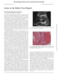
Fio. 2.—Pelvic radiograph showing a large destructive lesion in the<br />
right acetabular ro<strong>of</strong>, eroding into the hip joint.<br />
demonstrated. An enlarged <strong>iliopsoas</strong> bursa was considered<br />
the most likely diagnosis. Ultrasound <strong>of</strong> the abdomen showed<br />
an enlarged liver <strong>of</strong> homogenous texture and no abdominal<br />
masses. The spleen and kidneys were normal, and there was<br />
no para-aortic lymphadenopathy and no free peritoneal fluid.<br />
An X-ray <strong>of</strong> the pelvis revealed a destructive lesion in the<br />
right acetabular ro<strong>of</strong> which had eroded into the hip joint (Fig.<br />
2). Aspiration <strong>of</strong> the mass revealed clear fluid which contained<br />
only a few polymorphs and lymphocytes. Crystals<br />
were absent and culture failed to grow any organisms.<br />
Cytological examination <strong>of</strong> the fluid, however, revealed a<br />
number <strong>of</strong> atypical cells. Positive results from a series <strong>of</strong><br />
investigations revealed an ESR <strong>of</strong> 40 mm/h, a CRP <strong>of</strong> 42 mg/1<br />
and an alkaline phosphatase <strong>of</strong> 437 IU/ml (normal range<br />
< 115 IU/ml). The chest radiograph showed enlarged lung<br />
fields and flattened diaphragms with no hilar lymphadenopathy<br />
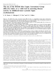
and no lung lesions. An MRI scan confirmed the presence<br />
<strong>of</strong> a distended <strong>iliopsoas</strong> bursa in communication with an<br />
effusion in the hip joint (Fig. 3). An isotope bone scan<br />
demonstrated multiple areas <strong>of</strong> abnormal uptake compatible<br />
with metastatic deposits in the right scapula, spine and ribs.<br />
Histology <strong>of</strong> tissue obtained by biopsy <strong>of</strong> a deposit in the<br />
scapula showed the tissue to be poorly differentiated adenocarcinoma,<br />
negative on immunocytochemistry for Prostrate<br />
Specific Antigen (PSA) and thyroglobulin. The site <strong>of</strong> the<br />
primary tumour was not identified. He was referred for<br />
palliative radiotherapy <strong>of</strong> the right hip.<br />
DISCUSSION<br />
The <strong>iliopsoas</strong> bursa is the largest synovial bursa in<br />
the body and the most important bursa around the hip.<br />
It is interposed between the <strong>iliopsoas</strong> muscle and the<br />
anterior surface <strong>of</strong> the capsule <strong>of</strong> the hip joint (Fig. 4).<br />
It is present bilaterally in 98% <strong>of</strong> adults [1]. The bursa<br />
extends from the inguinal ligament superiorly to the<br />
lesser trochanter <strong>of</strong> the femur, and is flanked by<br />
the femoral vein, artery and nerve. It lies adjacent<br />
FIG. 3.—MRJ scan. Axial T2 weighted image demonstrating a high signal fluid mass anterior to the right hip, and communication with an<br />
underlying hip effusion.<br />
Downloaded from<br />
http://rheumatology.oxfordjournals.org/<br />
by guest on April 10, 2013