iliopsoas bursitis—an unusual presentation of ... - Rheumatology
iliopsoas bursitis—an unusual presentation of ... - Rheumatology
iliopsoas bursitis—an unusual presentation of ... - Rheumatology

Create successful ePaper yourself
Turn your PDF publications into a flip-book with our unique Google optimized e-Paper software.
British Journal <strong>of</strong> <strong>Rheumatology</strong> 1996;35:285-288<br />
CASE REPORT<br />
ILIOPSOAS BURSITIS—AN UNUSUAL PRESENTATION OF METASTATIC<br />
BONE DISEASE<br />
P. A. C. BYRNE, J. I. S. REES* and B. D. WILLIAMS<br />
Department <strong>of</strong> <strong>Rheumatology</strong> & * Radiology, University Hospital <strong>of</strong> Wales, Heath Park, Cardiff<br />
SUMMARY<br />
Diagnostic uncertainty concerning the nature <strong>of</strong> an enlarging inguinal mass in an elderly male with a short history <strong>of</strong> hip pain<br />
was resolved by a combination <strong>of</strong> ultrasound and magnetic resonance imaging (MRI). Subsequent investigations showed that<br />
the enlarged <strong>iliopsoas</strong> bursa, which contained a number <strong>of</strong> atypical cells, was an <strong>unusual</strong> presenting feature <strong>of</strong> a destructive<br />
metastatic lesion in the right hip.<br />
KEY WORDS: Iliopsoas bursitis, Neoplasm, Metastatic carcinomatous arthritis.<br />
POINTS <strong>of</strong> potential friction between ligaments and the<br />
skin overlying bony prominences are <strong>of</strong>ten protected<br />
by lubricating bursae. The <strong>iliopsoas</strong> bursa lies lateral to<br />
the femoral vessels in a position between the capsule <strong>of</strong><br />
the hip joint and the <strong>iliopsoas</strong> muscles. A variety <strong>of</strong><br />
inflammatory and degenerative diseases <strong>of</strong> the hip joint<br />
can produce weakening <strong>of</strong> the capsule anteriorly,<br />
thereby allowing communication between the joint<br />
space and the adjacent <strong>iliopsoas</strong> bursa. The mode <strong>of</strong><br />
<strong>presentation</strong> <strong>of</strong> <strong>iliopsoas</strong> bursitis reflects the direction<br />
and magnitude <strong>of</strong> enlargement <strong>of</strong> the bursa and it<br />
can therefore present as hip pain, an enlarging mass in<br />
the groin or may cause symptoms which are the result<br />
<strong>of</strong> compression <strong>of</strong> structures closely applied to the<br />
enlarging bursa. Even in specialist musculoskeletal units,<br />
the clinical diagnosis <strong>of</strong> this condition is <strong>of</strong>ten not made<br />
and its many and varied <strong>presentation</strong>s lead therefore to<br />
unnecessary investigation and inappropriate treatment.<br />
CASE REPORT<br />
A 77-yr-old man with chronic rheumatoid arthritis presented<br />
with a 3 month history <strong>of</strong> increasingly severe right hip<br />
pain and a 1 month history <strong>of</strong> an enlarging mass in the right<br />
groin. He was anorexic and had lost 101b in weight during<br />
the preceding 10 months. Rheumatoid arthritis had been<br />
diagnosed 40 yr previously; it initially required second-line<br />
therapy, although during the last 5 yr the disease had been<br />
quiescent and required only symptomatic therapy. The<br />
patient gave a long history <strong>of</strong> exertional dyspnoea and he had<br />
smoked heavily for over 50 yr.<br />
He walked with an antalgic gait. Examination <strong>of</strong> the right<br />
hip revealed flexion to 100°, but extension was limited and no<br />
internal or external rotation was possible. A fluctuant nonpulsatile,<br />
non-tender mass (5* x 3") was present in the right<br />
groin. No cough impulse was palpable and bowel sounds<br />
were not heard over his mass. The femoral artery was<br />
palpable medially over the mass and a bruit was audible<br />
over the artery. Abdominal examination revealed a<br />
smooth-edged liver enlarged to four finger breadths due to<br />
associated emphysema and chronic obstructive airways disease.<br />
He had the typical changes <strong>of</strong> nodular rheumatoid<br />
Submitted 27 June 1995; revised version accepted 28 September<br />
1995.<br />
285<br />
arthritis. Clubbing, lymphadenopathy, and signs <strong>of</strong> anaemia<br />
and liver disease were all absent. A firm clinical diagnosis was<br />
not made at this time.<br />
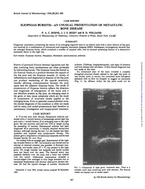
An ultrasound scan <strong>of</strong> the mass revealed a complex<br />
elongated structure closely related to the right hip joint. It<br />
was mostly cystic in nature, but contained some echogenic<br />
elements with no flow on Doppler to suggest an aneurysm<br />
(Fig. 1). An effusion within the hip joint could not be<br />
FIG. 1.—Ultrasound <strong>of</strong> right groin, transverse scan. There is a<br />
complex cystic mass containing some solid elements. The femoral<br />
vessels are located over its medial aspect.<br />
© 1996 British Society for <strong>Rheumatology</strong><br />
Downloaded from<br />
http://rheumatology.oxfordjournals.org/<br />
by guest on April 10, 2013
286 BRITISH JOURNAL OF RHEUMATOLOGY VOL. 35 NO. 3<br />
Fio. 2.—Pelvic radiograph showing a large destructive lesion in the<br />
right acetabular ro<strong>of</strong>, eroding into the hip joint.<br />
demonstrated. An enlarged <strong>iliopsoas</strong> bursa was considered<br />
the most likely diagnosis. Ultrasound <strong>of</strong> the abdomen showed<br />
an enlarged liver <strong>of</strong> homogenous texture and no abdominal<br />
masses. The spleen and kidneys were normal, and there was<br />
no para-aortic lymphadenopathy and no free peritoneal fluid.<br />
An X-ray <strong>of</strong> the pelvis revealed a destructive lesion in the<br />
right acetabular ro<strong>of</strong> which had eroded into the hip joint (Fig.<br />
2). Aspiration <strong>of</strong> the mass revealed clear fluid which contained<br />
only a few polymorphs and lymphocytes. Crystals<br />
were absent and culture failed to grow any organisms.<br />
Cytological examination <strong>of</strong> the fluid, however, revealed a<br />
number <strong>of</strong> atypical cells. Positive results from a series <strong>of</strong><br />
investigations revealed an ESR <strong>of</strong> 40 mm/h, a CRP <strong>of</strong> 42 mg/1<br />
and an alkaline phosphatase <strong>of</strong> 437 IU/ml (normal range<br />
< 115 IU/ml). The chest radiograph showed enlarged lung<br />
fields and flattened diaphragms with no hilar lymphadenopathy<br />
and no lung lesions. An MRI scan confirmed the presence<br />
<strong>of</strong> a distended <strong>iliopsoas</strong> bursa in communication with an<br />
effusion in the hip joint (Fig. 3). An isotope bone scan<br />
demonstrated multiple areas <strong>of</strong> abnormal uptake compatible<br />
with metastatic deposits in the right scapula, spine and ribs.<br />
Histology <strong>of</strong> tissue obtained by biopsy <strong>of</strong> a deposit in the<br />
scapula showed the tissue to be poorly differentiated adenocarcinoma,<br />
negative on immunocytochemistry for Prostrate<br />
Specific Antigen (PSA) and thyroglobulin. The site <strong>of</strong> the<br />
primary tumour was not identified. He was referred for<br />
palliative radiotherapy <strong>of</strong> the right hip.<br />
DISCUSSION<br />
The <strong>iliopsoas</strong> bursa is the largest synovial bursa in<br />
the body and the most important bursa around the hip.<br />
It is interposed between the <strong>iliopsoas</strong> muscle and the<br />
anterior surface <strong>of</strong> the capsule <strong>of</strong> the hip joint (Fig. 4).<br />
It is present bilaterally in 98% <strong>of</strong> adults [1]. The bursa<br />
extends from the inguinal ligament superiorly to the<br />
lesser trochanter <strong>of</strong> the femur, and is flanked by<br />
the femoral vein, artery and nerve. It lies adjacent<br />
FIG. 3.—MRJ scan. Axial T2 weighted image demonstrating a high signal fluid mass anterior to the right hip, and communication with an<br />
underlying hip effusion.<br />
Downloaded from<br />
http://rheumatology.oxfordjournals.org/<br />
by guest on April 10, 2013
Iliopsoas<br />
Bursa<br />
BYRNE ET AL.: ILIOPSOAS BURSITIS 287<br />
Pectineus<br />
FKJ. 4.—Vertical section through the hip joint, demonstrating the path along which the <strong>iliopsoas</strong> bursa enlarges (black arrow). N = femoral<br />
nerve; A «= femoral artery, V «• femoral vein; L — lymphatics/femoral canal.<br />
to the thinnest part <strong>of</strong> the hip joint capsule, and a<br />
defect in this capsule will allow communication between<br />
the hip joint and the bursa. Such a defect is<br />
common and has been observed in 14% <strong>of</strong> normal<br />
cadavers [1]. It is felt to be an acquired condition since<br />
this finding was not present in children. Raised intraarticular<br />
pressure, friction from the overlying <strong>iliopsoas</strong><br />
tendon, or a weakening <strong>of</strong> the capsule may all contribute<br />
to the development <strong>of</strong> communication between the<br />
hip joint and the <strong>iliopsoas</strong> bursa [2]. Under these<br />
circumstances, the <strong>iliopsoas</strong> bursa acts as a volume<br />
reservoir for the hip joint and is sometimes referred to<br />
as a synovial cyst. Iliopsoas bursitis is usually associated<br />
with both inflammatory and degenerative diseases<br />
<strong>of</strong> the hip joint, the conditions most commonly associated<br />
with it are rheumatoid arthritis [3] and osteoarthritis<br />
[4]. Rarely, cases due to synovial<br />
chondromatosis [5], avascular necrosis [6] or tuberculosis<br />
have been described. In most instances, a long<br />
duration <strong>of</strong> hip disease is typical [7]. In the absence <strong>of</strong><br />
hip disease, inflammation and enlargement <strong>of</strong> the <strong>iliopsoas</strong><br />
bursa may also occur as a result <strong>of</strong> trauma or<br />
sports injuries associated with vigorous hip flexion and<br />
extension [8].<br />
One <strong>of</strong> the difficulties involved in arriving at<br />
the correct clinical diagnosis <strong>of</strong> <strong>iliopsoas</strong> bursal<br />
enlargement is that the symptoms are both non-specific<br />
and varied, consisting mainly <strong>of</strong> pain, a palpable groin<br />
mass or lower limb swelling. The mode <strong>of</strong> <strong>presentation</strong><br />
is <strong>of</strong>ten determined by the magnitude and direction <strong>of</strong><br />
enlargement. A palpable groin mass has a broad<br />
differential diagnosis, whilst a retroperitoneal extension<br />
<strong>of</strong> the bursa may confuse the physician by presenting<br />
as either a palpable abdominal or pelvic mass [9],<br />
or may produce clinical signs which are due to<br />
compression <strong>of</strong> lower limb lymphatics, the femoral<br />
vein or femoral nerve [10]. Rarely, displacement <strong>of</strong><br />
abdominal viscera occurs [11]. Although anterior hip<br />
pain is the earliest and most frequent symptom associated<br />
with <strong>iliopsoas</strong> bursal enlargement, the pain may<br />
also be referred to the abdomen, the thigh or knee joint.<br />
Occasionally, the bursa may undergo rupture or<br />
become infected.<br />
The diagnosis, as in this case, is best achieved by<br />
a combination <strong>of</strong> ultrasound and MRI scanning.<br />
Ultrasound is the modality <strong>of</strong> choice for demonstrating<br />
the predominantly cystic nature <strong>of</strong> the mass, and its<br />
relationship to the femoral vessels. It may occasionally<br />
reveal a hip effusion, and can be used to guide needle<br />
aspiration <strong>of</strong> the bursa. The clear advantage <strong>of</strong> MRI is<br />
that it provides optimal anatomical definition, and can<br />
demonstrate the communication between the bursa and<br />
joint cavity, thus precluding the need for arthrography<br />
or bursography. CT scanning is also appropriate, but<br />
it is inferior to MRI in demonstrating an effusion in the<br />
hip joint. Plain radiographs are mandatory for the<br />
assessment <strong>of</strong> underlying articular disease <strong>of</strong> the hip.<br />
The contribution <strong>of</strong> MRI here adds to the list <strong>of</strong><br />
conditions in rheumatology where MRI plays an<br />
important diagnostic role [12].<br />
Our case is <strong>of</strong> particular interest because an<br />
enlarging painful <strong>iliopsoas</strong> bursa was the presenting<br />
feature <strong>of</strong> metastatic disease. Previous associations<br />
with malignant disease consist only <strong>of</strong> one case report<br />
where <strong>iliopsoas</strong> bursitis was associated with a primary<br />
chondrosarcoma [13]. A Medline search from 1966 has<br />
failed to find other associations. The plain radiographic<br />
finding <strong>of</strong> a destructive lesion in the ro<strong>of</strong> <strong>of</strong> the (right)<br />
acetabulum and the presence <strong>of</strong> atypical cells within<br />
bursal fluid indicate spread <strong>of</strong> malignant cells from<br />
the metastatic deposit into the joint space and bursa.<br />
Pre-existing inflammation associated with rheumatoid<br />
Downloaded from<br />
http://rheumatology.oxfordjournals.org/<br />
by guest on April 10, 2013
288 BRITISH JOURNAL OF RHEUMATOLOGY VOL. 35 NO. 3<br />
arthritis may have already produced direct communication<br />
between the hip joint and the bursa, therefore<br />
facilitating the rapid increase in the size <strong>of</strong> the bursa<br />
seen in our patient. The clinical diagnosis was not made<br />
prior to investigation, but ultrasonography quickly<br />
suggested the correct diagnosis, and the MRI scan<br />
demonstrated the communication with an effusion <strong>of</strong><br />
the hip and its aetiology.<br />
Although infrequent documentation <strong>of</strong> isolated<br />
cases suggests a low prevalence, <strong>iliopsoas</strong> bursitis<br />
may be more common since the recent application <strong>of</strong><br />
diagnostic imaging studies has led to increased<br />
awareness <strong>of</strong> this condition. To most clinicians,<br />
including those specializing in musculoskeletal<br />
disorders, the condition remains unfamiliar and because<br />
its <strong>presentation</strong> can be so varied it is <strong>of</strong>ten not<br />
considered as a cause <strong>of</strong> symptoms involving the<br />
inguinal area, the lower limb and abdomen. Ultrasound<br />
is the quickest and most cost-effective way<br />
<strong>of</strong> demonstrating the bursa and its contents, whilst<br />
CT and MRI provide a better appreciation <strong>of</strong> the<br />
regional anatomy and associated hip joint disease. The<br />
appropriate application <strong>of</strong> these modalities may<br />
prevent unnecessary investigation and inappropriate<br />
treatment.<br />
REFERENCES<br />
1. Chandler SB. The <strong>iliopsoas</strong> bursa in man. Anal Rec<br />
1934^8:235.<br />
2. Gerber NJ, Dixon ASU. Synovia! cysts and juxta<br />
articular bone cysts (Geodcs). Semin Arthritis Rheum<br />
Dis 974;3:323-48.<br />
3. Coventry MB, Polley HF, Weiner AD. Rheumatoid<br />
synovial cyst <strong>of</strong> the hip. / Bone Joint Surg 1959;<br />
41A:721-30.<br />
4. Melamed M, Bauer CA, Johnston JH. Iliopsoas bursal<br />
extension <strong>of</strong> arthritic disease <strong>of</strong> the hip. Radiology<br />
1967;89:54-8.<br />
5. Eisenberg KS, Johnston JO. Synovial chondromatosis <strong>of</strong><br />
the hip joint presenting as an intra-pelvic mass. J Bone<br />
Joint Surg 1972;54A: 176-8.<br />
6. Cohen JM, Hodges SC, Weinreb JC. MR imaging <strong>of</strong><br />
<strong>iliopsoas</strong> bursitis and concurrent avascular necrosis<br />
<strong>of</strong> the femoral head. J Computer Assisted Tomography<br />
1985;9:969-71.<br />
7. Meaney JF, Cassar-Pullicino BN, Etherington R.<br />
Iliopsoas bursa enlargement. Clin Radiol 1992;45: 161-8.<br />
8. O'Connor DS. Early recognition <strong>of</strong> iliopectineal bursitis.<br />
Surg Gynecol Obstet 1933^7:674-84.<br />
9. Eisenberg KS, Johnston JO. Synovial chondromatosis <strong>of</strong><br />
the hip joint presenting as an intra-pelvic mass. J Bone<br />
Joint Surg 1972;54A: 176-8.<br />
10. Lavyne MH, Voorhies RM, Coll RH. Femoral neuropathy<br />
caused by an <strong>iliopsoas</strong> bursa cyst. J Neurosurg<br />
1982;56:584-6.<br />
11. Watson JD, Ochsner SF. Compression <strong>of</strong> bladder due<br />
to rheumatoid cysts <strong>of</strong> the hip joint. Am J Roentgenol<br />
1967;99:695-6.<br />
12. Nissenbaum MA, Adamis MK. Magnetic resonance<br />
imaging in rheumatology. Rheum Dis Clin North Am<br />
1994;20:343-60.<br />
13. Toohey AK, Lasalle TL, Martinos S. Iliopsoas bursitis<br />
clinical features radiographic findings and disease associations.<br />
Semin Arthritis Rheum 1990;20:41-7.<br />
Downloaded from<br />
http://rheumatology.oxfordjournals.org/<br />
by guest on April 10, 2013