Clinical and Technical Review - Tecomedical
Clinical and Technical Review - Tecomedical
Clinical and Technical Review - Tecomedical

You also want an ePaper? Increase the reach of your titles
YUMPU automatically turns print PDFs into web optimized ePapers that Google loves.
Author:<br />
Peter<br />
Haima, Ph<br />
<strong>Clinical</strong> <strong>and</strong><br />
<strong>Technical</strong> <strong>Review</strong><br />
Bone & Cartilage Metabolism<br />
Cell culture<br />
Diagnostic <strong>and</strong> therapy control<br />
Animal models
2<br />
Bone turnover Biomarker in<br />
Osteoporosis<br />
Diagnosis of metabolic bone diseases can be established<br />
by measuring bone mineral density (BMD) at the hips or spine.<br />
However, BMD is a static measure of bone composition,<br />
reflecting its history. A baseline BMD value does not offer<br />
any prediction of future bone loss or response to therapy.<br />
Moreover, since biochemical bone marker reflect the wholebody<br />
rates of bone turnover, the combined measurement of<br />
bone marker <strong>and</strong> BMD provides more information on the<br />
overall bone loss than BMD measurement at specific skeletal<br />
sites alone.<br />
The increasing number of drugs available for treatment of<br />
osteoporosis <strong>and</strong> other bone diseases requires the use of<br />
more rapid <strong>and</strong> predictive methods to assess therapy efficacy.<br />
While detectable <strong>and</strong> significant changes in BMD take 18 to<br />
24 months to develop, bone turnover marker have been<br />
shown to detect changes in bone tissue within 3-6 months<br />
after starting anti-resorptive therapy. Therefore, measurement<br />
of bone turnover marker is increasingly recommended<br />
as a key component of therapy management: to rapidly<br />
identify therapy responders <strong>and</strong> non-responders, to assess<br />
therapy efficacy <strong>and</strong> to determine the optimal therapy <strong>and</strong><br />
dose of treatment.<br />
Bone marker are intended for use as an<br />
aid in:<br />
• management of postmenopausal osteoporosis <strong>and</strong><br />
Paget’s disease;<br />
• monitoring of postmenopausal women on hormonal<br />
or bisphosphonate therapy;<br />
• prediction of skeletal response to hormonal therapy<br />
in postmenopausal women.<br />
• Detection <strong>and</strong> monitoring of Bone metastasis.<br />
Biomarker <strong>and</strong> Cartilage Destruction<br />
Degenerative joint disease such as osteoarthritis (OA) <strong>and</strong><br />
rheumatoid arthritis (RA) are major causes of disability <strong>and</strong><br />
impaired quality of life among the elderly. Despite better<br />
underst<strong>and</strong>ing of the underlying pathomechanisms, current<br />
clinical practice for diagnosing these diseases mainly relies<br />
on symptom assessment, which has limited sensitivity <strong>and</strong><br />
specificity. By the time radiological techniques can reveal<br />
specific sign, the disease has advanced to a late phase with<br />
pronounced structural damage, when the only therapeutical<br />
option remaining is joint replacement. As marker provide a<br />
dynamic measure of current degradation rate in contrast to<br />
radiological investigations, they allow a rapid determination<br />
of the therapeutic effects, <strong>and</strong> they may also be used for<br />
selecting the therapies <strong>and</strong> drug dose, which are most efficient.<br />
In the lack of established diagnostic methods allowing<br />
the detection of cartilage degradation, early diagnosis<br />
<strong>and</strong> disease monitoring remain a major challenge of health<br />
care professionals.<br />
Therefore Biochemical marker for Cartilage degradation <strong>and</strong><br />
synthesis are helpful for the management of Rheumatoid<br />
Arthritis <strong>and</strong> Osteoarthritis.<br />
Biomarker for diagnosing skeletal<br />
metastases<br />
Cancers have the ability to metastasize to organs distant<br />
from the site of the primary tumor. Breast, prostate,<br />
<strong>and</strong> lung cancer are primary tumors that most frequently<br />
metastasize to the skeleton. The chronic presence of bone<br />
metastasis often results in complete pathologic remodeling<br />
of the affected bone compartment, making affected bones<br />
vulnerable to several complications. Such complications<br />
include pathologic fractures, spinal cord compression,<br />
hypocalcemia, <strong>and</strong> severe bone pain. All reducing quality of<br />
life <strong>and</strong> worsening prognosis. Therefore, early diagnosis <strong>and</strong><br />
adequate treatment of bone metastasis is critically important<br />
for the clinical management of cancer patients.<br />
Emerging evidence suggests that biomarker of bone<br />
turnover carry notable potentials to become useful<br />
diagnostic tools for the diagnosis of bone metastases.<br />
The study by Leeming et al was sought to assess the<br />
relative use of eight biomarker for the detection of bone<br />
metastases in cancer forms frequently spreading to the skeleton.<br />
Participants were 161 patients with either breast, prostate,<br />
or lung cancer. The presence <strong>and</strong> extent of<br />
bone metastases was assessed by imaging techniques<br />
(computer tomography <strong>and</strong>/or magnetic resonance<br />
imaging) <strong>and</strong> Technetium-99m scintigraphy. Number of<br />
bone metastases was recorded, <strong>and</strong> the skeletal load was<br />
graded, as previously proposed by Soloway. The Serum<br />
or urinary level of the bone resorption marker (alpha-CTX,<br />
beta-CTX, NTX, <strong>and</strong> ICTP), formation marker (BSAP),<br />
<strong>and</strong> osteoclastogenesis marker (osteoprotegerin, RANKL,<br />
<strong>and</strong> TRAP5b) was measured by commercially available<br />
immunoassays. There was a uniform pattern of significantly<br />
increased levels of the resorption marker in patients with<br />
bone metastases compared with those without.<br />
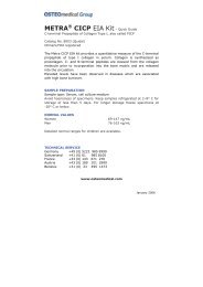
The relative responsiveness of marker was calculated to<br />
obtain insights into the relative sensitivity of the different<br />
marker to signal skeletal involvement. The plot demonstrates<br />
a trend toward greater relative increases in the level of the<br />
urine alpha-CTX <strong>and</strong> serum BSAP marker compared with<br />
other marker; differences becoming more evident with<br />
increasing Soloway score.
Figure 1:<br />
Soloway 0 = Patients without bone metastasis<br />
Soloway 1 = Patients with < 6 bone metastases<br />
Soloway 2 = Patients with < 20 bone metastases<br />
Soloway 3 = Patients with > 20 bone metastases but less<br />
than a “super scan”<br />
Soloway 4 = Patients with “super scan” that is defined by<br />
a > 75 % involvement of the ribs, vertebrae<br />
<strong>and</strong> pelvic bones<br />
Relative increases in bone resorption, bone formation, <strong>and</strong><br />
osteoclastogenesis marker as a function of the extent of skeletal<br />
involvement assessed in 132 breast <strong>and</strong> prostate cancer<br />
patients. Relative increases are expressed as percentage of<br />
levels in patients with Soloway score 0 (1) .<br />
Currently, the diagnosis of bone metastasis in cancer<br />
patients relies predominantly on imaging techniques,<br />
such as plain radiography or Technetium-99 scintigraphy.<br />
Although scintigraphy is more sensitive than plain<br />
radiography <strong>and</strong> even can give quantitative information<br />
regarding skeletal involvement, this examination is also<br />
more expensive, invasive, time-consuming <strong>and</strong> exposes<br />
cancer patients to irradiation, limiting its use for monitoring<br />
purposes. Thus, these weaknesses of current methodologies<br />
point out an unmet need for establishing supplementary<br />
diagnostic tools like biochemical marker.<br />
Regulators of bone turnover<br />
Bone remodeling is an ongoing dynamic process<br />
consisting of bone resorption (due to osteoclasts digesting<br />
type I collagen) <strong>and</strong> bone formation (due to osteoblasts).<br />
Normally, these processes are balanced, resulting in 10 %<br />
replacement of the skeleton, each year. However, due to<br />
aging, disease or other conditions, bone turnover may<br />
become imbalanced where bone resorption <strong>and</strong> formation<br />
occur at different rates.<br />
OPG (Osteoprotegerin), also known as osteoclast inhibiting<br />
factor (OCIF), inhibits the differentiation <strong>and</strong> activation of<br />
osteoclasts.<br />
On the other h<strong>and</strong>, sRANKL (soluble receptor activator of<br />
nuclear factor (NF)-κB lig<strong>and</strong>) is the main stimulator for the<br />
formation of mature osteoclasts. Stimulation occurs through<br />
binding of sRANKL to the osteoclastic membrane receptor<br />
RANK.<br />
OPG inhibits the binding of sRANKL to RANK <strong>and</strong> thus<br />
the activation of osteoclasts. The OPG/sRANKL system is<br />
therefore a key regulator of bone resorption. Abnormalities<br />
in the balance of the OPG/sRANKL system may be the<br />
cause of bone loss in many metabolic bone diseases as<br />
osteoporosis, Paget’s disease, metastatic cancers <strong>and</strong><br />
rheumatic bone degradation.<br />
In normal healthy people, sRANKL levels are generally low<br />
because the majority is bound by OPG. Decreased levels of<br />
OPG <strong>and</strong>/or increased levels of sRANKL may indicate an<br />
imbalance of the OPG/sRANKL system.<br />
OPG <strong>and</strong>/or sRANKL measurements can<br />
be used as an aid in determining the cause<br />
of bone loss by assessing imbalances in<br />
the OPG/sRANKL system in:<br />
• Post menopausal osteoporosis<br />
• Paget’s disease<br />
• Metastatic cancer<br />
• Diseases with locally increased resorption activity<br />
• Indicating bone loss in rheumatoid arthritis<br />
• Therapy monitoring after treatment with OPG<br />
• Determining an imbalance in vascular calcification (high<br />
OPG was associated with cardiovascular- <strong>and</strong> all-cause<br />
mortality in hemodialysis patients; Morena et al. 2006).<br />
• Metastatic Renal Cell Carcinoma (OPG)<br />
3
4<br />
Bone Formation<br />
Osteoblast:<br />
The osteoblast is the bone cell responsible for:<br />
1) The formation <strong>and</strong> organization of the extracellular matrix<br />
(ECM) of bone <strong>and</strong> its subsequent mineralization;<br />
2) Synthesis of collagen <strong>and</strong> other bone proteins. Three<br />
periods are distinguished in the osteoblast life cycle.<br />
A) Cell proliferation: genes associated with formation of the<br />
ECM, like Type I Collagen, are expressed <strong>and</strong> gradually<br />
down regulated.<br />
B) ECM maturation: proteins associated with the osteoblast<br />
phenotype are expressed, like Bone-specific Alkaline<br />
Phosphatase (BAP).<br />
C) ECM mineralization: BAP gene expression declines,<br />
Bone Sialo Protein (BSP), osteopontin <strong>and</strong> osteocalcin<br />
gene expression increase.<br />
Bone matrix:<br />
Consists of Type 1 collagen (90 % of the protein in bone),<br />
osteocalcin, osteopontin, osteonectin, proteoglycans,<br />
alkaline phosphatase <strong>and</strong> bone sialo protein.<br />
Proteins<br />
OPG:<br />
OPG (Osteoprotegerin) or OCIF (Osteoclast Inhibiting<br />
Factor) or OBF (Osteoclast Binding Factor) is a key factor in<br />
inhibition of osteoclast differentiation <strong>and</strong> activity.<br />
It binds <strong>and</strong> acts as as decoy receptor for s-RANKL.<br />
sRANKL:<br />
Soluble Receptor Activator of Nuclear factor (NF)-κB Lig<strong>and</strong>.<br />
sRANKL binds to osteoclast receptor: RANK (NF-κB ) <strong>and</strong> is<br />
the main stimulatory factor for the formation of mature<br />
osteoclasts.<br />
BAP:<br />
Bone-specific Alkaline Phosphatase is an osteoblastic enzyme<br />
involved in bone formation. It is assumed that BAP plays<br />
a role in ECM maturation. BAP is a key biochemical bone<br />
marker used for assessing bone turnover <strong>and</strong><br />
monitoring therapy.<br />
Bone Sialoprotein (BSP):<br />
Major structural protein of the bone matrix, expression of<br />
BSP is normally restricted to mineralized connective tissues<br />
of bones <strong>and</strong> teeth. This role has been associated with<br />
mineral crystal formation.<br />
Osteocalcin:<br />
Major structural protein of the bone matrix, binds calcium<br />
<strong>and</strong> attracts osteoclasts.<br />
Osteonectin:<br />
Protein that binds calcium <strong>and</strong> is involved in regulation<br />
of mineralization.<br />
Osteopontin:<br />
Cell-binding protein that anchors osteoclasts to<br />
mineralised matrix.<br />
Proteoglycans:<br />
Monomer looks like test tube brush with keratan <strong>and</strong><br />
chondroitin sulphate chains (= GAGs) bound to a protein<br />
core molecule. Monomers are attached via a link protein<br />
to hyaluronic acid.<br />
DKK-1:<br />
Dickkopf-1(DKK-1) is a 28,672 Da secreted protein that acts<br />
as soluble inhibitor of the WNT signalling pathway. DKK-1<br />
regulates different developmental processes <strong>and</strong> is also<br />
involved in the regulation of bone metabolism as it inhibits<br />
the differentiation of osteoblast.<br />
Collagen metabolites <strong>and</strong> epitopes<br />
Type I Procollagen:<br />
Secreted precursor of Type I Collagen. Extracellular cleavage<br />
results in N- <strong>and</strong> C-terminal propeptides.<br />
PINP:<br />
Epitope of N- terminal propeptide, released during cleavage<br />
of Type I Procollagen.<br />
CICP/PICP:<br />
Epitope of C- terminal propeptide, released during cleavage<br />
of Type I Procollagen (PICP = CICP).<br />
Type I Collagen:<br />
Collagen molecules consist of three chains to form a triple<br />
helix. Crosslinks between the chains <strong>and</strong> the molecules of<br />
collagen give collagen its strength.
Bone Resorption<br />
Osteoclast:<br />
Large motile, multinucleated bone cell located on bone surfaces.<br />
Responsible for the resorption of bone matrix (osteoid).<br />
Osteoclasts have a ruffled border of the cell membrane that<br />
is surrounded by an organelle-free region, or « clear zone ».<br />
Mineral Dissolution:<br />
These processes take place beneath the ruffled border<br />
<strong>and</strong> depend on lysosomal enzyme secretion <strong>and</strong> an acid<br />
microenvironment. A pH gradient across the ruffled<br />
membrane is the consequence of active transport<br />
mechanisms by the osteoclasts.<br />
Collagen degradation:<br />
Osteoclasts actively synthesize lysosomal enzymes,<br />
in particular the tartrate resistant isoenzyme of acid<br />
phosphatase (TRAP 5b) <strong>and</strong> cysteine-proteinases such<br />
as Cathepsin K that are capable of degrading collagen.<br />
Two bone collagenolytic pathways exist:<br />
1. A Cathepsin K collagen degradation pathway is the prevailing<br />
one. Resulting in the following epitopes:<br />
CTX, NTX, DPD <strong>and</strong> PYD.<br />
2. A Matrix MetalloProteinase (MMP) collagen degradation<br />
pathway resulting in the epitope ICTP. This pathway becomes<br />
significant in some situations, including metastatic<br />
bone diseases <strong>and</strong> multiple myeloma. ICTP however, has<br />
proven to be a very poor marker in osteoporosis.<br />
Proteins<br />
TRAP 5b:<br />
The active isoform of TRAP5b, serum b<strong>and</strong> 5 tartrateresistant<br />
acid phosphatase, is specifically synthesized<br />
by bone-resorbing osteoclasts. It has been shown that<br />
TRAP5b catalyzes the formation of reactive oxygen<br />
species (ROS). Research results indicate that ROS<br />
generated by TRAP5b are involved in the degradation<br />
of bone matrix products in resorbing osteoclasts.<br />
TRAP5b activity reflects the osteoclast activity <strong>and</strong> is associated<br />
with the number of osteoclasts. TRAP5b is consideredamarkerfortherateofboneresorption<strong>and</strong>maybeof<br />
particular importance for patients with renal failure because<br />
- in contrast to other marker of resorption -<br />
TRAP5b does not accumulate in the blood.<br />
In tumor patients TRAP5b is an indicator for increased bone<br />
resorption due to bone metastases. Changes in TRAP<br />
activity during therapy monitoring allow the assessment of<br />
the efficiency of antiresorptive therapies in osteoporotic<br />
patients.<br />
RANK:<br />
Osteoclastic receptor for sRANKL, the main stimulatory factor<br />
for the formation of mature osteoclasts.<br />
Cathepsin K:<br />
Main osteoclastic protease, responsible for bulk degradation<br />
of Type I Collagen. Acts both intra- <strong>and</strong> extra-cellularly.<br />
Lysosomal enzymes:<br />
Enzymes secreted by the osteoclasts, responsible for<br />
mineral dissolution in an acid environment.<br />
MMP:<br />
Matrix metalloproteinases MMP-2, -9, -13, -14 participate in<br />
the degradation of the collagenous bone matrix,<br />
resulting in epitope ICTP.<br />
Calcitonin receptor:<br />
Osteoclastic receptor for calcitonin, an inhibitor of<br />
osteoclasts activity.<br />
Sclerostin:<br />
Sclerostin is the protein product of the SOST gene, which is<br />
located at 17q12-21 <strong>and</strong> highly conserved across vertebrate<br />
species.<br />
The highest expression of sclerostin throughout the adult<br />
skeleton has been observed in hypertrophic chondrocytes<br />
<strong>and</strong> osteocytes. Sclerostin belongs to the DAN1 family of<br />
glycoproteins of which multiple family members antagonize<br />
bone morphogenetic protein (BMP) <strong>and</strong>/or Wnt2 activity.<br />
Sclerostin blocks canonical Wnt signaling by binding to the<br />
Wnt coreceptors LRP5/63 . Thus, it inhibits bone formation<br />
by regulating osteoblast function <strong>and</strong> promoting osteoblast<br />
apoptosis. By blocking the Wnt-pathway Sclerostin also<br />
antagonises bone morphogenetic protein action e.g. osteoblast<br />
differentiation, but does not inhibit direct BMP-induced<br />
responses.<br />
Sclerostin expression is down-regulated by Parathyroid hormone<br />
(PTH) as well as the mechanical stimulation of bone<br />
reduces the expression of sclerostin.<br />
1 DAN = differential screening-selected gene aberrative in neuroblastomaa<br />
2 Wnt = wingless Proteine<br />
3 LRP = low-density lipoprotein receptor-related protein 5 <strong>and</strong> 6<br />
5
6<br />
Collagen metabolites <strong>and</strong> epitopes<br />
DPD:<br />
Deoxypyridinoline. Breakdown product from Type I collagen<br />
degradation.<br />
PYD:<br />
Pyridinoline. Breakdown product from Type I collagen<br />
degradation.<br />
NTX:<br />
Cross-linked N-terminal telopeptide resulting from<br />
Type I collagen degradation. NTX = new synthesized <strong>and</strong><br />
agemodified bone matrix.<br />
CTX:<br />
C-terminal telopeptide resulting from Type I collagen<br />
degradation, CTX alpha (New Bone Matrix), CTX beta (Old<br />
Bone Matrix).<br />
ICTP=CTX-MMP:<br />
Carboxy-terminal telopeptide of type I collagen.<br />
Larger epitope containing the smaller CTX epitope.<br />
Helical Peptide:<br />
Peptide derived from the helical region of the α-1 chain of<br />
Type 1 collagen.<br />
Note:<br />
The concentration of the different biomarker are partly<br />
dependent on age, gender, race as well as influenced by<br />
diurnal rhythm, food intake etc. (see table on page 8). Age<br />
dependency is of high importance if biomarker are used for<br />
preclinical testing, especially in juvenile laboratory animals<br />
(rat, mice).<br />
Bone turnover regulators<br />
Calcitonin:<br />
Inhibits bone resorption, directly acts on osteoclasts.<br />
Parathyroid hormone (PTH):<br />
Key factor in the maintenance of calcium <strong>and</strong> phosphate<br />
homeostasis. Stimulates osteoclasts activity.<br />
1,25(OH) 2D3 :<br />
1,25 dihydroxy vitamin D3 (or 1,25-dihydroxycholecalciferol)<br />
is the biologically active form of vitamin D. It is<br />
synthesized in the kidney from 25-vitamin D (25-hydroxycholecalciferol).<br />
1,25(OH) 2D3 is the principal regulator<br />
of calcium homeostasis in the body. It enhances the<br />
efficiency of calcium.<br />
Fetuin A:<br />
Glycoprotein synthesized by liver, secreted into blood.<br />
Deposited as noncollagenous protein in mineralized bones.<br />
Potent inhibitor of soft tissue (vascular) calcification by binding<br />
excess mineral in serum. Regulates calcium metabolism<br />
<strong>and</strong> osteogenesis.<br />
FGF-23:<br />
Fibroblast Growth Factor 23. Important regulator of<br />
phosphate homeostasis. Able to “block” renal reabsorption<br />
of Pi. FGF-23 abnormalities are involved in renal phos-phate<br />
wasting disorders leading to “hypophosphatemia”.<br />
Literature:<br />
1. Leeming DJ, Koizumi M, Byrjalsen I, Li B, Qvist P, Tanko LB.<br />
The relative use of eight collagenous <strong>and</strong> noncollagenous markers<br />
for diagnosis of skeletal metastases in breast, prostate, or<br />
lung cancer patients.<br />
Cancer Epidemiol Biomarkers Prev. 2006; 15(1):32-8.<br />
2. Caulfield MP, Reitz RE. Biochemical markers of bone turnover <strong>and</strong><br />
their utility in osteoporosis. MLO-online April 2004.<br />
Schaller S, Henriksen K, Hoegh-Andersen P, Søndergaard BC,<br />
Sumer EU, Tanko LB, Qvist P <strong>and</strong> Karsdal MA. In vitro, ex vivo, <strong>and</strong><br />
in vivo methodological approaches for studying<br />
therapeutic targets of osteoporosis <strong>and</strong> degenerative joint<br />
diseases: How biomarkers can assist? Assay And Drug<br />
Development Technologies 2005; 3:553-580.<br />
3. Delmas PD, Eastell R, Garnero P, Seibel MJ <strong>and</strong> Stephan J. The use<br />
of biochemical markers of bone turnover in osteoporosis.<br />
Osteoporosis Int 2000: Supp 6: S2-17.
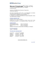
Bone Turnover – Biochemical Marker<br />
Sclerostin:<br />
inhibits differentiation <strong>and</strong><br />
function of osteoblasts<br />
through BMP<br />
DKK-1<br />
regultes osteoblast<br />
differentiation<br />
P *<br />
C<br />
I<br />
P<br />
MMPs<br />
BAP<br />
(bone-specific alkaline phosphatase)<br />
“calcification of the matrix”<br />
ICTP **<br />
ICP<br />
*<br />
P<br />
,<br />
P<br />
N<br />
I<br />
P<br />
Helical Peptide<br />
** Not so relevant in<br />
Osteoporosis<br />
Fetuin A<br />
* CICP = PICP<br />
7
8<br />
Bone marker <strong>and</strong> characteristics<br />
Biomarker Method Main<br />
Sample<br />
Type<br />
Formation<br />
BAP<br />
Osteocalcin<br />
Intact<br />
Osteocalcin<br />
N-mid<br />
PINP<br />
PICP = CICP<br />
Resorption<br />
ICTP =<br />
CTX-MMP<br />
Others<br />
ELISA<br />
IRMA/<br />
ELISA<br />
ELISA<br />
RIA<br />
ELISA<br />
Serum<br />
Serum<br />
Serum<br />
Serum<br />
Serum<br />
A(a) B C D E F G H<br />
Pre- &<br />
postmenopause<br />
+<br />
+<br />
+<br />
+<br />
NA<br />
Analytical<br />
variation<br />
+<br />
+<br />
+<br />
+<br />
+<br />
Treatment<br />
response<br />
Short<br />
time<br />
change<br />
Within<br />
person<br />
variation<br />
Daily<br />
variation<br />
Food<br />
intake<br />
TRAP 5b ELISA Serum ± + ± ± NA + + ±<br />
DPD ELISA Urine + + + + + - + +<br />
NTx ELISA<br />
CTx ELISA<br />
+<br />
+<br />
+<br />
+<br />
+<br />
Sample<br />
stability<br />
Scores sind: + (yes), - (no), ± (fair/indeterminate), NA (not available)<br />
(a) Letters correspond to following, the marker:<br />
H. preferably demonstrates high stability in the biological<br />
A. shows a difference in the rate of bone turnover pre-<strong>and</strong><br />
specimen<br />
post-menopause<br />
(b) In contrast to OPG, sRANKL showed no response to<br />
B. demonstrates minimal analytical variation<br />
bisphosphonate treatment of osteoporotic postmeno-<br />
C. significantly changes in response to treatment<br />
pausal women. However, PTH treatment of glucocorticoid<br />
D. detect changes in short time interval (months)<br />
induced osteoporotic women resulted in a fast (1 month)<br />
E. demonstrates minimal within person (biological) variation <strong>and</strong> sustained increase of sRANKL levels.<br />
F. preferably demonstrates little variation over the day<br />
(c) Cathepsin K is elevated in patients with established<br />
G. no influence by food intake<br />
rheumatoid arthritis.<br />
+<br />
+<br />
+<br />
+<br />
+<br />
+<br />
±<br />
±<br />
±<br />
+<br />
+<br />
+<br />
+<br />
+<br />
±<br />
+<br />
+<br />
+<br />
+<br />
+<br />
+<br />
-<br />
+<br />
+<br />
+<br />
5 days<br />
2–8 °C<br />
4hours<br />
2–8 °C<br />
5 days<br />
2–8 °C<br />
5 days<br />
2–8 °C<br />
5 days<br />
2–8 °C<br />
2 days<br />
2–8 °C<br />
7 days<br />
2–8 °C<br />
Serum + + + + + - ± ± 1day<br />
2–8 °C<br />
Urine + + + + ± - ± +<br />
3 days<br />
2–8 °C<br />
Serum + + + + + - - + 1day<br />
2–8 °C<br />
Urine + + + + ± - - +<br />
ELISA Serum - + - - NA ± + +<br />
7 days<br />
2–8 °C<br />
5 days<br />
2–8 °C<br />
sRANKL ELISA Plasma NA + ± (b) - NA NA + ± 1day<br />
2–8 °C<br />
Osteoprotegerin<br />
(OPG)<br />
ELISA Plasma NA + + - NA NA + ± 1day<br />
2–8 °C<br />
Cathepsine K ELISA Serum NA (c) + NA NA NA NA + ±<br />
2 days<br />
2–8 °C
Bone marker <strong>and</strong> their behavior in various bone diseases<br />
Biomarker CTX-I NTX-I Free<br />
DPD<br />
Bone Disease<br />
• ↑ Increased values of the biomarker in this disease were<br />
observed in various studies as compared to a healthy<br />
control group.<br />
• ↓ Decreased values of the biomarker in this disease were<br />
observed in various studies as compared to a healthy<br />
control group.<br />
• ↑ Large arrows: indicate large <strong>and</strong> significant differences.<br />
↑<br />
Small arrows: indicate small to medium differences<br />
with the control group.<br />
Degradation marker Formation marker<br />
Free<br />
PYD<br />
Helical<br />
peptide<br />
ICTP TRAP<br />
5b<br />
BAP OC PICP/<br />
CICP<br />
↑<br />
Osteoporosis Osteoporose ↑ ↑ ↑ ↑ ↑ ± + ± ↑ ↑ ↑<br />
↑<br />
Hyperparathyroidism Osteoporose ↑ ↑ ↑ ↑ ↑ ± ↑ + ↑ ↑ ↑<br />
↑<br />
Hyperthyroidism ↑ ↑ ↑ ↑ ↑ (↑)<br />
Bone metastases* ↑ ↑ ↑ ↑ ↑ ↑ (↑) ↑ ↑<br />
Multiple myeoloma ↑ ↑ ↑ ↑ ↓ ±<br />
Rickets/<br />
Vitamin-D Deficiency<br />
Osteomalacia<br />
“adult rickets”<br />
↑ ↑ ↑ ↑ ↓ ↑<br />
↑ ↑ ↑ ↑ ↑<br />
Paget´s ↑ ↑ ↑ ↑ ↑ ↑ ↑ ↑<br />
Anorexia nervosa (↑) (↑) (↓) (↓↑) (↓)<br />
Growth hormone<br />
deficiency<br />
↓ ↓ ↓<br />
Renal failure ↑ ↑ ↑ ↑ ↑ ↑<br />
PINP<br />
• (↓) (↑): arrows between brackets: different studies<br />
suggest inconsistent behavior of this marker in<br />
this disease.<br />
• ± indicates that according to various studies this marker<br />
is not considered as valuable for this disease.<br />
• Empty spaces indicate that no information was identified<br />
on this biomarker for this disease.<br />
* Increased values were observed for urin CTX alpha <strong>and</strong><br />
Serum BAP in bone metastases.<br />
9
10<br />
Cartilage Degradation<br />
Chrondocyte:<br />
Chondroblasts trapped in lacunae develop into chondrocytes.<br />
Chondrocytes are important in the control of matrix turnover<br />
through production of collagen, proteoglycans <strong>and</strong> enzymes<br />
for cartilage metabolism.<br />
Proteins<br />
Matrix metalloproteinases:<br />
Are involved in the cleavage of Type II Collagen <strong>and</strong> the proteoglycan<br />
aggrecan. Three collagenases (MMP-1, -8,-13) are<br />
mainly responsible for primary cleavage of Type II Collagen.<br />
MMP-1, -8, -13 <strong>and</strong> 14 are involved in cleavage of the core<br />
protein of aggrecan.<br />
Cathepsin K:<br />
Protease produced by synovial fibroblasts, enzyme<br />
plays critical role in cartilage degradation (together with<br />
matrix metalloproteinases). MMPs perform extracellular<br />
predigestion of collagen, after endocytosis of large<br />
fragments, Cathepsin K degrades collagen <strong>and</strong> aggrecan in<br />
acidic lysosomes.<br />
Aggrecanases:<br />
Enzymes involved in cleavage of aggrecan.<br />
COMP:<br />
Cartilage Oligomeric Matrix Protein is an abundant cartilage<br />
glycoprotein also found in tendon <strong>and</strong> other tissues.<br />
Synthesized by chondrocytes, synovial <strong>and</strong> other skeleton<br />
cells. Intact <strong>and</strong> fragmented COMP in synovial fluid or<br />
serum is correlating to cartilage degradation in OA <strong>and</strong> RA.<br />
Cartilage metabolites <strong>and</strong> epitopes<br />
CTX-II:<br />
A 6 amino acid sequence epitope of the nonhelical C- terminal<br />
telopeptides resulting from Type II collagen degradation.<br />
C2C:<br />
C2C or COL2-3/4CLong epitope that specifically appears<br />
into circulation when Type II collagen degradation occurs.<br />
C1,2C:<br />
C1, 2C or COL2-3/4CShort epitope that appears into circulation<br />
when Type II but also Type I collagen degradation occurs.<br />
CS-GAG:<br />
Chondroitin sulfate glycosaminoglycans are bound at high<br />
densities, to a core protein forming the cartilage proteoglycan<br />
aggrecan molecule.<br />
Cartilage Synthesis<br />
Cartilage metabolites <strong>and</strong> epitopes<br />
Type II Procollagen:<br />
Secreted precursor of Type II Collagen. Extracellular cleavage<br />
results in N- <strong>and</strong> C-terminal propeptides.<br />
CP-II:<br />
Epitope of C- terminal propeptide, released during<br />
maturation of Type II Procollagen to Type II Collagen.<br />
Type II Collagen:<br />
The principal structural component of cartilage is an extensive<br />
network of Type II collagen molecules, arranged in fibrils.<br />
Collagen molecules consist of three chains to form a triple<br />
helix. Crosslinks between the chains <strong>and</strong> the molecules of<br />
collagen give collagen its strength.<br />
Proteoglycan Aggrecan:<br />
Responsible for the compressive strength of cartilage. Serve<br />
to trap <strong>and</strong> hold water to regulate matrix hydration. Monomer<br />
looks like test tube brush with keratan <strong>and</strong> chondroitin<br />
sulphate chains (= GAGs) bound to a protein core molecule.<br />
Monomers are attached via a link protein to hyaluronic acid.<br />
CS-GAG:<br />
Chondroitin sulfate glycosaminoglycans are bound at high<br />
densities, to a core protein forming the cartilage proteoglycan<br />
aggrecan molecule.<br />
Aggrecan epitope CS 846:<br />
Chondroitin native epitope, present on intact bioactive<br />
(“fetal-like”) proteoglycan only.<br />
PIINP:<br />
Epitope of Type II N-terminal propeptide, released during<br />
maturation of Type II Procollagen to Type II Collagen. PIINP<br />
has been postulated to play a role in chondrogenesis. It has<br />
been found to be synthesized by osteoarthritic chondrocytes<br />
in diseased cartilage <strong>and</strong> may serve as a specific arthritis<br />
biomarker that reflects an attempt by the chondrocytes to<br />
repair diseased cartilage.<br />
Proteins<br />
BMP1:<br />
BMP1 (Bone morphogenetic protein) is a protein that is<br />
capable of inducing formation of cartilage in vivo. It cleaves<br />
the C-terminal propeptides of procollagen I, II, <strong>and</strong> III <strong>and</strong><br />
plays an important role in collagen maturation.
Biomarker according to NIH Osteoarthritis Biomarker Network<br />
Synovium<br />
Type III collagen<br />
Noncollagenous proteins<br />
Enzymes<br />
Aggrecan<br />
Type II collagen<br />
Cartilage<br />
Noncollagenous proteins<br />
Enzymes<br />
Proliferation/Formation Degradation<br />
PIIINP<br />
anti-CCP<br />
COMP<br />
Hyaluronan<br />
Pentosidine<br />
YKL-40<br />
MMPs<br />
TIMPs<br />
Chondroitin sulfate<br />
PIICP<br />
PIINP<br />
PIIANP<br />
YKL-40<br />
TIMPs<br />
Detailed information cartilage metabolism<br />
"Biomarker for diagnosis <strong>and</strong> monitoring of degenerative joint diseases" by Petra Seebeck, PhD,<br />
Glc-Gal-PYD<br />
PYD<br />
Keratan sulfate<br />
GAGs<br />
C1, 2C<br />
C2C<br />
COL2-1<br />
COL2-1NO2<br />
CTX-II<br />
HELIX II<br />
TIINE<br />
COMP<br />
CILP<br />
MMPs<br />
ADAMTSs<br />
Cathepsin K<br />
Inflammation marker – synovial metabolism<br />
Joint inflammation is a key feature of RA but can occur also during OA. Proliferative synovial inflammation leads to the<br />
forced syntheses of different marker. However, most of them are not synovium-specific, since they are also produced in<br />
cartilage <strong>and</strong> other tissues.<br />
anti-CCP<br />
Citrullin arises from enzymatic modification of the amino-acid arginine for instance during inflammatory processes.<br />
Auto-antibodies against cyclic citrullinated peptides (anti-CCP) were detected in up to 80%of RA patients already during a<br />
very early stage of disease, but only in very little patients with juvenile idiopathic arthritis (JIA). The concentrations of anti-CCP<br />
were not correlated to the disease activity score (DAS28), but to radiographic signs of joint destruction. Anti-rheumatic<br />
immunosuppressive therapy (Infliximab) reduced the sanguineous anti-CCP level in RA patients.<br />
Hyaluronan<br />
Hyaluronan is a main component of the cartilage matrix as well as the synovial fluid. In patients with knee OA the serological<br />
hyaluronan level correlated with the degree of synovial proliferation <strong>and</strong> the length of osteophytes but not with the femoral<br />
cartilage thickness. Increased hyaluronan levels were observed in OA as well as RA patients with patients with higher initial<br />
values showing a more progressive course of disease. Serological hyaluronan levels correlated with the degree of joint space<br />
narrowing. RA patients with synovial inflammation showed decreasing hyaluronan concentrations after anti-inflammatory therapy.<br />
11
12<br />
Cartilage Degradation – Biochemical Marker
Cartilage Synthesis – Biochemical Marker<br />
BMP-<br />
MMP<br />
13
14<br />
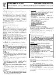
Cartilage - Type II procollagen<br />
Figure 3:<br />
Schematic drawing of type II procollagen <strong>and</strong> the localization of the different epitopes. Numbering is based on the amino-acid<br />
sequence of an alpha (II) chain of human type IIB collagen (COL2A1_HUMAN, P02458, UniProtKB, Swiss).<br />
* Type IIA procollagen would include an additional sequence of sixty-nine amino-acids at position 29-97, the numbering of the<br />
following sections would be shifted accordingly
Cartilage Biomarker <strong>and</strong> their behavior in various arthritis clinical situations<br />
Biomarker<br />
(u = urinary;<br />
s = serum;<br />
syn = synovial fluid;<br />
c = cellculture))<br />
<strong>Clinical</strong> situation<br />
Degradation marker Formation marker<br />
u CTX-II s C2C s C1,2C c C1,2C s COMP<br />
1) Only the ratio of C2C / C1,2C was prognostic.<br />
2) Changes in serum COMP are prognostic,<br />
not baseline levels.<br />
3) Prognostic for rapid joint destruction.<br />
4) Levels reflect the degree of synovial inflammation.<br />
syn<br />
s CPII<br />
COMP<br />
syn<br />
CPII<br />
s<br />
PIINP<br />
s<br />
Aggrecan<br />
Epitop<br />
syn<br />
Aggrecan<br />
Epitop<br />
s<br />
YKL-40<br />
OA ↑ ↑ ↑ ↑ ↑ ↓/N ↑ ↓ ↑ ↑ ↑<br />
OA Early ↑ ↑ ↑ N<br />
OA Late ↑ ↑ ↑<br />
OA Severity<br />
(Baseline levels<br />
indicate severity)<br />
OA Prognosis<br />
(Baseline levels predict<br />
progression)<br />
syn<br />
YKL-40<br />
+ + 2) + ± 4)<br />
+ 5) + 1) + 1) + + 5)<br />
RA ↑ ↑ ↑ ↑ ↑ ↑ ↓ ↑ ↑<br />
RA Early ↑ ↑ ↑<br />
RA Late ↑<br />
RA Progressive<br />
(Levels in progressive RA<br />
resulting in rapid joint<br />
destruction)<br />
↑ ↑ ↑ ↑ ↑ ↓/N<br />
RA Chronic ↑ ↑ ↑<br />
RA Severity<br />
(Baseline levels<br />
indicate severity)<br />
RA Prognosis<br />
(Baseline levels<br />
predict progression)<br />
+ + ± 4)<br />
+ + + + 3) − + +<br />
5) Patients with knee OA are characterized by an uncoupling<br />
of type II collagen synthesis <strong>and</strong> degradation which<br />
can be detected by assays for serum PIINP <strong>and</strong> urinary<br />
CTX-II. The combination of CTX-II <strong>and</strong> PIINP measurements<br />
seems prognostic for identifying knee OA patients<br />
at high risk for rapid progression of joint damage.<br />
Table was adapted from Schaller et al. (2005).<br />
15
16<br />
Bonemarker normal values for children<br />
Bone marker<br />
BAP (serum)<br />
bone specified alkaline<br />
phosphatase<br />
CICP/PICP (serum)<br />
c-terminal propeptid<br />
of type I collagen<br />
NTX (serum)<br />
N-terminal telopeptide<br />
of type I collagen<br />
NTX (urine)<br />
N-terminal telopeptide<br />
of type I collagen<br />
Deoxypyridinolin<br />
(urine)<br />
free DPD<br />
Helicale peptide (urine)<br />
Helicale peptide<br />
620 - 633 from the<br />
alpha-I-chain of<br />
type I collagen<br />
Pyridinolin (urine)<br />
free Pyd & DPD<br />
Cat.<br />
No.<br />
8012<br />
8003<br />
504836<br />
504837<br />
8007<br />
8022<br />
8010<br />
Sex Unit Age group<br />
m U/l ± SD<br />
f U/l ± SD<br />
Tanner<br />
Stage<br />
Tanner<br />
I<br />
104 ± 21<br />
0–2 y.<br />
122 ± 19<br />
0–2 y.<br />
Tanner<br />
II–III<br />
94 ± 17<br />
3–9 y.<br />
106 ± 16<br />
3–8 y.<br />
Tanner<br />
IV–V<br />
m U/l ± SD 95 ± 22 114 ± 35 121 ± 37<br />
f U/l ± SD 84 ± 23 113 ± 43 79 ± 46<br />
m/f ng/ml<br />
m ng/ml<br />
f ng/ml<br />
m/f ng/ml<br />
Tanner<br />
Stage<br />
m<br />
f<br />
m/f<br />
Tanner<br />
Stage<br />
m<br />
f<br />
m/f<br />
m/f<br />
m/f<br />
m/f<br />
BCE<br />
nmol/l<br />
BCE<br />
nmol/l<br />
BCE<br />
nmol/l<br />
Creatinin<br />
nmol/mml<br />
Creatinin<br />
nmol/mmol<br />
Creatinin<br />
nmol/mml<br />
Creatinin<br />
ug/mmol<br />
Creatinin<br />
nmol/mmol<br />
Creatinin<br />
nmol/mmol<br />
Creatinin<br />
Pyridinolin (serum) 8019 m/f nmol/l<br />
Tanner<br />
I<br />
50.6<br />
(37.2–88.9)<br />
53.6<br />
(31.2–90.9)<br />
211–1.310<br />
0–2 y.<br />
230–1.104<br />
0–2 y.<br />
Tanner<br />
II–IV<br />
71.7<br />
(32.7–126.0)<br />
50.2<br />
(12.9–79.8)<br />
295–365<br />
4.0–12.9 y.<br />
147–830<br />
3–9 y.<br />
155–568<br />
3–8 y.<br />
113–426<br />
4–13 y.<br />
Tanner<br />
V<br />
18.0<br />
(11.3–50.4)<br />
12.8<br />
(9.6–22.2)<br />
114 ± 21<br />
10–14 y.<br />
112 ± 16<br />
9–12 y.<br />
444 ± 192<br />
13 y.<br />
123–618<br />
10–14 y.<br />
122–373<br />
9–12 y.<br />
449 ± 123<br />
14 y.<br />
127–189<br />
15–19 y.<br />
74–167<br />
13–17 y.<br />
110–443<br />
14–18 y.<br />
0–1 y. 2–5 y. 6–10 y. 11–15 y. 16–20 y.<br />
1639<br />
(102-4769)<br />
Tanner<br />
I<br />
689<br />
(34-1752)<br />
Tanner<br />
II–III<br />
497<br />
(90-1356)<br />
Tanner<br />
IV–V<br />
23.5 ± 1.9 25.6 ± 4.0 31.1 ± 9.5<br />
21.0 ± 2.1 33.0 ± 5.1 23.2 ± 7.0<br />
22.5 ± 1.4 28.7 ± 3.2 28.0 ± 6.2<br />
804 –<br />
11305<br />
4–12.9 y.<br />
45.7–85.1<br />
4–12.9 y.<br />
55.5 ± 6.5<br />
4–12.9 y.<br />
2.49 ± 0.12<br />
4–12.9 y.<br />
429<br />
(34-2158)<br />
40.8 ± 6.4<br />
13–18 y.<br />
1.6 ± 1.0<br />
13–18 y.<br />
192<br />
(34-780)<br />
59 ± 19<br />
15–18 y.<br />
39 ± 23<br />
13–17 y.<br />
347 ± 145<br />
15–18 y.<br />
77–183<br />
20–50 y.<br />
37–133<br />
20–50 y.<br />
References<br />
[1]/[2]<br />
[3]<br />
[4]<br />
[1]<br />
[5]<br />
[6]<br />
[9]<br />
[5]<br />
[10]<br />
[11]<br />
[11]
Bonemarker normal values for children<br />
Cat.<br />
Bone marker Sex Unit Age group<br />
No.<br />
TRAP5b (Serum)<br />
Tartrate-Resistant Acid<br />
Phosphatase active<br />
isoform 5b<br />
8036<br />
Following references refer to the table<br />
on page 16 <strong>and</strong> 17.<br />
[1] Tsai et al<br />
Bone Alkaline Phosphatase Isoenzyme <strong>and</strong> Carboxy-Terminal<br />
Propeptide of Type-I Procollagen in Healthy Chinese Girls <strong>and</strong><br />
Boys.<br />
<strong>Clinical</strong> Chemistry 45, No. 1, 1999<br />
[2] Elmlinger MW et al<br />
Significance of bone specific alkaline phosphatase <strong>and</strong><br />
procollagen-I-peptide as diagnostic markers of bone<br />
formation for monitoring growth hormone therapy<br />
at the Congress on Calcium Regulating Hormones <strong>and</strong> Bone<br />
Metabolism from the German Society for Endocrinology, Giessen,<br />
Germany, September 29-30, 1995<br />
[3] Siu L. Hui et al<br />
Difference in Bone Mass between Black <strong>and</strong> White American<br />
Children: Attributable to Body, Build, Sex Hormone Levels, or<br />
Bone Turnover?<br />
Journal of <strong>Clinical</strong> Endocrinology & Metabolism 88 (2), 642 - 649,<br />
2003<br />
[4] Winterbottom et al<br />
An assay for the C-term. Propeptide of type I collagen.<br />
The Endocrine Society, Anaheim, CA, June 15 - 18, 1994.<br />
[5] Quidel In house study<br />
status<br />
f U/l<br />
m U/l<br />
Pre-<br />
Pubertal<br />
8.1±3.8<br />
1–9 y.<br />
6.6±3.6<br />
1–10 y.<br />
[6] Van der Sluis<br />
A Cross-Sectional Study on Biochemical Parameters of bone<br />
Turnover <strong>and</strong> Vitamin D Metabolites in Healthy Dutch Children<br />
<strong>and</strong> Young Adults<br />
Hormon Research 2002; 57: 170-179.<br />
Early Adolescence<br />
10.0±2.7<br />
10–13 y.<br />
9.9±3.3<br />
11–13 y.<br />
Late Adolescence<br />
2.3±0.7<br />
14–17 y.<br />
3.4±1.4<br />
14–17 y.<br />
[7] F. Rauch<br />
Urinary immunoreactive dpd in children <strong>and</strong> adolescents:<br />
variations with age, sex, <strong>and</strong> growth velocity.<br />
Scan J. Clin Lab Invest 1996:56: 715 - 719 (Metra Ref # 1540)<br />
[8] A. Bourdeau<br />
Age Dependent Variations in Healthy Children of Urinary<br />
Excretion of Pyridinium Crosslinks.<br />
XII International Conf. On Calcium Regulating Hormones, Australia,<br />
Feb. 14-19, 1995.<br />
[9] A. Conti<br />
Urinary free deoxypyridinoline levels during childhood.<br />
J. Endo. Invest. 21: 318 - 322, 1998. Ref#2574<br />
[10] Husain, et al<br />
Urinary excretion of pyridinium crosslinks in healthy<br />
4 - 10 year olds.<br />
Arch. Dis. Child 1999, 80: 370 - 373<br />
[11] A. Colwell<br />
The Renal Clearance of Free <strong>and</strong> Conjugated Pyridinium<br />
Crosslinks of Collagen.<br />
J. of Bone <strong>and</strong> Min. Res. Vol. 11, Number 12, 1996<br />
References<br />
[12] Price HE, Langman CB <strong>and</strong> Brooks ER.<br />
TRAP5b – profiles in children with chronic kidney disease.<br />
Poster presented at ASBMR (2007).<br />
[12]<br />
[12]<br />
17
18<br />
Biomarkers in Cell Culture<br />
If cell culture samples are used in assay systems which have been validated for the determination of biomarkers in serum<br />
<strong>and</strong> plasma, several aspects have to be taken into consideration.<br />
An assay system validated for serum / plasma may be affected by sample material generated from cell culture.<br />
To examine whether the assay system is influenced by cross-reactions or interferences induced by the components used,<br />
it is recommended to determine both the medium <strong>and</strong> all further substances in the assay. FCS in particular may crossreact<br />
with antibodies in the test or the media can affect the antibody response.<br />
It is recommended to use a culture medium comprising the following components:<br />
• DMEM or equivalent commercial medium<br />
• Fetal bovine or calf serum or serum substitutes (FCS-free medium)<br />
• L-Glutamine or ascorbate<br />
• Antibiotic<br />
To exclude the influence of FCS, it is recommended to use FCS-free medium, if possible.<br />
Testing of medium, medium additives, <strong>and</strong> other buffers used<br />
• Buffer: e.g. elution buffers which have been used to elute a substance from a gel or tissue<br />
• Matrix: e.g. serum-free medium, serum substitutes<br />
• Testing of medium combination:<br />
1. Medium<br />
2. Medium including all additives, but no FCS or alternative matrix<br />
3. Medium including all additives <strong>and</strong> FCS or alternative matrix (complete medium)<br />
4. Only FCS or corresponding alternative matrix<br />
• Recovery:<br />
Add st<strong>and</strong>ard material/substance to the complete medium (spiking) to determine recovery;<br />
e.g. at a ratio 1:5, test 20% of the highest kit st<strong>and</strong>ard <strong>and</strong> 80% of complete medium in the assay<br />
• Linearity:<br />
Measure dilutions of the spiked medium to control linearity:<br />
a) by using dilution buffer or zero st<strong>and</strong>ard from the kit at a ratio 1:2 <strong>and</strong> 1:4<br />
b) by using medium at a ratio 1:2 <strong>and</strong> 1:4<br />
• St<strong>and</strong>ard curve (optional):<br />
Dilute stock st<strong>and</strong>ard or the highest st<strong>and</strong>ard contained in the kit with complete medium <strong>and</strong> perform measurement
Specific recommendations<br />
BAP<br />
Osteocalcin<br />
CICP / PICP<br />
DPD<br />
PYD<br />
NTX<br />
Helical Peptide<br />
Assay Analyt Comments to the test procedure<br />
Bone Alkaline Phosphatase<br />
Intact Osteocalcin<br />
C-terminal Propeptide of Type-I-<br />
Collagen (CICP)<br />
Desoxypyridinolin-Crosslinks<br />
Pyridinolin-Crosslinks<br />
alpha-2 (I) N- telopeptide<br />
Helical Peptide<br />
The Alkaline Phosphate is membrane<br />
bound <strong>and</strong> can be measured in cell<br />
lysate.<br />
The cells can be grown in serum containing<br />
media, but osteocalcin must be<br />
harvested from tissue culture supernatant<br />
that is serum-free.<br />
The cells can be grown in serum containing<br />
media; however, to measure<br />
the parameters serum-free tissue culture<br />
supernatant has to be used.<br />
Bone collagen specific –<br />
direct measurement of bone resorption.<br />
The cells can be grown in serum containing<br />
media; however, to measure the<br />
parameters serum-free tissue culture<br />
supernatant has to be used.<br />
First results show that the ELISA is<br />
suited for the determination of the helical<br />
Peptide for in vitro analyses. This<br />
test method is especially advantageous<br />
because it requires small sample quantities<br />
<strong>and</strong> the curve covers a large<br />
range of the sample concentrations (30<br />
fold of the CTX-beta in vitro Assays).<br />
The background concentrations<br />
through the medium are very low.<br />
TRAP5b Tartratresistent acid phosphatase 5b Measurement of the osteoclast activity.<br />
Interferences<br />
In order to test if there are any cross reactions or interferences of the media used for the cell culture with the assay system,<br />
it is recommended to measure the media <strong>and</strong> maybe other substances in the assay. Especially FCS could contain interfering<br />
substances respectively cross react or certain media suppress the antibody reaction.<br />
19
20<br />
Measurement of BAP <strong>and</strong> Osteocalcin<br />
in cell culture<br />
• DMEM or equivalent commercial medium<br />
• Fetal bovine serum or calf serum or serum substitutes<br />
(FCS free medium)<br />
• L-Glutamine or Ascorbat<br />
• Antibiotic<br />
Additionally, 50 nM Vitamin D3 is necessary to generate<br />
measurable quantities of osteocalcin. The cells can be<br />
grown in serum containing media, but osteocalcin must be<br />
harvested from tissue culture supernatant that is serumfree,<br />
because the antibody will also detect this analyte in<br />
serum.<br />
Bone alkaline phosphatase is membrane-bound <strong>and</strong> it has<br />
to be measured in culture from cell lysate. Cell culture media<br />
should not be treated with ion chelators like EDTA or citrate<br />
because of an inhibition of the BAP enzyme activity.<br />
After the incubation the cell layers should be washed twice<br />
with saline <strong>and</strong> scraped into TMN buffer solution (20 mM<br />
Tris-HCl, pH 7.4; 2 mM MgCl2; 150 mM NaCl) uising a stir<br />
stick. The number of cells has to be determined (e.g. Coulter<br />
cell counter Model F) before the cells become solubilized by<br />
the addition of Triton X-100 to a final concentration of 1 %.<br />
Centrifugate the samples at 70,000g for 60 min <strong>and</strong> examine<br />
the aliquots of the supernatant for alkaline phosphatase<br />
activity.<br />
Sheep <strong>and</strong> human cells will be fine for this purpose.<br />
References for BAP, PICP, OSTEOCALCIN<br />
in cell culture<br />
Kaspar D, Seidl W, Neidlinger-Wilke C, Ignatius A, Claes L<br />
(2000) Dynamic cell stretching increases human osteoblast<br />
proliferation <strong>and</strong> CICP synthesis but decreases osteocalcin<br />
synthesis <strong>and</strong> alkaline phosphatase activity.<br />
J Biomech 33, 45-51.<br />
Martínez ME, Medina S, Del Campo MT, et al. (1998) Effect<br />
of polyethylene on osteocalcin, alkaline phosphatase <strong>and</strong><br />
procollagen secretion by human osteoblastic cells.<br />
Calcif Tissue Int 62, 453-456.<br />
Measurement of CICP / PICP<br />
in cell culture<br />
The assay is suitable for the measurement of cell culture<br />
supernatant from cells producing type-I collagen. The<br />
METRA ® CICP Test can be used for the determination of the<br />
collagen production level of skin fibroblasts <strong>and</strong> bone cells.<br />
It is recommended, that the supernatant of these cultures<br />
contains serum-free medium. Bovine serum in the medium<br />
may contain bovine CICP, which cross-reacts with the antibody<br />
in the kit. This would result in erroneously increased<br />
values.<br />
In a confluating cell culture at an optimal collagen production<br />
level, the amount of CICP in the supernatant is approximately<br />
20 – 50 ng/ml or slightly below a normal human serum<br />
sample. Dilute the cell culture supernatant 1:12 (like a normal<br />
serum sample) <strong>and</strong> calculate the final concentration according<br />
to the dilution. If the CICP concentration of the 1:12<br />
dilution is below the detection limit it is possible to choose<br />
a 1:6-dilution.<br />
Sell S, Gaissmaier C, Fritz J et al. (1998) Different behavior<br />
of human osteoblast-like cells isolated from normal <strong>and</strong><br />
heterotopic bone in vitro. Calcif Tissue Int 62, 51-59.<br />
Siggelkow H, Niedhart C, Kurre W, et al. (1998) In vitro differentiation<br />
potential of a new human osteosarcoma cell line<br />
(HOS 58). Differentiation 63, 81-91.<br />
Siggelkow H, Rebenstorff K, Kurre W, et al. (1999)<br />
Development of the osteoblast phenotype in primary human<br />
osteoblasts in culture: comparison with rat calvarial cells in<br />
osteoblast differentiation. J Cell Biochem 75, 22-35.
Bone marker - Reference values in animals<br />
BAP in Dogs<br />
Serum / Plasma: < 1 year 56.3 ± 9.8 U/L<br />
1 – 2 years 10.7 ± 4.5 U/L<br />
2 – 3 years 7.0 ± 2.5 U/L<br />
3 – 7 years 6.7 ± 3.6 U/L<br />
> 8 years 7.0 ± 2.9 U/L<br />
Reference: Allen LC et al. (2000) A comparison of two techniques for the determination of serum bone-specific<br />
alkaline phosphatase activity in dogs. Res Vet Sci 68, 231-235.<br />
BAP in Cats<br />
Serum: < 2 years 10 – 70 U/L<br />
> 2 years 2 – 15 U/L<br />
Reference: DeLaurier A, Jackson B, Ingham K, Pfeiffer D, Horton MA, Price JS. (2002) Biochemical markers of<br />
bone turnover in the domestic cat: relationships with age <strong>and</strong> feline osteoclastic resorptive lesions.<br />
J Nutr 132, 1742S-4S.<br />
BAP in Horses<br />
Serum: 12.2 – 25.5 U/L<br />
Plasma: 12.6 – 22.7 U/L<br />
Reference: Hoekstra K et al. (1999) Comparison of bone mineral content <strong>and</strong> biochemical markers of bone<br />
metabolism in stall vs. pasture-reared horses. Equine Exercise Phys Equine Vet J 30, 601-604.<br />
BAP in goats<br />
Serum: 12 ± 4 U/L<br />
Reference: Liesegang A, Risteli J, Wanner M (2005) The effects of first gestation <strong>and</strong> lactation on bone<br />
metabolism in dairy goats <strong>and</strong> milk sheep. Bone. Dec 16; [Epub ahead of print]<br />
BAP in sheep<br />
Serum: 13 ± 4 U/L<br />
Reference: Liesegang A, Risteli J, Wanner M (2005) The effects of first gestation <strong>and</strong> lactation on bone<br />
metabolism in dairy goats <strong>and</strong> milk sheep. Bone. Dec 16; [Epub ahead of print]<br />
BAP in porcine<br />
Reference: Liesegang A et al. (2002) Influence of a Vegetarian Diet Versus a Diet with Fishmeal on Bone<br />
in Growing Pigs. J. Vet. Med. A 49, 230-238<br />
21
22<br />
DPD in Dogs<br />
Urine: < 1 year 45 nM/mM Creatinine<br />
1 – 2 years 4 – 5 nM/mM Creatinine<br />
2 – 3 years 4 – 5 nM/mM Creatinine<br />
3 – 7 years 4 – 5 nM/mM Creatinine<br />
Reference: Allen MJ et al. (2000) Urinary markers of type-I collagen degradation in the dog.<br />
Res Vet Sci 69, 123-127.<br />
DPD in Cats<br />
Urine: 1-10 years 11.3 nM/mM Creatinine<br />
Reference: DeLaurier A, Jackson B, Ingham K, Pfeiffer D, Horton MA, Price JS. (2002) Biochemical markers of<br />
bone turnover in the domestic cat: relationships with age <strong>and</strong> feline osteoclastic resorptive lesions.<br />
J Nutr 132, 1742S-4S.<br />
DPD in Horses<br />
Urine: 6.0 – 95 nM/mM Creatinine<br />
Reference: Hoekstra K et al. (1999) Comparison of bone mineral content <strong>and</strong> biochemical markers of bone<br />
metabolism in stall vs. pasture-reared horses. Equine Exercise Phys Equine Vet J 30, 601-604.<br />
Osteocalcin intact in Horses<br />
Serum: 15 – 50 ng/ml<br />
Reference: Hoekstra K et al. (1999) Comparison of bone mineral content <strong>and</strong> biochemical markers of bone<br />
metabolism in stall vs. pasture-reared horses. Equine Exercise Phys Equine Vet J 30, 601-604.<br />
Creatinine for animal species<br />
Reference: Sierra RI, Specker BL, Jimenez F, Cruz C, Pedraza-Chaverri J (1997) Biochemical bone markers,<br />
bone mineral content, <strong>and</strong> bone mineral density in rats with experimental nephrotic syndrome.<br />
Ren Fail19, 409-424.<br />
PTH in Dogs<br />
Serum / Plasma: 15 – 150 pg/ml<br />
PTH in Cats<br />
Serum / Plasma: 3.3 – 22.5 pg/ml<br />
PTH in Horses<br />
Serum / Plasma: 20 – 120 pg/ml mean 56 pg/ml
Cross reactivity - different species<br />
Produkt<br />
Product<br />
Produit<br />
Zellkultur<br />
Cell culture<br />
Culture cellulaire<br />
Human<br />
Human<br />
Homme<br />
Elefant<br />
Elephant<br />
Eléphant<br />
Eichhörnchen<br />
Squirrel<br />
Ecureuil<br />
Huhn<br />
Chicken<br />
Poulet<br />
Hund<br />
Dog<br />
Chien<br />
Kaninchen<br />
Rabbit<br />
Lapin<br />
Katze<br />
Cat<br />
Chat<br />
Cynomolgus Makake<br />
Cynomolgus Macaque<br />
Macaque<br />
Maus<br />
Mouse<br />
Souris<br />
Meerschweinchen<br />
Guinea pig<br />
Cobaye<br />
Pavian<br />
Baboon<br />
Babouin<br />
Pferd<br />
Horse<br />
Cheval<br />
Rind<br />
Bovine<br />
Bovin<br />
Schwein<br />
Porcine<br />
Porc<br />
Ratte<br />
Rat<br />
Rat<br />
Rhesus Affe<br />
Rhesus macaque<br />
Singe Rhésus<br />
KNOCHEN METABOLISMUS / BONE METABOLISM / MÉTABOLISME DE L'OS<br />
Truthahn<br />
Turkey<br />
Dinde<br />
Schaf<br />
Sheep<br />
Mouton<br />
Schimpanse<br />
Chimpanzee<br />
Chimpanzé<br />
BAP X X X X X X N X X X X N X X X<br />
Cathepsin K X<br />
CICP/PICP X X N X X N N X N X N<br />
Ziege<br />
Goat<br />
Chèvre<br />
Hamster<br />
Hamser<br />
Hamster<br />
Alle Spezies<br />
All species<br />
Toutes espèces<br />
Creatinine X<br />
DKK-1 X X X<br />
Helical Peptide X X X X X X X X X X X X X X X X<br />
NTX Serum (X) X X X X X X X<br />
NTX Urine X X X X X X X<br />
Osteocalcin Intact X X N X X N X X X X N X X<br />
Osteocalcin Mouse X X X<br />
Osteocalcin<br />
N-MID (1-43/49) X X<br />
Osteocalcin Rat X X X<br />
Osteopontin<br />
Human X X<br />
Osteopontin<br />
Mouse X X<br />
Osteopontin<br />
Rat X X<br />
Osteoprotegerin<br />
(OPG) Human X X N N N X N N N X N N<br />
Osteoprotegerin<br />
(OPG) Human X X X<br />
Pyridinoline (Pyd)<br />
Serum X X X X X X X X X X X<br />
Pyrilinks<br />
(Pyd + Dpd) X X X X X X X X X X X X<br />
Pyrilinks D (Dpd) X X X X X N X X X X X X X X X X<br />
Sclerostin TECO X X N N<br />
sRankl Human<br />
High Sensitive X X X<br />
Total Dpd X X X X X X X X X X X X X X X X<br />
TRAP5b Human X X N N N N N N N N N<br />
TRAP5b Rat X X<br />
KNORPEL METABOLISMUS / CARTILAGE METABOLISM / MÉTABOLISME DU CARTILAGE<br />
C1–2C X X X (X) X (X) X X (X) X (X)<br />
C2C, Serum/Urine X X X X (X) X X (X) X X (X)<br />
COMP X X<br />
CP II/PIICP X X X (X) X (X) X X X X (X)<br />
CS-846 X X X X (X) X X X X X (X)<br />
Hyaluronic Acid<br />
TECO X X X X X X<br />
23
24<br />
Cross reactivity - different species<br />
Produkt<br />
Product<br />
Produit<br />
Zellkultur<br />
Cell culture<br />
Culture cellulaire<br />
Human<br />
Human<br />
Homme<br />
sGAG X<br />
Elefant<br />
Elephant<br />
Eléphant<br />
Eichhörnchen<br />
Squirrel<br />
Ecureuil<br />
Huhn<br />
Chicken<br />
Poulet<br />
Hund<br />
Dog<br />
Chien<br />
Kaninchen<br />
Rabbit<br />
Lapin<br />
Katze<br />
Cat<br />
Chat<br />
Cynomolgus Makake<br />
Cynomolgus Macaque<br />
Macaque<br />
Maus<br />
Mouse<br />
Souris<br />
Meerschweinchen<br />
Guinea pig<br />
Cobaye<br />
Pavian<br />
Baboon<br />
Babouin<br />
Pferd<br />
Horse<br />
Cheval<br />
Rind<br />
Bovine<br />
Bovin<br />
Schwein<br />
Porcine<br />
Porc<br />
Ratte<br />
Rat<br />
Rat<br />
Rhesus Affe<br />
Rhesus macaque<br />
Singe Rhésus<br />
KNORPEL METABOLISMUS / CARTILAGE METABOLISM / MÉTABOLISME DU CARTILAGE<br />
YKL-40 X X X X X<br />
Chemerin X X<br />
hs-CRP X<br />
Myeloperoxidase<br />
(MPO)<br />
X<br />
Progranulin X<br />
ENTZÜNDUNG / INFLAMMATORY / INFLAMMATION<br />
YKL-40 X X X X X<br />
Calcitonin Human X<br />
Truthahn<br />
Turkey<br />
Dinde<br />
KALZIUM METABOLISMUS / CALCIUM METABOLISM / MÉTABOLISME DU CALCIUM<br />
Calcitonin Rat X X X<br />
Fetuin A Human X X<br />
Fetuin A Rat X N X<br />
FGF-23 Intact X X N N<br />
Schaf<br />
Sheep<br />
Mouton<br />
Schimpanse<br />
Chimpanzee<br />
Chimpanzé<br />
FGF-23 Intact<br />
(Kainos) X X X<br />
FGF-23 (C-Term)<br />
2nd Generation X X N N<br />
FGF-23 Mouse<br />
(C-Term) X X X<br />
PTH 1-34 Anti-<br />
Human Antibody X X<br />
PTH 1-34 Human<br />
High Sensitive X X<br />
PTH 1-84 Bioactive<br />
Human X X X X X X X<br />
PTH 1-84 Bioactive<br />
Rat X N X N X<br />
PTH 1-84 Bovine X X (X) (X) (X) X (X) (X) (X) (X)<br />
PTH 1-84 Intact<br />
Dog X X<br />
PTH 1-84 Intact<br />
Human<br />
X<br />
PTH 1-84 Intact<br />
Mouse X N X N X<br />
PTH 1-84 Intact<br />
Rat X N (X) X N X<br />
PTH 1-84 Porcine X X X (X) X X (X)<br />
PTH C-Terminal<br />
Human X X<br />
PTH C-Terminal Rat X X<br />
PTH Horse X X X X X<br />
PTH Rat X X (X) X X X X X X<br />
25 OH Vitamin D<br />
direct X X<br />
Ziege<br />
Goat<br />
Chèvre<br />
Hamster<br />
Hamser<br />
Hamster<br />
Alle Spezies<br />
All species<br />
Toutes espèces
Cross reactivity - different species<br />
Produkt<br />
Product<br />
Produit<br />
Zellkultur<br />
Cell culture<br />
Culture cellulaire<br />
Human<br />
Human<br />
Homme<br />
Myostatin X<br />
BMP 7 X<br />
Adiponectin Human<br />
TECO X X<br />
Adiponectin<br />
Mouse<br />
Adiponectin<br />
Rat<br />
Chemerin X X<br />
Fetuin A Human X X<br />
Elefant<br />
Elephant<br />
Eléphant<br />
Eichhörnchen<br />
Squirrel<br />
Ecureuil<br />
Huhn<br />
Chicken<br />
Poulet<br />
Hund<br />
Dog<br />
Chien<br />
Kaninchen<br />
Rabbit<br />
Lapin<br />
Katze<br />
Cat<br />
Chat<br />
Cynomolgus Makake<br />
Cynomolgus Macaque<br />
Macaque<br />
Maus<br />
Mouse<br />
Souris<br />
Meerschweinchen<br />
Guinea pig<br />
Cobaye<br />
Pavian<br />
Baboon<br />
Babouin<br />
Pferd<br />
Horse<br />
Cheval<br />
Rind<br />
Bovine<br />
Bovin<br />
Schwein<br />
Porcine<br />
Porc<br />
Ratte<br />
Rat<br />
Rat<br />
Rhesus Affe<br />
Rhesus macaque<br />
Singe Rhésus<br />
MUSKEL – SKELETT / MUSCLE – SKELETON / MUSCLE - SQUELETTE<br />
DIABETES & ADIPOSITAS / DIABETES & OBESITY / DIABÈTE & OBÉSITÉ<br />
X<br />
X<br />
Truthahn<br />
Turkey<br />
Dinde<br />
Schaf<br />
Sheep<br />
Mouton<br />
Schimpanse<br />
Chimpanzee<br />
Chimpanzé<br />
Ziege<br />
Goat<br />
Chèvre<br />
Hamster<br />
Hamser<br />
Hamster<br />
Alle Spezies<br />
All species<br />
Toutes espèces<br />
Ghrelin X X X X X X X X X X X X X<br />
GLP-1 Active<br />
(7-36) Human X X X X<br />
GLP-1 Active<br />
(7-36) Mouse / Rat X X<br />
GLP-1 Total X<br />
Intact ProInsulin<br />
TECO X X (X) (X) (X)<br />
Leptin Human<br />
TECO X X<br />
Leptin Mouse/Rat X X X<br />
Resistin X X N<br />
YKL-40 X X X X X<br />
LEBERERKRANKUNG / LIVER DISEASE / MALADIES DU FOIE<br />
Hyaluronic Acid<br />
TECO X X X X X X<br />
M30-Apoptosense<br />
Chronic Liver Disease<br />
X<br />
hGH / High Sensitive X X<br />
IGFBP-1 X X<br />
WACHSTUMSSTOFFWECHSEL / GROWTH METABOLISM / MÉTABOLISME DE LA CROISSANCE<br />
IGFBP-2 X X X X X X<br />
IGFBP-2 Mouse / Rat X X X X X<br />
ALS (Acid Labile<br />
Subunit)<br />
X<br />
hGH X N<br />
IGFBP-3 X X<br />
IGFBP-3<br />
Functional X X<br />
IGFBP-3<br />
Mouse/Rat X X<br />
IGF-I (BP blocked) X X X X X X X X X X X X X<br />
IGF-I Mouse/Rat X X<br />
IGF-II X X<br />
IGF-II X X X<br />
25
26<br />
Cross reactivity - different species<br />
Produkt<br />
Product<br />
Produit<br />
Zellkultur<br />
Cell culture<br />
Culture cellulaire<br />
Human<br />
Human<br />
Homme<br />
Elefant<br />
Elephant<br />
Eléphant<br />
Eichhörnchen<br />
Squirrel<br />
Ecureuil<br />
Huhn<br />
Chicken<br />
Poulet<br />
Hund<br />
Dog<br />
Chien<br />
Kaninchen<br />
Rabbit<br />
Lapin<br />
Katze<br />
Cat<br />
Chat<br />
Cynomolgus Makake<br />
Cynomolgus Macaque<br />
Macaque<br />
Maus<br />
Mouse<br />
Souris<br />
Meerschweinchen<br />
Guinea pig<br />
Cobaye<br />
Pavian<br />
Baboon<br />
Babouin<br />
Pferd<br />
Horse<br />
Cheval<br />
Rind<br />
Bovine<br />
Bovin<br />
Schwein<br />
Porcine<br />
Porc<br />
Ratte<br />
Rat<br />
Rat<br />
Rhesus Affe<br />
Rhesus macaque<br />
Singe Rhésus<br />
Truthahn<br />
Turkey<br />
Dinde<br />
Schaf<br />
Sheep<br />
Mouton<br />
Schimpanse<br />
Chimpanzee<br />
Chimpanzé<br />
KARDIOVASKULÄRE MARKER / CARDIOVASCULAR MARKER / MARQUEUR CARDIOVASCULAIRE<br />
Big Endothelin X X N N N<br />
BNP Fragment X X N N N N N N N N N N N N N<br />
Endothelin X X X X N X X X<br />
NT-proANP X X X X<br />
NT-proCNP X X X X X X X X X<br />
OXIDATIVE STRESS MARKER / OXIDATIVE STRESS MARKER / MARQUEUR DU STRESS OXYDATIF<br />
oLAB X N N N N N N N N N N N N N<br />
Oxidized LDL X<br />
OxyStat X<br />
EZ4U X<br />
CELL PROLIFERATION & CYTOTOXICITY ASSAY<br />
APOPTOSE / APOPTOSIS / APOPTOSE<br />
M30-<br />
Apoptosense X X X X X X<br />
M30-CytoDeath X X X X X X<br />
M65-EpiDeath X X X A* X X A* X X<br />
ANDERE PARAMETER / OTHER PARAMETERS / AUTRES PARAMÈTRES<br />
ACTH X X X<br />
Erythropoetin X<br />
Fecal Calprotectin X<br />
Prekallikrein Activator<br />
Assay (PKA)<br />
X<br />
TPMT X<br />
TSH Receptor Antibody<br />
2 Gen. TECO<br />
X<br />
Ziege<br />
Goat<br />
Chèvre<br />
Hamster<br />
Hamser<br />
Hamster<br />
Alle Spezies<br />
All species<br />
Toutes espèces
Cross reactivity - different species<br />
Produkt<br />
Product<br />
Produit<br />
Zellkultur<br />
Cell culture<br />
Culture cellulaire<br />
Human<br />
Human<br />
Homme<br />
AH50 Eq X<br />
Elefant<br />
Elephant<br />
Eléphant<br />
Eichhörnchen<br />
Squirrel<br />
Ecureuil<br />
Huhn<br />
Chicken<br />
Poulet<br />
Hund<br />
Dog<br />
Chien<br />
Kaninchen<br />
Rabbit<br />
Lapin<br />
Katze<br />
Cat<br />
Chat<br />
Cynomolgus Makake<br />
Cynomolgus Macaque<br />
Macaque<br />
Maus<br />
Mouse<br />
Souris<br />
Meerschweinchen<br />
Guinea pig<br />
Cobaye<br />
A* = Humane Xenotransplantate / human xenograft / Hétérogreffe humaine<br />
N = keine Kreuzreaktion / no cross reactivity / pas de réaction croisée<br />
(x) = schwache Kreuzreaktion / low cross reactivity / faible réaction croisée<br />
= Nicht getestet / not tested / pas testé<br />
x = Kreuzreaktion / cross reactivity / réaction croisée<br />
Pavian<br />
Baboon<br />
Babouin<br />
Pferd<br />
Horse<br />
Cheval<br />
Rind<br />
Bovine<br />
Bovin<br />
Schwein<br />
Porcine<br />
Porc<br />
Ratte<br />
Rat<br />
Rat<br />
Rhesus Affe<br />
Rhesus macaque<br />
Singe Rhésus<br />
KOMPLEMENT TEST / COMPLEMENT TEST / TEST DE COMPLÉMENT><br />
Bb Plus X X X X<br />
C1-Inhibitor X<br />
C3a Plus X X X X<br />
C4d X X X X<br />
C5a X<br />
CH50 Eq X X<br />
CIC-C1q X<br />
CIC-C3d (Raji-<br />
Cell-Replacement)<br />
X<br />
iC3b X<br />
SC5b-9 Plus X X X X<br />
Truthahn<br />
Turkey<br />
Dinde<br />
Schaf<br />
Sheep<br />
Mouton<br />
Schimpanse<br />
Chimpanzee<br />
Chimpanzé<br />
Ziege<br />
Goat<br />
Chèvre<br />
Hamster<br />
Hamser<br />
Hamster<br />
Alle Spezies<br />
All species<br />
Toutes espèces<br />
27
The Specialist for Biochemical Markers<br />
For further information please contact: / Für weitere Informationen wenden Sie sich bitte an: / Pour plus d’informations, vous pouvez contacter:<br />
Exclusive Strategic Partnership in Europe:<br />
www.quidel.com<br />
Germany<br />
TECOmedical GmbH<br />
Wasserbreite<br />
32257 Bünde<br />
Germany<br />
phone + 49 (0) 52 23 985 99 99<br />
fax + 49 (0) 52 23 985 99 98<br />
mail info@tecomedical.com<br />
web www.tecomedical.com<br />
TECOmedical Group<br />
Headquarters<br />
Switzerl<strong>and</strong>/International<br />
TECOmedical AG<br />
Gewerbestrasse 10<br />
4450 Sissach<br />
Switzerl<strong>and</strong><br />
phone + 41 (0) 61 985 81 00<br />
fax + 41 (0) 619 85 81 09<br />
mail info@tecomedical.com<br />
web www.tecomedical.com<br />
France<br />
TECOmedical SARL<br />
20 rue du Bois Chal<strong>and</strong><br />
91090 Lisses<br />
France<br />
phone 0800 100 437<br />
fax 0800 100 480<br />
mail chdu@tecomedical.com<br />
web www.tecomedical.com<br />
Benelux<br />
TECOmedical NL<br />
‘t Hazeveld 34<br />
3862 XB Nijkerk<br />
The Netherl<strong>and</strong>s<br />
phone + 31 (0) 33 4951 473<br />
fax + 31 (0) 33 4951 635<br />
mail sbk@tecomedical.com<br />
web www.tecomedical.com<br />
01/2011 © TECOmedical Group

![PTH [Hormone Parathyroïdienne] Intacte ELISA](https://img.yumpu.com/1233682/1/190x245/pth-hormone-parathyroidienne-intacte-elisa.jpg?quality=85)