
You also want an ePaper? Increase the reach of your titles
YUMPU automatically turns print PDFs into web optimized ePapers that Google loves.
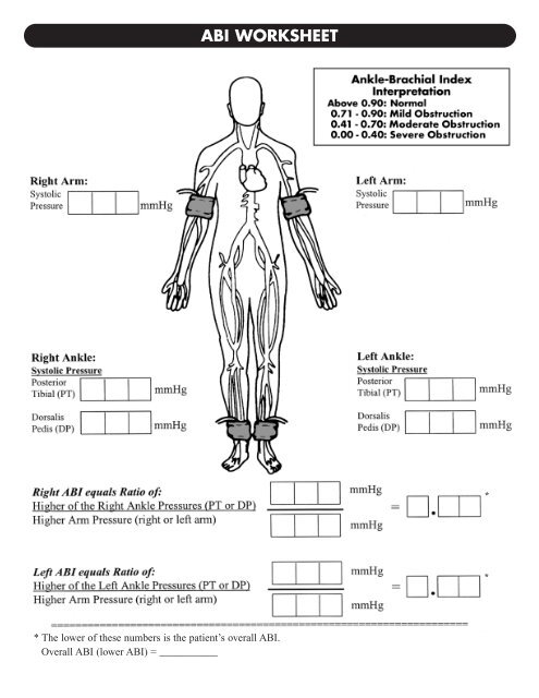
<strong>ABI</strong> <strong>WORKSHEET</strong><br />
* The lower of these numbers is the patient’s overall <strong>ABI</strong>.<br />
Overall <strong>ABI</strong> (lower <strong>ABI</strong>) = ___________<br />
*<br />
*
PERFORMING THE <strong>ABI</strong> TEST<br />
The <strong>ABI</strong> measurement is performed with the patient resting in a supine position. The examiner should make all arm and leg blood<br />
pressure measurements with an appropriately sized blood pressure cuff and the Doppler device. The systolic blood pressure is<br />
determined in both arms, and the ankle systolic blood pressure is determined for the right and left posterior tibial (PT) and dorsalis<br />
pedis (DP) arteries. The <strong>ABI</strong> for each leg is determined by using the higher of the two readings from either the PT or DP arteries,<br />
and the higher of the two brachial readings. The lower <strong>ABI</strong> of the two is used for diagnostic purposes. An <strong>ABI</strong> measurement<br />
can usually be performed in less than 10 minutes. (See sample <strong>ABI</strong> worksheet.)<br />
Step 1. Have the patient lie in a supine position with shoes and<br />
stockings removed for at least 10 minutes prior to obtaining blood<br />
pressure measurements.<br />
Step 2. Apply the blood pressure cuff snugly on the upper arm<br />
with the lower edge of the cuff 1 inch above the antecubital<br />
fossa. Usually the cuff that is the appropriate size for the patient’s<br />
arm will also be suitable for the ankle pressure measurement. In<br />
the rare instance that upper arm and ankle pressures are<br />
markedly different, choose cuff sizes that are appropriate for<br />
each site.<br />
Step 3. Apply a 1–2 centimeter ribbon of Doppler gel to the<br />
antecubital area. Be sure to use enough gel.<br />
Step 4. Turn the Doppler probe on and place it at the antecubital<br />
area at approximately a 60-degree angle to the surface of<br />
the skin. Move the probe around until the clearest arterial pulse<br />
sounds are heard and keep the probe at that position.<br />
Step 5. Inflate the blood pressure cuff to approximately 20 mm Hg<br />
above the numerical reading where the pulse sounds cease.<br />
Step 6. Deflate the cuff at a rate of 2 mm Hg per second until the<br />
first arterial pulse sound is heard. When this number is determined,<br />
deflate the cuff completely and record this systolic reading.<br />
Remove the gel from the patient’s skin with a tissue.<br />
Step 7. Apply the same blood pressure cuff snugly to the<br />
ankle on the same side of the body.<br />
Step 8. Palpate the area around the medial malleolus to find<br />
the posterior tibial (PT) arterial pulse.<br />
<strong>ABI</strong> PROCEDURE<br />
HELPFUL HINTS<br />
Step 9. If this pulse is palpable, apply a 1–2 centimeter ribbon<br />
of Doppler gel to the area. If there is no palpable pulse, apply<br />
gel to the general area, turn on the Doppler probe, and move the<br />
probe around until the clearest arterial sound is heard. Keep the<br />
probe in that position. Continue inflating the blood pressure cuff<br />
as before, followed by deflation and reading (Steps 5–6).<br />
Step 10. Palpate the dorsal arch of the same foot for the dorsalis<br />
pedis (DP) arterial pulse. Apply the Doppler gel and use the<br />
Doppler probe as before (Step 9).<br />
Step 11. Apply the blood pressure cuff to the opposite ankle<br />
and record the PT and DP pressures as before (Steps 8–10).<br />
Step 12. Then repeat Steps 2–6 on the other arm.<br />
Use the <strong>ABI</strong> worksheet page to figure the patient’s <strong>ABI</strong>.<br />
Measurements should be noted in the patient’s medical record.<br />
Both the DP and PT arterial pressures are measured to provide<br />
a complete assessment of the extent of PAD in each limb.<br />
Additionally, some patients may have a congenitally absent dorsalis<br />
pedis pulse.<br />
Other patients, particularly some elderly and diabetic individuals,<br />
have calcification in their arteries that prevents occlusion of flow by<br />
the pressure cuff. This will cause an abnormally high reading.<br />
Typically any reading greater than 1.50 is considered abnormal.<br />
Such patients should be referred for additional testing in a vascular<br />
laboratory.<br />
Follow the instructions specific to the Doppler probe you are using.<br />
Be sure to use enough gel.<br />
Use a cuff size that is right for both the arm and ankle of the patient.<br />
Be sure you’re centered on the pulse when you take the reading; if you’re off to the side, the reading will be low.<br />
Be aware of known diabetics with calcified vessels and abnormally high <strong>ABI</strong>.<br />
In a small percentage of patients, one of the ankle pressures will be nondetectable; use the detectable pressure for calculating<br />
the <strong>ABI</strong>.<br />
Patients with an <strong>ABI</strong> value of 0.90 or less are diagnosed as having PAD and considered at increased risk for cardiovascular ischemic<br />
events. Prompt investigation and risk-reducing treatments are then warranted.<br />
Don’t be discouraged if measuring the <strong>ABI</strong> seems slow or clumsy at first. Like any procedure, the <strong>ABI</strong> becomes easier to do with practice.