Handbook of Shrimp Diseases - the National Sea Grant Library
Handbook of Shrimp Diseases - the National Sea Grant Library
Handbook of Shrimp Diseases - the National Sea Grant Library

Create successful ePaper yourself
Turn your PDF publications into a flip-book with our unique Google optimized e-Paper software.
Molt arrest occurs in affected animals <strong>of</strong>some populations.<br />
Animals begin, but are unable to complete <strong>the</strong> molting process.<br />
In some cases, <strong>the</strong>re is abnormal adherence to underlying skin,<br />
but most animals appear to lack <strong>the</strong> necessary stamina. Nutri<br />
tional inadequacies and water quality factors have been identi<br />
fied as causes.<br />
Growth Problems<br />
Growth problems become obvious inaquaculture stocks. A<br />
harvested population may show a larger percentage <strong>of</strong>ranting<br />
than expected. Some research hasconnected viral disease with<br />
ranting in pond stocks and it is generally held that variable<br />
growth may result from disease agents, genetic makeup and<br />
environmental influences.<br />
For unknown reasons, <strong>the</strong> shell orcuticle may become frag<br />
ile in members <strong>of</strong> captive shrimp stocks.<br />
Shells are normally s<strong>of</strong>t for a couple <strong>of</strong>days after molting,<br />
but shells <strong>of</strong> those suffering from s<strong>of</strong>t-shell condition remain<br />
both s<strong>of</strong>t and thin and havea tendency to crack under <strong>the</strong><br />
slightest pressure. Some evidence <strong>of</strong>cause suggests pesticide<br />
toxicity, starvation (mentioned above) or mineral imbalance.<br />
Color Anomalies<br />
<strong>Shrimp</strong> <strong>of</strong> unusual color arc occasionally found among wild<br />
and farm stocks. Thestriking coloration, which may begold,<br />
blue or pink, appears throughout <strong>the</strong> tissue and is notconfined<br />
Microbes are minute, living organisms, especially vi<br />
ruses, bacteria, rickcttsia and fungi. Sometimes protozoa arc<br />
considered microbes.<br />
Protozoa are microscopic, usually one-celled,animals<br />
that belong to <strong>the</strong> lowest division <strong>of</strong> <strong>the</strong> animal kingdom.<br />
Normally, <strong>the</strong>y are many times larger than bacteria. The<br />
typical protozoareproduce by simple or multipledivision or<br />
by budding. The more complex protozoa alternate between<br />
hosts and produce cells with multiple division stages called<br />
spores.<br />
Fungi associated with shrimp are microscopic plantsthat<br />
develop interconnecting tubular structures. They reproduce<br />
by forming small cells known as spores or fruiting bodies<br />
Microbes<br />
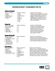
Fig. 21. Deformed larval shrimp. Arrow points todeformed appendage.<br />
(Photo courtesy<strong>of</strong> George Chamberlain.)<br />
to <strong>the</strong> cuticle or underlying skin. A genetic cause is suspected.<br />
Transformation to blue coloration from a natural brown is<br />
known for some captive crustaceans and has been linked to<br />
nutrition. Pond-cultured, giant tiger shrimp sometime develop a<br />
condition wheredigestive gland degeneration contributes to a<br />
reddish coloration.<br />
that are capable <strong>of</strong> developing into a new individual.<br />
Bacteria areone-celled organisms that can be seen only<br />
with a microscope. Compared to protozoans, <strong>the</strong>y arc <strong>of</strong> less<br />
complex organization and normally less than 1/5,000 inch<br />
(1/2000 cm) in size.<br />
Rickettsia are microbes with similarity to both viruses<br />
and bacteria and have a size that is normally somewhat inbetween.<br />
Most think <strong>of</strong> <strong>the</strong>m as small bacteria.<br />
Viruses arc ultramicroscopic, infective agents capable <strong>of</strong><br />
multiplying in connection with living cells. Normally, vi<br />
ruses are many times smaller than bacteria but may be made<br />
clearly visible at high magnification provided by an electron<br />
microscope.