Handbook of Shrimp Diseases - the National Sea Grant Library
Handbook of Shrimp Diseases - the National Sea Grant Library
Handbook of Shrimp Diseases - the National Sea Grant Library

Create successful ePaper yourself
Turn your PDF publications into a flip-book with our unique Google optimized e-Paper software.
LOAN COPY ONLY TAMU-H-95-001 C3<br />
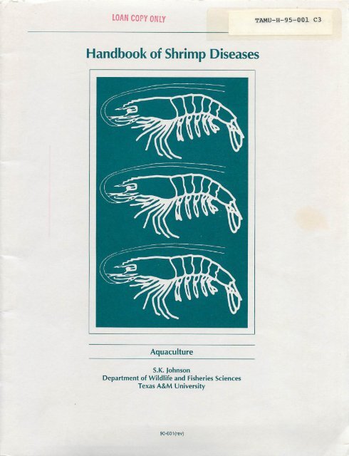
<strong>Handbook</strong> <strong>of</strong> <strong>Shrimp</strong> <strong>Diseases</strong><br />
Aquaculture<br />
S.K. Johnson<br />
Department <strong>of</strong> Wildlife and Fisheries Sciences<br />
Texas A&M University<br />
90-601 (rev)
Introduction 2<br />
<strong>Shrimp</strong> Species 2<br />
<strong>Shrimp</strong> Anatomy 2<br />
Obvious Manifestations <strong>of</strong><strong>Shrimp</strong> Disease 3<br />
Damaged Shells , 3<br />
Inflammation and Melanization 3<br />
Emaciation and Nutritional Deficiency 4<br />
Muscle Necrosis 5<br />
Tumors and O<strong>the</strong>r Tissue Problems 5<br />
Surface Fouling 6<br />
Cramped <strong>Shrimp</strong> 6<br />
Unusual Behavior 6<br />
Developmental Problems 6<br />
Growth Problems 7<br />
Color Anomalies 7<br />
Microbes 8<br />
Viruses 8<br />
Baceteria and Rickettsia 10<br />
Fungus 12<br />
Protozoa 12<br />
Haplospora 13<br />
Gregarina 15<br />
Body Invaders 16<br />
Surface Infestations 16<br />
Worms 18<br />
Trematodes 18<br />
Cestodes 18<br />
Nematodes 18<br />
Environment 20<br />
Publication <strong>of</strong> this handbook is a coop<br />
erative effort <strong>of</strong> <strong>the</strong> Texas A&M Univer<br />
sity <strong>Sea</strong> <strong>Grant</strong> College Program, <strong>the</strong><br />
Texas A&M Department <strong>of</strong> Wildlife and<br />
Fisheries Sciences and <strong>the</strong> Texas<br />
Agricultural Extension Service. Produc<br />
tion is supported in part by Institutional<br />
<strong>Grant</strong> No. NA16RG0457-01 to Texas<br />
A&M University by <strong>the</strong> <strong>National</strong> <strong>Sea</strong><br />
<strong>Grant</strong> Program, <strong>National</strong> Oceanic and<br />
Atmospheric Administration, U.S. De<br />
partment <strong>of</strong> Commerce.<br />
$2.00<br />
Additional copies available from:<br />
<strong>Sea</strong> <strong>Grant</strong> College Program<br />
1716 Briarcrest Suite 603<br />
Bryan, Texas 77802<br />
TAMU-SG-90-601(r)<br />
2M August 1995<br />
NA89AA-D-SG139<br />
A/1-1
<strong>Handbook</strong> <strong>of</strong><strong>Shrimp</strong> <strong>Diseases</strong><br />
S.K. Johnson<br />
Extension Fish Disease Specialist<br />
This handbook is designed as an information source and<br />
field guide for shrimp culturists, commercial fishermen, and<br />
o<strong>the</strong>rs interested in diseases or abnormal conditions <strong>of</strong> shrimp.<br />
It describes and illustrates common maladies, parasites and<br />
commensals <strong>of</strong> commercially important marine shrimp. De<br />
scriptions include information on <strong>the</strong> life cycles and general<br />
biological characteristics <strong>of</strong> disease-producing organisms that<br />
spend all or part <strong>of</strong> <strong>the</strong>ir life cycles with shrimp.<br />
Disease is one <strong>of</strong> <strong>the</strong> several causes <strong>of</strong> mortality in shrimp<br />
stocks. Death from old age is <strong>the</strong> potential fate <strong>of</strong> all shrimp,<br />
but <strong>the</strong> toll taken by predation (man being one <strong>of</strong> <strong>the</strong> major<br />
predators), starvation, infestation, infection and adverse envi<br />
ronmental conditions is much more important.<br />
Although estimates <strong>of</strong> <strong>the</strong> importance <strong>of</strong> disease in natural<br />
populations are generally unreliable, <strong>the</strong> influence <strong>of</strong> disease,<br />
like predation and starvation, is accepted as important in lower<br />
ing numbers <strong>of</strong> natural stocks whenever <strong>the</strong>y grow to excess.<br />
Disease problems are considered very important to success<br />
ful production in shrimp aquaculture. Because high-density,<br />
confined rearing is unnatural and may produce stress, some<br />
shrimp-associated organisms occasionally become prominent<br />
factors in disease. Special measures are required to <strong>of</strong>fset <strong>the</strong>ir<br />
detrimental effects.<br />
Disease may be caused by living agents or o<strong>the</strong>r influences<br />
<strong>of</strong> <strong>the</strong> general environment. Examples <strong>of</strong> influences in <strong>the</strong><br />
general environment that cause disease are lack <strong>of</strong> oxygen,<br />
poisons, low temperatures and salinity extremes. This guide<br />
concentrates on <strong>the</strong> living agents and on visual presentation <strong>of</strong><br />
<strong>the</strong> structure and effects <strong>of</strong> such agents.<br />
<strong>Shrimp</strong> Species<br />
There are many shrimp species distributed world-wide.<br />
Important shrimp <strong>of</strong> <strong>the</strong> Gulf <strong>of</strong> Mexico catch are <strong>the</strong> brown<br />
shrimp, Penaeus aztecus\ <strong>the</strong> white shrimp, Penaeus setiferus;<br />
and <strong>the</strong> pink shrimp; Penaeus duorarum.<br />
Two exotic shrimp have gained importance in Gulf Coast<br />
aquaculture operations. These are <strong>the</strong> Pacific white (white leg)<br />
shrimp, Penaeus vannamei, and <strong>the</strong> Pacific blue shrimp,<br />
Penaeus stylirostris. These two species are used likewise<br />
throughout <strong>the</strong> Americas on both east and west coasts.<br />
In Asia, <strong>the</strong> Pacific, and to some extent <strong>the</strong> Mediterranean,<br />
<strong>the</strong> following species are used: Penaeus monodon, Penaeus<br />
merguiensis, Penaeus chinensis, Penaeusjaponicus, Penaeus<br />
semisulcatus, Penaeus indicus, Penaeus penicillatus and<br />
Metapenaeusensis. Penaeus monodon, <strong>the</strong> giant tiger (or black<br />
tiger) shrimp is <strong>the</strong> world leader in aquaculture.<br />
<strong>Shrimp</strong> Anatomy<br />
A shrimp is covered with a protective cuticle (exoskeleton,<br />
shell) and has jointed appendages. Most organs are located in<br />
<strong>the</strong> head end (cephalothorax) with muscles concentrated in <strong>the</strong><br />
tail end (abdomen). The parts listed below are apparent upon<br />
outside examination (Fig. 1).<br />
1. Cephalothorax<br />
2. Abdomen<br />
3. Antennules<br />
4. Antenna<br />
5. Antennal scale<br />
6. Rostrum (horn)<br />
7. Eye<br />
8. Mouthparts (several<br />
appendages for holding<br />
and tearing food)<br />
9. Carapace (covering <strong>of</strong><br />
cephalothorax)<br />
10. Walking legs (pereiopods)<br />
11. Abdominal segment<br />
12. Swimmerets (pleopods)<br />
13. Sixth abdominal seg<br />
ment<br />
14. Telson<br />
15. Uropod<br />
16. Gills<br />
Inside structures include (Fig. 2)<br />
1. Esophagus<br />
2. Stomach<br />
3. Hemocoel (blood space)<br />
4. Digestive gland (hepatopancreas)<br />
5. Heart<br />
6. Intestine<br />
7. Abdominal muscles<br />
The "skin" or hypodermis<br />
<strong>of</strong> a shrimp lies just beneath<br />
<strong>the</strong> cuticle. It is functional in<br />
secreting <strong>the</strong> new exoskeleton<br />
that develops to replace <strong>the</strong><br />
old at shedding. Shedding <strong>of</strong><br />
<strong>the</strong> cuticle (also known as<br />
molting or ecdysis) occurs at<br />
intervals during a shrimp's<br />
life and allows for change in<br />
developmental stage and ex<br />
pansion in size.<br />
Fig. 1. External anatomy <strong>of</strong> shrimp.<br />
(Numbers conform to list.)<br />
Fig. 2. Internal anatomy <strong>of</strong><br />
shrimp. (Numbers conform to<br />
list.) Jagged line represents<br />
cutaway <strong>of</strong> cuticle to expose<br />
internal organs.<br />
The reproductive organs <strong>of</strong> adults are particularly notice<br />
able. When ripe, <strong>the</strong> ovaries <strong>of</strong> females may be seen through<br />
<strong>the</strong> cuticle to begin in <strong>the</strong> cephalothorax and extend dorsally<br />
into <strong>the</strong> abdomen. Spermatophores, a pair <strong>of</strong> oval structures<br />
containing <strong>the</strong> sperm in adult males, are also visible through<br />
<strong>the</strong> cuticle when viewed from <strong>the</strong> underside near <strong>the</strong> juncture<br />
<strong>of</strong> cephalothorax and abdomen. The principle nervous struc-
ture, <strong>the</strong> ventral nerve cord, is visible along <strong>the</strong> underside <strong>of</strong><br />
<strong>the</strong> body between <strong>the</strong> swimmerets.<br />
Obvious Manifestations <strong>of</strong> <strong>Shrimp</strong> Disease<br />
Damaged Shells<br />
<strong>Shrimp</strong> cuticle is easily damaged in aquaculture situations<br />
when hard structures are impacted or rubbed. (Fig. 3). Blood<br />
runs openly (outside <strong>of</strong> vessels) under <strong>the</strong> shell <strong>of</strong> shrimps,out<br />
through appendagesand into tiny fringe parts. When injury<br />
occurs to <strong>the</strong> shell, <strong>the</strong> blood quickly clots and protectsdeeper<br />
parts (Fig. 4).<br />
Shell damage may also be inflicted by <strong>the</strong> pinching or biting<br />
<strong>of</strong> o<strong>the</strong>rshrimpin crowded conditions. Parts <strong>of</strong> appendages<br />
such as antennae may be missing. Cannibalism has an impor<br />
tant influence on survival in some phases <strong>of</strong> shrimp culture<br />
where stronger individuals devour weak ones (Fig. 5).<br />
Shells may also be damaged because <strong>the</strong>y become infected.<br />
A protective outer layer is part <strong>of</strong> <strong>the</strong> cuticle. If underlying<br />
portions arc exposed opportunistic microbes will invade <strong>the</strong><br />
shell and use it as a food base or portal for entry into deeper<br />
tissue. Larger marks darken and become obvious (Fig. 6).<br />
Inflammation and Melanization<br />
Darkening <strong>of</strong> shell and deeper tissues is a frequent occur<br />
rence with shrimp and o<strong>the</strong>r crustaceans. In <strong>the</strong> usual case,<br />
blood cells gradually congregate in particular tissue areas (in<br />
flammation) where damage has occurred and this is followed<br />
by pigment (melanin) deposition. An infective agent, injury or<br />
a toxin may cause damage and stimulate <strong>the</strong> process (Fig. 7).<br />
Gills arc particularly prone to darkening due to <strong>the</strong>ir fragile<br />
nature and <strong>the</strong>ir function as a collecting site for elimination <strong>of</strong><br />
<strong>the</strong> body's waste products (Fig. 8). Gills readily darken upon<br />
exposure to toxic metals or chemicals and as a result <strong>of</strong> infec<br />
tion by certain fungi (Fusarium sp.).<br />
Less common but important are dark blotches that sometime<br />
occur within <strong>the</strong> tails <strong>of</strong> pond shrimp. This manifestation <strong>of</strong><br />
necrosis (breakdown and death) <strong>of</strong> muscle portions followed by<br />
melanization degrades <strong>the</strong> product's market potential. It is<br />
possible that this condition results from deep microbial inva<br />
sions that run through spaces between muscle bundles but its<br />
actual causes remain unknown (Fig. 9).<br />
Fig. 5. Cannibalism usually begins as o<strong>the</strong>r shrimp devour <strong>the</strong> append<br />
ages.<br />
Fig. 3. Eyes <strong>of</strong> shrimp are normally black, but rubbing <strong>of</strong> a tank wall<br />
has caused this eye to appear whitish because <strong>of</strong> a prominent lesion.<br />
Fig. 4. Microscopic view <strong>of</strong> a lesion on a uropod (tail part). Note crease<br />
from bend in part and loss <strong>of</strong> fringe setae.<br />
Fig. 6. Tail ends <strong>of</strong> two shrimp. The lower shrimp shows typical darken<br />
ing <strong>of</strong> cuticle that involves microbial action. The darkening itself is<br />
considered a host response. The telsons <strong>of</strong> <strong>the</strong> upper shrimp are<br />
opaque because <strong>of</strong> dead inner tissue. Successful entry and tissue<br />
destruction by bacteria was accomplished only in those parts.
Fig. 7. A shrimp photographed (above) near time <strong>of</strong> back injury and<br />
(below) hours later. Injury by a toxin or disease agent will usually trig<br />
ger a similar response <strong>of</strong> inflammation and melanization.<br />
, •k<br />
J<br />
f .<br />
Fig. 8. Microscopic view <strong>of</strong> damaged and melanized gill tips.<br />
Emaciation and Nutritional Deficiency<br />
Unfed shrimp lose <strong>the</strong>ir normal full and robust appearance<br />
and exhibit emaciation. The shell becomes thin and flexible as<br />
it covers underlying tissue such as tail meat that becomes<br />
greatly resorbed for lack <strong>of</strong> nutrients. Molting is curtailed and<br />
shell and gills may darken in time (Fig. 10). Emaciation may<br />
also follow limited feeding behavior during chronic disease<br />
conditions or an exposure to unfavorable environmental condi<br />
tions. Empty intestines arc easily observed through transparent<br />
cuticle and flesh.<br />
Prepared diets deficient in necessary constituents may pre<br />
dispose or cause disease. Vitamin C deficiency, for example,<br />
will initiate darkening <strong>of</strong> gills or certain tissues associated with<br />
<strong>the</strong> cuticle and eventually result in deaths.<br />
Fig. 9. Areas <strong>of</strong> melanized necrotic tissue in tail musculature.<br />
Fig. 10. Emaciated shrimp. Gills and body fringes have become obviously<br />
darkened and <strong>the</strong> s<strong>of</strong>t tail is covered with a thin and fragile cuticle.<br />
Fig. 11. Lipoid (fat) spheres in microscopic view <strong>of</strong> digestive gland tubule.
Digestive glands sometimes will become reduced in size.<br />
Among o<strong>the</strong>r things, this is an indication <strong>of</strong> poor nutrition.<br />
Well-fed shrimp will have an abundance <strong>of</strong> fat globules within<br />
storage cells <strong>of</strong> <strong>the</strong> digestive gland tubules that provide bulk to<br />
<strong>the</strong> gland (Figs. 11 and 12).<br />
Muscle Necrosis<br />
Opaque muscles are characteristic <strong>of</strong> this condition. When<br />
shrimp areexposed tostressful conditions, such as low oxygen<br />
or crowding, <strong>the</strong>muscles lose<strong>the</strong>ir normal transparency and<br />
become blotched with whitish areas throughout. This may<br />
progress until <strong>the</strong> entire tail area takes on a whitish appearance<br />
(Fig. 13).<br />
If shrimp arc withdrawn from <strong>the</strong> adverse environment<br />
before prolonged exposure, <strong>the</strong>y may return to normal. Ex<br />
tremely affected shrimp do not recover, however, and die<br />
within a few minutes (Fig. 14). In moderately affected shrimp,<br />
only parts <strong>of</strong> <strong>the</strong> body return to normal; o<strong>the</strong>r parts, typically<br />
<strong>the</strong> last segments <strong>of</strong> <strong>the</strong> tail are unable to recover and areprone<br />
to bacterial infection (Fig. 15). These shrimp die within oneor<br />
two days (Fig. 5). <strong>Shrimp</strong> muscles with this condition are<br />
known to undergo necrosis (death or decay <strong>of</strong> tissue).<br />
Tumors and O<strong>the</strong>r Tissue Problems<br />
Conspicuous body swellings or enlargements<strong>of</strong> tissueshave<br />
been reported in shrimp. In most cases, affected individuals<br />
werecaptured from polluted waters. Occurrence<strong>of</strong> shrimp with<br />
evident tumors is rare in commercial catches. Miscellaneous<br />
irritations experienced by captive shrimp in tanksystems will<br />
sometimes result in focal areas <strong>of</strong> tissue overgrowth (Fig. 16).<br />
A particularly vulnerable tissue <strong>of</strong> captivejuvenile and adult<br />
shrimp is found on <strong>the</strong> inner surface <strong>of</strong> <strong>the</strong> portion <strong>of</strong> carapace<br />
that covers <strong>the</strong> gills. When microbes invade this tissue, it and<br />
<strong>the</strong>adjacent outershell maycompletely disintegrate exposing<br />
<strong>the</strong>gills. In o<strong>the</strong>rcases a partial loss<strong>of</strong> <strong>the</strong> tissue distally may<br />
result in an outward flaring <strong>of</strong> <strong>the</strong> exposed cuticle. A hemolymphoma<br />
or fluid-filled blister also forms sometime in this<br />
Fig. 15. Damage to abdomen <strong>of</strong> a shrimp as a result <strong>of</strong> Vibrio infection.<br />
Fig. 12. Pond-raised shrimp with full, normal and reduced, abnormal<br />
digestive gland. Arrow points to abnormal gland.<br />
Fig. 13. <strong>Shrimp</strong> with necrotic muscle tissue following exposure to<br />
stressful environment. Affected tissue at arrow.<br />
Fig. 14. <strong>Shrimp</strong> with advanced muscle necrosis (arrow) shown beside<br />
normal shrimp.<br />
Fig. 16. Tumorous growth on an adult shrimp from a tank system.
Fig. 17. Blister condition. Insetshows blister removed. The blister will<br />
darken upon death <strong>of</strong> shrimp degrading marketability <strong>of</strong> heads-on<br />
product.<br />
portion <strong>of</strong> <strong>the</strong> carapace in pond shrimp (Fig. 17). Primary<br />
causes <strong>of</strong> <strong>the</strong>se manifestations are not understood.<br />
A degeneration <strong>of</strong> male reproductivetracts occasionally<br />
occurs in captive adults <strong>of</strong> certain penaeid species. A swelling<br />
and darkening <strong>of</strong> <strong>the</strong> tubule leading from <strong>the</strong> testes to <strong>the</strong> spcrmatophorc<br />
is readily apparent when viewed through <strong>the</strong> trans<br />
lucent body (Fig. 18).<br />
Surface Fouling<br />
The surfaces<strong>of</strong> shrimpsarc prone to an accumulation <strong>of</strong> vari<br />
ous fouling organisms. Heavy infestations can interfere with mo<br />
bility or breathing and influence marketability (Fig. 19).<br />
Cramped <strong>Shrimp</strong><br />
This is a condition described for shrimp kept in a variety <strong>of</strong><br />
culture situations. The tail is drawn under <strong>the</strong> body and be<br />
comes rigid to <strong>the</strong> point that it cannot be straightened (Fig. 20).<br />
The cause <strong>of</strong> cramping is unknown, but some research points to<br />
mineral imbalance.<br />
Unusual Behavior<br />
Diseased shrimps <strong>of</strong>ten display listless behavior and cease<br />
to feed. In <strong>the</strong> case <strong>of</strong> water quality extremes such as low oxy<br />
gen, shrimp may surface and congregate along shores where<br />
<strong>the</strong>y become vulnerable to bird predation. Cold water may<br />
cause shrimp to burrow and an environmental stimulation such<br />
as low oxygen, <strong>the</strong>rmal change or sudden exposures to unusual<br />
chemicals may initiate widespread molting.<br />
Fig. 18. Darkening <strong>of</strong> male reproductive tract <strong>of</strong> Penaeus stylirostris. A.<br />
Normal tract. B. Initial darkening. Darkening will advance until spermatophores<br />
and testes become affected. (Photos courtesy <strong>of</strong> George<br />
Chamberlain.)<br />
Fig. 19. Algal overgrowth on shrimp exposed to abundant light. (Photo<br />
courtesy <strong>of</strong> Steve Robertson.)<br />
Developmental Problems<br />
Fig.20. Cramped shrimp condition. Full flexure (A). Flexure maintained when pressure applied (B).<br />
Deformities are quite prevalent in some populations. They<br />
arise from complex interactions that involve environment, diet<br />
and gene expression. Bodies may be twisted or appendages<br />
misshaped or missing. Deformities arc less prevalent in wildcaught<br />
larvae than hatchery populations probably because wild<br />
shrimp have more opportunity for natural selection and expo<br />
sure to normal developmental conditions (Fig. 21).
Molt arrest occurs in affected animals <strong>of</strong>some populations.<br />
Animals begin, but are unable to complete <strong>the</strong> molting process.<br />
In some cases, <strong>the</strong>re is abnormal adherence to underlying skin,<br />
but most animals appear to lack <strong>the</strong> necessary stamina. Nutri<br />
tional inadequacies and water quality factors have been identi<br />
fied as causes.<br />
Growth Problems<br />
Growth problems become obvious inaquaculture stocks. A<br />
harvested population may show a larger percentage <strong>of</strong>ranting<br />
than expected. Some research hasconnected viral disease with<br />
ranting in pond stocks and it is generally held that variable<br />
growth may result from disease agents, genetic makeup and<br />
environmental influences.<br />
For unknown reasons, <strong>the</strong> shell orcuticle may become frag<br />
ile in members <strong>of</strong> captive shrimp stocks.<br />
Shells are normally s<strong>of</strong>t for a couple <strong>of</strong>days after molting,<br />
but shells <strong>of</strong> those suffering from s<strong>of</strong>t-shell condition remain<br />
both s<strong>of</strong>t and thin and havea tendency to crack under <strong>the</strong><br />
slightest pressure. Some evidence <strong>of</strong>cause suggests pesticide<br />
toxicity, starvation (mentioned above) or mineral imbalance.<br />
Color Anomalies<br />
<strong>Shrimp</strong> <strong>of</strong> unusual color arc occasionally found among wild<br />
and farm stocks. Thestriking coloration, which may begold,<br />
blue or pink, appears throughout <strong>the</strong> tissue and is notconfined<br />
Microbes are minute, living organisms, especially vi<br />
ruses, bacteria, rickcttsia and fungi. Sometimes protozoa arc<br />
considered microbes.<br />
Protozoa are microscopic, usually one-celled,animals<br />
that belong to <strong>the</strong> lowest division <strong>of</strong> <strong>the</strong> animal kingdom.<br />
Normally, <strong>the</strong>y are many times larger than bacteria. The<br />
typical protozoareproduce by simple or multipledivision or<br />
by budding. The more complex protozoa alternate between<br />
hosts and produce cells with multiple division stages called<br />
spores.<br />
Fungi associated with shrimp are microscopic plantsthat<br />
develop interconnecting tubular structures. They reproduce<br />
by forming small cells known as spores or fruiting bodies<br />
Microbes<br />
Fig. 21. Deformed larval shrimp. Arrow points todeformed appendage.<br />
(Photo courtesy<strong>of</strong> George Chamberlain.)<br />
to <strong>the</strong> cuticle or underlying skin. A genetic cause is suspected.<br />
Transformation to blue coloration from a natural brown is<br />
known for some captive crustaceans and has been linked to<br />
nutrition. Pond-cultured, giant tiger shrimp sometime develop a<br />
condition wheredigestive gland degeneration contributes to a<br />
reddish coloration.<br />
that are capable <strong>of</strong> developing into a new individual.<br />
Bacteria areone-celled organisms that can be seen only<br />
with a microscope. Compared to protozoans, <strong>the</strong>y arc <strong>of</strong> less<br />
complex organization and normally less than 1/5,000 inch<br />
(1/2000 cm) in size.<br />
Rickettsia are microbes with similarity to both viruses<br />
and bacteria and have a size that is normally somewhat inbetween.<br />
Most think <strong>of</strong> <strong>the</strong>m as small bacteria.<br />
Viruses arc ultramicroscopic, infective agents capable <strong>of</strong><br />
multiplying in connection with living cells. Normally, vi<br />
ruses are many times smaller than bacteria but may be made<br />
clearly visible at high magnification provided by an electron<br />
microscope.
Viruses<br />
Microbes<br />
Our knowledge <strong>of</strong> <strong>the</strong> diversity <strong>of</strong> shrimp viruses continues to<br />
grow. Viruses <strong>of</strong> shrimp have been assigned explicitly or tenta<br />
tively to six or seven categories. Several shrimp viruses are recog<br />
nized to have special economic consequence in aquaculture:<br />
Baculoviruses<br />
Baculovirus penaei — a virus common to Gulf <strong>of</strong> Mexico<br />
shrimp. It damages tissue by entering a cell nucleus and subse<br />
quently destroys <strong>the</strong> cell as it develops (Fig. 23). An occlusion<br />
is formed (Fig. 24). This virus has become a constant problem<br />
for many shrimp hatcheries where it damages <strong>the</strong> young larval<br />
animals. Occlusions <strong>of</strong> <strong>the</strong> same or closely related viruses are<br />
seen in Pacific and Atlantic Oceans <strong>of</strong> <strong>the</strong> Americas. At least<br />
ten shrimp species arc known to show disease manifestations in<br />
aquaculture settings.<br />
Monodon-typc baculovirus — one that forms spherical<br />
occlusions (Fig. 25) and whose effects arc seen mostly in <strong>the</strong><br />
culture <strong>of</strong> <strong>the</strong> giant tiger prawn, Penaeus monodon. Damage <strong>of</strong><br />
less importance has been seen in Penaeus japonicus, Penaeus<br />
merguiensis and Penaeus plebejus.<br />
Midgut gland necrosis virus — a naked baculovirus harmful<br />
to <strong>the</strong> Kuruma prawn, Penaeusjaponicus, in Japan.<br />
Solubility in Gut<br />
Ingestion ot Contaminated Food<br />
Infection <strong>of</strong> Host<br />
Fig. 23. Baculovirus lifecycle. Transmission <strong>of</strong> <strong>the</strong> virus is thought to<br />
be initiated as a susceptible shrimp ingests a viral occlusion. Virus<br />
initially enters cell cytoplasm ei<strong>the</strong>r by viroplexis (cell engulfs particle<br />
with surrounding fluid) or by fusion where viral and cell membranes<br />
fuse and viral core passes into cell. Secondary infection occurs as<br />
extracellular virus continues to infect. (Redrawn by Summers and<br />
Smith, 1987. Used with permission <strong>of</strong> author and Texas Agricultural<br />
Experiment Station, The Texas A&M University System.)<br />
Fig. 24. Occlusion bodies <strong>of</strong> Baculovirus penaei. These bodies, visible<br />
to low power <strong>of</strong> a light microscope, are characteristic <strong>of</strong> this virus. The<br />
occlusions and those <strong>of</strong> o<strong>the</strong>r baculoviruses are found mainly in <strong>the</strong><br />
digestive gland and digestive tract.<br />
Fig. 25. Monodon baculovirus in a tissue squash showing groups <strong>of</strong><br />
spherical occlusions. Light microscopy.<br />
Parvoviruses<br />
Infectious hypodcrmal and hematopoietic necrosis virus —<br />
a virus affecting several commercially important shrimp and,<br />
particularly, <strong>the</strong> Pacific blue shrimp, Penaeus stylirostris.<br />
Hepatopancreatic parvo-Iikc virus — a virus causing disease<br />
in several Asian shrimp. Transmission to Penaeus vannamei<br />
did not result in disease to that species.<br />
Nodavirus<br />
Taura virus — a virus causing obvious damage to various<br />
tissues and in <strong>the</strong> acute phase, to <strong>the</strong> hypodermis and subse<br />
quently <strong>the</strong> cuticle <strong>of</strong> Penaeus vannamei (Fig. 26). It is an<br />
important problem for both production and marketing. During<br />
<strong>the</strong> 1995 growing season, this virus caused large losses to<br />
aquaculture stocks in Texas. Damage was great in Central and<br />
South America beginning in 1992.
O<strong>the</strong>r viruses<br />
Yellow head virus — a virus causing serious disease <strong>of</strong> <strong>the</strong><br />
giant tiger prawn, Penaeus monodon. Large losses have been<br />
experienced in Asian aquaculture units. Gills and digestive<br />
glands <strong>of</strong> infected shrimp arc pale yellow.<br />
White spot diseases — viruses <strong>of</strong> similar size and structure<br />
have been shown to cause a similar manifestation and heavy<br />
losses to Penaeus japonicus, Penaeus monodon and Penaeus<br />
penicillatus in Taiwan and Japan. Advanced infections show<br />
development <strong>of</strong> obvious white spots on <strong>the</strong> inside <strong>of</strong> <strong>the</strong> cuticle<br />
(Fig. 27).<br />
Several o<strong>the</strong>r viruses with relatively little known importance<br />
arc considered as members <strong>of</strong> <strong>the</strong> rcoviruses, rhabdoviruscs,<br />
togaviruscs.<br />
Fig. 27. Asian shrimp showing signs <strong>of</strong> white spot disease. (Photo<br />
courtesy <strong>of</strong> R. Rama Krishna.)<br />
Viruses<br />
Viruses cause disease as <strong>the</strong>y replicate within a host cell and<br />
<strong>the</strong>reby cause destruction or improper cell function. A virus is<br />
essentially a particle containing a core <strong>of</strong> nucleic acids, DNA<br />
or RNA. Once inside a proper host cell, <strong>the</strong> viral nucleic acid<br />
interacts with that <strong>of</strong> a normal cell to cause reproduction <strong>of</strong> <strong>the</strong><br />
virus. The ability to parasitize and cause damage may be lim<br />
ited to a single species or closely related group <strong>of</strong> hosts, a host<br />
tissue and usually <strong>the</strong> place within a cell in which damage<br />
takes place.<br />
The cause and effect for all shrimp virus disease needs care<br />
ful attention. Some viruses cause disease only after exposure to<br />
unusual environmental conditions. Also, impressions about<br />
virus identity arc <strong>of</strong>ten based on results <strong>of</strong> routine examinations<br />
that give presumptive results. Certainly viruses cause important<br />
disease in particular circumstances but key understandings <strong>of</strong><br />
most shrimp viruses are largely unknown: longevity within<br />
systems, source <strong>of</strong> infection, method <strong>of</strong> transmission, normal<br />
and unusual carriers, and potential to cause damage.<br />
Our ability to detect shrimp viruses is ahead <strong>of</strong> our ability to<br />
evaluate <strong>the</strong>ir importance or to implement controls. For viral<br />
identification, scientists have employed <strong>the</strong> recent technology<br />
that detects characteristic nucleic acids. This is augmented by<br />
careful microscopical study <strong>of</strong> tissues to detect characteristic<br />
damage to cells. Use <strong>of</strong> electron microscopy to determine size<br />
and shape <strong>of</strong> virus particles has also been helpful (Fig. 22).<br />
A peculiar feature <strong>of</strong> some baculoviruses <strong>of</strong> shrimp and<br />
o<strong>the</strong>r invertebrate animals is to occurrence <strong>of</strong> <strong>the</strong> occlusion<br />
bodies within infected cells. These are relatively large masses<br />
<strong>of</strong> consistent shape that contain virus particles embedded<br />
Fig. 26. Advanced stage <strong>of</strong> infection with Taura virus showing damage<br />
to cuticle. Smaller shrimp with acute infection do not show such dam<br />
age but do show reddish telson and uropods.<br />
within. O<strong>the</strong>r "naked" baculoviruses do not show formation <strong>of</strong><br />
occlusions.<br />
A<br />
Fig. 22. Structure <strong>of</strong> viruses reported from shrimps. A. Baculoviridae.<br />
Size range is about 250 to 400 nanometers in length. B. Basic struc<br />
ture <strong>of</strong> most <strong>of</strong> <strong>the</strong> o<strong>the</strong>r shrimp viruses: Parvo-like viruses—20 to 24<br />
nm in diameter containing DNA; Reo-like viruses—55 to 70 nm diam<br />
eter, RNA; nodavirus—30 nm diameter, RNA; toga-like virus 30 diam<br />
eter, RNA, enveloped. Rhabdoviruses are elongated like baculoviruses<br />
but a blunt end provides bullet-shapes—150 to 250 nm, RNA.<br />
B
m<br />
• *<br />
Fig. 28. View with light microscope <strong>of</strong> a tissue squash <strong>of</strong> infected di<br />
gestive gland. Note dark necrotized tissue <strong>of</strong> tubules (arrows).<br />
Bacteria and Rickettsia<br />
Bacterial infections <strong>of</strong> shrimp have been observed for many<br />
years. Scientists have noticed that bacterial infection usually<br />
occurs when shrimp arc weakened. O<strong>the</strong>rwise normal shrimp<br />
also may become infected if conditions favor presence and<br />
abundance <strong>of</strong> a particularly harmful bacterium.<br />
<strong>Shrimp</strong> body fluids are most <strong>of</strong>ten infected by <strong>the</strong> bacterial<br />
group named Vibrio. Infected shrimp show discoloration <strong>of</strong> <strong>the</strong><br />
body tissues in some instances, but not in o<strong>the</strong>rs. The clotting<br />
function <strong>of</strong> <strong>the</strong> blood, critical in wound repair, is slowed or lost<br />
during some infections. Members <strong>of</strong> one group <strong>of</strong> Vibrio have<br />
<strong>the</strong> characteristic <strong>of</strong> luminescence giving heavily infected ani<br />
mals a "glow-in-thc-dark" appearance.<br />
Bacteria also invade <strong>the</strong> digestive tract. A typical infection<br />
in larval animals is seen throughout <strong>the</strong> digestive system. In<br />
Fig. 30. Histological cross section <strong>of</strong> a digestive gland tubule. Rickett<br />
sial microcolonies are shown at arrows. Rickettsiae will exhibit constant<br />
brownian motion and color red with Giemsa stain, but electron micros<br />
copy is needed for definite diagnosis. (Specimen courtesy <strong>of</strong> J. Brock)<br />
Fig. 29. Transverse section <strong>of</strong> digestive gland tubules showing pro<br />
gression <strong>of</strong> granuloma formation. Normal tubules are to <strong>the</strong> left (N) and<br />
affected tubules are to <strong>the</strong> right (G.).<br />
larger animals, infection becomes obvious in <strong>the</strong> digestive<br />
gland after harmful bacteria gain entry to it, presumably via<br />
connections to <strong>the</strong> gut.<br />
Digestive gland tissues are organized as numerous tubular<br />
structures that ultimately feed into <strong>the</strong> digestive tract. Pondreared<br />
shrimp occasionally die in large numbers because <strong>of</strong><br />
diseased digestive glands. The specialized cells that line <strong>the</strong><br />
inside <strong>of</strong> <strong>the</strong> tubules arc particularly fragile and arc easily in<br />
fected. Tubules progressively die and darken (Figs. 28 and 29).<br />
This kind <strong>of</strong> disease manifestation is seen in recent reports <strong>of</strong><br />
rickettsial infection. Cells <strong>of</strong> <strong>the</strong> digestive gland tubules arc<br />
severely damaged as rickcttsiac invade and develop <strong>the</strong>rein<br />
(Figs. 30 and 31).<br />
If infected by bacteria capable <strong>of</strong> using shell for nutrition,<br />
<strong>the</strong> exoskeleton will demonstrate erosive and blackened areas<br />
-<br />
**<br />
»>'*'* 4«l5<br />
...-V -••-
Fig. 32. Microscopic view <strong>of</strong> filamentous bacteria on a shrimp pleopod.<br />
10<br />
Digestive glands are routinely searched by pathologists<br />
for signs <strong>of</strong> disease. This is done after chemical preservation,<br />
microsection, slide-mounting and staining <strong>the</strong> tissue. Trans<br />
verse sections <strong>of</strong> <strong>the</strong> tubules are <strong>the</strong>n examined with a light<br />
microscope. General damage is seen when bacteria such as<br />
Vibrio species invade tubules. Rickettsiae, viruses,<br />
microsporans and haplosporans are more selective. They<br />
invade cells and progressively cause damage from within.<br />
For comparative purposes, a drawing <strong>of</strong> a normal tubule is<br />
compared with a tubule showing a variety <strong>of</strong> typical manifes<br />
tations (Fig. 33).<br />
Microbial Disease and Digestive Glands<br />
(Fig. 6). These bacteria typically attack edges or tips <strong>of</strong> exosk<br />
eleton parts, but if break occurs in <strong>the</strong> exoskeleton <strong>the</strong> bacteria<br />
are quick to enter and cause damage.<br />
Filamentous bacteria are commonly found attached to <strong>the</strong><br />
cuticle, particularly fringe areas beset with setae (Fig. 32).<br />
When infestation is heavy, filamentous bacteria may also be<br />
present in large quantity on <strong>the</strong> gill filaments. Smaller, less<br />
obvious bacteria also settle on cuticular surfaces but arc not<br />
considered as threatening as <strong>the</strong> filamentous type.<br />
Fig. 33. A. Drawing <strong>of</strong> transverse section <strong>of</strong> digestive gland tubule<br />
with bold lines that separate several types <strong>of</strong> disease conditions. 1.<br />
Haplosporan parasite (microsporans similar but may show fully devel<br />
oped spores, see Figs. 42 and 43). 2. Rickettsiae. 3. Virus infection<br />
with manifestation <strong>of</strong> inclusion in cytoplasm. 4. Virus infection with<br />
inclusion in nucleus. 5. Virus with occlusions in swollen nucleus. As<br />
cells are destroyed, more general lesions are formed from viruses.<br />
Inclusions <strong>of</strong> viruses normally show distinctive shape and staining<br />
features. Particular viruses will infect particular tissue types (not<br />
always hepatopancreas tissue) and cell locations (nucleus or cyto<br />
plasm) within preferred hosts. Cells enlarged by haplosporans and<br />
rickettsiae may be initially distinguished by comparing larger internal<br />
components <strong>of</strong> <strong>the</strong> pre-spore units <strong>of</strong> haplosporans with almost submicroscopic<br />
particles <strong>of</strong> microcolonies <strong>of</strong> rickettsiae. H, = early<br />
haplosporan stage, H? - later stage; R - rickettsial microcolony; CI =<br />
cytoplasmic inclusion <strong>of</strong> virus; Nl = nuclear inclusion <strong>of</strong> virus; SN =<br />
swollen nucleus with occlusions within.<br />
B. The normal tubule. Toward <strong>the</strong> digestive tract, secretory cells (1)<br />
predominate and fibrous cells (3) become more numerous. Absorp<br />
tive cells (2) contain varying amounts <strong>of</strong> vacuoles according to nutri<br />
tional status.
Fungus<br />
Several fungi are known as shrimp pathogens. Two groups<br />
commonly infect larval shrimp, whereas ano<strong>the</strong>r attacks <strong>the</strong><br />
juvenile or larger shrimp. The most common genera affecting<br />
larval shrimp are Lagcnidium and Sirolpidium. The method <strong>of</strong><br />
infection requires a thin cuticle such as that characteristic <strong>of</strong><br />
larval shrimp (Figs. 34 and 35).<br />
The most common genus affecting larger shrimp is<br />
Fusarium. It is thought that entry into <strong>the</strong> shrimp is gained via<br />
cracks or eroded areas <strong>of</strong> <strong>the</strong> cuticle. Fusarium may be identi<br />
fied by <strong>the</strong> presence <strong>of</strong> canoe-shaped macroconidia that <strong>the</strong><br />
fungus produces. Macroconidia and examples <strong>of</strong> fungal infec<br />
tions arc shown in Figures 36, 37 and 38.<br />
Fig. 35. Lagenidium infection in larval shrimp. Note extensive develop<br />
ment <strong>of</strong> branchings <strong>of</strong> fungus throughout <strong>the</strong> body. (Photo courtesy <strong>of</strong><br />
Dr. Don Lightner, University <strong>of</strong> Arizona.)<br />
Fig. 37. <strong>Shrimp</strong> photographed immediately after mole: Old appendage<br />
(arrow) is not shed due to destruction <strong>of</strong> hypodermis by active fungal<br />
infection.<br />
D<br />
-<strong>Sea</strong>rch for Host<br />
jSKba<br />
Fig. 34. Transmission <strong>of</strong> Lagenidium. A. Fungus sends out discharge<br />
tube from within shrimp body. B. Vesicle forms. C. Vesicle produces<br />
motile spores that are released. D. Motile spores contact shrimp and<br />
undergo encystment. E. Germ tube is sent into <strong>the</strong> body <strong>of</strong> <strong>the</strong> shrip<br />
and fungus <strong>the</strong>n spreads throughout.<br />
Fig. 36. Canoe-shaped macroconidia <strong>of</strong> Fusarium. These structures<br />
bud <strong>of</strong>f branches <strong>of</strong> <strong>the</strong> fungus and serve to transmit fungus to shrimp.<br />
Fig. 38. Microscopic view <strong>of</strong> fungus at tip <strong>of</strong> antenna.<br />
OX<br />
n
Protozoa<br />
Protozoan parasites and commensals <strong>of</strong> shrimp will occur<br />
on <strong>the</strong> insideor outside <strong>of</strong> <strong>the</strong> body. Those on <strong>the</strong> outside are<br />
considered harmless unless present in massive or burdensome<br />
numbers. Those on <strong>the</strong> insidecan cause disease and arc repre<br />
sentative<strong>of</strong> several groups <strong>of</strong> protozoan parasites: Microspora,<br />
Haplospora and Grcgarina. Members<strong>of</strong> <strong>the</strong>se groups are<br />
known or believed to require thatsome animal besides shrimp<br />
bepresent inorder to facilitate completion <strong>of</strong> <strong>the</strong>ir lifecycles.<br />
A few protozoa are known to invade weakened larval animals<br />
directly and contribute to disease.<br />
Microspora parasitize most major animal groups, notably<br />
insects, fish and crustaceans. In shrimp, microsporan infections<br />
are best known locally as cause <strong>of</strong> a condition known as "milk"<br />
or "cotton" shrimp (Figs. 39, 40 and 41). Microsporans become<br />
remarkably abundant in <strong>the</strong> infected shrimp and cause <strong>the</strong><br />
white appearance <strong>of</strong> muscle tissues. A typical catch <strong>of</strong> wild<br />
shrimp will contain a few individuals with this condition.<br />
These shrimp are usually discarded before processing. Depend<br />
ing on <strong>the</strong> type <strong>of</strong> microsporan, <strong>the</strong> site <strong>of</strong> infection will be<br />
throughout <strong>the</strong> musculature <strong>of</strong> <strong>the</strong> shrimp or, in particular or<br />
gans and tissues.<br />
Microsporans are present in <strong>the</strong> affected shrimp in <strong>the</strong> form<br />
<strong>of</strong> spores. Spores arc small cells that can develop into a new<br />
individual.They are very minute and detection requires exami<br />
nation with a microscope (Figs. 42 and 43).<br />
Infected shrimp are noted to be agile and apparently feed as<br />
normal shrimp. However, tissue damage occurs and no doubt<br />
affects many life functions. No eggs have been found in "milk"<br />
shrimp and it is suspected that all types <strong>of</strong> microsporan infec<br />
tions can render shrimp incapable <strong>of</strong> reproduction (Fig. 44).<br />
The life cycles <strong>of</strong> shrimp microsporans have not been satis<br />
factorily worked out. However, examination <strong>of</strong> <strong>the</strong> cycles <strong>of</strong><br />
related species and miscellaneous facts contained in literature<br />
indicate that <strong>the</strong> cycle presented in Figure 45 is representative<br />
<strong>of</strong> microsporans.<br />
Fig. 39. Infected or "milk" shrimp (upper) in comparison to normal<br />
shrimp (lower). (Photo courtesy <strong>of</strong> Dr. R. Nickelson.)<br />
12<br />
Fig. 40. Two brown shrimp cut across tail.<strong>Shrimp</strong> with whitish flesh has<br />
microsporan infections throughout muscle tissue.<br />
Fig. 41. Grass shrimp with "milk" shrimp condition. The normal shrimp<br />
in <strong>the</strong> figure is transparent.<br />
Fig. 42. Microscopic view <strong>of</strong> many spores <strong>of</strong> Ameson (-Nosema) sp.<br />
The spores are free or unenveloped. Parasitic microsporans <strong>of</strong> com<br />
mercially important shrimp with enclosing envelopes are assigned to<br />
genera Pleistophora, Thelohania and Agmasoma. The latter two differ<br />
from Pleistophora in that <strong>the</strong>ir membranes retain a constant spore<br />
number <strong>of</strong> eight per envelope. Pleistophora sp. have more than eight<br />
spores per envelope.
Haplospora<br />
A member <strong>of</strong> <strong>the</strong> Haplospora, ano<strong>the</strong>r spore forming proto<br />
zoan group, was recently recognized in as important to shrimp<br />
health when researchers found infected animals in an experi<br />
mental population that had been imported into Cuba from <strong>the</strong><br />
Pacific Coast <strong>of</strong> Central America. The parasites invaded and<br />
destroyed tissues <strong>of</strong> <strong>the</strong> digestive gland (Fig. 32). Such infec<br />
tions arc not common in aquaculture.<br />
Fig. 45. Life cycle <strong>of</strong> microsporan <strong>of</strong><br />
shrimp. A. Ingestion <strong>of</strong> spores by shrimp.<br />
B. In gut <strong>of</strong> shrimp, <strong>the</strong> spore extrudes a<br />
filament that penetrates gut wall and<br />
deposits an infective unit. A cell engulfs<br />
this unit. C. Infective unit enters <strong>the</strong><br />
nucleus <strong>of</strong> <strong>the</strong> cell, undergoes develop<br />
ment and <strong>the</strong>n divides to form schizonts.<br />
D. Schizonts <strong>the</strong>n divide and develop into<br />
spores. E. By <strong>the</strong> time spores are<br />
formed, <strong>the</strong>y are located in a specific<br />
tissue (muscle, tissues around intestine,<br />
etc.). The spores are ei<strong>the</strong>r discharged<br />
from <strong>the</strong> shrimp while living or after<br />
death, but <strong>the</strong> method <strong>of</strong> release and <strong>the</strong><br />
pathway taken is not known. F. Experi<br />
ments designed to transmit infection by<br />
feeding infected shrimp to uninfected<br />
have been unsuccessful. It is assumed<br />
particular events such as involvement <strong>of</strong><br />
ano<strong>the</strong>r host may be required to com<br />
plete passage from one shrimp to <strong>the</strong><br />
next. Successful transmission has been<br />
reported when infected shrimp were fed<br />
to fish (speckled trout) and fish fecal<br />
material was <strong>the</strong>n fed to shrimp.<br />
Fig. 43. Microscopic view <strong>of</strong> many spores <strong>of</strong> Thelohania sp. Note<br />
envelope (arrow).<br />
Fig. 44. Agmasoma penaei in white shrimp. This parasite is always<br />
located along <strong>the</strong> dorsal midline (arrows). Advanced infections can be<br />
seen through <strong>the</strong> cuticle with <strong>the</strong> unaided eye.<br />
13
•* - *<br />
S:M'&i*. 1<br />
53 • '<br />
Fig. 48. Life cycle <strong>of</strong> a gregarine<br />
<strong>of</strong> shrimp. A. <strong>Shrimp</strong> ingests<br />
spores with bottom debris. B.<br />
Sporozoite emerges in <strong>the</strong> gut <strong>of</strong><br />
<strong>the</strong> shrimp. C. Sporozoite at<br />
taches to <strong>the</strong> intestinal wall and<br />
grows into a delicate trophozoite;<br />
o<strong>the</strong>r trophozoites do not attach<br />
to <strong>the</strong> wall but onto each o<strong>the</strong>r<br />
and form unusual shapes (See<br />
Fig. 47). D. The unusual forms<br />
develop and attach to <strong>the</strong> end <strong>of</strong><br />
<strong>the</strong> intestine (rectum) to form<br />
gametocysts. E. Gametocyst<br />
undergoes multiple divisions to<br />
produce "gymnospores" that are<br />
set free with rupture <strong>of</strong> <strong>the</strong><br />
gametocyst. F. Gymnospores<br />
are engulfed by cells at <strong>the</strong><br />
surface <strong>of</strong> <strong>the</strong> flesh <strong>of</strong> clams. G.<br />
They develop to form spores in<br />
<strong>the</strong> clam. H. Then <strong>the</strong> spores<br />
(with sporozoite inside) are<br />
liberated from <strong>the</strong> clam in mu<br />
cous strings (slime).<br />
14<br />
Fig. 46. Microscopic views <strong>of</strong> gregarines. A. and B. Nematopsis sp. tropohzoites. C. Nematopsis sp. gametocyst.<br />
D. Trophozoites <strong>of</strong> Cephalolobus sp., a gregarine that attaches to <strong>the</strong> base <strong>of</strong> <strong>the</strong> terminal lappets <strong>of</strong> <strong>the</strong> shrimp<br />
stomach ra<strong>the</strong>r than <strong>the</strong> intestinal wall (photo courtesy <strong>of</strong> Dr. C. Corkern). E. Trophozites <strong>of</strong> Paraophioidina sp., a<br />
gregarine found recently in Pacific white shrimp larvae.<br />
Gregarina<br />
Gregarine are protozoa that occur within <strong>the</strong> digestive tract<br />
and tissues <strong>of</strong> various invertebrate animals. They occur in <strong>the</strong><br />
digestive tract <strong>of</strong> shrimp and arc observed most <strong>of</strong>ten in <strong>the</strong><br />
form <strong>of</strong>a trophozoite (Fig. 46) or occasionally a gametocyst<br />
(Fig. 47). The life cycle involves o<strong>the</strong>r invertebrates such as<br />
snails, clams or marine worms as diagrammed in Figure 48.<br />
Minor damage to <strong>the</strong> host shrimp results from attachment <strong>of</strong><br />
<strong>the</strong> trophozoites to <strong>the</strong> lining <strong>of</strong> <strong>the</strong> intestine. Earlier study sug<br />
gested that absorption <strong>of</strong> food or intestinal blockage by <strong>the</strong> proto<br />
zoa is perhaps detrimental but that pathological damage was rela<br />
tively unimportant. Recent study indicates that when trophozoites<br />
Fig. 47. Microscopic view <strong>of</strong> °' Nematopsis species arc present in large numbers, damage to <strong>the</strong><br />
gametocyst <strong>of</strong> Nematopsis sp. gut lining occurs that may facilitate infection by bacteria.
A. Zoothamnium<br />
D. Acineta<br />
* *<br />
Fig. 49. Weakened larval shrimp invaded by ciliated protozoans (arrows).<br />
Body Invaders<br />
Several protozoa have been noted to invade a shrimp body<br />
and feed on tissues as <strong>the</strong>y wander throughout. Tentative iden<br />
tifications name Piwauronema, Leptomonas, Paranophrys and<br />
an amoeba. Adverse effects <strong>of</strong> <strong>the</strong>se protozoa are not fully<br />
understood, but <strong>the</strong>y are usually found associated with shrimp<br />
that have become weakened or diseased (Fig. 49).<br />
Ectocommensal Protozoa<br />
Surface Infestations<br />
Several kinds <strong>of</strong> protozoa are regularly found on surfaces,<br />
includinggills, <strong>of</strong> shrimp. Apparently, shrimp surfaces are a<br />
favored place to live within <strong>the</strong> water environment. Common<br />
on <strong>the</strong> surfaces <strong>of</strong> shrimp are species <strong>of</strong> Zoothamnium,<br />
Epistylis, Acineta, Eplwlota, and Lagenophry.s (Fig. 50A-E).<br />
i-rr<br />
*#<br />
B. Epistylis<br />
Zoothamnium is a frequent inhabitant <strong>of</strong> <strong>the</strong> gill surfaces <strong>of</strong><br />
shrimp, and in ponds with low oxygen content, heavily infested<br />
shrimp can suffocate. Surface-settling protozoa occasionally<br />
cause problems in shrimp hatcheries when larval shrimp be<br />
come overburdened and arc unable to swim normally. As pro<br />
tozoa continuously multiply in numbers, shrimp acquire an<br />
increasing burden until shedding <strong>of</strong> <strong>the</strong> cuticle provides relief.<br />
Members <strong>of</strong> one unique group <strong>of</strong> protozoa, <strong>the</strong> apostomc<br />
ciliatcs, have a resting stage that will settle on shrimp surfaces.<br />
When <strong>the</strong> crustacean molts, <strong>the</strong> protozoan releases and com<br />
pletes <strong>the</strong> life cycle within <strong>the</strong> shed cuticle before entering a<br />
resting stage on a new crustacean (Fig. 51).<br />
O<strong>the</strong>r Surface Infestations<br />
A variety <strong>of</strong> o<strong>the</strong>r organisms attach to shrimp surfaces.<br />
Their abundant presence on gills and limbs can interfere with<br />
breathing and mobility. Small, single-cell plants called diatoms<br />
are <strong>of</strong>ten found attached to larval shrimp in hatcheries. (Fig.<br />
52). <strong>Shrimp</strong> from aquaculture facilities that are exposed to an<br />
unusual amount <strong>of</strong> sunlight <strong>of</strong>ten will have over-growths <strong>of</strong><br />
algae <strong>of</strong> mixed variety (Fig. 19).<br />
Occasionally, one will find barnacles, leeches and <strong>the</strong> colo<br />
nial hydroid Obelia bicuspidata affixed to body surfaces (Fig.<br />
53). These organisms arc probably quite common in <strong>the</strong> vicin<br />
ity <strong>of</strong> <strong>the</strong> shrimp and select surfaces <strong>of</strong> infrequently molting,<br />
older shrimp as spots to take up residence. Insects will some<br />
times lay eggs on shrimp (Fig. 54).<br />
Some members <strong>of</strong> <strong>the</strong> crustacean group called isopods are<br />
parasitic on shrimp <strong>of</strong> commercial importance. Commercially<br />
important shrimp <strong>of</strong> <strong>the</strong> Gulf <strong>of</strong> Mexico are apparently not<br />
parasitized. However, smaller shrimp <strong>of</strong> <strong>the</strong> family<br />
Palaemonidae arc <strong>of</strong>ten seen infested along our coastline. Com<br />
mercial shrimp <strong>of</strong> <strong>the</strong> family Pcnaeidae are parasitized in Pa<br />
cific areas (Fig. 55A and B).<br />
C. Lagenophrys<br />
Fig. 50. Microscopic views <strong>of</strong> common surface<br />
dwelling protozoa. •.<br />
E. Ephelota<br />
15
Fig. 51. Microscopic view <strong>of</strong> several apostome ciliates inside grass<br />
shrimp molt. Proper identifiation <strong>of</strong> genus cannot be determined from<br />
living animal. Staining with a technique called silver impregnation is<br />
required.<br />
Fig. 52. Microscopic view <strong>of</strong> diatoms (arrow) attached to gill surfaces <strong>of</strong><br />
larval shrimp.<br />
Fig. 53. Grass shrimp with leech attached.<br />
If,<br />
Fig. 54. <strong>Shrimp</strong> with insect eggs attached to cuticular surface.<br />
Fig. 55A. Asian shrimp infested with parasitic isopods (gill cover flared<br />
open to expose parasites).<br />
Fig. 55B. Parasitic isopods removed from shrimp.
Fig. 56. Common sites <strong>of</strong> infesta<br />
tion by worms. 1. Tapeworms;<br />
usually associated with tissues<br />
covering digestive gland. 2.<br />
Roundworms; in and outside<br />
organs in cephalothorax, but<br />
also along outside <strong>of</strong> intestine. 3.<br />
Flukes; commonly encysted in<br />
tissues adjacent to organ in<br />
cephalothorax, but also in ab<br />
dominal musculature and under<br />
cuticle.<br />
Fig. 57. Drawing <strong>of</strong> microscopic view <strong>of</strong> common flukes <strong>of</strong> shrimp<br />
(excysted). A. Microphallus sp. B. Opeocoeloides fimbriatus.<br />
Fig. 58. Hypo<strong>the</strong>tical life cycle<strong>of</strong> a shrimpfluke, Opecoeloides fimbriatus.<br />
A. Infective stageorcercaria penetrates shrimp. B. Cercaria migrates to<br />
<strong>the</strong>appropriate tissue and encysts forming a stage calledmetacercaria. C.<br />
<strong>Shrimp</strong> infected with metacercaria is eaten byfish (silver perch, reddrum,<br />
sheepshead, several o<strong>the</strong>rs). D.<strong>Shrimp</strong>is digested. This releases meta<br />
cercaria. Metacercaria stage undergoes development until itformsan<br />
Worms<br />
Worm parasites <strong>of</strong> shrimp are categorized as trematodes<br />
(flukes), cestodes (tapeworms) and nematodes (roundworms).<br />
Some species are more common than o<strong>the</strong>rs and, as yet, none<br />
have been known to cause widespread shrimp mortality.<br />
Worms may be found in various parts <strong>of</strong> <strong>the</strong> body (Fig. 56).<br />
Trematodes<br />
Trematodes (flukes) are present in shrimp as immature<br />
forms (metacercariae) encysted in various body tissues. Metacercariae<br />
<strong>of</strong> trematodes <strong>of</strong> <strong>the</strong> families Opecoelidae,<br />
Microphallidae and Echinostomatidae have been reported from<br />
commercial species <strong>of</strong> penaeid shrimp (Fig. 57). One species,<br />
Opecoeloidesfimbriatus, has been noted to be more common<br />
than o<strong>the</strong>rs along our coast, and <strong>the</strong> hypo<strong>the</strong>tical life cycle <strong>of</strong><br />
this species is illustrated in Figure 58.<br />
Cestodes<br />
Tapeworms in shrimp are associated typically with <strong>the</strong> di<br />
gestive gland. They are usually found imbedded in <strong>the</strong> gland,<br />
or next to it, in <strong>the</strong> covering tissue. In shrimp, tapeworms are<br />
present as immature forms (Fig. 59), while adult forms are<br />
found in rays. Species <strong>of</strong> <strong>the</strong> genera Prochristianella,<br />
Parachristianella and Renibulbus are common. O<strong>the</strong>r tape<br />
worms from wild shrimp include a relatively common pearshaped<br />
worm <strong>of</strong> <strong>the</strong> intestine and a less common worm <strong>of</strong> <strong>the</strong><br />
cyclophyllidean group. Tapeworms are most <strong>of</strong>ten encountered<br />
in wild shrimp.<br />
Differentiation between <strong>the</strong> tapeworm groups is made in<br />
general body form and tentacular armature. A hypo<strong>the</strong>tical life<br />
cycle for Prochristianella penaei is presented in Figure 60.<br />
Nematodes<br />
Nematodes occur more commonly in wild shrimp than in<br />
culturedshrimp.The degree <strong>of</strong> infection is probably relatedto<br />
<strong>the</strong> absence<strong>of</strong> appropriatealternate hosts in culture systems.<br />
Nematodes will occur within and around most body organs,as<br />
adult. E. Eggs laid byadultfluke pass out <strong>of</strong>fishwith wastes. Egg hatches<br />
and an infective stage known as a miracidium is released. The miracidium<br />
penetrates a snail and multiplies in number within sporocysts. F. Cercariae<br />
develop within sporocysts. When fully developed, cercaria leaves <strong>the</strong><br />
sporocyst and snail and swims in search <strong>of</strong> a shrimp. Ifcontact is made<br />
with a shimpwithin a short period, <strong>the</strong> cycleis completed.<br />
17
o<br />
F<br />
V ^<br />
i<br />
•'i<br />
?>J«£<br />
"<br />
* * 'JSfT fc<br />
Fig. 59. A. A drawing <strong>of</strong> <strong>the</strong> shrimp tapeworm, Prochristianella penaei<br />
as it would appear in a microscopic view after removal from its cyst. B.<br />
Unnamed pear-shaped tapeworm larvae in gut. (Photo courtesy <strong>of</strong> Dr.<br />
C. Corkern.)<br />
"f*C<br />
jjsyw"!<br />
twl<br />
rw/y T\ ^ N^^^X^\N=-:<br />
B<br />
r«<br />
\Ev jS^y|<br />
Fig. 60. Hypo<strong>the</strong>tical life cycle <strong>of</strong> <strong>the</strong> tapeworm, Prochristianellapenaei<br />
Kruse. A. <strong>Shrimp</strong> eats a copepod or o<strong>the</strong>r small crustacean infested<br />
with larval tapeworm. B. Tapeworm develops into advanced larval<br />
stage in tissues <strong>of</strong> shrimp. C. Stingray ingests infested shrimp. D.<br />
Fig. 62. Hypo<strong>the</strong>tical lifecycle <strong>of</strong> Hysterothylacium relinquens, a round<br />
worm <strong>of</strong> shrimp. A. <strong>Shrimp</strong> eats a copepod or o<strong>the</strong>r small crustacean<br />
infested with larval roundworm. B. Roundworm develops into advanced<br />
18<br />
well as in <strong>the</strong> musculature. Nematodes <strong>of</strong> shrimp include<br />
Spirocamallanus pereirai (Fig. 61), Leptolaimus sp. and<br />
Ascaropsis sp. The most common nematode in Gulf shrimp is<br />
Hysterothylacium reliquens (Fig. 61).<br />
It is <strong>the</strong>juvenile state <strong>of</strong> nematodes that infects shrimp with<br />
<strong>the</strong> adult occurring in fish. An illustrated life cycle thought to<br />
represent Hysterothylacium reliquens is depicted in Figure 62.<br />
B<br />
Fig. 61. Drawing <strong>of</strong> microscopic view <strong>of</strong> head end <strong>of</strong> (A)<br />
Spirocamallanus pereirai and (B) Hysterothylacium sp. common round<br />
worms found in penaeid shrimp.<br />
c i<br />
Tapeworm develops into adult in gut (spiral valve) <strong>of</strong> ray and begins to<br />
release eggs. E. Eggs pass out <strong>of</strong> <strong>the</strong> fish with feces and are eaten by<br />
copepod. Eggs hatch and larval worm develops inside copepod.<br />
larval stage in tissues ot snrimp. C. Toadfish ingests infested shrimp.<br />
D. Roundworm devleops into adult in gut <strong>of</strong> fish with feces and are<br />
eaten by copepod.
Environmental Extremes<br />
Environment<br />
Temperature, irradiation, gas saturation, hydrogen ion con<br />
tent (pH), oxygen content and salinity all have appropriate<br />
tolerable ranges for sustaining life <strong>of</strong> various shrimp species<br />
and life stages. If <strong>the</strong>se ranges arc exceeded or extremes com<br />
bine for an interactive effect, shrimp will become diseased.<br />
Besides <strong>the</strong> direct effect from <strong>the</strong>se noninfectious agents, expo<br />
sure may result in prcdisposal to effects <strong>of</strong> opportunistic infec<br />
tive agents.<br />
Gas bubbles will form in <strong>the</strong> blood <strong>of</strong> shrimp if exposed to<br />
waters with large differences in gas saturations. If a large<br />
amount <strong>of</strong> bubbling occurs in <strong>the</strong> blood, death will result.<br />
In <strong>the</strong> presence <strong>of</strong> acidic water, minerals will <strong>of</strong>ten precipi<br />
tate on cuticular surfaces. Usually <strong>the</strong> precipitant is iron salt<br />
(Fig. 63).<br />
Toxicity<br />
Poisoning can result from toxic substances absorbed from<br />
<strong>the</strong> water or consumed food. Water may accumulate excessive<br />
concentrations <strong>of</strong> ammonia, nitrite, hydrogen sulfide or carbon<br />
dioxide, all <strong>of</strong> which can have a toxic effect on shrimp. Some<br />
metals also may cause a toxic effect when present in excess.<br />
Both presence and toxicity <strong>of</strong> <strong>the</strong>se chemicals arc influenced<br />
by <strong>the</strong> changeable environmental conditions. They may act<br />
singularly or have combined effects.<br />
Certain microbes and algae will excrete poisonous materi<br />
als. Examples <strong>of</strong> algal release arc <strong>the</strong> occasional red tides that<br />
occur along our coast. Aside from survival loss, affected ani<br />
mals behave in a disoriented manner. Microbes such as bacteria<br />
become concentrated in high density rearing systems. When<br />
microbial species with potential for toxic release greatly in<br />
crease <strong>the</strong>rein, stocks may be damaged.<br />
Pesticides can be harmful if <strong>the</strong>y occur seasonally in surface<br />
water supplies affected by agricultural practice. Because <strong>of</strong><br />
migrations into estuaries or near effluent disposal sites, wild<br />
shrimp populations are more susceptible than cultured stocks to<br />
<strong>the</strong> variety <strong>of</strong> pollutants released.<br />
There arc reports <strong>of</strong> toxicity caused by <strong>the</strong> food shrimp<br />
consume. Toxins from microbes are known to build up in feeds<br />
stored in unfavorable conditions. Some food stuffs and live<br />
larval food, such as brine shrimp, can contain pesticides. Per<br />
haps more common arc undesirable effects <strong>of</strong> feeds that have<br />
aged and become rancid.<br />
Breakdown <strong>of</strong> lining tissues (necrosis) <strong>of</strong> <strong>the</strong> intestine have<br />
been associated with consumption <strong>of</strong> certain algae. Because<br />
cultured shrimp feed both on prepared feeds and bottom mate<br />
rials, it is suspected that <strong>the</strong> occasional occurrence <strong>of</strong> detrimen<br />
tal irritants and toxins contained within bottom surfaces could<br />
cause tissue breakdown when such sediments are consumed.<br />
Fig. 63. Precipitant <strong>of</strong> iron salt on a shrimp's fringe hair (setule)
Selected Bibliography<br />
<strong>Shrimp</strong> Species<br />
Bielsa, L.M., W.H. Murdich and R.F.<br />
Labisky. 1983. Species pr<strong>of</strong>iles: life histories<br />
and environmental requirements <strong>of</strong> coastal<br />
fishes andinvertebrates (south Florida) - pink<br />
shrimp. U.S. Fish Wildl. Serv. FWS/OBS-82/<br />
11.17 U.S. Army Corps <strong>of</strong> Engineers, TR<br />
EL#82-4/21 pp.<br />
Holthuis, L.B. 1980. FAO species cata<br />
logue, Vol. 1<strong>Shrimp</strong>sandprawns<strong>of</strong> <strong>the</strong> world.<br />
FAO Fisheries Synopsis No. 125, Volume 1.<br />
FoodandAgricultureOrganization<strong>of</strong><strong>the</strong>United<br />
Nations, Rome, 271 p.<br />
Lassuy,D.R. 1983. Brownshrimp.Species<br />
pr<strong>of</strong>iles: Life histories and environmental re<br />
quirements <strong>of</strong> coastal fishes and invertebrates<br />
(Gulf <strong>of</strong> Mexico). USFWS Publ. No. FWS/<br />
OBS-82/11.1<br />
Muncy, R.J. 1984. White shrimp. Species<br />
pr<strong>of</strong>iles: Life histories and environmental re<br />
quirements <strong>of</strong> coastal fishes and invertebrates<br />
(Gulf <strong>of</strong> Mexico). USFWS Publ. No. FWS/<br />
OBS-82/11.20 (also SouthAtlantic, FWS/OBS-<br />
82/11.21).<br />
Perez Farfante, I. 1969.Western Atlantic<br />
shrimps <strong>of</strong> <strong>the</strong> genus Penaeus U.S. Fish Wildl.<br />
Serv. Fish. Bull., 67:461-591.<br />
1988. Illustrated key to penaeoid<br />
shrimps <strong>of</strong>commerce in <strong>the</strong> Americas. NOAA<br />
Technical Report NMFS 64, 32 pp.<br />
Yu, H-P. and T-Y. Chan. 1986. The illus<br />
trated Penaeoid prawns <strong>of</strong> Taiwan. Sou<strong>the</strong>rn<br />
Materials Center, Inc., Taipei, 183 p.<br />
<strong>Shrimp</strong> Anatomy<br />
Al-Mohanna, S. Y., and J. Nott, 1986. Bcells<br />
and digestion in <strong>the</strong> hepatopancreas <strong>of</strong><br />
Penaeus semisulcatus (Crustacea: Decapoda).<br />
J. Mar. Biol. Assn. U.K., 66:403-414.<br />
Al-Mohanna, S. Y., J. Nott and D. Lane,<br />
1985. Mictotic E- and Secretory F-cells in <strong>the</strong><br />
hepatopancreas <strong>of</strong> <strong>the</strong> shrimp Penaeus<br />
semiculcatus (Crustacea: Decapoda). J. Mar.<br />
Biol. Assn. U.K., 65:901-910.<br />
Al-Mohanna, S. Y., J. Nott and D. Lane.<br />
1984. M- "miget" cells in <strong>the</strong> hepatopancreas<br />
<strong>of</strong> <strong>the</strong> shrimp Penaeus semisulcatus De Haan,<br />
1844 (Decapoda, Natantia). Crustaceana,<br />
48:260-268.<br />
Bell, T. A. and D.V. Lightner. 1988. A hand<br />
book <strong>of</strong>normal penaeidshrimp histology. World<br />
Aquaculture Society, Baton Rouge, Louisiana,<br />
114 p.<br />
Foster, C.A. and H.D. Howse. 1978. A<br />
morphological study on gills <strong>of</strong> <strong>the</strong> brown<br />
shrimp, PenaeusaztecusTissue & Cell, 10:77-<br />
92.<br />
Gibson, R. and P. Barker, 1979. The deca<br />
pod hepatopancreas. Oceanogr. Mar. Biol.<br />
Ann. Rev., 17:285-346.<br />
Johnson, P.T. 1980. Histology <strong>of</strong> <strong>the</strong> blue<br />
crab, Callinectes sapidus. A model for <strong>the</strong><br />
20<br />
Decapoda. Praeger Publ., N.Y., 440 p.<br />
Young,J.H. 1959.Morphology <strong>of</strong> <strong>the</strong> white<br />
shrimpPenaeussetiferus(Linnaeus1758). Fish<br />
ery Bulletin, 59 (145): 1-168.<br />
General <strong>Diseases</strong><br />
Brock, J. and Lightner, D., 1990. <strong>Diseases</strong><br />
<strong>of</strong> Crustacea. <strong>Diseases</strong> caused by microorgan<br />
isms. In: O. Kinne (ed), <strong>Diseases</strong> <strong>of</strong> Marine<br />
Animals, Vol. 3,Biologische Anstalt Helgoland,<br />
Hamburg, Germany, pp. 245-349.<br />
Couch, J.A. 1978. <strong>Diseases</strong>, parasites, and<br />
toxic responses <strong>of</strong> commercial shrimps <strong>of</strong> <strong>the</strong><br />
Gulf <strong>of</strong> Mexico and South Atlantic Coasts <strong>of</strong><br />
North America. Fishery Bulletin, 76:1-44.<br />
Davidson,E.W. (ed.) 1981.Pathogenesis<strong>of</strong><br />
invertebrate microbial diseases. Allanheld,<br />
Osmun Publishers, Totowa, New Jersey.<br />
Feigenbaum, D.L. 1975. Parasites <strong>of</strong> <strong>the</strong><br />
commercial shrimp Penaeus vannamei Boone<br />
and Penaeus brasiliensis Latreille. Bull. Mar.<br />
Sci., 25:491-514.<br />
Fontaine, C.T. 1985. A survey <strong>of</strong> potential<br />
disease-causing organisms in bait shrimp from<br />
West Galveston Bay, Texas. NOAA Technical<br />
Memorandum NMFS-SEFC-169, 25 p.+ 16<br />
figs.<br />
Fulks, W. and K. Main, (eds), 1992. Dis<br />
eases <strong>of</strong> cultured penaeid shrimp in Asia and<br />
<strong>the</strong> United States. Oceanic Institute, Honolulu,<br />
392 pages.<br />
Johnson, S.K. 1977. Crawfish and freshwa<br />
ter shrimp diseases. Texas A&M University<br />
<strong>Sea</strong> <strong>Grant</strong> College Program Publ. No. TAMU-<br />
SG-77-605.<br />
Kruse.D.N. 1959. Parasites <strong>of</strong><strong>the</strong> commer<br />
cial shrimps, Penaeus aztecus Ives, P. duorarum<br />
Burkenroad,andRre///en«(Linnaeus).Tulane<br />
Stud. Zool., 7:123-144.<br />
Lewis, D.H. and J.K. Leong, eds. 1979.<br />
Proceedings <strong>of</strong> <strong>the</strong> Second Biennial Crusta<br />
cean Health Workshop. Texas A&M <strong>Sea</strong> <strong>Grant</strong><br />
College Program Publ. No. TAMU-SG-79-114,<br />
400 p.<br />
Lightner,D.,T.Bell,R.Redman,L.Mohney,<br />
J. Natividad, A. Rukyani and A. Poernomo,<br />
1992. A review <strong>of</strong> some major diseases <strong>of</strong><br />
economic significance in penaeid prawns/<br />
shrimp <strong>of</strong><strong>the</strong> Americas and Indopacific. In: M.<br />
Shariff, R. Subasinghe and J. Arthur, (eds),<br />
<strong>Diseases</strong> in Asian Aquaculture I. Fish Health<br />
Section, Asian Fisheries Society, Manila, pages<br />
57-80.<br />
Lightner, D.V. 1975. Some potentially seri<br />
ous disease problems in <strong>the</strong> culture <strong>of</strong> penaeid<br />
shrimp in North America. Pages 75-97 in Pro<br />
ceedings <strong>of</strong> <strong>the</strong> Third U.S.-Japan Meeting on<br />
Aquaculture at Tokyo, Japan, October, 15-16,<br />
1974. Special Publication <strong>of</strong> Fishery Agency,<br />
Japanese Government and Japan <strong>Sea</strong> Regional<br />
Fisheries Research Laboratory, Niigata, Japan.<br />
Lightner, D.V. 1993. <strong>Diseases</strong> <strong>of</strong> cultured<br />
penaeid shrimp. In: McVey, J.P. (ed.). Hand<br />
book<strong>of</strong>Mariculture, Vol.I,Crustacean Aquac<br />
ulture, 2nd edition. CRC Press, Boca Raton,<br />
pages 393- 475.<br />
Overstreet, R.M. 1973. Parasites <strong>of</strong> some<br />
penaeid shrimps with emphasis on reared hosts.<br />
Aquaculture, 2:105-140.<br />
Provenzano, A.J. 1983. Pathobiology. The<br />
Biology <strong>of</strong> Crustacea. Volume 6. Academic<br />
Press, New York, NY, 290 p.<br />
Sindermann, C.J. and D.V. Lightner. 1988.<br />
Developments in aquaculture and fisheries sci<br />
ence, 17.Disease diagnosis and control in North<br />
American marine aquaculture, 2nd edition.<br />
Elsevier, Amsterdam, 431 p.<br />
Tareen, I.U. 1982. Control <strong>of</strong>diseases in <strong>the</strong><br />
cultured population <strong>of</strong> penaeid shrimp, Penaeus<br />
semisulcatus (de Haan). J. World MaricultureSoc.,<br />
13:157-161.<br />
Turnbull, J.F., P. Larkins, C. McPadden and<br />
R. Matondang, 1994. A histopathological dis<br />
ease survey <strong>of</strong> cultured shrimp in North East<br />
Sumatcra, Indonesia. J. Fish Dis. 17:57-65.<br />
Villella, J.B., E.S. Iversen and C.J.<br />
Sindermann. 1970. Comparison <strong>of</strong> <strong>the</strong> para<br />
sites <strong>of</strong> pond-reared and wild pink shrimp<br />
(Penaeus duorarum Burkenroad) in South<br />
Florida. Trans. Am. Fish. Soc, 99:789-794.<br />
1978. Marine maladies? Worms,<br />
germs and o<strong>the</strong>r symbionts from <strong>the</strong> Nor<strong>the</strong>rn<br />
Gulf <strong>of</strong> Mexico. Mississippi-Alabama<strong>Sea</strong><strong>Grant</strong><br />
Consortium Publ. No. MASGP-78-021,140 p.<br />
1986.Solving parasite-related prob<br />
lems in cultured crustaceans, pp. 309-318. In<br />
M.J. Howell (editor), Parasitology-Quo Vadit?<br />
Proc. 6th Intl.Congress Parasitol.<br />
, R.M. Redman, D.A. Danald, R.R.<br />
Williams, and L.A. Perez. 1984. major diseases<br />
encountered in controlled environment culture<br />
<strong>of</strong> penaeid shrimp at Puerto Penasco, Sonora,<br />
Mexico.Pages 25-33. in C.J. Sindermann (edi<br />
tor), Proc. 9th and Tenth U.S. Japan Meetings<br />
on Aquaculture, NOAA Tech. Rept. NMFS 16.<br />
Damaged Shells<br />
Fontaine, C.T. and R.C. Dyjak. 1973. The<br />
development<strong>of</strong>scar tissue in <strong>the</strong> brown shrimp,<br />
Penaeus aztecus^ after wounding with <strong>the</strong><br />
Petersen disk tag. J. Invertebr. Pathol., 22:476-<br />
477.<br />
Fontaine, C.T. and D.V. Lightner. 1973.<br />
Observations on <strong>the</strong> process <strong>of</strong>wound repair in<br />
penaeid shrimp. J. Invertebr. Pathol., 22:23-<br />
33.<br />
1975. Cellular response to injury in<br />
penaeid shrimp. Mar. Fish. Rev., 37:4-10.<br />
Halcrow, K. 1988. Absence <strong>of</strong> epicuticle<br />
from <strong>the</strong> repair cuticle produced by four malacostracan<br />
crustaceans. J. Crustacean Biol.,<br />
8:346-354.<br />
Nyhlen, L. and T. Unestam. 1980. Wound<br />
reactions and Apahnomyces astaci growth in
crayfishcuticle. J. Invertebr. Pathol., 36:187-<br />
197.<br />
Inflammation and Melanization<br />
Couch , J.A. 1977. Ultrastructural study <strong>of</strong><br />
lesions in gills <strong>of</strong> a marine shrimp exposed to<br />
cadmium. J. Invertebr. Pathol., 29:267-288.<br />
Doughtie D.G. and K.R. Rao. 1983. Ultrastructural<br />
and histological study <strong>of</strong> degenera<br />
tive changes leading to black gills in grass<br />
shrimp exposed to a dothiocarbamate biocide.<br />
J. Invertebr. Pathol. 41:33-50.<br />
Doughtie, D.G., P.J. Conklin and K.R. Rao.<br />
1983. Cuticularlesions induced in grass shrimp<br />
exposed to hexavalentchromium. J. Invertebr.<br />
Pathol., 42:249-258.<br />
Johansson, M. and K. Soderhall, 1989. Cel<br />
lular immunity in crustaceans and <strong>the</strong> proPO<br />
system, Parasitology Today, 5:171-<br />
Lightner, D. and R. Redman, 1977. His-<br />
tochemical demonstration <strong>of</strong> melanin in cellu<br />
lar inflammatory processes <strong>of</strong> penaeid shrimp.<br />
J. Invertebr. Pathol., 30:298-<br />
Lightner, D.V. and R.M. Redman. 1977.<br />
Histochemical demonstration <strong>of</strong> melanin in<br />
cellular inflammatory processes <strong>of</strong> penaeid<br />
shrimp. J. Invertebr. Pathol., 30:298-302.<br />
Nimmo, D.R., Lightner, D.V. and L.H.<br />
Bahner. 1977. Effects <strong>of</strong> cadmium on <strong>the</strong><br />
shrimps, Penaeus duorarum, Palaemonetes<br />
pugio, and P. vulgaris,.Pages 131-183, in F.J.<br />
Vernberg, A Calabrese, F.P. Thurberg and W.B.<br />
Vernberg, eds. Physiological responses <strong>of</strong> ma<br />
rine biota to pollutants. Academic Press, N.Y.<br />
Soderhall, K., L. Hall, T. Unestam and L.<br />
Nyhlen. 1979. Attachment <strong>of</strong> phenoloxidase to<br />
fungal cell walls in arthropod immunity. J.<br />
Invertebr. Pathol., 34:285-294.<br />
Solangi, M.A. and D.V. Lightner. 1976.<br />
Cellular inflammatory response <strong>of</strong> Penaeus<br />
aztecusand P. setiferus to <strong>the</strong> pathogenic fun<br />
gus, Fusarium sp., isolated from <strong>the</strong> California<br />
brown shrimp, P. californiensis. J. Invertebr.<br />
Pathol., 27:77-86.<br />
Emaciation and Nutritional Defi<br />
ciency<br />
Baticados, C.L., R.M. Coloso and R.C.<br />
Duremidez. 1986. Studies on <strong>the</strong> chronic s<strong>of</strong>tshell<br />
syndrome in <strong>the</strong> tiger prawn, Penaeus<br />
monodon Fabricius,frombrackishwater ponds.<br />
Aquaculture, 56:271-285.<br />
Central Marine Fisheries Research Insti<br />
tute, Cochin, India. 1982-83, 1983-84, 1984-<br />
85. Physiology, Nutrition and Pathology Divi<br />
sion, Annual Reports.<br />
Dall, W. and D.M. Smith. 1986. Oxygen<br />
consumption and ammonia-N excretion in fed<br />
and starved tiger prawns, Penaeus esculentus<br />
Haswell. Aquaculture, 55:23-33.<br />
Lightner, D.V., L.B. Colvin, C. Brand and<br />
D.A. Danald. 1977. Black death, a disease<br />
syndrome related to a dietary deficiency <strong>of</strong><br />
ascorbic acid. Proc. World Mariculture Soc,<br />
8:611-623.<br />
Lightner, D.V., B. Hunter, P.C. Magarelli,<br />
Jr. and L.B. Colvin. 1979. Ascorbic acid: nutri<br />
tional requirement and role in wound repair in<br />
penaeid shrimp. Proc. World Maricult. Soc.,<br />
6:347-365.<br />
Magarelli,P.C,Jr., B.Hunter,D.V.Lightner<br />
and L.B. Colvin. 1979. Black death: an ascor<br />
bic acid deficiency disease in penaeid shrimp.<br />
Comp. Biochem. Physiol., 63A.103-108.<br />
Vogt, G., V. Storch, E.T. Quinitio and F.P.<br />
Pascual. 1985. Midgut gland as monitor organ<br />
for <strong>the</strong> nutritional value <strong>of</strong> diets in Penaeus<br />
monodon (Decapoda). Aquaculture, 48:1-12.<br />
Muscle Necrosis<br />
Akiyama D.M., J.A. Brock and S.R. Haley.<br />
1982. Idiopathic muscle necrosis in <strong>the</strong> cul<br />
tured freshwater prawn, Macrobrachium<br />
rosenbergii. VM/SAC, 1119-1121.<br />
Delves-Broughton, J. and C.W. Poupard,<br />
1976. Disease problems <strong>of</strong>prawns in recircula<br />
tion systems in <strong>the</strong> U.K., Aquaculture 7:201-<br />
217.<br />
Lakshmi, G.J., A. Vcnkataramiah and H.D.<br />
House, 1978. Effect <strong>of</strong>salinity and temperature<br />
on spontaneous muscle necrosis in Penaeus<br />
aztecus Ives. Aquaculture, 13:35-43.<br />
McDonald, D.G., B.R. McMahon and CM.<br />
Wood. 1979. An analysis <strong>of</strong> acid-base distur<br />
bances in <strong>the</strong> haemolymph following strenous<br />
activity in <strong>the</strong> Dungeness crab, Cancermagister.<br />
J. Exp. Biol., 79:49-58.<br />
McMahon B.R., D.G. McDonald and CM.<br />
Wood. 1979. Ventilation, oxygen uptake and<br />
haemolymph oxygen transport, following en<br />
forced exhausting activity in <strong>the</strong> Dungeness<br />
crab, Cancer magister. J. Exp. Biol., 80:271-<br />
285.<br />
Momoyama, K. and T. Matsuzato. 1987.<br />
Muscle necrosis <strong>of</strong> cultured kuruma shrimp<br />
(Penaeus japonicus). Fish Pathology, 22:69-<br />
75.<br />
Nash, G., S. Chinabut and C Limsuwan.<br />
1987. Idiopathic muscle necrosis in <strong>the</strong> fresh<br />
water prawn, Macrobrachium rosenbergii de<br />
Man, cultured in Thailand. J. <strong>of</strong>Fish <strong>Diseases</strong>,<br />
10:109-120.<br />
Phillips, J.W., R.J.W. McKinney, F.J.R.<br />
Hird and D.L. MacMillan. 1977. Lactic acid<br />
formation in crustaceans and <strong>the</strong> liver function<br />
<strong>of</strong> <strong>the</strong> midgut gland questioned. Comparative<br />
Biochemistry and Physiology 56B, 427-433.<br />
Rigdon, R.H. and K.N. Baxter. 1970. Spon<br />
taneous necroses in muscles <strong>of</strong> brown shrimp,<br />
Penaeus aztecus Ives. Trans. Am. Fish Soc.,<br />
99:583-587.<br />
Spotts, D.G. and P.L. Lutz 1981. L-lactic<br />
acid accumulation during activity stress in<br />
Macrobrachium rosengergii and Penaeus<br />
duorarum. J. World Mar. Soc., 12:244-249.<br />
Digestive Gland Manifestation<br />
Bautista, M.N. 1986. The response <strong>of</strong><br />
Penaeusmonodonjuveniles to varying protein/<br />
energy ratios in test diets. Aquaculture,53:229-<br />
242.<br />
Doughtie. D.G. and K.R. Rao. 1983. Ultra-<br />
structural an histological study <strong>of</strong>degenerative<br />
changes in <strong>the</strong> antennal glands, hepatopan<br />
creas, and midgut <strong>of</strong> grass shrimp exposed to<br />
two dithiocarbamate biocides. J. Invertebr.<br />
Pathol., 41:281-300.<br />
Egusa S., Y. Takahashi, T. Itami and K.<br />
Momoyama. 1988. Histopathology <strong>of</strong>vibriosis<br />
in <strong>the</strong> Kuruma prawn, Penaeusjaponicus Bate.<br />
Fish Pathology, 23:59-65.<br />
Lightner, D.V., R.M. Redman, R.P. Price<br />
and M.O. Wiseman. 1982. Histopathology <strong>of</strong><br />
aflatoxicosis in <strong>the</strong> marine shrimp Penaeus<br />
stylirostris and P. vannamei. J. Invertebr.<br />
Pathol., 40:279-291.<br />
Lightner, D.V. and R.M. Redman. 1985.<br />
Necrosis <strong>of</strong> <strong>the</strong> hepatopancreas in Penaeus<br />
monodon and P. stylirostris with red disease.<br />
Journal <strong>of</strong> Fish <strong>Diseases</strong>, 8:181-188.<br />
Sparks, A.K. 1980. Multiple granulomas in<br />
<strong>the</strong> midgut <strong>of</strong> <strong>the</strong> dungeness crab, Cancer<br />
magister. J. Invertebr. Pathol., 35:323-324.<br />
Rosemark, R., P.R. Bowser and N. Baum.<br />
1980. Histological observations <strong>of</strong><strong>the</strong> hepato<br />
pancreas in juvenile lobsters subjected to di<br />
etary stress. Proc. World Mariculture Soc.,<br />
11:471-478.<br />
Vogt, G. 1987. Monitoring <strong>of</strong>environmen<br />
tal pollutants such as pesticides in prawn aquac<br />
ulture by histological diagnosis. Aquaculture,<br />
67: 157-164.<br />
Tumors and O<strong>the</strong>r Tissue Prob<br />
lems<br />
Lightner, D.V., R.M. Redman, T.A. Bell<br />
and J.A. Brock. 1984. An idiopathic prolifera<br />
tive disease syndrome <strong>of</strong> <strong>the</strong> midgut and ven<br />
tral nerve in <strong>the</strong> Kuruma prawn, Penaeus<br />
japonicus Bate, cultured in Hawaii. Journal <strong>of</strong><br />
Fish <strong>Diseases</strong>, 7:183-191.<br />
Overstreet, R.M. and T. Van Devender.<br />
1978. Implication <strong>of</strong> an environmentally in<br />
duced hamartoma in commercial shrimps. J.<br />
Invertebr. Pathol., 31:234-238.<br />
Sparks, A.K. 1972. Invertebrate pathology:<br />
Noncommunicable diseases. Academic Press,<br />
New York.<br />
Sparks, A.K. and D.V. Lightner. 1973. A<br />
tumorlike papilliform growth in <strong>the</strong> brown<br />
shrimp (Penaeus aztecus). J. Invertebr.<br />
Pathol., 22:203-212.<br />
Cramped <strong>Shrimp</strong><br />
Johnson, S.K. 1975. Cramped condition in<br />
pond-reared shrimp. Texas A&M Univ. Fish<br />
Disease Diagnostic Laboratory Leaflet No.<br />
FDDL-S6.<br />
Developmental Problems<br />
Alcaraz, M. and F. Sanda. 1981. Oxygen<br />
consumption by Nephrops norvegicus (L.)<br />
(Crustacea, Decapoda) in relationship to its<br />
moulting cycle. J. Exp. Mar. Biol. Ecol.,<br />
54:113-118.<br />
Chen, H.C 1981. Some abnormal aspects<br />
<strong>of</strong> <strong>the</strong> prawn Palaemon elegans exposed to<br />
heavy metals. Proc. R.O.C-U.S. Coop. Sci.<br />
21
Seminaron FishDisease<strong>National</strong> Sci.Council,<br />
R.O.C, 3:75-83.<br />
Clark, J.V. 1986. Inhibition <strong>of</strong> moulting in<br />
Penaeus semisulcatus (deHann) bylong-term<br />
hypoxia. Aquaculture, 52:253-254.<br />
Man<strong>the</strong>, D.,R. Malone and H.Perry. 1984.<br />
Factors affecting molting success <strong>of</strong> <strong>the</strong> blue<br />
crabCallinectessapidusRathbun heldinclosed,<br />
recirculating seawatersystems.Abstracts,1984<br />
Ann. Meeting, <strong>National</strong> Shellfisheries Assn.,<br />
June 25-28,Tampa, Florida, page 40.<br />
Stern,S. and D. Cohen. 1982.Oxygencon<br />
sumption and ammonia excretion during <strong>the</strong><br />
moult cycle <strong>of</strong> <strong>the</strong> freshwater prawn,<br />
Macrobrachium rosenbergii (deMan). Comp.<br />
Biochem. Physiol. A., 73:417-419.<br />
Wickins, J.F. 1976.Prawnbiologyandcul<br />
ture. Oceanogr. Mar. Biol. Annu. Rev., 14:<br />
435-507.<br />
Color Anomalies<br />
Johnson, S.K. 1978. <strong>Handbook</strong> <strong>of</strong> shrimp<br />
diseases. Texas A&M Univ. <strong>Sea</strong> <strong>Grant</strong> Publi<br />
cation SG-75-603, 23 p.<br />
Liao,I.-C, M.-S.SuandC.-F. Chang. 1992.<br />
<strong>Diseases</strong> <strong>of</strong> Penaeus monodon in Taiwan: A<br />
review from 1977 to 1991. In: Fulks, W. and K.<br />
Main, (eds),<strong>Diseases</strong><strong>of</strong>culturedpenaeidshrimp<br />
in Asia and <strong>the</strong> United States. Oceanic Insti<br />
tute, Honolulu, pages 113-137.<br />
Virus<br />
Adams, J., and J. Bonami, 1991. Atlas <strong>of</strong><br />
Invertebrate Viruses. CRC Press, Boca Raton,<br />
FL.pp. 1-53.<br />
Bell, T.A. and D.V. Lightner. 1983. The<br />
penaeid shrimp species affected and known<br />
geographic distribution <strong>of</strong>IHHN virus. In First<br />
Biennial Conference on Warm WaterAquacul<br />
ture Crustacea. Brighim Young Univ. (Hawaii<br />
Campus), Lare, HI. AVI Press, Westport, CT.<br />
1984. IHHN virus: Infectivity and<br />
pathogenicity studies in Penaeus stylirostris<br />
and Penaeus vannamei. Aquaculture, 38:185-<br />
194.<br />
1987. IHHN disease <strong>of</strong> Penaeus<br />
stylirostris: Effects <strong>of</strong> shrimp size on disease<br />
expression. Journal <strong>of</strong>Fish <strong>Diseases</strong>, 10:165-<br />
170.<br />
Bonami, J.R. 1976. Viruses from crusta<br />
ceans and annelids: Our state <strong>of</strong> knowledge.<br />
Proc. Int. Colloq. Invertebr Pathol., 1:20-23<br />
Bonami, J., M. Brehelin, J. Mari, B.<br />
Trummperand D. Lightner, 1990: Purification<br />
and characterization<strong>of</strong>a IHHN virus <strong>of</strong>penaeid<br />
shrimps. J. Gen. Virol. 71:2657-2664.<br />
Bonami, J.R., M. Brehelin and M. Weppe.<br />
1986. Observations sur la pathogenicite, La<br />
transmissionet la resistancedu MBV (monodon<br />
baculovirus). Proc. 2nd. International<br />
Colloquium on Pathology in Marine Aquacul<br />
ture, Porto, Portugal, 7-11 Sept. 1986, p. 119.<br />
Boonyaratpalin, S., K. Supamataya, J.<br />
Kasornchandra, S. Direkbusarakom, U.<br />
Ekpanithanpong, and C Chantanachookin,<br />
1993. Non-occluded baculo-like virus <strong>the</strong>caus<br />
22<br />
ative agent <strong>of</strong> yellow-head disease in <strong>the</strong> black<br />
tiger shrimp, Penaeus monodon. Fish Pathol<br />
ogy, 28:103-109.<br />
Bovo, G. Ceshcia, G. Giorgetti and M.<br />
Vanelli. 1984 Isolation <strong>of</strong> an IPN-like virus<br />
from adultkuruma shrimp Penaeusjaponicus.<br />
Bull. Eur. Assn. Fish Pathol., 4:21.<br />
Brock,J., G. Remedose,D.Lightner and K.<br />
Hasson,1995.Anoverview<strong>of</strong>Taurasyndrome,<br />
an important disease <strong>of</strong> farmed Penaeus<br />
vannamei. In: C Browdy and J. Hopkins, (eds).<br />
Swimming through troubled water, Proceed<br />
ings<strong>of</strong><strong>the</strong>Special Session on<strong>Shrimp</strong> Farming,<br />
Aquaculture '95. WorldAquaculture Society,<br />
Baton Rouge, Louisiana, USA,pages84-94.<br />
Chantanachookin C, S. Boonyaratpalin, J.<br />
Kasonrchandra, S. Direkbusarakom, U.<br />
Ekpanithanpong, K. Supamataya, S.<br />
Sriurairatana, and T. Flegel, 1993. Histology<br />
and ultrastructure reveal a new granulosis-like<br />
virus in Penaeusmonodon affected by yellowhead<br />
disease. Dis. Aquat. Org. 17:145-157.<br />
Chen, S.-N., P.S. Chang, and G.-H. Kou.<br />
1989. Observation on pathogenicity and epizootiology<br />
<strong>of</strong> Penaeus monodon baculovirus<br />
(MBV) in cultured shrimp in Taiwan. Fish<br />
Pathol. 24:189-195.<br />
Chen, S.-N., P.S. Chang, and G.-H. Kou.<br />
1992. Infection routeanderadication<strong>of</strong>Penaeus<br />
monodon Baculovirus (MBV) in larval giant<br />
tiger prawns, Penaeus monodon. In: Fulks, W.<br />
and K. Main, (eds), <strong>Diseases</strong><strong>of</strong>cultured penaeid<br />
shrimp in Asia and <strong>the</strong> United States. Oceanic<br />
Institute, Honolulu, pages 177-184.<br />
Chen, S.N. 1995. Current status <strong>of</strong> shrimp<br />
aquaculture in Taiwan. In: C Browdy and J.<br />
Hopkins, (eds). Swimming through troubled<br />
water, Proceedings <strong>of</strong> <strong>the</strong> Special Session on<br />
<strong>Shrimp</strong> Farming, Aquaculture '95. World<br />
Aquaculture Society, Baton Rouge, Louisiana,<br />
USA. pages 29-34.<br />
Chen, S.N. and G.H. Kou. 1989. Infection<br />
<strong>of</strong> cultured cells from <strong>the</strong> lymphoid organ <strong>of</strong><br />
Penaeus monodonFabricius by monodon-type<br />
baculovirus (MBV). Journal <strong>of</strong>Fish <strong>Diseases</strong>,<br />
12:73-76.<br />
Colorni, A., T. Samocha and B. Colorni.<br />
1987. Pathogenic viruses introduced into Is<br />
raeli mariculture systems by imported penaeid<br />
shrimp. Bamidgeh, 39:21-28.<br />
Couch, J.A. 1974. Free and occluded virus,<br />
similar to Baculovirus, in hepatopancreas <strong>of</strong><br />
pink shrimp. Nature (London), 247:227-231.<br />
An enzootic nuclear polyhedrosis<br />
virus <strong>of</strong> pink shrimp ultrastructure, prevalence,<br />
enhancement. J. Invertebr. Pathol., 24:311-<br />
331.<br />
1976. Attempts to increase<br />
Baculovirus prevalence in shrimp by chemical<br />
exposure. Prog. Exp. Tumor Res., 20:304-<br />
314.<br />
1981. Viral diseases<strong>of</strong>invertebrates<br />
o<strong>the</strong>r than insects. Pages 127-160, In Patho<br />
genesis <strong>of</strong> invertebrate microbial diseases.<br />
E.W. Davidson, ed. Allanheld (Osmum Publ.)<br />
Totowa, New Jersey.<br />
, M. D. Summers and L. Courtney.<br />
1975. Environmental significance <strong>of</strong><br />
Baculovirus infections in estuarine marine<br />
shrimp. Ann. N.Y. Acad. Sci., 266:528-536.<br />
and L. Courtney. 1977. Interaction<br />
<strong>of</strong> chemical pollutants and virus in a crusta<br />
cean: A novel bioassay system. Ann. N.Y.<br />
Acad. Sci., 298:497-504.<br />
Fegan, D., T. Flegel, S. Siurairatana, and M.<br />
Waiyakruttha. 1991.The occurrence,develop<br />
ment and histopathology <strong>of</strong> monodon<br />
baculovirus in Penaeus monodon in sou<strong>the</strong>rn<br />
Thailand. Aquaculture 96:205-217.<br />
Flegel, T., S. Sriurairatana, C.<br />
Wongteerasupaya, V. Boonsaeng, S. Panyim<br />
and B. Withyachumnarnkul, 1995.Progress in<br />
characterization and control <strong>of</strong> yellow-head<br />
virus <strong>of</strong> Penaeusmonodon. In: C Browdyand<br />
J. Hopkins, (eds). Swimming through troubled<br />
water, Proceedings <strong>of</strong> <strong>the</strong> Special Session on<br />
<strong>Shrimp</strong> Farming, Aquaculture *95. World<br />
Aquaculture Society, Baton Rouge, Louisiana,<br />
USA, pages 76-83.<br />
Foster, C.A., C.A. Farley and P.T. Johnson.<br />
1981. Virus-like particles in cardiac cells <strong>of</strong> <strong>the</strong><br />
brown shrimp, Penaeus aztecus Ives. J.<br />
Submicrosc. Cytol., 13:723-726.<br />
Francki, R., C. Fauquet, D. Knudson, and F.<br />
Brown (eds), 1991. Classification and nomen<br />
clature <strong>of</strong> viruses, 5th report <strong>of</strong> <strong>the</strong> Interna<br />
tional Committee on Taxonomy <strong>of</strong> Viruses.<br />
Springer-Verlag, N.Y.<br />
Inouye, K., S. Miwa, N. Oseko, H. Nakano,<br />
T. Kimura, K. Momoyama and H. Midori,<br />
1994. Mass mortalities <strong>of</strong> cultured Kuruma<br />
shrimp Penaeus japonicus in Japan in 1993:<br />
Electron microscopic evidence <strong>of</strong> a causative<br />
virus. Fish Pathology, 29:149-158.<br />
Johnson, P.T. 1983. <strong>Diseases</strong> caused by<br />
viruses, rickettsiae, bacteria and fungi. In<br />
Provenzano, A.J., ed. The biology <strong>of</strong> Crusta<br />
cea, Vol. 6, Academic Press, New York, p. 1-<br />
78.<br />
Johnson, P.T. 1984. Viral diseases <strong>of</strong> ma<br />
rine invertebrates. Helgolander Meeresunters,<br />
37:65-98.<br />
Kalagayan, H., D. Godin, R. Kanna, G.<br />
Hagino, J. Sweeney, J. Wyban, and J. Brock.,<br />
1991. IHHN virus as an etiological factor in<br />
runt deformity syndrome <strong>of</strong>juvenile Penaeus<br />
vannamei cultured in Hawaii. J. World<br />
Aquacult. Soc., 22:235-243.<br />
Kasornchandra, J., K. Supamattaya and S.<br />
Boonyaratpalin, 1993. Electron microscopic<br />
observations on <strong>the</strong> replication <strong>of</strong>yellow-head<br />
baculovirus in <strong>the</strong> lymphoid organ <strong>of</strong> Penaeus<br />
monodon. Asian <strong>Shrimp</strong> News, 3rd quarter,<br />
number 15, Bangkok, p. 2-3.<br />
Krol, R., W. Hawkins and R. Overstreet,<br />
1990. Reo-like virus in white shrimp Penaeus<br />
vannamei (Crustacea: Decapoda): co-occur<br />
rence with Baculoviruspenaei in experimental<br />
infections. Dis. Aquat. Org. 8:45-49.<br />
Laramore, R., 1993. Survey <strong>of</strong><strong>the</strong> health <strong>of</strong><br />
cultured shrimp in Honduras: given a current<br />
assessment and prospects for <strong>the</strong> future. Pro-
ceedings <strong>of</strong> <strong>the</strong> II Central American <strong>Shrimp</strong><br />
FarmingSymposium, Tegucigalpa, Honduras,<br />
April, 1993.<br />
LeBlanc, B., R. Overstreet and J. Lotz,<br />
1991.Relativesusceptibility <strong>of</strong> Penaeusaztecus<br />
to Baculovirus penaei. J. World Aq. Soc.,<br />
22:173-177.<br />
LeBlanc, B. andR. Overstreet, 1990. Preva<br />
lence<strong>of</strong> Baculovirus penaei in experimentally<br />
infectedwhite shrimp (Penaeus vannamei)rela<br />
tive to age. Aquaculture 87:237-242.<br />
LeBlanc, B. and R. Overstreet, 1991. Effect<br />
<strong>of</strong>desiccation, pH, heat, and ultraviolet irradia<br />
tion on viability <strong>of</strong> Baculovirus penaei. J. In<br />
vert. Pathol., 57:277-286.<br />
Lewis, D.H. 1986. An enzyme-linked<br />
immunosorbent assay (ELISA) for detecting<br />
penaeid baculovirus. Journal <strong>of</strong>Fish <strong>Diseases</strong>,<br />
9:519-522.<br />
Lightner, D., 1994. <strong>Shrimp</strong> virus diseases:<br />
Diagnosis, distribution and management. In: J.<br />
Wyban (ed), Proceedings <strong>of</strong> <strong>the</strong> Special Session<br />
on<strong>Shrimp</strong> Farming, World Aquaculture Society,<br />
Baton Rouge,LA, USA, pages238-253.<br />
Lightner, D.V., R.M. Redman and T.A.<br />
Bell. 1983. Infectious hypodermal and<br />
hematopoetic necrosis (IHHN), a newly<br />
recognised virusdisease<strong>of</strong> penaeid shrimp. J.<br />
Invertebr. Pathol., 42:62-70.<br />
1983. Histopathology anddiagnos<br />
tic methods for IHHN and MBV diseases in<br />
cultured penaeid shrimp.Proc.Symposium on<br />
Warm Water Aquaculture Crustacea. Brigham<br />
YoungUniv. Laie, Hawaii, Feb. 9-11, 1983.<br />
1983. Observations on <strong>the</strong> geo<br />
graphic distribution, pathogenesis and mor<br />
phology <strong>of</strong> <strong>the</strong> baculovirus from Penaeus<br />
monodon Fabricius. Aquaculture,32:209-233.<br />
Lightner, D.V., R.M. Redman, T.A. Bell<br />
andJ.A. Brock. 1983. Detection <strong>of</strong>IHHN virus<br />
in Penaeus stylirostris and P. vannamei im<br />
ported into Hawaii. J. World Marie. Soc<br />
14:212-225.<br />
Lightner, D.V. and R.M. Redman. 1985. A<br />
parvo-like virus disease <strong>of</strong> penaeid shrimp. J.<br />
Invertebr. Pathol., 45:47-53.<br />
Lightner, D.V., R.M. Redman, R.R. Will<br />
iams, L.L. Mohney, J.P.M. Clerx, T.A. Bell<br />
and J.A. Brock. 1985. Recent advances in<br />
penaeid virusdiseaseinvestigations. J. World<br />
Aquaculture Soc., 16:267-274.<br />
Lightner, D.V. and T.A. Bell. 1987. IHHN<br />
disease<strong>of</strong>Penaeusstylirostris: effecls<strong>of</strong>shrimp<br />
size on disease expression. Journal <strong>of</strong> Fish<br />
<strong>Diseases</strong>, 10:165-170.<br />
Lightner,D.,T. Bell and R. Redman, 1990.<br />
A review <strong>of</strong> <strong>the</strong> known hosts, geographical<br />
rangeandcurrent diagnostic procedures for <strong>the</strong><br />
virus diseases <strong>of</strong> cultured penaeid shrimp. In:<br />
Advances in Tropical Aquaculture, Tahiti,<br />
March, 1989, J. Barret (ed), Actes Colloq.<br />
IFREMER, number 9, pages 113-126.<br />
Lightner, D. and R. Redman, 1991. Hosts,<br />
geographic range and diagnostic procedures<br />
for <strong>the</strong> penaeid virus diseases <strong>of</strong> concern to<br />
shrimp culturists in<strong>the</strong>Americas. In: P.Deloach,<br />
W. Dougherty and M. Davidson (eds), Fron<br />
tiers <strong>of</strong> <strong>Shrimp</strong> Research. Elsevier, Amsterdam,<br />
pp. 173-196.<br />
Lightner, D. and R. Redman, 1992. Geo<br />
graphic distribution, hosts, and diagnostic pro<br />
cedures for <strong>the</strong> penaeid virus diseases <strong>of</strong> con<br />
cern to shrimp culturists in <strong>the</strong> Americas. In: A.<br />
Fast and J. Lester (eds), Culture <strong>of</strong> Marine<br />
<strong>Shrimp</strong>: Principles and Practices. Elsevier,<br />
Amsterdam, pp. 573-592.<br />
Lightner, D., R. Redman, T. Bell, and R.<br />
Thurman, 1992. Geographic dispersion <strong>of</strong> <strong>the</strong><br />
viruses IHHN, MBV, and HPV as a conse<br />
quence<strong>of</strong> transfersandintroductions<strong>of</strong> penaeid<br />
shrimp to new regions for aquaculture pur<br />
poses. In: A. Rosenfield and R. Mann (eds),<br />
Dispersal <strong>of</strong> Living Organisms into Aquatic<br />
Ecosystems.Maryland<strong>Sea</strong><strong>Grant</strong>College,UM-<br />
SG-TS-92-01,CollegePark, MD, pp. 155-173.<br />
Lightner, D., R. Redman, K. Hasson and C<br />
Pantoja, 1995. Taura syndrome in Penaeus<br />
vannamei (Crustacea: Decapoda):gross signs,<br />
histopathology and ultrastructure. Dis. Aquat.<br />
Org., 21:53-59.<br />
Loh, P., Y. Lu, and J. Brock, 1990. Growth<br />
<strong>of</strong> <strong>the</strong>penaeidshrimpvirusinfectious hypoder<br />
mal and hematopoietic necrosis virus in a fish<br />
cell line. J. Virol. Methods, 28:273-280.<br />
Lu, Y., E. Nadala, J. Brock and P. Loh,<br />
1991. A newvirus isolate from infectious hy<br />
podermal and hematopoietic necrosis virus<br />
(IHHNV)-infected penaeid shrimps.J. Virol.<br />
Methods, 31:189-196.<br />
Lu, Y. and P. Loh, 1992. Some biological<br />
properties <strong>of</strong> a rhabdovirus isolated from<br />
penaeid shrimps. Arch. Virol 127:339-343.<br />
Lu, Y. and P. Loh, 1994. Viral structural<br />
proteins and genome analyses <strong>of</strong> <strong>the</strong> rhabdovi<br />
rus<strong>of</strong> penaeid shrimp (RPS). Dis.Aquat Org.<br />
19:187-192.<br />
Lu,Y.,L.Tapay,J.BrockandP.Loh, 1994.<br />
Infection <strong>of</strong> <strong>the</strong> yellow head baculo-like virus<br />
(YBV) in two species <strong>of</strong> penaeid shrimp,<br />
Penaeusstylirostris (Stimpson) and Penaeus<br />
vannamei (Boone). J. Fish Dis., 17:649-656.<br />
Lu, Y. P. Loh, and E. Nadala, 1994. Sero<br />
logical studies <strong>of</strong> <strong>the</strong> rhabdovirus <strong>of</strong> penaeid<br />
shrimp(RPS) anditsrelationship tothreeo<strong>the</strong>r<br />
fish rhabdoviruses. J. Fish Dis., 17:303-309.<br />
Mari, J., J. Bonami, B. Poulos, and D.<br />
Lightner, 1993. Preliminary characterization<br />
partial cloning <strong>of</strong> <strong>the</strong> genome <strong>of</strong> a baculovirus<br />
from Penaeus monodon (PmSNPV = MBV).<br />
Dis. Aquat. Org. 16:217-221.<br />
Momoyama, K., 1992. Viral diseases <strong>of</strong><br />
culturedpenaeidshrimpinJapan.In: Fulks,W.<br />
andK.Main, (eds), <strong>Diseases</strong> <strong>of</strong>cultured penaeid<br />
shrimp in Asia and <strong>the</strong> United States. Oceanic<br />
Institute, Honolulu,pages 185-192.<br />
Nadala, E., Y. Lu, P. Loh, and J. Brock.<br />
1992. Infection <strong>of</strong>Penaeus stylirostris (Boone)<br />
with a rhabdovirus isolated from Penaeus spp.<br />
Fish Pathology, 27:143-147.<br />
Natividad, J. and D. Lightner, 1992. Sus<br />
ceptibility <strong>of</strong> different larval and postlarval<br />
stages <strong>of</strong> <strong>the</strong> black tiger prawn, Penaeus<br />
monodon Fabricius, to monodon baculovirus<br />
(MBV). In: M. Shariff, R. Subasinghe and J.<br />
Arthur, (eds), <strong>Diseases</strong> in Asian AquacultureI.<br />
Fish Health Section, Asian Fisheries Society,<br />
Manila, pages 57-80.<br />
Overstreet, R., 1994. BP (Baculovirus<br />
penaei) in penaeid shrimps. USMSFP 10th<br />
Anniv. Review, GCRL Special Publication<br />
No. 1,97-106.<br />
Overstreet, R.M., K.C. Stuck, R.A. Krol<br />
and W.E. Hawkins. 1988. Experimental infec<br />
tions with Baculovirus penaei in <strong>the</strong> white<br />
shrimp Penaeus vannamei (Crustacea:<br />
Decapoda) as a biosassay. J. World Aquacul<br />
ture Soc, 19:175-187.<br />
Owens, L., I. Anderson, M. Kenway, L.<br />
Trott, and J. Benzie, 1992.Infectioushypoder<br />
mal and haematopoietic necrosis virus (IHHNV)<br />
in a hybrid penaeid prawn from tropical Aus<br />
tralia. Dis. Aquat. Org., 14:219-228.<br />
Paynter, J.L., D.V. Lightner and R.J.G.<br />
Lester. 1985.Prawnvirusfromjuvenile Penaeus<br />
esculentus. Pages 61-64. In: P.C. Rothlisberg,<br />
B.J. Hill and D.J. Staples, eds., Second Austra<br />
lian <strong>National</strong> Prawn Seminar, NPS2, Cleve<br />
land, Australia.<br />
Sano, T, and K. Momoyama, 1992.<br />
Baculovirus infection <strong>of</strong> penaeid shrimp in<br />
Japan. In:Fulks, W. and K. Main,(eds), 1992.<br />
<strong>Diseases</strong> <strong>of</strong> cultured penaeid shrimp in Asia<br />
and <strong>the</strong> UnitedStates. Oceanic Institute, Hono<br />
lulu, pages 169-174.<br />
Siqueira Bueno, S., R. Nascimento and I.<br />
Nascimento. 1990. Baculovirus penaei infec<br />
tion in Penaeus subtilis: a new host and a new<br />
geographical range <strong>of</strong> <strong>the</strong> disease. J. World<br />
Aq. Soc., 21:235-237.<br />
Stuck, K. and R. Overstreet, 1994.Effect<strong>of</strong><br />
Baculovirus penaei on growth and survival <strong>of</strong><br />
experimentally infected postlarvae <strong>of</strong> <strong>the</strong> Pa<br />
cific white shrimp, Penaeus vannamei. J. In<br />
vert. Pathol., 64:18-25.<br />
Summers, M.D. 1977. Characterization <strong>of</strong><br />
shrimp baculovirus. Ecological Research Se<br />
ries, U.S. Environmental Protection Agency<br />
EPA-600/3-77-130, November, 1977.<strong>National</strong><br />
Technical Information Service, Springfield,<br />
VA. 22161, USA,36 pages.<br />
Summers, M.D. and G.E. Smith. 1987. A<br />
manual <strong>of</strong>methods for baculovirus vectors and<br />
insect cell culture procedures. Texas Agricul<br />
tural Experiment Station Bulletin. No. 1555,<br />
56 pages.<br />
Takahashi, Y., I. Toshiaki, K. Masakazu,<br />
M.Maeda, F.Reiko.T. Omonaga,S. Kidchakan<br />
and S. Boonyaratpalin, 1994. Electron micro<br />
scopic evidence <strong>of</strong> bacilliform virus infection<br />
in Kuruma shrimp (Penaeus japonicus). Fish<br />
Pathology, 29:121-125.<br />
Tsing, A. and J.R. Bonami. 1987. A new<br />
viral disease <strong>of</strong> <strong>the</strong> tiger shrimp Penaeus<br />
japonicus Bate. Journal <strong>of</strong> Fish <strong>Diseases</strong>,<br />
10:139-141.<br />
Wongteerasupaya, C, J. Vickers, S.<br />
Sriurairatana, G. Nash, A. Akarajamorn, V.<br />
Boonsaeng, S. Panyim, A. Tassanakajon, B.<br />
23
Withyachumnarnkul, and T. Flegel, 1995. A<br />
non-occluded, systemic baculovirus that oc<br />
curs in cells <strong>of</strong> ectodermal and mesodermal<br />
origin and causes high mortality in <strong>the</strong> black<br />
tiger prawn Penaeus monodon. Dis. Aquat.<br />
Org. 21:69-77.<br />
Bacteria and Rickettsia<br />
Boemare, N., F. Cousserans and J.R.<br />
Bonami. 1978. Epizootie a Vibrionaceae dans<br />
leselevages de crevettes Penaeidae. Ann. Zool.<br />
Ecol. Anim., 10:227-238.<br />
Boyle, P.J. and R. Mitchell. 1978. Absence<br />
<strong>of</strong> microorganisms in crustacean digestive<br />
tracts. Science, 20:1157-1159.<br />
Brock, J.A., 1988. Rickettsial infection <strong>of</strong><br />
penaeid shrimp. Pages 38-4 In C Sindermann<br />
and D. Lightner, eds. Disease diagnosis and<br />
control in North American aquaculture.<br />
Elsevier, Amsterdam.<br />
Brock, J.A..L.K. Nakagawa.T. Hayashi, S.<br />
Teruya and H. Van Campen. 1986.<br />
Hepatopancreatic rickettsial infection <strong>of</strong> <strong>the</strong><br />
penaeidshrimp,Penaeus marginatus (Randall),<br />
from Hawaii. Journal <strong>of</strong>Fish <strong>Diseases</strong>. 9:353-<br />
355.<br />
Chen, S.-N., S. Huang and G.-H. Kou, 1992.<br />
Studies on <strong>the</strong>epizootiology and pathogenicity<br />
<strong>of</strong> bacterial infections in cultured giant tiger<br />
prawns,Penaeusmonodon, inTaiwan. In: Fulks,<br />
W. and K. Main, (eds), <strong>Diseases</strong> <strong>of</strong> cultured<br />
penaeid shrimp in Asia and <strong>the</strong> United States.<br />
Oceanic Institute, Honolulu, pages 195-205.<br />
Cipriani, G.R., R.S. Wheeler and R.K.<br />
Sizemore. 1980.Characterization<strong>of</strong>brown spot<br />
disease <strong>of</strong> Gulf Coast shrimp. J. Invertebr.<br />
Pathol., 36:255.<br />
Colorni.A., T. Samocha and B. Colorni.<br />
1987. Pathogenic viruses introduced into Is<br />
raelimariculture systemsby importedpenaeid<br />
shrimp. Bamidgeh, 39:21-28.<br />
Cook, D.W. and S.R. L<strong>of</strong>ton. 1973.<br />
Chitinoclastic bacteria associated with shell<br />
disease in Penaeus shrimp and <strong>the</strong> blue crab<br />
(Callinectes sapidus). J. Wildlife <strong>Diseases</strong>,<br />
9:154-158.<br />
de la Pena , L., T. Tamaki, K. Momoyama,<br />
T. Nakaiand K. Muroga, 1993.Characteristics<br />
<strong>of</strong> <strong>the</strong> causative bacterium <strong>of</strong> vibriosis in <strong>the</strong><br />
kuruma prawn, Penaeus japonicus. Aquacul<br />
ture, 115:1-12.<br />
Frelier, P., R. Sis, T. Bell and D. Lewis,<br />
1992. Microscopic and ultrastructural studies<br />
<strong>of</strong> necrotizing hepatopancreatitis in Pacific<br />
white shrimp (Penaeusvannamei) cultured in<br />
Texas. Vet. Pathol., 29:269-<br />
Garriques, D. and G. Arevalo, 1995. An<br />
evaluation <strong>of</strong> <strong>the</strong> production and use <strong>of</strong> a live<br />
bacterial isolate to manipulate <strong>the</strong> microbial<br />
flora in <strong>the</strong> commercial production <strong>of</strong> Penaeus<br />
vannamei postlarvaeinEcuador.In:C.Browdy<br />
and J. Hopkins, (eds). Swimming through<br />
troubled water, Proceedings <strong>of</strong> <strong>the</strong> Special Ses<br />
sion on <strong>Shrimp</strong> Farming, Aquaculture '95.<br />
World Aquaculture Society, Baton Rouge,<br />
Louisiana, USA, pages 53-59.<br />
24<br />
Getchell, G., 1989. Bacterial shell disease<br />
in crustaceans: A review. J. Shellfish Res.,<br />
8:1-6<br />
Hood, M.A. and S.P. Meyers. 1977. Micro<br />
biological and chitinoclastic activities associ<br />
ated with Penaeussetiferus. J. Oceanographical<br />
Society, Japan, 33:235.<br />
Krol, R., W. Hawkins and R. Overstreet,<br />
1991. Rickettsial and mollicute infections in<br />
hepatopancreaticcells<strong>of</strong>cultured Pacific white<br />
shrimp (Penaeus vannamei). J. Invertebr.<br />
Pathol., 57:362-370.<br />
Krol, R., W. Hawkins, W. Vogelbein and R.<br />
Overstreet, 1989. Histopathology and ultrastructure<br />
<strong>of</strong><strong>the</strong> hemocytic response to an acidfast<br />
bacterial infection in cultured Penaeus<br />
vannamei. J. Aquat. Anim. Health, 1:37-<br />
Lavilla-Pitogo, C, M. Baticados, E. Cruz-<br />
Lacierda and L. de la Pena, 1990. Occurrence<br />
<strong>of</strong> luminous bacterial disease <strong>of</strong> Penaeus<br />
monodon larvae in <strong>the</strong> Philippines. Aquacul<br />
ture 91:1-13.<br />
Leong, J.K. and C.T. Fontaine. 1979. Ex<br />
perimental assessment <strong>of</strong> <strong>the</strong> virulence <strong>of</strong> four<br />
species <strong>of</strong> Vibrio bacteria in penaeid shrimp.<br />
Pages 109-132. In:Lewis, D.L. and J.K. Leong,<br />
eds. Proceedings <strong>of</strong> <strong>the</strong> Second Biennial Crus<br />
tacean Health Workshop. Texas A&M Univ.<br />
<strong>Sea</strong> <strong>Grant</strong> College Program Publ. No. TAMU-<br />
SG-79-114.<br />
Lewis, D.H. 1973. Response <strong>of</strong> brown<br />
shrimptoinfection withVibrio sp.,Proc.World<br />
Mariculture Soc., 4:333.<br />
Lewis, D.H., J.K. Leong and C Mock. 1982.<br />
Aggregation <strong>of</strong> penaeid shrimp larvae due to<br />
microbialepibionts.Aquaculture, 27:149-155.<br />
Lightner, D., R. Redman and J. Bonami,<br />
1992.Morphologicalevidencefora single bac<br />
terial etiology in Texas necrotizing<br />
hepatopancreatitis inPenaeus vannamei (Crus<br />
tacea: Decapoda). Dis Aquat. Org., 13:235-<br />
Lightner, D.V. and D.H. Lewis. 1975. A<br />
septicemic bacterial disease syndrome <strong>of</strong><br />
penaeid shrimp. Marine Fisheries Review,<br />
37:25-28.<br />
Lightner, D.V.and R.M.Redman. 1986. A<br />
probable Mycobacterium sp. infection <strong>of</strong> <strong>the</strong><br />
marineshrimpPenaeus vannamei (Crustacea:<br />
Decapoda). Journal <strong>of</strong> Fish <strong>Diseases</strong>, 9:357-<br />
359.<br />
Liu, K., 1989. Histopathological study <strong>of</strong><br />
<strong>the</strong> bacterial inducedhepatopancreatitis<strong>of</strong> cul<br />
tured shrimp (Penaeus monodon Fabricius).<br />
Fish Disease Research, 9:34-41.<br />
Sparks, A.K., 1981. Bacterial diseases <strong>of</strong><br />
invertebrates o<strong>the</strong>r than insects, In: Davidson<br />
E.W., Ed., Pathogenesis <strong>of</strong> inertebrate micro<br />
bial diseases, Allanheld, Osmum,Totowa, New<br />
Jersey, pp. 323-363.<br />
Vanderzant, C, E. Mroz and R. Nickelson.<br />
1970. Microbial flora <strong>of</strong> Gulf <strong>of</strong> Mexico and<br />
pond shrimp. J. Food Milk Technol., 33:346.<br />
Vanderzant, C, R. Nickelson and J.C.<br />
Parker. 1970. Isolation <strong>of</strong> Vibrio<br />
parahaemolyticus from Gulf coast shrimp. J.<br />
Milk Food Technol., 33:161-162.<br />
Vanderzant, C, R. Nickelson and P.W.<br />
Judkins. 1971. Microbial flora <strong>of</strong> pond-reared<br />
brown shrimp (Penaeus aztecus). App.<br />
Microbiol., 21:916.<br />
Vanderzant,C.and R. Nickelson.<br />
1973.Vibrioparahaemolyticus: A problem in<br />
mariculture? J. Milk Food Technol., 36:135-<br />
139.<br />
Yasuda, K. and T. Kitao. 1980. Bacterial<br />
flora in <strong>the</strong> digestive tract <strong>of</strong> prawns, Penaeus<br />
japonicus Bate. Aquaculture, 19:229.<br />
Fungus<br />
Aquacop. 1977. Observations on diseases<br />
<strong>of</strong>crustacean cultures in Polynesia. Proc. 8th.<br />
Ann. Mtg. World Mariculture Soc., 8:685-<br />
703.<br />
Bland, C.E. 1974. A survey <strong>of</strong> fungal dis<br />
eases <strong>of</strong> marine organisms with emphasis on<br />
current research concerning Lagenidium<br />
callinectes. Proc. GulfCoast Regional Sympo<br />
sium on <strong>Diseases</strong> <strong>of</strong> Aquatic animals, Baton<br />
Rouge, LA., pp. 47-53.<br />
1975. Fungal diseases <strong>of</strong> marine<br />
Crustacea. Proceedings <strong>of</strong> <strong>the</strong> Third US-Japan<br />
Meeting on Aquaculture at Tokyo, October<br />
1974, pp. 41-48.<br />
Bland, C.E., D.G. Ruch, B.R. Salser and<br />
D.V. Lightner. 1976. Chemical control <strong>of</strong><br />
Lagenidiuma fungal pathogen <strong>of</strong> marine Crus<br />
tacea. University <strong>of</strong> North Carolina <strong>Sea</strong> <strong>Grant</strong><br />
Publication, UNC-SG-76-02, 38pp.<br />
Brock, J. and B. LeaMaster, 1994. A Look<br />
at <strong>the</strong> principal bacterial, fungal and parasitic<br />
diseases <strong>of</strong> farmed shrimp. In: J. Wyban (ed),<br />
Proceedings <strong>of</strong> <strong>the</strong> Special Session on <strong>Shrimp</strong><br />
Farming, World Aquaculture Society, Baton<br />
Rouge, LA, USA, pages 212-226.<br />
Chu, S.H. 1976. Note on <strong>the</strong> fungus disease<br />
<strong>of</strong> <strong>the</strong> pond cultured grass prawn Penaeus<br />
monodon. Jour. Fish. Soc. Taiwan, 4:31-35.<br />
Colorni, A., 1989. Fusariosis in <strong>the</strong> shrimp<br />
Penaeus semisulcatus cultured in Israel.<br />
Mycopathologia, 108:145-<br />
Cook, H.L. 1971.Fungi parasitic on shrimp.<br />
FAO Aquaculture Bull., 3:13.<br />
Equsa, S. and T. Ueda. 1972. A Fusarium<br />
sp. associated with black gill disease <strong>of</strong> <strong>the</strong><br />
kuruma prawn, Penaeus japonicus Bate. Bul<br />
letin <strong>of</strong> <strong>the</strong> Japanese Society <strong>of</strong> Scientific<br />
Fisheries, 38(11), 1253-1260.<br />
Guary,J.C.M. Guary and S. Egusa. 1974.<br />
Infections bacterienneset fongiques de crustaces<br />
peneides Penaeusjaponicus Bate en elevage.<br />
Colloque sur L'Aquaculture. Actes de<br />
Colloques. No. 1, 1975CNEXO ed., pp. 125-<br />
135.<br />
Hatai, K. and S. Egusa. 1978b. Studies on<br />
<strong>the</strong> pathogenic fungus associated with black<br />
gill disease <strong>of</strong> Kuruma prawn, Penaeus<br />
japonicus -II. Some <strong>of</strong> <strong>the</strong> note on <strong>the</strong> BGfungus.<br />
Fish Pathology, 12:225-231.<br />
Hatai, K. Furuya and S. Egusa. 1978.Stud<br />
ies on <strong>the</strong> pathogenic fungus associated with<br />
black gill disease <strong>of</strong> Kuruma prawn, Penaeus<br />
japonicus I. Isolation and identification <strong>of</strong> <strong>the</strong>
BG-fusarium. Fish Pathology, 12:219-224.<br />
Hatai,K., B.Z. Bian, CA. Baticados, and S.<br />
Egusa. 1980.Studies on <strong>the</strong> fungal diseases in<br />
crustaceans. II. Haliphthoros philippensissp.<br />
nov.isolatedfromcultivated larvae<strong>of</strong> <strong>the</strong>jumbo<br />
tiger prawn Penaeus monodon. Transactions<br />
<strong>of</strong> <strong>the</strong> Mycological Society <strong>of</strong> Japan, 21:47-<br />
55.<br />
Hose, J.E., D.V. Lightner, R.M. Redman<br />
and D.A. Danald. 1984. Observations on <strong>the</strong><br />
pathogenesis <strong>of</strong> <strong>the</strong>imperfect fungus,Fusarium<br />
solaniin <strong>the</strong>California brown shrimp, Penaeus<br />
californiensis. J. Invertebr. Pathol., 44:292-<br />
303.<br />
Johnson, S.K. 1974.Fusarium sp. in labora<br />
tory-held pink shrimp. Texas A&M Univ. Agr.<br />
Ext.Serv. Fish Disease Diagnostic Laboratory<br />
Publication FDDL-S1.<br />
Laramore, C.R., J.A. Barkate and H.O.<br />
Persyn. 1977. Fusarium infection in eyes <strong>of</strong><br />
mature shrimpPenaeus vannamei. Texas A&M<br />
University Agr. Ext. Serv. Fish Disease Diag<br />
nostic Laboratory Publication FDDL-S9.<br />
Lightner, D.V. and C.T. Fontaine. 1973. A<br />
new fungus disease<strong>of</strong><strong>the</strong>whiteshrimpPenaeus<br />
setiferus. J. Invertebr. Pathol., 22:94-99.<br />
Lightner, D.V., D. Moore and D.A. Danald.<br />
1979. A mycotic disease <strong>of</strong> cultured penaeid<br />
shrimpcuased by <strong>the</strong> fungus Fusarium solani.<br />
InProc. 2nd. Biennial Crustacean Health Work<br />
shop, D.H. Lewis and J.K. Leong, eds. Texas<br />
A&M Univ. <strong>Sea</strong> <strong>Grant</strong> Publ. No. TAMU-SG-<br />
79-114, pp. 137-158.<br />
Rhoobunjongde, W., K. Hatai, S. Wada and<br />
S. Kubota, 1991. Fusarium moniliforme<br />
(Sheldon) isolated from gills<strong>of</strong> kuruma prawn<br />
Penaeus japonicus (Bate) with black gill dis<br />
ease, Nippon Suisan Gakkaishi 57:629-<br />
Scura, E., 1995. Dry season production prob<br />
lems on shrimp farms in Central America and<br />
<strong>the</strong> Caribbean Basin. In: C. Browdy and J.<br />
Hopkins, (eds). Swimming through troubled<br />
water, Proceedings <strong>of</strong> <strong>the</strong> Special Session on<br />
<strong>Shrimp</strong> Farming, Aquaculture '95. World<br />
Aquaculture Society,BatonRouge, Louisiana,<br />
USA, pages 200-213.<br />
Solangi, M.A. and D.V. Lightner. 1976.<br />
Cellular inflammatory response <strong>of</strong> Penaeus<br />
aztecus and P. setiferus to <strong>the</strong> pathogenicfun<br />
gus, Fusariumsp. isolated from <strong>the</strong> California<br />
brown shrimp. P. californiensis. J. Invertebr.<br />
Pathol., 27, 77-86.<br />
Protozoa<br />
Anderson, I.G., M. Shariff and G.<br />
Nash. 1989. Ahepatopancreatic microsporidian<br />
inpond-reared tigershrimp,Penaeus monodon,<br />
from Malaysia. J. Invertebr. Pathol., 53:278-<br />
280.<br />
Baxter, K.N., R.H. Rigdon and C. Hanna.<br />
1970. Pleistophora sp. (Microsporidia:<br />
Nosematidae): a new parasite <strong>of</strong> shrimp. J.<br />
Invertebr. Pathol., 16:289-301.<br />
Bradbury, P.C. 1966. The life cycle and<br />
morphology <strong>of</strong> <strong>the</strong> apostomatous ciliatc,<br />
Hyalophysa chattoni n.g., n.sp. J. Protozool.,<br />
13:209-225.<br />
Bradbury, P.C, J.C Clamp and J.T. Lyon,<br />
III. 1974. Terebrospira chattonisp. n., a para<br />
site <strong>of</strong> <strong>the</strong> endocuticle <strong>of</strong> <strong>the</strong> shrimp<br />
Palaemonetes pugio Holthuis. J. Protozool.,<br />
21:678-686.<br />
Constransitch, M.J. 1970. Description, pa<br />
thology and incidence <strong>of</strong> Plistophorapeanei n.<br />
sp. (Microsporidia, Nosematidae), a parasite <strong>of</strong><br />
commercial shrimp. Masters Thesis, North<br />
western St. Univ., 35 p.<br />
Corkern, C.C. Jr. 1977. Protozoanand metazoan<br />
symbionts <strong>of</strong> natural and maricultured<br />
populations <strong>of</strong> commercial shrimp from <strong>the</strong><br />
northwestern coast <strong>of</strong><strong>the</strong> Gulf<strong>of</strong> Mexico. Dis<br />
sertation Texas A&M University, p. 167.<br />
Couch,J.A. 1983.<strong>Diseases</strong>caused byproto<br />
zoa. Pages 79-111, In: Provenzano, Jr., A.J.,<br />
ed. Pathobiology. The biology <strong>of</strong> Crustacea.<br />
Dykova, I. and J. Lorn. 1988. A new<br />
haplosporean infecting <strong>the</strong> hepatopancreas in<br />
<strong>the</strong> penaeid shrimp, Penaeus vannamei. Jour<br />
nal <strong>of</strong> Fish <strong>Diseases</strong>, 11:15-22.<br />
Foster, C.A., T.G. Sarphie and W.E.<br />
Hawkins. 1978. Fine structure <strong>of</strong> <strong>the</strong> ectocommensal<br />
Zoothamnium sp. with emphasis on its<br />
mode <strong>of</strong> attachment to penaeid shrimp. Jour<br />
nal <strong>of</strong> Fish <strong>Diseases</strong>, 1:321 -335.<br />
Gacutan, R.Q.,AT. Llobrera, CB. Santiago,<br />
P.J. Gutierrez an G.L. Po. 1979. A suctorean<br />
parasite <strong>of</strong> Penaeus monodon larvae. Pages<br />
202-213, In D.H. Lewisand J.K. Leong, eds.,<br />
Proceedings <strong>of</strong> <strong>the</strong> Second Biennial Crusta<br />
cean Health Workshop TAMU-SG-79-114.<br />
Texas A&M University <strong>Sea</strong><strong>Grant</strong>College Pro<br />
gram, College Station, Texas.<br />
Grimes, B.H. 1976. Notes on <strong>the</strong> distribu<br />
tion <strong>of</strong> Hyalophysa and Gymnodiniodies on<br />
crustacean hosts in coastal North Carolina and<br />
a description <strong>of</strong> Hyalophysa trageri sp. n. J.<br />
Protozool., 23:246-251.<br />
Anderson, I.,Shariff, M. and G. Nash, 1989.<br />
A hepatopancreatic microsporidian in pondreared<br />
tiger shrimp, Penaeus monodon, from<br />
Malaysia. J. Invertebr. Pathol., 53:278-<br />
Hutton,R.F., F.S. Bernal, B. Eldred, R.M.<br />
Ingle and K.D. Woodburn. 1959. Investiga<br />
tions on <strong>the</strong> parasites and diseases <strong>of</strong> saltwater<br />
shrimps(Penaeidae) <strong>of</strong> sportsandcommercial<br />
importance to Florida. Tech. Ser. No. 26, Fla.<br />
St. Bd. Conserv.,36 p.<br />
Iversen, E.S. and R.B. Manning. 1959. A<br />
new microsporidian parasite from <strong>the</strong> pink<br />
shrimp (Penaeusduorarum). Trans. Am. Fish.<br />
Soc., 88:130-132.<br />
Iversen, E.S. and N.N. Van Meter. 1964. A<br />
record <strong>of</strong> <strong>the</strong> microsporidian, Thelohania<br />
duorara parasitizing <strong>the</strong> shrimp. Penaeus<br />
brasiliensis. Bull. Mar. Sci. Gulf Carib.<br />
14:549-553.<br />
Iversen, E.S. and J.F. Kelly. 1976.<br />
Microsporidiosis successfully transmitted ex<br />
perimentally in pink shrimp. J. Invertebr.<br />
Pathol., 27:407-408.<br />
Johnson, CA. 1976. Observations on <strong>the</strong><br />
occurence<strong>of</strong> <strong>the</strong> parasitic ciliateSynophrya in<br />
decapods in coastal waters <strong>of</strong>f<strong>the</strong> sou<strong>the</strong>astern<br />
United States. J. Protozool. 23:252256.<br />
Jones, T.C, R.M. Overstreet, J.M. Lotz,<br />
and P.F. Frelier. 1994. Paraophioidina<br />
scolecoides n. sp., a new aseptate gregarine<br />
from cultured Pacific white shrimp Penaeus<br />
vannamei. Dis. Aquat. Org. 19:67-75.<br />
Kelly, J.F. 1975. A description <strong>of</strong> <strong>the</strong> histo<br />
logical structure <strong>of</strong> normal and microsporidaninfected<br />
pink shrimp, Penaeus duorarum<br />
Burkenroad. Ph.D. Thesis, University <strong>of</strong> Mi<br />
ami, Miami, Fla.<br />
1979. Tissue specificities <strong>of</strong><br />
Thelohania duorara, Agmasoma penaei, and<br />
Pleistophora sp., microsporidian parasites <strong>of</strong><br />
pink shrimp, Penaeus duorarum.J. Invertebr.<br />
Pathol., 33:331-339.<br />
Kruse, D.N. 1959. Parasites <strong>of</strong><strong>the</strong> commer<br />
cial shrimps, Penaeusaztecuslves, P.duorarum<br />
Burkenroad and P. setiferus (Linnaeus) Tulane<br />
Stud Zool., 7:123-144.<br />
1966. Life cycle studies on<br />
Nematopsis duorari n. sp. (Gregarina:<br />
Porosporidea), a parasite <strong>of</strong> <strong>the</strong> pink shrimp<br />
(Penaeus duorarum) and pelecypod molluscs.<br />
Dissertation Florida State University, p. 174.<br />
For abstract see diss, abstr. Ph.D. 27B, 2919-B,<br />
(1966).<br />
Laramore, C.R. and J.A. Barkate. 1979.<br />
Mortalitiesproducedin <strong>the</strong> protozoaestages<strong>of</strong><br />
penaeid shrimp by an unspeciated amoeba.<br />
Texas A&M Univ. Fish Disease Diagnostic<br />
Laboratory LeafletNo.FDDL-S12, 7 p.<br />
Miglarese, J.V. and M.H. Shealy. 1974.<br />
Incidence <strong>of</strong>microsporidian andtrypanorhynch<br />
cestodes in white shrimp, Penaeus setiferus<br />
Linnaeus in South Carolina estuaries. S.C.<br />
Acad. Sci. Bull., 36:93.<br />
Overstreet, R.M. and S. Safford. 1980. Dia<br />
tomsin<strong>the</strong>gills<strong>of</strong><strong>the</strong>commercial whiteshrimp.<br />
Gulf Research Reports, 6:421-422.<br />
Sawyer, T.K. and S.A. MacLean. 1978.<br />
Some protozoan diseases <strong>of</strong> decapod crusta<br />
ceans. Marine Fisheries Review Paper,<br />
1342:32-35.<br />
Sprague, V. 1950. Notes on three<br />
microsporidian parasites<strong>of</strong> decapodCrustacea<br />
<strong>of</strong> Louisiana coastal waters. Occ. Pap. Mar.<br />
Lab. St. Univ., 5:1-8.<br />
1970.Some protozoanparasitesand<br />
hyperparasites in marine decapod Crustacea.<br />
Pages416-430. in: Snieszko, S.F.(ed.). Asym<br />
posium on diseases <strong>of</strong> fishes and shellfishes.<br />
Spec. Publ. No. 5, Amer. Fish. Soc., Washing<br />
ton, D.C.<br />
Sprague, V. and J. Couch. 1971. An anno<br />
tatedlist<strong>of</strong> protozoan parasites,hyperparasites<br />
and commensals <strong>of</strong> decapod Crustacea. J.<br />
Protozool., 18:526-537.<br />
General Surface Infestation<br />
Abu-Hakima,R. 1984. Preliminaryobser<br />
vations <strong>of</strong> <strong>the</strong> effects <strong>of</strong> Epipenaeon elegans<br />
chopra (Isopoda: Bopyridae)on <strong>the</strong> reproduc<br />
tion <strong>of</strong> Penaeus semisulcatus de Haan<br />
(Decapoda: Penaeidae). Int. J. Invertebr. Repro.,<br />
7:51-62.<br />
25
Bourdon, R. 1981. Isopod Crustacea: 1<br />
Bopyridae parasites on penaeids. Mem.<br />
Orstrom, 91:237-260.<br />
Cheng, W.W. and W.Y. Tseng. 1982. A<br />
parasite, Epipenaeon ingens Nobili <strong>of</strong> <strong>the</strong><br />
commerical shrimp. Penaeus semisulcatus de<br />
Hann in Hong Kong. Pages 393-399, In B.<br />
Morton, ed., The marine flora and fauna <strong>of</strong><br />
Hong Kong. Introduction <strong>of</strong> Taxonomy.<br />
Dawson, CE. 1957. Balanus fouling <strong>of</strong><br />
shrimp. Science, 126:1068.<br />
1958. Observations on <strong>the</strong> infection<br />
<strong>of</strong> <strong>the</strong> shrimp Penaeus semisulcatus by<br />
Epipenaeon elegans in <strong>the</strong> Persian Gulf. J.<br />
Parasitol., 44:240-241.<br />
Eldred, B. 1962. The attachment <strong>of</strong> <strong>the</strong><br />
barnacle Balanus amphitrite nivens Darwin,<br />
ando<strong>the</strong>r fouling organisms <strong>of</strong> <strong>the</strong> rock shrimp,<br />
Sicyonia dorsalis Kingsley. Crustaceana,<br />
3:233-206.<br />
El- Musa, M., M. Al-Hossaimi and A.R.<br />
Abdul-Ghaffar. 1981. Infestation <strong>of</strong> <strong>the</strong> shrimp<br />
Penaeussemisculcatus(deHaan)bylhebopyr\d<br />
Epipenaeonelegans (Chopra) in Kuwait. Ku<br />
wait Inst. Sci. Res. Tech. Rep., 369:1-22.<br />
H<strong>of</strong>fman, E.G. and R.M. Yancey. 1966.<br />
Ellobiopsidae <strong>of</strong> Alaskan coastal waters. Pa<br />
cific Science, 20:70-78.<br />
Johnson, S.K. 1974. Ectocommensals and<br />
parasites <strong>of</strong> shrimp from Texas rearing ponds.<br />
Texas A&M University <strong>Sea</strong> <strong>Grant</strong> College<br />
Publication No. TAMU-SG-74-207, 19 p.<br />
Markham, J.C 1982. Bopyrid isopods para<br />
sitic on decapod crustaceans in Hong Kong and<br />
sou<strong>the</strong>rn China. Pages 325-391, In:B. Morton,<br />
ed., Marine flora and fauna <strong>of</strong>Hong Kong and<br />
sou<strong>the</strong>rn China. Introduction and taxonomy.<br />
McCauley, J.E. 1962. Ellobiopsidae from<br />
<strong>the</strong> Pacific. Science, 137:867-868.<br />
Ma<strong>the</strong>ws, C.P., M. El-Musa, M. Al-<br />
Hossaini, M. Samuel and A.R. Abdul Ghaffar.<br />
1988. Infestations <strong>of</strong> Epipenaenon elegans on<br />
Penaeus semisulcatus and <strong>the</strong>ir use as biologi<br />
cal tags. J. Crust. Biol., 8:53-62.<br />
Nearhos, S.P. 1980. Aspects <strong>of</strong> parasitization<br />
<strong>of</strong> Queensland penaeid prawns by two<br />
members <strong>of</strong> <strong>the</strong> family Bopyridae (Isopoda:<br />
Epicaridea). M. Sc. Qual. Thesis University <strong>of</strong><br />
Queensland.<br />
Nearhos, S.P. and R.J.G. Lester. 1984. New<br />
records <strong>of</strong> bopyridae (Crustacea: Isopoda:<br />
Epicaridea) from Queensland waters. Mem.<br />
Queensl. Mus., 21:257-259.<br />
Owens, L. 1983. Bopyrid parasite<br />
Epipenaeon ingensNobiliasabiologicalmarker<br />
for banana prawn, Penaeus merguiensis de<br />
Man. AusL J. Mar. Fresh. Res., 34:477-481.<br />
Owens, L. and J.S. Glazebrook. 1985. The<br />
biology <strong>of</strong> bopyrid isopods parasitic on com<br />
mercial penaeid prawns in nor<strong>the</strong>rn Australia.<br />
Pages 105-113, in P.C Rothlisberg, B.J. Hill<br />
and D.J. Staples, Eds. Second Aust. Nat. Prawn<br />
Sem. NPS2, Cleveland, Australia, 105-113.<br />
Thomas, M.M. 1977. A new record <strong>of</strong><br />
Epipenaeon ingens Nobili(Bopyridae,Isopoda)<br />
parasitic on Penaeus semisulcatus de Hann<br />
26<br />
from Palk Bay and Gulf <strong>of</strong> Mannar. Indian J.<br />
Fish., 24:258-261.<br />
Vader, W. 1973. A bibliography <strong>of</strong> <strong>the</strong><br />
Ellobiopsidae, 1959-1971, with a list <strong>of</strong><br />
Thalassomycesspecies and <strong>the</strong>ir hosts. Sarsia,<br />
No. 52, pages 175-179.<br />
Worms<br />
Aldrich, D.V. 1965. Observations on <strong>the</strong><br />
ecologyand\ifecyc\e<strong>of</strong>Prochristianellapenaei<br />
Kruse (Cestoda: Trypanorhyncha). J.<br />
Parasitol., 51:370-376.<br />
Corkern, C.C. 1970. Investigations <strong>of</strong> <strong>the</strong><br />
helmin<strong>the</strong>s from <strong>the</strong> hepatopancreas <strong>of</strong> <strong>the</strong><br />
brown shrimp Penaeus aztecus Ives, from<br />
Galveston Bay, Texas M. Sc. Thesis, Texas<br />
A&M University, 51 p.<br />
1977.Protozoan and metazoan symbionts<br />
<strong>of</strong>natural and maricultured populations<br />
<strong>of</strong> commercial shrimp from <strong>the</strong> northwestern<br />
coast<strong>of</strong><strong>the</strong>Gulf<strong>of</strong>Mexico. DissertationTexas<br />
A&M University, 167 p.<br />
1978. A larval cyclophyllidean<br />
(Cestoda:Trypanorhyncha). J. Parasitol.<br />
45:490.<br />
Deardorff, T.L. and R.M. Overstreet. 1981.<br />
Larval Hysterothylacium (=Thynnascaris)<br />
(Nematoda: Anisakidae) from fishes and inver<br />
tebrates in <strong>the</strong> Gulf <strong>of</strong> Mexico. Proc.<br />
Helminthol. Soc. Wash., 48:113-126.<br />
1981. Review <strong>of</strong>Hysterothylacium<br />
and Iheringascaris (both previously =<br />
Thynnascaris) (Nematoda: Anisakidae) from<br />
<strong>the</strong> nor<strong>the</strong>rn Gulf <strong>of</strong> Mexico. Proc. Biol. Soc.<br />
Wash., 93:1035-1079.<br />
Feigenbaum, D.L. 1975. Parasites <strong>of</strong> <strong>the</strong><br />
commercial shrimp Penaeus vannameiBoone<br />
and Penaeus brasiliensis Latreile. Bull. Sci.<br />
25:491-514.<br />
Feigenbaum, D. and J. Carnuccio. 1976.<br />
Comparison between<strong>the</strong>trypanorhynchid cestode<br />
infections <strong>of</strong> Penaeus duorarum and<br />
Penaeus brasiliensis in Biscayne Bay, Florida.<br />
J. Invertebr. Pathol., 28:127-130.<br />
Fusco, A.C and R.M. Overstreet. 1978.<br />
Spirocamallanus cricotus sp. n. and S.<br />
halitrophus sp. n. (Nematoda: Camallanidea)<br />
from fishes in <strong>the</strong> nor<strong>the</strong>rn Gulf <strong>of</strong> Mexico. J.<br />
Parasitol., 64:239-244.<br />
Hutton, R.F., T. Ball and B. Eldred. 1962.<br />
Immature nematodes <strong>of</strong> <strong>the</strong> genus<br />
Contracaecum Raillet and Henry, 1912, from<br />
shrimps. J. Parasitol., 48:327-332.<br />
Hutton, R.F., F. Sogandares-Bernaland B.<br />
Eldred. 1959. Ano<strong>the</strong>r species <strong>of</strong> Microphallus<br />
Ward, 1901 from <strong>the</strong> pink shrimp, Penaeus<br />
duorarium Burkenroad. J. Parasitol., 45:490-<br />
490.<br />
Hutton R.F., F. Sogandares-Bernal, B.<br />
Eldred, R.M. Ingle and K.D.Woodburn. 1959.<br />
Investigations on <strong>the</strong> parasites and diseases <strong>of</strong><br />
<strong>the</strong> saltwater shrimps <strong>of</strong> sports and commercial<br />
imporance <strong>of</strong> Florida. Tech. Ser. Fla. Bd.<br />
Conserv., 26:1-38.<br />
Kruse, D.N. 1959. Parasites <strong>of</strong> <strong>the</strong><br />
commerical shrimps, Penaeus aztecus Ives, P.<br />
duorarum Burkenroad and P. setiferus<br />
(Linnaeus). Tulane Stud. Zool., 7:123-144.<br />
Natarajan, P. 1979. On <strong>the</strong> occurrence <strong>of</strong><br />
trypanorhynchan plerocercoid in <strong>the</strong> marine<br />
prawn Penaeus indicus. Curr. Res. (India),<br />
8:63-64.<br />
Norris, D.E. and R.M. Overstreet. 1976.<br />
The public health implications <strong>of</strong> larval<br />
Thynnascarisnematodesfrom shellfish. J. Milk<br />
Food Technol., 39:47-54.<br />
Overstreet, R.M. 1983. Metazoan symbionts<br />
<strong>of</strong> crustaceans. Pages 155-250, in A.J.<br />
Provenzano, ed., Pathobiology. The biology<br />
<strong>of</strong> Crustacea, Academic Press, N.Y.<br />
Owens, L. 1985. Polypocephalus sp.<br />
(Cestoda: Lecanicephalidae) as a biological<br />
markerforbanana prawns, Penaeusmerguiensis<br />
de Man, in <strong>the</strong> Gulf <strong>of</strong> Carpentaria. Aust J.<br />
Mar. Freshw. Res., 36:291-299.<br />
Owens, L. 1987. A checklist <strong>of</strong> metazoan<br />
parasites from Natantia (excluding <strong>the</strong> crusta<br />
cean parasites <strong>of</strong> <strong>the</strong> Caridea). J. Shellfish<br />
Research, 6:117-124.<br />
Ragan, J.G. and D.V. Aldrich. 1972. Infec<br />
tion <strong>of</strong> brown shrimp, Penaeus aztecus Ives by<br />
Prochristianella penaei Kruse (Cestoda:<br />
Trypanorhyncha) in sou<strong>the</strong>astern Louisiana<br />
Bays. Trans. Am. Fish. Soc., 101:226-238.<br />
Sogandares-Bernal, F. and R.F. Hutton.<br />
1959. The identity <strong>of</strong> Metacercaria B reported<br />
from <strong>the</strong> pink shrimp, Penaeus duorarum<br />
Burkenroad, by Woodburn, et al. in 1957. J.<br />
Parasitol., 45:362 and 378.<br />
Environmental Extremes<br />
Brisson, S., 1985. Gas-bubble disease ob<br />
served in pink shrimps, Penaeus brasiliensis<br />
and Penaeus paulensis. Aquaculture, 47:97-<br />
99.<br />
Lightner, D.V., B.R. Salser and R.S.<br />
Wheeler. 1974.Gas-bubbledisease in <strong>the</strong>brown<br />
shrimp(Penaeusaztecus). Aquaculture,4:81-<br />
84.<br />
Nash, G., I.G. Anderson and M. Shariff.<br />
1988. Pathological changes in <strong>the</strong> tiger prawn,<br />
Penaeus monodon Fabricius, associated with<br />
culture in brackishwater ponds developed from<br />
potentially acid sulfate soils. J. <strong>of</strong> Fish Dis<br />
eases, 11:113-123.<br />
Simpson,H.J.,H.W.Ducklow,B.Deckand<br />
H.L. Cook. 1983. Brackish-water aquaculture<br />
in pyrite-bearing tropical soils. Aquaculture,<br />
34:333-350.<br />
Supplee, V.C and D.V. Lightner. 1976.<br />
Gas-bubble disease due to oxygen supersaturation<br />
in raceway-reared California brown shrimp.<br />
Prog. Fish-Cult., 38:158-159.<br />
Wickins, J.F. 1984. The effect <strong>of</strong><br />
hypercapnic sea water on growth and mineral<br />
ization in penaeid prawns. Aquaculture,41:37-<br />
48.<br />
Toxicity<br />
Chen, J.C, Y.Y. Ting, H. Lin and T.C Lian.<br />
1985. Heavy metal concentrations in sea water<br />
from grass prawn hatcheries and <strong>the</strong> coast <strong>of</strong>
Taiwan. J. World Maricul. Soc, 16:316-332.<br />
Couch, J.A. 1978. <strong>Diseases</strong>, parasites, and<br />
toxic responses <strong>of</strong> commercial penaeid shrimps<br />
<strong>of</strong><strong>the</strong>Gulf<strong>of</strong>Mexico and South Atlantic coasts<br />
<strong>of</strong> North America. Fish. Bull., 76:1 -44.<br />
Couch, J.A. and L. Courtney. 1977. Interac<br />
tion <strong>of</strong> chemical pollutants and virus in a crus<br />
tacean: a novel bioassay system. Ann. N.Y.<br />
Acad. Sci., 228:497-504.<br />
Liener, I.E., 1980. Toxic constituents <strong>of</strong><br />
plant foodstuffs. Academic Press, N.Y.<br />
Lightner, D.V., D.A. Danald, R.M. Redman,<br />
C. Brand, B.R. Salser and J. Reprieta. 1978.<br />
Suspected blue-green algal poisoning in <strong>the</strong><br />
blue shrimp. Proc. World Maricult. Soc.<br />
9:447-458.<br />
Lightner, D.V. 1978. Possible toxic effects<br />
<strong>of</strong> <strong>the</strong> marine blue-green alga, Spirulina<br />
subsalsa, on <strong>the</strong> blue shrimp, Penaeus<br />
stylirostris. J. Invertebr. Pathol., 32:139-150.<br />
1988. Blunted head syndrome <strong>of</strong><br />
penaeid shrimp. Pages 93-95, In C.J.<br />
Sindermann and D.V. Lightner, eds., Disease<br />
diagnosis andcontrolinNorthAmerican aquac<br />
ulture. Elsevier, Amsterdam.<br />
1988. Alfatoxicosis <strong>of</strong> penaeid<br />
shrimp. Pages 96-99, In CJ. Sindermann and<br />
D.V. Lightner, eds.,Diseasediagnosisandcon<br />
trol in North American aquaculture. Elsevier,<br />
Amsterdam.<br />
Lovell, R.T. 1984. Microbial toxins in fish<br />
feeds. Aquacult. Mag., 10:34.<br />
Medlyn, R.A. 1980. Susceptibility <strong>of</strong> four<br />
geographical strains <strong>of</strong> adult Artemia to<br />
Ptychodiscus brevis toxin(s). Pages 225-231,<br />
InG. Persoone, P. Sorgeloos, O. Roels, and E.<br />
Jaspers, eds. The brine shrimpArtemia,. Vol<br />
ume I, Morphology, Genetics, Radiobiology,<br />
Toxicology. Universa Press, Wettern, Belgium.<br />
Overstreet, R.M. 1988. Aquatic pollution<br />
problems, Sou<strong>the</strong>astern U.S. coasts:<br />
histopathological indicators. AquaticToxicol<br />
ogy, 11:213-239.<br />
Sindermann, CJ. 1979. Pollution-associ<br />
ated diseases and abnormalities <strong>of</strong> fish and<br />
shellfish: a review. Fish. Bull., 76:717-749.<br />
Vogt, G. 1987. Monitoring <strong>of</strong> environmen<br />
tal pollutants such aspesticides inprawn aquac<br />
ulture byhistological diagnosis. Aquaculture,<br />
67:157-164.<br />
27
Questions or comments should be addressed to <strong>the</strong> author —<br />
Dr. S.K. Johnson<br />
Extension Fish Disease Specialist<br />
Department <strong>of</strong>Wildlife and Fisheries Sciences<br />
Nagle Hall<br />
Texas A&M University<br />
College Station, Texas 77843-2258