Slected A survey of selected polychaete families and their feeding ...
Slected A survey of selected polychaete families and their feeding ...
Slected A survey of selected polychaete families and their feeding ...

Create successful ePaper yourself
Turn your PDF publications into a flip-book with our unique Google optimized e-Paper software.
A <strong>Slected</strong> <strong>survey</strong> <strong>of</strong> <strong>selected</strong><br />
<strong>polychaete</strong> <strong>families</strong><br />
<strong>and</strong> <strong>their</strong> <strong>feeding</strong><br />
mechanisms<br />
Barrie G.M. Jamieson<br />
Department <strong>of</strong> Zoology <strong>and</strong> Entomology<br />
University <strong>of</strong> Queensl<strong>and</strong><br />
Brisbane 4072<br />
Australia
Selected <strong>polychaete</strong> <strong>families</strong> <strong>and</strong> <strong>their</strong> <strong>feeding</strong> mechanisms.<br />
(version 1.2 2000)<br />
The contents <strong>of</strong> this disc, excluding the Acrobat(R) Reader s<strong>of</strong>tware, are the<br />
copyright <strong>of</strong> the author, Barrie G. M. Jamieson <strong>and</strong> Bennelong Press<br />
<strong>and</strong> may not be reproduced without the permission <strong>of</strong> the author.<br />
Do not copy this disc.<br />
Acrobat(R) Reader copyright (C) 1987-1997 Adobe Systems Incorporated.<br />
All rights reserved.<br />
Adobe <strong>and</strong> Acrobat are trademarks <strong>of</strong> Adobe Systems Incorporated.
We will here <strong>survey</strong> the major <strong>families</strong> <strong>of</strong> the Polychaeta in the context<br />
<strong>of</strong> a classification developed by Dales (1961) on the basis <strong>of</strong> the structure<br />
<strong>of</strong> the stomodaeum <strong>and</strong> will include later work by Rouse <strong>and</strong> Fauchald<br />
(1997). Dales’ work has been severely criticized by Orrhage (1973) who<br />
has shown that the form <strong>of</strong> the stomodaeum is not as constant within the<br />
family as Dales suggested. In the Maldanidae, for instance, some species<br />
have the axial <strong>and</strong> others the ventral proboscis. Nevertheless, the<br />
classification has been found useful <strong>and</strong> was largely retained by Fauchald<br />
(1977). A new, phylogenetically based classification has been developed by<br />
Rouse (a former University <strong>of</strong> Queensl<strong>and</strong> student) <strong>and</strong> Fauchald (Rouse<br />
<strong>and</strong> Fauchald, 1997), both working at the Smithsonian Institution,<br />
Washington. In the latter work, most orders recognized by Dales are<br />
retained, a new higher classification <strong>of</strong> <strong>polychaete</strong>s is given <strong>and</strong> major<br />
changes, such as inclusion <strong>of</strong> the Pogonophora in the Polychaeta are made.
Despite some reservations as to phylogenetic utility <strong>of</strong> the types <strong>of</strong><br />
stomodaeum, we will follow the arrangement <strong>of</strong> Dales as a means <strong>of</strong><br />
examining methods <strong>of</strong> <strong>feeding</strong> in the Polychaeta while studying the<br />
taxonomy <strong>of</strong> the group. The account will be largely restricted to <strong>selected</strong><br />
species from Queensl<strong>and</strong> <strong>and</strong> northern New South Wales, Australia.<br />
In the system <strong>of</strong> Rouse <strong>and</strong> Fauchald (1997), the Polychaeta are<br />
divided into two groups, the Palpata (with palps) <strong>and</strong> the Scolecida<br />
(lacking palps). The Palpata are in turn divided into the Canalipalpata<br />
(with grooved palps) <strong>and</strong> the Aciculata (with one or more rods, the<br />
aciculae, supporting the parapodia). The Scolecida have no further<br />
subdivisions above the level <strong>of</strong> <strong>families</strong>. The Canalipalpata are further<br />
subdivided into the Sabellida, Terebellida <strong>and</strong> Spionida. The Aciculata<br />
contain the Phyllodicida <strong>and</strong> the Eunicida. These subdivisions conform<br />
closely to the orders with those names recognized by Dales (1961).
However, the relationships between the groups <strong>of</strong> <strong>families</strong> which they<br />
represent are sometimes radically different from those suggested by Dales.<br />
VENTRAL PROBOSCIS MODIFIED FOR PREDATION<br />
Aciculata: Eunicida<br />
In this group Rouse <strong>and</strong> Fauchald (1997) place the <strong>families</strong><br />
*Amphinomidae, Euphrosinidae, *Dorvilleidae, Lumbrineridae, *Eunicidae<br />
<strong>and</strong> *Onuphidae. We will refer to members <strong>of</strong> the asterisked <strong>families</strong>. Dales<br />
(1961) considered that such modification occurred in the ancestry <strong>of</strong> the<br />
Amphinomidae <strong>and</strong> Eunicidae as well as the isolated genus Magelona. A<br />
buccal bulb, with retractor <strong>and</strong> protractor muscles, is well exemplified by<br />
Questa ersei (family Questidae), which is, however, placed in the<br />
Scoelecida in the classification <strong>of</strong> Rouse <strong>and</strong> Fauchald (1997).
Amphinomidae<br />
Amphinomids (e.g. Eurythoe, Chloeia) have a ventral proboscis which<br />
consists <strong>of</strong> a muscular cushion armed with rasping cuticular folds which is<br />
situated in the floor <strong>of</strong> the stomodaeum <strong>and</strong> is protrusible making up a<br />
"buccal bulb". They are peculiar in having a ventral mouth as drawn here<br />
for Eurythoe <strong>and</strong> seen in a live specimen <strong>of</strong> Chloeia (it is also ventral in<br />
some eunicids but is not, as in amphinomids, circumscribed by several<br />
segments). They usually have a dorsal cushion like protuberance, at the<br />
posterior end <strong>of</strong> the prostomium, termed a caruncle.<br />
Amphinomids are <strong>of</strong>ten “browsers”, commonly <strong>feeding</strong> by rasping<br />
sponges <strong>of</strong>f rocks, the most specialized ones more or less parasitic on<br />
sponges but Chloeia is an active scavenger.<br />
In amphinomids, as seen in longitudinal section in Eurythoe, a large<br />
elongated proboscis leads to the much folded intestine.<br />
The dorsal wall <strong>of</strong> the stomodaeum is relatively thin but contains<br />
particularly well-developed longitudinal muscle fibres.
The ventral side <strong>of</strong> the stomodaeum raised into elaborate inverted Ushaped<br />
cushion made up <strong>of</strong> vertical lamellae. In front <strong>of</strong> it are vascularized<br />
lips.<br />
The internal lamellar cushion is muscular <strong>and</strong> the vertical or more or<br />
less transverse edges <strong>of</strong> the lamellae have a thickened cuticle, the whole<br />
organ forming a rasping apparatus (compare the radula <strong>of</strong> molluscs).<br />
The pharynx (proboscis) is protrusible. Protractors are attached to a<br />
split buccal ring <strong>and</strong> extend from the ventral side to the body wall.<br />
Retractors extend backwards from the hinder end <strong>of</strong> this rasping organ.<br />
Laboratory notes<br />
Eurythoe<br />
Often very common under stones on the shore. In life it is pink with<br />
conspicuous white setae which detach readily <strong>and</strong> cause severe irritation,<br />
forming a protective mechanism. Note long body; mouth circumscribed by
several segments. Caruncle long with a thick, sinuous median ridge.<br />
Branchiae much subdivided (dendritic). Dorsal cirri single. Eyes present.<br />
Revise dorsal <strong>and</strong> ventral views. The specimen shown had defaecated the<br />
s<strong>and</strong> grains in the photograph.<br />
Chloeia<br />
A very active scavenger <strong>and</strong> carnivore, not uncommonly caught on baited<br />
fishing hooks. Note body ovate, ventral mouth circumscribed by several<br />
segments, caruncle large, much longer than wide, with indistinct lateral<br />
folds. Branchiae not dendritic but pennate (feather-like). Dorsal cirri single.<br />
Eyes present. Large bilobed palp ventrally anterior to the mouth.
Family Eunicidae <strong>and</strong> related <strong>families</strong> (e.g. Eunice, Marphysa,<br />
Australonuphis, Diopatra).<br />
The Eunicidae <strong>and</strong> the closely related Onuphidae include errant <strong>and</strong><br />
sedentary species, indicating the non-phylogenetic nature <strong>of</strong> classification<br />
<strong>of</strong> <strong>polychaete</strong>s into Errantia <strong>and</strong> Sedentaria. Proboscis entirely ventral;<br />
when well-developed projects backwards under the oesophagus as a<br />
muscular sac. It may therefore be termed the ‘ventral diverticular type’.<br />
This develops as a ventral pocket <strong>of</strong> the stomodaeum <strong>of</strong> the early<br />
larva. The mouth is terminal.<br />
The proboscis has protractor <strong>and</strong> retractor muscles attached to it.<br />
A jaw apparatus present; this consists <strong>of</strong> a dorsal series (maxillae) <strong>of</strong><br />
which the larger, at least, are joined in the midline to the other side.<br />
Ventral jaws on the floor <strong>of</strong> proboscis are known as m<strong>and</strong>ibles.<br />
The maxillae are hardened <strong>and</strong> specialized parts <strong>of</strong> the edges <strong>of</strong> lateral<br />
folds projecting into the cavity <strong>of</strong> proboscis.
The maxillary apparatus is constantly renewed. The chief variation is<br />
in the dental armature although the extent <strong>of</strong> the musculature also varies.<br />
The length <strong>of</strong> the proboscis very variable <strong>and</strong> reflects the length <strong>of</strong> the<br />
maxillary carriers or basal plates to which the main muscles are attached.<br />
The buccal organ is reduced, though still large, in the onuphid<br />
Diopatra a tube dweller with a relatively small (though still considerable)<br />
globular proboscis.<br />
Laboratory notes<br />
Family Eunicidae<br />
Very elongated errant, burrowing <strong>and</strong> tubiculous <strong>polychaete</strong>s. Prostomium<br />
lacking appendages or with 1 to 5 antennae. Sometimes with a pair <strong>of</strong><br />
tentacular cirri on segment 2. Parapodia uniramous, the notopodium being<br />
represented by a gill <strong>and</strong> a dorsal cirrus. Sometimes the dorsal acicula is
present (in addition to the ventral), giving the sesquiramous condition (i.e.<br />
one <strong>and</strong> half rami). Setae simple or composite. Proboscis when well<br />
developed extending backwards as a muscular sac under the oesophagus <strong>and</strong><br />
armed with dorsal ‘maxillae’ <strong>and</strong> ventral ‘m<strong>and</strong>ibles’.<br />
Eunice<br />
A large (up to 3 feet) active swimming <strong>and</strong> crawling carnivore, with lateral<br />
parapodia, neuropodium with setae, notopodium reduced to a muscular<br />
cirrus, dorsal cirrus as a pectinate gill; head with two eyes, five prostomial<br />
antennae, achaetous peristomium, a pair <strong>of</strong> dorsal tentacular cirri on first<br />
trunk segment. As in many other <strong>polychaete</strong>s, the dorsal cirri may be<br />
associated with gills.<br />
Marphysa<br />
Differs from Eunice in lacking peristomial cirri. Note bilobed prostomium,<br />
bearing 5 simple antennae <strong>and</strong> 2 small, black eyes. Ventral cirri obtuse
tubercles, dorsal cirri simple. Gills beginning towards the 16 to 30th<br />
segment, at first simple, more posteriorly with 4-7 filaments.<br />
Family Onuphidae<br />
2 small antennae anteriorly <strong>and</strong> usually 5 larger antennae more posteriorly<br />
on the prostomium. Notopodium again represented by dorsal cirrus <strong>and</strong><br />
branchiae but <strong>of</strong>ten supported by acicula.<br />
Australonuphis<br />
Australonuphis teres, the Australian Beach Worm, is a carnivore <strong>and</strong><br />
scavenger <strong>and</strong> one <strong>of</strong> the largest annelids, reaching more than a metre in<br />
length with hundreds <strong>of</strong> segments. The head <strong>and</strong> first few segments are <strong>of</strong><br />
typical errant form but from about trunk segment 6 posteriorly the parapodia<br />
are dorsolateral <strong>and</strong> leave between them a middorsal channel along which a<br />
respiratory <strong>and</strong> cleansing water current flows, these segments remaining in<br />
the burrow in the s<strong>and</strong> when the anterior segments are protruded for <strong>feeding</strong>.
Examine dorsal view <strong>of</strong> head with two small <strong>and</strong> three large antennae; two<br />
bulbous ventral palps; anterior parapodia with unbranched dorsal cirrus;<br />
other parapodia with pectinate gills. Large, black eye spots on the more<br />
anterior parapodia.<br />
Also examine head divided in half longitudinally to reveal the dorsal<br />
maxillae, ventral m<strong>and</strong>ibles <strong>and</strong> the muscular ventral proboscis <strong>of</strong> the<br />
diverticular type.<br />
Diopatra<br />
A greatly modified eunicid adapted to life in a tube composed <strong>of</strong> shell<br />
fragments cemented by a fibrous organic secretion. Ventral proboscis<br />
relatively reduced, globular though still considerable. Dorsal cirri digitate;<br />
gills enlarged relative to those <strong>of</strong> Australonuphis <strong>and</strong> spiral. Frontal<br />
antennae short <strong>and</strong> conical; tentacular cirri present.
Family Dorvillaeidae<br />
These minute <strong>polychaete</strong>s show <strong>their</strong> relationship with eunicids in<br />
possessing maxillae <strong>and</strong> m<strong>and</strong>ibles.<br />
BUCCAL ORGAN RETAINED AS A SORTING LIP<br />
(DEPOSIT FEEDING)<br />
The Terebellida <strong>of</strong> Dales have been exp<strong>and</strong>ed by Rouse <strong>and</strong> Fauchald<br />
(1997) <strong>and</strong> contain the Acrocirridae, Flabelligeridae, *Cirratulidae,<br />
Alvinellidae, Ampharetidiae, *Pectinariidae, *Terebellidae <strong>and</strong><br />
Trichobranchidae. They fall in <strong>their</strong> Canalipalpata: Terebellida.<br />
Dales recognized, in the <strong>families</strong> Cirratulidae, Ampharetidiae,<br />
Pectinariidae <strong>and</strong> Terebellidae, forms with a modified buccal organ retained<br />
for deposit <strong>feeding</strong>.
The protrusible ventral tongue <strong>of</strong> the Cirratulidae (distinguished by<br />
lateral tentacles), for instance, is little modified relative to a buccal bulb.<br />
Modification as a sorting lip is well exemplified by the Terebellidae.<br />
Tentacles in terebellids cannot be fully retracted into the mouth; they are<br />
usually grooved (hence Canalipalpata) <strong>and</strong> are used in selective deposit<br />
<strong>feeding</strong> on the substrate (sorting by the modified buccal bulb) or capture<br />
particles from the water.<br />
The Terebellida typically have mixonephridia throughout the body.<br />
Laboratory notes<br />
Family Terebellidae. Sedentary <strong>polychaete</strong>s with a mucous, membranous or<br />
s<strong>and</strong>y tube. Thorax with notopodia <strong>and</strong> neuropodia; dorsal capillary setae
<strong>and</strong> ventral uncini (biramous); abdomen with only neuropodia, bearing<br />
uncini (uniramous). Prostomium reduced but bearing numerous elongated<br />
ciliated <strong>feeding</strong> tentacles. 1, 2, or (commonly) 3 pairs <strong>of</strong> branchial plumes<br />
(gills) on the first segments (sometimes absent). Peristomium without<br />
appendages.<br />
Reteterebella queensl<strong>and</strong>ia. Well exemplifies the 3 pairs <strong>of</strong> branchial<br />
plumes <strong>and</strong> long tentacles which <strong>of</strong>ten extend, on Heron Isl<strong>and</strong>, over an area<br />
<strong>of</strong> a metre square on the reef flat, the worm being concealed under coral<br />
heads or rubble.<br />
Family Pectinariidae (Amphictenidae)<br />
Sedentary worms. Very readily identified by the striking glossy metalliclooking<br />
paleal setae <strong>and</strong> the gently curved, tapering, tusk-shaped exquisitely<br />
formed s<strong>and</strong>y tube, open at both ends. Body short, with three parts: (1)
thoracic, comprising the first three setigerous segments, which lack the<br />
neuropodium, <strong>and</strong> the anterior gill-bearing segments, (2) abdominal,<br />
biramous segments with dorsal setae <strong>and</strong> ventral uncini, (3)<br />
caudal, a leaf-like region with segments asetigerous <strong>and</strong> vestigial except the<br />
first. Prostomium indistinct, but covered by numerous prehensile, ciliated<br />
<strong>feeding</strong> tentacles. Two bundles <strong>of</strong> enlarged forwardly directed setae form an<br />
operculum. Pectinaria burrows in s<strong>and</strong> using anterior setae while remaining<br />
in tube.<br />
BUCCAL ORGAN LOST (FILTER FEEDERS)<br />
Canalipalpata: Sabellida<br />
Among the most modified <strong>of</strong> all <strong>polychaete</strong>s are the <strong>families</strong><br />
Sabellidae (e.g. Sabellastarte australis) <strong>and</strong> Serpulidae (e.g. Galeolaria)<br />
which were united by Dales in the order Sabellida. Rouse <strong>and</strong> Fauchald add
to these the Sabellariidae <strong>and</strong>, notably, the Siboglinidae (Pogonophora,<br />
once regarded as a separate, deuterostome phylum).<br />
Sabellida are typically true filter feeders using only ciliated prostomial<br />
plumes.<br />
They have a single pair <strong>of</strong> anterior mixonephridia opening by a single<br />
median dorsal pore<br />
Family Sabellidae<br />
Sabellids have a mud <strong>and</strong> mucous tube. This tube <strong>and</strong> the crown <strong>of</strong><br />
tentacles are well exempliified by Sabellastarte australis.<br />
The thorax has dorsal capillary setae (notosetae) <strong>and</strong> ventral uncini<br />
(neurosetae).
The abdomen has the reverse situation, with dorsal uncini <strong>and</strong> ventral<br />
capillary setae.<br />
They are true filter feeders. A crown <strong>of</strong> ciliated pinnate tentacles<br />
(radioles) is developed from prostomium. It <strong>of</strong>ten forms two half circles,<br />
opposed to form a funnel when exp<strong>and</strong>ed beyond tube. Beating <strong>of</strong> cilia on<br />
the pinnules produces a current <strong>of</strong> water that flows through radioles into<br />
funnel <strong>and</strong> then flows upwards <strong>and</strong> out. Particles are trapped on the<br />
pinnules <strong>and</strong> are driven by cilia into a ciliated groove running the length <strong>of</strong><br />
each radiole.<br />
At the base <strong>of</strong> the radioles there is a complex sorting mechanism. The<br />
largest particles are rejected <strong>and</strong> fine material is carried by ciliated tracts<br />
into the mouth. Medium sized particles may be used for tube construction.
A large introduced Sabella is now a major problem in Port Phillip Bay.<br />
Laboratory notes<br />
Sabellids are sedentary worms with a tube made <strong>of</strong> various materials<br />
but never calcareous. The ‘trunk’ is divided into thorax <strong>and</strong> abdomen; the<br />
thoracic region having dorsal capillary notosetae <strong>and</strong> ventral uncini; the<br />
abdomen having dorsal uncini <strong>and</strong> ventral capillary setae. The prostomium<br />
is more or less distinctly trilobed. The tentacles are filiform, simple or<br />
pinnate, <strong>and</strong> are said to be retractile into the mouth. They are true filter<br />
feeders, using only the ciliated prostomial tentacles for directing food<br />
particles in suspension to the mouth. The proboscis has been lost. They<br />
have a single pair <strong>of</strong> anterior mixonephridia opening by a single median<br />
dorsal pore.
Sabellastarte. One <strong>of</strong> our biggest native ‘feather duster worms’. Observe a<br />
living demonstration specimen in its tube.<br />
Family Serpulidae<br />
Serpulids have a basically similar mode <strong>of</strong> <strong>feeding</strong> to that <strong>of</strong> sabellids.<br />
The family is distinguished from the Sabellidae in having the right<br />
dorsal filament modified as an operculum; having a thoracic membrane<br />
(dorsolateral frills) <strong>and</strong> a calcareous tube.<br />
The setae are arranged as in sabellids. The thorax has 7 setigerous<br />
segments, with dorsal capillary notosetae, <strong>and</strong> ventral uncini.<br />
The abdomen has a variable number <strong>of</strong> segments. There are ventral<br />
capillary neurosetae <strong>and</strong> dorsal uncini. The animal commonly lies on its<br />
back but in ‘colonial’ species orientation is variable.
Laboratory notes<br />
Serpulids are distinguished from the Sabellidae by the possession <strong>of</strong> an<br />
operculum, a thoracic membrane (rarely absent) <strong>and</strong> a calcareous tube.<br />
Galeolaria<br />
An extremely abundant Australian worm, the white calcareous tubes <strong>of</strong><br />
which forms a distinct midlittoral b<strong>and</strong> on rocky shores throughout New<br />
South Wales <strong>and</strong> southern Queensl<strong>and</strong>. A Galeolaria caespitosa ‘clump’ is<br />
home to a considerable diversity <strong>of</strong> fauna; here some small coelenterates<br />
(Sea anemones - Actiniaria) are visible among the tubes. It is highly<br />
specialized for ciliary <strong>feeding</strong> <strong>and</strong> for life in its calcareous tube attached to<br />
the substrate.<br />
Examine Galeolaria tubes containing the living animals. Note how the<br />
animals can contract completely into <strong>their</strong> tubes, closing the entrance by the
peculiarly ornamented operculum (differing in numbers <strong>of</strong> ‘teeth’ between<br />
the two sexes), or partially emerge, exposing a crown <strong>of</strong> tentacles.<br />
Break open the tubes <strong>and</strong> extract the animals. Observe males: smaller, black,<br />
with a white abdomen filled with sperm; females; larger, with a bright<br />
orange abdomen filled with eggs. Make careful investigations <strong>of</strong> the<br />
anatomy, noting the following regions: head, thorax, asetigerous region <strong>and</strong><br />
abdomen.<br />
Recognize in the specimens <strong>and</strong> accompanying drawings the following<br />
features. Observe the very informative lateral view; also dorsal <strong>and</strong> ventral<br />
views.<br />
Head. Prostomium vestigial but bearing a crown <strong>of</strong> filaments developed<br />
from the prostomial palps. Each filament (‘radiole’) a stiff pinnate structure,<br />
each element <strong>of</strong> which has a ciliated groove. Cilia create food-bearing water
current. (Observe the currents by pipetting carmine particles into the water<br />
near the crown). Operculum at end <strong>of</strong> peduncle, believed to represent a<br />
modified right dorsal radiole.<br />
A ciliated groove on the dorsal face <strong>of</strong> the operculum connects with a groove<br />
along the body which turns ventrally on the posterior part <strong>of</strong> the asetigerous<br />
region <strong>and</strong> is ventral on the abdomen; this carries faeces, urine <strong>and</strong> gametes<br />
out <strong>of</strong> tube. Dorsal tentacles <strong>of</strong> the prostomium are reduced, represented by<br />
6 to 8 small filaments surrounding the buccal cavity (<strong>of</strong>ten absent). The<br />
peristomium is asetigerous, enlarged as ventrolateral ‘collar’ which secretes<br />
the tube <strong>and</strong> is probably respiratory. The collar is fused with pair <strong>of</strong><br />
dorsolateral frills, the ‘thoracic membrane’, which extends back on the sides<br />
<strong>of</strong> thorax.<br />
Thorax. Seven setigerous segments. Dorsally no longer segmented. Note the<br />
two sides <strong>of</strong> thoracic membrane uniting posteriorly as a flap extending over
the anterior part <strong>of</strong> the asetigerous region. Setae: ventral uncini, on<br />
neuropodium, <strong>and</strong> dorsal capillary setae on notopodium. Thoracic membrane<br />
unites ventrally. The animal <strong>of</strong>ten lies on its back.<br />
Asetigerous region. Immediately behind the thorax. No external<br />
segmentation or setae.<br />
Abdomen. Variable number <strong>of</strong> segments. Setae: ventral capillary setae, on<br />
neuropodium, <strong>and</strong> dorsal uncini on notopodium. Size <strong>and</strong> colour difference<br />
between sexes.<br />
The calcareous tube <strong>of</strong> the type-species, Serpula vermicularis, is <strong>of</strong>ten<br />
covered by bryozoans.<br />
Some serpulids, by virtue <strong>of</strong> <strong>their</strong> dense populations <strong>and</strong> calcareous tubes,<br />
are pests. They may block lock-gates <strong>and</strong> fowl dock piles. Hydroides<br />
norvegica has a delicate tube but seriously fowls the undersides <strong>of</strong> boats.
AXIAL PROBOSCIS<br />
Forms with an axial proboscis, recognized by Dales (1961), comprise,<br />
as he was aware, a polyphyletic group. They belong to all three divisions <strong>of</strong><br />
the Polychaeta: the Scolecida, Spionida <strong>and</strong> Aciculata in the system <strong>of</strong><br />
Rouse <strong>and</strong> Fauchald (1997).<br />
The axial proboscis is generally a symmetrically developed structure<br />
which is axial in that it is a muscular specialization <strong>of</strong> the anterior region <strong>of</strong><br />
the alimentary canal (stomodaeum).<br />
It is frequently armed on its inner wall with jaws which are exposed<br />
when the proboscis is extruded. In the latter case the posterior region <strong>of</strong> the<br />
proboscis is very muscular <strong>and</strong> is termed the pharynx. A muscular pharynx<br />
may be present without jaws, as in Phyllodoce.
Simple Axial proboscis<br />
Here there is no muscular pharynx at the hind end <strong>of</strong> the buccal tube.<br />
There are two main <strong>feeding</strong> types: the s<strong>and</strong> <strong>and</strong> mud eaters <strong>and</strong> the<br />
suspension feeders. This form <strong>of</strong> the proboscis gives a polyphyletic<br />
grouping according to the phylogeny <strong>of</strong> Rouse <strong>and</strong> Fauchald (1997),<br />
consisting <strong>of</strong> Scolecida <strong>and</strong> Spionida.<br />
S<strong>and</strong> <strong>and</strong> mud eaters<br />
These comprise the order Capitellida <strong>of</strong> Dales, including the <strong>families</strong><br />
Capitellidae, *Arenicolidae <strong>and</strong> *Maldanidae, etc. They lie in the Scolecida<br />
<strong>of</strong> Rouse <strong>and</strong> Fauchald (1997), which, in addition to these three <strong>families</strong>,<br />
contains the Opheliidae, Scalibregmatidae, Orbiniidae, Paraonidae,<br />
*Questidae (pleisomophically retaining a ventral proboscis), <strong>and</strong><br />
Cossuridae.
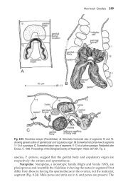
notopodium<br />
gill<br />
prostomium<br />
peristomium<br />
Arenicola (After Brown)<br />
neuropodium<br />
anus<br />
pharynx<br />
buccal papillae
A familiar example is Arenicola. This is not a Queensl<strong>and</strong> genus but is<br />
the well-studied British ‘lugworm’. Here the proboscis is <strong>of</strong> importance in<br />
burrowing <strong>and</strong> though papillate is unarmed. As usual in <strong>polychaete</strong>s, the<br />
proboscis is housed in an anterior asepate chamber but here the most<br />
anterior septum is especially important because in the Arenicolidae, at least,<br />
it contributes the retractor muscle. It is protruded by coelomic pressure.<br />
Nephridia: Arenicola has mixonephridia; Capitellidae are exceptional<br />
(with some nereidids) in retaining separate metanephridia <strong>and</strong> complete<br />
coelomoducts.<br />
Suspension feeders<br />
These are the Spionida <strong>of</strong> Dales <strong>and</strong> include the family Spionidae,<br />
*Chaetopteridae (e.g. Chaetopterus) <strong>and</strong> *Sabellariidae (e.g. Idanthyrsus).<br />
The group is recognized by Rouse <strong>and</strong> Fauchald (1997) for the <strong>families</strong>
Spionidae, *Chaetopteridae, Magelonidae, Apistobranchidae,<br />
Trochochaetidae, Longosomatidae, <strong>and</strong> Poecilochaetidae. They remove the<br />
Sabellariidae to the Sabellida, a controversial placement.<br />
In the Spionidae there is reduction <strong>of</strong> the proboscis <strong>and</strong> elaboration <strong>of</strong><br />
<strong>feeding</strong> tentacles, while in the Chaetopteridae, with the utilization <strong>of</strong> the<br />
dorsal respiratory current for filter <strong>feeding</strong>, by means <strong>of</strong> a mucus ‘bag’, the<br />
tentacles are greatly reduced. The stomodaeum <strong>of</strong> the Chaetopteridae is a<br />
simple tube with no vestige <strong>of</strong> a proboscis.<br />
Relationships <strong>of</strong> Sabellariidae (s<strong>and</strong>-mason worms, e.g. Idanthyrsus)<br />
are very controversial. Placed by Hatschek (1893) with sabellids <strong>and</strong><br />
serpulids. Fitzhugh (1989). found the Sabellida, sensu strictu, to be<br />
monophyletic <strong>and</strong> the sister group to the s<strong>and</strong>-mason worms, the<br />
Sabellariidae, the supposed synapomorphy for the clade being the presence<br />
<strong>of</strong> setal inversion between thoracic <strong>and</strong> abdominal regions. The prostomial
appendages <strong>of</strong> the groups have also been found to be homologous (Orrhage<br />
1980).<br />
Dales (1961) argued for descent <strong>of</strong> sabellariids <strong>and</strong> chaetopterids from<br />
a spionid stock on the grounds that:<br />
1. The early larvae <strong>of</strong> sabellariids are practically identical with those <strong>of</strong><br />
spionids.<br />
2. Anterior <strong>feeding</strong> tentacles <strong>of</strong> larva are retained into the adult as in<br />
spionids but show progressive reduction in different genera with increasing<br />
elaboration <strong>of</strong> secondary <strong>feeding</strong> tentacles on the ventral face <strong>of</strong> the<br />
opercular stem.<br />
3. Uncini <strong>of</strong> sabellariids are similar to those <strong>of</strong> chaetopterids.
4. As in spionids, there is a dorsal ciliary respiratory current <strong>and</strong> long<br />
notopodial gills arched over back.<br />
Some supposed similarities with Sabellidae were not confirmed by<br />
Dales, viz.<br />
(a) a dorsal median excretory pore was not seen <strong>and</strong><br />
(b) reversal <strong>of</strong> notosetae <strong>and</strong> neurosetae between the thorax <strong>and</strong><br />
abdomen was considered, with justification, different from the reversal <strong>of</strong><br />
setae in Sabellida.<br />
Possibly sabellariids are nearer sabellids but <strong>their</strong> position is still<br />
uncertain. Sabellariids also show notable similarities to pectinariids in the<br />
Terebellida <strong>and</strong> were placed in the latter order by Fauchald (1977).
Idanthyrsus<br />
Observe a live specimen in its tube <strong>of</strong> s<strong>and</strong>, shell fragments etc. The fused<br />
tubes form considerable clumps or even small reefs. The animals extend<br />
<strong>their</strong> <strong>feeding</strong> tentacles into the surrounding water. Remove one from its tube<br />
<strong>and</strong> examine it in a Petri dish. Revise dorsal, ventral <strong>and</strong> lateral views.<br />
Note the following:<br />
Large opercular lobes turned forwards <strong>and</strong> fused dorsally bearing two<br />
distinct rows <strong>of</strong> large setae, the internal (ventral) <strong>and</strong> external (dorsal)<br />
paleae. Some <strong>of</strong> these coarsely serrate or denticulate. Each row ending<br />
medianly with a large hook.<br />
Uniramous region, with two segments, behind the operculum. Each with a<br />
dorsal cirrus <strong>and</strong> ventral capillary setae.
Parathoracic region, with 3 segments which have longer dorsal cirri, very<br />
large oar-shaped notopodial setae, <strong>and</strong> ventral capillary neuropodial setae.<br />
Trunk region. Long, with increasingly long dorsal cirri between which<br />
respiratory current is maintained; with vertically elongate, ridge like<br />
notopodia bearing uncini <strong>and</strong> small neuropodia with capillary setae.<br />
Asetous, apodous region. With no parapodia or setae. Ending in the anus<br />
<strong>and</strong> in life held ventrally with the anus anterior.<br />
Note also the ventral view showing: between the opercular lobes, a median<br />
cirrus on the prostomium <strong>and</strong> behind the cirrus two tortuous palps; behind<br />
these the mouth flanked by a large diagonal lip on each side. On the ventral<br />
face <strong>of</strong> the opercular lobes numerous <strong>feeding</strong> tentacles.
Behind the opercular lobes note the segmental regions <strong>and</strong> setation as noted<br />
in the account <strong>of</strong> the dorsal view.<br />
Chaetopterus<br />
Lives in U-shaped parchment tube (actually raised cuticle) about 25<br />
cm long, buried in mud with the ends in water, a small ‘chimney’<br />
protruding above the mud at each end. It can be observed by transfer to a<br />
glass tube where lateral <strong>and</strong> dorsal views are informative.<br />
Notopodia <strong>of</strong> segment 12 are extremely long (aliform or wing-like).<br />
Their epithelium is ciliated <strong>and</strong> mucus-secreting.<br />
Parapodia on 14-16 are fan-like. Each pair forms a piston ring fitting<br />
against the cylindrical walls <strong>of</strong> tube. Beating <strong>of</strong> the fans (60 beats/minute)<br />
produces a current through the tube from the anterior end. The aliform<br />
notopodia are splayed <strong>and</strong> a sheet <strong>of</strong> mucus like a net is secreted between<br />
them. It is continuously secreted to form a mucus bag, at 1 mm/sec. The<br />
end <strong>of</strong> the bag is grasped by a ciliated cup middorsal in segment 13. This
cupule (seen here in dorsal view, though the bag is not visible) rolls up the<br />
end <strong>of</strong> the bag. Almost all incoming water is filtered through this bag;<br />
detritus <strong>and</strong> plankton are retained.<br />
Large objects brought into tube are detected by peristomial cilia <strong>and</strong><br />
shunted to either side; the aliform notopodia are then raised to let the large<br />
objects pass, thus avoiding damage to the bag. The mucus bag is<br />
continually rolled up by the cupule. When the ball (bolus) reaches a certain<br />
size it is cut loose from the notopodia. The cupule then places the bolus on<br />
the ciliated mid-dorsal groove which carries it forward to mouth. The food<br />
ball averaged 3 mm diameter in an 18 cm (6 to 8 inch) specimen.<br />
Axial proboscis with muscular pharynx<br />
The axial proboscis with muscular pharynx is a development for<br />
predation <strong>and</strong> scavenging <strong>and</strong> characterizes the order Phyllodocida <strong>of</strong><br />
Dales, also recognized by Rouse <strong>and</strong> Fauchald (1997). Three groupings on
the basis <strong>of</strong> nephridia are supported on other grounds: those with (1)<br />
protonephridia or protonephromixia (e.g. Phyllodocidae, Alciopidae,<br />
Tomopteridae, Glyceridae, Goniadidae <strong>and</strong> Nephthyidae); (2)<br />
Mixonephridia (Aphroditidae, Polynoidae <strong>and</strong> Chrysopetalidae) <strong>and</strong> (3)<br />
Metanephridia or metanephromixia (Nereididae <strong>and</strong> Syllidae).<br />
The Phyllodocida is a large group containing, in the classification <strong>of</strong><br />
Rouse <strong>and</strong> Fauchald (1997), the Acoetidae, *Aphroditidae, Eulepethidae,<br />
*Polynoidae, Chrysopetalidae, *Glyceridae, Goniadidae, Paralacyoniidae,<br />
Pisionidae, Lactdoniidae, *Phyllodocidae, Nephtyidae, *Nereididae,<br />
Hesionidae, Pilargidae, Sphaerodoridae, <strong>and</strong> *Syllidae, <strong>of</strong> which those<br />
asterisked are dealt with here.<br />
The division <strong>of</strong> the stomodaeum into anterior buccal tube <strong>and</strong> posterior<br />
muscular pharynx is well exemplified by a syllid.
The proboscis in (1) phyllodocids, glycerids, nephthyids <strong>and</strong> (2)<br />
aphroditids <strong>and</strong> polynoids, is similar in having a long cylindrical crushing<br />
<strong>and</strong>, in at least some cases, sucking pharynx made up <strong>of</strong> concentric lamellae<br />
<strong>of</strong> circular or radial muscle. Strong paired retractor muscles connected to<br />
the body wall are not developed; in spite <strong>of</strong> the extreme length <strong>of</strong> pharynx,<br />
<strong>and</strong> retraction is effected by longitudinal muscles <strong>of</strong> oesophagus <strong>and</strong> buccal<br />
tube or by muscles extending on to intestine. Radial muscles to the body<br />
wall where present are probably developed from septa.<br />
Intestinal retractors <strong>of</strong> glycerids are median <strong>and</strong> have presumably been<br />
derived from the dorsal mesentery. [Phyllodoce is similar but has no jaws].<br />
In Glycera the muscular pharynx lies at the level <strong>of</strong> segment 23 but can be<br />
everted, to expose the four jaws, without the aid <strong>of</strong> paired protractor or<br />
retractor muscles.
Nereidids <strong>and</strong> syllids (3) with metanephridia or metanephromixia, on<br />
the other h<strong>and</strong>, have strong protractor <strong>and</strong> retractor muscles which are<br />
paired <strong>and</strong> are inserted on the pharynx; the more anterior motor muscles<br />
arise from the ventrolateral or dorsolateral muscles <strong>of</strong> the body wall.<br />
Laboratory notes<br />
Family Phyllodocidae<br />
Body long <strong>and</strong> slender; segments very numerous. Prostomium well<br />
developed; 2 eyes; 4 to 5 antennae; 2 to 4 pairs <strong>of</strong> tentacular cirri. Long<br />
muscular axial proboscis; unarmed but with numerous papillae. Parapodia<br />
uniramous, with foliaceous dorsal <strong>and</strong> ventral cirri. Setae composite.
Phyllodoce<br />
Exceedingly common on estuarine mudflats in Queensl<strong>and</strong>. Though a s<strong>and</strong><br />
crawler which can also burrow, the family as a whole is specialized for a<br />
pelagic life. Observe living specimens <strong>feeding</strong> <strong>and</strong> watch for eversion <strong>of</strong><br />
the proboscis; this may be facilitated by <strong>feeding</strong> with a portion <strong>of</strong> another<br />
worm <strong>of</strong> by placing the animal in a hypotonic medium (fresh water).<br />
Revise dorsal, lateral <strong>and</strong> ventral views.<br />
Although the proboscis is <strong>of</strong> the type with a muscular pharynx, the latter is<br />
exceptional in being unarmed (lacking jaws).<br />
Note the prostomium with a pair <strong>of</strong> dorsal eyes <strong>and</strong> 4 small simple antennae<br />
(the ventral pair might be termed palps); 4 pairs <strong>of</strong> tentacular cirri on the<br />
peristomium. Remaining, general, segments uniramous, each with foliaceous
dorsal <strong>and</strong> ventral cirrus, the dorsal very large, <strong>and</strong> capillary neurosetae<br />
only.<br />
(2) With mixonephridia<br />
Family Aphroditidae<br />
‘Errantia’ with flattened or vermiform body. 1 to 3 antennae, 2 elongate<br />
palps; 2 pairs <strong>of</strong> tentacular cirri. Parapodia biramous or sesquiramous. With<br />
an alternation <strong>of</strong> elytra <strong>and</strong> dorsal cirri which is more or less regular. Dorsal<br />
setae simple, ventral setae simple or composite.<br />
Aphrodite<br />
The Sea Mouse. This is very similar to Lepidonotus but is larger <strong>and</strong> shows<br />
unique specializations for burrowing in mud. The notopodial setae are<br />
exceedingly numerous <strong>and</strong> form a continuous ‘felt’ over the dorsal surface
<strong>of</strong> the animal, maintaining a mud-free channel in which the elytra lie <strong>and</strong><br />
along which a respiratory water current is passed. The name Aphrodite refers<br />
to the iridescence <strong>of</strong> the lateral setae. This is a Queensl<strong>and</strong> relative.<br />
Family Polynoidae<br />
Lepidonotus<br />
Found under stones on muddy bottoms. Morphology much as for the<br />
Aphroditidae; with 3 antennae <strong>and</strong> 2 palps; peristomium setigerous, but the<br />
dorsal <strong>and</strong> uniramous ventral cirrus <strong>of</strong> each side are turned forwards to<br />
function as sensory tentacular cirri. On the trunk segments the dorsal cirri<br />
are alternately filiform <strong>and</strong> flattened (as an elytron) on each side. The elytra<br />
are numerous in this genus. They overlap <strong>and</strong> cover the dorsal surface. They<br />
are protective, generally pigmented <strong>and</strong> <strong>of</strong>ten give a cryptic coloration, are<br />
respiratory, <strong>and</strong> in the female form a brood pouch for the developing eggs.
(3) With metanephridia<br />
Family Nereididae<br />
Errant <strong>polychaete</strong>s with vermiform body, 2 antennae, 2 ovoid, 2-jointed<br />
palps. 4 eyes. 4 pairs <strong>of</strong> tentacular cirri. Axial proboscis with muscular<br />
pharynx armed with two horny jaws <strong>and</strong> conical paragnaths. Parapodia<br />
almost always biramous. A cirrus <strong>and</strong> two or three lobes on each ramus.<br />
Setae composite. Possessing an epitokous Heteronereis stage.<br />
Australonereis<br />
A very common <strong>polychaete</strong> <strong>of</strong> the Queensl<strong>and</strong> littoral zone. In this or other<br />
nereidid note typically errant form. Capable <strong>of</strong> swimming <strong>and</strong> crawling but<br />
also burrowing in s<strong>and</strong> in which it constructs a definite s<strong>and</strong>y tube which can<br />
<strong>of</strong>ten be seen protruding above the substrate. Examine nereidid material
provided. Note dorsal <strong>and</strong> ventral views <strong>of</strong> the anterior region, with <strong>and</strong><br />
without eversion <strong>of</strong> the pharynx. Note the formidable jaws on the inner wall<br />
<strong>of</strong> the far posterior pharynx before eversion.<br />
Anterior triangular prostomium with 2 pairs <strong>of</strong> eyes, 2 dorsal antennae. On<br />
the peristomium, lateral sensory tentacular cirri. Examine specimens with<br />
<strong>and</strong> without extrusion <strong>of</strong> the large muscular axial proboscis. Prosboscis with<br />
jaws <strong>and</strong> paragnaths; all trunk segments similar <strong>and</strong> bearing each a pair <strong>of</strong><br />
two-lobed, well developed parapodia. The jaws are exposed when the<br />
pharynx is everted.<br />
Examine a nereidid parapodium as revision <strong>of</strong>, albeit variable, parapodial<br />
structure: the dorsal (notopodial) <strong>and</strong> ventral (neuropodial) bundles <strong>of</strong> setae<br />
each emerging from a setal sac supported by a strong central rod, the acicula<br />
(plural aciculae), the flattened form <strong>of</strong> the parapodia which are <strong>of</strong><br />
importance in respiration, <strong>and</strong> the small dorsal <strong>and</strong> ventral sensory <strong>and</strong><br />
respiratory cirri.
Family Syllidae<br />
Mostly small. Prostomium distinct. 4 eyes. 2 palps. 3 antennae. 2 pairs <strong>of</strong><br />
cirri on the peristomium which always lacks setae. Barrel shaped axial<br />
pharynx preceded by chitinized chamber bearing 1 or more teeth. Parapodia<br />
uniramous, <strong>of</strong>ten with a dorsal <strong>and</strong> ventral cirrus. Setae usually compound. 2<br />
anal cirri. Have metanephridia <strong>and</strong> metanephromixia.<br />
Eusyllis<br />
From local rocky shores. Note prostomium with 4 eyes <strong>and</strong> 3 antennae<br />
which, though not articulated, are somewhat annulated as common in syllids.<br />
Anterior border <strong>of</strong> pharynx chitinous <strong>and</strong> with a single large dorsal tooth.<br />
Some syllids are brilliantly bioluminescent.<br />
Note the large muscular pharynx <strong>and</strong> buccal tube <strong>of</strong> two syllids: 1 & 2.
Myzostomida<br />
Myzostomes are obligate commensals or parasites <strong>of</strong> echinoderms,<br />
usually crinoids. They have been regarded as platyhelminths or even as<br />
tardigrades. Because they have an eversible pharynx like that <strong>of</strong><br />
phyllodocids <strong>and</strong> have acicula, Rouse <strong>and</strong> Fauchald (1997) have placed the<br />
myzostomes in the Polychaeta, <strong>and</strong> specifically in the Phyllodocida.<br />
Myzostomes mimic the colour patterns <strong>of</strong> the host crinoid, clearly as a<br />
result <strong>of</strong> selection for individuals less visible to predators. Here a greenish<br />
species <strong>and</strong> a reddish species mimic the colour patterns <strong>of</strong> <strong>their</strong> respective<br />
host crinoids on Heron Isl<strong>and</strong>, Great Barrier Reef.
ACKNOWLEDGEMENTS<br />
All line drawings are by Bruce Barnes, excepting labelled sketches <strong>of</strong><br />
pharyngeal types by the author.<br />
Colour photographs are by the author or were donated to the author by<br />
Dr. Isobel Bennett, Mr. Justice Myers, Owen Kelly <strong>and</strong> Tom Gorringe.<br />
They may not be reproduced without the permission <strong>of</strong> the author.
REFERENCES AND FURTHER READING<br />
Dales R.P. 1961. The <strong>polychaete</strong> stomodaeum <strong>and</strong> the inter-relationships <strong>of</strong><br />
the <strong>families</strong> <strong>of</strong> Polychaeta. Proc. Zool. Soc.Lond. 139: 389-428.<br />
Dales R.P 1963. Annelids. Hutchinson University Library, London.<br />
Fauchald K. 1977. The <strong>polychaete</strong> worms. Definitions <strong>and</strong> keys to the<br />
orders, <strong>families</strong> <strong>and</strong> genera. Natural History Museum <strong>of</strong> Los Angeles<br />
County. Science series, 28.<br />
Fitzhugh, K. 1989. A systematic review <strong>of</strong> the Sabellidae-Caobangidae-<br />
Sabellongidae complex (Annelida: Polychaeta). Bull. Am. Mus. Nat.<br />
Hist. 192: 1-104.<br />
Jamieson, B.G.M. <strong>and</strong> Webb, R. I. 1984. The morphology, spermatozoal<br />
ultrastructure <strong>and</strong> phylogenetic affinities <strong>of</strong> a new species <strong>of</strong> questid<br />
(Polychaeta: Annelida). In Proceedings <strong>of</strong> the First International<br />
Polychaete Conference, Sydney, Australia, 1983 (ed. P.A. Hutchings):<br />
21-34, The Linnean Society <strong>of</strong> New South Wales, Sydney.
Orrhage L. 1973. The fundamental requirements for phylogenetic-scientific<br />
works as a background for an analysis <strong>of</strong> Dales’s (1962) <strong>and</strong> Webbs<br />
(1969) theories. Z. Zool. Syst. Evolut-forsch. (1973): 161-173.<br />
Rouse, G.W. <strong>and</strong> Fauchald, K. 1997. Cladistics <strong>and</strong> <strong>polychaete</strong>s. Zoologica<br />
Scripta, 26: 139-204.<br />
Westheide W. (ed.) 1988. The Ultrastructure <strong>of</strong> the Polychaeta. Fischer<br />
Verlag.