New species of Cryptosporidium Tyzzer, 1907 (Apicomplexa) from ...
New species of Cryptosporidium Tyzzer, 1907 (Apicomplexa) from ...
New species of Cryptosporidium Tyzzer, 1907 (Apicomplexa) from ...

Create successful ePaper yourself
Turn your PDF publications into a flip-book with our unique Google optimized e-Paper software.
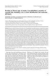
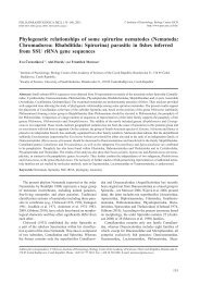
Figs. 13–22. Transmission electron micrographs <strong>of</strong> <strong>Cryptosporidium</strong> fragile sp. n. Fig. 13. Gastric epithelium covered by trophozoites<br />
and macrogamonts. Fig. 14. Immature macrogamont and meront. Note the distinct dense bands (arrows). Fig. 15. Internal<br />
details <strong>of</strong> merozoites, composite figure. Fig. 16. Macrogamont; dense line (arrowhead). Fig. 17. Microgamont with microgametes<br />
(arrow). Fig. 18. Microgamete; apical adhesive zone (arrow). Fig. 19. Oocyst within detached parasitophorous sac; oocyst wall<br />
and papular structures on its inner surface (arrow); parasitophorous sac (arrowheads); dense band area (double arrow). Fig. 20.<br />
Released oocysts in gastric lumen; sporozoites (arrowheads) with distinct micronemes; oocyst wall (arrows). Fig. 21. Detail <strong>of</strong><br />
host-parasite interface; dense line (arrowhead); dense band (double arrowhead). Fig. 22. Feeder organelle in cross-section; dense<br />
line (arrowhead). Abbreviations: a – amylopectin granules; d – dense bodies; fo – feeder organelle; m – merozoite; mn – micronemes;<br />
n – nucleus; or – oocyst residuum; r – rhoptries; rb – residual body; v – vacuole; * – radial folds <strong>of</strong> parasitophorous<br />
sac. Scale bars: Figs. 13, 19 = 2 µm; Figs. 14, 16, 17, 20 = 1 µm; Figs. 15, 22 = 500 nm; Fig. 18 = 200 nm; Fig. 21 = 250 nm.<br />
86