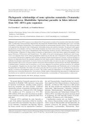
New species of Cryptosporidium Tyzzer, 1907 (Apicomplexa) from ...
New species of Cryptosporidium Tyzzer, 1907 (Apicomplexa) from ...
New species of Cryptosporidium Tyzzer, 1907 (Apicomplexa) from ...

You also want an ePaper? Increase the reach of your titles
YUMPU automatically turns print PDFs into web optimized ePapers that Google loves.
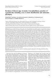
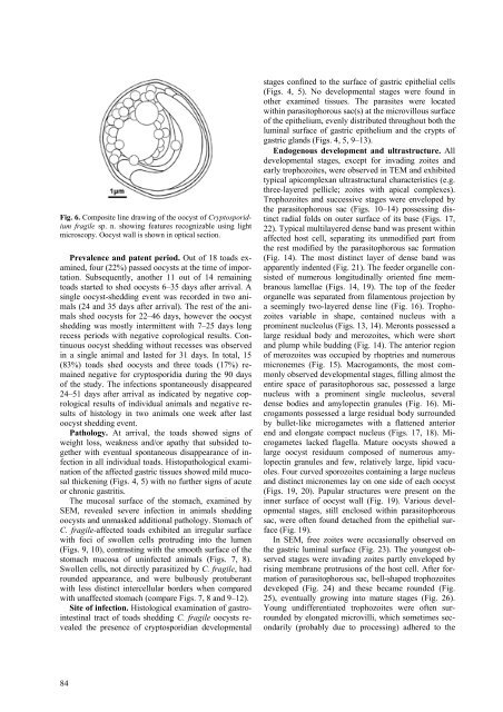
Fig. 6. Composite line drawing <strong>of</strong> the oocyst <strong>of</strong> <strong>Cryptosporidium</strong><br />
fragile sp. n. showing features recognizable using light<br />
microscopy. Oocyst wall is shown in optical section.<br />
Prevalence and patent period. Out <strong>of</strong> 18 toads examined,<br />
four (22%) passed oocysts at the time <strong>of</strong> importation.<br />
Subsequently, another 11 out <strong>of</strong> 14 remaining<br />
toads started to shed oocysts 6–35 days after arrival. A<br />
single oocyst-shedding event was recorded in two animals<br />
(24 and 35 days after arrival). The rest <strong>of</strong> the animals<br />
shed oocysts for 22–46 days, however the oocyst<br />
shedding was mostly intermittent with 7–25 days long<br />
recess periods with negative coprological results. Continuous<br />
oocyst shedding without recesses was observed<br />
in a single animal and lasted for 31 days. In total, 15<br />
(83%) toads shed oocysts and three toads (17%) remained<br />
negative for cryptosporidia during the 90 days<br />
<strong>of</strong> the study. The infections spontaneously disappeared<br />
24–51 days after arrival as indicated by negative coprological<br />
results <strong>of</strong> individual animals and negative results<br />
<strong>of</strong> histology in two animals one week after last<br />
oocyst shedding event.<br />
Pathology. At arrival, the toads showed signs <strong>of</strong><br />
weight loss, weakness and/or apathy that subsided together<br />
with eventual spontaneous disappearance <strong>of</strong> infection<br />
in all individual toads. Histopathological examination<br />
<strong>of</strong> the affected gastric tissues showed mild mucosal<br />
thickening (Figs. 4, 5) with no further signs <strong>of</strong> acute<br />
or chronic gastritis.<br />
The mucosal surface <strong>of</strong> the stomach, examined by<br />
SEM, revealed severe infection in animals shedding<br />
oocysts and unmasked additional pathology. Stomach <strong>of</strong><br />
C. fragile-affected toads exhibited an irregular surface<br />
with foci <strong>of</strong> swollen cells protruding into the lumen<br />
(Figs. 9, 10), contrasting with the smooth surface <strong>of</strong> the<br />
stomach mucosa <strong>of</strong> uninfected animals (Figs. 7, 8).<br />
Swollen cells, not directly parasitized by C. fragile, had<br />
rounded appearance, and were bulbously protuberant<br />
with less distinct intercellular borders when compared<br />
with unaffected stomach (compare Figs. 7, 8 and 9–12).<br />
Site <strong>of</strong> infection. Histological examination <strong>of</strong> gastrointestinal<br />
tract <strong>of</strong> toads shedding C. fragile oocysts revealed<br />
the presence <strong>of</strong> cryptosporidian developmental<br />
84<br />
stages confined to the surface <strong>of</strong> gastric epithelial cells<br />
(Figs. 4, 5). No developmental stages were found in<br />
other examined tissues. The parasites were located<br />
within parasitophorous sac(s) at the microvillous surface<br />
<strong>of</strong> the epithelium, evenly distributed throughout both the<br />
luminal surface <strong>of</strong> gastric epithelium and the crypts <strong>of</strong><br />
gastric glands (Figs. 4, 5, 9–13).<br />
Endogenous development and ultrastructure. All<br />
developmental stages, except for invading zoites and<br />
early trophozoites, were observed in TEM and exhibited<br />
typical apicomplexan ultrastructural characteristics (e.g.<br />
three-layered pellicle; zoites with apical complexes).<br />
Trophozoites and successive stages were enveloped by<br />
the parasitophorous sac (Figs. 10–14) possessing distinct<br />
radial folds on outer surface <strong>of</strong> its base (Figs. 17,<br />
22). Typical multilayered dense band was present within<br />
affected host cell, separating its unmodified part <strong>from</strong><br />
the rest modified by the parasitophorous sac formation<br />
(Fig. 14). The most distinct layer <strong>of</strong> dense band was<br />
apparently indented (Fig. 21). The feeder organelle consisted<br />
<strong>of</strong> numerous longitudinally oriented fine membranous<br />
lamellae (Figs. 14, 19). The top <strong>of</strong> the feeder<br />
organelle was separated <strong>from</strong> filamentous projection by<br />
a seemingly two-layered dense line (Fig. 16). Trophozoites<br />
variable in shape, contained nucleus with a<br />
prominent nucleolus (Figs. 13, 14). Meronts possessed a<br />
large residual body and merozoites, which were short<br />
and plump while budding (Fig. 14). The anterior region<br />
<strong>of</strong> merozoites was occupied by rhoptries and numerous<br />
micronemes (Fig. 15). Macrogamonts, the most commonly<br />
observed developmental stages, filling almost the<br />
entire space <strong>of</strong> parasitophorous sac, possessed a large<br />
nucleus with a prominent single nucleolus, several<br />
dense bodies and amylopectin granules (Fig. 16). Microgamonts<br />
possessed a large residual body surrounded<br />
by bullet-like microgametes with a flattened anterior<br />
end and elongate compact nucleus (Figs. 17, 18). Microgametes<br />
lacked flagella. Mature oocysts showed a<br />
large oocyst residuum composed <strong>of</strong> numerous amylopectin<br />
granules and few, relatively large, lipid vacuoles.<br />
Four curved sporozoites containing a large nucleus<br />
and distinct micronemes lay on one side <strong>of</strong> each oocyst<br />
(Figs. 19, 20). Papular structures were present on the<br />
inner surface <strong>of</strong> oocyst wall (Fig. 19). Various developmental<br />
stages, still enclosed within parasitophorous<br />
sac, were <strong>of</strong>ten found detached <strong>from</strong> the epithelial surface<br />
(Fig. 19).<br />
In SEM, free zoites were occasionally observed on<br />
the gastric luminal surface (Fig. 23). The youngest observed<br />
stages were invading zoites partly enveloped by<br />
rising membrane protrusions <strong>of</strong> the host cell. After formation<br />
<strong>of</strong> parasitophorous sac, bell-shaped trophozoites<br />
developed (Fig. 24) and these became rounded (Fig.<br />
25), eventually growing into mature stages (Fig. 26).<br />
Young undifferentiated trophozoites were <strong>of</strong>ten surrounded<br />
by elongated microvilli, which sometimes secondarily<br />
(probably due to processing) adhered to the