New species of Cryptosporidium Tyzzer, 1907 (Apicomplexa) from ...
New species of Cryptosporidium Tyzzer, 1907 (Apicomplexa) from ...
New species of Cryptosporidium Tyzzer, 1907 (Apicomplexa) from ...

You also want an ePaper? Increase the reach of your titles
YUMPU automatically turns print PDFs into web optimized ePapers that Google loves.
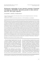
Fig. 36. Phylogenetic reconstruction <strong>of</strong> <strong>Cryptosporidium</strong> <strong>species</strong> for the SSU rDNA. Reconstruction using available sequences<br />
<strong>from</strong> named <strong>Cryptosporidium</strong> <strong>species</strong> and selected genotypes with partial coverage and dataset <strong>of</strong> complete SSU rDNA sequences<br />
only (inset). The phylogenetic tree was inferred using the Minimum Evolution method. The bootstrap support values<br />
(>50%) for Minimum Evolution (1,000 replicates) / Maximum Parsimony (500 replicates) / Maximum Likelihood (100 replicates)<br />
are shown next to the branches. The evolutionary distances were computed using the Maximum Composite Likelihood<br />
method. Minimum Evolution and Maximum Parsimony were calculated in MEGA4. Maximum Likelihood was calculated in<br />
PhyML 2.4.4 using GTR+G+I model.<br />
differentiated into distinct layers compared to majority<br />
<strong>of</strong> the other cryptosporidian <strong>species</strong>; however, higher<br />
magnification revealed several layers, <strong>of</strong> which one was<br />
more distinct and finely indented as reported previously<br />
in C. muris and C. wrairi (Vetterling et al. 1971, Uni et<br />
al. 1987, Valigurová et al. 2007). Parasitophorous sac <strong>of</strong><br />
C. fragile invariably possesses distinct, structurally uniform,<br />
radial folds evenly distributed around its base.<br />
The absence <strong>of</strong> these folds or their structural and spatial<br />
irregularity together with the presence <strong>of</strong> distinct and<br />
bulky filamentous projection in C. muris (weakly developed<br />
in C. fragile), make these two <strong>species</strong> clearly distinguishable<br />
<strong>from</strong> each other at the ultrastructural level<br />
(Valigurová et al. 2008).<br />
Despite mentioned differences, the ultrastructure <strong>of</strong><br />
C. fragile developmental stages was comparable to<br />
other cryptosporidian <strong>species</strong>. Unfortunately, many <strong>of</strong><br />
the observed characters are insufficiently characterized<br />
in other <strong>Cryptosporidium</strong> spp., hampering the significance<br />
in <strong>species</strong> identification. Nevertheless, we find<br />
morphological traits correlating with individual host<br />
groups. Evident similarities occur within <strong>Cryptosporidium</strong><br />
<strong>species</strong> <strong>from</strong> (i) fish, (ii) mammals and (iii) reptiles.<br />
90<br />
In the fish-affecting <strong>species</strong>, the surface <strong>of</strong> parasitophorous<br />
sac is covered by so-called rudimentary microvilli<br />
(Paperna and Vilenkin 1996). These structures, together<br />
with the localisation <strong>of</strong> the oocysts deep within the gastric<br />
mucosa, were used to erect the genus Piscicryptosporidium<br />
for the fish <strong>species</strong> (Paperna and Vilenkin<br />
1996), which is currently considered a synonym <strong>of</strong><br />
<strong>Cryptosporidium</strong> (Alvarez-Pellitero and Sitjà-Bobadilla<br />
2002, Alvarez-Pellitero et al. 2004). We did not detect<br />
these structures in the amphibian gastric mucosa affected<br />
by C. fragile. In general, fish cryptosporidia exhibit<br />
well developed massive lamellae <strong>of</strong> the feeder organelle<br />
and inconspicuous filamentous projection (Paperna<br />
and Vilenkin 1996, Alvarez-Pellitero and Sitjà-<br />
Bobadilla 2002, Alvarez-Pellitero et al. 2004). Similarly,<br />
C. muris and C. andersoni, gastric <strong>species</strong> <strong>from</strong><br />
mammalian hosts, seemingly exhibit common features,<br />
i.e. the shape and bulkiness <strong>of</strong> the filamentous projection<br />
(Uni et al. 1987, Masuno et al. 2006, Valigurová et<br />
al. 2007). On the other hand, the button-shaped feeder<br />
organelle with fine lamellae is present in C. fragile and<br />
both gastric as well as intestinal <strong>species</strong> <strong>from</strong> reptiles<br />
(Brownstein et al. 1977, Koudela and Modrý 1998). The