New species of Cryptosporidium Tyzzer, 1907 (Apicomplexa) from ...
New species of Cryptosporidium Tyzzer, 1907 (Apicomplexa) from ...
New species of Cryptosporidium Tyzzer, 1907 (Apicomplexa) from ...

Create successful ePaper yourself
Turn your PDF publications into a flip-book with our unique Google optimized e-Paper software.
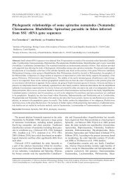
FOLIA PARASITOLOGICA 55: 81–94, 2008<br />
<strong>New</strong> <strong>species</strong> <strong>of</strong> <strong>Cryptosporidium</strong> <strong>Tyzzer</strong>, <strong>1907</strong> (<strong>Apicomplexa</strong>)<br />
<strong>from</strong> amphibian host: morphology, biology and phylogeny<br />
Miloslav Jirků 1,2 , Andrea Valigurová 3 , Břetislav Koudela 1,2 , Jaroslav Křížek 2 , David Modrý 1,2 and<br />
Jan Šlapeta 4<br />
1<br />
Department <strong>of</strong> Parasitology, University <strong>of</strong> Veterinary and Pharmaceutical Sciences, Palackého 1–3, 612 42 Brno, Czech<br />
Republic;<br />
2<br />
Institute <strong>of</strong> Parasitology, Biology Centre <strong>of</strong> the Academy <strong>of</strong> Sciences <strong>of</strong> the Czech Republic, Branišovská 31, 370 05 České<br />
Budějovice, Czech Republic;<br />
3 Department <strong>of</strong> Botany and Zoology, Faculty <strong>of</strong> Science, Masaryk University, Kotlářská 2, 611 37 Brno, Czech Republic;<br />
4 Faculty <strong>of</strong> Veterinary Science, University <strong>of</strong> Sydney, McMaster Building – B14, <strong>New</strong> South Wales 2006, Australia<br />
Key words: <strong>Cryptosporidium</strong> fragile, new <strong>species</strong>, frog, Duttaphrynus melanostictus, ultrastructure, host specificity,<br />
phylogeny, quarantine, global amphibian decline<br />
Abstract. <strong>Cryptosporidium</strong> fragile sp. n. (<strong>Apicomplexa</strong>) is described <strong>from</strong> black-spined toads, Duttaphrynus melanostictus<br />
(Schneider) (Amphibia, Anura, Bufonidae) <strong>from</strong> the Malay Peninsula. The parasitized animals were directly imported <strong>from</strong> Malaysia<br />
and harboured C. fragile at the time <strong>of</strong> arrival. Oocysts were subspherical to elliptical with irregular contour in optical<br />
section, measuring 6.2 (5.5–7.0) × 5.5 (5.0–6.5) µm. Oocyst wall was smooth and colourless in light microscopy. The endogenous<br />
development <strong>of</strong> C. fragile in the stomach <strong>of</strong> black-spined toad was analysed in detail using light and electron microscopy.<br />
Cryptosporidian developmental stages were confined to the surface <strong>of</strong> gastric epithelial cells. In transmission experiments, C.<br />
fragile has not been infective for one fish <strong>species</strong>, four amphibian <strong>species</strong>, one <strong>species</strong> <strong>of</strong> reptile and SCID mice. Full length<br />
small subunit rRNA gene sequence was obtained. Phylogenetic reconstruction revealed distinct status <strong>of</strong> C. fragile within the<br />
clade <strong>of</strong> <strong>species</strong> with gastric localisation including <strong>Cryptosporidium</strong> muris <strong>Tyzzer</strong>, <strong>1907</strong>, <strong>Cryptosporidium</strong> serpentis Levine,<br />
1980 and <strong>Cryptosporidium</strong> andersoni Lindsay, Upton, Owens, Morgan, Mead et Blagburn, 2000. Described characteristics differentiate<br />
C. fragile <strong>from</strong> the currently recognized <strong>Cryptosporidium</strong> <strong>species</strong>. Our experience with the description <strong>of</strong> C. fragile has<br />
led us to revise the recommended criteria for an introduction <strong>of</strong> a new <strong>Cryptosporidium</strong> <strong>species</strong> name. C. fragile is the first <strong>species</strong><br />
described and named <strong>from</strong> an amphibian host. Its prevalence <strong>of</strong> 83% (15/18) in black-spined toads within the 3 months after<br />
importation calls for strict quarantine measures and import regulation for lower vertebrates.<br />
The genus <strong>Cryptosporidium</strong> was erected by Ernest E.<br />
<strong>Tyzzer</strong> for a peculiar parasite affecting mouse stomach,<br />
<strong>Cryptosporidium</strong> muris <strong>Tyzzer</strong>, <strong>1907</strong> (<strong>Tyzzer</strong> <strong>1907</strong>).<br />
Later he recognized a distinct <strong>species</strong> <strong>Cryptosporidium</strong><br />
parvum <strong>Tyzzer</strong>, 1912 affecting mouse intestine (<strong>Tyzzer</strong><br />
1912). In the following years, multiple new cryptosporidian<br />
<strong>species</strong> were described, infecting majority <strong>of</strong><br />
vertebrates. Initially, cryptosporidia were considered <strong>of</strong><br />
a doubtful clinical significance, but with increasing<br />
knowledge the disease, cryptosporidiosis, was recognized<br />
as a major concern in immunocompromised and<br />
even healthy hosts (Fayer et al. 2000, Chen et al. 2002,<br />
Thompson et al. 2005).<br />
Currently, there are 21 recognized <strong>species</strong> within the<br />
genus <strong>Cryptosporidium</strong> in fish, reptiles, birds and<br />
mammals (Šlapeta 2008). Seven <strong>of</strong> these affect host’s<br />
stomach – C. muris <strong>Tyzzer</strong>, <strong>1907</strong> in rodents, C. andersoni<br />
Lindsay, Upton, Owens, Morgan, Mead et Blagburn,<br />
2000 in cattle, C. serpentis Levine, 1980 in snakes<br />
and lizards, C. galli Pavlásek, 1999 in birds, and three<br />
<strong>species</strong> in fish stomach – C. cichlidis (Paperna et Vilenkin,<br />
1996), C. reichenbachklinkei (Paperna et Vilenkin,<br />
1996) and C. molnari Alvarez-Pellitero et Sitjà-<br />
Bobadilla, 2002 (Paperna and Vilenkin 1996, Tilley et<br />
al. 1990, Lindsay et al. 2000, Alvarez-Pellitero and<br />
Sitjà-Bobadilla 2002, Ryan et al. 2003a).<br />
Here we report on a cryptosporidian infection in<br />
toads directly imported to the Czech Republic <strong>from</strong> Malaysia<br />
for pet trade. We summarise morphological characteristics,<br />
results <strong>of</strong> transmission experiments and molecular<br />
data to describe this isolate as a new <strong>Cryptosporidium</strong><br />
<strong>species</strong>, representing the first characterized<br />
<strong>species</strong> <strong>from</strong> an amphibian host.<br />
MATERIALS AND METHODS<br />
Origin <strong>of</strong> the parasites and host animals. In total, 18<br />
adult black-spined toads, Duttaphrynus (syn. Bufo) melanostictus<br />
(Schneider) (Amphibia, Bufonidae) were obtained via<br />
import <strong>of</strong> pet animals <strong>from</strong> the Malay Peninsula in April 2006<br />
(n = 9) and May 2006 (n = 9) in accordance with the Czech<br />
Republic regulations. Toads arrived in plastic boxes with<br />
moist moss, 3–4 individuals per box, and were quarantined in<br />
the premises <strong>of</strong> the University <strong>of</strong> Veterinary and Pharmaceutical<br />
Sciences Brno, Czech Republic (UVPB). The animals<br />
were housed individually in 8-litre glass vivaria with wet coco<br />
substrate, and fed 2–3 times weekly with crickets (Gryllus<br />
assimilis) supplemented with Reptivite (ZooMed Laboratories<br />
Inc).<br />
Address for correspondence: M. Jirků, Institute <strong>of</strong> Parasitology, Biology Centre <strong>of</strong> the Academy <strong>of</strong> Sciences <strong>of</strong> the Czech Republic, Branišovská<br />
31, 370 05 České Budějovice, Czech Republic. Phone: +420 387 775 474; Fax: +420 385 310 388; E-mail: miloslav.jirku@seznam.cz<br />
81
Parasitological examination. All faeces expelled by individual<br />
toads were monitored for the presence <strong>of</strong> parasites using<br />
flotation (sugar solution, s.g. 1.3) <strong>from</strong> the day <strong>of</strong> import<br />
up to 90 days after arrival. Animals shedding oocysts were<br />
euthanised by pithing. Fresh squash preparations <strong>of</strong> various<br />
viscera, gastrointestinal smears, histological preparations and<br />
oocysts concentrated by flotation were examined by light microscopy<br />
using an Olympus AX 70 microscope equipped with<br />
Nomarski interference-contrast optics (NIC). For histology,<br />
the tissues <strong>of</strong> three animals were fixed in 10% buffered formalin<br />
or AFA (Alcohol-Formalin-Acetic Acid), embedded in<br />
Histoplast II and 6 µm sections were stained with haematoxylin-eosin<br />
(H&E). For transmission electron microscopy<br />
(TEM), the tissues <strong>of</strong> two animals were fixed overnight at 4°C<br />
in 3% glutaraldehyde in 0.2 M phosphate buffer and processed<br />
as described previously (Valigurová et al. 2008). Ultrathin<br />
sections were double-stained with uranyl acetate and lead<br />
citrate and viewed in a JEOL 1010. For scanning electron<br />
microscopy (SEM), the tissues <strong>of</strong> three animals were fixed<br />
overnight at 4°C in 3% glutaraldehyde in cacodylate buffer<br />
and processed according to Valigurová et al. (2008). Samples<br />
were examined using a JEOL JSM-7401F, field emission<br />
scanning electron microscope capable <strong>of</strong> high resolution <strong>of</strong> up<br />
to 1.0 nm.<br />
Experimental host transmissions. Sieved faecal samples<br />
containing <strong>Cryptosporidium</strong> oocysts were pooled and kept in<br />
dechlorinated tap water in 0.5-litre containers without preservatives<br />
to avoid intoxication <strong>of</strong> experimental animals.<br />
Every two days, the suspension was stirred, sedimented (1 h),<br />
water removed and the tanks were re-filled with fresh water.<br />
The sedimented debris containing oocysts (1 year) 20 (10) 10 3 –10 4 AMPHIBIANS<br />
Neg. PC<br />
Adults (>3 years) 2 (2) 10 4 Bufo bufo<br />
Neg. W<br />
Tadpoles 40 (40) unknown Neg. W<br />
Rana temporaria Adults (>3 years) 2 (2) 10 4 Neg. W<br />
Litoria caerulea Adults (>3 years) 2 (1) 10 4 Neg. PI<br />
Adults (>2 years) 5 (5) 10 4 Xenopus laevis<br />
Neg. LC<br />
REPTILES<br />
Tadpoles 20 (20) unknown Neg. LC<br />
Pantherophis guttatus Adult (2 years) 1 (1) 10 4 MAMMALS<br />
Neg. PC<br />
Mus musculus SCID Subadults (12 weeks) 2 (2) 10 4 Neg. LC<br />
LC – laboratory colony (Xenopus laevis <strong>from</strong> the University <strong>of</strong> Veterinary and Pharmaceutical Sciences Brno and SCID mice <strong>from</strong> the Charles<br />
Rivers Laboratories); PC – pet trade, captive-bred; PI – pet trade, import <strong>from</strong> the wild; W – wild-caught in Brno, Czech Republic.
RESULTS<br />
Parasitological examination <strong>of</strong> black-spined toads,<br />
Duttaphrynus melanostictus (Fig. 1) <strong>from</strong> the Malay<br />
Peninsula, revealed that 83% (15/18) passed oocysts <strong>of</strong><br />
<strong>Cryptosporidium</strong>. Morphological, experimental and<br />
molecular analyses showed that the isolate represents a<br />
new <strong>species</strong>, the description <strong>of</strong> which follows.<br />
<strong>Cryptosporidium</strong> fragile sp. n. Figs. 2–35<br />
Oocyst morphology. Oocysts (Figs. 2, 3, 6) obtained<br />
by flotation method (Fig. 2) <strong>from</strong> fresh faeces were irregular<br />
in shape, subspherical to elliptical; usually with<br />
apparently deformed/uneven contour in optical section;<br />
6.2 (5.5–7.0) × 5.5 (5.0–6.5) µm; length/width 1.13<br />
(1.00–1.30) (n = 50); oocyst wall smooth, colourless in<br />
light microscopy. To confirm that the oocyst shape variability<br />
is not an artefact, oocysts were examined in native<br />
preparations <strong>from</strong> freshly euthanised animals. Oo-<br />
Jirků et al.: <strong>New</strong> <strong>Cryptosporidium</strong> <strong>from</strong> an amphibian<br />
cysts observed in squash preparations <strong>of</strong> gastric mucosa<br />
(Fig. 3) showed the same degree <strong>of</strong> the shape variability;<br />
6.5 (6.0–7.5) × 5.8 (5.0–6.5) µm; length/width 1.13<br />
(1.00–1.36) (n = 50). Oocyst residuum was composed <strong>of</strong><br />
a mass <strong>of</strong> fine granules 0.5 µm in diameter, and a<br />
spherical globule 1.5–2.0 µm in diameter. A large<br />
vacuolated area occupied a significant part <strong>of</strong> the oocyst<br />
residuum in all oocysts. Sporulation was endogenous;<br />
oocysts were fully sporulated in fresh faeces and gut<br />
content. Four vermiform sporozoites lay parallel along<br />
one side <strong>of</strong> oocyst, tightly enclosing oocyst residuum.<br />
Neither nucleus nor refractile bodies were observed<br />
within sporozoites using light microscopy. Noteworthy,<br />
oocysts were extremely sensitive to hypertonic conditions,<br />
crumpling immediately when exposed to flotation<br />
solutions. The fragility <strong>of</strong> oocysts was further confirmed<br />
by SEM examination <strong>of</strong> oocysts concentrated by flotation<br />
(not shown). Oocysts spontaneously disintegrated<br />
after 4 weeks <strong>of</strong> storage in water at 4°C, 10°C and 20°C.<br />
Figs. 1–5. Type host and light microscopy <strong>of</strong> <strong>Cryptosporidium</strong> fragile sp. n. Fig. 1. Black-spined toad, Duttaphrynus melanostictus,<br />
the type host. Specimen <strong>from</strong> the Malay Peninsula, snout-vent length 73 mm. Fig. 2. Oocysts <strong>from</strong> faeces, flotation (NIC).<br />
Fig. 3. Composite micrograph <strong>of</strong> oocysts in squash preparation <strong>of</strong> the gastric mucosa (NIC). Figs. 4, 5. Stomach <strong>of</strong> infected toad<br />
in longitudinal histological sections (H&E). Scale bars: Figs. 2, 3 (same scale) = 10 µm; Fig. 4 = 100 µm; Fig. 5 = 50 µm.<br />
83
Fig. 6. Composite line drawing <strong>of</strong> the oocyst <strong>of</strong> <strong>Cryptosporidium</strong><br />
fragile sp. n. showing features recognizable using light<br />
microscopy. Oocyst wall is shown in optical section.<br />
Prevalence and patent period. Out <strong>of</strong> 18 toads examined,<br />
four (22%) passed oocysts at the time <strong>of</strong> importation.<br />
Subsequently, another 11 out <strong>of</strong> 14 remaining<br />
toads started to shed oocysts 6–35 days after arrival. A<br />
single oocyst-shedding event was recorded in two animals<br />
(24 and 35 days after arrival). The rest <strong>of</strong> the animals<br />
shed oocysts for 22–46 days, however the oocyst<br />
shedding was mostly intermittent with 7–25 days long<br />
recess periods with negative coprological results. Continuous<br />
oocyst shedding without recesses was observed<br />
in a single animal and lasted for 31 days. In total, 15<br />
(83%) toads shed oocysts and three toads (17%) remained<br />
negative for cryptosporidia during the 90 days<br />
<strong>of</strong> the study. The infections spontaneously disappeared<br />
24–51 days after arrival as indicated by negative coprological<br />
results <strong>of</strong> individual animals and negative results<br />
<strong>of</strong> histology in two animals one week after last<br />
oocyst shedding event.<br />
Pathology. At arrival, the toads showed signs <strong>of</strong><br />
weight loss, weakness and/or apathy that subsided together<br />
with eventual spontaneous disappearance <strong>of</strong> infection<br />
in all individual toads. Histopathological examination<br />
<strong>of</strong> the affected gastric tissues showed mild mucosal<br />
thickening (Figs. 4, 5) with no further signs <strong>of</strong> acute<br />
or chronic gastritis.<br />
The mucosal surface <strong>of</strong> the stomach, examined by<br />
SEM, revealed severe infection in animals shedding<br />
oocysts and unmasked additional pathology. Stomach <strong>of</strong><br />
C. fragile-affected toads exhibited an irregular surface<br />
with foci <strong>of</strong> swollen cells protruding into the lumen<br />
(Figs. 9, 10), contrasting with the smooth surface <strong>of</strong> the<br />
stomach mucosa <strong>of</strong> uninfected animals (Figs. 7, 8).<br />
Swollen cells, not directly parasitized by C. fragile, had<br />
rounded appearance, and were bulbously protuberant<br />
with less distinct intercellular borders when compared<br />
with unaffected stomach (compare Figs. 7, 8 and 9–12).<br />
Site <strong>of</strong> infection. Histological examination <strong>of</strong> gastrointestinal<br />
tract <strong>of</strong> toads shedding C. fragile oocysts revealed<br />
the presence <strong>of</strong> cryptosporidian developmental<br />
84<br />
stages confined to the surface <strong>of</strong> gastric epithelial cells<br />
(Figs. 4, 5). No developmental stages were found in<br />
other examined tissues. The parasites were located<br />
within parasitophorous sac(s) at the microvillous surface<br />
<strong>of</strong> the epithelium, evenly distributed throughout both the<br />
luminal surface <strong>of</strong> gastric epithelium and the crypts <strong>of</strong><br />
gastric glands (Figs. 4, 5, 9–13).<br />
Endogenous development and ultrastructure. All<br />
developmental stages, except for invading zoites and<br />
early trophozoites, were observed in TEM and exhibited<br />
typical apicomplexan ultrastructural characteristics (e.g.<br />
three-layered pellicle; zoites with apical complexes).<br />
Trophozoites and successive stages were enveloped by<br />
the parasitophorous sac (Figs. 10–14) possessing distinct<br />
radial folds on outer surface <strong>of</strong> its base (Figs. 17,<br />
22). Typical multilayered dense band was present within<br />
affected host cell, separating its unmodified part <strong>from</strong><br />
the rest modified by the parasitophorous sac formation<br />
(Fig. 14). The most distinct layer <strong>of</strong> dense band was<br />
apparently indented (Fig. 21). The feeder organelle consisted<br />
<strong>of</strong> numerous longitudinally oriented fine membranous<br />
lamellae (Figs. 14, 19). The top <strong>of</strong> the feeder<br />
organelle was separated <strong>from</strong> filamentous projection by<br />
a seemingly two-layered dense line (Fig. 16). Trophozoites<br />
variable in shape, contained nucleus with a<br />
prominent nucleolus (Figs. 13, 14). Meronts possessed a<br />
large residual body and merozoites, which were short<br />
and plump while budding (Fig. 14). The anterior region<br />
<strong>of</strong> merozoites was occupied by rhoptries and numerous<br />
micronemes (Fig. 15). Macrogamonts, the most commonly<br />
observed developmental stages, filling almost the<br />
entire space <strong>of</strong> parasitophorous sac, possessed a large<br />
nucleus with a prominent single nucleolus, several<br />
dense bodies and amylopectin granules (Fig. 16). Microgamonts<br />
possessed a large residual body surrounded<br />
by bullet-like microgametes with a flattened anterior<br />
end and elongate compact nucleus (Figs. 17, 18). Microgametes<br />
lacked flagella. Mature oocysts showed a<br />
large oocyst residuum composed <strong>of</strong> numerous amylopectin<br />
granules and few, relatively large, lipid vacuoles.<br />
Four curved sporozoites containing a large nucleus<br />
and distinct micronemes lay on one side <strong>of</strong> each oocyst<br />
(Figs. 19, 20). Papular structures were present on the<br />
inner surface <strong>of</strong> oocyst wall (Fig. 19). Various developmental<br />
stages, still enclosed within parasitophorous<br />
sac, were <strong>of</strong>ten found detached <strong>from</strong> the epithelial surface<br />
(Fig. 19).<br />
In SEM, free zoites were occasionally observed on<br />
the gastric luminal surface (Fig. 23). The youngest observed<br />
stages were invading zoites partly enveloped by<br />
rising membrane protrusions <strong>of</strong> the host cell. After formation<br />
<strong>of</strong> parasitophorous sac, bell-shaped trophozoites<br />
developed (Fig. 24) and these became rounded (Fig.<br />
25), eventually growing into mature stages (Fig. 26).<br />
Young undifferentiated trophozoites were <strong>of</strong>ten surrounded<br />
by elongated microvilli, which sometimes secondarily<br />
(probably due to processing) adhered to the
Jirků et al.: <strong>New</strong> <strong>Cryptosporidium</strong> <strong>from</strong> an amphibian<br />
Figs. 7–12. Comparison between gastric epithelium <strong>of</strong> non-infected and infected toad <strong>of</strong> Duttaphrynus melanostictus. Figs. 7, 8.<br />
Surface <strong>of</strong> gastric epithelium <strong>of</strong> non-infected toad. Figs. 9, 10. Surface <strong>of</strong> infected gastric epithelium covered by numerous developmental<br />
stages <strong>of</strong> <strong>Cryptosporidium</strong> fragile sp. n. Fig. 11. Surface <strong>of</strong> gastric epithelium during acute cryptosporidiosis.<br />
Fig 12. Surface <strong>of</strong> gastric epithelium during post-acute cryptosporidiosis. Note the presence <strong>of</strong> numerous ruptured parasitophorous<br />
sacs (arrows). Scale bars: Figs. 7, 9 = 50 µm; Figs. 8, 10–12 = 10 µm.<br />
outer surface <strong>of</strong> parasitophorous sac (Fig. 24). Mature<br />
trophozoites and successive stages were completely<br />
enveloped by the parasitophorous sac with distinct,<br />
structurally uniform, radial folds that were evenly distributed<br />
around its base (Figs. 26–28). Some <strong>of</strong> completely<br />
enveloped parasites were located on a long stem<br />
<strong>of</strong> the host cell origin, the surface <strong>of</strong> which sometimes<br />
possessed microvilli (Figs. 27, 28). Meronts produced<br />
merozoites, which were short and plump while budding<br />
(Fig. 29) and more elongate and slender when fully matured<br />
(Fig. 30). Maturing oocysts were enveloped by<br />
parasitophorous sac and showed marked superficial ornamentation<br />
(Fig. 31) and a typical semicircular longitudinal<br />
suture in the oocyst wall (Fig. 32). Numerous<br />
85
Figs. 13–22. Transmission electron micrographs <strong>of</strong> <strong>Cryptosporidium</strong> fragile sp. n. Fig. 13. Gastric epithelium covered by trophozoites<br />
and macrogamonts. Fig. 14. Immature macrogamont and meront. Note the distinct dense bands (arrows). Fig. 15. Internal<br />
details <strong>of</strong> merozoites, composite figure. Fig. 16. Macrogamont; dense line (arrowhead). Fig. 17. Microgamont with microgametes<br />
(arrow). Fig. 18. Microgamete; apical adhesive zone (arrow). Fig. 19. Oocyst within detached parasitophorous sac; oocyst wall<br />
and papular structures on its inner surface (arrow); parasitophorous sac (arrowheads); dense band area (double arrow). Fig. 20.<br />
Released oocysts in gastric lumen; sporozoites (arrowheads) with distinct micronemes; oocyst wall (arrows). Fig. 21. Detail <strong>of</strong><br />
host-parasite interface; dense line (arrowhead); dense band (double arrowhead). Fig. 22. Feeder organelle in cross-section; dense<br />
line (arrowhead). Abbreviations: a – amylopectin granules; d – dense bodies; fo – feeder organelle; m – merozoite; mn – micronemes;<br />
n – nucleus; or – oocyst residuum; r – rhoptries; rb – residual body; v – vacuole; * – radial folds <strong>of</strong> parasitophorous<br />
sac. Scale bars: Figs. 13, 19 = 2 µm; Figs. 14, 16, 17, 20 = 1 µm; Figs. 15, 22 = 500 nm; Fig. 18 = 200 nm; Fig. 21 = 250 nm.<br />
86
developmental stages were found detached <strong>from</strong> the<br />
epithelial surface (Fig. 33), showing the base <strong>of</strong> parasitophorous<br />
sac with distinct radial folds inserted under<br />
the button-like area <strong>of</strong> dense band (Figs. 27, 28, 33).<br />
Ruptured, empty parasitophorous sacs were <strong>of</strong>ten present<br />
on the epithelial surface, revealing their bases with<br />
the feeder organelles encircled by Y-shaped membrane<br />
junctions (annular rings) (Figs. 34, 35).<br />
Experimental host transmissions. None <strong>of</strong> the experimental<br />
animals showed clinical signs <strong>of</strong> infection<br />
and no oocysts were passed in their faeces up to 8<br />
weeks p.i. (Table 1). No endogenous stages were seen<br />
during the histological and fresh examinations <strong>of</strong> gastrointestinal<br />
tissue samples <strong>from</strong> animals killed 8 weeks p.i.<br />
Molecular characterisation and phylogenetic analysis.<br />
We sequenced a complete C. fragile SSU rDNA<br />
sequences. Four obtained sequences, each 1,748 bp<br />
long, differed <strong>from</strong> each other at 7 single nucleotide positions<br />
(4 A>G, 2 T>C, 1 G>A). Pairwise distance<br />
with available sequences in GenBank TM revealed that<br />
the intestinal C. parvum group <strong>of</strong> sequences was 90%<br />
identical while the pairwise distance with the gastric C.<br />
muris / C. andersoni group <strong>of</strong> sequences was 93%. Amplification<br />
<strong>of</strong> the protein-coding genes using published<br />
primers for COWP and actin (Spano et al. 1997, Xiao et<br />
al. 2000, Sulaiman et al. 2002) repeatedly failed to produce<br />
a cryptosporidian amplicon.<br />
Phylogenetic reconstruction (Fig. 36) revealed the affinity<br />
<strong>of</strong> C. fragile with the gastric clade including C.<br />
muris, C. andersoni, C. serpentis and C. galli. Sequence<br />
<strong>of</strong> C. galli and genotypes <strong>from</strong> star tortoise Geochelone<br />
elegans, leopard gecko Eublepharis macularius and<br />
Eurasian woodcock Scolopax rusticola belong to the<br />
gastric clade but have less than 50% sequence coverage<br />
<strong>of</strong> the full length SSU rDNA and were excluded <strong>from</strong><br />
the analysis where only complete sequences were included<br />
(Fig. 36, inset). ME and MP phylogenetic reconstruction<br />
revealed monophyly <strong>of</strong> the gastric clade. ML<br />
did not support the monophyly <strong>of</strong> gastric sequences,<br />
lining them up at the base <strong>of</strong> the intestinal <strong>species</strong>. Sequences<br />
<strong>of</strong> C. fragile formed a distinct clade within the<br />
gastric genotypes and named <strong>species</strong>. The basal position<br />
<strong>of</strong> C. fragile in the gastric clade was supported by low<br />
bootstrap support.<br />
DISCUSSION<br />
The <strong>species</strong> identification <strong>of</strong> apicomplexan parasites<br />
is traditionally based on the host specificity, the site <strong>of</strong><br />
infection, and the morphology <strong>of</strong> life-cycle stages including<br />
oocysts. In an attempt to better characterize and<br />
describe C. fragile we have re-evaluated the current<br />
practice for naming new <strong>Cryptosporidium</strong> <strong>species</strong>.<br />
The criteria for naming a new <strong>species</strong> in the genus<br />
<strong>Cryptosporidium</strong><br />
Naming a new <strong>species</strong> is nomenclatural act that is<br />
covered by certain rules and practices. In case <strong>of</strong> the<br />
Jirků et al.: <strong>New</strong> <strong>Cryptosporidium</strong> <strong>from</strong> an amphibian<br />
majority <strong>of</strong> parasitic protozoa including <strong>Cryptosporidium</strong><br />
spp., it is the International Code <strong>of</strong> Zoological Nomenclature<br />
(ICZN). The stability and longevity <strong>of</strong> a<br />
taxon stands on its holotype, the name bearing specimen<br />
used for its description. The type is the specimen(s) illustrated/photographed<br />
or described (Article 72.5.6). On<br />
the other hand, the fact that the specimen illustrated no<br />
longer exists, or cannot be traced, does not invalidate<br />
the type designation (Article 73.1.4). The lack <strong>of</strong> reference<br />
material coupled with lack <strong>of</strong> recognizable morphological<br />
differences among named <strong>Cryptosporidium</strong><br />
spp. have been a major drawback in resolving some <strong>of</strong><br />
the nomenclatural issues. A consensus on what are the<br />
minimal criteria for description <strong>of</strong> a new <strong>species</strong> <strong>of</strong><br />
<strong>Cryptosporidium</strong> has been previously summarised into<br />
four recommended requirements: (i) morphometric studies<br />
<strong>of</strong> oocysts; (ii) genetic characterisations; (iii) demonstration<br />
<strong>of</strong> natural and, whenever feasible, at least<br />
some experimental host specificity; and (iv) compliance<br />
with ICZN (Egyed et al. 2003, Xiao et al. 2004a).<br />
While we agree, that the oocyst morphology is important,<br />
it must not be the only morphological trait investigated<br />
for a new <strong>species</strong> designation. Demonstration<br />
<strong>of</strong> development within the host and ultrastructural<br />
analyses should be mandatory information in all descriptive<br />
works. While describing C. fragile we employed<br />
an array <strong>of</strong> ultrastructural techniques, to better<br />
characterize this new <strong>species</strong>. Besides morphology, the<br />
biological characteristics play an essential role in establishing<br />
a new <strong>species</strong>. Localisation, host specificity and<br />
pathology associated with an individual host will remain<br />
to play a vital role in the <strong>species</strong> identification. After<br />
justifying the mandatory morphological and biological<br />
description, an attempt to characterize the isolate genetically<br />
at least on a single molecular marker should be<br />
made. If such an attempt is considered, complete gene<br />
sequences are preferable or even mandatory. Finally yet<br />
importantly, it is the differential diagnosis where authors<br />
must include the statement how to distinguish the<br />
organism <strong>from</strong> already named <strong>species</strong>. Singly genetic<br />
distinction <strong>from</strong> already known <strong>species</strong> should not merit<br />
value <strong>of</strong> differential diagnosis in the <strong>species</strong> description.<br />
Our experience with the description <strong>of</strong> C. fragile has<br />
led us to review and revise the recommended criteria for<br />
an introduction <strong>of</strong> a new <strong>Cryptosporidium</strong> <strong>species</strong> name.<br />
We summarise the recommended criteria as follows, in<br />
compliance with the ICZN. A robust description <strong>of</strong> (i)<br />
morphology <strong>of</strong> exogenous stages (oocysts) and the morphological<br />
demonstration <strong>of</strong> the developmental stages,<br />
accompanied by a deposition <strong>of</strong> preserved infected tissues<br />
for further morphological analyses and (ii) biological<br />
characterisation. These should lead to (iii) a differential<br />
diagnosis that should, if possible, be supported by<br />
(iv) genetic characterisation accompanied by a deposition<br />
<strong>of</strong> sequences and material for further DNA characterisation.<br />
Deposited material should be made available<br />
87
Figs. 23–30. Scanning electron micrographs <strong>of</strong> <strong>Cryptosporidium</strong> fragile sp. n. Fig. 23. Unidentified developmental stages (arrow)<br />
and released motile zoites (arrowhead). Fig. 24. Young, bell-shaped trophozoite. Fig. 25. Surface <strong>of</strong> gastric epithelium with<br />
several rounded trophozoites (arrows). Fig. 26. Unidentified developmental stages. Figs. 27, 28. Developmental stages with<br />
stems. Fig. 29. Immature meront with budding merozoites and ruptured parasitophorous sac. Fig. 30. Mature meront with slender<br />
merozoites. Abbreviations: bl – button-like dense band area; m – merozoite; mv – microvilli; ps – parasitophorous sac; s – stem;<br />
* – radial folds <strong>of</strong> parasitophorous sac. Scale bars: Figs. 23, 25 = 2 µm; Fig. 24 = 500 nm; Figs. 26–30 = 1 µm.<br />
88
Jirků et al.: <strong>New</strong> <strong>Cryptosporidium</strong> <strong>from</strong> an amphibian<br />
Figs. 31–35. Scanning electron micrographs <strong>of</strong> <strong>Cryptosporidium</strong> fragile sp. n. Fig. 31. Oocyst enveloped by ruptured parasitophorous<br />
sac; oocyst wall suture (arrow). Note the oocyst wall surface ornamentation (). Fig. 32. Oocyst enveloped by parasitophorous<br />
sac showing protruding oocyst wall suture (arrowhead). Fig. 33. Detached parasitophorous sac containing unidentified<br />
developmental stage showing the button-like dense band area and radial folds <strong>of</strong> parasitophorous sac. Fig. 34. Base <strong>of</strong> recently<br />
ruptured parasitophorous sac with well preserved lamellae <strong>of</strong> the feeder organelle; Y-shaped membrane junction (arrowhead).<br />
Fig. 35. Base <strong>of</strong> ruptured parasitophorous sac revealing signs <strong>of</strong> microvillar surface regeneration as reflected by barely discernible<br />
feeder organelle and Y-shaped membrane junction (arrowhead) remnants. Abbreviations: bl – button-like dense band area;<br />
fo – feeder organelle; ps – parasitophorous sac; * – radial folds <strong>of</strong> parasitophorous sac. All scale bars = 1 µm.<br />
via a museum or other academic institution. The abovementioned<br />
criteria were followed in the description <strong>of</strong><br />
C. fragile and an effort has been made to fulfil all these<br />
requirements.<br />
<strong>Cryptosporidium</strong> fragile: <strong>species</strong> parasitizing<br />
amphibian stomach<br />
Oocyst morphology, shape and size in particular, are<br />
traits differentiating the intestinal and stomach <strong>species</strong>,<br />
as originally pointed out by <strong>Tyzzer</strong> (1912). The oocysts<br />
<strong>of</strong> intestinal <strong>species</strong> are rounded, measuring 4–6 µm in<br />
diameter, while those <strong>of</strong> gastric <strong>species</strong> are larger and<br />
elliptical, reaching 7–9 µm. The subspherical to elliptical<br />
shape <strong>of</strong> C. fragile already indicates its affinity with<br />
the gastric <strong>species</strong>. The oocysts <strong>of</strong> C. fragile are smaller<br />
compared with C. muris, C. andersoni or C. galli, but<br />
comparable in size with C. serpentis (Table 2). The<br />
shape variability and the irregular shape <strong>of</strong> the oocysts<br />
(Figs. 2, 3, 6) represent other traits not recorded previously<br />
in <strong>Cryptosporidium</strong> spp. The surface ornamenta-<br />
tion <strong>of</strong> the oocyst wall <strong>of</strong> C. fragile (Fig. 31) differs<br />
<strong>from</strong> smooth to finely wrinkled oocyst wall <strong>of</strong> C. parvum<br />
(Reduker et al. 1985) and smooth oocyst wall <strong>of</strong> C.<br />
muris (Valigurová et al. 2008). The fragility <strong>of</strong> the oocysts<br />
is a noteworthy character not even reported for the<br />
fish <strong>species</strong> (Alvarez-Pellitero and Sitjà-Bobadilla<br />
2002).<br />
The gastric cells parasitized by C. fragile occasionally<br />
formed what we call in this study the stem-like<br />
structures (Figs. 27, 28). This feature was previously<br />
observed in ileum <strong>of</strong> guinea pig infected with C. wrairi<br />
(Vetterling et al. 1971), suggesting that it might be<br />
common but overlooked in other cryptosporidia. The<br />
bell-shaped early trophozoites <strong>of</strong> C. fragile observed in<br />
our SEM preparations (Fig. 24) are comparable with<br />
those observed in TEM micrographs in cryptosporidia<br />
<strong>from</strong> fish and reptiles (Ostrovska and Paperna 1990,<br />
Paperna and Vilenkin 1996). The dense band below the<br />
feeder organelle in C. fragile was thick and less clearly<br />
89
Fig. 36. Phylogenetic reconstruction <strong>of</strong> <strong>Cryptosporidium</strong> <strong>species</strong> for the SSU rDNA. Reconstruction using available sequences<br />
<strong>from</strong> named <strong>Cryptosporidium</strong> <strong>species</strong> and selected genotypes with partial coverage and dataset <strong>of</strong> complete SSU rDNA sequences<br />
only (inset). The phylogenetic tree was inferred using the Minimum Evolution method. The bootstrap support values<br />
(>50%) for Minimum Evolution (1,000 replicates) / Maximum Parsimony (500 replicates) / Maximum Likelihood (100 replicates)<br />
are shown next to the branches. The evolutionary distances were computed using the Maximum Composite Likelihood<br />
method. Minimum Evolution and Maximum Parsimony were calculated in MEGA4. Maximum Likelihood was calculated in<br />
PhyML 2.4.4 using GTR+G+I model.<br />
differentiated into distinct layers compared to majority<br />
<strong>of</strong> the other cryptosporidian <strong>species</strong>; however, higher<br />
magnification revealed several layers, <strong>of</strong> which one was<br />
more distinct and finely indented as reported previously<br />
in C. muris and C. wrairi (Vetterling et al. 1971, Uni et<br />
al. 1987, Valigurová et al. 2007). Parasitophorous sac <strong>of</strong><br />
C. fragile invariably possesses distinct, structurally uniform,<br />
radial folds evenly distributed around its base.<br />
The absence <strong>of</strong> these folds or their structural and spatial<br />
irregularity together with the presence <strong>of</strong> distinct and<br />
bulky filamentous projection in C. muris (weakly developed<br />
in C. fragile), make these two <strong>species</strong> clearly distinguishable<br />
<strong>from</strong> each other at the ultrastructural level<br />
(Valigurová et al. 2008).<br />
Despite mentioned differences, the ultrastructure <strong>of</strong><br />
C. fragile developmental stages was comparable to<br />
other cryptosporidian <strong>species</strong>. Unfortunately, many <strong>of</strong><br />
the observed characters are insufficiently characterized<br />
in other <strong>Cryptosporidium</strong> spp., hampering the significance<br />
in <strong>species</strong> identification. Nevertheless, we find<br />
morphological traits correlating with individual host<br />
groups. Evident similarities occur within <strong>Cryptosporidium</strong><br />
<strong>species</strong> <strong>from</strong> (i) fish, (ii) mammals and (iii) reptiles.<br />
90<br />
In the fish-affecting <strong>species</strong>, the surface <strong>of</strong> parasitophorous<br />
sac is covered by so-called rudimentary microvilli<br />
(Paperna and Vilenkin 1996). These structures, together<br />
with the localisation <strong>of</strong> the oocysts deep within the gastric<br />
mucosa, were used to erect the genus Piscicryptosporidium<br />
for the fish <strong>species</strong> (Paperna and Vilenkin<br />
1996), which is currently considered a synonym <strong>of</strong><br />
<strong>Cryptosporidium</strong> (Alvarez-Pellitero and Sitjà-Bobadilla<br />
2002, Alvarez-Pellitero et al. 2004). We did not detect<br />
these structures in the amphibian gastric mucosa affected<br />
by C. fragile. In general, fish cryptosporidia exhibit<br />
well developed massive lamellae <strong>of</strong> the feeder organelle<br />
and inconspicuous filamentous projection (Paperna<br />
and Vilenkin 1996, Alvarez-Pellitero and Sitjà-<br />
Bobadilla 2002, Alvarez-Pellitero et al. 2004). Similarly,<br />
C. muris and C. andersoni, gastric <strong>species</strong> <strong>from</strong><br />
mammalian hosts, seemingly exhibit common features,<br />
i.e. the shape and bulkiness <strong>of</strong> the filamentous projection<br />
(Uni et al. 1987, Masuno et al. 2006, Valigurová et<br />
al. 2007). On the other hand, the button-shaped feeder<br />
organelle with fine lamellae is present in C. fragile and<br />
both gastric as well as intestinal <strong>species</strong> <strong>from</strong> reptiles<br />
(Brownstein et al. 1977, Koudela and Modrý 1998). The
Jirků et al.: <strong>New</strong> <strong>Cryptosporidium</strong> <strong>from</strong> an amphibian<br />
Table 2. Summary <strong>of</strong> biological and morphological characteristics <strong>of</strong> named gastric <strong>Cryptosporidium</strong> <strong>species</strong> (C. baileyi and<br />
C. hominis are included for comparative purposes).<br />
Name Oocysts size (µm) Localisation in the host Type host / host range Reference<br />
C. fragile sp. n. 6.2 (5.5–7.0) × 5.5 (5.0–6.5) Stomach Toad / * This study<br />
C. muris 8.4 (7.5–9.8) × 6.3 (5.5–7.0) Stomach Mouse / rodents Lindsay et al. 2000<br />
C. andersoni 7.4 (6.0–8.1) × 5.5 (5.0–6.5) Stomach Cattle / * Lindsay et al. 2000<br />
C. serpentis 6.2 (5.6–6.6) × 5.3 (4.8–5.6) Stomach Corn snake / reptiles Upton et al. 1989,<br />
6.6 (5.9–6.7) × 5.8 (4.9–6.1)<br />
Graczyk et al. 1998<br />
C. galli 8.25 (8.0–8.5) × 6.3 (6.2–6.4) Stomach Chicken / birds Ryan et al. 2003a<br />
C. molnari 4.72 (3.23–5.45) × 4.47 (3.02–5.04) Stomach Sea bream / sea fish Alvarez-Pellitero and<br />
Sitjà-Bobadilla 2002<br />
C. baileyi 6.2 (6.3–5.6) × 4.6 (4.8–4.5) Bursa Fabricii and cloaca Chicken / birds Current et al. 1986<br />
C. hominis 5.2 (4.4–5.9) × 4.86 (4.4–5.4) Intestine Human / * Morgan-Ryan et al. 2002<br />
<strong>Cryptosporidium</strong> molnari is rarely found also in intestine; *the <strong>species</strong> is either host specific or there is no comprehensive information available;<br />
oocyst sizes are derived <strong>from</strong> the referenced publications.<br />
lamellae in these cryptosporidia seem to be fragile and<br />
<strong>of</strong>ten become disrupted during the specimen processing.<br />
Obviously, the morphology <strong>of</strong> both feeder organelle and<br />
the whole attachment site differ considerably between<br />
cryptosporidia found in different hosts and might be <strong>of</strong><br />
some taxonomic significance.<br />
Amphibians are rarely reported to be parasitized with<br />
<strong>Cryptosporidium</strong> spp. (Wright and Whitaker 2001,<br />
Cranfield and Graczyk 2006). Correspondingly, amphibians<br />
were refractory to experimental infection with<br />
characterized <strong>Cryptosporidium</strong> <strong>species</strong> (Graczyk et al.<br />
1996, 1998). The zoonotic AUCP-1 strain <strong>of</strong> C. parvum<br />
‘bovine genotype’, now recognized as C. pestis (Šlapeta<br />
2006), was experimentally inoculated into African<br />
clawed frogs (Xenopus laevis) and poison-dart frogs<br />
(Dendrobates auratus), but experimental animals remained<br />
negative (Graczyk et al. 1996). Similarly, the<br />
inoculation <strong>of</strong> adult African clawed frogs (X. laevis) and<br />
tadpoles and adults <strong>of</strong> wood frogs (Rana sylvatica) by<br />
C. serpentis did not result in infections (Graczyk et al.<br />
1998). In our cross-transmission experiments, we selected<br />
such hosts as to rule out the conspecificity <strong>of</strong> the<br />
studied isolate with some <strong>of</strong> the already described<br />
<strong>Cryptosporidium</strong> spp. Negative results <strong>of</strong> experiments<br />
support our conclusion that C. fragile is a separate <strong>species</strong>.<br />
Although the SCID mice are known to be a suitable<br />
model for intestinal and gastric cryptosporidiosis<br />
(Mead et al. 1991, Lindsay et al. 2000), they remained<br />
negative after inoculation with C. fragile indicating its<br />
inability to affect mammalian hosts. We further evaluated<br />
the host specificity <strong>of</strong> C. fragile in an array <strong>of</strong> frogs<br />
and tadpoles. None <strong>of</strong> the inoculated amphibians was<br />
found to be a susceptible host. Because we could not<br />
rule out the latent cryptosporidiosis in available <strong>Cryptosporidium</strong>-negative<br />
black-spined toads, we did not attempt<br />
to inoculate them as a positive control. However,<br />
the inoculum was freshly prepared and presence <strong>of</strong> high<br />
numbers <strong>of</strong> morphologically intact oocysts was confirmed<br />
before each trial. Although experiment using an<br />
outbred or wild animal is biased by the immunological<br />
status <strong>of</strong> the host, our findings suggest C. fragile to be<br />
host specific.<br />
Phylogenetic analysis based on SSU rDNA has supported<br />
the fact that C. fragile falls in the monophyletic<br />
clade comprising the gastric <strong>species</strong>, using ME and MP<br />
phylogenetic methods. ML did not support the monophyly<br />
<strong>of</strong> gastric SSU rDNA sequences lining them up at<br />
the base <strong>of</strong> the intestinal <strong>species</strong> due to a poor reliability<br />
<strong>of</strong> the alignment <strong>of</strong> SSU rDNA, as previously discussed<br />
by Morrison (2006). Unfortunately, only partial SSU<br />
rDNA are available for novel genotypes as well as C.<br />
galli (Ryan et al. 2003a, b, 2004, Xiao et al. 2004b).<br />
The partial nature <strong>of</strong> these sequences rendered the position<br />
<strong>of</strong> C. fragile inaccurate within the gastric <strong>species</strong><br />
and genotypes. Nonetheless, the sequence <strong>of</strong> C. fragile<br />
is distinct <strong>from</strong> any <strong>of</strong> the sequences available in databases,<br />
supporting the description <strong>of</strong> the new <strong>species</strong>.<br />
Amphibian cryptosporidiosis: fact or fiction<br />
Occurrence <strong>of</strong> <strong>Cryptosporidium</strong> oocysts in the faeces<br />
<strong>of</strong> a captive mice-fed ornate horned frog, Ceratophrys<br />
ornata (Crawshaw and Mehren 1987) is probably the<br />
first report related to amphibians. Another two reports<br />
deal with natural and experimental cryptosporidiosis in<br />
giant toads, Rhinella marina (previously Bufo marinus)<br />
(Arcay and Bruzal 1993, Arcay et al. 1995). While these<br />
reports provide noteworthy data about cryptosporidiosis<br />
in R. marina, the identity <strong>of</strong> <strong>Cryptosporidium</strong> isolate<br />
used in the experiments is unclear and the cryptosporidiosis<br />
in R. marina thus needs further investigation<br />
and re-evaluation.<br />
The only well described cryptosporidiosis in frogs is<br />
a case <strong>of</strong> proliferative gastritis in a single laboratoryreared<br />
African clawed frog (X. laevis) associated with<br />
finding <strong>of</strong> <strong>Cryptosporidium</strong> sp. in the gastric mucosa<br />
(Green et al. 2003). The authors have demonstrated developmental<br />
stages using histological techniques as well<br />
as TEM; moreover the oocysts were recovered <strong>from</strong> the<br />
sediment in the aquarium. The affected frog had been<br />
laboratory-reared by a commercial producer and kept in<br />
a laboratory facility in the USA with ~2,000 individuals,<br />
but no other frog in the facility was affected. The identity<br />
<strong>of</strong> this <strong>Cryptosporidium</strong> sp. remains elusive, but it is<br />
unlikely that it was any <strong>of</strong> the known <strong>species</strong>. Cryptosporidia<br />
are considered to be host specific within either<br />
91
fish, reptiles, birds or mammals. The only exception is<br />
C. meleagridis infecting both mammals and birds (Akiyoshi<br />
et al. 2003). Conspecificity <strong>of</strong> X. laevis-isolate<br />
with C. fragile is unlikely because the morphological<br />
characteristics well differentiate these two cryptosporidia.<br />
The button-shaped feeder organelle is apparently<br />
more compact with an obvious Y-shaped membrane<br />
junction and the dense band is more distinct in C.<br />
fragile [compare our Figs. 14, 19 with fig. 2A in Green<br />
at al. (2003)]. Moreover, neither the fragility nor the<br />
irregular shape <strong>of</strong> C. fragile oocysts was noticed by<br />
Green at al. (2003).<br />
The prevalence <strong>of</strong> C. fragile in toads in Malaysia remains<br />
unknown. Similarly, we do not know whether<br />
Duttaphrynus melanostictus is the true natural host or an<br />
accidental host that acquired the infection in the exporter<br />
facilities. Generally, quarantine measures during<br />
the importation/exportation <strong>of</strong> live exotic amphibians<br />
and fish are weak or nil and need careful revision. Strict<br />
implementation <strong>of</strong> quarantine and further regulations are<br />
necessary. Our experience with C. fragile imported<br />
<strong>from</strong> Malaysia suggests that quarantine measures for<br />
live amphibians are insufficient both in the exporting as<br />
well as the importing country. We recommend the examination<br />
<strong>of</strong> imported amphibians for the presence <strong>of</strong><br />
<strong>Cryptosporidium</strong> spp. as an obligatory part <strong>of</strong> implemented<br />
measures.<br />
Further research is needed to elucidate the conditions<br />
under which an amphibian becomes a susceptible host<br />
and the extent <strong>of</strong> cryptosporidiosis in amphibians. Such<br />
information are <strong>of</strong> special significance in the context <strong>of</strong><br />
the global amphibian declines, frequently associated<br />
with emerging infectious diseases that cause mass die<strong>of</strong>fs<br />
and extinctions <strong>of</strong> naïve amphibian populations<br />
(Daszak et al. 2003, Stuart et al. 2004, Skerratt et al.<br />
2007).<br />
Taxonomic summary<br />
<strong>Cryptosporidium</strong> fragile sp. n.<br />
T y p e h o s t : Black-spined toad Duttaphrynus melanostictus<br />
(Schneider, 1799) (Anura: Bufonidae).<br />
T y p e l o c a l i t y : Malay Peninsula; exact locality unknown.<br />
S i t e o f i n f e c t i o n : Gastric mucosal cells.<br />
P r e v a l e n c e : 22% (4/18) and 83% (15/18) D. melanostictus<br />
infected at the time <strong>of</strong> import and within 90 days <strong>of</strong><br />
the study, respectively.<br />
T y p e m a t e r i a l / h a p a n t o t y p e : Histological sections<br />
<strong>of</strong> infected stomach, gold-coated infected stomach<br />
tissue, infected stomach in absolute ethanol, digital photo-<br />
REFERENCES<br />
AKIYOSHI D.E., DILO J., PEARSON C., CHAPMAN S., TUMWINE J.,<br />
TZIPORI S. 2003: Characterization <strong>of</strong> <strong>Cryptosporidium</strong> meleagridis<br />
<strong>of</strong> human origin passaged through different host<br />
<strong>species</strong>. Infect. Immun. 71: 1828–1832.<br />
ALVAREZ-PELLITERO P., QUIROGA M.I., SITJÀ-BOBADILLA A.,<br />
REDONDO M.J., PALENZUELA O., PADRÓS F., VÁZQUEZ S.,<br />
92<br />
micrographs on CD, and symbiotype D. melanostictus<br />
specimen with liver tissue sample in absolute ethanol – deposited<br />
at the type parasitological collection <strong>of</strong> the Institute<br />
<strong>of</strong> Parasitology, Biology Centre, Academy <strong>of</strong> Sciences <strong>of</strong><br />
the Czech Republic, České Budějovice, No. IP ASCR Prot.<br />
Coll.: P-3.<br />
D N A s e q u e n c e s : SSU rDNA submitted to GenBank TM<br />
under the accession numbers EU162751–EU162754.<br />
E t y m o l o g y : The specific epithet “fragile” (Latin, adj.,<br />
meaning fragile, easily broken) refers to the extraordinary<br />
sensitivity <strong>of</strong> oocysts to hypertonic conditions and early<br />
disintegration <strong>of</strong> oocysts in water.<br />
Remarks. Up to date there are seven <strong>Cryptosporidium</strong><br />
spp. described to parasitize primarily stomachs <strong>of</strong><br />
vertebrates; C. muris and C. andersoni in mammals, C.<br />
serpentis in reptiles, C. galli in birds, and C. cichlidis,<br />
C. reichenbachklinkei and C. molnari in fish. <strong>Cryptosporidium</strong><br />
fragile is the first <strong>species</strong> described <strong>from</strong> an<br />
amphibian host. Oocyst size <strong>of</strong> C. fragile is similar to C.<br />
serpentis, but its oocysts differ in marked shape variability<br />
and irregularity. In addition, we could not experimentally<br />
infect the corn snake (Pantherophis guttatus)<br />
with C. fragile. Oocysts <strong>of</strong> cryptosporidia <strong>from</strong><br />
fish are smaller, while mammalian and avian cryptosporidia<br />
are larger. Ultrastructurally, oocysts <strong>of</strong> C. fragile<br />
possess a surface ornamentation not recorded in<br />
other <strong>Cryptosporidium</strong> spp. <strong>Cryptosporidium</strong> fragile<br />
differs <strong>from</strong> all ultrastructurally characterized <strong>species</strong> in<br />
the weakly developed filamentous projection coupled<br />
with the extremely fine lamellae <strong>of</strong> the button-shaped<br />
feeder organelle and the thick dense band. Furthermore,<br />
the structurally uniform radial folds evenly distributed<br />
around the base <strong>of</strong> parasitophorous sac, which are inserted<br />
under the button-like area <strong>of</strong> the dense band, differentiate<br />
C. fragile <strong>from</strong> other <strong>Cryptosporidium</strong> spp.<br />
possessing irregular folds or lacking these folds.<br />
Acknowledgements. This study was supported by grants <strong>of</strong><br />
the Grant Agency <strong>of</strong> the Czech Republic Nos. 524/03/H133<br />
and 524/05/0992, Institute <strong>of</strong> Parasitology (projects Z602<br />
20518 and LC 522), and by a grant MSM 0021622416. Authors<br />
are greatly indebted to the members <strong>of</strong> the Laboratory <strong>of</strong><br />
Electron Microscopy (Institute <strong>of</strong> Parasitology), Věra Kučerová,<br />
Blanka Cikánová and Martina Tesařová for generous<br />
help and technical assistance. JS would like to acknowledge<br />
support <strong>from</strong> the Faculty <strong>of</strong> Veterinary Science, University <strong>of</strong><br />
Sydney, towards the preparation <strong>of</strong> this manuscript. We also<br />
thank Jakub Bednář for provision <strong>of</strong> amphibians involved in<br />
this study. Fig. 31 was taken, with permission <strong>of</strong> the Australian<br />
Society for Parasitology, <strong>from</strong> Fig. 6A in Valigurová et al.<br />
(doi:10.1016/j.ijpara.2007.11.003), International Journal for<br />
Parasitology, published by Elsevier.<br />
NIETO J.M. 2004: <strong>Cryptosporidium</strong> scophthalmi n. sp. (<strong>Apicomplexa</strong>:<br />
Cryptosporidiidae) <strong>from</strong> cultured turbot Scophthalmus<br />
maximus. Light and electron microscope description<br />
and histopathological study. Dis. Aquat. Org. 62: 133–145.<br />
ALVAREZ-PELLITERO P., SITJÀ-BOBADILLA A. 2002: <strong>Cryptosporidium</strong><br />
molnari n. sp. (<strong>Apicomplexa</strong>: Cryptosporidiidae)
infecting two marine fish <strong>species</strong>, Sparus aurata L. and<br />
Dicentrarchus labrax L. Int. J. Parasitol. 32: 1007–1021.<br />
ARCAY L., BAEZ DE BORDES E., BRUZAL E. 1995: Criptosporidiosis<br />
experimental en la escala de vertebrados. I. – Infection<br />
experimentales II. – Estudio histopatológico. Parasitol. al Día<br />
19: 20–29.<br />
ARCAY L., BRUZAL E. 1993: <strong>Cryptosporidium</strong> en ríos de Venezuela.<br />
Encuesta epidemiológica de una población humana y<br />
fauna en convivencia. Parasitol. al Día 17: 11–18.<br />
BROWNSTEIN D.G., STRANDBERG J.D., MONTALI R.J., BUSH M.,<br />
FORTNER J. 1977: <strong>Cryptosporidium</strong> in snakes with hypertrophic<br />
gastritis. Vet. Pathol. 14: 606–617.<br />
CHEN X.M., KEITHLY J.S., PAYA C.V., LARUSSO N.F. 2002:<br />
Cryptosporidiosis. N. Engl. J. Med. 346: 1723–1731.<br />
CRANFIELD M.R., GRACZYK T.K. 2006: Cryptosporidiosis. In:<br />
D.R. Mader (Ed.), Reptile Medicine and Surgery. Second Edition.<br />
Saunders Elsevier, St. Louis, pp. 756–762.<br />
CRAWSHAW G.J., MEHREN K.G. 1987: Cryptosporidiosis in ZOO<br />
and wild animals. In: Erkrankungen der Zootiere. Verhandlungsbericht<br />
des 29. Internationalen Symposiums über die Erkrankungen<br />
der Zootiere, Cardiff. Akademie-Verlag, Berlin,<br />
pp. 353–362.<br />
CURRENT W.L., UPTON S.J., HAYNES T.B. 1986: The life cycle <strong>of</strong><br />
<strong>Cryptosporidium</strong> baileyi n. sp. (<strong>Apicomplexa</strong>, Cryptosporidiidae)<br />
infecting chickens. J. Protozool. 33: 289–296.<br />
DASZAK P., CUNNINGHAM A.A., HYATT A.D. 2003: Infectious<br />
disease and amphibian population declines. Divers. Distrib. 9:<br />
141–150.<br />
EGYED Z., SRÉTER T., SZÉLL Z., VARGA I. 2003: Characterization<br />
<strong>of</strong> <strong>Cryptosporidium</strong> spp. – recent developments and future<br />
needs. Vet. Parasitol. 111: 103–114.<br />
FAYER R., MORGAN U., UPTON S.J. 2000: Epidemiology <strong>of</strong><br />
<strong>Cryptosporidium</strong>: transmission, detection and identification.<br />
Int. J. Parasitol. 30: 1305–1322.<br />
GRACZYK T.K., CRANFIELD M.R., GEITNER M.E. 1998: Multiple<br />
<strong>Cryptosporidium</strong> serpentis oocyst isolates <strong>from</strong> captive<br />
snakes are not transmissible to amphibians. J. Parasitol. 84:<br />
1298–1300.<br />
GRACZYK T.K., FAYER R., CRANFIELD M.R. 1996: <strong>Cryptosporidium</strong><br />
parvum is not transmissible to fish, amphibians, or reptiles.<br />
J. Parasitol. 82: 748–751.<br />
GREEN S.L., BOULEY D.M., JOSLING C.A., FAYER R. 2003:<br />
Cryptosporidiosis associated with emaciation and proliferative<br />
gastritis in a laboratory-reared South African clawed frog<br />
(Xenopus laevis). Comp. Med. 53: 81–84.<br />
GUINDON S., GASCUEL O. 2003: A simple, fast, and accurate<br />
algorithm to estimate large phylogenies by maximum likelihood.<br />
Syst. Biol. 52: 696–704.<br />
KOUDELA B., MODRÝ D. 1998: <strong>New</strong> <strong>species</strong> <strong>of</strong> <strong>Cryptosporidium</strong><br />
(<strong>Apicomplexa</strong>, Cryptosporidiidae) <strong>from</strong> lizards. Folia Parasitol.<br />
45: 93–100.<br />
LINDSAY D.S., UPTON S.J., OWENS D.S., MORGAN U.M., MEAD<br />
J.R., BLAGBURN B.L. 2000: <strong>Cryptosporidium</strong> andersoni n. sp.<br />
(<strong>Apicomplexa</strong>: Cryptosporiidae) <strong>from</strong> cattle, Bos taurus. J.<br />
Eukaryot. Microbiol. 47: 91–95.<br />
MASUNO K., YANAI T., HIRATA A., YONEMARU K., SAKAI H.,<br />
SATOH M., MASEGI T., NAKAI Y. 2006: Morphological and<br />
immunohistochemical features <strong>of</strong> <strong>Cryptosporidium</strong> andersoni<br />
in cattle. Vet. Pathol. 43: 202–207.<br />
MEAD J.R., ARROWOOD M.J., SIDWELL R.W., HEALEY M.C.<br />
1991: Chronic <strong>Cryptosporidium</strong> parvum infections in congenitally<br />
immunodeficient SCID and nude mice. J. Infect.<br />
Dis. 163: 1297–1304.<br />
MORGAN-RYAN U.M., FALL A., WARD L.A., HIJJAWI N., SU-<br />
LAIMAN I., FAYER R., THOMPSON R.C.A., OLSON M., LAL A.,<br />
XIAO L. 2002: <strong>Cryptosporidium</strong> hominis n. sp. (<strong>Apicomplexa</strong>:<br />
Jirků et al.: <strong>New</strong> <strong>Cryptosporidium</strong> <strong>from</strong> an amphibian<br />
Cryptosporidiidae) <strong>from</strong> Homo sapiens. J. Eukaryot. Microbiol.<br />
49: 433–440.<br />
MORRISON D.A. 2006: Phylogenetic analyses <strong>of</strong> parasites in the<br />
new millennium. Adv. Parasitol. 63: 1–124.<br />
OSTROVSKA K., PAPERNA I. 1990: <strong>Cryptosporidium</strong> sp. <strong>of</strong> the<br />
starred lizard Agama stellio: ultrastructure and life cycle.<br />
Parasitol. Res. 76: 712–720.<br />
PAPERNA I., VILENKIN M. 1996: Cryptosporidiosis in the gourami<br />
Trichogaster leeri: description <strong>of</strong> a new <strong>species</strong> and a proposal<br />
for a new genus, Piscicryptosporidium, for <strong>species</strong> infecting<br />
fish. Dis. Aquat. Org. 27: 95–101.<br />
POSADA D., CRANDALL K.A. 1998: MODELTEST: testing the<br />
model <strong>of</strong> DNA substitution. Bioinformatics 14: 817–818.<br />
REDUKER D.W., SPEER C.A., BLIXT J.A. 1985: Ultrastructure <strong>of</strong><br />
<strong>Cryptosporidium</strong> parvum oocysts and excysting sporozoites<br />
as revealed by high resolution scanning electron microscopy.<br />
J. Protozool. 32: 708–711.<br />
RYAN U., O’HARA A., XIAO L. 2004: Molecular and biological<br />
characterization <strong>of</strong> a <strong>Cryptosporidium</strong> molnari-like isolate<br />
<strong>from</strong> a guppy (Poecilia reticulata). Appl. Environ. Microbiol.<br />
70: 3761–3765.<br />
RYAN U.M., XIAO L., READ C., SULAIMAN I.M., MONIS P., LAL<br />
A.A., FAYER R., PAVLÁSEK I. 2003a: A redescription <strong>of</strong><br />
<strong>Cryptosporidium</strong> galli Pavlásek, 1999 (<strong>Apicomplexa</strong>: Cryptosporidiidae)<br />
<strong>from</strong> birds. J. Parasitol. 89: 809–813.<br />
RYAN U., XIAO L., READ C., ZHOU L., LAL A.A., PAVLÁSEK I.<br />
2003b: Identification <strong>of</strong> novel <strong>Cryptosporidium</strong> genotypes<br />
<strong>from</strong> the Czech Republic. Appl. Environ. Microbiol. 69:<br />
4302–4307.<br />
SKERRATT L.F., BERGER L., SPEARE R., CASHINS S., MCDONALD<br />
K.R., PHILLOTT A.D., HINES H.B., KENYON N. 2007: Spread<br />
<strong>of</strong> chytridiomycosis has caused the rapid global decline and<br />
extinction <strong>of</strong> frogs. Ecohealth 4: 125–134.<br />
ŠLAPETA J. 2006: <strong>Cryptosporidium</strong> <strong>species</strong> found in cattle: a<br />
proposal for a new <strong>species</strong>. Trends Parasitol. 22: 469–474.<br />
ŠLAPETA J. 2008: Centenary <strong>of</strong> the genus <strong>Cryptosporidium</strong>: <strong>from</strong><br />
morphological to molecular <strong>species</strong> identification. In: M.G.<br />
Ortega-Pierres, S. Caccio, R. Fayer, T. Mank, H. Smith and<br />
R.C.A. Thompson (Eds.), Giardia and <strong>Cryptosporidium</strong>.<br />
CABI Publishing, Morelia, Mexico. (In press.)<br />
SPANO F., PUTIGNANI L., MCLAUCHLIN J., CASEMORE D.P., CRI-<br />
SANTI A. 1997: PCR-RFLP analysis <strong>of</strong> the <strong>Cryptosporidium</strong><br />
oocyst wall protein (COWP) gene discriminates between C.<br />
wrairi and C. parvum, and between C. parvum isolates <strong>of</strong><br />
human and animal origin. FEMS Microbiol. Lett. 150: 209–<br />
217.<br />
STUART S.N., CHANSON J.S., COX N.A., YOUNG B.E., RODRI-<br />
GUES A.S.L., FISCHMAN D.L., WALLER R.W. 2004: Status<br />
and trends <strong>of</strong> amphibian declines and extinctions worldwide.<br />
Science 306: 1783–1786.<br />
SULAIMAN I.M., LAL A.A., XIAO L. 2002: Molecular phylogeny<br />
and evolutionary relationships <strong>of</strong> <strong>Cryptosporidium</strong> parasites at<br />
the actin locus. J. Parasitol. 88: 388–394.<br />
TAMURA K., DUDLEY J., NEI M., KUMAR S. 2007: MEGA4: Molecular<br />
Evolutionary Genetics Analysis (MEGA) S<strong>of</strong>tware<br />
Version 4.0. Mol. Biol. Evol. 24: 1596–1599.<br />
THOMPSON R.C.A., OLSON M.E., ZHU G., ENOMOTO S., ABRA-<br />
HAMSEN M.S., HIJJAWI N.S. 2005: <strong>Cryptosporidium</strong> and<br />
cryptosporidiosis. Adv. Parasitol. 59: 77–158.<br />
TILLEY M., UPTON S.J., FREED P.S. 1990: A comparative study <strong>of</strong><br />
the biology <strong>of</strong> <strong>Cryptosporidium</strong> serpentis and <strong>Cryptosporidium</strong><br />
parvum (<strong>Apicomplexa</strong>: Cryptosporidiidae). J. Zoo Wildl.<br />
Med. 21: 463–467.<br />
TYZZER E.E. <strong>1907</strong>: A sporozoan found in the peptic glands <strong>of</strong> the<br />
common mouse. Proc. Soc. Exp. Biol. Med. 5: 12–13.<br />
93
TYZZER E.E. 1912: <strong>Cryptosporidium</strong> parvum (sp. nov.), a coccidium<br />
found in the small intestine <strong>of</strong> the common mouse. Arch.<br />
Protistenkd. 26: 394–412.<br />
UNI S., ISEKI M., MAEKAWA T., MORIYA K., TAKADA S. 1987:<br />
Ultrastructure <strong>of</strong> <strong>Cryptosporidium</strong> muris (strain RN 66) parasitizing<br />
the murine stomach. Parasitol. Res. 74: 123–132.<br />
UPTON S.J., MCALLISTER C.T., FREED P.S., BARNARD S.M. 1989:<br />
<strong>Cryptosporidium</strong> spp. in wild and captive reptiles. J. Wildl.<br />
Dis. 25: 20–30.<br />
VALIGUROVÁ A., HOFMANNOVÁ L., KOUDELA B., VÁVRA J.<br />
2007: An ultrastructural comparison <strong>of</strong> the attachment sites<br />
between Gregarina steini and <strong>Cryptosporidium</strong> muris. J. Eukaryot.<br />
Microbiol. 54: 495–510.<br />
VALIGUROVÁ A., JIRKŮ M., KOUDELA B., GELNAR M., MODRÝ<br />
D., ŠLAPETA J. 2008: Cryptosporidia: epicellular parasites<br />
embraced by the host cell membrane. Int. J. Parasitol. (In<br />
press.)<br />
VETTERLING J.M., TAKEUCHI A., MADDEN P.A. 1971: Ultrastructure<br />
<strong>of</strong> <strong>Cryptosporidium</strong> wrairi <strong>from</strong> the guinea pig. J. Protozool.<br />
18: 248–260.<br />
WRIGHT M.K., WHITAKER B.R. 2001: Quarantine. In: M.K.<br />
Wright and B.R. Whitaker (Eds.), Amphibian Medicine and<br />
Received 2 November 2007 Accepted 14 January 2008<br />
94<br />
Captive Husbandry. Krier Publishing Company, Malabar,<br />
Florida, pp. 303–307.<br />
XIAO L., ESCALANTE L., YANG C., SULAIMAN I., ESCALANTE<br />
A.A., MONTALI R.J., FAYER R., LAL A.A. 1999: Phylogenetic<br />
analysis <strong>of</strong> <strong>Cryptosporidium</strong> parasites based on the smallsubunit<br />
rRNA gene locus. Appl. Environ. Microbiol. 65:<br />
1578–1583.<br />
XIAO L., FAYER R., RYAN U., UPTON S.J. 2004a: <strong>Cryptosporidium</strong><br />
taxonomy: recent advances and implications for public<br />
health. Clin. Microbiol. Rev. 17: 72–97.<br />
XIAO L., LIMOR J., MORGAN U.M., SULAIMAN I.M., THOMPSON<br />
R.C.A., LAL A.A. 2000: Sequence differences in the diagnostic<br />
target region <strong>of</strong> the oocyst wall protein gene <strong>of</strong> <strong>Cryptosporidium</strong><br />
parasites. Appl. Environ. Microbiol. 66: 5499–<br />
5502.<br />
XIAO L., RYAN U.M., GRACZYK T.K., LIMOR J., LI L., KOMBERT<br />
M., JUNGE R., SULAIMAN I.M., ZHOU L., ARROWOOD M.J.,<br />
KOUDELA B., MODRÝ D., LAL A.A. 2004b: Genetic diversity<br />
<strong>of</strong> <strong>Cryptosporidium</strong> spp. in captive reptiles. Appl. Environ.<br />
Microbiol. 70: 891–899.